giant adrenal cyst: case study
TRANSCRIPT

Journal of Medicine and Life Vol. 3, No.3, July‐September 2010, pp.308‐313
© 2010, Carol Davila University Foundation
Giant adrenal cyst: case study
Catalina Poiana*’ **, Mara Carsote*, Corina Chirita**, Dana Terzea*’ **’ ***, S. Paun*’ ****, M. Beuran*’ **** *Carol Davila University of Medicine and Pharmacy, Bucharest, Romania
**CI Parhon National Institute of Endocrinology, Bucharest, Romania ***Babes National Institute of Research and Development, Bucharest, Romania
****Floreasca Emergency Hospital, Bucharest, Romania
Correspondence to: C. Poiana, M.D, Ph.D CI Parhon National Institute of Endocrinology,
34-36 Aviatorilor Blvd., District 1, Bucharest 011863, Romania,
Received: March 9th, 2010 – Accepted: June 20th, 2010 Abstract
One of the rarest situations regarding an adrenal incidentaloma is an adrenal cyst. We present the case of a 61-year old male patient diagnosed with peritonitis. During surgery, a right adrenal tumor of 2 cm is discovered. The patient was referred to endocrinology. 6 months later the diameter of the tumor is 7 times bigger than the initial stage. It has no secretory phenotype, except for the small increase of serum aldosterone and the 24-h 17-ketosteroids. Open right adrenalectomy is performed and a cyst of 15 cm is removed. The evolution after surgery is good. The pathological exam reveals an adrenal cyst with calcifications and osteoid metaplasia. The immunohistochemistry showed a positive reaction for CD34 and ACT in the vessels and VIM in the stroma. The adrenal cysts are not frequent and represent a challenge regarding the preoperative diagnostic and surgical procedure of resection. The pathological exam highlights the major aspects.
Keywords : ad rena l cys t , ad rena lec tomy
Introduction
A gigantic adrenal tumor is a rare finding. In the lack of surgery, the differential diagnosis is difficult. Thus, the surgery becomes an important tool to evaluate the case. The adrenal cysts are exceptional. Because of the hemorrhage, the diameters of the cyst may enlarge rapidly but the clinical picture varies from asymptomatic (incidentaloma discovered by a random ultrasound or CT) to acute complications such as abdominal pain, hypovolemic shock.
Case presentation The 61-year old male patient had an appendicle
crisis complicated with peritonitis. Surgical approach is performed. During surgery, a right adrenal tumor of almost 2 cm is discovered. After surgery, the patient is referred to an endocrinologist. Six months later the CT scan found a right adrenal tumor of maximum 10 cm and several abdominal lymph nodes. Two more months later, he came to our attention. The clinical exam showed a slightly overweight patient. The biochemical profile pointed mild anemia (hemoglobin - 11.8g/dL, hematocrit -
35.3%). The patient had normal renal and hepatic functions. The hormonal panel was normal. (Table 1) Hormone Value Normal range Plasma metanephrines (pg/mL) 10 10 - 90 Plasma normetanephrines (pg/mL) 40 15 - 100 17–OH progesterone (ng/mL) 2.126 0.3 - 3 Serum serotonin (ng/mL) 177 40 - 200 Chromogranin A (pg/mL) 157 40 - 100 Total plasma testosterone (ng/mL) 6.13 2.41 - 8.27
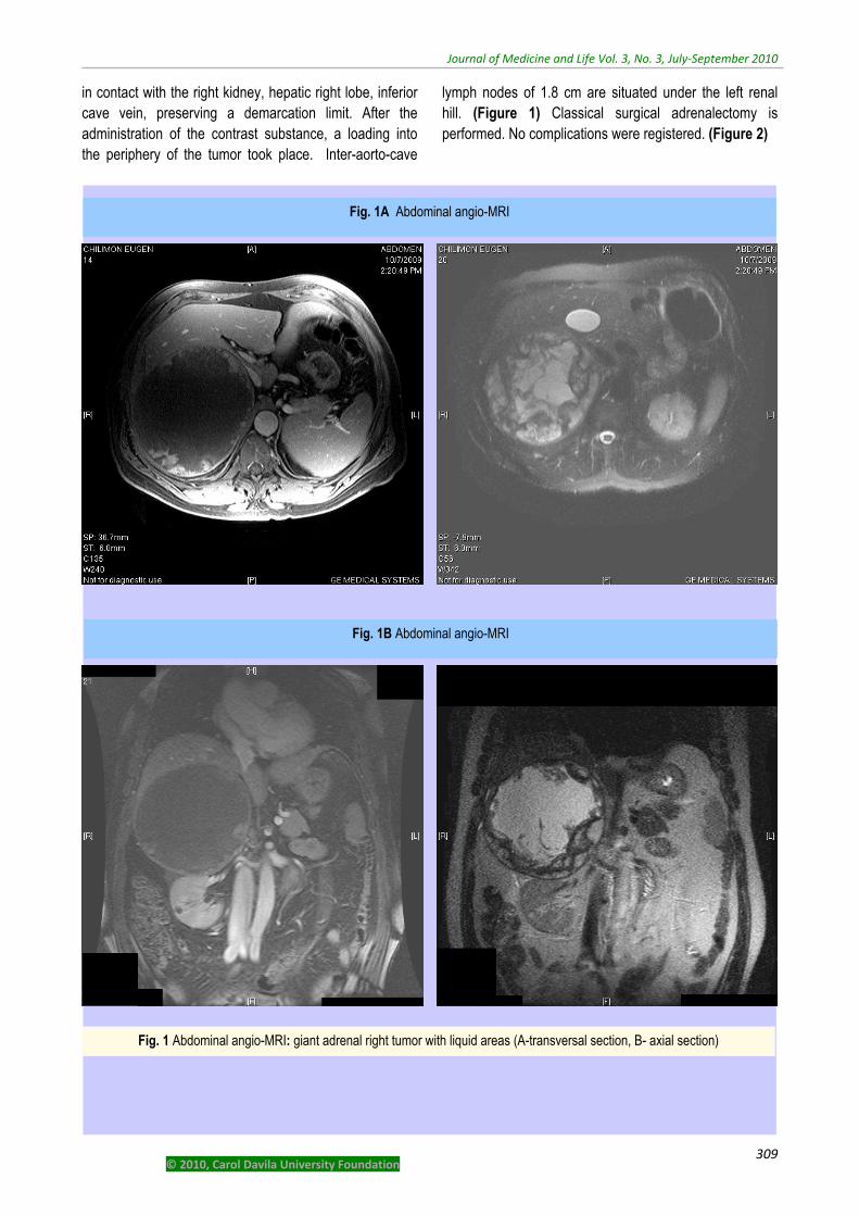
The plasma basal cortisol was of 15.53 μg/dL (normal between 4.3 and 22.4), with suppression at 0.94 μg/dL after 2 mg of dexametasone (2 days); plasma basal ACTH - 47.02 pg/mL (normal between 1 and 66). Mildly changed parameters were: serum aldosterone – 278.97 pg/mL (normal between 10 and 160) and 24-hours urinary 17 ketosteroids - 20.2 mg (normal between 8 and 18). The angioMRI revealed a tumor of 15 by 14.7 cm with a large central area filled with liquid signal, an intact capsule
Tabel 1 Normal hormonal parameters
Journal of Medicine and Life Vol. 3, No. 3, July‐September 2010
309© 2010, Carol Davila University Foundation
in contact with the right kidney, hepatic right lobe, inferior cave vein, preserving a demarcation limit. After the administration of the contrast substance, a loading into the periphery of the tumor took place. Inter-aorto-cave
lymph nodes of 1.8 cm are situated under the left renal hill. (Figure 1) Classical surgical adrenalectomy is performed. No complications were registered. (Figure 2)
Fig. 1A Abdominal angio-MRI
Fig. 1B Abdominal angio-MRI
Fig. 1 Abdominal angio-MRI: giant adrenal right tumor with liquid areas (A-transversal section, B- axial section)

Journal of Medicine and Life Vol. 3, No. 3, July‐September 2010
310 © 2010, Carol Davila University Foundation
Fi. 2 A, B, C Right adrenalectomy: aspects during surgical procedure
A A
B B
C

Journal of Medicine and Life Vol. 3, No. 3, July‐September 2010
311© 2010, Carol Davila University Foundation
No adrenal insufficiency was registered. Basal plasma cortisol was of 17μg/dL and mild hepatic cytolysis was remitted within 7 days, probably due the hepatic intra-surgery manipulation. The full recovery of the patient was present. The patient will be kept under observation in the next period. (Figure 3)
The pathological exam pointed an adrenal cyst
having hemorrhagic content, a fibro-conjunctive capsule with areas of calcifications inside the capsule and osteoid metaplasia under the capsule. Normal adrenal tissue was found peri and intra-capsule. (Figure 4)
The immunohistochemistry revealed stromal
positive reaction for VIM, ACT and CD34 positive into the vessels, CROMO positive into the medullar and negative INHIBIN. (Figure 5)
Fig. 3 One week after surgery
Fig. 4 Hematoxilin eosin: different aspects of the adrenal cyst with normal adrenal epithelium
A

Journal of Medicine and Life Vol. 3, No. 3, July‐September 2010
312 © 2010, Carol Davila University Foundation
Discussions
Adrenal cysts are rare. The reported incidence in clinical series is of 5.4%. [1] In the autopsy series they count less than 0.064 to 0.18%. [2] There are a few similar cases reported in the literature with mixed giant adrenal tumor and rapid increase that suggests an adrenal carcinoma. [3] In this case, the areas of calcification and osteoid metaplasia might suggest a long-standing process but apparently, the tumor increased to 15 cm in 6 months, probably due to the high blood amount. An adrenal carcinoma may also have necrosis and calcification, slightly modified endocrine secretor parameters of the adrenal and rapidly increasing dimensions. There are also a small number of cases in literature regarding adrenal cysts hormonally active (Cushing’s syndrome, pheocromocytoma). [1]
Adrenal cysts are usually asymptomatic; mostly an occasional discovery during ultrasound or CT. In our case, it was a surgical procedure for a different cause. Acute abdominal pain may be presented in some cases. [4] In this case the patient had initially abdominal pain but for another disease. There are some patients reported with sudden onset of abdominal pain and features compatible with acute appendicitis probably caused by a hemorrhage into the cyst. [5] In our case, at the moment
of the first surgery, the tumor was small, macroscopically of 2 cm, so a hemorrhage was less probable at that moment. Examples of atypical situations are three cases of acute hemorrhage during pregnancy. [6] Some other emergencies of the cyst are its rupture, acute hypovolemia or shock due to the intense bleeding. [7, 8, 9] Insidious presentation of the cyst is represented by digestive disturbances due to the changes of the local anatomy. While most of the cysts have less than 10 cm, one of the largest described has 45 cm. [10]
From the histological point of view, the adrenal cysts are vascular or endothelial, hemorrhagic or pseudoscyst and epithelial-lined or “true” adrenal cysts. There are a few cases of bilateral cysts reported. [11] 7% of cysts are malignant. [12] Among adrenal cysts, the most common types are epithelial cysts and pseudo-cysts. Some authors appreciate that the adrenal pseudo cyst is the most frequent type. [13] Intra-cystic hemorrhage spontaneous or post-traumatic may be present. This was probably presented in our case, explaining the rapid increase of the tumor. Sometimes, the hemorrhage causes acute anemia that indicates surgery. The operation is usually a laparotomic adrenalectomy, since the laparoscopic approach is not sufficient to control large masses with frequent active bleeding inside. [14] On the other hand, small adrenal
D
C B
Fig. 5 5A-IHC: VIM +ve in the stroma 5B-IHC: CD34 +ve in the vessels 5C-IHC: ACT +ve in the vessels 5D-IHC: CROMO +ve in the medulosuprarenal

Journal of Medicine and Life Vol. 3, No. 3, July‐September 2010
313© 2010, Carol Davila University Foundation
cysts do not necessary need surgical approach. They may be followed up by ultrasound or CT and hormonally. [15] There are still controversies related to the adequate management. [16] Surgical indication has tumors larger than 5 cm, symptomatic tumors or rapid expanding. [17]
A patient with a giant adrenal mass is a challenge regarding the differential diagnosis from other types of adrenal masses. There are many parasitosis with cysts aspects. [18] The diagnosis could only be made following the anatomo-pathological analyses. Pre operative CT guided biopsy (if the tumor is not hormonal active or there is no suspicion of malignancy), urography or angioMRI (like the one in our case) are useful. Complete cyst aspiration, rather than surgical excision, may be carried out initially for diagnosis and management of indeterminate suprarenal cystic lesions and
symptomatic cysts of the adrenal gland. [19] The present case demonstrates that the uncommon forms of adrenal masses must be taken into account only after surgery because the pathological evaluation provides the final remark. [20]
Conclusion A giant adrenal cyst mimicking a carcinoma by
the preoperative phenotype is a very rare finding. The surgical approach is challenging because of the large tumor dimensions and increased blood amount. Nevertheless, the pathological exam brings the most spectacular aspects regarding the differential diagnosis and the prognosis.
References
1. Pradeep PV, Mishra AK, Aggarwal V, Bhargav PR, Gupta SK. Agarwal A Adrenal cysts: an institutional experience. World J Surg. 2006; 30(10):1817-20.
2. Bellantone R, Ferrante A, Raffaelli M, Boscherini M, Lombardi CP, Crucitti F. Adrenal cystic lesions: report of 12 surgically treated cases and review of the literature. J Endocrinol Invest. 1998;21(2):109-14.
3. Mohan H, Aggarwal R, Tahlan A, Bawa AS, Ahluwalia M. Giant adrenal pseudocyst mimicking a malignant lesion. Can J Surg. 2003; 46(6):474.
4. Khilnani GC, Kumar A, Bammigatti C, Sharma R, Gupta SD. Hemorrhagic pseudocyst of the adrenal gland causing acute abdominal pain. J Assoc Physicians India. 2008; 56:379-80.
5. Moreno-Aranda J, Wingartz-Plata HF, Maldonado-Arce B, Vega-Espinosa JJ, Maldonado-Alcaraz E. Hemorrhagic adrenal pseudocyst: case report and a review of the literature.Gac Med Mex. 2004; 140(5):553-5.
6. Papaziogas B, Katsikas B, Psaralexis K, Makris J, Chatzimavroudis G, Tsiaousis R, Dragoumis D, Radopoulos K, Panagiotopoulou K, Atmatzidis K. Adrenal pseudocyst presenting as acute abdomen during pregnancy. Acta Chir Belg. 2006;106(6):722-5.
7. Chew SP, Sim R, Teoh TA, Low CH. Hemorrhage into non-functioning adrenal cysts--report of two cases and review of the literature. Ann Acad Med Singapore. 1999; 28(6):863-6.
8. Ferrante A, Civello IM, Bellantone R, Boscherini M. Hemorrhagic adrenal cyst: an unusual reason of acute hypovolemia. Chir Ital. 1999; 51(4):325-8.
9. Sivasankar A, Jeswanth S, Johnson MA, Ravichandran P, Rajendran S, Kannan DG, Surendran R. Acute hemorrhage into adrenal pseudocyst presenting with shock: diagnostic dilemmas--report of three cases and review of literature. Scientific World Journal. 2006; 19(6):2381-7.
10. Tagge DU, Baron PL . Giant adrenal cyst: management and review of the literature. Am Surg. 1997; 63(8):744-6.
11. Terachi T, Terai A, Machida S, Oomori K. Two cases of adrenal cysts. Hinyokika Kiyo. 1986; 32(10):1497-503.
12. Schmid H, Mussack T, Wörnle M, Pietrzyk MC, Banas B. Clinical management of large adrenal cystic lesions. Int Urol Nephrol. 2005; 37(4):767-71.
13. Sáenz Medina J, Esteban Peris A, Linares Quevedo AI, Vallejo Herrador J, Castillón Vela I, Páez Borda A. Adrenal pseudocyst. Report of a new case and review of
the literature. Actas Urol Esp. 2006; 30(4):420-3.
14. Basile G, Buffone A, Cicciarella G, di Mari P, Cirino E. Hemorrhagic adrenal pseudocyst: case report. Ann Ital Chir. 2004; 75(4):487-90.
15. de Bree E, Schoretsanitis G, Melissas J, Christodoulakis M, Tsiftsis D. Cysts of the adrenal gland: diagnosis and management. Int Urol Nephrol. 1998; 30(4):369-76.
16. Sanz Mayayo E, Maganto Pavón E, Gómez García I, Mayayo Dehesa T, Rodríguez-Patrón Rodríguez R, García González R, Escudero Barrilero A. Adrenal cysts: presentation of 6 cases. Arch Esp Urol. 2003; 56(4):345-53.
17. De Toma G, Gabriele R, Plocco M, Sapienza P. Adrenal cysts: therapeutic indications. Minerva Chir. 1995; 50(10):925-8.
18. Akçay MN, Akçay G, Balik AA, Böyük A. Hydatid cysts of the adrenal gland: review of nine patients. World J Surg. 2004; 28(1):97-9
19. Tung GA, Pfister RC, Papanicolaou N, Yoder IC. Adrenal cysts: imaging and percutaneous aspiration. Radiology. 1989; 173(1):107-10.
20. Mármol Navarro S, Napal Lecumberri S, Rajab R, Guadalajara Jurado J, Fachal Bermúdez C. A giant adrenal mass: the anatomicopathological surprise finding. Arch Esp Urol. 1995; 48(9):949-51.