genomic features of lactic acid bacteria effecting bioprocessing and health
TRANSCRIPT

www.fems-microbiology.org
FEMS Microbiology Reviews 29 (2005) 393–409
Genomic features of lactic acid bacteria effectingbioprocessing and health
Todd R. Klaenhammer a,b,*, Rodolphe Barrangou a,b, B. Logan Buck a,M. Andrea Azcarate-Peril a, Eric Altermann a
a Departments of Food Science and Microbiology, North Carolina State University, Box 7624, Raleigh, NC 27695-7624, United Statesb Functional Genomics Program, Southeast Dairy Foods Research Center, Raleigh, NC 27695, United States
Received 24 March 2005; accepted 27 April 2005
First published online 28 August 2005
Abstract
The lactic acid bacteria are a functionally related group of organisms known primarily for their bioprocessing roles in food and
beverages. More recently, selected members of the lactic acid bacteria have been implicated in a number of probiotic roles that
impact general health and well-being. Genomic analyses of multiple members of the lactic acid bacteria, at the genus, species,
and strain level, have now elucidated many genetic features that direct their fermentative and probiotic roles. This information
is providing an important platform for understanding core mechanisms that control and regulate bacterial growth, survival, signal-
ing, and fermentative processes and, in some cases, potentially underlying probiotic activities within complex microbial and host
ecosystems.
� 2005 Federation of European Microbiological Societies. Published by Elsevier B.V. All rights reserved.
Keywords: Lactic acid bacteria; Genomics; Bioprocessing; Probiotics; Comparative genomics; Functional genomics
Contents
1. Introduction . . . . . . . . . . . . . . . . . . . . . . . . . . . . . . . . . . . . . . . . . . . . . . . . . . . . . . . . . . . . . . . . . . . . . . . . . . . . 393
2. Genome characteristics. . . . . . . . . . . . . . . . . . . . . . . . . . . . . . . . . . . . . . . . . . . . . . . . . . . . . . . . . . . . . . . . . . . . . 394
3. Comparative genomics of lactobacilli. . . . . . . . . . . . . . . . . . . . . . . . . . . . . . . . . . . . . . . . . . . . . . . . . . . . . . . . . . . 399
4. Functional genomic analysis . . . . . . . . . . . . . . . . . . . . . . . . . . . . . . . . . . . . . . . . . . . . . . . . . . . . . . . . . . . . . . . . . 401
5. Concluding remarks . . . . . . . . . . . . . . . . . . . . . . . . . . . . . . . . . . . . . . . . . . . . . . . . . . . . . . . . . . . . . . . . . . . . . . . 406
Acknowledgments . . . . . . . . . . . . . . . . . . . . . . . . . . . . . . . . . . . . . . . . . . . . . . . . . . . . . . . . . . . . . . . . . . . . . . . . 406
References . . . . . . . . . . . . . . . . . . . . . . . . . . . . . . . . . . . . . . . . . . . . . . . . . . . . . . . . . . . . . . . . . . . . . . . . . . . . . 406
1. Introduction
Lactic acid bacteria (LAB) are a heterogeneous fam-
ily of microorganisms that can ferment a variety of
0168-6445/$22.00 � 2005 Federation of European Microbiological Societies
doi:10.1016/j.femsre.2005.04.007
* Corresponding author. Tel.: +1 919 515 2972; fax: +1 919 513 0014.
E-mail address: [email protected] (T.R. Klaenhammer).
nutrients [1] primarily into lactic acid. They are mainly
Gram-positive, anaerobic bacteria, non-sporulating,
and acid tolerant. Biochemically, LAB include both
homofermenters and heterofermenters. The former pro-
duce primarily lactic acid, while the latter yield also a
variety of fermentation by-products, including lacticacid, acetic acid, ethanol, carbon dioxide and formic
. Published by Elsevier B.V. All rights reserved.

394 T.R. Klaenhammer et al. / FEMS Microbiology Reviews 29 (2005) 393–409
acid [2,3]. Although their primary contribution centers
on rapid acid production and acidification of food prod-
ucts, they also contribute to flavor, texture and nutrition
[3]. LAB are found naturally in a variety of environmen-
tal habitats, including dairy, meat, vegetable, cereal and
plant environments, where fermentation can occur. His-torically, the traditional roles for many LAB have been
as starter cultures to drive food and dairy fermentations,
leading to their widespread human consumption and
generally recognized as safe (GRAS) status.
It was nearly 100 years ago that Russian scientist,
Elie Metchnikoff, then at the Pasteur Institute, proposed
that lactic bacteria in fermented milk could promote the
development of a healthy intestinal microbiota. Specifi-cally, the Nobel-laureate developed a theory that lactic-
acid bacteria in the digestive tract could prolong life by
preventing putrefaction. Since that time, it has been rec-
ognized that some LAB and the high G + C content
Gram-positive bifidobacteria are also found naturally
within human and animal cavities, including the gastro-
intestinal tract (Lactobacillus acidophilus, Lactobacillus
gasseri, Lactobacillus johnsonii, Lactobacillus plantarum,Streptococcus agalactiae, Enterococcus faecalis), the oral
cavity (Streptococcus mutans, Bifidobacterium longum),
and the vaginal cavity (B. longum, S. agalactiae, Lacto-
bacillus crispatus) [4–6]. LAB are considered to be
important components of the normal intestinal microbi-
ota, which contribute to a variety of functions including
intestinal integrity, immunomodulation, and pathogen
resistance. Selected groups of Lactobacillus and Bifido-
bacterium are used widely as probiotics primarily in
dairy products and dietary supplements [5,7,8].
Defined most recently as ‘‘live microorganisms which,
when administered in adequate amounts, confer a health
benefit on the host’’ [9], probiotic cultures have been
found useful in the maintenance of gastrointestinal
(GI) health including the treatment of diarrheal diseases
and preservation of intestinal integrity and mobility. Re-cent evidence from in vitro systems, animal models, and
clinical studies suggests that LAB can enhance both spe-
cific and non-specific immune responses, possibly by
activating macrophages, altering cytokine expression,
increasing natural killer cell activity, and/or increasing
levels of immunoglobulins [5,10,11]. However, the mech-
anisms through which these LAB function as immuno-
modulators are not characterized and specific reactionscan be highly variable among different strains.
In the recent past, substantial progress has been
achieved in microbial genomics, particularly in genome
sequencing. To date, over 229 complete microbial gen-
omes (prokaryotes and archaea) have been published
(NCBI website, www.ncbi.nlm.nih.gov/genomes/MI-
CROBES/complete.html), covering a wide diversity of
taxonomic groups. Early microbial genome analysesindicate that genomic content reflects an organism�smetabolism, physiology, biosynthetic capabilities, and
adaptability to varying conditions and environments.
In the case of the LAB, genome analysis is revolutioniz-
ing our view of their metabolic processes, bioprocessing
capabilities and potential roles in health and well-being.
2. Genome characteristics
The published genome sequences of the lactic acid
bacteria and bifidobacteria include Lactococcus lactis
[12], S. mutans [13], S. pneumoniae [14], S. agalactiae
[15], S. pyogenes [16], S. thermophilus [17], B. longum
[18], L. plantarum [19], L. johnsonii [20] and L. acidoph-
ilus [21]. The 11 draft genomes represented by the LacticAcid Bacteria Genomics Consortium [22,23] have re-
cently been completed (unpublished results). The LAB
analyzed were Lactobacillus brevis, L. casei, L. gasseri,
Lc. cremoris, Leuconostoc mesenteroides, Oenococcus
oeni, Pediococcus pentosaceus, and S. thermophilus. For
the LAB and bifidobacteria, probiotic organisms, and
other related industrial microbes, genome features are
presented in Table 1.LAB and bifidobacteria are Gram-positive bacteria
with low and high GC content, respectively, with small
genomes ranging in size between 1.8 and 3.3 Mb (Table
1). For those species where complete genomes are pub-
lished and annotated, a broad picture emerges of con-
served and varying biosynthetic and metabolic
capabilities. Glycolysis enzymes are uniformly repre-
sented among members of the LAB. A recent transcrip-tional analysis of global gene expression by L.
acidophilus during growth on eight different carbohy-
drates revealed that genes of the glycolytic pathway were
among the most highly expressed genes within the gen-
ome [25] (Fig. 1). Since LAB recover their primary en-
ergy via glycolysis, it seems likely that this is a
universal feature. Genome analyses have shown that lac-
tobacilli, bifidobacteria, streptococci, and lactococcipossess broad saccharolytic potentials, which reflect
the nutrient diversity provided by the range of environ-
ments they inhabit [13,14,18–21]. Analysis of the L.
plantarum genome revealed many transporters, particu-
larly PTS (phosphotransferase system) transporters
(25) correlating with the organism�s broad capacity to
metabolize varied carbohydrates from different environ-
ments [19]. In particular, a ‘‘lifestyle adaptation island’’was defined over a 213 kb region that harbored genes in-
volved in sugar transport and metabolism. Similarly, the
diversity of transporters in S. mutans and S. pneumoniae
has been associated with an increased ability to utilize
nutrient sources present in their environments, namely
the oral cavity and respiratory tract [13,14]. Analysis
of the L. johnsonii [20], L. acidophilus [21], and L. gasseri
genomes further substantiate these observations show-ing a preponderance of PTS transporters, and only 2
to 3 ABC (ATP-binding cassette) transporters identified

Table 1
Genomes of lactic acid bacteria and other industrially used species
Genus Species Strain Size (Mbp) %GC Status Reference
Bifidobacterium longum NCC2705 2.3 60.1 Ca [18]
longum DJ010A 2.4 59 IPb JGIc
Brevibacterium linens BL2/ATCC9174 4.4 60.9 IP JGI
Enterococcus faecalis V583 3.2 37.5 C [24]
Lactobacillus acidophilus NCFM 2.0 34.7 C [21]
gasseri ATCC333323 1.8 35.1 IP JGI
johnsonii NCC533 2.0 34.6 C [20]
plantarum WCFS1 3.3 44.5 C [19]
casei ATCC334 2.5 41.1 IP JGI
casei BL23 2.6 4.6 IP [23]
rhamnosus HN001 2.4 46.4 IP Lubbers et al. (unpublished)
helveticus CNRZ32 2.4 37.1 IP Steele et al. (unpublished)
helveticus CM4 2.0 37 C Shinoda (unpublished)
sakei 23K 1.9 41.2 C [23]
delbrueckii ATCCBAA365 2.3 45.7 IP JGI
delbrueckii ATCC11842 2.3 50 IP [23]
delbrueckii DN-100107 2.1 IP [23]
reuteri IP JGI
salivarius UCC118 IP [23]
brevis ATCC367 2.0 43.1 IP JGI
Lactococcus lactis ssp. lactis IL1403 2.3 35.4 C [12]
lactis ssp. cremoris SK11 2.3 30.9 IP JGI
lactis spp. cremoris MG1363 2.6 37.1 IP [23]
Leuconostoc mesenteroides ATCC8293 2.0 37.4 IP JGI
Oenococcus oeni ATCCBAA331 1.8 37.5 IP JGI
oeni IOEB84.13 1.8 37.9 IP [23]
Pediococcus pentosaceus ATCC25745 2.0 37.0 IP JGI
Propionibacterium freudenreichii ATCC6207 2.6 67.4 IP [23]
Streptococcus agalactiae 2603V/R 2.2 35.7 C [15]
mutans UA159 2.0 36.8 C [13]
pneumoniae TIGR4 2.2 39.7 C [14]
pyogenes M1 1.9 38.5 C [16]
thermophilus LMD9 1.8 36.8 IP JGI
thermophilus LMG18311 1.9 39 C [17]
thermophilus CNRZ1066 1.8 39 C [17]
Adapted from [22,23,25].a C, complete.b IP, in progress.c JGI, Joint Genome Institute.
T.R. Klaenhammer et al. / FEMS Microbiology Reviews 29 (2005) 393–409 395
for maltose and complex carbohydrates like fructooligo-
saccharide and raffinose (Table 2). An overview of the
carbohydrate transporters found in L. acidophilus is
shown in Fig. 2, where PTS systems were predominant
and lactose and galactose were predicted to be trans-
ported by a galactoside permease [25]. A recent analysis
of 9 LAB genomes for transporter capabilities revealed
that 13–17 % of their total genes encoded transport pro-teins (Lorca, G.L., Zlotopolski, V., Tran, C., Winnen,
B., Hvorup, R.N., Nguyen, E., Huang, L.-W., and
Saier, M.H., unpublished). This proportion was larger
than observed for most bacteria. Interestingly, amino
acid uptake systems predominated over sugar and pep-
tide uptake systems.
Although amino acid biosynthetic pathways are com-
plete in L. lactis, they are deficient in varying levels in
most other LAB. L. plantarum is missing only a few syn-
thetic pathways including those for branched chain
amino acid synthesis [19], whereas species of the
‘‘L. acidophilus complex’’ (L. gasseri, L. johnsonii and
L. acidophilus) [27] are largely deficient in amino acid
biosynthetic capacity [20,21]. Compensating for thesedeficiencies, the lactobacilli generally encode a large
number of peptidases, amino acid permeases, and multi-
ple oligo-peptide transporters that could support effi-
cient processing and recovery of amino acids from
nutritionally rich environmental sources. However, of
the intestinal lactobacilli (including comparisons with

Fig. 1. Hierarchical clustering analyses of gene expression patterns (left panel). The expression of 1889 genes (vertically) after growth on eight
carbohydrates (horizontally) is shown colorimetrically. Least squares means, representing overall gene expression level corrected for systematic and
random errors low = blue, high = red; hierarchical clustering of least squares means allows visualization of the relative expression levels of all genes
within each treatment. Lanes from left to right are fructooligosaccharides; fructose; galactose; glucose; lactose; raffinose; sucrose; and trehalose.
Microarrays were carried out using PCR products of predicted ORFs [26]. Expression of glycolysis genes (right panel). In a whole genome array, the
global transcription responses during growth on eight different carbohydrates are denoted for D-lactate dehydrogenase (D-LDH, La55),
phosphyglycerate mutase (PGM, La185), L-lactate dehydrogenase (L-LDH, La271), glyceraldehyde 3-phosphate dehydrogenase (GPDH, La698),
phosphoglycerate kinase (PGK La699), glucose 6-phosphate isomerase (GPI, La752), 2-phosphoglycerate dehydratase (PGDH, La889),
phosphofructokinase (PFK, La956), pyruvate kinase (PK, La957), fructose-biphosphate aldolase (FBPA,La1599). Expression: low
high [25].
396 T.R. Klaenhammer et al. / FEMS Microbiology Reviews 29 (2005) 393–409
L. gasseri and L. plantarum), only L. acidophilus and
L. johnsonii were found to encode the cell wall-associ-
ated proteinase, PrtP. Interestingly, the gene predicted
to encode the maturation protein, PrtM, was found in
all these Lactobacillus genomes.
For the LAB, their genomes are collectively of low
GC content and relatively small. Those species with
the smallest genomes can be highly auxotrophic anddeficient in a number of biosynthetic pathways, corre-
sponding to their apparent adaptation to nutritionally
rich environments [20,21]. For the lactobacilli, a sum-
mary of biosynthetic pathways (amino acids, nucleo-
tides, fatty acids and vitamins) illustrated that L.
gasseri and L. johnsonii exhibit the fewest metabolic bio-
synthetic pathways (6–8), whereas L. acidophilus shows
a higher number at 14 pathways (K. Makarova, E. Koo-nin and LABGC, unpublished data). In contrast,
L. plantarum encodes a more complete complement of
biosynthetic pathways (22), supporting its more diverse
metabolic capabilities. In this regard, a direct compari-
son of metabolic pathways via Kyoto Encyclopedia of
Genes and Genomes (KEGG) between L. plantarum
and L. johnsonii by Boekhorst et al. [28] highlighted
the biosynthetic deficiencies in L. johnsonii and ex-
panded capacity of L. plantarum.
In the current analysis of the complete LAB gen-
omes, it has been suggested that evolution to nutrition-ally rich environments (e.g., milk, human GI tract) has
promoted genome simplification and degradation for
some species. Notably, in the recent genome analysis
of two S. thermophilus strains, Bolotin et al. [17] found
that 10% of the genes were pseudogenes and non-func-
tional due to frameshifts, nonsense mutation, deletion,
or truncation. Evidence for genome decay was particu-
larly noted for genes involved in carbohydrate metab-olism, uptake and fermentation. In contrast, a specific
symporter for lactose was found in S. thermophilus
that was absent from other pathogenic streptococci.

Table 2
Carbohydrate utilization profiles and predicted transporters for lactobacilli with complete genomes (from [25])
Type Sugar Fermentation
Laca Lplb Ljoc Lgad
Pentoses Arabinose Yese
Ribose Yes
Ribulose
Xylose
Xylulose
Hexoses Fructose PTSf PTS PTS PTS
Galactose GPHg Yes Yes Yes
Glucose PTS Yes Yes Yes
Mannose PTS PTS PTS PTS
Disaccharides Cellobiose PTS PTS PTS
Gentiobiose PTS PTS Yes
Lactose GPH Yes PTS PTS
Maltose ABCh Yes ABC ABC
Melibiose PTS Yes Yes
Sucrose PTS PTS Yes
Trehalose PTS Yes Yes Yes
Turanose Yes
Oligosaccharides FOS ABC Yes
Melezitose Yes
Raffinose ABC Yes PTS
Sugar alcohols Galactitol PTS
Glycerol
Mannitol PTS
Sorbitol PTS
Deoxysugars Fucose
Rhamnose Yes
Modified Sugars Amygdalin Yes PTS Yes Yes
Arbutin PTS PTS Yes
Esculin PTS Yes Yes
Gluconate PTS
Malate
N-acetylglucosamine PTS PTS PTS PTS
Salicin PTS PTS Yes Yes
a Lactobacillus acidophilus.b Lactobacillus plantarum.c Lactobacillus johnsonii.d Lactobacillus gasseri.e Determined by fermentation patterns obtained from API50CHO (BioMerieux, Durham, NC).f PTS, phosphoenolpyruvate phosphortransferase system transporter.g GPH, galactoside pentose hexuronide permease.h ABC, ATP-binding cassette transporter.
T.R. Klaenhammer et al. / FEMS Microbiology Reviews 29 (2005) 393–409 397
It was suggested that evolution of S. thermophilus to
milk resulted in genome degradation of many genes
(including any pathogenic genes) that were despensible
as this organism evolved to this specialized environ-
ment [17]. Interestingly, evidence was also presented
for horizontal gene transfer between S. thermophilus
and other organisms co-occupying the dairy environ-
ment. A 17 kb region was identified that containedmultiple copies of IS1191 and a mosaic of fragments
with over 90% identity to L. bulgaricus and L. lactis.
Among them was a unique copy of metC that allows
methionine biosynthesis, which is a rare amino acid
in milk.
For L. lactis, considerable evidence has demonstrated
that members of this species have aquired plasmid DNA
elements encoding critical functions for growth and
competition in a milk environment, such as lactose
metabolism, proteolytic activity, bacteriocin production,
exopolysaccharide production and resistance to bacte-
riophages [29]. In addition, it is also apparent that hor-
izontal gene transfer has introduced important functionsto the genomes of a number of LAB that are expected to
promote their competition in these environments. Genes
encoding sugar transporters and carbohydrate hydro-
lases can represent a large portion of strain-specific
genes that have been acquired by horizontal gene trans-

MELIBIOSE
MELIBIOSE-6P
GLUCOSE
GLUCOSE-6P
EC2.7.1.2 La1433
AMYGDALIN
SALICIN
ARBUTIN
N-ACETYL-GLUCOSAMINE
MANNOSE
MANNOSE-6P
MALTOSE
MALTOSE
CELLOBIOSE
CELLOBIOSE-6P
GENTIOBIOSE
GENTIOBIOSE-6P
AMYGDALIN-6P
SALICIN-6P
ARBUTIN-6P
N-ACETYLGLUCOSAMINE-6P
SUCROSE
SUCROSE-6P
FRUCTOSE
FRUCTOSE-1P
TREHALOSE
TREHALOSE-6P
GLUCOSE
FRUCTOSE
FRUCTOSE-6P GLUCOSE-6P GLUCOSE-6P GLUCOSE-6P GALACTOSE-6P GLUCOSE-6P FRUCTOSE1,6P2
GLUCOSE GLUCOSE GLUCOSE GLUCOSE FRUCTOSE
GLUCOSE-6PPRUNASIN
GLUCOSE-6P
GLUCOSE-6P
GLUCOSAMINE-6P
GLUCOSE-6P
GLUCOSE-6PGLUCOSE
FOS
RAFFINOSE
FOS
RAFFINOSE
FRUCTOSE
GALACTOSESUCROSE
GLUCOSEFRUCTOSE
3.2.1.22 La1438
2.4.1.7 La1437
3.2.1.26 La505
3.2.1.93 La10142.7.1.56 La1778
3.2.1.86 La874
3.5.1.25 La144
3.2.1.21 La1366 3.2.1.21 La1365 2.4.1.8 La1870 5.4.2.6 La1869
5.3.1.8 La745
3.2.1.117
GLUCOSE
EC 3.2.1.118
3.2.1.86 La885
2.6.1.16 La462
3.2.1.86 La1706
LACTOSE
GALACTOSEUDP-GLUCOSE
UDP-GALACTOSE GALACTOSE-1P
LACTOSE 3.2.1.23 La1467
GLUCOSEGALACTOSE
GALACTOSE2.7.1.6 La1459
GLUCOSE-1P
GLUCOSE-6P
GLUCOSE-6P
2.7.7.10La1458
5.1.3.2 La1469
2.7.7.9 La1719
5.4.2.2 La687
3.2.1.26 La400
2.7.1.4 La16
La889
FRUCTOSE-6P
FRUCTOSE-1,6P2
GLYCERALDEHYDE-3P
GLYCERALDEHYDE-1,3P2
GLYCERATE-3P
GLYCERATE-2P
PHOSPHOENOLPYRUVATE
PYRUVATE
La956
La185
La271 L-LDHLa55 D-LDH
La752
La1599
La698
La699
La957
2.7.1.11
4.1.2.13
1.2.1.12
2.7.2.3
5.4.2.1
4.2.1.11
2.7.1.40
1.1.1.27
5.3.1.9
SUBSTRATE SUBSTRATE-P
IIC IIB IIA
LACTATE
Fig. 2. Transporters and pathways predicted for carbohydrate utilization by Lactobacillus acidophilus. The diagram shows transporters, hydrolases
and glycolysis enzymes, as predicted by the putative genome annotation. Gene and enzyme numbers are indicated for each enzymatic reaction. For
transporters, red indicates a putative PTS transporter; green, a putative ABC transporter; and yellow, a galactoside permease. For sugars, identical
compounds share the same color (from [21] with permission).
398 T.R. Klaenhammer et al. / FEMS Microbiology Reviews 29 (2005) 393–409
fer. It has been suggested previously that selected genes
involved in sugar transport, catabolic properties, and
exopolysaccharide synthesis in L. plantarum [19] have
been acquired via horizontal gene transfer, as part of
the adaptation process of this organism to a diverse
number of environments (e.g., plants, cereals, GI tract).
Evidence supporting horizontal gene transfer for these
regions include their grouped position near the originof replication, lowered GC content (41.5% versus
44.5%; Fig. 5), and high variability as to the presence
or absense of these genes among different strains of L.
plantarum [19,23]. The accumulating evidence suggests
that evolution of the LAB to nutritionally complex envi-
ronments has been driven by two major processes; first,
gene degradation and loss of dispensible functions from
ancestral types [17], and second, gene acquisition viahorizontal gene transfer and duplication of important
capabilities [17,19–21, D.A. Mills, K. Makarova, and
E. Koonin, and LABGC, unpublished data].
In addition, there are numerous examples of gene
duplications and multiple copies of related genes pre-
dicted to direct important functions in the genomes of
the sequenced LABs. Examples include PTS transport-
ers, b- and phospho-b galactosidases, lactic dehydrogen-ases, peptidases, and oligopeptide and amino acid
transporters [19–21]. Also notable are the multiple cop-
ies of homologs for mucus-binding (Mub) proteins
found in L. gasseri (7), L. acidophilus (5), L. johnsonii
(4), and L. plantarum (4). First discovered in L. reuteri,
Tuomola et al. [30] reported a 358-kDa surface protein
able to bind to mucin glycoproteins. The predicted
Mub proteins, now revealed in the genomes of many
intestinal lactobacilli, are unusually large proteins rang-
ing in size from 1000 to 4300 amino acids and often rep-resent the largest open reading frames (ORFs) in the
genome. While similar in their large size and the pres-
ence of multiple repeats (4–6) of �20 aa sequences
[20], their amino acid identity is relatively low at 24–
38%, indicating considerable sequence variability within
surface proteins presumed to serve important and simi-
lar roles in mucus binding. In this regard, Altermann
et al. [21] found that 6.6% of unclassified COGs (clustersof orthologous groups) in the L. acidophilus genome
were represented in five distinct regions (Fig. 3, COG re-
gions I, II, III, IV, V). All the genes within these regions
were predicted to be involved with host recognition or
epithelial adherence; including mucus binding, fibronectin
binding, and other cell surface associated type proteins.
A region was identified in L. johnsonii that contained a
mub gene within a predicted nine-gene operon that in-cluded a large serine rich protein with homology to a
Streptococcus fimbrial adhesin. This unique region in

Fig. 3. Genome atlas of L. acidophilus NCFM. The atlas represents a circular view of the complete genome sequence of L. acidophilus NCFM. The
right-hand legend describes the single circles in the top-down-outermost-innermost direction. The circle was created using Genewiz [73] and in house
developed softwares [21]. Circle 1, Intermost, GC-Skew. Circle 2, COG classification. Predicted ORFs were analyzed using the COG database and
grouped into the four major categories. 1, Information storage and processing; 2, Cellular processes and signaling; 3, Metabolism; 4, Poorly
characterized; and 5, ORFs with uncharacterized COGs or no COG assignment. Circle 3, ORF orientation. ORFs in sense orientation (ORF+) are
shown in blue; ORFs oriented in anti-sense direction (ORF�) in red. Circle 4, Blast similarities. Deduced amino-acid sequences compared against the
non-redundant (nr) database using gapped BlastP. Regions in blue represent unique proteins in NCFM, whereas highly conserved features are shown
in red. The degree of color saturation corresponds to the level of similarity. Circle 5, G + C content deviation. Deviations from the average GC-
content are shown in either green (low GC spike) or orange (high GC spike). A boxfilter was applied to visualize contiguous regions of low or high
deviations. Circle 6, Ribosomal machinery. tRNAs, rRNAs and ribosomal proteins are shown as green, cyan, or red lines, respectively. Clusters of
thereof are represented as colored boxes to maintain readability. Circle 7, Mobile elements. Predicted transposases are shown as light purple, phage-
related Integrases as orange dots. Circle 8, Stress response. Genes involved in general stress response, including chaperones, and genes involved in
heat shock, DNA repair, and pH regulation, are shown in dark purple. Circle 9, Peptide and amino acid utilization. Proteases and peptidases are
shown in green, non-sugar-related transporters in light blue dots. Circle 10, Outermost Two-component regulators (2CRS). Each 2CRS is represented
as brown dots, consisting of a response-regulator and a histidine-kinase. In circles 7–10 each full dot represents one predicted ORF and clusters of
ORFs are represented by stacked dots. Selected features representing single ORFs and ORF cluster are shown outside of circle 10 with bars
indicating their absolute size. Origin and terminus of DNA replication are identified in green and red, respectively. Other features are: SlpA and B (S-
layer proteins), CdpA (Cell division protein), sugar utilization (Sucrose, FOS, Trehalose, Raffinose), LacE (PTS-sugar transporter), BshA and B (Bile
salt hydrolases), Mub-909 to Mub-1709 (mucus-binding proteins, numbers correspond to the La-number scheme), FbpA (fibronectin-binding
protein), Cfa (Cyclopropane fatty acid synthase), Fibronectin_binding (fibronectin-binding protein cluster), EPS_cluster (Exopolysaccharides),
Lactacin_B (bacteriocin), pauLA-I to pauLA-III (potential autonomous units), and prLA-I and prLA-II (phage remnants).
T.R. Klaenhammer et al. / FEMS Microbiology Reviews 29 (2005) 393–409 399
L. johnsonii [20] was positioned within a common locus
that has now been found in L. acidophilus, L. gasseri,
and L. plantarum (Fig. 4; E. Altermann and T.R. Klaen-
hammer, unpublished data).
3. Comparative genomics of lactobacilli
In spite of the explosion of genomic information on
microorganisms, complete genomes of beneficial com-
mensals, symbionts, and probionts are just now becom-
ing available [31]. Comparison of the similarities and
differences within these groups is expected to provide
an important view of gene content, organization, and
regulation that contributes to both gut and probioticfunctionality [22,31]. A recent comparative analysis be-
tween the complete genomes of L. plantarum and L.
johnsonii revealed striking differences in gene content
and synteny in the genome, prompting a conclusion that
these two species are only marginally more related to

Fig. 4. Map (not drawn to scale) of surrounding genes of the possible L. johnsonii fimbriae operon LJ0388 to LJ0394 (middle panel, from [20]) and its
comparison to the syntenic regions in L. gasseri (upper panel) and L. acidophilus (lower panel). Predicted open reading frames (ORFs) are shown as
arrows, the black line represents the genome. ORFs are grouped into colored clusters, according to their location, their functionality, or their degree
of homology. Homologous genes are connected by light colored parallelograms between the genome lines. Genes with no syntenic homology are
shown in grey. Gene clusters in different shades of green represent putative insertion events in L. johnsonii and L. acidophilus. The dark green cluster
(L. johnsonii) represents an ABC transporter and a putative sugar phosphatase. The mobile element (tranposase) adjacent to this insertion is shown in
orange (L. johnsonii and L. gasseri). The light green cluster (L. johnsonii) shows the possible fimbriae operon and the green cluster in L. acidophilus
indicates the potential autonomous unit pauLA-II [21], harboring a potential DNA modification system (methylase) and other phage-related
proteins.
Fig. 5. Circular plot of genome diversity found in 20 L. plantarum
strains isolated from different environments, using the method of
DNA–DNA hybridization to L. plantarum WCFS1 microarrays (D.
Molenaar, R.J. Siezen, M. Kleerebezem, unpublished). From outside
to inside: ring 1, base deviation index (BDI), from low (red) via
intermediate (yellow) to high (green); ring 2, DNA variability, from
low (present in all 20 strains) to high (absent in 1–19 strains compared
to WCFS1); ring 3, gene clusters for plantaricin biosynthesis, non-
ribosomal peptide biosynthesis (NRPS), prophages, polysaccharide
biosynthesis, nitrate respiration and sugar metabolism; ring 4, GC%.
This picture was generated with the Microbial Genome Viewer
(www.cmbi.kun.nl/MGV) (from [23], reprinted with permission).
400 T.R. Klaenhammer et al. / FEMS Microbiology Reviews 29 (2005) 393–409
each other than to other Gram-positive bacteria [28].
Nevertheless, 70% of the proteins in L. johnsonii still
had homologs (defined by a Blast score of <1E � 10)
in the larger L. plantarum genome. Unique proteins
found in these two genomes, when compared against
the published and draft genomes of the LAB, were pri-
marily unknown proteins and prophage-related ORFs
[28]. While whole genome comparisons with draft and
incomplete genomes for LAB-specific genes are useful,
some inaccuracies should be expected because gaps inthe draft sequences are likely to contain important
information.
Complete genomes are now published or available for
four Lactobacillus species (acidophilus, gasseri, johnsonii
and plantarum). Whole genome comparison over these
species (Fig. 6) substantiates the lack of synteny among
L. plantarum and the other three lactobacilli. In con-
trast, L. johnsonii and L. acidophilus show extensive con-servation of gene content and order over the length of
the genome. L. gasseri and L. johnsonii are even more
strikingly similar across the length of the genome, except
for two apparent chromosomal inversion events in L.
gasseri resulting in a reversal of gene order when com-
pared to the other two closely related species. A compar-
ison of ORFs between L. gasseri and L. johnsonii
revealed that 83–85% of the proteins had homologs inboth genomes [28]. Overall, the comparisons demon-
strate a high degree of gene synteny in the three species
that have been collectively referred to as members of the
L. acidophilus complex. Differentiation of these three
species, particularly L. gasseri and L. johnsonii, has been
historically difficult using traditional or molecular taxo-
nomic tools [27,32].
In an effort to distinguish between L. gasseri and L.
johnsonii strains, the translated genomic sequences of
three Lactobacillus strains were compared to find genes
unique to L. gasseri ATCC 33323, which do not occur
in L. acidophilus NCFM or L. johnsonii NCC533. Four-
teen unique genes were found in L. gasseri, which were
also not found in the NCBI non-redundant database.
Using specific primers designed for each of these unique
ORFs, L. gasseri specific amplicons could be generatedfrom all the L. gasseri strains evaluated (Table 3). Nota-
bly, primer pairs 4 and 11 appeared unique to the L. gas-

Fig. 6. Multiple whole genome comparison on protein level. The finished and annotated genomes of L. gasseri (top line), L. johnsonii (second line), L.
acidophilus NCFM (third line), and L. plantarum (bottom line) were analyzed using a bidirectional BlastP algorithm. Deduced amino acid sequences
from predicted open reading frames (ORFs) were compared to the respective partner-ORFeome using the standalone Blast provided by NCBI.
Translating these results into a compatible format, visualization of the comparison was realized using the Artemis Comparison Tool (ACT, Sanger
Center). Basepair positions are indicated for each genome in the white centerline. ORFs are shown above and below this line, indicating their length
and orientation on the respective genome. Degrees of similarity and positional relationships are indicated by the red and blue bars. Red bars show the
same chromosomal orientation, blue bars indicate opposite ones. The level of similarity is shown by color shading. The higher the similarity, the more
intense the color of the bars. The overall cutoff-value was 1e � 70. Only BlastP hits with a more significant e-value are displayed.
T.R. Klaenhammer et al. / FEMS Microbiology Reviews 29 (2005) 393–409 401
seri species. Although too few L. johnsonii strains were
analyzed to support a definitive conclusion, this genome
comparison suggests that species-specific genes are pres-
ent and the two species can be differentiated (E. Alter-mann, E. Durmaz and T. Klaenhammer, unpublished).
Conserved genes found between closely related spe-
cies can also provide important clues to function and
importance. Whole genome comparison between L. aci-
dophilus, L. gasseri and L. johnsonii revealed a highly
conserved region harboring a cluster of genes predicted
to encode a cell surface exopolysaccharide (EPS) (Fig.
7). The cluster of genes was oriented similarly in L. aci-
dophilus and L. johnsonii, and inverted in the L. gasseri
genome as a result of the chromosomal rearrangement.
Transcriptional analysis revealed that the eps genes were
expressed by L. acidophilus during log phase growth on
most of the eight carbohydrates examined [25]. Also
apparent was a large region of inverted synteny between
L. gasseri and L. johnsonii, at its downstream end (not
shown). No gene synteny was found outside this low-GC region in L. acidophilus. Detailed in silico analyses
revealed that for all three genomes, the low GC-content
region harbors the EPS cluster. The variable EPS cores
are embedded between the conserved regions and bear
little or no similarities between each other. Each EPS
cluster is bordered downstream by a transposase gene,
followed by a low-GC spike (not shown). It is significantthat this closely related group of intestinal lactobacilli
have conserved this cluster and potentially the ability
to produce an EPS layer.
4. Functional genomic analysis
Whole genome sequencing, genome data mining, andcomparative genomics provide insights into genetic con-
tent, differences and similarities, and offer important
clues into possible gene functions, both essential and un-
ique. Thus far, genomic analyses of LAB have revealed a
number of interesting features that are generally consid-
ered to be important to the roles of these organisms in
bioprocessing or health. Among those considered poten-
tially important to probiotic functions in the LAB are:adherence/attachment factors such as fimbrae [18,20],
mucus-binding proteins [20,21], fibronectin-binding pro-
teins [21], EPS clusters [19–21], and mannose-specific

Table 3
PCR amplicons generated in Lactobacillus species using L. gasseri-specific primers
Lactobacillus species Primer set/strain 1 2 3 4 5 6 7 8 9 10 11 12 13 14
gasseri 11089 * * * * *
ATCC19992 (ADH)a * * * * * ** *
FR2 * * * * * * *
JG141 * * * * * *
AM1 * * * * *
SD10 * * * * * *
WD19 * * * * * *
SK12 * * * ** *
RF81 * * * * *
RF14 * * * * *
ML3 * * * *
ML1a * * * * * * * * * *
ATCC33323 * * * * * * * * * * * * * *
johnsonii ATCC33200 *
12600 * * *
ATCC11506
acidophilus NCFM
ATCC4356
Primer set 4:
4 28 5 0-AGCTGAGTATTATCAATCATTAATCCCT-3 0
4 28 5 0-AATAATGAACAAGAATACATTGTTGGAA-3 0
Primer set 11:
11 28 5 0-TTAAATTTGTTAAAGCCAGACTTACTGA-3 0
11 28 5 0-AATTATGCTGTCTAAATTCTTTTCTTCC-3 0
a Designates strains that could not be distinguished by 16S rRNA sequencing and were thus initially classified as ‘‘L. gasseri/L. johnsonii’’.* Indicates a reproducible PCR product.
** The PCR product was 1.2 kb, all other products from this primer pair were 0.3 kb.
Fig. 7. Organization of the exopolysaccharide (EPS) gene cluster conserved between L. gasseri, L. johnsonii, and L. acidophilus. The complete
genomes of L. gasseri, L. johnsonii, and L. acidophilus were subjected to bidirectional BlastP analysis and results were visualized using ACT. Cut-off
e-values for displayed degrees of similarity were 1e � 10. Also shown in the right panel are the expression profiles of the EPS gene cluster (vertical)
during growth under eight different carbohydrates (horizontal). Low expression, green; high expression, red [25].
402 T.R. Klaenhammer et al. / FEMS Microbiology Reviews 29 (2005) 393–409

T.R. Klaenhammer et al. / FEMS Microbiology Reviews 29 (2005) 393–409 403
adhesion proteins [33]; prophage-encoded proteins sus-
pected of imparting probiotic properties via lysogenic
conversion [34,35]; bacteriocins [36,19–21]; two compo-
nent regulatory systems and signaling pathways
[21,26,37]; stress and acid tolerance factors [38,19–21];
and bile salt hydrolases [39,20]. As the list of genomicfeatures in LAB expands, the need to characterize or
confirm genes important in bioprocessing and health
will increase exponentially. Within this list are also
groups of unknown or unclassified proteins (�25–40%
of the ORFs defined), many of which are highly or dif-
ferentially expressed [25] and are also likely to contrib-
ute important functions or features to LAB.
One powerful strategy to identify potentially signifi-cant genes impacting probiotic functionality is the ‘‘in
vivo expression technology’’ (IVET) that has been em-
ployed in both L. reuteri [40] and L. plantarum [41].
The approach allows the identification of promoter ele-
ments that are expressed during in vivo transit of probi-
otic cultures, and secondarily reveals the corresponding
genes driven by these promoters. A total of 75 inducible
genes have thus far been identified by these strategiesand included groups encoding nutrient acquisition,
intermediate or cofactor biosynthesis, and stress re-
sponses [31]. Two of these same genes were again recov-
ered in screening an alr-complementation library in L.
plantarum for bile-inducible promoter elements [42].
The bile responsive elements identified in both studies
[41,42] were linked to genes encoding an integral mem-
brane protein and an argininosuccinate synthase. Bothgenes were subsequently shown to be induced, in situ,
using a reverse transcriptase-PCR of RNA isolated from
the intestine of mice fed L. plantarum WCFS1. In addi-
tion, four extracellular proteins were induced in L. plan-
tarum which were considered candidates for interaction
with host tissues [42]. Among the genes induced, a sub-
stantial number were shared by both L. plantarum and
various pathogenic bacteria, prompting de Vos et al.[31] to speculate that common gene categories identified
by the IVET strategies were obviously important in sur-
vival of L. plantarum in the host GI-tract environment,
rather than virulence.
Functional genomic analysis to identify or confirm
gene function is vital to our understanding of cellular
physiology, metabolic pathways, sensing, signaling,
and elucidating mechanisms that underly probiotic func-tions. Instrumental in this process are genetic and
molecular tools that can be used for gene cloning,
expression, complementation and inactivation. It was
20 years ago when Kok et al. [43] constructed one of
the first cloning and shuttle vectors for LAB, based on
the lactococcal replicon pWV01. Genetic accessibility
via electroporation was first reported in 1987 by Chassy
and Flickinger [44] and then widely expanded to variousGram-positive bacteria in 1988 [45]. Since then a pleth-
oria of vectors, expression systems, and integration vec-
tors have been constructed and used for genetic
characterization of most LAB. In terms of functional
genomics, notable among these have been the tempera-
ture-sensitive integration vectors pGhost [46], pSA3
[47], and the two plasmid-pORI28 system [48,49]. While
effective in lactococci and some lactobacilli [50], theiruse in some thermophilic-probiotic lactobacilli was ham-
pered because the vectors were genetically unstable at
optimal growth temperatures (e.g., 37–40 �C). The
pORI28 system was expanded for use in thermophilic
lactobacilli by using the pGK12-derivative, pTRK699
as the helper plasmid [51]. This helper plasmid was rela-
tively stable at optimum growth/transformation temper-
atures from 37 to 40 �C, but could be readilydestabilized at 42 �C in species of the L. acidophilus com-
plex. This pORI28-pTRK669 system has been markedly
effective in targeting integration events and creating gene
replacements in a variety of probiotic lactobacilli. An
alternative integration system, avoiding potential repli-
cation problems, was introduced in 1997 by van Kran-
enburg et al. [52]. Based on the well-described
plasmids pUC18 and pUC19 [53], derivatives harboringdesired antibiotic resistance cassettes and regions of
chromosomal homologies, were constructed in the repli-
cative host Escherichia coli. Subsequent transformation
into non-replicative target hosts of lactococci [52] and
lactobacilli [54] forced crossover events within the re-
gions of homology. Double-crossover events occurring
under non-selective growth conditions, functionally
inactivating selected target genes. As a result, a numberof gene regions suspected to encode probiotic features
have been characterized and functionally linked to
important phenotypes.
At this point, few predicted gene functions have been
confirmed by a functional genomic analysis. Table 4 lists
those genes to date that have been functionally analyzed
or modified by targeted insertional mutagenesis in pro-
biotic lactobacilli. First among these was the ldhD genein L. johnsonii [55]. Upon gene replacement of ldhD with
a deleted version, the derivative produced exclusively
L-LDH. This form is considered safer, albeit arguably
[60], for use in infant probiotic applications. This was
the first example of directed genetic engineering de-
signed for an improvement of a health target in a probi-
otic culture. More recently, genes implicated in an acid
tolerance response of L. acidophilus were investigatedby a functional genomic approach. Azcarate-Peril
et al. [38] identified four gene loci putatively involved
in acid resistance by gene sequence similarity. Inser-
tional mutagenesis in these regions (see Table 4) created
acid sensitive derivatives confirming their participation
in the acid tolerance of L. acidophilus. Notably, how-
ever, treatment of these mutants at pH 5.5 for 1 h at
37 �C, prior to challenge at pH 3.5, resulted in totalrecovery of acid tolerance to pH conditions that were
previously lethal. Therefore, the organism is capable of
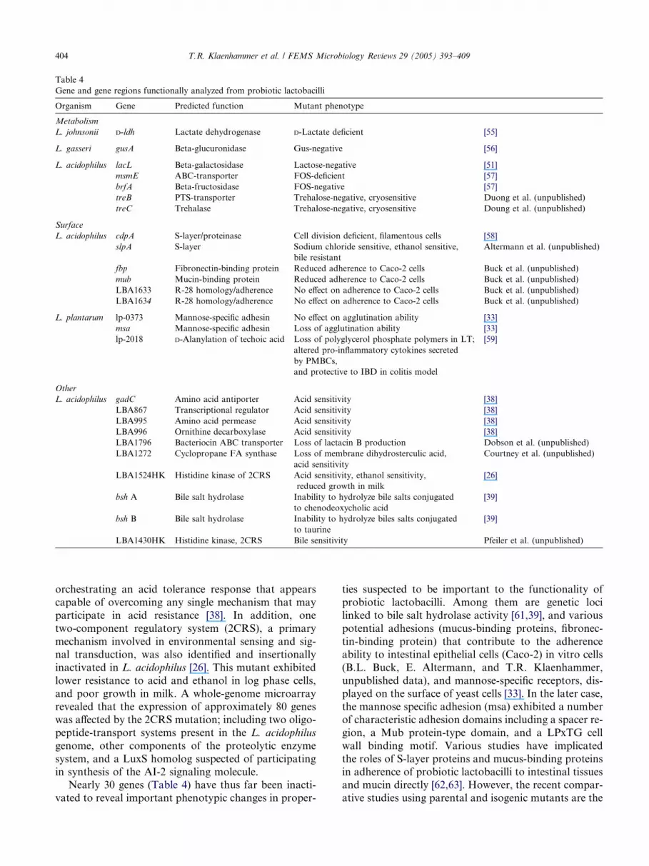
Table 4
Gene and gene regions functionally analyzed from probiotic lactobacilli
Organism Gene Predicted function Mutant phenotype
Metabolism
L. johnsonii D-ldh Lactate dehydrogenase D-Lactate deficient [55]
L. gasseri gusA Beta-glucuronidase Gus-negative [56]
L. acidophilus lacL Beta-galactosidase Lactose-negative [51]
msmE ABC-transporter FOS-deficient [57]
brfA Beta-fructosidase FOS-negative [57]
treB PTS-transporter Trehalose-negative, cryosensitive Duong et al. (unpublished)
treC Trehalase Trehalose-negative, cryosensitive Doung et al. (unpublished)
Surface
L. acidophilus cdpA S-layer/proteinase Cell division deficient, filamentous cells [58]
slpA S-layer Sodium chloride sensitive, ethanol sensitive,
bile resistant
Altermann et al. (unpublished)
fbp Fibronectin-binding protein Reduced adherence to Caco-2 cells Buck et al. (unpublished)
mub Mucin-binding protein Reduced adherence to Caco-2 cells Buck et al. (unpublished)
LBA1633 R-28 homology/adherence No effect on adherence to Caco-2 cells Buck et al. (unpublished)
LBA1634 R-28 homology/adherence No effect on adherence to Caco-2 cells Buck et al. (unpublished)
L. plantarum lp-0373 Mannose-specific adhesin No effect on agglutination ability [33]
msa Mannose-specific adhesin Loss of agglutination ability [33]
lp-2018 D-Alanylation of techoic acid Loss of polyglycerol phosphate polymers in LT;
altered pro-inflammatory cytokines secreted
by PMBCs,
and protective to IBD in colitis model
[59]
Other
L. acidophilus gadC Amino acid antiporter Acid sensitivity [38]
LBA867 Transcriptional regulator Acid sensitivity [38]
LBA995 Amino acid permease Acid sensitivity [38]
LBA996 Ornithine decarboxylase Acid sensitivity [38]
LBA1796 Bacteriocin ABC transporter Loss of lactacin B production Dobson et al. (unpublished)
LBA1272 Cyclopropane FA synthase Loss of membrane dihydrosterculic acid,
acid sensitivity
Courtney et al. (unpublished)
LBA1524HK Histidine kinase of 2CRS Acid sensitivity, ethanol sensitivity,
reduced growth in milk
[26]
bsh A Bile salt hydrolase Inability to hydrolyze bile salts conjugated
to chenodeoxycholic acid
[39]
bsh B Bile salt hydrolase Inability to hydrolyze biles salts conjugated
to taurine
[39]
LBA1430HK Histidine kinase, 2CRS Bile sensitivity Pfeiler et al. (unpublished)
404 T.R. Klaenhammer et al. / FEMS Microbiology Reviews 29 (2005) 393–409
orchestrating an acid tolerance response that appears
capable of overcoming any single mechanism that may
participate in acid resistance [38]. In addition, one
two-component regulatory system (2CRS), a primary
mechanism involved in environmental sensing and sig-
nal transduction, was also identified and insertionally
inactivated in L. acidophilus [26]. This mutant exhibited
lower resistance to acid and ethanol in log phase cells,and poor growth in milk. A whole-genome microarray
revealed that the expression of approximately 80 genes
was affected by the 2CRS mutation; including two oligo-
peptide-transport systems present in the L. acidophilus
genome, other components of the proteolytic enzyme
system, and a LuxS homolog suspected of participating
in synthesis of the AI-2 signaling molecule.
Nearly 30 genes (Table 4) have thus far been inacti-vated to reveal important phenotypic changes in proper-
ties suspected to be important to the functionality of
probiotic lactobacilli. Among them are genetic loci
linked to bile salt hydrolase activity [61,39], and various
potential adhesions (mucus-binding proteins, fibronec-
tin-binding protein) that contribute to the adherence
ability to intestinal epithelial cells (Caco-2) in vitro cells
(B.L. Buck, E. Altermann, and T.R. Klaenhammer,
unpublished data), and mannose-specific receptors, dis-played on the surface of yeast cells [33]. In the later case,
the mannose specific adhesion (msa) exhibited a number
of characteristic adhesion domains including a spacer re-
gion, a Mub protein-type domain, and a LPxTG cell
wall binding motif. Various studies have implicated
the roles of S-layer proteins and mucus-binding proteins
in adherence of probiotic lactobacilli to intestinal tissues
and mucin directly [62,63]. However, the recent compar-ative studies using parental and isogenic mutants are the

T.R. Klaenhammer et al. / FEMS Microbiology Reviews 29 (2005) 393–409 405
first to seek clear evidence of the specific contributions
of these cell surface proteins to adherence mechanisms,
within the context of the whole bacterium.
In a recent study by B.L. Buck, E.A. Altermann and
T.R. Klaenhammer (unpublished), mutants of L. aci-
dophilus were constructed by targeted inactivation ofgenes suspected to encode surface proteins potentially
mediating adherence to mucus or the intestinal epithe-
lium. Analysis of the adhesive properties of these mu-
tants to intestinal Caco-2 epithelial cells, in vitro,
revealed that two streptococcal R28 homolog mutants,
LBA1633 and LBA1634, did not show reproducible de-
creases in adhesion. SlpA�, a surface-layer mutant,
showed the highest decrease in adhesion while fibronec-tin-binding protein (FbpA) and mucus-binding protein
(Mub) mutants showed significant decreases in adhesion
compared to the control (Fig. 8). Interestingly, it was
also observed that treatment of the bacterial cells under
a specific set of environmental conditions could result in
an explosive increase of adherence ability, for both the
parent and the five individual gene knockout mutants
(Fig. 8, inset panels; B.L. Buck and T.R. Klaenhammer,unpublished observations). Therefore, additional factors
are involved, inducible in both the parent and adher-
ence-deficient mutants that can significantly elevate the
capacity of L. acidophilus to adhere in this model sys-
Fig. 8. Relative adhesion of L. acidophilus NCFM mutant strains to Caco-
monolayers in 17 microscopic fields. Each experiment was done in duplicate
tested: W+, L. acidophilus containing a plasmid integration in the b-galactosida fibronectin-binding protein; SlpA-, integration in gene encoding a surface la
protein; and 1633� and 1634�, integration into individual tandem genes h
pyogenes [64]. Inset photos show Gram stained L. acidophilus NCFM adherin
adherence ability (B.L. Buck and T.R. Klaenhammer, unpublished).
tem. These genetic regions and their potential adhesion
and signaling factors are currently being identified by
microarray analysis and investigated by functional
genomic approaches.
Of significant interest is the recent report of Grang-
ette et al. [59], showing that the presence or absence ofteichoic acids on the cell surface of L. plantarum can af-
fect the cytokine expression pattern by peripheral blood
mononuclear cells (PBMCs) and monocytes. The dlt op-
eron, responsible for D-alanylation of teichoic acids, was
disrupted to lead to a substantial reduction in the con-
centration of polyglycerol phosphate polymers (with
D-Ala) in the techoic acids of the bacterial cell wall.
Notably, this change in the chemical composition corre-lated with a reduced secretion of pro-inflamatory cyto-
kines produced by PBMCs, and increased secretion of
the anti-inflammatory cytokine IL-10, when exposed
to the Dlt-mutant. Use of the Dlt-mutant in a murine
colitis model was also found to be protective against
TNBS-induced colitis. This result provides further evi-
dence that LAB communicate with PBMCs and, for
the first time, provides evidence that LAB may induceproinflamatory or anti-inflammatory reponses based
on their cell wall composition in teichoic acids [59],
and perhaps in the display of cell surface bound proteins
or polysaccharides, as well. In this regard, a number of
2 monolayers. Adhesion is expressed as total cells adhering to Caco-2
and replicated at least three times. The following mutant strains were
ase gene used as parental control; FbpA-, integration in gene encoding
yer protein; Mub-, an integration in the gene encoding a mucus-binding
omologous to a gene encoding an epithelial cell-binding protein in S.
g to Caco-2 cells before (A) and after (B) cell treatment that promotes

406 T.R. Klaenhammer et al. / FEMS Microbiology Reviews 29 (2005) 393–409
reports have already shown that different strains and
species of lactobacilli, and other commensal bacteria,
can modulate cytokine expression by both human and
murine antigen presenting (dendritic) cells [65–68].
Overall the results suggest that variations in bacterial
strains and species can direct immunological responsestoward pro- or anti-inflammatory responses. Based on
the results of Grangette et al. [59], the direction of these
responses could reflect the chemical composition and
architecture of the Gram-positive cell wall. Considering
the field�s current position, having a number of complete
probiotic genomes (L. acidophilus, L. gasseri, L. johnso-
nii, and L. plantarum) and the capability to carry out
functional genomic analyses on these organisms, thecourse to understanding the interactions of signaling
molecules with immune cells will be both challenging
and exciting in the years ahead.
5. Concluding remarks
Today�s exciting discoveries based on gene contentand predicted function follow closely behind the explo-
sion of DNA sequence information on microbial gen-
omes. Genomic and comparative genomic analyses are
revealing key gene regions in LAB worthy of continued
investigation for their potential roles in both bioprocess-
ing and health.Microarray analysis of LAB cultures, and
various mutant derivatives thereof, promises to reveal
genetic networks that orchestrate complex microbial re-sponses to a variety of conditions that are critical to
growth, metabolic activity, survival, communication,
signaling, and probiotic functionality. Across the LAB,
genome sequences have already provided information
on genetic content that establishes platforms for meta-
bolic [69] and nutrient engineering [70], understanding
mechanisms of probiotic action [38,59,71], and providing
platforms to engineer LAB for delivery of biotherapeu-tics [72]. The future, armed with genome information
and genetic tools, is an exciting one. It is the first time
in the history of this field that the promising potential
of these beneficial organisms can be mechanistically
investigated, understood, and inevitably expanded for
the benefit of humankind.
Acknowledgments
The substantial contributions of many individuals
participating in this work in our laboratory and as col-
laborators are gratefully acknowledged. Support for
the LAB genetics and Functional Genomics programs
has been provided through the North Carolina Agricul-
tural Research Service, the North Carolina Dairy Foun-dation, Danisco, Inc., Dairy Management, Inc., the
Southeast Dairy Foods Research Center, the California
Dairy Research Foundation, the NIH Biotechnology
Program, GAANN Fellowships in Biotechnology,
IGERT Genomics Fellowships, and the USDA Na-
tional Research Initiative Competitive Grants Program.
Our special gratitude is expressed to Evelyn Durmaz,
Rosemary Sanozky-Dawes, and Edwina Kleeman formany years of laboratory management and excellent re-
search, to the US Department of Energy Joint Genome
Institute for draft sequencing, the members of the U.S.
Lactic Acid Bacteria Genomics Consortium (LABGC)
for genome information and analysis, and to Fidelity
Systems, Inc., for sequencing efforts to close and com-
plete selected genomes used in these analyses. Thanks
are also extended to A. Mercenier, W. de Vos, T. Shi-noda, M. Saier, D.A. Mills, K. Makarova, and E. Koo-
nin for providing data and/or preprints prior to
publication.
References
[1] Poolman, B. (2002) Transporters and their roles in LAB cell
physiology. Antonie van Leeuwenhoek 82, 147–164.
[2] Hugenholtz, J., Sybesma, W., Groot, M.N., Wisselink, W.,
Ladero, V., Birgess, K., van Sinderen, D., Piard, J.C., Eggink,
G., Smid, E.J., Savoy, G., Sesma, F., Jansen, T., Hols, P. and
Kleerebezem, M. (2002) Metabolic engineering of lactic acid
bacteria for the production of nutraceuticals. Antonie van
Leeuwenhoek 82, 217–235.
[3] Kleerebezem, M. and Hugenholtz, J. (2003) Metabolic pathway
engineering in lactic acid bacteria. Curr. Opin. Biotechnol. 14,
232–237.
[4] Tannock, G.W. (1999) Analysis of the intestinal microflora, a
renaissance. Antonie van Leeuwenhoek 76, 265–278.
[5] Ouwehand, A.C., Salminen, S. and Isolauri, E. (2002) Probiotics,
an overview of beneficial effects. Antonie van Leeuwenhoek 82,
279–289.
[6] Vaughan, E.E., de Vries, M.C., Zoetendal, E.G., Ben-Amor, K.,
Akkermans, A.D. and de Vos, W.M. (2002) The intestinal LABs.
Antonie van Leeuwenhoek 82, 341–352.
[7] Gibson, G.R. and Roberfroid, M.B. (1995) The bifidogenic nature
of chicory inulin and its hydrolysis products. J. Nutr. 125, 1401–
1412.
[8] Reid, G. (1999) The scientific basis for probiotic strains of
Lactobacillus. Appl. Environ. Microbiol. 65, 3763–3766.
[9] Reid, G., Sanders, M.E., Gaskins, H.R., Gibson, G.R.,
Mercenier, A., Rastall, R., Roberfroid, M., Rowland, I.,
Cherbut, C. and Klaenhammer, T.R. (2003) New scientific
paradigms for probiotics and prebiotics. J. Clin. Gastroenterol.
37, 105–118.
[10] Pena, J.A., Rogers, A.B., Ge, Z., Ng, V., Li, S.Y., Fox, J.G. and
Versalovic, J. (2005) Probiotic Lactobacillus spp. diminish Heli-
cobacter hepaticus-induced inflammatory bowel disease in inter-
leukin-10-deficient mice. Infect Immun. 73, 912–920.
[11] Vitini, E., Alvarez, S., Medina, M., Medici, M., de Budeguer,
M.V. and Perdigon, G. (2000) Gut mucosal immunostimulation
by lactic acid bacteria. Biocell 24, 223–232.
[12] Bolotin, A., Wincker, P., Mauger, S., Jaillon, O., Malarme, K.,
Weissenbach, J., Ehrlich, S.D. and Sorokin, A. (2001) The
complete genome sequence of the lactic acid bacterium Lactococ-
cus lactis ssp. lactis IL1403. Genome Res. 11, 731–753.
[13] Ajdic, D., McShan, W.M., McLaughlin, R.E., Savic, G., Chang,
J., Carson, M.B., Primeaux, C., Tian, R., Kenton, S., Jia, H., Lin,

T.R. Klaenhammer et al. / FEMS Microbiology Reviews 29 (2005) 393–409 407
S., Qian, Y., Li, S., Zhu, H., Najar, F., Lai, H., White, J., Roe,
B.A. and Ferretti, J.J. (2002) Genome sequence of Streptococcus
mutans UA159, a cariogenic dental pathogen. Proc. Natl. Acad.
Sci. USA 99, 14434–14439.
[14] Tettelin, H., Nelson, K.E., Paulsen, I.T., Eisen, J.A., Read, T.D.,
Peterson, S., Heidelberg, J., Deboy, R.T., Haft, D.H., Dodson,
R.J., Durkin, A.S., Gwinn, M., Kolonay, J.F., Nelson, W.C.,
Peretron, J.D., Umayam, L.A., While, O., Salzberg, S.L., Lewis,
M.R., Radune, D., Holtzapple, E., Khouri, H., Wolf, A.M.,
Utterback, T.R., Hansen, C.L., McDonald, L.A., Feldblyum,
T.V., Angiuoli, S., Dickinson, T., Hickey, E.K., Holt, I.E.,
Loftus, B.J., Yang, F., Smith, H.O., Venter, J.C., Dougherty,
B.A., Morrison, D.A., Hollingshead, S.K. and Fraser, C.M.
(2001) Complete genome sequence of a virulent isolate of
Streptococcus pneumoniae. Science 293, 498–506.
[15] Tettelin, H., Masignani, V., Cieslewicz, M.J., Eisen, J.A., Peter-
son, S., Wessels, M.R., Paulsen, I.T., Nelson, K.E., Margarit, I.,
Read, T.D., Madoff, L.C., Wolf, A.M., Beanan, M.J., Brinkac,
L.M., Daugherty, S.C., DeBoy, R.T., Durkin, A.S., Kolonay,
J.F., Madupu, R., Lewis, M.R., Radune, D., Fedorova, N.B.,
Scanlan, D., Khouri, H., Mulligan, S., Carty, H.A., Cline, R.T.,
Van Aken, S.E., Gill, J., Scarselli, M., Mora, M., Iacobini, E.T.,
Brettoni, C., Galli, G., Mariani, M., Vegni, F., Maione, D.,
Rinaudo, D., Rappuoli, R., Telford, J.L., Kasper, D.L., Grandi,
G. and Fraser, C.M. (2002) Complete genome sequence and
comparative genomic analysis of an emerging human pathogen,
serotype V Streptococcus agalactiae. Proc. Natl. Acad. Sci. USA
99, 12391–12396.
[16] Ferretti, J.J., McShan, W.M., Ajdic, D., Savic, D.J., Savic, G.,
Lyon, K., Primeaux, C., Sezate, S., Suvorov, A., Kenton, S., Lai,
H.S., Lin, S.P., Qian, Y., Jia, H.G., Najar, F.Z., Ren, Q., Zhu, H.,
Song, L., White, J., Yuan, X., Clifton, S.W., Roe, B.A. and
McLaughlin, R. (2001) Complete genome sequence of an M1
strain of Streptococcus pyogenes. Proc. Natl. Acad. Sci. USA 98,
4658–4663.
[17] Bolotin, A., Quinquis, B., Renault, P., Sorokin, A., Ehrlich, S.D.,
Kulakauskas, S., Lapidus, A., Goltsman, E., Mazur, M., Pusch,
G.D., Fonstein, M., Overbeek, R., Kyprides, N., Purnelle, B.,
Prozzi, D., Ngui, K., Masuy, D., Hancy, F., Burteau, S., Boutry,
M., Delcour, J., Goffeau, A. and Hols, P. (2004) Complete
sequence and comparative genome analysis of the dairy bacterium
Streptococcus thermophilus. Nat. Biotechnol. 22, 1554–1558.
[18] Schell, M.A., Karmirantzou, M., Snel, B., Vilanova, D., Berger,
B., Pessi, G., Zwahlen, M.C., Desiere, F., Bork, P., Delley, M.,
Pridmore, R.D. and Arigoni, F. (2002) The genome sequence of
Bifidobacterium longum reflects its adaptation to the human
gastrointestinal tract. Proc. Natl. Acad. Sci. USA 99, 14422–
14427.
[19] Kleerebezem, M., Boekhorst, J., van Kranenburg, R., Molenaar,
D., Kuipers, O.P., Leer, R., Tarchini, R., Peters, S.A., Sandbrink,
H.M., Fiers, M.W., Stiekema, W., Lankhorst, R.M., Bron, P.A.,
Hoffer, S.M., Groot, M.N., Kerkhoven, R., de Vries, M., Ursing,
B., de Vos, W.M. and Siezen, R.J. (2003) Complete genome
sequence of Lactobacillus plantarum WCFS1. Proc. Natl. Acad.
Sci. USA 100, 1990–1995.
[20] Pridmore, R.D., Berger, B., Desiere, F., Vilanova, D., Barretto,
C., Pittet, A.C., Zwahlen, M.C., Rouvet, M., Altermann, E.,
Barrangou, R., Mollet, B., Mercenier, A., Klaenhammer, T.R.,
Arigoni, F. and Schell, M.A. (2004) The genome sequence of the
probiotic intestinal bacterium Lactobacillus johnsonii NCC 533.
Proc. Natl. Acad. Sci. USA 101, 2512–2517.
[21] Altermann, E., Russell, W.M., Azcarate-Peril, M.A., Barrangou,
R., Buck, B.L., McAuliffe, O., Souther, N., Dobsen, A., Doung,
T., Callanan, M., Lick, S., Hamrick, A., Cano, R. and Klaen-
hammer, T.R. (2005) Complete genome sequence of the probiotic
lactic acid bacterium Lactobacillus acidophilus NCFM. Proc.
Natl. Acad. Sci. USA 102, 3906–3912.
[22] Klaenhammer, T.R., Altermann, E., Arigoni, F., Bolotin, A.,
Breidt, F., Broadbent, J., Cano, R., Chaillou, S., Deutscher, J.,
Gasson, M., van de Guchte, M., Guzzo, J., Hartke, A., Hawkins,
T., Hols, P., Hutkins, R., Kleerebezem, M., Kok, J., Kuipers, O.,
Lubbers, M., Maguin, E., McKay, L., Mills, D., Nauta, A.,
Overbeek, R., Pel, H., Pridmore, D., Saier, M., van Sinderen, D.,
Sorokin, A., Steele, J., O�Sullivan, D., de Vos, W., Weimer, B.,
Zagorec, M. and Siezen, R. (2002) Discovering lactic acid bacteria
by genomics. Antonie van Leeuwenhoek 82, 29–58.
[23] Siezen, R.J., van Enckevort, F.H.J., Kleerebezem, M. and
Teusink, B. (2004) Genome data mining of lactic acid bacteria,
the impact of bioinformatics. Curr. Opin. Biotechnol. 15, 105–
115.
[24] Paulsen, I.T., Nguyen, L., Sliwinski, M.K., Rabus, R. and Saier,
M.H. (2000) Microbial genome analyses, comparative transport
capabilities in eighteen prokaryotes. J. Mol. Biol. 301, 75–100.
[25] Barrangou, R. (2004) Functional genomic analyses of carbohy-
drate utilization by Lactobacillus acidophilus. Ph.D. Thesis in
Functional Genomics, NCSU..
[26] Azcarate-Peril, M.A., McAuliffe, O., Altermann, O., Lick, S.,
Russell, W.M. and Klaenhammer, T.R. (2005) Microarray
analysis of a two-component regulatory system involved in acid
resistance and proteolytic activity in Lactobacillus acidophilus.
Appl. Environ. Microbiol., In press..
[27] Klaenhammer, T.R. and Russell, W.M. (2000) Species of the
Lactobacillus acidophilus complex. In: (Robinson, R.K, Batt, C.
and Patel, P.D., Eds.). Encyclopedia of Food Microbiology, Vol.
2, pp. 1151–1157. Academic Press, San Diego.
[28] Boekhorst, J., Siezen, R.J., Zwahlen, M.C., Vilanova, D.,
Pridmore, R.D., Mercenier, A., Kleerebezem, M., de Vos,
W.M., Brussow, H. and Desiere, F. (2004) The complete genomes
of Lactobacillus plantarum and Lactobacillus johnsonii reveal
extensive differences in chromosome organization and gene
content. Microbiology 150, 3601–3611.
[29] McKay, L.L. (1983) Functional properties of plasmids in lactic
streptococci. Antonie van Leeuwenhoek 49, 259–274.
[30] Tuomola, E.M., Ouwehand, A.C. and Salminen, S.J. (2000)
Chemical, physical and enzymatic pre-treatments of probiotic
lactobacilli alter their adhesion to human intestinal mucus
glycoproteins Int. J. Food Microbiol. 60, 75–81.
[31] DeVos, W.M., Bron, P.A. and Kleerebezem, M. (2004) Post-
genomics of lactic acid bacteria and other food-grand bacteria to
discover gut functionality. Curr. Opin. Biotechnol. 15, 86–93.
[32] Kullen, M.J., Sanozky-Dawes, R.B., Crowell, D.C. and Klaen-
hammer, T.R. (2000) Use of DNA sequence of variable regions of
the 16SrRNA gene for rapid and accurate identification of
bacteria in the Lactobacillus acidophilus complex. J. Appl.
Microbiol. 89, 511–518.
[33] Pretzer, G., Snel, J., Molenaar, D., Wiersma, A., Bron, P.A.,
Lambert, J., de Vos, W.M., Van der Meer, R., Smits, M.A. and
Kleerebezem, M. (2005) Biodiversity-based identification and
functional characterization of the mannose-specific adhesion of
Lactobacillus plantarum. J. Bacteriol., In press..
[34] Canchaya, C., Proux, C., Fournous, G., Bruttin, A. and Brussow,
H. (2003) Prophage genomics. Microbiol. Mol. Biol. Rev. 67,
238–276.
[35] McGrath, S., Fitzgerald, G.F. and van Sinderen, D. (2004) The
impact of bacteriophage genomics. Curr. Opin. Biotechnol. 15,
94–99.
[36] Nes, I.F. and Johnsborg, O. (2004) Exploration of antimicrobial
potential in LAB by genomics. Curr. Opin. Biotechnol. 15, 100–
104.
[37] MacConaill, L.E., Butler, D., O�Connell-Motherway, M., Fitz-
gerald, G.F. and van Sinderen, D. (2004) Identification of two-
component regulatory systems in Bifidobacterium infantis by
functional complementation and degenerate PCR approaches.
Appl. Envir. Microbiol. 69, 4219–4226.

408 T.R. Klaenhammer et al. / FEMS Microbiology Reviews 29 (2005) 393–409
[38] Azcarate-Peril, M.A., Altermann, E., Hoover-Fitzula, R.L.,
Cano, R.J. and Klaenhammer, T.R. (2004) Identification and
inactivation of genetic loci involved with Lactobacillus acidophilus
acid tolerance. Appl. Environ. Microbiol. 70, 5315–5322.
[39] McAuliffe, O., Cano, R. and Klaenhammer, T.R. (2005) Genetic
analysis of two bile salt hydrolase activities in Lactobacillus
acidophilus NCFM. Appl. Environ. Microbiol., In Press..
[40] Walter, J., Heng, N.C., Hammes, W.P., Loach, D.M., Tannock,
G.W. and Hertel, C. (2003) Identification of Lactobacillus reuteri
genes specifically induced in the mouse gastrointestinal tract.
Appl. Environ. Microbiol. 69, 2044–20451.
[41] Bron, P.A., Hoffer, S.M., Van Swam, I.I., DeVos, W.M. and
Kleerebezem, M. (2004) Selection and characterization of condi-
tionally active promoters in Lactobacillus plantarum, using
alanine racemase as a promoter probe. Appl. Environ. Microbiol.
70, 310–317.
[42] Bron, P.A., Marco, M., Hoffer, S.M., Van Mullekom, E., de Vos,
W.M. and Kleerebezem, M. (2004) Genetic characterization of
the bile salt response in Lactobacillus plantarum and analysis of
responsive promoters in vitro and in situ in the gastrointestinal
tract. J. Bacteriol. 186, 7829–7835.
[43] Kok, J., van der Vossen, J.M. and Venema, G. (1984) Construc-
tion of plasmid cloning vectors for lactic streptococci which also
replicate in Bacillus subtilis and Escherichia coli. Appl. Environ.
Microbiol. 48, 726–731.
[44] Chassy, B.M. and Flickinger, J.L. (1987) Transformation of
Lactobacillus casei by electroporation. FEMS Microbiol. Lett. 44,
173–177.
[45] Luchansky, J.B., Muriana, P.M. and Klaenhammer, T.R. (1988)
Application of electroporation for transfer of plasmid DNA to
Lactobacillus, Lactococcus, Leuconostoc, Listeria, Pediococcus,
Bacillus, Staphylococcus, Enterococcus and Propionibacterium.
Mol. Microbiol. 2, 637–646.
[46] Biswas, I., Gruss, A., Ehrlich, S.D. and Maguin, E. (1993) High-
efficiency gene inactivation and replacement system for gram-
positive bacteria. J. Bacteriol. 175, 3628–3635.
[47] Dao, M.L. and Ferretti, J.J. (1985) Streptococcus–Escherichia coli
shuttle vector pSA3 and its use in the cloning of streptococcal
genes. Appl. Environ. Microbiol. 49, 115–119.
[48] Leenhouts, K.J., Kok, J. and Venema, G. (1991) Lactococcal
plasmid pWV01 as an integration vector for lactococci. Appl.
Environ. Microbiol. 57, 2562–2567.
[49] Leenhouts, K., Buist, G., Bolhuis, A., ten Berge, A., Kiel, J.,
Mierau, I., Dabrowska, M., Venema, G. and Kok, J. (1996) A
general system for generating unlabeled gene replacements in
bacterial chromosomes. Mol. Gen. Genet. 253, 217–224.
[50] Bhowmik, T., Fernandez, L. and Steele, J.L. (1993) Gene
replacement in Lactobacillus helveticus. J. Bacteriol. 175, 6341–
6344.
[51] Russell, W.M. and Klaenhammer, T.R. (2001) Efficient system for
directed integration into the Lactobacillus acidophilus and Lacto-
bacillus gasseri chromosomes via homologous recombination.
Appl. Environ. Microbiol. 67, 4361–4364.
[52] Van Kranenburg, R., Marugg, J.D., van Swam, I.I., Norwin, J.W.
and de Vos, W.M. (1997) Molecular characterization of the
plasmid-encoded eps gene cluster essential for exopolysaccharide
biosynthesis in Lactococcus lactis. Mol. Microbiol. 24, 387–397.
[53] Yanisch-Perron, C., Vieira, J. and Messing, J. (1985) Improved
M13 phage cloning vectors and host strains: nucleotide sequences
of the M13mp18 and pUC19 vectors. Gene 33, 103–119.
[54] Nierop Groot, M.N., Klaassens, E., de Vos, W.M., Delcour, J.,
Hols, P. and Kleerebezem, M. (2005) Genome-based in silico
detection of putative manganese transport systems in Lactobacil-
lus plantarum and their genetic analysis. Microbiology 151, 1229–
1238.
[55] Lapierre, L., Germond, J.E., Ott, A., Delley, M. and Mollet, B.
(1999) D-Lactate dehydrogenase gene (ldhD) inactivation and
resulting metabolic effects in the Lactobacillus johnsonii strains
La1 and N312. Appl. Environ. Microbiol. 65, 4002–4007.
[56] Russell, W.M. and Klaenhammer, T.R. (2001) Identification and
cloning of gusA, encoding a new beta-glucuronidase from
Lactobacillus gasseri ADH. Appl. Environ. Microbiol. 67, 1253–
1261.
[57] Barrangou, R., Altermann, E., Hutkins, R., Cano, R. and
Klaenhammer, T.R. (2003) Functional and comparative genomic
analyses of an operon involved in fructooligosaccharide utiliza-
tion by Lactobacillus acidophilus. Proc. Natl. Acad. Sci. USA 100,
8957–8962.
[58] Altermann, E., Buck, B.L, Cano, R. and Klaenhammer, T.R.
(2004) Identification and phenotypic characterization of the cell-
division protein CdpA. Gene 342, 189–197.
[59] Grangette, G., Nutten, S., Palumbo, E., Morath, S., Hermann,
C., Dewulf, J., Pot, B., Hartung, T., Hols, P. and Mercenier, A.
(2005) Enhanced anti-infammatory capacity of a Lactobacillus
plantarum mutant synthesizing modified teichoic acids. Proc. Nat.
Acad. Sci. USA, In press..
[60] Mack, D.R. (2004) D(�)-Lactic acid-producing probiotics, D(�)-
lactic acidosis and infants. Can. J. Gastroenterol. 18, 671–675.
[61] Elkins, C.A., Moser, S.A. and Savage, D.C. (2001) Genes
encoding bile salt hydrolases and conjugated bile salt transporters
in Lactobacillus johnsonii 100–100 and other Lactobacillus species.
Microbiology 147, 3403–3412.
[62] Frece, J., Kos, B., Svetec, I.K., Zgaga, Z., Mrsa, V. and
Suskovic, J. (2002) Importance of S-layer proteins in probiotic
activity of Lactobacillus acidophilus M92. J. Appl. Microbiol. 98,
285–292.
[63] Roos, S. and Jonsson, H. (2002) A high-molecular-mass cell-
surface protein from Lactobacillus reuteri 1063 adheres to mucus
components. Microbiology 148, 433–442.
[64] Stalhammar-Carlemalm, M., Areschoug, T., Larsson, C. and
Lindahl, G. (1999) The R28 protein of Streptococcus pyogenes is
related to several group B streptococcal surface proteins, confers
protective immunity and promotes binding to human epithelial
cells. Mol. Microbiol. 33, 208–219.
[65] Braat, H., de Jong, E.C., van den Brande, J.M., Kapsenberg,
M.L., Peppelenbosch, M.P., van Tol, E.A. and van Deventer, S.J.
(2004) Dichotomy between Lactobacillus rhamnosus and Klebsi-
ella pneumoniae on dendritic cell phenotype and function. J. Mol.
Med. 82, 197–205.
[66] Karlsson, H., Larsson, P., Wold, A.E. and Rudin, A. (2004)
Pattern of cytokine responses to gram-positive and gram-
negative commensal bacteria is profoundly changed when
monocytes differentiate into dendritic cells. Infect. Immun. 72,
2671–2678.
[67] Veckman, V., Miettinen, M., Pirhonen, J., Siren, J., Matikainen,
S. and Julkunen, I. (2004) Pattern of cytokine responses to gram-
positive and gram-negative commensal bacteria is profoundly
changed when monocytes differentiate into dendritic cells. J.
Leukocyte Biol. 75, 764–771.
[68] Mohamadzadeh, M., Olson, S., Kalina, W.V., Ruthel, G.,
Demmin, G.L., Warfield, K.L., Barvari, S. and Klaenhammer,
T.R. (2005) Lactobacilli activate human dendritic cells that skew
T cells toward T helper 1 polarization. Proc. Natl. Acad. Sci. USA
102, 2280–2285.
[69] Hols, P., Kleerebezem, M., Schanck, A.N., Ferain, T., Huge-
nholtz, J., Delcour, J. and de Vos, W.M. (1999) Conversion of
Lactococcus lactis from homolactic to homoalanine fermentation
through metabolic engineering. Nat. Biotechnol. 17, 588–592.
[70] Wegkamp, A., Starrenburg, M., de Vos, W.M., Hugenholtz, J.
and Sybesma, W. (2004) Transformation of folate-consuming
Lactobacillus gasseri into a folate producer. Appl. Environ.
Microbiol. 70, 3146–3148.
[71] Christensen, H.R., Frokiaer, H. and Pestka, J.J. (2002) Lactoba-
cilli differentially modulate expression of cytokines and matura-

T.R. Klaenhammer et al. / FEMS Microbiology Reviews 29 (2005) 393–409 409
tion surface markers in murine dendritic cells. J. Immunol. 168,
171–178.
[72] Steidler, L., Hans, W., Schotte, L., Neirynck, S., Obermeier, F.,
Falk, W., Fiers, W. and Remaut, E. (2000) Treatment of murine
colitis by Lactococcus lactis secreting interleukin-10. Science 289,
1352–1355.
[73] Jensen, L.J., Friis, C. and Ussery, D.W. (1999) Three views of
microbial genomes. Res. Microbiol. 150, 773–777.