further data on sesamoid identity from two anuran species
TRANSCRIPT

Further Data On Sesamoid IdentityFrom Two Anuran Species
MIRIAM CORINA VERA,1 MAR�IA LAURA PONSSA,1* AND VIRGINIA ABDALA2
1Unidad Ejecutora Lillo, Instituto de Herpetolog�ıa, CONICET-Fundaci�on Miguel Lillo,Miguel Lillo 251, San Miguel de Tucum�an 4000, Argentina
2Instituto de Biodiversidad Neotropical, UNT—CONICET, C�atedra de Biolog�ıa General,Facultad de Ciencias Naturales e IML, UNT. Miguel Lillo 205, San Miguel de Tucum�an
4000, Argentina
ABSTRACTConsidering that the identification of equivalent entities is the basis
for any comparative analysis, we compare the histology, histochemistry,shape and dimensions of epiphyses, carpal and sesamoids in two anuranfrogs. Our goal was to explore the morphological correspondence amongthese three skeletal elements in order to clarify the sesamoid identity. Westudied the skeletogenesis, contour geometric morphometry and dimen-sions of forelimb elements of juveniles of two anurans species Leptodacty-lus bufonius and Rhinella arenarum. Skeletogenesis in anurans present acommon trait between carpals and sesamoids: both elements exhibit endo-chondral ossification. A difference between these elements is the presenceof fibrocartilage in the development of sesamoids. The geometric mor-phometry does not allow us to establish a shape pattern that can be com-pared either between sesamoids and epiphyses or carpals. With regard todimensions, our data indicate that bones categorization based on theseaspects is ambiguous and therefore is useless to classify of skeletal bones.The data about tissue differentiation of sesamoids provide evidence thatsupport the idea that these elements should be considered part of the typ-ical endowment of the vertebrate skeleton. Anat Rec, 00:000–000, 2015.
VC 2015 Wiley Periodicals, Inc.
Key words: histochemistry; histology; morphometry; forelimb;frog; juveniles
The forelimb skeleton of tetrapods is composed ofthree elements: long bones, short bones (carpals) andsesamoids. Short bones are those having their threedimensions equally developed, with a thin cortex of com-pact bone tissue and spongy bone tissue inside (Tortoraand Derrickson, 2006; Ross and Pawlina, 2007). Theword sesamoid comes from seed-like, morphology as ses-ame seed (Vickaryous and Olson, 2007). These elementsare rarely mentioned in the descriptions of normal anat-omy of any tetrapod skeleton because of their allegedvariability (but see Jerez et al., 2009; Ponssa et al.,2010). It has been proposed that sesamoids originatethrough persistent mechanical stimuli on tendons thatwrap around an epiphysis (Carter et al., 1998). However,Ponssa et al. (2010) demonstrate that differentiation ofmany sesamoids begins well before the tendinous tissueis recognizable. Their histological data show that themain stimulus eliciting the graciella sesamoid genesis
Abbreviations used: ECM 5 extracellular matrix; PCA 5 principalcomponent analysis
Grant sponsor: ANPCyT; Grant number: BID-PICT 616, PICT2011-1524, PICT 2013-0404; Grant sponsor: CONICET; Grantnumbers: PIP 0284. FONCyT, PICT 2011-1524; PICT 2010-0616. UNT, G519.
*Correspondence to: Mar�ıa Laura Ponssa, Unidad EjecutoraLillo, Instituto De Herpetolog�ıa, CONICET-Fundaci�on MiguelLillo, Miguel Lillo 251, San Miguel De Tucum�an 4000,Argentina. E-mail: [email protected]
Received 10 November 2014; Accepted 26 January 2015.
DOI 10.1002/ar.23158Published online 00 Month 2015 in Wiley Online Library(wileyonlinelibrary.com).
THE ANATOMICAL RECORD 00:00–00 (2015)
VVC 2015 WILEY PERIODICALS, INC.
should be genetic because the cartilage precursor is pres-ent before the dense connective tissue differentiates intoa tendon (Ponssa et al., 2010). They also interpretedthat if a whole group includes a particular sesamoid, itwill be constant, and its origin could be mainly linked togenetic stimulus; lack of regularity could be showing apreeminence of extrinsic factors over those genetic ones(Ponssa et al., 2010). Thus, sesamoid origin is still amatter of debate (Le Minor, 1987; Giori et al., 1993; Car-ter et al., 1998; Sarin et al., 1999; Ponssa et al., 2010).
The development of the sesamoids in tetrapods wasstudied generally in larval/postnatal and adult stages(Bland and Ashhurst, 1997; Doherty, 2010; Olson, 2000;Prochel, 2006; Kim et al., 2009; Ponssa et al., 2010;Shearman and Maglia, 2015). In lizards, many sesamoidsappear in post-hatching specimens (Jerez et al., 2009).Fabrezi et al. (2007) showed that sesamoids of this groupundergo endochondral ossification, a complex process bywhich the growth cartilage within the bone cortex is pro-gressively replaced by bone tissue (Felisbino and Car-valho, 2001). Doherty (2010) provided data ofendochondral ossification of sesamoids in mammals. Shedescribed the presence of hypertrophic chondrocyteswithin the calcifying core of the sesamoid, similar to thatobserved in the epiphyseal growth plates of the phalangesand metapodophalangeal bones. In anurans, data aboutsesamoid skeletogenesis are very scarse (Ponssa et al.,2010), they appear mostly at metamorphic stages, reach-ing a cartilaginous phase at the moment of the metamor-phosis (Ponssa et al., 2010; Vera and Ponssa, 2014).
The juvenile period in anurans typically involves thepassage from aquatic to terrestrial environments(McDiarmid and Altig, 1999; Fabrezi et al., 2014); thus,it is a critical phase in the development of the osteologi-cal elements, because they affect the locomotor capacityof the froglet and its adaptation to the terrestrial habitat(Vera and Ponssa, 2014). The changes in the skeletal tis-sue development occurring through the juvenile phase -when the limbs are fully functional- are relevant tounderstand its relation with the acquisition of locomotorcapacities. In spite of the pervasive relationship that hasbeen proposed between sesamoids and locomotion (Sarinet al., 1999; Vickaryous and Olson, 2007), data aboutskeletogenesis of these structures during the juvenileanuran stages are lacking. In this work we present dataabout the skeletogenesis of the sesamoids in juveniles oftwo anuran species, and relate them with the acquisitionof terrestrial locomotion.
Bone classifications based on their shape have beenwidely used (Tortora and Derrickson, 2006; Ross andPawlina, 2007) highlighting the biological meaningattributed to shape. Thus, it can be a source of evidenceto find out whether sesamoids could be defined as shortbones. Methodologies such as morphometric geometryallow a rigorous analysis of shape variations.
Here, we integrate several lines of evidence as anattempt of to fill the gaps in our knowledge of sesa-moids, probably one of the most neglected and enigmaticstructures of the vertebrate skeleton. We compare forthe first time a) the skeletogenesis sequence of the juve-nile stages between short bones such as ulnare and radi-ale, long bones epiphyses, and sesamoids such as thepalmar sesamoid, and b) the shape and dimension of allthose structures to identify common traits that allow usto consider them anatomically equivalent. To discard
that sesamoids undergo only a mineralization processlike the long bone epiphyses of anurans (Ecker, 1889,Haines, 1942; Dickson, 1982; DellOrbo et al., 1992), weinclude epiphyses in our study.
MATERIALS AND METHODS
Histological and Histochemical Analyses
We analyzed samples of radio-ulna epiphyses, carpalsand sesamoids of post-metamorphic specimens of Lepto-dactylus bufonius and Rhinella arenarum. We chosethese species because they left the aquatic habitat afterthe metamorphosis and face terrestrial locomotion bytwo highly different locomotor modes: jumping (Lepto-dactylus bufonius) and walking (Rhinella arenarum).The specimens examined belong to the herpetologicalcollection of Fundaci�on Miguel Lillo and are identifiedas follows: L. bufonius (FML 27869, FML 27873, FML27874, FML 27870, FML 27871, FML 27872, FML27875, and FML 27876); R. arenarum (FML 27877, FML27878, FML 27879, FML 27880, FML 27881, FML27882). All specimens were measured with digital cali-pers (Mitutoyo CD-30C and CD-15B; 60.01 mm) (Table1). The size of the specimens was used as a proxy of thedevelopment rate of the specimens (Dickson, 1982). How-ever, this criterion should be considered with cautionbecause a direct relationship between age and size hasnot been proven in anurans (Rozenblut and Ogielska,2005). The stages of individuals were determined accord-ing to the relative percentage of the maximum length,which corresponds to the length of the adult specimen ofeach studied species (Table 1). Each stage represents apercentage range (61%) (e.g., stage 2 includes individu-als which have a SVL that correspond to the 19–21% ofthe total length of the adult), thus, some specimens withsimilar but not identical SVL could be the same stage.The specimens were fixed in a 10% formaldehyde solu-tion for 24 hr. The extracted forelimbs were decalcifiedwith 5% formic acid (5 mL of formic acid, 5 mL of form-aldehyde and 100 mL of distilled water) for 1–6 weeks,
TABLE 1. Juvenile’s stages according to the relativepercentage of the total length of the adult, SVL of
Leptodactylus bufonius: 51.6–53.6 mm, SVL of Rhine-lla arenarum: 80–100 mm (Cei, 1980)
Rhinella arenarum
Stage SVL (mm)% Of the total length
of the adult
1 17.3 18.21%2 20.43 21.5%5 25.75 27.1%13 41 43.15%16 47.36 49.85%28 70.48 74.18%30 40.9 77.97%
Leptodactylus bufonius
14 23.68 45.14%22 32.32 - 32.92 61.62%– 62.76%25 35.45 67.58%29 39.52 75.34%30 40.9 77.97%34 45.29 86.34%35 46.48 88.61%
2 MIRIAM VERA ET AL.

depending on the sample size. The material was thendehydrated in a graded ethanol series and in n-butylethanol, and embedded in Histoplast embeddingmedium. Serial sagittal sections 7-mm thick were cutwith a rotary microtome (Microm HM 325) and stainedwith Mallory trichrome, Hematoxylin-Eosin and alcianblue-periodic acid-Schiff. With the Mallory trichrometechnique, the collagen fibers present in the extracellu-lar matrix (ECM) and the fibrocartilage are stained blueand the cellular nucleus and the glycosaminoglycans(keratan sulphate) of the osseous matrix are stained red.The compounds of the cartilage ECM, such as the glycos-aminoglycans and proteoglycans, react with basic dyesstaining purple with Hematoxylin. The ECM reacts posi-tively with Alcian-Blue because the acid mucins, such asglycosaminoglycans and proteoglycans. A high concen-tration of neutral mucins, such as collagen fibers, pres-ent in the mineralized ECM also reacts positively withthe periodic acid-schiff dye, getting a pink coloration(PAS-positive). While the histological techniques showthe acidic and basic properties of the tissues, histochem-istry involves a chemical and physical reaction that indi-cates the molecular content of the tissue (Humason,1962). Histological samples were observed under opticalmicroscopy (Leica ICC 50 HD) and photographed withNikon Coolpix P6000digital camera. Terminology of dig-its and carpal osteology follows Fabrezi (1992), and sesa-moids terminology follows Ponssa et al. (2010).
Morphometric Analysis
To compare shape among humerus epiphyses, carpalsand sesamoids, a contour geometric morphometric anal-ysis was performed. We select the humerus for thisanalysis because it has only one epiphysis, whereas theradio-ulna presents two fused epiphyses, making thecomparison of their shapes not possible. This method isbased on the concept that the two figure contours arehomologous, and it was initially thought for objectswithout discrete landmarks (Toro Ibacache et al., 2010).To obtain the contour the dots must be taken aroundthat contour, and must be positioned equidistantly (ToroIbacache et al., 2010). The digitized dots are adjusted toan ad-hoc function, such as harmonics of Fourier analy-sis, which was recalculated inversely using an eigen-vector matrix, letting the score on a particular PC beequal to the mean 6 2 s.d. (standard deviation); finally,the function coefficients are used as components of theshape (Rohlf, 1990). It is known that information aboutshape is conveyed via the curves of an object boundarylines (Freeman, 1974). We extirpated the humerus, twocarpal bones (ulnare and radiale) and two sesamoids(pararadial and palmar sesamoids) of 11 stained andcleared adults Leptodactylus bufonius specimens fromthe Herpetological Collection of Fundaci�on Miguel Lillo(FML-589, FML-672, FML-3568, FML-3868, FML-4366,FML-9779, FML-9780, FML-9782, FML-9783, FML-4908 (two specimens). Radiale and ulnare were selectedbecause, along with element Y, they seem to be highlyconservative elements of anuran carpus (Fabrezi andBarg, 2001). Long bones epiphyses, carpal bones andsesamoids were photographed with a Nikon CoolpixP6000 digital camera. The images were previously proc-essed with the software Photoshop to delimit the outlineof each bone (i.e., carpals, sesamoids) and the epiphyses
of the humerus. We did not consider the size of thebones in this analysis. Each photograph was saved as abmp image and then processed with the softwareSHAPE ver. 1.3 (Iwata and Ukai, 2002). This softwarepackage evaluates contour shapes based on Elliptic Fou-rier descriptor (EFDs). EFDs have been effectivelyapplied to the analysis of various biological shapes inanimals (Rohlf and Archie, 1984; Ferson et al., 1985;Bierbaum and Ferson, 1986; Diaz et al., 1989; Liuet al., 1996; Laurie et al., 1997) and plants (Yoshiokaet al., 2004). The SHAPE software contains four pro-grams: ChainCoder, Chc2Nef, PrinComp and PrinPrint(Iwata and Ukai, 2002). ChainCoder converts a fullcolor image to a binary (black and white) image,reduces noise, traces the contours of objects anddescribes the contour information as chain-code;Chc2Nef calculates the normalized EFDs from thechain-code information exported by the ChainCoder pro-gram. We used the normalization method based on thefirst harmonics ellipse (20 harmonics), which corre-sponds to the first Fourier approximation to the contourinformation; the size and orientation of the contour isstandardized according to the size and alignment of themajor axis of the ellipse (Iwata and Ukai, 2002). ThePrinComp program performs a principal componentanalysis (PCA) to summarize the information of theFourier coefficients. Actually, PCA is based on a covari-ance–variance matrix of the coefficients and not on thecorrelation matrix. PCA may be used to reduce thedimensionality of the data by analyzing a limited num-ber of PC (principal component) scores of the specimensinstead of the original data (Sheets et al., 2006). Finally,we used the PrinPrint program to visualize shape varia-tion. First, the coefficients of the elliptic Fourierdescriptors are calculated, letting the score for a partic-ular PC be equal to the mean plus or minus two (62)times the standard deviation and the scores of theremaining components be zero. Then the contour shapeon each condition can be reconstructed from the coeffi-cients by inverse Fourier transformation. The use ofelliptic Fourier descriptors and principal componentanalysis (EF–PCA) is advantageous because, on the onehand, the method can accurately detect small shapevariations and, on other hand, EF–PCA can evaluatethe shapes of objects independently of size. This inde-pendence is a great advantage because human visualjudgment of shape is often misled by size factors.
To detect differences in shape among long bone epi-physes, carpal bones and sesamoids, we performed aMANOVA (homogeneity of slopes test), with the PCs asdependent variables and bones as grouping factor, usingthe software Statistica (2004).
The three elements studied (epiphysis, carpal and ses-amoid) were compared with one-way ANOVA followed byTukey’s test using Infostat software (Di Rienzo et al.,2013). The measurements considered were maximumlength and maximum width of five elements of themanus; humerus epiphyses; ulnare and radiale; palmarand pararadial sesamoids. These measurements weretaken considering the elements according its physiologi-cal position. P values less than 0.05 were considered assignificant. Although the definition of short bones con-siders three dimensions, length, width and height (Rossand Pawlina, 2007), the bidimensional analysis allowedus to gather important data for this study.
MORPHOLOGY AND DEVELOPMENT OF ANURAN SKELETAL ELEMENTS 3

RESULTS
Histological and Histochemical Analyses
The histological analysis showed that the evaluatedbones (radio-ulna, carpals, and sesamoids) underwentchondral (perichondral and endochondral) ossificationprocess. This technique helped us both to determine thetiming of ossification during postmetamorphic stagesand to compare the elements (long bones epiphyses, car-pals and sesamoids) among them and between the two
analyzed species. The stages with the most relevantchanges are described below.
Rhinella arenarum
Stages 1, 2, and 5 (Fig. 1): In the epiphyses of longbones and carpals, a cartilaginous ECM with isogenousgroups is visible (Fig. 1A,B,D). The ECM stains bluewith Hematoxylin-Eosin, suggesting a high concentra-tion of glycosaminoglycans and proteoglycans (Fig. 1A–
Fig. 1. Stage 5 of Rhinella arenarum. A: section of Metacarpal Vstained with Hematoxylin-Eosin. B: section of Distal carpal 5 1 413stained with Hematoxylin-Eosin. C: section of radio-ulna stained withalcian blue-periodic acid-schiff. D: section of Distal carpal 5 1 413stained with alcian blue-periodic acid-schiff. E: detail of section of lat-
eral articular cartilage of the epiphyses of radio-ulna stained withHematoxylin-Eosin. (DC) Distal carpal, (MC) metacarpal, (GS) glidesesamoid, (Ph) phalange, (RU) radio-ulna, (ig) isogen groups, (ol)osteochondral ligament, (mm) mineralized matrix. Scale bar 200 mm.
4 MIRIAM VERA ET AL.

C). The radio-ulna diaphyses show the growth cartilagewith its characteristic zones (Fig. 1C). In Stage 5, thediaphyses of radio-ulna show mineralization in the ECMof the hypertrophic zone of the growth cartilage (Fig.1C). A detail of the lateral articular cartilage of theradio-ulna epiphyses, and the osteochondral ligamentwith its collagens fibers, which run parallel to the boneaxis, is shown in Fig. 1E. A glide sesamoid between thephalange and the metacarpal of digit IV (Fig. 2A), andthe pararadial sesamoid between the ulnare and ulna(Fig. 2B) show hypertrophic chondrocytes; they areimmersed in a tendinous tissue.
Stage 13 (Fig. 3): In the epiphysis of the phalange ofdigit IV, the lateral articular cartilage is still not fullyformed. The diaphyses of this phalanx exhibit thegrowth cartilage and osteocytes within the marrow cav-ity (Fig. 3A). In the joint between metacarpal II andphalanx of digit II, the collagen fibers of the immatureligaments show abundant nuclei (Fig. 3B). Carpals showno major changes. A glide sesamoid, which is visible asfibrocartilaginous tissue, is conspicuous on one side ofthis joint (Fig. 3B).
Stage 16 (Fig. 4): In the long bones, the diaphyses ofthe metacarpal II display endochondral trabeculae (Fig.4A). Carpals continue without major changes. The para-radial sesamoid of fibrocartilage is visible between theulna and the ulnare (Fig. 4B). Fibrocartilaginous tissueis also evident in the menisci (Fig. 4A).
Stage 28 (Fig. 5): Both the diaphyses of long bonesand carpals (radiale and the distal carpal 3 1 415) showendonchondral ossification (Fig. 5A,B,D,E). The carpalspresent a small marrow cavity with cartilaginous spi-cules, osteoblast and trabeculae of endochondral bone(Fig. 5B,E); the presence of abundant erythrocytes insidethe blood vessels is also noticeable (Fig. 5E). The palmar
sesamoid shows a cartilaginous ECM with isogengroups. This sesamoid is surrounded by tendinous tissue(Fig. 5C,F).
Leptodactylus bufonius
Stage 14 (Fig. 6A,B): The radio-ulna diaphyses andthe core of the carpals show hypertrophic chondrocytesthat area arranged concentrically. A myotendinous junc-tion is distinguishable between the radio-ulna epiphysesand the pronator quadratus (Fig. 6D). In the cartilageECM of the long bone epiphyses, the presence of nega-tive sulfate groups is evident (Fig. 6C). In the epiphysealcartilage of radio-ulna, the articular cartilage, lateralarticular cartilage and growth cartilage are observed(Fig. 6C); a thin layer of immature tendon with manynucleus is evident around the radiale (Fig. 6B).
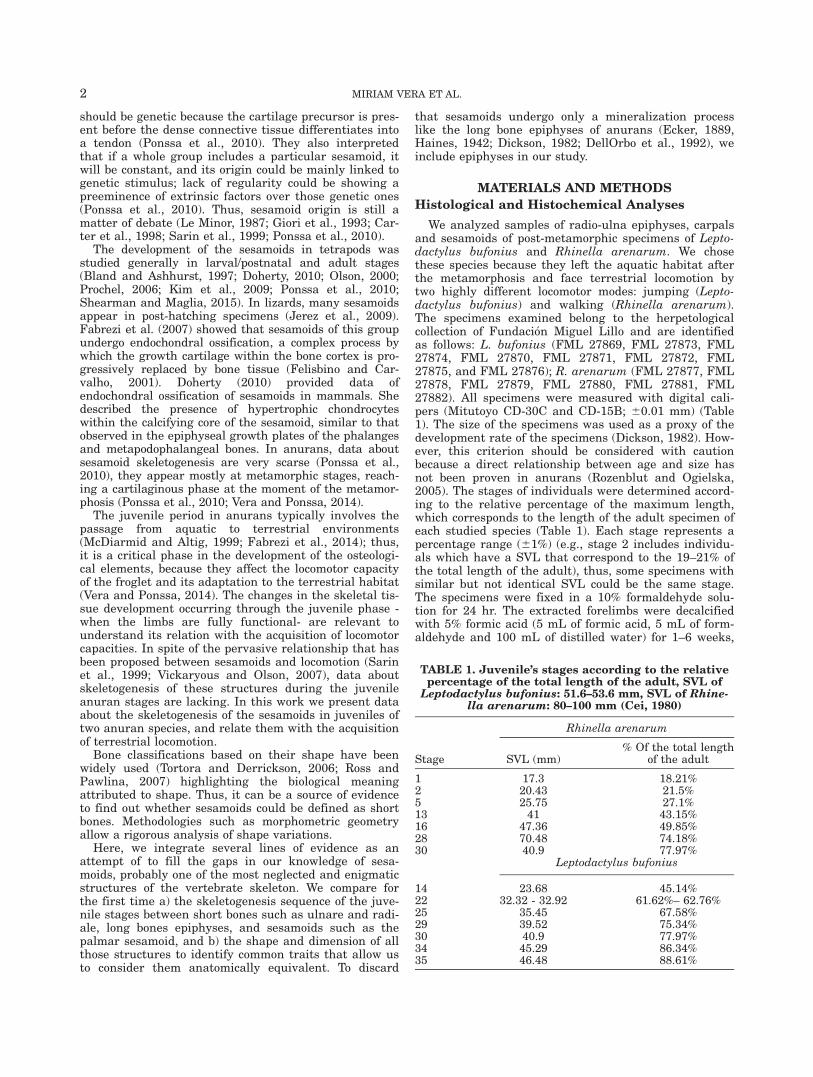
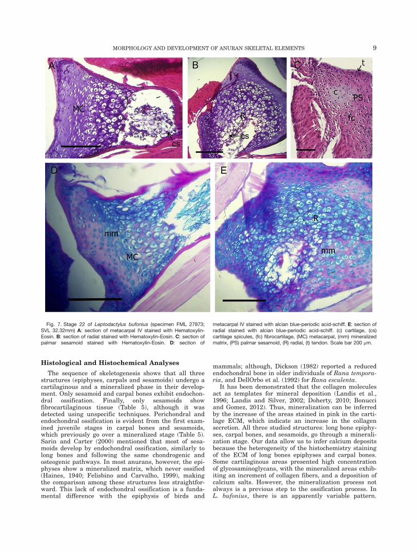
Stage 22 (Figs. 7 and 8): The long bone diaphyses andcarpals show an increment of the medular cavity sizeand cartilage spicules (Fig. 7A,B; 8A, C, E). In one speci-men, the ECM is mineralized, as shown by the high col-lagen concentration around the isogen groups and thehypertrophic chondrocytes in diaphysis and epiphysis(Fig. 7D,E). Other specimens do not show mineralizationin the matrix of the metacarpal III (Fig. 8B,D,F). Hya-line cartilage of the developing palmar sesamoidemerges from a fibrocartilaginous transition area intothe tendinous tissue (Fig. 7C).
Stage 25 (Fig. 9): Endochondral bone trabeculaeappear in the medular cavity of both long bone diaphy-ses and carpals, but not in the epiphyses of long bone.The ECM stains blue with Hematoxylin-Eosin, suggest-ing a high concentration of proteoglycans and is not min-eralized. Abundant collagen is evident in the bonetrabeculae and bone edges.
Fig. 2. Stage 5 of Rhinella arenarum. A: section of glide sesamoid between metacarpal and phalangeIV stained with Hematoxylin-Eosin. B: section of pararadial stained with Hematoxylin-Eosin. (GS) glidesesamoid, (PrR) pararadial sesamoid, (t) tendon. Scale bar 200 mm.
MORPHOLOGY AND DEVELOPMENT OF ANURAN SKELETAL ELEMENTS 5

Stage 29 (Fig. 10): The trabeculae of endochondralbones and an increased size of the marrow cavity in thelong bone diaphyses and carpals are evident (Fig. 10A, B).
The ECM of the cartilage in the epiphyses of the longbones and in diaphyses is mineralized (Fig. 10D, E). Alarge amount of osteocytes and blood vessels with
Fig. 3. Stage 13 of Rhinella arenarum. A: section of phalange IV stained with Hematoxylin-Eosin, thecartilage growth zones are noticeable. B: section of metacarpal II stained with Mallory trichrome, a fibro-cartilage tissue is noticeable. (fc) fibrocartilage, (GS) glide sesamoid, (hc) hypertrophic chondrocyte zone(o) osteocytes, (MC) metacarpal, (pc) proliferation cartilage zone, (rc) reserve cartilage zone, (sg) serousgland. Scale bar 200 mm.
Fig. 4. Stage 16 of Rhinella arenarum. A: section of metacarpal II stained with Hematoxylin-Eosin. B:section of fibrocartilaginous tissue stained with Hematoxylin-Eosin. (PrR) pararadial sesamoid, (et) endo-chondral trabeculae, (fc) fibrocartilaginous tissue. Scale bar 200 mm.
6 MIRIAM VERA ET AL.

erythrocytes is evident in the marrow cavity in both car-pal and diaphyses of the long bones (Fig. 10G). The pal-mar sesamoid is cartilaginous, and a fibrocartilaginoustransition between this sesamoid and its tendon is visible(Fig. 10C). The fibers of the osteochondral ligament (Felis-bino and Carvalho, 2000) are visible (Fig. 10F).
Stage 30 (Fig. 11): The trabeculae of endochondralbones in long bones diaphyses and carpals (Fig. 11D) areconspicuous. In the epiphyses, the osteochondral ligament(Felisbino and Carvalho, 2000) between the periostealbone and the lateral articular cartilage is evident (Fig.11A). The joint capsule is formed by dense fibrous connec-tive tissue that adheres firmly to the bones via theattachment-zone tendinous-cartilage (Fig. 11A). The car-pal bone ulnar is rounded by tendinous tissue (Fig. 11B).
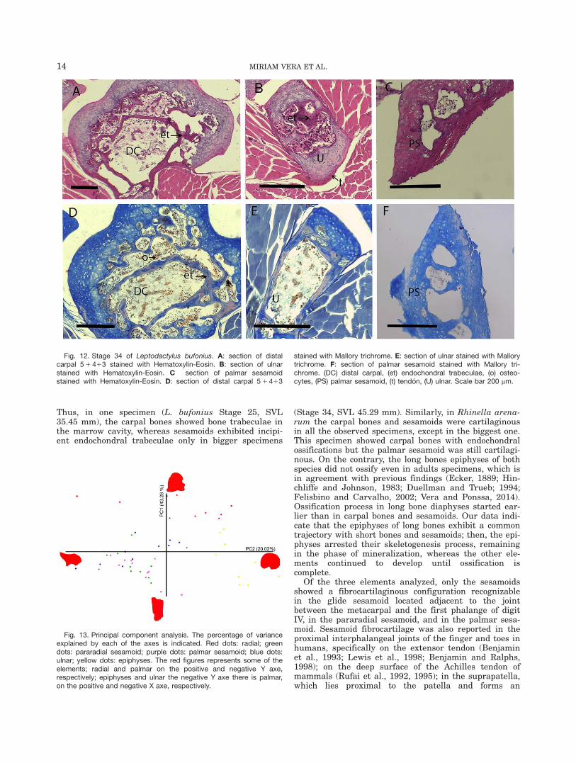
Stage 34 (Fig. 12) and Stage 35: Diaphyses of longbones, carpals and palmar sesamoid show a bigger mar-row cavity and even more endochondral trabeculae thanin the previous stages (Fig. 12A, B, C, F). In the speci-men in Stage 34, the tendinous tissue surrounding theulnare is evident (Fig. 12B). The osseous matrix showsconsiderable amount of keratin sulphate (Fig. 12D, E);this is noticeable in the endochondral trabeculae, whichstains red. A big amount of osteocytes and blood vesselswith erythrocytes are evident in the marrow cavity incarpals (Fig. 12D, E).
Morphometric Analysis
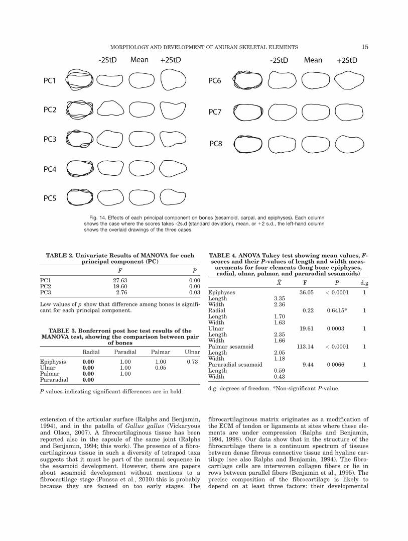
The PCA of the bone data set (N 5 51) of the contourmorphometric geometric analysis is shown in the scatter
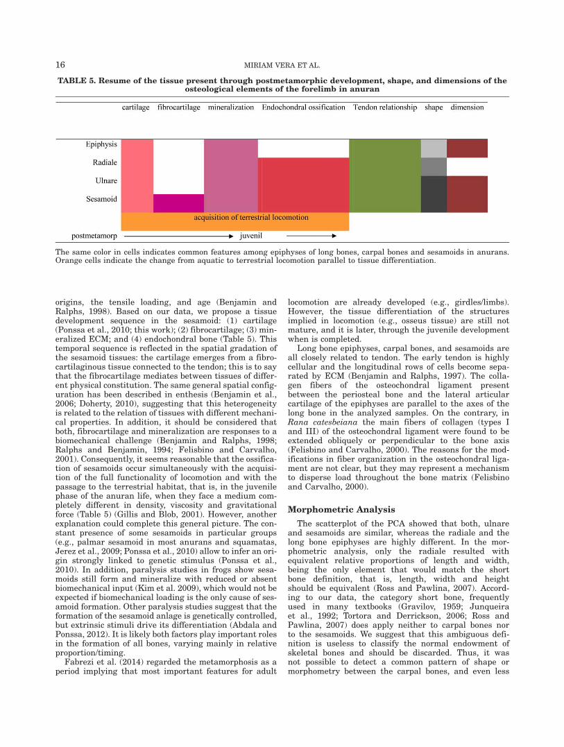
plot of the first and second principal components (PC1vs. PC2; Fig. 13). PC1 and PC2 captured 43.28% and20.02% of the total shape variation, respectively, andtogether with PC3 accounted for 72.29%; the remainingpercentages are listed in Appendix . The effects of eachPC on bone (epiphyses, carpal, and sesamoid) shape arevisualized in the Fig. 14. These reconstructed shapesindicate that the first PC described shape change associ-ated with an enlargement of the maximum longitudinaldiameter, this expansion being stronger in the left por-tion of the element. On PC2, shape variation was deter-mined by expansion in the extreme sides, which gave atriangular configuration to both extreme shapes, withthe more expanded sides in opposite areas (above andbelow) of the variation.
The PCA of the contour morphometric variables ofthe five osteological elements shows a clear separationthrough PC1 of a group composed of the radiale,whereas PC2 separated a cluster of the epiphyses, anda group formed by the sesamoids (palmar and parara-dial) and the ulnare. The MANOVA shows significantdifferences among bones (Wilk’s k F12,19=0.05,P<0.000). Subsequent univariate F-test showed thatthis difference was significant for each principal compo-nent (PC1, PC2 and PC3) (Table 2). Bone–bone compar-isons (Table 3) show that the shape of radialesignificantly differed from those of paradial sesamoid,palmar sesamoid, ulnare, and epiphyses of humerus(P< 0.00).
The ANOVA including the length and the width of thefive elements of the manus revealed a significant
Fig. 5. Stage 28 of Rhinella arenarum. A: section of metacarpal IIstained with Hematoxylin-Eosin. B: section of Y element stained withHematoxylin-Eosin. C: section of palmar sesamoid stained withHematoxylin-Eosin. D: section of metacarpal II stained with Mallory tri-
chrome. E: section of Y element stained with Mallory trichrome. D:section of palmar sesamoid stained with Mallory trichrome. (cs) carti-lage spicules, (e) erythrocytes, (et) endochondral trabeculae, (o) osteo-cytes, (PS) palmar sesamoid, (t) tendon. Scale bar 200 mm.
MORPHOLOGY AND DEVELOPMENT OF ANURAN SKELETAL ELEMENTS 7

difference (P< 0.05) in all the elements, except for theradiale bone (P 5 0.64) (Table 4).
DISCUSSION
Of the three analyzed aspects (skeletogenesis, shapeand proportions of sesamoids), skeletogenesis is clearly ashared configuration between carpal bones and sesa-moids. Even when the ossification process shows animportant difference in sesamoids—the appearance offibrocartilage in the transition between the tendon and
the hyaline cartilage during their development—bothstructures present the same pattern of endochondralossification. It should be considered however, that sesa-moids exhibit a pervasive relationship with tendons thathas not been described in carpal bones or long bones epi-physes. With regard to sesamoid shape and dimensions,our data indicate that bones categorization based onthese aspects-particularly those that stay that shortbones are those having their three dimensions equallydeveloped (Ross and Pawlina, 2007) is vague and there-fore is ineffective to classify the skeletal bones.
Fig. 6. Stage 14 of Leptodactylus bufonius. A: section of radio-ulnastained with Hematoxylin-Eosin. B: section of radial stained withHematoxylin-Eosin. C: section of radio-ulna stained with alcian blue-periodic acid-schiff. D: section of radio-ulna and the myo-tendinous
junction stained with Hematoxylin-Eosin. (ac) articular cartilage, (gc)growth cartilage, (lac) lateral articular cartilage, (mtj) myo-tendinousjunction, (ol) osteochondral ligament, (RU) radio-ulna, (R) radial, (t) ten-don. Scale bar 200 mm.
8 MIRIAM VERA ET AL.
Histological and Histochemical Analyses
The sequence of skeletogenesis shows that all threestructures (epiphyses, carpals and sesamoids) undergo acartilaginous and a mineralized phase in their develop-ment. Only sesamoid and carpal bones exhibit endochon-dral ossification. Finally, only sesamoids showfibrocartilaginous tissue (Table 5), although it wasdetected using unspecific techniques. Perichondral andendochondral ossification is evident from the first exam-ined juvenile stages in carpal bones and sesamoids,which previously go over a mineralized stage (Table 5).Sarin and Carter (2000) mentioned that most of sesa-moids develop by endochondral ossification, similarly tolong bones and following the same chondrogenic andosteogenic pathways. In most anurans, however, the epi-physes show a mineralized matrix, which never ossified(Haines, 1940; Felisbino and Carvalho, 1999), makingthe comparison among these structures less straightfor-ward. This lack of endochondral ossification is a funda-mental difference with the epiphysis of birds and
mammals; although, Dickson (1982) reported a reducedendochondral bone in older individuals of Rana tempora-ria, and DellOrbo et al. (1992) for Rana esculenta.
It has been demonstrated that the collagen moleculesact as templates for mineral deposition (Landis et al.,1996; Landis and Silver, 2002; Doherty, 2010; Bonucciand Gomez, 2012). Thus, mineralization can be inferredby the increase of the areas stained in pink in the carti-lage ECM, which indicate an increase in the collagensecretion. All three studied structures: long bone epiphy-ses, carpal bones, and sesamoids, go through a minerali-zation stage. Our data allow us to infer calcium depositsbecause the heterogeneity of the histochemistry stainingof the ECM of long bones epiphyses and carpal bones.Some cartilaginous areas presented high concentrationof glycosaminoglycans, with the mineralized areas exhib-iting an increment of collagen fibers, and a deposition ofcalcium salts. However, the mineralization process notalways is a previous step to the ossification process. InL. bufonius, there is an apparently variable pattern.
Fig. 7. Stage 22 of Leptodactylus bufonius (specimen FML 27873;SVL 32.32mm) A: section of metacarpal IV stained with Hematoxylin-Eosin. B: section of radial stained with Hematoxylin-Eosin. C: section ofpalmar sesamoid stained with Hematoxylin-Eosin. D: section of
metacarpal IV stained with alcian blue-periodic acid-schiff. E: section ofradial stained with alcian blue-periodic acid-schiff. (c) cartilage, (cs)cartilage spicules, (fc) fibrocartilage, (MC) metacarpal, (mm) mineralizedmatrix, (PS) palmar sesamoid, (R) radial, (t) tendon. Scale bar 200 mm.
MORPHOLOGY AND DEVELOPMENT OF ANURAN SKELETAL ELEMENTS 9
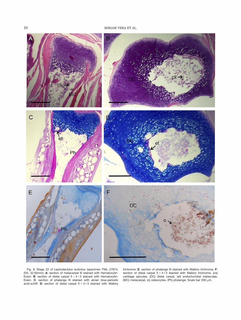
Fig. 8. Stage 22 of Leptodactylus bufonius (specimen FML 27874,SVL 32.92mm). A: section of metacarpal III stained with Hematoxylin-Eosin. B: section of distal carpal 5 1 413 stained with Hematoxylin-Eosin. C: section of phalange III stained with alcian blue-periodicacid-schiff. D: section of distal carpal 5 1 413 stained with Mallory
trichrome. E: section of phalange III stained with Mallory trichrome. F:section of distal carpal 5 1 413 stained with Mallory trichrome. (cs)cartilage spicules, (DC) distal carpal, (et) endochondral trabeculae,(MC) metacarpal, (o) osteocytes, (Ph) phalange. Scale bar 200 mm.
10 MIRIAM VERA ET AL.
Fig. 9. Stage 25 of Leptodactylus bufonius. A: section of metacarpalIII stained with Hematoxylin-Eosin. B: section of distal carpal 5 1 413stained with Hematoxylin-Eosin. C: section of metacarpal and pha-lange IV stained with alcian blue-periodic acid-schiff. D: section of dis-tal carpal 5 1 413 stained with alcian blue-periodic acid-schiff.
E: section of metacarpal II stained with Mallory trichrome. F: sectionof distal carpal 5 1 413 with Mallory trichrome. (DC) distal carpal, (et)endochondral trabeculae, (MC) metacarpal, (ol) ostochondral ligament(Ph) phalange. Scale bar 200 mm.

Fig. 10. Stage 29 of Leptodactylus bufonius. A: section of metacar-pal IV stained with Hematoxylin-Eosin. B: section of distal carpal5 1 413 stained with Hematoxylin-Eosin. C: section of palmar sesa-moid stained with Hematoxylin-Eosin. D: section of metacarpal IVstained with alcian blue-periodic acid-schiff. E: section of distal carpal5 1 413 stained with alcian blue-periodic acid-schiff. F: section of
radio-ulna stained with Mallory trichrome. G: section of distal carpal5 1 413 stained with Mallory trichrome. (c) cartilage, (DC) distal car-pal, (e) erythrocytes, (et) endochondral trabeculae, (Ey) Y element, (fc)fibrocartilage, (MC) metacarpal, (ol) osteochondral ligament, (PS) pal-mar sesamoid, (R) radial, (RU) radio-ulna, (mm) mineralized matrix, (t)tendon. Scale bar 200 mm.
12 MIRIAM VERA ET AL.

Specimens in Stages 22 (Fig. 9) and 29 (Fig. 12) showeda mineralized matrix in their carpal bones before bonetrabeculae formation; whereas other specimens of Stages22 and Stage 25 showed a cartilaginous matrix first, andthen bone trabeculae; and a stage of mineralization wasnot observed. Felisbino and Carvalho (2001) stressedthat the formation of bone trabeculae is not dependenton cartilage mineralization, since osteoblast can depositbone on the surface of unmineralized cartilage. Our find-ings in L. bufonius supports the idea of Felisbino andCarvalho (2001) regarding the lack of spatial or tempo-ral association with calcium deposition and trabeculaeformation; accordingly, many events occurring during
endochondral ossification are not necessarily related toeach other. Matrix mineralization could be explained asa consequence of epigenetic factors acting on each indi-vidual; that is, some specimens could be subjected tohigher mechanical force in their lives than others (Van’tVeen et al., 1995). Thus, matrix mineralization in Ranacatesbeiana is variable, and occurs as a reinforcementevent of bones in adulthood (Felisbino and Carvalho,2001); whether this is the case in the species here stud-ied remain as an open question.
The sequence of mineralization and ossification proc-esses is equivalent in long bones epiphyses, carpals andsesamoids, although their timing is not synchronic.
Fig. 11. Stage 30 of Leptodactylus bufonius. A: section of phalangeIV stained with Hematoxylin-Eosin. B: section of ulnar stained withHematoxylin-Eosin. C: section of metacarpal IV stained with alcianblue-periodic acid-schiff. D: section of distal carpal 5 1 413 stained
with alcian blue-periodic acid-schiff. (az) attachment-zone fibrocarti-lage, (DC) distal carpal, (et) endochondral trabeculae, (MC) metacar-pal, (ol) osteochondral ligament, (Ph) phalange, (t) tendon, (U) ulnar.Scale bar 200 mm.
MORPHOLOGY AND DEVELOPMENT OF ANURAN SKELETAL ELEMENTS 13
Thus, in one specimen (L. bufonius Stage 25, SVL35.45 mm), the carpal bones showed bone trabeculae inthe marrow cavity, whereas sesamoids exhibited incipi-ent endochondral trabeculae only in bigger specimens
(Stage 34, SVL 45.29 mm). Similarly, in Rhinella arena-rum the carpal bones and sesamoids were cartilaginousin all the observed specimens, except in the biggest one.This specimen showed carpal bones with endochondralossifications but the palmar sesamoid was still cartilagi-nous. On the contrary, the long bones epiphyses of bothspecies did not ossify even in adults specimens, which isin agreement with previous findings (Ecker, 1889; Hin-chliffe and Johnson, 1983; Duellman and Trueb; 1994;Felisbino and Carvalho, 2002; Vera and Ponssa, 2014).Ossification process in long bone diaphyses started ear-lier than in carpal bones and sesamoids. Our data indi-cate that the epiphyses of long bones exhibit a commontrajectory with short bones and sesamoids; then, the epi-physes arrested their skeletogenesis process, remainingin the phase of mineralization, whereas the other ele-ments continued to develop until ossification iscomplete.
Of the three elements analyzed, only the sesamoidsshowed a fibrocartilaginous configuration recognizablein the glide sesamoid located adjacent to the jointbetween the metacarpal and the first phalange of digitIV, in the pararadial sesamoid, and in the palmar sesa-moid. Sesamoid fibrocartilage was also reported in theproximal interphalangeal joints of the finger and toes inhumans, specifically on the extensor tendon (Benjaminet al., 1993; Lewis et al., 1998; Benjamin and Ralphs,1998); on the deep surface of the Achilles tendon ofmammals (Rufai et al., 1992, 1995); in the suprapatella,which lies proximal to the patella and forms an
Fig. 12. Stage 34 of Leptodactylus bufonius. A: section of distalcarpal 5 1 413 stained with Hematoxylin-Eosin. B: section of ulnarstained with Hematoxylin-Eosin. C section of palmar sesamoidstained with Hematoxylin-Eosin. D: section of distal carpal 5 1 413
stained with Mallory trichrome. E: section of ulnar stained with Mallorytrichrome. F: section of palmar sesamoid stained with Mallory tri-chrome. (DC) distal carpal, (et) endochondral trabeculae, (o) osteo-cytes, (PS) palmar sesamoid, (t) tend�on, (U) ulnar. Scale bar 200 mm.
Fig. 13. Principal component analysis. The percentage of varianceexplained by each of the axes is indicated. Red dots: radial; greendots: pararadial sesamoid; purple dots: palmar sesamoid; blue dots:ulnar; yellow dots: epiphyses. The red figures represents some of theelements; radial and palmar on the positive and negative Y axe,respectively; epiphyses and ulnar the negative Y axe there is palmar,on the positive and negative X axe, respectively.
14 MIRIAM VERA ET AL.
extension of the articular surface (Ralphs and Benjamin,1994), and in the patella of Gallus gallus (Vickaryousand Olson, 2007). A fibrocartilaginous tissue has beenreported also in the capsule of the same joint (Ralphsand Benjamin, 1994; this work). The presence of a fibro-cartilaginous tissue in such a diversity of tetrapod taxasuggests that it must be part of the normal sequence inthe sesamoid development. However, there are papersabout sesamoid development without mentions to afibrocartilage stage (Ponssa et al., 2010) this is probablybecause they are focused on too early stages. The
fibrocartilaginous matrix originates as a modification ofthe ECM of tendon or ligaments at sites where these ele-ments are under compression (Ralphs and Benjamin,1994, 1998). Our data show that in the structure of thefibrocartilage there is a continuum spectrum of tissuesbetween dense fibrous connective tissue and hyaline car-tilage (see also Ralphs and Benjamin, 1994). The fibro-cartilage cells are interwoven collagen fibers or lie inrows between parallel fibers (Benjamin et al., 1995). Theprecise composition of the fibrocartilage is likely todepend on at least three factors: their developmental
Fig. 14. Effects of each principal component on bones (sesamoid, carpal, and epiphyses). Each columnshows the case where the scores takes -2s.d (standard deviation), mean, or 12 s.d., the left-hand columnshows the overlaid drawings of the three cases.
TABLE 2. Univariate Results of MANOVA for eachprincipal component (PC)
F P
PC1 27.63 0.00PC2 19.60 0.00PC3 2.76 0.03
Low values of p show that difference among bones is signifi-cant for each principal component.
TABLE 3. Bonferroni post hoc test results of theMANOVA test, showing the comparison between pair
of bones
Radial Paradial Palmar Ulnar
Epiphysis 0.00 1.00 1.00 0.73Ulnar 0.00 1.00 0.05Palmar 0.00 1.00Pararadial 0.00
P values indicating significant differences are in bold.
TABLE 4. ANOVA Tukey test showing mean values, F-scores and their P-values of length and width meas-
urements for four elements (long bone epiphyses,radial, ulnar, palmar, and pararadial sesamoids)
�X F P d.g
Epiphyses 36.05 < 0.0001 1Length 3.35Width 2.36Radial 0.22 0.6415* 1Length 1.70Width 1.63Ulnar 19.61 0.0003 1Length 2.35Width 1.66Palmar sesamoid 113.14 < 0.0001 1Length 2.05Width 1.18Pararadial sesamoid 9.44 0.0066 1Length 0.59Width 0.43
d.g: degrees of freedom. *Non-significant P-value.
MORPHOLOGY AND DEVELOPMENT OF ANURAN SKELETAL ELEMENTS 15
origins, the tensile loading, and age (Benjamin andRalphs, 1998). Based on our data, we propose a tissuedevelopment sequence in the sesamoid: (1) cartilage(Ponssa et al., 2010; this work); (2) fibrocartilage; (3) min-eralized ECM; and (4) endochondral bone (Table 5). Thistemporal sequence is reflected in the spatial gradation ofthe sesamoid tissues: the cartilage emerges from a fibro-cartilaginous tissue connected to the tendon; this is to saythat the fibrocartilage mediates between tissues of differ-ent physical constitution. The same general spatial config-uration has been described in enthesis (Benjamin et al.,2006; Doherty, 2010), suggesting that this heterogeneityis related to the relation of tissues with different mechani-cal properties. In addition, it should be considered thatboth, fibrocartilage and mineralization are responses to abiomechanical challenge (Benjamin and Ralphs, 1998;Ralphs and Benjamin, 1994; Felisbino and Carvalho,2001). Consequently, it seems reasonable that the ossifica-tion of sesamoids occur simultaneously with the acquisi-tion of the full functionality of locomotion and with thepassage to the terrestrial habitat, that is, in the juvenilephase of the anuran life, when they face a medium com-pletely different in density, viscosity and gravitationalforce (Table 5) (Gillis and Blob, 2001). However, anotherexplanation could complete this general picture. The con-stant presence of some sesamoids in particular groups(e.g., palmar sesamoid in most anurans and squamatas,Jerez et al., 2009; Ponssa et al., 2010) allow to infer an ori-gin strongly linked to genetic stimulus (Ponssa et al.,2010). In addition, paralysis studies in frogs show sesa-moids still form and mineralize with reduced or absentbiomechanical input (Kim et al. 2009), which would not beexpected if biomechanical loading is the only cause of ses-amoid formation. Other paralysis studies suggest that theformation of the sesamoid anlage is genetically controlled,but extrinsic stimuli drive its differentiation (Abdala andPonssa, 2012). It is likely both factors play important rolesin the formation of all bones, varying mainly in relativeproportion/timing.
Fabrezi et al. (2014) regarded the metamorphosis as aperiod implying that most important features for adult
locomotion are already developed (e.g., girdles/limbs).However, the tissue differentiation of the structuresimplied in locomotion (e.g., osseus tissue) are still notmature, and it is later, through the juvenile developmentwhen is completed.
Long bone epiphyses, carpal bones, and sesamoids areall closely related to tendon. The early tendon is highlycellular and the longitudinal rows of cells become sepa-rated by ECM (Benjamin and Ralphs, 1997). The colla-gen fibers of the osteochondral ligament presentbetween the periosteal bone and the lateral articularcartilage of the epiphyses are parallel to the axes of thelong bone in the analyzed samples. On the contrary, inRana catesbeiana the main fibers of collagen (types Iand III) of the osteochondral ligament were found to beextended obliquely or perpendicular to the bone axis(Felisbino and Carvalho, 2000). The reasons for the mod-ifications in fiber organization in the osteochondral liga-ment are not clear, but they may represent a mechanismto disperse load throughout the bone matrix (Felisbinoand Carvalho, 2000).
Morphometric Analysis
The scatterplot of the PCA showed that both, ulnareand sesamoids are similar, whereas the radiale and thelong bone epiphyses are highly different. In the mor-phometric analysis, only the radiale resulted withequivalent relative proportions of length and width,being the only element that would match the shortbone definition, that is, length, width and heightshould be equivalent (Ross and Pawlina, 2007). Accord-ing to our data, the category short bone, frequentlyused in many textbooks (Gravilov, 1959; Junqueiraet al., 1992; Tortora and Derrickson, 2006; Ross andPawlina, 2007) does apply neither to carpal bones norto the sesamoids. We suggest that this ambiguous defi-nition is useless to classify the normal endowment ofskeletal bones and should be discarded. Thus, it wasnot possible to detect a common pattern of shape ormorphometry between the carpal bones, and even less
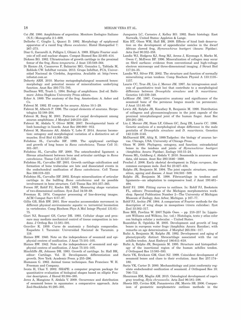
TABLE 5. Resume of the tissue present through postmetamorphic development, shape, and dimensions of theosteological elements of the forelimb in anuran
The same color in cells indicates common features among epiphyses of long bones, carpal bones and sesamoids in anurans.Orange cells indicate the change from aquatic to terrestrial locomotion parallel to tissue differentiation.
16 MIRIAM VERA ET AL.

between carpals and sesamoids or long bone epiphyses.Possibly the differences explained by the geometricmorphometry analysis between ulnare and carpale aredue to differences in the origin and ontogeny of theseelements (Fabrezi, 1992; Fabrezi and Alberch, 1996;Fabrezi and Barg, 2001).
ACKNOWLEDGEMENT
All authors are grateful to CONICET, FONCyT andUNT for supporting our work via their research grants.They are grateful to Franco Pucci, Ana Pucci and Mar-isa Alcaide (Fundaci�on Miguel Lillo) for their histologicalhelp.
APPENDIX 1
Eigen Values and Percentages for the PrincipalComponents of the Contour Morphometric
Geometric Analysis
EigenvalueProportion
(%)Cumulative
(%)
PC1 1.517110E-002 43.2828 43.2828PC2 7.018560E-003 20.0238 63.3067PC3 3.151810E-003 8.9921 72.2987PC4 2.983025E-003 8.5105 80.8092PC5 1.604576E-003 4.5778 85.3870PC6 1.521624E-003 4.3412 89.7282PC7 7.983505E-004 2.2777 92.0059PC8 5.424554E-004 1.5476 93.5535PC9 4.302533E-004 1.2275 94.7810PC10 3.724067E-004 1.0625 95.8435PC11 2.563079E-004 0.7312 96.5747PC12 1.836043E-004 0.5238 97.0985PC13 1.453192E-004 0.4146 97.5131PC14 1.165021E-004 0.3324 97.8455PC15 1.061555E-004 0.3029 98.1484PC16 8.538100E-005 0.2436 98.3919PC17 8.305919E-005 0.2370 98.6289PC18 6.547855E-005 0.1868 98.8157PC19 5.380333E-005 0.1535 98.9692PC20 5.054454E-005 0.1442 99.1134PC21 4.285794E-005 0.1223 99.2357PC22 3.596169E-005 0.1026 99.3383PC23 3.367885E-005 0.0961 99.4344PC24 2.693603E-005 0.0768 99.5112PC25 2.218930E-005 0.0633 99.5745PC26 1.911831E-005 0.0545 99.6291PC27 1.616938E-005 0.0461 99.6752PC28 1.496114E-005 0.0427 99.7179PC29 1.415310E-005 0.0404 99.7583PC30 1.052167E-005 0.0300 99.7883PC31 9.887945E-006 0.0282 99.8165PC32 8.430689E-006 0.0241 99.8405PC33 8.237374E-006 0.0235 99.8641PC34 6.863616E-006 0.0196 99.8836PC35 6.049597E-006 0.0173 99.9009PC36 5.488812E-006 0.0157 99.9166PC37 4.933255E-006 0.0141 99.9306PC38 4.198116E-006 0.0120 99.9426PC39 3.520445E-006 0.0100 99.9526PC40 2.708275E-006 0.0077 99.9604PC41 2.531769E-006 0.0072 99.9676PC42 2.058809E-006 0.0059 99.9735PC43 1.952440E-006 0.0056 99.9790PC44 1.727703E-006 0.0049 99.9840
APPENDIX 1 (Continued)
PC45 1.482378E-006 0.0042 99.9882PC46 1.131113E-006 0.0032 99.9914PC47 1.061376E-006 0.0030 99.9945PC48 9.215644E-007 0.0026 99.9971PC49 5.474278E-007 0.0016 99.9986PC50 4.750627E-007 0.0014 100.0000PC51 1.120043E-018 0.0000 100.0000PC52 4.506704E-020 0.0000 100.0000PC53 2.629335E-020 0.0000 100.0000PC54 2.294601E-020 0.0000 100.0000PC55 1.542047E-020 0.0000 100.0000PC56 7.952505E-021 0.0000 100.0000PC57 6.216223E-021 0.0000 100.0000PC58 5.051052E-021 0.0000 100.0000PC59 3.352603E-021 0.0000 100.0000PC60 2.895798E-021 0.0000 100.0000PC61 1.717373E-021 0.0000 100.0000PC62 1.056774E-021 0.0000 100.0000PC63 5.775427E-022 0.0000 100.0000PC64 2.121933E-022 0.0000 100.0000PC65 25.630339E-022 0.0000 100.0000PC66 27.998032E-022 0.0000 100.0000PC67 21.349949E-021 0.0000 100.0000PC68 21.662556E-021 0.0000 100.0000PC69 21.951877E-021 0.0000 100.0000PC70 22.383037E-021 0.0000 100.0000PC71 23.149249E-021 0.0000 100.0000PC72 23.673008E-021 0.0000 100.0000PC73 26.239967E-021 0.0000 100.0000PC74 21.099636E-020 0.0000 100.0000PC75 21.601198E-020 0.0000 100.0000PC76 22.201115E-020 0.0000 100.0000PC77 23.744892E-020 0.0000 100.0000Total
variance:3.505107E-002
LITERATURE CITED
Abdala V, Ponssa ML. 2012. Life in the slow lane: the effect ofreduced mobility on tadpole limb development. Anat Rec 295:5–17.
Benjamin M, Ralphs JR, Shibu M, Irwin M. 1993. Capsular tissuesof the proximal interphalangeal joint: normal composition andeffects of dupuytren’s disease and rheumatoid arthritis. J HandSurg 18B:371–376.[PMC][8345272]
Benjamin M, Qin S, Ralphs JR. 1995. Fibrocartilage associated withhuman tendons and their pulleys. J Anat 187:625–633.
Benjamin M, Ralphs JR. 1997. Tendons and ligaments—an over-view. Histol Histopathol 12:1135–1144.
Benjamin M, Ralphs JR. 1998. Fibrocartilage in tendons andligaments: an adaptation to compressive load. J Anat 193:481–494.
Benjamin M, Toumi H, Ralphs JR, Bydder G, Best TM, Milz S.2006. Where tendons and ligaments meet bone : attachment sites(“entheses”) in relation to exercise and/or mechanical load. J Anat208:471–490.
Bierbaum RM, Ferson S. 1986. Do symbiotic pea crabs decreasegrowth rate in mussels? Biol Bull 170:51–61.
Bland YS, Ashhurst DE. 1997. Fetal and postnatal development ofthe patella, patellar tendon and suprapatella in the rabbit;changes in the distribution of the fibrillar collagens. J Anat 190:327–342.
Bonucci E, Gomez S. 2012. Cartilage calcification. In: Jong Seto edi-tor. Advanced topics in biomineralization. Rijeka: InTech. p 85–111.
Carter DR, Mikic B, Padian K. 1998. Epigenetic mechanical factorsin the evolution of long bone epiphyses. Zool J Linn Soc 123:163–178.
MORPHOLOGY AND DEVELOPMENT OF ANURAN SKELETAL ELEMENTS 17
Cei JM. 1980. Amphibians of argentina. Monitore Zoologico Italiano(N.S.) Monografia 2:1–609.
Dellorbo C, Gioglio L, Quacci D. 1992. Morphology of epiphysealapparatus of a ranid frog (Rana esculenta). Histol Histopathol 7:267–273.
Diaz G, Zuccarelli A, Pelligra I, Ghiani A. 1989. Elliptic Fourier anal-ysis of cell and nuclear shapes. Comput Biomed Res 22:405–414.
Dickson RG. 1982. Ultrastructure of growth cartilage in the proximalfemur of the frog, Rana temporaria. J Anat 135:549–564.
Di Rienzo JA, Casanoves F, Balzarini MG, Gonzalez L, Tablada M,Robledo CW. InfoStat versi�on. 2013. Grupo InfoStat, FCA, Univer-sidad Nacional de C�ordoba, Argentina. Available at: http://www.infostat.com.ar.
Doherty ARH. 2010. Murine metapodophalangeal sesamoid bones:morphology and potential means of mineralization underlyingfunction. Anat Rec 293:775–785.
Duellman WE, Trueb L. 1994. Biology of amphibians. 2nd ed. Balti-more: Johns Hopkins University Press editors.
Ecker A. 1889. The anatomy of the frog. Amsterdam: A. Asher andCo.
Fabrezi M. 1992. El carpo de los anuros. Alytes 10:1–29.Fabrezi M, Alberch P. 1996. The carpal elements of anurans. Herpe-
tologica 52:188–204.Fabrezi M, Barg M. 2001. Patterns of carpal development among
anuran amphibians. J Morphol 249:210–220.Fabrezi M, Abdala V, Oliver MIM. 2007. Developmental basis of
limb homology in lizards. Anat Rec 290:9002912.Fabrezi M, Manzano AS, Abdala V, Lobo F. 2014. Anuran locomo-
tion: ontogeny and morphological variation of a distinctive set ofmuscles. Evol Biol 41:308–326.
Felisbino SL, Carvalho HF. 1999. The epiphyseal cartilageand growth of long bones in Rana catesbeiana. Tissue Cell 31:301–307.
Felisbino SL, Carvalho HF. 2000. The osteochondral ligament: afibrous attachment between bone and articular cartilage in Ranacatesbeiana. Tissue Cell 32:527–536.
Felisbino SL, Carvalho HF. 2001. Growth cartilage calcification andformation of bone trabeculae are late and dissociated events inthe endochondral ossification of Rana catesbeiana. Cell TissueRes 306:319–323.
Felisbino SL, Carvalho HF. 2002. Ectopic mineralization of articularcartilage in the bullfrog Rana catesbeiana and its possibleinvolvement in bone structure. Cell Tissue Res 301:357–365.
Ferson SF, Rohlf FJ, Koehn RK. 1985. Measuring shape variationof two-dimensional outlines. Syst Zool 34:59–68.
Freeman H, 1974. Computer processing of line-drawing images.ACM Comput Surv 6:57–97.
Gillis GB, Blob RW. 2001. How muscles accommodate movement indifferent physical environments: aquatic vs. terrestrial locomotionin vertebrates. Comp Biochem Phys A Mol Integr Physiol 131:61–75.
Giori NJ, Beaupr�e GS, Carter DR. 1993. Cellular shape and pres-sure may mediate mechanical control of tissue composition in ten-dons. J Orthop Res 11:581–591.
Gravilov K. 1959. Curso de anatom�ıa y fisiolog�ıa comparadas.Esqueleto I. Tucum�an: Universidad Nacional de Tucum�an. p119.
Haines RW. 1940. Note on the independence of sesamoid and epi-physial centres of ossification. J Anat 75:101–105.
Haines RW. 1942. Note on the independence of sesamoid and epi-physial centres of ossification. J Anat 75:101–105.
Hinchliffe JR, Johnson DR. 1983. Growth of cartilage. In: Hall BK,editor: Cartilage. Vol. II: Development, differentiation andgrowth. New York: Academic Press. p 255–296.
Humason G. 1962. Animal tissue techniques. San Francisco: W. H.Freeman and Company.
Iwata H, Ukai Y. 2002. SHAPE: a computer program package forquantitative evaluation of biological shapes based on elliptic Fou-rier descriptors. J Hered 93:384–385.
Jerez A, Mangione S, Abdala V. 2009. Occurrence and distributionof sesamoid bones in squamates: a comparative approach. ActaZool-Stockholm 91:295–305.
Junqueira LC, Carneiro J, Kelley RO. 1992. Basic histology. EastNorwalk, United States: Appleton & Lange.
Kim HT, Olson WM, Hall BK. 2009. Effects of hind limb denerva-tion on the development of appendicular ossicles in the dwarfAfrican clawed frog, Hymenochirus boettgeri (Anura: Pipidae).Acta Zool 90:352–358.
Landis WJ, Hodgens KJ, Song MJ, Arena J, Kiyonaga S, Marko M,Owen C, McEwen BF. 1996. Mineralization of collagen may occuron fibril surfaces: evidence from conventional and high-voltageelectron microscopy and three-dimensional imaging. J Struct Biol117:24–35.
Landis WJ, Silver FH. 2002. The structure and function of normallymineralizing avian tendons. Comp Biochem Physiol A 133:1135–1157.
Laurie CC, True JR, Liu J, Mercer JM. 1997. An introgression anal-ysis of quantitative trait loci that contribute to a morphologicaldifference between Drosophila simulans and D. mauritiana.Genetics 145:339–348.
LeMinor JM. 1987. Comparative anatomy and significance of thesesamoid bone of the peroneus longus muscle (os peroneum).J Anat 151:85–99.
Lewis AR, Ralphs JR, Kneafsey B, Benjamin M. 1998. Distributionof collagens and glycosaminoglycans in the joint capsule of theproximal interphalangeal joint of the human finger. Anat Rec250:281–291.
Liu J, Mercer JM, Stam LF, Gibson GC, Zeng ZB, Laurie CC. 1996.Genetic analysis of a morphological shape difference in the malegenitalia of Drosophila simulans and D. mauritania. Genetics142:1129–1145.
McDiarmid RW, Altig R. 1999.Tadpoles: the biology of anuran lar-vae. Chicago, USA: University of Chicago Press.
Olson W. 2000. Phylogeny, ontogeny, and function: extraskeletalbones in the tendons and joints of Hymenochirus boettgeri(Amphibia: Anura: Pipidae). Zoology 103:15–24.
Ponssa ML, Goldberg J, Abdala V. 2010. Sesamoids in anurans: newdata, old issues. Anat Rec 293:164621668.
Prochel J. 2006. Early skeletal development in Talpa europaea, thecommon european mole. Zool Sci 23:427–434.
Ralphs JR, Benjamin M. 1994. The joint capsule: structure, compo-sition, ageing and disease. J Anat 184:5032509.
Ralphs JR, Benjamin M. 1998. Fibrocartilage in tendons andligaments—an adaptation to compressive load. J Anat 193:481–494.
Rohlf FJ. 1990. Fitting curves to outlines. In: Rohlf FJ, BooksteinFL, editors: Proceedings of the Michigan morphometrics work-shop. Special Publication Number 2—The University of MichiganMuseum of Zoology, Ann Arbor, Michigan. p 167–77.
Rohlf FJ, Archie JW. 1984. A comparison of Fourier methods for thedescription of wing shape in mosquitoes (rirera culicidae). SystZool 33:302–317.
Ross MH, Pawlina W. 2007.Tejido �Oseo. – pp. 218–257 In: Lippin-cott Williams and Wilkins, Inc. (ed.): Histolog�ıa, texto y atlas colorcon biolog�ıa celular y molecular. – United States.
Rozenblut B, Ogielska M. 2005. Development and growth of longbones in european water frogs (Amphibia: Anura: Ranidae), withremarks on age determination. J Morphol 265:3042317.
Rufai A, Benjamin M, Ralphs JR. 1992. Development and aging ofphenotypically distinct fibrocartilage associated with the ratachilles tendon. Anat Embryol 186:611–618.
Rufai A, Ralphs JR, Benjamin M. 1995. Structure and histopathol-ogy of the insertional region of the human achilles tendon.J Orthopaed Res 13:585–593.
Sarin VK, Erickson GM, Giori NJ. 1999. Coincident development ofsesamoid bones and clues to their evolution. Anat Rec 257:174–180.
Sarin VK, Carter D. 2000. Mechanobiology and joint conformity reg-ulate endochondral ossification of sesamoid. J Orthopaed Res 18:706–712.
Shearman RM, Maglia AM. 2015. Osteological development of cope’sgray treefrog, Hyla chrysoscelis. Acta Zool 96:181–198.
Sheets HD, Covino KM, Panasiewicz JM, Morris SR. 2006. Compar-ison of geometric morphometric outlines methods in the
18 MIRIAM VERA ET AL.

discrimination of age-related differences in feather shape. FrontZool 3:15-
StatSoft, Inc. 2004. Statistica (data analysis software system), Ver-sion 7. Available at: www.statsoft.com.
ToroIbacache MV, Manriquez SG, Suazo GI. 2010. Geometric mor-phometrics and the study of biologic shapes: from descriptive toquantitative morphology. Int J Morphol 28:977–990.
Tortora GJ, Derrickson BH. 2006. Principios de Anatom�ıa yFisiolog�ıa. Argentina: M�edica-Panamericana, Buenos Aires.
Van’tVeen SJ, Hagen JW, Van Ginkel FC, Prahl-Andersen B,Burger EH. 1995. Intermittent compression stimulates cartilagemineralization. Bone 17:461–465.
Vera MC, Ponssa ML. 2014. Skeletogenesis in anurans: cranial andpostcranial development in metamorphic and postmetamorphicstages of Leptodactylus bufonius (Anura: Leptodactylidae). ActaZool 95:44–62. Stockholm
Vickaryous MK, Olson WM. 2007. Sesamoids and Ossicles in theAppendicular Skeleton. In: Hall BK, editor. Fins into limbs: evolu-tion, development, and transformation. Chicago: University ofChicago Press. p 323–341.
Yoshioka Y, Iwata H, Ohsawa R, Ninomiya S. 2004. Analysis ofpetal shape variation of primula sieboldii by elliptic fourierdescriptors and principal component analysis. Ann Bot 94:6572664. London
MORPHOLOGY AND DEVELOPMENT OF ANURAN SKELETAL ELEMENTS 19