energy-linked anion transport. cloning, sequencing, and characterization of a full length cdna...
TRANSCRIPT

THE JOURNAL OF BIOLOGICAL CHEMISTRY 0 1989 by The American Society for Biochemistry and Molecular Biology, Inc
Val. 264, No. 26, Issue of September 15, pp. 15628-15633.1989 Printed in L I S A .
Energy-linked Anion Transport CLONING, SEQUENCING, AND CHARACTERIZATION OF A FULL LENGTH cDNA ENCODING THE RAT LIVER MITOCHONDRIAL PROTON/PHOSPHATE SYMPORTER*
(Received for publication, April 6, 1989)
Gloria C. FerreiraS, Raymond D. Pratts, and Peter L. Pedersenll From the Laboratory for Molecular and Cellular Bioenergetics, Department of Biological Chemistry, The Johns Hopkins University School of Medicine, Baltimore, Maryland 21205
A full length cDNA clone encoding the precursor of the rat liver mitochondrial phosphate transporter (H+/ Pi symporter) has been isolated from a cDNA library using a bovine heart partial length phosphate trans- porter clone as a hybridization probe. The entire clone is 1263 base pairs in length with 5’- and 3”untrans- lated regions of 16 and 168 base pairs, respectively. The open reading frame encodes for the mature protein (312 amino acids) preceded by a presequence of 44 amino acids enriched in basic residues. The polypeptide sequence predicted from the DNA sequence was con- firmed by analyzing the first 17 amino-terminal amino acids of the pure phosphate transporter protein.
The rat liver phosphate transporter differs from the bovine heart transporter in 32 amino acids (i.e. - 10%). It contains a region from amino acid 139 to 159 which is 37% identical with the @-subunit of the liver mito- chondrial ATP synthase. Amino acid sequence compar- isons of the Pi transporter with Pi binding proteins, other H+-linked symporters, and the human glucose transporter did not reveal significant sequence homol- ogy.
Analysis of genomic DNA from both rat and S. cere- uisiae by Southern blots using the rat liver mitochon- drial Pi carrier cDNA as a probe revealed remarkably similar restriction patterns, a finding consistent with the presence in lower and higher eukaryotes of homol- ogous Pi carrier proteins.
This is the first report of the isolation, sequencing, and characterization of a full length cDNA coding for a protein involved in energy-coupled Pi transport.
The supply of substrates required for oxidative phos- phorylation in eukaryotic cells involves the exchange of cy- toplasmic ADP for mitochondrial ATP and the uptake of inorganic phosphate (Pi) from the cytoplasm into the mito- chondrial matrix. Nucleotide exchange is maintained by the ADP/ATP carrier, while two transport systems are responsi-
* This work was supported in part by National Science Foundation Grant DMB-8606759 (to P. L. P.). The costs of publication of this article were defrayed in part by the payment of page charges. This article must therefore be hereby marked “aduertisement” in accord- ance with 18 U.S.C. Section 1734 solely to indicate this fact.
The nucleotide sequence(s) reported in thispaper has been submitted to the GenBankTM/EMBL Data Bank with accession number(s) M23984.
3 Supported by Fellowship DRG-986 from the Damon Runyon- Walter Winchell Cancer Fund.
Supporkd by National Institutes of Health Physician Scientist Award K12-AM01298.
V To whom correspondence and reprint requests should be ad- dressed.
ble for the translocation of Pi across the inner mitochondrial membrane (1-3). The major transport system, the Pi carrier (Pic),’ catalyzes an N-ethylmaleimide-sensitive PJH’ sym- port, whereas the other system catalyzes an N-ethylmaleim- ide-insensitive electroneutral exchange of Pi and/or dicarbox- ylate (3, 4).
Pic has been purified to homogeneity from bovine heart and rat liver mitochondria (5,6). Also, the rat liver mitochondrial transporter has been successfully reconstituted into phospho- lipid vesicles, retaining a high degree of function (7). The sequence of an amino-terminal fragment (8), as well as the partial sequence of the bovine heart Pic, has been determined by chemical protein sequence analysis (9). More recently, the amino acid sequence has been deduced from the nucleotide sequence of several overlapping clones encoding for the bovine heart Pic (10). However, a full length clone encoding for the Pic is a necessary prerequisite for both the expression of a functional transporter and the study, by site-directed muta- genesis, of the role of individual amino acids in the transport mechanism.
In this paper, we report the isolation, sequencing, and characterization of a full length cDNA clone for the rat liver mitochondrial Pic. In addition, we have examined the capacity of this clone to hybridize with genomic DNA sequences from both rat and the yeast, Saccharomyces cereuisiae.
EXPERIMENTAL PROCEDURES
Materials
A rat liver Xgtll cDNA library was purchased from Clontech
Biolabs, United States Biochemicals, Bethesda Research Laborato- Laboratories. Restriction enzymes were obtained from New England
ries (BRL), Pharmacia LKB Biotechnology Inc., and Boehringer Mannheim Biochemicals and were used according to the suppliers’ instructions. Sequenase and Klenow fragment were from United States Biochemicals, and T4 DNA ligase was from New England Biolabs. Universal sequencing 17-mer primers, M13mp18RF and -19RF, pUC18 and -19, and J M 101 strain were purchased from New England Biolabs. pUC-9, deoxy- and dideoxynucleotide triphos- phates, and random hexamers were from Pharmacia. 13%]dATP was from Amersham Corp., and ”P-labeled nucleotides were from ICN. Acrylamide and gel reagents were products of Bio-Rad, and Seakem GTG agarose was from FMC Corp. Bromochloroindoyl galactoside, isopropyl thiogalactoside, and lysozyme were from Sigma. NA45 nitrocellulose (DEAE) membrane was obtained from Schleicher & Schuell. All other chemicals were of the highest purity available. Escherichia coli strain Y1090 was a gift of Dr. D. W. Cleveland (The John Hopkins School of Medicine). S. cerevisiae 2180-1A was ob- tained from the Yeast Genetic Stock Center, Berkeley, CA. A partial bovine heart P,c cDNA was kindly provided by Dr. J. E. Walker (Medical Research Council, Cambridge).
The abbreviations used are: P,c, phosphate carrier/or phosphate transporter; SDS, sodium dodecyl sulfate; bp, base pair(s); kb, kilo- base pair(s).
15628

Rat Liver Mitochondrial Phosphate Transporter 15629
Methods
Library Screening-A rat liver cDNA library was screened with a ['"PIdATP-labeled DNA fragment according to the method of Benton and Davis (11). The DNA fragment was obtained after EcoRI and HindIII restriction of partial bovine heart Pic cDNA clone, followed by separation by agarose gel electrophoresis (12) and random primer labeling of the electroeluted Pic DNA (13). The first round of screen- ing of lo6 phages (plated a t a density of 300-400 plaques/cm2) yielded 19 potential positive clones. These were rescreened a t a lower density (30,000 plaques/l5-cm dish), and the remaining 12 potential positive clones were subjected to one more round of screening a t a density of less than 100 plaques/l6-cm dish. This led to the isolation of 10 cDNA clones.
Isolation and Analysis of Recombinants-The plaques giving the strongest signal were amplified, and the phage DNA was purified essentially as described by Maniatis et al. (12). The recombinant DNA was released by digestion with KpnI and SacI, and the cDNA clone that yielded the longest insert was further restricted with EcoRI. The products were separated through a 1% agarose gel, electroeluted from the gel with a DEAE-membrane according to the manufacturer's instructions. DNA was recovered by phenol extraction and ligated into the EcoRI site of M13mp18 and -mp19 vectors. The recombinant DNA was sequenced according to the dideoxy chain termination method (14), using E. coli .JM 101 as host. Preliminary sequence data enabled further restrictions of the recombinant DNA: 1) EcoRI and SphI; 2) EcoRI and HindIII; 3) EcoRI and Eco01091, that were used to create new cloned fragments and complete the sequence.
The DNA sequences generated were compiled and analyzed with the help of the BIONET Data Base (Intelligenetics, Inc.) and the DPSA program obtained from Dr. Ch. Marck (Centre D'Etudes Nucleaires de Saclay, France). Alignment of amino acid sequences was performed using the ALIGN Program (Scientific and Educational Software, Silver Spring, IMD). This program used the method of Hirschenberg (15) to find the best overall alignment between all residues in two sequences.
Hybridizations with Genomic DNA-High molecular weight ge- nomic DNA was isolated from rat liver (16) and S. cereuisiae 2180- 1A (17). The restriction enzyme digestions were performed overnight a t 37 "C, and the resulting fragments were separated on a 0.7% agarose gel, depurinated, denatured, and transferred to nylon mem- branes (16). Hybridizations were performed using sodium dodecyl sulfate/bovine serum albumin solutions with 50% deionized form- amide a t 68 "C (18). After hybridization, the blots were washed twice with 2 X SSC (0.15 M NaCI, 0.015 M sodium citrate, pH 7.0), 0.1% SDS at 25 "C, once with 1 X SSC, 0.1% SDS, and then at final stringency of 0.2 X SSC, 0.1 SDS at 68 "C for 45 min. Filters were exposed to x-ray film (Kodak X-OMAT AR GBX-2) a t -70 "C with intensifying screen. Digesxion with restriction endonucleases and random priming to label DNA fragments were performed according to instructions provided by the commercial suppliers.
Protein Sequence Analysis-Amino-terminal sequence analysis of the purified rat liver mitochondrial P,c, through 17 residues, was performed on a 470A Applied Biosystems sequenator. Purified rat liver mitochondrial carrier was prepared as described by Kaplan et al. (6), except the fast flow DEAE-Sepharose was used in place of Sepharose CL-GB, and the cardiolipin in all the column buffers was replaced with a mixture of asolectin (80%)/cardiolipin (20%) a t 1.0 mg/ml. An aliquot (16 pg) of the purified protein was precipitated with 50 volumes of 100% ethanol a t -20 "C. The resulting precipitate was resuspended in aqueous 100 mM 2-mercaptoethanol/acetonitrile/ dimethyl sulfoxide (1:l:l).
RESULTS
Isolation and Sequence of a Full Length Rat Liver Pic cDNA Clone-After the third round of screening of a rat liver cDNA library in X g t l l (-lo6 plaque-forming units), 10 positives reacted with a "P-labele'd hybridization probe, derived from a bovine heart clone (10). The 10 recombinant clones were isolated and amplified, and each DNA was purified for further analysis. The longest clone, predicted to be of sufficient length to encode the complete Pic, was sequenced. Fig. 1 illustrates the sequencing strategy, while Fig. 2 summarizes both the nucleotide and amino acid sequences. The entire clone is 1263 bp in length with 5'- and 3"untranslated regions of 16 and 168 bp, respectively. The open reading frame encodes for the
RI 0 SP 0 RI HD HD RI
FIG. 1. Sequencing strategy for the Pic cDNA clone. The arrows represent the direction and length of sequence obtained from each fragment cloned into M13mp19 and -mp18. The protein coding region is represented by the heauy closed box. The single-stranded templates were sequenced by the dideoxy chain termination method as described under "Methods." RI = EcoRI; HD = HindIII; SP = SphI; 0 = Eco0109I.
-40 1 M l l C C G G C U l C l l A G W G W l G l l C l C G l C C G l A G C C C * t C
-30
~etPhcScrSerValAlaUi~LMla*rgllMsnP~oPhMsMl~ProHioL~lnLwVsluls
160 rm 631 GMCAACGCllullGCGllClACMGGGlGllGClCClGlGlGWlWWUWlCCUlACACCAlWl~GllCGCClGClllGM
180
GluClUGlvLeul~n)ilaPhelvrL~GlyYalAllaPr~VaIfr~efArgGlnlleProlyrlh~letlletLysPhMl~CysPheCLu
1261 G M
FIG. 2. Nucleotide and predicted amino acid sequence of a full length cDNA clone encoding the precursor of the rat liver mitochondrial Pic. Numbering of the nucleotides starts at the second base of the EcoRI linker introduced during library construc- tion. The first amino acid of the Pic is designated +1, and the presequence runs from amino acid -44 to amino acid -1.
15630 Rat Liver Mitochondrial Phosphate Transporter
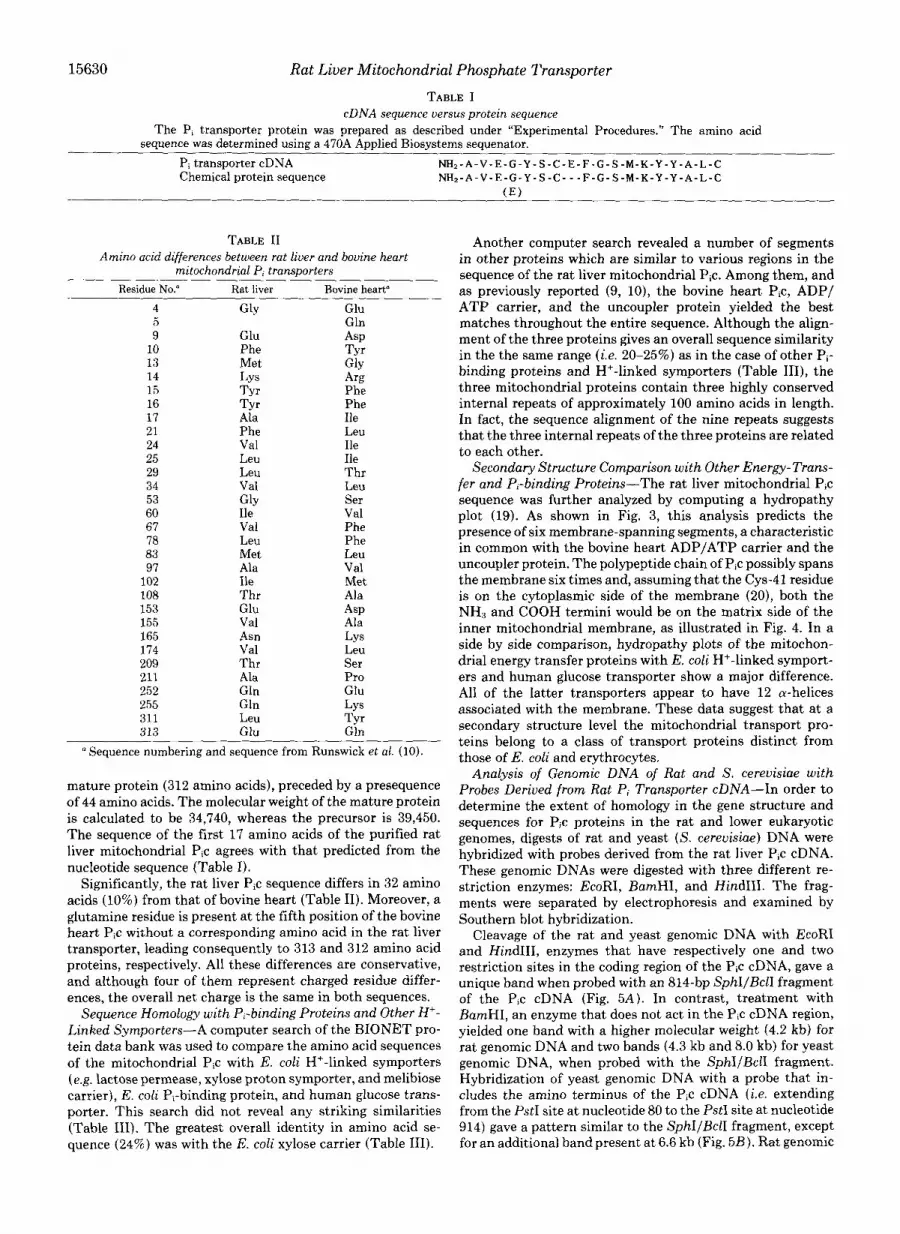
TABLE I cDNA sequence versus protein sequence
The P, transporter protein was prepared as described under "Experimental Procedures." The amino acid sequence was determined using a 470A Applied Biosystems sequenator.
Pi transporter cDNA NH2-A-V-E-G-Y-S-C-E-F-G-S-M-K-Y-Y-A-L-C Chemical protein sequence NH2-A-V-E-G-Y-S-C---F-G-S-M-K-Y-Y-A-L-C
( E )
TABLE I1 Amino acid differences between rat liver and bovine heart
mitochondrial P, transporters Residue No." Rat liver Bovine heart"
4 GlY Glu 5 Gln 9 Glu
10 ASP
Phe 13
Tyr Met GlY
14 LY s 15 T r r Phe 16 Tyr Phe 17 Ala Ile 21 Phe 24 Val
Leu Ile
25 Leu 29
Ile Leu Thr
34 53
Val Leu GlY Ser
60 Ile 67
Val Val Phe
78 Leu Phe 83 97
Met Leu Ala V a1
102 Ile Met 108 Thr Ala 153 Glu ASP 155 Val Ala 165 Asn 174
LY s Val
209 Thr Leu Ser
211 Ala 252
Pro Gln Glu
255 Gln 311
LY s Leu
313 TYr
Glu Gln ' Sequence numbering and sequence from Runswick et al. (IO).
mature protein (312 amino acids), preceded by a presequence of 44 amino acids. The molecular weight of the mature protein is calculated to be 34,740, whereas the precursor is 39,450. The sequence of the first 17 amino acids of the purified rat liver mitochondrial Pic agrees with that predicted from the nucleotide sequence (Table I).
Significantly, the rat liver Pic sequence differs in 32 amino acids (10%) from that of bovine heart (Table 11). Moreover, a glutamine residue is present at the fifth position of the bovine heart Pic without a corresponding amino acid in the rat liver transporter, leading consequently to 313 and 312 amino acid proteins, respectively. All these differences are conservative, and although four of them represent charged residue differ- ences, the overall net charge is the same in both sequences.
Sequence Homology with P,-binding Proteins and Other H'- Linked Symporters-A computer search of the BIONET pro- tein data bank was used to compare the amino acid sequences of the mitochondrial Pic with E. coli H'-linked symporters (e.g. lactose permease, xylose proton symporter, and melibiose carrier), E. coli Pi-binding protein, and human glucose trans- porter. This search did not reveal any striking similarities (Table 111). The greatest overall identity in amino acid se- quence (24%) was with the E. coli xylose carrier (Table 111).
Another computer search revealed a number of segments in other proteins which are similar to various regions in the sequence of the rat liver mitochondrial Pic. Among them, and as previously reported (9, lo), the bovine heart Pic, ADP/ ATP carrier, and the uncoupler protein yielded the best matches throughout the entire sequence. Although the align- ment of the three proteins gives an overall sequence similarity in the the same range (i.e. 20-25%) as in the case of other Pi- binding proteins and H'-linked symporters (Table 111), the three mitochondrial proteins contain three highly conserved internal repeats of approximately 100 amino acids in length. In fact, the sequence alignment of the nine repeats suggests that the three internal repeats of the three proteins are related to each other.
Secondary Structure Comparison with Other Energy-Trans- fer and P,-binding Proteins-The rat liver mitochondrial Pic sequence was further analyzed by computing a hydropathy plot (19). As shown in Fig. 3, this analysis predicts the presence of six membrane-spanning segments, a characteristic in common with the bovine heart ADP/ATP carrier and the uncoupler protein. The polypeptide chain of Pic possibly spans the membrane six times and, assuming that the Cys-41 residue is on the cytoplasmic side of the membrane (20), both the NH, and COOH termini would be on the matrix side of the inner mitochondrial membrane, as illustrated in Fig. 4. In a side by side comparison, hydropathy plots of the mitochon- drial energy transfer proteins with E. coli H+-linked symport- ers and human glucose transporter show a major difference. All of the latter transporters appear to have 12 a-helices associated with the membrane. These data suggest that at a secondary structure level the mitochondrial transport pro- teins belong to a class of transport proteins distinct from those of E. coli and erythrocytes.
Analysis of Genomic DNA of Rat and S. cereuisiae with Probes Derived from Rat Pi Transporter cDNA-In order to determine the extent of homology in the gene structure and sequences for Pic proteins in the rat and lower eukaryotic genomes, digests of rat and yeast (S. cereuisiae) DNA were hybridized with probes derived from the rat liver Pic cDNA. These genomic DNAs were digested with three different re- striction enzymes: EcoRI, BamHI, and HindIII. The frag- ments were separated by electrophoresis and examined by Southern blot hybridization.
Cleavage of the rat and yeast genomic DNA with EcoRI and HindIII, enzymes that have respectively one and two restriction sites in the coding region of the Pic cDNA, gave a unique band when probed with an 814-bp SphIIBclI fragment of the Pic cDNA (Fig. 5 A ) . In contrast, treatment with BamHI, an enzyme that does not act in the P,c cDNA region, yielded one band with a higher molecular weight (4.2 kb) for rat genomic DNA and two bands (4.3 kb and 8.0 kb) for yeast genomic DNA, when probed with the SphIIBclI fragment. Hybridization of yeast genomic DNA with a probe that in- cludes the amino terminus of the Pic cDNA (i.e. extending from the PstI site at nucleotide 80 to the PstI site at nucleotide 914) gave a pattern similar to the SphIIBclI fragment, except for an additional band present at 6.6 kb (Fig. 5B). Rat genomic
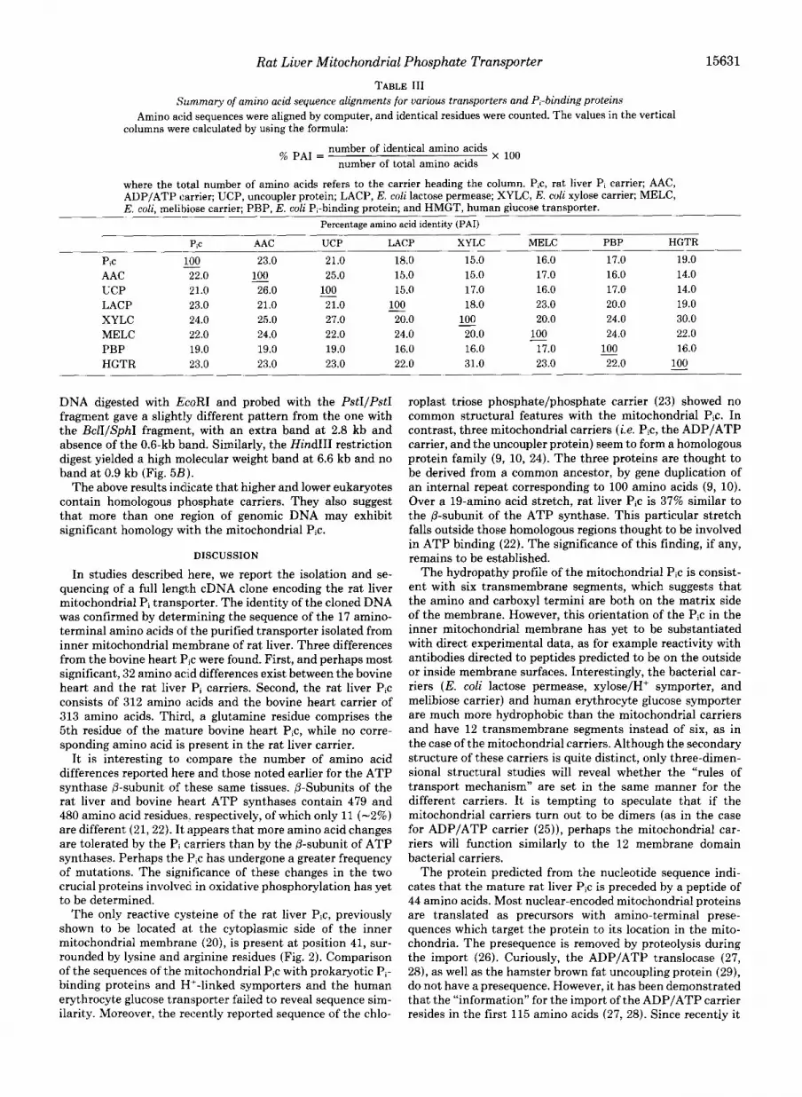
15631 Rat Liver Mitochondrial Phosphate Transporter TABLE I11
Summary of amino acid sequence alignments for various transporters and Pi-binding proteins Amino acid sequences were aligned by computer, and identical residues were counted. The values in the vertical
columns were calculated by using the formula:
number of identical amino acids number of total amino acids % PA1 = x 100
where the total number of amino acids refers to the carrier heading the column. P,c, rat liver P, carrier; AAC, ADP/ATP carrier; UCP, uncoupler protein; LACP, E. coli lactose permease; XYLC, E. coli xylose carrier; MELC, E. coli, melibiose carrier; PBP, E. coli Pi-binding protein; and HMGT, human glucose transporter.
Percentage amino acid identity (PAI) -
P,c AAC UCP LACP XYLC MELC PBP HGTR
Pic - 100 23.0 21.0 18.0 15.0 16.0 17.0 19.0
UCP 21.0 26.0 - 100 15.0 17.0 16.0 17.0 14.0 AAC 22.0 100 25.0 15.0 15.0 17.0 16.0 14.0 -
LACP 23.0 21.0 21.0 100 18.0 23.0 20.0 19.0 XYLC 24.0 25.0 27.0 20.0 100 20.0 24.0 30.0 MELC 22.0 24.0 22.0 24.0 20.0 100 24.0 22.0 PBP 19.0 19.0 19.0 16.0 16.0 17.0 100 16.0 HGTR 23.0 23.0 23.0 22.0 31.0 23.0 22.0 100
- -
- -
-
DNA digested with EcoRI and probed with the PstIIPstI fragment gave a slightl!~ different pattern from the one with the BclI/SphI fragment, with an extra band at 2.8 kb and absence of the 0.6-kb band. Similarly, the Hind111 restriction digest yielded a high molecular weight band at 6.6 kb and no band at 0.9 kb (Fig. 5B) .
The above results indicate that higher and lower eukaryotes contain homologous phosphate carriers. They also suggest that more than one region of genomic DNA may exhibit significant homology with the mitochondrial Pic.
DISCUSSION
In studies described here, we report the isolation and se- quencing of a full length cDNA clone encoding the rat liver mitochondrial Pi transporter. The identity of the cloned DNA was confirmed by determining the sequence of the 17 amino- terminal amino acids of the purified transporter isolated from inner mitochondrial membrane of rat liver. Three differences from the bovine heart Pic were found. First, and perhaps most significant, 32 amino ac-id differences exist between the bovine heart and the rat liver Pi carriers. Second, the rat liver Pic consists of 312 amino t3cids and the bovine heart carrier of 313 amino acids. Third, a glutamine residue comprises the 5th residue of the mature bovine heart Pic, while no corre- sponding amino acid is present in the rat liver carrier.
It is interesting to compare the number of amino acid differences reported here and those noted earlier for the ATP synthase @-subunit of these same tissues. @-Subunits of the rat liver and bovine heart ATP synthases contain 479 and 480 amino acid residues., respectively, of which only 11 (-2%) are different (21,22). It lippears that more amino acid changes are tolerated by the Pi carriers than by the @-subunit of ATP synthases. Perhaps the Pic has undergone a greater frequency of mutations. The significance of these changes in the two crucial proteins involved in oxidative phosphorylation has yet to be determined.
The only reactive cysteine of the rat liver Pic, previously shown to be located at. the cytoplasmic side of the inner mitochondrial membrane (20), is present at position 41, sur- rounded by lysine and arginine residues (Fig. 2). Comparison of the sequences of the mitochondrial Pic with prokaryotic Pi- binding proteins and H’-linked symporters and the human erythrocyte glucose transporter failed to reveal sequence sim- ilarity. Moreover, the recently reported sequence of the chlo-
roplast triose phosphate/phosphate carrier (23) showed no common structural features with the mitochondrial Pic. In contrast, three mitochondrial carriers (i.e. Pic, the ADP/ATP carrier, and the uncoupler protein) seem to form a homologous protein family (9, 10, 24). The three proteins are thought to be derived from a common ancestor, by gene duplication of an internal repeat corresponding to 100 amino acids (9, 10). Over a 19-amino acid stretch, rat liver Pic is 37% similar to the @-subunit of the ATP synthase. This particular stretch falls outside those homologous regions thought to be involved in ATP binding (22). The significance of this finding, if any, remains to be established.
The hydropathy profile of the mitochondrial Pic is consist- ent with six transmembrane segments, which suggests that the amino and carboxyl termini are both on the matrix side of the membrane. However, this orientation of the Pic in the inner mitochondrial membrane has yet to be substantiated with direct experimental data, as for example reactivity with antibodies directed to peptides predicted to be on the outside or inside membrane surfaces. Interestingly, the bacterial car- riers (E. coli lactose permease, xylose/H+ symporter, and melibiose carrier) and human erythrocyte glucose symporter are much more hydrophobic than the mitochondrial carriers and have 12 transmembrane segments instead of six, as in the case of the mitochondrial carriers. Although the secondary structure of these carriers is quite distinct, only three-dimen- sional structural studies will reveal whether the “rules of transport mechanism” are set in the same manner for the different carriers. I t is tempting to speculate that if the mitochondrial carriers turn out to be dimers (as in the case for ADP/ATP carrier (25)), perhaps the mitochondrial car- riers will function similarly to the 12 membrane domain bacterial carriers.
The protein predicted from the nucleotide sequence indi- cates that the mature rat liver Pic is preceded by a peptide of 44 amino acids. Most nuclear-encoded mitochondrial proteins are translated as precursors with amino-terminal prese- quences which target the protein to its location in the mito- chondria. The presequence is removed by proteolysis during the import (26). Curiously, the ADP/ATP translocase (27, 28), as well as the hamster brown fat uncoupling protein (29), do not have a presequence. However, it has been demonstrated that the “information” for the import of the ADP/ATP carrier resides in the first 115 amino acids (27, 28). Since recently it

15632 Rat Liver Mitochondrial Phosphate Transporter
E. coli LACXVSE PERMEASE 1
u "
-a - 1. n. m. Iv. v. VI. -3B - 4- 4-
9 -._ . . . . . . . . . . . . . . . " . . . . . . . . . . . . . . . . . . . . . c P .b 1 u1 lzm' sa' Z 4 d d 1- 8.' tu1 - 2-
50* 4 0 - lJn-- Ea ! " E.coli I(ELIB1OSE CARRIER
40 - 1-
a - la - e -
-le - -2ll - -30 - 48-
30-
-41)-
-51) 4 la . . . . . . . . . . . . . . . 1m 2 u ad 7 JII m- m' Y' 4Sa1
.I.* FIG. 3. Comparison of the hydropathy profiles of the Pi transporter, ADP/ATP translocase, uncou-
pler protein, and E. coli H+-linked symporters. Consecutive hydropathy averages are plotted for a window of 11 amino acids advancing from the NH3 to the COOH terminus. (Z, ZZ = internal repeat 1; III, IV = internal repeat 2; V, VI = internal repeat 3). Uncoupler = uncoupler binding protein.
FIG. 4. Secondary structure model of the rat liver mito- chondrial Pi transporter based on the hydropathy profile of the protein. Hydrophobic segments are shown in bones representing transmembrane cu-helical domains. Hydrophilic loops are shown con- necting the transmembrane regions.
has been shown that the uncoupling protein has no prese- quence as well (29, 30), probably its import process in the mitochondrial membrane is similar to that of ADP/ATP carrier. These findings raise the question of how Pic is im- ported into mitochondria. Is the presequence involved or is the import process similar to that of the ADPjATP carrier?
Analysis of genomic DNA from both rat and S. cereuisiue by Southern blots, using probes to almost the entire coding region of the rat mitochondrial Pic, reveals a similar restric- tion pattern. This suggests that mitochondrial Pic as the mitochondrial ADP/ATP carrier shares significant homology among species both at the amino acid and the gene structural level. Comparison of published nucleotide sequences of human and yeast ADP/ATP carriers reveals a 53.5% overall homol- ogy, with many stretches of DNA having 80-90% homology. The hybridization data suggest also that the 5' region of the rat liver Pjc cDNA shows homology with more genomic frag- ments than the most distal region of the Pic. Whether these are simply repeated sequences which share homologies or represent multiple genes for the same transporter cannot be determined at this time. The existence of multiple genes for

Rat Liver Mitochondrial Phosphate Transporter 15633
A Yeast Rat
H B E H B E 23.1-
P f t I Soh I
B Yeast Rat
H B E H B E
Pst-Pst > 23.1- 9 4- 6.6- m .: .? .. 2.3- 2.0.
1 .o-
0.6-
Sph-Bcll Hlndl I I
EcoRI Hlndlll P s t l P s t l I B c l l
Presequence P I Carr rer
188 bp
FIG. 5. Hybridization of genomic DNA of rat and the yeast S. cereuisiae with probes derived from rat Pi transporter cDNA. Genomic DNA from rat and S. cerevisiae was digested with: 1) HindIII ( H ) ; 2) BamHI ( B ) ; and 3) EcoRI ( E ) , subjected to electrophoresis, transferred to Hybond-N membranes, and hybridized with the probes as described under “Methods.” A, Southern blot probed with 814-bp SphI-BclI fragment. R, Southern blot probed with 834-hp PstI-PstI fragment. The markers are the positions of frag- ments of DNA from bacteriophage X generated by digestion with the restriction enzyme HindIII.
mitochondrial transporters has been shown for both yeast and mammalian systems (31).
Knowledge of the molecular structure and topology of the P, carrier in the membrane is a prerequisite to the understand- ing of the mechanism of Pi transport across the membrane. With a full length cDNA clone encoding the Pi transporter, we can now answer questions pertinent to the molecular mechanism of Pi transport across the mitochondrial mem- brane as well as the role of individual amino acids in the transport process.
Acknowledgments-We wish to thank Dr. P. Shenbagamurthi for performing the amino-terminal amino acid sequencing of the mito- chondrial phosphate carrier. We are grateful to David Garboczi for carefully reading the manuscript and for his advice throughout the course of this work. We also thank Dr. Xavier Ysern for discussions throughout the study. Finally, we thank Starlene Murray for process- ing this manuscript for publication.
REFERENCES
1. LaNoue, K. F., and Schoolwerth, A. C. (1979) Annu. Rev. Riochem. 48,871-922
2. Klingenherg, M. (1985) in The Enzymes of Biological Membranes (Martonosi, A. N., ed) Vol. 1, pp. 511-553, Plenum Publishing Co., New York
3. Pedersen, P. L., and Wehrle, J. P. (1982) in Membranes and Transport (Martonosi, A. N., ed) Vol. 1, pp. 645-663, Plenum Publishing Co., New York
4. Kaplan, R. S., and Pedersen, P. L. (1983) Biochem. J . 212, 279- 288
5. Kolhe, H. V. J., Costello, D., Wong, A., Lu, R. C., and Wohlrah, H. (1984) J . Biol. Chem. 259,9115-9120
6. Kaplan, R. S., Pratt, R. D., and Pedersen, P. L. (1986) J. Bid. Chem. 261, 12767-12773
7. Kaplan, R. S., Pratt, R. D., and Pedersen, P. L. (1989) Methods
8. Kolbe, H. V. J., and Wohlrab, H. (1985) J . Bid. Chem. 2 6 0 , Enzymol., in press
9. Aquila, H., Link, T. A., and Klingenberg, M. (1987) FEBS Lett.
10. Runswick, M. J., Powell, S. J., Nyren, P., and Walker, J. E.
11. Benton, W. P., and Davis, R. W. (1977) Science (Washington,
12. Maniatis, T., Fritsch, E. F., and Sambrook, J. (1982) Molecular Cloning:A Laboratory Manual, Cold Spring Harbor Laboratory, Cold Spring Harbor, NY
13. Feinberg, A. P., and Vogelstein, B. (1983) Anal. Biochem. 132,
14. Sanger, F., Nicklen, S., and Coulson, A. R. (1977) Proc. Natl.
15. Hirschenberg, D. S. (1975) Commun. Assoc. Comput. Mach. 18,
16. Davis, L. G., Dibner, M. D., and Battey, J. F. (1986) in Basic Methods in Molecular Biology, Elsevier Science Publishing Co., Inc., New York
17. Sherman, F., Fink, G. R., and Hicks, J. B. (1986) in Methods in Yeast Genetics, Cold Spring Harbor Laboratory, Cold Spring Harbor, NY
18. Hames, B. D., and Higgens, S. S. (1986) Nucleic Acid Hybridiza- tion: A Practical Approach, pp. 113-133, IRC Press, Washing- ton, D.C.
15899-15906
2 1 2 , l - 9
(1987) EMBO J. 6, 1367-1373
D.C.) 196, 180-184
6-13
Acad. Sci. U. S. A. 74, 5463-5467
341-343
19. Kyte, J., and Doolittle, R. F. (1982) J. Mol. Biol. 157, 105-132 20. Houstek, d., and Pedersen, P. L. (1985) J. Biol. Chem. 260,6288-
21. Breen, G. A. M., Holmans, P. L., and Garnett, K. E. (1988)
22. Garboczi, D. N., Fox, A. H., Gerring, S. L., and Pedersen, P. L.
23. Flugge, U. I., Fisher, K., Gross, A,, Sehald, W., Lottspeich, F.,
24. Saraste, M., and Walker, J. E. (1982) FEBS Lett. 144, 250-254 25. Aquila, H., Link, T. A., and Klingenberg, M. (1985) EMBOJ. 4,
26. Hay, R., Bohni, P., and Gasser, S. (1984) Biochim. Biophys. Acta
27. Smagula, C., and Douglas, M. G. (1988) J . Biol. Chem. 263, 6783-6790
28. Adrian, G. S., McCammon, M. T., Montgomery, D. L., and Douglas, M. G. (1986) Mol. Cell. Bid. 6, 626-634
29. Kozak, L. P., Britton, J. H., Kozak, U. C., and Wells, J. M. (1988) J. Biol. Chem. 263, 12274-12277
30. Ridley, R. G., Patel, H. V., Gerher, G. E., Morton, R. C., and Freeman, K. B. (1986) NucleicAcid.7 Res. 14, 4025-4035
31. Lawson, J . E., and Douglas, M. G. (1988) J. Biol. Chem. 263 ,
6295
Biochemistry 27, 3955-3961
(1988) Biochemistry 27,553-560
and Eckerskorn, C. (1989) EMBO J. 8, 39-46
2369-2376
7 7 9 , 6 5 4 7
14812-14818