supramolecular chemistry of anion-binding receptors ... - jyx
TRANSCRIPT
Anniina Kiesilä
JYU DISSERTATIONS 137
Supramolecular Chemistry of Anion-Binding Receptors Based on Concave Macrocycles
Anniina Kiesilä
Supramolecular Chemistry of Anion-Binding Receptors
Based on Concave Macrocycles
Esitetään Jyväskylän yliopiston matemaattis-luonnontieteellisen tiedekunnan suostumuksellajulkisesti tarkastettavaksi yliopiston Ylistönrinteen salissa YlistöKem4
lokakuun 11. päivänä 2019 kello 12.
Academic dissertation to be publicly discussed, by permission ofthe Faculty of Mathematics and Science of the University of Jyväskylä,
in Ylistönrinne, auditorium YlistöKem4, on October 11, 2019 at 12 o’clock noon.
JYU DISSERTATIONS 137
JYVÄSKYLÄ 2019

EditorsElina KaleniusDepartment of Chemistry, University of JyväskyläVille KorkiakangasOpen Science Centre, University of Jyväskylä
ISBN 978-951-39-7855-6 (PDF)URN:ISBN:978-951-39-7855-6ISSN 2489-9003
Copyright © 2019, by University of Jyväskylä Permanent link to this publication: http://urn.fi/URN:ISBN:978-951-39-7855-6
ABSTRACT
Kiesilä, Anniina Supramolecular chemistry of anion-binding receptors based on concave macrocycles Jyväskylä: University of Jyväskylä, 2019, 68 p. (JYU Dissertations ISSN 2489-9003; 137) ISBN 978-951-39-7855-6 (PDF) This thesis describes the supramolecular chemistry of different anion-binding host molecules with a special focus on pyridine[4]arenes. The first part of the thesis gives a brief introduction to supramolecular chemistry and its special features, host-guest chemistry and self-assembly. Later on, a short review of different hosts is presented, especially focusing on those utilized in the experimental part of the thesis. In the second part of the thesis an introduction is presented to the structural analyzation techniques utilized in the experimental part. A special focus is set on ion mobility mass spectrometry, a relatively new technique for structural characterization of supramolecular complexes.
The third part presents the summary of experimental work. The binding properties of pyridinearene turned out to completely differ from previous re-ports in the literature. New binding features of pyridinearene were discovered, such as simultaneous endo- and exo-complex formation with neutral and anionic guests. The formation of the hexameric assembly of pyridinearene was also ob-served for the first time in gas phase and in solid state. In solution, the thermo-dynamic balance in self-assembly was revealed.
Different analyzation techniques have been utilized in the structural char-acterisation of pyridinearenes. Ion mobility mass spectrometry proved to be spectacular for structural characterization of the gas phase assemblies. As a new technique, its utilization for structural chemistry is demonstrated in the experi-mental part with several examples.
In this thesis, also the binding properties of cyclohexylhemicucurbit[8]uril and crown ether urea receptors were revealed and thoroughly studied in gas phase, solution and in solid state. Keywords: pyridine[4]arene, supramolecular chemistry, ion mobility mass spectrometry, self-assembly, anion binding, X-ray crystallography,
Author’s address Anniina Kiesilä Department of Chemistry Nanoscience Center P.O. Box 35 FI-40014 University of Jyväskylä Finland [email protected] Supervisor Adjunct Professor Elina Kalenius Department of Chemistry Nanoscience Center University of Jyväskylä Finland Reviewers Research Director Dr. Valérie Gabelica Institut Européen de Chimie et Biologie University of Bordeaux Pessac, France Professor Alessandro Casnati Department of Chemistry, Life Sciences and Sustainability University of Parma Parma, Italy Opponents Professor David V. Dearden Department of Chemistry and Biochemistry Brigham Young University Provo, Utah, United States

ACKNOWLEDGEMENTS
The work presented in this thesis was carried out at the Department of Chemistry, University of Jyväskylä during the years 2015-2019. Partly the journey started already in 2014, when the learning experience to supramolecular chemistry started by work with crown ether urea receptors for my Master`s thesis. These years have been challenging, stressful, but also rewarding and full of excitement. I would not change any of it!
First and foremost, I want to thank Adjunct Professor Elina Kalenius for giving me the opportunity to work in this interesting project. I truly admire all your knowledge and expertise on mass spectrometry, thank you for sharing so big part of it! I feel grateful also for the independence I have received in my work, and for the guidance whenever needed. I also want to thank Professor Kari Ris-sanen for all the advices and encouragement, and also for taking me as a subunit to his wonderful research group.
Professor Alessandro Casnati and Dr. Valérie Gabelica are sincerely thanked for the pre-examination of this thesis. I feel honoured that my work has been evaluated by such distinguished scientists. I also thank Matti Nurmia for the language revision.
Doctors Filip Topić and Rakesh Puttreddy are thanked for their patience of checking my salty crystallization experiments and solving the structures of the lucky attempts. Academy research fellow Dr. Jani O. Moilanen is gratefully thanked for the collaboration and all the DFT calculations. Professor Perdita Bar-ran, Professor Christoph A. Schalley and Dr. Anneli Kruve are thanked for the collaboration during and after my research visits to Manchester and Berlin, and for the introduction to new mass spectrometry techniques.
I would also like to thank all the colleagues and staff at the department of chemistry for creating such a nice working atmosphere. The new and former members of group Rissanen, thank you for sharing the good and bad moments at work, and especially for the awesome parties we have had together! Thank you all the friends, in and out of chemistry life, you know who you are and how much you mean to me. A very special thanks goes to Jyväskylä Muay Thai and Minh “Joji” Le for taking care of my physical (and mental) health, especially after some sport injuries. The members of local paw patrol in Kotimäki are warmly thanked for the long walks and taking my mind off from work every evening.
Last but not least, I owe my deepest gratitude to my husband Jussi and my daughter Justiina. Thank you for your endless love, support and patience. With-out your encouragement this journey would have been significantly harder. I want to thank also the-one-on-the-way, for living through the stressful writing process and enabling the writing without (bad) morning sickness.
Jyväskylä 12.9.2019 Anniina Kiesilä
LIST OF ORIGINAL PUBLICATIONS
This thesis is based on the following original publications, which are referred to in the text by their Roman numerals.
I A. Kiesilä, L. Kivijärvi, N. K. Beyeh, J. O. Moilanen, M. Groessl, T.
Rothe, S. Götz, F. Topić, K. Rissanen, A. Lützen and E. Kalenius. Sim-ultaneous endo and exo complex formation of pyridine[4]arene di-mers with neutral and anionic guests, Angew. Chem. Int. Ed. 2017, 56, 10942 – 10946.
II A. Kiesilä, J. O. Moilanen, A. Kruve, C. A. Schalley, P. Barran and E.
Kalenius. Anion-driven encapsulation of cationic guest inside pyri-dine[4]arene dimers. manuscript submitted
III A. Kiesilä, N. K. Beyeh, J. O. Moilanen, R. Puttreddy, Sven Götz, K.
Rissanen, P. Barran, A. Lützen and E. Kalenius. Thermodynamically driven self-assembly of Pyridinearene to Hexameric capsules. Org. Biomol. Chem. 2019, 17, 6980 – 6984.
IV S. Kaabel, J. Adamson, F. Topić, A. Kiesilä, E. Kalenius, M. Öeren, M.
Reimund, E. Prigorchenko, A. Lõokene, H. J. Reich, K. Rissanen and R. Aav, Chiral hemicucurbit[8]uril as an anion receptor: selectivity to size, shape and charge distribution, Chem. Sci. 2017, 8, 2184 – 2190.
V T. Mäkelä, A. Kiesilä, E. Kalenius and K. Rissanen. Ion‐Pair Complex-
ation with Dibenzo[21]Crown‐7 and Dibenzo[24]Crown‐8 bis‐Urea Receptors, Chem. Eur. J. 2016, 22, 40, 14264 – 14272.
Author`s contribution
In papers I-III author has synthesized the compounds used in the experimental work. She has carried out the mass spectrometric experiments, excluding ion mo-bility mass spectrometry experiments in I. She has been involved in NMR exper-iments and has carried out most of the crystallizations trials. The author has been actively involved in writing process and has written the first drafts of the papers. In papers IV and V the author has carried out the mass spectrometric experi-ments and has contributed to the writing of the original drafts. In paper V author has also been involved in NMR experiments and carried out part of the crystalli-zation trials.
CONTENTS
ABSTRACT ACKNOWLEDGEMENTS LIST OF ORIGINAL PUBLICATIONS CONTENTS ABBREVIATIONS
1 INTRODUCTION .............................................................................................. 11 1.1 Supramolecular chemistry....................................................................... 11
1.1.1 Non-covalent interactions ............................................................ 12 1.1.2 Host-guest chemistry .................................................................... 13 1.1.3 Self-assembly ................................................................................. 16
1.2 Supramolecular hosts ............................................................................... 17 1.2.1 Resorcin[4]arenes .......................................................................... 17 1.2.2 Pyridine[4]arenes .......................................................................... 20 1.2.3 Hemicucurbiturils ......................................................................... 23 1.2.4 Crown ether-urea receptors ......................................................... 24
2 RESEARCH METHODS .................................................................................... 27 2.1 Gas phase ................................................................................................... 27
2.1.1 Mass spectrometry ........................................................................ 27 2.1.2 Ion mobility mass spectrometry ................................................. 29
2.1.2.1 IM-MS of supramolecular complexes ................................. 31 2.1.3 DFT calculations ............................................................................ 33
2.2 Solution ...................................................................................................... 34 2.2.1 NMR ................................................................................................ 34
2.2.1.1 DOSY NMR ............................................................................ 35 2.3 Solid state ................................................................................................... 35
2.3.1 X-ray crystallography ................................................................... 35
3 RESULTS AND DISCUSSION ......................................................................... 36 3.1 Aim of the work ........................................................................................ 36 3.2 Pyridine[4]arenes ...................................................................................... 37
3.2.1 Synthesis I,124 .................................................................................. 37 3.2.2 Studies of complexation properties ............................................ 38
3.2.2.1 Anionic guests I,124 ................................................................. 38 3.2.2.2 Neutral guests 124 ................................................................... 43 3.2.2.3 Cationic guests II .................................................................... 45
3.2.3 Self-assembly of pyridine[4]arenes III ......................................... 50 3.3 Hemicucurbiturils IV ................................................................................. 53 3.4 Crown ether-urea receptors V ................................................................. 56
CONCLUSIONS .......................................................................................................... 60

REFERENCES ............................................................................................................... 61
APPENDIX ................................................................................................................... 67
ABBREVIATIONS ATD Arrival time distribution CCS Collision cross section CID Collision-induced dissociation DFT Density functional theory DMSO Dimethyl sulfoxide DTIM Drift tube ion mobility DTM Diffuse trajectory method ESI Electrospray ionization ESP Electrostatic potential Et2O Diethylether EtOH Ethanol D Diffusion coefficient DCM Dichloromethane DMSO Dimethylsulfoxide DOSY Diffusion-ordered spectroscopy FTICR Fourier transform ion cyclotron resonance IM-MS Ion mobility mass spectrometry IRMPD Infrared multiphoton dissociation K Association constant MALDI Matrix assisted laser desorption ionization MeCN Acetonitrile MeOH Methanol MS Mass spectrometry m/z mass to charge ratio NMR Nuclear magnetic resonance spectroscopy TBA Tetrabutylammonium TFA Trifluoroacetic acid THF Tetrahydrofuran TIMS Trapped ion mobility spectrometry TWIMS Travelling wave ion mobility spectrometry
1.1 Supramolecular chemistry
The origin of supramolecular chemistry is often ascribed to the work of Jean-Marie Lehn, Donald Cram and Charles Pedersen, who shared the Nobel Prize in 1987 for “development and use of molecules with structure-specific interactions of high selectivity”. Lehn described his area of research as “chemistry beyond the molecule”,1 whereas Donald Cram referred to the same area as host-guest chemistry.2 These descriptions of supramolecular chemistry illustrate the weak, non-covalent interactions between molecules, which can organize together to form a supramolecular complex. The concept of supramolecular chemistry reaches further in history; the first definitions date to year 1978. Emil Fischer introduced already in 1894 a lock and key-principle, which describes the basic concepts of host-guest chemistry.3,4 The classical main areas in supramolecular chemistry are host-guest chemistry and self-assembly, but today supramolecular chemistry is understood as a much wider field. The 2016 Nobel prize was awarded to Jean-Pierre Sauvage, Bernard Feringa and Sir Fraser Stoddart for “the design and synthesis of molecular machines” based on supramolecular interac-tions. This type of nanochemistry is under a rapid development, for example, in constructing highly organized structures and nanomaterials. 5,6
Even though the area of supramolecular chemistry has spread enormously, the nature of supramolecular interactions remains the same. The term “weak” does not always describe the supramolecular interactions, as they usually work cooperatively to maximize the binding strength and to stabilize the formed complex.7,8 Yet, the key role in the non-covalent interactions, and why they are called weak, is that they are reversible. Many biological processes are based on such interactions, for example, the folding of DNA double helix and the transportation of ions through the cell membrane.9 Therefore supramolecular chemistry is often inspired by the nature, as many supramolecular processes mimic those happening in biological context. In the next chapter the non-covalent
1 INTRODUCTION
12 interactions are explained briefly and the key concepts in host-guest chemistry and self-assemby are discussed in detail.
1.1.1 Non-covalent interactions
Non-covalent interactions are forces that govern the formation of supramolecular complexes. In order to interact, the components need to have attraction towards each other, but the term non-covalent also comprehends repulsive interactions. A key feature in non-covalent nature is that the interactions are reversible, which provides the complex an ability to break and form again.
Ion-ion interactions are the strongest weak interactions; they are comparable to covalent bonds in strength, varying from 100 to 350 kJ mol-1. In ionic bonds the components have electrostatic attraction towards each other as ionic bonds are formed between two ions of opposite charges, forming salts.9 Ion-dipole interactions are formed between an ion and a polar molecule that has a dipole. The strength of ion-dipole interaction varies between 50-200 kJ mol-1. Coordination bonds are also counted as ion-dipole interactions, which makes it sometimes hard to distinguish supramolecular and molecular species from each other.9
Matching of two dipoles with opposing polarities towards each other can form surprisingly strong dipole-dipole interactions, usually in the range of 5-50 kJ mol-1. The dipole-dipole interactions are typical, for example, in carbonyl compounds, which can align the opposing dipoles towards each other.9 Hydrogen bonding is a type of dipole-dipole interaction where hydrogen is attached to an electronegative atom or electron withdrawing group. Hydrogen bond (HB) is generally described as a D-H··· A system where D (donor) and A (acceptor) are electronegative atoms or electron withdrawing groups, which share the hydrogen. Typical distances between hydrogen bonded O··· O are 2.50-2.80 Å, but significant interactions can exist also 3.0 Å apart. The angle of HB is close to linear (180°) but in weaker HB the angle can be between 100-150°. The bond strength can vary from 10 to 120 kJ mol-1. Hydrogen bonding is one of the most important interactions existing in nature.9
Halogen bond (XB) can be described, similarly to hydrogen bonds, as an R-X···Y system. In this system R is a carbon, nitrogen or halogen atom where electrophilic halogen atom X (Lewis acid, XB donor) is attached. Y is a donor of electron density (Lewis base, XB acceptor). Halogen bonds strongly resemble hydrogen bonding, although their polarizability makes them more sensitive to steric hindrance. Also notable, and one key feature in halogen bond, is that they are highly linear. The angle of R-X··· Y system is close to 180°. The strength of halogen bonds varies from 10 to 200 kJ mol-1.10,11
π-interactions consist of cation-π, anion-π, π-π and CH-π interactions. A π system is typically an aromatic ring. Cation-π interactions are thus formed between cation and aromatic ring. The permanent quadrupole moment (Qzz) formed in electron-rich benzene rings forms negatively charged surface areas in the middle of the ring, where the cation can interact with the ring center.12 As the
13
aromatic ring is electron rich, it is intuitive that it would cause repulsive interaction with anions. However, anion-π interactions are formed when an anion is interacting with an electron-deficient aromatic ring. In contrast to cation-π, the ring has several electron withdrawing groups (as in hexafluorobenzene), which creates an electron deficient center to the middle of the ring. The anion can then interact with the electron deficient center of the aromatic ring.13,14 π-π interactions are formed between two aromatic rings, from which one is usually electron rich and the other electron deficient. Most common orientations of the two aromatic rings are face-to-face and edge-to-face. Aromatic rings cannot be stacked straight on top of each other, as it causes repulsions.9 CH-π interactions are formed between CH groups and π systems.15
Van der Waal forces, also known as dispersion forces, are the weakest of intermolecular forces. They arise from polarization of electron cloud within a molecule caused by proximity of an adjacent nucleus. VdW forces are nondirectional and hence cannot be utilized for designing of specific host molecules. Although VdW forces don’t seem to play a major role in the group of weak interactions, it should be still noted that their energy is proportional to their surface area. Therefore in larger molecules VdW forces do play a role. In supramolecular chemistry, VdW forces are the ones that usually govern the inclusion of a small organic compound inside cavities.9
Hydrophobic effect arises from exclusion of non-polar groups in aqueous solution. Water molecules prefer to interact with other polar groups and repel non-polar ones, as the non-polarity creates a “hole” without any interactions for water molecules. Therefore for example dichloromethane and water are immiscible, as the components aggregate rather than being dispersed. Hydrophobic effect can also be understood as an empirical rule “like dissolves like”. The phenomenon applies to non-polar molecules in aqueous solution and also to water molecules in non-polar solvents. Therefore more generally the term solvophobic effect is preferred to describe the whole phenomenon. Hydrophobic effect can be a key feature in high binding of a guest inside a host molecule in aqueous solution. 16,17
1.1.2 Host-guest chemistry
In host-guest chemistry, the host is typically a larger molecule that has a suitable binding site for a smaller molecule, the guest. The host can also be a larger aggre-gate composed of several molecules. The host can have a hole or cavity to accom-modate the guest. The binding site needs to be suitable for guest binding without repulsive interactions. 9
The background of host-guest chemistry lies in Emil Fisher`s lock-key prin-ciple that dates back to 1894.3,4 In his theory, Fisher illustrated how in order for a substrate (key) to bind an enzyme (lock), the components need to be complemen-tary to each other. This laid the basis for molecular recognition where the host can distinguish a certain guest from a crowd. In 1995 Daniel Koshland corrected the theory with induced fit model.18 Whereas lock and key analogue is often seen
14 as rigid model where two components need to have perfect complementary ge-ometry, the induced fit considers how the components can make significant con-formational changes in order to fit each other. Nevertheless, both models de-scribe the nature of host (lock) and guest (key).
In supramolecular systems, the complexation rarely relies just on one spe-cific interaction. Usually the positive cooperativity has an important effect, mean-ing that the interactions enhance each other. In multivalent systems, cooperativ-ity can exist in two different forms: allosteric and chelate cooperativity. The allo-steric effect usually exists in hosts that can bind several guests. In the allosteric effect, the binding of one guest enhances the binding of the other.7 Chelate effect is more important in systems that have multiple binding sites. In coordination chemistry the binding sites are referred to as teeth, and bidental ligands form more stable complexes than unidental ones. The replacement of a bidental ligand requires significantly more energy than the replacement of two unidental lig-ands.17 Energetics can be expressed in terms of the Gibbs free energy change (ΔGº) in equation (1),
∆𝐺° = ∆𝐻° − 𝑇∆𝑆° (1)
where ΔHº is the enthalpy change, T is temperature and ΔSº is entropy change. According to equation (1) the replacement of unidental ligands with bidental ones increases the energetic factor of the reaction, as more molecules become free in solution and the entropy of the system increases.17
The energetic factor is important also from the perspective of host preor-ganisation. Macrocycles, which don’t have to undergo significant conformational changes for guest binding, are highly preorganized and they benefit from the macrocyclic effect. This effect does not exist on hosts such as acyclic molecules with a long chain. This kind of host needs to change its conformation significantly, which costs energy. The entropic term becomes unfavourable when moving to-wards organization, decreasing the possibility of binding.17
For host-guest complexation the environment also plays a significant role. In solution both complex-forming components are highly solvated. The solvent is present in huge molar excess and it can compete for binding. Polar solvents can block the hydrogen bonding sites and prevent the complexation of other mol-ecules.9,19 However, the binding can be enhanced in polar media by utilization of an apolar binding site for an apolar guest.19,20
Several aspects influence the binding ability of the host. When considering complementarity, size is not all that matters and the electronic environment must meet the binding requirements. For the binding of an ionic guest, this can be il-lustrated with hard and soft acids and bases theory21 that shows how hard acids bind hard bases and soft acids soft bases. In general, the binding of guests is affected by solvent effects (polarity and hydrogen bonding), degree of solvation, the effects of the counter ion, and chelate ring size, especially in the case of cati-onic guests.9
15 The binding of anions has been considered more challenging than binding
of cations. Anions are usually larger than isoelectric cations, which means that their binding interactions are not as effective as those of cations. Anions are also sensitive to changes in pH, and might get protonated and lose their charge. Therefore anion receptors need to function in a narrow pH range. Anions also exist in a wider range of geometries, which makes the design of complementary hosts especially challenging. Solvation effects are also more crucial with anions, as they are usually highly solvated.22
In solution, ions are never far from their counter ions, and quite often they exist as contact ion pairs. The simple cation and anion receptors are actually deal-ing with ion pairs. The binding of a single ion can have a huge energetic cost, as the ions need to be first separated from each other. Therefore, the binding of an ion pair instead of single ion has an energetic benefit. When receptor binds the whole ion pair, there is no energetic cost to overcome. Also, the advantage in heteroditopic receptors over monotopic ones is that ditopic receptors usually function in a highly cooperative manner. Thus the binding of one ion (usually the cation) enhances the binding of the next ion (anion).23 This has been demon-strated with several receptor systems that without the cation have poor affinity towards anions, but with bound cation the anion binding is remarkably in-creased.24
Neutral guest molecules are not charged and therefore relatively weak in-teractions need to be utilized for their binding. These interactions must be care-fully designed to assist in binding.9
Hosts can bind a certain type of guest molecules, for example metal cations, without being selective which metal cations they bind. A receptor is called spe-cific if it is capable of binding one guest from a crowd of other similar molecules. This is called molecular recognition. Molecular recognition has been a hot topic in supramolecular chemistry for decades and it can be utilized in several appli-cations. For example, a sensor is a host which is capable of both molecular recog-nition and sensing. The binding of the guest produces in the sensor a signal that can be physically detected. The binding can cause an emission (photochemical), a current (electrochemical) or other change that can be externally detected, such as a change in the colour. 25
Other applications for molecular recognition can be found, for example, in extraction, solubilisation, and ion transport. The next chapter describes systems, that form larger assemblies to build a suitable container for guest binding. In these systems the host is not a single receptor molecule but a self-assembled structure.
16 1.1.3 Self-assembly
Self-assembly refers to a process where compounds spontaneously arrange into larger assemblies and are held together by non-covalent interactions, meaning that the formation is reversible. These assemblies can form spontaneously or they can be constructed by sophisticated design.26 The self-assembly can be utilized to build containers, such as capsules or cages, for large guest molecules. The syn-thetic workload can be minimized when large structures are self-assembled from small molecules. This produces economic and environmental benefits. The need and understanding of self-assembly is also recognized in nanochemistry. Self-assembly has already been exploited to build molecular machines, such as cate-nanes and rotaxanes.27 The self-assembly process has also been utilized to con-struct very elegant nanosized hierarchies such as the star of David (figure 1b).28,29
The self-assembly process occurs due to the attractive and repulsive inter-actions. To form an ensemble, the components need to be able to move with re-spect to each other. Therefore self-assembly is often studied in fluid systems, but it can be detected and studied also in gas phase and solid state. Many biological systems, such as folding of proteins, RNA or DNA, are governed by hydrogen bonding. Hydrogen bonds are highly selective and directional, and therefore uti-lizable for self-assembly.26 Large hydrogen bonding capsules will be discussed in detail in the following chapter. Other directional interactions, such as halogen bonding and coordination bonds of metal atoms, can also be used as building blocks in self-assembly. For example, Turunen et al. have demonstrated the utili-zation of halogen bonding to construct capsules.30,31
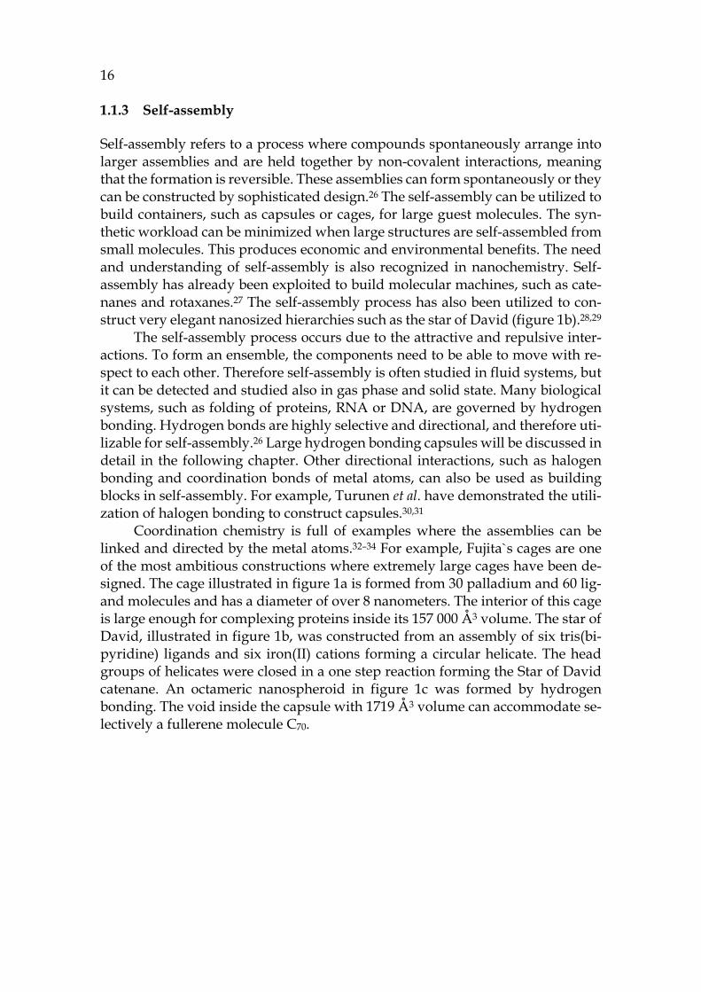
Coordination chemistry is full of examples where the assemblies can be linked and directed by the metal atoms.32–34 For example, Fujita`s cages are one of the most ambitious constructions where extremely large cages have been de-signed. The cage illustrated in figure 1a is formed from 30 palladium and 60 lig-and molecules and has a diameter of over 8 nanometers. The interior of this cage is large enough for complexing proteins inside its 157 000 Å3 volume. The star of David, illustrated in figure 1b, was constructed from an assembly of six tris(bi-pyridine) ligands and six iron(II) cations forming a circular helicate. The head groups of helicates were closed in a one step reaction forming the Star of David catenane. An octameric nanospheroid in figure 1c was formed by hydrogen bonding. The void inside the capsule with 1719 Å3 volume can accommodate se-lectively a fullerene molecule C70.
17
FIGURE 1. Self-assembled structures. a) Fujitas M30L60 icosidodecahedron b) star of David catenane and c) hydrogen bonded octameric nanospheroid. Figures adapted with permission a) from ref. 35 copyright © 2016 Elsevier Inc b) from ref. 28 copyright © 2014 Springer Nature and c) from ref. 36 CC BY 4.0, copyright © 2017 Springer Nature.
1.2 Supramolecular hosts
1.2.1 Resorcin[4]arenes
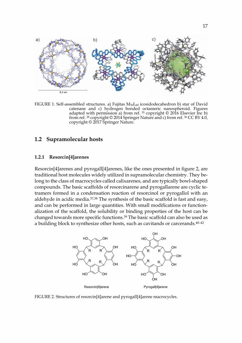
Resorcin[4]arenes and pyrogall[4]arenes, like the ones presented in figure 2, are traditional host molecules widely utilized in supramolecular chemistry. They be-long to the class of macrocycles called calixarenes, and are typically bowl-shaped compounds. The basic scaffolds of resorcinarene and pyrogallarene are cyclic te-tramers formed in a condensation reaction of resorcinol or pyrogallol with an aldehyde in acidic media.37,38 The synthesis of the basic scaffold is fast and easy, and can be performed in large quantities. With small modifications or function-alization of the scaffold, the solubility or binding properties of the host can be changed towards more specific functions.39 The basic scaffold can also be used as a building block to synthesize other hosts, such as cavitands or carcerands.40–42
FIGURE 2. Structures of resorcin[4]arene and pyrogall[4]arene macrocycles.
18
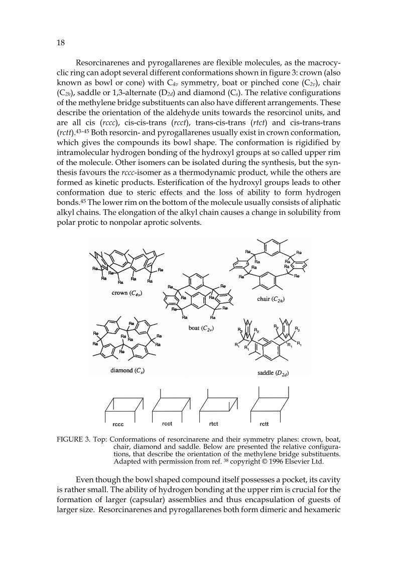
Resorcinarenes and pyrogallarenes are flexible molecules, as the macrocy-clic ring can adopt several different conformations shown in figure 3: crown (also known as bowl or cone) with C4v symmetry, boat or pinched cone (C2v), chair (C2h), saddle or 1,3-alternate (D2d) and diamond (Cs). The relative configurations of the methylene bridge substituents can also have different arrangements. These describe the orientation of the aldehyde units towards the resorcinol units, and are all cis (rccc), cis-cis-trans (rcct), trans-cis-trans (rtct) and cis-trans-trans (rctt).43–45 Both resorcin- and pyrogallarenes usually exist in crown conformation, which gives the compounds its bowl shape. The conformation is rigidified by intramolecular hydrogen bonding of the hydroxyl groups at so called upper rim of the molecule. Other isomers can be isolated during the synthesis, but the syn-thesis favours the rccc-isomer as a thermodynamic product, while the others are formed as kinetic products. Esterification of the hydroxyl groups leads to other conformation due to steric effects and the loss of ability to form hydrogen bonds.45 The lower rim on the bottom of the molecule usually consists of aliphatic alkyl chains. The elongation of the alkyl chain causes a change in solubility from polar protic to nonpolar aprotic solvents.
FIGURE 3. Top: Conformations of resorcinarene and their symmetry planes: crown, boat, chair, diamond and saddle. Below are presented the relative configura-tions, that describe the orientation of the methylene bridge substituents. Adapted with permission from ref. 38 copyright © 1996 Elsevier Ltd.
Even though the bowl shaped compound itself possesses a pocket, its cavity is rather small. The ability of hydrogen bonding at the upper rim is crucial for the formation of larger (capsular) assemblies and thus encapsulation of guests of larger size. Resorcinarenes and pyrogallarenes both form dimeric and hexameric
19
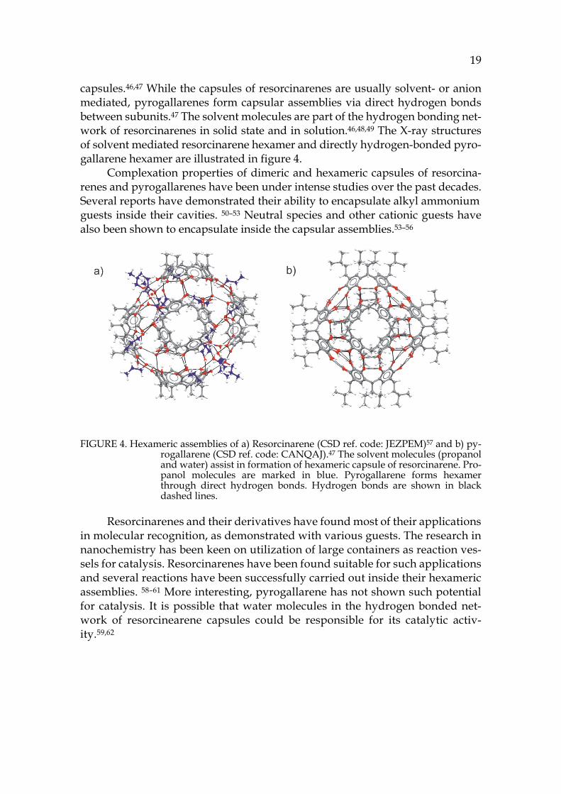
capsules.46,47 While the capsules of resorcinarenes are usually solvent- or anion mediated, pyrogallarenes form capsular assemblies via direct hydrogen bonds between subunits.47 The solvent molecules are part of the hydrogen bonding net-work of resorcinarenes in solid state and in solution.46,48,49 The X-ray structures of solvent mediated resorcinarene hexamer and directly hydrogen-bonded pyro-gallarene hexamer are illustrated in figure 4.
Complexation properties of dimeric and hexameric capsules of resorcina-renes and pyrogallarenes have been under intense studies over the past decades. Several reports have demonstrated their ability to encapsulate alkyl ammonium guests inside their cavities. 50–53 Neutral species and other cationic guests have also been shown to encapsulate inside the capsular assemblies.53–56
FIGURE 4. Hexameric assemblies of a) Resorcinarene (CSD ref. code: JEZPEM)57 and b) py-rogallarene (CSD ref. code: CANQAJ).47 The solvent molecules (propanol and water) assist in formation of hexameric capsule of resorcinarene. Pro-panol molecules are marked in blue. Pyrogallarene forms hexamer through direct hydrogen bonds. Hydrogen bonds are shown in black dashed lines.
Resorcinarenes and their derivatives have found most of their applications in molecular recognition, as demonstrated with various guests. The research in nanochemistry has been keen on utilization of large containers as reaction ves-sels for catalysis. Resorcinarenes have been found suitable for such applications and several reactions have been successfully carried out inside their hexameric assemblies. 58–61 More interesting, pyrogallarene has not shown such potential for catalysis. It is possible that water molecules in the hydrogen bonded net-work of resorcinearene capsules could be responsible for its catalytic activ-ity.59,62
20 1.2.2 Pyridine[4]arenes
Pyridine[4]arenes63 are interesting compounds, as they structurally possess clear similarities to resorcinarenes. The interesting feature in pyridinearenes is that they show completely different binding properties compared to resorcinarenes. While resorcinarenes are recognized for their cation binding properties, pyri-dinearenes have shown affinity towards anionic guests.64,65
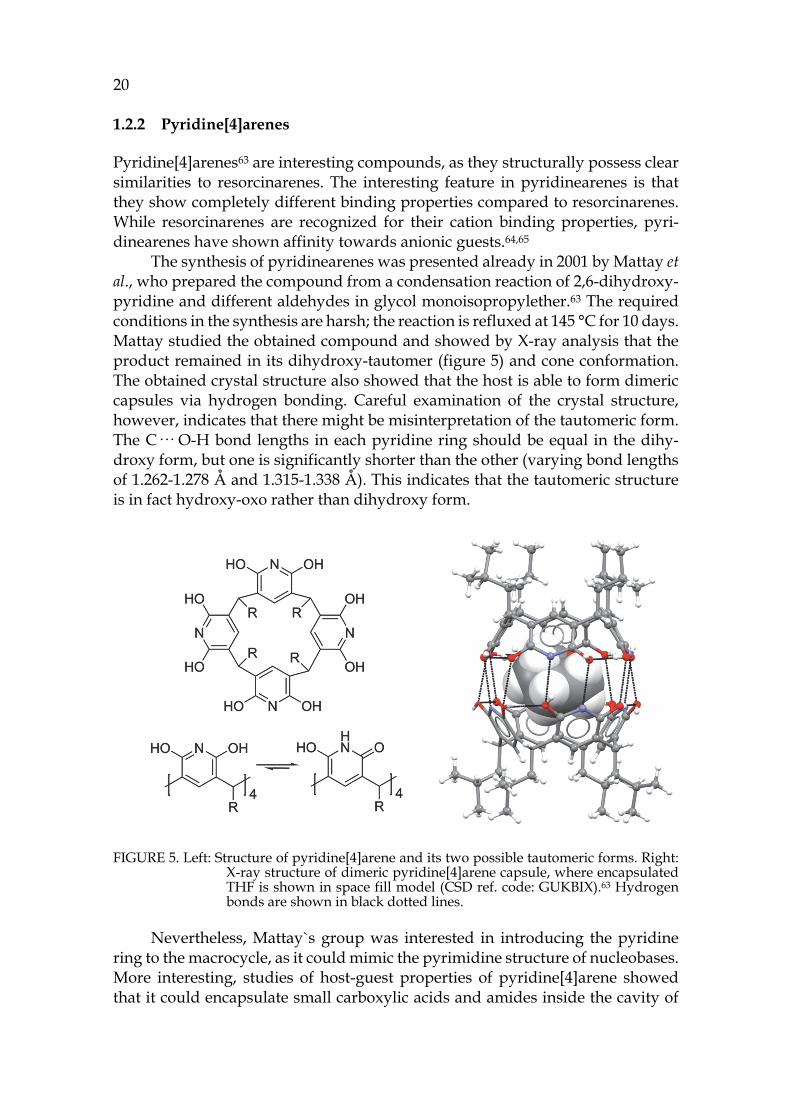
The synthesis of pyridinearenes was presented already in 2001 by Mattay et al., who prepared the compound from a condensation reaction of 2,6-dihydroxy-pyridine and different aldehydes in glycol monoisopropylether.63 The required conditions in the synthesis are harsh; the reaction is refluxed at 145 °C for 10 days. Mattay studied the obtained compound and showed by X-ray analysis that the product remained in its dihydroxy-tautomer (figure 5) and cone conformation. The obtained crystal structure also showed that the host is able to form dimeric capsules via hydrogen bonding. Careful examination of the crystal structure, however, indicates that there might be misinterpretation of the tautomeric form. The C··· O-H bond lengths in each pyridine ring should be equal in the dihy-droxy form, but one is significantly shorter than the other (varying bond lengths of 1.262-1.278 Å and 1.315-1.338 Å). This indicates that the tautomeric structure is in fact hydroxy-oxo rather than dihydroxy form.
FIGURE 5. Left: Structure of pyridine[4]arene and its two possible tautomeric forms. Right: X-ray structure of dimeric pyridine[4]arene capsule, where encapsulated THF is shown in space fill model (CSD ref. code: GUKBIX).63 Hydrogen bonds are shown in black dotted lines.
Nevertheless, Mattay`s group was interested in introducing the pyridine ring to the macrocycle, as it could mimic the pyrimidine structure of nucleobases. More interesting, studies of host-guest properties of pyridine[4]arene showed that it could encapsulate small carboxylic acids and amides inside the cavity of
21
the dimer. This was supported by electrospray Fourier transform ion cyclotron resonance mass spectrometry (ESI-FTICR-MS), nuclear magnetic resonance (NMR) experiments and by density functional theory (DFT) calculations.64 In ESI-MS, the complexation was observed only for the dimer, and there seemed to be a clear preference in the size of the complex forming guests. These facts lead the authors to believe that guests are incorporated inside the cavity.64
The authors tried to prove the encapsulation by performing infrared mul-tiphoton dissociation (IRMPD) experiments for the observed complexes. In IRMPD the ions are isolated and irradiated to produce fragmentation, which can yield structural information. From studied [dimer+guest+H]+ ions the elim-ination of a neutral guest was observed at the first step, producing a [dimer+H]+ ion. The hydrogen bonded capsule further breaks apart showing [mono-mer+H]+ ion. The authors assigned this behaviour as endo-complexation of the guest by claiming that the [monomer+guest+H]+ ion is not stable enough to be observed. They still raised the point that the guest might also be bound outside the capsule. 64
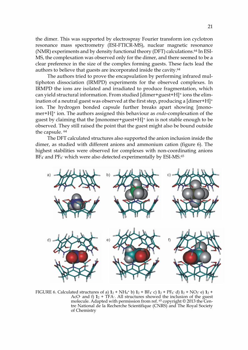
The DFT calculated structures also supported the anion inclusion inside the dimer, as studied with different anions and ammonium cation (figure 6). The highest stabilities were observed for complexes with non-coordinating anions BF4- and PF6- which were also detected experimentally by ESI-MS.65
FIGURE 6. Calculated structures of a) 12 + NH4+ b) 12 + BF4- c) 12 + PF6- d) 12 + NO3- e) 12 + AcO- and f) 12 + TFA-. All structures showed the inclusion of the guest molecule. Adapted with permission from ref. 65 copyright © 2013 the Cen-tre National de la Recherche Scientifique (CNRS) and The Royal Society of Chemistry
22
The binding properties of pyridinearene have also been studied in solution, where complexation of TFA could be demonstrated by 19F NMR. In chloroform solution, pyridine[4]arene forms two species, which were first assigned as mon-omer and dimer.64 Later Evan-Salem & Cohen proved by DOSY NMR that these signals corresponded to dimer and hexamer by using resorcinarenes of similar size as reference compounds.66 In the same study, the encapsulation of chloro-form molecules to pyridinearene capsules was detected.
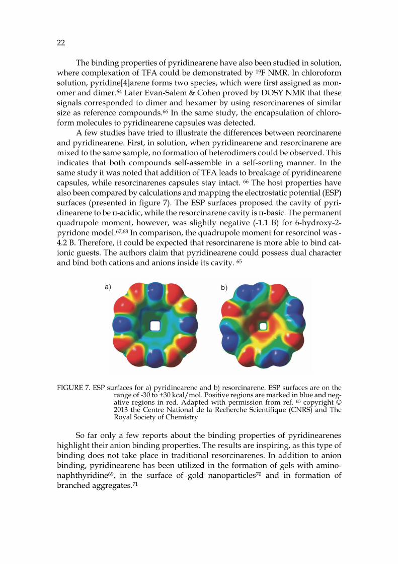
A few studies have tried to illustrate the differences between reorcinarene and pyridinearene. First, in solution, when pyridinearene and resorcinarene are mixed to the same sample, no formation of heterodimers could be observed. This indicates that both compounds self-assemble in a self-sorting manner. In the same study it was noted that addition of TFA leads to breakage of pyridinearene capsules, while resorcinarenes capsules stay intact. 66 The host properties have also been compared by calculations and mapping the electrostatic potential (ESP) surfaces (presented in figure 7). The ESP surfaces proposed the cavity of pyri-dinearene to be π-acidic, while the resorcinarene cavity is π-basic. The permanent quadrupole moment, however, was slightly negative (-1.1 B) for 6-hydroxy-2-pyridone model.67,68 In comparison, the quadrupole moment for resorcinol was -4.2 B. Therefore, it could be expected that resorcinarene is more able to bind cat-ionic guests. The authors claim that pyridinearene could possess dual character and bind both cations and anions inside its cavity. 65
FIGURE 7. ESP surfaces for a) pyridinearene and b) resorcinarene. ESP surfaces are on the range of -30 to +30 kcal/mol. Positive regions are marked in blue and neg-ative regions in red. Adapted with permission from ref. 65 copyright © 2013 the Centre National de la Recherche Scientifique (CNRS) and The Royal Society of Chemistry
So far only a few reports about the binding properties of pyridinearenes highlight their anion binding properties. The results are inspiring, as this type of binding does not take place in traditional resorcinarenes. In addition to anion binding, pyridinearene has been utilized in the formation of gels with amino-naphthyridine69, in the surface of gold nanoparticles70 and in formation of branched aggregates.71
23
1.2.3 Hemicucurbiturils
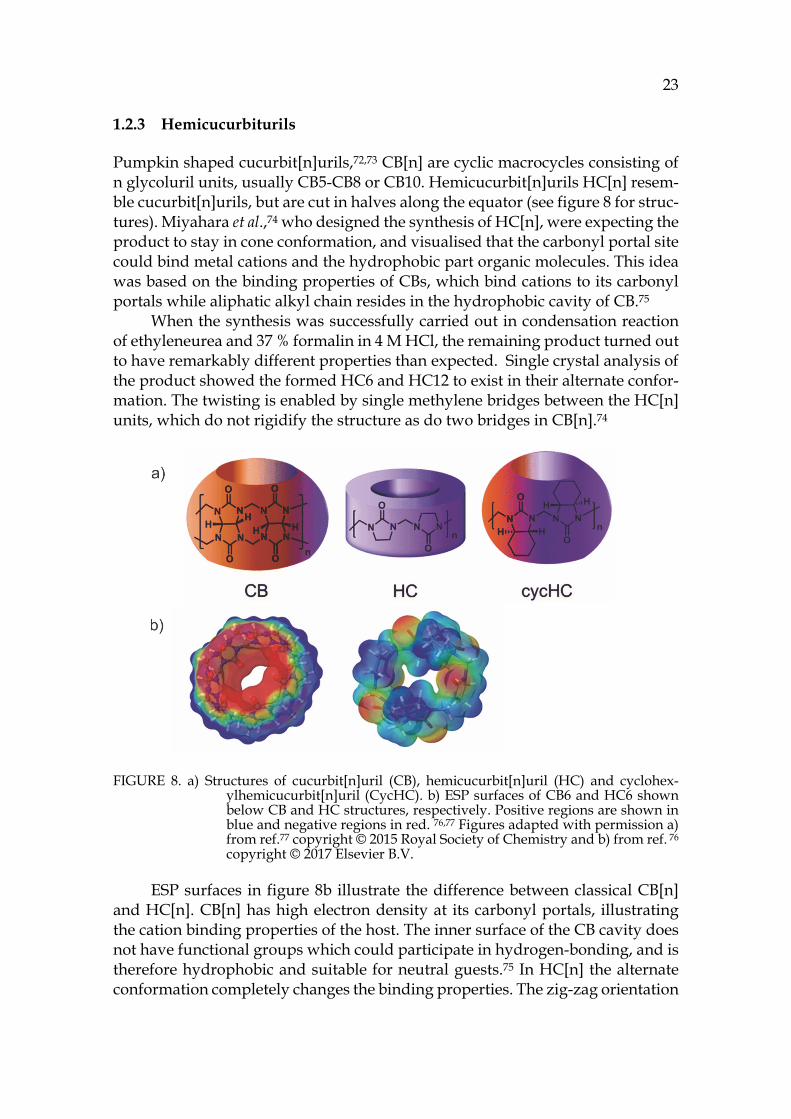
Pumpkin shaped cucurbit[n]urils,72,73 CB[n] are cyclic macrocycles consisting of n glycoluril units, usually CB5-CB8 or CB10. Hemicucurbit[n]urils HC[n] resem-ble cucurbit[n]urils, but are cut in halves along the equator (see figure 8 for struc-tures). Miyahara et al.,74 who designed the synthesis of HC[n], were expecting the product to stay in cone conformation, and visualised that the carbonyl portal site could bind metal cations and the hydrophobic part organic molecules. This idea was based on the binding properties of CBs, which bind cations to its carbonyl portals while aliphatic alkyl chain resides in the hydrophobic cavity of CB.75
When the synthesis was successfully carried out in condensation reaction of ethyleneurea and 37 % formalin in 4 M HCl, the remaining product turned out to have remarkably different properties than expected. Single crystal analysis of the product showed the formed HC6 and HC12 to exist in their alternate confor-mation. The twisting is enabled by single methylene bridges between the HC[n] units, which do not rigidify the structure as do two bridges in CB[n].74
FIGURE 8. a) Structures of cucurbit[n]uril (CB), hemicucurbit[n]uril (HC) and cyclohex-ylhemicucurbit[n]uril (CycHC). b) ESP surfaces of CB6 and HC6 shown below CB and HC structures, respectively. Positive regions are shown in blue and negative regions in red. 76,77 Figures adapted with permission a) from ref.77 copyright © 2015 Royal Society of Chemistry and b) from ref. 76 copyright © 2017 Elsevier B.V.
ESP surfaces in figure 8b illustrate the difference between classical CB[n] and HC[n]. CB[n] has high electron density at its carbonyl portals, illustrating the cation binding properties of the host. The inner surface of the CB cavity does not have functional groups which could participate in hydrogen-bonding, and is therefore hydrophobic and suitable for neutral guests.75 In HC[n] the alternate conformation completely changes the binding properties. The zig-zag orientation
24 of the carbonyl groups scatters the electron density and leaves the inner part of the macrocycle rather electron deficient.
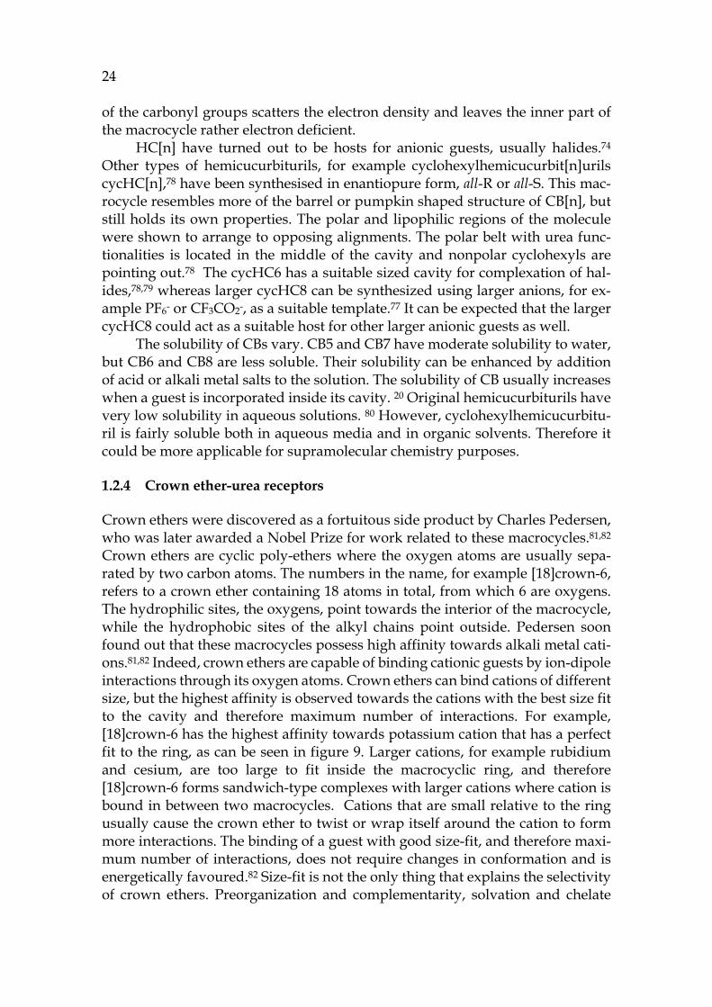
HC[n] have turned out to be hosts for anionic guests, usually halides.74 Other types of hemicucurbiturils, for example cyclohexylhemicucurbit[n]urils cycHC[n],78 have been synthesised in enantiopure form, all-R or all-S. This mac-rocycle resembles more of the barrel or pumpkin shaped structure of CB[n], but still holds its own properties. The polar and lipophilic regions of the molecule were shown to arrange to opposing alignments. The polar belt with urea func-tionalities is located in the middle of the cavity and nonpolar cyclohexyls are pointing out.78 The cycHC6 has a suitable sized cavity for complexation of hal-ides,78,79 whereas larger cycHC8 can be synthesized using larger anions, for ex-ample PF6- or CF3CO2-, as a suitable template.77 It can be expected that the larger cycHC8 could act as a suitable host for other larger anionic guests as well.
The solubility of CBs vary. CB5 and CB7 have moderate solubility to water, but CB6 and CB8 are less soluble. Their solubility can be enhanced by addition of acid or alkali metal salts to the solution. The solubility of CB usually increases when a guest is incorporated inside its cavity. 20 Original hemicucurbiturils have very low solubility in aqueous solutions. 80 However, cyclohexylhemicucurbitu-ril is fairly soluble both in aqueous media and in organic solvents. Therefore it could be more applicable for supramolecular chemistry purposes.
1.2.4 Crown ether-urea receptors
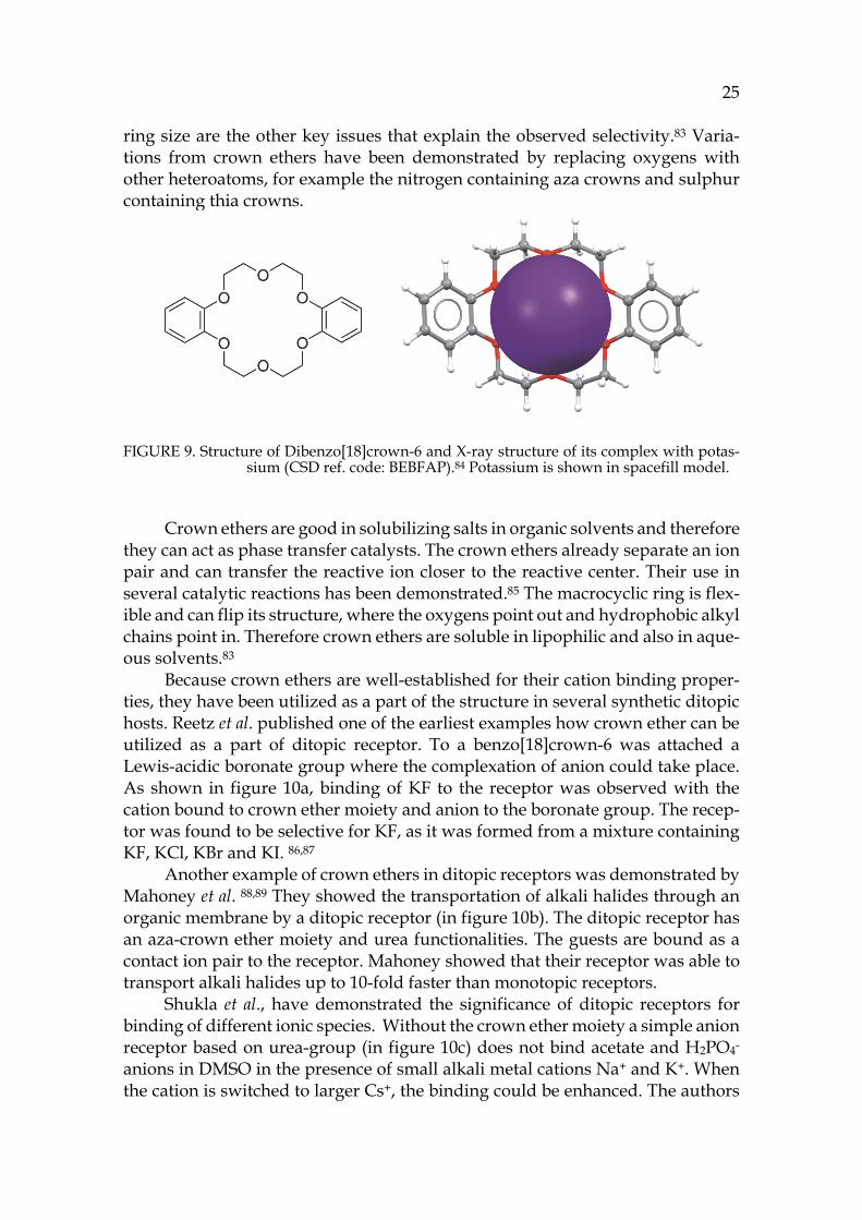
Crown ethers were discovered as a fortuitous side product by Charles Pedersen, who was later awarded a Nobel Prize for work related to these macrocycles.81,82 Crown ethers are cyclic poly-ethers where the oxygen atoms are usually sepa-rated by two carbon atoms. The numbers in the name, for example [18]crown-6, refers to a crown ether containing 18 atoms in total, from which 6 are oxygens. The hydrophilic sites, the oxygens, point towards the interior of the macrocycle, while the hydrophobic sites of the alkyl chains point outside. Pedersen soon found out that these macrocycles possess high affinity towards alkali metal cati-ons.81,82 Indeed, crown ethers are capable of binding cationic guests by ion-dipole interactions through its oxygen atoms. Crown ethers can bind cations of different size, but the highest affinity is observed towards the cations with the best size fit to the cavity and therefore maximum number of interactions. For example, [18]crown-6 has the highest affinity towards potassium cation that has a perfect fit to the ring, as can be seen in figure 9. Larger cations, for example rubidium and cesium, are too large to fit inside the macrocyclic ring, and therefore [18]crown-6 forms sandwich-type complexes with larger cations where cation is bound in between two macrocycles. Cations that are small relative to the ring usually cause the crown ether to twist or wrap itself around the cation to form more interactions. The binding of a guest with good size-fit, and therefore maxi-mum number of interactions, does not require changes in conformation and is energetically favoured.82 Size-fit is not the only thing that explains the selectivity of crown ethers. Preorganization and complementarity, solvation and chelate
25
ring size are the other key issues that explain the observed selectivity.83 Varia-tions from crown ethers have been demonstrated by replacing oxygens with other heteroatoms, for example the nitrogen containing aza crowns and sulphur containing thia crowns.
FIGURE 9. Structure of Dibenzo[18]crown-6 and X-ray structure of its complex with potas-sium (CSD ref. code: BEBFAP).84 Potassium is shown in spacefill model.
Crown ethers are good in solubilizing salts in organic solvents and therefore
they can act as phase transfer catalysts. The crown ethers already separate an ion pair and can transfer the reactive ion closer to the reactive center. Their use in several catalytic reactions has been demonstrated.85 The macrocyclic ring is flex-ible and can flip its structure, where the oxygens point out and hydrophobic alkyl chains point in. Therefore crown ethers are soluble in lipophilic and also in aque-ous solvents.83
Because crown ethers are well-established for their cation binding proper-ties, they have been utilized as a part of the structure in several synthetic ditopic hosts. Reetz et al. published one of the earliest examples how crown ether can be utilized as a part of ditopic receptor. To a benzo[18]crown-6 was attached a Lewis-acidic boronate group where the complexation of anion could take place. As shown in figure 10a, binding of KF to the receptor was observed with the cation bound to crown ether moiety and anion to the boronate group. The recep-tor was found to be selective for KF, as it was formed from a mixture containing KF, KCl, KBr and KI. 86,87
Another example of crown ethers in ditopic receptors was demonstrated by Mahoney et al. 88,89 They showed the transportation of alkali halides through an organic membrane by a ditopic receptor (in figure 10b). The ditopic receptor has an aza-crown ether moiety and urea functionalities. The guests are bound as a contact ion pair to the receptor. Mahoney showed that their receptor was able to transport alkali halides up to 10-fold faster than monotopic receptors.
Shukla et al., have demonstrated the significance of ditopic receptors for binding of different ionic species. Without the crown ether moiety a simple anion receptor based on urea-group (in figure 10c) does not bind acetate and H2PO4- anions in DMSO in the presence of small alkali metal cations Na+ and K+. When the cation is switched to larger Cs+, the binding could be enhanced. The authors
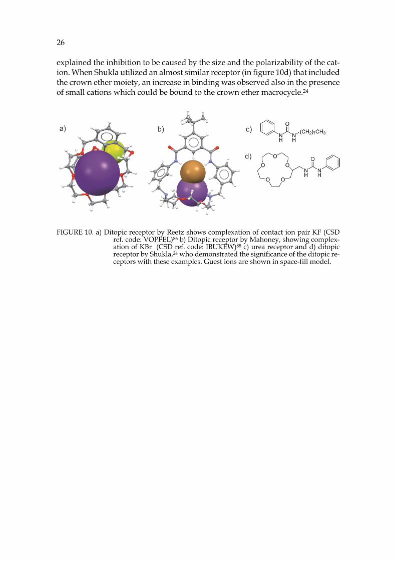
26 explained the inhibition to be caused by the size and the polarizability of the cat-ion. When Shukla utilized an almost similar receptor (in figure 10d) that included the crown ether moiety, an increase in binding was observed also in the presence of small cations which could be bound to the crown ether macrocycle.24
FIGURE 10. a) Ditopic receptor by Reetz shows complexation of contact ion pair KF (CSD ref. code: VOPFEL)86 b) Ditopic receptor by Mahoney, showing complex-ation of KBr (CSD ref. code: IBUKEW)88 c) urea receptor and d) ditopic receptor by Shukla,24 who demonstrated the significance of the ditopic re-ceptors with these examples. Guest ions are shown in space-fill model.

Supramolecular systems and processes can be monitored in all physical states; in gas phase, in solution and in solid state. This chapter briefly describes the techniques that can be utilized to study supramolecular systems, especially fo-cusing on the structural chemistry methods used in the experimental part of this work.
2.1 Gas phase
2.1.1 Mass spectrometry
IUPAC defines mass spectrometry as “…the study of matter through the for-mation of gas phase ions that are characterized using mass spectrometers by their mass, charge, structure and/or physico-chemical properties.” 90 Mass spectrom-etry is a technique which separates ions according to their mass to charge ratio (m/z). In an MS spectrum, m/z is plotted against the relative abundance of the ions. Mass spectrometer consists of the sample inlet system, the ion source, the mass analyzer and the detector. Depending on the method, the inlet system is not al-ways necessary, but it can be for example a chromatograph or a direct inlet. The crucial thing in mass spectrometry is that it can only detect ions, and therefore the sample needs to be ionized before analysis. Different ion sources can be uti-lized for this purpose. For example, electron ionization (EI), electrospray ioniza-tion (ESI) or matrix assisted laser desorption ionization (MALDI) can be utilized to ionize different analytes. The ionized sample is transferred to the mass ana-lyzer that operates in a high vacuum. Typical analyzers are time-of-flight (TOF), quadrupole, orbitrap or Fourier transform ion cyclotron resonance (FTICR). After the analysis ions reach a detector that transfers the electric current into a mass spectrum.91,92
2 RESEARCH METHODS
28
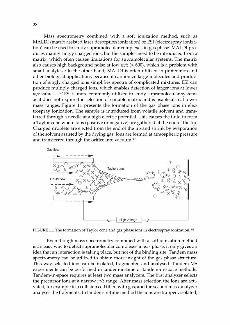
Mass spectrometry combined with a soft ionization method, such as MALDI (matrix assisted laser desorption ionization) or ESI (electrospray ioniza-tion) can be used to study supramolecular complexes in gas phase. MALDI pro-duces mainly singly charged ions, but the samples need to be introduced from a matrix, which often causes limitations for supramolecular systems. The matrix also causes high background noise at low m/z (< 600), which is a problem with small analytes. On the other hand, MALDI is often utilized in proteomics and other biological applications because it can ionize large molecules and produc-tion of singly charged ions simplifies spectra of complicated mixtures. ESI can produce multiply charged ions, which enables detection of larger ions at lower m/z values.91,92 ESI is more commonly utilized to study supramolecular systems as it does not require the selection of suitable matrix and is usable also at lower mass ranges. Figure 11 presents the formation of the gas phase ions in elec-trospray ionization. The sample is introduced from volatile solvent and trans-ferred through a needle at a high electric potential. This causes the fluid to form a Taylor cone where ions (positive or negative) are gathered at the end of the tip. Charged droplets are ejected from the end of the tip and shrink by evaporation of the solvent assisted by the drying gas. Ions are formed at atmospheric pressure and transferred through the orifice into vacuum.92
FIGURE 11. The formation of Taylor cone and gas phase ions in electrospray ionization. 92
Even though mass spectrometry combined with a soft ionization method is an easy way to detect supramolecular complexes in gas phase, it only gives an idea that an interaction is taking place, but not of the binding site. Tandem mass spectrometry can be utilized to obtain more insight of the gas phase structure. This way selected ions can be isolated, fragmented and analysed. Tandem MS experiments can be performed in tandem-in-time or tandem-in-space methods. Tandem-in-space requires at least two mass analyzers. The first analyzer selects the precursor ions at a narrow m/z range. After mass selection the ions are acti-vated, for example in a collision cell filled with gas, and the second mass analyzer analyses the fragments. In tandem-in-time method the ions are trapped, isolated,
29
fragmented and separated inside the same device. Only quadrupole ion trap (QIT) and FTICR instruments are capable of performing tandem-in-time experiments. The long capture time enables the use of very slow activation methods such as electron capture dissociation (ECD) or infrared multiphoton dissociation (IRMPD). 91,92
2.1.2 Ion mobility mass spectrometry
Ion mobility (IM) 93 measures mobilities of ions through a buffer gas where the movement is caused by an electric field. Ion mobility coupled to mass spectrom-eter creates a hybrid technique, ion mobility mass spectrometry (IM-MS). Ion mo-bility is usually coupled to TOF analyser that can record spectra in a µs time scale.93 The basic principle to measure mobilities of the ions is that the ions are released in a pulsed manner to a mobility cell where an electric field is applied. In the mobility cell ions travel through the buffer gas and the time they spend in the mobility cell is called the drift time. As the cell is filled with buffer gas, ions collide with the gas while passing through the chamber and this affects their drift times. Ions with various geometries and chemical compositions collide differ-ently because of their different sizes and interactions between the gas and the analyte. With a known drift time, m/z and experimental parameters, the collision cross section (CCS) value of the analyte can be determined. CCS is thus depend-ent on the gas and is affected by the size, shape and chemical composition of the analyte. 94–97 CCS is not a true molecular cross section but an observable property that averages all possible orientations and interactions.95 Yet, CCS values can be roughly compared to other measures obtained from X-ray crystallography or DOSY NMR.94,96 The accuracy of experimental CCS values can be supported by comparing them to calculated ones. 98–102
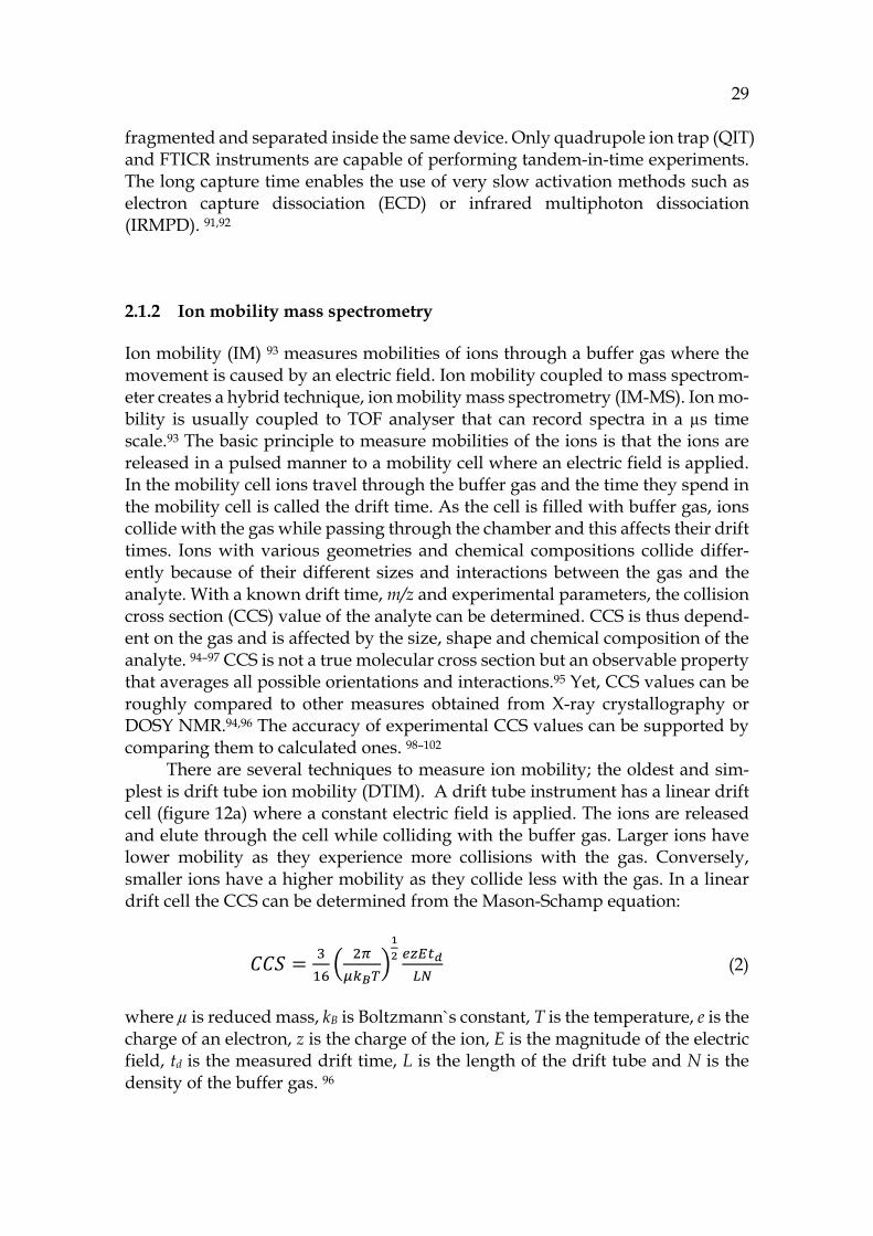
There are several techniques to measure ion mobility; the oldest and sim-plest is drift tube ion mobility (DTIM). A drift tube instrument has a linear drift cell (figure 12a) where a constant electric field is applied. The ions are released and elute through the cell while colliding with the buffer gas. Larger ions have lower mobility as they experience more collisions with the gas. Conversely, smaller ions have a higher mobility as they collide less with the gas. In a linear drift cell the CCS can be determined from the Mason-Schamp equation: 𝐶𝐶𝑆 = (2)
where µ is reduced mass, kB is Boltzmann`s constant, T is the temperature, e is the charge of an electron, z is the charge of the ion, E is the magnitude of the electric field, td is the measured drift time, L is the length of the drift tube and N is the density of the buffer gas. 96
30
In a travelling wave ion mobility (TWIM) instrument the electric field is ap-plied through ring electrodes connected to opposite phases of radiofrequency voltages. A DC potential is added to the confining RF to create a wave potential that carries ions through the buffer gas. The smaller high-mobility ions can keep up with the waves and “surf” through the cell. Larger ions with lower mobilities need more waves to get through.103 The determination of CCS values is more complicated with TWIMS instrument. As the electric field is not constant, the Mason-Schamp equation does not hold for TWIMS. Therefore calibrants are needed for determination of CCS values, but calibration of the TWIMS instru-ment is not straightforward.97,104,105 The optimal calibrant should have similar size and same kind of interactions with the buffer gas as the analyte. Therefore supramolecular systems cannot be calibrated with commonly used peptides.
Trapped ion mobility (TIM) is the reversal of classic DTIM analyzer. In TIMS the ions are held stationary by radiofrequency voltages and an axial electric field, while the buffer gas “pushes” the ions through the cell (figure 12c). The electric field is gradually decreased so that all ions finally elute through the cell. It is noteworthy that in TIMS the larger species have highest mobility, as they are pushed the most by buffer gas, and therefore they elute out first. The smallest species have low mobility and elute last, which is reversed in DTIM and TWIM analyzers.106–108 In TIMS, calibrants are needed to calibrate an ion`s mobility, K, which can be described in terms of drift velocity vd, or its components length L and drift time td, and an electric field E as in equation (3):
𝐾 = = (3)
With a known mobility the CCS values can be determined from Mason-Schamp equation (2).
31
FIGURE 12. Schematic presentation of different ion mobility cells to illustrate their operation. a) In DTIM instrument the electric field is constant and separate ions based on their different mobilities while they travel through buffer gas. b) In TWIM instrument ions surf on a travelling waves applied through ring electrodes of opposing polarities. Ions with higher mobilities elute first. c) In TIM ions are trapped in an electric field at t1. The electric field is then gradually decreased (t2) while buffer gas pushes the ions through the mo-bility cell. In TIM buffer gas pushes the biggest (green) analyte the most, and therefore it elutes first. 96
Even though the techniques to measure the mobility differ from each other, the ion mobility mass spectrometry instruments consist of similar components. An IM-MS instrument consists of an ion source (typically ESI), an ion mobility analyser (for example drift tube), and mass analyser (TOF). The instrument can contain also a mass filter (usually a quadrupole), which can be located before or after the mobility analyser. In commercial TWIMS instrument, for example, the mass filter is before ion mobility analyser, which allows the activation of the iso-lated ions before measuring their mobilities.109
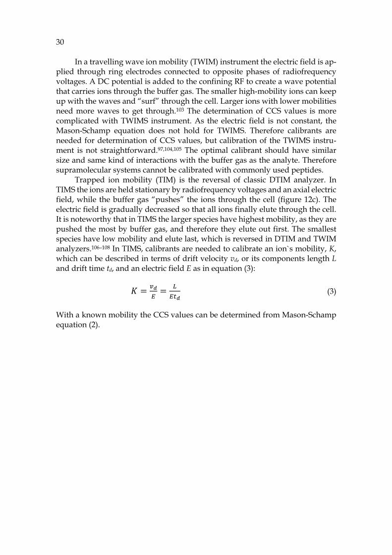
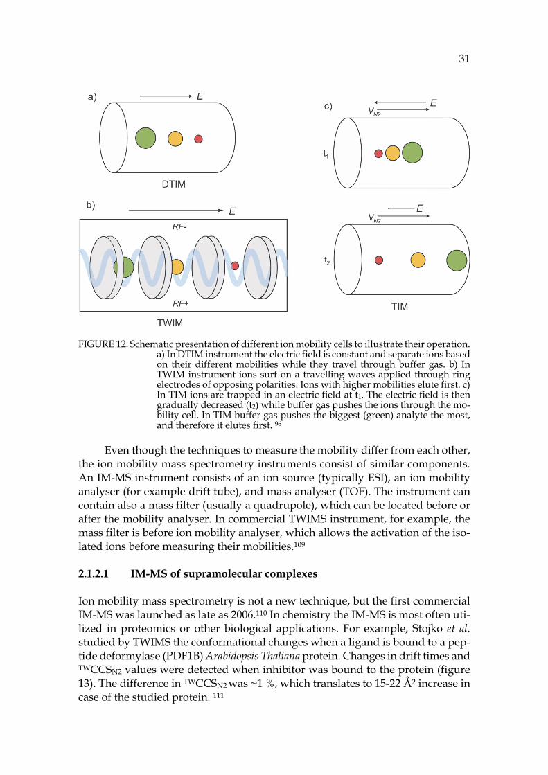
2.1.2.1 IM-MS of supramolecular complexes Ion mobility mass spectrometry is not a new technique, but the first commercial IM-MS was launched as late as 2006.110 In chemistry the IM-MS is most often uti-lized in proteomics or other biological applications. For example, Stojko et al. studied by TWIMS the conformational changes when a ligand is bound to a pep-tide deformylase (PDF1B) Arabidopsis Thaliana protein. Changes in drift times and TWCCSN2 values were detected when inhibitor was bound to the protein (figure 13). The difference in TWCCSN2 was ~1 %, which translates to 15-22 Å2 increase in case of the studied protein. 111
32
Ion mobility can be also utilized to study metallosupramolecular complexes such as in the work of Ujma et al. In their work, DTIMS was demonstrated in study of three-dimensional supramolecular platinum cages. The ligands used for construction of the cages could have different relative orientations, cis or trans. The isobaric structures were in all cases easily separated according to their dif-ferent drift times.112 IM-MS can also be used to study larger complexes and their self-assembly in gas phase. This has been demonstrated by elegant structures of 2D- and 3D-stars of David.29
FIGURE 13. Binding of three different bacterial PDF inhibitors to peptide deformylase A. thaliana (PDF1B) detected by TWIMS. Adapted with permission from ref. 111, CC BY 3.0, Copyright © 2015 The Royal Society of Chemistry.
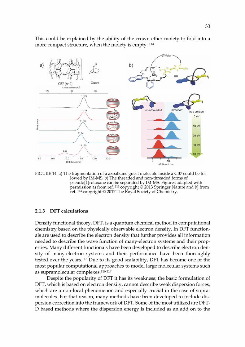
A novel ion mobility study of a supramolecular host-guest system was demonstrated by Lee et al. who proved by IM-MS that CB7 can perform a catalytic reaction inside its cavity. The azoalkane guest can undergo a retro-Diels-Alder reaction inside the cavity of CB7. The structure of the complex during the guest fragmentation was followed by TWIMS (see figure 14a), where the ion was acti-vated before measuring its mobility. It could be noted that during the fragmen-tation the host squeezed a little bit to maximize its interactions with the remain-ing guest. This was observed as a small albeit notable decrease in the drift time. When the dissociation was completed, the remaining host had the same drift time as the free host.113
More recent ion mobility study of a supramolecular host-guest system was demonstrated by Schröder et al. with a pseudo[1]rotaxane (in figure 14b).114 Ro-taxanes and catenanes have been actively utilized in nanochemistry applications as molecular switches and machines.6,27 The studied lasso-type pseudo[1]rotax-ane is redox-switchable, which can exist in 3 charge states. The conformational changes are triggered by redox-stimuli. The charge states were all observable in an ESI-MS spectrum, but IM-MS showed only two drift peaks, which could be identified as threaded and non-threaded forms. In a CID experiment the in-creased collision energy caused unthreading of pseudo[1]rotaxane. Surprisingly, the cyclic threaded structure had larger CCS than the open non-threaded form.
33
This could be explained by the ability of the crown ether moiety to fold into a more compact structure, when the moiety is empty. 114
FIGURE 14. a) The fragmentation of a azoalkane guest molecule inside a CB7 could be fol-lowed by IM-MS. b) The threaded and non-threaded forms of pseudo[1]rotaxane can be separated by IM-MS. Figures adapted with permission a) from ref. 113 copyright © 2013 Springer Nature and b) from ref. 114 copyright © 2017 The Royal Society of Chemistry.
2.1.3 DFT calculations
Density functional theory, DFT, is a quantum chemical method in computational chemistry based on the physically observable electron density. In DFT function-als are used to describe the electron density that further provides all information needed to describe the wave function of many-electron systems and their prop-erties. Many different functionals have been developed to describe electron den-sity of many-electron systems and their performance have been thoroughly tested over the years.115 Due to its good scalability, DFT has become one of the most popular computational approaches to model large molecular systems such as supramolecular complexes.116,117
Despite the popularity of DFT it has its weakness; the basic formulation of DFT, which is based on electron density, cannot describe weak dispersion forces, which are a non-local phenomenon and especially crucial in the case of supra-molecules. For that reason, many methods have been developed to include dis-persion correction into the framework of DFT. Some of the most utilized are DFT-D based methods where the dispersion energy is included as an add on to the
34 calculated DFT energy.118,119 DFT-D3 is the newest method in the DFT-D family to describe weak dispersion forces and its good performance has been demon-strated with various supramolecular systems.116,117
2.2 Solution
2.2.1 NMR
Nuclear magnetic resonance (NMR) spectroscopy detects the resonances of re-laxed spin states from nuclei exposed to a strong magnetic field. The NMR probe is tuned for certain nuclei (for example 1H, 13C, 19F), and those nuclei present in the sample are observed as signals with corresponding chemical shifts in the NMR spectrum. The chemical shift of the nuclei depends on their chemical envi-ronment. When the nucleus is located close to groups that withdraw electron density, the nucleus is deshielded and the signal moves downfield in the spec-trum. Vice versa, when the electronic cloud is shielding a nucleus, it moves up-field in the spectrum.120
In case of supramolecules, a complexation can be observed upon addition of the guest into the solution of host. In the case of complexation a shift of the signals is observed. NMR can give information of the binding, but more im-portantly, of the site where the binding is taking place. For example protons, which exhibit the highest shifts, also experience the greatest change in their chemical environment. The changes in the chemical shifts also indicate regiose-lectivity, where the binding is taking place. The strength of the binding can be determined in NMR titrations as a binding or association constant Ka that can be described in units of dm3 mol-1 or M-1. The higher the binding constant, the more stable is the formed complex. The scale of binding constants can vary from 10 to 104 M-1. Magnitudes of Ka are usually expressed in logarithmic scale and therefore Ka should be denoted as unitless quantity.9
In NMR spectroscopy the stoichiometry of the supramolecular complex can be determined in solution using the method of continuous variation, also known as the Job Plot`s method. The right stoichiometry is crucial for calculation of the binding constants. In Job´s plot method, the total concentration of host and guest is kept constant, but their molar fractions are changed while the chemical shift is monitored. The Job´s Plot method has been commonly utilized but it can lead to misinterpretation if not carefully analysed. Job´s plot method works well if only one type of binding model is observed. But if several binding models are detected, the method becomes unreliable. In the latter case, other methods for determina-tion of the possible binding models should be combined to support the analysis. 121
35
2.2.1.1 DOSY NMR
Diffusion-ordered spectroscopy is used to measure diffusion and ultimately to determine diffusion coefficient D. The diffusion coefficient can be related to the size, shape and molecular weight of the diffusing particle. The hydrodynamic radius rh can be calculated from Stokes-Einstein equation
𝐷 = (4)
where T is the temperature, kB the Boltzmann constant, η is the viscosity of the fluid and rs is the radius of the diffusing species. Equation (4) is only valid for spherical species and it neglects the effects of solvation and other interactions. For many cages and capsular assemblies this equation is a reasonable approxi-mation, as they are large enough and their diverse axes are not very different. Therefore rs can be approximated as rh.
DOSY NMR can be utilized to gain information of the size of diffusing spe-cies, but also of the encapsulation process. The guest molecules are a lot smaller than the host where they are encapsulated. Usually a dramatic change is ob-served in the diffusion coefficient of the guest when encapsulated, and their dif-fusion should be the same as hosts if the exchange is slow in the NMR time scale.122
2.3 Solid state
2.3.1 X-ray crystallography
X-ray crystallography is a technique based on X-ray diffraction, and it is used to determine the three-dimensional arrangement of atoms in a crystalline material via evaluating the electron densities within the crystal. X-ray crystallography is a powerful tool for structural analysis. It is used widely, for example to determine the structures of supramolecular complexes in solid state. It is the only method that can give a precise picture of the contents in a crystal at the atomic level. The conformation and stoichiometry are visible from the structure. Moreover, the de-termination of intra- and intermolecular bond lengths and angles gives detailed information of the interactions between the components. 123,124 The limitation of X-ray crystallography is that it cannot state the strength of the binding in case of a host-guest complex. Thus, this technique is often combined with other struc-tural analysis methods. X-ray crystallography has already demonstrated its power in analysis of several supramolecular complexes throughout this thesis, for example the depicted self-assembled structures in chapter 1.1.3.
3.1 Aim of the work
The aim of this work was to study supramolecular chemistry and binding prop-erties of different anion binding hosts: pyridine[4]arene, cyclohexylhemicucur-bit[8]uril and crown ether urea receptors. Pyridine[4]arene and cyclohex-ylhemicucurbit[8]uril have already shown affinity towards anionic guests,64,65,77 but their binding properties are not fully established. Crown ether urea receptors are completely new synthetic receptors that utilize the binding of an ion pair in-stead of separate ions. Similar receptors have been utilized earlier in ion pair binding.125
Pyridine[4]arene is a relatively new host, whose supramolecular chemistry is not yet widely studied. The focus of this work is also on the self-assembly of pyridine[4]arene, as it can form capsules of different size. The earlier reports on self-assembly are contradictory about whether the most abundant assemblies are dimeric or hexameric.64,66 So far, only one report exists on the ability of pyri-dine[4]arene to form hexameric aggregates in solution.66
The binding behaviour, characterization of the observed complexes and self-assembly have been studied by mass spectrometry, NMR spectroscopy and X-ray crystallography whenever possible. Also DFT calculations have been uti-lized to visualize the gas-phase structures. In this work, a special interest hasbeen in utilization of ion mobility mass spectrometry to study the structural fea-tures of pyridinearene complexes.
3 RESULTS AND DISCUSSION
37
3.2 Pyridine[4]arenes
3.2.1 Synthesis I,126
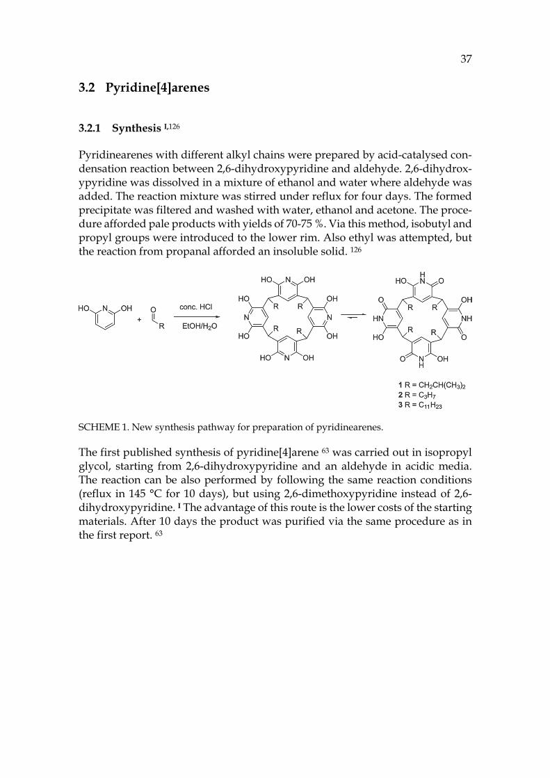
Pyridinearenes with different alkyl chains were prepared by acid-catalysed con-densation reaction between 2,6-dihydroxypyridine and aldehyde. 2,6-dihydrox-ypyridine was dissolved in a mixture of ethanol and water where aldehyde was added. The reaction mixture was stirred under reflux for four days. The formed precipitate was filtered and washed with water, ethanol and acetone. The proce-dure afforded pale products with yields of 70-75 %. Via this method, isobutyl and propyl groups were introduced to the lower rim. Also ethyl was attempted, but the reaction from propanal afforded an insoluble solid. 126
SCHEME 1. New synthesis pathway for preparation of pyridinearenes.
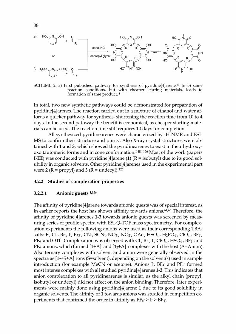
The first published synthesis of pyridine[4]arene 63 was carried out in isopropyl glycol, starting from 2,6-dihydroxypyridine and an aldehyde in acidic media. The reaction can be also performed by following the same reaction conditions (reflux in 145 °C for 10 days), but using 2,6-dimethoxypyridine instead of 2,6-dihydroxypyridine. I The advantage of this route is the lower costs of the starting materials. After 10 days the product was purified via the same procedure as in the first report. 63
38
SCHEME 2. a) First published pathway for synthesis of pyridine[4]arene.63 In b) same reaction conditions, but with cheaper starting materials, leads to formation of same product. I
In total, two new synthetic pathways could be demonstrated for preparation of pyridine[4]arenes. The reaction carried out in a mixture of ethanol and water af-fords a quicker pathway for synthesis, shortening the reaction time from 10 to 4 days. In the second pathway the benefit is economical, as cheaper starting mate-rials can be used. The reaction time still requires 10 days for completion.
All synthesized pyridinearenes were characterized by 1H NMR and ESI-MS to confirm their structure and purity. Also X-ray crystal structures were ob-tained with 1 and 3, which showed the pyridinearenes to exist in their hydroxy-oxo tautomeric forms and in cone conformation.I-III, 126 Most of the work (papers I-III) was conducted with pyridine[4]arene (1) (R = isobutyl) due to its good sol-ubility in organic solvents. Other pyridine[4]arenes used in the experimental part were 2 (R = propyl) and 3 (R = undecyl).126
3.2.2 Studies of complexation properties
3.2.2.1 Anionic guests I,126 The affinity of pyridine[4]arene towards anionic guests was of special interest, as in earlier reports the host has shown affinity towards anions.64,65 Therefore, the affinity of pyridine[4]arenes 1-3 towards anionic guests was screened by meas-uring series of profile spectra with ESI-Q-TOF mass spectrometry. For complex-ation experiments the following anions were used as their corresponding TBA-salts: F-, Cl-, Br-, I-, Br3-, CN-, SCN-, NO3-, NO2-, OAc-, HSO4-, H2PO4-, ClO4-, BF4-, PF6- and OTf-. Complexation was observed with Cl-, Br-, I-, ClO4-, HSO4-, BF4- and PF6- anions, which formed [1+A]- and [12+A]- complexes with the host (A=Anion). Also ternary complexes with solvent and anion were generally observed in the spectra as [12+S+A]- ions (S=solvent), depending on the solvent(s) used in sample introduction (for example MeCN or acetone). Anions I-, BF4- and PF6- formed most intense complexes with all studied pyridine[4]arenes 1-3. This indicates that anion complexation to all pyridinearenes is similar, as the alkyl chain (propyl, isobutyl or undecyl) did not affect on the anion binding. Therefore, later experi-ments were mainly done using pyridine[4]arene 1 due to its good solubility in organic solvents. The affinity of 1 towards anions was studied in competition ex-periments that confirmed the order in affinity as PF6- > I- > BF4-.
39 The location of the anion in the gas phase structure was studied with colli-
sion-induced dissociation (CID). In CID experiments [12+A]- ions (A=Anion: PF6-, I- or BF4-, also later on) were isolated and activated. With PF6- the elimination of anion was observed at the first step and only a minority of other fragments were observed with increased collision energy (see figure 15c). This indicates that the anion would be bound outside the capsule (exo) rather than inside the cavity (endo), as in CID the breakage of weakest bond is usually observed. The isolated [12+BF4]- showed [1+BF4]- at the first dissociation step, which could indicate endo-complexation. Similarly [12+I]- resulted to [1+I]- at the first dissociation step. To study the binding site of the anion, the ternary complexes with solvent and ani-ons were also studied by CID. The cavity of the pyridinearene dimer is too small to occupy two guest molecules simultaneously and therefore one of them has to be located exo. The isolated [12+acetone+A]- showed again elimination of the an-ion when PF6- was used. [12+acetone+BF4]- resulted in [1+BF4]- at the first disso-ciation step. The CID experiments suggest that PF6- is bound exo to the pyri-dinearene capsule, while BF4- and I- could be located endo. Most likely acetone is located endo when the anion is bound exo. The kinetic stabilities of the complexes were compared by calculating the CE50% values, which represent the relative en-ergy needed for the complex to dissociate to its half intensity. In the stabilities of dimeric [12+A]- complexes the differences were negligible. However, the mono-meric [1+PF6]- was found to be less stable than [1+BF4]- and [1+I]- ions. This indi-cates that all studied ions show similar dissociation, but the [1+PF6]- ion could not be observed due to its lower stability. In this case, it is likely that all anions are located exo.
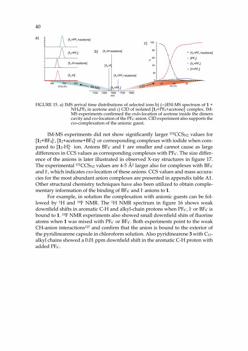
To characterize the structures of the anion complexes they were further studied in IM-MS experiments. [12+PF6]- and [12+acetone+PF6]- ions showed longer drift times and ~8Å2 larger DTCCSN2 values compared to [12-H]- and [12-H+acetone]- ions (see figure 15a for arrival time distributions). This confirms that acetone is located endo in the dimeric capsule in gas phase, while the PF6- anion is bound outside, exo. These findings show unusual complexation properties for host 1, as similar behaviour has not been observed with resorcinarenes before. The simultaneous binding of neutral and anionic guests is a new phenomenon. Solvent adducts are common, but they are usually less abundant. A solvent in-clusion in gas phase has not been reported earlier.
40
FIGURE 15. a) IMS arrival time distributions of selected ions b) (―)ESI-MS spectrum of 1 + NH4PF6 in acetone and c) CID of isolated [12+PF6+acetone]- complex. IM-MS experiments confirmed the endo-location of acetone inside the dimers cavity and exo-location of the PF6- anion. CID experiment also supports the exo-complexation of the anionic guest.
IM-MS experiments did not show significantly larger DTCCSN2 values for [12+BF4]-, [12+acetone+BF4]- or corresponding complexes with iodide when com-pared to [12-H]- ion. Anions BF4- and I- are smaller and cannot cause as large differences in CCS values as corresponding complexes with PF6-. The size differ-ence of the anions is later illustrated in observed X-ray structures in figure 17. The experimental DTCCSN2 values are 4-5 Å2 larger also for complexes with BF4- and I-, which indicates exo-location of these anions. CCS values and mass accura-cies for the most abundant anion complexes are presented in appendix table A1. Other structural chemistry techniques have also been utilized to obtain comple-mentary information of the binding of BF4- and I- anions to 1.
For example, in solution the complexation with anionic guests can be fol-lowed by 1H and 19F NMR. The 1H NMR spectrum in figure 16 shows weak downfield shifts in aromatic C-H and alkyl-chain protons when PF6-, I- or BF4- is bound to 1. 19F NMR experiments also showed small downfield shits of fluorine atoms when 1 was mixed with PF6- or BF4-. Both experiments point to the weak CH-anion interactions127 and confirm that the anion is bound to the exterior of the pyridinearene capsule in chloroform solution. Also pyridinearene 3 with C11- alkyl chains showed a 0.01 ppm downfield shift in the aromatic C-H proton with added PF6-.
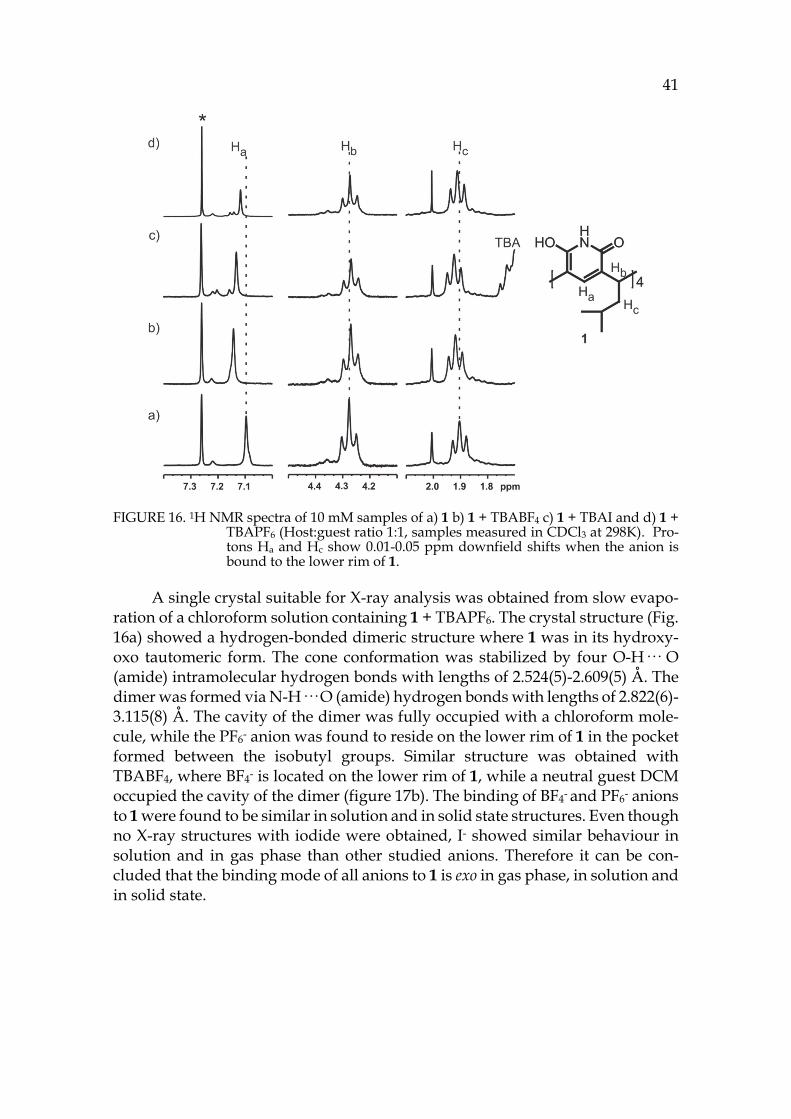
41
FIGURE 16. 1H NMR spectra of 10 mM samples of a) 1 b) 1 + TBABF4 c) 1 + TBAI and d) 1 + TBAPF6 (Host:guest ratio 1:1, samples measured in CDCl3 at 298K). Pro-tons Ha and Hc show 0.01-0.05 ppm downfield shifts when the anion is bound to the lower rim of 1.
A single crystal suitable for X-ray analysis was obtained from slow evapo-ration of a chloroform solution containing 1 + TBAPF6. The crystal structure (Fig. 16a) showed a hydrogen-bonded dimeric structure where 1 was in its hydroxy-oxo tautomeric form. The cone conformation was stabilized by four O-H··· O (amide) intramolecular hydrogen bonds with lengths of 2.524(5)-2.609(5) Å. The dimer was formed via N-H··· O (amide) hydrogen bonds with lengths of 2.822(6)-3.115(8) Å. The cavity of the dimer was fully occupied with a chloroform mole-cule, while the PF6- anion was found to reside on the lower rim of 1 in the pocket formed between the isobutyl groups. Similar structure was obtained with TBABF4, where BF4- is located on the lower rim of 1, while a neutral guest DCM occupied the cavity of the dimer (figure 17b). The binding of BF4- and PF6- anions to 1 were found to be similar in solution and in solid state structures. Even though no X-ray structures with iodide were obtained, I- showed similar behaviour in solution and in gas phase than other studied anions. Therefore it can be con-cluded that the binding mode of all anions to 1 is exo in gas phase, in solution and in solid state.
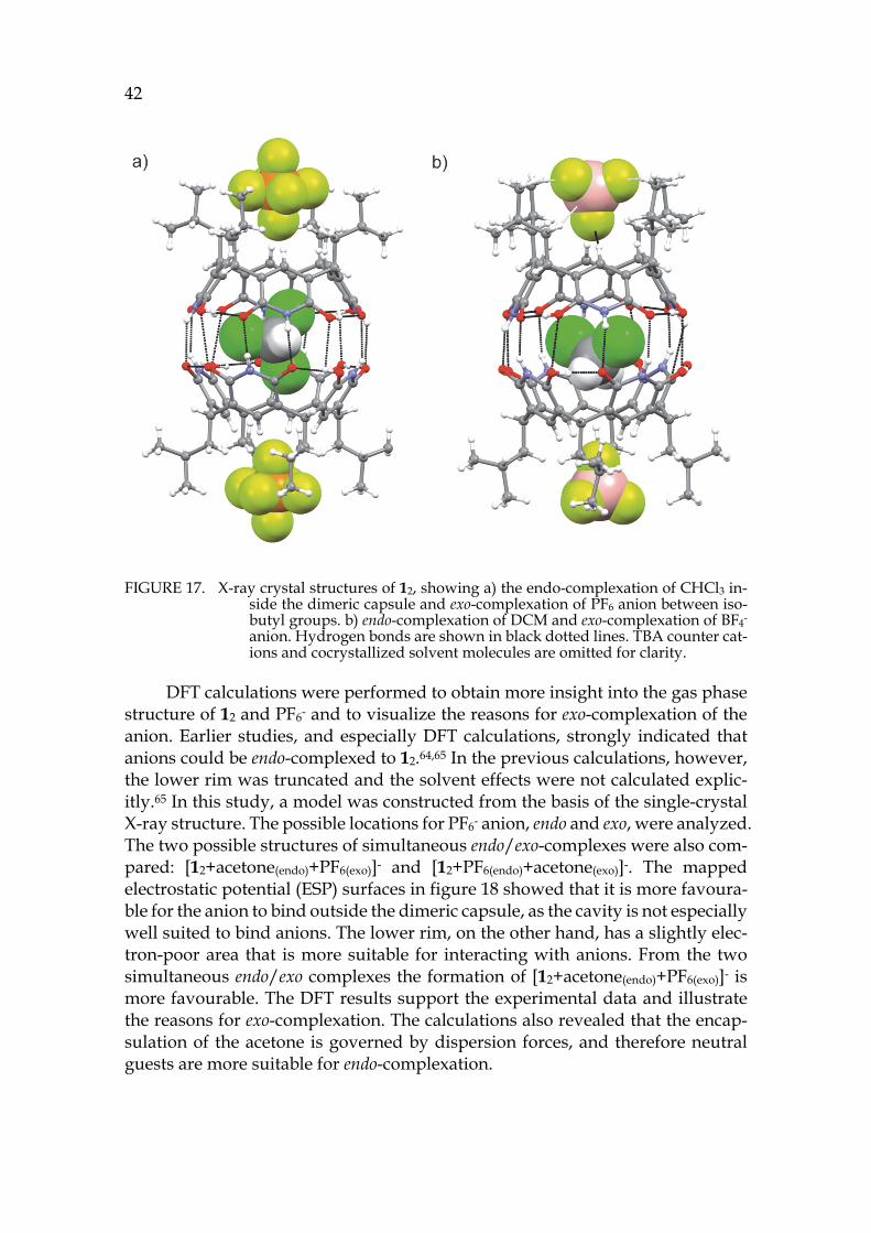
42
FIGURE 17. X-ray crystal structures of 12, showing a) the endo-complexation of CHCl3 in-side the dimeric capsule and exo-complexation of PF6 anion between iso-butyl groups. b) endo-complexation of DCM and exo-complexation of BF4- anion. Hydrogen bonds are shown in black dotted lines. TBA counter cat-ions and cocrystallized solvent molecules are omitted for clarity.
DFT calculations were performed to obtain more insight into the gas phase structure of 12 and PF6- and to visualize the reasons for exo-complexation of the anion. Earlier studies, and especially DFT calculations, strongly indicated that anions could be endo-complexed to 12.64,65 In the previous calculations, however, the lower rim was truncated and the solvent effects were not calculated explic-itly.65 In this study, a model was constructed from the basis of the single-crystal X-ray structure. The possible locations for PF6- anion, endo and exo, were analyzed. The two possible structures of simultaneous endo/exo-complexes were also com-pared: [12+acetone(endo)+PF6(exo)]- and [12+PF6(endo)+acetone(exo)]-. The mapped electrostatic potential (ESP) surfaces in figure 18 showed that it is more favoura-ble for the anion to bind outside the dimeric capsule, as the cavity is not especially well suited to bind anions. The lower rim, on the other hand, has a slightly elec-tron-poor area that is more suitable for interacting with anions. From the two simultaneous endo/exo complexes the formation of [12+acetone(endo)+PF6(exo)]- is more favourable. The DFT results support the experimental data and illustrate the reasons for exo-complexation. The calculations also revealed that the encap-sulation of the acetone is governed by dispersion forces, and therefore neutral guests are more suitable for endo-complexation.
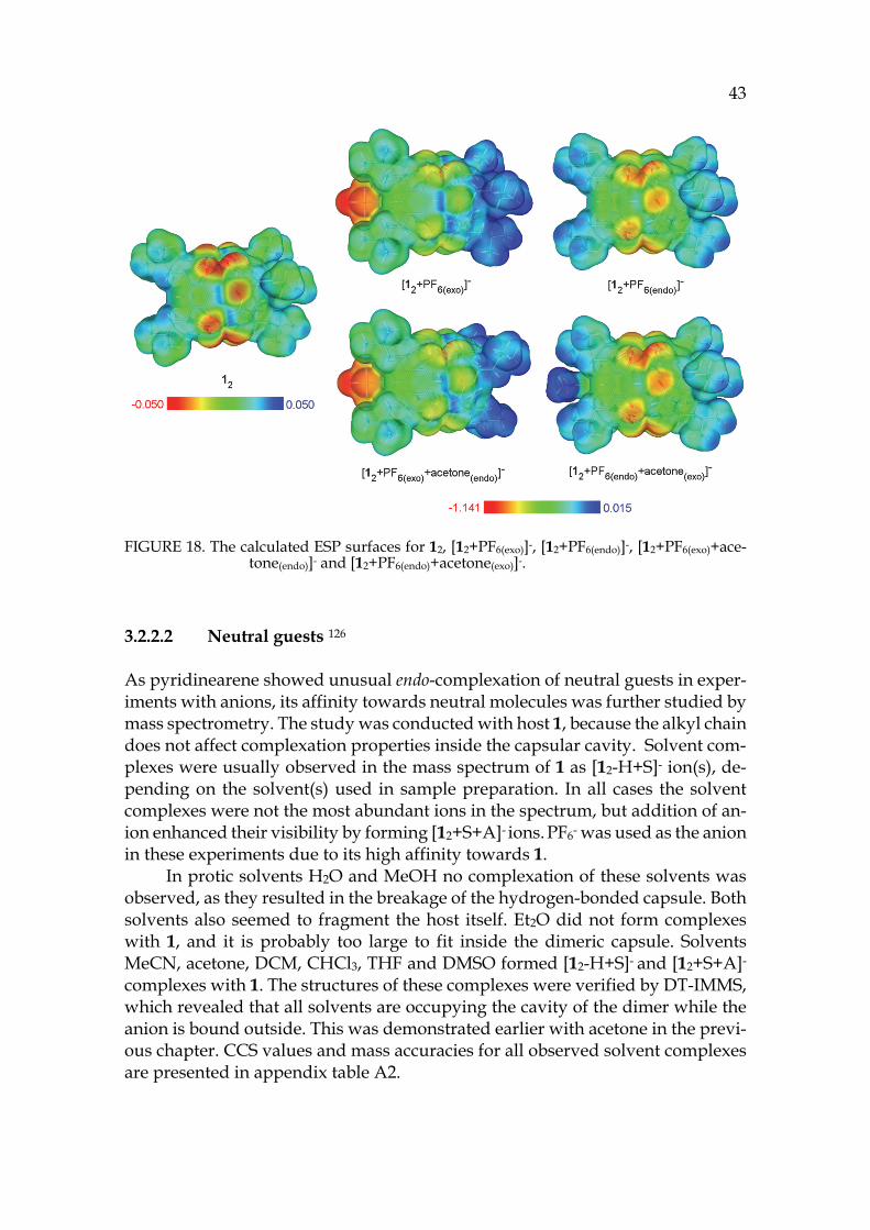
43
FIGURE 18. The calculated ESP surfaces for 12, [12+PF6(exo)]-, [12+PF6(endo)]-, [12+PF6(exo)+ace-tone(endo)]- and [12+PF6(endo)+acetone(exo)]-.
3.2.2.2 Neutral guests 126
As pyridinearene showed unusual endo-complexation of neutral guests in exper-iments with anions, its affinity towards neutral molecules was further studied by mass spectrometry. The study was conducted with host 1, because the alkyl chain does not affect complexation properties inside the capsular cavity. Solvent com-plexes were usually observed in the mass spectrum of 1 as [12-H+S]- ion(s), de-pending on the solvent(s) used in sample preparation. In all cases the solvent complexes were not the most abundant ions in the spectrum, but addition of an-ion enhanced their visibility by forming [12+S+A]- ions. PF6- was used as the anion in these experiments due to its high affinity towards 1.
In protic solvents H2O and MeOH no complexation of these solvents was observed, as they resulted in the breakage of the hydrogen-bonded capsule. Both solvents also seemed to fragment the host itself. Et2O did not form complexes with 1, and it is probably too large to fit inside the dimeric capsule. Solvents MeCN, acetone, DCM, CHCl3, THF and DMSO formed [12-H+S]- and [12+S+A]- complexes with 1. The structures of these complexes were verified by DT-IMMS, which revealed that all solvents are occupying the cavity of the dimer while the anion is bound outside. This was demonstrated earlier with acetone in the previ-ous chapter. CCS values and mass accuracies for all observed solvent complexes are presented in appendix table A2.
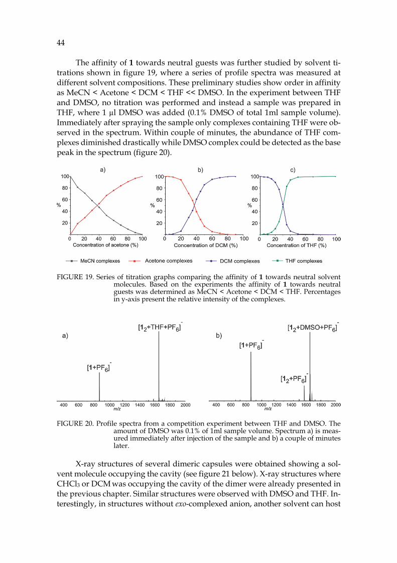
44
The affinity of 1 towards neutral guests was further studied by solvent ti-trations shown in figure 19, where a series of profile spectra was measured at different solvent compositions. These preliminary studies show order in affinity as MeCN < Acetone < DCM < THF << DMSO. In the experiment between THF and DMSO, no titration was performed and instead a sample was prepared in THF, where 1 µl DMSO was added (0.1% DMSO of total 1ml sample volume). Immediately after spraying the sample only complexes containing THF were ob-served in the spectrum. Within couple of minutes, the abundance of THF com-plexes diminished drastically while DMSO complex could be detected as the base peak in the spectrum (figure 20).
FIGURE 19. Series of titration graphs comparing the affinity of 1 towards neutral solvent molecules. Based on the experiments the affinity of 1 towards neutral guests was determined as MeCN < Acetone < DCM < THF. Percentages in y-axis present the relative intensity of the complexes.
FIGURE 20. Profile spectra from a competition experiment between THF and DMSO. The amount of DMSO was 0.1% of 1ml sample volume. Spectrum a) is meas-ured immediately after injection of the sample and b) a couple of minutes later.
X-ray structures of several dimeric capsules were obtained showing a sol-vent molecule occupying the cavity (see figure 21 below). X-ray structures where CHCl3 or DCM was occupying the cavity of the dimer were already presented in the previous chapter. Similar structures were observed with DMSO and THF. In-terestingly, in structures without exo-complexed anion, another solvent can host
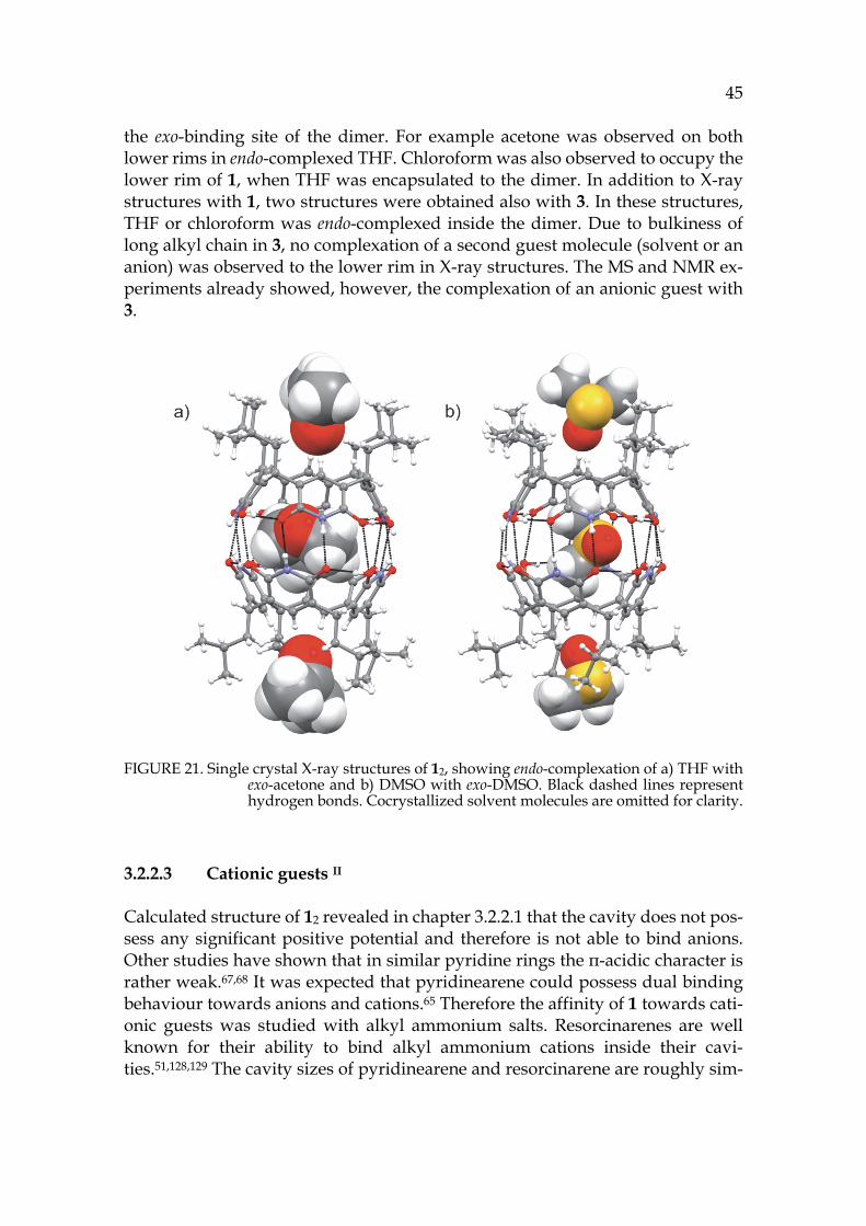
45
the exo-binding site of the dimer. For example acetone was observed on both lower rims in endo-complexed THF. Chloroform was also observed to occupy the lower rim of 1, when THF was encapsulated to the dimer. In addition to X-ray structures with 1, two structures were obtained also with 3. In these structures, THF or chloroform was endo-complexed inside the dimer. Due to bulkiness of long alkyl chain in 3, no complexation of a second guest molecule (solvent or an anion) was observed to the lower rim in X-ray structures. The MS and NMR ex-periments already showed, however, the complexation of an anionic guest with 3.
FIGURE 21. Single crystal X-ray structures of 12, showing endo-complexation of a) THF with exo-acetone and b) DMSO with exo-DMSO. Black dashed lines represent hydrogen bonds. Cocrystallized solvent molecules are omitted for clarity.
3.2.2.3 Cationic guests II
Calculated structure of 12 revealed in chapter 3.2.2.1 that the cavity does not pos-sess any significant positive potential and therefore is not able to bind anions. Other studies have shown that in similar pyridine rings the π-acidic character is rather weak.67,68 It was expected that pyridinearene could possess dual binding behaviour towards anions and cations.65 Therefore the affinity of 1 towards cati-onic guests was studied with alkyl ammonium salts. Resorcinarenes are well known for their ability to bind alkyl ammonium cations inside their cavi-ties.51,128,129 The cavity sizes of pyridinearene and resorcinarene are roughly sim-
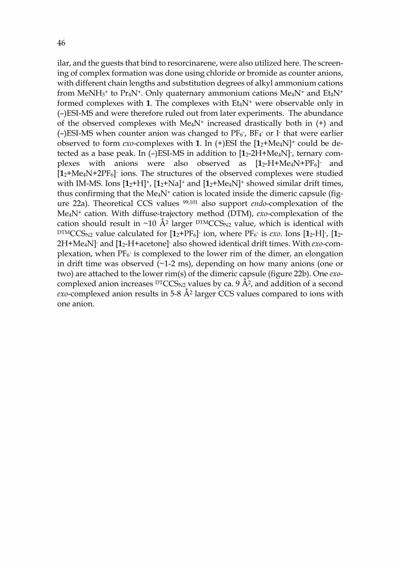
46 ilar, and the guests that bind to resorcinarene, were also utilized here. The screen-ing of complex formation was done using chloride or bromide as counter anions, with different chain lengths and substitution degrees of alkyl ammonium cations from MeNH3+ to Pr4N+. Only quaternary ammonium cations Me4N+ and Et4N+ formed complexes with 1. The complexes with Et4N+ were observable only in (–)ESI-MS and were therefore ruled out from later experiments. The abundance of the observed complexes with Me4N+ increased drastically both in (+) and (–)ESI-MS when counter anion was changed to PF6-, BF4- or I- that were earlier observed to form exo-complexes with 1. In (+)ESI the [12+Me4N]+ could be de-tected as a base peak. In (–)ESI-MS in addition to [12-2H+Me4N]-, ternary com-plexes with anions were also observed as [12-H+Me4N+PF6]- and [12+Me4N+2PF6]- ions. The structures of the observed complexes were studied with IM-MS. Ions [12+H]+, [12+Na]+ and [12+Me4N]+ showed similar drift times, thus confirming that the Me4N+ cation is located inside the dimeric capsule (fig-ure 22a). Theoretical CCS values 99,101 also support endo-complexation of the Me4N+ cation. With diffuse-trajectory method (DTM), exo-complexation of the cation should result in ~10 Å2 larger DTMCCSN2 value, which is identical with DTMCCSN2 value calculated for [12+PF6]- ion, where PF6- is exo. Ions [12-H]-, [12-2H+Me4N]- and [12-H+acetone]- also showed identical drift times. With exo-com-plexation, when PF6- is complexed to the lower rim of the dimer, an elongation in drift time was observed (~1-2 ms), depending on how many anions (one or two) are attached to the lower rim(s) of the dimeric capsule (figure 22b). One exo-complexed anion increases DTCCSN2 values by ca. 9 Å2, and addition of a second exo-complexed anion results in 5-8 Å2 larger CCS values compared to ions with one anion.
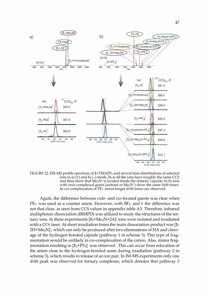
47
FIGURE 22. ESI-MS profile spectrum of 1+TMAPF6 and arrival time distributions of selected ions in a) (+) and b) (–) mode. In a) all the ions have roughly the same CCS and thus show that Me4N+ is located inside the dimeric capsule. In b) ions with endo-complexed guest (acetone or Me4N+) show the same drift times. In exo-complexation of PF6- anion longer drift times are observed.
Again, the difference between endo- and exo-located guests was clear when PF6- was used as a counter anion. However, with BF4- and I- the difference was not that clear, as seen from CCS values in appendix table A3. Therefore, infrared multiphoton dissociation (IRMPD) was utilized to study the structures of the ter-nary ions. In these experiments [12+Me4N+2A]- ions were isolated and irradiated with a CO2 laser. At short irradiation times the main dissociation product was [1-2H+Me4N]-, which can only be produced after two eliminations of HA and cleav-age of the hydrogen bonded capsule (pathway 1 in scheme 3). This type of frag-mentation would be unlikely in exo-complexation of the cation. Also, minor frag-mentation resulting in [12+PF6]- was observed. This can occur from relocation of the anion close to the hydrogen-bonded seam during irradiation (pathway 2 in scheme 3), which results in release of an ion pair. In IM-MS experiments only one drift peak was observed for ternary complexes, which denotes that pathway 3
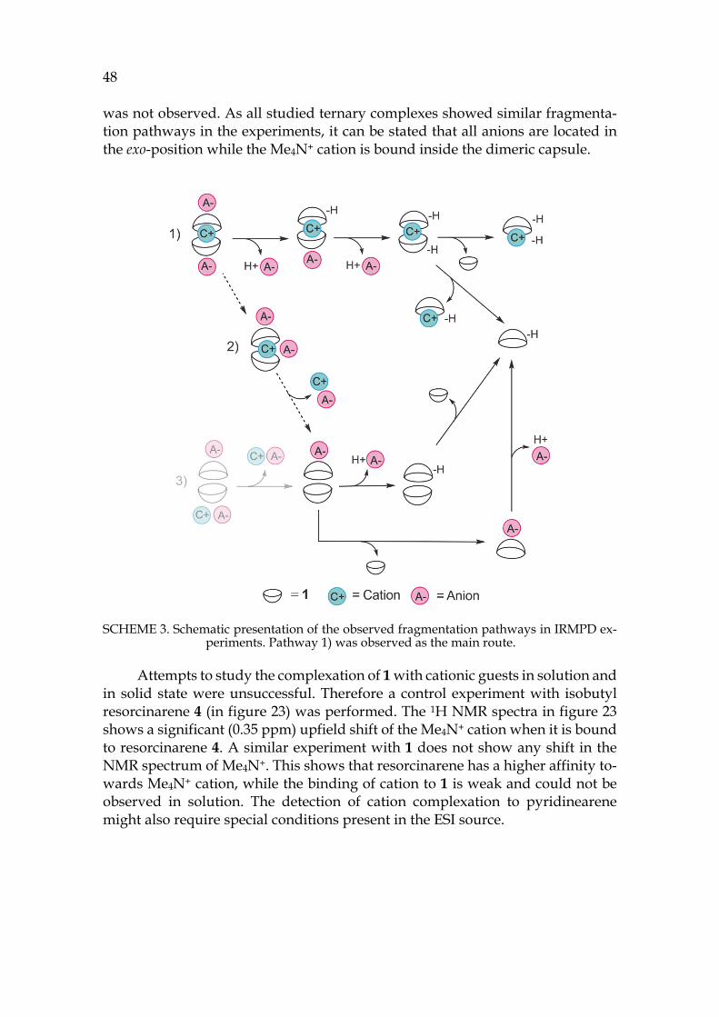
48 was not observed. As all studied ternary complexes showed similar fragmenta-tion pathways in the experiments, it can be stated that all anions are located in the exo-position while the Me4N+ cation is bound inside the dimeric capsule.
SCHEME 3. Schematic presentation of the observed fragmentation pathways in IRMPD ex-periments. Pathway 1) was observed as the main route.
Attempts to study the complexation of 1 with cationic guests in solution and in solid state were unsuccessful. Therefore a control experiment with isobutyl resorcinarene 4 (in figure 23) was performed. The 1H NMR spectra in figure 23 shows a significant (0.35 ppm) upfield shift of the Me4N+ cation when it is bound to resorcinarene 4. A similar experiment with 1 does not show any shift in the NMR spectrum of Me4N+. This shows that resorcinarene has a higher affinity to-wards Me4N+ cation, while the binding of cation to 1 is weak and could not be observed in solution. The detection of cation complexation to pyridinearene might also require special conditions present in the ESI source.
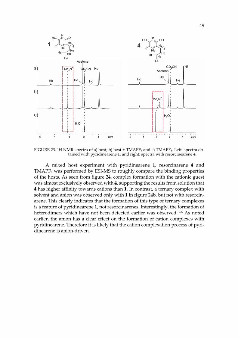
49
FIGURE 23. 1H NMR spectra of a) host, b) host + TMAPF6 and c) TMAPF6. Left: spectra ob-tained with pyridinearene 1, and right: spectra with resorcinearene 4.
A mixed host experiment with pyridinearene 1, resorcinarene 4 and TMAPF6 was performed by ESI-MS to roughly compare the binding properties of the hosts. As seen from figure 24, complex formation with the cationic guest was almost exclusively observed with 4, supporting the results from solution that 4 has higher affinity towards cations than 1. In contrast, a ternary complex with solvent and anion was observed only with 1 in figure 24b, but not with resorcin-arene. This clearly indicates that the formation of this type of ternary complexes is a feature of pyridinearene 1, not resorcinarenes. Interestingly, the formation of heterodimers which have not been detected earlier was observed. 66 As noted earlier, the anion has a clear effect on the formation of cation complexes with pyridinearene. Therefore it is likely that the cation complexation process of pyri-dinearene is anion-driven.
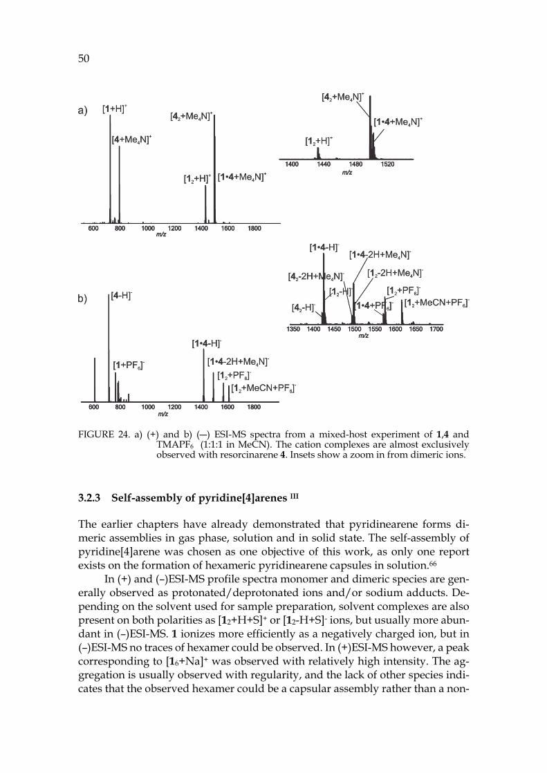
50
FIGURE 24. a) (+) and b) (―) ESI-MS spectra from a mixed-host experiment of 1,4 and TMAPF6 (1:1:1 in MeCN). The cation complexes are almost exclusively observed with resorcinarene 4. Insets show a zoom in from dimeric ions.
3.2.3 Self-assembly of pyridine[4]arenes III
The earlier chapters have already demonstrated that pyridinearene forms di-meric assemblies in gas phase, solution and in solid state. The self-assembly of pyridine[4]arene was chosen as one objective of this work, as only one report exists on the formation of hexameric pyridinearene capsules in solution.66
In (+) and (–)ESI-MS profile spectra monomer and dimeric species are gen-erally observed as protonated/deprotonated ions and/or sodium adducts. De-pending on the solvent used for sample preparation, solvent complexes are also present on both polarities as [12+H+S]+ or [12-H+S]- ions, but usually more abun-dant in (–)ESI-MS. 1 ionizes more efficiently as a negatively charged ion, but in (–)ESI-MS no traces of hexamer could be observed. In (+)ESI-MS however, a peak corresponding to [16+Na]+ was observed with relatively high intensity. The ag-gregation is usually observed with regularity, and the lack of other species indi-cates that the observed hexamer could be a capsular assembly rather than a non-
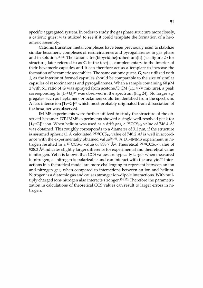
51
specific aggregated system. In order to study the gas phase structure more closely, a cationic guest was utilized to see if it could template the formation of a hex-americ assembly.
Cationic transition metal complexes have been previously used to stabilize similar hexameric complexes of resorcinarenes and pyrogallarenes in gas phase and in solution.56,130 The cationic tris(bipyridine)ruthenium(II) (see figure 25 for structure, later referred to as G in the text) is complementary to the interior of their hexameric capsules and it can therefore act as a template to increase the formation of hexameric assemblies. The same cationic guest, G, was utilized with 1, as the interior of formed capsules should be comparable to the size of similar capsules of resorcinarenes and pyrogallarenes. When a sample containing 60 µM 1 with 6:1 ratio of G was sprayed from acetone/DCM (1:1 v/v mixture), a peak corresponding to [16+G]2+ was observed in the spectrum (Fig 24). No larger ag-gregates such as heptamers or octamers could be identified from the spectrum. A less intense ion [15+G]2+ which most probably originated from dissociation of the hexamer was observed.
IM-MS experiments were further utilized to study the structure of the ob-served hexamer. DT-IMMS experiments showed a single well-resolved peak for [16+G]2+ ion. When helium was used as a drift gas, a DTCCSHe value of 746.4 Å2 was obtained. This roughly corresponds to a diameter of 3.1 nm, if the structure is assumed spherical. A calculated DTMCCSHe value of 748.2 Å2 is well in accord-ance with the experimentally obtained value99,101. A DT-IMMS experiment in ni-trogen resulted in a DTCCSN2 value of 838.7 Å2. Theoretical DTMCCSN2 value of 928.3 Å2 indicates slightly larger difference for experimental and theoretical value in nitrogen. Yet it is known that CCS values are typically larger when measured in nitrogen, as nitrogen is polarizable and can interact with the analyte.97 Inter-actions in a theoretical model are more challenging to represent between an ion and nitrogen gas, when compared to interactions between an ion and helium. Nitrogen is a diatomic gas and causes stronger ion-dipole interactions. With mul-tiply charged ions nitrogen also interacts stronger.131,132 Therefore the parametri-zation in calculations of theoretical CCS values can result to larger errors in ni-trogen.
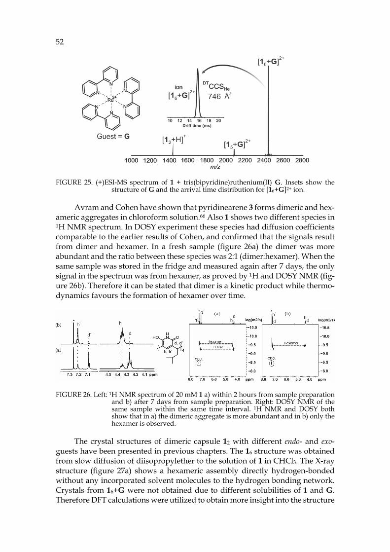
52
FIGURE 25. (+)ESI-MS spectrum of 1 + tris(bipyridine)ruthenium(II) G. Insets show the structure of G and the arrival time distribution for [16+G]2+ ion.
Avram and Cohen have shown that pyridinearene 3 forms dimeric and hex-americ aggregates in chloroform solution.66 Also 1 shows two different species in 1H NMR spectrum. In DOSY experiment these species had diffusion coefficients comparable to the earlier results of Cohen, and confirmed that the signals result from dimer and hexamer. In a fresh sample (figure 26a) the dimer was more abundant and the ratio between these species was 2:1 (dimer:hexamer). When the same sample was stored in the fridge and measured again after 7 days, the only signal in the spectrum was from hexamer, as proved by 1H and DOSY NMR (fig-ure 26b). Therefore it can be stated that dimer is a kinetic product while thermo-dynamics favours the formation of hexamer over time.
FIGURE 26. Left: 1H NMR spectrum of 20 mM 1 a) within 2 hours from sample preparation and b) after 7 days from sample preparation. Right: DOSY NMR of the same sample within the same time interval. 1H NMR and DOSY both show that in a) the dimeric aggregate is more abundant and in b) only the hexamer is observed.
The crystal structures of dimeric capsule 12 with different endo- and exo-guests have been presented in previous chapters. The 16 structure was obtained from slow diffusion of diisopropylether to the solution of 1 in CHCl3. The X-ray structure (figure 27a) shows a hexameric assembly directly hydrogen-bonded without any incorporated solvent molecules to the hydrogen bonding network. Crystals from 16+G were not obtained due to different solubilities of 1 and G. Therefore DFT calculations were utilized to obtain more insight into the structure
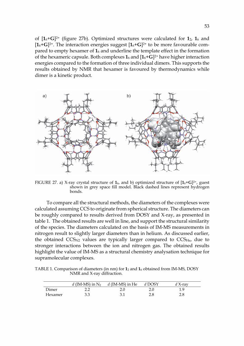
53
of [16+G]2+ (figure 27b). Optimized structures were calculated for 12, 16 and [16+G]2+. The interaction energies suggest [16+G]2+ to be more favourable com-pared to empty hexamer of 16 and underline the template effect in the formation of the hexameric capsule. Both complexes 16 and [16+G]2+ have higher interaction energies compared to the formation of three individual dimers. This supports the results obtained by NMR that hexamer is favoured by thermodynamics while dimer is a kinetic product.
FIGURE 27. a) X-ray crystal structure of 16, and b) optimized structure of [16+G]2+, guest shown in grey space fill model. Black dashed lines represent hydrogen bonds.
To compare all the structural methods, the diameters of the complexes were calculated assuming CCS to originate from spherical structure. The diameters can be roughly compared to results derived from DOSY and X-ray, as presented in table 1. The obtained results are well in line, and support the structural similarity of the species. The diameters calculated on the basis of IM-MS measurements in nitrogen result to slightly larger diameters than in helium. As discussed earlier, the obtained CCSN2 values are typically larger compared to CCSHe, due to stronger interactions between the ion and nitrogen gas. The obtained results highlight the value of IM-MS as a structural chemistry analysation technique for supramolecular complexes.
TABLE 1. Comparison of diameters (in nm) for 12 and 16 obtained from IM-MS, DOSY NMR and X-ray diffraction.
d (IM-MS) in N2 d (IM-MS) in He d DOSY d X-ray Dimer 2.2 2.0 2.0 1.9 Hexamer 3.3 3.1 2.8 2.8
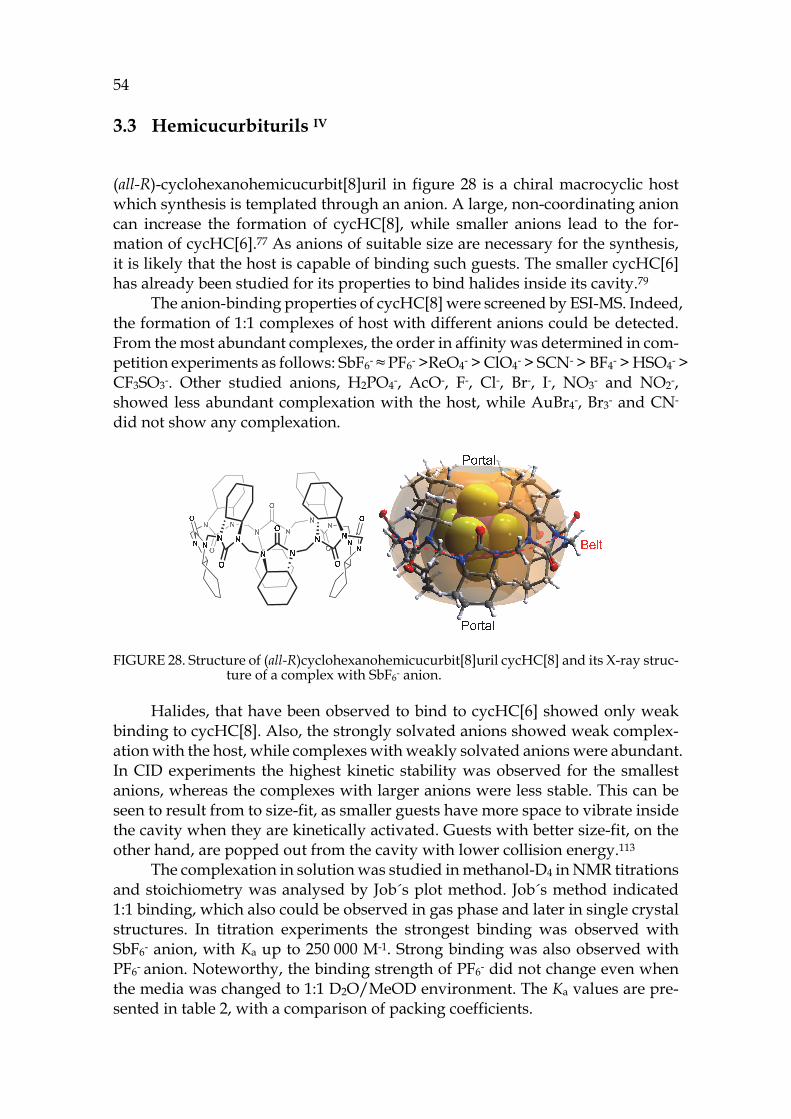
54 3.3 Hemicucurbiturils IV
(all-R)-cyclohexanohemicucurbit[8]uril in figure 28 is a chiral macrocyclic host which synthesis is templated through an anion. A large, non-coordinating anion can increase the formation of cycHC[8], while smaller anions lead to the for-mation of cycHC[6].77 As anions of suitable size are necessary for the synthesis, it is likely that the host is capable of binding such guests. The smaller cycHC[6] has already been studied for its properties to bind halides inside its cavity.79
The anion-binding properties of cycHC[8] were screened by ESI-MS. Indeed, the formation of 1:1 complexes of host with different anions could be detected. From the most abundant complexes, the order in affinity was determined in com-petition experiments as follows: SbF6- ≈ PF6- >ReO4- > ClO4- > SCN- > BF4- > HSO4- > CF3SO3-. Other studied anions, H2PO4-, AcO-, F-, Cl-, Br-, I-, NO3- and NO2-, showed less abundant complexation with the host, while AuBr4-, Br3- and CN- did not show any complexation.
FIGURE 28. Structure of (all-R)cyclohexanohemicucurbit[8]uril cycHC[8] and its X-ray struc-ture of a complex with SbF6- anion.
Halides, that have been observed to bind to cycHC[6] showed only weak binding to cycHC[8]. Also, the strongly solvated anions showed weak complex-ation with the host, while complexes with weakly solvated anions were abundant. In CID experiments the highest kinetic stability was observed for the smallest anions, whereas the complexes with larger anions were less stable. This can be seen to result from to size-fit, as smaller guests have more space to vibrate inside the cavity when they are kinetically activated. Guests with better size-fit, on the other hand, are popped out from the cavity with lower collision energy.113
The complexation in solution was studied in methanol-D4 in NMR titrations and stoichiometry was analysed by Job´s plot method. Job´s method indicated 1:1 binding, which also could be observed in gas phase and later in single crystal structures. In titration experiments the strongest binding was observed with SbF6- anion, with Ka up to 250 000 M-1. Strong binding was also observed with PF6- anion. Noteworthy, the binding strength of PF6- did not change even when the media was changed to 1:1 D2O/MeOD environment. The Ka values are pre-sented in table 2, with a comparison of packing coefficients.
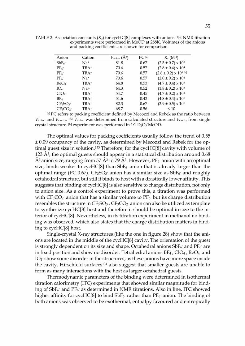
55
TABLE 2. Association constants (Ka) for cycHC[8] complexes with anions. 1H NMR titration experiments were performed in MeOD at 288K. Volumes of the anions and packing coefficients are shown for comparison.
Anion Cation Vanion (Å3) PC [a] Ka (M-1) SbF6- Na+ 81.8 0.67 (2.5 ± 0.7) x 105 PF6- TBA+ 70.6 0.57 (2.8 ± 0.4) x 104 PF6- TBA+ 70.6 0.57 (2.6 ± 0.2) x 104 [b] PF6- Na+ 70.6 0.57 (2.0 ± 0.2) x 104 ReO4- TBA+ 64.8 0.53 (4.7 ± 0.4) x 103 IO4- Na+ 64.3 0.52 (1.8 ± 0.2) x 103 ClO4- TBA+ 54.7 0.45 (4.7 ± 0.2) x 102 BF4- TBA+ 51.6 0.42 (4.8 ± 0.4) x 101 CF3SO3- TBA+ 82.3 0.67 (3.9 ± 0.5) x 101 CF3CO2- TBA+ 68.7 0.56 < 10
[a] PC refers to packing coefficient defined by Mecozzi and Rebek as the ratio between Vanion and Vcavity. 133 Vanion was determined from calculated structure and Vcavity from single crystal structure. [b] experiment was performed in 1:1 D2O/MeOD.
The optimal values for packing coefficients usually follow the trend of 0.55
± 0.09 occupancy of the cavity, as determined by Mecozzi and Rebek for the op-timal guest size in solution.133 Therefore, for the cycHC[8] cavity with volume of 123 Å3, the optimal guests should appear in a statistical distribution around 0.68 Å3 anion size, ranging from 57 Å3 to 79 Å3. However, PF6- anion with an optimal size, binds weaker to cycHC[8] than SbF6- anion that is already larger than the optimal range (PC 0.67). CF3SO3- anion has a similar size as SbF6- and roughly octahedral structure, but still it binds to host with a drastically lower affinity. This suggests that binding of cycHC[8] is also sensitive to charge distribution, not only to anion size. As a control experiment to prove this, a titration was performed with CF3CO2- anion that has a similar volume to PF6- but its charge distribution resembles the structure in CF3SO3-. CF3CO2- anion can also be utilized as template to synthesize cycHC[8] host and therefore it should be optimal in size to the in-terior of cycHC[8]. Nevertheless, in its titration experiment in methanol no bind-ing was observed, which also states that the charge distribution matters in bind-ing to cycHC[8] host.
Single-crystal X-ray structures (like the one in figure 28) show that the ani-ons are located in the middle of the cycHC[8] cavity. The orientation of the guest is strongly dependent on its size and shape. Octahedral anions SbF6- and PF6- are in fixed position and show no disorder. Tetrahedral anions BF4-, ClO4-, ReO4- and IO4- show some disorder in the structures, as these anions have more space inside the cavity. Hirschfeld surfaces134 also suggest that smaller guests are unable to form as many interactions with the host as larger octahedral guests.
Thermodynamic parameters of the binding were determined in isothermal titration calorimetry (ITC) experiments that showed similar magnitude for bind-ing of SbF6- and PF6- as determined in NMR titrations. Also in line, ITC showed higher affinity for cycHC[8] to bind SbF6- rather than PF6- anion. The binding of both anions was observed to be exothermal, enthalpy favoured and entropically
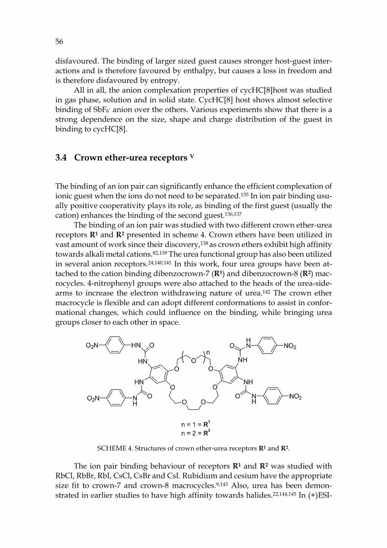
56 disfavoured. The binding of larger sized guest causes stronger host-guest inter-actions and is therefore favoured by enthalpy, but causes a loss in freedom and is therefore disfavoured by entropy.
All in all, the anion complexation properties of cycHC[8]host was studied in gas phase, solution and in solid state. CycHC[8] host shows almost selective binding of SbF6- anion over the others. Various experiments show that there is a strong dependence on the size, shape and charge distribution of the guest in binding to cycHC[8].
3.4 Crown ether-urea receptors V
The binding of an ion pair can significantly enhance the efficient complexation of ionic guest when the ions do not need to be separated.135 In ion pair binding usu-ally positive cooperativity plays its role, as binding of the first guest (usually the cation) enhances the binding of the second guest.136,137
The binding of an ion pair was studied with two different crown ether-urea receptors R1 and R2 presented in scheme 4. Crown ethers have been utilized in vast amount of work since their discovery,138 as crown ethers exhibit high affinity towards alkali metal cations.82,139 The urea functional group has also been utilized in several anion receptors.24,140,141 In this work, four urea groups have been at-tached to the cation binding dibenzocrown-7 (R1) and dibenzocrown-8 (R2) mac-rocycles. 4-nitrophenyl groups were also attached to the heads of the urea-side-arms to increase the electron withdrawing nature of urea.142 The crown ether macrocycle is flexible and can adopt different conformations to assist in confor-mational changes, which could influence on the binding, while bringing urea groups closer to each other in space.
SCHEME 4. Structures of crown ether-urea receptors R1 and R2.
The ion pair binding behaviour of receptors R1 and R2 was studied with RbCl, RbBr, RbI, CsCl, CsBr and CsI. Rubidium and cesium have the appropriate size fit to crown-7 and crown-8 macrocycles.9,143 Also, urea has been demon-strated in earlier studies to have high affinity towards halides.22,144,145 In (+)ESI-
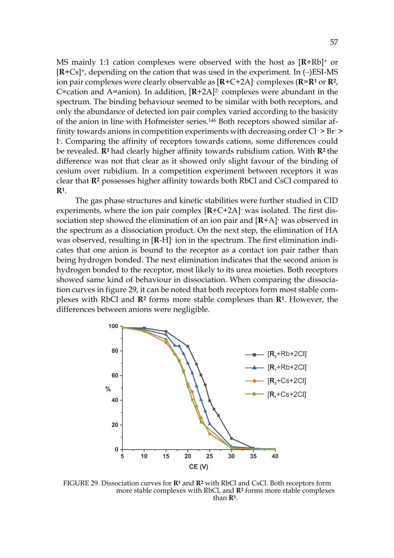
57
MS mainly 1:1 cation complexes were observed with the host as [R+Rb]+ or [R+Cs]+, depending on the cation that was used in the experiment. In (‒)ESI-MS ion pair complexes were clearly observable as [R+C+2A]- complexes (R=R1 or R2, C=cation and A=anion). In addition, [R+2A]2- complexes were abundant in the spectrum. The binding behaviour seemed to be similar with both receptors, and only the abundance of detected ion pair complex varied according to the basicity of the anion in line with Hofmeister series.146 Both receptors showed similar af-finity towards anions in competition experiments with decreasing order Cl- > Br- > I-. Comparing the affinity of receptors towards cations, some differences could be revealed. R1 had clearly higher affinity towards rubidium cation. With R2 the difference was not that clear as it showed only slight favour of the binding of cesium over rubidium. In a competition experiment between receptors it was clear that R2 possesses higher affinity towards both RbCl and CsCl compared to R1.
The gas phase structures and kinetic stabilities were further studied in CID experiments, where the ion pair complex [R+C+2A]- was isolated. The first dis-sociation step showed the elimination of an ion pair and [R+A]- was observed in the spectrum as a dissociation product. On the next step, the elimination of HA was observed, resulting in [R-H]- ion in the spectrum. The first elimination indi-cates that one anion is bound to the receptor as a contact ion pair rather than being hydrogen bonded. The next elimination indicates that the second anion is hydrogen bonded to the receptor, most likely to its urea moieties. Both receptors showed same kind of behaviour in dissociation. When comparing the dissocia-tion curves in figure 29, it can be noted that both receptors form most stable com-plexes with RbCl and R2 forms more stable complexes than R1. However, the differences between anions were negligible.
FIGURE 29. Dissociation curves for R1 and R2 with RbCl and CsCl. Both receptors form more stable complexes with RbCl, and R2 forms more stable complexes
than R1.
58
The binding behaviour of R1 and R2 and their Rb+ and Cs+ complexes was studied in a 4:1 CDCl3/DMSO solution with special interest in determining the stoichiometry and strength of the binding. Job´s plot analysis has often been uti-lized for determination of stoichiometry, but critical evaluations of its usability have been published.121 Therefore, more value was given to the fit of the experi-mental data to calculated binding constants, while Job´s plot data were seen as approximations. In titration experiments TBA-halides were used to provide the anion and alkali metals were provided as tetraphenyl borates (BPh4). The non-interacting nature of the chosen counter ions has been demonstrated earlier with similar receptor systems.125
The observed chemical shifts could be followed from three different protons. When testing only the anion binding behaviour of the receptors, the largest dif-ferences in chemical shifts were observed upon addition of Cl- anion. R1 showed slightly higher binding of Cl- and Br- anions but the differences were modest. Also, the binding strengths follow the basicity of the anion. Next, the cooperative effect of the bound Rb+ or Cs+ cation on anion complexation properties of recep-tors was studied. A dramatic increase in binding was observed with all anions. Especially the binding of chloride is significantly magnified when cation is in-volved to the complexation in both receptors. The bound Rb+ enhances the bind-ing of chloride more than the bound Cs+. This behaviour was observed with both receptors, and is perhaps surprising in the case of R2 with a larger crown ether moiety. Yet, the larger size of cesium cation causes lower charge density and po-larizability, which could explain the weaker electrostatic interaction observed be-tween bound cation and anion. This same phenomenon was observed with all studied anions. More importantly, the binding constants show that the cation complex of R2 has a higher affinity towards all anions. The enhancement in anion binding when the cation is bound to the receptor demonstrates well that both receptors function in a strongly cooperative manner.
Several X-ray structures were attained with R2 and ion pairs, but only one with R1. All structures show that the symmetricity of the crown-8 moiety in R2 allows it to assume a folded conformation (figure 30b). On the contrary, R1 lacks the symmetry and has an open non-folded conformation in the solid state (figure 30a). The difference in the solid state can explain the observed results from solu-tion and gas phase: the folded structure of R2 is more efficient in binding.
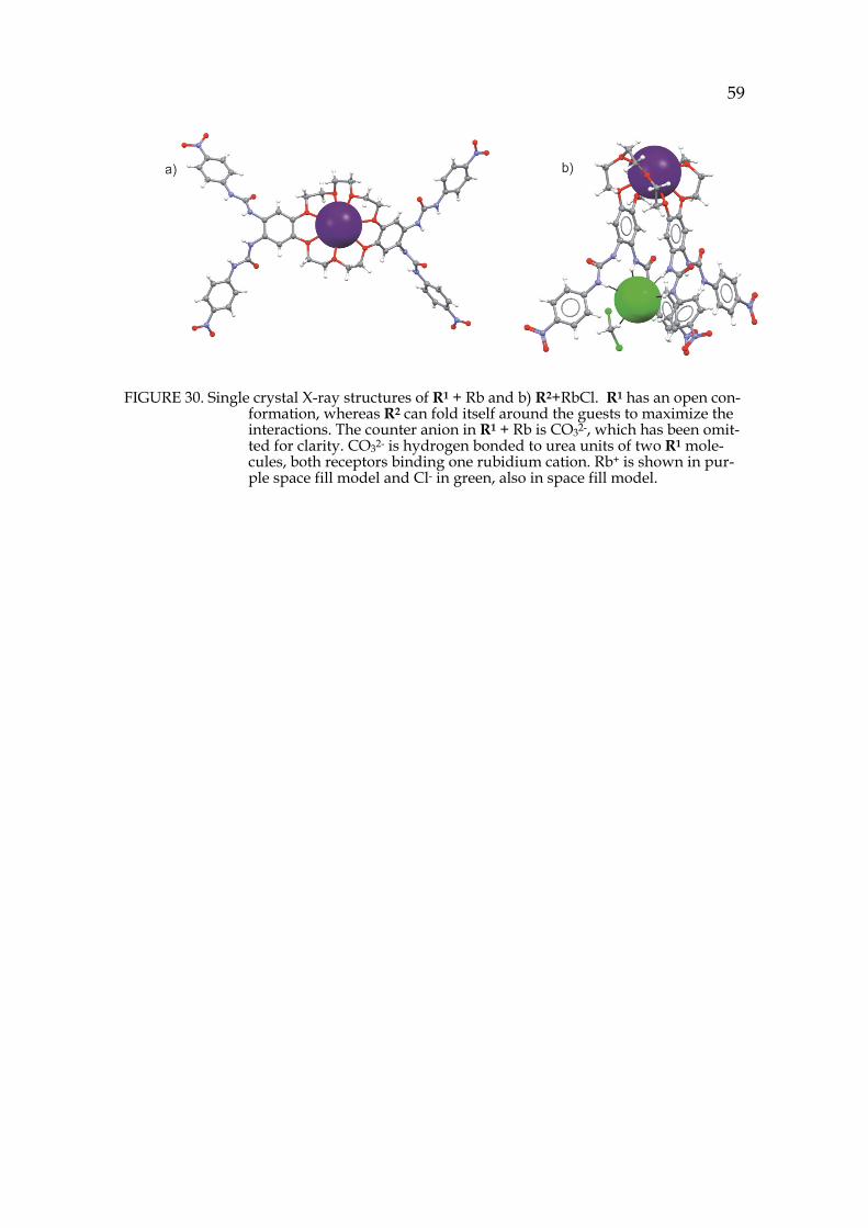
59
FIGURE 30. Single crystal X-ray structures of R1 + Rb and b) R2+RbCl. R1 has an open con-formation, whereas R2 can fold itself around the guests to maximize the interactions. The counter anion in R1 + Rb is CO32-, which has been omit-ted for clarity. CO32- is hydrogen bonded to urea units of two R1 mole-cules, both receptors binding one rubidium cation. Rb+ is shown in pur-ple space fill model and Cl- in green, also in space fill model.
60
CONCLUSIONS
Before this study it was commonly thought that pyridinearenes could bind ani-onic guests inside their cavities, based on the studies performed by Mattay´s group.64,65 It is now clear that the binding properties of pyridinearene are com-pletely different, even reverse to the early assumptions as demonstrated by the encapsulation of cationic guests.II, III However, the pyridinearene does not seem to be very efficient cation receptor either when compared to resorcinarenes. Pyr-idinearene has affinity towards anions, but instead of endo-complexation the binding of anions takes place to their exo-positions.I Presumably, pyridinearenes possesses the highest affinity towards neutral guests, which were shown to be encapsulated to the dimer in several X-ray structures and also for the first time in gas phase structures.I,126 The DFT calculations also support that the cavity of pyridinearene is most suited to bind neutral guests.I
In addition to the binding properties of pyridinearenes, new information on their self-assembly was attained. Even if it could be thought that the larger as-semblies are not as easily formed as smaller ones, thermodynamics favour the formation of hexameric pyridinearene capsules over dimeric ones. The hex-americ assembly of pyridinearene was also detected for the first time in gas phase and in solid state. The X-ray structures illustrated that both dimeric and hex-americ capsules of pyridinearene are directly hydrogen-bonded between the sub-units, without any bridging solvent molecules which are usually present in sim-ilar structures of resorcinarenes. III
From all the studied receptors, cycHC[8] was demonstrated to be the most promising as an anion receptor, with especially high binding shown for environ-mental pollutant SbF6-. The CycHC[8] host also showed a clear discrimination between the studied guests, as the binding was dependent on shape, size and charge distribution.IV A different approach to anion-binding was demonstrated by crown ether urea receptors, which are not very efficient anion receptors as their selves. However, when a cation was added as a part of the host macrocycle, a dramatic increase was observed in their anion-binding affinity. This shows that the crown ether urea receptors function in a highly cooperative manner, and they are more suited to bind a whole ion pair, not just separated ions.V
To conclude, this thesis describes the supramolecular chemistry and anion-binding properties of different host molecules. Especially the self-assembly of pyridinearene and its ability to form different types of capsules was under inves-tigation. The studies have been conducted, whenever possible, in gas phase, in solution and in solid state. Ion mobility mass spectrometry, a relatively new tech-nique, was utilized to study the structural features of pyridinearene capsules in the gas phase. Ion mobility mass spectrometry proved to be a valuable tool in structural analysis of the studied supramolecular complexes, and well compara-ble with other structural chemistry methods.
61
REFERENCES
1. J.-M Lehn, Angew. Chem. Int. Ed. Engl. 1988, 27, 89–112. 2. D. J. Cram, Angew. Chem. Int. Ed. Engl. 1988, 27, 1009–1020. 3. E. Fischer, Berichte der Dtsch. Chem. Gesellschaft 1894, 27, 2985–2993. 4. F. W. Lichtenthaler, Angew. Chem. Int. Ed. Engl. 1995, 33, 2364–2374. 5. B. L. Feringa, Angew. Chem. Int. Ed. 2017, 56, 11060–11078. 6. J. F. Sir Stoddart, Angew. Chem. Int. Ed. Engl. 2017, 56, 11094–11125. 7. C. A. Hunter & H. L. Anderson, Angew. Chem. Int. Ed. 2009, 48, 7488–7499. 8. J. D. Badjić, A. Nelson, S. J. Cantrill, W. B. Turnbull, & J. F Stoddart, Acc.
Chem. Res. 2005, 38, 723–732. 9. J. W. Steed & J. L. Atwood, Supramolecular chemistry, 2nd ed., Wiley,
Chichester, UK, 2009. 10. G. R. Desiraju, P. S. Ho, L. Kloo, A. C. Legon, R. Marquardt, P. Metrangolo,
P. Politzer, G. Resnati & K. Rissanen. Pure Appl. Chem. 2013, 85, 1711–1713. 11. P. Metrangolo, F. Meyer, T. Pilati, & G. Resnati, Angew. Chem. Int. Ed. 2008,
47, 6114–6127. 12. D. A. Dougherty, Science 1996, 271, 163–169. 13. D. Quiñonero, C. Garau, C. Rotger, A. Frontera, P. Ballester, A. Costa & P.
M. Deyà, Angew. Chem. Int. Ed. 2002, 41, 3389–3392. 14. M. Göth, F. Witte, M. Quennet, P. Jungk, G. Podolan, D. Lentz, W.
Hoffmann, K. Pagel, H. U. Reissig, B. Paulus & C. A. Schalley, Chem. Eur. J. 2018, 24, 12879–12889.
15. M. Nishio, Phys. Chem. Chem. Phys. 2011, 13, 13873–13900. 16. A. Marmur, J. Am. Chem. Soc. 2000, 122, 2120–2121. 17. J. W. Steed, D. R.Turner, & K. J. Wallace, Core Concepts in Supramolecular
Chemistry and Nanochemistry, 1st Ed., John Wiley and Sons, Ltd, 2007. 18. D. E. Koshland, Angew. Chem. Int. Ed. 1994, 33, 2375–2378. 19. D. B. Smithrud, E. M. Sanford, I. Chao, S. B. Ferguson, D. R. Carganague, J.
D. Evanseck, K. N. Houk & F. Diederich, Pure Appl. Chem. 1990, 62, 2227–2236.
20. J. Murray, K. Kim, T. Ogoshi, W. Yao, & B. C. Gibb, Chem. Soc. Rev. 2017, 46, 2479–2496.
21. R. G., Pearson, J. Am. Chem. Soc. 1963, 85, 3533–3539. 22. J. L. Sessler, P. A. Gale & W.-S. Cho, Anion Receptor Chemistry, Royal Society
of Chemistry, Cambridge, UK, 2006. 23. A. J. McConnell & P. D. Beer, Angew. Chem. Int. Ed. 2012, 51, 5052–5061. 24. R. Shukla, T. Kida, & B. D. Smith, Supramol. Chem. 2000, 2, 3099–3102. 25. L. Fabbrizzi, & A. Poggi, Chem. Soc. Rev. 1995, 24, 197–202. 26. G. M. Whitesides, & B. Grzybowski, Science 2002, 295, 2418–2421. 27. D. Philp, & J. F. Stoddart, Angew. Chem. Int. Ed. 1996, 35, 1154–1196. 28. D. A. Leigh, R. G. Pritchard, & A. J. Stephens, Nat. Chem. 2014, 6, 978–982. 29. B. Song, Z. Zhang, K. Wang, C. H. Hsu, O. Bolarinwa, J. Wang, Y. Li, G. Q.
Yin, E. Riviera, H. B. Yang, C. Liu, B. Xu & X. Li., Angew. Chem. Int. Ed. 2017, 56, 5258–5262.
62 30. L. Turunen, U. Warzok, R. Puttreddy, N. K. Beyeh, C. A. Schalley & K.
Rissanen, Angew. Chem. Int. Ed. 2016, 55, 14033–14036. 31. L. Turunen, U. Warzok, C. A. Schalley & K. Rissanen, Chem 2017, 3, 861–
869. 32. P. Jacopozzi, & E. Dalcanale, Angew. Chem. Int. Ed. 1997, 36, 613–615. 33. R. Pinalli, V. Cristini, V. Sottili, S. Geremia, M. Campagnolo, A. Caneschi
& E. Dalcanale, J. Am. Chem. Soc. 2004, 126, 6516–6517. 34. S. J. Dalgarno, N. P. Power, & J. L. Atwood, Coord. Chem. Rev. 2008, 252,
825–841. 35. D. Fujita, Y. Ueda, S. Sato, H. Yokoyama, N. Mizuno, T. Kumasaka & M.
Fujita, Chem 2016, 1, 91–101 36. G. Markiewicz, A. Jenczak, M. Kołodziejski, J. J. Holstein, J. K. M. Sanders,
& A. R. Stefankiewicz, Nat. Commun. 2017, 8, 6–13. 37. J. B. Niederl & H. J. Vogel, J. Am. Chem. Soc. 1940, 62, 2512–2514. 38. P. Timmerman, W. Verboom, & D. N. Reinhoudt, Tetrahedron 1996, 52,
2663–2704. 39. N. K. Beyeh & K. Rissanen, Isr. J. Chem. 2011, 51, 769–780. 40. D. J. Cram, S. Karbach, H. E. Kim, C. B. Knobler, E. F. Maverick, J. L. Ericson
& R. C. Helgeson, J. Am. Chem. Soc. 1988, 110, 2229–2237. 41. S. Ma, D. M. Rudkevich & J. Rebek, J. Am. Chem. Soc. 1998, 120, 4977–4981. 42. J. R. Moran, J. L. Ericson, E. Dalcanale, J. A. Bryant, & C. B. Knobler, J. Am.
Chem. Soc. 1991, 113, 5707–5714. 43. H. Erdtman & S. Högberg, Tetrahedron Lett. 1968, 1679–1682. 44. S. Högberg, J. Am. Chem. Soc. 1980, 102, 6046–6050. 45. A. G. S. Högberg, J. Org. Chem. 1980, 45, 4498–4500. 46. J. L. Atwood & L. R. MacGillivray, Nature 1997, 389, 469–472. 47. T. Gerkensmeier, W. Iwanek, C. Agena, R. Fröchlich, S. Kotila, C. Näther &
J. Mattay, Eur. J. Org. Chem. 1999, 2257–2262. 48. K. Murayama, Chem. Commun. 1998, 607–608. 49. L. Avram, & Y. Cohen, Org. Lett. 2002, 4, 4365–4368. 50. T. Lippmann, H. Wilde, A. Schayer, M. Hesse & G. Mann, Angew. Chem.
Int. Ed. 1993, 32, 1991–1993. 51. H. Mansikkamäki, M. Nissinen, C. A. Schalley, & K. Rissanen, New J. Chem.
2003, 27, 88–97. 52. A. Åhman, M. Luostarinen, K. Rissanen & M. Nissinen, New J. Chem. 2007
31, 169–177. 53. L. Avram, & Y. Cohen, J. Am. Chem. Soc. 2002, 124, 15148–15149. 54. H. Mansikkamäki, M. Nissinen, & K. Rissanen, Chem. Commun. 2002, 1902–
1903. 55. L. C. Palmer, A. Shivanyuk, M. Yamanaka, & J. Rebek, Chem. Commun. 2005,
5, 857–858. 56. N. K. Beyeh, M. Kogej, A. Åhman, K. Rissanen, & C. A. Schalley, Angew.
Chem. Int. Ed. 2006, 45, 5214–5218. 57. R. M. Payne & C. L. Oliver, CrystEngComm. 2018, 20, 1919–1922. 58. Q. Zhang, L. Catti & K. Tiefenbacher, Acc. Chem. Res. 2018, 51, 2107–2114.
63
59. Q. Zhang & K. Tiefenbacher, J. Am. Chem. Soc. 2013, 135, 16213-16219. 60. C. H. Pollok, Q. Zhang, K. Tiefenbacher & C. Merten, ChemPhysChem 2017,
18, 1987–1991. 61. C. Gaeta, C. Talotta, M. De Rosa, P. La Manna, A. Soriente & P. Neri, Chem.
Eur. J. 2019, 25, 4899–4913. 62. Q. Zhang, L. Catti, V. R. I. Kaila & K. Tiefenbacher, Chem. Sci. 2017, 8, 1653–
1657. 63. T. Gerkensmeier, J. Mattay & C. Näther, Chem. Eur. J. 2001, 7 (2), 465–474. 64. M. C. Letzel, B. Decker, A. B. Rozhenko, W. W. Schoeller & J. Mattay, J. Am.
Chem. Soc. 2004, 126, 9669–9674. 65. A. B. Rozhenko, W. W. Schoeller, M. C. Letzel, B. Decker & J. Mattay, New
J. Chem. 2013, 37, 356–365. 66. T. Evan-Salem & Y. Cohen, Chem. Eur. J. 2007, 13, 7659–7663. 67. B. L. Schottel, H. T. Chifotides & K. R. Dunbar, Chem. Soc. Rev. 2008, 37, 68–
83. 68. R. E. Dawson, A. Hennig, D. P. Weimann, D. Emery, V. Ravikumar, J.
Montenegro, T. Takeuchi, S. Gabutti, M. Mayor, J. Mareda, C. A. Schalley & S. Matile, Nat. Chem. 2010, 2, 533–538.
69. T. Gerkensmeier, B. Decker, M. Schwertfeger, W. Buchheim & J. Mattay, Eur. J. Org. Chem. 2002, 2120–2125.
70. B. Kim, R. Balasubramanian, W. Perez-Segarra, A. Wei, B. Decker & J. Mattay, Supramol. Chem. 2005, 17, 173–180.
71. T. Witte, B. Decker, J. Mattay & K. Huber, J. Am. Chem. Soc. 2004, 126, 9276–9282.
72. K. I. Assaf & W. M. Nau, Chem. Soc. Rev. 2015, 44, 394–418. 73. J. Lagona, P. Mukhopadhyay, S. Chakrabarti & L. Isaacs, Angew. Chemie Int.
Ed. 2005, 44, 4844–4870. 74. Y. Miyahara, K. Goto, M. Oka & T. Inazu, Angew. Chem. Int. Ed. 2004, 43,
5019–5022. 75. C. Márquez, R. R. Hudgins & W. M. Nau, J. Am. Chem. Soc. 2004, 126, 5806–
5816. 76. T. F. G. G. Cova, S. C. C. Nunes, A. J. M. Valente, T. M. V. D. Pinho e Melo
& A. A. C. C. Pais, J. Mol. Liq. 2017, 242, 640–652. 77. E. Prigorchenko, M. Öeren, S. Kaabel, M. Fomitšenko, I. Reile, I. Järving, T.
Tamm, F. Topić, K. Rissanen & R. Aav, Chem. Commun. 2015, 51, 10921–10924.
78. R. Aav, E. Shmatova, I. Reile, M. Borissova, F. Topić & K. Rissanen, Org. Lett. 2013, 15, 3786–3789.
79. M. Öeren, E. Shmatova, T. Tamm & R. Aav, Phys. Chem. Chem. Phys. 2014, 16, 19198.
80. N. N. Andersen, M. Lisbjerg, K. Eriksen & M. Pittelkow, Isr. J. Chem. 2018, 58, 435–448.
81. C. J. Pedersen, J. Am. Chem. Soc. 1967, 89, 2495–2496. 82. C. J. Pedersen, J. Incl. Phenom. Mol. Recognit. Chem. 1992, 12, 7–10. 83. J. W. Steed, Coord. Chem. Rev. 2001, 215, 171–221.
64 84. R. Hilgenfeld & W. A Saenger, Angew. Chem. Int. Ed. 1981, 242, 1045–1046. 85. C. Yoo, H. M. Dodge & A. J. M. Miller, Chem. Commun. 2019, 55, 5047–5059. 86. M. T. Reetz, C. M. Niemeyer & K. Harms, Angew. Chem. Int. Ed. 1991, 30,
1472–1474. 87. M. T. Reetz, C. M. Niemeyer & K. Harms, Angew. Chem. Int. Ed. 1991, 30,
1474–1476. 88. J. M. Mahoney, G. U. Nawaratna, A. M. Beatty, P. J. Duggan, & B. D. Smith,
Inorg. Chem. 2004, 43, 5902–5907. 89. J. M. Mahoney, A. M. Beatty, & B. D. Smith, Inorg. Chem. 2004, 43, 7617–
7621. 90. K. K. Murray, R. K. Boyd, M. E. Eberlin, G. J. Langley, L. Li & Y. Naito, Pure
Appl. Chem. 2013, 85, 1515–1609. 91. K. Downard, Mass spectrometry: A foundation course, Royal Society of
Chemistry, 2004. 92. D. M. Desiderio, N. M. Nibbering & A. Kraj, Mass spectrometry :
instrumentation, interpretation, and applications. John Wiley & Sons, Inc., 2009. 93. C. Lapthorn, F. Pullen, & B. Z. Chowdhry, Mass Spectrom. Rev. 2013, 32, 43–
71. 94. E. Jurneczko & P. E. Barran, Analyst 2011, 136, 20–28. 95. J. C. May, C. B. Morris & J. A. Mclean, Anal. Chem. 2017, 89, 1032-1044. 96. E. Kalenius, M. Groessl & K. Rissanen, Nat. Rev. Chem. 2019, 3, 4–14. 97. V. Gabelica & E. Marklund, Curr. Opin. Chem. Biol. 2018, 42, 51–59. 98. T. Wyttenbach, C. Bleiholder & M. T. Bowers, Anal. Chem. 2013, 85, 2191–
2199. 99. C. Larriba & C. J. Hogan, J. Phys. Chem. A 2013, 117, 3887–3901. 100. T. Wyttenbach, C. Bleiholder, S. E. Anderson & M. T. Bowers, Mol. Phys.
2015, 113, 2344–2349. 101. C. Larriba & C. J. Hogan, J. Comput. Phys. 2013, 251, 344–363. 102. A. A. Shvartsburg & M. F. Jarrold, Chem. Phys. Lett. 1996, 261, 86–91. 103. K. Giles, J. P. Williams & I. Campuzano, Rapid Commun. Mass Spectrom.
2011 25, 1559–1566. 104. J. C. May & J. A. Mclean, Int. J. Ion Mobil. Spectrom. 2003, 16, 85–94. 105. K. M. Hines, J. C. May, J. A. McLean & L. Xu, Anal. Chem. 2016, 88, 7329–
7336. 106. K. Michelmann, J. A. Silveira, M. E. Ridgeway & M. A. Park, J. Am. Soc.
Mass Spectrom. 2014, 26, 14–24. 107. J. A. Silveira, K. Michelmann, M. E. Ridgeway & M. A. Park, J. Am. Soc.
Mass Spectrom. 2016, 27, 585–595. 108. M. E. Ridgeway, M. Lubeck, J. Jordens, M. Mann, & M. A. Park, Int. J. Mass.
Spectrom. 2018, 425, 22–35. 109. J. C. May & J. A. McLean, Anal. Chem. 2015, 87, 1422–1436. 110. K. Giles, S. D. Pringle, K. R. Worthington, D. Little, J. L. Wildgoose & R. H.
Bateman, Rapid Commun. Mass Spectrom. 2004, 18, 2401–2414. 111. J. Stojko, S. Fieulaine, S. Petiot-Bécard, A. Van Dorsselaer, T. Meinnel, C.
Giglione and S. Cianférani, Analyst 2015, 140, 7234–7245.
65
112. J. Ujma, M. De Cecco, O. Chepelin, H. Levene, C. Moffat, S. J. Pike, P. J. Lusby & P. E. Barran, Chem. Commun. 2012, 48, 4423–4425.
113. T.-C. Lee, E. Kalenius, A. I. Lazar, K. I. Assaf, N. Kuhnert, C. H. Grün, J. Jänis, O. A. Scherman & W. M. Nau, Nat. Chem. 2013, 5, 376–82.
114. H. V. Schröder, J. M. Wollschläger & C. A. Schalley, Chem. Commun. 2017, 53, 9218–9221.
115. W. Koch & M. C. Holthausen, A Chemist’s Guide to Density Functional Theory. WILEY-VCH Verlag GmbH, 2001.
116. S. Grimme, Chem. Eur. J. 2012, 18, 9955–9964. 117. J. Antony, R. Sure & S. Grimme, Chem. Commun. 2015, 51, 1764–1774. 118. S. Grimme, J. Comput. Chem. 2004, 25, 1463–1473. 119. S. Grimme, J. Antony, S. Ehrlich & H. Krieg, J. Chem. Phys. 2010, 132, 154104. 120. J. H. Simpson, Organic structure determination using 2-D NMR spectroscopy.
Academic Press, 2012. 121. P. Thordarson, Chem. Soc. Rev. 2011, 40, 1305–1323. 122. L. Avram & Y. Cohen, Chem. Soc. Rev. 2015, 44, 586–602. 123. W. Massa, Crystal Structure Determination, Springer, 2000. 124. K. Rissanen, Chem. Soc. Rev. 2017, 46, 2638–2648. 125. T. Mäkelä, E. Kalenius & K. Rissanen, Inorg. Chem. 2015, 54, 9154–9165. 126. A. Kiesilä, J. O. Moilanen, F. Topić, S. Götz, K. Rissanen, A. Lützen & E.
Kalenius. Unpublished results. 127. F. Tancini, R. M. Yebeutchou, L. Pirondini, R. De Zorzi, S. Geremia, Oren
A. Scherman & E. Dalcanale, Chem. Eur. J. 2010, 16, 14313–14321. 128. K. Murayama, Chem. Commun. 1998, 607–608. 129. M. Mäkinen, P. Vainiotalo, M. Nissinen & K. Rissanen, J. Am. Soc. Mass
Spectrom. 2003, 14, 143–151. 130. G. Bianchini, A. Scarso, G. La Sorella & G. Strukul, Chem. Commun. 2012,
48, 12082–12084. 131. K. J. Laszlo & M. F. Bush, J. Phys. Chem. A 2017, 121, 7768–7777. 132. D. Canzani, K. J. Laszlo & M. F. Bush, J. Phys. Chem. A 2018, 122, 5625–5634. 133. S. Mecozzi & J. Rebek, Chem. Eur. J. 1998, 4, 1016–1022. 134. M. A. Spackman & D. Jayatilaka, CrystEngComm. 2009, 11, 19–32. 135. M. D. Lankshear, I. Dudley, K. M. Chan, A. R. Cowley, S. M. Santos, V.
Felix & P. D. Beer, Chem. Eur. J. 2008, 14, 2248–2263. 136. P. D. Beer & S. W. Dent, Chem. Commun. 1998, 825–826. 137. J. Romański, B. Trzaskowski & P. Piątek, Dalt. Trans. 2013, 42, 15271–15274. 138. G. W. Gokel, W. M. Leevy & M. E. Weber, Chem. Rev. 2004, 104, 2723–2750. 139. C. J. Pedersen, J. Am. Chem. Soc. 1967, 89, 2495–2496. 140. O. Jurček, H. Valkenier, R. Puttreddy, M. Novak, H. A. Sparkles, R. Marek,
K. Rissanen & A. P. Davis, Chem. Eur. J. 2018, 24, 8178–8185. 141. P. A. Gale, Chem. Soc. Rev. 2010, 39, 3746–3771. 142. S. Camiolo, P. A. Gale, M. B. Hursthouse & M. E. Light, Org. Biomol. Chem.
2003, 1, 741–744. 143. J. C. Bryan, R. A. Sachleben, J. M. Lavis & M. C. Davis, Inorg. Chem. 2002,
37, 2749–2755.

66 144. P. A. Gale, N. Busschaert, C. J. E. Haynes, L. E. Karagiannidis & I. L. Kirby,
Chem. Soc. Rev. 2014, 43, 205–41. 145. A. Pramanik, D. R. Powell, B. M. Wong & A. Hossain, Inorg. Chem. 2012, 51,
4274–4284. 146. Y. Zhang & P. S. Cremer, Curr. Opin. Chem. Biol. 2006, 10, 658–663.
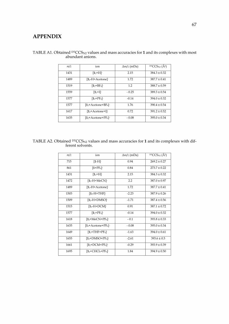
67
APPENDIX
TABLE A1. Obtained DTCCSN2 values and mass accuracies for 1 and its complexes with most abundant anions.
m/z ion Δm/z (mDa) DTCCSN2 (Å2)
1431 [12+H]- 2.15 384.3 ± 0.32
1489 [12-H+Acetone]- 1.72 387.7 ± 0.41
1519 [12+BF4]- 1.2 388.7 ± 0.59
1559 [12+I]- - 0.25 389.3 ± 0.54
1577 [12+PF6]- -0.14 394.0 ± 0.32
1577 [12+Acetone+BF4]- 1.76 390.4 ± 0.54
1617 [12+Acetone+I]- 0.72 391.2 ± 0.52
1635 [12+Acetone+PF6]- - 0.08 395.0 ± 0.34
TABLE A2. Obtained DTCCSN2 values and mass accuracies for 1 and its complexes with dif-ferent solvents.
m/z ion Δm/z (mDa) DTCCSN2 (Å2)
715 [1-H]- 0.94 269.2 ± 0.27
861 [1+PF6]- 0.84 273.7 ± 0.22
1431 [12+H]- 2.15 384.3 ± 0.32
1472 [12-H+MeCN]- 2.2 387.0 ± 0.97
1489 [12-H+Acetone]- 1.72 387.7 ± 0.41
1503 [12-H+THF]- -2.23 387.9 ± 0.26
1509 [12-H+DMSO]- -1.71 387.4 ± 0.56
1515 [12-H+DCM]- 0.91 387.1 ± 0.72
1577 [12+PF6]- -0.14 394.0 ± 0.32
1618 [12+MeCN+PF6]- - 0.1 393.8 ± 0.33
1635 [12+Acetone+PF6]- - 0.08 395.0 ± 0.34
1649 [12+THF+PF6]- -1.63 394.0 ± 0.61
1655 [12+DMSO+PF6]- -2.61 393.6 ± 0.5
1661 [12+DCM+PF6]- -0.29 393.9 ± 0.39
1695 [12+CHCl3+PF6]- 1.84 394.9 ± 0.50
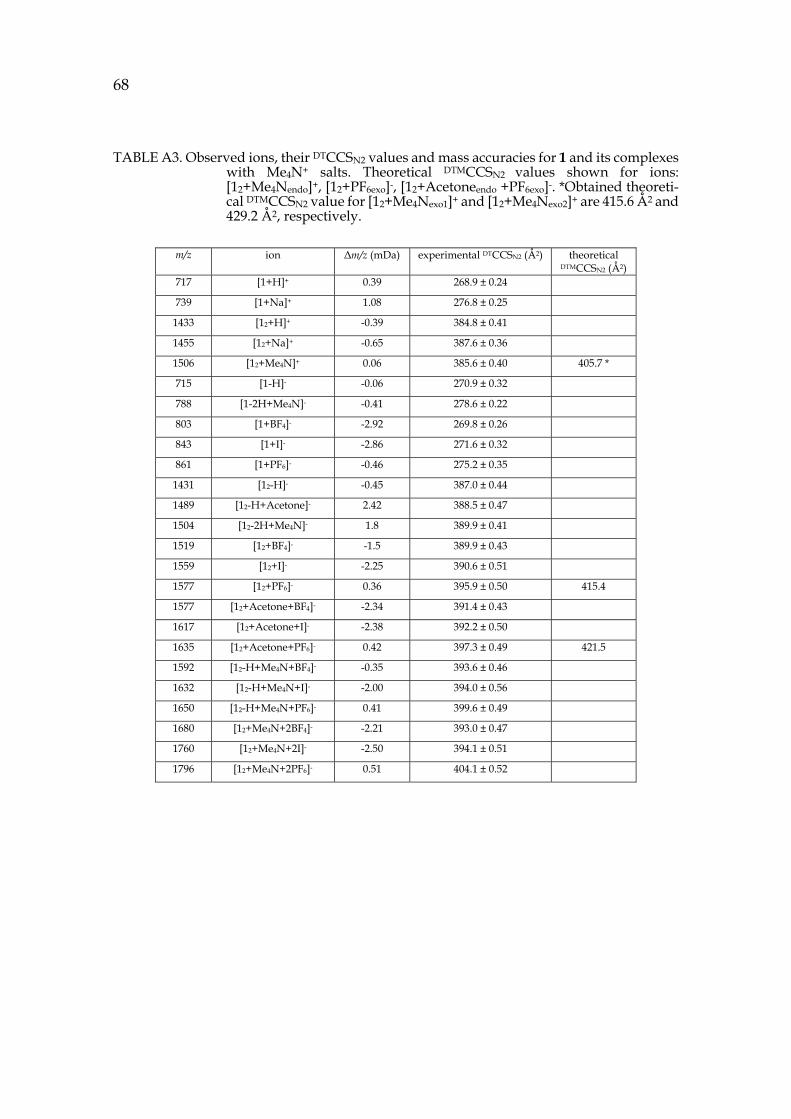
68
TABLE A3. Observed ions, their DTCCSN2 values and mass accuracies for 1 and its complexes with Me4N+ salts. Theoretical DTMCCSN2 values shown for ions: [12+Me4Nendo]+, [12+PF6exo]-, [12+Acetoneendo +PF6exo]-. *Obtained theoreti-cal DTMCCSN2 value for [12+Me4Nexo1]+ and [12+Me4Nexo2]+ are 415.6 Å2 and 429.2 Å2, respectively.
m/z ion Δm/z (mDa) experimental DTCCSN2 (Å2) theoretical DTMCCSN2 (Å2)
717 [1+H]+ 0.39 268.9 ± 0.24
739 [1+Na]+ 1.08 276.8 ± 0.25
1433 [12+H]+ -0.39 384.8 ± 0.41
1455 [12+Na]+ -0.65 387.6 ± 0.36
1506 [12+Me4N]+ 0.06 385.6 ± 0.40 405.7 *
715 [1-H]- -0.06 270.9 ± 0.32
788 [1-2H+Me4N]- -0.41 278.6 ± 0.22
803 [1+BF4]- -2.92 269.8 ± 0.26
843 [1+I]- -2.86 271.6 ± 0.32
861 [1+PF6]- -0.46 275.2 ± 0.35
1431 [12-H]- -0.45 387.0 ± 0.44
1489 [12-H+Acetone]- 2.42 388.5 ± 0.47
1504 [12-2H+Me4N]- 1.8 389.9 ± 0.41
1519 [12+BF4]- -1.5 389.9 ± 0.43
1559 [12+I]- -2.25 390.6 ± 0.51
1577 [12+PF6]- 0.36 395.9 ± 0.50 415.4
1577 [12+Acetone+BF4]- -2.34 391.4 ± 0.43
1617 [12+Acetone+I]- -2.38 392.2 ± 0.50
1635 [12+Acetone+PF6]- 0.42 397.3 ± 0.49 421.5
1592 [12-H+Me4N+BF4]- -0.35 393.6 ± 0.46
1632 [12-H+Me4N+I]- -2.00 394.0 ± 0.56
1650 [12-H+Me4N+PF6]- 0.41 399.6 ± 0.49
1680 [12+Me4N+2BF4]- -2.21 393.0 ± 0.47
1760 [12+Me4N+2I]- -2.50 394.1 ± 0.51
1796 [12+Me4N+2PF6]- 0.51 404.1 ± 0.52
ORIGINAL PAPERS
I
SIMULTANEOUS ENDO AND EXO COMPLEX FORMATION OF PYRIDINE[4]ARENE DIMERS WITH NEUTRAL AND
ANIONIC GUESTS
by
Anniina Kiesilä, Lauri Kivijärvi, Ngong Kodiah Beyeh, Jani O. Moilanen, Michael Groessl, Tatiana Rothe, Sven Götz, Filip Top
Kalenius.
Angewandte Chemie International Edition, 2017, 57, 10942-10946.
Reproduced with permission. Copyright © 2017 Wiley –
https://doi.org/10.1002/anie.201704054
AngewandteInternational Edition
A Journal of the Gesellschaft Deutscher Chemiker
www.angewandte.orgChemie
Accepted Article
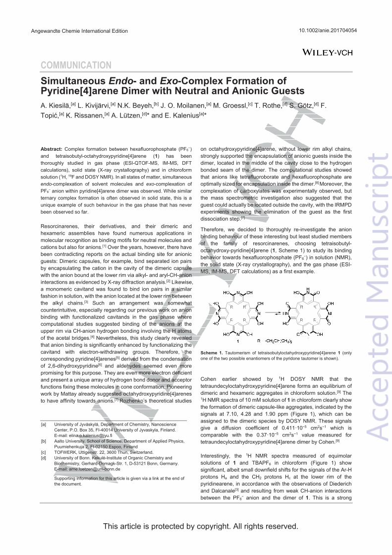
COMMUNICATION Endo Exo
endo- exo
.
10.1002/anie.201704054Angewandte Chemie International Edition
This article is protected by copyright. All rights reserved.
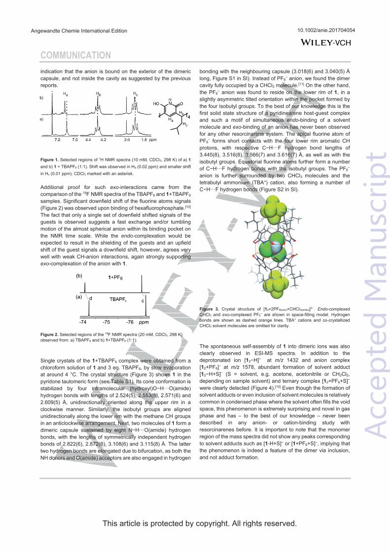
COMMUNICATION
exo-
endo-
exo
endoexo
Endoexo
m/zm/z
10.1002/anie.201704054Angewandte Chemie International Edition
This article is protected by copyright. All rights reserved.
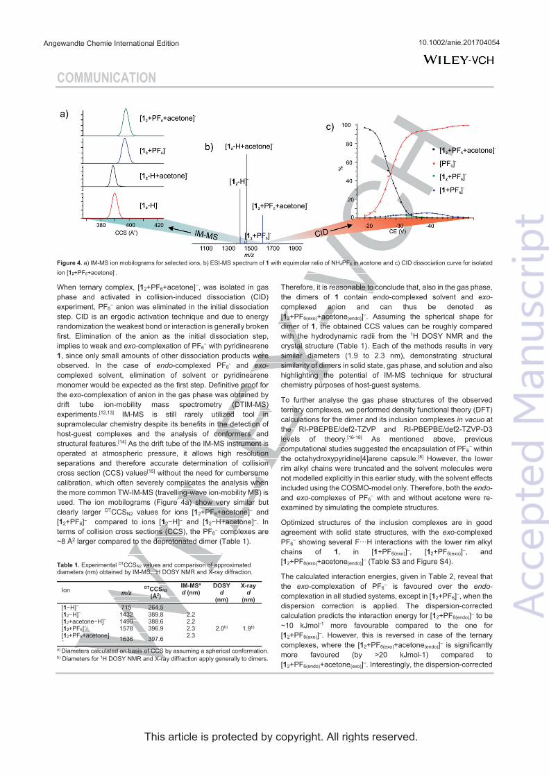
COMMUNICATION
exo
endo exo
exo
endo exo
in vacuo
endoexo
exo
exo endom/z d d d
10.1002/anie.201704054Angewandte Chemie International Edition
This article is protected by copyright. All rights reserved.
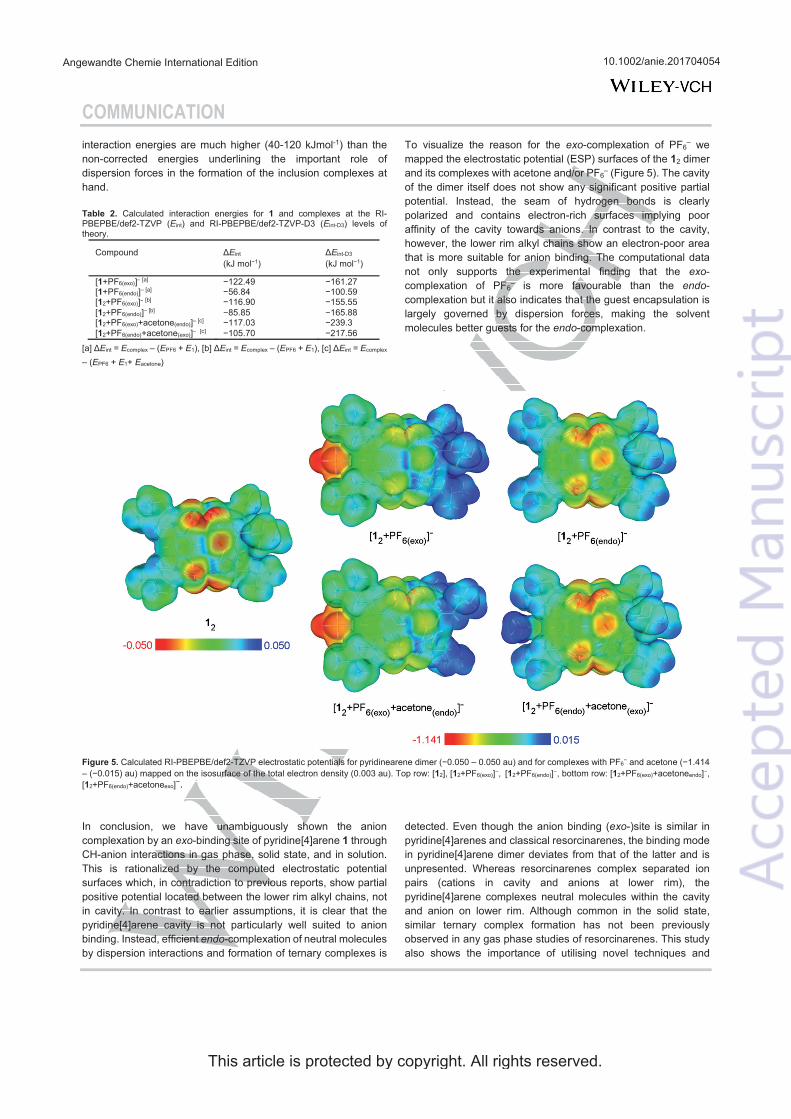
COMMUNICATION
E E
E E E E E E E E E EE E E
exo
exoendo
endo
exo
endo
exo
E E
10.1002/anie.201704054Angewandte Chemie International Edition
This article is protected by copyright. All rights reserved.
COMMUNICATION
J. Incl. Phenom. Mol. Recogn. Chem19
Tetrahedron 52 Org. Biomol. Chem 2 Chem. Soc. Rev. 36 J. Incl. Phen. Macrocycl. Chem. 78Chem. Soc. Rev 44
Chem. CommunChem.
CommunNew J. Chem 27
J. Am. Chem. Soc 131
Chem. Eur. 16
Angew. Chem120 Angew. Chem. Int. Ed 47
Chem. Eur. J 7
J. Am. Chem. Soc 126
New J. Chem 37Chem. Eur. J 13
ex
Nat. Chem. 6
Mass Spec. Rev. 32
Nat. Chem., 5
Inorg. Chem 54
Angew. Chem 54
J. Am. Chem. Soc. 127
Anal. Chem 83Chem.
Commun. 48Chem. Commun. 49
Anal. Chem. , 89, 123
Phys. Rev 81 Phys. Rev. B45 Phys.
Rev. Lett 77Chem. Phys.
Lett 242 242Theor. Chem. Acc 97
J. Chem. Phys 132
10.1002/anie.201704054Angewandte Chemie International Edition
This article is protected by copyright. All rights reserved.
II
ANION-DRIVEN ENCAPSULATION OF CATIONIC GUEST INSIDE PYRIDINE[4]ARENE DIMERS
by
Submitted manuscript
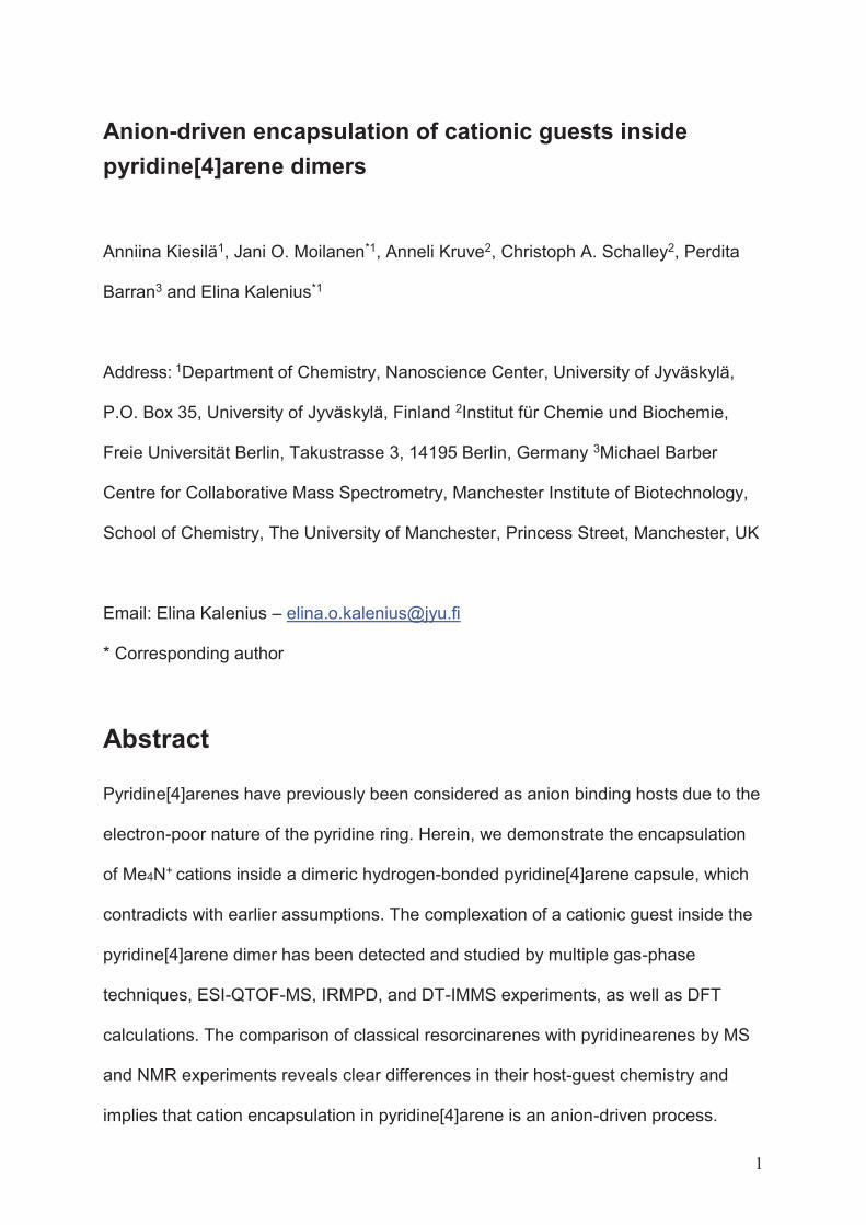
1
Anion-driven encapsulation of cationic guests inside pyridine[4]arene dimers
Anniina Kiesilä1, Jani O. Moilanen*1, Anneli Kruve2, Christoph A. Schalley2, Perdita
Barran3 and Elina Kalenius*1
Address: 1Department of Chemistry, Nanoscience Center, University of Jyväskylä,
P.O. Box 35, University of Jyväskylä, Finland 2Institut für Chemie und Biochemie,
Freie Universität Berlin, Takustrasse 3, 14195 Berlin, Germany 3Michael Barber
Centre for Collaborative Mass Spectrometry, Manchester Institute of Biotechnology,
School of Chemistry, The University of Manchester, Princess Street, Manchester, UK
Email: Elina Kalenius – [email protected]
* Corresponding author
Abstract
Pyridine[4]arenes have previously been considered as anion binding hosts due to the
electron-poor nature of the pyridine ring. Herein, we demonstrate the encapsulation
of Me4N+ cations inside a dimeric hydrogen-bonded pyridine[4]arene capsule, which
contradicts with earlier assumptions. The complexation of a cationic guest inside the
pyridine[4]arene dimer has been detected and studied by multiple gas-phase
techniques, ESI-QTOF-MS, IRMPD, and DT-IMMS experiments, as well as DFT
calculations. The comparison of classical resorcinarenes with pyridinearenes by MS
and NMR experiments reveals clear differences in their host-guest chemistry and
implies that cation encapsulation in pyridine[4]arene is an anion-driven process.
2
Keywords
pyridinearenes; resorcinarenes; ion mobility mass spectrometry; DFT calculations;
cation binding
Introduction
Resorcinarenes and their derivatives are known for the molecular recognition
properties of their self-assembled dimeric and hexameric capsules, which can
encapsulate cationic and neutral guests [1-3]. Pyridine[4]arenes [4] are analogous
macrocycles to resorcin[4]arenes. Whereas resorcinarenes are cyclic tetramers of
resorcinol, pyridinearenes are formed from 2,6-dihydroxypyridine (see Scheme 1).
Although the synthesis of pyridine[4]arenes dates back to 2001 [4], their host-guest
chemistry is still much less explored. Both macrocycles are concave and are known
to form capsular assemblies via intermolecular hydrogen bonding [5,6]. Pyridine is
significantly less electron-rich than benzene. Consequently, pyridinearene capsules
were originally assumed to encapsulate anionic guests inside their cavity due to a π-
acidic character of the aromatic walls [7,8]. Mattey et. al. detected 1:1 complex
formation with PF6- and BF4- by mass spectrometry, however without ion mobility
mass spectrometry, the location of anion could not be verified and the anions were
assumed to interact with the pyridinearene cavity. Inclusion complexes of anions
within pyridinearene dimer were also theoretically studied by DFT calculations, but
using truncated pyridinearene dimer model [8]. However, previous studies have also
shown that the π-acidic character of pyridine rings such as 2-oxo-6-oxypyridine is
rather weak [9,10]. Thus, pyridine[4]arenes may be expected to show a dual binding
behaviour towards anions and cations [8]. In addition to anion complexes also 2:1
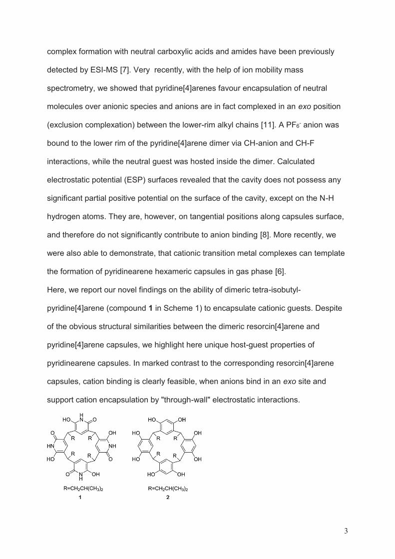
3
complex formation with neutral carboxylic acids and amides have been previously
detected by ESI-MS [7]. Very recently, with the help of ion mobility mass
spectrometry, we showed that pyridine[4]arenes favour encapsulation of neutral
molecules over anionic species and anions are in fact complexed in an exo position
(exclusion complexation) between the lower-rim alkyl chains [11]. A PF6- anion was
bound to the lower rim of the pyridine[4]arene dimer via CH-anion and CH-F
interactions, while the neutral guest was hosted inside the dimer. Calculated
electrostatic potential (ESP) surfaces revealed that the cavity does not possess any
significant partial positive potential on the surface of the cavity, except on the N-H
hydrogen atoms. They are, however, on tangential positions along capsules surface,
and therefore do not significantly contribute to anion binding [8]. More recently, we
were also able to demonstrate, that cationic transition metal complexes can template
the formation of pyridinearene hexameric capsules in gas phase [6].
Here, we report our novel findings on the ability of dimeric tetra-isobutyl-
pyridine[4]arene (compound 1 in Scheme 1) to encapsulate cationic guests. Despite
of the obvious structural similarities between the dimeric resorcin[4]arene and
pyridine[4]arene capsules, we highlight here unique host-guest properties of
pyridinearene capsules. In marked contrast to the corresponding resorcin[4]arene
capsules, cation binding is clearly feasible, when anions bind in an exo site and
support cation encapsulation by "through-wall" electrostatic interactions.
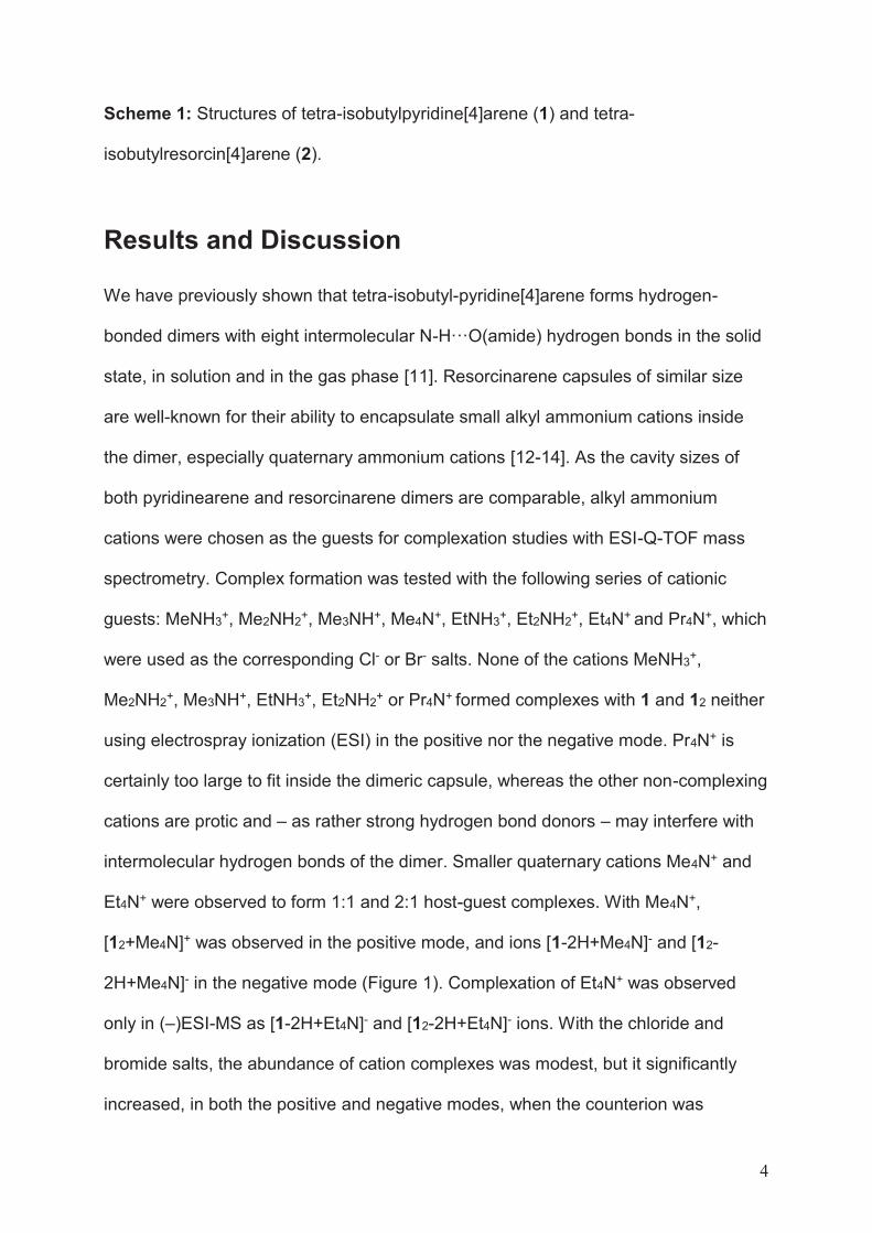
4
Scheme 1: Structures of tetra-isobutylpyridine[4]arene (1) and tetra-
isobutylresorcin[4]arene (2).
Results and Discussion
We have previously shown that tetra-isobutyl-pyridine[4]arene forms hydrogen-
bonded dimers with eight intermolecular N-H···O(amide) hydrogen bonds in the solid
state, in solution and in the gas phase [11]. Resorcinarene capsules of similar size
are well-known for their ability to encapsulate small alkyl ammonium cations inside
the dimer, especially quaternary ammonium cations [12-14]. As the cavity sizes of
both pyridinearene and resorcinarene dimers are comparable, alkyl ammonium
cations were chosen as the guests for complexation studies with ESI-Q-TOF mass
spectrometry. Complex formation was tested with the following series of cationic
guests: MeNH3+, Me2NH2+, Me3NH+, Me4N+, EtNH3+, Et2NH2+, Et4N+ and Pr4N+, which
were used as the corresponding Cl- or Br- salts. None of the cations MeNH3+,
Me2NH2+, Me3NH+, EtNH3+, Et2NH2+ or Pr4N+ formed complexes with 1 and 12 neither
using electrospray ionization (ESI) in the positive nor the negative mode. Pr4N+ is
certainly too large to fit inside the dimeric capsule, whereas the other non-complexing
cations are protic and – as rather strong hydrogen bond donors – may interfere with
intermolecular hydrogen bonds of the dimer. Smaller quaternary cations Me4N+ and
Et4N+ were observed to form 1:1 and 2:1 host-guest complexes. With Me4N+,
[12+Me4N]+ was observed in the positive mode, and ions [1-2H+Me4N]- and [12-
2H+Me4N]- in the negative mode (Figure 1). Complexation of Et4N+ was observed
only in (‒)ESI-MS as [1-2H+Et4N]- and [12-2H+Et4N]- ions. With the chloride and
bromide salts, the abundance of cation complexes was modest, but it significantly
increased, in both the positive and negative modes, when the counterion was
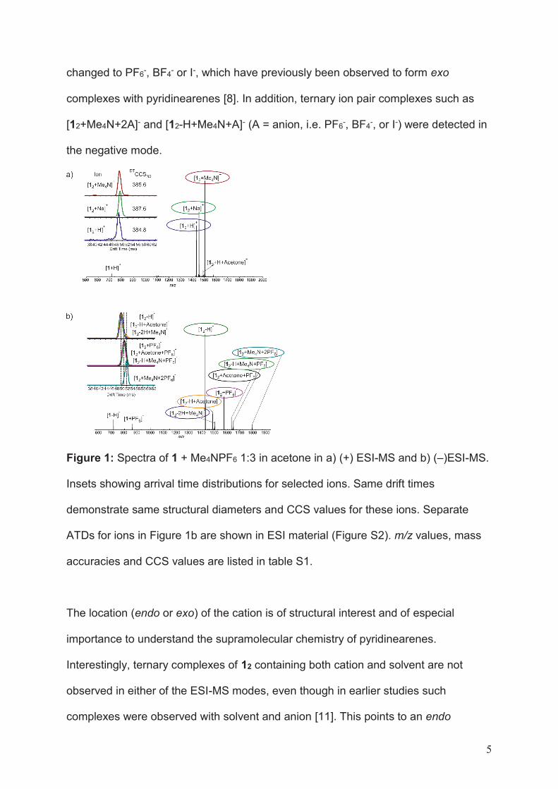
5
changed to PF6-, BF4- or I-, which have previously been observed to form exo
complexes with pyridinearenes [8]. In addition, ternary ion pair complexes such as
[12+Me4N+2A]- and [12-H+Me4N+A]- (A = anion, i.e. PF6-, BF4-, or I-) were detected in
the negative mode.
Figure 1: Spectra of 1 + Me4NPF6 1:3 in acetone in a) (+) ESI-MS and b) (‒)ESI-MS.
Insets showing arrival time distributions for selected ions. Same drift times
demonstrate same structural diameters and CCS values for these ions. Separate
ATDs for ions in Figure 1b are shown in ESI material (Figure S2). m/z values, mass
accuracies and CCS values are listed in table S1.
The location (endo or exo) of the cation is of structural interest and of especial
importance to understand the supramolecular chemistry of pyridinearenes.
Interestingly, ternary complexes of 12 containing both cation and solvent are not
observed in either of the ESI-MS modes, even though in earlier studies such
complexes were observed with solvent and anion [11]. This points to an endo
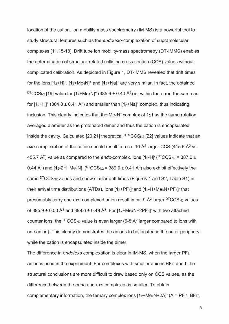
6
location of the cation. Ion mobility mass spectrometry (IM-MS) is a powerful tool to
study structural features such as the endo/exo-complexation of supramolecular
complexes [11,15-18]. Drift tube ion mobility-mass spectrometry (DT-IMMS) enables
the determination of structure-related collision cross section (CCS) values without
complicated calibration. As depicted in Figure 1, DT-IMMS revealed that drift times
for the ions [12+H]+, [12+Me4N]+ and [12+Na]+ are very similar. In fact, the obtained
DTCCSN2 [19] value for [12+Me4N]+ (385.6 ± 0.40 Å2) is, within the error, the same as
for [12+H]+ (384.8 ± 0.41 Å2) and smaller than [12+Na]+ complex, thus indicating
inclusion. This clearly indicates that the Me4N+ complex of 12 has the same rotation
averaged diameter as the protonated dimer and thus the cation is encapsulated
inside the cavity. Calculated [20,21] theoretical DTMCCSN2 [22] values indicate that an
exo-complexation of the cation should result in a ca. 10 Å2 larger CCS (415.6 Å2 vs.
405.7 Å2) value as compared to the endo-complex. Ions [12-H]- (DTCCSN2 = 387.0 ±
0.44 Å2) and [12-2H+Me4N]- (DTCCSN2 = 389.9 ± 0.41 Å2) also exhibit effectively the
same DTCCSN2 values and show similar drift times (Figures 1 and S2, Table S1) in
their arrival time distributions (ATDs). Ions [12+PF6]- and [12-H+Me4N+PF6]- that
presumably carry one exo-complexed anion result in ca. 9 Å2 larger DTCCSN2 values
of 395.9 ± 0.50 Å2 and 399.6 ± 0.49 Å2. For [12+Me4N+2PF6]- with two attached
counter ions, the DTCCSN2 value is even larger (5-8 Å2 larger compared to ions with
one anion). This clearly demonstrates the anions to be located in the outer periphery,
while the cation is encapsulated inside the dimer.
The difference in endo/exo complexation is clear in IM-MS, when the larger PF6-
anion is used in the experiment. For complexes with smaller anions BF4- and I- the
structural conclusions are more difficult to draw based only on CCS values, as the
difference between the endo and exo complexes is smaller. To obtain
complementary information, the ternary complex ions [12+Me4N+2A]- (A = PF6-, BF4-,
7
I- ) were further investigated by infrared multiphoton dissociation (IRMPD). IRMPD is
an MS/MS technique, which can be utilized to study the fragmentation of
supramolecular complexes [23]. In these experiments, the [12+Me4N+2A]- ions were
mass-selected and irradiated for 20 to 250 ms with a laser power of 95 % of 25 W. At
short irradiation times, the main dissociation product was [12+Me4N-2H]-, which is
produced after elimination of two molecules of HA (HA = H+A-, Schematic
presentation of dissociation in Figure S4). This ion further dissociates to a neutral
monomer and to [1-2H+Me4N]- ion, which can only result from two HA eliminations
and dissociation of the H-bonded capsule. Elimination of two HA molecules and
formation of [1+Me4N-2H]- would be unexpected from a complex with exo-complexed
cation. Minor fragments resulting from a direct elimination of an ion pair are observed
and [12+A]- appears in spectra. This likely results from a relocation of the anion to a
position close to the hydrogen-bonded seam of the dimer, which is then partially
opened to release the ion pair (pathway 2 in Figure S4). As all studied ternary
complexes behaved similarly in the experiments, it can be stated that Me4N+ is
located inside the cavity, while the PF6-, BF4- and I- anions reside in the exo-binding
site.
The binding efficiency of pyridine[4]arene towards the Me4N+ cation was compared to
that of resorcin[4]arene, which is known to bind small cations with high affinity. 1, 2
and Me4NPF6 were mixed in 1:1:1 ratio and measured by ESI-QTOF-MS. As seen in
Figure 2, the resorcin[4]arene dimer [22+Me4N]+ was detected as the base peak in
the positive mode, while [12+Me4N]+ was hardly observed. In addition to the pure
dimers, the formation of the heterodimer [1•2+Me4N]+ was also observed. In the
negative mode, heterodimers [1•2-H]- and [1•2-2H+Me4N]- were observed with higher
intensity than the corresponding homodimeric capsules of 1 or 2 (Figure S1). This is
surprising due to differences in H-bonding geometry between dimers of 1 or 2.
8
Previously in case of hexameric capsules heterohexamers of pyridinearene and
resorcinarene were not observed [5]. The PF6- anion was observed to be bound to
the pyridine[4]arene dimer in [12+PF6]- and the heterodimer [1•2+PF6]-, but
interestingly the abundance of [1•2+PF6]- was half of [12+PF6]-, and PF6- complexes
with 2 or 22 had even lower abundance. This clearly shows that the anion has a
higher affinity to pyridinearene than to resorcinarene. Inclusion complexation of
solvent was also only detected with the pyridinearene homodimer (for example as the
[12+MeCN+PF6]- ion). This mixed host experiment shows the resorcin[4]arene affinity
towards Me4N+ to be higher than that of pyridine[4]arene. Also, the lack of anion
and/or solvent complex formation with 2 or 22 in gas phase indicates that this binding
feature is unique to pyridinearenes and it does not take place with resorcinarene. The
high abundance of ternary ion pair complexes with pyridinearene, but not with
resorcinarene indicates that the cation complexation process to pyridinearene is
anion driven at least to some extent. The reduced electron-density in the pyridine
rings is compensated by favourable "through-wall" electrostatic interactions between
the exo-anion(s) and the endo-cation.
Figure 2: (+)ESI-MS profile spectrum of the mixture of 1, 2 and TMAPF6 in
acetonitrile (20 μM, 1:1:1). Inset shows a zoom in for region showing dimeric ions.
In solution, by 1H NMR, complexation of Me4N+ cation was observed with 2, but not
with 1 (Figures S5-S6). In a sample of 10 mM 2 and Me4NPF6 (1:3 ratio in
9
CDCl3/CD3CN 4:3 v:v mixture) a 0.35 ppm upfield shift was observed in Me4N+
signal. In a sample of 1 and Me4NPF6 no shift is observed. This clearly indicates that
also in solution 1 does not possess similar affinity towards Me4N+ cation as 2. Also,
all our attempts to obtain solid-state single crystal structure of 1 with cationic guest
were unsuccessful. It is possible that observation of complexes require special
conditions present in ESI source.
To obtain more detailed insight into the observed gas phase structures, DFT
calculations were carried out for 1, 1OH, 12, [12+Me4Nendo]+, [12+Me4Nexo1]+,
[12+Me4Nexo2]+, [12+Me4Nendo+Iendo], [12+Me4Nendo+Iexo], [12+Me4Nendo+2Iexo]-, [12+Iexo]+,
and [12+2Iexo]+ (Figure S7) without (PBE0/def2-TZVP) and with (PBE0-D3/def2-TZVP)
dispersion correction because in the gas phase dispersion plays an important role in
the formation of supramolecular complexes [24-30]. Conformational analysis was first
performed for both tautomers of monomer to ensure that further calculations were
carried out for the most stable tautomer. The conformational analysis revealed that
the pyridone tautomer (1) is ~150 kJ/mol lower in energy than the dihydroxyl
tautomer (1OH). The geometry optimizations showed that the geometry of
[12+Me4Nendo]+ is similar without and with dispersion correction (hydrogen bond
lengths are listed in Table S2). This is expected, because the standard hybrid
functionals are able to describe strong hydrogen bonds such as NH···O and OH···O
in a reasonable manner [31,32]. Due to the encapsulated guest molecule, the
[12+Me4Nendo]+ complex has slightly longer NH···O and OH···O bonds than empty 12.
The addition of I- anion(s) at the lower rim(s) of [12+Me4Nendo]+ has hardly any
influence on the length of NH···O and OH···O bonds as exemplified by the hydrogen
bond lengths of [12+Me4Nendo+Iexo] and [12+Me4Nendo +2Iexo]-. The same trend is also
observed for cation-free complexes ([12+Iexo]- and [12+2Iexo]2-) if their NH···O and
OH···O bond lengths are compared to those in 12.
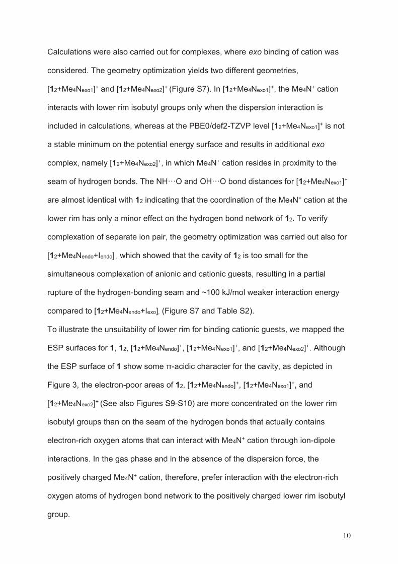
10
Calculations were also carried out for complexes, where exo binding of cation was
considered. The geometry optimization yields two different geometries,
[12+Me4Nexo1]+ and [12+Me4Nexo2]+ (Figure S7). In [12+Me4Nexo1]+, the Me4N+ cation
interacts with lower rim isobutyl groups only when the dispersion interaction is
included in calculations, whereas at the PBE0/def2-TZVP level [12+Me4Nexo1]+ is not
a stable minimum on the potential energy surface and results in additional exo
complex, namely [12+Me4Nexo2]+, in which Me4N+ cation resides in proximity to the
seam of hydrogen bonds. The NH···O and OH···O bond distances for [12+Me4Nexo1]+
are almost identical with 12 indicating that the coordination of the Me4N+ cation at the
lower rim has only a minor effect on the hydrogen bond network of 12. To verify
complexation of separate ion pair, the geometry optimization was carried out also for
[12+Me4Nendo+Iendo] , which showed that the cavity of 12 is too small for the
simultaneous complexation of anionic and cationic guests, resulting in a partial
rupture of the hydrogen-bonding seam and ~100 kJ/mol weaker interaction energy
compared to [12+Me4Nendo+Iexo], (Figure S7 and Table S2).
To illustrate the unsuitability of lower rim for binding cationic guests, we mapped the
ESP surfaces for 1, 12, [12+Me4Nendo]+, [12+Me4Nexo1]+, and [12+Me4Nexo2]+. Although
the ESP surface of 1 show some π-acidic character for the cavity, as depicted in
Figure 3, the electron-poor areas of 12, [12+Me4Nendo]+, [12+Me4Nexo1]+, and
[12+Me4Nexo2]+ (See also Figures S9-S10) are more concentrated on the lower rim
isobutyl groups than on the seam of the hydrogen bonds that actually contains
electron-rich oxygen atoms that can interact with Me4N+ cation through ion-dipole
interactions. In the gas phase and in the absence of the dispersion force, the
positively charged Me4N+ cation, therefore, prefer interaction with the electron-rich
oxygen atoms of hydrogen bond network to the positively charged lower rim isobutyl
group.
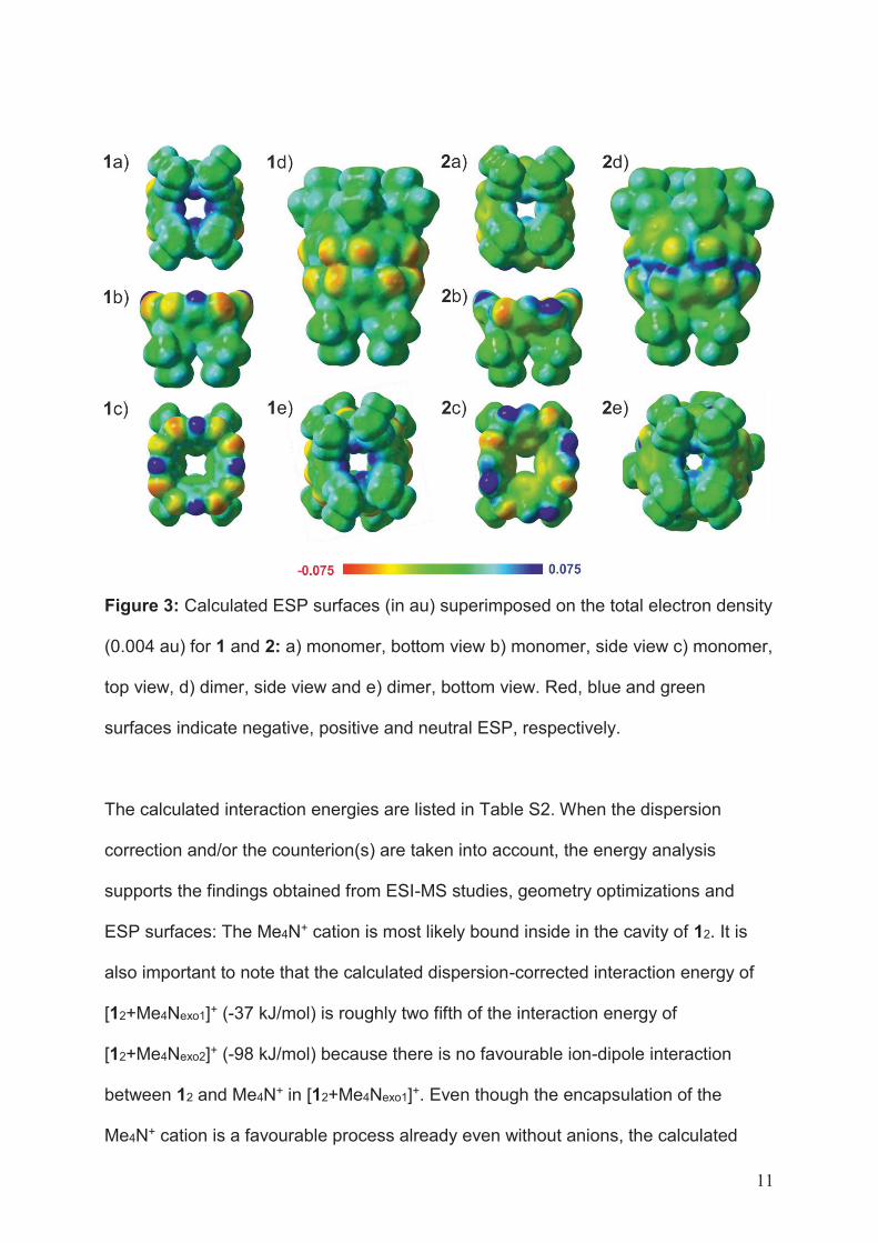
11
Figure 3: Calculated ESP surfaces (in au) superimposed on the total electron density
(0.004 au) for 1 and 2: a) monomer, bottom view b) monomer, side view c) monomer,
top view, d) dimer, side view and e) dimer, bottom view. Red, blue and green
surfaces indicate negative, positive and neutral ESP, respectively.
The calculated interaction energies are listed in Table S2. When the dispersion
correction and/or the counterion(s) are taken into account, the energy analysis
supports the findings obtained from ESI-MS studies, geometry optimizations and
ESP surfaces: The Me4N+ cation is most likely bound inside in the cavity of 12. It is
also important to note that the calculated dispersion-corrected interaction energy of
[12+Me4Nexo1]+ (-37 kJ/mol) is roughly two fifth of the interaction energy of
[12+Me4Nexo2]+ (-98 kJ/mol) because there is no favourable ion-dipole interaction
between 12 and Me4N+ in [12+Me4Nexo1]+. Even though the encapsulation of the
Me4N+ cation is a favourable process already even without anions, the calculated
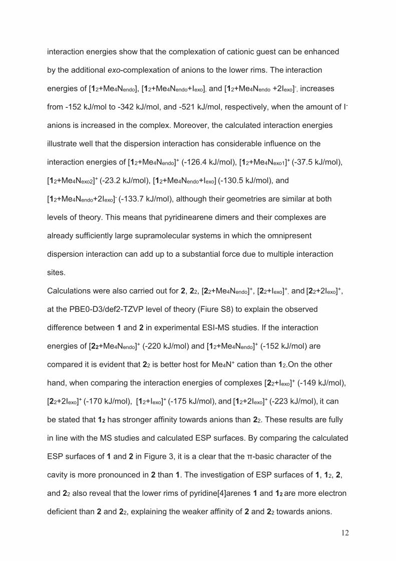
12
interaction energies show that the complexation of cationic guest can be enhanced
by the additional exo-complexation of anions to the lower rims. The interaction
energies of [12+Me4Nendo], [12+Me4Nendo+Iexo], and [12+Me4Nendo +2Iexo]-, increases
from -152 kJ/mol to -342 kJ/mol, and -521 kJ/mol, respectively, when the amount of I-
anions is increased in the complex. Moreover, the calculated interaction energies
illustrate well that the dispersion interaction has considerable influence on the
interaction energies of [12+Me4Nendo]+ (-126.4 kJ/mol), [12+Me4Nexo1]+ (-37.5 kJ/mol),
[12+Me4Nexo2]+ (-23.2 kJ/mol), [12+Me4Nendo+Iexo] (-130.5 kJ/mol), and
[12+Me4Nendo+2Iexo]- (-133.7 kJ/mol), although their geometries are similar at both
levels of theory. This means that pyridinearene dimers and their complexes are
already sufficiently large supramolecular systems in which the omnipresent
dispersion interaction can add up to a substantial force due to multiple interaction
sites.
Calculations were also carried out for 2, 22, [22+Me4Nendo]+, [22+Iexo]+, and [22+2Iexo]+,
at the PBE0-D3/def2-TZVP level of theory (Fiure S8) to explain the observed
difference between 1 and 2 in experimental ESI-MS studies. If the interaction
energies of [22+Me4Nendo]+ (-220 kJ/mol) and [12+Me4Nendo]+ (-152 kJ/mol) are
compared it is evident that 22 is better host for Me4N+ cation than 12.On the other
hand, when comparing the interaction energies of complexes [22+Iexo]+ (-149 kJ/mol),
[22+2Iexo]+ (-170 kJ/mol), [12+Iexo]+ (-175 kJ/mol), and [12+2Iexo]+ (-223 kJ/mol), it can
be stated that 12 has stronger affinity towards anions than 22. These results are fully
in line with the MS studies and calculated ESP surfaces. By comparing the calculated
ESP surfaces of 1 and 2 in Figure 3, it is a clear that the π-basic character of the
cavity is more pronounced in 2 than 1. The investigation of ESP surfaces of 1, 12, 2,
and 22 also reveal that the lower rims of pyridine[4]arenes 1 and 12 are more electron
deficient than 2 and 22, explaining the weaker affinity of 2 and 22 towards anions.
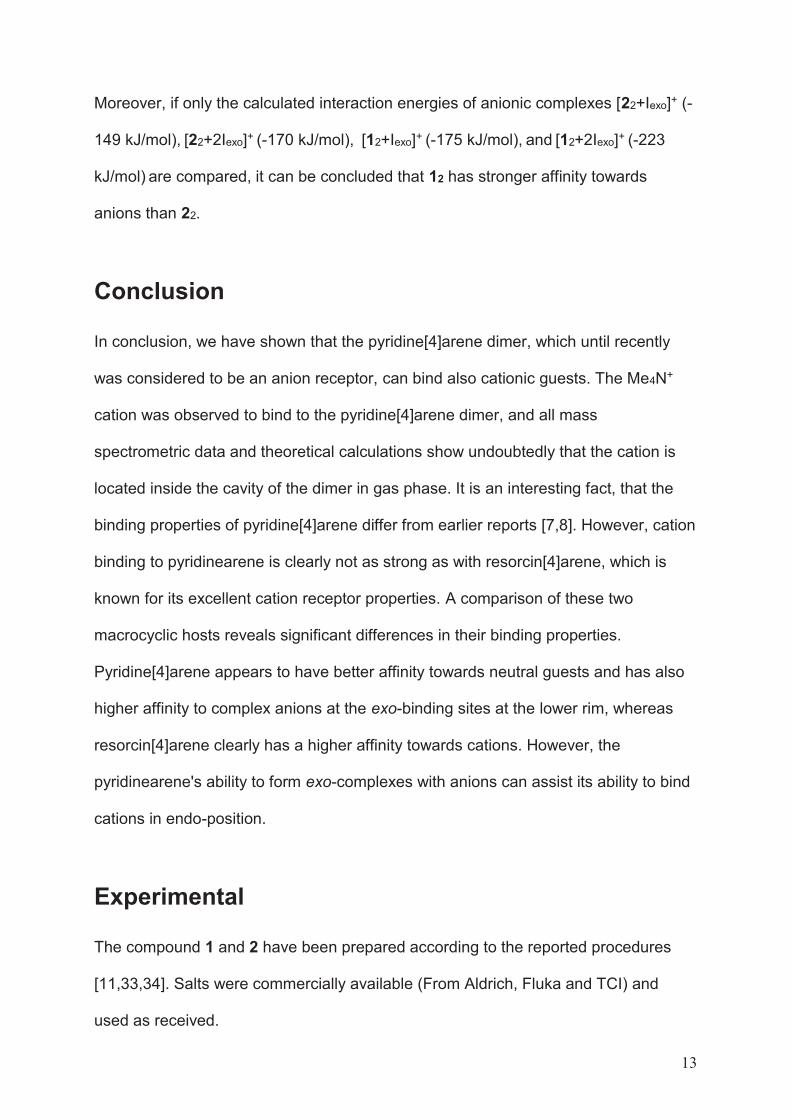
13
Moreover, if only the calculated interaction energies of anionic complexes [22+Iexo]+ (-
149 kJ/mol), [22+2Iexo]+ (-170 kJ/mol), [12+Iexo]+ (-175 kJ/mol), and [12+2Iexo]+ (-223
kJ/mol) are compared, it can be concluded that 12 has stronger affinity towards
anions than 22.
Conclusion
In conclusion, we have shown that the pyridine[4]arene dimer, which until recently
was considered to be an anion receptor, can bind also cationic guests. The Me4N+
cation was observed to bind to the pyridine[4]arene dimer, and all mass
spectrometric data and theoretical calculations show undoubtedly that the cation is
located inside the cavity of the dimer in gas phase. It is an interesting fact, that the
binding properties of pyridine[4]arene differ from earlier reports [7,8]. However, cation
binding to pyridinearene is clearly not as strong as with resorcin[4]arene, which is
known for its excellent cation receptor properties. A comparison of these two
macrocyclic hosts reveals significant differences in their binding properties.
Pyridine[4]arene appears to have better affinity towards neutral guests and has also
higher affinity to complex anions at the exo-binding sites at the lower rim, whereas
resorcin[4]arene clearly has a higher affinity towards cations. However, the
pyridinearene's ability to form exo-complexes with anions can assist its ability to bind
cations in endo-position.
Experimental
The compound 1 and 2 have been prepared according to the reported procedures
[11,33,34]. Salts were commercially available (From Aldrich, Fluka and TCI) and
used as received.
14
Mass spectrometric experiments have been performed with ABSciex QSTAR Elite
ESI-Q-TOF mass spectrometer. Ion Mobility mass spectrometry experiments were
conducted with Waters Synapt G2 equipped with a linear drift cell and Agilent 6560
Ion mobility Q-TOF mass spectrometers. All CCS values were obtained using
nitrogen as a drift gas and stepped field methods. IRMPD experiments were
performed with Ionspec QFT-7 ESI-FT-ICR with a 7 T superconducting magnet.
Samples for all mass spectrometric experiments were prepared with 10 or 20 μM
concentration and 1:3 host:guest ratio in acetone. Theoretical CCS values were
calculated using IMoS [20,21].
NMR experiments were performed with Bruker Avance III HD 300 NMR
Spectrometer. Samples were prepared with 10 mM concentration and 1:3 host:guest
ratio in CDCl3/CD3CN (4:3, v:v) mixture.
DFT calculations were performed by Spartan’ 16 and Gaussian 09 (D01) software
packages.
More detailed information of the experiments and parametrization can be found from
supporting information.
Supporting Information
Supporting Information File 1:
File Name: SI_Pyridinearenes_manuscript_BJOC_Kalenius
File Format: PDF
Title: Experimental details and supplementary information
Acknowledgements
15
We wish to thank The Academy of Finland (projects 284562, 278743 and 312514 to
E.K. and 285855 to J.O.M.), the University of Jyväskylä and the Deutsche
Forschungsgemeinschaft (Core Facility BioSupraMol) for financial support. J.O.M
wishes to acknowledge CSC – IT Center for Science, Finland, and Prof. Heikki M.
Tuononen for computational resources. A. Kruve is grateful for a postdoctoral
fellowship from the Alexander von Humboldt Foundation. Prof. Magdalena Zimnicka
is gratefully acknowledged for her assistance and helpful discussions regarding the
determination of theoretical CCS values.
ORCID® iDs
Anniina Kiesilä - https://orcid.org/0000-0002-2952-2377
Jani O. Moilanen - https://orcid.org/0000-0002-2096-593X
Anneli Kruve - https://orcid.org/0000-0001-9725-3351
Christoph A. Schalley - https://orcid.org/0000-0002-8634-3578
Perdita Barran - https://orcid.org/0000-0002-7720-586X
Elina Kalenius - https://orcid.org/0000-0001-8038-1314
References
1. Timmerman, P.; Verboom, W.; Reinhoudt, D. N. Tetrahedron 1996, 52, 2663‒
2704.
2. Avram, L.; Cohen, Y. J. Am. Chem. Soc. 2002, 124, 15148‒15149.
3. Palmer, L. C.; Shivanyuk, A.; Yamanaka, M.; Rebek, J. Jr. Chem. Commun. 2005,
0, 857‒858.
4. Gerkensmeier, T.; Mattay, J.; Näther, C. Chem. Eur. J. 2001, 7, 465‒474.
5. Evan-Salem, T.; Cohen, Y. Chem. Eur. J. 2007, 13, 7659‒7663.
16
6. Kiesilä, A.; Beyeh, N. K.; Moilanen, J. O.; Puttreddy, R.; Götz, S.; Rissanen, K.;
Barran, P.; Lützen, A.; E. Kalenius. Org. Biomol. Chem. 2019, 17, 6980–6984.
7. Letzel, M. C.; Decker, B.; Rozhenko, A. B.; Schoeller, W. W.; Mattay, J. J. Am.
Chem. Soc. 2004, 126, 9669‒9674.
8. Rozhenko, A. B.; Schoeller, W. W.; Letzel, M. C.; Decker, B.; Mattay, J. New J.
Chem. 2013, 37, 356‒365.
9. Schottel, B. L.; Chifotides, H. T.; Dunbar, K. R. Chem. Soc. Rev. 2008, 37, 68–83.
10. Dawson, R. E.; Hennig, A.; Weimann, D. P.; Emery, D.; Ravikumar, V.;
Montenegro, J.; Takeuchi, T.; Gabutti, S.; Mayor, M.; Mareda, J.; Schalley, C. A.;
Matile, S. Nat. Chem. 2010, 2, 533–538.
11. Kiesilä, A.; Kivijärvi, L.; Beyeh, N. K.; Moilanen, J. O.; Groessl, M.; Rothe, T.;
Götz, S.; Topić, F.; Rissanen K.; Lützen, A.; Kalenius, E. Angew. Chem. Int. Ed.
2017, 56, 10942‒10946.
12. Murayama, K.; Aoki, K. Chem. Commun. 1998, 0, 607‒608.
13. Mansikkamäki, H.; Nissinen, M.; Schalley, C. A.; Rissanen. K. New J. Chem.
2003, 27, 88‒97.
14. Mäkinen, M.; Vainiotalo, P.; Nissinen, M.; Rissanen, K. J. Am. Soc. Mass
Spectrom. 2003, 14, 143‒151.
15. Kalenius, E.; Groessl, M.; Rissanen, K. Nat. Rev. Chem. 2019, 3, 4-14.
16. Lee, T-C.; Kalenius, E.; Lazar, A. I.; Assaf, K. I.; Kuhnert, N.; Grün, C. H.; Jänis,
J.; Scherman, O. A.; Nau, W. M. Nat. Chem. 2013, 5, 376‒382.
17. Dearden, D. V.; Ferrell, T. A.; Asplund, M. C.; Zilch, L. W.; Julian, R. R.; Jarrold.
M. F. J. Phys. Chem. A 2009, 113, 989‒997.
18. Jurček, O.; Bonakdarzadeh, P.; Kalenius, E.; Linnanto, J. M.; Groessl, M.;
Knochenmuss, R.; Ihalainen, J. A.; Rissanen, K. Angew. Chem. Int. Ed. 2015, 54,
15462‒15467.
17
19. DTCCSN2 states for CCS values obtained using drift tube instrument with nitrogen
gas, nomenclature recommended in Gabelica V. et al., Mass Spectrom. Rev. 2019,
38, 291-320.
20. Larriba, C.; Hogan C. J. Jr., J. Comput. Phys. 2013, 251, 344-363.
21. Larriba, C.; Hogan C. J. Jr. J. Phys. Chem. A 2013, 117, 3887-3901.
22. DTMCCSN2 states for CCS values obtained using diffuse trajectory method with
nitrogen gas.
23. Beyeh, N. K.; Kogej, M.; Åhman, A.; Rissanen, K.; Schalley, C. A. Angew. Chem.
Int. Ed. 2006, 45, 5214‒5218.
24. Ernzerhof, M.; Scuseria, G. E. J. Chem. Phys. 1999, 110, 5029–5036.
25. Adamo, C.; Barone, V. J. Chem. Phys. 1999, 110, 6158–6170.
26. Weigend, F.; Ahlrichs, R. Phys. Chem. Chem. Phys. 2005, 7, 3297–3305.
27. Grimme, S.; Antony, J.; Ehrlich, S.; Krieg, H. J. Chem. Phys. 2010, 132, 154104.
28. Grimme, S.; Ehrlich, S.; Goerigk, L. J. Comput. Chem. 2011, 32, 1456–1465.
29. Hermann, J.; DiStasio, R. A.; Tkatchenko, A. Chem. Rev. 2017, 117, 4714–4758.
30. Ambrosetti, A.; Alfè, D.; Distasio, R. A.; Tkatchenko, A. J. Phys. Chem. Lett.
2014, 5, 849–855.
31. Rao, L.; Ke, H.; Fu, G.; Xu, X.; Yan, Y. J. Chem. Theory Comput. 2009, 5, 86–96.
32. Ireta, J.; Neugebauer, J.; Scheffler, M. J. Phys. Chem. A 2004, 108, 5692–5698.
33. Högberg, A.G.S. J. Org. Chem. 1980, 45, 4498-4500.
34. Tunstad, L. M.; Tucker, J. A.; Dalcanale, E.; Weiser, J.; Bryant, J. A.; Sherman, J.
C.; Helgeson, R. C.; Knobler, C. B.; Cram, D. J. J. Org. Chem. 1989, 54, 1305–1312.
III
THERMODYNAMICALLY DRIVEN SELF-ASSEMBLY OF PYRIDINEARENE TO HEXAMERIC CAPSULES
by
A. Kiesilä, N. K. Beyeh, J. O. Moilanen, R. Puttreddy, Sven Götz, K. Rissanen, P.
Organic and Biomolecular Chemistry, 2019, 17, 6980 – 6984
Reproduced with permission. Copyright © 2019 The Royal Society of Chemistry
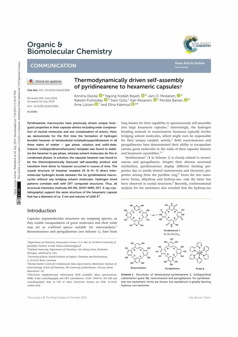
Organic &Biomolecular Chemistry
COMMUNICATION
Cite this: DOI: 10.1039/c9ob01383k
Received 19th June 2019,Accepted 1st July 2019
DOI: 10.1039/c9ob01383k
rsc.li/obc
Thermodynamically driven self-assemblyof pyridinearene to hexameric capsules†
Anniina Kiesilä, a Ngong Kodiah Beyeh, b Jani O. Moilanen, a
Rakesh Puttreddy, a Sven Götz,c Kari Rissanen, a Perdita Barran, d
Arne Lützen c and Elina Kalenius *a
Pyridinearene macrocycles have previously shown unique host–
guest properties in their capsular dimers including endo complexa-
tion of neutral molecules and exo complexation of anions. Here,
we demonstrate for the first time the formation of hydrogen
bonded hexamer of tetraisobutyl-octahydroxypyridinearene in all
three states of matter – gas phase, solution and solid-state.
Cationic tris(bipyridine)ruthenium(II) template was found to stabil-
ize the hexamer in gas phase, whereas solvent molecules do this in
condensed phases. In solution, the capsular hexamer was found to
be the thermodynamically favoured self-assembly product and
transition from dimer to hexamer occurred in course of time. The
crystal structure of hexamer revealed 24 N–H⋯O direct inter-
molecular hydrogen bonds between the six pyridinearene macro-
cycles without any bridging solvent molecules. Hydrogen bond
patterns correlate well with DFT computed structures. Thus, all
structural chemistry methods (IM-MS, DOSY NMR, DFT, X-ray crys-
tallography) support the same structure of the hexameric capsule
that has a diameter of ca. 3 nm and volume of 1160 Å3.
Introduction
Capsular supramolecular structures are tempting species, asthey enable encapsulation of guest molecules and their voidsmay act as confined spaces suitable for nanocatalysis.1
Resorcinarenes and pyrogallarenes (see Scheme 1), have been
long known for their capability to spontaneously self-assembleinto large hexameric capsules.2 Interestingly, the hydrogenbonding network in resorcinarene hexamers typically involvebridging solvent molecules, which might even be responsiblefor their unique catalytic activity.4 Both resorcinarenes andpyrogallarenes have demonstrated their ability to encapsulatevarious guest molecules in the voids of their capsular dimericand hexameric assemblies.2,3
Pyridinearene5 (1 in Scheme 1) is closely related to resorci-narene and pyrogallarene. Despite their obvious structuralsimilarities, pyridinearenes display different binding pro-perties due to amide–iminol tautomerism and electronic pro-perties arising from the pyridine ring.6 From the two tauto-meric forms, dihydroxy and hydroxy-oxo, only the latter hasbeen observed in crystal structures.6 Recently, conformationalanalysis for the tautomers also revealed that the hydroxy-oxo
Scheme 1 Structures of tetraisobutyl-pyridinearene 1, tris(bipyridine)ruthenium(II) guest (G), resorcinarene and pyrogallarene. For pyridinear-ene two tautomeric forms are shown, but equilibrium is greatly favoringhydroxy-oxo tautomer.
†Electronic supplementary information (ESI) available: Mass spectrometry,NMR, X-Ray crystallography and DFT calculations. CCDC 1899745. For ESI andcrystallographic data in CIF or other electronic format see DOI: 10.1039/c9ob01383k
aDepartment of Chemistry, Nanoscience Center, P. O. Box 35, FI-40014 University of
Jyväskylä, Finland. E-mail: [email protected] University, Department of Chemistry, 146 Library Drive, Rochester,
Michigan, 48309-4479, USAcUniversity of Bonn, Kekulé-Institute of Organic Chemistry and Biochemistry,
A, D-53121 Bonn, GermanydMichael Barber Centre for Collaborative Mass Spectrometry, Manchester Institute of
Biotechnology, School of Chemistry, The University of Manchester, Princess Street,
Manchester, UK
This journal is © The Royal Society of Chemistry 2019 Org. Biomol. Chem.
Ope
n A
cces
s Arti
cle.
Pub
lishe
d on
02
July
201
9. D
ownl
oade
d on
7/1
0/20
19 1
2:36
:43
PM.
Thi
s arti
cle
is lic
ense
d un
der a
Cre
ativ
e Co
mm
ons A
ttrib
utio
n-N
onCo
mm
erci
al 3
.0 U
npor
ted
Lice
nce. View Article Online
View Journal
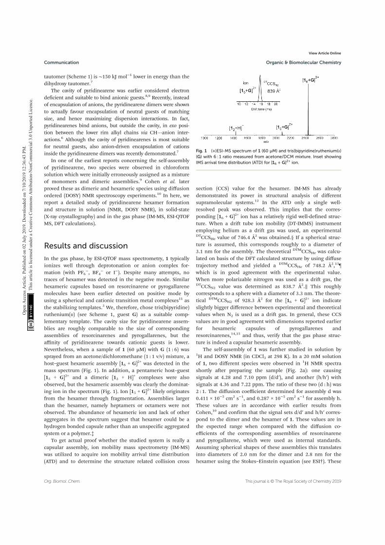
tautomer (Scheme 1) is ∼150 kJ mol−1 lower in energy than thedihydroxy tautomer.7
The cavity of pyridinearene was earlier considered electrondeficient and suitable to bind anionic guests.8,9 Recently, insteadof encapsulation of anions, the pyridinearene dimers were shownto actually favour encapsulation of neutral guests of matchingsize, and hence maximizing dispersion interactions. In fact,pyridinearenes bind anions, but outside the cavity, in exo posi-tion between the lower rim alkyl chains via CH⋯anion inter-actions.6 Although the cavity of pyridinearenes is most suitablefor neutral guests, also anion-driven encapsulation of cationsinside the pyridinearene dimers was recently demonstrated.7
In one of the earliest reports concerning the self-assemblyof pyridinearene, two species were observed in chloroformsolution which were initially erroneously assigned as a mixtureof monomers and dimeric assemblies.9 Cohen et al. laterproved these as dimeric and hexameric species using diffusionordered (DOSY) NMR spectroscopy experiments.10 In here, wereport a detailed study of pyridinearene hexamer formationand structure in solution (NMR, DOSY NMR), in solid-state(X-ray crystallography) and in the gas phase (IM-MS, ESI-QTOFMS, DFT calculations).
Results and discussion
In the gas phase, by ESI-QTOF mass spectrometry, 1 typicallyionizes well through deprotonation or anion complex for-mation (with PF6
−, BF4− or I−). Despite many attempts, no
traces of hexamer was detected in the negative mode. Similarhexameric capsules based on resorcinarene or pyrogallarenemolecules have been earlier detected on positive mode byusing a spherical and cationic transition metal complexes11 asthe stabilizing templates.3 We, therefore, chose tris(bipyridine)ruthenium(II) (see Scheme 1, guest G) as a suitable comp-lementary template. The cavity size for pyridinearene assem-blies are roughly comparable to the size of correspondingassemblies of resorcinarenes and pyrogallarenes, but theaffinity of pyridinearene towards cationic guests is lower.Nevertheless, when a sample of 1 (60 μM) with G (1 : 6) wassprayed from an acetone/dichloromethane (1 : 1 v/v) mixture, ahost–guest hexameric assembly [16 + G]2+ was detected in themass spectrum (Fig. 1). In addition, a pentameric host–guest[15 + G]2+ and a dimeric [12 + H]+ complexes were alsoobserved, but the hexameric assembly was clearly the dominat-ing ion in the spectrum (Fig. 1). Ion [15 + G]2+ likely originatesfrom the hexamer through fragmentation. Assemblies largerthan the hexamer, namely heptamers or octamers were notobserved. The abundance of hexameric ion and lack of otheraggregates in the spectrum suggest that hexamer could be ahydrogen bonded capsule rather than an unspecific aggregatedsystem or a polymer.‡
To get actual proof whether the studied system is really acapsular assembly, ion mobility mass spectrometry (IM-MS)was utilized to acquire ion mobility arrival time distribution(ATD) and to determine the structure related collision cross
section (CCS) value for the hexamer. IM-MS has alreadydemonstrated its power in structural analysis of differentsupramolecular systems.12 In the ATD only a single well-resolved peak was observed. This implies that the corres-ponding [16 + G]2+ ion has a relatively rigid well-defined struc-ture. When a drift tube ion mobility (DT-IMMS) instrumentemploying helium as a drift gas was used, an experimentalDTCCSHe value of 746.4 Å2 was obtained.§ If a spherical struc-ture is assumed, this corresponds roughly to a diameter of3.1 nm for the assembly. The theoretical DTMCCSHe was calcu-lated on basis of the DFT calculated structure by using diffusetrajectory method and yielded a DTMCCSHe of 748.2 Å2,13¶which is in good agreement with the experimental value.When more polarizable nitrogen was used as a drift gas, theDTCCSN2 value was determined as 838.7 Å2.∥ This roughlycorresponds to a sphere with a diameter of 3.3 nm. The theore-tical DTMCCSN2 of 928.3 Å2 for the [16 + G]2+ ion indicateslightly bigger difference between experimental and theoreticalvalues when N2 is used as a drift gas. In general, these CCSvalues are in good agreement with dimensions reported earlierfor hexameric capsules of pyrogallarenes andresorcinarenes,14,15 and thus, verify that the gas phase struc-ture is indeed a capsular hexameric assembly.
The self-assembly of 1 was further studied in solution by1H and DOSY NMR (in CDCl3 at 298 K). In a 20 mM solutionof 1, two different species were observed in 1H NMR spectrashortly after preparing the sample (Fig. 2a): one causingsignals at 4.28 and 7.10 ppm (d/d′), and another (h/h′) withsignals at 4.36 and 7.22 ppm. The ratio of these two (d : h) was2 : 1. The diffusion coefficient determined for assembly d was0.411 × 10−5 cm2 s−1, and 0.287 × 10⁻⁵ cm2 s−1 for assembly h.These values are in accordance with earlier results fromCohen,10 and confirm that the signal sets d/d′ and h/h′ corres-pond to the dimer and the hexamer of 1. These values are inthe expected range when compared with the diffusion co-efficients of the corresponding assemblies of resorcinareneand pyrogallarene, which were used as internal standards.Assuming spherical shapes of these assemblies this translatesinto diameters of 2.0 nm for the dimer and 2.8 nm for thehexamer using the Stokes–Einstein equation (see ESI†). These
Fig. 1 (+)ESI-MS spectrum of 1 (60 μM) and tris(bipyridine)ruthenium(II)(G) with 6 : 1 ratio measured from acetone/DCM mixture. Inset showingIMS arrival time distribution (ATD) for [16 + G]2+ ion.
Communication Organic & Biomolecular Chemistry
Org. Biomol. Chem. This journal is © The Royal Society of Chemistry 2019
Ope
n A
cces
s Arti
cle.
Pub
lishe
d on
02
July
201
9. D
ownl
oade
d on
7/1
0/20
19 1
2:36
:43
PM.
Thi
s arti
cle
is lic
ense
d un
der a
Cre
ativ
e Co
mm
ons A
ttrib
utio
n-N
onCo
mm
erci
al 3
.0 U
npor
ted
Lice
nce.
View Article Online
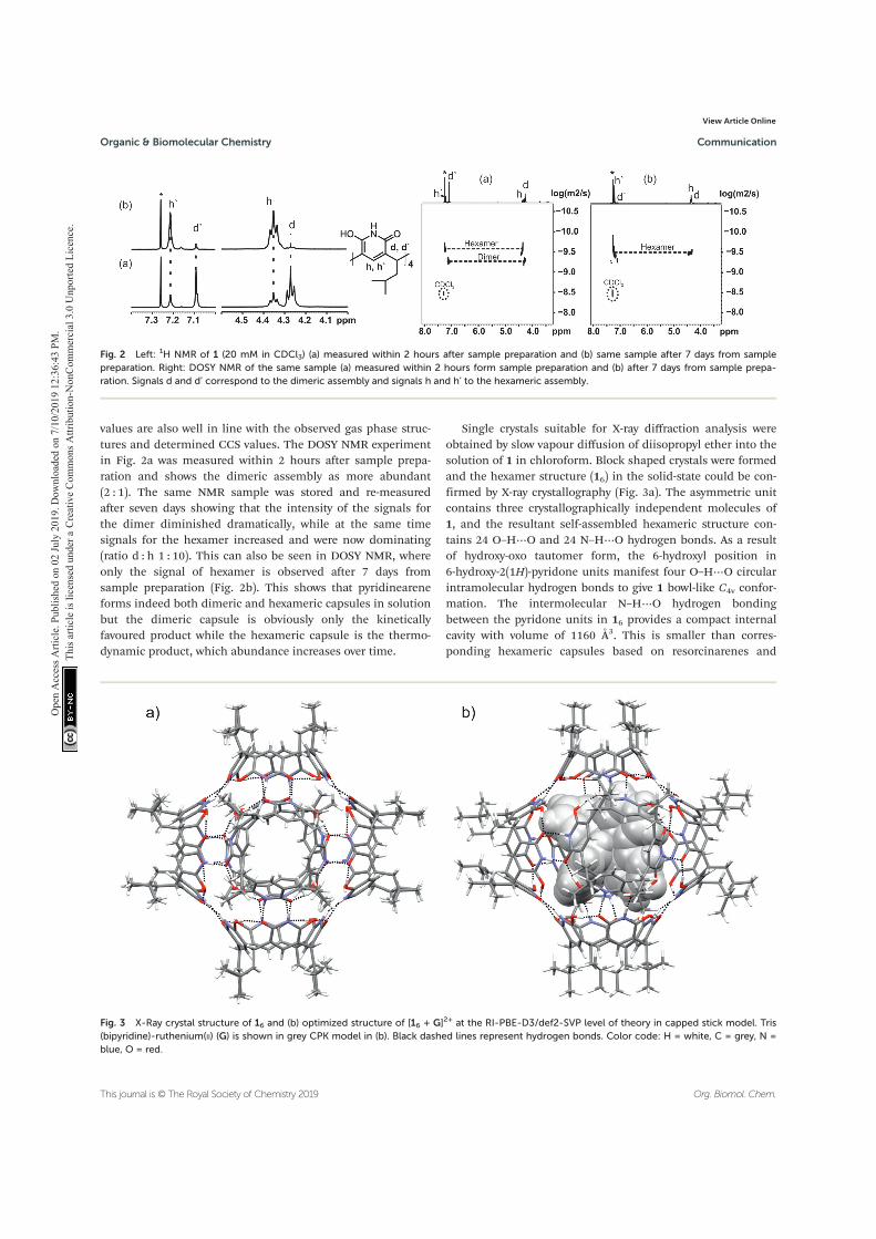
values are also well in line with the observed gas phase struc-tures and determined CCS values. The DOSY NMR experimentin Fig. 2a was measured within 2 hours after sample prepa-ration and shows the dimeric assembly as more abundant(2 : 1). The same NMR sample was stored and re-measuredafter seven days showing that the intensity of the signals forthe dimer diminished dramatically, while at the same timesignals for the hexamer increased and were now dominating(ratio d : h 1 : 10). This can also be seen in DOSY NMR, whereonly the signal of hexamer is observed after 7 days fromsample preparation (Fig. 2b). This shows that pyridineareneforms indeed both dimeric and hexameric capsules in solutionbut the dimeric capsule is obviously only the kineticallyfavoured product while the hexameric capsule is the thermo-dynamic product, which abundance increases over time.
Single crystals suitable for X-ray diffraction analysis wereobtained by slow vapour diffusion of diisopropyl ether into thesolution of 1 in chloroform. Block shaped crystals were formedand the hexamer structure (16) in the solid-state could be con-firmed by X-ray crystallography (Fig. 3a). The asymmetric unitcontains three crystallographically independent molecules of1, and the resultant self-assembled hexameric structure con-tains 24 O–H⋯O and 24 N–H⋯O hydrogen bonds. As a resultof hydroxy-oxo tautomer form, the 6-hydroxyl position in6-hydroxy-2(1H)-pyridone units manifest four O–H⋯O circularintramolecular hydrogen bonds to give 1 bowl-like C4v confor-mation. The intermolecular N–H⋯O hydrogen bondingbetween the pyridone units in 16 provides a compact internalcavity with volume of 1160 Å3. This is smaller than corres-ponding hexameric capsules based on resorcinarenes and
Fig. 2 Left: 1H NMR of 1 (20 mM in CDCl3) (a) measured within 2 hours after sample preparation and (b) same sample after 7 days from samplepreparation. Right: DOSY NMR of the same sample (a) measured within 2 hours form sample preparation and (b) after 7 days from sample prepa-ration. Signals d and d’ correspond to the dimeric assembly and signals h and h’ to the hexameric assembly.
Fig. 3 X-Ray crystal structure of 16 and (b) optimized structure of [16 + G]2+ at the RI-PBE-D3/def2-SVP level of theory in capped stick model. Tris(bipyridine)-ruthenium(II) (G) is shown in grey CPK model in (b). Black dashed lines represent hydrogen bonds. Color code: H = white, C = grey, N =blue, O = red.
Organic & Biomolecular Chemistry Communication
This journal is © The Royal Society of Chemistry 2019 Org. Biomol. Chem.
Ope
n A
cces
s Arti
cle.
Pub
lishe
d on
02
July
201
9. D
ownl
oade
d on
7/1
0/20
19 1
2:36
:43
PM.
Thi
s arti
cle
is lic
ense
d un
der a
Cre
ativ
e Co
mm
ons A
ttrib
utio
n-N
onCo
mm
erci
al 3
.0 U
npor
ted
Lice
nce.
View Article Online
pyrogallarenes (volumes >1200 Å3, see ESI Fig. S4†).14,15 TheDOSY derived hydrodynamic radii (2.80 nm) agrees well withthe calculated value obtained from the X-ray crystal structure(2.82 nm). Of special note, the hydroxy-oxo tautomerismrenders 1 a rigid cavity and definite directionality for hydrogenbonding. Robust C4v cavities are seamed together with directN–H⋯O intermolecular hydrogen bonding to produce solvent-free hexamer 16. Solvent-free hydrogen bonding is alsoobserved in the solid-state pyrogallarene hexamer structures.14
In the resorcinarene hexamers, due to flexible C4v cavities andsteric reasons solvent molecules typically assist in capsule for-mation via intermolecular hydrogen bonding interactions,found between two resorcinarene units.15 Although chloroformis essential to obtain a stable 16 single-crystals its passive rolein hydrogen bonding could be observed in the 3-D crystallattice. The features of pyridinearene lead to unique and comp-lementary alignment of the individual monomers in order tomaximize the number of intermolecular hydrogen bonds. Inthe case of the resorcinarenes, this requires incorporation ofsolvent molecules as part of the intermolecular hydrogen bondnetwork. Consequently, this causes the differences in the voidsize for three different hexamers.
Unfortunately, all our attempts to obtain single crystalsfrom [16 + G]2+ were unsuccessful. Therefore, we decided tocarry out the computational analysis for studied systems at theRI-PBE-D3/def2-SVP//RI-PBE-D3/def2-TZVP level of theory inorder to investigate the gas phase structure of 16 and [16 + G]2+
and to evaluate their interaction energies in detail.16–24
The optimized structures and hydrogen bonding patternsof 16 and [16 + G]2+ are illustrated in Fig. 3b (see also ESI†).Within 16 and [16 + G]2+, each monomer interacts with fourneighbouring monomers through intermolecular N–H⋯Obonds whose distances vary from 1.703 to 1.716 Å and from1.657 to 1.699 Å for 16 and [16 + G]2+, respectively (Table 1).Interestingly, as compared to the dimeric capsule, 12, theN–H⋯O bonds are slightly shorter in the hexamer. The totalnumber of N–H⋯O bonds in one hexamer unit is 24.Furthermore, the seam of hydrogen bonds, consisting of fourintramolecular O–H⋯O bonds, of each monomer 1, is retainedduring hexamer formation. However, small changes areobserved in the O–H⋯O bonds of 1, 12, 16 and [16 + G]2+ ifcompared with each other: the H-bonds are slightly longer inthe hexamers and the dimers than in the monomer. The
elongation of the O–H⋯O bonds in the hexamers and dimersmost likely arises from the small changes in electron densitydistribution around/on the oxygen atoms that function as ahydrogen bond acceptors in both, N–H⋯O and O–H⋯O,bonds in the hexamers and the dimers. Some electron densityis therefore transferred from O–H⋯O bonds to N–H⋯O bondsupon the formation of dimers and hexamers. In the monomer,however, these acceptors oxygen atoms participates only in theformation of O–H⋯O bonds.
Due to the multiple intermolecular N–H⋯O bonds, 24 intotal, the interaction energies of 16 and [16 + G]2+ are very high−717 kJ mol−1 and −1205 kJ mol−1, respectively (Table 1). Theseenergy numbers suggest that the [16 + G]2+ is ∼500 kJ mol−1
more stable than empty hexamer 16 and underline the signifi-cance of template effect.3 If the interaction energies of 16 and[16 + G]2+ are compared to the interaction energies of 3 individualdimers 12 (3 × −197 kJ mol−1 = −590 kJ mol−1), it is clear that theformation of hexamer is more favourable than the formation of3 individual dimers from six monomers. Although this very sim-plified gas phase model does not take into account all forcesacting in the solution state, it not only supports the resultobtained from MS studies, but is also in agreement with theNMR data: the hexamer 16 is thermodynamically more stablethan the dimer 12 in gas phase and solution state.
Conclusions
To conclude, the pyridinearene hexamers were structurallycharacterized in all three states of matter, and hence, for thefirst time in gas-phase and solid-state. X-ray structure showsthat the hexameric assembly is formed via directly linked,intermolecular hydrogen bonds without bridging solvent mole-cules, which are usually required in the solid-state structuresof closely related resorcinarenes. This makes hexamer morecompact and decreases the size of empty windows betweenmonomer units, which is a significant feature when appli-cations, especially related to catalysis in confined space, areconsidered. In chloroform solution the hexamer was found tobe the thermodynamically favoured product compared to thecorresponding kinetically favoured dimer. All analytical tech-niques used result in a structure with a diameter of ∼3 nm,and support the structural similarity of the pyridinearenehexamer in gas phase, solution and in solid state. As such, it isslightly more compact than the related hexamers of resorcinar-enes and pyrogallarenes due to the different intermolecularhydrogen bonding network that implies to unique mutualalignment of the monomers in these assemblies. Interestingly,the use of a cationic, structurally complementary templateincreased the gas phase stability of the hexamer substantially,which could be rationalized by sophisticated DFT calculations.
Having established the structural properties of these largehydrogen-bonded capsules now paves the avenue to explore itshost–guest binding properties arising from its different andunique interactions and geometric features compared to theother similar assemblies.
Table 1 Calculated O–H⋯O and N–H⋯O distances and interactionenergies at the RI-PBE-D3/def2-SVP level of theory. RI-PBE-D3/def2-SVP//RI-PBE-D3/def2-TZVP single-point energies in parenthesis
Compound O–H⋯O [Å] N–H⋯O [Å] Eint [kJ mol−1]
1 1.425–1.432 — —12 1.519–1.525 1.800–1.821 −280 (−197)a16 1.554–1.560 1.703–1.716 −942 (−717)b[16 + G]2+ 1.528–1.563 1.657–1.699 −1517 (−1205)c
a Eint = Edimer − 2 × Emonomer.b Ehexamer − 6 × Emonomer.
c Ecomplex − (6 ×Emonomer + Eguest).
Communication Organic & Biomolecular Chemistry
Org. Biomol. Chem. This journal is © The Royal Society of Chemistry 2019
Ope
n A
cces
s Arti
cle.
Pub
lishe
d on
02
July
201
9. D
ownl
oade
d on
7/1
0/20
19 1
2:36
:43
PM.
Thi
s arti
cle
is lic
ense
d un
der a
Cre
ativ
e Co
mm
ons A
ttrib
utio
n-N
onCo
mm
erci
al 3
.0 U
npor
ted
Lice
nce.
View Article Online
Conflicts of interest
There are no conflicts to declare.
Acknowledgements
The authors acknowledge the Academy of Finland for thefunding (JOM: 285855, 315829, 320015, RP: 298817, EK:284562, 278743 and 312514) as well as Prof. H. M. Tuononen,University of Jyväskylä, CSC-IT Center for Science in Finland,and the Finnish Grid and Cloud Infrastructure (persistentidentifierurn:nbn:fi:research-infras-2016072533) for providingcomputational resources. NKB gratefully acknowledge thefinancial support from Oakland University, MI, USA.
Notes and references‡Please note, that the template G is not necessary to observe the hexamericassembly in (+)ESI-MS. Without templating guest hexamer can be observed as[16 + Na]+ even in low (20 μM) concentration, but with relatively lower abundancy(see Fig. S1†).§DTCCSHe states for CCS values obtained using drift tube instrument withhelium gas, nomenclature recommended in V. Gabelica et al.25
¶DTMCCSHe states for CCS values obtained using diffuse trajectory method withhelium gas.∥According to Mason–Schamp equation, different gases result in different drifttimes, and thus to different CCS values. For more detailed explanation seeV. Gabelica and E. Marklund.26
1 D. Fiedler, D. H. Leung, R. G. Bergman andK. N. Raymond, Acc. Chem. Res., 2005, 38, 349; M. D. Pluth,R. G. Bergman and K. N. Raymond, Acc. Chem. Res., 2009,42, 1650; L. Catti, Q. Zhang and K. Tiefenbacher, Chem. –Eur. J., 2016, 22, 9060.
2 M. Yamanaka, A. Shivanyuk and J. Rebek, J. Am. Chem.Soc., 2004, 126, 2939; L. Avram and Y. Cohen, J. Am. Chem.Soc., 2002, 124, 15148; L. MacGillivray and J. Atwood,Nature, 1997, 389, 469.
3 N. K. Beyeh, M. Kogej, A. Åhman, K. Rissanen andC. A. Schalley, Angew. Chem., Int. Ed., 2006, 45, 5214.
4 Q. Zhang, L. Catti and K. Tiefenbacher, Acc. Chem. Res.,2018, 51, 2107.
5 T. Gerkensmeier, J. Mattay and C. Näther, Chem. – Eur. J.,2001, 7, 465.
6 A. Kiesilä, L. Kivijärvi, N. K. Beyeh, J. O. Moilanen,M. Groessl, T. Rothe, S. Götz, F. Topić, K. Rissanen,A. Lützen and E. Kalenius, Angew. Chem., Int. Ed., 2017, 56,10942.
7 A. Kiesilä, J. O. Moilanen, A. Kruve, C. A. Schalley, P. Barranand E. Kalenius, Unpublished results.
8 A. B. Rozhenko, W. W. Schoeller, M. C. Letzel, B. Deckerand J. Mattay, New J. Chem., 2013, 37, 356.
9 M. C. Letzel, B. Decker, A. B. Rozhenko, W. W. Schoellerand J. Mattay, J. Am. Chem. Soc., 2004, 126, 9669.
10 T. Evan-Salem and Y. Cohen, Chem. – Eur. J., 2007, 13, 7659.11 G. Bianchini, A. Scarso, G. La Sorella and G. Strukul, Chem.
Commun., 2012, 48, 12082–12084.12 E. Kalenius, M. Groessl and K. Rissanen, Nat. Chem. Rev.,
2019, 3, 4.13 C. Larriba and C. J. Hogan, Jr., J. Comput. Phys., 2013, 251,
344; C. Larriba and C. J. Hogan Jr., J. Phys. Chem. A, 2013,117, 3887.
14 T. Gerkensmeier, W. Iwanek, C. Agena, R. Fröhlich, S. Kotila,C. Näther and J. Mattay, Eur. J. Org. Chem., 1999, 2257.
15 R. M. Payne and C. L. Oliver, CrystEngComm, 2018, 20, 1919.16 K. Eichkorn, O. Treutler, H. Öhm, M. Häser and
R. Ahlrichs, Chem. Phys. Lett., 1995, 242, 652.17 K. Eichkorn, F. Weigend, O. Treutler and R. Ahlrichs,
Theor. Chem. Acc., 1997, 97, 119.18 K. Eichkorn, O. Treutler, H. Öhm, M. Häser and
R. Ahlrichs, Chem. Phys. Lett., 1995, 240, 283.19 J. P. Perdew and Y. Wang, Phys. Rev. B: Condens. Matter
Mater. Phys., 1992, 45, 13244.20 J. J. Perdew, K. Burke and M. Ernzerhof, Phys. Rev. Lett.,
1996, 77, 3865.21 S. Grimme, J. Antony, S. Ehrlich and H. Krieg, J. Chem.
Phys., 2010, 132, 154104.22 S. Grimme, S. Ehrlich and L. Goerigk, J. Comput. Chem.,
2011, 32, 1456.23 D. Andrae, U. Haeussermann, M. Dolg, H. Stoll and
H. Preuss, Theor. Chim. Acta, 1990, 77, 123.24 F. Weigend and R. Ahlrichs, Phys. Chem. Chem. Phys., 2005,
7, 3297.25 V. Gabelica, et al., Mass Spectrom. Rev., 2019, DOI: 10.1002/
mas.21585.26 V. Gabelica and E. Marklund, Curr. Opin. Chem. Biol., 2018,
42, 51.
Organic & Biomolecular Chemistry Communication
This journal is © The Royal Society of Chemistry 2019 Org. Biomol. Chem.
Ope
n A
cces
s Arti
cle.
Pub
lishe
d on
02
July
201
9. D
ownl
oade
d on
7/1
0/20
19 1
2:36
:43
PM.
Thi
s arti
cle
is lic
ense
d un
der a
Cre
ativ
e Co
mm
ons A
ttrib
utio
n-N
onCo
mm
erci
al 3
.0 U
npor
ted
Lice
nce.
View Article Online
IV CHIRAL HEMICUCURBIT[8]URIL AS AN ANION RECEPTOR: SELECTIVITY TO SIZE, SHAPE AND CHARGE DISTRIBUTION
by
Prigorchenko, A. Lõokene, H. J. Reich, K. Rissanen R. Aav
Chemical Science, 2017, 8, 2184 – 2190
Reproduced with permission. Copyright © 2017 The Royal Society of Chemistry
Chiral hemicucurbit[8]uril as an anion receptor:selectivity to size, shape and charge distribution†
Sandra Kaabel,ab Jasper Adamson,c Filip Topic,b Anniina Kiesila,b Elina Kalenius,b
Mario Oeren,a Mart Reimund,a Elena Prigorchenko,a Aivar Lookene,a Hans J. Reich,d
Kari Rissanen*b and Riina Aav*a
A novel eight-membered macrocycle of the hemicucurbit[n]uril family, chiral (all-R)-
cyclohexanohemicucurbit[8]uril (cycHC[8])‡ binds anions in a purely protic solvent with remarkable
selectivity. The cycHC[8] portals open and close to fully encapsulate anions in a 1 : 1 ratio, resembling
a molecular Pac-Man™. Comprehensive gas, solution and solid phase studies prove that the binding is
governed by the size, shape and charge distribution of the bound anion. Gas phase studies show an
order of SbF6� z PF6
� > ReO4� > ClO4
� > SCN� > BF4� > HSO4
� > CF3SO3� for anion complexation
strength. An extensive crystallographic study reveals the preferred orientations of the anions within the
octahedral cavity of cycHC[8] and highlights the importance of the size- and shape-matching between
the anion and the receptor cavity. The solution studies show the strongest binding of the ideally fitting
SbF6� anion, with an association constant of 2.5 � 105 M�1 in pure methanol. The symmetric, receptor
cavity-matching charge distribution of the anions results in drastically stronger binding than in the case
of anions with asymmetric charge distribution. Isothermal titration calorimetry (ITC) reveals the
complexation to be exothermic and enthalpy-driven. The DFT calculations and VT-NMR studies
confirmed that the complexation proceeds through a pre-complex formation while the exchange of
methanol solvent with the anion is the rate-limiting step. The octameric cycHC[8] offers a unique
example of template-controlled design of an electroneutral host for binding large anions in
a competitive polar solvent.
Introduction
The importance of ion recognition and transport in biologicalsystems is well established, bringing about the quest for syntheticreceptors capable of binding ions in physiological conditions.The syntheses of crown ethers,1 cryptands2 and cucurbiturils3
among others have contributed to the rich history of cationrecognition whereas development of anion receptors effective inprotic solvents remains challenging.4–7 Anion sensing motifsbased on cationic species are usually effective in a narrow range
of pH, plagued by low binding selectivity and counteranioncompetition for the binding site. Neutral anion receptors relyingon hydrogen or halogen bonding are oen affected by strongcompetition from the protic solvent with host–guest interactionshaving to disrupt the solvation shell of the anion.8,9
Exemplary studies on anion binding by cyclic hexapeptidesshow that introducing a conned cavity to the structure of thereceptor signicantly increases the anion binding ability, asabundant 2 : 1 host–guest complexes were observed withhalides and SO4
2� where the anion was enclosed in the cavityformed by two cyclopeptide units.10,11 This led to the design ofsandwich-like bis(cyclopeptide) receptors that reached associa-tion constants of up to 106 M�1 for binding SO4
2� in a water–methanol mixture.12,13 Employing a hydrophobic pocket foranion binding in water has been illustrated by the Gibb's octa-acid cavitand,14 where the binding of partially hydrated anionsyielded association constants up to 103 M�1 at pH 11.5. Like-wise, the size-dependent complexation of dodecaborate dia-nions within the hydrophobic cavity of g-cyclodextrin wasstudied by Nau and co-workers, with the strongest associationin the case of B12Br12
2� (Ka ¼ 9.6 � 105 M�1).15
Cucurbituril family members have been explored as ionreceptors due to their well-dened hydrophobic cavity.16,17
aDepartment of Chemistry, Tallinn University of Technology, Akadeemia tee 15, 12618
Tallinn, Estonia. E-mail: [email protected] of Jyvaskyla, Department of Chemistry, Nanoscience Center, P.O. Box. 35,
FI-40014 Jyvaskyla, Finland. E-mail: [email protected] Institute of Chemical Physics and Biophysics, Akadeemia tee 23, 12618
Tallinn, EstoniadDepartment of Chemistry, University of Wisconsin, Madison, WI 53706, USA
† Electronic supplementary information (ESI) available: MS, NMR, dynamic NMRand computational details and a DFT-based video of complexation. CCDC1514736–1514741, 1521388. For ESI and crystallographic data in CIF or otherelectronic format see DOI: 10.1039/c6sc05058a
‡ The name cyclohexylhemicucurbituril, previously used for these macrocycles, ischanged in accordance with the IUPAC nomenclature for fused cycles, as thecyclohexane substituents are fused with the parent hemicucurbituril.
Cite this: Chem. Sci., 2017, 8, 2184
Received 16th November 2016Accepted 29th November 2016
DOI: 10.1039/c6sc05058a
www.rsc.org/chemicalscience
2184 | Chem. Sci., 2017, 8, 2184–2190 This journal is © The Royal Society of Chemistry 2017
ChemicalScience
EDGE ARTICLE
Ope
n A
cces
s Arti
cle.
Pub
lishe
d on
30
Nov
embe
r 201
6. D
ownl
oade
d on
7/1
0/20
19 1
2:41
:29
PM.
Thi
s arti
cle
is lic
ense
d un
der a
Cre
ativ
e Co
mm
ons A
ttrib
utio
n 3.
0 U
npor
ted
Lice
nce.
View Article OnlineView Journal | View Issue
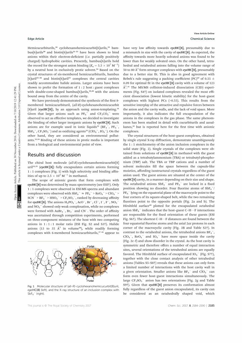
Hemicucurbiturils,18 cyclohexanohemicucurbit[6]urils,19 bam-bus[6]urils20 and biotin[6]urils21–23 have been shown to bindanions within their electron-decient (i.e. partially positivelycharged) hydrophobic cavities. Presently, bambus[6]urils holdthe record for the strongest anion binding (Ka ¼ 5.5 � 107 M�1)by a neutral host in exclusively protic solvent.24 Based on thecrystal structures of six-membered hemicucurbiturils, bambus[6]uril25–28 and biotin[6]uril21 complexes the central cavitiesreadily accommodate halide anions. Larger anions have beenshown to prefer the formation of 1 : 2 host : guest complexeswith double-cone-shaped bambus[6]urils,29,30 with the anionsbound away from the centre of the cavity.
We have previously demonstrated the synthesis of the rst 8-membered hemicucurbituril, (all-R)-cyclohexanohemicucurbit[8]uril (cycHC[8]), by an approach using anion-templating.31
Given that larger anions such as PF6� and CF3CO2
� wereobserved to act as effective templates, we decided to investigatethe binding of other larger inorganic anions by cycHC[8]. Suchanions are for example used in ionic liquids32 (BF4
�, PF6�,
SbF6�, CF3SO3
�) and as oxidizing agents33 (ClO4�, IO4
�). On theother hand, they are considered as environmental pollut-ants.33,34 Binding of these anions in protic media is importantfrom a biological and environmental point of view.
Results and discussion
The chiral host molecule (all-R)-cyclohexanohemicucurbit[8]uril31,35 (cycHC[8]) fully encapsulates certain anions forming1 : 1 complexes (Fig. 1) with high selectivity and binding affin-ities of up to 2.5 � 105 M�1 in methanol.
The scope of anionic guests that form complexes withcycHC[8] was determined by mass spectrometry (see ESI†). Only1 : 1 complexes were observed in ESI-MS spectra and abundantcomplexes were observed with SbF6
� z PF6� > ReO4
� > ClO4� >
SCN� > BF4� > HSO4
� > CF3SO3�, ranked by decreasing affinity
for cycHC[8]. The anions H2PO4�, AcO�, Br�, Cl�, I�, F�, NO3
�
and NO2� showed only weak complexation, while no complexes
were formed with AuBr4�, Br3
� and CN�. The order of affinitywas ascertained through competition experiments, performedon three-component mixtures of the host with two competinganions in 1 : 1 : 1 molar ratio (ESI Fig. S2 and S3†). Halideanions (13 to 35 A3 in volume36), while readily formingcomplexes with 6-membered hemicucurbiturils,37–41 appear to
have very low affinity towards cycHC[8], presumably due toa mismatch in size with the cavity of cycHC[8]. As expected, theaffinity towards more heavily solvated anions was found to belower than for weakly solvated ones. On the other hand, tetra-hedral and octahedral anions falling into the volume range of50 to 80 A3 form stronger complexes with cycHC[8], presumablydue to a better size t. This is also in good agreement withRebek's rule suggesting a packing coefficient (PC)42 of 0.55 �0.09 for optimal t in the cycHC[8] cavity with a volume of 123A3.31 The MS/MS collision-induced dissociation (CID) experi-ments (Fig. S4†) on isolated complexes revealed the most effi-cient dissociation (lowest kinetic stability) for the host–guestcomplexes with highest PCs (>0.55). This results from thesensitive interplay of the attractive and repulsive forces betweenthe anion and the cavity walls, and the lack of void space. Moreimportantly, it also indicates the full encapsulation of theanions in the complexes in the gas phase. The same phenom-enon has been studied in detail with cucurbiturils and azoal-kanes,43 but is reported here for the rst time with anioniccomplexes.
The crystal structures of the host–guest complexes, obtainedby single crystal X-ray diffraction, demonstrate unambiguouslythe 1 : 1 stoichiometry of the anion inclusion complexes in thesolid state (Fig. 2). Single crystals of the complexes were ob-tained from solutions of cycHC[8] in methanol with the guestadded as a tetrabutylammonium (TBA) or tetrabutyl-phospho-nium (TBP) salt. The TBA or TBP cations and a number ofsolvent molecules ll the space between the capsule-likemoieties, affording isostructural crystals regardless of the guestanion used. The guest anions are situated at the center of thecycHC[8] cavity, in a manner depending on their size and shape.The octahedral anions SbF6
� and PF6� are locked in a xed
position showing no disorder. Four uorine atoms of SbF6�/
PF6� lying on the equatorial plane of themacrocycle point to the
four corners of its square-shaped belt, while the two remaininguorines point to the opposite portals (Fig. 2a and b). TheHirshfeld surface44 plotted for the encapsulated octahedralanion SbF6
� indicates that the host–guest C–H/F interactionsare responsible for the xed orientation of these guests (ESIFig. S6†). The shortest C–H/F distances are found between thefour equatorial uorine atoms and the axial 2ax protons in eachcorner of the macrocyclic cavity (Fig. 3B and Table S3†). Incontrast to the octahedral anions, the tetrahedral anions BF4
�,ClO4
�, ReO4� and IO4
� have more space inside the cavity(Fig. 2c–f) and show disorder in the crystal. As the host cavity issymmetric and therefore offers a number of equal interactionsites, several orientations of the tetrahedral anions are equallyfavored. The Hirshfeld surface of encapsulated IO4
� (Fig. S7†),together with the close contact analysis of other tetrahedralanions (Tables S5–S8†) reveals that these anions can only forma limited number of interactions with the host cavity wall ina given orientation. Smaller anions like BF4
� and ClO4� can
form even fewer host–guest interactions simultaneously. Thelarge CF3SO3
� anion has two orientations (Fig. 2g and TableS9†). Given that cycHC[8] preserves its conformation almostfully regardless of the guest anion encapsulated, its cavity canbe considered as an octahedrally shaped void, which
Fig. 1 Molecular structure of (all-R)-cyclohexanohemicucurbit[8]uril,cycHC[8] (left), and the X-ray structure of an inclusion complex withSbF6
� (right).
This journal is © The Royal Society of Chemistry 2017 Chem. Sci., 2017, 8, 2184–2190 | 2185
Edge Article Chemical Science
Ope
n A
cces
s Arti
cle.
Pub
lishe
d on
30
Nov
embe
r 201
6. D
ownl
oade
d on
7/1
0/20
19 1
2:41
:29
PM.
Thi
s arti
cle
is lic
ense
d un
der a
Cre
ativ
e Co
mm
ons A
ttrib
utio
n 3.
0 U
npor
ted
Lice
nce.
View Article Online
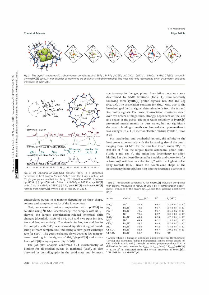
encapsulates guests in a manner depending on their shape,volume and complementarity of the interactions.
Next, we examined anion complexation with cycHC[8] insolution using 1H NMR spectroscopy. The complex with SbF6
�
showed the largest complexation-induced chemical shichanges (downeld shis of 0.52, 0.23 and 0.64 ppm for 2ax,4ax and 6ax, respectively). The signals for 2ax, 4ax and 6ax inthe complex with SbF6
� also showed signicant signal broad-ening at room temperature, indicating a slow guest exchangerate for SbF6
�. The guest exchange slows down at low temper-ature resulting in the signals of SbF6
�@cycHC[8] and excessfree cycHC[8] being separate (Fig. 3C(d)).
The Job plot analysis conrmed 1 : 1 stoichiometry ofbinding for all studied guests in methanol (ESI†), as alsoobserved by crystallography in the solid state and by mass
spectrometry in the gas phase. Association constants weredetermined by NMR titrations (Table 1), simultaneouslyfollowing three cycHC[8] proton signals 1ax, 2ax and 3eq(Fig. 3A). The association constant for SbF6
� was, due to thebroadening of the 2ax signal, determined only from the 1ax and3eq proton signals. The range of association constants variedover ve orders of magnitude, strongly dependent on the sizeand shape of the guest. The poor water solubility of cycHC[8]prevented measurements in pure water, but no signicantdecrease in binding strength was observed when pure methanolwas changed to a 1 : 1 methanol/water mixture (Table 1, rows2–3).
For tetrahedral and octahedral anions, the affinity to thehost grows exponentially with the increasing size of the guest,ranging from 48 M�1 for the smallest tested anion BF4
� to250 000 M�1 for the largest tested octahedral anion SbF6
�
(Table 1 and Fig. 4). The anion size dependency for anionbinding has also been discussed by Sindelar and co-workers fora bambus[6]uril host in chloroform,24 with the highest selec-tivity towards ClO4
�. Given the double-cone shape of thedodecabenzylbambus[6]uril host and the restricted diameter of
Fig. 2 The crystal structures of 1 : 1 host–guest complexes of (a) SbF6�, (b) PF6
�, (c) BF4�, (d) ClO4
�, (e) IO4�, (f) ReO4
� and (g) CF3SO3� anions in
the cycHC[8] cavity. Minor disorder components are shown as a wireframe model. The host in (b–f) is represented by an octahedron depictingthe cavity of cycHC[8].
Fig. 3 (A) Labelling of cycHC[8] protons, (B) C–H/F distancesbetween the host proton 2ax and SbF6
� from the X-ray structure; all(CH2)4 groups are omitted for clarity, (C) 1H NMR in MeOD of (a) freecycHC[8], (b) cycHC[8] with 0.6 eq. of NaSbF6 at 288 K (c) cycHC[8]with 10 eq. of NaSbF6 at 288 K; (d) SbF6
�@cycHC[8] and free cycHC[8]formed from cycHC[8] with 0.6 eq. of NaSbF6 at 229 K.
Table 1 Association constants Ka for cycHC[8] inclusion complexeswith anions, measured in MeOD at 288 K by 1H NMR titration experi-ments. Volumes of the anions (Vanion) and their packing coefficients(PC)a
Anion Cation Vanion (A3) PC Ka (M�1)
SbF6� Na+ 81.8 0.67 (2.5 � 0.7) � 105
PF6� Bu4N
+ 70.6 0.57 (2.8 � 0.4) � 104
PF6�b Bu4N
+ 70.6 0.57 (2.6 � 0.2) � 104
PF6� Na+ 70.6 0.57 (2.0 � 0.2) � 104
ReO4� Bu4N
+ 64.8 0.53 (4.7 � 0.4) � 103
IO4� Na+ 64.3 0.52 (1.8 � 0.2) � 103
ClO4� Bu4N
+ 54.7 0.45 (4.7 � 0.2) � 102
BF4� Bu4N
+ 51.6 0.42 (4.8 � 0.4) � 10CF3SO3
� Bu4N+ 82.3 0.67 (3.9 � 0.5) � 10
CF3CO2� Bu4N
+ 68.7 0.56 <10
a Anion volume is based on optimized anion geometries (BP86-D/def2-TZVPD) and calculated using a triangulated sphere model (based onCSD default atomic radii) through the Olex2 program package.45 PC isdened as the ratio between the Vanion to Vcavity(host).42 Vcavity(cycHC[8])¼ 123.0 A3 is measured from the crystal structure of cycHC[8].31b 1H NMR in 1 : 1 MeOD/D2O.
2186 | Chem. Sci., 2017, 8, 2184–2190 This journal is © The Royal Society of Chemistry 2017
Chemical Science Edge Article
Ope
n A
cces
s Arti
cle.
Pub
lishe
d on
30
Nov
embe
r 201
6. D
ownl
oade
d on
7/1
0/20
19 1
2:41
:29
PM.
Thi
s arti
cle
is lic
ense
d un
der a
Cre
ativ
e Co
mm
ons A
ttrib
utio
n 3.
0 U
npor
ted
Lice
nce.
View Article Online
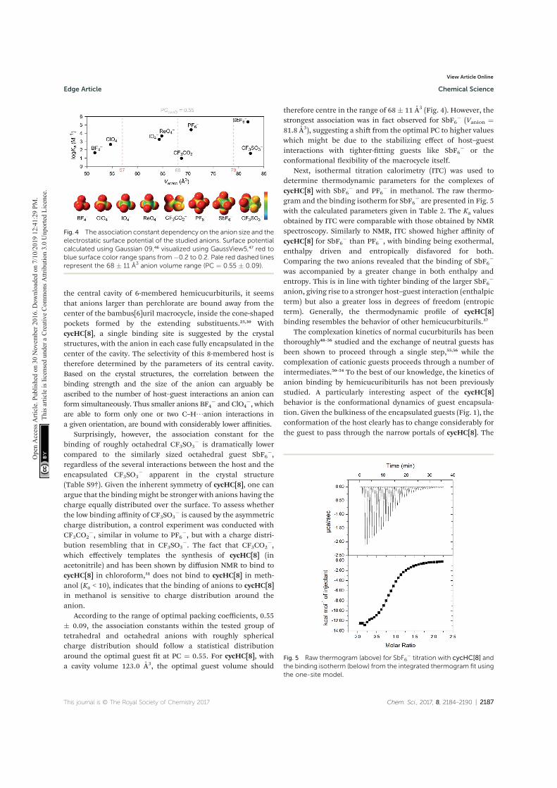
the central cavity of 6-membered hemicucurbiturils, it seemsthat anions larger than perchlorate are bound away from thecenter of the bambus[6]uril macrocycle, inside the cone-shapedpockets formed by the extending substituents.25,30 WithcycHC[8], a single binding site is suggested by the crystalstructures, with the anion in each case fully encapsulated in thecenter of the cavity. The selectivity of this 8-membered host istherefore determined by the parameters of its central cavity.Based on the crystal structures, the correlation between thebinding strength and the size of the anion can arguably beascribed to the number of host–guest interactions an anion canform simultaneously. Thus smaller anions BF4
� and ClO4�, which
are able to form only one or two C–H/anion interactions ina given orientation, are bound with considerably lower affinities.
Surprisingly, however, the association constant for thebinding of roughly octahedral CF3SO3
� is dramatically lowercompared to the similarly sized octahedral guest SbF6
�,regardless of the several interactions between the host and theencapsulated CF3SO3
� apparent in the crystal structure(Table S9†). Given the inherent symmetry of cycHC[8], one canargue that the bindingmight be stronger with anions having thecharge equally distributed over the surface. To assess whetherthe low binding affinity of CF3SO3
� is caused by the asymmetriccharge distribution, a control experiment was conducted withCF3CO2
�, similar in volume to PF6�, but with a charge distri-
bution resembling that in CF3SO3�. The fact that CF3CO2
�,which effectively templates the synthesis of cycHC[8] (inacetonitrile) and has been shown by diffusion NMR to bind tocycHC[8] in chloroform,31 does not bind to cycHC[8] in meth-anol (Ka < 10), indicates that the binding of anions to cycHC[8]in methanol is sensitive to charge distribution around theanion.
According to the range of optimal packing coefficients, 0.55� 0.09, the association constants within the tested group oftetrahedral and octahedral anions with roughly sphericalcharge distribution should follow a statistical distributionaround the optimal guest t at PC ¼ 0.55. For cycHC[8], witha cavity volume 123.0 A3, the optimal guest volume should
therefore centre in the range of 68 � 11 A3 (Fig. 4). However, thestrongest association was in fact observed for SbF6
� (Vanion ¼81.8 A3), suggesting a shi from the optimal PC to higher valueswhich might be due to the stabilizing effect of host–guestinteractions with tighter-tting guests like SbF6
� or theconformational exibility of the macrocycle itself.
Next, isothermal titration calorimetry (ITC) was used todetermine thermodynamic parameters for the complexes ofcycHC[8] with SbF6
� and PF6� in methanol. The raw thermo-
gram and the binding isotherm for SbF6� are presented in Fig. 5
with the calculated parameters given in Table 2. The Ka valuesobtained by ITC were comparable with those obtained by NMRspectroscopy. Similarly to NMR, ITC showed higher affinity ofcycHC[8] for SbF6
� than PF6�, with binding being exothermal,
enthalpy driven and entropically disfavored for both.Comparing the two anions revealed that the binding of SbF6
�
was accompanied by a greater change in both enthalpy andentropy. This is in line with tighter binding of the larger SbF6
�
anion, giving rise to a stronger host–guest interaction (enthalpicterm) but also a greater loss in degrees of freedom (entropicterm). Generally, the thermodynamic prole of cycHC[8]binding resembles the behavior of other hemicucurbiturils.37
The complexation kinetics of normal cucurbiturils has beenthoroughly48–56 studied and the exchange of neutral guests hasbeen shown to proceed through a single step,55,56 while thecomplexation of cationic guests proceeds through a number ofintermediates.50–54 To the best of our knowledge, the kinetics ofanion binding by hemicucuribiturils has not been previouslystudied. A particularly interesting aspect of the cycHC[8]behavior is the conformational dynamics of guest encapsula-tion. Given the bulkiness of the encapsulated guests (Fig. 1), theconformation of the host clearly has to change considerably forthe guest to pass through the narrow portals of cycHC[8]. The
Fig. 4 The association constant dependency on the anion size and theelectrostatic surface potential of the studied anions. Surface potentialcalculated using Gaussian 09,46 visualized using GaussView5,47 red toblue surface color range spans from �0.2 to 0.2. Pale red dashed linesrepresent the 68 � 11 A3 anion volume range (PC ¼ 0.55 � 0.09).
Fig. 5 Raw thermogram (above) for SbF6� titration with cycHC[8] and
the binding isotherm (below) from the integrated thermogram fit usingthe one-site model.
This journal is © The Royal Society of Chemistry 2017 Chem. Sci., 2017, 8, 2184–2190 | 2187
Edge Article Chemical Science
Ope
n A
cces
s Arti
cle.
Pub
lishe
d on
30
Nov
embe
r 201
6. D
ownl
oade
d on
7/1
0/20
19 1
2:41
:29
PM.
Thi
s arti
cle
is lic
ense
d un
der a
Cre
ativ
e Co
mm
ons A
ttrib
utio
n 3.
0 U
npor
ted
Lice
nce.
View Article Online
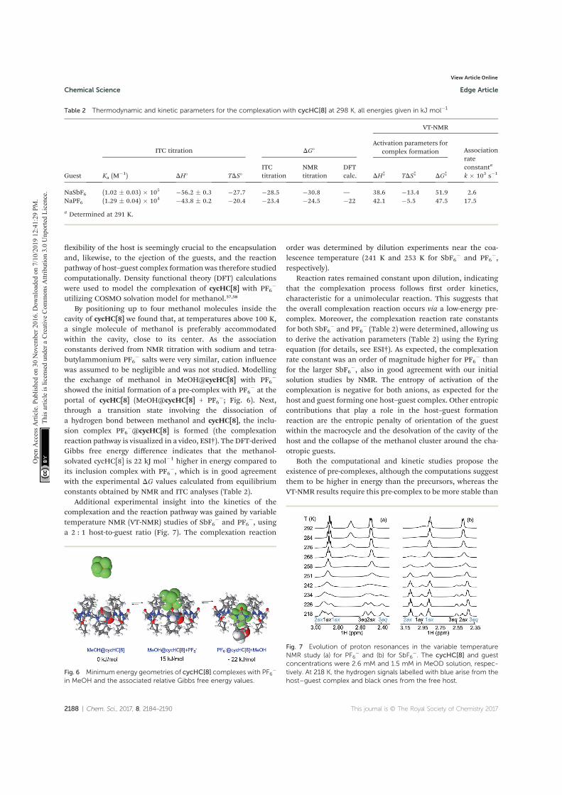
exibility of the host is seemingly crucial to the encapsulationand, likewise, to the ejection of the guests, and the reactionpathway of host–guest complex formation was therefore studiedcomputationally. Density functional theory (DFT) calculationswere used to model the complexation of cycHC[8] with PF6
�
utilizing COSMO solvation model for methanol.57,58
By positioning up to four methanol molecules inside thecavity of cycHC[8] we found that, at temperatures above 100 K,a single molecule of methanol is preferably accommodatedwithin the cavity, close to its center. As the associationconstants derived from NMR titration with sodium and tetra-butylammonium PF6
� salts were very similar, cation inuencewas assumed to be negligible and was not studied. Modellingthe exchange of methanol in MeOH@cycHC[8] with PF6
�
showed the initial formation of a pre-complex with PF6� at the
portal of cycHC[8] (MeOH@cycHC[8] + PF6�; Fig. 6). Next,
through a transition state involving the dissociation ofa hydrogen bond between methanol and cycHC[8], the inclu-sion complex PF6
�@cycHC[8] is formed (the complexationreaction pathway is visualized in a video, ESI†). The DFT-derivedGibbs free energy difference indicates that the methanol-solvated cycHC[8] is 22 kJ mol�1 higher in energy compared toits inclusion complex with PF6
�, which is in good agreementwith the experimental DG values calculated from equilibriumconstants obtained by NMR and ITC analyses (Table 2).
Additional experimental insight into the kinetics of thecomplexation and the reaction pathway was gained by variabletemperature NMR (VT-NMR) studies of SbF6
� and PF6�, using
a 2 : 1 host-to-guest ratio (Fig. 7). The complexation reaction
order was determined by dilution experiments near the coa-lescence temperature (241 K and 253 K for SbF6
� and PF6�,
respectively).Reaction rates remained constant upon dilution, indicating
that the complexation process follows rst order kinetics,characteristic for a unimolecular reaction. This suggests thatthe overall complexation reaction occurs via a low-energy pre-complex. Moreover, the complexation reaction rate constantsfor both SbF6
� and PF6� (Table 2) were determined, allowing us
to derive the activation parameters (Table 2) using the Eyringequation (for details, see ESI†). As expected, the complexationrate constant was an order of magnitude higher for PF6
� thanfor the larger SbF6
�, also in good agreement with our initialsolution studies by NMR. The entropy of activation of thecomplexation is negative for both anions, as expected for thehost and guest forming one host–guest complex. Other entropiccontributions that play a role in the host–guest formationreaction are the entropic penalty of orientation of the guestwithin the macrocycle and the desolvation of the cavity of thehost and the collapse of the methanol cluster around the cha-otropic guests.
Both the computational and kinetic studies propose theexistence of pre-complexes, although the computations suggestthem to be higher in energy than the precursors, whereas theVT-NMR results require this pre-complex to be more stable than
Table 2 Thermodynamic and kinetic parameters for the complexation with cycHC[8] at 298 K, all energies given in kJ mol�1
Guest
ITC titration DG�
VT-NMR
Activation parameters forcomplex formation Association
rateconstanta
k � 103 s�1Ka (M�1) DH� TDS�
ITCtitration
NMRtitration
DFTcalc. DH‡ TDS‡ DG‡
NaSbF6 (1.02 � 0.03) � 105 �56.2 � 0.3 �27.7 �28.5 �30.8 — 38.6 �13.4 51.9 2.6NaPF6 (1.29 � 0.04) � 104 �43.8 � 0.2 �20.4 �23.4 �24.5 �22 42.1 �5.5 47.5 17.5
a Determined at 291 K.
Fig. 6 Minimum energy geometries of cycHC[8] complexes with PF6�
in MeOH and the associated relative Gibbs free energy values.
Fig. 7 Evolution of proton resonances in the variable temperatureNMR study (a) for PF6
� and (b) for SbF6�. The cycHC[8] and guest
concentrations were 2.6 mM and 1.5 mM in MeOD solution, respec-tively. At 218 K, the hydrogen signals labelled with blue arise from thehost–guest complex and black ones from the free host.
2188 | Chem. Sci., 2017, 8, 2184–2190 This journal is © The Royal Society of Chemistry 2017
Chemical Science Edge Article
Ope
n A
cces
s Arti
cle.
Pub
lishe
d on
30
Nov
embe
r 201
6. D
ownl
oade
d on
7/1
0/20
19 1
2:41
:29
PM.
Thi
s arti
cle
is lic
ense
d un
der a
Cre
ativ
e Co
mm
ons A
ttrib
utio
n 3.
0 U
npor
ted
Lice
nce.
View Article Online
the starting components. This reects a complicated complex-ation reaction pathway that includes several steps. We proposethat either the anion pre-complex formation or the reorgani-zation of the methanol solvation shell around anions and themacrocycle play an important role, which merits furtherinvestigation.
Conclusions
The inherently chiral (all-R)-cyclohexanohemicucurbit[8]uril,cycHC[8], is the rst example of an octameric macrocycle of thehemicucurbituril family acting as a neutral host that fullyencapsulates anions as 1 : 1 complexes in gas, solution andsolid state. The binding affinity strongly depends on the size,shape and charge distribution of the anion. Crystallographicstudies show the importance of the shape t between thecycHC[8] cavity and the anion guest, with the octahedral guestsbound in a perfectly ordered manner, while the tetrahedralguests exhibited various degrees of disorder. Moreover, due tothe unique size and shape of cycHC[8], the large, octahedrallyshaped SbF6
� was found to be the most tightly bound guest,closely followed by PF6
�. On the other hand, binding of thesimilarly sized CF3SO3
� and CF3CO2� was around four orders of
magnitude weaker, which could be ascribed to their asymmetriccharge distribution limiting the number of hydrogen bondsthat can be formed simultaneously. The complexation wasfound to proceed through the formation of a pre-complex,accompanied by the ejection of a solvent molecule from thecycHC[8] cavity and involving a large movement of the portalsduring the encapsulation.
This work, besides being the rst comprehensive anion-binding study on an eight-membered hemicucurbituril-typemacrocycle, also demonstrates the unique anion bindingproperties of a macrocyclic host easily accessible througha simple templated synthetic protocol.31 Moreover, it proposesa pathway to and encourages the preparation of new hemi-cucurbiturils for anion binding and transport, catalysis inconned space and other supramolecular applications.
Author contribution
The manuscript was written through contributions of allauthors.
Acknowledgements
This research was supported by the Academy of Finland (KR:grants 263256, 265328 and 292746, EK: grants 284562 and278743), the Estonian Ministry of Education and Researchthrough Grants IUT19-32, IUT19-9, IUT23-7 and PUT692, TUTgrant No. B25, the ESF DoRa, and the EU European RegionalDevelopment Fund through the Center of Excellence in Molec-ular Cell Engineering projects 3.2.0101.08-0017 and TK134.Computations were performed on the HPC cluster at TUT,which is part of the ETAIS project. FT acknowledges the supportfromNGS-NANO through a Ph.D. fellowship. The authors wouldlike to thank Marina Kudrjasova and Mari-Liis Kasemets for
assistance with NMR, Ly Parnaste with ITC, Toomas Kaevandwith computations and Lauri Kivijarvi with MS experiments.
Notes and references
1 C. J. Pedersen, Angew. Chem., 1988, 100, 1053; Angew. Chem.,Int. Ed. Engl., 1988, 27, 1021.
2 J.-M. Lehn, Angew. Chem., 1988, 100, 91; Angew. Chem., Int.Ed. Engl., 1988, 27, 89.
3 W. A. Freeman, W. L. Mock and N. Y. Shih, J. Am. Chem. Soc.,1981, 103, 7367.
4 M. J. Langton, C. J. Serpell and P. D. Beer, Angew. Chem., Int.Ed., 2016, 55, 1974.
5 P. A. Gale and C. Caltagirone, Chem. Soc. Rev., 2015, 44, 4212.6 N. H. Evans and P. D. Beer, Angew. Chem., Int. Ed., 2014, 53,11716.
7 J. L. Sessler, P. A. Gale and W.-S. Cho, Anion ReceptorChemistry, ed. J. F. Stoddart, Royal Society of Chemistry,Cambridge, 2006.
8 P. A. Gale, E. N. W. Howe and X. Wu, Chem, 2016, 1, 351.9 S. Kubik, Chem. Soc. Rev., 2010, 39, 3648.10 S. Kubik, R. Goddard, R. Kirchner, D. Nolting and J. Seidel,
Angew. Chem., Int. Ed., 2001, 40, 2648.11 S. Kubik and R. Goddard, Proc. Natl. Acad. Sci. U. S. A., 2002,
99, 5127.12 S. Kubik, R. Kirchner, D. Nolting and J. Seidel, J. Am. Chem.
Soc., 2002, 124, 12752.13 F. Sommer and S. Kubik, Org. Biomol. Chem., 2014, 12, 8851.14 P. Sokkalingam, J. Shraberg, S. W. Rick and B. C. Gibb, J. Am.
Chem. Soc., 2016, 138, 48.15 K. I. Assaf, M. S. Ural, F. Pan, T. Georgiev, S. Simova,
K. Rissanen, D. Gabel and W. M. Nau, Angew. Chem., Int.Ed., 2015, 54, 6852.
16 E. Masson, X. Ling, R. Joseph, L. Kyeremeh-Mensah andX. Lu, RSC Adv., 2012, 2, 1213.
17 S. J. Barrow, S. Kasera, M. J. Rowland, J. Del Barrio andO. A. Scherman, Chem. Rev., 2015, 115, 12320.
18 Y. Miyahara, K. Goto, M. Oka and T. Inazu, Angew. Chem., Int.Ed., 2004, 43, 5019.
19 R. Aav, E. Shmatova, I. Reile, M. Borissova, F. Topic andK. Rissanen, Org. Lett., 2013, 15, 3786.
20 J. Svec, M. Necas and V. Sindelar, Angew. Chem., Int. Ed.,2010, 49, 2378.
21 M. Lisbjerg, B. M. Jessen, B. Rasmussen, B. Nielsen,A. Ø. Madsen and M. Pittelkow, Chem. Sci., 2014, 5, 2647.
22 M. Lisbjerg, B. E. Nielsen, B. O. Milhøj, S. P. A. Sauer andM. Pittelkow, Org. Biomol. Chem., 2015, 13, 369.
23 M. Lisbjerg, H. Valkenier, B. M. Jessen, H. Al-Kerdi,A. P. Davis and M. Pittelkow, J. Am. Chem. Soc., 2015, 137,4948.
24 M. A. Yawer, V. Havel and V. Sindelar, Angew. Chem., Int. Ed.,2015, 54, 276.
25 V. Havel, J. Svec, M. Wimmerova, M. Dusek, M. Pojarova andV. Sindelar, Org. Lett., 2011, 13, 4000.
26 J. Svec, M. Dusek, K. Fejfarova, P. Stacko, P. Klan,A. E. Kaifer, W. Li, E. Hudeckova and V. Sindelar, Chem.–Eur. J., 2011, 17, 5605.
This journal is © The Royal Society of Chemistry 2017 Chem. Sci., 2017, 8, 2184–2190 | 2189
Edge Article Chemical Science
Ope
n A
cces
s Arti
cle.
Pub
lishe
d on
30
Nov
embe
r 201
6. D
ownl
oade
d on
7/1
0/20
19 1
2:41
:29
PM.
Thi
s arti
cle
is lic
ense
d un
der a
Cre
ativ
e Co
mm
ons A
ttrib
utio
n 3.
0 U
npor
ted
Lice
nce.
View Article Online
27 J. Rivollier, P. Thuery andM.-P. Heck,Org. Lett., 2013, 15, 480.28 M. Singh, E. Solel, E. Keinan and O. Reany, Chem.–Eur. J.,
2015, 21, 536.29 T. Fiala and V. Sindelar, Supramol. Chem., 2016, 28, 810.30 V. Havel, V. Sindelar, M. Necas and A. E. Kaifer, Chem.
Commun., 2014, 50, 1372.31 E. Prigorchenko, M. Oeren, S. Kaabel, M. Fomitsenko,
I. Reile, I. Jarving, T. Tamm, F. Topic, K. Rissanen andR. Aav, Chem. Commun., 2015, 51, 10921.
32 H. Olivier-Bourbigou, L. Magna and D. Morvan, Appl. Catal.,A, 2010, 373, 1.
33 E. T. Urbansky, Biorem. J., 1998, 2, 81.34 R. F. M. Frade and C. A. M. Afonso, Hum. Exp. Toxicol., 2010,
29, 1038.35 M. Fomitsenko, E. Shmatova, M. Oeren, I. Jarving and R. Aav,
Supramol. Chem., 2014, 26, 698.36 Anion volumes in this report are calculated using
a triangulated sphere model (based on CSD default atomicradii) through Olex2 program package. Ref. 42.
37 H.-J. Buschmann, E. Cleva and E. Schollmeyer, Inorg. Chem.Commun., 2005, 8, 125.
38 H.-J. Buschmann, A. Zielesny and E. J. Schollmeyer,J. Inclusion Phenom. Macrocyclic Chem., 2006, 54, 181.
39 M. Sundararajan, R. V. Solomon, S. K. Ghosh andP. Venuvanalingam, RSC Adv., 2011, 1, 1333.
40 H.-J. Buschmann and A. Zielesny, Comput. Theor. Chem.,2013, 1022, 14.
41 M. Oeren, E. Shmatova, T. Tamm and R. Aav, Phys. Chem.Chem. Phys., 2014, 16, 19198.
42 S. Mecozzi and J. Rebek, Chem.–Eur. J., 1998, 4, 1016.43 T.-C. Lee, E. Kalenius, A. I. Lazar, K. I. Assaf, N. Kuhnert,
C. H. Grun, J. Janis, O. A. Scherman and W. M. Nau, Nat.Chem., 2013, 5, 376.
44 M. A. Spackman and D. Jayatilaka, CrystEngComm, 2009, 11,19.
45 O. V. Dolomanov, L. J. Bourhis, R. J. Gildea, J. A. K. Howardand H.-J. Puschmann, J. Appl. Crystallogr., 2009, 42, 339.
46 M. J. Frisch, G. W. Trucks, H. B. Schlegel, G. E. Scuseria,M. A. Robb, J. R. Cheeseman, G. Scalmani, V. Barone,B. Mennucci, G. A. Petersson, H. Nakatsuji, M. Caricato,
X. Li, H. P. Hratchian, A. F. Izmaylov, J. Bloino, G. Zheng,J. L. Sonnenberg, M. Hada, M. Ehara, K. Toyota,R. Fukuda, J. Hasegawa, M. Ishida, T. Nakajima, Y. Honda,O. Kitao, H. Nakai, T. Vreven, J. A. Montgomery Jr,J. E. Peralta, F. Ogliaro, M. Bearpark, J. J. Heyd,E. Brothers, K. N. Kudin, V. N. Staroverov, R. Kobayashi,J. Normand, K. Raghavachari, A. Rendell, J. C. Burant,S. S. Iyengar, J. Tomasi, M. Cossi, N. Rega, J. M. Millam,M. Klene, J. E. Knox, J. B. Cross, V. Bakken, C. Adamo,J. Jaramillo, R. Gomperts, R. E. Stratmann, O. Yazyev,A. J. Austin, R. Cammi, C. Pomelli, J. W. Ochterski,R. L. Martin, K. Morokuma, V. G. Zakrzewski, G. A. Voth,P. Salvador, J. J. Dannenberg, S. Dapprich, A. D. Daniels,O. Farkas, J. B. Foresman, J. V. Ortiz, J. Cioslowski andD. J. Fox, Gaussian 09, Revision E.01, Gaussian, Inc.,Wallingford CT, 2009.
47 R. Dennington, T. Keith and J. Millam, GaussView, Version 5,Semichem Inc., Shawnee Mission, KS, 2009.
48 W. L. Mock and N.-Y. Shih, J. Org. Chem., 1986, 51, 4440.49 W. L. Mock and N.-Y. Shih, J. Am. Chem. Soc., 1989, 111,
2697.50 R. Hoffmann, W. Knoche, C. Fenn and H.-J. Buschmann,
J. Chem. Soc., Faraday Trans., 1994, 90, 1507.51 C. Marquez and W. M. Nau, Angew. Chem., Int. Ed., 2001, 40,
3155.52 C. Marquez, R. R. Hudgins andW. M. Nau, J. Am. Chem. Soc.,
2004, 126, 5806.53 X. Ling, E. L. Samuel, D. L. Patchell and E. Masson, Org. Lett.,
2010, 12, 2730.54 H. Tang, D. Fuentealba, Y. H. Ko, N. Selvapalam, K. Kim and
C. Bohne, J. Am. Chem. Soc., 2011, 133, 20623.55 J.-S. Yu, F.-G. Wu, L.-F. Tao, J.-J. Luo and Z.-W. Yu, Phys.
Chem. Chem. Phys., 2011, 13, 3638.56 Z. Miskolczy and L. Biczok, J. Phys. Chem. B, 2014, 118, 2499.57 TURBOMOLE V6.5 2013, a development of University of
Karlsruhe and Forschungszentrum Karlsruhe GmbH, 1989-2007, TURBOMOLE GmbH, since 2007, available fromhttp://www.turbomole.com.
58 A. Klamt and G. Schuurmann, J. Chem. Soc., Perkin Trans. 2,1993, 799.
2190 | Chem. Sci., 2017, 8, 2184–2190 This journal is © The Royal Society of Chemistry 2017
Chemical Science Edge Article
Ope
n A
cces
s Arti
cle.
Pub
lishe
d on
30
Nov
embe
r 201
6. D
ownl
oade
d on
7/1
0/20
19 1
2:41
:29
PM.
Thi
s arti
cle
is lic
ense
d un
der a
Cre
ativ
e Co
mm
ons A
ttrib
utio
n 3.
0 U
npor
ted
Lice
nce.
View Article Online
V
ION--PAIR COMPLEXATION WITH DIBENZO[21]CROWN-7 AND DIBENZO[24]CROWN-8 BIS-UREA RECEPTORS
by
T. Mäkelä, A. Kiesi ssanen
Chemistry - A European Journal, 2016, 22, 40, 14264 – 14272
Reproduced with kind permission. Copyright © 2016 Wiley –
https://doi.org/10.1002/chem.201602362
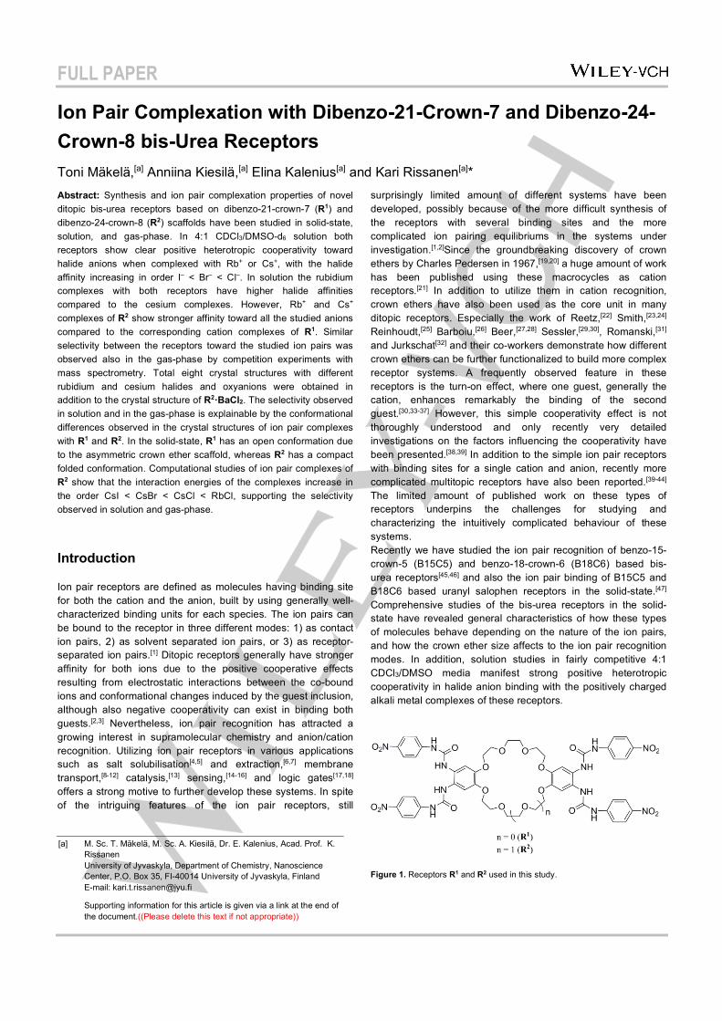
FULL PAPER
Ion Pair Complexation with Dibenzo-21-Crown-7 and Dibenzo-24-Crown-8 bis-Urea Receptors Toni Mäkelä,[a] Anniina Kiesilä,[a] Elina Kalenius[a] and Kari Rissanen[a]* Abstract: Synthesis and ion pair complexation properties of novel ditopic bis-urea receptors based on dibenzo-21-crown-7 (R1) and dibenzo-24-crown-8 (R2) scaffolds have been studied in solid-state, solution, and gas-phase. In 4:1 CDCl3/DMSO-d6 solution both receptors show clear positive heterotropic cooperativity toward halide anions when complexed with Rb+ or Cs+, with the halide affinity increasing in order I– < Br– < Cl–. In solution the rubidium complexes with both receptors have higher halide affinities compared to the cesium complexes. However, Rb+ and Cs+ complexes of R2 show stronger affinity toward all the studied anions compared to the corresponding cation complexes of R1. Similar selectivity between the receptors toward the studied ion pairs was observed also in the gas-phase by competition experiments with mass spectrometry. Total eight crystal structures with different rubidium and cesium halides and oxyanions were obtained in addition to the crystal structure of R2·BaCl2. The selectivity observed in solution and in the gas-phase is explainable by the conformational differences observed in the crystal structures of ion pair complexes with R1 and R2. In the solid-state, R1 has an open conformation due to the asymmetric crown ether scaffold, whereas R2 has a compact folded conformation. Computational studies of ion pair complexes of R2 show that the interaction energies of the complexes increase in the order CsI < CsBr < CsCl < RbCl, supporting the selectivity observed in solution and gas-phase.
Introduction
Ion pair receptors are defined as molecules having binding site for both the cation and the anion, built by using generally well-characterized binding units for each species. The ion pairs can be bound to the receptor in three different modes: 1) as contact ion pairs, 2) as solvent separated ion pairs, or 3) as receptor-separated ion pairs.[1] Ditopic receptors generally have stronger affinity for both ions due to the positive cooperative effects resulting from electrostatic interactions between the co-bound ions and conformational changes induced by the guest inclusion, although also negative cooperativity can exist in binding both guests.[2,3] Nevertheless, ion pair recognition has attracted a growing interest in supramolecular chemistry and anion/cation recognition. Utilizing ion pair receptors in various applications such as salt solubilisation[4,5] and extraction,[6,7] membrane transport,[8-12] catalysis,[13] sensing,[14-16] and logic gates[17,18] offers a strong motive to further develop these systems. In spite of the intriguing features of the ion pair receptors, still
surprisingly limited amount of different systems have been developed, possibly because of the more difficult synthesis of the receptors with several binding sites and the more complicated ion pairing equilibriums in the systems under investigation.[1,2]Since the groundbreaking discovery of crown ethers by Charles Pedersen in 1967,[19,20] a huge amount of work has been published using these macrocycles as cation receptors.[21] In addition to utilize them in cation recognition, crown ethers have also been used as the core unit in many ditopic receptors. Especially the work of Reetz,[22] Smith,[23,24] Reinhoudt,[25] Barboiu,[26] Beer,[27,28] Sessler,[29,30], Romanski,[31] and Jurkschat[32] and their co-workers demonstrate how different crown ethers can be further functionalized to build more complex receptor systems. A frequently observed feature in these receptors is the turn-on effect, where one guest, generally the cation, enhances remarkably the binding of the second guest.[30,33-37] However, this simple cooperativity effect is not thoroughly understood and only recently very detailed investigations on the factors influencing the cooperativity have been presented.[38,39] In addition to the simple ion pair receptors with binding sites for a single cation and anion, recently more complicated multitopic receptors have also been reported.[39-44] The limited amount of published work on these types of receptors underpins the challenges for studying and characterizing the intuitively complicated behaviour of these systems. Recently we have studied the ion pair recognition of benzo-15-crown-5 (B15C5) and benzo-18-crown-6 (B18C6) based bis-urea receptors[45,46] and also the ion pair binding of B15C5 and B18C6 based uranyl salophen receptors in the solid-state.[47] Comprehensive studies of the bis-urea receptors in the solid-state have revealed general characteristics of how these types of molecules behave depending on the nature of the ion pairs, and how the crown ether size affects to the ion pair recognition modes. In addition, solution studies in fairly competitive 4:1 CDCl3/DMSO media manifest strong positive heterotropic cooperativity in halide anion binding with the positively charged alkali metal complexes of these receptors.
Figure 1. Receptors R1 and R2 used in this study.
[a] M. Sc. T. Mäkelä, M. Sc. A. Kiesilä, Dr. E. Kalenius, Acad. Prof. K. Rissanen University of Jyvaskyla, Department of Chemistry, Nanoscience Center, P.O. Box 35, FI-40014 University of Jyvaskyla, Finland E-mail: [email protected]
Supporting information for this article is given via a link at the end of the document.((Please delete this text if not appropriate))
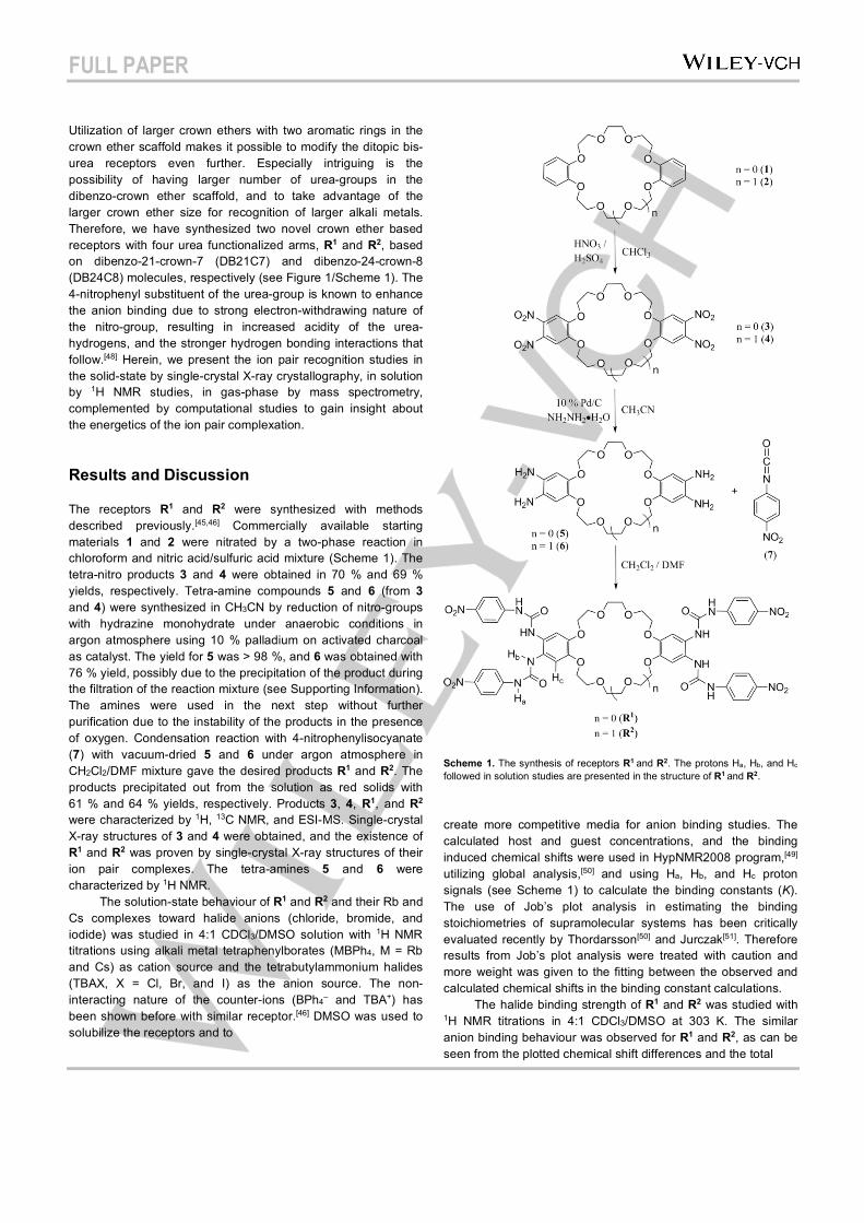
FULL PAPER
Utilization of larger crown ethers with two aromatic rings in the crown ether scaffold makes it possible to modify the ditopic bis-urea receptors even further. Especially intriguing is the possibility of having larger number of urea-groups in the dibenzo-crown ether scaffold, and to take advantage of the larger crown ether size for recognition of larger alkali metals. Therefore, we have synthesized two novel crown ether based receptors with four urea functionalized arms, R1 and R2, based on dibenzo-21-crown-7 (DB21C7) and dibenzo-24-crown-8 (DB24C8) molecules, respectively (see Figure 1/Scheme 1). The 4-nitrophenyl substituent of the urea-group is known to enhance the anion binding due to strong electron-withdrawing nature of the nitro-group, resulting in increased acidity of the urea-hydrogens, and the stronger hydrogen bonding interactions that follow.[48] Herein, we present the ion pair recognition studies in the solid-state by single-crystal X-ray crystallography, in solution by 1H NMR studies, in gas-phase by mass spectrometry, complemented by computational studies to gain insight about the energetics of the ion pair complexation.
Results and Discussion
The receptors R1 and R2 were synthesized with methods described previously.[45,46] Commercially available starting materials 1 and 2 were nitrated by a two-phase reaction in chloroform and nitric acid/sulfuric acid mixture (Scheme 1). The tetra-nitro products 3 and 4 were obtained in 70 % and 69 % yields, respectively. Tetra-amine compounds 5 and 6 (from 3 and 4) were synthesized in CH3CN by reduction of nitro-groups with hydrazine monohydrate under anaerobic conditions in argon atmosphere using 10 % palladium on activated charcoal as catalyst. The yield for 5 was > 98 %, and 6 was obtained with 76 % yield, possibly due to the precipitation of the product during the filtration of the reaction mixture (see Supporting Information). The amines were used in the next step without further purification due to the instability of the products in the presence of oxygen. Condensation reaction with 4-nitrophenylisocyanate (7) with vacuum-dried 5 and 6 under argon atmosphere in CH2Cl2/DMF mixture gave the desired products R1 and R2. The products precipitated out from the solution as red solids with 61 % and 64 % yields, respectively. Products 3, 4, R1, and R2 were characterized by 1H, 13C NMR, and ESI-MS. Single-crystal X-ray structures of 3 and 4 were obtained, and the existence of R1 and R2 was proven by single-crystal X-ray structures of their ion pair complexes. The tetra-amines 5 and 6 were characterized by 1H NMR. The solution-state behaviour of R1 and R2 and their Rb and Cs complexes toward halide anions (chloride, bromide, and iodide) was studied in 4:1 CDCl3/DMSO solution with 1H NMR titrations using alkali metal tetraphenylborates (MBPh4, M = Rb and Cs) as cation source and the tetrabutylammonium halides (TBAX, X = Cl, Br, and I) as the anion source. The non-interacting nature of the counter-ions (BPh4– and TBA+) has been shown before with similar receptor.[46] DMSO was used to solubilize the receptors and to
Scheme 1. The synthesis of receptors R1 and R2. The protons Ha, Hb, and Hc followed in solution studies are presented in the structure of R1 and R2.
create more competitive media for anion binding studies. The calculated host and guest concentrations, and the binding induced chemical shifts were used in HypNMR2008 program,[49] utilizing global analysis,[50] and using Ha, Hb, and Hc proton signals (see Scheme 1) to calculate the binding constants (K). The use of Job’s plot analysis in estimating the binding stoichiometries of supramolecular systems has been critically evaluated recently by Thordarsson[50] and Jurczak[51]. Therefore results from Job’s plot analysis were treated with caution and more weight was given to the fitting between the observed and calculated chemical shifts in the binding constant calculations. The halide binding strength of R1 and R2 was studied with 1H NMR titrations in 4:1 CDCl3/DMSO at 303 K. The similar anion binding behaviour was observed for R1 and R2, as can be seen from the plotted chemical shift differences and the total
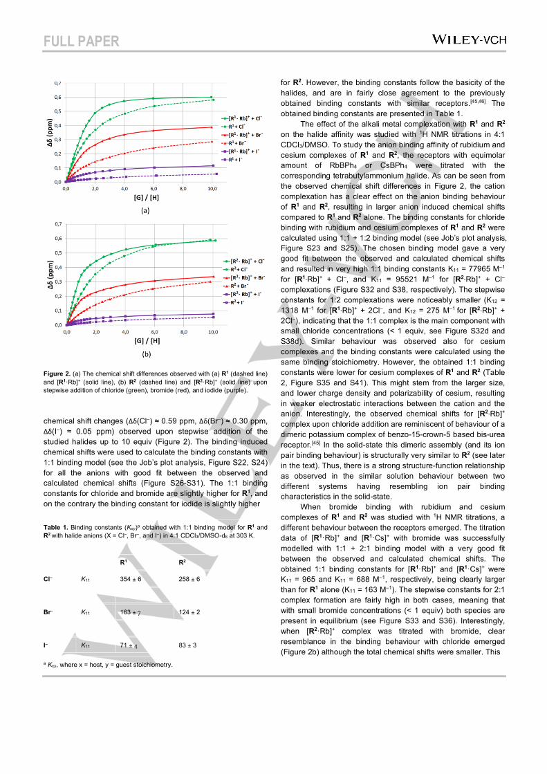
FULL PAPER
Figure 2. (a) The chemical shift differences observed with (a) R1 (dashed line) and [R1·Rb]+ (solid line), (b) R2 (dashed line) and [R2·Rb]+ (solid line) upon stepwise addition of chloride (green), bromide (red), and iodide (purple).
chemical shift changes (Δδ(Cl–) ≈ 0.59 ppm, Δδ(Br–) ≈ 0.30 ppm, Δδ(I–) ≈ 0.05 ppm) observed upon stepwise addition of the studied halides up to 10 equiv (Figure 2). The binding induced chemical shifts were used to calculate the binding constants with 1:1 binding model (see the Job’s plot analysis, Figure S22, S24) for all the anions with good fit between the observed and calculated chemical shifts (Figure S26-S31). The 1:1 binding constants for chloride and bromide are slightly higher for R1, and on the contrary the binding constant for iodide is slightly higher Table 1. Binding constants (Kxy)a obtained with 1:1 binding model for R1 and R2 with halide anions (X = Cl–, Br–, and I–) in 4:1 CDCl3/DMSO-d6 at 303 K.
R1 R2
Cl– K11 354 ± 6 258 ± 6
Br– K11 163 ± 7 124 ± 2
I– K11 71 ± 4 83 ± 3
a Kxy, where x = host, y = guest stoichiometry.
for R2. However, the binding constants follow the basicity of the halides, and are in fairly close agreement to the previously obtained binding constants with similar receptors.[45,46] The obtained binding constants are presented in Table 1. The effect of the alkali metal complexation with R1 and R2 on the halide affinity was studied with 1H NMR titrations in 4:1 CDCl3/DMSO. To study the anion binding affinity of rubidium and cesium complexes of R1 and R2, the receptors with equimolar amount of RbBPh4 or CsBPh4 were titrated with the corresponding tetrabutylammonium halide. As can be seen from the observed chemical shift differences in Figure 2, the cation complexation has a clear effect on the anion binding behaviour of R1 and R2, resulting in larger anion induced chemical shifts compared to R1 and R2 alone. The binding constants for chloride binding with rubidium and cesium complexes of R1 and R2 were calculated using 1:1 + 1:2 binding model (see Job’s plot analysis, Figure S23 and S25). The chosen binding model gave a very good fit between the observed and calculated chemical shifts and resulted in very high 1:1 binding constants K11 = 77965 M–1 for [R1·Rb]+ + Cl–, and K11 = 95521 M–1 for [R2·Rb]+ + Cl– complexations (Figure S32 and S38, respectively). The stepwise constants for 1:2 complexations were noticeably smaller (K12 = 1318 M–1 for [R1·Rb]+ + 2Cl–, and K12 = 275 M–1 for [R2·Rb]+ + 2Cl–), indicating that the 1:1 complex is the main component with small chloride concentrations (< 1 equiv, see Figure S32d and S38d). Similar behaviour was observed also for cesium complexes and the binding constants were calculated using the same binding stoichiometry. However, the obtained 1:1 binding constants were lower for cesium complexes of R1 and R2 (Table 2, Figure S35 and S41). This might stem from the larger size, and lower charge density and polarizability of cesium, resulting in weaker electrostatic interactions between the cation and the anion. Interestingly, the observed chemical shifts for [R2·Rb]+ complex upon chloride addition are reminiscent of behaviour of a dimeric potassium complex of benzo-15-crown-5 based bis-urea receptor.[45] In the solid-state this dimeric assembly (and its ion pair binding behaviour) is structurally very similar to R2 (see later in the text). Thus, there is a strong structure-function relationship as observed in the similar solution behaviour between two different systems having resembling ion pair binding characteristics in the solid-state. When bromide binding with rubidium and cesium complexes of R1 and R2 was studied with 1H NMR titrations, a different behaviour between the receptors emerged. The titration data of [R1·Rb]+ and [R1·Cs]+ with bromide was successfully modelled with 1:1 + 2:1 binding model with a very good fit between the observed and calculated chemical shifts. The obtained 1:1 binding constants for [R1·Rb]+ and [R1·Cs]+ were K11 = 965 and K11 = 688 M–1, respectively, being clearly larger than for R1 alone (K11 = 163 M–1). The stepwise constants for 2:1 complex formation are fairly high in both cases, meaning that with small bromide concentrations (< 1 equiv) both species are present in equilibrium (see Figure S33 and S36). Interestingly, when [R2·Rb]+ complex was titrated with bromide, clear resemblance in the binding behaviour with chloride emerged (Figure 2b) although the total chemical shifts were smaller. This
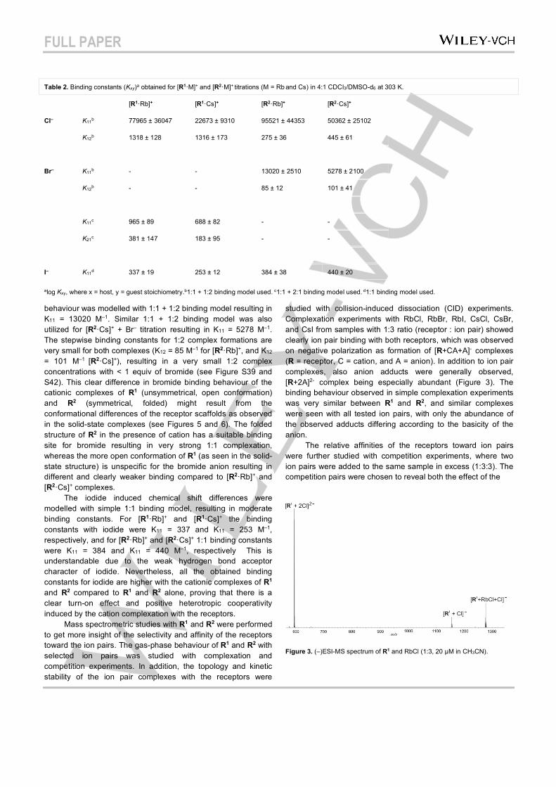
FULL PAPER
Table 2. Binding constants (Kxy)a obtained for [R1·M]+ and [R2·M]+ titrations (M = Rb and Cs) in 4:1 CDCl3/DMSO-d6 at 303 K.
[R1·Rb]+ [R1·Cs]+ [R2·Rb]+ [R2·Cs]+
Cl– K11b 77965 ± 36047 22673 ± 9310 95521 ± 44353 50362 ± 25102
K12b 1318 ± 128 1316 ± 173 275 ± 36 445 ± 61
Br– K11b - - 13020 ± 2510 5278 ± 2100
K12b - - 85 ± 12 101 ± 41
K11c 965 ± 89 688 ± 82 - -
K21c 381 ± 147 183 ± 95 - -
I– K11d 337 ± 19 253 ± 12 384 ± 38 440 ± 20
alog Kxy, where x = host, y = guest stoichiometry.b1:1 + 1:2 binding model used. c1:1 + 2:1 binding model used. d1:1 binding model used.
behaviour was modelled with 1:1 + 1:2 binding model resulting in K11 = 13020 M–1. Similar 1:1 + 1:2 binding model was also utilized for [R2·Cs]+ + Br– titration resulting in K11 = 5278 M–1. The stepwise binding constants for 1:2 complex formations are very small for both complexes (K12 = 85 M–1 for [R2·Rb]+, and K12 = 101 M–1 [R2·Cs]+), resulting in a very small 1:2 complex concentrations with < 1 equiv of bromide (see Figure S39 and S42). This clear difference in bromide binding behaviour of the cationic complexes of R1 (unsymmetrical, open conformation) and R2 (symmetrical, folded) might result from the conformational differences of the receptor scaffolds as observed in the solid-state complexes (see Figures 5 and 6). The folded structure of R2 in the presence of cation has a suitable binding site for bromide resulting in very strong 1:1 complexation, whereas the more open conformation of R1 (as seen in the solid-state structure) is unspecific for the bromide anion resulting in different and clearly weaker binding compared to [R2·Rb]+ and [R2·Cs]+ complexes. The iodide induced chemical shift differences were modelled with simple 1:1 binding model, resulting in moderate binding constants. For [R1·Rb]+ and [R1·Cs]+ the binding constants with iodide were K11 = 337 and K11 = 253 M–1, respectively, and for [R2·Rb]+ and [R2·Cs]+ 1:1 binding constants were K11 = 384 and K11 = 440 M–1, respectively This is understandable due to the weak hydrogen bond acceptor character of iodide. Nevertheless, all the obtained binding constants for iodide are higher with the cationic complexes of R1 and R2 compared to R1 and R2 alone, proving that there is a clear turn-on effect and positive heterotropic cooperativity induced by the cation complexation with the receptors. Mass spectrometric studies with R1 and R2 were performed to get more insight of the selectivity and affinity of the receptors toward the ion pairs. The gas-phase behaviour of R1 and R2 with selected ion pairs was studied with complexation and competition experiments. In addition, the topology and kinetic stability of the ion pair complexes with the receptors were
studied with collision-induced dissociation (CID) experiments. Complexation experiments with RbCl, RbBr, RbI, CsCl, CsBr, and CsI from samples with 1:3 ratio (receptor : ion pair) showed clearly ion pair binding with both receptors, which was observed on negative polarization as formation of [R+CA+A]- complexes (R = receptor, C = cation, and A = anion). In addition to ion pair complexes, also anion adducts were generally observed, [R+2A]2- complex being especially abundant (Figure 3). The binding behaviour observed in simple complexation experiments was very similar between R1 and R2, and similar complexes were seen with all tested ion pairs, with only the abundance of the observed adducts differing according to the basicity of the anion. The relative affinities of the receptors toward ion pairs were further studied with competition experiments, where two ion pairs were added to the same sample in excess (1:3:3). The competition pairs were chosen to reveal both the effect of the
Figure 3. (–)ESI-MS spectrum of R1 and RbCl (1:3, 20 µM in CH3CN).
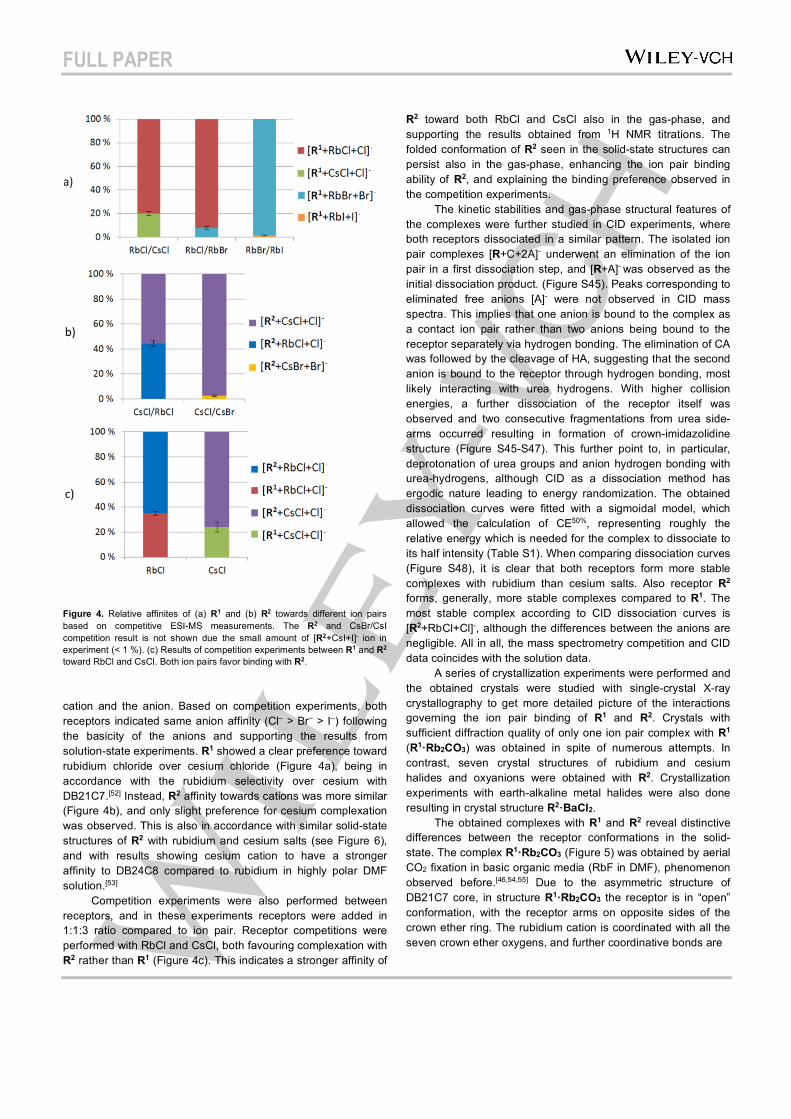
FULL PAPER
Figure 4. Relative affinites of (a) R1 and (b) R2 towards different ion pairs based on competitive ESI-MS measurements. The R2 and CsBr/CsI competition result is not shown due the small amount of [R2+CsI+I]- ion in experiment (< 1 %). (c) Results of competition experiments between R1 and R2 toward RbCl and CsCl. Both ion pairs favor binding with R2.
cation and the anion. Based on competition experiments, both receptors indicated same anion affinity (Cl– > Br– > I–) following the basicity of the anions and supporting the results from solution-state experiments. R1 showed a clear preference toward rubidium chloride over cesium chloride (Figure 4a), being in accordance with the rubidium selectivity over cesium with DB21C7.[52] Instead, R2 affinity towards cations was more similar (Figure 4b), and only slight preference for cesium complexation was observed. This is also in accordance with similar solid-state structures of R2 with rubidium and cesium salts (see Figure 6), and with results showing cesium cation to have a stronger affinity to DB24C8 compared to rubidium in highly polar DMF solution.[53] Competition experiments were also performed between receptors, and in these experiments receptors were added in 1:1:3 ratio compared to ion pair. Receptor competitions were performed with RbCl and CsCl, both favouring complexation with R2 rather than R1 (Figure 4c). This indicates a stronger affinity of
R2 toward both RbCl and CsCl also in the gas-phase, and supporting the results obtained from 1H NMR titrations. The folded conformation of R2 seen in the solid-state structures can persist also in the gas-phase, enhancing the ion pair binding ability of R2, and explaining the binding preference observed in the competition experiments. The kinetic stabilities and gas-phase structural features of the complexes were further studied in CID experiments, where both receptors dissociated in a similar pattern. The isolated ion pair complexes [R+C+2A]- underwent an elimination of the ion pair in a first dissociation step, and [R+A]- was observed as the initial dissociation product. (Figure S45). Peaks corresponding to eliminated free anions [A]- were not observed in CID mass spectra. This implies that one anion is bound to the complex as a contact ion pair rather than two anions being bound to the receptor separately via hydrogen bonding. The elimination of CA was followed by the cleavage of HA, suggesting that the second anion is bound to the receptor through hydrogen bonding, most likely interacting with urea hydrogens. With higher collision energies, a further dissociation of the receptor itself was observed and two consecutive fragmentations from urea side-arms occurred resulting in formation of crown-imidazolidine structure (Figure S45-S47). This further point to, in particular, deprotonation of urea groups and anion hydrogen bonding with urea-hydrogens, although CID as a dissociation method has ergodic nature leading to energy randomization. The obtained dissociation curves were fitted with a sigmoidal model, which allowed the calculation of CE50%, representing roughly the relative energy which is needed for the complex to dissociate to its half intensity (Table S1). When comparing dissociation curves (Figure S48), it is clear that both receptors form more stable complexes with rubidium than cesium salts. Also receptor R2 forms, generally, more stable complexes compared to R1. The most stable complex according to CID dissociation curves is [R2+RbCl+Cl]-, although the differences between the anions are negligible. All in all, the mass spectrometry competition and CID data coincides with the solution data. A series of crystallization experiments were performed and the obtained crystals were studied with single-crystal X-ray crystallography to get more detailed picture of the interactions governing the ion pair binding of R1 and R2. Crystals with sufficient diffraction quality of only one ion pair complex with R1 (R1·Rb2CO3) was obtained in spite of numerous attempts. In contrast, seven crystal structures of rubidium and cesium halides and oxyanions were obtained with R2. Crystallization experiments with earth-alkaline metal halides were also done resulting in crystal structure R2·BaCl2. The obtained complexes with R1 and R2 reveal distinctive differences between the receptor conformations in the solid-state. The complex R1·Rb2CO3 (Figure 5) was obtained by aerial CO2 fixation in basic organic media (RbF in DMF), phenomenon observed before.[46,54,55] Due to the asymmetric structure of DB21C7 core, in structure R1·Rb2CO3 the receptor is in “open” conformation, with the receptor arms on opposite sides of the crown ether ring. The rubidium cation is coordinated with all the seven crown ether oxygens, and further coordinative bonds are
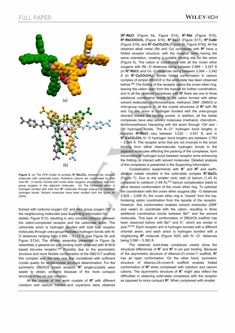
FULL PAPER
Figure 5. (a) The CPK model of complex R1·Rb2CO3, showing two receptor molecules with carbonate anion. Rubidium cations are coordinated by total nine Rb···O bonds, formed with crown ether oxygens, and carbonyl and nitro group oxygens in the adjacent molecules. (b) The carbonate anion is hydrogen bonded with total four R1 molecules through urea-group mediated hydrogen bonds. Solvent molecules have been omitted from the figure for clarity.
formed with carbonyl oxygen O2’ and nitro group oxygen O8’’ in the neighbouring molecules (see Supporting Information for details, Figure S13), resulting in very complex interplay between the cation-complexed receptor and the carbonate anion. The carbonate anion is hydrogen bonded with total four receptor molecules through urea-group mediated hydrogen bonds with N-O distances ranging from 2.854 – 3.112 Å (see Figure 5b and Figure S13d). The dimeric assembly presented in Figure 5a resembles a general ion pair binding motif observed with B18C6 based bis-urea receptor.[46] Possibly due to the asymmetric structure and more flexible conformation of the DB21C7 scaffold, this complex was the only one that crystallized with sufficient crystal quality for single-crystal structure determination. For the symmetric DB24C8 based receptor R2 single-crystals were easier to obtain, probably because of the more compact structure of the ion pair complex. In the course of this work crystals of R2 with different rubidium and cesium halides and oxyanions were obtained
[R2·RbCl (Figure 6a, Figure S14), R2·RbI (Figure S15), R2·RbCOOCH3 (Figure S16), R2·CsCl (Figure S17), R2·CsBr (Figure S18), and R2·CsCO3CH3 (Figure 6b, Figure S19)]. All the obtained alkali metal (Rb and Cs) complexes with R2 have a folded receptor structure, with the receptor arms having the same orientation, creating a suitable binding site for the anion (Figure 6). The cation is coordinated with all the crown ether oxygens with Rb···O distances being between 2.889 – 3.247 Å (in R2·RbCl) and Cs···O distances being between 3.004 – 3.342 Å (in R2·CsCO3CH3). Similar folded conformation in cesium complex of simple DB24C8 in the solid-state has been observed before.[56] The folding of the receptor opens the crown ether ring, leaving the cation open from the topside for further coordination, and in all the obtained complexes with R2 there are one to three additional coordinative bonds to the cation formed with either solvent molecules (dichloromethane, methanol, DMF, DMSO) or nitro-group oxygens. In all the crystal structures of R2 with Rb and Cs, the anion is hydrogen bonded with the urea-groups directed toward the binding pocket. In addition, all the halide complexes have also solvent molecules (methanol, chloroform, dichloromethane) interacting with the anion through -OH and -CH hydrogen bonds. The N···Cl– hydrogen bond lengths in complex R2·RbCl vary between 3.220 – 3.551 Å, and in R2·CsCO3CH3 N···O hydrogen bond lengths are between 2.763 – 2.924 Å. The receptor arms that are not involved in the anion binding form either intermolecular hydrogen bonds to the adjacent molecules affecting the packing of the complexes, form intramolecular hydrogen bond between receptor arms enhancing the folding, or interact with solvent molecules. Detailed analysis of the complexes is presented in the Supporting Information. Crystallization experiments with R1 and R2 and earth-alkaline metals resulted in the solid-state complex R2·BaCl2
(Figure 7). Due to the smaller ionic radii of barium (1.40 Å) compared to rubidium (1.68 Å),[57] barium complexation leads to more twisted conformation of the crown ether ring. To optimize the coordination with the crown ether oxygens (Ba···O distances 2.829 – 3.095 Å), the crown ether ring is “closed” from the top, hindering cation coordination from the topside of the receptor. However, this conformation enables solvent molecules (DMF and water) to coordinate with the cation, resulting in three additional coordinative bonds between Ba2+ and the solvent molecules. This type of conformation of DB24C8 scaffold has been observed before with Ba2+ and K+, which are similar in size.[58,59] Each receptor arm is hydrogen bonded with a different chloride anion, and each anion is hydrogen bonded with a neighboring R2 molecule (Figure S20) with N···Cl– distances being 3.090 – 3.392 Å. The obtained solid-state complexes clearly show the structural differences of R1 and R2 in ion pair binding. Because of the asymmetric structure of dibenzo-21-crown-7 scaffold, R1 has an open conformation. On the other hand, symmetric structure of dibenzo-24-crown-8 scaffold enables folded conformation of R2 when complexed with rubidium and cesium cations. The asymmetric structure of R1 might also reflect the difficulties in obtaining solid-state complexes with the receptor as opposed to more compact R2. When complexed with smaller
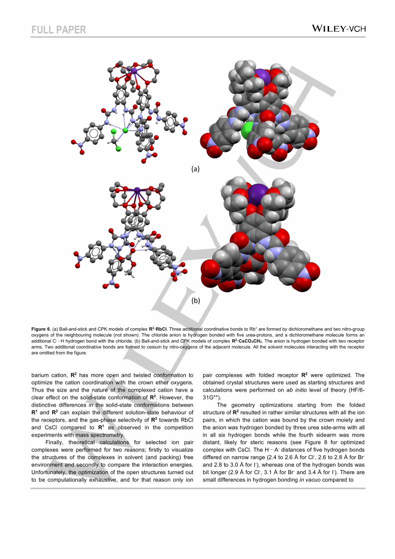
FULL PAPER
Figure 6. (a) Ball-and-stick and CPK models of complex R2·RbCl. Three additional coordinative bonds to Rb+ are formed by dichloromethane and two nitro-group oxygens of the neighbouring molecule (not shown). The chloride anion is hydrogen bonded with five urea-protons, and a dichloromethane molecule forms an additional C···H hydrogen bond with the chloride. (b) Ball-and-stick and CPK models of complex R2·CsCO3CH3. The anion is hydrogen bonded with two receptor arms. Two additional coordinative bonds are formed to cesium by nitro-oxygens of the adjacent molecule. All the solvent molecules interacting with the receptor are omitted from the figure.
barium cation, R2 has more open and twisted conformation to optimize the cation coordination with the crown ether oxygens. Thus the size and the nature of the complexed cation have a clear effect on the solid-state conformation of R2. However, the distinctive differences in the solid-state conformations between R1 and R2 can explain the different solution-state behaviour of the receptors, and the gas-phase selectivity of R2 towards RbCl and CsCl compared to R1 as observed in the competition experiments with mass spectrometry. Finally, theoretical calculations for selected ion pair complexes were performed for two reasons; firstly to visualize the structures of the complexes in solvent (and packing) free environment and secondly to compare the interaction energies. Unfortunately, the optimization of the open structures turned out to be computationally exhaustive, and for that reason only ion
pair complexes with folded receptor R2 were optimized. The obtained crystal structures were used as starting structures and calculations were performed on ab initio level of theory (HF/6-31G**). The geometry optimizations starting from the folded structure of R2 resulted in rather similar structures with all the ion pairs, in which the cation was bound by the crown moiety and the anion was hydrogen bonded by three urea side-arms with all in all six hydrogen bonds while the fourth sidearm was more distant, likely for steric reasons (see Figure 8 for optimized complex with CsCl. The H···A- distances of five hydrogen bonds differed on narrow range (2.4 to 2.6 Å for Cl-, 2.6 to 2.8 Å for Br- and 2.8 to 3.0 Å for I-), whereas one of the hydrogen bonds was bit longer (2.9 Å for Cl-, 3.1 Å for Br- and 3.4 Å for I-). There are small differences in hydrogen bonding in vacuo compared to
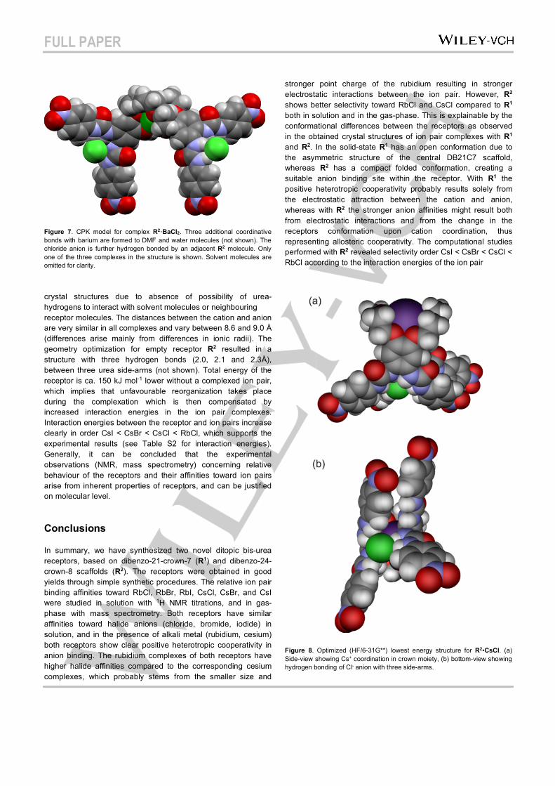
FULL PAPER
Figure 7. CPK model for complex R2·BaCl2. Three additional coordinative bonds with barium are formed to DMF and water molecules (not shown). The chloride anion is further hydrogen bonded by an adjacent R2 molecule. Only one of the three complexes in the structure is shown. Solvent molecules are omitted for clarity.
crystal structures due to absence of possibility of urea-hydrogens to interact with solvent molecules or neighbouring receptor molecules. The distances between the cation and anion are very similar in all complexes and vary between 8.6 and 9.0 Å (differences arise mainly from differences in ionic radii). The geometry optimization for empty receptor R2 resulted in a structure with three hydrogen bonds (2.0, 2.1 and 2.3Å), between three urea side-arms (not shown). Total energy of the receptor is ca. 150 kJ mol-1 lower without a complexed ion pair, which implies that unfavourable reorganization takes place during the complexation which is then compensated by increased interaction energies in the ion pair complexes. Interaction energies between the receptor and ion pairs increase clearly in order CsI < CsBr < CsCl < RbCl, which supports the experimental results (see Table S2 for interaction energies). Generally, it can be concluded that the experimental observations (NMR, mass spectrometry) concerning relative behaviour of the receptors and their affinities toward ion pairs arise from inherent properties of receptors, and can be justified on molecular level.
Conclusions
In summary, we have synthesized two novel ditopic bis-urea receptors, based on dibenzo-21-crown-7 (R1) and dibenzo-24-crown-8 scaffolds (R2). The receptors were obtained in good yields through simple synthetic procedures. The relative ion pair binding affinities toward RbCl, RbBr, RbI, CsCl, CsBr, and CsI were studied in solution with 1H NMR titrations, and in gas-phase with mass spectrometry. Both receptors have similar affinities toward halide anions (chloride, bromide, iodide) in solution, and in the presence of alkali metal (rubidium, cesium) both receptors show clear positive heterotropic cooperativity in anion binding. The rubidium complexes of both receptors have higher halide affinities compared to the corresponding cesium complexes, which probably stems from the smaller size and
stronger point charge of the rubidium resulting in stronger electrostatic interactions between the ion pair. However, R2 shows better selectivity toward RbCl and CsCl compared to R1 both in solution and in the gas-phase. This is explainable by the conformational differences between the receptors as observed in the obtained crystal structures of ion pair complexes with R1 and R2. In the solid-state R1 has an open conformation due to the asymmetric structure of the central DB21C7 scaffold, whereas R2 has a compact folded conformation, creating a suitable anion binding site within the receptor. With R1 the positive heterotropic cooperativity probably results solely from the electrostatic attraction between the cation and anion, whereas with R2 the stronger anion affinities might result both from electrostatic interactions and from the change in the receptors conformation upon cation coordination, thus representing allosteric cooperativity. The computational studies performed with R2 revealed selectivity order CsI < CsBr < CsCl < RbCl according to the interaction energies of the ion pair
Figure 8. Optimized (HF/6-31G**) lowest energy structure for R2•CsCl. (a) Side-view showing Cs+ coordination in crown moiety, (b) bottom-view showing hydrogen bonding of Cl- anion with three side-arms.
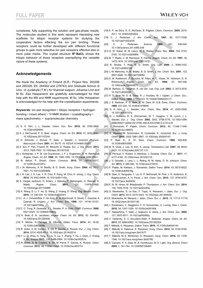
FULL PAPER
complexes, fully supporting the solution and gas-phase results. The molecules studied in this work represent interesting new scaffolds for ditopic receptor systems for studying the cooperative factors affecting the ion pair binding. These receptors could be further developed with different functional groups to gain more selective ion pair receptors effective also in more polar media. The crystal structure R2·BaCl2 shows the tritopic behavior of these receptors exemplifying the versatile nature of these systems.
Acknowledgements
We thank the Academy of Finland (K.R.: Project Nos. 263256 and 265328, EK: 284562 and 278743) and Graduate School of Univ. of Jyväskylä (T.M.) for financial support. Johanna Lind and M. Sc. Esa Haapaniemi are gratefully acknowledged for their help with the MS and NMR experiments. M Sc. Ville Holopainen is acknowledged for his help with the crystallization experiments.
Keywords: ion pair recognition • ditopic receptors • hydrogen bonding • crown ethers • 1H NMR titration • crystallography • mass spectrometry • supramolecular chemistry
[1] S. K. Kim, J. L. Sessler, Chem. Soc. Rev. 2010, 39, 3784-3809. 10.1039/c002694h
[2] A. J. McConnell, P. D. Beer, Angew. Chem. Int. Ed. 2012, 51, 5052-5061. 10.1002/anie.201107244
[3] G. Kirkovits, J. Shriver, P. Gale, J. Sessler, J. Inclusion Phenom. Macrocyclic Chem. 2001, 41, 69-75. 10.1023/A:1014468126351
[4] Z. Sun, F. Pan, Triyanti, M. Albrecht, G. Raabe, Eur. J. Org. Chem. 2013, 2013, 7922-7932. 10.1002/ejoc.201301032
[5] J. Scheerder, J. P. M. van Duynhoven, J. F. J. Engbersen, D. N. Reinhoudt, Angew. Chem. Int. Ed. 1996, 35, 1090-1093. 10.1002/anie.199610901
[6] B. Akhuli, P. Ghosh, Chem. Commun. 2015, 51, 16514-16517. 10.1039/c5cc07291c
[7] J. M. Mahoney, A. M. Beatty, B. D. Smith, Inorg. Chem. 2004, 43, 7617-7621. 10.1021/ic049066b
[8] J. H. Lee, J. H. Lee, Y. R. Choi, P. Kang, M. Choi, K. Jeong, J. Org. Chem. 2014, 79, 6403-6409. 10.1021/jo501145z
[9] A. Vargas Jentzsch, D. Emery, J. Mareda, P. Metrangolo, G. Resnati, S. Matile, Angew. Chem. 2011, 123, 11879-11882. 10.1002/ange.201104966
[10] X. Wang, S. Li, Y. Ao, Q. Wang, Z. Huang, D. Wang, Org. Biomol. Chem. 2016, 14, 330-334. 10.1039/c5ob02291f
[11] L. A. J. Chrisstoffels, F. de Jong, D. N. Reinhoudt, S. Sivelli, L. Gazzola, A. Casnati, R. Ungaro, J. Am. Chem. Soc. 1999, 121, 10142-10151. 10.1021/ja991588g
[12] C. C. Tong, R. Quesada, J. L. Sessler, P. A. Gale, Chem. Commun. 2008, 6321-6323. 10.1039/b814988g
[13] K. Brak, E. N. Jacobsen, Angew. Chem. Int. Ed. 2013, 52, 534-561. 10.1002/anie.201205449
[14] P. Molina, A. Tarraga, M. Alfonso, Dalton Trans. 2014, 43, 18-29. 10.1039/c3dt52459k
[15] R. Gotor, A. M. Costero, S. Gil, P. Gaviña, K. Rurack, Eur. J. Org. Chem. 2014, 2014, 4005-4013. 10.1002/ejoc.201402214
[16] Y. Li, Q. Zhao, H. Yang, S. Liu, X. Liu, Y. Zhang, T. Hu, J. Chen, Z. Chang, X. Bu, Analyst 2013, 138, 5486-5494. 10.1039/c3an00351e
[17] R. Gotor, A. M. Costero, S. Gil, M. Parra, P. Gavina, K. Rurack, Chem. Commun. 2013, 49, 11056-11058. 10.1039/c3cc45377d
[18] A. P. de Silva, G. D. McClean, S. Pagliari, Chem. Commun. 2003, 2010-2011. 10.1039/b305262a
[19] C. J. Pedersen, J. Am. Chem. Soc. 1967, 89, 7017-7036. 10.1021/ja01002a035
[20] C. J. Pedersen, Science 1988, 241, 536-540. 10.1126/science.241.4865.536
[21] G. W. Gokel, W. M. Leevy, M. E. Weber, Chem. Rev. 2004, 104, 2723-2750. 10.1021/cr020080k
[22] M. T. Reetz, C. M. Niemeyer, K. Harms, Angew. Chem. Int. Ed. 1991, 30, 1472-1474. 10.1002/anie.199114721
[23] R. Shukla, T. Kida, B. D. Smith, Org. Lett. 2000, 2, 3099-3102. 10.1021/ol0063104
[24] J. M. Mahoney, A. M. Beatty, B. D. Smith, J. Am. Chem. Soc. 2001, 123, 5847-5848. 10.1021/ja0156082
[25] D. M. Rudkevich, Z. Brzozka, M. Palys, H. C. Visser, W. Verboom, D. N. Reinhoudt, Angew. Chem. Int. Ed. 1994, 33, 467-468. 10.1002/anie.199404671
[26] M. Barboiu, G. Vaughan, A. van der. Lee, Org. Lett. 2003, 5, 3073-3076. 10.1021/ol035096r
[27] P. D. Beer, M. G. B. Drew, R. J. Knubley, M. I. Ogden, J. Chem. Soc., Dalton Trans. 1995, 3117-3123. 10.1039/DT9950003117
[28] J. E. Redman, P. D. Beer, S. W. Dent, M. G.B. Drew, Chem. Commun. 1998, 231-232. 10.1039/A707680K
[29] S. K. Kim, J. L. Sessler, Acc. Chem. Res. 2014, 47, 2525-2536. 10.1021/ar500157a
[30] G. J. Kirkovits, R. S. Zimmerman, M. T. Huggins, V. M. Lynch, J. L. Sessler, Eur. J. Org. Chem. 2002, 2002, 3768-3778. 10.1002/1099-0690(200211)2002:22<3768::AID-EJOC3768>3.0.CO;2-R
[31] J. Romanski, P. Piątek, Chem. Commun. 2012, 48, 11346-11348. 10.1039/c2cc36607j
[32] G. Reeske, M. Schürmann, B. Costisella, K. Jurkschat, Eur. J. Inorg. Chem. 2005, 2005, 2881-2887. 10.1002/ejic.200500191
[33] P. Beer D., S. W. Dent, Chem. Commun. 1998, 825-826. 10.1039/A800356D
[34] M. K. Chae, J. Lee, N. Kim, K. Jeong, Tetrahedron Lett. 2007, 48, 6624-6627. 10.1016/j.tetlet.2007.07.115
[35] O. Perraud, V. Robert, A. Martinez, J. Dutasta, Chem. Eur. J. 2011, 17, 4177-4182. 10.1002/chem.201002116.
[36] J. V. Gavette, J. Lara, L. L. Reling, M. M. Haley, D. W. Johnson, Chem. Sci. 2013, 4, 585-590. 10.1039/c2sc21501b
[37] P. Piątek, M. Karbarz, J. Romański, Dalton Trans. 2014, 43, 8515-8522. 10.1039/c4dt00576g
[38] B. Qiao, A. Sengupta, Y. Liu, K. P. McDonald, M. Pink, J. R. Anderson, K. Raghavachari, A. H. Flood, J. Am. Chem. Soc. 2015, 137, 9746-9757. 10.1021/jacs.5b05839
[39] E. N. W. Howe, M. Bhadbhade, P. Thordarson, J. Am. Chem. Soc. 2014, 136, 7505-7516. 10.1021/ja503383e
[40] S. Moerkerke, S. Le Gac, F. Topić, K. Rissanen, I. Jabin, Eur. J. Org. Chem. 2013, 2013, 5315-5322. 10.1002/ejoc.201300639
[41] S. Moerkerke, M. Ménand, I. Jabin, Chem. Eur. J. 2010, 16, 11712-11719. 10.1002/chem.201001193
[42] J. Eckelmann, V. Saggiomo, F. D. Sonnichsen, U. Luning, New J. Chem. 2010, 34, 1247-1250. 10.1039/c0nj00160k
[43] T. Nabeshima, T. Saiki, J. Iwabuchi, S. Akine, J. Am. Chem. Soc. 2005, 127, 5507-5511. 10.1021/ja042882y
[44] V. Valderrey, E. C. Escudero-Adán, P. Ballester, Angew. Chem. Int. Ed. 2013, 52, 6898-6902. 10.1002/anie.201302524
[45] T. Mäkelä, K. Rissanen, Dalton Trans. 2016. 10.1039/c6dt00414h [46] T. Mäkelä, E. Kalenius, K. Rissanen, Inorg. Chem. 2015, 54, 9154-9165.
10.1021/acs.inorgchem.5b01577 [47] T. Mäkelä, M.-E. Minkkinen, K. Rissanen, Inorg. Chem. 2016, 55, 1339-
1346. 10.1021/acs.inorgchem.5b02780 [48] S. Camiolo, P. A. Gale, M. B. Hursthouse, M. E. Light, Org. Biomol. Chem.
2003, 1, 741-744. 10.1039/B210848H
FULL PAPER
[49] C. Frassineti, S. Ghelli, P. Gans, A. Sabatini, M. S. Moruzzi, A. Vacca, Anal. Biochem. 1995, 231, 374-382. 10.1006/abio.1995.9984
[50] P. Thordarson, Chem. Soc. Rev. 2011, 40, 1305-1323. 10.1039/c0cs00062k
[51] F. Ulatowski, K. Dąbrowa, T. Bałakier, J. Jurczak, J. Org. Chem. 2016, 81, 1746-1756. 10.1021/acs.joc.5b02909
[52] S. Katsuta, T. Kuwano, Y. Ito, Y. Takeda, J. Chem. Eng. Data 2005, 50, 1313-1318. 10.1021/je050045l
[53] C. M. Goff, M. A. Matchette, N. Shabestary, S. Khazaeli, Polyhedron 1996, 15, 3897-3903. 10.1016/0277-5387(96)00018-6
[54] R. Custelcean, L. H. Delmau, B. A. Moyer, J. L. Sessler, W. Cho, D. Gross, G. W. Bates, S. J. Brooks, M. E. Light, P. A. Gale, Angew. Chem. Int. Ed. 2005, 44, 2537-2542. 10.1002/anie.200462945
[55] I. Ravikumar, P. Ghosh, Chem. Commun. 2010, 46, 1082–1084. 10.1039/b915661e
[56] J. A. Rusanova, P. J. Squattrito, V. V. Ponomareva, K. V. Domasevitch, V. N. Kokozay, Z. Naturforsch. B Chem. Sci. 1999, 54, 1129-1132. 10.1515/znb-1999-0907
[57] O. Johnson, Inorg. Chem. 1973, 12, 780-785. 10.1021/ic50122a015 [58] S. Uwe, Ç Erk, E. Kleinpeter, Z. Krist. 2006, 221, 231-235.
10.1524/zkri.2006.221.3.231 [59] N. V. Kozhemyakina, J. Nuss, M. Jansen, Eur. J. Inorg. Chem. 2009,
2009, 3900-3903. 10.1002/ejic.200900542

FULL PAPER
FULL PAPER
Dibenzo-21-crown-7 and dibenzo-24-crown-8 bis-urea receptors R1 and R2, respectively, have been studied in solution, gas-phase, and solid-state for their ion pair recognition behavior. Both receptors show strong positive heterotropic cooperativity toward chloride when complexed with Rb+ or Cs+.
T. Mäkelä, A. Kiesilä, E. Kalenius & K. Rissanen*
Page No. – Page No.
Ion Pair Complexation with Dibenzo-21-Crown-7 and Dibenzo-24-Crown-8 bis-Urea Receptors
DEPARTMENT OF CHEMISTRY, UNIVERSITY OF JYVÄSKYLÄ
RESEARCH REPORT SERIES
1. Vuolle, Mikko: Electron
paramagnetic resonance and
molecular orbital study of radical
ions generated from
(2.2)metacyclophane, pyrene and
its hydrogenated compounds by
alkali metal reduction and by
thallium(III)trifluoroacetate
oxidation. (99 pp.) 1976
2. Pasanen, Kaija: Electron
paramagnetic resonance study of
cation radical generated from
various chlorinated biphenyls. (66
pp.) 1977
3. Carbon-13 Workshop, September
6-8, 1977. (91 pp.) 1977
4. Laihia, Katri: On the structure
determination of norbornane
polyols by NMR spectroscopy.
(111 pp.) 1979
5. Nyrönen, Timo: On the EPR,
ENDOR and visible absorption
spectra of some nitrogen
containing heterocyclic
compounds in liquid ammonia.
(76 pp.) 1978
6. Talvitie, Antti: Structure
determination of some
sesquiterpenoids by shift reagent
NMR. (54 pp.) 1979
7. Häkli, Harri: Structure analysis
and molecular dynamics of cyclic
compounds by shift reagent
NMR. (48 pp.) 1979
8. Pitkänen, Ilkka: Thermodynamics
of complexation of 1,2,4-triazole
with divalent manganese, cobalt,
nickel, copper, zinc, cadmium and
lead ions in aqueous sodium
perchlorate solutions. (89 pp.)
1980
9. Asunta, Tuula: Preparation and
characterization of new
organometallic compounds
synthesized by using metal
vapours. (91 pp.) 1980
10. Sattar, Mohammad Abdus:
Analyses of MCPA and its
metabolites in soil. (57 pp.) 1980
11. Bibliography 1980. (31 pp.) 1981
12. Knuuttila, Pekka: X-Ray
structural studies on some
divalent 3d metal compounds of
picolinic and isonicotinic acid
N-oxides. (77 pp.) 1981
13. Bibliography 1981. (33 pp.) 1982
14. 6th National NMR Symposium,
September 9-10, 1982, Abstracts.
(49 pp.) 1982
15. Bibliography 1982. (38 pp.) 1983
16. Knuuttila, Hilkka: X-Ray
structural studies on some Cu(II),
Co(II) and Ni(II) complexes with
nicotinic and isonicotinic acid
N-oxides. (54 pp.) 1983
17. Symposium on inorganic and
analytical chemistry May 18,
1984, Program and Abstracts.
(100 pp.) 1984
18. Knuutinen, Juha: On the
synthesis, structure verification
and gas chromatographic
determination of chlorinated
catechols and guaiacols occuring
in spent bleach liquors of kraft
pulp mill. (30 pp.) 1984
19. Bibliography 1983. (47 pp.) 1984
20. Pitkänen, Maija: Addition of
BrCl, B2 and Cl2 to methyl esters
of propenoic and 2-butenoic acid
derivatives and 13C NMR studies
on methyl esters of saturated
aliphatic mono- and
dichlorocarboxylic acids. (56 pp.)
1985
21. Bibliography 1984. (39 pp.) 1985
22. Salo, Esa: EPR, ENDOR and
TRIPLE spectroscopy of some
nitrogen heteroaromatics in liquid
ammonia. (111 pp.) 1985
DEPARTMENT OF CHEMISTRY, UNIVERSITY OF JYVÄSKYLÄ
RESEARCH REPORT SERIES
23. Humppi, Tarmo: Synthesis,
identification and analysis of
dimeric impurities of
chlorophenols. (39 pp.) 1985
24. Aho, Martti: The ion exchange
and adsorption properties of
sphagnum peat under acid
conditions. (90 pp.) 1985
25. Bibliography 1985 (61 pp.) 1986
26. Bibliography 1986. (23 pp.) 1987
27. Bibliography 1987. (26 pp.) 1988
28. Paasivirta, Jaakko (Ed.):
Structures of organic
environmental chemicals. (67 pp.)
1988
29. Paasivirta, Jaakko (Ed.):
Chemistry and ecology of
organo-element compounds. (93
pp.) 1989
30. Sinkkonen, Seija: Determination
of crude oil alkylated
dibenzothiophenes in
environment. (35 pp.) 1989
31. Kolehmainen, Erkki (Ed.): XII
National NMR Symposium
Program and Abstracts. (75 pp.)
1989
32. Kuokkanen, Tauno:
Chlorocymenes and
Chlorocymenenes: Persistent
chlorocompounds in spent bleach
liquors of kraft pulp mills. (40
pp.) 1989
33. Mäkelä, Reijo: ESR, ENDOR and
TRIPLE resonance study on
substituted 9,10-anthraquinone
radicals in solution. (35 pp.) 1990
34. Veijanen, Anja: An integrated
sensory and analytical method for
identification of off-flavour
compounds. (70 pp.) 1990
35. Kasa, Seppo: EPR, ENDOR and
TRIPLE resonance and molecular
orbital studies on a substitution
reaction of anthracene induced by
thallium(III) in two fluorinated
carboxylic acids. (114 pp.) 1990
36. Herve, Sirpa: Mussel incubation
method for monitoring
organochlorine compounds in
freshwater recipients of pulp and
paper industry. (145 pp.) 1991
37. Pohjola, Pekka: The electron
paramagnetic resonance method
for characterization of Finnish
peat types and iron (III)
complexes in the process of peat
decomposition. (77 pp.) 1991
38. Paasivirta, Jaakko (Ed.):
Organochlorines from pulp mills
and other sources. Research
methodology studies 1988-91.
(120 pp.) 1992
39. Veijanen, Anja (Ed.): VI National
Symposium on Mass
Spectrometry, May 13-15, 1992,
Abstracts. (55 pp.) 1992
40. Rissanen, Kari (Ed.): The 7.
National Symposium on Inorganic
and Analytical Chemistry, May
22, 1992, Abstracts and Program.
(153 pp.) 1992
41. Paasivirta, Jaakko (Ed.):
CEOEC'92, Second
Finnish-Russian Seminar:
Chemistry and Ecology of
Organo-Element Compounds. (93
pp.) 1992
42. Koistinen, Jaana: Persistent
polychloroaromatic compounds in
the environment:
structure-specific analyses. (50
pp.) 1993
43. Virkki, Liisa: Structural
characterization of chlorolignins
by spectroscopic and liquid
chromatographic methods and a
comparison with humic
substances. (62 pp.) 1993
44. Helenius, Vesa: Electronic and
vibrational excitations in some
DEPARTMENT OF CHEMISTRY, UNIVERSITY OF JYVÄSKYLÄ
RESEARCH REPORT SERIES
biologically relevant molecules.
(30 pp.) 1993
45. Leppä-aho, Jaakko: Thermal
behaviour, infrared spectra and
x-ray structures of some new rare
earth chromates(VI). (64 pp.)
1994
46. Kotila, Sirpa: Synthesis, structure
and thermal behavior of solid
copper(II) complexes of
2-amino-2- hydroxymethyl-1,3-pr
opanediol. (111 pp.) 1994
47. Mikkonen, Anneli: Retention of
molybdenum(VI), vanadium(V)
and tungsten(VI) by kaolin and
three Finnish mineral soils. (90
pp.) 1995
48. Suontamo, Reijo: Molecular
orbital studies of small molecules
containing sulfur and selenium.
(42 pp.) 1995
49. Hämäläinen, Jouni: Effect of fuel
composition on the conversion of
fuel-N to nitrogen oxides in the
combustion of small single
particles. (50 pp.) 1995
50. Nevalainen, Tapio:
Polychlorinated diphenyl ethers:
synthesis, NMR spectroscopy,
structural properties, and
estimated toxicity. (76 pp.) 1995
51. Aittola, Jussi-Pekka:
Organochloro compounds in the
stack emission. (35 pp.) 1995
52. Harju, Timo: Ultrafast polar
molecular photophysics of
(dibenzylmethine)borondifluoride
and 4-aminophthalimide in
solution. (61 pp.) 1995
53. Maatela, Paula: Determination of
organically bound chlorine in
industrial and environmental
samples. (83 pp.) 1995
54. Paasivirta, Jaakko (Ed.):
CEOEC'95, Third
Finnish-Russian Seminar:
Chemistry and Ecology of
Organo-Element Compounds.
(109 pp.) 1995
55. Huuskonen, Juhani: Synthesis and
structural studies of some
supramolecular compounds. (54
pp.) 1995
56. Palm, Helena: Fate of
chlorophenols and their
derivatives in sawmill soil and
pulp mill recipient environments.
(52 pp.) 1995
57. Rantio, Tiina:
Chlorohydrocarbons in pulp mill
effluents and their fate in the
environment. (89 pp.) 1997
58. Ratilainen, Jari: Covalent and
non-covalent interactions in
molecular recognition. (37 pp.)
1997
59. Kolehmainen, Erkki (Ed.): XIX
National NMR Symposium, June
4-6, 1997, Abstracts. (89 pp.)
1997
60. Matilainen, Rose: Development of
methods for fertilizer analysis by
inductively coupled plasma
atomic emission spectrometry. (41
pp.) 1997
61. Koistinen, Jari (Ed.): Spring
Meeting on the Division of
Synthetic Chemistry, May 15-16,
1997, Program and Abstracts. (36
pp.) 1997
62. Lappalainen, Kari: Monomeric
and cyclic bile acid derivatives:
syntheses, NMR spectroscopy and
molecular recognition properties.
(50 pp.) 1997
63. Laitinen, Eira: Molecular
dynamics of cyanine dyes and
phthalimides in solution:
picosecond laser studies. (62 pp.)
1997
64. Eloranta, Jussi: Experimental and
theoretical studies on some
DEPARTMENT OF CHEMISTRY, UNIVERSITY OF JYVÄSKYLÄ
RESEARCH REPORT SERIES
quinone and quinol radicals. (40
pp.) 1997
65. Oksanen, Jari: Spectroscopic
characterization of some
monomeric and aggregated
chlorophylls. (43 pp.) 1998
66. Häkkänen, Heikki: Development
of a method based on laser-
induced plasma spectrometry for
rapid spatial analysis of material
distributions in paper coatings.
(60 pp.) 1998
67. Virtapohja, Janne: Fate of
chelating agents used in the pulp
and paper industries. (58 pp.)
1998
68. Airola, Karri: X-ray structural
studies of supramolecular and
organic compounds. (39 pp.) 1998
69. Hyötyläinen, Juha: Transport of
lignin–type compounds in the
receiving waters of pulp mills. (40
pp.) 1999
70. Ristolainen, Matti: Analysis of the
organic material dissolved during
totally chlorine-free bleaching.
(40 pp.) 1999
71. Eklin, Tero: Development of
analytical procedures with
industrial samples for atomic
emission and atomic absorption
spectrometry. (43 pp.) 1999
72. Välisaari, Jouni: Hygiene
properties of resol-type phenolic
resin laminates. (129 pp.) 1999
73. Hu, Jiwei: Persistent
polyhalogenated diphenyl ethers:
model compounds syntheses,
characterization and molecular
orbital studies. (59 pp.) 1999
74. Malkavaara, Petteri: Chemometric
adaptations in wood processing
chemistry. (56 pp.) 2000
75. Kujala Elena, Laihia Katri,
Nieminen Kari (Eds.): NBC 2000,
Symposium on Nuclear,
Biological and Chemical Threats
in the 21st Century. (299 pp.) 2000
76. Rantalainen, Anna-Lea:
Semipermeable membrane
devices in monitoring persistent
organic pollutants in the
environment. (58 pp.) 2000
77. Lahtinen, Manu: In situ X-ray
powder diffraction studies of
Pt/C, CuCl/C and Cu2O/C
catalysts at elevated temperatures
in various reaction conditions. (92
pp.) 2000
78. Tamminen, Jari: Syntheses,
empirical and theoretical
characterization, and metal cation
complexation of bile acid-based
monomers and open/closed
dimers. (54 pp.) 2000
79. Vatanen, Virpi: Experimental
studies by EPR and theoretical
studies by DFT calculations of -
amino-9,10-anthraquinone radical
anions and cations in solution. (37
pp.) 2000
80. Kotilainen, Risto: Chemical
changes in wood during heating at
150-260 C. (57 pp.) 2000
81. Nissinen, Maija: X-ray structural
studies on weak, non-covalent
interactions in supramolecular
compounds. (69 pp.) 2001
82. Wegelius, Elina: X-ray structural
studies on self-assembled
hydrogen-bonded networks and
metallosupramolecular
complexes. (84 pp.) 2001
83. Paasivirta, Jaakko (Ed.):
CEOEC’2001, Fifth
Finnish-Russian Seminar:
Chemistry and Ecology of
Organo-Element Compounds.
(163 pp.) 2001
84. Kiljunen, Toni: Theoretical
studies on spectroscopy and
DEPARTMENT OF CHEMISTRY, UNIVERSITY OF JYVÄSKYLÄ
RESEARCH REPORT SERIES
atomic dynamics in rare gas
solids. (56 pp.) 2001
85. Du, Jin: Derivatives of dextran:
synthesis and applications in
oncology. (48 pp.) 2001
86. Koivisto, Jari: Structural analysis
of selected polychlorinated
persistent organic pollutants
(POPs) and related compounds.
(88 pp.) 2001
87. Feng, Zhinan: Alkaline pulping of
non-wood feedstocks and
characterization of black liquors.
(54 pp.) 2001
88. Halonen, Markku: Lahon
havupuun käyttö
sulfaattiprosessin raaka-aineena
sekä havupuun lahontorjunta. (90
pp.) 2002
89. Falábu, Dezsö: Synthesis,
conformational analysis and
complexation studies of
resorcarene derivatives. (212 pp.)
2001
90. Lehtovuori, Pekka: EMR
spectroscopic studies on radicals
of ubiquinones Q-n, vitamin K3
and vitamine E in liquid solution.
(40 pp.) 2002
91. Perkkalainen, Paula:
Polymorphism of sugar alcohols
and effect of grinding on thermal
behavior on binary sugar alcohol
mixtures. (53 pp.) 2002
92. Ihalainen, Janne: Spectroscopic
studies on light-harvesting
complexes of green plants and
purple bacteria. (42 pp.) 2002
93. Kunttu, Henrik, Kiljunen, Toni
(Eds.): 4th International
Conference on Low Temperature
Chemistry. (159 pp.) 2002
94. Väisänen, Ari: Development of
methods for toxic element
analysis in samples with
environmental concern by ICP-
AES and ETAAS. (54 pp.) 2002
95. Luostarinen, Minna: Synthesis
and characterisation of novel
resorcarene derivatives. (200 pp.)
2002
96. Louhelainen, Jarmo: Changes in
the chemical composition and
physical properties of wood and
nonwood black liquors during
heating. (68 pp.) 2003
97. Lahtinen, Tanja: Concave
hydrocarbon cyclophane π-
prismands. (65 pp.) 2003
98. Laihia, Katri (Ed.): NBC 2003,
Symposium on Nuclear,
Biological and Chemical Threats
– A Crisis Management
Challenge. (245 pp.) 2003
99. Oasmaa, Anja: Fuel oil quality
properties of wood-based
pyrolysis liquids. (32 pp.) 2003
100. Virtanen, Elina: Syntheses,
structural characterisation, and
cation/anion recognition
properties of nano-sized bile acid-
based host molecules and their
precursors. (123 pp.) 2003
101. Nättinen, Kalle: Synthesis and X-
ray structural studies of organic
and metallo-organic
supramolecular systems. (79 pp.)
2003
102. Lampiselkä, Jarkko:
Demonstraatio lukion kemian
opetuksessa. (285 pp.) 2003
103. Kallioinen, Jani: Photoinduced
dynamics of Ru(dcbpy)2(NCS)2 –
in solution and on nanocrystalline
titanium dioxide thin films. (47
pp.) 2004
104. Valkonen, Arto (Ed.): VII
Synthetic Chemistry Meeting and
XXVI Finnish NMR Symposium.
(103 pp.) 2004
DEPARTMENT OF CHEMISTRY, UNIVERSITY OF JYVÄSKYLÄ
RESEARCH REPORT SERIES
105. Vaskonen, Kari: Spectroscopic
studies on atoms and small
molecules isolated in low
temperature rare gas matrices. (65
pp.) 2004
106. Lehtovuori, Viivi: Ultrafast light
induced dissociation of
Ru(dcbpy)(CO)2I2 in solution. (49
pp.) 2004
107. Saarenketo, Pauli: Structural
studies of metal complexing
Schiff bases, Schiff base derived
N-glycosides and cyclophane π-
prismands. (95 pp.) 2004
108. Paasivirta, Jaakko (Ed.):
CEOEC’2004, Sixth
Finnish-Russian Seminar:
Chemistry and Ecology of
Organo-Element Compounds.
(147 pp.) 2004
109. Suontamo, Tuula: Development
of a test method for evaluating the
cleaning efficiency of hard-
surface cleaning agents. (96 pp.)
2004
110. Güneş, Minna: Studies of
thiocyanates of silver for
nonlinear optics. (48 pp.) 2004
111. Ropponen, Jarmo: Aliphatic
polyester dendrimers and
dendrons. (81 pp.) 2004
112. Vu, Mân Thi Hong: Alkaline
pulping and the subsequent
elemental chlorine-free bleaching
of bamboo (Bambusa procera).
(69 pp.) 2004
113. Mansikkamäki, Heidi: Self-
assembly of resorcinarenes. (77
pp.) 2006
114. Tuononen, Heikki M.: EPR
spectroscopic and quantum
chemical studies of some
inorganic main group radicals. (79
pp.) 2005
115. Kaski, Saara: Development of
methods and applications of laser-
induced plasma spectroscopy in
vacuum ultraviolet. (44 pp.) 2005
116. Mäkinen, Riika-Mari: Synthesis,
crystal structure and thermal
decomposition of certain metal
thiocyanates and organic
thiocyanates. (119 pp.) 2006
117. Ahokas, Jussi: Spectroscopic
studies of atoms and small
molecules isolated in rare gas
solids: photodissociation and
thermal reactions. (53 pp.) 2006
118. Busi, Sara: Synthesis,
characterization and thermal
properties of new quaternary
ammonium compounds: new
materials for electrolytes, ionic
liquids and complexation studies.
(102 pp.) 2006
119. Mäntykoski, Keijo: PCBs in
processes, products and
environment of paper mills using
wastepaper as their raw material.
(73 pp.) 2006
120. Laamanen, Pirkko-Leena:
Simultaneous determination of
industrially and environmentally
relevant aminopolycarboxylic and
hydroxycarboxylic acids by
capillary zone electrophoresis. (54
pp.) 2007
121. Salmela, Maria: Description of
oxygen-alkali delignification of
kraft pulp using analysis of
dissolved material. (71 pp.) 2007
122. Lehtovaara, Lauri: Theoretical
studies of atomic scale impurities
in superfluid 4He. (87 pp.) 2007
123. Rautiainen, J. Mikko: Quantum
chemical calculations of
structures, bonding, and
spectroscopic properties of some
sulphur and selenium iodine
cations. (71 pp.) 2007
124. Nummelin, Sami: Synthesis,
characterization, structural and
DEPARTMENT OF CHEMISTRY, UNIVERSITY OF JYVÄSKYLÄ
RESEARCH REPORT SERIES
retrostructural analysis of self-
assembling pore forming
dendrimers. (286 pp.) 2008
125. Sopo, Harri: Uranyl(VI) ion
complexes of some organic
aminobisphenolate ligands:
syntheses, structures and
extraction studies. (57 pp.) 2008
126. Valkonen, Arto: Structural
characteristics and properties of
substituted cholanoates and N-
substituted cholanamides. (80 pp.)
2008
127. Lähde, Anna: Production and
surface modification of
pharmaceutical nano- and
microparticles with the aerosol
flow reactor. (43 pp.) 2008
128. Beyeh, Ngong Kodiah:
Resorcinarenes and their
derivatives: synthesis,
characterization and complexation
in gas phase and in solution. (75
pp.) 2008
129. Välisaari, Jouni, Lundell, Jan
(Eds.): Kemian opetuksen päivät
2008: uusia oppimisympäristöjä ja
ongelmalähtöistä opetusta. (118
pp.) 2008
130. Myllyperkiö, Pasi: Ultrafast
electron transfer from potential
organic and metal containing solar
cell sensitizers. (69 pp.) 2009
131. Käkölä, Jaana: Fast
chromatographic methods for
determining aliphatic carboxylic
acids in black liquors. (82 pp.)
2009
132. Koivukorpi, Juha: Bile acid-arene
conjugates: from
photoswitchability to cancer cell
detection. (67 pp.) 2009
133. Tuuttila, Tero: Functional
dendritic polyester compounds:
synthesis and characterization of
small bifunctional dendrimers and
dyes. (74 pp.) 2009
134. Salorinne, Kirsi: Tetramethoxy
resorcinarene based cation and
anion receptors: synthesis,
characterization and binding
properties. (79 pp.) 2009
135. Rautiainen, Riikka: The use of
first-thinning Scots pine (Pinus
sylvestris) as fiber raw material
for the kraft pulp and paper
industry. (73 pp.) 2010
136. Ilander, Laura: Uranyl salophens:
synthesis and use as ditopic
receptors. (199 pp.) 2010
137. Kiviniemi, Tiina: Vibrational
dynamics of iodine molecule and
its complexes in solid krypton -
Towards coherent control of
bimolecular reactions? (73 pp.)
2010
138. Ikonen, Satu: Synthesis,
characterization and structural
properties of various covalent and
non-covalent bile acid derivatives
of N/O-heterocycles and their
precursors. (105 pp.) 2010
139. Siitonen, Anni: Spectroscopic
studies of semiconducting single-
walled carbon nanotubes. (56 pp.)
2010
140. Raatikainen, Kari: Synthesis and
structural studies of piperazine
cyclophanes – Supramolecular
systems through Halogen and
Hydrogen bonding and metal ion
coordination. (69 pp.) 2010
141. Leivo, Kimmo: Gelation and gel
properties of two- and three-
component Pyrene based low
molecular weight organogelators.
(116 pp.) 2011
142. Martiskainen, Jari: Electronic
energy transfer in light-harvesting
complexes isolated from Spinacia
oleracea and from three
DEPARTMENT OF CHEMISTRY, UNIVERSITY OF JYVÄSKYLÄ
RESEARCH REPORT SERIES
photosynthetic green bacteria
Chloroflexus aurantiacus,
Chlorobium tepidum, and
Prosthecochloris aestuarii. (55
pp.) 2011
143. Wichmann, Oula: Syntheses,
characterization and structural
properties of [O,N,O,X´]
aminobisphenolate metal
complexes. (101 pp.) 2011
144. Ilander, Aki: Development of
ultrasound-assisted digestion
methods for the determination of
toxic element concentrations in
ash samples by ICP-OES. (58 pp.)
2011
145. The Combined XII Spring
Meeting of the Division of
Synthetic Chemistry and XXXIII
Finnish NMR Symposium. Book
of Abstracts. (90 pp.) 2011
146. Valto, Piia: Development of fast
analysis methods for extractives
in papermaking process waters.
(73 pp.) 2011
147. Andersin, Jenni: Catalytic activity
of palladium-based nanostructures
in the conversion of simple
olefinic hydro- and
chlorohydrocarbons from first
principles. (78 pp.) 2011
148. Aumanen, Jukka: Photophysical
properties of dansylated
poly(propylene amine)
dendrimers. (55 pp.) 2011
149. Kärnä, Minna: Ether-
functionalized quaternary
ammonium ionic liquids –
synthesis, characterization and
physicochemical properties. (76
pp.) 2011
150. Jurček, Ondřej: Steroid conjugates
for applications in pharmacology
and biology. (57 pp.) 2011
151. Nauha, Elisa: Crystalline forms of
selected Agrochemical actives:
design and synthesis of cocrystals.
(77 pp.) 2012
152. Ahkola, Heidi: Passive sampling
in monitoring of nonylphenol
ethoxylates and nonylphenol in
aquatic environments. (92 pp.)
2012
153. Helttunen, Kaisa: Exploring the
self-assembly of resorcinarenes:
from molecular level interactions
to mesoscopic structures. (78 pp.)
2012
154. Linnanto, Juha: Light excitation
transfer in photosynthesis
revealed by quantum chemical
calculations and exiton theory.
(179 pp.) 2012
155. Roiko-Jokela, Veikko: Digital
imaging and infrared
measurements of soil adhesion
and cleanability of semihard and
hard surfaces. (122 pp.) 2012
156. Noponen, Virpi: Amides of bile
acids and biologically important
small molecules: properties and
applications. (85 pp.) 2012
157. Hulkko, Eero: Spectroscopic
signatures as a probe of structure
and dynamics in condensed-phase
systems – studies of iodine and
gold ranging from isolated
molecules to nanoclusters. (69
pp.) 2012
158. Lappi, Hanna: Production of
Hydrocarbon-rich biofuels from
extractives-derived materials. (95
pp.) 2012
159. Nykänen, Lauri: Computational
studies of Carbon chemistry on
transition metal surfaces. (76 pp.)
2012
160. Ahonen, Kari: Solid state studies
of pharmaceutically important
molecules and their derivatives.
(65 pp.) 2012
DEPARTMENT OF CHEMISTRY, UNIVERSITY OF JYVÄSKYLÄ
RESEARCH REPORT SERIES
161. Pakkanen, Hannu:
Characterization of organic
material dissolved during alkaline
pulping of wood and non-wood
feedstocks. (76 pp.) 2012
162. Moilanen, Jani: Theoretical and
experimental studies of some
main group compounds: from
closed shell interactions to singlet
diradicals and stable radicals. (80
pp.) 2012
163. Himanen, Jatta: Stereoselective
synthesis of Oligosaccharides by
De Novo Saccharide welding.
(133 pp.) 2012
164. Bunzen, Hana: Steroidal
derivatives of nitrogen containing
compounds as potential gelators.
(76 pp.) 2013
165. Seppälä, Petri: Structural diversity
of copper(II) amino alcohol
complexes. Syntheses, structural
and magnetic properties of
bidentate amino alcohol
copper(II) complexes. (67 pp.)
2013
166. Lindgren, Johan: Computational
investigations on rotational and
vibrational spectroscopies of
some diatomics in solid
environment. (77 pp.) 2013
167. Giri, Chandan: Sub-component
self-assembly of linear and non-
linear diamines and
diacylhydrazines, formulpyridine
and transition metal cations. (145
pp.) 2013
168. Riisiö, Antti: Synthesis,
Characterization and Properties of
Cu(II)-, Mo(VI)- and U(VI)
Complexes With
Diaminotetraphenolate Ligands.
(51 pp.) 2013
169. Kiljunen, Toni (Ed.): Chemistry
and Physics at Low Temperatures.
Book of Abstracts. (103 pp.) 2013
170. Hänninen, Mikko: Experimental
and Computational Studies of
Transition Metal Complexes with
Polydentate Amino- and
Aminophenolate Ligands:
Synthesis, Structure, Reactivity
and Magnetic Properties. (66 pp.)
2013
171. Antila, Liisa: Spectroscopic
studies of electron transfer
reactions at the photoactive
electrode of dye-sensitized solar
cells. (53 pp.) 2013
172. Kemppainen, Eeva: Mukaiyama-
Michael reactions with α-
substituted acroleins – a useful
tool for the synthesis of the
pectenotoxins and other natural
product targets. (190 pp.) 2013
173. Virtanen, Suvi: Structural Studies
of Dielectric Polymer
Nanocomposites. (49 pp.) 2013
174. Yliniemelä-Sipari, Sanna:
Understanding The Structural
Requirements for Optimal
Hydrogen Bond Catalyzed
Enolization – A Biomimetic
Approach.(160 pp.) 2013
175. Leskinen, Mikko V: Remote β-
functionalization of β’-keto esters.
(105 pp.) 2014
176. 12th European Conference on
Research in Chemistry Education
(ECRICE2014). Book of
Abstracts. (166 pp.) 2014
177. Peuronen, Anssi: N-
Monoalkylated DABCO-Based
N-Donors as Versatile Building
Blocks in Crystal Engineering and
Supramolecular Chemistry. (54
pp.) 2014
178. Perämäki, Siiri: Method
development for determination
and recovery of rare earth
elements from industrial fly ash.
(88 pp.) 2014
DEPARTMENT OF CHEMISTRY, UNIVERSITY OF JYVÄSKYLÄ
RESEARCH REPORT SERIES
179. Chernyshev, Alexander, N.:
Nitrogen-containing ligands and
their platinum(IV) and gold(III)
complexes: investigation and
basicity and nucleophilicity,
luminescence, and aurophilic
interactions. (64 pp.) 2014
180. Lehto, Joni: Advanced
Biorefinery Concepts Integrated
to Chemical Pulping. (142 pp.)
2015
181. Tero, Tiia-Riikka: Tetramethoxy
resorcinarenes as platforms for
fluorescent and halogen bonding
systems. (61 pp.) 2015
182. Löfman, Miika: Bile acid amides
as components of microcrystalline
organogels. (62 pp.) 2015
183. Selin, Jukka: Adsorption of
softwood-derived organic material
onto various fillers during
papermaking. (169 pp.) 2015
184. Piisola, Antti: Challenges in the
stereoselective synthesis of allylic
alcohols. (210 pp.) 2015
185. Bonakdarzadeh, Pia:
Supramolecular coordination
polyhedra based on achiral and
chiral pyridyl ligands: design,
preparation, and characterization.
(65 pp.) 2015
186. Vasko, Petra: Synthesis,
characterization, and reactivity of
heavier group 13 and 14
metallylenes and metalloid
clusters: small molecule
activation and more. (66 pp.)
2015
187. Topić, Filip: Structural Studies of
Nano-sized Supramolecular
Assemblies. (79 pp.) 2015
188. Mustalahti, Satu: Photodynamics
Studies of Ligand-Protected Gold
Nanoclusters by using Ultrafast
Transient Infrared Spectroscopy.
(58 pp.) 2015
189. Koivisto, Jaakko: Electronic and
vibrational spectroscopic studies
of gold-nanoclusters. (63 pp.)
2015
190. Suhonen, Aku: Solid state
conformational behavior and
interactions of series of aromatis
oligoamide foldamers. (68 pp.)
2016
191. Soikkeli, Ville:
Hydrometallurgical recovery and
leaching studies for selected
valuable metals from fly ash
samples by ultrasound-assisted
extraction followed by ICP-OES
determination. (107 pp.) 2016
192. XXXVIII Finnish NMR
Symposium. Book of Abstracts.
(51 pp.) 2016
193. Mäkelä, Toni: Ion Pair
Recognition by Ditopic Crown
Ether Based bis-Urea and Uranyl
Salophen Receptors. (75 pp.)
2016
194. Lindholm-Lehto, Petra:
Occurrence of pharmaceuticals in
municipal wastewater treatment
plants and receiving surface
waters in Central and Southern
Finland. (98 pp.) 2016
195. Härkönen, Ville: Computational
and Theoretical studies on Lattice
Thermal conductivity and
Thermal properties of Silicon
Clathrates. (89 pp.) 2016
196. Tuokko, Sakari: Understanding
selective reduction reactions with
heterogeneous Pd and Pt:
climbing out of the black box. (85
pp.) 2016
197. Nuora, Piia: Monitapaustutkimus
LUMA-Toimintaan liittyvissä
oppimisympäristöissä tapahtuvista
kemian oppimiskokemuksista.
(171 pp.) 2016
DEPARTMENT OF CHEMISTRY, UNIVERSITY OF JYVÄSKYLÄ
RESEARCH REPORT SERIES
198. Kumar, Hemanathan: Novel
Concepts on The Recovery of By-
Products from Alkaline Pulping.
(61 pp.) 2016
199. Arnedo-Sánchez, Leticia:
Lanthanide and Transition Metal
Complexes as Building Blocks for
Supramolecular Functional
Materials. (227 pp.) 2016
200. Gell, Lars: Theoretical
Investigations of Ligand Protected
Silver Nanoclusters. (134 pp.)
2016
201. Vaskuri, Juhani: Oppiennätyksistä
opetussuunnitelman perusteisiin -
lukion kemian kansallisen
opetussuunnitelman kehittyminen
Suomessa vuosina 1918-2016.
(314 pp.) 2017
202. Lundell Jan, Kiljunen Toni (Eds.):
22nd Horizons in Hydrogen Bond
Research. Book of Abstracts.
2017
203. Turunen, Lotta: Design and
construction of halogen-bonded
capsules and cages. (61 pp.) 2017
204. Hurmalainen, Juha: Experimental
and computational studies of
unconventional main group
compounds: stable radicals and
reactive intermediates. (88 pp.)
2017
205. Koivistoinen Juha: Non-linear
interactions of femtosecond laser
pulses with graphene: photo-
oxidation, imaging and
photodynamics. (68 pp.) 2017
206. Chen, Chengcong: Combustion
behavior of black liquors: droplet
swelling and influence of liquor
composition. (39 pp.) 2017
207. Mansikkamäki, Akseli:
Theoretical and Computational
Studies of Magnetic Anisotropy
and Exchange Coupling in
Molecular Systems. (190 p. +
included articles) 2018.
208. Tatikonda, Rajendhraprasad:
Multivalent N-donor ligands for
the construction of coordination
polymers and coordination
polymer gels. (62 pp.) 2018
209. Budhathoki, Roshan:
Beneficiation, desilication and
selective precipitation techniques
for phosphorus refining from
biomass derived fly ash. (64 pp.)
2018
210. Siitonen, Juha: Synthetic Studies
on 1-azabicyclo[5.3.0]decane
Alkaloids. (140 pp.) 2018
211. Ullah, Saleem: Advanced
Biorefinery Concepts Related to
Non-wood Feedstocks. (57 pp.)
2018
212. Ghalibaf, Maryam: Analytical
Pyrolysis of Wood and Non-
Wood Materials from Integrated
Biorefinery Concepts. (106 pp.)
2018
DEPARTMENT OF CHEMISTRY, UNIVERSITY OF JYVÄSKYLÄ
DISSERTATIONS PUBLISHED IN THE JYU DISSERTATIONS RESEARCH SERIES
1. Bulatov, Evgeny: Synthetic and
structural studies of covalent and
non-covalent interactions of
ligands and metal center in
platinum(II) complexes
containing 2,2′-dipyridylamine or
oxime ligands. (58 pp.) 2019.
JYU Dissertations 70.
2. Annala, Riia: Conformational
Properties and Anion Complexes
of Aromatic Oligoamide
Foldamers. (80 pp.) 2019.
JYU Dissertations 84.
3. Isoaho, Jukka Pekka: Dithionite
Bleaching of Thermomechanical
Pulp - Chemistry and Optimal
Conditions. (73 pp.) 2019.
JYU Dissertations 85.
4. Nygrén, Enni: Recovery of
rubidium from power plant fly
ash. 2019.
JYU Dissertations.