design considerations for incorporating sodium iodide symporter reporter gene imaging into prostate...
TRANSCRIPT
HUMAN GENE THERAPY 18:306–316 (April 2007)© Mary Ann Liebert, Inc.DOI: 10.1089/hum.2006.131
Design Considerations for Incorporating Sodium Iodide Symporter Reporter Gene Imaging into
Prostate Cancer Gene Therapy Trials
FARZAN SIDDIQUI,1 KENNETH N. BARTON,1 HANS J. STRICKER,2 PHILLIP F. STEYN,3
SUSAN M. LARUE,3 KASTYTIS C. KARVELIS,4 RICHARD B. SPARKS,5 JAE HO KIM,1
STEPHEN L. BROWN,1 and SVEND O. FREYTAG1
ABSTRACT
This study was done to aid in the design of a phase I gene therapy trial in patients with prostate cancer. Wedetermined the dosimetric characteristics of our reporter gene system when coupled with intravenous ad-ministration of radioactive sodium pertechnetate (Na99mTcO4) and determined the feasibility of using humansodium iodide symporter (hNIS) as a reporter gene to study the dynamics of adenoviral transgene expressionin a large animal tumor. A replication-competent Ad5-yCD/mutTKSR39rep-hNIS adenovirus was injected intothe prostate gland of dogs for dosimetry purposes, and into a canine soft tissue sarcoma (STS) for imagingpurposes. After resection of the prostate, the amount of 99mTcO4
– sequestered in the prostate was determined,the radiation dose absorbed by the prostate and nontarget critical organs was calculated, and hNIS reportergene expression was imaged in the STS by single-photon emission computed tomography (SPECT). On thebasis of the findings from 25 dogs, the amount of 99mTcO4
– sequestered in the prostate ranged from 13 to 276�Ci. Using the highest value observed, absorbed radiation dose to critical organs was calculated and foundto be below U.S. Food and Drug Administration limits for diagnostic imaging. Also, 99mTcO4
� uptake wasreadily detected by SPECT and found to persist in vivo for at least 4 days. On the basis of our dosimetry cal-culations, up to five imaging procedures can be safely performed in humans after intraprostatic injection ofthe Ad5-yCD/mutTKSR39rep-hNIS adenovirus and the hNIS reporter gene system can be used to study the dy-namics of adenoviral gene therapy vectors in large animal tumors.
1Department of Radiation Oncology, Henry Ford Health System, Detroit, MI 48202.2Department of Urology, Henry Ford Health System, Detroit, MI 48202.3Department of Environmental and Radiological Health Sciences, Colorado State University, Fort Collins, CO 80523.4Department of Diagnostic Radiology, Henry Ford Health System, Detroit, MI 48202.5CDE Dosimetry Services, Knoxville, TN 37922.
306
OVERVIEW SUMMARY
Cancer gene therapy is an investigational approach that is be-ing investigated at preclinical and clinical stages. Many stud-ies involve the use of therapeutic genes delivered by viral vec-tors. However, there is an urgent need to develop and validatetechniques to ascertain the temporal and spatial dynamics oftransgene expression in tumors. We employed a replication-competent adenovirus (Ad5-yCD/mutTKSR39rep-hNIS), car-rying two therapeutic genes (yCD/mutTKSR39) and a re-porter gene (hNIS), in a canine model to investigate whetherintravenously administered radioactive technetium was safeand feasible for use in transgene expression imaging. On the
basis of our results it was determined that five imaging pro-cedures can be safely performed during the course of treat-ment of a patient within the limits mandated by the U.S.Food and Drug Administration. The results were extrapo-lated to the human body and a clinical gene therapy trialhas been designed for patients with prostate cancer.
INTRODUCTION
CANCER GENE THERAPY is a new investigational approach inwhich a therapeutic gene is delivered to tumors in an at-
tempt to achieve tumor control. Although many gene therapy

strategies have been developed including cytotoxic genes, tu-mor-suppressing genes, and immune-modulatory genes, fewhave demonstrated efficacy in the clinic. Two major limitationsof gene therapy are a low efficiency of gene transfer and poortumor specificity in vivo (Freytag et al., 2004). Moreover, cur-rent approaches are somewhat limited by the fact that gene ex-pression cannot be easily quantified and monitored in vivo.Without the ability to image gene expression routinely and non-invasively in humans, improvements in gene delivery will re-main challenging.
For gene therapy to reach its maximum potential, techniquesto image gene expression noninvasively in vivo need to be de-veloped and validated in the clinic. Two active lines of inves-tigation involve the use of positron emission tomography (PET)and single-photon emission computed tomography (SPECT).PET has been used to image herpes simplex virus type-1 thymi-dine kinase (HSV-1 TK) gene expression, using positron-emitting substrates such as 9-(4-[18F]fluoro-3-hydroxymethyl-butyl)guanine ([18F]FHBG) and 131I-labeled 2�-fluoro-2�- deoxy-1-�-D-arabinofuranosyl-5-iodouracil ([131I]FIAU)(Gambhir et al., 1999; Herschman et al., 2000; Jacobs et al.,2001; Jacobs and Heiss, 2002; Deng et al., 2004; Yaghoubi etal., 2005). SPECT has been used to image human sodium io-dide symporter (hNIS) gene expression, using sodium pertech-netate as substrate (Cho et al., 2002; La Perle et al., 2002; Bar-ton et al., 2003; Marsee et al., 2004). The hNIS reporter genesystem has added flexibility in that it can also be imaged byPET when coupled with radioactive iodine (Groot-Wassink etal., 2002; Niu et al., 2005). Although the feasibility of usingPET and SPECT to monitor gene expression noninvasively hasbeen well documented in small animal models, to our knowl-edge these important technologies have been evaluated in onlytwo human trials (Jacobs et al., 2001; Penuelas et al., 2005).Many gene therapy strategies would benefit greatly from hav-ing the ability to image gene expression noninvasively in pa-tients, because the knowledge generated would aid in the opti-mization of treatment plans.
We have developed a gene therapy approach that is designedto improve the effectiveness of conformal radiotherapy (Kim etal., 1994; Freytag et al., 1998, 2002a,b, 2003). Our approachuses a replication-competent, oncolytic adenovirus to delivertwo therapeutic genes to tumors. Not only do the therapeuticgenes generate a local chemotherapeutic effect, they also func-tion as potent radiosensitizers (Rogulski et al., 1997a,b; Kim etal., 1998). The safety and short-term efficacy of our approachhas been evaluated in phase I clinical trials of prostate cancer(Freytag et al., 2002a, 2003). The investigational therapy hasproven to be safe and preliminary signs of efficacy are begin-ning to emerge. However, before evaluating the efficacy of thisapproach in an upcoming randomized, controlled clinical trial,we wanted to incorporate an imaging component into the trialdesign so we could better monitor the quality of the adenovi-ral injection as well as the persistence and biodistribution of theadenovirus in vivo.
One imaging approach that we plan to incorporate in our hu-man gene therapy trials uses hNIS as a reporter gene. Whencoupled with intravenous administration of sodium pertechne-tate (Na99mTcO4), hNIS gene expression can be imaged non-invasively by SPECT (Barton et al., 2003). Before implement-ing this imaging technology in a human trial, we had to address
two issues: (1) the absorbed radiation dose to nontarget criticalorgans as a radioactive substrate is used, and (2) the feasibilityof using the hNIS reporter gene to monitor the in vivo dynam-ics of an adenoviral gene therapy vector in the tumor of a largeanimal. In this report, we present an estimate of the absorbedradiation dose to critical organs from a single imaging proce-dure, which enabled us to determine the maximum number ofimaging procedures that could safely be performed in humans.Moreover, we demonstrate that it is possible to use the hNISreporter gene to study the dynamics of gene expression and vi-ral persistence in a spontaneously arising soft tissue sarcoma ina dog. The knowledge generated aided in the design of a phaseI gene therapy trial in prostate cancer, which is ongoing.
MATERIALS AND METHODS
Ad5-yCD/mutTKSR39rep-hNIS adenovirus
Ad5-yCD/mutTKSR39rep-hNIS is a replication-competent,oncolytic adenovirus containing a yeast cytosine deaminase(yCD)/mutant herpes simplex virus-1 thymidine kinase (mut-TKSR39) fusion gene in the E1 region and the human sodiumiodide symporter (hNIS) gene in the E3 region. Both genes areunder the transcriptional control of the cytomegalovirus (CMV)promoter. Generation and characterization of the Ad5-yCD/mutTKSR39rep-hNIS adenovirus has been previously de-scribed (Freytag et al., 1998, 2002a; Cho et al., 2000).
Adenoviral injections into dog prostate and absorbed dose calculations
Dosimetry studies were performed in sexually intact maledogs (n � 25) obtained from Marshall Farms (North Rose, NY).The dogs used for dosimetry purposes in the present study werecontrol animals used over the last 3 years to quantify gene ex-pression magnitude and volume in other studies (Barton et al.,2003, 2004, 2006). Ad5-yCD/mutTKSR39rep-hNIS (3 � 1011
viral particles [VP]) was injected into the surgically exposedprostate gland as previously described (Barton et al., 2003).This adenovirus dose was chosen because it represents the mid-point, on a logarithmic scale, between the two dose levels (1 �1011 and 1 � 1012 VP) to be administered in an upcoming hu-man gene therapy trial (BB-IND 12786). One day later, ani-mals were administered 32 mCi of Na99mTcO4 intravenouslyand SPECT imaging commenced within 4 hr. After SPECTimaging, animals were killed, the prostate was removed, andthe amount of radioactivity sequestered in the gland was de-termined with a gamma counter. All studies were approved bythe Henry Ford Health System (Detroit, MI) Institutional Ani-mal Care and Use Committee.
The total radiation dose absorbed by various parts of the bodyis a sum of that due to (1) systemically administered Na99mTcO4
and (2) 99mTcO4� sequestered in the prostate by Ad5-
yCD/mutTKSR39rep-hNIS-infected cells. The absorbed dosedue to systemically administered Na99mTcO4 was estimated ac-cording to the MIRD Dose Estimate Report (MIRD Commit-tee, 1976). Two assumptions were made when calculating theseabsorbed dose values. First, the model for “nonresting patients”provides a reasonable first approximation excluding the accu-mulated activity in the prostate due to Ad5-yCD/mutTKSR39rep-
SODIUM IODIDE SYMPORTER REPORTER GENE IMAGING 307
hNIS. Second, the presence of Ad5-yCD/mutTKSR39rep-hNISin the prostate does not alter the 99mTcO4
� biodistribution innonprostatic tissues. The validity of the second assumption wasinvestigated here.
The absorbed radiation dose to various tissues was calcu-lated by summing the absorbed dose due to the systemic injection of Na99mTcO4 and the absorbed dose due to the radioactivity sequestered in the prostate as a result of hNIS-specific 99mTcO4
� uptake. The absorbed dose to nonprostaticorgans due to radiation emitted from a distant source (i.e., theprostate) is calculated on the basis of S values (Loevinger andBerman, 1968). An S value defined at the organ level assumesthat the activity inside the source organ (i.e., the prostate) is ho-mogeneously distributed. The S values were derived by MonteCarlo methods, an anthropomorphic medical internal radiationdose (MIRD) phantom, and a computer application, MIRDOSE(Stabin, 1996), to simplify the calculation of S values from thesystemic distribution of 99mTcO4
�. The combined absorbeddose was calculated with the MIRDOSE software (Stabin, 1996;Siegel et al., 1999), available from the Radiation Internal DoseInformation Center (RIDIC) at the Oak Ridge Institute for Sci-ence and Education (ORISE, Oak Ridge, TN).
Canine tumor study
A dog with a spontaneously arising soft tissue sarcoma onthe left hind limb, measuring 72 � 59 � 53 mm, was recruitedfor the study through the James L. Voss Veterinary TeachingHospital of Colorado State University (Fort Collins, CO). Thisdog was selected because the size and location of the tumor al-lowed for direct intratumoral injections. Written informed con-sent was obtained from the owners and the study was approvedby the Colorado State University Animal Care and Use Com-mittee.
Adenovirus was injected into the tumor at two locations, us-ing two arrays of nine 25-gauge needles placed 1 cm apart ina 3 � 3 grid pattern. The two injection locations were separatedby 2 cm. Before injecting the adenovirus, a computed tomog-raphy (CT) scan was performed to visually ascertain that theneedles were approximately parallel, equidistant, and reachedthe deep tumor–normal tissue interface. Each injection locationreceived a total dose of 1 � 1011 VP that was divided equallyamong the nine identical deposits (each deposit, 1.1 � 1010 VPin 100 �l of normal saline). To obtain a more homogeneousdistribution throughout the tumor, the adenovirus was deliveredalong the needle track as the needles were gradually withdrawn.
SPECT imaging commenced within 2 hr of intravenous ad-ministration of 20 mCi of Na99mTcO4. As a control, a SPECTscan (day 0) was obtained just before the adenovirus injectionto determine the 99mTcO4
� biodistribution at baseline. Shortlyafter this baseline scan was complete, the dog was moved tothe CT scanner and the intratumoral injection of adenovirus wasperformed. SPECT scans were obtained on subsequent days(days 1–4) within 2 hr of administration of 20 mCi ofNa99mTcO4. SPECT imaging was performed in the ColoradoState University Animal Cancer Center nuclear medicine facil-ity, using a GE Healthcare (Chalfont St. Giles, UK) MillenniumVG gamma camera.
Scintigraphic image processing was done on 5-mm tumorsections to determine the distribution of adenovirus within the
SIDDIQUI ET AL.308
target volume. To quantify 99mTcO4� uptake in the tumor, two
regions of interest (ROIs) were drawn. One corresponded to theareas of high contrast representing 99mTcO4
� uptake in the tu-mor and the other corresponded to a region in the limb wherethere was no tumor present, representing the background. Theaverage counts per pixel were obtained and hNIS-specific99mTcO4
� uptake was expressed as a ratio relative to back-ground.
To ascertain that the quantitative information obtained fromthe imaging software agreed well with the contrast in theSPECT images, a 13 � 25 grid was overlaid on the sagittal im-age that corresponded to the maximum 99mTcO4
� uptake in thetumor. Each square of the grid contained 16 pixels, binned 4 �4. The average pixel intensity in each 4 � 4 binned square wasobtained with GE Xpert version 5.1 software (GE Healthcare).The resulting 325 (13 � 25) values were entered into SigmaPlot(Systat Software, San Jose, CA) to generate a filled contourplot.
RESULTS
Absorbed radiation dose to critical tissues
The hNIS gene-imaging technology described here uses ra-dioactive anions to detect adenoviral gene expression in vivo.For this technology to be used safely in humans, the absorbedradiation dose to critical organs must not exceed U.S. Food andDrug Administration (FDA) limits for diagnostic imaging. Theabsorbed dose to the whole body, active blood-forming organs,
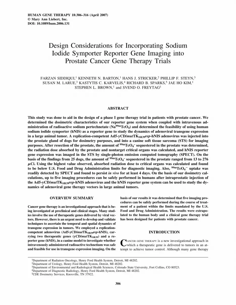
FIG. 1. Comparison of 99mTcO4� biodistribution in the ab-
sence and presence of injected adenovirus. Na99mTcO4 (32mCi) was administered to two dogs, one of which received anintraprostatic injection of the Ad5-yCD/mutTKSR39rep-hNISadenovirus (3 � 1011 VP) 24 hr earlier. After euthanasia, or-gans were removed and the amount of radioactivity in variousorgans was measured wit a gamma counter. The values of thedog that did (x axis), or did not (y axis), receive the adenovi-rus are plotted. If uptake in the two animals was identical, theslope would be 1 (line of identity, dashed line). The slope ofthe line in this study was 0.85 with a correlation coefficient of0.994.
SODIUM IODIDE SYMPORTER REPORTER GENE IMAGING 309
lens of the eye, or gonads cannot exceed 3 rem (1 rem � 1 cSv)for a single procedure and cumulatively 5 rem for all proce-dures conducted within a 1-year period. Likewise, the absorbeddose to other organs cannot exceed 5 rem for a single proce-dure and cumulatively 15 rem for all procedures conductedwithin 1 year (Code of Federal Regulations, 21 CFR 361.1).
We tested the validity of our assumption that the presenceof the Ad5-yCD/mutTKSR39rep-hNIS in the prostate does notalter the 99mTcO4
� biodistribution in nonprostatic tissues. Forthis we compared the 99mTcO4
� biodistribution in dogs with-out and with an intraprostatic injection of the Ad5-yCD/mut-TKSR39rep-hNIS adenovirus. With the exception of the prostate(the target organ), all organs studied sequestered similar ra-dioactivity whether virus was present or not (Fig. 1). These re-
sults suggested that the presence of the Ad5-yCD/mut-TKSR39rep-hNIS adenovirus in the prostate did not affect 99mTcuptake in nonprostatic organs. Importantly, there was no evi-dence of viral spread from the site of viral injection in theprostate to critical normal tissues such as kidney, liver, or lung.This observation confirms our unpublished observations that in-traprostatic adenovirus injection at the doses studied (3 � 1011
VP) produce no dose-limiting toxicity (n � 25) and no evidenceof viral spread to the liver (liver enzymes, including aspartateaminotransferase [AST], alanine aminotransferase [ALT], andalkaline phosphatase remained within the normal range beforeand after viral injection).
The second radiation source that must be considered is the99mTcO4
� sequestered in the prostate by Ad5-yCD/mut-
TABLE 1. RADIATION ABSORBED BY VARIOUS ORGANS FROM 99mTcO4� SEQUESTERED IN PROSTATE AFTER
Ad5-yCD/mut TKSR39rep-hNIS INJECTIONa,b
Absorbed dose from99mTcO4
� in prostate Absorbed doseS value over the residence resulting from 276 �Ci
(cGy/mCi � hr) time (cGy) in the prostate
16-g prostate 2.65 23.0 6.3
Estimatedradiation dose Absorbed dose from Absorbed dose
per injected 99mTcO4� in prostate resulting from activity
activity time for the residence time in prostateOrgan (cGy/mCi � hr) (cGy/mCi) (cGy)
Adrenals 4.90 � 10�5 4.25 � 10�4 1.17 � 10�4
Brain 3.94 � 10�8 3.42 � 10�7 9.43 � 10�8
Gall bladder wall 2.04 � 10�4 1.77 � 10�3 4.88 � 10�4
Gall bladder contents 1.77 � 10�4 1.54 � 10�3 4.24 � 10�4
Lower large intestine wall 1.01 � 10�2 8.76 � 10�2 2.42 � 10�2
Lower large intestine contents 6.88 � 10�3 5.97 � 10�2 1.65 � 10�2
Small intestine 1.16 � 10�5 1.00 � 10�2 2.77 � 10�3
Stomach wall 1.00 � 10�4 8.68 � 10�4 2.40 � 10�4
Stomach contents 1.00 � 10�4 8.70 � 10�4 2.40 � 10�4
Upper large intestine wall 8.88 � 10�4 7.70 � 10�3 2.13 � 10�3
Upper large intestine contents 8.84 � 10�4 7.67 � 10�3 2.12 � 10�3
Heart wall 1.60 � 10�5 1.39 � 10�4 3.82 � 10�5
Heart contents 9.59 � 10�6 8.31 � 10�5 2.29 � 10�5
Kidneys 1.15 � 10�4 9.99 � 10�4 2.76 � 10�4
Liver 6.62 � 10�5 5.74 � 10�4 1.58 � 10�4
Lungs 8.96 � 10�6 7.77 � 10�5 2.14 � 10�5
Spleen 6.62 � 10�5 5.74 � 10�4 1.58 � 10�4
Pancreas 6.38 � 10�5 5.53 � 10�4 1.53 � 10�4
Prostate 2.68 2.32 � 101� 6.41Skeleton 1.08 � 10�3 9.37 � 10�3 2.58 � 10�3
Active marrow 7.96 � 10�4 6.91 � 10�3 1.91 � 10�3
Skin 7.22 � 10�4 6.26 � 10�3 1.73 � 10�3
Thyroid 6.27 � 10�7 5.44 � 10�6 1.50 � 10�6
Thymus 5.22 � 10�6 4.53 � 10�5 1.25 � 10�5
Testes 1.78 � 10�2 1.55 � 10�1 4.27 � 10�2
Urinary bladder wall 3.34 � 10�2 2.90 � 10�1 8.00 � 10�2
Urinary bladder contents 3.02 � 10�2 2.62 � 10�1 7.23 � 10�2
Whole body 2.33 � 10�3 2.02 � 10�2 5.58 � 10�3
aShown are absorbed radiation dose estimates for 276 �Ci of 99mTcO4� localized in the prostate with a residence time of 8.7
hr. The values shown are for a single imaging procedure.bOne sievert (Sv) � 1 Gy for this situation, with quality and weighting factors � 1.

SIDDIQUI ET AL.310
TKSR39rep-hNIS-infected cells. To estimate the absorbed doseto various organs due to this radiation source, Ad5-yCD/mut-TKSR39rep-hNIS (3 � 1011 VP) was injected intraprostaticallyand the amount of radioactivity sequestered in the prostate wasquantified 24 hr after Na99mTcO4 administration. On average(n � 25), the canine prostates weighed 21 � 12 g (range, 8–52g) and contained 75 � 74 �Ci (range, 13–276 �Ci) of se-questered radioactivity. To calculate the absorbed dose to var-ious organs due to this radiation source, we used the highestactivity observed in the prostate (i.e., 276 �Ci). It was assumed,for dosimetry purposes, that the human prostate would have aninstantaneous uptake of 276 �Ci with removal by physical de-cay only. The residence time of 99mTcO4
� activity in theprostate is 8.7 hr, which was calculated from the reciprocal ofthe decay constant (� � 0.693/t1/2) and considering only phys-ical decay (for 99mTc, � 6.01/0.693 � 8.7 hr). Using these as-sumptions, the absorbed dose to various organs due to the se-questered radioactivity in the prostate was calculated (Table 1).Three scenarios were initially considered: (1) a point source ofradiation, (2) a 16-g uniform source of radiation, and (3) a 40-g uniform source of radiation. We used the 16-g uniform sourcein our calculations because this prostate weight is standard forinternal dosimetry purposes (Stabin, 1994). As expected, theorgan receiving the highest dose is the prostate (2.68 cGy/
mCi � hr), with all other organs receiving 2% of the prostatedose. Although the actual distribution of radioactivity in theprostate is likely to be heterogeneous, this will not affect thedose to body tissues other than prostate.
The absorbed doses to various organs due to the two po-tential radiation sources described above were summed andare presented in Table 2. Except for prostate, it is estimatedthat all organs will receive an absorbed dose under the FDAlimit for a single procedure. Although the absorbed dose tothe prostate from a single procedure (6.4 rem) exceeds theFDA limit (5 rem), it is insignificant (0.1%) relative to thedose the prostate will receive due to the conformal radiother-apy (7600 cGy, 7600 rem) and, therefore, is not a factor whendetermining the maximum allowable number of imaging pro-cedures. Not surprisingly, the dose-limiting tissue is the uri-nary bladder wall, which will receive an absorbed dose (2.16rem) approximately 40% that of the FDA limit for a singleprocedure. Other tissues that will receive significant doses (indescending order) include the lower large intestinal wall (LLI,1.624 rem, 32%), upper large intestinal wall (ULI, 1.602 rem,32%), thyroid (1.36 rem, 27%), stomach (0.752, 15%), wholebody (0.63 rem, 13%), and small intestines (0.483 rem, 10%).The thyroid and stomach will receive significant doses be-cause they express endogenous hNIS and therefore can accu-
TABLE 2. ABSORBED RADIATION DOSE ESTIMATES FOR SINGLE AND
MULTIPLE IMAGING PROCEDURESa
One procedure Five procedure Seven proceduresOrgan (rem/10 mCi)b (rem/50 mCi) (rem/70 mCi)
Adrenals 0.176 0.880 1.232Brain 0.107 0.535 0.749Gallbladder wall 0.304 1.520 2.128LLI wall 1.624 8.120 11.368Small intestine 0.483 2.415 3.381Stomach 0.752 3.760 5.264ULI wall 1.602 8.010 11.214Heart wall 0.160 0.800 1.120Kidneys 0.192 0.960 1.344Liver 0.176 0.880 1.232Lungs 0.134 0.670 0.938Pancreas 0.272 1.360 1.904Prostatec 6.410 32.050 44.870Red marrow 0.194 0.970 1.358Boen surfaces 0.291 1.455 2.037Skin 0.100 0.500 0.700Spleen 0.208 1.040 1.456Testes 0.235 1.175 1.645Thymus 0.130 0.650 0.910Thyroid 1.360 6.800 9.520Urinary bladder wall 2.160 10.800 15.120Whole body EDE 0.630 3.150 4.410
aShown are calculated absorbed dose to various tissues in the human body when exposed to in-ternal radiation from systemically injected 99mTcO4
� and 276 �Ci of 99mTcO4� localized in prostate
after Ad5-yCD/mutTKSR39rep-hNIS injection. The values that exceed FDA-stipulated limits areshown in boldface.
bSixteen microcuries at time of injection.cTarget organ, to receive external beam radiation therapy to a dose of 7600 cGy.Abbreviations: EDE, effective dose equivalent; LLI, lower large intestine; ULI, upper large in-
testine.
SODIUM IODIDE SYMPORTER REPORTER GENE IMAGING 311
mulate 99mTcO4�. All other organs will receive 10% the
FDA limit for a single procedure.
Use of hNIS reporter gene and SPECT to study the dynamics of adenoviral gene therapy vector in large animal tumor
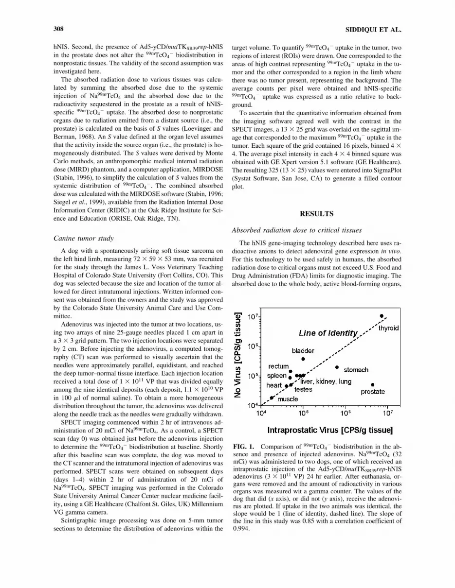
To examine the feasibility of using the hNIS reporter geneand SPECT imaging to study the dynamics of an adenoviralgene therapy vector in the tumor of a large animal, the Ad5-yCD/mutTKSR39rep-hNIS adenovirus was injected into two dis-tinct regions of a soft tissue sarcoma of a dog, using CT guid-ance (Fig. 2). One day after the adenovirus injection, 20 mCiof Na99mTcO4 was administered and SPECT imaging com-menced within 2 hr. Two regions of 99mTcO4
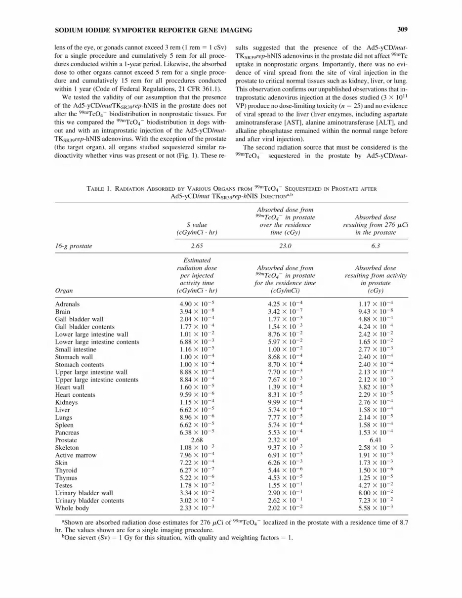
� uptake corre-sponding to the adenovirus injection sites were readily de-tectable by SPECT (Fig. 3A). The average pixel intensitiesobtained from the imaging software were used to generate acolor contour plot depicting the relative levels of hNIS geneexpression within the tumor (Fig. 3B). As expected, the areasof high pixel intensity in the colored contour plot (Fig. 3B, red)corresponded well with areas of high contrast in the SPECT im-ages (Fig. 3A). We have shown previously that sequestered ra-dioactivity reflects transgene expression (Barton et al., 2003)and demonstrated that these parameters are significantly corre-lated by a nonlinear relationship (unpublished data). This servedto illustrate that the numerical values obtained from the imag-ing software can be used to quantify the level of transgene ex-pression after gene transfer.
To study the dynamics of reporter gene expression in this tu-mor model, SPECT images were obtained at baseline (day 0)before adenovirus injection and on four consecutive days (days1–4) after adenovirus injection. To make the day-to-day com-parisons valid, the same plane through the tumor was examinedat each time point. Two distinct areas of 99mTcO4
� uptake cor-responding to the two adenoviral injections were detected bySPECT (Fig. 4a). The two injected areas were selected as re-gions of interest (ROI) and the total 99mTcO4
� uptake in eachROI was expressed relative to the background (Fig. 4b). Therelative 99mTcO4
� uptake for each ROI was plotted versus timeand the results were compared wit the SPECT images.99mTcO4
� uptake in the tumor reached a maximum on day 2and persisted at least through day 4 (Fig. 4c). As expected, thehigh contrast observed in the SPECT images agreed well withthe relative levels of 99mTcO4
� uptake measured by the imag-ing software, demonstrating that the hNIS reporter gene can beimaged noninvasively by SPECT to quantitatively study the dy-namics of the Ad5-yCD/mutTKSR39rep-hNIS adenovirus afterintratumoral administration.
Design of phase I gene therapy trial incorporatingimaging component
On the basis of FDA guidelines and the data presented inTable 2, we have designed a phase I gene therapy trial with theAd5-yCD/mutTKSR39rep-hNIS adenovirus that will include animaging component. The indication is men with newly diag-nosed, intermediate- to high-risk prostate cancer. The study hastwo parts.
FIG. 2. CT-guided intratumoral injection of adenovirus. Adenovirus was injected into the tumor in two distinct regions, sepa-rated by 2 cm, using a 3 � 3 grid pattern. Before injecting the adenovirus, needles were placed and a CT scan was performed toascertain their positions. (A) Sagittal view; (B) transverse axial view.

SIDDIQUI ET AL.312
300
LeftRight
Background
cb
a
250
200
150
100
1 2 3Days
Upt
ake
ratio
s
4
LeftRight
Day 0 ay 1D Day 2 Day 3 Day 4
FIG. 3. Quantification of hNIS gene expression. (A) A 13 � 25 grid was overlaid onto a representative SPECT image and theaverage pixel intensity in each square was obtained, using SPECT imaging software. (B) Color contour plot generated from pixelvalues obtained in (A).
FIG. 4. Dynamics of hNIS gene expression by SPECT imaging. (a) Na99mTcO4 was administered on each day 1–2 hr beforeSPECT imaging. A baseline scan (day 0) was performed just before the adenoviral injection. SPECT images were obtained on sub-sequent days (days 1–4) after the adenovirus injection. (b) 99mTcO4
� uptake in the two ROIs (left and right) representing the in-jected tumor and nontumor region (background) was quantified. (c) Plot of relative 99mTcO4
� uptake for each ROI on days 1–4.

SODIUM IODIDE SYMPORTER REPORTER GENE IMAGING 313
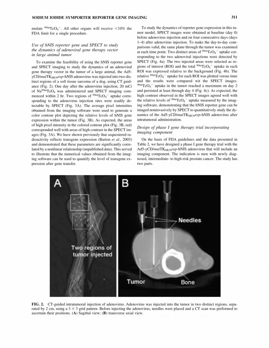
Part 1 is a dose escalation study to determine the toxicityand maximum tolerated dose (MTD) of a single intratumoralinjection of the Ad5-yCD/mutTKSR39rep-hNIS adenovirus fol-lowed by 3 weeks of 5-fluorocytosine (5-FC) and valganciclovir(vGCV) prodrug therapy, a standard course (7600 cGy) of in-tensity-modulated radiotherapy (IMRT), and two 16-mCi in-jections of Na99mTcO4 (Fig. 5). This part will include two cohorts of three subjects, each of whom will receive the ade-novirus at two dose levels (1011 and 1012 VP). Within 30 daysbefore the adenovirus injection (day 1), subjects will be ad-ministered 16 mCi of Na99mTcO4 intravenously and will un-dergo whole body and pelvic planar scans, using gamma cam-era scintigraphy, to establish a baseline. Three days after theadenovirus injection (day 4), subjects will be administered 16mCi of Na99mTcO4 and will undergo whole body and pelvicplanar scans to determine whether hNIS-mediated 99mTcO4
�
uptake can be detected. If specific 99mTcO4� uptake is detected
in the prostate, SPECT of the pelvic region will be performedto obtain a three-dimensional tomographic image of the prostateto estimate the volume of 99mTcO4
� uptake. The primary endpoint is toxicity up to and including day 90. Secondary endpoints include (1) the feasibility of using hNIS as a reportergene to monitor adenoviral gene therapy vectors noninvasivelyin humans, (2) the volume of 99mTcO4
� uptake in the prostate,and (3) prostate biopsy status at 2 years.
On establishing the safety and feasibility of this combinedgene therapy/imaging approach in humans, Part 2, a persistence
and biodistribution study, will commence. The Ad5-yCD/mut-TKSR39rep-hNIS adenovirus will be injected intraprostaticallyon day 1 at the maximum tolerated dose (MTD) established inpart 1, followed by 3 weeks of prodrug therapy and 7600 cGyof IMRT (Fig. 5). During the gene therapy/radiation course,subjects will receive up to four 16-mCi injections of Na99mTcO4
on days 4, 8, 15, and 22 that will be followed by nuclear med-icine imaging. To comply with FDA guidelines, no subject willundergo more than five imaging procedures, including the base-line evaluation. As shown in Table 2, with five imaging pro-cedures, the dose-limiting tissue (urinary bladder wall) is esti-mated to receive an absorbed dose of 10.8 rem, which is 72%of the FDA limit (15 rem) for all procedures within a 1-yearperiod. The administration of Na99mTcO4 on days 8, 15, and 22will require that specific 99mTcO4
� uptake be detected in theprostate at the previous time point. The primary end point istoxicity up to and including day 90. Secondary end points in-clude (1) persistence and whole body distribution of the Ad5-yCD/mutTKSR39rep-hNIS adenovirus, (2) persistence of ade-noviral DNA in blood, (3) volume of 99mTcO4
� uptake in theprostate, and (4) prostate biopsy status at 2 years.
DISCUSSION
The purpose of these studies was to determine the feasibil-ity of using hNIS as a reporter gene to study the dynamics of
Toxicity Assessments
IMRT (38 � 2 Gy for 76 Gy)weekdays only
Inject Ad5-yCD/mutTKSR39rep-hNIS adenovirusCohort 1-1011vp (Part 1)Cohort 2-1012vp (Part 1)Cohort 3-MTD (Part 2)
Part 2
5-FC + vGCV (3 weeks)weekdays only
1�30 4 8
Pre
trea
tmen
t Eva
luat
ions
15 22 55 90
Day
Part 1
Administer 16 mCiNa99m TcO4Perform �-camera scintigraphy/SPECT within 4 h
Toxicity Endpoint
(once weekly)
FIG. 5. Treatment schema of planned phase I prostate cancer gene therapy trial including an imaging component.
an adenoviral gene therapy vector in the tumor of a large ani-mal and to determine the dosimetric characteristics of this re-porter system when coupled with intravenous administration ofNa99mTcO4. We demonstrate here that SPECT can be used tostudy the spatial distribution and dynamics of hNIS-containingadenoviruses after intratumoral injection. On the basis of dosi-metric calculations, we have determined that up to five imag-ing procedures can be safely performed in humans. These re-sults provide the scientific basis for including this imagingtechnology in human gene therapy trials of prostate cancer andset an upper limit on the number of imaging procedures thatshould be performed.
The ability to monitor adenoviral persistence and therapeu-tic gene activity in vivo has the potential to improve both thesafety and efficacy of human gene therapy. Historically, genetransfer in humans has been assessed by measuring DNA,mRNA, or protein levels in tumor biopsies, methods that arefraught with drawbacks. Because they are invasive and causediscomfort to the patient, it is difficult to justify repeated biop-sies during the treatment course. Moreover, needle biopsies areprone to high sampling error and are likely to underestimate thetrue efficiency of gene transfer. Finally, needle biopsies do notallow for “real-time” determination of transgene expression,persistence, and biodistribution and therefore do not permit tai-loring of the treatment plan (i.e., duration of prodrugs) to eachindividual patient.
Taking these limitations into consideration, we developed anadenoviral gene therapy vector, Ad5-yCD/mutTKSR39rep-hNIS, combining both therapeutic and reporter genes in one ad-enovirus. The therapeutic genes, yCD and mutTKSR39, convertFDA-approved prodrugs (5-FC and vGCV) into active agents,which function as potent chemotherapeutics and radiosensitiz-ers (Rogulski et al., 1997a,b; Kim et al., 1998). The hNIS geneallows for noninvasive monitoring of adenoviral replication,persistence, and biodistribution, using 99mTcO4
� and SPECT.To demonstrate proof of principle to support an upcoming phaseI gene therapy trial in prostate cancer, we performed a geneimaging study in a dog with a spontaneously arising soft tissuesarcoma. The results demonstrated that it is possible to moni-tor adenoviral gene activity in a patient, using radiopharma-ceuticals (i.e., Na99mTcO4) and equipment readily available innuclear medicine departments.
One of the concerns when using radioisotopes for gene-imag-ing purposes is the absorbed radiation dose to critical tissuestructures. Here, we present the considerations and absorbeddose calculations based on data obtained in the dog. The high-est activity measured in the prostate of 25 dogs injected with3 � 1011 VP of Ad5-yCD/mutTKSR39rep-hNIS and 32 mCi ofNa99mTcO4 was 276 �Ci, which was used to estimate the ab-sorbed radiation dose in humans. We assumed instantaneousuptake of 99mTcO4
� by the prostate and only physical decay.On the basis of these data, we have determined that five imag-ing procedures can be safely performed in humans when ad-ministering 16 mCi of Na99mTcO4 per procedure. It should benoted that the Na99mTcO4 dose administered to the dogs (2–5mCi/kg) is, on a weight basis, at least 10 times that to be ad-ministered to human subjects (0.2 mCi/kg, 80-kg man) in ourproposed phase I trial. Also, the volume of the adult humanprostate (40–80 cm3) is roughly twice that of the dog prostate(mean, 21 cm3; range, 8–52 cm3) used in these studies. On the
basis of these two considerations only, our calculations re-garding the absorbed dose to the prostate and nontarget organsare likely to be overestimates. However, a weakness of the dogmodel is that human adenoviruses replicate inefficiently in ca-nine versus human tissue, which may lead to an underestima-tion of the absorbed dose to critical structures. This is not aconcern when considering the prostate, for this is the target or-gan and is scheduled to receive 7600 cGy (7600 rem) of con-formal radiation, which far exceeds the estimated dose (32 rem)from five imaging procedures. When considering nontarget tis-sues, the dose-limiting organ is the urinary bladder wall, whichis estimated to receive 11 rem with five imaging proceduresand 15 rem with seven procedures, the latter of which exceedsFDA limits. For this reason, we limited the number of imagingprocedures in our proposed phase I study to five. However, itis important to note that most (96%) of the absorbed dose tothe urinary bladder wall is due to the residence time of99mTcO4
� in that organ and not to the sequestered radioactiv-ity in the prostate. The same is true for all other nontarget or-gans. Therefore, despite the poor replication efficiency of hu-man adenoviruses in the canine prostate, we do not believe thatour absorbed dose estimates to nontarget tissues are skewed tothe point where it is unsafe to proceed with the phase I studyproposed here.
Having determined the feasibility and number of imagingprocedures that can be safely incorporated in a human trial withthe Ad5-yCD/mutTKSR39rep-hNIS adenovirus, we plan to in-clude an imaging component in all of our future gene therapytrials and believe that important knowledge will be generated.For example, using this imaging technology, we will be able toassess the quality of the adenovirus injection. Previously, wedemonstrated that the resolution of this imaging technology is0.5–1 cm and that it is possible to discern among different re-gions (left and right sides, base, midgland, and apex) of theprostate (Barton et al., 2003). Thus, we will be able to verify,with limited resolution, that the adenovirus was deposited cor-rectly and that it is active. Although such information could beobtained as early as 1 day after the adenovirus injection, weplan to perform the first postinjection imaging procedure on day4 (day 1 being the day of the adenovirus injection) for conve-nience and because our preclinical studies have indicated thatgene expression peaks on day 3 or 4 in vivo. Moreover, usingSPECT, we will be able to quantify the magnitude and volumeof gene expression in the prostate and correlate it with prostate-specific antigen (PSA) or tumor (prostate biopsy at 2 years) re-sponse. By imaging on multiple days, we will obtain importantknowledge regarding the persistence and biodistribution of theadenovirus in vivo. The former information (i.e., persistence)will guide us in treatment planning regarding the optimal du-ration of prodrug administration (i.e., they should be given onlywhile genes are expressed). Such information could also be usedto compare different adenoviruses and/or vector formulationsthat may result in greater persistence of therapeutic gene ex-pression. The latter information (i.e., biodistribution) may pro-vide better data regarding adenovirus dissemination to collat-eral tissues such as liver, which could prevent a patient deathby signaling that prodrug administration should be withheld.Indeed, we believe the imaging technology described here hasthe potential to improve not only the efficacy, but also thesafety, of human gene therapy.
SIDDIQUI ET AL.314

Another imaging modality being used to assess gene ex-pression in humans is PET, using the HSV-1 TK or hNIS re-porter gene (Gambhir et al., 1999; Herschman et al., 2000; Ja-cobs et al., 2001; Groot-Wassink et al., 2002; Jacobs and Heiss,2002; Deng et al., 2004; Niu et al., 2005; Yaghoubi et al., 2005).An article (Miyagawa et al., 2005) comparing these two re-porter genes found that hNIS yielded higher signal intensity andimaging contrast compared with HSV-1 TK when coupled withPET imaging. Therefore, we plan to evaluate in future trials themerits of PET imaging using HSV-1 TK and/or hNIS as re-porter genes. Yaghoubi et al. (2001) performed pharmacoki-netic and dosimetry studies with [18F]FHBG, a substrate forHSV-1 TK, in 10 healthy human volunteers in the absence ofan adenovirus injection. They found that the primary routes ofclearance were renal and hepatobiliary and that high radioac-tivity was observed in the bladder, gut, liver, and kidneys. Aswas found here with the hNIS reporter system, the urinary blad-der was the dose-limiting organ. This information will be use-ful when incorporating PET imaging in our human gene ther-apy trials.
CONCLUSIONS
The results of these studies indicate that hNIS can be usedas a reporter gene to study the dynamics of an adenoviralgene therapy vector in humans when coupled with intra-venous administration of radioactive sodium pertechnetate.When applied in the clinic, this technology will yield impor-tant information regarding the quality of the adenovirus in-jection and the persistence and biodistribution of the adeno-virus in vivo. The knowledge gained has the potential toimprove not only the efficacy but also the safety of humangene therapy.
ACKNOWLEDGMENT
This work was supported by P01 CA097012 (S.O.F.).
REFERENCES
BARTON, K.N., TYSON, D., STRICKER, H., LEW, Y.S., HEISEY,G., KOUL, S., DE LA ZERDA, A., YIN, F.F., YAN, H., NA-GARAJA, T.N., RANDALL, K.A., JIN, G.K., FENSTERMACHER,J.D., JHIANG, S., HO KIM, J., FREYTAG, S.O., and BROWN, S.L.(2003). GENIS: Gene expression of sodium iodide symporter fornoninvasive imaging of gene therapy vectors and quantification ofgene expression in vivo. Mol. Ther. 8, 508–518.
BARTON, K.N., XIA, X., YAN, H., STRICKER, H., HEISEY, G.,YIN, F.F., NAGARAJA, T.N., ZHU, G., KOLOZSVARY, A., FEN-STERMACHER, J.D., LU, M., KIM, J.H., FREYTAG, S.O., andBROWN, S.L. (2004). A quantitative method for measuring gene ex-pression magnitude and volume delivered by gene therapy vectors.Mol. Ther. 9, 625–631.
BARTON, K.N., PAIELLI, D., ZHANG, Y., KOUL, S., BROWN, S.L.,LU, M., SEELY, J., KIM, J.H., and FREYTAG, S.O. (2006). Sec-ond-generation replication-competent oncolytic adenovirus armedwith improved suicide genes and ADP gene demonstrates greater ef-ficacy without increased toxicity. Mol. Ther. 13, 347–356.
CHO, J.Y., XING, S., LIU, X., BUCKWALTER, T.L., HWA, L.,SFERRA, T.J., CHIU, I.M., and JHIANG, S.M. (2000). Expressionand activity of human Na�/I� symporter in human glioma cells byadenovirus-mediated gene delivery. Gene Ther. 7, 740–749.
CHO, J.Y., SHEN, D.H., YANG, W., WILLIAMS, B., BUCKWAL-TER, T.L., LA PERLE, K.M., HINKLE, G., POZDERAC, R.,KLOOS, R., NAGARAJA, H.N., BARTH, R.F., and JHIANG, S.M.(2002). In vivo imaging and radioiodine therapy following sodiumiodide symporter gene transfer in animal model of intracerebralgliomas. Gene Ther. 9, 1139–1145.
DENG, W.P., YANG, W.K., LAI, W.F., LIU, R.S., HWANG, J.J.,YANG, D.M., FU, Y.K., and WANG, H.E. (2004). Non-invasive invivo imaging with radiolabelled FIAU for monitoring cancer genetherapy using herpes simplex virus type 1 thymidine kinase and gan-ciclovir. Eur. J. Nucl. Med. Mol. Imaging 31, 99–109.
FREYTAG, S.O., ROGULSKI, K.R., PAIELLI, D.L., GILBERT, J.D.,and KIM, J.H. (1998). A novel three-pronged approach to kill can-cer cells selectively: Concomitant viral, double suicide gene, and ra-diotherapy. Hum. Gene Ther. 9, 1323–1333.
FREYTAG, S.O., KHIL, M., STRICKER, H., PEABODY, J., MENON,M., DEPERALTA-VENTURINA, M., NAFZIGER, D., PEGG, J.,PAIELLI, D., BROWN, S., BARTON, K., LU, M., AGUILAR-CORDOVA, E., and KIM, J.H. (2002a). Phase I study of replica-tion-competent adenovirus-mediated double suicide gene therapy forthe treatment of locally recurrent prostate cancer. Cancer Res. 62,4968–4976.
FREYTAG, S.O., PAIELLI, D., WING, M., ROGULSKI, K., BROWN,S., KOLOZSVARY, A., SEELY, J., BARTON, K., DRAGOVIC,A., and KIM, J.H. (2002b). Efficacy and toxicity of replication-com-petent adenovirus-mediated double suicide gene therapy in combi-nation with radiation therapy in an orthotopic mouse prostate cancermodel. Int. J. Radiat. Oncol. Biol. Phys. 54, 873–885.
FREYTAG, S.O., STRICKER, H., PEGG, J., PAIELLI, D., PRAD-HAN, D.G., PEABODY, J., DEPERALTA-VENTURINA, M., XIA,X., BROWN, S., LU, M., and KIM, J.H. (2003). Phase I study ofreplication-competent adenovirus-mediated double-suicide genetherapy in combination with conventional-dose three-dimensionalconformal radiation therapy for the treatment of newly diagnosed,intermediate- to high-risk prostate cancer. Cancer Res. 63,7497–7506.
FREYTAG, S.O., KIM, J.H., BROWN, S.L., BARTON, K., LU, M.,and CHUNG, M. (2004). Gene therapy strategies to improve the ef-fectiveness of cancer radiotherapy. Expert Opin. Biol. Ther. 4,1757–1770.
GAMBHIR, S.S., BARRIO, J.R., PHELPS, M.E., IYER, M., NA-MAVARI, M., SATYAMURTHY, N., WU, L., GREEN, L.A.,BAUER, E., MACLAREN, D.C., NGUYEN, K., BERK, A.J.,CHERRY, S.R., and HERSCHMAN, H.R. (1999). Imaging adeno-viral-directed reporter gene expression in living animals with posi-tron emission tomography. Proc. Natl. Acad. Sci. U.S.A. 96,2333–2338.
GROOT-WASSINK, T., ABOAGYE, E.O., GLASER, M., LEMOINE,N.R., and VASSAUX, G. (2002). Adenovirus biodistribution andnoninvasive imaging of gene expression in vivo by positron emis-sion tomography using human sodium/iodide symporter as reportergene. Hum. Gene Ther. 13, 1723–1735.
HERSCHMAN, H.R., MACLAREN, D.C., IYER, M., NAMAVARI,M., BOBINSKI, K., GREEN, L.A., WU, L., BERK, A.J.,TOYOKUNI, T., BARRIO, J.R., CHERRY, S.R., PHELPS, M.E.,SANDGREN, E.P., and GAMBHIR, S.S. (2000). Seeing is believ-ing: Non-invasive, quantitative and repetitive imaging of reportergene expression in living animals, using positron emission tomogra-phy. J. Neurosci. Res. 59, 699–705.
JACOBS, A., and HEISS, W.D. (2002). Towards non-invasive imag-ing of HSV-1 vector-mediated gene expression by positron emissiontomography. Vet. Microbiol. 86, 27–36.
SODIUM IODIDE SYMPORTER REPORTER GENE IMAGING 315
SIDDIQUI ET AL.316
JACOBS, A., VOGES, J., RESZKA, R., LERCHER, M., GOSS-MANN, A., KRACHT, L., KAESTLE, C., WAGNER, R., WIEN-HARD, K., and HEISS, W.D. (2001). Positron-emission tomogra-phy of vector-mediated gene expression in gene therapy for gliomas.Lancet 358, 727–729.
KIM, J.H., KIM, S.H., BROWN, S.L., and FREYTAG, S.O. (1994).Selective enhancement by an antiviral agent of the radiation-inducedcell killing of human glioma cells transduced with HSV-tk gene. Can-cer Res. 54, 6053–6056.
KIM, J.H., KOLOZSVARY, A., ROGULSKI, K., KHIL, M.S.,BROWN, S.L., and FREYTAG, S.O. (1998). Selective radiosensiti-zation of 9L glioma in the brain transduced with double suicide fu-sion gene. Cancer J. Sci. Am. 4, 364–369.
LA PERLE, K.M., SHEN, D., BUCKWALTER, T.L., WILLIAMS, B.,HAYNAM, A., HINKLE, G., POZDERAC, R., CAPEN, C.C., andJHIANG, S.M. (2002). In vivo expression and function of the so-dium iodide symporter following gene transfer in the MATLyLu ratmodel of metastatic prostate cancer. Prostate 50, 170–178.
LOEVINGER, R., and BERMAN, M. (1968). A formalism for calcu-lation of absorbed dose from radionuclides. Phys. Med. Biol. 13,205–217.
MARSEE, D.K., SHEN, D.H., MACDONALD, L.R., VADYSIRI-SACK, D.D., LIN, X., HINKLE, G., KLOOS, R.T., and JHIANG,S.M. (2004). Imaging of metastatic pulmonary tumors following NISgene transfer using single photon emission computed tomography.Cancer Gene Ther. 11, 121–127.
MIRD (MEDICAL INTERNAL RADIATION DOSE) COMMITTEE.(1976). MIRD/Dose Estimate Report No. 8: Summary of current ra-diation dose estimates to normal humans from 99mTc as sodiumpertechnetate. J. Nucl. Med. 17, 74–77.
MIYAGAWA, M., ANTON, M., WAGNER, B., HAUBNER, R., SOU-VATZOGLOU, M., GANSBACHER, B., SCHWAIGER, M., andBENGEL, F.M. (2005). Non-invasive imaging of cardiac transgeneexpression with PET: Comparison of the human sodium/iodide sym-porter gene and HSV1-tk as the reporter gene. Eur. J. Nucl. Med.Mol. Imaging 32, 1108–1114.
NIU, G., KRAGER, K.J., GRAHAM, M.M., HICHWA, R.D., and DO-MANN, F.E. (2005). Noninvasive radiological imaging of pulmonarygene transfer and expression using the human sodium iodide sym-porter. Eur. J. Nucl. Med. Mol. Imaging 32, 534–540.
PENUELAS, I., MAZZOLINI, G., BOAN, J.F., SANGRO, B.,MARTI-CLIMENT, J., RUIZ, M., RUIZ, J., SATYAMURTHY, N.,QIAN, C., BARRIO, J.R., PHELPS, M.E., RICHTER, J.A., GAMB-HIR, S.S., and PRIETO, J. (2005). Positron emission tomographyimaging of adenoviral-mediated transgene expression in liver cancerpatients. Gastroenterology 128, 1787–1795.
ROGULSKI, K.R., KIM, J.H., KIM, S.H., and FREYTAG, S.O.(1997a). Glioma cells transduced with an Escherichia coli CD/HSV-1 TK fusion gene exhibit enhanced metabolic suicide and radiosen-sitivity. Hum. Gene Ther. 8, 73–85.
ROGULSKI, K.R., ZHANG, K., KOLOZSVARY, A., KIM, J.H., andFREYTAG, S.O. (1997b). Pronounced antitumor effects and tumorradiosensitization of double suicide gene therapy. Clin. Cancer Res.3, 2081–2088.
SIEGEL, J.A., THOMAS, S.R., STUBBS, J.B., STABIN, M.G.,HAYS, M.T., KORAL, K.F., ROBERTSON, J.S., HOWELL, R.W.,WESSELS, B.W., FISHER, D.R., WEBER, D.A., and BRILL, A.B.(1999). MIRD pamphlet no. 16: Techniques for quantitative radio-pharmaceutical biodistribution data acquisition and analysis for usein human radiation dose estimates. J. Nucl. Med. 40, 37S–61S.
STABIN, M.G. (1994). A model of the prostate gland for use in inter-nal dosimetry. J. Nucl. Med. 35, 516–520.
STABIN, M.G. (1996). MIRDOSE: Personal computer software for in-ternal dose assessment in nuclear medicine. J. Nucl. Med. 37,538–546.
YAGHOUBI, S., BARRIO, J.R., DAHLBOM, M., IYER, M., NA-MAVARI, M., SATYAMURTHY, N., GOLDMAN, R., HER-SCHMAN, H.R., PHELPS, M.E., and GAMBHIR, S.S. (2001). Hu-man pharmacokinetic and dosimetry studies of [18F]FHBG: Areporter probe for imaging herpes simplex virus type-1 thymidine ki-nase reporter gene expression. J. Nucl. Med. 42, 1225–1234.
YAGHOUBI, S.S., BARRIO, J.R., NAMAVARI, M., SATYA-MURTHY, N., PHELPS, M.E., HERSCHMAN, H.R., and GAMB-HIR, S.S. (2005). Imaging progress of herpes simplex virus type 1thymidine kinase suicide gene therapy in living subjects with positronemission tomography. Cancer Gene Ther. 12, 329–339.
Address reprint requests to:Dr. Stephen L. Brown
Department of Radiation OncologyHenry Ford Health System
2799 West Grand BlvdDetroit, MI 48202
E-mail: [email protected]
Received for publication September 11, 2006; accepted afterrevision November 20, 2006.
Published online: April 4, 2007.