endogenous viral elements in animal genomes
TRANSCRIPT

Endogenous Viral Elements in Animal GenomesAris Katzourakis1*, Robert J. Gifford2*
1 Department of Zoology, University of Oxford, Oxford, United Kingdom, 2 Aaron Diamond AIDS Research Center, New York, New York, United States of America
Abstract
Integration into the nuclear genome of germ line cells can lead to vertical inheritance of retroviral genes as host alleles. Forother viruses, germ line integration has only rarely been documented. Nonetheless, we identified endogenous viralelements (EVEs) derived from ten non-retroviral families by systematic in silico screening of animal genomes, including thefirst endogenous representatives of double-stranded RNA, reverse-transcribing DNA, and segmented RNA viruses, and thefirst endogenous DNA viruses in mammalian genomes. Phylogenetic and genomic analysis of EVEs across multiple hostspecies revealed novel information about the origin and evolution of diverse virus groups. Furthermore, several of theelements identified here encode intact open reading frames or are expressed as mRNA. For one element in the primatelineage, we provide statistically robust evidence for exaptation. Our findings establish that genetic material derived from allknown viral genome types and replication strategies can enter the animal germ line, greatly broadening the scope ofpaleovirological studies and indicating a more significant evolutionary role for gene flow from virus to animal genomes thanhas previously been recognized.
Citation: Katzourakis A, Gifford RJ (2010) Endogenous Viral Elements in Animal Genomes. PLoS Genet 6(11): e1001191. doi:10.1371/journal.pgen.1001191
Editor: Harmit S. Malik, Fred Hutchinson Cancer Research Center, United States of America
Received June 1, 2010; Accepted September 30, 2010; Published November 18, 2010
Copyright: � 2010 Katzourakis, Gifford. This is an open-access article distributed under the terms of the Creative Commons Attribution License, which permitsunrestricted use, distribution, and reproduction in any medium, provided the original author and source are credited.
Funding: RJG was supported by the Aaron Diamond AIDS Research Center. AK was funded by the Wellcome Trust (Grant #: 086173/A/08/Z). The funders had norole in study design, data collection and analysis, decision to publish, or preparation of the manuscript.
Competing Interests: The authors have declared that no competing interests exist.
* E-mail: [email protected] (RJG); [email protected] (AK)
Introduction
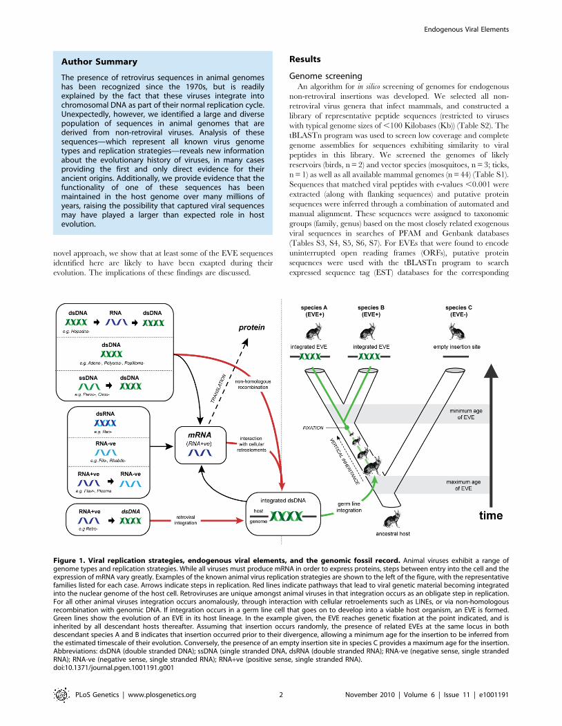
Viral infection of germ line cells (i.e. gametes, or cells of the
early embryo) can lead to viral genes or genomes becoming
integrated into chromosomes and inherited as host alleles [1,2].
These insertions, which we refer to here as endogenous viral elements
(EVEs), are usually eliminated from the host gene pool within a
small number of generations. However, they can also increase in
frequency, and some eventually reach fixation [3–11].
In animal genomes, the majority of EVEs are derived from
reverse transcribing RNA (rtRNA) viruses (i.e. retroviruses)
[5,12,13]. Retroviruses are the only animal viruses that integrate
into the genome of the host cell as an obligate step in their
replication strategy, and are thus predisposed to enter the host
germ line (Figure 1). EVEs derived from viruses that use other
genome replication strategies also occur, but are much less
common [6,7,9,11,14,15]. Genomic integration of non-retroviral
viruses may be mediated by non-homologous recombination with
chromosomal DNA [16–18] or by interactions with retroelements
in the host cell [11,19–22] (Figure 1).
EVEs reveal complex evolutionary relationships between viruses
and their hosts. For example, endogenous retroviruses have
shaped vertebrate genome evolution, not only by acting as genetic
parasites [23,24], but also by introducing useful genetic novelty.
Indeed, the role of exapted retroviral genes (i.e. integrated retroviral
genes that have adapted to serve a function in the host genome) in
mammalian reproduction [25,26] identifies EVEs as a key factor
in the evolution of placental mammals from egg-laying ancestors.
Similarly, in parasitoid wasps, genes derived from ancestral
nudiviruses have been exapted to facilitate a parasitic lifestyle
[9]. These remarkable examples demonstrate an important role
for gene flow from viruses to hosts in animal evolution.
EVEs also constitute an invaluable resource for reconstructing
the long-term history of virus and host evolution [27,28]. Viruses
exhibit the potential for extremely high rates of nucleotide
substitution, host switching, and lineage extinction, and this sets
limitations on what can be reliably inferred from observations of
contemporary isolates [29,30]. EVE sequences effectively
represent the ‘molecular fossils’ of ancient viral genomes,
preserving information about ancient virus and host interactions
that would otherwise be difficult, if not impossible, to infer. For
example, EVEs are subject to host rates of evolution and can thus
be dated relatively reliably with molecular clock-based ap-
proaches, in which genetic divergence correlates linearly with
time [31]. In contrast, structural constraints in exogenous viruses
may lead to the decoupling of short and long-term rates of viral
evolution, rendering molecular clock assumptions unusable over
longer timescales [30,32–34]. Furthermore, the identification of
orthologous EVE insertions allows the incorporation of inde-
pendent age estimates based on host species divergences (see
Figure 1) [35].
Despite the large quantity of published genome sequence data,
the diversity of non-retroviral viruses in animal genomes has not
been systematically explored. In this report, we use an in silico
approach to screen the genomes of mammals, birds and insect
vector species for endogenous sequences derived from non-
retroviral mammalian viruses. We identify sequences derived from
a very broad range of viruses, revealing an extensive history of
non-retroviral genome invasion ranging back to at least the late
Mesozoic Era (,93 million years ago). We demonstrate that these
sequences can be highly informative; (i) revealing novel virus
diversity; (ii) providing a timescale for virus evolution; (iii)
indicating the likely host range of virus groups, and; (iv) identifying
rare instances of horizontal transmission. Furthermore, using a
PLoS Genetics | www.plosgenetics.org 1 November 2010 | Volume 6 | Issue 11 | e1001191
novel approach, we show that at least some of the EVE sequences
identified here are likely to have been exapted during their
evolution. The implications of these findings are discussed.
Results
Genome screeningAn algorithm for in silico screening of genomes for endogenous
non-retroviral insertions was developed. We selected all non-
retroviral virus genera that infect mammals, and constructed a
library of representative peptide sequences (restricted to viruses
with typical genome sizes of ,100 Kilobases (Kb)) (Table S2). The
tBLASTn program was used to screen low coverage and complete
genome assemblies for sequences exhibiting similarity to viral
peptides in this library. We screened the genomes of likely
reservoirs (birds, n = 2) and vector species (mosquitoes, n = 3; ticks,
n = 1) as well as all available mammal genomes (n = 44) (Table S1).
Sequences that matched viral peptides with e-values ,0.001 were
extracted (along with flanking sequences) and putative protein
sequences were inferred through a combination of automated and
manual alignment. These sequences were assigned to taxonomic
groups (family, genus) based on the most closely related exogenous
viral sequences in searches of PFAM and Genbank databases
(Tables S3, S4, S5, S6, S7). For EVEs that were found to encode
uninterrupted open reading frames (ORFs), putative protein
sequences were used with the tBLASTn program to search
expressed sequence tag (EST) databases for the corresponding
Figure 1. Viral replication strategies, endogenous viral elements, and the genomic fossil record. Animal viruses exhibit a range ofgenome types and replication strategies. While all viruses must produce mRNA in order to express proteins, steps between entry into the cell and theexpression of mRNA vary greatly. Examples of the known animal virus replication strategies are shown to the left of the figure, with the representativefamilies listed for each case. Arrows indicate steps in replication. Red lines indicate pathways that lead to viral genetic material becoming integratedinto the nuclear genome of the host cell. Retroviruses are unique amongst animal viruses in that integration occurs as an obligate step in replication.For all other animal viruses integration occurs anomalously, through interaction with cellular retroelements such as LINEs, or via non-homologousrecombination with genomic DNA. If integration occurs in a germ line cell that goes on to develop into a viable host organism, an EVE is formed.Green lines show the evolution of an EVE in its host lineage. In the example given, the EVE reaches genetic fixation at the point indicated, and isinherited by all descendant hosts thereafter. Assuming that insertion occurs randomly, the presence of related EVEs at the same locus in bothdescendant species A and B indicates that insertion occurred prior to their divergence, allowing a minimum age for the insertion to be inferred fromthe estimated timescale of their evolution. Conversely, the presence of an empty insertion site in species C provides a maximum age for the insertion.Abbreviations: dsDNA (double stranded DNA); ssDNA (single stranded DNA, dsRNA (double stranded RNA); RNA-ve (negative sense, single strandedRNA); RNA-ve (negative sense, single stranded RNA); RNA+ve (positive sense, single stranded RNA).doi:10.1371/journal.pgen.1001191.g001
Author Summary
The presence of retrovirus sequences in animal genomeshas been recognized since the 1970s, but is readilyexplained by the fact that these viruses integrate intochromosomal DNA as part of their normal replication cycle.Unexpectedly, however, we identified a large and diversepopulation of sequences in animal genomes that arederived from non-retroviral viruses. Analysis of thesesequences—which represent all known virus genometypes and replication strategies—reveals new informationabout the evolutionary history of viruses, in many casesproviding the first and only direct evidence for theirancient origins. Additionally, we provide evidence that thefunctionality of one of these sequences has beenmaintained in the host genome over many millions ofyears, raising the possibility that captured viral sequencesmay have played a larger than expected role in hostevolution.
Endogenous Viral Elements
PLoS Genetics | www.plosgenetics.org 2 November 2010 | Volume 6 | Issue 11 | e1001191
mRNA. For all EVEs disclosing similarity to contemporary virus
isolates, putative EVE protein sequences were aligned with
representative viral protein sequences, and maximum likelihood
phylogenies were constructed.
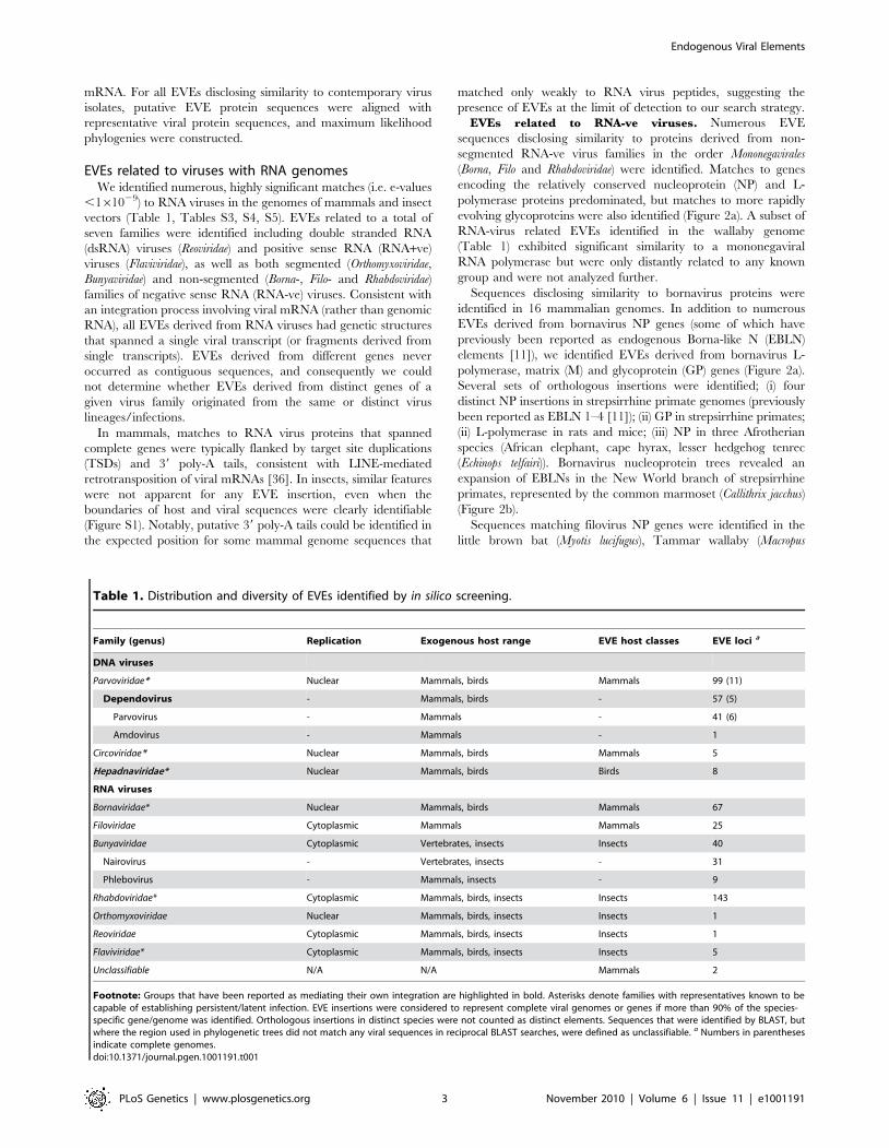
EVEs related to viruses with RNA genomesWe identified numerous, highly significant matches (i.e. e-values
,161029) to RNA viruses in the genomes of mammals and insect
vectors (Table 1, Tables S3, S4, S5). EVEs related to a total of
seven families were identified including double stranded RNA
(dsRNA) viruses (Reoviridae) and positive sense RNA (RNA+ve)
viruses (Flaviviridae), as well as both segmented (Orthomyxoviridae,
Bunyaviridae) and non-segmented (Borna-, Filo- and Rhabdoviridae)
families of negative sense RNA (RNA-ve) viruses. Consistent with
an integration process involving viral mRNA (rather than genomic
RNA), all EVEs derived from RNA viruses had genetic structures
that spanned a single viral transcript (or fragments derived from
single transcripts). EVEs derived from different genes never
occurred as contiguous sequences, and consequently we could
not determine whether EVEs derived from distinct genes of a
given virus family originated from the same or distinct virus
lineages/infections.
In mammals, matches to RNA virus proteins that spanned
complete genes were typically flanked by target site duplications
(TSDs) and 39 poly-A tails, consistent with LINE-mediated
retrotransposition of viral mRNAs [36]. In insects, similar features
were not apparent for any EVE insertion, even when the
boundaries of host and viral sequences were clearly identifiable
(Figure S1). Notably, putative 39 poly-A tails could be identified in
the expected position for some mammal genome sequences that
matched only weakly to RNA virus peptides, suggesting the
presence of EVEs at the limit of detection to our search strategy.
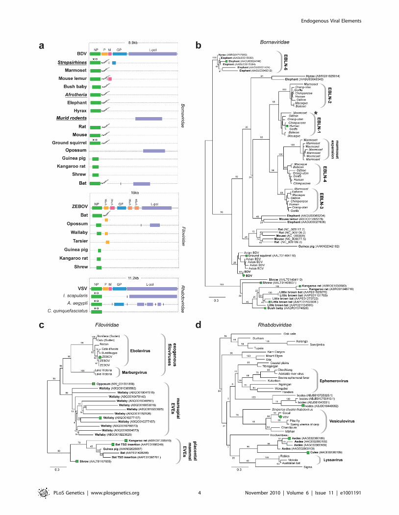
EVEs related to RNA-ve viruses. Numerous EVE
sequences disclosing similarity to proteins derived from non-
segmented RNA-ve virus families in the order Mononegavirales
(Borna, Filo and Rhabdoviridae) were identified. Matches to genes
encoding the relatively conserved nucleoprotein (NP) and L-
polymerase proteins predominated, but matches to more rapidly
evolving glycoproteins were also identified (Figure 2a). A subset of
RNA-virus related EVEs identified in the wallaby genome
(Table 1) exhibited significant similarity to a mononegaviral
RNA polymerase but were only distantly related to any known
group and were not analyzed further.
Sequences disclosing similarity to bornavirus proteins were
identified in 16 mammalian genomes. In addition to numerous
EVEs derived from bornavirus NP genes (some of which have
previously been reported as endogenous Borna-like N (EBLN)
elements [11]), we identified EVEs derived from bornavirus L-
polymerase, matrix (M) and glycoprotein (GP) genes (Figure 2a).
Several sets of orthologous insertions were identified; (i) four
distinct NP insertions in strepsirrhine primate genomes (previously
been reported as EBLN 1–4 [11]); (ii) GP in strepsirrhine primates;
(ii) L-polymerase in rats and mice; (iii) NP in three Afrotherian
species (African elephant, cape hyrax, lesser hedgehog tenrec
(Echinops telfairi)). Bornavirus nucleoprotein trees revealed an
expansion of EBLNs in the New World branch of strepsirrhine
primates, represented by the common marmoset (Callithrix jacchus)
(Figure 2b).
Sequences matching filovirus NP genes were identified in the
little brown bat (Myotis lucifugus), Tammar wallaby (Macropus
Table 1. Distribution and diversity of EVEs identified by in silico screening.
Family (genus) Replication Exogenous host range EVE host classes EVE loci a
DNA viruses
Parvoviridae* Nuclear Mammals, birds Mammals 99 (11)
Dependovirus - Mammals, birds - 57 (5)
Parvovirus - Mammals - 41 (6)
Amdovirus - Mammals - 1
Circoviridae* Nuclear Mammals, birds Mammals 5
Hepadnaviridae* Nuclear Mammals, birds Birds 8
RNA viruses
Bornaviridae* Nuclear Mammals, birds Mammals 67
Filoviridae Cytoplasmic Mammals Mammals 25
Bunyaviridae Cytoplasmic Vertebrates, insects Insects 40
Nairovirus - Vertebrates, insects - 31
Phlebovirus - Mammals, insects - 9
Rhabdoviridae* Cytoplasmic Mammals, birds, insects Insects 143
Orthomyxoviridae Nuclear Mammals, birds, insects Insects 1
Reoviridae Cytoplasmic Mammals, birds, insects Insects 1
Flaviviridae* Cytoplasmic Mammals, birds, insects Insects 5
Unclassifiable N/A N/A Mammals 2
Footnote: Groups that have been reported as mediating their own integration are highlighted in bold. Asterisks denote families with representatives known to becapable of establishing persistent/latent infection. EVE insertions were considered to represent complete viral genomes or genes if more than 90% of the species-specific gene/genome was identified. Orthologous insertions in distinct species were not counted as distinct elements. Sequences that were identified by BLAST, butwhere the region used in phylogenetic trees did not match any viral sequences in reciprocal BLAST searches, were defined as unclassifiable. a Numbers in parenthesesindicate complete genomes.doi:10.1371/journal.pgen.1001191.t001
Endogenous Viral Elements
PLoS Genetics | www.plosgenetics.org 3 November 2010 | Volume 6 | Issue 11 | e1001191
Endogenous Viral Elements
PLoS Genetics | www.plosgenetics.org 4 November 2010 | Volume 6 | Issue 11 | e1001191
eugenii), and gray short-tailed opossum (Monodelphis domestica)
genomes. The majority of these matches comprised fragments of
genes, although two full-length NP gene EVEs (displaying poly-A
tails and TSDs) were identified in bats (Figure 2a). More divergent
fragments of the NP gene were identified in the kangaroo rat
(Dipodymys ordii), guinea pig (Cavia porcellus) and common shrew
(Sorex araneus) genomes. Additionally, fragments of the L-polymer-
ase and VP35 gene were identified in the genomes of the opossum
and Philippine tarsier (Tarsius syrichta) respectively. In phylogenies,
EVEs derived from filovirus NP genes grouped into two well-
supported clades (Figure 2c), the largest of which included
exogenous filoviruses and EVEs derived from marsupials (wallaby
and opossum). EVEs in the smaller clade were more distantly
related to extant filoviruses, and were derived from the little brown
bat, guinea pig, shrew and kangaroo rat genomes. An opossum
EVE derived from L-polymerase grouped relatively closely with
Marburgviruses. Conflicting phylogenetic trees for oppossum L-
polymerase and NP-derived EVEs strongly indicated they are
derived from distinct ancestral viruses.
EVEs related to rhabdoviruses were identified in the black-
legged tick genome (Ixodes scapularis), and in the genomes of both
Aedes and Culex mosquitoes. Among these were insertions that
encoded intact NP and GP ORFs (Figure 2a). Phylogenies
constructed using NP tentatively grouped rhabdovirus EVEs
derived from mosquitoes in a clade with lyssaviruses and
Drosophila sigma virus (Figure 2d). However phylogenetic support
for this clade was very weak, with only 20% bootstrap support for
the monophyly of the clade, although support for the grouping of
EVEs from Aedes and Culex was high (96%). A robust clade (100%
bootstrap support) placed four Ixodes EVEs into a single group,
suggesting they are likely derived from the same exogenous virus
lineage, but their placement relative to other Rhabdoviruses was
ambiguous, as they formed a clade with a number of distinct
Rhabdoviruses with minimal bootstrap support (8%). Phyloge-
nies constructed using L-polymerase sequences weakly grouped
Ixodes and Aedes insertions with Lyssaviruses and Moussa virus,
but not Drosophila sigma virus. Weak support for basal
relationships was obtained with both trees, making it difficult to
confidently place thenovel EVEs with respect to the known
rhabdovirus diversity.
Matches to RNA-ve viruses with segmented genomes were
identified in the genomes of insect vectors (Figure 3a). In the I.
scapularis genome, we identified EVEs related to viruses isolated
from ticks and birds basal to the proposed genus Quarjavirus
(including Quaranfil and Johnston Atoll viruses) [37] in the family
Orthomyxoviridae (Figure 3c), EVEs distantly related to the
Bunyaviridae (Phlebovirus and Nairovirus genera) were identified
in the I. scapularis genome. Nairovirus-derived EVEs were distantly
related to Hazara virus (Figure 3d), indicating they represent a
distinct lineage within this tick-vectored genus. Phlebovirus EVEs
formed a robustly supported cluster in phylogenies with exogenous
viruses (Figure 3e), closest to Uukuniemi and Catch-me cave
viruses (vectored by ticks and mosquitoes respectively), suggesting
they are derived from the same exogenous lineage.
EVEs related to dsRNA viruses. In the A. aegyptii genome
we identified an EVE that was very closely related (,98%
nucleotide sequence identity) to segment 5 of the Liaoning virus
genome (Figure S1, Figure 3a and 3b). Liaoning is a dsRNA virus
(family Reoviridae, genus Seadornavirus) that was recently isolated
from Aedes dorsalis mosquitoes [38]. The Liaoning EVE in A. aegyptii
had a large inframe deletion, but encoded an otherwise intact
ORF. This is the first EVE derived from a dsRNA virus to be
described. As with other RNA virus EVEs in insect genomes, the
mechanism of genomic integration was unclear. The intact ORFs
and high level of identity to a circulating virus raise the possibility
this EVE formed recently and is not fixed in the host population.
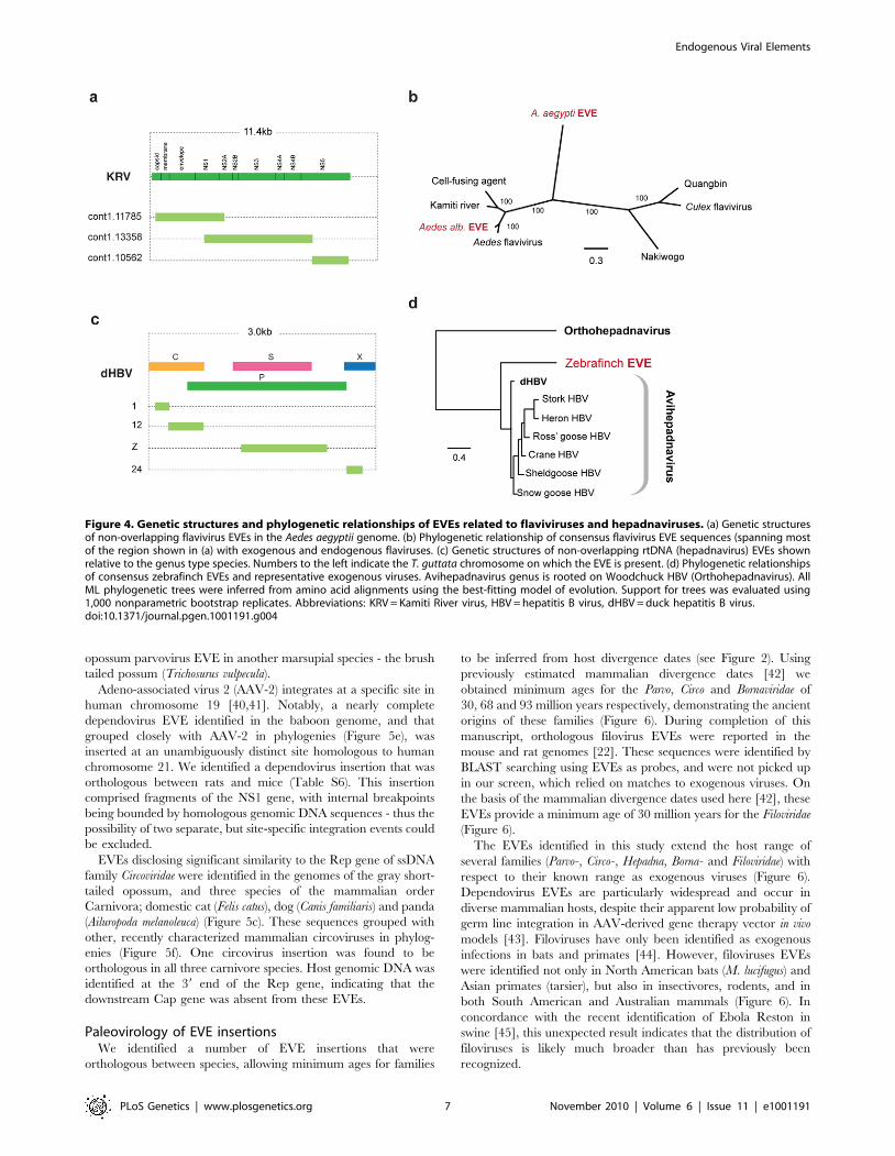
EVEs related to RNA+ve viruses. The genome of the Aedes
mosquito contains several sequences exhibiting similarity to the
viruses of the RNA+ve family Flaviviridae. Endogenous flaviviruses
have previously been reported in the genomes of A. aegyptii and A.
albopictus mosquitoes [6,39], but complete putative genomic
structures have not been determined. In particular, a large
fragment spanning the flaviviral NS1, NS2A, NS2B, NS3 and
NS4A genes has been described in A. albopictus, and a range of
smaller fragments at the 39 end of the flaviviral genome, mostly of
the NS5 gene, have been described in both albopictus and aegyptii
species. We have identified fragments that together span almost
the entire flavivirus genome in A. aegyptii (based on alignment to
Kamiti river virus (Figure 4a)), including a single fragment that
spans the equivalent region from the albopictus genome.
Phylogenetic trees that included both aegyptii and albopictus EVEs
showed that they are distinct viruses, separated by known
exogenous isolates. Thus, the EVEs in these two mosquito
species appear to be derived from at least two distinct flavivirus
lineages, with the aegyptii virus being most divergent from
previously characterized isolates. The albopictus sequence grouped
in a clade that included both Kamiti river virus and cell fusing
agent, as previously described by Crochu et al. [6].
EVEs related to viruses with DNA genomesWe identified highly significant matches to three families of
viruses with DNA genomes in the genomes of mammals and birds
(Table 1, Tables S6 and S7). These included matches to two single
stranded DNA (ssDNA) virus families (Parvoviridae and Circoviridae -
the first ssDNA virus EVEs to be described in mammals - and one
family of reverse transcribing DNA (rtDNA) viruses (Hepadnaviridae)
- the first rtDNA EVEs to be described. A single match to a double
stranded DNA (dsDNA) virus family (Adenoviridae) was identified in
the kangaroo rat genome, but this sequence was unambiguously
viral across its entire length (,17 Kb), encoding thirteen
completely intact viral ORFs (Figure S2), and is thus likely to
have derived from free virus and not an EVE.
A subset of parvovirus-related EVEs represented complete or
nearly complete viral genomes (Figure 5a). For one insertion in the
M. lucifugus genome, we identified putative 59 and 39 terminal non-
coding regions encoding characteristic inverted terminal repeats
(Figure S3). In general, however, DNA virus EVEs occurred as
genomic fragments, with no particular region of the viral genome
Figure 2. Genetic structures and phylogenetic relationships of Mononegavirus EVEs. (a) Summary genetic structures of MononegavirusEVE sets (Borna-, Rhabdo- and Filoviridae) shown relative to genus type species. The most intact elements are shown for each host taxon. Vertical linesbetween EVEs in the same host species that are derived from distinct genes indicate that the EVEs are not contiguous in the host genome.Abbreviations for viral type species (bold), host species (italics), and host taxa (bold, italic, underline) are indicated to the left of each EVE. Taxonomicgroups are shown for EVE insertions identified as orthologs. Poly-A tails are shown for EVEs that had these features. Intact ORFs (circles) andexpressed sequences (crosses) are indicated. Phylogenetic relationships of (b) bornavirus, (c) rhabdovirus and (d) filovirus EVEs and representativeexogenous viruses. Taxa that are shown as genetic structures in (a) are indicated by colored squares. Support for the ML phylogenetic trees wasevaluated using 1,000 nonparametric bootstrap replicates, and all three trees are midpoint rooted for display purposes. Abbreviations: BDV = Bornadisease virus; ZEBOV = Zaire ebola virus; VSV = vesicular stomatitis virus, L-pol = L-polymerase.doi:10.1371/journal.pgen.1001191.g002
Endogenous Viral Elements
PLoS Genetics | www.plosgenetics.org 5 November 2010 | Volume 6 | Issue 11 | e1001191

being obviously favored, with the exception of the circoviruses, for
which only the Rep gene was found.
EVEs related to rtDNA viruses. We identified sequences
disclosing significant similarity to Hepadnaviruses in the genomes
of the black-legged tick (I. scapularis), and the zebrafinch
(Taeniopygia guttata). In the zebrafinch genome, a total of 7 loci
were identified on 7 distinct chromosomes (Figure 4c). Sequences
at each locus generally corresponded to distinct, non-overlapping
regions of viral genome, suggesting that host genome
arrangements had fragmented a more complete insertion,
spanning most if not all of the viral genome. A consensus
constructed using all 7 zebrafinch hepadnavirus EVE insertions,
and representing ,80% of the viral genome, grouped with avian
hepadnaviruses in maximum likelihood phylogenies (Figure 4d).
Although only distantly related to vertebrate hepadnaviruses (and
hence not included in phylogenies), matches in the tick genome
indicate the existence of an uncharacterized lineage of insect
hepadnaviruses.
EVEs related to ssDNA viruses. EVEs derived from viruses
of the family Parvoviridae were identified in a broad range of
mammalian genomes (Figure 5a). In total, 58 EVEs in 17 species
matched closely to the Dependovirus genus, 41 EVEs in 5 species
matched the Parvovirus genus, and a single element in the cape
hyrax (Procavia capensis) genome matched the Amdovirus genus
(Table 1, Table S6). Phylogenies confirmed the majority of these
designations, grouping EVEs robustly within the diversity of
genera to which they were assigned. However, a group of EVEs
identified in the Tammar wallaby, opossum, and guinea pig
genomes formed a distinct and well-supported clade, potentially
representing a novel genus, intermediate between the Parvovirus
and Amdovirus genera (Figure 5e).
The majority of parvovirus EVEs were not intact, and are
unlikely to express RNA or protein. However, a dependovirus
EVE in the genome of the African elephant (Loxodonta africana)
encoded an intact NS1 gene (Figure 5a). Additionally, screening of
EST databases identified expressed sequences related to an
Figure 3. Genetic structures and phylogenetic relationships of EVEs related to segmented RNA viruses. Summary genetic structures ofEVEs derived from segmented RNA viruses (a) Reoviridae (Seadornavirus genus), (b) Orthomyxoviridae (Quarjavirus genus), (c) Bunyaviridae (Nairovirusand Phlebovirus genera)) shown relative to the genus type species. The most intact elements are shown for each host taxon. Intact ORFs (circles) andexpressed sequences (crosses) are indicated. Maximum likelihood phylogenies of EVEs and exogenous viruses are shown; (d) Reoviridae (segment 5)(e) Orthomyxoviridae (GP), (f) Nairovirus (NP) (g) Phlebovirus (NP) Colored boxes indicate taxa that are shown as genetic structures in panels a-c.Support for trees was evaluated using 1,000 nonparametric bootstrap replicates. Abbreviations: LNV = Liaoning virus; CCHF = Crimean-Congohemorrhagic fever virus; UUKV = Uukuniemi virus; QRFV = Quaranfil virus.doi:10.1371/journal.pgen.1001191.g003
Endogenous Viral Elements
PLoS Genetics | www.plosgenetics.org 6 November 2010 | Volume 6 | Issue 11 | e1001191
opossum parvovirus EVE in another marsupial species - the brush
tailed possum (Trichosurus vulpecula).
Adeno-associated virus 2 (AAV-2) integrates at a specific site in
human chromosome 19 [40,41]. Notably, a nearly complete
dependovirus EVE identified in the baboon genome, and that
grouped closely with AAV-2 in phylogenies (Figure 5e), was
inserted at an unambiguously distinct site homologous to human
chromosome 21. We identified a dependovirus insertion that was
orthologous between rats and mice (Table S6). This insertion
comprised fragments of the NS1 gene, with internal breakpoints
being bounded by homologous genomic DNA sequences - thus the
possibility of two separate, but site-specific integration events could
be excluded.
EVEs disclosing significant similarity to the Rep gene of ssDNA
family Circoviridae were identified in the genomes of the gray short-
tailed opossum, and three species of the mammalian order
Carnivora; domestic cat (Felis catus), dog (Canis familiaris) and panda
(Ailuropoda melanoleuca) (Figure 5c). These sequences grouped with
other, recently characterized mammalian circoviruses in phylog-
enies (Figure 5f). One circovirus insertion was found to be
orthologous in all three carnivore species. Host genomic DNA was
identified at the 39 end of the Rep gene, indicating that the
downstream Cap gene was absent from these EVEs.
Paleovirology of EVE insertionsWe identified a number of EVE insertions that were
orthologous between species, allowing minimum ages for families
to be inferred from host divergence dates (see Figure 2). Using
previously estimated mammalian divergence dates [42] we
obtained minimum ages for the Parvo, Circo and Bornaviridae of
30, 68 and 93 million years respectively, demonstrating the ancient
origins of these families (Figure 6). During completion of this
manuscript, orthologous filovirus EVEs were reported in the
mouse and rat genomes [22]. These sequences were identified by
BLAST searching using EVEs as probes, and were not picked up
in our screen, which relied on matches to exogenous viruses. On
the basis of the mammalian divergence dates used here [42], these
EVEs provide a minimum age of 30 million years for the Filoviridae
(Figure 6).
The EVEs identified in this study extend the host range of
several families (Parvo-, Circo-, Hepadna, Borna- and Filoviridae) with
respect to their known range as exogenous viruses (Figure 6).
Dependovirus EVEs are particularly widespread and occur in
diverse mammalian hosts, despite their apparent low probability of
germ line integration in AAV-derived gene therapy vector in vivo
models [43]. Filoviruses have only been identified as exogenous
infections in bats and primates [44]. However, filoviruses EVEs
were identified not only in North American bats (M. lucifugus) and
Asian primates (tarsier), but also in insectivores, rodents, and in
both South American and Australian mammals (Figure 6). In
concordance with the recent identification of Ebola Reston in
swine [45], this unexpected result indicates that the distribution of
filoviruses is likely much broader than has previously been
recognized.
Figure 4. Genetic structures and phylogenetic relationships of EVEs related to flaviviruses and hepadnaviruses. (a) Genetic structuresof non-overlapping flavivirus EVEs in the Aedes aegyptii genome. (b) Phylogenetic relationship of consensus flavivirus EVE sequences (spanning mostof the region shown in (a) with exogenous and endogenous flaviruses. (c) Genetic structures of non-overlapping rtDNA (hepadnavirus) EVEs shownrelative to the genus type species. Numbers to the left indicate the T. guttata chromosome on which the EVE is present. (d) Phylogenetic relationshipsof consensus zebrafinch EVEs and representative exogenous viruses. Avihepadnavirus genus is rooted on Woodchuck HBV (Orthohepadnavirus). AllML phylogenetic trees were inferred from amino acid alignments using the best-fitting model of evolution. Support for trees was evaluated using1,000 nonparametric bootstrap replicates. Abbreviations: KRV = Kamiti River virus, HBV = hepatitis B virus, dHBV = duck hepatitis B virus.doi:10.1371/journal.pgen.1001191.g004
Endogenous Viral Elements
PLoS Genetics | www.plosgenetics.org 7 November 2010 | Volume 6 | Issue 11 | e1001191
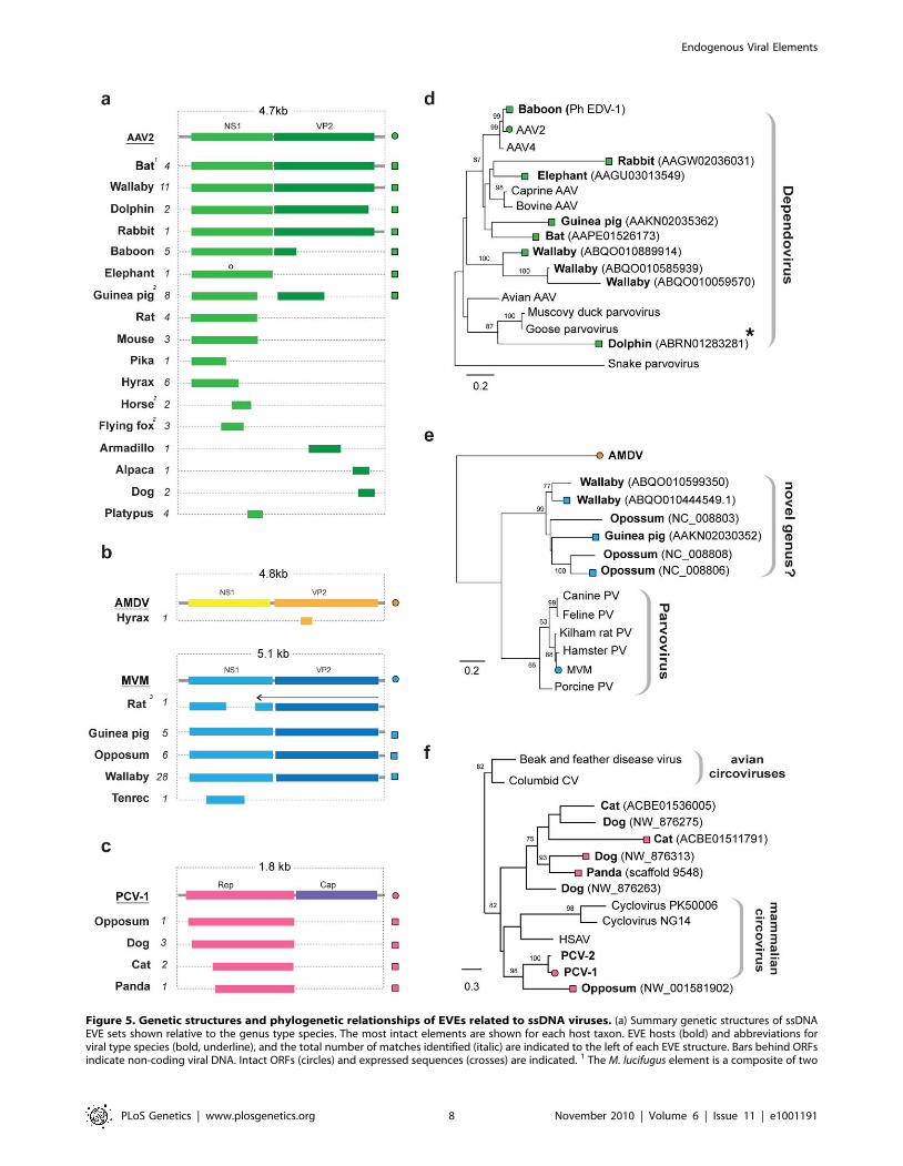
Figure 5. Genetic structures and phylogenetic relationships of EVEs related to ssDNA viruses. (a) Summary genetic structures of ssDNAEVE sets shown relative to the genus type species. The most intact elements are shown for each host taxon. EVE hosts (bold) and abbreviations forviral type species (bold, underline), and the total number of matches identified (italic) are indicated to the left of each EVE structure. Bars behind ORFsindicate non-coding viral DNA. Intact ORFs (circles) and expressed sequences (crosses) are indicated. 1 The M. lucifugus element is a composite of two
Endogenous Viral Elements
PLoS Genetics | www.plosgenetics.org 8 November 2010 | Volume 6 | Issue 11 | e1001191
Highly discordant host ranges among closely related EVEs (or
EVEs and exogenous viruses) can provide information about
transmission events. In this regard, we note that a dependovirus
EVE in the bottlenose dolphin (Tursiops truncatus) genome grouped
robustly with avian dependoviruses (rather than mammalian
isolates) in NS1 trees (Figure 6d), suggesting cross-class transmis-
sion of parvoviruses between birds and mammals may have
occurred in the past.
Evidence for exaptation of EVEsEVEs that are neutral or only slightly deleterious in their hosts
may fortuitously drift or hitchhike [46] to fixation, accumulating
mutations at the host neutral rate. Alternatively, EVE insertions
may confer an advantageous phenotype on the host and spread
through the population by selection. In such exapted sequences,
selection will act to maintain the functionality of the EVE
sequence. Many of the EVEs identified in this study were highly
mutated and/or fragmented and these likely represent non-
functional, neutrally evolving pseudogenes. However, several
EVEs encoded intact ORFs, and some also express RNA
(Figure 2a, Figure 3a, Figure 5a). For most of these EVEs, the
time since insertion is unknown, and intact ORFs could reflect
recent insertion rather than a long-standing history of purifying
selection within the host genome. In primates, however, orthology
of the bornavirus-derived insert EBLN-1, which is intact in several
species, demonstrates an insertion date predating the divergence of
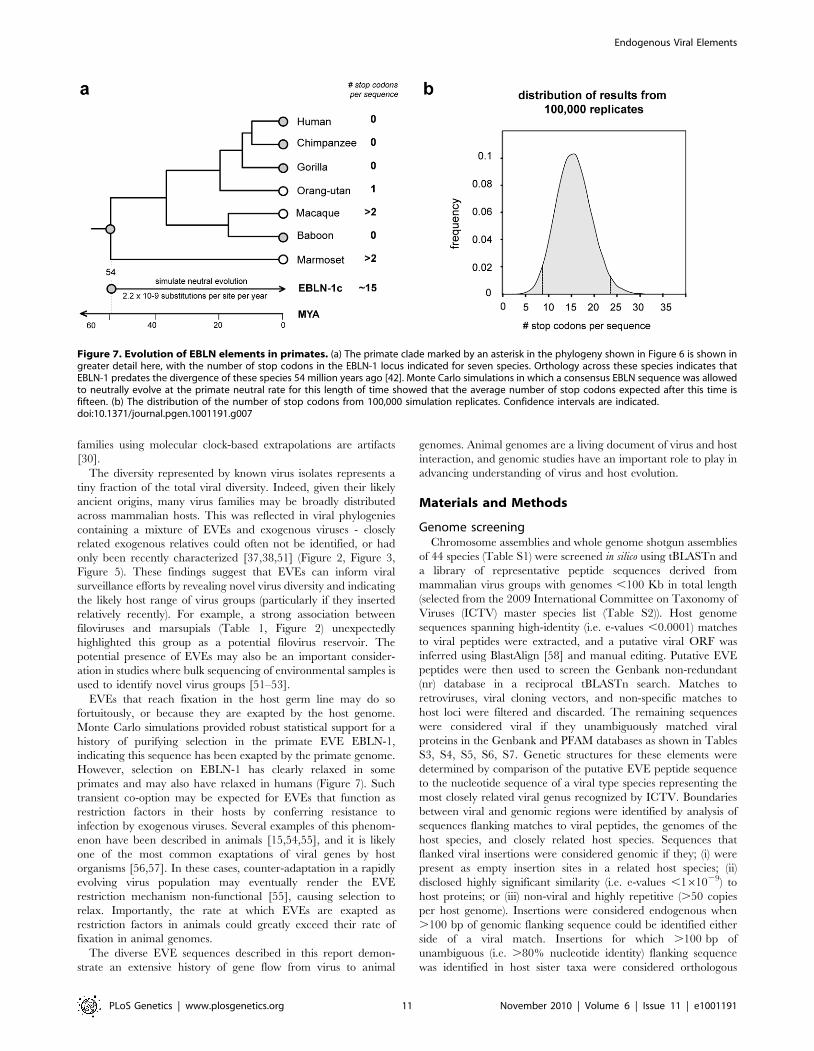
strepsirhine primates (,54 million years ago (MYA)) (Figure 7).
Simulations in which a consensus derived from all EBLN-1
sequences was allowed to neutrally evolve over this time period
indicated the probability of maintaining an intact ORF in the
absence of purifying selection was ,0.00001 (100,000 replicates,
mean number of stop codons = 15.57, 95% confidence range 7.9–
23.3). This analysis provides more robust support for purifying
selection than classical tests based on the ratio of synonymous to
non-synonymous mutations (which are weakly significant for
EBLN-1 [11]), strongly indicating that EBLN-1 has been exapted
in the primate genome, at least during part of its evolutionary
history. Curiously, however, EBLN-1 has not retained coding
capacity in all primate species. Perhaps selection to maintain it has
recently been lost across all primates, and all the inserts may
become inactivated in future.
Discussion
In this report, systematic screening revealed that sequences derived
from a broad range of non-retroviral mammalian virus groups occur
as endogenous elements in the genomes of mammals, birds and insect
vectors. We describe the first EVEs derived from the rtDNA and
dsRNA groups, thereby establishing that the complete range of
known animal virus replication strategies (see Figure 1) are
represented by endogenous elements in animal genomes.
Richer sampling of animal genomes is likely to reveal an even
greater diversity of EVEs. While EVEs that are very ancient (i.e.
that inserted prior to the divergence of major host lineages) can be
identified by selectively screening a small number of host species,
identification of more recent insertions will often require richer
sampling within orders and genera. Sampling of mammalian
species for whole genome sequencing has generally been across,
rather than within orders (primates are an exception). Conse-
quently the majority of mammal species sampled in this study
diverged more than 50 million years ago (Figure 6). Any mammal
species that was not sampled, and diverged more recently, could
contain uncharacterized EVEs. Sampling of avian and insect
vector genomes has so far been quite limited, and these may also
harbor a rich virus fossil history. Furthermore, the vast majority of
EVE insertions never reach fixation, and there are likely many
unfixed EVEs present within species gene pools at a given time
(known examples of unfixed EVEs include Israeli acute paralysis
virus (IAPPV) insertions in honey bees (Apis mellifera) [15], koala
endogenous retrovirus (KoRV) in koala bears [47], and human
herpesvirus 6 (HHV-6) and HERV-K HML-2 insertions in
humans [18,48]). Identification of such unfixed EVEs will often
require population-level screening.
The in silico screening strategy employed here likely underesti-
mates the actual diversity of EVEs for several reasons. Firstly, only
low-coverage, incomplete genome data were available for most
species. Furthermore, EVEs within the data we screened could
have been overlooked because (i) screening was based on similarity
searches, and is thus dependent on current (limited) knowledge of
viral diversity, and (ii) more ancient EVEs may not be identified
due to the divergence in both host and virus lineages subsequent to
insertion (this may also result in a bias toward detecting more
conserved genes).
Certain groups of (non-retroviral) viruses appear to be better
represented in the genomic fossil record than others (e.g.
Parvoviridae, Mononegavirales). This likely reflects a predisposition
for germline integration among viruses with particular patterns of
replication and infection. Notably, viruses that establish persistent
infections and/or replicate within the nucleus are particularly well
represented among the EVEs identified in this study. Nevertheless,
these characteristics do not appear to be prerequisites for germ line
integration (Table 1). Indeed, since retroelements are ubiquitous in
animal genomes, and replication of all known viruses requires the
expression of RNA, retroelement activity in germ line cells [49]
may present a general mechanism for mediating insertion of virus
genes into animal germ lines (see Figure 1).
The discovery that a broad range of viruses are represented by
EVEs in animal genomes indicates that viral ‘molecular fossils’ can
provide the basis for robust, time-scaled, macroevolutionary
studies across a range of animal and virus groups. For example,
EVE sequences can be combined with phylogenetic data of extant
host species to reveal patterns of inter-class virus transmission
(Figure 5) [50]. In this study, orthologous EVEs derived from the
Borna-, Filo- Circo-, and Parvoviridae provided direct evidence for the
ancient origins of these families (Figure 6). These findings also
indicate that more recent dates of origin obtained for other virus
genomic contigs; 2 Structures represent a composite of semi-overlapping fragments; 3 Element has undergone genomic rearrangements, with thearrow indicating the direction of the rearranged fragment. (b) Phylogenetic relationships of dependovirus EVEs and representative exogenous viruses,based on NS1 gene and rooted on snake parvovirus. A dolphin EVE (indicated by an asterisk) groups robustly with avian rather than mammalianisolates. (d) Phylogenetic relationships of parvovirus EVEs and representative exogenous viruses, based on NS1 gene and rooted on Aleutian minkdisease virus. Support for both ML phylogenetic trees was evaluated using 1,000 nonparametric bootstrap replicates. EVEs potentially comprising anew genus are indicated. (d) Phylogenetic relationships of circovirus EVEs and representative exogenous viruses, based on the Rep gene and rootedon avian circoviruses, with support for the ML phylogenetic tree evaluated using 1,000 nonparametric bootstrap replicates. Taxa that are shown asgenetic structures in (a) are indicated by colored squares (EVEs) and circles (exogenous viruses) (a). Abbreviations: AAV = adeno-associated virus;MMV = minute virus of mice, AMDV = Aleutian mink disease virus, PV = parvovirus, CV = circovirus, PCV = porcine circovirus; HSAV = Human stool-associated circular virus.doi:10.1371/journal.pgen.1001191.g005
Endogenous Viral Elements
PLoS Genetics | www.plosgenetics.org 9 November 2010 | Volume 6 | Issue 11 | e1001191
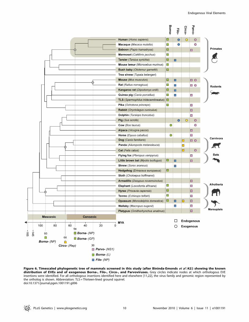
Figure 6. Timescaled phylogenetic tree of mammals screened in this study (after Bininda-Emonds et al [42]) showing the knowndistribution of EVEs and of exogenous Borna-, Filo-, Circo-, and Parvoviruses. Grey circles indicate nodes at which orthologous EVEinsertions were identified. For all orthologous insertions identified here and elsewhere [11,22], the virus family and genomic region represented bythe ortholog is shown. Abbreviation: TLS = Thirteen-lined ground squirrel.doi:10.1371/journal.pgen.1001191.g006
Endogenous Viral Elements
PLoS Genetics | www.plosgenetics.org 10 November 2010 | Volume 6 | Issue 11 | e1001191
families using molecular clock-based extrapolations are artifacts
[30].
The diversity represented by known virus isolates represents a
tiny fraction of the total viral diversity. Indeed, given their likely
ancient origins, many virus families may be broadly distributed
across mammalian hosts. This was reflected in viral phylogenies
containing a mixture of EVEs and exogenous viruses - closely
related exogenous relatives could often not be identified, or had
only been recently characterized [37,38,51] (Figure 2, Figure 3,
Figure 5). These findings suggest that EVEs can inform viral
surveillance efforts by revealing novel virus diversity and indicating
the likely host range of virus groups (particularly if they inserted
relatively recently). For example, a strong association between
filoviruses and marsupials (Table 1, Figure 2) unexpectedly
highlighted this group as a potential filovirus reservoir. The
potential presence of EVEs may also be an important consider-
ation in studies where bulk sequencing of environmental samples is
used to identify novel virus groups [51–53].
EVEs that reach fixation in the host germ line may do so
fortuitously, or because they are exapted by the host genome.
Monte Carlo simulations provided robust statistical support for a
history of purifying selection in the primate EVE EBLN-1,
indicating this sequence has been exapted by the primate genome.
However, selection on EBLN-1 has clearly relaxed in some
primates and may also have relaxed in humans (Figure 7). Such
transient co-option may be expected for EVEs that function as
restriction factors in their hosts by conferring resistance to
infection by exogenous viruses. Several examples of this phenom-
enon have been described in animals [15,54,55], and it is likely
one of the most common exaptations of viral genes by host
organisms [56,57]. In these cases, counter-adaptation in a rapidly
evolving virus population may eventually render the EVE
restriction mechanism non-functional [55], causing selection to
relax. Importantly, the rate at which EVEs are exapted as
restriction factors in animals could greatly exceed their rate of
fixation in animal genomes.
The diverse EVE sequences described in this report demon-
strate an extensive history of gene flow from virus to animal
genomes. Animal genomes are a living document of virus and host
interaction, and genomic studies have an important role to play in
advancing understanding of virus and host evolution.
Materials and Methods
Genome screeningChromosome assemblies and whole genome shotgun assemblies
of 44 species (Table S1) were screened in silico using tBLASTn and
a library of representative peptide sequences derived from
mammalian virus groups with genomes ,100 Kb in total length
(selected from the 2009 International Committee on Taxonomy of
Viruses (ICTV) master species list (Table S2)). Host genome
sequences spanning high-identity (i.e. e-values ,0.0001) matches
to viral peptides were extracted, and a putative viral ORF was
inferred using BlastAlign [58] and manual editing. Putative EVE
peptides were then used to screen the Genbank non-redundant
(nr) database in a reciprocal tBLASTn search. Matches to
retroviruses, viral cloning vectors, and non-specific matches to
host loci were filtered and discarded. The remaining sequences
were considered viral if they unambiguously matched viral
proteins in the Genbank and PFAM databases as shown in Tables
S3, S4, S5, S6, S7. Genetic structures for these elements were
determined by comparison of the putative EVE peptide sequence
to the nucleotide sequence of a viral type species representing the
most closely related viral genus recognized by ICTV. Boundaries
between viral and genomic regions were identified by analysis of
sequences flanking matches to viral peptides, the genomes of the
host species, and closely related host species. Sequences that
flanked viral insertions were considered genomic if they; (i) were
present as empty insertion sites in a related host species; (ii)
disclosed highly significant similarity (i.e. e-values ,161029) to
host proteins; or (iii) non-viral and highly repetitive (.50 copies
per host genome). Insertions were considered endogenous when
.100 bp of genomic flanking sequence could be identified either
side of a viral match. Insertions for which .100 bp of
unambiguous (i.e. .80% nucleotide identity) flanking sequence
was identified in host sister taxa were considered orthologous
Figure 7. Evolution of EBLN elements in primates. (a) The primate clade marked by an asterisk in the phylogeny shown in Figure 6 is shown ingreater detail here, with the number of stop codons in the EBLN-1 locus indicated for seven species. Orthology across these species indicates thatEBLN-1 predates the divergence of these species 54 million years ago [42]. Monte Carlo simulations in which a consensus EBLN sequence was allowedto neutrally evolve at the primate neutral rate for this length of time showed that the average number of stop codons expected after this time isfifteen. (b) The distribution of the number of stop codons from 100,000 simulation replicates. Confidence intervals are indicated.doi:10.1371/journal.pgen.1001191.g007
Endogenous Viral Elements
PLoS Genetics | www.plosgenetics.org 11 November 2010 | Volume 6 | Issue 11 | e1001191
insertions. PERL scripts were used to automate BLAST searches
and sequence extraction. Putative EVE peptide sequences, and
alignments of EVEs and exogenous retroviruses, are available
online (http://saturn.adarc.org/paleo/).
Phylogenetic analysisPutative EVE sequences inferred using BlastAlign were aligned
with closely related viruses using MUSCLE and manually edited
[59]. Maximum likelihood (ML) phylogenies were estimated using
amino acid sequence alignments with RAXML [60], implement-
ing in each case the best fitting substitution model as determined
by ProtTest [61]. Support for the ML trees was evaluated with
1000 nonparametric bootstrap replicates. The best fitting models
for the datasets were: Parvoviridae: dependovirus NS1 gene (JTT+C,
332 amino acids across 17 taxa), Parvoviridae: parvovirus NS1 gene,
(JTT+C, 293 amino acids across 13 taxa), Circoviridae: Rep gene
(Blosum62+C+F, 235 amino acids across 14 taxa), Hepadnaviridae:
polymerase gene (JTT+C+F, 661 amino acids across 9 taxa),
Orthomyxoviridae: GP gene (WAG+C+F, 482 amino acids across 5
taxa), Reoviridae: VP5 gene (Dayhoff+C+F, 171 amino acids across
4 taxa), Bunyaviridae: phlebovirus NP gene (LG+C, 247 amino acids
across 12 taxa), Bunyaviridae: nairovirus NP gene (LG+C, 446
amino acids across 5 taxa), Flaviviridae: mostly NS3 gene (LG+C+F,
1846 amino acids across 8 taxa), Filoviridae: NP gene (JTT+C, 369
amino acids across 29 taxa), Filoviridae: L gene (LG+C+F, 517
amino acids across 9 taxa), Bornaviridae: NP gene (JTT+C, 147
amino acids across 73 taxa), Bornaviridae: L gene (JTT+C+F, 1243
amino acids across 12 taxa), Rhabdoviridae: NP gene (LG+C, 220
amino acids across 34 taxa), Rhabdoviridae: L gene (LG+C+F, 383
amino acids across 26 taxa).
SimulationA Monte Carlo simulation procedure was employed to
determine the probability that the bornavirus-derived element
EBLN-1 has retained coding capacity over 54.1 million years
under neutral evolution (i.e. not under purifying selection). A
consensus EBLN-1 sequence was inferred, and the effects of
neutral evolution were simulated using seq-gen [62] for a branch
length equivalent to the minimum amount of time that EBLN-1
orthologs have resided in primate genomes, based on the primate
divergences estimated by Bininda-Emonds et al [42], and given a
neutral rate of evolution of 2.2610–9 [12]. The number of stop
codons accrued was counted for 100,000 iterations of the
simulation. The probability that the reading frame could have
remained open under neutrality is given by the number of
replicates under which no stop codons have evolved, divided by
the number of iterations.
Sequences and accession numbersParvoviridae; AAV2 (NC_001401); Minute virus of mice
(NC_001510.1); AMDV (NC_001662); Goose parvovirus
(EU583390.1); Muscovy duck parvovirus (X75093.1); Porcine
hokovirus (EU200671.1); Snake parvovirus (AY349010.1); Avian
AAV (AY629582.1, AY629583.1, GQ368252.1); AAV1
(AF063497.1); AAV4 (U89790); AAV2 (AY695375.1); Bovine
AAV (AY388617.1); Caprine AAV (DQ335246.2); Bocavirus
(M14363.1); Erythrovirus (AB126265.1); Aleutian mink disease
virus (M20036.1); Porcine parvovirus (EU790642.1); Feline
panleukopenia virus (EF988660.1); Canine parvovirus
(EU310373.2); Rat parvovirus (AF036710.1); Hamster parvovirus
(U34255.1); Minute virus of mice (DQ196317.1); Kilham rat virus
(U79033.1); Circoviridae; Porcine circovirus 1 (NC_006266);
Porcine circovirus 2 (GU325757); Cyclovirus PK5006
(GQ404856.1); Cyclovirus NG14 (GQ404855.1); Human stool-
associated circular virus NG13 (GQ404856.1); Beak and feather
disease virus (AY450436.1); Columbid circovirus (AF252610.1);
Hepadnaviridae; duck HBV (NC_001344); Stork HBV
(AJ251937.1|); Heron HBV (NC_001486); Ross’ Goose HBV
(AY494849.1); Crane HBV (AJ441113.1); Sheldgoose HBV
(AY494852.1); Snow goose HBV (AF111000.1); Woodchuck
HBV (AF410861.1); Flaviviridae; Kamiti river virus (NC_005064);
Aedes flavivirus (NC_012932); Quang binh virus (NC_012671);
Culex flavivirus (NC_008604); Nakiwogo virus (GQ165809).
Reoviridae; Liaoning virus (NC_007736 - NC_007747); Kadipiro
virus (NC_004199, NC_004205-NC_004210, NC_004212-
NC_004216); Banna virus (NC_004198, NC_004200-
NC_004204, NC_004211, NC_004217-NC_004221). Bunyaviridae;
Crimean-Congo hemorrhagic fever virus (NC_005300, NC_
005301, NC_005302); Uukuniemi virus (NC_005214, NC_
005220, NC_005221); Uukuniemi virus (M33551.1); Catch-me-
cave virus (EU274384.1); Sandfly fever Naples virus (EF201832.1);
Massilia virus (EU725773.1); Punta Toro virus (EF201834.1);
Buenaventura virus (EF201839.1); Rift Valley fever virus
(DQ380156.1); Phlebovirus sp. (EF201818.1); Icoaraci virus
(EF076014.1). Orthomyxoviridae; Quaranfil virus (FJ861694.1);
Johnston Atoll virus (FJ861696.1); Thogoto virus (M77280.1);
Dhori virus (M34002.1). Bornaviridae; Borna disease virus
(NC_001607); Avian BDV (FJ169441). Filoviridae; Reston ebola
virus (NC_002549); Zaire ebola virus (NC_002549); Lake Victoria
marburgvirus (NC_001608). Rhabdoviridae; vesicular stomatitis
virus (NC_001560); Wongabel virus (NC_011639); Kotonkon
virus (DQ457099); Adelaide river virus (U10363.1); Obodhiang
virus (DQ457098.1); Bovine ephemeral fever virus (AF234533.1);
Rochambeau virus (DQ457104.1); Mount elgon bat virus
(DQ457103.1); Oita rhabdovirus (AB116386); Kern canyon virus
(DQ457101.1); Sandjimba virus (DQ457102.1); Kolongo virus
(DQ457100.1); Tupaia rhabdovirus (AY840978.1); Spring viremia
of carp (DQ491000.1); Pike fry rhabdovirus (FJ872827.1); Cocal
virus (EU373657.1); Vesicular stomatitis Indiana virus
(AF473865.1); Isfahan virus (AJ810084.2); Chandipura virus
(AY614728.1); Ngaingan virus (FJ715959.1); Wongabel virus
(EF612701.1); Flanders virus (AF523194.1). Nyaviridae; Midway
virus (NC_012702); Nyamanini virus (NC_012703).
Supporting Information
Figure S1 Sequence alignment of an EVE identified in the Aedes
aegyptii genome and Liaoning virus segment 5. Genomic regions, as
determined by alignment to a repetitive element (RE) in the A.
aegyptii genome, are indicated in blue, on coding viral regions are
shown in red, and regions encoding viral proteins are shown in
green.
Found at: doi:10.1371/journal.pgen.1001191.s001 (0.40 MB PDF)
Figure S2 Genetic structure of an adenovirus related sequence
identified in whole-genome shotgun sequence data for Ord’s
kangaroo rat (Dipodymys ordii). The name of the corresponding
protein in the most closely related virus (tree shrew adenovirus 1;
AF258784.1) is indicated above each open reading frame (ORF).
Arrows beneath ORFs indicate frames encoded in reverse
direction relative to contig. Abbreviations: kd = kiloDalton;
pol = DNA polymerase; T = terminal protein; P = penton base;
Mco = minor core; Mca = minor capsid; DB = DNA binding;
Ma = Major coat; H = hexon-associated; S = shaft.
Found at: doi:10.1371/journal.pgen.1001191.s002 (0.27 MB PDF)
Figure S3 Genetic structure of a complete dependovirus genome
identified in the little brown bat (Myotis lucifugus) genome. The
element is a composite of two genomic contigs, which were
Endogenous Viral Elements
PLoS Genetics | www.plosgenetics.org 12 November 2010 | Volume 6 | Issue 11 | e1001191
assembled by identifying the empty pre-integration site in the
closest relative (Pteropus vampyrus). The inset box shows an
alignment the inverted repeats in the 59 and 39 untranslated
regions. Abbreviations: IR = inverted repeat. NS1 = Non-structur-
al protein 1; VP2 = Viral protein 2; UTR = untranslated region.
Found at: doi:10.1371/journal.pgen.1001191.s003 (0.32 MB PDF)
Table S1 Genome sequences screened for endogenous viral
elements.
Found at: doi:10.1371/journal.pgen.1001191.s004 (0.09 MB
DOC)
Table S2 Viral reference sequences used for in silico screening of
host genomes.
Found at: doi:10.1371/journal.pgen.1001191.s005 (0.14 MB
DOC)
Table S3 Endogenous viral elements related to negative sense
RNA viruses.
Found at: doi:10.1371/journal.pgen.1001191.s006 (0.40 MB
DOC)
Table S4 Endogenous viral elements related to doubled-
stranded RNA viruses.
Found at: doi:10.1371/journal.pgen.1001191.s007 (0.03 MB
DOC)
Table S5 Endogenous viral elements related to positive sense
RNA viruses.
Found at: doi:10.1371/journal.pgen.1001191.s008 (0.09 MB
DOC)
Table S6 Endogenous viral elements related to single stranded
DNA viruses.
Found at: doi:10.1371/journal.pgen.1001191.s009 (0.21 MB
DOC)
Table S7 Endogenous viral elements related to reverse tran-
scribing DNA viruses.
Found at: doi:10.1371/journal.pgen.1001191.s010 (0.04 MB
DOC)
Acknowledgments
The authors thank Paul Bieniasz, Michael Tristem, Paul Klenerman, and
anonymous reviewers for helpful comments and suggestions.
Author Contributions
Conceived and designed the experiments: AK RJG. Performed the
experiments: AK RJG. Analyzed the data: AK RJG. Contributed
reagents/materials/analysis tools: AK RJG. Wrote the paper: AK RJG.
References
1. Benveniste RE, Todaro GJ (1974) Evolution of C-type viral genes: inheritance of
exogenously acquired viral genes. Nature 252: 456–459.
2. Jaenisch R (1976) Germ line integration and Mendelian transmission of the
exogenous Moloney leukemia virus. Proc Natl Acad Sci U S A 73: 1260–1264.
3. Bejarano ER, Khashoggi A, Witty M, Lichtenstein C (1996) Integration of
multiple repeats of geminiviral DNA into the nuclear genome of tobacco duringevolution. Proc Natl Acad Sci U S A 93: 759–764.
4. Herniou E, Martin J, Miller K, Cook J, Wilkinson M, et al. (1998) Retroviral
diversity and distribution in vertebrates. J Virol 72: 5955–5966.
5. Tristem M (2000) Identification and characterization of novel human
endogenous retrovirus families by phylogenetic screening of the human genomemapping project database. J Virol 74: 3715–3730.
6. Crochu S, Cook S, Attoui H, Charrel RN, De Chesse R, et al. (2004) Sequencesof flavivirus-related RNA viruses persist in DNA form integrated in the genome
of Aedes spp. mosquitoes. J Gen Virol 85: 1971–1980.
7. Tang KF, Lightner DV (2006) Infectious hypodermal and hematopoietic
necrosis virus (IHHNV)-related sequences in the genome of the black tigerprawn Penaeus monodon from Africa and Australia. Virus Res 118: 185–191.
8. Staginnus C, Richert-Poggeler KR (2006) Endogenous pararetroviruses: two-faced travelers in the plant genome. Trends Plant Sci 11: 485–491.
9. Bezier A, Annaheim M, Herbiniere J, Wetterwald C, Gyapay G, et al. (2009)Polydnaviruses of braconid wasps derive from an ancestral nudivirus. Science
323: 926–930.
10. Taylor DJ, Bruenn J (2009) The evolution of novel fungal genes from non-
retroviral RNA viruses. BMC Biol 7: 88.
11. Horie M, Honda T, Suzuki Y, Kobayashi Y, Daito T, et al. (2010) Endogenous
non-retroviral RNA virus elements in mammalian genomes. Nature 463: 84–87.
12. Lander ES, Linton LM, Birren B, Nusbaum C, Zody MC, et al. (2001) Initialsequencing and analysis of the human genome. Nature 409: 860–921.
13. Sperber G, Lovgren A, Eriksson NE, Benachenhou F, Blomberg J (2009)RetroTector online, a rational tool for analysis of retroviral elements in small
and medium size vertebrate genomic sequences. BMC Bioinformatics 10(Suppl
6): S4.
14. Malik HS, Henikoff S, Eickbush TH (2000) Poised for contagion: evolutionaryorigins of the infectious abilities of invertebrate retroviruses. Genome Res 10:
1307–1318.
15. Maori E, Tanne E, Sela I (2007) Reciprocal sequence exchange between non-
retro viruses and hosts leading to the appearance of new host phenotypes.Virology 362: 342–349.
16. Shafritz DA, Shouval D, Sherman HI, Hadziyannis SJ, Kew MC (1981)Integration of hepatitis B virus DNA into the genome of liver cells in chronic
liver disease and hepatocellular carcinoma. Studies in percutaneous liver biopsies
and post-mortem tissue specimens. N Engl J Med 305: 1067–1073.
17. Berns KI, Linden RM (1995) The cryptic life style of adeno-associated virus.Bioessays 17: 237–245.
18. Arbuckle JH, Medveczky MM, Luka J, Hadley SH, Luegmayr A, et al. (2010)The latent human herpesvirus-6A genome specifically integrates in telomeres of
human chromosomes in vivo and in vitro. Proc Natl Acad Sci U S A 107:5563–5568.
19. Zhdanov VM (1975) Integration of viral genomes. Nature 256: 471–473.
20. Klenerman P, Hengartner H, Zinkernagel RM (1997) A non-retroviral RNA
virus persists in DNA form. Nature 390: 298–301.
21. Geuking MB, Weber J, Dewannieux M, Gorelik E, Heidmann T, et al. (2009)Recombination of retrotransposon and exogenous RNA virus results in
nonretroviral cDNA integration. Science 323: 393–396.
22. Taylor DJ, Leach RW, Bruenn J (2010) Filoviruses are ancient and integrated
into mammalian genomes. BMC Evol Biol 10: 193.
23. Ribet D, Harper F, Dupressoir A, Dewannieux M, Pierron G, et al. (2008) Aninfectious progenitor for the murine IAP retrotransposon: emergence of an
intracellular genetic parasite from an ancient retrovirus. Genome Res 18: 597–609.
24. Kazazian HH, Jr. (2004) Mobile elements: drivers of genome evolution. Science
303: 1626–1632.
25. Dunlap KA, Palmarini M, Varela M, Burghardt RC, Hayashi K, et al. (2006)Endogenous retroviruses regulate periimplantation placental growth and
differentiation. Proc Natl Acad Sci U S A 103: 14390–14395.
26. Dupressoir A, Vernochet C, Bawa O, Harper F, Pierron G, et al. (2009)
Syncytin-A knockout mice demonstrate the critical role in placentation of a
fusogenic, endogenous retrovirus-derived, envelope gene. Proc Natl AcadSci U S A 106: 12127–12132.
27. Gifford R, Tristem M (2003) The evolution, distribution and diversity ofendogenous retroviruses. Virus Genes 26: 291–316.
28. Emerman M, Malik HS (2010) Paleovirology–modern consequences of ancient
viruses. PLoS Biol 8: e1000301. doi:10.1371/journal.pbio.1000301.
29. Charleston MA, Robertson DL (2002) Preferential host switching by primate
lentiviruses can account for phylogenetic similarity with the primate phylogeny.Syst Biol 51: 528–535.
30. Holmes EC (2003) Molecular clocks and the puzzle of RNA virus origins. J Virol
77: 3893–3897.
31. Katzourakis A, Tristem M, Pybus OG, Gifford RJ (2007) Discovery and analysis
of the first endogenous lentivirus. Proc Natl Acad Sci U S A 104: 6261–6265.
32. Simmonds P, Smith DB (1999) Structural constraints on RNA virus evolution.
J Virol 73: 5787–5794.
33. Belshaw R, Gardner A, Rambaut A, Pybus OG (2008) Pacing a small cage:mutation and RNA viruses. Trends Ecol Evol 23: 188–193.
34. Gibbs AJ, Fargette D, Garcia-Arenal F, Gibbs MJ (2010) Time–the emergingdimension of plant virus studies. J Gen Virol 91: 13–22.
35. Keckesova Z, Ylinen LM, Towers GJ, Gifford RJ, Katzourakis A (2009)
Identification of a RELIK orthologue in the European hare (Lepus europaeus)reveals a minimum age of 12 million years for the lagomorph lentiviruses.
Virology 384: 7–11.
36. Esnault C, Maestre J, Heidmann T (2000) Human LINE retrotransposons
generate processed pseudogenes. Nat Genet 24: 363–367.
37. Presti RM, Zhao G, Beatty WL, Mihindukulasuriya KA, da Rosa AP, et al.
(2009) Quaranfil, Johnston Atoll, and Lake Chad viruses are novel members of
the family Orthomyxoviridae. J Virol 83: 11599–11606.
38. Attoui H, Mohd Jaafar F, Belhouchet M, Tao S, Chen B, et al. (2006) Liao ning
virus, a new Chinese seadornavirus that replicates in transformed and embryonicmammalian cells. J Gen Virol 87: 199–208.
Endogenous Viral Elements
PLoS Genetics | www.plosgenetics.org 13 November 2010 | Volume 6 | Issue 11 | e1001191
39. Roiz D, Vazquez A, Seco MP, Tenorio A, Rizzoli A (2009) Detection of novel
insect flavivirus sequences integrated in Aedes albopictus (Diptera: Culicidae) inNorthern Italy. Virol J 6: 93.
40. Samulski RJ, Zhu X, Xiao X, Brook JD, Housman DE, et al. (1991) Targeted
Integration of adeno-associated virus (AAV) into human chromosome 19. EmboJournal 10: 3941–3950.
41. Kotin RM, Linden RM, Berns KI (1992) Characterization of a preferred site onhuman chromosome 19q for integration of adeno-associated virus DNA by non-
homologous recombination. EMBO J 11: 5071–5078.
42. Bininda-Emonds OR, Cardillo M, Jones KE, MacPhee RD, Beck RM, et al.(2007) The delayed rise of present-day mammals. Nature 446: 507–512.
43. Favaro P, Downey HD, Zhou JS, Wright JF, Hauck B, et al. (2009) Host andvector-dependent effects on the risk of germline transmission of AAV vectors.
Mol Ther 17: 1022–1030.44. Towner JS, Amman BR, Sealy TK, Carroll SA, Comer JA, et al. (2009) Isolation
of genetically diverse Marburg viruses from Egyptian fruit bats. PLoS Pathog 5:
e1000536. doi:10.1371/journal.ppat.1000536.45. Barrette RW, Metwally SA, Rowland JM, Xu L, Zaki SR, et al. (2009) Discovery
of swine as a host for the Reston ebolavirus. Science 325: 204–206.46. Smith JM, Haigh J (1974) The hitch-hiking effect of a favourable gene. Genet
Res 23: 23–35.
47. Tarlinton R, Meers J, Young P (2008) Biology and evolution of the endogenouskoala retrovirus. Cell Mol Life Sci 65: 3413–3421.
48. Belshaw R, Dawson AL, Woolven-Allen J, Redding J, Burt A, et al. (2005)Genomewide screening reveals high levels of insertional polymorphism in the
human endogenous retrovirus family HERV-K(HML2): implications forpresent-day activity. J Virol 79: 12507–12514.
49. Giordano R, Magnano AR, Zaccagnini G, Pittoggi C, Moscufo N, et al. (2000)
Reverse transcriptase activity in mature spermatozoa of mouse. J Cell Biol 148:1107–1113.
50. Martin J, Herniou E, Cook J, O’Neill RW, Tristem M (1999) Interclasstransmission and phyletic host tracking in murine leukemia virus-related
retroviruses. J Virol 73: 2442–2449.
51. Li L, Kapoor A, Slikas B, Bamidele OS, Wang C, et al. (2010) Multiple diverse
circoviruses infect farm animals and are commonly found in human and
chimpanzee feces. J Virol 84: 1674–1682.
52. Victoria JG, Kapoor A, Dupuis K, Schnurr DP, Delwart EL (2008) Rapid
identification of known and new RNA viruses from animal tissues. PLoS Pathog
4: e1000163. doi:10.1371/journal.ppat.1000163.
53. Li L, Victoria JG, Wang C, Jones M, Fellers GM, et al. (2010) Bat guano virome:
predominance of dietary viruses from insects and plants plus novel mammalian
viruses. J Virol 84: 6955–6965.
54. Best S, Le Tissier P, Towers G, Stoye JP (1996) Positional cloning of the mouse
retrovirus restriction gene Fv1. Nature 382: 826–829.
55. Arnaud F, Caporale M, Varela M, Biek R, Chessa B, et al. (2007) A paradigm
for virus-host coevolution: sequential counter-adaptations between endogenous
and exogenous retroviruses. PLoS Pathog 3: e170. doi:10.1371/journal.
ppat.0030170.
56. Koonin EV (2010) Taming of the shrewd: novel eukaryotic genes from RNA
viruses. BMC Biol 8: 2.
57. van der Oost J, Jore MM, Westra ER, Lundgren M, Brouns SJ (2009) CRISPR-
based adaptive and heritable immunity in prokaryotes. Trends Biochem Sci 34:
401–407.
58. Belshaw R, Katzourakis A (2005) BlastAlign: a program that uses blast to align
problematic nucleotide sequences. Bioinformatics 21: 122–123.
59. Edgar RC (2004) MUSCLE: multiple sequence alignment with high accuracy
and high throughput. Nucleic Acids Res 32: 1792–1797.
60. Stamatakis A (2006) RAxML-VI-HPC: maximum likelihood-based phylogenetic
analyses with thousands of taxa and mixed models. Bioinformatics 22:
2688–2690.
61. Abascal F, Zardoya R, Posada D (2005) ProtTest: selection of best-fit models of
protein evolution. Bioinformatics 21: 2104–2105.
62. Rambaut A, Grassly NC (1997) Seq-Gen: an application for the Monte Carlo
simulation of DNA sequence evolution along phylogenetic trees. Comput Appl
Biosci 13: 235–238.
Endogenous Viral Elements
PLoS Genetics | www.plosgenetics.org 14 November 2010 | Volume 6 | Issue 11 | e1001191