emerging perspectives on the mechanisms, regulation, and distribution of light color acclimation in...
TRANSCRIPT
Molecular Plant • Pages 1–13, 2011 REVIEW ARTICLE
Emerging Perspectives on the Mechanisms,Regulation, and Distribution of Light ColorAcclimation in Cyanobacteria
Andrian Gutua,b and David M. Kehoea,1
a Department of Biology, 1001 East Third Street, Indiana University, Bloomington, IN 47405, USAb Present address: Howard Hughes Medical Institute/Department of Molecular and Cellular Biology, FAS Center for Systems Biology, Harvard University,Cambridge, MA 02138, USA
ABSTRACT Chromatic acclimation (CA) providesmany cyanobacteriawith the ability to tailor the properties of their light-
harvesting antennae to the spectral distribution of ambient light. CA was originally discovered as a result of its dramatic
cellular phenotype in red and green light. However, discoveries over the past decade have revealed thatmany pairs of light
colors, ranging from blue to infrared, can trigger CA responses. The capacity to undergo CA is widespread geographically,
occurs in most habitats around the world, and is found within all major cyanobacterial groups. In addition, many other
cellular activities have been found to be under CA control, resulting in distinct physiological and morphological states for
cells under different light-color conditions. Several types of CA appear to be the result of convergent evolution, where
different strategies are used to achieve the final goal of optimizing light-harvesting antenna composition to maximize
photon capture. The regulation of CA has been found to occur primarily at the level of RNA abundance. The CA-regulatory
pathways uncovered thus far are two-component systems that use phytochrome-class photoreceptors with sensor-kinase
domains to control response regulators that function as transcription factors. However, there is also at least one CA-
regulatory pathway that operates at the post-transcriptional level. It is becoming increasingly clear that large numbers
of cyanobacterial species have the capacity to acclimate to a wide variety of light colors through the use of a range of
different CA processes.
Key words: Chromatic adaptation; phycobilisome; cyanobacteria; light regulation; gene regulation; signal transduction;
light harvesting; phenotypic plasticity.
INTRODUCTION
Cyanobacteria comprise a phylogenetically cohesive group of
gram-negative prokaryotes capable of oxygenic photosynthe-
sis (Stanier and Cohen-Bazire, 1977; Woese, 1987), generally
characterized by the presence of chlorophyll a and accessory
pigments called phycobiliproteins. They are one of the oldest
groups of bacteria, dating back to the Pre-Cambrian by some
estimates (Schopf, 2002) to as much as 3.5 billion years ago,
and their influence on our planet has been considerable.
Unlike other photosynthetic bacteria, during the ‘light reac-
tions’, they use both Photosystem I (PSI) and II (PSII) to extract
and transfer electrons from water molecules to electron
acceptors and generate oxygen as a by-product. They are also
unique in their ability to fix both carbon and nitrogen from
the atmosphere under aerobic conditions, which, along with
their tolerance of a wide range of environments, has allowed
colonization of many of the most extreme biotopes on Earth.
From oligotrophic oceans (Paerl, 2000) to the arid Antarctic,
subtropical deserts (Wynn-Williams, 2000), and the hot springs
of Yellowstone (Ward and Castenholzh, 2000), cyanobacteria
have adapted to occupy and expand the boundaries of the
biosphere.
As photoautotrophic organisms, the rates of photosynthesis
and growth of cyanobacteria are directly affected by the phys-
ical parameters of the environment, particularly light. Since
this resource can vary in terms of both quality (color) and
amount (intensity), sensing and adequately responding to
light is a key attribute of their eco-physiological versatility.
1 To whom correspondence should be addressed. E-mail dkehoe@india-
na.edu, tel. (812) 856-4715, fax (812) 855-6705.
ª The Author 2011. Published by the Molecular Plant Shanghai Editorial
Office in association with Oxford University Press on behalf of CSPB and
IPPE, SIBS, CAS.
doi: 10.1093/mp/ssr054
Received 7 February 2011; accepted 31 May 2011
Molecular Plant Advance Access published July 19, 2011 by guest on A
ugust 3, 2011m
plant.oxfordjournals.orgD
ownloaded from
At the cellular level, the cyanobacterial photosynthetic appa-
ratus and its light-harvesting antennae, called phycobilisomes
(PBS), are intimately attuned to ambient light conditions. In
large part, by controlling the size, composition, number,
and location of PBS, these organisms have perfected the fine
balance between maximizing the absorption of light for pho-
tosynthesis and minimizing the accumulation of excess energy
in their reaction centers, which can lead to photoinhibition
(damage to PSII reaction centers).
Cyanobacteria have adopted a number of photosynthetic
strategies to help them cope with changes in their light envi-
ronment. The different light absorption properties of the two
photosystems and their associated light-harvesting proteins
require frequent balancing of the input excitation energy be-
tween the two reaction centers. Such adjustments can act on
a very short timescale (seconds to minutes) and these are called
state transitions. Preferential excitation of PSII leads to state 2,
in which excess energy is channeled to PSI, while over excita-
tion of PSI leads to state 1, during which energy is redistributed
to PSII. State transitions in plants are accomplished through
phosphorylation and redistribution of the light-harvesting
complex II within thylakoid membranes (Rochaix, 2007; Kargul
and Barber, 2008). In cyanobacteria, this process is not as well
understood, although several lines of evidence suggest that
PBS are capable of transferring excess energy to PSI during
state 2 and that rpaC, a cyanobacteria-specific gene, is essen-
tial for this response (Fujita et al., 1994; Bhaya et al., 2000; Mul-
lineaux and Emlyn-Jones, 2005). Structural components of the
PBS have been shown to be required for state transitions
(Kondo et al., 2009). In addition, cyanobacteria undergoing
CA have been found to exist in different states, depending
on the ambient light color (Campbell, 1996). This will be dis-
cussed further below. In addition, although cyanobacterial
species are usually specialized to a particular light-irradiance
niche, they can acclimate to a range of irradiances above
and below the level required for maximal rates of photosyn-
thesis through a process termed photoacclimation, the subject
of a number of reviews (Wyman and Fay, 1987; Anderson et al.,
1995; MacIntyre et al., 2002; Walters, 2005). They also are able
to deal with excess light energy through the use of a number
of photoprotection mechanisms, which have also been
reviewed recently (Kirilovsky, 2007; Bailey and Grossman,
2008; Kirilovsky, 2010). Lastly, cyanobacteria are capable of
sensing and responding to light color, and this review will fo-
cus on these long-term acclimation responses of cyanobacteria
to changes in ambient light color. This process, which occurs in
a wide range of species and many different habitats, involves
shifts in PBS composition via the induction of specific genes
and the coordination of these changes with many additional
aspects of cellular physiology and morphology.
PBS STRUCTURE
PBS reside on the cytoplasmic surface of the thylakoid
membrane and consist of phycobiliproteins with covalently
attached bilin (open-chain tetrapyrrole) chromophores and
linker proteins. They increase the cross-sectional area for light
capture and transfer this energy to the photosystem reaction
centers. These structures are remarkably plastic, capable of
adjusting to optimize photon capture rates in different envi-
ronmental conditions by changing size, shape, protein and
bilin composition, cellular number, and association with pho-
tosynthesis reaction centers. While a variety of PBS forms exist,
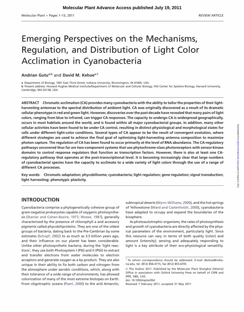
the most common is hemidiscoidal. Fan-shaped in appearance,
such PBS consist of an inner core and a series of outwardly ex-
tended rods (Figure 1). Significant variety exists even within
this structural subtype, with species- and strain-specific differ-
ences in the size of the core, the number and length of the
rods, the types and number of attached bilins, and the protein
makeup of the rods themselves. Numerous reviews addressing
the details of PBS composition are available (Sidler, 1994;
MacColl, 1998; Adir, 2005), so only a basic introduction will
be provided here. Both rods and cores are cylindrical structures
composed of a series of discs of phycobiliproteins, which con-
sist of chromophorylated alpha and beta subunits that are
together called a ‘monomer’. Two stacks of three monomers
form a disc. Linker proteins hold the discs together, keep
the rods connected to the core, and facilitate unidirectional
energy transfer from the outer portions of the rods into the
reaction centers. In addition, specialized linker proteins keep
the core, and thus the PBS itself, associated with the thylakoid
membrane. While different PBS may be composed of rods with
various types of phycobiliproteins, the core-distal discs always
have absorption wavelength maxima that are shorter than or
equivalent to the core-proximal ones to ensure unidirectional
energy transfer into the reaction center. In hemidiscoidal PBS,
which are the most common and best-studied PBS form, cores
consist of the phycobiliprotein allophycocyanin (AP; absorp-
tion maximum (kmax) = 650 nm) and the core-proximal discs
in the rods are made of phycocyanin (PC; kmax = 620 nm).
Depending on the species and environmental conditions,
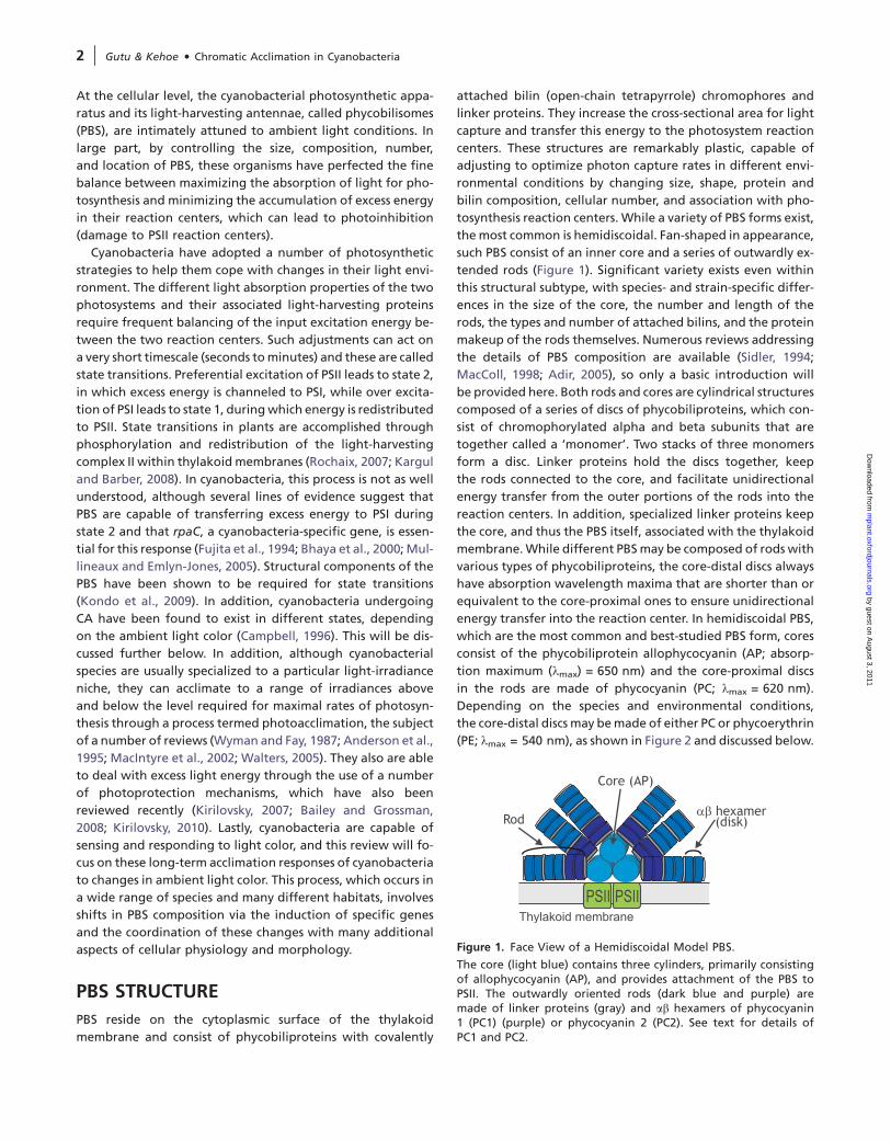
the core-distal discs may be made of either PC or phycoerythrin
(PE; kmax = 540 nm), as shown in Figure 2 and discussed below.
Figure 1. Face View of a Hemidiscoidal Model PBS.
The core (light blue) contains three cylinders, primarily consistingof allophycocyanin (AP), and provides attachment of the PBS toPSII. The outwardly oriented rods (dark blue and purple) aremade of linker proteins (gray) and ab hexamers of phycocyanin1 (PC1) (purple) or phycocyanin 2 (PC2). See text for details ofPC1 and PC2.
2 | Gutu & Kehoe d Chromatic Acclimation in Cyanobacteria
by guest on August 3, 2011
mplant.oxfordjournals.org
Dow
nloaded from
CHROMATIC ACCLIMATION RESPONSES
Overview of Chromatic Acclimation Responses as Defined
by PBS Structural Changes
In addition to light irradiance variation, cyanobacteria experi-
ence differences in light color in their natural environment.
When exposed to different light colors, many species are able
to adjust the composition of their PBS through a process
known as chromatic acclimation or adaptation (CA) (Tandeau
de Marsac, 1977, 1983; Kehoe and Gutu, 2006). Although ‘ad-
aptation’ was used when this process was initially discovered,
this response does not appear to involve any genetic alter-
ation, and significant data support the premise that CA is
the result of changes in gene-expression patterns. Thus, the
term ‘acclimation’ was proposed (Kehoe and Gutu, 2006)
and will be used instead of ‘adaptation’. Over the years, our
knowledge of the different colors of light being sensed during
CA has expanded as CA-capable cyanobacteria continue to be
discovered in additional environments. The original descrip-
tion of CA involved species that were capable of sensing red
and green light (Gaiducov, 1902), but, more recently, addi-
tional species that sense blue and green, and red and infrared
light have also been uncovered (Palenik, 2001; Duxbury et al.,
2009). This list may expand as more CA-capable species are
identified in the future.
Cyanobacteria containing both PE and PC and thus poten-
tially capable of red–green CA were classified in a comprehen-
sive study by Tandeau de Marsac and subsequent studies
(Tandeau de Marsac, 1977; Bryant, 1981, 1982; Tandeau de
Marsac, 1983). Group I species did not alter PC or PE levels in
response to changing light colors. Group II species had higher
PE levels in green light than in red light, while PC levels did
not change in either light condition. The third group, called
Group III, had higher PE levels in green light than in red light
but also accumulated more PC in red light than in green light
(Figure 2). It was a Group III species that was first noted to be
capable of CA almost a century earlier (Engelmann, 1902;
Gaiducov, 1902, 1903) and this response was named ‘comple-
mentary chromatic adaptation’ because the color of the cells
was complementary to the ambient light color: the accumula-
tionofPEmakesthesecells redcoloredingreenlight,whilehigh
levels of PC makes them blue–green in red light (Figure 2). The
filamentous Group III species Fremyella diplosiphon UTEX 481
(also calledCalothrixor Tolypothrix sp. PCC7601) has been used
extensively for CA studies and many aspects of the photobiol-
ogy and molecular biology underlying this process have
been uncovered in this organism (Tandeau de Marsac, 1983;
Grossman, 2003; Kehoe and Gutu, 2006). Group II species are
considered to be capable of undergoing ‘type 2’ CA, or CA2,
while Group III species undergo ‘type 3’ CA, or CA3. These
two groups are widely distributed globally and have been
found in freshwater, marine, hot springs, and soil environments
(Carr, 1973; Tandeau de Marsac, 1983; Postius et al., 2001;
Dufresne et al., 2008; Acinas et al., 2009; Duxbury et al., 2009).
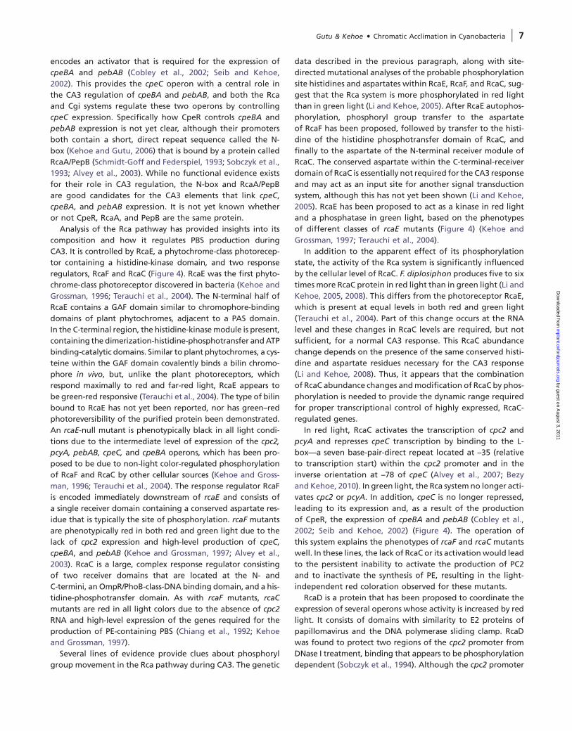
Several additional types of CA have been uncovered more re-
cently. Type 4 CA (CA4) is responsive to blue and green light and
thus far has only been found to occur in the marine environment
(Palenik, 2001). Unlike type 2 and type 3 CA, there are apparently
no major changes in PBS protein composition during CA4. In-
stead, it appears to be the ratio of two bilin isomers that are at-
tached to a specialized type of PE called ‘PEII’ in the most core-
distal portions of the rods of the PBS that change (Palenik, 2001;
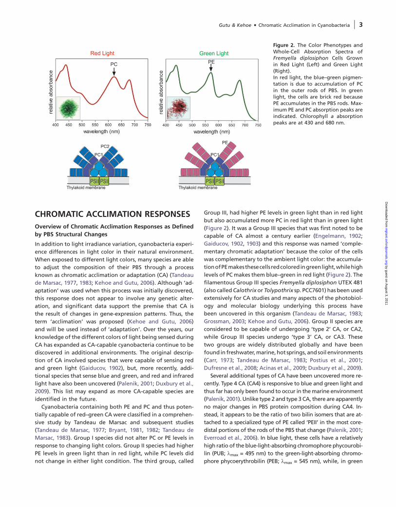
Everroad et al., 2006). In blue light, these cells have a relatively
high ratio of the blue-light-absorbing chromophore phycourobi-
lin (PUB; kmax = 495 nm) to the green-light-absorbing chromo-
phore phycoerythrobilin (PEB; kmax = 545 nm), while, in green
Figure 2. The Color Phenotypes andWhole-Cell Absorption Spectra ofFremyella diplosiphon Cells Grownin Red Light (Left) and Green Light(Right).In red light, the blue–green pigmen-tation is due to accumulation of PCin the outer rods of PBS. In greenlight, the cells are brick red becausePE accumulates in the PBS rods. Max-imum PE and PC absorption peaks areindicated. Chlorophyll a absorptionpeaks are at 430 and 680 nm.
Gutu & Kehoe d Chromatic Acclimation in Cyanobacteria | 3
by guest on August 3, 2011
mplant.oxfordjournals.org
Dow
nloaded from
light, this ratio decreases significantly (Figure 3A). These
changes in the PUB:PEB ratio change the color phenotype
of these cells between orange and pink (Figure 3B). Another
recently described type of CA appears to occur in at least one
strain of Acaryochloris marina. PC-containing PBS increase
during growth in 625 nm light and decrease in 720 nm light,
suggesting that this organism has the ability to acclimate to
changes in its near-infrared light environment (Duxbury
et al., 2009).
Distribution and Role of CA in the Natural Environment
The ability to carry out CA is not restricted to a single branch of
the cyanobacterial lineage. It occurs in all major groups of
cyanobacteria and is not strongly correlated with any particu-
lar type of environment, but rather is a geographically
widespread process (Carr, 1973; Tandeau de Marsac, 1983;
Postius et al., 2001; Dufresne et al., 2008; Acinas et al., 2009;
Duxbury et al., 2009). Despite our extensive molecular under-
standing of at least some forms of CA (see below), its role(s)
in the natural environment has not yet been clearly elucidated.
The spectral partitioning of light as it is absorbed by water is an
attractive possible explanation for the existence of CA. As white
light passes through water, red wavelengths are best absorbed,
followed by green, then blue. Thus, CA-capable species may
adjust the absorption characteristics of their PBS to match
the ambient spectral distribution of light, which varies with
depth. Underwater light quality is also influenced by the back-
ground turbidity due to particulates and other organic dis-
solved material, as well as perhaps by the vibrations of water
molecules themselves (Postius et al., 2001; Stomp et al.,
2007). Other field observations have shown that PE-containing
cyanobacteria are prevalent in environments in which green
light prevails, and cyanobacteria containing only PC are more
abundant in red light-rich environments (Voros et al., 1998; Vila
and Abella, 2001). However, the CA capacity of these organisms
was not analyzed. Interestingly, cyanobacteria that appear to
be capable of CA exist in microbial mats of hot springs (Brown
et al., 2010). In this case, it is possible that CA provides a fitness
advantage for a species that must cope with light that has been
spectrally altered as a result of the absorption of specific wave-
lengths by neighboring organisms. In fact, it is likely that CA is
capable of conferring a selective advantage in multiple environ-
mental settings. CA3 has been shown experimentally to convey
a fitness advantage during growth in changing light color con-
ditions (Stomp et al., 2004), although its benefits are evident
only when the light environment fluctuates on a timescale lon-
ger than the timescale required for CA-driven changes in PBS
composition to occur (Li and Kehoe, 2008; Stomp et al., 2008).
The PBS composition changes that occur during CA3 have
also been shown to maximize the efficiency of photon capture
for photosynthesis. When F. diplosiphon cells are fully accli-
mated to and grown in red light, they have comparable chlo-
rophyll a levels and exhibit photosynthesis rates similar to F.
diplosiphon cells that have been fully acclimated to and grown
in green light (Campbell, 1996). However, this study also
showed that when either red or green light-acclimated cells
are shifted to the opposite light color, the rate of photosynthe-
sis drops by approximately 40%. Fluorescence, photochemical,
and non-photochemical quenching measurements demon-
strated that, in red light, which can be used by both PC and
chlorophyll a, cells are in state I, with PSII exhibiting relatively
high oxidation rates and low non-photochemical quenching.
Conversely, green light-acclimated cells are in state II, in which
some of the light absorbed by PE is channeled to drive PSI pho-
tochemistry and higher non-photochemical quenching of PSII
occurs (Campbell, 1996). This ‘long-term’ state transition phe-
nomenon, physiologically, is equivalent to classical state tran-
sitions, which are historically defined as occurring only on very
short timescales and are controlled by redox states of the plas-
toquinone pool (Fujita et al., 1994; Li and Sherman, 2000).
Figure 3. Fluorescence and Phenotypic Color Changes during CA4in the Marine Unicellular Bacterium Synechococcus sp. RS9916.
(A) Fluorescence excitation spectra of RS9916 cells grown in bluelight (blue line) or green light (green line) showing the changein the relative fluorescence excitation of PUB at 495 nm and PEBat 550 nm under the two light conditions.(B) Whole-cell color differences between cells grown in blue light(left) and green light (right).
4 | Gutu & Kehoe d Chromatic Acclimation in Cyanobacteria
by guest on August 3, 2011
mplant.oxfordjournals.org
Dow
nloaded from
Interestingly, recent results implicate CpcG rod-core linkers of
Synechocystis sp. PCC 6803 in the process of state transitions,
and the expression of one family member has been reported to
be regulated by red and green light (Kondo et al., 2005;
Katayama and Ikeuchi, 2006; Kondo et al., 2009). This suggests
a possible area of study for exploring mechanisms by which
‘long-term’ state transitions may be maintained in CA-capable
cyanobacterium.
Additional Cellular Changes Occur During CA
Although CA was originally defined by the capacity to modify
the composition of PBS in response to changing light colors,
many additional physiological and morphological responses
that are light color regulated have been identified. In the
CA3 species F. diplosiphon, for example, cells acclimated to
red or green light have different morphologies. In red light,
the filaments are significantly shorter than in green light. Also,
individual cells are larger and more rounded in red light, while
in green light, they are cylindrical (Bennett and Bogorad, 1973;
Bogorad et al., 1983; Bordowitz and Montgomery, 2008). The
shortened filament length in red-grown cells is due, at least in
part, to the formation of necredia (cells undergoing pro-
grammed cell death) along the filament (Bennett and
Bogorad, 1973; Bogorad, 1975). The reason for these changes
is not yet known. Many cellular processes in F. diplosiphon are
regulated by light color as well. Gas vesicle-gene expression
and the development of hormogonia, which are short, motile
filaments important for dispersal and survival (Rippka et al.,
1979), are also regulated in part by red and green light in
this species (Tandeau de Marsac et al., 1988; Damerval et al.,
1991; Campbell et al., 1993). In addition, microarray and
two-dimensional-protein gel electrophoresis analyses showed
that at least 80–100 non-PBS proteins are CA3 regulated in
F. diplosiphon (Stowe-Evans et al., 2004). Among these is chlL,
encoding a subunit of the light-independent form of proto-
chlorophyllide reductase, which catalyzes the penultimate
step in chlorophyll a synthesis. This gene and chlB and chlN,
encoding the other two subunits of this enzyme, are more
highly expressed in green light than red light—a response that
likely compensates for the decreased activity of the alternative
form of this enzyme, PorA, which is activated by red light (Shui
et al., 2009). Although these examples are from F. diplosiphon,
CA2 and CA4 species are also very likely to have many
CA-regulated cellular responses in addition to PBS biogenesis.
Molecular Basis of CA-Mediated PBS Biogenesis and the
Regulatory Systems Controlling CA
Most research on the molecular mechanisms and regulation of
CA has focused on CA3 in F. diplosiphon. However, studies ex-
amining how CA2 and CA4 operate, and how they are regu-
lated, are now being carried out as well. These have begun
to contribute to our overall understanding of the process of
CA and how it is regulated at the molecular level. A summary
of each of these is provided below.
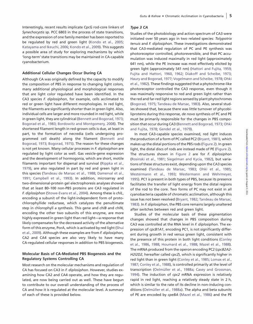
Type 3 CA
Studies of the photobiology and action spectrum of CA3 were
initiated over 50 years ago in two related species: Tolypotrix
tenuis and F. diplosiphon. These investigations demonstrated
that CA3-mediated regulation of PC and PE synthesis was
photoreceptor controlled, photoreversible, and that PC accu-
mulation was induced maximally in red light (approximately
641 nm), while the PE increase was most effectively elicited by
green light (approximately 541 nm) (Hattori and Fujita, 1959;
Fujita and Hattori, 1960, 1962; Diakoff and Scheibe, 1973;
Haury and Bogorad, 1977; Vogelmann and Scheibe, 1978; Ohki
et al., 1982). These findings suggested that a phytochrome-like
photoreceptor controlled the CA3 response, even though it
was maximally responsive to red and green light rather than
the red and far-red light regions sensed by plant phytochromes
(Bogorad, 1975; Tandeau de Marsac, 1983). Also, several stud-
ies showed that, because there was little turnover of phycobi-
liproteins during this response, de novo synthesis of PC and PE
must be primarily responsible for the changes in PBS compo-
sition that occur during CA3 (Bennett and Bogorad, 1973; Ohki
and Fujita, 1978; Gendel et al., 1979).
In most CA3-capable species examined, red light induces
the production of a form of PC called PC2 (Bryant, 1981), which
makes up the distal portions of the PBS rods (Figure 2). In green
light, the distal discs of rods are instead made of PE (Figure 2).
The structures shown in Figure 2 are for F. diplosiphon
(Rosinski et al., 1981; Siegelman and Kycia, 1982), but varia-
tions of these structures exist, depending upon the CA3 species
examined (Tandeau de Marsac, 1983; Ohki et al., 1985;
Westermann et al., 1993; Westermann and Wehrmeyer,
1995). PC1 is present in both types of PBS, because its presence
facilitates the transfer of light energy from the distal regions
of the rod to the core. Two forms of PC may not exist in all
cyanobacteria capable of chromatic acclimation, although this
issue has not been resolved (Bryant, 1982; Tandeau de Marsac,
1983). In F. diplosiphon, the PBS core remains largely unaltered
after switches between red and green light.
Studies of the molecular basis of these pigmentation
changes showed that changes in PBS composition during
CA3 was controlled at the RNA level in F. diplosiphon. The ex-
pression of cpcB1A1, encoding PC1, is not significantly differ-
ent during growth in red versus green light, consistent with
the presence of this protein in both light conditions (Conley
et al., 1986, 1988; Houmard et al., 1988; Mazel et al., 1988).
The mRNA produced from the operon encoding PC2 (cpcB2A2-
H2I2D2, hereafter called cpc2), which is significantly higher in
red light than in green light (Conley et al., 1985; Lomax et al.,
1987; Conley et al., 1988), is controlled primarily at the level of
transcription (Oelmuller et al., 1988a; Casey and Grossman,
1994). The induction of cpc2 mRNA expression is relatively
rapid in red light, reaching a relatively steady state in 2 h,
which is similar to the rate of its decline in non-inducing con-
ditions (Oelmuller et al., 1988a). The alpha and beta subunits
of PE are encoded by cpeBA (Mazel et al., 1986) and the PE
Gutu & Kehoe d Chromatic Acclimation in Cyanobacteria | 5
by guest on August 3, 2011
mplant.oxfordjournals.org
Dow
nloaded from
linkers are encoded by the first three genes of the cpeCDESTR
(cpeC) operon (Federspiel and Grossman, 1990; Federspiel and
Scott, 1992). mRNA levels from the cpeBA and cpeC operons
increase rapidly and reach a maximum 4–8 h after transfer
from red to green light (Federspiel and Grossman, 1990). In ad-
dition, the genes encoding the chromophore-synthesis
enzymes (Frankenberg et al., 2001) are regulated by CA3 in
F. diplosiphon. The expression of pcyA, which encodes an ox-
idoreductase that produces phycocyanobilin, the bilin that is
attached to PC, is approximately fivefold higher in red light
than in green light (Alvey et al., 2007), while pebAB, the
operon that encodes two additional oxidoreductases that
produce PEB for attachment to PE, is more highly expressed
in green light than in red light (Alvey et al., 2003).
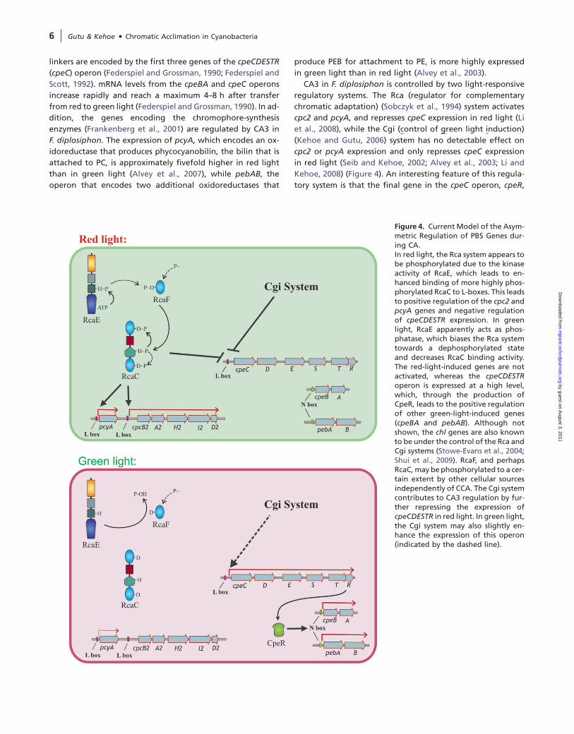
CA3 in F. diplosiphon is controlled by two light-responsive
regulatory systems. The Rca (regulator for complementary
chromatic adaptation) (Sobczyk et al., 1994) system activates
cpc2 and pcyA, and represses cpeC expression in red light (Li
et al., 2008), while the Cgi (control of green light induction)
(Kehoe and Gutu, 2006) system has no detectable effect on
cpc2 or pcyA expression and only represses cpeC expression
in red light (Seib and Kehoe, 2002; Alvey et al., 2003; Li and
Kehoe, 2008) (Figure 4). An interesting feature of this regula-
tory system is that the final gene in the cpeC operon, cpeR,
Figure 4. Current Model of the Asym-metric Regulation of PBS Genes dur-ing CA.In red light, the Rca system appears tobe phosphorylated due to the kinaseactivity of RcaE, which leads to en-hanced binding of more highly phos-phorylated RcaC to L-boxes. This leadsto positive regulation of the cpc2 andpcyA genes and negative regulationof cpeCDESTR expression. In greenlight, RcaE apparently acts as phos-phatase, which biases the Rca systemtowards a dephosphorylated stateand decreases RcaC binding activity.The red-light-induced genes are notactivated, whereas the cpeCDESTRoperon is expressed at a high level,which, through the production ofCpeR, leads to the positive regulationof other green-light-induced genes(cpeBA and pebAB). Although notshown, the chl genes are also knownto be under the control of the Rca andCgi systems (Stowe-Evans et al., 2004;Shui et al., 2009). RcaF, and perhapsRcaC, may be phosphorylated to a cer-tain extent by other cellular sourcesindependently of CCA. The Cgi systemcontributes to CA3 regulation by fur-ther repressing the expression ofcpeCDESTR in red light. In green light,the Cgi system may also slightly en-hance the expression of this operon(indicated by the dashed line).
6 | Gutu & Kehoe d Chromatic Acclimation in Cyanobacteria
by guest on August 3, 2011
mplant.oxfordjournals.org
Dow
nloaded from
encodes an activator that is required for the expression of
cpeBA and pebAB (Cobley et al., 2002; Seib and Kehoe,
2002). This provides the cpeC operon with a central role in
the CA3 regulation of cpeBA and pebAB, and both the Rca
and Cgi systems regulate these two operons by controlling
cpeC expression. Specifically how CpeR controls cpeBA and
pebAB expression is not yet clear, although their promoters
both contain a short, direct repeat sequence called the N-
box (Kehoe and Gutu, 2006) that is bound by a protein called
RcaA/PepB (Schmidt-Goff and Federspiel, 1993; Sobczyk et al.,
1993; Alvey et al., 2003). While no functional evidence exists
for their role in CA3 regulation, the N-box and RcaA/PepB
are good candidates for the CA3 elements that link cpeC,
cpeBA, and pebAB expression. It is not yet known whether
or not CpeR, RcaA, and PepB are the same protein.
Analysis of the Rca pathway has provided insights into its
composition and how it regulates PBS production during
CA3. It is controlled by RcaE, a phytochrome-class photorecep-
tor containing a histidine-kinase domain, and two response
regulators, RcaF and RcaC (Figure 4). RcaE was the first phyto-
chrome-class photoreceptor discovered in bacteria (Kehoe and
Grossman, 1996; Terauchi et al., 2004). The N-terminal half of
RcaE contains a GAF domain similar to chromophore-binding
domains of plant phytochromes, adjacent to a PAS domain.
In the C-terminal region, the histidine-kinase module is present,
containing the dimerization-histidine-phosphotransfer and ATP
binding-catalytic domains. Similar to plant phytochromes, a cys-
teine within the GAF domain covalently binds a bilin chromo-
phore in vivo, but, unlike the plant photoreceptors, which
respond maximally to red and far-red light, RcaE appears to
be green-red responsive (Terauchi et al., 2004). The type of bilin
bound to RcaE has not yet been reported, nor has green–red
photoreversibility of the purified protein been demonstrated.
An rcaE-null mutant is phenotypically black in all light condi-
tions due to the intermediate level of expression of the cpc2,
pcyA, pebAB, cpeC, and cpeBA operons, which has been pro-
posed to be due to non-light color-regulated phosphorylation
of RcaF and RcaC by other cellular sources (Kehoe and Gross-
man, 1996; Terauchi et al., 2004). The response regulator RcaF
is encoded immediately downstream of rcaE and consists of
a single receiver domain containing a conserved aspartate res-
idue that is typically the site of phosphorylation. rcaF mutants
are phenotypically red in both red and green light due to the
lack of cpc2 expression and high-level production of cpeC,
cpeBA, and pebAB (Kehoe and Grossman, 1997; Alvey et al.,
2003). RcaC is a large, complex response regulator consisting
of two receiver domains that are located at the N- and
C-termini, an OmpR/PhoB-class-DNA binding domain, and a his-
tidine-phosphotransfer domain. As with rcaF mutants, rcaC
mutants are red in all light colors due to the absence of cpc2
RNA and high-level expression of the genes required for the
production of PE-containing PBS (Chiang et al., 1992; Kehoe
and Grossman, 1997).
Several lines of evidence provide clues about phosphoryl
group movement in the Rca pathway during CA3. The genetic
data described in the previous paragraph, along with site-
directed mutational analyses of the probable phosphorylation
site histidines and aspartates within RcaE, RcaF, and RcaC, sug-
gest that the Rca system is more phosphorylated in red light
than in green light (Li and Kehoe, 2005). After RcaE autophos-
phorylation, phosphoryl group transfer to the aspartate
of RcaF has been proposed, followed by transfer to the histi-
dine of the histidine phosphotransfer domain of RcaC, and
finally to the aspartate of the N-terminal receiver module of
RcaC. The conserved aspartate within the C-terminal-receiver
domain of RcaC is essentially not required for the CA3 response
and may act as an input site for another signal transduction
system, although this has not yet been shown (Li and Kehoe,
2005). RcaE has been proposed to act as a kinase in red light
and a phosphatase in green light, based on the phenotypes
of different classes of rcaE mutants (Figure 4) (Kehoe and
Grossman, 1997; Terauchi et al., 2004).
In addition to the apparent effect of its phosphorylation
state, the activity of the Rca system is significantly influenced
by the cellular level of RcaC. F. diplosiphon produces five to six
times more RcaC protein in red light than in green light (Li and
Kehoe, 2005, 2008). This differs from the photoreceptor RcaE,
which is present at equal levels in both red and green light
(Terauchi et al., 2004). Part of this change occurs at the RNA
level and these changes in RcaC levels are required, but not
sufficient, for a normal CA3 response. This RcaC abundance
change depends on the presence of the same conserved histi-
dine and aspartate residues necessary for the CA3 response
(Li and Kehoe, 2008). Thus, it appears that the combination
of RcaC abundance changes and modification of RcaC by phos-
phorylation is needed to provide the dynamic range required
for proper transcriptional control of highly expressed, RcaC-
regulated genes.
In red light, RcaC activates the transcription of cpc2 and
pcyA and represses cpeC transcription by binding to the L-
box—a seven base-pair-direct repeat located at –35 (relative
to transcription start) within the cpc2 promoter and in the
inverse orientation at –78 of cpeC (Alvey et al., 2007; Bezy
and Kehoe, 2010). In green light, the Rca system no longer acti-
vates cpc2 or pcyA. In addition, cpeC is no longer repressed,
leading to its expression and, as a result of the production
of CpeR, the expression of cpeBA and pebAB (Cobley et al.,
2002; Seib and Kehoe, 2002) (Figure 4). The operation of
this system explains the phenotypes of rcaF and rcaC mutants
well. In these lines, the lack of RcaC or its activation would lead
to the persistent inability to activate the production of PC2
and to inactivate the synthesis of PE, resulting in the light-
independent red coloration observed for these mutants.
RcaD is a protein that has been proposed to coordinate the
expression of several operons whose activity is increased by red
light. It consists of domains with similarity to E2 proteins of
papillomavirus and the DNA polymerase sliding clamp. RcaD
was found to protect two regions of the cpc2 promoter from
DNase I treatment, binding that appears to be phosphorylation
dependent (Sobczyk et al., 1994). Although the cpc2 promoter
Gutu & Kehoe d Chromatic Acclimation in Cyanobacteria | 7
by guest on August 3, 2011
mplant.oxfordjournals.org
Dow
nloaded from
regions to which RcaD binds are not needed to maintain the
fully acclimated red light state during CA (Casey and Grossman,
1994; Li et al., 2008), it has been proposed that RcaD may reg-
ulate the expression of red light-activated genes only during
the transient period of early acclimation, before PBS have
become fully adjusted to red light (Noubir et al., 2002).
The Rca system has also been shown to regulate non-PBS
responses in F. diplosiphon. The CA3 response of chlLN and
chlBgene expression is Rca controlled and, although the mech-
anism is not known, these genes also require CpeR for their
expression, similar to cpeBA and pebAB (Stowe-Evans et al.,
2004; Shui et al., 2009). In addition, the CA3-regulated changes
in morphology appear to be largely dependent on RcaE,
although a gene encoding one component in this response,
tonB, is CA3 regulated but not controlled via the Rca system
(Shui et al., 2009; Pattanaik and Montgomery, 2010). More
work is needed to elucidate the molecular basis of this
photoregulatory response.
Much less is understood about the Cgi system than the Rca
pathway. The existence of this pathway was first proposed dur-
ing pigment analyses of PE chromophore attachment (cpeYZ)
mutants, based on the observation that some photoregulation
of PE continued in the absence of rcaC (Kahn et al., 1997). Thus
far, the Cgi system is known to control only the CA3 response
of the cpeC, cpeBA, and pebAB operons, although many
additional genes may be under its control (Seib and Kehoe,
2002; Alvey et al., 2003; Li and Kehoe, 2005). Based on tran-
script abundance analyses in various rca mutant backgrounds,
the Cgi pathway is a significant contributor to the light color
response in F. diplosiphon, controlling approximately one-
third of the 9–10-fold change in CA3-mediated expression
for these genes. This system acts by repressing cpeC expression
during growth in red light, apparently through a post-
transcriptional mechanism that requires a sequence capable
of forming a stem-loop within the 5’ leader region of cpeC
(Bezy and Kehoe, submitted). Thus, for PBS genes that are
up-regulated in green light, the Cgi and Rca systems work
together to jointly repress cpeC expression, thus blocking
the synthesis of the CpeR activator and the production of RNAs
from the genes it controls. There have been no reports on the
isolation of Cgi pathway components thus far.
Type 2 CA
The physiology and regulation of the changes in PBS composi-
tion that occur during CA2 have been studied in many cyano-
bacterial species. An action spectrum, using Synechocystis sp.
PCC 6701, demonstrated that the accumulation of PE is most ef-
fectively controlled by red and green light (Tandeau de Marsac
et al., 1980). PBS structural changes have been elucidated in
a number of type 2 species. In general, the rods of PBS from
green-light-grown cells are one disc longer than those from
red-light-grown cells, and the PBS composition changes during
CA2 always occur at the core-distal end of the rods (Tandeau de
Marsac, 1983). Changes in both PE abundance and cpeBAmRNA
levels in red versus green light were shown for Synechocystis sp.
strain BO 8402 (Neuschaefer-Rube et al., 2002). In Nostoc punc-
tiforme, quantitative changes in PE accumulation have been
measured in cells grown in red versus green light (Wolf and
Schussler, 2005) and CA2 regulation of the RNA levels of three
genes involved in PBS biogenesis, cpeC, cpcG2, and cpeR1, has
been demonstrated (Hirose et al., 2010). The CA2-mediated ex-
pression of these genes is regulated by a two-component system
that is controlled by the sensorhistidine kinase CcaS, a cyanobac-
terial phytochrome-class photoreceptor that is red light–green
light photoreversible. CcaS has strong sequence similarity to
RcaE from F. diplosiphon. The response regulator of this system
is CcaR, which is capable of binding to the DNA region upstream
of cpeC in N. punctiforme and has strong sequence similarity to
RcaC from F. diplosiphon (Hirose et al., 2010).
Type 4 CA
An action spectrum has not yet been conducted for CA4.
However, the initial analysis of this response demonstrated
that changes in the relative abundances of PUB and PEB in
a wide range of marine Synechococcus species could be
achieved with green and blue light, but not by changes in
the intensity of a white-light source or different nitrogen
sources (Palenik, 2001). Subsequent research on two Synecho-
coccus strains isolated from the Gulf of Mexico suggested that
CA4 does not involve the production of different phycobili-
proteins during the acclimation process, as occurs for CA3,
but rather the differential chromophorylation of the alpha
subunit of PEII, one of the two types of PE found in these spe-
cies (Everroad et al., 2006). PEII, which is located at the core-
distal regions of the rods, consists of alpha and beta subunits
called MpeA and MpeB, and these researchers suggested that
the replacement of chromophores attached to MpeA is the
molecular basis of CA4. MpeA has been proposed to have
three PUBs attached in blue light and one PUB and two PEBs
attached in green light. The differential attachment of these
two isomers was proposed to occur through the action of one
or more phycobilin lyases or lyase/isomerases whose activity
or level(s) is controlled by CA4. Neither these enzymes nor any
CA4 regulatory components have been identified to date.
However, an in-depth analysis of the genomes of many
marine Synechococcus strains, some capable of CA4 and many
that are not, has uncovered genes that may be involved di-
rectly or indirectly in this acclimation process. These were
identified based on the correlation between the presence
of these genes in a particular genome and the physiological
capacity of the organism containing that genome to undergo
CA4 (Six et al., 2007; Dufresne et al., 2008). Although 12 or 13
predicted proteins with homology or relatedness to known
lyases were found to be present in strains capable of CA4,
these workers suggested that MpeV, MpeU, and MpeZ might
be the best candidates for involvement in CA4. The only pos-
sible photoreceptor identified using this approach was AplA,
which was identified in F. diplosiphon as a member of a new
class of cyanobacterial photoreceptors of unknown function
(Montgomery et al., 2004).
8 | Gutu & Kehoe d Chromatic Acclimation in Cyanobacteria
by guest on August 3, 2011
mplant.oxfordjournals.org
Dow
nloaded from
Evolution of CA Systems
An initial photobiological study correctly concluded that CA3
in F. diplosiphon was regulated either by two separate sys-
tems or by a single sensory system that controlled PC and
PE production through different downstream components
(Oelmuller et al., 1988b). While it is now clear that two
separate photosensory systems indeed control CA3, Nature
has introduced a twist into this scheme, since the Rca
pathway controls both red- and green-light-expressed genes,
while the other, the Cgi system, appears to regulate only
green-light-expressed genes.
How commonly are the Rca and Cgi regulatory systems used
in CA3-capable cyanobacteria? Although this cannot be an-
swered for the Cgi pathway, since it has not yet been suffi-
ciently characterized, genome sequence information is
available from enough CA3 species to begin to address this
question for the Rca system. Highly conserved, similarly ori-
ented L-boxes are present upstream of PBS genes in all three
of the CA3 species for which sequences are available (Alvey
et al., 2007; Bezy and Kehoe, 2010), and also are present at sim-
ilar locations within the incomplete draft genome of a recently
described hot springs cyanobacterium that may be capable of
CA3 (Brown et al., 2010). Genes encoding homologs of the Rca
components are also present in all of the available sequenced
genomes of CA3-capable species, so the Rca system appears to
be a widely used control pathway for CA3. Because the species
carrying these genomes are in different branches of the cya-
nobacterial 16S rRNA tree and originate from diverse environ-
ments (marine, freshwater, and hot springs), it is most likely
that the ability to undergo CA3 has been spread by extensive
lateral gene transfer, although it is also possible that this ca-
pability was simply lost non-uniformly. A detailed study of
L-box structure and function demonstrated that the sequences
of these elements are highly conserved across species and that
this conservation is required for high-level activation of gene
expression (Bezy and Kehoe, 2010). Taken together, these
results suggest that the portion of the CA3 system that is
controlled by the Rca pathway has either recently spread
throughout cyanobacteria and/or that natural selection main-
tains the high sequence conservation between L-boxes from
different species, perhaps because of the need for continued
high-level expression of genes that encode very abundant
light-harvesting proteins.
In F. diplosiphon, the Rca system has subjugated the activa-
tor CpeR and made it part of the CA3 regulatory system. But
many species that are not capable of any form of CA still con-
tain CpeR and N-boxes. In fact, all cyanobacteria that produce
PE have been found to contain at least one cpeR gene (Cobley
et al., 2002), so the role of CpeR in controlling genes involved
in PE production seems to be widespread and is certainly not
always linked to CA, as it is in F. diplosiphon. It is also likely
that CpeR has been widely integrated into the CA control
of green-light-expressed genes, since N-boxes have been iden-
tified upstream of the cpeBA operon in both a CA2-capable
Synechocystis species and a Pseudanabaena strain capable
of CA3 (Neuschaefer-Rube et al., 2002). Biochemical and func-
tional studies are needed to test the hypothesis that the func-
tion of CpeR is to coordinate the expression of multiple genes
whose increased activity is green-light dependent.
The discovery of the Cgi system in F. diplosiphon led to the
hypothesis that CA2 species use only the Cgi system to highly
express genes in green light, while CA3 species also contain the
Rca system, which both further represses these genes in red
light and activates genes involved in the production of PC
(Kehoe and Gutu, 2006; Kehoe, 2010). Currently, this hypoth-
esis cannot be tested by examining CA2 species, since the com-
ponents that make up the Cgi system in F. diplosiphon are not
known. Another approach that was taken to begin to test this
hypothesis was to identify the components controlling the
regulation of cpeC gene expression in the CA2-capable
species N. punctiforme. This was accomplished by testing
the role of CcaSR, a phytochrome-class photoreceptor-based
two-component system, in the CA2 response (Hirose et al.,
2010). In N. punctiforme, the genes encoding these compo-
nents are adjacent to cpeC. This system was capable of sensing
red and green light and controlling the CA2 response of cpeC
in this species. These researchers proposed that this pathway is
phosphorylated in green light, resulting in cpeC up-regulation
through enhanced binding of the response regulator to its
promoter region. This light regulation is the inverse of the pro-
posed mechanism of the Rca system in F. diplosiphon, where
the transcriptional repression of the cpeC operon in red light
has been proposed to be the result of the phosphorylation of
this system (Li and Kehoe, 2005; Kehoe and Gutu, 2006; Li et al.,
2008). This led to the proposal that the Cgi system in CA3-
capable species simply consists of an RcaE-like photoreceptor
controlling a two-component system whose light color regu-
lation is at the transcriptional level and whose operation is
complementary to the Rca system (Hirose et al., 2008). How-
ever, recent work on the Cgi system in F. diplosiphon has shown
that it operates post-transcriptionally through the 5’ leader se-
quence of cpeC (Bezy and Kehoe, submitted). Taken together,
these data demonstrate that the hypothesis that the Cgi and
the CA2 regulatory systems are related (Kehoe and Gutu, 2006;
Kehoe, 2010) is not correct, and establish that the CcaSR and
the Cgi systems are different in at least the final steps of these
pathways. This does not eliminate the possibility that compo-
nents related to CcaSR are acting upstream of the post-
transcriptional regulatory step within the Cgi system. But these
results do suggest that, unlike the apparently widespread use
of the Rca system for CA3 control of red-light up-regulated
genes, there may be some variation in the CA control mecha-
nisms for genes that are up-regulated by green light. As the
components of the Cgi pathway in F. diplosiphon are discov-
ered, it will be interesting to examine the virtual proteomes
of other CA3 species for such components and to compare
them to the CcaSR two-component system that controls gene
expression during CA2 in N. punctiforme.
Gutu & Kehoe d Chromatic Acclimation in Cyanobacteria | 9
by guest on August 3, 2011
mplant.oxfordjournals.org
Dow
nloaded from
SUMMARY
Recent advances in the study of CA in several cyanobacterial
species have begun to uncover the diversity of these responses
and their regulation. The capacity to undergo CA is present in
a wide range of species and environments and is likely to make
a significant contribution globally to maximizing the efficiency
of photon capture for photosynthesis. Our current understand-
ing of the various types of CA suggest that, although they all
maximize the efficiency of ambient light capture, in many cases,
they appear to be the result of convergent evolution. For exam-
ple, the blue–green CA4 response does not involve any detect-
able changes in PBS rod proteins, while red–green CA2 and CA3
responses do. In addition, some of the signal-transduction path-
ways controlling these responses, such as the Rca system, may be
broadly employed, while the use of others, such as the CcaSR
and Cgi systems controlling green-light-expressed genes during
CA2 and CA3 in N. punctiforme and F. diplosiphon, may be less
widespread. The future should expand our knowledge concern-
ing these relationships and further cement our understanding
of how frequently CA3 species use the Rca and Cgi systems and
CA2 species use the CcaSR pathway. We predict that, as more is
understood about this fascinating, colorful process, even more
variations in CA responses and their modes of regulation will
become apparent. Some may be quite subtle and others pro-
nounced. It will be an enormous challenge to determine
whether these variations have arisen and been employed by
chance, or whether each of the differences that is uncovered
represents a finely tuned form of CA that provides a selective
advantage to the species that employs it in a very specific
environmental setting.
FUNDING
This work was supported by funding from the National Science
Foundation under Grant No. MCB-1029414 to D.M.K.
ACKNOWLEDGMENTS
The authors wish to thank the members of the Kehoe laboratory for
many helpful discussions. No conflict of interest declared.
REFERENCES
Acinas, S.G., Haverkamp, T.H., Huisman, J., and Stal, L.J. (2009).
Phenotypic and genetic diversification of Pseudanabaena spp.
(cyanobacteria). ISME. J. 3, 31–46.
Adir, N. (2005). Elucidation of the molecular structures of compo-
nents of the phycobilisome: reconstructing a giant. Photosynth.
Res. 85, 15–32.
Alvey, R.M., Bezy, R.P., Frankenberg-Dinkel, N., and Kehoe, D.M.
(2007). A light regulated OmpR-class promoter element co-ordi-
nates light-harvesting protein and chromophore biosynthetic
enzyme gene expression. Mol. Microbiol. 64, 319–332.
Alvey, R.M., Karty, J.A., Roos, E., Reilly, J.P., and Kehoe, D.M. (2003).
Lesions in phycoerythrin chromophore biosynthesis in Fremyella
diplosiphon reveal coordinated light regulation of apoprotein
and pigment biosynthetic enzyme gene expression. Plant Cell.
15, 2448–2463.
Anderson, J.M., Chow, W.S., and Park, Y.I. (1995). The grand design
of photosynthesis: acclimation of the photosynthetic apparatus
to environmental cues. Photosynth. Res. 46, 129–139.
Bailey, S., and Grossman, A. (2008). Photoprotection in cyanobac-
teria: regulation of light harvesting. Photochem. Photobiol. 84,
1410–1420.
Bennett, A., and Bogorad, L. (1973). Complementary chromatic adap-
tation in a filamentous blue–green alga. J. Cell. Biol. 58, 419–435.
Bezy, R.P., and Kehoe, D.M. (2010). Functional characterization of
a cyanobacterial OmpR/PhoB class transcription factor binding site
controlling light color responses. J. Bacteriol. 192, 5923–5933.
Bhaya, D., Schwarz, R., and Grossman, A.R. (2000). Molecular
responses to environmental stress. In The Ecology of Cyanobac-
teria, Whitton, B.A., and Potts, M., eds (Kluwer Academic Pub-
lisher, Dordrecht/London/Boston), pp. 391–442.
Bogorad, L. (1975). Phycobiliproteins and complementary chro-
matic adaptation. Ann. Rev. Plant Physiol. 26, 369–401.
Bogorad, L., Gendel, S.M., Haury, J.F., and Koller, K.-P. (1983). Pho-
tomorphogenesis and complementary chromatic adaptation in
Fremyella diplosiphon In Photosynthetic Prokaryotes: Cell Differ-
entiation and Function. Proceedings of the Special FEBS Meeting
on Cell Differentiation and Function, April 25-29, 982, Athens,
Greece, Papageorgiou, G.C., and Packer, L., eds (Elsevier Biomed-
ical, New York), pp. 119–126.
Bordowitz, J.R., and Montgomery, B.L. (2008). Photoregulation of
cellular morphology during complementary chromatic adapta-
tion requires sensor-kinase-class protein RcaE in Fremyella diplo-
siphon. J. Bacteriol. 190, 4069–4074.
Brown, I.I., et al. (2010). Polyphasic characterization of a thermo-
tolerant siderophilic filamentous cyanobacterium that produ-
ces intracellular iron deposits. Appl. Environ. Microbiol. 76,
6664–6672.
Bryant, D.A. (1981). The photoregulated expression of multiple
phycocyanin species: a general mechanism for the control of
phycocyanin synthesis in chromatically adapting cyanobacteria.
Eur. J. Biochem. 119, 425–429.
Bryant, D.A. (1982). Phycoerythrocyanin and phycoerythrin: prop-
erties and occurrence in cyanobacteria. J. Gen. Microbiol. 128,
835–844.
Campbell, D. (1996). Complementary chromatic adaptation alters
photosynthetic strategies in the cyanobacterium Calothrix. Mi-
crobiology. 142, 1255–1263.
Campbell, D., Houmard, J., and Tandeau de Marsac, N. (1993). Elec-
tron transport regulates cellular differentiation in the filamen-
tous cyanobacterium Calothrix. Plant Cell. 5, 451–463.
Carr, N.G. (1973). Physiology and ecology of marine blue–green al-
gae. In The Biology of Blue–Green Algae, Carr, N.G., and
Whitton, B.A., eds (Oxford: Blackwell Scientific), pp. 368–378.
Casey, E.S., and Grossman, A.R. (1994). In vivo and in vitro charac-
terization of the light-regulated cpcB2A2 promoter of Fremyella
diplosiphon. J. Bacteriol. 176, 6362–6374.
Chiang, G.G., Schaefer, M.R., and Grossman, A.R. (1992). Comple-
mentation of a red-light-indifferent cyanobacterial mutant.
Proc. Natl Acad. Sci. U S A. 89, 9415–9419.
10 | Gutu & Kehoe d Chromatic Acclimation in Cyanobacteria
by guest on August 3, 2011
mplant.oxfordjournals.org
Dow
nloaded from
Cobley, J.G., et al. (2002). CpeR is an activator required for expres-
sion of the phycoerythrin operon (cpeBA) in the cyanobacterium
Fremyella diplosiphon and is encoded in the phycoerythrin
linker-polypeptide operon (cpeCDESTR). Mol. Microbiol. 44,
1517–1531.
Conley, P.B., Lemaux, P.G., and Grossman, A.R. (1985). Cyanobacte-
rial light-harvesting complex subunits encoded in two red-light
induced transcripts. Science. 230, 550–553.
Conley, P.B., Lemaux, P.G., and Grossman, A.R. (1988). Molecular
characterization and evolution of sequences encoding light-
harvesting components in the chromatically adapting cyanobac-
terium Fremyella diplosiphon. J. Mol. Biol. 199, 447–465.
Conley, P.B., Lemaux, P.G., Lomax, T.L., and Grossman, A.R. (1986).
Genes encoding major light-harvesting polypeptides are clus-
tered on the genome of the cyanobacterium Fremyella diplosi-
phon. Proc. Natl Acad. Sci. U S A. 83, 3924–3928.
Damerval, T., Guglielmi, G., Houmard, J., and TandeaudeMarsac, N.
(1991). Hormogonium differentiation in the cyanobacterium
Calothrix: a photoregulated developmental process. Plant Cell.
3, 191–201.
Diakoff, S. and Scheibe, J. (1973). Action spectra for chromatic ad-
aptation in Tolypothrix tenuis. Plant Physiol. 51, 382–385.
Dufresne, A., et al. (2008). Unraveling the genomic mosaic of a ubiq-
uitous genus of marine cyanobacteria. Genome Biol. 9, R90.
Duxbury, Z., Schliep, M., Ritchie, R.J., Larkum, A.W., and Chen, M.
(2009). Chromatic photoacclimation extends utilisable photo-
synthetically active radiation in the chlorophyll d-containing
cyanobacterium, Acaryochloris marina. Photosynth. Res. 101,
69–75.
Engelmann, T.W. (1902). Untersuchungen uber die qualitativen
Beziehungen zwieschen Absorbtion des Lichtes und Assimilation
in Pflanzenzellen. I. Das Mikrospectraphotometer, ein Apparat
zur qualitativen Mikrospectralanalyse. II. Experimentelle
Grundlangen zur Ermittelung der quantitativen Beziehungen
zwieschen Assimilationsenergie und Absorptiongrosse. III.
Bestimmung der Vertheilung der Energie im Spectrum von
Sonnenlicht mittels Bacterien-Methode und quantitativen
Mikrospectralanalyse. Bot. Z. 42, 81–105.
Everroad, C., Six, C., Partensky, F., Thomas, J.C., Holtzendorff, J.,
and Wood, A.M. (2006). Biochemical bases of type IV chromatic
adaptation in marine Synechococcus spp. J. Bacteriol. 188,
3345–3356.
Federspiel, N.A., and Grossman, A.R. (1990). Characterization of
the light-regulated operon encoding the phycoerythrin-associ-
ated linker proteins from the cyanobacterium Fremyella diplosi-
phon. J. Bacteriol. 172, 4072–4081.
Federspiel, N.A., and Scott, L. (1992). Characterization of a light-
regulated gene encoding a new phycoerythrin-associated linker
protein from the cyanobacterium Fremyella diplosiphon. J. Bac-
teriol. 174, 5994–5998.
Frankenberg, N., Mukougawa, K., Kohchi, T., and Lagarias, J.C.
(2001). Functional genomic analysis of the HY2 family of ferre-
doxin-dependent bilin reductases from oxygenic photosynthetic
organisms. Plant Cell. 13, 965–978.
Fujita, Y., and Hattori, A. (1960). Effect of chromatic lights on phy-
cobilin formation in a blue–green alga, Tolypothrix tenuis. Plant
Cell Physiol. 1, 293–303.
Fujita, Y., and Hattori, A. (1962). Photochemical interconversion be-
tween precursors of phycobilin chromoproteids in Tolypothrix
tenuis. Plant Cell Physiol. 3, 209–220.
Fujita, Y., Murakami, A., and Aizawa, K. (1994). Short-term
and long-term adaptation of the photosynthetic apparatus:
homeostatic properties of thylakoids. In The Molecular Biology
of Cyanobacteria, Bryant D., ed. (Kluwer Academic Publisher,
Dordrecht), pp. 677–692.
Gaiducov, N. (1902). Uber den Einfluss farbigen Lichtes auf die
Farbung lebender Oscillatorien. Abh. Preuss. Akad. Wiss. V,
1–36.
Gaiducov, N. (1903). Die Farbenveranderung bei den Prozessen der
komplementaren chromatischen Adaptation. Ber. Deutsch. Bot.
Ges. 21, 517–522.
Gendel, S., Ohad, I., and Bogorad, L. (1979). Control of phycoery-
thrin synthesis during chromatic adaptation. Plant Physiol. 64,
786–790.
Grossman, A.R. (2003). A molecular understanding of complemen-
tary chromatic adaptation. Photosynth. Res. 76, 207–215.
Hattori, A., and Fujita, Y. (1959). Effect of pre-illumination on the
formation of phycobilin pigments in a blue–green alga, Tolypo-
thrix tenuis. J. Biochem. 46, 1259–1261.
Haury, J.F., and Bogorad, L. (1977). Action spectra for phycobilipro-
tein synthesis in a chromatically adapting cyanophyte, Fremyella
diplosiphon. Plant Physiol. 60, 835–839.
Hirose, Y., Narikawa, R., Katayama,M., and Ikeuchi, M. (2010). Cya-
nobacteriochrome CcaS regulates phycoerythrin accumulation
in Nostoc punctiforme, a Group II chromatic adapter. Proc. Natl
Acad. Sci. U S A. 107, 8854–8859.
Hirose, Y., Shimada, T., Narikawa, R., Katayama,M., and Ikeuchi, M.
(2008). Cyanobacteriochrome CcaS is the green light receptor
that induces the expression of phycobilisome linker protein.
Proc. Natl Acad. Sci. U S A. 105, 9528–9533.
Houmard, J., Capuano, V., Coursin, T., and Tandeau de Marsac, N.
(1988). Genes encoding core components of the phycobilisome
in the cyanobacterium Calothrix sp. strain PCC 7601: occurrence
of a multigene family. J. Bacteriol. 170, 5512–5521.
Kahn, K., Mazel, D., Houmard, J., Tandeau de Marsac, N., and
Schaefer, M.R. (1997). A role for cpeYZ in cyanobacterial phyco-
erythrin biosynthesis. J. Bacteriol. 179, 998–1006.
Kargul, J., and Barber, J. (2008). Photosynthetic acclimation: struc-
tural reorganisation of light harvesting antenna: role of redox-
dependent phosphorylation of major and minor chlorophyll a/
b binding proteins. FEBS J. 275, 1056–1068.
Katayama, M., and Ikeuchi, M. (2006). Perception and transduction
of light signals by cyanobacteria. In Frontiers in Life Sciences,
Fujiwara, M., Sato, N., and Ishiura, S., eds (Kerala, India: Research
Signpost), pp. 65–90.
Kehoe, D.M. (2010). Chromatic adaptation and the evolution of
light color sensing in cyanobacteria. Proc. Natl Acad. Sci. U S
A. 107, 9029–9030.
Kehoe, D.M., and Grossman, A.R. (1996). Similarity of a chromatic
adaptation sensor to phytochrome and ethylene receptors. Sci-
ence. 273, 1409–1412.
Kehoe, D.M., and Grossman, A.R. (1997). New classes of mutants in
complementary chromatic adaptation provide evidence for
Gutu & Kehoe d Chromatic Acclimation in Cyanobacteria | 11
by guest on August 3, 2011
mplant.oxfordjournals.org
Dow
nloaded from
a novel four-step phosphorelay system. J. Bacteriol. 179,
3914–3921.
Kehoe, D.M., and Gutu, A. (2006). Responding to color: the regu-
lation of complementary chromatic adaptation. Annu. Rev. Plant
Biol. 57, 127–150.
Kirilovsky, D. (2007). Photoprotection in cyanobacteria: the orange
carotenoid protein (OCP)-related non-photochemical-quench-
ing mechanism. Photosynth. Res. 93, 7–16.
Kirilovsky, D. (2010). The photoactive orange carotenoid protein
and photoprotection in cyanobacteria. Adv. Exp. Med. Biol.
675, 139–159.
Kondo, K., Geng, X.X., Katayama, M., and Ikeuchi, M. (2005). Dis-
tinct roles of CpcG1 and CpcG2 in phycobilisome assembly in the
cyanobacterium Synechocystis sp. PCC 6803. Photosynth. Res. 84,
269–273.
Kondo, K., Mullineaux, C.W., and Ikeuchi, M. (2009). Distinct roles
of CpcG1-phycobilisome and CpcG2-phycobilisome in state tran-
sitions in a cyanobacterium Synechocystis sp. PCC 6803. Photo-
synth. Res. 99, 217–225.
Li, H., and Sherman, L.A. (2000). A redox-responsive regulator of
photosynthesis gene expression in the cyanobacterium Synecho-
cystis sp. strain PCC 6803. J. Bacteriol. 182, 4268–4277.
Li, L., Alvey, R.M., Bezy, R.P., and Kehoe, D.M. (2008). Inverse tran-
scriptional activities during complementary chromatic adaptation
are controlled by the response regulator RcaC binding to red and
green light-responsive promoters. Mol. Microbiol. 68, 286–297.
Li, L., and Kehoe, D.M. (2005). In vivo analysis of the roles of con-
served aspartate and histidine residues within a complex re-
sponse regulator. Mol. Microbiol. 55, 1538–1552.
Li, L., and Kehoe, D.M. (2008). Abundance changes of the response
regulator RcaC require specific aspartate and histidine residues
and are necessary for normal light color responsiveness. J. Bac-
teriol. 190, 7241–7250.
Lomax, T.L., Conley, P.B., Schilling, J., and Grossman, A.R. (1987).
Isolation and characterization of light-regulated phycobilisome
linker polypeptide genes and their transcription as a polycis-
tronic mRNA .J. Bacteriol. 169, 2675–2684.
MacColl, R. (1998). Cyanobacterial phycobilisomes. J. Struct. Biol.
124, 311–334.
MacIntyre, H.L., Kana, T.M., Anning, T., and Geider, R.J. (2002). Pho-
toacclimation of photosynthesis irradiance response curves and
photosynthetic pigments in microalgae and cyanobacteria. J.
Phycol. 38, 17–38.
Mazel, D., Guglielmi, G., Houmard, J., Sidler, W., Bryant, D.A., and
Tandeau de Marsac, N. (1986). Green light induces transcription
of the phycoerythrin operon in the cyanobacterium Calothrix
7601. Nucleic Acids Res. 14, 8279–8290.
Mazel, D., Houmard, J., and Tandeau de Marsac, N. (1988). A multi-
gene family in Calothrix sp. PCC 7601 encodes phycocyanin, the
major component of the cyanobacterial light-harvesting
antenna. Mol. Gen. Genet. 211, 296–304.
Montgomery, B.L., Casey, E.S., Grossman, A.R., and Kehoe, D.M.
(2004). AplA, a member of a new class of phycobiliproteins lack-
ing a traditional role in photosynthetic light harvesting. J. Bac-
teriol. 186, 7420–7428.
Mullineaux, C.W., and Emlyn-Jones, D. (2005). State transitions: an
example of acclimation to low-light stress. J. Exp. Bot. 56, 389–393.
Neuschaefer-Rube, O., Boger, P., and Ernst, A. (2002). Interference
of an apcA insertion with complementary chromatic adaptation
in the diazotrophic Synechocystis sp. strain BO 8402. Biochim.
Biophys. Acta-Bioenergetics. 1553, 279–295.
Noubir, S., Luque, I., Ochoa de Alda, J.A.G., Perewoska, I., Tandeau
deMarsac, N., Cobley, J.G., andHoumard, J. (2002). Co-ordinated
expression of phycobiliprotein operons in the chromatically
adapting cyanobacterium Calothrix PCC 7601: a role for RcaD
and RcaG. Mol. Microbiol. 43, 749–762.
Oelmuller, R., Conley, P.B., Federspiel, N., Briggs, W.R., and
Grossman, A.R. (1988a). Changes in accumulation and synthesis
of transcripts encoding phycobilisome components during accli-
mation of Fremyella diplosiphon to different light qualities.
Plant Physiol. 88, 1077–1083.
Oelmuller, R., Grossman, A.R., and Briggs, W.R. (1988b). Photore-
versibility of the effect of red and green light-pulses on the ac-
cumulation in darkness of mRNAs coding for phycocyanin and
phycoerythrin in Fremyella diplosiphon. Plant Physiol. 88,
1084–1091.
Ohki, K., and Fujita, Y. (1978). Photocontrol of phycoerythrin for-
mation in blue–green alga Tolypothrix tenuis growing in the
dark. Plant Cell Physiol. 19, 7–15.
Ohki, K., Gantt, E., Lipschultz, C.A., and Ernst, M.C. (1985). Constant
phycobilisome size in chromatically adapted cells of the cyano-
bacterium Tolypothrix tenuis, and variation in Nostoc sp. Plant
Physiol. 79, 943–948.
Ohki, K., Watanabe, M., and Fujita, Y. (1982). Action of near UV and
blue light on the photocontrol of phycobiliprotein formation: A
complementary chromatic adaptation. Plant Cell Physiol. 23,
651–656
Paerl, H.W. (2000). Marine plankton. In The Ecology of Cyanobac-
teria, Whitton, B.A., and Potts, M., eds (Kluwer Academic Pub-
lishers, Dordrecht/London/Boston), pp. 121–148.
Palenik, B. (2001). Chromatic adaptation in marine Synechococcus
strains. Appl. Environ. Microbiol. 67, 991–994.
Pattanaik, B., and Montgomery, B.L. (2010). FdTonB is involved in
the photoregulation of cellular morphology during complemen-
tary chromatic adaptation in Fremyella diplosiphon. Microbiol-
ogy. 156, 731–741.
Postius, C., Neuschaefer-Rube, O., Haid, V., and Boger, P. (2001). N2-
fixation and complementary chromatic adaptation in non-het-
erocystous cyanobacteria from Lake Constance. FEMS Microbiol.
Ecol. 37, 117–125.
Rippka, R., Deruelles, J., Waterbury, J.B., Herdman, M., and
Stanier, R.Y. (1979). Generic assignments, strain histories and
properties of pure cultures of cyanobacteria. J. Gen. Microbiol.
111, 1–61.
Rochaix, J.D. (2007). Role of thylakoid protein kinases in photosyn-
thetic acclimation. FEBS Lett. 581, 2768–2775.
Rosinski, J., Hainfeld, J.F., Rigbi, M., and Siegelman, H.W. (1981).
Phycobilisome ultrastructure and chromatic adaptation in Fre-
myella diplosiphon. Ann. Bot. 47, 1–12.
Schmidt-Goff, C.M., and Federspiel, N.A. (1993). In vivo and in vitro
footprinting of a light-regulated promoter in the cyanobacte-
rium Fremyella diplosiphon. J. Bacteriol. 175, 1806–1813.
Schopf, J.W. (2002). The fossil record: tracing the roots of the cya-
nobacterial lineage. In The Ecology of Cyanobacteria, Whitton,
12 | Gutu & Kehoe d Chromatic Acclimation in Cyanobacteria
by guest on August 3, 2011
mplant.oxfordjournals.org
Dow
nloaded from
B.A., and Potts, M., eds (Kluwer Academic Publishers, Dor-
drecht/London/Boston), pp. 12–35.
Seib, L.O., and Kehoe, D.M. (2002). A turquoise mutant genetically
separates expression of genes encoding phycoerythrin and its as-
sociated linker peptides. J. Bacteriol. 184, 962–970.
Shui, J., Saunders, E., Needleman, R., Nappi, M., Cooper, J., Hall, L.,
Kehoe, D., and Stowe-Evans, E. (2009). Light-dependent and
light-independent protochlorophyllide oxidoreductases in the
chromatically adapting cyanobacterium Fremyella diplosiphon
UTEX 481. Plant Cell Physiol. 50, 1507–1521.
Sidler, W. (1994). Phycobilisome and phycobiliprotein structures. In
The Molecular Biology of Cyanobacteria, Bryant D.A., ed. (Dor-
drecht: Kluwer Academic Publishers), pp. 139–216.
Siegelman, H.W., and Kycia, J.H. (1982). Molecular morphology of
cyanobacterial phycobilisomes. Plant Physiol. 70, 887–897.
Six, C., Thomas, J.C., Garczarek, L., Ostrowski, M., Dufresne, A.,
Blot, N., Scanlan, D.J., and Partensky, F. (2007). Diversity and evo-
lution of phycobilisomes in marine Synechococcus spp.: a com-
parative genomics study. Genome Biol. 8, R259.
Sobczyk, A., Bely, A., Tandeau de Marsac, N., and Houmard, J.
(1994). A phosphorylated DNA-binding protein is specific for
the red-light signal during complementary chromatic adapta-
tion in cyanobacteria. Mol. Microbiol. 13, 875–885.
Sobczyk, A., Schyns, G., Tandeau de Marsac, N., and Houmard, J.
(1993). Transduction of the light signal during complementary
chromatic adaptation in the cyanobacterium Calothrix sp. PCC
7601: DNA-binding proteins and modulation by phosphoryla-
tion. EMBO J. 12, 997–1004.
Stanier, R.Y., and Cohen-Bazire, G. (1977). Phototrophic prokar-
yotes: the cyanobacteria. Annu. Rev. Microbiol. 31, 225–274.
Stomp, M., Huisman, J., De, J.F., Veraart, A.J., Gerla, D.,
Rijkeboer, M., Ibelings, B.W., Wollenzien, U.I., and Stal, L.J.
(2004). Adaptive divergence in pigment composition promotes
phytoplankton biodiversity. Nature. 432, 104–107.
Stomp, M., Huisman, J., Stal, L.J., andMatthijs, H.C.P. (2007). Color-
ful niches of phototrophic microorganisms shaped by vibrations
of the water molecule. ISME J. 1, 271–282.
Stomp, M., van Dijk, M.A., van Overzee, H.M., Wortel, M.T.,
Sigon, C.A., Egas, M., Hoogveld, H., Gons, H.J., and Huisman, J.
(2008). The timescale of phenotypic plasticity and its impact on
competition in fluctuating environments. Am. Nat. 172, 169–185.
Stowe-Evans, E.L., Ford, J., and Kehoe, D.M. (2004). Genomic DNA
microarray analysis: identification of new genes regulated by
light color in the cyanobacterium Fremyella diplosiphon. J. Bac-
teriol. 186, 4338–4349.
Tandeau de Marsac, N. (1977). Occurrence and nature of chromatic
adaptation in cyanobacteria. J. Bacteriol. 130, 82–91.
Tandeau de Marsac, N. (1983). Phycobilisomes and complementary
chromatic adaptation in cyanobacteria. Bull. Inst. Pasteur. 81,
201–254.
Tandeau de Marsac, N., Castets, A.M., and Cohen-Bazire, G. (1980).
Wavelength modulation of phycoerythrin synthesis in Synecho-
cystis sp. 6701. J. Bacteriol. 142, 310–314.
Tandeau de Marsac, N., Mazel, D., Damerval, T., Guglielmi, G.,
Capuano, V., and Houmard, J. (1988). Photoregulation of gene
expression in the filamentous cyanobacterium Calothrix sp.
PCC 7601: light-harvesting complexes and cell differentiation.
Photosynth. Res. 18, 99–132.
Terauchi, K., Montgomery, B.L., Grossman, A.R., Lagarias, J.C., and
Kehoe, D.M. (2004). RcaE is a complementary chromatic adapta-
tion photoreceptor required for green and red light responsive-
ness. Mol. Microbiol. 51, 567–577.
Vila, X., and Abella, C.A. (2001). Light-harvesting adaptations of
planktonic phototrophic micro-organisms to different light
quality conditions. Hydrobiologia. 452, 15–30.
Vogelmann, T.C., and Scheibe, J. (1978). Action spectra for chro-
matic adaptation in the blue-green alga Fremyella diplosiphon.
Planta. 3, 233–239.
Voros, L., Callieri, C., Balogh, K.V., and Bertoni, R. (1998). Freshwa-
ter picocyanobacteria along a trophic gradient and light quality
range. Hydrobiologia. 370, 117–125.
Walters, R.G. (2005). Towards an understanding of photosynthetic
acclimation. J. Exp. Bot. 56, 435–447.
Ward, D., and Castenholzh, R.W. (2000). Cyanobacteria in
geothermal habitats. In The Ecology of Cyanobacteria,
Whitton, B.A., and Potts, M., eds (Kluwer Academic Publishers,
Dordrecht/London/Boston), pp. 37–59.
Westermann, M., and Wehrmeyer, W. (1995). A new type of com-
plementary chromatic adaptation exemplified by Phormidium
sp. C86: changes in the number of peripheral rods and in the stoi-
chiometry of core complexes in phycobilisomes. Arch. Microbiol.
164, 132–141.
Westermann, M., Reuter, W., Schimek, C., and Wehrmeyer, W.
(1993). Presence of both hemidiscoidal and hemiellipsoidal phy-
cobilisomes in a Phormidium species (Cyanobacteria). Z. Natur-
forsch. C. 48, 28–34.
Woese, C.R. (1987). Bacterial evolution. Microbiol. Rev. 51, 221–271.
Wolf, E., and Schussler, A. (2005). Phycobiliprotein fluorescence
of Nostoc punctiforme changes during the life cycle and chro-
matic adaptation: characterization by spectral confocal laser
scanning microscopy and spectral unmixing. Plant Cell Environ.
28, 480–491.
Wyman, W., and Fay, P. (1987). Acclimation to the natural light
climate. (In The Cyanobacteria, Fay P. and Van Baalen C., eds
(Elsevier Science Publishers, Amsterdam), pp. 347–376.
Wynn-Williams, D.D. (2000). Cyanobacteria in deserts: life at
the limit? In The Ecology of Cyanobacteria, Whitton B.A.
and Potts M., eds (Kluwer Academic Publishers, Dordrecht/
London/Boston), pp. 341–366.
Gutu & Kehoe d Chromatic Acclimation in Cyanobacteria | 13
by guest on August 3, 2011
mplant.oxfordjournals.org
Dow
nloaded from