dissertation prion strain adaptation - mountain
TRANSCRIPT
DISSERTATION
PRION STRAIN ADAPTATION: BREAKING AND BUILDING SPECIES BARRIERS
Submitted by
Crystal Meyerett Reid
Department of Microbiology, Immunology, Pathology
In partial fulfillment of the requirements
For the Degree of Doctor of Philosophy
Colorado State University
Fort Collins, Colorado
Spring 2014
Doctoral Committee:
Advisor: Mark Zabel
Edward Hoover
Terry Spraker
Anna Fails

Copyright by Crystal Meyerett Reid 2014
All Rights Reserved
ii
ABSTRACT
PRION STRAIN ADAPTATION: BREAKING AND BUILDING SPECIES BARRIER
Prions have been an enigma to researchers and agricultural producers alike
since their inception. The timing and order of prion disease discovery can be attributed
to the scrutiny of the prion protein-only hypothesis. The characterization of bacteria,
viruses, and the infectious qualities encoded by their genomes only confounded the
hypothetical notion of protein as an infectious agent. Perhaps viral etiology theories
could have been disregarded earlier if genetic prion diseases were not quickly
overshadowed by experimental transmissibility of the putative infectious protein.
Despite the discordant journey, mounting evidence suggests that prion
pathogenesis is caused by the conversion of the normal cellular host protein, (PrPC) into
a protease-resistant, abnormal disease-causing isoform devoid of nucleic acid (PrPRES).
Importantly, no differences are observed in the primary sequence of PrPC as compared
to PrPRES indicating that observable differences between the normal and disease-
causing proteins must be conformational. Additionally, even in the absence of nucleic
acid, prions are able to infect various hosts differently, suggesting the phenomenon of
prion strains. Characteristically long incubation periods and incomplete attack rates, as
consequence of primary passage of prion infected material between differing species,
but often even within the same species, have been defined as the species and
transmission barrier respectively.
iii
Conversion efficiency of infectious prions is most efficient when host and donor
PrPC are identical leading some researchers to believe that heterologous PrP blocks
conversion, extending the days to onset of clinical disease. Evidence also suggests that
prion protein primary sequence predisposes PrPC to fold in an un-infectious normal
conformation but interaction with a PrPRES conformer, enciphering biological strain
characteristics, provides a template for misfolding PrPC into an infectious conformation.
Protein misfolding cyclic amplification (PMCA) has provided additional evidence that
PrPRES acts as a template that can convert normal prion protein (PrPC) into the
infectious misfolded PrPRES isoform. PMCA utilizes sonication to break up PK resistant
aggregates into smaller prion seeds that may interact and template PrPC substrate
present in the uninfected brain homogenate.
Uniquely, prion disease can be inherited, transmitted, or occur spontaneously.
Recently, several investigators have reported spontaneous generation of infectious
prions using in vitro methods such as PMCA. Additional investigations into host factors
needed for efficient conversion and replication has led to the discovery of differences in
the propensity of PrPC misfolding among different species. Several groups have
recently suggested that cervid prion protein has a higher propensity for misfolding in
vitro and in vivo as a result of a unique rigid loop identifiable in cervid PrPC secondary
structure. It has been proposed that increased transmission efficiency of cervid prions
can be attributed to the presence of this rigid loop.
The principle interest in the current research of this dissertation is to gain deeper
knowledge about what fundamental factors play a role in prion strain adaptation, to
challenge current theories about prion strain fidelity and to assess species barriers and
iv
prion strain dynamics with the aid of differential mouse models of prion disease. The
comprehensive hypothesis of this dissertation is that host factors, including but not
solely PrPC, mediate prion strain adaptation and determine host range and strength of
species barriers. We used PMCA, bioassay using transgenic mice expressing variable
amounts of PrPC from mouse and cervid species, and cell culture lines expressing
different host PrPC to address these questions.
We challenged the efficiency and congruency of PMCA by characterizing strain
properties of amplified material in parallel with mouse bioassay by: incubation period,
PK resistance, glycoform ratios, lesion profiles, and conformational stability. We further
wanted to test if PMCA de novo generated prions were infectious and what strain
properties they would emulate. We hypothesized that the PK resistant material
generated with PMCA was infectious and transmissible and possess strain properties
reminiscent of other cervid prion strains. Finally, our lab hypothesized that PrPRES
conformation enciphers prion strain properties by acting as a template for nascent
PrPRES but that host factors also play a role in adapting prion strains derived from a
different host and that species barriers can be overcome through this adaptation.

v
ACKNOWLEDGEMENTS
Numerous people have contributed to my successful journey as a graduate
student. I would first like to acknowledge and thank my committee for pouring into me
their knowledge and professionalism in order to mold me into a better scientist:
I am forever grateful to my advisor Dr. Mark Zabel for taking a chance on me.
When I first met Mark and explained to him my passion for prions but my inexperience
with research, he graciously focused on my strengths as a prospective scientist. He has
taught me many important skills that have molded me into the scientist I am today. I
would also like to gratefully acknowledge your patience and understanding when I had
to take maternity leave three months early do to an unexpected premature baby. I know
your unwavering optimism, enthusiasm and innovative thinking will always bring you
great success.
Dr. Terry Spraker has been a very influential mentor throughout my graduate
career. Terry is a man of character, integrity, and faith. He has always been my biggest
cheerleader. I am forever grateful for his time and commitment and willingness to go
above and beyond at any opportunity that might aid my career. Terry often has set
aside personal responsibilities to attend to mine so that I could succeed. Terry is a great
man of faith and his trust in God has taught me so much about my own faith. I
appreciate his encouragement and willingness to teach me under any circumstance. I
will always cherish the skills and knowledge he has impressed upon me during elk
necropsies. Terry is an irreplaceable friend and pathologist that I am so blessed to have
had the privilege of learning from.

vi
The Zabel laboratory has been instrumental in refining my research and my skills
as a scientist. In particular I would like to thank Dr. Christy Wyckoff and Dr. Bruce
Pulford for the years of encouragement, support and friendship they have shown me.
Bruce has been a reliable confidant in and out of the lab. I appreciate his willingness to
listen to my complicated research and personal life and sharing his thoughts and
predictions with me. I attribute a lot of my success to the efficiency of Bruce as a lab
manager and as a friend. Bruce always has gone above and beyond in helping me with
whatever task I ask of him. I would also like to thank Dr. Brady Michel, Dr. Valerie
Johnson, Sarah Kane, Aimee Ortega, Dana Hill, Adam Ferguson, Annalis Norman, Jeff
Seligman, Rachel Walsh, and Dr. Traci Nichols for their insightful perspectives and
suggestions about my dissertation work.
I am forever grateful to Dr. Clare Hoover, Heather Bender, and Brittney Wyatt for
assisting me with mouse necropsies when I was at the hospital with my daughter for
several months. They are great team players that contributed to the overall morale of
the Zabel lab and my success. Their friendship will be irreplaceable.
I attribute much of my success to those individuals that encouraged me to
challenge myself. In particular, Margaret Koops, Dave Perkins (Mr. Perkins), Breanna
Smith and Kathy Cosenza encouraged me to fight through difficulties and utilize my
skills to the best of my ability. Without their gentle nudges throughout my academic
career, I would not have known my capabilities.
Many people have donated their time and skills for the success of my graduate
career and have supported my research goals: Jifeng Bian and Jeff Christianson took
time to teach me new methods and to refine my experiments, Todd Bass and Bruce

vii
Cummings at the CSU Veterinary Diagnostic Laboratory have spent countless hours
preparing and staining slides for me, Dr. Katherine O’Rourke has been answering
questions and providing materials for my research quandaries since high school, and
Randy Wilson who put up with me insisting on competing in the science fair even
though it wasn’t a requirement for high school students. Without their assistance my
dissertation work would not be so polished.
Finally, I would like to thank my family and friends for their continued support and
patience with me on this journey. Their unconditional love and understanding has not
only been the reason for my success but it has helped me become the person I am
today professionally and personally. Thank you for your prayers and for withstanding my
ramblings about my research. I especially would like to thank my parents Tom and Pam
Meyerett, my brother Donald Meyerett, my husband Preston Reid and my in-laws Dave
and Nadine Reid, for their support, hugs, encouragement, prayers, and commitment so
that I may accomplish my career goals. Without the assistance of my church family, my
family and my friends I would not have the accomplishments I do and I would not be the
person I am today.

viii
DEDICATION
I would like to dedicate this work to my parents Tom and Pam Meyerett. They
have supported and believed in me no matter what crazy ideas I have approached them
with. Their patience, optimism and unconditional love provided me with the confidence
and determination to overcome life’s obstacles and obtain my goals.
I would also like to dedicate this work to my husband, Preston Reid, who has
sacrificed his own goals and aspirations to allow me to obtain mine. He has challenged
me to be a better student, mother, partner and person through the trials and tribulations
of my graduate career and I am forever grateful for his support and willingness to walk
through this journey with me.
Finally, I dedicate this work to my daughter Logyne. Her 3 month premature birth
taught me more about life, priorities and my faith than I would have ever thought
possible. Her strength, endurance and ambition to survive were contagious. Witnessing
her struggle allowed me to realize that you can accomplish anything you set your mind
to. I know I would not have survived this journey without the lessons she has taught me
in a very short 15 months.
ix
TABLE OF CONTENTS
ABSTRACT..................................................................................................................... ii
ACKNOWLEDGEMENTS .............................................................................................. v
DEDICATION ............................................................................................................... viii
TABLE OF CONTENTS ................................................................................................. ix
CHAPTER 1: INTRODUCTION...................................................................................... 1
REFERENCES ......................................................................................................... 45
CHAPTER 2: IN VITRO STRAIN ADAPTATION OF CWD PRIONS BY SERIAL
PROTEIN MISFOLDING CYCLIC AMPLIFICATION .................................................... 70
SUMMARY ............................................................................................................... 70
INTRODUCTION ...................................................................................................... 70
MATERIALS AND METHODS .................................................................................. 72
RESULTS ................................................................................................................. 77
DISCUSSION ........................................................................................................... 91
REFERENCES ......................................................................................................... 97
CHAPTER 3: DE NOVO GENERATION OF INFECTIOUS CERVID PRIONS USING
PROTEIN MISFOLDING CYCLIC AMPLIFICATION (PMCA) .................................... 102
SUMMARY ............................................................................................................. 102
INTRODUCTION .................................................................................................... 102
MATERIALS AND METHODS ................................................................................ 104
x
RESULTS ............................................................................................................... 109
DISCUSSION ......................................................................................................... 122
REFERENCES ....................................................................................................... 127
CHAPTER 4: CERVID PRION PROTEIN PROMISCUITY ABROGATES
TRANSMISSION BARRIERS .................................................................................... 131
SUMMARY ............................................................................................................. 131
INTRODUCTION .................................................................................................... 132
MATERIALS AND METHODS ................................................................................ 134
RESULTS ............................................................................................................... 141
DISCUSSION ......................................................................................................... 169
REFERENCES ....................................................................................................... 178
OVERALL CONCLUSION AND FUTURE DIRECTIONS ........................................... 182
APPENDIX................................................................................................................. 187
1
CHAPTER 1:
INTRODUCTION
History of Protein
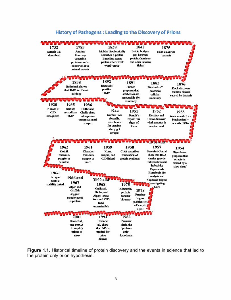
The significance of proteins in cellular processes was recognized and reported
as early as the 18th century. Protein innovator, Antoine Fourcroy, was the first to
recognize albumin, fibrin and gelatin from animal sources and was the first known
chemist to postulate that vegetable proteins could be converted into animal protein1,2.
Nearly a century later, in 1838 Dutch Chemist Gerhardus Johannes Mulder applied the
knowledge gained from Fourcroy, and with his own experimentation, biochemically
described a protein. 2 Through written correspondence, Mulder took the direction of
Swedish colleague, Jons Jakob Berzelius, and named the fundamental substance
“protein,” after the Greek word “prota,” meaning “of primary importance.” Mulder further
went on to examine Fourcroy’s predictions that plant protein could be transferred to an
animal unaltered.
Shortly after Mulder published his elemental analysis of these nitrogen-containing
animal proteins described fifty years earlier by Fourcroy, Justus Leibig recognized the
importance of the protein chemistry field. Liebig was a well-known chemist at the
University of Giessen and was fascinated with all things chemistry2. It was Liebig’s
obsession with chemistry that helped bridge the gap between chemistry and related
scientific fields such as agriculture, nutrition, physiology, and fermentation3. Many
advancements and knowledge have been founded on these primary experiments.
Researchers now have a richer understanding of protein structure, function, and their
2
involvement in cellular processes. However, the knowledge gained from these
pioneering investigators has not come without controversy. Theories explaining protein
structure and function have been widely disputed, especially during primitive
experimentation when technological tools were lacking. Perhaps this dissent can be
attributed to the fact that so many scientific fields rely on and incorporate a fundamental
understanding of protein biology. It is not surprising then, that a protein that defies
biochemical and biological paradigms (such as the prion protein) described by our
scientific forefathers also has a lengthy history of controversy and ambiguity.
History of Pathogens
Protein science was an important foundation for pathogen discovery in the 18th
and 19th centuries, but many aspects of disease, the pathogens that cause them, and
the host response to them were not well understood. Sheep scrapie was described in
1732 but little was comprehended about several dimensions of this disease, including
etiology, pathology, and mechanism. In 1875 Ferdinard Cohn published an early
classification of bacteria 4 while Koch, one year later, announced that anthrax disease is
caused by a bacterium5. Metchnikoff, prescient in immunology, described cellular
immunity in 1882,6 and 9 years later Paul Ehrlich proposed that antibodies (proteins) are
responsible for immunity7. In 1892 D.I. Ivanoski was the first scientist to isolate and
describe an infectious pathogen, the tobacco mosaic virus (TMV), affecting leaves of
plants8. In 1898 M.W. Beijerinck championed the idea that the infectious filterable
pathogen was of viral etiology9.
Over the next 30 years experts gained a better understanding of viruses,
bacteria, their diseases, and associated proteins. Wendell Stanley made significant
3
advances in crystallization of TMV that led to high-resolution structures of viral proteins
and viral particles that advanced viral science10-12. In the 1920s neuropsychiatrists,
neurologists, and physicians were introduced to a neurological disorder of unknown
etiology that would bewilder them for the next 60 years: Creutzfeldt-Jakob disease
(CJD), which was named after its discoverers13,14. At this time intraspecies transmission
of scrapie was shown to occur by inoculating brain and cerebrospinal fluid from infected
sheep 15,16. Then in the early 1950s, Kuru, another neurological disorder of humans, was
being reported in Papua New Guinea, but the etiological agent was baffling scientists,
much like CJD and scrapie17. Alongside this discovery, Hershey and Chase anticipated
that only DNA is needed for viral replication18 and James Watson, Francis Crick, and
Maurice Wilkins biochemically described DNA. 19-23 Only a few years later, Fraenkel-
Conrat et al. explained that RNA carried genetic information and had infectivity in TMV,
another important discovery in virology24-28. In 1958 Crick described the foundation of
protein synthesis and defined the “central dogma of biology;” information coded by
nucleic acid can be synthesized or transferred but proteins cannot transfer information29.
The 1950s and 1960s proved important in revealing characteristics of
neurodegenerative diseases caused by an undetermined pathogen. Research into
animal and human neuropathologies was gaining momentum thanks to the work of
Gajdusek, Gibbs, Klatzo, Hadlow, Zigas and Sigurdsson. Pattison reported that Cuille
and Chelle hypothesized that scrapie was caused by a ‘slow virus’ as early as 1938.
However, the first published hypothesis of this was in 1954 when Sigurdsson reported
the idea of sheep scrapie being caused by a ‘slow virus’ due to the long incubation, an
important insight into what might be causing these encephalopathies in sheep (and
4
humans)30. Collaborative efforts in 1959 revealed that kuru was linked to scrapie and
CJD31,32. During this time Hadlow proposed that kuru was transmissible like scrapie and
he further anticipated (based on Sigurdsson’s 1954 study) that kuru was a ‘slow virus.’
He recommended infecting chimpanzees with kuru31. In 1966 Gajdusek, Gibbs and
Alpers completed these experiments in chimpanzees and repeated them two years later
with CJD, leading to the discovery that both diseases were transmissible after
intracerebral inoculation33,34. Research of viruses and their pathogenicity continued to
intrigue the scientific community, as did the transmissible spongiform encephalopathies.
Earlier findings that RNA encoded genetic information and was infectious25-27
overwhelmingly influenced the first theories stating that CJD, scrapie, and kuru were
caused by a ‘slow virus’ 30,31,35, 28.
‘Slow virus’ or Protein: birth of the “Prion”
An important distinction between viruses and what was causing these
encephalopathies was mistakenly disregarded early on. In 1944, veterinarian W.S.
Gordon used formalin to inactivate looping-ill virus (a common practice) found in brain
and spleen of infected animals and used these treated tissues to vaccinate healthy
animals36. Formalin inactivated the virus but it did not inactivate the scrapie agent that
was unknowingly present, and the vaccinated animals died of scrapie two years later36.
It wasn’t until 1966 that the scrapie pathogen received attention for its stability37,38. Many
experts from different fields tried to inactivate the scrapie agent by ionizing and UV
irradiation, extreme heat, high pressures, and other compounds known to inactivate
viruses and bacteria37,39-41, 42,43. Characterization of the scrapie agent was more easily
accomplished with mouse models44 but masked the earlier observations that CJD had a
5
familial or genetic component of transmissibility45-47, another piece of the puzzle that
should have revealed striking differences between the scrapie agent and typical
pathogens.
Bioassay in mice and then hamsters was a turning point in characterizing the true
infectious agent causing the mysterious encephalopathies. Chandler was the first to
demonstrate transmissibility and the curious lengthy incubation times upon intracerebral
inoculation of scrapie brain homogenate into wild type mice44. Intriguingly, Chandler and
Eklund shortened the incubation period upon serial passage, a phenomenon we now
know to be adaptation48,49. A few years later Zlotnik50 repeated transmission studies in
Syrian Hamsters, and incubation periods were shortened to half those of mouse
models51-53. Rodent bioassays were instrumental in determining the physiochemical
properties of the scrapie agent, as well as investigating intra and inter species barriers.
Investigators were able to use end-point titration to characterize incubation periods and
titers. Stanley Prusiner used these techniques to begin sedimentation experiments to
isolate the infectious agent in cell fractions54. In concert with the bioassay experiments,
a few prescient scientists, Tikvah Alper, I.H. Pattison, and J.S Griffith speculated that
the scrapie agent could be of protein origin. Their theories went against the central
dogma of biology for which Crick so elegantly provided the foundation for a few years
earlier; nevertheless, this inspired exciting, novel, but disputed research.
In 1966 Tikvah Alper used ionizing radiation to try to inactivate and determine the
size of the scrapie agent only to conclude that the agent was not easily inactivated with
high amounts of UV radiation and therefore must be replicating without nucleic acid37.
Pattison added further evidence that the scrapie agent was of protein origin based on
6
his purification experiments55. Boldly, Griffith was the first scientist that not only
speculated that the scrapie agent was protein but he also offered three mechanisms
that might explain how a protein could be infectious and how this infection could be
controlled genetically and occur spontaneously41. Griffith alluded to the controversy and
chagrin some might think his mechanisms brought to the field in the discussion of his
1967 paper: “…the occurrence of a protein agent would not necessarily be
embarrassing although it would be most interesting41.” Several researchers followed in
Griffith’s footsteps and accumulated data that continued to suggest the scrapie agent
was dependent upon protein56-58. It was Stanley Prusiner, however, that took the
“protein-only” hypothesis to a rebellious new level. Prusiner and colleagues were able to
strengthen their argument through successful protein inactivation experiments of the
purified infectious agent from diseased animal preparations59-65. In 1982 Prusiner coined
the term “prion,” proteinaceous (only) infectious particle, to describe the infectious
scrapie agent, for which he would later win the Noble Prize.
Despite Prusiner’s bold attempts to prove the protein-only hypothesis, decades
would pass before it became an accepted hypothesis and understandably so; the
positing of an infectious and transmissible protein contradicted core scientific
knowledge. Prion and rival scientific fields have spent countless years trying to prove or
disprove the prion hypothesis. However, further evidence supporting Prusiner and his
“protein-only” predecessors was presented when the normal cellular prion protein, PrPC,
was shown to be necessary for infection through the use of Prnp0/0 knockout mice 66, 67.
Additionally, Prusiner and colleagues provided fundamental evidence that prions are
nonimmunogenic, suggesting that the scrapie agent must be a recognized host
7
encoded protein68,69. Moreover, confirmation of spontaneous prion formation from non-
infectious material in vivo and in vitro and the ability to replicate the infectious particle in
vitro with the use of PrPC as a substrate with the protein misfolding cyclic amplification
(PMCA) assay provided concrete protein-only evidence70-78,79,80. Most of the scientific
community now accepts the hypothesis that prions are infectious proteins devoid of
nucleic acid.
8
Figure 1.1. Historical timeline of protein discovery and the events in science that led to the protein only prion hypothesis.
9
Prion Protein Properties
Prions cause a class of diseases classified as transmissible spongiform
encephalopathies (TSEs), which characterizes the pathogen well, as prions can be
transmitted and vacuoles appear histologically in the brain of infected individuals81.
Mounting evidence demonstrates that prion pathogenesis is caused by the conversion
of the normal cellular host protein, (PrPC) into a protease-resistant, abnormal disease-
causing isoform devoid of nucleic acid (PrPRES)58,82. Importantly, no differences are
observed in the primary structure of PrPC as compared to PrPRES indicating that
observable differences between the normal and disease-causing proteins must be
conformational. Currently, there are no antibodies that can distinguish between PrPC
and PrPRES in infected animals without protease digestion. However, investigations of
conformation dependent epitopes of the prion protein isoforms are looking promising83.
Approximately 250 amino acids encode the mammalian prion protein (PrP) which
has several distinct domains (Figure 1.2): the amino-terminal signal peptide that is
cleaved during posttranslational processing; a highly conserved, hydrophobic
octapeptide repeat region; and a hydrophobic carboxy-terminal sequence that signals
the addition of a glycosylphosphatidyl-inositol (GPI) anchor84. Within the C-terminal
globular domain of PrPC there are a few unique properties observed in the structure.
During biosynthesis a GPI anchor is attached, associating PrPC
with cholesterol-rich
lipid rafts at the cell membrane84,85. PrPC and PrPRES can be un-, mono-, or di-
glycosylated with N-linked oligosaccharide chains that are added at two positions (180
and 196 in mice) initially in the endoplasmic reticulum (ER), but are made more stable
within the golgi86.
10
Analysis of secondary structure of purified Syrian hamster brain and nuclear
magnetic resonance of recombinant Syrian hamster and mouse PrP determined that the
C-terminal globular domain is comprised of 3 α-helices (42%) and 2 β-sheets (3%)87-89.
Conversely, PrPRES is thought to be predominately of β-sheet structure (43%) and
therefore, the hallmark of prion disease is the conversion of PrPC α-helical structure to
the PrPRES β-sheet isoform. These structural transitions of PrP are responsible for overt
physiochemical differences. PrPC is soluble in non-denaturing detergents and fully
hydrolyzed in the presence of a serine-protease (proteinase K (PK)) while PrPRES is
insoluble and partially resistant to PK, producing an infectious, protease-resistant amino
acid core of about 142 amino acids and 27-30 kDa (PrP27-30)59,90. The second and third
α-helices are joined by a disulfide bridge, and α-helix 2 and β-sheet 2 are linked with a
large loop, known as the L1 loop, which has unique inter-specific properties (Figure
1.2)91,92. The L1 loop has been shown to be exceptionally flexible in most species but is
very rigid in deer and elk (Figure 1.2). Variation seen in the L1 loop is thought to be
associated with conversion and transmission susceptibility of the host92. Unique
conservation of PrP structure is observed in the disordered N-terminal domain which
contains a glycine-rich octapeptide-repeat region, composed of amino acids
PHGGGWGQ, that is thought to bind metal ligands such as copper and manganese87,93-
97, 98.
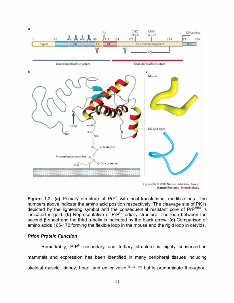
11
Figure 1.2. (a) Primary structure of PrPc with post-translational modifications. The numbers above indicate the amino acid position respectively. The cleavage site of PK is depicted by the lightening symbol and the consequential resistant core of PrPRES is indicated in gold. (b) Representative of PrPC tertiary structure. The loop between the second β-sheet and the third α-helix is indicated by the black arrow. (c) Comparison of amino acids 165-172 forming the flexible loop in the mouse and the rigid loop in cervids.
Prion Protein Function
Remarkably, PrPC secondary and tertiary structure is highly conserved in
mammals and expression has been identified in many peripheral tissues including
skeletal muscle, kidney, heart, and antler velvet99-101, 102 but is predominate throughout
12
the central nervous system (CNS) and the lymphoreticular system (LRS). Curiously,
knock-out mice that do not express PrPC are relatively normal so the physiological
function is not understood or the function is redundant in other cellular processes. The
only role of PrPC that is absolutely certain is that it is needed for prion disease, as Prnp
knock-out mice are completely resistant to infection but are completely susceptible upon
rescue of the wild type phenotype103,104, 105.
Many theories have explored the idea that PrPC must be associated with cell
signaling transduction because, as with most cell signaling proteins, it is GPI-anchored
in lipid rafts at the cell membrane. Most of the cell signal transduction theories have
been based on neuroprotective functions. Chiarini et al., reported that PrPC conferred
neuroprotection to retinal explants by rescuing them from apoptosis106. Lopes et al.,
showed that interaction of PrPC with stress-inducible protein 1 in situ hippocampal
neurons could be neuroprotective by activation of cAMP-dependent protein kinase A
through the MAPK pathway107. Other researchers have suggested that PrPC offers
neuroprotective properties or cryoprotection against harmful agents or oxidative stress
by enhancing divalent metal cation (Cu and Zn) dependent superoxide dismutase
(SOD)108, 109, 110. Wong and Brown et al., convincingly showed that SOD activity
correlated with the level of PrPC expression, and PrPC contributed to total SOD
activity111,112. Many experiments have failed to demonstrate that PrPC is fastidious to any
particular function or interaction with proteins. Therefore, the importance and role of
PrPC remains an enigma for prion researchers even though it is ubiquitously expressed
in vertebrates113.
13
The Significance of Prion Protein (PrP) Genetics
Prion diseases can manifest as genetic, sporadic or infectious conditions, all of
which can be associated with the PrP gene on some level. Inherited forms of prion
diseases, referred to as Gerstmann–Sträussler–Scheinker syndrome (GSS), familial
CJD, and fatal familial insomnia (FFI) disease, account for 10-15% of all prion
disease45,46,114,115. Mutations in the prion protein gene (PRNP) are thought to be
responsible for inherited prion disorders and possibly for some sporadic cases116,117.
Polymorphisms in the gene have been well documented and linked to susceptibility for
many prion diseases, most notably CJD and scrapie118-120.
The PrP gene is a member of the Prn gene family. The open reading frame of the
PrP gene is located within one single exon and in humans is located on the short arm of
chromosome 20 (chromosome 2 in mice)121. PrP mRNA is constitutively expressed in
neurons, astrocytes, and oligodendrocytes of adult brains, but is more tightly regulated
during embryogenesis and fetal development122-124. Ford et al. did not see a direct
correlation between levels of mRNA in a cell and levels of PrPC expression, so not all
expressed mRNA is being translated99. Interestingly, another gene (prnd), which is
under the promoter of Prnp, has been named “doppel” because it is downstream and
prion protein like of prnp125-127. Doppel maintains a similar structure to PrP but is only
expressed in the testis. Interestingly, expression of doppel in the CNS leads to cerebral
dysfunction and neurodegeneration125,126. The discovery of doppel led researchers to
investigate similarities of Prnp to other genes, and this resulted in the discovery of sinc
and prn-i genes. Investigators speculated these genes were responsible for short and
long incubation differences among mice126,128-130. Dickinson published a persuasive paper
14
suggesting that prion strains encode their own unique disease characteristics, but that
host encoded PrPC, specifically the sinc and prn-i genes, regulate the disease
progression and pathogenicity128. Elegant gene targeting studies by Moore et al.
confirmed that sinc, prnp, and prn-i genes were all congruent, and PrP allotypes at
codons 108 and 189 were responsible for long or short incubation periods in mice130.
Certain polymorphisms of Prnp have conferred complete resistance to prion
disease, most notably in sheep. In 1960, RH Parry adopted the theory that sheep
scrapie was a transmissible hereditary disease linked to a recessive gene131. Years of
sheep breeding records demonstrated that scrapie wasn’t hereditary, but susceptibility
or resistance to scrapie was. Three polymorphisms have been found in sheep at codons
136 (A/V), 154 (R/H), and 171 (Q/R)119. Suffolk sheep heterozygous or homozygous for
arginine at codon 171 demonstrated complete resistance to scrapie, while sheep
homozygous for glutamine are susceptible120132-134. This phenomenon was also
demonstrated in Cheviot sheep homozygous for alanine at codon 136 (resistant) or
homozygous for valine (susceptible). Curiously, sheep heterozygous at codon 136 were
susceptible to disease but the incubation period was lengthened135. Goats show
redundancy in polymorphic susceptibility to sheep at codons 142, 143, and 240136,137.
Scrapie is a unique prion disease as it has been eradicated from flocks all over the
world by breeding for the resistant polymorphisms. Ironically, however, many of the
flocks that are resistant to classical scrapie are susceptibility to other prion strains such
as BSE and atypical scrapie, making eradication of all prion diseases in sheep and
goats impossible138,139. Cervids (deer, elk and moose) also have PrP polymorphisms that
may aid in protection against chronic wasting disease (CWD). Telling et al. used
15
transgenic mouse models to demonstrate that mice homozygous or heterozygous for
leucine at codon 132 were resistant to CWD prions while those mice homozygous for
methionine were susceptible140. Similarly, polymorphisms for methionine to valine in
humans at codon 129 renders the host resistant to inherited, sporadic and iatrogenic
prion disease118,141,142. Transgenic mice have become vital for the progression of prion
research and have allowed researchers to investigate transmission efficiency,
susceptibility and resistance by manipulating genes.
Exploitative Nature of Transgenes in Prion Research
Animal models have been instrumental in advancing scientific knowledge in all
fields. Mouse models in particular, have been an invaluable tool in studying prion
biology and disease progression. Without the manipulation of the prion protein gene,
much would still be unknown about fundamental prion biology because mouse models
abrogated the need for lengthy and costly studies in the natural host.
As described earlier, the use of PrP knock-out mice intensified evidence that
prion disease is caused by misfolded protein. Many different strategies have achieved
knock-out or knock-in mouse models to aid in the study of normal and diseased prion
biology. Bueler used a gene targeting strategy to disrupt the PrP gene in Zurich I mice
by using an expression cassette for neomycin phosphotransferase66. This resulted in
some fused mRNA containing neo and residual PrP sequences but no expressed PrPC.
Comparably, Jean Manson mutated the gene so that no mRNA or PrP protein could be
detected by using a neo cassette and a unique KpnI site on exon 3143. In addition,
Mallucci designed an elegant study that utilized a Cre-loxP expression system to create
a conditional post-natal PrP null mouse. LoxP sites were under control of the Prnp

16
promoter element, while Cre-recombinase was under the control of the neurofilament
heavy chain promoter. Therefore, expression of the recombinase during development
resulted in the deletion of PrP in neurons144. The techniques utilized to establish knock-
out or knock-in models of PrP have proven delicate as some gene targets have had
deleterious or overexpression effects on different but related genes, resulting in un-
characteristic neurological phenotypes145.
Another concern with using mouse models for prion diseases is that even
partially expressed mouse PrPC can confer protection or resistance to prion disease
when inoculated with a strain from an unrelated host, e.g. human, cervid, bovid146, 147. It
has become a common practice to create transgenic mice on a PrP null background to
eliminate any protective effect mouse PrPC could have on disease phenotype. Host
encoded PrPC plays a pivotal role in transmission efficiency in other species as well. If
host PrPC does not match that of the infectious inoculum, transmission is inefficient, a
phenomenon known as a species barrier. For example, wild type mice are resistant to
CWD prions but transgenic technologies are able to rectify this problem by knocking out
the mouse Prnp and inserting a cervid transgene.
Additionally, there is an inverse relationship with the level of the PrPC expression
and incubation periods148. Therefore, transgenic mice overexpressing PrPC from the host
of interest succumb to prion disease much faster than those with normal expression
levels. Browning et al., constructed a cervidized transgenic mouse (Tg(cerPrP)) that
overexpressed (6-8 fold higher) cervid PrPC and efficiently replicated prion strains from
deer and elk. They achieved overexpression by modifying the CerPrP S2 allele
nucleotide sequence by site directed mutagenesis149. The construction of mice that
17
overexpress (5-8 fold higher) PrPC has resulted in an observable shorter disease course
and has given researchers the tools to study any prion disease using the same model.
Prion Strain Dynamics
Kuru, CJD, GSS, FFI, scrapie, etc. have distinct phenotypes and strain properties
so were not linked to the same infectious agent until 1959. The concept of strains was a
novel idea that had not been defined in early prion disease, resulting in a different name
for every prion disease despite being caused by the same etiological agent. Historically
prion disease nomenclature has added to the complexity of linking prion diseases and
discovering unique strains.
Cullié and Chelle were the first investigators to discover scrapie transmissibility
by different injection routes into healthy sheep with spinal cord or brain from a diseased
sheep. In 1939 they continued their transmission studies demonstrating efficient
transmission of sheep scrapie into goats but resulting in different clinical signs15.
Unknowingly, Chelle expanded the idea of strains in 1942 by describing different clinical
signs in goats and sheep naturally infected with sheep scrapie compared to
experimentally inoculated goats150. During this time, a new transmissible spongiform
encephalopathy strain was discovered in farmed mink, transmissible mink
encephalopathy (TME). However, TME was not described and characterized until the
late 1980s and early 1990s52,151. In the 1950s Wilson expanded scrapie transmission
studies by serially passaging scrapie material in sheep for 9 passages152 and
subsequently investigated species barriers in mice, guinea pigs, and rabbits. In 1957
Pattison, Gordon and Millson followed suit in goats, demonstrating that goats were even
more susceptible hosts than sheep but exhibited a “drowsy” phenotype150. They named
18
the scrapie goat strain SSBP/1. A few short years later, in 1961, Chandler
revolutionized prion research by successfully inoculating mice with goat scrapie44. Other
researchers began to mimic bioassay experiments in mice but were displeased with the
lengthy incubation periods paralleling the disease course of the natural host153. Zlotnik
and Chandler began testing species barriers in different rodents in search of new, more
efficient models of scrapie. In 1963 Zlotnik experimented with hamster bioassays, and
Chandler challenged rats; both were pleasantly surprised by their success50,154.
At this time Zlotnik and Rennie developed a well-known scrapie strain, ME7. This
strain was derived from a pool of infected Suffolk sheep spleens. Zlotnik and Rennie
inoculated mice intragastrically (i.g.) with the spleen pool and created a brain pool from
the infected mice which they subsequently passaged intracranially (i.c.) into RIII (C57
Blk6 mouse lineage) mice155.
A new prion disease of cervids, chronic wasting disease (CWD), was first
described in 1967 in captive mule deer156. Ten years later, Dickinson characterized 3
strains (22A, 22C, 22L) from mice inoculated with SSBP/1 goat scrapie using incubation
periods and vacuole distribution157, 158. In the late 1970s and into the late 1980s, RH
Kimberlin and colleagues like Walker, Dickinson, and Fraser, investigated strain
characteristics more fully using rodent bioassay models and were able to define prion
species barriers. Meanwhile, CWD was gaining more attention as Williams described
the first case of a TSE affecting a wild population159,160. DeArmond and Bruce et al.,
elaborated on strain characterization in the late 1980s by describing patterns of PrPRES
accumulation in the CNS along with lesion profiling161,162. In 1986 three dairy cattle in the
UK were recognized as having neurologic disease that was later accepted as a new
19
TSE and named bovine spongiform encephalopathy (BSE)163. In 1992 Bessen and
Marsh described the isolation of two distinct strains from TME that they appropriately
named “hyper” (HY) and “drowsy” (DY) based on the hamsters’ demeanor164. HY and
DY TME had observable differences not only in clinical signs but also in incubation
times and lesion profiles. A few years later, Prusiner et al., established that 5 of 20
mutations known in huPrP are linked to the dominantly inherited human prion diseases
fCJD, GSS, and FFI. Additionally, Collinge et al. observed differences in glycosylation
patterns between strains adding criteria for characterizing a new strain165
A pivotal moment in prion history and identification of prions strains was when
the zoonotic potential of BSE was discovered in 1996. An association between people
who ate infected beef and a new rapid human TSE was named as variant CJD
(vCJD)166. One year later, Prusiner’s lab generated novel strains by infecting transgenic
mice with different PrP amino acid sequences167.
In 1998 and 1999 biochemical characterization of prion strains was became an
effective approach for describing novel strains as well characterizing the similarities of
strains in different diseases (e.g. BSE and vCJD). Additional tools to characterize
strains were also being described at this time; Prusiner, along with Groschup, correlated
differences in PK stability with conformational changes between strains and postulated
that the more sensitive a prion strain is to PK digestion the quicker an animal succumbs
to disease168,169. At the start of the millennium Jason Bartz and colleagues investigated
mechanisms of strain adaptation and selection of TME using the HY and DY strains in a
competition hamster model. Remarkably, DY was the selected strain upon first passage
but HY was selected in subsequent passages, presumably because HY is a more
20
efficient replicator170. In 2002 more research groups concluded that strain propagation
was based on the conformation of PrPRES independent of PrPC sequence171,172. It is
important to note, however, that many groups conducting strain studies failed to serially
passage the new PrPRES strain, overlooking strain adaptation and host factor effects on
the nascent PrPRES conformer over time.
In 2006, Aguzzi’s laboratory claimed that they were able to cross a strict species
barrier and infect transgenic mice expressing high levels of mouse PrPC with a CWD
strain173. These investigators reported that the original CWD strain characteristics were
unchanged after serial passage into the mice, suggesting that strain-specific CWD
properties are not altered by host factors. It is important to note that their results could
have been a result of spontaneous disease (a common phenomenon in aged,
overexpressing mice) as mice did not show clinical signs before 500 days post
inoculation (DPI).
In 2008, our group contradicted the Aguzzi’s lab findings and demonstrated that
prion strains could be adapted (altered) through serial passage using transgenic
cervidized mice and a cervid prion strain but failed at crossing the species barrier into
wild-type mice. In addition, we were able to show that the cervid prion strain could be
more efficiently adapted in vitro using protein misfolding cyclic amplification (PMCA).
Evidence of adaptation was seen through distinguishable incubation periods,
conformational stability, PK stability, glycoform ratios, and neuropathology of mouse-
adapted RML74. Kimberlin and colleagues paved a persuasive foundation of species
barriers and prion strains through their tedious transmission studies. Most of their
research resulted in the abrogation or construction of species barriers and novel strains
21
using species with differing PrPC amino acid sequences51-53. Namely, he used a goat
scrapie strain and serially propagated this through mice, rats, and hamsters. He
observed incomplete attack rates upon the first passage into hamsters with 300 day
post inoculation (dpi) incubation period. However, second passage accelerated disease
and all the hamsters were sick in 130 dpi51. More recently, Tamguney et al. manipulated
elk transgenic mice to chimerically express c-terminal mouse PrPC and found that
species barriers were created and destroyed based on certain elk-to-mouse or mouse-
to-elk mutations in C-terminal amino acids174.
Animal Strains
Scrapie
Scrapie was the first described prion disease, dating as early as 1732 in
England. At that time, the disease was only documented by clinical signs, as prions
were not even postulated until the mid-1900s. Scrapie is one of the most well studied
prion diseases, perhaps because rodent models have been relatively susceptible to
scrapie. Scrapie is known to naturally affect sheep, goats, and mouflon. “Scrapie”
became the accepted disease name due to the clinical sign of pruritus, evidenced by
sheep scraping against objects intensely enough to scrape their wool off. Goats have
been observed to preferentially use their hind legs or horns to scratch. In addition to
severe itching, animals are ataxic, often trotting with an uncharacteristically high
stepping, unbalanced gait. Many people often referred to scrapie as the “trotting
disease,” or the “trembling disease” because of observed clinical signs. Weight loss is
also observed even though the animals are polyphagic. Infected animals have also
been recorded as being restless, anxious, easily stimulated, grinding teeth, excessive
22
thirst and consequently, excessive micturition15. Remarkably, reproductive function is
not impaired during disease and many ewes give birth to big, healthy lambs without
assistance. However, depending on the animal’s disease course, the mother may not
have the ability to nurture and care for the lamb once born. Once clinical signs are
apparent, death follows in 2 weeks to 6 months15. A distinguishing characteristic of prion
disease is the lengthy incubation period; signs of scrapie disease are typically seen 18
months to 2 years after exposure.
Scrapie is unique in that it can be eradicated through selective breeding of
resistant polymorphic genotypes135(USDA/APHIS). Suffolk sheep heterozygous or
homozygous for arginine at codon 171 have been shown to be resistant to scrapie
infection and BSE while maintaining traits producers desire120,175,176, 177, 178. Unfortunately,
in 1998 an atypical scrapie case (Nor98) was reported in Norway. This scrapie strain
has many different pathological features than classical scrapie, but most notable is that
sheep considered resistant (ARR or ARQ) to classical scrapie are susceptible to
Nor98177,179-182. There are reports that Nor98 (atypical scrapie) is restricted to the CNS183-
186but more recently, there is evidence that there is peripheral expression but not easily
detected due to infectious PK sensitive characteristics of this strain187.
It is still unclear how scrapie started, although most investigators believe that a
sheep must have spontaneously developed the transmissible disease. In 1914
McGowan was convinced that scrapie was caused by a parasite because scrapie
infected sheep exhibited sarcosporidia in the musculature, but no other evidence links
this parasite to scrapie15. Transmission of all prion diseases is also unclear but most
evidence suggests that scrapie is transmitted orally and vertically from mother to
23
offspring188-194, 176, 195-198. Because infectious prion proteins are so resistant to degradation,
it is appropriate to evaluate lateral/horizontal transmission of scrapie. Increasing
evidence points to horizontal transmission; introducing uninfected flocks to areas
infected flocks once inhabited results in disease a few years later192,199,200, 201.
Currently there are several ante mortem tests that can be done on lymphoid
tissue to diagnose scrapie in sheep and goats: the third eyelid test, rectal biopsy, and
tonsil biopsy; however, these are not as sensitive and reliable as testing the brain
sections once the animal is dead. In all tests tissues are considered positive if there is
immunohistochemical (IHC) staining for PrPRES. Like all prion diseases, positivity is only
definitively determined by the analysis of the brain for presence of the characteristic
spongiform plaques as well as aggregated prion plaques or fibrils. Scrapie prions are
characteristically present in the medulla, particularly in the dorsal motor nucleus of the
vagus nerve, which innervates the gut and consequently, prions can be found in the gut
throughout Peyer patches in the small intestine202, 153. Rodent models share the same
neuropathology as the natural host, but IHC staining can be more diffuse with less
obvious aggregated punctate plaques74,203,204.
BSE
Bovine spongiform Encephalopathy (BSE) is the prion disease that affects cattle.
BSE has become better known to the public as “mad cow disease” by the media
attention it received in the late 1990s when BSE has a zoonotic outbreak into humans.
BSE was first diagnosed in the United Kingdom in 1986 in three dairy cattle presenting
neurological signs similar to sheep and goats with scrapie. Salient behavioral signs of
BSE include aggression, apprehension, and exaggerated responses to stimuli. In
24
addition cattle are ataxic, have decreased milk production, become very thin, and at end
stage clinical disease, become recumbent205. Cattle can display clinical signs for 1-6
months before succumbing to the disease. Determining the incubation time for BSE has
been challenging due to several factors: transmission events appear to be relatively low
between a herd, and most cattle are slaughtered by the age of 6 years old; making a
longitudinal study nearly impossible. Anderson et al., correlated epidemiological data,
accounting for complications noted above, and statistically predicted the mean
incubation period to be around 5 years206.
Genetic polymorphisms have not been associated with BSE susceptibility as in
other species. Some cattle have the presence of an additional octapeptide repeat but
this has not been linked to BSE occurrence/susceptibility in tested cattle194,207-209. Similar
to scrapie, however, atypical BSE strains were identified in 2004. Researchers
characterized atypical BSE strains by distinct molecular and neuropathological
differences. France and Italy were the first to describe two new strains in cattle that
were atypical of BSE well characterized in the UK. Biacabe et al., reported that PrPRES
in cattle 8-15 years old had a high affinity to the monoclonal antibody P4, a noticeable
change in molecular characteristics from classic BSE as only scrapie prions were
recognized by this antibody previously210. The French investigators denoted this new
strain as H-type because of an observable high molecular mass band of the un-
glycosylated PrPRES after PK digestion. That same year, in Italy, Casalone et al.,
described a low molecular weight un-glycosylated band that they denoted L-type211. This
strain also differed from classic BSE because it produced a higher ratio of mono-
25
glycosylated PrPRES rather than di-glycosylated211. Atypical BSE cases have since been
recognized in Japan,212Belgium,213 and Germany214.
The origin of BSE has been very controversial but persuasive evidence suggests
that BSE arose from cattle eating scrapie-tainted meat and bone meal (MBM) and BSE
cases declined dramatically after banning this feed practice215,216,217,218. The evidence
linking MBM to cases of BSE was so convincing that the practice of feeding MBM was
banned in the UK in 1988 and later, in 1996, the practice was banned for all livestock
and mammalian-derived meat (not just sheep and cattle) in Europe and in the United
States206,219. Some researchers believe that BSE began with spontaneously diseased
cattle that was rendered into MBM and fed back to cattle and/or that milk replacers that
contain infected tallow were fed to calves220-222. In 1996, it became evident that BSE was
unique from other animal prion diseases; BSE turned into a zoonotic epidemic among
people that had eaten infectious beef in the UK (described in the human prion disease
section below). BSE has experimentally infected cattle, sheep, pigs, mice, and mink223-
225, 226 and has been linked to natural infections in humans, domestic cats, and captive
wild exotic (i.e. zoo animals) bovidae and felidae227-229, 165,166,230.
There are not currently any ante-mortem tests for BSE because infectious
material is not expressed in peripheral tissues. BSE appears to be restricted to the CNS
and therefore diagnosis can only be made by identifying the presence of PrPRES in CNS
tissues, namely the medulla oblongata. Pathological changes seen in diseased cattle
are the characteristic spongiform change, neuronal vacuolation and degeneration,
accompanied by astrocytic activation231. BSE does not generate robust amyloid plaques
but rather marked spongiform change. Because of minimal peripheral shedding of
26
infectious prions through, meat, milk, blood, etc., horizontal transmission appears to be
an inefficient route of transmission216,232-234.
Chronic Wasting Disease
Chronic wasting disease (CWD) affects captive and free-ranging deer, elk, and
moose (collectively known as cervids) in North America at nearly a 30% prevalence
rate235,236. CWD was first identified as a clinical disease in a captive Colorado mule deer
in 1967 by the late Beth Williams but was not recognized as a TSE until 11 years
later156. In 1980 the disease was recognized for the first time in wild and captive elk160.
CWD is the only prion disease known to affect a wild population. CWD was named
based on observed principle clinical signs such as poor body condition leading to
emaciation over an extended disease course of 2 weeks to 8 months156. Animals are
observed as having behavioral changes, depression, listlessness, polydipsia, polyuria,
grinding of teeth, excess salivation, drooping of the head, and (less noticeable) changes
in gait/movement156. The incubation period of CWD in a natural population is not known,
but the National Parks Service, in collaboration with Colleagues of Colorado State
University, is currently conducting a longitudinal study with elk to try to further
characterize and understand prion progression in the wild (unpublished data). In
captivity, animals succumb to disease between 16 months and 4 years after
exposure235. Evidence suggests that genetic polymorphisms may influence incubation
and susceptibility in cervids analogous to other species affected with prion disease.
Despite homology observed in cervid Prnp, cervids contain several species-
specific polymorphisms. Mule deer contain a serine/phenylalanine polymorphism at
codon 225. CWD infected individuals show a higher prevalence of serine homozygosity
27
at this codon while those heterozygous or phenylalanine homozygous, though
underrepresented in the population, have been reported to have a lower risk of infection
or exhibit delayed disease237. White tail deer also have a polymorphism containing
serine, but deer with serine homozygosity at codon 96 have a decreased risk and/or a
delay in disease as compared to serine/glycine heterozygote or glycine homozygote
deer238,239. Polymorphisms at codon 132 in elk correspond to human polymorphisms at
codon 129, but elk are polymorphic for methionine to leucine. Surveyed elk
demonstrated a very low frequency of leucine homozygous individuals in a given
population. This is unfortunate because these elk, though they are not completely
resistant to CWD infection, live longer240,241. It is emphatically important however, to
consider that those individuals that have a delayed disease onset or do not succumb to
disease at all could be asymptomatic carriers of the disease and could be aiding in
transmission rather than aiding in eradication of the CWD.
Parallel to other prion diseases, the origin of CWD is ambiguous, though many
theories offer explanations. One theory is that CWD originated with a rare spontaneous
case that, over time, was able to gain momentum and become efficiently transmitted
into susceptible herd-mates. It is also not known if CWD originated in captive animals or
in the wild. Epidemiological evidence however, suggests that farmed or captive cervids
have been instrumental to the maintenance and spread of CWD. Williams et al.
hypothesized that cervids grazing on the same lands as scrapie infected sheep could
easily explain interspecies transmission of a TSE242,243. Hamir et al., tested this
hypothesis by intracranially inoculating scrapie infected brain homogenate into elk, but
the results were inconclusive as only half the animals were infected244. In a reverse
28
experiment (CWD infected brain into sheep and cattle), results indicated that animals
were poorly susceptible to CWD from mule deer but cattle were highly susceptible to
CWD from white tail deer240,245,246, 247. Unfortunately, much about the origin and
transmission of CWD in a free-ranging wild population remains difficult to elucidate
because of the many variables that have to be accounted for in the wild. Through
observations of captive cervids, it has become apparent that CWD is transmitted
horizontally through an oral route via blood, saliva, feces, urine and environment248, 249-253,
235,254, 255,256. CWD poses a new challenge in wildlife management and eradication of such
an efficiently transmitted and persistent disease of cervids seems impossible.
Definitive diagnosis of CWD infected animals is accomplished on deceased
animals with immunohistochemistry (IHC) of brain tissue, usually the medulla. Several
ante-mortem tests have been developed for early diagnosis of prion diseases. All of
these tests require biopsy of lymphoid tissue (e.g. third eyelid, tonsil, or rectal tissue)
and IHC to identify accumulation of PrPRES257-259. Comparatively, these ante-mortem
tests are not sensitive, resulting in false negatives of early or unique CWD infections.
However, ante-mortem tests can be a powerful diagnostic tool when used in conjunction
with more sensitive assays such as PMCA or IHC of brain tissue260 (Wyckoff, in review).
The neuropathologic spongiosis is evident in CWD infected animals as it is in all
prion diseases but is most consistently noted in the medulla oblongata. This brain area
has become a standard region for diagnosis and surveillance in captive and free
ranging cervids. Distribution of PrP plaques is variable but generally is not detected in
the cerebellum. Differentiation of CWD from other prion diseases can be made by
29
looking at glycoform ratios after PK digestion as CWD has the highest signal in the
diglycosylated band261.
Through increased surveillance and natural transmission, CWD has now been
identified in 22 states in the US, two Canadian provinces, and South Korea262,263.
Consequently, there is a need for a better understanding of CWD transmission routes,
efficiency and the environment’s role, replication and pathologic mechanisms, and
descriptions of strain and species barriers. Because of the unique characteristics and
availability of CWD samples, it may be the most appropriate prion disease to study to
elucidate the mechanisms prions utilize that continue to elude scientists.
Human Prion Diseases
Kuru
In the early 1950s, an anthropologist couple, Ronald and Catherine Berndt, took
interest in the Foré people of Papua New Guinea. During their encounters they learned
that Foré tribes practiced sorcery: an act of taking an intimate object (feces, discarded
food, hair, etc.) from the intended victim and placing the wrapped object at the edge of
a swamp to watch the coincidental decay of the object and the victim264. When tribal
members came down with an illness it was presumed that someone had used sorcery
against them to make them ill. Specifically, the Foré believed those exhibiting the
unique signs of kuru were victims of sorcery. Kuru first drew the attention of Vin Zigas in
1955 when he diagnosed a woman with signs of kuru as having ‘acute hysteria’ and
otherwise healthy264. A prominent misconception arose that the Foré were succumbing
to sorcery in a psychosomatic manner. Individuals with kuru presented with an intense
trembling or shaking of the entire body, which is what “kuru” means and why it has been
30
established as the disease name. Interestingly, investigators, such as Zigas and the
Berndts determined that kuru mostly infected woman and children264. In 1957 Carleton
Gajdusek joined Zigas to study the kuru phenomenon17. Kuru was characterized as a
TSE in 1968 by Gajdusek and Gibbs et al after he experimentally transmitted kuru to
chimpanzees34, 34,265.
Kuru begins with symptoms of, “lethargy, headache, vertigo and vomiting264.” The
clinical signs include autonomic tremors, unsteadiness and mild ataxia, eventually
leading to paresis and severe ataxia, erratic eye movements, and gradually urinary and
fecal incontinence and disphagia17,32. Sufferers also go through stages of euphoria and
uncontrollable laughing alternating with depression17. Clinical signs can persist up to 9
months and incubation period of disease is as early as 12 months are as long as 50
years17,33,266, 267,268.
Increasing evidence suggests that Foré people homozygous for methionine at
residue 129 are more susceptible to kuru transmission and experience shortened
incubation periods269, 267,268. In fact, those individuals that presented with disease long
after exposure proved to be heterozygous (M/V) at residue 129. There is also
convincing evidence that kuru has caused genotypic selective pressures in Foré tribes
resulting in more resistant genotypes267.
In 1984 Klitzman et al., determined through epidemiologic analysis that the kuru
epidemic was result of ritualistic endocannibalism that began in the 1940s and 1950s266.
Alpers substantiated this evidence with his epidemiologic review in 1992270.
Endocannibalism is the practice of eating flesh from deceased community members.
Kuru only affected the Foré linguistic tribes and close neighbors with whom they
31
intermarried, and predominately affected the woman and young children in these
communities271. During cannibalistic rituals, men had the preference of meat to consume
and ultimately, women and young children were left to feast on brain266. Once
endocannibalism was discouraged by the government (and tribes complied) in the late
1950s and early 1960s, kuru incidence decreased (especially in young children)266,271.
The origin of kuru is unknown. The most plausible hypothesis is that it was introduced
spontaneously and cannibalism transmitted the disease efficiently, similarly to the BSE
epidemic in cattle.
Kuru neuropathology consists of the characteristic spongiosis change and
amyloid plaques with the most severe pathological deficits present in the cerebellum272
Grossly, atrophy of the cerebellum, specifically of the vermis and flocculonodular lobes,
is evident273. In 1959, Hadlow was the first to point out the similarities between scrapie
and kuru31, which led to the transmission studies completed in chimpanzees, which
continued upon the classification of kuru as a TSE, and ultimately as a prion disease,
and the first naturally transmitted human prion disease.
Inherited Prion Diseases
In 1921 Jakob identified Cruetzfeldt Jakob Disease (CJD) in conjunction with a
referenced case of Cruetzfeldt’s in 192013,14. Ultimately these two investigators
contributed to the naming of the invariably fatal neuropathologic disease. CJD can
present in a sporadic, inherited, or transmitted form. Sporadic cases of CJD are the
most prevalent form of human prion disease but familial CJD was first characterized.
Reports about an unknown neurologic disease, now known as Gerstmann-Sträussler-
Scheinker, were as early as 1912274 but it was Gerstmann, Scheinker and Sträussler
32
who characterized the disease (and named it) in the late 1920s and 1930s275. Fatal
Familial Insomnia disease wasn’t described until 1986 by Lugaresi and Gambetti276.
The first familial case of CJD (fCJD) was recorded by Kirschbaum in 1924 and
more convincingly described by Meggendorfer in 193045,277. The “Backer” family had a
long lineage of CJD and aided in the discovery of fCJD phenotypes. There are three
different inherited prion diseases currently characterized: Gerstmann-Sträussler-
Scheinker (GSS), Fatal Familial Insomnia (FFI), and CJD. These diseases are classified
by the phenotype presented in addition to the pathogenic mutation(s) of PrP and their
genotype at residue 129 (haplotype). To date, 55 mutations coupled with 16
polymorphisms in the PRNP gene have been associated to human prion disease278.
Clinical signs of inherited diseases can present differently depending on the mutation
and genotype but most signs are redundant. GSS, FFI, and CJD all present with
cognitive impairment, psychiatric changes (emotional changes), and dementia279280. CJD
additionally can present with seizures and visual disturbances281,282. FFI, as noted by the
name, presents with altered sleep-wake cycles resulting in insomnia and these patients
often hallucinate and are confused280. GSS can present with Parkinsonian-like signs and
ataxia.
The incubation period and duration of clinical signs of these inherited diseases
rely on the individual’s haplotype. Generally GSS disease affects individuals between
40-60 years of age and clinical symptoms are present for 3-6 years on average283,284.
Clinical signs of fCJD generally appear in 30-55 years and persist for months to
years281,285-288. FFI presents in patients 40-50 years of age and can persist from 12-16
months289.
33
GSS, FFI, and CJD have distinct histopathological differences. fCJD resembles
other prion diseases with the characteristic spongiosis and astrogliosis throughout the
cortex and deep nuclei287,290. GSS is well characterized by intense amyloid plaques
present when staining for PrP, while FFI has distinct neuronal loss and astrogliosis in
the thalamus and inferior olive but no spongiosis is detected289,291, 290.
Inherited prion diseases are autosomal dominant and therefore, are easily
retained and passed through populations. Researchers have done an incredible amount
of work to map out 30 known genetic mutations that can elicit these diseases292. Genetic
mutations leading to inherited prion diseases can be classified into two distinct groups:
(1) abnormal number (addition or deletion) of octapeptide repeats located in the N-
terminus of PRNP and (2) Point mutations in the C-terminus of PRNP. Within the first
classification, M129V polymorphism has proven additional importance in determining
the age of clinical onset. For example, in 5 or 6 octapeptide repeat insertion (OPRI)
mutations, the mean age of clinical onset of disease is about 70 years but an individual
heterozygous at residue M129V can have later clinical onset of disease by 10 years as
compared to an M/M homozygous individual293. Interestingly, an individual homozygous
for M/M at residue 129 but has 4-OPRI encounters a later disease onset well into old
age and heterozygous individuals do not develop clinical signs of disease; perhaps
these individuals outlive the onset of clinical stage disease or they are resistant
completely. The second broad classification of inherited prion diseases has a higher
prevalence in an infected population. Curiously, some point mutations can be identified
by ethno-geographic clustering within a disease phenotype. This is demonstrated in a
report by Hsiao et al., where they associated the occurrence of fCJD in Libyan Jews to
34
a lysine mutation at codon 200281. Certain point mutations, coupled with a specific
polymorphism at residue 129, create a haplotype that depicts the disease phenotype.
Gibbs et al., showed a unique dependence of CJD and FFI phenotypes on the
haplotype created at codon 129. If the mutation occurs on the 129 methionine allele,
then the patient will develop FFI, but if the patient has the mutation on the 129 valine
allele, the patient will almost always present with a CJD phenotype293. It is important to
note that in a Caucasian population, 43% are methionine homozygotes, 49% are
heterozygotes, and 8% are valine homozygotes and in Japan, almost the entire
population is methionine homozygous (92%) and little to none are heterozygous at
codon 129. Even with the high percentage of M/M individuals in a given population,
inherited prion disease is estimated to only affect one case per million or account for
just 10% of human disease 294. A selection of inherited prion diseases have proven
experimentally infectious into laboratory animals{Roos:1973vh}. This novel finding excited
researchers as this is the first account for a hereditary disease to be shown infectious
and substantiated the protein only prion hypothesis. Luckily, epidemiological evidence
suggests that familial diseases are not normally infectious within a population, which
may account for the minute amount of prion infected individuals.
Sporadic and Infectious Human Diseases
Sporadic CJD (sCJD) accounts for 85% of human prion diseases295. Patients that
present with clinical signs representative of CJD and do not have known PRNP
mutations or risk factors are consequently categorized as being inflicted with sporadic
CJD. Ultimately the specific cause of sCJD is unknown and cannot be linked to any
geographic or temporal clustering296. Distinctively, vCJD clinical signs progress within a
35
few short months (<6) compared to patients with fCJD297. sCJD patients present with
either ataxia or cognitive impairment and as the disease progresses those patients that
preceded with ataxia will sequentially show signs of cognitive impairment and vice-
versa298 Myoclonus or muscle twitching is a distinct sign that appears as the disease
progresses299. sCJD trends towards infecting late middle-aged people between 65-79
years of age but becomes fatal in an average of 5 months290,298.
Prion diseases captured worldwide public attention in the late 1990s when a BSE
outbreak crossed the species barrier into humans. In 1996 10 people presented with
neurological signs in the UK within months of each other. Surprisingly, the mean onset
of disease was 29 years of age while the disease course was sustained an average of
12 months166. Additionally, all patients exhibited distinct neuropathology and
corresponding clinical signs. Patients primarily presented with behavior changes and
progressive dementia but little memory impairment166. Spongiform change was
prominent throughout the basal ganglia and thalamus while PrP plaques were
ubiquitously detected in the cerebrum and cerebellum166. Astonishingly, all patients were
M/M homozygous at codon 129 and lacked any associated haplotypes of other human
prion diseases166. In succession with epidemiologic studies, several transmission studies
provided unequivocal evidence linking consumption of BSE infected meat with
transmission to humans225,300. The new disease phenotype was designated as variant
CJD (vCJD) because this prion was a new variant to what was previously known for
human prion diseases. Since the first outbreak of vCJD, over 200 cases have been
identified. This zoonotic transmissible disease not only caused an upheaval in the
livestock industry (particularly feed and slaughtering practices) but, in association with
36
transmissibility of all prion diseases, it has concentrated efforts to minimize iatrogenic
transmission in the public.
Iatrogenic transmission of CJD has been well documented as early as in the
1950s301. Transmission of CJD through medical procedures has been achieved
innocently through corneal grafts,302growth hormone therapy,303 dura mater
graphs,304multi-use neurosurgical instruments and probes,305 and the potential of
transmission through blood transfusion250,306,307.Iatrogenic CJD (iCJD) is denoted as
sCJD unless sufficient evidence of an iatrogenic event can definitively define otherwise.
In most cases, it is impossible to know whether sCJD was transmitted through a
medical procedure or blood transfusion because clinical signs are relatively similar. On
the rare occasion however, a direct iatrogenic event can be identified proactively. In
example, thirteen brain-surgery patients in several states were possibly exposed to
sCJD prions through the use of the same contaminated equipment from a possibly
sCJD infected patient. The surgical equipment was used on the infected patient for
brain surgery just 3 months before death (prion titers in the brain would presumably be
high at this end stage of disease) 308. Because prions are so tolerant of standard
sterilization techniques, equipment could have remained contaminated and therefore,
transmitted CJD to all naïve patients. Unfortunately, only time (5-10 or more years) will
tell as to whether or not individuals develop the invariably fatal disease308.
Spontaneously Generated Prions
Further evidence that prions are protein-only infectious particles have been
demonstrated through spontaneously generated prions in vitro using Protein Misfolding
Cyclic Amplification (PMCA). PMCA was first utilized by Castilla et al., in 2005 and
37
revolutionized conventional prion detection methods. PMCA utilizes sonication to break
up PK resistant aggregates so the prion seed may ubiquitously interact and template
PrPC present in the uninfected brain homogenate substrate309. Protocols for PMCA
differ across research laboratories but in brief: samples containing a mixture of PrPRES
and uninfected brain homogenate go through a 24 hour repetitive cycle of 30 minutes of
incubation and 20 seconds of sonication in a water bath maintained at 37o Celsius.
Each round is about 144 cycles and at the end of a round, new PrPC substrate is added
to the newly generated in vitro PrPRES to allow for increased conversion and
amplification of the template. This replication technique is analogous to PCR amplifying
DNA in a cyclic fashion to detectable amounts. Interestingly, Deleault et al., generated
de novo PrPRES by altering the PMCA protocol to only include PrPC, co-purified lipids,
and polyanions79,310. However, skeptics discredit these findings suggesting that cross-
contamination cannot be ruled out76. Barria and colleagues report that they were able to
generate rare de novo prions using a modified version of PMCA that consisted of 240
cycles (5 days) and 10 rounds total. Spontaneous generation of a PK resistant prion
was only seen in uninfected mouse and hamster brain and not until round 10 using the
extended PMCA protocol. These experiments were completed in a prion free laboratory
to rule out any chance of cross-contamination. Wang et al., was the first to report their
production of an infectious bacterially expressed recombinant prion protein using
standard PMCA cycle conditions311. John Collinge’s group claimed that they
inadvertently generated mammalian prions that were distinct from any of their mouse-
adapted laboratory strain by binding normal brain homogenate to metal wires312. More
recently, Zhang and colleagues published that they were able to infect wild-type mice
38
with one of two de novo generated recombinant prions strains propagated using PMCA
in a prion free laboratory313. Christina Sigurdson’s lab added further evidence of the
occurrence of spontaneously generated prions with their first report of generating a
spontaneous prion disease inadvertently in a mouse model that over-expressed two
mutations that confer a rigid loop structure between β2-α2 in the primary PrP sequence,
analogous to the loop characterized in the elk prion protein314. Even more interestingly,
they were able to transmit these spontaneous prions into mice overexpressing the wild-
type mouse sequence (TgA20) and sequentially into wild-type mice. It is important to
note, however, that clinical onset of disease was observed with a mean incubation of
364 days in TgA20 mice. Though there are no published reports of TgA20 mice being
susceptible to spontaneous disease, many unpublished reports and other over-
expressing mouse models suggest this as a possible interpretation of late-stage clinical
disease. It is also not clear if inoculation of prions into overexpressing mice might
accelerate spontaneous disease already “programmed” to occur at a later time76,315-317.
More convincingly, numerous in vivo transgenic mouse models expressing certain
PRNP mutations have shown the development of spontaneous disease that is
transmissible316,318,319. The mechanisms that lead to spontaneous generation of prions
are controversial and poorly understood.
Are Prions Disguising Themselves as Other Protein Misfolding Diseases?
New theories about prions and their diseases are bridging the gap between other
protein misfolding diseases such as Parkinson’s and Alzheimer’s diseases. Other
protein misfolding diseases parallel prion pathogenesis by producing aggregated
proteins; this mechanism is implicated in more than 20 human320,321. Interestingly, most
39
aggregated proteins characteristic in other diseases are synonymous to prion structure
as they are β-sheet rich oligomers that tend to form amyloid-like aggregates and
consequently, are partially protease resistant322,323. The solid contradiction between prion
diseases and other protein misfolding diseases is the matter of transmissibility.
Currently, there is not strong evidence demonstrating transmission of other protein
misfolding disease and any studies that suggest the propensity for transmission are full
of contradiction. An important challenge or question in transmission studies is
incubation time; how long is long enough to determine there was not a transmission
event? Baker et al., inoculated primates with an Alzheimer’s disease brain homogenate
and observed the induction of β-amyloid after 6 years324. Furthermore, Kane et al.,
concluded from their study that β-amyloid was seeded in β-amyloid precursor protein-
transgenic mice model infused with an Alzheimer’s brain325. They did not observe any
Aβ-deposition in the brains up to 4 weeks dpi but after 5 months plaques were
observable. Meyer-Luehmann published an agreeable paper to this concept also
hypothesizing that Aβ-aggregates act as seeds in Alzheimer’s pathogenesis326. Daringly
this group also suggested that there are polymorphic Aβ strains that are reminiscent of
the biological activities that prion strains encipher. Other groups have implicated PrPC
as a receptor for Aβ and have concluded that the PrPC- Aβ interaction suppresses
synaptic plasticity and is the cause of memory impairment in Alzheimer disease327,328.
More recently, You et al., suggested that Aβ neurotoxicity is dependent on triad
interaction between PrPC, copper ions, and N-methyl-D-aspartate receptors329.
Interactions with PrPC and other protein misfolding diseases are currently gaining
momentum. If a causal link can be identified between all protein misfolding diseases
40
than more effort and progress can be made to unravel the mechanisms causing
neurodegeneration and ultimately, therapeutic strategies could be implemented to help
the victims of these fatal, long lasting diseases.
Confounding Prion Names
It is intriguing to observe how the timing and order of prion disease discovery
resulted in scrutiny of the protein-only hypothesis. Perhaps viral etiology theories could
have been disregarded earlier on if genetic prion diseases were not quickly
overshadowed by experimental transmissibility of prion diseases. This could have
resulted in an earlier acceptance of the protein-only hypothesis and consequently, prion
diseases could have been categorized together earlier and prion disease names could
have shown an association. Desperate attempts have been made to umbrella all prion
diseases as transmissible spongiform encephalopathies (TSEs) but the common names
are primarily used. Acronym names for normal cellular PrP and the misfolded PK
resistant form in combination with different strains has only added to the confusion.
Prusiner has pleaded for investigators to refer to cellular PrP as PrPC, which has been
readily accepted. However, Prusiner has also asked that investigators refer to the
misfolded prion as PrPSC even when not referring to scrapie prions. It seems plausible
that the infectious form should be reminiscent of the associated disease, i.e., PrPSC
should represent scrapie prions and PrPCWD should denote CWD prions and ambiguous
prions, not in reference to a specific strain, can be abbreviated as PrPRES to
characterize PK resistance.
Much has been discovered about prion diseases since their inception over 200
years ago but their uniqueness and puzzling qualities are perplexing and a generous
41
amount of experimentation is still required in order to understand prion disease
mechanisms fully. The discovery of prion replication and pathological mechanisms may
translate to other protein misfolding diseases and lead to therapeutic strategies.
Moreover, identification of factors that nascent PrPRES and PrPC utilize in combination
with unknown host factors would help elucidate how prion strains are created and
ultimately, may lead to the eradication of TSE’s forever.
42
INTRODUCTION TO WORK IN THIS DISSERTATION THESIS
The principle interest in the current research is to gain deeper knowledge about
what fundamental factors play a role in prion strain adaptation, to challenge current
theories about prion strain fidelity and to assess species barriers and prion strain
dynamics with the aid of differential mouse models of prion disease. The
comprehensive hypothesis of this thesis is that host factors, including but not
solely PrPC, mediate prion strain adaptation and determine host range and
strength of species barriers. We employed protein misfolding cyclic amplification
(PMCA), transgenic mouse bioassay models expressing variable amounts of PrPC from
mouse and cervid species, and cell culture lines expressing different host PrPC to
navigate through the following questions:
Question 1: Can PMCA be used to mimic in vivo strain adaption in vitro?
Bioassays of laboratory animals have been the accepted technique for assessing
prion transmission susceptibility and ultimately strain detection and characterization
since the inception of TSE’s. The challenge with studying prion diseases however, has
been the dubious incubation periods and the sheer number of animals needed to
achieve statistical significance. The development of PMCA has overcome these
research barriers by providing an efficient and linear way to replicate prions in vitro in a
diminutive amount of time. Nonetheless, if PMCA does not mimic in vivo mechanisms
and outcomes than it is not an appropriate substitute for bioassay. Here we challenge
the efficiency and congruency of PMCA by characterizing strain properties of amplified
43
material in parallel with mouse bioassay by: incubation period, PK resistance, glycoform
ratios, lesion profiles, and conformational stability.
Question 2: Are PMCA de novo prions infectious and what strain characteristics
do they maintain?
The advancement of PMCA as an assay has propelled prion research and further
substantiated the protein only hypothesis. PMCA has been scrutinized for its ability to
fabricate false positives as a consequence of being highly sensitive (efficient). In most
cases, false positives are contributed to laboratory contamination but some reports76,80,310
indicate that PMCA provides an environment for spontaneous generation of prions in
vitro. In order to test the rate of false positives in our laboratory, we tested normal brain
homogenate substrate using serial protein misfolding cyclic amplification (sPMCA) in a
prion free laboratory with new reagents and equipment. To our surprise we were able to
generate de novo prions within rounds 4, 5, and 7. Spontaneously generated PMCA
prions were indistinguishable for cervid prion strains as expected because the normal
brain homogenate used as substrate expressed cervid PrPC. We further wanted to test
if de novo prions were infectious and what strain properties it would emulate. We
hypothesized that the PK resistant material generated with PMCA was infectious and
transmissible and possess strain properties reminiscent of other cervid prion strains. To
test this we inoculated cervid transgenic and wild type mice and subsequently
characterized strain properties by incubation period, glycoform ratios, lesion profiles,
and conformational stability.
44
Question 3: Can nascent species barriers be overcome and constructed through
prion strain adaptation in a differential host?
A delineating contradiction in the prion research field exists concerning prion
strain fidelity and prion strain adaptation. Conflicting data representing both theories
have been reported but no clear answer has been established. The ambiguity about
prion strains most likely points to a biological overlap or coordination between both
theories resulting in both observations. Our lab hypothesizes that PrPRES conformation
enciphers prion strain properties by acting as a template for nascent PrPRES but
additionally, host factors play a role in adapting prion strains derived from a different
host and species barriers can be overcome through this adaptation. To test this
hypothesis we inoculated different strains of mice, expressing mouse or cervid PrPC,
with different prion strains originating from the same or different host. Additionally, we
generated a mouse model that co-over-expresses mouse and cervid PrPC and
challenged these mice with different prion strains to assess species barriers and
competition between different host-encoded PrPC. Subsequently, we took the strains
generated in these mouse models and passaged them into the same or different host to
identify species barriers that were overcome or produced.
45
REFERENCES
1. Perrett, D. From ‘protein’ to the beginnings of clinical proteomics. Proteomics-
Clinical Applications (2007).
2. Tanford, C. & Reynolds, J. Nature's robots: a history of proteins. 2001 (2001).
3. Brock, W. Justus von Liebig: The chemical gatekeeper. 2002 (2002).
4. Brock, T. Milestones in Microbiology: 1546 to 1940.
5. Koch, R. Die Aetiologie der Milzbrand-Krankheit, begründet auf die
Entwicklungsgeschichte des Bacillus anthracis. Gesammelte Werke von Robert
Koch-(Band 1) (1910).
6. Gordon, S. Elie Metchnikoff: father of natural immunity. European journal of
immunology (2008).
7. Piro, A., Tagarelli, A. & Tagarelli, G. Paul Ehrlich: the Nobel Prize in physiology
or medicine 1908. International (2008).
8. Lechevalier, H. Dmitri Iosifovich Ivanovski (1864-1920). Bacteriological Reviews
36, 135 (1972).
9. Beijerinck, M. CONCERNING A CONTAGIUM VIWM FLUIDUM AS CAUSE OF
THE SPOT DISEASE OF TOBACCO LEAVES. 1898 1-22 (1898).
10. Stanley, W. Isolation of a crystalline protein possessing the properties of
tobacco-mosaic virus. Science (1935).
11. Stanley, W. & Wyckoff, R. The isolation of tobacco ring spot and other virus
proteins by ultracentrifugation. Science (1937).
12. Stanley, W. VIRUS PROTEINS—A NEW GROUP OF MACROMOLECULES1.
The Journal of Physical Chemistry (1938).
13. Creutzfeldt, H. Über eine eigenartige herdförmige Erkrankung des
Zentralnervensystems (vorläufige Mitteilung). Zeitschrift für die gesamte
Neurologie und Psychiatrie (1920).
14. Jakob, A. Über eigenartige Erkrankungen des Zentralnervensystems mit
bemerkenswertem anatomischen Befunde. Zeitschrift für die gesamte Neurologie
und Psychiatrie (1921).
46
15. Plummer, P. J. G. Scrapie—A Disease of Sheep: A Review of the literature.
Canadian Journal of Comparative Medicine and Veterinary Science 10, 49
(1946).
16. Fast, C. & Groschup, M. Classical and Atypical Scrapie in Sheep and Goats.
Prions and Diseases (2013).
17. Gajdusek, D. C. & Zigas, V. Kuru. The American journal of medicine 26, 442-469
(1959).
18. Hershey, A. & Chase, M. Independent functions of viral protein and nucleic acid
in growth of bacteriophage. The Journal of general physiology (1952).
19. Watson, J. & Crick, F. A structure for deoxyribose nucleic acid. Nature (1953).
20. Crick, F. & Watson, J. Molecular structure of nucleic acids. Nature (1953).
21. Watson, J. & Crick, F. Molecular structure of nucleic acids. Nature (1953).
22. Crick, F. & Watson, J. The complementary structure of deoxyribonucleic acid. in
Proceedings of the Royal (1954).
23. Watson, J. & Crick, F. The structure of DNA. Cold Spring Harbor Symposia on (
1953).
24. Fraenkel-Conrat, H. Degradation of tobacco mosaic virus with acetic acid.
Virology 4, 1-4 (1957).
25. Fraenkel-Conrat, H., Singer, B. & Williams, R. C. Infectivity of viral nucleic acid.
Biochimica et biophysica acta 25, 87-96 (1957).
26. Fraenkel-Conrat, H. & Williams, R. C. Reconstitution of active tobacco mosaic
virus from its inactive protein and nucleic acid components. of Sciences of the
United States (1955).
27. Fraenkel-Conrat, H. THE ROLE OF THE NUCLEIC ACID IN THE
RECONSTITUTION OF ACTIVE TOBACCO MOSAIC VIRUS1. Journal of the
American Chemical Society (1956).
28. Gierer, A. & Schramm, G. Infectivity of ribonucleic acid from tobacco mosaic
virus. 1956 (1956).
29. Crick, F. Central dogma of molecular biology. Nature (1970).
47
30. Sigurdsson, B. Rida, a chronic encephalitis of sheep. With general remarks on
infections, which develop slowly, and some of their special characteristics. Br Vet
J 1954 341–354
31. Hadlow, W. J. Scrapie and kuru. The Lancet 274, 289-290
32. KLATZO, I., GAJDUSEK, D. C. & ZIGAS, V. Pathology of Kuru. Laboratory
nvestigation; a journal of technical methods and pathology 8, 799-847 (1959).
33. Alpers, M. P., Asher, D., Gajdusek, D. C. & Gibbs, C. Creutzfeldt-Jakob disease
The neuropathology of a transmission experiment. Brain (1969).
34. Gajdusek, D. C., Gibbs, C. & Alpers, M. Transmission and passage of
experimental ‘kuru’to chimpanzees. Science (1967).
35. GAJDUSEK, D., Clarence, J., Jr & ALPERS, M. Slow, latent, and temperate virus
infections. Slow (1965).
36. GORDON, W. S. Advances in veterinary research. The Veterinary record 58,
516-25 (1946).
37. Alper, T. et al. Does the Agent of Scrapie Replicate without Nucleic Acid ? Nature
214, 764-766 (1967).
38. Alper, T., Haig, D. A. & Clarke, M. C. The exceptionally small size of the scrapie
agent. Biochemical and biophysical research communications 22, 278-84 (1966).
39. Bellinger-Kawahara, C., Cleaver, J. E., Diener, T. O. & Prusiner, S. B. Purified
scrapie prions resist inactivation by UV irradiation. Journal of virology 61, 159-66
(1987).
40. Latarjet, R., Muel, B., Da H & CLARKE, T. M. Inactivation of the scrapie agent by
near monochromatic ultraviolet light. 1970 (1970).
41. Griffith, J. S. Self-replication and scrapie. Nature 215, 1043-4 (1967).
42. HUNTER, G. D. & MILLSON, G. C. STUDIES ON THE HEAT STABILITY AND
CHROMATOGRAPHIC BEHAVIOUR OF THE SCRAPIE AGENT. Journal of
general microbiology 37, 251-8 (1964).
43. PATTISON, I. H. RESISTANCE OF THE SCRAPIE AGENT TO FORMALIN.
Journal of comparative pathology 75, 159-64 (1965).
44. CHANDLER, R. L. Encephalopathy in mice produced by inoculation with scrapie
brain material. Lancet 1, 1378-9 (1961).
48
45. Meggendorfer, F. Klinische und genealogische Beobachtungen bei einem Fall
von spastischer Pseudosklerose Jakobs. Zeitschrift für die gesamte Neurologie
und Psychiatrie (1930).
46. Masters, C., Harris, J. & Gajdusek, D. C. Creutzfeldt‐Jakob disease: Patterns of
worldwide occurrence and the significance of familial and sporadic clustering.
Annals of (1979).
47. Masters, C. & Gibbs, C., Jr. Transmissible spongiform encephalopathy
(Creutzfeldt-Jakob disease). Atypical clinical and pathological findings. Archives
of (1981).
48. Chandler, R. L. Experimental scrapie in the mouse. Res. Vet. Sci 4, 276 (1963).
49. Eklund, C. & Hadlow, W. Some properties of the scrapie agent and its behavior
in mice. in Proceedings of the Society (1963).
50. Experimental transmission of scrapie to golden hamsters. The Lancet 282, 1072
(1963).
51. Kimberlin, R. H. & Walker, C. A. Characteristics of a short incubation model of
scrapie in the golden hamster. Journal of General Virology (1977).
52. Kimberlin, R. H. & Marsh, R. Comparison of scrapie and transmissible mink
encephalopathy in hamsters. I. Biochemical studies of brain during development
of disease. Journal of Infectious Diseases (1975).
53. Kimberlin, R. H. & Walker, C. A. Evidence that the Transmission of One Source
of Scrapie Agent to Hamsters Involves Separation of Agent Strains from a
Mixture. Journal of General Virology 39, 487-496 (1978).
54. Prusiner, S. B. An approach to the isolation of biological particles using
sedimentation analysis. The Journal of biological chemistry 253, 916-21 (1978).
55. Pattison, I. H. & Jones, K. M. The possible nature of the transmissible agent of
scrapie. The Veterinary record 80, 2-9 (1967).
56. Hunter, G. D., Gibbons, R. A., Kimberlin, R. H. & Millson, G. C. Further studies of
the infectivity and stability of extracts and homogenates derived from scrapie
affected mouse brains. Journal of comparative pathology 79, 101-8 (1969).
57. Cho, H. J. Requirement of a protein component for scrapie infectivity.
Intervirology 14, 213-6 (1980).
49
58. Prusiner, S. B. Novel proteinaceous infectious particles cause scrapie. Science
216, 136-144 (1982).
59. McKinley, M. & Bolton, D. A protease-resistant protein is a structural component
of the scrapie prion. Cell (1983).
60. DIRINGER, H., HILMERT, H. & SIMON, D. Towards purification of the scrapie
agent. European Journal of (1983).
61. Bolton, D. & McKinley, M. Identification of a protein that purifies with the scrapie
prion. Science (1982).
62. Prusiner, S. B. et al. Electrophoretic properties of the scrapie agent in agarose
gels. Proceedings of the National Academy of Sciences of the United States of
America 77, 2984-8 (1980).
63. Prusiner, S. B. et al. Further purification and characterization of scrapie prions.
Biochemistry 21, 6942-50 (1982).
64. Prusiner, S. B. et al. Partial purification and evidence for multiple molecular forms
of the scrapie agent. Biochemistry 17, 4993-9 (1978).
65. Prusiner, S. B. et al. Measurement of the scrapie agent using an incubation time
interval assay. Annals of neurology 11, 353-8 (1982).
66. Büeler, H. et al. Mice devoid of PrP are resistant to scrapie. Cell 73, 1339-1347
(1993).
67. Raeberi, A., Fischert, M., Saileri, A. & Kobayashit, Y. Normal host prion protein
necessary for scrapie-induced neurotoxicity. 1-5 (1996).
68. Groth, D. & Serban, A. Ablation of the prion protein (PrP) gene in mice prevents
scrapie and facilitates production of anti-PrP antibodies. in Proceedings of the
(1993).
69. Circumventing tolerance to generate autologous monoclonal antibodies to the
prion protein. in Proceedings of the (1996).
70. Atarashi, R. et al. Simplified ultrasensitive prion detection by recombinant PrP
conversion with shaking. Nature methods 5, 211-2 (2008).
71. Caughey, B., Kocisko, D. A., Raymond, G. J. & Lansbury, P. T., Jr. Aggregates
of scrapie-associated prion protein induce the cell-free conversion of protease-
sensitive prion protein to the protease-resistant state. Chemistry & biology 2,
807-17 (1995).
50
72. Castilla, J. et al. Protein misfolding cyclic amplification for diagnosis and prion
propagation studies. Methods in enzymology 412, 3-21 (2006).
73. Saborio, G., Permanne, B. & Soto, C. Sensitive detection of pathological prion
protein by cyclic amplification of protein misfolding. Nature (2001).
74. Meyerett, C. et al. In vitro strain adaptation of CWD prions by serial protein
misfolding cyclic amplification. Virology 382, 267-276 (2008).
75. Soto, C. Prion hypothesis: the end of the controversy? Trends in biochemical
sciences (2011).
76. Barria, M. A., Mukherjee, A., Gonzalez-Romero, D., Morales, R. & Soto, C. De
Novo Generation of Infectious Prions In Vitro Produces a New Disease
Phenotype. PLoS Pathogens 5, e1000421 (2009).
77. Soto, C. & Castilla, J. The controversial protein-only hypothesis of prion
propagation. 2004 (2004).
78. Castilla, J. & Soto, C. Ultra-efficient replication of infectious prions by automated
protein misfolding cyclic amplification. Journal of Biological Chemistry (2006).
79. Deleault, N. R., Geoghegan, J. & Nishina, K. Protease-resistant prion protein
amplification reconstituted with partially purified substrates and synthetic
polyanions. Journal of Biological (2005).
80. Deleault, N. R. & Lucassen, R. W. RNA molecules stimulate prion protein
conversion. Nature 425, 717-720 (2003).
81. Brown, J. H. A. A. P. et al. Neuropathological Diagnostic Criteria for
Creutzfeldt&###x2010;Jakob Disease (CJD) and Other Human Spongiform
Encephalopathies (Prion Diseases). Brain Pathology 5, 459-466 (1995).
82. Prusiner, S. B. Prion Diseases and the BSE Crisis. Science 278, 245-251
83. Calvi, C. et al. Characterization of Conformation-dependent Prion Protein
Epitopes. Journal of Biological Chemistry 287, 37219-37232 (2012).
84.Da Harris, Huber, M., van Dijken, P. & Shyng, S. Processing of a cellular prion
protein: identification of N-and C-terminal cleavage sites. Biochemistry (1993).
85. Cobb, N. & Surewicz, W. Prion diseases and their biochemical mechanisms†.
Biochemistry (2009).
86. Caughey, B., Race, R., Ernst, D. & Buchmeier, M. Prion protein biosynthesis in
scrapie-infected and uninfected neuroblastoma cells. Journal of (1989).
51
87. Pan, K., Baldwin, M. & Nguyen, J. Conversion of alpha-helices into beta-sheets
features in the formation of the scrapie prion proteins. in Proceedings of the
(1993).
88. Riek, R., Hornemann, S., Wider, G. & Billeter, M. NMR structure of the mouse
prion protein domain PrP (121-231). Nature (1996).
89. Liu, H., Farr-Jones, S., Ulyanov, N. & Llinas, M. Solution structure of Syrian
hamster prion protein rPrP (90-231). Biochemistry (1999).
90. Tempst, P., Teplow, D., Le Hood & Prusiner, S. B. A cellular gene encodes
scrapie PrP 27-30 protein. Cell (1985).
91. Gossert, A., Bonjour, S. & Da Lysek. Prion protein NMR structures of elk and of
mouse/elk hybrids. in Proceedings of the (2005).
92. Gorfe, A. A. & Caflisch, A. Ser170 controls the conformational multiplicity of the
loop 166–175 in prion proteins: implication for conversion and species barrier.
The FASEB journal 3279-3287 (2007).
93. Riek, R., Hornemann, S., Wider, G. & Glockshuber, R. NMR characterization of
the full-length recombinant murine prion protein, m PrP (23–231). FEBS letters
(1997).
94. Zahn, R., Liu, A., Lührs, T. & Riek, R. NMR solution structure of the human prion
protein. in Proceedings of the (2000).
95. Hornshaw, M., McDermott, J., C, J., Candy, J. & y. Copper binding to the N-
terminal tandem repeat region of mammalian and avian prion protein: structural
studies using synthetic peptides. and biophysical research (1995).
96. Garnett, A. & Viles, J. Copper Binding to the Octarepeats of the Prion Protein
Affinity, Specificity, Folding, and Cooperativity: Insights from Circular Dichroism.
Journal of Biological Chemistry (2003).
97. Jackson, G., Murray, I. & Hosszu, L. Location and properties of metal-binding
sites on the human prion protein. in Proceedings of the (2001).
98. Requena, J. Copper-catalyzed oxidation of the recombinant SHa (29–231) prion
protein. in Proceedings of the (2001).
99. Ford, M., Burton, L., Morris, R. & Hall, S. Selective expression of prion protein in
peripheral tissues of the adult mouse. Neuroscience (2002).
52
100. Angers, R. C. et al. Chronic Wasting Disease Prions in Elk Antler Velvet.
Emerging Infectious Diseases 15, 696-703 (2009).
101. Browning, S. R. & Sigurdson, C. J. Prions in skeletal muscles of deer with
chronic wasting disease. Science (2006).
102. Bendheim, P., Brown, H., Rudelli, R. & Scala, L. Nearly ubiquitous tissue
distribution of the scrapie agent precursor protein. Neurology (1992).
103. Fischer, M., Rülicke, T., Raeber, A. & Sailer, A. Prion protein (PrP) with amino-
proximal deletions restoring susceptibility of PrP knockout mice to scrapie. The
EMBO (1996).
104. Bluethmann, H., Lipp, H. & DeArmond, S. Normal development and behaviour of
mice lacking the neuronal cell-surface PrP protein. 1992 (1992).
105. Brandner, S., ner, Raeber, A. & Sailer, A. Normal host prion protein (PrPC) is
required for scrapie spread within the central nervous system. in Proceedings of
the (1996).
106. Chiarini, L., Freitas, A. & Zanata, S. Cellular prion protein transduces
neuroprotective signals. The EMBO 1-10 (2002).
107. Lopes, M., Hajj, G. & Muras, A. Interaction of cellular prion and stress-inducible
protein 1 promotes neuritogenesis and neuroprotection by distinct signaling
pathways. The Journal of 1-10 (2005).
108. Brown, D. & BESINGER, A. Prion protein expression and superoxide dismutase
activity. Biochem J (1998).
109. Kim, B., Lee, H., Choi, J., Kim, J. & Choi, E. The cellular prion protein (PrP C)
prevents apoptotic neuronal cell death and mitochondrial dysfunction induced by
serum deprivation. Molecular brain (2004).
110. Bounhar, Y., Goodyer, C. & LeBlanc, A. Prion protein protects human neurons
against Bax-mediated apoptosis. Journal of Biological (2001).
111. Brown & Nicholas, R. Lack of prion protein expression results in a neuronal
phenotype sensitive to stress. Journal of neuroscience 1-14 (2002).
112. Wong, B., Pan, T., Liu, T. & Li, R. Differential Contribution of Superoxide
Dismutase Activity by Prion Protein in Vivo. and biophysical research 1-4 (2000).
53
113. Wopfner, F., Weidenhöfer, G. & Schneider, R. Analysis of 27 mammalian and 9
avian PrPs reveals high conservation of flexible regions of the prion protein.
Journal of molecular (1999).
114. Rosenthal, N., Keesey, J., Cr, B., Crandall, B. & all. Familial neurological disease
associated with spongiform encephalopathy. Archives of (1976).
115. Stender, A. Weitere Beiträge zum Kapitel „Spastische Pseudosklerose Jakobs”.
Zeitschrift für die gesamte Neurologie und Psychiatrie (1930).
116. Hsiao, K., Baker, H., Crow, T. J., Poulter, M. & Owen, F. Linkage of a prion
protein missense variant to Gerstmann-Str~ iussler syndrome. Nature (1989).
117. Hsiao, K. & Baker, H. Linkage of a prion protein missense variant to Gerstmann–
Sträussler syndrome. (1989).
118. Palmer, M., Dryden, A., Hughes, J. & Collinge, J. Homozygous prion protein
genotype predisposes to sporadic Creutzfeldt–Jakob disease. 1991 (1991).
119. Goldmann, W., Hunter, N., Benson, G., Foster, J. & Hope, J. Different scrapie-
associated fibril proteins (PrP) are encoded by lines of sheep selected for
different alleles of the Sip gene. J Gen Virol (1991).
120. Westaway, D., Zuliani, V. & Cooper, C. Homozygosity for prion protein alleles
encoding glutamine-171 renders sheep susceptible to natural scrapie. Genes &
(1994).
121. Basler, K., Oesch, B., Scott, M., Westaway, D. & Wälchli, M. Scrapie and cellular
PrP isoforms are encoded by the same chromosomal gene. Cell (1986).
122. Manson, J., West, J., Thomson, V. & McBride, P. The prion protein gene: a role
in mouse embryogenesis? (1992).
123. Moser, M., Colello, R., Pott, U. & Oesch, B. Developmental expression of the
prion protein gene in glial cells. Neuron (1995).
124. Mobley, W. & Neve, R. Nerve growth factor increases mRNA levels for the prion
protein and the beta-amyloid protein precursor in developing hamster brain. in
Proceedings of the (1988).
125. Moore, R. C. & Westaway, D. Two different neurodegenerative diseases caused
by proteins with similar structures. in Proceedings of the (2001).
54
126. Moore, R. C., Lee, I., Silverman, G. & Harrison, P. Ataxia in prion protein (PrP)-
deficient mice is associated with upregulation of the novel PrP-like protein
doppel. Journal of molecular (1999).
127. Behrens, A., Genoud, N., Naumann, H. & Rülicke, T. Absence of the prion
protein homologue Doppel causes male sterility. The EMBO (2002).
128. McConnell, I. et al. The disease characteristics of different strains of scrapie in
Sinc congenic mouse lines: implications for the nature of the agent and host
control of pathogenesis. The Journal of general virology 72 ( Pt 3), 595-603
(1991).
129. Carlson, G., Kingsbury, D., Goodman, P. & Coleman, S. Linkage of prion protein
and scrapie incubation time genes. Cell (1986).
130. Moore, R. C. et al. Mice with gene targetted prion protein alterations show that
Prnp, Sine and Prni are congruent. Nature genetics 18, 118-125 (1998).
131. Parry, H. Scrapie: a transmissible hereditary disease of sheep. Nature (1960).
132. Vey, M. & Pilkuhn, S. COOH-terminal sequence of the cellular prion protein
directs subcellular trafficking and controls conversion into the scrapie isoform. in
Proceedings of the (1997).
133. Zulianello, L., Erpel, S. & Han, D. Dominant-negative inhibition of prion formation
diminished by deletion mutagenesis of the prion protein. Journal of (2000).
134. Perrier, V. & Safar, J. Dominant-negative inhibition of prion replication in
transgenic mice. in Proceedings of the (2002).
135. Goldmann, W., Smith, G. & Foster, J. PrP genotype and agent effects in scrapie:
change in allelic interaction with different isolates of agent in sheep, a natural
host of scrapie. Journal of General (1994).
136. Goldmann, W., Martin, T. & Foster, J. Novel polymorphisms in the caprine PrP
gene: a codon 142 mutation associated with scrapie incubation period. Journal of
General (1996).
137. Obermaier, G., Kretzschmar, H. & Hafner, A. Spongiform central nervous system
myelinopathy in African dwarf goats. Journal of comparative (1995).
138. Goldmann, W. & Gravenor, M. Epidemiological implications of the susceptibility
to BSE of putatively resistant sheep. Journal of General (2003).
55
139. Houston, F., Goldmann, W., Chong, A. & Jeffrey, M. Prion diseases: BSE in
sheep bred for resistance to infection. Nature (2003).
140. Green, K. M., Browning, S. R. & Seward, T. S. The elk PRNP codon 132
polymorphism controls cervid and scrapie prion propagation. Journal of General
598-608 (2008).
141. Palmer, M. Prion diseases. 1997 (1997).
142. Wadsworth, J., Asante, E. & Desbruslais, M. Human prion protein with valine 129
prevents expression of variant CJD phenotype. Science (2004).
143. Manson, J. C. et al. 129/Ola mice carrying a null mutation in PrP that abolishes
mRNA production are developmentally normal. Molecular Neurobiology 8, 121-
127 (1994).
144. Mallucci, G., Ratte, S., Asante, E. & Linehan, J. Post‐natal knockout of prion
protein alters hippocampal CA1 properties, but does not result in
neurodegeneration. The EMBO (2002).
145. Sakaguchi, S. et al. Loss of cerebellar Purkinje cells in aged mice homozygous
for a disrupted PrP gene. , Published online: 11 April 1996; |
doi:10.1038/380528a0 380, 528-531 (1996).
146. Torchia, M., Cohen, F. E., DeArmond, S. & Prusiner, S. B. Prion propagation in
mice expressing human and chimeric PrP transgenes implicates the interaction
of cellular PrP with another protein. Cell (1995).
147. Telling, G. C., Scott, M. & Hsiao, K. Transmission of Creutzfeldt-Jakob disease
from humans to transgenic mice expressing chimeric human-mouse prion
protein. in Proceedings of the (1994).
148. Prusiner, S. B. et al. Transgenetic studies implicate interactions between
homologous PrP isoforms in scrapie prion replication. Cell 63, 673-86 (1990).
149. Browning, S. R. et al. Transmission of prions from mule deer and elk with chronic
wasting disease to transgenic mice expressing cervid PrP. Journal of 13345-
13350 (2004).
150. Pattison, I. H., Gordon, W. S. & Millson, G. C. Experimental Production of
Scrapie in Goats. Journal of Comparative Pathology and Therapeutics 69, 300-
IN20 (1959).
56
151. Marsh, R. et al. Epidemiological and experimental studies on a new incident of
transmissible mink encephalopathy. The Journal of general virology 72 ( Pt 3),
589-594 (1991).
152. Wilson, D. R., Anderson, R. D. & Smith, W. Studies in scrapie. Journal of
Comparative Pathology and Therapeutics 60, 267-IN12 (1950).
153. Zlotnik, I. & Rennie, J. C. The pathology of the brain of mice inoculated with
tissues from scrapie sheep. Journal of Comparative Pathology and Therapeutics
72, 360-IN44 (1962).
154. CHANDLER, R. L. & FISHER, J. EXPERIMENTAL TRANSMISSION OF
SCRAPIE TO RATS. Lancet 2, 1165 (1963).
155. Zlotnik, I. & Rennie, J. C. Further observations on the experimental transmission
of scrapie from sheep and goats to laboratory mice. Journal of Comparative
Pathology and Therapeutics 73, 150-IN13 (1963).
156. WILLIAMS, E. S., WILLIAMS, E. S., Young, S. & YOUNG, S. CHRONIC
WASTING DISEASE OF CAPTIVE MULE DEER: A SPONGIFORM
ENCEPHALOPATHY. Journal of wildlife diseases 16, 89-98 (1980).
157. Fraser, H. The sequential development of the brain lesions of scrapie in three
strains of mice. Journal of comparative pathology (1968).
158. Bruce, M. & Dickinson, A. G. Cerebral amyloidosis in scrapie in the mouse: effect
of agent strain and mouse genotype. and Applied Neurobiology (1976).
159. Williams, E. S., Johnson, F. & Young, S. Amyloid plaques in spongiform
encephalopathy of mule deer. Journal of comparative (1985).
160. Williams, E. S. & Young, S. SPONGIFORM ENCEPHALOPATHY OF ROCKY
MOUNTAIN ELK 1. Journal of wildlife diseases (1982).
161. DeArmond, S., Mobley, W., DeMott, D. & Barry, R. Changes in the localization of
brain prion proteins during scrapie infection. Neurology (1987).
162. Bruce, M. Genetic control of amyloid plaque production and incubation period in
scrapie-infected mice. Journal of Neuropathology & (1985).
163. Wilesmith, J. & Wells, G. Bovine spongiform encephalopathy. Transmissible
Spongiform Encephalopathies: (1991).
57
164. Bessen, R. et al. Identification of two biologically distinct strains of transmissible
mink encephalopathy in hamsters. The Journal of general virology 73 ( Pt 2),
329-334 (1992).
165. Sidle, K. C. L., Meads, J., Ironside, J., Hill, A. F. & Ironside, A. F. J. M. J.
Molecular analysis of prion strain variation and the aetiology of ‘new variant’ CJD.
, Published online: 24 October 1996; | doi:10.1038/383685a0 383, 685-690
(1996).
166. Will, R. G. et al. A new variant of Creutzfeldt-Jakob disease in the UK. The
Lancet 347, 921-925 (1996).
167. Prusiner, S. B. & Scott, M. R. Genetics of prions. Annual review of genetics 31,
139-75 (1997).
168. Safar, J. et al. Eight prion strains have PrPSc molecules with different
conformations. Nature medicine (1998).
169. Kuczius, T. & Groschup, M. Differences in proteinase K resistance and neuronal
deposition of abnormal prion proteins characterize bovine spongiform
encephalopathy (BSE) and scrapie strains. Molecular Medicine (1999).
170. Bartz, J., Bessen, R., McKenzie, D. & Marsh, R. Adaptation and selection of
prion protein strain conformations following interspecies transmission of
transmissible mink encephalopathy. Journal of (2000).
171. Peretz, D., Williamson, R. A., Legname, G. & Matsunaga, Y. A change in the
conformation of prions accompanies the emergence of a new prion strain.
Neuron 921-932 (2002).
172. Asante, E., Linehan, J., Desbruslais, M. & Joiner, S. BSE prions propagate as
either variant CJD-like or sporadic CJD-like prion strains in transgenic mice
expressing human prion protein. The EMBO 1-9 (2002).
173. Sigurdson, C. J. et al. Strain Fidelity of Chronic Wasting Disease upon Murine
Adaptation. Journal of Virology 80, 12303-12311 (2006).
174. Tamgüney, G. et al. Chimeric elk/mouse prion proteins in transgenic mice.
Journal of General Virology 94, 443-452 (2013).
175. Goldmann, W. & Foster, J. Natural scrapie and PrP genotype: case-control
studies in British sheep. Veterinary (1997).
176. Detwiler, L. & Baylis, M. The epidemiology of scrapie. Revue Scientifique et
(2003).
58
177. Amigues, Y., Schelcher, F., Ducrocq, V. & Andreoletti, O. Genetic susceptibility
and transmission factors in scrapie: detailed analysis of an epidemic in a closed
flock of Romanov. Archives of (1999).
178. Jeffrey, M., Martin, S., Gonzalez, L. & Ryder, S. Differential diagnosis of
infections with the bovine spongiform encephalopathy (BSE) and scrapie agents
in sheep. Journal of comparative (2001).
179. Benestad, S., Arsac, J., Goldmann, W. & Nöremark, M. Atypical/Nor98 scrapie:
properties of the agent, genetics, and epidemiology. Veterinary research (2008).
180. Lühken, G., Groschup, M. & Erhardt, G. Prion protein allele A136 H 154 Q 171 is
associated with high susceptibility to scrapie in purebred and crossbred German
Merinoland sheep. Archives of virology (2004).
181. Lühken, G., Buschmann, A., Brandt, H. & Eiden, M. Epidemiological and
genetical differences between classical and atypical scrapie cases. Veterinary
(2007).
182. Moreno, C., Moazami-Goudarzi, K. & Laurent, P. Which PrP haplotypes in a
French sheep population are the most susceptible to atypical scrapie? Archives
of (2007).
183. Benestad, S., Sarradin, P. & Thu, B. Cases of scrapie with unusual features in
Norway and designation of a new type, Nor98. Veterinary (2003).
184. Lühken, G. & Schultz, J. Neuronal accumulation of abnormal prion protein in
sheep carrying a scrapie-resistant genotype (PrPARR/ARR). Journal of General
(2004).
185. Nentwig, A. et al. Diversity in neuroanatomical distribution of abnormal prion
protein in atypical scrapie. PLoS Pathogens 3, e82 (2007).
186. Vidal, E. et al. Lack of PrP(sc) immunostaining in intracranial ectopic lymphoid
follicles in a sheep with concomitant non-suppurative encephalitis and Nor98-like
atypical scrapie: a case report. Veterinary journal (London, England : 1997) 177,
283-8 (2008).
187. Andréoletti, O., Orge, L., Benestad, S. & Beringue, V. Atypical/Nor98 scrapie
infectivity in sheep peripheral tissues. PLoS (2011).
188. Hoinville, L. A review of the epidemiology of scrapie in sheep. Revue scientifique
et technique (International Office of (1996).
59
189. Onodera, T., Ikeda, T. & Muramatsu, Y. Isolation of scrapie agent from the
placenta of sheep with natural scrapie in Japan. Microbiology and (1993).
190. Hadlow, W. & Kennedy, R. Natural infection of Suffolk sheep with scrapie virus.
Journal of Infectious (1982).
191. Hagenaars, T. The transmission dynamics of the aetiological agent of scrapie in
a sheep flock. Mathematical (2000).
192. Dickinson, A. G., Stamp, J. T. & Renwick, C. C. Maternal and lateral transmission
of scrapie in sheep. Journal of comparative pathology 19-25 (1974).
193. O'Rourke, K., Holyoak, G. & Clark, W. PrP genotypes and experimental scrapie
in orally inoculated Suffolk sheep in the United States. Journal of General (1997).
194. Goldmann, W. & Benson, G. … sheep affected by natural scrapie differ
significantly in PrP genotype frequencies from healthy sheep and those selected
for reduced incidence of scrapie. Journal of General (1993).
195. Pattison, I. H. Scrapie—a personal view. Journal of Clinical Pathology 1-6 (1972).
196. Foster, J., Goldmann, W., Stear, M. & Hope, J. Natural scrapie in a closed flock
of Cheviot sheep occurs only in specific PrP genotypes. Archives of (1996).
197. Foster, J. & Williams, A. Observations on the transmission of scrapie in
experiments using embryo transfer. Veterinary (1996).
198. Foster, J. et al. Studies on maternal transmission of scrapie in sheep by embryo
transfer. Veterinary Record 130, 341-343 (1992).
199. Brotherston, J., Renwick, C. C., Stamp, J. T. & Zlotnik, I. Spread of scrapie by
contact to goats and sheep. Journal of comparative (1968).
200. Ryder, S., Dexter, G., Bellworthy, S. & Tongue, S. Demonstration of lateral
transmission of scrapie between sheep kept under natural conditions using
lymphoid tissue biopsy. Research in veterinary science (2004).
201. PALSSON, P. RIDA (SCRAPIE) IN ICELANDIC SHEEP AND ITS
EPIDEMIOLOGY. 1980 (1980).
202. Ryder, S., Spencer, Y. & Bellerby, P. Immunohistochemical detection of PrP in
the medulla oblongata of sheep: the spectrum of staining in normal and scrapie-
affected sheep. Veterinary (2001).
60
203. Jendroska, K., Heinzel, F., Torchia, M. & Stowring, L. Proteinase‐resistant prion
protein accumulation in Syrian hamster brain correlates with regional pathology
and scrapie infectivity. Neurology (1991).
204. Lasmézas, C., Deslys, J. & Demaimay, R. Strain specific and common
pathogenic events in murine models of scrapie and bovine spongiform
encephalopathy. Journal of General 1-9 (1996).
205. Unknown. CLINICAL SIGNS OF BOVINE SPONGIFORM ENCEPHALOPATHY
IN CATTLE. http://www.defra.gov.uk/ahvla-en/files/clinical-signs-bse-cattle-
stills.pdf
206. Anderson, R. M. et al. Transmission dynamics and epidemiology of BSE in
British cattle. Nature 382, 779-788 (1996).
207. Prusiner, S. B. et al. Immunologic and molecular biologic studies of prion proteins
in bovine spongiform encephalopathy. The Journal of infectious diseases 167,
602-13 (1993).
208. Goldmann, W. & Smith, G. Frequencies of PrP gene variants in healthy cattle
and cattle with BSE in Scotland. Veterinary (1994).
209. Goldmann, W., Martin, T., Dawson, M. & Hope, J. Different forms of the bovine
PrP gene have five or six copies of a short, G-C-rich element within the protein-
coding exon. The Journal of general virology 72 ( Pt 1), 201-4 (1991).
210. Biacabe, A. -., Laplanche, J., Ryder, S. & Baron, T. Distinct molecular
phenotypes in bovine prion diseases. EMBO reports (2004).
211. Casalone, C., Zanusso, G. & Acutis, P. Identification of a second bovine
amyloidotic spongiform encephalopathy: molecular similarities with sporadic
Creutzfeldt-Jakob disease. in Proceedings of the (2004).
212. Yamakawa, Y., Hagiwara, K. & Nohtomi, K. Atypical Proteinase K-Resistant
Prion Protein (PrP^ r^ e^ s) Observed in an Apparently Healthy 23-Month-Old
Holstein Steer. Japanese journal of (2003).
213. de Bosschere, H. & Roels, S. Atypical case of bovine spongiform
encephalopathy in an East-Flemish cow in Belgium. Journal of (2004).
214. Buschmann, A. et al. Atypical BSE in Germany—Proof of transmissibility and
biochemical characterization. Veterinary 117, 103-116 (2006).
215. Schreuder, B. General aspects of transmissible spongiform encephalopathies
and hypotheses about the agents. Veterinary Quarterly (1993).
61
216. Taylor, K. Origin of BSE. The Veterinary record 137, 674-5 (1995).
217. Wilesmith, J., Ryan, J. & Atkinson, M. Bovine spongiform encephalopathy:
epidemiological studies on the origin. Veterinary Record (1991).
218. Wilesmith, J., Wells, G. & Cranwell, M. Bovine spongiform encephalopathy:
epidemiological studies. Veterinary (1988).
219. Doherr, M. Bovine Spongiform Encephalopathy (BSE)–Infectious, Contagious,
Zoonotic or Production. Acta vet scand (2003).
220. Kamphues, J. [Alternatives to presently established forms of animal body
removal--tolerated, intended and feared?]. DTW Deutsche tierarztliche
Wochenschrift (1997).
221. Paisley, L. & Hostrup-Pedersen, J. A quantitative assessment of the risk of
transmission of bovine spongiform encephalopathy by tallow-based calf milk-
replacer. Preventive veterinary medicine (2004).
222. Eddy, R. G. Origin of BSE. The Veterinary record 137, 648 (1995).
223. Fraser, H., Bruce, M., Chree, A., Mcconnell, I. & Wells, G. Transmission of
bovine spongiform encephalopathy to mice. Veterinary Record (1988).
224. Dawson, M., Wells, G. & Parker, B. Preliminary evidence of the experimental
transmissibility of bovine spongiform encephalopathy to cattle. Veterinary Record
(1990).
225. Bruce, M., Will, R. G., Ironside, J. & McConnell, I. Transmissions to mice indicate
that ‘new variant’CJD is caused by the BSE agent. Nature (1997).
226. Hadlow, W., Huff, T. & Wells, G. Experimental infection of mink with bovine
spongiform encephalopathy. Journal of General (1994).
227. Baxa, U., Taylor, K., Wall, J., Simon, M. & Cheng, N. Architecture of Ure2p Prion
Filaments THE N-TERMINAL DOMAINS FORM A CENTRAL CORE FIBER.
Journal of Biological (2003).
228. Kirkwood, J., Cunningham, A. & Flach, E. Spongiform encephalopathy in another
captive cheetah (Acinonyx jubatus): evidence for variation in susceptibility or
incubation periods between species? Journal of Zoo and (1995).
229. Wilesmith, J. Bovine spongiform encephalopathy. of the New York Academy of
(1994).
62
230. Cooper, J. & Bird, S. UK bovine carcass meat consumed as burgers, sausages
and other meat products: by birth cohort and gender. Journal of cancer
epidemiology and (2002).
231. Ghetti, B., Beavis, R., Bugiani, O. & Frangione, B. … prion protein (PrP)
fragment, an integral component of the PrP region required for infectivity, is the
major amyloid protein in Gerstmann-Sträussler-Scheinker disease …. Journal of
Biological (2001).
232. Middleton, D. & Barlow, R. Failure to transmit bovine spongiform encephalopathy
to mice by feeding them with extraneural tissues of affected cattle. Veterinary
Record (1993).
233. Wells, G. A. et al. Infectivity in the ileum of cattle challenged orally with bovine
spongiform encephalopathy. The Veterinary record 135, 40-1 (1994).
234. Wells, G. A. et al. Preliminary observations on the pathogenesis of experimental
bovine spongiform encephalopathy (BSE): an update. The Veterinary record 142,
103-6 (1998).
235. Williams, E. S. Chronic wasting disease. Veterinary Pathology Online 530-549
(2005).
236. Miller, M. W. et al. Lions and Prions and Deer Demise. PLoS One 3, e4019
(2008).
237. Jewell, J. E., Conner, M. & Wolfe, L. Low frequency of PrP genotype 225SF
among free-ranging mule deer (Odocoileus hemionus) with chronic wasting
disease. Journal of General Virology 86, 2127-2134 (2005).
238. Johnson, C., Johnson, J., JP, V, Vanderloo, J. & erloo. Prion protein
polymorphisms in white-tailed deer influence susceptibility to chronic wasting
disease. Journal of General Virology 87, 2109-2114 (2006).
239. O'Rourke, K., Spraker, T. & Hamburg, L. Polymorphisms in the prion precursor
functional gene but not the pseudogene are associated with susceptibility to
chronic wasting disease in white-tailed deer. Journal of General (2004).
240. Hamir, A., Gidlewski, T. & Spraker, T. Preliminary observations of genetic
susceptibility of elk (Cervus elaphus nelsoni) to chronic wasting disease by
experimental oral inoculation. Journal of Veterinary (2006).
241. O'Rourke, K., Spraker, T., Zhuang, D. & Greenlee, J. Elk with a long incubation
prion disease phenotype have a unique PrPd profile. (2007).
63
242. Williams, E. S., Williams, E. S., Williams, E. S., Young, S. & YOUNG, S.
CHRONIC WASTING DISEASE OF CAPTIVE MULE DEER: A SPONGIFORM
ENCEPHALOPATHY. Journal of wildlife diseases 16, 89-98 (1980).
243. Williams, E. S. & Young, S. Spongiform encephalopathies in Cervidae. Revue
scientifique et technique ( (1992).
244. Hamir, A., Miller, J., Cutlip, R. & Kunkle, R. Transmission of sheep scrapie to elk
(Cervus elaphus nelsoni) by intracerebral inoculation: final outcome of the
experiment. Journal of Veterinary (2004).
245. Hamir, A., Kunkle, R., Cutlip, R. & Miller, J. Experimental transmission of chronic
wasting disease agent from mule deer to cattle by the intracerebral route. Journal
of Veterinary (2005).
246. Hamir, A., Miller, J., Kunkle, R. & Hall, S. Susceptibility of cattle to first-passage
intracerebral inoculation with chronic wasting disease agent from white-tailed
deer. Veterinary Pathology (2007).
247. Hamir, A., Kunkle, R., Cutlip, R. & Miller, J. Transmission of chronic wasting
disease of mule deer to Suffolk sheep following intracerebral inoculation. Journal
of Veterinary (2006).
248. Spraker, T., Miller, M. W. & Sigurdson, C. J. Infectious prions in the saliva and
blood of deer with chronic wasting disease. Science (2006).
249. Mathiason, C., Carver, S., Zabel, M. & Telling, G. C. Detection of chronic wasting
disease prions in salivary, urinary, and intestinal tissues of deer: potential
mechanisms of prion shedding and transmission. Journal of (2011).
250. Mathiason, C., Hays, S., Powers, J. & Hayes-Klug, J. Infectious prions in pre-
clinical deer and transmission of chronic wasting disease solely by environmental
exposure. PLoS One (2009).
251. Haley, N., Seelig, D. & Zabel, M. Detection of CWD prions in urine and saliva of
deer by transgenic mouse bioassay. PLoS One (2009).
252. Sigurdson, C. J. A prion disease of cervids: Chronic wasting disease. Veterinary
research 39, 41 (2008).
253. Nichols, T. et al. Detection of protease-resistant cervid prion protein in water from
a CWD-endemic area. Prion 3, 171
64
254. Wyckoff, A. C., Lockwood, K., Meyerett-Reid, C. & Michel, B. Estimating Prion
Adsorption Capacity of Soil by BioAssay of Subtracted Infectivity from Complex
Solutions (BASICS). PLoS One (2013).
255. Tamgüney, G., Miller, M. W., Wolfe, L. & Sirochman, T. Asymptomatic deer
excrete infectious prions in faeces. Nature (2010).
256. Pulford, B., Spraker, T. & Wyckoff, A. C. Detection of PrPCWD in feces from
naturally exposed Rocky Mountain elk (Cervus elaphus nelsoni) using protein
misfolding cyclic amplification. Journal of Wildlife (2012).
257. Wolfe, L., Spraker, T. & González, L. PrPCWD in rectal lymphoid tissue of deer
(Odocoileus spp.). Journal of General (2007).
258. Spraker, T., VerCauteren, K. & Gidlewski, T. Antemortem Detection of PrPCWD
in Preclinical, Ranch-Raised Rocky Mountain Elk (Cevvus Elaphus Nelsoni) by
Biopsy of the Rectal Mucosa. Journal of Veterinary (2009).
259. Spraker, T. & Gidlewski, T. Detection of PrPCWD in postmortem rectal lymphoid
tissues in Rocky Mountain elk (Cervus elaphus nelsoni) infected with chronic
wasting disease. Journal of Veterinary (2006).
260. Haley, N., Mathiason, C. & Zabel, M. Detection of sub-clinical CWD infection in
conventional test-negative deer long after oral exposure to urine and feces from
CWD+ deer. PLoS One (2009).
261. Race, R., Raines, A., Baron, T. & Miller, M. Comparison of abnormal prion
protein glycoform patterns from transmissible spongiform encephalopathy agent-
infected deer, elk, sheep, and cattle. Journal of (2002).
262. USGS. Chronic Wasting Disease.
http://www.nwhc.usgs.gov/disease_information/chronic_wasting_disease/ (2013).
263. Sohn, H., Kim, J., Choi, K., Nah, J. & Joo, Y. A case of chronic wasting disease
in an elk imported to Korea from Canada. The Journal of (2002).
264. Anderson, W. H. Early perceptions of an epidemic. Philosophical Transactions of
the Royal Society B: Biological Sciences 363, 3675-3678 (2008).
265. Gajdusek, C. Discussion on kuru, scrapie and the experimental kuru-like
syndrome in chimpanzees. 1967 (1967).
266. Klitzman, R. L., Alpers, M. P. & Gajdusek, D. C. The Natural Incubation Period of
Kuru and the Episodes of Transmission in Three Clusters of Patients.
Neuroepidemiology 3, 3-20 (1984).
65
267. Collinge, J. et al. A clinical study of kuru patients with long incubation periods at
the end of the epidemic in Papua New Guinea. Philosophical Transactions of the
Royal Society B: Biological Sciences 363, 3725-3739 (2008).
268. Whitfield, J. et al. Kuru in the 21st century—an acquired human prion disease
with very long incubation periods. The Lancet 367, 2068-2074 (2006).
269. Lee, H. & Cervenáková, L. Increased susceptibility to Kuru of carriers of the
PRNP 129 methionine/methionine genotype. Journal of Infectious (2001).
270. Collinge, J., Powell, J. & Anderton, B. Prion diseases of humans and animals.
1992 (1992).
271. Gajdusek, D. C. & Zigas, V. Kuru. The American journal of medicine 26, 442-469
(1959).
272. LECOURS, A. & GAJDUSEK, D. Further Observations on the Pathology of Kuru
(A Study of the Two Ceredka in Serial Section). of Neuropathology & (1967).
273. Beck, E. et al. The pathogenesis of transmissible spongiform encephalopathy an
ultrastructural study. Brain (1982).
274. Dimitz, L. Bericht des Vereines fur Psychiatrie und Neurologie in Wien
(Vereinsjahr 1912/13). Jahrb. Psychiatr. Neurol 11, 384
275. Gerstmann, J., Sträussler, E. & Scheinker, I. Über eine eigenartige hereditär-
familiäre Erkrankung des Zentralnervensystems. Zeitschrift für die gesamte
(1935).
276. Lugaresi, E. et al. Fatal Familial Insomnia and Dysautonomia with Selective
Degeneration of Thalamic Nuclei. New England Journal of Medicine 315, 997-
1003 (1986).
277. Kirschbaum, W. Zwei eigenartige Erkrankungen des Zentralnervensystems nach
Art der spastischen Pseudosklerose (Jakob). Zeitschrift für die gesamte
Neurologie und Psychiatrie (1924).
278. 1 An Introduction to Prion Biology and Diseases. Cold Spring Harbor Monograph
Archive (2004).
279. Collinge, J., Owen, F., Lofthouse, R., Shah, T. & Harding, A. Diagnosis of
Gerstmann-Sträussler syndrome in familial dementia with prion protein gene
analysis. The Lancet (1989).
66
280. Montagna, P., Cortelli, P., Avoni, P. & Tinuper, P. Clinical features of fatal familial
insomnia: phenotypic variability in relation to a polymorphism at codon 129 of the
prion protein gene. Brain (1998).
281. Hsiao, K., Meiner, Z., Kahana, E. & Cass, C. Mutation of the prion protein in
Libyan Jews with Creutzfeldt–Jakob disease. England Journal of 1-7 (1991).
282. Brown, P., Goldfarb, L. & Gibbs, C., Jr. The phenotypic expression of different
mutations in transmissible familial Creutzfeldt-Jakob disease. European Journal
of (1991).
283. Mastrianni, J., Curtis, M. & Oberholtzer, J. Prion disease (PrP‐A117V) presenting
with ataxia instead of dementia. Neurology (1995).
284. Piccardo, P., Dlouhy, S. & Lievens, P. Phenotypic variability of Gerstmann-
Straussler-Scheinker disease is associated with prion protein heterogeneity. of
Neuropathology & (1998).
285. Hsiao, K., Cass, C., Schellenberg, G. & Bird, T. A prion protein variant in a family
with the telencephalic form of Gerstmann‐Sträussler‐Scheinker syndrome.
Neurology (1991).
286. Barbanti, P., Fabbrini, G., Salvatore, M. & Petraroli, R. Polymorphism at codon
129 or codon 219 of PRNP and clinical heterogeneity in a previously unreported
family with Gerstmann-Straussler-Scheinker disease (PrP- …. Neurology (1996).
287. Kong, Q., Surewicz, W. & Petersen, R. B. 14 Inherited Prion Diseases. Cold
Spring Harbor (2004).
288. Si, K., Choi, Y., White-Grindley, E. & Majumdar, A. Aplysia CPEB Can Form
Prion-like Multimers in Sensory Neurons that Contribute to Long-Term
Facilitation. Cell (2010).
289. Gambetti, P., Parchi, P., Petersen, R. B. & Chen, S. Fatal Familial Insomnia and
Familial Creutzfeldt‐Jakob Disease: Clinical, Pathological and Molecular
Features. Brain (1995).
290. Mastrianni, J. The genetics of prion diseases. Genetics in Medicine (2010).
291. Gambetti, P. et al. 13 Inherited Prion Diseases. Cold Spring Harbor Monograph
Archive 38, 509-583 (1999).
292. Lloyd, S., Mead, S. & Collinge, J. Genetics of prion disease. Prion Proteins 1-308
(2011).
67
293. Brown, P., Goldfarb, L., Kovanen, J. & Haltia, M. Phenotypic characteristics of
familial Creutzfeldt‐Jakob disease assoicated with the codon 178AsnPRNP
mutation. Annals of (1992).
294. Brown, P., Cathala, F., Raubertas, R. & Gajdusek, D. The epidemiology of
Creutzfeldt‐Jakob disease Conclusion of a 15‐year investigation in France and
review of the world literature. Neurology (1987).
295. Will, R. G., Alperovitch, A., Poser, S. & Pocchiari, M. Descriptive epidemiology of
Creutzfeldt‐Jakob disease in six european countries, 1993–1995. Annals of
(1998).
296. Will, R. G., Matthews, W. B., Smith, P. G. & Hudson, C. A retrospective study of
Creutzfeldt-Jakob disease in England and Wales 1970-1979. II: Epidemiology.
Journal of Neurology, Neurosurgery & Psychiatry 49, 749-755 (1986).
297. Zerr, I. & Poser, S. Clinical diagnosis and differential diagnosis of CJD and vCJD.
APMIS 110, 88-98 (2002).
298. Will, R. G., Alpers, M. & Dormont, D. 12 Infectious and Sporadic Prion Diseases.
Cold Spring Harbor (1999).
299. Brown, P., Cathala, F. & Castaigne, P. Creutzfeldt‐Jakob disease: Clinical
analysis of a consecutive series of 230 neuropathologically verified cases.
Annals of (1986).
300. Lasmézas, C. I. et al. BSE transmission to macaques. Nature 381, 743-4 (1996).
301. Jones, D. & Nevin, S. Rapidly progressive cerebral degeneration (subacute
vascular encephalopathy) with mental disorder, focal disturbances, and
myoclonic epilepsy. Journal of Neurology (1954).
302. Duffy, P. et al. Letter: Possible person-to-person transmission of Creutzfeldt-
Jakob disease. The New England journal of medicine 290, 692-3 (1974).
303. Brown, P., Preece, M. & Will, R. G. ‘ Friendly fire’ in medicine: Hormones,
homografts, and Creutzfeldt-Jakob disease. The Lancet (1992).
304. Martinez-Lage, J., Poza, M. & Sola, J. Accidental transmission of Creutzfeldt-
Jakob disease by dural cadaveric grafts. Journal of Neurology (1994).
305. Bernoulli, C. et al. Danger of accidental person-to-person transmission of
Creutzfeldt-Jakob disease by surgery. Lancet 1, 478-9 (1977).
68
306. Houston, F., McCutcheon, S. & Goldmann, W. Prion diseases are efficiently
transmitted by blood transfusion in sheep. (2008).
307. Foster, J. & Chong, A. Transmission of prion diseases by blood transfusion.
Journal of General (2002).
308. Winter, M. Eight New Hampshire patients and five elsewhere are notified of
possible Creutzfeldt-Jakob infection.
http://www.usatoday.com/story/news/nation/2013/09/04/new-hampshire-hospital-
fatal-brain-disease/2764645/ (2013).
309. Castilla, J., Saá, P., Hetz, C. & Soto, C. In Vitro Generation of Infectious Scrapie
Prions. Cell 121, 195-206 (2005).
310. Deleault, N., Lee, K. S., Harris, B., Caughey, B. & Rees, J. A simplified recipe for
prions. in PNAS 104, 9551-9552 (2007).
311. Wang, F., Wang, X., Yuan, C. & Ma, J. Generating a prion with bacterially
expressed recombinant prion protein. Science (2010).
312. Edgeworth, J. A. et al. Spontaneous generation of mammalian prions. in PNAS
107, 14402-14406 (National Acad Sciences, 2010).
313. Zhang, Z. et al. De novo generation of infectious prions with bacterially
expressed recombinant prion protein. The FASEB journal 27, 4768-4775 (2013).
314. Sigurdson, C. J. et al. De novo generation of a transmissible spongiform
encephalopathy by mouse transgenesis. in PNAS 106, 304-309 (National Acad
Sciences, 2009).
315. Westaway, D., DeArmond, S. & Cayetano-Canlas, J. Degeneration of skeletal
muscle, peripheral nerves, and the central nervous system in transgenic mice
overexpressing wild-type prion proteins. Cell (1994).
316. Chiesa, R., Piccardo, P., Ghetti, B. & Da Harris. Neurological illness in transgenic
mice expressing a prion protein with an insertional mutation. Neuron (1998).
317. Collinge, J. & Clarke, A. R. A general model of prion strains and their
pathogenicity. Science 1-8 (2007).
318. Hsiao, K. et al. Spontaneous neurodegeneration in transgenic mice with mutant
prion protein. Science 250, 1587-1590 (1990).
69
319. Chiesa, R., Drisaldi, B. & Quaglio, E. Accumulation of protease-resistant prion
protein (PrP) and apoptosis of cerebellar granule cells in transgenic mice
expressing a PrP insertional mutation. in Proceedings of the (2000).
320. Soto, C. Unfolding the role of protein misfolding in neurodegenerative diseases.
Nature Reviews Neuroscience (2003).
321. Chiti, F. & Dobson, C. Protein misfolding, functional amyloid, and human
disease. Annu Rev Biochem (2006).
322. Blake, C., Serpell, L. & Sunde, M. A molecular model of the amyloid fibril. Of
Amyloid Fibrils (1996).
323. Soto, C., Estrada, L. & Castilla, J. Amyloids, prions and the inherent infectious
nature of misfolded protein aggregates. Trends in biochemical sciences 31, 150-
155 (2006).
324. Baker, H., Ridley, R., Duchen, L. & Crow, T. Induction of β (a4)-amyloid in
primates by injection of Alzheimer's disease brain homogenate. Molecular
(1994).
325. Kane, M. D. et al. Evidence for Seeding of β-Amyloid by Intracerebral Infusion of
Alzheimer Brain Extracts in β-Amyloid Precursor Protein-Transgenic Mice. The
Journal of neuroscience 20, 3606-3611 (2000).
326. Meyer-Luehmann, M., Coomaraswamy, J. & Bolmont, T. Exogenous induction of
cerebral ß-amyloidogenesis is governed by agent and host. Science 1781-1784
(2006).
327. Laurén, J., Da Gimbel, Nygaard, H. & Gilbert, J. Cellular prion protein mediates
impairment of synaptic plasticity by amyloid-&bgr; oligomers. Nature (2009).
328. Da Gimbel, Nygaard, H. & Coffey, E. Memory impairment in transgenic
Alzheimer mice requires cellular prion protein. The Journal of (2010).
329. You, H., Tsutsui, S. & Hameed, S. Aβ neurotoxicity depends on interactions between copper ions, prion protein, and N-methyl-D-aspartate receptors. in Proceedings of the (2012).
70
CHAPTER 2:
IN VITRO STRAIN ADAPTATION OF CWD PRIONS BY SERIAL PROTEIN
MISFOLDING CYCLIC AMPLIFICATION
SUMMARY
We used serial protein misfolding cyclic amplification (sPMCA) to amplify the D10
strain of CWD prions in a linear relationship over two logs of D10 dilutions. The resultant
PMCA-amplified D10 induced terminal TSE disease in CWD-susceptible
Tg(cerPrP)1536 mice with a survival time approximately 80 days shorter than the
original D10 inoculum, similar to that produced by in vivo sub-passage of D10 in
Tg(cerPrP)1536 mice. Both in vitro-amplified and mouse-passaged D10 produced brain
lesion profiles, glycoform ratios and conformational stabilities significantly different than
those produced by the original D10 inoculum in Tg(cerPrP)1536 mice. These findings
demonstrate that sPMCA can amplify and adapt prion strains in vitro as effectively as
and much more quickly than in vivo strain adaptation by mouse passage. Thus sPMCA
may represent a powerful tool to assess prion strain adaptation and species barriers in
vitro.
INTRODUCTION
According to the prion hypothesis, a proteinacious pathogen devoid of
instructional nucleic acid initiates and propagates transmissible spongiform
encephalopathies (TSEs), a group of invariably fatal, infectious neurological diseases 1
characterized by auto-conversion of the normal host cellular prion protein (PrPC) into a
*Previously published in part as:
Meyerett, C et. al. In vitro strain adaptation of CWD prions by serial protein misfolding cyclic amplification. 2008. Virology. 382(2): 267–276. .
71
misfolded, insoluble, proteinase K (PK)-resistant form (PrPRES). Mounting biochemical
and biological evidence supports the prion hypothesis1-4. Prion infectivity correlates with
PrPRES in brain homogenates from animals afflicted with TSEs, including sheep scrapie,
bovine spongiform encephalopathy (BSE) and chronic wasting disease (CWD, 5-10.
More recently, synthetic prions made from truncated recombinant PrP have been shown
to be infectious when inoculated into transgenic mice expressing the same isoform and
in wild type mice upon subsequent passage 11. Additional evidence has come from
several reports that demonstrate in vitro generation of infectious hamster prions using
serial protein misfolding cyclic amplification (sPMCA), a highly efficient amplification
method employing repeated cycles of incubation of prions with normal brain
homogenate (NBH) as a source of PrPC substrate to grow existing prion templates, and
sonication to break the resulting large aggregates into smaller, more numerous prion
templates 12-15. Serial PMCA of PrPRES from mouse-adapted scrapie (PrPSc) and CWD
prions (PrPCWD) has recently been described16,17 although infectivity of these amplified
materials was not assessed.
The unique etiology of mammalian prion diseases complicates characterization,
identification, and even the definition of prion strains. Traditionally prion strains have
been typed according to host range, incubation time to terminal disease and
neuroanatomical lesion profiles based on seminal work comparing human and animal
TSEs 18-21. Other criteria have been developed based on biochemical and biophysical
properties of prions to investigate heritable structural differences among different prion
strains, including size and extent of the PK-resistant core 22-24, glycoform ratio and
conformational stability upon chemical or thermal denaturation 25-31. These parameters
72
have proven useful for identification of prion strains with similar origins, host ranges and
pathology and to predict transmission barriers to heterologous host species 32-34.
Experimental inoculation of animals represents the most reliable and accepted
measure of efficiency of strain adaptation and transmission of prions into new hosts 7,35-
39. However, these experiments often require extraordinarily long incubation periods,
even by prion experimental standards, to fully assess strain adaptation and species
barriers. Here we report efficient linear amplification of PrPCWD by sPMCA resulting in in
vitro generation of infectious CWD prions. Remarkably, we observed a drastic, nearly
identical reduction in incubation time to terminal disease of CWD-susceptible
Tg(cerPrP)1536 mice inoculated with in vitro-amplified or mouse-passaged prions from
the D10 isolate of CWD prions when compared to the original D10 inoculum. In vitro-
amplified and in vivo-passaged D10 also shared similar neuropathological and
biochemical characteristics that were significantly different than the original D10 strain,
more closely resembling the RML strain of mouse-adapted scrapie prions. By all
accepted parameters used to characterize prion strains that we examined, sPMCA
adapted the D10 CWD strain as efficiently as passage in Tg(cerPrP)1536 mice, and
represents a powerful, efficient tool for assessing strain adaptation and species barriers
in vitro.
MATERIALS AND METHODS
Mice
FVB mice were purchased from the Jackson Laboratories (Bar Harbor, ME).
Tg(cerPrP)1536 and Tga20 mice were generated as previously described 10,40. All mice
were bred and maintained at Lab Animal Resources, accredited by the Association for
73
Assessment and Accreditation of Lab Animal Care International, in accordance with
protocols approved by the Institutional Animal Care and Use Committee at Colorado
State University.
Sources and preparation of prion inocula
The D10 isolate of CWD prions (D10) and the Rocky Mountain Lab strain of
mouse-adapted scrapie prions passage 5 (RML) were previously described10,41. D10
was then propagated through Tg(cerPrP)1536 mice or amplified in vitro by serial protein
misfolding cyclic amplification (sPMCA). 10% brain homogenates were prepared in PBS
or PMCA buffer (4mM EDTA, 150 mM NaCl in PBS). Densitometric analysis of PK-
digested samples on western blots revealed similar band intensities of equal dilutions of
D10, mouse-passaged D10, amplified 10-24 D10 and RML. We therefore diluted equal
volumes of each inoculum 1:10 in 320 mM sucrose supplemented with 100 units/mL
Penicillin and 100 µg/mL Streptomycin (Gibco) in PBS thirty minutes prior to
intracerebral inoculations.
Preparation of normal brain homogenate (NBH) and PMCA
Mice were euthanized and perfused with 30 ml 5mM EDTA in PBS. Whole brains
were removed and immediately frozen in liquid nitrogen. Brains were weighed and
transferred into 1.5 mL Eppendorf tubes with 2.5 mm glass beads. PMCA buffer with 2X
Complete Protease Inhibitor Cocktail (Roche) was added to make a 20 % w/v solution.
Samples were homogenized for 20 s at 4.5 m/s in a FastPrep machine (Biogene),
cooled on ice for two minutes and centrifuged at 14,000 x g for 10 s to reduce foaming.
This process was repeated twice. NBH was diluted to a 10% w/v solution by adding an
equal volume of PMCA conversion buffer containing 2% Triton X-100 and incubated on
74
ice for 20 min. NBH was clarified by centrifuging for 30 s at 1,500 x g and supernatants
were aliquoted to into new tubes and stored at -70oC. PMCA was modified from a
previous protocol17. Briefly, 50 µL samples of D10 diluted into 10% NBH were placed
into wells of a 96-well microplate. The entire plate was suspended in the water bath of a
3000MP sonicator (Misonix) and sonicated at 70- 85% maximum power for 40 seconds,
followed by a 30-minute incubation at 37OC. This cycle was repeated 96 times. For
sPMCA, 50 µL of a 10-3 D10 dilution in NBH was subjected to PMCA. 50 µL of a 10-3
dilution of amplified material in fresh NBH was subjected to another PMCA round. This
process was repeated for a total of eight rounds.
PK digestion and western blotting
Samples were digested with 50 g/ml PK (Roche) for 30 min at 37oC. The
reaction was stopped by adding lithium dodecyl sulfate sample loading buffer
(Invitrogen) and incubating at 95oC for 5 min. Proteins were electrophoretically
separated through 12% sodium dodecyl sulfate-polyacrylamide gels (Invitrogen), and
transferred to polyvinylidene difluoride membranes (Millipore). Non-specific membrane
binding was blocked by incubation in 5% milk blocking solution (Bio-Rad) for 1 h.
Membranes were then incubated for 1 h at room temperature with horseradish
peroxidase-conjugated Bar224 anti-PrP monoclonal antibody (SPI bio) diluted 1:20,000
in Superblock (Pierce), washed 6 x 10 min in PBS with 0.2% Tween 20, and incubated
for 5 min with enhanced chemiluminescent substrate (Millipore). Membranes were
digitally photographed using the FujiDoc gel documentation system equipped with a
cooled charge-coupled diode camera (Fuji). Densitometric analyses were performed
using Quantity One software (Bio-Rad).
75
Prion inoculations and clinical scoring
Mice were anesthetized by Isofluorane inhalation. Thirty microliters of each
inoculum was injected intracerebrally 3 mm deep through the coronal suture 3-5 mm
lateral of the sagittal suture. Mice were monitored daily for clinical symptoms of prion
disease, including tail rigidity, impaired extensor reflex, akinesia, tremors, ataxia, weight
loss and paralysis. Mice with any four of these symptoms were scored terminally sick
and euthanized.
Histochemistry and immunohistochemistry (IHC)
Tissues were fixed in 10% paraformaldehyde, embedded in paraffin and 5-10 µm
sections mounted on glass slides. For PrP staining tissue sections were deparaffinized,
treated with concentrated formic acid for thirty minutes, then autoclaved at 121oC in
target retrieval solution (Dako) for 2 h, washed 2 x 2 min in 1X PBS, treated with 0.3%
H202 in methanol for 10 min and blocked for 15 min with 1% BSA in PBS. Sections were
then incubated with horseradish peroxidase-conjugated anti-PrP Bar 224 monoclonal
antibody diluted 1:100 in antibody diluent (Dako) for 1 hour, rinsed 2 x 2 min in PBS,
exposed to a Diaminobenzidine solution (Dako) for 2 min, rinsed 2 x 2 min in PBS and
counterstained with hematoxylin. Slides were dehydrated in xylene and coverslips
mounted with Cytoseal (Richard Allan Scientific). For Thioflavin T staining, sections
were treated with 1% Thioflavin T in distilled water for 10 min at room temperature,
followed by 1% acetic acid in water for 10 min and then rinsed with distilled water for 5
min. Hematoxylin and Eosin (H&E) and glial fibrillary acidic protein (GFAP) staining was
performed on a NexES automated IHC stainer (Ventana Medical systems, Inc. Tucson,
AZ). Sections were stained with H&E for 4 min at room temperature. Sections were
76
stained with rabbit polyclonal antisera against GFAP (diluted 1: 8) for 10 min at 37oC
followed by Biotinylated goat anti-rabbit Ig (mouse/rat adsorbed) for 8 min, then
counterstained with hematoxylin for 4 min. Sections were visualized and digitally
photographed using an Olympus BX60 microscope equipped with a cooled charge-
coupled diode camera.
Lesion profiling
Brain lesion profiling was performed as previously described 42 with slight
modifications. Ten neuroanatomic regions were identified in medial sagittal brain
sections from at least six mice of each group: 1-dorsal medulla, 2-cerebellum, 3-
superior colliculus, 4- red nucleus, 5-hypothalamus, 6-hippocampus, 7-thalamus, 8-
cerebral cortex, 9-primary somatosensory cortex and 10-caudate-putamen. Four
investigators blinded to group identification scored each region for vacuolation,
astrogliosis and PrPRES deposition using the following severity scale: normal (0-1), mild
(2), moderate (3) and severe (4). The average of the sum of the three scores constitutes
the severity score for each region.
Conformational stability assay
Conformational stability assays were modified from a previous protocol43. 15 l
aliquots of brain homogenates were incubated for 1 h at room temperature with
increasing concentrations of guanidine hydrochloride (GdnHCl, Sigma) ranging from 0
to 3.75 M in 0.375 M increments. Samples were precipitated in ice-cold methanol at -
20oC overnight, centrifuged at 13,000 X g for 30 min at 4 oC. Pellets were washed in
PMCA buffer and centrifuged 3x, resuspended in 18 l of PMCA buffer, PK-digested
and western blotted. Conformational stability was quantified by densitometric analyses
77
of western blots, plotting the mean percentage of PrPRES remaining ± SD as a function
of GdnHCl concentration, and using fourth order polynomial equations and nonlinear
regression (GraphPad Prism) to fit denaturation curves for each prion strain.
Statistical analyses
One-way ANOVA with Tukey post-test analysis was performed using GraphPad
Prism.
RESULTS
Linear amplification of PrPCWD
In vitro amplification of PK-resistant PrP (PrPRES) from CWD prions using protein
misfolding cyclic amplification (PMCA) has recently been reported17. We extend this
work in several ways. We first tested whether NBH expressing heterologous PrPC could
be used to amplify PrPRES. We performed PMCA of D10 and RML serially diluted into
NBH from wild type FVB mice (Figure 1A) and TgA20 mice expressing 4-5-fold more
mouse PrPC (moPrPC, Figure 1B). Both moPrPC substrates supported amplification of
scrapie PrPRES (PrPSc), but not CWD PrPRES (PrPCWD). We then performed PMCA on
D10 and RML samples serially diluted into Tg(cerPrP)1536 NBH expressing five-fold
more cervid PrPC (cerPrP) than FVB mice express moPrPC (C). After PMCA we
detected a 6 x 10⁶-fold dilution of PrPCWD (lane 8), whose band intensity matched that of
the 9 x 10²-fold dilution of unamplified D10 (lane 3), thus yielding an approximately
6666-fold increase in PrPCWD. Tg(cerPrP)1536 NBH failed to support PrPSc
amplification. Quantitative analyses of band intensities from three experiments
demonstrated a consistent, rapid decrease in band intensity of unamplified PrPCWD
dilutions, with complete loss of signal at 10-3 dilutions (D). Plotting -fold amplification as
78
a function of –fold dilution of D10 on a log-log scale reveals a linear relationship
between 103 and 2 x 10⁵-fold dilution of D10 (E).
Figure 2.1. Protein misfolding cyclic amplification of chronic wasting disease prions. Three-fold serial dilutions of infected brain homogenate from the D10 isolate of CWD prions (D10, lanes 1-9) or Rocky Mountain lab strain of mouse-adapted scrapie prions (RML, lane 10) diluted 1:3 x 10² into normal brain homogenate (NBH) were either snap-frozen (lanes 1-3) or subjected to 96 cycles of PMCA (lanes 4-10). Proteinase K (PK) digestion and western blotting of samples reveal that wild type NBH (A) or TgA20 NBH (B) supported PrPSc (lane 10), but not PrPCWD (lanes 4-9) amplification, whereas Tg(cerPrP)1536 NBH (C) efficiently amplified PrPCWD (lanes 4-9), but not PrPSc (lane 10), resulting in approximately 6666-fold PrPCWD amplification after one PMCA round (compare lane 3 to 8). Molecular weight markers are shown in kDa to the left of the blots. (D) Quantification of band intensities demonstrates a dramatic decrease in signal intensity upon dilution of D10 without PMCA, and sustained intensity to 6 x 10⁶ -fold dilution with PMCA. (E) Plotting –fold amplification as a function of D10 dilution reveals a linear relationship over two logs of D10 dilutions. Data are representative of at least three independent experiments.
79
sPMCA or mouse passage of D10 shortens mean incubation time to terminal
prion disease
To determine whether amplified PrPCWD is infectious, we used serial PMCA
(sPMCA) to generate PrPCWD in vitro. From our quantification data, repeated 10-3
dilution and amplification should maintain sPMCA in the linear range of PrPCWD
amplification (Figure 1E). Beginning with a 10-3 D10 dilution, we achieved a ten-fold
amplification efficiency after one PMCA round, producing PrPCWD equivalent to a 10-2
dilution. Re-diluting this material 103-fold produced a 10-5 dilution equivalent. We then
amplified this material approximately 103-fold, again producing a 10-2 equivalent dilution.
We continued this sPMCA strategy for six more rounds, oscillating between 10-5 and 10-
2 equivalent dilutions upon re-dilution and PMCA to arrive at an overall D10 dilution of
10-24, a point at which the original PrPRES inoculum has been estimated to be lost, and
only amplified PrPRES is present in the sample 12. Western blot analysis demonstrated
the maintenance of a strong PrPRES signal in the amplified product throughout sPMCA,
whereas unamplified samples were undetectable (figure 2A). Densitometric analysis
revealed similar band intensities for PK-digested D10, amplified 10-24 D10 and mouse-
passaged D10, with RML producing approximately half the intensity of the other three
inocula (Figure 2B). We therefore inoculated Tg(cerPrP)1536 mice intracerebrally with
30 µl of each D10-derived inoculum diluted 1:10 in sterile 320 mM sucrose
supplemented with antibiotics in PBS. All mice inoculated with amplified D10 contracted
neurologic disease consistent with TSE with a mean incubation time to terminal illness
of 169 ± 4 days post inoculation (DPI), while all mice inoculated with unamplified D10
are currently asymptomatic at >500 DPI (Table 1). Surprisingly, Tg(cerPrP)1536 mice
80
inoculated with the original D10 strain contracted disease 82 days later (251 ± 3 DPI)
than amplified D10-inoculated mice. Inoculation of Tg(cerPrP)1536 mice with mouse-
passaged D10 (D10 Passage 1) resulted in an 80-day shortening of incubation time
(171 ± 3 DPI) that closely matched the incubation time for amplified D10-inoculated
mice. Wild type mice inoculated with D10 and Tg(cerPrP)1536 mice inoculated with
RML are currently asymptomatic at >500 and 300 DPI, respectively. Wild type mice
inoculated with 30 µL of RML contracted scrapie 156 ± 8 DPI, which is very similar to
incubation times observed for mice inoculated with in vitro-amplified and mouse-
adapted D10. Taken together, these data comparing primary passage of amplified D10,
primary passage of D10 and secondary D10 passage using mean incubation time to
terminal disease, the accepted benchmark for prion strain characterization, strongly
suggest that strain adaptation of D10 CWD prions occurred in vitro and in vivo.
However, because PrPRES concentration does not always directly correlate with prion
infectivity 44,45, normalizing prion inoculum by estimating PrPRES content may have
resulted in imprecise measurement of prion doses that could explain the differences in
mean incubation times that we observed in the mouse bioassay. We therefore analyzed
these inocula by neuropathological and biochemical criteria previously used to assess
other prion strain properties.
81
Figure 2.2. (A) Serial PMCA sustains CWD prion amplification. A 103-fold dilution of D10 was subjected to serial PMCA (sPMCA). After each round, the sample was re-diluted 1:103 in fresh NBH and subjected to an additional 96 PMCA cycles. Fold-dilutions refer to the overall dilution relative to the starting dilution and were detected at the indicated round. Unamplified samples at each dilution (U) and unspiked normal brain (NB) were not detected. (B) Western blot signal intensity comparison of 10, 20 and 40-fold dilutions of amplified D10, original D10, mouse-passaged D10 and RML inocula.
82
Table 2.1. Strain adaptation of CWD Prions.
sPMCA and mouse passage of D10 changes CWD neuropathology
To compare CWD neuropathology of terminally sick mice inoculated with in vitro-
amplified and mouse-passaged D10 to mice inoculated with the original D10 strain, we
analyzed brain sections for vacuolation, astrogliosis, PrPRES deposition and
amyloidogenicity (Figure 3). Brains of Tg(cerPrP)1536 mice inoculated with amplified
D10 (Amp D10 p1 mice) exhibited little to no neuronal cell loss (Figure 3A), mild
vacuolation (figure 3A-C), disseminated astrogliosis (figure 3B) and diffuse PrPCWD
deposits (figure 3C) in the hippocampal region (Figure 3A-C). These deposits stained
poorly with Thioflavin T (Figure 3D), indicating little or no amyloid structure. We
observed increased neuropathology in the cerebellum, with more vacuolation (figure 3E-
G), astrogliosis (figure 3F) and PrPRES deposition (figure 3G). Thioflavin T stained a few
deposits, revealing some amyloid structure (Figure 3H). In contrast, hippocampal
sections of Tg(cerPrP)1536 mice inoculated with original D10 inoculum (D10 p1 mice)
83
displayed extremely severe vacuolation (Figure 3I-K), neuronal cell loss (Figure 3I) and
astrogliosis (Figure 3J). Numerous PrPCWD deposits appeared more focal and dense
(Figure 3K) and Thioflavin T stained these punctate plaques sharply, revealing a more
defined, radially organized amyloid structure (Figure 3L). However, little or no cerebellar
neuropathology was evident in these mice compared to Amp D10 p1 mice (figure 3M-
P). Upon secondary passage of D10 (D10 p2 mice), we observed decreased
neuropathology in the hippocampus similar to that found in Amp D10 p1 mice, with little
or no vacuolation or neuronal cell loss (Fig3Q-S). The mild to moderate astrogliosis
observed (Figure 3R) was dramatically decreased compared to D10 p1 mice. Little or
no PrPRES deposition was evident (Figure 3S) and no Thioflavin T positive staining was
observed (Figure 3T). In the cerebellum, we observed an increase in vacuolation,
neuronal cell loss (Figure 3U-W) and astrogliosis (Figure 3V). We also observed faintly
stained, diffuse PrPCWD deposits (Figure 3W), some of which were stained by Thioflavin
T (Figure 3X). These rare amyloid structures appeared more diffuse and lacked the
radially structured punctae clearly evident in amyloid from D10 p1 mice (Figure 3L).
Hippocampal sections of wild type mice inoculated with RML exhibited vacuolation,
neuronal cell loss (Figure 3Y), astrogliosis (Figure 3Z) and diffuse PrPSc deposition
(Figure 3AA) typical of mouse scrapie and reminiscent of neuropathology observed in
Amp D10 p1 and D10 p2 brains.
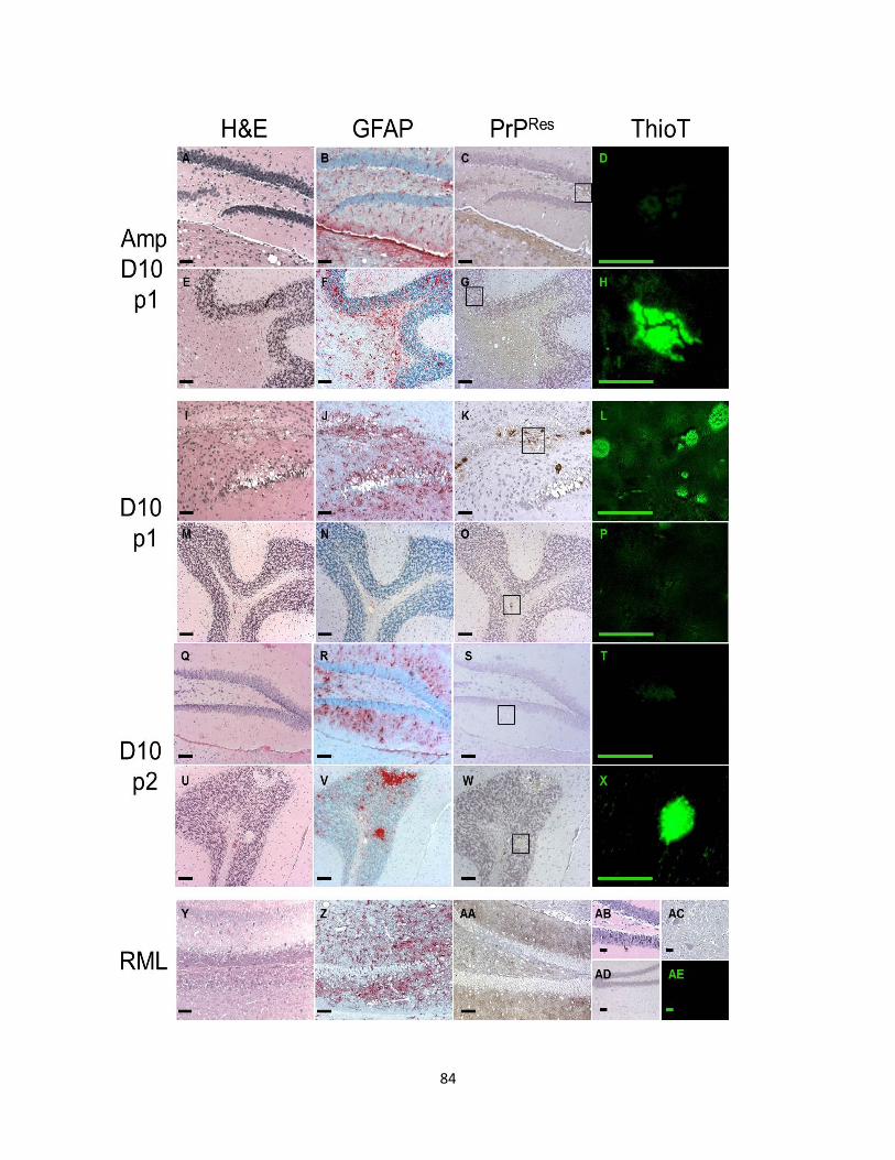
84
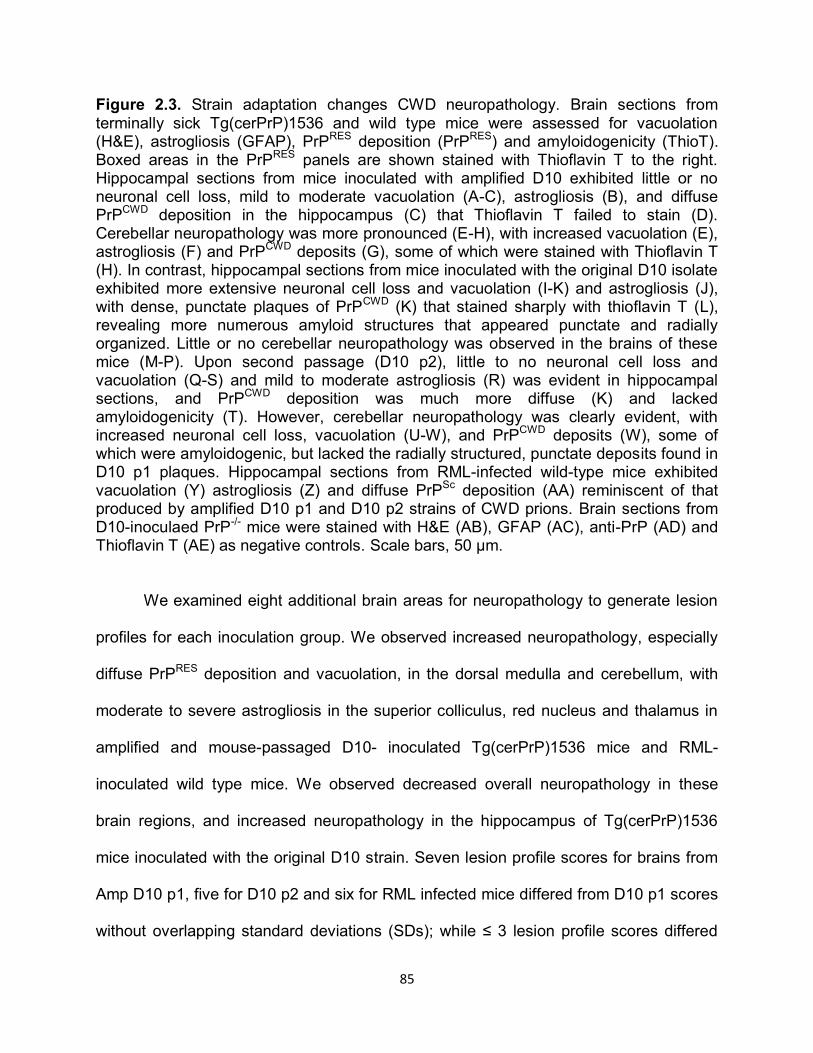
85
Figure 2.3. Strain adaptation changes CWD neuropathology. Brain sections from terminally sick Tg(cerPrP)1536 and wild type mice were assessed for vacuolation (H&E), astrogliosis (GFAP), PrPRES deposition (PrPRES) and amyloidogenicity (ThioT). Boxed areas in the PrPRES panels are shown stained with Thioflavin T to the right. Hippocampal sections from mice inoculated with amplified D10 exhibited little or no neuronal cell loss, mild to moderate vacuolation (A-C), astrogliosis (B), and diffuse PrPCWD deposition in the hippocampus (C) that Thioflavin T failed to stain (D). Cerebellar neuropathology was more pronounced (E-H), with increased vacuolation (E), astrogliosis (F) and PrPCWD deposits (G), some of which were stained with Thioflavin T (H). In contrast, hippocampal sections from mice inoculated with the original D10 isolate exhibited more extensive neuronal cell loss and vacuolation (I-K) and astrogliosis (J), with dense, punctate plaques of PrPCWD (K) that stained sharply with thioflavin T (L), revealing more numerous amyloid structures that appeared punctate and radially organized. Little or no cerebellar neuropathology was observed in the brains of these mice (M-P). Upon second passage (D10 p2), little to no neuronal cell loss and vacuolation (Q-S) and mild to moderate astrogliosis (R) was evident in hippocampal sections, and PrPCWD deposition was much more diffuse (K) and lacked amyloidogenicity (T). However, cerebellar neuropathology was clearly evident, with increased neuronal cell loss, vacuolation (U-W), and PrPCWD deposits (W), some of which were amyloidogenic, but lacked the radially structured, punctate deposits found in D10 p1 plaques. Hippocampal sections from RML-infected wild-type mice exhibited vacuolation (Y) astrogliosis (Z) and diffuse PrP
Sc deposition (AA) reminiscent of that
produced by amplified D10 p1 and D10 p2 strains of CWD prions. Brain sections from D10-inoculaed PrP-/- mice were stained with H&E (AB), GFAP (AC), anti-PrP (AD) and Thioflavin T (AE) as negative controls. Scale bars, 50 µm.
We examined eight additional brain areas for neuropathology to generate lesion
profiles for each inoculation group. We observed increased neuropathology, especially
diffuse PrPRES deposition and vacuolation, in the dorsal medulla and cerebellum, with
moderate to severe astrogliosis in the superior colliculus, red nucleus and thalamus in
amplified and mouse-passaged D10- inoculated Tg(cerPrP)1536 mice and RML-
inoculated wild type mice. We observed decreased overall neuropathology in these
brain regions, and increased neuropathology in the hippocampus of Tg(cerPrP)1536
mice inoculated with the original D10 strain. Seven lesion profile scores for brains from
Amp D10 p1, five for D10 p2 and six for RML infected mice differed from D10 p1 scores
without overlapping standard deviations (SDs); while ≤ 3 lesion profile scores differed
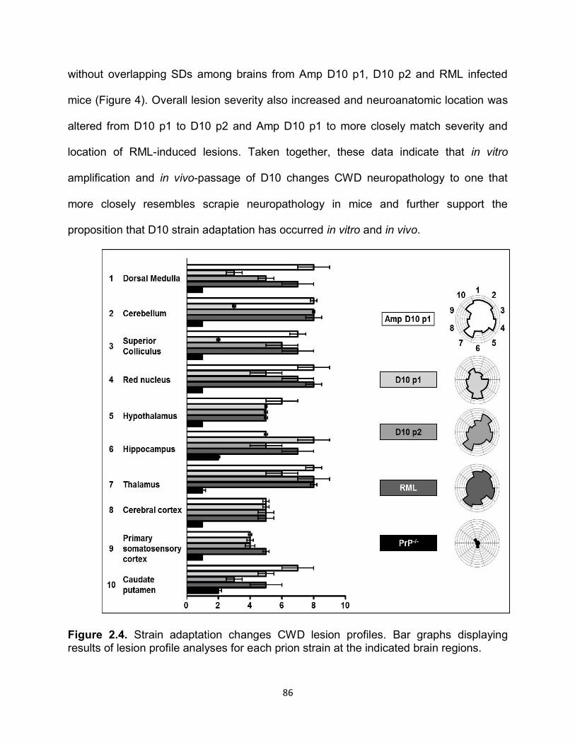
86
without overlapping SDs among brains from Amp D10 p1, D10 p2 and RML infected
mice (Figure 4). Overall lesion severity also increased and neuroanatomic location was
altered from D10 p1 to D10 p2 and Amp D10 p1 to more closely match severity and
location of RML-induced lesions. Taken together, these data indicate that in vitro
amplification and in vivo-passage of D10 changes CWD neuropathology to one that
more closely resembles scrapie neuropathology in mice and further support the
proposition that D10 strain adaptation has occurred in vitro and in vivo.
Figure 2.4. Strain adaptation changes CWD lesion profiles. Bar graphs displaying results of lesion profile analyses for each prion strain at the indicated brain regions.
87
PrP-/- mice inoculated with D10 is shown as a baseline control. Radar plots shown to the right facilitate comparison of overall lesion profiles. Each wedge of the plot represents the indicated brain region and each concentric circle represents a severity point. Amp D10 p1, D10 p2 and RML share similar profiles that are distinct from the D10 p1 profile. sPMCA and mouse passage of D10 changes PrPCWD glycoform ratios
We next characterized these prion inocula biochemically by assessing PK-
resistant fragment sizes and glycoform ratios, which have previously been used to
characterize prion strains26,28. While analysis of pooled brain homogenate from at least
three mice from each group or three PMCA reactions revealed no differences in PK-
resistant core fragment sizes, we did detect significant differences in glycoform ratios
(Figure 5A). RML consisted of substantial monoglycosylated PrPSc, while diglycosylated
PrPCWD constituted the vast majority of D10. Significant amounts of di- and
monoglycosylated PrPCWD
was observed in Amp D10 p1. Primary passage of D10 into
Tg(cerPrP)1536 mice (D10 p1) shifted the glycoform ratio toward monoglycosylated
PrPCWD, which was further increased upon secondary passage (D10 p2). sPMCA of D10
also shifted the glycoform ratio toward monoglycosylated PrPCWD. We did observe some
variability in these trends when we analyzed brain homogenates from individual mice,
especially among D10 p1 samples (Figure 5B). We therefore quantified band intensities
from multiple western blots to more accurately assess glycoform ratios of all prion
strains (Figure 5C and D). The RML di: mono: unglycosylated PrPSc ratio of 0.24 ± 0.09:
0.53 ± 0.12: 0.23 ± 0.08 was drastically different from those of all other strains (p<0.01).
D10 consisted of a PrPCWD glycoform ratio of 0.71 ± 0.05: 0.26 ± 0.05: 0.03 ± 0.01.
sPMCA of D10 produced amplified material with a glycoform ration of 0.74 ±0.04: 0.23
±0.03: 0.03 ±0.01, nearly identical to D10. Passage of serially amplified D10 into
Tg(cerPrP)1536 mice (Amp D10 p1) significantly changed the glycoform ratio to 0.49 ±
88
0.05: 0.35 ± 0.04: 0.16 ± 0.03 (p<0.01 compared to D10), decreasing overall
glycosylation. A similar phenomenon occurred upon in vivo D10 adaptation. Primary
passage into Tg(cerPrP)1536 mice (D10 p1) slightly altered the glycoform ratio to 0.66 ±
0.21: 0.24 ± 0.14: 0.10 ± 0.08, while secondary passage (D10 p2) significantly shifted
the ratio to 0.57 ± 0.07: 0.33 ± 0.06: 0.10 ± 0.07 (p<0.05 for di- and monoglycosylated
PrPCWD compared to D10). These data reveal similar glycoforms ratios of Amp D10 p1
and D10 p2 prion inocula that are significantly different from D10 and D10 p1 inocula.
Figure 2.5. Strain adaptation changes PrPCWD glycoform ratios. (A) Western blot of pooled brain homogenates (BH) from at least three mice demonstrates that the RML strain of mouse-adapted scrapie consists of substantial monoglycosylated PrPSc (lane 2). The original D10 isolate of CWD prions contains primarily diglycosylated PrPCWD (lane 3). Pooled BH from terminally sick mice inoculated with in vitro amplified D10
89
contains increased monoglycosylated PrPCWD compared to D10 (lane 4). Inoculation of D10 into Tg(cerPrP)1536 mice shifts glycoform ratios toward the monoglycosylated form upon primary (lane 6) and especially secondary (lane 7) passage. Similarly, sPMCA of D10 shifts the ratio toward the monoglyosylated form (lane 8). Pooled BH from asymptomatic wild type mice inoculated with D10 contained no detectable PrPCWD (lane 4). (B) Western blot of individual BH confirms increased monoglycosylated PrPCWD of Amp D10 p1 (lanes 3-5) and D10 p2 (lanes 9 and 10), while D10 p1 is more variable (lanes 6-8). (C) Quantification of band intensities from western blots displayed as a triplot of mean glycoform ratios ± standard deviations for each group. Arrows indicate the axis to be read from points to determine the percent of each glycoform. The glycoform ratio of D10 p1 (open circle) is not significantly different from the original D10 ratio (black square), while the D10 p2 ratio is significantly different (black circle). sPMCA of D10 (open diamonds) produces material displaying a glycoform ratio closely resembling both D10 and D10 p1. Passage of sPMCA D10 into Tg(cerPrP)1536 mice results in a further shift towards monoglycosylated PrPCWD (black diamonds), differing significantly from D10 and resembling D10 p2. RML (black triangles) exhibited the greatest proportion of monoglycosylation and was significantly different than all other strains. (D) Triplot of raw data of at least six samples for each group used to generate the mean glycoforms ratios in (C). sPMCA and mouse passage of D10 changes PrPCWD conformational stability
We further characterized these prion inocula biochemically by assessing
conformational stability in the presence of GdnHCl, another established criteria for prion
strain typing30,31,43,46. We quantified PrPRES band intensities from western blots of brain
extracts incubated with increasing GdnHCl concentrations then PK to determine the
concentration of GdnHCl required to PK digest 50% of PrPRES ([GdnHCl]50). D10,
mouse-passaged D10 (D10 p1) and serially-amplified D10 (sPMCA) exhibited nearly
identical conformational stabilities, with mean [GdnHCl]50 values of 2.35 ± .01, 2.35 ±
.10 and 2.33 ± 0.12 M, respectively (Figure 6). Conformational stability was significantly
reduced upon secondary passage of D10 (D10 p2, 2.03 ± 0.06 M, p < 0.01) and primary
passage of amplified D10 (Amp D10 p1, 1.89 ± 0.06 M, p < 0.01) in Tg(cerPrP)1536.
The RML prion strain was significantly less stable than all other strains (1.46 ± 0.01 M, p
< 0.01). In toto, the biological, neuropathological and biochemical data reported here
90
strongly support the conclusion that D10 strain adaptation has occurred in vitro and in
vivo.
Figure 2.6. Strain adaptation changes PrPCWD conformational stability. Western blots of samples from the indicated prion strains treated with increasing concentrations of guanidine hydrochloride (GdnHCl) and 50 µg/mL PK reveal that D10 retains its conformational stability after primary passage in Tg(cerPrP)1536 mice (D10 p1) or serial amplification (sPMCA). Secondary passage (D10 p2) or primary passage of amplified D10 (Amp D10 p1) destabilizes PrPCWD conformation, more closely resembling that of RML. Denaturation curves to the right of each blot quantify PrPRES conformational stability (see Methods). The concentration of GdnHCl required to denature 50% of PrPRES ([GdnHCL]50), a measure of the conformational stability, is shown to the right of the corresponding plot. Data are from at least three independent experiments using at least three animals per group.
91
DISCUSSION
The first demonstration of amplifying PrPCWD using PMCA was recently reported
to yield an approximately 200-fold amplification efficiency per round 17. In this study we
have increased this efficiency up to several thousand-fold per round by optimizing NBH
preparation and sonication parameters. We also demonstrated the specificity of the
reaction by using moPrPC substrates as negative controls. We observed a linear
relationship between –fold amplification and D10 dilutions between 10-3 and 2 x 10⁻⁵.
Limiting substrate at lower dilutions and limiting prion templates at higher dilutions
probably account for asymptotic amplification efficiencies. By maintaining PrPCWD
concentrations in the dynamic range of PMCA, we were able to produce linear
amplification over repeated PMCA rounds. This resulted in highly efficient and
reproducible amplification over at least eight PMCA rounds that generated significant
amounts of amplified 10-24 D10 with which we infected Tg(cerPrP)1536 mice.
Comparing equal dilutions of the original D10 inoculum and amplified 10-24 D10 by
western blot confirmed that both samples displayed nearly identical signal intensities.
We next demonstrated the first in vitro generation of infectious CWD prions using
amplified 10-24 D10 as the inoculum. In vitro-amplified D10 elicited terminal prion
disease earlier than the original D10 used to seed the sPMCA reaction. This result was
surprising because generation of infectious hamster-adapted scrapie prions in vitro
using PMCA has previously been reported to elicit terminal disease later than the
original scrapie inocula, a phenomenon largely abrogated by serial passage or by
conjugation of amplified material to nitrocellulose prior to inoculation12-15. One possible
explanation for this discrepancy is that previous studies used homologous brain inocula,
92
substrates and hosts, whereas we used infected cervid brain as the prion inocula and
Tg(cerPrP)1536 mice as a source for brain substrate and host. Primary passage of
cervid-derived D10 into a murine host may decrease prion replication efficiency, which
increases upon in vitro amplification using NBH from Tg(cerPrP)1536 mice as a
substrate or secondary passage in Tg(cerPrP)1536 mice. Indeed, we observed a
reduction in mean incubation time to terminal disease upon serial passage of D10 in
Tg(cerPrP)1536 mice, in accordance with another recent report 47. These data implicate
other host factors present in the brain that may influence prion amplification in vitro and
in vivo, resulting in nearly identical CWD prion infectivity titres that we observed for
sPMCA-amplified and mouse-passaged D10. Many prion-forming cofactors have been
proposed, including proteins 48-50, polysaccharides 51,52 or polyanions 53,54 that may
facilitate host-specific prion amplification in vitro and in vivo. Another possibility
postulated by Soto and colleagues is that in vitro-generated scrapie prions may have
created a new prion strain that requires in vivo adaptation to acquire the same infectivity
titre as the original prion strain 12. Our data supports this hypothesis, but because
sPMCA generated a cervid prion strain using mouse NBH expressing cerPrPC, we
conclude that strain adaptation occurred in vitro, resulting in a more infectious prion
strain in mice. Correlation with our in vivo data demonstrating mouse adaptation of D10
following serial passage strongly supports this interpretation. Serial passage of natural
sheep scrapie and bovine spongiform encephalopathy prion isolates in Tg(ovPrP) mice
has also recently been shown to reduce incubation times 55,56. As a corollary to the
experiments conducted here, it would be interesting to determine whether sPMCA could
93
be used to adapt natural scrapie isolates in vitro and increase their infectivity titres as
does in vivo adaptation via mouse passage.
Another straightforward explanation for the shortened incubation times we
observed is that inocula from in vitro amplification and in vivo adaptation contains no
heterologous brain homogenate present in the original D10 inoculum, abrogating a
potential immune response that may clear a significant portion of the initial inocula.
Seminal work comparing mouse and hamster prion titres in mice showed that hamster
prions, containing heterologous hamster brain homogenate, induced disease much
earlier than mouse prions containing homologous mouse brain homogenate, arguing
against immune clearance significantly affecting prion titers4. Indeed, we have never
observed any clinical, cellular, biochemical or molecular evidence of inflammation upon
i.c. inoculation with heterologous brain material (our unpublished data), most likely
because the brain is a relatively immune privileged site. In the present study, if immune
clearance simply lowered the effective D10 prion dose compared to in vitro-amplified or
mouse-passaged D10 without strain adaptation, then both of these D10-derived strains
should exhibit increased infectivity titres while maintaining other strain properties of the
original D10. We explored this possibility by first comparing neuropathological
properties of the original D10 strain, in vitro-amplified and mouse-passaged D10, and
RML. Both in vitro-amplified and in vivo-adapted D10 prions induced similar
neuropathology in Tg(cerPrP)1536 mice, characterized by diffuse PrPCWD deposition
that is similar to that observed in brains from wild type mice infected with RML, a
scrapie strain well-adapted to the mouse 41,57,58. These three prion strains produced
PrPCWD deposition clearly different than the dense, punctate plaques evident in brains of
94
Tg(cerPrP)1536 mice inoculated with the original D10 strain. Similar diffuse PrPRES
deposition has been observed in RML-infected and CWD-infected mice expressing
mouse PrPC59,60 and scrapie-infected mice expressing bovine and mouse–bovine
chimeric PrPC31. Amplified and mouse-passaged D10 and RML also produced similar
brain lesion profiles that were consistent with neuropathology previously reported in
scrapie-infected mice58,61,62 and distinct from the profile induced by the original D10
strain. The changes in location and severity of prion lesions evident among these prion
strains are consistent with D10 strain adaptation by in vitro amplification that is
indistinguishable from in vivo adaptation via passage of D10 in Tg(cerPrP)1536 mice.
We also detected biochemical similarities among in vitro and in vivo-adapted
prion strains. While sPMCA D10 and D10 p1 displayed nearly identical PrPCWD
glycoforms ratios as the parental D10 strain, Amp D10 p1 and D10 p2 significantly
increased monoglycosylated PrPCWD compared to the principally diglycosylated D10
strain, similar to the transition observed in another study of murine adaptation of CWD
prions59. These results are intriguing in light of another recent study demonstrating that
host PrPC glycoforms dictate in vitro PrPSc amplification efficiency63, suggesting that
perhaps murine PrPC glycoforms select, from a heterologous mixture, distinct strains of
CWD prions that are preferentially amplified in vitro or in vivo. We also show that Amp
D10 p1 and D10 p2 conformations were significantly more destabilized by GdnHCl
denaturation compared to sPMCA D10 and D10 p1, the stabilities of which were nearly
identical to that of D10. RML adopted a conformation that was by far the least stable of
all prion strains investigated. Previous reports have documented relatively less stable
mouse-passaged scrapie prion conformations and more stable CWD prion
95
conformations31,43,47. Our data demonstrate that serial passage of CWD prions in
Tg(cerPrP)1536 mice can destabilize their conformation, and that sPMCA using
Tg(cerPrP) NBH as substrate expedites this process. Interestingly, decreased stability
of in vitro and in vivo-adapted D10 prion strains correlates with shortened incubation
times in Tg(cerprP)1536 mice, a phenomenon previously exhibited for other novel prion
strains generated in vitro 64 and in vivo 46.
We did not observe statistically significant changes in either glycosylation or
conformational stability of sPMCA D10 or D10 p1, as one might expect if adaptation had
occurred. Perhaps sPMCA and primary passage of D10 initiates creation of a new or
intermediate prion strain, or selection of a small concentration of a pre-existing one that,
upon further passage into mice, is preferentially selected and amplified. This
interpretation is supported by the increased variability in the biochemical properties of
D10 p1 (glycosylation and conformational stability) and sPMCA (conformational
stability), which may indicate the emergence of nascent prion strains.
The similar neuropathological and biochemical characteristics of in vitro and in
vivo-adapted D10 that clearly differentiate them from primary-passaged D10 prions
strongly indicate that sPMCA has adapted D10 as effectively as in vivo adaptation by
mouse passage. However, we cannot rule out the possibility that in vitro amplification
and in vivo passage of D10 also increased the prion titre of these strains, either by
strain adaptation, immune abrogation, or both. Unlike RML, whose titre has been
determined by mouse bioassay and cell culture assays 65,66, exact titres for most scrapie
and all CWD prion strains have yet to be determined. Comparing western blot signal
intensities of PrPRES remains a crude estimate of prion infectivity because PrPRES
96
concentration and prion infectivity do not always closely correlate44,45. Here we present
evidence for strain adaptation by sPMCA. The possibility of a concomitant increase in
prion titre would also be a novel and interesting result of prion amplification by sPMCA.
We conclude that in vitro D10 amplification parallels primary passage of D10 in
Tg(cerPrP)1536 mice, and that sPMCA represents a viable means of adapting prion
strains to new hosts. Since strain adaptation correlates with species barriers, we
envision sPMCA to potentially be a powerful tool with which to probe strain differences
and assess species barriers in a fraction of the time and cost of animal bioassays.
97
REFERENCES
1 Prusiner, S. B. Novel proteinaceous infectious particles cause scrapie. Science 216, 136-144 (1982).
2 Oesch, B. et al. A cellular gene encodes scrapie PrP 27-30 protein. Cell 40, 735-746 (1985).
3 Hope, J. et al. The major polypeptide of scrapie-associated fibrils (SAF) has the same size, charge distribution and N-terminal protein sequence as predicted for the normal brain protein (PrP). Embo J 5, 2591-2597 (1986).
4 Büeler, H. R. et al. Mice devoid of PrP are resistant to scrapie. Cell 73, 1339-1347 (1993).
5 Bolton, D. C., McKinley, M. P. & Prusiner, S. B. Identification of a protein that purifies with the scrapie prion. Science 218, 1309-1311 (1982).
6 Hope, J. et al. Fibrils from brains of cows with new cattle disease contain scrapie-associated protein. Nature 336, 390-392 (1988).
7 Collinge, J. et al. Unaltered susceptibility to BSE in transgenic mice expressing human prion protein. Nature 378, 779-783 (1995).
8 Scott, M. R. et al. Compelling transgenetic evidence for transmission of bovine spongiform encephalopathy prions to humans. Proc Natl Acad Sci U S A 96, 15137-15142 (1999).
9 Race, R. E. et al. Comparison of abnormal prion protein glycoform patterns from transmissible spongiform encephalopathy agent-infected deer, elk, sheep, and cattle. J Virol 76, 12365-12368 (2002).
10 Browning, S. R. et al. Transmission of prions from mule deer and elk with chronic wasting disease to transgenic mice expressing cervid PrP. J Virol 78, 13345-13350 (2004).
11 Legname, G. et al. Synthetic mammalian prions. Science 305, 673-676 (2004).
12 Castilla, J., Saa, P., Hetz, C. & Soto, C. In vitro generation of infectious scrapie prions. Cell 121, 195-206 (2005).
13 Deleault, N. R., Harris, B. T., Rees, J. R. & Supattapone, S. Formation of native prions from minimal components in vitro. Proc Natl Acad Sci U S A 104, 9741-9746 (2007).
14 Weber, P. et al. Generation of genuine prion infectivity by serial PMCA. Vet Microbiol 123, 346-357 (2007).
98
15 Murayama, Y. et al. Urinary excretion and blood level of prions in scrapie-infected hamsters. J Gen Virol 88, 2890-2898 (2007).
16 Murayama, Y. et al. Efficient in vitro amplification of a mouse-adapted scrapie prion protein. Neurosci Lett 413, 270-273 (2007).
17 Kurt, T. D. et al. Efficient in vitro amplification of chronic wasting disease PrPRES. J Virol 81, 9605-9608 (2007).
18 Hadlow, W. J. Scrapie and kuru. Lancet 2, 289-290 (1959).
19 Gajdusek, D. C., Gibbs, C. J. & Alpers, M. Experimental transmission of a Kuru-like syndrome to chimpanzees. Nature 209, 794-796 (1966).
20 Gibbs, C. J., Jr. & Gajdusek, D. C. Transmission and characterization of the agents of spongiform virus encephalopathies: kuru, Creutzfeldt-Jakob disease, scrapie and mink encephalopathy. Res Publ Assoc Res Nerv Ment Dis 49, 383-410 (1971).
21 Taraboulos, A. et al. Regional mapping of prion proteins in brain. Proc Natl Acad Sci U S A 89, 7620-7624 (1992).
22 Bessen, R. A. & Marsh, R. F. Distinct PrP properties suggest the molecular basis of strain variation in transmissible mink encephalopathy. J Virol 68, 7859-7868 (1994).
23 Telling, G. C. et al. Evidence for the conformation of the pathologic isoform of the prion protein enciphering and propagating prion diversity Science 274, 2079-2082 (1996).
24 Korth, C. et al. Abbreviated incubation times for human prions in mice expressing a chimeric mouse-human prion protein transgene. Proc Natl Acad Sci U S A 100, 4784-4789 (2003).
25 Safar, J., Roller, P. P., Gajdusek, D. C. & Gibbs, C. J., Jr. Thermal stability and conformational transitions of scrapie amyloid (prion) protein correlate with infectivity. Protein Sci 2, 2206-2216 (1993).
26 Collinge, J., Sidle, K. C., Meads, J., Ironside, J. & Hill, A. F. Molecular analysis of prion strain variation and the aetiology of 'new variant' CJD. Nature 383, 685-690 (1996).
27 Peretz, D. et al. A conformational transition at the N terminus of the prion protein features in formation of the scrapie isoform. J Mol Biol 273, 614-622 (1997).
28 Hill, A. F. et al. The same prion strain causes vCJD and BSE [letter] Nature 389, 448-450 (1997).
99
29 Safar, J. et al. Eight prion strains have PrP(Sc) molecules with different conformations Nat Med 4, 1157-1165 (1998).
30 Peretz, D. et al. Strain-specified relative conformational stability of the scrapie prion protein. Protein Sci 10, 854-863 (2001).
31 Scott, M. R., Peretz, D., Nguyen, H. O., Dearmond, S. J. & Prusiner, S. B. Transmission barriers for bovine, ovine, and human prions in transgenic mice. J Virol 79, 5259-5271 (2005).
32 Collinge, J. & Rossor, M. A new variant of prion disease. Lancet 347, 916-917 (1996).
33 Will, R. G. et al. A new variant of Creutzfeldt-Jakob disease in the UK. Lancet 347, 921-925 (1996).
34 Raymond, G. J. et al. Evidence of a molecular barrier limiting susceptibility of humans, cattle and sheep to chronic wasting disease. Embo J 19, 4425-4430. (2000).
35 Collinge, J. et al. Transmission of fatal familial insomnia to laboratory animals. Lancet 346, 569-570 (1995).
36 Hamir, A. N. et al. Experimental transmission of chronic wasting disease agent from mule deer to cattle by the intracerebral route. J Vet Diagn Invest 17, 276-281 (2005).
37 Hamir, A. N. et al. Transmission of chronic wasting disease of mule deer to Suffolk sheep following intracerebral inoculation. J Vet Diagn Invest 18, 558-565 (2006).
38 Hamir, A. N., Kunkle, R. A., Miller, J. M., Bartz, J. C. & Richt, J. A. First and second cattle passage of transmissible mink encephalopathy by intracerebral inoculation. Vet Pathol 43, 118-126 (2006).
39 Hamir, A. N. et al. Preliminary observations on the experimental transmission of scrapie to elk (Cervus elaphus nelsoni) by intracerebral inoculation. Vet Pathol 40, 81-85 (2003).
40 Fischer, M. et al. Prion protein (PrP) with amino-proximal deletions restoring susceptibility of PrP knockout mice to scrapie. EMBO J 15, 1255-1264 (1996).
41 Prusiner, S. B., Hadlow, W. J., Eklund, C. M., Race, R. E. & Cochran, S. P. Sedimentation characteristics of the scrapie agent from murine spleen and brain. Biochemistry 17, 4987-4992 (1978).
42 Fraser, H. & Dickinson, A. G. The sequential development of the brain lesion of scrapie in three strains of mice. J Comp Pathol 78, 301-311 (1968).
100
43 Xie, Z. et al. Chronic wasting disease of elk and deer and Creutzfeldt-Jakob disease: comparative analysis of the scrapie prion protein. J Biol Chem 281, 4199-4206 (2006).
44 Hill, A. F., Antoniou, M. & Collinge, J. Protease-resistant prion protein produced in vitro lacks detectable infectivity. J Gen Virol 80 ( Pt 1), 11-14 (1999).
45 Rubenstein, R. et al. Concentration and distribution of infectivity and PrPSc following partial denaturation of a mouse-adapted and a hamster-adapted scrapie strain. Arch Virol 139, 301-311 (1994).
46 Peretz, D. et al. A change in the conformation of prions accompanies the emergence of a new prion strain. Neuron 34, 921-932 (2002).
47 Green, K. M. et al. The elk PRNP codon 132 polymorphism controls cervid and scrapie prion propagation. J Gen Virol 89, 598-608 (2008).
48 Telling, G. C. et al. Prion propagation in mice expressing human and chimeric PrP transgenes implicates the interaction of cellular PrP with another protein. Cell 83, 79-90 (1995).
49 Kaneko, K. et al. Evidence for protein X binding to a discontinuous epitope on the cellular prion protein during scrapie prion propagation. Proc Natl Acad Sci U S A 94, 10069-10074 (1997).
50 Yehiely, F. et al. Identification of candidate proteins binding to prion protein. Neurobiol Dis 3, 339-355 (1997).
51 Appel, T. R., Dumpitak, C., Matthiesen, U. & Riesner, D. Prion rods contain an inert polysaccharide scaffold. Biol Chem 380, 1295-1306 (1999).
52 Dumpitak, C. et al. The polysaccharide scaffold of PrP 27-30 is a common compound of natural prions and consists of alpha-linked polyglucose. Biol Chem 386, 1149-1155 (2005).
53 Deleault, N. R. et al. Post-transcriptional suppression of pathogenic prion protein expression in Drosophila neurons. J Neurochem 85, 1614-1623 (2003).
54 Deleault, N. R., Lucassen, R. W. & Supattapone, S. RNA molecules stimulate prion protein conversion. Nature 425, 717-720 (2003).
55 Bencsik, A. et al. Scrapie strain transmission studies in ovine PrP transgenic mice reveal dissimilar susceptibility. Histochem Cell Biol 127, 531-539 (2007).
56 Baron, T. & Biacabe, A. G. Molecular behaviors of "CH1641-like" sheep scrapie isolates in ovine transgenic mice (TgOvPrP4). J Virol 81, 7230-7237 (2007).
101
57 Chandler, R. L. Encephalopathy in mice produced by inoculation with scrapie brain material. Lancet 1, 1378-1379 (1961).
58 Kingsbury, D. T. et al. Genetic control of scrapie and Creutzfeldt-Jakob disease in mice. J Immunol 131, 491-496 (1983).
59 Sigurdson, C. J. et al. Strain fidelity of chronic wasting disease upon murine adaptation. J Virol 80, 12303-12311 (2006).
60 Zabel, M. D. et al. Stromal complement receptor CD21/35 facilitates lymphoid prion colonization and pathogenesis. J Immunol 179, 6144-6152 (2007).
61 Eklund, C. M., Kennedy, R. C. & Hadlow, W. J. Pathogenesis of scrapie virus infection in the mouse. J Infect Dis 117, 15-22 (1967).
62 Baringer, J. R. & Prusiner, S. B. Experimental scrapie in mice: ultrastructural observations. Ann Neurol 4, 205-211 (1978).
63 Nishina, K. A. et al. The stoichiometry of host PrPC glycoforms modulates the efficiency of PrPSc formation in vitro. Biochemistry 45, 14129-14139 (2006).
64 Legname, G. et al. Strain-specified characteristics of mouse synthetic prions. Proc Natl Acad Sci U S A 102, 2168-2173 (2005).
65 Klohn, P. C., Stoltze, L., Flechsig, E., Enari, M. & Weissmann, C. A quantitative, highly sensitive cell-based infectivity assay for mouse scrapie prions. Proc Natl Acad Sci U S A 100, 11666-11671 (2003).
66 Prusiner, S. B. et al. Measurement of the scrapie agent using an incubation time interval assay. Ann Neurol 11, 353-358 (1982).
102
CHAPTER 3:
DE NOVO GENERATION OF INFECTIOUS CERVID PRIONS USING PROTEIN MISFOLDING CYCLIC AMPLIFICATION (PMCA)
SUMMARY
Chronic wasting disease is a prion disease that affects free ranging and captive
cervids (deer, elk, and moose). Substantial evidence suggests that prions are
misfolded, infectious, insoluble, and protease resistant proteins (PrPRES) devoid of
nucleic acid. Protein misfolding cyclic amplification (PMCA) has provided additional
evidence that PrPRES acts as a template that can convert normal prion protein (PrPC)
into the infectious misfolded PrPRES isoform. Human PrPC has been shown to rarely
spontaneously convert into an infectious misfolded state causing sporadic Creutzfeldt-
Jakob disease (sCJD). Recently, several investigators have reported spontaneous
generation of prions utilizing in vitro methods, including PMCA. Here we tested the rate
of de novo generation of prions in our laboratory using our standard PMCA protocol. We
report that we were able to generate de novo prions in rounds 4,5, and 7 at low rates of
1.6, 5.0, and 6.7% respectively. The prions were infectious upon inoculation into
cervidized mice and they displayed similar but unique characteristics to other cervid
prion strains.
INTRODUCTION
Prions cause diseases classified as transmissible spongiform encephalopathies
(TSEs) which are characterized by distinct neuropathologic vacuoles and accumulation
of a transmissible proteinase K (PK)-resistant protein (PrPRES) 1,2,3. Bovine spongiform
encephalopathy (BSE) of cattle, Creutzfeldt-Jakob disease (CJD) of humans, scrapie of
103
sheep and goats, and chronic wasting disease (CWD) of cervids (deer, elk and moose)
are notable prion diseases that can be transmitted, inherited, or occur spontaneously.
Mounting evidence demonstrates that prion pathogenesis is caused by the conversion
of the normal cellular host protein, (PrPC) into a protease-resistant, abnormal disease-
causing isoform devoid of nucleic acid (PrPRES) 4,5, 6,7,8.
In vitro generation of infectious prion protein using protein misfolding cyclic
amplification (PMCA) has substantiated the protein-only hypothesis. PMCA utilizes
sonication and incubation steps to break up PK-resistant aggregates encouraging the
prion seed to interact and template PrPC present in the uninfected brain homogenate
substrate9. Employing repeated cycles of incubation and sonication has led to efficient
amplification of minute quantities of PrPRES using substrate from varied species9-13.
Serial PMCA (sPMCA) has also been employed to successfully evaluate strain
adaptation and species barriers in the absence of lengthy and expensive bioassays10,14-
16. Intriguingly, Deleault et al., interrogated PMCA component requirements for in vitro
amplification resulting in spontaneous generation of infectious prions from non-
infectious components: native hamster PrPC in combination with co-purified lipids and
synthetic polyanions17. Barria et al., expanded on these findings and reported
generating spontaneous prions from uninfected normal brain homogenate substrate in a
prion free laboratory upon extended rounds and modification of normal sPMCA
conditions18.
In order to examine the rate of spontaneous conversion with our PMCA protocol
we subjected uninfected brain homogenate derived from cervidized transgenic mice to
seven rounds (48 cycles each) of PMCA under normal conditions. In order to avoid the
104
possibility of cross contamination all experiments were performed in a prion free lab with
new reagents and new equipment. Surprisingly, we were able to detect PK-resistant
bands by western blot at round 4 of sPMCA at a rate of 1.6%. Further generation of
spontaneous prions were observed after round 5 and round 7 with rates of 5.0% and
6.74% respectively. Bioassay determined that the spontaneously generated cervid
prions were infectious to cervidized transgenic but not wild type mice and biochemical
analysis resulted in a unique profile differing from prion strains used within our
laboratory. These data strongly suggest that these prions were derived spontaneously
and not from contamination. Recent evidence suggests that amino acids including and
between 170 and 174 of the cervid prion protein structurally form a rigid loop increasing
the propensity for misfolding19-23. We propose that the de novo generation of infectious
cervid prions in our laboratory is a novel cervid prion strain that occurred spontaneously
with the aid of PMCA and occurs at a very low frequency after 8 rounds of 48 cycles of
sPMCA.
MATERIALS AND METHODS
Preparation of normal brain homogenate (NBH)
All NBH preparations were completed in a prion free laboratory not in association
with any of the prion laboratories in the building. Mice were euthanized and perfused
with 30 ml 5mM EDTA in PBS. Whole brains were removed and immediately frozen in
liquid nitrogen. Brains were weighed and transferred into 1.5 mL Eppendorf tubes with
2.5 mm glass beads. PMCA buffer with 2X Complete Protease Inhibitor Cocktail
(Roche) was added to make a 20 % w/v solution. Samples were homogenized for 20 s
at 4.5 m/s in a FastPrep machine (Biogene), cooled on ice for two minutes and
105
centrifuged at 14,000 x g for 10 s to reduce foaming. This process was repeated twice.
NBH was diluted to a 10% w/v solution by adding an equal volume of PMCA conversion
buffer containing 2% Triton X-100 and incubated on ice for 20 min. NBH was clarified by
centrifuging for 5 min at 1,500 x g and supernatants were aliquoted to into new tubes
and stored at -70oC.
Serial PMCA
Serial PMCA (sPMCA) was conducted in a new prion free laboratory separate
from the laboratory the NBH was made. All reagents and equipment were new and
never used in a prion contaminated laboratory. sPMCA was modified from a previous
protocol (Appendix A1.6)15. Briefly, 50 µL samples of 10% NBH were placed into 20
wells of a 96-well microplate. The entire plate was suspended in the water bath of a new
3000MP sonicator (Misonix) and sonicated at 70- 85% maximum power for 40 seconds,
followed by 30-minute incubation at 37OC. This cycle was repeated 48 times constituting
1 round. For each additional round 25 µL of NBH from the previous round was diluted
into 25 µL of fresh NBH and subjected to another PMCA round. This process was
repeated for a total of 8 rounds. Replicate experiments of 20 NBH samples were started
3 days from each other totalling in 3 experiments and 60 NBH samples. Gloves were
changed between each replicate group and samples were western blotted immediately
after the completed round to look for PK resistant material. Any PK resistant material
that was definitively positive was not subjected to further rounds of PMCA to avoid cross
contamination of other samples. Those samples that were not definitively positive (low
band intensities and banding patterns that mimicked undigested material) were
106
subjected to another round of PMCA to confirm positivity. If they also showed positivity
at the next round they were called positive at the previous round.
Mice
B6129SF2/J mice (stock number 101045) were purchased from the Jackson
Laboratories (Bar Harbor, ME). Tg(cerPrP)5037 were generated as previously
described24. All mice were bred and maintained at Lab Animal Resources, accredited by
the Association for Assessment and Accreditation of Lab Animal Care International, in
accordance with protocols approved by the Institutional Animal Care and Use
Committee at Colorado State University.
Sources and preparation of prion inocula
10% brain homogenates were prepared in PMCA buffer (4mM EDTA, 150 mM
NaCl in PBS). PMCA samples that were western blot positive indicating de novo prions
were pooled together to be used as inoculum. We diluted equal volumes of the
inoculum 1:10 in 320 mM sucrose supplemented with 100 units/mL Penicillin and 100
µg/mL Streptomycin (Gibco) in PBS thirty minutes prior to intracerebral inoculations.
Remaining de novo brain homogenate from PMCA was further amplified to 10 rounds in
order to create enough de novo positive brain material for biochemical analysis.
PK digestion and western blotting
Samples were digested with 50 g/ml PK (Roche) for 30 min at 37oC. The
reaction was stopped by adding lithium dodecyl sulfate sample loading buffer
(Invitrogen) and incubating at 95oC for 5 min. Proteins were electrophoretically
separated through 12% sodium dodecyl sulfate-polyacrylamide gels (Invitrogen), and
transferred to polyvinylidene difluoride membranes (Millipore). Non-specific membrane
107
binding was blocked by incubation in 5% milk blocking solution (Bio-Rad) for 1 h.
Membranes were then incubated for 1 h at room temperature with horseradish
peroxidase-conjugated Bar224 anti-PrP monoclonal antibody (SPI bio) diluted 1:20,000
in Superblock (Pierce), washed 6 x 10 min in PBS with 0.2% Tween 20, and incubated
for 5 min with enhanced chemiluminescent substrate (Millipore). Membranes were
digitally photographed using the FujiDoc gel documentation system equipped with a
cooled charge-coupled diode camera (Fuji). Densitometric analyses were performed
using Quantity One software (Bio-Rad).
Prion inoculations and clinical scoring
Mice were anesthetized by Isofluorane inhalation. Thirty microliters of the
inoculum was injected intracerebrally 3 mm deep through the coronal suture 3-5 mm
lateral of the sagittal suture. Mice were monitored daily for clinical symptoms of prion
disease, including tail rigidity, impaired extensor reflex, akinesia, tremors, ataxia, 15 %
weight loss and paralysis. Mice with any four of these symptoms were scored terminally
sick and euthanized.
Histochemistry and immunohistochemistry (IHC)
Tissues were fixed in 10% paraformaldehyde, embedded in paraffin and 5-10 µm
sections mounted on glass slides. For PrP staining tissue sections were deparaffinized,
treated with concentrated formic acid for thirty minutes, then autoclaved at 121oC in
target retrieval solution (Dako) for 2 h, washed 2 x 7 min in 1X PBS, treated with 0.3%
H202 in methanol for 30 min and blocked for 1 hr with 5% BSA in PBS and mixed 1:1 in
superblock (Pierce). Excess block was tapped off and sections were incubated with
anti-PrP Bar 224 monoclonal antibody diluted 1:500 in block solution for 1 hour. Slides
108
were then washed 2 x 7 min in PBS and incubated 30 min with Envision+HRP mouse
secondary (DAKO). After another 2 x 7 min wash, slides were incubated for 5-7 min with
AEC+Substrate-Chromagen (DAKO) and rinsed 2 x 7 min in PBS and counterstained
with hematoxylin. Slides were rinsed in H2O, immersed in a 0.1% sodiumbicarbonate
bluing reagent for 5 min, rinsed in tap water and coversliped with aqueous mounting
medium (Richard Allan Scientific). Hematoxylin and Eosin (H&E) and glial fibrillary
acidic protein (GFAP) staining was performed on a NexES automated IHC stainer
(Ventana Medical systems, Inc. Tucson, AZ). Sections were stained with H&E for 4 min
at room temperature. Sections were stained with rabbit polyclonal antisera against
GFAP (diluted 1: 8) for 10 min at 37oC followed by Biotinylated goat anti-rabbit Ig
(mouse/rat adsorbed) for 8 min, then counterstained with hematoxylin for 4 min.
Sections were visualized and digitally photographed using an Olympus BX60
microscope equipped with a cooled charge-coupled diode camera.
Glycoform Ratios
Western blots of 10% brain homogenates of infected mice were analyzed by
densiometric analysis (Quantity One). Di, mono, and unglycosylated banding intensities
were calculated as percentages of the total density of each PK treated sample. The
averages of 2 replicate samples were plotted on a tri-plot based on calculated glycoform
ratios.
Conformational stability assay
Conformational stability assays were modified from a previous protocol15. 15 l
aliquots of brain homogenates were incubated for 1 h at room temperature with
increasing concentrations of guanidine hydrochloride (GdnHCl, Sigma) ranging from 0
109
to 5 M in 0.5 M increments. Samples were precipitated in ice-cold methanol at -20oC
overnight, and centrifuged at 13,000 X g for 30 min at 4 oC. Pellets were washed in
PMCA buffer and centrifuged 3x, resuspended in 18 l of PMCA buffer, PK-digested
and western blotted. Conformational stability was quantified by densitometric analyses
of western blots, plotting the mean percentage of PrPRES remaining ± SD as a function
of GdnHCl concentration, and using fourth order polynomial equations and nonlinear
regression (GraphPad Prism) to fit denaturation curves for each prion strain.
Lesion profiling
Brain lesion profiling was performed as previously described15 with slight
modifications. Ten neuroanatomic regions were identified in coronal brain sections from
at 5 mice of each group: 1-dorsal medulla, 2-cerebellum, 3- superior colliculus, 4-
reticular formation, 5-hypothalamus, 6-hippocampus, 7-thalamus, 8-cerebral cortex, 9-
primary somatosensory cortex and 10-caudate-putamen. A professional pathologist
blinded to the group identification scored each region for vacuolation, astrogliosis and
PrPRES deposition using the following severity scale: normal (0), mild (1), moderate (2)
severe (3). The average of the sum of the three scores constitutes the severity score for
each region.
Statistical analyses
One-way ANOVA with Tukey post-test analysis was performed using GraphPad Prism.
RESULTS
Generation of de novo prions using PMCA
Firstly, we have previously reported that Tg(5037) normal brain homogenate
(NBH) supports efficient PMCA amplification using a standard PMCA protocol15,25,26.
110
Importantly, false positives in our negative controls have been rare and even under
strict measures, contamination cannot be dismissed. Interestingly, the rare occurrence
of false positives in our negative controls are primarily observed after 5 rounds of PMCA
and are not reproducible while true positives are generally observed by 3 rounds in
multiple replicates. To determine the rate of PMCA false positives in our laboratory, we
ran Tg(5037) cervidized mouse24 NBH through our standard sPMCA protocol. Three
groups of 20 NBH samples were subjected to 8 rounds of sPMCA in a prion free
laboratory to avoid cross-contamination. Additionally, all reagents and equipment were
new and brain homogenate was made in a different laboratory unexposed to prions.
After each round of PMCA (48 cycles of 30 min incubation followed by 40 sec sonication
pulse) NBH samples were analyzed for positivity by western blot and any confirmed
positives were removed from the PMCA plate to avoid contamination of the remaining
negative samples.
Western blot analysis resulted in identification of one protease-resistant PrP
(PrPRES) sample after 4 rounds of sPMCA generating de novo PrPRES at a rate of 1.6%
(Figure 3.1). sPMCA rounds 5 and 7 propagated de novo material accumulating rates of
5.0% and 6.7% respectively (Figure 3.1B).
111
Figure 3.1. (A) Representative western blots of de novo prions generated by PMCA. First well is a non-amplified, non-PK treated NBH followed by 10 amplified, PK-treated NBH samples and a non-amplified PrPRES control for comparison. De novo prions are indicated by an *. (B) Graphical representation of the rate of de novo generated prions at respective rounds. The light blue bars indicate the rate of de novo generation for that individual round (1.6, 3.3, and 1.6% respectively) and dark blue bars indicate the cumulative rate of de novo generation for all rounds (1.6, 5.0, and 6.7% respectively).
De novo prions are infectious
To determine if de novo generated prions were infectious, we intracerebrally (i.c.)
inoculated cervid transgenic mice (Tg(5037)) and wild type mice with western blot
positive de novo prions generated by PMCA. Here we report that Tg(5037) mice but not
wild type inoculated with de novo prions displayed clinical signs of disease. Tg(5037)
mice loss 15% of body weight, had a stiff tail, were ataxic, were observed with hind legs
splayed behind them when not in motion, but did not exhibit a hind leg grasp when held
up by their tail (Figure 3.2C). Curiously de novo prions did not have a complete attack
rate as only 4/5 Tg(5037) displayed clinical signs by 317 days post inoculation (dpi) and
0/5 wild type mice showed signs of infectivity (Table 3.1, Figure 3.2). We compared dpi
of de novo prions to a previously reported natural deer prion isolate (D10) and a well
characterized mouse-adapted scrapie strain (RML 5)15. Mice inoculated with de novo

112
prions generated a survival curve with a comparable mean of 273 dpi to the D10 cervid
strain with mean of 282 dpi (Table 3.1, Figure 3.2A). However, Tg(5037) mice
inoculated with de novo prions had a significant (p<0.01) 104 day delay in disease of as
compared to cervidized mice inoculated with a serially amplified PMCA D10 (sPMCA)
strain (Figure 3.2B). Additionally, de novo prions inoculated into wild type or cervidized
mice had a lengthened incubation period when compared to mouse adapted scrapie
prions. Interestingly, the dpi range among mice inoculated with de novo or D10 prions
was rather large in contrast to mice inoculated with adapted prion strains (Table 3.1).
Table 3.1. Summary of inoculation experiments comparing de novo incidence and
incubation to previously reported prion strains from our laboratory15.
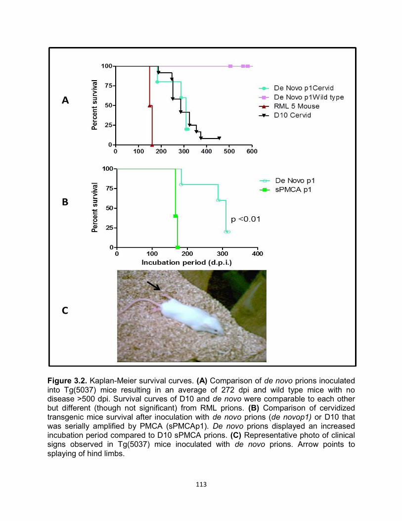
113
Figure 3.2. Kaplan-Meier survival curves. (A) Comparison of de novo prions inoculated into Tg(5037) mice resulting in an average of 272 dpi and wild type mice with no disease >500 dpi. Survival curves of D10 and de novo were comparable to each other but different (though not significant) from RML prions. (B) Comparison of cervidized transgenic mice survival after inoculation with de novo prions (de novop1) or D10 that was serially amplified by PMCA (sPMCAp1). De novo prions displayed an increased incubation period compared to D10 sPMCA prions. (C) Representative photo of clinical signs observed in Tg(5037) mice inoculated with de novo prions. Arrow points to splaying of hind limbs.
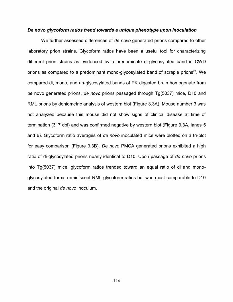
114
De novo glycoform ratios trend towards a unique phenotype upon inoculation
We further assessed differences of de novo generated prions compared to other
laboratory prion strains. Glycoform ratios have been a useful tool for characterizing
different prion strains as evidenced by a predominate di-glycosylated band in CWD
prions as compared to a predominant mono-glycosylated band of scrapie prions27. We
compared di, mono, and un-glycosylated bands of PK digested brain homogenate from
de novo generated prions, de novo prions passaged through Tg(5037) mice, D10 and
RML prions by deniometric analysis of western blot (Figure 3.3A). Mouse number 3 was
not analyzed because this mouse did not show signs of clinical disease at time of
termination (317 dpi) and was confirmed negative by western blot (Figure 3.3A, lanes 5
and 6). Glycoform ratio averages of de novo inoculated mice were plotted on a tri-plot
for easy comparison (Figure 3.3B). De novo PMCA generated prions exhibited a high
ratio of di-glycosylated prions nearly identical to D10. Upon passage of de novo prions
into Tg(5037) mice, glycoform ratios trended toward an equal ratio of di and mono-
glycosylated forms reminiscent RML glycoform ratios but was most comparable to D10
and the original de novo inoculum.
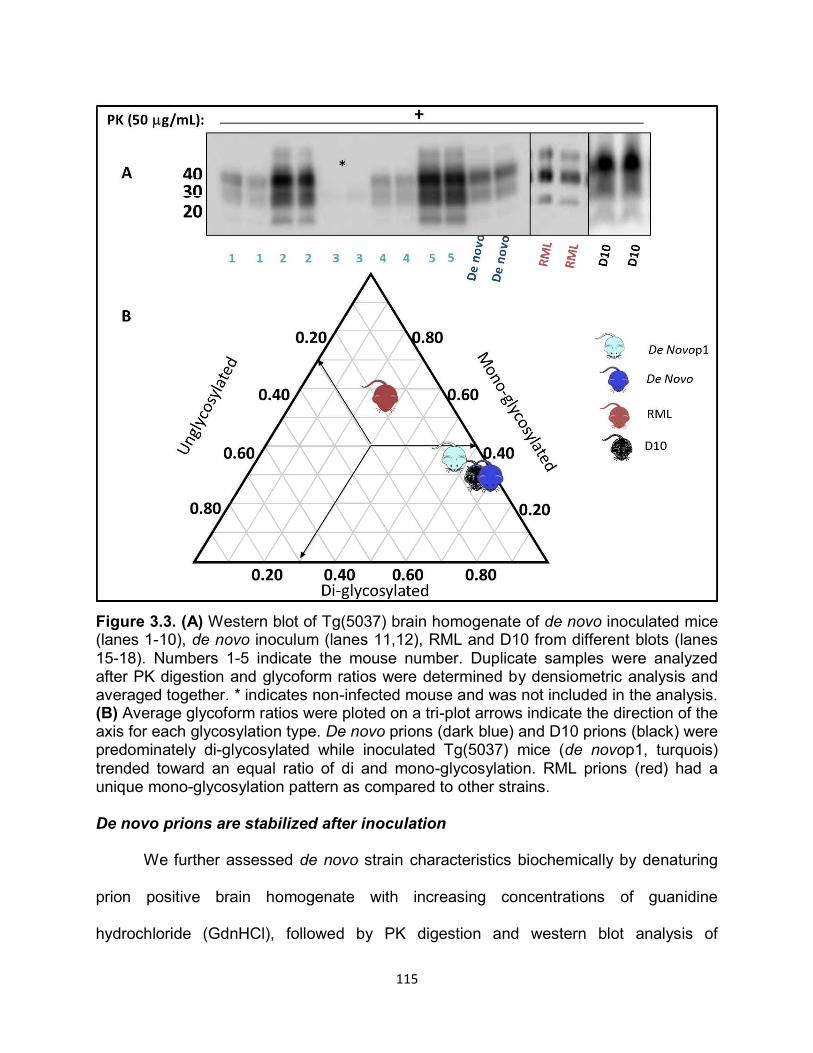
115
Figure 3.3. (A) Western blot of Tg(5037) brain homogenate of de novo inoculated mice (lanes 1-10), de novo inoculum (lanes 11,12), RML and D10 from different blots (lanes 15-18). Numbers 1-5 indicate the mouse number. Duplicate samples were analyzed after PK digestion and glycoform ratios were determined by densiometric analysis and averaged together. * indicates non-infected mouse and was not included in the analysis. (B) Average glycoform ratios were ploted on a tri-plot arrows indicate the direction of the axis for each glycosylation type. De novo prions (dark blue) and D10 prions (black) were predominately di-glycosylated while inoculated Tg(5037) mice (de novop1, turquois) trended toward an equal ratio of di and mono-glycosylation. RML prions (red) had a unique mono-glycosylation pattern as compared to other strains. De novo prions are stabilized after inoculation
We further assessed de novo strain characteristics biochemically by denaturing
prion positive brain homogenate with increasing concentrations of guanidine
hydrochloride (GdnHCl), followed by PK digestion and western blot analysis of
116
remaining PrPRES (Figure 3.4A). De novo prions exhibited a conformational stability
similar to D10 prions propagated through cervidized mice (D10p1) or serially amplified
by PMCA (sPMCA) with mean [GdnHCl]50 values of 2.25+0.01, 2.35+0.01, and
2.33+0.01 respectively but was significantly different (p<0.001) from D10 (Figure
3.4B,C). Passage of de novo prions through Tg(5037) mice (de novop1) resulted in a
significantly higher (p<0.001) [GdnHCl]50 value of 3.40+0.01 compared to all strains
including, sPMCA prions passaged into cervidized mice (sPMCA p1). Interestingly,
sPMCAp1 prions created similarly to de novop1 prions (but with a PMCA seeded
reaction), exhibited a much lower [GdnHCl]50 value of 1.89+0.01, nearly identical to
RML.
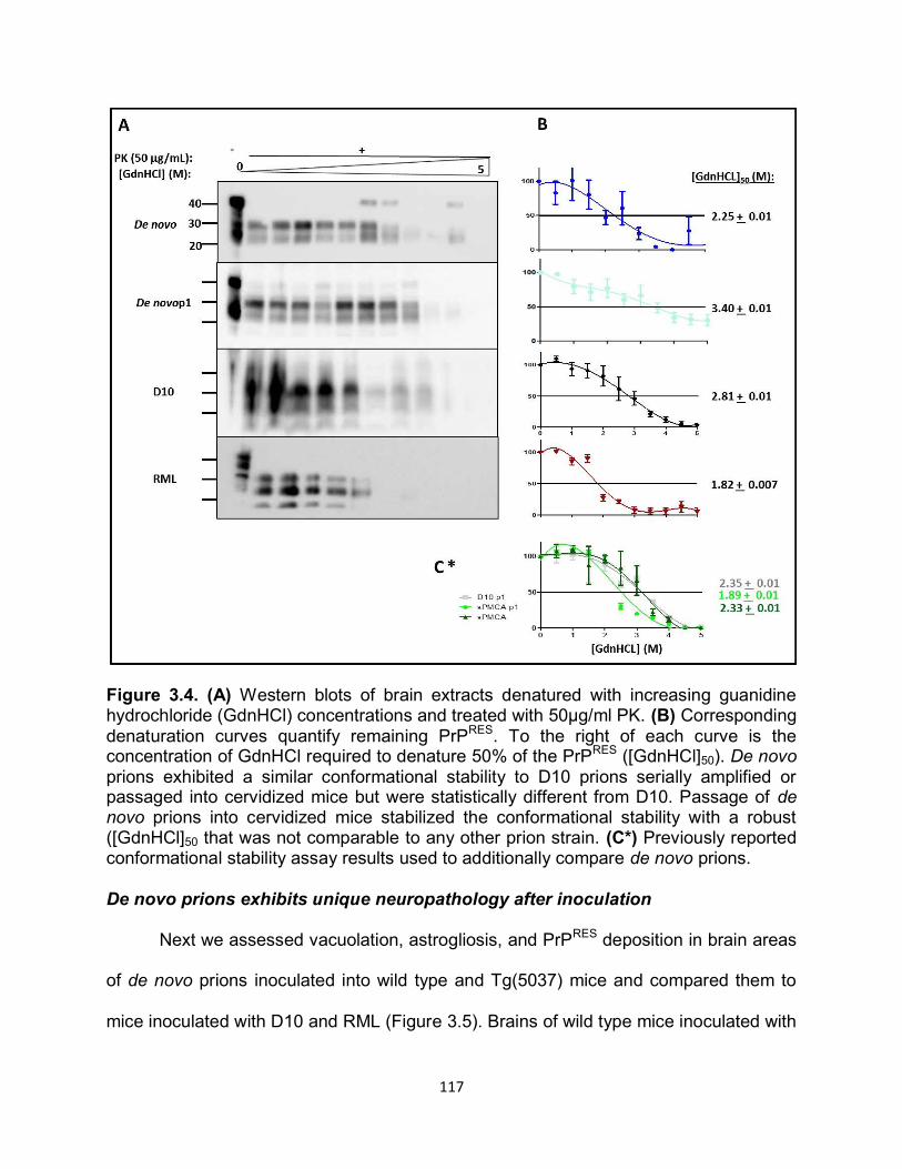
117
Figure 3.4. (A) Western blots of brain extracts denatured with increasing guanidine hydrochloride (GdnHCl) concentrations and treated with 50μg/ml PK. (B) Corresponding denaturation curves quantify remaining PrPRES. To the right of each curve is the concentration of GdnHCl required to denature 50% of the PrPRES ([GdnHCl]50). De novo prions exhibited a similar conformational stability to D10 prions serially amplified or passaged into cervidized mice but were statistically different from D10. Passage of de novo prions into cervidized mice stabilized the conformational stability with a robust ([GdnHCl]50 that was not comparable to any other prion strain. (C*) Previously reported conformational stability assay results used to additionally compare de novo prions. De novo prions exhibits unique neuropathology after inoculation Next we assessed vacuolation, astrogliosis, and PrPRES deposition in brain areas
of de novo prions inoculated into wild type and Tg(5037) mice and compared them to
mice inoculated with D10 and RML (Figure 3.5). Brains of wild type mice inoculated with
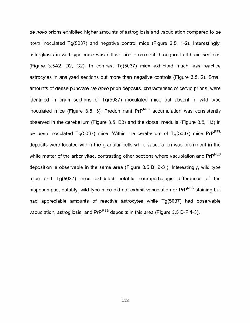
118
de novo prions exhibited higher amounts of astrogliosis and vacuolation compared to de
novo inoculated Tg(5037) and negative control mice (Figure 3.5, 1-2). Interestingly,
astrogliosis in wild type mice was diffuse and prominent throughout all brain sections
(Figure 3.5A2, D2, G2). In contrast Tg(5037) mice exhibited much less reactive
astrocytes in analyzed sections but more than negative controls (Figure 3.5, 2). Small
amounts of dense punctate De novo prion deposits, characteristic of cervid prions, were
identified in brain sections of Tg(5037) inoculated mice but absent in wild type
inoculated mice (Figure 3.5, 3). Predominant PrPRES accumulation was consistently
observed in the cerebellum (Figure 3.5, B3) and the dorsal medulla (Figure 3.5, H3) in
de novo inoculated Tg(5037) mice. Within the cerebellum of Tg(5037) mice PrPRES
deposits were located within the granular cells while vacuolation was prominent in the
white matter of the arbor vitae, contrasting other sections where vacuolation and PrPRES
deposition is observable in the same area (Figure 3.5 B, 2-3 ). Interestingly, wild type
mice and Tg(5037) mice exhibited notable neuropathologic differences of the
hippocampus, notably, wild type mice did not exhibit vacuolation or PrPRES staining but
had appreciable amounts of reactive astrocytes while Tg(5037) had observable
vacuolation, astrogliosis, and PrPRES deposits in this area (Figure 3.5 D-F 1-3).
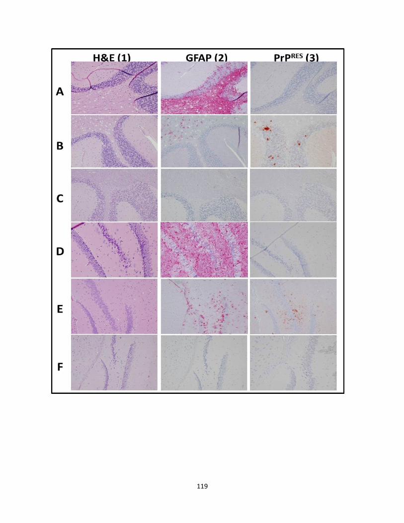
119
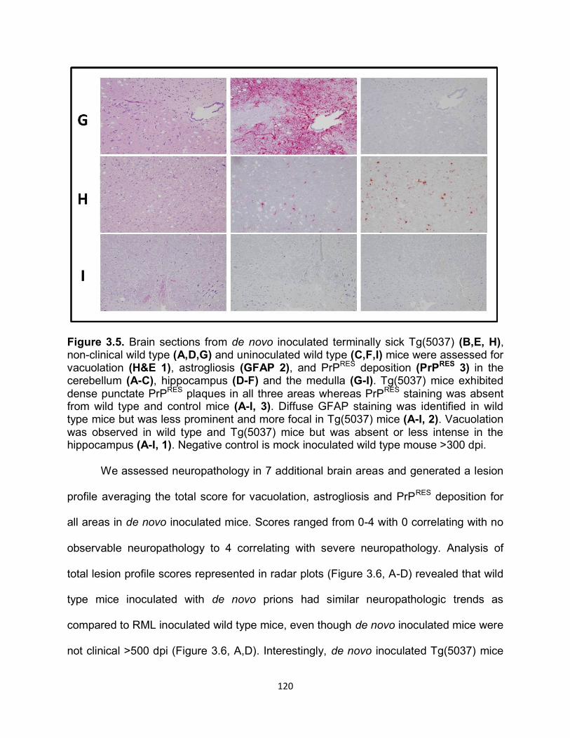
120
Figure 3.5. Brain sections from de novo inoculated terminally sick Tg(5037) (B,E, H), non-clinical wild type (A,D,G) and uninoculated wild type (C,F,I) mice were assessed for vacuolation (H&E 1), astrogliosis (GFAP 2), and PrPRES deposition (PrPRES 3) in the cerebellum (A-C), hippocampus (D-F) and the medulla (G-I). Tg(5037) mice exhibited dense punctate PrPRES plaques in all three areas whereas PrPRES staining was absent from wild type and control mice (A-I, 3). Diffuse GFAP staining was identified in wild type mice but was less prominent and more focal in Tg(5037) mice (A-I, 2). Vacuolation was observed in wild type and Tg(5037) mice but was absent or less intense in the hippocampus (A-I, 1). Negative control is mock inoculated wild type mouse >300 dpi.
We assessed neuropathology in 7 additional brain areas and generated a lesion
profile averaging the total score for vacuolation, astrogliosis and PrPRES deposition for
all areas in de novo inoculated mice. Scores ranged from 0-4 with 0 correlating with no
observable neuropathology to 4 correlating with severe neuropathology. Analysis of
total lesion profile scores represented in radar plots (Figure 3.6, A-D) revealed that wild
type mice inoculated with de novo prions had similar neuropathologic trends as
compared to RML inoculated wild type mice, even though de novo inoculated mice were
not clinical >500 dpi (Figure 3.6, A,D). Interestingly, de novo inoculated Tg(5037) mice
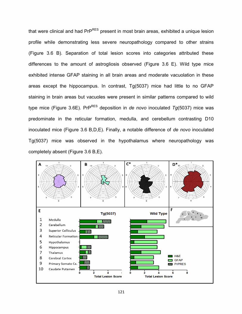
121
that were clinical and had PrPRES present in most brain areas, exhibited a unique lesion
profile while demonstrating less severe neuropathology compared to other strains
(Figure 3.6 B). Separation of total lesion scores into categories attributed these
differences to the amount of astrogliosis observed (Figure 3.6 E). Wild type mice
exhibited intense GFAP staining in all brain areas and moderate vacuolation in these
areas except the hippocampus. In contrast, Tg(5037) mice had little to no GFAP
staining in brain areas but vacuoles were present in similar patterns compared to wild
type mice (Figure 3.6E). PrPRES deposition in de novo inoculated Tg(5037) mice was
predominate in the reticular formation, medulla, and cerebellum contrasting D10
inoculated mice (Figure 3.6 B,D,E). Finally, a notable difference of de novo inoculated
Tg(5037) mice was observed in the hypothalamus where neuropathology was
completely absent (Figure 3.6 B,E).
122
Figure 3.6. Neuropathology of 10 brain areas (F) were scored from 0-4 (0 indicating no neuropathology and 4 indicating severe neuropathology) for vacuolation (H&E), astrogliosis (GFAP), and PrPRES deposition. Scores were plotted on radar plots for comparison (A-D). De novo prions inoculated into wild type generated a radar plot pattern that was similar but less intense than RML prions, even though these mice were not clinical and PrPRES deposition was absent in all sections (A,D). In contrast, de novo prions inoculated into Tg(5037) mice exhibited a unique profile in lesioned brain regions and less severe neuropathology overall (B). Separation of lesion scores by criteria revealed noticeable differences in GFAP staining between Tg(5037) and wild type inoculated mice (E) which can be attributed to vacuolation and PrPRES scores (E). (F) Anatomical diagram of brain areas analyzed. * Data has been previously published.
DISCUSSION
Evidence supporting spontaneous generation of prions in humans as sCJD28,29,
has led to many related hypotheses about the inception of some animal prion diseases
such as chronic wasting disease and scrapie. However, proving a spontaneous event
over natural transmission is challenging, especially in wild populations like cervids.
Currently, the only way to attack challenging questions about biological mechanisms
and the conditions needed to form de novo prions is through transgenic mouse models
and in vitro assays.
Several prion research groups have reported the generation of de novo prions in
vitro through recombinant technology,30-33 PMCA17,18 and inadvertently, by binding PrPC
and presumable co-factors to metal30. In previous reports de novo generation of PrPRES
utilizing PMCA required addition of synthetic polyanions and/or co-purified lipids, and
modification of PMCA by increasing incubation and sonication cycles17,18. Prion seeded
PMCA experiments in our laboratory rarely generate false positives in negative controls
but usually they are observed after 5 rounds of PMCA, are not reproducible in replicate
samples, and contamination cannot be dismissed. In order to determine the possibility
of false positives arising from spontaneous generation of prions we tested our standard
123
PMCA protocol in a new, prion free lab with new equipment and reagents. After 4
rounds of 48 cycles of PMCA, PK resistant material was detected by western blot in 1 of
60 total samples or at a rate of 1.6%. False positive spontaneous prions were ultimately
produced at a low frequency of 6.7% after 7 rounds of standard PMCA. Therefore, we
report generation of de novo prions at a low frequency by standard PMCA methods,
using normal cervidized brain homogenate, in a prion free environment.
Barria et al., investigated the occurrence of spontaneous conversion of various
specie substrates using PMCA in their laboratory18. They reported that they were not
able to generate de novo prions by standard PMCA protocols and were only able to do
so by extending the number of PMCA rounds and cycles where 240 cycles (5 days)
constituted one round. Intriguingly, the de novo prions they generated were infectious
and produced a new disease phenotype. We hypothesize that we generated
spontaneous cervid prions without modifications and additions to our standard PMCA
protocol as a consequence of the unique rigid loop in the globular domain of cervid
PrPC. Sigurdson et al., reported that altering the flexible loop of mouse PrP at amino
acids 170 and 174, reminiscent of the rigid loop in cervid PrP, resulted in spontaneous
prion disease in vivo34. Additionally, Kyle et al., reported that cell free misfolding and
conversion assays of recombinant mouse PrP containing cervid amino acid
substitutions for the rigid loop, had a higher misfolding propensity as compared to wild
type recombinant PrP22. Although we have never had an occurrence of spontaneous
disease in young or aged transgenic cervid overexpressors, it is plausible that the rigid
loop structure of cervid PrP confers susceptibility to misfolding under pressures applied
by PMCA. Transmission efficiency and prevalence of CWD prions in wild and captive
124
cervids35-40 suggests that cervid PrPC has a higher propensity for misfolding22, although
the necessary factors and conditions for increasing transmission efficiency are still
unknown.
We tested infectivity of de novo prions in Tg(5037) cervidized mice and wild type
mice resulting in clinical disease of cervidized but not wild type mice. De novo
inoculated cervidized mice had a similar survival to D10 inoculated cervidized mice
while wild type mice were not susceptible to either strain. Importantly, Tg(5037) mice
inoculated with de novo prions had a statistically significant delay of 104 days until
average clinical disease as compared to cervidized mice inoculated with a D10 strain
serially PMCA’d (sPMCA p1). sPMCA p1 was produced similarly to de novo through
serial PMCA rounds but sPMCA was a seeded reaction. If de novo prions were identical
to D10 we would have expected to see similar biological characteristics as other groups
have reported that PMCA propagates strain characteristics14,41,42. In addition, we have
shown that PMCA recapitulates in vivo strain adaptation and therefore, if de novo prions
were identical to D10, transmission should be much more efficient as indicated by a
shorter incubation period15.Lastly, species barrier studies suggest that transmission
efficiency is highly dependent on homology between host PrPC primary sequence and
PrPRES23,39,43,44. Therefore, we expect that cervid PrPC present in the brain homogenate
substrate would only spontaneously misfold into a cervid prion. Consequently, we
propose that PMCA generated de novo prions are a novel cervid strain.
Glycoform profiles provided further evidence that de novo prions are a cervid
prions as de novo prions are mostly di-glycosylated. Curiously, de novo prions
passaged through cervidized mice had a more equal di to mono-glycosylated ratio. It
125
would be interesting to investigate what phenotype would be observed after further
passages into cervidized mice. Further biochemical analysis of de novo prions by a
conformational stability assay revealed a similar profile to D10 sPMCA prions but
inoculation of de novo prions into Tg(5037) mice stabilized the strain in contrast with
sPMCA p1 prions, which were less stable. The increased stability upon passage into
Tg(5037) mice may contribute to the variation in onset of disease and increased dpi45,46.
Neuropathologic analysis of Tg(5037) mice inoculated with de novo prions
showed PrPRES deposition characteristic to CWD dense punctate plaques. However,
total lesion profile scores suggested minimal neuropathology as compared to D10,
RML, and even de novo inoculated wild type mice. De novo prions ultimately produced
a unique neuropathologic phenotype in Tg(5037) mice as compared to other strains,
again suggesting de novo prions are a novel cervid strain. Curiously, wild type mice
exhibited more neuropathology than Tg(5037) in the absence of PrPRES staining and
clinical signs. Total lesion profile scores of wild type mice were highly skewed because
of intense astrogliosis in all brain sections. Even in the absence of PrPRES detection by
western blot and IHC, wild type mice exhibited vacuolation in most areas of the brain. It
is possible that old age (>500 days) can be attributed to observed neuropathology.
Interestingly, these mice displayed a very similar but less severe lesion profile, when
compared by radar plot, to RML inoculated wild type mice. It is possible that the wild
type mice were subclinical at study termination. Castilla et al., reported that a species
barrier between Tg(porcine) mice and low dose BSE prions resulted in a subclinical
infection that became evident only after re-passage into Tg(porcine) mice47. However,
the subclinical mice did not show any neuropathology as demonstrated in this study. It
126
is also possible that the de novo inoculum was a low titer that was not able to efficiently
transmit into wild type mice, although western blot analysis revealed strong PK resistant
bands. In the future, we plan to further test the subclinical hypothesis.
In summary, our lab generated a novel de novo cervid prion strain that was
infectious to cervid transgenic but not wild type mice. Infected mice displayed
characteristic clinical signs of prion disease including ataxia and weight loss and days to
clinical onset of disease was reminiscent of a CWD prion strain. Further biochemical
and neuropathologic analysis revealed a unique phenotype of de novo prions upon
passage through Tg(5037) mice. Production of de novo prions in vitro using PMCA is a
rare event that was only observed in 4 out of 60 samples after 7 rounds of standard
PMCA. We feel that our data provides further evidence that cervid PrPC has a higher
propensity for misfolding as compared to other species under the right conditions.
127
REFERENCES
1. Brown, J. H. A. A. P. et al. Neuropathological Diagnostic Criteria for Creutzfeldt&###x2010;Jakob Disease (CJD) and Other Human Spongiform Encephalopathies (Prion Diseases). Brain Pathology 5, 459-466 (1995).
2. Bendheim, P., Barry, R., DeArmond, S. & Stites, D. Antibodies to a scrapie prion protein. Nature (1984).
3. Dearmond, S., McKinley, M., Barry, R. & Braunfeld, M. Identification of prion amyloid filaments in scrapie-infected brain. Cell (1985).
4. Tempst, P., Teplow, D., Le Hood & Prusiner, S. B. A cellular gene encodes scrapie PrP 27-30 protein. Cell (1985).
5. Büeler, H. et al. Mice devoid of PrP are resistant to scrapie. Cell 73, 1339-1347 (1993).
6. Hope, J., Morton, L., Farquhar, C. & Multhaup, G. The major polypeptide of scrapie-associated fibrils (SAF) has the same size, charge distribution and N-terminal protein sequence as predicted for the normal brain …. The EMBO (1986).
7. Prusiner, S. B. Novel proteinaceous infectious particles cause scrapie. Science 216, 136-144 (1982).
8. Prusiner, S. B. Prion Diseases and the BSE Crisis. Science 278, 245-251
9. Castilla, J., Saá, P., Hetz, C. & Soto, C. In Vitro Generation of Infectious Scrapie Prions. Cell 121, 195-206 (2005).
10. Kurt, T., Telling, G. C., Zabel, M. & Hoover, E. Trans-species amplification of PrP CWD and correlation with rigid loop 170N. Virology 1-9 (2009).
11. Murayama, Y., Yoshioka, M., Yokoyama, T. & Iwamaru, Y. Efficient in vitro amplification of a mouse-adapted scrapie prion protein. Neuroscience (2007).
12. Thorne, L., Holder, T., Ramsay, A. & Edwards, J. In vitro amplification of ovine prions from scrapie-infected sheep from Great Britain reveals distinct patterns of propagation. BMC veterinary (2012).
13. Nemecek, J., Nag, N., Carlson, C. & Schneider, J. Red-Backed Vole Brain Promotes Highly Efficient In Vitro Amplification of Abnormal Prion Protein from Macaque and Human Brains Infected with Variant …. PLoS One (2013).
14. Castilla, J., Gonzalez-Romero, D., Saá, P. & Morales, R. Crossing the Species Barrier by PrP Sc Replication In Vitro Generates Unique Infectious Prions. Cell (2008).
128
15. Meyerett, C. et al. In vitro strain adaptation of CWD prions by serial protein misfolding cyclic amplification. Virology 382, 267-276 (2008).
16. Green, K. M., Castilla, J., Seward, T. S. & Napier, D. Accelerated high fidelity prion amplification within and across prion species barriers. PLoS (2008).
17. Deleault, N., Lee, K. S., Harris, B., Caughey, B. & Rees, J. A simplified recipe for prions. in PNAS 104, 9551-9552 (2007).
18. Barria, M. A., Mukherjee, A., Gonzalez-Romero, D., Morales, R. & Soto, C. De Novo Generation of Infectious Prions In Vitro Produces a New Disease Phenotype. PLoS Pathogens 5, e1000421 (2009).
19. Sigurdson, C. J., Nilsson, K. P. R. & Hornemann, S. A molecular switch controls interspecies prion disease transmission in mice. The Journal of clinical (2010).
20. Soto, C. et al. De novo generation of a transmissible spongiform encephalopathy by mouse transgenesis. in PNAS 106, 304-309 (2009).
21. Jiang, L., Bett, C., Eisenberg, D., Sigurdson, C. J. & Kurt, T. D. A Proposed Mechanism for the Promotion of Prion Conversion Involving a Strictly Conserved Tyrosine Residue in the 2- 2 loop of PrPC. Journal of Biological Chemistry 1-18 doi:10.1074/jbc.M114.549030
22. Kyle, L. M., John, T. R., Schätzl, H. M. & Lewis, R. V. Introducing a Rigid Loop Structure from Deer into Mouse Prion Protein Increases Its Propensity for Misfolding In Vitro. PLoS One 8, e66715 (2013).
23. Tamgüney, G. et al. Chimeric elk/mouse prion proteins in transgenic mice. Journal of General Virology 94, 443-452 (2013).
24. Angers, R. C. et al. Chronic Wasting Disease Prions in Elk Antler Velvet. Emerging Infectious Diseases 15, 696-703 (2009).
25. Nichols, T. et al. Detection of protease-resistant cervid prion protein in water from a CWD-endemic area. Prion 3, 171
26. Pulford, B., Spraker, T. & Wyckoff, A. C. Detection of PrPCWD in feces from naturally exposed Rocky Mountain elk (Cervus elaphus nelsoni) using protein misfolding cyclic amplification. Journal of Wildlife (2012).
27. Race, R., Raines, A., Baron, T. & Miller, M. Comparison of abnormal prion protein glycoform patterns from transmissible spongiform encephalopathy agent-infected deer, elk, sheep, and cattle. Journal of (2002).
28. Zerr, I. & Poser, S. Clinical diagnosis and differential diagnosis of CJD and vCJD. APMIS 110, 88-98 (2002).

129
29. Will, R. G., Alpers, M. & Dormont, D. 12 Infectious and Sporadic Prion Diseases. Cold Spring Harbor (1999).
30. Edgeworth, J. A. et al. Spontaneous generation of mammalian prions. in PNAS 107, 14402-14406 (National Acad Sciences, 2010).
31. Wang, F., Wang, X., Yuan, C. & Ma, J. Generating a prion with bacterially expressed recombinant prion protein. Science (2010).
32. Makarava, N., Kovacs, G. & Bocharova, O. Recombinant prion protein induces a new transmissible prion disease in wild-type animals. Acta (2010).
33. Zhang, Z. et al. De novo generation of infectious prions with bacterially expressed recombinant prion protein. The FASEB journal 27, 4768-4775 (2013).
34. Sigurdson, C. J. et al. De novo generation of a transmissible spongiform encephalopathy by mouse transgenesis. in PNAS 106, 304-309 (National Acad Sciences, 2009).
35. Williams, E. S., Williams, E. S., Williams, E. S., Young, S. & YOUNG, S. CHRONIC WASTING DISEASE OF CAPTIVE MULE DEER: A SPONGIFORM ENCEPHALOPATHY. Journal of wildlife diseases 16, 89-98 (1980).
36. Williams, E. S. & Miller, M. W. Chronic wasting disease in deer and elk in North America. Revue scientifique et technique ( (2002).
37. Williams, E. S. Chronic wasting disease. Veterinary Pathology Online 530-549 (2005).
38. Miller, M. W., Hobbs, N. & Tavener, S. Dynamics of prion disease transmission in mule deer. Ecological Applications (2006).
39. Browning, S. R. et al. Transmission of prions from mule deer and elk with chronic wasting disease to transgenic mice expressing cervid PrP. Journal of 13345-13350 (2004).
40. Saunders, S., Bartelt-Hunt, S. & Bartz, J. Occurrence, transmission, and zoonotic potential of chronic wasting disease. Emerg Infect Dis (2012).
41. Joaqu et al. Cell free propagation of prion strains. The EMBO 27, 2557-2566 (2008).
42. Jones, M., Peden, A. & Prowse, C. In vitro amplification and detection of variant Creutzfeldt–Jakob disease PrPSc. The Journal of (2007).
43. Telling, G. C. Transgenic mouse models and prion strains. Prion Proteins (2011).
130
44. Tamgüney, G. & Giles, K. Transmission of elk and deer prions to transgenic mice. Journal of (2006).
45. Safar, J. et al. Eight prion strains have PrPSc molecules with different conformations. Nature medicine (1998).
46. Kuczius, T. & Groschup, M. Differences in proteinase K resistance and neuronal deposition of abnormal prion proteins characterize bovine spongiform encephalopathy (BSE) and scrapie strains. Molecular Medicine (1999).
47. Castilla, J. Subclinical Bovine Spongiform Encephalopathy Infection in Transgenic Mice Expressing Porcine Prion Protein. Journal of neuroscience 24, 5063-5069 (2004).
131
CHAPTER 4:
CERVID PRION PROTEIN PROMISCUITY ABROGATES TRANSMISISON BARRIERS
SUMMARY
Prions have a controversial history fueled by characteristics unlike other
infectious microorganisms. The protein-only hypothesis suggests that prion
pathogenesis is a consequence of cellular prion protein (PrPC) misfolding into a β-sheet
rich conformation, rendering it PK-resistant and insoluble (PrPRES). How prions are able
generate differing strains in the absence of nucleic acid is still an enigma. Evidence
suggests that strains are enciphered by the PrPRES conformation and acts as a template
to convert cellular PrPC into the same misfolded conformation. Transmission of prions
from one host into another can often result in delayed incubation times, incomplete
attack rates and sometimes complete resistance, a phenomenon defined as a species
or transmission barrier. A strict species barrier has been reported between mice and
cervid prions that cause CWD despite evidence that demonstrates CWD is very
efficiently transmitted between cervids. We wanted to investigate the role of host factors
in adaptation of cervid and mouse prions by challenging wild type mice with CWD that
was previously adapted through cervidized mice. We report, for the first time, that we
were able to adapt CWD through wild type mice after subsequent passage of non-
clinical FVB mice inoculated with D10, a natural CWD isolate from deer, adapted
through Tg(5037) mice 3 times. Additionally, we investigated the propensity of cervid
prion protein to propagate mouse and cervid-adapted prion strains. We found that mice
expressing cervid prion protein were much more susceptible to prion infection despite
132
the infectious prion being derived from a heterologous host. We conclude that cervid
prion protein is promiscuous and is able to abrogate species barriers.
INTRODUCTION
Chronic wasting disease (CWD), a prion-mediated disease of cervids, is the only
known transmissible spongiform encephalopathy (TSE) to infect a wild population1,2.
There is ever-increasing biological and biochemical evidence suggesting that prion
pathogenesis is caused by the conversion of the soluble normal host encoded protein
(PrPC) high in α-helical content, into an abnormal disease-causing isoform (PrPRES) that
is rich in β-sheets, insoluble, and partially protease resistant3,4,5. Unlike conventional
microorganisms like viruses and bacteria, prions are devoid of nucleic acid3, yet prions
infect various hosts differently, suggesting the phenomenon of prion strains6, 7,8.
Characteristic long incubation periods and incomplete attack rates, as consequence of
primary passage of prion infected material between differing species, but often even
within the same species, has been defined as the species or transmission barrier
respectively9,10, 7
Conversion efficiency of infectious prions is most efficient when host and donor
PrPC are identical leading some researchers to believe that heterologous PrP blocks
conversion, extending the days to onset of clinical disease9,11. Evidence also suggests
that prion protein primary sequence predisposes PrPC to fold a certain way but PrP
RES
conformation enciphers biological characteristics of the strain providing a template for
misfolding12,13, 14.
Kimberlin et al., has completed extensive experimentation in mouse models to
investigate strain properties. Through hamster bioassay, he suggested that natural
133
isolates of scrapie contained mixtures of prion strains and passage of strains through
heterologous hosts may alter prion properties15. In addition, Kimberlin reported the
propagation of new strains by serial passage through a heterologous host led to an
eventual stabilization of incubation periods by 3 or 4 passages in most models15,16
Importantly, Kimberlin showed that serial passage of prion strains through heterologous
hosts could modify prion strain characteristics, selecting for a mutant prion strain and in
most cases, the original phenotype could be rescued upon inoculation back into the
original host. This data suggests that other host factors may play a role in prion
propagation and misfolding15,16. Angers et al., provided additional evidence of these
findings by showing the emergence of two distinct CWD strains from deer and
transgenic cervidized mice. Propagation of one strain over the other was dependent on
primary PrPC sequence and PrPRES conformation17. Pan et al., further demonstrated the
interaction of dependence of PrPRES with PrPC by passaging mouse or hamster prions
through transgenic mice that expressed both mouse and hamster prion protein18.
Interestingly, mouse prions preferentially converted to mouse prions and hamster
preferentially propagated hamster prions in co-expressing mice. This study also
suggests that expression of heterologous PrPC does not compete or inhibit replication,
even when co-expressed with mouse PrPC.
Many research groups have identified a species barrier phenomenon in wild type
mice19-25. In particular, wild type mice appear resistant to CWD prions24,25, 26,27, 28.
Curiously, adaptation of a cervid strain D10 created a new strain phenotype in Tg(1536)
mice, through serial passage, that was biologically and biochemically reminiscent of
mouse-adapted scrapie prions25. Therefore, we hypothesize that mouse host factors
134
may play a role in adaption in conjunction with host encoded PrPC. In addition, it has
been shown that shedding of PrPRES and CWD transmission between cervids is highly
efficient compared to other prion diseases of animals 24,29-33. To investigate cervid
susceptibility and the role host co-factors may play in strain adaptation and transmission
efficiency, we challenged mice overexpressing murine PrPC, wild type mice with normal
PrPC expression, and Tg5037xTgA20 mice that co-overexpress cervid and murine PrPC
with mouse and cervid-adapted prion strains and characterized them biologically and
biochemically using standard methods. Remarkably, we report here for the first time,
that we were able to generate a wild type mouse-adapted CWD strain that only caused
clinical disease in inoculated animals after secondary passage. In addition, we
successfully propagated mouse-adapted RML scrapie and D10 CWD strains back
through cervidized mice but encountered a strict species barrier between cervid-
adapted prions and hosts encoding murine PrPC. Passage of mouse and cervid adapted
strains through mice co-overexpressing mouse and cervid prion protein revealed that
homologous strains were preferentially generated but cervid PrPC expressing mice
promiscuously converted both mouse and cervid adapted strains with varying degrees
of efficiency while wild type mice were only susceptible to mouse-adapted strains. We
conclude that cervid prion protein is promiscuous and therefore, abrogates the species
barrier between cervid and mouse prion protein.
MATERIALS AND METHODS Mice
FVB mice were purchased from the Jackson Laboratories (Bar Harbor, ME).
TgA20, Tg(cerPrP)1536, and Tg(cerPrP)5037 were generated as previously
described24,34,35. Tg5037xTgA20 mice were developed by breeding Tg(cerPrP)5037 mice
135
with TgA20 mice. F1’s were checked for the presence of both mouse and cervid prion
genes by PCR. F1’s expressing both genes were bred and second generation mice
PCR positive for cervid and mouse were used for experimentation. Western blot of
animals using cervid specific antibody 9E9 (described earlier)36 and mouse specific
antibody R137 provided further evidence that mice were cervid and mouse co-
expressers. All mice were bred and maintained at Lab Animal Resources, accredited by
the Association for Assessment and Accreditation of Lab Animal Care International, in
accordance with protocols approved by the Institutional Animal Care and Use
Committee at Colorado State University.
Sources and preparation of prion inocula
10% brain homogenates were prepared in PMCA buffer (4mM EDTA, 150 mM
NaCl in PBS). We diluted equal volumes of the inoculum 1:10 in 320 mM sucrose
supplemented with 100 units/mL Penicillin and 100 µg/mL Streptomycin (Gibco) in PBS
thirty minutes prior to intracerebral inoculations. Inoculum was made up of only one
chosen animal that had an intermediate DPI compared to other infected animals within
that group.
Cervid Prions
We used natural CWD isolates from brain tissue from a Colorado captive deer
D10 isolate (D10) (previously described24}) and a captive elk (E2) as CWD prions for this
study. D10 was inoculated into TgA20, Tg(1536), Tg(5037), FVB to create CWD
adapted strains. Strain Nomenclature: the inoculum is listed first with corresponding
passage number into host with corresponding PrPC (i.e., D10 propagated through
cervidized mice is denoted D10p1 (passaged once), D10p2 (passaged twice)). The
136
passage of D10 into a host with differing PrPC is denoted by the host strain name
followed by the passage number (i.e., D10p3FVBp3 indicates the D10 strain was
passaged through cervidized mice 3 times and then through wild type mice 3 times)
(Appendix A1.3-5).
Mouse-adapted Strains
The Rocky Mountain Lab strain of mouse-adapted scrapie prions passage 5
(RML5) was previously published{38. RML5 was passaged through TgA20, Tg(1536),
Tg(5037), FVB, and Tg5037xTgA20 mice to create scrapie adapted strains. Strain
Nomenclature: the inoculum is listed first with corresponding passage number into host
with corresponding PrPC (i.e.,RML passaged through wild type mice is denoted RML5
(passaged five times). The passage of RML into a host with differing PrPC is denoted by
the host strain name followed by the passage number (i.e., RML5/5037p2 indicates the
RML strain was passaged through wild type mice 5 times and then through cervidized
(5037) mice twice) (Appendix A1.3-5).
Prion inoculations and clinical scoring
Mice were anesthetized by Isofluorane inhalation. Thirty microliters of the
inoculum was injected intracerebrally 3 mm deep through the coronal suture 3-5 mm
lateral of the sagittal suture. Mice were monitored daily for clinical symptoms of prion
disease, including tail rigidity, impaired extensor reflex, akinesia, tremors, ataxia, 15 %
weight loss and paralysis. Mice with any four of these symptoms were scored terminally
sick and euthanized.
137
PK digestion and western blotting
Samples were digested with 50 g/ml PK (Roche) for 30 min at 37oC. The
reaction was stopped by adding lithium dodecyl sulfate sample loading buffer
(Invitrogen) and incubating at 95oC for 5 min. Proteins were electrophoretically
separated through 12% sodium dodecyl sulfate-polyacrylamide gels (Invitrogen), and
transferred to polyvinylidene difluoride membranes (Millipore). Non-specific membrane
binding was blocked by incubation in 5% milk blocking solution (Bio-Rad) for 1 h.
Membranes were then incubated for 1 h at room temperature with horseradish
peroxidase-conjugated Bar224 anti-PrP monoclonal antibody (SPI bio) diluted 1:20,000
in Superblock (Pierce), washed 6 x 10 min in PBS with 0.2% Tween 20, and incubated
for 5 min with enhanced chemiluminescent substrate (Millipore). Membranes were
digitally photographed using the FujiDoc gel documentation system equipped with a
cooled charge-coupled diode camera (Fuji). Densitometric analyses were performed
using Quantity One software (Bio-Rad). To discriminate between mouse and cervid
prion protein, western blots were run as mentioned above but incubated 1 h at room
temperature with cervid specific 9E9 mAb 1:3000 followed by horseradish peroxidase-
conjugated anti-mouse secondary antibody (GE Healthcare) or with mouse specific R1
F(Ab) 1:1000 followed by horseradish peroxidase-conjugated anti-human secondary
antibody (Pierce).
Histochemistry and immunohistochemistry (IHC)
Tissues were fixed in 10% paraformaldehyde, embedded in paraffin and 5-10 µm
sections mounted on glass slides. For PrP staining tissue sections were deparaffinized,
treated with concentrated formic acid for thirty minutes, then autoclaved at 121oC in
138
target retrieval solution (Dako) for 2 h, washed 2 x 7 min in 1X PBS, treated with 0.3%
H202 in methanol for 30 min and blocked for 1 hr with 5% BSA in PBS and mixed 1:1 in
superblock (Pierce). Excess block was tapped off and sections were incubated with
anti-PrP Bar 224 monoclonal antibody diluted 1:500 in block solution for 1 hour. Slides
were then washed 2 x 7 min in PBS and incubated 30 min with Envision+HRP mouse
secondary (DAKO). After another 2 x 7 min wash, slides were incubated for 5-7 min with
AEC+Substrate-Chromagen (DAKO) and rinsed 2 x 7 min in PBS and counterstained
with hematoxylin. Slides were rinsed in H2O, immersed in a 0.1% sodiumbicarbonate
bluing reagent for 5 min, rinsed in tap water and coversliped with aqueous mounting
medium (Richard Allan Scientific). Hematoxylin and Eosin (H&E) and glial fibrillary
acidic protein (GFAP) staining was performed on a NexES automated IHC stainer
(Ventana Medical systems, Inc. Tucson, AZ). Sections were stained with H&E for 4 min
at room temperature. Sections were stained with rabbit polyclonal antisera against
GFAP (diluted 1: 8) for 10 min at 37oC followed by Biotinylated goat anti-rabbit Ig
(mouse/rat adsorbed) for 8 min, then counterstained with hematoxylin for 4 min.
Sections were visualized and digitally photographed using an Olympus BX60
microscope equipped with a cooled charge-coupled diode camera.
Glycoform Ratios
Western blots of 10% brain homogenates of infected mice were analyzed by
densiometric analysis (Quantity One). Di, mono, and unglycosylated banding intensities
were calculated as percentages of the total density of each PK treated sample. The
averages of 2 replicate samples were plotted on a tri-plot based on calculated glycoform
ratios.
139
CPCA and Conformational stability assay
We quantified the infectivity of our prion strains with the cervid prion cell assay
(CPCA) as described previously39. Briefly, 10% brain homogenate was passed through
an 18 gauge needle 15 times to break up aggregates and then passed through 21-, 23-,
26-, and 28 gauge needles respectively. Inocula was diluted to 1% in cold PBS lacking
calcium and magnesium ions and coated and dried on 96 well plates.
A rabbit kidney epithelial (RK13) cell line (ATCC, Manassas VA) engineered to express
deer prion protein, referred to as Deer5E9-S1, which is highly sensitive to deer CWD
prions, was utilized in this assay in addition to RKM cells engineered to express murine
prion protein, and RKV cells with no expression or prion protein used as a negative
control. Twenty thousand cells per well were plated on top of dried inoculum in a volume
of 100 ml per well. Cells were passaged three times at four day intervals at 1:10 split
ratios. When cells reached confluence at the third passage, 20,000 cells per well were
filtered onto Multiscreen IP 96-well 0.45-mm filter plates (Elispot plates, Millipore,
Billerica, MA). Plates were dried at 50o C and cells were digested for 90 min at
37o C in 60 ml of lysis buffer containing 5 mg/ml proteinase K (PK) then terminated with
phenylmethanesulfonylfluoride (PMSF) (2 mM). To expose the epitope of PrP27-30,
cells were incubated in 120 ml 3 M guanidinium thiocyanate in 10 mM Tris-HCl (pH 8.0)
for 10 min at room temperature then rinsed four times with 160 ml PBS. For
immunodetection, wells were filled with 120 ml of filtered 5% superblock (Pierce,
Rockford, IL) and incubated for one hr at room temperature. The solution was
140
removed by vacuum, and wells were incubated with 60 ml of 6H4 mAb, diluted 1: 5000
in TBST for one hr at RT or overnight at 4uC. Wells were rinsed four times with 160 ml
of TBST then incubated with 60 ml AP-a-Mouse IgG (Southern Biotechnology
Associates, Birmingham, AL), diluted 1: 5000 in TBST, after one hr at RT, the wells
were rinsed four times with 160 ml TBST, followed by a final wash with PBS. Plates
were allowed to dry completely. Visualization was done by adding 60 ml of AP
conjugate substrate kit (Bio-Rad, Hercules, CA) at RT and rinsing twice with 160 ml
water and allowed to completely dry. Images were scanned with a ImmunoSpot S6-V
analyzer (Cellular Technology Ltd, Shaker Heights, OH), and spot numbers were
determined using ImmunoSpot5 software (Cellular Technology Ltd, Shaker Heights,
OH). Statistical analyses were performed using GraphPad Prism.
Cells lysates that were determined chronically infected by western blot analysis
were re-plated and grown to confluency. Cells were re-plated on Elispot plates following
the above protocol but with modifications. Briefly, cells were incubated for 1 hr with
increasing concentrations of 0-5.5M guanidine hydrochloride in 0.5M increments. Cells
were rinsed and then PK digested with 5ug/ml PK in cold cell lysis buffer for 1.5hr at
37oC. Digestion was stopped by the addition of PMSF and the plate was processed
identical to protocol mentioned above.
Plates were scanned with a ImmunoSpot S6-V analyzer (Cellular Technology
Ltd, Shaker Heights, OH), and spot numbers were determined using ImmunoSpot5
software (Cellular Technology Ltd, Shaker Heights, OH). Conformational stability was
calculated as Fapp=(observed-Native/unfolded-Native) and mean percentage of PrPRES
remaining ± SD were plotted as a function of GdnHCl concentration, and using fourth
141
order polynomial equations and nonlinear regression (GraphPad Prism) to fit
denaturation curves for each prion strain.
Lesion profiling
Brain lesion profiling was performed as previously described25 with slight
modifications. Ten neuroanatomical regions were identified in coronal brain sections
from at 5 mice of each group: 1-dorsal medulla, 2-cerebellum, 3- superior colliculus, 4-
reticular formation, 5-hypothalamus, 6-hippocampus, 7-thalamus, 8-cerebral cortex, 9-
primary somatosensory cortex and 10-caudate-putamen. A pathologist blinded to the
group identification scored each region for vacuolation, astrogliosis and PrPRES
deposition using the following severity scale: normal (0), minimal (1), mild (2), moderate
(3), severe (4). The average of the sum of the three scores constitutes the severity
score for each region.
Statistical analyses
Survival curves were generated using Kaplan-Meier survival fractions (GraphPad Prism)
One-way ANOVA with Tukey post-test analysis was performed using GraphPad Prism.
RESULTS Adaptation of cervid prion strain does not abrogate wild type species barrier
To challenge the species barrier of wild type mice to cervid adapted CWD prions,
we intracerebrally inoculated FVB mice with a CWD strain (D10) that was propagated
through cervidized mice 3 times (D10p3). We hypothesized that the murine host factors
present in transgenic mice, in addition to the cervid encoded PrPC, would impact strain
adaptation and therefore, cervid prions adapted through cervidized mice, would gain
characteristics that would render wild type mice susceptible to cervidized mouse-
142
adapted CWD prions. Primary passage of D10p3 into FVB wild type mice did not
abrogate the species barrier. Mice were non-clinical >380 dpi and did not have any
observable PK resistant material by western blot using cervid and mouse PrP
discriminatory antibodies (Figure 4.1A). In addition, despite observable vacuolation, no
detectable PrPRES was present by immunohistochemistry (IHC) (Figure 4.2). In contrast,
Tg(5037) mice expressing cervid prion protein, had detectable PrPRES by western blot
(Figure 4.4B) and exhibited dense, punctate PrPRES staining throughout the cerebellum,
cerebral cortex, and hippocampus after inoculation with the same D10p3 strain (Figure
4.2). Further stabilization of this strain through Tg(5037) also failed to cross the species
barrier into wild type mice. D10p4 inoculated FVB mice were non-clinical and PK
resistant material was absent in brain homogenate (Figure 4.3) and by IHC at 400dpi
(data not shown). To demonstrate that the absence of PrPRES in the brains of FVB mice
inoculated with D10p3 and D10p4 was not due to a delayed onset of disease, we
inoculated TgA20 mice overexpressing murine prion protein with D10p4. It has been
previously reported that TgA20 mice that overexpress mouse PrPC progress faster to
clinical prion disease than wild type mice 34. However, here we report that TgA20 mice
also failed to show signs of clinical disease and the presence of PrPRES was not
detected in brain homogenates or by IHC in mice >300 dpi (data not shown).
143
Figure 4.1. Western blot of FVB mice inoculated with CWD strain adapted through cervidized mice 3 times (D10p3/FVBp1). Western blot and antibody controls were RML 5 and Tg5037xTgA20 NBH (express mouse and cervid prion protein). Discriminatory antibodies were used to look for the presence of both mouse and cervid prions. PK resistant bands were not present in brain homogenate after >380 dpi.
144
Figure 4.2. (A) Representative slides of Tg(5037) mice inoculated with same D10p3 inoculum as FVB mice in (B). Tg(5037) mice developed severe vacuolation accompanied by dense PrPRES deposits in the cerebellum, cerebral cortex, and hippocampus respectively. In contrast PrPRES staining is absent from these areas in FVB mice. Tg(5037) and FVB mice had little to no astrogliosis present as indicated by the absence of GFAP staining.
145
Figure 4.3. FVB mice were inoculated with brain homogenate from a cervid mouse described in Figure 4.2. D10p4 inoculation did not abrogate the wild type species barrier as observed by the absence of PK resistant material in the western blot. Tg(5037) and TgA20 normal brain homogenate were used as antibody controls.
Subsequent passage of cervid adapted prions into wild type mice abolishes the
species barrier
Castilla et al., reported successful secondary transmission of BSE prions from
porcine transgenic mice (poTg) inoculated with low-dose BSE40. First passage of the
low-dose BSE inoculum into poTg mice did not cause clinical disease and PrPRES
material was not detected by immunoblot or IHC analysis. Surprisingly, secondary
passage of the non-clinical brains back into poTg mice caused clinical disease in all
animals with a greatly reduced incubation rate. To investigate the potential of a
146
subclinical CWD infection in wild type mice, we subsequently passaged brain
homogenate from an FVB mouse that was challenged with D10p3 prions and was
determined negative by clinical scoring, western blot, and IHC. Remarkably, all mice
inoculated with D10p3/FVBp1 displayed signs of clinical disease in an average of 153
dpi and exhibited detectable PrPRES in brain homogenate (Figure 4.4A). Interestingly,
cervid and mouse discriminatory antibodies revealed that passage of D10p3 through
Tg(5037) mice resulted in a cervid prion phenotype as PK resistant material was only
detected with a cervid specific antibody 9E9 (Figure 4.4B). In contrast, the same
inoculum passaged twice through FVB mice resulted in mouse prions (Figure 4.4A).
These results prompted us to complete the same serial passage experiment of non-
clinical brain homogenate from FVB mice inoculated with a natural CWD isolate, D10.
Interestingly, subsequent passage of FVB brain homogenate (D10/FVBp1) back into
FVB mice did not result in infection in contrast to the D10p3/FVBp1 strain.
Further passage of the newly generated mouse-adapted CWD strain into FVB
mice (passage 3) further decreased the incubation period to 139 dpi. Remarkably, this
strain infected FVB mice more efficiently than the mouse-adapted RML 5 strain (Table
4.1, row 2, Figure 4.5A). Additionally, we tested transmission efficiency of the newly
generated mouse adapted CWD strain into TgA20 mice. Unexpectedly, inoculation of
D10p3/FVBp1 (non-clinical) into TgA20 mice retarded the incubation to clinical disease
as compared to RML 5 inoculation and only 4 of the 5 mice were susceptible (Table 4.1
row 5, Figure 4.5B). However, further passage into TgA20 mice
(D10p3/FVBp1/TgA20p2 and p3) greatly reduced and stabilized time to clinical disease
to 68 dpi. Western blot analysis using 9E9 and R1 antibodies revealed that the cervid
147
D10 strain was converted to mouse PrPRES upon transmission into FVB and TgA20
mice.
Figure 4.4. Representative western blots of infected mice. (A) Passage of D10p3 into FVB mice did not cause clinical disease but secondary passage of the uninfected brain homogenate (D10p3/FVBp1) into FVB mice caused clinical disease in 100% of the animals and PrPRES bands were detected by western blot. The newly converted FVB prion strain was only detected with a mouse specific antibody and not cervid. (B) In contrast, Tg(5037) mice inoculated with the same primary strain (D10p3) was only observed when probed with a cervid specific antibody. Western blot labels indicate the resulting strain from inoculation, not the inoculum.
148
Figure 4.5. (A) Survival curves of FVB mice inoculated with a natural CWD prion strain from a deer (D10). The legend indicates the inoculum. D10 cervid prions became infectious after secondary passage into wild type mice and subsequent passages appeared to adapt the strain mimicking the survival curve of wild type mice inoculated with RML 5 mouse-adapted scrapie prions. (B) Survival curves of D10 inoculated TgA20 mice that over express mouse prion protein. TgA20 mice were only susceptible to D10 upon secondary passage into wild type mice. Serial passage of D10/FVBp1 into TgA20 mice adapted the strain resembling RML 5 mouse-adapted prions as seen in the wild type mice. Cervidized mice are susceptible to FVB-adapted CWD prions but not TgA20-
adapted CWD prions
149
To assess the biological properties of the newly generated mouse-adapted CWD
prions (D10p3/FVBp2) we inoculated mouse propagated strains back into the original
Tg(5037) donor. One would expect that transmission would be inefficient resulting in
lengthy incubation periods and incomplete attack rates because of heterologous PrPC
and PrPRES sequences between donor and host. However, transmission of murine CWD
into Tg(5037) mice resulted in similar incubation periods of D10p2 adapted through
cervidized mice and RML 5 into wild type mice with a 75% transmission rate (Table 4.1,
row 6). As expected, subsequent passage of D10p3/FVBp2/Tg5037p1 into Tg(5037)
mice further shortened the incubation period to 121 days and transmission was
observed in 100% of the animals (Table 4.1, row 6). Curiously, mouse and cervid
discriminatory antibodies revealed a sample that reproducibly reacted with the mouse
specific antibody contradicting all other cervid strains and previous data showing that
PrPRES converted to the same isoform as the host PrPC (i.e., cervid prions transmitted to
mice reacted only to mouse specific antibody) (Figure 4.6). In contradiction to FVB-
mouse adapted CWD data, inoculation of Tg(5037) mice with the CWD strain adapted
through FVB and then throughTgA20 mice (D10p3/FVBp1/TgA20p1) prevented
transmission with no detectable PrPRES or clinical disease after 450 days (Table 4.1, row
8). Nevertheless, this same CWD-TgA20 strain was infectious upon passage back into
FVB mice but had a delayed onset of disease as compared to TgA20 mice and FVB
mice inoculated with the same parent strain before passage through TgA20 mice (Table
4.1, row 8). Importantly, TgA20 mice became resistant to the previously infectious FVB-
adapted CWD strain when inoculated into Tg(5037) mice (Table 4.1, row 9).
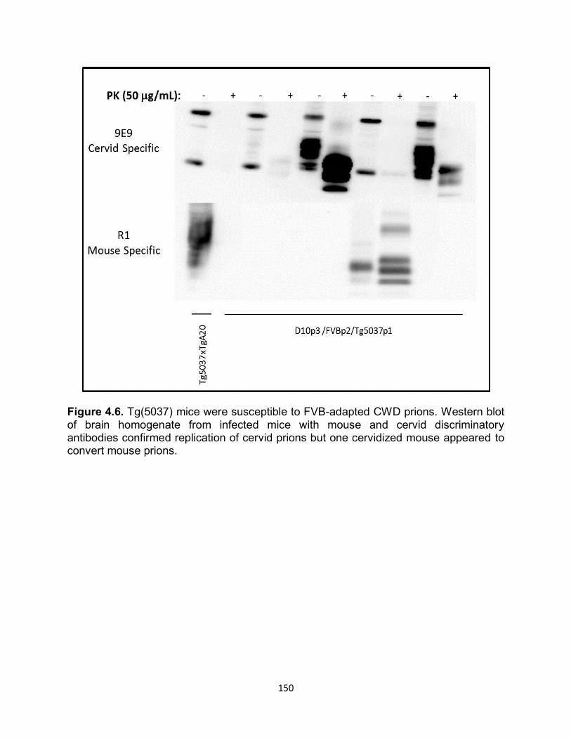
150
Figure 4.6. Tg(5037) mice were susceptible to FVB-adapted CWD prions. Western blot of brain homogenate from infected mice with mouse and cervid discriminatory antibodies confirmed replication of cervid prions but one cervidized mouse appeared to convert mouse prions.
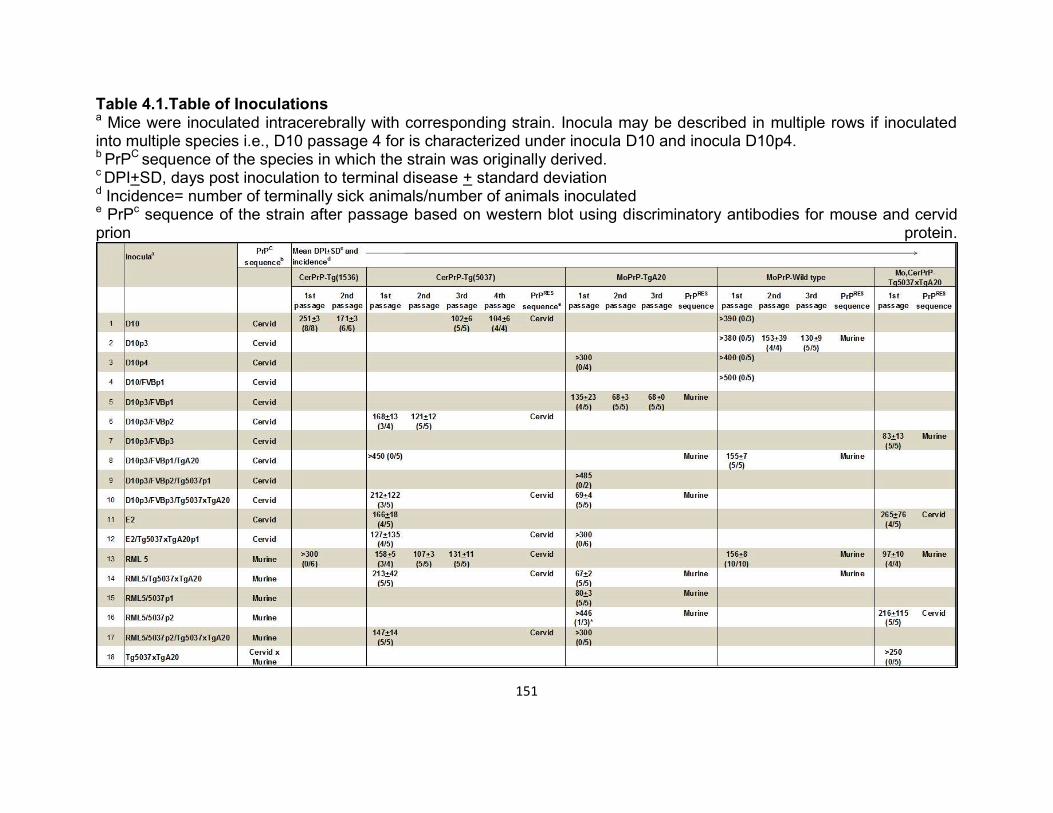
151
Table 4.1.Table of Inoculations a Mice were inoculated intracerebrally with corresponding strain. Inocula may be described in multiple rows if inoculated into multiple species i.e., D10 passage 4 for is characterized under inocula D10 and inocula D10p4. b PrPC sequence of the species in which the strain was originally derived. c DPI+SD, days post inoculation to terminal disease + standard deviation d Incidence= number of terminally sick animals/number of animals inoculated e PrPc sequence of the strain after passage based on western blot using discriminatory antibodies for mouse and cervid prion protein.
152
Cervidized mice are susceptible to wild type-adapted RML prions
Browning et al., reported that Tg(1536) cervidized mice were not susceptible to
RML prions after i.c. inoculation24. To test this conclusion in Tg(5037) mice we
intracranially inoculated Tg(5037) mice with an RML prion strain that was previously
adapted into CD-1 wild type mice. Unexpectedly, Tg(5037) were 75% susceptible to
RML prions upon primary passage. Remarkably, days to terminal disease mimicked that
of wild type inoculated with the same RML 5 strain (Table 4.1, row 13). Further passage
of cervidized RML prions resulted in shortened incubation periods and complete attack
rates. In contrast to FVB-adapted CWD prions propagated back through Tg(5037) mice,
cervid-adapted RML prions were infectious to TgA20 mice with a 100% attack rate,
although a slight delay to clinical disease was observed (Table 4.1, row 15). As
expected, cervid-adapted RML was only detected by a cervid specific antibody while
TgA20 mice propagated mouse specific prions (Figure 4.7).

153
Figure 4.7. Duplicate blots demonstrate that Tg(5037) mice were susceptible to cervid-adapted RML prions and only propagated cervid prions as evident by PK resistant bands only detectable with a cervid PrP-specific antibody (top left). TgA20 mice inoculated with the same cervid-adapted RML strain were also susceptible and produced mouse prions (bottom right). Western blot labels indicate the same inoculum used for Tg(5037) mice (left) and TgA20 mice (right) and Tg5037xTgA20 NBH was a non-infected sample control for discriminatory antibodies. Cervid and murine-adapted prion strains were infectious in transgenic mice that
co-overexpressed cervid and murine PrPC
Several research groups have investigated the prion strain phenomenon through
competitive assays either by mixing strains together and inoculating or identifying two or
more strains from a single inoculum and sorting out strain characteristics. Most of these
experiments concluded that one strain would predominate over the other presumably by
inhibiting replication or replicating more efficiently and using up available PrPC
154
substrate41-44, 15. In addition to strain competition studies, scientists have examined the
role of host PrPC in regards to species and transmission barriers and have concluded
that PrPRES enciphers species specific strain properties 16,22,28. Additionally, recent work
by Tamgϋney et al., demonstrated that mutations of C-terminal residues in transgenic
mice expressing chimeric mouse and cervid prion protein residues, played an important
role in transmission efficiency45. In order to test the promiscuity of cervid and mouse
prion proteins to replicate various cervid and mouse CWD and scrapie strains, we
generated a transgenic mouse that co-overexpressed mouse and cervid PrPC
(Tg5037xTgA20) and i.c. inoculated them with 4 different prion strains adapted through
or initiating from heterologous PrPC sequences. We hypothesized that the expression of
heterologous mouse and cervid PrPC would inhibit replication of all four strains.
Surprisingly, all of the strains caused disease but incubation times varied.
Notably, inoculation of cervid strains delayed clinical disease with high variance and
murine strains had a shorter incubation disease with little variance (Figure 4.8).
Inoculation of D10p3/FVBp3 into co-overexpressors was the most efficient of all tested
strains as mice developed clinical disease in 83 dpi (Table 4.1, row7, Figure 4.8). In
contrast, E2, a CWD strain derived from a natural elk isolate, had the longest incubation
period of 265 dpi and only infected 75% of the animals (Table 4.1, row 11, Figure 4.8).
RML 5 caused disease in all animals within 93 dpi which is delayed compared to RML
inoculated TgA20 mice but greatly reduced as compared to wild type and Tg(5037)
mice (Table 4.1, row 13, Figure 4.8). Finally, inoculation of the RML5/5037p2 strain
caused disease in all animals by 216 dpi (Table 4.1, row 16, Figure 4.8). Interestingly,
western blot analysis of infected Tg5037xTgA20 brain homogenates with mouse and

155
cervid discriminatory antibodies demonstrated that the nascent PrPRES sequence
coincided with the last animal the strain was propagated through and not the original
host’s PrPC sequence. Therefore, even though D10p3/FVBp3 initiated as a cervid prion
strain, adaptation through wild type mice generated a mouse prion strain that
preferentially converted mouse PrPC in Tg5037xTgA20 co-overexpressing mice (Table
4.1. rows 7, 11, 13, and 16, Figure 4.9). Similarly, RML 5 prions reproduced mouse
prions after passage through Tg5037xTgA20 mice, while subsequent passage of RML5
through Tg(5037) and then Tg5037xTgA20 mice preferentially replicated cervid prions
(Figure4.9).
Figure 4.8. Survival curves of Tg5037xTgA20 mice that co-overexpress mouse and cervid prion protein. Inoculation of E2 (a natural elk CWD strain), RML 5 (mouse-adapted scrapie strain), D10p3/FVBp3 (mouse-adapted CWD strain), and RML5/5037p2 (cervid-adapted scrapie strain), resulted in clinical disease with varying incubation periods. All of the inoculated strains except E2 had complete attack rates.
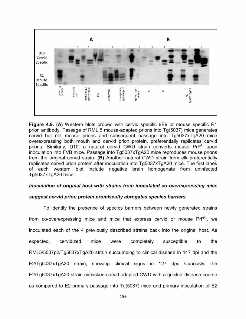
156
Figure 4.9. (A) Western blots probed with cervid specific 9E9 or mouse specific R1 prion antibody. Passage of RML 5 mouse-adapted prions into Tg(5037) mice generates cervid but not mouse prions and subsequent passage into Tg5037xTgA20 mice overexpressing both mouth and cervid prion protein, preferentially replicates cervid prions. Similarly, D10, a natural cervid CWD strain converts mouse PrP
C upon
inoculation into FVB mice. Passage into Tg5037xTgA20 mice reproduces mouse prions from the original cervid strain. (B) Another natural CWD strain from elk preferentially replicates cervid prion protein after inoculation into Tg5037xTgA20 mice. The first lanes of each western blot include negative brain homogenate from uninfected Tg5037xTgA20 mice. Inoculation of original host with strains from inoculated co-overexpressing mice
suggest cervid prion protein promiscuity abrogates species barriers
To identify the presence of species barriers between newly generated strains
from co-overexpressing mice and mice that express cervid or mouse PrPC, we
inoculated each of the 4 previously described strains back into the original host. As
expected, cervidized mice were completely susceptible to the
RML5/5037p2/Tg5037xTgA20 strain succumbing to clinical disease in 147 dpi and the
E2/Tg5037xTgA20 strain, showing clinical signs in 127 dpi. Curiously, the
E2/Tg5037xTgA20 strain mimicked cervid adapted CWD with a quicker disease course
as compared to E2 primary passage into Tg(5037) mice and primary inoculation of E2
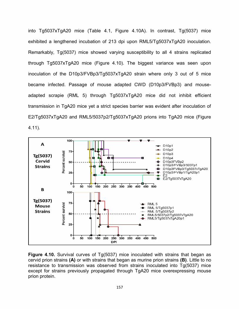
157
into Tg5037xTgA20 mice (Table 4.1, Figure 4.10A). In contrast, Tg(5037) mice
exhibited a lengthened incubation of 213 dpi upon RML5/Tg5037xTgA20 inoculation.
Remarkably, Tg(5037) mice showed varying susceptibility to all 4 strains replicated
through Tg5037xTgA20 mice (Figure 4.10). The biggest variance was seen upon
inoculation of the D10p3/FVBp3/Tg5037xTgA20 strain where only 3 out of 5 mice
became infected. Passage of mouse adapted CWD (D10p3/FVBp3) and mouse-
adapted scrapie (RML 5) through Tg5037xTgA20 mice did not inhibit efficient
transmission in TgA20 mice yet a strict species barrier was evident after inoculation of
E2/Tg5037xTgA20 and RML5/5037p2/Tg5037xTgA20 prions into TgA20 mice (Figure
4.11).
Figure 4.10. Survival curves of Tg(5037) mice inoculated with strains that began as cervid prion strains (A) or with strains that began as murine prion strains (B). Little to no resistance to transmission was observed from strains inoculated into Tg(5037) mice except for strains previously propagated through TgA20 mice overexpressing mouse prion protein.
158
Figure 4.11. Survival curves of TgA20 mice inoculated with strains originating as cervid prions (A) or mouse strains (B). TgA20 mice were only able to propagate murine-adapted strains once inoculated through Tg5037xTgA20 mice that co-overexpress cervid and mouse prion protein. Cervidized mice have increased neuropathology To compare neuropathology between cervid and mouse adapted prion strains,
we scored 10 brain sections (Table 4.2) for vacuolation, astrogliosis, and PrPRES
deposition. Severity of neuropathology increased in Tg(5037) mice despite heterologous
PrPC sequences (Figure 4.12 and 4.13). Most notably, passage of RML 5 through
Tg5037xTgA20 mice did not cause severe neurodegeneration as compared to cervid
strains propagated through this same strain of mice (Figure 4.12A) However,
159
subsequent passage of RML5/Tg5037xTgA20 back into cervidized mice exacerbated
disease compared to primary inoculation of RML 5 into Tg(5037) mice. Lesion profiles
plotted on radar plots demonstrate that all strains propagated in Tg(5037) mice had
similar neuropathologic patterns but with varying severity (Figure 4.12, Appendix A1.1).
Additionally, mouse-adapted CWD (D10p3/FVBp3) propagated in Tg5037xTgA20 mice
produced a similar but less intense radar plot as compared to cervid-adapted strains
(Figure 4.13).
Table 4.2. Neuroanatomical areas analyzed for lesion profiling.
160
Figure 4.12. Neuropathology of terminally sick mice inoculated with mouse and cervid adapted strains were scored in 10 brain areas by presence of vacuolation, astrogliosis, and PrPRES deposition. Average scores were plotted on radar plots according to neuroanatomical sections affected. RML 5 (A), RML5/5037p2 (B), and E2 (C) progression are plotted as strains are primarily propogated through Tg(5037), secondarily through Tg5037xTgA20 mice and then back into Tg(5037) mice. Tg(5037) demonstrate efficient propagation of all three strains. Unfortunately, PrPRES staining was difficult to detect within FVB and TgA20 mice and within Tg(5037) mice inoculated with D10p3/FVBp3/Tg5037xTgA20 prions. Labels above individual radar plot correlate with the strain. Numbers around the radar plot correlate with associated areas analyzed for lesion profiles (Table 4.2).
161
Figure 4.13. Lesion profiles of Tg5037xTgA20 mice demonstrate that cervid-adapted strains caused more severe neuropathology compared to mouse-adapted prion strains. Labels above each individual plot represent the prion strain after inoculation and the numbers around the radar plots correspond to Table 4.2. Interestingly, all three strains that have been propagated through Tg(5037) mice have similar lesion profiles despite variability in severity. Intriguingly, PrPRES detection by IHC in FVB and TgA20 mice that had been
inoculated with adapted strains was minimal despite strong PK resistant material
detectable by western blot. This was also observed in Tg(5037) mice inoculated with
mouse-adapted strain D10p3/FVBp3/Tg5037xTgA20 (Figure 4.14). Notably, radar plots
of Tg5037xTgA20 mice inoculated with either D10p3/FVBp3 or RML 5 had less severe
scoring as compared to cervid-adapted strains (Figure 4.13). Even more compelling is
the observation that Tg5037xTgA20 control mice that were homozygous for cervid prion
protein had detectable PrPRES deposition by IHC correlating with increased severity of
lesion profiles as compared to Tg5037xTgA20 control mice that were homozygous for
murine prion protein (Appendix A1.2). In addition, Tg5037xTgA20 mice inoculated with
mouse-adapted prions had little observable vacuolation n comparison to cervid-adapted
strains yet exhibited increased amounts of reactive astrocytes comparable to cervid-
adapted strains (Figure 4.14 and 4.15). Tg5037xTgA20 mice had severe neuronal
staining most notable in the cerebellum, cerebral cortex, and hippocampus (Figure
4.15). Dense, punctate PrPRES plaques were observed characteristic of CWD prions but
162
diffuse staining characteristic of mouse-adapted RML was also present in brain of
terminally sick Tg5037xTgA20 mice (Figure 4.15). Intense GFAP staining, a marker for
reactive astrocytes, correlated in these areas of dense PrPRES plaques. Furthermore,
subsequent passage of mouse and cervid-adapted prions back into Tg(5037) mice
efficiently propagated prions throughout the brains of infected mice (Figure 4.16). These
mice also exhibited diffuse and punctate PrPRES staining.
Figure 4.14. Immunohistochemistry (IHC) of terminally sick Tg5037xTgA20 mice inoculated with mouse-adapted CWD (D10p3/FVBp3) and mouse-adapted RML 5. The negative control is a mock inoculated Tg5037xTgA20 mouse. Representative sections of cerebellum, cerebral cortex, and hippocampus, respectively, demonstrate minimal to
163
no PrPRES deposition despite significant GFAP staining signifying reactive astrocytes.
Figure 4.15. Immunohistochemistry (IHC) of terminally sick Tg5037xTgA20 mice inoculated with cervid-adapted RML 5 (D10p3/FVBp3) and primary passage of CWD natural isolate, E2. The negative control is a mock inoculated Tg5037xTgA20 mouse. Severe PrPRES staining is significant in the cerebellum, cerebral cortex, and hippocampus, respectively. PrPRES deposits are characteristic of both CWD and RML 5 prions. Astrogliosis is evident by intense staining for GFAP.
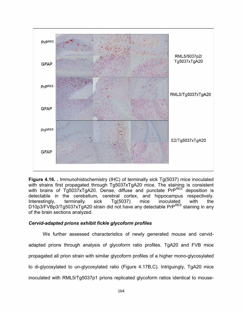
164
Figure 4.16. . Immunohistochemistry (IHC) of terminally sick Tg(5037) mice inoculated with strains first propagated through Tg5037xTgA20 mice. The staining is consistent with brains of Tg5037xTgA20. Dense, diffuse and punctate PrPRES deposition is detectable in the cerebellum, cerebral cortex, and hippocampus respectively. Interestingly, terminally sick Tg(5037) mice inoculated with the D10p3/FVBp3/Tg5037xTgA20 strain did not have any detectable PrPRES staining in any of the brain sections analyzed. Cervid-adapted prions exhibit fickle glycoform profiles We further assessed characteristics of newly generated mouse and cervid-
adapted prions through analysis of glycoform ratio profiles. TgA20 and FVB mice
propagated all prion strain with similar glycoform profiles of a higher mono-glycosylated
to di-glycosylated to un-glycosylated ratio (Figure 4.17B,C). Intriguingly, TgA20 mice
inoculated with RML5/Tg5037p1 prions replicated glycoform ratios identical to mouse-
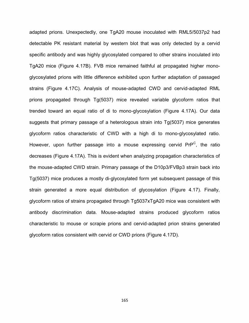
165
adapted prions. Unexpectedly, one TgA20 mouse inoculated with RML5/5037p2 had
detectable PK resistant material by western blot that was only detected by a cervid
specific antibody and was highly glycosylated compared to other strains inoculated into
TgA20 mice (Figure 4.17B). FVB mice remained faithful at propagated higher mono-
glycosylated prions with little difference exhibited upon further adaptation of passaged
strains (Figure 4.17C). Analysis of mouse-adapted CWD and cervid-adapted RML
prions propagated through Tg(5037) mice revealed variable glycoform ratios that
trended toward an equal ratio of di to mono-glycosylation (Figure 4.17A). Our data
suggests that primary passage of a heterologous strain into Tg(5037) mice generates
glycoform ratios characteristic of CWD with a high di to mono-glycosylated ratio.
However, upon further passage into a mouse expressing cervid PrPC, the ratio
decreases (Figure 4.17A). This is evident when analyzing propagation characteristics of
the mouse-adapted CWD strain. Primary passage of the D10p3/FVBp3 strain back into
Tg(5037) mice produces a mostly di-glycosylated form yet subsequent passage of this
strain generated a more equal distribution of glycosylation (Figure 4.17). Finally,
glycoform ratios of strains propagated through Tg5037xTgA20 mice was consistent with
antibody discrimination data. Mouse-adapted strains produced glycoform ratios
characteristic to mouse or scrapie prions and cervid-adapted prion strains generated
glycoform ratios consistent with cervid or CWD prions (Figure 4.17D).
166
Figure 4.17. Glycoform ratios were analyzed by densiometric analysis by western blot. Ratios are plotted on triplot where arrows indicate the axis that corresponds to the glycoform. The legend to the right of each plot indicates the prion strain while the label at the top of the triplot indicates the mouse strain: (A) Tg5037 mice inoculated with mouse and cervid adapted prion strains revealed that primary passage of a strain generated more characteristic CWD glycoform ratios while adaptation of a strain through Tg(5037) mice trended toward an equal di to mono-glycosylated ratio. (B) TgA20 and (C) FVB mice propagated glycoform ratios characteristic of mouse-adapted prions. (D) Primary passage of prion strains in Tg5037xTgA20 mice also generated glycoform ratios characteristic of the donor’s PrPC sequence. In vitro cervid prion cell culture assay only propagates prion strains from
homologous donor
The cervid prion cell assay (CPCA) has been previously reported as an effective
in vitro assay to investigate species barriers and transmission efficiency between cells
expressing heterologous PrPC and multiple prion strains39. Here we used this in vitro
167
system to reproduce our in vivo transmission studies with our adapted prion strains and
cells expressing mouse or deer PrPC. This assay revealed more strict species barriers
between heterologous PrPC sequences between donor and host as compared to our
bioassay data. Mouse-adapted prion strains were only able to infect cells expressing
mouse PrPC and reciprocally, cervid-adapted prion strains were only able to chronically
infect cells expressing cervid PrPC (Figure 4.18). Our bioassay data contradicts in that
RML 5 was able to cause terminal disease in animals that expressed mouse PrPC,
cervid PrPC, and mice that expressed both.
Figure 4.18. Prion strains tested with the Cervid Prion Cell Assay (CPCA) were only able to infect cells expressing homologous PrPC. Cervid prion strains propagated through co-overexpressing mice exhibit a
decrease in stability while mouse prions become more stable
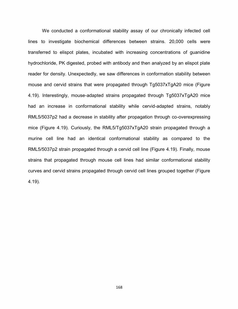
168
We conducted a conformational stability assay of our chronically infected cell
lines to investigate biochemical differences between strains. 20,000 cells were
transferred to elispot plates, incubated with increasing concentrations of guanidine
hydrochloride, PK digested, probed with antibody and then analyzed by an elispot plate
reader for density. Unexpectedly, we saw differences in conformation stability between
mouse and cervid strains that were propagated through Tg5037xTgA20 mice (Figure
4.19). Interestingly, mouse-adapted strains propagated through Tg5037xTgA20 mice
had an increase in conformational stability while cervid-adapted strains, notably
RML5/5037p2 had a decrease in stability after propagation through co-overexpressing
mice (Figure 4.19). Curiously, the RML5/Tg5037xTgA20 strain propagated through a
murine cell line had an identical conformational stability as compared to the
RML5/5037p2 strain propagated through a cervid cell line (Figure 4.19). Finally, mouse
strains that propagated through mouse cell lines had similar conformational stability
curves and cervid strains propagated through cervid cell lines grouped together (Figure
4.19).
169
Figure 4.19. Conformation stability curves generated from chronically infected cell lines with mouse and cervid-adapted prion strains. [GdnHCl]50 is the concentration required to denature 50% of PrPRES. Mouse-adapted prions adapted through murine cell lines had similar conformational stabilities to each other as did cervid-adapted prions. Interestingly, propagation of mouse-adapted strains through Tg5037xTgA20 mice stabilized strains while the conformational stability of cervid-adapted strains de-stabilized slightly.
DISCUSSION We have shown for the first time that FVB mice can be susceptible to CWD
cervid-adapted prions after subsequent passage of the non-infected brain homogenate
170
back into FVB mice. We inoculated FVB mice with a CWD strain D10 that was
previously propagated through cervidized mice 3 times (D10p3). FVB mice did not show
any clinical signs of disease or PK resistant material in brains after 300 dpi. However,
subsequent passage of brain homogenate from one of the non-clinical mice led to
abrogation of the species barrier and all inoculated mice came down with clinical
disease in 153+39 dpi, which is similar to mice inoculated with RML scrapie prions that
have been adapted through mice. Other research groups have reported a similar
phenomenon in other models. Hill et al., reported subclinical disease in CD-1 wild type
mice upon inoculation of 263K hamster scrapie, however, PK resistant material and
neuropathology was detected in some of these mice46. In contrast, we did not detect PK
resistant material in any of the inoculated mice by western blot of IHC. More recently,
Castilla et al., demonstrated similar results in a porcine transgenic mouse model
inoculated with BSE prions40. Brains from inoculated mice without clinical signs of
disease and the absence of detectable PK resistant material were able to infect the
same mouse model upon subsequent passage. It is possible that the FVB mice we
inoculated harbored a very low infectious titer of prion material that was not detectable
by conventional methods but was efficiently propagated upon serial passage. However,
we accounted for this possibility by inoculating TgA20 mice that overexpress mouse
prion protein and have been shown to have a much shorter disease progression
compared to wild type mice34. Although, we show transmission of the D10p3/FVBp1
strain to TgA20 mice, these mice had a significant delay in disease as compared to
TgA20 mice inoculated with RML prions. Additionally, only 3 of the 4 TgA20 mice
developed clinical disease. Further passage of the D10p3/FVBp1 strain through TgA20
171
or FVB mice considerably shortened the incubation period and was stabilized in TgA20
mice after the third passage. The delay in onset to disease of TgA20 mice indicate that
prion titers from the primary inoculum were not the reason we did not see clinical signs
in FVB mice. It does suggest, however, that a new strain emerged as TgA20 mice with
the same PrPC primary sequence as FVB mice, albeit in higher amounts, did not
succumb to prion disease as rapidly as other reported mouse-adapted strains. These
differences could be attributed to the difference in mouse backgrounds and if this is
true, this point validates our hypothesis that other host factors, in addition to PrPC
sequence, play a role in strain adaptation. Curiously, FVB mice inoculated with D10
(natural isolate of deer CWD before adaptation through cervidized mice) also did not
show clinical signs of disease or detectable PrPRES and subsequent passage through
FVB mice, mimicking the previous experimental design, also did not succumb to
disease. It is plausible that abrogation of the murine species barrier with CWD prions
requires additional host factors present in the mice.
To further assess characteristics of the newly generated mouse-adapted CWD
strain we passaged the brain homogenate from terminally sick FVB mice and brain
homogenate from the primary passage of D10p3/FVBp1 from TgA20 mice back into the
original host, Tg(5037) mice expressing cervid prion protein. Surprisingly, Tg(5037)
were 75% susceptible to the mouse-adapted strain with 30 day increase in incubation
as compared to the FVB mice inoculated with the same strain. An additional passage
into Tg(5037) mice shortened the incubation period and had a complete attack rate. In
contrast, Tg(5037) mice inoculated with the CWD adapted-TgA20 strain were
completely resistant to infection. It is possible that prion titers were lower in the TgA20
172
brain homogenate because these mice succumb to disease in a shorter time period
giving less time for replication of infectious prions. We tested this theory by inoculating
the TgA20-adapted CWD strain back into FVB mice. Consequently, all FVB mice were
clinical at 155 dpi. This suggests that the overexpression of mouse prion protein in
TgA20 mice had a role in generating a new species barrier. Perhaps, as Kimberlin has
reported, multiple conformers are present in brain homogenate of infected animals and
the overexpression of mouse PrPC in TgA20 mice allows for a more efficient selection
and propagation of a mouse prion conformer and conformers that would transmit readily
to cervidized mice are diluted out15. Interestingly, passage of the
D10p3/FVBp2/Tg5037p1 strain into TgA20 mice generated another species barrier as
TgA20 mice were completely resistant to infection. Perhaps providing additional
evidence that the FVB brain homogenate contained more than one strain and those
strains were preferentially replicated by different host PrPC sequences, diluting out the
other infectious strains so that transmission is now barred in heterologous species.
Remarkably, Tg(5037) mice were 75% susceptible to mouse-adapted RML 5
strain upon primary passage and they exhibited an incubation period identical to FVB
mice inoculated with the same strain. Previous reports have demonstrated a strict
species barrier between mouse-adapted prions and cervidized mice24,25, 2627, 28. However,
most of these studies report on a cervid transgenic line that expresses deer prion
protein. Here we report efficient transmission in elk cervid transgenic mice. It is possible
that transmission barrier differences can be attributed to differences at amino acid 226
in deer and elk PrPC primary sequence, although the mechanism is unclear17. Further
adaptation of RML through Tg(5037) shortened the stabilized the incubation period.
173
Curiously, tertiary passage through Tg(5037) mice drastically increased the incubation
period but this result from not using a pooled inoculum from all mice and using a mouse
that developed disease in between other mice. Passage of the newly generated cervid-
adapted RML prions (RML5/5037p1) into TgA20 mice caused disease in all of the mice
within 80 dpi. This was a much shorter incubation compared to TgA20 mice inoculated
with the mouse-adapted CWD strain from FVB mice. However, RML 5 adapted through
Tg(5037) mice twice (RML5/5037p2) generated a species barrier between TgA20 mice.
It is important to note that one of the TgA20 mice did show clinical signs and PK
resistant material in the brain after inoculation of RML5/5037p2. Even more importantly
is that the PK resistant material from this mouse was only detected with a cervid specific
antibody 9E9 and glycoform ratios revealed predominately di-glycosylated banding
pattern, a characteristic of CWD prions (Figure 4.17). Unfortunately, based on other
reports of spontaneous generation of prions in overexpressing transgenic mouse
models40,47, we cannot rule out this possibility, however, you would not expect to see
spontaneous mouse prions detectable with a cervid specific antibody. Further analysis
and experimentation would be necessary to confirm this result.
We further investigated strain properties of our mouse and cervid-adapted prions
by bioassay into Tg5037xTgA20 mice that co-overexpress mouse and cervid prion
protein. We expected inhibition of transmission in these mice because of the
heterologous expression of PrPC. However, Tg5037xTgA20 mice were susceptible, with
varying degrees, to all 4 strains we challenged them with. Primary passage of mouse-
adapted prion strains RML 5 and D10p3/FVBp3 preferentially propagated mouse prions
and the inverse was demonstrated by cervid-adapted RML 5 and E2 (CWD natural
174
isolate) as they preferentially propagated cervid prion strains. Passage of RML 5
through co-overexpressing mice developed disease in 93 dpi, which is an intermediate
time between TgA20 mice inoculated with RML 5 (60dpi) and Tg(5037) mice (158dpi).
The D10p3/FVBp3 strain produced disease in slightly shorter time frame of 83 dpi but
also had a complete attack rate. In contrast, cervid-adapted RML and E2 inoculated into
Tg5037xTgA20 showed a delay in disease at 216 and 265 dpi respectively and E2 only
infected 4/5 mice. Differences in incubation times and attack rates could be attributed to
differences in cervid to mouse PrPC expression levels in these mice but western blot
analysis using discriminatory antibodies suggest that expression is similar in most cases
(Figure 4.9B). When mice were screened for co-expression of cervid and mouse PrPC
by PCR, we did detect some littermates that only expressed mouse or cervid prion
protein. We utilized these mice as mouse only or cervid only controls and we found that
the controls were consistent with bioassay results attained from TgA20 and Tg(5037)
mouse strains. Our mouse only controls were only susceptible to mouse-adapted
strains and were infected with similar incubation periods to TgA20 mice inoculated with
RML 5 and the same was demonstrated with our cervid only controls as they were
susceptible to all the strains as previously described. Finally, we inoculated strains
propagated through Tg5037xTgA20 mice back into the original host to investigate the
generation of new species barriers and new strains. Interestingly, mouse-adapted
prions, including mouse-adapted CWD did transmit to TgA20 mice with great efficiency
in 67 and 69 dpi respectively. These strains also transmitted to Tg(5037) mice but with
varying efficiency. RML 5/Tg5037xTgA20 propagated in Tg(5037) mice with an
increased incubation time similar to that observed with RML5/5037p2 passaged through
175
Tg5037xTgA20 mice of 213 dpi. The D10p3/FVBp3/Tg5037xTgA20 strain infected 3 of
5 Tg(5037) mice with an identical incubation period as seen with RML5/Tg5037xTgA20.
In contrast, neither of the cervid-adapted strains propagated through Tg5037xTgA20
mice transmitted to TgA20 mice. This suggests that cervid prion protein is more
promiscuous and more easily converted to a disease causing isoform. Several groups
have now show that the rigid loop conformation of cervid prion protein increases
transmission efficiency and has a higher propensity to misfold45,48-50. We feel our
bioassay data strongly correlates with other reports. It is plausible that the rigid loop
structure actually allows for more flexibility of misfolding, almost by creating a structural
platform that allows heterologous PrPRES to interact and dictate conformational changes
more easily.
Biochemical and histological analysis provide further evidence that cervid prion
protein is promiscuous. Tg(5037) mice and Tg5037xTgA20 mice exhibited a great
degree of neuropathology as compared to TgA20 and FVB mice. Interestingly, IHC of
Tg5037xTgA20 mice inoculated with cervid-adapted strains exhibited PrPRES deposition
with both mouse and cervid prion characteristics. It is important to note that is was
difficult to detect PrPRES by IHC in FVB, TgA20, and Tg5037xTgA20 mice that were
inoculated with the mouse-adapted CWD strain, D10p3/FVBp3 despite detectable
amounts by western blot. We feel that this strain may have become acid sensitive, a
harsh step in our IHC protocol. Further analysis should be done to interpret these
results adequately.
Glycoform ratio analysis demonstrated that the host PrPC of inoculated animals
corresponded with strain characteristics for mice. TgA20 and FVB mice propagated
176
strains that had higher mono-glycosylated to di-glycosylated ratios and increased un-
glycosylated material. In contrast, Tg(5037) mice did not produce consistent glycoform
ratios. Primary passage of a heterologous strain usually produced more di-glycosylated
prions but serial passage lowered the di to mono-glycosylated ratio to more equal
values. Perhaps this is further evidence that cervid PrPC is more impressionable.
Finally, in vitro experimentation utilizing CPCA to propagate adapted strains in
cervid or murine PrPC expressing lines demonstrated that transmission was only
efficient between homologous sequences, i.e., cervid-adapted prions could only infect
cervid cells and vice- versa. This was in contrast to our bioassay data where Tg(5037)
mice could propagate mouse or cervid-adapted prions (with varying efficiency). This
also provides evidence that certain host factors are required for strain adaptation.
Conformational stability assays of infected cell lines revealed that mouse-adapted
prions similarly increased in stability after passage through Tg5037xTgA20 mice
whereas cervid-adapted prions slightly decreased in stability. Interestingly, GdnHCl50
curves for mouse-adapted prions were similar demonstrating that CWD was efficiently
adapted through a murine host. Intriguingly, the RML5 strain adapted through Tg5037
mice twice had the same conformational stability as RML5 propagated through
Tg5037xTgA20 mice. However, the RML5/Tg5037p2 strain dramatically reduced in
stability once propagated through Tg5037xTgA20 mice. A possible explanation is that
even though cervid prions are not propagated in Tg5037xTgA20 mice inoculated with
RML5 (at least not at detectable levels), cervid PrPC may interact and play a role in
mouse PrPC conversion.
177
We have demonstrated through extensive bioassay experiments that we were
able to adapt CWD through FVB mice after adaptation of CWD through cervidized mice.
Additionally, we have collectively shown that cervid prion protein is more promiscuous
and more readily converted to an infectious prion no matter the host’s primary PrPC
sequence. We have also demonstrated that cervid prion protein promiscuity may rely on
additional host factors for efficient transmission and adaptation. Further investigations
on the mechanisms involved in converting cervid prion protein could help lead to
therapeutic strategies that could aid in generating transmission and species barriers so
prion replication would be terminated.
178
REFERENCES
1. Williams, E. S., McCarty, C. & Spraker, T. Epizootiology of chronic wasting disease in free-ranging cervids in Colorado and Wyoming. of Wildlife Diseases (2000).
2. Williams, E. S. & Young, S. SPONGIFORM ENCEPHALOPATHY OF ROCKY MOUNTAIN ELK 1. Journal of wildlife diseases (1982).
3. Prusiner, S. B. Novel proteinaceous infectious particles cause scrapie. Science (New York, N.Y.) 216, 136-44 (1982).
4. Cohen, F. E. et al. Structural clues to prion replication. Science (New York, N.Y.) 264, 530-1 (1994).
5. Pan, K., Baldwin, M. & Nguyen, J. Conversion of alpha-helices into beta-sheets features in the formation of the scrapie prion proteins. in Proceedings of the (1993).
6. Pattison, I. & Millson, G. Scrapie produced experimentally in goats with special reference to the clinical syndrome. Journal of Comparative Pathology and (1961).
7. Pattison, I. H. Scrapie in the welsh mountain breed of sheep and its experimental transmission to goats. The Veterinary record 77, 1388-90 (1965).
8. Telling, G. C. Transgenic mouse models and prion strains. Prion Proteins (2011).
9. Raymond, G., Bossers, A. & Raymond, L. Evidence of a molecular barrier limiting susceptibility of humans, cattle and sheep to chronic wasting disease. The EMBO (2000).
10. Hill, A. & Collinge, J. Prion strains and species barriers. 2004 (2004).
11. Horiuchi, M., Priola, S. & Chabry, J. Interactions between heterologous forms of prion protein: binding, inhibition of conversion, and species barriers. in Proceedings of the (2000).
12. Scott, M., Groth, D., Tatzelt, J. & Torchia, M. Propagation of prion strains through specific conformers of the prion protein. Journal of (1997).
13. Safar, J. et al. Eight prion strains have PrPSc molecules with different conformations. Nature medicine (1998).
14. Peretz, D., Scott, M., Groth, D. & Williamson, R. A. Strain‐specified relative conformational stability of the scrapie prion protein. Protein (2001).
179
15. Kimberlin, R. H. & Walker, C. A. Evidence that the Transmission of One Source of Scrapie Agent to Hamsters Involves Separation of Agent Strains from a Mixture. Journal of General Virology 39, 487-496 (1978).
16. Kimberlin, R. H. et al. Temporary and permanent modifications to a single strain of mouse scrapie on transmission to rats and hamsters. The Journal of general virology 68 ( Pt 7), 1875-1881 (1987).
17. Angers, R. C. et al. Prion Strain Mutation Determined by Prion Protein Conformational Compatibility and Primary Structure. Science 328, 1154-1158 (2010).
18. Prusiner, S. B. et al. Transgenetic studies implicate interactions between homologous PrP isoforms in scrapie prion replication. Cell 63, 673-86 (1990).
19. Kimberlin, R. H. & Walker, C. A. Pathogenesis of scrapie: agent multiplication in brain at the first and second passage of hamster scrapie in mice. Journal of General Virology (1979).
20. Scott, M. et al. Transgenic mice expressing hamster prion protein produce species-specific scrapie infectivity and amyloid plaques. Cell 59, 847-857 (1989).
21. Torchia, M., Cohen, F. E., DeArmond, S. & Prusiner, S. B. Prion propagation in mice expressing human and chimeric PrP transgenes implicates the interaction of cellular PrP with another protein. Cell (1995).
22. Prusiner, S. B. et al. Transgenetic studies implicate interactions between homologous PrP isoforms in scrapie prion replication. Cell 63, 673-686 (1990).
23. Telling, G. C., Haga, T., Torchia, M. & Tremblay, P. Interactions between wild-type and mutant prion proteins modulate neurodegeneration in transgenic mice. Genes & (1996).
24. Browning, S. R. et al. Transmission of prions from mule deer and elk with chronic wasting disease to transgenic mice expressing cervid PrP. Journal of 13345-13350 (2004).
25. Meyerett, C. et al. In vitro strain adaptation of CWD prions by serial protein misfolding cyclic amplification. Virology 382, 267-276 (2008).
26. Favara, C., Gardner, D. & Williams, E. S. Transmission and adaptation of chronic wasting disease to hamsters and transgenic mice: evidence for strains. Journal of (2007).
27. Williams, E. S. & Young, S. Spongiform encephalopathies in Cervidae. Revue scientifique et technique ( (1992).
180
28. Kurt, T., Telling, G. C., Zabel, M. & Hoover, E. Trans-species amplification of PrP CWD and correlation with rigid loop 170N. Virology 1-9 (2009).
29. Mathiason, C., Carver, S., Zabel, M. & Telling, G. C. Detection of chronic wasting disease prions in salivary, urinary, and intestinal tissues of deer: potential mechanisms of prion shedding and transmission. Journal of (2011).
30. Mathiason, C., Hays, S., Powers, J. & Hayes-Klug, J. Infectious prions in pre-clinical deer and transmission of chronic wasting disease solely by environmental exposure. PLoS One (2009).
31. Williams, E. S. & Miller, M. W. Chronic wasting disease in deer and elk in North America. Revue scientifique et technique ( (2002).
32. Tamgüney, G., Miller, M. W., Wolfe, L. & Sirochman, T. Asymptomatic deer excrete infectious prions in faeces. Nature (2009).
33. Spraker, T., VerCauteren, K. & Gidlewski, T. Antemortem Detection of PrPCWD in Preclinical, Ranch-Raised Rocky Mountain Elk (Cevvus Elaphus Nelsoni) by Biopsy of the Rectal Mucosa. Journal of Veterinary (2009).
34. Fischer, M., Rülicke, T., Raeber, A. & Sailer, A. Prion protein (PrP) with amino-proximal deletions restoring susceptibility of PrP knockout mice to scrapie. The EMBO (1996).
35. Angers, R. C. et al. Chronic Wasting Disease Prions in Elk Antler Velvet. Emerging Infectious Diseases 15, 696-703 (2009).
36. Calvi, C. et al. Characterization of Conformation-dependent Prion Protein Epitopes. Journal of Biological Chemistry 287, 37219-37232 (2012).
37. Williamson, R. A. et al. Mapping the Prion Protein Using Recombinant Antibodies. Journal of Virology 72, 9413-9418 (1998).
38. Prusiner, S. B., Hadlow, W. J., Eklund, C. M., Race, R. E. & Cochran, S. P. Sedimentation characteristics of the scrapie agent from murine spleen and brain. Biochemistry 17, 4987-92 (1978).
39. Bian, J., Khaychuck, V., Angers, R., Graham, C. & Telling, G. Cell-Based Quantification of Chronic Wasting Disease Prions. Journal of Virology 84, 8322-8326 (2010).
40. Castilla, J. Subclinical Bovine Spongiform Encephalopathy Infection in Transgenic Mice Expressing Porcine Prion Protein. Journal of neuroscience 24, 5063-5069 (2004).
41. Bartz, J., Kramer, M., Sheehan, M. & Hutter, J. Prion interference is due to a reduction in strain-specific PrPSc levels. Journal of (2007).
181
42. Bartz, J., Aiken, J. & Bessen, R. Delay in onset of prion disease for the HY strain of transmissible mink encephalopathy as a result of prior peripheral inoculation with the replication-deficient DY strain. Journal of General Virology (2004).
43. Dickinson, A. G., Fraser, H. & Outram, G. Scrapie incubation time can exceed natural lifespan. Nature (1975).
44. Kimberlin, R. & Walker, C. Competition between strains of scrapie depends on the blocking agent being infectious. Intervirology (1985).
45. Tamgüney, G. et al. Chimeric elk/mouse prion proteins in transgenic mice. Journal of General Virology 94, 443-452 (2013).
46. Hill, A. F., Joiner, S. & Linehan, J. Species-barrier-independent prion replication in apparently resistant species. in Proceedings of the (2000).
47. Westaway, D., DeArmond, S. & Cayetano-Canlas, J. Degeneration of skeletal muscle, peripheral nerves, and the central nervous system in transgenic mice overexpressing wild-type prion proteins. Cell (1994).
48. Sigurdson, C. J., Nilsson, K. P. R. & Hornemann, S. A molecular switch controls interspecies prion disease transmission in mice. The Journal of clinical (2010).
49. Kyle, L. M., John, T. R., Schätzl, H. M. & Lewis, R. V. Introducing a Rigid Loop Structure from Deer into Mouse Prion Protein Increases Its Propensity for Misfolding In Vitro. PLoS One 8, e66715 (2013).
50. Sigurdson, C. J. et al. Spongiform Encephalopathy in Transgenic Mice Expressing a Point Mutation in the 2- 2 Loop of the Prion Protein. Journal of neuroscience 31, 13840-13847 (2011).
182
OVERALL CONCLUSION AND FUTURE DIRECTIONS
The ability of prions to encode strain properties in the absence of nucleic acid has
confounded fundamental biological principles by demonstrating the significance of
protein folding and conformation to epigenetic information transfer and infection.
Discoveries elucidating mechanisms of strain adaptation and species barriers are
important for developing therapeutic strategies for diseased patients and for abrogating
efficient inter and intra species transmission. However, lengthy incubation periods and
enigmatic strain properties have proved challenging to study prion strain characteristics.
The use of in vitro methods, like cell culture, to study prion disease have led to
expedited models of prion disease but often lack biological relevance. Although
transgenic rodent models mimic natural prion infection and have allowed researchers to
further exploit prion disease pathology, disease incubation periods can be
extraordinarily long. Combining in vivo and in vitro assays can provide insight into prion
replication, pathology, and strain mechanisms. The invention of PMCA has provided a
biologically relevant tool for elucidating prion transmission and replication mechanisms
within days to weeks rather than months and years.
The first part of this dissertation we assessed the ability of PMCA to adapt CWD prions
in vitro as compared to in vivo mouse bioassay. Here we report efficient linear
amplification of PrPCWD by sPMCA resulting in in vitro generation of infectious CWD
prions. Remarkably, we observed a drastic, nearly identical reduction in incubation time
to terminal disease of CWD-susceptible Tg(cerPrP)1536 mice inoculated with in vitro-
amplified or mouse-passaged prions from the D10 isolate of CWD prions when
183
compared to the original D10 inoculum. In vitro-amplified and in vivo-passaged D10 also
shared similar neuropathological and biochemical characteristics that were significantly
different than the original D10 strain, more closely resembling the RML strain of mouse-
adapted scrapie prions. By all accepted parameters used to characterize prion strains
that we examined, sPMCA adapted the D10 CWD strain as efficiently as passage in
Tg(cerPrP)1536 mice, and represents a powerful, efficient tool for assessing strain
adaptation and species barriers in vitro.
We and others observed that PMCA could generate de novo prions. We subjected
uninfected brain homogenate derived from cervidized transgenic mice to seven rounds
(48 cycles each) of PMCA under normal conditions in a prion-free laboratory using all
new reagents and equipment. Bioassay determined that the spontaneously generated
cervid prions were infectious to cervidized transgenic but not wild type mice and
biochemical analysis resulted in a unique profile differing from prion strains used within
our laboratory. These data strongly suggest that these prions were generated
spontaneously and not from contamination. We next examined the rate of spontaneous
conversion with our PMCA protocol. We generated PK-resistant de novo prions that
were detectable by western blot at round 4 of sPMCA at a rate of 1.6%. Further
generation of spontaneous prions were observed after round 5 and round 7 with rates of
5.0% and 6.74% respectively. These low rates of spontaneous conversion support
PMCA as an efficient and sensitive tool to investigate strain adaptation and species
barriers if limited rounds and proper controls are used.
Many research groups have identified a species barrier in wild type mice. In particular,
wild type mice appear resistant to CWD prions. Curiously, the data reported in the
184
second chapter of this dissertation suggested that adaptation of cervid prions through
cervidized mice resulted in strain properties resembling mouse-adapted prion strains.
Therefore, we hypothesized that mouse host factors may play a role in adaption in
conjunction with host encoded PrPC and this adaptation could lead to abrogation of
species barriers. We challenged this hypothesis by mouse bioassay and reported for
the first time adaption of cervid prions through wild type mice after secondary passage.
Because primary passage of cervid adapted prions did not cause clinical disease, we
further investigated the propensity of cervid PrPC misfolding and the role host co-factors
may play in strain adaptation and transmission efficiency. We challenged mice
overexpressing murine PrPC, wild type mice, and Tg5037xTgA20 mice that co-
overexpress cervid and murine PrPC with mouse and cervid-adapted prion strains.
Biological and biochemical characterization revealed successful propagation of mouse-
adapted and cervid-adapted strains through cervidized mice. We observed limited
propagation of cervid-adapted prions through hosts expressing only murine PrPC.
Passage of mouse and cervid adapted strains through mice co-overexpressing mouse
and cervid prion protein revealed that homologous strains were preferentially generated.
Re-passage of the co-adapted strains through mice expressing only cervid or mouse
PrPC divulged species barriers between cervid prions and mice expressing murine PrPC
while mice expressing cervid PrPC converted both mouse and cervid adapted strains
with varying degrees of efficiency. We conclude that cervid prion protein can interact
more promiscuously with heterologous prion strains and, therefore, abrogates the
species barrier between cervid and mouse prion protein.
185
We are very interested in continuing this work in vitro using PMCA to further elucidate
the mechanisms of prion strain adaptation and abrogation of species barriers. We
propose to further test the promiscuity of cervid PrPC to convert various prion strains by
utilizing mice that transgenically express the rigid loop region of cervid PrPC as a
substrate for PMCA. This experiment would also allow us to test the effect of murine
host factors in the presence of a rigid loop. It would be interesting to see if species
barriers are abrogated solely by the replacement of the murine loop with a cervid loop or
if other factors play a role in adaptation and promiscuity. Similarly, it would be intriguing
to further test strain adaptation of our newly generated mouse and cervid-adapted
prions in these same transgenic mice through mouse bioassay.
Additionally, we would like to investigate the phenomenon of propagating heterologous
prion strains. As seen in figure 4.6, we rarely identified a heterologous prion strain
detected using mouse and cervid PrPC discriminatory antibodies. Further analysis of
these samples by genotyping, bioassay, PMCA, and western blot would confirm that the
prion strain was truly heterologous to host PrPC.
Investigation of strain adaptation and species barrier through cell culture models such
as CPCA could help determine host factor requirements for conversion. The differing
results of infection exhibited between bioassay and CPCA suggest that host factors that
are absent in RK13 cells are needed to abrogate species barriers. We hope to further
test this hypothesis by using cell from different origins in addition to using less adapted
cervid and mouse prion strains (passaged through the host once rather than two or
three times).
186
Finally, we think it would be important to conduct competitive bioassay experiments
using a mixture of cervid and mouse adapted strains and propagating these through
mouse only, cervid only, or mouse and cervid expressing mice. Probing the resultant
strains propagated with cervid and mouse discriminatory antibodies would identify if
cervid, mouse or both prion strains would be preferentially propagated. We also plan to
use these discriminatory antibodies with IHC to determine if mouse and cervid prions
co-localize in brain areas of co-expressing mice.
In conclusion, our research has showed the importance of host factors, in addition to
host encoded PrPC, for strain adaptation. We have also provided further evidence that
cervid PrPC promiscuity can abrogate species barriers and lead to efficient inter and
intra species transmission. As we gain more knowledge about the requirements for host
adaptation and transmission the prion field will have more insight into generating
therapeutic strategies for infected individuals and ultimately, investigators can determine
the tools necessary for eliminating transmission of prion disease.
187
APPENDIX
Figure A1.1. Survival curves of (A) FVB wild type mice and (B) TgA20 mice overexpressing murine prion protein inoculated with cervid-adapted CWD prions as compared to mouse-adapted RML 5. FVB and TgA20 are both resistant to primary passage of CWD prions adapted through cervidized mice but are susceptible upon secondary passage through FVB mice. The legend indicates the inoculum used to produce the survival curves.
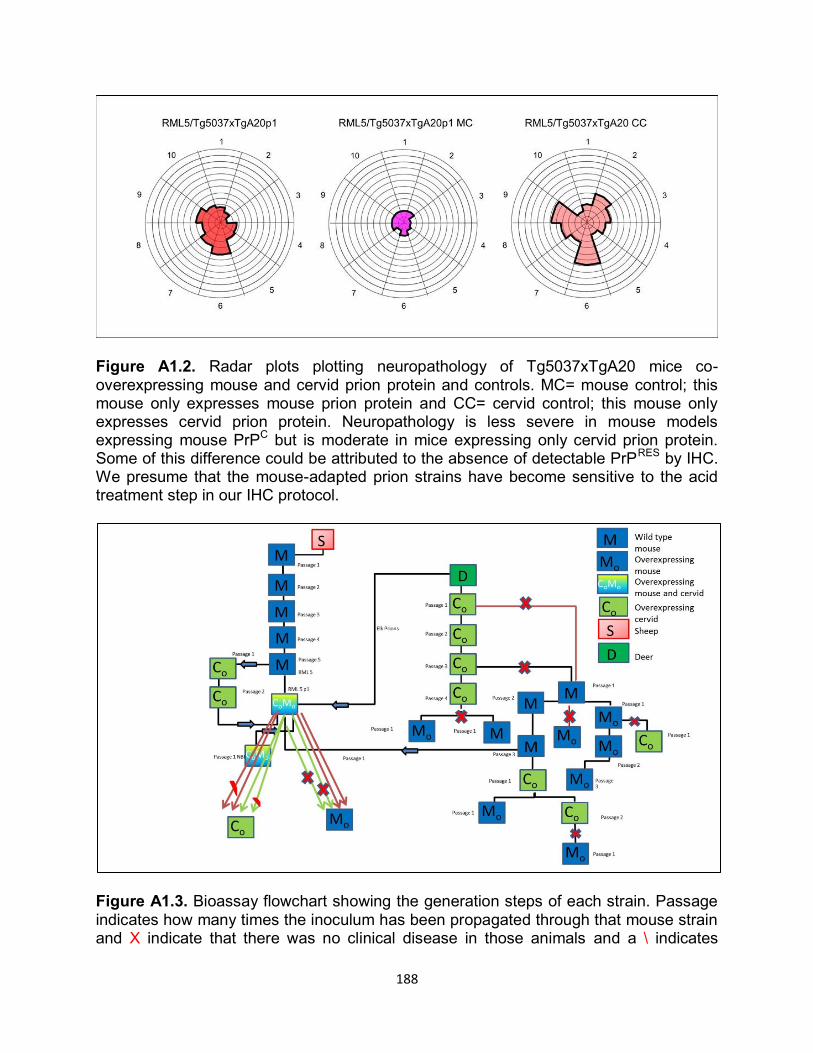
188
Figure A1.2. Radar plots plotting neuropathology of Tg5037xTgA20 mice co-overexpressing mouse and cervid prion protein and controls. MC= mouse control; this mouse only expresses mouse prion protein and CC= cervid control; this mouse only expresses cervid prion protein. Neuropathology is less severe in mouse models expressing mouse PrPC but is moderate in mice expressing only cervid prion protein. Some of this difference could be attributed to the absence of detectable PrPRES by IHC. We presume that the mouse-adapted prion strains have become sensitive to the acid treatment step in our IHC protocol.
Figure A1.3. Bioassay flowchart showing the generation steps of each strain. Passage indicates how many times the inoculum has been propagated through that mouse strain and X indicate that there was no clinical disease in those animals and a \ indicates
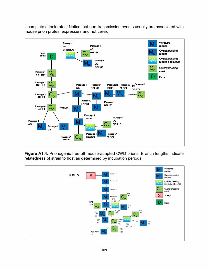
189
incomplete attack rates. Notice that non-transmission events usually are associated with mouse prion protein expressers and not cervid.
Figure A1.4. Prionogenic tree off mouse-adapted CWD prions. Branch lengths indicate relatedness of strain to host as determined by incubation periods.
190
Figure A1.5. Prionogenic tree of cervid-adapted RML 5 prions. Branch lengths indicate relatedness of strain to host based upon incubation periods.
Figure A1.6. Illustration of PMCA protocol used for Chapter 3. Notice there is no addition of a prion seed, just normal brain homogenate (NBH) used as a PrPC substrate.