prion amplification and hierarchical bayesian modeling refine detection of prion infection
TRANSCRIPT

Prion Amplification and HierarchicalBayesian Modeling Refine Detection ofPrion InfectionA. Christy Wyckoff1,2*, Nathan Galloway3*, Crystal Meyerett-Reid1, Jenny Powers4, Terry Spraker1,Ryan J. Monello4, Bruce Pulford1, Margaret Wild4, Michael Antolin3, Kurt VerCauteren2 & Mark Zabel1
1Colorado State University Prion Research Center, Department of Microbiology, Immunology and Pathology, College of VeterinaryMedicine and Biomedical Sciences, Fort Collins, CO 80523, USA, 2National Wildlife Research Center, Wildlife Services, UnitedStates Department of Agriculture, La Porte Avenue, Fort Collins, CO 80521, 3Department of Biology, College of Natural Sciences,Colorado State University, Fort Collins, Co 80523, 4National Park Service, Biological Resource Management Division, 1201Oakridge Drive, Fort Collins, CO 80525, USA.
Prions are unique infectious agents that replicate without a genome and cause neurodegenerative diseasesthat include chronic wasting disease (CWD) of cervids. Immunohistochemistry (IHC) is currentlyconsidered the gold standard for diagnosis of a prion infection butmay be insensitive to early or sub-clinicalCWD that are important to understanding CWD transmission and ecology. We assessed the potential ofserial protein misfolding cyclic amplification (sPMCA) to improve detection of CWD prior to the onset ofclinical signs. We analyzed tissue samples from free-ranging Rocky Mountain elk (Cervus elaphus nelsoni)and used hierarchical Bayesian analysis to estimate the specificity and sensitivity of IHC and sPMCAconditional on simultaneously estimated disease states. Sensitivity estimates were higher for sPMCA(99.51%, credible interval (CI) 97.15–100%) than IHC of obex (brain stem, 76.56%, CI 57.00–91.46%) orretropharyngeal lymph node (90.06%, CI 74.13–98.70%) tissues, or both (98.99%, CI 90.01–100%). Ourhierarchical Bayesian model predicts the prevalence of prion infection in this elk population to be 18.90%(CI 15.50–32.72%), compared to previous estimates of 12.90%. Our data reveal a previously unidentifiedsub-clinical prion-positive portion of the elk population that could represent silent carriers capable ofsignificantly impacting CWD ecology.
CWD is a neurodegenerative disease first seen in captive Colorado cervid populations in 1967, lateridentified as a transmissible spongiform encephalopathy (TSE) in 19781 and first found in free-rangingpopulations in 19811,2. Prions, the infectious agent of TSEs, arise from the misfolding of the normal host-
encoded cellular prion protein (PrPC) into an insoluble, aggregated and infectious form that resists proteasedegradation. Prions causing CWD arise from PrPCWD, the misfolded, infectious form of cervid PrPC. CWD, theonly known TSE to occur in free-ranging wildlife, affects several cervid species including elk (Cervus elaphusnelsoni), mule deer (Odocoileus hemionus), white-tailed deer (O. virginianus) and less commonly moose (Alcesalces spp.). Prevalence of CWD in free-ranging deer populations in Colorado and Wyoming ranges from 0–30%and prevalence in free-ranging elk herds ranges from 0–13% in Colorado3–6. CWD and sheep scrapie are capableof both horizontal transmission from infected individuals7–9 as well as efficient indirect transmission fromcontaminated environments10–13.
Despite cross-species transmissibility of PrPCWD among cervids, the pathogenesis of the disease may bedifferent between deer and elk. For example, studies with experimentally infected deer suggest that PrPCWD infectand replicate in peripheral lymphatic tissues prior to neuroinvasion14,15. Studies with captive or experimentallyinfected elk are less conclusive and suggest prions may first be detectable in the obex of the brain stem or theretropharyngeal lymph node without a clear pattern16–18. Studies of free-ranging elk, however, suggest thatPrPCWD can likely be detected first in lymphatic tissue19. It is unclear if route of inoculation or prion strain canalso affect these apparent differences.
Infected animals shed prions into the environment through saliva, feces, urine and even antler velvet15,20–24.Studies have successfully transmitted PrPCWD through a single dose of urine or feces from animals displaying signsof CWD, indicating that at the time of clinical disease sufficient prions are shed to result in an infectious dose24,25.However, at what stage(s) of disease animals shed prions into the environment remains unclear. If shedding
OPEN
SUBJECT AREAS:
PROTEINS
MOLECULAR ECOLOGY
STATISTICS
Received27 June 2014
Accepted19 January 2015
Published10 February 2015
Correspondence and
requests for materials
should be addressed to
M.Z. (mark.zabel@
colostate.edu)
* These authors
contributed equally to
this work.
SCIENTIFIC REPORTS | 5 : 8358 | DOI: 10.1038/srep08358 1

occurs early in disease, a sub-clinical animal may not only shedprions into the environment, increasing the infectious reservoir,but may also transmit CWD horizontally to their associates.Unfortunately little is known about the prevalence of early or sub-clinical infection, and what role they may play in CWD transmissionecology. This ignorance raises a critical question that must beanswered: how long do free-ranging animals live once infected?Answering it requires detection of prion infections as early as pos-sible in the course of infection.Cervid prion protein gene (PRNP) polymorphisms can influence
CWD kinetics. Elk PRNP methionine or leucine polymorphism atcodon 132 can dramatically affect incubation time and possibly sus-ceptibility of inoculated elk20–24,26. Experimental and observationalevidence suggests that 132LL homozygous elk are rare (#2.5%) infree-ranging populations20,25,27,28, and when inoculated have a sub-stantially delayed disease course (.48 months) in captivity com-pared to MM homozygote and ML heterozygote elk (12–24months)26,29. Despite differences in disease kinetics, Perucchiniet al.27 found that the prevalence of CWD within elk genotypes wasnot disproportionate, indicating the three genotypes maintain pro-portional CWD prevalence despite the disease course differences.Whether cervid PRNP genotype affects transmission and sheddingof PrPCWD remains uncertain. Surveys of free-ranging deer suggestincreased rates of CWD between 2–11 years of age30 but work on elksuggests a much wider age range can be infected19 and no study todate has quantified the interaction between age and prevalence ofCWD in elk.Currently, prion protein immunohistochemistry (IHC) of brain
tissue and lymph nodes is the gold standard for CWD detection31.However, this method requires 2–3 weeks for sample preparation,relatively large quantities of well-preserved tissue and specializedtraining for accurate microscopy work. Commercially availableenzyme-linked immunosorbent assay-based tests detect prion infec-tionmore rapidly, andwith similar sensitivity to IHC32. Serial proteinmisfolding cyclic amplification (sPMCA) has emerged in the field ofprion research as a reliable and sensitive alternative detection assayfor a variety of tissue and sample types1,22,27,33–37. Haley et al.38 foundcomparable sensitivity between an abbreviated sPMCA vs. IHC inlongitudinal tonsil biopsies from experimentally inoculated, captivewhite-tailed deer.We have optimized both sensitivity and specificity to maximize
PrPCWD amplification and detection, leading to a more sensitivesPMCA protocol that processes samples in less than two weeks23.We used sPMCA to test obex tissue samples from a free-ranging elkherd in the RockyMountain National Park (RMNP), Estes Park, CO;and Bayesian hierarchical modeling to compare our results to IHC ofretropharyngeal lymph node and obex tissue and estimate prevalenceof PrPCWD in this herd.
ResultsOf the 85 elk tested, 20 were IHC-positive in one or more tissues(Table 1). Of the 20 IHC-positive animals, sPMCA identified 19correlating obex samples as positive. The one sample that sPMCAdid not generate a positive result for was a 2011 sample found to beIHC-positive in the RPLN only. sPMCA also identified an additional18 IHC-negative samples as PrPCWD-positive.We found a strong correlation between sPMCA and IHC scores
for each elk sample (Figure 1). Linear regression found a positiveassociation (slope 5 0.39, R2
5 0.64) between samples scoring pos-itive by both IHC and sPMCA. Samples that disagreed, scoring IHCnegative but sPMCA positive, were not included in the linear regres-sion, but are overlaid in Figure 1 to show the low sPMCA scores ofsamples that were otherwise IHC-negative. A high rate of sPMCA-positive samples in 2010 suggested an unidentified portion of thosesamples may have been false-positives due to contamination. Thismay also be the case for 2009. However, sPMCA did identify 4
additional positives in 2011 compared to the 3 found by IHC (6 total,Table 1 and Figure S1). One of the IHC-positive samples was thesingle disagreement mentioned above.We developed a novel hierarchical Bayesian model to estimate
specificity, sensitivity and disease prevalence that considers all IHCand sPMCA test results (Figure 2). The model simultaneously esti-mates infection status of each individual animal and all derivedquantities of interest, including estimates of PrPCWD prevalencewithin the herd, and sensitivity and specificity of IHC for each tissueand sPMCA. This model also allowed us to quantify the potentialeffects of contamination in two of the collection years (2009 and2010).The separate analysis of Trusted and Unknown samples allowed
us to estimate sPMCA specificity by amplification round (Figure 3a)and for each group of samples (Figure 3b). When compared to spe-cificity of raw data from previous sPMCA studies23,35,39 (99.60%, CI98.00–100%) ourmodel estimates specificity for our Trusted samplesto be 93.85% (CI 90.00–96.84%), and a reduced specificity of 61.98%(CI 51.81–71.18%) for Unknown samples after six PMCA rounds.We did not separate samples into Trusted and Unknown groups
for sensitivity analyses because sensitivity is the ability of the test tocorrectly identify a true positive. The sensitivity of sPMCA after 6rounds was estimated at 94.68% (CI 83.12–99.84%) for one replicate.Again we compared sensitivity of known positive samples (97.00%,confidence interval 95.07–99.02%) from previous work to dem-onstrate the agreement between raw data and the model estimates(Figure 3C). We typically assess at least two replicate samples byPMCA, which our model predicts to increase sensitivity to 99.51%(CI 97.15–100%) with corresponding decreases in specificity oftrusted (91.03%, CI 84.42–97.80%) and unknown (42.01%, CI27.72–50.11%) samples.We also calculated sensitivity estimates for IHC of obex and RPLN
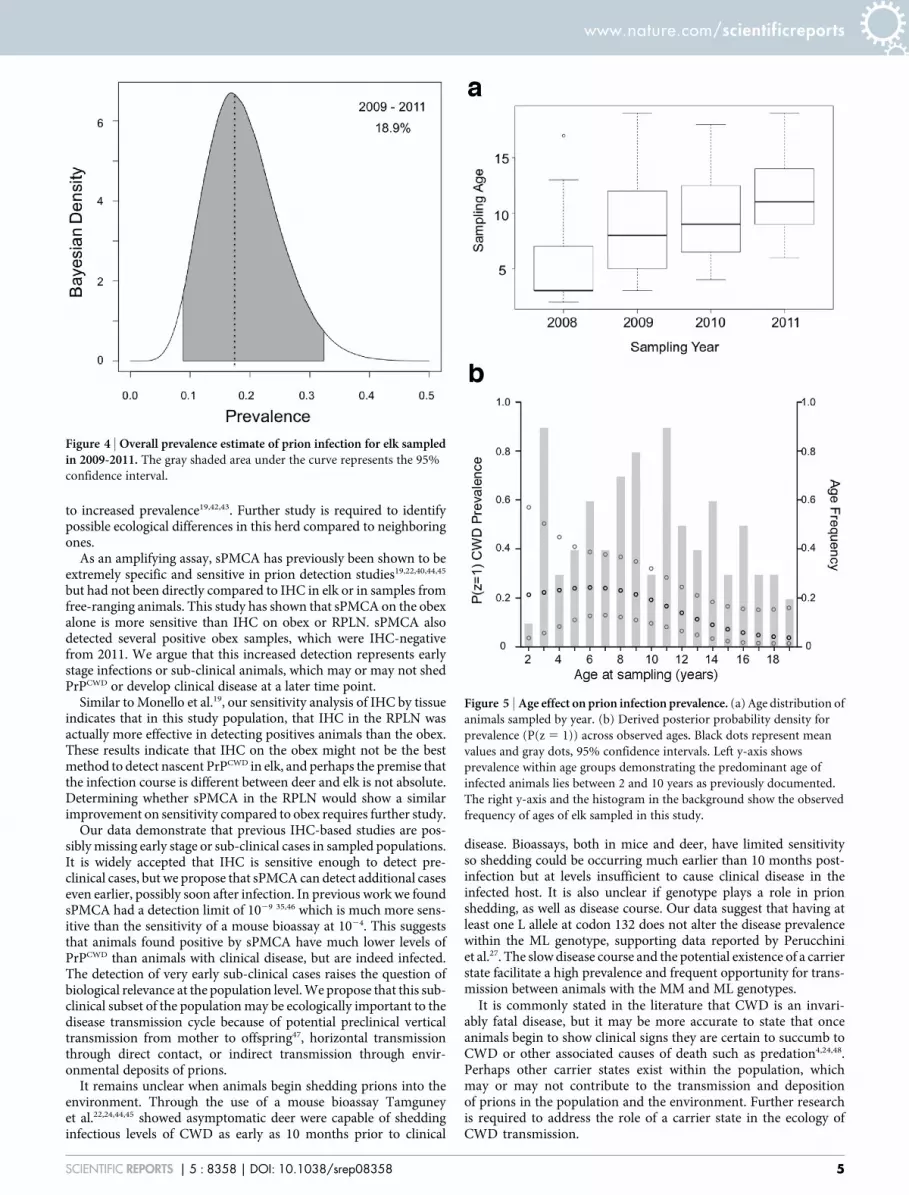
(Figure 3D). RPLN IHC (90.06%, CI 74.13–98.70%) was more sens-itive than obex (76.56%, CI 57.00–91.46%). Combining obex andRPLN IHC increases sensitivity to 98.99% (CI 90.01–100%).Inputting all data from the two IHC and duplicate PMCA tests,our model estimated the overall period prevalence of CWD for2009–2011 to be 18.90% (CI 8.71–32.39%; Figure 4).The age distribution of elk sampled by year showed an overlap
among years, with 2009 and 2010 including primarily middle-agedanimals (Figure 5A). Our model predicts the primary age of infectedanimals lies between 2–11 years (Figure 5B). Means and credibleintervals for the logistic regression coefficients, H, are reported inSupplemental Table 2. No difference in overall prevalence was foundbetween MM and ML elk (p 5 1.0).
DiscussionWe compared specificity and sensitivity estimates of two tests forCWD prions in a free-ranging elk herd in RMNP, the gold standardPrPCWD IHC versus sPMCA testing analyzed by Bayesian hierarch-ical modeling.We found that sPMCA detected PrPCWD in the obex ofinfected elk with greater sensitivity than IHC of obex or RPLN. Weused these estimates to predict prevalence of prion infection in thisherd, and discovered animals with subclinical CWDmay represent asignificant carrier state in this population. Although there appears tobe no upper age limit to infection in adult female elk19, we found thatprevalence decreases with age and the primary age cohort for infec-tion is 2 to 11 years.sPMCA specificity was lower than expected, likely due to cross-
contamination during necropsy in early years, which was reflected inthe difference in sPMCA-positive results between 2010 and 2011.Our decontamination protocol instituted during necropsy in 2011to limit cross-contamination reduced the sPMCA-positive rate totwice the IHC rate (sPMCA 5 18.2% and IHC 5 9.1%, Table 1).Our cross-contaminationmitigation strategy provided us with a yearof samples that we considered contamination-free.
www.nature.com/scientificreports
SCIENTIFIC REPORTS | 5 : 8358 | DOI: 10.1038/srep08358 2

We therefore consider the data showing the identification of 4additional positive samples by sPMCA in 2011 to be reliable, andused them to inform our model of sPMCA specificity and sensitivitycompared to IHC. Detection of four unique positives by sPMCAcorrelates with the low amounts of PrPCWD predicted to exist inanimals with early and sub-clinical disease.Hierarchical Bayesianmodeling allowed us to work with authentic
but possibly imperfect data and let the model test parameters to findthe best estimates. Having to discount some of the sPMCA findingsfor 2009 and 2010 was not ideal, but the model allowed us to adapt tothe realities of research. When we removed possible false-positiveresults for specificity estimates the model estimated that specificityincreased from 61.98% (Unknown samples) to 93.9% (Trusted sam-ples). Uncertainty introduced by possible sample contaminationlikely resulted in the model predicting a lower specificity in thesefield samples than our previous specificity of 99.6% observed incontrolled laboratory experiments35. Increased specificity usingTrusted while excluding Unknown samples shows that sPMCA hasa high specificity when cross-contamination is prevented. Moreover,if replicate samples are used, sensitivity increases to 99. 51%, whilespecificity remains acceptable at 91.03%. We consider these to be
conservative estimates of sensitivity and specificity considering pos-sible contamination at early collections, and our model allows us touse imperfect data to inform our model and estimate prevalence inthis herd.Our results suggest that prevalence of prion infection in this free-
ranging RMNP elk herd is much higher than previously reported.Prior to 2013, the CWD prevalence in elk surrounding RMNP wasestimated at ,2%19,40,41. In 2013, Monello et al. reported CWD pre-valence of 12.9% in RMNP based on PrPCWD IHC of RAMALT19,35,over 4 times higher than previous reports. Here we report an esti-mated overall prevalence of 18.90%, consistent with the finding byMonello et al., who found 28% of adult female elk were infectedduring the course of their three-year study. We conclude thatsPMCA can detect early cases of PrPCWD infection and our modelconservatively estimated overall prevalence since all known PrPCWD
positive animals from 2008, which tested positive on RAMALTbiopsy, were removed from the sample population.The higher overall prevalence estimate in this herd suggests pre-
vious measurements have been missing a large portion of PrPCWD -positive animals and that a long history of exposure to prions anddecades of relatively high densities on the winter range may have led
Table 1 | Summary of detection assay results
Sampling Year n 5 elk sampled sPMCA 1
IHC 1
Obex RPLN Ob & RPa IHC 1 Total
2008b 11 11 11 112009 17 5 c 1 12010 24 17c 1 3 1 52011d 33 6e 1 2 3Totals 85 39 1 4 15 20
aPrPCWD was found in both obex and RPLN samples.ball animals were RAMALT-positive at start of study, euthanized in the field within two months and tested further.cCross-contamination of tissue samples at necropsy likely resulted in some false sPMCA-positives these years.dA decontamination protocol was put in place to prevent cross-contamination at necropsy.esPMCA identified 4 additional samples that were positive for PrPCWD, but misdiagnosed one that was positive via IHC in RPLN only (the only such misdiagnosis by sPMCA).
Figure 1 | Correlation between IHC score and sPMCA score of each sample. (a) Sample scores by IHC and sPMCA were compared by linear regression
to evaluate correlation between the two tests. Samples found positive by both tests are considered in ‘‘Agreement’’ (black circles). Samples found negative
by IHC but positive by sPMCA are considered in ‘‘Disagreement (grey triangles). Each data point represents the mean of all replicates per animal.
(b) Representative IHC of a positive obex sample. Scale bars, 100 mm. (c) Representative western blots: Lane 1, undigested NBH; lanes 2–3, negative elk
obex from ND and MT after 6 rounds of sPMCA. Lanes 4–5, RMNP elk obex featured in (b) after 3 and after 6 sPMCA rounds (lanes 6 & 7).
www.nature.com/scientificreports
SCIENTIFIC REPORTS | 5 : 8358 | DOI: 10.1038/srep08358 3

Figure 2 | Model network diagram. Network diagram outlining the hierarchical Bayesian model used to estimate specificity and sensitivity of IHC and
sPMCA and population prevalence of CWD. Ob, Obex; RP, Retropharyngeal lymph node; Unk, Unknown samples; Tr, Trusted samples.
Figure 3 | Specificity and sensitivity estimates of sPMCA and IHC. (a) Specificity estimates of sPMCA by amplification round. Three sample groups are
presented: Known samples are positive controls used in previous studies and presented as raw data; Trusted samples are those which are verified by IHC or
are from the 2011 sampling; and Unknown samples are all remaining samples which may include contamination-related false positives. (b) Overall
sPMCA specificity estimates for Trusted and Unknown samples. The overall specificity of sPMCA is estimated at 93.9% (CI 90.1–96.9%) for Trusted
samples and 63.4% (CI 52.6–74.1%) for Unknown samples. (c) Sensitivity of sPMCA by round. Sensitivity increases with each amplification round to
94.68% (CI 83.12–99.84%) by round 6. (d) Overall sensitivity estimates for IHC and sPMCA. Sensitivity estimates for IHC by individual tissue (obex
(76.56%, CI 57.00–91.46%) and RPLN (90.06%, CI 74.13–98.70%)) and combined 98.99% (CI 90.01–100%); one replicate of sPMCA after 6 rounds
(94.68%, CI 83.12–99.84%); and two replicates of sPMCA (99.51%, CI 97.15–100%).
www.nature.com/scientificreports
SCIENTIFIC REPORTS | 5 : 8358 | DOI: 10.1038/srep08358 4
to increased prevalence19,42,43. Further study is required to identifypossible ecological differences in this herd compared to neighboringones.As an amplifying assay, sPMCA has previously been shown to be
extremely specific and sensitive in prion detection studies19,22,40,44,45
but had not been directly compared to IHC in elk or in samples fromfree-ranging animals. This study has shown that sPMCA on the obexalone is more sensitive than IHC on obex or RPLN. sPMCA alsodetected several positive obex samples, which were IHC-negativefrom 2011. We argue that this increased detection represents earlystage infections or sub-clinical animals, which may or may not shedPrPCWD or develop clinical disease at a later time point.Similar to Monello et al.19, our sensitivity analysis of IHC by tissue
indicates that in this study population, that IHC in the RPLN wasactually more effective in detecting positives animals than the obex.These results indicate that IHC on the obex might not be the bestmethod to detect nascent PrPCWD in elk, and perhaps the premise thatthe infection course is different between deer and elk is not absolute.Determining whether sPMCA in the RPLN would show a similarimprovement on sensitivity compared to obex requires further study.Our data demonstrate that previous IHC-based studies are pos-
siblymissing early stage or sub-clinical cases in sampled populations.It is widely accepted that IHC is sensitive enough to detect pre-clinical cases, but we propose that sPMCA can detect additional caseseven earlier, possibly soon after infection. In previous work we foundsPMCA had a detection limit of 1029 35,46 which is much more sens-itive than the sensitivity of a mouse bioassay at 1024. This suggeststhat animals found positive by sPMCA have much lower levels ofPrPCWD than animals with clinical disease, but are indeed infected.The detection of very early sub-clinical cases raises the question ofbiological relevance at the population level.We propose that this sub-clinical subset of the populationmay be ecologically important to thedisease transmission cycle because of potential preclinical verticaltransmission from mother to offspring47, horizontal transmissionthrough direct contact, or indirect transmission through envir-onmental deposits of prions.It remains unclear when animals begin shedding prions into the
environment. Through the use of a mouse bioassay Tamguneyet al.22,24,44,45 showed asymptomatic deer were capable of sheddinginfectious levels of CWD as early as 10 months prior to clinical
disease. Bioassays, both in mice and deer, have limited sensitivityso shedding could be occurring much earlier than 10 months post-infection but at levels insufficient to cause clinical disease in theinfected host. It is also unclear if genotype plays a role in prionshedding, as well as disease course. Our data suggest that having atleast one L allele at codon 132 does not alter the disease prevalencewithin the ML genotype, supporting data reported by Perucchiniet al.27. The slow disease course and the potential existence of a carrierstate facilitate a high prevalence and frequent opportunity for trans-mission between animals with the MM and ML genotypes.It is commonly stated in the literature that CWD is an invari-
ably fatal disease, but it may be more accurate to state that onceanimals begin to show clinical signs they are certain to succumb toCWD or other associated causes of death such as predation4,24,48.Perhaps other carrier states exist within the population, whichmay or may not contribute to the transmission and depositionof prions in the population and the environment. Further researchis required to address the role of a carrier state in the ecology ofCWD transmission.
Figure 4 | Overall prevalence estimate of prion infection for elk sampledin 2009-2011. The gray shaded area under the curve represents the 95%
confidence interval.
Figure 5 | Age effect on prion infection prevalence. (a) Age distribution ofanimals sampled by year. (b) Derived posterior probability density for
prevalence (P(z 5 1)) across observed ages. Black dots represent mean
values and gray dots, 95% confidence intervals. Left y-axis shows
prevalence within age groups demonstrating the predominant age of
infected animals lies between 2 and 10 years as previously documented.
The right y-axis and the histogram in the background show the observed
frequency of ages of elk sampled in this study.
www.nature.com/scientificreports
SCIENTIFIC REPORTS | 5 : 8358 | DOI: 10.1038/srep08358 5

The application of sPMCA will be important both to research andfor diagnostic investigation, and may improve state and federal sur-veillance programs for CWD in both naıve and endemic host popu-lations. Increased sensitivity, and the need for only obex tissue, maylead to detection of new focal points prior to clinical disease emergingin otherwise CWD-free populations. Additionally, in the econom-ically and politically difficult scenario of culling captive herds thattested positive for CWD, extremely sensitive assays such as sPMCAof prions from tissue and excreta are essential to verify that moreanimals besides the index case were infected, and if any sub-clinicalcarriers may have been shedding into the environment.Overall, our data contribute to the increasing evidence that a por-
tion of a herd may be infected, but die from other causes whileinfected with PrPCWD because of age, genetic susceptibility or otherunknown factors. However, the contribution of prions shed into theenvironment from this sub-clinical population may be importantand requires further investigation. The existence of an infectiousPrPCWD carrier state aligns with disease ecology theory, which pro-poses balance between transmissibility and pathogenesis of a patho-gen. As such, through selection pressures from the host and externalenvironment the pathogen will tend towards the greatest transmis-sibility strategy. CWD transmission may be more complicated thandisease ecologymight predict, since prolonged persistence and indir-ect transmission of prions in the environment may potentiate spreadwithout affecting pathogenesis.Despite the fact that prions are only protein, studies continue to
point at evolutionary behavior and selection pressures of prionswhich indicate that like other pathogens, prions are capable of evol-ving and adapting to their environment4,27,48,49. With increasing pre-valence at the population level, as is reported in this study, sPMCAwill continue to be an important tool to investigate CWD in wildlife.
MethodsMice.All mice were bred and maintained at Lab Animal Resources, accredited by theAssociation for Assessment and Accreditation of Lab Animal Care International, inaccordance with protocols approved by the Institutional Animal Care and UseCommittee at Colorado State University.
Elk brain tissue samples. Brain tissues were collected at necropsy from 85 free-ranging elk that were radio collared and later euthanized for research andmanagement purposes19. Briefly, 136 elk were initially captured, sampled and collaredin 2008. Rectoanal mucosal-associated lymphatic tissue (RAMALT) samples werecollected from each elk during initial capture and tested for PrPCWD by IHC7–9,38,50. In2008, samples were collected from 11 PrPCWD -positive animals that were recaptured,euthanized and necropsied within two months of original capture. In subsequentyears 17, 24, and 33 randomly selected animals were recaptured, euthanized,necropsied, and included in this study. These opportunistic collections were IHC-negative via RAMALT biopsy in 2008 and no elk exhibited clinical evidence of CWDwhen euthanized. Elk were euthanized in the field then transported to the TSEnecropsy facility at the Colorado State University Veterinary Teaching Hospitalwithin 8 hours of euthanasia19. We removed a primary incisor from all elk carcassesto determine age by cementum analysis (Matson Lab, Milltown, MT). Fieldeuthanasia and subsequent necropsies were approved by NPS (permit ROMO-2007-SCI-0077), ColoradoDivision ofWildlife (permit TR1081), and CSU IACUC (permit07-231A).
Multiple tissues were collected from each animal during necropsy. Here we com-pare IHC results from obex and retropharyngeal lymph nodes14,15,50,51 to sPMCAresults from the obex alone. All lymph node samples and half the obex sample werefixed in 10% neutral buffered formalin for IHC analaysis; the other half of the obexsample was stored in a whirl pack at 280uC for testing by sPMCA.
IHC. Sections of retropharyngeal lymph node (RPLN) and obex were examined byIHC as previously described16,50. Briefly for IHC, tissues were fixed, paraffin-embedded and 10 mm sections cut, mounted on glass microscope slides, andimmunolabeled with anti-prion protein monoclonal antibodies (mAbs) F99/97.6.1(mAb 99) and mAb P4. PrPCWD was visualized by incubation with alkalinephosphatase (AP)-conjugated donkey antibodies against mouse IgG and fast redchromogen that revealed red aggregate deposits in neural and lymphoid tissues. Ascoring system (0–10) was used to evaluate intensity of PrPCWD deposition asdescribed in Spraker et al.52.
Brain Homogenization. Frozen elk obex samples were partially thawed andapproximately 200 mg of tissue was collected from the interior of the obex sample,placed into a 2 ml tube containing silica beads and 180 ml of sPMCA buffer #1
(150 mMNaCl, 4 mMEDTA, in PBS) was added. Tissues were homogenized using aFastPrep machine (Thermo Scientific) as outlined in Meyerett et al.46. The clarified10% homogenate supernatant was removed and stored at 280uC.
sPMCA substrate consisted of 10% mouse normal brain homogenate (NBH)prepared in a prion-free room fromTg5037mice expressing cervid PrPC as previouslydescribed46.
sPMCA and western blotting. Twenty-five ml of RMNP elk obex homogenate wasadded to 25 ml NBH in 0.2 ml tubes. Samples were sonicated in a Misonix 4000sonicator (Misonix Inc., Farmingdale, NY) for 40 s every 30 minutes for 24 hours at37uC constituting one round46. For each subsequent round, 25 ml of each sample fromthe previous round was combined with 25 ml of fresh NBH. Duplicate samples wererun for 6 sPMCA rounds to balance desired sensitivity and specificity (.90%) aspreviously observed35. Each sPMCA experiment contained at least six NBH-negativecontrols and two positive plate controls (CWD-positive elk brain homogenate E2,151000). The negative brain samples were collected fromMontana (n5 1) andNorthDakota (n 5 10) and homogenized as previously described23 Montana eNBH wasconfirmed negative by bioassay in CWD susceptible mice (data not shown) and NDsamples were confirmed negative by ELISA (Bio-Rad Laboratories). Elk samplescollected in 2008 and 2009 were not blind to laboratory researchers, but all 2010 and2011 samples were blinded. The PrPCWD status of all negative controls was known atthe time of sPMCA.
PMCA samples were assayed by western blot as previously described23,46. Briefly,18 ml of sample was digested with 2 ml of 50 mg/ml proteinase K (PK, Roche, Basel,Switzerland) for 30 minutes at 45uC. Samples were electrophoresed, electro-trans-ferred to PVDF membranes and visualized with HRP-conjugated anti-PrP Bar-224monoclonal antibody (SPI-Bio, Montigny-le-Bretonneux, France). Because prionload inversely correlates with the round in which it is first detected, samples weregiven a score inversely proportional to the first positive sPMCA round23. For example,if a sample scored positive in the first of six rounds, it received a sample score of 6. Apositive sample detected in round six would receive a score of 1 and a negative samplewould be scored 0.
Cross-contamination management. During the first three years of necropsies(2008–2010), decontamination was not standard practice during tissue collection.Necropsies from 2009–2011 were performed in a new TSE necropsy room, reducingthe risk of contamination. In 2011, a decontamination protocol was implemented tofurther prevent cross-contamination during sample collection, including sodiumdodecyl sulfate/acetic acid46,53 treatment of working surfaces and all necropsyinstruments, and glove and apron changes between animals. Disposable sterilescalpels were used for all CNS tissue harvest. Samples were processed according toprotocols implemented to prevent cross-contamination at the lab bench, includingusing sterile scalpels, forceps and clean gloves during sub-sampling, homogenizationand sPMCA.
Model to estimate specificity, sensitivity and prevalence. The model is representedas a network diagram in Figure 2. Each animal was considered to have a true infectionstatus, denoted zi, where zi5 0 when animal i is uninfected and zi5 1 when infected.Infection status was modeled as a logistic regression with age and age covariates2, aswell as a categorical covariate for the samples from the year 2008, such that:
zi*Binom pið Þ
logit pið Þ~b0zb1ageizb2age2izb3yr:08i
b*Norm 0,1:5ð Þ
ð1Þ
For example, the IHC results for the obex tissue were described by amixture model asfollows:
yobex,i*
Binom 1{SpecIHCð Þ, zi~0
Binom SensObexð Þ, zi~1
( )
SensIHC*Beta 1,1ð Þ
ð2Þ
We denote sPMCA results for individual i and replicate j across amplification roundst 5 156 as wi,j,t which is contingent upon the latent disease state of individual i,Sensitivity and Specificity probabilities across amplification rounds, Se and Sp, andthe result from the previous round where applicable, wi,j,t-1.
wi,j,1*Binom 1{Sp1ð Þ, z
i~0
Binom Se1ð Þ, zi~1
� �
ð3Þ
wi,j,t
*Binom 1{Sptð Þ, zi~0
*Binom Setð Þ, zi~1
� �
, wi,j,t{1~0
~1, wi,j,t{1~1
8
<
:
9
=
;
ð4Þ
Se and Sp represent the probability of a sPMCA test result transitioning from anegative test result to a positive within amplification round t. These parameters aremodeled as flat Dirichlet distributions of length T1 1. Incorporating the probabilityof a negative test result allows the probabilities to sum to one. This transition model isa modification of the Cormack-Jolly-Seber survival model with perfect detection54.
www.nature.com/scientificreports
SCIENTIFIC REPORTS | 5 : 8358 | DOI: 10.1038/srep08358 6

The parallel occurs because after a sample is positive in one sPMCA cycle, it remainspositive in the following rounds, just as, for example, a mortality event at any timeguarantees all following times maintain that state. The model was fit to the data inJAGS 3.1.055,56 with the rjags package55 in the R 2.15.1 computing environment57.
Errors in specificity, or false positives, can occur as a result of cross-contaminationof samples during necropsy or possibly by spontaneous misfolding during sPMCA.We previously reported our method of sPMCA has a specificity of 99.6% in thelaboratory setting35. Negative samples used for this sPMCA experiment were used toshow specificity in our laboratory setting, but do not account for possible necropsycontamination of the elk tissues. To remove bias from possible necropsy-related falsepositives in years 2009 and 2010 we separated ‘‘Trusted’’ from ‘‘Unknown’’ samples.Trusted samples were those found positive by IHC (2008–2010) and all samples from2011, when we employed decontamination techniques at necropsy. Unknown sam-ples are IHC-negative samples from 2009 and 2010. sPMCA results for these samplescould be true, sub-clinical positives outside of the detection limit of IHC, or they couldbe false positives resulting from contamination during sample collection at necropsy.To maintain a conservative estimate of the specificity of sPMCA, Trusted andUnknown samples were assumed to have independent specificity probabilities.
Errors in sensitivity, or false negatives, for either assay occurred for two reasons:either the concentration of PrPCWD was below the detection limit of the assay or,despite the overall presence of PrPCWD in the tissue, the exacted portion that wasassayed did not contain detectable levels of PrPCWD due to non-homogenous distri-bution19,30,41,58. All estimates are reported with a 95% Bayesian credible interval (CI).
Genetic data for 30 MM and 30 ML randomly selected animals were used to assessfor prevalence differences between genotypes. IHC and sPMCA results were pooledallowing for positive or negative status to be tested against MM and ML genotypestatus using a Fisher’s Exact Test (p , 0.05).
1. Williams, E. S. & Young, S. Chronic wasting disease of captive mule deer: aspongiform encephalopathy. J. Wild. Dis. 16, 89–98 (1980).
2. Williams, E. S. & Young, S. Spongiform encephalopathy of Rocky Mountain elk.J. Will. Dis. 18, 465–471 (1982).
3. Walter,W. D.,Walsh, D. P., Farnsworth,M. L.,Winkelman, D. L. &Miller, M.W.Soil clay content underlies prion infection odds. Nat. Comm. 2, 200–6 (2011).
4. Miller, M.W. et al. Lions and prions and deer demise. PLoS ONE 3, e4019 (2008).5. Edmunds, D. R. Chronicwasting disease ecology and epidemiology of white-tailed
deer in Wyoming. A Dissertation. 1–224 (2013). at,http://search.proquest.com/docview/304452515. (Date of access: 06/02/2015).
6. Colorado Parks andWildlife. CWD in Colorado: 2010–2011 Surveillance Update.1–1 (2011). at ,https://cpw.state.co.us/Documents/Research/Mammals/Publications/2010-2011WILDLIFERESEARCHREPORT.pdf. (Date of access:06/02/2015).
7. Dickinson, A. G., Stamp, J. T. & Renwick, C. C. Maternal and lateral transmissionof scrapie in sheep. J. Comp. Path. 84, 19–25 (1974).
8. Miller, M. W. & Williams, E. S. Prion disease: horizontal prion transmission inmule deer. Nature 425, 35–36 (2003).
9. LJ, H. A review of the epidemiology of scrapie in sheep. Rev. Sci. Tech. 15, 827–852(1996).
10. Miller, M. W., Williams, E. S., Hobbs, N. T. &Wolfe, L. L. Environmental sourcesof prion transmission in mule deer. Emerg. Infect. Dis. 10, 1003–1006 (2004).
11. Mathiason, C. K. et al. Infectious Prions in Pre-Clinical Deer and Transmission ofChronic Wasting Disease Solely by Environmental Exposure. PLoS ONE 4, e5916(2009).
12. Georgsson, G., Sigurdarson, S. & Brown, P. Infectious agent of sheep scrapie maypersist in the environment for at least 16 years. J. Gen. Virol 87, 3737–3740 (2006).
13. Dexter, G. et al. The evaluation of exposure risks for natural transmission ofscrapie within an infected flock. BMC Vet. Res. 5, 38 (2009).
14. Nichols, T. A. et al. Intranasal Inoculation of White-Tailed Deer (Odocoileusvirginianus) with Lyophilized Chronic Wasting Disease Prion ParticulateComplexed to Montmorillonite Clay. PLoS ONE 8, e62455 (2013).
15. Fox, K. A., Jewell, J. E., Williams, E. S. & Miller, M. W. Patterns of PrPCWDaccumulation during the course of chronic wasting disease infection in orallyinoculatedmule deer (Odocoileus hemionus). J. Gen. Virol. 87, 3451–3461 (2006).
16. Spraker, T. R., Balachandran, A., Zhuang, D. & O’Rourke, K. I. Variable patternsof distribution of PrP(CWD) in the obex and cranial lymphoid tissues of RockyMountain elk (Cervus elaphus nelsoni) with subclinical chronic wasting disease.Vet. Rec. 155, 295–302 (2004).
17. Race, B. L. et al. Levels of abnormal prion protein in deer and elk with chronicwasting disease. Emerg. Infect. Dis. 13, 824–830 (2007).
18. Peters, J., Miller, J. M., Jenny, A. L., Peterson, T. L. & Carmichael, K. P.Immunohistochemical diagnosis of chronic wasting disease in preclinicallyaffected elk from a captive herd. J. Vet. Diagn. Invest. 12, 579–82 (2000).
19. Monello, R. J. et al. Efficacy of antemortem rectal biopsies to diagnose andestimate prevalence of chronic wasting disease in free-ranging cow elk (Cervuselaphus nelsoni). J. Wild. Dis. 49, 270–278 (2013).
20. Haley, N. J., Mathiason, C. K., Zabel, M. D., Telling, G. C. & Hoover, E. A.Detection of sub-clinical CWD infection in conventional test-negative deer longafter oral exposure to urine and feces from CWD 1 deer. PLoS ONE 4, e7990(2009).
21. Henderson, D. M. et al. Rapid Antemortem Detection of CWD Prions in DeerSaliva. PLoS ONE 8, e74377 (2013).
22. Angers, R. C. et al. Chronic Wasting Disease Prions in Elk Antler Velvet. Emerg.Infect. Dis. 15, 696–703 (2009).
23. Pulford, B. et al. Detection of PrPCWD in feces from naturally-exposed RockyMountain elk (Cervus elaphus nelsoni) using protein misfolding cyclicamplification. J. Wild. Dis. 48, 424–435 (2012).
24. Tamguney, G. et al. Asymptomatic deer excrete infectious prions in faeces.Nature461, 529–32; DOI: 10.1038/nature08289 (2009).
25. Haley, N. J., Seelig, D.M., Zabel, M. D., Telling, G. C. &Hoover, E. A. Detection ofCWD prions in urine and saliva of deer by transgenic mouse bioassay. PLoS ONE4, e4848 (2009).
26. Hamir, A. N. et al. Preliminary observations of genetic susceptibility of elk (Cervuselaphus nelsoni) to chronic wasting disease by experimental oral inoculation. J VetDiagn Invest. 18, 110–114 (2006).
27. Perucchini, M., Griffin, K., Miller, M.W. & Goldmann,W. PrP genotypes of free-ranging wapiti (Cervus elaphus nelsoni) with chronic wasting disease. J. Gen.Virol. 89, 1324–1328 (2008).
28. O’Rourke, K. I. et al. PrP genotypes of captive and free-ranging Rocky Mountainelk (Cervus elaphus nelsoni) with chronic wasting disease. J. Gen. Virol. 80,2765–2769 (1999).
29. O’Rourke, K. I. et al. Elk with a long incubation prion disease phenotype have aunique PrPd profile. Neuroreport 18, 1935–1938 (2007).
30. Miller, M. W. et al. Epizootiology of chronic wasting disease in free-rangingcervids in Colorado and Wyoming. J. Wildl. Dis. 36, 676–90 (2000).
31. USDA Animal Health Inspection Service. Animal Health: Chronic WastingDisease Information. at ,http://www.aphis.usda.gov/animal_health/animal_diseases/cwd/diagnostics.shtml. (Date of access: 05/09/2014).
32. Hibler, C. P. et al. Field validation and assessment of an enzyme-linkedimmunosorbent assay for detecting chronic wasting disease in mule deer(Odocoileus hemionus), white-tailed deer (Odocoileus virginianus), and RockyMountain elk (Cervus elaphus nelsoni). J. Vet. Diagn. Invest. 15, 311–319 (2003).
33. Saborio, G., Permanne, B. & Soto, C. Sensitive detection of pathological prionprotein by cyclic amplification of protein misfolding. Nature 411, 810–3 (2001).
34. Saunders, S. E., Shikiya, R. A., Langenfeld, K., Bartelt-Hunt, S. L. & Bartz, J. C.Replication efficiency of soil-bound prions varies with soil type. J. Virol. 85,5476–5482 (2011).
35. Nichols, T. A. et al. Detection of protease-resistant cervid prion protein in waterfrom a CWD-endemic area. Prion 3, 171–183 (2009).
36. Soto, C. et al. Pre-symptomatic detection of prions by cyclic amplification ofprotein misfolding. FEBS Letters 579, 638–642 (2005).
37. Saa, P., Castilla, J. & Soto, C. Ultra-efficient Replication of Infectious Prions byAutomated Protein Misfolding Cyclic Amplification. J. Biol. Chem. 281,35245–35252 (2006).
38. Haley, N. J. et al. Sensitivity of protein misfolding cyclic amplification versusimmunohistochemistry in ante-mortem detection of chronic wasting disease. J. ofGen. Virol. 93, 1141–1150 (2012).
39. Michel, B. et al. Genetic Depletion of Complement Receptors CD21/35 PreventsTerminal Prion Disease in a Mouse Model of Chronic Wasting Disease.J. Immunol. 189, 4520–7 (2012).
40. Colorado Parks and Wildlife. Report: Chronic wasting disease in Colorado, 2006–2008. 1–5 (2009). at ,http://cospl.coalliance.org/fedora/repository/co:9702/nr62w292009internet.pdf. (Date of access: 05/09/2014).
41. Thomsen, B. V. et al. Diagnostic accuracy of rectal mucosa biopsy testing forchronic wasting disease within white-tailed deer (Odocoileus virginianus) herdsin North America: effects of age, sex, polymorphism at PRNP codon 96, anddisease progression. J Vet. Diag. Invest. 24, 878–887 (2012).
42. Lubow, B. C., Singer, F. J., Johnson, T. L. & Bowden, D. C. Dynamics of InteractingElk Populations within and Adjacent to Rocky Mountain National Park. J. Wild.Manag. 66, 757 (2002).
43. Monello, R. J., Powers, J. G. & Hobbs, N. T. Survival and population growth of afree-ranging elk population with a long history of exposure to chronic wastingdisease. J. Wild. Manag. 78, 214–223 (2014).
44. Wyckoff, A. C. et al. Estimating Prion Adsorption Capacity of Soil by BioAssay ofSubtracted Infectivity from Complex Solutions (BASICS). PLoS ONE 8, e58630(2013)
45. Nichols, T. A. et al. Detection of prion protein in the cerebrospinal fluid of elk(Cervus canadensis nelsoni) with chronic wasting disease using proteinmisfolding cyclic amplification. J. Vet. Diagn. Invest. 24, 746–749; DOI: 10.1177/1040638712448060 (2012).
46. Meyerett, C. et al. In vitro strain adaptation of CWD prions by serial proteinmisfolding cyclic amplification. Virology 382, 267–76; DOI: 10.1016/j.virol.2008.09.023.
47. Nalls, A. V. et al. Mother to offspring transmission of chronic wasting disease inreeves’ muntjac deer. PLoS ONE 8, e71844 (2013).
48. Krumm, C. E., Conner, M. M., Hobbs, N. T., Hunter, D. O. & Miller, M. W.Mountain lions prey selectively on prion-infected mule deer. Biol Lett 6, 209–211(2009).
49. Li, J., Browning, S., Mahal, S. P., Oelschlegel, A. M. & Weissmann, C. DarwinianEvolution of Prions in Cell Culture. Science 327, 869–872 (2010).
50. Spraker, T. R. et al. Antemortem detection of PrPCWD in preclinical, ranch-raised Rocky Mountain elk (Cervus elaphus nelsoni) by biopsy of the rectalmucosa. J. Vet. Diagn Invest. 21, 15–24 (2009).
www.nature.com/scientificreports
SCIENTIFIC REPORTS | 5 : 8358 | DOI: 10.1038/srep08358 7

51. Williams, E. S. & Young, S. Neuropathology of chronic wasting disease of muledeer (Odocoileus hemionus) and elk (Cervus elaphus nelsoni). Vet. Pathol. 30,36–45 (1993).
52. Spraker, T. R. et al. Detection of the abnormal isoform of the prion proteinassociated with chronic wasting disease in the optic pathways of the brain andretina of Rocky Mountain elk (Cervus elaphus nelsoni). Vet. Pathol. 47, 536–546(2010).
53. Peretz, D. et al. Inactivation of prions by acidic sodium dodecyl sulfate. J. Virol. 80,322–331 (2006).
54. Lebreton, J.-D., Burnham, K. P., Clobert, J. & Anderson, D. R. Modeling Survivaland Testing Biological Hypotheses Using Marked Animals: A Unified Approachwith Case Studies. Ecol.l Monog. 62, 67–118 (1992).
55. Plummer, M. JAGS: A program for analysis of Bayesian graphical models usingGibbs sampling. URL http://citeseer.ist.psu.edu/plummer03jags.html (2003). at,http://www.ci.tuwien.ac.at/Conferences/DSC-2003/Drafts/Plummer.pdf.(Date of access: 05/09/2014).
56. Plummer, M. JAGS version 3.0.0 user manual. sourceforge.net (2011). Accessed atat ,http://sourceforge.net/projects/mcmc-jags/files/. (Date of access: 05/09/2014).
57. R Development Core Team. R: A Language and Environment for StatisticalComputing.Vienna, Austria: the R Foundation for Statistical Computing. ISBN: 3-900051-07-0. Accessed at http://www.R-project.org/. ( Date of access: 11/24/2014)
58. Spraker, T. R. et al. Spongiform encephalopathy in free-ranging mule deer(Odocoileus hemionus), white-tailed deer (Odocoileus virginianus) and RockyMountain elk (Cervus elaphus nelsoni) in northcentral Colorado. J. Wild. Dis. 33,1–6 (1997).
AknowledgementsWe thank theNational Park Service for access to samples and IHC result, Dr.M.Hooten for
his assistance with model design, the National Wildlife Research Center-United States
Department of Agriculture and the National Science Foundation (grant EF-0914489) for
funding and support of this research.
Author contributionsA.C.W. and M.Z. conceived the research. A.C.W., C.M., J.P., T.S., R.M., M.W. and B.P.
assisted in procurement, collection and tissue preparation. A.C.W. conducted all PMCA
experiments. T.S. conducted all histopathology. N.G. led, and A.C.W. and M.Z. assisted in,
development and execution of Bayesianmodel and analysis. All authors discussed the study
design, results and interpretation of study. M.W., M.A., K.V. and M.Z. supervised project.
A.C.W., N.G. and M.Z. wrote the paper.
Additional informationSupplementary information accompanies this paper at http://www.nature.com/
scientificreports
Competing financial interests: The authors declare no competing financial interests.
How to cite this article:Wyckoff, A.C. et al. Prion Amplification and Hierarchical Bayesian
Modeling Refine Detection of Prion Infection. Sci. Rep. 5, 8358; DOI:10.1038/srep08358(2015).
This work is licensed under a Creative Commons Attribution 4.0 International
License. The images or other third party material in this article are included in the
article’s Creative Commons license, unless indicated otherwise in the credit line; if
the material is not included under the Creative Commons license, users will need
to obtain permission from the license holder in order to reproduce thematerial. To
view a copy of this license, visit http://creativecommons.org/licenses/by/4.0/
www.nature.com/scientificreports
SCIENTIFIC REPORTS | 5 : 8358 | DOI: 10.1038/srep08358 8