cellular factors implicated in prion replication
TRANSCRIPT
FEBS Letters 584 (2010) 2409–2414
journal homepage: www.FEBSLetters .org
Cellular factors implicated in prion replication
Karim Abid a,1, Rodrigo Morales a,b, Claudio Soto a,b,*
a Department of Neurology University of Texas Medical Branch, Galveston, TX, USAb Mitchell Center for Alzheimer’s Disease and Related Brain Disorders, Department of Neurology, University of Texas Houston Medical School, Houston, TX 77030, USA
a r t i c l e i n f o
Article history:Received 11 March 2010Revised 6 April 2010Accepted 14 April 2010Available online 20 April 2010
Edited by Jesus Avila
Keywords:PrionConversion factorTransmissible spongiform encephalopathyProtein misfolding cyclic amplificationInfectious protein
0014-5793/$36.00 � 2010 Federation of European Biodoi:10.1016/j.febslet.2010.04.040
* Corresponding author. Address: 6431 Fannin St.,E-mail address: [email protected] (C. Soto
1 Present address: Division of Clinical PharmacoHospitalier Universitaire Vaudois (CHUV), 1011 Lausan
a b s t r a c t
Prions are the unconventional infectious agents responsible for prion diseases, which are composedmainly by the misfolded prion protein (PrPSc) that replicates by converting the host associated cel-lular prion protein (PrPC). Several lines of evidence suggest that other cellular components partici-pate in prion conversion, however, the identity or even the chemical nature of such factors areentirely unknown. In this article we study the conversion factor activity by complementation of aPMCA procedure employing purified PrPC and PrPSc. Our results show that the conversion factoris present in all major organs of diverse mammalian species, and is predominantly located in thelipid raft fraction of the cytoplasmic membrane. On the other hand, it is not present in the lowerorganisms tested (yeast, bacteria and flies). Surprisingly, treatments that eliminate the major classesof chemical molecules do not affect conversion activity, suggesting that various different com-pounds may act as conversion factor in vitro. This conclusion is further supported by experimentsshowing that addition of various classes of molecules have a small, but detectable effect on enhanc-ing prion replication in vitro. More research is needed to elucidate the identity of these factors, theirdetailed mechanism of action and whether or not they are essential component of the infectiousparticle.� 2010 Federation of European Biochemical Societies. Published by Elsevier B.V. All rights reserved.
1. Introduction
Compelling evidence indicate that prions are composed exclu-sively of the misfolded prion protein (termed PrPSc), which trans-mit the disease in the absence of nucleic acids by transferring itsfolding properties to the normal cellular prion protein (PrPC) pres-ent in the host. The prion hypothesis remained highly controversialfor decades until recent studies in which infectious PrPSc was suc-cessfully generated in a cell-free system [1–6]. Prion replication ishypothesized to occur when PrPSc in the infecting inoculum inter-acts specifically with host PrPC, catalyzing its conversion to themisfolded form of the protein. A physical association betweenthe two isoforms during the infectious process is suggested bythe primary sequence specificity in prion transmission [7] and bythe reported in vitro generation of PrPSc by mixing purified PrPC
with PrPSc [8,9]. However, the exact mechanism underlying theconversion is not known, particularly with respect to the putativerole of other cellular factors in prion replication [10].
The existence of a host factor involved in prion replication wasfirst suspected when transgenic mice expressing both human and
chemical Societies. Published by E
Houston, TX 77546, USA.).
logy and Toxicology, Centrene, Switzerland.
mouse PrPC were challenged with human prions. Surprisingly,mice co-expressing both proteins were resistant to prion replica-tion while mice expressing only human PrPC developed the diseasefollowing inoculation with human prions [7]. Interestingly, trans-genic animals expressing a chimeric protein consisting of piecesof the human and the mouse gene were also susceptible to infec-tion with human prions [11]. The interpretation of these resultswas that mouse PrPC inhibited the conversion of human PrPC bybinding to an additional cellular protein implicated in prion repli-cation. Further studies performed by the same group showed thatthe host factor, termed ‘‘protein X”, was able to bind PrPC throughits C-terminal end [12] and this interaction can be precluded bysome small molecule compounds [13]. More recent biochemicalstudies have provided additional evidence for the existence of aconversion factor in prion replication. We demonstrated previouslythat in vitro prion conversion does not occur under our experimen-tal conditions when purified PrPC and PrPSc are mixed and incu-bated [14]. The conversion activity was recovered when the bulkof cellular components were added back to the sample [14]. In aseries of elegant studies, Supattapone and colleagues have shownthat small, highly structured RNA, vertebrate RNA and, homopoly-meric nucleic acids such as poly(A) and poly(dT) can take the placeof the cellular conversion factor to enable PrPC to PrPSc conversionby PMCA (Protein Misfolding Cyclic Amplification) and generationof infectious material [2,15–17]. Their findings revealed that RNA
lsevier B.V. All rights reserved.
2410 K. Abid et al. / FEBS Letters 584 (2010) 2409–2414
molecules are selectively incorporated into nuclease-resistantcomplexes with PrP during prion formation. Very recently, Wanget al., reported the de novo generation of PrPSc and infectivity whenpurified recombinant PrP was subjected to PMCA in the presence ofRNA and lipids [4].
The aim of this work was to study the nature of the conversionfactors implicated in prion replication by evaluating the ability ofdifferent cellular fractions to complement in vitro conversion ofpurified PrPC and PrPSc in PMCA assays.
2. Materials and methods
2.1. PrPC purification
PrPC was purified from brain extracts using magnetic beadscovered by 3F4 antibody (Covance). Magnetic beads-3F4 complexwas prepared by incubating anti-mouse IgG covered DynabeadsM-280 (Invitrogen) with the antibody for 2 h at 4 �C. Glutaralde-hyde was used in the process in order to avoid 3F4 release inthe PrPC elution process. Then immunoprecipitation was per-formed by using Syrian hamster (SHa) brain homogenates clearedby a short centrifugation process (2000�g for 2 min using aneppendorf 5810 R centrifuge). Samples were incubated overnightat 4 �C with shaking and then three sequential elutions of PrPC inConversion Buffer (1% Triton X-100 in PBS without Ca2+ andMg2+) containing 2.5 M NaCl. Collected PrPC was pooled and con-centrated using YM-10 microcon filters (Millipore) at 16 000�gand 4 �C until complete filtration of the sample. A PBS wash waslater performed in order to remove salt excess. Finally, concen-trated samples were collected by inverting the filter device in anew eppendorf tube and centrifuging for 2 min at 5000�g. Therecovery yield was checked by Western blotting using 3F4 anti-body. The purity of the preparation was checked by sodium dode-cylsulphate polyacrylamide gel electrophoresis (SDS–PAGE)followed by silver staining and Western blots.
2.2. PrPSc purification
Brains from symptomatic Chinese hamsters (Cha) infected withthe 263 K prion strain were homogenized at 10% in PBS. Sampleswere sonicated and centrifuged at 5000 rpm for 30 s using aneppendorf 5810 R centrifuge. Supernatants were mixed with 1/3volume of PBS-Zwittergent 3-14 (Calbiochem), 10% sarkosyl andcentrifuged for 2 h at 100 000�g in a Biosafe Optima MAX ultra-centrifuge (Beckman–Coulter). Pellets were resuspended in PBScontaining 10% NaCl and 0.1% Zwittergent 3–14 and sonicated.The resulting material was placed in a sucrose cushion (20% su-crose prepared in PBS plus 10% NaCl and 0.1% Zwittergent 3–14)and centrifuged at 100 000�g for 3 h. Pellets were resuspendedin PBS containing 0.1% SB3–14 and the preparation was treatedwith proteinase K (PK) (100 lg/mL) for 2 h at 37 �C. PK treatedsamples were placed over a sucrose cushion and centrifuged at100 000�g for 1.5 h. Finally, pellets were resuspended in PBS andpurity was analyzed by silver staining. All centrifugation processeswere performed at 4 �C.
2.3. Preparation of tissue homogenates
Tissues from different organs (liver, kidney, brain, heart andmuscle from rabbit) were homogenized at 10% in CB (1% TritonX-100 and 150 mM NaCl in PBS plus Complete Protease InhibitorsCocktail (Roche)). The same procedure was applied for lowerorganism tested. Homogenates were cleared by centrifuging thesamples at 2000 rpm for 45 s at 4 �C.
2.4. Protein Misfolding Cyclic Amplification (PMCA)
The PMCA procedure has been extensively explained elsewhere[18–20]. Briefly, purified Sha-PrPC complemented with several tis-sue homogenates or chemicals (Poly-A, bovine serum albumin,heparin or a mixture of fatty acids) were mixed with purifiedCha-PrPSc and submitted to 96 cycles of PMCA. A cocktail of prote-ase inhibitor was added to the sample (except when the effect ofthe proteases was evaluated) to avoid degradation of the substrate.Samples were loaded onto 0.2-mL PCR tubes. Tubes were posi-tioned on an adaptor placed on the plate holder of a microsonicator(Misonix Model 3000, Farmingdale, NY) and programmed to per-form cycles of 30 min incubation at 37 �C followed by a 20 s pulseof sonication set at potency of 7.5 (75%). Samples were treatedwith 50 lg/mL of PK for 1 h at 37 �C and amplification of misfoldedprions was assessed by Western blotting as explained below.
2.5. Western blotting
PK treated samples were fractionated by SDS–PAGE and trans-ferred to Hybond Nitrocelulose membranes (Amersham) andprobed using 3F4 or 6H4 antibodies (Prionics). Reaction was devel-oped using ECL Plus system (Amersham).
2.6. Isolation of lipid raft fractions
Brain homogenates from rabbit were homogenized in CB andmixed with iodixanol (final concentration, 40%). The resulting mix-ture was deposited in the bottom of an ultracentrifugation tubeand two layers of 35% and 30% iodixanol were added in order togenerate a discontinuous gradient. Ultracentrifugation was per-formed at 200 000�g for 4 h. The fraction containing lipid rafts mi-grates at the interface between the top layer and the adjacentdenser layer and can be easily collected with a pipette. In orderto wash the samples from Triton X-100 and iodixanol, sampleswere mixed with nine volumes of cold PBS and further ultracentri-fuged at 200 000�g for 2 h. Supernatant was carefully discardedand pellet containing the lipid raft was resuspended in CB. All pro-cedures were performed at 4 �C. The presence of lipid rafts wasanalyzed for their PrPC and Fyn content by Western blot with spe-cific antibody: 6H4 for PrPC (Prionics) and anti-Fyn (Santa Cruz).
2.7. Enzymatic treatment of brain homogenates
Brain homogenates were treated with benzonase (1.65 units/mL, 45 min at 37 �C), Heparinase II and Heparinase III (0.5 units/mL, 1 h at 37 �C), Chondroitinase ABC (0.5 units/mL, 1 h at 37 �C),and lipase A (5% v/v, 1 h at 37 �C). In the case of protein degrada-tion by trypsin and PK, samples were incubated with the enzymeat a final concentration of 0.5 mg/mL (1.5 h at 37 �C). Before addingto the PMCA mixture, protease activity was stopped by heating thesamples for 15 min at 85 �C. The resulting sample in each case wasused to measure conversion activity in vitro. The efficiency of theprocedures to eliminate nucleic acids and proteins was checkedby electrophoresis.
3. Results
In order to develop a clean biochemical assay to evaluate forconversion factor activity, we worked with PrP proteins from Syr-ian and Chinese hamsters, two close hamster species in whichthere is only a small species barrier [21]. Despite the high sequenceconservation between these species, only Syrian hamster (Sha) PrPis recognized with the widely used 3F4 antibody. In vitro conver-sion experiments showed that Sha-PrPC was efficiently converted
K. Abid et al. / FEBS Letters 584 (2010) 2409–2414 2411
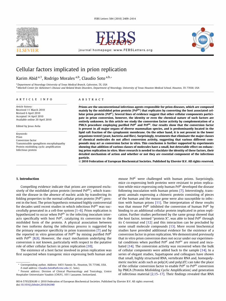
by Cha-PrPSc (Fig. 1A). Indeed, the amplification efficiency was onlyslightly lower than the one obtained when Cha-PrPSc was propa-gated at expenses of Cha-PrPC. The advantage of working withthe heterologous mixture is that any signal observed with the3F4 antibody corresponds to newly generated Sha-PrPSc and notto the inoculum added to trigger conversion.
To develop the conversion factor complementation assay, wepartially purified Sha-PrPC by affinity chromatography, as de-scribed in Methods. Analysis by silver staining and Western blotafter polyacrilamide gel electrophoresis revealed that PrPC is morethan 70% pure (Fig. 1B). Incubation of this purified Sha-PrPC withhighly purified Cha-PrPSc (using a standard procedure that resultsin > 90% purity) produced no amplification by PMCA (Fig. 1C).Strikingly, a large amount of conversion product was observedwhen the same mixture was supplemented with 10% Cha normalbrain homogenate (Fig. 1C). This result confirms our previous
Fig. 1. PMCA complementation assay to study conversion factor activity. (A) GoldenSyrian hamster normal brain homogenate (NBH) containing PrPC was incubatedwith various dilutions of Sha or Cha scrapie sick brain homogenate. Samples weresubjected to 96 PMCA cycles and either frozen (mixture without PMCA) or PMCAamplified material were analyzed by Western blot (after PK digestion) with the 3F4antibody that detects Sha PrP, but not Cha PrP. (B) PrPC was partially purified fromSha normal brain homogenate by affinity chromatography as described in methods.Both unpurified (U) and purified (P) samples were analyzed by silver staining (leftpanel) or Western blotting (right panel) using the 3F4 antibody. (C) Mixtures ofpurified Sha-PrPC and Cha-PrPSc were subjected to PMCA amplification in thepresence of conversion buffer or 10% Cha normal brain homogenate (NBH). PrPSc
signal was evaluated after PK treatment by Western blot using 3F4 antibody.
publication [14] and provides an excellent biochemical assay totest for conversion factor activity.
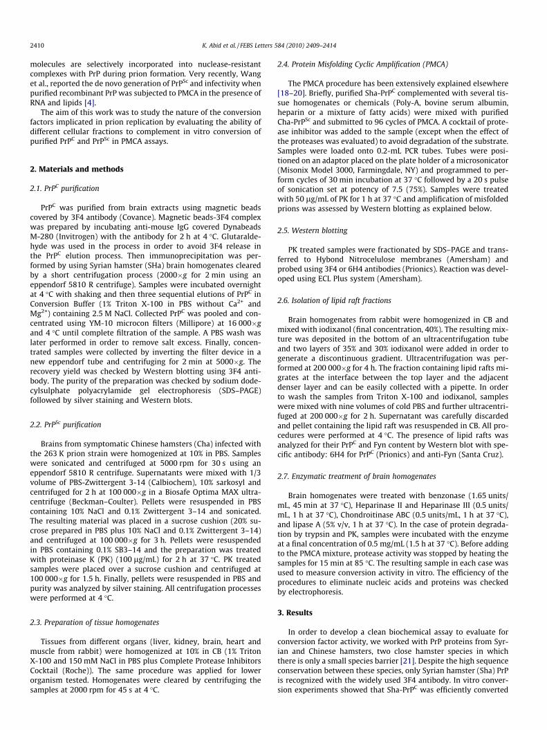
We first studied the species-specificity of the conversion factorby complementing the PMCA assay using purified Sha-PrPC andCha-PrPSc with brain homogenates from other mammal species.Brain homogenates from mice, prnp knock out mice and rabbitswere able to complement prion conversion with similar efficiencyas Cha tissue (Fig. 2A). This result indicates that the conversion fac-tor is not species-specific and is likely present in the brain of var-ious mammal species, including rabbit, an animal which is highlyresistant to prion infection [22]. However, addition of similar quan-tities of total extracts from lower organisms, including bacteria,yeast and drosophila was unable to complement prion conversion(Fig. 2B), although a faint band with the yeast extract was detectedin some experiments. These results indicate that the conversionfactor was not present in these evolutionary lower organisms.Next, we studied whether the conversion factor was present inother major organs and tissues. PMCA complementation using10% homogenates from liver, kidney, heart and muscles from rab-bit produced a similar level of prion replication as the brain tissue
ig. 2. Conversion factor activity in various tissue homogenates and extracts fromistinct species. (A) To study species-specificity of conversion factor, PMCA ofurified Sha-PrPC and Cha-PrPSc was complemented with 10% brain homogenatesom Cha, rabbit and PrP knockout mice. Buffer corresponds to the negative control
which no tissue homogenate (only buffer) was added to the sample. Mixturesere subjected to 96 PMCA cycles and formation of Sha-PrPSc was checked byestern blot after PK digestion. (B) A similar experiment as described in (A) was
one using as complement 10% (w/v) extracts of bacteria (Escherichia coli), yeastaccharomyces cerevisiae) or fly (Drosophila melanogaster). Again the efficiency of
MCA complementation was checked by Western blot using 3F4 antibody. (C) Theresence of conversion factor activity in various major organs (liver, kidney, brain,eart and muscles) was evaluated by using 10% tissue homogenates to complementMCA using purified proteins. In the blots showed in this figure all samples wereeated with PK to remove non-converted PrPC, except in the migration controlBH, No PK), which correspond to full-length Sha-PrPC.
FdpfrinwWd(SPphPtr(N
2412 K. Abid et al. / FEBS Letters 584 (2010) 2409–2414
(Fig. 2C), indicating that the conversion factor activity is not exclu-sive of the brain, but is present in other major mammalian organs.
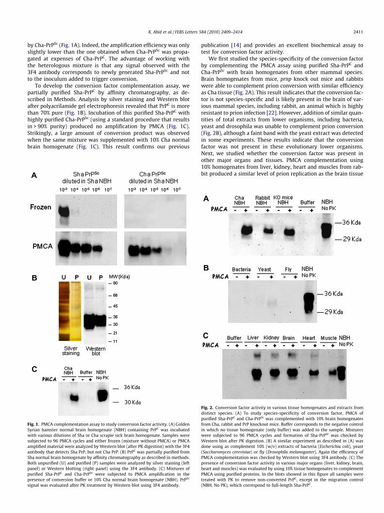
PrP is a cell surface protein located in specialized membranemicro-domains known as lipid rafts [23,24]. Since it is likely thatprion conversion takes place with PrPC bound to the membraneeither on the cell surface or in endocytic vesicles [25], we reasonedthat the conversion factor should be located in lipid rafts. To eval-uate this hypothesis we purified lipid rafts from rabbit brainhomogenate employing density gradient centrifugation, as de-scribed in Section 2. The efficiency of the lipid raft preparationmethodology was checked using the lipid raft markers Fyn andPrPC (Fig. 3A). Then each fraction of the gradient was tested forits ability to complement PMCA conversion of purified Sha-PrPC
triggered by purified Cha-PrPSc. As seen in Fig. 3B, the two fractionscorresponding to the lipid raft preparation (fractions 1 and 2)showed the highest ability to complement prion conversion. Thisresult is not dependent on the presence of PrPC in the complement-ing material, since similar results were obtained when lipid raftswere purified from prnp�/� mice brain (data not shown). Someother fractions of the gradient showed a much lower, but detect-able, conversion factor activity (e.g. fractions 3, 4, 6, 9 and 10). Con-sidering that lipid rafts correspond to a very small fraction of thetotal brain, our findings suggest that lipid rafts might be an excel-lent starting point to attempt isolating the conversion factor bybiochemical fractionation of this preparation.
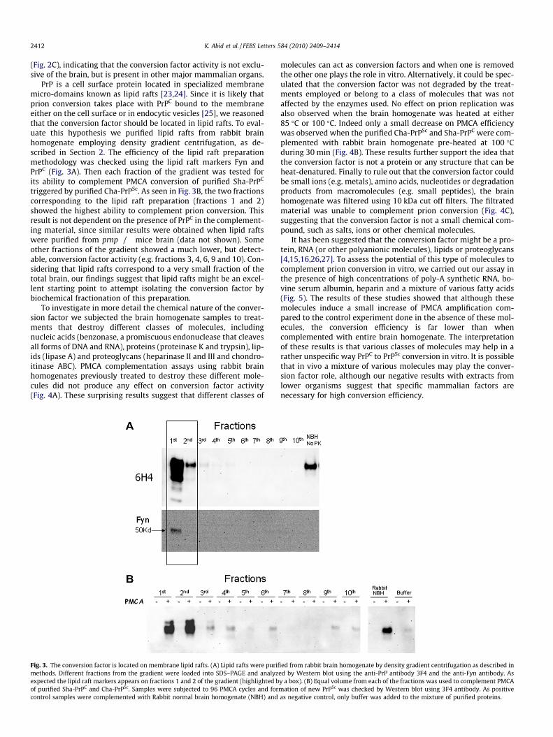
To investigate in more detail the chemical nature of the conver-sion factor we subjected the brain homogenate samples to treat-ments that destroy different classes of molecules, includingnucleic acids (benzonase, a promiscuous endonuclease that cleavesall forms of DNA and RNA), proteins (proteinase K and trypsin), lip-ids (lipase A) and proteoglycans (heparinase II and III and chondro-itinase ABC). PMCA complementation assays using rabbit brainhomogenates previously treated to destroy these different mole-cules did not produce any effect on conversion factor activity(Fig. 4A). These surprising results suggest that different classes of
Fig. 3. The conversion factor is located on membrane lipid rafts. (A) Lipid rafts were purifimethods. Different fractions from the gradient were loaded into SDS–PAGE and analyzexpected the lipid raft markers appears on fractions 1 and 2 of the gradient (highlighted bof purified Sha-PrPC and Cha-PrPSc. Samples were subjected to 96 PMCA cycles and forcontrol samples were complemented with Rabbit normal brain homogenate (NBH) and
molecules can act as conversion factors and when one is removedthe other one plays the role in vitro. Alternatively, it could be spec-ulated that the conversion factor was not degraded by the treat-ments employed or belong to a class of molecules that was notaffected by the enzymes used. No effect on prion replication wasalso observed when the brain homogenate was heated at either85 �C or 100 �C. Indeed only a small decrease on PMCA efficiencywas observed when the purified Cha-PrPSc and Sha-PrPC were com-plemented with rabbit brain homogenate pre-heated at 100 �Cduring 30 min (Fig. 4B). These results further support the idea thatthe conversion factor is not a protein or any structure that can beheat-denatured. Finally to rule out that the conversion factor couldbe small ions (e.g. metals), amino acids, nucleotides or degradationproducts from macromolecules (e.g. small peptides), the brainhomogenate was filtered using 10 kDa cut off filters. The filtratedmaterial was unable to complement prion conversion (Fig. 4C),suggesting that the conversion factor is not a small chemical com-pound, such as salts, ions or other chemical molecules.
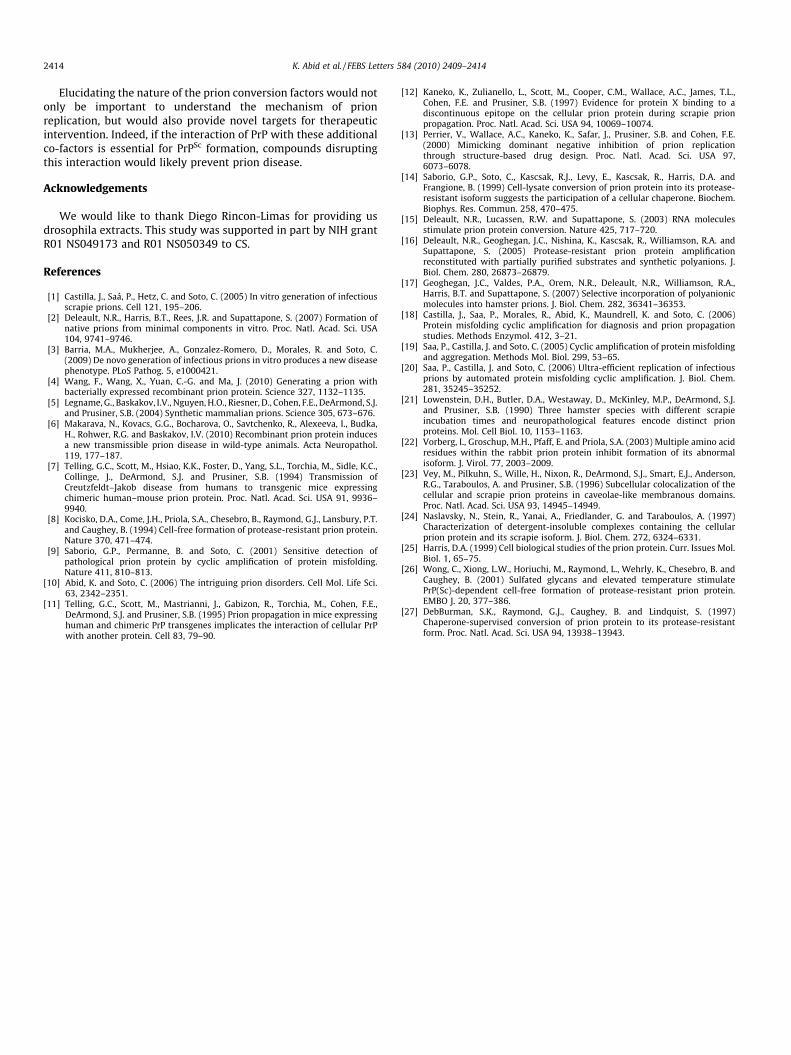
It has been suggested that the conversion factor might be a pro-tein, RNA (or other polyanionic molecules), lipids or proteoglycans[4,15,16,26,27]. To assess the potential of this type of molecules tocomplement prion conversion in vitro, we carried out our assay inthe presence of high concentrations of poly-A synthetic RNA, bo-vine serum albumin, heparin and a mixture of various fatty acids(Fig. 5). The results of these studies showed that although thesemolecules induce a small increase of PMCA amplification com-pared to the control experiment done in the absence of these mol-ecules, the conversion efficiency is far lower than whencomplemented with entire brain homogenate. The interpretationof these results is that various classes of molecules may help in arather unspecific way PrPC to PrPSc conversion in vitro. It is possiblethat in vivo a mixture of various molecules may play the conver-sion factor role, although our negative results with extracts fromlower organisms suggest that specific mammalian factors arenecessary for high conversion efficiency.
ed from rabbit brain homogenate by density gradient centrifugation as described ined by Western blot using the anti-PrP antibody 3F4 and the anti-Fyn antibody. Asy a box). (B) Equal volume from each of the fractions was used to complement PMCAmation of new PrPSc was checked by Western blot using 3F4 antibody. As positiveas negative control, only buffer was added to the mixture of purified proteins.
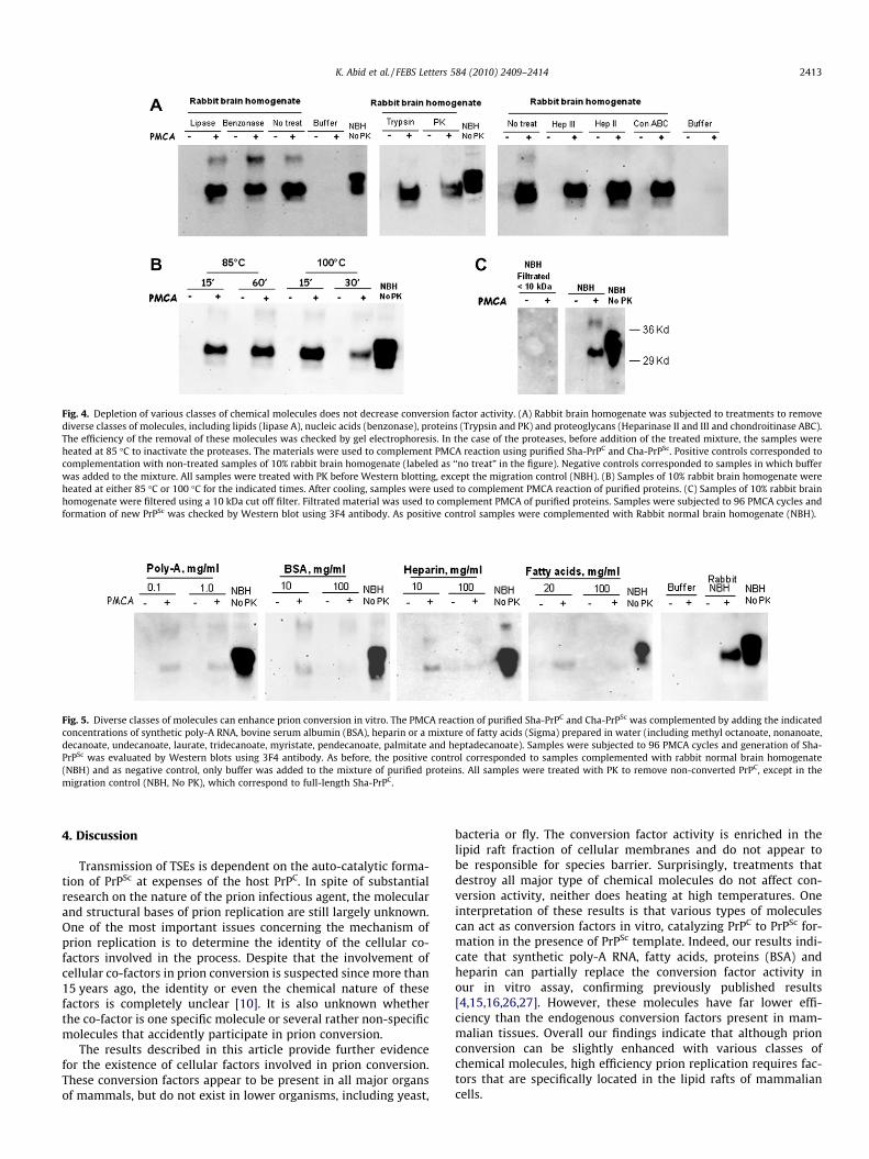
Fig. 4. Depletion of various classes of chemical molecules does not decrease conversion factor activity. (A) Rabbit brain homogenate was subjected to treatments to removediverse classes of molecules, including lipids (lipase A), nucleic acids (benzonase), proteins (Trypsin and PK) and proteoglycans (Heparinase II and III and chondroitinase ABC).The efficiency of the removal of these molecules was checked by gel electrophoresis. In the case of the proteases, before addition of the treated mixture, the samples wereheated at 85 �C to inactivate the proteases. The materials were used to complement PMCA reaction using purified Sha-PrPC and Cha-PrPSc. Positive controls corresponded tocomplementation with non-treated samples of 10% rabbit brain homogenate (labeled as ‘‘no treat” in the figure). Negative controls corresponded to samples in which bufferwas added to the mixture. All samples were treated with PK before Western blotting, except the migration control (NBH). (B) Samples of 10% rabbit brain homogenate wereheated at either 85 �C or 100 �C for the indicated times. After cooling, samples were used to complement PMCA reaction of purified proteins. (C) Samples of 10% rabbit brainhomogenate were filtered using a 10 kDa cut off filter. Filtrated material was used to complement PMCA of purified proteins. Samples were subjected to 96 PMCA cycles andformation of new PrPSc was checked by Western blot using 3F4 antibody. As positive control samples were complemented with Rabbit normal brain homogenate (NBH).
Fig. 5. Diverse classes of molecules can enhance prion conversion in vitro. The PMCA reaction of purified Sha-PrPC and Cha-PrPSc was complemented by adding the indicatedconcentrations of synthetic poly-A RNA, bovine serum albumin (BSA), heparin or a mixture of fatty acids (Sigma) prepared in water (including methyl octanoate, nonanoate,decanoate, undecanoate, laurate, tridecanoate, myristate, pendecanoate, palmitate and heptadecanoate). Samples were subjected to 96 PMCA cycles and generation of Sha-PrPSc was evaluated by Western blots using 3F4 antibody. As before, the positive control corresponded to samples complemented with rabbit normal brain homogenate(NBH) and as negative control, only buffer was added to the mixture of purified proteins. All samples were treated with PK to remove non-converted PrPC, except in themigration control (NBH, No PK), which correspond to full-length Sha-PrPC.
K. Abid et al. / FEBS Letters 584 (2010) 2409–2414 2413
4. Discussion
Transmission of TSEs is dependent on the auto-catalytic forma-tion of PrPSc at expenses of the host PrPC. In spite of substantialresearch on the nature of the prion infectious agent, the molecularand structural bases of prion replication are still largely unknown.One of the most important issues concerning the mechanism ofprion replication is to determine the identity of the cellular co-factors involved in the process. Despite that the involvement ofcellular co-factors in prion conversion is suspected since more than15 years ago, the identity or even the chemical nature of thesefactors is completely unclear [10]. It is also unknown whetherthe co-factor is one specific molecule or several rather non-specificmolecules that accidently participate in prion conversion.
The results described in this article provide further evidencefor the existence of cellular factors involved in prion conversion.These conversion factors appear to be present in all major organsof mammals, but do not exist in lower organisms, including yeast,
bacteria or fly. The conversion factor activity is enriched in thelipid raft fraction of cellular membranes and do not appear tobe responsible for species barrier. Surprisingly, treatments thatdestroy all major type of chemical molecules do not affect con-version activity, neither does heating at high temperatures. Oneinterpretation of these results is that various types of moleculescan act as conversion factors in vitro, catalyzing PrPC to PrPSc for-mation in the presence of PrPSc template. Indeed, our results indi-cate that synthetic poly-A RNA, fatty acids, proteins (BSA) andheparin can partially replace the conversion factor activity inour in vitro assay, confirming previously published results[4,15,16,26,27]. However, these molecules have far lower effi-ciency than the endogenous conversion factors present in mam-malian tissues. Overall our findings indicate that although prionconversion can be slightly enhanced with various classes ofchemical molecules, high efficiency prion replication requires fac-tors that are specifically located in the lipid rafts of mammaliancells.
2414 K. Abid et al. / FEBS Letters 584 (2010) 2409–2414
Elucidating the nature of the prion conversion factors would notonly be important to understand the mechanism of prionreplication, but would also provide novel targets for therapeuticintervention. Indeed, if the interaction of PrP with these additionalco-factors is essential for PrPSc formation, compounds disruptingthis interaction would likely prevent prion disease.
Acknowledgements
We would like to thank Diego Rincon-Limas for providing usdrosophila extracts. This study was supported in part by NIH grantR01 NS049173 and R01 NS050349 to CS.
References
[1] Castilla, J., Saá, P., Hetz, C. and Soto, C. (2005) In vitro generation of infectiousscrapie prions. Cell 121, 195–206.
[2] Deleault, N.R., Harris, B.T., Rees, J.R. and Supattapone, S. (2007) Formation ofnative prions from minimal components in vitro. Proc. Natl. Acad. Sci. USA104, 9741–9746.
[3] Barria, M.A., Mukherjee, A., Gonzalez-Romero, D., Morales, R. and Soto, C.(2009) De novo generation of infectious prions in vitro produces a new diseasephenotype. PLoS Pathog. 5, e1000421.
[4] Wang, F., Wang, X., Yuan, C.-G. and Ma, J. (2010) Generating a prion withbacterially expressed recombinant prion protein. Science 327, 1132–1135.
[5] Legname, G., Baskakov, I.V., Nguyen, H.O., Riesner, D., Cohen, F.E., DeArmond, S.J.and Prusiner, S.B. (2004) Synthetic mammalian prions. Science 305, 673–676.
[6] Makarava, N., Kovacs, G.G., Bocharova, O., Savtchenko, R., Alexeeva, I., Budka,H., Rohwer, R.G. and Baskakov, I.V. (2010) Recombinant prion protein inducesa new transmissible prion disease in wild-type animals. Acta Neuropathol.119, 177–187.
[7] Telling, G.C., Scott, M., Hsiao, K.K., Foster, D., Yang, S.L., Torchia, M., Sidle, K.C.,Collinge, J., DeArmond, S.J. and Prusiner, S.B. (1994) Transmission ofCreutzfeldt–Jakob disease from humans to transgenic mice expressingchimeric human–mouse prion protein. Proc. Natl. Acad. Sci. USA 91, 9936–9940.
[8] Kocisko, D.A., Come, J.H., Priola, S.A., Chesebro, B., Raymond, G.J., Lansbury, P.T.and Caughey, B. (1994) Cell-free formation of protease-resistant prion protein.Nature 370, 471–474.
[9] Saborio, G.P., Permanne, B. and Soto, C. (2001) Sensitive detection ofpathological prion protein by cyclic amplification of protein misfolding.Nature 411, 810–813.
[10] Abid, K. and Soto, C. (2006) The intriguing prion disorders. Cell Mol. Life Sci.63, 2342–2351.
[11] Telling, G.C., Scott, M., Mastrianni, J., Gabizon, R., Torchia, M., Cohen, F.E.,DeArmond, S.J. and Prusiner, S.B. (1995) Prion propagation in mice expressinghuman and chimeric PrP transgenes implicates the interaction of cellular PrPwith another protein. Cell 83, 79–90.
[12] Kaneko, K., Zulianello, L., Scott, M., Cooper, C.M., Wallace, A.C., James, T.L.,Cohen, F.E. and Prusiner, S.B. (1997) Evidence for protein X binding to adiscontinuous epitope on the cellular prion protein during scrapie prionpropagation. Proc. Natl. Acad. Sci. USA 94, 10069–10074.
[13] Perrier, V., Wallace, A.C., Kaneko, K., Safar, J., Prusiner, S.B. and Cohen, F.E.(2000) Mimicking dominant negative inhibition of prion replicationthrough structure-based drug design. Proc. Natl. Acad. Sci. USA 97,6073–6078.
[14] Saborio, G.P., Soto, C., Kascsak, R.J., Levy, E., Kascsak, R., Harris, D.A. andFrangione, B. (1999) Cell-lysate conversion of prion protein into its protease-resistant isoform suggests the participation of a cellular chaperone. Biochem.Biophys. Res. Commun. 258, 470–475.
[15] Deleault, N.R., Lucassen, R.W. and Supattapone, S. (2003) RNA moleculesstimulate prion protein conversion. Nature 425, 717–720.
[16] Deleault, N.R., Geoghegan, J.C., Nishina, K., Kascsak, R., Williamson, R.A. andSupattapone, S. (2005) Protease-resistant prion protein amplificationreconstituted with partially purified substrates and synthetic polyanions. J.Biol. Chem. 280, 26873–26879.
[17] Geoghegan, J.C., Valdes, P.A., Orem, N.R., Deleault, N.R., Williamson, R.A.,Harris, B.T. and Supattapone, S. (2007) Selective incorporation of polyanionicmolecules into hamster prions. J. Biol. Chem. 282, 36341–36353.
[18] Castilla, J., Saa, P., Morales, R., Abid, K., Maundrell, K. and Soto, C. (2006)Protein misfolding cyclic amplification for diagnosis and prion propagationstudies. Methods Enzymol. 412, 3–21.
[19] Saa, P., Castilla, J. and Soto, C. (2005) Cyclic amplification of protein misfoldingand aggregation. Methods Mol. Biol. 299, 53–65.
[20] Saa, P., Castilla, J. and Soto, C. (2006) Ultra-efficient replication of infectiousprions by automated protein misfolding cyclic amplification. J. Biol. Chem.281, 35245–35252.
[21] Lowenstein, D.H., Butler, D.A., Westaway, D., McKinley, M.P., DeArmond, S.J.and Prusiner, S.B. (1990) Three hamster species with different scrapieincubation times and neuropathological features encode distinct prionproteins. Mol. Cell Biol. 10, 1153–1163.
[22] Vorberg, I., Groschup, M.H., Pfaff, E. and Priola, S.A. (2003) Multiple amino acidresidues within the rabbit prion protein inhibit formation of its abnormalisoform. J. Virol. 77, 2003–2009.
[23] Vey, M., Pilkuhn, S., Wille, H., Nixon, R., DeArmond, S.J., Smart, E.J., Anderson,R.G., Taraboulos, A. and Prusiner, S.B. (1996) Subcellular colocalization of thecellular and scrapie prion proteins in caveolae-like membranous domains.Proc. Natl. Acad. Sci. USA 93, 14945–14949.
[24] Naslavsky, N., Stein, R., Yanai, A., Friedlander, G. and Taraboulos, A. (1997)Characterization of detergent-insoluble complexes containing the cellularprion protein and its scrapie isoform. J. Biol. Chem. 272, 6324–6331.
[25] Harris, D.A. (1999) Cell biological studies of the prion protein. Curr. Issues Mol.Biol. 1, 65–75.
[26] Wong, C., Xiong, L.W., Horiuchi, M., Raymond, L., Wehrly, K., Chesebro, B. andCaughey, B. (2001) Sulfated glycans and elevated temperature stimulatePrP(Sc)-dependent cell-free formation of protease-resistant prion protein.EMBO J. 20, 377–386.
[27] DebBurman, S.K., Raymond, G.J., Caughey, B. and Lindquist, S. (1997)Chaperone-supervised conversion of prion protein to its protease-resistantform. Proc. Natl. Acad. Sci. USA 94, 13938–13943.