disruption of redox homeostasis in tumor necrosis factor-induced apoptosis in a murine hepatocyte...
TRANSCRIPT

Disruption of Redox Homeostasis in Tumor NecrosisFactor-Induced Apoptosis in a Murine HepatocyteCell Line
Robert H. Pierce,* Jean S. Campbell,*Alyssa B. Stephenson,* Christopher C. Franklin,*Michelle Chaisson,* Martin Poot,*Terrance J. Kavanagh,† Peter S. Rabinovitch,*and Nelson Fausto*From the Department of Pathology,* the School of Medicine, and
the Department of Environmental Health,† the School of Public
Health, the University of Washington, Seattle, Washington
Tumor necrosis factor (TNF) is a mediator of the acutephase response in the liver and can initiate prolifer-ation and cause cell death in hepatocytes. We investi-gated the mechanisms by which TNF causes apoptosisin hepatocytes focusing on the role of oxidativestress, antioxidant defenses, and mitochondrial dam-age. The studies were conducted in cultured AML12cells, a line of differentiated murine hepatocytes. As isthe case for hepatocytes in vivo , AML12 cells were notsensitive to cell death by TNF alone, but died by apo-ptosis when exposed to TNF and a small dose ofactinomycin D (Act D). Morphological signs of apo-ptosis were not detected until 6 hours after the treat-ment and by 18 hours ;50% of the cells had died.Exposure of the cells to TNF1Act D did not blockNFkB nuclear translocation, DNA binding, or its over-all transactivation capacity. Induction of apoptosiswas characterized by oxidative stress indicated by theloss of NAD(P)H and glutathione followed by mito-chondrial damage that included loss of mitochondrialmembrane potential, inner membrane structuraldamage, and mitochondrial condensation. Thesechanges coincided with cytochrome C release and theactivation of caspases-8, -9, and -3. TNF-induced apo-ptosis was dependent on glutathione levels. In cellswith decreased levels of glutathione, TNF by itself inthe absence of transcriptional blocking acted as anapoptotic agent. Conversely, the antioxidant a-lipoicacid, that protected against the loss of glutathione incells exposed to TNF1Act D completely preventedmitochondrial damage, caspase activation, cyto-chrome C release, and apoptosis. The results demon-strate that apoptosis induced by TNF1Act D in AML12cells involves oxidative injury and mitochondrialdamage. As injury was regulated to a larger extent bythe glutathione content of the cells, we suggest thatthe combination of TNF1Act D causes apoptosis be-
cause Act D blocks the transcription of genes requiredfor antioxidant defenses. (Am J Pathol 2000,157:221–236)
Tumor necrosis factor (TNF) is a multifunctional cytokinethat binds to two cell surface receptors,1 tumor necrosisfactor receptor type 1 and type 2 (TNFR1 and TNFR2,respectively). Most of the biological effects of TNF, in-cluding cell death,2 are transduced through TNFR1, areceptor that contains a death domain near the intracel-lular C-terminus, similar to Fas (CD95 or Apo1). Cluster-ing of the death domain of trimerized TNFR1 after ligandbinding provides a docking site for TRADD (TNFR-asso-ciated death domain), an adapter protein3 that can bindsignaling molecules such as TRAF2 (TNFR-associatedfactor 2), RIP (receptor interacting protein), and FADD(Fas-associated death domain). Association of TRAF2and RIP to the receptor results in the downstream acti-vation of NFkB and AP-1, whereas FADD signaling ini-tiates caspase activation.4,5 Thus, TNFR1 signaling isbifurcated into two opposing pathways; one activatingpro-inflammatory and mitogenic or survival responseswhile the other initiates programmed cell death.
In the liver, endotoxin (lipopolysaccharide)-mediatedrelease of TNF triggers the up-regulation as well as down-regulation of acute phase response genes.6 In addition toits role in inflammatory responses, TNF has other impor-tant effects in the liver. Recent studies from this and otherlaboratories7,8 established that TNF is involved in theinitiation of liver regeneration after partial hepatectomy orchemical injury through TNFR1 signaling.9,10 In contrast,this cytokine acts as a cytotoxic agent in different types ofliver injury such as in transplantation rejection and alco-hol-induced hepatic disease,11 although the precisemechanisms of TNF effects in these complex conditionsare not entirely understood.12 It has been reported thatTNF by itself does not cause hepatocyte cell death.13
Supported by National Institutes of Health grants CA23226 and CA74131(to N. F.), AG01751 (to P. S. R.), ES07032 (to T. J. K.), and CA 75316 (toC. C. F.). R. H. P. was supported by training grant ES07032 and the IrwinM. Arias Postdoctoral Research Fellowship from the American Liver Foun-dation. M. C. was supported by training grants CA09437 and GM0720.
Accepted for publication April 10, 2000.
Address reprint requests to Nelson Fausto, M.D., Department of Pa-thology, University of Washington, K-078 Health Sciences Building, Box357705, Seattle, WA 98195. E-mail: [email protected].
American Journal of Pathology, Vol. 157, No. 1, July 2000
Copyright © American Society for Investigative Pathology
221
However, sensitization to TNF cytotoxicity can be causedby drugs that directly block gene transcription or RNAtranslation or by less specific drugs that also inhibit genetranscription such as galactosamine and a-amanitine.14
Despite the extensive studies of TNF-mediated apo-ptosis in lymphocytes and other cells, there is relativelylittle specific information regarding the cell-death path-ways activated by TNFR1 in hepatocytes. TNF causescell death by necrosis or apoptosis depending on theintensity and duration of the stimulus and the overallmetabolic status.15 In cultured hepatocytes infected withan adenovirus expressing a dominant NFkB repressor,TNF-induced apoptosis and necrosis was preceded bythe opening of high-conductance mitochondrial pores.16
Although mitochondrial alterations may have been pro-duced by reactive oxygen species (ROS), it remainscontroversial what role alterations in redox potential andcell antioxidant defenses may play in TNF-induced celldeath. In L929 cells,17,18 TNF toxicity was associated withROS production while glutathione (g-glutamylcysteinyl-glycine, GSH) acted as the main agent capable of reduc-ing ROS levels. In contrast with these findings, Leist etal13 reported that both oxidative stress and decreases inGSH occurred after morphological changes of apoptosisthat were evident in hepatocytes exposed to TNF. Fur-thermore, Xu et al19 also concluded that oxidative stressplayed no role in TNF-induced hepatocyte apoptosis be-cause prevention of the up-regulation of antioxidant re-sponses did not sensitize hepatocytes to TNF-inducedcell death.
The interpretations of experiments to test the role ofoxidant stress in TNF-mediated hepatocyte apoptosis arecomplicated by several factors. First, TNF may be capa-ble of triggering apoptosis by more than one signalingpathway depending on variables such as the metabolicstate of the cell and oxidative phosphorylation cou-pling.20 For instance in HeLa cells, TNF given in combi-nation with the protein synthesis inhibitor, emeline, acti-vated two distinct apoptotic pathways, only one of whichdepended on the early release of mitochondrial ROS.21 Inaddition, the intensity of the stimulus may determine thenature of the cellular response as excessive oxidativestress can block caspase activity by a direct effect onthese proteases. Nonhepatic cells exposed to high con-centrations of H2O2, as well as hepatocytes depleted ofGSH, were resistant to apoptosis and had no detectablecaspase-3 activity after treatment with agonist Fas anti-bodies.22 Lastly, hepatocytes may differ from other celltypes in their response to oxidative injury because theycontain very high GSH levels.23,24 It is likely that modu-lations of the cellular GSH content would have majoreffects on cell survival. All of these observations areconsistent with a general model for mammalian cell ap-optosis25,26 that proposes that there are two main exe-cution pathways: one involving mitochondrial damage(type II cells) and the other, a mitochondrial-independentpathway in which early caspase-8 activation is a majorinitiating event (type I cells). We hypothesized that be-cause of the abundance of mitochondria, high oxidativephosphorylation rates, and the high levels of GSH inhepatocytes, TNF-induced apoptosis in these cells may
be highly dependent on mitochondrial functional andstructural integrity.
In this report, we focused our analysis of the mecha-nisms mediating TNF-induced apoptosis in hepatocyteson the role of oxidative stress, antioxidant defenses, andmitochondrial damage. The studies were conducted incultured AML12 hepatocytes, which, as is the case formurine hepatocytes in vivo,13 are not sensitive to cellkilling by TNF, but die by apoptosis when exposed to TNFand in the presence of a small dose of actinomycin D (ActD). The results obtained indicate that the balance be-tween mitochondrial oxidant stress and endogenous an-tioxidant defense mechanisms involving GSH plays adeterminant role in TNF-induced apoptosis in hepato-cytes.
Materials and Methods
Tissue Culture
AML12 hepatocytes, a well-differentiated, nontrans-formed murine hepatocyte cell line derived from trans-forming growth factor-a (TGF-a) transgenic mice wereused for all experiments.27 In brief, AML12 cells weremaintained in Dulbecco’s minimal essential medium/F12(Life Technologies Inc., Grand Island, NY) with 10% fetalbovine serum (Hyclone, Logan, UT), 5 mg/ml insulin, 5mg/ml transferrin, 5 ng/ml selenium (ITS Premix, Collab-orative Biomedical Products (Bedford, MA)), 50 mg/mlgentamicin, and 0.1 mmol/L dexamethasone. Cultureswere grown at 37°C in a humidified 6% CO2 atmosphere,fed approximately every 72 hours, and passaged at ;80to 90% confluence.
Chemicals and Reagents
Act D, buthionine sulfoximine (BSO), diethyl maleate (DEM),dexamethasome, and menadione (MEN) were purchasedthrough Sigma Chemical Co. (St. Louis, MO). Acetyl-Asp(OMe)-Glu(OMe)-Val-Asp(OMe)-aminomethylcoumarin(DEVD-AMC), acetyl- Ilu(OMe)-Glu(OMe)-Thr-Asp(OMe)-aminomethylcoumarin (IETD-AMC), and acetyl-Leu(OMe)-Glu(OMe)-His-Asp(OMe)-aminomethylcoumarin (LEHD-AMC)were purchased from Biomol (Plymouth Meeting, PA)whereas benzyloxycarbonyl-Val-Ala-Asp(OMe)-fluorom-ethylketone (zVAD-FMK) and a-lipoic (a-LA) acid wereobtained from Bachem (Torrance, CA) or Calbiochem (LaJolla, CA), respectively. Mitotracker green (MTG), chlo-romethyl-x-rosamine (CMX), nonyl acridine orange(NAO), monochlorobimane (MCB), and monobromobi-mane were purchased from Molecular Probes (Eugene,OR). Recombinant murine TNF was purchased from R&DSystems (Minneapolis, MN). Antibodies to p65 (sc-372-G), and caspase-8 (Mch 5, sc-6134) were purchasedfrom Santa Cruz Biotechnology (Santa Cruz, CA),whereas anti-caspase-3 antibodies (C76920) were pur-chased from Pharmingen (San Diego, CA). Horseradishperoxidase-conjugated anti-mouse secondary antibodywas purchased from Amersham Pharmacia Biotech (NA931; Piscataway, NJ) and the horseradish peroxidase-
222 Pierce et alAJP July 2000, Vol. 157, No. 1

conjugated anti-goat secondary antibody was purchasedfrom Santa Cruz (sc-2020).
Induction of Apoptosis with TNF1Act D
AML12 cells were trypsinized, plated, and allowed toadhere and grow overnight. At 90 to 95% confluence, thecells were pretreated with either 200 nmol/L (250 ng/ml)Act D or phosphate-buffered saline (PBS) in fresh me-dium for 30 minutes followed by either 20 ng/ml TNF(dissolved in PBS containing 1.0% bovine serum albu-min, fraction V, to a stock concentration of 2.0 mg/ml) orPBS control. Treatment of cells plated at lower densitiesresulted in much more rapid kinetics of apoptosis thancultures plated at 90 to 95% confluency. In vivo inductionof hepatocyte apoptosis was done as described by Leistet al.13 For these experiments C57BL/6 mice receivedeither TNF (3.3 mg/kg) or Act D (800 mg/kg) alone or incombination. The Act D injection was given 15 minutesbefore the TNF injection and the mice were killed 3 to 6hours after the TNF injection. Livers were harvested, fixedin 10% buffered formalin, processed, and stained withhematoxylin and eosin. Apoptosis was assessed mor-phologically.
Pharmacological Activation and Inhibition of AML12Apoptosis
AML12 cells were pretreated with 100 mmol/L zVAD-FMK (3 hours pretreatment) or 1 mmol/L a-LA acid (60minutes pretreatment) followed by treatment with Act Dand then TNF as described above. To alter intracellularGSH content, AML12 cells were pretreated with BSO (1mmol/L dissolved in AML12 media) for 60 minutes toblock de novo GSH synthesis then 0.8 mmol/L DEM(made fresh in AML12 media) to acutely deplete GSH.For experiments with menadione, AML12 cells were pre-treated for 3 hours at a concentration of 100 mmol/L,followed by treatment with Act D or Act D with a-LA (1mmol/L) for 9 hours.
Electrophoretic Mobility Shift Assay (EMSA)
AML12 cells were lysed and nuclei were extracted asreported previously.28 In brief, 5 mg of nuclear proteinwere incubated at room temperature for 30 minutes with0.2 ng of 32P-end-labeled double-stranded oligonucleo-tide (NFkB binding site from the class 1 major histocom-patibility enhancer element H2K), followed by electro-phoresis through a 5% polyacrylamide Tris-glycine-ethylenediaminetetraacetic acid (EDTA) gel. Gels weredried under a vacuum and exposed overnight to KodakX-AR film (Eastman Kodak Co., Rochester, NY) at 280°Cwith an intensifying screen.
Immunoblot Analysis
AML12 cells were rinsed in PBS and lysed in a 1% TritonX-100 buffer containing 50 mmol/L Tris-HCl, pH 7.4, 50
mmol/L b-glycerophosphate, 150 mmol/L NaCl, 2 mmol/LEDTA, 1 mmol/L Na3VO4, 1 mmol/L benzamidine, 1mmol/L dithiothreitol, 10 mg/ml leupeptin, 10 mg/ml pep-statin A, 10 mg/ml aprotinin, 0.5 mmol/L 4-(2-aminoethyl)-benzenesulfonyl fluoride hydrochlorine (ICN Biomedi-cals, Irvine, CA), and 10% glycerol. Protein quantitationwas performed using Bradford reagent (Bio-Rad, Her-cules, CA) and 50 mg of total protein lysate was sub-jected to sodium dodecyl sulfate-polyacrylamide gelelectrophoresis and transferred to polyvinylidene difluo-ride (Millipore, Bedford, MA). Immunoblot analysis of nu-clear protein was performed using 10 mg of protein iso-lated as described above. Release of cytochrome C intothe cytosol was determined by immunoblot as describedby Ghibelli et al29 using a mouse monoclonal anti-cyto-chrome C antibody (PharMingen, La Jolla, CA). Mem-branes were blocked in Tris-buffered saline with 0.1%Tween 20 containing 5% milk (blotting grade; BioRad,Hercules, CA) at 4°C and incubated with primary anti-bodies at the following dilutions: p65, 1:2,000; Mch5,1:1,000; caspase-3, 1:1,000; and cytochrome C, 1:1,000,in 0.5% milk in Tris-buffered saline with 0.1% Tween 20for 1 to 2 hours. The appropriate secondary antibodieswere added for 2 to 3 hours in 0.5% milk in Tris-bufferedsaline with 0.1% Tween 20 and antigen-antibody com-plexes were detected with enhanced chemiluminescentreagents purchased from either Dupont-New EnglandNuclear (Boston, MA) or Pierce (Rockford, IL).
Transient Transfections and Luciferase Assays
AML12 cells were co-transfected with a 43 NFkB lucif-erase reporter gene30 and a CMV b-galactosidase geneusing lipofectamine (Life Technologies, Inc.) following themanufacturer’s protocol in a 2:1 ratio (1.5 mg total DNA).The transfection medium containing DNA/lipofectaminewas removed after 5 hours and cells were treated imme-diately. Cells were harvested 14 hours after transfectionand processed for luciferase and b-galactosidase as-says using the Dual Light System (Tropix, Bedford, MA),according to the manufacturer’s instructions. This systemallows for sequential determinations of luciferase andb-galactosidase on the same experimental sample.
Flow Cytometric Detection of Cellular RedoxState and Mitochondrial Alterations
Flow cytometry was performed on a Coulter Epics Elite(Coulter Electronics, Hialeah, FL) using time-resolveddual laser excitation: 15 mW 488 argon (nondelayed) and20 mW UV argon (delayed, 40 mS). Fluorescence mea-surements were done with the following dyes as previ-ously described:31–33 MCB and monobromobimane tomeasure GSH and reduced cellular thiols, respectively;CMX to measure mitochondrial membrane potential;MTG to measure mitochondrial mass; NAO to measurecardiolipin content; and dichloromethyl-x-rosamine (H2-CMXROS), a mitochondrial selective dye that becomesfluorescent when oxidized, to measure mitochondrialROS. UV excited blue autofluorescence was used to
TNF Apoptosis in Hepatocytes 223AJP July 2000, Vol. 157, No. 1

measure reduced NAD, designated NADPH to representcontributions by both NADH and NADPH. For each sam-ple, 488-nm forward light scatter to determine cell sizeand UV laser right-angle light scatter were measured.Fluorescent intensity was displayed on a logarithmicscale.
AML12 cells were cultured in 6-well tissue cultureplates. The culture supernatant was pooled with the at-tached cells, which were harvested with trypsin/EDTA(Life Technologies, Inc.). An aliquot of cells was added toa tube containing an equal volume of 23 solution of theindicated dyes in cell culture medium and incubated withoccasional mixing for 30 minutes in the tissue cultureincubator. The cells were then placed on ice in the darkand run on the flow cytometer. At least 15,000 eventswere recorded per sample.
Analysis of flow cytometric experiments was per-formed using the software package MPLUS (PhoenixFlow Systems, San Diego, CA). To help eliminate debris,a UV-side scatter versus 488-forward scatter gate deter-mined by analysis of the untreated control samples wasapplied. Analytic (nonexclusionary) gates drawn aroundthe entire population or distinct subpopulations wereused to determine the median fluorescence values andpercentage of cells associated with those groups. Iden-tical gates were used for all comparisons.
DAPI Fluorescence
Trypsinized cells were fixed in 70% ETOH, centrifuged at50 to 80 3 g for 5 minutes and resuspended in a solutioncontaining 0.5% Nonidet P-40 and 10 mg/ml DAPI. Apo-ptotic nuclear morphology was assessed as describedby Hotz et al34 on 200 cells per experimental point andeach point was determined in triplicate.
Confocal Laser Microscopy
AML12 cells were cultured on 2-well coverslip chambers(Nunc Inc., Bountiful, UT) and after treatment, the cul-tures were stained with CMXROS and MTG for 15 minutesat 37°C. Cells were scanned with an ACAS Ultima Con-focal Laser Cytometer (Meridian Instruments, Okemos,MI) and confocal images were acquired using a 603 oilobjective (NA 5 1.3). Fields were first scanned for fluo-rescence from the mitochondrial stains, and then theNAD(P)H fluorescence images were collected from thesame field. Images are displayed as composite images(red, green, blue) using Adobe Photoshop (Adobe Sys-tems, Inc., San Jose, CA).
Transmission Electron Microscopy
Cells harvested by trypsinization were fixed at roomtemperature overnight in one-half strength Karnovsky’sfixative. Cells were then rinsed in 0.1 mol/L phosphatebuffer, postfixed in a 2% buffered osmium tetroxidesolution, followed by dehydration through a gradedseries of ethanol solutions. Cells were rinsed with pro-pylene oxide and infiltrated with 1:1 mix of PolyBed
(Polysciences, Warrington, PA)/propylene oxide fol-lowed by 100% PolyBed. Cells were then embedded inPolyBed and polymerized at 60°C overnight. Sections(70 to 90 nm) were stained with saturated solutions ofuranyl acetate and lead tartrate and micrographs weretaken on a Philips 410 transmission electron micro-scope (FEI Co., Hillsboro, OR).
Detection of Caspase Activity
Caspase-8, caspase-9, and caspase-3 activities were mea-sured using the substrates, IETD-AMC, LEHD-AMC, andDEVD-AMC (Biomol), respectively. Cells were harvestedand lysed in the Triton X-100 lysis buffer described above.Protein lysate (50 mg) was incubated for 30 minutes at 37°Cin a caspase assay buffer (50 mmol/L Hepes, pH 7.4, 100mmol/L NaCl, 2 mmol/L EDTA, 10% sucrose, 0.1% CHAPS,(3-[(3-cholamidopropyl)dimethyl ammonio]-1-propanesul-fonate (Meriden, CN) 10 mmol/L dithiothreitol) containing 10mmol/L of each peptide-aminomethylcoumarin substrate.Enzymatic assays and aminomethylcoumarin standardcurves were carried out in duplicates using a fluorescentplate reader (Packard Instruments, Meriden, CT) with exci-tation and emission wavelengths of 360 nm and 460 nm,respectively. Fluorescence of the substrate blank was sub-tracted as background in each assay. Data analysis wasperformed using Packard’s I-Smart software.
Statistical Analysis
Statview 4.5 (Abacus Concepts, Berkeley, CA) was usedto perform the statistical analyses. For comparisons be-tween means of multiple groups multifactorial analysis ofvariance with a Fischer’s least significant difference wasused. To assess the interaction between variables, 2-fac-tor analysis of variance was used. The degree of corre-lation between flow cytometric parameters was assessedusing a simple regression analysis. For all tests, P , 0.05was accepted as significant.
Results
To study TNF-induced apoptosis in murine hepatocytes,we first determined whether TNF alone or in conjunctionwith Act D would cause apoptosis of mouse hepatocytesin vivo. In agreement with the data of Leist et al,13 histo-logical analysis of the liver of mice showed that neitherTNF nor Act D by themselves caused cell death but thecombination of TNF1Act D was highly apoptotic (datanot shown). In livers of mice injected with TNF1Act Dthere were large numbers of apoptotic hepatocytesshowing typical chromatin condensation in the nuclearperiphery as well as the presence of apoptotic bodiesand destruction of hepatic cords.
Similar to murine hepatocytes in vivo, AML12 hepato-cytes in cultures were also insensitive to TNF-inducedkilling but died by apoptosis after exposure to TNF1ActD. AML12 cells were divided into four groups: 1) pre-treated with Act D (200 mmol/L) for 30 minutes followed
224 Pierce et alAJP July 2000, Vol. 157, No. 1
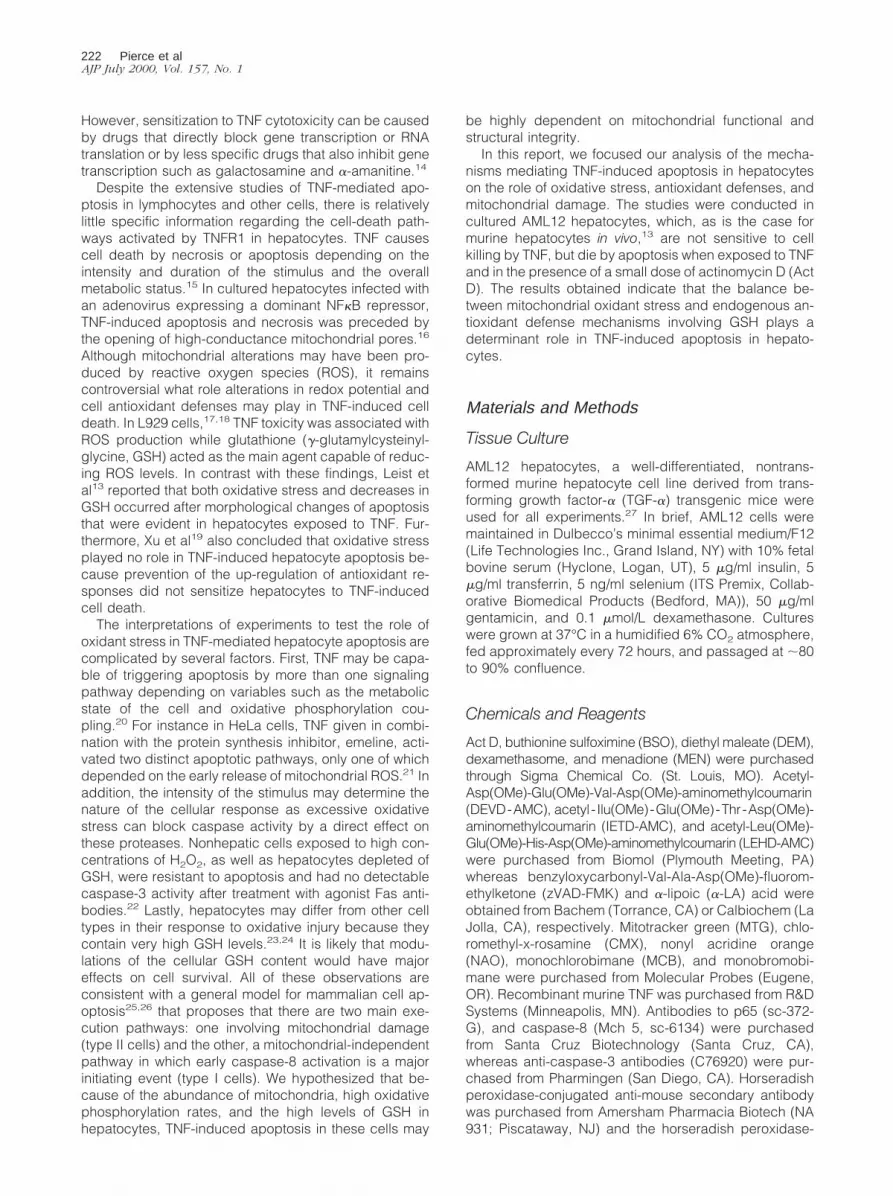
by addition of 20 ng/ml of TNF; 2) treated with TNF only;3) treated with Act D only; and 4) left untreated for theduration of the experiment. Apoptosis was assessed be-tween 3 and 24 hours by various procedures: light andelectron microscopy, DAPI staining, detection of a subG1peak by flow cytometry, and terminal dUTP nick-endlabeling assays. The results obtained by these variousmethods were consistent and only electron microscopyand DAPI fluorescence analysis are presented (Figure 1,A and B). Untreated cells or cells treated with either TNFor Act D alone exhibited no morphological signs of apo-ptosis when analyzed by electron microscopy. In markedcontrast, cells treated with TNF1Act D showed typicalchromatin condensation (Figure 1A) as well as apoptoticbodies, in a pattern similar to that observed in livers ofmice injected with the same drug combination. Micro-scopic analysis by DAPI fluorescence showed thatchanges in nuclear morphology characteristic of apopto-sis were negligible (,1%) in cells exposed to TNF aloneor left untreated. In cells exposed to TNF1Act D therewas no detectable effect on DAPI fluorescence within the
first 6 hours after exposure. However, after 24 hours ofexposure ;80% of the cells were dead with characteris-tic morphological changes of apoptosis (Figure 1B). Ap-proximately 50% of the cells exposed to TNF1Act D wereapoptotic by 18 hours whereas only 5% of the cellsshowed morphological changes after 18 hours of expo-sure to Act D alone.
NFkB Binding and Transactivation Activity afterTNF1Act D Treatment
In hepatocytes and other cell types, inhibition of NFkBactivation sensitizes cells to TNF-induced apopto-sis.16,35–39 Microinjection of IkB protein or p65(rel A)antibodies into AML12 cells as well as transfection of thecells with an IkB superrepressor gene, conditions inwhich NFkB is made functionally inactive, sensitizesAML12 hepatocytes to apoptosis after TNF treat-ment.40,41 To determine whether Act D sensitizes AML12cells to TNF-induced apoptosis by inhibiting NFkB activ-ity, p65 translocation to the nucleus and NFkB DNA bind-ing activity were analyzed by immunoblot and EMSA,respectively. TNF activation of NFkB is generally accom-panied by a transient increase of NFkB (p65/p50) in thenucleus.42 Western blot analysis presented in Figure 2Ashows an increase of nuclear p65 levels in cells treatedfor 30 minutes with either TNF alone or TNF1Act D. After2 hours there was still no difference in nuclear p65 levelsbetween these two treatment groups (data not shown). Todetermine whether TNF1Act D treatment blocked NFkBbinding, nuclear extracts were prepared from cellstreated with Act D or TNF alone or together. NFkB DNAbinding determined by EMSA was increased in cellstreated with TNF for 30 minutes. The apoptosis-inducingcombination of TNF1Act D did not inhibit NFkB bindingmeasured after 30 minutes (Figure 2B) or 2 hours (datanot shown) of TNF treatment. Signal transducers andactivators of transcription 3 (STAT3) binding also in-creased after TNF treatment in AML12 cells and thisbinding was not inhibited by Act D pretreatment (data notshown). Activation of STAT3 is linked to NFkB activitythrough interleukin-6, a NFkB target gene and a potenttransactivator of STAT3.43–45
NFkB EMSA analysis measures the translocation of theheterodimer to the nucleus and its binding to DNA. How-ever, DNA binding may not accurately reflect NFkB trans-activation activity. To determine whether NFkB transacti-vation capacity was decreased in cells pretreated withAct D, cells transiently transfected with a NFkB-luciferasereporter construct were pretreated for 30 minutes withAct D followed by a 9-hour exposure to TNF. NFkB tran-scriptional activity was unaltered in cells exposed to ActD alone whereas TNF treatment alone caused a seven-fold to eightfold increase in reporter gene activity relativeto the activity of untreated cells (Figure 2C). This increasewas not blocked in cells pretreated with Act D and sub-sequently exposed to TNF. Cells were cotransfected witha CMV driven b-galactosidase gene to serve as a controlin these experiments. b-galactosidase activity was in-creased by 50 to 70% in all treatment groups relative to
Figure 1. Apoptosis in AML12 cells exposed to TNF1Act D. Cells werepretreated with Act D (200 nmol/L) or saline (control) for 30 minutes andthen TNF (20ng/ml) or PBS was added for the indicated times. A: After 15hours of the indicated treatment, the cells were fixed and prepared fortransmission electron microscopy as described in Materials and Methods (leftpanel, TNF treated; right panel, TNF1Act D). B: Cells were fixed in 70%ETOH at the indicated time points and apoptotic nuclear morphology wasvisualized by DAPI fluorescence. The percentage of apoptotic cells wasmeasured in four treatment groups: saline and PBS (squares), Act D alone(circles), TNF alone (triangles), or TNF1Act D (diamonds). Each pointwas determined in triplicate and the error bars represent SEM. The results arerepresentative of at least five independent experiments.
TNF Apoptosis in Hepatocytes 225AJP July 2000, Vol. 157, No. 1

untreated cells. Together, these results show that expo-sure of cells to the apoptotic-inducing combination ofTNF1Act D did not block NFkB nuclear translocation orDNA binding or transactivation capacity. However, theluciferase transactivation assay may overestimate biolog-ically relevant NFkB transcriptional activity and not ac-count for the possible inhibition of transactivation of indi-vidual genes. Indeed, IkBa mRNA, the product of a genetransactivated by NFkB, was decreased after 6 hours ofexposure to TNF 1 Act D compared to TNF alone (datanot shown).
Caspase Activation during TNF-InducedApoptosis
Caspase activation is a characteristic feature of the ap-optotic process. To determine the kinetics of caspaseactivation during TNF1Act D-induced apoptosis,caspase-3, -8, and -9 activities were measured usingfluorogenic substrates after exposure of AML12 cells toTNF1Act D. No significant activation of caspase-8 and -9measured, respectively, as IETD and LEHD cleavageactivities was detected before 6 hours of treatment ofTNF1Act D (Figure 3A). Caspase-3 activity (DEVD), onthe other hand, was detectable between 3 and 6 hours.To directly investigate the activation of the caspases,immunoblot analysis was performed to detect the active,cleaved forms of caspase-8 (inset in Figure 3A) andcaspase-3 (inset in Figure 3B). The caspase-8 cleavageproduct was not detectable until 16 hours after TNF1ActD treatment and was greatly increased by 24 hours. The
Figure 2. Analysis of NFkB activation and transcriptional activity. A and B:Cells were pretreated for 30 minutes with Act D (200 nmol/L) or salinefollowed by TNF (20 ng/ml). Nuclear extracts were prepared as described inMaterials and Methods. A: Nuclear accumulation of p65 was assessed byimmunoblot analysis. Duplicate samples of nuclear extracts (10 mg) wereanalyzed for p65 protein after 30 minutes of TNF treatment. Molecular weightmarkers indicated on the left. B: EMSA for NFkB after 30 minutes of exposureto TNF were carried out as described in Materials and Methods. The upperband (NFkB) consists of the p65/p50 heterodimer; the lower band is thep50/p50 homodimer. Duplicate samples are shown. These results are repre-sentative of three independent experiments. C: NFkB activation afterTNF1Act D treatment was analyzed by a luciferase reporter gene. Cells werecotransfected with CMV b-galactosidase and NFkB-driven luciferase reporterconstructs using lipofectamine, as described in Materials and Methods. After5 hours, TNF1Act D was added for 9 hours as described in Materials andMethods. Cells were harvested and aliquots of protein lysate were assayedsequentially for both luciferase (black bars) and b-galactosidase activity(stippled bars). The bars depict SEM from six independent samples relativeto untreated cells (UNTX).
Figure 3. Activation of caspase-3, -8, and -9 in AML12 cells treated withTNF1Act D. Cells were pretreated for 30 minutes with Act D (200 nmol/L)followed by stimulation with TNF (20 ng/ml). Cellular extracts were pre-pared and protein concentrations were determined as described in Materialsand Methods. A: Caspase-8 and caspase-9 activities were determined onprotein lysates using IETD-AMC (filled triangles) and LEHD-AMC (opencircles), respectively. Inset: Immunoblot analysis on the appearance of thecleaved, active form of caspase-8 at the indicated times after treatment ofTNF1Act D. B: Caspase-3 activity was determined on protein lysates usingDEVD-AMC. Inset: Immunoblot analysis on the appearance of the cleaved,active form of caspase-3 at the indicated times after treatment with TNF1ActD. Each data point represents the average of duplicate samples. These dataare representative of three independent experiments.
226 Pierce et alAJP July 2000, Vol. 157, No. 1

cleaved, active form of caspase-3 was detected by 9hours and was maximal by 15 to 18 hours. Cells exposedto TNF or Act D as single agents had no increase in theactivity of caspases-3, -8, and -9 (data not shown). Thesedata indicate that TNF-induced caspase activation inhepatocytes is not a direct effect of the cytokine but isdetected during apoptosis caused by treatment of cellswith TNF in conjunction with Act D.
NAD(P)H and GSH Levels Decrease inHepatocytes Treated with TNF1Act D
TNF has been shown to induce mitochondrial ROS pro-duction in cultured hepatocytes as well as in the liver invivo.24 NADH and NADPH are major sources of cellularreducing equivalents which are, in part, components ofantioxidant defense mechanisms that prevent damagefrom oxidative stress. The liver is the main producer ofGSH, a tripeptide thiol, which, among other functions,scavenges free radicals and helps to maintain proteinthiol redox status.23,46 To determine whether treatment ofAML12 cells with TNF1Act D causes a change in reduc-ing equivalents, NAD(P)H and GSH were measured byflow cytometry in cells at various times after treatment.Figure 4A shows a NAD(P)H histogram comparing un-treated cells to cells exposed to TNF1Act D for 12 hours.It is clear from the histogram that the treatment caused adramatic decrease in the number of cells containing nor-mal levels of NAD(P)H. Quantification of these changes isshown in Figure 4B. This figure was constructed fromhistograms similar to that shown in Figure 4A for cellsexposed to TNF1Act D for 3 to 15 hours. A reduction inNAD(P)H levels was first detected at 3 hours and by 6hours there was a statistically significant difference inNAD(P)H between untreated cells and cells exposed toTNF1Act D. By 15 hours of treatment, NAD(P)H levelshad decreased by 50% (Figure 4B) and only 25% of thecells retained normal NAD(P)H levels (data not shown).Cellular GSH levels also decreased with kinetics similarto that of NAD(P)H in response to the TNF1Act D treat-ment (Figure 4B). Measurements of total reduced thiols inthese cells by flow cytometry using monobromobimanefluorescence (not shown) demonstrated that total re-duced thiols also decreased after TNF1Act D treatmentin a manner similar to that of GSH. This is to be expectedbecause GSH is present in large amounts in hepatocytesand is the main component of the total reduced thiolfraction of these cells.
Mitochondrial Alterations in TNF-InducedApoptosis
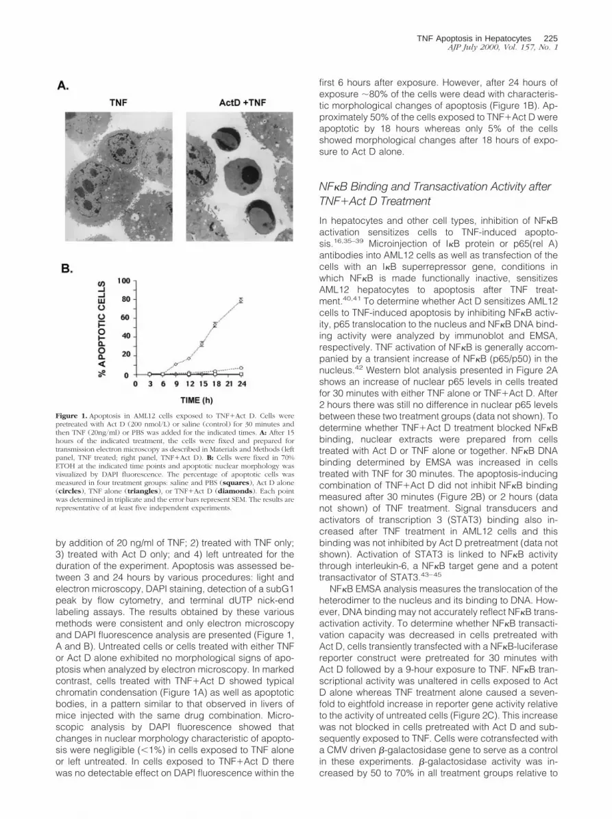
To determine whether the decrease in reducing equiva-lents caused by TNF1Act D treatment involves loss ofmitochondrial function or damage to mitochondrial struc-ture, mitochondrial transmembrane potential (Dcm) wasmeasured by flow cytometry using the fluorescent dyeCMX. A bivariate plot of the transmembrane potential andNAD(P)H shows that a large proportion of cells exposed
to TNF1Act D for 12 hours had reduced transmembranepotential and lower NAD(P)H levels compared to un-treated cells (Figure 5A). The relationship between theseparameters is linear; simple regression analysis (notshown) demonstrated that there is a highly significantcorrelation (R2 5 0.72; P , 0.0001) between the de-crease in DCm and NAD(P)H loss. A reduction in DCmwas detectable between 3 and 6 hours after exposure ofcells to TNF1Act D and steadily declined over time,dropping to ;65% of control after 15 hours of treatment(Figure 5C).
Cardiolipin is a mitochondrial inner membrane phos-pholipid which plays a major role in maintenance of mem-brane proteins including cytochrome C.47,48 Structuraldefects of cardiolipin-containing mitochondrial mem-
Figure 4. Loss of NAD(P)H and GSH levels in cells undergoing apoptosis.Cells were treated with TNF1Act D or saline (UNTX) and harvested for flowcytometry as described in Materials and Methods. A: Univariant NAD(P)Hhistogram for UNTX cells and cells treated for 12 hours with TNF1Act D.Total cellular reduced NAD (labeled as NAD(P)H to reflect contributions offrom both NADH and NADPH) was measured as UV-excited blue autofluo-rescence (450 nm). Note that median fluorescence as well as the percent ofthe cells that remain within normal range can be derived from these curves.B: Time course of the coordinate loss of NAD(P)H and GSH is shown.NAD(P)H was determined by UV-excited blue fluorescence at 450 nm. GSHwas measured by staining with MCB. Data points represent the percentage ofcells that retain normal NAD(P)H (open triangles) or GSH (open dia-monds) levels relative to the corresponding values obtained for the un-treated samples (open squares). Error bars represent SEM from three inde-pendent samples. These data are representative of at least three independentexperiments.
TNF Apoptosis in Hepatocytes 227AJP July 2000, Vol. 157, No. 1
Figure 5. Loss of mitochondrial membrane potential and mitochondrial cardiolipin in cells undergoing apoptosis. Cells were treated with TNF1Act D andharvested for flow cytometry analysis as described in Materials and Methods. A: Bivariate dot-plots of membrane potential (DCm, measured by CMX staining) andNAD(P)H from untreated (UNTX) cultures and cultures treated for 12 hours with TNF1Act D. B: Bivariate dot-plots of cardiolipin content (NAO staining) andNAD(P)H from untreated (UNTX) cultures and cultures treated for 12 hours with TNF1Act D. C and D: Time course of the progressive loss of mitochondrialmembrane potential (C) and cardiolipin content (D) after TNF1Act D exposure. The exposure time is indicated on the abscissa. Data points represent the meanof the percent of cells that retain normal mitochondrial membrane potential (C) or cardiolipin content (D). Levels were calculated relative to untreated samples(open squares). Error bars represent SEM from three independent samples. These data are representative of at least three independent experiments.
228 Pierce et alAJP July 2000, Vol. 157, No. 1

branes caused by lipid peroxidation49–51 can be as-sessed by staining with the fluorescent dye NAO. Todetermine whether cardiolipin staining was altered inTNF-induced apoptosis, cells were exposed to TNF1ActD for 12 hours and analyzed by flow cytometry for cardi-olipin and NAD(P)H (Figure 5B). A bivariate plot of car-diolipin (NAO fluorescence) and NAD(P)H demonstratedthat TNF1Act D exposure increased the proportion ofcells with reduced cardiolipin and lower NAD(P)H con-tent. There was a high degree of correlation betweenthese changes (R2 5 0.88; P . 0.0001). Loss of NAOfluorescence indicative of mitochondrial membrane dam-age progressively increased in cells exposed toTNF1Act D over time (Figure 5D) in a similar way as thechanges in Dcm.
Mancini et al52 reported that apoptosis of human coloncarcinoma cells was associated with proliferation of con-densed mitochondria with diminished transmembranepotential. These mitochondrial changes followed cell cy-cle arrest and preceded apoptosis. Electron microscopyand flow cytometry were used to determine whether sim-ilar changes occurred in AML12 cells during TNF1Act Dinducing apoptosis. AML12 hepatocytes treated for 12hours with TNF1Act D showed a decrease in mitochon-drial volume, increased electron density, and an obscur-ing of cristae structure (Figure 6A). These mitochondriaresembled mitochondrial condensation during apoptosisin other systems variably referred to as condensation,hypercondensation, or ultracondensation.52–54 Com-pared to untreated cells, TNF1Act D did not cause areadily demonstrable mitochondrial swelling (typical ofnecrosis or the later stages of apoptosis) or an increasein mitochondrial number.
The dye MTG can be used to quantitate total cellularmitochondrial mass.55 A bivariate plot of MTG fluores-cence and NAD(P)H in untreated cells and in cells ex-posed to TNF1Act D is shown in Figure 6B. Most of theuntreated cells fit into a population with high NAD(P)Hcontent and variable MTG fluorescence. However in cellstreated with TNF1Act D, a small proportion of the cellshad lower NAD(P)H levels and high MTG fluorescence.This population of cells increased by twofold to threefoldafter 12 hours of treatment. A vertical line in Figure 6Bwas drawn as a point of reference defining the lower limitof NAD(P)H levels in the main population of untreatedcells. The electron micrographs taken together with theincrease in MTG staining suggest that after TNF1Act Dexposure, the mass of individual mitochondria increased,without apparent change to the total number of mitochon-dria.
We next determined whether TNF1Act D stimulatedthe release of cytochrome C into the cytosolic fraction ofAML12 cells. In untreated cells, very little cytosolic cyto-chrome C was detected by immunoblot (Figure 6C). After6 hours of TNF1Act D treatment, cytochrome C wasdetected in the cytosol and increased up to 18 hours. Therelease of mitochondrial cytochrome C was also detectedby immunofluorescent staining of treated and fixedAML12 cells with an anti-cytochrome C antibody (datanot shown). The time course of cytochrome C releasefrom mitochondria is consistent with the timing of
caspase-9 activation (Figure 3A), thought to be depen-dent on release of cytochrome C.
Confocal imaging of NAD(P)H (Figure 7A), Dcm (Fig-ure 7B) and mitochondrial mass (Figure 7C) of living cellscorroborated the flow cytometric data shown above. Asingle field is displayed in Figure 7, A–C, and the threefields are merged in Figure 7D, to illustrate cells in thevarious stages of apoptosis after TNF1Act D treatmentfor 12 hours. Nonapoptotic cells are indicated by a star.Early apoptotic cells (reduced mitochondrial Dcm but noapparent morphological changes) are indicated by ar-rows. The arrowheads point to two cells with morpholog-ical changes of apoptosis. Nonapoptotic cells (star), re-tained high levels of reducing equivalents (purple, Figure7A) and high mitochondrial membrane potential (orange,Figure 7B) with no change in mitochondrial mass (Figure7C) resulting in magenta-colored cells in the mergedimage (Figure 7D). Three early apoptotic cells, indicatedby arrows, had low NAD(P)H (Figure 7A) and low Dcm(Figure 7B) but no detectable increase in mitochondrialmass (Figure 7C). In the merged image these cells areseen as orange (Figure 7D). Frankly apoptotic cells arebright green in the merged image (Figure 7D) whichreflects the loss of NAD(P)H (Figure 7A), low Dcm (Figure7B), and increased in mitochondrial mass (Figure 7C).The increased mitochondrial mass coincided with knownmorphological changes, such as membrane blebbing,which is seen in the green cell in the lower right handcorner.
GSH Depletion Increases ROS Accumulationand Sensitizes Hepatocytes to TNF-InducedApoptosis
The results shown so far suggest that either increases inmitochondrial generated oxidant stress or loss of cellularantioxidant defense mechanisms (or both) plays a keyrole in hepatocyte apoptosis. To determine whether ROSwere detectable in TNF-treated cells, we used flow cyto-metric assessment of H2CMX, a nonfluorescent com-pound which becomes fluorescent when oxidized. InAML12 cells exposed to TNF, no changes in ROS levelswere detected (Figure 8A). If, however, cells were pre-treated with DEM to acutely deplete cellular GSH levelsand then exposed to TNF, the combined treatment re-sulted in a 70% decrease in GSH levels and a 55%increase in ROS levels (Figure 8A). DEM treatment alonedecreased GSH by more than 50% with no increase inROS. The data demonstrate that ROS accumulate in TNF-treated hepatocytes only after partial depletion of GSH.
Given these results, we examined whether TNF as asingle agent would have an apoptotic effect in cells withlow levels of cellular GSH. For these experiments AML12hepatocytes were treated with DEM to acutely depleteGSH as well as with BSO, an inhibitor of g-glutamyl cys-teine synthetase (g-GCS) activity, to block de novo GSHsynthesis.56 As expected, exposure of the cells to TNFalone caused no apoptosis. In contrast, ;45% of cellswere apoptotic after combined exposure to TNF, BSO,and DEM (Figure 8B) demonstrating that TNF, which
TNF Apoptosis in Hepatocytes 229AJP July 2000, Vol. 157, No. 1

does not normally cause cell death in hepatocytes, is anapoptotic agent for hepatocytes with low GSH content.Treatment of cells with both BSO and DEM in the ab-sence of TNF also increased apoptosis in AML12 cells(Figure 8B), demonstrating that a basal level of oxidative
stress may be present in these cells which becomesapparent if GSH is depleted.
a-LA is a potent antioxidant capable of protecting GSHlevels in injured cells.57 a-LA functions through multipleactivities including direct reduction of oxidized glutathi-
Figure 6. Changes in mitochondrial mass and release of cytochrome C in cells undergoing apoptosis. Cells were treated with TNF1Act D and harvested at 12hours for transmission electron microscopy, flow cytometry, and immunoblot analysis as described in Materials and Methods. A: Mitochondrial condensation asdetected by transmission electron microscopy. Electron micrographs from UNTX- and TNF1Act D-treated cells (312,000). The arrow indicates normalmitochondria with visible cristae and normal matrix density in UNTX samples. In the treated samples, the arrow indicates condensed mitochondria with increasedelectron density that obscures the cristae structure. B: Bivariate dot-plots of mitochondrial protein (MTG staining) and NAD(P)H from untreated (UNTX) culturesor cultures exposed to TNF1Act D. C: Time course of the release of cytosolic cytochrome C after treatment with TNF1Act D. Molecular weight markers areindicated on the left.
230 Pierce et alAJP July 2000, Vol. 157, No. 1

Figure 7. Confocal imaging of AML12 cells undergoing apoptosis. Cells were grown on coverslips and NAD(P)H fluorescence (A), mitochondrial membranepotential (CMX staining) (B), and mitochondrial mass (MTG) (C) were examined as described in Materials and Methods. A single field is shown in A–C and theimages were merged in (D). The field displayed represents the spectrum of changes observed from early to late apoptosis: apparently normal cells (star); cellswith decrease NAD(P)H but normal DCm (arrows); frankly apoptotic cells (arrowheads).
TNF Apoptosis in Hepatocytes 231AJP July 2000, Vol. 157, No. 1

one, scavenging ROS, chelating metals, reducing oxi-dized vitamin E and C, as well as stimulating de novosynthesis of GSH by maximizing cysteine flow to maintainoptimal activity of gGCS, the rate limiting enzymatic stepin GSH biosynthesis.58,59 Figure 9A shows that a-LA notonly prevented the decrease in GSH caused by TNF1ActD treatment, but actually increased the GSH content of
these cells. These results indicate a-LA may be effectivein blocking cell injury caused by a known oxidative agent.MEN is an oxidant that acts by generating mitochondrialROS.60 Mitochondrial injury (as assessed by flow cyto-metric determination of cardiolipin) was detected in cellspretreated with Act D and exposed to MEN for 3 hours.
Figure 8. ROS detection and TNF-induced apoptosis in cells with low GSHcontent. A: TNF-induced production of ROS in hepatocytes with low GSH.Cultures were untreated, treated with DEM (0.8 mmol/L), TNF (20 ng/ml), orboth DEM and TNF. DEM exposure was for 60 minutes before TNF addition.Cells were harvested 18 hours later for flow cytometry using MCB staining(for GSH) and H2-CMXROS (for measuring ROS). The data are expressedrelative to values of untreated samples (UNTX). Error bars represent SEM oftriplicate samples. B: TNF promotes apoptosis in cells with low GSH. Cul-tures were left untreated (UNTX) or were treated with TNF (20 ng/ml), BSO(1 mmol/L) 1 DEM (0.8 mmol/L), or with a combination ofBSO1DEM1TNF. Exposure to BSO and DEM started 60 minutes and 30minutes, respectively, before TNF treatment. Cells were harvested 19 hourslater, fixed in 70% ethanol after trypsinization, and stained with DAPI forfluorescence microscopy analysis. Error bars represent SEM from triplicatesamples. Data shown are representative of at least three independent exper-iments.
Figure 9. a-Lipoic acid increases GSH levels and prevents injury from oxi-dative agents. A: a-LA blocks the loss of GSH induced by TNF1Act D. Cellswere treated with TNF, TNF1Act D, or pretreated with 1 mmol/L a-LA for 60minutes before the addition of TNF (20 ng/ml) plus Act D (200 nmol/L). Aftera 12-hour incubation, cells were harvested and cellular GSH content wasassessed by flow cytometry using MCB staining as described in Materials andMethods. Error bars represent SEM from triplicate samples. B: Act D poten-tiates cardiolipin loss. Cells were pretreated with Act D (200 nmol/L) alone ortogether with a-LA (1 mmol/L) for 9 hours. MEN (100 mmol/L) was thenadded for 3 hours. Cells were harvested and mitochondrial cardiolipin wasassessed by flow cytometry using NAO staining as described in Materials andMethods. The error bars represent SEM of triplicate samples relative tomenadione-treated cells. MEN treatment resulted in minimal loss of cardio-lipin. Data shown are representative of at least three independent experi-ments.
232 Pierce et alAJP July 2000, Vol. 157, No. 1

These cells had cardiolipin levels that were ;25% lowerthan those cells exposed to MEN alone (Figure 9B) buttreatment of cultures with a-LA completely prevented thisdecrease. These results demonstrate that a-LA can blockthe damaging effects caused by mitochondrial-gener-ated ROS. Moreover, they imply that injury may resultfrom blockage by Act D of cellular antioxidant defensesinvolving GSH homeostasis in cells pretreated with Act Dand then exposed to either TNF or MEN.
a-LA Acid and a Caspase Inhibitor PreventMitochondrial Damage and TNF-InducedApoptosis
Data presented above demonstrated that a-LA maintainsGSH levels in cells treated with TNF1Act D and preventsmitochondrial damage caused by the oxidizing drugMEN. We then determined whether a-LA would preventapoptosis in AML12 cells exposed to TNF1Act D. Thegeneral caspase inhibitor, zVAD-FMK that has previouslybeen shown to inhibit apoptosis in many cell types, wasused for comparison. Although 60% of cells were apo-ptotic as measured by DAPI staining after 15 hours ofexposure to TNF1Act D, apoptosis was detectable inonly 5% of cells pretreated with zVAD-FMK (Figure 10A).Remarkably, a-LA was as effective as the caspase inhib-itor in blocking apoptosis in cells treated with TNF1Act D(Figure 10A). Both zVAD and a-LA also prevented theloss of cardiolipin in cells treated with TNF1Act D (Figure10B). Moreover, a-LA inhibited the activation ofcaspase-3 activity (Figure 10C). Caspase-8 and -9 activ-ities were also inhibited by a-LA 12 hours after treatmentwith TNF1Act D (data not shown). The inset in Figure 10Cshows that a-LA inhibited the release of cytochrome C tothe cytosol. Thus the antioxidant a-LA prevents mitochon-drial damage, caspase activation, and apoptosis inhepatocytes exposed to TNF1Act D reinforcing the no-tion that oxidative stress is essential for the developmentof apoptosis in this system. It is of interest to note thata-LA inhibits TNF-induced NFkB activation57 and on thebasis of this effect might have been expected to enhancerather than prevent apoptosis.
Discussion
TNF can initiate apoptosis, cell proliferation, and theacute phase response in the liver. The goal of this workwas to investigate the mechanisms by which TNF causesapoptosis in hepatocytes. AML12 hepatocytes are resis-tant to TNF-induced cell death, but can be sensitized toundergo apoptosis if exposed to TNF in conjunction withAct D, as is the case for murine hepatocytes in vivo.Treatment with TNF1Act D caused oxidative stress asdemonstrated by a decrease in NAD(P)H and GSH. Theextent of apoptosis in AML12 hepatocytes was exquis-itely sensitive to GSH levels as depletion of cellular GSHsensitized cells to TNF-induced apoptosis in the absenceof a transcriptional blocking agent. TNF1Act D also in-duced mitochondrial damage, as shown by decreased
transmembrane potential, by losses in cardiolipin con-tent, and by increased mitochondrial condensation andmass.
A salient aspect of this work is the demonstration thatTNF by itself can cause apoptosis in GSH-depletedAML12 hepatocytes, a finding that highlights two impor-tant observations regarding the mechanisms of TNF-in-duced apoptosis in hepatocytes. First, oxidative stressand mitochondrial damage play prominent roles in thisprocess and, second, the apoptotic effects of TNF can toa large extent be modulated by the thiol content of thecells. Hepatocytes differ from other cell types such asfibroblasts and lymphocytes, which have been used inmany studies on the mechanisms mediating TNF-in-duced apoptosis, by their very high GSH content, thecapacity of the liver to systemically export GSH as well as
Figure 10. a-LA and a caspase inhibitor block TNF-induced mitochondrialinjury and apoptosis. Cells were pretreated with zVAD-FMK (100 mmol/L) for3 hours or a-LA (1 mmol/L) for 1 hour before the addition of TNF1Act D asdescribed in Materials and Methods. A: Both a-LA and zVAD-FMK blockapoptosis induced by TNF1Act D treatment. Cells were harvested 18 hoursafter the addition of TNF, fixed in 70% ETOH, and stained with DAPI asdescribed in Materials and Methods. Error bars represent SEM of triplicateplates. UNTX, black bars and TNF1Act D treated, stippled bars. B: Botha-LA and zVAD-FMK block loss of cardiolipin induced by TNF1Act Dtreatment. Cells were harvested 12 hours after the addition of TNF andanalyzed by flow cytometry using NAO staining as described in Materials andMethods. Error bars represent SEM of triplicate plates. UNTX, black bars andTNF1Act D treated, stippled bars. Values are normalized to UNTX samples.C: Both a-LA and zVAD-FMK block caspase-3 activity induced by TNF1ActD treatment. Cells were harvested 15 hours after the addition of TNF andanalyzed for caspase-3 activity as described in Materials and Methods. Errorbars represent SEM of triplicate plates. Values are normalized to untreatedsamples. Data shown are representative of at least three independent exper-iments. Inset: TNF1Act D-induced cytosolic cytochrome C release isblocked by a-LA. Cells were harvested 12 hours after the addition of TNF andrelease of cytosolic cytochrome C was detected by immunoblot analysis asdescribed in Materials and Methods. Protein from untreated cells, cells treatedwith TNF1Act D for 12 hours, and cells pretreated with a-LA and then treatedwith TNF1Act D for 12 hours are shown in lanes 1, 2, and 3, respectively.
TNF Apoptosis in Hepatocytes 233AJP July 2000, Vol. 157, No. 1

the activity of catalase.23,24 These systems provide hepa-tocytes with the ability to rapidly and efficiently neutralizethe increased ROS produced by TNF. Given the role ofGSH as an antioxidant in hepatocytes, it is not entirelysurprising that a decrease in the levels of GSH wouldhave a profound effect on apoptosis initiated by oxidantstress. Nevertheless this issue is controversial becausesome ROS scavenging agents do not effectively blockTNF-induced apoptosis in certain cell systems. Free rad-ical scavengers may vary in their protective effect onROS presumably because of the inability of some agentsto partition into the hydrophobic area in which ROS areproduced.2
The involvement of oxidative stress in mediating TNF-induced apoptosis is further highlighted by the ability ofthe potent antioxidant, a-LA, to prevent the loss of GSH incells exposed to TNF1Act D and to completely blockcaspase activation, mitochondrial damage, cytochromeC release, and apoptosis. Conversely, GSH depletionand inhibition of its de novo GSH synthesis sensitized thecells to the cytotoxic effects of TNF given as a singleagent. In some cells in culture, increased efflux of GSHhas been shown to alter the redox state of the cell withoutprimary involvement of ROS.61,62 In AML12 hepatocytes,however, the protective effect of a-LA acid suggests thatincreased ROS drives the consumption of multiple reduc-ing species (NAD(P)H, total cellular thiols, and GSH). ActD sensitization to TNF-mediated cytotoxicity was not spe-cific for this cytokine because Act D also sensitizedAML12 cells to the mitochondrial damage caused by theredox cycling agent MEN. Thus, one mechanism bywhich Act D may sensitize hepatocytes to apoptosis isthe blocking of transcription of genes involved in oxidantdefenses. A potential target of this effect is gGCS tran-scription, which in preliminary experiments seems to bedecreased after TNF1Act D treatment (data not shown).Glutathione decrease has been considered to be a con-tributing factor in TNF-induced cell death.63–65 Mehlen etal63 showed that proteins such as heat shock protein 27(hsp27) and alphaB-crystallin provide protection againstTNF-induced cell death by raising intracellular GSH.More recently Manna et al66 demonstrated that overex-pression of gGCS protects H4–11E hepatoma cells fromTNF-induced cytotoxicity.
Condensed mitochondria with increased folding of theinner membrane and low energy states have been de-tected at an early stage of mitochondrial injury duringapoptosis.53,54,67 In Colo-205 human colon carcinomacells, herbimycin-induced apoptosis resulted in con-densed mitochondria in which the inner and outer mem-brane are closely associated and increase in numberpossibly as a compensatory mechanism.52 However,these condensed mitochondria have low membrane po-tential, thus, their proliferation actually contributes to in-jury. The morphology of condensed mitochondria inAML12 cells was similar to that described in Colo-205apoptosis induced by herbimycin.52
TNF binding to its receptors results in the recruitmentof adapter molecules including TRADD and FADD. En-gagement of FADD with the receptor complex leads tocaspase-8 activation.1,5 In apoptotic pathways that pri-
marily depend on mitochondrial damage and cyto-chrome-C release (type II cells), there is a weak, earlyactivation of caspase-8, as well as a late activation be-lieved to be a consequence of caspase-3 and -9 activi-ty.25,26 The latter condition was observed in our experi-ments, emphasizing the important role of mitochondria inTNF-induced hepatocyte apoptosis. However, it is diffi-cult to establish a precise sequence of events in AML12hepatocyte apoptosis because cell death occurred as anonsynchronized process within a given cell population.Mitochondrial permeability transition, a good marker forapoptotic events, occurs gradually in TNF-induced apo-ptosis, taking several hours to affect all mitochondria of asingle hepatocyte.16 Although our data suggest that de-creased NAD(P)H precedes caspase-3 activation, it ispossible that some activation of caspase-3 and -8 mayoccur before the detection of mitochondrial damage. Thisis suggested by experiments in which caspase inhibitorsblocked not only apoptosis but also the loss of cardiolipinin cells exposed to TNF1Act D. These results imply thatearly caspase activity during apoptosis in this systemmay indirectly alter redox homeostatic mechanisms bydamaging antioxidant defenses. On the other hand,Bradham et al16 reported that in rat hepatocytes infectedwith an adenovirus expressing an IkBa superrepressor,mitochondrial permeability transition in TNF-induced ap-optosis is upstream of caspase-3 but downstream ofFADD. However, activation of other caspases was notexamined in these studies.
It is well known that blockage of NFkB transcriptionalactivity sensitizes cells to apoptosis induced by TNF.14
Likewise, it has been suggested that Act D sensitizescells to TNF-induced apoptosis by inhibiting the expres-sion of anti-apoptotic genes including those regulated byNFkB.19 However, in AML12 hepatocytes Act D had noeffect on TNF-induced NFkB nuclear translocation, DNAEMSA, or transcriptional activity determined by reportergene assay. However, transcripts of several NFkB targetgenes were decreased after Act D treatment, such asIkBa and gGCS (data not shown). The possible contribu-tion of NFkB dependent and independent genes to theapoptotic process in AML12 cells is under study.
In summary, we demonstrated that oxidative stressinduced by TNF does not produce injury in normal hepa-tocytes, but causes apoptosis in hepatocytes in whichantioxidant defenses are compromised, by Act D or GSHdeficiency. Conversely, the antioxidant, a-LA, which pre-vented the GSH decrease after exposure of cells toTNF1Act D, blocked mitochondrial injury and apoptosis.We conclude that in TNF-induced hepatocyte apoptosis,mitochondrial oxidative injury is a major determinant ofcell injury.
Acknowledgments
The assistance of Colin Pritchard, Lisa Prichard, NorbertDeschner, and Priscilla Thurber, and Dr. D. Hockenbery’scomments are gratefully acknowledged. We thank themembers of the Fausto laboratory for active discussions
234 Pierce et alAJP July 2000, Vol. 157, No. 1

and critiques and Matthew Mueller for his help in prepar-ing the manuscript.
References
1. Ashkenazi A, Dixit VM: Death receptors: signaling and modulation.Science 1998, 281:1305–1308
2. Wallach D, Boldin M, Varfolomeev E, Beyaert R, Vandenabeele P,Fiers W: Cell death induction by receptors of the TNF family: towardsa molecular understanding. FEBS Lett 1997, 410:96–106
3. Arch RH, Gedrich RW, Thompson CB: Tumor necrosis factor recep-tor-associated factors (TRAFs): a family of adapter proteins that reg-ulates life and death. Genes Dev 1998, 12:2821–2830
4. Ghosh S, May MJ, Kopp EB. NFkB and Rel proteins: evolutionarilyconserved mediators of immune responses. Annu Rev Immunol 1998,16:225–260
5. Hsu H, Xiong J, Goeddel D: The TNF receptor 1-associated proteinTRADD signals cell death and NFkB activation. Cell 1995, 81:495–504
6. Heinrich P, Behrmann I, Graeve L, Grotzinger J, Haan S, Horn F,Horsten U, Kerr I, May P, Muller-Newen G, Schaper F, Terstegen L,Thiel S: The acute-phase response of the liver: molecular mechanismof IL-6 signalling from the plasma membrane to the nucleus. Signal-ling in the Liver. Edited by D Haussinger, P Heinrich. Dordrecht,Kluwer Academic Publishers, 1998, pp 55–71
7. Akerman P, Cote P, Yang SQ, McClain C, Nelson S, Bagby GJ, DiehlAM: Antibodies to tumor necrosis factor-alpha inhibit liver regenera-tion after partial hepatectomy. Am J Physiol 1992, 263:G579–G585
8. Yamada Y, Kirillova I, Peschon JJ, Fausto N: Initiation of liver growthby tumor necrosis factor: deficient liver regeneration in mice lackingtype I tumor necrosis factor receptor. Proc Natl Acad Sci USA 1997,94:1441–1446
9. Yamada Y, Fausto N: Deficient liver regeneration after carbon tetra-chloride injury in mice lacking type 1 but not type 2 tumor necrosisfactor receptor. Am J Pathol 1998, 152:1577–1589
10. Yamada Y, Webber EM, Kirillova I, Peschon JJ, Fausto N: Analysis ofliver regeneration in mice lacking type 1 or type 2 tumor necrosisfactor receptor: requirement for type 1 but not type 2 receptor.Hepatology 1998, 28:959–970
11. Colell A, Garcia-Ruiz C, Miranda M, Ardite E, Mari M, Morales A,Corrales F, Kaplowitz N, Fernandez-Checa JC: Selective glutathionedepletion of mitochondria by ethanol sensitizes hepatocytes to tumornecrosis factor. Gastroenterology 1998, 115:1541–1551
12. Faubion WA, Gores GJ: Death receptors in liver biology and patho-biology. Hepatology 1999, 29:1–4
13. Leist M, Gantner F, Bohlinger I, Germann PG, Tiegs G, Wendel A:Murine hepatocyte apoptosis induced in vitro and in vivo by TNF-alpha requires transcriptional arrest. J Immunol 1994, 153:1778–1788
14. Leist M, Gantner F, Kunstle G, Wendel A: Cytokine-mediated hepaticapoptosis. Rev Physiol Biochem Pharmacol 1998, 133:109–155
15. Lemasters JJ, Nieminen AL, Qian T, Trost LC, Elmore SP, NishimuraY, Crowe RA, Cascio WE, Bradham CA, Brenner DA, Herman B: Themitochondrial permeability transition in cell death: a common mech-anism in necrosis, apoptosis and autophagy. Biochim Biophys Acta1998, 1366:177–196
16. Bradham CA, Qian T, Streetz K, Trautwein C, Brenner DA, LemastersJJ: The mitochondrial permeability transition is required for tumornecrosis factor alpha-mediated apoptosis and cytochrome c release.Mol Cell Biol 1998, 18:6353–6364
17. Goossens V, Grooten J, De Vos K, Fiers W: Direct evidence for tumornecrosis factor-induced mitochondrial reactive oxygen intermediatesand their involvement in cytotoxicity. Proc Natl Acad Sci USA 1995,92:8115–8119
18. Schulze-Osthoff K, Bakker AC, Vanhaesebroeck B, Beyaert R, JacobWA, Fiers W: Cytotoxic activity of tumor necrosis factor is mediated byearly damage of mitochondrial functions. Evidence for the involve-ment of mitochondrial radical generation. J Biol Chem 1992, 267:5317–5323
19. Xu Y, Bialik S, Jones BE, Iimuro Y, Kitsis RN, Srinivasan A, BrennerDA, Czaja MJ: NF-kappaB inactivation converts a hepatocyte cell line
TNF-alpha response from proliferation to apoptosis. Am J Physiol1998, 275:C1058–C1066
20. Qian T, Herman B, Lemasters JJ: The mitochondrial permeabilitytransition mediates both necrotic and apoptotic death of hepatocytesexposed to Br-A23187. Toxicol Appl Pharmacol 1999, 154:117–125
21. Sidoti-de Fraisse C, Rincheval V, Risler Y, Mignotte B, Vayssiere JL:TNF-alpha activates at least two apoptotic signaling cascades. On-cogene 1998, 17:1639–1651
22. Hampton MB, Orrenius S: Dual regulation of caspase activity byhydrogen peroxide: implications for apoptosis. FEBS Lett 1997, 414:552–556
23. Lu SC: Regulation of hepatic glutathione synthesis. Semin Liver Dis1998, 18:331–343
24. Fernandez-Checa JC, Kaplowitz N, Garcia-Ruiz C, Colell A: Mito-chondrial glutathione: importance and transport. Semin Liver Dis1998, 18:389–401
25. Scaffidi C, Fulda S, Srinivasan A, Friesen C, Li F, Tomaselli KJ,Debatin KM, Krammer PH, Peter ME: Two CD95 (APO-1/Fas) signal-ing pathways. Embo J 1998, 17:1675–1687
26. Gross G, McDonnell JM, Korsmeyer SJ: BCL-2 family members andthe mitochondria in apoptosis. Genes Dev 1999, 13:1899–1911
27. Wu JC, Merlino G, Fausto N: Establishment and characterization ofdifferentiated, nontransformed hepatocyte cell lines derived frommice transgenic for transforming growth factor alpha. Proc Natl AcadSci USA 1994, 91:674–678
28. FitzGerald MJ, Webber EM, Donovan JR, Fausto N: Rapid DNAbinding by nuclear factor kappa B in hepatocytes at the start of liverregeneration. Cell Growth Differ 1995, 6:417–427
29. Ghibelli L, Coppola S, Fanelli C, Rotilio G, Civitareale P, Scovassi AI,Ciriolo MR: Glutathione depletion causes cytochrome c release evenin the absence of cell commitment to apoptosis. FASEB J 1999,13:2031–2036
30. Berberich I, Shu GL, Clark EA. Cross-linking CD40 on B cells rapidlyactivates nuclear factor-kappa B: J Immunol 1994, 153:4357–4366
31. Poot M, Pierce RH: Detection of changes in mitochondrial functionduring apoptosis by simultaneous staining with multiple fluorescentdyes and correlated multiparameter flow cytometry. Cytometry 1999,35:311–317
32. Poot M, Verkerk A, Koster JF, Jongkind JF: De novo synthesis ofglutathione in human fibroblasts during in vitro ageing and in somemetabolic diseases as measured by a flow cytometric method. Bio-chim Biophys Acta 1986, 883:580–584
33. Hedley DW, Chow S: Evaluation of methods for measuring cellularglutathione content using flow cytometry. Cytometry 1994, 15:349–358
34. Hotz MA, Gong J, Traganos F, Darzynkiewicz Z: Flow cytometricdetection of apoptosis: comparison of the assays of in situ DNAdegradation and chromatin changes. Cytometry 1994, 15:237–244
35. Van Antwerp DJ, Martin SJ, Verma IM, Green DR: Inhibition of TNF-induced apoptosis by NF-kappa B. Trends Cell Biol 1998, 8:107–111
36. Iimuro Y, Nishiura T, Hellerbrand C, Behrns KE, Schoonhoven R,Grisham JW, Brenner, DA. NFkappaB prevents apoptosis and liverdysfunction during liver regeneration: J Clin Invest 1998, 101:802–811
37. Wu M, Lee H, Bellas RE, Schauer SL, Arsura M, Katz D, FitzGeraldMJ, Rothstein TL, Sherr DH, Sonenshein GE: Inhibition of NF-kappaB/Rel induces apoptosis of murine B cells. EMBO J 1996, 15:4682–4690
38. Doi TS, Marino MW, Takahashi T, Yoshida T, Sakakura T, Old LJ,Obata Y: Absence of tumor necrosis factor rescues RelA-deficientmice from embryonic lethality. Proc Natl Acad Sci USA 1999, 96:2994–2999
39. Rosenfeld M, Prichard L, Shiojiri N, Fausto N: Lack of signalingthrough tumor necrosis factor receptor type 1 (TNFR-1) rescues theembryonic lethal phenotype of RelA knockout mice. Am J Pathol2000, 156:997–1007
40. Bellas RE, FitzGerald MJ, Fausto N, Sonenshein GE: Inhibition ofNF-kappa B activity induces apoptosis in murine hepatocytes. Am JPathol 1997, 151:891–896
41. Arsura M, FitzGerald MJ, Fausto N, Sonenshein GE: Nuclear factor-kappaB/Rel blocks transforming growth factor beta1-induced apo-ptosis of murine hepatocyte cell lines. Cell Growth Differ 1997,8:1049–1059
TNF Apoptosis in Hepatocytes 235AJP July 2000, Vol. 157, No. 1

42. Baldwin AS Jr: The NF-kappa B and I kappa B proteins: new discov-eries and insights. Annu Rev Immunol 1996, 14:649–683
43. Cressman DE, Greenbaum LE, DeAngelis RA, Ciliberto G, Furth EE,Poli V, Taub R: Liver failure and defective hepatocyte regeneration ininterleukin-6-deficient mice. Science 1996, 274:1379–1383
44. Kirillova I, Chaisson M, Fausto N: Tumor necrosis factor induces DNAreplication in hepatic cells through nuclear factor kB activation. CellGrowth Differ 1999, 10:819–828
45. Libermann TA, Baltimore D: Activation of interleukin-6 gene expres-sion through the NFkB transcription factor. Mol Cell Biol 1990, 10:2327–2334
46. Lu SC: Regulation of hepatic glutathione synthesis: current conceptsand controversies. FASEB J 1999, 13:1169–1183
47. Shidoji Y, Hayashi K, Komura S, Ohishi N, Yagi K: Loss of molecularinteraction between cytochrome c and cardiolipin due to lipid peroxi-dation. Biochem Biophys Res Commun 1999, 264:343–347
48. McAuley KE, Fyfe PK, Ridge JP, Isaacs NW, Cogdell RJ, Jones MR:Structural details of an interaction between cardiolipin and an integralmembrane protein. Proc Natl Acad Sci USA 1999, 96:14706–14711
49. Petit PX, Lecoeur H, Zorn E, Dauguet C, Mignotte B, Gougeon ML:Alterations in mitochondrial structure and function are early events ofdexamethasone-induced thymocyte apoptosis. J Cell Biol 1995, 130:157–167
50. Polyak K, Xia Y, Zweier JL, Kinzler KW, Vogelstein B: A model forp53-induced apoptosis. Nature 1997, 389:300–305
51. Macho A, Castedo M, Marchetti P, Aguilar JJ, Decaudin D, ZamzamiN, Girard PM, Uriel J, Kroemer G: Mitochondrial dysfunctions incirculating T lymphocytes from human immunodeficiency virus-1 car-riers. Blood 1995, 86:2481–2487
52. Mancini M, Anderson BO, Caldwell E, Sedghinasab M, Paty PB,Hockenbery DM: Mitochondrial proliferation and paradoxical mem-brane depolarization during terminal differentiation and apoptosis in ahuman colon carcinoma cell line. J Cell Biol 1997, 138:449–469
53. Dinsdale D, Zhuang J, Cohen GM: Redistribution of cytochrome cprecedes the caspase-dependent formation of ultracondensed mito-chondria, with a reduced inner membrane potential, in apoptoticmonocytes. Am J Pathol 1999, 155:607–618
54. Jia L, Dourmashkin RR, Newland AC, Kelsey SM: Mitochondrial ul-tracondensation, but not swelling, is involved in TNF alpha-inducedapoptosis in human T-lymphoblastic leukaemic cells. Leuk Res 1997,21:973–983
55. Metivier D, Dallaporta B, Zamzami N, Larochette N, Susin SA, MarzoI, Kroemer G: Cytofluorometric detection of mitochondrial alterationsin early CD95/Fas/APO-1-triggered apoptosis of Jurkat T lymphoma
cells. Comparison of seven mitochondrion-specific fluorochromes.Immunol Lett 1998, 61:157–163
56. Meister A: Glutathione deficiency produced by inhibition of its syn-thesis, and its reversal; applications in research and therapy. Phar-macol Ther 1991, 51:155–194
57. Packer L, Witt EH, Tritschler, HJ: Alpha-lipoic acid as a biologicalantioxidant: Free Radic Biol Med 1995, 19:227–250
58. Han D, Handelman G, Marcocci L, Sen CK, Roy S, Kobuchi H,Tritschler HJ, Flohe L, Packer L: Lipoic acid increases de novosynthesis of cellular glutathione by improving cystine utilization. Bio-factors 1997, 6:321–338
59. Biewenga GP, Haenen GR, Bast A: The pharmacology of the antiox-idant lipoic acid. Gen Pharmacol 1997, 29:315–331
60. Thor H, Smith MT, Hartzell P, Bellomo G, Jewell SA, Orrenius S: Themetabolism of menadione (2-methyl-1,4-naphthoquinone) by isolatedhepatocytes. A study of the implications of oxidative stress in intactcells. J Biol Chem 1982, 257:12419–12425
61. Ghibelli L, Fanelli C, Rotilio G, Lafavia E, Coppola S, Colussi C,Civitareale P, Ciriolo MR: Rescue of cells from apoptosis by inhibitionof active GSH extrusion. FASEB J 1998, 12:479–486
62. van den Dobbelsteen DJ, Nobel CSI, Schlegel J, Cotgreave IA,Orrenius S, Slater AF: Rapid and specific efflux of reduced glutathi-one during apoptosis induced by anti-Fas/APO-1 antibody. J BiolChem 1996, 271:15420–15427
63. Mehlen P, Kretz-Remy C, Preville X, Arrigo AP: Human hsp27, Dro-sophila hsp27 and human alphaB-crystallin expression-mediated in-crease in glutathione is essential for the protective activity of theseproteins against TNFalpha-induced cell death. Embo J 1996, 15:2695–2706
64. Shoji Y, Uedono Y, Ishikura H, Takeyama N, Tanaka T: DNA damageinduced by tumour necrosis factor-alpha in L929 cells is mediated bymitochondrial oxygen radical formation. Immunology 1995, 84:543–548
65. Fernandez-Checa JC, Garcia-Ruiz C, Colell A, Morales A, Mari M,Miranda M, Ardite E: Oxidative stress: role of mitochondria and pro-tection by glutathione. Biofactors 1998, 8:7–11
66. Manna SK, Kuo MT, Aggarwal BB: Overexpression of gamma-glu-tamylcysteine synthetase suppresses tumor necrosis factor-inducedapoptosis and activation of nuclear transcription factor-kappa B andactivator protein-1. Oncogene 1999, 18:4371–4382
67. James TN, Terasaki F, Pavlovich ER, Vikhert AM: Apoptosis andpleomorphic micromitochondriosis in the sinus nodes surgically ex-cised from five patients with the long QT syndrome. J Lab Clin Med1993, 122:309–323
236 Pierce et alAJP July 2000, Vol. 157, No. 1