coupling the circadian clock to homeostasis
TRANSCRIPT
Coupling the Circadian Clock to Homeostasis:The Role of Period in Timing Physiology
Pureum Kim,1 Henrik Oster,2 Hendrik Lehnert,3,4 Sebastian M. Schmid,3,4 Nicole Salamat,1
Johanna L. Barclay,5 Erik Maronde,6 Warrick Inder,7,8 and Oliver Rawashdeh1
1School of Biomedical Sciences, University of Queensland, Brisbane, Queensland 4072, Australia; 2Institute ofNeurobiology, University of Lubeck, 23562 Lubeck, Germany; 3Department of Internal Medicine 1, Universityof Lubeck, 23562 Lubeck, Germany; 4German Center for Diabetes Research, 85764 Neuherberg, Germany;5Mater Research Institute, University of Queensland, Brisbane, Queensland 4072, Australia; 6Department ofAnatomy, Goethe University Frankfurt, 60590 Frankfurt, Germany; 7Faculty of Medicine, University ofQueensland, Brisbane, Queensland 4072, Australia; and 8Department of Diabetes and Endocrinology, PrincessAlexandra Hospital, Brisbane, Queensland 4102, Australia
ORCiD numbers: 0000-0002-7147-4778 (O. Rawashdeh).
ABSTRACT A plethora of physiological processes show stable and synchronized daily oscillations that are either driven or modulated by
biological clocks. A circadian pacemaker located in the suprachiasmatic nucleus of the ventral hypothalamus coordinates 24-hour
oscillations of central and peripheral physiology with the environment. The circadian clockwork involved in driving rhythmic physiology is
composed of various clock genes that are interlocked via a complex feedback loop to generate precise yet plastic oscillations of ~24 hours.
This review focuses on the specific role of the core clockwork gene Period1 and its paralogs on intra-oscillator and extra-oscillator functions,
including, but not limited to, hippocampus-dependent processes, cardiovascular function, appetite control, as well as glucose and lipid
homeostasis. Alterations in Period gene function have been implicated in a wide range of physical and mental disorders. At the same time, a
variety of conditions including metabolic disorders also impact clock gene expression, resulting in circadian disruptions, which in turn often
exacerbates the disease state. (Endocrine Reviews 40: 66 – 95, 2019)
L ife on earth evolved under conditions of dailyand seasonal rhythmicity imposed by the earth’s
rotation about its axis and the sun. During the earlystages of evolution, earth’s hostile environment, par-ticularly the lack of ozone to protect from harmfuldaytime UV radiation, likely steered toward the se-lection of a timing system that could predict such dailyevents. Hence, time is an integral component of ourevolution, and mechanisms that can predict time musttherefore be deeply implanted into the building blocksof life. The integration of timed schedules into bi-ological systems evolved in the form of molecularclocks that adopted a period of ~ hours, thus thename “circadian” [derived from the Latin circa (about)and dies (day)] clock. Circadian clocks are complexoscillating systems that synchronize the organism’sphysiology with the environment by integratingtemporal information about the solar cycle (). Theoscillations generated by circadian clocks are sustained
by autonomous feedback loops that are comprised ofclock genes.
The Nobel Prize in Physiology or Medicine for went to chronobiology, which represents anenormous recognition to the field. The prize wasawarded to three extraordinary scientists (Jeffrey C.Hall, Michael Rosbash, and Michael W. Young) whodedicated their career to identifying clock genes andunderstanding the underlying mechanism for thegeneration and maintenance of circadian rhythms.Notably, the seminal findings of the three Nobellaureates center on the discovery of the Period (Per)gene and the cyclic expression of its protein (PER)(–). Investigations into the rhythmic expression ofPER in the fruit fly Drosophila melanogaster identifiedit as an essential component of the transcriptional–translational feedback loop (TTFL) responsible forgenerating and maintaining circadian oscillations ().This critical discovery illuminated our understanding
ISSN Print: 0163-769X
ISSN Online: 1945-7189
Printed: in USA
Copyright © 2019
Endocrine Society
Received: 9 February 2018
Accepted: 6 July 2018
First Published Online:
28 August 2018
66 https://academic.oup.com/edrv doi: 10.1210/er.2018-00049
REVIEWD
ownloaded from
https://academic.oup.com
/edrv/article-abstract/40/1/66/5079301 by guest on 11 February 2019
of how organisms, from unicellular prokaryotes tohumans, predict and adapt to daily rhythms. Thisreview focuses on the integration of the circadian clock
in biological rhythms and with respect to the non-oscillatory function of clock genes, particularly Period,on the coupling of the circadian clock to physiology.
PERIODWithin the Central Circadian Network
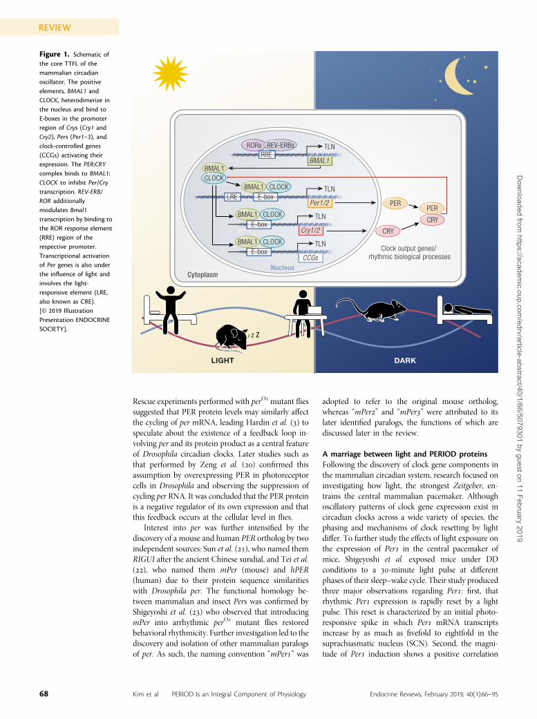
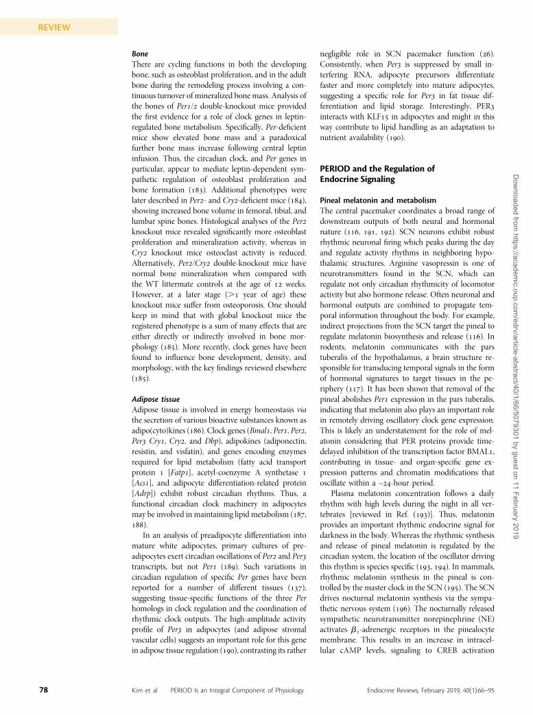
The circadian clockworkIn mammals, the circadian system is divided into twotypes of circadian clocks: () the central pacemaker and() peripheral oscillators, both of which are inter-connected via humoral and neuronal networks ().Biological clocks are comprised of three principalcomponents: an environmental input, a moleculartimekeeping mechanism (oscillator), and physiological/behavioral outputs. Input refers to cues present in theenvironment that provide temporal information to theclock, such as light under alternating light/dark (LD)cycles and food in response to feeding. These time cuesare commonly referred to as Zeitgebers (German for“time givers”) (). The molecular core oscillator con-sists of interlocked TTFLs that are autonomous and self-regulating. Figure illustrates a simplified schematic ofmammalian TTFLs. Briefly, in the central TTFL, thebasic helix–loop–helix transcription factors ARNTL,also known as BMAL brain and muscle Arnt-likeprotein- (BMAL) and circadian locomotor outputcycles kaput (CLOCK), dimerize and bind to E-boxeson promoters of Cryptochrome [(Cry)/] and Per(Per–) genes (). PER and CRY proteins dimerizein the cytoplasm before translocating into the nu-cleus, where they bind to and inhibit E-box trans-activation by BMAL:CLOCK, thus suppressingtheir own gene expression (Fig. ). This molecularoscillation is self-sustained and runs with a re-markably precise period of ~ hours (). Temporalinformation provided by Zeitgebers is integratedinto the molecular timekeeping mechanism, and, asa result, cellular clocks and downstream clock-dependent physiology are aligned with external
time. The circadian timekeeping mechanism is de-pendent on the rhythmic transcription of clockgenes, the protein products of which in turn controloutput genes that affect physiological, metabolic, andbehavioral rhythms (), hence the coupling ofphysiological processes known as the output toupstream circadian oscillators. As such, the molec-ular clockwork modulates physiological processesand ensures their optimal coordination to a cyclingenvironment, thus maximizing resource efficiencyand enhancing the chance of survival.
The discovery of the Period genesThe clock gene Period, a core component of the centralTTFL, was first discovered in D. melanogaster. Threeoriginal mutations of period (per) were found to affectthe circadian locomotor activity of adult flies in anobservable way: perS shortened activity, perL lengthenedactivity, and perO abolished rhythmic locomotion al-together (). Further cloning and immunohisto-chemical analyses demonstrated a broad expression ofper in numerous tissues throughout the adultDrosophilabody (, –).
The function of per as a negative-feedback mod-ulator of circadian clocks was first proposed by Hardinet al. () in . Prior to their investigation, it hadbeen established that Drosophila PER proteins oscillatefollowing a circadian rhythm, and that this expressionis affected by per gene mutations. Hardin et al. ana-lyzed per mRNA levels in Drosophila brains under/-hour LD cycles and in the absence of time cues,in this case constant darkness (DD). The study showsthat per mRNA oscillates in a circadian fashion. It wasthen proposed that cycling per mRNA is likely themechanism behind the rhythmicity of PER proteins.
ESSENTIAL POINTS
· The circadian system is integrated into all forms of life, as evident through its role in driving physiological rhythms andsynchronizing them to environmental cycles, and maintaining biological stability
· Clock genes are the interface in the bidirectional communication between the circadian clock and central/peripheralphysiological processes
· Physiological processes are intimately linked with circadian clocks; shift work and jet lag impair circadian clocks andclearly increase the risk for metabolic disorders and other disease states
· Clock genes are further involved in a plethora of extraclock functions; the clock gene Period in particular has beenimplicated in multiple central and peripheral processes, from invertebrates to humans
· Cyclic Period gene expression essentially implies a tissue- and organ-specific regulation of rhythmic BMAL1-dependentgene expression
· Animal research remains invaluable for proof-of-concept and mechanism-of-action studies related to chronobiologicalquestions on human health
67doi: 10.1210/er.2018-00049 https://academic.oup.com/edrv
REVIEWD
ownloaded from
https://academic.oup.com
/edrv/article-abstract/40/1/66/5079301 by guest on 11 February 2019
Rescue experiments performed with perOmutant fliessuggested that PER protein levels may similarly affectthe cycling of per mRNA, leading Hardin et al. () tospeculate about the existence of a feedback loop in-volving per and its protein product as a central featureof Drosophila circadian clocks. Later studies such asthat performed by Zeng et al. () confirmed thisassumption by overexpressing PER in photoreceptorcells in Drosophila and observing the suppression ofcycling per RNA. It was concluded that the PER proteinis a negative regulator of its own expression and thatthis feedback occurs at the cellular level in flies.
Interest into per was further intensified by thediscovery of a mouse and human PER ortholog by twoindependent sources: Sun et al. (), who named themRIGUI after the ancient Chinese sundial, and Tei et al.(), who named them mPer (mouse) and hPER(human) due to their protein sequence similaritieswith Drosophila per. The functional homology be-tween mammalian and insect Pers was confirmed byShigeyoshi et al. () who observed that introducingmPer into arrhythmic perO mutant flies restoredbehavioral rhythmicity. Further investigation led to thediscovery and isolation of other mammalian paralogsof per. As such, the naming convention “mPer” was
adopted to refer to the original mouse ortholog,whereas “mPer” and “mPer” were attributed to itslater identified paralogs, the functions of which arediscussed later in the review.
A marriage between light and PERIOD proteinsFollowing the discovery of clock gene components inthe mammalian circadian system, research focused oninvestigating how light, the strongest Zeitgeber, en-trains the central mammalian pacemaker. Althoughoscillatory patterns of clock gene expression exist incircadian clocks across a wide variety of species, thephasing and mechanisms of clock resetting by lightdiffer. To further study the effects of light exposure onthe expression of Per in the central pacemaker ofmice, Shigeyoshi et al. exposed mice under DDconditions to a -minute light pulse at differentphases of their sleep–wake cycle. Their study producedthree major observations regarding Per: first, thatrhythmic Per expression is rapidly reset by a lightpulse. This reset is characterized by an initial photo-responsive spike in which Per mRNA transcriptsincrease by as much as fivefold to eightfold in thesuprachiasmatic nucleus (SCN). Second, the magni-tude of Per induction shows a positive correlation
CRY
CRY
PERPER
Z Z Z
BMAL1
DARKLIGHT
CLOCK
TLN
CCGs
BMAL1 CLOCK TLN
Cry1/2
BMAL1 CLOCK TLN
Per1/2
RORs REV-ERBs TLN
BMAL1
NucleusCytoplasm
Clock output genes/ rhythmic biological processes
E-box
E-box
BMAL1 CLOCK
E-box
LRE
RRE
Figure 1. Schematic ofthe core TTFL of themammalian circadianoscillator. The positiveelements, BMAL1 andCLOCK, heterodimerize inthe nucleus and bind toE-boxes in the promoterregion of Crys (Cry1 andCry2), Pers (Per1–3), andclock-controlled genes(CCGs) activating theirexpression. The PER:CRYcomplex binds to BMAL1:CLOCK to inhibit Per/Crytranscription. REV-ERB/ROR additionallymodulates Bmal1transcription by binding tothe ROR response element(RRE) region of therespective promoter.Transcriptional activationof Per genes is also underthe influence of light andinvolves the light-responsive element (LRE,also known as CRE).[© 2019 IllustrationPresentation ENDOCRINESOCIETY].
68 Kim et al PERIOD Is an Integral Component of Physiology Endocrine Reviews, February 2019, 40(1):66–95
REVIEWD
ownloaded from
https://academic.oup.com
/edrv/article-abstract/40/1/66/5079301 by guest on 11 February 2019
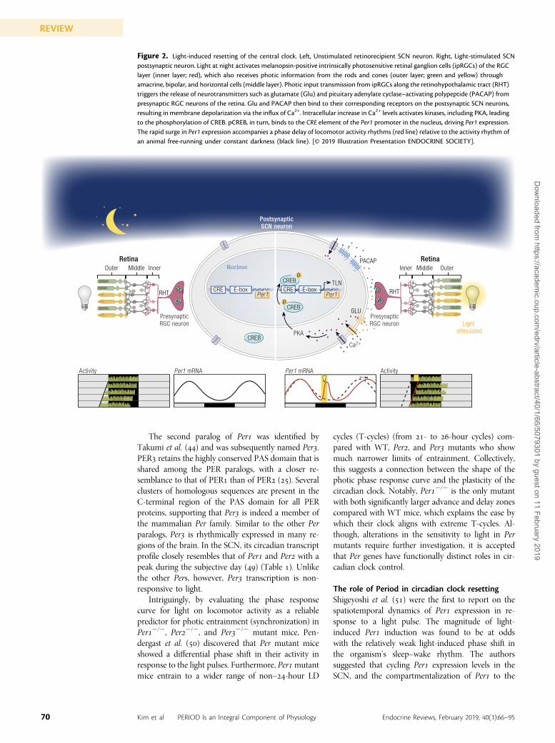
with the extent of behavioral rhythm resetting. Finally,in situ hybridization experiments of Per in the SCNrevealed a differential expression pattern of Perwithin the SCN, with Per expression levels generallylow in the dorsal, ventral, and medial portions of theSCN. After light pulse administration, Shigeyoshi et al.observed an induction of Per expression that firststarted in the ventral SCN before spreading moredorsally. Such findings were the first to show theventral SCN as the site of the light input into the SCN.Taken together, Per is a clockwork component thatalso functions as a photoresponsive immediate earlygene whose light induction correlates with behavioralphase resetting and clock gene rhythm resetting of themammalian central pacemaker within the SCN. Aschematic of light-induced resetting of the centralclock is shown in Fig. .
Additionally, other studies sought to determine theeffects of light exposure on the Period paralog Per,which was isolated and identified by Albrecht et al.(). Both PER proteins (PER and PER) contain twoPER-ARNT-SIM (PAS) domains (PAS-A and PAS-B)(). Structurally, PER differs from PER by a singleamino acid exchange within its PAS domains, a featureto which many of the nonredundant functions ofPER have been attributed (). Similar to Per, PermRNA expression oscillates rhythmically in the SCN(Table ) (–), but with a -hour phase delaycompared with the Per rhythm. Also similar to Per,Per expression rhythms are entrained (phase alignedor synchronized) to the external LD cycle and Pertranscription can be acutely induced by light; however,the extent of this response depends on the circadiantime of light exposure. Albrecht et al. and others(–) showed that although Per transcription islight-responsive throughout the (subjective) night,photic induction of Per is high during the beginningand low toward the end of the night. Therefore, tofurther elaborate on the role of Per and Per in light-induced clock resetting, the activity of mice carryingPer or Per mutations was analyzed in response tolight pulses applied at the early or late subjective night.A light pulse in the early night induces phase delays insleep–wake rhythms in wild-type (WT) and Permutant mice, whereas light pulses at late night triggerphase advances only in WT mice but not in Permutant mice. Taken together, Per and Per genesplay a pivotal role in the photic resetting of the cir-cadian clock ().
To assess additional functions of Per within themouse circadian system, Zheng et al. () generatedand identified a Per loss-of-function mutation. Thismutation (PerBrdm) is characterized by a two-exondeletion encoding for the region of PER, which ismost conserved between mouse and Drosophila per.Homozygous PerBrdm mice show shortened circa-dian periods of locomotor activity, followed by a lossof circadian rhythmicity (arrhythmicity) in the absence
of time cues (i.e., under DD conditions), under whichconditions WT mice free-run with incredible accuracyand steadiness with a cycle time that approximates theperiod length of the earth’s rotation about its axis( hours; hence the term circa-dian). PerBrdm micedisplay diminished cycling transcript levels of bothPer and Per in the SCN, which was unexpected giventhe assumed role of Per genes as negative regulators oftranscription in the mammalian TTFL. Consideringthat a truncated form of Per is still expressed in thePerBrdm mice, these studies may suggest that PERfunctions as a positive regulator of Per expression, oralternatively the existence of an autoregulation andcross-regulation between the two Per genes in anindependent molecular cycle.
Similarly, mice homozygous for loss-of-functionmutations in Per show robust circadian activity cyclesin the presence of time cues (light; under LD con-ditions) and a shortened circadian period (~ hour)under DD conditions (). In contrast to PerBrdm
mutants, PerBrdm animals display persistent robustrhythmicity of (non–protein coding) Per and Pertranscripts in DD, comparable to WT controls ().Thus, PER is not essential to maintain its own cir-cadian expression. Taken together, the PER proteinappears to be dispensable with regard to maintainingcircadian behavior, whereas PER is involved inregulating circadian gene expression via transcrip-tional control (, ). More specifically, PER exerts apositive regulation on mammalian clock gene ex-pression because loss of PER function significantlyreduces peak expression of Per, Per, and Cry (,). Although Per and Per single-mutant mice arerhythmic under both LD and constant conditions,Per/ double-mutant mice are arrhythmic in theirsleep–wake cycles and core clock and clock-regulatedgene expression profiles. Collectively, this suggests thatPer and Per are important for circadian clock control(). Furthermore, the immediate and complete loss ofrhythm phenotype in Per/Per double-mutant micein constant conditions (DD) suggests that PER andPER cooperate in the core clock mechanism. Thenormal activity pattern of the Per/Per double mu-tants under LD conditions raises the possibility thatthere is some residual clock function. However, thispossibility was dismissed with the discovery that a lightpulse could not reestablish a circadian rhythm in thesemice () as is the case for the single mutants (),which is consistent with a complete loss of a functionalclock. An alternative explanation is that the behavioraladaptation to LD maybe due to a masking effect of thelight (through direct photic inhibition of locomotoractivity). In case of a true loss of clock control theanimals are expected to respond passively to externalcues as observed in double and single Permutants ().Thus, Per and Per couple the animal’s rhythmicity toan internal clock to be influenced, but not driven, byexternal cues.
69doi: 10.1210/er.2018-00049 https://academic.oup.com/edrv
REVIEWD
ownloaded from
https://academic.oup.com
/edrv/article-abstract/40/1/66/5079301 by guest on 11 February 2019
The second paralog of Per was identified byTakumi et al. () and was subsequently named Per.PER retains the highly conserved PAS domain that isshared among the PER paralogs, with a closer re-semblance to that of PER than of PER (). Severalclusters of homologous sequences are present in theC-terminal region of the PAS domain for all PERproteins, supporting that Per is indeed a member ofthe mammalian Per family. Similar to the other Perparalogs, Per is rhythmically expressed in many re-gions of the brain. In the SCN, its circadian transcriptprofile closely resembles that of Per and Per with apeak during the subjective day () (Table ). Unlikethe other Pers, however, Per transcription is non-responsive to light.
Intriguingly, by evaluating the phase responsecurve for light on locomotor activity as a reliablepredictor for photic entrainment (synchronization) inPer2/2, Per2/2, and Per2/2 mutant mice, Pen-dergast et al. () discovered that Per mutant miceshowed a differential phase shift in their activity inresponse to the light pulses. Furthermore, Permutantmice entrain to a wider range of non–-hour LD
cycles (T-cycles) (from - to -hour cycles) com-pared with WT, Per, and Per mutants who showmuch narrower limits of entrainment. Collectively,this suggests a connection between the shape of thephotic phase response curve and the plasticity of thecircadian clock. Notably, Per2/2 is the only mutantwith both significantly larger advance and delay zonescompared with WT mice, which explains the ease bywhich their clock aligns with extreme T-cycles. Al-though, alterations in the sensitivity to light in Permutants require further investigation, it is acceptedthat Per genes have functionally distinct roles in cir-cadian clock control.
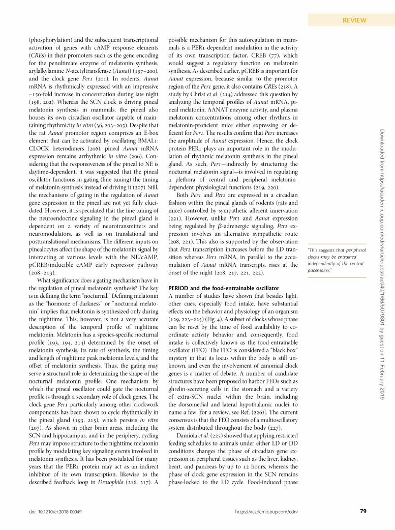
The role of Period in circadian clock resettingShigeyoshi et al. () were the first to report on thespatiotemporal dynamics of Per expression in re-sponse to a light pulse. The magnitude of light-induced Per induction was found to be at oddswith the relatively weak light-induced phase shift inthe organism’s sleep–wake rhythm. The authorssuggested that cycling Per expression levels in theSCN, and the compartmentalization of Per to the
Figure 2. Light-induced resetting of the central clock. Left, Unstimulated retinorecipient SCN neuron. Right, Light-stimulated SCNpostsynaptic neuron. Light at night activates melanopsin-positive intrinsically photosensitive retinal ganglion cells (ipRGCs) of the RGClayer (inner layer; red), which also receives photic information from the rods and cones (outer layer; green and yellow) throughamacrine, bipolar, and horizontal cells (middle layer). Photic input transmission from ipRGCs along the retinohypothalamic tract (RHT)triggers the release of neurotransmitters such as glutamate (Glu) and pituitary adenylate cyclase–activating polypeptide (PACAP) frompresynaptic RGC neurons of the retina. Glu and PACAP then bind to their corresponding receptors on the postsynaptic SCN neurons,resulting in membrane depolarization via the influx of Ca2+. Intracellular increase in Ca2+ levels activates kinases, including PKA, leadingto the phosphorylation of CREB. pCREB, in turn, binds to the CRE element of the Per1 promoter in the nucleus, driving Per1 expression.The rapid surge in Per1 expression accompanies a phase delay of locomotor activity rhythms (red line) relative to the activity rhythm ofan animal free-running under constant darkness (black line). [© 2019 Illustration Presentation ENDOCRINE SOCIETY].
PKA
TLN
Nuucleus
PostsynapticSCN neuron
Light stimulated
PresynapticRGC neuron
PresynapticRGC neuron
CREB
CREB
E-boxCREPer1
E-boxPer1
Per1 mRNAPer1 mRNA ActivityActivity
CRE
p
CREBp
Ca2+
Outer
RetinaMiddle Inner Outer
RetinaMiddleInner
RHT RHT
PACAP
GLU
70 Kim et al PERIOD Is an Integral Component of Physiology Endocrine Reviews, February 2019, 40(1):66–95
REVIEWD
ownloaded from
https://academic.oup.com
/edrv/article-abstract/40/1/66/5079301 by guest on 11 February 2019
ventral SCN, shape the light-induced behavioral shifts.The time-of-day–dependent expression levels of Perwithin the ventral SCN subregion are thought to setthe responsiveness of ventral SCN neurons to photicstimuli at the cellular level (). The role of Per inphase resetting is thought to be exerted in a time-of-day–dependent manner. Per and Per mutant micereset sleep–wake cycles differently depending on thetime of nocturnal light exposure (). A subsequentstudy by Yan and Silver () further elaborated on thedifferential functions of Per in resetting the mam-malian clock. The authors investigated the inductionand localization of Per and Per in the SCN withfocus on the heterogeneity of SCN neurons upondelaying and advancing phase shifts. They showed thatthe SCN can be divided into two anatomically distinctregions: a ventral region (core), which receives di-rect retinal input and contains vasoactive intestinalpeptide–expressing neurons, and a dorsomedial zone
(shell), consisting largely of arginine vasopressin–expressing cells. The time course and the localizationof light-induced Per and Per expression differentiatebetween the ventral and dorsomedial regions of theSCN. Light administration substantially induces Perexpression in the core throughout the subjective night,whereas strong light-induced Per expression in thisarea is only observed after a phase-delaying light pulse.In contrast to the SCN core, light-stimulated Perexpression in the shell region was found to align withbehavioral phase shifts. A phase-advancing light pulseleads to significant Per expression, although sub-stantial induction of Per is only observed in responseto a phase-delaying light pulse, and both Per and Perare not induced following a light pulse where nobehavioral phase shift is observed. These findingsindicate that light-induced Per or Per expression inthe SCN core is insufficient to generate behavioralphase shifts. Rather, differential Per and Per gene
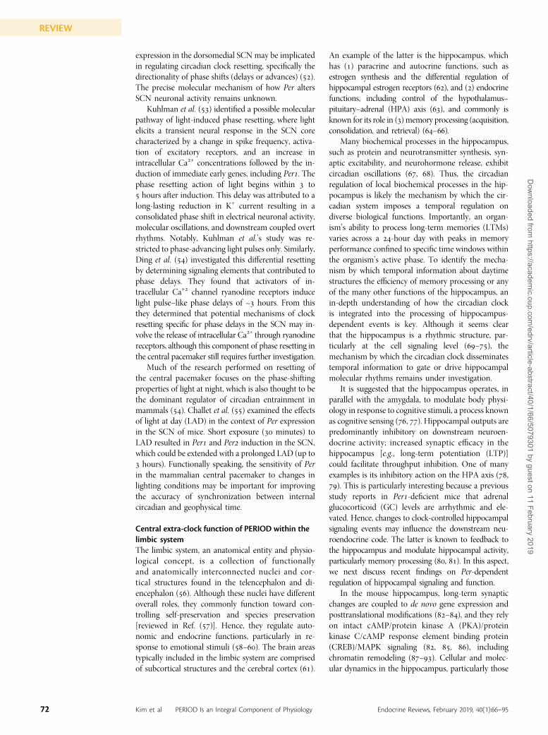
Table 1. Evidence for Expression of Per1–3 in Central and Peripheral Tissues in the Mouse
Circadian Rhythm of Period Genes in Mice
Tissue/Organs Per Genes Strain Rhythmic Under Constant Conditions
ExpressionUnder 12/12 LD
ReferencesPeak Trough
Brain SCN Per1 129/sv, CD-1-ICR, C57BL/6 Yes ZT8 ZT20 (26–30)
Per2 129/sv, CD-1-ICR, C57BL/6 Yes ZT10 ZT22 (26–30)
Per3 Unknown Yes ZT8 ZT0 (29)
Hippocampus Per1 C3H/He yes ZT10 ZT22 (31, 32)
Per2 C3H/He Yes ZT12 ZT0 (31, 32)
Periphery Adipose tissue Per1 C57BL/6 Yes ZT12 ZT0 (29, 33)
Per2 C57BL/6 Yes ZT14 ZT2 (29, 33, 34)
Per3 N/A Yes ZT12 ZT0 (29)
Heart Per1 BALB/c, C3H/He Yes ZT10 ZT20 (35, 36)
Per2 BALB/c, C3H/He Yes ZT12 ZT0 (35, 36)
Per3 BALB/c, C3H/He Yes ZT12 ZT20 (35, 36)
Liver Per1 BALB/c, C57BL/6,C3H/He
Yes ZT10 ZT22 (29, 35–37)
Per2 BALB/c, C57BL/6,C3H/He
Yes ZT12 ZT0 (29, 35–37)
Per3 BALB/c, C3H/He Yes ZT10 ZT22 (29, 35, 36)
Adrenals Per1 C57BL/6, C3H/He Yes ZT10 ZT22 (38–40)
Per2 C57BL/6, 129S2 Yes ZT12 ZT0 (38, 39, 41)
Per3 C57BL/6 Yes ZT12 ZT0 (38)
Pancreas Per1 C57BL/6 Yes ZT12 ZT0 (39)
Per2 C57BL/6 Yes ZT12 ZT0 (39, 42)
Abbreviations: N/A, not available; ZT, Zeitgeber time.
71doi: 10.1210/er.2018-00049 https://academic.oup.com/edrv
REVIEWD
ownloaded from
https://academic.oup.com
/edrv/article-abstract/40/1/66/5079301 by guest on 11 February 2019
expression in the dorsomedial SCN may be implicatedin regulating circadian clock resetting, specifically thedirectionality of phase shifts (delays or advances) ().The precise molecular mechanism of how Per altersSCN neuronal activity remains unknown.
Kuhlman et al. () identified a possible molecularpathway of light-induced phase resetting, where lightelicits a transient neural response in the SCN corecharacterized by a change in spike frequency, activa-tion of excitatory receptors, and an increase inintracellular Ca+ concentrations followed by the in-duction of immediate early genes, including Per. Thephase resetting action of light begins within to hours after induction. This delay was attributed to along-lasting reduction in K+ current resulting in aconsolidated phase shift in electrical neuronal activity,molecular oscillations, and downstream coupled overtrhythms. Notably, Kuhlman et al.’s study was re-stricted to phase-advancing light pulses only. Similarly,Ding et al. () investigated this differential resettingby determining signaling elements that contributed tophase delays. They found that activators of in-tracellular Ca+ channel ryanodine receptors inducelight pulse–like phase delays of ~ hours. From thisthey determined that potential mechanisms of clockresetting specific for phase delays in the SCN may in-volve the release of intracellular Ca+ through ryanodinereceptors, although this component of phase resetting inthe central pacemaker still requires further investigation.
Much of the research performed on resetting ofthe central pacemaker focuses on the phase-shiftingproperties of light at night, which is also thought to bethe dominant regulator of circadian entrainment inmammals (). Challet et al. () examined the effectsof light at day (LAD) in the context of Per expressionin the SCN of mice. Short exposure ( minutes) toLAD resulted in Per and Per induction in the SCN,which could be extended with a prolonged LAD (up to hours). Functionally speaking, the sensitivity of Perin the mammalian central pacemaker to changes inlighting conditions may be important for improvingthe accuracy of synchronization between internalcircadian and geophysical time.
Central extra-clock function of PERIOD within thelimbic systemThe limbic system, an anatomical entity and physio-logical concept, is a collection of functionallyand anatomically interconnected nuclei and cor-tical structures found in the telencephalon and di-encephalon (). Although these nuclei have differentoverall roles, they commonly function toward con-trolling self-preservation and species preservation[reviewed in Ref. ()]. Hence, they regulate auto-nomic and endocrine functions, particularly in re-sponse to emotional stimuli (–). The brain areastypically included in the limbic system are comprisedof subcortical structures and the cerebral cortex ().
An example of the latter is the hippocampus, whichhas () paracrine and autocrine functions, such asestrogen synthesis and the differential regulation ofhippocampal estrogen receptors (), and () endocrinefunctions, including control of the hypothalamus–pituitary–adrenal (HPA) axis (), and commonly isknown for its role in () memory processing (acquisition,consolidation, and retrieval) (–).
Many biochemical processes in the hippocampus,such as protein and neurotransmitter synthesis, syn-aptic excitability, and neurohormone release, exhibitcircadian oscillations (, ). Thus, the circadianregulation of local biochemical processes in the hip-pocampus is likely the mechanism by which the cir-cadian system imposes a temporal regulation ondiverse biological functions. Importantly, an organ-ism’s ability to process long-term memories (LTMs)varies across a -hour day with peaks in memoryperformance confined to specific time windows withinthe organism’s active phase. To identify the mecha-nism by which temporal information about daytimestructures the efficiency of memory processing or anyof the many other functions of the hippocampus, anin-depth understanding of how the circadian clockis integrated into the processing of hippocampus-dependent events is key. Although it seems clearthat the hippocampus is a rhythmic structure, par-ticularly at the cell signaling level (–), themechanism by which the circadian clock disseminatestemporal information to gate or drive hippocampalmolecular rhythms remains under investigation.
It is suggested that the hippocampus operates, inparallel with the amygdala, to modulate body physi-ology in response to cognitive stimuli, a process knownas cognitive sensing (, ). Hippocampal outputs arepredominantly inhibitory on downstream neuroen-docrine activity; increased synaptic efficacy in thehippocampus [e.g., long-term potentiation (LTP)]could facilitate throughput inhibition. One of manyexamples is its inhibitory action on the HPA axis (,). This is particularly interesting because a previousstudy reports in Per-deficient mice that adrenalglucocorticoid (GC) levels are arrhythmic and ele-vated. Hence, changes to clock-controlled hippocampalsignaling events may influence the downstream neu-roendocrine code. The latter is known to feedback tothe hippocampus and modulate hippocampal activity,particularly memory processing (, ). In this aspect,we next discuss recent findings on Per-dependentregulation of hippocampal signaling and function.
In the mouse hippocampus, long-term synapticchanges are coupled to de novo gene expression andposttranslational modifications (–), and they relyon intact cAMP/protein kinase A (PKA)/proteinkinase C/cAMP response element binding protein(CREB)/MAPK signaling (, , ), includingchromatin remodeling (–). Cellular and molec-ular dynamics in the hippocampus, particularly those
72 Kim et al PERIOD Is an Integral Component of Physiology Endocrine Reviews, February 2019, 40(1):66–95
REVIEWD
ownloaded from
https://academic.oup.com
/edrv/article-abstract/40/1/66/5079301 by guest on 11 February 2019
relevant for LTM formation, are molded by time ofday (), which supports an interaction between thecircadian system and memory. Per and Per beingrhythmically expressed in the mouse hippocampus(, –), and shown to modulate behavioral sensi-tization (), implies a regulatory role for the clock geneproteins PER and PER in the temporal modulation ofhippocampal function (e.g., in learning and memory).
Circadian core clock components are rhythmicallyexpressed in the hippocampus of Per2/2mice, yet thephases of clock gene expression rhythms in Per2/2
mice are shifted compared with WT mice, despite thatthe SCN clock of Per2/2 mice is properly phased toambient lighting conditions (). Per2/2 mice arerhythmic under both diurnal and constant conditionssimilar to control littermates (Per+/+), suggesting thatPER may have a specific local role in modulatinghippocampal physiology (, , ). This is sup-ported by in vitro studies showing that cAMP/PKAsignaling to the memory-dependent transcriptionfactor CREB is impaired in primary hippocampalneurons derived from Per2/2 mice (). This mayexplain why the diurnal rhythm of hippocampal CREBphosphorylation () is lost in Per2/2 mice ().Notably, the rhythmic phosphorylation of bothMAPKand CREB in the mouse hippocampus is important forthe maintenance of LTM (, ).
Long-lasting changes in synaptic plasticity knownas LTP are the cellular correlate for LTM (, ). Themagnitude of LTP at perforant path–granule cellsynapses in the dentate gyrus is compromised inPer2/2 mice, whereas basic properties of synaptictransmission appear normal, indicating that functionaldeficits are not likely due to alterations in networkexcitability (). The recorded reduction in LTPamplitude observed in Per2/2 mice may suggest aspecific role for PER in the reinforcement andconsolidation of spatial memories.
Long-term memory formation depends on dif-ferent signaling cascades, many of which converge toactivate the transcription factor CREB to initiate LTM-dependent gene expression (–). The silencingof one or several of these pathways will likely alterlearning-induced dynamics in CREB activation, andconsequently affect LTM formation. Importantly, notethat although Per2/2 mice show a reduction in theamplitude of in vivo LTP, they do acquire LTM.However, in contrast to Per+/+ mice, day/night dif-ferences in memory performance are absent, at leastwhen comparing the Zeitgeber time and timepoints. This may be linked to the absence of day/nightvariations in phosphorylated CREB (pCREB) levels inPer2/2 mice [reviewed in Ref. ()].
It is tempting to assume that the mechanism bywhich temporal information is integrated into LTMprocessing involves a rhythmic interference of cyclicclock proteins such as PER with memory-relevantmolecular signaling events. Clockwork components
may have non–clock-related functions. Per, for ex-ample, is constitutively expressed in the dentate gyrusof the hippocampus (), and thus it cannot possiblyconvey temporal information by rhythmically in-terfering with local events. Therefore, deficits in long-term trace fear memory observed in Per mutant mice() reflect an extra-clock function of PER. The reg-ulation of hippocampal neurogenesis and, hence,neurogenesis-dependent memory formation [reviewedin Ref. ()] is another intriguing example of an extra-clock function for the core clockwork component PER.The expression of rhythmic clock genes in the hippo-campus suggests a functional role for the hippocampalcircadian oscillator in memory processing and beyond,as highlighted by several reports [reviewed in Refs. (,, )]. Interestingly, studies investigating the role ofclock genes in hippocampal functions suggest thatdifferent clock genes are involved in modulating dif-ferent types of hippocampus-dependent behaviors (,, ). The herein reported role of Per genes inhippocampal signaling and how it translates to LTMprocesses is one example and supports further in-vestigations into more downstream hippocampus-regulated endocrine functions.
There is compelling evidence to suggest thathormones as well as metabolic signals can modulatecircadian oscillations of clock gene expression in thebrain and the periphery. The pineal hormone mela-tonin, for example, modulates the rhythm of Perexpression in the pituitary gland, striatum, and adrenalcortex (–). Furthermore, adrenal GCs wereshown to modulate the rhythm of expression of PERin the oval nucleus of the bed nucleus of the striaterminalis and the central nucleus of the amygdala(, ). This suggest that clock genes, being anintegral component of memory processing and limbicfunction, also serve to communicate peripheral en-docrine functions to central processes.
PERIOD Within the PeripheralCircadian Network
General statementsAs briefly introduced, the circadian system is com-posed of two categories of circadian clocks: the centralpacemaker in the SCN, and peripheral oscillatorsdistributed throughout the body. The latter time therhythmic expression of tissue- and organ-specificgenes (, ), therefore emphasizing the impor-tance of temporal coordination of various biologicalprocesses (). Oscillations of physiological processesin peripheral tissues are shown in Fig. . For example,in diurnal mammals, insulin responses (resulting inglucose disposal) are highest during daytime, whencarbohydrate uptake peaks due to SCN-regulateddaytime activity and food intake. Similarly, glucagonsecretion peaks at night so that stored glycogen may
“Clock genes in general andperiod genes in particular arerhythmically expressed inmost peripheral tissues.”
73doi: 10.1210/er.2018-00049 https://academic.oup.com/edrv
REVIEWD
ownloaded from
https://academic.oup.com
/edrv/article-abstract/40/1/66/5079301 by guest on 11 February 2019
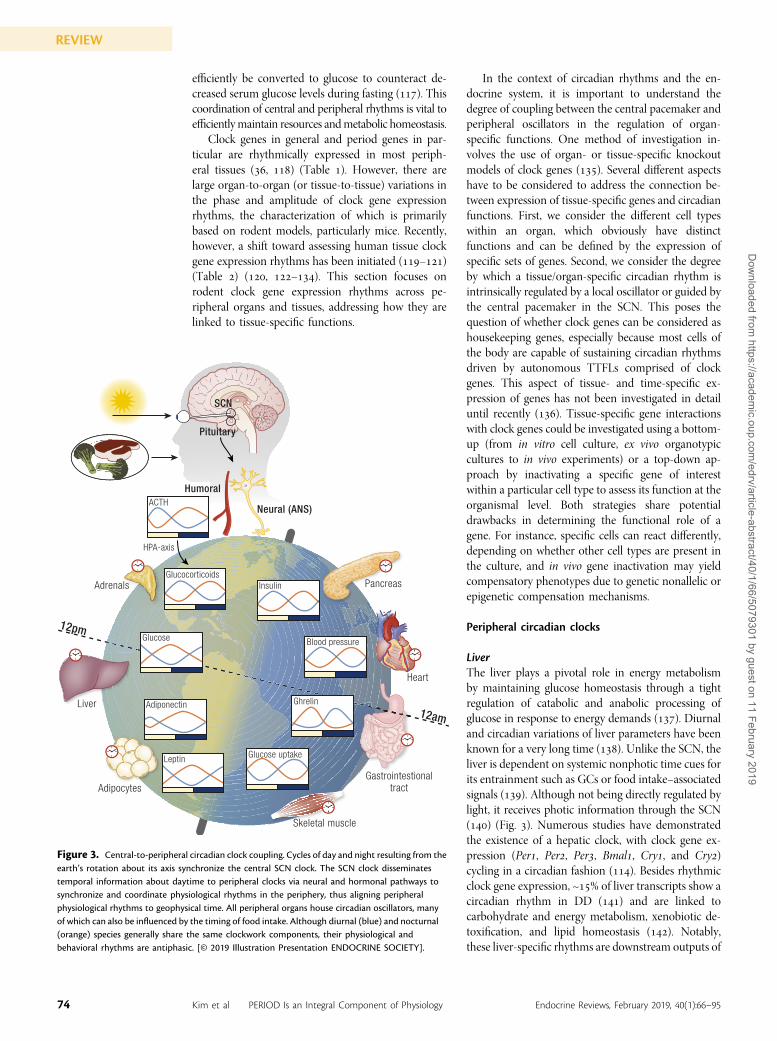
efficiently be converted to glucose to counteract de-creased serum glucose levels during fasting (). Thiscoordination of central and peripheral rhythms is vital toefficientlymaintain resources andmetabolic homeostasis.
Clock genes in general and period genes in par-ticular are rhythmically expressed in most periph-eral tissues (, ) (Table ). However, there arelarge organ-to-organ (or tissue-to-tissue) variations inthe phase and amplitude of clock gene expressionrhythms, the characterization of which is primarilybased on rodent models, particularly mice. Recently,however, a shift toward assessing human tissue clockgene expression rhythms has been initiated (–)(Table ) (, –). This section focuses onrodent clock gene expression rhythms across pe-ripheral organs and tissues, addressing how they arelinked to tissue-specific functions.
In the context of circadian rhythms and the en-docrine system, it is important to understand thedegree of coupling between the central pacemaker andperipheral oscillators in the regulation of organ-specific functions. One method of investigation in-volves the use of organ- or tissue-specific knockoutmodels of clock genes (). Several different aspectshave to be considered to address the connection be-tween expression of tissue-specific genes and circadianfunctions. First, we consider the different cell typeswithin an organ, which obviously have distinctfunctions and can be defined by the expression ofspecific sets of genes. Second, we consider the degreeby which a tissue/organ-specific circadian rhythm isintrinsically regulated by a local oscillator or guided bythe central pacemaker in the SCN. This poses thequestion of whether clock genes can be considered ashousekeeping genes, especially because most cells ofthe body are capable of sustaining circadian rhythmsdriven by autonomous TTFLs comprised of clockgenes. This aspect of tissue- and time-specific ex-pression of genes has not been investigated in detailuntil recently (). Tissue-specific gene interactionswith clock genes could be investigated using a bottom-up (from in vitro cell culture, ex vivo organotypiccultures to in vivo experiments) or a top-down ap-proach by inactivating a specific gene of interestwithin a particular cell type to assess its function at theorganismal level. Both strategies share potentialdrawbacks in determining the functional role of agene. For instance, specific cells can react differently,depending on whether other cell types are present inthe culture, and in vivo gene inactivation may yieldcompensatory phenotypes due to genetic nonallelic orepigenetic compensation mechanisms.
Peripheral circadian clocks
LiverThe liver plays a pivotal role in energy metabolismby maintaining glucose homeostasis through a tightregulation of catabolic and anabolic processing ofglucose in response to energy demands (). Diurnaland circadian variations of liver parameters have beenknown for a very long time (). Unlike the SCN, theliver is dependent on systemic nonphotic time cues forits entrainment such as GCs or food intake–associatedsignals (). Although not being directly regulated bylight, it receives photic information through the SCN() (Fig. ). Numerous studies have demonstratedthe existence of a hepatic clock, with clock gene ex-pression (Per, Per, Per, Bmal, Cry, and Cry)cycling in a circadian fashion (). Besides rhythmicclock gene expression, ~% of liver transcripts show acircadian rhythm in DD () and are linked tocarbohydrate and energy metabolism, xenobiotic de-toxification, and lipid homeostasis (). Notably,these liver-specific rhythms are downstream outputs of
SCN
Neural (ANS)
Humoral
Glucocorticoids
ACTH
HPA-axis
Adiponectin
Leptin
Insulin
Blood pressure
Glucose uptake
Ghrelin
Pituitary
Skeletal muscle
Gastrointestionaltract
Heart
PancreasAdrenals
Liver
Adipocytes
12pm
12am
Glucose
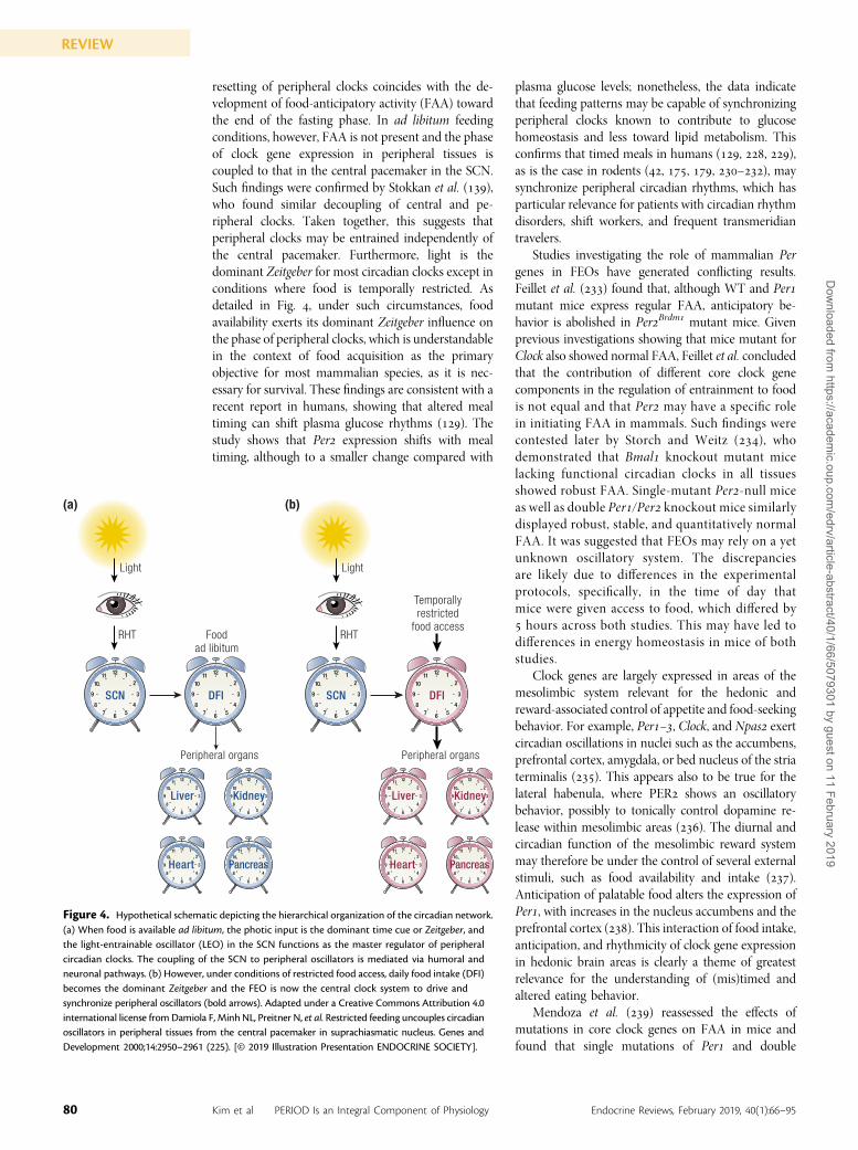
Figure 3. Central-to-peripheral circadian clock coupling. Cycles of day and night resulting from theearth’s rotation about its axis synchronize the central SCN clock. The SCN clock disseminatestemporal information about daytime to peripheral clocks via neural and hormonal pathways tosynchronize and coordinate physiological rhythms in the periphery, thus aligning peripheralphysiological rhythms to geophysical time. All peripheral organs house circadian oscillators, manyof which can also be influenced by the timing of food intake. Although diurnal (blue) and nocturnal(orange) species generally share the same clockwork components, their physiological andbehavioral rhythms are antiphasic. [© 2019 Illustration Presentation ENDOCRINE SOCIETY].
74 Kim et al PERIOD Is an Integral Component of Physiology Endocrine Reviews, February 2019, 40(1):66–95
REVIEWD
ownloaded from
https://academic.oup.com
/edrv/article-abstract/40/1/66/5079301 by guest on 11 February 2019
the local hepatic oscillator because the knockdownof the core clock gene Bmal in the liver was shownto interfere with hepatic function, rendering theaforementioned physiological processes arrhythmic().
The study by Ramanathan et al. () looked intocell-specific effects of downregulating Per genes usinglentiviral short hairpin RNA vectors for Per, Per, andPer. The results show that knockdown of either Per,Per, or Per results in short-period, low-amplituderhythms of cycling clock genes within hepatocytes.Double and triple knockdowns of Per/ and Per//,respectively, result in complete arrhythmicity, similarlyto the results reported by Kornmann et al. () usingliver-specific REVERBa-overexpressing mice. In ad-dition to these findings, a recent and very intriguingreport showed that in Per-null mice, polyploidizationof hepatocytes is markedly accelerated, possibly due toan impaired MAPK phosphatase–mediated alterationin ERK/ activity. These data clearly support thenotion that Per genes are not only responsible forregulating period length and amplitude of hepatocyteclocks and metabolic activity, but also for hepatocyteturnover and self-renewal, with Per deficiency leadingto a massive accumulation of polyploid cells and
potentially favoring malignant transformation ().In line with this, mice with a deletion of the Per gene() display the three cancer hallmarks, includinguncontrolled cell proliferation, genomic instability,and tumor-promoting inflammation. This is reflectedby profound alterations in hepatic oncogenic geneexpression, including the avian myelocytomatosis vi-rus oncogene cellular homolog (c-Myc), Wee tyrosinekinase, G/mitotic-specific cyclin-B (Ccnb), andKirsten rat sarcoma GTPase (K-ras), as well as in-creased inflammation with high IL- levels in theabsence of any carcinogen exposure. These findingspredict that functional mutations in the Per geneincrease the chance to develop liver carcinoma. In fact,Per mutant mice are nearly four times more likelyto develop cancer as compared with controls ().Alternatively, overexpressing Per significantly sup-presses tumor growth in vivo by improving tumor celladhesion and suppressing cell migratory activity ().This newly discovered role of the Period gene adds toits known circadian function with respect to hormoneproduction, development, regeneration, and healthyaging of liver tissue.
In recent times, the description of the circadianliver proteome () gave the field a fresh look and has
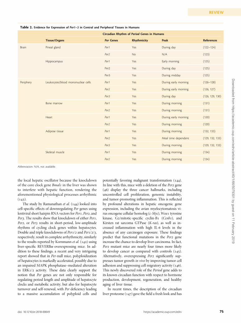
Table 2. Evidence for Expression of Per1–3 in Central and Peripheral Tissues in Humans
Circadian Rhythm of Period Genes in Humans
Tissue/Organs Per Genes Rhythmicity Peak References
Brain Pineal gland Per1 Yes During day (122–124)
Per2 No N/A (123)
Hippocampus Per1 Yes Early morning (125)
Per2 Yes During day (125)
Per3 Yes During midday (125)
Periphery Leukocytes/blood mononuclear cells Per1 Yes During early morning (126–128)
Per2 Yes During early morning (126, 127)
Per3 Yes During day (126, 129, 130)
Bone marrow Per1 Yes During morning (131)
Per2 Yes During morning (131)
Heart Per1 Yes During early morning (120)
Per2 Yes During morning (120)
Adipose tissue Per1 Yes During morning (132, 133)
Per2 Yes Meal time dependent (129, 132, 133)
Per3 Yes During morning (129, 132, 133)
Skeletal muscle Per1 Yes During morning (134)
Per2 Yes During morning (134)
Abbreviation: N/A, not available.
75doi: 10.1210/er.2018-00049 https://academic.oup.com/edrv
REVIEWD
ownloaded from
https://academic.oup.com
/edrv/article-abstract/40/1/66/5079301 by guest on 11 February 2019
since been extended and advanced by analyses ofclock-dependent and clock-independent liver pro-teomes (). These studies have been further com-plemented by the evaluation of posttranscriptionalmechanisms () and rhythmic degradation ofprotein products in the liver (). In this context, ithas also been shown that the mitochondrial proteomeof the liver depends on the presence of the PERproteins within the cell nuclei of hepatocytes ().More recent in-depth quantitative mass spectro-metric analysis of the liver nuclear proteome ()and the liver phosphoproteome revealed that ~ ofthe nuclear proteins enter and leave the he-patocyte nuclei in a circadian manner and % of theprotein phospho-sites oscillate diurnally. Moreover,the lysine “acylome” of liver proteins, the group ofproteins containing acetylated lysine, which is veryrelevant for the orchestration of metabolic activity ofthe liver, are under diurnal (and feeding) control().
Another interesting question to raise is whethersynchronizing factors rhythmically produced by thecentral circadian clock can target liver function. Thisidea originates from a classical experiment showingthat the reintroduction of the SCN, housed within acapsule permeable to molecules up to the size ofproteins, but not nerve fibers, can reinstate sleephomeostasis (). Follow-up experiments confirmedthat such SCN-specific factors (peptides and proteins)exist and are indeed responsible for internal syn-chronization (, ). Alternatively, adropin, apeptide hormone produced in the liver and secretedin a diurnal manner, was shown to act on the brain(). This would add another level of complexity tothe mutual influence of peripheral circadian outputson central clocks. Mining the data sets originatingfrom the mass spectrometric studies mentionedabove may allow the discovery of new peripheraltissue-specific rhythmic peptides or proteins thatcouple peripheral clocks to the central biologicalclock.
Adrenals and the HPA axisThe adrenals are the effector tissue of the HPA axisand are central in the regulation of multiple physio-logical functions, including gluconeogenesis, lipidmetabolism, immunity, stress response, and re-production (). Most prominently and well studied,however, is the adrenals’ role in synchronizing be-havior and physiology via the rhythmic secretion ofGCs (). Extensive studies on the mechanismsdriving -hour GC rhythms revealed that theadrenal houses a circadian oscillator (, ). Robustrhythmic expression of canonical clock genes, in-cluding all three Per paralogs, was found within theadrenal cortex (and to a lesser extent in the adrenalmedulla) of rodents (, ). Consistent with mostother tissues studied and across experimental models,
circadian rhythms in the expression of adrenal clockgenes could also be verified in nonhuman primates(), which similarly suggests the presence of anadrenal circadian oscillator. Additionally, a tran-scriptional circadian profiling study of the adrenal ledto the discovery that adrenal genes involved in en-docrine functions, including steroid and cholesterolbiosynthesis, and catecholamine metabolism are undercircadian control (). A recent study emphasized theimportance of the adrenal clock in the regulation ofdaily rhythmic release of GCs, showing that whencentral and peripheral rhythms are uncoupled bydaytime-restricted feeding in mice, adrenal GC con-tent and steroidogenic acute regulatory (StAR) proteinlevels are phase shifted. This is consistent with a shiftedrhythm in clock gene expression in the adrenal ().Notably, restricted feeding leads to two distinct peaksin plasma GC levels: an early peak that correspondsto a shift in adrenal steroidogenesis, and a later peakthat is in phase with the organism’s sleep–wake cycle,and thus represents an SCN-dependent rhythm. Thesefindings indicate that both SCN-driven autonomicinnervation and the adrenal clock are crucial in drivingthe daily GC rhythm and its stabilization on a dailybasis ().
Although the SCN is required to generate thediurnal pattern of the HPA axis (), the role of Pergenes in this process is not well characterized. Yanget al. () assessed the circadian rhythm of the HPAaxis in Per+/+ and Per2/2 mice. The lack of corti-costerone rhythm in Per2/2 mice suggested adefect in either corticosterone production or gen-eration of HPA rhythm. Although Per2/2 micelack a recognizable corticosterone rhythm, they canproduce corticosterone in response to stress. Thissuggests that Per may be important for generatingthe ACTH rhythm and/or the adrenal corticalrhythm.
Although the central (hypothalamic–pituitary)activity of the HPA axis may play a part in the phaseadjustment of hormone production, it may be lesscritical for generating the daily rhythms of GC se-cretion. Ishida et al. () demonstrated that lightexposure during the night leads to a rapid induction ofPer in the adrenals and a subsequent increase inplasma corticosterone (the primary GC in rodents)independently of the adrenocorticotrophic hormone(ACTH). Interestingly, denervation of the adrenalsabolishes photic induction of Per in the adrenalcortex, which is accompanied by a reduction in GCresponses. This suggests that sympathetic innervationrather than hypothalamic–pituitary activation is thepredominant route by which the SCN relays photicinformation from the environment to the adrenals. Inconjunction with previous observations, Ishida et al.’sstudy provides strong evidence for the presence of aseparate peripheral clock within the adrenals and alsoimplies a potential role of Per in light-evoked
76 Kim et al PERIOD Is an Integral Component of Physiology Endocrine Reviews, February 2019, 40(1):66–95
REVIEWD
ownloaded from
https://academic.oup.com
/edrv/article-abstract/40/1/66/5079301 by guest on 11 February 2019
physiological changes in GC signaling. Later in-vestigations by Oster et al. () sought to confirm thepresence and characterized the function of this adrenalclock. The group confirmed the presence of an adrenalcircadian oscillator and reported robust rhythmicexpression of canonical clock genes, including all threePer paralogs within the adrenal cortex. They alsoobserved that circadian rhythms of the ACTH, cor-ticosterone, and clock gene expression are abolished inPer/Cry mutant mice, which is consistent with theirprevious observations of disrupted locomotor andfeeding activity (). Examination of adrenal slicecultures led to the discovery that the adrenal cortexshows a gated sensitivity to ACTH; that is, ACTHstimulation triggers corticosterone release in a tem-porally controlled fashion in the absence of SCN input(). Clock-deficient adrenals lack this sensitivity,which indicates that the responsiveness to ACTHrelies on a functional adrenal clock. To confirm thismechanism in vivo, Oster et al. () transplanted WTadrenals into clock-deficient Per/Cry double-mutanthosts and into WT hosts lacking adrenals. Trans-plantation of WT adrenals into mutant hosts restoredcorticosterone rhythmicity, whereas mutant adrenaltransplantation into WT hosts dampened corticoste-rone expression. Such observations illustrate themechanism of the gated adrenal response to ACTH,wherein the constant (i.e., rhythm-ablated) ACTHsignaling in mutant hosts is translated by the WTadrenal clock into a rhythmic output of corticosterone.WT hosts had normal rhythmic ACTH signaling inputthat was in phase with the adrenal clock and served toconstructively propagate the rhythmic secretion ofcorticosterone to drive high-amplitude hormone re-lease. Although this study was performed with mutantPer/Cry mice, it was concluded that mice carryingdifferent clock gene mutations such as Per/-deficientmice were likely to produce similar results, althoughthe sensitivity of the adrenal clock to ACTH mayvary between mutants. Oster et al. () proposeda role for the peripheral adrenal clock as a localphase stabilizer for rhythmic peripheral hormonesecretion.
The question of the function of the adrenal clock inrelationship to the mechanism by which ACTH es-tablishes the timed secretion of GCs has been in-vestigated. Studies have shown that circadian rhythmsof the steroidogenic protein StAR are absent in ar-rhythmic Per/ and Cry/ double-mutant mice.StAR is a target of ACTH and is the rate-limiting stepin GC biosynthesis (). This study was the first toestablish StAR as an adrenal clock-controlled gene.GCs have long been thought to act as synchronizers ofperipheral clocks (, ) by directly regulating theexpression of Per via distal GC-responsive promoterelements. Accordingly, the altered expression of Perwas attributed to the reduced levels of circulatingGCs.
In summary, the adrenal peripheral clock isthought to have profound roles in harmonizing themammalian circadian timing system, especially thoseslow-adjusting peripheral clocks, in response tochanges in environmental stimulus via the rhythmicgeneration and secretion of GCs. In particular, Perplays an important role in modulating the circadianrhythm of GCs given the induction of Per expressionby nocturnal light exposure accompanied with sub-sequent secretion of corticosterone and loss ofrhythmic secretion of ACTH as well as corticosteronein Per/ double-mutant mice. Whether Permediatessuch synchronizing effects of GCs is yet to be furtheridentified.
The endocrine pancreasThe endocrine pancreas is integral to glucose regu-lation via its secretion of insulin and glucagon (). Itis composed of a heterogeneous population of celltypes. Pancreatic b- and a-cells secrete insulin andglucagon, respectively. Similar to other peripheralorgans, endocrine pancreatic tissue exhibits a robustexpression of major circadian clock genes (Per, Per,Bmal, Cry, and Clock) (). Plasma levels of insulinand glucagon have also been show to cycle (, ),which is thought to be regulated by the pancreaticcircadian oscillator (–), as illustrated in Fig. .Subsequent studies provided bona fide evidence for therole of the pancreatic circadian clock in regulating itsfunction in rodents and humans (, ). b-Cell–specific Bmal knockout mice exhibit phenotypes suchas defective insulin production and severe glucoseintolerance, indicative of b-cell dysfunction (, ,). This strongly supports a role for the b-cell in-trinsic oscillator in insulin secretion and glucosehomeostasis.
The effect of environmental factors (e.g., nighttimelight exposure) on the pancreas revealed novel insightson the role of Per genes in endocrine pancreasfunction. Although changes in the LD cycle in vivoentrain the phase of islet clock transcriptional oscil-lations, weeks of continued exposure to light atnight impairs the amplitude, phase, and inter-isletsynchrony of clock transcriptional oscillations ().Interestingly, it was observed that glucose regulates theamplitude and period of Per oscillations, indicating anutrient-sensing mechanism in the islet clock. It hasalso been reported that constant light regimes or-hour advances of the light cycle every third dayaccelerate the development of diabetes in rats trans-genic for the human islet amyloid polypeptide ().Exposure to a high fat–containing diet has been shownto change the circadian expression pattern of Per, andPer in mouse pancreatic islets, as well as circadianinsulin secretion (). Thus, environmental condi-tions that affect circadian rhythms can also impair thepancreatic clock and the function of this endocrineorgan.
“Environmental conditionsthat affect circadian rhythmscan also impair the pancreaticclock and the function of thisendocrine organ.”
77doi: 10.1210/er.2018-00049 https://academic.oup.com/edrv
REVIEWD
ownloaded from
https://academic.oup.com
/edrv/article-abstract/40/1/66/5079301 by guest on 11 February 2019
BoneThere are cycling functions in both the developingbone, such as osteoblast proliferation, and in the adultbone during the remodeling process involving a con-tinuous turnover of mineralized bone mass. Analysis ofthe bones of Per/ double-knockout mice providedthe first evidence for a role of clock genes in leptin-regulated bone metabolism. Specifically, Per-deficientmice show elevated bone mass and a paradoxicalfurther bone mass increase following central leptininfusion. Thus, the circadian clock, and Per genes inparticular, appear to mediate leptin-dependent sym-pathetic regulation of osteoblast proliferation andbone formation (). Additional phenotypes werelater described in Per- and Cry-deficient mice (),showing increased bone volume in femoral, tibial, andlumbar spine bones. Histological analyses of the Perknockout mice revealed significantly more osteoblastproliferation and mineralization activity, whereas inCry knockout mice osteoclast activity is reduced.Alternatively, Per/Cry double-knockout mice havenormal bone mineralization when compared withthe WT littermate controls at the age of weeks.However, at a later stage (. year of age) theseknockout mice suffer from osteoporosis. One shouldkeep in mind that with global knockout mice theregistered phenotype is a sum of many effects that areeither directly or indirectly involved in bone mor-phology (). More recently, clock genes have beenfound to influence bone development, density, andmorphology, with the key findings reviewed elsewhere().
Adipose tissueAdipose tissue is involved in energy homeostasis viathe secretion of various bioactive substances known asadipo(cyto)kines (). Clock genes (Bmal, Per, Per,Per Cry, Cry, and Dbp), adipokines (adiponectin,resistin, and visfatin), and genes encoding enzymesrequired for lipid metabolism (fatty acid transportprotein [Fatp], acetyl-coenzyme A synthetase [Acs], and adipocyte differentiation-related protein[Adrp]) exhibit robust circadian rhythms. Thus, afunctional circadian clock machinery in adipocytesmay be involved in maintaining lipid metabolism (,).
In an analysis of preadipocyte differentiation intomature white adipocytes, primary cultures of pre-adipocytes exert circadian oscillations of Per and Pertranscripts, but not Per (). Such variations incircadian regulation of specific Per genes have beenreported for a number of different tissues (),suggesting tissue-specific functions of the three Perhomologs in clock regulation and the coordination ofrhythmic clock outputs. The high-amplitude activityprofile of Per in adipocytes (and adipose stromalvascular cells) suggests an important role for this genein adipose tissue regulation (), contrasting its rather
negligible role in SCN pacemaker function ().Consistently, when Per is suppressed by small in-terfering RNA, adipocyte precursors differentiatefaster and more completely into mature adipocytes,suggesting a specific role for Per in fat tissue dif-ferentiation and lipid storage. Interestingly, PERinteracts with KLF in adipocytes and might in thisway contribute to lipid handling as an adaptation tonutrient availability ().
PERIOD and the Regulation ofEndocrine Signaling
Pineal melatonin and metabolismThe central pacemaker coordinates a broad range ofdownstream outputs of both neural and hormonalnature (, , ). SCN neurons exhibit robustrhythmic neuronal firing which peaks during the dayand regulate activity rhythms in neighboring hypo-thalamic structures. Arginine vasopressin is one ofneurotransmitters found in the SCN, which canregulate not only circadian rhythmicity of locomotoractivity but also hormone release. Often neuronal andhormonal outputs are combined to propagate tem-poral information throughout the body. For example,indirect projections from the SCN target the pineal toregulate melatonin biosynthesis and release (). Inrodents, melatonin communicates with the parstuberalis of the hypothalamus, a brain structure re-sponsible for transducing temporal signals in the formof hormonal signatures to target tissues in the pe-riphery (). It has been shown that removal of thepineal abolishes Per expression in the pars tuberalis,indicating that melatonin also plays an important rolein remotely driving oscillatory clock gene expression.This is likely an understatement for the role of mel-atonin considering that PER proteins provide time-delayed inhibition of the transcription factor BMAL,contributing in tissue- and organ-specific gene ex-pression patterns and chromatin modifications thatoscillate within a ~-hour period.
Plasma melatonin concentration follows a dailyrhythm with high levels during the night in all ver-tebrates [reviewed in Ref. ()]. Thus, melatoninprovides an important rhythmic endocrine signal fordarkness in the body. Whereas the rhythmic synthesisand release of pineal melatonin is regulated by thecircadian system, the location of the oscillator drivingthis rhythm is species specific (, ). In mammals,rhythmic melatonin synthesis in the pineal is con-trolled by the master clock in the SCN (). The SCNdrives nocturnal melatonin synthesis via the sympa-thetic nervous system (). The nocturnally releasedsympathetic neurotransmitter norepinephrine (NE)activates b-adrenergic receptors in the pinealocytemembrane. This results in an increase in intracel-lular cAMP levels, signaling to CREB activation
78 Kim et al PERIOD Is an Integral Component of Physiology Endocrine Reviews, February 2019, 40(1):66–95
REVIEWD
ownloaded from
https://academic.oup.com
/edrv/article-abstract/40/1/66/5079301 by guest on 11 February 2019
(phosphorylation) and the subsequent transcriptionalactivation of genes with cAMP response elements(CREs) in their promoters such as the gene encodingfor the penultimate enzyme of melatonin synthesis,arylalkylamine N-acetyltransferase (Aanat) (–),and the clock gene Per (). In rodents, AanatmRNA is rhythmically expressed with an impressive~-fold increase in concentration during late night(, ). Whereas the SCN clock is driving pinealmelatonin synthesis in mammals, the pineal alsohouses its own circadian oscillator capable of main-taining rhythmicity in vitro (, –). Despite thatthe rat Aanat promotor region comprises an E-boxelement that can be activated by oscillating BMAL:CLOCK heterodimers (), pineal Aanat mRNAexpression remains arrhythmic in vitro (). Con-sidering that the responsiveness of the pineal to NE isdaytime-dependent, it was suggested that the pinealoscillator functions in gating (fine tuning) the timingof melatonin synthesis instead of driving it (). Still,the mechanisms of gating in the regulation of Aanatgene expression in the pineal are not yet fully eluci-dated. However, it is speculated that the fine tuning ofthe neuroendocrine signaling in the pineal gland isdependent on a variety of neurotransmitters andneuromodulators, as well as on translational andposttranslational mechanisms. The different inputs onpinealocytes affect the shape of the melatonin signal byinteracting at various levels with the NE/cAMP,pCREB/inducible cAMP early repressor pathway(–).
What significance does a gating mechanism have inthe regulation of pineal melatonin synthesis? The keyis in defining the term “nocturnal.”Defining melatoninas the “hormone of darkness” or “nocturnal melato-nin” implies that melatonin is synthesized only duringthe nighttime. This, however, is not a very accuratedescription of the temporal profile of nighttimemelatonin. Melatonin has a species-specific nocturnalprofile (, , ) determined by the onset ofmelatonin synthesis, its rate of synthesis, the timingand length of nighttime peak melatonin levels, and theoffset of melatonin synthesis. Thus, the gating mayserve a structural role in determining the shape of thenocturnal melatonin profile. One mechanism bywhich the pineal oscillator could gate the nocturnalprofile is through a secondary role of clock genes. Theclock gene Per particularly among other clockworkcomponents has been shown to cycle rhythmically inthe pineal gland (, ), which persists in vitro(). As shown in other brain areas, including theSCN and hippocampus, and in the periphery, cyclingPermay impose structure to the nighttime melatoninprofile by modulating key signaling events involved inmelatonin synthesis. It has been postulated for manyyears that the PER protein may act as an indirectinhibitor of its own transcription, likewise to thedescribed feedback loop in Drosophila (, ). A
possible mechanism for this autoregulation in mam-mals is a PER-dependent modulation in the activityof its own transcription factor, CREB (), whichwould suggest a regulatory function on melatoninsynthesis. As described earlier, pCREB is important forAanat expression, because similar to the promotorregion of the Per gene, it also contains CREs (). Astudy by Christ et al. () addressed this question byanalyzing the temporal profiles of Aanat mRNA, pi-neal melatonin, AANAT enzyme activity, and plasmamelatonin concentrations among other rhythms inmelatonin-proficient mice either expressing or de-ficient for Per. The results confirm that Per increasesthe amplitude of Aanat expression. Hence, the clockprotein PER plays an important role in the modu-lation of rhythmic melatonin synthesis in the pinealgland. As such, Per—indirectly by structuring thenocturnal melatonin signal—is involved in regulatinga plethora of central and peripheral melatonin-dependent physiological functions (, ).
Both Per and Per are expressed in a circadianfashion within the pineal glands of rodents (rats andmice) controlled by sympathetic afferent innervation(). However, unlike Per and Aanat expressionbeing regulated by b-adrenergic signaling, Per ex-pression involves an alternative sympathetic route(, ). This also is supported by the observationthat Per transcription increases before the LD tran-sition whereas Per mRNA, in parallel to the accu-mulation of Aanat mRNA transcripts, rises at theonset of the night (, , , ).
PERIOD and the food-entrainable oscillatorA number of studies have shown that besides light,other cues, especially food intake, have substantialeffects on the behavior and physiology of an organism(, –) (Fig. ). A subset of clocks whose phasecan be reset by the time of food availability to co-ordinate activity behavior and, consequently, foodintake is collectively known as the food-entrainableoscillator (FEO). The FEO is considered a “black box”mystery in that its locus within the body is still un-known, and even the involvement of canonical clockgenes is a matter of debate. A number of candidatestructures have been proposed to harbor FEOs such asghrelin-secreting cells in the stomach and a varietyof extra-SCN nuclei within the brain, includingthe dorsomedial and lateral hypothalamic nuclei, toname a few [for a review, see Ref. ()]. The currentconsensus is that the FEO consists of a multioscillatorysystem distributed throughout the body ().
Damiola et al. () showed that applying restrictedfeeding schedules to animals under either LD or DDconditions changes the phase of circadian gene ex-pression in peripheral tissues such as the liver, kidney,heart, and pancreas by up to hours, whereas thephase of clock gene expression in the SCN remainsphase-locked to the LD cycle. Food-induced phase
“This suggests that peripheralclocks may be entrainedindependently of the centralpacemaker.”
79doi: 10.1210/er.2018-00049 https://academic.oup.com/edrv
REVIEWD
ownloaded from
https://academic.oup.com
/edrv/article-abstract/40/1/66/5079301 by guest on 11 February 2019
resetting of peripheral clocks coincides with the de-velopment of food-anticipatory activity (FAA) towardthe end of the fasting phase. In ad libitum feedingconditions, however, FAA is not present and the phaseof clock gene expression in peripheral tissues iscoupled to that in the central pacemaker in the SCN.Such findings were confirmed by Stokkan et al. (),who found similar decoupling of central and pe-ripheral clocks. Taken together, this suggests thatperipheral clocks may be entrained independently ofthe central pacemaker. Furthermore, light is thedominant Zeitgeber for most circadian clocks except inconditions where food is temporally restricted. Asdetailed in Fig. , under such circumstances, foodavailability exerts its dominant Zeitgeber influence onthe phase of peripheral clocks, which is understandablein the context of food acquisition as the primaryobjective for most mammalian species, as it is nec-essary for survival. These findings are consistent with arecent report in humans, showing that altered mealtiming can shift plasma glucose rhythms (). Thestudy shows that Per expression shifts with mealtiming, although to a smaller change compared with
plasma glucose levels; nonetheless, the data indicatethat feeding patterns may be capable of synchronizingperipheral clocks known to contribute to glucosehomeostasis and less toward lipid metabolism. Thisconfirms that timed meals in humans (, , ),as is the case in rodents (, , , –), maysynchronize peripheral circadian rhythms, which hasparticular relevance for patients with circadian rhythmdisorders, shift workers, and frequent transmeridiantravelers.
Studies investigating the role of mammalian Pergenes in FEOs have generated conflicting results.Feillet et al. () found that, although WT and Permutant mice express regular FAA, anticipatory be-havior is abolished in PerBrdm mutant mice. Givenprevious investigations showing that mice mutant forClock also showed normal FAA, Feillet et al. concludedthat the contribution of different core clock genecomponents in the regulation of entrainment to foodis not equal and that Per may have a specific rolein initiating FAA in mammals. Such findings werecontested later by Storch and Weitz (), whodemonstrated that Bmal knockout mutant micelacking functional circadian clocks in all tissuesshowed robust FAA. Single-mutant Per-null miceas well as double Per/Per knockout mice similarlydisplayed robust, stable, and quantitatively normalFAA. It was suggested that FEOs may rely on a yetunknown oscillatory system. The discrepanciesare likely due to differences in the experimentalprotocols, specifically, in the time of day thatmice were given access to food, which differed by hours across both studies. This may have led todifferences in energy homeostasis in mice of bothstudies.
Clock genes are largely expressed in areas of themesolimbic system relevant for the hedonic andreward-associated control of appetite and food-seekingbehavior. For example, Per–, Clock, and Npas exertcircadian oscillations in nuclei such as the accumbens,prefrontal cortex, amygdala, or bed nucleus of the striaterminalis (). This appears also to be true for thelateral habenula, where PER shows an oscillatorybehavior, possibly to tonically control dopamine re-lease within mesolimbic areas (). The diurnal andcircadian function of the mesolimbic reward systemmay therefore be under the control of several externalstimuli, such as food availability and intake ().Anticipation of palatable food alters the expression ofPer, with increases in the nucleus accumbens and theprefrontal cortex (). This interaction of food intake,anticipation, and rhythmicity of clock gene expressionin hedonic brain areas is clearly a theme of greatestrelevance for the understanding of (mis)timed andaltered eating behavior.
Mendoza et al. () reassessed the effects ofmutations in core clock genes on FAA in mice andfound that single mutations of Per and double
12
6
9 3
21
1011
4857
12
6
9 3
21
1011
4857
12
6
9 3
21
1011
4857
12
6
9 3
21
1011
4857
12
6
9 3
21
1011
4857
12
6
9 3
21
1011
4857
12
6
9 3
21
1011
4857
12
6
9 3
21
1011
4857
12
6
9 3
21
1011
4857
12
6
9 3
21
1011
4857
12
6
9 3
21
1011
4857
12
6
9 3
21
1011
4857
Temporallyrestricted
food access
Light
Liver Kidney
Heart Pancreas
DFISCN
RHT Foodad libitum
DFISCN
Peripheral organs
Liver Kidney
Heart Pancreas
Peripheral organs
(a)
Light
(b)
RHT
Figure 4. Hypothetical schematic depicting the hierarchical organization of the circadian network.(a) When food is available ad libitum, the photic input is the dominant time cue or Zeitgeber, andthe light-entrainable oscillator (LEO) in the SCN functions as the master regulator of peripheralcircadian clocks. The coupling of the SCN to peripheral oscillators is mediated via humoral andneuronal pathways. (b) However, under conditions of restricted food access, daily food intake (DFI)becomes the dominant Zeitgeber and the FEO is now the central clock system to drive andsynchronize peripheral oscillators (bold arrows). Adapted under a Creative Commons Attribution 4.0international license fromDamiola F, Minh NL, Preitner N, et al. Restricted feeding uncouples circadianoscillators in peripheral tissues from the central pacemaker in suprachiasmatic nucleus. Genes andDevelopment 2000;14:2950–2961 (225). [© 2019 Illustration Presentation ENDOCRINE SOCIETY].
80 Kim et al PERIOD Is an Integral Component of Physiology Endocrine Reviews, February 2019, 40(1):66–95
REVIEWD
ownloaded from
https://academic.oup.com
/edrv/article-abstract/40/1/66/5079301 by guest on 11 February 2019

Per/Per-deficient mice displayed FAA expressionthat was significantly blunted compared with WTmice. Incongruently, they also found that PerBrdm
mice did not exhibit FAA. Storch and Weitz ()proposed the discrepancies to be linked to the differenttypes of mutations used to knockout Per. Both Feilletet al. () and Mendoza et al. () used PerBrdm
mice whereas Storch and Weitz () used micehomozygous for a targeted disruption of Per, referredto as Per2/2. Accordingly, it was suggested that thePerBrdm mutation may not act as a pure loss-of-function allele. Therefore, repetition of Feillet et al.’sand Mendoza et al.’s studies using Per2/2 mutantanimals may warrant further investigation to confirmthe role of Per genes in mammalian FEOs as proposedby Storch and Weitz ().
An alternative theory of Per function in mam-malian FEOs was proposed by Oster et al. () titledthe “hierarchy of activity potentials” hypothesis. Thehypothesis states that a hierarchy exists within thenegative limb of the core TTFL (which consists ofPER, PER, CRY, and CRY) to repress BMALactivity (Fig. ). This hypothesis was founded on thepremise that circadian arrhythmicity in mammals waslargely due to irregularities in BMAL activity thatoccur secondary to disruptions in these proteins. Basedon their observations, the authors concluded thatCRY is a stronger repressor of BMAL than CRYwhereas PER is more potent than PER. Within theTTFL, a multimeric complex composed of at least twoCRY proteins and two PER proteins forms in thecytoplasm and eventually translocates to the nucleus torepress BMAL. Oster et al. suggested that the totalactivity potential (TAP) of this complex is dependenton the summation of individual potentials of theproteins, and that this TAP is ultimately responsiblefor whether BMAL’s transactivatory activity is af-fected by mutations in its components. For example,the standard WT PER–CRY complex is composed ofCRY, CRY, PER, and PER, which has an in-termediate repressor potential for BMAL and thusmaintains a normal TTFL cycle. However, arrhyth-micity results in mice that have complexes where theTAP is too high or too low. For instance, in PerBrdm
mice, the PER–CRY complex consists of only PERproteins, and both paralogs in CRY result in a lowerTAP compared with WT, which may account for theobserved arrhythmicity and lack of FAA in these mice.Alternatively, in PerBrdm/Cry2/2 mice the PER–CRY complex contains only Per and Cry, resultingin an overall intermediate TAP that may then accountfor the rescued rhythmicity and FAA in these animals.Recent studies have indicated that Bmal and Clock areprincipally dispensable in FAA (, ). Thus, it ispossible that this hypothesis may have to be adaptedfurther to fit the current FEO model.
It is well known that food intake in organisms notonly functions as a synchronizing cue but also
contributes to the total energy homeostasis of thebody. Energy homeostasis describes the state in whichan animal’s energy (food) intake is equal to its energyexpenditure. This balance is maintained by molecularmetabolic processes that occur in peripheral organssuch as the liver and kidney. These processes may bedriven or subdued by circulating hormones producedby endocrine glands to allow the organism to use itsenergy stores in situations when it is most needed (e.g.,intense physical activity, fasting, and stress).
Within the field of chronobiology there is clearconsensus that molecular mechanisms of circadianoscillators and the regulation of metabolic homeostasisare intimately linked [reviewed in Ref. ()]. Meta-bolic disorders such as obesity and diabetes arebecoming more prevalent worldwide (, ). Al-though such diseases may in part be attributed togenetics and hyperalimentation, environmental factorssuch as the urbanization of developed countries,sedentary lifestyle, and increasing numbers of nightshift workers are important contributors to the risingincidence and prevalence of metabolic diseases. Theexposure to conditions that promote disengagementfrom natural circadian rhythms in an attempt to ac-commodate work and social demands () leads to“circadian misalignment,” a term comprising desyn-chrony between internal circadian rhythms and the-hour solar cycle, that is, inappropriate timing ofsleep and wake phases, as well as dissociation of centraland peripheral rhythms (). Circadian misalignmentcan cause adverse effects on health, as circadian clockshave been implicated in a plethora of physiologicalfunctions, including cardiovascular activity, renalfunction, endocrine systems, and metabolism (,). In fact, the relationship between the clock andmetabolism is bidirectional, in which the circadianclock also constantly receives metabolic feedback. Toacutely visualize how deep the roots of the circadiansystem penetrate physiological processes, particularlymetabolism, requires a momentary disruption of thecircadian system.
PERIOD and the Clock in Health and Disease
Mounting evidence suggests that the alignment ofclocks throughout the body is essential for circadianfunction. This information is largely based on datafrom laboratory rodent studies where access to alltissue types at any developmental stage is possible,providing valuable insights into the dynamics andphasing of clock genes and clock-controlled genesbetween different tissues. In humans, however, thedemonstration of the molecular basis of circadiancontrol presents many unique challenges, includingthe lack of control of environmental and geneticvariables and, of course, the difficulty in collect-ing biological samples. Multi-tissue, multispecies
81doi: 10.1210/er.2018-00049 https://academic.oup.com/edrv
REVIEWD
ownloaded from
https://academic.oup.com
/edrv/article-abstract/40/1/66/5079301 by guest on 11 February 2019
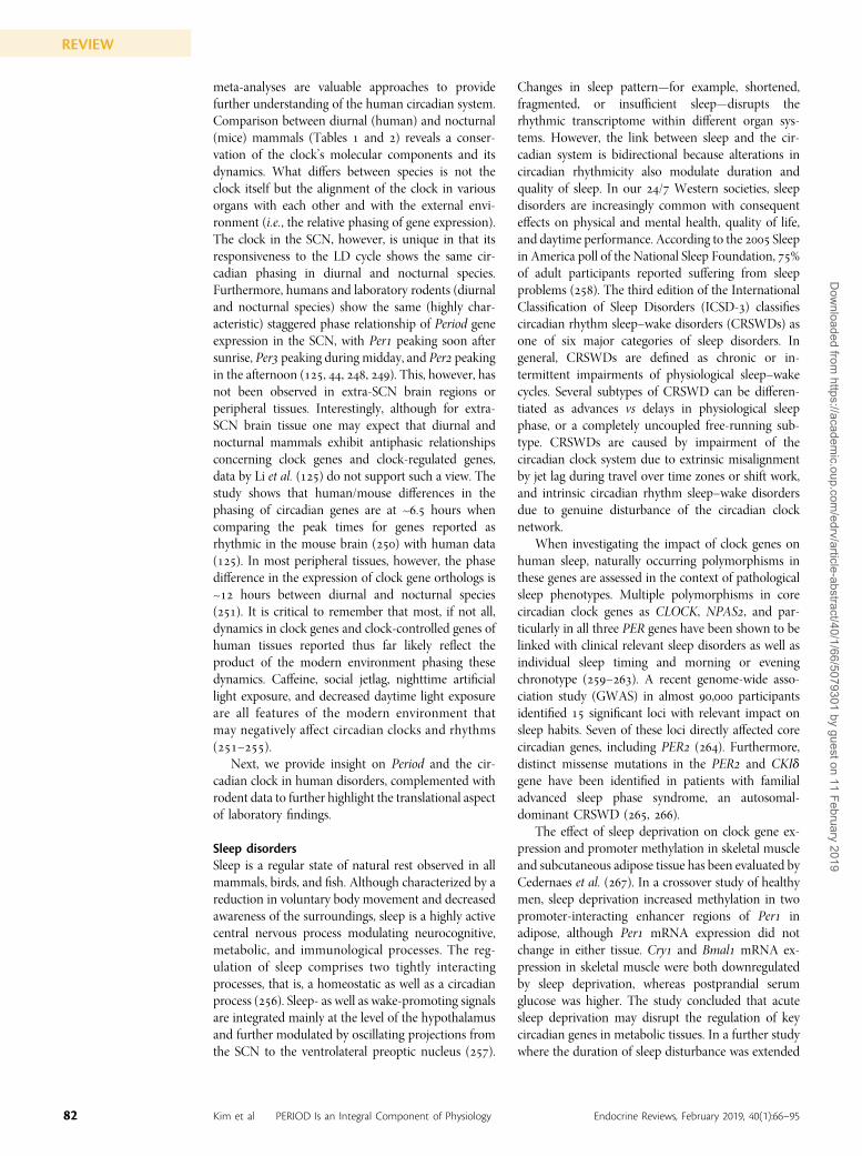
meta-analyses are valuable approaches to providefurther understanding of the human circadian system.Comparison between diurnal (human) and nocturnal(mice) mammals (Tables and ) reveals a conser-vation of the clock’s molecular components and itsdynamics. What differs between species is not theclock itself but the alignment of the clock in variousorgans with each other and with the external envi-ronment (i.e., the relative phasing of gene expression).The clock in the SCN, however, is unique in that itsresponsiveness to the LD cycle shows the same cir-cadian phasing in diurnal and nocturnal species.Furthermore, humans and laboratory rodents (diurnaland nocturnal species) show the same (highly char-acteristic) staggered phase relationship of Period geneexpression in the SCN, with Per peaking soon aftersunrise, Per peaking during midday, and Per peakingin the afternoon (, , , ). This, however, hasnot been observed in extra-SCN brain regions orperipheral tissues. Interestingly, although for extra-SCN brain tissue one may expect that diurnal andnocturnal mammals exhibit antiphasic relationshipsconcerning clock genes and clock-regulated genes,data by Li et al. () do not support such a view. Thestudy shows that human/mouse differences in thephasing of circadian genes are at ~. hours whencomparing the peak times for genes reported asrhythmic in the mouse brain () with human data(). In most peripheral tissues, however, the phasedifference in the expression of clock gene orthologs is~ hours between diurnal and nocturnal species(). It is critical to remember that most, if not all,dynamics in clock genes and clock-controlled genes ofhuman tissues reported thus far likely reflect theproduct of the modern environment phasing thesedynamics. Caffeine, social jetlag, nighttime artificiallight exposure, and decreased daytime light exposureare all features of the modern environment thatmay negatively affect circadian clocks and rhythms(–).
Next, we provide insight on Period and the cir-cadian clock in human disorders, complemented withrodent data to further highlight the translational aspectof laboratory findings.
Sleep disordersSleep is a regular state of natural rest observed in allmammals, birds, and fish. Although characterized by areduction in voluntary body movement and decreasedawareness of the surroundings, sleep is a highly activecentral nervous process modulating neurocognitive,metabolic, and immunological processes. The reg-ulation of sleep comprises two tightly interactingprocesses, that is, a homeostatic as well as a circadianprocess (). Sleep- as well as wake-promoting signalsare integrated mainly at the level of the hypothalamusand further modulated by oscillating projections fromthe SCN to the ventrolateral preoptic nucleus ().
Changes in sleep pattern—for example, shortened,fragmented, or insufficient sleep—disrupts therhythmic transcriptome within different organ sys-tems. However, the link between sleep and the cir-cadian system is bidirectional because alterations incircadian rhythmicity also modulate duration andquality of sleep. In our / Western societies, sleepdisorders are increasingly common with consequenteffects on physical and mental health, quality of life,and daytime performance. According to the Sleepin America poll of the National Sleep Foundation, %of adult participants reported suffering from sleepproblems (). The third edition of the InternationalClassification of Sleep Disorders (ICSD-) classifiescircadian rhythm sleep–wake disorders (CRSWDs) asone of six major categories of sleep disorders. Ingeneral, CRSWDs are defined as chronic or in-termittent impairments of physiological sleep–wakecycles. Several subtypes of CRSWD can be differen-tiated as advances vs delays in physiological sleepphase, or a completely uncoupled free-running sub-type. CRSWDs are caused by impairment of thecircadian clock system due to extrinsic misalignmentby jet lag during travel over time zones or shift work,and intrinsic circadian rhythm sleep–wake disordersdue to genuine disturbance of the circadian clocknetwork.
When investigating the impact of clock genes onhuman sleep, naturally occurring polymorphisms inthese genes are assessed in the context of pathologicalsleep phenotypes. Multiple polymorphisms in corecircadian clock genes as CLOCK, NPAS, and par-ticularly in all three PER genes have been shown to belinked with clinical relevant sleep disorders as well asindividual sleep timing and morning or eveningchronotype (–). A recent genome-wide asso-ciation study (GWAS) in almost , participantsidentified significant loci with relevant impact onsleep habits. Seven of these loci directly affected corecircadian genes, including PER (). Furthermore,distinct missense mutations in the PER and CKIdgene have been identified in patients with familialadvanced sleep phase syndrome, an autosomal-dominant CRSWD (, ).
The effect of sleep deprivation on clock gene ex-pression and promoter methylation in skeletal muscleand subcutaneous adipose tissue has been evaluated byCedernaes et al. (). In a crossover study of healthymen, sleep deprivation increased methylation in twopromoter-interacting enhancer regions of Per inadipose, although Per mRNA expression did notchange in either tissue. Cry and Bmal mRNA ex-pression in skeletal muscle were both downregulatedby sleep deprivation, whereas postprandial serumglucose was higher. The study concluded that acutesleep deprivation may disrupt the regulation of keycircadian genes in metabolic tissues. In a further studywhere the duration of sleep disturbance was extended
82 Kim et al PERIOD Is an Integral Component of Physiology Endocrine Reviews, February 2019, 40(1):66–95
REVIEWD
ownloaded from
https://academic.oup.com
/edrv/article-abstract/40/1/66/5079301 by guest on 11 February 2019
to week, the circadian mRNA expression of Per,Per, and Per in whole-blood RNA samples wasexamined. Whereas all three Per genes remainedrhythmic, Per was upregulated during sleep dep-rivation and after sleep restriction, together withgenes associated with the inflammatory response().
Psychiatric disordersBeyond their impact on physical health and disease,circadian clock genes might also play a role in mentalstate and psychiatric disorders (). Because sleepdisorders are highly prevalent in psychiatric disease,there seems to be a tridirectional relationship con-necting circadian rhythms, sleep, and mental health.One hypothesis for the development of psychiatricdisorders related to impairment of the circadiannetwork postulates an individual’s reduced ability toadapt to environmental changes due to a mismatchbetween individual internal and external rhythms.Likewise, studies on early development of social com-munication highlight the necessity of environment-adapted synchrony in motor, emotional, and in-terpersonal rhythms for mental health (). The earlyphase of physiological brain development is, amongother factors, under control of clock genes ().Therefore, an impaired clock network might contributeto a higher susceptibility for a later development ofpsychiatric disorders (). A mechanistic link mightbe a disturbance in sensory processing by parvalbuminneurons under conditions of an insufficient circadianclock network. Similarly, a knockdown of parvalbuminin mice leads to an autism-like phenotype ().
In humans, target gene approaches and GWASshave identified a possible link between single-nucleotide polymorphisms in relevant genes of theclock machinery and multiple psychiatric disorders. Inpatients with bipolar disorders, several polymorphismsin core clock genes have been associated with rapidcycling between episodes of mania and depression(CRY) as well as the risk of bipolar disorder andschizophrenia onset (PER) (, ). Moreover, arecent study showed an association between weakercyclic patterns in PER– and BMAL in brains ofpatients with major depression as compared withhealthy controls (). Similar associations have beenshown for the psychiatric spectrum of attention deficithyperactivity () and anxiety disorder ().
Human studies have relied on associations that areunable to establish causality. Impairment of circadianrhythmicity is associated with sleep disturbances andpsychiatric disorders, but we cannot conclude whichis the chicken or the egg. One possible mechanisticlink between impairment of the circadian clocknetwork, sleep disorders, and psychiatric disordersmight be alterations in GC rhythms, which are highlyrelevant in the context of mental health as well assleep (, ).
ObesityExisting evidence points to a strong genetic associationbetween the circadian system and obesity. A GWASexploring the genetic predisposition of obesity withhypothalamic genes disclosed that common variants inPER and NUCB, a gene coding for nesfatin-, anadipokine involved in the circadian regulation of foodintake, show a significant allelic association withobesity (). Accordingly, it might be postulated thatcircadian disruptions lead to metabolic diseases. In-deed, circadian alterations caused by shift work andsleep deprivation have been linked to unfavorablemetabolic sequelae [reviewed in Ref. ()]. Shiftworkers suffering from disrupted circadian physiologyand sleep disorder are more prone to eat at night andbecome obese (, ). Even short exposure ofnormal, healthy, young adults to sleep deprivation inthe laboratory increases appetite and food intake as-sociated with decreased leptin and increased ghrelinlevels (, ), reflecting a condition known as“night eating syndrome.” Animal research reveals adirect link between circadian clock genes and nighteating syndrome. A study in mice deficient in the clockprotein PER shows that the circadian clock regulatestwo critical metabolic rhythms, GC release and foodintake (), both of which are absent in Per2/2mice.On a high-fat diet, the food intake in Per2/2 mice issimilar across the light and dark phases and results insignificant obesity. The diurnal rhythm of neuroen-docrine peptide a-melanocyte-stimulating hormone(aMSH), a major effector of appetite control, is dis-rupted in Per2/2 mice. Peripheral injection ofaMSH, which has been shown to enter the brain,restores the feeding rhythm and induces weight loss inPer2/2 mice. These findings emphasize the impor-tance of Per in appetite control during the sleepingphase and the potential role of peripherally admin-istered aMSH in restoring a night–day eating patternin individuals with circadian eating disorders such asnight eating syndrome, associated with obesity. Ad-ditional studies in mice also demonstrated that ani-mals with access to a high-fat diet restricted to thesleep phase for weeks gain .-fold more weight thando control mice fed during the active phase ().These findings are further supported by a recent study() showing that week of restricted feeding duringthe inactive phase causes hyperphagia and increasingadiposity and plasma leptin levels as compared withnighttime feeding. Mistimed food consumption alsoprominently affects the expression of circadian clockgenes (Bmal, RevErba, Per, and Per), which showeither phase advances or inverted expression profilesin liver and white adipose tissue in inactive phase mice.Moreover, time-restricted access to a high-fat dietduring an extended period ( weeks) ameliorates thedisruptive effects of diet-induced obesity. Mice fedwith a high-fat diet during the active phase gained %less body weight, decreased serum cholesterol by %,
“The timing of feeding hasa pivotal role and canameliorate the detrimentaleffects of an obesogenic diet.”
83doi: 10.1210/er.2018-00049 https://academic.oup.com/edrv
REVIEWD
ownloaded from
https://academic.oup.com
/edrv/article-abstract/40/1/66/5079301 by guest on 11 February 2019
and increased insulin sensitivity more than threefoldcompared with mice fed ad libitum. More importantly,the disrupted circadian expression profiles of the clockgenes Per, Cry, and Clock in the liver induced by anad libitum high-fat diet can be restored via a time-restricted high-fat diet (). As such, the timing offeeding has a pivotal role and can ameliorate thedetrimental effects of an obesogenic diet.
In humans, both the expression levels and circa-dian rhythmicity of clock genes, including Per, Per,and Per, in subcutaneous adipose tissue are similar inlean, overweight/obese, and overweight/obese in-dividuals with type diabetes mellitus (TDM) ().Nevertheless, there appears to be good evidence thatthe expression of clock genes is altered by weightchanges. As such, PER and NRD expression isincreased in subcutaneous adipose tissue after a periodof weeks of weight loss. Expression levels are tightlycorrelated with body mass index (BMI), serum cho-lesterol levels, and several metabolic and inflammatorygenes. These data also emphasize the close interactionbetween clock changes and alterations in adiposetissue mass and metabolism ().
The adipokine leptin, a potent appetite suppres-sant, plays an important role in the interaction ofperipheral signals with the SCN to maintain energyhomeostasis. Plasma leptin levels show robust diurnalrhythms in both humans and rodents that are closelyassociated with food intake. Food availability restrictedto the inactive phase in mice reverses the diurnal leptinrhythm (). Leptin resistance is a hallmark of obesityand is significantly affected by circadian disruptionssuch as jet lag or clock gene knockout in mice ().Kettner et al. () demonstrated that both acute andchronic jet lag alone is sufficient to induce leptinresistance by disrupting cycling leptin concentrations.The importance of circadian clocks in leptin regulationwas further emphasized by findings showing that Per/ double-knockout mice display abolished daily var-iations in plasma leptin resulting in complete loss ofleptin feedback to the arcuate nucleus. These findingssuggest that food intake at the wrong time of day candesynchronize peripheral clocks, resulting in meta-bolic disorders by mechanisms involving leptin re-sistance, hyperphagia, physical inactivity, and adiposity(Fig. ).
The question as to which of these physiologicalchanges that may contribute to obesity are actuallymediated by the circadian clock, has been raised byHusse et al. (). This study investigated the role ofthe circadian clock machinery in metabolism underconditions of sleep disruption using the mouse model.The group examined the effects of timed sleep re-striction (TSR)—a protocol that sleep deprives micefor their first hours of sleep time during the lightphase—in WT and in arrhythmic Per/ double-mutant mice. WT mice subjected to TSR show in-creased food intake, hyperleptinemia, and a significant
upregulation of genes involved in lipid biosynthesis inwhite adipose tissue. These changes persist for at least week after TSR and thus indicate the onset of obesityand associated leptin resistance. Importantly, TSR-induced persistent metabolic effects observed in WTmice were absent in Per/ mutants, suggesting aprotective role of Per genes on metabolic deteriorationresulting from sleep disruption (). The extent towhich these protecting effects are mediated by Per orPer or the overall disruption of endogenous rhyth-micity remains to be determined.
Type 2 diabetesThe epidemic of diabetes is increasing at an alarmingrate, concurrent with the prevalence of obesitythroughout the world. TDM is a multifactorialcomplex metabolic disease that occurs due to an in-teraction between genetic predisposition and envi-ronmental factors (, ). A number of studiessupport the involvement of circadian clock genes inregulating glucose metabolism and diabetes. Clinicalstudies in individuals suffering from sleep disruptionor circadian misalignment show an increased sus-ceptibility and incidence of TDM, evidenced by in-sulin resistance, impaired glucose tolerance, andhyperglycemia (–). A study by Englund et al.() addressed whether circadian clock poly-morphisms contribute to seasonal variations in be-havior and to the metabolic syndrome in humans. Thegroup genotyped single nucleotide polymorphismsfrom genes that were either canonical circadianclock genes or genes related to the circadian clock-work from individuals. The findings link circa-dian gene variants to the risk factors of the metabolicsyndrome, because Npas was associated with hy-pertension and Per was associated with high fastingblood glucose.
Mice deficient for the core clock genes Bmal orClock display impaired recovery of normal bloodglucose levels after insulin-induced hypoglycemia,indicative of a dysfunction in hepatic gluconeogenesis(). Furthermore, prominent hepatic sequelae of themetabolic syndrome such as steatosis, hyperlipidemia,and altered secretion of hepatokines are observed inthese mice (), stressing the importance of thecircadian clock in glucose homeostasis.
Rodent studies show that the expression of clockgenes is significantly affected in nonobese mice withinsulin resistance, a contributor to the development ofTDM (, ). Furthermore, nonobese Cry mu-tant mice show early onset diabetes mellitus withsymptoms similar to those of maturity onset diabetesof the young in humans in terms of early onset,nonobesity, and primary dysfunction of b-cells ().In accordance with these findings, administration ofglucose has been shown to modify the expression ofPer and Per in fibroblasts and immortalized hy-pothalamic cell lines (, ). Clinical studies have
84 Kim et al PERIOD Is an Integral Component of Physiology Endocrine Reviews, February 2019, 40(1):66–95
REVIEWD
ownloaded from
https://academic.oup.com
/edrv/article-abstract/40/1/66/5079301 by guest on 11 February 2019
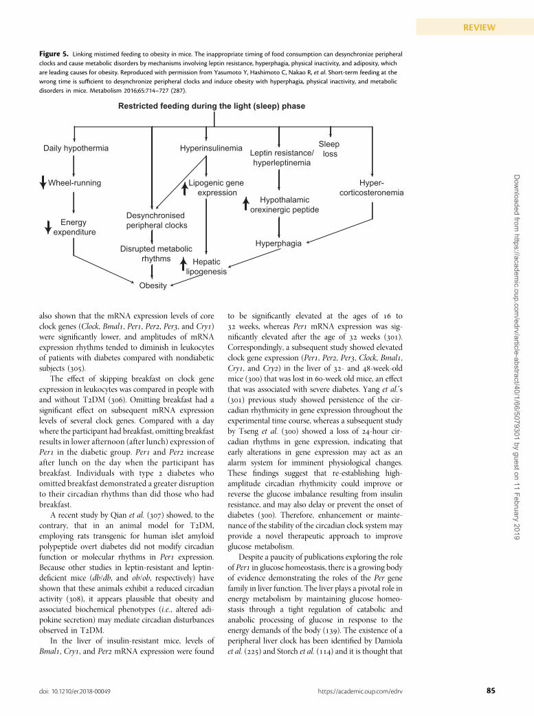
also shown that the mRNA expression levels of coreclock genes (Clock, Bmal, Per, Per, Per, and Cry)were significantly lower, and amplitudes of mRNAexpression rhythms tended to diminish in leukocytesof patients with diabetes compared with nondiabeticsubjects ().
The effect of skipping breakfast on clock geneexpression in leukocytes was compared in people withand without TDM (). Omitting breakfast had asignificant effect on subsequent mRNA expressionlevels of several clock genes. Compared with a daywhere the participant had breakfast, omitting breakfastresults in lower afternoon (after lunch) expression ofPer in the diabetic group. Per and Per increaseafter lunch on the day when the participant hasbreakfast. Individuals with type diabetes whoomitted breakfast demonstrated a greater disruptionto their circadian rhythms than did those who hadbreakfast.
A recent study by Qian et al. () showed, to thecontrary, that in an animal model for TDM,employing rats transgenic for human islet amyloidpolypeptide overt diabetes did not modify circadianfunction or molecular rhythms in Per expression.Because other studies in leptin-resistant and leptin-deficient mice (db/db, and ob/ob, respectively) haveshown that these animals exhibit a reduced circadianactivity (), it appears plausible that obesity andassociated biochemical phenotypes (i.e., altered adi-pokine secretion) may mediate circadian disturbancesobserved in TDM.
In the liver of insulin-resistant mice, levels ofBmal, Cry, and Per mRNA expression were found
to be significantly elevated at the ages of to weeks, whereas Per mRNA expression was sig-nificantly elevated after the age of weeks ().Correspondingly, a subsequent study showed elevatedclock gene expression (Per, Per, Per, Clock, Bmal,Cry, and Cry) in the liver of - and -week-oldmice () that was lost in -week old mice, an effectthat was associated with severe diabetes. Yang et al.’s() previous study showed persistence of the cir-cadian rhythmicity in gene expression throughout theexperimental time course, whereas a subsequent studyby Tseng et al. () showed a loss of -hour cir-cadian rhythms in gene expression, indicating thatearly alterations in gene expression may act as analarm system for imminent physiological changes.These findings suggest that re-establishing high-amplitude circadian rhythmicity could improve orreverse the glucose imbalance resulting from insulinresistance, and may also delay or prevent the onset ofdiabetes (). Therefore, enhancement or mainte-nance of the stability of the circadian clock system mayprovide a novel therapeutic approach to improveglucose metabolism.
Despite a paucity of publications exploring the roleof Per in glucose homeostasis, there is a growing bodyof evidence demonstrating the roles of the Per genefamily in liver function. The liver plays a pivotal role inenergy metabolism by maintaining glucose homeo-stasis through a tight regulation of catabolic andanabolic processing of glucose in response to theenergy demands of the body (). The existence of aperipheral liver clock has been identified by Damiolaet al. () and Storch et al. () and it is thought that
Figure 5. Linking mistimed feeding to obesity in mice. The inappropriate timing of food consumption can desynchronize peripheralclocks and cause metabolic disorders by mechanisms involving leptin resistance, hyperphagia, physical inactivity, and adiposity, whichare leading causes for obesity. Reproduced with permission from Yasumoto Y, Hashimoto C, Nakao R, et al. Short-term feeding at thewrong time is sufficient to desynchronize peripheral clocks and induce obesity with hyperphagia, physical inactivity, and metabolicdisorders in mice. Metabolism 2016;65:714–727 (287).
85doi: 10.1210/er.2018-00049 https://academic.oup.com/edrv
REVIEWD
ownloaded from
https://academic.oup.com
/edrv/article-abstract/40/1/66/5079301 by guest on 11 February 2019
hepatocyte clocks are involved in maintaining meta-bolic homeostasis by integrating external food cues totemporally tune the expression of genes that encodefor metabolism-associated enzymes. It appears thatPER in the liver may be involved in food entrainmentof local clocks. Glucose was reported to be a key step intriggering the expression of clock genes by down-regulating Per and Per mRNA in rat- fibroblasts(). Behavioral studies in mice show that acute foodintake downregulates Per transcripts in the liver byinhibiting CREB signaling () while upregulatingPer, possibly through a postprandial rise in bodytemperature leading to an increase in heat shockprotein production, which then bind to a cis-actingenhancer element in the Per promoter (). Re-setting of the liver clock by feeding has been at-tributed to a number of food-associated pancreatichormones such as insulin and glucagon. Recently,oxyntomodulin, an anorexigenic gut-derived hor-mone stimulated by food intake, has been proposedas a novel direct pathway by which food may resetliver clocks via the specific induction of Per and Per().
Per has also been investigated for its role in he-patic lipid metabolism and is linked to diabetes bysuppressing insulin signaling. PER activates peroxi-some proliferator-activated receptor (Ppar)a (), amaster lipid regulator involved in the modulation ofhepatic fatty acid oxidation (), and Bhlhe (Dec)and Bhlhe (Dec), which are thought to be re-pressors of adipogenesis. The noncanonical mecha-nism involved suggests that PER suppresses Cry,which in turn leads to the suppression of Clock/Bmal.The function of Per in hepatic fatty acid metabolismwas further confirmed in mice lacking Per, whichshow reduced ethanol-induced triglyceride accu-mulation in liver, alleviating ethanol-induced hepa-totoxicity. Overexpression of Per reverses thisphenotype, leading to an upregulation of genes relatedto lipogenesis such as Pparg, a nuclear receptor criticalfor adipogenesis, and downstream target genes ().Similarly, Per has been found to directly and spe-cifically repress Pparg by blocking its recruitment totarget promoters (). Ablation of PER in mice isassociated with increased activation of adipogenicgenes and markers and a drastic decrease in totalserum triacylglyceride (TAG) levels (). This in-teraction between Per and Pparg seems to beparalog-specific, as Per, despite sharing a high de-gree of homology with Per, does not show anyinvolvement in adipogenesis. Collectively, thisindicates a role for the Per gene family in main-taining body fat and serum TAG levels in mammalsby regulating liver fatty acid metabolism andadipogenesis. In contrast, recent evidence byAdamovich et al. () suggests that diurnal os-cillations in hepatic triglycerides and mRNA ex-pression of TAG metabolism-associated enzymes
can persist in Per/ double-knockout mice. It isunclear whether this is due to functional redundancywithin the Per family, as no studies have examinedhepatic lipid metabolism in Per// triple-knockoutanimals.
GC disordersSuccessful adaptation to the environment depends onan organism’s ability to appropriately modulate itsstress response (). Central to this modulation arethe adrenals, whose cortex and medulla releasehormones in a time-of-day–dependent fashion, withGC secretion being the best studied in terms of itscircadian effects (). The loss of circadian se-cretion of cortisol is one of the most sensitive andspecific diagnostic features of Cushing syndrome,and thus is recommended by the guidelines toevaluate the disease state. Adrenalectomy in com-bination with postoperative GC replacement, usinghydrocortisone, is a common treatment of Cushingsyndrome.
A human study investigated the acute effects ofhydrocortisone administration on Per gene expressionin whole blood. Hydrocortisone resulted in inductionof Per and Per but there was no effect on PermRNA expression (). The hydrocortisone-inducedincrease in Per mRNA was partly suppressed by theGC receptor antagonist RU. The doses of hy-drocortisone result in serum cortisol concentrationssimilar to those observed during acute stress, implyingthat peripheral Per and Per may be temporarilyupregulated during acute stress in humans. When thehydrocortisone administration was administeredrepeatedly, the acute effect on increasing Per ex-pression was confirmed; this experiment used pe-ripheral blood mononuclear cells. Additionally, after days of hydrocortisone administration ( mg/dtaken hours after their scheduled time of waking),the phases of Per, Per, and Bmal were shifted bybetween . and . hours, further supporting thenotion that peripheral clocks are entrained by GCs inhumans ().
Cardiovascular diseasesEpidemiological data report that cardiovascular eventssuch as myocardial infarction, sudden cardiac death,angina pectoris, ischemic and hemorrhagic stroke, andventricular tachycardia exhibit circadian variation(, ). Hemodynamic parameters such as bloodpressure and heart rate as well as gene and proteinexpression of known regulators of vascular physi-ology show circadian oscillations (–). That is,human endothelial vasodilatory activity decreases inthe early morning and coronary artery tone in-creases in the morning, whereas blood pressure andheart rate increase in the early morning. Therefore,chronodisruption/circadian misalignment as com-monly observed in night shift workers significantly
86 Kim et al PERIOD Is an Integral Component of Physiology Endocrine Reviews, February 2019, 40(1):66–95
REVIEWD
ownloaded from
https://academic.oup.com
/edrv/article-abstract/40/1/66/5079301 by guest on 11 February 2019
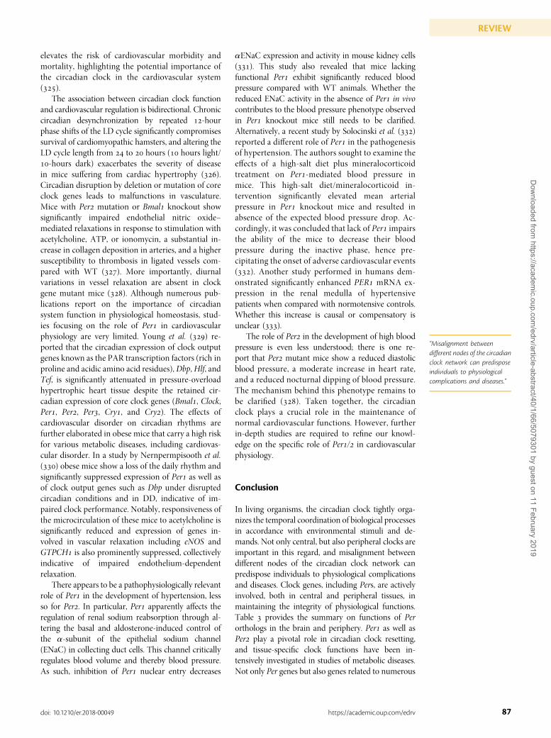
elevates the risk of cardiovascular morbidity andmortality, highlighting the potential importance ofthe circadian clock in the cardiovascular system().
The association between circadian clock functionand cardiovascular regulation is bidirectional. Chroniccircadian desynchronization by repeated -hourphase shifts of the LD cycle significantly compromisessurvival of cardiomyopathic hamsters, and altering theLD cycle length from to hours ( hours light/-hours dark) exacerbates the severity of diseasein mice suffering from cardiac hypertrophy ().Circadian disruption by deletion or mutation of coreclock genes leads to malfunctions in vasculature.Mice with Per mutation or Bmal knockout showsignificantly impaired endothelial nitric oxide–mediated relaxations in response to stimulation withacetylcholine, ATP, or ionomycin, a substantial in-crease in collagen deposition in arteries, and a highersusceptibility to thrombosis in ligated vessels com-pared with WT (). More importantly, diurnalvariations in vessel relaxation are absent in clockgene mutant mice (). Although numerous pub-lications report on the importance of circadiansystem function in physiological homeostasis, stud-ies focusing on the role of Per in cardiovascularphysiology are very limited. Young et al. () re-ported that the circadian expression of clock outputgenes known as the PAR transcription factors (rich inproline and acidic amino acid residues), Dbp,Hlf, andTef, is significantly attenuated in pressure-overloadhypertrophic heart tissue despite the retained cir-cadian expression of core clock genes (Bmal, Clock,Per, Per, Per, Cry, and Cry). The effects ofcardiovascular disorder on circadian rhythms arefurther elaborated in obese mice that carry a high riskfor various metabolic diseases, including cardiovas-cular disorder. In a study by Nernpermpisooth et al.() obese mice show a loss of the daily rhythm andsignificantly suppressed expression of Per as well asof clock output genes such as Dbp under disruptedcircadian conditions and in DD, indicative of im-paired clock performance. Notably, responsiveness ofthe microcirculation of these mice to acetylcholine issignificantly reduced and expression of genes in-volved in vascular relaxation including eNOS andGTPCH is also prominently suppressed, collectivelyindicative of impaired endothelium-dependentrelaxation.
There appears to be a pathophysiologically relevantrole of Per in the development of hypertension, lessso for Per. In particular, Per apparently affects theregulation of renal sodium reabsorption through al-tering the basal and aldosterone-induced control ofthe a-subunit of the epithelial sodium channel(ENaC) in collecting duct cells. This channel criticallyregulates blood volume and thereby blood pressure.As such, inhibition of Per nuclear entry decreases
aENaC expression and activity in mouse kidney cells(). This study also revealed that mice lackingfunctional Per exhibit significantly reduced bloodpressure compared with WT animals. Whether thereduced ENaC activity in the absence of Per in vivocontributes to the blood pressure phenotype observedin Per knockout mice still needs to be clarified.Alternatively, a recent study by Solocinski et al. ()reported a different role of Per in the pathogenesisof hypertension. The authors sought to examine theeffects of a high-salt diet plus mineralocorticoidtreatment on Per-mediated blood pressure inmice. This high-salt diet/mineralocorticoid in-tervention significantly elevated mean arterialpressure in Per knockout mice and resulted inabsence of the expected blood pressure drop. Ac-cordingly, it was concluded that lack of Per impairsthe ability of the mice to decrease their bloodpressure during the inactive phase, hence pre-cipitating the onset of adverse cardiovascular events(). Another study performed in humans dem-onstrated significantly enhanced PER mRNA ex-pression in the renal medulla of hypertensivepatients when compared with normotensive controls.Whether this increase is causal or compensatory isunclear ().
The role of Per in the development of high bloodpressure is even less understood; there is one re-port that Per mutant mice show a reduced diastolicblood pressure, a moderate increase in heart rate,and a reduced nocturnal dipping of blood pressure.The mechanism behind this phenotype remains tobe clarified (). Taken together, the circadianclock plays a crucial role in the maintenance ofnormal cardiovascular functions. However, furtherin-depth studies are required to refine our knowl-edge on the specific role of Per/ in cardiovascularphysiology.
Conclusion
In living organisms, the circadian clock tightly orga-nizes the temporal coordination of biological processesin accordance with environmental stimuli and de-mands. Not only central, but also peripheral clocks areimportant in this regard, and misalignment betweendifferent nodes of the circadian clock network canpredispose individuals to physiological complicationsand diseases. Clock genes, including Pers, are activelyinvolved, both in central and peripheral tissues, inmaintaining the integrity of physiological functions.Table provides the summary on functions of Perorthologs in the brain and periphery. Per as well asPer play a pivotal role in circadian clock resetting,and tissue-specific clock functions have been in-tensively investigated in studies of metabolic diseases.Not only Per genes but also genes related to numerous
“Misalignment betweendifferent nodes of the circadianclock network can predisposeindividuals to physiologicalcomplications and diseases.”
87doi: 10.1210/er.2018-00049 https://academic.oup.com/edrv
REVIEWD
ownloaded from
https://academic.oup.com
/edrv/article-abstract/40/1/66/5079301 by guest on 11 February 2019

physiological processes, including sleep and energymetabolism, have been reported to be significantlyaffected under the circumstances of chronodisruption.Also, genetic or diet-induced disease conditionscan alter Per gene expression, emphasizing the bi-directional connection between circadian clock andphysiology. Improvements in entrainability to differ-ent lengths of LD cycles and metabolic functions in theabsence of Per indicate that Per may play a crucialrole in synchronization between central and peripheralclocks.
We acknowledge that the in vivo evidence sup-porting a role of PER in endocrine and metabolicfunctions is currently based on global Per knockoutstudies in rodents and observations in humans withsingle nucleotide polymorphisms in the Per gene.The substantial amount of evidence provided inthis review for the role of Period in endocrineand metabolic regulation as well as disordersshould drive the generation of cell-specific Perknockout models to prove the specific and highlyrelevant role of Period genes on the tissue-specific
Table 3. The Functions of PERIOD in Central and Peripheral Tissues of the Mouse
Tissue Function: Tissue Gene
Function: Gene
ReferencesCommon Specific
Central SCN Central pacemaker of circadian system Per1 Photic resetting N/A (45, 51, 52)
Per2 Maintenance of circadian rhythm stability (45, 46, 52)
Per3 Possible extra-clock functions (218)
Hippocampus Learning and memory Per1 Modulation of hippocampal memory-relevant signaling molecules andspatial working memory
(74, 75, 104)
Per2 Memory recall (96)
Periphery Adiposetissue
Fat storage and regulation of metabolichomeostasis via the secretion ofadipocytokines
Per1 Rhythmic regulation ofleptin levels
N/A (133, 280,289, 291,292)
Per2 Energy homeostasis N/A (133, 289,291, 292)
Per3 N/A Modulation of adipogenesis and lipid handling (190)
Liver Regulation of glucose homeostasis Per1 Modulation ofhomeostatichepatocytesturnover
Regulation of hepatic lipid metabolism (309, 310,313)
Per2 Maintenance ofhepatocyte clockfunction
Regulation of adipogenesis (309, 310,314)
Resets the hepaticclock
Regulates insulinresponse
Adrenal gland Regulation and synchronization ofphysiological functions in periphery
Per1 Mediates light-induced secretion of GCs via the autonomic nervoussystem Regulation of rhythmic secretion of GCs and StAR expression
(39, 166)
Per2 Regulation of adrenal sensitivity to ACTH; regulation of circadian rhythmsof ACTH, GCs, and local clock
(167)
Heart andvasculature
Blood, oxygen and nutrients supply andremoval of metabolic waste products
Per1 Regulation of blood pressure rhythm (331, 332)
Per2
Other FEO Resetting of the behavior and physiologyof an organism by the time of foodavailability
Per1 Role of Per genes inFEOs remainscontroversial
Synchronization of peripheral oscillatorsaccording to the timing of feeding whenfeeding is restricted
(233, 234,239)
Per2
Per1–3 are widely expressed throughout the brain and the periphery. Centrally, PERIOD plays a major role in light-induced clock resetting. In the periphery, circadian oscillators arenecessary for maintaining metabolic homeostasis by synchronizing the associated physiological processes with environmental rhythms.Abbreviation: N/A, not available.
88 Kim et al PERIOD Is an Integral Component of Physiology Endocrine Reviews, February 2019, 40(1):66–95
REVIEWD
ownloaded from
https://academic.oup.com
/edrv/article-abstract/40/1/66/5079301 by guest on 11 February 2019
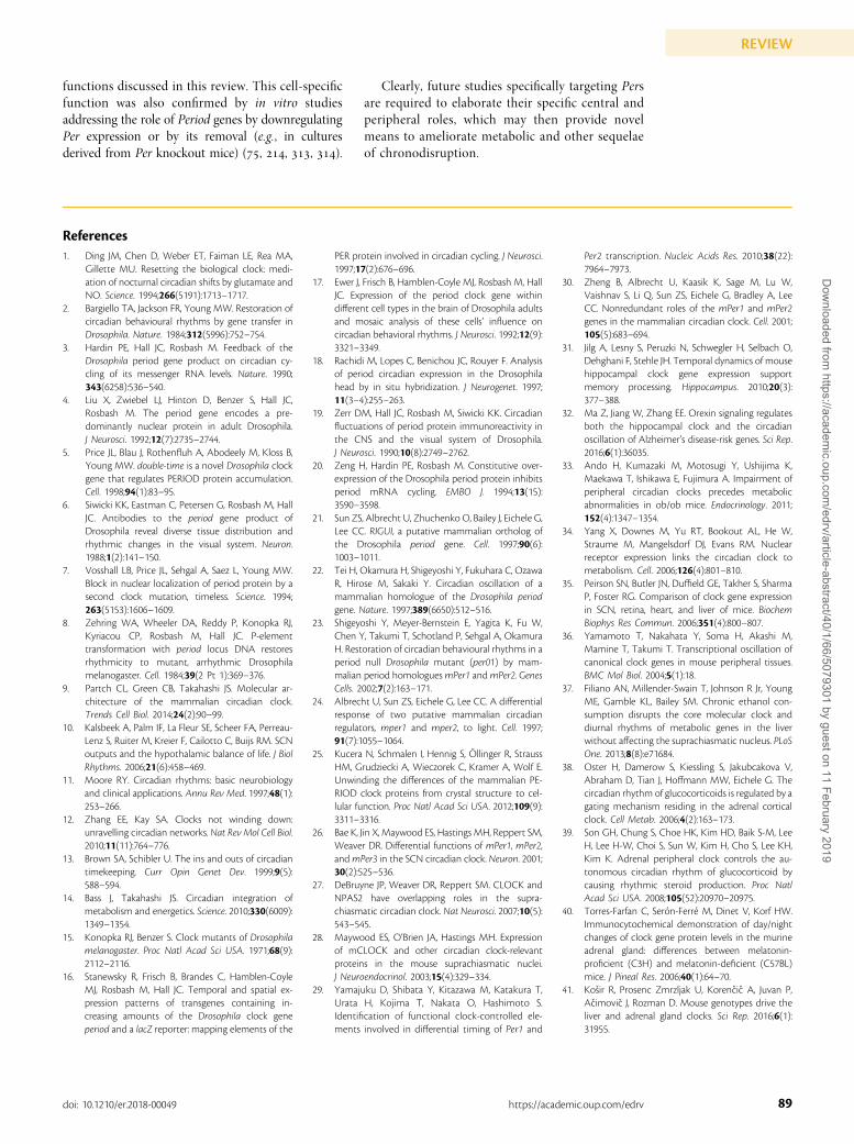
functions discussed in this review. This cell-specificfunction was also confirmed by in vitro studiesaddressing the role of Period genes by downregulatingPer expression or by its removal (e.g., in culturesderived from Per knockout mice) (, , , ).
Clearly, future studies specifically targeting Persare required to elaborate their specific central andperipheral roles, which may then provide novelmeans to ameliorate metabolic and other sequelaeof chronodisruption.
References1. Ding JM, Chen D, Weber ET, Faiman LE, Rea MA,
Gillette MU. Resetting the biological clock: medi-ation of nocturnal circadian shifts by glutamate andNO. Science. 1994;266(5191):1713–1717.
2. Bargiello TA, Jackson FR, Young MW. Restoration ofcircadian behavioural rhythms by gene transfer inDrosophila. Nature. 1984;312(5996):752–754.
3. Hardin PE, Hall JC, Rosbash M. Feedback of theDrosophila period gene product on circadian cy-cling of its messenger RNA levels. Nature. 1990;343(6258):536–540.
4. Liu X, Zwiebel LJ, Hinton D, Benzer S, Hall JC,Rosbash M. The period gene encodes a pre-dominantly nuclear protein in adult Drosophila.J Neurosci. 1992;12(7):2735–2744.
5. Price JL, Blau J, Rothenfluh A, Abodeely M, Kloss B,Young MW. double-time is a novel Drosophila clockgene that regulates PERIOD protein accumulation.Cell. 1998;94(1):83–95.
6. Siwicki KK, Eastman C, Petersen G, Rosbash M, HallJC. Antibodies to the period gene product ofDrosophila reveal diverse tissue distribution andrhythmic changes in the visual system. Neuron.1988;1(2):141–150.
7. Vosshall LB, Price JL, Sehgal A, Saez L, Young MW.Block in nuclear localization of period protein by asecond clock mutation, timeless. Science. 1994;263(5153):1606–1609.
8. Zehring WA, Wheeler DA, Reddy P, Konopka RJ,Kyriacou CP, Rosbash M, Hall JC. P-elementtransformation with period locus DNA restoresrhythmicity to mutant, arrhythmic Drosophilamelanogaster. Cell. 1984;39(2 Pt 1):369–376.
9. Partch CL, Green CB, Takahashi JS. Molecular ar-chitecture of the mammalian circadian clock.Trends Cell Biol. 2014;24(2):90–99.
10. Kalsbeek A, Palm IF, La Fleur SE, Scheer FA, Perreau-Lenz S, Ruiter M, Kreier F, Cailotto C, Buijs RM. SCNoutputs and the hypothalamic balance of life. J BiolRhythms. 2006;21(6):458–469.
11. Moore RY. Circadian rhythms: basic neurobiologyand clinical applications. Annu Rev Med. 1997;48(1):253–266.
12. Zhang EE, Kay SA. Clocks not winding down:unravelling circadian networks. Nat Rev Mol Cell Biol.2010;11(11):764–776.
13. Brown SA, Schibler U. The ins and outs of circadiantimekeeping. Curr Opin Genet Dev. 1999;9(5):588–594.
14. Bass J, Takahashi JS. Circadian integration ofmetabolism and energetics. Science. 2010;330(6009):1349–1354.
15. Konopka RJ, Benzer S. Clock mutants of Drosophilamelanogaster. Proc Natl Acad Sci USA. 1971;68(9):2112–2116.
16. Stanewsky R, Frisch B, Brandes C, Hamblen-CoyleMJ, Rosbash M, Hall JC. Temporal and spatial ex-pression patterns of transgenes containing in-creasing amounts of the Drosophila clock geneperiod and a lacZ reporter: mapping elements of the
PER protein involved in circadian cycling. J Neurosci.1997;17(2):676–696.
17. Ewer J, Frisch B, Hamblen-Coyle MJ, Rosbash M, HallJC. Expression of the period clock gene withindifferent cell types in the brain of Drosophila adultsand mosaic analysis of these cells’ influence oncircadian behavioral rhythms. J Neurosci. 1992;12(9):3321–3349.
18. Rachidi M, Lopes C, Benichou JC, Rouyer F. Analysisof period circadian expression in the Drosophilahead by in situ hybridization. J Neurogenet. 1997;11(3–4):255–263.
19. Zerr DM, Hall JC, Rosbash M, Siwicki KK. Circadianfluctuations of period protein immunoreactivity inthe CNS and the visual system of Drosophila.J Neurosci. 1990;10(8):2749–2762.
20. Zeng H, Hardin PE, Rosbash M. Constitutive over-expression of the Drosophila period protein inhibitsperiod mRNA cycling. EMBO J. 1994;13(15):3590–3598.
21. Sun ZS, Albrecht U, Zhuchenko O, Bailey J, Eichele G,Lee CC. RIGUI, a putative mammalian ortholog ofthe Drosophila period gene. Cell. 1997;90(6):1003–1011.
22. Tei H, Okamura H, Shigeyoshi Y, Fukuhara C, OzawaR, Hirose M, Sakaki Y. Circadian oscillation of amammalian homologue of the Drosophila periodgene. Nature. 1997;389(6650):512–516.
23. Shigeyoshi Y, Meyer-Bernstein E, Yagita K, Fu W,Chen Y, Takumi T, Schotland P, Sehgal A, OkamuraH. Restoration of circadian behavioural rhythms in aperiod null Drosophila mutant (per01) by mam-malian period homologuesmPer1 andmPer2. GenesCells. 2002;7(2):163–171.
24. Albrecht U, Sun ZS, Eichele G, Lee CC. A differentialresponse of two putative mammalian circadianregulators, mper1 and mper2, to light. Cell. 1997;91(7):1055–1064.
25. Kucera N, Schmalen I, Hennig S, Ollinger R, StraussHM, Grudziecki A, Wieczorek C, Kramer A, Wolf E.Unwinding the differences of the mammalian PE-RIOD clock proteins from crystal structure to cel-lular function. Proc Natl Acad Sci USA. 2012;109(9):3311–3316.
26. Bae K, Jin X, Maywood ES, Hastings MH, Reppert SM,Weaver DR. Differential functions of mPer1, mPer2,andmPer3 in the SCN circadian clock. Neuron. 2001;30(2):525–536.
27. DeBruyne JP, Weaver DR, Reppert SM. CLOCK andNPAS2 have overlapping roles in the supra-chiasmatic circadian clock. Nat Neurosci. 2007;10(5):543–545.
28. Maywood ES, O’Brien JA, Hastings MH. Expressionof mCLOCK and other circadian clock-relevantproteins in the mouse suprachiasmatic nuclei.J Neuroendocrinol. 2003;15(4):329–334.
29. Yamajuku D, Shibata Y, Kitazawa M, Katakura T,Urata H, Kojima T, Nakata O, Hashimoto S.Identification of functional clock-controlled ele-ments involved in differential timing of Per1 and
Per2 transcription. Nucleic Acids Res. 2010;38(22):7964–7973.
30. Zheng B, Albrecht U, Kaasik K, Sage M, Lu W,Vaishnav S, Li Q, Sun ZS, Eichele G, Bradley A, LeeCC. Nonredundant roles of the mPer1 and mPer2genes in the mammalian circadian clock. Cell. 2001;105(5):683–694.
31. Jilg A, Lesny S, Peruzki N, Schwegler H, Selbach O,Dehghani F, Stehle JH. Temporal dynamics of mousehippocampal clock gene expression supportmemory processing. Hippocampus. 2010;20(3):377–388.
32. Ma Z, Jiang W, Zhang EE. Orexin signaling regulatesboth the hippocampal clock and the circadianoscillation of Alzheimer’s disease-risk genes. Sci Rep.2016;6(1):36035.
33. Ando H, Kumazaki M, Motosugi Y, Ushijima K,Maekawa T, Ishikawa E, Fujimura A. Impairment ofperipheral circadian clocks precedes metabolicabnormalities in ob/ob mice. Endocrinology. 2011;152(4):1347–1354.
34. Yang X, Downes M, Yu RT, Bookout AL, He W,Straume M, Mangelsdorf DJ, Evans RM. Nuclearreceptor expression links the circadian clock tometabolism. Cell. 2006;126(4):801–810.
35. Peirson SN, Butler JN, Duffield GE, Takher S, SharmaP, Foster RG. Comparison of clock gene expressionin SCN, retina, heart, and liver of mice. BiochemBiophys Res Commun. 2006;351(4):800–807.
36. Yamamoto T, Nakahata Y, Soma H, Akashi M,Mamine T, Takumi T. Transcriptional oscillation ofcanonical clock genes in mouse peripheral tissues.BMC Mol Biol. 2004;5(1):18.
37. Filiano AN, Millender-Swain T, Johnson R Jr, YoungME, Gamble KL, Bailey SM. Chronic ethanol con-sumption disrupts the core molecular clock anddiurnal rhythms of metabolic genes in the liverwithout affecting the suprachiasmatic nucleus. PLoSOne. 2013;8(8):e71684.
38. Oster H, Damerow S, Kiessling S, Jakubcakova V,Abraham D, Tian J, Hoffmann MW, Eichele G. Thecircadian rhythm of glucocorticoids is regulated by agating mechanism residing in the adrenal corticalclock. Cell Metab. 2006;4(2):163–173.
39. Son GH, Chung S, Choe HK, Kim HD, Baik S-M, LeeH, Lee H-W, Choi S, Sun W, Kim H, Cho S, Lee KH,Kim K. Adrenal peripheral clock controls the au-tonomous circadian rhythm of glucocorticoid bycausing rhythmic steroid production. Proc NatlAcad Sci USA. 2008;105(52):20970–20975.
40. Torres-Farfan C, Seron-Ferre M, Dinet V, Korf HW.Immunocytochemical demonstration of day/nightchanges of clock gene protein levels in the murineadrenal gland: differences between melatonin-proficient (C3H) and melatonin-deficient (C57BL)mice. J Pineal Res. 2006;40(1):64–70.
41. Kosir R, Prosenc Zmrzljak U, Korencic A, Juvan P,Acimovic J, Rozman D. Mouse genotypes drive theliver and adrenal gland clocks. Sci Rep. 2016;6(1):31955.
89doi: 10.1210/er.2018-00049 https://academic.oup.com/edrv
REVIEWD
ownloaded from
https://academic.oup.com
/edrv/article-abstract/40/1/66/5079301 by guest on 11 February 2019
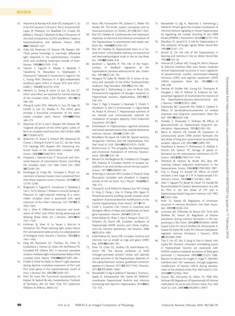
42. Marcheva B, Ramsey KM, Buhr ED, Kobayashi Y, SuH, Ko CH, Ivanova G, Omura C, Mo S, Vitaterna MH,Lopez JP, Philipson LH, Bradfield CA, Crosby SD,JeBailey L, Wang X, Takahashi JS, Bass J. Disruption ofthe clock components CLOCK and BMAL1 leads tohypoinsulinaemia and diabetes. Nature. 2010;466(7306):627–631.
43. Zylka MJ, Shearman LP, Weaver DR, Reppert SM.Three period homologs in mammals: differentiallight responses in the suprachiasmatic circadianclock and oscillating transcripts outside of brain.Neuron. 1998;20(6):1103–1110.
44. Takumi T, Taguchi K, Miyake S, Sakakida Y,Takashima N, Matsubara C, Maebayashi Y,Okumura K, Takekida S, Yamamoto S, Yagita K, YanL, Young MW, Okamura H. A light-independentoscillatory gene mPer3 in mouse SCN and OVLT.EMBO J. 1998;17(16):4753–4759.
45. Albrecht U, Zheng B, Larkin D, Sun ZS, Lee CC.mPer1 and mPer2 are essential for normal resettingof the circadian clock. J Biol Rhythms. 2001;16(2):100–104.
46. Zheng B, Larkin DW, Albrecht U, Sun ZS, Sage M,Eichele G, Lee CC, Bradley A. The mPer2 geneencodes a functional component of the mam-malian circadian clock. Nature. 1999;400(6740):169–173.
47. Shearman LP, Jin X, Lee C, Reppert SM, Weaver DR.Targeted disruption of the mPer3 gene: subtle ef-fects on circadian clock function.Mol Cell Biol. 2000;20(17):6269–6275.
48. Shearman LP, Sriram S, Weaver DR, Maywood ES,Chaves I, Zheng B, Kume K, Lee CC, van der HorstGTJ, Hastings MH, Reppert SM. Interacting mo-lecular loops in the mammalian circadian clock.Science. 2000;288(5468):1013–1019.
49. Hirayama J, Sassone-Corsi P. Structural and func-tional features of transcription factors controllingthe circadian clock. Curr Opin Genet Dev. 2005;15(5):548–556.
50. Pendergast JS, Friday RC, Yamazaki S. Photic en-trainment of period mutant mice is predicted fromtheir phase response curves. J Neurosci. 2010;30(36):12179–12184.
51. Shigeyoshi Y, Taguchi K, Yamamoto S, Takekida S,Yan L, Tei H, Moriya T, Shibata S, Loros JJ, Dunlap JC,Okamura H. Light-induced resetting of a mam-malian circadian clock is associated with rapidinduction of the mPer1 transcript. Cell. 1997;91(7):1043–1053.
52. Yan L, Silver R. Differential induction and locali-zation of mPer1 and mPer2 during advancing anddelaying phase shifts. Eur J Neurosci. 2002;16(8):1531–1540.
53. Kuhlman SJ, Silver R, Le Sauter J, Bult-Ito A,McMahon DG. Phase resetting light pulses inducePer1 and persistent spike activity in a subpopulationof biological clock neurons. J Neurosci. 2003;23(4):1441–1450.
54. Ding JM, Buchanan GF, Tischkau SA, Chen D,Kuriashkina L, Faiman LE, Alster JM, McPherson PS,Campbell KP, Gillette MU. A neuronal ryanodinereceptor mediates light-induced phase delays of thecircadian clock. Nature. 1998;394(6691):381–384.
55. Challet E, Poirel VJ, Malan A, Pevet P. Light exposureduring daytime modulates expression of Per1 andPer2 clock genes in the suprachiasmatic nuclei ofmice. J Neurosci Res. 2003;72(5):629–637.
56. Pierri JN, Lewis DA. Functional neuroanatomy. In:Kaplan HI, Sadock BJ, eds. Comprehensive Textbookof Psychiatry. 8th ed. New York, NY: LippincottWilliams & Wilkins; 2004:3–32.
57. Roxo MR, Franceschini PR, Zubaran C, Kleber FD,Sander JW. The limbic system conception and itshistorical evolution. Sci World J. 2011;11:2427–2441.
58. Ruit KG, Neafsey EJ. Cardiovascular and respiratoryresponses to electrical and chemical stimulation ofthe hippocampus in anesthetized and awake rats.Brain Res. 1988;457(2):310–321.
59. Ruit KG, Neafsey EJ. Hippocampal input to a “vis-ceral motor” corticobulbar pathway: an anatomicaland electrophysiological study in the rat. Exp BrainRes. 1990;82(3):606–616.
60. Jacobson L, Sapolsky R. The role of the hippo-campus in feedback regulation of thehypothalamic-pituitary-adrenocortical axis. EndocrRev. 1991;12(2):118–134.
61. Morgane PJ, Galler JR, Mokler DJ. A review of sys-tems and networks of the limbic forebrain/limbicmidbrain. Prog Neurobiol. 2005;75(2):143–160.
62. Prange-Kiel J, Wehrenberg U, Jarry H, Rune GM.Para/autocrine regulation of estrogen receptors inhippocampal neurons. Hippocampus. 2003;13(2):226–234.
63. Hojo Y, Higo S, Kawato S, Hatanaka Y, Ooishi Y,Murakami G, Ishii H, Komatsuzaki Y, Ogiue-IkedaM, Mukai H, Kimoto T. Hippocampal synthesis ofsex steroids and corticosteroids: essential formodulation of synaptic plasticity. Front Endocrinol(Lausanne). 2011;2:43.
64. Eichenbaum H. Hippocampus: cognitive processesand neural representations that underlie declarativememory. Neuron. 2004;44(1):109–120.
65. Broadbent NJ, Squire LR, Clark RE. Spatial memory,recognition memory, and the hippocampus. ProcNatl Acad Sci USA. 2004;101(40):14515–14520.
66. Richter-Levin G. The amygdala, the hippocampus,and emotional modulation of memory. Neurosci-entist. 2004;10(1):31–39.
67. Barnes CA, McNaughton BL, Goddard GV, DouglasRM, Adamec R. Circadian rhythm of synaptic ex-citability in rat and monkey central nervous system.Science. 1977;197(4298):91–92.
68. Scheving LE, Harrison WH, Gordon P, Pauly JE. Dailyfluctuation (circadian and ultradian) in biogenicamines of the rat brain. Am J Physiol. 1968;214(1):166–173.
69. Chiang CK, Xu B, Mehta N, Mayne J, SunWY, ChengK, Ning Z, Dong J, Zou H, Cheng HM, Figeys D.Phosphoproteome profiling reveals circadian clockregulation of posttranslational modifications in themurine hippocampus. Front Neurol. 2017;8:110.
70. Cirelli C, Gutierrez CM, Tononi G. Extensive anddivergent effects of sleep and wakefulness on braingene expression. Neuron. 2004;41(1):35–43.
71. Eckel-Mahan KL, Phan T, Han S, Wang H, Chan GC,Scheiner ZS, Storm DR. Circadian oscillation ofhippocampal MAPK activity and cAmp: implica-tions for memory persistence. Nat Neurosci. 2008;11(9):1074–1082.
72. Eckel-Mahan KL, Storm DR. Circadian rhythms andmemory: not so simple as cogs and gears. EMBORep. 2009;10(6):584–591.
73. Phan TX, Chan GC, Sindreu CB, Eckel-Mahan KL,Storm DR. The diurnal oscillation of MAP(mitogen-activated protein) kinase and adenylylcyclase activities in the hippocampus depends onthe suprachiasmatic nucleus [published correctionappears in J Neurosci. 2011;31(32):11744]. J Neurosci.2011;31(29):10640–10647.
74. Rawashdeh O, Jilg A, Jedlicka P, Slawska J, Thomas L,Saade A, Schwarzacher SW, Stehle JH. PERIOD1coordinates hippocampal rhythms and memoryprocessing with daytime. Hippocampus. 2014;24(6):712–723.
75. Rawashdeh O, Jilg A, Maronde E, Fahrenkrug J,Stehle JH. Period1 gates the circadian modulation ofmemory-relevant signaling in mouse hippocampusby regulating the nuclear shuttling of the CREBkinase pP90RSK. J Neurochem. 2016;138(5):731–745.
76. Davidson TL, Jarrard LE. A role for hippocampus inthe utilization of hunger signals. Behav Neural Biol.1993;59(2):167–171.
77. Jarrard LE. On the role of the hippocampus inlearning and memory in the rat. Behav Neural Biol.1993;60(1):9–26.
78. Herman JP, Cullinan WE, Young EA, Akil H, WatsonSJ. Selective forebrain fiber tract lesions implicateventral hippocampal structures in tonic regulationof paraventricular nucleus corticotropin-releasinghormone (CRH) and arginine vasopressin (AVP)mRNA expression. Brain Res. 1992;592(1–2):228–238.
79. Herman JP, Schafer MK, Young EA, Thompson R,Douglass J, Akil H, Watson SJ. Evidence for hip-pocampal regulation of neuroendocrine neurons ofthe hypothalamo-pituitary-adrenocortical axis.J Neurosci. 1989;9(9):3072–3082.
80. Dubrovsky BO, Liquornik MS, Noble P, Gijsbers K.Effects of 5a-dihydrocorticosterone on evokedresponses and long-term potentiation. Brain ResBull. 1987;19(6):635–638.
81. Pavlides C, Watanabe Y, McEwen BS. Effects ofglucocorticoids on hippocampal long-term po-tentiation. Hippocampus. 1993;3(2):183–192.
82. Barco A, Alarcon JM, Kandel ER. Expression ofconstitutively active CREB protein facilitates thelate phase of long-term potentiation by enhancingsynaptic capture. Cell. 2002;108(5):689–703.
83. Pastalkova E, Serrano P, Pinkhasova D, Wallace E,Fenton AA, Sacktor TC. Storage of spatial in-formation by the maintenance mechanism of LTP.Science. 2006;313(5790):1141–1144.
84. Whitlock JR, Heynen AJ, Shuler MG, Bear MF.Learning induces long-term potentiation in thehippocampus. Science. 2006;313(5790):1093–1097.
85. Frey U, Huang YY, Kandel ER. Effects of cAMPsimulate a late stage of LTP in hippocampal CA1neurons. Science. 1993;260(5114):1661–1664.
86. Abel T, Nguyen PV, Barad M, Deuel TA, Kandel ER,Bourtchouladze R. Genetic demonstration of a rolefor PKA in the late phase of LTP and inhippocampus-based long-term memory. Cell. 1997;88(5):615–626.
87. Roth TL, Sweatt JD. Regulation of chromatinstructure in memory formation. Curr Opin Neuro-biol. 2009;19(3):336–342.
88. Levenson JM, O’Riordan KJ, Brown KD, Trinh MA,Molfese DL, Sweatt JD. Regulation of histoneacetylation during memory formation in the hip-pocampus. J Biol Chem. 2004;279(39):40545–40559.
89. Gupta S, Kim SY, Artis S, Molfese DL, Schumacher A,Sweatt JD, Paylor RE, Lubin FD. Histone methylationregulates memory formation. J Neurosci. 2010;30(10):3589–3599.
90. Tian F, Hu XZ, Wu X, Jiang H, Pan H, Marini AM,Lipsky RH. Dynamic chromatin remodeling eventsin hippocampal neurons are associated withNMDA receptor-mediated activation of Bdnf genepromoter 1. J Neurochem. 2009;109(5):1375–1388.
91. Buttner N, Johnsen SA, Kugler S, Vogel T. Af9/Mllt3interferes with Tbr1 expression through epigeneticmodification of histone H3K79 during develop-ment of the cerebral cortex. Proc Natl Acad Sci USA.2010;107(15):7042–7047.
92. Hunter RG, McCarthy KJ, Milne TA, Pfaff DW,McEwen BS. Regulation of hippocampal H3 histonemethylation by acute and chronic stress. Proc NatlAcad Sci USA. 2009;106(49):20912–20917.
90 Kim et al PERIOD Is an Integral Component of Physiology Endocrine Reviews, February 2019, 40(1):66–95
REVIEWD
ownloaded from
https://academic.oup.com
/edrv/article-abstract/40/1/66/5079301 by guest on 11 February 2019
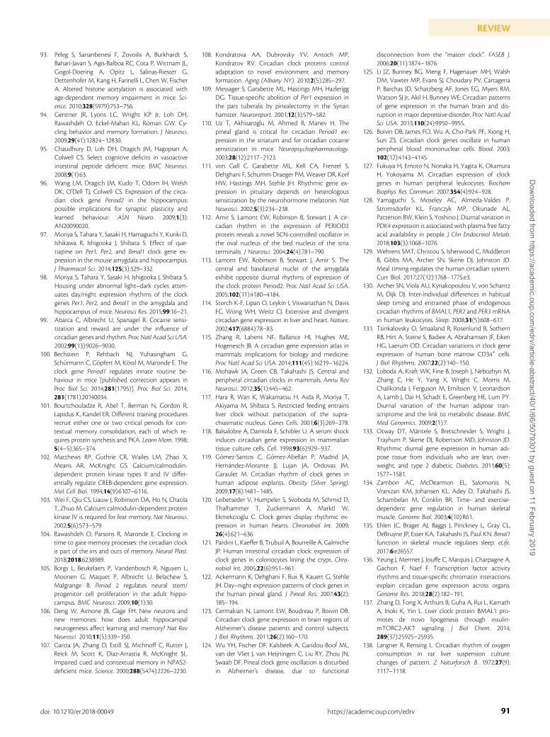
93. Peleg S, Sananbenesi F, Zovoilis A, Burkhardt S,Bahari-Javan S, Agis-Balboa RC, Cota P, Wittnam JL,Gogol-Doering A, Opitz L, Salinas-Riester G,Dettenhofer M, Kang H, Farinelli L, Chen W, FischerA. Altered histone acetylation is associated withage-dependent memory impairment in mice. Sci-ence. 2010;328(5979):753–756.
94. Gerstner JR, Lyons LC, Wright KP Jr, Loh DH,Rawashdeh O, Eckel-Mahan KL, Roman GW. Cy-cling behavior and memory formation. J Neurosci.2009;29(41):12824–12830.
95. Chaudhury D, Loh DH, Dragich JM, Hagopian A,Colwell CS. Select cognitive deficits in vasoactiveintestinal peptide deficient mice. BMC Neurosci.2008;9(1):63.
96. Wang LM, Dragich JM, Kudo T, Odom IH, WelshDK, O’Dell TJ, Colwell CS. Expression of the circa-dian clock gene Period2 in the hippocampus:possible implications for synaptic plasticity andlearned behaviour. ASN Neuro. 2009;1(3):AN20090020.
97. Moriya S, Tahara Y, Sasaki H, Hamaguchi Y, Kuriki D,Ishikawa R, Ishigooka J, Shibata S. Effect of que-tiapine on Per1, Per2, and Bmal1 clock gene ex-pression in the mouse amygdala and hippocampus.J Pharmacol Sci. 2014;125(3):329–332.
98. Moriya S, Tahara Y, Sasaki H, Ishigooka J, Shibata S.Housing under abnormal light–dark cycles atten-uates day/night expression rhythms of the clockgenes Per1, Per2, and Bmal1 in the amygdala andhippocampus of mice. Neurosci Res. 2015;99:16–21.
99. Abarca C, Albrecht U, Spanagel R. Cocaine sensi-tization and reward are under the influence ofcircadian genes and rhythm. Proc Natl Acad Sci USA.2002;99(13):9026–9030.
100. Bechstein P, Rehbach NJ, Yuhasingham G,Schurmann C, Gopfert M, Kossl M, Maronde E. Theclock gene Period1 regulates innate routine be-haviour in mice [published correction appears inProc Biol Sci. 2014;281(1795)]. Proc Biol Sci. 2014;281(1781):20140034.
101. Bourtchouladze R, Abel T, Berman N, Gordon R,Lapidus K, Kandel ER. Different training proceduresrecruit either one or two critical periods for con-textual memory consolidation, each of which re-quires protein synthesis and PKA. Learn Mem. 1998;5(4–5):365–374.
102. Matthews RP, Guthrie CR, Wailes LM, Zhao X,Means AR, McKnight GS. Calcium/calmodulin-dependent protein kinase types II and IV differ-entially regulate CREB-dependent gene expression.Mol Cell Biol. 1994;14(9):6107–6116.
103. Wei F, Qiu CS, Liauw J, Robinson DA, Ho N, ChatilaT, Zhuo M. Calcium calmodulin-dependent proteinkinase IV is required for fear memory. Nat Neurosci.2002;5(6):573–579.
104. Rawashdeh O, Parsons R, Maronde E. Clocking intime to gate memory processes: the circadian clockis part of the ins and outs of memory. Neural Plast.2018;2018:6238989.
105. Borgs L, Beukelaers P, Vandenbosch R, Nguyen L,Moonen G, Maquet P, Albrecht U, Belachew S,Malgrange B. Period 2 regulates neural stem/progenitor cell proliferation in the adult hippo-campus. BMC Neurosci. 2009;10(1):30.
106. Deng W, Aimone JB, Gage FH. New neurons andnew memories: how does adult hippocampalneurogenesis affect learning and memory? Nat RevNeurosci. 2010;11(5):339–350.
107. Garcia JA, Zhang D, Estill SJ, Michnoff C, Rutter J,Reick M, Scott K, Diaz-Arrastia R, McKnight SL.Impaired cued and contextual memory in NPAS2-deficient mice. Science. 2000;288(5474):2226–2230.
108. Kondratova AA, Dubrovsky YV, Antoch MP,Kondratov RV. Circadian clock proteins controladaptation to novel environment and memoryformation. Aging (Albany NY). 2010;2(5):285–297.
109. Messager S, Garabette ML, Hastings MH, HazleriggDG. Tissue-specific abolition of Per1 expression inthe pars tuberalis by pinealectomy in the Syrianhamster. Neuroreport. 2001;12(3):579–582.
110. Uz T, Akhisaroglu M, Ahmed R, Manev H. Thepineal gland is critical for circadian Period1 ex-pression in the striatum and for circadian cocainesensitization in mice. Neuropsychopharmacology.2003;28(12):2117–2123.
111. von Gall C, Garabette ML, Kell CA, Frenzel S,Dehghani F, Schumm-Draeger PM, Weaver DR, KorfHW, Hastings MH, Stehle JH. Rhythmic gene ex-pression in pituitary depends on heterologoussensitization by the neurohormone melatonin. NatNeurosci. 2002;5(3):234–238.
112. Amir S, Lamont EW, Robinson B, Stewart J. A cir-cadian rhythm in the expression of PERIOD2protein reveals a novel SCN-controlled oscillator inthe oval nucleus of the bed nucleus of the striaterminalis. J Neurosci. 2004;24(4):781–790.
113. Lamont EW, Robinson B, Stewart J, Amir S. Thecentral and basolateral nuclei of the amygdalaexhibit opposite diurnal rhythms of expression ofthe clock protein Period2. Proc Natl Acad Sci USA.2005;102(11):4180–4184.
114. Storch K-F, Lipan O, Leykin I, Viswanathan N, DavisFC, Wong WH, Weitz CJ. Extensive and divergentcircadian gene expression in liver and heart. Nature.2002;417(6884):78–83.
115. Zhang R, Lahens NF, Ballance HI, Hughes ME,Hogenesch JB. A circadian gene expression atlas inmammals: implications for biology and medicine.Proc Natl Acad Sci USA. 2014;111(45):16219–16224.
116. Mohawk JA, Green CB, Takahashi JS. Central andperipheral circadian clocks in mammals. Annu RevNeurosci. 2012;35(1):445–462.
117. Hara R, Wan K, Wakamatsu H, Aida R, Moriya T,Akiyama M, Shibata S. Restricted feeding entrainsliver clock without participation of the supra-chiasmatic nucleus. Genes Cells. 2001;6(3):269–278.
118. Balsalobre A, Damiola F, Schibler U. A serum shockinduces circadian gene expression in mammaliantissue culture cells. Cell. 1998;93(6):929–937.
119. Gomez-Santos C, Gomez-Abellan P, Madrid JA,Hernandez-Morante JJ, Lujan JA, Ordovas JM,Garaulet M. Circadian rhythm of clock genes inhuman adipose explants. Obesity (Silver Spring).2009;17(8):1481–1485.
120. Leibetseder V, Humpeler S, Svoboda M, Schmid D,Thalhammer T, Zuckermann A, Marktl W,Ekmekcioglu C. Clock genes display rhythmic ex-pression in human hearts. Chronobiol Int. 2009;26(4):621–636.
121. Pardini L, Kaeffer B, Trubuil A, Bourreille A, GalmicheJP. Human intestinal circadian clock: expression ofclock genes in colonocytes lining the crypt. Chro-nobiol Int. 2005;22(6):951–961.
122. Ackermann K, Dehghani F, Bux R, Kauert G, StehleJH. Day–night expression patterns of clock genes inthe human pineal gland. J Pineal Res. 2007;43(2):185–194.
123. Cermakian N, Lamont EW, Boudreau P, Boivin DB.Circadian clock gene expression in brain regions ofAlzheimer’s disease patients and control subjects.J Biol Rhythms. 2011;26(2):160–170.
124. Wu YH, Fischer DF, Kalsbeek A, Garidou-Boof ML,van der Vliet J, van Heijningen C, Liu RY, Zhou JN,Swaab DF. Pineal clock gene oscillation is disturbedin Alzheimer’s disease, due to functional
disconnection from the “master clock”. FASEB J.2006;20(11):1874–1876.
125. Li JZ, Bunney BG, Meng F, Hagenauer MH, WalshDM, Vawter MP, Evans SJ, Choudary PV, CartagenaP, Barchas JD, Schatzberg AF, Jones EG, Myers RM,Watson SJ Jr, Akil H, Bunney WE. Circadian patternsof gene expression in the human brain and dis-ruption in major depressive disorder. Proc Natl AcadSci USA. 2013;110(24):9950–9955.
126. Boivin DB, James FO, Wu A, Cho-Park PF, Xiong H,Sun ZS. Circadian clock genes oscillate in humanperipheral blood mononuclear cells. Blood. 2003;102(12):4143–4145.
127. Fukuya H, Emoto N, Nonaka H, Yagita K, OkamuraH, Yokoyama M. Circadian expression of clockgenes in human peripheral leukocytes. BiochemBiophys Res Commun. 2007;354(4):924–928.
128. Yamaguchi S, Moseley AC, Almeda-Valdes P,Stromsdorfer KL, Franczyk MP, Okunade AL,Patterson BW, Klein S, Yoshino J. Diurnal variation inPDK4 expression is associated with plasma free fattyacid availability in people. J Clin Endocrinol Metab.2018;103(3):1068–1076.
129. Wehrens SMT, Christou S, Isherwood C, MiddletonB, Gibbs MA, Archer SN, Skene DJ, Johnston JD.Meal timing regulates the human circadian system.Curr Biol. 2017;27(12):1768–1775.e3.
130. Archer SN, Viola AU, Kyriakopoulou V, von SchantzM, Dijk DJ. Inter-individual differences in habitualsleep timing and entrained phase of endogenouscircadian rhythms of BMAL1, PER2 and PER3mRNAin human leukocytes. Sleep. 2008;31(5):608–617.
131. Tsinkalovsky O, Smaaland R, Rosenlund B, SothernRB, Hirt A, Steine S, Badiee A, Abrahamsen JF, EikenHG, Laerum OD. Circadian variations in clock geneexpression of human bone marrow CD34+ cells.J Biol Rhythms. 2007;22(2):140–150.
132. Loboda A, Kraft WK, Fine B, Joseph J, Nebozhyn M,Zhang C, He Y, Yang X, Wright C, Morris M,Chalikonda I, Ferguson M, Emilsson V, LeonardsonA, Lamb J, Dai H, Schadt E, Greenberg HE, Lum PY.Diurnal variation of the human adipose tran-scriptome and the link to metabolic disease. BMCMed Genomics. 2009;2(1):7.
133. Otway DT, Mantele S, Bretschneider S, Wright J,Trayhurn P, Skene DJ, Robertson MD, Johnston JD.Rhythmic diurnal gene expression in human adi-pose tissue from individuals who are lean, over-weight, and type 2 diabetic. Diabetes. 2011;60(5):1577–1581.
134. Zambon AC, McDearmon EL, Salomonis N,Vranizan KM, Johansen KL, Adey D, Takahashi JS,Schambelan M, Conklin BR. Time- and exercise-dependent gene regulation in human skeletalmuscle. Genome Biol. 2003;4(10):R61.
135. Ehlen JC, Brager AJ, Baggs J, Pinckney L, Gray CL,DeBruyne JP, Esser KA, Takahashi JS, Paul KN. Bmal1function in skeletal muscle regulates sleep. eLife.2017;6:e26557.
136. Yeung J, Mermet J, Jouffe C, Marquis J, Charpagne A,Gachon F, Naef F. Transcription factor activityrhythms and tissue-specific chromatin interactionsexplain circadian gene expression across organs.Genome Res. 2018;28(2):182–191.
137. Zhang D, Tong X, Arthurs B, Guha A, Rui L, KamathA, Inoki K, Yin L. Liver clock protein BMAL1 pro-motes de novo lipogenesis through insulin-mTORC2-AKT signaling. J Biol Chem. 2014;289(37):25925–25935.
138. Langner R, Rensing L. Circadian rhythm of oxygenconsumption in rat liver suspension culture:changes of pattern. Z Naturforsch B. 1972;27(9):1117–1118.
91doi: 10.1210/er.2018-00049 https://academic.oup.com/edrv
REVIEWD
ownloaded from
https://academic.oup.com
/edrv/article-abstract/40/1/66/5079301 by guest on 11 February 2019
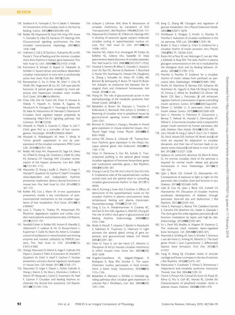
139. Stokkan K-A, Yamazaki S, Tei H, Sakaki Y, MenakerM. Entrainment of the circadian clock in the liver byfeeding. Science. 2001;291(5503):490–493.
140. Reddy AB, Maywood ES, Karp NA, King VM, InoueY, Gonzalez FJ, Lilley KS, Kyriacou CP, Hastings MH.Glucocorticoid signaling synchronizes the livercircadian transcriptome. Hepatology. 2007;45(6):1478–1488.
141. Vollmers C, Gill S, DiTacchio L, Pulivarthy SR, Le HD,Panda S. Time of feeding and the intrinsic circadianclock drive rhythms in hepatic gene expression. ProcNatl Acad Sci USA. 2009;106(50):21453–21458.
142. Kornmann B, Schaad O, Bujard H, Takahashi JS,Schibler U. System-driven and oscillator-dependentcircadian transcription in mice with a conditionallyactive liver clock. PLoS Biol. 2007;5(2):e34.
143. Ramanathan C, Xu H, Khan SK, Shen Y, Gitis PJ,Welsh DK, Hogenesch JB, Liu AC. Cell type-specificfunctions of period genes revealed by novel adi-pocyte and hepatocyte circadian clock models.PLoS Genet. 2014;10(4):e1004244.
144. Chao HW, Doi M, Fustin JM, Chen H, Murase K,Maeda Y, Hayashi H, Tanaka R, Sugawa M,Mizukuchi N, Yamaguchi Y, Yasunaga JI, MatsuokaM, Sakai M, Matsumoto M, Hamada S, Okamura H.Circadian clock regulates hepatic polyploidy bymodulating Mkp1-Erk1/2 signaling pathway. NatCommun. 2017;8(1):2238.
145. Mteyrek A, Filipski E, Guettier C, Okyar A, Levi F.Clock gene Per2 as a controller of liver carcino-genesis. Oncotarget. 2016;7(52):85832–85847.
146. Miyazaki K, Wakabayashi M, Hara Y, Ishida N.Tumor growth suppression in vivo by over-expression of the circadian component, PER2. GenesCells. 2010;15(4):351–358.
147. Reddy AB, Karp NA, Maywood ES, Sage EA, DeeryM, O’Neill JS, Wong GK, Chesham J, Odell M, LilleyKS, Kyriacou CP, Hastings MH. Circadian orches-tration of the hepatic proteome. Curr Biol. 2006;16(11):1107–1115.
148. Mauvoisin D, Wang J, Jouffe C, Martin E, Atger F,Waridel P, Quadroni M, Gachon F, Naef F. Circadianclock-dependent and -independent rhythmicproteomes implement distinct diurnal functions inmouse liver. Proc Natl Acad Sci USA. 2014;111(1):167–172.
149. Robles MS, Cox J, Mann M. In-vivo quantitativeproteomics reveals a key contribution of post-transcriptional mechanisms to the circadian regu-lation of liver metabolism. PLoS Genet. 2014;10(1):e1004047.
150. Luck S, Thurley K, Thaben PF, Westermark PO.Rhythmic degradation explains and unifies circa-dian transcriptome and proteome data. Cell Reports.2014;9(2):741–751.
151. Neufeld-Cohen A, Robles MS, Aviram R, Manella G,Adamovich Y, Ladeuix B, Nir D, Rousso-Noori L,Kuperman Y, Golik M, Mann M, Asher G. Circadiancontrol of oscillations in mitochondrial rate-limitingenzymes and nutrient utilization by PERIOD pro-teins. Proc Natl Acad Sci USA. 2016;113(12):E1673–E1682.
152. Wang J, Mauvoisin D, Martin E, Atger F, Galindo AN,Dayon L, Sizzano F, Palini A, KussmannM,Waridel P,Quadroni M, Dulic V, Naef F, Gachon F. Nuclearproteomics uncovers diurnal regulatory landscapesin mouse liver. Cell Metab. 2017;25(1):102–117.
153. Mauvoisin D, Atger F, Dayon L, Nuñez Galindo A,Wang J, Martin E, Da Silva L, Montoliu I, Collino S,Martin FP, Ratajczak J, Canto C, Kussmann M, NaefF, Gachon F. Circadian and feeding rhythms or-chestrate the diurnal liver acetylome. Cell Reports.2017;20(7):1729–1743.
154. LeSauter J, Lehman MN, Silver R. Restoration ofcircadian rhythmicity by transplants of SCN“micropunches”. J Biol Rhythms. 1996;11(2):163–171.
155. Maywood ES, Chesham JE, O’Brien JA, Hastings MH.A diversity of paracrine signals sustains molecularcircadian cycling in suprachiasmatic nucleus cir-cuits. Proc Natl Acad Sci USA. 2011;108(34):14306–14311.
156. Hatcher NG, Atkins N Jr, Annangudi SP, Forbes AJ,Kelleher NL, Gillette MU, Sweedler JV. Massspectrometry-based discovery of circadian peptides.Proc Natl Acad Sci USA. 2008;105(34):12527–12532.
157. Ghoshal S, Stevens JR, Billon C, Girardet C, Sitaula S,Leon AS, Rao DC, Skinner JS, Rankinen T, BouchardC, Nunez MV, Stanhope KL, Howatt DA, DaughertyA, Zhang J, Schuelke M, Weiss EP, Coffey AR,Bennett BJ, Sethupathy P, Burris TP, Havel PJ, ButlerAA. Adropin: an endocrine link between the bi-ological clock and cholesterol homeostasis. MolMetab. 2018;8;51–64.
158. Wang M. The role of glucocorticoid action in thepathophysiology of the metabolic syndrome. NutrMetab (Lond). 2005;2(1):3.
159. Balsalobre A, Brown SA, Marcacci L, Tronche F,Kellendonk C, Reichardt HM, Schutz G, Schibler U.Resetting of circadian time in peripheral tissues byglucocorticoid signaling. Science. 2000;289(5488):2344–2347.
160. Bittman EL, Doherty L, Huang L, Paroskie A. Periodgene expression in mouse endocrine tissues. Am JPhysiol Regul Integr Comp Physiol. 2003;285(3):R561–R569.
161. Lemos DR, Downs JL, Urbanski HF. Twenty-four-hour rhythmic gene expression in the rhesus ma-caque adrenal gland. Mol Endocrinol. 2006;20(5):1164–1176.
162. Oster H, Damerow S, Hut RA, Eichele G. Tran-scriptional profiling in the adrenal gland revealscircadian regulation of hormone biosynthesis genesand nucleosome assembly genes. J Biol Rhythms.2006;21(5):350–361.
163. Chung S, Lee EJ, Cha HK, Kim J, Kim D, Son GH, KimK. Cooperative roles of the suprachiasmatic nucleuscentral clock and the adrenal clock in controllingcircadian glucocorticoid rhythm. Sci Rep. 2017;7(1):46404.
164. Abe K, Kroning J, Greer MA, Critchlow V. Effects ofdestruction of the suprachiasmatic nuclei on thecircadian rhythms in plasma corticosterone, bodytemperature, feeding and plasma thyrotropin.Neuroendocrinology. 1979;29(2):119–131.
165. Yang S, Liu A, Weidenhammer A, Cooksey RC,McClain D, KimMK, Aguilera G, Abel ED, Chung JH.The role of mPer2 clock gene in glucocorticoid andfeeding rhythms. Endocrinology. 2009;150(5):2153–2160.
166. Ishida A, Mutoh T, Ueyama T, Bando H, MasubuchiS, Nakahara D, Tsujimoto G, Okamura H. Lightactivates the adrenal gland: timing of gene ex-pression and glucocorticoid release. Cell Metab.2005;2(5):297–307.
167. Oster H, Yasui A, van der Horst GT, Albrecht U.Disruption of mCry2 restores circadian rhythmicityin mPer2 mutant mice. Genes Dev. 2002;16(20):2633–2638.
168. Angeles-Castellanos M, Salgado-Delgado R,Rodriguez K, Buijs RM, Escobar C. The supra-chiasmatic nucleus participates in food entrain-ment: a lesion study. Neuroscience. 2010;165(4):1115–1126.
169. Balsalobre A, Marcacci L, Schibler U. Multiple sig-naling pathways elicit circadian gene expression incultured Rat-1 fibroblasts. Curr Biol. 2000;10(20):1291–1294.
170. Jiang G, Zhang BB. Glucagon and regulation ofglucose metabolism. Am J Physiol Endocrinol Metab.2003;284(4):E671–E678.
171. Muhlbauer E, Wolgast S, Finckh U, Peschke D,Peschke E. Indication of circadian oscillations in therat pancreas. FEBS Lett. 2004;564(1-2):91–96.
172. Boden G, Ruiz J, Urbain JL, Chen X. Evidence for acircadian rhythm of insulin secretion. Am J Physiol.1996;271(2 Pt 1):E246–E252.
173. Ruiter M, La Fleur SE, van Heijningen C, van der VlietJ, Kalsbeek A, Buijs RM. The daily rhythm in plasmaglucagon concentrations in the rat is modulated bythe biological clock and by feeding behavior. Di-abetes. 2003;52(7):1709–1715.
174. Peschke E, Peschke D. Evidence for a circadianrhythm of insulin release from perifused rat pan-creatic islets. Diabetologia. 1998;41(9):1085–1092.
175. Perelis M, Marcheva B, Ramsey KM, Schipma MJ,Hutchison AL, Taguchi A, Peek CB, Hong H, HuangW, Omura C, Allred AL, Bradfield CA, Dinner AR,Barish GD, Bass J. Pancreatic b cell enhancersregulate rhythmic transcription of genes controllinginsulin secretion. Science. 2015;350(6261):aac4250.
176. Dibner C, Schibler U. A pancreatic clock timesinsulin release. Science. 2015;350(6261):628–629.
177. Saini C, Petrenko V, Pulimeno P, Giovannoni L,Berney T, Hebrok M, Howald C, Dermitzakis ET,Dibner C. A functional circadian clock is required forproper insulin secretion by human pancreatic isletcells. Diabetes Obes Metab. 2016;18(4):355–365.
178. Lee J, Moulik M, Fang Z, Saha P, Zou F, Xu Y, NelsonDL, Ma K, Moore DD, Yechoor VK. Bmal1 and b-cellclock are required for adaptation to circadiandisruption, and their loss of function leads to ox-idative stress-induced b-cell failure in mice.Mol CellBiol. 2013;33(11):2327–2338.
179. Sadacca LA, Lamia KA, deLemos AS, Blum B, WeitzCJ. An intrinsic circadian clock of the pancreas isrequired for normal insulin release and glucosehomeostasis in mice. Diabetologia. 2011;54(1):120–124.
180. Qian J, Block GD, Colwell CS, Matveyenko AV.Consequences of exposure to light at night on thepancreatic islet circadian clock and function in rats.Diabetes. 2013;62(10):3469–3478.
181. Gale JE, Cox HI, Qian J, Block GD, Colwell CS,Matveyenko AV. Disruption of circadian rhythmsaccelerates development of diabetes throughpancreatic beta-cell loss and dysfunction. J BiolRhythms. 2011;26(5):423–433.
182. Vieira E, Marroquı L, Batista TM, Caballero-GarridoE, Carneiro EM, Boschero AC, Nadal A, Quesada I.The clock gene Rev-erba regulates pancreatic b-cellfunction: modulation by leptin and high-fat diet.Endocrinology. 2012;153(2):592–601.
183. Fu L, Patel MS, Bradley A, Wagner EF, Karsenty G.The molecular clock mediates leptin-regulatedbone formation. Cell. 2005;122(5):803–815.
184. Maronde E, Schilling AF, Seitz S, Schinke T, SchmutzI, van der Horst G, Amling M, Albrecht U. The clockgenes Period 2 and Cryptochrome 2 differentiallybalance bone formation. PLoS One. 2010;5(7):e11527.
185. Yang N, Meng QJ. Circadian clocks in articularcartilage and bone: a compass in the sea of matrices.J Biol Rhythms. 2016;31(5):415–427.
186. Matsuzawa Y, Funahashi T, Kihara S, Shimomura I.Adiponectin and metabolic syndrome. ArteriosclerThromb Vasc Biol. 2004;24(1):29–33.
187. Zvonic S, Ptitsyn AA, Conrad SA, Scott LK, Floyd ZE,Kilroy G, Wu X, Goh BC, Mynatt RL, Gimble JM.Characterization of peripheral circadian clocks inadipose tissues. Diabetes. 2006;55(4):962–970.
92 Kim et al PERIOD Is an Integral Component of Physiology Endocrine Reviews, February 2019, 40(1):66–95
REVIEWD
ownloaded from
https://academic.oup.com
/edrv/article-abstract/40/1/66/5079301 by guest on 11 February 2019
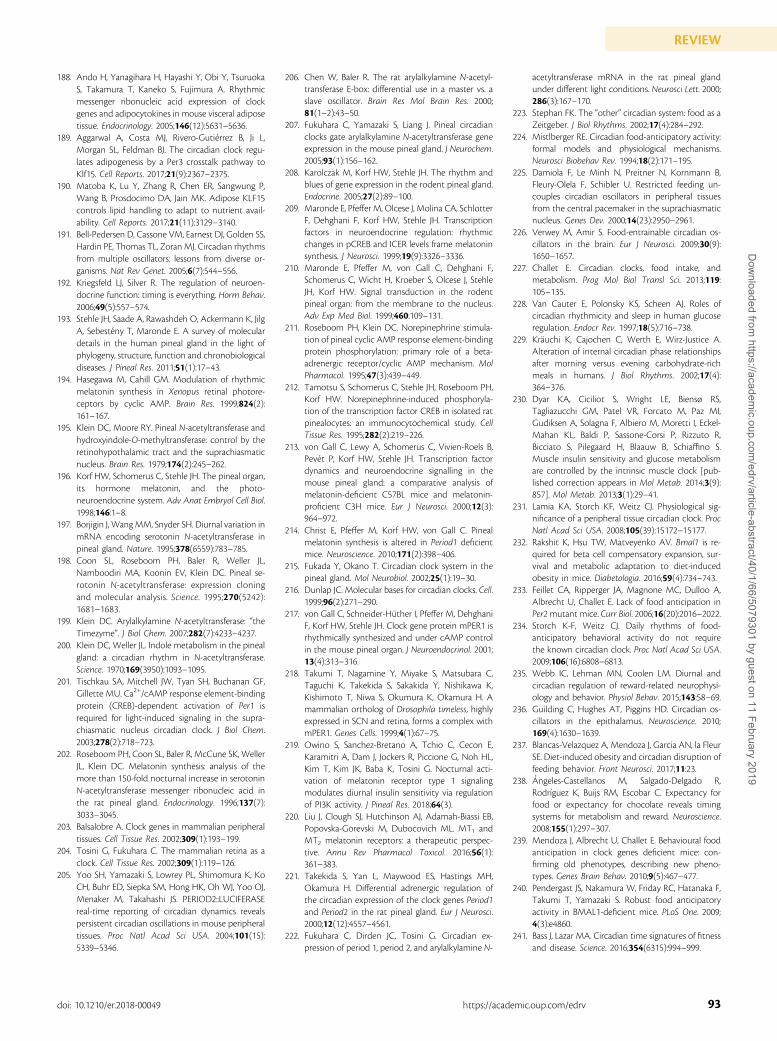
188. Ando H, Yanagihara H, Hayashi Y, Obi Y, TsuruokaS, Takamura T, Kaneko S, Fujimura A. Rhythmicmessenger ribonucleic acid expression of clockgenes and adipocytokines in mouse visceral adiposetissue. Endocrinology. 2005;146(12):5631–5636.
189. Aggarwal A, Costa MJ, Rivero-Gutierrez B, Ji L,Morgan SL, Feldman BJ. The circadian clock regu-lates adipogenesis by a Per3 crosstalk pathway toKlf15. Cell Reports. 2017;21(9):2367–2375.
190. Matoba K, Lu Y, Zhang R, Chen ER, Sangwung P,Wang B, Prosdocimo DA, Jain MK. Adipose KLF15controls lipid handling to adapt to nutrient avail-ability. Cell Reports. 2017;21(11):3129–3140.
191. Bell-Pedersen D, Cassone VM, Earnest DJ, Golden SS,Hardin PE, Thomas TL, Zoran MJ. Circadian rhythmsfrom multiple oscillators: lessons from diverse or-ganisms. Nat Rev Genet. 2005;6(7):544–556.
192. Kriegsfeld LJ, Silver R. The regulation of neuroen-docrine function: timing is everything. Horm Behav.2006;49(5):557–574.
193. Stehle JH, Saade A, Rawashdeh O, Ackermann K, JilgA, Sebesteny T, Maronde E. A survey of moleculardetails in the human pineal gland in the light ofphylogeny, structure, function and chronobiologicaldiseases. J Pineal Res. 2011;51(1):17–43.
194. Hasegawa M, Cahill GM. Modulation of rhythmicmelatonin synthesis in Xenopus retinal photore-ceptors by cyclic AMP. Brain Res. 1999;824(2):161–167.
195. Klein DC, Moore RY. Pineal N-acetyltransferase andhydroxyindole-O-methyltransferase: control by theretinohypothalamic tract and the suprachiasmaticnucleus. Brain Res. 1979;174(2):245–262.
196. Korf HW, Schomerus C, Stehle JH. The pineal organ,its hormone melatonin, and the photo-neuroendocrine system. Adv Anat Embryol Cell Biol.1998;146:1–8.
197. Borjigin J, WangMM, Snyder SH. Diurnal variation inmRNA encoding serotonin N-acetyltransferase inpineal gland. Nature. 1995;378(6559):783–785.
198. Coon SL, Roseboom PH, Baler R, Weller JL,Namboodiri MA, Koonin EV, Klein DC. Pineal se-rotonin N-acetyltransferase: expression cloningand molecular analysis. Science. 1995;270(5242):1681–1683.
199. Klein DC. Arylalkylamine N-acetyltransferase: “theTimezyme”. J Biol Chem. 2007;282(7):4233–4237.
200. Klein DC, Weller JL. Indole metabolism in the pinealgland: a circadian rhythm in N-acetyltransferase.Science. 1970;169(3950):1093–1095.
201. Tischkau SA, Mitchell JW, Tyan SH, Buchanan GF,Gillette MU. Ca2+/cAMP response element-bindingprotein (CREB)-dependent activation of Per1 isrequired for light-induced signaling in the supra-chiasmatic nucleus circadian clock. J Biol Chem.2003;278(2):718–723.
202. Roseboom PH, Coon SL, Baler R, McCune SK, WellerJL, Klein DC. Melatonin synthesis: analysis of themore than 150-fold nocturnal increase in serotoninN-acetyltransferase messenger ribonucleic acid inthe rat pineal gland. Endocrinology. 1996;137(7):3033–3045.
203. Balsalobre A. Clock genes in mammalian peripheraltissues. Cell Tissue Res. 2002;309(1):193–199.
204. Tosini G, Fukuhara C. The mammalian retina as aclock. Cell Tissue Res. 2002;309(1):119–126.
205. Yoo SH, Yamazaki S, Lowrey PL, Shimomura K, KoCH, Buhr ED, Siepka SM, Hong HK, Oh WJ, Yoo OJ,Menaker M, Takahashi JS. PERIOD2::LUCIFERASEreal-time reporting of circadian dynamics revealspersistent circadian oscillations in mouse peripheraltissues. Proc Natl Acad Sci USA. 2004;101(15):5339–5346.
206. Chen W, Baler R. The rat arylalkylamine N-acetyl-transferase E-box: differential use in a master vs. aslave oscillator. Brain Res Mol Brain Res. 2000;81(1–2):43–50.
207. Fukuhara C, Yamazaki S, Liang J. Pineal circadianclocks gate arylalkylamine N-acetyltransferase geneexpression in the mouse pineal gland. J Neurochem.2005;93(1):156–162.
208. Karolczak M, Korf HW, Stehle JH. The rhythm andblues of gene expression in the rodent pineal gland.Endocrine. 2005;27(2):89–100.
209. Maronde E, Pfeffer M, Olcese J, Molina CA, SchlotterF, Dehghani F, Korf HW, Stehle JH. Transcriptionfactors in neuroendocrine regulation: rhythmicchanges in pCREB and ICER levels frame melatoninsynthesis. J Neurosci. 1999;19(9):3326–3336.
210. Maronde E, Pfeffer M, von Gall C, Dehghani F,Schomerus C, Wicht H, Kroeber S, Olcese J, StehleJH, Korf HW. Signal transduction in the rodentpineal organ: from the membrane to the nucleus.Adv Exp Med Biol. 1999;460:109–131.
211. Roseboom PH, Klein DC. Norepinephrine stimula-tion of pineal cyclic AMP response element-bindingprotein phosphorylation: primary role of a beta-adrenergic receptor/cyclic AMP mechanism. MolPharmacol. 1995;47(3):439–449.
212. Tamotsu S, Schomerus C, Stehle JH, Roseboom PH,Korf HW. Norepinephrine-induced phosphoryla-tion of the transcription factor CREB in isolated ratpinealocytes: an immunocytochemical study. CellTissue Res. 1995;282(2):219–226.
213. von Gall C, Lewy A, Schomerus C, Vivien-Roels B,Pevet P, Korf HW, Stehle JH. Transcription factordynamics and neuroendocrine signalling in themouse pineal gland: a comparative analysis ofmelatonin-deficient C57BL mice and melatonin-proficient C3H mice. Eur J Neurosci. 2000;12(3):964–972.
214. Christ E, Pfeffer M, Korf HW, von Gall C. Pinealmelatonin synthesis is altered in Period1 deficientmice. Neuroscience. 2010;171(2):398–406.
215. Fukada Y, Okano T. Circadian clock system in thepineal gland. Mol Neurobiol. 2002;25(1):19–30.
216. Dunlap JC. Molecular bases for circadian clocks. Cell.1999;96(2):271–290.
217. von Gall C, Schneider-Huther I, Pfeffer M, DehghaniF, Korf HW, Stehle JH. Clock gene protein mPER1 isrhythmically synthesized and under cAMP controlin the mouse pineal organ. J Neuroendocrinol. 2001;13(4):313–316.
218. Takumi T, Nagamine Y, Miyake S, Matsubara C,Taguchi K, Takekida S, Sakakida Y, Nishikawa K,Kishimoto T, Niwa S, Okumura K, Okamura H. Amammalian ortholog of Drosophila timeless, highlyexpressed in SCN and retina, forms a complex withmPER1. Genes Cells. 1999;4(1):67–75.
219. Owino S, Sanchez-Bretano A, Tchio C, Cecon E,Karamitri A, Dam J, Jockers R, Piccione G, Noh HL,Kim T, Kim JK, Baba K, Tosini G. Nocturnal acti-vation of melatonin receptor type 1 signalingmodulates diurnal insulin sensitivity via regulationof PI3K activity. J Pineal Res. 2018;64(3).
220. Liu J, Clough SJ, Hutchinson AJ, Adamah-Biassi EB,Popovska-Gorevski M, Dubocovich ML. MT1 andMT2 melatonin receptors: a therapeutic perspec-tive. Annu Rev Pharmacol Toxicol. 2016;56(1):361–383.
221. Takekida S, Yan L, Maywood ES, Hastings MH,Okamura H. Differential adrenergic regulation ofthe circadian expression of the clock genes Period1and Period2 in the rat pineal gland. Eur J Neurosci.2000;12(12):4557–4561.
222. Fukuhara C, Dirden JC, Tosini G. Circadian ex-pression of period 1, period 2, and arylalkylamine N-
acetyltransferase mRNA in the rat pineal glandunder different light conditions. Neurosci Lett. 2000;286(3):167–170.
223. Stephan FK. The “other” circadian system: food as aZeitgeber. J Biol Rhythms. 2002;17(4):284–292.
224. Mistlberger RE. Circadian food-anticipatory activity:formal models and physiological mechanisms.Neurosci Biobehav Rev. 1994;18(2):171–195.
225. Damiola F, Le Minh N, Preitner N, Kornmann B,Fleury-Olela F, Schibler U. Restricted feeding un-couples circadian oscillators in peripheral tissuesfrom the central pacemaker in the suprachiasmaticnucleus. Genes Dev. 2000;14(23):2950–2961.
226. Verwey M, Amir S. Food-entrainable circadian os-cillators in the brain. Eur J Neurosci. 2009;30(9):1650–1657.
227. Challet E. Circadian clocks, food intake, andmetabolism. Prog Mol Biol Transl Sci. 2013;119:105–135.
228. Van Cauter E, Polonsky KS, Scheen AJ. Roles ofcircadian rhythmicity and sleep in human glucoseregulation. Endocr Rev. 1997;18(5):716–738.
229. Krauchi K, Cajochen C, Werth E, Wirz-Justice A.Alteration of internal circadian phase relationshipsafter morning versus evening carbohydrate-richmeals in humans. J Biol Rhythms. 2002;17(4):364–376.
230. Dyar KA, Ciciliot S, Wright LE, Biensø RS,Tagliazucchi GM, Patel VR, Forcato M, Paz MI,Gudiksen A, Solagna F, Albiero M, Moretti I, Eckel-Mahan KL, Baldi P, Sassone-Corsi P, Rizzuto R,Bicciato S, Pilegaard H, Blaauw B, Schiaffino S.Muscle insulin sensitivity and glucose metabolismare controlled by the intrinsic muscle clock [pub-lished correction appears in Mol Metab. 2014;3(9):857]. Mol Metab. 2013;3(1):29–41.
231. Lamia KA, Storch KF, Weitz CJ. Physiological sig-nificance of a peripheral tissue circadian clock. ProcNatl Acad Sci USA. 2008;105(39):15172–15177.
232. Rakshit K, Hsu TW, Matveyenko AV. Bmal1 is re-quired for beta cell compensatory expansion, sur-vival and metabolic adaptation to diet-inducedobesity in mice. Diabetologia. 2016;59(4):734–743.
233. Feillet CA, Ripperger JA, Magnone MC, Dulloo A,Albrecht U, Challet E. Lack of food anticipation inPer2mutant mice. Curr Biol. 2006;16(20):2016–2022.
234. Storch K-F, Weitz CJ. Daily rhythms of food-anticipatory behavioral activity do not requirethe known circadian clock. Proc Natl Acad Sci USA.2009;106(16):6808–6813.
235. Webb IC, Lehman MN, Coolen LM. Diurnal andcircadian regulation of reward-related neurophysi-ology and behavior. Physiol Behav. 2015;143:58–69.
236. Guilding C, Hughes AT, Piggins HD. Circadian os-cillators in the epithalamus. Neuroscience. 2010;169(4):1630–1639.
237. Blancas-Velazquez A, Mendoza J, Garcia AN, la FleurSE. Diet-induced obesity and circadian disruption offeeding behavior. Front Neurosci. 2017;11:23.
238. Angeles-Castellanos M, Salgado-Delgado R,Rodrıguez K, Buijs RM, Escobar C. Expectancy forfood or expectancy for chocolate reveals timingsystems for metabolism and reward. Neuroscience.2008;155(1):297–307.
239. Mendoza J, Albrecht U, Challet E. Behavioural foodanticipation in clock genes deficient mice: con-firming old phenotypes, describing new pheno-types. Genes Brain Behav. 2010;9(5):467–477.
240. Pendergast JS, Nakamura W, Friday RC, Hatanaka F,Takumi T, Yamazaki S. Robust food anticipatoryactivity in BMAL1-deficient mice. PLoS One. 2009;4(3):e4860.
241. Bass J, Lazar MA. Circadian time signatures of fitnessand disease. Science. 2016;354(6315):994–999.
93doi: 10.1210/er.2018-00049 https://academic.oup.com/edrv
REVIEWD
ownloaded from
https://academic.oup.com
/edrv/article-abstract/40/1/66/5079301 by guest on 11 February 2019
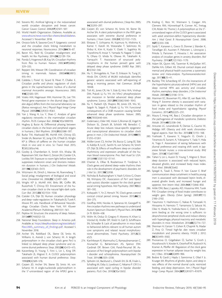
242. Stevens RG. Artificial lighting in the industrializedworld: circadian disruption and breast cancer.Cancer Causes Control. 2006;17(4):501–507.
243. World Health Organization. Diabetes. Available at:www.who.int/news-room/fact-sheets/detail/diabetes.Accessed 5 November 2018.
244. Orozco-Solis R, Sassone-Corsi P. Epigenetic controland the circadian clock: linking metabolism toneuronal responses. Neuroscience. 2014;264:76–87.
245. Baron KG, Reid KJ. Circadian misalignment andhealth. Int Rev Psychiatry. 2014;26(2):139–154.
246. Panda S, Hogenesch JB, Kay SA. Circadian rhythmsfrom flies to human. Nature. 2002;417(6886):329–335.
247. Reppert SM, Weaver DR. Coordination of circadiantiming in mammals. Nature. 2002;418(6901):935–941.
248. Caldelas I, Poirel VJ, Sicard B, Pevet P, Challet E.Circadian profile and photic regulation of clockgenes in the suprachiasmatic nucleus of a diurnalmammal Arvicanthis ansorgei. Neuroscience. 2003;116(2):583–591.
249. Vosko AM, Hagenauer MH, Hummer DL, Lee TM.Period gene expression in the diurnal degu (Octo-don degus) differs from the nocturnal laboratory rat(Rattus norvegicus). Am J Physiol Regul Integr CompPhysiol. 2009;296(2):R353–R361.
250. Yan J, Wang H, Liu Y, Shao C. Analysis of generegulatory networks in the mammalian circadianrhythm. PLOS Comput Biol. 2008;4(10):e1000193.
251. Hughey JJ, Butte AJ. Differential phasing betweencircadian clocks in the brain and peripheral organsin humans. J Biol Rhythms. 2016;31(6):588–597.
252. Burke TM, Markwald RR, McHill AW, Chinoy ED,Snider JA, Bessman SC, Jung CM, O’Neill JS, WrightKP Jr. Effects of caffeine on the human circadianclock in vivo and in vitro. Sci Transl Med. 2015;7(305):305ra146.
253. Gooley JJ, Chamberlain K, Smith KA, Khalsa SB,Rajaratnam SM, Van Reen E, Zeitzer JM, Czeisler CA,Lockley SW. Exposure to room light before bedtimesuppresses melatonin onset and shortens melato-nin duration in humans. J Clin Endocrinol Metab.2011;96(3):E463–E472.
254. Wittmann M, Dinich J, Merrow M, Roenneberg T.Social jetlag: misalignment of biological and socialtime. Chronobiol Int. 2006;23(1-2):497–509.
255. Wright KP Jr, McHill AW, Birks BR, Griffin BR,Rusterholz T, Chinoy ED. Entrainment of the hu-man circadian clock to the natural light-dark cycle.Curr Biol. 2013;23(16):1554–1558.
256. Czeisler CA, Dijk DJ. Human circadian physiologyand sleep–wake regulation. In: Takahashi JS, Turek F,Moore RY, eds. Handbook of Behavioral Neurobi-ology: Circadian Clocks. New York, NY: KluwerAcademic/Plenum Publishing; 2001:531–561.
257. Peplow M. Structure: the anatomy of sleep. Nature.2013;497(7450):S2–S3.
258. National Sleep Foundation. Sleep in America poll.Available at: https://sleepfoundation.org/sites/default/files/2005_summary_of_findings.pdf. Accessed 5November 2018.
259. Archer SN, Robilliard DL, Skene DJ, Smits M,Williams A, Arendt J, von Schantz M. A lengthpolymorphism in the circadian clock gene Per3 islinked to delayed sleep phase syndrome and ex-treme diurnal preference. Sleep. 2003;26(4):413–415.
260. Katzenberg D, Young T, Finn L, Lin L, King DP,Takahashi JS, Mignot E. A CLOCK polymorphismassociated with human diurnal preference. Sleep.1998;21(6):569–576.
261. Carpen JD, Archer SN, Skene DJ, Smits M, vonSchantz M. A single-nucleotide polymorphism inthe 59-untranslated region of the hPER2 gene is
associated with diurnal preference. J Sleep Res. 2005;14(3):293–297.
262. Carpen JD, von Schantz M, Smits M, Skene DJ,Archer SN. A silent polymorphism in the PER1 geneassociates with extreme diurnal preference inhumans. J Hum Genet. 2006;51(12):1122–1125.
263. Ebisawa T, Uchiyama M, Kajimura N, Mishima K,Kamei Y, Katoh M, Watanabe T, Sekimoto M,Shibui K, Kim K, Kudo Y, Ozeki Y, Sugishita M,Toyoshima R, Inoue Y, Yamada N, Nagase T, OzakiN, Ohara O, Ishida N, Okawa M, Takahashi K,Yamauchi T. Association of structural poly-morphisms in the human period3 gene withdelayed sleep phase syndrome. EMBO Rep. 2001;2(4):342–346.
264. Hu Y, Shmygelska A, Tran D, Eriksson N, Tung JY,Hinds DA. GWAS of 89,283 individuals identifiesgenetic variants associated with self-reporting ofbeing a morning person. Nat Commun. 2016;7:10448.
265. Toh KL, Jones CR, He Y, Eide EJ, Hinz WA, VirshupDM, Ptacek LJ, Fu YH. An hPer2 phosphorylationsite mutation in familial advanced sleep phasesyndrome. Science. 2001;291(5506):1040–1043.
266. Xu Y, Padiath QS, Shapiro RE, Jones CR, Wu SC,Saigoh N, Saigoh K, Ptacek LJ, Fu YH. Functionalconsequences of a CKId mutation causing familialadvanced sleep phase syndrome. Nature. 2005;434(7033):640–644.
267. Cedernaes J, Osler ME, Voisin S, Broman JE, Vogel H,Dickson SL, Zierath JR, Schioth HB, Benedict C.Acute sleep loss induces tissue-specific epigeneticand transcriptional alterations to circadian clockgenes in men. J Clin Endocrinol Metab. 2015;100(9):E1255–E1261.
268. Moller-Levet CS, Archer SN, Bucca G, Laing EE, SlakA, Kabiljo R, Lo JC, Santhi N, von Schantz M, SmithCP, Dijk DJ. Effects of insufficient sleep on circadianrhythmicity and expression amplitude of the hu-man blood transcriptome. Proc Natl Acad Sci USA.2013;110(12):E1132–E1141.
269. Charrier A, Olliac B, Roubertoux P, Tordjman S.Clock genes and altered sleep–wake rhythms: theirrole in the development of psychiatric disorders. IntJ Mol Sci. 2017;18(5):938.
270. Nicholas B, Rudrasingham V, Nash S, Kirov G, OwenMJ, Wimpory DC. Association of Per1 and Npas2with autistic disorder: support for the clock genes/social timing hypothesis. Mol Psychiatry. 2007;12(6):581–592.
271. Kobayashi Y, Ye Z, Hensch TK. Clock genes controlcortical critical period timing. Neuron. 2015;86(1):264–275.
272. Geoffray MM, Nicolas A, Speranza M, Georgieff N.Are circadian rhythms new pathways to understandAutism Spectrum Disorder? J Physiol Paris. 2016;110(4 Pt B)434–438.
273. Wohr M, Orduz D, Gregory P, Moreno H, Khan U,Vorckel KJ, Wolfer DP, Welzl H, Gall D, SchiffmannSN, Schwaller B. Lack of parvalbumin in mice leadsto behavioral deficits relevant to all human autismcore symptoms and related neural morphofunc-tional abnormalities. Transl Psychiatry. 2015;5(3):e525.
274. Karthikeyan R, Marimuthu G, Ramasubramanian C,Arunachal G, BaHammam AS, Spence DW,Cardinali DP, Brown GM, Pandi-Perumal SR. As-sociation of Per3 length polymorphism with bipolarI disorder and schizophrenia. Neuropsychiatr DisTreat. 2014;10:2325–2330.
275. Sjoholm LK, Backlund L, Cheteh EH, Ek IR, Frisen L,Schalling M, Osby U, Lavebratt C, Nikamo P. CRY2 isassociated with rapid cycling in bipolar disorderpatients. PLoS One. 2010;5(9):e12632.
276. Kissling C, Retz W, Wiemann S, Coogan AN,Clement RM, Hunnerkopf R, Conner AC, FreitagCM, Rosler M, Thome J. A polymorphism at the 39-untranslated region of the CLOCK gene is associatedwith adult attention-deficit hyperactivity disorder.Am J Med Genet B Neuropsychiatr Genet. 2008;147(3):333–338.
277. Sipila T, Kananen L, Greco D, Donner J, Silander K,Terwilliger JD, Auvinen P, Peltonen L, Lonnqvist J,Pirkola S, Partonen T, Hovatta I. An associationanalysis of circadian genes in anxiety disorders. BiolPsychiatry. 2010;67(12):1163–1170.
278. Adam EK, Quinn ME, Tavernier R, McQuillan MT,Dahlke KA, Gilbert KE. Diurnal cortisol slopes andmental and physical health outcomes: a systematicreview and meta-analysis. Psychoneuroendocrinol-ogy. 2017;83:25–41.
279. Buckley TM, Schatzberg AF. On the interactions ofthe hypothalamic-pituitary-adrenal (HPA) axis andsleep: normal HPA axis activity and circadianrhythm, exemplary sleep disorders. J Clin EndocrinolMetab. 2005;90(5):3106–3114.
280. Mariman EC, Bouwman FG, Aller EE, van Baak MA,Wang P. Extreme obesity is associated with varia-tion in genes related to the circadian rhythm offood intake and hypothalamic signaling. PhysiolGenomics. 2015;47(6):225–231.
281. Maury E, Hong HK, Bass J. Circadian disruption inthe pathogenesis of metabolic syndrome. DiabetesMetab. 2014;40(5):338–346.
282. Antunes LC, Levandovski R, Dantas G, Caumo W,Hidalgo MP. Obesity and shift work: chronobio-logical aspects. Nutr Res Rev. 2010;23(1):155–168.
283. Yoshizaki T, Kawano Y, Noguchi O, Onishi J,Teramoto R, Sunami A, Yokoyama Y, Tada Y, HidaA, Togo F. Association of eating behaviours withdiurnal preference and rotating shift work in Jap-anese female nurses: a cross-sectional study. BMJOpen. 2016;6(11):e011987.
284. Taheri S, Lin L, Austin D, Young T, Mignot E. Shortsleep duration is associated with reduced leptin,elevated ghrelin, and increased body mass index.PLoS Med. 2004;1(3):e62.
285. Spiegel K, Tasali E, Penev P, Van Cauter E. Briefcommunication: sleep curtailment in healthy youngmen is associated with decreased leptin levels, el-evated ghrelin levels, and increased hunger andappetite. Ann Intern Med. 2004;141(11):846–850.
286. Arble DM, Bass J, Laposky AD, Vitaterna MH, TurekFW. Circadian timing of food intake contributes toweight gain. Obesity (Silver Spring). 2009;17(11):2100–2102.
287. Yasumoto Y, Hashimoto C, Nakao R, Yamazaki H,Hiroyama H, Nemoto T, Yamamoto S, Sakurai M,Oike H, Wada N, Yoshida-Noro C, Oishi K. Short-term feeding at the wrong time is sufficient todesynchronize peripheral clocks and induce obesitywith hyperphagia, physical inactivity and metabolicdisorders in mice. Metabolism. 2016;65(5):714–727.
288. Sherman H, Genzer Y, Cohen R, Chapnik N, MadarZ, Froy O. Timed high-fat diet resets circadianmetabolism and prevents obesity. FASEB J. 2012;26(8):3493–3502.
289. Pivovarova O, Gogebakan O, Sucher S, Groth J,Murahovschi V, Kessler K, OsterhoffM, Rudovich N,Kramer A, Pfeiffer AF. Regulation of the clock geneexpression in human adipose tissue by weight loss.Int J Obes. 2016;40(6):899–906.
290. Bodosi B, Gardi J, Hajdu I, Szentirmai E, Obal F Jr,Krueger JM. Rhythms of ghrelin, leptin, and sleep inrats: effects of the normal diurnal cycle, restrictedfeeding, and sleep deprivation. Am J Physiol RegulIntegr Comp Physiol. 2004;287(5):R1071–R1079.
94 Kim et al PERIOD Is an Integral Component of Physiology Endocrine Reviews, February 2019, 40(1):66–95
REVIEWD
ownloaded from
https://academic.oup.com
/edrv/article-abstract/40/1/66/5079301 by guest on 11 February 2019
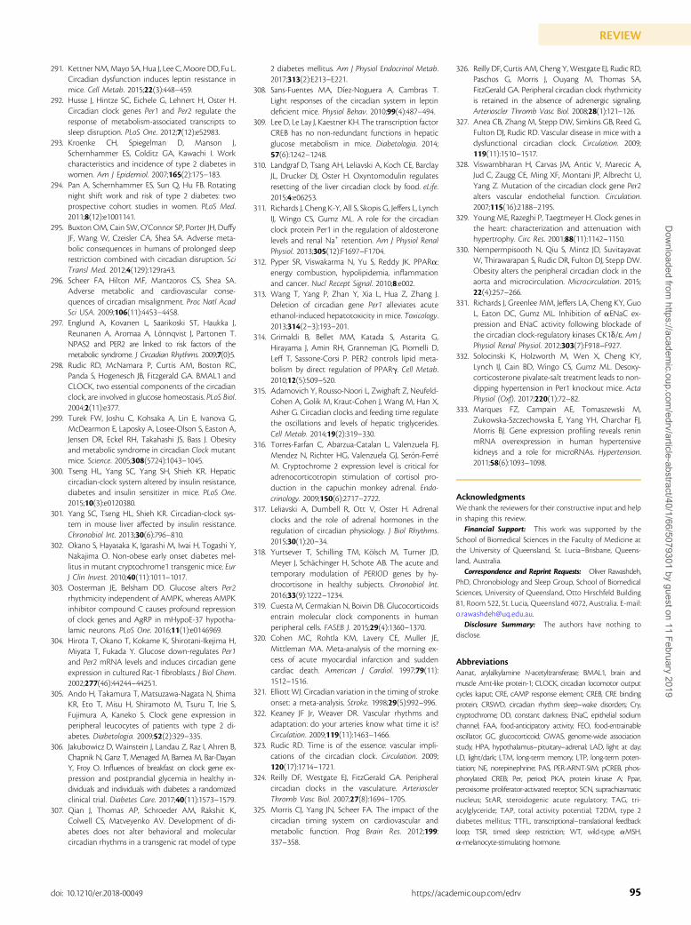
291. Kettner NM,Mayo SA, Hua J, Lee C, Moore DD, Fu L.Circadian dysfunction induces leptin resistance inmice. Cell Metab. 2015;22(3):448–459.
292. Husse J, Hintze SC, Eichele G, Lehnert H, Oster H.Circadian clock genes Per1 and Per2 regulate theresponse of metabolism-associated transcripts tosleep disruption. PLoS One. 2012;7(12):e52983.
293. Kroenke CH, Spiegelman D, Manson J,Schernhammer ES, Colditz GA, Kawachi I. Workcharacteristics and incidence of type 2 diabetes inwomen. Am J Epidemiol. 2007;165(2):175–183.
294. Pan A, Schernhammer ES, Sun Q, Hu FB. Rotatingnight shift work and risk of type 2 diabetes: twoprospective cohort studies in women. PLoS Med.2011;8(12):e1001141.
295. Buxton OM, Cain SW, O’Connor SP, Porter JH, DuffyJF, Wang W, Czeisler CA, Shea SA. Adverse meta-bolic consequences in humans of prolonged sleeprestriction combined with circadian disruption. SciTransl Med. 2012;4(129):129ra43.
296. Scheer FA, Hilton MF, Mantzoros CS, Shea SA.Adverse metabolic and cardiovascular conse-quences of circadian misalignment. Proc Natl AcadSci USA. 2009;106(11):4453–4458.
297. Englund A, Kovanen L, Saarikoski ST, Haukka J,Reunanen A, Aromaa A, Lonnqvist J, Partonen T.NPAS2 and PER2 are linked to risk factors of themetabolic syndrome. J Circadian Rhythms. 2009;7(0):5.
298. Rudic RD, McNamara P, Curtis AM, Boston RC,Panda S, Hogenesch JB, Fitzgerald GA. BMAL1 andCLOCK, two essential components of the circadianclock, are involved in glucose homeostasis. PLoS Biol.2004;2(11):e377.
299. Turek FW, Joshu C, Kohsaka A, Lin E, Ivanova G,McDearmon E, Laposky A, Losee-Olson S, Easton A,Jensen DR, Eckel RH, Takahashi JS, Bass J. Obesityand metabolic syndrome in circadian Clock mutantmice. Science. 2005;308(5724):1043–1045.
300. Tseng HL, Yang SC, Yang SH, Shieh KR. Hepaticcircadian-clock system altered by insulin resistance,diabetes and insulin sensitizer in mice. PLoS One.2015;10(3):e0120380.
301. Yang SC, Tseng HL, Shieh KR. Circadian-clock sys-tem in mouse liver affected by insulin resistance.Chronobiol Int. 2013;30(6):796–810.
302. Okano S, Hayasaka K, Igarashi M, Iwai H, Togashi Y,Nakajima O. Non-obese early onset diabetes mel-litus in mutant cryptochrome1 transgenic mice. EurJ Clin Invest. 2010;40(11):1011–1017.
303. Oosterman JE, Belsham DD. Glucose alters Per2rhythmicity independent of AMPK, whereas AMPKinhibitor compound C causes profound repressionof clock genes and AgRP in mHypoE-37 hypotha-lamic neurons. PLoS One. 2016;11(1):e0146969.
304. Hirota T, Okano T, Kokame K, Shirotani-Ikejima H,Miyata T, Fukada Y. Glucose down-regulates Per1and Per2 mRNA levels and induces circadian geneexpression in cultured Rat-1 fibroblasts. J Biol Chem.2002;277(46):44244–44251.
305. Ando H, Takamura T, Matsuzawa-Nagata N, ShimaKR, Eto T, Misu H, Shiramoto M, Tsuru T, Irie S,Fujimura A, Kaneko S. Clock gene expression inperipheral leucocytes of patients with type 2 di-abetes. Diabetologia. 2009;52(2):329–335.
306. Jakubowicz D, Wainstein J, Landau Z, Raz I, Ahren B,Chapnik N, Ganz T, Menaged M, Barnea M, Bar-DayanY, Froy O. Influences of breakfast on clock gene ex-pression and postprandial glycemia in healthy in-dividuals and individuals with diabetes: a randomizedclinical trial. Diabetes Care. 2017;40(11):1573–1579.
307. Qian J, Thomas AP, Schroeder AM, Rakshit K,Colwell CS, Matveyenko AV. Development of di-abetes does not alter behavioral and molecularcircadian rhythms in a transgenic rat model of type
2 diabetes mellitus. Am J Physiol Endocrinol Metab.2017;313(2):E213–E221.
308. Sans-Fuentes MA, Dıez-Noguera A, Cambras T.Light responses of the circadian system in leptindeficient mice. Physiol Behav. 2010;99(4):487–494.
309. Lee D, Le Lay J, Kaestner KH. The transcription factorCREB has no non-redundant functions in hepaticglucose metabolism in mice. Diabetologia. 2014;57(6):1242–1248.
310. Landgraf D, Tsang AH, Leliavski A, Koch CE, BarclayJL, Drucker DJ, Oster H. Oxyntomodulin regulatesresetting of the liver circadian clock by food. eLife.2015;4:e06253.
311. Richards J, Cheng K-Y, All S, Skopis G, Jeffers L, LynchIJ, Wingo CS, Gumz ML. A role for the circadianclock protein Per1 in the regulation of aldosteronelevels and renal Na+ retention. Am J Physiol RenalPhysiol. 2013;305(12):F1697–F1704.
312. Pyper SR, Viswakarma N, Yu S, Reddy JK. PPARa:energy combustion, hypolipidemia, inflammationand cancer. Nucl Recept Signal. 2010;8:e002.
313. Wang T, Yang P, Zhan Y, Xia L, Hua Z, Zhang J.Deletion of circadian gene Per1 alleviates acuteethanol-induced hepatotoxicity in mice. Toxicology.2013;314(2–3):193–201.
314. Grimaldi B, Bellet MM, Katada S, Astarita G,Hirayama J, Amin RH, Granneman JG, Piomelli D,Leff T, Sassone-Corsi P. PER2 controls lipid meta-bolism by direct regulation of PPARg. Cell Metab.2010;12(5):509–520.
315. Adamovich Y, Rousso-Noori L, Zwighaft Z, Neufeld-Cohen A, Golik M, Kraut-Cohen J, Wang M, Han X,Asher G. Circadian clocks and feeding time regulatethe oscillations and levels of hepatic triglycerides.Cell Metab. 2014;19(2):319–330.
316. Torres-Farfan C, Abarzua-Catalan L, Valenzuela FJ,Mendez N, Richter HG, Valenzuela GJ, Seron-FerreM. Cryptochrome 2 expression level is critical foradrenocorticotropin stimulation of cortisol pro-duction in the capuchin monkey adrenal. Endo-crinology. 2009;150(6):2717–2722.
317. Leliavski A, Dumbell R, Ott V, Oster H. Adrenalclocks and the role of adrenal hormones in theregulation of circadian physiology. J Biol Rhythms.2015;30(1):20–34.
318. Yurtsever T, Schilling TM, Kolsch M, Turner JD,Meyer J, Schachinger H, Schote AB. The acute andtemporary modulation of PERIOD genes by hy-drocortisone in healthy subjects. Chronobiol Int.2016;33(9):1222–1234.
319. Cuesta M, Cermakian N, Boivin DB. Glucocorticoidsentrain molecular clock components in humanperipheral cells. FASEB J. 2015;29(4):1360–1370.
320. Cohen MC, Rohtla KM, Lavery CE, Muller JE,Mittleman MA. Meta-analysis of the morning ex-cess of acute myocardial infarction and suddencardiac death. American J Cardiol. 1997;79(11):1512–1516.
321. Elliott WJ. Circadian variation in the timing of strokeonset: a meta-analysis. Stroke. 1998;29(5):992–996.
322. Keaney JF Jr, Weaver DR. Vascular rhythms andadaptation: do your arteries know what time it is?Circulation. 2009;119(11):1463–1466.
323. Rudic RD. Time is of the essence: vascular impli-cations of the circadian clock. Circulation. 2009;120(17):1714–1721.
324. Reilly DF, Westgate EJ, FitzGerald GA. Peripheralcircadian clocks in the vasculature. ArteriosclerThromb Vasc Biol. 2007;27(8):1694–1705.
325. Morris CJ, Yang JN, Scheer FA. The impact of thecircadian timing system on cardiovascular andmetabolic function. Prog Brain Res. 2012;199:337–358.
326. Reilly DF, Curtis AM, Cheng Y, Westgate EJ, Rudic RD,Paschos G, Morris J, Ouyang M, Thomas SA,FitzGerald GA. Peripheral circadian clock rhythmicityis retained in the absence of adrenergic signaling.Arterioscler Thromb Vasc Biol. 2008;28(1):121–126.
327. Anea CB, Zhang M, Stepp DW, Simkins GB, Reed G,Fulton DJ, Rudic RD. Vascular disease in mice with adysfunctional circadian clock. Circulation. 2009;119(11):1510–1517.
328. Viswambharan H, Carvas JM, Antic V, Marecic A,Jud C, Zaugg CE, Ming XF, Montani JP, Albrecht U,Yang Z. Mutation of the circadian clock gene Per2alters vascular endothelial function. Circulation.2007;115(16):2188–2195.
329. Young ME, Razeghi P, Taegtmeyer H. Clock genes inthe heart: characterization and attenuation withhypertrophy. Circ Res. 2001;88(11):1142–1150.
330. Nernpermpisooth N, Qiu S, Mintz JD, SuvitayavatW, Thirawarapan S, Rudic DR, Fulton DJ, Stepp DW.Obesity alters the peripheral circadian clock in theaorta and microcirculation. Microcirculation. 2015;22(4):257–266.
331. Richards J, Greenlee MM, Jeffers LA, Cheng KY, GuoL, Eaton DC, Gumz ML. Inhibition of aENaC ex-pression and ENaC activity following blockade ofthe circadian clock-regulatory kinases CK1d/e. Am JPhysiol Renal Physiol. 2012;303(7):F918–F927.
332. Solocinski K, Holzworth M, Wen X, Cheng KY,Lynch IJ, Cain BD, Wingo CS, Gumz ML. Desoxy-corticosterone pivalate-salt treatment leads to non-dipping hypertension in Per1 knockout mice. ActaPhysiol (Oxf). 2017;220(1):72–82.
333. Marques FZ, Campain AE, Tomaszewski M,Zukowska-Szczechowska E, Yang YH, Charchar FJ,Morris BJ. Gene expression profiling reveals reninmRNA overexpression in human hypertensivekidneys and a role for microRNAs. Hypertension.2011;58(6):1093–1098.
AcknowledgmentsWe thank the reviewers for their constructive input and helpin shaping this review.Financial Support: This work was supported by the
School of Biomedical Sciences in the Faculty of Medicine atthe University of Queensland, St. Lucia–Brisbane, Queens-land, Australia.Correspondence and Reprint Requests: Oliver Rawashdeh,
PhD, Chronobiology and Sleep Group, School of BiomedicalSciences, University of Queensland, Otto Hirschfeld Building81, Room 522, St. Lucia, Queensland 4072, Australia. E-mail:[email protected] Summary: The authors have nothing to
disclose.
AbbreviationsAanat, arylalkylamine N-acetyltransferase; BMAL1, brain andmuscle Arnt-like protein-1; CLOCK, circadian locomotor outputcycles kaput; CRE, cAMP response element; CREB, CRE bindingprotein; CRSWD, circadian rhythm sleep–wake disorders; Cry,cryptochrome; DD, constant darkness; ENaC, epithelial sodiumchannel; FAA, food-anticipatory activity; FEO, food-entrainableoscillator; GC, glucocorticoid; GWAS, genome-wide associationstudy; HPA, hypothalamus–pituitary–adrenal; LAD, light at day;LD, light/dark; LTM, long-term memory; LTP, long-term poten-tiation; NE, norepinephrine; PAS, PER-ARNT-SIM; pCREB, phos-phorylated CREB; Per, period; PKA, protein kinase A; Ppar,peroxisome proliferator-activated receptor; SCN, suprachiasmaticnucleus; StAR, steroidogenic acute regulatory; TAG, tri-acylglyceride; TAP, total activity potential; T2DM, type 2diabetes mellitus; TTFL, transcriptional–translational feedbackloop; TSR, timed sleep restriction; WT, wild-type; aMSH,a-melanocyte-stimulating hormone.
95doi: 10.1210/er.2018-00049 https://academic.oup.com/edrv
REVIEWD
ownloaded from
https://academic.oup.com
/edrv/article-abstract/40/1/66/5079301 by guest on 11 February 2019