intrinsic circadian clock of the mammalian retina: importance for retinal processing of visual...
TRANSCRIPT
Intrinsic Circadian Clock of theMammalian Retina: Importance forRetinal Processing of Visual InformationKai-Florian Storch,1,4 Carlos Paz,1,4 James Signorovitch,2 Elio Raviola,1 Basil Pawlyk,3 Tiansen Li,3
and Charles J. Weitz1,*1Department of Neurobiology, Harvard Medical School2Department of Biostatistics, Harvard School of Public Health
Boston, MA 02115, USA3Massachusetts Eye & Ear Infirmary, Berman-Gund Laboratory, Department of Ophthalmology, Harvard Medical School, Boston
MA 02114, USA4These authors contributed equally to this work.*Correspondence: [email protected]
DOI 10.1016/j.cell.2007.06.045
SUMMARY
Circadian clocks are widely distributed in mam-malian tissues, but little is known about thephysiological functions of clocks outside thesuprachiasmatic nucleus of the brain. The ret-ina has an intrinsic circadian clock, but its im-portance for vision is unknown. Here we showthat mice lacking Bmal1, a gene required forclock function, had abnormal retinal transcrip-tional responses to light and defective inner ret-inal electrical responses to light, but normalphotoreceptor responses to light and retinasthat appeared structurally normal by light andelectron microscopy. We generated mice witha retina-specific genetic deletion of Bmal1,and they had defects of retinal visual physiol-ogy essentially identical to those of mice lack-ing Bmal1 in all tissues and lacked a circadianrhythm of inner retinal electrical responses tolight. Our findings indicate that the intrinsic cir-cadian clock of the retina regulates retinal visualprocessing in vivo.
INTRODUCTION
Circadian clocks are endogenous oscillators that drive
daily rhythms of physiology and behavior. In mammals,
the circadian clock mechanism is built upon a molecular
feedback loop in which the CLOCK-BMAL1 transcription
factor drives expression of its PER and CRY inhibitors (Ge-
kakis et al., 1998; Ko and Takahashi, 2006). The clock gen-
erates circadian rhythms cell autonomously, and it is
thought to generate rhythms of physiology in large part
by driving rhythms of transcription of output genes (Panda
et al., 2002; Storch et al., 2002; Akhtar et al., 2002; Duffield
730 Cell 130, 730–741, August 24, 2007 ª2007 Elsevier Inc.
et al., 2002). The adaptive significance of circadian clocks
likely lies in their ability to allow anticipatory responses to
predictable daily variations in the environment (Ouyang
et al., 1998).
It has long been known that the circadian clock regulat-
ing behavior in mammals is located in the suprachiasmatic
nucleus (SCN) of the brain (Ko and Takahashi, 2006). Re-
cently it has become clear that circadian clocks are dis-
tributed in mammalian tissues, present at sites such as
the retina (Tosini and Menaker, 1996), multiple brain re-
gions (Abe et al., 2002), and in many peripheral tissues
(Balsalobre et al., 1998; Yamazaki et al., 2000; Damiola
et al., 2000). It is thought that clocks outside the SCN
have physiological functions, but to date few studies
have addressed this question (Durgan et al., 2006;
McDearmon et al., 2006). In Drosophila there is compelling
evidence that a clock in the antenna drives rhythms of
olfactory sensitivity (Tanoue et al., 2004).
Retinas from a wide range of vertebrates, including am-
phibians (Besharse and Iuvone, 1983), birds (Pierce et al.,
1993), and mammals (Tosini and Menaker, 1996), contain
a circadian clock. In Xenopus retina, photoreceptors are
the circadian clock cells (Cahill and Besharse, 1993; Hay-
asaka et al., 2002), whereas in the mammalian retina circa-
dian clock cells are found in the inner retina (Witkovsky
et al., 2003; Gustincich et al., 2004; Ruan et al., 2006)
(Figure S1).
Fundamental retinal processes are under circadian con-
trol, including photoreceptor disc shedding (LaVail and
Ward, 1978), release of melatonin and dopamine (Doyle
et al., 2002), and retinal electrical responses to light, man-
ifested as a circadian rhythm of one or more components
of the electroretinogram (ERG) (Manglapus et al., 1998;
Barnard et al., 2006). At present little is known about the
biochemical pathways under circadian control in the
mammalian retina or the molecular mechanisms that mod-
ulate retinal physiological responses to light. A circadian
rhythm of melatonin release is driven autonomously from
the retina (Tosini and Menaker, 1996), but it is not yet
known to what extent circadian rhythms of retinal electri-
cal activity in response to light reflect the action of a local
retinal clock or the action of a remote clock, such as the
SCN. Evidence from nonmammalian vertebrates suggests
that circadian rhythms of retinal electrical responses to
light are driven at least in part from the brain (Miranda-
Anaya et al., 2002) or pineal (McGoogan and Cassone,
1999).
RESULTS
Daily Rhythms of Retinal Gene ExpressionTo gain a view of molecular regulation in the mammalian
retina by a circadian clock, light, or both, we performed
whole-genome microarray studies in mice to identify
genes with a �24 hr rhythm of expression during a 3 day
interval in constant darkness (DD) or in a 12:12 hr light-
dark cycle (LD) (Figure S2). Because of the role of melato-
nin in retinal function (Doyle et al., 2002), we used CBA/
CaJ mice, a strain that makes melatonin (Goto et al.,
1989) and does not have retinal degeneration. To optimize
the efficiency of tissue collection, we used whole eyes for
RNA extraction (Supplemental Results).
To identify rhythmic variations in gene expression, we
computed the best-fit function that models the expression
of a gene across the 3 day, 18 time point microarray profile
as a �24 hr rhythmic pattern; we did not assume a simple
waveform. For some genes, the best-fitting rhythmic func-
tion makes a good fit—for most, a poor fit. Next we ran-
domly permuted the order of the 18 time points for each
gene 50,000 times. With 18 time points, random permuta-
tion of the time series will degrade the fit of a truly rhythmic
profile but will have little or no effect on the fit of noisy or
flat profiles (Figure S2). This procedure allows all or any
subset of the 45,101 probe sets on the array to be ranked
quantitatively for rhythmicity, and one of its key advan-
tages is that a statistical threshold for rhythmicity can be
set according to any desired false discovery rate (Storey
et al., 2004), defined as the percentage of genes ranking
above the threshold that can be accounted for by noise
(Supplemental Results).
In DD, we identified 277 genes with a circadian rhythm
of expression at a moderate threshold corresponding to
a 15% false discovery rate, (Figure 1A, top, DD). Phases
of peak expression around the clock were represented
roughly equally (Figure 1A, top, DD), and the dataset
included genes with known circadian regulation in other
tissues, including clock components (Table S1). Overall,
genes expressed in the retina made a major contribution
to the data set (Supplemental Results and Figure S3),
and the genes represent a wide range of functions, includ-
ing synaptic transmission, photoreceptor signaling, inter-
cellular communication, and regulation of the cytoskele-
ton and chromatin (Figure S4 and Table S1). For some
genes, expression was limited to photoreceptors
(Figure S3), likely reflecting non-cell-autonomous regula-
tion that depends on clock cells in the inner retina or
elsewhere. The rhythmic data set included secreted
factors expressed exclusively in the inner retina, candi-
dates for circadian signals from inner retinal clock cells
to photoreceptors (Figure S3).
In LD, at the same statistical threshold, we identified
2670 genes with rhythmic expression (Figure 1A, bottom,
LD). Included were 80% of the genes with rhythmic ex-
pression in DD and many genes with known expression
in the retina (Table S2). In addition to the �9-fold greater
number of genes showing rhythmic expression in LD
than DD, the distribution of phases in LD differed markedly
from that in DD, with somewhat more than half of the
genes showing a peak of expression during the night
and the rest showing a roughly equal phase distribution
(Figure 1A, bottom, LD). Both of these differences be-
tween the two conditions were observed at more stringent
thresholds (Figure 1B), indicating that they do not arise
from chance inclusion of noise. These results suggest
that LD cycles drive expression of a large number of genes
in the retina and, in particular, a large cluster of genes with
a nighttime peak of expression. Additional analyses indi-
cated that any loss of circadian synchrony among mice
or cells during the 3 day period in DD did not contribute
substantially to the differences between the DD and LD
data sets (Supplemental Results and Figure S5).
For validation, we selected 26 genes with rhythmic ex-
pression from DD and 28 from LD (identified in Tables
S1 and S2), as well as genes from each classified as
nonrhythmic, for assessment by quantitative reverse tran-
scriptase PCR (Q-PCR). In all cases but one, Q-PCR con-
firmed the rhythmic profiles (examples, Figure 1C). Micro-
array analysis thus accurately identified genes with
rhythmic expression.
Importance of Bmal1 for Retinal Gene ExpressionRhythms in a Light-Dark CycleGiven that the retina is a dedicated photosensory organ,
the �9-fold excess of genes exhibiting rhythmic expres-
sion in LD compared to DD could simply result from regu-
lation of many genes by light, independently of circadian
clock function. To test this expectation, we compared
temporal profiles of ocular gene expression in LD in
wild-type mice and Bmal1�/� (Mop3�/�) (Bunger et al.,
2000) littermates, which lack an essential component of
the clock in all tissues and consequently are expected to
lack all clock function. Genes regulated purely by LD cy-
cles would be expected to retain full rhythms of expres-
sion in Bmal1�/� mice. In contrast, genes regulated by
LD cycles in a manner that depends on clock function
would be expected to exhibit altered regulation in LD,
even if the genes are primarily driven by light and do not
exhibit detectable rhythmic expression in DD. In initial
Q-PCR studies of eyes from Bmal1�/�mice in DD, we ob-
served loss of rhythms of expression of Per1, Per2, Dbp,
and Rev-erba, indicating that Bmal1 is required for circa-
dian rhythms in the eye, as expected (Figure S6A).
It is important to note that the null mutation of Bmal1
can affect clock-regulated processes in two ways
Cell 130, 730–741, August 24, 2007 ª2007 Elsevier Inc. 731
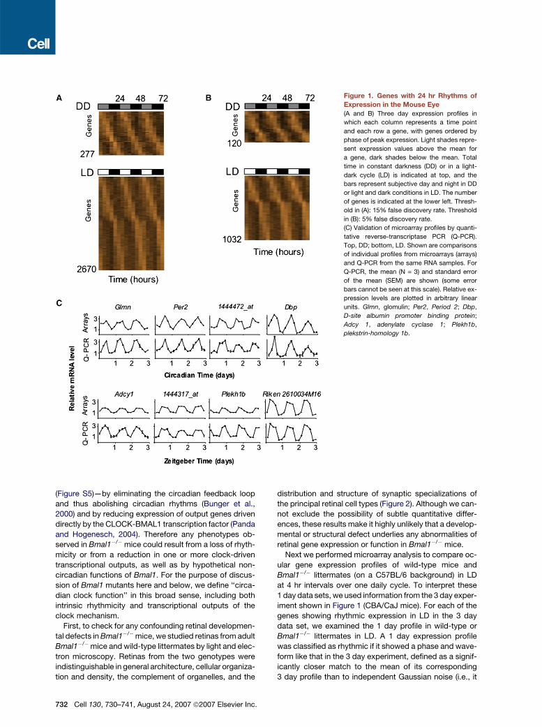
Figure 1. Genes with 24 hr Rhythms of
Expression in the Mouse Eye
(A and B) Three day expression profiles in
which each column represents a time point
and each row a gene, with genes ordered by
phase of peak expression. Light shades repre-
sent expression values above the mean for
a gene, dark shades below the mean. Total
time in constant darkness (DD) or in a light-
dark cycle (LD) is indicated at top, and the
bars represent subjective day and night in DD
or light and dark conditions in LD. The number
of genes is indicated at the lower left. Thresh-
old in (A): 15% false discovery rate. Threshold
in (B): 5% false discovery rate.
(C) Validation of microarray profiles by quanti-
tative reverse-transcriptase PCR (Q-PCR).
Top, DD; bottom, LD. Shown are comparisons
of individual profiles from microarrays (arrays)
and Q-PCR from the same RNA samples. For
Q-PCR, the mean (N = 3) and standard error
of the mean (SEM) are shown (some error
bars cannot be seen at this scale). Relative ex-
pression levels are plotted in arbitrary linear
units. Glmn, glomulin; Per2, Period 2; Dbp,
D-site albumin promoter binding protein;
Adcy 1, adenylate cyclase 1; Plekh1b,
plekstrin-homology 1b.
(Figure S5)—by eliminating the circadian feedback loop
and thus abolishing circadian rhythms (Bunger et al.,
2000) and by reducing expression of output genes driven
directly by the CLOCK-BMAL1 transcription factor (Panda
and Hogenesch, 2004). Therefore any phenotypes ob-
served in Bmal1�/�mice could result from a loss of rhyth-
micity or from a reduction in one or more clock-driven
transcriptional outputs, as well as by hypothetical non-
circadian functions of Bmal1. For the purpose of discus-
sion of Bmal1 mutants here and below, we define ‘‘circa-
dian clock function’’ in this broad sense, including both
intrinsic rhythmicity and transcriptional outputs of the
clock mechanism.
First, to check for any confounding retinal developmen-
tal defects in Bmal1�/�mice, we studied retinas from adult
Bmal1�/�mice and wild-type littermates by light and elec-
tron microscopy. Retinas from the two genotypes were
indistinguishable in general architecture, cellular organiza-
tion and density, the complement of organelles, and the
732 Cell 130, 730–741, August 24, 2007 ª2007 Elsevier Inc.
distribution and structure of synaptic specializations of
the principal retinal cell types (Figure 2). Although we can-
not exclude the possibility of subtle quantitative differ-
ences, these results make it highly unlikely that a develop-
mental or structural defect underlies any abnormalities of
retinal gene expression or function in Bmal1�/� mice.
Next we performed microarray analysis to compare oc-
ular gene expression profiles of wild-type mice and
Bmal1�/� littermates (on a C57BL/6 background) in LD
at 4 hr intervals over one daily cycle. To interpret these
1 day data sets, we used information from the 3 day exper-
iment shown in Figure 1 (CBA/CaJ mice). For each of the
genes showing rhythmic expression in LD in the 3 day
data set, we examined the 1 day profile in wild-type or
Bmal1�/� littermates in LD. A 1 day expression profile
was classified as rhythmic if it showed a phase and wave-
form like that in the 3 day experiment, defined as a signif-
icantly closer match to the mean of its corresponding
3 day profile than to independent Gaussian noise (i.e., it
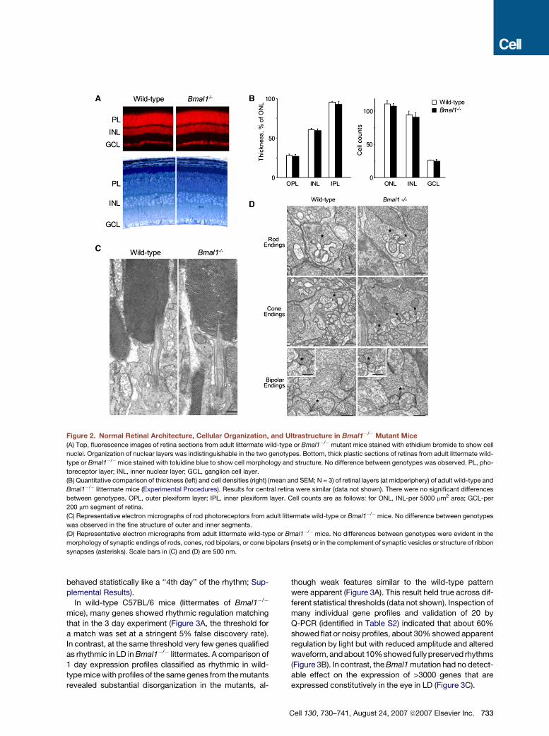
Figure 2. Normal Retinal Architecture, Cellular Organization, and Ultrastructure in Bmal1�/� Mutant Mice
(A) Top, fluorescence images of retina sections from adult littermate wild-type or Bmal1�/� mutant mice stained with ethidium bromide to show cell
nuclei. Organization of nuclear layers was indistinguishable in the two genotypes. Bottom, thick plastic sections of retinas from adult littermate wild-
type or Bmal1�/�mice stained with toluidine blue to show cell morphology and structure. No difference between genotypes was observed. PL, pho-
toreceptor layer; INL, inner nuclear layer; GCL, ganglion cell layer.
(B) Quantitative comparison of thickness (left) and cell densities (right) (mean and SEM; N = 3) of retinal layers (at midperiphery) of adult wild-type and
Bmal1�/� littermate mice (Experimental Procedures). Results for central retina were similar (data not shown). There were no significant differences
between genotypes. OPL, outer plexiform layer; IPL, inner plexiform layer. Cell counts are as follows: for ONL, INL-per 5000 mm2 area; GCL-per
200 mm segment of retina.
(C) Representative electron micrographs of rod photoreceptors from adult littermate wild-type or Bmal1�/�mice. No difference between genotypes
was observed in the fine structure of outer and inner segments.
(D) Representative electron micrographs from adult littermate wild-type or Bmal1�/� mice. No differences between genotypes were evident in the
morphology of synaptic endings of rods, cones, rod bipolars, or cone bipolars (insets) or in the complement of synaptic vesicles or structure of ribbon
synapses (asterisks). Scale bars in (C) and (D) are 500 nm.
behaved statistically like a ‘‘4th day’’ of the rhythm; Sup-
plemental Results).
In wild-type C57BL/6 mice (littermates of Bmal1�/�
mice), many genes showed rhythmic regulation matching
that in the 3 day experiment (Figure 3A, the threshold for
a match was set at a stringent 5% false discovery rate).
In contrast, at the same threshold very few genes qualified
as rhythmic in LD in Bmal1�/� littermates. A comparison of
1 day expression profiles classified as rhythmic in wild-
type mice with profiles of the same genes from the mutants
revealed substantial disorganization in the mutants, al-
though weak features similar to the wild-type pattern
were apparent (Figure 3A). This result held true across dif-
ferent statistical thresholds (data not shown). Inspection of
many individual gene profiles and validation of 20 by
Q-PCR (identified in Table S2) indicated that about 60%
showed flat or noisy profiles, about 30% showed apparent
regulation by light but with reduced amplitude and altered
waveform,and about 10% showed fully preserved rhythms
(Figure 3B). In contrast, the Bmal1 mutation had no detect-
able effect on the expression of >3000 genes that are
expressed constitutively in the eye in LD (Figure 3C).
Cell 130, 730–741, August 24, 2007 ª2007 Elsevier Inc. 733
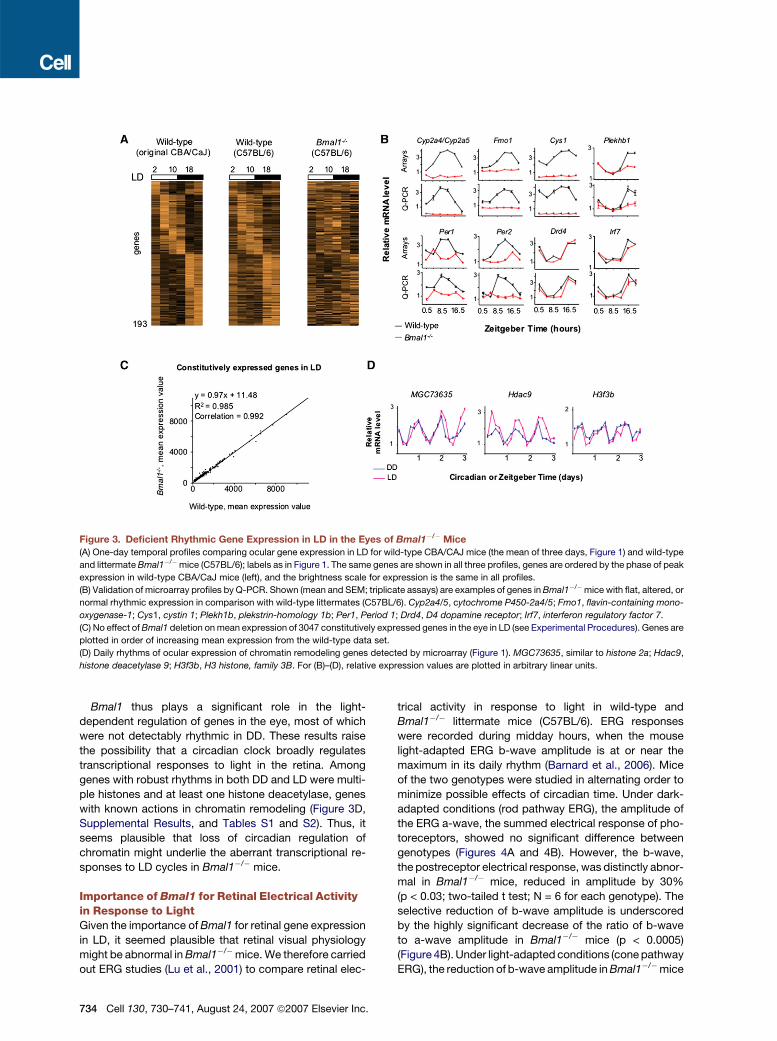
Figure 3. Deficient Rhythmic Gene Expression in LD in the Eyes of Bmal1�/� Mice
(A) One-day temporal profiles comparing ocular gene expression in LD for wild-type CBA/CAJ mice (the mean of three days, Figure 1) and wild-type
and littermate Bmal1�/�mice (C57BL/6); labels as in Figure 1. The same genes are shown in all three profiles, genes are ordered by the phase of peak
expression in wild-type CBA/CaJ mice (left), and the brightness scale for expression is the same in all profiles.
(B) Validation of microarray profiles by Q-PCR. Shown (mean and SEM; triplicate assays) are examples of genes in Bmal1�/�mice with flat, altered, or
normal rhythmic expression in comparison with wild-type littermates (C57BL/6). Cyp2a4/5, cytochrome P450-2a4/5; Fmo1, flavin-containing mono-
oxygenase-1; Cys1, cystin 1; Plekh1b, plekstrin-homology 1b; Per1, Period 1; Drd4, D4 dopamine receptor; Irf7, interferon regulatory factor 7.
(C) No effect of Bmal1 deletion on mean expression of 3047 constitutively expressed genes in the eye in LD (see Experimental Procedures). Genes are
plotted in order of increasing mean expression from the wild-type data set.
(D) Daily rhythms of ocular expression of chromatin remodeling genes detected by microarray (Figure 1). MGC73635, similar to histone 2a; Hdac9,
histone deacetylase 9; H3f3b, H3 histone, family 3B. For (B)–(D), relative expression values are plotted in arbitrary linear units.
Bmal1 thus plays a significant role in the light-
dependent regulation of genes in the eye, most of which
were not detectably rhythmic in DD. These results raise
the possibility that a circadian clock broadly regulates
transcriptional responses to light in the retina. Among
genes with robust rhythms in both DD and LD were multi-
ple histones and at least one histone deacetylase, genes
with known actions in chromatin remodeling (Figure 3D,
Supplemental Results, and Tables S1 and S2). Thus, it
seems plausible that loss of circadian regulation of
chromatin might underlie the aberrant transcriptional re-
sponses to LD cycles in Bmal1�/� mice.
Importance of Bmal1 for Retinal Electrical Activityin Response to LightGiven the importance of Bmal1 for retinal gene expression
in LD, it seemed plausible that retinal visual physiology
might be abnormal in Bmal1�/�mice. We therefore carried
out ERG studies (Lu et al., 2001) to compare retinal elec-
734 Cell 130, 730–741, August 24, 2007 ª2007 Elsevier Inc.
trical activity in response to light in wild-type and
Bmal1�/� littermate mice (C57BL/6). ERG responses
were recorded during midday hours, when the mouse
light-adapted ERG b-wave amplitude is at or near the
maximum in its daily rhythm (Barnard et al., 2006). Mice
of the two genotypes were studied in alternating order to
minimize possible effects of circadian time. Under dark-
adapted conditions (rod pathway ERG), the amplitude of
the ERG a-wave, the summed electrical response of pho-
toreceptors, showed no significant difference between
genotypes (Figures 4A and 4B). However, the b-wave,
the postreceptor electrical response, was distinctly abnor-
mal in Bmal1�/� mice, reduced in amplitude by 30%
(p < 0.03; two-tailed t test; N = 6 for each genotype). The
selective reduction of b-wave amplitude is underscored
by the highly significant decrease of the ratio of b-wave
to a-wave amplitude in Bmal1�/� mice (p < 0.0005)
(Figure 4B). Under light-adapted conditions (cone pathway
ERG), the reduction of b-wave amplitude in Bmal1�/�mice
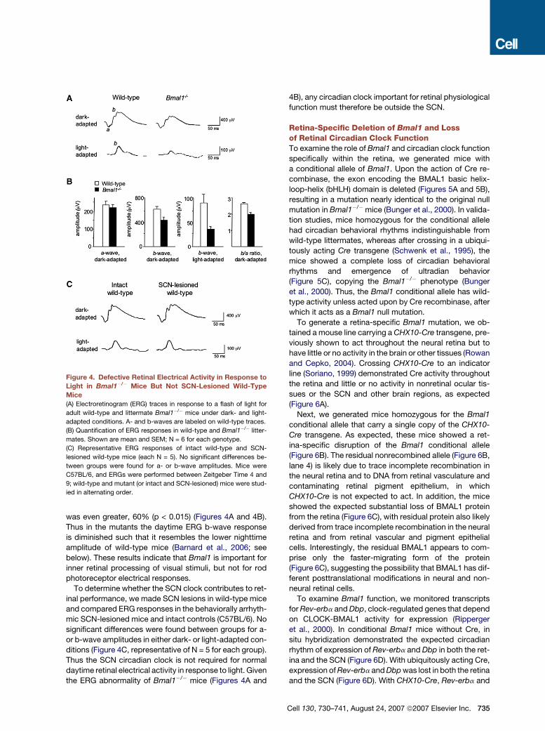
was even greater, 60% (p < 0.015) (Figures 4A and 4B).
Thus in the mutants the daytime ERG b-wave response
is diminished such that it resembles the lower nighttime
amplitude of wild-type mice (Barnard et al., 2006; see
below). These results indicate that Bmal1 is important for
inner retinal processing of visual stimuli, but not for rod
photoreceptor electrical responses.
To determine whether the SCN clock contributes to ret-
inal performance, we made SCN lesions in wild-type mice
and compared ERG responses in the behaviorally arrhyth-
mic SCN-lesioned mice and intact controls (C57BL/6). No
significant differences were found between groups for a-
or b-wave amplitudes in either dark- or light-adapted con-
ditions (Figure 4C, representative of N = 5 for each group).
Thus the SCN circadian clock is not required for normal
daytime retinal electrical activity in response to light. Given
the ERG abnormality of Bmal1�/� mice (Figures 4A and
Figure 4. Defective Retinal Electrical Activity in Response to
Light in Bmal1�/� Mice But Not SCN-Lesioned Wild-Type
Mice
(A) Electroretinogram (ERG) traces in response to a flash of light for
adult wild-type and littermate Bmal1�/� mice under dark- and light-
adapted conditions. A- and b-waves are labeled on wild-type traces.
(B) Quantification of ERG responses in wild-type and Bmal1�/� litter-
mates. Shown are mean and SEM; N = 6 for each genotype.
(C) Representative ERG responses of intact wild-type and SCN-
lesioned wild-type mice (each N = 5). No significant differences be-
tween groups were found for a- or b-wave amplitudes. Mice were
C57BL/6, and ERGs were performed between Zeitgeber Time 4 and
9; wild-type and mutant (or intact and SCN-lesioned) mice were stud-
ied in alternating order.
4B), any circadian clock important for retinal physiological
function must therefore be outside the SCN.
Retina-Specific Deletion of Bmal1 and Lossof Retinal Circadian Clock FunctionTo examine the role of Bmal1 and circadian clock function
specifically within the retina, we generated mice with
a conditional allele of Bmal1. Upon the action of Cre re-
combinase, the exon encoding the BMAL1 basic helix-
loop-helix (bHLH) domain is deleted (Figures 5A and 5B),
resulting in a mutation nearly identical to the original null
mutation in Bmal1�/�mice (Bunger et al., 2000). In valida-
tion studies, mice homozygous for the conditional allele
had circadian behavioral rhythms indistinguishable from
wild-type littermates, whereas after crossing in a ubiqui-
tously acting Cre transgene (Schwenk et al., 1995), the
mice showed a complete loss of circadian behavioral
rhythms and emergence of ultradian behavior
(Figure 5C), copying the Bmal1�/� phenotype (Bunger
et al., 2000). Thus, the Bmal1 conditional allele has wild-
type activity unless acted upon by Cre recombinase, after
which it acts as a Bmal1 null mutation.
To generate a retina-specific Bmal1 mutation, we ob-
tained a mouse line carrying a CHX10-Cre transgene, pre-
viously shown to act throughout the neural retina but to
have little or no activity in the brain or other tissues (Rowan
and Cepko, 2004). Crossing CHX10-Cre to an indicator
line (Soriano, 1999) demonstrated Cre activity throughout
the retina and little or no activity in nonretinal ocular tis-
sues or the SCN and other brain regions, as expected
(Figure 6A).
Next, we generated mice homozygous for the Bmal1
conditional allele that carry a single copy of the CHX10-
Cre transgene. As expected, these mice showed a ret-
ina-specific disruption of the Bmal1 conditional allele
(Figure 6B). The residual nonrecombined allele (Figure 6B,
lane 4) is likely due to trace incomplete recombination in
the neural retina and to DNA from retinal vasculature and
contaminating retinal pigment epithelium, in which
CHX10-Cre is not expected to act. In addition, the mice
showed the expected substantial loss of BMAL1 protein
from the retina (Figure 6C), with residual protein also likely
derived from trace incomplete recombination in the neural
retina and from retinal vascular and pigment epithelial
cells. Interestingly, the residual BMAL1 appears to com-
prise only the faster-migrating form of the protein
(Figure 6C), suggesting the possibility that BMAL1 has dif-
ferent posttranslational modifications in neural and non-
neural retinal cells.
To examine Bmal1 function, we monitored transcripts
for Rev-erba and Dbp, clock-regulated genes that depend
on CLOCK-BMAL1 activity for expression (Ripperger
et al., 2000). In conditional Bmal1 mice without Cre, in
situ hybridization demonstrated the expected circadian
rhythm of expression of Rev-erba and Dbp in both the ret-
ina and the SCN (Figure 6D). With ubiquitously acting Cre,
expression of Rev-erba and Dbp was lost in both the retina
and the SCN (Figure 6D). With CHX10-Cre, Rev-erba and
Cell 130, 730–741, August 24, 2007 ª2007 Elsevier Inc. 735
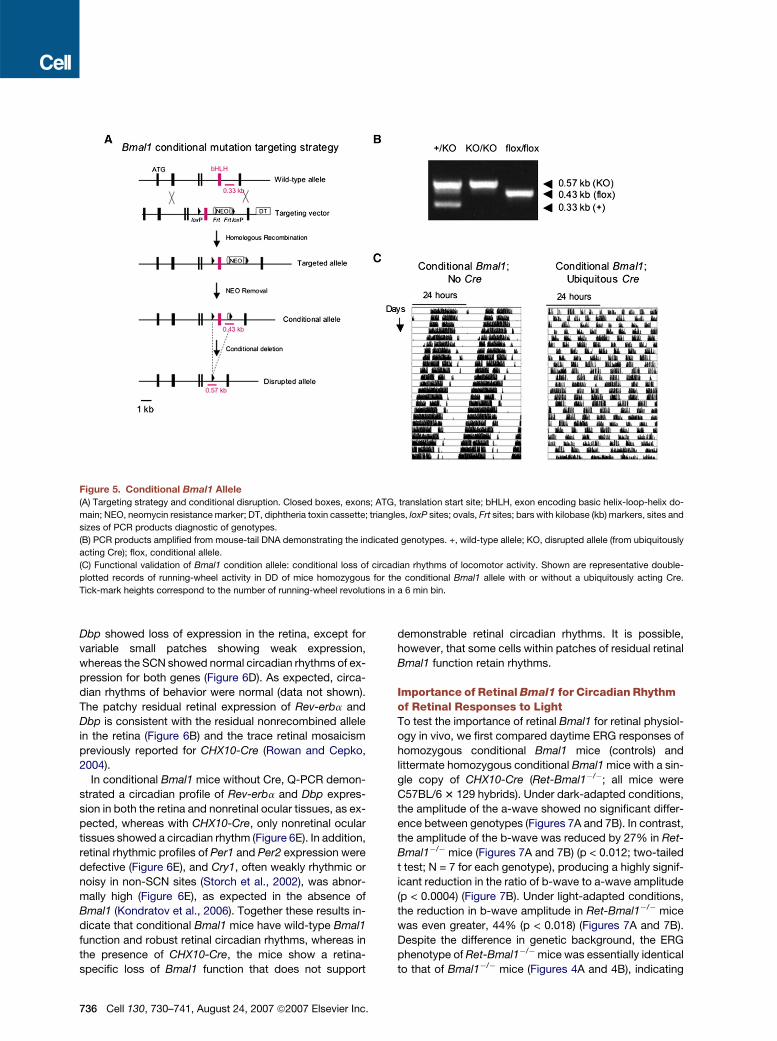
Figure 5. Conditional Bmal1 Allele
(A) Targeting strategy and conditional disruption. Closed boxes, exons; ATG, translation start site; bHLH, exon encoding basic helix-loop-helix do-
main; NEO, neomycin resistance marker; DT, diphtheria toxin cassette; triangles, loxP sites; ovals, Frt sites; bars with kilobase (kb) markers, sites and
sizes of PCR products diagnostic of genotypes.
(B) PCR products amplified from mouse-tail DNA demonstrating the indicated genotypes. +, wild-type allele; KO, disrupted allele (from ubiquitously
acting Cre); flox, conditional allele.
(C) Functional validation of Bmal1 condition allele: conditional loss of circadian rhythms of locomotor activity. Shown are representative double-
plotted records of running-wheel activity in DD of mice homozygous for the conditional Bmal1 allele with or without a ubiquitously acting Cre.
Tick-mark heights correspond to the number of running-wheel revolutions in a 6 min bin.
Dbp showed loss of expression in the retina, except for
variable small patches showing weak expression,
whereas the SCN showed normal circadian rhythms of ex-
pression for both genes (Figure 6D). As expected, circa-
dian rhythms of behavior were normal (data not shown).
The patchy residual retinal expression of Rev-erba and
Dbp is consistent with the residual nonrecombined allele
in the retina (Figure 6B) and the trace retinal mosaicism
previously reported for CHX10-Cre (Rowan and Cepko,
2004).
In conditional Bmal1 mice without Cre, Q-PCR demon-
strated a circadian profile of Rev-erba and Dbp expres-
sion in both the retina and nonretinal ocular tissues, as ex-
pected, whereas with CHX10-Cre, only nonretinal ocular
tissues showed a circadian rhythm (Figure 6E). In addition,
retinal rhythmic profiles of Per1 and Per2 expression were
defective (Figure 6E), and Cry1, often weakly rhythmic or
noisy in non-SCN sites (Storch et al., 2002), was abnor-
mally high (Figure 6E), as expected in the absence of
Bmal1 (Kondratov et al., 2006). Together these results in-
dicate that conditional Bmal1 mice have wild-type Bmal1
function and robust retinal circadian rhythms, whereas in
the presence of CHX10-Cre, the mice show a retina-
specific loss of Bmal1 function that does not support
736 Cell 130, 730–741, August 24, 2007 ª2007 Elsevier Inc.
demonstrable retinal circadian rhythms. It is possible,
however, that some cells within patches of residual retinal
Bmal1 function retain rhythms.
Importance of Retinal Bmal1 for Circadian Rhythmof Retinal Responses to LightTo test the importance of retinal Bmal1 for retinal physiol-
ogy in vivo, we first compared daytime ERG responses of
homozygous conditional Bmal1 mice (controls) and
littermate homozygous conditional Bmal1 mice with a sin-
gle copy of CHX10-Cre (Ret-Bmal1�/�; all mice were
C57BL/6 3 129 hybrids). Under dark-adapted conditions,
the amplitude of the a-wave showed no significant differ-
ence between genotypes (Figures 7A and 7B). In contrast,
the amplitude of the b-wave was reduced by 27% in Ret-
Bmal1�/� mice (Figures 7A and 7B) (p < 0.012; two-tailed
t test; N = 7 for each genotype), producing a highly signif-
icant reduction in the ratio of b-wave to a-wave amplitude
(p < 0.0004) (Figure 7B). Under light-adapted conditions,
the reduction in b-wave amplitude in Ret-Bmal1�/� mice
was even greater, 44% (p < 0.018) (Figures 7A and 7B).
Despite the difference in genetic background, the ERG
phenotype of Ret-Bmal1�/�mice was essentially identical
to that of Bmal1�/� mice (Figures 4A and 4B), indicating
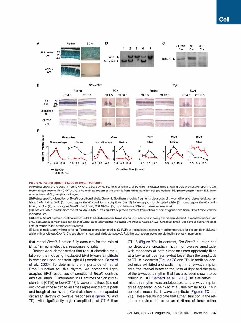
Figure 6. Retina-Specific Loss of Bmal1 Function(A) Retina-specific Cre activity from CHX10-Cre transgene. Sections of retina and SCN from indicator mice showing blue precipitate reporting Cre
recombinase activity. For CHX10-Cre, blue stain at bottom of the brain is from retinal ganglion cell projections. PL, photoreceptor layer; INL, inner
nuclear layer; GCL, ganglion cell layer.
(B) Retina-specific disruption of Bmal1 conditional allele. Genomic Southern showing fragments diagnostic of the conditional or disrupted Bmal1 al-
leles. (1–4), Retina DNA. (1), homozygous Bmal1 conditional, ubiquitous Cre; (2), heterozygous for disrupted allele; (3), homozygous Bmal1 condi-
tional, no Cre; (4), homozygous Bmal1 conditional, CHX10-Cre; (5), hypothalamus DNA from same mouse as (4).
(C) Loss of BMAL1 protein from the retina. Anti-BMAL1 western blot of protein extracts from retinas of homozygous conditional Bmal1 mice with the
indicated Cre.
(D) Loss of Bmal1 function in retina but not SCN. In situ hybridization to retina and SCN sections showing expression of Bmal1-dependent genes Rev-
erba and Dbp in homozygous conditional Bmal1 mice carrying the indicated Cre transgene are shown. Circadian times (CT) correspond to the peak
(left) or trough (right) of transcript rhythms.
(E) Loss of molecular rhythms in retina. Temporal expression profiles (Q-PCR) of the indicated genes in mice homozygous for the conditional Bmal1
allele with or without CHX10-Cre are shown (mean and triplicate assays). Relative expression levels are plotted in arbitrary linear units.
that retinal Bmal1 function fully accounts for the role of
Bmal1 in retinal electrical responses to light.
Recent work demonstrates that robust circadian regu-
lation of the mouse light-adapted ERG b-wave amplitude
is revealed under constant light (LL) conditions (Barnard
et al., 2006). To determine the importance of retinal
Bmal1 function for this rhythm, we compared light-
adapted ERG responses of conditional Bmal1 controls
and Ret-Bmal1�/� littermates in LL at times of high (circa-
dian time [CT] 6) or low (CT 18) b-wave amplitude (it is not
yet known if these circadian times represent the true peak
and trough of the rhythm). Controls showed the expected
circadian rhythm of b-wave responses (Figures 7C and
7D), with significantly higher amplitudes at CT 6 than
CT 18 (Figure 7D). In contrast, Ret-Bmal1�/� mice had
no detectable circadian rhythm of b-wave amplitude,
with responses at both circadian times apparently fixed
at a low amplitude, somewhat lower than the amplitude
at CT 18 in controls (Figures 7C and 7D). In addition, con-
trol mice exhibited a circadian rhythm of b-wave implicit
time (the interval between the flash of light and the peak
of the b-wave), a rhythm that has also been shown to be
robust in DD (Barnard et al., 2006). In Ret-Bmal1�/�
mice this rhythm was undetectable, and b-wave implicit
times appeared to be fixed at a value similar to CT 18 in
controls, much like b-wave amplitude (Figures 7C and
7D). These results indicate that Bmal1 function in the ret-
ina is required for circadian rhythms of inner retinal
Cell 130, 730–741, August 24, 2007 ª2007 Elsevier Inc. 737
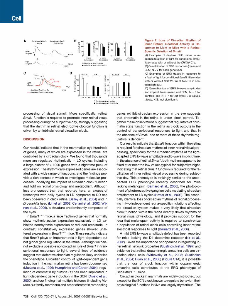
Figure 7. Loss of Circadian Rhythm of
Inner Retinal Electrical Activity in Re-
sponse to Light in Mice with a Retina-
Specific Deletion of Bmal1
(A) Examples of daytime ERG traces in re-
sponse to a flash of light for conditional Bmal1
littermates with or without the CHX10-Cre.
(B) Quantification of ERG responses (mean and
SEM; N = 7 for each genotype).
(C) Examples of ERG traces in response to
a flash of light for conditional Bmal1 littermates
with or without CHX10-Cre at two CT in con-
stant light (LL).
(D) Quantification of ERG b-wave amplitudes
and implicit times (mean and SEM; N = 8 for
controls and N = 7 for ret-Bmal1). p values,
t tests. N.S., not significant.
processing of visual stimuli. More specifically, retinal
Bmal1 function is required to promote inner retinal visual
processing during the subjective day, strongly suggesting
that the rhythm in retinal electrophysiological function is
driven by an intrinsic retinal circadian clock.
DISCUSSION
Our results indicate that in the mammalian eye hundreds
of genes, many of which are expressed in the retina, are
controlled by a circadian clock. We found that thousands
more are regulated rhythmically in LD cycles, including
a large cluster of > 1000 genes with a nighttime peak of
expression. The rhythmically expressed genes are associ-
ated with a wide range of functions, and the findings pro-
vide a rich context in which to investigate molecular pro-
cesses underlying the impact of circadian clock function
and light on retinal physiology and metabolism. Although
less pronounced than that reported here, an excess of
transcripts with daily cycles in LD compared to DD has
been observed in chick retina (Bailey et al., 2004) and in
Drosophila head (Lin et al., 2002; Ceriani et al., 2002; Wij-
nen et al., 2006), a structure predominantly composed of
the eyes.
In Bmal1�/�mice, a large fraction of genes that normally
show rhythmic ocular expression exclusively in LD ex-
hibited nonrhythmic expression or reduced amplitude. In
contrast, constitutively expressed genes showed unal-
tered expression in Bmal1�/�mice. These results indicate
that Bmal1 plays an important role in light-dependent but
not global gene regulation in the retina. Although we can-
not exclude a possible noncircadian role of Bmal1 in tran-
scriptional responses to light, several lines of evidence
suggest that defective circadian regulation likely underlies
the phenotype. Circadian control of light-dependent gene
induction in the mammalian retina has been documented
(Masana et al., 1996; Humphries and Carter, 2004), regu-
lation of chromatin by histone H3 has been implicated in
light-dependent gene induction in the SCN (Crosio et al.,
2000), and our finding that multiple histones (including his-
tone H3 family members) and other chromatin remodeling
738 Cell 130, 730–741, August 24, 2007 ª2007 Elsevier Inc.
genes exhibit circadian expression in the eye suggests
that chromatin in the retina is under clock control. To-
gether these observations suggest that regulators of chro-
matin state function in the retina as clock outputs in the
control of transcriptional responses to light and that in
the absence of Bmal1 one or more of these rhythmic reg-
ulators is deficient.
Our results indicate that Bmal1 function within the retina
is required for circadian rhythms of inner retinal visual pro-
cessing, specifically for the circadian rhythms of the light-
adapted ERG b-wave amplitude and b-wave implicit time.
In the absence of retinal Bmal1, both rhythms appear to be
fixed at or near the low values typical for subjective night,
indicating that retinal Bmal1 function is required for the fa-
cilitation of inner retinal visual processing during subjec-
tive day. This phenotype is strikingly similar to the unex-
pected ERG phenotype recently described for mice
lacking melanopsin (Barnard et al., 2006), the photopig-
ment of photoreceptive ganglion cells mediating circadian
entrainment to LD cycles (Hattar et al., 2003). The essen-
tially identical loss of circadian rhythms of retinal process-
ing in two independent retina-specific mutations affecting
the circadian system makes it very likely that circadian
clock function within the retina directly drives rhythms of
retinal visual physiology, and it provides support for the
idea that melanopsin activity is required to synchronize
a population of retinal clock cells controlling inner retinal
electrical responses to light (Barnard et al., 2006).
A mild ERG b-wave amplitude defect has been reported
for mice lacking the D4 dopamine receptor (Nir et al.,
2002). Given the importance of dopamine in regulating in-
ner retinal network properties (Gustincich et al., 1997) and
evidence that retinal dopaminergic amacrine cells are cir-
cadian clock cells (Witkovsky et al., 2003; Gustincich
et al., 2004; Ruan et al., 2006) (Figure S1A), it is possible
that the loss of clock function within dopaminergic
amacrine cells contributes to the ERG phenotype of
Ret-Bmal1�/� mice.
Circadian clocks in mammals are widely distributed, but
except for the SCN clock known to regulate behavior, their
physiological functions in vivo are largely mysterious. The
studies described here indicate that an intrinsic retinal cir-
cadian clock regulates retinal visual processing in vivo and
that it does so autonomously, with no detectable contribu-
tion from the SCN or other clocks. Together with observa-
tions emerging from other tissue-specific manipulations of
clock function (Durgan et al., 2006; McDearmon et al.,
2006), our work provides evidence that circadian clocks
outside the SCN contribute important physiological func-
tions in vivo. Thus over evolutionary time different cell
types have likely recruited the circadian clock mechanism
inherited from a single-celled ancestor for control of spe-
cialized tissue-specific processes.
EXPERIMENTAL PROCEDURES
Mice and Tissue Collection
For 3 day microarray experiments, 108 adult male CBA/CaJ mice
(Jackson Laboratory) were entrained to a 12 hr:12 hr LD cycle for
3 weeks. During the dark phase, 54 mice were transferred to DD, the
remainder kept in LD. At 4 hr intervals, three mice from each group
were euthanized (CO2), and eyes were collected, frozen in liquid nitro-
gen, and stored (�80�C). Total RNA was purified separately from the
eyes of each mouse (Trizol, Invitrogen and RNAeasy, QIAGEN), and
equal aliquots of RNA from the three pairs of eyes collected at a single
time point were pooled. Studies were performed in accordance with
the protocol approved by the Harvard Medical School Standing Com-
mittee on Animals.
Microroarray Analysis
Samples were hybridized to Affymetrix mouse 430.2 arrays represent-
ing the complete mouse genome (one array per time point). Fluores-
cence images were normalized to median brightness (Li and Wong,
2001), and model-based expression values were computed using
DNA-Chip Analyzer (Li and Wong, 2003). In each 3 day, 18 array
data set, probe sets that were not classified as ‘‘expressed’’ (http://
www.Affymetrix.com/products/algorithms_tech.htm) in at least three
arrays were excluded, as were probe sets that had three or more miss-
ing expression values. The square-root transformation was applied to
all expression values to make the error distribution more symmetric
while limiting amplification of low-level noise. For a detailed descrip-
tion of procedures for identifying rhythmic expression, see Supple-
mental Results. To assess nonrhythmic transcripts, expression values
determined by DNA-Chip Analyzer (Li and Wong, 2003) for the six ar-
rays corresponding to the six time-points from wild-type mice in LD
were used to select a ‘‘constitutively expressed’’ set of genes. Genes
with low mean expression value were excluded (<200 units; range:
0–10,900 units), and remaining genes with a near-constant level of
expression across the six arrays were selected (standard deviation
of expression < 5%, yielding 3047 genes). Microarray data have
been deposited in the ArrayExpress database (accession number
E-TABM-285).
Light and Electron Microscopy of Retinas
For light microscopy, eyecups of three mice of each genotype were
fixed (2% formaldehyde in 0.15 M Sørensen phosphate buffer,
pH 7.4), cryoprotected (20% sucrose in PBS), and frozen in monochloro-
difluoromethane. Sections parallel to and including the horizontal
meridian were obtained and stained with 300 nM 40,6-diamidino-2-
phenylindole (DAPI) in PBS. Fluorescence was detected with a Zeiss
LSM 510 Meta confocal system and Zeiss Axioplan 2 microscope. Be-
cause photoreceptor activity was unaltered in Bmal1�/� mice, the
thickness of the deep retinal layers was normalized to that of the
ONL in each retina in order to avoid errors due to obliquity of sections.
For EM, two mice of each genotype were perfused with 2% formal-
dehyde and 2.5% glutaraldehyde in 0.15 M Sørenson phosphate
buffer (pH 7.4), followed by 1% OsO4, 1.5% potassium ferrocyanide,
and stained en bloc with 1% uranyl acetate. Thick sections of the eye-
cup were stained with toluidine blue and examined with a Nikon
Eclipse E600 microscope. Thin sections were stained with uranyl
and lead, and micrographs were obtained with a JEOL X100 electron
microscope.
Quantitative RT-PCR
Total RNA from whole eyes or dissected eye fractions (retinal, lens, and
the rest of the eye) was transcribed into cDNA using random hexamers
and Superscript reverse transcriptase (Invitrogen). cDNA derived from
25 ng of total RNA was PCR amplified in a PTC-200 thermocycler with
a Chromo4 module (MJ Research) using SYBR green (IQ SYBR Green
Supermix, Bio-Rad) according to the manufacturer’s instructions.
Templates were amplified with the annealing temperature initially low-
ered from 70�C to 60�C in 2 cycle increments followed by 30 cycles of
the following three steps: 20 s at 94�C, 20 s at 60�C, and 30 s at 72�C.
For quantitation, threshold cycle number difference was calculated
based on the sample with the lowest expression, and the values ob-
tained were 2n transformed. Input RNA concentration or the expres-
sion level of hypoxanthine guanine phosphoribosyl transferase-1 was
used to normalize expression values. Sequences of primers are avail-
able upon request.
Electroretinography
ERG recordings were performed as described (Lu et al., 2001). Briefly,
mice were dark-adapted overnight prior to recording. Mice were anes-
thetized, their pupils dilated, and responses were recorded with a chlo-
ride silver wire loop placed on the cornea. ERG responses were elicited
with 10 ms flashes of white light (1.37 3 105 cd/m2) presented in a Ganz-
feld dome at intervals of 1 min in darkness. Mice were then adapted to
a background illumination (12 ft.-L., 10 min) and responses were eli-
cited by 1 Hz flashes of white light (1.37 3 105 cd/m2). Circadian com-
ponents of the light-adapted ERG were obtained from mice in constant
light as described (Barnard et al., 2006), except the intensity of light
was 300 Lux.
SCN Lesions
Adult C57BL/6J mice were anesthetized with ketamine-xylazine
(45 mg/kg-5 mg/kg), and an electrode (RNE-300X, Rhodes Medical In-
struments) was lowered stereotaxically through the skull at the mid-
sagittal sinus (anteroposterior -0.45 mm from Bregma) with the final
tip position at 6.0 mm below the skull. Lesions were generated with
constant current (2 mA, 10 s; D.C. Constant Lesion Maker, Grass In-
struments). From 1 week after surgery, locomotor activity in DD was
monitored, and only mice showing arrhythmic behavior for >4 weeks
were selected for ERG studies. Only mice with histologically verified
complete SCN lesions were included in the analysis (data not shown).
Generation of Conditional Bmal1 Mice
Conditional Bmal1 mice can be ordered from The Jackson Laboratory
(stock number 7668). A cassette containing neomycin acetyltransfer-
ase (neo) flanked by two FRT sites and a single loxP site (EcoRI-BamHI
fragment from vector PL452; Liu et al., [2003]) was inserted into a bac-
terial artificial chromosome encompassing the Bmal1 locus (RPCI23-
331K23, BACPAC resources, Children’s Hospital Oakland Research
Institute) by homologous recombination in bacteria (Liu et al., 2003).
The insertion site was immediately upstream of the PacI site in the in-
tron 30 of the exon encoding the basic helix-loop-helix domain (exon 8).
A 10.8 kb PmlI-fragment containing the neo cassette with flanking ge-
nomic regions was subcloned into the HpaI-SmaI-digested targeting
vector pKOII (Bardeesy et al., 2002), which contained a diphtheria toxin
expression cassette. An oligonucleotide comprising a single loxP site
preceded by a BamHI restriction site was cloned into the SanDI site
of the intron preceding exon 8, resulting in the final targeting vector.
Electroporation of ES cells, selection of neomycin-resistant clones,
and injection of ES cells into blastocysts were performed using
Cell 130, 730–741, August 24, 2007 ª2007 Elsevier Inc. 739
standard methods. The neo cassette was removed by mating chi-
meras to mice carrying a Flpe knockin at the Rosa26 locus (Farley
et al., 2000). Genotyping was performed using multiplex PCR and
primers L1, ACTGGAAGTAACTTTATCAAACTG; L2, CTGACCAACTT
GCTAACAATTA (reverse primer); and R4, CTCCTAACTTGGTTTTT
GTCTGT. For assessing the efficiency of loxP recombination, retinal
genomic DNA was digested with AvrII, and Southern blot hybridization
was performed using a 350 bp probe specific for sequences immedi-
ately following the 30 flanking region used for targeting.
In Situ Hybridization, Histology, and Western Blots
For in situ hybridization, dissected retinas were fixed (cold 4% formal-
dehyde in PBS, 10 min), embedded (Tissue-Tek), cut frozen (20 mm),
and processed as described (Kraves and Weitz, 2006). Sequences
of riboprobes are available upon request. b-galactosidase activity in
20 mm brain or retina sections was detected with 5-bromo-4-chloro-
3-indolyl b-D-galactopyranoside (X-gal). Western analysis was per-
formed with rabbit anti-BMAL1 antiserum (1:500).
Supplemental Data
Supplemental Data include six figures, Supplemental Results, and two
tables and can be found with this article online at http://www.cell.com/
cgi/content/full/130/4/730/DC1/.
ACKNOWLEDGMENTS
We thank Susan Dymecki and Connie Cepko for data on CHX10-Cre;
Christopher Bradfield for Bmal1�/� (Mop3�/�) mice; Ueli Schibler for
anti-BMAL1 antiserum; the Gene Manipulation Core of the Develop-
mental Disabilities Research Center of Children’s Hospital, Boston
for expert service; and Ming Liu and Xiao Ling Long for expert technical
assistance. This work was supported by grants from the National Insti-
tutes of Health to C.J.W. (NS055831), E.R. (EY001344), and T.L.
(EY10309).
Received: January 11, 2007
Revised: April 27, 2007
Accepted: June 18, 2007
Published: August 23, 2007
REFERENCES
Abe, M., Herzog, E.D., Yamazaki, S., Straume, M., Tei, H., Sakaki, Y.,
Menaker, M., and Block, G.D. (2002). Circadian rhythms in isolated
brain regions. J. Neurosci. 22, 350–356.
Akhtar, R.A., Reddy, A.B., Maywood, E.S., Clayton, J.D., King, V.M.,
Smith, A.G., Gant, T.W., Hastings, M.H., and Kyriacou, C.P. (2002).
Circadian cycling of the mouse liver transcriptome, as revealed by
cDNA microarray, is driven by the suprachiasmatic nucleus. Curr.
Biol. 12, 540–550.
Bailey, M.J., Beremand, P.D., Hammer, R., Reidel, E., Thomas, T.L.,
and Cassone, V.M. (2004). Transcriptional profiling of circadian pat-
terns of mRNA expression in the chick retina. J. Biol. Chem. 279,
52247–52254.
Barnard, A.R., Hattar, S., Hankins, M.W., and Lucas, R.J. (2006). Mel-
anopsin regulates visual processing in the mouse retina. Curr. Biol. 16,
389–395.
Balsalobre, A., Damiola, F., and Schibler, U. (1998). A serum shock in-
duces circadian gene expression in cultured Rat-1 fibroblasts. Cell 93,
929–937.
Bardeesy, N., Sinha, M., Hezel, A.F., Signoretti, S., Hathaway, N.A.,
Sharpless, N.E., Loda, M., Carrasco, D.R., and DePinho, R.A. (2002).
Loss of the Lkb1 tumour suppressor provokes intestinal polyposis
but resistance to transformation. Nature 419, 162–167.
740 Cell 130, 730–741, August 24, 2007 ª2007 Elsevier Inc.
Besharse, J.C., and Iuvone, P.M. (1983). Circadian clock in Xenopus
eye controlling retinal serotonin N-acetyltransferase. Nature 305,
133–135.
Bunger, M.K., Wilsbacher, L.D., Moran, S.M., Clendenin, C., Radcliffe,
L.A., Hogenesch, J.B., Simon, M.C., Takahashi, J.S., and Bradfield,
C.A. (2000). Mop3 is an essential component of the master circadian
pacemaker in mammals. Cell 103, 1009–1017.
Cahill, G.M., and Besharse, J.C. (1993). Circadian clock functions lo-
calized in xenopus retinal photoreceptors. Neuron 10, 573–577.
Ceriani, M.F., Hogenesch, J.B., Yanovsky, M., Panda, S., Straume, M.,
and Kay, S.A. (2002). Genome-wide expression analysis in Drosophila
reveals genes controlling circadian behavior. J. Neurosci. 22, 9305–
9319.
Crosio, C., Cermakian, N., Allis, C.D., and Sassone-Corsi, P. (2000).
Light induces chromatin modification in cells of the mammalian circa-
dian clock. Nat. Neurosci. 3, 1241–1247.
Damiola, F., Le Minh, N., Preitner, N., Kornmann, B., Fleury-Olela, F.,
and Schibler, U. (2000). Restricted feeding uncouples circadian oscil-
lators in peripheral tissues from the central pacemaker in the supra-
chiasmatic nucleus. Genes Dev. 14, 2950–2961.
Doyle, S.E., Grace, M.S., McIvor, W., and Menaker, M. (2002). Circa-
dian rhythms of dopamine in mouse retina: the role of melatonin. Vis.
Neurosci. 19, 593–601.
Duffield, G.E., Best, J.D., Meurers, B.H., Bittner, A., Loros, J.J., and
Dunlap, J.C. (2002). Circadian programs of transcriptional activation,
signaling, and protein turnover revealed by microarray analysis of
mammalian cells. Curr. Biol. 12, 551–557.
Durgan, D.J., Trexler, N.A., Egbejimi, O., McElfresh, T.A., Suk, H.Y.,
Petterson, L.E., Shaw, C.A., Hardin, P.E., Bray, M.S., Chandler,
M.P., et al. (2006). The circadian clock within the cardiomyocyte is es-
sential for responsiveness of the heart to fatty acids. J. Biol. Chem.
281, 24254–24269.
Farley, F.W., Soriano, P., Steffen, L.S., and Dymecki, S.M. (2000).
Widespread recombinase expression using FLPeR (flipper) mice. Gen-
esis 28, 106–110.
Gekakis, N., Staknis, D., Nguyen, H.B., Davis, F.C., Wilsbacher, L.D.,
King, D.P., Takahashi, J.S., and Weitz, C.J. (1998). Role of the CLOCK
protein in the mammalian circadian mechanism. Science 280, 1564–
1569.
Goto, M., Oshima, I., Tomita, T., and Ebihara, S. (1989). Melatonin
content of the pineal gland in different mouse strains. J. Pineal Res.
7, 195–204.
Gustincich, S., Feigenspan, A., Wu, D.K., Koopman, L.J., and Raviola,
E. (1997). Control of dopamine release in the retina: a transgenic ap-
proach to neural networks. Neuron 18, 723–736.
Gustincich, S., Contini, M., Gariboldi, M., Puopolo, M., Kadota, K.,
Bono, H., LeMieux, J., Walsh, P., Carninci, P., Hayashizaki, Y., et al.
(2004). Gene discovery in genetically labeled single dopaminergic neu-
rons of the retina. Proc. Natl. Acad. Sci. USA 101, 5069–5074.
Hattar, S., Lucas, R.J., Mrosovsky, N., Thompson, S., Douglas, R.H.,
Hankins, M.W., Lem, J., Biel, M., Hofmann, F., Foster, R.G., et al.
(2003). Melanopsin and rod-cone photoreceptive systems account
for all major accessory visual functions in mice. Nature 424, 76–81.
Hayasaka, N., LaRue, S.I., and Green, C.B. (2002). In vivo disruption of
Xenopus CLOCK in the retinal photoreceptor cells abolishes circadian
melatonin rhythmicity without affecting its production levels. J. Neuro-
sci. 22, 1600–1607.
Humphries, A., and Carter, D.A. (2004). Circadian dependency of noc-
turnal immediate-early protein induction in rat retina. Biochem. Bio-
phys. Res. Commun. 320, 551–556.
Ko, C.H., and Takahashi, J.S. (2006). Molecular components of the
mammalian circadian clock. Hum. Mol. Genet. 15, R271–R277.
Kondratov, R.V., Shamanna, R.K., Kondratova, A.A., Gorbacheva,
V.Y., and Antoch, M.P. (2006). Dual role of the CLOCK/BMAL1 circa-
dian complex in transcriptional regulation. FASEB J. 20, 530–532.
Kraves, S., and Weitz, C.J. (2006). A role for cardiotrophin-like cytokine
in the circadian control of mammalian locomotor activity. Nat. Neuro-
sci. 9, 212–219.
LaVail, M.M., and Ward, P.A. (1978). Studies on the hormonal control
of circadian outer segment disc shedding in the rat retina. Invest. Oph-
thalmol. Vis. Sci. 17, 1189–1193.
Li, C., and Wong, W.H. (2001). Model-based analysis of oligonucleo-
tide arrays: model validation, design issues and standard error appli-
cation. Genome Biol. 2, RESEARCH0032.
Li, C., and Wong, W.H. (2003). DNA-Chip Analyzer (dChip). In The Anal-
ysis of Gene Expression Data: Methods and Software, G. Parmigiani,
E.S. Garrett, R. Irizarry, and S.L. Zeger, eds. (New York, NY: Springer),
pp. 120–141.
Lin, Y., Han, M., Shimada, B., Wang, L., Gibler, T.M., Amarakone, A.,
Awad, T.A., Stormo, G.D., Van Gelder, R.N., and Taghert, P.H.
(2002). Influence of the period-dependent circadian clock on diurnal,
circadian, and aperiodic gene expression in Drosophila melanogaster.
Proc. Natl. Acad. Sci. USA 99, 9562–9567.
Liu, P., Jenkins, N.A., and Copeland, N.G. (2003). A highly efficient
recombineering-based method for generating conditional knockout
mutations. Genome Res. 13, 476–484.
Lu, C., Peng, Y.W., Shang, J., Pawlyk, B.S., Yu, F., and Li, T. (2001).
The mammalian retinal degeneration B2 gene is not required for pho-
toreceptor function and survival. Neuroscience 107, 35–41.
Manglapus, M.K., Uchiyama, H., Buelow, N.F., and Barlow, R.B.
(1998). Circadian rhythms of rod-cone dominance in the Japanese
quail retina. J. Neurosci. 18, 4775–4784.
Masana, M.I., Benloucif, S., and Dubocovich, M.L. (1996). Light-
induced c-fos mRNA expression in the suprachiasmatic nucleus and
the retina of C3H/HeN mice. Brain Res. Mol. Brain Res. 42, 193–201.
McDearmon, E.L., Patel, K.N., Ko, C.H., Walisser, J.A., Schook, A.C.,
Chong, J.L., Wilsbacher, L.D., Song, E.J., Hong, H.K., Bradfield, C.A.,
et al. (2006). Dissecting the functions of the mammalian clock protein
BMAL1 by tissue-specific rescue in mice. Science 314, 1304–1308.
McGoogan, J.M., and Cassone, V.M. (1999). Circadian regulation of
chick electroretinogram: effects of pinealectomy and exogenous mel-
atonin. Am. J. Physiol. 277, R1418–R1427.
Miranda-Anaya, M., Bartell, P.A., and Menaker, M. (2002). Circadian
rhythm of iguana electroretinogram: the role of dopamine and melato-
nin. J. Biol. Rhythms 17, 526–538.
Nir, I., Harrison, J.M., Haque, R., Low, M.J., Grandy, D.K., Rubinstein,
M., and Iuvone, P.M. (2002). Dysfunctional light-evoked regulation of
cAMP in photoreceptors and abnormal retinal adaptation in mice lack-
ing dopamine D4 receptors. J. Neurosci. 22, 2063–2073.
Ouyang, Y., Andersson, C.R., Kondo, T., Golden, S.S., and Johnson,
C.H. (1998). Resonating circadian clocks enhance fitness in cyanobac-
teria. Proc. Natl. Acad. Sci. USA 95, 8660–8664.
Panda, S., and Hogenesch, J.B. (2004). It’s all in the timing: many
clocks, many outputs. J. Biol. Rhythms 19, 374–387.
Panda, S., Antoch, M.P., Miller, B.H., Su, A.I., Schook, A.B., Straume,
M., Schultz, P.G., Kay, S.A., Takahashi, J.S., and Hogenesch, J.B.
(2002). Coordinated transcription of key pathways in the mouse by
the circadian clock. Cell 109, 307–320.
Pierce, M.E., Sheshberadaran, H., Zhang, Z., Fox, L.E., Applebury,
M.L., and Takahashi, J.S. (1993). Circadian regulation of iodopsin
gene expression in embryonic photoreceptors in retinal cell culture.
Neuron 10, 579–584.
Ripperger, J.A., Shearman, L.P., Reppert, S.M., and Schibler, U.
(2000). CLOCK, an essential pacemaker component, controls expres-
sion of the circadian transcription factor DBP. Genes Dev. 14, 679–
689.
Rowan, S., and Cepko, C.L. (2004). Genetic analysis of the homeo-
domain transcription factor Chx10 in the retina using a novel multifunc-
tional BAC transgenic mouse reporter. Dev. Biol. 271, 388–402.
Ruan, G.X., Zhang, D.Q., Zhou, T., Yamazaki, S., and McMahon, D.G.
(2006). Circadian organization of the mammalian retina. Proc. Natl.
Acad. Sci. USA 103, 9703–9708.
Schwenk, F., Baron, U., and Rajewsky, K. (1995). A cre-transgenic
mouse strain for the ubiquitous deletion of loxP-flanked gene seg-
ments including deletion in germ cells. Nucleic Acids Res. 23, 5080–
5081.
Soriano, P. (1999). Generalized lacZ expression with the ROSA26 Cre
reporter strain. Nat. Genet. 21, 70–71.
Storch, K.F., Lipan, O., Leykin, I., Viswanathan, N., Davis, F.C., Wong,
W.H., and Weitz, C.J. (2002). Extensive and divergent circadian gene
expression in liver and heart. Nature 417, 78–83.
Storey, J.D., Taylor, J.E., and Siegmund, D. (2004). Strong control,
conservative point estimation and simultaneous conservative consis-
tency of false discovery rates: a unified approach. J. R. Stat. Soc.
Ser. B Stat. Methodol. 66, 187–205.
Tanoue, S., Krishnan, P., Krishnan, B., Dryer, S.E., and Hardin, P.E.
(2004). Circadian clocks in antennal neurons are necessary and suffi-
cient for olfaction rhythms in Drosophila. Curr. Biol. 14, 638–649.
Tosini, G., and Menaker, M. (1996). Circadian rhythms in cultured
mammalian retina. Science 272, 419–421.
Wijnen, H., Naef, F., Boothroyd, C., Claridge-Chang, A., and Young,
M.W. (2006). Control of daily transcript oscillations in Drosophila by
light and circadian clock. PLoS Genet. 2, e39.
Witkovsky, P., Veisenberger, E., LeSauter, J., Yan, L., Johnson, M.,
Zhang, D.Q., McMahon, D., and Silver, R. (2003). Cellular location
and circadian rhythm of expression of the biological clock gene Period
1 in the mouse retina. J. Neurosci. 23, 7670–7676.
Yamazaki, S., Numano, R., Abe, M., Hida, A., Takahashi, R., Ueda, M.,
Block, G.D., Sakaki, Y., Menaker, M., and Tei, H. (2000). Resetting cen-
tral and peripheral circadian oscillators in transgenic rats. Science 288,
682–685.
Cell 130, 730–741, August 24, 2007 ª2007 Elsevier Inc. 741