heterogeneous expression of the core circadian clock proteins among neuronal cell types in mouse...
TRANSCRIPT

Heterogeneous Expression of the Core Circadian ClockProteins among Neuronal Cell Types in Mouse RetinaXiaoqin Liu1, Zhijing Zhang1, Christophe P. Ribelayga1,2*
1Department of Ophthalmology and Visual Science, The University of Texas Health Science Center at Houston, Medical School, Houston, Texas, United States of America,
2 The University of Texas Health Science Center at Houston, Graduate School of Biomedical Sciences, Houston, Texas, United States of America
Abstract
Circadian rhythms in metabolism, physiology, and behavior originate from cell-autonomous circadian clocks located inmany organs and structures throughout the body and that share a common molecular mechanism based on the clockgenes and their protein products. In the mammalian neural retina, despite evidence supporting the presence of severalcircadian clocks regulating many facets of retinal physiology and function, the exact cellular location and genetic signatureof the retinal clock cells remain largely unknown. Here we examined the expression of the core circadian clock proteinsCLOCK, BMAL1, NPAS2, PERIOD 1(PER1), PERIOD 2 (PER2), and CRYPTOCHROME2 (CRY2) in identified neurons of the mouseretina during daily and circadian cycles. We found concurrent clock protein expression in most retinal neurons, includingcone photoreceptors, dopaminergic amacrine cells, and melanopsin-expressing intrinsically photosensitive ganglion cells.Remarkably, diurnal and circadian rhythms of expression of all clock proteins were observed in the cones whereas only CRY2expression was found to be rhythmic in the dopaminergic amacrine cells. Only a low level of expression of the clockproteins was detected in the rods at any time of the daily or circadian cycle. Our observations provide evidence that conesand not rods are cell-autonomous circadian clocks and reveal an important disparity in the expression of the core clockcomponents among neuronal cell types. We propose that the overall temporal architecture of the mammalian retina doesnot result from the synchronous activity of pervasive identical clocks but rather reflects the cellular and regionalheterogeneity in clock function within retinal tissue.
Citation: Liu X, Zhang Z, Ribelayga CP (2012) Heterogeneous Expression of the Core Circadian Clock Proteins among Neuronal Cell Types in Mouse Retina. PLoSONE 7(11): e50602. doi:10.1371/journal.pone.0050602
Editor: Gianluca Tosini, Morehouse School of Medicine, United States of America
Received September 11, 2012; Accepted October 24, 2012; Published November 26, 2012
Copyright: � 2012 Liu et al. This is an open-access article distributed under the terms of the Creative Commons Attribution License, which permits unrestricteduse, distribution, and reproduction in any medium, provided the original author and source are credited.
Funding: The work was funded by the National Eye Institute (R01 EY018640 and P30 EY010608), www.nei.nih.gov/, a Challenge Grant from Research to PreventBlindness, http://www.rpbusa.org/rpb/, and the Hermann Eye Fund, http://www.cizikeye.org/hermann_eye_fund.htm/. The funding agencies had no role in studydesign, data collection and analysis, decision to publish, or preparation of the manuscript. The content of this publication is solely the responsibility of the authorsand does not necessarily represent the official views of the funding agencies.
Competing Interests: The authors have declared that no competing interests exist.
* E-mail: [email protected]
Introduction
Circadian clocks orchestrate metabolism, physiology, and
behavior with respect to the 24-h rotations of the Earth and the
associated variations in the external world. These internal
timekeeping mechanics provide living beings with the adaptive
advantage of anticipating and preparing for the daily geophysical
fluctuations of their environment [1]. The core machinery of
circadian clocks is a well-conserved cellular mechanism based on
a set of genes-the clock genes- and their protein products-the clock
proteins- interlocked in transcriptional-translational feedback
loops that self-regenerate with a period close to 24-h [1]. In
mammals, fundamental elements of the clock mechanism have
been identified. These include the transcription activators
CLOCK, NPAS2, and BMAL1 and their inhibitors PERIOD
(PER) and CRYPTOCHROME (CRY) [2].
Many, if not all, aspects of the physiology and function of the
vertebrate retina vary on a daily basis. These include photore-
ceptor disk shedding, gene expression, the synthesis and release of
neurohormones and neurotransmitters (such as melatonin and
dopamine), neuronal light responses, and components of the
electroretinogram [3,4,5,6]. Importantly, most of these rhythms
persist in constant conditions (i.e. constant darkness) with a period
of approximately 24 h, reflecting their control by endogenous
circadian clocks [3,4,5,6]. The formal demonstration that the
vertebrate retina contains a circadian clock came from the classic
in vitro work on retinal and photoreceptor melatonin by Cahill and
Besharse [7,8]. Those works on Xenopus were then followed by
essentially similar papers on mouse retina [9,10,11]. In mammals
however, notwithstanding intense research, our knowledge of the
origin of retinal circadian rhythms remains incomplete. In
particular, despite widespread clock gene expression in the retinal
tissue [4,5] and functional evidence supporting the presence of
a clock in the photoreceptor layer [9,10,11] and in the inner retina
[12,13,14], it is still unknown whether the clock components are
expressed in most or in specific retinal cells. To date, the only
retinal cell type in which concurrent expression of the core clock
components has been consistently observed is the dopaminergic
amacrine cell [12,15,16,17]. In addition, it is still largely unknown
whether rhythms of clock gene transcript expression translate into
rhythms of clock protein accumulation in retinal cells.
In an attempt to identify the circadian clock neurons in mouse
retina, we used a semi-quantitative immunocytochemical ap-
proach to investigate the expression of six key circadian clock
proteins in a number of retinal neurons labeled with specific
markers. Our data indicate that the core clock elements are
PLOS ONE | www.plosone.org 1 November 2012 | Volume 7 | Issue 11 | e50602
expressed in most neurons in the mouse retina and reveal a large
degree of homogeneity within a same cell type and of heteroge-
neity between cell types not only in the amount but also in the
rhythmic occurrence of clock protein expression. This important
disparity in clock protein expression among cell types suggests that
circadian rhythms in the retina are built upon distinct subpopula-
tions of neuronal cellular clocks. Our observations raise the
possibility that the strong heterogeneity we observed in the retina,
and that others have observed in the suprachiasmatic nucleus of
the hypothalamus (SCN) [18], is a general feature of circadian
clock organization in mammalian tissues.
Materials and Methods
Animals and Lighting ConditionsThis study was carried out in strict accordance with the
recommendations in the Guide for the Care and Use of
Laboratory Animals of the National Institute of Health. The
protocol was approved by the Animal Welfare Committee of the
University of Texas Health Science Center at Houston (Protocol
Number: HSC-AWC-09-095, renewed 12–043). Most of the
experiments were conducted on adult (1–6 months) C57Bl/6J
mice of either sex supplied by the Jackson Laboratory (stock
000664; Bar Harbor, MA). In addition, some of the experiments
were conducted on Opn4Cre; R26Z/G mice that express enhanced
green fluorescence protein (eGFP) in the melanopsin-expressing
intrinsically photosensitive ganglion cells ipRGCs. These mice
were created and given to us by Dr. Samer Hattar (The Johns
Hopkins University, [19]). We also used retinal tissue from Clock2/
2 mice [20], Npas22/2 [21], Per12/2, and Per22/2 mice
(described in [22]) (gifts from Dr. David R. Weaver, University
of Massachusetts), and rodless and coneless mice (gifts from Dr.
Benjamin E. Reese, University of California at Santa Barbara).
The rodless mice were created by Dr. Anand Swaroop (National
Eye Institute) and are deficient for the transcription factor neural
retina-specific leucine zipper protein (NRL), which is critical for
rod differenciation and homeostasis. Nrl2/2 mice have a retina
with predominantly S-opsin-containing cones that exhibit molec-
ular and functional characteristics of wild-type cones. Because
Nrl2/2 retinas start to degenerate at about one month of age, we
used retinas from 3 to 4 week old mice [23]. The coneless mice
were created by Dr. Jeremy Nathans (The Johns Hopkins
University) by expressing an attenuated diphtheria toxin under
the control of flanking sequences from the human L-cone opsin
gene. Fully mature coneless retinas lack most of the cones (,95%)
but otherwise appear structurally quite normal [24]. We used 3 to
4 week old coneless mice.
Mice were housed in groups of 5 in polycarbonate cages and
entrained for at least 2 weeks to a 12-h light/12-h dark (LD) cycle
with lights on at 7:00 a.m. prior to an experiment. Circadian (DD)
conditions were created by keeping the mice in the dark for up to
36 h, with dark adaptation starting at the end of the light phase
(7.00 p.m.). We refer to the light phase between 7:00 a.m. and
7:00 p.m. as the day, also defined as the period between Zeitgeber
Time (ZT) 0 and ZT12, and the dark phase between 7:00 p.m.
and 7:00 a.m. as the night or the period between ZT12 and ZT24.
Accordingly, under circadian conditions, we refer to the subjective
day as the period between Circadian Time (CT) 0 and CT12 that
is, between 12 and 24 h after the beginning of dark adaptation,
and the subjective night as the period between CT12 and CT24, that
is 24 and 36 h after the beginning of dark adaptation.
Manipulation of the animals, tissue collection, and fixation were
performed under room lights during the day and under infrared
light during the night or under circadian conditions with the help
of infrared goggles (D-321G-A; Night Optics USA, Hungtington
Beach, CA).
Tissue CollectionMice were anesthetized with a mixture of ketamine and xylazine
(100/10 mg/kg, I.M.), decapitated, and their eyes rapidly
collected and placed in phosphate buffered saline (PBS) pH 7.3.
Two cuts were made in the cornea under a dissecting microscope
and under infrared illumination with dual-unit Prowler Night
Vision scopes (Meyers Electro Optics, Redmond, WA). The eyes
were then fixed in 4% paraformaldehyde in 0.1 M PBS at 4uC for
1 h. After a quick rinse in PBS, the fixed eyeballs were placed
overnight in 30% sucrose in PBS at 4uC. Cryoprotected eyes were
embedded in optimum cutting temperature formulation, flash
frozen, and cut in 12 mm sections in a cryostat. Sections were
placed on glass slides and dried for 30 min at 40uC in
a hybridization oven and kept at 280uC until use.
SDS-PAGE and Western BlottingC57Bl/6J mouse neural retinas were dissected between ZT09
and ZT12 under room lights. Ten freshly dissected retinas were
immersed in 5 volumes of lysis buffer A (20 mM HEPES, pH 7.4,
2 mM ethylenediaminetetraacetic acid [EDTA], 2 mM DL-
dithiothreitol [DTT], 0.2 mM phenylmethylsulfonyl fluoride
(PMSF), 1x Protease Inhibitor Cocktail (P8340; Sigma-Aldrich
Corp., St. Louis, MO)], and immediately homogenized on ice with
an ultrasonic dismembrator (model 150E; Fisher Scientific,
Pittsburg, PA). The homogenate was mixed with 5 volumes of
Buffer B (20 mM HEPES, pH 7.4, 0.2 M sodium chloride [NaCl],
2% Triton X-100), and incubated at 4uC with agitation for 30
minutes and centrifuged at 20,000 rpm for 30 minutes at 4 uC.The supernatant was collected and mixed with 2x Laemmli
sample buffer (161–0737; Bio-Rad Laboratories, Hercules, CA).
Protein samples were separated on Mini-PROTEAN TGX Gels
(any kD, 456–9034S; Bio-Rad Laboratories) and transferred
electrophoretically to nitrocellulose membranes (Micron Separa-
tions, Westborough, MA). The membranes were incubated in
blocking buffer (5% milk in Tris-buffered saline [TBS]) for 2 h at
room temperature. All subsequent incubations were performed in
blocking buffer. The following primary antibodies were used:
rabbit anti-CLOCK (1:1000; AB2203; Millipore, Billerica, MA),
rabbit anti-BMAL1 (1:4000; R37), rabbit anti-NPAS2 (1:6000;
R35 final), rabbit anti-PER1 (1:500; AB5424P; Millipore), rabbit
anti-PER2 (1:500; AB5428P; Millipore), or rabbit anti-CRY2
(1:500; CRY21-A; Alpha Diagnostics, San Antonio, TX) (see
Table 1 for full description of the antibodies). The blots were
probed with a DyLight488-conjugated donkey anti-rabbit second-
ary antibody (1:2000; 711-485-152; Jackson ImmunoResearch
Laboratories, West Grove, PA), and the signals detected by
a variable mode imager (Typhoon 9400; Amersham Biosciences;
Piscataway, NJ). Alternatively, a peroxidase-conjugated secondary
antibody (1:5000; 711-035-152; Jackson ImmunoResearch Labo-
ratories) was used with a chemiluminescence detection system
(cat# 34080; SuperSignal West Pico Chemiluminescent substrate,
Pierce Biotechnology Inc., Rockford, IL).
ImmunohistochemistryA comprehensive list of all the antibodies used in this study is
given in Table 1. We were able to detect and study CLOCK,
BMAL1, NPAS2, PER1, PER2, and CRY2 expression in the
mouse retina. We did not find an antibody to CRY1 that gave
acceptable staining, nor did we test antibodies to proteins not
considered bona fide components of the mammalian clock (e.g.
DEC1). For immunofluorescent labeling, retinal sections were
Circadian Clock Cells in the Mouse Retina
PLOS ONE | www.plosone.org 2 November 2012 | Volume 7 | Issue 11 | e50602

washed for 5 min in PBS and treated with 10% methanol (in PBS)
for 5 min, blocked for 2 h in 5% donkey serum/0.3% Triton X-
100 (in PBS), and incubated overnight at room temperature with
a primary antibody against one of the clock proteins. Following
incubation with the primary antibody, sections were rinsed in PBS
(1065 min) and reacted with a secondary antibody conjugated to
Dylight488 or Dylight 549 (1:600; Jackson ImmunoResearch
Laboratories) for 2 h at room temperature in the dark. Finally,
sections were covered with a DAPI fluorescent mounting medium
and sealed with nail polish. In double labeling experiments,
sections were first reacted with a primary antibody against a cell
marker (see Table 1 for a complete list). After reaction with the
secondary antibody, they were washed in PBS (1065 min) and
reacted with a primary antibody against a clock component. The
order of this sequence proved to be the best to preserve clock
protein immunogenicity.
Although the specificity of each of the antibodies to clock
components used in this study has been previously characterized
by others (Table 1), we performed a series of controls to confirm
the previous results. Control experiments included (1) omission of
one or both primary antibodies, resulting in labeling by the
remaining primary antibody or nonspecific background staining
(data not shown); (2) Western blot analysis of protein extracts from
mouse retinas. The results were in agreement with previous studies
[25] and the molecular weight of the clock protein (data not
shown); (3) testing the antibody in an animal in which the clock
gene had been mutated (Clock2/2, Npas22/2, Per12/2, Per22/2) or
following the pre-adsorption of the synthetic peptide against which
the antibody was raised (see Table 1 for details); and (4) comparing
the staining pattern obtained with two antibodies against different
portions of the same molecule (see Table 1 for details). The
specificity of the immunocytochemical markers has been exten-
sively described by others in the field of retinal circuitry (see for
example [26]). In our hands, each of these antibodies only stained
cells with the classic morphology and distribution described by
others, and therefore no further characterization of their specificity
was pursued.
Image Acquisition and Data AnalysisImages were digitally captured using a confocal microscope
(Zeiss LSM510; Carl Zeiss Microscopy, LLC, Thornwood, NY)
equipped with a laser diode (405 nm) and argon (488 nm),
Table 1. List of the antibodies used in this study.
Antibody Source/dilution References/additional controls
CLOCK (Rb a-mouse CLOCK)* Chemicon-Millipore, AB2203, 1:1000 [20,22,25]/no staining in Clock2/2 retina; similar pattern oflabeling with R40 or GP89x (1:16,000; gifts from Dr. Weaver)
BMAL1 (Rb a-mouse BMAL1)* gift from Dr. Weaver, R37, 1:5,000 [22,25]/similar pattern of labeling with GP85x (1:16,000; giftfrom Dr. Weaver)
NPAS2 (Rb a-mouse NPAS2)* gift from Dr. Weaver, R35, 1:5,000 [20]/very weak staining in NPas22/2 retina; similar pattern oflabeling with GP84 (1:8,000; gift from Dr. Weaver)
PER1 (Rb a-human PERIOD1) * Chemicon-Millipore, AB5424p, 1:500 [25]/no staining following the pre-adsorption of the syntheticpeptide AG330 (Chemicon); similar pattern of labeling with1117 or R43 (1:16,000; gift from Dr. Weaver) or EX0005 (1:500)
PER1 (GP a-mouse PERIOD1) KeraFAST Inc., EX0005, 1:500 [20,25]/no staining in Per12/2 retina; similar pattern of labelingwith 1117 or R43 (1:16,000; gift from Dr. Weaver) or AB5424p(1:500)
PER2 (Rb a-mouse PERIOD2)* Chemicon-Millipore, AB5428p, 1:200 [25]/weak staining in Per22/2 retina; no staining following thepre-adsorption of the synthetic peptide AG332 (Chemicon);similar pattern of labeling with GP87 (1:8,000; gift from Dr.Weaver) or EX0006 (1:500)
PER2 (GP a-mouse PERIOD2) KeraFAST Inc., EX0006, 1:500 [22,25]/no staining in Per22/2 retina; similar pattern of labelingwith GP87 (1:8,000; gift from Dr. Weaver) or AB5428p (1:200)
CRY2 (Rb a-mouse CRYPTOCHROME2)* Alpha Diagnostic Intl., CRY21-A, 1:100 [17,25]/no staining following the pre-adsorption of thesynthetic peptide CRY21-P (Alpha Diagnostic Intl.)
Antibody Source/dilution Cell type marker
cARR (Gt a-cone arrestin) Santa Cruz Biotechnology, sc-54355, 1:500 cone photoreceptors**
Blue opsin (Rb a-blue opsin) Chemicon-Millipore, AB5407, 1:200 short-wavelength (blue) cone photoreceptors
CALD28 (Ms a-calbindin D-28K) Sigma-Aldrich Co. LLC, C9848, 1:5,000 horizontal cells
CHX10 (Shp a-CHX10) Exalpha Biologicals, Inc., X1180P, 1:300 bipolar cells
TH (Shp a- tyrosine hydroxylase) Chemicon-Millipore, AB1542, 1:5,000 dopaminergic amacrine cells
ChAT (Gt a-choline acetyl transferase) Chemicon-Millipore, AB144P, 1:200 cholinergic (Starburst) amacrine cells
Pax6 (Ms a-paired box protein 6) Chemicon-Millipore, MAB5552, 1:100 amacrine cells+ganglion cells
Brn3b (Gt a-brain-specific homeobox/POUdomain protein 3b)
Santa Cruz Biotechnology, sc-6026, 1:200 ganglion cells
Melanopsin (Rb a-melanopsin) Adv Target Sys., AB-N38, 1:5,000 ipRGCs
GFP (Ck a-green fluorescent protein) Aves, GFP-1020, 1:1,000 eGFP+ cells (ipRGCs)
*: antibodies against clock proteins that we used for the semi-quantitative analysis of clock protein expression in mouse retina.**: we determined that the cARR antibody labeled all cones in the mouse retina, included the blue cones, in double labeling experiments with the blue opsin antibody(Chemicon-Millipore, AB5407).doi:10.1371/journal.pone.0050602.t001
Circadian Clock Cells in the Mouse Retina
PLOS ONE | www.plosone.org 3 November 2012 | Volume 7 | Issue 11 | e50602

helium/neon (546 nm) and helium/neon (633 nm) lasers. Because
the thickness of the mouse retina and variation of the densities of
neurons between central and peripheral retina [27], images were
collected at randomly selected locations in the region from 20 to
80% of the distance from the optic nerve head to the periphery.
However, ganglion cell (GC) density in the mouse retina decreases
from a peak near the optic nerve head towards the periphery.
Specifically, in 20–80% of retinal eccentricity the decrease in GC
density is approximately 1.5 in B6 mice (Dr. Steven W. Wang,
personal communication). To obtain a reliable measure of the
section mean fluorescence intensity, and because variation in the
number of GCs with eccentricity (and thus per section) could have
introduced some noise in the measurements, we selected sections
with a similar number of cells in the GC layer (GCL). However, to
quantify the clock protein immunosignal in cells of the GCL, we
measured the fluorescence intensity within the boundary of each
cell (visualized by DAPI staining). With this method, the mean
average intensity was independent of the number of cells per
section. We did not observe any obvious differences in
immunosignal intensity between different areas (at different
eccentricities) of the retina, in agreement with a previous study
[15]. Three confocal slices at 0.4 mm intervals were stacked to
generate a 1,200 dpi/12 bit tiff image. Rhythmic intensity analysis
of each clock protein required us to perform a preliminary
screening of the sections to determine the maximum intensity and
the brightest pixel intensity so that the intensity of the majority of
pixels fell in the linear range of the intensity curve. The settings,
including final magnification of the image, were then kept
unchanged when images were acquired from other sections. Five
images were collected for each eyecup, with images distributed
among three to four sections. Each eyecup was collected from
a different animal, and at least five animals were sampled for each
experimental condition.
Statistical AnalysisQuantification of clock protein-related fluorescence was per-
formed using the SimplePCI software (Version 5.3.0.1102,
Compix Inc. Imaging Systems, Cranberry Township, PA). Mean
fluorescence intensity of clock protein expression within a retinal
section was determined within a squared area whose upper and
lower boundaries were the outer and inner limiting membranes,
respectively. To count the number of cells per retinal layer that
expressed the clock proteins, cells were visualized with DAPI
staining of their nucleus, and the immunofluorescent signal was
quantified. Clock protein expression in the cell was considered
significant when the fluorescence value was higher than 150% of
the background value. Clock protein expression within an
identified retinal neuron was determined by measuring the mean
fluorescence intensity within the boundaries of the cell soma
labeled for a specific marker for all the somas within a section. All
the cells from a given type within a section were indiscriminately
subjected to quantification, whether they appeared to express
clock proteins or not. Mean fluorescence in rods was determined
within a large area (,50620 mm) selected in the outer nuclear
layer (ONL) that did not include the cone somas. Background
fluorescence was measured by placing a 50620 mm rectangle on
the inner plexiform layer (IPL) and was subtracted to each
individual value. However, we found that the immunofluorescence
level in rods and cones was often lower than that in the IPL, even
though it was found rhythmic in cones. As we established
constitutively low expression for any clock protein in rods (rod
signal ,5% of section mean fluorescence) and because rods and
cones have similar shape and volume, we used the mean
fluorescence level measured in the rods as the value of background
that was subtracted from the cone value.
Statistics was performed with Prism 5 for Windows (Version
5.01, GraphPad Software, Inc.). Specific immunoflurorescence
measurements from individual cells from the same type from 3 to 4
sections from the same animal were tested for normality using the
Kolmogorov-Smirnov test and averaged. Data from 5 to 6 animals
were averaged for each time point and also tested for normality.
We then determined whether the expression of a clock component
varied with the time of day using a one-way analysis of variance
(ANOVA). Clock component expression was considered rhythmic
only when P,0.05. When rhythmicity was established, to further
characterize the rhythm, individual values were fitted to the
COSINOR equation:
y~azb cos½2p(x{c)=t�
where y is the nth data point (specific immunofluorescence level),6the time of the nth data point (h), a the mean (mesor; specific
immunofluorescence level), b the amplitude (equal to one-half of
the sinusoid; specific immunofluorescence level), c the acrophase
(h), and t the endogenous period (h). For simplification, t was set
to 24.0 h. The regression coefficients (a, b, and c) were computed
and are given in the text with their respective standard error
estimates. Post hoc comparison of the regression coefficients
between LD and DD cycles was performed using the Student t-
test. For all analyses performed in the present study, P,0.05 was
considered significant.
Results
Distribution and Temporal Expression Profiles of the CoreMammalian Circadian Clock Proteins in the Mouse RetinaWe examined the expression of the core mammalian circadian
clock proteins CLOCK, BMAL1, NPAS2, PER1, PER2, and
CRY2 in adult C57Bl/6J mouse retinas collected in early morning
(ZT02-04) during a LD cycle. Immunolabeling was observed for
each clock protein, primarily in the inner retina (Fig. 1). Hence,
clock protein immunolabeling was detected in the perikarya of
many cells located in the inner nuclear layer (INL) and the
ganglion cell layer (GCL). In the outer nuclear layer (ONL), which
contains the somatas of rod and cone photoreceptors, immuno-
labeling was overall lower compared to the inner retina, except
within a few cells whose somatas were located in the outermost
part of the ONL (Fig. 1). These cells represented ,3–4% of all the
cells in the ONL and were later formally identified as cone
photoreceptors (see below). A gradient in fluorescence intensity
was clearly observed from the proximal (GCL) to the distal (ONL)
areas of the retina for most clock proteins. Regional variations in
fluorescence intensity may originate from a variety of reasons,
including differences in cell size or cytoplasm volume and may not
reflect differences in clock protein expression and/or function. For
these reasons we did not pursue the analysis of relative
fluorescence intensity between retinal regions or cell types. Rather,
we focused on the temporal changes in fluorescence intensity
within identified neuronal cell types (see below). The proportion of
cells that showed clock protein-related immunofluorescence above
background at ZT02-04 was calculated for each nuclear layer and
is shown in Table 2.
Previous investigations using the green fluorescent protein
(GFP) as a reporter for Per1 expression in the mouse retina
described much of the GFP-PER1 labeling in less than 10% of the
INL and GCL cells [15]. Because the antibody we used against
PER1 was generated against the C-terminus portion of human
Circadian Clock Cells in the Mouse Retina
PLOS ONE | www.plosone.org 4 November 2012 | Volume 7 | Issue 11 | e50602

PER1 (AB5424p; Chemicon-Millipore) and yielded more exten-
sive immunolabeling among retinal cells than in [15] (Table 2), in
addition to a series of tests of specificity (see Table 1), we also
tested a different antibody directed against the N-terminus part of
mouse PER1 (PER1-1-GP; KeraFast; 1:200). The pattern of
immunolabeling of PER1-1-GP was very similar to that of
AB5424p (data not shown). A recent study also reported
widespread expression of PER1 in the mouse retina [28]. Our
observations and others indicate that PER1 expression is
consistently broader in mouse retina when assessed by immuncy-
tochemistry than GFP-PER1 expression.
Figure 1. Distribution of six core mammalian circadian clock proteins in the mouse retina. Typical examples of vertical sections of C57Bl/6mouse retinas collected in the middle of the day during a LD cycle (ZT02-06) are illustrated for CLOCK (A), BMAL1 (B), NPAS2 (C), PER1 (D), PER2 (E),and CRY2 (F). Expression of each clock protein is clearly prominent in the inner nuclear layer (INL) and most cells in the ganglion cell layer (GCL).However, expression is also detected in few cells in the outer nuclear layer (ONL) (vertical arrows). These cells were later identified as cones. OPL:outer plexiform layer; IPL: inner plexiform layer. Optical sections 360.4 mm. Bar is 10 mm.doi:10.1371/journal.pone.0050602.g001
Table 2. Proportion of cells among the retinal nuclear layers that express the core circadian clock components.
nuclear layer Clock protein
CLOCK BMAL1 NPAS2 PER1 PER2 CRY2
ONL 3.9560.13% 3.5960.26% 3.1460.19% 3.1760.16% 2.9960.24% 4.2560.17%
(125/3162) (139/3869) (123/3914) (105/3315) (110/3675) (123/2895)
INL 91.2261.74% 86.6064.94% 88.6062.76% 62.1164.97% 81.2361.98% 81.0962.09%
(1029/1128) (1221/1410) (1143/1290) (654/1053) (870/1071) (669/825)
GCL 90.7465.31% 85.6363.39% 91.4662.26% 83.3363.06% 64.6266.67% 72.0064.03%
(147/162) (149/174) (225/246) (165/198) (126/195) (108/150)
Retinas were collected around ZT02 during a LD cycle. Cells were identified by DAPI staining of their nucleus. Significant clock protein expression was defined asfluorescent level higher than 150% background. The number of positive cells/total number of cells counted is indicated between parentheses. Data shown are averagedvalues from 3–5 animals (3 pictures from 3 different sections per animal) 6 SEM.doi:10.1371/journal.pone.0050602.t002
Circadian Clock Cells in the Mouse Retina
PLOS ONE | www.plosone.org 5 November 2012 | Volume 7 | Issue 11 | e50602
To determine the temporal expression profile of the core clock
proteins, retinas were harvested every 4 h during a LD and a DD
cycle. Examples of clock protein immunolabeling at different time
points of the cycles are shown in Figure 2. The mean fluorescence
intensity per retinal section was quantified in a squared area whose
upper and lower boundaries were the outer and inner limiting
membranes, respectively (Fig. 2; see Material and Methods section
for details). The expression of CLOCK, BMAL1, NPAS2, PER2
and CRY2 did not significantly vary during an LD or a DD cycle
(Fig. 2). In contrast, PER1 expression was rhythmic under both
LD and DD conditions, with a peak of expression around ZT05
and CT07, respectively (Fig. 2D).
All together these findings indicate that the six key components
of the mammalian circadian clock can be detected in every retinal
layer, and the overall expression of most of them (CLOCK,
CRY2, BMAL1, NPAS2, PER2) is arrhythmic under both LD and
DD conditions, whereas only PER1 is significantly rhythmic under
both conditions. These data globally agree with previous studies
that reported low amplitude, if any, clock gene expression in the
rodent retina [11,12,13,14,15,28,29,30,31,32,33,34,35,36,37,38].
However, there is little information about the exact identity of the
clock protein-expressing cells, and it is still unclear whether the
overall profile of expression mirrors that within individual retinal
cells. After identifying the neuronal cell types that expressed core
clock components, we next followed clock protein expression
within several cell types during a LD and a DD cycle to test
whether the overall temporal profile of expression reflected the
expression behavior in individual cells and/or cell types.
Expression of the Circadian Clock Core Components inIdentified Retinal NeuronsWe examined the distribution of clock protein expression in
specific neurons of the mouse retina by double-label immunocy-
tochemistry using antibodies against specific cell markers (see
Table 1 for a complete list). In some experiments, we used retinas
from the Opn4Cre; R26Z/G mouse line that express eGFP in all five
subtypes of ipRGCs [19]. IpRGCs express the photopigment
melonopsin and represent a third class of photoreceptor in the
mammalian retina. These cells play a key role in non-image
forming vision. In particular, they convey irradiance information
to the SCN and provide photo-entrainment of the SCN clock and
control the pupillary light reflex [39,40]. We investigated clock
protein immunolabeling in retinas collected around ZT02-06 in
cones (cARR positive cells in the ONL, these include short-
wavelength (blue) cones), horizontal cells (CALD28 positive),
bipolar cells (Chx10 positive), rod bipolar cells (PKCa positive),
amacrine cells (Pax6 positive cells in the INL), dopaminergic
amacrine cells (TH positive), starburst amacrine cells (ChAT
positive), ganglion cells (Brn3b positive and Pax6 positive cells in
the RGC layer), and ipRGCs (melanopsin or eGFP positive).
Clock protein expression in rods was determined by measuring the
mean intensity of the immunofluorescent signal in an area of the
ONL that did not show cARR-immunoreactivity (see Materials
and Methods section for details). The expression of each clock
protein was significant in most cell types with the notable
exception of rods, in which labeling intensity was consistently less
than 5% of the mean intensity of the section (Figs. 3, 4, 5; Table 3).
In addition, even though the level of expression varied from one
cell type to another, we found that most cells within a given type
had an equivalent and normally distributed level of clock protein
expression (data not illustrated).
Clock protein expression was further examined in retinas of
mice that either lack cones (coneless) or rods (rodless) (Fig. 6). Both
rodless and coneless retinas showed significant immunostaining in
the inner retina comparable to the wild-type C57Bl/6J mouse
(Fig. 1 and Fig. 6 top row). However, the level of immunofluo-
rescence for any clock protein in the ONL of coneless retinas was
very low (,5% of section mean fluorescence) (Fig. 6, middle row)
whereas nearly all cells in the ONL of rodless retinas clearly
expressed the clock components (Fig. 6, bottom row). The results
are consistent with the observations in wild-type C57Bl/6J retinas
and confirm that in the ONL, all six core circadian clock
components are primarily expressed in cones and not in rods. We
next analyzed whether undetectable levels of clock protein
expression in rods at ZT02/06 represented the nadir of a daily
rhythm or reflected a permanent low level of clock protein
expression in rods.
Daily and Circadian Rhythms of Expression of the CoreCircadian Clock Proteins in Dopaminergic Amacrine Cellsand Rod and Cone PhotoreceptorsTo further define the spatial and temporal patterns of
expression of the core clock proteins in the mouse retina, we
analyzed the expression of each clock protein during a LD and
a DD cycle in three specific cell types: the dopaminergic amacrine
cells, rods, and cones. Each circadian clock protein was identified
specifically in TH-positive amacrine cells in the inner retina or
cARR-positive (cones) or -negative cells (rods) of the ONL. The
intensity of immunolabeling of each clock protein in cones, rods,
or dopaminergic cells was analyzed from 3–4 sections for each
animal. The values from 5 to 6 animals of each clock protein at
a given time were averaged. We found that at any given time
point, the values of all labeled intensity were under normal
distribution (or were distributed evenly), and, therefore, we are
confident that the expression of these clock proteins displays a high
degree of homogeneity among cells of the same type. The
expression of all six clock proteins was rhythmic in cones under
both LD and DD conditions, with the phase of the peak typically
occurring around mid-day/subjective day (Fig. 7A,B; Table 4).
The timing of the peak of expression of BMAL1 may appear at
odds with the well-accepted model of the SCN clock mechanism,
in which BMAL1 peak is anti-phase to the peak of expression of
PER and CRY [2]. However, a similar observation has recently
been reported in rat photoreceptors [37,38], suggesting that the
synchronous expression of the core clock genes and proteins may
be a feature of the clock in mammalian photoreceptors (see
Discussion for more detail). Surprisingly, the amplitude of the
PER2 rhythm was ,2-fold higher under LD compared to DD,
indicating that daylight per se may increase PER2 expression in the
cones (Fig. 7A,B; Table 4). Circadian clock protein expression in
rods remained extremely low at any time point of the LD or DD
cycle (Fig. 7A,C), suggesting that clock protein expression is
constitutively low in these cells. Unexpectedly, only CRY2
expression was found rhythmic in the dopaminergic cells, whereas
CLOCK, BMAL1, NPAS2, PER1 and PER2 expression re-
mained constant and arrhythmic under either a LD or DD cycle
(Fig. 7D,E; Table 4). Finally, no differences were observed in the
dopaminergic cells between LD or DD conditions for any clock
protein and in particular for PER2 (Fig. 7D,E; Table 4). These
observations strengthen the view that the expression of the
circadian clock components is constitutively low in rod photo-
receptors. In addition, they demonstrate that the occurrence of
both clock protein expression and of a rhythm of expression are
neuronal cell-type dependent and that the coherence of clock
protein expression within cells of the same type does not
necessarily reflect the trend of the whole retina. These observa-
tions imply that analyses of the circadian organization and clock
Circadian Clock Cells in the Mouse Retina
PLOS ONE | www.plosone.org 6 November 2012 | Volume 7 | Issue 11 | e50602

Figure 2. Expression of the six core mammalian circadian clock proteins CLOCK, BMAL1, NPAS2, PER1, PER2 and CRY2 in mouseretina during LD and DD cycles. Typical examples of vertical sections of C57Bl/6 mouse retinas collected in the middle of the day (ZT06; A–F) orsubjective day (CT06; A’’–F’’) or middle of the night (ZT18; A’–F’) or subjective night (CT18; A’’’–F’’’) are illustrated for CLOCK (A–A’’’), BMAL1 (B–B’’’),NPAS2 (C–C’’’), PER1 (D–D’’’), PER2 (E–E’’’), and CRY2 (F–F’’’). Optical sections 360.4 mm. Bar is 10 mm. Temporal expression profiles of CLOCK, BMAL1,NPAS2, PER1, PER2, and CRY2 in mouse retinal sections under LD and DD conditions (A’’’’–F’’’’). Mouse retinas were collected every 4 h during a LDcycle or a DD cycle, and clock protein expression was quantified by measuring the mean fluorescence per section within a squared area whose upperand lower boundaries were the outer and inner limiting membranes, respectively (see Materials and Methods for details). COSINOR regression wasperformed only when variations in clock protein expression with time of day were detected (one-way ANOVA; P,0.05). Note that only PER1expression is significantly rhythmic under either LD or DD conditions (D’’’’). Each data point represents the mean fluorescence/section +/2 SEM of 5/6animals (5 pictures from 3–4 sections/animal). ONL: outer nuclear layer; OPL: outer plexiform layer; INL: inner nuclear layer; IPL: inner plexiform layer;GCL: ganglion cell layer.doi:10.1371/journal.pone.0050602.g002
Circadian Clock Cells in the Mouse Retina
PLOS ONE | www.plosone.org 7 November 2012 | Volume 7 | Issue 11 | e50602
mechanism in the mammalian retina require cell-type specific
investigations.
Discussion
In this study, we have examined the expression of six key
components of the mammalian circadian clock in mouse retina.
Although our results globally agree with previous studies that
reported widespread clock gene expression in retinal tissue,
a general limitation of our study is that the experiments were
performed by sampling in vivo retinas and therefore any rhythms
or lack of could have been driven by signaling from other cells or
tissues. Although in vitro experiments are more appropriate to
demonstrate self-sustained rhythms, explanted retinal clocks may
behave differently as a result of being kept outside of their normal
environment. Our study is thus limited to the behavior of the native
circadian clock proteins in mouse retinal neurons in in vivo
conditions. In these conditions, we observed that: (1) among the
photoreceptor cells, circadian clock core components are primarily
expressed in cones and ipRGCs; (2) the occurrence of a rhythm of
expression of clock proteins depends on the neuronal cell type, as
illustrated by the differences between cones and dopaminergic
cells; and (3) the expression of a given clock component appears to
occur synchronously and to the same extent among retinal
neurons of the same type. Thus, although our data support the
view that retinal rhythms in the mouse retina originate from
retinal pacemaker neurons, they also differ from the prevailing
model in that they clearly indicate that the clockwork may operate
differently in different retinal cell types.
Which Photoreceptor Cell Types are Circadian Clocks?Although clearly established in lower vertebrates [7,8], the
presence of a circadian clock in the conventional photoreceptors (i.e.
rods and cones) of the mammalian retina has long been elusive.
On the one hand, photoreceptor cells contain a circadian clock,
because the rhythmic synthesis of melatonin takes place in the
photoreceptor layer and persists in vitro [9,10] even when
photoreceptors are isolated from the inner retina [11]. On the
other hand, the detection of rhythmic expression of clock genes in
photoreceptors has been controversial; several studies have failed
to detect this pattern [12,13,15,17,28], while with different
techniques others have succeeded [11,37,38]. In addition, because
cones represent only ,3% of the photoreceptor cells in the mouse
retina and their morphology is similar to rods [41,42], it is not
clear whether rods and/or cones express clock genes. Single cell
PCR studies of six core clock genes in mouse rods reported no
clock gene expression in 100% [17] and in 33% [12] of the cells
tested. Out of the 66% of rods that expressed at least one clock
gene, none (0%) expressed all six concurrently [12]. In the same
study, a limited number of cells (eight) were identified as cone-like
based on their morphology [12]. PCR results were similar to those
obtained for rods. Together, results from these two studies
converge towards the view that clock gene expression is very low
in mouse rods. The weak level of clock protein expression we
found in rods agrees with these studies but prevented us from
determining whether clock genes were differentially expressed in
subpopulations of rods, as suggested by the Ruan et al. study [12].
However, considering the limitations of single-cell molecular
biology, the small sample size and the lack of information about
the collected cells, it is hard to make a strong case for or against the
presence of a clock mechanism in cones based on the Ruan et al.
study. Here, we found strong rhythmic expression of six core
components of the mammalian circadian clock in cone photo-
receptors (Fig. 7). In contrast, we found only limited expression, if
any, in rods (Fig. 7). Although we cannot rule out that core clock
proteins are expressed in rods below detectable level, our
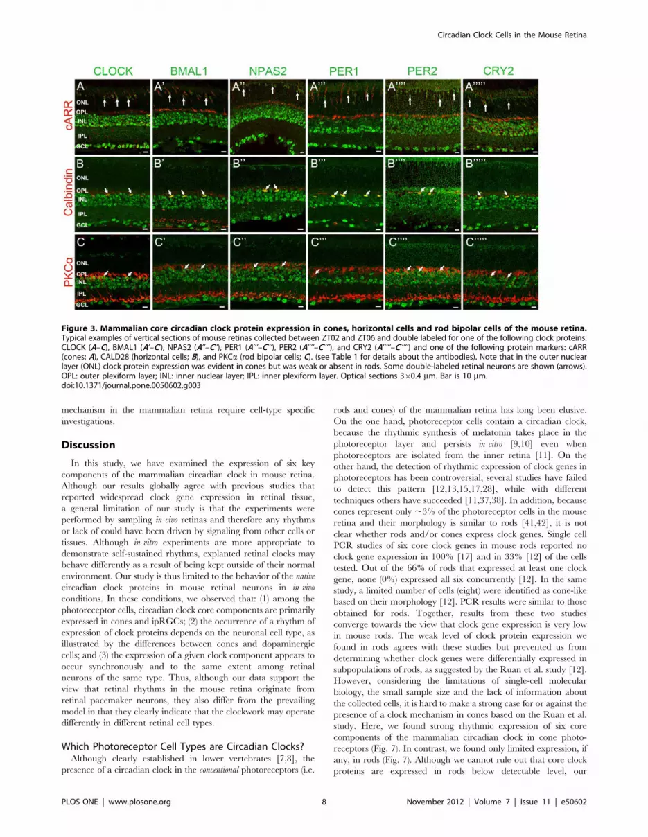
Figure 3. Mammalian core circadian clock protein expression in cones, horizontal cells and rod bipolar cells of the mouse retina.Typical examples of vertical sections of mouse retinas collected between ZT02 and ZT06 and double labeled for one of the following clock proteins:CLOCK (A–C), BMAL1 (A’–C’), NPAS2 (A’’–C’’), PER1 (A’’’–C’’’), PER2 (A’’’’–C’’’’), and CRY2 (A’’’’’–C’’’’’) and one of the following protein markers: cARR(cones; A), CALD28 (horizontal cells; B), and PKCa (rod bipolar cells; C). (see Table 1 for details about the antibodies). Note that in the outer nuclearlayer (ONL) clock protein expression was evident in cones but was weak or absent in rods. Some double-labeled retinal neurons are shown (arrows).OPL: outer plexiform layer; INL: inner nuclear layer; IPL: inner plexiform layer. Optical sections 360.4 mm. Bar is 10 mm.doi:10.1371/journal.pone.0050602.g003
Circadian Clock Cells in the Mouse Retina
PLOS ONE | www.plosone.org 8 November 2012 | Volume 7 | Issue 11 | e50602
observations agree with previous studies that reported limited
clock gene expression in the photoreceptor layer [11,37,38] or in
isolated rod photoreceptors [12,17]. Together with these studies,
our data suggest that most of the clock gene expression found in
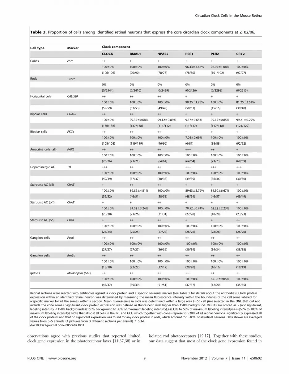
Table 3. Proportion of cells among identified retinal neurons that express the core circadian clock components at ZT02/06.
Cell type Marker Clock component
CLOCK BMAL1 NPAS2 PER1 PER2 CRY2
Cones cArr ++ + + + + +
10060% 10060% 10060% 96.3363.66% 98.9261.08% 10060%
(106/106) (90/90) (78/78) (78/80) (101/102) (97/97)
Rods - cArr – – – – – –
0% 0% 0% 0% 0% 0%
(0/2544) (0/2410) (0/2439) (0/2426) (0/3298) (0/2213)
Horizontal cells CALD28 ++ ++ ++ + + +
10060% 10060% 10060% 98.2561.75% 10060% 81.2563.61%
(59/59) (53/53) (49/49) (50/51) (15/15) (39/48)
Bipolar cells CHX10 ++ ++ ++ – + +
10060% 99.3260.68% 99.1260.88% 9.3760.65% 99.1560.85% 99.2160.79%
(136/136) (137/138) (111/112) (11/117) (117/118) (121/122)
Bipolar cells PKCa ++ ++ ++ – + +
10060% 10060% 10060% 7.0460.69% 10060% 10060%
(108/108) (119/119) (96/96) (6/87) (88/88) (92/92)
Amacrine cells (all) PAX6 ++ ++ ++ +++ ++ +
10060% 10060% 10060% 10060% 10060% 10060%
(76/76) (71/71) (64/64) (64/64) (73/73) (69/69)
Dopaminergic AC TH +++ ++ ++ +++ +++ +++
10060% 10060% 10060% 10060% 10060% 10060%
(49/49) (37/37) (38/38) (39/39) (36/36) (30/30)
Starburst AC (all) ChAT + ++ ++ + + +
10060% 89.6264.81% 10060% 89.6365.79% 81.5066.67% 10060%
(52/52) (46/51) (58/58) (48/54) (46/57) (49/49)
Starburst AC (off) ChAT + + ++ + + +
10060% 81.0263.24% 10060% 78.5260.74% 62.2262.23% 10060%
(28/28) (21/26) (31/31) (22/28) (18/29) (23/23)
Starburst AC (on) ChAT + ++ ++ + + ++
10060% 10060% 10060% 10060% 10060% 10060%
(24/24) (25/25) (27/27) (26/26) (28/28) (26/26)
Ganglion cells Pax6 ++ ++ ++ ++ ++ ++
10060% 10060% 10060% 10060% 10060% 10060%
(27/27) (27/27) (36/36) (39/39) (34/34) (38/38)
Ganglion cells Brn3b ++ ++ ++ ++ ++ ++
10060% 10060% 10060% 10060% 10060% 10060%
(18/18) (22/22) (17/17) (20/20) (16/16) (19/19)
ipRGCs Melanopsin (GFP) ++ ++ ++ +++ ++ ++
10060% 10060% 10060% 10060% 62.3869.05% 10060%
(47/47) (39/39) (51/51) (37/37) (12/20) (35/35)
Retinal sections were reacted with antibodies against a clock protein and a specific neuronal marker (see Table 1 for details about the antibodies). Clock proteinexpression within an identified retinal neuron was determined by measuring the mean fluorescence intensity within the boundaries of the cell soma labeled fora specific marker for all the somas within a section. Mean fluorescence in rods was determined within a large area (,50620 mm) selected in the ONL that did notinclude the cone somas. Significant clock protein expression was defined as fluorescent level higher than 150% background. Results are scored as: - (not significant,labeling intensity ,150% background),+(150% background to 33% of maximum labeling intensity),++(33% to 66% of maximum labeling intensity),+++(66% to 100% ofmaximum labeling intensity). Note that almost all cells in the INL and GCL, which together with cones represent ,20% of all retinal neurons, significantly expressed allof the clock proteins and that no significant expression was found for any clock protein in rods, which account for,80% of all retinal neurons. Data shown are averagedvalues from 3–5 animals (3 pictures from 3 different sections per animal) 6 SEM.doi:10.1371/journal.pone.0050602.t003
Circadian Clock Cells in the Mouse Retina
PLOS ONE | www.plosone.org 9 November 2012 | Volume 7 | Issue 11 | e50602

Figure 4. Mammalian core circadian clock protein expression in bipolar, amacrine and ganglion cells of the mouse retina. Typicalexamples of vertical sections of mouse retinas collected between ZT02 and ZT06 and double labeled for one of the following clock proteins: CLOCK(A–C), BMAL1 (A’–C’), NPAS2 (A’’–C’’), PER1 (A’’’–C’’’), PER2 (A’’’’–C’’’’), and CRY2 (A’’’’’–C’’’’’) and one of the following protein markers: Chx10 (bipolarcells; A), Pax6 (most amacrine cells and ganglion cells; B), and TH (dopaminergic amacrine cells; C) (see Table 1 for details about the antibodies). Theanalysis was restricted to type-1 catecholamine amacrine cells that express high levels of TH. Some double-labeled retinal neurons are shown(arrows). Abbreviations and bar as in Fig. 3.doi:10.1371/journal.pone.0050602.g004
Figure 5. Mammalian core circadian clock protein expression in amacrine and ganglion cells of the mouse retina. Typical examples ofvertical sections of mouse retinas collected between ZT02 and ZT06 and double labeled for one of the following clock proteins: CLOCK (A–C), BMAL1(A’–C’), NPAS2 (A’’–C’’), PER1 (A’’’–C’’’), PER2 (A’’’’–C’’’’), and CRY2 (A’’’’’–C’’’’’) and one of the following protein markers: ChAT (starburst amacrinecells; A), Brn3b (most ganglion cells; B), and eGFP (ipRGCs; C) (see Table 1 for details about the antibodies). The concurrent expression of CLOCK,BMAL1, NPAS2, PER1, PER2, and CRY2 was found in all identified neurons. Note also that all the clock proteins are expressed in the ON starburstamacrine cells whose cell body is located in the ganglion cell layer (GCL), indicating that both GCs and displaced amacrine cells in the GCL express thecore components of the mammalian clock. Clock protein expression in ipRGCs was confirmed with the AB-N38 antibody (data not illustrated),although this antibody only labeled M1 and M2 ipRGC subtypes. Some double-labeled retinal neurons are shown (arrows). Abbreviations and bar asin Fig. 3.doi:10.1371/journal.pone.0050602.g005
Circadian Clock Cells in the Mouse Retina
PLOS ONE | www.plosone.org 10 November 2012 | Volume 7 | Issue 11 | e50602

Figure 6. Core circadian clock protein expression in wild-type, coneless and rodeless retinas. Typical examples of vertical sections ofwild-type C57Bl/6J (A–A’’’’’), coneless (B–B’’’’’), and rodeless (C–C’’’’’) retinas immunolabeled for each of the following core clock proteins: CLOCK (A–C), BMAL1 (A’–C’), NPAS2 (A’’–C’’), PER1 (A’’’–C’’’), PER2 (A’’’’–C’’’’), and CRY2 (A’’’’’–C’’’’’). Retinal tissue was collected around ZT09. For a given clockprotein antibody, confocal settings were adjusted on the brightest picture and the 2 other sections were taken at the same settings. Note that clockprotein expression in the outer nuclear layer (ONL) is detected in a few cells in the wild-type retina (vertical arrows) and in most cells in the rodlessretina (oblique arrows), but is very weak in the coneless retina. OPL: outer plexiform layer; INL: inner nuclear layer; IPL: inner plexiform layer; GCL:ganglion cell layer. Optical sections 360.4 mm. Bar is 10 mm.doi:10.1371/journal.pone.0050602.g006
Table 4. COSINOR analysis of core circadian clock component expression in cones and dopaminergic amacrine cells.
Cell type/conditions Clock component a (mesor) b (amplitude) c (acrophase) (h) F-value P-value
Cones/LD
CLOCK 34.6261.40 9.2361.99 5.5160.82 (F3,27) 209.90 ,0.0001
BMAL1 16.1960.92 9.3261.30 4.9560.53 (F3,27) 121.44 ,0.0001
NPAS2 20.6161.52 12.4462.15 5.0560.66 (F3,27) 72.74 ,0.0001
PER1 7.6360.39 3.3460.55 3.3460.55 (F3,27) 138.76 ,0.0001
PER2 19.1161.49 10.2362.07 5.2560.80 (F3,23) 62.32 ,0.0001
CRY2 15.3460.99 5.1261.40 5.0461.04 (F3,25) 86.87 ,0.0001
Cones/DD
CLOCK 30.8661.87 9.3662.64 3.0161.08 (F3,27) 95.35 ,0.0001
BMAL1 14.6061.01 7.6661.42 5.0660.71 (F3,27) 79.85 ,0.0001
NPAS2 18.0261.64 9.9162.32 5.9960.90 (F3,27) 46.22 ,0.0001
PER1 6.6660.33 2.7060.46 6.8860.66* (F3,21) 148.89 ,0.0001
PER2 11.4960.94* 4.2861.30* 8.0261.20* (F3,23) 51.82 ,0.0001
CRY2 11.9160.64* 3.8460.89 7.0360.92 (F3,25) 120.74 ,0.0001
Dopaminergic cells/LD
CRY2 84.2464.27 38.9966.03 9.4960.59 (F3,27) 143.87 ,0.0001
Dopaminergic cells/DD
CRY2 90.5763.48 30.3564.93 9.7960.62 (F3,27) 237.94 ,0.0001
COSINOR regression analysis was performed on the data illustrated in Figure 7 and only for clock protein levels displaying significant temporal variation over the courseof a LD or DD cycle (as determined by one-way ANOVA; P,0.05). The regression coefficients (a, b, and c) are given with their respective standard error estimates.*: P,0.05 compared to respective LD value (Student t-test).doi:10.1371/journal.pone.0050602.t004
Circadian Clock Cells in the Mouse Retina
PLOS ONE | www.plosone.org 11 November 2012 | Volume 7 | Issue 11 | e50602

the photoreceptor layer with sensitive techniques that required
nucleic acid amplification may have originated from cones and not
from rods and that cones are likely autonomous circadian clock
cells that drive rhythms in the photoreceptor layer. The control of
circadian rhythms in rods by the cone circadian clock could
involve the nighttime-restricted diffusion of substances through the
rod-cone gap junction [43] and/or paracrine loops involving
melatonin and/or dopamine [5,44] or other means. The presence
of a clock mechanism in cones and not rods is consistent with the
observation that light-entrainment of retinal rhythms in verte-
brates requires bright (photopic) light [7,8,9,13,45,46].
Although the clock in cones resembles that in the SCN in
rhythmically expressing circadian clock proteins [2,22], it differs
from the SCN clock in several aspects. In particular, CLOCK
expression is rhythmic in cones while it is constitutively arrhythmic
in the SCN [2]. More surprisingly, expression of all six core clock
proteins is coordinated in cones with a peak during the daytime
(Fig. 7) whereas in the SCN, the peak in PER and CRY proteins
occurs late in the day/early night (,ZT/CT 12), and the BMAL1
peak is antiphase and occurs in the late night/early morning
(,ZT/CT 0) [2]. Two recent studies in the rat retina have shown
that Clock/Bmal1 transcripts peak in phase with Per/Cry transcripts
in photoreceptors [37,38].
The reported concomitant increase in clock gene expression at
night in the photoreceptor layer is consistent with the increase in
clock protein expression in cones during the daytime that we
report here. The circadian expression of Bmal1 is influenced by
nuclear receptors of the ROR family and Rev-ERBa, which target
a ROR-response element in the promoter of the Bmal1 gene [2].
RORs and REV-ERBa compete for binding the Bmal1 promoter,
Figure 7. Circadian clock core component expression in mouse rod and cone photoreceptors and dopaminergic amacrine cellsunder LD and DD conditions. Typical examples of clock protein immunostaining in cones (A–A’’’’’) and dopaminergic cells (D–D’’’’’) obtained fromretinas collected in the middle of the day (ZT06) or subjective day (CT06) or middle of the night (ZT18) or subjective night (CT18) are illustrated forCLOCK (A,D), BMAL1 (A’,D’), NPAS2 (A’’,D’’), PER1 (A’’’,D’’’), PER2 (A’’’’,D’’’’), and CRY2 (A’’’’’,D’’’’’). COSINOR regression analysis (cosine curves) wasperformed only for clock protein levels displaying significant temporal variation (as determined by one-way ANOVA; P,0.05). The results from theCOSINOR analysis are shown in Table 4. Note that the expression of all six clock proteins is rhythmic under LD and DD conditions in cones (B–B’’’’’) butarrhythmic under the same conditions in dopaminergic amacrine cells, except for CRY which is rhythmic under both LD and DD conditions (E–E’’’’’).Clock protein expression in rods remained low at any time point under both LD and DD conditions (C–C’’’’’). Each data point represents the meanfluorescence/identified neuron +/2 SEM of 5/6 animals (5 pictures from 3–4 sections/animal).doi:10.1371/journal.pone.0050602.g007
Circadian Clock Cells in the Mouse Retina
PLOS ONE | www.plosone.org 12 November 2012 | Volume 7 | Issue 11 | e50602

with RORs activating while REV-ERBa repressing Bmal1
expression. It has been shown that tissue-specific variation of the
ratio of expression between these two families of trans-regulatory
elements impinges on the rhythmic expression of Bmal1 in a tissue-
dependent manner [2]. ROR family members also control Npas2
(and to some extent Clock) expression [2]. Interestingly, Sandru
et al. [38] reported a strong rhythm of Rorb in rat photoreceptors
with an increased expression at night and a much smaller-
amplitude rhythm in Rev-ERBa under a LD cycle that did not
persist under DD conditions. It is tempting to speculate that the
nighttime peak in Rorb might shift the balance of modulators of
Bmal1 expression in favor of ROR activators and result in an
increase in Bmal1, Clock and Npas2 expression towards the end of
the night [38]. This would translate into a peak in proteins 4–6 hrs
later- that is around midday. This hypothetical model would
require in particular that BMAL1 and CLOCK or NPAS2 were
not rate-limiting in the transcriptional activation of the Per and Cry
genes. In addition, other factors, such as cAMP, whose in-
tracellular levels increase at night, may play a role as well in
framing the temporal expression of the clock genes in cones [5].
Although many details remain to be clarified, our study together
with others [37,38] clearly indicates that a unique circadian
clockwork is expressed in mammalian cones.
We also report that all six core elements of the mammalian
clock are expressed in ipRGCs (Fig. 5; Table 3). This type of
ganglion cell expresses the photopigment melanopsin and is
considered a third class of photoreceptor in mammals [39,40].
Although a previous study did not find detectable expression of
Per1 in ipRGCs [15] the expression of clock components in
ipRGCs is consistent with a body of indirect evidence that links
ipRGCs with several circadian rhythms within the eyes and the
SCN. These include (1) a circadian rhythm in melanopsin
expression [47]; (2) a biochemical circadian rhythm in the SCN-
onto which the ipRGC project- that disappears following
enucleation [48]; (3) the circadian modulation of ipRGC intrinsic
light responses [49,50]; and (4) a role for ipRGCs in the circadian
modulation of the ERG [51]. Although these studies, together with
our discovery of clock component expression in ipRGCs (Fig. 5;
Table 3) strongly suggest that ipRGCs may be autonomous
circadian clocks, the possibility that some other oscillators in the
retina may drive rhythms in ipRGCs has been proposed as well
[52,53]. Thus even if our observations and others suggest that
ipRGCs are circadian clocks, we must be cautious in interpreting
these results. The functional role of the core clock components in
ipRGCs remains to be firmly established.
Are Dopaminergic Amacrine Cells Autonomous CircadianClocks?Numerous studies have consistently reported the existence of
clock gene and protein expression in inner retinal cells
[12,15,16,17,28]. Clock gene expression in the inner retina persists
in vitro even in the absence of rod and cone photoreceptors,
supporting the view that some cells in the inner retina are
circadian clocks that function autonomously and independently of
the photoreceptor clock [12,13,14]. Even though most cells of the
inner retina express the core clock components (Figs. 1, 2; Table 2),
we found that five out of six clock proteins were constitutively
expressed in the dopaminergic amacrine cells (Fig. 7E). In
addition, no difference in clock protein expression level was
observed between LD and DD conditions in dopaminergic cells
(Fig. 7E), while PER2 expression in the cones (Fig. 7B) was clearly
higher during the light phase of the LD cycle compared to the
subjective day of the DD cycle (Table 4). Light-entrainment is a key
property of circadian clocks, which relies on the induction of clock
gene and protein expression. Light-induced expression in Per genes
and/or proteins has been reported in vertebrate retinas
[30,31,45,46,54]. Together with the arrhythmic expression of
most clock proteins, the absence of light-sensitivity of clock protein
expression in the dopaminergic cells would argue against the
presence of a functional circadian clock in these cells. In addition,
to date no circadian phenotype has been linked to the clock
mechanism within the dopaminergic cells, as the circadian rhythm
in dopamine release requires the presence of a melatonin rhythm
[5]. Thus, while confirming the expression of circadian clock
components in dopaminergic cells, our data expose differences in
the clock mechanism between cones and dopaminergic cells and
question the cell autonomy of the later.
However, dopaminergic amacrine cells could play a key role in
the temporal organization of retinal function through the release
of dopamine. Dopamine release is under the dual control of
a circadian clock and light [55], with a peak during the day under
light-adapted conditions and a trough at night in the dark, and is
known to synchronize retinal rhythms [7,8,45,46], including in
mammals [13]. Thus, whether retinal dopamine neurons are
autonomous circadian clocks remains an open question, but these
cells may play a greater role in participating in the light
entrainment of retinal rhythms and synchronization among retinal
neurons rather than in initiating retinal pacemaker activity. In
a way, retinal dopaminergic neurons may play a role similar to the
retinocepient vasointestinal neuropeptidergic neurons of the SCN
[2,18].
A General Organization of Mammalian Circadian Cocks?Our observations in the retina may represent the case of more
general organization of circadian clock cells that also occur in
other tissues. For instance, emerging views for the organization of
SCN rhythmic function emphasizes the strong heterogeneity in the
individual properties of its cell-autonomous circadian oscillators
and the important role of neuronal networks in shaping a robust
and coherent rhythmic output [18]. However, although most SCN
neurons express the core clock components, a link between specific
SCN neuronal subtypes and rhythmic properties has been difficult
to establish, and the means through which networks synchronize
cellular oscillators and rhythmic outputs are still unclear [18]. By
demonstrating that clock components are widely expressed among
retinal neurons and that a high degree of heterogeneity in their
expression occurs among retinal cell types, our results suggest that
the organization of populations of clock cells in retinal tissue may
share similar features with that of the SCN. Dissecting the clock
mechanism on a cell-type basis in the retina will thus likely shed
light not only on the circadian organization of retinal function but
also on the general organization of circadian clocks in mammalian
tissues.
Acknowledgments
We thank Drs. Samer Hattar, David R. Weaver, Benjamin E. Reese,
Steven M. Reppert, Anand Swaroop, Jeremy Nathans, Stuart C. Mangel,
Stephen C. Massey, and Cheryl M. Craft for gifts of mice, retinal tissue,
antibodies and helpful advice. We thank Dr. Alice Z. Chuang for help with
the statistical analysis of the data and Drs. Kimberly A. Mankiewicz,
Gladys Ko, and Nange Jin for critical reading and editing of the
manuscript.
Author Contributions
Conceived and designed the experiments: XL ZZ CPR. Performed the
experiments: XL ZZ CPR. Analyzed the data: XL ZZ CPR. Contributed
reagents/materials/analysis tools: XL ZZ CPR. Wrote the paper: XL ZZ
CPR.
Circadian Clock Cells in the Mouse Retina
PLOS ONE | www.plosone.org 13 November 2012 | Volume 7 | Issue 11 | e50602

References
1. Dunlap JC, Loros JJ, DeCoursey PJ (2003) Chronobiology. Biological
Timekeeping. Sunderland, MA: Sinauer Associates, Inc. 406 p.
2. Lowrey PL, Takahashi JS (2011) Genetics of circadian rhythms in mammalian
model organisms. Adv Genet 74: 175–230.
3. Barlow R (2001) Circadian and efferent modulation of visual sensitivity. Prog
Brain Res 131: 487–503.
4. Green CB, Besharse JC (2004) Retinal circadian clocks and control of retinal
physiology. J Biol Rhythms 19: 91–102.
5. Iuvone PM, Tosini G, Pozdeyev N, Haque R, Klein DC, et al. (2005) Circadian
clocks, clock-controlled genes and melatonin biosynthesis in the retina. Prog
Retin Eye Res 24: 433–456.
6. Mangel SC, Ribelayga C (2010) Comparative eye: The circadian clock in the
retina regulates rod and cone pathways. In: Dartt DA, Besharse JC, Dana R,
editors. Encyclopedia of the Eye, Vol. 1. Oxford: Elsevier. 283–289.
7. Cahill G, Besharse JC (1991) Resetting the circadian clock in cultured Xenopus
eyecups: regulation of retinal melatonin rhythms by light and D2 dopamine
receptors. J Neurosci 11: 2959–2971.
8. Cahill G, Besharse JC (1993) Circadian clock functions localized in xenopus
retinal photoreceptors. Neuron 10: 573–577.
9. Tosini G, Menaker M (1996) Circadian rhythms in cultured mammalian retina.
Science 272: 419–421.
10. Tosini G, Menaker M (1998) The clock in the mouse retina: melatonin synthesis
and photoreceptor degeneration. Brain Res 789: 221–228.
11. Tosini G, Davidson AJ, Fukuhara C, Kasamatsu M, Castanon-Cervantes O
(2007) Localization of a circadian clock in mammalian photoreceptors. FASEB J
21: 3866–3871.
12. Ruan GX, Zhang DQ, Zhou T, Yamazaki S, McMahon DG (2006) Circadian
organization of the mammalian retina. Proc Natl Acad Sci USA 103: 9703–
9708.
13. Ruan GX, Allen GC, Yamazaki S, McMahon DG (2008) An autonomous
circadian clock in the inner mouse retina regulated by dopamine and GABA.
PLoS Biol 6: e249.
14. Ruan GX, Gamble KL, Risner ML, Young LA, McMahon DG (2012)
Divergent roles of clock genes in retinal and suprachiasmatic nucleus circadian
oscillators. PLoS One 7: e38985.
15. Witkovsky P, Veisenberger E, LeSauter J, Yan L, Johnson M, et al. (2003)
Cellular location and circadian rhythm of expression of the biological clock gene
Period 1 in the mouse retina. J Neurosci 23: 7670–7676.
16. Gustincich S, Contini M, Gariboldi M, Puopolo M, Kadota K, et al. (2004)
Gene discovery in genetically labeled single dopaminergic neurons of the retina.
Proc Natl Acad Sci USA 101: 5069–5074.
17. Dorendos R, Contini M, Hirasawa H, Gustincich S, Raviola E (2007)
Expression of circadian clock genes in retinal cells. Vis Neurosci 24: 573–580.
18. Welsh DK, Takahashi JS, Kay SA (2010) Suprachiasmatic nucleus: cellaut-
onomy and network properties. Annu Rev Physiol 72: 551–577.
19. Ecker JL, Dumitrescu ON, Wong KY, Alam NM, Chen SK, et al. (2010)
Melanopsin-expressing retinal ganglion-cell photoreceptors: cellular diversity
and role in pattern vision. Neuron 67: 49–60.
20. Debruyne JP, Noton E, Lambert CM, Maywood ES, Weaver DR, et al. (2006) A
clock shock: mouse CLOCK is not required for circadian oscillator function.
Neuron 50: 465–477.
21. Garcia JA, Zhang D, Estill SJ, Michnoff C, Rutter J, et al. (2000) Impaired cued
and contextual memory in NPAS2-deficient mice. Science 288: 2226–2230.
22. LeSauter J, Lambert CM, Robotham MR, Model Z, Silver R, et al. (2012)
Antibodies for assessing circadian clock proteins in the rodent suprachiasmatic
nucleus. PLoS One 7: e35938.
23. Mears AJ, Kondo M, Swain PK, Takada Y, Bush RA, et al. (2001) Nrl is
required for rod photoreceptor development. Nat Genet 29: 447–52.
24. Soucy E, Wang Y, Nirenberg S, Nathans J, Meister M (1998) Novel signaling
pathway from rod photoreceptors to ganglion cells in mammalian retina.
Neuron, 21: 481–493.
25. Lee C, Etchegaray JP, Cagampang FR, Loudon AS, Reppert SM (2001)
Posttranslational mechanisms regulate the mammalian circadian clock. Cell 107:
855–867.
26. Haverkamp S, Wassle H (2000) Immunocytochemical analysis of the mouse
retina. J Comp Neurol 424: 1–23.
27. Jeon CJ, Strettoi E, Masland RH (1998) The major cell populations of the mouse
retina. J Neurosci 18: 8936–8946.
28. Storch KF, Paz C, Signorovitch J, Raviola E, Pawlyk B, et al. (2007) Intrisic
circadian clock of the mammalian retina: importance of retinal processing ofvisual information. Cell 130: 730–741.
29. Gekakis N, Staknis D, Nguyen HB, Davis FC, Wilsbacher LD, et al. (1998) Roleof the CLOCK protein in the mammalian circadian mechanism. Science 280:
1564–1569.
30. Namihira M, Honma S, Abe H, Masubuchi S, Ikeda M, et al. (2001) Circadianpattern, light responsiveness and localization of rPer1 and rPer2 gene expression
in the rat retina. Neuroreport 12: 471–475.31. Namihira M, Honma S, Abe H, Tanahashi Y, Ikeda M, et al. (1999) Circadian
rhythms and light responsiveness of mammalian clock gene, Clock and BMAL1,
transcripts in the rat retina. Neurosci Lett 271: 1–4.32. Tosini G, Fukuhara C (2002) The mammalian retina as a clock. Cell Tissue Res
309: 119–126.33. Kamphuis W, Cailotto C, Dijk F, Bergen A, Buijs RM (2005) Circadian
expression of clock genes and clock-controlled genes in the rat retina. BiochemBiophys Res Commun 330: 18–26.
34. Dinet V, Ansari N, Torres-Farfan C, Jorf H-W (2006) Clock gene expression in
the retina of melatonin-proficient (C3H) and melatonin-deficient (C57BL) mice.J Pineal Res 42: 83–91.
35. Peirson SN, Butler JN, Duffield GE, Takher S, Sharma P, et al. (2006) BiochemBiophys Rev Commun 351: 800–807.
36. Tosini G, Kasamatsu M, Sakamoto K (2007) Clock gene expression in the rat
retina: effects of lighting conditions and photoreceptor degeneration. Brain Res1159: 134–140.
37. Schneider K, Tippmann S, Spiwoks-Becker I, Holthues H, Wolloscheck T, et al.(2010) Unique clockwork in photoreceptor of rat. J Neurochem 115: 585–594.
38. Sandu C, Hicks D, Felder-Schmittbuhl (2011) Rat photoreceptor circadianoscillator strongly relies on lighting conditions. Eur J Neurosci 34: 507–516.
39. Berson DM (2007) Phototransduction in ganglion-cell photoreceptors. Pflugers
Arch 454: 849–855.40. Schmidt TM, Do MT, Dacey D, Lucas R, Hattar S, et al. (2011) Melanopsin-
positive intrinsically photosensitive retinal ganglion cells: from form to function.J Neurosci 31: 16094–16101.
41. Rodieck RW (1998) The First Step in Seeing. Sunderland, MA: Sinauer
Associates. 562 p.42. Dowling JE (2012). The Retina, an Approachable Part of the Brain – Revised
Edition. Cambridge, MA: Harvard University Press. 355 p.43. Ribelayga C, Cao Y, Mangel SC (2008) A circadian clock in the retina controls
rod-cone coupling. Neuron 59: 790–801.44. Sengupta A, Baba K, Mazzoni F, Pozdeyev NV, Strettoi E, et al. (2011)
Localization of melatonin receptor 1 in mouse retina and its role in the circadian
regulation of the electroretinogram and dopamine levels. PLoS One 6: e24483.45. Steenhard BM, Besharse JC (2000) Phase shifting the retinal circadian clock:
xPer2 mRNA induction by light and dopamine. J Neurosci 20: 8572–8577.46. Besharse JC, Zhuang M, Freeman K, Fogerty J (2004) Regulation of
photoreceptor Per1 and Per2 by light, dopamine and a circadian clock.
Eur J Neurosci 20: 167–174.47. Hannibal J, Georg B, Hindersson P, Fahrenkrug J (2005) Light and darkness
regulate melanopsin in the retinal ganglion cells of the albino Wistar rat. J MolNeurosci 27: 147–155.
48. Lee HS, Nelms JL, Nguyen M, Silver R, Lehman MN (2003) The eye isnecessary for a circadian rhythm in the suprachiasmatic nucleus. Nat Neurosci 6:
111–112.
49. Weng S, Wong KY, Berson DM (2009) Circadian modulation of melanopsin-driven light response in rat ganglion-cell photoreceptors. J Biol Rhythms 24:
391–402.50. Zele AJ, Feigl B, Smith SS, Markwell EL (2011) The circadian response of
intrinsically photosensitive retinal ganglion cells. PLoS One 6: e17860.
51. Hankins MW, Lucas RJ (2002) The primary visual pathway in humans isregulated according to long-term light exposure through the action of
a nonclassical photopigment. Curr Biol 12: 191–198.52. Sakamoto K, Liu C, Tosini G (2004) Classical photoreceptors regulate
melanopsin mRNA levels in the rat retina. J Neurosci 24: 9693–9697.
53. Van Hook MJ, Wong KY, Berson DM (2012) Dopaminergic modulation ofganglion-cell photoreceptors in rat. Eur J Neurosci 35: 507–518.
54. Zhuang M, Wang Y, Steenhard BM, Besharse JC (2000) Differential regulationof two period genes in the Xenopus eye. Brain Res Mol Brain Res 82: 52–64.
55. Witkovsky P (2004) Dopamine and retinal function. Doc Ophthalmol 108: 17–40.
Circadian Clock Cells in the Mouse Retina
PLOS ONE | www.plosone.org 14 November 2012 | Volume 7 | Issue 11 | e50602