larissa sbaglia celiberto intestinal homeostasis and host
TRANSCRIPT
UNIVERSIDADE ESTADUAL PAULISTA
“JÚLIO DE MESQUITA FILHO”
FACULDADE DE CIÊNCIAS FARMACÊUTICAS
CAMPUS ARARAQUARA
LARISSA SBAGLIA CELIBERTO
INTESTINAL HOMEOSTASIS AND HOST DEFENSE AS PROMOTED BY
COMMENSAL BACTERIA AND THE COLONIC MUCUS LAYER
ARARAQUARA - SP
2018
UNIVERSIDADE ESTADUAL PAULISTA
“JÚLIO DE MESQUITA FILHO”
FACULDADE DE CIÊNCIAS FARMACÊUTICAS
CAMPUS ARARAQUARA
LARISSA SBAGLIA CELIBERTO
INTESTINAL HOMEOSTASIS AND HOST DEFENSE AS PROMOTED BY
COMMENSAL BACTERIA AND THE COLONIC MUCUS LAYER
Tese apresentada à Faculdade de Ciências Farmacêuticas da Universidade Estadual Paulista “Júlio de Mesquita Filho”, para a obtenção do título de Doutora em Alimentos e Nutrição, área Ciência dos Alimentos.
Orientadora: Profa. Dra. Daniela Cardoso Umbelino Cavallini Orientador no exterior: Prof. Dr. Bruce A. Vallance Co-orientador: Dr. Luis Caetano Martha Antunes
ARARAQUARA - SP 2018
Ficha Catalográfica
Elaborada Por Diretoria Técnica de Biblioteca e Documentação
Faculdade de Ciências Farmacêuticas
UNESP – Campus de Araraquara
Celiberto, Larissa Sbaglia. C392i Intestinal homeostasis and host defense as promoted by commensal bacteria
and the colonic mucus layer / Larissa Sbaglia Celiberto. – Araraquara, 2018. 170 f. : il.
Tese (Doutorado) – Universidade Estadual Paulista. “Júlio de Mesquita Filho”. Faculdade de Ciências Farmacêuticas. Programa de Pós Graduação em Alimentos e Nutrição. Área de concentração em Ciência de Alimentos.
Orientadora: Daniela Cardoso Umbelino Cavallini. Orientador no exterior: Bruce A. Vallance. Coorientador: Luis Caetano Martha Antunes.
1. IBD. 2. Colitis. 3. Microbiota. 4. Microbiota biobank.. 4. Personalized probiotic.
5. Mucus layer. I. Cavallini, Daniela Cardoso Umbelino, orient. II. Vallance, Bruce A.,
orient. III. Antunes, Luis Caetano Martha, coorient. IV. Título.
CAPES: 50700006
The following individuals certify that they have read, and recommend to the Faculty of Graduate and Postdoctoral Studies for acceptance, the dissertation entitled: Intestinal homeostasis and host defense as promoted by commensal bacteria and the colonic mucus layer
submitted by Larissa Sbaglia Celiberto in partial fulfillment of the requirements for
the degree of Doctor of Philosophy
in Food and Nutrition and Experimental Medicine Examining Committee:
Daniela Cavallini, Food and Nutrition Co-supervisor
Bruce Vallance, Experimental Medicine Co-supervisor
Carla Fontana, Pharmaceutical Sciences Supervisory Committee Member
Xiaonan Lu, Food Science University Examiner
Ligia Sassaki, Clinical Medicine University Examiner
Additional Supervisory Committee Members:
Kevan Jacobson, Experimental Medicine Supervisory Committee Member
Rosana Ferreira, Microbiology and Immunology Supervisory Committee Member
ii
Abstract
The intestinal tract harbours the largest population of microbes in the human body where they play
an important role in promoting the health of their host. If the composition of these microbes is
altered, this may lead to dysbiosis that triggers or exacerbates intestinal and extra-intestinal
diseases. Probiotics have been investigated as a complementary therapy in dysbiosis-related
diseases. However, their effectiveness in treating severe conditions such as Inflammatory Bowel
Disease (IBD) is quite variable and have shown controversial results. To address the importance
of a personalized probiotic approach to treat intestinal inflammation, we first examined the effect
of personalized bacteria using a model of chemical induced colitis. The animals that received
commensals isolated from their own feces were more protected against inflammation as they
showed reduced signs of colitis, less histological damage and lower levels of inflammatory
markers as compared to mice given a commercial probiotic strain. Next, the role of the intestinal
mucin Muc2 and the Core-1 enzyme that glycosylates it were explored using the Citrobacter
rodentium model of infectious colitis. The intestinal mucus layer is the first line of defense in the
intestine and is largely composed of the secreted mucin Muc2. Since almost all enteric bacteria
must cross the overlying mucus layer to infect the host, the mucus-enteric bacterial interactions
provide fundamental knowledge about infectious diseases as well as inflammatory conditions
linked to dysbiosis (e.g. IBD). Specifically, we compared C. rodentium susceptibility by infecting
WT, Muc2 -/-, core 3 (C3GnT) -/-, core -1 (C1galt1) -/-, and C1galt1 f/f mice. While C3GnT -/- mice
showed a very similar phenotype to WT mice with only mild inflammation, complete absence of
Muc2 or just core 1 derived O-glycans resulted in significantly higher histological damage, barrier
disruption, and increased pathogen burdens. Interestingly, the supplementation of tributyrin
iii
protected mice against infection resulting in less histological damage and lower C. rodentium
colonization as compared to control groups. These studies highlight a novel personalized therapy
that may be considered relevant to diseases affected by dysbiosis as well as the key role of Muc2
and its core 1 glycosylation in host defense against enteric infections.
Keywords: IBD; colitis, microbiota; microbiota biobank, personalized probiotic, mucus layer
iv
Lay Summary
The human gut harbors several types of bacteria that play an important role in the well-being of
their host. The intestinal mucus layer is also important for intestinal health, since it acts as a
physical barrier that prevents bacteria and food products from escaping the gut and causing
inflammation. Using an animal model of intestinal inflammation, I discovered that beneficial
bacteria isolated from the host are more effective in protecting mice against intestinal damage as
compared to probiotic bacteria available on the market. Further, using an animal model of a gut
bacterial infection, I tested the impact of the mucus layer on protection against intestinal
inflammation. I found that the mucin Muc2 and one of its sugar compounds protect mice against
intestinal damage caused by enteric bacteria. These findings have implications for both
Inflammatory Bowel Disease and people with gut infections– and could help develop new and
effective complementary treatments.
v
Preface
Chapter 2
I designed and conducted the majority of the studies reported in this chapter, analyzed the data,
prepared all of the figures and wrote the manuscript under the supervision of Dr. Daniela C.U.
Cavallini and Dr. Bruce A. Vallance. Dr. Elizeu A. Rossi provided input to the study design. Ms.
Roseli A. Pinto assisted in microbiological tests and performed the antibiotic susceptibility test
leading to Supplemental Figure A1.
A version of this chapter has been published in the journal Nutrients as: Celiberto LS, Pinto RA,
Rossi EA, Vallance BA, Cavallini DCU. Isolation and characterization of potentially probiotic
bacterial strains from mice: proof of concept for personalized probiotics. Nutrients 2018, 10(11),
1684; doi: 10.3390/nu10111684 (Gut Microbiome and Human Health).
Ethics approval was required for this research and was obtained from the Sao Paulo State
University Animal Care Committee certificate number 34/2014 and the University of British
Columbia Animal Care Committee certificate number A15-0206.
vi
Chapter 3
I conducted the majority of the studies reported in this chapter, analyzed the data, prepared all of
the figures and wrote the manuscript under the supervision of Dr. Bruce A. Vallance and Dr.
Daniela CU Cavallini. Dr. HT Law, Dr. Genelle Healey, Mr. Justin HY Chan and Ms. Qiaochu
Liang assisted with study design, animal experimentation, histopathological scores and provided
useful insights regarding the results discussion. Dr. Kiran Bhullar performed experiments with
C3GnT -/- mice leading to figure 3.4 IEC C1galt1 -/- and C3GnT -/- mice were generated and kindly
provided by Dr. Lijun Xia, University of Oklahoma. A version of this chapter will be submitted for
publication.
Ethics approval was required for this research and was obtained from the University of British
Columbia Animal Care Committee certificate number A15-0206.
vii
Table of Contents
Abstract .......................................................................................................................................... ii
Lay Summary ............................................................................................................................... iv
Preface .............................................................................................................................................v
Table of Contents ........................................................................................................................ vii
List of Tables ................................................................................................................................ xi
List of Figures .............................................................................................................................. xii
List of Symbols ........................................................................................................................... xiv
Acknowledgements .................................................................................................................... xix
Dedication ................................................................................................................................... xxi
Chapter 1: Literature review ........................................................................................................1
1.1 The human microbiome .................................................................................................. 1
1.2 The human gastrointestinal tract ..................................................................................... 6
1.3 The intestinal microbiota .............................................................................................. 10
1.4 Gut microbiome in health and disease .......................................................................... 16
1.5 Animal models of intestinal inflammation ................................................................... 18
1.6 Modulation of the gut microbiome through probiotics ................................................. 22
1.7 The intestinal mucus layer and its role in host defense ................................................ 27
1.7.1 MUC2 structure and synthesis .............................................................................. 28
1.7.2 MUC2 glycosylation ............................................................................................. 30
1.7.3 Mucus and the gut microbiome ............................................................................. 32
1.8 Research hypothesis and objectives .............................................................................. 37
viii
Chapter 2: Isolation and characterization of potentially probiotic bacterial strains from
mice: proof of concept for personalized probiotics ...................................................................39
2.1 Introduction ................................................................................................................... 39
2.2 Experimental procedures .............................................................................................. 43
2.2.1 Mice ...................................................................................................................... 43
2.2.2 Isolation of commensal bacteria strains ................................................................ 43
2.2.3 Preliminary identification ..................................................................................... 44
2.2.4 Genera confirmation ............................................................................................. 44
2.2.5 Evaluation of survival in simulated gastrointestinal conditions ........................... 45
2.2.6 Antibiotic susceptibility test ................................................................................. 46
2.2.7 Dextran sodium sulfate (DSS)-induced colitis experiment .................................. 48
2.2.8 Tissue collection ................................................................................................... 50
2.2.9 Histopathological scoring ..................................................................................... 50
2.2.10 RNA extractions and quantitative real-time PCR ................................................. 50
2.2.11 Myeloperoxidase (MPO) and malondialdehyde (MDA) activity ......................... 51
2.2.12 Statistical analysis ................................................................................................. 52
2.3 Results ........................................................................................................................... 53
2.3.1 Isolation and genera confirmation of the strains ................................................... 53
2.3.2 In vitro tests demonstrate a potential probiotic effect of the isolated strains ........ 54
2.3.3 Personalized commensal strains protect mice against acute dextran sodium
sulfate-induced colitis ........................................................................................................... 58
2.3.4 Personalized probiotic therapy positively modulates the host immune response
during DSS-colitis ................................................................................................................. 61
ix
2.4 Discussion ..................................................................................................................... 65
Chapter 3: Role of the mucin (Muc)2 and its glycosylation in controlling susceptibility to
Citrobacter rodentium infection ...................................................................................................73
3.1 Introduction ................................................................................................................... 73
3.2 Experimental procedures .............................................................................................. 77
3.2.1 Mice ...................................................................................................................... 77
3.2.2 Bacterial strains, Citrobacter rodentium infection and tributyrin supplementation .
............................................................................................................................... 77
3.2.3 Tissue collection ................................................................................................... 78
3.2.4 Histopathological scoring ..................................................................................... 78
3.2.5 Short chain fatty acid analysis .............................................................................. 79
3.2.6 Statistical analysis ................................................................................................. 80
3.3 Results ........................................................................................................................... 81
3.3.1 Muc2 -/- mice show increased susceptibility to C. rodentium infection ................ 81
3.3.2 C3GnT-/- mice show modest susceptibility to C. rodentium similar to WT mice 82
3.3.3 C1galt1 (IEC) -/- mice develop exaggerated colitis during C. rodentium infection
............................................................................................................................... 84
3.3.4 C1galt1 -/- mice carry high C. rodentium intestinal burdens similar to Muc2-/- .... 87
3.3.5 Muc2-/- mice exhibit lower stool concentrations of propionic and butyric acids
under baseline condition ....................................................................................................... 91
3.3.6 Tributyrin supplementation ameliorates damage caused by C. rodentium infection
............................................................................................................................... 93
3.4 Discussion ..................................................................................................................... 97
x
Chapter 4: Conclusions .............................................................................................................102
4.1 The big picture: potential application of the research findings ................................... 102
4.2 Future directions ......................................................................................................... 105
4.3 Final remarks .............................................................................................................. 107
References ...................................................................................................................................109
Appendix A ............................................................................................................................. 145

xi
List of Tables
Table 2.1 Population of the strains exposed to stimulated gastrointestinal solutions ................... 55
Table 2.2 Zone diameter values to indicate susceptible, intermediate and resistance breakpoints of
each strain ..................................................................................................................................... 57
xii
List of Figures
Figure 1.1 Schematic of the human colon showing the full thickness of the intestinal wall .......... 9
Figure 1.2 Histological analysis of damage caused in colonic tissues by DSS-colitis ................. 20
Figure 1.3 Histological damage caused to murine colonic tissues by C. rodentium infection ..... 22
Figure 1.4 Domain structure and biosynthesis of MUC2 mucin .................................................. 30
Figure 1.5 The biosynthesis of mucin type O-glycans ................................................................. 32
Figure 1.6 Intestinal homeostasis as promoted by SCFA ............................................................. 36
Figure 2.1 Schematic image of the probiotic personalization procedure ...................................... 47
Figure 2.2 Description of the groups used in the DSS experiment .............................................. 48
Figure 2.3 DSS experiment timeline ............................................................................................. 49
Figure 2.4 PCR products of isolated commensal bacteria ............................................................ 54
Figure 2.5 Disease activity index (DAI) during the course of DSS-induced colitis ..................... 59
Figure 2.6 Representative photomicrographs of mouse distal colon sections with haematoxylin
and eosin ....................................................................................................................................... 60
Figure 2.7 Histopathology score ................................................................................................... 61
Figure 2.8 Pro-inflammatory and anti-inflammatory cytokines ................................................... 63
Figure 2.9 Colonic expression of MPO and MDA ....................................................................... 64
Figure 3.1 Muc2 -/- mice exhibit dramatic susceptibility to C. rodentium-induced morbidity and
mortality ........................................................................................................................................ 81
Figure 3.2 Macroscopic image of heightened mucosal damage in Muc2-/- as compared to WT
mice ............................................................................................................................................... 82
Figure 3.3 C3GnT-/- mice exhibit similar C. rodentium susceptibility to WT mice ..................... 83
xiii
Figure 3.4 C3GnT-/- mice exhibit modest tissue damage similar to WT mice ............................. 84
Figure 3.5 Muc2-/- and C1galt -/ mice exhibit similar C. rodentium susceptibility ........................ 86
Figure 3.6 Muc2-/- and C1galt1 -/- mice carry heavier intestinal pathogen burdens ................... 88
Figure 3.7 Muc2-/- and C1galt1 -/- mice display similar susceptibility to C. rodentium infection . 90
Figure 3.8 Muc2-/- mice display lower levels of propionic and butyric acids under baseline
condition ....................................................................................................................................... 92
Figure 3.9 Muc2-/-and C1galt -/ mice display lower C. rodentium susceptibility with TB
supplementation ............................................................................................................................ 94
Figure 3.10 C1galt1 -/- mice carry heavier intestinal pathogen burdens ...................................... 95
Figure 3.11 Muc2-/- and C1galt1 -/- display less severe colitis with TB supplementation ......... 96
xiv
List of symbols and abbreviations
α alpha
β beta
γ gamma
κ kappa
µ micro
° degree
C Celsius
< less than
≤ less than or equal to
> greater than
+ positive
± plus or minus
-/- deficient
5-HT 5-hyroxytyrtamine (serotonin)
ADP adenosine diphosphate
A/E attaching and effacing
AMP antimicrobial peptide
ATP adenosine triphosphate
CFU colony forming units
CTRL control
DAI disease activity index
xv
DAPI 4',6-diamidino-2-phenylindole
DC dendritic cell
DNA deoxyribose nucleic acid
DNBS 2,4-dinitrobenzenesulfonic acid
DSS dextran sodium sulfate
EHEC Enterohemorrhagic Escherichia coli
e.g. exempli gratia (for example)
ER endoplasmic reticulum
EPEC Enteropathogenic Escherichia coli
F/B Firmicutes/Bacteroidetes
FITC fluorescein isothiocyanate
FOS fructooligosaccharide
g gram
GI Gastrointestinal tract
GF germ free
GL glycerol
GOS galactooligosaccharide
GPR G protein coupled receptor
HIV Human Immunodeficiency virus
HMP Human Microbiome Project
IAP intestinal alkaline phosphatase
IBD Inflammatory Bowel Disease
IBS irritable bowel syndrome
xvi
i.e. id est (that is)
IEC intestinal epithelial cell
IEC-C1galt1-/- IEC specific deletion of core 1-derived O-glycans
IEL intraepithelial lymphocyte
IFN interferon
Ig immunoglobulin
IL interleukin
ITS internal transcribed spacer
L litre
LEE locus of enterocyte effacement
LGG Lactobacillus rhamnosus GG
LI large intestine
LPS lipopolysaccharide
m meters
M cells microfold cells
MAPK mitogen activated protein kinase
MDA malonaldehyde
MetaHIT Metagenomics of the Human Intestinal Tract.
MLN mesenteric lymph nodes
MPO myeloperoxidase
Muc2 mucin 2
NGS next generation sequence
NIH National Institute of Health
xvii
NOD nucleotide-binding oligomerization domain
NF-κB Nuclear factor kappa-B
pi post infection
PP personalized probiotics
PTS Proline- threonine -serine
qPCR quantitative PCR
Reg III γ regenerating islet-derived protein 3 gamma
Relmβ resistin like molecule beta
RNA ribonucleic acid
rRNA ribosomal RNA
ROS reactive oxygen species
SCFA short chain fatty acid
SD standard deviation
SEM standard error mean
SI small intestine
S. Typhimurium Salmonella enterica serovar Typhimurium
T3SS type 3 secretion system
TB tributyrin
TCR T cell receptor
TFF3 trefoil like factor 3
TGF β transforming growth factor beta
Th T helper
Tir translocated intimin receptor
xviii
TJ tight junction
TLR toll like receptor
TNBS 2,4,6-trinitrobenzene sulphonic acid
TNF tumor necrosis factor
Treg T regulatory
Wnt Wingless/Integrated
WT wildtype
ZO zonula occludens
xix
Acknowledgements
I am very fortunate to have received unreserved support from many exceptional individuals during
my PhD studies. I am forever grateful to my graduate co-supervisors, Dr. Daniela Cavallini and
Dr. Bruce Vallance for their patience, kindness and guidance throughout my tenure as a PhD
student. Thank you both for providing me with opportunities to learn different laboratory
techniques and experimental models. I also appreciate all the support I had during the Joint degree
set up between the two universities. Thank you Daniela for showing me the wonderful world of
‘good bacteria’ and all the amazing applications of probiotics. Also, thank you for all your support
with my crazy and ambitious ideas with this international collaboration. I could not have
accomplished this without you. Bruce - Thank you for teaching me so much about gut disorders
and how to apply my previous knowledge in all these different models. Thank you for teaching
me how to “tell a story” through research and scientific writing and for all the incredible support
during my thesis writing. I cannot describe how grateful I am for the opportunity to work in your
lab in Canada. I also thank my graduate supervisory committee Dr. Carla Fontana and Dr. Kevan
Jacobson, for inspiring me with their own research and for providing me with guidance and
valuable insights.
I owe a special thank you to Dr. Elizeu Antonio Rossi for providing me with the idea from one of
my PhD projects. I also thank Dr. Caetano Antunes for introducing me to Dr. Vallance and for all
the support during my PhD.
To all the past and current members of the Cavallini and Vallance laboratories, I thank all of them
for creating a great working environment that encourages collaboration and supports new ideas. I
thank Ms. Roseli Pinto, Ms. Josiane Canaan, Ms. Caixia Ma, Ms. Tina Huang and Ms. Mimi Kuan
xx
for teaching me many laboratory techniques and always being ready to lend a helping hand. To
Olivia Zordão, thank you for all the hard work and friendship during long animal experiments.
Thank you to my colleagues who have provided friendship, encouragement and thoughtful
suggestions through all the ups and downs of graduate school, especially Juliana Marchesin,
Juliana Witzler, Ana Luiza Duque and Fernanda Bianchi. Thank you for my amazing “Core 1
team” Dr. Genelle Healey, Dr. HT Law, Justin YH Chan and Qiaochu Liang. It is truly a pleasure
working with you everyday in the lab. A special thank you goes to my wonderful friends in Canada
Dr. Joannie Allaire, Franzi Graef, Else Bosman, Shauna Crowley and Vivian Han. Thank you girls
for kindly welcoming me into Vancouver and for all the science and life contributions you have
made in my life. You are all truly an inspiration!
I offer my enduring gratitude to the faculty and staff at UNESP and UBC, who have helped me
with a lot of paper work and orientation during my studies. I owe particular thanks to Ms. Claudia
Molina, Ms. Jennifer Fletcher and everyone from the Dean’s department from both universities
for all the work during the Joint PhD set-up agreement.
Thank you to my husband Moises for endless support, unconditional love, and for always standing
by me through all the difficult times of graduate student life. With you, life is filled with positivity
and happiness. And finally, I would like to express my deepest gratitude to my parents and my
sister who have provided nothing but love and encouragement throughout my life. Thank mom
and dad for your unconditional support during my education. I could never describe in words how
fortunate I am to have you all in my life.
I would like to acknowledge Sao Paulo Research Foundation (FAPESP) for the Doctoral and the
international internship (BEPE) scholarships. I also thank Dr. Bruce Vallance for providing my
stipend during my second year of studies in Canada.

xxi
Dedication
To my mom and dad, for always loving and supporting me.
1
Chapter 1: Literature review
1.1 The human microbiome
Humans have co-evolved with the trillions of microbes that inhabit their bodies, thereby creating
a complex yet symbiotic ecosystem. These microbes include bacteria, viruses, archaea, and
eukaryotic microorganisms (1). It is estimated that the human body contain slightly more bacterial
cells than human cells (4 × 10¹³/ 3 × 10¹³) (2), with over 1000 different strains of bacteria. (3,4).
Several definitions have recently been used to describe this microbial community that colonizes
our bodies. While microbiota refers to a community of microorganisms that are present in a
particular habitat, the microbiome comprises both the microorganisms at a specific site as well as
their genomes, physiochemical properties and activities (5). Therefore, microbiome is a more
refined term that should be used when taking into consideration the genes carried by the microbes,
as well as the environment that a particular microbiota inhabits.
Several bacterial and fungal communities have been investigated in the past decades using
traditional microbiology techniques such as culturing using selective and differential media. These
media usually contain all the necessary nutrients for the growth of certain microorganisms as well
as inhibitors to select and identify target microorganisms. Studies using conventional microbiology
methods provided significant information regarding microbial environments and are still very
useful in terms of classifying different genera and/or determining their antimicrobial
susceptibilities. However, considerable disadvantages are involved in culturing techniques - such
as poor sensitivity for some sample types (blood, tissues, fecal content), challenges to characterize
2
microbes to the species level, the long incubation time (especially for fungi and mycobacteria),
poor clinical application, and little information provided about community dynamics (6,7).
Moreover, with regards to the human microbiota, it is estimated that 20% to 60% of the microbes
living within the human body are unculturable (8). This range varies according to the body site but
is it already a strong indication of how little was known about the human microbiome and its
diversity until recent years.
Over the last two decades, advances in next-generation sequencing (NGS), along with
bioinformatic developments, have brought new insights regarding our understanding of the
unculturable microbes that inhabit soil, oceans and the human body (9,10). This field of research,
called metagenomics, allows the examination of the total genomic DNA of microbial communities
sampled from natural environments without the prior need for culturing, thus providing valuable
information about the complexity and diversity of the human microbiome – in situ. The two main
types of sequencing data analysis are marker gene metagenomics and shotgun metagenomics
(11,12). Sequencing of the 16S ribosomal RNA (rRNA) gene is restricted to microbes of the
bacteria and archaea domains, with the sequencing data compared to a database containing a great
number of sequences of this gene fragment (16S libraries). A similar approach can be applied for
fungi and eukaryotes, although in this case the preferred marker genes are the internal transcribed
spacer (ITS) and the 18S rRNA gene, respectively (11). Through shotgun metagenomics analysis,
the complete sequences of all microorganisms present in a sample (previously characterized or
novel) are investigated, thus offering extremely useful insights regarding microbial community
dynamics (11,12). Besides the knowledge gained about the differences in the microbiome at
particular body sites, these molecular techniques help researchers understand how microbial
3
communities affect health and disease and how it is possible to influence or even manipulate the
human microbiome (13). In addition, more than just aiding our understanding of microbiome-host
interactions, some of these high-throughput sequencing technologies allow the identification of
microbiome changes in a more detailed manner such as to the strain-level, which poses a
significant improvement over the detection of species-level differences which may not represent a
reliable marker in several health conditions (3).
The Human Microbiome Project (HMP) was created in 2008 by the National Institutes of Health
(NIH) in the United States, as an initiative to study the different microorganisms living in
association with the human body, and thus potentially involved in human health and disease. The
mission of this project is to generate resources that will help scientists in the field understand the
makeup of the “normal” human microbiome and if certain medical conditions affect the microbes
living in our body (8). The term “normal” rather than “healthy” was chosen by body site-specific
experts and was used in the study as a matter of criteria selection for volunteers. According to
clinicians’ opinions, the recruitment of “healthy” volunteers would lead to several exclusion
criteria making the selection a very slow and complicated process (8). The main (and ongoing)
goals of the HMP are: to characterize the human microbiome by studying samples from multiple
body sites (gastrointestinal tract, mouth, nasal cavity, vagina, and skin) collected from “normal”
volunteers; to understand the influence of the microbiome in health and disease by studying their
differences in several clinical conditions such as obesity, psoriasis, bacterial vaginosis, and cancer;
and finally to provide standardized data resources as well as improved technology to facilitate
future studies in this field (8).
4
The HMP and all the other various studies characterizing the human microbiome play a very
important role in better defining the concept of “dysbiosis” and the diseases related to this
condition. Presently, there is no specific description of what constitutes a “healthy" microbiome
or which group of microorganisms are present in a dysbiotic state. Early research in the field aimed
to identify a “core” group of microorganisms that are universally present in healthy individuals
but are also absent in those individuals presenting with disease phenotypes (14,15). However,
microbiomes regularly show a large degree of interpersonal variation - even in the absence of any
overt disease (16,17), therefore the general concept of a “healthy” microbiome is no longer
practical as a reference or marker for eubiotic or dysbiotic states. Moreover, besides the variability
in the metagenome of the human microbiome, only a third of its constituent genes are found in the
majority of healthy individuals (14), making the search for specific taxa a poor reflection of what
constitutes the “normal” microbial community.
Although no specific characteristics can be used to define dysbiosis, several authors associate this
term with certain conditions. The oral cavity carries hundreds of bacterial species and an imbalance
in this microbiota is associated with oral diseases, mainly periodontitis and dental caries (18). In
periodontitis, pathogens such as Porphyromonas gingivalis, Treponema denticola and Tannerella
forsythia are described to play a key role in this disease due to their ability to produce biofilms
(19). Regarding dental caries, Streptococcus mutans seems to be the chief pathogen associated
with this condition, although Lactobacillus spp., Prevotella spp., Atopobium spp., Olsenella spp.
and Actinomyces spp. have also been implicated (20). The skin microbiota composition varies
according to the body site and its particular properties (dry versus moist versus sebaceous sites),
and is mainly colonized by Corynebacterium spp., Propionibacterium spp., and Staphylococcus
5
spp. (21). Staphylococcus aureus is closely related to atopic dermatitis and although most species
of Corynebacterium do not cause any disease in humans, Corynebacterium minutissimum and
Corynebacterium tenuis have been associated with superficial skin pathology (22). Curiously,
rather than showing a lack of microbial diversity - female urogenital diseases (bacterial vaginosis,
yeast infection, sexually transmitted infection) and their consequences (e.g. pre-term birth, HIV
infection) are often associated with a more diverse vaginal microbiota profile, where there is a shift
from predominantly lactic acid-producing bacteria (i.e. Lactobacillus spp.) to more strict
anaerobes comprising taxa such as Sneathia spp., Prevotella amnii, Atopobium spp. and
Gardnerella vaginalis (23,24). In gastrointestinal disorders, a reduced diversity of microbes, a
lower abundance of obligate anaerobic bacteria, and an expansion of facultative anaerobic bacteria,
such as Escherichia spp., are thought to indicate the presence of an abnormal/aberrant microbiota.
These changes in the microbiota are often seen in combination with intestinal and extra-intestinal
disorders such as inflammatory bowel disease (IBD), irritable bowel syndrome (IBS), obesity, type
2 diabetes, among others (25).
Interestingly, studies investigating the human microbiome in different countries have observed
inter-population differences in the composition of what is considered a “healthy microbiome”,
emphasizing the geographical variation in microbes worldwide (14). This inter-country variation
in taxonomic composition was compared using large cohort studies from different continents –
MetaHIT (Europe), HMP (America), and Chinese diabetes cohorts – and together with genetics,
diet and other environmental factors, one’s place of residence joins the list of factors that influence
the human microbiome (14). Besides evolution, immigration patterns and modifiable factors that
reflect lifestyle (diet, exercise, hygiene habits), the healthcare system of a particular country may
6
also influence the microbiome composition of that population. Medical procedures like mode of
infant delivery and use of antibiotics at an early age are directly associated with differences in
microbiota composition, and the frequency of these procedures usually varies between countries.
Moreover, climate and sunlight exposure have also been described as factors that impact microbial
dysbiosis, by, for example, influencing the biosynthesis of vitamin D (25). Lower sunlight
exposure combined with a lack of adequate vitamin D supplementation can result in vitamin D
deficiency, pre-disposing to dysbiosis and inflammatory conditions such as IBD (25).
1.2 The human gastrointestinal tract
The mammalian gastrointestinal (GI) tract is a complex organ system that includes anatomically
and functionally distinct regions – each with a unique diversity of cell types (26). The GI tract
extends from the mouth to the anus and is in essence, a long tubular structure comprising (in order)
the oral cavity, esophagus, stomach, small and large intestines, rectum and anus. Each of these
segments is strategically separated by sphincters that not only create different compartments in the
GI tract but also control the flow of the digestive process (27). The primary functions of the GI
tract are to maintain water homeostasis, secrete enzymes to aid the digestion process, sample and
absorb essential nutrients and electrolytes, and finally eliminate waste products in the form of feces
(26,27). In addition, adjacent glandular organs (e.g. salivary glands, liver, gall bladder, and
pancreas), neurons and vasculature are connected to the GI tract, where they help facilitate
digestion in an integrated process whereby each organ and all the different cell types play their
specialized roles in the gut (26).
7
The small intestine is the longest segment of the GI tract, measuring 6-9 m long in adults, and
consisting of three parts: the duodenum, jejunum, and ileum. The duodenum is the most proximal
segment of the small intestine and it plays an important role in the mixing of food products with
digestive enzymes from the pancreas as well as with alkaline secretions from duodenal glands, that
together are responsible for neutralizing the acidic chyme that is received from the stomach. The
bile produced in the liver and stored in the gall bladder also helps the digestive process by
emulsifying dietary fat into micelles for absorption (28). Another critical enzyme for digestion,
intestinal alkaline phosphatase (IAP), is expressed and secreted by intestinal epithelial cells (IEC)
and is found in high concentrations in the duodenum. One of the major functions of IAP is the
regulation of bicarbonate secretion and duodenal surface pH, thus contributing to intestinal
homeostasis along with other functions such as inactivation of bacteria lipopolysaccharide (LPS)
and regulation of the gut microbiome through dephosphorylation of adenosine triphosphate (ATP)
and adenosine diphosphate (ADP) in the intestinal lumen. IAP converts these phosphorylated
nucleotides to adenosine which acts as a scavenger of oxygen thus influencing bacterial growth
and diversity (29). Following the digestion of food, nutrient absorption continues to take place
throughout the jejunum and ileum. The main function of the small intestine is thus the digestion
and absorption of nutrients, and to do this, the inner surface of the small intestine is covered with
villi and microvilli that project into the lumen resulting in a very high surface area of approximately
30 m2 in humans (27).
The large intestine, also known as the colon, is wider and shorter (~1.5 m) than the small intestine
and extends from the distal end of the ileum to the anus (30). This region receives chyme from the
8
small intestine and is responsible for forming the feces, as well as absorbing fluids and salts that
maintain homeostasis in the human body. The colon can be functionally divided into the ascending,
transverse and descending colon. The cecum and ascending colon are located on the right side of
the abdomen and play a central role in water and electrolyte absorption, in the fermentation of
complex carbohydrates (i.e. dietary fiber) as well as in the production of metabolites (i.e. short-
chain fatty acid) by resident microbiota (28). The transverse colon crosses the abdomen
transversely connecting the right side to the left side, including the descending colon, sigmoid
colon and rectum, which are primarily involved in the storage and evacuation of feces (28). The
appendix is a tube-like structure found just off the cecum, and acts as a storage compartment for
commensal bacteria and immune cells (31).
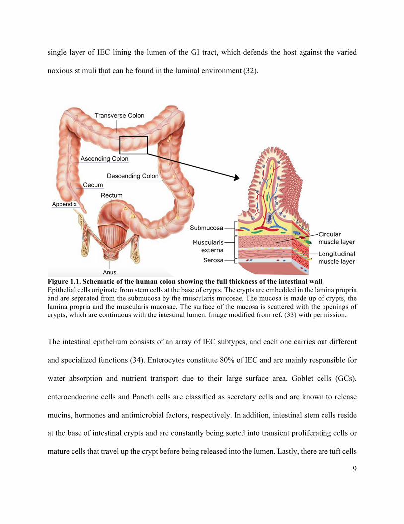
The wall of the human GI tract consists of four distinct functional layers, which from the outer
surface inward include: the serosa, the muscularis externa, the submucosa, and the mucosa. The
serosa is the outermost layer formed of connective tissue that contains major blood vessels and
nerves. The muscularis externa consists of smooth muscle which is usually arranged as an inner
circular layer and an outer longitudinal layer that together coordinate the muscle contractions that
mediate peristalsis (32). The submucosa layer is made up of connective tissue that supports the
mucosa and contains larger blood vessels, lymphatic vessels and nerves. Together, they help
control secretions from the mucosal glands and regulate mucosal movement and blood flow. The
mucosa is made up of three sublayers: the muscularis mucosae, which consists of a thin smooth
muscle layer that separates the lamina propria and the underlying submucosa; the lamina propria,
which resides at the base of the intestinal epithelium and is rich in immune cells; and finally a
9
single layer of IEC lining the lumen of the GI tract, which defends the host against the varied
noxious stimuli that can be found in the luminal environment (32).
Figure 1.1. Schematic of the human colon showing the full thickness of the intestinal wall. Epithelial cells originate from stem cells at the base of crypts. The crypts are embedded in the lamina propria and are separated from the submucosa by the muscularis mucosae. The mucosa is made up of crypts, the lamina propria and the muscularis mucosae. The surface of the mucosa is scattered with the openings of crypts, which are continuous with the intestinal lumen. Image modified from ref. (33) with permission.
The intestinal epithelium consists of an array of IEC subtypes, and each one carries out different
and specialized functions (34). Enterocytes constitute 80% of IEC and are mainly responsible for
water absorption and nutrient transport due to their large surface area. Goblet cells (GCs),
enteroendocrine cells and Paneth cells are classified as secretory cells and are known to release
mucins, hormones and antimicrobial factors, respectively. In addition, intestinal stem cells reside
at the base of intestinal crypts and are constantly being sorted into transient proliferating cells or
mature cells that travel up the crypt before being released into the lumen. Lastly, there are tuft cells
10
which are chemosensory cells that can sense luminal helminths, and M cells which overlie Peyer’s
patches and aid in antigen uptake and sensing (34,35). All together, these various IEC subtypes
work together harmoniously to maintain intestinal homeostasis and promote host defense, thus
guaranteeing a functional balance within the GI tract (34).
The mucosal surface that coats the GI tract is approximately 200-300 m2 in humans (36) and it
harbors a vast array of microorganisms known as the microbiome (25,37). These microbes include
bacteria, viruses, fungi, and eukaryotes and under normal conditions they maintain a healthy
symbiotic relationship with the host. Since the GI mucosal surface is the largest area of the body
that is in contact with the external environment, the interactions between the different cells in the
gut play a key role in regulating normal gut physiology as well as host defense by working to
exclude harmful opportunistic microbes. Before particles present in the luminal environment reach
the IEC, they first encounter the mucus layer, which is a physical and biochemical protective
barrier comprised of mucin glycoproteins that coats the entire GI tract (38). This dynamic barrier
is constantly renewed, thus limiting any transient impairments in epithelial barrier integrity (39).
1.3 The intestinal microbiota
The first time a human usually encounters environmental microorganisms occurs at birth. During
delivery the newborn comes into contact with the microbes present in the mother’s vaginal canal
and on her skin, leading to these commensal bacteria colonizing the infant. This community of
microorganisms gradually develops into a diverse ecosystem as the host ages and grows, with both
microbes and the immune system maturing together in a symbiotic relationship (37). The host
11
provides a place to reside as well as nutrition for the microbes, while the gut microbiota in turn
protects the host against pathogenic bacteria (40,41), helps in food digestion and vitamin
biosynthesis (42,43), produces important metabolites such as short chain fatty acids (SCFA)
(40,44), and promotes normal immune function (45).
The intestine harbours the largest population of microorganisms within the human body, and the
four main intestinal microbial phyla in humans are Firmicutes, Bacteroidetes, Proteobacteria and
Actinobacteria (46). Firmicutes and Bacteroidetes are described as the dominant phyla in the gut
(4,17) accounting for more than 90% of the total community (47–49), and although this broad level
of classification may not directly indicate a health status association, the Firmicutes/Bacteroidetes
(F/B) ratio is a common index used as a comparison of gut microbiota composition in certain
diseases such as obesity and intestinal disorders. Obese subjects usually show a higher proportion
of Firmicutes over Bacteroidetes as compared to lean people, and the F/B ratio tends to decrease
as an individual loses weight (50). Conversely, a higher Bacteroidetes population has been
associated with intestinal conditions such as IBD (51). At the genera level, it has been suggested
that most individuals can be classified into one of three clusters or “enterotypes” according to their
prevalent genera (Bacteroides spp., Prevotella spp. or Ruminococcus spp.) (52). These enterotypes
seem to be driven primarily by dietary habits and despite being still a wide categorization, they
may help us understand underlying mechanisms linking gut microbiota to diseases. The
stratification of individuals in these three groups may identify possible correlations with dietary
patterns such as the consumption of animal protein versus vegetarianism, fermented food,
“Western” diet lifestyle, among others.
12
As previously discussed, the concept of a “healthy microbiome” is difficult to define, however this
definition may be characterized by the microbiome behavior over time, where a personalized
“functional core” of metabolic and molecular functions provides benefits to the host and protects
it against noxious stimuli (14). Since interpersonal variation in the gut microbiome is high, a better
way to predict dysbiosis related diseases would be the stability of the microbiota within an
individual through time. It is believed that our microbiota develops during the first few years of
life becoming relatively stable in healthy adults (1,42). However, studies have indicated that a
decrease in Bifidobacterium spp., Bacteroides spp., and Lactobacillus spp. occurs in elderly
population, accompanied by an increase in the number of facultative anaerobes (53–55).
Mode of delivery is considered the first aspect that influences gut microbiota composition, even
though recent studies indicate that diet and stress in late pregnancy can also influence the first
microbiome colonizers (49,56,57). Infants born by vaginal delivery present a microbial community
close to the one found in the vaginal microbiota of their mothers (mainly Lactobacillus spp.), while
infants delivered by caesarean section display a microbiota dominated by typical skin microbial
taxa such as Staphylococcus spp. and Propionibacterium spp. (58). Breastfeeding is another factor
that has been suggested to enrich Lactobacillus spp. in comparison with formula feeding in
newborns (59). Furthermore, Bifidobacterium spp. present in breast milk display bifidogenic
activity that protects infants against gastrointestinal infections and acute diarrhea (60).
Antibiotic use is one of the major external perturbations that directly affects the microbiota
composition. The aim of antibiotic treatment is to fight against severe - and sometime life-
threatening - infections that may cause major complications to the host. However, despite the acute
13
beneficial effects of eliminating pathogenic bacteria, the use of antibiotics is also linked to long
term negative consequences as the microbial community may not always return to its pre-treatment
composition. Interestingly, even months after antibiotic treatment - a low bacterial diversity has
been described in the microbiota of adults treated with ciprofloxacin (61). Furthermore, studies
show that a single course of antibiotics is sufficient to modify the microbial community with this
change persisting for years (62–64). Lastly, the overuse of antibiotics is associated with an increase
in antibiotic-resistant pathogens, as after the treatment some pathogens have more opportunity to
outgrow commensal bacteria and to colonize the microbiota in a permanent way (1).
Dietary changes are also another modifiable factor that directly influences the microbiota
composition. Products of digestion that were not absorbed in the upper digestive tract reach the
colon where they come in contact with the gut microbiota. As an anaerobic environment, the
fermentation process provides both energy and carbon sources for the microorganisms in addition
to changing pH and substrate availability in the colon, therefore impacting both host metabolism
and health (65). In the last several decades, dietary patterns have shifted dramatically from diverse
and nutritious whole foods to what is known as a modern “Western” style. The “Western” diet
comprises large amounts of sugar and fat rich, high calorie processed foods combined with a lack
of fruits, vegetables, legumes and whole grains (66). Aside from lacking fiber and essential
nutrients such as vitamins and minerals, this “Western” dietary lifestyle is associated with
inflammatory and detrimental health effects leading to increased rates of obesity and other chronic
diseases (67).
14
Studies using animal models have demonstrated that lean mice have a greater percentage of
Firmicutes as compared to Bacteroidetes (60% and 40% respectively). However, obese mice have
an even greater percentage of the same phyla, thus correlating obesity with increased numbers of
Firmicutes in mice (68–70). Additionally, it has been shown in mice that shifting to a high-fat,
high-sugar “Western” diet from a low-fat, plant polysaccharide-rich diet can change the microbiota
within a day (71). Moreover, animals fed with a high fat diet showed reduced cecal
Bifidobacterium spp., increased circulating LPS concentrations (72,73), and lower abundance of
Clostridium cluster XIVa, including Roseburia spp. (74).
The consumption of the typical “Western” style diet is also associated with significant changes in
human microbiota composition at the phylum and genus levels, including reductions in both Gram
positive (e.g., Bifidobacterium spp.) and Gram negative bacteria (e.g., Bacteroides spp.) in African
and European Americans as compared to native Africans with a diet rich in resistant starch and
low in animal products (75). In another study in humans, shifting from a high-fat/low-fiber diet to
a low-fat/high-fiber diet caused notable changes in the gut microbiota composition within 24 hours
(76). Interestingly, enterotypes are associated with long-term diet, as individuals on a diet high in
protein and animal fat have a Bacteroidetes-dominated enterotype, whereas a carbohydrate-rich
diet is associated with a Prevotella dominated enterotype (76).
Regarding the impact of macronutrients in the gut microbiota, most benefits are related to
carbohydrates in the form of dietary fibers. Carbohydrates are the major carbon/energy source for
colonic microbes and fiber promotes gut health by increasing digesta mass thus facilitating fecal
transit thought the colon. Additionally, SCFA (acetate, propionate and butyrate) are the principal
15
endproducts of carbohydrate fermentation and besides helping lower pH levels within the colon
thereby limiting pathogenic bacterial activity, these acids have roles beyond the gut - impacting
metabolic and immune system diseases and disorders (77). These by-products of colonic
microbiota have received significant attention in the recent years, in particular butyrate, which is
produced by bacteria from the Clostridiale clusters IV and XIVa. The main butyrate producing
species are described to be Eubacterium rectale and Faecalibacterium prausnitzii, in addition to
others in the genera Coprococcus and Roseburia (78). The presence of butyrate producers in the
colon has been shown to be negatively correlated with functional dysbiosis, while reducing the
risk of infections with opportunistic pathogens and decreasing oxidative stress, highlighting
beneficial synergistic interactions between diet, microbes and host. Butyrate producers can
respond to different environmental conditions, such as diet or pH, and engage different
fermentation pathways in which the final products are lactate, formate, hydrogen and carbon
dioxide. It has been shown that cross- feeding between Bifidobacterium spp. and butyrate
producers is also possible: Bifidobacterium spp. break down polysaccharides and produce lactate
and acetate, which are further utilized by butyrate - producers to form butyrate (78). Butyrate is
also the main source of energy for colonocytes and it inhibits expression of pro-inflammatory
cytokines in the mucosal layer of the intestine, thus playing a key role in maintaining homeostasis
of the intestine (79). Moreover, butyrate has a positive effect on the integrity of the mucosal layer
by stimulating expression of tight junction proteins, and by inducing the production of mucin and
antimicrobial peptides (80).
The use of probiotics and prebiotics as nutritional strategies are also known for promoting general
health benefits by improving the gut microbiota composition. A prebiotic is “a substrate that is
16
selectively utilized by host microorganisms conferring a health benefit” (81). Established
prebiotics include inulin-type fructans (i.e. fructo-oligosaccharides [FOS], inulin and
oligofructose), galacto-oligosaccharides (GOS) and lactulose. Other fermentable carbohydrates
that have shown prebiotic potential include resistant starch, β-glucans, arabinoxylan
oligosaccharides, xylo-oligosaccharides, soy bean oligosaccharides, isomalto-oligosaccharides
and pectin. Prebiotics are found naturally in foods (i.e. inulin is found in breads and cereals, onions,
garlic and artichokes), are added to foods to increase their fiber content (i.e. inulin containing
yoghurts), or can be added to the diet in the form of powdered supplements. Prebiotics have the
potential to create a new nutritional niche within the human GI tract, providing microbes sufficient
nutrients to establish residence. Probiotics, are defined as “microorganisms that confer a health
benefit to the host when administered in adequate amounts” (82,83) and will be discussed further
in this chapter as a strategy to modulate the gut microbiota.
1.4 Gut microbiome in health and disease
Through its metabolic activity and its direct interactions with the host, the gut microbiota plays an
important role in regulating certain metabolic functions such as insulin resistance, lipid and choline
metabolism, vitamin biosynthesis, and the breakdown of complex carbohydrates – thus generating
SCFA (37,84,85). Another critical role of commensal microorganisms is their ability to promote
immune system development and maturation (86), as animals raised under germ-free conditions
often display defective T and B cell function, poorly developed lymphoid tissues, lower levels of
CD4+T cells and decreased antibody production (87,88). Germ-free animals also have anatomic
17
alterations such as cecum enlargement, villi hyperplasia and defective crypt cell cycling (89,90),
thus emphasizing the importance of microbes in normal gut architecture.
Considering the important role of the gut microbiota and the several benefits described in the
literature, it is clear that a breakdown in gut microbiota homeostasis impacts an individual’s
predisposition to chronic diseases. For example, over the last few decades, intestinal dysbiosis has
been linked to several intestinal and extra-intestinal conditions such as IBD (91–93), IBS (94),
obesity (50), type 2 diabetes (95), asthma (96), colon cancer (97,98), non-alcoholic fatty liver
disease (99,100), and neurological diseases (101,102).
IBDs, including Crohn’s Disease (CD) and Ulcerative Colitis (UC) are chronic relapsing diseases
characterized by intestinal inflammation and microbial dysbiosis. The gut microbiota of
individuals with IBD are characterized by low microbial diversity (103,104), a reduced abundance
of Bifidobacterium spp. (103,105), Lactobacillus spp. (104) and Faecalibacterium prausnitzii
(103,105,106), and a higher abundance of pathobionts such as AIEC (107,108) and Clostridium
difficile (109), resulting in lower SCFA concentrations (110) as compared to healthy individuals.
The reason why a dysbiotic microbiota is found in so many IBD patients has not been fully
elucidated. Several studies have indicated that intestinal dysbiosis might play a causative role in
IBD, since inflammation is usually located in the distal ileum or colon, which are also the sites of
highest bacterial abundance in the intestine. Moreover, studies using spontaneous and induced
animal models of IBD have shown that animals develop little if any inflammation under germ free
conditions (111–113). However, an inflamed environment on its own influences oxygen levels in
the gut and thereby seems to favour the typical dysbiosis seen in IBD patients, i.e. depletion of
18
Firmicutes and the expansion of Enterobacteriaceae, especially E. coli strains (108,114,115).
Considering the unique ability of Enterobacteriaceae to thrive proximal to inflamed tissues, these
examples support the concept that microbial dysbiosis might be a consequence rather than a cause
of inflammation in IBD patients (25).
1.5 Animal models of intestinal inflammation
In the past decades, several animal models have been developed to study IBD and intestinal
inflammation. These models can be broadly divided into those involving chemically induced
colitis, bacterially induced colitis, spontaneous colitis (i.e. congenital and genetically engineered),
and adoptive transfer models (116). Each model possesses advantages and disadvantages, and
therefore should be carefully chosen according to the research hypothesis. Although animal models
cannot fully represent human diseases, they do allow us to study different aspects of GI
inflammation thus being extremely helpful in understanding the pathogenesis of IBD and in the
development of novel therapeutic approaches. The remainder of this section will focus on chemical
and bacterial induced colitis models, since they are directly relevant to this thesis.
Chemically induced mouse models of colitis are commonly used to test potential therapeutic agents
such as drugs, peptides, and probiotics (117). The chemical administration varies from rectal to
oral delivery and recreates similar histopathological and clinical features to those seen in GI
disorders. These models are widely employed to investigate intestinal inflammation based on their
simplicity, short duration, practicality and controllability of disease severity (116). Acute and
chronic colitis can be induced by rectally injecting a haptenating agent (e.g. trinitrobenzene
19
sulfonic acid (TNBS), dinitrobenzene sulfonic acid (DNBS) and oxazolone) dissolved in ethanol.
The ethanol damages the epithelial barrier in the colon allowing the haptenating substances to
trigger an inflammatory response. Moreover, the inflammation causes epithelial and mucus barrier
disruption, thereby resulting in an altered and dysbiotic microbial ecosystem (117).
Colitis can also be induced orally by providing rodents with drinking water supplemented with
dextran sodium sulphate (DSS) for several days (118). The DSS model is the most commonly used
model to test probiotic candidates and multistrain combinations due its simplicity and
reproducibility. This was therefore our model of choice for testing the probiotic strategies outlined
in chapter 2. DSS is a sulfated polysaccharide, which seems to be toxic to colonic epithelial cells,
thereby causing a disruption of the surface epithelium that affects tight junction (TJ) proteins and
compromises the mucosal barrier. One cycle of 3-5% DSS in drinking water for 5-7 days results
in an acute colitis, characterized by weight loss, loose stools/diarrhea and rectal bleeding (119).
Histologically, the DSS-colitis phenotype resembles the clinical course of human UC, with crypt
and epithelial cell damage in the distal colon, tissue edema and ulceration, as well as infiltration
of granulocytes and mononuclear cells (Figure 1.2) (116,119). Moreover, DSS can be used to study
chronic and relapsing intestinal inflammation by optimizing the protocol with different
concentrations, frequency or even cycling the administration of this chemical. Regarding the
inflammatory response, chronic DSS colitis seems to be T-cell mediated while acute DSS colitis
is independent of the adaptive immune system (B and T cells) since immunodeficient mice also
develop severe intestinal inflammation when treated with DSS (120). Therefore, the DSS model
is useful in studying the role of the innate immune system in colitis as well as several aspects of
IBD such as barrier disruption, mucosal healing and bacteria-host interactions at the mucosal
20
surface. Furthermore, this model can be applied in several mouse backgrounds, thus facilitating
the investigation of probiotic effectiveness in different diseases (117,118).
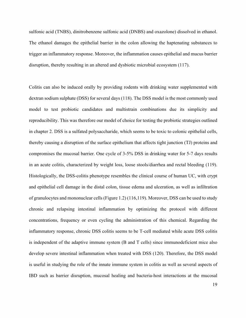
Figure 1.2. Histological analysis of damage caused in colonic tissues by DSS-colitis. A: Images show healthy distal colon tissue from a mouse that received only water as a control. B: inflamed distal colon from mice that received 3% DSS in the drinking water for 4 days, characterized by epithelial damage (arrow), ulceration, crypt damage and severe inflammation. Image reproduced from reference (121) with permission.
Another way to study intestinal disorders is by infecting the host with a pathogen that triggers gut
inflammation. There are several enteric bacterial pathogens that are used as animal models of
intestinal inflammation: Salmonella, Vibrio, Shigella, Clostridium difficile, Campylobacter
enterohemorrhagic and enteropathogenic Escherichia coli (EHEC and EPEC). These pathogens
alter the intestinal ecosystem when infecting their hosts (122). Moreover, how the host responds
to and recovers from the GI inflammation and the dysbiosis caused by the virulence factors of
these pathogens highlight mechanisms that could be useful for future therapeutic options and
clinical applications. Additionally, changes in the microbiota composition post-GI inflammation
21
such as increased colonization of E. coli and C. difficile has been linked to a higher risk of
developing IBD (123).
Citrobacter rodentium is a Gram-negative bacterium and murine pathogen surrogate to EPEC and
EHEC, two human pathogens of significant clinical interest (124,125). Our laboratory has been one of
the leading groups using C. rodentium as a model of infection to understand the pathogenesis of
attaching and effacing (A/E) pathogens, a family of bacteria that attach to the apical cell membrane of
IEC – thereby forming a pedestal-like structure (126). Since EPEC and EHEC are unable to infect mice
effectively, C. rodentium is commonly chosen for studying A/E pathogen-host interactions in vivo as
it shares 67% of its genes with both EPEC and EHEC (including the locus of enterocyte effacement
(LEE) pathogenicity island) thus causing very similar pathological lesions (127). Following
administration by oral gavage, C. rodentium colonizes the cecum at earlier stages of infection
progressing to the distal colon 2 to 3 days later. The peak of infection occurs between day 6 and 10
and clearance is complete between 21 and 28 days post-infection in most mouse strains, including
C57BL/6, NIH Swiss and Balb/c. A few mouse strains such as C3H/HeJ and CeHOu/J have been
described as being extremely susceptible to this infection, suffering high mortality rates (128). During
its peak, C. rodentium is usually shed from its host in the stool where it is hyper-infectious and can
effectively transmit to new hosts via coprophagy (oral-fecal route). The hallmark pathologies
associated with C. rodentium infection include dramatic crypt hyperplasia, goblet cell depletion, barrier
disruption as well as a strong Th1/Th17 response, resulting in immune cell infiltration into the intestinal
mucosa (Figure 1.3) (124,129). As a non-invasive pathogen, C. rodentium is an ideal microorganism
to investigate A/E bacterial pathogenesis as well as the mucosal host responses generated through
different cells and mediators against A/E pathogens (128). Furthermore, it is believed IEC and
22
epithelial-derived mucin production play a significant role in limiting C. rodentium colonization (130–
132), which explains our choice of model for the experiments performed in chapter 3.
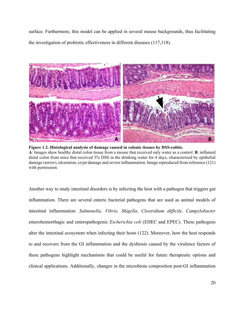
Figure 1.3 Histological damage caused to murine colonic tissues by C. rodentium infection. By day 7 post-infection moderate immune cell infiltration, as well as elongation of crypts, goblet cell depletion and mild edema are observed – as compared to control uninfected tissue. Image reproduced from reference (126) with permission.
1.6 Modulation of the gut microbiome through probiotics
One approach to overcome the microbial dysbiosis seen in IBD patients is through the oral or
enema delivery of beneficial gut microbes (probiotics) known to be lacking in IBD patients.
Probiotics can be easily incorporated into the diet through the consumption of fermented foods
23
(i.e. yoghurt, kefir, kimchi, sauerkraut) or, consumed on a daily basis as a probiotic supplement.
Studies attribute several health benefits to probiotics including direct effects such as producing
SCFAs (e.g. butyrate) and excluding pathogens from the gut by competition for space and
nutrients, as well as indirect effects such as enhancing epithelial barrier function and promoting
antimicrobial peptide secretory IgA production. Moreover, probiotics have been shown to increase
mucin secretion from intestinal goblet cells and to beneficially modulate the host immune system
through the stimulation of anti-inflammatory cytokines such as IL-10 and TGF-β as well as
stimulate the induction of Tregs (133,134). The exact mechanism(s) by which probiotics exert
these positive effects are unclear; however, it is clear that the efficacy of probiotics varies
depending on the microbial strain used and the dose administered.
Microorganisms from the genera Lactobacillus and Bifidobacterium as well as the yeast
Saccharomyces boulardii are among the most common probiotic candidates. Extensive in vitro
research has shown that several Lactobacillus spp. exhibit anti-inflammatory effects, as primarily
assessed by toll-like receptor (TLR) activation (135). For example, Lactobacillus casei Shirota
treatment restores the normal stimulatory capacity of dendric cells (DC) from UC patients by
reducing TLR2 and TLR4 expression (136,137). Lactobacillus plantarum CGMCC1258 increases
tight junction protein levels and decreases permeability in the intestinal epithelial cell line, IPEC-
J2. Moreover, this probiotic reduces IL-8 and TNFα expression in intestinal porcine epithelial cells
challenged by E. coli K88, possibly via a decrease in TLR expression, nuclear factor kappa B
(NFκB) activation, and mitogen-activated protein kinase (MAPK) pathways (138).
24
In mouse models of intestinal inflammation, Lactobacillus acidophilus Bar 13 and
Bifidobacterium longum Bar 33 promote the expansion of Treg cells and reduce the number of
intraepithelial lymphocytes in the 2,4,6-trinitrobenzene sulfonic acid (TNBS) induced colitis
(139). In a similar model of murine colitis (2,4-Dinitrobenzene sulfonic acid [DNBS]), L. casei
DN-114 001 ameliorates disease severity through the induction and expansion of colonic
CD4+FoxP3+ Treg cells (140). Other studies using mice with dextran sulfate sodium (DSS) colitis
show that a combination of eight different probiotic strains (VSL#3) effectively reduces disease
activity and colon inflammation including a significant reduction in inflammatory markers such as
IL-1β, NFκB, and the neutrophil marker, myeloperoxidase (MPO) (141–143). Similarly,
administration of L. plantarum 299V prevents spontaneous colitis development in IL-10 deficient
(Il10−/−) mice (144), and treatment with VSL#3 ameliorates colitis and overall disease activity in
Il10−/− mice.
Curiously, despite the broad success of probiotics in animal models of colitis, their effects in
clinical IBD trials have been less successful, with only small subsets of treated patients showing
beneficial effects (reviewed in (145–147). One reason behind this limited effect in IBD patients
may stem from the “one size fits all” approach that has been commonly employed with probiotics.
It is strongly believed that as an infant’s immune system matures, they develop a mutualistic
relationship with the resident microbes in their intestine. This ensures that these resident gut
microbes establish an environmental niche, as well as an immunological niche which is recognized
by the immune system as a long-term part of the host. In contrast, new microbes encountered after
this relationship has developed are typically seen as foreign and are expelled. Thus, providing
exogenous probiotic microbes to patients without defining whether there are environmental/
25
immunological niches for those gut microbes, may mean that the probiotics will be seen as foreign
and unable to take up permanent residence in the GI tracts of those patients. Similarly, the inflamed
intestines of IBD patients are often\inhospitable to probiotic microbes due to the exaggerated
inflammatory and antimicrobial responses seen during disease. These responses clear new
microbes, including potentially beneficial bacteria, rapidly from the intestine, often before they
have the opportunity to work.
Clearly, new approaches to designing probiotics, and promoting their survival will be key to the
future success of this potential therapy. Additionally, engineered probiotics have been developed
that produce and release the anti-inflammatory cytokine IL-10 (148,149) or trefoil factor (TFF) as
strategies to locally suppress intestinal inflammation and promote healing (150). Moreover, recent
insights regarding the makeup of the human microbiome should allow us to identify potential next-
generation probiotic species with improved potential for colonizing the human GI tract. Recently,
Mandonado-Gómez and colleagues (151) demonstrated that the microbe B. longum AH1206 was
able to persist in the intestines of a subset of individuals for at least six months after administration
without causing side effects or overtly altering the resident microbiota composition. This
microbe’s extended colonization is attributed to its ability to establish a nutritional niche related
to genes involved in carbohydrate utilization. This finding suggests that the establishment of new
probiotic microbes will depend on an individual’s baseline microbiota as well as on the availability
of nutritional resources, thus supporting the critical role of dietary substrates such as fermentable
carbohydrates and prebiotics in permitting the long-term persistence of a probiotic strain.
26
Studies over the past two decades have provided important information identifying person-specific
particularities (e.g. allelic gene variations, increased disease predisposition in patients under
medication from a pre-existing condition) that could be used to diagnose disease as well as
optimize both disease prevention and therapies (152,153). This new strategy represents a shift from
a disease-specific approach towards a patient-specific approach, favoring stratifications of
subpopulations that consequently improves the accuracy and cost-effectiveness of follow up and
treatment. Most progress in precision medicine has been within the oncology field (152,153) (e.g.
preventive mastectomy for BRCA1/2 mutation patients (154)), however, this personalized concept
has also been discussed by scientists in the microbiome field. It is believed that the gut microbiome
may act as a marker for diseases such as obesity (69,155), asthma (156), and rheumatoid arthritis
(157), and assessing its composition could be useful in stratifying a patient’s disease risk and
consequently aid in early disease detection in humans.
In the same line of thought, we believe personalized strategies for the use of probiotics are clearly
needed - especially for dysbiotic-related diseases. While commercial and novel probiotics are
promising in conditions with a specific target, diseases such as IBD do not appear to benefit from
single or even multi-strain probiotics since each patient seems to have a distinct dysbiotic profile.
The individual characteristics of a person’s microbiota, as well as its changes during their lifetime
or in response to environmental stimuli, are the only known factors regarding microbiota
composition. Therefore, strategies focusing on personalized probiotics may provide advantages
over specific isolates from an exogenous source.
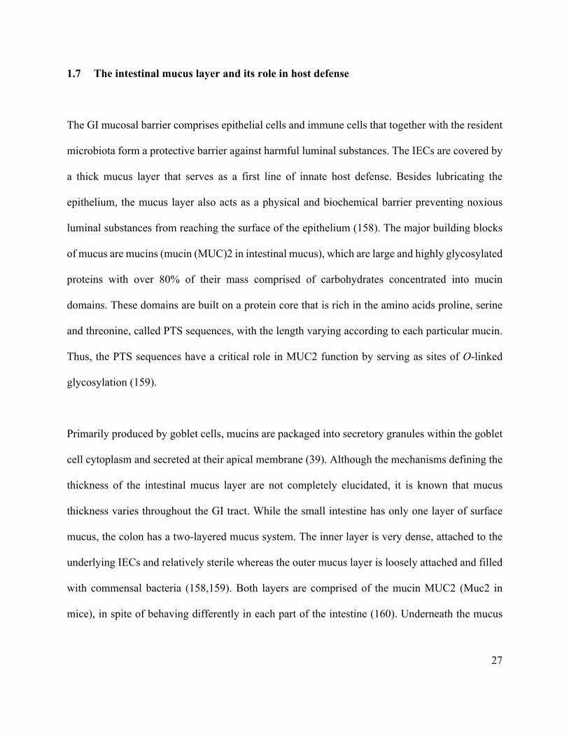
27
1.7 The intestinal mucus layer and its role in host defense
The GI mucosal barrier comprises epithelial cells and immune cells that together with the resident
microbiota form a protective barrier against harmful luminal substances. The IECs are covered by
a thick mucus layer that serves as a first line of innate host defense. Besides lubricating the
epithelium, the mucus layer also acts as a physical and biochemical barrier preventing noxious
luminal substances from reaching the surface of the epithelium (158). The major building blocks
of mucus are mucins (mucin (MUC)2 in intestinal mucus), which are large and highly glycosylated
proteins with over 80% of their mass comprised of carbohydrates concentrated into mucin
domains. These domains are built on a protein core that is rich in the amino acids proline, serine
and threonine, called PTS sequences, with the length varying according to each particular mucin.
Thus, the PTS sequences have a critical role in MUC2 function by serving as sites of O-linked
glycosylation (159).
Primarily produced by goblet cells, mucins are packaged into secretory granules within the goblet
cell cytoplasm and secreted at their apical membrane (39). Although the mechanisms defining the
thickness of the intestinal mucus layer are not completely elucidated, it is known that mucus
thickness varies throughout the GI tract. While the small intestine has only one layer of surface
mucus, the colon has a two-layered mucus system. The inner layer is very dense, attached to the
underlying IECs and relatively sterile whereas the outer mucus layer is loosely attached and filled
with commensal bacteria (158,159). Both layers are comprised of the mucin MUC2 (Muc2 in
mice), in spite of behaving differently in each part of the intestine (160). Underneath the mucus
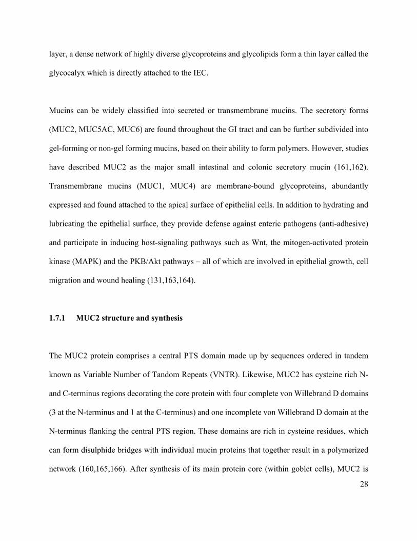
28
layer, a dense network of highly diverse glycoproteins and glycolipids form a thin layer called the
glycocalyx which is directly attached to the IEC.
Mucins can be widely classified into secreted or transmembrane mucins. The secretory forms
(MUC2, MUC5AC, MUC6) are found throughout the GI tract and can be further subdivided into
gel-forming or non-gel forming mucins, based on their ability to form polymers. However, studies
have described MUC2 as the major small intestinal and colonic secretory mucin (161,162).
Transmembrane mucins (MUC1, MUC4) are membrane-bound glycoproteins, abundantly
expressed and found attached to the apical surface of epithelial cells. In addition to hydrating and
lubricating the epithelial surface, they provide defense against enteric pathogens (anti-adhesive)
and participate in inducing host-signaling pathways such as Wnt, the mitogen-activated protein
kinase (MAPK) and the PKB/Akt pathways – all of which are involved in epithelial growth, cell
migration and wound healing (131,163,164).
1.7.1 MUC2 structure and synthesis
The MUC2 protein comprises a central PTS domain made up by sequences ordered in tandem
known as Variable Number of Tandom Repeats (VNTR). Likewise, MUC2 has cysteine rich N-
and C-terminus regions decorating the core protein with four complete von Willebrand D domains
(3 at the N-terminus and 1 at the C-terminus) and one incomplete von Willebrand D domain at the
N-terminus flanking the central PTS region. These domains are rich in cysteine residues, which
can form disulphide bridges with individual mucin proteins that together result in a polymerized
network (160,165,166). After synthesis of its main protein core (within goblet cells), MUC2 is
29
transported to the endoplasmic reticulum (ER) where it forms head-to-head covalently linked
dimers via disulfide linkages between the cysteine-knot domains on the C-terminus. Next, the
MUC2 dimers pass into the Golgi apparatus, where the O-glycans are attached onto the VNTR
region through the actions of specific glycosyltransferases (i.e. Polypeptide N-
acetylgalactosaminyltransferase, Core 1 β1-3 galactosyltransferase, Core 2 β1-6 N-
acetylglucosaminyltransferase, Core 3 β1-3 N-acetylglucosaminyltransferase, Core 2/4 β1-6 N-
acetylglucosaminyltransferase), resulting in dimers with molecular masses of approximately
5MDa. In the trans-Golgi, disulfide bridges are formed between the N-terminal D termini to form
a net-like MUC2 structure (167). Once ready to transport, MUC2 polymers are packaged into
numerous secretory granules and stored in the goblet cell theca creating the apical granule mass
that gives the theca its swollen appearance. Once released, MUC2 becomes highly hydrated and
instantly and dramatically expands in volume, forming the gel-like structure that comprises the
inner and outer mucus layers (39,168).
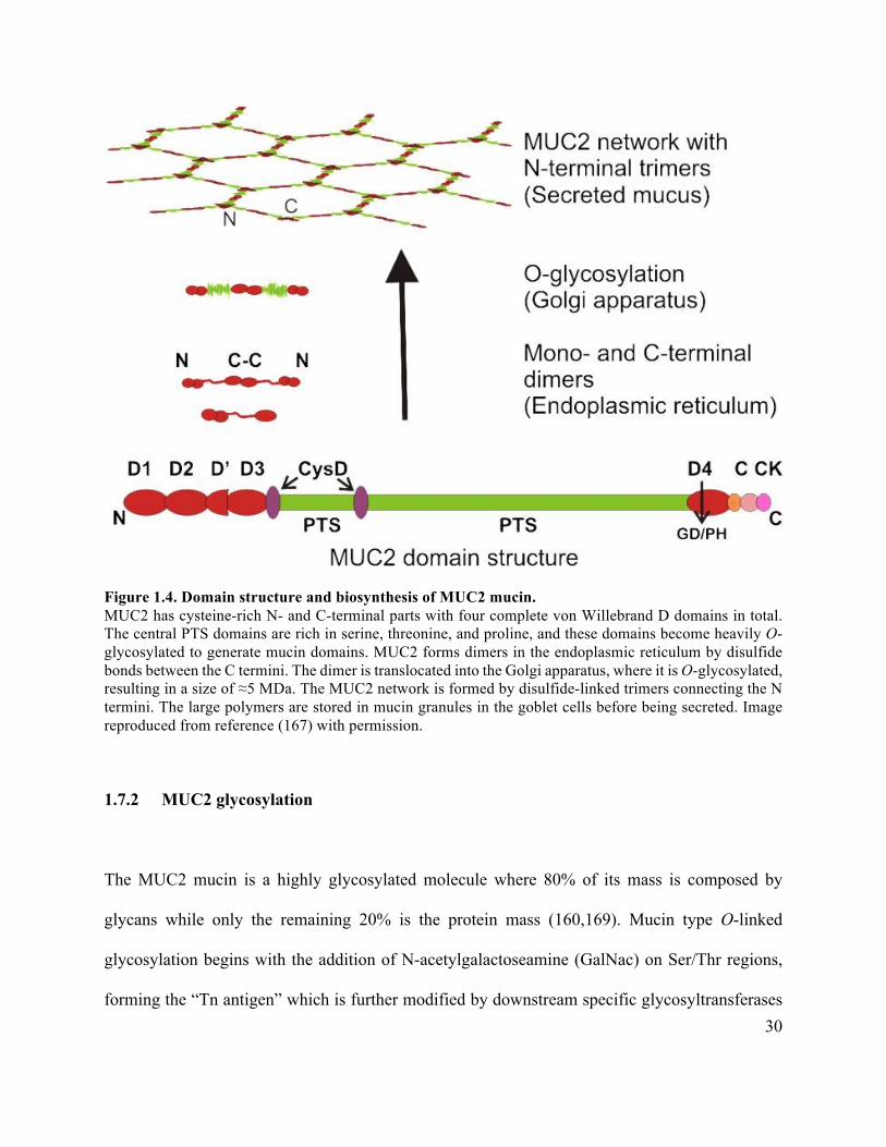
30
Figure 1.4. Domain structure and biosynthesis of MUC2 mucin. MUC2 has cysteine-rich N- and C-terminal parts with four complete von Willebrand D domains in total. The central PTS domains are rich in serine, threonine, and proline, and these domains become heavily O-glycosylated to generate mucin domains. MUC2 forms dimers in the endoplasmic reticulum by disulfide bonds between the C termini. The dimer is translocated into the Golgi apparatus, where it is O-glycosylated, resulting in a size of ≈5 MDa. The MUC2 network is formed by disulfide-linked trimers connecting the N termini. The large polymers are stored in mucin granules in the goblet cells before being secreted. Image reproduced from reference (167) with permission.
1.7.2 MUC2 glycosylation
The MUC2 mucin is a highly glycosylated molecule where 80% of its mass is composed by
glycans while only the remaining 20% is the protein mass (160,169). Mucin type O-linked
glycosylation begins with the addition of N-acetylgalactoseamine (GalNac) on Ser/Thr regions,
forming the “Tn antigen” which is further modified by downstream specific glycosyltransferases
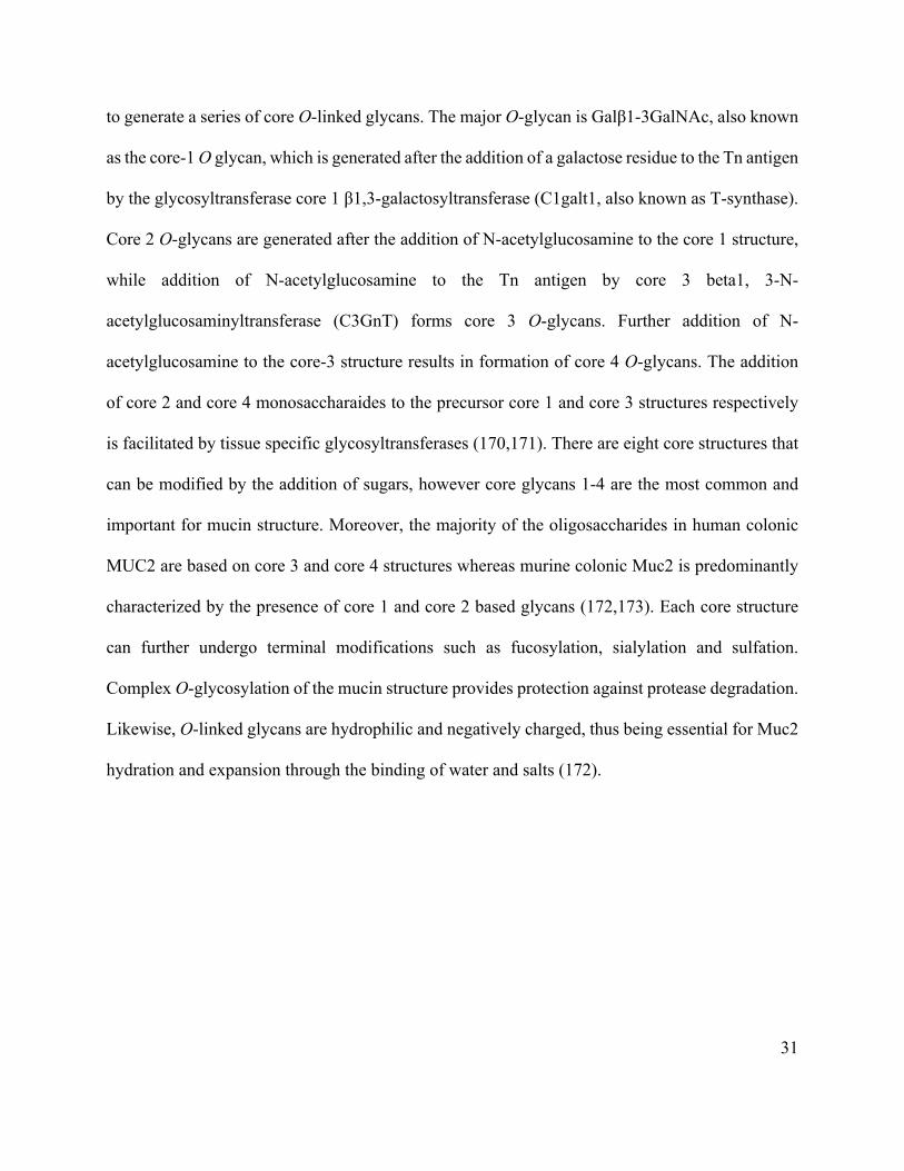
31
to generate a series of core O-linked glycans. The major O-glycan is Galβ1-3GalNAc, also known
as the core-1 O glycan, which is generated after the addition of a galactose residue to the Tn antigen
by the glycosyltransferase core 1 β1,3-galactosyltransferase (C1galt1, also known as T-synthase).
Core 2 O-glycans are generated after the addition of N-acetylglucosamine to the core 1 structure,
while addition of N-acetylglucosamine to the Tn antigen by core 3 beta1, 3-N-
acetylglucosaminyltransferase (C3GnT) forms core 3 O-glycans. Further addition of N-
acetylglucosamine to the core-3 structure results in formation of core 4 O-glycans. The addition
of core 2 and core 4 monosaccharaides to the precursor core 1 and core 3 structures respectively
is facilitated by tissue specific glycosyltransferases (170,171). There are eight core structures that
can be modified by the addition of sugars, however core glycans 1-4 are the most common and
important for mucin structure. Moreover, the majority of the oligosaccharides in human colonic
MUC2 are based on core 3 and core 4 structures whereas murine colonic Muc2 is predominantly
characterized by the presence of core 1 and core 2 based glycans (172,173). Each core structure
can further undergo terminal modifications such as fucosylation, sialylation and sulfation.
Complex O-glycosylation of the mucin structure provides protection against protease degradation.
Likewise, O-linked glycans are hydrophilic and negatively charged, thus being essential for Muc2
hydration and expansion through the binding of water and salts (172).
32
Figure 1.5. The biosynthesis of mucin type O-glycans. Muc2 glycosylation begins with the addition of GalNac onto the hydroxyl group of PTS (proline, threonine, serine) amino acids, generating a Tn antigen. Next, the Tn antigen can be modified by the addition of galactose by core 1 glycosyltransferase (C1galt1) generating core 1 O-glycans. Similarly, addition of a GlcNac residue to the Tn antigen by core 3 glycosyltransferase results in the formation of core 3 structures. Core 1 and core 3 glycans can be further modified to generate Core 2 and Core 4 O-linked glycans, respectively. Addition of sialyl by sialyltransferase forms the sialylated Tn antigen which cannot be modified further. Image reproduced from reference (171) with permission.
1.7.3 Mucus and the gut microbiome
The colonic mucus layer plays an important role in intestinal homeostasis by protecting the
underlying epithelial cells from commensal bacteria and food products. In addition, the mucins
33
present in this barrier seem to influence the composition of the GI tract microbiota. Although the
fetal intestine possesses both GCs and mucus, it is only after birth that the complete glycosylation
structure is determined with the addition of either sialic acid or fucose residues, thus supporting
the role of bacterial colonization in determining the glycosylation patterns of Muc2 (39,174).
Moreover, studies using germ free (GF) mice describe their mucus layer as thinner and more
penetrable as compared to conventionally housed mice, with their intestinal tissues possessing
fewer GCs and only modest levels of Muc2 as well as the pro-inflammatory resistin-like molecule-
β (Relm-β) (175). Likewise, the expression of enzymes responsible for these glycosylations also
appear to be reduced in GF mice (176). Correspondingly, it has been hypothesized that these
glycan structures influence the makeup of the gut microbiota since they impact the expression of
binding sites for specific bacteria thus controlling their ability to adhere to the intestinal mucus
(39).
Studies using mice deficient in Muc2 (Muc2-/-) have shown that they exhibit an increased
susceptibility to infection by enteric pathogens. When infected with C. rodentium, Muc2-/- mice
exhibited rapid weight loss, higher pathogen burdens and suffered more histological damage as
compared to WT mice (130). Moreover, commensal microbes seem to cluster with C. rodentium
and adhere to host tissues in Muc2-/- mice in the form of multispecies biofilms, suggesting a key
role for Muc2 in regulating the intestinal microbiota as well defending the gut mucosal surface
(130). Another recent study demonstrated that Muc2-/- mice carry an altered bacterial community
within their guts, and this dysbiotic state was attenuated when the deletion of Muc2-/- was
combined with loss of another GC derived mediator (RELM-β) (177). Analysis suggested that
34
expression of RELM-β promoted an antimicrobial environment that led to the depletion of specific
microbes, such as Lactobacilli.
Several bacteria from the Bacteroidetes, Firmicutes, Actinobacteria, and Verrucomicrobia phyla
have been described as mucin degraders. These bacteria produce specific enzymes (i.e.
fucosidases, mucinases, glycosilsulfatases, sialidases, sialate O-acetylesterases and N-
acetylneuraminatelyses) that are able to degrade glycans found within the mucus layer. Thus,
terminal O-glycans cleaved/released from the MUC2 protein can serve as a nutrient source for
both commensal and pathogenic bacteria in the gut (178). Bacteroides thetaiotaomicron is a
member of the human intestinal microbiota that relies on mucins and other host glycans for
nutrients in the absence of dietary complex carbohydrates (179). It has been described that B.
thetaiotaomicron produces enzymes that cleave fucose from host glycans, thus increasing the
concentration of this terminal sugar within the gut lumen (180). Besides B. thetaiotaomicron itself,
specific enteric pathogens such as EHEC and Campylobacter jejuni are able to sense these newly
released glycans, thus resulting in an indirect influence on their virulence and ability to colonize
hosts. Regarding potentially beneficial microbes, Akkermansia muciniphila is another well studied
example of a mucin-degrading bacterium that is present both in within feces and on the gut mucosal
surface (181–183). Notably, A. muciniphila levels in the stool seem to be reduced in IBD patients
thus suggesting an anti-inflammatory role for this bacterium – with some groups suggesting A.
muciniphila as a potential next generation probiotic (183). The metabolism of certain
Bifidobacterium spp. is also impacted by the presence of mucin-type O-glycans (171) as various
strains of this genus possess enzymes to degrade glycans from mucin (i.e. glycoside hydrolases,
endo-α-N-acetylgalatosaminidase) (184,185). Members of this genus are Gram-positive obligate
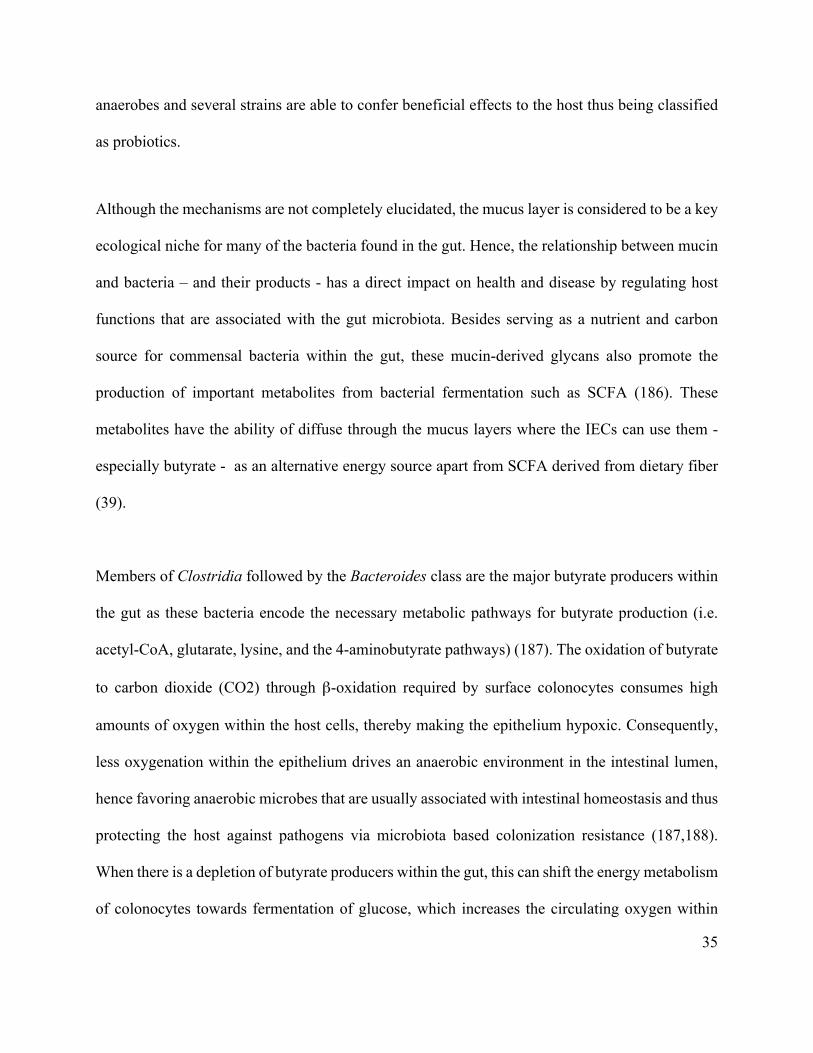
35
anaerobes and several strains are able to confer beneficial effects to the host thus being classified
as probiotics.
Although the mechanisms are not completely elucidated, the mucus layer is considered to be a key
ecological niche for many of the bacteria found in the gut. Hence, the relationship between mucin
and bacteria – and their products - has a direct impact on health and disease by regulating host
functions that are associated with the gut microbiota. Besides serving as a nutrient and carbon
source for commensal bacteria within the gut, these mucin-derived glycans also promote the
production of important metabolites from bacterial fermentation such as SCFA (186). These
metabolites have the ability of diffuse through the mucus layers where the IECs can use them -
especially butyrate - as an alternative energy source apart from SCFA derived from dietary fiber
(39).
Members of Clostridia followed by the Bacteroides class are the major butyrate producers within
the gut as these bacteria encode the necessary metabolic pathways for butyrate production (i.e.
acetyl-CoA, glutarate, lysine, and the 4-aminobutyrate pathways) (187). The oxidation of butyrate
to carbon dioxide (CO2) through β-oxidation required by surface colonocytes consumes high
amounts of oxygen within the host cells, thereby making the epithelium hypoxic. Consequently,
less oxygenation within the epithelium drives an anaerobic environment in the intestinal lumen,
hence favoring anaerobic microbes that are usually associated with intestinal homeostasis and thus
protecting the host against pathogens via microbiota based colonization resistance (187,188).
When there is a depletion of butyrate producers within the gut, this can shift the energy metabolism
of colonocytes towards fermentation of glucose, which increases the circulating oxygen within
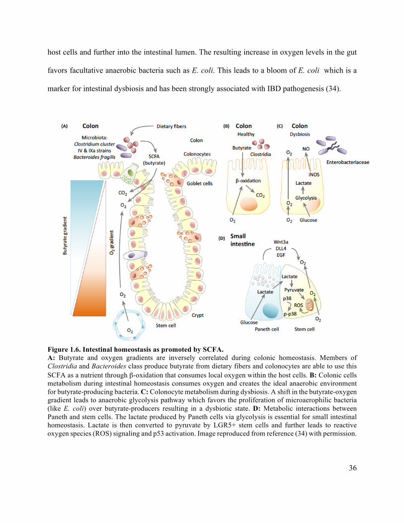
36
host cells and further into the intestinal lumen. The resulting increase in oxygen levels in the gut
favors facultative anaerobic bacteria such as E. coli. This leads to a bloom of E. coli which is a
marker for intestinal dysbiosis and has been strongly associated with IBD pathogenesis (34).
Figure 1.6. Intestinal homeostasis as promoted by SCFA. A: Butyrate and oxygen gradients are inversely correlated during colonic homeostasis. Members of Clostridia and Bacteroides class produce butyrate from dietary fibers and colonocytes are able to use this SCFA as a nutrient through β-oxidation that consumes local oxygen within the host cells. B: Colonic cells metabolism during intestinal homeostasis consumes oxygen and creates the ideal anaerobic environment for butyrate-producing bacteria. C: Colonocyte metabolism during dysbiosis. A shift in the butyrate-oxygen gradient leads to anaerobic glycolysis pathway which favors the proliferation of microaerophilic bacteria (like E. coli) over butyrate-producers resulting in a dysbiotic state. D: Metabolic interactions between Paneth and stem cells. The lactate produced by Paneth cells via glycolysis is essential for small intestinal homeostasis. Lactate is then converted to pyruvate by LGR5+ stem cells and further leads to reactive oxygen species (ROS) signaling and p53 activation. Image reproduced from reference (34) with permission.
37
1.8 Research hypothesis and objectives
Taken together, these studies suggest that beneficial microbes such as probiotics help in
maintaining an overall bacterial balance within the gut and may be beneficial in preventing
dysbiotic-related diseases. However, the effect of probiotics in severe conditions such as IBD still
show conflicting results mainly due to strain specific effects, the baseline status of the host, and
microbiota resilience in incorporating new microbes in a permanent way. This lack of
reproducibility highlights the need for further research in the field and the mechanisms involved
need to be better elucidated. Moreover, we wondered whether the mucin Muc2 could potentially
impact host defense, since the mucus layer is not only a physical protective barrier but also serves
as a nutrient source for several bacteria in the gut, thus impacting gut microbiota composition and
bacterial pathogenesis. Therefore, the goals of my PhD thesis were twofold. First - I sought to
determine how personalized commensal bacteria isolated from the host could impact inflammatory
responses in the GI tract during DSS induced colitis, focusing on intestinal epithelial pathology
and gut microbiota modulation. DSS-colitis was chosen based on its wide application when testing
novel probiotics and because it replicates closely several phenotypes found in IBD patients
including loss of colonic crypts, severe ulceration and infiltration of inflammatory cells. Second -
for the experiments investigating the role of mucus layer in host defense, the C. rodentium model
was chosen as it replicates A/E bacterial pathogenesis as well as certain aspects of IBD, especially
epithelial barrier disruption, dysbiosis, and the histological tissue damage. Furthermore, the C.
rodentium mouse model is well established and allowed us to explore enteric microbe-mucus
interactions from both the host and pathogen perspective in a dynamic way. We hypothesize that
a personalized probiotic approach represents a better strategy to beneficially modulate the host
response against DSS-colitis as endogenous bacteria are already incorporated in the environment
38
and it is just a matter of quantitative balance to protect the host against noxious stimuli.
Additionally, we believe the mucin Muc2 and its core 1 glycosylation play an important role in
host defense against enteric pathogens functioning as a physical barrier as well as providing a
niche for complex interactions with the intestinal microbiota and its metabolites. The objectives of
my thesis are 1) To examine the protective role of endogenous commensal bacteria acting as a
personalized probiotic during intestinal inflammation induced by DSS, and 2) To determine how
the mucin Muc2 and one of its major glycosylations (core 1) impact colonic homeostasis and
susceptibility to infection with C. rodentium. Findings from these studies will increase our
understanding of the interactions between a balanced gut microbiota and disease susceptibility thus
helping define basic mechanisms of intestinal homeostasis and consequently intestinal health.
Moreover, my work also highlights the importance of the mucus layer in maintaining host-microbial
homeostasis by presenting a possible mechanistic role for intestinal disease caused by enteric
pathogens.
39
Chapter 2: Isolation and characterization of potentially probiotic bacterial
strains from mice: proof of concept for personalized probiotics
2.1 Introduction
At birth, the human gastrointestinal (GI) tract becomes colonized by a complex ecological
community of microorganisms, referred to as the “gut microbiota” (15,37). Most microbes residing
in the gut are harmless or even beneficial to the host, thus resulting in a harmonious and generally
symbiotic relationship. However, disruption of the normal balance in bacterial composition,
function, and diversity (termed dysbiosis) has recently been associated with several negative health
conditions such as Inflammatory Bowel Disease (IBD) (91–93), Irritable Bowel Syndrome (IBS)
(94), obesity (50), type 2 diabetes (95), asthma (96), colon cancer (97,98), non-alcoholic fatty liver
disease (99,100), and neurological diseases (101,102).
The term “microbial dysbiosis” is still poorly defined, as there is much uncertainty regarding what
constitutes a ‘healthy’ microbiota. Previous studies in the GI tract have claimed that a dysbiotic
condition is usually characterized by a reduced diversity of microbes often in combination with a
lower abundance of obligate anaerobic bacteria and an expansion of facultative anaerobic bacteria,
particularly from the Proteobacteria phylum (189–192). Nevertheless, more evidence is needed to
better describe the concept of dysbiosis, considering that it may be defined differently at the
population and individual levels. For the purpose of this paper, ‘dysbiosis' refers to an aberrant
40
microbiota that is represented by low microbial diversity, loss of beneficial microbes, and an
expansion of pathobionts.
Besides genetics, environmental factors, such as diet and the gut microbial community, appear to
be involved in IBD development. These modifiable factors have gained more attention over the
last few decades since IBD incidence has increased significantly in developing countries that have
recently transitioned to a more ‘Western’ diet style and lifestyle (25). One of the strategies
proposed to positively modulate the gut microbiota is the oral administration of beneficial
microbes known as probiotics. Studies suggest that probiotics promote several positive host-
microbe interactions, by excluding pathogens via their competition for nutrients and space, and by
promoting epithelial barrier function and mucus secretion from intestinal goblet cells. Probiotics
have also been shown to increase the production of antimicrobial peptides and SCFA, and
stimulate the expression of anti-inflammatory cytokines such as IL-10 and TGF-β (117,193–198).
Despite the beneficial effects of probiotics seen in different conditions (reviewed in (25,199–201)),
the mechanisms by which they exert these benefits in humans remains uncertain. Moreover, the
effects of probiotics are known to be strain- and dose- specific, which may explain in part the
divergent results found when using different probiotic strains, even though they are from the same
genera or species. Additionally, there are several factors that might influence the results seen using
probiotics in clinical trials, including the use of different probiotic strains, either alone or in
combination with other therapies, the baseline health status of the host, the resistance of the host
microbiota to incorporating new microbes in a permanent way (microbiota resilience), and the
environmental niche created (and controlled) by the host immune system during the early stages
41
of life (25). Furthermore, in certain serious conditions such as IBD, probiotics are usually taken as
adjuvants to traditional therapies or during the remission stage of the disease, thus leaving little to
no opportunity to evaluate their potential effectiveness during times of acute inflammation and
severe intestinal dysbiosis.
Recently, an area of significant discussion has emerged regarding the self-regenerative capacity of
the host microbiota (202). The gut microbiota is sensitive to many environmental challenges such
as diet, infections, antibiotic use, and hygiene habits (i.e. early-life exposure to environmental
microbes, household pets and siblings, city vs. rural living conditions) (49,58). Even so, the
changes in microbiota makeup caused by these challenges appear to be limited and/or transient, as
microbes are considered both resilient and resistant to change, and thus, an individual’s microbiota
composition and function are thought to be fairly stable in the face of external perturbations
(194,202). While the resilience of the intestinal microbiota clearly protects us from infections by
the myriad pathogens we encounter (202–204), it also poses an obstacle to the use of probiotic
strains, making it a significant challenge to manipulate/alter the microbiota, even during states of
dysbiosis. This phenomenon explains the need for probiotics to be repeatedly delivered to observe
their health benefits over the long term (194).
Based on our understanding of how an individual’s genetics influences their physiology, a new
concept termed “personalized” or “precision” medicine has emerged focusing on diagnostics and
treatments that favor the specific characteristics of the host, thus avoiding the “one size fits all”
approach, that has often proven ineffective (153). A personalized strategy using probiotics was
recently proposed for bacterial vaginosis, an aberrant state of the vaginal microbiota, which may
42
be attenuated by the administration of Lactobacillus spp. isolated from the host (205). The method
called “TripleA” involves three consecutive steps described as the Acquisition of the patient
sample during an infection, the Alteration of the microbial composition by sampling and enriching
Lactobacillus spp. ex-vivo, and finally the Administration of the enriched sample in a personalized
gel formula (205). This method hypothesizes that Lactobacillus spp. isolated in a personalized way
would facilitate colonization based on the specific genetic makeup of the host. Although there are
no studies yet demonstrating the efficacy of the TripleA method, this strategy presents possible
advantages in comparison to the common antibiotic treatment (i.e. extensive side effects and
infection recurrences), as well as to commercial probiotics available on the market, because they
are adapted to the host microbiota and would (in theory) simply restore a healthy vaginal
microbiota.
Taken together, the aim of the current study was to introduce the concept of a personalized
probiotic therapy for intestinal diseases, where commensal bacteria isolated from the gut
microbiota of healthy hosts could be stored in a ‘microbiota biobank’ after having their intrinsic
characteristics tested, and ultimately used as a therapy for dysbiosis-related diseases.
43
2.2 Experimental procedures
2.2.1 Mice
Male C57BL/6 mice were purchased from CEMIB (Campinas, SP, Brazil), kept in sterilized, filter-
topped cages and fed autoclaved food (Standard Rodent Diet, Presence, Paulínia, SP, Brazil) and
water ad libitum under specific-pathogen-free conditions at the Sao Paulo State University.
Findings in Brazil were repeated with C57BL/6 mice bred in house at the British Columbia
Children’s Hospital Research Institute (BCCHRI) (Vancouver, BC, Canada). Sentinel animals
were routinely tested for common pathogens at both facilities. The protocols employed were
approved by the Sao Paulo State University (34/2014) and by the University of British Columbia’s
Animal Care Committee (A15-0206), and were in direct accordance with guidelines provided by
the Brazilian College of Animal Experimentation and by the Canadian Council on the Use of
Laboratory Animals.
2.2.2 Isolation of commensal bacteria strains
Several commensal bacteria strains were isolated from the stool of healthy C57BL/6 mice (n=10).
In brief, fresh stool pellets were homogenized in 1.0 mL phosphate buffered saline (pH 7.2), plated
on Man Rogosa Sharpe agar (MRS – Acumedia, Lansing, MI, USA) and Bifidobacterium
iodoacetate medium 25 agar (BIM-25 - Acumedia, Lansing, MI, USA) for Lactobacillus spp. and
Bifidobacterium spp. selection, respectively. The plates were incubated under anaerobic conditions
at 37 °C for 48 h to 72 h. Five colonies with distinct morphologies were selected from
Lactobacillus spp. and Bifidobacterium spp. genera from each mouse sample. The selected
colonies were further purified by streak plating in the same media. The isolated colonies were
44
transferred to MRS broth (Lactobacillus spp.) or MRS broth with the addition of 0.5% L-cysteine
(InLab, Brazil) (Bifidobacterium spp.), and then incubated under anaerobic conditions at 37 °C for
48 h to obtain a liquid culture of each individual isolate.
2.2.3 Preliminary identification
The isolated colonies were stained using Gram’s method (206) and then classified into Gram-
positive and Gram-negative bacteria based on their cell wall properties and the resulting color
(pink or purple). The slides were analyzed using a trinocular microscope with a camera (E200
Nikon, Tokyo, Japan). Additionally, a loop containing a liquid culture of each isolated bacterium
was tested for the expression of the catalase enzyme using 3% hydrogen peroxide (H2O2). A
positive result for catalase was confirmed with the rapid evolution of oxygen (5-10 s) as evidenced
by bubbling (206)
2.2.4 Genera confirmation
All selected strains underwent a combination of colony-PCR and randomly-amplified polymorphic
DNA–polymerase chain reaction (RAPD-PCR) to confirm the target genera and as a preliminary
form of species identification. In brief, single colonies from each isolated strain were used directly
as the template, without any DNA extraction and purification prior to PCR. Next, a set of two short
arbitrary (10bp) primers was used in the PCR reaction: Lactobacillus spp. (Lab-0159: 5’- GGA
AAC AG (A/G) TGC TAA TAC CG-3’; Lab-0677: 5’- CAC CGC TAC ACA TGG AG -3’) (207),
Bifidobacterium spp. (OPA-02: 5’-TGC CGA GCT G -3’; OPA-07: 5’- AGG CGG GAA C -3’)
(208). The RAPD-PCR was performed by adding 1x Taq DNA buffer, 1.5 mM of MgCL2, 0.2 mM
of each deoxynucleotide, 1 µM of each primer, 2 ng of genomic DNA and 2U of Taq DNA
45
polymerase enzyme, in a final volume of 25 µL. All reactions were assembled in duplicate and the
amplification was carried out using a Veriti Thermo Cycler (Thermo Fisher Scientific, Waltham,
MA, USA) under the following conditions: initial denaturation at 94°C for 10 min, followed by 35
cycles at 94°C for 20 s, 55°C for 20 s, 72°C for 30 s and then a final extension at 72°C for 5 min.
Distinct species of Lactobacillus spp. and Bifidobacterium spp. were identified by analyzing the
PCR products through agarose gel electrophoresis (1% in TAE buffer– 40 nmol/L Tris, 11%
glacial acetic acid, 1 mmol/L EDTA) using a 100 bp ladder (Invitrogen, Carlsbad, CA, USA) as a
molecular size marker. The gels were stained with SYBR Safe (Invitrogen, Carlsbad, CA, USA)
and the images acquired under UV illumination using a Gel Doc XR System (Bio-Rad, Hercules,
CA, USA). The similarity of the RAPD profiles was compared within the isolates to distinguish
possible different species, while commercially-purchased strains of Lactobacillus spp. and
Bifidobacterium spp. were used as controls.
2.2.5 Evaluation of survival in simulated gastrointestinal conditions
The gastrointestinal resistance of the isolated strains was tested according to the approach of
Liresse and colleagues (209) and Buriti and colleagues (210), with minor modifications. Aliquots
of each bacterial culture suspension (108CFU/mL) were added to an acid solution (NaCl 0.85%,
1N HCl, 3g/L of pepsin from porcine stomach mucosa (Sigma® Aldrich Co., St. Louis, MO,
USA), and 0.9mg/L of lipase from porcine pancreas (Sigma® Aldrich Co., St. Louis, MO, USA)
to reach a pH of 2.4. Samples were incubated at 37 °C (150 rpm) (Incubator shaker, Tecnal,
Piracicaba, SP, Brazil) for 2 h, leading to the simulated gastric phase. Next, the pH was increased
to 5.0 using an alkaline solution (150 mL of 1 N NaOH, 10.77 g of PO4H2Na.2H2O and distilled
water up to 1 L) and biliary salts (Oxgall Powder, Sigma® Aldrich Co., St. Louis, MO, USA) and
46
porcine pancreatin (Sigma® Aldrich Co., St. Louis, MO, USA) were added to reach a
concentration of 10 g/L and 1 g/L, respectively. Samples were incubated again at 37 °C for 2 h
under agitation (150 rpm), leading to the simulated enteric phase 1. Finally, the pH was increased
to 7.0 using the same alkaline solution. Biliary salts and pancreatin were adjusted to maintain their
concentrations at 10 g/L and 1 g/L, respectively, and the samples were incubated again at 37 °C
for 2 h under agitation, leading to the simulated enteric phase 2, thus completing the 6 h of assay.
Enumeration of Lactobacillus spp. and Bifidobacterium spp. was performed in aliquots collected
in duplicate after 2 h, 4 h, and 6 h. Aliquots of 1 mL were pour-plated in MRS or BIM-25 for
colony forming unites (CFU) of Lactobacillus spp. and Bifidobacterium spp., respectively.
2.2.6 Antibiotic susceptibility test
Each isolated strain was tested for its susceptibility against ten different antibiotics (ceftriaxone 30
µg, imipenem 10 µg, aztreonam 30 µg, erythromycin 15 µg, vancomycin 30 µg, chloramphenicol
30 µg, tetracycline 30 µg, nitrofurantoin 300 µg, norfloxacin 10 µg, and ciprofloxacin 5 µg) using
the disk diffusion assay (211,212). Fresh bacterial cultures were diluted to a suitable turbidity
equivalent to 0.5 McFarland Units (108CFU/mL). Five antibiotic disks were placed in MRS or
BIM-25 agar plates containing 100 µL of each bacterial culture, and the plates were incubated
under anaerobic conditions at 37 oC for 16 h to 18 h. The tests were conducted in duplicate, and
the plates were not inverted during the incubation period. The zone diameter around each disk was
measured as the inhibited bacterial growth areas, and the strains were categorized as sensitive
(≥20mm), intermediate (15-19mm), or resistant (≤14mm), according to criteria established by the
Clinical and Laboratory Standards Institute (211).
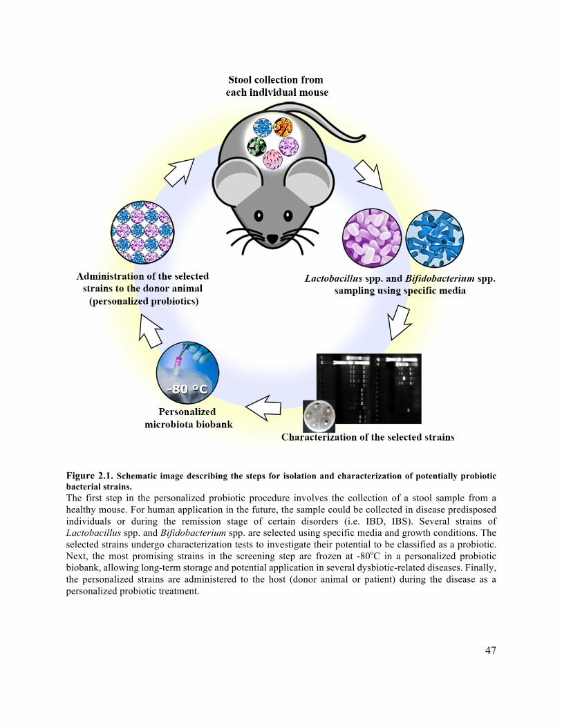
47
Figure 2.1. Schematic image describing the steps for isolation and characterization of potentially probiotic bacterial strains. The first step in the personalized probiotic procedure involves the collection of a stool sample from a healthy mouse. For human application in the future, the sample could be collected in disease predisposed individuals or during the remission stage of certain disorders (i.e. IBD, IBS). Several strains of Lactobacillus spp. and Bifidobacterium spp. are selected using specific media and growth conditions. The selected strains undergo characterization tests to investigate their potential to be classified as a probiotic. Next, the most promising strains in the screening step are frozen at -80oC in a personalized probiotic biobank, allowing long-term storage and potential application in several dysbiotic-related diseases. Finally, the personalized strains are administered to the host (donor animal or patient) during the disease as a personalized probiotic treatment.
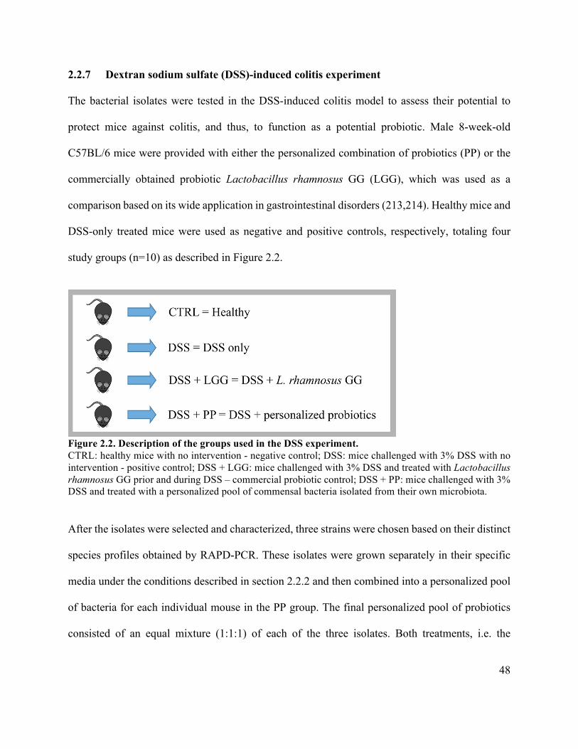
48
2.2.7 Dextran sodium sulfate (DSS)-induced colitis experiment
The bacterial isolates were tested in the DSS-induced colitis model to assess their potential to
protect mice against colitis, and thus, to function as a potential probiotic. Male 8-week-old
C57BL/6 mice were provided with either the personalized combination of probiotics (PP) or the
commercially obtained probiotic Lactobacillus rhamnosus GG (LGG), which was used as a
comparison based on its wide application in gastrointestinal disorders (213,214). Healthy mice and
DSS-only treated mice were used as negative and positive controls, respectively, totaling four
study groups (n=10) as described in Figure 2.2.
Figure 2.2. Description of the groups used in the DSS experiment. CTRL: healthy mice with no intervention - negative control; DSS: mice challenged with 3% DSS with no intervention - positive control; DSS + LGG: mice challenged with 3% DSS and treated with Lactobacillus rhamnosus GG prior and during DSS – commercial probiotic control; DSS + PP: mice challenged with 3% DSS and treated with a personalized pool of commensal bacteria isolated from their own microbiota. After the isolates were selected and characterized, three strains were chosen based on their distinct
species profiles obtained by RAPD-PCR. These isolates were grown separately in their specific
media under the conditions described in section 2.2.2 and then combined into a personalized pool
of bacteria for each individual mouse in the PP group. The final personalized pool of probiotics
consisted of an equal mixture (1:1:1) of each of the three isolates. Both treatments, i.e. the
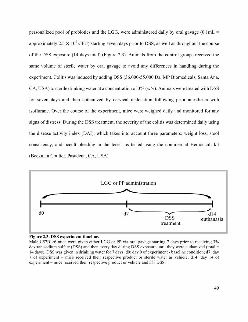
49
personalized pool of probiotics and the LGG, were administered daily by oral gavage (0.1mL =
approximately 2.5 × 10⁹ CFU) starting seven days prior to DSS, as well as throughout the course
of the DSS exposure (14 days total) (Figure 2.3). Animals from the control groups received the
same volume of sterile water by oral gavage to avoid any differences in handling during the
experiment. Colitis was induced by adding DSS (36.000-55.000 Da, MP Biomedicals, Santa Ana,
CA, USA) to sterile drinking water at a concentration of 3% (w/v). Animals were treated with DSS
for seven days and then euthanized by cervical dislocation following prior anesthesia with
isoflurane. Over the course of the experiment, mice were weighed daily and monitored for any
signs of distress. During the DSS treatment, the severity of the colitis was determined daily using
the disease activity index (DAI), which takes into account three parameters: weight loss, stool
consistency, and occult bleeding in the feces, as tested using the commercial Hemoccult kit
(Beckman Coulter, Pasadena, CA, USA).
Figure 2.3. DSS experiment timeline. Male C57BL/6 mice were given either LGG or PP via oral gavage starting 7 days prior to receiving 3% dextran sodium sulfate (DSS) and then every day during DSS exposure until they were euthanized (total = 14 days). DSS was given in drinking water for 7 days. d0: day 0 of experiment - baseline condition; d7: day 7 of experiment – mice received their respective product or sterile water as vehicle; d14: day 14 of experiment – mice received their respective product or vehicle and 3% DSS.
50
2.2.8 Tissue collection
After euthanasia, mouse colonic tissues were collected for histological analysis. Colon tissues were
opened longitudinally, stool was gently removed, and distal colon sections were placed in
histological cassettes. The cassettes were immediately placed in 10% neutral buffered formalin
(Fisher, Hampton, NH, USA) (24 h, R/T) for histological processing.
2.2.9 Histopathological scoring
Colonic pathology was scored using a previously-adapted scoring system (215). In brief, paraffin-
embedded colonic tissue sections (5 µm) were stained with hematoxylin and eosin (HE), and were
examined by three blind observers. The tissue sections were assessed for immune cell infiltration
(0 = occasional immune cell in LP; 1 = granulocytes in LP; 2 =infiltration into the submucosa; 3
= extensive transmural infiltration), severity of crypt damage (0 = all crypts intact; 1 = loss of basal
side of crypts; 2 = some crypt structure can be identified 3 = crypt structure is lost with surface
epithelium still intact; 4 = Crypt structure is lost with epithelial surface erosion), edema (0= no
edema 1= mild/occasional edema 2=moderate edema 3=severe edema/over long stretches), and
amount of tissue affected (0 = 0%; 1 = 5-25%; 2 = 25-50%; 3 = 50-75% 4 =75-100%). The
maximum score that could be obtained with this system was 14 points.
2.2.10 RNA extractions and quantitative real-time PCR
Following euthanization of the mice, distal colonic tissues were immediately placed in RNA-later
(Qiagen, Hilden, Germany) and stored at -80 °C. Total RNA was extracted using the Qiagen
RNeasy Mini Kit according to manufacture’s instructions, and then quantified using a Nanodrop
spectrophotometer (ND1000 – Thermo Fisher Scientific, Massachusetts, USA). Complementary
51
DNA (cDNA) was synthesized using 1 µg of RNA with Omniscript RT kit (Qiagen, Hilden,
Germany), followed by quantitative real-time PCR techniques. The qPCR reaction had a final
volume of 20 µL, where 5 µL of cDNA was added to 15 µL of a PCR mix containing 10 µL of
BioRad SsoFast EvaGreen and 5 µL comprised of RNase- and DNase-free water and primers to a
final concentration of 0.6 µM. Primer sequences and annealing temperatures were as follows: IL-
10 (forward: 5′-GTT GCC AAG CCT TAT CGG AA-3′; reverse: 5′-CCA GGG AAT TCA AAT
GCT CCT-3′; annealing 55 °C) (216); TGF-β (forward: 5′-GAC TCT CCA CCT GCA AGA CCA
T-3′; reverse: 5′-GGG ACT GGC GAG CCT TAG TT-3’; annealing 59 °C) (216); IL-6 (forward:
5′-GAG GAT ACC ACT CCC AAC AGA CC-3′; reverse: 5′-AAG TGC ATC ATC GTT GTT
CAT-3′; annealing 59 °C) (217); IL-1β (forward: 5′-CAG GAT GAG GAC ATG AGC ACC-3′;
reverse: 5′-CTC TGC AGA CTC AAA CTC CAC-3′; annealing 59 °C) (217); Tbp (reference gene)
(forward: 5’-ACC GTG AAT CTT GGC TGT AAA-3’; reverse 5’-GCA GCA AAT CGC TTG
GGA TTA-3’; annealing 59 °C) (218). qPCR was carried out on a CFX Connect Real-Time PCR
system (BioRad, Hercules, CA, USA) for 40 cycles using the following conditions: denaturation
at 95 °C for 5 s, annealing at 57 °C or 59 °C for 10 s, and elongation at 72 °C for 20 s. The data
was analyzed using CFX Manager Software (BioRad, Hercules, CA, USA). The mRNA
expression was determined by the average quantification cycle (Cq) values from duplicate
measurements using Tbp as a reference gene (218), and was normalized with the average Cq value
of the control group (healthy mice) (219).
2.2.11 Myeloperoxidase (MPO) and malondialdehyde (MDA) activity
Colonic tissue homogenates were used to measure MPO and MDA activity. MPO activity was
measured using a colorimetric activity assay kit (Sigma-Aldrich, St. Louis, MO, USA). In brief,
52
the tissues were homogenized in the buffer solution provided with the kit, and the assay was
performed according to the manufacturer’s instructions. Colorimetric change was measured on a
microplate reader (BioRad, Hercules, CA, USA) using 412nm absorbance, and the results were
used to calculate the MPO concentration in each sample as per the manufacturer’s instructions.
Similarly, the lipid peroxidation in colonic tissue was investigated using a commercial kit (Abcam,
Cambridge, UK) that assesses MDA levels though a method dependent on thiobarbituric acid
(TBA). The assay was performed according to the manufacturer’s protocol where TBA was added
to the homogenate supernatant, and the samples were boiled for 60 min at 95 °C before the
absorbance was assessed at 532nm using a spectrophotometer (BioRad, Hercules, CA, USA).
2.2.12 Statistical analysis
All results presented in this study are expressed as the mean value ± Standard Deviation (SD).
Statistical analysis was performed using the GraphPad Prism Software Version 7.0 (GraphPad
Software, San Diego, California). One-way analysis of variance (ANOVA) and Tukey’s or
Dunnett’s multiple-comparison test were used to analyze the results. Significance was declared
when p<0.05.
53
2.3 Results
2.3.1 Isolation and genera confirmation of the strains
From the different stool samples of each mouse plated either in MRS or BIM-25 media, colonies
displaying typical Lactobacillus spp. and Bifidobacterium spp. characteristics (220,221) were
selected based on their distinct morphology. Initially, ten strains were isolated from each mouse
in the experiment, where 50% were Lactobacillus spp. and 50% were Bifidobacterium spp. All
samples presented typical colony morphologies of these two genera, confirming that they were
part of the murine gut microbiome as described in previous studies (222). Figure 2.4 shows the
PCR products from each strain isolated per mouse, revealing two different band profiles for
Bifidobacterium spp. and only one profile for Lactobacillus spp. The similar species profile found
for these mice likely reflects their inbred nature and their co-housing, resulting in similar gut
microbiota. Based on the profiles found in the agarose gel, three different bacterial strains per
mouse (two Bifidobacterium spp. and one Lactobacillus spp.) were chosen to proceed for further
analysis totaling in 30 host-selected strains (n=10). In summary, a personalized combination of
three different commensal bacteria was prepared for each mouse after growing the strains
separately in their specific media and conditions.
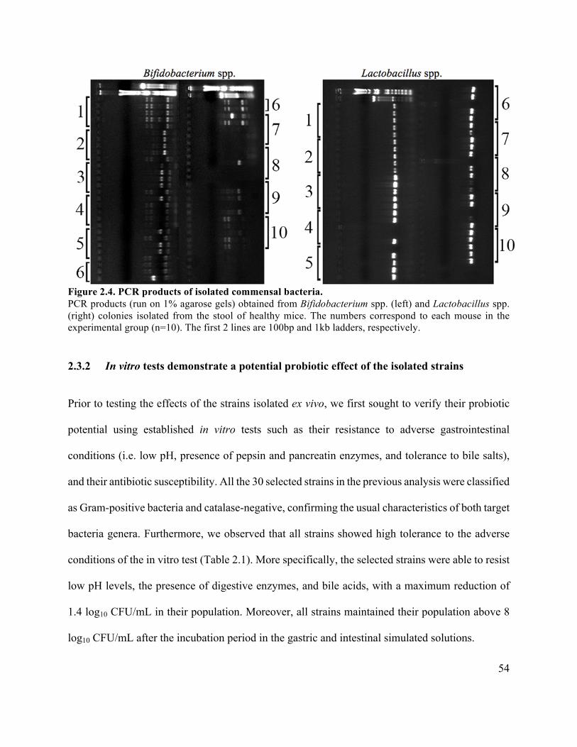
54
Figure 2.4. PCR products of isolated commensal bacteria. PCR products (run on 1% agarose gels) obtained from Bifidobacterium spp. (left) and Lactobacillus spp. (right) colonies isolated from the stool of healthy mice. The numbers correspond to each mouse in the experimental group (n=10). The first 2 lines are 100bp and 1kb ladders, respectively.
2.3.2 In vitro tests demonstrate a potential probiotic effect of the isolated strains
Prior to testing the effects of the strains isolated ex vivo, we first sought to verify their probiotic
potential using established in vitro tests such as their resistance to adverse gastrointestinal
conditions (i.e. low pH, presence of pepsin and pancreatin enzymes, and tolerance to bile salts),
and their antibiotic susceptibility. All the 30 selected strains in the previous analysis were classified
as Gram-positive bacteria and catalase-negative, confirming the usual characteristics of both target
bacteria genera. Furthermore, we observed that all strains showed high tolerance to the adverse
conditions of the in vitro test (Table 2.1). More specifically, the selected strains were able to resist
low pH levels, the presence of digestive enzymes, and bile acids, with a maximum reduction of
1.4 log10 CFU/mL in their population. Moreover, all strains maintained their population above 8
log10 CFU/mL after the incubation period in the gastric and intestinal simulated solutions.
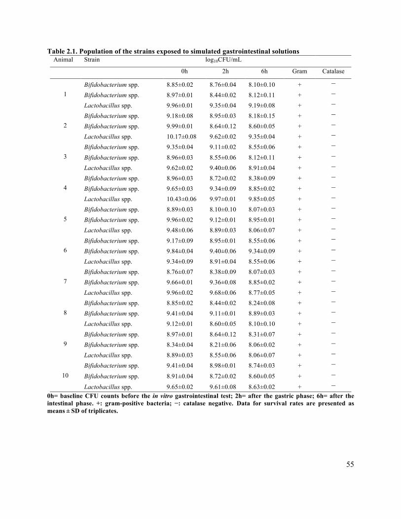
55
Table 2.1. Population of the strains exposed to simulated gastrointestinal solutions Animal Strain log10CFU/mL
0h 2h 6h Gram Catalase
1 Bifidobacterium spp. 8.85±0.02 8.76±0.04 8.10±0.10 + −
Bifidobacterium spp. 8.97±0.01 8.44±0.02 8.12±0.11 + −
Lactobacillus spp. 9.96±0.01 9.35±0.04 9.19±0.08 + −
2 Bifidobacterium spp. 9.18±0.08 8.95±0.03 8.18±0.15 + −
Bifidobacterium spp. 9.99±0.01 8.64±0.12 8.60±0.05 + −
Lactobacillus spp. 10.17±0.08 9.62±0.02 9.35±0.04 + −
3 Bifidobacterium spp. 9.35±0.04 9.11±0.02 8.55±0.06 + −
Bifidobacterium spp. 8.96±0.03 8.55±0.06 8.12±0.11 + −
Lactobacillus spp. 9.62±0.02 9.40±0.06 8.91±0.04 + −
4 Bifidobacterium spp. 8.96±0.03 8.72±0.02 8.38±0.09 + −
Bifidobacterium spp. 9.65±0.03 9.34±0.09 8.85±0.02 + −
Lactobacillus spp. 10.43±0.06 9.97±0.01 9.85±0.05 + −
5 Bifidobacterium spp. 8.89±0.03 8.10±0.10 8.07±0.03 + −
Bifidobacterium spp. 9.96±0.02 9.12±0.01 8.95±0.01 + −
Lactobacillus spp. 9.48±0.06 8.89±0.03 8.06±0.07 + −
6 Bifidobacterium spp. 9.17±0.09 8.95±0.01 8.55±0.06 + −
Bifidobacterium spp. 9.84±0.04 9.40±0.06 9.34±0.09 + −
Lactobacillus spp. 9.34±0.09 8.91±0.04 8.55±0.06 + −
7 Bifidobacterium spp. 8.76±0.07 8.38±0.09 8.07±0.03 + −
Bifidobacterium spp. 9.66±0.01 9.36±0.08 8.85±0.02 + −
Lactobacillus spp. 9.96±0.02 9.68±0.06 8.77±0.05 + −
8 Bifidobacterium spp. 8.85±0.02 8.44±0.02 8.24±0.08 + −
Bifidobacterium spp. 9.41±0.04 9.11±0.01 8.89±0.03 + −
Lactobacillus spp. 9.12±0.01 8.60±0.05 8.10±0.10 + −
9 Bifidobacterium spp. 8.97±0.01 8.64±0.12 8.31±0.07 + −
Bifidobacterium spp. 8.34±0.04 8.21±0.06 8.06±0.02 + −
Lactobacillus spp. 8.89±0.03 8.55±0.06 8.06±0.07 + −
10 Bifidobacterium spp. 9.41±0.04 8.98±0.01 8.74±0.03 + −
Bifidobacterium spp. 8.91±0.04 8.72±0.02 8.60±0.05 + −
Lactobacillus spp. 9.65±0.02 9.61±0.08 8.63±0.02 + − 0h= baseline CFU counts before the in vitro gastrointestinal test; 2h= after the gastric phase; 6h= after the intestinal phase. +: gram-positive bacteria; −: catalase negative. Data for survival rates are presented as means ± SD of triplicates.
56
Ten antibiotics from different classes and with diverse mechanisms of action were tested to assure
the safety of the commensals isolated from the mice, thus guaranteeing they could be eliminated
by antibiotic use if necessary. The antibiotic susceptibility of each strain is presented in Table 2.2
as the zone diameter values for each antibiotic investigated. The results show that all the evaluated
strains were resistant to the antibiotic aztreonam (30 µg). In contrast, all evaluated strains proved
susceptible to the other antibiotics tested, as evidenced by the inhibition halo formed around each
disk (Supplemental Figure A1).

57
Table 2.2. Zone diameter values to indicate susceptible, intermediate, and resistance breakpoints of each strain.
Animal Strains CRO IPM ATM ERI VAN CLO TET NIT NOR CIP
1
Bifidobacterium spp. 29.0 27.0 9.0 32.0 23.0 32.0 37.0 25.0 23.0 26.0
Bifidobacterium spp. 20.0 29.0 0.0 35.0 25.0 34.0 39.0 25.0 26.0 27.0
Lactobacillus spp. 25.0 25.0 0.0 33.0 27.0 32.0 34.0 29.0 24.0 25.0
2
Bifidobacterium spp. 27.0 31.0 0.0 35.0 25.0 30.0 35.0 26.0 29.0 30.0
Bifidobacterium spp. 26.0 30.0 0.0 31.0 23.0 32.0 32.0 20.0 21.0 23.0
Lactobacillus spp. 25.0 34.0 0.0 32.0 29.0 30.0 32.0 28.0 25.0 28.0
3
Bifidobacterium spp. 29.0 32.0 0.0 34.0 22.0 32.0 35.0 25.0 23.0 24.0
Bifidobacterium spp. 28.0 33.0 0.0 33.0 26.0 32.0 40.0 22.0 24.0 26.0
Lactobacillus spp. 27.0 29.0 0.0 30.0 24.0 30.0 35.0 27.0 29.0 21.0
4
Bifidobacterium spp. 25.0 29.0 11.0 35.0 25.0 31.0 27.0 24.0 27.0 27.0
Bifidobacterium spp. 18.0 28.0 0.0 36.0 27.0 38.0 40.0 24.0 26.0 29.0
Lactobacillus spp. 23.0 25.0 0.0 31.0 26.0 31.0 31.0 25.0 28.0 23.0
5
Bifidobacterium spp. 28.0 28.0 2.0 33.0 25.0 35.0 39.0 25.0 25.0 28.0
Bifidobacterium spp. 28.0 32.0 0.0 36.0 25.0 32.0 35.0 25.0 27.0 30.0
Lactobacillus spp. 30.0 30.0 0.0 34.0 24.0 32.0 32.0 25.0 26.0 24.0
6
Bifidobacterium spp. 26.0 27.0 0.0 39.0 26.0 31.0 35.0 25.0 25.0 29.0
Bifidobacterium spp. 28.0 32.0 0.0 35.0 25.0 32.0 36.0 24.0 27.0 30.0
Lactobacillus spp. 31.0 26.0 0.0 36.0 24.0 30.0 34.0 24.0 24.0 32.0
7
Bifidobacterium spp. 20.0 30.0 0.0 36.0 26.0 37.0 40.0 26.0 26.0 30.0
Bifidobacterium spp. 26.0 30.0 1.0 37.0 26.0 32.0 36.0 23.0 27.0 30.0
Lactobacillus spp. 25.0 25.0 0.0 37.0 23.0 30.0 35.0 23.0 29.0 30.0
8
Bifidobacterium spp. 29.0 30.0 0.0 34.0 27.0 39.0 36.0 23.0 26.0 29.0
Bifidobacterium spp. 26.0 31.0 1.0 37.0 27.0 31.0 37.0 28.0 26.0 29.0
Lactobacillus spp. 27.0 32.0 0.0 37.0 25.0 30.0 39.0 27.0 27.0 32.0
9
Bifidobacterium spp. 30.0 36.0 0.0 37.0 26.0 31.0 36.0 24.0 26.0 30.0
Bifidobacterium spp. 16.0 30.0 0.0 39.0 27.0 35.0 41.0 21.0 26.0 29.0
Lactobacillus spp. 29.0 26.0 0.0 36.0 25.0 33.0 39.0 23.0 23.0 31.0
10
Bifidobacterium spp. 25.0 28.0 8.0 36.0 25.0 31.0 37.0 25.0 27.0 30.0
Bifidobacterium spp. 25.0 29.0 9.0 37.0 27.0 30.0 34.0 25.0 26.0 29.0
Lactobacillus spp. 27.0 25.0 0.0 35.0 26.0 34.0 33.0 28.0 24.0 31.0
CRO=ceftriaxone 30 µg, IPM=imipenem 10 µg, ATM=aztreonam 30 µg, ERI=erythromycin 15 µg, VAN=vancomycin 30 µg, CLO=chloramphenicol 30 µg, TET=tetracycline 30 µg, NIT=nitrofurantoin 300 µg, NOR=norfloxacin 10 µg e CIP=ciprofloxacin 5 µg. All values are expressed in millimeters (mm).
58
2.3.3 Personalized commensal strains protect mice against acute dextran sodium sulfate-
induced colitis
To determine if the isolated strains had the potential to be considered probiotic bacteria, we
conducted animal experiments using a well-characterized mouse model of intestinal inflammation
(DSS-induced colitis). This model was chosen for its wide application in testing new probiotic
strains for use in the GI tract (116,117,223). Fresh cultures of the isolated strains, as well as the
probiotic LGG, were prepared every two days in their respective media, and an aliquot was plated
to perform CFU counts. All the cultures remained above 9 log10CFU throughout the experimental
protocols (Supplemental Table A1). The weekly average population of the commercial strain L.
rhamnosus GG was 9.34 log10CFU and 9.74 log10CFU in the first and the second week of study,
respectively (Supplemental Figure A2).
Mice were given equal quantities of either LGG, the PP, or sterile water via oral gavage 7 days
prior to being challenged with 3% DSS in their drinking water, and then every day thereafter until
they were euthanized at day 7. As expected, DSS-colitic mice given just water lost significant
levels of weight (15%), whereas mice treated with LGG or PP lost much less weight (5%) during
DSS exposure. Notably, PP mice showed a significantly lower disease activity index (DAI) as
compared to healthy controls, beginning at the 5th day of DSS challenge and lasting until mice
were euthanized, indicating a better outcome with PP regarding the parameters of stool consistency
and blood in the stool (Figure 2.5). Besides weight loss and the DAI score, mice treated with the
PP presented fewer clinical signs of morbidity (i.e. hunched posture, no activity, riffled fur) and
appeared active and healthy throughout the experiment. These results suggest that the PP treatment
was more effective than LGG at reducing the susceptibility of mice to DSS colitis. As expected,
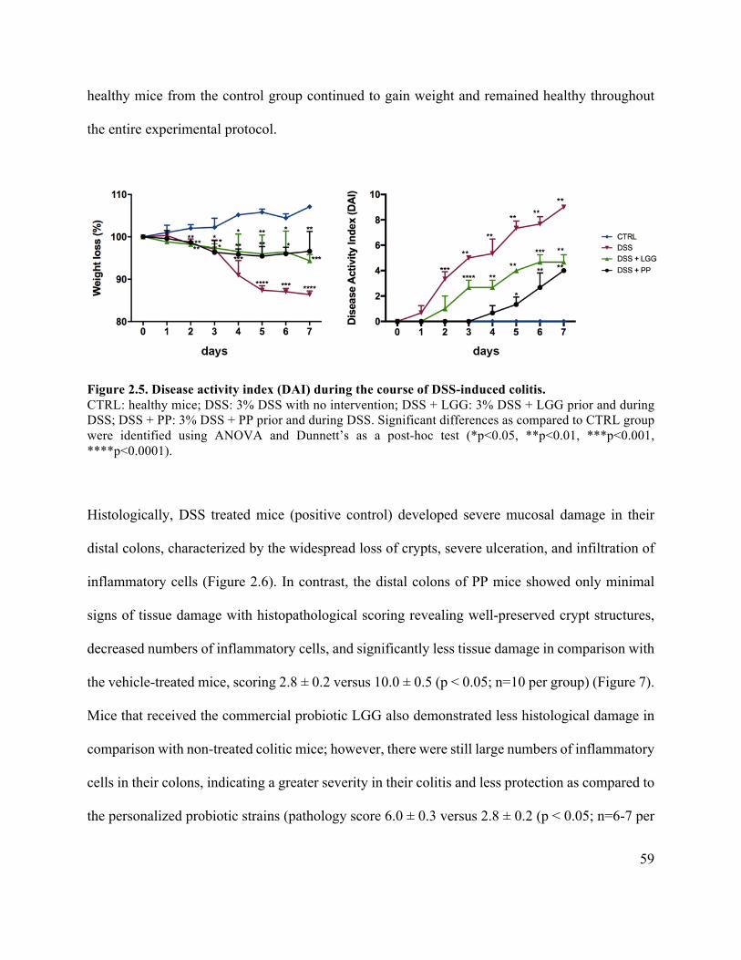
59
healthy mice from the control group continued to gain weight and remained healthy throughout
the entire experimental protocol.
Figure 2.5. Disease activity index (DAI) during the course of DSS-induced colitis. CTRL: healthy mice; DSS: 3% DSS with no intervention; DSS + LGG: 3% DSS + LGG prior and during DSS; DSS + PP: 3% DSS + PP prior and during DSS. Significant differences as compared to CTRL group were identified using ANOVA and Dunnett’s as a post-hoc test (*p<0.05, **p<0.01, ***p<0.001, ****p<0.0001).
Histologically, DSS treated mice (positive control) developed severe mucosal damage in their
distal colons, characterized by the widespread loss of crypts, severe ulceration, and infiltration of
inflammatory cells (Figure 2.6). In contrast, the distal colons of PP mice showed only minimal
signs of tissue damage with histopathological scoring revealing well-preserved crypt structures,
decreased numbers of inflammatory cells, and significantly less tissue damage in comparison with
the vehicle-treated mice, scoring 2.8 ± 0.2 versus 10.0 ± 0.5 (p < 0.05; n=10 per group) (Figure 7).
Mice that received the commercial probiotic LGG also demonstrated less histological damage in
comparison with non-treated colitic mice; however, there were still large numbers of inflammatory
cells in their colons, indicating a greater severity in their colitis and less protection as compared to
the personalized probiotic strains (pathology score 6.0 ± 0.3 versus 2.8 ± 0.2 (p < 0.05; n=6-7 per
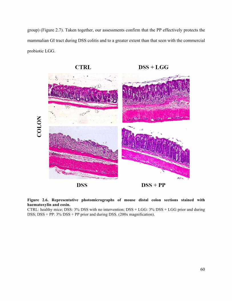
60
group) (Figure 2.7). Taken together, our assessments confirm that the PP effectively protects the
mammalian GI tract during DSS colitis and to a greater extent than that seen with the commercial
probiotic LGG.
Figure 2.6. Representative photomicrographs of mouse distal colon sections stained with haematoxylin and eosin. CTRL: healthy mice; DSS: 3% DSS with no intervention; DSS + LGG: 3% DSS + LGG prior and during DSS; DSS + PP: 3% DSS + PP prior and during DSS. (200x magnification).
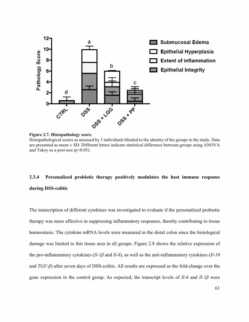
61
Figure 2.7. Histopathology score. Histopathological scores as assessed by 3 individuals blinded to the identity of the groups in the study. Data are presented as mean ± SD. Different letters indicate statistical difference between groups using ANOVA and Tukey as a post-test (p<0.05). 2.3.4 Personalized probiotic therapy positively modulates the host immune response
during DSS-colitis
The transcription of different cytokines was investigated to evaluate if the personalized probiotic
therapy was more effective in suppressing inflammatory responses, thereby contributing to tissue
homeostasis. The cytokine mRNA levels were measured in the distal colon since the histological
damage was limited to this tissue area in all groups. Figure 2.8 shows the relative expression of
the pro-inflammatory cytokines (Il-1β and Il-6), as well as the anti-inflammatory cytokines (Il-10
and TGF-β) after seven days of DSS-colitis. All results are expressed as the fold-change over the
gene expression in the control group. As expected, the transcript levels of Il-6 and Il-1β were

62
higher in the DSS-vehicle treated group, confirming the more severe inflammation found through
histological analysis. Moreover, both groups treated with probiotics showed evidence of attenuated
inflammation, as their tissues showed lower transcript levels of Il-6 and Il-1β and higher levels of
Il-10 and TGF-β as compared to vehicle treated mice. However, the most striking result from data
in Figure 8 was the cytokine mRNA expression comparing the two probiotics treated groups (LGG
and PP). The PP treatment was clearly more effective in increasing anti-inflammatory responses
and in suppressing the elevated expression of pro-inflammatory cytokines as compared to the
commercial probiotic LGG (p<0.05).
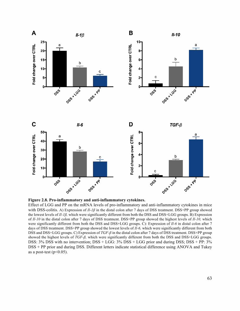
63
Figure 2.8. Pro-inflammatory and anti-inflammatory cytokines. Effect of LGG and PP on the mRNA levels of pro-inflammatory and anti-inflammatory cytokines in mice with DSS-colitis. A) Expression of Il-1β in the distal colon after 7 days of DSS treatment. DSS+PP group showed the lowest levels of Il-1β, which were significantly different from both the DSS and DSS+LGG groups. B) Expression of Il-10 in the distal colon after 7 days of DSS treatment. DSS+PP group showed the highest levels of Il-10, which were significantly different from both the DSS and DSS+LGG groups. C): Expression of Il-6 in distal colon after 7 days of DSS treatment. DSS+PP group showed the lowest levels of Il-6, which were significantly different from both DSS and DSS+LGG groups. C) Expression of TGF-β in the distal colon after 7 days of DSS treatment. DSS+PP group showed the highest levels of TGF-β, which were significantly different from both the DSS and DSS+LGG groups. DSS: 3% DSS with no intervention; DSS + LGG: 3% DSS + LGG prior and during DSS; DSS + PP: 3% DSS + PP prior and during DSS. Different letters indicate statistical difference using ANOVA and Tukey as a post-test (p<0.05).
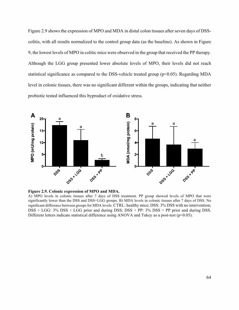
64
Figure 2.9 shows the expression of MPO and MDA in distal colon tissues after seven days of DSS-
colitis, with all results normalized to the control group data (as the baseline). As shown in Figure
9, the lowest levels of MPO in colitic mice were observed in the group that received the PP therapy.
Although the LGG group presented lower absolute levels of MPO, their levels did not reach
statistical significance as compared to the DSS-vehicle treated group (p<0.05). Regarding MDA
level in colonic tissues, there was no significant different within the groups, indicating that neither
probiotic tested influenced this byproduct of oxidative stress.
Figure 2.9. Colonic expression of MPO and MDA. A) MPO levels in colonic tissues after 7 days of DSS treatment. PP group showed levels of MPO that were significantly lower than the DSS and DSS+LGG groups. B) MDA levels in colonic tissues after 7 days of DSS. No significant difference between groups for MDA levels. CTRL: healthy mice; DSS: 3% DSS with no intervention; DSS + LGG: 3% DSS + LGG prior and during DSS; DSS + PP: 3% DSS + PP prior and during DSS. Different letters indicate statistical difference using ANOVA and Tukey as a post-test (p<0.05).
65
2.4 Discussion
At present, it remains unclear if the microbial dysbiosis seen in IBD patients plays a role in their
disease pathogenesis, or if it is simply secondary to the inflammation that develops in these
conditions. A potential causative role for dysbiosis is supported by studies showing that
spontaneous mouse models of IBD often show no disease development when the mice are raised
under germ-free conditions. Moreover, the predominant inflamed sites in IBD patients are also the
sites that hold the highest abundance of microbes in the intestine (distal ileum and colon). In this
case, IBD could be explained as an impaired immune response to gut microbes in genetically-
predisposed individuals. However, an inflammatory environment itself seems to favor an
expansion of Enterobacteriaceae and a depletion of Firmicutes bacteria, which curiously are the
typical features of the microbial dysbiosis observed in IBD patients (25).
Although the microbiota imbalance seen in the stool of IBD patients has yet to be defined as either
the cause or a consequence in IBD, it almost certainly plays a role in these conditions, as IBD
patients usually show a significant decrease in commensal bacteria diversity. A healthy and diverse
microbiota is important for several intestinal physiological processes, such as protection against
pathogens and pathobionts, food digestion, and vitamin biosynthesis, as well as the production of
key metabolites and anti-inflammatory mediators such as transforming growth factor-β (TGF-β),
retinoic acid, and thymic stromal lymphopoietin (TSLP) (25). Importantly, in the absence of a
noxious stimulus, a balanced and diverse microbiota keeps the immune system in a hypo-
responsive state, thus influencing both host metabolism and immunity. Therefore, developing new
66
approaches to positively modulate the gut microbiota and maintain bacterial diversity offer the
potential to act as therapies for dysbiosis-related diseases.
Our work demonstrates that a personalized probiotic therapy can protect mice against DSS-
induced colitis to a greater extent than that seen with the commercial probiotic LGG, and this better
outcome – if replicated in clinical studies – could make a tremendous difference to the treatment
of severe diseases such as IBD, IBS, type 2 diabetes, among others. Although probiotic bacteria
are thought to benefit hosts through several mechanisms of actions, the ability to persistently
colonize the gut may be critical for the ability of probiotics to effectively treat dysbiosis-related
diseases. Because the human microbiome is a complex and dynamic ecosystem, and dysbiosis is
linked to diverse diseases, we believe that beneficial bacteria need to be present in the gut for an
extended period, or preferably in a permanent way, to avoid the bacterial imbalance that triggers
dysbiosis. However, the majority of commercially-available probiotics appear unable to
accomplish this task. Recent findings outline that a combination of 11 probiotic strains (Superherb
Bio-25) varies considerably in their ability to colonize the human gut, depending on the host’s
indigenous microbiome, as well as on host factors and site-specific immune responses (224), thus
highlighting the need for new, personalized approaches. Thus, delivering large numbers of
endogenous commensal bacteria to the gut after their propagation ex vivo could represent a more
promising strategy for serious microbiota-related diseases, since these commensals are already
incorporated in the host’s microbiota, and will thus more readily colonize the host gut where they
can – for example - help promote colonization resistance against invading pathogens/pathobionts.
67
In our study, we first confirmed that the selected strains isolated from each individual mouse could
be considered potentially probiotic due to their performance in preliminary screens using in vitro
tests. RAPD-PCR is a practical method that provides both sensitive and rapid results. For this
reason, it has been widely used in the differentiation of lactic acid bacteria (225–228). It is worth
noting that this step was performed to confirm the bacterial genera, as well as select different
species from Lactobacillus spp. and Bifidobacterium spp. to be administered prior to - and during
DSS challenge. However, the exact identification of each particular strain was not the focus of our
study, since our current aim was to develop a personalized approach for probiotic administration
focusing on the healthy microbiota composition prior to inflammation, rather than isolating
particular strains to be commercialized for general health benefits. The results from this part of the
study indicated that three commensals (two of Bifidobacterium spp. and one of Lactobacillus spp.),
isolated from each animal in the PP group had the potential to be used as probiotic microorganisms
as they were able to survive in adverse conditions, and therefore, they were administered to the
donor animal in the second part of the study (animal experiment).
High survival rates during gastrointestinal transit is one of the key requirements to classify a
commensal microbe as a potential probiotic bacterium (83,199), as the probiotic needs to be acid
and bile resistant to exert its beneficial effects in the colon, when dealing with microbial dysbiosis-
related diseases (25). Another important feature when selecting new probiotic strains is to evaluate
their susceptibility to the antibiotics commonly used in antimicrobial therapy. Although negative
events following the administration of commensal microbial strains are extremely uncommon,
antibiotic resistance of each strain is important to guarantee that the bacterium could be easily
removed in case of bacterial translocation, and consequently – in extremely rare circumstances –
68
systemic infection. Considering that we focused on endogenous bacteria, there are fewer risks of
complications and negative effects than with commercial microbes. However knowledge about the
antimicrobial susceptibility of the isolates also helps our characterization of the personalized
strains, as the classification could be used in the future for stratification purposes. Resistance of
bacteria to antibiotics may be intrinsic or acquired as a result of a chromosomal mutation or by
horizontal gene transfer (229). Lactobacillus spp. are usually susceptible to β-lactam antibiotics
(greater sensitivity to penicillins and less to cephalosporins), protein synthesis inhibitors
(chloramphenicol, macrolides and tetracycline), and more resistant to vancomycin (extrinsic
resistance). Strains from Bifidobacterium spp. are usually intrinsically resistant to mupirocin (an
antibiotic used in selective media for Bifidobacterium spp.) and high concentrations of
aminoglycosides. On the other hand, they are sensitive to macrolides (erythromycin, aztreonam),
chloramphenicol, β-lactams, vancomycin, streptomycin, and rifampicin (230,231). All the strains
selected to be administered in the PP group were resistant to the antibiotic aztreonam; however,
this does not represent a safety issue in itself, considering that intrinsic antibiotic resistance in
probiotic strains could actually be useful for restoring the diverse microbiome after antibiotic
treatment (229).
In the second part of this study, we compared the ability of the PP isolated from the host against
the ability of the commercial probiotic LGG to reduce the inflammatory characteristics of DSS-
induced colitis. The DSS-colitis model is one of the most-widely used mouse models of intestinal
inflammation due its simplicity, reproducibility, and controllability (232,233). DSS is a sulfated
polysaccharide that cause colitis by disrupting the colonic epithelium and allowing the passage of
luminal bacteria and associated antigens into the mucosa. This activates an inflammatory process
69
in the underlying tissues, resulting in clinical and histopathological features similar to those seen
in IBD patients. These include weight loss, diarrhea, occult blood in stools, mucin depletion, loss
of epithelial crypt architecture, and neutrophil infiltration (232). In our study, the groups that
received DSS in their drinking water showed typical signs of colitis characterized by body weight
loss, diarrhea, occult blood in their stools, and piloerection. However, probiotic administration
before and during DSS exposure was able to reduce these signs, especially in the group receiving
the PP. Furthermore, histological changes were attenuated in the PP group as compared to the mice
receiving the commercial probiotic LGG, suggesting that the proposed personalized strategy was
more effective in decreasing DSS-colitis. We strongly believe that the provision of endogenous
commensal bacteria protected the host against the inflammation caused by DSS, and helped
maintain gut homeostasis by replenishing the numbers of endogenous commensal bacteria in the
gut after their ex vivo propagation.
Under homeostatic conditions, the intestinal mucosa is able to maintain the balance between pro-
inflammatory and anti-inflammatory cytokines. However, IBD patients often display increased
intestinal permeability and impaired epithelial barrier function (234–237), leading to elevated
levels of pro-inflammatory cytokines such TNF-α, IFN-γ, interleukin (IL)-1, IL-6, and IL-12
(238). Similar increases in pro-inflammatory mediators are also observed in the DSS-colitis model
correlating well with clinical parameters (232). Consistent with the known anti-inflammatory
effects of some probiotic strains (239–242), we found that both probiotic strategies (PP and LGG)
significantly reduced mRNA levels of Il-1β and Il-6 in the colons of treated mice, which were
accompanied by increased anti-inflammatory cytokine expression (Il-10 and TGF-β).
Interestingly, the personalized probiotic group showed a more effective immune modulation as
70
compared to the commercial probiotic LGG, suggesting that resident microbes of the gut may yield
stronger anti-inflammatory effects since they already possess a mutualistic anti-inflammatory
relationship with the host’s immune system.
Certain biochemical markers, such as myeloperoxidase (MPO) and malonaldehyde (MDA)
activity, have also been investigated in IBD and in animal models of intestinal inflammation as
parameters of intestinal damage. MPO is commonly used as a neutrophil marker, while MDA has
been studied as a oxidative stress marker associated with the pathogenesis of IBD (243–245). As
expected based on previous studies (246,247), we found higher levels of MPO in all groups
challenged with DSS in comparison with healthy mice. However, MPO levels in the DSS + PP
group were significantly reduced as compared to both the non-treated group and the group that
received the commercial probiotic LGG. These results support the idea of personalized probiotics
as better candidates to protect against DSS-colitis, at least in part due to their ability to reduce
neutrophil infiltration. MDA levels were not significantly different between the groups that
received DSS, suggesting that neither probiotic was able to attenuate the typical oxidative stress
caused by inflammation.
Based on our results, commensal bacteria isolated from the host microbiota were more effective
at preventing the symptoms and pathological changes seen in the DSS-colitis model. The improved
efficacy of the personalized treatment in comparison with the commercial probiotic LGG suggests
the benefits of using microorganisms that are already incorporated into the host microbiota to
reduce the typical dysbiosis seen in DSS treatment, as well as other forms of colitis, since the
personalized strategy is based on the maintenance of the initial healthy microbiota by constantly
71
replenishing the host with these beneficial microbes. We believe that administration of these
personalized probiotics has the potential to treat dysbiotic-related and multifactorial diseases based
mainly on maintaining the quantitative balance of beneficial versus pathobiont and/pathogenic
microbes. Moreover, we recognize that commercial probiotics, as well as novel engineered
bacteria expressing specific transgenes, could prove effective in diseases where the etiology and
the mechanisms of disease are better elucidated. In additional, specific populations (e.g. infants,
athletes) and their particular needs would also benefit from designed probiotics aiming at a specific
beneficial effect.
The current evidence supporting personalized therapies are more focused on strategies to treat
specific groups of subjects and avoid the generalist approach considered outdated for many disease
treatments. Although we recognize the importance of developing novel probiotics for specific
populations, we believe that certain diseases linked to microbial dysbiosis could benefit more from
the use of personalized probiotics, which we have named “autoprobiotics”. The novelty of our
approach includes not only selecting strains from the host in a personalized way, but also by
selecting these strains before disease begins, or alternatively, in the remission phase in the case of
IBD. As described in Figure 2.1, the prior isolation and selection of the strains leaves the
opportunity to store the isolates and use them to treat any disease where microbial dysbiosis is
thought to play a central role.
In summary, these results show that personalized probiotics are a potent and promising approach
to healthcare, and as such, may protect against dysbiosis-related symptoms during clinical
diseases. However, considering the novelty of the proposal, it is clear that additional studies need
72
to be performed using different models of intestinal and extra-intestinal diseases as well as
different comparisons made with commercially available probiotics, before these personalized
probiotics are ultimately tested in clinical trials. We emphasize that this strategy has the potential
to assist in the treatment of several diseases associated with microbial dysbiosis.
73
Chapter 3: Role of the mucin (Muc)2 and its glycosylation in controlling
susceptibility to Citrobacter rodentium infection
3.1 Introduction
Enteric bacterial pathogens are a major cause of diarrheal disease in developed as well as
developing countries (248,249). Infection by enteric pathogens is known to promote GI
inflammation along with intestinal pathology and pathophysiology, including not only diarrhea
but also intestinal epithelial barrier dysfunction (126). Additionally, infections by enteric
pathogens as well as impaired intestinal barrier function have been associated with IBD, although
it remains unclear if the microbial community in the gut plays a causative or an aggravating role
in IBD (25).
To infect their host, most enteric pathogens need to directly infect the intestinal epithelium.
However, to do so, they must cross the overlying intestinal mucus layer. As previously discussed
in Chapter 1, intestinal mucus is predominantly comprised of the mucin Muc2, a highly O-
glycosylated protein with core 1 and core 3 derived O-glycans (in mice) as its primary constituents.
We previously showed that mice lacking Muc2 are highly susceptible to infection by Citrobacter
rodentium (130), a mouse specific relative of the bacterial pathogen enterohemorrhagic E. coli
(EHEC), with Muc2 deficient (-/-) mice carrying very high pathogen burdens as well as suffering
severe intestinal inflammation and epithelial damage. Moreover, Muc2-/- mice are also more
susceptible to Salmonella Typhimurium infection as compared to wide-type mice, indicating that
the protective role of the mucin Muc2 is not limited to a singular virulence mechanism (217).
74
Interestingly, IBD patients often display a relatively thin mucus layer in affected regions of their
intestines, as well as overt GC depletion in inflamed intestinal tissues (250). At present, it is not
completely elucidated if the impaired mucus barrier is a primary abnormality in IBD patients or if
it is simply a consequence of the intestinal inflammation in addition to the microbial dysbiosis
typically found in the disease. Studies have shown that mice deficient in Muc2 develop
spontaneous colitis as they age (~12-16 weeks), correlating with increased interactions between
gut bacteria and the intestinal mucosal surface, thus supporting a causative role for mucin
dysfunction in IBD. Moreover, it has been described that some IBD patients and animal models of
IBD develop overt defects in their mucus layer even prior to overt inflammation (251,252).
At present, despite the essential role of Muc2 in promoting intestinal homeostasis and mucosal
protection, it is still unclear whether this protection reflects the actions of the Muc2 protein itself,
its glycosylation, or both. The mucin-type O-glycans are localized around the mucin protein
domain thus being intimately associated with the external environment. Therefore, based on the
evidence that spontaneous colitis in Muc2-/- mice is directly associated with increased contact
between microbes and the mucosal surface, the mucin-type O-glycans seem to influence in host
diseases susceptibility as they are constantly interacting with the microorganisms present in the
environment (171). Additionally, a similar spontaneous colitis phenotype to that seen in Muc2-/-
mice is also described in mice lacking the core 1 glycosylation specifically in their IEC (IEC
C1galt1−/−) (171).
75
Core 1 and core 3-derived O-glycans are important components of colonic mucin glycosylation,
whereas core 1-derived O-glycans are the most predominant O-glycans expressed in mouse tissues
(171,253). IEC C1galt1−/− mice display a thinner inner mucus layer than normal and they suffer
overt breaches in its structure as compared to IEC C1galt1+/+ mice. The spontaneous colitis
displayed at an older age by IEC C1galt1-/- mice resembles human UC (253), characterized by the
infiltration of myeloid cells and the development of crypt abscesses especially in the distal colon
and rectum. Although not developing spontaneous disease, mice deficient in core 3 derived O-
glycans (C3GnT-/-) showed higher susceptibility to DSS-colitis due their impaired mucosal barrier
integrity (254).
The outer mucus layer contains an array of intestinal bacteria and the glycoproteins that comprise
the mucus layer can be considered a potential carbon source for bacteria , although just a few
bacteria display the mucolytic properties necessary to break down the complex mucus glycans
(255). Besides the impaired mucus barrier, previous studies have shown that mice lacking the
mucin Muc2 also carry an aberrant gut microbiome (256), showing reduced levels of Lactobacillus
spp. (177). Mechanistically, it remains uncertain how this dysbiotic microbiota influences disease
susceptibility in Muc2-/- mice, but the SCFA butyrate appears to be involved as it is a major energy
source for colonocytes and it may promotes mucosal healing (177). Moreover, butyrate seems to
play a role in modulating mucin synthesis and release (257,258) as well as in modifying MUC
gene expression in intestinal GC deprived of glucose (259).
The aim of this study was to explore the role of Muc2 glycosylation in providing host defense in
mice challenged with the bacterial pathogen C. rodentium, by comparing the susceptibility of mice
76
lacking Muc2 and Core 1 glycosylation with their respective littermate controls. We also tested
tributyrin supplementation in these knockout mice to investigate its effect in the C. rodentium
model, as a means to see if the susceptibility of the Muc2 -/- and IEC C1galt1-/- mice was due to
an impairment in the ability of their commensal microbes to produce butyrate. We hypothesized
that core 1–derived O-glycans play a key nutritional role within the GI tract, and are thus essential
in preventing dysbiotic changes in the gut microbiome. Therefore, alterations in the Muc2
glycosylation pattern may be closely associated with the pathogenesis of common intestinal
diseases, such as IBD and enteric infections.
77
3.2 Experimental procedures
3.2.1 Mice
Six to eight week old C57BL/6 and Muc2-/- mice were bred in-house at the BC Children’s Hospital
Research Institute (BCCHRI). Mice deficient in core 3 derived O-glycans (C3GnT-/-) and intestinal
epithelial cell (IEC) specific knockout mice IEC C1galt1-/- (core 1 β1,3-galactosyltransferase) (on the
C57BL/6 and 129 genetic background, respectively) were generated in Dr. Lijun Xia’s laboratory
(University of Oklahoma) as previously described (253) and bred in the BCCHRI animal facility.
Briefly, IEC C1galt -/- mice (lacking C1galt1-/- specifically in their IEC) were generated by crossing
mice with loxP sites flanking C1galt1 with an intestinal epithelium-specific Cre-expressing transgenic
line (VillinCre mice). IEC C1galt1+/+ mice were used as littermate controls in all experiments. Mice
were kept in sterilized, filter-topped cages and fed autoclaved food and water while being routinely
monitored and tested for common pathogens. The protocol employed in the experiments was
approved by the University of British Columbia’s Animal Care Committee (A15-0206) and was
in direct accordance with guidelines provided by the Canadian Council on the Use of Laboratory
Animals.
3.2.2 Bacterial strains, Citrobacter rodentium infection and tributyrin supplementation
For C. rodentium infection, mice were orally gavaged with 100 µL (∼2.5 × 10⁸ CFU) of wild-type
C. rodentium DBS100. The culture was grown overnight with shaking at 200 rpm in Luria-Bertani
(LB) broth at 37 °C. For tributyrin supplementation experiments, mice received 100 µL (~5g/kg)
of tributyrin (97% FG) (Sigma-Aldrich, St. Louis, USA) or only glycerol as a control by oral
gavage every other day starting on day 1 post C. rodentium infection. Mice from other groups were
78
mock treated with glycerol at the same timepoints. Tributyrin is a triglyceride composed of butyric
acid and glycerol, that presents some advantages over butyrate supplementation as it is odourless
and it is rapidly absorbed and chemically stable (260).
3.2.3 Tissue collection
Mice were anesthetized with isoflurane and euthanized by cervical dislocation at 6 days post-
infection or after losing approximately 20% of their initial bodyweight and showing signs of
significant morbidity (piloerection, hunching and/or less activity). For bacterial enumeration, gut and
systemic tissues (colon, cecum, spleen, liver and mesenteric lymph nodes – MLN) as well as
luminal contents (stool) were collected in pre-weighed 2 mL tubes containing 1 mL of phosphate
buffered saline (PBS), pH 7.2, and metal beads (Qiagen, Hilden, Germany). Next, samples were
homogenized in a Retch MM400 1/30 Htz homogenizer for 6 min and CFU counts were
determined by serial dilutions on specific LB agar plates supplemented with 100 µg/mL
streptomycin. Plates were incubated overnight at 37 °C and colony counts were normalized by the
weight of the respective tissue to obtain CFU/gram results. For histology, colon and cecum tissues
were fixed in 10% neutral buffered formalin (Fisher, USA) (24 h, R/T) and then transferred to 70%
ethanol. Fixed tissues were embedded in paraffin, cut into 5 µm sections and routinely processed
according to standard techniques by the Histology Core Facility at BCCHRI. Additionally, colon
and cecum tissues were also stored in RNA later (Qiagen, Hilden, Germany) at -80 °C for further
host response analysis by quantitative PCR.
3.2.4 Histopathological scoring
Colonic and cecal pathology were scored using a previously adapted scoring system (216). In brief,
79
hematoxylin and eosin (HE) stained slides were examined by three blinded observers. Tissue
sections were assessed for submucosal edema (0 = no change; 1 = mild; 2 = moderate; and 3 =
profound), epithelial hyperplasia (scored based on percentage above the height of the control
where 0 = no change; 1 = 1–50%; 2 = 51–75%; and 3 = >75%), epithelial integrity (0 = no change;
1 = <10 epithelial cells shedding per lesion; 2 = 11–20 epithelial cells shedding per lesion; 3 =
epithelial ulceration; and 4 = epithelial ulceration with severe crypt destruction) and neutrophil
and mononuclear cell infiltration (0 = none; 1 = mild; 2 = moderate; and 3 = severe). The maximum
score that could be obtained with this system was 15 points.
3.2.5 Short chain fatty acid analysis
Using gas chromatography, the fecal concentration of the three major SCFAs – acetate, propionate,
and butyrate – was analyzed as previously described (261). Briefly, fresh stool pellets were
collected from healthy mice before infection and then immediately frozen at −20°C until analysis.
Fecal samples were homogenized in distilled water resulting in a 17% (w/w) suspension, which
was further acidified with 5M HCl until pH 2-3 was reached. The suspension was centrifuged for
20 min at 5000 rpm and a 2-ethylbutyric acid solution was spiked into the supernatant at a final
concentration of 1 mm. The supernatant was injected into a Thermo TG-WAXMS A GC column
(Thermo Trace 1310, Fisher Scientific, Waltham, MA, USA), coupled to a flame ionization
detector (Thermo Fisher Scientific, Waltham, MA, USA). The results were expressed as mmol of
acetate, propionate and butyrate per kilogram of feces.
80
3.2.6 Statistical analysis
All results presented in this study are expressed as the mean value ± Standard Deviation (SD).
Statistical analysis was performed using a two-tailed Student t test, with assistance from GraphPad
Prism Software Version 7.0 (GraphPad Software, San Diego, California). Significance was
declared when p<0.05.
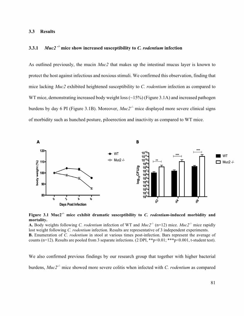
81
3.3 Results
3.3.1 Muc2 -/- mice show increased susceptibility to C. rodentium infection
As outlined previously, the mucin Muc2 that makes up the intestinal mucus layer is known to
protect the host against infectious and noxious stimuli. We confirmed this observation, finding that
mice lacking Muc2 exhibited heightened susceptibility to C. rodentium infection as compared to
WT mice, demonstrating increased body weight loss (~15%) (Figure 3.1A) and increased pathogen
burdens by day 6 PI (Figure 3.1B). Moreover, Muc2-/- mice displayed more severe clinical signs
of morbidity such as hunched posture, piloerection and inactivity as compared to WT mice.
Figure 3.1 Muc2-/- mice exhibit dramatic susceptibility to C. rodentium-induced morbidity and mortality. A. Body weights following C. rodentium infection of WT and Muc2-/- (n=12) mice. Muc2-/- mice rapidly lost weight following C. rodentium infection. Results are representative of 3 independent experiments. B. Enumeration of C. rodentium in stool at various times post-infection. Bars represent the average of counts (n=12). Results are pooled from 3 separate infections. (2 DPI, **p<0.01; ***p<0.001, t-student test).
We also confirmed previous findings by our research group that together with higher bacterial
burdens, Muc2-/- mice showed more severe colitis when infected with C. rodentium as compared
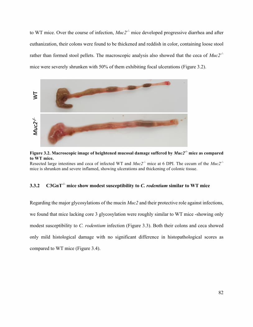
82
to WT mice. Over the course of infection, Muc2-/- mice developed progressive diarrhea and after
euthanization, their colons were found to be thickened and reddish in color, containing loose stool
rather than formed stool pellets. The macroscopic analysis also showed that the ceca of Muc2-/-
mice were severely shrunken with 50% of them exhibiting focal ulcerations (Figure 3.2).
Figure 3.2. Macroscopic image of heightened mucosal damage suffered by Muc2-/- mice as compared to WT mice. Resected large intestines and ceca of infected WT and Muc2-/- mice at 6 DPI. The cecum of the Muc2-/- mice is shrunken and severe inflamed, showing ulcerations and thickening of colonic tissue.
3.3.2 C3GnT-/- mice show modest susceptibility to C. rodentium similar to WT mice
Regarding the major glycosylations of the mucin Muc2 and their protective role against infections,
we found that mice lacking core 3 glycosylation were roughly similar to WT mice -showing only
modest susceptibility to C. rodentium infection (Figure 3.3). Both their colons and ceca showed
only mild histological damage with no significant difference in histopathological scores as
compared to WT mice (Figure 3.4).
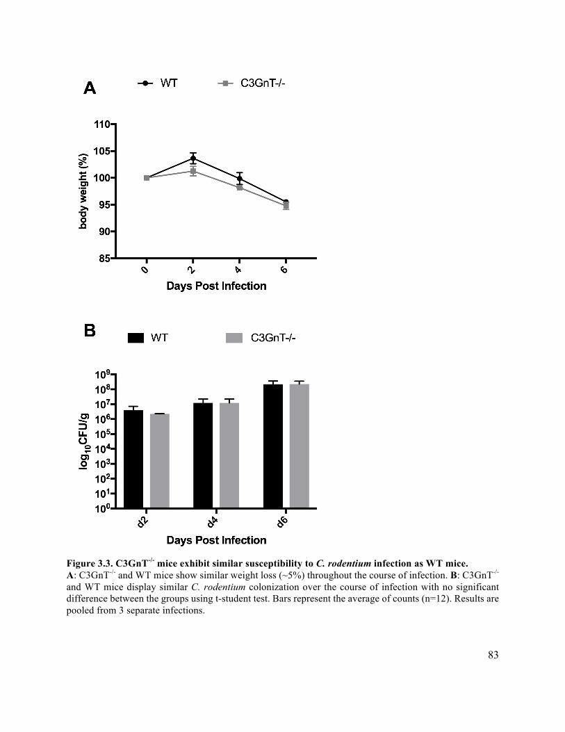
83
Figure 3.3. C3GnT-/- mice exhibit similar susceptibility to C. rodentium infection as WT mice. A: C3GnT-/- and WT mice show similar weight loss (~5%) throughout the course of infection. B: C3GnT-/- and WT mice display similar C. rodentium colonization over the course of infection with no significant difference between the groups using t-student test. Bars represent the average of counts (n=12). Results are pooled from 3 separate infections.
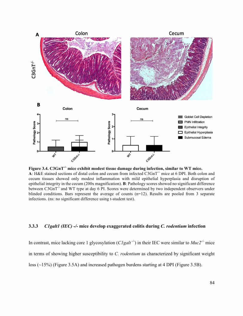
84
Figure 3.4. C3GnT-/- mice exhibit modest tissue damage during infection, similar to WT mice. A: H&E stained sections of distal colon and cecum from infected C3GnT-/- mice at 6 DPI. Both colon and cecum tissues showed only modest inflammation with mild epithelial hyperplasia and disruption of epithelial integrity in the cecum (200x magnification). B: Pathology scores showed no significant difference between C3GnT-/- and WT type at day 6 PI. Scores were determined by two independent observers under blinded conditions. Bars represent the average of counts (n=12). Results are pooled from 3 separate infections. (ns: no significant difference using t-student test).
3.3.3 C1galt1 (IEC) -/- mice develop exaggerated colitis during C. rodentium infection
In contrast, mice lacking core 1 glycosylation (C1galt -/-) in their IEC were similar to Muc2-/- mice
in terms of showing higher susceptibility to C. rodentium as characterized by significant weight
loss (~15%) (Figure 3.5A) and increased pathogen burdens starting at 4 DPI (Figure 3.5B).
85
To assess the role of core 1 glycosylation during C. rodentium infection and compare the findings
with mice completely deficient in the mucin Muc2, we infected Muc2 -/-, and C1galt1 -/- mice
with C. rodentium and monitored their body weights over a 6 day infection period. The results
were compared to WT mice as well as mice with loxP sites flanking C1galt1 (C1galt1f/f). While
WT and C1galt1f/f mice showed only modest body weight loss, C1galt1 -/- mice displayed
significant weight loss during C. rodentium infection. By day 6 PI, the C1galt1 -/- mice had lost
~15% of their initial/starting body weight (Figure 3.5A) and they also displayed clinical signs of
morbidity such as hunched posture, piloerection and inactivity as compared to C1galt1f/f mice.
Additionally, we enumerated C. rodentium burdens in the stool samples of these mice over the
course of infection. While there were no significant differences in C. rodentium burdens between
Muc2 -/- and C1galt1 -/- mice, both knockout mouse strains showed significantly greater pathogen
burdens than control mice (10-100 fold greater) starting at day 4 and maintaining this trend until
the end of infection (Figure 3.5B). Overall, these findings suggested that both Muc2-/- and C1galt1
-/- mice were highly susceptible to C. rodentium infection, shedding much higher C. rodentium
burdens than other mouse strains during infection.
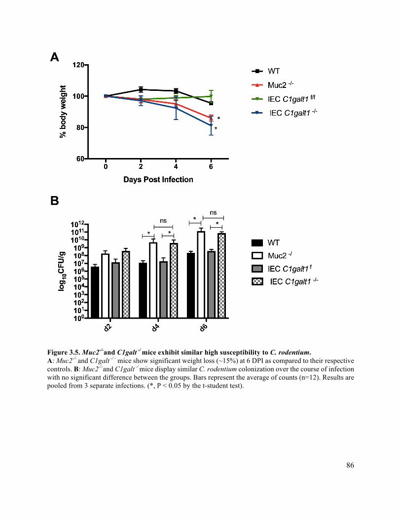
86
Figure 3.5. Muc2-/-and C1galt -/ mice exhibit similar high susceptibility to C. rodentium. A: Muc2-/- and C1galt -/ - mice show significant weight loss (~15%) at 6 DPI as compared to their respective controls. B: Muc2-/-and C1galt -/ mice display similar C. rodentium colonization over the course of infection with no significant difference between the groups. Bars represent the average of counts (n=12). Results are pooled from 3 separate infections. (*, P < 0.05 by the t-student test).
87
3.3.4 C1galt1 -/- mice carry high C. rodentium intestinal burdens similar to Muc2-/-
Next, we enumerated C. rodentium burdens within the GI tract and at systemic sites. C. rodentium
CFU counts from Muc2-/- and C1galt1 -/- mice was significantly greater (10-1000 fold higher)
than in control mice, at all intestinal sites (colon and cecum), with these microbes considered to
be adherent (or directly infecting) these tissues. Luminal content burdens, representing non-
adherent C. rodentium collected from Muc2-/- and C1galt1 -/- mice were also significantly greater
as compared to WT and C1galt1f/f mice (Figure 3.6). Likewise, C. rodentium burdens in systemic
tissues also reflected the trend found within the GI tract, with higher C. rodentium burdens found
in the Muc2-/- and C1galt1 -/- mice as compared to their respective controls. Interestingly, no
differences were found in CFU counts when comparing Muc2-/- and C1galt1 -/- mice, thus
emphasizing the impact of core 1 glycosylation in host defense against C. rodentium, with its loss
producing a similar phenotype to the loss of the entire Muc2 protein.
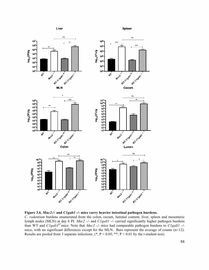
88
Figure 3.6. Muc2-/- and C1galt1 -/- mice carry heavier intestinal pathogen burdens. C. rodentium burdens enumerated from the colon, cecum, luminal content, liver, spleen and mesenteric lymph nodes (MLN) at day 6 PI. Muc2 -/- and C1galt1 -/- carried significantly higher pathogen burdens than WT and C1galt1f/f mice. Note that Muc2 -/- mice had comparable pathogen burdens to C1galt1 -/- mice, with no significant differences except for the MLN. Bars represent the average of counts (n=12). Results are pooled from 3 separate infections. (*, P < 0.05; **, P < 0.01 by the t-student test).
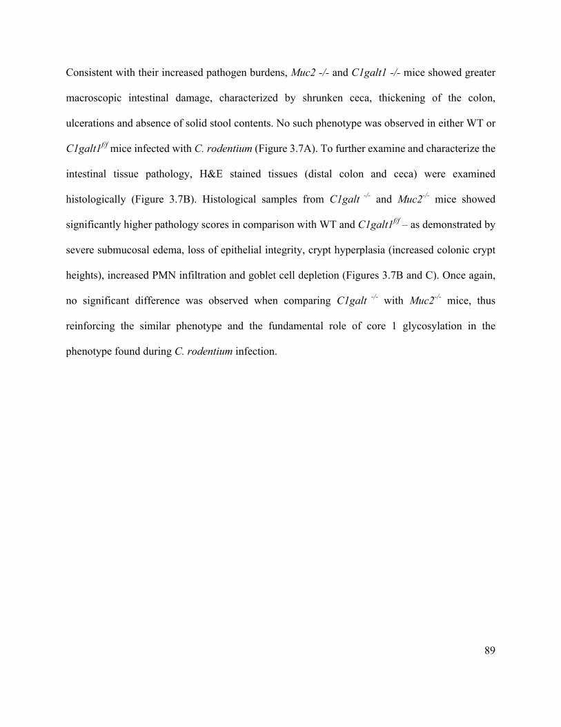
89
Consistent with their increased pathogen burdens, Muc2 -/- and C1galt1 -/- mice showed greater
macroscopic intestinal damage, characterized by shrunken ceca, thickening of the colon,
ulcerations and absence of solid stool contents. No such phenotype was observed in either WT or
C1galt1f/f mice infected with C. rodentium (Figure 3.7A). To further examine and characterize the
intestinal tissue pathology, H&E stained tissues (distal colon and ceca) were examined
histologically (Figure 3.7B). Histological samples from C1galt -/- and Muc2-/- mice showed
significantly higher pathology scores in comparison with WT and C1galt1f/f – as demonstrated by
severe submucosal edema, loss of epithelial integrity, crypt hyperplasia (increased colonic crypt
heights), increased PMN infiltration and goblet cell depletion (Figures 3.7B and C). Once again,
no significant difference was observed when comparing C1galt -/- with Muc2-/- mice, thus
reinforcing the similar phenotype and the fundamental role of core 1 glycosylation in the
phenotype found during C. rodentium infection.
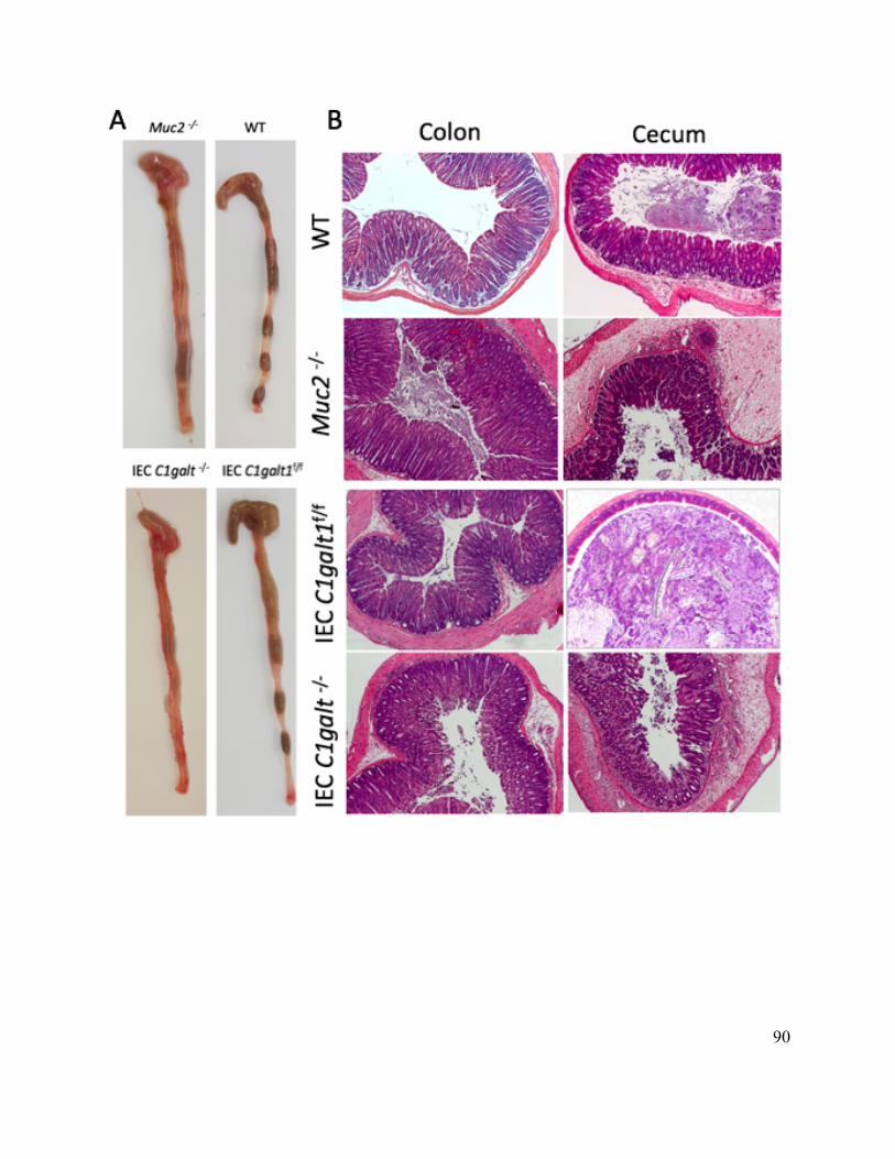
90
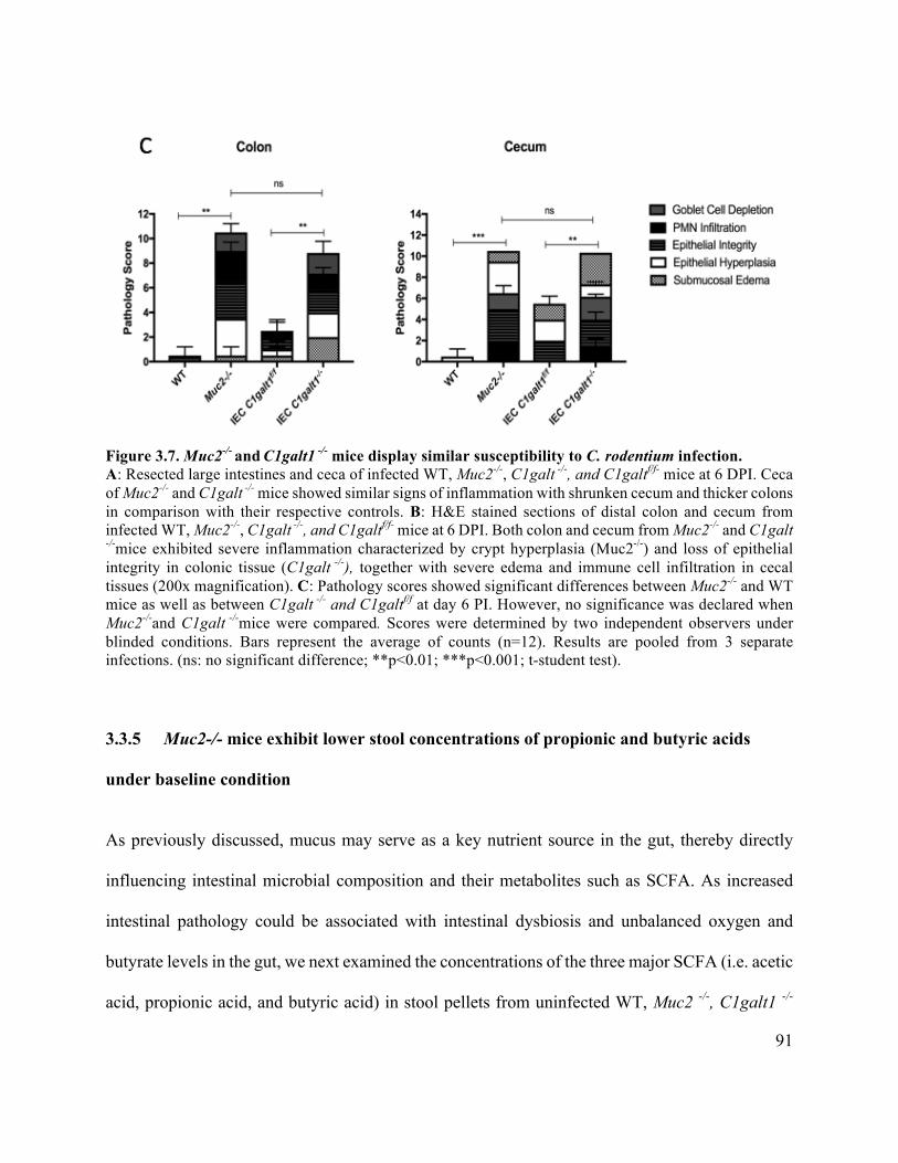
91
Figure 3.7. Muc2-/- and C1galt1 -/- mice display similar susceptibility to C. rodentium infection. A: Resected large intestines and ceca of infected WT, Muc2-/-, C1galt -/-, and C1galtf/f- mice at 6 DPI. Ceca of Muc2-/- and C1galt -/- mice showed similar signs of inflammation with shrunken cecum and thicker colons in comparison with their respective controls. B: H&E stained sections of distal colon and cecum from infected WT, Muc2-/-, C1galt -/-, and C1galtf/f- mice at 6 DPI. Both colon and cecum from Muc2-/- and C1galt
-/-mice exhibited severe inflammation characterized by crypt hyperplasia (Muc2-/-) and loss of epithelial integrity in colonic tissue (C1galt -/-), together with severe edema and immune cell infiltration in cecal tissues (200x magnification). C: Pathology scores showed significant differences between Muc2-/- and WT
mice as well as between C1galt -/- and C1galtf/f at day 6 PI. However, no significance was declared when Muc2-/-and C1galt -/-mice were compared. Scores were determined by two independent observers under blinded conditions. Bars represent the average of counts (n=12). Results are pooled from 3 separate infections. (ns: no significant difference; **p<0.01; ***p<0.001; t-student test).
3.3.5 Muc2-/- mice exhibit lower stool concentrations of propionic and butyric acids
under baseline condition
As previously discussed, mucus may serve as a key nutrient source in the gut, thereby directly
influencing intestinal microbial composition and their metabolites such as SCFA. As increased
intestinal pathology could be associated with intestinal dysbiosis and unbalanced oxygen and
butyrate levels in the gut, we next examined the concentrations of the three major SCFA (i.e. acetic
acid, propionic acid, and butyric acid) in stool pellets from uninfected WT, Muc2 -/-, C1galt1 -/-
92
and C1galt1 f/f mice. Interestingly, we found that Muc2 -/- mice showed significant lower levels of
propionic acid as well as butyric acid as compared to WT mice under baseline conditions (Figure
3.8 B and C). While C1galt1 -/- mice levels did not reach significance as compared to their
counterpart for all SCFA investigated, we did observe a trend towards decreased levels of butyric
acid before the infection (Figure 3.8C).
Figure 3.8. Muc2-/- mice display lower levels of propionic and butyric acids under baseline condition. A: No significant difference in acetic acid levels between groups for all mouse strains analyzed. B: Muc2 -/- exhibited lower concentrations of propionic acid in comparison with WT mice. C: Muc2 -/- exhibited lower concentrations of butyric acid in comparison with WT mice as well as in comparison with C1galt1-/- mice. Bars represent the average of counts (n=8). Results are pooled from 2 separate infections. (ns: no significant difference; *p<0.05; **p<0.01; t-student test).
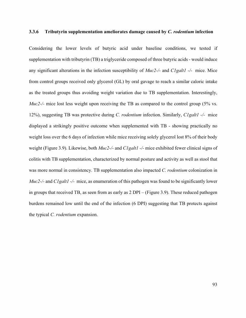
93
3.3.6 Tributyrin supplementation ameliorates damage caused by C. rodentium infection
Considering the lower levels of butyric acid under baseline conditions, we tested if
supplementation with tributyrin (TB) a triglyceride composed of three butyric acids - would induce
any significant alterations in the infection susceptibility of Muc2-/- and C1galt1 -/- mice. Mice
from control groups received only glycerol (GL) by oral gavage to reach a similar caloric intake
as the treated groups thus avoiding weight variation due to TB supplementation. Interestingly,
Muc2-/- mice lost less weight upon receiving the TB as compared to the control group (5% vs.
12%), suggesting TB was protective during C. rodentium infection. Similarly, C1galt1 -/- mice
displayed a strikingly positive outcome when supplemented with TB - showing practically no
weight loss over the 6 days of infection while mice receiving solely glycerol lost 8% of their body
weight (Figure 3.9). Likewise, both Muc2-/- and C1galt1 -/- mice exhibited fewer clinical signs of
colitis with TB supplementation, characterized by normal posture and activity as well as stool that
was more normal in consistency. TB supplementation also impacted C. rodentium colonization in
Muc2-/- and C1galt1 -/- mice, as enumeration of this pathogen was found to be significantly lower
in groups that received TB, as seen from as early as 2 DPI – (Figure 3.9). These reduced pathogen
burdens remained low until the end of the infection (6 DPI) suggesting that TB protects against
the typical C. rodentium expansion.
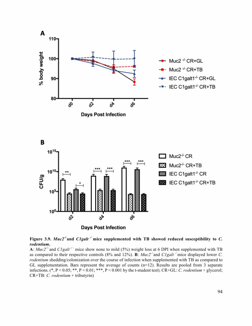
94
Figure 3.9. Muc2-/-and C1galt -/ mice supplemented with TB showed reduced susceptibility to C. rodentium. A: Muc2-/- and C1galt -/ - mice show none to mild (5%) weight loss at 6 DPI when supplemented with TB as compared to their respective controls (8% and 12%). B: Muc2-/-and C1galt -/ mice displayed lower C. rodentium shedding/colonization over the course of infection when supplemented with TB as compared to GL supplementation. Bars represent the average of counts (n=12). Results are pooled from 3 separate infections. (*, P < 0.05; **, P < 0.01; ***, P < 0.001 by the t-student test). CR+GL: C. rodentium + glycerol; CR+TB: C. rodentium + tributyrin)
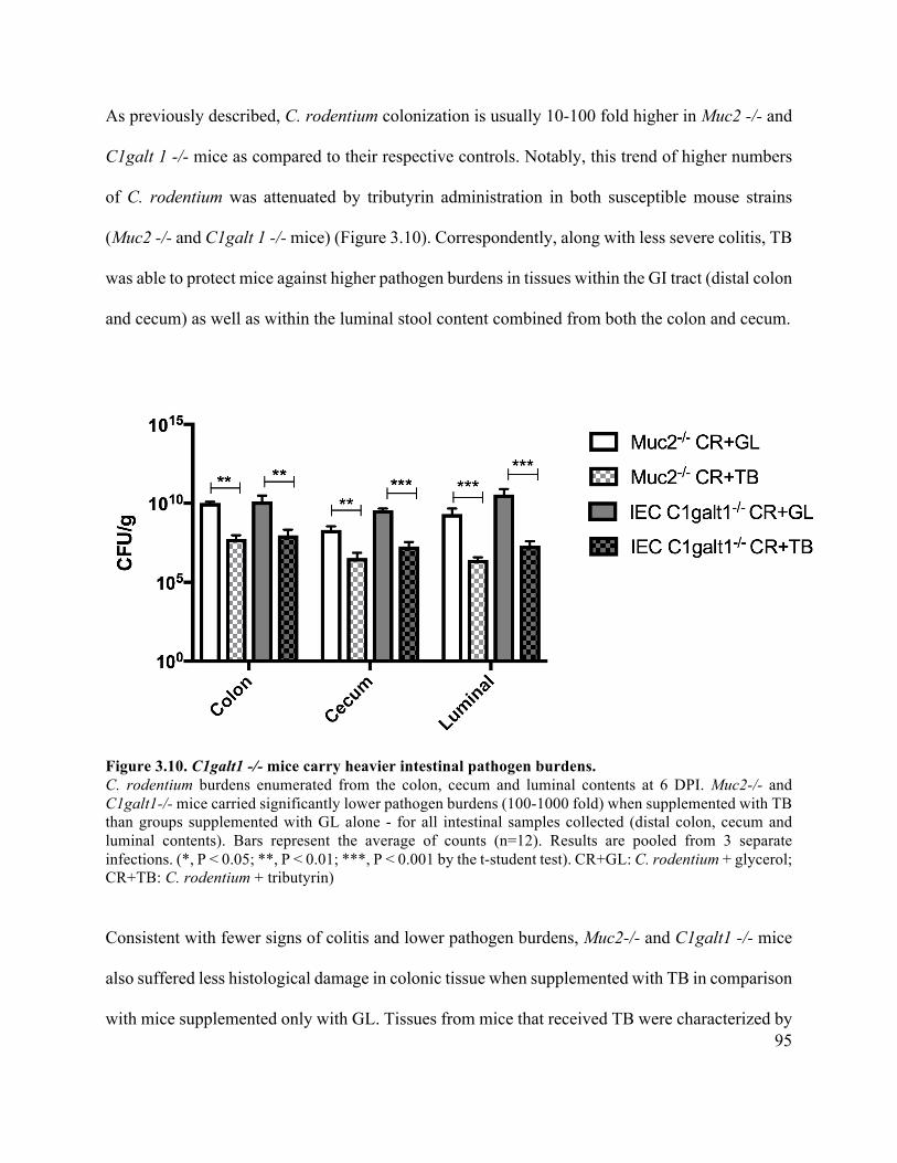
95
As previously described, C. rodentium colonization is usually 10-100 fold higher in Muc2 -/- and
C1galt 1 -/- mice as compared to their respective controls. Notably, this trend of higher numbers
of C. rodentium was attenuated by tributyrin administration in both susceptible mouse strains
(Muc2 -/- and C1galt 1 -/- mice) (Figure 3.10). Correspondently, along with less severe colitis, TB
was able to protect mice against higher pathogen burdens in tissues within the GI tract (distal colon
and cecum) as well as within the luminal stool content combined from both the colon and cecum.
Figure 3.10. C1galt1 -/- mice carry heavier intestinal pathogen burdens. C. rodentium burdens enumerated from the colon, cecum and luminal contents at 6 DPI. Muc2-/- and C1galt1-/- mice carried significantly lower pathogen burdens (100-1000 fold) when supplemented with TB than groups supplemented with GL alone - for all intestinal samples collected (distal colon, cecum and luminal contents). Bars represent the average of counts (n=12). Results are pooled from 3 separate infections. (*, P < 0.05; **, P < 0.01; ***, P < 0.001 by the t-student test). CR+GL: C. rodentium + glycerol; CR+TB: C. rodentium + tributyrin)
Consistent with fewer signs of colitis and lower pathogen burdens, Muc2-/- and C1galt1 -/- mice
also suffered less histological damage in colonic tissue when supplemented with TB in comparison
with mice supplemented only with GL. Tissues from mice that received TB were characterized by
96
less edema and immune cell infiltration, in addition to have their crypt organization and
architecture almost intact and displaying few signs of crypt hyperplasia (Figure 3.11).
Figure 3.11. Muc2-/- and C1galt1 -/- display less severe colitis with TB supplementation. Representative H&E stained colonic tissues of C. rodentium infected Muc2 -/- and C1galt 1 -/- mice. Groups that received TB supplementation develop less exaggerated damage to their mucosal surface and displayed normal crypt heights as well as maintenance of epithelial integrity (200x magnification).

97
3.4 Discussion
The intestinal mucus layer is considered the first barrier of protection against noxious luminal
stimuli such as pathogens and some food/bacterial products. Studies have shown that patients with
intestinal disorders such as IBD and colon cancer often have a defective intestinal mucus layer,
which seems to be thinner and more penetrable than that seen in healthy subjects (160,262–264).
As most bacteria in the gut are localized to the lumen and the outer mucus layer, an impaired
mucus barrier may facilitate these microorganisms and their end products crossing this “normally”
protective barrier resulting in undesired interactions with the underlying immune cells. The Muc2
mucin is the predominant mucin found within the distal GI tract, and previous studies by our
research group have shown that Muc2 promotes host defense against enteric pathogens such as C.
rodentium (130) and S. Typhimurium (217). Although we have already demonstrated that the
Muc2 mucin plays an important role in host defense, the exact mechanisms behind its protective
role have not been completely elucidated. 80% of the mass of the Muc2 mucin is comprised of
carbohydrates, and in fact Muc2 is decorated by several different terminal glycans that are able to
interact with bacterial products and dietary fibers (265). Besides providing a physical and
biochemical barrier against luminal products, our understanding of the role and the mechanism of
action of Muc2 glycosylation in providing protection in the gut is limited.
Considering the importance of the mucin MUC2/Muc2 in IBD patients as well as in enteric
infections, our focus was to investigate which part of the Muc2 molecule is key to promoting host
defense – the protein core, the glycosylation of the protein, or both. In this study, we provide
evidence that the loss of core 1 derived O- glycans dramatically increases host susceptibility to the
98
A/E pathogen C. rodentium, with the C1galt1 -/- mice showing a very similar infectious
phenotype, to that seen in mice suffering the complete loss of the mucin Muc2. An impaired mucus
barrier also increased intestinal pathogen burdens along with systemic translocation of C.
rodentium in both Muc2-/- and C1galt1 -/- mice, resulting in dramatically increased morbidity and
pathology suffered by these mice during infection. Overall these findings suggest that core 1
derived-O glycans, rather core 3 derived-O glycans, play a significant role in controlling luminal
and mucosal pathogen burdens in mice. Additional host innate and adaptive immune responses
may also influence C. rodentium colonization and burdens during infection (266,267). Moreover,
interactions of the intestinal microbiota with components of the intestinal mucus seem to regulate
pathogen growth and colonization, since the mucus barrier also prevents microbes from reaching
and adhering to the intestinal epithelial surfaces and thus play a key role in innate defense.
However, for the purpose of this study, our focus was to investigate the role of the mucin Muc2
and its glycosylation in host defense as well as the relationship between the mucus layer and
bacterial products in the gut.
Overall, mice lacking the core 1 glycosylation solely in their IECs (IEC C1galt1 -/-) displayed a
similar susceptibility phenotype to that seen in mice completely deficient in the mucin Muc2
(Muc2 -/- mice), indicating that the key function of Muc2 in our model appears to reflect its
glycosylation. Notably, C1galt1 -/- mice still produce and secrete Muc2, although in lower
concentrations and with a different structure than in WT mice. Thus, proper Muc2 glycosylation
rather than the mere presence of the Muc2 mucin appears to control C. rodentium burdens in the
intestinal mucosa and its translocation to systemic sites. These results confirm previous data that
99
Muc2 production and secretion are critical host defense mechanisms that regulate the interactions
of C. rodentium with the intestinal mucosal surface (130).
Curiously, loss of core 3 derived O-glycans did not overtly impact susceptibility to C. rodentium
infection in our studies, in contrast to the major effect seen with C1galt1 -/- mice. C3GnT -/- mice
showed a similar phenotype as WT mice, which was characterized by modest weight loss and mild
histological damage such as intestinal crypt elongation, immune cell infiltration, and goblet cell
depletion (126,129). This finding is consistent with previous studies describing core 3 O-
glycosylation of Muc2 playing an important role in humans, but a less important role in promoting
murine host defense against enteric pathogens such as C. rodentium and S. Typhimurium
(217,268,269).
Previous data from our research group has shown a dysbiotic intestinal microbiota in Muc2 -/-
mice with reduced numbers of Firmicutes bacteria, involving specific deficits in Lactobacillus spp.
along with a trend towards more γ-Proteobacteria (130,177). In keeping with the loss of such
microbes, it is already well known that metabolites such SCFA are key products to confer
protection against colitis. Considering the importance of dysbiosis in triggering inflammatory
responses, we assessed the three major SCFAs – acetate, propionate and butyrate – in Muc2 -/-
and C1galt1 -/- mice before infection with C. rodentium, to better define whether these SCFAs
might play a role in infection susceptibility. Interestingly, Muc2 -/- mice displayed lower levels of
propionate and butyrate under baseline conditions, suggesting an imbalanced microbiota with a
reduction in obligate anaerobes such as butyrate producers and an increase in facultative anaerobes
such as E. coli and most pathogenic/pathobiont bacteria. Although not significantly different from
100
their controls, C1galt1 -/- mice also showed a trend towards lower levels of butyrate that may be
a reflection of their thinner mucus layer, rather than the complete absence of Muc2. Nevertheless,
further studies investigating their microbiota composition are necessary to confirm the hypothesis
that specific microbes and their products regulate/limit C. rodentium infection.
Since SCFAs – especially butyrate - have been successful applied in murine models of intestinal
inflammation as well as in IBD patients (270–273), we administered the triglyceride tributyrin for
three consecutive days after C. rodentium infection and compared the disease severity in treated
mice with mice given only the tributyrin carrier matrix glycerol. We noted that mice supplemented
with tributyrin proved less susceptible to C. rodentium infection, as indicated by them suffering
only modest body weight loss, carrying relatively small pathogen burdens as well as suffering only
mild histological damage in their colonic tissues. We also noted that Muc2-/- and C1galt1 -/- mice
again showed a similar phenotype, ie. both mouse strains benefitted from the tributyrin. These
findings are supported by other studies showing that oral supplementation of tributyrin acts as
more than just an energy source for colonocytes but also controls immune responses and interferes
in immune cell migration and mucosal healing (260). Another study found that S. Typhimurium
was able to expand in the gut following antibiotic treatment due to elevated epithelial oxygenation
as a consequence of the loss of butyrate-producing Clostridial bacteria (187). As reviewed by
Allaire and colleagues (34), butyrate directly impacts gut homeostasis by influencing intestinal
pro- and anti-inflammatory mechanisms, colonocyte metabolism, and consequently the intestinal
microbiota composition.
101
The present study provides additional evidence of the critical protective role of the mucus layer in
providing host defense against C. rodentium infection. It also demonstrates that the protective role
played by Muc2 in this model reflects its glycosylation, rather than the protein itself. Furthermore,
our data also provides evidence that tributyrin supplementation could benefit the host during
enteric infections – putatively by shifting oxygen and butyrate gradients in the gut, and potentially
through changes in the gut microbiota. These findings underscore the need for further exploration
of the mechanisms by which the mucus layer protects the intestinal tract from bacterial pathogens
and other noxious stimuli.
102
Chapter 4: Conclusions
4.1 The big picture: potential application of the research findings
Animal models have been widely used to study several GI human diseases, such as IBD and enteric
infections. Although animals are not completely identical to humans in terms of anatomy and
physiology, they share most human genes and are biologically similar. Therefore, murine models
have served as important tools to examine underlying mechanisms involved in both the initiation
and the progression of IBD. Moreover, these models have contributed towards novel therapeutic
strategies for intestinal diseases before these strategies are tested in humans. Thus, the animal
models used in this research project, along with the results we obtained, provide interesting
insights into the development of novel probiotics to prevent and treat intestinal conditions.
Moreover, the use of mice lacking specific genes, such as the Muc2-/- mice and the C1galt1-/- mice,
allow us to isolate particular targets from the complex intestinal environment that might be playing
a more important role in host defense.
Over the last years, several approaches have been studied to positively modulate the gut
microbiome and therefore prevent gastrointestinal and metabolic diseases related to a dysbiotic
state. One of the most common strategies currently used is the administration of beneficial
microbes known as probiotics. Probiotics can be incorporated into the daily diet through the
consumption of fermented food or as dietary supplement. Although the use of probiotics has
proven beneficial in different conditions such as cow’s milk allergy (274), antibiotic associated
diarrhea (275), infant colic and constipation (276), respiratory tract infections (277,278), among
others, their efficacy in IBD is still controversial. The commercial probiotic VSL#3 seems to be
103
effective in inducing remission in active UC patients as well as in preventing disease relapse in
these patients. However, more evidence is required for the use of probiotics in CD patients as its
efficacy remains uncertain in several studies (279).
The work described in this thesis, proposes a novel strategy – i.e. a personalized probiotic approach
that we believe is highly promising for treating different dysbiotic related diseases. Considering
the particular characteristics of an individual’s microbiome, as well as the environmental factors
that influence this microbial community during life, we believe that therapies should focus on the
host’s endogenous commensal microbes. By isolating personalized probiotics, rather than trying
to incorporate new microorganisms from exogenous sources, we propose this strategy will prove
more effective as compared to the “one size fits all” approach that has been used for several
decades and yet has not proven sufficiently reproducible to affect clinical practice in IBD. Our
personalized approach did protect mice against the intestinal inflammation caused by DSS, and to
a greater extent than that seen with the commercial probiotic LGG.
Although further studies are necessary to define the exact mechanism of this protection, our
hypothesis is that this beneficial effect reflects an increase in the numbers of these endogenous
commensal bacteria in the gut after their ex vivo propagation and delivery. Previous studies have
shown IBD patients, as well as people suffering from enteric infections undergo dramatic shifts in
the makeup of their microbiomes. Thus, by orally providing/restoring large numbers of these
microbes to intestinal diseases patients, we hypothesize this will reduce the dysbiosis, as well as
provides beneficial factors to reduce inflammation, such as short chain fatty acids.
104
Another important factor in shaping the composition of the intestinal microbiome is the colonic
mucus layer. Besides acting as a primary defense barrier against enteric pathogens, the colonic
mucus layer also serves as a nutrient source for commensal bacteria, therefore shaping the gut
microbial community. Recent findings have described that in the absence of Muc2, the major gel-
like forming mucin in the colonic mucus layer, mice are significantly more susceptible to enteric
infections (130,217). Moreover, patients with IBD often have a thinner and more penetrable mucus
layer, which could be either cause or consequence for a dysbiotic state within the gut. These
findings intrigued us to explore in more detail the enzymes that control the glycosylation patterns
of the mucin Muc2, and how their absence would affect mice challenged with C. rodentium.
We found that mice deficient in Core 1 derived O-glycans (C1galt1 -/-) showed very similar
susceptibility to C. rodentium infection as that seen with the complete absence of the mucin Muc2
(Muc2 -/- mice). Conversely, mice lacking the Core 3 derived O-glycans had only minor effects on
pathogen burdens and inflammation, showing a roughly similar phenotype to that displayed by
WT mice. The different data obtained from the two major glycosylations of Muc2 may reflect the
specific role of each O-glycan in the models studied, since Core 1 is the most prominent
glycosylation in mice while Core 3 is most prominent in humans. Our results thus indicate that
Core 1 glycosylation may play the key role in protecting mice against C. rodentium rather the
protein core of the mucin Muc2. We also demonstrated that the administration of tributyrin was
effective in decreasing pathogen burdens during infection in both Muc2 -/- and C1galt1 -/- mice.
Future studies are needed to verify the mechanisms of this protective role, however, we believe
the supplementation of tributyrin shifts the intestinal environment to a more anaerobic state, thus
creating a disadvantageous habitat for the expansion of facultative anaerobes such as C. rodentium.
105
Overall, these findings are important to establish a central role for Muc2 and its glycosylation in
intestinal homeostasis. Besides confirming previous discoveries of the importance of Muc2 in host
health and disease, my studies provide novel insights regarding the complex interactions between
mucus, enteric bacteria and host defense.
4.2 Future directions
Since dysbiosis has been associated with the pathogenesis of numerous intestinal diseases, future
studies focusing on how to positively modulate the intestinal ecosystem may provide new therapies
for patients with IBD as well as those suffering from enteric infections. Furthermore, additional
discoveries regarding the mechanistic role of the microbiota in these disorders may assist in
developing novel therapies for intestinal conditions that directly benefit the overall health and
general well-being of individuals.
Regarding the personalized probiotics strategy, we understand that, like all novel ideas, additional
experiments are necessary to validate and confirm the positive effects described in this thesis. Even
so, we believe the manuscript published with our findings will initiate a productive discussion
regarding optimal strategies for personalized therapies in the microbiome field. We believe our
study is the first one demonstrating a promising result when shifting the strategy of microbiota
manipulation from a general “one size fits all” approach to a more personalized approach for each
patient. To prove this, we suggest further comparisons of personalized probiotics against
commercially available probiotics containing different formulations and applications.
106
Additionally, we recognize that the selection process will be more challenging in humans as
compared to murine models due to the complexity of the human microbiome. A few strategies
have been discussed by our research group seeking an approach that will be effective in clinical
practice. First, the selection of personalized commensals at an early age in children genetically
predisposed to develop IBD (for example - and before any disease has developed), could represent
a promising idea. These bacteria could be stratified based on their potential probiotic capacity (in
vitro tests and characterization) and then kept in a microbiota biobank as proposed in Chapter 2.
Since several diseases have been liked to a dysbiotic state in the gut we see the microbiota bank
as an opportunity to treat both intestinal and extra-intestinal conditions. Second, we suggest the
selection of beneficial commensal bacteria during the remission phase of inflammatory conditions
such as IBD or any other relapsing condition. This period is known to have a more diverse
microbiota as compared to that found during acute disease, thus allowing the selection of different
genera/species/strains. Lastly, we propose for future studies the isolation and selection of
commensal bacteria during the acute phase of IBD or any other inflammatory/dysbiotic disease,
in combination (or not) with the ones isolated when the patient is in remission/healthy. Although
microbial diversity is affected during the disease/inflammatory phase, the potentially beneficial
strains that survive within this noxious inflammatory environment could be the most promising
ones to act as probiotic bacteria due to their capacity to survive through adverse conditions.
With reference to the mucus layer and its importance in intestinal homeostasis, several additional
experiments are necessary to further elucidate the mechanistic roles of the mucin Muc2 and its
glycosylations in host defense. It is fundamental to better understand the complex dynamics of the
major Muc2 glycosylations before and during infection as some specific steps of these
107
glycosylations pathways may impact in IBD susceptibility. Furthermore, we should examine the
microbiota composition of the different mouse strains studied in Chapter 3, before and over the
course of infection. Such analysis may bring new insights regarding specific groups of microbes
affected by the mucus layer and by C. rodentium. Analysis using high-throughput techniques, such
as 16S sequencing, may help us to understand in more detail how different microbes behave in the
absence of mucus or its glycosylations as well as how they affect host health. Additionally, the
investigation of bacterial metabolites such as SCFA also contributes towards our understanding of
changes in the microbial gut composition and the impact of this complex community on health and
disease. Moreover, future studies in our lab will focus on the supplementation of tributyrin through
the diet, as this represents a better model of how SCFA might be modified in humans.
Lastly, we believe that focusing on oxygen levels in the gut is extremely important for future
research projects as the availability of oxygen, along with other electron acceptors, may impact
host physiology and control the luminal expansion of Enterobacteriaceae (280). As previously
mentioned in Chapter 3, the oxygen-butyrate gradient within the colon provides important
information about the microbial community in the gut, as well as influencing the host resident and
transient microorganisms and their impact on health and disease.
4.3 Final remarks
Overall, the work described in this thesis has provided numerous contributions towards the
advancement of strategies to positively modulate the intestinal microbiota, either by developing a
new and personalized strategy for using probiotics or by improving our understanding of the
108
interactions between the mucus layer and enteric bacteria as well as their metabolites. Besides the
novelty and the positive effects found when using endogenous commensal microbes to prevent
intestinal inflammation, the personalized probiotic strategy opens an interesting discussion in the
field regarding the focus on the particularities of each person’s unique microbiota. Furthermore,
the role of Muc2 and Core 1 glycosylation in controlling susceptibility to bacterial infections
provides new research directions in the lab to continue investigating how the intestinal microbial
community is affected by the mucus components and bacterial metabolites, and how these
interactions are reflected in the health or disease of the host.
109
References
1. Clemente JC, Ursell LK, Parfrey LW, Knight R. The impact of the gut microbiota on
human health: an integrative view. Cell [Internet]. NIH Public Access; 2012 Mar 16 [cited
2018 Jul 27];148(6):1258–70. Available from:
http://www.ncbi.nlm.nih.gov/pubmed/22424233
2. Sender R, Fuchs S, Milo R. Are We Really Vastly Outnumbered? Revisiting the Ratio of
Bacterial to Host Cells in Humans. Cell [Internet]. Elsevier; 2016 Jan 28 [cited 2018 Aug
14];164(3):337–40. Available from:
http://linkinghub.elsevier.com/retrieve/pii/S0092867416000532
3. Wang B, Yao M, Lv L, Ling Z, Li L. The Human Microbiota in Health and Disease.
Engineering [Internet]. Elsevier; 2017 Feb 1 [cited 2018 Jul 25];3(1):71–82. Available
from: https://www.sciencedirect.com/science/article/pii/S2095809917301492#bib7
4. Consortium THMP, Huttenhower C, Gevers D, Knight R, Abubucker S, Badger JH, et al.
Structure, function and diversity of the healthy human microbiome. Nature [Internet].
Nature Publishing Group; 2012 Jun 14 [cited 2018 Jul 25];486(7402):207–14. Available
from: http://www.nature.com/articles/nature11234
5. Young VB. The role of the microbiome in human health and disease: an introduction for
clinicians. BMJ [Internet]. British Medical Journal Publishing Group; 2017 Mar 15 [cited
2018 Jul 25];356:j831. Available from: http://www.ncbi.nlm.nih.gov/pubmed/28298355
6. Arnold JW, Roach J, Azcarate-Peril MA. Emerging Technologies for Gut Microbiome
Research. Trends Microbiol [Internet]. NIH Public Access; 2016 [cited 2018 Aug
1];24(11):887–901. Available from: http://www.ncbi.nlm.nih.gov/pubmed/27426971
7. Laupland KB, Valiquette L. The changing culture of the microbiology laboratory. Can J
Infect Dis Med Microbiol = J Can des Mal Infect la Microbiol medicale [Internet].
Hindawi Limited; 2013 [cited 2018 Jul 27];24(3):125–8. Available from:
http://www.ncbi.nlm.nih.gov/pubmed/24421822
8. NIH HMP Working Group TNHW, Peterson J, Garges S, Giovanni M, McInnes P, Wang
L, et al. The NIH Human Microbiome Project. Genome Res [Internet]. Cold Spring
Harbor Laboratory Press; 2009 Dec [cited 2018 Jul 25];19(12):2317–23. Available from:
110
http://www.ncbi.nlm.nih.gov/pubmed/19819907
9. Human Microbiome Project Consortium BA, Nelson KE, Pop M, Creasy HH, Giglio MG,
Huttenhower C, et al. A framework for human microbiome research. Nature [Internet].
NIH Public Access; 2012 Jun 13 [cited 2018 Jul 27];486(7402):215–21. Available from:
http://www.ncbi.nlm.nih.gov/pubmed/22699610
10. Gilbert JA, Dupont CL. Microbial Metagenomics: Beyond the Genome. Ann Rev Mar Sci
[Internet]. 2011 Jan 15 [cited 2018 Jul 27];3(1):347–71. Available from:
http://www.ncbi.nlm.nih.gov/pubmed/21329209
11. Oulas A, Pavloudi C, Polymenakou P, Pavlopoulos GA, Papanikolaou N, Kotoulas G, et
al. Metagenomics: tools and insights for analyzing next-generation sequencing data
derived from biodiversity studies. Bioinform Biol Insights [Internet]. SAGE Publications;
2015 [cited 2018 Aug 1];9:75–88. Available from:
http://www.ncbi.nlm.nih.gov/pubmed/25983555
12. Jovel J, Patterson J, Wang W, Hotte N, O’Keefe S, Mitchel T, et al. Characterization of
the Gut Microbiome Using 16S or Shotgun Metagenomics. Front Microbiol [Internet].
Frontiers Media SA; 2016 [cited 2018 Aug 1];7:459. Available from:
http://www.ncbi.nlm.nih.gov/pubmed/27148170
13. Ursell LK, Metcalf JL, Parfrey LW, Knight R. Defining the human microbiome. Nutr Rev
[Internet]. NIH Public Access; 2012 Aug [cited 2018 Jul 25];70 Suppl 1(Suppl 1):S38-44.
Available from: http://www.ncbi.nlm.nih.gov/pubmed/22861806
14. Lloyd-Price J, Abu-Ali G, Huttenhower C. The healthy human microbiome. Genome Med
[Internet]. BioMed Central; 2016 Dec 27 [cited 2018 Aug 1];8(1):51. Available from:
http://genomemedicine.biomedcentral.com/articles/10.1186/s13073-016-0307-y
15. Lozupone CA, Stombaugh JI, Gordon JI, Jansson JK, Knight R. Diversity, stability and
resilience of the human gut microbiota. Nature [Internet]. NIH Public Access; 2012 Sep
13 [cited 2018 Apr 20];489(7415):220–30. Available from:
http://www.ncbi.nlm.nih.gov/pubmed/22972295
16. Consortium THMP, Huttenhower C, Gevers D, Knight R, Abubucker S, Badger JH, et al.
Structure, function and diversity of the healthy human microbiome. Nature [Internet].
Nature Publishing Group; 2012 Jun 14 [cited 2018 Jul 27];486(7402):207–14. Available
111
from: http://www.nature.com/articles/nature11234
17. Qin J, Li R, Raes J, Arumugam M, Burgdorf KS, Manichanh C, et al. A human gut
microbial gene catalogue established by metagenomic sequencing. Nature [Internet]. 2010
Mar 4 [cited 2018 Aug 1];464(7285):59–65. Available from:
http://www.ncbi.nlm.nih.gov/pubmed/20203603
18. Belda-Ferre P, Alcaraz LD, Cabrera-Rubio R, Romero H, Simón-Soro A, Pignatelli M, et
al. The oral metagenome in health and disease. ISME J [Internet]. Nature Publishing
Group; 2012 Jan [cited 2018 Aug 7];6(1):46–56. Available from:
http://www.ncbi.nlm.nih.gov/pubmed/21716308
19. Duran-Pinedo AE, Chen T, Teles R, Starr JR, Wang X, Krishnan K, et al. Community-
wide transcriptome of the oral microbiome in subjects with and without periodontitis.
ISME J [Internet]. Nature Publishing Group; 2014 Aug [cited 2018 Aug 7];8(8):1659–72.
Available from: http://www.ncbi.nlm.nih.gov/pubmed/24599074
20. Kianoush N, Adler CJ, Nguyen K-AT, Browne G V, Simonian M, Hunter N. Bacterial
profile of dentine caries and the impact of pH on bacterial population diversity. PLoS One
[Internet]. Public Library of Science; 2014 [cited 2018 Aug 7];9(3):e92940. Available
from: http://www.ncbi.nlm.nih.gov/pubmed/24675997
21. Grice EA, Kong HH, Conlan S, Deming CB, Davis J, Young AC, et al. Topographical and
temporal diversity of the human skin microbiome. Science [Internet]. American
Association for the Advancement of Science; 2009 May 29 [cited 2018 Aug
7];324(5931):1190–2. Available from: http://www.ncbi.nlm.nih.gov/pubmed/19478181
22. Chen YE, Fischbach MA, Belkaid Y. Skin microbiota-host interactions. Nature [Internet].
NIH Public Access; 2018 [cited 2018 Aug 7];553(7689):427–36. Available from:
http://www.ncbi.nlm.nih.gov/pubmed/29364286
23. Ravel J, Gajer P, Abdo Z, Schneider GM, Koenig SSK, McCulle SL, et al. Vaginal
microbiome of reproductive-age women. Proc Natl Acad Sci U S A [Internet]. National
Academy of Sciences; 2011 Mar 15 [cited 2018 Aug 7];108 Suppl 1(Supplement 1):4680–
7. Available from: http://www.ncbi.nlm.nih.gov/pubmed/20534435
24. Fettweis JM, Brooks JP, Serrano MG, Sheth NU, Girerd PH, Edwards DJ, et al.
Differences in vaginal microbiome in African American women versus women of
112
European ancestry. Microbiology [Internet]. Microbiology Society; 2014 Oct [cited 2018
Aug 7];160(Pt 10):2272–82. Available from:
http://www.ncbi.nlm.nih.gov/pubmed/25073854
25. Celiberto LS, Graef FA, Healey GR, Bosman ES, Jacobson K, Sly LM, et al.
Inflammatory Bowel Disease and Immunonutrition: Novel Therapeutic Approaches
Through Modulation of Diet and the Gut Microbiome. Immunology [Internet]. 2018 Apr
25 [cited 2018 Apr 30]; Available from: http://www.ncbi.nlm.nih.gov/pubmed/29693729
26. Saffrey MJ. Aging of the mammalian gastrointestinal tract: a complex organ system. Age
(Omaha) [Internet]. Springer Netherlands; 2014 Jun 20 [cited 2018 May 15];36(3):9603.
Available from: http://link.springer.com/10.1007/s11357-013-9603-2
27. Bellmann S, Carlander D, Fasano A, Momcilovic D, Scimeca JA, Waldman WJ, et al.
Mammalian gastrointestinal tract parameters modulating the integrity, surface properties,
and absorption of food-relevant nanomaterials. Wiley Interdiscip Rev Nanomedicine
Nanobiotechnology [Internet]. 2015 Sep [cited 2018 May 4];7(5):609–22. Available from:
http://www.ncbi.nlm.nih.gov/pubmed/25641962
28. Thomson A, Shaffer E. First Principles of Gastroenterology [Internet]. [cited 2018 May
17]. Available from: https://www.cag-acg.org/publications/first-principles-of-
gastroenterology
29. Fawley J, Gourlay DM. Intestinal alkaline phosphatase: a summary of its role in clinical
disease. J Surg Res [Internet]. NIH Public Access; 2016 May 1 [cited 2018 May
17];202(1):225–34. Available from: http://www.ncbi.nlm.nih.gov/pubmed/27083970
30. Mowat AM, Agace WW. Regional specialization within the intestinal immune system.
Nat Rev Immunol [Internet]. Nature Publishing Group; 2014 Oct 19 [cited 2018 May
18];14(10):667–85. Available from: http://www.nature.com/articles/nri3738
31. Laurin M, Everett M Lou, Parker W. The Cecal Appendix: One More Immune
Component With a Function Disturbed By Post-Industrial Culture. Anat Rec Adv Integr
Anat Evol Biol [Internet]. 2011 Apr [cited 2018 May 18];294(4):567–79. Available from:
http://www.ncbi.nlm.nih.gov/pubmed/21370495
32. Young B (Pathologist), O’Dowd G, Woodford P. Wheater’s functional histology : a text
and colour atlas. 433 p.
113
33. Jejunum’s Function in the Small Intestine and Digestive System: [Internet]. [cited 2018
Oct 16]. Available from: https://www.healthpages.org/anatomy-function/anatomy-
function-digestive-system/
34. F F M Allaire DI, Crowley SM, Law HT, Chang S-Y, Ko H-J, Vallance BA. The
Intestinal Epithelium: Central Coordinator of Mucosal Immunity. Trends Immunol
[Internet]. 2018 [cited 2018 May 4]; Available from:
https://www.cell.com/trends/immunology/pdf/S1471-4906(18)30068-1.pdf
35. Crosnier C, Stamataki D, Lewis J. Organizing cell renewal in the intestine: stem cells,
signals and combinatorial control. Nat Rev Genet [Internet]. Nature Publishing Group;
2006 May 1 [cited 2018 Aug 7];7(5):349–59. Available from:
http://www.nature.com/articles/nrg1840
36. Hao W-L, Lee Y-K. Microflora of the Gastrointestinal Tract: A Review. In: Public Health
Microbiology [Internet]. New Jersey: Humana Press; 2004 [cited 2018 May 15]. p. 491–
502. Available from: http://link.springer.com/10.1385/1-59259-766-1:491
37. Cénit MC, Matzaraki V, Tigchelaar EF, Zhernakova A. Rapidly expanding knowledge on
the role of the gut microbiome in health and disease. Biochim Biophys Acta - Mol Basis
Dis [Internet]. Elsevier B.V.; 2014 Oct;1842(10):1981–92. Available from:
http://linkinghub.elsevier.com/retrieve/pii/S0925443914001513
38. Johansson ME V., Hansson GC. Immunological aspects of intestinal mucus and mucins.
Nat Rev Immunol [Internet]. Nature Publishing Group; 2016 Oct 8 [cited 2018 May
17];16(10):639–49. Available from: http://www.nature.com/articles/nri.2016.88
39. Allaire JM, Morampudi V, Crowley SM, Stahl M, Yu H, Bhullar K, et al. Frontline
defenders: goblet cell mediators dictate host-microbe interactions in the intestinal tract
during health and disease. Am J Physiol Liver Physiol [Internet]. 2018 Mar 1 [cited 2018
Aug 8];314(3):G360–77. Available from: http://www.ncbi.nlm.nih.gov/pubmed/29122749
40. Fukuda S, Toh H, Hase K, Oshima K, Nakanishi Y, Yoshimura K, et al. Bifidobacteria
can protect from enteropathogenic infection through production of acetate. Nature
[Internet]. 2011 Jan 27 [cited 2018 Jul 18];469(7331):543–7. Available from:
http://www.ncbi.nlm.nih.gov/pubmed/21270894
41. Candela M, Perna F, Carnevali P, Vitali B, Ciati R, Gionchetti P, et al. Interaction of
114
probiotic Lactobacillus and Bifidobacterium strains with human intestinal epithelial cells:
Adhesion properties, competition against enteropathogens and modulation of IL-8
production. Int J Food Microbiol [Internet]. 2008 Jul 31 [cited 2018 Jul 18];125(3):286–
92. Available from: http://www.ncbi.nlm.nih.gov/pubmed/18524406
42. Yatsunenko T, Rey FE, Manary MJ, Trehan I, Dominguez-Bello MG, Contreras M, et al.
Human gut microbiome viewed across age and geography. Nature [Internet]. 2012 Jun 9
[cited 2018 Jul 19];486(7402):222–7. Available from:
http://www.ncbi.nlm.nih.gov/pubmed/22699611
43. LeBlanc JG, Milani C, de Giori GS, Sesma F, van Sinderen D, Ventura M. Bacteria as
vitamin suppliers to their host: a gut microbiota perspective. Curr Opin Biotechnol
[Internet]. 2013 Apr [cited 2018 Jul 19];24(2):160–8. Available from:
http://www.ncbi.nlm.nih.gov/pubmed/22940212
44. Furusawa Y, Obata Y, Fukuda S, Endo T a, Nakato G, Takahashi D, et al. Commensal
microbe-derived butyrate induces the differentiation of colonic regulatory T cells. Nature.
2013;504(7480):446–50.
45. Olszak T, An D, Zeissig S, Vera MP, Richter J, Franke A, et al. Microbial Exposure
During Early Life Has Persistent Effects on Natural Killer T Cell Function. Science (80- )
[Internet]. 2012 Apr 27 [cited 2018 Jul 19];336(6080):489–93. Available from:
http://www.ncbi.nlm.nih.gov/pubmed/22442383
46. Dominguez-Bello MG, Blaser MJ. The Human Microbiota as a Marker for Migrations of
Individuals and Populations. Annu Rev Anthropol [Internet]. Annual Reviews ; 2011 Oct
21 [cited 2018 Aug 7];40(1):451–74. Available from:
http://www.annualreviews.org/doi/10.1146/annurev-anthro-081309-145711
47. Ley RE, Turnbaugh PJ, Klein S, Gordon JI. Microbial ecology: Human gut microbes
associated with obesity. Nature [Internet]. 2006 Dec 21 [cited 2018 Apr
20];444(7122):1022–3. Available from: http://www.ncbi.nlm.nih.gov/pubmed/17183309
48. Eckburg PB, Bik EM, Bernstein CN, Purdom E, Dethlefsen L, Sargent M, et al. Diversity
of the Human Intestinal Microbial Flora. Science (80- ) [Internet]. 2005 Jun 10 [cited 2018
Aug 22];308(5728):1635–8. Available from:
http://www.ncbi.nlm.nih.gov/pubmed/15831718
115
49. Martin R, Nauta AJ, Amor K Ben, Knippels LMJ, Knol J, Garssen J. Early life : gut
microbiota and immune development in infancy. 2010;1(December):367–82.
50. Ley RE, Turnbaugh PJ, Klein S, Gordon JI. Microbial ecology: Human gut microbes
associated with obesity. Nature. 2006;444:1022–3.
51. Kabeerdoss J, Jayakanthan P, Pugazhendhi S, Ramakrishna BS. Alterations of mucosal
microbiota in the colon of patients with inflammatory bowel disease revealed by real time
polymerase chain reaction amplification of 16S ribosomal ribonucleic acid. Indian J Med
Res [Internet]. Wolters Kluwer -- Medknow Publications; 2015 Jul [cited 2018 Aug
22];142(1):23–32. Available from: http://www.ncbi.nlm.nih.gov/pubmed/26261163
52. Arumugam M, Raes J, Pelletier E, Le Paslier D, Yamada T, Mende DR, et al. Enterotypes
of the human gut microbiome. Nature [Internet]. Nature Publishing Group; 2011 May 12
[cited 2018 Aug 16];473(7346):174–80. Available from:
http://www.nature.com/doifinder/10.1038/nature09944
53. Mariat D, Firmesse O, Levenez F, Guimarăes V, Sokol H, Doré J, et al. The
Firmicutes/Bacteroidetes ratio of the human microbiota changes with age. BMC Microbiol
[Internet]. BioMed Central; 2009 Jun 9 [cited 2018 Aug 22];9(1):123. Available from:
http://bmcmicrobiol.biomedcentral.com/articles/10.1186/1471-2180-9-123
54. Woodmansey EJ. Intestinal bacteria and ageing. J Appl Microbiol [Internet]. 2007 May
[cited 2018 Aug 22];102(5):1178–86. Available from:
http://www.ncbi.nlm.nih.gov/pubmed/17448153
55. Hébuterne X. Gut changes attributed to ageing: effects on intestinal microflora. Curr Opin
Clin Nutr Metab Care [Internet]. 2003 Jan [cited 2018 Aug 22];6(1):49–54. Available
from: http://www.ncbi.nlm.nih.gov/pubmed/12496680
56. Kirjavainen* P V., Kalliomäki M, Salminen SJ, Isolauri E. Postnatal effects of obstetrical
epidural anesthesia on allergic sensitization. Allergy [Internet]. Wiley/Blackwell
(10.1111); 2007 Jan 1 [cited 2018 Aug 22];62(1):88–9. Available from:
http://doi.wiley.com/10.1111/j.1398-9995.2006.01259.x
57. Heikkilä MP, Saris PEJ. Inhibition of Staphylococcus aureus by the commensal bacteria
of human milk. J Appl Microbiol [Internet]. 2003 [cited 2018 Aug 22];95(3):471–8.
Available from: http://www.ncbi.nlm.nih.gov/pubmed/12911694
116
58. Dominguez-bello MG, Costello EK, Contreras M, Magris M, Hidalgo G. Delivery mode
shapes the acquisition and structure of the initial microbiota across multiple body habitats
in newborns. 2010;107(26):11971–5.
59. Martín R, Heilig GHJ, Zoetendal EG, Smidt H, Rodríguez JM. Diversity of the
Lactobacillus group in breast milk and vagina of healthy women and potential role in the
colonization of the infant gut. J Appl Microbiol [Internet]. Wiley/Blackwell (10.1111);
2007 Jul 20 [cited 2018 Aug 22];103(6):2638–44. Available from:
http://doi.wiley.com/10.1111/j.1365-2672.2007.03497.x
60. Liepke C, Adermann K, Raida M, Mägert H-J, Forssmann W-G, Zucht H-D. Human milk
provides peptides highly stimulating the growth of bifidobacteria. Eur J Biochem
[Internet]. Wiley/Blackwell (10.1111); 2002 Feb 1 [cited 2018 Aug 23];269(2):712–8.
Available from: http://doi.wiley.com/10.1046/j.0014-2956.2001.02712.x
61. Dethlefsen L, Huse S, Sogin ML, Relman DA. The Pervasive Effects of an Antibiotic on
the Human Gut Microbiota, as Revealed by Deep 16S rRNA Sequencing. Eisen JA,
editor. PLoS Biol [Internet]. 2008 Nov 18 [cited 2018 Aug 22];6(11):e280. Available
from: http://www.ncbi.nlm.nih.gov/pubmed/19018661
62. Jernberg C, Löfmark S, Edlund C, Jansson JK. Long-term ecological impacts of antibiotic
administration on the human intestinal microbiota. ISME J [Internet]. 2007 May 1 [cited
2018 Aug 23];1(1):56–66. Available from:
http://www.ncbi.nlm.nih.gov/pubmed/18043614
63. Löfmark S, Jernberg C, Billström H, Andersson DI, Edlund C. Restored fitness leads to
long-term persistence of resistant Bacteroides strains in the human intestine. Anaerobe
[Internet]. 2008 Jun [cited 2018 Aug 23];14(3):157–60. Available from:
http://www.ncbi.nlm.nih.gov/pubmed/18434218
64. Sjölund M, Wreiber K, Andersson DI, Blaser MJ, Engstrand L. Long-term persistence of
resistant Enterococcus species after antibiotics to eradicate Helicobacter pylori. Ann
Intern Med [Internet]. 2003 Sep 16 [cited 2018 Aug 23];139(6):483–7. Available from:
http://www.ncbi.nlm.nih.gov/pubmed/13679325
65. Bernalier-Donadille A. Fermentative metabolism by the human gut microbiota.
Gastroentérologie Clin Biol [Internet]. Elsevier Masson; 2010 Sep 1 [cited 2018 Oct
117
10];34:S16–22. Available from:
https://www.sciencedirect.com/science/article/pii/S0399832010700166
66. Briefel RR, Johnson CL. Secular trends in dietary intake in the United States. Annu Rev
Nutr. 2004;24(1):401–31.
67. Thevaranjan N, Puchta A, Schulz C, Naidoo A, Szamosi JC, Verschoor CP, et al. Age-
Associated Microbial Dysbiosis Promotes Intestinal Permeability, Systemic Inflammation,
and Macrophage Dysfunction. Cell Host Microbe [Internet]. 2017 Apr 12 [cited 2018 Jan
6];21(4):455–466.e4. Available from: http://www.ncbi.nlm.nih.gov/pubmed/28407483
68. Ley RE, Backhed F, Turnbaugh P, Lozupone CA, Knight RD, Gordon JI. Obesity alters
gut microbial ecology. Proc Natl Acad Sci [Internet]. 2005 Aug 2 [cited 2018 Oct
10];102(31):11070–5. Available from: http://www.ncbi.nlm.nih.gov/pubmed/16033867
69. Turnbaugh PJ, Ley RE, Mahowald MA, Magrini V, Mardis ER, Gordon JI. An obesity-
associated gut microbiome with increased capacity for energy harvest. Nature [Internet].
2006 Dec 21 [cited 2018 May 1];444(7122):1027–131. Available from:
http://www.nature.com/doifinder/10.1038/nature05414
70. Turnbaugh PJ, Bäckhed F, Fulton L, Gordon JI. Diet-Induced Obesity Is Linked to
Marked but Reversible Alterations in the Mouse Distal Gut Microbiome. Cell Host
Microbe [Internet]. 2008 Apr 17 [cited 2018 Apr 20];3(4):213–23. Available from:
http://www.ncbi.nlm.nih.gov/pubmed/18407065
71. Turnbaugh PJ, Ridaura VK, Faith JJ, Rey FE, Knight R, Gordon JI. The effect of diet on
the human gut microbiome: a metagenomic analysis in humanized gnotobiotic mice. Sci
Transl Med [Internet]. NIH Public Access; 2009 Nov 11 [cited 2018 Oct 10];1(6):6ra14.
Available from: http://www.ncbi.nlm.nih.gov/pubmed/20368178
72. Cani PD, Amar J, Iglesias MA, Poggi M, Knauf C, Bastelica D, et al. Metabolic
Endotoxemia Initiates Obesity and Insulin Resistance. Diabetes [Internet]. 2007 Jul 1
[cited 2018 Oct 10];56(7):1761–72. Available from:
http://diabetes.diabetesjournals.org/cgi/doi/10.2337/db06-1491
73. Cani PD, Neyrinck AM, Fava F, Knauf C, Burcelin RG, Tuohy KM, et al. Selective
increases of bifidobacteria in gut microflora improve high-fat-diet-induced diabetes in
mice through a mechanism associated with endotoxaemia. Diabetologia [Internet]. 2007
118
Oct 1 [cited 2018 Oct 10];50(11):2374–83. Available from:
http://www.ncbi.nlm.nih.gov/pubmed/17823788
74. Neyrinck AM, Possemiers S, Verstraete W, De Backer F, Cani PD, Delzenne NM. Dietary
modulation of clostridial cluster XIVa gut bacteria (Roseburia spp.) by chitin–glucan fiber
improves host metabolic alterations induced by high-fat diet in mice. J Nutr Biochem
[Internet]. 2012 Jan [cited 2018 Oct 10];23(1):51–9. Available from:
http://www.ncbi.nlm.nih.gov/pubmed/21411304
75. Nava GM, Carbonero F, Ou J, Benefiel AC, O’Keefe SJ, Gaskins HR. Hydrogenotrophic
microbiota distinguish native Africans from African and European Americans. Environ
Microbiol Rep [Internet]. 2012 Jun [cited 2018 Oct 10];4(3):307–15. Available from:
http://www.ncbi.nlm.nih.gov/pubmed/23760794
76. Wu GD, Chen J, Hoffmann C, Bittinger K, Chen Y-Y, Keilbaugh SA, et al. Linking Long-
Term Dietary Patterns with Gut Microbial Enterotypes. Science (80- ) [Internet]. 2011 Oct
7 [cited 2018 Oct 10];334(6052):105–8. Available from:
http://www.ncbi.nlm.nih.gov/pubmed/21885731
77. Conlon MA, Bird AR. The impact of diet and lifestyle on gut microbiota and human
health. Nutrients [Internet]. Multidisciplinary Digital Publishing Institute (MDPI); 2014
Dec 24 [cited 2018 Oct 10];7(1):17–44. Available from:
http://www.ncbi.nlm.nih.gov/pubmed/25545101
78. Louis P, Flint HJ. Diversity, metabolism and microbial ecology of butyrate-producing
bacteria from the human large intestine. FEMS Microbiol Lett [Internet]. 2009 May [cited
2018 Oct 10];294(1):1–8. Available from:
http://www.ncbi.nlm.nih.gov/pubmed/19222573
79. Segain JP, Raingeard de la Blétière D, Bourreille A, Leray V, Gervois N, Rosales C, et al.
Butyrate inhibits inflammatory responses through NFkappaB inhibition: implications for
Crohn’s disease. Gut [Internet]. 2000 Sep [cited 2018 Oct 10];47(3):397–403. Available
from: http://www.ncbi.nlm.nih.gov/pubmed/10940278
80. Fava F, Danese S. Intestinal microbiota in inflammatory bowel disease: Friend of foe?
World J Gastroenterol [Internet]. 2011 Feb 7 [cited 2018 Oct 10];17(5):557. Available
from: http://www.ncbi.nlm.nih.gov/pubmed/21350704
119
81. Gibson GR, Hutkins R, Sanders ME, Prescott SL, Reimer RA, Salminen SJ, et al. Expert
consensus document: The International Scientific Association for Probiotics and
Prebiotics (ISAPP) consensus statement on the definition and scope of prebiotics. Nat Rev
Gastroenterol Hepatol [Internet]. Nature Publishing Group; 2017 Jun 14 [cited 2018 Jul
11];14(8):491. Available from:
http://www.nature.com/doifinder/10.1038/nrgastro.2017.75
82. Hill C, Guarner F, Reid G, Gibson GR, Merenstein DJ, Pot B, et al. The International
Scientific Association for Probiotics and Prebiotics consensus statement on the scope and
appropriate use of the term probiotic. Nat Rev Gastroenterol Hepatol [Internet]. Nature
Publishing Group; 2014 Aug 10 [cited 2018 Jul 11];11(8):506–14. Available from:
http://www.nature.com/articles/nrgastro.2014.66
83. Ontario L. Joint FAO/WHO Working Group Report on Drafting Guidelines for the
Evaluation of Probiotics in Food Guidelines for the Evaluation of Probiotics in Food
Report of a Joint FAO/WHO Working Group on Drafting Guidelines for the Evaluation of
Probiotics in Food Joint FAO/WHO Working Group Report on Drafting Guidelines for
the Evaluation of Probiotics in Food. [cited 2018 Apr 21]; Available from:
http://www.who.int/foodsafety/fs_management/en/probiotic_guidelines.pdf
84. Suez J, Korem T, Zeevi D, Zilberman-Schapira G, Thaiss CA, Maza O, et al. Artificial
sweeteners induce glucose intolerance by altering the gut microbiota. Nature. Nature
Research; 2014 Sep;514(7521):181.
85. Krishnan S, Alden N, Lee K. Pathways and functions of gut microbiota metabolism
impacting host physiology. Curr Opin Biotechnol [Internet]. NIH Public Access; 2015
Dec [cited 2018 Jul 10];36:137–45. Available from:
http://www.ncbi.nlm.nih.gov/pubmed/26340103
86. Koboziev I, Reinoso Webb C, Furr KL, Grisham MB. Role of the enteric microbiota in
intestinal homeostasis and inflammation. Free Radic Biol Med [Internet]. 2014 Mar [cited
2018 Oct 10];68:122–33. Available from: http://www.ncbi.nlm.nih.gov/pubmed/24275541
87. Chung H, Pamp SJ, Hill JA, Surana NK, Edelman SM, Troy EB, et al. Gut Immune
Maturation Depends on Colonization with a Host-Specific Microbiota. Cell [Internet].
2012 Jun 22 [cited 2018 Oct 10];149(7):1578–93. Available from:
120
http://www.ncbi.nlm.nih.gov/pubmed/22726443
88. Ivanov II, Littman DR. Modulation of immune homeostasis by commensal bacteria. Curr
Opin Microbiol [Internet]. 2011 Feb [cited 2018 Oct 10];14(1):106–14. Available from:
http://www.ncbi.nlm.nih.gov/pubmed/21215684
89. Alam M, Midtvedt T, Uribe A. Differential cell kinetics in the ileum and colon of
germfree rats. Scand J Gastroenterol [Internet]. 1994 May [cited 2018 Oct 10];29(5):445–
51. Available from: http://www.ncbi.nlm.nih.gov/pubmed/8036460
90. Gustafsson BE, Midtvedt T, Strandberg K. Effects of microbial contamination on the
cecum enlargement of germfree rats. Scand J Gastroenterol [Internet]. 1970 [cited 2018
Oct 10];5(4):309–14. Available from: http://www.ncbi.nlm.nih.gov/pubmed/5429896
91. Walters WA, Xu Z, Knight R. Meta-analyses of human gut microbes associated with
obesity and IBD. FEBS Lett. Federation of European Biochemical Societies; 2014
Oct;588(22):4223–33.
92. Dicksved J, Halfvarson J, Rosenquist M, Järnerot G, Tysk C, Apajalahti J, et al. Molecular
analysis of the gut microbiota of identical twins with Crohn’s disease. ISME J [Internet].
2008 Jul 10 [cited 2018 Apr 20];2(7):716–27. Available from:
http://www.ncbi.nlm.nih.gov/pubmed/18401439
93. Frank DN, St Amand AL, Feldman RA, Boedeker EC, Harpaz N, Pace NR. Molecular-
phylogenetic characterization of microbial community imbalances in human inflammatory
bowel diseases. Proc Natl Acad Sci U S A [Internet]. National Academy of Sciences; 2007
Aug 21 [cited 2018 Apr 20];104(34):13780–5. Available from:
http://www.ncbi.nlm.nih.gov/pubmed/17699621
94. Rodiño-Janeiro BK, Vicario M, Alonso-Cotoner C, Pascua-García R, Santos J. A Review
of Microbiota and Irritable Bowel Syndrome: Future in Therapies. Adv Ther [Internet].
2018 Mar 1 [cited 2018 Apr 20];35(3):289–310. Available from:
http://www.ncbi.nlm.nih.gov/pubmed/29498019
95. Wu X, Ma C, Han L, Nawaz M, Gao F, Zhang X, et al. Molecular Characterisation of the
Faecal Microbiota in Patients with Type II Diabetes. Curr Microbiol [Internet]. 2010 Jul
20 [cited 2018 Apr 20];61(1):69–78. Available from:
http://www.ncbi.nlm.nih.gov/pubmed/20087741

121
96. Arrieta M-C, Arévalo A, Stiemsma L, Dimitriu P, Chico ME, Loor S, et al. Associations
between infant fungal and bacterial dysbiosis and childhood atopic wheeze in a
nonindustrialized setting. J Allergy Clin Immunol [Internet]. 2017 Dec 30 [cited 2018 Apr
20]; Available from: http://www.ncbi.nlm.nih.gov/pubmed/29241587
97. Ohigashi S, Sudo K, Kobayashi D, Takahashi O, Takahashi T, Asahara T, et al. Changes
of the intestinal microbiota, short chain fatty acids, and fecal pH in patients with colorectal
cancer. Dig Dis Sci. 2013;58(6):1717–26.
98. Lupton JR. Microbial Degradation Products Influence Colon Cancer Risk: the Butyrate
Controversy. J Nutr [Internet]. 2004 Feb 1 [cited 2018 Apr 20];134(2):479–82. Available
from: http://www.ncbi.nlm.nih.gov/pubmed/14747692
99. Lau LHS, Wong SH. Microbiota, Obesity and NAFLD. In: Advances in experimental
medicine and biology [Internet]. 2018 [cited 2018 Jul 10]. p. 111–25. Available from:
http://www.ncbi.nlm.nih.gov/pubmed/29956210
100. Jia W, Rajani C. The Influence of Gut Microbial Metabolism on the Development and
Progression of Non-alcoholic Fatty Liver Disease. In: Advances in experimental medicine
and biology [Internet]. 2018 [cited 2018 Jul 10]. p. 95–110. Available from:
http://www.ncbi.nlm.nih.gov/pubmed/29956209
101. Gonzalez A, Stombaugh J, Lozupone C, Turnbaugh PJ, Gordon JI, Knight R. The mind-
body-microbial continuum. Dialogues Clin Neurosci [Internet]. 2011 [cited 2018 Apr
20];13(1):55–62. Available from: http://www.ncbi.nlm.nih.gov/pubmed/21485746
102. Pulikkan J, Maji A, Dhakan DB, Saxena R, Mohan B, Anto MM, et al. Gut Microbial
Dysbiosis in Indian Children with Autism Spectrum Disorders. Microb Ecol [Internet].
2018 Mar 21 [cited 2018 Apr 20]; Available from:
http://www.ncbi.nlm.nih.gov/pubmed/29564487
103. Andoh A, Kuzuoka H, Tsujikawa T, Nakamura S, Hirai F, Suzuki Y, et al. Multicenter
analysis of fecal microbiota profiles in Japanese patients with Crohn’s disease. J
Gastroenterol. 2012;47(12):1298–307.
104. Ott SJ, Musfeldt M, Wenderoth DF, Hampe J, Brant O, Fölsch UR, et al. Reduction in
diversity of the colonic mucosa associated bacterial microflora in patients with active
inflammatory bowel disease. Gut. 2004;53(5):685–93.
122
105. Joossens M, Huys G, Cnockaert M, De Preter V, Verbeke K, Rutgeerts P, et al. Dysbiosis
of the faecal microbiota in patients with Crohn’s disease and their unaffected relatives.
Gut. 2011;60(5):631–7.
106. Sokol H, Seksik P, Furet JP, Firmesse O, Nion-Larmurier I, Beaugerie L, et al. Low
counts of Faecalibacterium prausnitzii in colitis microbiota. Inflamm Bowel Dis.
2009;15(8):1183–9.
107. Sokol H, Lepage P, Seksik P, Doré J, Marteau P. Temperature gradient gel electrophoresis
of fecal 16S rRNA reveals active Escherichia coli in the microbiota of patients with
ulcerative colitis. J Clin Microbiol. 2006;44(9):3172–7.
108. Darfeuille-Michaud A, Boudeau J, Bulois P, Neut C, Glasser AL, Barnich N, et al. High
prevalence of adherent-invasive Escherichia coli associated with ileal mucosa in Crohn’s
disease. Gastroenterology. 2004;127(2):412–21.
109. Rodemann JF, Dubberke ER, Reske KA, Seo DH, Stone CD. Incidence of Clostridium
difficile infection in inflammatory bowel disease. Clin Gastroenterol Hepatol.
2007;5(3):339–44.
110. Huda-Faujan N, Abdulamir AS, Fatimah AB, Anas OM, Shuhaimi M, Yazid AM, et al.
The impact of the level of the intestinal short chain fatty acids in inflammatory bowel
disease patients versus healthy subjects. Open Biochem J. 2010;4(Ic):53–8.
111. Taurog JD, Richardson JA, Croft JT, Simmons WA, Zhou M, Fernández-Sueiro JL, et al.
The germfree state prevents development of gut and joint inflammatory disease in HLA-
B27 transgenic rats. J Exp Med [Internet]. 1994 Dec 1 [cited 2018 Jan 6];180(6):2359–64.
Available from: http://www.ncbi.nlm.nih.gov/pubmed/7964509
112. Dianda L, Hanby AM, Wright NA, Sebesteny A, Hayday AC, Owen MJ. T cell receptor-
alpha beta-deficient mice fail to develop colitis in the absence of a microbial environment.
Am J Pathol [Internet]. American Society for Investigative Pathology; 1997 Jan [cited
2018 Jan 6];150(1):91–7. Available from: http://www.ncbi.nlm.nih.gov/pubmed/9006326
113. Hudcovic T, Stĕpánková R, Cebra J, Tlaskalová-Hogenová H. The role of microflora in
the development of intestinal inflammation: acute and chronic colitis induced by dextran
sulfate in germ-free and conventionally reared immunocompetent and immunodeficient
mice. Folia Microbiol (Praha) [Internet]. 2001 [cited 2018 Jan 6];46(6):565–72. Available
123
from: http://www.ncbi.nlm.nih.gov/pubmed/11898350
114. Gophna U, Sommerfeld K, Gophna S, Doolittle WF, Veldhuyzen van Zanten SJO.
Differences between Tissue-Associated Intestinal Microfloras of Patients with Crohn’s
Disease and Ulcerative Colitis. J Clin Microbiol [Internet]. 2006 Nov 1 [cited 2018 Jan
6];44(11):4136–41. Available from: http://www.ncbi.nlm.nih.gov/pubmed/16988016
115. Frank DN, St. Amand AL, Feldman RA, Boedeker EC, Harpaz N, Pace NR. Molecular-
phylogenetic characterization of microbial community imbalances in human inflammatory
bowel diseases. Proc Natl Acad Sci [Internet]. 2007 Aug 21 [cited 2018 Jan
6];104(34):13780–5. Available from: http://www.ncbi.nlm.nih.gov/pubmed/17699621
116. Perše M, Cerar A. Dextran sodium sulphate colitis mouse model: traps and tricks. J
Biomed Biotechnol [Internet]. 2012 [cited 2018 May 7];2012:718617. Available from:
http://www.hindawi.com/journals/bmri/2012/718617/
117. Martín R, Chain F, Miquel S, Motta J-P, Vergnolle N, Sokol H, et al. Using murine colitis
models to analyze probiotics–host interactions. FEMS Microbiol Rev [Internet]. 2017 Aug
1 [cited 2018 Apr 21];41(Supp_1):S49–70. Available from:
http://www.ncbi.nlm.nih.gov/pubmed/28830096
118. Wirtz S, Neufert C, Weigmann B, Neurath MF. Chemically induced mouse models of
intestinal inflammation. Nat Protoc [Internet]. Nature Publishing Group; 2007 Mar 1
[cited 2018 Oct 2];2(3):541–6. Available from:
http://www.nature.com/articles/nprot.2007.41
119. Kawada M, Arihiro A, Mizoguchi E. Insights from advances in research of chemically
induced experimental models of human inflammatory bowel disease. World J
Gastroenterol [Internet]. 2007 Nov 14 [cited 2018 Oct 2];13(42):5581–93. Available from:
http://www.ncbi.nlm.nih.gov/pubmed/17948932
120. Dieleman LA, Ridwan BU, Tennyson GS, Beagley KW, Bucy RP, Elson CO. Dextran
sulfate sodium-induced colitis occurs in severe combined immunodeficient mice.
Gastroenterology [Internet]. W.B. Saunders; 1994 Dec 1 [cited 2018 Oct 2];107(6):1643–
52. Available from: https://www.sciencedirect.com/science/article/pii/0016508594908036
121. Yoda K, Miyazawa K, Hosoda M, Hiramatsu M, Yan F, He F. Lactobacillus GG-
fermented milk prevents DSS-induced colitis and regulates intestinal epithelial
124
homeostasis through activation of epidermal growth factor receptor. Eur J Nutr [Internet].
NIH Public Access; 2014 Feb [cited 2018 Oct 16];53(1):105–15. Available from:
http://www.ncbi.nlm.nih.gov/pubmed/23468308
122. Sham HP, Yu EYS, Gulen MF, Bhinder G, Stahl M, Chan JM, et al. SIGIRR, a Negative
Regulator of TLR/IL-1R Signalling Promotes Microbiota Dependent Resistance to
Colonization by Enteric Bacterial Pathogens. Philpott DJ, editor. PLoS Pathog [Internet].
Public Library of Science; 2013 Aug 8 [cited 2018 Oct 2];9(8):e1003539. Available from:
http://dx.plos.org/10.1371/journal.ppat.1003539
123. Loh G, Blaut M. Role of commensal gut bacteria in inflammatory bowel diseases. Gut
Microbes [Internet]. 2012 Nov 16 [cited 2018 Oct 2];3(6):544–55. Available from:
http://www.ncbi.nlm.nih.gov/pubmed/23060017
124. ECKMANN L. Animal Models of Inflammatory Bowel Disease: Lessons from Enteric
Infections. Ann N Y Acad Sci [Internet]. 2006 Aug 1 [cited 2018 Oct 2];1072(1):28–38.
Available from: http://www.ncbi.nlm.nih.gov/pubmed/17057188
125. Mundy R, MacDonald TT, Dougan G, Frankel G, Wiles S. Citrobacter rodentium of mice
and man. Cell Microbiol [Internet]. 2005 Dec [cited 2018 Oct 2];7(12):1697–706.
Available from: http://www.ncbi.nlm.nih.gov/pubmed/16309456
126. Bhinder G, Sham HP, Chan JM, Morampudi V, Jacobson K, Vallance BA. The
Citrobacter rodentium mouse model: studying pathogen and host contributions to
infectious colitis. J Vis Exp [Internet]. MyJoVE Corporation; 2013 Feb 19 [cited 2018
Aug 13];(72):e50222. Available from: http://www.ncbi.nlm.nih.gov/pubmed/23462619
127. Petty NK, Bulgin R, Crepin VF, Cerdeno-Tarraga AM, Schroeder GN, Quail MA, et al.
The Citrobacter rodentium Genome Sequence Reveals Convergent Evolution with Human
Pathogenic Escherichia coli. J Bacteriol [Internet]. 2010 Jan 15 [cited 2018 Oct
2];192(2):525–38. Available from: http://www.ncbi.nlm.nih.gov/pubmed/19897651
128. Vallance BA, Deng W, Jacobson K, Finlay BB. Host susceptibility to the attaching and
effacing bacterial pathogen Citrobacter rodentium. Infect Immun [Internet]. 2003 Jun
[cited 2018 Oct 2];71(6):3443–53. Available from:
http://www.ncbi.nlm.nih.gov/pubmed/12761129
129. Bosman ES, Chan JM, Bhullar K, Vallance BA. Investigation of Host and Pathogen
125
Contributions to Infectious Colitis Using the Citrobacter rodentium Mouse Model of
Infection. In: Methods in molecular biology (Clifton, NJ) [Internet]. 2016 [cited 2018 Oct
2]. p. 225–41. Available from: http://www.ncbi.nlm.nih.gov/pubmed/27246037
130. Bergstrom KSB, Kissoon-Singh V, Gibson DL, Ma C, Montero M, Sham HP, et al. Muc2
protects against lethal infectious colitis by disassociating pathogenic and commensal
bacteria from the colonic mucosa. PLoS Pathog [Internet]. Public Library of Science;
2010 May 13 [cited 2018 Aug 13];6(5):e1000902. Available from:
http://www.ncbi.nlm.nih.gov/pubmed/20485566
131. Linden SK, Sutton P, Karlsson NG, Korolik V, McGuckin MA. Mucins in the mucosal
barrier to infection. Mucosal Immunol [Internet]. 2008 May 5 [cited 2018 Oct
2];1(3):183–97. Available from: http://www.ncbi.nlm.nih.gov/pubmed/19079178
132. Dharmani P, Srivastava V, Kissoon-Singh V, Chadee K. Role of Intestinal Mucins in
Innate Host Defense Mechanisms against Pathogens. J Innate Immun [Internet]. 2009
[cited 2018 Oct 2];1(2):123–35. Available from:
http://www.ncbi.nlm.nih.gov/pubmed/20375571
133. Lebeer S, Vanderleyden J, De Keersmaecker SCJ. Genes and Molecules of Lactobacilli
Supporting Probiotic Action. Microbiol Mol Biol Rev [Internet]. 2008 Dec 1 [cited 2018
Jan 6];72(4):728–64. Available from: http://www.ncbi.nlm.nih.gov/pubmed/19052326
134. Borchers AT, Selmi C, Meyers FJ, Keen CL, Gershwin ME. Probiotics and immunity. J
Gastroenterol [Internet]. 2009 Jan 22 [cited 2018 Jan 6];44(1):26–46. Available from:
http://www.ncbi.nlm.nih.gov/pubmed/19159071
135. Wells JM. Immunomodulatory mechanisms of lactobacilli. Microb Cell Fact [Internet].
2011 Aug 30 [cited 2018 Jan 7];10(Suppl 1):S17. Available from:
http://www.ncbi.nlm.nih.gov/pubmed/21995674
136. Mann ER, You J, Horneffer-van der Sluis V, Bernardo D, Omar Al-Hassi H, Landy J, et
al. Dysregulated Circulating Dendritic Cell Function in Ulcerative Colitis Is Partially
Restored by Probiotic Strain Lactobacillus casei Shirota. Mediators Inflamm [Internet].
2013 [cited 2018 Jan 7];2013:1–12. Available from:
http://www.ncbi.nlm.nih.gov/pubmed/23970814
137. Mann ER, Bernardo D, Ng SC, Rigby RJ, Al-Hassi HO, Landy J, et al. Human Gut

126
Dendritic Cells Drive Aberrant Gut-specific T-cell Responses in Ulcerative Colitis,
Characterized by Increased IL-4 Production and Loss of IL-22 and IFNγ. Inflamm Bowel
Dis [Internet]. 2014 Dec [cited 2018 Jan 7];20(12):2299–307. Available from:
http://www.ncbi.nlm.nih.gov/pubmed/25397892
138. Wu Y, Zhu C, Chen Z, Chen Z, Zhang W, Ma X, et al. Protective effects of Lactobacillus
plantarum on epithelial barrier disruption caused by enterotoxigenic Escherichia coli in
intestinal porcine epithelial cells. Vet Immunol Immunopathol [Internet]. 2016 Apr [cited
2018 Jan 7];172:55–63. Available from: http://www.ncbi.nlm.nih.gov/pubmed/27032504
139. Roselli M, Finamore A, Nuccitelli S, Carnevali P, Brigidi P, Vitali B, et al. Prevention of
TNBS-induced colitis by different Lactobacillus and Bifidobacterium strains is associated
with an expansion of γδT and regulatory T cells of intestinal intraepithelial lymphocytes.
Inflamm Bowel Dis [Internet]. 2009 Oct [cited 2018 Jan 7];15(10):1526–36. Available
from: http://www.ncbi.nlm.nih.gov/pubmed/19504616
140. Hacini-Rachinel F, Nancey S, Boschetti G, Sardi F, Doucet-Ladeveze R, Durand P-Y, et
al. CD4+ T Cells and Lactobacillus casei Control Relapsing Colitis Mediated by CD8+ T
Cells. J Immunol [Internet]. 2009 Nov 1 [cited 2018 Jan 7];183(9):5477–86. Available
from: http://www.ncbi.nlm.nih.gov/pubmed/19843933
141. Dai C, Zheng C-Q, Meng F, Zhou Z, Sang L, Jiang M. VSL#3 probiotics exerts the anti-
inflammatory activity via PI3k/Akt and NF-κB pathway in rat model of DSS-induced
colitis. Mol Cell Biochem [Internet]. 2013 Feb 23 [cited 2018 Jan 7];374(1–2):1–11.
Available from: http://www.ncbi.nlm.nih.gov/pubmed/23271629
142. Salim SY, Young PY, Lukowski CM, Madsen KL, Sis B, Churchill TA, et al. VSL#3
probiotics provide protection against acute intestinal ischaemia/reperfusion injury. Benef
Microbes [Internet]. 2013 Dec 1 [cited 2018 Jan 7];4(4):357–65. Available from:
http://www.ncbi.nlm.nih.gov/pubmed/24240573
143. Talero E, Bolivar S, Ávila-Román J, Alcaide A, Fiorucci S, Motilva V. Inhibition of
Chronic Ulcerative Colitis-associated Adenocarcinoma Development in Mice by VSL#3.
Inflamm Bowel Dis [Internet]. 2015 May [cited 2018 Jan 7];21(5):1027–37. Available
from: http://www.ncbi.nlm.nih.gov/pubmed/25793324
144. Schultz M, Veltkamp C, Dieleman LA, Grenther WB, Wyrick PB, Tonkonogy SL, et al.
127
Lactobacillus plantarum 299V in the treatment and prevention of spontaneous colitis in
interleukin-10-deficient mice. Inflamm Bowel Dis [Internet]. 2002 Mar [cited 2018 Jan
7];8(2):71–80. Available from: http://www.ncbi.nlm.nih.gov/pubmed/11854603
145. DuPont A, Richards DM, Jelinek KA, Krill J, Rahimi E, Ghouri Y. Systematic review of
randomized controlled trials of probiotics, prebiotics, and synbiotics in inflammatory
bowel disease. Clin Exp Gastroenterol [Internet]. 2014 Dec [cited 2018 Jan 7];7:473.
Available from: http://www.ncbi.nlm.nih.gov/pubmed/25525379
146. Whelan K, Quigley EMM. Probiotics in the management of irritable bowel syndrome and
inflammatory bowel disease. Curr Opin Gastroenterol [Internet]. 2013 Mar [cited 2018
Jan 7];29(2):184–9. Available from: http://www.ncbi.nlm.nih.gov/pubmed/23286925
147. Veerappan GR, Betteridge J, Young PE. Probiotics for the Treatment of Inflammatory
Bowel Disease. Curr Gastroenterol Rep [Internet]. 2012 Aug 13 [cited 2018 Jan
7];14(4):324–33. Available from: http://www.ncbi.nlm.nih.gov/pubmed/22581276
148. Steidler L, Hans W, Schotte L, Neirynck S, Obermeier F, Falk W, et al. Treatment of
murine colitis by Lactococcus lactis secreting interleukin-10. Science [Internet]. 2000 Aug
25 [cited 2018 Jan 7];289(5483):1352–5. Available from:
http://www.ncbi.nlm.nih.gov/pubmed/10958782
149. Braat H, Rottiers P, Hommes DW, Huyghebaert N, Remaut E, Remon J, et al. A Phase I
Trial With Transgenic Bacteria Expressing Interleukin-10 in Crohn’s Disease. Clin
Gastroenterol Hepatol [Internet]. 2006 Jun [cited 2018 Jan 7];4(6):754–9. Available from:
http://www.ncbi.nlm.nih.gov/pubmed/16716759
150. Vandenbroucke K, Hans W, Van Huysse J, Neirynck S, Demetter P, Remaut E, et al.
Active delivery of trefoil factors by genetically modified Lactococcus lactis prevents and
heals acute colitis in mice. Gastroenterology [Internet]. 2004 Aug [cited 2018 Jan
7];127(2):502–13. Available from: http://www.ncbi.nlm.nih.gov/pubmed/15300583
151. Maldonado-Gómez MX, Martínez I, Bottacini F, O’Callaghan A, Ventura M,
van Sinderen D, et al. Stable Engraftment of Bifidobacterium longum AH1206 in the
Human Gut Depends on Individualized Features of the Resident Microbiome. Cell Host
Microbe [Internet]. 2016 Oct 12 [cited 2018 Jan 7];20(4):515–26. Available from:
http://www.ncbi.nlm.nih.gov/pubmed/27693307
128
152. Zmora N, Zeevi D, Korem T, Segal E, Elinav E. Taking it Personally: Personalized
Utilization of the Human Microbiome in Health and Disease. Cell Host Microbe
[Internet]. Elsevier; 2016 Jan 13 [cited 2018 Jan 10];19(1):12–20. Available from:
http://www.ncbi.nlm.nih.gov/pubmed/26764593
153. Jameson JL, Longo DL. Precision Medicine — Personalized, Problematic, and Promising.
N Engl J Med [Internet]. 2015 Jun 4 [cited 2018 Apr 21];372(23):2229–34. Available
from: http://www.nejm.org/doi/10.1056/NEJMsb1503104
154. Rebbeck TR, Friebel T, Lynch HT, Neuhausen SL, van ’t Veer L, Garber JE, et al.
Bilateral prophylactic mastectomy reduces breast cancer risk in BRCA1 and BRCA2
mutation carriers: the PROSE Study Group. J Clin Oncol [Internet]. American Society of
Clinical Oncology; 2004 Mar 15 [cited 2018 May 1];22(6):1055–62. Available from:
http://ascopubs.org/doi/10.1200/JCO.2004.04.188
155. Turnbaugh PJ, Hamady M, Yatsunenko T, Cantarel BL, Duncan A, Ley RE, et al. A core
gut microbiome in obese and lean twins. Nature [Internet]. Nature Publishing Group; 2009
Jan 22 [cited 2018 May 1];457(7228):480–4. Available from:
http://www.nature.com/doifinder/10.1038/nature07540
156. Arrieta M-C, Stiemsma LT, Dimitriu PA, Thorson L, Russell S, Yurist-Doutsch S, et al.
Early infancy microbial and metabolic alterations affect risk of childhood asthma. Sci
Transl Med [Internet]. American Association for the Advancement of Science; 2015 Sep
30 [cited 2018 May 1];7(307):307ra152. Available from:
http://www.ncbi.nlm.nih.gov/pubmed/26424567
157. Taneja V. Arthritis susceptibility and the gut microbiome. FEBS Lett [Internet]. Wiley-
Blackwell; 2014 Nov 17 [cited 2018 May 1];588(22):4244–9. Available from:
http://doi.wiley.com/10.1016/j.febslet.2014.05.034
158. Cornick S, Tawiah A, Chadee K. Roles and regulation of the mucus barrier in the gut.
Tissue barriers [Internet]. Taylor & Francis; 2015 [cited 2018 Oct 6];3(1–2):e982426.
Available from: http://www.ncbi.nlm.nih.gov/pubmed/25838985
159. Johansson ME V, Sjövall H, Hansson GC. The gastrointestinal mucus system in health
and disease. Nat Rev Gastroenterol Hepatol [Internet]. NIH Public Access; 2013 Jun
[cited 2018 Oct 7];10(6):352–61. Available from:

129
http://www.ncbi.nlm.nih.gov/pubmed/23478383
160. Johansson ME V., Phillipson M, Petersson J, Velcich A, Holm L, Hansson GC. The inner
of the two Muc2 mucin-dependent mucus layers in colon is devoid of bacteria. Proc Natl
Acad Sci [Internet]. 2008 Sep 30 [cited 2018 Oct 7];105(39):15064–9. Available from:
http://www.ncbi.nlm.nih.gov/pubmed/18806221
161. van Klinken BJ, Einerhand AW, Duits LA, Makkink MK, Tytgat KM, Renes IB, et al.
Gastrointestinal expression and partial cDNA cloning of murine Muc2. Am J Physiol
[Internet]. 1999 Jan [cited 2018 Oct 16];276(1 Pt 1):G115-24. Available from:
http://www.ncbi.nlm.nih.gov/pubmed/9886986
162. Tytgat KM, Büller HA, Opdam FJ, Kim YS, Einerhand AW, Dekker J. Biosynthesis of
human colonic mucin: Muc2 is the prominent secretory mucin. Gastroenterology
[Internet]. 1994 Nov [cited 2018 Oct 16];107(5):1352–63. Available from:
http://www.ncbi.nlm.nih.gov/pubmed/7926500
163. Hattrup CL, Gendler SJ. Structure and Function of the Cell Surface (Tethered) Mucins.
Annu Rev Physiol [Internet]. 2008 Mar [cited 2018 Oct 16];70(1):431–57. Available
from: http://www.ncbi.nlm.nih.gov/pubmed/17850209
164. Jonckheere N, Skrypek N, Van Seuningen I. Mucins and pancreatic cancer. Cancers
(Basel) [Internet]. Multidisciplinary Digital Publishing Institute (MDPI); 2010 Oct 25
[cited 2018 Oct 21];2(4):1794–812. Available from:
http://www.ncbi.nlm.nih.gov/pubmed/24281201
165. Perez-Vilar J, Hill RL. The structure and assembly of secreted mucins. J Biol Chem
[Internet]. 1999 Nov 5 [cited 2018 Oct 16];274(45):31751–4. Available from:
http://www.ncbi.nlm.nih.gov/pubmed/10542193
166. McGuckin MA, Lindén SK, Sutton P, Florin TH. Mucin dynamics and enteric pathogens.
Nat Rev Microbiol [Internet]. 2011 Apr 1 [cited 2018 Oct 16];9(4):265–78. Available
from: http://www.ncbi.nlm.nih.gov/pubmed/21407243
167. Johansson ME V., Larsson JMH, Hansson GC. The two mucus layers of colon are
organized by the MUC2 mucin, whereas the outer layer is a legislator of host-microbial
interactions. Proc Natl Acad Sci [Internet]. 2011 Mar 15 [cited 2018 Oct
16];108(Supplement_1):4659–65. Available from:
130
http://www.ncbi.nlm.nih.gov/pubmed/20615996
168. Specian RD, Oliver MG. Functional biology of intestinal goblet cells. Am J Physiol
Physiol [Internet]. 1991 Feb [cited 2018 Oct 16];260(2):C183–93. Available from:
http://www.ncbi.nlm.nih.gov/pubmed/1996606
169. Arike L, Hansson GC. The Densely O-Glycosylated MUC2 Mucin Protects the Intestine
and Provides Food for the Commensal Bacteria. J Mol Biol [Internet]. NIH Public Access;
2016 [cited 2018 May 3];428(16):3221–9. Available from:
http://www.ncbi.nlm.nih.gov/pubmed/26880333
170. Bennett EP, Mandel U, Clausen H, Gerken TA, Fritz TA, Tabak LA. Control of mucin-
type O-glycosylation: A classification of the polypeptide GalNAc-transferase gene family.
Glycobiology [Internet]. 2012 Jun 1 [cited 2018 Oct 16];22(6):736–56. Available from:
http://www.ncbi.nlm.nih.gov/pubmed/22183981
171. Bergstrom KSB, Xia L. Mucin-type O-glycans and their roles in intestinal homeostasis.
Glycobiology [Internet]. Oxford University Press; 2013 Sep [cited 2018 Aug
13];23(9):1026–37. Available from: http://www.ncbi.nlm.nih.gov/pubmed/23752712
172. Thomsson KA, Holmén-Larsson JM, Ångström J, Johansson ME, Xia L, Hansson GC.
Detailed O-glycomics of the Muc2 mucin from colon of wild-type, core 1- and core 3-
transferase-deficient mice highlights differences compared with human MUC2.
Glycobiology [Internet]. 2012 Aug [cited 2018 Oct 16];22(8):1128–39. Available from:
http://www.ncbi.nlm.nih.gov/pubmed/22581805
173. Holmén Larsson JM, Karlsson H, Sjövall H, Hansson GC. A complex, but uniform O-
glycosylation of the human MUC2 mucin from colonic biopsies analyzed by
nanoLC/MSn. Glycobiology [Internet]. 2009 Jul [cited 2018 Oct 16];19(7):756–66.
Available from: http://www.ncbi.nlm.nih.gov/pubmed/19321523
174. Rokhsefat S, Lin A, Comelli EM. Mucin–Microbiota Interaction During Postnatal
Maturation of the Intestinal Ecosystem: Clinical Implications. Dig Dis Sci [Internet].
Springer US; 2016 Jun 20 [cited 2018 Oct 20];61(6):1473–86. Available from:
http://link.springer.com/10.1007/s10620-016-4032-6
175. Johansson MEV, Jakobsson HE, Holmén-Larsson J, Schütte A, Ermund A, Rodríguez-
Piñeiro AM, et al. Normalization of Host Intestinal Mucus Layers Requires Long-Term
131
Microbial Colonization. Cell Host Microbe [Internet]. Elsevier; 2015 Nov 11 [cited 2018
Oct 20];18(5):582–92. Available from:
https://linkinghub.elsevier.com/retrieve/pii/S1931312815004175
176. Arike L, Holmén-Larsson J, Hansson GC. Intestinal Muc2 mucin O-glycosylation is
affected by microbiota and regulated by differential expression of glycosyltranferases.
Glycobiology [Internet]. Oxford University Press; 2017 Apr 1 [cited 2018 May
3];27(4):318–28. Available from: http://www.ncbi.nlm.nih.gov/pubmed/28122822
177. Morampudi V, Dalwadi U, Bhinder G, Sham HP, Gill SK, Chan J, et al. The goblet cell-
derived mediator RELM-β drives spontaneous colitis in Muc2-deficient mice by
promoting commensal microbial dysbiosis. Mucosal Immunol [Internet]. 2016 Sep 27
[cited 2018 Jul 25];9(5):1218–33. Available from:
http://www.ncbi.nlm.nih.gov/pubmed/26813339
178. Tailford LE, Crost EH, Kavanaugh D, Juge N. Mucin glycan foraging in the human gut
microbiome. Front Genet [Internet]. 2015 Mar 19 [cited 2018 Oct 20];6:81. Available
from: http://www.ncbi.nlm.nih.gov/pubmed/25852737
179. Martens EC, Chiang HC, Gordon JI. Mucosal glycan foraging enhances fitness and
transmission of a saccharolytic human gut bacterial symbiont. Cell Host Microbe
[Internet]. NIH Public Access; 2008 Nov 13 [cited 2018 Oct 21];4(5):447–57. Available
from: http://www.ncbi.nlm.nih.gov/pubmed/18996345
180. Xu J, Bjursell MK, Himrod J, Deng S, Carmichael LK, Chiang HC, et al. A Genomic
View of the Human-Bacteroides thetaiotaomicron Symbiosis. Science (80- ) [Internet].
2003 Mar 28 [cited 2018 Oct 21];299(5615):2074–6. Available from:
http://www.ncbi.nlm.nih.gov/pubmed/12663928
181. Collado MC, Derrien M, Isolauri E, de Vos WM, Salminen S. Intestinal integrity and
Akkermansia muciniphila, a mucin-degrading member of the intestinal microbiota present
in infants, adults, and the elderly. Appl Environ Microbiol [Internet]. American Society
for Microbiology (ASM); 2007 Dec [cited 2018 Oct 21];73(23):7767–70. Available from:
http://www.ncbi.nlm.nih.gov/pubmed/17933936
182. Derrien M, Collado MC, Ben-Amor K, Salminen S, de Vos WM. The Mucin degrader
Akkermansia muciniphila is an abundant resident of the human intestinal tract. Appl
132
Environ Microbiol [Internet]. American Society for Microbiology (ASM); 2008 Mar
[cited 2018 Oct 21];74(5):1646–8. Available from:
http://www.ncbi.nlm.nih.gov/pubmed/18083887
183. Derrien M, Belzer C, de Vos WM. Akkermansia muciniphila and its role in regulating
host functions. Microb Pathog [Internet]. 2017 May [cited 2018 Jan 10];106:171–81.
Available from: http://www.ncbi.nlm.nih.gov/pubmed/26875998
184. Fujita K, Oura F, Nagamine N, Katayama T, Hiratake J, Sakata K, et al. Identification and
Molecular Cloning of a Novel Glycoside Hydrolase Family of Core 1 Type O -Glycan-
specific Endo-α- N -acetylgalactosaminidase from Bifidobacterium longum. J Biol Chem
[Internet]. 2005 Nov 11 [cited 2018 Oct 22];280(45):37415–22. Available from:
http://www.ncbi.nlm.nih.gov/pubmed/16141207
185. Turroni F, Bottacini F, Foroni E, Mulder I, Kim J-H, Zomer A, et al. Genome analysis of
Bifidobacterium bifidum PRL2010 reveals metabolic pathways for host-derived glycan
foraging. Proc Natl Acad Sci [Internet]. 2010 Nov 9 [cited 2018 Oct 22];107(45):19514–
9. Available from: http://www.ncbi.nlm.nih.gov/pubmed/20974960
186. Sun M, Wu W, Liu Z, Cong Y. Microbiota metabolite short chain fatty acids, GPCR, and
inflammatory bowel diseases. J Gastroenterol [Internet]. Springer Japan; 2017 Jan 23
[cited 2018 Oct 22];52(1):1–8. Available from: http://link.springer.com/10.1007/s00535-
016-1242-9
187. Rivera-Chávez F, Zhang LF, Faber F, Lopez CA, Byndloss MX, Olsan EE, et al.
Depletion of Butyrate-Producing Clostridia from the Gut Microbiota Drives an Aerobic
Luminal Expansion of Salmonella. Cell Host Microbe [Internet]. Elsevier; 2016 Apr 13
[cited 2018 Jan 15];19(4):443–54. Available from:
http://linkinghub.elsevier.com/retrieve/pii/S1931312816300968
188. Kelly CJ, Zheng L, Campbell EL, Saeedi B, Scholz CC, Bayless AJ, et al. Crosstalk
between Microbiota-Derived Short-Chain Fatty Acids and Intestinal Epithelial HIF
Augments Tissue Barrier Function. Cell Host Microbe [Internet]. Cell Press; 2015 May 13
[cited 2018 Oct 22];17(5):662–71. Available from:
https://www.sciencedirect.com/science/article/pii/S1931312815001225
189. Levy M, Kolodziejczyk AA, Thaiss CA, Elinav E. Dysbiosis and the immune system. Nat
133
Rev Immunol [Internet]. 2017 Apr 6 [cited 2018 Jul 10];17(4):219–32. Available from:
http://www.ncbi.nlm.nih.gov/pubmed/28260787
190. Petersen C, Round JL. Defining dysbiosis and its influence on host immunity and disease.
Cell Microbiol [Internet]. Wiley-Blackwell; 2014 Jul [cited 2018 Jul 10];16(7):1024–33.
Available from: http://www.ncbi.nlm.nih.gov/pubmed/24798552
191. Hooks KB, O’Malley MA. Dysbiosis and Its Discontents. MBio [Internet]. American
Society for Microbiology (ASM); 2017 [cited 2018 Jul 10];8(5). Available from:
http://www.ncbi.nlm.nih.gov/pubmed/29018121
192. Vangay P, Ward T, Gerber JS, Knights D. Antibiotics, pediatric dysbiosis, and disease.
Cell Host Microbe [Internet]. NIH Public Access; 2015 May 13 [cited 2018 Jul
10];17(5):553–64. Available from: http://www.ncbi.nlm.nih.gov/pubmed/25974298
193. Borchers AT, Selmi C, Meyers FJ, Keen CL, Gershwin ME. Probiotics and immunity. J
Gastroenterol [Internet]. 2009 Jan 22 [cited 2018 Apr 21];44(1):26–46. Available from:
http://www.ncbi.nlm.nih.gov/pubmed/19159071
194. Khalesi S, Bellissimo ● Nick, Vandelanotte ● Corneel, Williams S, Stanley D, Irwin C. A
review of probiotic supplementation in healthy adults: helpful or hype? Eur J Clin Nutr
[Internet]. [cited 2018 Apr 11]; Available from: https://doi.org/10.1038/s41430-018-0135-
9
195. Fedorak RN, Madsen KL. Probiotics and the management of inflammatory bowel disease.
Inflamm Bowel Dis. 2004;10(3):286–99.
196. Boirivant M, Strober W. The mechanism of action of probiotics. Curr Opin Gastroenterol
[Internet]. 2007 Nov [cited 2018 Aug 12];23(6):679–92. Available from:
https://insights.ovid.com/crossref?an=00001574-200711000-00016
197. Marco ML, Pavan S, Kleerebezem M. Towards understanding molecular modes of
probiotic action. Curr Opin Biotechnol [Internet]. Elsevier Current Trends; 2006 Apr 1
[cited 2018 Aug 12];17(2):204–10. Available from:
https://www.sciencedirect.com/science/article/pii/S0958166906000309?_rdoc=1&_fmt=h
igh&_origin=gateway&_docanchor=&md5=b8429449ccfc9c30159a5f9aeaa92ffb
198. Celiberto LS, Bedani R, Rossi EA, Cavallini DCU. Probiotics: The Scientific Evidence in
the Context of Inflammatory Bowel Disease. Crit Rev Food Sci Nutr [Internet]. 2015;0.
134
Available from: http://www.ncbi.nlm.nih.gov/pubmed/25996176
199. Hill C, Guarner F, Reid G, Gibson GR, Merenstein DJ, Pot B, et al. The International
Scientific Association for Probiotics and Prebiotics consensus statement on the scope and
appropriate use of the term probiotic. Nat Rev Gastroenterol Hepatol [Internet]. 2014 Aug
10 [cited 2018 Apr 20];11(8):506–14. Available from:
http://www.ncbi.nlm.nih.gov/pubmed/24912386
200. Reid G. Probiotics: definition, scope and mechanisms of action. Best Pract Res Clin
Gastroenterol [Internet]. Baillière Tindall; 2016 Feb 1 [cited 2018 Jul 11];30(1):17–25.
Available from: https://www.sciencedirect.com/science/article/pii/S1521691815001651
201. Parker EA, Roy T, D’Adamo CR, Wieland LS. Probiotics and gastrointestinal conditions:
An overview of evidence from the Cochrane Collaboration. Nutrition [Internet]. Elsevier;
2018 Jan 1 [cited 2018 Jul 11];45:125–134.e11. Available from:
https://www.sciencedirect.com/science/article/pii/S0899900717301351
202. Sommer F, Anderson JM, Bharti R, Raes J, Rosenstiel P. The resilience of the intestinal
microbiota influences health and disease. Nat Rev Microbiol [Internet]. 2017 Jun 19 [cited
2018 Apr 20];15(10):630–8. Available from:
http://www.ncbi.nlm.nih.gov/pubmed/28626231
203. McNaughton SJ. Diversity and Stability of Ecological Communities: A Comment on the
Role of Empiricism in Ecology [Internet]. Vol. 111, The American Naturalist. The
University of Chicago PressThe American Society of Naturalists; [cited 2018 Apr 21]. p.
515–25. Available from: https://www.jstor.org/stable/2460237
204. Naeem S, Li S. Biodiversity enhances ecosystem reliability. Nature [Internet]. Nature
Publishing Group; 1997 Dec 4 [cited 2018 Apr 21];390(6659):507–9. Available from:
http://www.nature.com/articles/37348
205. Kort R. Personalized therapy with probiotics from the host by TripleA. Trends Biotechnol
[Internet]. Elsevier Ltd; 2014;32(6):291–3. Available from:
http://dx.doi.org/10.1016/j.tibtech.2014.04.002
206. Pelczar MJ. Microbiology [Internet]. Tata McGraw-Hill; 2003 [cited 2018 Apr 20]. 918 p.
Available from:
https://books.google.ca/books/about/Microbiology.html?id=BKq5iXPHMlcC
135
207. Heilig HGHJ, Zoetendal EG, Vaughan EE, Marteau P, Akkermans ADL, de Vos WM.
Molecular Diversity of Lactobacillus spp . and Other Lactic Acid Bacteria in the Human
Intestine as Determined by Specific Amplification of 16S Ribosomal DNA Molecular
Diversity of Lactobacillus spp . and Other Lactic Acid Bacteria in the Human Intestine.
Appl Environ Microbiol. 2002;68(1):114–23.
208. Vincent D, Roy D, Mondou F, Déry C. Characterization of bifidobacteria by random
DNA amplification. Int J Food Microbiol [Internet]. Elsevier; 1998 Sep 8 [cited 2018 Apr
20];43(3):185–93. Available from:
https://www.sciencedirect.com/science/article/pii/S0168160598001093?via%3Dihub
209. Liserre AM, Ré MI, Franco BDGM. Microencapsulation of Bifidobacterium animalis
subsp. lactis in Modified Alginate-chitosan Beads and Evaluation of Survival in Simulated
Gastrointestinal Conditions. Food Biotechnol [Internet]. Taylor & Francis Group ; 2007
Mar 6 [cited 2018 Apr 20];21(1):1–16. Available from:
http://www.tandfonline.com/doi/abs/10.1080/08905430701191064
210. Buriti FCA, Castro IA, Saad SMI. Viability of Lactobacillus acidophilus in synbiotic
guava mousses and its survival under in vitro simulated gastrointestinal conditions. Int J
Food Microbiol [Internet]. 2010 Feb 28 [cited 2018 Apr 20];137(2–3):121–9. Available
from: http://www.ncbi.nlm.nih.gov/pubmed/20056289
211. Clinical and Laboratory Standards Institute. [cited 2018 Apr 20]; Available from:
http://demo.nextlab.ir/getattachment/27407437-3d73-4048-8239-81857d68cf3d/CLSI-
M2-A9.aspx
212. Performance Standards for Antimicrobial Susceptibility Testing An informational
supplement for global application developed through the Clinical and Laboratory
Standards Institute. [cited 2018 Apr 20]; Available from:
http://ljzx.cqrmhospital.com/upfiles/201601/20160112155335884.pdf
213. Segers ME, Lebeer S. Towards a better understanding of Lactobacillus rhamnosus GG -
host interactions. Microb Cell Fact [Internet]. 2014 Aug 29 [cited 2018 May 1];13(Suppl
1):S7. Available from: http://www.ncbi.nlm.nih.gov/pubmed/25186587
214. Szajewska H, Kołodziej M. Systematic review with meta-analysis: Lactobacillus
rhamnosus GG in the prevention of antibiotic-associated diarrhoea in children and adults.
136
Aliment Pharmacol Ther [Internet]. 2015 Nov [cited 2018 May 1];42(10):1149–57.
Available from: http://www.ncbi.nlm.nih.gov/pubmed/26365389
215. Cooper HS, Murthy SN, Shah RS, Sedergran DJ. Clinicopathologic study of dextran
sulfate sodium experimental murine colitis. Lab Invest [Internet]. 1993 Aug [cited 2018
Jul 2];69(2):238–49. Available from: http://www.ncbi.nlm.nih.gov/pubmed/8350599
216. Ryz NR, Lochner A, Bhullar K, Ma C, Huang T, Bhinder G, et al. Dietary vitamin D3
deficiency alters intestinal mucosal defense and increases susceptibility to Citrobacter
rodentium-induced colitis. Am J Physiol Gastrointest Liver Physiol [Internet]. American
Physiological Society; 2015 Nov 1 [cited 2018 Jul 2];309(9):G730-42. Available from:
http://www.ncbi.nlm.nih.gov/pubmed/26336925
217. Zarepour M, Bhullar K, Montero M, Ma C, Huang T, Velcich A, et al. The mucin Muc2
limits pathogen burdens and epithelial barrier dysfunction during Salmonella enterica
serovar Typhimurium colitis. Infect Immun [Internet]. American Society for Microbiology
(ASM); 2013 Oct [cited 2018 Jul 2];81(10):3672–83. Available from:
http://www.ncbi.nlm.nih.gov/pubmed/23876803
218. Eissa N, Hussein H, Wang H, Rabbi MF, Bernstein CN, Ghia J-E. Stability of Reference
Genes for Messenger RNA Quantification by Real-Time PCR in Mouse Dextran Sodium
Sulfate Experimental Colitis. PLoS One [Internet]. Public Library of Science; 2016 [cited
2018 Jul 2];11(5):e0156289. Available from:
http://www.ncbi.nlm.nih.gov/pubmed/27244258
219. Schmittgen TD, Livak KJ. Analyzing real-time PCR data by the comparative C(T)
method. Nat Protoc [Internet]. 2008 [cited 2018 Jul 2];3(6):1101–8. Available from:
http://www.ncbi.nlm.nih.gov/pubmed/18546601
220. Goldstein EJC, Tyrrell KL, Citron DM. Lactobacillus Species: Taxonomic Complexity
and Controversial Susceptibilities. Clin Infect Dis [Internet]. Oxford University Press;
2015 May 15 [cited 2018 May 6];60(suppl_2):S98–107. Available from:
http://academic.oup.com/cid/article/60/suppl_2/S98/379146/Lactobacillus-Species-
Taxonomic-Complexity-and
221. Muñoa FJ, Pares R. Selective medium for isolation and enumeration of Bifidobacterium
spp. Appl Environ Microbiol [Internet]. American Society for Microbiology (ASM); 1988
137
Jul [cited 2018 May 6];54(7):1715–8. Available from:
http://www.ncbi.nlm.nih.gov/pubmed/3415235
222. Xiao L, Feng Q, Liang S, Sonne SB, Xia Z, Qiu X, et al. A catalog of the mouse gut
metagenome. Nat Biotechnol [Internet]. 2015 Oct 28 [cited 2018 May 6];33(10):1103–8.
Available from: http://www.ncbi.nlm.nih.gov/pubmed/26414350
223. Chen S, Chen L, Chen L, Ren X, Ge H, Li B, et al. Potential probiotic characterization of
Lactobacillus reuteri from traditional Chinese highland barley wine and application for
room-temperature-storage drinkable yogurt. J Dairy Sci [Internet]. 2018 Apr 25 [cited
2018 May 7]; Available from: http://www.ncbi.nlm.nih.gov/pubmed/29705426
224. Zmora N, Zilberman-Schapira G, Suez J, Mor U, Dori-Bachash M, Bashiardes S, et al.
Personalized Gut Mucosal Colonization Resistance to Empiric Probiotics Is Associated
with Unique Host and Microbiome Features. Cell [Internet]. Elsevier; 2018 Sep 6 [cited
2018 Sep 8];174(6):1388–1405.e21. Available from:
https://linkinghub.elsevier.com/retrieve/pii/S0092867418311024
225. De Vuyst L, Neysens P. The sourdough microflora: biodiversity and metabolic
interactions. Trends Food Sci Technol [Internet]. Elsevier; 2005 Jan 1 [cited 2018 Jul
2];16(1–3):43–56. Available from:
https://www.sciencedirect.com/science/article/abs/pii/S092422440400192X
226. Ehrmann MA, Müller MRA, Vogel RF. Molecular analysis of sourdough reveals
Lactobacillus mindensis sp. nov. Int J Syst Evol Microbiol [Internet]. 2003 Jan 1 [cited
2018 Jul 2];53(1):7–13. Available from: http://www.ncbi.nlm.nih.gov/pubmed/12656145
227. Hayford AE, Petersen A, Vogensen FK, Jakobsen M. Use of conserved randomly
amplified polymorphic DNA (RAPD) fragments and RAPD pattern for characterization of
Lactobacillus fermentum in Ghanaian fermented maize dough. Appl Environ Microbiol
[Internet]. 1999 Jul [cited 2018 Jul 2];65(7):3213–21. Available from:
http://www.ncbi.nlm.nih.gov/pubmed/10388723
228. Venturi M, Guerrini S, Granchi L, Vincenzini M. Typing of Lactobacillus
sanfranciscensis isolates from traditional sourdoughs by combining conventional and
multiplex RAPD–PCR profiles. Int J Food Microbiol [Internet]. Elsevier; 2012 May 15
[cited 2018 Apr 23];156(2):122–6. Available from:
138
https://www.sciencedirect.com/science/article/pii/S0168160512001304#bb0050
229. Gueimonde M, Sánchez B, G. de los Reyes-Gavilán C, Margolles A. Antibiotic resistance
in probiotic bacteria. Front Microbiol [Internet]. 2013 [cited 2018 Jul 2];4:202. Available
from: http://www.ncbi.nlm.nih.gov/pubmed/23882264
230. Zhou JS, Pillidge CJ, Gopal PK, Gill HS. Antibiotic susceptibility profiles of new
probiotic Lactobacillus and Bifidobacterium strains. Int J Food Microbiol [Internet]. 2005
Feb 1 [cited 2018 Jul 3];98(2):211–7. Available from:
http://www.ncbi.nlm.nih.gov/pubmed/15681048
231. Mayrhofer S, Mair C, Kneifel W, Domig KJ. Susceptibility of Bifidobacteria of Animal
Origin to Selected Antimicrobial Agents. Chemother Res Pract [Internet]. 2011 [cited
2018 Jul 3];2011:1–6. Available from: http://www.ncbi.nlm.nih.gov/pubmed/22312561
232. Perše M, Cerar A. Dextran sodium sulphate colitis mouse model: traps and tricks. J
Biomed Biotechnol [Internet]. Hindawi Limited; 2012 [cited 2018 Jul 23];2012:718617.
Available from: http://www.ncbi.nlm.nih.gov/pubmed/22665990
233. Eichele DD, Kharbanda KK. Dextran sodium sulfate colitis murine model: An
indispensable tool for advancing our understanding of inflammatory bowel diseases
pathogenesis. World J Gastroenterol [Internet]. Baishideng Publishing Group Inc; 2017
Sep 7 [cited 2018 Jul 23];23(33):6016–29. Available from:
http://www.ncbi.nlm.nih.gov/pubmed/28970718
234. König J, Wells J, Cani PD, García-Ródenas CL, MacDonald T, Mercenier A, et al. Human
Intestinal Barrier Function in Health and Disease. Clin Transl Gastroenterol [Internet].
Nature Publishing Group; 2016 Oct 20 [cited 2018 Jul 23];7(10):e196–e196. Available
from: http://www.nature.com/articles/ctg201654
235. Michielan A, D’Incà R. Intestinal Permeability in Inflammatory Bowel Disease:
Pathogenesis, Clinical Evaluation, and Therapy of Leaky Gut. Mediators Inflamm
[Internet]. Hindawi Limited; 2015 [cited 2018 Jul 23];2015:628157. Available from:
http://www.ncbi.nlm.nih.gov/pubmed/26582965
236. Chang J, Leong RW, Wasinger VC, Ip M, Yang M, Phan TG. Impaired Intestinal
Permeability Contributes to Ongoing Bowel Symptoms in Patients With Inflammatory
Bowel Disease and Mucosal Healing. Gastroenterology [Internet]. 2017 Sep [cited 2018
139
Jul 23];153(3):723–731.e1. Available from:
http://www.ncbi.nlm.nih.gov/pubmed/28601482
237. Fukui H. Increased Intestinal Permeability and Decreased Barrier Function: Does It Really
Influence the Risk of Inflammation? Inflamm Intest Dis [Internet]. Karger Publishers;
2016 [cited 2018 Jul 23];1(3):135–45. Available from:
https://www.karger.com/Article/FullText/447252
238. Mijan M Al, Lim BO. Diets, functional foods, and nutraceuticals as alternative therapies
for inflammatory bowel disease: Present status and future trends. World J Gastroenterol
[Internet]. Baishideng Publishing Group Inc; 2018 Jul 7 [cited 2018 Jul 23];24(25):2673–
85. Available from: http://www.ncbi.nlm.nih.gov/pubmed/29991873
239. Celiberto LS, Bedani R, Dejani NN, Ivo de Medeiros A, Sampaio Zuanon JA, Spolidorio
LC, et al. Effect of a probiotic beverage consumption (Enterococcus faecium CRL 183
and Bifidobacterium longum ATCC 15707) in rats with chemically induced colitis. Smidt
H, editor. PLoS One [Internet]. Public Library of Science; 2017 Apr 24 [cited 2018 May
4];12(4):e0175935. Available from: http://dx.plos.org/10.1371/journal.pone.0175935
240. Seo S, Shin JS, Lee WS, Rhee YK, Cho CW, Hong H Do, et al. Anti-colitis effect of
Lactobacillus sakei K040706 via suppression of inflammatory responses in the dextran
sulfate sodium-induced colitis mice model. J Funct Foods [Internet]. Elsevier Ltd;
2017;29(December):256–68. Available from: http://dx.doi.org/10.1016/j.jff.2016.12.045
241. Shigemori S, Watanabe T, Kudoh K, Ihara M, Nigar S, Yamamoto Y, et al. Oral delivery
of Lactococcus lactis that secretes bioactive heme oxygenase-1 alleviates development of
acute colitis in mice. Microb Cell Fact [Internet]. 2015 Dec 25 [cited 2018 Jul
23];14(1):189. Available from: http://www.ncbi.nlm.nih.gov/pubmed/26608030
242. Park J-S, Joe I, Rhee PD, Jeong C-S, Jeong G. A lactic acid bacterium isolated from
kimchi ameliorates intestinal inflammation in DSS-induced colitis. J Microbiol [Internet].
2017 Apr 26 [cited 2018 Jul 23];55(4):304–10. Available from:
http://www.ncbi.nlm.nih.gov/pubmed/28124779
243. Camuesco D, Peran L, Comalada M, Nieto A, Di Stasi LC, Rodriguez-Cabezas ME, et al.
Preventative effects of lactulose in the trinitrobenzenesulphonic acid model of rat colitis.
Inflamm Bowel Dis [Internet]. 2005 Mar [cited 2018 May 29];11(3):265–71. Available
140
from: http://www.ncbi.nlm.nih.gov/pubmed/15735433
244. Osman N, Adawi D, Molin G, Ahrne S, Berggren A, Jeppsson B. Bifidobacterium infantis
strains with and without a combination of oligofructose and inulin attenuate inflammation
in DSS-induced colitis in rats. BMC Gastroenterol. 2006;6:31.
245. Krieglstein CF, Anthoni C, Cerwinka WH, Stokes KY, Russell J, Grisham MB, et al. Role
of Blood- and Tissue-Associated Inducible Nitric-Oxide Synthase in Colonic
Inflammation. Am J Pathol [Internet]. 2007 Feb [cited 2018 Jul 21];170(2):490–6.
Available from: http://www.ncbi.nlm.nih.gov/pubmed/17255317
246. Alex P, Zachos NC, Nguyen T, Gonzales L, Chen T-E, Conklin LS, et al. Distinct
Cytokine Patterns Identified from Multiplex Profiles of Murine DSS and TNBS-Induced
Colitis. Inflamm Bowel Dis [Internet]. 2009 Mar [cited 2018 Jul 21];15(3):341–52.
Available from: http://www.ncbi.nlm.nih.gov/pubmed/18942757
247. Ramasamy S, Nguyen DD, Eston MA, Alam SN, Moss AK, Ebrahimi F, et al. Intestinal
alkaline phosphatase has beneficial effects in mouse models of chronic colitis. Inflamm
Bowel Dis [Internet]. NIH Public Access; 2011 Feb [cited 2018 Jul 21];17(2):532–42.
Available from: http://www.ncbi.nlm.nih.gov/pubmed/20645323
248. Vallance BA, Chan C, Robertson ML, Finlay BB. Enteropathogenic and
enterohemorrhagic Escherichia coli infections: emerging themes in pathogenesis and
prevention. Can J Gastroenterol [Internet]. 2002 Nov [cited 2018 Aug 13];16(11):771–8.
Available from: http://www.ncbi.nlm.nih.gov/pubmed/12464970
249. Das S, Jayaratne R, Barrett KE. The Role of Ion Transporters in the Pathophysiology of
Infectious Diarrhea. Cell Mol Gastroenterol Hepatol [Internet]. 2018 [cited 2018 Aug
13];6(1):33–45. Available from: http://www.ncbi.nlm.nih.gov/pubmed/29928670
250. Fyderek K, Strus M, Kowalska-Duplaga K, Gosiewski T, Wedrychowicz A, Jedynak-
Wasowicz U, et al. Mucosal bacterial microflora and mucus layer thickness in adolescents
with inflammatory bowel disease. World J Gastroenterol [Internet]. 2009 Nov 14 [cited
2018 Aug 13];15(42):5287–94. Available from:
http://www.ncbi.nlm.nih.gov/pubmed/19908336
251. Schwerbrock NMJ, Makkink MK, van der Sluis M, Büller HA, Einerhand AWC, Sartor
RB, et al. Interleukin 10-deficient mice exhibit defective colonic Muc2 synthesis before
141
and after induction of colitis by commensal bacteria. Inflamm Bowel Dis [Internet]. 2004
Nov [cited 2018 Aug 13];10(6):811–23. Available from:
http://www.ncbi.nlm.nih.gov/pubmed/15626900
252. Johansson ME V, Gustafsson JK, Holmén-Larsson J, Jabbar KS, Xia L, Xu H, et al.
Bacteria penetrate the normally impenetrable inner colon mucus layer in both murine
colitis models and patients with ulcerative colitis. Gut [Internet]. 2014 Feb [cited 2018
Aug 13];63(2):281–91. Available from: http://www.ncbi.nlm.nih.gov/pubmed/23426893
253. Fu J, Wei B, Wen T, Johansson MEV, Liu X, Bradford E, et al. Loss of intestinal core 1–
derived O-glycans causes spontaneous colitis in mice. J Clin Invest [Internet]. 2011 Apr 1
[cited 2018 Aug 13];121(4):1657–66. Available from:
http://www.ncbi.nlm.nih.gov/pubmed/21383503
254. An G, Wei B, Xia B, McDaniel JM, Ju T, Cummings RD, et al. Increased susceptibility to
colitis and colorectal tumors in mice lacking core 3–derived O-glycans. J Exp Med
[Internet]. 2007 Jun 11 [cited 2018 Aug 20];204(6):1417–29. Available from:
http://www.ncbi.nlm.nih.gov/pubmed/17517967
255. Li H, Limenitakis JP, Fuhrer T, Geuking MB, Lawson MA, Wyss M, et al. The outer
mucus layer hosts a distinct intestinal microbial niche. Nat Commun [Internet]. Nature
Publishing Group; 2015 Sep 22 [cited 2018 Jan 10];6:8292. Available from:
http://www.nature.com/doifinder/10.1038/ncomms9292
256. Huang EY, Inoue T, Leone VA, Dalal S, Touw K, Wang Y, et al. Using Corticosteroids to
Reshape the Gut Microbiome. Inflamm Bowel Dis [Internet]. 2015 May [cited 2018 Aug
20];21(5):963–72. Available from: http://www.ncbi.nlm.nih.gov/pubmed/25738379
257. Finnie IA, Dwarakanath AD, Taylor BA, Rhodes JM. Colonic mucin synthesis is
increased by sodium butyrate. Gut [Internet]. 1995 Jan [cited 2018 Aug 20];36(1):93–9.
Available from: http://www.ncbi.nlm.nih.gov/pubmed/7890244
258. Shimotoyodome A, Meguro S, Hase T, Tokimitsu I, Sakata T. Short chain fatty acids but
not lactate or succinate stimulate mucus release in the rat colon. Comp Biochem Physiol A
Mol Integr Physiol [Internet]. 2000 Apr [cited 2018 Aug 20];125(4):525–31. Available
from: http://www.ncbi.nlm.nih.gov/pubmed/10840229
259. Gaudier E, Jarry A, Blottière HM, De Coppet P, Buisine MP, Aubert JP, et al. Butyrate
142
specifically modulates MUC gene expression in intestinal epithelial goblet cells deprived
of glucose. [cited 2018 May 3]; Available from:
https://www.physiology.org/doi/pdf/10.1152/ajpgi.00219.2004
260. Leonel AJ, Teixeira LG, Oliveira RP, Santiago AF, Batista N V., Ferreira TR, et al.
Antioxidative and immunomodulatory effects of tributyrin supplementation on
experimental colitis. Br J Nutr [Internet]. 2013 Apr 20 [cited 2018 Aug 20];109(08):1396–
407. Available from: http://www.ncbi.nlm.nih.gov/pubmed/22906779
261. Zhao G, Nyman M, Åke Jönsson J. Rapid determination of short-chain fatty acids in
colonic contents and faeces of humans and rats by acidified water-extraction and direct-
injection gas chromatography. Biomed Chromatogr [Internet]. 2006 Aug [cited 2018 Nov
6];20(8):674–82. Available from: http://www.ncbi.nlm.nih.gov/pubmed/16206138
262. Alipour M, Zaidi D, Valcheva R, Jovel J, Martínez I, Sergi C, et al. Mucosal Barrier
Depletion and Loss of Bacterial Diversity are Primary Abnormalities in Paediatric
Ulcerative Colitis. J Crohn’s Colitis [Internet]. 2016 Apr [cited 2018 Oct 23];10(4):462–
71. Available from: http://www.ncbi.nlm.nih.gov/pubmed/26660940
263. Kasprzak A, Siodła E, Andrzejewska M, Szmeja J, Seraszek-Jaros A, Cofta S, et al.
Differential expression of mucin 1 and mucin 2 in colorectal cancer. World J
Gastroenterol [Internet]. 2018 Sep 28 [cited 2018 Oct 23];24(36):4164–77. Available
from: http://www.ncbi.nlm.nih.gov/pubmed/30271081
264. Johansson ME V, Larsson JMH, Hansson GC. The two mucus layers of colon are
organized by the MUC2 mucin, whereas the outer layer is a legislator of host-microbial
interactions. Proc Natl Acad Sci U S A [Internet]. National Academy of Sciences; 2011
Mar 15 [cited 2018 Oct 16];108 Suppl 1(Suppl 1):4659–65. Available from:
http://www.ncbi.nlm.nih.gov/pubmed/20615996
265. Desai MS, Seekatz AM, Koropatkin NM, Kamada N, Hickey CA, Wolter M, et al. A
Dietary Fiber-Deprived Gut Microbiota Degrades the Colonic Mucus Barrier and
Enhances Pathogen Susceptibility. Cell [Internet]. NIH Public Access; 2016 [cited 2018
May 3];167(5):1339–1353.e21. Available from:
http://www.ncbi.nlm.nih.gov/pubmed/27863247
266. Maaser C, Housley MP, Iimura M, Smith JR, Vallance BA, Finlay BB, et al. Clearance of
143
Citrobacter rodentium Requires B Cells but Not Secretory Immunoglobulin A (IgA) or
IgM Antibodies. Infect Immun [Internet]. 2004 Jun 1 [cited 2018 Oct 24];72(6):3315–24.
Available from: http://www.ncbi.nlm.nih.gov/pubmed/15155635
267. Bry L, Brigl M, Brenner MB. CD4+-T-Cell Effector Functions and Costimulatory
Requirements Essential for Surviving Mucosal Infection with Citrobacter rodentium.
Infect Immun [Internet]. 2006 Jan 1 [cited 2018 Oct 24];74(1):673–81. Available from:
http://www.ncbi.nlm.nih.gov/pubmed/16369024
268. An G, Wei B, Xia B, McDaniel JM, Ju T, Cummings RD, et al. Increased susceptibility to
colitis and colorectal tumors in mice lacking core 3–derived O-glycans. J Exp Med
[Internet]. 2007 Jun 11 [cited 2018 Oct 24];204(6):1417–29. Available from:
http://www.ncbi.nlm.nih.gov/pubmed/17517967
269. Robbe C, Capon C, Coddeville B, Michalski J-C. Structural diversity and specific
distribution of O-glycans in normal human mucins along the intestinal tract. Biochem J
[Internet]. Portland Press Ltd; 2004 Dec 1 [cited 2018 Oct 24];384(Pt 2):307–16.
Available from: http://www.ncbi.nlm.nih.gov/pubmed/15361072
270. Vieira ELM, Leonel AJ, Sad AP, Beltrão NRM, Costa TF, Ferreira TMR, et al. Oral
administration of sodium butyrate attenuates inflammation and mucosal lesion in
experimental acute ulcerative colitis. J Nutr Biochem. Elsevier Inc.; 2012;23(5):430–6.
271. Scheppach W, Sommer H, Kirchner T, Paganelli GM, Bartram P, Christl S, et al. Effect of
butyrate enemas on the colonic mucosa in distal ulcerative colitis. Gastroenterology
[Internet]. 1992 Jul [cited 2018 Oct 24];103(1):51–6. Available from:
http://www.ncbi.nlm.nih.gov/pubmed/1612357
272. Scheppach W. Treatment of distal ulcerative colitis with short-chain fatty acid enemas. A
placebo-controlled trial. German-Austrian SCFA Study Group. Dig Dis Sci [Internet].
1996 Nov [cited 2018 Oct 24];41(11):2254–9. Available from:
http://www.ncbi.nlm.nih.gov/pubmed/8943981
273. Patz J, Jacobsohn WZ, Gottschalk-Sabag S, Zeides S, Braverman DZ. Treatment of
refractory distal ulcerative colitis with short chain fatty acid enemas. Am J Gastroenterol
[Internet]. 1996 Apr [cited 2018 Oct 24];91(4):731–4. Available from:
http://www.ncbi.nlm.nih.gov/pubmed/8677939
144
274. Guest JF, Kobayashi RH, Mehta V, Neidich G. Cost-effectiveness of using an extensively
hydrolyzed casein formula containing Lactobacillus rhamnosus GG in managing infants
with cow’s milk allergy in the US. Curr Med Res Opin [Internet]. Taylor & Francis; 2018
Sep 2 [cited 2018 Dec 4];34(9):1539–48. Available from:
https://www.tandfonline.com/doi/full/10.1080/03007995.2017.1400962
275. Lenoir-Wijnkoop I, Nuijten MJC, Craig J, Butler CC. Nutrition economic evaluation of a
probiotic in the prevention of antibiotic-associated diarrhea. Front Pharmacol [Internet].
Frontiers; 2014 Feb 17 [cited 2018 Dec 4];5:13. Available from:
http://journal.frontiersin.org/article/10.3389/fphar.2014.00013/abstract
276. Indrio F, Di Mauro A, Riezzo G, Civardi E, Intini C, Corvaglia L, et al. Prophylactic Use
of a Probiotic in the Prevention of Colic, Regurgitation, and Functional Constipation.
JAMA Pediatr [Internet]. American Medical Association; 2014 Mar 1 [cited 2018 Dec
4];168(3):228. Available from:
http://archpedi.jamanetwork.com/article.aspx?doi=10.1001/jamapediatrics.2013.4367
277. Lenoir-Wijnkoop I, Gerlier L, Roy D, Reid G. The Clinical and Economic Impact of
Probiotics Consumption on Respiratory Tract Infections: Projections for Canada. Arez
AP, editor. PLoS One [Internet]. Public Library of Science; 2016 Nov 10 [cited 2018 Dec
4];11(11):e0166232. Available from: http://dx.plos.org/10.1371/journal.pone.0166232
278. Lenoir-Wijnkoop I, Gerlier L, Bresson J-L, Le Pen C, Berdeaux G. Public Health and
Budget Impact of Probiotics on Common Respiratory Tract Infections: A Modelling
Study. Foligne B, editor. PLoS One [Internet]. 2015 Apr 10 [cited 2018 Dec
4];10(4):e0122765. Available from: http://www.ncbi.nlm.nih.gov/pubmed/25859849
279. Derwa Y, Gracie DJ, Hamlin PJ, Ford AC. Systematic review with meta-analysis: the
efficacy of probiotics in inflammatory bowel disease. Aliment Pharmacol Ther [Internet].
2017 Aug [cited 2018 Dec 4];46(4):389–400. Available from:
http://www.ncbi.nlm.nih.gov/pubmed/28653751
280. Rivera-Chávez F, Lopez CA, Bäumler AJ. Oxygen as a driver of gut dysbiosis. Free Radic
Biol Med [Internet]. 2017 Apr [cited 2018 Dec 4];105:93–101. Available from:
http://www.ncbi.nlm.nih.gov/pubmed/27677568

145
Appendix A
Addendum to Chapter 2.
Supplemental Figure A.1. Representative image of one selected strain challenged with different antibiotics. CRO=ceftriaxone 30 µg, IPM=imipenem 10 µg, ATM=aztreonam 30 µg, ERI=erythromycin 15 µg, VAN=vancomycin 30 µg, CLO=chloramphenicol 30 µg, TET=tetracycline 30 µg, NIT=nitrofurantoin 300 µg, NOR=norfloxacin 10 µg e CIP=ciprofloxacin 5 µg.
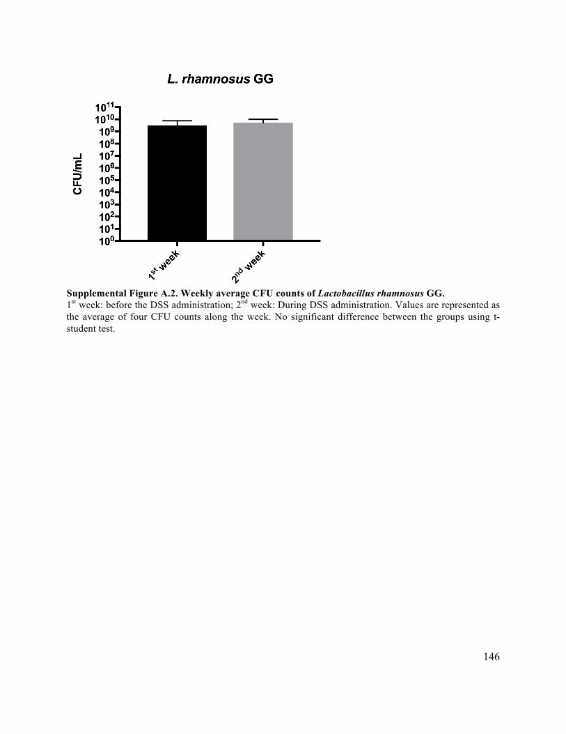
146
Supplemental Figure A.2. Weekly average CFU counts of Lactobacillus rhamnosus GG. 1st week: before the DSS administration; 2nd week: During DSS administration. Values are represented as the average of four CFU counts along the week. No significant difference between the groups using t-student test.
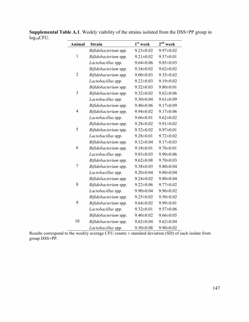
147
Supplemental Table A.1. Weekly viability of the strains isolated from the DSS+PP group in log10CFU.
Animal Strain 1st week 2nd week
1 Bifidobacterium spp. 9.23±0.02 9.97±0.02 Bifidobacterium spp. 9.21±0.02 9.57±0.01 Lactobacillus spp. 9.64±0.06 9.85±0.03
2 Bifidobacterium spp. 9.34±0.02 9.62±0.02 Bifidobacterium spp. 9.00±0.03 9.35±0.02 Lactobacillus spp. 9.22±0.03 9.19±0.02
3 Bifidobacterium spp. 9.52±0.03 9.80±0.01 Bifidobacterium spp. 9.32±0.02 9.62±0.06 Lactobacillus spp. 9.30±0.04 9.61±0.09
4 Bifidobacterium spp. 9.40±0.06 9.17±0.09 Bifidobacterium spp. 9.94±0.02 9.17±0.06 Lactobacillus spp. 9.66±0.01 9.62±0.02
5 Bifidobacterium spp. 9.28±0.02 9.91±0.02 Bifidobacterium spp. 9.52±0.02 9.97±0.01 Lactobacillus spp. 9.28±0.01 9.72±0.02
6 Bifidobacterium spp. 9.12±0.04 9.17±0.03 Bifidobacterium spp. 9.18±0.01 9.76±0.01 Lactobacillus spp. 9.83±0.03 9.90±0.06
7 Bifidobacterium spp. 9.62±0.08 9.70±0.03 Bifidobacterium spp. 9.38±0.03 9.80±0.04 Lactobacillus spp. 9.20±0.04 9.80±0.04
8 Bifidobacterium spp. 9.24±0.02 9.80±0.04 Bifidobacterium spp. 9.22±0.06 9.77±0.02 Lactobacillus spp. 9.90±0.04 9.96±0.02
9 Bifidobacterium spp. 9.25±0.02 9.50±0.02 Bifidobacterium spp. 9.64±0.02 9.99±0.01 Lactobacillus spp. 9.32±0.01 9.57±0.06
10 Bifidobacterium spp. 9.40±0.02 9.66±0.05 Bifidobacterium spp. 9.62±0.04 9.62±0.04 Lactobacillus spp. 9.50±0.08 9.90±0.02
Results correspond to the weekly average CFU counts ± standard deviation (SD) of each isolate from group DSS+PP.