diagnostic - myobase
TRANSCRIPT
6- Qool3fi3)
Diagnostic
Criteria
for Neuromuscular
Disorders
2nd Edition
Edited by
Alan EH Emery
Research Director, ENMC
Royal Society of Medicine Press. London
European Neuromuscular Centre, Baarn, The Netherlands
RFM
r‘ .: _ : m. ~.;» .... ‘7; NCO Dana-(3251711i:|f..‘.‘..\!l
Diagnostic
Criteria
for Neuromuscular
Disorders
2nd Edition
Edited by Alan EH Emery
Research Director, ENMC
Royal Society of Medicine Press, London
European Neuromuscular Centre, Baarn, The Netherlands
1997
Diagnostic
Criteria for
Neuromuscular
Disorders
The primary aim of ENMC is to facilitate and co-ordinate research
into the cause, prevention and treatment of neuromuscular
disorders. But such research depends primarily on a precise
diagnosis. For this reason priority has been given to establishing
agreed DIAGNOSTIC CRITERIA, based on both clinical and laboratory
data, for each disorder or group of disorders. These are now
presented in the hope that this information will be useful to medical
scientists engaged in research in this field.
© 1997 Royal Society of Medicine Press Limited
1 Wimpole Street, London W1M 8AE, UK
16 East 69 Street, New York, NY 10021, USA
Apart from any fair dealing for the purposes of research or private
study, criticism or review, as permitted under the UK Copyright,
Designs and Patents Act, 1988, no part of this publication may be
reproduced, stored or transmitted, in any form or by any means,
without the prior permission in writing of the publishers or in the case
of reprographic reproduction in accordance with the terms of licenses
issued by the Copyright Licensing Agency in the UK, or in accordance
with the terms of licenses issued by the appropriate Reproduction
Rights Organization outside the UK. Enquiries concerning reproduction
outside the terms stated here should be sent to the publishers at the
UK address printed on this page.
British Library Cataloguing in Publication Data
A catalogue record for this book is available from the British Library
ISBN 1 85315 301 X
Design by Nutshell, Newcastle upon Tyne
Phototypeset by Dobbie Typesetting Limited, Tavistock, Devon
Printed in Great Britain by Ebenezer Baylis, The Trinity Press, Worcester
CONTENTS
Acknowledgements
Introduction
Diagnostic criteria
aN
\l
10
13
14
15
16
Duchenne and Becker muscular dystrophies
E Bakker, FGI Jennekens, M de Visser, AR Wintzen
Emery-Dreifuss muscular dystrophy
JRW Yates
Facioscapulohumeral muscular dystrophy
GW Padberg, PW Lunt, M Koch. M Fardeau
The limb-girdle muscular dystrophies
KMD Bushby
Congenital muscular dystrophies
V Dubowitz
Myotonic dystrophy (Steinert’s disease)
HG Brunner, FGI Jennekens, HJM Smeets, M de Visser, AR Wintzen
Non—dystrophic myotonias and periodic paralyses
F Lehmann-Horn, R Riidel
Spinal muscular atrophy
TL Munsat, KE Davies
Familial amyotrophie lateral sclerosis
M Swash, CED Shaw, PN Leigh
Hereditary motor and sensory neuropathy or Charcot—Marie-Tooth
disease types 1A and B
M de Visser, C van Broeckhoven. E Nelis
Chronic inflammatory neuropathies
H Franssen, M Vermeulen, FGI Jennekens
Distal myopathies
H Somer
Myotubular/eentronuclear myopathy
C Wallgren-Pettersson
Nemaline myopathy
C Wallgren—Pettersson
Mini core disease and central core disease
LT Middleton, H Moser
Desminopathies
HH Goebel, M Fardeau
17
23
27
31
37
43
49
53
61
65
69
73
75
iii
17 Inclusion body myositis 81
JJ Verschuurcn, UA Badrising, AR Wintzen, BGM van Engelcn,
H van dcr Hoevcn, J Hoogendijk
18 Mitochondrial myopathies 85
L Bindoff, G Brown, J Poulton
19 Congenital myasthenic syndromes 91
LT Middleton
20 Post—polio muscle dysfunction 99
K Borg, J Borg, E Stélberg
Index 101
ACKNOWLEDGEMENTS
These diagnostic criteria for various neuromuscular disorders have, in
most cases, been generated by European Neuromuscular Centre
(ENMC) Workshops which have been generously supported by the
muscular dystrophy associations of France (AFM), Britain (MDG), the
Netherlands (VSN), Italy (Telethon 8t UILDM), Germany (DGM),
Switzerland and Denmark as well as the European Union.
We are very grateful to the editor of Neuromuscular Disorders
(Professor Victor Dubowitz) and to Pergamon Press for permission to
reproduce various reports of diagnostic criteria which have been
previously published in that journal.
I am personally grateful for the support I have had from Mr Michael
Rutgers and the Executive Committee of ENMC (Chairman: Mr Fergus
Logan), and Mr Howard Croft and Ms Tricia Dixon of RSM Press.
Finally I am especially grateful to Ms Janine de Vries for her
exceptional secretarial assistance.
INTRODUCTION
The European Neuromuscular Centre (ENMC) was established some
seven years ago with the specific aims of encouraging and facilitating
collaborative research into neuromuscular disorders. The majority are
genetic and attention has focused on locating, isolating, and
characterizing genes for specific disorders, encouraging the sharing
and exchange of DNA samples between research groups, and the
storing and banking of material. Agreed protocols for assessing the
effects of any future proposed treatment are now also being drawn up.
But an essential prerequisite of all such work is a precise diagnosis in
each case and family being studied. For this reason, priority has been
given to establishing diagnostic criteria for these disorders.
This has been achieved through Workshops, nearly 50 of which have
been held so far, each attended by experts in a particular field. To date
over 600 medical scientists have attended these Workshops mainly
from Europe, but also from countries further afield such as the United
States, Canada, Australia, Japan, Brazil, Tunisia, Israel and Saudi
Arabia. Many of the diagnostic criteria presented in this book have
been drawn up by Chairpersons of these Workshops, and have
subsequently been published in Neuromuscular Disorders where fiill
lists of the participants will be found. With editing, updating and with
some additions these various criteria, both clinical and laboratory, are
now reproduced here along with a few selected pertinent, but not
exhaustive, references.
These criteria are not meant to be definitive but are presented for
discussion and it is hoped they will be found useful to both clinicians
and scientists engaged in research in this field.
Alan EH Emery
Research Director, ENMC
1997
vii
Duchenne and
Becker Muscular
Dystrophies
E Bakker Institute for Anthropogenetics, University of Leiden,
Leiden, The Netherlands
FGI Jennekens Dutch Neuromuscular Research Support Centre,
Baarn, The Netherlands
M de Visser Dept ofNeurology, Academic Medical Centre,
Amsterdam, The Netherlands
AR Wintzen Dept ofNeurology, University Hospital Leiden, Leiden,
The Netherlands
> Diagnostic Criteria
Here are presented the diagnostic criteria for Duchenne muscular dystrophy (DMD)
and Becker muscular dystrophy (BMD).
b Duchenne muscular dystrophy
Elements
1 Symptoms are present before the age of 5 years.
2 Clinical signs comprise progressive symmetrical muscular weakness; proximal
limb muscles more than distal muscles; initially only lower limb muscles. Calf
hypertrophy is often present.
3 Exclusions: fasciculations, loss of sensory modalities.
4 Loss of unassisted ambulation before the age of 13 years.
5 There is at least a 10—fold increase of serum creatinine kinase (SCK) activity (in
relation to age and mobility).
6 Muscle biopsy: abnormal variation in diameter ofthe muscle fibres (atrophic and
hypertrophic fibres), (foci of) necrotic and regenerative fibres, hyalin fibres,
increase of endomysial connective and fat tissue.
7 Muscle biopsy: almost no dystrophin demonstrable, except for an occasional
muscle fibre (less than 5% of fibres).
8 DNA: Duchenne-type mutation within the dystrophin gene, identical haplotype,
involving closely linked markers, as in previous cases in the family.
9 Positive family history, compatible with Xolinked recessive inheritance.
BAKKER, JENNEKENS, DE VISSER, WINTZEN
Assessment
The diagnosis is definite when:
A The first case in a family:
B
a age <5 years: (2), 3, 5, 6, 7, (8) all present
b age 5—12 years: 1, 2, 3, 4, 5 (at least once), 6, 7, (8) all present
c age >12 years: (1), 2, 3, 4, 5 (at least once), 8, (or 6 and 7) all present.
*Another case in the family (according to element 9) complies with the criteria
under A:
a age <5 years: 5 and 9 present
b age 5—12 years: 1, 2, 3, 5 (at least once) all present
c age > 12 years: (1), 2, 3, 4, 5 (at least once) all present.
The diagnosis is possible when:
a age <5 years: (2), 3, 5, 6, all present
b age 5-12 years: 1, 2, 3, (4), 5 (at least once), 6, all present.
b Becker muscular dystrophy
L0
Elements
Clinical signs comprise progressive symmetrical muscular weakness and
atrophy: proximal limb muscles more than distal muscles: initially only lower
limb muscles. Calf hypertrophy is often present. Weakness of quadriceps femoris
may be the only manifestation for a long time. Some patients have cramps that
are mostly induced by activity. Contractures of the elbow fiexors occur late in
the course of the disease. Becker-type dystrophy may present with myalgia and
cramps, exercise intolerance and myoglobinuria, asymptomatic hyperCKaernia,
cardiomyopathy1 or cognitive dysfunctionz.
Exclusions: fasciculations, loss of sensory modalities.
No wheelchair dependency before 16th birthday.
There is a more than 5—fold increase of SCK activity (in relation to age and
mobility).
Electromyography: short duration, low amplitude, polyphasic action potentials,
fibrillations and positive waves. Normal motor and sensory nerve conduction
velocities.
Muscle biopsy: abnormal variation in diameter of the muscle fibres (dissemi—
nated or small groups of atrophic and hypertrophic fibres), (foci of) regenerative
fibres, mostly disseminated necrotic fibres. Dependent on stage and course ofthe
disease, there may be a minor degree of grouping of histochemical fibre types
and increase of connective and fat tissue.
Muscle biopsy: dystrophin of abnormal molecular weight and/or amount.
DNA: Becker—type mutation within the dystrophin gene, identical haplotype,
involving closely linked markers, as in previous case in the family
Positive family history, compatible with X—linked recessive inheritance.
‘When family history is positive (according to element 9) and B is not valid. one should rule as specified under
A.
DUCHENNE AND BECKER MUSCULAR DYSTROPHIES
Assessment
The diagnosis is definite when:
A The first case in a family:
(1), 2, 3, 4, 5 and either 8 or 6 and 7 all present.
B *Another case in the family (according to element 9) complies with the criteria
under A:
a the case is a first-degree relative: 4 (at least twice) present
b in other situations: (1), 2, 3, 4, 5 and either 8 or 6 and 7 all present.
The diagnosis is possible when:
(1], 2, (3), 4, 5 and 6 all present.
p DNA Studies
The dystrophin gene was cloned in 1986. Since then it has been found that, in 65%
of cases, gross rearrangements (deletions or duplications) have been detected within
the gene. Since 1985 carrier detection and prenatal diagnosis have been performed
using polymorphic markers within Xp21. The polymerase chain reaction (PCR)
technique has revolutionized deletion detection and haplotype analysis. Worth
mentioning are the simultaneous amplification of nine exons for deletion detection
(multiplex—PCRP, the high number of polymorphic CA—repeats for haplotype
analysis and the development of methods for point mutation detection. Both the
DNA (SSCP)4 and the mRNA (RT-PCR)5 methods are now being used. Recently the
protein truncation test (PTl')6 has been developed for visualizing premature
termination mutations. Although the latter techniques do not (yet) belong to the
standard diagnostic tests, some major laboratories might have facilities to apply
them.
a Standard diagnostic approach for DNA analysis of DMD
and BMD
When a DMD or BMD patient is available for diagnosis, a double PCR multiplex test
is performed on the patient‘s DNA. If a deletion is detected, quantitative Southern
blots of the patient‘s DNA and the DNA of all female relatives are prepared using at
least two restriction enzymes — Hind III and Pqu or BglII. This is to confirm the
detected deletion, to gain insight into the extent of the deletion and to detect carriers
of the deletion. A ‘loss of heterozygostity‘ test for a polymorphic loci within the
deletion is a good alternative and may be a better choice for laboratories that do not
feel comfortable with quantitative Southern blot analysis.
When no living patient is available for DNA analysis or no deletion is found using
the multiplex test, haplotype analysis is performed on the DNA of family members
using highly polymorphic CA-repeat markers of RFLPs.
If the DNA ofthe patient is available but the mutation is not detectable, a cross-over
event between flanking RFLPs may occasionally hamper the diagnosis.
“When family history is positive (according to element 9) and B is not valid, one should rule as specified under
A.
BAKKER, JENNEKENS, DE VISSER, WINTZEN
At present the dystrophin gene is known to harbour some 43 polymorphic sites
[GBD—Online Genome Database), ten of which are CA—repeats with a high
information content. Routinely used loci include 3’DYSI, STRSO, 5’DYSIII, 5’DYSII
and S’DYSI. If necessary, this panel can be extended to other loci, either within or
flanking the gene.
In the case of new mutations, the risk of germinal mosaicism7'El has to be taken into
account.
For a recent review see Ahn and Kunkelg.
References
1 Muntoni F, Mellis MA, Ganau A, et al. Transcription of the dystrophin gene in
normal tissues and in skeletal muscle of a family with X—linked dilated
cardiomyopathy. Am J Hum Genet 1995; 56: 151—6.
2 North KN, Miller G, Iannaccone ST, et al. Cognitive dysfunction as the major
presenting feature of Becker‘s muscular dystrophy. Neurology 1996; 46: 461—4.
3 Beggs AH, Koenig M, Boyce FM, Kunkel LM. Detection of 98% of DMD/BMD
gene deletions by polymerase chain reaction. Hum Genet 1990; 86: 45—48.
4 Kneppers ALJ, DeutZ-Terlouw PP, van Ommen GJB, Bakker E. Point—mutation
detection screening for Duchenne muscular dystrophy by SSCP-analysis of
multiplex pcr products by use ofthe PhadtSystem‘m. Am JHum Genet 1993; 53:
1493 (Abstract).
S Roberts RG. Barby TFM, Manners E, Bobrow M, Bentley DR. Direct detection of
dystrophin gene rearrangements by analysis of dystrophin mRNA in peripheral
blood lymphocytes. Am JHum Genet 1991; 49: 298—310.
6 Roest PAM, Roberts RG, Sugino S, van Ommen GJB, den Dunnen JT. Protein
truncation test (PTT) for rapid detection of translation—termination mutations.
Hum Mol Genet 1993; 2: 1719—21.
7 Baker E, Veenema H, den Dunnen JT, et al. Germinal mosaicism increases the
recurrence risk for 'new‘ Duchenne muscular dystrophy mutations. J Med Genet
1989; 26: 553—9.
8 Passos—Bueno MR, Bakker E, Kneppers ALJ, et al. Different mosaicism
frequencies for proximal and distal DMD mutations indicate difference in
etiology and recurrence risk. Am JHum Genet 1992; 51: 1150—5.
9 Ahn AH, Kunkel LM. The structural and functional diversity of dystrophin. Nat
Genet 1993; 3: 283—91.
This is based on a report originally published in Neuromuscul Disord 1991; 1(6):
389—91, with permission from Pergamon Press Ltd, Headington Hill Hall, Oxford
OX3 OBW, UK.
2
Emery—Dreifuss
Muscular Dystrophy
JRW Yates Dept of Pathology, University of Cambridge and
Dept of Medical Genetics, Addenbrooke’s Hospital,
Cambridge, UK
At the 1991 ENMC Workshop on Emery—Dreifuss muscular dystrophy stringent
diagnostic criteria for X—linked EMD were agreed for use in gene mapping studies.
These provide a useful guide for general clinical practice. Similar diagnostic criteria
would apply to the rarer autosomal dominant form of EMD.
p Diagnostic Criteria
Establishing unequivocal X—linked inheritance in Emery—Dreifuss muscular
dystrophy (EMD) requires a minimum of two affected males with obligate
transmission ofthe gene through a female who is asymptomatic or who has cardiac
conduction defects or evidence of cardiomyopathy. The diagnosis of EMD in the
family can be achieved by having a single case with all the typical features or
several cases who between them have the typical features. There is then the separate
task of classifying other males as affected or unaffected. Females may occasionally
show sufficient clinical manifestations to be confidently classified as carriers, but
the status of most at—risk females will be uncertain.
In classifying these criteria, inclusion criteria are indicated with an ‘I‘, exclusion
criteria with an ‘E‘ and comments by ‘C‘.
5» Requirements for a firm diagnosis of X-linked EMD
in a family
Summary
The features which establish the diagnosis of X—linked EMD in a family are the
presence of all the following (but not necessarily in a single patient):
1 Early contractures of the Achilles tendons, elbows and spine.
2 Slowly progressive muscle wasting and weakness with a predominantly humeral
(upper arm) and peroneal (lower leg) distribution, bilateral and approximately
symmetrical.
3 Cardiac conduction defect and/or other evidence of cardiomyopathy.
4 Muscle biopsy showing myopathic features or overt muscular dystrophy.
5 Pedigree consistent with unequivocal X-linked inheritance.
YATES
Age at onset
C Usually childhood. Onset after the age of 20 years is rare.
Early contractures
I Contractures usually develop before there is any significant weakness. These
involve the elbows which result in the arms being carried in a flexed position;
the Achilles tendons, so that the patient walks on his toes; and the spine,
resulting in limitation of flexion, particularly of the neck.
C There may be extension contractures ofthe wrist and/or flexion contractures of
the fingers.
Muscle wasting and weakness
I Muscle wasting and weakness with a predominantly humeral (upper arm) and
peroneal (lower leg) distribution.
I Bilateral and approximately symmetrical. Later weakness of the shoulder, pelvic
girdle and thigh muscles may develop.
C Some patients may have facial weakness. There may be wasting/weakness of
stemomastoids.
Muscle hypertrophy
C There is usually wasting of the calf muscles.
E Marked calf hypertrophy.
Coarse
C There is progression of the disease, usually slow.
Cardiac involvement
I Cardiac conduction defect (e.g. bradycardia, extrasystoles, atrioventricular block,
right bundle branch block) and/or other evidence of cardiomyopathy (e.g.
cardiomegaly, impaired left ventricular function). Such defects may only be
evident on 24-hour ECG monitoring. Almost always present by the age of 30.
Evidence ofX—linked inheritance
I Pedigree consistent with unequivocal X-linked inheritance, i.e. comprising at
least two affected males and obligate transmission of the gene through a female
who is asymptomatic or has cardiac conduction defects and/0r evidence of
cardiomyopathy.
C Two affected males with the same mother are not sufficient evidence ofX—linked
inheritance and could result from germline mosaicism. If the mother manifests
features of EMD the family could be autosomal dominant.
Intellect
E Severe mental retardation excludes the diagnosis.
Serum creatine kinase
C Usually moderately elevated but can be normal.
Electromyography {EMG}
C Myopathic and/or neurogenic and does not contribute to diagnosis.
EMERY<DREIFUSS MUSCULAR DYSTROPHY
Muscle biopsy
1 Myopathic or dystrophic features ofvariable degree. Some cases may have focal
atrophic fibres resembling ‘denervation'.
C Dystrophin is normal.
DNA analysis
C In atypical cases deletions of the BMD/DMD gene should be excluded.
p Minimum requirements for designating a male subject as
affected in a family with established X—linked EMD
I Any of the inclusion features detailed in ‘Requirements for a firm diagnosis’ p5—7.
C An elevated serum creatine kinase alone is suggestive but not conclusive
evidence of a male being affected.
C Abnormalities on ultrasound or CT imaging of muscles may be useful in
confirming that a subject is affected.
C 24—hour ECG monitoring and echocardiography may be useful in confirming
that a subject is affected.
5» Requirements for designating a male subject as unaffected
Aged 20 years or older.
Normal serum creatine kinase.
No clinical evidence of cardiomyopathy.
Examination by a clinician familiar with the disease shows no evidence of EMD.y—rr—4>—«>—4
b» Requirements for designating a female subject as a
carrier in a family with established X—linked EMD
I Any of the inclusion features detailed in ‘Requirements for a firm diagnosis'
p5—7.
C An elevated serum creatine kinase alone is suggestive but not conclusive
evidence of carrier status.
p DNA Studies
Molecular genetic research has focused on the X-linked form of EMD. The
chromosomal location ofthe rarer autosomal dominant form ofthe disorder has not
been determined. Genetic linkage studies mapped X-linked EMD to band Xq28 at
the tip of the long arm of the X chromosome1 and led to the positional cloning of
the gene by Bione et al (1994)? The EMD gene codes for a novel 254 amino acid
serine-n'ch protein called emerinz. The gene comprises six small exons spanning
2 kb of genomic DNA3. To date 26 different mutations have been described in 28
familiesz‘7. Most are base substitutions, small deletions or insertions and would
result in a truncated or absent protein. Emerin is ubiquitously expressed. In cardiac
and skeletal muscle it is localized to the nuclear membrane6 and has been shown to
be absent in patients with EMD. This should provide the means for confirmation of
the diagnosis by immunohistochemistry or perhaps Western blotting on blood
leucocytes in suspected cases.
YATES
References
1 Yates JRW, Warner JP, Smith JA, et al. Emery—Dreifuss muscular dystrophy:
linkage to markers in distal Xq28. JMed Genet 1993; 30: 108—1 1.
2 Bione S, Maestrini E, Rivella S, et al. Identification of a novel X—linked gene
responsible for Emery—Dreifuss muscular dystrophy. Nat Genet 1994; 8: 323—7.
3 Bione S, Small K, Aksmanovic VMA, et al. Identification of new mutations in the
Emery—Dreifuss muscular dystrophy gene and evidence for genetic heterogeneity
of the disease. Hum Mol Genet 1995; 4: 1859—63.
4 Klauck SM, Wilgenbus P, Yates JRW, et al. Identification of novel mutations in
three families with Emery—Dreifuss muscular dystrophy. Hum Mol Genet 1995;
4: 1853—7.
5 Nigro V, Bruni P, Ciccodicola A, et al. SSCP detection of novel mutations in
patients with Emery—Dreifuss muscular dystrophy: definition of a small
C—terminal region required for emerin function. Hum Mol Gen 1995; 4: 2003—
2004.
6 Nagano A, Koga R, Ogawa M, et al. Emerin deficiency at the nuclear membrane
in patients with Emery—Dreifuss muscular dystrophy. Nat Genet 1996; 12: 254—
9.
7 Yates JRW, Aksmanovic VMA, McMahon R, et al. Mutation analysis in Emery—
Dreifuss muscular dystrophy. Eur J Hum Genet 1996; 4 suppl 1: 62.
This is partly based on a report originally published in Neuromuscul Disord 1991;
1(6): 393—6, with permission from Pergamon Press Ltd, Headington Hill Hall,
Oxford 0X3 OBW, UK.
Facioscapulohumeral
Muscular Dystrophy
GW Padberg Dept of Neurology, University Hospital, Nijmegen,
The Netherlands
PW Lunt Bristol Royal Hospitalfor Sick Children, St. Michael’s
Hill, Bristol, UK
M Koch Klinikum der Philipps—Universitat Marburg, Marburg,
Germany
M Fardeau Institut National de la Santé et de la Recherche
Médicale, Paris, France
> Diagnostic Criteria
There are four main criteria which define facioscapulohumeral muscular dystrophy
(FSHD) at the clinical level. However, it is likely that the definitive diagnostic test
will be at the DNA level; the specificity and sensitivity of a deleted 4q35 DNA
fragment at D4F 104 51 locus is currently being evaluated for this purpose. The four
clinical criteria are:
1 Onset of the disease in facial or shoulder girdle muscles; sparing of the extra—
ocular, pharyngeal and lingual muscles and the myocardium.
2 Facial weakness in more than 50% of the affected family members.
Autosomal dominant inheritance in familial cases.
4 Evidence of myopathic disease in electromyography (EMG) and muscle biopsy in at
least one affected member without biopsy features specific to alternative diagnoses.
w
The more detailed clinical criteria, aimed at a standardized clinical diagnosis, are
described below, and offer guidance for genetic studies. Depending upon the results
of molecular genetic studies, and in particular the range of phenotype defined by
deletion at 4q35 recognized by D4F 10481, the criteria may need to be adjusted in
the future. Since FSHD is defined here firstly on clinical grounds, the following
definitions are understood:
Non—penetrance refers to an obligate gene carrier without symptoms (complaints or
subjective findings) or signs [objective phenomena) relating to the disease.
Presymptomatic indicates that a person has no complaints (symptoms) related to
the disease but has muscle atrophy and weakness demonstrable by physical
examination. (These cases are sometimes called ‘paucisymptomatic' in those
languages that do not separate the terms ‘symptoms' and ‘signs‘ in a well defined
manner. Other terms are ‘abortive cases‘ and ‘minimally affected patients'.)
PADBERG, LUNT, KOCH, FARDEAU
Symptomatic refers to patients with complaints and objective findings related to the
weakness and muscle atrophy of FSHD.
> Clinical criteria“8
Onset
I Onset of the disease is in facial or shoulder girdle muscles. Presenting symptoms
usually relate to weakness or wasting of these muscles.
E Onset in pelvic girdle muscles suggests alternative diagnoses; although
subsequent pelvic girdle involvement is not uncommon in FSHD.
C Clinically recognizable age at onset is very variable; age at symptomatic
presentation is even more so. The mean age at recognizable onset (albeit
presymptomatic] is in the second decade. Onset before the age of 5 years,
although rare in families, is not uncommon in the more severe proven new
mutation cases, and does not exclude the diagnosis. Infantile or early childhood
onset requires facial weakness to be present, since a clinical diagnosis cannot
otherwise be reliably made.
Facial
I Facial weakness affecting eye closure (orbicularis oculi] and peri-oral muscles
(orbicularis oris) occurs in the vast majority of patients. In the absence of facial
weakness, a diagnosis of FSHD can be accepted only if the majority of affected
family members have facial weakness.
E Extra-ocular, masticatory, pharyngeal and lingual muscle weakness is not part of
the disease.
C Facial weakness may be very subtle and is sometimes noticeable by asymmetry
of facial expressions only. There is also some evidence that a dominant
scapulohumeral presentation without facial weakness may be due to the same
mutation mechanism at 4q35.
Shoulders
C The scapular fixators are the muscles most prominently involved. Also the
pectoralis major muscles will become affected early in most cases. The deltoid
muscles remain unaffected for a long period of time and often have a particular
pattern of atrophy, i.e. partial and proximal.
Asymmetry
I Asymmetry of involvement in the shoulder girdle muscle is the rule. usually
affecting the right side first.
C Symmetrical weakness and atrophy at presentation is unusual and necessitates
increased caution before accepting the diagnosis as FSHD. Asymmetrical
involvement of facial muscles occurs frequently.
C NMR, ultrascan or CT-scan may be of help to detect asymmetry of muscle
atrophy.
Progression
I Progression is inevitable, albeit at a rate which is highly variable and in some
cases virtually imperceptible.
E Regression of symptoms and signs does not occur and would exclude the
diagnosis.
FACIO S CAPULOHUMERAL DYSTROPHY
The rate of progression and severity level reached tend to correlate inversely
with age at onset.
Progression of the disease usually includes involvement of abdominal and foot
extensor muscles at an early stage; pelvic girdle weakness and upper arm
weakness may occur at any time after the onset of shoulder girdle weakness.
Neck extensor, intrinsic hand and triceps surae muscle weakness is uncommon
but can be observed occasionally within families and is not dependent on
advanced age or severe involvement.
Severity
At any age the disease has a wide range of severity. Five aspects of note are:
Overall, between 10—20% of cases have eventual requirement for a wheelchair.
Severity in recognized isolated new mutation cases tends to be greater than in
large families.
Presymptomatic cases occur at any age and appear to comprise approxi—
mately 30% of all cases in large families.
Once symptomatic, the disease is progressive in the majority of cases. The rate
of progression is variable, although faster rates tend to be seen with earlier ages
at onset. Rarely, there can be long periods of apparent arrest of progression.
There is broad correlation in 4q35 cases between greater clinical severity and
smaller residual DNA fragment size at D4F 104 S]; it is currently uncertain
whether this may also be influenced by possible generational anticipation.
There appears to be no difference in mean age at death between patients and
their non-affected sibs.
Contractures
Contractures and pseudohypertrophy of muscles may be present.
Severe and diffuse contractures exclude the diagnosis of FSHD.
Cardiac disease
Cardiomyopathy is not part of the disease. When present it suggests an
alternative diagnosis.
Hearing loss
Hearing loss is part of the disease; it starts with high tone perceptive deafness
and may progress to involve all frequencies. The severity of the hearing loss
varies between subjects at any age, but tends to be progressive. It is
recommended that the results of hearing assessments be documented for several
affected members in each family.
Retinal disease
A retinal vasculopathy with capillary telangiectasis, microaneurysms and
capillary closure has been reported in some members of some FSHD families. At
present it is unclear whether this is a specific association. It should not be used
for diagnostic purposes.
Mental retardation
A few cases have been reported with mental retardation. It is recommended that
investigation of any such case should include chromosome analysis, concen-
trating on the distal long arm of chromosome 4. However, no causally associated
ll
PADBERG, LUNT, KOCH, FARDEAU
cytogenetic abnormalities have yet been recorded, and haploinsuffiency of the
4q35 region does not seem to cause FSHD.
D Laboratory criteriazv9
C Serum creatine kinase (SCK) levels can be normal, but are often elevated, though
rarely exceed five times the upper limit of normal. Persistently high CK values
above this level warrant exclusion of other neuromuscular diagnoses.
C EMG often shows short duration, low amplitude polyphasic potentials. Some
neurogenic features such as high amplitude potentials and positive sharp waves
are present occasionally, but do not characterize individual families. Motor and
sensory nerve conduction velocities are normal.
Giant potentials are not a feature of the disease.
C Muscle biopsies may exhibit any of the standard myopathic criteria. In addition,
small angular fibres are not uncommon and moth-eaten fibres are frequently
found. An occasional small group of atrophic fibres may be observed, in which
case another biopsy in the same patient or an affected sib is desirable. Cellular
infiltrates are not uncommon in FSHD and can be extensive. Their significance is
unknown. In these cases, either an autosomal dominant pattern of inheritance or
a deleted DNA fragment at 4q35 is required to establish the diagnosis of FSHD.
m
a» Clinical inclusion and exclusion criteria within a family for
phenotypic—genotypic analysis
Within a FSHD family there may be some members who are difficult to score as
‘affected’ or ‘unaffected’, particularly if the significance of a clinical finding can be
disputed, or if there are other coincidental neurological abnormalities. Such
individuals should be excluded consistently from any linkage analysis. Validation of
suggested standard clinical criteria will only be possible once a diagnostic DNA test
is confirmed as having full specifity. Phenotypic—genotypic analysis or linkage tests
should be based on the following:
I Individuals who have been examined by a physician familiar with this disease
and classified as affected according to the above criteria.
I Clinically unaffected family members aged 20 years and over who have been
examined as above.
Unrelated spouses, whether or not examined.
Any subject whose clinical status remains in dispute.
Apparently unaffected individuals under the age of 20 years.
Any apparently unaffected individual with a CK level repeatedly above the
normal range in the absence of a proven alternative explanation for this.
mmmH
b» Recommended investigations in at least one member of
each family included in phenotypic-genotypic studies
The following are recommended investigations:
Fully documented history and clinical examination
Serum creatine kinase
EMG
Muscle biopsy from an affected muscle for routine analysis
12
FACIOSCAPULOHUMERAL DYSTROPHY
Audiometry
Lymphoblast cell line and/or high molecular weight DNA sample suitable for
pulsed field gel studies, and tested for persistence of DNA fragment of
size<40 kb at locus D4F 10451, following double digestion of DNA with
restriction enzymes EcoRI and Bln 1.
y DNA Studies“)—17
In 1990 the gene for FHSD was located on chromosome 4q35. Subsequent analysis
by the international FSHD consortium confirmed the linkage, 5 cM distal to the
linkage group D45171—F11—D43163—D43139. In the search for flanking markers 21
single copy probe was isolated from cosmid 13E. This probe. p13E—11(D4F104Sl),
recognizes a polymorphic system containing fragments ranging in size from
20 kb to 320 kb. These fragments derive from two non-allelic loci, both containing
arrays of a variable number (6—96) of tandem repeats of 3.3 kb monomeric unit size.
One locus is on 4q35 and contains fragments of at least 50 kb in unaffected
individuals; the location of the second locus is at 10q26, and its fragments can
be shorter than 50 kb. Use of a double digest with restriction enzyme Bln1 in
addition to EcoRl can be employed to capitalize on slight differences in DNA
sequence of the 3.3 kb repeat units at 10q26 and 4q35 in order to distinguish
between these. The potential of this technique as a specific diagnostic test for 4q35
FSHD is currently being evaluated.
In FSHD patients the 4q35-linked Eco RI fragment detected by P13E—11 is usually
shorter than 40 kb. These ‘shortened' fragments differ in size between families but are
constant within FSHD families. The significance of these fragments is underscored by
the demonstration of the de novo appearance of a shortened fragment in over 80% of
sporadic cases of FSHD, and by an overall broad correlation between age at first onset
and fragment size. The shortened EcoRl fragments detectable by P13E-11 in FSHD
patients, seem to be the result of deletions of an integral number of 3.3 kb repeated
units. The gene for FSHD could be contained within the repeated units, or it may be
that the repeated units are necessary for adequate expression or integrity of a gene
outside this region. Although reports of rare recombinants between the short fragment
and the disease in apparently affected subjects may in some cases be best explained by
misinterpretation of fragments that are derived from 10q26 rather than 4q35, other
recombinant cases, where the short fragment is definitely cosegregating with 4q35
markers, cannot yet be explained satisfactorily. Besides the possibility of coincidental
occurrence in the family of a clinical phenocopy of 4q35 FSHD, other potential
explanations include the hypothesis that the observed 3.3 kb unit deletion may be
exerting either a position effect or a premutation effect on a putative syntenic muscle—
expressed FSHD structural gene. Thus, although in most cases the shortened fragment
appears directly related to the disease, it is not yet certain if this always remains so.
Worldwide linkage analysis has demonstrated non-linkage to 4q35 in a few families,
demonstrating the genetic heterogeneity of FSHD. At this moment therefore,
presyrnptomatic diagnosis can be performed reliably only in large families, or in
families where a new mutation can be proved by detection of a new fragment in an
apparently isolated case which is not found in either parent. The double digest
l3
PADBERG, LUNT, KOCH, FARDEAU
technique may extend presymptomatic and prenatal diagnosis to smaller families,
and perhaps even to single cases, with or without a family history.
References
1
11
14
15
16
Sorrel-Dejerine Y, Fardeau M. Naissance et métamorphoses de la myopathie
atrophique progressive de Landouzy et Dejerine. Revue Neurologie (Paris) 1982;
138: 1041—51.
Munsat TL. Facioscapulohumeral muscular dystrophy and the scapulohumeral
syndrome. In: Engel AG, Banker BO, eds. Myology, McGraw—Hill: New York,
1986; 1251—66.
Brooke MM. A clinician‘s view of neuromuscular disease, 2nd Edn. Williams 81
Wilkins: Baltimore, 1986.
Jardine PE, Koch MD, Lunt PW, et al. De novo facioscapulohumeral muscular
dystrophy defined by DNA probe p13E-11 (D4F104Sl). Arch Dis Child 1994;
71: 221—7.
Jardine PE, Upadhyaya M, Maynard J, et al. A scapular onset muscular
dystrophy without facial involvement: possible allelism with facioscapulo-
humeral muscular dystrophy. Neuromuscul Disord 1994; 4: 477—82.
Lunt PW, Harper PS. Genetic counseling in facioscapulohumeral muscular
dystrophy. JMed Genet 1991; 28: 655—64.
Lunt PW, Jardine PE, Koch MC, et al. Correlation between fragment size at
DF4104Sl and age at onset or at wheelchair use, with a possible generational
effect, accounts for much phenotypic variation in 4q35—facioscapulohumeral
muscular dystrophy (FSHD). Hum Mol Genet 1995; 4: 951—8, 1243—4.
Tupler R, Berardinelli A, Barbierato L, et al. Monosomy of distal 4q does not
cause facioscapulohumeral muscular dystrophy. J Med Genet 1996; 33:
366—70.
Dubowitz V. Muscle biopsy, 2nd Edn, Bailli‘ere Tindall: London, 1985.
Wijmenga C, Padberg GW, Moerer P, et al. Mapping of facioscapulohumeral
muscular dystrophy gene to chromosome 4q35—qter by multipoint linkage
analysis and in situ hybridization. Genomics 1991; 9: 570—5.
Wijmenga C, Hewitt JE, Sandkuijl LA, et al. Chromosome 4q DNA
rearrangements associated with facioscapulohumeral muscular dystrophy, Nat
Genet 1992; 2: 26—30.
Deidda G, Cacurri S, Piazzo N, et al. Direct detection of 4q35 rearrangements
implicated in facioscapulohumeral muscular dystrophy (FSHD). JMed Genet
1996; 33: 361—5.
Weiffenbach B, Dubois .l, Storvick D, et al. Mapping the facioscapulohumeral
muscular dystrophy gene is complicated by chromosome 4q35 recombination
events. Nat Genet 1993; 4: 165—9.
Wijmenga C, Frants RR, Hewitt JE, et al. Molecular genetics of facioscapulo-
humeral muscular dystrophy. Neuromuscul Disord 1993; 3: 487—91.
Bakker E, Wijmenga C, Vossen RHAM, et al. The FSHD—linked locus D4F104Sl
(p13E—1 1) on 4q35 has a homologue on 10q.ter. Muscle Nerve 1995; SupplZ:
39—44.
Winokur ST, Bengtsson U, Feddersen J, et al. The DNA rearrangement
associated with facioscapulohumeral muscular dystrophy involves a hetero—
chromatin associated repetitive element: implications for a role of chromatin
structure in the pathogenesis of the disease. Chromosome Res; 2: 225—34.
FACIOSCAPULOHUMERAL DYSTROPHY
17 Gilbert JR, Stajich JM, Wall 5, et al. Evidence for heterogeneity in
facioseapulohumeral muscular dystrophy (FSHD). Am J Hum Genet 1993; 53:
401—408.
This is partly based on a report originally published in Neuromuscul Disord 1991;
1(4): 231—4 with permission from Pergamon Press Ltd, Headington Hill Hall, Oxford
0X3 OBW, UK.
15
The Limb-Girdle
Muscular
Dystrophies
KMD Bushby Dept ofHuman Genetics, University of
Newcastle upon Tyne, Newcastle upon Tyne, UK
> Diagnostic Criteria
The limb—girdle muscular dystrophies (LGMD) are a group of genetically determined
progressive disorders of muscle, in which the pelvic or shoulder girdle musculature
is predominantly or primarily involved. The term was suggested to recognize the
existence of cases which could not be definitively diagnosed as either X-linked
muscular dystrophy or facioscapulohumeral muscular dystrophy in the classifica—
tions of Stevenson in 1953] and Walton Et Nattrass in 19542. Since then, the
existence of the group as a separate entity has been questioned because of the
overlap of symptomatology with patients who can now be proved to have disorders
which are known to be clinically and genetically different. For example, patients
with Becker muscular dystrophy and manifesting carriers of dystrophin mutations
were frequently diagnosed as having ‘limb-girdle muscular dystrophy' before the
availability of direct genetic and dystrophin analysis for these conditionsl“.
Disorders such as spinal muscular atrophy and mitochondrial and metabolic
myopathies have also been the subject of diagnostic confusion, as all of these
conditions may present with weakness in a limb-girdle distribution.
Even where other diagnoses can be excluded, the LGMD group remains
heterogeneous. It is now known that the category of the limb—girdle muscular
dystrophies includes a number of separate and genetically distinct conditions. the
molecular basis of many of which can now be determined5'6. Establishing the
precise diagnosis in LGMD may require specialized protein and/or genetic
techniques. A number of clinical and general laboratory criteria, however, remain
appropriate in order to ensure that the correct group of patients is selected for more
specialized study. These clinical and general laboratory criteria therefore form the
first part of the diagnostic process.
> Clinical criteria
Onset
I In the original description of LGMD, onset of the disease was reported as
involving either the pelvic or shoulder girdle muscles or both simultaneously;
17
BUSHBY
the initial symptoms usually relating to weakness in one of these muscle groups.
Clinical information available so far from the genetically defined forms of LGMD
suggests that onset in these forms is most often in the pelvic girdle.
Onset of weakness in distal, facial or extra-ocular muscles should suggest
alternative diagnoses, though these muscle groups may be involved later in the
course of the disease.
Onset of the disease may be at any age. In recessive families onset beyond the
early twenties is rare, but in dominant cases later onset can be seen.
Progression
Progression of the weakness is inevitable but ranges from very fast to very slow.
Preliminary evidence from the forms of LGMD where the genes are known,
suggests that there is a correlation between mutation types and severity of
disease. Involvement of other systems is rare.
Mode of inheritance
LGMD may be inherited in an autosomal recessive or dominant fashion. Current
estimates suggest that approximately 10% of all patients with LGMD may have a
dominant mutation5. In the absence of a family history there are at present no
clear indications to distinguish the two modes of inheritance (although see
comment on serum creatine kinase below).
b General laboratory criteria
I SCK is always elevated in recessive cases (at least when the disease is active) and
may be used as a presymptomatic test in families where early elevation of SCK
has been documented. In some families moderate elevation of SCK has been
documented in asymptomatic cases with normal muscle biopsies, and it is
possible that in these families carriers show some elevation of SCK. In some
dominant families SCK may be normal, and is not normally greater than six
times normal7, while in autosomal recessively inherited cases SCK may be as
high as 200 times normal.
Investigations such as electromyography and muscle biopsy usually provide
evidence of non-specific myopathic or dystrophic changes. Muscle CT scanning
may also provide evidence of hypodensity in the involved muscles. This may be
useful in the differential diagnosis of LGMD and spinal muscular atrophy, as well
as determining the exact pattern of muscle involvement.
The diagnosis of LGMD is excluded by the finding of severely abnormal
dystrophin staining on muscle biopsy (providing there are adequate controls for
membrane integrity) or the finding of a dystrophin gene abnormality. In female
patients presenting as the first case in their family, chromosome analysis should
exclude the finding of an X-autosome translocation in association with
Duchenne muscular dystrophy. In families with more than one affected boy in a
sibship, examination with probes within the dystrophin gene can also be used to
exclude X-linkage. The finding of muscle biopsy features diagnostic of a
neuropathic process, inflammatory changes, metabolic or mitochondrial
abnormalities also exclude the diagnosis.
These exclusions are a vital part of the general diagnosis of LGMD.
18
THE LIMB-GIRDLE MUSCULAR DYSTROPHIES
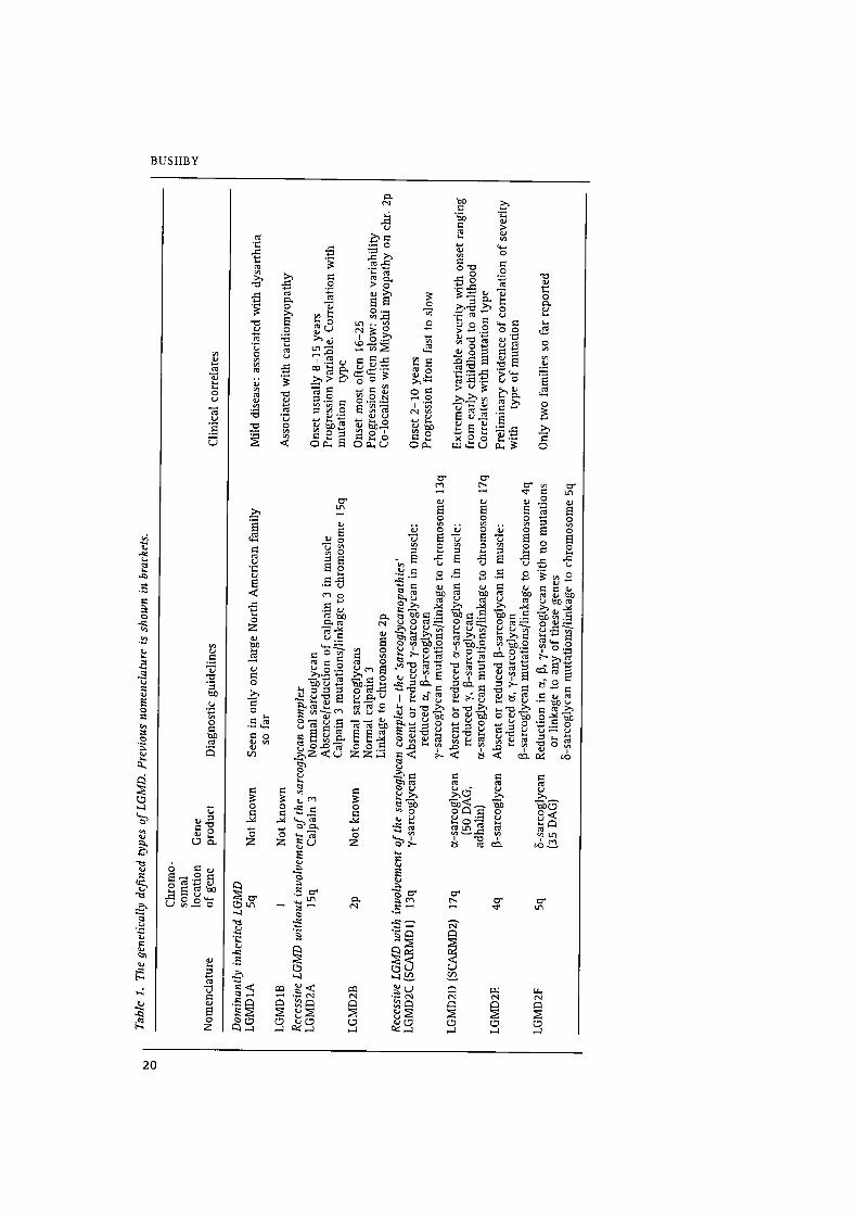
a Specific laboratory criteria
In patients and families fulfilling the basic diagnostic criteria for LGMD, more
specialized tests may now be successful in determining the molecular basis of the
disease. Based on the most recent findings about the molecular pathology of the
different limb-girdle muscular dystrophies a new classification of the group has
been suggested? This classification is shown in Table 1 (overleaf). The genetically
defined groups of autosomal recessive LGMD may be subdivided depending on
whether or not there is involvement of the sarcoglycan complexa‘“. In LGMDZA
and 2B, the components of the sarcoglycan complex are normal. LGMDZA is caused
by mutations in the muscle specific calcium—dependent protease calpain 3, thereby
being the first demonstration of deficiency of an enzyme in the products of a
muscular dystrophy‘z'”. The underlying molecular defect in LGMDZB is not yet
know1114'15. LGMDZB maps to the same markers on chromosome 2p13 as a gene for
a distal muscular dystrophy (Miyoshi myopathy) raising the intriguing possibility
that these two muscle diseases, with different patterns of muscle involvement, may
arise through the involvement of a single gene”.
Involvement of the or, B, y and 8 components of the sarcoglycan complex has been
demonstrated as directly responsible for four other types of LGMD (LGMDZD, 2E, 2C
and 2F respectively‘7‘21). Where a mutation is present in one of the sarcoglycan
genes it is most usually seen in muscle that the protein encoded by that gene is
completely absent or very severely reduced. A secondary reduction in the other
components of the sarcoglycan complex is also usually seen. In some ofthese cases
there may be a minor reduction in dsytrophin staining too. Therefore, using protein
analysis it is possible to direct mutation analyses in these disorders towards the gene
most likely to be involved. In large enough families, linkage analysis may be used to
direct mutation analysis towards the most likely gene. A combination of genetic and
protein testing is probably optimal in most situations to achieve full diagnosis.
It is likely that not all patients with LGMD will be accounted for by these various
genetic loci, and that further mechanisms underlying an LGMD phenotype will yet
be identified. However, the current classifications and diagnostic guidelines now
available do provide the basis for a rational approach to precise diagnosis in the
group.
> Conclusions
The current classification of LGMD relies on genetic and protein data. These
analyses are therefore essential to achieving a precise diagnosis in any patient
fulfilling the base clinical and laboratory criteria.
Current knowledge suggests that all of the various LGMD genes with the exception
of LGMDIA are represented world-wide. Further detailed epidemiological studies
are however necessary.
A spectrum of disease severity has been observed in association with most of the
genetically defined types. Preliminary indications are that the disease severity at
least partially relates to the type of mutation found.
20
Table
1.The
geneticallydefined
typesofLGMD.
Previousnomenclature
isshown
in
brackets.
Chromo—
somal
location
ofgene
Gene
Nomenclature
product
Diagnosticguidelines
Clinical
correlates
Dominantly
inheritedLGMD
LGMDIA
Sq
Notknown
LGMDIB
1Notknown
Seen
inonlyone
largeNorthAmerican
family
so
far
RecessiveLGMD
withoutinvolvementofthesarcoglycancomplex
LGMDZA
15q
Calpain
3
LGMDZB
2p
Notknown
Normalsarcoglycan
Absence/reductionofcalpain
3inmuscle
Calpain
3mutations/linkage
tochromosome
15q
Normal
sarcoglycans
Normal
calpain
3
Linkage
tochromosome
2p
RecessiveLGMD
withinvolvementofthesarcoglycancomplex—
the
‘sarcoglycanapathies’
LGMDZC(SCARMD
1)
13q
y-sarcoglycan
LGMDZD(SCARMDZ)
17q
a-sarcoglycan
(50DAG,
adhalin]
LGMDZE
4q
B-sarcoglycan
LGMDZF
5q
5-sarcoglycan
(35DAG)
Absent
orreducedy—sarcoglycan
inmuscle:
reduced
(1,B-sarcoglycan
y—sarcoglycanmutations/linkage
tochromosome
13q
Absentorreduced
ot—sarcoglycan
inmuscle:
reduced
y,B—sarcoglycan
a—sarcoglycanmutations/linkage
tochromosome
17q
Absent
orreducedB—sarcoglycan
inmuscle:
reduced
0!,y—sarcoglycan
B—sarcoglycanmutations/linkage
tochromosome4q
Reduction
in
a,
B,y—sarcoglycanwithnomutations
orlinkage
toanyofthesegenes
5—sarcoglycanmutations/linkage
tochromosomeSq
Mild
disease:associatedwith
dysarthria
Associatedwithcardiomyopathy
Onsetusually8—15
years
Progression
variable.
Correlationwith
mutation
type
Onsetmost
often16—25
Progression
oftenslow:some
variability
Co-localizeswithMiyoshimyopathyon
chr.2p
Onset2—10
years
Progressionfrom
fasttoslow
Extremelyvariableseveritywith
onsetranging
from
earlychildhood
toadulthood
Correlateswithmutationtype
Preliminaryevidenceofcorrelationofseverity
with
typeofmutation
Onlytwo
familiesso
farreported
BUSHBY
THE LIMB-GIRDLE MUSCULAR DYSTROPHIES
Clinical descriptions of the various genetic subtypes are beginning to emergezz'”. At
the present time, however, distinction between the different types of LGMD is not
possible on clinical criteria alone. Collection of reliable clinical data from large
groups of genetically defined patients is still urgently needed.
References
l
10
11
12
13
14
16
17
18
Stevenson AC. Muscular dystrophy in Northern Ireland. Ann Eugenics 1953;
18: 50-91.
Walton JN, Nattrass FJ. On the classification, natural history and treatment of
the myopathies. Brain 1954; 77: 169—231.
Norman A, Thomas N, Coackley J, et al. Distinction of Becker from limb-girdle
muscular dystrophy by means of dystrophin cDNA probes. Lancet 1989; i:
466—8.
Hoffman EP, Arahata K, Minetti C, et al. Dystrophinopathy in isolated cases of
myopathy in females. Neurology 1992; 42: 967—75.
Bushby KMD, Beckmann JS. Report of the 30th and 315t ENMC international
Workshop: the limb—girdle muscular dystrophies, and proposal for a new
nomenclature. Neuromuscul Disord 1995; 5: 337—44.
Beckmann JS, Bushby KMD. Advances in the molecular genetics of the limb—
girdle type of autosomal recessive progressive muscular dystrophy. Current
Opinions Neurol 1996; 9: 389—93.
Bushby K. Report of the 12th ENMC sponsored international Workshop the
‘limb-girdle‘ muscular dystrophies. Neuromuseul Disord 1992; 2: 3—5.
Campbell KP, Kahl SK. Association of dystrophin and an integral membrane
glycoprotein. Nature 1989; 338: 259—62.
Tinsley JM, Blake DJ, Zuellig A, et al. Increasing complexity of the dystrophin-
associated protein complex. Proc Natl Acad Sci {USA} 1994; 91: 8307—131.
Campbell KP. Three muscular dystrophies: Loss of Cytoskeleton—Extracellular
matrix linkage. Cell 1995; 80: 675—9.
Worton RG. Muscular dystrophies: diseases of the dystrophin—glycoprotein
complex. Science 1995; 270: 755—6.
Beckmann 13, Richard 1, Hillaire D, et al. A gene for limb—girdle muscular
dystrophy maps to chromosome 15 by linkage. CR Acad Sci Paris 1991; 312:
141—8.
Richard 1, Broux O, Allaman V, et al. Mutations in the proteolytic enzyme,
calpain 3, cause limb-girdle muscular dystrophy type 2A. Cell 1995; 81: 27—40.
Bashir R, Strachan T, Keers S, et al. A gene for autosomal recessive limb-girdle
muscular dystrophy maps to chromosome 2p. Hum Mol Genet 1994; 3: 455—7.
Bashir R, Keers S, Strachan T, et al. Genetic and physical mapping at the limb-
girdle muscular dystrophy locus (LGMDZB) on chromosome 2p. Genomics
1996; 33: 46—52.
Bejaoui K, Hirabayashi K, Hentati F, et al. Linkage of Miyoshi myopathy (distal
autosomal recessive muscular dystrophy) locus to chromosome 2p12-14.
Neurology 1995; 45: 768—72.
Roberds S, Letureq F, Allaman V, et al. Missense mutations in the adhalin gene
linked to autosomal recessive muscular dystrophy. Cell 1994; 78: 625—33.
Bonnemann CG, Modi R, Noguchi S, et al. B-sarcoglycan (A3b) mutations cause
autosomal recessive muscular dystrophy with loss of the sarcoglycan complex.
Nat Genet 1995; 11: 266—73.
21
BUSHBY
19
20
21
22
23
Lim LE, Duclos F, Bronx 0, et al. B-sarcoglycan: characterisation and role in
limb—girdle muscular dystrophy linked to 4q12. Nat Genet 1995; 11: 257—65.
Noguchi S, McNally EM, Ben Othmane K, et al. Mutations in the dystrophin-
associated protein y-sarcoglycan in chromosome 13 muscular dystrophy.
Science 1995. 270: 819—22.
Nigno V, de sa Moreira E, Piluso G, et al. Autosomal recessive limb-girdle
muscular dystrophy, LGMDZF, is caused by a mutation in the 5—sarcoglycan
gene. Nat Genet 1996; 14: 195—8.
Fardeau M, Hillaire D, Mignard C, et al. Juvenile limb—girdle muscular
dystrophy. Clinical, histopathological and genetic data on a small community
living in the Reunion Island. Brain 1996; 119: 295—308.
Mahjneh I, Passos-Bueno MR, Zatz M, et al. The phenotype of chromosome 2p-
linked limb-girdle muscular dystrophy (LGMDZB). Neuromuscul Disord (in
press).
This is partly based on reports originally published in Neuromuscul Disard 1992;
2(1): 35 and 1995; 5(4): 337—43, with permission from Pergamon Press Ltd,
Headington Hill Hall, Oxford OX3 OBW, UK.
22
Congenital Muscular
Dystrophies
V Dubowitz Dept of Paediatrics and Neonatal Medicine,
Royal Postgraduate Medical School, Hammersmith Hospital,
London, UK
The diagnostic criteria for congenital muscular dystrophy (CMD) were agreed at the
first Workshop on CMD in May, 1993‘. The main aims at that meeting were to define
the various recognizable syndromes and to establish collaborative studies for gene
linkage and further molecular genetic studies. These clinical criteria remain relevant.
p Diagnostic Criteria
The term congenital muscular dystrophy has been used widely for a group of infants
presenting with muscle weakness at birth or certainly within the first few months of
life, in association with a dystrophic pattern on muscle biopsy. There is often an
associated hypotonia on clinical presentation but other cases may present with
arthrogryposis and associated contractures of variousjoints. The condition tends to
remain relatively static but some cases may show slow progression. Others, however,
may have actual functional improvement, pass various motor milestones and
achieve the ability to walk. There may be variable respiratory and swallowing
problems at the time of presentation and the associated diaphragmatic involvement
may lead to respiratory failure in later childhood or adolescence.
In recent years a number of syndromes of congenital muscular dystrophy in
association with central nervous system involvement have been reported.
The following clinical phenotypes can currently be defined:
> ‘Pure’ congenital muscular dystrophy
The main features are:
Muscle weakness with hypotonia or arthrogryposis.
Histological changes of a dystrophic nature, often with extensive connective
tissue or adipose proliferation, but no substantial evidence of necrosis or
regeneration.
Normal or moderately elevated SCK.
Intellect is usually normal.
Brain imaging may show a normal picture or evidence of changes in the white
matter on CT or magnetic resonance imaging.
23
DUBOWITZ
b Fukuyama—type congenital muscular dystrophy
In addition to muscle weakness and a dystrophic muscle biopsy, this form of
congenital muscular dystrophy is characterized by:
The consistent association of mental retardation which is often severe in degree
A consistently elevated SCK
Consistent structural changes in the brain at autopsy or on imaging
No consistent ocular involvement
Frequent association of seizures with the condition (about 40%)
Survival of most cases beyond infancy and childhood and into adolescence.
w Muscle—eye—brain disease
In addition to the muscle weakness and associated dystrophic changes in the muscle,
there is consistent ocular and central nervous system involvement. There is
associated mental retardation which is often severe.
The most consistent ocular abnormality is severe myopia but there may also be
strabismus, glaucoma, lens opacity, retinal atrophy and optic atrophy. Epilepsy is
also commonly associated and the EEG is always abnormal after the age of one year.
Hydrocephalus is present in the majority of cases. The SCK may be normal within
the first year, but is always elevated thereafter.
b Walker—Warburg syndrome
This syndrome is characterized by structural changes and associated mental
retardation in addition to the muscle weakness and dystrophic changes.
The consistent central nervous system abnormalities on imgaging are a type ll
lissencephaly, comprising variable gyral malformations, together with an abnor-
mally thick cortex and decreased interdigitations between the white matter and
cortex. There may also be other structural changes within the nervous system.
Ocular malformations are also common but are thought to be less severe and less
consistent than in muscle—eye—brain disease.
There is divergence of opinion as to whether the Walker—Warburg syndrome and
muscle—eye—brain disease constitute one entity with variable severity, or whether
they represent two separate entities in view of the more striking ocular involvement
in muscle—eye—brain disease. There is certainly some degree of overlap in the
structural changes within the central nervous system.
> DNA and Protein Studies
By the time of the second Workshop in April, 19942, the gene for Fukuyama CMD
had been located on chromosome 9q, and a deficiency of a protein, the laminin
alpha-2 chain of merosin, had been discovered in about 40% of the cases of
classical CMD. Further studies showed that this was indeed a primary deficiency,
and that these cases linked to the locus of the corresponding gene (LAMAZ) on
chromosome 6q. It also became clear once the classical cases were subdivided into
24
CONGENITAL MUSCULAR DYSTROPHIES
merosin deficient and merosin positive, that the merosin deficient group comprised
a much more severe phenotype, usually with inability to walk unaided, in contrast
to the merosin-positive group, most of whom achieved independent walking. In
addition the cases that had shown increased signal in the white matter in T2-
weighted magnetic resonance imaging of the brain were also consistently merosin
deficient.
At the recent third Workshop in March, 19963, mutations in the LAMA2 gene were
reported in a small number of merosin—deficient cases to date. With the more routine
screening for merosin in all dystrophic biopsies, without deficiency of dystrophin or
the sarcoglycans, a number of atypical cases of CMD with later onset or milder
phenotype have also been recognized.
An important new development, also reported at the third Workshop, was the
discovery of a deficit in the protein alpha-actinin 3, in a small number of cases of
merosin-positive CMD. It is not yet known whether this is a primary or a
secondary deficit, and what proportion of cases of merosin—positive CMD it is
associated with.
With regard to the Walker—Warburg syndrome and muscle—eye—brain disease, it has
not yet been possible to establish a gene location, and whether they constitute two
separate entities or not, mainly due to the relative rarity of these two syndromes and
availability of informative families. The data suggest that they are not linked to
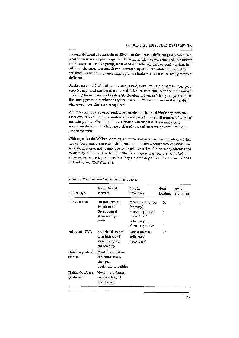
either chromosome St] or 9q, so that they are probably distinct from classical CMD
and Fukuyama CMD (Table 1).
TabIe 1. The congenital muscular dystrophies.
Main clinical Protein Gene Gene
Clinical type features deficiency location mutations
Classical CMD No intellectual Merosin-deficiency 6q +
impairment (primary)
No structural Merosin-positive .7
abnormality in oc—actinin 3
brain deficiency
Merosin-positive ?
Fukuyama CMD Associated mental Partial merosin 9q
retardation and deficiency
structural brain (secondary)
abnormality
Muscle—eye—brain Mental retardation
disease Structural brain
changes
Ocular abnormalities
Walker—Warburg Mental retardation
syndrome Lissencephaly II
Eye changes
25
DUBOWITZ
References
1 Dubowitz V. Workshop report: 22nd ENMC sponsored Workshop on congenital
muscular dystrophy. Neuromuscul Disord 1994; 4: 75—81.
2 Dubowitz V, Fardeau M. Workshop report: 27th ENMC sponsored international
Workshop: congenital muscular dystrophy. Neuromuscul Disord 1995; 5(3):
253—8.
3 Dubowitz V. Workshop report: 4lst ENMC sponsored international Workshop:
congenital muscular dystrophy. Neuromuscul Disord 1996; 6(4): 295—301.
This is based on reports originally published in Neuromuscul Disord 1994; 4(1):
75—81, 1995; 5(3): 253—8 and 1996; 6(4): 295—301 with permission from
Pergamon Press Ltd, Headington Hill Hall, Oxford
26
6
Myotonic Dystrophy
(Steinert's Disease)
HG Brunner Dept ofAnthropogenetics, University Hospital
Nijmegen, Nijrnegen, The Netherlands
FGI Jennekens Dutch Neuromuscular Research Support Centre,
Baarn, The Netherlands
HJM Smeets Dept ofAnthropogenetics, University Hospital
Nijmegen, Nijmegen, The Netherlands
M de Visser Dept of Neurology, Academic Medical Centre,
Amsterdam, The Netherlands
AR Wintzen Dept ofNeurology, University Hospital, Leiden,
The Netherlands
> Diagnostic Criteria
With regard to the diagnostic criteria for myotonic dystrophy, the clinical picture
depends on the age at onset.
a Congenital (1) and early childhood myotonic dystrophy (2), age <10 years.
b Juvenile/adult (classical) myotonic dystrophy, age 10—50 years.
c Minimal myotonic dystrophy, age > 50 years.
Elements
a1 Congenital myotonic dystrophy
1 Stillbirth or generalized severe muscular weakness (including the face) and
hypotonia with sucking. swallowing and sometimes respiratory insufficiency.
Absence of tendon reflexes. Club feet.
2 Symptoms of myotonic dystrophy (see b) in the mother.
3 Amplification (>45) of a trinucleotide repeat unit in the myotonic dystrophy
gene on chromosome 19.
a2 Early childhood myotonic dystrophy
1 Mental retardation.
2 Generalized weakness, especially of the face and distal limbs; myotonia starts
usually between the ages of 5 and 10 years.
3 Electroymyography‘: myotonic volleys in several muscles.
Symptoms of myotonic dystrophy in one of the parents.
'Electromyography: myotonic volleys ('dive bomber‘) resemble repetitive denervation potentials with
inconstant frequency 20-120 HZ, duration at least 0.55. Examination of mm. orbicularis oris, masseter, thenar,
tibialis anterior.
27
BRUNNER, JENNEKENS, SMEETS, DE VISSER, WINTZEN
5 Amplification (>45) of a trinucleotide repeat unit in the myotonic dystrophy
gene on chromosome 19.
b Juvenile/adult (classical) mytonic dystrophy
Myotonia 0f grip and/or percussion myotonia of thenar muscle.
2 Weakness of one or more of the following: m. orbicularis oculi., pharyngeal
muscles, distal limb muscles. Atrophy of masticatory muscles and/or distal limb
muscles may be obvious.
Cortical cataracti (slit lamp examination mandatory).
Electromyography‘: myotonic volleys in several muscles.
Positive family history compatible with autosomal dominant inheritance.
Amplification (>45) of a trinucleotide repeat unit in the myotonic dystrophy
gene on chromosome 19.
O‘lU‘lubLQ
Minimal myotonic dystrophy
Cortical cataracti Rarely neuromuscular symptoms (see b).
Electromyography': myotonic volleys in several muscles.
Positive family history compatible with autosomal dominent inheritance.
Amplification (>45) of a trinucleotide repeat unit in the myotonic dystrophy
gene on chromosome 19.
thNHO
Asymptomatic heterozygotes occur, even in old age.
Assessment
The diagnosis is definite as shown in Table 1.
Table 1. Assessment of myotonic dystrophy. (§ Whenfamily history is positive and B
is not valid, one should rule as under A.)
A First case in the family B§ There is a first—degree relative
who complies with the criteria
under A
a1 1, 2, (3) all present 1, 2 (3) all present
a2 (1), 2, (3), 4, (5) all present (1), 2, (3), 4, (5) all present
b One of 1—4, (5) and 6 1, 2
2
3
4
or 6 present
c >1 element 1
or 2
and 4 present or 4 present
ICataract should be cortical, assessed by experienced ophthalmologist with slit lamp and should not be used
as criterion if no first-degree family member is affected.
'Electromyography: myotonic volleys ('dive bomber') resemble repetitive denervation potentials with
inconstant frequency 20-120 Hx, duration at least 0.55. Examination of mm. orbicularis oris, masseter, thenar,
tibialis anterior.
28
MYOTONIC DYSTROPHY (STEINERT‘S DISEASE)
p DNA Studies
Myotonic dystrophy or dystrophia myotonica (BM) is caused by an increased
number of CTG trinucleotide repeats located in the 3’-untranslated region of the
putative DM gene. This gene has been termed DM—kinase (or myotonin-kinase)
because of similarities to a class of genes that encode serine/threonine kinases.
Molecular diagnosis of DM is possible by DNA analysis of various tissues, usually
blood cells, muscle chorionic villus or amnion cells. The DM mutation is detected
either by Southern blotting and hybridization with a DNA probe or by polymerase
chain reaction [PCR] of the relevant DNA fragment.
The number of CTG trinucleotides on normal chromosomes ranges from 3 to 30.
When the CTG repeat number exceeds 40, the DNA repeat seqence is unstable1 and
the number of repeats tends to increase on parent—to-child transmission. A decrease
in CTG number occurs more rarely. Unstable repeats of 40 to approximately 100
CTG trinucleotides are often associated with cataract but muscular symptoms are
very rare. Such repeats nearly always increase on transmission to the next
generation (so—called “anticipation") and this increase is most marked when
tramission is through a female. Larger repeats found in offspring may range from
less than 100 to several thousand CTG trinucleotides. The number of CTG
trinucleotides correlates broadly with age at onset and severity of symptoms.
However, genotype/phenotype correlates are currently too imprecise to allow
precise DNA—based prognosis.
For recent reviews see Harley et al1 and Harper and Riidelz.
References
1 Harley HG, Rundle SA, MacMillan JC, et al. Size of the unstable CTG repeat
sequence in relation to phenotype and parental transmission in myotonic
dystrophy. Am JHum Genet 1993; 52: 1164—74.
2 Harper PS, Riidel R. Myotonic dystrophy. In: Engel AG and Franzini-Armstrong
C (editors) Myology, basic and clinical. New York: McGraw—Hill Inc, 1994:
1 192—2 19.
This is based on a report originally published in Neuromuscul Disord 1991;
1(6):389—91 with permission from Pergamon Press Ltd, Headington Hill Hall,
Oxford OX3 OBW, UK
29
Non—dystrophic
Myotonias and
Periodic Paralyses
F Lehmann—Horn and R Riidel Dept ofPhysiology, University of
Ulm, Ulm, Germany
> Diagnostic Criteria
Since the genes and the gene products are known in the principal diseases of non—
dystrophic mytonias and periodic paralyses. and since an increasing number of
molecular biological laboratories have the relevant genetic markers available, an
exact diagnosis can be made by the identification of the mutation. At present, many
laboratories are engaged in correlating the clinical symptoms of their individual
families with the various mutations and, therefore, a precise statement of the clinical
diagnostic criteria remains useful”.
It is important to state that myotonia, i.e. muscle stiffness, is a symptom that can be
present in both muscle Cl“ and Na" channel diseases (and, of course, also in
myotonic dystrophy, proximal myotonic myopathy and Schwartz—Jampel syn—
drome). The myotonia is best assessed as myotonic runs in the electromyogram.
Diagnostic differentiation of the various diseases on the mere basis of these runs is
not dependable. Muscle biopsy is usually not helpful for establishing the diagnosis.
The class of Cl‘ channel diseases comprises dominant myotonia congenita
(Thomsen) and recessive generalized myotonia (Becker). The term myotonia
congenita should only be reserved for these C1" channel diseases.
The class of Na’“ channel diseases encompasses hyperkalaemic periodic paralysis
(HyperPP), normokalaemic periodic paralysis (NormoPP), paramyotonia congenita
(PC) and potassium-aggravated myotonia (PAM). Although the key symptoms,
namely attacks of muscle weakness and episodes of muscle stiffness, are known to
overlap to various degrees, it makes sense from a clinical point of view to maintain
the differentiation between HyperPP (identical with Gamstorp's adynamia episodica
hereditaria) and PC, because preventive measures are different for the two
syndromes. HyperPP also implies a possible prognosis of progressive permanent
weakness that is not a feature of PC. In potassium—aggravated myotonia the key
symptom is muscle stiffness that resembles the myotonia in myotonia congenita.
This myotonia may be mild (myotonia fluctuans), moderate or severe (myotonia
permanens]. Hypokalaemic periodic paralysis, the only disease in this group not
associated with myotonia, is a Ca” channel disease.
31
LEHMANN—HORN AND RUDEL
b Dominant myotonia congenita
The usual (but rare] form is Thomsen's disease2'3'4. There is also a form that is
distinguished by very mild myotonia (De Jong's myotonia levior). It is caused by an
allelic mutation in the muscle chloride channel gene5.
Family history
Autosomal dominant inheritance; 100% penetrance.
Age at onset
From birth to early childhood.
Clinical signs
Muscle stiffness, particularly after rest, muscle function improving with
continuing exercise (warm up). Myotonia fluctuates only slightly during
lifetime; there is no progression and muscle hypertrophy is frequent.
Although patients with myotonia congenita, when asked, often state that their
stiffness increases in the cold, this cannot be substantiated with objective
measurements of muscle relaxation times.
Clinical signs which must not be mistaken. There are cases of Na+ channel
disease having myotonia without any weakness (PAM). The myotonia may exist
without cooling. Before the advent of molecular biology, these cases were
misdiagnosed as forms of myotonia congenita.
b» Recessive generalized myotonia
Many loss—of-function mutations in the muscle chloride channel gene can cause the
same clinical picture4'6'7'3.
Family history
Autosomal recessive inheritance. Some of the heterozygous carriers show
myotonic runs in the EMG. Such cases must not be confused with dominant
myotonia, and sometimes molecular biology is required to differentiate this from
myotonic dystrophy.
Age at onset
Occasionally present in early childhood, usually the first decade of life, in some
cases not before the end of the second decade, and even progression of
symptoms into the third decade of life.
Clinical signs
Muscle stiffness, particularly after rest, muscle function improving with
continuing exercise (warm up). In many patients marked transient weakness
after rest which improves during several minutes of continued exercise.
Weakness is more pronounced in the upper extremities, stiffness is more
pronounced in the legs. In many cases hip and leg muscles are hypertrophied.
The signs are usually progressive for a few years after their first appearance and
then remain stable for the rest of life.
32
NON-DYSTROPHIC MYOTONIAS AND PERIODIC PARALYSES
Clinical signs which must not be mistaken. The well—known phenomenon of
anticipation in myotonic dystrophy may lead to a familial constellation suggesting
recessive inheritance and, as a consequence, may lead to the spurious diagnosis of
recessive generalized myotonia. On the other hand, misinterpretation of the
transient weakness may lead to the spurious diagnosis of myotonic dystrophy. In
older patients with recessive generalized myotonia muscle biopsies may show a
morphologic pattern that can be misdiagnosed as myotonic dystrophy.
D Paramyotonia congenita
The classical form was described by Eulenburg and independently by Rich. Several
mutations in the Na+ channel gene result in the classical clinical pictureg'lo.
Family history
Autosomal dominant inheritance; 1000/0 penetrance.
Age at onset
From birth.
Clinical signs
Muscle stiffness increasing with exercise (paradoxical myotonia). In many
families paramyotonia is dramatically increased when the muscles are exercised
in the cold. Recovery from weakness may last several hours.
Some families present consistently cold- and exercise-induced stiffness without
weakness. These are often misdiagnosed as having myotonia congenita.
Variability of signs. There are families where affected members present with the
classical symptoms of paramyotonia congenita and also often experience attacks
of hyperkalaemic paralysis. The presentation of both sets of symptoms in severe
form was termed paralysis periodica paramyotonica (PPP) by PE Becker;
however, a continuum seems to exist, with PPP families and families having
‘pure' paramyotonia congenita presenting the two extremes. The severity of
stiffness is not the same in all paramyotonia families.
Clinical signs which must not be mistaken. Permanent weakness is not observed
in paramyotonia congenita.
h Hyperkalaemic periodic paralysis
Several mutations in the Na+ channel gene may lead to the classical clinical picture.
Family history
Autosomal dominant inheritance; complete penetrance. but severity is very
variable.
Age at onset
Early childhood to second decade of life.
Clinical signs
Attacks of weakness, usually in the morning, lasting from 10min to 1 h or so.
very rarely up to 1—2 days. Some patients experience only a few attacks of
33
LEHMANN'HORN AND RUDEL
weakness in their lifetime, others have attacks of generalized weakness almost
every day. During the attacks, serum K+ is elevated to upper normal levels or
above. Myotonic stiffness is not observed. Rest after exercise, fasting, or oral
intake of K+ are very efficient in precipitating attacks (provocative tests). Some
patients always show slight signs of mytonia between and at the beginning of
attacks; others show signs of paramyotonia; in a third category myotonic signs
are absent.
Clinical signs which must be not mistaken. At the end of a paralytic attack,
serum K“ can fall below the normal level. A blood sample taken during this time
can suggest hypokalaemic periodic paralysis.
D» Normokalaemic periodic paralysis
Very few families have been described. Attacks can last several days without an
increase of the serum K+ concentration. Since it was shown for one family that the
Na+ channel gene is mutated at a locus (Thr704) that causes HyperPP in other
families we suggest that the form of periodic paralysis without hyperkalaemia is a
variant of HyperPP.
P
So
Potassium—aggravated myotonia (PAM)
far five different mutations in the Na+ channel gene have been describedll-‘Z'u.
Family history
Autosomal dominant inheritance.
Clinical signs
Myotom'a fluctuans
Muscle stiffness, which may fluctuate from day to day, is provoked by exercise
(‘delayed-onset myotonia“). Ingestion of potassium aggravates myotonia but
does not induce weakness as in hyperkalaemic periodic paralysis. Also other
depolarizing agents such as suxamethonium can induce or aggravate myotonia
so that severe ventilation problems may occur during general anaesthesia if
patient and anaesthesiologist are unaware of the condition. Even in the absence
of clinical myotonia, latent myotonia can be consistently recorded by the use of
electromyography. In acetazolamide-responsive myotonia, also described as
atypical myotonia congenita, the muscle stiffness also fluctuates and, in
addition, muscle pain is induced by exercise.
Myotom'a permanens
Persistent generalized myotonia and muscle hypertrophy, particularly in the
neck and shoulder. Attacks of severe muscle stiffness of the thoracic muscles
may be life—threatening due to impaired ventilation, in particular in children.
Since the myotonia is so severe and further aggravated by depolarizing agents,
potassium must never be administered as a diagnostic tool.
34
NON—DYSTROPHIC MYOTONIAS AND PERIODIC PARALYSES
b Hypokalaemic periodic paralysis
Two mutations in the gene (CACLN1A3) encoding the L-type calcium channel (DI-IF
receptor) a1 subunit cause the clinical picture in more than 50% of the
families‘4v‘5-‘6.
Family history
Autosomal dominant trait with reduced penetrance in women (the male to
female ratio is 3—4 to 1).
Age of onset
Severe cases present in early childhood. about 60% present before age 16, and
mild cases as late as the third decade of life.
Clinical signs
Attacks of weakness, usually in the second half of the night or in the early
morning. Initially the attacks are infrequent but after a few months or years they
increase in frequency, and eventually may recur daily. An attack may range in
severity from slight temporary weakness of an isolated muscle group to
generalized paralysis. Usually strength gradually increases as the day passes.
Occasionally the weakness lasts 2 to 3 days. The trigger for a nocturnal attack is
often strenuous physical activity or a carbohydrate—rich meal on the preceding
day. During the day, attacks can be provoked or worsened by high carbohydrate
and high sodium intake, and by excitement. Slight physical activity can
sometimes prevent or delay mild attacks. During major attacks. the serum
potassium decreases and may cause sinus bradycardia and ECG signs of
hypokalaemia. Neither clinical nor electrical myotonia is present. Independently
of the severity and frequency of the paralytic attacks, 30% of the patients
develop a progressive proximal myopathy with permanent residual weakness.
p DNA Studies
Electrophysiology has shown that the muscle stiffness in dominant myotonia
congenita (Thomsen) and in recessive generalized myotonia (Becker) is caused by a
reduction of the Cl‘ conductance of the muscle fibre membrane. After the CHLCNl
gene encoding the muscular Cl" channel was localized on chromosome 7q32—ter,
linkage was quickly established for both Thomsen and Becker families.
Several dominant or recessive allelic mutations have been discovered. Electro—
physiology has suggested and molecular biology has proved that paramyotonia
congenita, hyper— and normokalaemic periodic paralysis, as well as PAM are caused
by point mutations in the gene encoding the at subunit of the Na’r channel in adult
human skeletal muscle, located on chromosome 17q23. Nineteen different mutations
have been described. After a systematic genome—wide search had demonstrated
linkage of hypokalaemic periodic paralysis to chromosome 1q31-32 and co-
segregation with the gene encoding the L-type calcium channel (DHP receptor) ocl
subunit, sequencing of cDNA derived from muscle biopsies of patients has revealed
three mutations so far. As shown by screening of genomic DNA, the majority of the
families carry either the Arg—528—His or the Arg—1239—His substitution.
35
LEHMANN-HORN AND RUDEL
> References
11
12
13
16
Hoffman EP, Lehmann-Horn F, Riidel R. Overexcited or inactive: Ion channels
in muscle disease. Cell 1995; 80: 681—6.
Lehmann—Horn F, Riidel R. Molecular pathophysiology of voltage-gated ion
channels. Rev Physiol Biochem Pharmacol 1996; 128: 197—268.
George AL Jr, Sloan—Brown K, Fenichel GM, et al. Nonsense and missense
mutations of the muscle chloride channel gene in patients with myotonia
congenita. Hum Mol Genet 1994; 3: 2071—2.
Meyer—Kleine C, Steinmeyer K. Ricker K, et al. Spectrum of mutations in the
major human skeletal muscle chloride channel gene (CLCNI) leading to
myotonia. Am JHum Genet 1995; 57: 1325—34.
Lehmann—l—lorn F, Mailander V, Heine R, et al. Myotonia levior is a chloride
channel myotonia. Hum Mol Genet 1995a; 4: 1397—1402.
Heine R, George AL, Pika U, et al. Proof of a non—functional muscle chloride
channel in recessive myotonia congenita (Becker) by detection of a 4 base pair
deletion. Hum Mol Genet 1994; 3: 1123—8.
Mailander V, Heine R, Deymeer F, et al. Novel chloride channel mutations and
their effects on heterozygous carriers. Am JHum Genet 1996; 58: 317—24.
Meyer-Kleine C, Ricker K, Otto M, et al. A recurrent 14 bp deletion in the CLCNl gene
associated with generalized myotonia (Becker). Hum Mol Genet 1994; 3: 1015—6.
Lerche H, Mitrovic N, Dubowitz V, et al. Paramyotonia congenita: The R1448P
sodium channel mutation in adult human skeletal muscle. Ann Neurol 1996;
39: 599—608.
Meyer—Kleine C, Otto M, 2011 B, et al. Molecular and genetic characterisation of
German families with paramyotonia congenita and demonstration of founder
effect in the Ravensberg families. Hum Gen 1994; 93: 707—10.
Mitrovic N, George AL Jr, Heine R, et al. Potassium-aggravated myotonia.
Biophysical and clinical implications ofthe G1306A/V/E human muscle sodium
channel mutations. JPhysiol 1995; 487: 107—14.
Ptacek LJ, Tawil R. Griggs RC, et al. Sodium channel mutations in
acetazolamide—responsive myotonia congenita, paramyotonia congenita and
hyperkaelmic periodic paralyses. Neurology 1994b; 4: 1500—503.
Ricker K, Moxley RT, Heine R, et al. Myotonia fluctuans, a third type of muscle
sodium channel disease. Arch Neurol 1994; 51: 1095—102.
Fontaine B, Vale Santos JM, Jurkat-Rott K, et al. Mapping of hypokalemic
periodic paralysis (HypoPP) to chromosome 1q31—q32 by a genome—wide
search in three European families. Nat Genet 1994; 6: 267—72.
Jurkat-Rott K, Lehmann—Horn F, Elbaz A, et al. A calcium channel mutation
causing hypokalemic periodic paralysis. Hum Mol Genet 1994; 3: 1414—9.
Ptacek L, Tawil R, Griggs RC, et al. Dihydropyridine receptor mutations cause
hypokalemic periodic paralyses. Cell 1994; 77: 863—8.
This is based on a report originally published in Neuromuscul Disord 1993; 3(2):
162—8 and on the report of the 37th ENMC lntemational Workshop: Paramyotonia,
potassium aggravated myotonias and periodic paralyses shortly to be published with
permission from Pergamon Press Ltd, Headington Hill Hall, Oxford OX3 OBW, UK.
36
Spinal Muscular
Atrophy
TL Munsat Dept ofNeurology, New England Medical Center,
Boston, USA
KE Davies Genetics Laboratory, Dept ofBiochemistry, University of
Oxford, Oxford, UK
> Diagnostic Criteria
The gene locus for childhood spinal muscular atrophy (SMA) has been localized to
the long arm of chromosome 5 (Sq) and diagnostic criteria are presented below.
b SMA I, II and III
> Clinical criteria
Age at onset
I In SMA type I (severe form) onset is from birth to 6 months;
in SMA type 11 (intermediate form) onset is before the age of 18 months;
in SMA type III (mild form) onset is after the age of 18 months.
C These criteria are arbitrary and subject to overlap.
Muscle weakness
I Muscle weakness of the trunk and limbs (proximal limb muscles more than
distal; lower limbs weaker than upper limbs).
Symmetrical.
E Weakness of extra-ocular muscles, diaphragm and the myocardium, or marked
facial weakness.
C Wasting is often not conspicuous in SMA type I.
D—
Other associated features
Fasciculations of tongue and tremor of hands.
Tremor of the hands is frequently observed in SMA types II and III.
Sensory disturbances.
Central nervous system dysfunction.
Arthrogryposis.
In SMA type I there may be some mild limitation of abduction of the hips or
extension of the knees or elbows.
Involvement of other neurological systems or organs, i.e. hearing or vision.
0:11:11th
m
37
MUNSAT AND DAVIE S
Course
I In SMA types I and II there is an arrest of development of motor milestones.
Children with SMA type I are never able to sit without support.
Children with SMA type II are unable to stand or walk without aid.
I In SMA type I death is usually <2 years of age.
In SMA type II death is usually above the age of 2 years.
In SMA type III death is in adulthood.
C There will be certain patients who do not clearly fit any one category.
> Laboratory criteria
Biochemistry
E SCK activity > 10 times the upper limit of normal.
Dystrophin deficiency.
E Hexosaminidase deficiency.
n1
Electrophysiology
I Abnormal spontaneous activity, i.e. fibrillations, positive sharp waves,
fasciculations.
I Increased mean duration and amplitude of motor unit action potentials.
E Reduction of motor nerve conduction velocities < 70% oflower limit of normal.
E Abnormal sensory nerve action potentials.
Histopathology
I Groups of atrophic fibres of both types.
Hypertrophic fibres of type 1.
Type grouping (chronic cases).
p ‘Variants’ oflnfantile SMA (J Ignatius)
Several patients have been described in the literature designated as infantile SMA,
but with associated 'atypical' features such as cerebellar hypoplasia, pontocerebellar
or cerebello-thalamospinal degeneration. multiple long bone fractures at birth.
diaphragmatic paralysis with early respiratory failure and congenital heart defects.
Most of these patients also had arthrogryposis. It is unclear as to whether these
patients represent separate clinical entities.
> Anterior horn cell disease with pontocerebellar hypoplasia
This condition might be mistaken for SMA type I because ofthe profound floppiness
at birth, tongue fasciculations and findings consistent with anterior horn cell drop—
out in EMG and muscle biopsy. However, unlike severe SMA, these infants are not
alert and there may be signs of upper motor neuron involvement (brisk reflexes.
jerky eye movements, pathological EEG). Multiple joint contractures are frequent
and at post—mortem there is cerebellar atrophy and involvement ofthe pons,
medulla and spinal cord.
38
SPINAL MUSCULAR ATROPHY
Based on linkage analysis data obtained in one family this condition was not linked
to Sq.
p Anterior horn cell disease with congenital fractures
This entity is characterized by multiple congenital, metaphyseal or epiphyseal long
bone fractures associated with large joint and digital contractures. It may include
two separate conditions. In some families polyhydramnios, intrauterine growth
retardation, hypomineralized bones and dysmorphic features are present.
The pedigrees are consistent with autosomal recessive inheritance. In some families
the pregnancy is uneventful, birth weight is normal and dysmorphic features are not
prominent. Pedigrees consistent with X-linked recessive inheritance have been
published. The clinical pattern appears consistent among affected sibs which
suggests that this is a separate entity.
Linkage analysis has been performed in one family‘. This disorder also appears to be
unlinked to 5q.
5» Anterior horn cell disease with early respiratory
insufficiency
The presenting symptom is acute respiratory distress at birth or during the first
weeks oflife. Generalized muscle weakness may not be evident before disease onset.
Distal joint contractures are common. The distribution of muscle weakness may be
atypical (often bilateral wrist drop). Eventration or abnormal motion of the
diaphragm is seen by X—ray/fluoroscopy. Findings on EMG, muscle biopsy and post—
mortem have been similar to SMA.
Only a few familial cases have been reported, but the clinical pattern appears
consistent among sibs, and it is likely that the condition is hereditary.
No DNA studies have been reported.
» Anterior horn cell disease with congenital heart defects
(S. Rudnik)
Congenital heart defects are common; they occur in 1% of births. Among these
septal defects are the most common: the incidence ofventricular septal defect (VSD)
is 2.5—5/1000 and that of atrial septal defect (ASD) 1/1000 live births. Thus a child
presenting with both SMA and heart defect may represent a coincidence.
Congenital heart defect (most often ASD) associated with SMA has been proposed as
a distinct entity. This association has now been observed among sibs. If carefully
examined, however, these patients appear to have additional atypical features such
as arthrogryposis, respiratory distress, bone fractures and, at post-mortem,
arrhinencephaly or partial corpus callosum hypoplasia. Thus, some may actually
belong with the entities described above. A heart defect associated with SMA should
prompt a search for additional atypical features.
39
MUNSAT AND DAVIE S
Linkage analysis has been performed in one family (J Melki] and no recombinants
were found.
b» Anterior horn cell disease associated with arthrogryposis
When carefully studied, most patients described in the literature as having ‘SMA and
arthrogryposis' appear to represent entities described above. Arthrogryposis associated
with anterior horn cell disease, but without other pathology, seems to be very rare.
These cases have almost invariably been sporadic. In some patients the disease has
not been progressive and these may represent neurogenic arthrogryposis multiplex
congenita. Arthrogryposis as an exclusion criterion isjustifred at present, especially
for linkage studies and prenatal testing.
3» SMA of nonrecessive inheritance
At present there is no good evidence for X—chromosomal inheritance in childhood
SMA. The patients described in the literature as X—linked severe SMA also had
arthrogryposis and bone fractures. Several patients suffering from mild X—linked
SMA (‘SMA of adolescent onset with hypertrophied calf muscles') were later
re—examined and defined as Becker muscular dystrophy.
Autosomal dominant inheritance cannot be excluded in some families with
childhood-onset SMA. Based on segregation analysis data, some patients classified as
SMA type II or SMA type III may represent new dominant mutations. There are also
some rare pedigrees where a child born to a SMA patient (without a family history of
the disease) has also been affected. Clinically, these patients are indistinguishable
from those suffering from the autosomal recessive SMASq. This possibility as well as
the proposed genetic complexity at the SMA locus, has important implications for
prenatal diagnosis, particularly as regards the milder forms of SMA.
p Differential Diagnosis of SMA (V Dubowitz)
Several congenital myopathies may mimic SMA. However, SMA infants have very
weak intercostals associated with relative sparing ofthe diaphragm. As a result they
have highly characteristic thorax deformity and abdominal type of breathing which
often allows diagnosis. In many myopathies there is also facial weakness. Muscle
biopsy is an important tool to differentiate these conditions.
Congenital hypomyelination neuropathy may also mimic early onset severe SMA.
This entity is rare and most cases have been sporadic. The muscle biopsy may show
grouped atrophy like SMA. The differential diagnosis is based on nerve conduction
velocities which are extremely slow (< 10 m/sec).
Hexosaminidase deficiency may rarely produce a clinical picture resembling
juvenile SMA. The adult type of hexosaminidase A deficiency is very rare. Most
patients have been of Ashkenazi Jewish origin. Signs of cerebellar dysfunction
(particularly speech difficulties) are highly characteristic of this disorder and, in
those cases with onset before 10 years of age, dysarthria has been invariably
40
SPINAL MUSCULAR ATROPHY
present. Cerebellar symptoms may appear years after clinical onset. The level of
hexosaminidase A in serum and most tissues of these patients is very low.
> DNA Studies (KE Davies)
The mutation for autosomal recessive SMA has been mapped to chromosome 5q13
and there is very little, if any, evidence for genetic heterogeneity. Two major genes
have been mapped to the region. The survival motor neuron (SMN) gene is present
in two copies. Deletion of exons 7 and 8 within the telomeric copy ofthe SMN gene
occurs in more than 95% of SMA patients. However, there is no correlation between
this deletion and the severity of the phenotype. However, through the analysis of
microsatellites in the region, type I patients appear to have more extensive deletions
in 5q13 than the more mildly affected patients.
Further evidence supporting a major role for SMN in the disease process comes from
the existence of point mutations and small deletions in the telomeric copy of the
gene which introduces stop codons. However, deletions of SMN have been found in
carriers and apparently phenotypically normal sibs of patients. It is possible that
mutation in SMN is a critical event but that the clinical course of the disorders is
dependent on other genes, either in 5q13 or elsewhere in the genome.
Whatever the role of SMN in the disease pathology, deletion of exons 7 and 8 ofthe
telomeric gene are proving very important for the diagnosis of the disorder,
especially of the mild forms of the disease.
A second candidate gene, the neuronal apoptosis inhibitor protein (NAIF) gene, may
be a good candidate for the second gene or may be the major locus which is
mutated. This gene has been shown recently to play a role in apoptosis and may thus
play a crucial role in motor neuron survival. Exons of the NAIP gene are present
through the region but there is only one functional copy of the gene. This maps
close to the telomeric copy of SMN. NAIF is deleted in type I patients (~600/o) more
frequently than type II and III patients but is also deleted in 2—3% ofphenotypically
normal carriers.
For detailed references see Crawfordz.
References
1 Lunt PW, Mathew C, Clark S. et al. Can prenatal diagnosis be offered in neontally
lethal spinal muscular atrophy (SMA) with arthrogryposis and fractures? JMed
Genet 1992; 29: 282 (abstract).
2 Crawford, TO. From enigmatic to problematic: the new molecular genetics of
childhood spinal muscular atrophy. Neurology 1996; 46: 335—40.
This is based on reports originally published in Neuromuscul Disord 1991; 1(2): 81,
1992; 2(5/6): 423—8, 1994; 4(2): 153—4, 1995; 5(4): 333—6, 1996; 6(2): 125—7, and
1996; 6(4): 296 with permission from Pergamon Press Ltd, Headington Hill Hall,
Oxford 0X3 OBW, UK
4i
Familial Amyotrophic
Lateral Sclerosis
M Swash Dept of Neurology, The Royal London Hospital,
London, UK
CED Shaw and PN Leigh Dept of Clinical Neurosciences, Institute
of Psychiatry and King ’5 College School of Medicine and
Dentistry, London, UK
> Diagnostic Criteria
The diagnosis of familial amyotrophic lateral sclerosis (FALS) must be consistent
with that of the sporadic disease. The criteria for the diagnosis of amyotrophic
lateral sclerosis (ALS) were agreed at a consensus conference held in 1990 in El
Escorial. Spain. These criteria have since been ratified by the World Federation of
Neurology Sub—Committee on Neuromuscular Disorders, and have been published in
the Journal of the Neurological Sciences (1994)‘. The relevance of such criteria is
shown by studies of diagnostic behaviour in the UK and other countries in which
striking differences emergedz. Since FALS is a form of ALS distinguished from the
more common sporadic variant only by its familial background, the diagnostic
criteria for FALS must follow those for the sporadic disease. In most series familial
cases comprise 55—10% of cases of the disease“.
The El Escorial criteria for diagnosis ofALS are shown in Table 1. These criteria take
account of the difficulties associated with early diagnosis, or with incomplete
clinical expression of the disease, by introducing different levels of certainty for the
diagnosis, i.e. definite, probable and possible (see Tables 1 and 2). The criteria
utilize the distribution of upper and lower motor neuron signs in the different
regions of the body, in assigning the degree of diagnostic certainty, and allow EMG
Table 1. El Escorial criteria for the diagnosis ofALS'.
The diagnosis of ALS requires
The presence of:
1 LMN signs (including EMG features in clinically normal muscles)
2 UMN signs
3 progression of the disorder
Together with the absence of:
elecrophysiological or neuroimaging evidence of other disease processes
LMN=lower motor neuron; UMN=upper motor neuron.
*For more details see ‘.
43
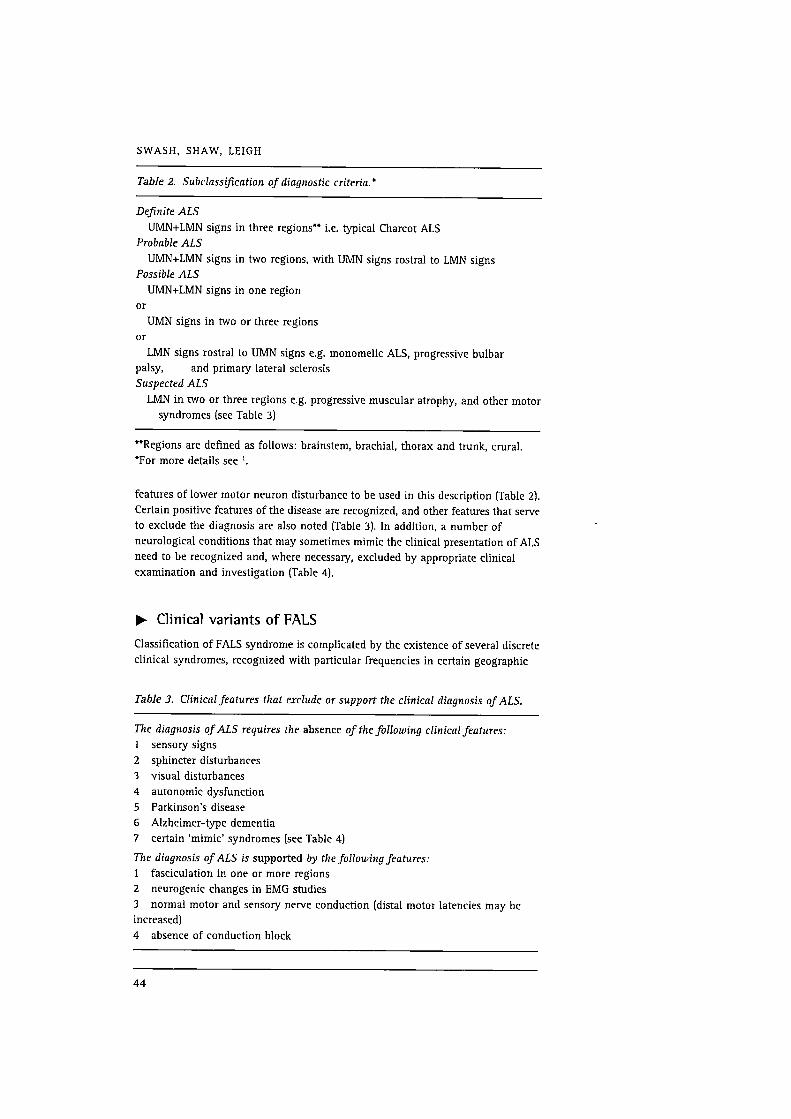
SWASH, SHAW, LEIGH
Table 2. Subclassification of diagnostic criteria.’
Definite ALS
UMN+LMN signs in three regions" i.e. typical Charcot ALS
Probable ALS
UMN+LMN signs in two regions, with UMN signs rostral to LMN signs
Possible ALS
UMN+LMN signs in one region
or
UMN signs in two or three regions
or
LMN signs rostral to UMN signs e.g. monomelic ALS, progressive bulbar
palsy, and primary lateral sclerosis
Suspected ALS
LMN in two or three regions e.g. progressive muscular atrophy, and other motor
syndromes (see Table 3)
“Regions are defined as follows: brainstem, brachial, thorax and trunk, crural.
*For more details see ‘.
features of lower motor neuron disturbance to be used in this description (Table 2).
Certain positive features of the disease are recognized, and other features that serve
to exclude the diagnosis are also noted [Table 3). In addition, a number of
neurological conditions that may sometimes mimic the clinical presentation of ALS
need to be recognized and, where necessary, excluded by appropriate clinical
examination and investigation (Table 4].
Bv Clinical variants of PALS
Classification of FALS syndrome is complicated by the existence of several discrete
clinical syndromes, recognized with particular frequencies in certain geographic
Table 3. Clinical features that exclude or support the clinical diagnosis ofALS.
The diagnosis ofALS requires the absence of the following clinicalfeatures:
sensory signs
sphincter disturbances
visual disturbances
autonomic dysfunction
Parkinson‘s disease
Alzheimer-type dementia
certain ‘mimic' syndromes (see Table 4]\immewwa
The diagnosis ofALS is supported by the following features:
1 fasciculation in one or more regions
2 neurogenic changes in EMG studies
3 normal motor and sensory nerve conduction [distal motor latencies may be
increased)
4 absence of conduction block
44
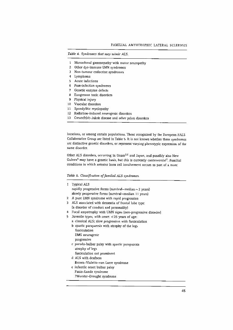
FAMILIAL AMYOTROPHIC LATERAL SCLEROSIS
TabIe 4. Syndromes that may mimic ALS.
1 Monoclonal gammopathy with motor neuropathy
2 Other dys-immune LMN syndromes
3 Non—tumour endocrine syndromes
4 Lymphoma
5 Acute infections
6 Post-infection syndromes
7 Genetic enzyme defects
8 Exogenous toxic disorders
9 Physical injury
10 Vascular disorders
11 Spondylitic myelopathy
12 Radiation-induced neurogenic disorders
13 Creutzfeldt—Jakob disease and other prion disorders
locations, or among certain populations. Those recognized by the European FALS
Collaborative Group are listed in Table 5. It is not known whether these syndromes
are distinctive genetic disorders, or represent varying phenotypic expression of the
same disorder.
Other ALS disorders, occurring in Guam5'6 and Japan, and possibly also New
Guinea5 may have a genetic basis. but this is currently controversia17. Familial
conditions in which anterior horn cell involvement occurs as part of a more
Table 5. Classification offamilial ALS syndromes.
1 Typical ALS
rapidly progressive forms (survival—median~2 years)
slowly progressive forms (survival—median 11 years)
A pure LMN syndrome with rapid progression
3 ALS associated with dementia of frontal lobe type
(a disorder of conduct and personality)
Focal amyotrophy with UMN signs (non—progressive disorder)
5 Juvenile types, with onset <25 years of age:
a classical ALS; slow progression with fasciculation
b spastic paraparesis with atrophy of the legs
fasciculation
EMG neurogenic
progressive
c pseudo-bulbar palsy with spastic paraparesis
atrophy of legs
fasciculation not prominent
(1 ALS with deafness
Brown—Vialetto—van Laere syndrome
infantile onset bulbar palsy
Fazio—Londe syndrome
?Worster—Drought syndrome
(D
SWASH, SHAW, LEIGH
widespread neurological disease do not form part of the syndrome of FALS itself,
but represent different disorders“.
Spinal muscular atrophy syndromes are excluded from this classification of FALS,
since they constitute a different group of disorders. Spinal muscular atrophy can
sometimes be confused clinically with ALS, particularly in relation to sporadic cases
of the progressive muscular atrophy variant of ALS.
The X—linked form of bulbo—spinal muscular atrophy (Kennedy's syndrome), now
known to be linked on the X chromosome with the androgen gene9, must be
excluded in the diagnosis of all familial syndromes involving the lower motor
neuron, particularly when the index case is sporadic and male, when there are no
corticospinal tract signs, when there are no affected female members of the family,
and when the pattern of inheritance is suggestive of X—linkage.
Similarly, whenever possible, a prion disorder must be excluded in all patients with
ALS associated with dementia of the frontal type10 or with other atypical features,
e.g. extrapyramidal rigidity“.
The clinical criteria for the diagnosis of FALS set out here are essentially intuitive,
representing a consensus achieved by discussion among two groups of neurologists.
They require testing by practical application and, more objectively, by an analysis
such as that used by Li et al.2 in their study of the consistency of diagnosis of
sporadic ALS in three countries.
p DNA Studies (M Swash)
Since the meeting report on PALS, there have been advances in the genetics of
FALS. Siddique, with colleagues, noted that FALS was expressed as an autosomal
disorder, indistinguishable clinically from the commoner sporadic form of the
disease‘z'”. The FALS locus was mapped in a small subset of informative families to
chromosome 21q22.1”. Seven of 11 families tested showed this locus, and linkage
to this region was excluded in three ofthe remaining families. It was recognized that
this locus was close to that for the Cu, Zn superoxide dismutase gene (SOD—1) on
chromosome 21. Subsequent studies have co-localized the locus with that for SOD-1
in these affected families, and have demonstrated mutations in exons 1, 2, 3 and 5
respectively in some of these familiesl5'15. There are five exons in the SOD-1 gene.
SOD-1 activity in individuals with SOD-1 gene mutations varies from about 30% to
normal. These data add an intriguing dimension to the notion that neurodegen-
eration in ALS might be due, at least in part, to an abnormality in the ability of
susceptible motorneuron pools to handle free radicalsnvla. The evidence for this
mechanism of pathogenesis in ALS is increasingly persuasive, but does not take
account ofthose families in which a mutation in the SOD-1 gene has been excluded,
and does not yet provide an entirely plausible explanation for sporadic cases.
Nonetheless, the discovery of the role of mutations in the SOD-1 gene in some
familial cases illustrates the power of genetic analysis in revealing hitherto
unsuspected metabolic abnormalities in progressive degenerative disorders.
References
1 El Escorial World Federation of Neurology criteria for the diagnosis of ALS. J
Neural Sci 1994, 124 (Suppl): 96—107.
46
FAMILIAL AMYOTROPHIC LATERAL SCLEROSIS
12
13
14
16
17
18
Li T—M, Swash M, Alberman A, Day SJ. Diagnosis of motor neuron disease in
three countries. JNeurol Neurosurg Psychiatry 1991; 54: 980—3.
Li T—M, Albennan E, Swash M. Comparison of sporadic and familial disease
amongst 580 cases of motor neuron disease. JNeurol Neurosurg Psychiatry
1988; 51: 778—84.
Williams DB, Floate DA, Leicester .1. Familial motor neurone disease: differing
patterns in large pedigrees. JNeurol Sci 1988; 86: 215—30.
Gajdusek DC. Environmental factors provoking physiological changes which
induce motor neuron disease and early neuronal aging in high incidence foci in
the Western Pacific. In: Rose FC, ed. Research progress in motor neuron disease.
Pitman: London, 1984: 44—69.
Garruto RM, Yanagihara R, Gajdusek DC. Disappearance of high incidence
amyotrophic lateral sclerosis and Parkinson—dementia on Guam. Neurology
1985; 35: 193—8.
Duncan MW. Role of the Cycad neurotoxin BMAA in the amyotrophic lateral
sclerosis—Parkinsonism—dementia complex of the Western Pacific. In: Rowland
LP, ed. Amyotrophic lateral sclerosis and other motor neuron diseases,
Advances in Neurology, Vol. 56. Raven Press: New York, 1991: 301—10.
Schwartz MS, Swash M. Motor neuron disease. In: Swash M, Oxbury J, eds.
Clinical neurology. Churchill Livingstone: Edinburgh, 1991: 1356—66.
La Spada AR, Wilson EM, Lubahn DB, Harding AE, Fischbeck KH. Androgen
receptor gene mutation in X—linked spinal and bulbar atrophy. Nature 1991;
352: 77—9.
Neary D, Snowden JS, Mann DMA, Northen P, Goulding PJ, MacDermott N.
Frontal lobe dementia and motor neuron disease. JNeurol Neurosurg Psychiatry
1990; 53: 23—32.
Mitsuyama Y. Presenile dementia with motor neuron disease in Japan;
clinicopathological review of 26 cases. JNeurol Neurosurg Psychiatry 1984; 47:
953—9.
Siddique T, Pericak-Vance MA, Brooks BR, et al. Linkage analysis in familial
amyotrophic lateral sclerosis. Neurology 1989; 39: 919—25.
Mulder DW, Kurland LT, Offord KP, Beard M. Familial adult motor neuron
disease: amyotrophic lateral sclerosis. Neurology 1986; 36: 511—7.
Siddique T, Figlewicz DA, Pericak-Vance MA, et al. Linkage of a gene causing
familial amyotrophic lateral sclerosis to chromosome 21 and evidence of
genetic—locus heterogeneity. NEJM 1991; 324: 1381—4.
Rosen DR, Siddique T, Patterson D, et al. Mutations in Cu/Zn superoxide
dismutase gene are associated with familial amyotrophic lateral sclerosis.
Nature 1993; 362: 59—62.
Deng H-X, Hentati A, Tainer JA, et al. Amyotrophic lateral sclerosis and
structural defects in Cu, Zn superoxide dismutase. Science 1993; 261: 1047—51.
Hallewell RA, Gutteridge JMC. Oxygen radicals and the nervous system. Trends
in Neurosciences 1985; 8: 22—6.
Brown RH. Amyotrophic lateral sclerosis: recent insights from genetics and
transgenic mice. Cell 1995; 80: 687—92.
This is partly based on a report originally published in Neuromuscul Disord 1992;
2(1): 7—9 with permission from Pergamon Press Ltd, Headlngton Hill Hall, Oxford
OX3 OBW, UK
47
Hereditary Motor
and Sensory
Neuropathy or
Charcot—Marie—Tooth
Disease Types IA
and IB
M de Visser Dept ofNeurology, Academic Medical Centre,
Amsterdam, The Netherlands
C van Broeckhoven and E Nelis Neurogenetics Laboratory, Flemish
Institute for Biotechnology, Born—Bunge Foundation, University
ofAntwerp, Dept of Biochemistry, Antwerp, Belgium
p Diagnostic Criteria
Charcot—Marie—Tooth (CMT) disease is a clinically and genetically heterogeneous
peripheral neuropathy characterized by wasting and weakness of the distal limb
muscles. CMT I, the demyelinating form, and CMT II, the axonal form, are the most
common types with an estimated prevalence of at least 1 in 10 000. Inheritance
pattern is autosomal dominant in the majority of patients.
Positional cloning has shown that CTM I is genetically heterogeneous with at least
three loci. The major autosomal dominant subtype CMT IA is linked to chromosome
17p11.2. A less frequent autosomal dominant subtype, CMT IB, is linked to
chromosome 1q22-q23, and a third also autosomal dominant subtype, CMT IC, is
not linked to either of these loci and as yet unassigned.
In the majority of CMT IA patients, either familial or sporadic, the disease is
associated with a tandem DNA duplication of 1.5 Mb. This duplication can be
detected by density differences in the alleles or by the presence of three alleles of
polymorphic DNA probes or microsatellite markers, or by a duplication junction
fragment with pulsed field gel electrophoresis technology. Almost all patients,
independent of their ethnic origin, have the same sized duplication. The
collaborative duplication screening of the European Consortium on CM 1 resulted
49
DE VISSER, VAN BROECKHOVEN, NELIS
in a duplication frequency of 70.7%. The duplication was found to contain the
human homologue of the peripheral myelin protein-22 gene (PMP—22). PMP—22 is
not altered by the duplication and shows a dosage effect in CMT IA patients. In
nonduplicated CMT IA patients point mutations in PMP-ZZ confirmed the direct role
of the gene in the CMT IA disease process.
In CMT IB mutations in the myelin protein zero gene (MPZ) appeared to co—
segregate with the disease.
In May 1991, the ENMC Consortium established diagnostic criteria for autosomal
dominant GMT I. These criteria were meant to serve only for research purposes, and
in particular for linkage analysis. However, a diagnosis of autosomal dominant
CMT I can now be accurately made by detection of the duplication or by
identification of a point mutation in either the PMP-ZZ or MP2 gene. Therefore, we
have amended the diagnostic criteria on CMT IA and CMT IB accordingly by adding
DNA analysis as an inclusion criterion. Furthermore, over the years genotype—
phenotype correlations have extended our knowledge of the clinical picture of
CMT I. Some patients who once had been diagnosed as suffering from Dejerine—
Sottas disease were found to have a PMP-22 or MP2 point mutation.
Linkage studies in other subtypes of CMT have yielded loci on chromosome 8q for
one form of autosomal recessive CMT I (with basal lamina onion bulbs] and on
chromosomes 1p and 3q, respectively for two subtypes of the autosomal
dominantly inherited CMT II. An X—linked dominant form of CMT with
demyelinating and neuronal characteristics also manifests with distal muscle
wasting and weakness. Males usually have more pronounced clinical features than
females. After assignment of the locus to qu3.1, mutations in the gene encoding
connexin 32 (Cx32), a gap junction protein were found in most CMTX patients.
Hereditary neuropathy with liability to pressure palsies (HNPP) is hallmarked by
periodic episodes of weakness or numbness often due to minor compression or
trauma to peripheral nerves. In some patients HNPP may mimic CMT. The disease
which is inherited as an autosomal dominant trait is usually associated with a
deletion in chromosome 17p11.2 encompassing the same markers that are
duplicated in CMT IA. A few HNPP patients have point mutations in PMP-ZZ.
An overview of the duplication/deletion and mutation screening techniques and an
estimation ofthe mutation frequencies in CMT I and HNPP patients is found in Nelis
et (11.1.
It is of note that as yet no attempts have been undertaken to establish diagnostic
criteria for the above mentioned subtypes of CMT.
The following criteria for inclusion (I) and exclusion (E) of the diagnosis are only
applicable to the autosomal dominant forms of CMT I in which the duplication or a
point mutation in either the PMP-ZZ or MP2 gene is present, i.e., CMT IA and
CMT IB, (C) refers to comments.
> Clinical criteria
Family history
I Autosomal dominant inheritance
50
HEREDITARY MOTOR AND SENSORY NEUROPATHY
C
[Tl
nmnmn
IT]
nr—4
Isolated cases as a result of a de novo duplication are found in a proportion of the
patients.
Age at onset
Usually in the first two decades of life.
Congenital onset with floppiness is very rare.
Muscle wasting and weakness
Muscle wasting and weakness of predominantly the distal part of the lower
limbs.
Symmetrical.
Later, wasting and weakness of the intrinsic hand muscles and the distal part of
the medial vastus muscle and other parts ofthe quadriceps muscle may develop.
Other associated features or diseases
Impaired sensation is frequently observed but is not an obligatory feature.
Arthrogryposis with the exception of talipes or pes cavus.
Scoliosis and nerve hypertrophy are present in a proportion of the patients.
CNS involvement.
The presence of brisk reflexes should militate against the diagnosis. Some
patients may have extensor plantar responses. There may be tremor or slight
limb ataxia.
Other major organ involvement (vision, hearing, cardiac).
There may be abnormalities of visual or auditory evoked responses but no overt
clinical evidence of involvement of the optic or auditory nerves.
Impairment of oculomotor and bulbar function; marked facial weakness.
Myotonic dystrophy.
Course and severity
Slowly progressive or stationary over long periods of time.
At any age there is a wide range of severity.
Some patients are asymptomatic and are found to have pes cavus and/or areflexia
on examination. Rarely, proximal muscles may become involved leading to
wheelchair dependence or respiratory insufficiency.
> Laboratory criteria
C
I
Electrophysiology
Median nerve motor conduction velocity (MCV) <38 m/sec. Absence or marked
decrease of sensory nerve action potentials (SNAPs) in the lower limbs.
An occasional patient may have, at some time in the course of the disease, a
median nerve MCV up to 40—45 m/sec. If so, the contralateral median and/or
ulnar nerve should be investigated.
Electromyography may show signs of denervation and reinnervation.
Molecular genetics
Duplication of 1.5 Mb (rarely smaller) on 17p11.2 or, in non-duplicated patients
point mutations in the myelin gene PMP—ZZ designates CMT IA.
51
DE VISSER. VAN BROECKHOVEN, NELIS
I In non—duplicated patients point mutations in the myelin gene MPZ designates
CMT IB.
Sensory nerve biopsy
(Usually sural nerve although a fibular nerve may sometimes be sampled.)
I Characteristic features are the following:
Marked reduction of density and total number of large and small myelinated
fibres
Segmental demyelination and remyelination.
An increase in total transverse fascicular area
Onion bulbs
Axonal changes are usually not prominent.
C With the introduction of DNA-analysis as a diagnostic tool nerve biopsies should
no longer be used for establishing the diagnosis.
Reference
1 Nelis E, Van Broeekhoven C. Estimation of the mutation frequencies in CMT I
and HNPP: a European collaborative study. Eur J Hum Genet 1996; 4:
25—33.
This is partly based on a report originally published in Neuromuscul Disord 1993;
3(1): 77—9 with permission from Pergamon Press Ltd, Headington Hill Hall, Oxford
OX3 OBW, UK
52
Chronic
Inflammatory
Neuropathies
H Franssen Dept of Clinical Neurophysiology, University Hospital
Utrecht, The Netherlands
M Vermeulen Dept of Neurology, Academic Medical Centre,
Amsterdam, The Netherlands
FGI Jennekens Dutch Neuromuscular Research Support Centre,
Baarn, The Netherlands
> Introduction
Research criteria for chronic inflammatory demyelinating polyneuropathy (CIDP)
were first proposed and published by an Ad Hoe Subcommittee of the American
Academy of Neurology in 19911. Since then several important developments have
taken place. Multifocal motor neuropathy (MMN) was recognized as related to CIDP,
but is different in several respects. Definition and recognition of electrophysiological
phenomena, as for example ‘conduction block' and ‘temporal dispersion' was
improved. Therefore it became desirable to adapt the criteria introduced in 1991.
Recent reports suggest that the group of chronic inflammatory neuropathy consists
of several entities. Among these are chronic inflammatory sensory neuropathy and
chronic inflammatory (or autoimmune) axonal neuropathy. The present criteria
concern only CIDP and MMN. They were agreed upon at two consensus workshops
organized by the Dutch Neuromuscular Research Support Centre in October 1995
and April 1996.
p Diagnostic Criteria for CIDP
Elements
1 CIDP may occur at all ages (in all decades)”.
2 CIDP is a chronic disease. It does not reach nadir until 8 weeks after onset. The
course is progressive, stepwise progressive or relapsing“.
3 The clinical signs involve muscle weakness and/or sensory disturbances. Motor
or sensory abnormalities are absent in some cases”.
4 The distribution of the clinical signs is (approximately) symmetrical. The first
signs appear distal at the limbs, in some cases initially or predominantly at the
53
FRANSSEN, VERMEULEN, JENNEKENS
11
12
13
14
15
upper limbs“. Marked weakness of the lower limbs with approximately normal
muscle power at the upper limbs is unusual. Weakness of neck extensors
presenting as a ‘dropped head syndrome' has been described5.
Cranial nerves (nerves innervating the extra-ocular muscles and facial and
hypoglossus nerves) may be involved“.
Sensory disturbances concern predominantly large sensory nerve fibre
modalities. Disturbances due to small sensory nerve fibre involvement and
autonomic disturbances are rarez.
Tendon reflexes are reduced or absent in most cases“.
At some time during the course of the disease postural tremor may become
apparent”.
There may be papilloedema“.
Nerve conduction of motor nerve fibres is abnormal and compatible with
demyelination“. Abnormalities are not present to the same degree in all
(mixed) nerves. Details on the electrophysiological methods are provided in the
Addendum page 57.
Motor conduction velocity (MCV) including F—waves is examined in four nerves
(ulnar, median, peroneal, tibial) on one side of the body. Sensory conduction on
distal stimulation is examined in the median, ulnar, and sural nerves on the
same side as motor conduction. Three of the following four items (at least one
in an upper limb) should be present:
a Motor conduction velocity (MCV) less than 75% of the lower limit of normal
in at least two nerves.
(Lower limits of normal MCV are generally considered to be 50 m/sec in
upper limb nerves and 40 m/sec in lower limb nerves. Values compatible
with demyelination are therefore 38 m/sec and 30 m/sec or less).
Definite or possible conduction block in at least one nerve segment.
Distal motor latency (DML) more than 130% of the upper limit of normal in
at least two nerves.
d Absence of F—wave, or shortest F-wave latency more than 130% ofthe upper
limit of normal, in at least one nerve.
The protein content ofthe cerebrospinal fluid (CSF) is raised. It is raised in 90—95%
of all cases to more than 0.5 g/l and often to more than 1 g/lz. The CSF cell
count is less than 30 x 105/1; this may be higher in HIV patients with CIDP.
A sural nerve biopsy may reveal active demyelination and mononuclear
inflammatory cell infiltrates7.
MRI reveals white matter lesions in a minority of the casesa.
CIDP may be associated with: systemic lupus erythematosus, Hodgkin's disease,
ulcerative colitis, multiple sclerosis, myasthenia gravis, psoriasis, diabetes
mellitus, hyperthyroidism, and HIV”. In patients with MGUS (monoclonal
gammopathy of unknown significance) a peripheral neuropathy fulfilling the
criteria of CIDP may occur.
Other neuropathies to be excluded include:
a Pure sensory neuropathies including ganglioneuritis and mixed
neuropathies with predominant loss of small sensory nerve fibre modalities.
b Axonal neuropathies.
Charcot—Marie—Tooth (CMT) neuropathies (CMT neuropathies differ from
CIDP in symptoms, electrophysiological signs, course, family history).
(1 Meningeal carcinomatosis or lymphomatosis (often cranial nerve involve—
ment and conus/cauda syndrome, diagnosis by CSF cytology).
54
CHRONIC INFLAMMATORY NEUROPATHIE S
e Other neuropathies with relapsing course (hereditary neuropathy with
liability to pressure palsies, Refsum disease (this neuropathy is associated
with night blindness and often with deafness, the phytanic acid level in serum
is raised), Tangier disease (disturbance predominantly of small sensory nerve
fibre qualities, conduction velocity is only moderately reduced).
f Other demyelinating neuropathies, e.g. paraproteinaemic neuropathies and
neuropathies in rare hereditary disorders as metachromatic leucodystrophy
and Krabbe disease.
g Infectious diseases of the peripheral nervous system, e.g. Lyme neurobor-
reliosis, (pleocytosis, raised serum antibody titres).
h Iatrogenic, toxic and metabolic neuropathies.
Assessment
From the above it is clear that CIDP can be diagnosed when patients with the
clinical features of a chronic sensory-motor polyneuropathy, and a raised
protein content of the cerebrospinal fluid, reveal evidence of demyelination at
electrophysiological investigation, and when other possible causes have been
ruled out. The diagnosis is less certain when one of these four features is weak or
absent. In figures:
The diagnosis is definite, when:
a 2, 3, 4, 7, 10 and 11 are all present and findings exclude 15
b the presence of 6 and/or 12 concurs with the diagnosis but is not a
requirement
c the presence of 5, 8, 9, 13, 14 is compatible with the diagnosis
The diagnosis is probable, when:
a 2, 3, 4, 7 and 11 are all present, 10 is partly present (less than three of the
specified items are present) and findings exclude 15
b the presence of 6 and/or 12 is in support of the diagnosis
the presence of 5, 8, 9, 13, 14 is compatible with the diagnosis
The diagnosis is possible, when:
a 2, 3, 4, 7, 11 are all present and findings exclude to 15
b the presence of 6 is in support of the diagnosis
c the presence of 5, 8, 9 and 14 is reason to consider the diagnosis
p Diagnostic Criteria for MMN
Elements
1 MMN is a chronic disease. Nadir is not reached until 8 weeks after onset, the
course of the disease is progressive, or stepwise progressive, or undulating over
many months or yearsg'“).
2 Muscle weakness, muscle atrophy, fasciculations and muscle cramps are the
main clinical signs. Muscle weakness may be present in non—atrophic muscles.
Slight sensory disturbances do not rule out the diagnosis. Cranial nerves are
rarely involved“?
3 Signs of the disease are initially asymmetric and remain asymmetric in most
patientsg'lo.
55
FRANSSEN, VERMEULEN, JENNEKENS
4
5
The disease usually begins at the upper limbs, predominantly distal.9'1°.
Tendon reflexes in affected limbs are decreased or absent. Generalized absence of
reflexes may occur9'10.
Motor conduction block is present. The changes in MCV are compatible with
multifocal asymmetric demyelination9'10. Sensory conduction is usually normal,
also in segments with motor conduction block. Slight sensory conduction
abnormalities are however compatible with the diagnosis9-10. Electromyography
reveals spontaneous muscle fibre activity and polyphasic or giant motor unit
potentials, in an asymmetric distribution. Critera for motor conduction block and
details of the electrophysiological methods are discussed in the Addendum.
Motor nerve conduction including F—waves with recording from hand and/or
foot muscles should be examined in the median, ulnar and peroneal nerves on
both sides. When no evidence of conduction block can be obtained, additional
nerves should be studied, especially nerves to weak non—atrophic lower or upper
arm muscles. These nerves include the median, musculocutaneous, axillary and
tibial nerves. Sensory conduction on distal stimulation is examined in median,
ulnar, and sural nerves at one side of the body and in nerve segments in which
conduction block is present.
The protein content ofthe cerebrospinal fluid is in most cases not raised and if so
only slightly (less than 1 g/l)9"°.
Diseases as mentioned in point 15 of the CIDP elements are excluded.
When the diagnosis is possible but not highly probable the presence of a raised
serum titre of anti-GM-l antibodies will increase the likelihood of the diagnosis
(from an a priori chance of 20—60% to 50—85%)”.
Assessment
The elements show that the diagnosis depends on the combination of a typical
clinical picture and the presence of motor conduction block. In some cases the
electrophysiological findings do not fully meet the criteria for definite motor
conduction block; the diagnosis is still likely in these cases still likely but is no
longer definite. In other cases extensive electrophysiological examination does
not provide any evidence for conduction block, but this does not imply that it is
excluded. The diagnosis is still possible when the clinical features are typical and
evidence of demyelination is present”.
The diagnosis is definite when:
a 1, 2, 3, 6 and 8 are all present
b There is “definite conduction block" with or without additional evidence of
demyelination
c 4, 5, 7 and 9 concur with the diagnosis, but may be absent.
The diagnosis is probable when:
a 1, 2, 3 and 8 are present and when 6 is present partially
b there is “possible conduction block" with or without further evidence of
demyelination
c 4, 5, 7 and 9 are in support of the diagnosis.
The diagnosis is possible when:
a 1, 2, 3, and 8 are present and when 6 includes evidence of demyelination but
not of conduction block
b 4, 5, 7 and 9 are in support of the diagnosis.
56
CHRONIC INFLAMMATORY NEUROPATHIES
p Addendum to the electrophysiological
examination of the chronic inflammatory
demyelinating neuropathies
> Recording and stimulation sites for motor conduction studies
Nerve Recording site Stimulation sites
Median M. abductor pollicis brevis Wrist, elbow, axilla, Erb‘s point
Median M. flexor carpi radialis Elbow, axilla, Erb's point
Ulnar M. abductor dig. V Wrist, 5 cm distal to elbow, 5 cm
proximal to elbow, axilla, Erb's point
Musculocuta— M. biceps brachii Axilla, Erb's point
neous
Axillary M. deltoideus Axilla, Erb's point
Peroneal M. extensor dig brevis Ankle, 5 cm distal to fibular head,
popliteal fossa
Tibial M. abductor hallucis brevis Ankle, popliteal fossa
The results of stimulation at Erb‘s point should be interpreted with caution as
stimulation may not be supramaximal.
> Slowing of conduction and demyelination
It has been reasonably established that a decrease of maximal conduction
velocity or an increase of DML beyond a certain value suggests demyelination.
Conduction velocity and histology ofthe sural nerve and other nerves have been
compared in CMT neuropathy types I and 11, experimental allergic neuritis and
controls. Values for MCV, DML and F-wave latency have thus been obtained that
are compatible with demyelination. On this basis values for MCV lower than 75%
of the lower limit of normal and for DML or shortest F—wave latency of more
than 130% of the upper limit of normal are considered to be indicative of
demyelination‘. Conclusions about the shortest F—wave latency should be based
on findings at 16—20 responses at distal stimulation. F—wave findings should
only be included as an item suggesting demyelination if DML and MCV in the
same nerve do not reach values compatible with demyelination. Under these
latter conditions, absence of F-waves is considered compatible with demyeli-
nation.
V Conduction block
The detection of (partial) motor conduction block rests upon a reduction in the
compound muscle action potential (CMAP) amplitude or area at proximal versus
distal stimulation of a nerve segment. There are no generally accepted criteria for
(partial) motor conduction block. A major problem is that demyelination may
result in increased differences between conduction times of action potentials in
individual nerve fibres. This ‘temporal dispersion' also causes reduction in CMAP
amplitude or area and an increase in CMAP duration on proximal versus distal
stimulation, especially when conduction distance is large. It is nevertheless likely
57
FRANSSEN, VERMEULEN, JENNEKENS
that an area reduction of more than 50% regardless of distance and duration
increase is due to conduction block in at least some nerve fibres”. This is also
true for an amplitude and area reduction of at least 30% over a short distance as
found by inching. The criterion suggested by Feasby et al. (1985) (amplitude
reduction of at least 20%) as used in the Research Criteria of the American
Academy of Neurology (199 1) is probably too liberal and may yield false positive
resultslv”. On the basis ofthese considerations criteria for conduction block were
defined.
Definite conduction block: reduction in CMAP area of at least 50%, independent
of conduction distance, or a reduction in CMAP amplitude and area of at least
30% over a short distance, detected by inching.
Possible conduction block: a reduction in CMAP amplitude of at least 30% for
upper limb nerves and 40% for lower limb nervesl-r’.
Nerve anastomosis should be excluded as possible cause for CMAP reduction.
p Minimal CMAP amplitudes
Values for MCV, DML and F-wave should be obtained from nerves which induce
at distal stimulation a CMAP with an amplitude of at least 0.5 mV (from the
baseline to the peak of negative deflection).
For the diagnosis of conduction block to be acceptable the amplitude of the
negative deflection the CMP should be at least lmV.
b Entrapment sites
Values of MCV, DML compatible with demyelination or conduction block at
common entrapment sites (median nerve at the carpal tunnel, ulnar nerve at the
elbow, peroneal nerve at the flbular head) should be considered as due to
entrapment (and not to CIDP or MMN) unless sensory conduction at these sites is
normal.
> Skin temperature
When the skin temperature at the level of ankle or wrist is below 32 uC, the lower
leg or the lower arm should be warmed in water to 37 °C for a period of at least
20 minutes”. Warming by an infrared heater is insufficient.
The application of correction factors to correct for low temperature is not
justified as the relation between temperature and conduction is altered in
demyelinating neuropathy”. Conduction block may be apparent only at a
temperature in the high physiological range and may be missed at lower
temperatures”.
References
1 Ad Hoc Subcommittee ofthe American Academy of Neurology AIDS Task Force:
Research criteria for diagnosis of chronic inflammatory demyelinating
polyneuropathy. Neurology 1991; 41: 617—8.
58

CHRONIC INFLAMMATORY NEUROPATHIES
10
12
13
14
16
17
Dyck PJ, Prineas J, Pollard J. Chronic inflammatory demyelinating poly—
radiculoneuropathy. In: Dyck PJ, Thomas PK, Griffln JW, Low PA, Poduslo JF
ed. Peripheral neuropathy, Vol 2, 3rd edn. Philadelphia: WB Saunders, 1993:
1498—517.
McCombe PA, Pollard JD, McLeod JG. Chronic inflammatory demylinating
polyradiculoneuropathy. A clinical and electrophysiological study of 92 cases.
Brain 1987; 10: 1617—30.
Vermeulen M, van Doorn PA, Brand A, et al. Intravenous immunoglobulin
treatment in patients with chronic inflammatory demyelinating polyneur-
opathy: a double blind placebo controlled study. JNeurol Neurosurg Psychiatry
1993; 56: 36—9.
Hoffman D, Gutmann L. The dropped head syndrome with chronic
inflammatory demyelinating polyneuropathy. Muscle Nerve 1994; 17: 808—10.
Bromberg MB. Comparison of electrodiagnostic criteria for primary demyeli-
nation in chronic polyneuropathy. Muscle Nerve 1991; 14: 968—76.
Prineas JW. Pathology of inflammatory demyelinating neuropathies. In: JG
McLeod (Ed), Inflammatory neuropathies, Bailliere's Clinical Neurology,
London: Bailli'ere Tindall, 1994; 43: 1—24.
Feasby TE, Hahn AF, Koopman RN, et al. Central lesions in chronic
inflammatory demyelinating polyneuropathy. Neurology 1990; 40: 476—8.
Parry GJ. Motor neuropathy with multifocal conduction block. In: Dyck PJ,
Thomas PK, Griffin JW, Low PA, Poduslo JP, eds. Peripheral neuropathy, Vol 2,
3rd edn. Philadelphia: WB Saunders, 1993; 1518—24.
Parry GJ. AAEM Case report 30: Multifocal motor neuropathy. Muscle Nerve
1996; 19: 269—76.
van Schaik IN, Bossuyt PMM, Brand A, et al. Diagnostic value of GMl
antibodies in motor neuron disorders and neuropathies. Neurology 1995; 45:
1570—7.
Pakiam A, Parry G. Multifocal motor neuropathy without evidence of
conduction block. Neurology 1996; 46: A 234.
Rhee EK, England JD, Sumner AJ. A computer simulation of conduction block:
effects produced by actual block versus interphase cancellation. Ann Neurol
1990; 28: 146—56.
Feasby T, Brown WP, Gilbert JJ, et al. The pathological basis of conduction
block in human neuropathies. JNeurol Neurosurg Psychiatry 1985; 48: 239—44.
Albers JW, Donofrio PD, McGonagle TK. Sequential electrodiagnostic
abnormalities in acute inflammatory demyelinating polyradiculoneuropathy.
Muscle Nerve 1985; 8: 528—39.
Franssen H, Wieneke GH. Nerve conduction and temperature: necessary
warming time. Muscle Nerve 1994; 17: 336—44.
Franssen H, Wienke GH, Notermans NC, et al. Temperature dependent
conduction block in peripheral neuropathy. Neuro Orthopedics 1995; 17/18:
72—82.
59
Distal Myopathies
Hannu Somer Dept ofNeurology, University ofHelsinki, Helsinki,
Finland
p Diagnostic Criteria
Distal myopathies comprise a group of disorders which cause distal muscle
weakness without clinically significant involvement of proximal, facial or trunk
muscles. Clinical examination reveals no signs of neurogenic involvement. Nerve
conduction velocities are normal and electromyographic studies are compatible
with myopathy without additional findings, such as myotonic discharges. SCK
activity may be mildly elevated, except for in one variety where it is remarkably
elevated to a level seen in generalized muscular dystrophies.
Muscle biopsy shows typical signs of myopathy such as variation in fibre size,
central nuclei and increased connective tissue at an early stage. Later on more
marked alterations appear. Rimmed vacuoles are seen frequently in some distal
myopathies.
Central cores, nemaline bodies, inclusion bodies, glycogen or lipid accumulations
seen in some patients with distal muscle weakness, are excluded from this group and
suggest other diagnostic entities.
Diagnosis and classification of distal myopathies is based on clinical, genetic and
morphological criteria. The following clinical phenotypes can currently be defined.
P' Late adult onset myopathy with onset in hands1 (Welander)
This is the most common form of the distal myopathies, by 1951 249 Swedish
patients had been describedl. The pattern ofinheritance is autosomal dominant with
penetrance of about 70 to 80%. The initial symptoms are clumsiness ofsmall precise
hand movements and difficulties in extension of fingers. Distal leg muscle weakness
develops later on causing difficulties in walking and inability to stand on heels.
Clinical course is slowly progressive. Many patients complain of coldness of hands
and feet, but sensation is normal. SCK may be mildly elevated. Muscle imaging (CT
or MRI) shows involvement of lower leg muscles both in the anterior and posterior
compartments. Muscle biopsy from anterior tibial muscle shows myopathic changes
and occasional vacuolation. Electromyography is ‘myopathic' in the early stages but
‘neurogenic' changes have been described especially in patients with severe muscle
weaknessz.
61
SOMER
> Late adult onset myopathy with onset in legs3'4
(Markesbery and Griggs; Udd).
The original description comes from the United States3. Subsequently, a similar
phenotype has been described in Finland“, where more than 100 patients have now
been discovered. Muscle weakness appears usually after the age of 35 years and is
confined to lower leg muscles, especially to tibial anterior muscles. The American
family also developed weakness of the intrinsic hand muscles and some proximal
muscle weakness. The disease is inherited in an autosomal dominant fashion.
SCK may be mildly elevated. Electromyographic changes are myopathic. These, as
well as abnormal findings in muscle imaging (CT or MRI), are confined mainly to
anterior tibial muscles, although mild abnormalities may also be detected elsewhere.
Histopathological changes are myopathic with a spectrum from mild changes to
severe end stage changes. Rimmed vacuoles may be present.
> Early adult onset myopathy with onset in posterior
compartment of lower legs5 (Miyoshi).
The initial symptoms include difficulty in climbing stairs or running. Patients can
not hop on one leg. Muscle weakness is most pronounced in the gastrocnemius
muscle. Intrinsic foot muscles and muscles of the anterior compartment may also be
involved as the disease progresses. Some patients have muscle weakness in their
hand muscles. Electromyographic studies show myopathic findings. Muscle imaging
studies show pronounced lesions in the posterior compartment muscles in the lower
legs. SCK is elevated 10 to lOO—fold being much higher than in other distal
myopathies. Muscle biopsy shows severe myopathic changes in biopsies from
proximal muscles. The disease is inherited in an autosomal recessive fashion5.
Several families have been reported in Japan, United States, Tunisia and from
various European countriess.
> Early adult onset myopathy with onset in anterior
compartment of lower legs7 (Nonaka).
The initial symptom is distal muscle weakness in the anterior compartment of lower
legs presenting with foot drop. Later. posterior compartment muscles may become
involved, but intrinsic foot muscles are usually spared. SCK is only mildly elevated.
Numerous rimmed vacuoles are typical of this entity. The disease is inherited in an
autosomal recessive fashion7.
b Other possible entities
In an Australian familyB selective weakness of the toes and flexors appeared before
the age of 25 years, followed by progressive weakness of finger extensors. Certain
proximal muscle groups were mildly affected. The disease was inherited in an
autosomal dominant fashion. Some families have been described with distal
myopathy appearing in adulthood and associated with desmin storage and
autophagocytosis9'lo. The clinical course is variable, some showing rapid progres—
sion. Progression may extend to bulbar, respiratory and facial muscles.
Cardiomyopathy with various conduction defects is common.
62
DISTAL MYOPATHIE S
y DNA Studies
In the Australian familyB positive linkage was obtained with 14 out of 15 markers
tested on chromosome 14. Maximum two point lod scores of 2.60 at recombination
fraction (O]=0.00 were obtained for two markers MYl-l7 and D14564 — the family
structure precludes a two-point lod score of 3 or greater. Recombinations with
D14S72 and D14S49 indicate that this distal myopathy locus should lie between
these markers.
The Miyoshi myopathy has been recently localized to chromosome 2p12—14 locus
D2S291 (Z max=15.3 at O—=0). The results are based on patient material obtained
from Japan, Tunisia and the United States“.
References
1 Welander L. Myopathia distalis tarda hereditaria: 249 examined cases in 72
pedigrees. Acta Med Scand 1951; 141: 1—124.
2 Borg K, Ahlberg G, Borg J, et al. Welander‘s distal myopathy: clinical,
neurophysiological and muscle biopsy observations in young and middle aged
adults with early symptoms. JNeural Neurosurg Psychiatry 1991; 54: 494—8.
3 Markesbery WR, Griggs RC, Leach RP, et al. Late onset hereditary distal
myopathy. Neurology 1974; 23: 127—34.
4 Udd B, Partanen J, Halonen P, et al. Tibial muscular dystrophy. Late adult onset
distal myopathy in 66 Finnish patients. Arch Neurol 1993; 50: 604—8.
5 Miyoshi K, Kawai H, lwasa M, et al. Autosomal recessive distal muscular
dystrophy as a new type of muscular dystrophy: seventeen cases in eight
families, including an autopsied case. Brain 1986; 109: 31—54.
6 Kuhn E, Schrdder JM. Autosomal recessively inherited distal myopathy. A new
type of distal myopathy. JNeurol 1981; 226: 181—5.
7 Nonaka I, Sunohara N, Satoyoshi E. Familial distal myopathy with rimmed
vacuole and lamellar (myeloid) body formation. JNeurol Sci 1981; 51:
141—55.
8 Laing NG, Laing BA, Meredith C, et al. Autosomal dominant distal myopathy:
linkage to chromosome 14. Am JHum Genet 1995; 56: 422—7.
9 Edstrom L, Thornell LE, Eriksson A. A new type of hereditary distal myopathy
with characteristic sarcoplasmic bodies and intermediate (skeletin) filaments. J
Neurol Sci 1990; 47: 171—90.
10 Horowitz SH, Schmalbruch H. Autosomal dominant distal myopathy with
desmin storage: a clinicopathological and electrophysiological study of a large
kinship. Muscle Nerve 1994; 17: 151—60.
1 1 Bejaoui K, Hirabayashi K, Hentati F, et al. Linkage of Miyoshi myopathy (distal
autosomal recessive muscular dystrophy) locus to chromosome 2p 12—14.
Neurology 1995; 45: 768—72.
This is based on a report originally published in Neuromuscul Disord 1995; 5(3):
249—52 with permission from Pergamon Press Ltd, Headington Hill Hall, Oxford
OX3 OBW, UK.
63
Myotubular/
Centronuclear
Myopathy
C Wallgren—Pettersson Dept of Medical Genetics, University of
Helsinki, and the Folkhc'ilsan Dept ofMedical Genetics, Helsinki,
Finland
p Diagnostic Criteria
All forms of myotubular myopathy are extremely rare, including the usually very
severe X—linked recessive form with pre- or neonatal onset (McKusick “310400), the
autosomal recessive form with onset in infancy or childhood (McKusick 255200)
and the relatively mild autosomal dominant form with late onset (McKusick
*160150).
Consensus definitions of clinical core criteria for the X-linked form of myotubular
myopathy (XMTM) include male sex, perinatal onset and severe generalized muscle
hypotonia and weakness associated with ventilatory insufficiency. and often a fatal
course. Additional features may include polyhydramnios, swallowing difficulties,
thin ribs, contractures of the hips or knees, puffy eyelids, ophthalmoplegia and
cryptorchidism. The contractures tend to be less severe than in congenital myotonic
dystrophy. These boys are often long and light for both length and gestational age,
with large heads. In comparison with infants with myotonic dystrophy, they seem to
lack diaphragmatic elevation and upturning of the upper lip. Evidence suggesting
X-linked inheritance may include miscarriages and neonatal deaths of male infants
in the maternal line.
I» Histological criteria
Consensus definitions of histological core criteria include smallness of muscle fibres
and muscle fibres with central nuclei resembling fetal myotubes. Some cases show
hypotrophy of type 1 fibres. Additional criteria are a central aggregation of
mitochondria in the fibres with associated highly dense oxidative enzyme staining
and a corresponding lack of central staining with the ATPase reaction.
Because of its histological similarities with myotubular myopathy, congenital
myotonic dystrophy has to be excluded by other than histological means (DNA
analysis, electromyography in the mother). Immunohistochemical studies of desmin
and vimentin may, however, contribute towards differential diagnosis. In myotonic
dystrophy the involved fibres within the same fascicle appear arrested at all stages
65
WALLGREN-PETTERSSON
of maturation, whereas in XMTM all the fibres are morphologically relatively
uniform, except for the few fibres that have a mature appearance.
Other disorders causing severe floppiness and muscle weakness, to be excluded by
careful clinical and histological evaluation, are the other congenital myopathies,
severe childhood spinal muscular atrophy, congenital muscular dystrophy,
myasthenia and motor neuropathies. Other causes of severe hypotonia, often with
normal muscle strength, include damage to the central nervous system, the Prader—
Willi syndrome, connective tissue disorders and metabolic disorders.
y DNA Studies (NST Thomas)
Linkage analyses in XMTM families localized the gene to proximal Xq281’6.
Detailed studies of critical recombinant events refined this position. Physical
mapping of DNA from two individuals with de novo interstitial deletions of Xq
confirmed and refined this localization —- one female with myotubular myopathy,
and one male with Hunter's syndrome (iduronate sulphatase deficiency) but with no
evidence of XMTM”. Physical mapping of Xq28 deletions in two unrelated males
with XMTM and ambiguous external genitalia narrowed down the XMTM region
still furtherg. These studies, and the possible existence of linkage heterogeneity, are
summarized in the report of the 33rd ENMC international workshop“).
This has led to the recent characterization of two candidate genes from this region, and
in one of these genes a range of different mutations have been found in a number of
affected males”. The MTMl gene has more than 10 exons and encodes a putative
tyrosine phosphatase, myotubularin. There is a highly conserved yeast homologue and
three human homologues. XMTMRl is located 100 kb telomeric of XMTMI, and the
two other homologues have not yet been localized and are of unknown function“.
Analysis of mutations in the MTMl gene has already proved useful for unambiguous
determination of carrier status in families with sporadic cases1 1.
For familial cases, indirect analysis with microsatellites (the closest being DXS7423
and DXSB377) provide a useful alternative for carrier and prenatal diagnosis”.
> Summary of current DNA studies
Genetic linkage analysis in several multigenerational XMTM families has identified
a number of critical recombinant meioses which allow localization of the XMTM
locus to the proximal Xq28 region. Physical mapping studies of DNA from an
isolated case of XMTM in a girl with a de novo deletion of the X chromosome have
confirmed this localization and defined a region ofless than 2 Mb encompassing the
XMT'MI gene7. The microsatellite marker ST71—1 has currently been shown to be
the closest informative marker not showing any recombinations in previously
reparted recombinant familiesa.
References
1 Thomas NST, Sarfarazi M, Roberts K, et al. X-linked myotubular myopathy
(MTMI): evidence for linkage to Xq28 DNA markers. Cytogenet Cell Genet 1987;
46: 704 (Abstr).
66

MYOTUBULAR/ CENTRONUCLEAR MYOPATHY
10
12
Darnfors C, Larsson HEB, Oldfors A, et al. X—linked myotubular myopathy: a
linkage study. Clin Genet 1990; 37: 335—40.
Lehesjoki A—E, Sankila E-M, Miao J. et al. X-linked neonatal myotubular
myopathy: one recombination detected with polymorphic DNA markers from
Xq28. JMed Genet 1990: 27: 288—91.
Starr J, Lamont M, Iselius J, Harvey J, Heckmatt J. A linkage study of a large
pedigree with X—linked centronuclear myopathy. JMed Genet 1990; 27: 281—3.
Thomas NST, Williams H, Cole G, et al. X-linked neonatal centronuclear
myotubular myopathy: evidence for linkage to Xq28 DNA marker loci. J Med
Genet 1990; 27: 284—7.
Liechti-Gallati S, Miiller B, Grimm T, et al. X—linked centronuclear myopathy:
mapping the gene to Xq28. Neuromuscul Disord 1991; 1: 239—45.
Dahl N, Hu L—J, Chery M, et al. Interstitial deletion at Xq27—q28 in a girl with
X-linked centronuclear myopathy. Cytogenet Cell Genet 1993; 64: 181 (Abstr).
Dahl N, Hu L—J, Chery M, et al. Myotubular myopathy in a girl with a deletion at
Xq27—q28 and unbalanced X—inactivation assigns the MTMl gene to a 600 kb
region. Am JHum Genet 1995; 56: 1108—15.
Hu L—J, Laporte J, Kress W, et al. Deletions in XqZB in two boys with
myotubular myopathy and abnormal genital development define a new
contiguous gene syndrome in a 430 kb region. Hum Mol Genet 1996; 5:
139—43.
Thomas NST, Wallgren—Pettersson C. Workshop report: X—linked myotubular
myopathy. 33rd ENMC international workshop, Soest, The Netherlands, 9—11
June 1995. Neuromuscul Disord 1996; 6: 129—32.
Laporte J, Hu L-J, Kretz C, et al A gene mutated in X—linked myotubular
myopathy encodes a member of a new putative tyrosine phosphatase family
conserved in yeast. Nat Genet. 1996; 98: 175—82.
Hu L—J, Laporte J, Kioschis P, et al. X-linked myotubular myopathy: refinement
of the gene to a 280 kb region with new and highly informative microsatellite
markers. Hum Genet, 1996; 98: 178—81.
This is based on reports originally published in Neuramuscul Disord 1994; 4(1):
71—74, and 1996; 6(2): 129—32 with permission from Pergamon Press Ltd,
Headington Hill Hall, Oxford OX3 OBW, UK.
67
Nemaline Myopathy
C Wallgren-Pettersson Dept of Medical Genetics, University of
Helsinki, and the Folkhdlsan Dept ofMedical Genetics, Helsinki,
Finland
There are at least two forms of nemaline myopathyl, one autosomal recessive
(McKusick “256030) and one autosomal dominant (McKusick ‘161800). The gene for
one autosomal dominant form, alpha—tropomyosin (TPM3), has been characterized,
and the gene for one autosomal recessive form has been localized to chromosome
2q. No consistent qualitative differences in the clinical or histological picture have
been found between the autosomal dominant and the autosomal recessive forms”.
Moreover, it is unclear whether neonatally severe cases, with or without
intranuclear rods, represent a separate entity.
p Diagnostic Criteria
Consensus core definition: Nemaline myopathy is a neuromuscular disorder
characterized by muscle weakness and the presence of nemaline bodies (synonym:
rods) in the muscle fibres”, in the absence of other known conditions sometimes
associated with rods.
> Clinical features
Muscle weakness
Usually most severe in the face, the neck flexors and the proximal limb muscles.
In some patients there is an additional distal involvement. The extra-ocular
muscles are spared. Respiratory problems are common and can be insidious.
Infants commonly have feeding difficulties.
Onset
Usually in infancy, but childhood-onset as well as adult-onset cases have been
described.
Inheritance
Commonly autosomal recessive, sometimes autosomal dominant. Many cases are
sporadic, and the incidence of new mutations is not known.
Laboratory and neurophysiological investigations
SCK levels are normal or slightly higher (up to 5—times higher) than normal. EMG
shows normal or ‘myopathic' changes in young children and in proximal
muscles of older patients. In distal muscles of young adult patients, EMG can
show 'neuropathic' features. Nerve conduction velocities are normal.
69
WALLGREN—PETTERSSON
Ir Histological features
Light microscopy of muscle biopsy sections stained with the Gomori trichrome
method shows rods in subsarcolemmal or sarcoplasmic regions ofthe muscle fibres.
Rarely, there are intranuclear rods. There is often predominance of type 1 fibres and
fibre type disproportion, or sometimes poor differentiation between fibre types.
Electron microscopy shows rods with a structural periodicity resembling the lattice
pattern of the Z disc. lmmunohistochemical studies show the rods and the Z discs to
be positive for alpha-actinin.
Exclusion criteria are sensory symptoms and signs, and other identifiable conditions
sometimes associated with rod formation.
> Molecular Genetic Studies
In one family, linkage of a gene (NEMl) for autosomal dominant nemaline
myopathy was found to chromosome 1q, and this gene was subsequently identified
as the mutated alpha—tropomyosin gene TPM3B. Close to 50 unrelated nemaline
myopathy families have since then been tested for the presence of the TPM3
nemaline myopathy mutation but it has still only been found in the original family.
Using samples from seven European multiplex families, a recessive form of
nemaline myopathy was assigned to chromosome 2q9. The disease gene (NEMZ) was
localized to a 13 cM region between the markers DZSISO and D25141/D23142
(internal order not determined). The maximum multipoint lod score, 5.34, was found
for a point corresponding to the marker DZSISI.
References
1 Dubowitz V. Muscle disorders in childhood, 2nd ed, WB Saunders: London, 1995,
147—52.
2 Wallgren-Pettersson C. Congenital nemaline myopathy: A clinical follow-up
study of twelve patients. JNcurol Sci 1989; 89: 1—14.
3 Wallgren-Pettersson C, Kaariainen H, Rapola J, et al. Genetics of congenital
nemaline myopathy — a study of ten families. JMed Genet 1990; 27: 480—7.
4 Barohn R, Jackson CE, Kagan—Hallet KS. Neonatal nemaline myopathy with
abundant intranuclear rods. Neuromuscul Disord 1994; 4: 513—20.
5 Conen PE, Murphy EG, Donohue WL. Light and electron microscopic studies of
”myogranules" in a child with hypotonia and muscle weakness. Can Med Assoc J
1963; 89: 983—6.
6 Shy GM, Engel WK, Somers JE, et al. Nemaline myopathy. A new congenital
myopathy. Brain 1963; 86: 793—810.
7 Laing NG, Majda BT, Akkari PA, et al. Assignment of a gene (NEM1) for
autosomal dominant nemaline myopathy to chromosome 1. Am J Hum Genet
1992; 50: 576—83.
8 Laing NG, Wilton SD, Akkari PA, er al. A mutation in the alpha-tropomyosin
gene TPM3 associated with autosomal dominant nemaline myopathy NEM1. Nat
Genet 1995; 9; 75—9.
7O
NEMALINE MYOPATHY
9 Wallgren—Pettersson C, Avela K, Marchand S, et al. A gene for autosomal
recessive nemaline myopathy assigned to chromosome 2q by linkage analysis.
Neuromuscul Disord 1995; 6: 441—3.
This is based on the report of the 40th ENMC International Workshop: Nemaline
myopathy shortly to be published in Neuromuscul Disord with permission from
Pergamon Press Ltd, Headington Hill Hall, Oxford OX3 OBW. UK.
71
Mini Core Disease
and Central Core
Disease
LT Middleton The Cyprus Institute of Neurology and Genetics,
Nicosia, Cyprus
H Moser Universitiits Kinderklinik, Inselspital, Bern, Switzerland
> Diagnostic Criteria
Here are presented the diagnostic criteria for two rare neuromuscular disorders: mini
core disease and central core disease.
> Mini core disease
Inheritance: autosomal recessive; isolated; (autosomal dominant?)
2 Age at onset: infancy; rarely 2—12 years of age.
3 Primary clinical criteria:
Generalized muscle weakness and hypotonia. proximal muscles more involved
than distal.
4 Additional features:
a Ptosis, facial and extra—ocular muscle weakness.
b Distal involvement, joint contractures.
c Cardiac abnormalities.
5 Evolution: non-progressive.
6 Histology:
a Mini cores demonstrated by oxidative enzyme reaction, usually measure 7 u
in width and up to 75 n in length. Usually multiple and found in both type 1
and type 2 fibres
b Distinctive electronmicroscopy
c Type 1 predominance.
7 Other laboratory investigations:
3 SCK normal or mildly elevated (three-times normal)
b EMG normal or myopathic.
8 Exclusion criteria:
a CNS dysfunction.
b Vision/hearing defects.
9 Note: adult cardiomyopathy with mini cores?
73
MIDDLETON AND MOSER
V
l
2
3
Central core disease
Inheritance: autosomal dominant; isolated; (autosomal recessive?).
Age at onset: infancy or, rarely, adult life.
Primary clinical criteria:
a Hypotonia. delayed motor milestones.
b Generalized muscle weakness, proximal muscles more involved than distal.
c Legs more involved than arms.
d Normal intelligence.
Additional features:
a Facial, sternomastoid and trapezius muscles may be involved but not extra—
ocular muscles.
b Skeletal abnormalities — flat feet, pes cavus, kyphoscoliosis, congenital hip
dislocation.
c Susceptibility to malignant hyperthermia.
Evolution: non-progressive, can be slowly progressive.
Histology:
a Central, well demarcated cores visible with oxidative enzyme reactions and
confined to type 1 fibres. The cores are long and may be eccentric or multiple.
b Distinctive electronmicroscopy.
c Type 1 fibre predominance.
d Minor myopathic features may occur but necrosis or regeneration is rare.
Other laboratory investigations:
3 SCK is normal or slightly increased (less than three-times normal).
b EMG is normal or myopathic.
This is based on a report originally published in Neuromuscul Disord 1995; 4(3):
273—5 with permission from Pergamon Press Ltd, Headington Hill Hall, Oxford
OX3 OBW, UK.
74
Desminopathies
HH Goebel Division ofNeuropathology, University Medical Centre,
Mainz, Germany
M Fardeau INSERM, 'Développement, Pathologie, Regeneration du
Systeme Neuromusculaire', Paris, France
> Introduction
Desmin, the intermediate filament of skeletal (and cardiac) muscle fibres abnormally
accumulates in certain myopathies — sometimes associated with cardiomyopa—
thy — sporadically or in a familial fashion. These are called desminopathies or
desmin ‘storage‘ myopathies.
Three groups of neuromuscular conditions fall into this category:
a The autosomal dominant form with accumulation of granule—filamentous
material (with or without evidence of neuropathic involvement)“”.
b The autosomal-dominant form with cytoplasmic/spheroid inclusion bodies”'“.
The autosomal—recessive form with ‘Mallory body‘-like inclusion bodies or
hyaline/desmin plaque525'27.
The separation and final definition of these neuromuscular disorders as nosological
entities will be complete upon their gene identification. Their current separation into
three categories is based on morphological features.
> Exclusion Criteria
Excluded are those neuromuscular conditions where desmin occurs as evidence of
regeneration or immaturity in certain congenital myopathies, e.g. myotubular and
nemaline myopathies.
p Diagnostic Criteria
> Autosomal dominant form with accumulation of granulo—
filamentous material (with or without evidence of neuro—
pathic involvement)“H
Age of onset
Most often early to middle adulthood.
Inheritance
Autosomal dominant.
75
GOEBEL AND FARDEAU
>
Clinical symptoms
Chest pain and distal muscle weakness and atrophy“, gait abnormalities“), cardiac
insufficency“, velopharyngeal muscles involved“. Normal eye movements.
Progression
Slowly progressive, but marked by cardiac death.
Cardiac involvement
Constant: hypertrophic cardiomyopathy and/or cardiac arrhythmias.
Electromyography
Myopathic or mixed pattern.
5CK
Normal or mildly elevated.
Additional signs
Lens opacities“, neuropathyzvlo, intestinal malabsorption and pseudo-
obstruction].
Morphology
Diffuse accumulation of granulofilamentous material, the filaments containing
desmin distributed in a garland—like fashion, subsarcolemmally and among
myofibrils with little or no disruption of sarcomeres“, desmin being abnormally
phosphorylatedw. In addition, dystrophin‘r‘v8 and vimentin6 may be associated.
Moreover, cytoplasmic bodies may also be present2-3'5. Increase of desmin
intermediate filaments is also encountered in cardiac myocytes1 or granulo-
filamentous material in cardiomyocytesz. Affected nerves show giant axons
owing to the accumulation of neurofilamentsz'lo.
Autosomal—dominant form with cytoplasmic/spheroid
inclusion bodies”—24
Age of onset
Usually adolescence or adulthood, occasionally childhood“.
Inheritance
Autosomal dominant, sporadic, (autosomal—recessive7].
Clinical symptoms
Muscle weakness, in some families distal, in others generalized or proximal,
respiratory insufficiency“, no involvement of eye muscles.
Progression
Slowly, but occasionally accelerated by respiratory insufficiency.
Cardiac involvement
Occasionally”.
76
DESMINOPATHIES
Electromyography
Myopathic.
SCK
Normal or mildly elevated.
Additional signs
Fatigability after exertion, dysphonia, and dysphagia“.
Morphology
Muscle fibres of index patients and other family members show desmin—related
inclusions, cytoplasmic bodies”, spheroid bodies‘5'19, spheroid—cytoplasmic
complexes, or sarcoplasmic bodies”. Cytoplasmic bodies are well circumscribed
and consist of a core of dense granular mateiial surrounded by a halo of
intermediate (desmin—positive) filaments. The spheroid bodies are larger and
display a more mixed pattern of granular material and filaments”, but the terms
cytoplasmic and spheroid have been used interchangeably”. Large masses of these
inclusions are referred to as spheroid—cytoplasmic complexes. In the modified
trichrome preparation these inclusion bodies are sometimes red, sometimes
greenish. Additional proteins include dystrophin”, actin, and utrophin”. In some
instances desmin could not be verified in cytoplasmic bodieszo'“.
> Autosomal—recessive form with ‘Mallory bod ’-like
inclusion bodies or hyaline/desmin plaques2 ‘27
Age of onset
Early childhood.
Inheritance
Autosomal—recessive
Clinical findings
Proximal or generalized weakness including respiratory failure, facial weakness,
but no eye muscle involvement, high—arched palate, scoliosis, 10rdosis. no
cardiac involvement.
Progression
Sometimes slowly, but often rapidly to death.
Electromyogram
Myopathic.
5CK
Mildly elevated.
Morphology
Desmin-positive inclusions with granular material, intermediate filaments, and
helical filaments called ‘Mallory body'—likc inclusion bodies or hyaline plaques.
77
GOEBEL AND FARDEAU
References
l
12
13
14
16
Ariza A, Coll J, Fernandez-Figueras MT, et al. Desmin myopathy: a multisystem
disorder involving skeletal, cardiac, and smooth muscle. Hum Pal’hol 1995; 26:
1032—7.
Bertini E, Bosman C, Ricci E, et al. Neuromyopathy and restrictive cardiomyo—
pathy with accumulation of intermediate filaments: a clinical, morpholigical
and biochemical study. Acta Neuropathal (Berl) 1991; 81: 632—40.
Cameron CHS, Mirakhur M, Allen IV. Desmin myopathy with cardiomyopathy.
Acta Neurapathal (Berl) 1995; 89: 560—6.
Fardeau M, Godet—Guillain J, Tome’ FMS, et al. Une nouvelle affection
musculaire familiale, définie par l'accumulation intrasarco—plasmique d'un
materiel granula—filamentaire dense en microscopic e'lectronique. Rev Neural
(Paris) 1978; 134: 411—25.
Goebel HH, Voit T, Warlo I, et al. Immunohistologic and electron microscopic
abnormalities of desmin and dystrophin in familial cardiomyopathy and
myopathy. Rev Neural (Paris) 1994; 150: 452—9.
Helliwell TR, Green ART, Green A, et al. Hereditary distal myopathy with
granulo-filamentous cytoplasmic inclusions containing desmin, dystrophin and
vimentin. JNeural Sci 1994; 124: 174—87.
Horowitz SH, Schmalbruch H. Autosomal dominant distal myopathy with
desmin storage: a clinicopathologic and electrophysiologic study of a large
kinship. Muscle Nerve 1994; 17: 151—60.
Prelle A, Moggio M, Comi GP, et al. Congenital myopathy associated with
abnormal accumulation of desmin and dystrophin. Neuramuscul Disard 1992;
2: 169—75.
RappapoIt L, Contard F, Samuel JL, et al. Storage of phosphorylated desmin in a
familial myopathy. FEBS Lett 1988; 231: 421—5.
Sabatelli M, Bertini E, Ricci E, et al. Peripheral neuropathy with giant axons
and cardiomyopathy associated with desmin type intermediate filaments in
skeletal muscle. JNeurol Sci 1992; 109: 1—10.
Vajsar J, Becker LE, Freedom RM, et al. Familial desminopathy: myopathy with
accumulation of desmin—type intermediate filaments. J Neural Neurosurg
Psychiatry 1993; 56: 644—8.
Caron A, Chapon F, Berthelin CH, et al. Inclusions in familial cytoplasmic body
myopathy are stained by anti—dystrophin antibodies. Neuramuscul Disord 1993;
3: 541—6.
Caron A, Viader F, Lechevalier B, et al. Cytoplasmic body myopathy: familial
cases with accumulation of desmin and dystrophin. An immunohistochemical,
immunoelectron microscopic and biochemical study. Acta Neurapathal {Berl}
1995; 90: 150—7.
Caron A, Lechevalier B, Chapon F. lmmunohistochemical, immunoelectron
microscopic and biochemical study of a familial cytoplasmic body myopathy.
Neuropathol Appl Neurobiol 1996; 22 (Suppl 1): 110 (P168).
Chapon E, Viader F, Fardeau M, et al. Myopathie familiale avec inclusions de
type «corps cytoplasmique» (ou <<sphéroides>>) révélée par une insuffisance
respiratoire. Rev Neural (Paris) 1989; 145: 460—5.
Clark JR, D'Agostino AN, Wilson J, et al. Autosomal dominant myofibrillar
inclusion body myopathy: clinical, histologic, histochemical, and ultrastruc—
tural characteristics. Neurology 1978; 28: 399.
78
DESMINOPATHIES
l7
18
20
21
22
23
24
25
26
27
Dickoff DJ. Adult onset of inherited myopathies. Prog Clin Neurosci 1988; 1:
65—80.
Edstrom L, Thornell LE, Eriksson A. A new type of hereditary distal myopathy
with characteristic sarcoplasmic bodies and intermediate (skeletin) filaments.
JNeurol Sci 1980; 47: 171—90.
Goebel HH, Muller J, Gillen HW, et al. Autosomal dominant “spheroid body
myopathy". Muscle Nerve 1978; 1: 14—26.
Guimaraes A, Rebelo O, Magalhaes M. Familial cytoplasmic body myopathy.
Neuroparhol Appl Neurobiol 1996; 22 (Suppl 1): 4 (C12).
Mizuno Y, Nakamura Y, Komiya K. The spectrum of cytoplasmic body
myopathy: report of a congenital severe case. Brain Dev 1989; 11: 20—25.
Navarro C, Teijeira S, Fernandez JM, et al. Desmin myopathy. Report on two
cases with different clinical phenotype and review of the literature. Clin
Neuropathol 1994; 13: 105.
Pellissier JF, Pouget J, Charpin C, et al. Myopathy associated with desmin type
intermediate filaments. JNeurol Sci 1989; 89: 49—61.
Pellisier JF, Baeta AM, Cassote E, et al. Familial desmin myopathies.
Neuropathol Appl Neurobiol 1996; 22 (Suppl 1): 109 (P164).
Fidzianska A, Goebel HH, Osborn M, et al. Mallory body-like inclusions in a
hereditary congenital neuromuscular disease. Muscle Nerve 1983; 6: 195—200.
Fidzianska A, Ryniewicz B. Barcikowska M, et al. A new familial congenital
myopathy in children with desmin and dystrophin reacting plaques. J Neurol
Sci 1995; 131: 88—95.
Goebel HH, Lenard HG, Langenbeck U, et al. A form of congenital muscular
dystrophy. Brain Dev 1980; 2: 387—400.
This is based on a report originally published in Neuromuscul Disord 1995; 5(2):
161—6 and on the report of the 36th ENMC International Workshop: Familial
desmin—related myopathies and cardiomyopathies shortly to be published with
permission from Pergamon Press Ltd, Headington Hill Hall, Oxford 0X3 OBW, UK.
79
l7
Inclusion Body
Myositis
JJ Vershuuren, UA Badrising, AR Wintzen Dept ofNeurology,
Leiden University Hospital, Leiden, The Netherlands
BGM van Engelen Dept ofNeurology, University Hospital
Nijrnegen, Nijmegen, The Netherlands
H van der Hoeven Dept ofNeurology, University Hospital
Groningen, Groningen, The Netherlands
J Hoogendijk Dept ofNeurology, University Hospital Utrecht,
Utrecht, The Netherlands
> Diagnostic Criteria
Here are presented the diagnostic criteria for inclusion body myositis (IBM). These
criteria are the result of a consensus Workshop organized by the Dutch
Neuromuscular Research Support Centre in April 1996.
Criteria are defined as a combination of elements. Depending on the combination of
elements which are fulfilled, the diagnosis ofIBM can be definite, probable or possible.
None of the clinical or laboratory elements are by themselves pathognomonic for
IBM. According to diagnostic criteria for IBM, published in 1995, a definite
diagnosis ofIBM depends completely on muscle biopsy features. None of the clinical
or laboratory features were mandatory ifthe muscle biopsy was diagnostic]. Recent
reports, however, stress that IBM might have a characteristic pattern of muscle
weakness, involving the quadriceps femoris muscles in the lower limbs and the
forearm muscles, particularly the finger flexors, in the upper limbsz'“. Therefore, it
was decided that clinical features should have a more prominent role in the
diagnostic criteria. The following diagnostic criteria include the possibility to make
a diagnosis of definite IBM based on the typical pattern of weakness in combination
with a muscle biopsy which shows inflammation and vacuolated fibres, but in which
the presence of tubulofilaments or amyloid has not been demonstrated.
Elements
1 Muscular weakness is presentlvs'é.
Comment. Weakness can be present in proximal, as well as in distal limb
muscles. Weakness usually starts in the lower limbs, involving particularly the
quadriceps muscle. Occasionally dysphagia is the first symptom. Weakness is
often asymmetrical. Facial muscles are involved in the disease process, but
external eye muscles are spared. Myalgia is unusual, but can incidentally be
present.
81
VERSHUUREN, BADRISING, WINTZEN ET AL.
10
ll
12
Weakness of the forearm muscles, particularly the finger flexor, and/or wrist
flexor muscles more than the wrist extensor muscles, is typical in the disease.
This has been reported as one of the first symptoms, even before the disease
process results in a more generalized weaknesszvl“.
Comment. The predominant involvement of these muscles is remarkable as
compared to other muscle diseases, and can be a valuable diagnostic clue. Some
recent studies report weakness in these muscles in about 80% of patients.
The disease has a slowly progressive course, during which the weakness extends
to other muscles, including the facial muscles”.
Comment. The presence of signs and/or symptoms 5 years or more before a
diagnosis of IBM is made is not exceptional: a range of 0.5 to 30 years has been
reported in the literature7. Spontaneous stabilization of the disease has never
been documented, but this possibility has not been excluded. Decreased or
absent tendon reflexes can be found in weak muscles.
IBM is most often a disease of middle—aged or elderly men“.
Comment. About 80% of the patients are 50 years or older at the time of
diagnosis. IBM can be found 2—4 times more often in males than females.
IBM is a sporadic diseasel's.
Comment. One report described an unusual type of IBM in two sisters. The
muscle biopsies showed infiltrates, which are not found in familial inclusion
body myopathy. The myositis improved during therapy with prednisolone and
was described as ‘glucocorticoid-sensitive hereditary inclusion body myositis'a.
SCK is normal or mildly to moderately increased”.
Comment. SCK is most often 2—5-times normal, and in a minority of patients up
to iZ-times increased. In some atypical cases higher values have been
reportedg. In 10—20% of patients normal SCK activity is found.
Electromyography is ‘myopathic' or ‘mixed neuromyopathic'. In a minority of
patients electromyographic studies only have ‘neuropathic' features.
Comment. Fibrillation potentials and/or positive sharp waves can be recorded
in most patients. In most patients motor unit potentials are ‘myopathic‘ (small/
short], but ‘neuropathic' (large/long) potentials can occur. A mild decrease of
nerve conduction velocity does not exclude a diagnosis of IBM.
A muscle biopsy shows mononuclear inflammatory cellular infiltrates, located
predominantly or exclusively in the endomysium, and invasion of non-necrotic
muscle fibres by mononuclear cellslo'”.
Comment. Necrotic muscle fibres can be present; atrophic, often angular,
muscle fibres are common; eosinophilic inclusions in the sarcoplasma may be
found. There is controversy if muscle fibres from patients with IBM do express
HLA-type 1 molecules‘Z-U.
Some non—necrotic muscle fibres contain rimmed vacuoles (at least 1 per 1000
muscle fibres)5'14.
Comment. Vacuoles often contain, or are rimmed by, basophilic material. Some
authors describe amyloid in non-necrotic vacuolated muscle fibres, using a
fluorescent Congo—red staining method.
Vacuolated muscle fibres contain cytoplasmic tubulofilaments, with diameters
of about 16—21nm. Similar tubulofilaments are also found in the nucleus].
In muscle biopsies of IBM patients ragged red fibres can be found‘v‘S.
Comment. Paracrystalline structures can be found in muscle mitochondria.
Immunosuppressive treatment does not result in stabilization or remission of
the disease process4'5-5'16'”.
82
INCLUSION BODY MYOSITIS
13
Comment. Some reports indicate that patients may benefit from prolonged
treatmentg-la.
Inclusion body myositis occurs in association with other, especially auto—
immune, diseases such as systemic lupus erythematodes, mixed connective
tissue disease, scleroderma, idiopathic thrombocytopenic purpura, thyroid
dysfunction, sarcoidosi55'19.
Assessment
The diagnosis is definite when:
a 1, 2, 3, 5, 8, 9 or 1,3, 5, 8, 9, 10 are fulfilled.
b 12 confirms the diagnosis.
c 4, 6, 11, and 13 are compatible with the diagnosis.
The diagnosis is probable when:
a 1, 2, 3, 5, 8 0r 1, 3, 5, 8, 9 are fulfilled.
b 4, 6, 11, 12, 13 are compatible with the diagnosis.
The diagnosis is possible when:
a 1, 3, 4, 8, 12 are fulfilled.
b 4, 6, 13 are compatible with the diagnosis.
References
1
12
Griggs RC, Askanas V, DiMauro S, et al. Inclusion body myositis and
myopathies. Ann Neural 1995; 38: 705—13.
Sekul E, Chow C, Dalakas MC. Magnetic resonance imaging [MRI] of the
forearm as a diagnostic aid in patients with inclusion body myositis (IBM).
Neurology 1994; 44: A310.
Amato AA, Gronseth GS, Jackson CE, et al. Inclusion—body myositis: pattern of
weakness and clinical features and evidence supporting the diagnosis of
probable inclusion—body myositis. Neurology 1996; 46: A486.
Lindberg C, Persson LI, Bjorklander J, et al. Inclusion body myositis: clinical,
morphological, physiological and laboratory findings in 18 cases. Acta Neurol
Seand 1994; 89: 123—31.
Lotz BP, Engel AG, Nishino H, et al. Inclusion body myositis. Observations in 40
patients. Brain 1989; 112: 727—47.
Beyenburg S, Zierz S, Jerusalem F. Inclusion body myositis: clinical and
histopathological features of 36 patients. Clin Invest 1993; 71: 351—61.
Riggs JE, Schocher SS, Gutmann L, et al. Childhood inclusion body myositis
mimicking limb-girdle muscular dystrophy. J Child Neural 1989; 4: 283—5.
Naumann M, Reichmann H, Goebel HH, et al. GIucocorticoid—sensitive
hereditary inclusion body myositis. J Neural 1996; 243: 126—30.
Jongen PJ, ter Laak HJ, van der Putte LB. Inclusion body myositis responding to
long—term chlorambucil treatment. JRheumatal 1995; 22: 576—8.
Engel AG, Arahata K. Monoclonal antibody analysis of mononuclear cells in
myopathies. II: phenotypes of autoinvasive cells in polymyositis and inclusion
body myositis. Ann Neurol 1984; 16: 209—15.
Ned Pruit IU, Showalter CJ, Engel AG. Sporadic inclusion body myositis: counts
of different types of abnormal fibres. Ann Neurol 1996; 39: 139—43.
McDoualI RM, Dunn MJ, Dubowitz V. Expression of class I and class II MHC
antigens in neuromuscular diseases. J Neural Sci 1989; 89: 213—26.
83
VERSHUUREN, BADRISING, WINTZEN ET AL.
13
14
15
16
17
Karpati G, Pouliot G, Carpenter S. Expression of immunoreactive major
histocompatibility complex products in human skeletal muscles. Ann Neurol
1988; 23: 64—72.
Jongen PJ, ter Laak HJ, Stadhouders AM. Rimmed basophilic vacuoles and
filamentous inclusions in neuromuscular disorders. Neuramuscul Disord 1995;
5: 31—8.
Rifai Z, Welle S, Kamp C, et al. Ragged red fibres in normal ageing and
inflammatory myopathy. Ann Neurol 1995; 37: 24—29.
Amato AA, Barohn RJ, Jackson CE, et al. Inclusion body myositis: treatment
with intravenous immunogobulin. Neurol 1994; 44: 1516—8.
Dalakas MC, Dambrosia JM, Sekul EA, et al. The efficacy of high dose
intravenous immunoglobulin (IvIg) in patients with inclusion body myositis
(IBM). Neurol 1994; 45 (Suppl 4): A208.
Sayers ME, Chou SM, Calabrese LH. Inclusion body myositis: Analysis of 32
cases. JRheumatol 1992; 19: 1385—9.
84
Mitochondrial
Myopathies
L Bindoff Dept of Neurology, Middlesborough General Hospital,
Middlesborough, UK
G Brown Dept ofBiochemistry, University of Oxford, Oxford, UK
J Poulton Dept ofPaediatrics, John Radcliffe Hospital, Oxford, UK
> introduction
It is important to recognize that mitochondrial diseases are systemic conditions and
not confined to the neuromuscular system (NMS). Many ofthem manifest as disease
of the NMS however, and these will be discussed here. Attempts at classifying
mitochondrial myopathies (MM) have used clinical, biochemical and more recently
genetic criteria, but no one category alone is sufficient, and a combination of two or
more is required. Certain phenotypes are almost always associated with
mitochondrial dysfunction, whilst in others mitochondrial dysfunction is one of
several potential causes. The combination of clinical phenotype and either
biochemical or genetic evidence of significant mitochondrial dysfunction is
sufficient to diagnose MMl, but the important qualification is the degree of the
abnormalities. Identifying mitochondrial dysfunction as the primary cause must
take into account that minor biochemical abnormalities have been detected in a
variety of conditions (ageing, Huntington's chorea, Parkinson's disease) and
mutated mitochondrial DNA (mtDNA) can be found in supposedly normal but aged
individuals. In order to provide some guidelines we have suggested levels of
abnormality considered significant. However, this may exclude some patients with
obvious MM based on widely accepted criteria, a clear reflection that the
classification remains imprecise. Diagnostic criteria are given for syndromes
recognized as mitochondrial disorders, and laboratory and genetic criteria.
> Clinical Criteria
Several clinical syndromes have been defined which are almost always due to
mitochondrial dysfunction‘. Although many patients fit clearly into one or other of
these syndromes, considerable overlap can occur between them.
> Kearns—Sayre syndrome (KSS)
I The cardinal features are progressive external ophthalmoplegia (PEO), retinal
pigmentary degeneration and an onset before 15 years of age.
C Additional features may include heart block, elevated CSF protein, ataxia,
85
BINDOFF, BROWN, POULTON
myopathy, dementia, small stature, sensory neural deafness and diabetes
mellitus. Rarely other endocrine dysfunction is found.
I Muscle biopsy will almost always show cytochrome oxidase (COX) negative
fibres and ragged red fibres (RF) in adults but RRFs are uncommon in young
children.
I Genetic analysis demonstrates rearrangement of mtDNA including a tandem
duplication2 and/or deletionz. Whilst the majority are sporadic, family history
may rarely be positive for maternal transmission.
p Chronic progressive external ophthalmoplegia (CPEO)
C This is a more loosely defined syndrome, but may contain many of the features
described for Kearns—Sayre syndrome with a later onset.
I Most patients have a mild proximal myopathy in addition to ophthalmoplegia,
and might have pigmentary retinopathy, ataxia, sensory neural deafness, mild
pyramidal or extrapyramidal features.
I Onset is later than Kearns—Sayre syndrome and may be as late as the fifth or
sixth decade.
I Muscle—biopsy shows COX negative fibres and RF and mtDNA analysis will
show a single large scale rearrangement in approximately 50% (sporadic cases).
I Autosomal dominant inheritance with evidence of multiple deletions on
Southern blotting and PCR analysis of muscle is characteristic of ADPEO
(autosomal dominant progressive external ophthalmoplegiafl Although docu-
mented in a small proportion of patients with CPEO it is probably under—
diagnosed.
I In a significant proportion of the other patients, point mutations of mtDNA can
be defined, the most common being position 3243 (see below) and maternal
relatives may be affected.
b» Mitochondrial encephalomyopathy, lactic acidosis and
stroke—like episodes (MELAS)
I The defining feature of this syndrome is recurrent stroke—like episodes. The
lesions are not related to vascular territories and often involve the parieto-
occipital regions, presenting with Visual abnormalities.
I Myopathy, lactic acidosis (predominantly found in CSF) and encephalopathy are
the other parts of the syndrome, but recent evidence demonstrates the
inconsistency with which all four features are found together.
C In its pure form, MELAS often affects young children and seizures are the
commonest presenting features. Patients are usually small, may have dementia,
sensory neural deafness and a family history with maternal inheritance.
I Muscle biopsy is abnormal in the majority showing both COX positive and
negative RRF and sometimes strongly succinate dehydrogenase positive vessels
(SSVs). The majority of cases are caused by a point mutation in mtDNA at
position 32435 although other mutations have also been described such as 3271,
8344, 32606 and large scale re—arrangements.
b Myoclonus, epilepsy with ragged red fibres (MERRF)
I The combination of myclonus, epilepsy and RRF myopathy are the cardinal
86
MITO CHONDRIAL MYOPATHIES
signs, but common additional features are deafness, ataxia and dementia.
Less commonly, small stature and lipomatosis can be found.
As suggested by the defining features RF and COX negative fibres are
identifiable in muscle, and genetic analysis demonstrates that a large percentage
ofthe cases can be defined by a point mutation at position 83447. A second point
mutation at 8356 has also been identified“.
y Pure myopathy
This can be divided into infantile and child/adult forms.
> Infantile pure myopathy
Two forms of this rare condition are recognized — lethal and benign.
I
C
Both types may be accompanied by renal tubulopathies and/or hepatic
dysfunction.
Most cases of mtDNA depletion fall into the fatal group.
Benign infantile myopathy (BIM)
This presents at birth with hypotonia, breathing and feeding difficulties plus
profound lactic acidosis. This condition spontaneously improves, usually from
around 6 to 9 months although it can be later.
If these children can be supported appropriately then they will survive.
The condition is usually associated with a deficiency of cytochrome c oxidase9
perhaps due to a fetal isoform. Muscle from these infants shows a uniform loss of
COX activity but not the mosaic seen in other MM. Improvement coincides with
normalization of COX activity.
Inheritance is probably autosomal recessive, but detailed genetic information is
lacking. Some cases of mtDNA depletion may be included in this group.
Lethal infantile myopathy {LIM}
Often presents soon after birth, usually within 2 to 3 weeks, with severe lactic
acidosis associated with hypotonia, respiratory and feeding difficulties.
These infants usually die within months and Fanconi syndrome may be
prominent.
Muscle biopsy once again shows a uniform loss of COX activity9, but in this
group other biochemical defects e.g. loss of complex I or complex III activity,
have also been described.
Most cases of mtDNA depletion fall into this fatal group and inheritance is
probably autosomal recessive in most cases”).
B> Child/adult pure myopathy1
I This is a progressive, usually painless myopathy which can mimic other muscle
diseases such as limb-girdle or fascioscapular humeral dystrophies. Fatigue may
be prominent, serum lactate is often mildly elevated and muscle biopsy shows
RRF, frequently with a mosaic of COX positive and negative fibres.
No pattern of inheritance is common and a variety of mtDNA defects have been
described, usually as single cases or single families.
87
BINDOFF, BROWN, POULTON
b Other syndromes
Apart from the well defined syndromes, there are several descriptions of
encephalomyopathies and cardiomyopathies due to primary mitochondrial disease
which have been investigated in detail.
Encephalopathies
I The major manifestations are seizures, encephalopathy, dementia or develop—
ment regression and ataxia.
I The presence of muscle involvement provides a convenient avenue for diagnosis
and the finding of significant numbers of COX negative fibres (see later) provides
the diagnosis.
I Genetic analysis has shown a number of point mutations in mtDNA.
Leigh's encephalopathy
I Leigh‘s encephalopathy is a pathological diagnosis based on characteristic cystic
cavitation with vascular proliferation, neuronal loss and demyelination in the
midbrain and basal ganglia”. Patients present with lactic acidosis, progressive
psychomotor retardation and brain stem and/or basal ganglia dysfunction.
I Severe muscle specific COX deficiency with autosomal recessive inheritance or
pyruvate dehydrogenase deficiency with X—linked inheritance are common
findings. Other patients may have high levels of one of a number of point
mutations in mtDNA (most commonly at bp 8993).
Cardiomyopathies
I In cardiomyopathies with a myopathic component the histochemical and genetic
findings are similar to the encephalopathies.
Neurogenic weakness ataxia and retinitis pigmentosa (NARP)
I Weakness is due to an axonal sensorimotor neuropathy and muscle biopsy is
frequently normal (histology and biochemical findings).
I Retinitis pigmentosa, mental retardation/developmental delay, ataxia and raised
lactate are common features.
I This syndrome is associated with a mtDNA point mutation at position 899311'12.
Manifestations are very variable. This disease is commonly identified in families
with one or more members presenting as maternally inherited Leigh's syndrome
(MILS).
y Laboratory Criteria
> Morphological and general biochemical indicators
I The single most important investigation is muscle biopsy with good
histochemical analysis. In the vast majority of adult cases with the conditions
described above, there will be subsarcolemmal accumulation of mitochondria
(RRF) and/or fibres which lack cytochrome c oxidase (COX negative fibres). This
is less consistent in the paediatric age range.
E Studies in aged muscle suggest that apparently normal individuals may have
1—2% COX negative fibres, but usually show no evidence whatsoever of RRF.
Both have been found in inflammatory conditions, but these should be
distinguishable on clinical grounds. Thus, we feel the presence of COX negative/
88
MITOCHONDRIAL MYOPATHIES
RRF at levels higher than 2% in the appropriate clinical setting is evidence of
mitochondrial myopathy.
COX negative fibres can occur without RRF. This can be the case in all the
conditions noted above, but is certainly the case in Leigh's disease due to
systemic COX deficiency and in some pure myopathies which may present with
isolated loss of COX activity which is uniformly distributed in all muscle cells.
The presence of a raised SCK, EMG evidence of myopathy and raised lactate (in
serum or CSF) are all important indicators but insufficient alone to be diagnostic
of MM. In patients with renal tubulopathies, urinary lactate may be raised
despite a normal blood lactate. All of these parameters may be normal and this
may provide the major clue to the presence ofa metabolic myopathy in a patient
with muscle symptoms.
b» Specific biochemical criteria
C In vitro assays of respiratory chain function are technically difficult and not
universally available. Techniques vary between centres, making comparisons
difficult (particularly in the case of complex I activity). Polarographic studies
have certain advantages but require fresh tissue in relatively large amount.
Myopathies in which morphological changes (RRF or COX negative fibres) are
associated with reduced activities of electron transport chain components can
still be defined as primarily mitochondrial. Because of the variation in absolute
activities, it may be more helpful to express results as ratios between the
different complexes and/or between the complexes and a mitochondrial marker
enzyme such as citrate synthase. The level of deficiency of respiratory chain
complex activity should be clearly below the control range in the appropriate
tissue, i.e. often below 20% for at least one of the complexes or ratios measured.
The expression of respiratory chain defects in cultured cells may be unstable.
Normal respiratory enzyme activities do not preclude mitochondrial dysfunction
even when the tissue tested expresses the disease.
p Genetic Criteria
> Rearrangement of mtDNA
I The presence of deletion3, duplication or a mixture ofthese species in addition to
wild type2 is regarded as pathological and is almost always seen in patients with
KSS.
Deletions of mtDNA (particularly the so called ‘common deletion') are
pathological in the appropriate clinical setting e.g. KSS, CPEO, but have been
found in low levels (<1°/o) in ageing tissue, particularly post mitotic tissue such
as skeletal muscle, but also in low levels in oocytes. Levels that are undetectable
by Southern blotting are generally considered not to be pathological.
b> Point mutations of mtDNA
I A variety of pathological mutations have been identified, some of which have been
verified as causative by rho zero cell experiments (the best established are: 3243,
3252, 3260, 3271, 8344, 8356). The level of these mutan'ons may vary between
89
BINDOFF, BROWN, POULTON
affected individuals and between tissues in an affected individual but their presence,
in the appropriate clinical setting, is diagnostic of mitochondrial disease. Currently,
these mutations should be viewed as potentially pathological and transmissable in
females even if present in apparently normal individuals at levels > 1%.
I There are now more than 30 mtDNA point mutations described in the literature,
many in single cases or in single families. It is impossible to give classification for all
without verification (e.g. rho zero experiments or correlation of level of mutant with
the phenotype of single cells) but it is likely that most, if not all, are pathological.
References
1 Jackson M, Schaefer J, Johnson M, et al. Presentation and clinical investigation
of mitochondrial respiratory chain disease: a study of 52 patients. Brain 1995;
118: 339-57.
2 Poulton J, Deadman NE, Bindoff L, et al. Families of mtDNA re-arrangements
can be detected in patients with mtDNA deletions: duplications may be a
transient intermediate form. Hum Mol Genet 1993; 2(1): 23—30.
3 Holt IJ, Harding AE, Morgan-Hughes JA. Deletions in muscle mitochondrial
DNA in patients with mitochondrial myopathies. Nature 1988; 331: 717—9.
4 Zeviani M, Servidei S, Gellera C, et al. An autosomal dominant disorder with
multiple deletions of mitochondrial DNA starting at the D—loop region. Nature
1989; 339: 309—11.
5 Goto Y1, Nonaka I, Horai S. A mutation in the tRNA 1eu(UUR) gene associated
with the MELAS subgroup of mitochondrial encephalomyopathies. Nature
1990; 348: 651—3.
6 Zeviani M, Gellera C, Antozzi C, et al. Maternally inherited myopathy and
cardiomyopathy: association with mutation in mitochondrial DNA tRNA
(Leu)(UUR). Lancet 1991; 338: 143—7.
7 Shoffner JM, Lott MT, Lezza AM, et al. Myoclonic epilepsy and ragged-red fiber
disease (MERRF) is associated with a mitochondrial DNA tRNA[Lys) mutation.
Cell 1990; 61: 931—7.
8 Zeviani M, Muntoni F, Saravese N, et al. A MERRF/MELAS overlap syndrome
associated with a new point mutation in the mitochondrial DNA tRNAlys gene.
Eur JHum Genet 1993; 1: 80—87.
9 Bresolin N, Gonano F, Comi G. Cytochrome—c oxidase deficiencies. ln: Darley—
Usmar V, Schapira A, Eds. Mitochondria: DNA, proteins and disease. London:
Portland Press 1994.
10 Moraes CT, Shanske S, Tritschler HG, et al. mtDNA depletion with variable
tissue expression: a novel genetic abnormality in mitochondrial disease. Am J
Hum Genet 1991; 48: 492—501.
11 Rahman S, Blok R, Dahl H, et al. Leigh syndrome: clinical features and
biochemical and DNA abnormalities. Ann Neurol 1996; 39: 343—52.
12 Holt IJ, Harding AE, Petty RK, et al. A new mitochondrial disease associated
with mitochondrial DNA heteroplasmy. Am JHum Genet 1990; 46: 428—33.
This is partly based on a report originally published in Neuromuseul Disord 1995;
5(4): 345—6 with permission from Pergamon Press Ltd, Headington Hill Hall, Oxford
OX3 OBW, UK.
90
Congenital
Myasthenic
Syndromes
LT Middleton Cyprus Institute ofNeurology and Genetics, Nicosia,
Cyprus
p Classification and Diagnostic Criteria
The term 'congenital myasthenic syndromes (CMS)' refers to a group of hereditary
congenital disorders affecting the neuromuscular junction. These contrast with the
auto—immune aetiology of myasthenia gravis and the Lambert—Eaton syndrome in
which, respectively, antibodies to acetylcholine receptors (AChRs) and voltage—gated
calcium channels are present in the majority of cases. Unlike the majority of other
inherited neuromuscular diseases, little is, as yet, known of their molecular genetic
mechanisms.
CMS have previously been classified according to the recognized site of the defect,
i.e. presynaptic, postsynaptic or both‘. In each of these groups, patients were further
classified according to recognized effective mechanisms.
Two forms of presynaptic CMS have been identified, one due to a defect in ACH
resynthesis of packagingz, and the other two possibly of synaptic vesicles and/or
reduced quanta] release3. The former defect was found in one study with familial
infantile myasthenia of autosomal recessive inheritance4'5.
Congenital endplate acetylcholinesterase (AChE) deficiency was recognized in 1977
by Engel et (11.5 and four other cases were further reported7.
Postynaptic defects involving the AChRs have been described with and without
AChR deficiency on the basis of kinetic studies, as:
1 ‘Kinetic abnormalities ofAChR with AChR deficiency“ including the ‘classic slow
channel syndrome‘ as described by Engel et al.3 and Oosterhuis et (11.9; the
‘Epsilon sub-unit mutations with prolonged open time and low conductance of
the AChR channel’lo; and ‘AChR deficiency and short channel open time'”.
2 'Kinetic abnormalities ofAChR without AChR deficiency‘, observed in isolated cases
by Engel. Two mechanisms were identified by Engel: ‘high conductance fast—channel
sy'ndrorne'12 and a syndrome attributed to abnormal interaction ofACh with AChR”.
3 Vincent et al. ‘4 and Bady etal.15 showed that many patients examined in the UK
had marked AChR deficiency associated with very abnormal endplate
morphology, these changes being sufficient in themselves to account for the
electrophysiological and clinical features ofthe disease. The inheritance in this
form is probably autosomal recessive, though several cases have been sporadic.
91
MIDDLETON
Other forms of CMS have been reported in the literature. Vincent et al. '4 and Bady et
(11.15 reported isolated patients with CMS resembling the Lambert—Eaton myasthenic
syndrome and a small number of patients were found to have a 'familial limb girdle
myasthenia‘16'17 of autosomal recessive inheritance. A benign form of CMS in
oriental Jews with facial malformation was also reported”.
Thus, clinical studies of CMS were limited to a very small number oflaboratories in
the world, where highly sophisticated morphological and electrophysiological
techniques are available.
The following proposed classification and diagnostic criteria for congenital
myasthenic syndromes are mainly based on clinical, genetic and neurophysiological
features.
h Classification
Type I autosomal recessive
Ia Familial infantile myasthenia.
Ib Limb girdle myasthenia.
Ic Acetylcholine esterase deficiency.
Id Acetylcholine receptor deficiency.
Type II autosomal dominant
IIa Slow channel syndrome.
Type III
Sporadic cases with no family history, excluding myasthenia gravis.
p Congenital lVlyasthenic Syndromes Type Ia:
Familial lnfantile Myasthenia Syndrome
> Clinical criteria
Mode of inheritance
Autosomal recessive.
Onset
From birth to early childhood, with fluctuating ptosis and involvement of bulbar
muscles (poor cry and suck, feeding difficulties) and possible early respiratory
distress.
Course
In childhood, symptoms and signs, of mild to moderate fatiguable weakness and
variable ptosis and/or ophthalmoparesis. Occasional episodic exacerbations,
usually precipitated by febrile illness and excitement, which may result in
92
CONGENITAL MYASTHENIC SYNDROMES
respiratory distress and apnoea. Subsequently, myasthenic symptoms and signs
become less pronounced, with occurrence of mild to moderate fatiguable
weakness of ocular, facial, bulbar or limb muscles.
Anticholinesterase medication
Usually improves symptoms and signs.
Associated symptoms and signs
Tendon reflexes remain normal. There is no atrophy, no signs of myopathy.
b» Laboratory criteria
1 Decremental response at 2—3 Hz stimulation in affected muscles, with the proviso
that the decremental response may require studies to produce ‘the exhaustion
phenomenon‘ (prolonged exercise or repetitive stimulation at 3—5 Hz for 3 min).
Single fibre electromyography (SFEMG) abnormalities similar to those noted in
myasthenia gravis, except that ‘exhaustion' may be demonstrated during
prolonged activation or axonal stimulation.
h» Exclusion criteria
1 Abnormal tendon reflexes or signs of atrophy/myopathy.
Progressive disease.
Presence of anti-AChR antibodies. Response to plasma exchange/immunosup-
pressive treatment.
Double CMAP responses to single nerve stimuli.
p Congenital Myasthenic Syndromes Type Ib:
Limb—Girdle Myasthenia Syndrome
> Clinical criteria
Mode of inheritance
Autosomal recessive/sporadic
Onset
Usually in the teens.
Cardinal features
Symmetrical fatiguable weakness of limb—girdle muscles of the four limbs.
> Laboratory criteria
1 Decremental response at 2—3 Hz repetitive stimulation.
2 Presence of tubular aggregates in muscle histochemistry.
y Exclusion criteria
1 Involvement of ocular muscles.
93
MIDDLETON
2 Double CMAP evoked responses of single nerve stimuli.
3 Presence of anti—AChR antibodies. Response to plasma exchange/immunosup—
pressive treatment.
p Congenital Myasthenic Syndromes Type Ic:
AChE Deficiency Syndrome
> Clinical criteria
Mode of inheritance
Autosomal recessive.
Cardinal features
Onset at birth to the age of 2 years, with fatiguable weakness of facial, ocular and
bulbar muscles. Delayed motor milestones
Selective involvement of axial muscles leading to fixed scoliosis in older patients
Slow pupillary responses to light
Symptoms refractory or worsened by anticholinesterase medication.
Additional features
Reduced tendon reflexes.
> Laboratory criteria
1 Decremental response to repetitive nerve stimulation at 2—3 Hz, not corrected by
Edrophonium.
2 Double CMAP evoked responses to single nerve stimuli.
3 Morphologic evidence of AChE deficiency using enzyme histochemical and/or
immunocytochemical and/or double staining techniques.
b Exclusion criteria
1 Improvement of symptoms by anticholinesterase medication.
2 Presence of anti-AChR antibodies. Response to plasma exchange/immunosup-
pressive treatment.
p Congenital Myasthenic Syndromes Type Id:
AChR Deficiency Syndrome
> Clinical criteria
Mode of inheritance
Autosomal recessive? but more common in males.
Onset
From birth or before 2 years with ptosis, bulbar muscle involvement and mild to
moderate fatiguable weakness. No obvious exacerbating circumstances.
94
CONGENITAL MYASTHENIC SYNDROMES
Course
Generally benign course but persists into adult life. Anti-AChE improves signs
and symptoms. No atrophy or myopathy.
> Laboratory criteria
Decremental response at 2—3 Hz, without exhaustion phenomenon. SFEMG
abnormalities.
> Optional inclusion criteria
AChE staining of endplates demonstrates abnormal elongation. AChR staining or
binding studies demonstrates reduced AChR numbers.
3» Exclusion criteria
Presence of anti-AChR. Response to plasma exchange or immunosuppressive
treatment. Double CMAP responses.
y Congenital Myasthenic Syndromes Type Ila:
Slow Channel Syndrome
> Clinical criteria
Mode of inheritance
Autosomal dominant, with complete penetrance and variable expressivity. Rare
sporadic cases.
Cardinal features
Variable age of onset, with fatiguable weakness of variable muscle distribution,
and degree of severity. Progression is gradual or intermittent/stepwise with
characteristic selective involvement of cranial and scapular muscles and the
extensors of the hands and the fingers. Variable involvement of facial, ocular
and bulbar muscles.
Additional features
Weakness and wasting of selectively affected muscles; reduced tendon reflexes.
b Laboratory criteria
1 Double CMAP evoked responses to single nerve stimuli (constant).
2 Decremental response at 2~3 Hz repetitive nerve stimulation, in affected muscles.
95
MIDDLETON
b
l
2
}
To
Exclusion criteria
Absence of double CMAP response.
Presence of anti-AChR antibodies. Response to plasma exchange/immunosup-
pressive treatment.
Congenital Myasthenic Disorders Type 111
include all patients who demonstrate fatiguable weakness of a localized or
generalized distribution, with age of onset before 12 years and neurophysiological
evidence of neuromuscular transmission defect.
> Exclusion criteria
1 Presence of anti-AChR or other specific antibodies.
2 Evidence of other CMS forms.
3 Response to plasma exchange or immunosuppression or other signs indicative of
autoimmune aetiology.
References
1 Engel AG. Congenital myasthenic syndromes. In: Engel AG, Erduzini-
Armstrong C, Eds. Myology. New York: McGraw—Hill, 1994; 1: 1806—35.
2 Mora M, Lambert EH, Engel AG. Synaptic vesicle abnormality in familial
infantile myasthenia. Neurology 1987; 37: 206—14.
3 Walls TJ, Engel AG, Nagel A5, at al. Congenital myasthenic syndrome
associated with paucity of synaptic vesicles and reduced quantal release. Ann
NYAcad Sci 1993; 681: 461.
4 Robertson WC, Chun RWM, Kornguth SE. Familial infantile myasthenia. Arch
Neurol 1980; 37: 117—9.
5 Engel AG, Lambert EH. Congenital myasthenic syndromes. Electroencephalogr
Clin Neurophysiol Suppl 1987; 39: 91—102.
6 Engel AG, Lambert EH, Gomez MR. A new myasthenic syndrome with end-plate
acetylcholinesterase deficiency, small nerve terminals, and reduced acetyl-
choline release. Ann Neurol 1977; 1: 315—30.
7 Hutchinson DO, Walls TJ, Nakano S, et al. Congenital endplate acetylcholin-
esterase deficiency. Brain 1993; 116: 633—53.
8 Engel AG, Lambert EH, Mulder DM, et al. A newly recognised congenital
myasthenic syndrome attributed to a prolonged open time of the acetylcholine-
induced ion channel. Ann Neurol 1982; 11: 553—69.
9 Oosterhuis HJGH, Newsom-Davis J, Wokke JHJ, et al. The slow channel
syndrome: two new cases. Brain 1987; 110: 1061—78.
10 Engel AG, Hutchinson D0, Nakano S, et al. Myasthenic syndromes attributed to
mutations affecting the epsilon subunit of the acetylcholine receptor. Ami Acad
Sci 1993; 681: 496—508.
1 1 Engel AG, Nagel A, Walls TJ, et al. Congenital myasthenic syndromes: 1.
Deficiency and short open—time of the acetylcholine receptor. Muscle Nerve
1993; 16: 1284—92.
96
CONGENITAL MYASTHENIC SYNDROMES
12
14
15
16
17
18
Engel AG, Uchitel 0, Walls TJ, et al. Newly recognized congenital myasthenic
syndrome associated with high conductance and fast closure of the
acetylcholine receptor channel. Ann Neurol 1993; 34: 38—47.
Uchitel O, Engel AG, Walls TJ, et al. Congenital myasthenic syndromes: 11. A
syndrome attributed to abnormal interaction of acetylcholine with its receptor.
Muscle Nerve 1993; 16: 1293—1301.
Vincent A, Newsom—Davis J, Wray D, et al. Clinical and experimental
observations in patients with congenital myasthenic syndromes. Aim NYAcad
Sci 1993; 681: 451—60.
Bady B, Chauplannaz G, Carrier H. Congenital Lambert—Eaton myasthenic
syndrome. JNeural Neurosurg Psychiatry 1987; 50: 476—8.
McQuillen MP. Familial limb—girdle myasthenia. Brain 1966; 89: 121—32.
Vincent A, Cull—Candy S, Newsom—Davis J, et al. Congenital myasthenia:
endplate acetylcholine receptors and electrophysiology in five cases. Muscle
Nerve 1981; 4: 306—18.
Bernstein B. Familial early myasthenia gravis. Acta Paediatr 1953; 42: 442—7.
This is partly based on a report published in Neuromuscul Disord 1996; 6(2): 133—6
with permission from Pergamon Press Ltd, Headington Hill Hall, Oxford OX3 OBW.
UK.
97
Post-polio Muscle
Dysfunction
K Borg and J Borg Dept ofNeurology, Karolinska Hospital,
Stockholm, Sweden
E Stalberg Dept of Clinical Neurophysiology, Uppsala University,
Uppsala, Sweden
p Diagnostic Criteria
Patients who have had poliomyelitis may experience more pronounced or new
symptoms decades after the acute infection. These symptoms, also called late effects
of polio, include unaccustomed fatigue, muscle and/orjoint pain, new weakness in
muscles previously affected or unaffected, new muscle atrophy, functional loss and
cold intolerance. The condition has been named the post—polio syndrome (PPS)
according to the clinical diagnostic criteria proposed by Halstead and Rossil. The
term post—polio muscular atrophy (PPMA) has been used for new muscle weakness
and muscle atrophyz, which were reported by more than 80% of a post-polio
population]. In an isokinetic, five year follow—up study, 56% of the post-polio
patients had developed increased or new muscle weakness at the second
examination“.
The 29th ENMC Workshop in 1994 was devoted to the neuromuscular symptoms of
the late effects of poliomyelitis and an international consortium for research was
founded5. There was a consensus among the participants that, based on
neurophysiological and muscle biopsy data, most of the increased and new muscle
weakness is due to ongoing denervation? The denervation is compensated for by
reinnervation and muscle fibre adaptation. Both these mechanisms are quite
effective, but they have an upper limit. Further denervation, insufficient
compensation and reduction of muscle fibre size lead to muscle weakness. The cause
of the denervation is unknown5.
In a large polio survey in Denmark, risk factors for developing late effects of polio
were severity of paralysis and hospitalization during the acute infection. Current age
and gender were confounding factorss. There is, on the basis of our knowledge
today, no reason to believe that there is any genetic contribution to susceptibility to
PPS.
The participants ofthe Workshop agreed that there was a need for diagnostic criteria
primarily considering the neuromuscular symptoms of PPS. The term post—polio
muscle dysfunction (PPMD) was proposed with the following clinical and
laboratory criteria:
99
BORG, BORG, STALBERG
> Clinical criteria
I
I
History of paralytic polio.
After a period of functional stability for at least 15 years development of new
muscle dysfunction. Muscle weakness and/or muscle atrophy and/or muscle pain
and/or muscle fatigue.
Neurological examination compatible with prior poliomyelitis.
Muscle weakness, muscle atrophy, and decreased or absent tendon reflexes
compatible with a lower motor neuron lesion.
No sensory loss.
Signs of generalized neuropathy exclude the diagnosis. Sensory loss limited to a
certain dermatome or to a peripheral nerve territory due to compression of nerve
roots or peripheral nerves does not exclude the diagnosis.
> Laboratory criteria
I Neurophysiological examination with findings compatible with prior polio—
myelitis. Signs of reinnervation on electromyography but no signs of
polyneuropathy.
Muscle biopsy and/or magnetic resonance imaging (MRI) with findings
compatible with prior poliomyelitis. Neuropathic histopathological abnormal-
ities in muscle biopsy are compatible with prior poliomyelitis but are also found
in other neurogenic conditions. Secondary myopathic abnormalities may be seen
in advanced cases of prior poliomyelitis. Both muscle biopsy and MRI may add
diagnostic information for inclusion and also for exclusion, but the examina-
tions are not required for the diagnosis.
References
l Halstead LS, Rossi CD. Post-polio syndrome: Clinical experience with 132
consecutive outpatients. In: Research and clinical aspects of the late efiects of
poliomyelitis. Halstead LS, Wiechers DO. Eds. Birth defects: Original Article
Series 1987; 23(4): 13—26.
Dalakas MC, Sever JL, Madden DL, et al. Late post—poliomyelitis muscular
atrophy: Clinical virologic and immunologic study. Rev Infect Dis 1984; 6
(Suppl 2): 5562—7.
Ahlstrom G, Gunnarsson LG, Leissner P, et al. Epidemiology of neuromuscular
diseases. Including the post-polio sequelae, in a Swedish county. Neuroepidemiol
1993; 12: 262—9.
Grimby G, Hedberg M. Henning GB. Changes in muscle morphology, strength
and enzymes in a 4—5 year follow—up of subjects with poliomyelitis sequelae.
Scand JRehab Med 1994; 26: 121—30.
Borg K. Workshop report. Post—polio muscle dysfunction. 29th ENMC Workship 14—
16 October 1994, Naarden, The Netherlands. Neuromuscul Disord 1996; 6: 75—80.
Lonnberg F. Late onset polio sequelae in Denmark. Presentation and results of a
nation—wide survey of 3607 polio survivors. Scand JRehab Med 1993; 28
(Supp 28): 7—15.
This is based on a report originally published in Neuromuscul Disord 1996; 6(1):
75—80 with permission from Pergamon Pres Ltd, Headington Hill Hall, Oxford OX3
OBW, UK.
lOO
Index
Note: As this book is set out with disease/disorder headings followed by diagnostic
criteria and laboratory investigations, the index is confined to main headings and
variants with only occasional further detail where considered necessary.
A
AChE deficiency syndrome, 94
AChR deficiency syndrome, 94—95
Amyotrophic lateral sclerosis E
Familial amyotrophic lateral sclerosis
Anterior horn cell disease
associated with arthrogryposis, 40
with congenital fractures, 39
with congenital heart defects, 39
with early respiratory insuffi—
ciency, 39
with pontocerebellar hypoplasia,
38—39
Arthrogryposis, associated with anterior
horn cell disease, 40
B
Becker muscular dystrophy
diagnostic criteria, 2-3
DNA studies, 3—4
Becker myotonia congenita fl
Recessive generalised myotonia
Bulbo-spinal muscular atrophy, X—
linked form, 46
C
Central core disease
diagnostic criteria, 74
Charcot—Marie—Tooth disease types 1A
and 1B, diagnostic criteria, 49—52
Child/adult pure myopathy, clinical
criteria, 87
Chronic inflammatory axonal neuropa—
thy, 53—55
electrophysiological examination,
57—58
Chronic inflammatory neuropathies,
53—59
diagnostic criteria, 53—58
for chronic inflammatory axonal
neuropathy, 53—55
for multifocal motor neuropathy,
55—56
Chronic progressive external
ophthalmoplegia (CPEO), clinical
criteria, 86
Congenital heart defects, associated
with anterior horn cell disease, 39
Congenital muscular dystrophies, 23—26
diagnostic criteria, 23—24
DNA and protein studies, 24—25
specific types, 23—24
Congenital myasthenic disorders type
III, 96
Congenital myasthenic syndromes. 91—97
classification and diagnostic cri-
teria, 91—92
Type Ia: familial infantile
myasthenia syndrome, 92—93
Type Ib: limb girdle myasthenia
syndrome, 93—94
Type Ic: AChE deficiency syn—
drome, 94
Type Id: AChR deficiency syn-
drome, 94—95
Type Ila: slow channel syndrome,
95—96
Congenital myotonic dystrophy,
diagnostic criteria and DNA
studies, 27
D
Desminopathies (desmin storage), 75—79
diagnostic criteria, 75
autosomal dominant forms,
75—77
autosomal recessive form, 77
exclusion criteria, 75
lOl
INDEX
Distal myopathies, 61—63
diagnostic criteria, 61—62
early adult onset myopathy with
onset in anterior compartment
of lower legs, 62
early adult onset myopathy with
onset in posterior compart—
ment of lower legs, 62
late adult onset myopathy with
onset in hands, 61
late adult onset myopathy with
onset in legs, 61—62
DNA studies, 62—63
Dominant myotonia congenita
diagnostic criteria, 31, 32
DNA studies, 35
Duchenne muscular dystrophy
diagnostic criteria, 1-2
DNA studies, 3—4
E
Early childhood myotonic dystrophy,
diagnostic criteria and DNA
studies, 27—28
El Escorial criteria for the diagnosis of
ALS, 43—44
Emery—Dreifuss muscular dystrophy,
5—8
diagnostic criteria, 5—7
DNA studies, 7—8
Encephalopathies, mitochondrial, 86
Eulenberg's disease, 31
diagnostic criteria, 33
F
Facioscapulohumeral muscular
dystrophy, 9—15
diagnostic criteria, 9—13
DNA studies, 13—14
Familial amyotrophic lateral sclerosis,
43—47
clinical variants, 44—46
diagnostic criteria, 43—46
DNA studies, 46
syndromes, 44—45
classification, 45
syndromes that mimic, 45
Familial infantile myasthenia syn-
drome, 92-93
Fractures, congenital, with anterior
horn cell disease, 39
Fukuyama—type congenital muscular
dystrophy, diagnostic criteria and
DNA studies, 24, 25
G
Gamstorp‘s adynamia episodic
hereditaria, 31
H
Hereditary motor and sensory neuro—
pathy E Charcot—Marie—Tooth
disease types 1A and 1B
Hereditary neuropathy with liability to
pressure palsies, 50
Hyperkalaemic periodic paralysis
diagnostic criteria, 31, 33—34
DNA studies, 35
Hypokalaemic periodic paralysis, diag-
nostic criteria, 31, 35
I
Inclusion body myositis, diagnostic
criteria, 81—84
Infantile pure myopathy, clinical cri-
teria, 87
Infantile spinal muscular atrophy,
variants, 38—40
J
Juvenile/adult (classical) myotonic
dystrophy, diagnostic criteria and
DNA studies, 28
K
Kearns—Sayre syndrome, clinical
criteria, 85—86
Kennedy's syndrome, 46
L
Leigh's encephalopathy, 88
Limb—girdle muscular dystrophies,
diagnostic criteria, 17—22
Limb girdle myasthenia syndrome, 93—
94
M
Mini core disease, diagnostic criteria, 73
102
INDEX
Minimal myotonic dystrophy, diag-
nostic criteria and DNA studies, 28
Mitochondrial encephalomyopathy,
lactic acidosis and stroke—like epi—
sodes (MELAS), clinical criteria, 86
Mitochondrial myopathies, 85—90
cardiomyopathies, 88
child/adult pure myopathy, 87
clinical criteria, 85—88
encephalopathies, 88
genetic criteria, 89—90
infantile pure myopathy, 87
laboratory criteria, 88—89
Leigh's encephalopathy, 88
neurogenic weakness ataxia and
retinitis pigmentosa, 88
Multifocal motor neuropathy, 55—56
electrophysiological examination,
57—58
Muscle—eye—brain disease, diagnostic
criteria and DNA studies, 24, 25
Muscular dystrophies E Becker:
Congenital: Duchennc: Emery—
Dreifuss: Facioscapulohumeral:
Limb—girdle
Myasthenic syndromes, congenital E
Congenital myasthenic syndromes
Myoclonus, epilepsy with ragged red
fibres (MERRF), clinical criteria,
86—87
Myopathy(ies)
distal, 61—63
myotubular/centronuclear, 65—67
nemaline, 69—71
see also Mitochondrial myopathies
Myotonias, non—dystrophic, 31—36
diagnostic criteria, 31—35
DNA studies, 35
Myotonic dystrophy, 27—29, 31
assessment, 28
diagnostic criteria, 27—28
DNA studies, 29
Myotubular/centronuclear myopathy,
65—67
diagnostic criteria, 65—66
DNA studies, 66
N
Nemaline myopathy, 69—71
autosomal recessive and autosomal
dominant forms, 69
diagnostic criteria, 69—70
molecular genetic studies, 70
Neurogenic weakness ataxia and
retinitis pigmentosa (NARP), 88
Neuropathies, chronic inflammatory,
53—59
diagnostic criteria, 53—58
for chronic inflammatory axonal
neuropathy, 53—55
for multifocal motor neuropathy,
55—56
Neuropathy, hereditary motor and
sensory se_e Charcot—Marie—Tooth
disease types 1A and 1B
Non-dystrophic mytonias, 31—36
diagnostic criteria, 31—35
DNA studies, 35
Normokalaemic periodic paralysis
diagnostic criteria, 31, 34
DNA studies, 35
P
Paramyotonia congenita, 31
diagnostic criteria, 33
DNA studies, 35
Pontocerebellar hypoplasia with ante-
rior horn cell disease, 38—39
Post-polio muscle dysfunction, 99—100
Potassium aggravated myotonia
diagnostic criteria, 31, 34
DNA studies, 35
Proximal myotonic myopathy, 31
‘Pure' (classical) congenital muscular
dystrophy, 23. 25
R
Recessive generalised myotonia
diagnostic criteria, 32
DNA studies, 35
Respiratory insufficiency, early, with
anterior horn cell disease, 39
S
Schwartz—Jampel syndrome, 31
Slow channel syndrome, 95—96
Spinal muscular atrophy, 37—42
diagnostic criteria, 37—40
differential diagnosis, 40—41
DNA studies, 41
103
INDEX
Steinert's disease
assessment, 28
diagnostic criteria, 27—28
DNA studies, 29
T
Thomsen‘s disease sci Dominant myo—
tonia congenita
W
Walker—Warburg syndrome, diagnostic
criteria and DNA studies, 24, 25
X
X—linked form of myotubular myopathy
(XMTM), 65, 66
104