manual of diagnostic ultrasound
TRANSCRIPT
0.1
[TIB 1.3]7.5L40/4.0SCHILDDR.100%48dB ZD44.0cm 11B/s ZTHICF5.1MHzPRF1102HzF-Mittel70dB ZD6
DF5.5MHzPRF5208Hz62dBFT25FG1.0
60
40
20
0
-20
cm/sM
anu
al of d
iagn
ostic u
ltrasou
nd
Second edition
Vol. 2
During the last decades , use of ultrasonography became increasingly common in medical practice and hospitals around the world, and a large number of scientific publications reported the benefit and even the superiority of ultrasonography over commonly used X-ray techniques, resulting in significant changes in diagnostic imaging procedures.
With increasing use of ultrasonography in medical settings, the need for education and training became essential. WHO took up this challenge and in 1995 published its first training manual in ultrasonography. Soon, however, rapid developments and improvements in equipment and indications for the extension of medical ultrasonography into therapy indicated the need for a totally new ultrasonography manual.
The manual (consisting of two volumes) has been written by an international group of experts of the World Federation for Ultrasound in Medicine and Biology (WFUMB), well-known for their publications regarding the clinical use of ultrasound and with substantial experience in the teaching of ultrasonography in both developed and developing countries. The contributors (more than fifty for the two volumes) belong to five different continents, to guarantee that manual content represents all clinical, cultural and epidemiological contexts
This new publication, which covers modern diagnostic and therapeutic ultrasonography extensively, will certainly benefit and inspire medical professionals in improving ‘health for all’ in both developed and emerging countries.
ISBN 978 92 4 154854 0
Manual ofdiagnostic ultrasound
Manual of diagnostic ultrasound vol. 2
v o l u m e 2
DIM Cover_Final Proof.indd 1 7/1/13 6:47 AM
Second edition
0.1
Manual ofdiagnostic ultrasound
[TIB 1.3]7.5L40/4.0SCHILDDR.100%48dB ZD44.0cm 11B/s ZTHICF5.1MHzPRF1102HzF-Mittel70dB ZD6
DF5.5MHzPRF5208Hz62dBFT25FG1.0
60
40
20
0
-20
cm/s
v o l u m e 2
Ttl_page_Final Proof.indd 1 6/10/13 10:43 AM
WHO Library Cataloguing-in-Publication Data
Manual of diagnostic ultrasound. Vol. 2 – 2nd ed. / edited by Elisabetta Buscarini, Harald Lutz and Paoletta Mirk.
1.Diagnostic imaging. 2.Ultrasonography. 3.Pediatrics - instrumentation. 4.Handbooks. I.Buscarini, Elisabetta. II.Lutz, Harald. III.Mirk, P. IV.World Health Organization. V.World Federation for Ultrasound in Medicine and Biology.
ISBN 978 92 4 154854 0 (NLM classification: WN 208)
© World Health Organization 2013
All rights reserved. Publications of the World Health Organization are available on the WHO web site (www.who.int) or can be purchased from WHO Press, World Health Organization, 20 Avenue Appia, 1211 Geneva 27, Switzerland (tel.: +41 22 791 3264; fax: +41 22 791 4857; e-mail: [email protected]).
Requests for permission to reproduce or translate WHO publications – whether for sale or for noncommercial distribution – should be addressed to WHO Press through the WHO web site (http://www.who.int/about/licensing/copyright_form/en/index.html).
The designations employed and the presentation of the material in this publication do not imply the expression of any opinion whatsoever on the part of the World Health Organization concerning the legal status of any country, territory, city or area or of its authorities, or concerning the delimitation of its frontiers or boundaries. Dotted lines on maps represent approximate border lines for which there may not yet be full agreement. The mention of specific companies or of certain manufacturers’ products does not imply that they are endorsed or recommended by the World Health Organization in preference to others of a similar nature that are not mentioned. Errors and omissions excepted, the names of proprietary products are distinguished by initial capital letters.
All reasonable precautions have been taken by the World Health Organization to verify the information contained in this publication. However, the published material is being distributed without warranty of any kind, either expressed or implied. The responsibility for the interpretation and use of the material lies with the reader. In no event shall the World Health Organization be liable for damages arising from its use.
The named editors alone are responsible for the views expressed in this publication.
Production editor: Melanie Lauckner Design & layout: Sophie Guetaneh Aguettant and Cristina Ortiz
Printed in Slovenia
Ttl_page_Final Proof.indd 2 6/10/13 10:43 AM
iii
Contents
vAcknowledgements v
Chapter 1 1 Safety of diagnostic ultrasoundStan Barnett
Chapter 2 7 ObstetricsDomenico Arduini, Leonardo Caforio, Anna Franca Cavaliere, Vincenzo D’Addario, Marco De Santis, Alessandra Di Giovanni, Lucia Masini, Maria Elena Pietrolucci, Paolo Rosati, Cristina Rossi
Chapter 3 131 GynaecologyCaterina Exacoustos, Paoletta Mirk, Stefania Speca, Antonia Carla Testa
Chapter 4 191 BreastPaolo Belli, Melania Costantini, Maurizio Romani
Chapter 5 227 Paediatric ultrasoundIbtissem Bellagha, Ferid Ben Chehida, Alain Couture, Hassen Gharbi, Azza Hammou, Wiem Douira Khomsi, Hela Louati, Corinne Veyrac
Chapter 6 407 Musculoskeletal ultrasoundGiovanni G. Cerri, Maria Cristina Chammas, Renato A. Sernik
Recommended reading 467Index 475
978-9241548540-TOC PRF ml-0-Final Proof.indd 3 6/10/13 10:28 AM

978-9241548540-TOC PRF ml-0-Final Proof.indd 4 6/10/13 10:28 AM

v
Acknowledgements
The Editors Elisabetta Buscarini, Harald Lutz and Paoletta Mirk wish to thank all members of
the Board of the World Federation for Ultrasound in Medicine and Biology for their support and
encouragement during preparation of this manual.
The Editors also express their gratitude to and appreciation of those listed below, who supported
preparation of the manuscript by contributing as co-authors and by providing illustrations and
competent advice.
Domenico Arduini: Department of Obstetrics and Gynecology, University of Roma
Tor Vergata, Rome, Italy
Stan Barnett: Discipline of Biomedical Science, Faculty of Medicine, University
of Sydney, Sydney, Australia
Ibtissem Bellagha: Department of Paediatric Radiology, Tunis Children’s Hospital,
Tunis, Tunisia
Paolo Belli: Department of Radiological Sciences, Catholic University of the
Sacred Heart, Rome, Italy
Leonardo Caforio: Department of Obstetrics and Gynecology, Catholic University of
the Sacred Heart, Rome, Italy
Lucia Casarella: Department of Obstetrics and Gynecology, Catholic University of
the Sacred Heart, Rome, Italy
Anna Franca Cavaliere: Department of Obstetrics and Gynecology, Catholic University of
the Sacred Heart, Rome, Italy
Giovanni Cerri: School of Medicine, University of Sao Paulo, Sao Paulo, Brazil
Maria Cristina Chammas: School of Medicine, University of Sao Paulo, Sao Paulo, Brazil
Ferid Ben Chehida: Department of Radiology, Ibn Zohr Center, Tunis, Tunisia
Melania Costantini: Department of Radiological Sciences, Catholic University of the
Sacred Heart, Rome, Italy
Alain Couture: Department of Paediatric Radiology, Arnaud de Villeneuve
Hospital, Montpellier, France
Vincenzo D’Addario: Department of Obstetrics, Gynecology and Neonatology,
University of Bari, Bari, Italy
Marco De Santis: Department of Obstetrics and Gynecology, Catholic University of
the Sacred Heart, Rome, Italy
Josef Deuerling: Department of Internal Medicine, Klinikum Bayreuth,
Bayreuth, Germany
978-9241548540-ACK PRF ml ln ml ml-1-Final Proof.indd 5 6/10/13 10:38 AM
vi
Alessandra Di Giovanni: Department of Obstetrics and Gynecology, University of Roma
Tor Vergata, Rome, Italy
Alessia Di Legge: Department of Obstetrics and Gynecology, Catholic University of
the Sacred Heart, Rome, Italy
Wiem Douira Khomsi: Department of Paediatric Radiology, Tunis Children’s Hospital,
Tunis El Manar University, Tunis, Tunisia
Caterina Exacoustos: Department of Obstetrics and Gynecology, University of Roma
Tor Vergata, Rome, Italy
Hassen A Gharbi: Department of Radiology, Ibn Zohr Center, Tunis, Tunisia
Azza Hammou: National Center for Radio Protection, Tunis, Tunisia
Hela Louati: Department of Paediatric Radiology, Tunis Children’s Hospital,
Tunis, Tunisia
Lucia Masini: Department of Obstetrics and Gynecology, Catholic University of
the Sacred Heart, Rome, Italy
Maria Elena Pietrolucci: Department of Obstetrics and Gynecology, University of Roma
Tor Vergata, Rome, Italy
Maurizio Romani: Department of Radiological Sciences, Catholic University of the
Sacred Heart, Rome, Italy
Paolo Rosati: Department of Obstetrics and Gynecology, Catholic University of
the Sacred Heart, Rome, Italy
Cristina Rossi: Department of Obstetrics, Gynecology and Neonatology,
University of Bari, Bari, Italy
Renato A. Sernik: Musculoskeletal Dept. Clinical Radiology, University of Sao Paulo,
Sao Paulo, Brazil
Stefania Speca: Department of Radiological Sciences, Catholic University of the
Sacred Heart, Rome, Italy
Antonia Carla Testa: Department of Obstetrics and Gynecology, Catholic University of
the Sacred Heart, Rome, Italy
Claudia Tomei: Department of Obstetrics and Gynecology, Catholic University of
the Sacred Heart, Rome, Italy
Corinne Veyrac: Department of Paediatric Radiology, Arnaud de Villeneuve
Hospital, Montpellier, France
Daniela Visconti: Department of Obstetrics and Gynecology, Catholic University of
the Sacred Heart, Rome, Italy
Maria Paola Zannella: Department of Obstetrics and Gynecology, Catholic University of
the Sacred Heart, Rome, Italy
978-9241548540-ACK PRF ml ln ml ml-1-Final Proof.indd 6 6/10/13 10:38 AM
Tendons 407410 Ultrasound �ndings
Ligaments 446446 Structural features447 Lateral ligament complex of the ankle
Muscle 451451 Muscle ruptures455 Rupture complications
Other disorders 457457 Baker cyst459 Morton neuroma460 Plantar fasciitis462 Super�cial �bromatosis462 Compressive neuropathies:
Carpal tunnel syndrome
Chapter 6 Musculoskeletal system
978-9241548540-C006 PRF ml YVR ln ml ml-1-Final Proof.indd 407 6/10/13 10:30 AM

978-9241548540-C006 PRF ml YVR ln ml ml-1-Final Proof.indd 408 6/10/13 10:30 AM
Tendons
Use of ultrasound for studying diseases of the musculoskeletal system is increas-ing because of improvements in the equipment, which permit visualization of small structures that were previously inaccessible. �is chapter focuses on the main diseases involving myotendinous and ligamental structures of the upper and lower members.
Tendons are composed of collagen (30%), proteoglycans (68%) and elastin (2%). Collagen �bres, 85% of which consist of type I collagen, form the primary fascicles. �ese give rise to secondary fascicles, which are separated by a �ne, loose net of connective tissue known as the endotendon, which brings together the small nerve endings, lymphatic vessels, venules and arterioles. �e endotendon is connected to the tissue surrounding the tendon, known as the epitenon. Vascularization occurs through the musculo-tendinous junction, the periphery of the tendon and the enthe-sis (junction with the bone). �e tendon is hypervascularized during its formation but is less vascularized when mature. �is basic architecture is common to all tendons.
�e external covering of tendons can be vascular or avascular. Vascular tendons are covered by a single layer of synovia and loose areolar tissue, known as the paratenon, which contains the vessels that perfuse the tendons. �e paratenon, together with the epitenon, gives rise to the peritendon. Avascular tendons are surrounded by a synovial sheath composed of visceral and parietal lea�ets connected by a mesotendon, through which vascular structures penetrate via the vincula. �ese tendons receive nutrients by di�usion of synovial �uid and through the vincula. Most of the tendons of the muscu-loskeletal system are vascular. Only the long head tendon of the brachial biceps and the �exor and extensor tendons located in the wrists, ankles, hands and feet are avascular.
Tendons are highly resistant, and healthy tendons do not rupture. In normal tendons, lesions occur either at sites of biomechanical differences between tissues (the myotendinous junction or adjacent to bone) or in hypovascularized regions, which are considered critical, such as the third distal of the calcaneus tendon and close to the insertions of the supraspinal and brachial biceps tendons. Mechanical and vascular factors are implicated in tendinopathies, which are expressed his-topathologically by the presence of tendinosis, corresponding to mucoid degen-eration of the tendon, often accompanied by neovascularization, necrosis and dystrophic calcifications.
6Musculoskeletal system
409
978-9241548540-C006 PRF ml YVR ln ml ml-1-Final Proof.indd 409 6/10/13 10:30 AM
410
Repetitive stress on a tendon causes two types of degenerative alteration. In eccentric contraction, tendinous �bres are stretched to 5–8% more than their length, and small ruptures start to appear inside the tendon. With increased temperature, relaxation transforms 5–10% of the generated energy into heat, raising the tempera-ture inside the tendon up to 45 °C.
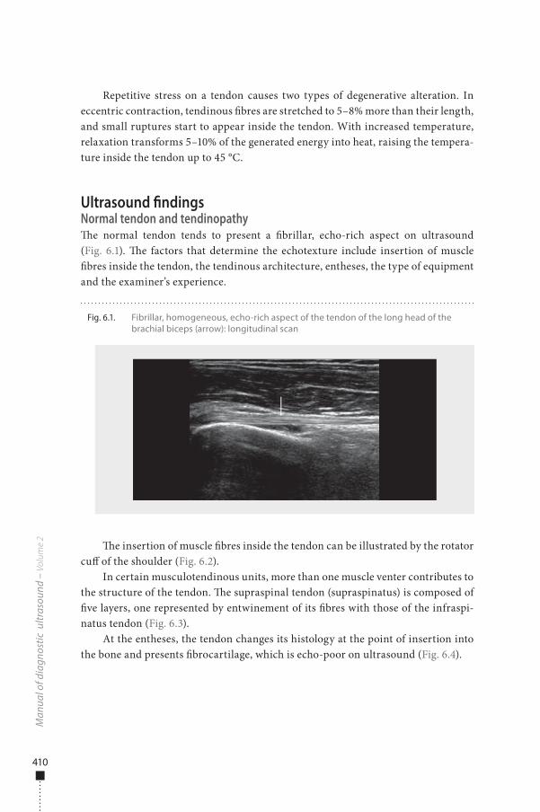
Ultrasound �ndingsNormal tendon and tendinopathy�e normal tendon tends to present a �brillar, echo-rich aspect on ultrasound (Fig. 6.1). �e factors that determine the echotexture include insertion of muscle �bres inside the tendon, the tendinous architecture, entheses, the type of equipment and the examiner’s experience.
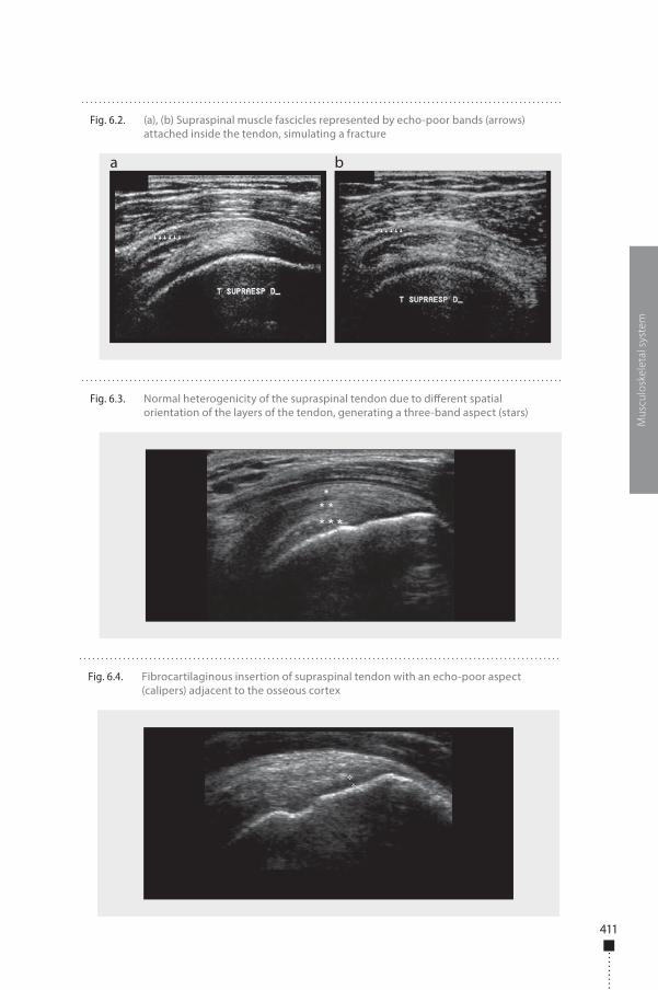
�e insertion of muscle �bres inside the tendon can be illustrated by the rotator cu� of the shoulder (Fig. 6.2).
In certain musculotendinous units, more than one muscle venter contributes to the structure of the tendon. �e supraspinal tendon (supraspinatus) is composed of �ve layers, one represented by entwinement of its �bres with those of the infraspi-natus tendon (Fig. 6.3).
At the entheses, the tendon changes its histology at the point of insertion into the bone and presents �brocartilage, which is echo-poor on ultrasound (Fig. 6.4).
Man
ual o
f dia
gnos
tic u
ltras
ound
– V
olum
e 2
Fig. 6.1. Fibrillar, homogeneous, echo-rich aspect of the tendon of the long head of the brachial biceps (arrow): longitudinal scan
978-9241548540-C006 PRF ml YVR ln ml ml-1-Final Proof.indd 410 6/10/13 10:30 AM
411
Mus
culo
skel
etal
sys
tem
Fig. 6.2. (a), (b) Supraspinal muscle fascicles represented by echo-poor bands (arrows) attached inside the tendon, simulating a fracture
a b
Fig. 6.3. Normal heterogenicity of the supraspinal tendon due to di�erent spatial orientation of the layers of the tendon, generating a three-band aspect (stars)
Fig. 6.4. Fibrocartilaginous insertion of supraspinal tendon with an echo-poor aspect (calipers) adjacent to the osseous cortex
978-9241548540-C006 PRF ml YVR ln ml ml-1-Final Proof.indd 411 6/10/13 10:30 AM
412
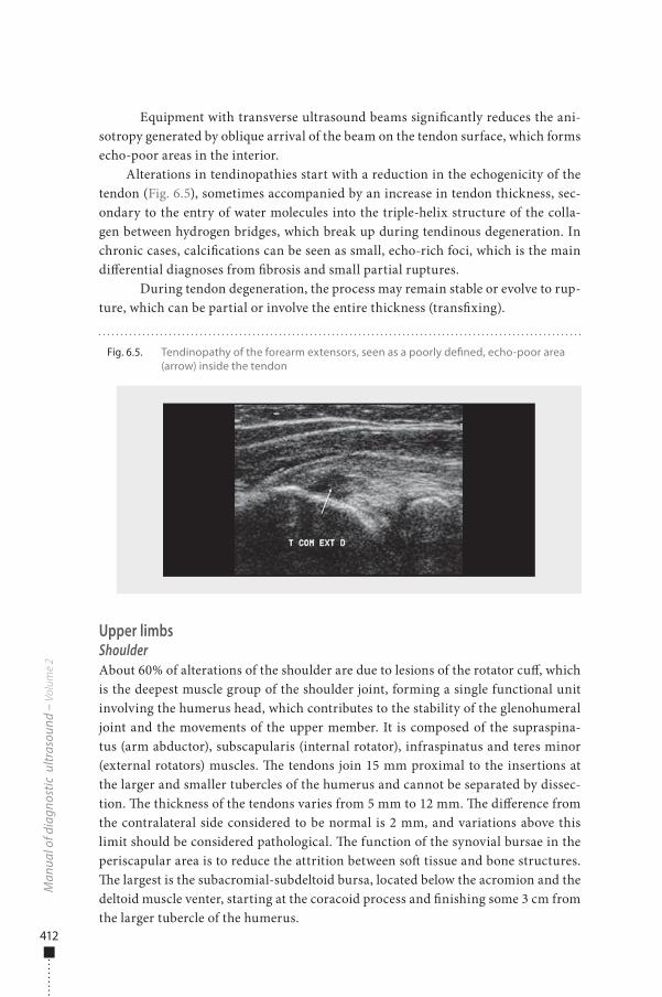
Equipment with transverse ultrasound beams signi�cantly reduces the ani-sotropy generated by oblique arrival of the beam on the tendon surface, which forms echo-poor areas in the interior.
Alterations in tendinopathies start with a reduction in the echogenicity of the tendon (Fig. 6.5), sometimes accompanied by an increase in tendon thickness, sec-ondary to the entry of water molecules into the triple-helix structure of the colla-gen between hydrogen bridges, which break up during tendinous degeneration. In chronic cases, calci�cations can be seen as small, echo-rich foci, which is the main di�erential diagnoses from �brosis and small partial ruptures.
During tendon degeneration, the process may remain stable or evolve to rup-ture, which can be partial or involve the entire thickness (trans�xing).
Upper limbsShoulderAbout 60% of alterations of the shoulder are due to lesions of the rotator cu�, which is the deepest muscle group of the shoulder joint, forming a single functional unit involving the humerus head, which contributes to the stability of the glenohumeral joint and the movements of the upper member. It is composed of the supraspina-tus (arm abductor), subscapularis (internal rotator), infraspinatus and teres minor (external rotators) muscles. �e tendons join 15 mm proximal to the insertions at the larger and smaller tubercles of the humerus and cannot be separated by dissec-tion. �e thickness of the tendons varies from 5 mm to 12 mm. �e di�erence from the contralateral side considered to be normal is 2 mm, and variations above this limit should be considered pathological. �e function of the synovial bursae in the periscapular area is to reduce the attrition between so� tissue and bone structures. �e largest is the subacromial-subdeltoid bursa, located below the acromion and the deltoid muscle venter, starting at the coracoid process and �nishing some 3 cm from the larger tubercle of the humerus.
Man
ual o
f dia
gnos
tic u
ltras
ound
– V
olum
e 2
Fig. 6.5. Tendinopathy of the forearm extensors, seen as a poorly de�ned, echo-poor area (arrow) inside the tendon
978-9241548540-C006 PRF ml YVR ln ml ml-1-Final Proof.indd 412 6/10/13 10:30 AM

413
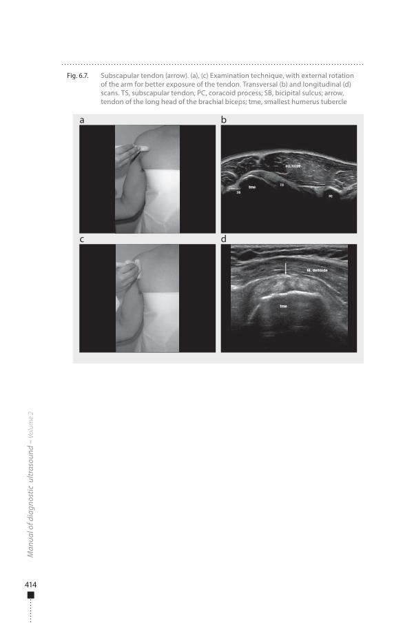
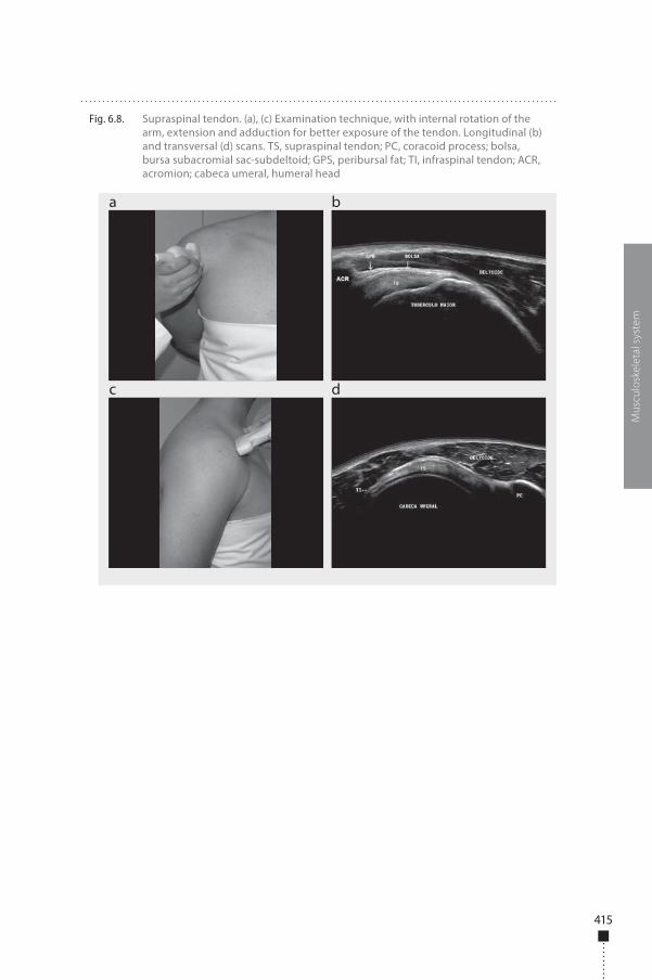
�e patient must cooperate during an ultrasound examination of the tendons of the rotator cu�, as external and internal rotation manoeuvres are necessary (Fig. 6.6, Fig. 6.7, Fig. 6.8, Fig. 6.9, Fig. 6.10). Both the infraspinatus and the teres minor tendon can be evaluated either by placing the hand on the contralateral shoulder or adopting the same position as for examination of the supraspinal tendon. �e pathological processes involving the rotator cu� usually a�ect the supraspinatus tendon, due to normal degeneration of the tendons, trauma, in�ammatory arthritis or tendinosis due to excessive traction or impact syndrome.
Impact syndrome is the commonest cause of pain in the shoulder. It is defined as a group of signs and symptoms characterized by pain and progressive disabling caused by mechanical attrition of the elements of the coracoacromial arch with the structures of the subacromial soft tissues. Abduction (between 70º and 130º) associated with external rotation or anterior elevation with internal rotation of the arm are the commonest movements that cause secondary pain after subacromial impact.
Mus
culo
skel
etal
sys
tem
Fig. 6.6. Extra-articular section of the tendon of the long head of the brachial biceps (TCLB). (a), (c) Examination technique. Transversal (b) and longitudinal (d) scans. tme, smallest humerus tubercle; tma, largest humerus tubercle; lig trans, transverse ligament
a
c
b
d
978-9241548540-C006 PRF ml YVR ln ml ml-1-Final Proof.indd 413 6/10/13 10:30 AM
414
Man
ual o
f dia
gnos
tic u
ltras
ound
– V
olum
e 2
Fig. 6.7. Subscapular tendon (arrow). (a), (c) Examination technique, with external rotation of the arm for better exposure of the tendon. Transversal (b) and longitudinal (d) scans. TS, subscapular tendon; PC, coracoid process; SB, bicipital sulcus; arrow, tendon of the long head of the brachial biceps; tme, smallest humerus tubercle
a
c
b
d
978-9241548540-C006 PRF ml YVR ln ml ml-1-Final Proof.indd 414 6/10/13 10:30 AM
415
Mus
culo
skel
etal
sys
tem
Fig. 6.8. Supraspinal tendon. (a), (c) Examination technique, with internal rotation of the arm, extension and adduction for better exposure of the tendon. Longitudinal (b) and transversal (d) scans. TS, supraspinal tendon; PC, coracoid process; bolsa, bursa subacromial sac-subdeltoid; GPS, peribursal fat; TI, infraspinal tendon; ACR, acromion; cabeca umeral, humeral head
a
c
b
d
978-9241548540-C006 PRF ml YVR ln ml ml-1-Final Proof.indd 415 6/10/13 10:30 AM
416
Man
ual o
f dia
gnos
tic u
ltras
ound
– V
olum
e 2
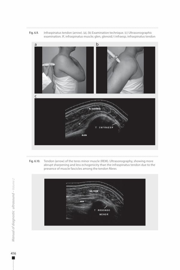
Fig. 6.9. Infraspinatus tendon (arrow). (a), (b) Examination technique. (c) Ultrasonographic examination. IF, infraspinatus muscle; glen, glenoid; t infraesp, infraspinatus tendon
a
c
b
Fig. 6.10. Tendon (arrow) of the teres minor muscle (REM). Ultrasonography, showing more abrupt sharpening and less echogenicity than the infraspinatus tendon due to the presence of muscle fascicles among the tendon �bres
978-9241548540-C006 PRF ml YVR ln ml ml-1-Final Proof.indd 416 6/10/13 10:30 AM
417
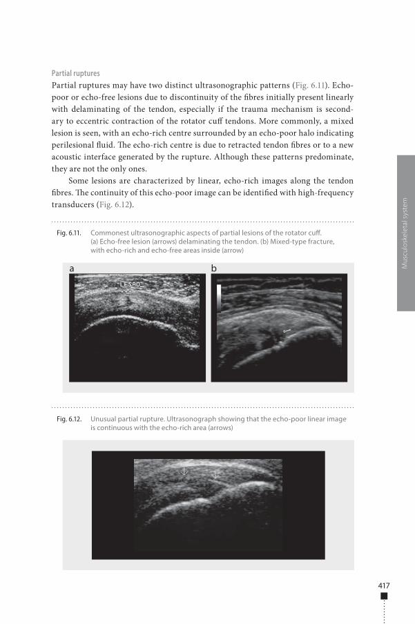
Partial rupturesPartial ruptures may have two distinct ultrasonographic patterns (Fig. 6.11). Echo-poor or echo-free lesions due to discontinuity of the �bres initially present linearly with delaminating of the tendon, especially if the trauma mechanism is second-ary to eccentric contraction of the rotator cu� tendons. More commonly, a mixed lesion is seen, with an echo-rich centre surrounded by an echo-poor halo indicating perilesional �uid. �e echo-rich centre is due to retracted tendon �bres or to a new acoustic interface generated by the rupture. Although these patterns predominate, they are not the only ones.
Some lesions are characterized by linear, echo-rich images along the tendon �bres. �e continuity of this echo-poor image can be identi�ed with high-frequency transducers (Fig. 6.12).
Mus
culo
skel
etal
sys
tem
Fig. 6.11. Commonest ultrasonographic aspects of partial lesions of the rotator cu�. (a) Echo-free lesion (arrows) delaminating the tendon. (b) Mixed-type fracture, with echo-rich and echo-free areas inside (arrow)
a b
Fig. 6.12. Unusual partial rupture. Ultrasonograph showing that the echo-poor linear image is continuous with the echo-rich area (arrows)
978-9241548540-C006 PRF ml YVR ln ml ml-1-Final Proof.indd 417 6/10/13 10:30 AM
418
Complete ruptureComplete, trans�xing ruptures of the entire thickness of the tendon are diagnosed from direct and indirect signs.
�e direct (primary) signs can be divided into two large groups: alteration of the tendinous outline, including the absence and focal tapering of the tendon, and alterations of the echo texture, comprising heterogeneous echogenicity and an echo-free intratendinous focus or split.
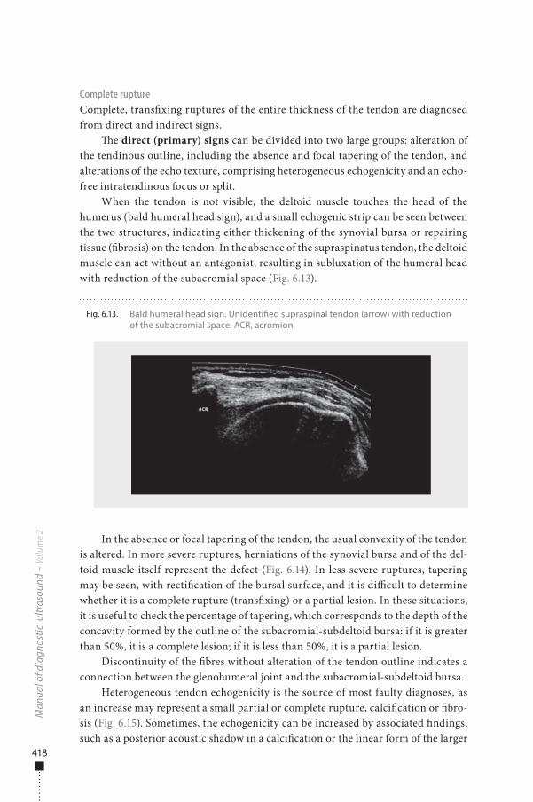
When the tendon is not visible, the deltoid muscle touches the head of the humerus (bald humeral head sign), and a small echogenic strip can be seen between the two structures, indicating either thickening of the synovial bursa or repairing tissue (�brosis) on the tendon. In the absence of the supraspinatus tendon, the deltoid muscle can act without an antagonist, resulting in subluxation of the humeral head with reduction of the subacromial space (Fig. 6.13).
In the absence or focal tapering of the tendon, the usual convexity of the tendon is altered. In more severe ruptures, herniations of the synovial bursa and of the del-toid muscle itself represent the defect (Fig. 6.14). In less severe ruptures, tapering may be seen, with recti�cation of the bursal surface, and it is di�cult to determine whether it is a complete rupture (trans�xing) or a partial lesion. In these situations, it is useful to check the percentage of tapering, which corresponds to the depth of the concavity formed by the outline of the subacromial-subdeltoid bursa: if it is greater than 50%, it is a complete lesion; if it is less than 50%, it is a partial lesion.
Discontinuity of the �bres without alteration of the tendon outline indicates a connection between the glenohumeral joint and the subacromial-subdeltoid bursa.
Heterogeneous tendon echogenicity is the source of most faulty diagnoses, as an increase may represent a small partial or complete rupture, calci�cation or �bro-sis (Fig. 6.15). Sometimes, the echogenicity can be increased by associated �ndings, such as a posterior acoustic shadow in a calci�cation or the linear form of the larger
Man
ual o
f dia
gnos
tic u
ltras
ound
– V
olum
e 2
Fig. 6.13. Bald humeral head sign. Unidenti�ed supraspinal tendon (arrow) with reduction of the subacromial space. ACR, acromion
978-9241548540-C006 PRF ml YVR ln ml ml-1-Final Proof.indd 418 6/10/13 10:30 AM
419
tubercle of the humerus in ruptures. Calci�cations sometimes have a slightly echo-rich aspect, with no acoustic shadow, surrounded by an artefactual linear, echo-poor image, simulating rupture in transition with the tendon. In such cases, a simple radiographic examination will con�rm the presence of calci�cation.
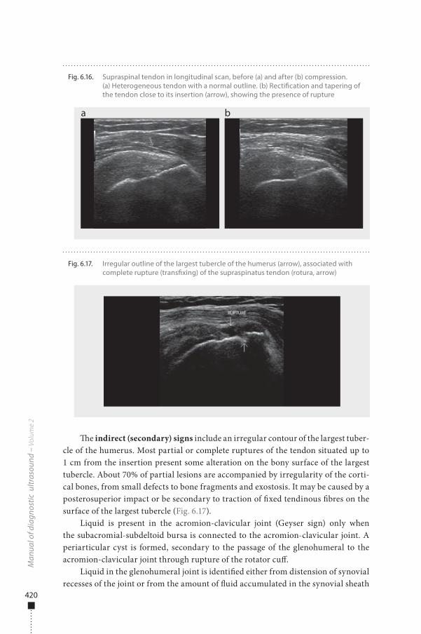
In acute lesions, echogenic blood may �ll the area of the rupture, impeding any change to the tendon and thus a diagnosis. As the echo texture of the tendon is heterogeneous, the transducer should be compressed on the tendon. In rup-tures associated with tendinopathy, the usual convexity of the tendon may be lost (Fig. 6.16). Another manoeuvre that can be used to remove doubt is returning the arm to the neutral position, causing relaxation of the subacromial-subdeltoid bursa and mobilization of the �uid inside the lesion.
Mus
culo
skel
etal
sys
tem
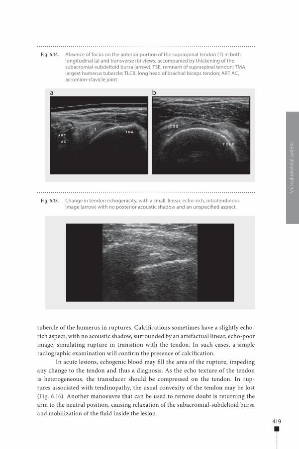
Fig. 6.14. Absence of focus on the anterior portion of the supraspinal tendon (T) in both longitudinal (a) and transverse (b) views, accompanied by thickening of the subacromial-subdeltoid bursa (arrow). TSE, remnant of supraspinal tendon; TMA, largest humerus tubercle; TLCB, long head of brachial biceps tendon; ART AC, acromion-clavicle joint
a b
Fig. 6.15. Change in tendon echogenicity, with a small, linear, echo-rich, intratendinous image (arrow) with no posterior acoustic shadow and an unspeci�ed aspect
978-9241548540-C006 PRF ml YVR ln ml ml-1-Final Proof.indd 419 6/10/13 10:30 AM
420
�e indirect (secondary) signs include an irregular contour of the largest tuber-cle of the humerus. Most partial or complete ruptures of the tendon situated up to 1 cm from the insertion present some alteration on the bony surface of the largest tubercle. About 70% of partial lesions are accompanied by irregularity of the corti-cal bones, from small defects to bone fragments and exostosis. It may be caused by a posterosuperior impact or be secondary to traction of �xed tendinous �bres on the surface of the largest tubercle (Fig. 6.17).
Liquid is present in the acromion-clavicular joint (Geyser sign) only when the subacromial-subdeltoid bursa is connected to the acromion-clavicular joint. A periarticular cyst is formed, secondary to the passage of the glenohumeral to the acromion-clavicular joint through rupture of the rotator cu�.
Liquid in the glenohumeral joint is identi�ed either from distension of synovial recesses of the joint or from the amount of �uid accumulated in the synovial sheath
Man
ual o
f dia
gnos
tic u
ltras
ound
– V
olum
e 2
Fig. 6.16. Supraspinal tendon in longitudinal scan, before (a) and after (b) compression. (a) Heterogeneous tendon with a normal outline. (b) Recti�cation and tapering of the tendon close to its insertion (arrow), showing the presence of rupture
a b
Fig. 6.17. Irregular outline of the largest tubercle of the humerus (arrow), associated with complete rupture (trans�xing) of the supraspinatus tendon (rotura, arrow)
RUPTURE
978-9241548540-C006 PRF ml YVR ln ml ml-1-Final Proof.indd 420 6/10/13 10:30 AM
421
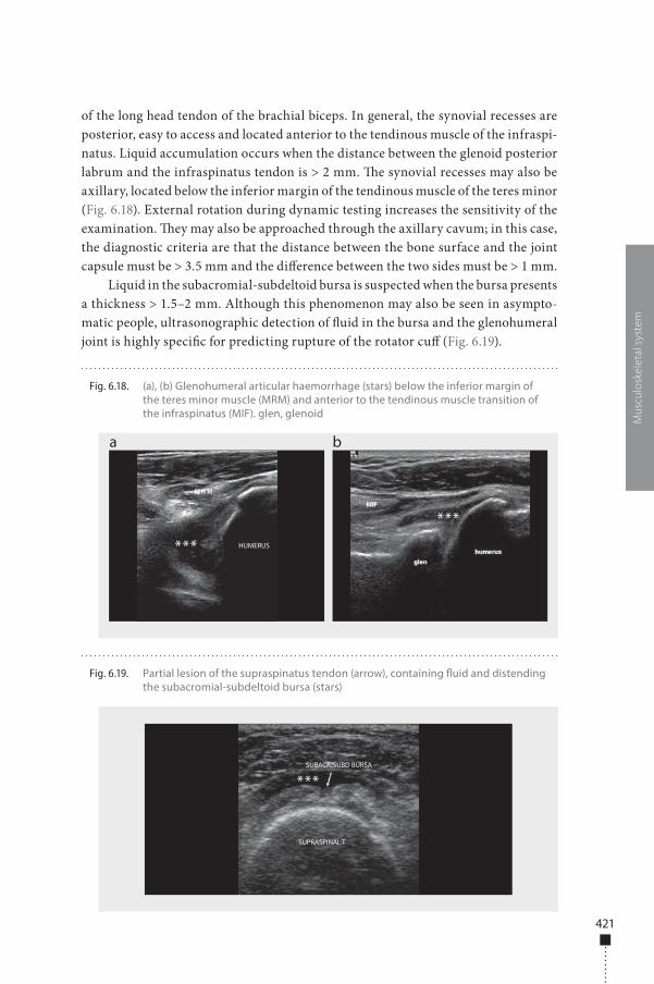
of the long head tendon of the brachial biceps. In general, the synovial recesses are posterior, easy to access and located anterior to the tendinous muscle of the infraspi-natus. Liquid accumulation occurs when the distance between the glenoid posterior labrum and the infraspinatus tendon is > 2 mm. �e synovial recesses may also be axillary, located below the inferior margin of the tendinous muscle of the teres minor (Fig. 6.18). External rotation during dynamic testing increases the sensitivity of the examination. �ey may also be approached through the axillary cavum; in this case, the diagnostic criteria are that the distance between the bone surface and the joint capsule must be > 3.5 mm and the di�erence between the two sides must be > 1 mm.
Liquid in the subacromial-subdeltoid bursa is suspected when the bursa presents a thickness > 1.5–2 mm. Although this phenomenon may also be seen in asympto-matic people, ultrasonographic detection of �uid in the bursa and the glenohumeral joint is highly speci�c for predicting rupture of the rotator cu� (Fig. 6.19).
Mus
culo
skel
etal
sys
tem
Fig. 6.18. (a), (b) Glenohumeral articular haemorrhage (stars) below the inferior margin of the teres minor muscle (MRM) and anterior to the tendinous muscle transition of the infraspinatus (MIF). glen, glenoid
a b
HUMERUS***
***
Fig. 6.19. Partial lesion of the supraspinatus tendon (arrow), containing �uid and distending the subacromial-subdeltoid bursa (stars)
SUBACR/SUBD BURSA
SUPRASPINAL T
***
978-9241548540-C006 PRF ml YVR ln ml ml-1-Final Proof.indd 421 6/10/13 10:30 AM
422
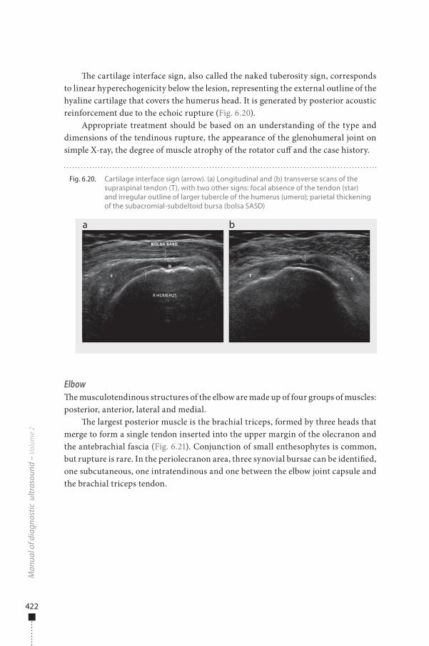
�e cartilage interface sign, also called the naked tuberosity sign, corresponds to linear hyperechogenicity below the lesion, representing the external outline of the hyaline cartilage that covers the humerus head. It is generated by posterior acoustic reinforcement due to the echoic rupture (Fig. 6.20).
Appropriate treatment should be based on an understanding of the type and dimensions of the tendinous rupture, the appearance of the glenohumeral joint on simple X-ray, the degree of muscle atrophy of the rotator cu� and the case history.
Elbow�e musculotendinous structures of the elbow are made up of four groups of muscles: posterior, anterior, lateral and medial.
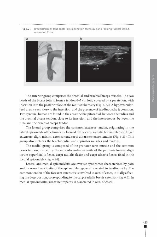
�e largest posterior muscle is the brachial triceps, formed by three heads that merge to form a single tendon inserted into the upper margin of the olecranon and the antebrachial fascia (Fig. 6.21). Conjunction of small enthesophytes is common, but rupture is rare. In the periolecranon area, three synovial bursae can be identi�ed, one subcutaneous, one intratendinous and one between the elbow joint capsule and the brachial triceps tendon.
Man
ual o
f dia
gnos
tic u
ltras
ound
– V
olum
e 2
Fig. 6.20. Cartilage interface sign (arrow). (a) Longitudinal and (b) transverse scans of the supraspinal tendon (T), with two other signs: focal absence of the tendon (star) and irregular outline of larger tubercle of the humerus (umero); parietal thickening of the subacromial-subdeltoid bursa (bolsa SASD)
a b
R HUMERUS
*
978-9241548540-C006 PRF ml YVR ln ml ml-1-Final Proof.indd 422 6/10/13 10:30 AM
423
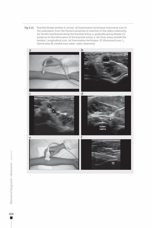
�e anterior group comprises the brachial and brachial biceps muscles. �e two heads of the biceps join to form a tendon 6–7 cm long covered by a paratenon, with insertion into the posterior face of the radius tuberosity (Fig. 6.22). A hypovascular-ized area is seen close to the insertion, and the presence of tendinopathy is common. Two synovial bursae are found in the area: the bicipitoradial, between the radius and the brachial biceps tendon, close to its insertion, and the interosseous, between the ulna and the brachial biceps tendon.
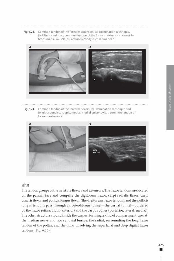
�e lateral group comprises the common extensor tendon, originating in the lateral epicondyle of the humerus, formed by the carpi radialis brevis extensor, �nger extensors, digiti minimi extensor and carpi ulnaris extensor tendons (Fig. 6.23). �is group also includes the brachioradial and supinator muscles and tendons.
�e medial group is composed of the pronator teres muscle and the common �exor tendon, formed by the musculotendinous units of the palmaris longus, digi-torum super�cialis �exor, carpi radialis �exor and carpi ulnaris �exor, �xed in the medial epicondyle (Fig. 6.24).
Lateral and medial epicondylitis are overuse syndromes characterized by pain and increased sensitivity of the epicondyles, generally related to tendinopathy. �e common tendon of the forearm extensors is involved in 80% of cases, initially a�ect-ing the deep portion, corresponding to the carpi radialis brevis extensor (Fig. 6.5). In medial epicondylitis, ulnar neuropathy is associated in 60% of cases.
Mus
culo
skel
etal
sys
tem
Fig. 6.21. Brachial triceps tendon (t). (a) Examination technique and (b) longitudinal scan. f, olecranon fossa
a b
OLECRANON
HUMERUS
978-9241548540-C006 PRF ml YVR ln ml ml-1-Final Proof.indd 423 6/10/13 10:30 AM
424
Man
ual o
f dia
gnos
tic u
ltras
ound
– V
olum
e 2
Fig. 6.22. Brachial biceps tendon (t, arrow). (a) Examination technique; transverse scan in the axial plane, from the forearm proximal to insertion in the radius tuberosity. (b) Tendon positioned along the brachial artery, a, gradually going deeper (c), posterior to the bifurcation of the brachial artery, a. (d) Ulnar artery beside the tendon. Longitudinal scan. (e) Examination technique. (f) Ultrasound scan. L, lateral area; M, medial area; tuber. radio, tuberosity
a
c
e f
b
d
978-9241548540-C006 PRF ml YVR ln ml ml-1-Final Proof.indd 424 6/10/13 10:30 AM
425
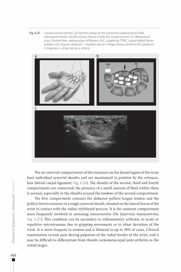
Wrist�e tendon groups of the wrist are �exors and extensors. �e �exor tendons are located on the palmar face and comprise the digitorum �exor, carpi radialis �exor, carpi ulnaris �exor and pollicis longus �exor. �e digitorum �exor tendons and the pollicis longus tendons pass through an osteo�brous tunnel—the carpal tunnel—bordered by the �exor retinaculum (anterior) and the carpus bones (posterior, lateral, medial). �e other structures found inside the carpus, forming a kind of compartment, are fat, the median nerve and two synovial bursae: the radial, surrounding the long �exor tendon of the pollex, and the ulnar, involving the super�cial and deep digital �exor tendons (Fig. 6.25).
Mus
culo
skel
etal
sys
tem
Fig. 6.23. Common tendon of the forearm extensors. (a) Examination technique. (b) Ultrasound scan; common tendon of the forearm extensors (arrow). br, brachioradial muscle; el, lateral epicondyle; cr, radius head
a b
Fig. 6.24. Common tendon of the forearm �exors. (a) Examination technique and (b) ultrasound scan. epic. medial, medial epicondyle. t, common tendon of forearm extensors
a b
978-9241548540-C006 PRF ml YVR ln ml ml-1-Final Proof.indd 425 6/10/13 10:30 AM
426
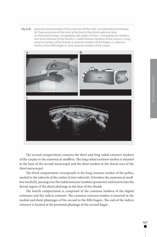
�e six synovial compartments of the extensors on the dorsal region of the wrist have individual synovial sheaths and are maintained in position by the retinacu-lum (dorsal carpal ligament; Fig. 6.26). �e sheaths of the second, third and fourth compartments are connected; the presence of a small amount of �uid within them is normal, especially in the sheaths around the tendons of the second compartment.
�e �rst compartment contains the abductor pollicis longus tendon and the pollicis brevis extensor in a single synovial sheath, situated on the lateral fascia of the wrist in contact with the radius stylohyoid process. It is the extensor compartment most frequently involved in stenosing tenosynovitis (De Quervain tenosynovitis; Fig. 6.27). �is condition can be secondary to in�ammatory arthritis, to acute or repetitive microtraumas due to gripping movements or to ulnar deviation of the wrist. It is more frequent in women and is bilateral in up to 30% of cases. Clinical examination reveals pain during palpation of the radial border of the wrist, and it may be di�cult to di�erentiate from thumb carpometacarpal joint arthritis in the initial stages.
Man
ual o
f dia
gnos
tic u
ltras
ound
– V
olum
e 2
Fig. 6.25. Carpal tunnel (arrow). (a) Section plane at the proximal carpal tunnel with ultrasound beam. (b) Structures found inside the carpal tunnel. (c) Ultrasound scan. Dotted line, retinaculum of �exors; ESC, scaphoid; TFRC, carpal radial �exor tendon; CG, Guyon channel; *, median nerve; t, �nger �exor tendon; PIS, pisiform; t, trapeze; n, ulnar nerve; a, artery
a
c
b
d
978-9241548540-C006 PRF ml YVR ln ml ml-1-Final Proof.indd 426 6/10/13 10:30 AM
427
�e second compartment contains the short and long radial extensor tendons of the carpus in the anatomical snu�ox. �e long radial extensor tendon is situated at the base of the second metacarpal and the short tendon in the dorsal area of the third metacarpal.
�e third compartment corresponds to the long extensor tendon of the pollex, medial to the tubercle of the radius (Lister tubercle). It borders the anatomical snu�-box medially, passing over the radial extensor tendons (posterior) and inserts into the dorsal region of the distal phalange at the base of the thumb.
�e fourth compartment is composed of the common tendons of the digital extensors and the indicis extensor. �e common extensor tendon is inserted in the medial and distal phalanges of the second to the ��h �ngers. �e end of the indicis extensor is located at the proximal phalange of the second �nger.
Mus
culo
skel
etal
sys
tem
Fig. 6.26. Synovial compartments of the extensors of the wrist. (a) Examination technique. (b) Transverse view of the wrist at the level of the distal radio and ulna. (c) Ultrasound image. Sonography: star, tuber of Lister; 1, long abductor tendons and short extensor of the thumb; 2, radial extensor tendons of the carpus; 3, long extensor tendon of the thumb; 4, extensor tendons of the �ngers; 5, extensor tendon of the �fth �nger; 6, ulnar extensor tendon of the carpus
a
c
b
d*
978-9241548540-C006 PRF ml YVR ln ml ml-1-Final Proof.indd 427 6/10/13 10:30 AM
428
�e ��h compartment contains the extensor tendon of the ��h �nger, seen posterior to the radioulnar joint, with insertion in the medial and distal phalanges of the ��h �nger.
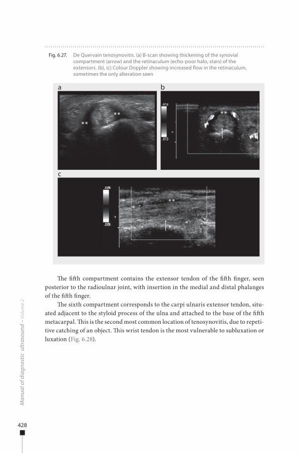
�e sixth compartment corresponds to the carpi ulnaris extensor tendon, situ-ated adjacent to the styloid process of the ulna and attached to the base of the ��h metacarpal. �is is the second most common location of tenosynovitis, due to repeti-tive catching of an object. �is wrist tendon is the most vulnerable to subluxation or luxation (Fig. 6.28).
Man
ual o
f dia
gnos
tic u
ltras
ound
– V
olum
e 2
Fig. 6.27. De Quervain tenosynovitis. (a) B-scan showing thickening of the synovial compartment (arrow) and the retinaculum (echo-poor halo, stars) of the extensors. (b), (c) Colour Doppler showing increased �ow in the retinaculum, sometimes the only alteration seen
a
c
b
d
**** **
**
978-9241548540-C006 PRF ml YVR ln ml ml-1-Final Proof.indd 428 6/10/13 10:30 AM
429
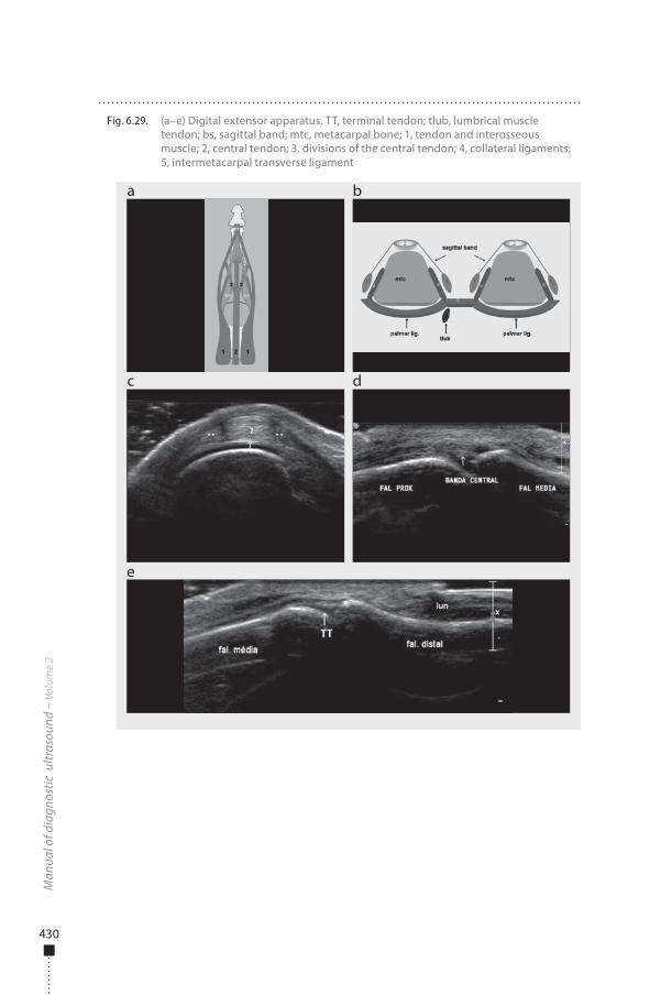
Fingers�e tendinous anatomy of the �ngers is di�erent in the palmar and dorsal regions. A central tendon is inserted in the base of the medium phalanx on its dorsal face. Two tendinous bands meet near the base of the distal phalanx, medially and laterally to this tendon, forming the terminal tendon. Narrow strips of collagen, known as sagit-tal bands, link these structures to provide stability and allow harmonious extension. Because of this complex anatomy, the term ‘digital extensor apparatus’ is used rather than ‘extensor tendon of the �nger’ (Fig. 6.29).
�e �exor tendons are located in the palmar region of the hand and �ngers. �e super�cial �exor tendon at the level of the proximal phalanx is anterior to the �exor digitorum profundus. In its distal course, it divides into two bands, with insertion in the medial phalanx posterior to the �exor digitorum profundus, which runs to the base of the distal phalanx (Fig. 6.30). In contrast to the extensor apparatus, the �exor tendons have a synovial sheath all along the phalanges.
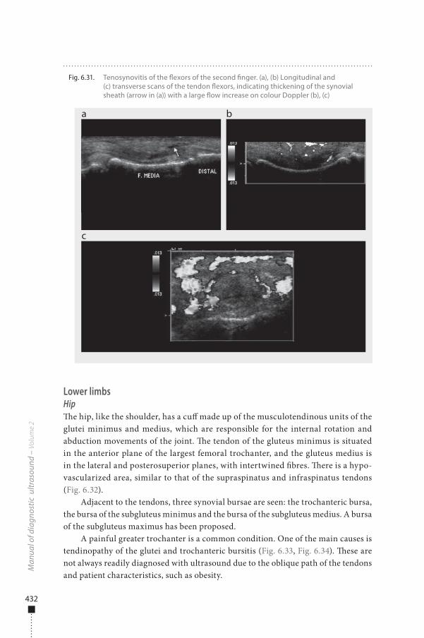
In cases of tenosynovitis, there may be some parietal thickening, �uid or increased �ow in the synovial sheath on colour Doppler (Fig. 6.31).
Mus
culo
skel
etal
sys
tem
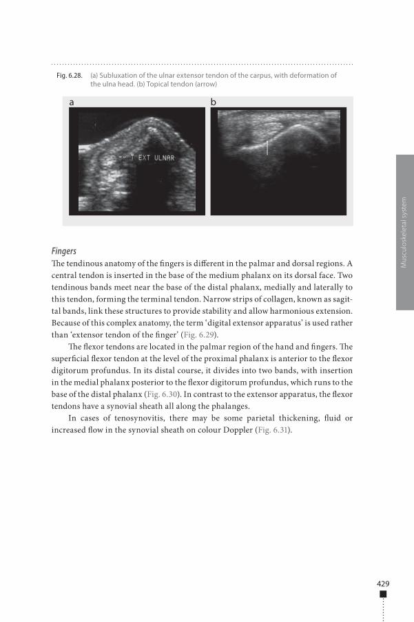
Fig. 6.28. (a) Subluxation of the ulnar extensor tendon of the carpus, with deformation of the ulna head. (b) Topical tendon (arrow)
a b
978-9241548540-C006 PRF ml YVR ln ml ml-1-Final Proof.indd 429 6/10/13 10:30 AM
430
Man
ual o
f dia
gnos
tic u
ltras
ound
– V
olum
e 2
Fig. 6.29. (a–e) Digital extensor apparatus. TT, terminal tendon; tlub, lumbrical muscle tendon; bs, sagittal band; mtc, metacarpal bone; 1, tendon and interosseous muscle; 2, central tendon; 3, divisions of the central tendon; 4, collateral ligaments; 5, intermetacarpal transverse ligament
a
c
e
b
d
978-9241548540-C006 PRF ml YVR ln ml ml-1-Final Proof.indd 430 6/10/13 10:30 AM

431
Mus
culo
skel
etal
sys
tem
Fig. 6.30. Flexor tendons. (a) Surgical view. (b)–(e) Sections at which transverse scans of the �exor tendons were made. (f) Longitudinal scan of the �exor tendons of the proximal, medial and distal phalanges. FS and continuous arrows, super�cial �exor tendon; FP and stars, deep �exor tendon; dotted arrow, �exor tendons
a
c
e f
b
d
** ** **
** **
978-9241548540-C006 PRF ml YVR ln ml ml-1-Final Proof.indd 431 6/10/13 10:30 AM
432
Lower limbsHip�e hip, like the shoulder, has a cu� made up of the musculotendinous units of the glutei minimus and medius, which are responsible for the internal rotation and abduction movements of the joint. �e tendon of the gluteus minimus is situated in the anterior plane of the largest femoral trochanter, and the gluteus medius is in the lateral and posterosuperior planes, with intertwined �bres. �ere is a hypo-vascularized area, similar to that of the supraspinatus and infraspinatus tendons (Fig. 6.32).
Adjacent to the tendons, three synovial bursae are seen: the trochanteric bursa, the bursa of the subgluteus minimus and the bursa of the subgluteus medius. A bursa of the subgluteus maximus has been proposed.
A painful greater trochanter is a common condition. One of the main causes is tendinopathy of the glutei and trochanteric bursitis (Fig. 6.33, Fig. 6.34). �ese are not always readily diagnosed with ultrasound due to the oblique path of the tendons and patient characteristics, such as obesity.
Man
ual o
f dia
gnos
tic u
ltras
ound
– V
olum
e 2
Fig. 6.31. Tenosynovitis of the �exors of the second �nger. (a), (b) Longitudinal and (c) transverse scans of the tendon �exors, indicating thickening of the synovial sheath (arrow in (a)) with a large �ow increase on colour Doppler (b), (c)
a
c
b
978-9241548540-C006 PRF ml YVR ln ml ml-1-Final Proof.indd 432 6/10/13 10:30 AM
433
Mus
culo
skel
etal
sys
tem
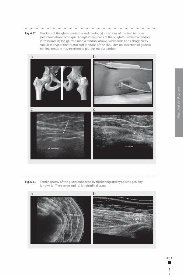
Fig. 6.32. Tendons of the gluteus minima and media. (a) Insertions of the two tendons. (b) Examination technique. Longitudinal scans of the (c) gluteus minima tendon (arrow) and (d) the gluteus media tendon (arrow), with forms and echogenicity similar to that of the rotator cu� tendons of the shoulder. mi, insertion of gluteus minima tendon; me, insertion of gluteus media tendon
a
c d
b
GL MINIMA TGL MEDIA T
Fig. 6.33. Tendinopathy of the glutei enhanced by thickening and hypoechogenicity (arrow). (a) Transverse and (b) longitudinal scans
a b
978-9241548540-C006 PRF ml YVR ln ml ml-1-Final Proof.indd 433 6/10/13 10:30 AM
434
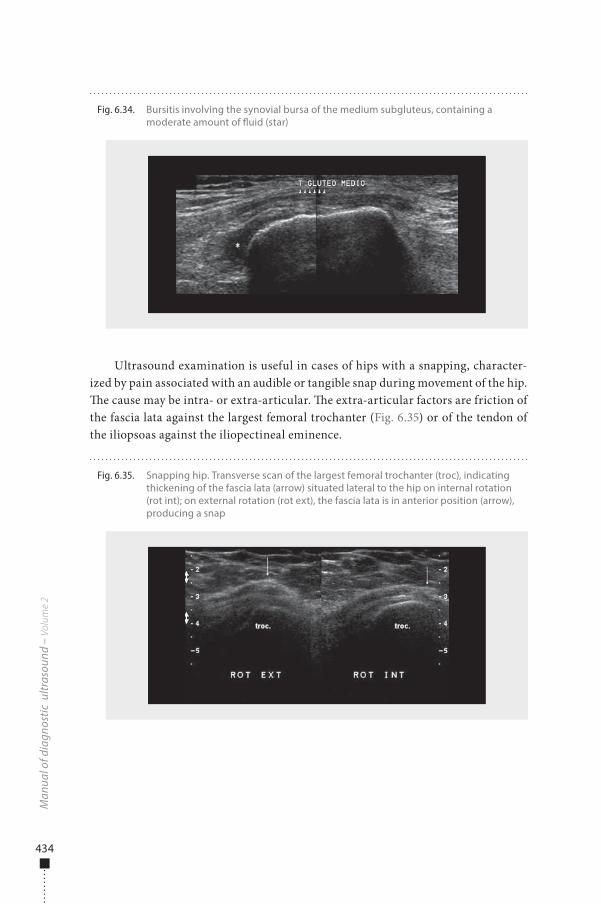
Ultrasound examination is useful in cases of hips with a snapping, character-ized by pain associated with an audible or tangible snap during movement of the hip. �e cause may be intra- or extra-articular. �e extra-articular factors are friction of the fascia lata against the largest femoral trochanter (Fig. 6.35) or of the tendon of the iliopsoas against the iliopectineal eminence.
Man
ual o
f dia
gnos
tic u
ltras
ound
– V
olum
e 2
Fig. 6.34. Bursitis involving the synovial bursa of the medium subgluteus, containing a moderate amount of �uid (star)
Fig. 6.35. Snapping hip. Transverse scan of the largest femoral trochanter (troc), indicating thickening of the fascia lata (arrow) situated lateral to the hip on internal rotation (rot int); on external rotation (rot ext), the fascia lata is in anterior position (arrow), producing a snap
978-9241548540-C006 PRF ml YVR ln ml ml-1-Final Proof.indd 434 6/10/13 10:30 AM
435
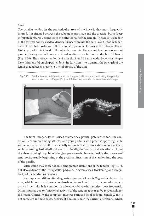
Knee�e patellar tendon in the periarticular area of the knee is that most frequently injured. It is situated between the subcutaneous tissue and the pretibial bursa (deep infrapatellar bursa), posterior to the inferior half of the tendon. �e acoustic shadow of the cortical bone is used to identify its insertion into the patella and into the tuber-osity of the tibia. Posterior to the tendon is a pad of fat known as the infrapatellar or Ho�a pad, which is joined to the articular synovia. �e normal tendon is formed of parallel, homogeneous �bres, visualized as alternate echo-poor and echo-rich bands (Fig. 6.36). �e average tendon is 4 mm thick and 21 mm wide. Sedentary people have thinner, ribbon-shaped tendons. Its function is to transmit the strength of the femoral quadriceps muscle to the tuberosity of the tibia.
�e term ‘jumper’s knee’ is used to describe a painful patellar tendon. �e con-dition is common among athletes and young adults who practise sport regularly, secondary to excessive e�ort, especially in sports that require extension of the knee, such as running, basketball and football. Usually, the dominant side is a�ected. From the histopathological point of view, jumper’s knee is characterized by the presence of tendinosis, usually beginning at the proximal insertion of the tendon into the apex of the patella.
Ultrasound may show not only echographic alterations of the tendon (Fig. 6.37), but also oedema of the infrapatellar pad and, in severe cases, thickening and irregu-larity of the tendinous envelope.
An important di�erential diagnosis of jumper’s knee is Osgood-Schlatter dis-ease, which consists of osteochondrosis or osteochondritis of the anterior tuber-osity of the tibia. It is common in adolescent boys who practise sport frequently. Microtraumas due to functional activity of the tendon appear to be responsible for the lesion. Clinically, the complaint involves pain and local oedema. Simple X-ray is not su�cient in these cases, because it does not show the earliest alterations, which
Mus
culo
skel
etal
sys
tem
Fig. 6.36. Patellar tendon. (a) Examination technique. (b) Ultrasound, indicating the patellar tendon and the Ho�a pad (GH), which is echo-poor with linear echo-rich images
a b
978-9241548540-C006 PRF ml YVR ln ml ml-1-Final Proof.indd 435 6/10/13 10:30 AM
436
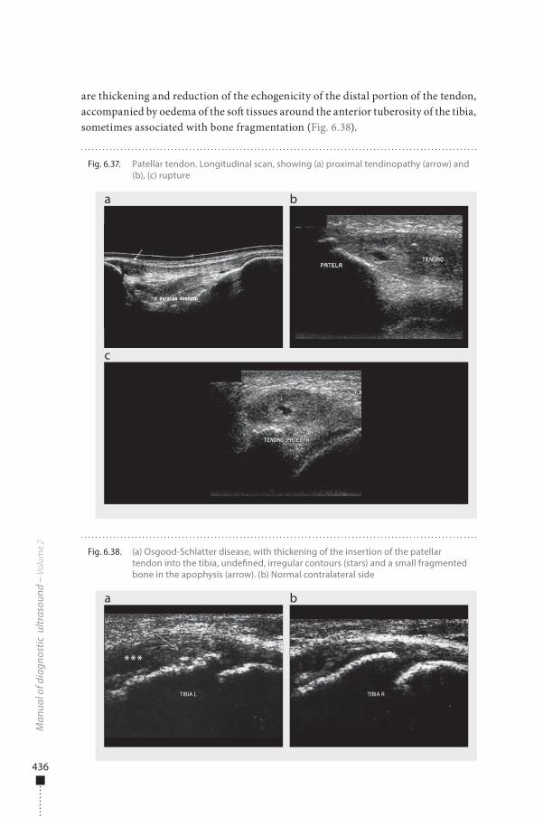
are thickening and reduction of the echogenicity of the distal portion of the tendon, accompanied by oedema of the so� tissues around the anterior tuberosity of the tibia, sometimes associated with bone fragmentation (Fig. 6.38).
Man
ual o
f dia
gnos
tic u
ltras
ound
– V
olum
e 2
Fig. 6.37. Patellar tendon. Longitudinal scan, showing (a) proximal tendinopathy (arrow) and (b), (c) rupture
a
c
b
Fig. 6.38. (a) Osgood-Schlatter disease, with thickening of the insertion of the patellar tendon into the tibia, unde�ned, irregular contours (stars) and a small fragmented bone in the apophysis (arrow). (b) Normal contralateral side
a b
TIBIA L TIBIA R
***
978-9241548540-C006 PRF ml YVR ln ml ml-1-Final Proof.indd 436 6/10/13 10:30 AM
437
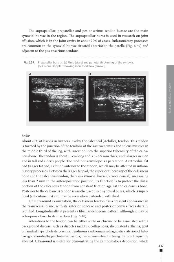
�e suprapatellar, prepatellar and pes anserinus tendon bursae are the main synovial bursae in the region. �e suprapatellar bursa is used in research on joint e�usion, which is in the joint cavity in about 90% of cases. In�ammatory processes are common in the synovial bursae situated anterior to the patella (Fig. 6.39) and adjacent to the pes anserinus tendons.
AnkleAbout 20% of lesions in runners involve the calcaneal (Achilles) tendon. �is tendon is formed by the junction of the tendons of the gastrocnemius and soleus muscles in the middle third of the leg, with insertion into the superior tuberosity of the calca-neus bone. �e tendon is about 15 cm long and 3.5–6.9 mm thick, and is larger in men and in tall and elderly people. �e tendinous envelope is a paratenon. A retrotibial fat pad (Kager fat pad) is found anterior to the tendon, which may be a�ected in in�am-matory processes. Between the Kager fat pad, the superior tuberosity of the calcaneus bone and the calcaneus tendon, there is a synovial bursa (retrocalcaneal), measuring less than 2 mm in the anteroposterior position; its function is to protect the distal portion of the calcaneus tendon from constant friction against the calcaneus bone. Posterior to the calcaneus tendon is another, acquired synovial bursa, which is super-�cial (subcutaneous) and may be seen when distended with �uid.
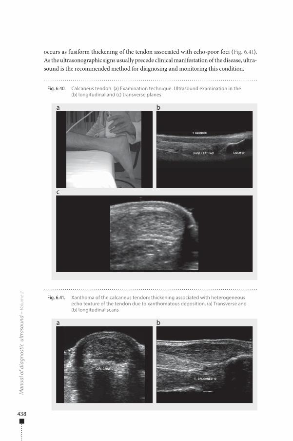
On ultrasound examination, the calcaneus tendon has a crescent appearance in the transversal plane, with its anterior concave and posterior convex faces distally recti�ed. Longitudinally, it presents a �brillar echogenic pattern, although it may be echo-poor closer to its insertion (Fig. 6.40).
Alterations to the tendon can be either acute or chronic or be associated with a background disease, such as diabetes mellitus, collagenosis, rheumatoid arthritis, gout or familial hypercholesterolaemia. Tendinous xanthoma is a diagnostic criterion of hete-rozygous familial hypercholesterolaemia, the calcaneus tendon being the most frequently a�ected. Ultrasound is useful for demonstrating the xanthomatous deposition, which
Mus
culo
skel
etal
sys
tem
Fig. 6.39. Prepatellar bursitis. (a) Fluid (stars) and parietal thickening of the synovia. (b) Colour Doppler showing increased �ow (arrows)
a b
978-9241548540-C006 PRF ml YVR ln ml ml-1-Final Proof.indd 437 6/10/13 10:30 AM
438
occurs as fusiform thickening of the tendon associated with echo-poor foci (Fig. 6.41). As the ultrasonographic signs usually precede clinical manifestation of the disease, ultra-sound is the recommended method for diagnosing and monitoring this condition.
Man
ual o
f dia
gnos
tic u
ltras
ound
– V
olum
e 2
Fig. 6.41. Xanthoma of the calcaneus tendon: thickening associated with heterogeneous echo texture of the tendon due to xanthomatous deposition. (a) Transverse and (b) longitudinal scans
a b
Fig. 6.40. Calcaneus tendon. (a) Examination technique. Ultrasound examination in the (b) longitudinal and (c) transverse planes
a
c
b
KAGER FAT PAD
978-9241548540-C006 PRF ml YVR ln ml ml-1-Final Proof.indd 438 6/10/13 10:30 AM
439
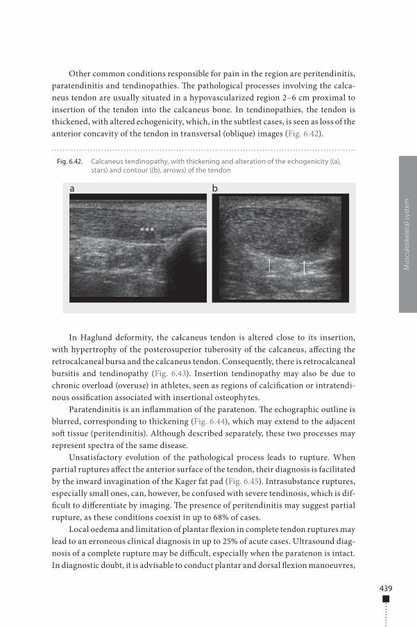
Other common conditions responsible for pain in the region are peritendinitis, paratendinitis and tendinopathies. �e pathological processes involving the calca-neus tendon are usually situated in a hypovascularized region 2–6 cm proximal to insertion of the tendon into the calcaneus bone. In tendinopathies, the tendon is thickened, with altered echogenicity, which, in the subtlest cases, is seen as loss of the anterior concavity of the tendon in transversal (oblique) images (Fig. 6.42).
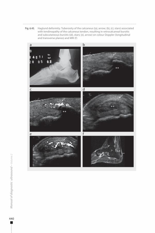
In Haglund deformity, the calcaneus tendon is altered close to its insertion, with hypertrophy of the posterosuperior tuberosity of the calcaneus, a�ecting the retrocalcaneal bursa and the calcaneus tendon. Consequently, there is retrocalcaneal bursitis and tendinopathy (Fig. 6.43). Insertion tendinopathy may also be due to chronic overload (overuse) in athletes, seen as regions of calci�cation or intratendi-nous ossi�cation associated with insertional osteophytes.
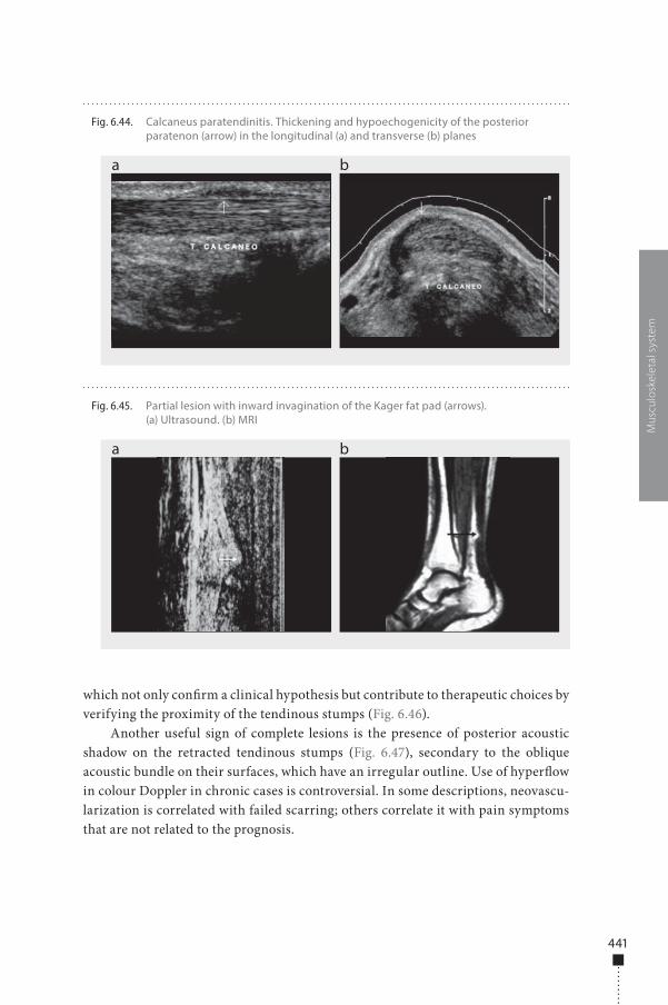
Paratendinitis is an in�ammation of the paratenon. �e echographic outline is blurred, corresponding to thickening (Fig. 6.44), which may extend to the adjacent so� tissue (peritendinitis). Although described separately, these two processes may represent spectra of the same disease.
Unsatisfactory evolution of the pathological process leads to rupture. When partial ruptures a�ect the anterior surface of the tendon, their diagnosis is facilitated by the inward invagination of the Kager fat pad (Fig. 6.45). Intrasubstance ruptures, especially small ones, can, however, be confused with severe tendinosis, which is dif-�cult to di�erentiate by imaging. �e presence of peritendinitis may suggest partial rupture, as these conditions coexist in up to 68% of cases.
Local oedema and limitation of plantar �exion in complete tendon ruptures may lead to an erroneous clinical diagnosis in up to 25% of acute cases. Ultrasound diag-nosis of a complete rupture may be di�cult, especially when the paratenon is intact. In diagnostic doubt, it is advisable to conduct plantar and dorsal �exion manoeuvres,
Mus
culo
skel
etal
sys
tem
Fig. 6.42. Calcaneus tendinopathy, with thickening and alteration of the echogenicity ((a), stars) and contour ((b), arrows) of the tendon
a b
***
978-9241548540-C006 PRF ml YVR ln ml ml-1-Final Proof.indd 439 6/10/13 10:30 AM
440
Man
ual o
f dia
gnos
tic u
ltras
ound
– V
olum
e 2
Fig. 6.43. Haglund deformity. Tuberosity of the calcaneus ((a), arrow; (b), (c), stars) associated with tendinopathy of the calcaneus tendon, resulting in retrocalcaneal bursitis and subcutaneous bursitis ((d), stars; (e), arrow) on colour Doppler (longitudinal and transverse planes) and MRI (f)
a
c
e f
b
d
****
**
978-9241548540-C006 PRF ml YVR ln ml ml-1-Final Proof.indd 440 6/10/13 10:30 AM
441
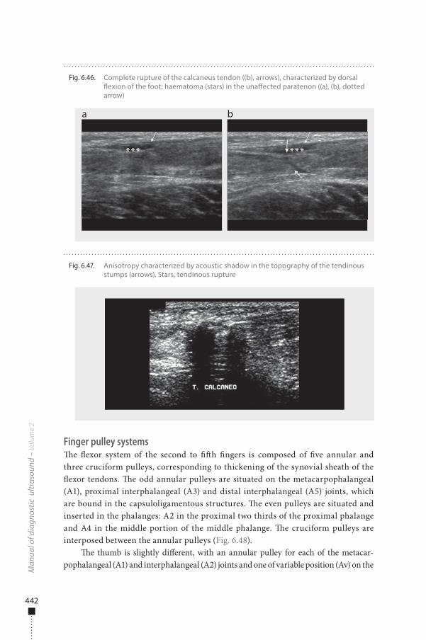
which not only con�rm a clinical hypothesis but contribute to therapeutic choices by verifying the proximity of the tendinous stumps (Fig. 6.46).
Another useful sign of complete lesions is the presence of posterior acoustic shadow on the retracted tendinous stumps (Fig. 6.47), secondary to the oblique acoustic bundle on their surfaces, which have an irregular outline. Use of hyper�ow in colour Doppler in chronic cases is controversial. In some descriptions, neovascu-larization is correlated with failed scarring; others correlate it with pain symptoms that are not related to the prognosis.
Mus
culo
skel
etal
sys
tem
Fig. 6.44. Calcaneus paratendinitis. Thickening and hypoechogenicity of the posterior paratenon (arrow) in the longitudinal (a) and transverse (b) planes
a b
Fig. 6.45. Partial lesion with inward invagination of the Kager fat pad (arrows). (a) Ultrasound. (b) MRI
a b
978-9241548540-C006 PRF ml YVR ln ml ml-1-Final Proof.indd 441 6/10/13 10:30 AM
442
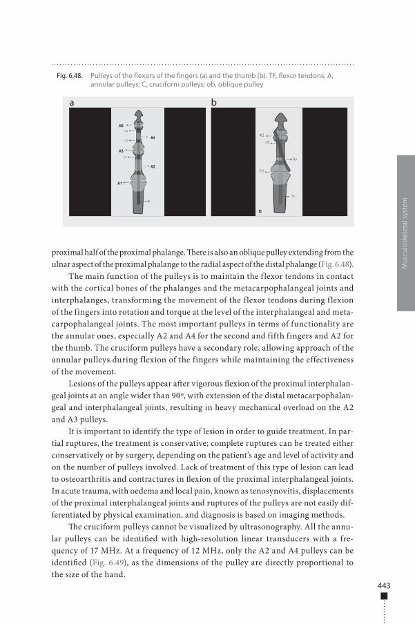
Finger pulley systems�e �exor system of the second to ��h �ngers is composed of �ve annular and three cruciform pulleys, corresponding to thickening of the synovial sheath of the �exor tendons. �e odd annular pulleys are situated on the metacarpophalangeal (A1), proximal interphalangeal (A3) and distal interphalangeal (A5) joints, which are bound in the capsuloligamentous structures. �e even pulleys are situated and inserted in the phalanges: A2 in the proximal two thirds of the proximal phalange and A4 in the middle portion of the middle phalange. �e cruciform pulleys are interposed between the annular pulleys (Fig. 6.48).
�e thumb is slightly di�erent, with an annular pulley for each of the metacar-pophalangeal (A1) and interphalangeal (A2) joints and one of variable position (Av) on the
Man
ual o
f dia
gnos
tic u
ltras
ound
– V
olum
e 2
Fig. 6.46. Complete rupture of the calcaneus tendon ((b), arrows), characterized by dorsal �exion of the foot; haematoma (stars) in the una�ected paratenon ((a), (b), dotted arrow)
a b
*** ***
Fig. 6.47. Anisotropy characterized by acoustic shadow in the topography of the tendinous stumps (arrows). Stars, tendinous rupture
**
978-9241548540-C006 PRF ml YVR ln ml ml-1-Final Proof.indd 442 6/10/13 10:30 AM
443
proximal half of the proximal phalange. �ere is also an oblique pulley extending from the ulnar aspect of the proximal phalange to the radial aspect of the distal phalange (Fig. 6.48).
The main function of the pulleys is to maintain the f lexor tendons in contact with the cortical bones of the phalanges and the metacarpophalangeal joints and interphalanges, transforming the movement of the f lexor tendons during f lexion of the fingers into rotation and torque at the level of the interphalangeal and meta-carpophalangeal joints. The most important pulleys in terms of functionality are the annular ones, especially A2 and A4 for the second and fifth fingers and A2 for the thumb. The cruciform pulleys have a secondary role, allowing approach of the annular pulleys during f lexion of the fingers while maintaining the effectiveness of the movement.
Lesions of the pulleys appear a�er vigorous �exion of the proximal interphalan-geal joints at an angle wider than 90º, with extension of the distal metacarpophalan-geal and interphalangeal joints, resulting in heavy mechanical overload on the A2 and A3 pulleys.
It is important to identify the type of lesion in order to guide treatment. In par-tial ruptures, the treatment is conservative; complete ruptures can be treated either conservatively or by surgery, depending on the patient’s age and level of activity and on the number of pulleys involved. Lack of treatment of this type of lesion can lead to osteoarthritis and contractures in �exion of the proximal interphalangeal joints. In acute trauma, with oedema and local pain, known as tenosynovitis, displacements of the proximal interphalangeal joints and ruptures of the pulleys are not easily dif-ferentiated by physical examination, and diagnosis is based on imaging methods.
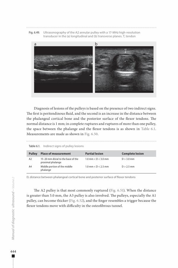
�e cruciform pulleys cannot be visualized by ultrasonography. All the annu-lar pulleys can be identi�ed with high-resolution linear transducers with a fre-quency of 17 MHz. At a frequency of 12 MHz, only the A2 and A4 pulleys can be identi�ed (Fig. 6.49), as the dimensions of the pulley are directly proportional to the size of the hand.
Mus
culo
skel
etal
sys
tem
Fig. 6.48. Pulleys of the �exors of the �ngers (a) and the thumb (b). TF, �exor tendons; A, annular pulleys; C, cruciform pulleys; ob, oblique pulley
a b
978-9241548540-C006 PRF ml YVR ln ml ml-1-Final Proof.indd 443 6/10/13 10:30 AM
444
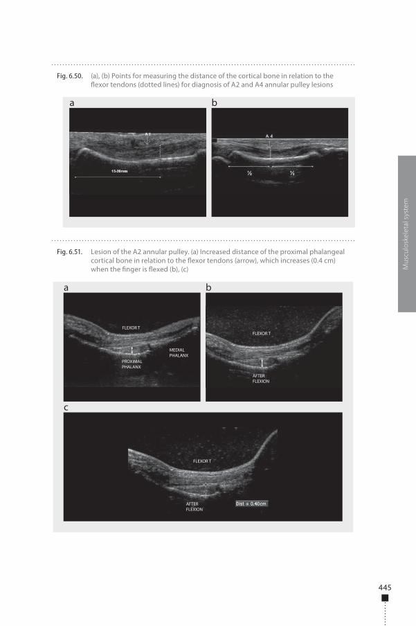
Diagnosis of lesions of the pulleys is based on the presence of two indirect signs. �e �rst is peritendinous �uid, and the second is an increase in the distance between the phalangeal cortical bone and the posterior surface of the �exor tendons. �e normal distance is 1 mm; in complete ruptures and ruptures of more than one pulley, the space between the phalange and the �exor tendons is as shown in Table 6.1. Measurements are made as shown in Fig. 6.50.
�e A2 pulley is that most commonly ruptured (Fig. 6.51). When the distance is greater than 5.0 mm, the A3 pulley is also involved. �e pulleys, especially the A1 pulley, can become thicker (Fig. 6.52), and the �nger resembles a trigger because the �exor tendons move with di�culty in the osteo�brous tunnel.
Man
ual o
f dia
gnos
tic u
ltras
ound
– V
olum
e 2
Fig. 6.49. Ultrasonography of the A2 annular pulley with a 17-MHz high-resolution transducer in the (a) longitudinal and (b) transverse planes. T, tendon
a b
PHALANX
Table 6.1. Indirect signs of pulley lesions
Pulley Place of measurement Partial lesion Complete lesion
A2 15–20 mm distal to the base of the proximal phalange
1.0 mm < D < 3.0 mm D > 3.0 mm
A4 Middle portion of the middle phalange
1.0 mm < D < 2.5 mm D > 2.5 mm
D, distance between phalangeal cortical bone and posterior surface of �exor tendons
978-9241548540-C006 PRF ml YVR ln ml ml-1-Final Proof.indd 444 6/10/13 10:30 AM
445
Mus
culo
skel
etal
sys
tem
Fig. 6.50. (a), (b) Points for measuring the distance of the cortical bone in relation to the �exor tendons (dotted lines) for diagnosis of A2 and A4 annular pulley lesions
a b
Fig. 6.51. Lesion of the A2 annular pulley. (a) Increased distance of the proximal phalangeal cortical bone in relation to the �exor tendons (arrow), which increases (0.4 cm) when the �nger is �exed (b), (c)
a
c
b
FLEXOR T
FLEXOR T
FLEXOR T
AFTER FLEXION
AFTER FLEXION
PROXIMAL PHALANX
MEDIAL PHALANX
978-9241548540-C006 PRF ml YVR ln ml ml-1-Final Proof.indd 445 6/10/13 10:30 AM
446
Ligaments
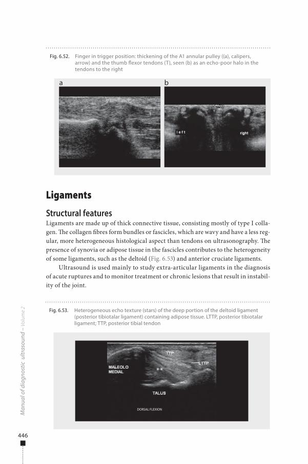
Structural featuresLigaments are made up of thick connective tissue, consisting mostly of type I colla-gen. �e collagen �bres form bundles or fascicles, which are wavy and have a less reg-ular, more heterogeneous histological aspect than tendons on ultrasonography. �e presence of synovia or adipose tissue in the fascicles contributes to the heterogeneity of some ligaments, such as the deltoid (Fig. 6.53) and anterior cruciate ligaments.
Ultrasound is used mainly to study extra-articular ligaments in the diagnosis of acute ruptures and to monitor treatment or chronic lesions that result in instabil-ity of the joint.
Man
ual o
f dia
gnos
tic u
ltras
ound
– V
olum
e 2
Fig. 6.52. Finger in trigger position: thickening of the A1 annular pulley ((a), calipers, arrow) and the thumb �exor tendons (T), seen (b) as an echo-poor halo in the tendons to the right
a b
Fig. 6.53. Heterogeneous echo texture (stars) of the deep portion of the deltoid ligament (posterior tibiotalar ligament) containing adipose tissue. LTTP, posterior tibiotalar ligament; TTP, posterior tibial tendon
DORSAL FLEXION
**
978-9241548540-C006 PRF ml YVR ln ml ml-1-Final Proof.indd 446 6/10/13 10:30 AM
447
Lateral ligament complex of the ankle�e commonest lesions associated with sport are of the lateral ligament complex of the ankle (16–21%). �ese become chronic in more than 40% of cases if not appropri-ately treated. �e lateral ligament complex of the ankle is made up of three ligaments: the calcaneo�bular and the anterior and posterior talo�bular.
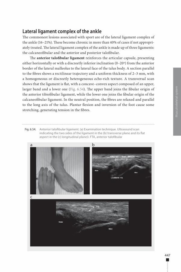
�e anterior talo�bular ligament reinforces the articular capsule, presenting either horizontally or with a discreetly inferior inclination (0–20º) from the anterior border of the lateral malleolus to the lateral face of the talus body. A section parallel to the �bres shows a rectilinear trajectory and a uniform thickness of 2–3 mm, with a homogeneous or discreetly heterogeneous echo-rich texture. A transversal scan shows that the ligament is �at, with a concave–convex aspect composed of an upper, larger band and a lower one (Fig. 6.54). �e upper band joins the �bular origin of the anterior tibio�bular ligament, while the lower one joins the �bular origin of the calcaneo�bular ligament. In the neutral position, the �bres are relaxed and parallel to the long axis of the talus. Plantar �exion and inversion of the foot cause some stretching, generating tension in the �bres.
Mus
culo
skel
etal
sys
tem
Fig. 6.54. Anterior talo�bular ligament. (a) Examination technique. Ultrasound scan indicating the two sides of the ligament in the (b) transverse plane and its �at aspect in the (c) longitudinal plane3. FTA, anterior talo�bular
a
c
b
978-9241548540-C006 PRF ml YVR ln ml ml-1-Final Proof.indd 447 6/10/13 10:30 AM
448
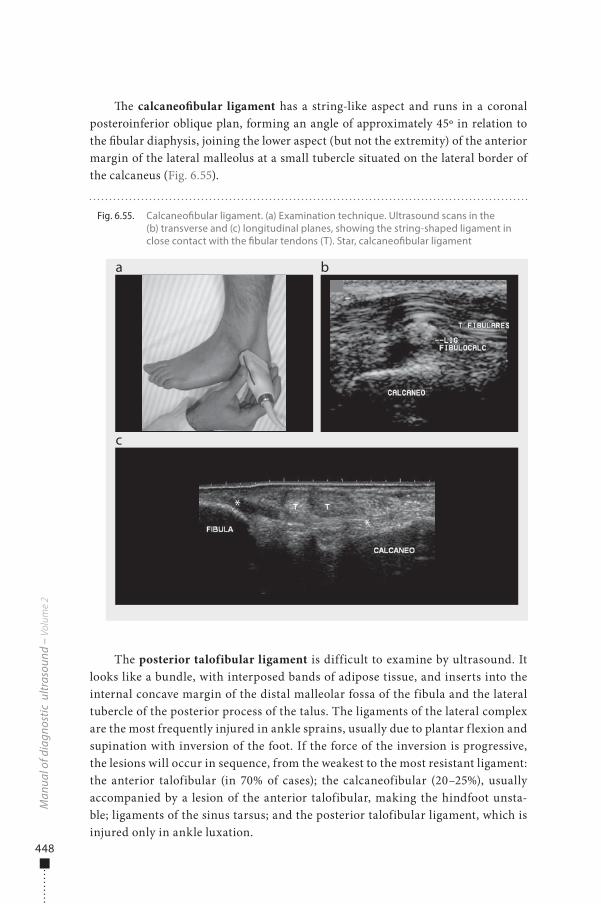
�e calcaneo�bular ligament has a string-like aspect and runs in a coronal posteroinferior oblique plan, forming an angle of approximately 45º in relation to the �bular diaphysis, joining the lower aspect (but not the extremity) of the anterior margin of the lateral malleolus at a small tubercle situated on the lateral border of the calcaneus (Fig. 6.55).
The posterior talofibular ligament is difficult to examine by ultrasound. It looks like a bundle, with interposed bands of adipose tissue, and inserts into the internal concave margin of the distal malleolar fossa of the fibula and the lateral tubercle of the posterior process of the talus. The ligaments of the lateral complex are the most frequently injured in ankle sprains, usually due to plantar f lexion and supination with inversion of the foot. If the force of the inversion is progressive, the lesions will occur in sequence, from the weakest to the most resistant ligament: the anterior talofibular (in 70% of cases); the calcaneofibular (20–25%), usually accompanied by a lesion of the anterior talofibular, making the hindfoot unsta-ble; ligaments of the sinus tarsus; and the posterior talofibular ligament, which is injured only in ankle luxation.
Man
ual o
f dia
gnos
tic u
ltras
ound
– V
olum
e 2
Fig. 6.55. Calcaneo�bular ligament. (a) Examination technique. Ultrasound scans in the (b) transverse and (c) longitudinal planes, showing the string-shaped ligament in close contact with the �bular tendons (T). Star, calcaneo�bular ligament
a
c
b
**
978-9241548540-C006 PRF ml YVR ln ml ml-1-Final Proof.indd 448 6/10/13 10:30 AM
449
A diagnosis is frequently made solely by clinical evaluation; however, the accu-racy of diagnosis of an acute lesion is reduced in 50% of cases by pain and local oedema, and imaging methods are recommended. MRI has been reported to be more accurate than ultrasound for the diagnosis of ligament lesions; however, the stud-ies were conducted before the advent of high-resolution transducers, and there has been no recent comparison of the performance of ultrasound and MRI with current ultrasound equipment.
Ligament lesions can be classi�ed according to the time since the trauma (acute and chronic lesions) and the extent or severity of the rupture (partial or complete). Ultrasound diagnosis is based on direct and indirect signs. �e nonspeci�c, indirect signs in calcaneo�bular ruptures are oedema or subcutaneous bruises on the lateral face of the ankle; articular e�usion in the anterolateral talo�bular recess; lesions of the anterior talo�bular ligament; and �uid in the synovial sheath of the �bular tendons.
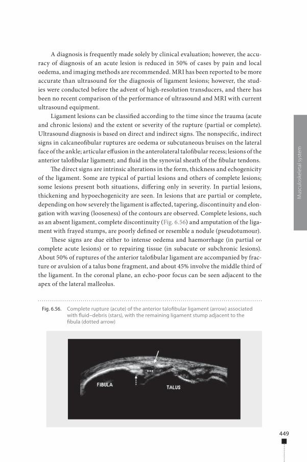
�e direct signs are intrinsic alterations in the form, thickness and echogenicity of the ligament. Some are typical of partial lesions and others of complete lesions; some lesions present both situations, di�ering only in severity. In partial lesions, thickening and hypoechogenicity are seen. In lesions that are partial or complete, depending on how severely the ligament is a�ected, tapering, discontinuity and elon-gation with waving (looseness) of the contours are observed. Complete lesions, such as an absent ligament, complete discontinuity (Fig. 6.56) and amputation of the liga-ment with frayed stumps, are poorly de�ned or resemble a nodule (pseudotumour).
�ese signs are due either to intense oedema and haemorrhage (in partial or complete acute lesions) or to repairing tissue (in subacute or subchronic lesions). About 50% of ruptures of the anterior talo�bular ligament are accompanied by frac-ture or avulsion of a talus bone fragment, and about 45% involve the middle third of the ligament. In the coronal plane, an echo-poor focus can be seen adjacent to the apex of the lateral malleolus.
Mus
culo
skel
etal
sys
tem
Fig. 6.56. Complete rupture (acute) of the anterior talo�bular ligament (arrow) associated with �uid–debris (stars), with the remaining ligament stump adjacent to the �bula (dotted arrow)
978-9241548540-C006 PRF ml YVR ln ml ml-1-Final Proof.indd 449 6/10/13 10:30 AM
450
Oedema of the so� tissue disappears during healing, which begins 7 days a�er a trauma. �e ligament is always thickened; the �rst evidence of repair of a ligament, with visualization of echoes �lling the discontinuities, appears about 5 weeks a�er a trauma. An echo-rich focus can be seen inside the scarred ligament, corresponding to calci�cations, and bone irregularities are found adjacent to the insertions into the �bula and the talus as a consequence of bone avulsion.
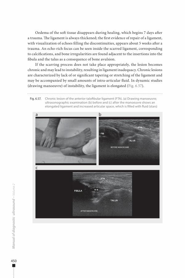
If the scarring process does not take place appropriately, the lesion becomes chronic and may lead to instability, resulting in ligament inadequacy. Chronic lesions are characterized by lack of or signi�cant tapering or stretching of the ligament and may be accompanied by small amounts of intra-articular �uid. In dynamic studies (drawing manoeuvre) of instability, the ligament is elongated (Fig. 6.57).
Man
ual o
f dia
gnos
tic u
ltras
ound
– V
olum
e 2
Fig. 6.57. Chronic lesion of the anterior talo�bular ligament (FTA). (a) Drawing manoeuvre; ultrasonographic examination (b) before and (c) after the manoeuvre shows an elongated ligament and increased articular space, which is �lled with �uid (stars)
a
c
b
BEFORE MANOEUVRE
AFTER MANOEUVRE
**
978-9241548540-C006 PRF ml YVR ln ml ml-1-Final Proof.indd 450 6/10/13 10:30 AM
451
Muscle
Muscle is the largest individual mass of corporal tissue, corresponding to 40–45% of a person’s weight. It is classi�ed as elastic or nonelastic. Elastic muscle tissue is made up of muscle �bres joined into fascicles, which form the muscle. Nonelastic struc-tures are made up of muscle surrounded by sheaths formed by connective tissues and muscle fasciae. �e endomysium is an extensive network of capillaries and nerves involving all muscle �bres. Muscle �bres are bound into fascicles by perimysium, a �broadipose septum made up of vessels, nerves and conjunctive and fat tissue. �e epimysium, composed of dense conjunctive tissue, separates muscle venters and dif-ferent muscles, such as the semimembranosus and the femoral biceps in the posterior thigh. �e fascia is situated externally to the epimysium and contains a whole muscle.
Muscles may contain slow-twitch (type I) �bres rich in oxygen or fast-twitch (type II) �bres, with anaerobic metabolism. �e proportion of each type of �bre inside the muscle venter is determined genetically, by type of physical training and by the location, form and function of the muscle. Posture muscles have linearly arranged fascicles, a prevalence of type I �bres and many mitochondria, allowing sustained low-energy contraction. �e muscles in the super�cial areas of the extremities, usu-ally passing over more than one joint, have �bres with a pennate distribution and contain predominantly type II �bres. Muscles with these characteristics give more vigorous contractions and have a propensity to rupture.
Muscle contractions can be divided into isotonic and isometric. In isometric con-tractions, the length of the muscle �bre remains constant with changes in the applied load on the muscle. In isotonic contractions, the length of the muscle �bre changes, either shortening (concentric contraction) or lengthening (eccentric contraction). Usually, agonist muscles involved in a certain movement undergo concentric contraction due to the stability of the closest joint, which is determined by the eccentric contraction of the antagonist muscle, which is also responsible for slowing down the movement. �is occurs, for instance, during a kick, when the stability of the knee joint is maintained by contraction of the ischiotibials, so that the femoral quadriceps can execute the movement.
Muscle rupturesMuscle ruptures are secondary to direct or indirect trauma. Direct traumas, or con-tusions, involve compression of the muscle against a bone structure, so that the lesion is due to crushing. Indirect traumas are due to stretching of muscle �bres and can be generated by passive hyperextension of the fascicles, although they usually occur during eccentric contraction of the muscle.
�us, both morphological and functional factors increase the risk for muscle lesion, the main ones being passing over more than one joint, eccentric contraction, predominance of type II �bres (quick contraction) and a super�cial location at the extremities, mainly in the lower limbs. �e site of the lesion depends on age and physical condition and is due to biomechanical particularities that determine weaker
Mus
culo
skel
etal
sys
tem
978-9241548540-C006 PRF ml YVR ln ml ml-1-Final Proof.indd 451 6/10/13 10:30 AM
452
areas. In the immature skeleton, lesions are usually found at the interface between tendon and bone, with a greater probability of fracture due to avulsion. In athletes and other young adults, lesions usually occur in the musculotendinous area, while in elderly people ruptures usually a�ect the tendon, resulting in tendinosis. When the lesion is of muscular origin, pain is restricted to the a�ected region, beginning imme-diately a�er the trauma. Sometimes, subcutaneous bruises can be seen 12–24 h a�er a trauma. If the alteration occurs in a tendon, the symptoms are di�use and irradiated.
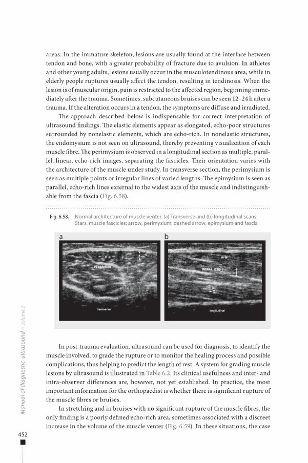
�e approach described below is indispensable for correct interpretation of ultrasound �ndings. �e elastic elements appear as elongated, echo-poor structures surrounded by nonelastic elements, which are echo-rich. In nonelastic structures, the endomysium is not seen on ultrasound, thereby preventing visualization of each muscle �bre. �e perimysium is observed in a longitudinal section as multiple, paral-lel, linear, echo-rich images, separating the fascicles. �eir orientation varies with the architecture of the muscle under study. In transverse section, the perimysium is seen as multiple points or irregular lines of varied lengths. �e epimysium is seen as parallel, echo-rich lines external to the widest axis of the muscle and indistinguish-able from the fascia (Fig. 6.58).
In post-trauma evaluation, ultrasound can be used for diagnosis, to identify the muscle involved, to grade the rupture or to monitor the healing process and possible complications, thus helping to predict the length of rest. A system for grading muscle lesions by ultrasound is illustrated in Table 6.2. Its clinical usefulness and inter- and intra-observer di�erences are, however, not yet established. In practice, the most important information for the orthopaedist is whether there is signi�cant rupture of the muscle �bres or bruises.
In stretching and in bruises with no signi�cant rupture of the muscle �bres, the only �nding is a poorly de�ned echo-rich area, sometimes associated with a discreet increase in the volume of the muscle venter (Fig. 6.59). In these situations, the case
Man
ual o
f dia
gnos
tic u
ltras
ound
– V
olum
e 2
Fig. 6.58. Normal architecture of muscle venter. (a) Transverse and (b) longitudinal scans. Stars, muscle fascicles; arrow, perimysium; dashed arrow, epimysium and fascia
a b
** **
**
978-9241548540-C006 PRF ml YVR ln ml ml-1-Final Proof.indd 452 6/10/13 10:30 AM
453
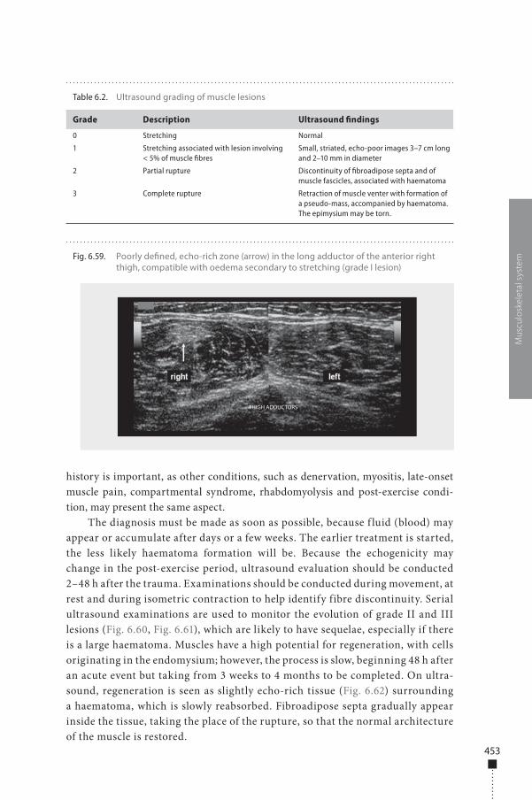
history is important, as other conditions, such as denervation, myositis, late-onset muscle pain, compartmental syndrome, rhabdomyolysis and post-exercise condi-tion, may present the same aspect.
The diagnosis must be made as soon as possible, because f luid (blood) may appear or accumulate after days or a few weeks. The earlier treatment is started, the less likely haematoma formation will be. Because the echogenicity may change in the post-exercise period, ultrasound evaluation should be conducted 2–48 h after the trauma. Examinations should be conducted during movement, at rest and during isometric contraction to help identify fibre discontinuity. Serial ultrasound examinations are used to monitor the evolution of grade II and III lesions (Fig. 6.60, Fig. 6.61), which are likely to have sequelae, especially if there is a large haematoma. Muscles have a high potential for regeneration, with cells originating in the endomysium; however, the process is slow, beginning 48 h after an acute event but taking from 3 weeks to 4 months to be completed. On ultra-sound, regeneration is seen as slightly echo-rich tissue (Fig. 6.62) surrounding a haematoma, which is slowly reabsorbed. Fibroadipose septa gradually appear inside the tissue, taking the place of the rupture, so that the normal architecture of the muscle is restored.
Mus
culo
skel
etal
sys
temFig. 6.59. Poorly de�ned, echo-rich zone (arrow) in the long adductor of the anterior right
thigh, compatible with oedema secondary to stretching (grade I lesion)
THIGH ADDUCTORS
Table 6.2. Ultrasound grading of muscle lesions
Grade Description Ultrasound findings
0 Stretching Normal
1 Stretching associated with lesion involving < 5% of muscle fibres
Small, striated, echo-poor images 3–7 cm long and 2–10 mm in diameter
2 Partial rupture Discontinuity of fibroadipose septa and of muscle fascicles, associated with haematoma
3 Complete rupture Retraction of muscle venter with formation of a pseudo-mass, accompanied by haematoma. The epimysium may be torn.
978-9241548540-C006 PRF ml YVR ln ml ml-1-Final Proof.indd 453 6/10/13 10:30 AM
454
Man
ual o
f dia
gnos
tic u
ltras
ound
– V
olum
e 2
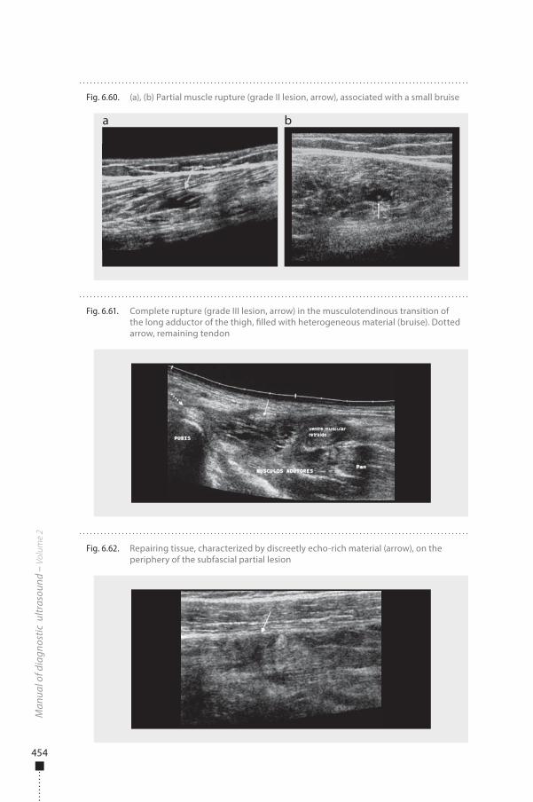
Fig. 6.60. (a), (b) Partial muscle rupture (grade II lesion, arrow), associated with a small bruise
a b
Fig. 6.61. Complete rupture (grade III lesion, arrow) in the musculotendinous transition of the long adductor of the thigh, �lled with heterogeneous material (bruise). Dotted arrow, remaining tendon
Fig. 6.62. Repairing tissue, characterized by discreetly echo-rich material (arrow), on the periphery of the subfascial partial lesion
978-9241548540-C006 PRF ml YVR ln ml ml-1-Final Proof.indd 454 6/10/13 10:30 AM
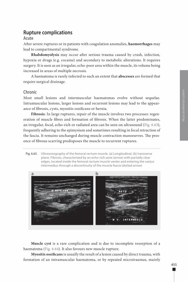
455
Rupture complicationsAcuteA�er severe ruptures or in patients with coagulation anomalies, haemorrhages may lead to compartmental syndrome.
Rhabdomyolysis may occur a�er serious trauma caused by crush, infection, hypoxia or drugs (e.g. cocaine) and secondary to metabolic alterations. It requires surgery. It is seen as an irregular, echo-poor area within the muscle, its volume being increased in areas of multiple necrosis.
A haematoma is rarely infected to such an extent that abscesses are formed that require surgical drainage.
ChronicMost small lesions and intermuscular haematomas evolve without sequelae. Intramuscular lesions, larger lesions and recurrent lesions may lead to the appear-ance of �brosis, cysts, myositis ossi�cans or hernia.
Fibrosis: In large ruptures, repair of the muscle involves two processes: regen-eration of muscle �bres and formation of �brosis. When the latter predominates, an irregular, focal, echo-rich or radiated area can be seen on ultrasound (Fig. 6.63), frequently adhering to the epimysium and sometimes resulting in focal retraction of the fascia. It remains unchanged during muscle contraction manoeuvres. �e pres-ence of �brous scarring predisposes the muscle to recurrent ruptures.
Muscle cyst is a rare complication and is due to incomplete resorption of a haematoma (Fig. 6.64). It also favours new muscle rupture.
Myositis ossi�cans is usually the result of a lesion caused by direct trauma, with formation of an intramuscular haematoma, or by repeated microtraumas, mainly
Mus
culo
skel
etal
sys
tem
Fig. 6.63. Ultrasonography of the femoral rectum muscle. (a) Longitudinal, (b) transverse plane. Fibrosis, characterized by an echo-rich zone (arrow) with partially clear edges, located inside the femoral rectum muscle venter and entering the vastus intermedius through a discontinuity of the muscle fascia (dotted arrow)
a b
978-9241548540-C006 PRF ml YVR ln ml ml-1-Final Proof.indd 455 6/10/13 10:30 AM
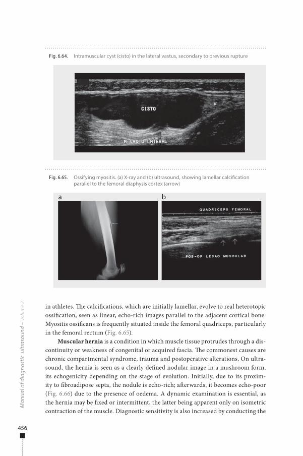
456
in athletes. �e calci�cations, which are initially lamellar, evolve to real heterotopic ossi�cation, seen as linear, echo-rich images parallel to the adjacent cortical bone. Myositis ossi�cans is frequently situated inside the femoral quadriceps, particularly in the femoral rectum (Fig. 6.65).
Muscular hernia is a condition in which muscle tissue protrudes through a dis-continuity or weakness of congenital or acquired fascia. �e commonest causes are chronic compartmental syndrome, trauma and postoperative alterations. On ultra-sound, the hernia is seen as a clearly de�ned nodular image in a mushroom form, its echogenicity depending on the stage of evolution. Initially, due to its proxim-ity to �broadipose septa, the nodule is echo-rich; a�erwards, it becomes echo-poor (Fig. 6.66) due to the presence of oedema. A dynamic examination is essential, as the hernia may be �xed or intermittent, the latter being apparent only on isometric contraction of the muscle. Diagnostic sensitivity is also increased by conducting the
Man
ual o
f dia
gnos
tic u
ltras
ound
– V
olum
e 2
Fig. 6.64. Intramuscular cyst (cisto) in the lateral vastus, secondary to previous rupture
Fig. 6.65. Ossifying myositis. (a) X-ray and (b) ultrasound, showing lamellar calci�cation parallel to the femoral diaphysis cortex (arrow)
a b
978-9241548540-C006 PRF ml YVR ln ml ml-1-Final Proof.indd 456 6/10/13 10:30 AM
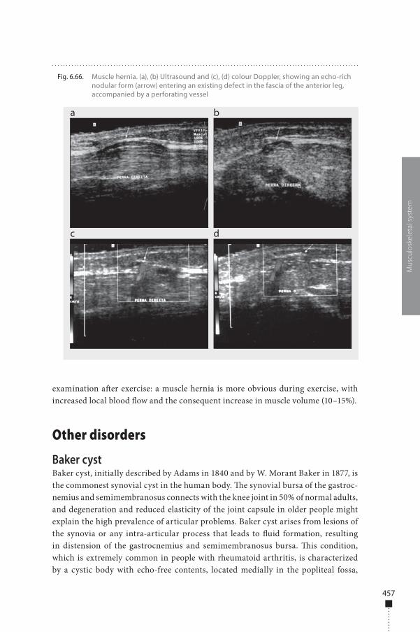
457
examination a�er exercise: a muscle hernia is more obvious during exercise, with increased local blood �ow and the consequent increase in muscle volume (10–15%).
Other disorders
Baker cystBaker cyst, initially described by Adams in 1840 and by W. Morant Baker in 1877, is the commonest synovial cyst in the human body. �e synovial bursa of the gastroc-nemius and semimembranosus connects with the knee joint in 50% of normal adults, and degeneration and reduced elasticity of the joint capsule in older people might explain the high prevalence of articular problems. Baker cyst arises from lesions of the synovia or any intra-articular process that leads to �uid formation, resulting in distension of the gastrocnemius and semimembranosus bursa. �is condition, which is extremely common in people with rheumatoid arthritis, is characterized by a cystic body with echo-free contents, located medially in the popliteal fossa,
Mus
culo
skel
etal
sys
tem
Fig. 6.66. Muscle hernia. (a), (b) Ultrasound and (c), (d) colour Doppler, showing an echo-rich nodular form (arrow) entering an existing defect in the fascia of the anterior leg, accompanied by a perforating vessel
a
c
b
d
978-9241548540-C006 PRF ml YVR ln ml ml-1-Final Proof.indd 457 6/10/13 10:30 AM
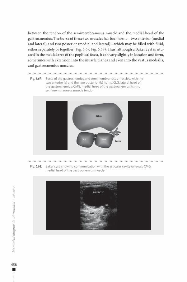
458
between the tendon of the semimembranosus muscle and the medial head of the gastrocnemius. �e bursa of these two muscles has four horns—two anterior (medial and lateral) and two posterior (medial and lateral)—which may be �lled with �uid, either separately or together (Fig. 6.67, Fig. 6.68). �us, although a Baker cyst is situ-ated in the medial area of the popliteal fossa, it can vary slightly in location and form, sometimes with extension into the muscle planes and even into the vastus medialis, and gastrocnemius muscles.
Man
ual o
f dia
gnos
tic u
ltras
ound
– V
olum
e 2
Fig. 6.67. Bursa of the gastrocnemius and semimembranosus muscles, with the two anterior (a) and the two posterior (b) horns. CLG, lateral head of the gastrocnemius; CMG, medial head of the gastrocnemius; tsmm, semimembranosus muscle tendon
a
a
b
b
Fig. 6.68. Baker cyst, showing communication with the articular cavity (arrows); CMG, medial head of the gastrocnemius muscle
BAKER CYST
978-9241548540-C006 PRF ml YVR ln ml ml-1-Final Proof.indd 458 6/10/13 10:30 AM
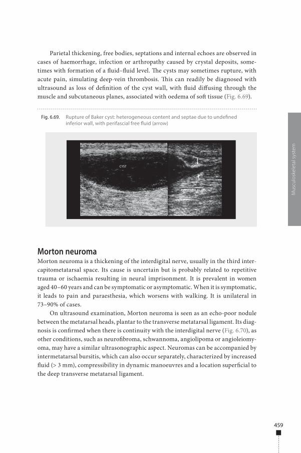
459
Parietal thickening, free bodies, septations and internal echoes are observed in cases of haemorrhage, infection or arthropathy caused by crystal deposits, some-times with formation of a �uid–�uid level. �e cysts may sometimes rupture, with acute pain, simulating deep-vein thrombosis. �is can readily be diagnosed with ultrasound as loss of de�nition of the cyst wall, with �uid di�using through the muscle and subcutaneous planes, associated with oedema of so� tissue (Fig. 6.69).
Morton neuromaMorton neuroma is a thickening of the interdigital nerve, usually in the third inter-capitometatarsal space. Its cause is uncertain but is probably related to repetitive trauma or ischaemia resulting in neural imprisonment. It is prevalent in women aged 40–60 years and can be symptomatic or asymptomatic. When it is symptomatic, it leads to pain and paraesthesia, which worsens with walking. It is unilateral in 73–90% of cases.
On ultrasound examination, Morton neuroma is seen as an echo-poor nodule between the metatarsal heads, plantar to the transverse metatarsal ligament. Its diag-nosis is con�rmed when there is continuity with the interdigital nerve (Fig. 6.70), as other conditions, such as neuro�broma, schwannoma, angiolipoma or angioleiomy-oma, may have a similar ultrasonographic aspect. Neuromas can be accompanied by intermetatarsal bursitis, which can also occur separately, characterized by increased �uid (> 3 mm), compressibility in dynamic manoeuvres and a location super�cial to the deep transverse metatarsal ligament.
Mus
culo
skel
etal
sys
tem
Fig. 6.69. Rupture of Baker cyst: heterogeneous content and septae due to unde�ned inferior wall, with perifascial free �uid (arrow)
CYST
978-9241548540-C006 PRF ml YVR ln ml ml-1-Final Proof.indd 459 6/10/13 10:30 AM
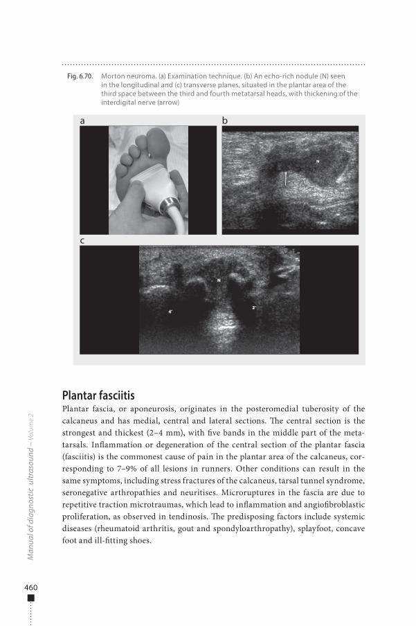
460
Plantar fasciitisPlantar fascia, or aponeurosis, originates in the posteromedial tuberosity of the calcaneus and has medial, central and lateral sections. �e central section is the strongest and thickest (2–4 mm), with �ve bands in the middle part of the meta-tarsals. In�ammation or degeneration of the central section of the plantar fascia (fasciitis) is the commonest cause of pain in the plantar area of the calcaneus, cor-responding to 7–9% of all lesions in runners. Other conditions can result in the same symptoms, including stress fractures of the calcaneus, tarsal tunnel syndrome, seronegative arthropathies and neuritises. Microruptures in the fascia are due to repetitive traction microtraumas, which lead to in�ammation and angio�broblastic proliferation, as observed in tendinosis. �e predisposing factors include systemic diseases (rheumatoid arthritis, gout and spondyloarthropathy), splayfoot, concave foot and ill-�tting shoes.
Man
ual o
f dia
gnos
tic u
ltras
ound
– V
olum
e 2
Fig. 6.70. Morton neuroma. (a) Examination technique. (b) An echo-rich nodule (N) seen in the longitudinal and (c) transverse planes, situated in the plantar area of the third space between the third and fourth metatarsal heads, with thickening of the interdigital nerve (arrow)
a
c
b
978-9241548540-C006 PRF ml YVR ln ml ml-1-Final Proof.indd 460 6/10/13 10:30 AM
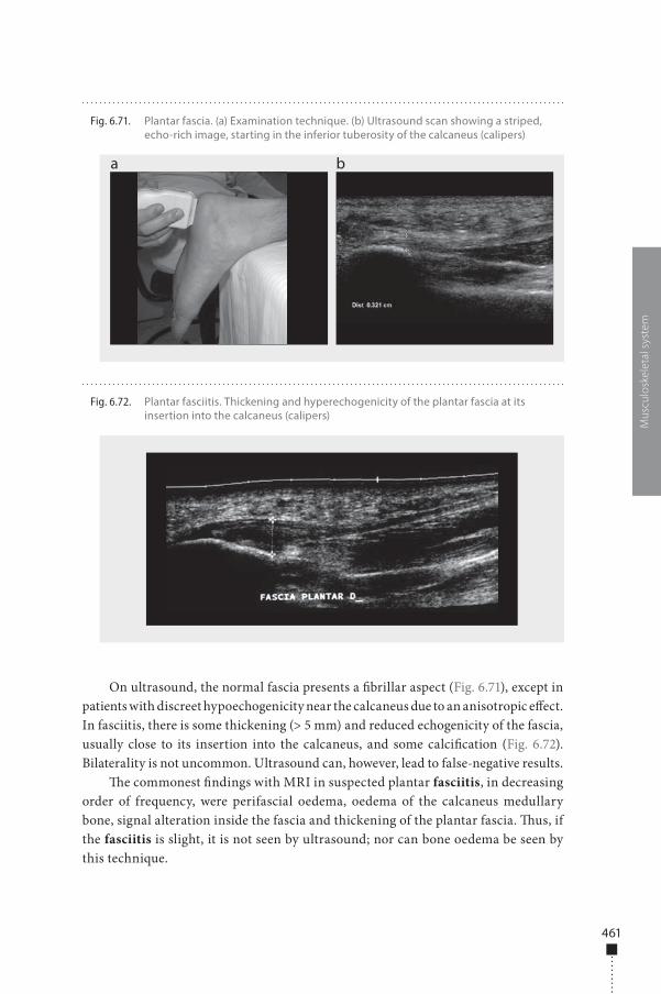
461
On ultrasound, the normal fascia presents a �brillar aspect (Fig. 6.71), except in patients with discreet hypoechogenicity near the calcaneus due to an anisotropic e�ect. In fasciitis, there is some thickening (> 5 mm) and reduced echogenicity of the fascia, usually close to its insertion into the calcaneus, and some calci�cation (Fig. 6.72). Bilaterality is not uncommon. Ultrasound can, however, lead to false-negative results.
�e commonest �ndings with MRI in suspected plantar fasciitis, in decreasing order of frequency, were perifascial oedema, oedema of the calcaneus medullary bone, signal alteration inside the fascia and thickening of the plantar fascia. �us, if the fasciitis is slight, it is not seen by ultrasound; nor can bone oedema be seen by this technique.
Mus
culo
skel
etal
sys
tem
Fig. 6.71. Plantar fascia. (a) Examination technique. (b) Ultrasound scan showing a striped, echo-rich image, starting in the inferior tuberosity of the calcaneus (calipers)
a b
Fig. 6.72. Plantar fasciitis. Thickening and hyperechogenicity of the plantar fascia at its insertion into the calcaneus (calipers)
978-9241548540-C006 PRF ml YVR ln ml ml-1-Final Proof.indd 461 6/10/13 10:30 AM
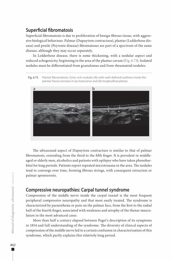
462
Super�cial �bromatosisSuper�cial �bromatosis is due to proliferation of benign �brous tissue, with aggres-sive biological behaviour. Palmar (Dupuytren contracture), plantar (Ledderhose dis-ease) and penile (Peyronie disease) �bromatoses are part of a spectrum of the same disease, although they may occur separately.
In Ledderhose disease, there is some thickening, with a nodular aspect and reduced echogenicity, beginning in the area of the plantar cavum (Fig. 6.73). Isolated nodules must be di�erentiated from granulomas and from rheumatoid nodules.
�e ultrasound aspect of Dupuytren contracture is similar to that of palmar �bromatosis, extending from the third to the ��h �nger. It is prevalent in middle-aged or elderly men, alcoholics and patients with epilepsy who have taken phenobar-bital for long periods. Patients report repeated microtrauma in the area. �e nodules tend to converge over time, forming �brous strings, with consequent retraction or palmar aponeurosis.
Compressive neuropathies: Carpal tunnel syndromeCompression of the middle nerve inside the carpal tunnel is the most frequent peripheral compressive neuropathy and that most easily treated. �e syndrome is characterized by paraesthesia or pain on the palmar face, from the �rst to the radial half of the fourth �nger, associated with weakness and atrophy of the thenar muscu-lature in the most advanced cases.
More than half a century elapsed between Paget’s description of its symptoms in 1854 and full understanding of the syndrome. �e diversity of clinical aspects of compression of the middle nerve led to a certain confusion in characterization of this syndrome, which partly explains this relatively long period.
Man
ual o
f dia
gnos
tic u
ltras
ound
– V
olum
e 2
Fig. 6.73. Plantar �bromatosis. Echo-rich nodules (N) with well-de�ned outlines inside the plantar fascia (arrows) in (a) transverse and (b) longitudinal planes
a b
978-9241548540-C006 PRF ml YVR ln ml ml-1-Final Proof.indd 462 6/10/13 10:30 AM
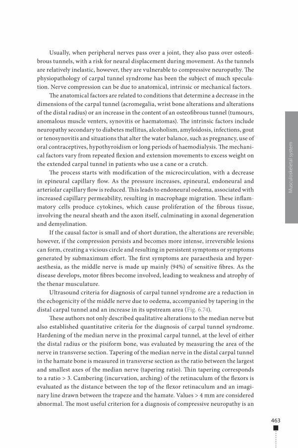
463
Mus
culo
skel
etal
sys
tem
Usually, when peripheral nerves pass over a joint, they also pass over osteo�-brous tunnels, with a risk for neural displacement during movement. As the tunnels are relatively inelastic, however, they are vulnerable to compressive neuropathy. �e physiopathology of carpal tunnel syndrome has been the subject of much specula-tion. Nerve compression can be due to anatomical, intrinsic or mechanical factors.
�e anatomical factors are related to conditions that determine a decrease in the dimensions of the carpal tunnel (acromegalia, wrist bone alterations and alterations of the distal radius) or an increase in the content of an osteo�brous tunnel (tumours, anomalous muscle venters, synovitis or haematomas). �e intrinsic factors include neuropathy secondary to diabetes mellitus, alcoholism, amyloidosis, infections, gout or tenosynovitis and situations that alter the water balance, such as pregnancy, use of oral contraceptives, hypothyroidism or long periods of haemodialysis. �e mechani-cal factors vary from repeated �exion and extension movements to excess weight on the extended carpal tunnel in patients who use a cane or a crutch.
�e process starts with modi�cation of the microcirculation, with a decrease in epineural capillary �ow. As the pressure increases, epineural, endoneural and arteriolar capillary �ow is reduced. �is leads to endoneural oedema, associated with increased capillary permeability, resulting in macrophage migration. �ese in�am-matory cells produce cytokines, which cause proliferation of the �brous tissue, involving the neural sheath and the axon itself, culminating in axonal degeneration and demyelination.
If the causal factor is small and of short duration, the alterations are reversible; however, if the compression persists and becomes more intense, irreversible lesions can form, creating a vicious circle and resulting in persistent symptoms or symptoms generated by submaximum e�ort. �e �rst symptoms are paraesthesia and hyper-aesthesia, as the middle nerve is made up mainly (94%) of sensitive �bres. As the disease develops, motor �bres become involved, leading to weakness and atrophy of the thenar musculature.
Ultrasound criteria for diagnosis of carpal tunnel syndrome are a reduction in the echogenicity of the middle nerve due to oedema, accompanied by tapering in the distal carpal tunnel and an increase in its upstream area (Fig. 6.74).
�ese authors not only described qualitative alterations to the median nerve but also established quantitative criteria for the diagnosis of carpal tunnel syndrome. Hardening of the median nerve in the proximal carpal tunnel, at the level of either the distal radius or the pisiform bone, was evaluated by measuring the area of the nerve in transverse section. Tapering of the median nerve in the distal carpal tunnel in the hamate bone is measured in transverse section as the ratio between the largest and smallest axes of the median nerve (tapering ratio). �in tapering corresponds to a ratio > 3. Cambering (incurvation, arching) of the retinaculum of the �exors is evaluated as the distance between the top of the �exor retinaculum and an imagi-nary line drawn between the trapeze and the hamate. Values > 4 mm are considered abnormal. �e most useful criterion for a diagnosis of compressive neuropathy is an
978-9241548540-C006 PRF ml YVR ln ml ml-1-Final Proof.indd 463 6/10/13 10:30 AM
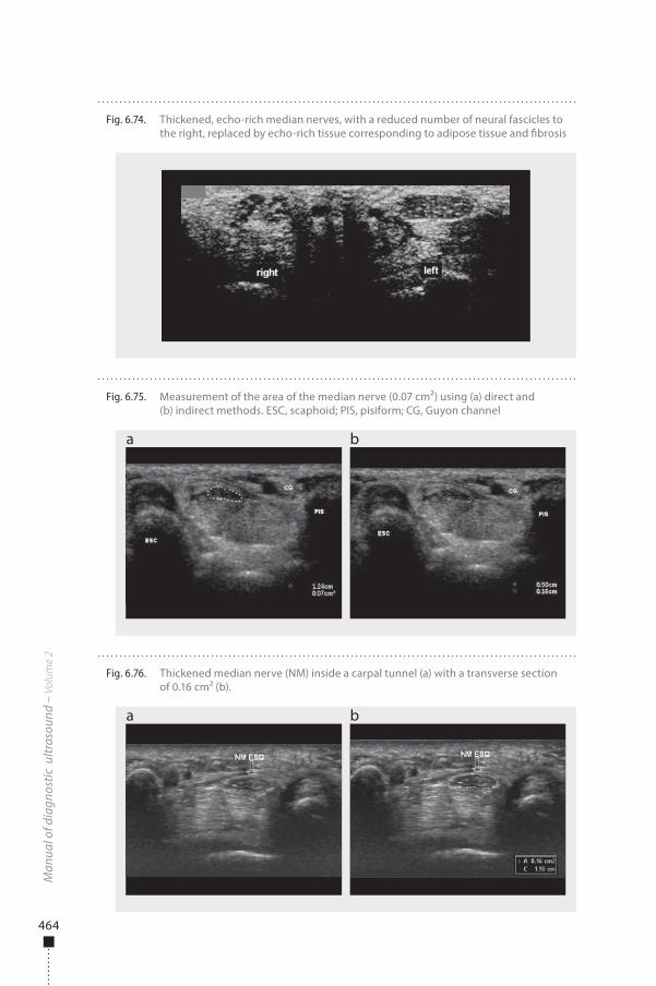
464
Man
ual o
f dia
gnos
tic u
ltras
ound
– V
olum
e 2
Fig. 6.74. Thickened, echo-rich median nerves, with a reduced number of neural fascicles to the right, replaced by echo-rich tissue corresponding to adipose tissue and �brosis
Fig. 6.75. Measurement of the area of the median nerve (0.07 cm²) using (a) direct and (b) indirect methods. ESC, scaphoid; PIS, pisiform; CG, Guyon channel
a b
Fig. 6.76. Thickened median nerve (NM) inside a carpal tunnel (a) with a transverse section of 0.16 cm² (b).
a b
978-9241548540-C006 PRF ml YVR ln ml ml-1-Final Proof.indd 464 6/10/13 10:30 AM
465
Mus
culo
skel
etal
sys
tem
increase in the cross-sectional area of the median nerve. Distal tapering of the nerve and incurvation of the retinaculum of the �exors showed poor reproducibility in subsequent studies.
�e cross-sectional area of the median nerve can be measured either indirectly or directly. In the indirect method, the formula for the area of the ellipse [p(D1 × D2) / 4] is used, in which D1 and D2 represent the transverse and anteroposterior diameters of the median nerve (Fig. 6.75 a). In the direct method, the area is calculated by ultrasound, from a continuous trace around the nerve (Fig. 6.75 b). Regardless of the method used, the neural sheath must always be excluded from the measure.
�e cut-o� point of the cross-sectional area for di�erentiating between normal and thickened nerves has been the subject of controversy in the literature, sugges-tions varying from 9 to 15 mm2. �is wide variation is due to the use of di�erent equipment, inclusion of people of both sexes in the same study, studies of people of di�erent ages, di�erent severity of disease and imprecise measurement area. Each unit should establish its own value on the basis of the population being studied. For women, we have adopted cross-sectional area cut-o� points of 9 mm2 measured by the indirect and 10 mm2 measured by the direct method (Fig. 6.76).
978-9241548540-C006 PRF ml YVR ln ml ml-1-Final Proof.indd 465 6/10/13 10:30 AM

978-9241548540-C006 PRF ml YVR ln ml ml-1-Final Proof.indd 466 6/10/13 10:30 AM
467
Recommended reading
Safety of diagnostic ultrasound
• AbramowiczJSetal.Fetalthermaleffectsofdiagnosticultrasound.AmericanInstituteofUltrasoundinMedicine.
Journal of Ultrasound in Medicine,2008,27:541-559.PMID:18359908
• BarnettSB.Safeuseofultrasoundcontrastagents.Ultrasound in Medicine & Biology,2007,33:171-172.doi:10.1016/j.
ultrasmedbio.2006.07.001PMID:17239523
• Basic physics of ultrasonographic imaging.Geneva,WorldHealthOrganization,2005.
• BioeffectsCommitteeoftheAmericanInstituteofUltrasoundinMedicineAmericanInstituteofUltrasoundinMedicine
consensusreportonpotentialbioeffectsofdiagnosticultrasound:executivesummary.Journal of Diagnostic Medical
Sonography,2011,27:3-13.doi:10.1177/8756479310394986
• BlyS,VandenHofMC.Obstetricultrasoundbiologicaleffectsandsafety,SOGCClinicalPracticeGuidelines.Journal of
Obstetrics and Gynaecology Canada,2005,27:572-580.PMID:16100635
• ClaudonMetal.Guidelinesandgoodclinicalpracticerecommendationsforcontrast-enhancedultrasound(CEUS)
–update2008.Ultraschall in der Medizin (Stuttgart, Germany : 1980),2008,29:28-44.doi:10.1055/s-2007-963785
PMID:18270887
• DuckFA.Hazards,risksandsafetyofdiagnosticultrasound.Medical Engineering & Physics,2008,30:1338-1348.
doi:10.1016/j.medengphy.2008.06.002PMID:18635388
• FowlkesJB.AmericanInstituteofUltrasoundinMedicineconsensusreportonpotentialbioeffectsofdiagnostic
ultrasound:executivesummary.Journal of Ultrasound in Medicine,2008,27:503-515.PMID:18359906
• Guidelines for the safe use of diagnostic ultrasound equipment.London,BritishMedicalUltrasoundSociety,2009.http://
www.bmus.org/policies-guides/BMUS-Safety-Guidelines-2009-revision-FINAL-Nov-2009.pdf
• Manual of diagnostic ultrasound.Geneva,WorldHealthOrganization,1995.
• Effectsofultrasoundandinfrasoundrelevanttohumanhealth.Progress in Biophysics and Molecular Biology,
2007,93:1-420.
• StratmeyerMEetal.Fetalultrasound:mechanicaleffects,AmericanInstituteofUltrasoundinMedicine.Journal of
Ultrasound in Medicine,2008,27:597-605.PMID:18359910
• WhitworthM,BrickerL,NeilsonJPetal.Routinecomparedwithselectiveultrasoundinearlypregnancy.Cochrane
Summaries,14April2010.
978-9241548540-Recommended -Final Proof.indd 467 6/10/13 10:29 AM
468
Obstetrics
• AcharyaGetal.Referencerangesforserialmeasurementsofbloodvelocityandpulsatilityindexattheintra-abdominal
portion,andfetalandplacentalendsoftheumbilicalartery.Ultrasound in Obstetrics & Gynecology,2005,26:162-169.
doi:10.1002/uog.1902PMID:15883983
• AmericanCollegeofObstetriciansandGynecologistsACOGPracticeBulletinNo.101:Ultrasonographyinpregnancy.
Obstetrics and Gynecology,2009,113:451-461.PMID:19155920
• ArduiniD,RizzoG.Predictionoffetaloutcomeinsmallforgestationalagefetuses:comparisonofDopplermeasure-
mentsobtainedfromdifferentfetalvessels.Journal of Perinatal Medicine,1992,20:29-38.doi:10.1515/jpme.1992.20.1.29
PMID:1608021
• BenacerrafB.Ultrasound of fetal syndromes.Philadelphia,ChurchillLivingstone,2007
• BenacerrafBR.Thehistoryofthesecond-trimestersonographicmarkersfordetectingfetalDownsyndrome,andtheir
currentroleinobstetricpractice.Prenatal Diagnosis,2010,30:644-652.doi:10.1002/pd.2531PMID:20572106
• BerghellaVetal.Cerclageforshortcervixonultrasonography:meta-analysisoftrialsusingindividualpatient-leveldata.
Obstetrics and Gynecology,2005,106:181-189.doi:10.1097/01.AOG.0000168435.17200.53PMID:15994635
• BerghellaV,BegaG.Ultrasoundevaluationofthecervix.In:CallenPW,ed..Ultrasonography in obstetrics and gynecology,
5thed.Philadelphia,Saunders-Elsevier,2008:698–720
• BirnholzJC.Analgorithmicapproachtoaccurateultrasonicfetalweightestimation.Investigative Radiology,1986,21:571-
576.doi:10.1097/00004424-198607000-00010PMID:3525451
• BrickerL,NeilsonJP,DowswellT.Routineultrasoundinlatepregnancy(after24weeks’gestation).Cochrane Database of
Systematic Reviews,2008,4:CD001451-doi:10.1002/14651858.CD001451.pub3
• CallenPW.Ultrasonography in obstetrics and gynecology,5thed.Philadelphia,Saunders-Elsevier,2008
• CampbellS,ThomsA.Ultrasoundmeasurementofthefetalheadtoabdomencircumferenceratiointheassessment
ofgrowthretardation.British Journal of Obstetrics and Gynaecology,1977,84:165-174.doi:10.1111/j.1471-0528.1977.
tb12550.xPMID:843490
• CampbellS,WilkinD.Ultrasonicmeasurementoffetalabdomencircumferenceintheestimationoffetalweight.British
Journal of Obstetrics and Gynaecology,1975,82:689-697.doi:10.1111/j.1471-0528.1975.tb00708.xPMID:1101942
• ChamberlainPFetal.Ultrasoundevaluationofamnioticfluidvolume.I.Therelationshipofmarginalanddecreased
amnioticfluidvolumestoperinataloutcome.American Journal of Obstetrics and Gynecology,1984,150:245-249.
PMID:6385713
• ChenMetal.Ultrasoundscreeningforfetalstructuralabnormalitiesperformedbytrainedmidwivesinthesecond
trimesterinalow-riskpopulation–anappraisal.Acta Obstetricia et Gynecologica Scandinavica,2009,88:713-719.
doi:10.1080/00016340902934688PMID:19412800
• DigheMetal.Sonographyinfirsttrimesterbleeding.Journal of Clinical Ultrasound,2008,36:352-366.doi:10.1002/
jcu.20451PMID:18335508
978-9241548540-Recommended -Final Proof.indd 468 6/10/13 10:29 AM
469
• Eik-NesSH.The18-weekfetalexaminationanddetectionofanomalies.Prenatal Diagnosis,2010,30:624-630.
doi:10.1002/pd.2576PMID:20572118
• Eik-NesSH,GrøttumP.Estimationoffetalweightbyultrasoundmeasurement.I.Developmentofanewformula.Acta
Obstetricia et Gynecologica Scandinavica,1982,61:299-305.PMID:7148403
• Eik-NesSH,GrøttumP,AnderssonNJ.Predictionoffetalgrowthdeviationbyultrasonicbiometry:I.Methodology.Acta
Obstetricia et Gynecologica Scandinavica,1982,61:307-312.doi:10.3109/00016348209156952PMID:7148404
• HadlockFPetal.Sonographicestimationoffetalweight.Thevalueoffemurlengthinadditiontoheadandabdomen
measurements.Radiology,1984,150:535-540.PMID:6691115
• HadlockFPetal.Estimationoffetalweightwiththeuseofhead,body,andfemurmeasurements–aprospectivestudy.
American Journal of Obstetrics and Gynecology,1985,151:333-337.PMID:3881966
• HarmanCR,BaschatAA.ArterialandvenousDopplersinIUGR.Clinical Obstetrics and Gynecology,2003,46:931-946.
doi:10.1097/00003081-200312000-00025PMID:14595236
• HillLM,BreckleR,GehrkingWC,O’BrienPC.Useoffemurlengthinestimationoffetalweight.American Journal of
Obstetrics and Gynecology,1985,152:847-852.PMID:3895952
• InternationalSocietyofUltrasoundinObstetrics&GynecologyCardiacscreeningexaminationofthefetus:guidelinesfor
performingthe‘basic’and‘extendedbasic’cardiacscan.Ultrasound in Obstetrics & Gynecology: the official journal of the
International Society of Ultrasound in Obstetrics and Gynecology,2006,27:107-113.PMID:16374757
• InternationalSocietyofUltrasoundinObstetrics&GynecologyEducationCommitteeSonographicexaminationof
thefetalcentralnervoussystem:guidelinesforperformingthe‘basicexamination’andthe‘fetalneurosonogram’.
Ultrasound in Obstetrics & Gynecology: the official journal of the International Society of Ultrasound in Obstetrics and
Gynecology,2007,29:109-116.doi:10.1002/uog.3909PMID:17200992
• NicolaidesKH,RizzoG,HecherK.Placental and fetal Doppler.Nashville,ParthenonPublishingEd.,2000
• NzehDA,RimmerS,MooreWMO,HuntL.Predictionofbirthweightbyfetalultrasoundbiometry.The British Journal of
Radiology,1992,65:987-989.doi:10.1259/0007-1285-65-779-987PMID:1450836
• OttWJ,DoyleS,FlammS.Accurateultrasonicestimationoffetalweight.Effectofheadshape,growthpatterns,and
amnioticfluidvolume.American Journal of Perinatology,1986,3:193-197.doi:10.1055/s-2007-999866PMID:3718640
• Practice guidelines for the performance of obstetric ultrasound examination.AmericanInstituteforUltrasoundinMedicine
(AIUM),2007.www.aium.org
• RoseBI,McCallumWD.Asimplifiedmethodforestimatingfetalweightusingultrasoundmeasurements.Obstetrics and
Gynecology,1987,69:671-675.PMID:3547218
• RumackCM,WilsonSR,CharboneauJW,JohnsonJAM.Diagnostic ultrasound.StLouis,Elsevier-Mosby,2005
• SabbaghaRE,MinogueJ,TamuraRK,HungerfordSA.Estimationofbirthweightbyuseofultrasonographicformulas
targetedtolarge-,appropriate-,andsmall-for-gestational-agefetuses.American Journal of Obstetrics and Gynecology,
1989,160:854-860,discussion860–862.PMID:2653039
978-9241548540-Recommended -Final Proof.indd 469 6/10/13 10:29 AM
470
• SalomonLJetal.InternationalSocietyofUltrasoundinObstetrics&Gynecology(ISUOG).Practiceguidelinesforper-
formanceoftheroutinemid-trimesterfetalultrasound.Ultrasound in Obstetrics & Gynecology: the official journal of the
International Society of Ultrasound in Obstetrics and Gynecology,2011,37:116-126.doi:10.1002/uog.8831PMID:20842655
• ShepardMJetal.Anevaluationoftwoequationsforpredictingfetalweightbyultrasound.American Journal of
Obstetrics and Gynecology,1982,142:47-54.PMID:7055171
• VintzileosAMetal.Fetalweightestimationformulaswithhead,abdominal,femur,andthighcircumferencemeasure-
ments.American Journal of Obstetrics and Gynecology,1987,157:410-414.PMID:3618691
• WarsofSL,GohariP,BerkowitzRL,HobbinsJC.Theestimationoffetalweightbycomputer-assistedanalysis.American
Journal of Obstetrics and Gynecology,1977,128:881-892.PMID:888868
Gynaecology
• CallenPW.Ultrasonography in obstetrics and gynecology,5thed.Philadelphia,Saunders-Elsevier,2008.
• FulghesuAMetal.Ultrasoundinpolycysticovarysyndrome–themeasuringofovarianstromaandrelationshipwith
circulatingandrogens:resultsofamulticentricstudy.Human Reproduction (Oxford, England),2007,22:2501-2508.
doi:10.1093/humrep/dem202PMID:17635847
• ChangHC,BhattS,Dogra.VS.Pearlsandpitfallsindiagnosisofovariantorsion.Radiographics,2008,28:1355-1368.
doi:10.1148/rg.285075130PMID:18794312
• JunqueiraBLPetal.Müllerianductanomaliesandmimicsinchildrenandadolescents:correlativeintraoperativeassess-
mentwithclinicalimaging.Radiographics,2009,29:1085-1103.doi:10.1148/rg.294085737PMID:19605658
• TimmermanDetal.Logisticregressionmodeltodistinguishbetweenthebenignandmalignantadnexalmass
beforesurgery:amulticenterstudybytheInternationalOvarianTumorAnalysisGroup.Journal of Clinical Oncology,
2005,23:8794-8801.doi:10.1200/JCO.2005.01.7632PMID:16314639
• RumackCM,WilsonSR,CharboneauJW,JohnsonJ-AM,eds.Diagnostic ultrasound.StLouis,ElsevierMosby,2005.
• SavelliL,CacciatoreB.Salpinges.In:Gynecological and early pregnancy ultrasound[ISUOGEducationalseries].London,
InternationalSocietyofUltrasoundinObstetricsandGynecology,2003.
• TwicklerDM,MoschosE.Ultrasoundandassessmentofovariancancerrisk.AJR. American Journal of Roentgenology,
2010,194:322-329.doi:10.2214/AJR.09.3562PMID:20093591
• VarrasMetal.Tubo-ovarianabscesses:spectrumofsonographicfindingswithsurgicalandpathologicalcorrelations.
Clinical and Experimental Obstetrics & Gynecology,2003,30:117-121.PMID:12854857
Breast
• AthanasiouAetal.Howtooptimizebreastultrasound.European Journal of Radiology,2009,69:6-13.doi:10.1016/j.
ejrad.2008.07.034PMID:18818037
• Boisserie-LacroixMetal.Breastultrasonography:anoverview.Gynecologie, Obstetrique & Fertilite,2006,34:1170-1177.
doi:10.1016/j.gyobfe.2006.10.015PMID:17140836
978-9241548540-Recommended -Final Proof.indd 470 6/10/13 10:29 AM
471
• ChenSCetal.Analysisofsonographicfeaturesforthedifferentiationofbenignandmalignantbreasttumorsofdifferent
sizes.Ultrasound in Obstetrics & Gynecology,2004,23:188-193.doi:10.1002/uog.930PMID:14770402
• CollD.Breasttumorimaging.Cancer Treatment and Research,2008,143:515-546.doi:10.1007/978-0-387-75587-8_20
PMID:18619231
• CostantiniMetal.Characterizationofsolidbreastmasses:useofthesonographicbreastimagingreportinganddata
systemlexicon.Journal of Ultrasound in Medicine,2006,25:649-659.PMID:16632790
• CostantiniMetal.L.Solidbreastmasscharacterisation:useofthesonographicBI-RADSclassification.La Radiologia
Medica,2007,112:877-894.doi:10.1007/s11547-007-0189-6PMID:17885742
• GokalpG,TopalU,KizilkayaE..PowerDopplersonography:anythingtoaddtoBI-RADSUSinsolidbreastmasses?
European Journal of Radiology,2009,70:77-85.doi:10.1016/j.ejrad.2007.12.007PMID:18243623
• Heywang-KoebrunnerS,SchreerD,DershawI.Diagnostic breast imaging mammography, sonography, magnetic
resonance imaging, and interventional procedures, 2ndedition.Stuttgart,Thieme,2001.
• HinesN,SlanetzPJ,EisenbergRL.Cysticmassesofthebreast.AJR. American Journal of Roentgenology,2010,194:W122-
W133.doi:10.2214/AJR.09.3688PMID:20093563
• HongASetal.BI-RADSforsonography:positiveandnegativepredictivevaluesofsonographicfeatures.AJR. American
Journal of Roentgenology,2005,184:1260-1265.PMID:15788607
• McCavertMetal.Ultrasoundisausefuladjuncttomammographyintheassessmentofbreasttumoursinallpatients.
International Journal of Clinical Practice,2009,63:1589-1594.doi:10.1111/j.1742-1241.2009.02102.xPMID:19686337
• MoonHJetal.Probablybenignbreastlesionsonultrasonography:aretrospectivereviewofultrasonographicfeatures
andclinicalfactorsaffectingtheBI-RADScategorization.Acta radiologica (Stockholm, Sweden: 1987),2010,51:375-382.
doi:10.3109/02841851003662780PMID:20350247
• NicholsonBTetal.Nipple–areolarcomplex:normalanatomyandbenignandmalignantprocesses.Radiographics,
2009,29:509-523.doi:10.1148/rg.292085128PMID:19325062
• RazaSetal.USofbreastmassescategorizedasBI-RADS3,4,and5:pictorialreviewoffactorsinfluencingclinical
management.Radiographics,2010,30:1199-1213.doi:10.1148/rg.305095144PMID:20833845
• RinaldiPetal.Cysticbreastlesions:sonographicfindingsandclinicalmanagement.Journal of Ultrasound in Medicine,
2010,29:1617-1626.PMID:20966473
• SmithGE,BurrowsP.Ultrasounddiagnosisoffibroadenoma–isbiopsyalwaysnecessary?Clinical Radiology,
2008,63:511-515.doi:10.1016/j.crad.2007.10.015PMID:18374713
• StavrosATetal.Solidbreastnodules:useofsonographytodistinguishbetweenbenignandmalignantlesions.
Radiology,1995,196:123-134.PMID:7784555
• YangW,DempseyPJ.Diagnosticbreastultrasound:currentstatusandfuturedirections.In:SicklesEA,ed.Breastimag-
ing.Radiologic Clinics of North America,2007,45:845-861.doi:10.1016/j.rcl.2007.06.009PMID:17888773
978-9241548540-Recommended -Final Proof.indd 471 6/10/13 10:29 AM
472
Paediatric ultrasound
• BianchiS,MartinoliC.Ultrasound of the Musculoskeletal system.Berlin,Springer-Verlag,2007.
• BruynR.Paediatric ultrasound: how, why and when.Philadelphia,ChurchillLivingstone,2005.
• ChateilJ,BrisseH,DacherJ.Ultrasoundinpediatricurology.Journal de Radiologie,2001,82:781-800.PMID:11443296
• DevredP,TréguierC,Ducou-Le-PointeH.Echographyofthehipandotherimagingtechniquesinpediatrics.Journal de
Radiologie,2001,82:803-816.PMID:11443297
• DonnellyLF.Pediatric Imaging: the fundamentals.Saunders-Elsevier,2009.
• Effective choices for diagnostic imaging in clinical practice[WHOTechnicalReportSeries795].Geneva,WorldHealth
Organization,1990:144.
• GarelLetal.USofthepediatricfemalepelvis:aclinicalperspective.Radiographics,2001,21:1393-1407.PMID:11706212
• GervaisDAetal.Percutaneousimaging-guidedabdominalandpelvicabscessdrainageinchildren.Radiographics,
2004,24:737-754.doi:10.1148/rg.243035107PMID:15143225
• KuhnJP,SlovisTL,HallerJO.Caffey’s Pediatric Diagnostic Imaging,10thed.Philadelphia,Elsevier/Mosby,2003.
• LevyJA,NobleVE.Bedsideultrasoundinpediatricemergencymedicine.Pediatrics,2008,121:e1404-1412.doi:10.1542/
peds.2007-1816PMID:18450883
• LoweLH,JohanekAJ,MooreCW.Sonographyoftheneonatalspine:part1,Normalanatomy,imagingpitfalls,andvari-
ationsthatmaysimulatedisorders.AJR. American Journal of Roentgenology,2007,188:733-738.doi:10.2214/AJR.05.2159
PMID:17312061
• LoweLH,JohanekAJ,MooreCW.Sonographyoftheneonatalspine:part2,Spinaldisorders.AJR. American Journal of
Roentgenology,2007,188:739-744.doi:10.2214/AJR.05.2160PMID:17312062
• LucayaJ,StrifeJL.Paediatric chest imaging.Berlin,Springer2002.
• PetitP,PracrosJ.Roleofultrasoundinchildrenwithemergencygastrointestinaldiseases.Journal de Radiologie,
2001,82:764-778.PMID:11443295
• Rational use of diagnostic imaging in paediatrics[WHOTechnicalReportSeries757].Geneva,
WorldHealthOrganization,1987
• RosenbergHK.Sonographyofpediatricneckmasses.Ultrasound Quarterly,2009,25:111-127.doi:10.1097/
RUQ.0b013e3181b6720bPMID:19730051
• SivitCJ.Imagingchildrenwithabdominaltrauma.AJR. American Journal of Roentgenology,2009,192:1179-1189.
doi:10.2214/AJR.08.2163PMID:19380540
• StringerDA,BabynPS,eds.Pediatric gastrointestinal imaging and intervention,2nded.London,
BCDeckerIncHamilton,2000
• Training in diagnostic ultrasound: essentials, principles and standards [WHOTechnicalReportSeries875].Geneva,
WorldHealthOrganization,1998
978-9241548540-Recommended -Final Proof.indd 472 6/10/13 10:29 AM
473
Musculoskeletal system
• Al-ShawiAetal.Thedetectionoffullthicknessrotatorcufftearsusingultrasound.The Journal of Bone and Joint Surgery.
British Volume,2008,90:889-892.doi:10.1302/0301-620X.90B7.20481PMID:18591598
• BeggsI.Ultrasoundoftheshoulderandelbow.The Orthopedic Clinics of North America,2006,37:277-285.doi:10.1016/j.
ocl.2006.03.004PMID:16846761
• BlumA,CarillonY,RailhacJetal.Instability[Chapter10].In:DaviesAM,HodlerJ.Imaging of the shoulder: techniques &
applications.Heidelberg,Springler-Verlag,2005.
• DaviesAM,WhitehouseRW,JenkinsJPR.Imaging of the foot & ankle. Heidelberg,Springer,2003:179–199.
• DeMaeseneerMetal.Sonographyofthenormalankle:atargetapproachusingskeletalreferencepoints.AJR. American
Journal of Roentgenology,2009,192:487-495.doi:10.2214/AJR.08.1316PMID:19155415
• DongQ,FessellDP.Achillestendonultrasoundtechnique.AJR. American Journal of Roentgenology,2009,193:W173-
doi:10.2214/AJR.09.3111PMID:19696253
• FessellDP,JacobsonJA.Ultrasoundofthehindfootandmidfoot.Radiologic Clinics of North America,2008,46:1027-1043.
doi:10.1016/j.rcl.2008.08.006PMID:19038611
• FinlayK,FriedmanL.Ultrasonographyofthelowerextremity.The Orthopedic Clinics of North America,2006,37:245-275.
doi:10.1016/j.ocl.2006.03.002PMID:16846760
• IlaslanH,SundaramM.Advancesinmusculoskeletaltumorimaging.The Orthopedic Clinics of North America,
2006,37:375-391.doi:10.1016/j.ocl.2006.05.003PMID:16846768
• JamadarDAetal.Musculoskeletalsonographytechnique:focusedversuscomprehensiveevaluation.AJR. American
Journal of Roentgenology,2008,190:5-9.doi:10.2214/AJR.07.2433PMID:18094286
• KhouryVetal.Musculoskeletalsonography:adynamictoolforusualandunusualdisorders.AJR. American Journal of
Roentgenology,2007,188:W63-73.doi:10.2214/AJR.06.0579PMID:17179329
• McNallyE.Practical musculoskeletal ultrasonography.Philadelphia,PA,ChurchillLivingstone,2005.
• MeyersPRetal.Shoulderultrasound.AJR. American Journal of Roentgenology,2009,193:W174-doi:10.2214/AJR.09.3229
PMID:19542411
• NazarianLN.Thetop10reasonsmusculoskeletalsonographyisanimportantcomplementaryoralternativetechniqueto
MRI.AJR. American Journal of Roentgenology,2008,190:1621-1626.doi:10.2214/AJR.07.3385PMID:18492916
• RobinsonP.Sonographyofcommontendoninjuries.AJR. American Journal of Roentgenology,2009,193:607-618.
doi:10.2214/AJR.09.2808PMID:19696272
• ZlatkinMB.Shoulderinstability[Chapter7].In:ZlatkinMB.MRI of the shoulder,2nded.Philadelphia,PA,Lippincott
Williams&Wilkins,2003.
978-9241548540-Recommended -Final Proof.indd 473 6/10/13 10:29 AM

978-9241548540-Recommended -Final Proof.indd 474 6/10/13 10:29 AM
475
Index
Notes
Pages numbers ending in f refer to fi gures
Pages numbers ending in t refer to tables
[A]
Abdomen (fetal)
abnormal shape 108
measurements in third trimester 44–45, 45f
second trimester assessment 40, 40f, 41f
subcutaneous tissue thickness 51
Abdominal aorta 311f, 397f, 399f
Abdominal cavity
formation in fetus 16
free air in (paediatric) 284, 285f
Abdominal circumference (fetal)
birth weight prediction 48
fetal weight estimation 48, 49t
increased, in ascites 108
intrauterine growth restriction
diagnosis 55, 84
multiple pregnancy 81, 84
second trimester 40, 40f
third trimester 44–45, 45f
Abdominal masses (paediatric) 279–281
adrenal haemorrhage causing 310, 310f
cystic 279–280, 280f
haematocolpos 325, 326f
non-cystic 281, 281f, 310
primary pelvic hydatid cyst 331, 332f
Abdominal pain (paediatric) 275–278, 394–395
acute, or chronic 395
adnexal torsion 326, 327f
appendicitis 276, 277f, 278f
cyclical 325
Henoch–Schönlein purpura 287, 287f
indications for ultrasound 394, 395
infl ammatory disorders 286, 287f
intussusception 275–276, 276f
mesenteric lymphadenitis 278, 279f
Abdominal trauma (paediatric)
blunt 284, 285f
liver 240, 240f, 241f
pancreatic 267, 270, 270f, 271f
renal 313, 313f, 314f
splenic 263, 263f, 264f
Abortion (spontaneous) 23, 24f, 148
absent intrauterine sac 23
‘complete’ or ‘incomplete’ 23
conjoined twins 33
intrauterine sac with embryo without cardiac
activity 24f, 25
intrauterine sac without embryo/yolk sac
23–25, 25f
‘missed’ 23, 25
recurrent 23
threatened 22, 23
twin 24f
Abscess(es) 393–394
adrenal 310
amoebic liver 240
breast 205–206, 207f
cervical 353
hepatic 239, 239f, 240
lung 358–359
muscle rupture complication 455
pancreas 267
peri-appendiceal 278f, 328f
978-9241548540-INDEX PRF ml ln ml ml-1-Final Proof.indd 475 6/10/13 10:29 AM
476
pyogenic liver 239, 239f
renal 303, 303f
subperiosteal 389f
Acardiac twin syndrome 88
Acetabular cartilage (paediatric) 384, 384f
Acetabular dysplasia 385
Achilles tendon see Calcaneus (Achilles) tendon
Achondrogenesis 117f
Acoustic inertial cavitation 5
Acoustic working frequency 5
Acrania 31, 32f
Acromelia 114
Acromion-clavicular joint, liquid in 420
Adenitis see Lymphadenitis
Adenoma
follicular (thyroid) 350, 350f
hepatic 239
lactating 214, 214f
nipple 214
parathyroid 351
tubular, of breast 214–215, 214f
Adenomyosis 154–155, 155f
Adhesions
acute pelvic inflammatory disease 185
endometrial 152
Fallopian tube 177–178, 177f
Adnexal lesions 163–174
cysts 176–177, 176f, 177f
paraovarian cysts 173–174
see also Fallopian tubes; Ovarian masses;
Ovarian tumours; Ovaries
Adnexal torsion 326, 327f
Adnexal tumours 169–175
Adolescents
bone and joint abnormalities 387
ectopic pregnancy 331
goitre 350
ovarian cysts 319–320, 319f
spleen size 255, 255f
testis (normal) 333
uterovaginal anomalies 325
see also Paediatric ultrasound
Adrenal glands (fetal) 110
Adrenal glands (paediatric) 309–311
abscess 310
age-related changes 309
congenital hyperplasia 309, 328, 342
cystic lesions 310
haemorrhage 310, 310f
neuroblastoma 310, 311f
normal sonographic appearance 309, 309f
tumours 311
Alagille syndrome 246, 248
Allantoic duct 15
Alobar holoprosencephaly 31, 32f, 93, 94f
Alonso-Lej classification 249
Alpha angle 385
α-fetoprotein 234, 235
Amenorrhea, primary 325
Amino acids, fetal growth and 53
Amniocentesis, fetal loss, risk 83
Amniotic cavity 14–15
Amniotic fluid 69–70
measurement methods 47
third trimester 47, 47f
volume
decline near term 47
multiple pregnancies 70
normal 42, 47f
reduced, fetal growth restriction
55–56, 61
volume assessment 42, 69
intrauterine growth restriction diagnosis
55–56
multiple pregnancies 70
single deepest pocket method 42, 47, 69
third trimester 47, 47f
two-diameter pocket method 70
see also Amniotic fluid index
Amniotic fluid index 42, 47, 69
measurement method 69–70
pitfalls in measurement 47
range/mean and distribution 69
use in preterm pregnancies 69
978-9241548540-INDEX PRF ml ln ml ml-1-Final Proof.indd 476 6/10/13 10:29 AM
477
Amniotic fluid pocket, deepest, measurement 42, 47, 69
Amniotic membrane 15f
formation, gestational age 14
twin pregnancies 78, 80, 81
Amniotic sac 16
multiple pregnancies 78, 81
Amoebic liver abscess 240
Anamnestic gestational age see Menstrual age
Anatomical snuffbox 427
Anencephaly 31, 91, 91f
Aneuploidy
first-trimester screening 20–21
hydatidiform moles and 29
risk in multiple pregnancies 82
Angioma, splenic 261, 262f
Anisotropy 412, 442f
Ankle
injuries 447, 448
ligaments see Lateral ligament complex
(ankle)
soft tissue oedema 450
sprains 448
tendons 437–442
see also Calcaneus (Achilles) tendon
Anoxic–ischaemic encephalopathy 371, 371f
Antenatal diagnosis
congenital anomalies see Congenital
anomalies
twin–twin transfusion syndrome 86
urinary tract anomalies 312
Anterior talofibular ligament 447, 447f
chronic lesions 450, 450f
complete rupture 449, 449f
Aorta, abdominal 311f, 397f, 399f
Aorta (fetal) 100, 101f, 111f
coarctation 104f
overriding 105, 105f
second trimester assessment 41f
transposition of great vessels 105, 105f
Aortic stenosis 106
Aponeurosis (plantar fascia) 460, 461f
Appendicitis 276, 277f, 278f
Appendix
dilated 278f
fluid-filled 276, 277f
inflammation 276, 277f, 278f
normal 276, 277f
perforated 276
Arachnoid cyst 92f
Arterial ischaemic infarct (neonatal) 371
Arteriohepatic dysplasia (Alagille syndrome) 246, 248
Arteriovenous malformation 390
Arthritis
juvenile rheumatoid 394
septic 389, 389f, 390f
Ascites
fetal 108, 109f
paediatric, in lymphoma 282f
Asphyxia, perinatal 371
Aspirin, intrauterine growth restriction prevention 62
Asplenia 257
Athletes/sports
ankle injuries 447
insertion tendinopathy 439
knee injuries 435
muscle lesions 452, 456
Athyroidism 348
Atrial anomalies (fetal) 102, 102f
Atrioventricular canal, complete 102, 102f
Atrioventricular valve, single 102, 102f
Atrium, single 102, 102f
Atrium thickness (atrial width) of lateral ventricles 35, 36f, 37, 93
Axillary lymph nodes 199, 199f
metastatic carcinoma 225–226, 226f
Axillary regions, ultrasound examination 194
[B]
Baker cyst 457–458, 458f
rupture 459, 459f
978-9241548540-INDEX PRF ml ln ml ml-1-Final Proof.indd 477 6/10/13 10:29 AM
478
Bald humeral head sign 418, 418f
Banana sign 96, 98
Basal ganglion 362f, 365
Basedow disease 349
Beads-on-a-string sign 178, 180f, 184f, 185
Beckwith-Wiedemann syndrome 234
Benign mammary dysplasia 211, 212f
Biceps see Brachial biceps
Bile ducts (extrahepatic) see Biliary tree (extrahepatic)
Bile ducts (intrahepatic)
choledochal cysts 249, 250, 251f
hydatid disease 242, 242f, 243f, 244f
interlobular, paucity 248
normal sonographic features 231, 232, 233f
Bile plug 251, 251f
Biliary atresia 234, 246–248
anomalies associated 248
neonatal hepatitis syndrome vs 246, 247
types 246, 246f
Biliary cyst 244, 245f
Biliary sludge 251, 251f, 254f
Biliary tract
embryonal rhabdomyosarcoma 235, 235f
in hydatid disease 242
paediatric ultrasound see Liver and biliary
tract
Biliary tree (extrahepatic)
calculi 252, 253f
choledochal cysts 249
deficient see Biliary atresia
embryonal rhabdomyosarcoma 235, 235f
hydatid disease 242, 244f
inspissated bile syndrome 251, 251f
normal 231, 232f
obstruction 251, 251f
Biloma, hepatic 240, 241f
Biometry, fetal see Fetus, biometric parameters
Biovular twins see Twin pregnancies, dizygotic
Biparietal diameter of head (fetus)
birth weight prediction 48
embryo/fetal size in first trimester 11–12
fetal weight estimation 48, 49t
intrauterine growth restriction diagnosis 55
measurement 11–12, 43, 44f
accuracy limitations 13
first trimester 11–12, 13f
second trimester 35–36, 36f, 90, 90f
third trimester 43, 44f
multiple pregnancy 81
Birth weight
multiple pregnancies 83
prediction 48, 49t
Bladder
fetal 68f
diameter in megacystis 32
dilatation 112–113, 114f
length 32
normal 110f
twin–twin transfusion syndrome 85f, 86
volume in monochorionic, diamniotic
twins 85f
hyperdistension, avoidance 134
involvement in cervical carcinoma 160
paediatric
capacity 292
congenital diverticulum 306
distension 307
duplication 306
examination technique 289
in gonadal dysgenesis 330f
neoplasms 308
neurogenic 308
normal anatomy 292, 293f
rhabdomyosarcoma 308, 309f
stones 307–308, 308f
thickness 292
urachal abnormalities 306
wall thickening 306f, 307, 308f
transabdominal ultrasound preparation 10,
71, 72, 134
placenta praevia diagnosis 65
transvaginal ultrasound preparation 10,
135–136
978-9241548540-INDEX PRF ml ln ml ml-1-Final Proof.indd 478 6/10/13 10:29 AM
479
Blake pouch cyst 94
‘Blighted ovum’ 23
Blood flow velocity
cerebral (neonatal) 363–364, 364f
Doppler, measurement 119–120, 119f, 121
waveform analysis 121–122, 122f
see also Doppler ultrasound
reversal, intrauterine growth restriction
57–58, 57f, 58f, 59f, 126
B-mode ultrasound scanning, safety 5
Bone
abnormalities (paediatric) 385–390
infections (paediatric) 388, 389f
mineralization 116, 118f
normal paediatric findings 384, 384f
ultrasound-induced heating 4–5
Borderline ventriculomegaly 93, 93f
Botryoid appearance 308, 324
Bowel (fetal) 106, 107f
obstruction 106
physiological herniation (normal) 32
Bowel (paediatric)
air in 396, 396f, 400, 401
haematoma 284, 285f
herniation 32, 34f, 108, 109f
infarction 276
intussusception 275–276
ischaemic disease 288, 288f
obstruction (neonatal) see Intestinal obstruc-
tion (neonatal)
trauma 284, 285f
see also Small bowel
Brachial artery 424f
Brachial biceps 423
long head, tendon of 409, 410, 410f, 414f
examination technique 413, 413f
fluid in synovial sheath 420–421
Brachial biceps tendon, examination/normal findings 424f
Brachial triceps 422, 423f
Brachial triceps tendon 423f
Brachioradialis muscle 425f
Brain
anoxic–ischaemic lesions (neonatal)
369, 370f
arterial ischaemic infarct 371
fetal
assessment 36
malformations 91
middle cerebral artery see Cerebral artery,
middle
teratoma 92f
ischaemic lesions (neonatal) 369–372,
370f, 371f
malformations (neonatal period) 373, 374f
parenchyma
calcifications 376f
normal neonatal ultrasound 361, 362f
posterior regions (premature brain) 365
premature 364–368, 366f, 368f, 369f
cysts in white matter 366, 369f
early follow-up 365
haemorrhagic lesion follow-up 365, 366f
intermediate follow-up 366
long-term follow-up 367, 368f
MRI role 367
normal cranial variants 363
timing of scans 368
tumours (neonatal/infant) 376
white matter injury, premature brain,
follow-up 367
Brain-sparing effect 57, 58f, 124–125
Branchial cleft cysts 346
Breast 193–226
age-related changes 195
anatomy 195, 195f
benign lesions 202–216, 218
abscesses 205–206, 207f
acute mastitis 205–206, 206f
adenoma 214–215, 214f
cysts see Breast, cysts
fibroadenoma 207–208, 208f
fibrocystic changes 211–212, 212f
fibrolipoadenoma 213, 213f
978-9241548540-INDEX PRF ml ln ml ml-1-Final Proof.indd 479 6/10/13 10:29 AM
480
galactocoele 213, 214f
haematoma 207, 207f
hamartoma 213, 213f
intraductal papilloma 209, 210f
intraparenchymal lymph nodes 210, 211f
liponecrosis 215, 215f
in males 216, 216f
phyllodes tumour 209, 209f
biopsy 193, 201
complications/risks 201
preoperative needle localization 201
techniques 201
cancer/carcinoma see Breast carcinoma
Cooper ligaments see Cooper ligaments
cysts 202–204
calcifications 203
complex 203, 204f
intracystic cancer 203, 205f
liponecrosis vs 215
sebaceous 203
simple 202–203, 203f
dense 195, 197f
ducts 195f, 198, 199f
epithelial cancers see Breast carcinoma
fatty 197f
lobes 195
lumps 193
lymph nodes in 199, 200f, 210, 211f
male, disease see Male breast disease
malignant lesions see Breast carcinoma
microcalcifications 218, 219, 220f
palpation 194–195
parenchyma 197, 197f
sclerosing adenosis 212, 212f
skin over 196, 196f
subcutaneous fat 196, 196f
tumours 218
carcinoma see Breast carcinoma
fibroepithelial (phyllodes) 209, 209f
Paget disease of nipple 221
ultrasound
accuracy 199–200
biopsy guided by 193, 201
diagnostic algorithm 200
examination technique 194–195
indications 193, 218
lexicon 199–200, 200t
new techniques 201–202
normal findings 195–200, 196f
preparation 193–194
Breast carcinoma 217–226
central necrosis 203, 205f
ductal carcinoma in situ 218
fibroadenoma vs 208
incidence and risk factors 217–218
intracystic 203, 205f
local staging 225–226, 226f
lymph node involvement 225–226, 226f
recording ultrasound criteria 218
sonographic features 218–225
good prognosis carcinomas 222–224
in situ carcinoma 218
inflammatory cancer 223, 224f
invasive ductal carcinoma 219–221, 219f,
220f, 225
invasive lobular carcinoma 222, 222f
male carcinoma 225, 225f
medullar carcinoma 223, 223f
metastatic cancer 225
microcalcifications 218, 219, 220f
mucinous carcinoma 222, 222f
papillary invasive carcinoma 223
premalignant lesions 218
rare tumours 225
size of lesion 219
skin and subcutaneous tissue 220–221, 221f
ultrasound role 218
Breast Imaging Reporting and Data System 199–200, 200t
Breathing movements
fetal, umbilical artery Doppler waveform 123
paediatric 360
Brenner tumours 172
Bruises 454f
subcutaneous 452
see also Contusions
978-9241548540-INDEX PRF ml ln ml ml-1-Final Proof.indd 480 6/10/13 10:29 AM
481
Budd-Chiari syndrome 242, 252
Burkitt lymphoma
abdominal mass 282f
ovary involvement 331, 332f
Bursae
ankle 437
elbow 422, 423
hip 432
knee 437, 457, 458, 458f
retrocalcaneal 437
subacromial-subdeltoid 412
Bursitis
intermetatarsal 459
prepatellar 437f
retrocalcaneal 439, 440f
trochanteric 432, 434f
[C]
Caesarean delivery, cervical length and 75
Calcaneofibular ligament 448, 448f
Calcaneus
plantar fasciitis and 460, 461, 461f
tuberosity, Haglund deformity 439, 440f
Calcaneus (Achilles) tendon 437
disorders/conditions affecting 437–439
examination technique 438f
normal dimensions/anatomy 437
normal ultrasound findings 437, 438f
paratendinitis 439, 441f
rupture 439
complete 439, 441, 442f
partial 439, 441f
tendinous stumps 441, 442f
tendinopathy 439, 439f
xanthoma 437, 438f
Calcar avis 363
Calcarine gyrus 363
Calculi
biliary tree (extrahepatic) 252, 253f
lower urinary tract 307–308, 308f
renal 298–299, 298f
Calvaria, abnormal shape 95f, 96
Calyceal diverticula 294, 294f
Candelabra sign 375, 376f
Capillary malformation 391
Cardiac activity (fetal)
absent in abortion diagnosis 25
first trimester 16
recording 26
Cardiac afterload, intrauterine growth restriction 57
Cardiac anomalies (fetal) 100–106, 101f, 102f, 103f, 104f, 105f
atrial 102, 102f
detected with four-chamber view 102,
103f, 104f
detection in first trimester 33
number 102
outflow tract 105–106, 105f, 106f
screening 100
Cardiac chambers (fetal) 98f
first trimester 18
screening for anomalies 100
second trimester 38, 38f, 39f
Cardiac output (fetal) 57, 125
in severe hypoxaemia 126
Cardiac rate (fetal)
first trimester 16, 26
normal 26
umbilical artery Doppler waveform 123
Caroli disease 250, 251f, 296
Carotid artery, normal 344
Carpal tunnel 425, 426f, 463
dimension decrease, mechanism 463
Carpal tunnel syndrome 462–465
causative factors 463
ultrasound findings 463, 464f, 465
Carpi ulnaris extensor tendon 428, 429f
Cartilage interface sign 422, 422f
Cartilaginous epiphysis 384, 384f, 385f
Cauda equina 379f
Caudal regression syndrome 382
Caudate nucleus 362f
Cavum septum pellucidum 36, 37f, 44, 363, 363f
Cavum veli interpositi 363
978-9241548540-INDEX PRF ml ln ml ml-1-Final Proof.indd 481 6/10/13 10:29 AM
482
Cavum vergae 363
Cellulitis 393
Central nervous system
abnormalities in first trimester 31, 32f
see also Brain; Spinal cord (paediatric)
Central renal sinus 290f, 291f
Cephalocoele 92, 92f
Cerebellar vermis, hypoplastic 94
Cerebellum
first trimester 18f
small, Chiari II malformation 95f, 96, 98
transverse diameter 35–36, 37, 37f
Cerebral anoxic–ischaemic lesions 369, 370f
Cerebral artery, anterior, normal haemodynamics 363–364, 364f
Cerebral artery, middle 125
Doppler velocimetry 124, 125, 127
fetal hypoxaemia prediction 124–125, 125t
intrauterine growth restriction 58f
ischaemic infarct 371, 372f
pulsatility index 124, 125t
Cerebral blood flow, neonatal 363–364, 364f
Cerebral hemispheres, alobar holoprosencephaly 31
Cerebral palsy 76
Cerebral vasodilatation, fetal growth restriction 57, 124–125
Cerebroplacental ratio 125
Cerebrospinal fluid (CSF) 377
Cervical abscess 353
Cervical carcinoma 158–160, 159f, 160f, 161f
Cervical cerclage 72f, 74
follow-up after 74–75, 75f
Cervical lymphadenitis 347, 348f
Cervical pregnancy 27f
Cervix 70–75, 137
endometrial carcinoma invasion 156–157
examination technique 71–73, 72f
funnelling 73, 74f
indications for ultrasound 70, 71
length
from 19-31 weeks 73, 74f
after cervical cerclage 74–75
children 315
gestational age at delivery 75
multiple pregnancy 82
normal 73
preterm birth prediction 73, 74, 82
shortening for labour 73, 74f
in multiple pregnancies 73, 82
neonatal 315, 316f
normal findings 72f, 73
pathological findings 73–75
cerclage, follow-up after 74–75, 75f
labour induction success prediction 71, 75
mode of delivery investigation 71, 75
preterm birth and risk of 73–74, 74f
preparation for ultrasound 71
Chest
cystic mass 356, 356f
hypoplastic 116, 117f, 118t
paediatric ultrasound 354–360
examination technique 354
indications 354
normal findings 354–355, 355f, 356f
pathological findings 356–360
preparation 354
second trimester assessment 38, 38f, 39f,
98, 99f
soft tissue abnormalities 356, 356f
Chest wall
anomalies 356, 356f
normal 354, 355f
Chiari II malformations 95f, 96, 98, 380
Chiari II syndrome 380
Child abuse 270, 387
Children, ultrasound see Paediatric ultrasound
Chlamydia trachomatis 327
Cholangiography 248, 250f
Cholecystitis (paediatric) 252
acute calculous 252, 253f
complications 253
Choledochal cyst (paediatric) 249–251
anatomical types 249, 249f
complications 251
differential diagnosis 250–251
978-9241548540-INDEX PRF ml ln ml ml-1-Final Proof.indd 482 6/10/13 10:29 AM
483
type I 249f, 250, 250f
types II-V 249f, 250
Choledocholithiasis 252, 253f
Cholelithiasis 252, 253f
Chorioangioma 66
Chorioncarcinoma 31, 161
Chorionic cavity 15
Chorionic membrane, twin pregnancies 19, 78, 80, 81
Chorionic plate 62
Chorionic villi 122
Chorionicity, determination 18–19, 77–78
Choroid plexus
cysts 93, 94f
lobular 363
papilloma 376
Cirrhosis, in children 252
Cisterna magna 37
anteroposterior diameter 36f
enlarged (mega) 94, 95f
Cleft palate 94f
Clitoromegaly 329f
Cloacal abnormalities 307
Cloverleaf skull 116, 117f
Club foot 116, 116f, 385–386
Coarctation of aorta 104f
Coelomic cavity 14, 15f
Cogwheel sign 178, 179f, 183
Collecting system 297f, 313f
Colon
intussusception 275
microcolon see Microcolon
see also Bowel; Intestinal obstruction
Colour Doppler 121, 126
appendicitis 278f
cervical lymphadenitis 347f
chest 354
cirrhosis of liver 252
De Quervain tenosynovitis 428f
endometrial carcinoma 157f
endometrial polyps 150, 151f
fibroids 152
finger tendons 432f
haemangioendothelioma 236
intratesticular vascular anatomy 333, 335f
invasive ductal carcinoma (breast) 221
lymph nodes of neck 346f, 391
muscle hernia 457f
neonatal cranial examination
ischaemic lesions 369
normal findings 361
severe haemodynamic distress 372,
372f, 373f
polycystic ovary syndrome 145
portal hypertension 259
premature brain 364
prepatellar bursitis 437f
renal vein thrombosis 305
splenic angioma 261
splenic lymphangioma 263
synovial diseases (paediatric) 387
tubal patency evaluation 187
twin reversed arterial perfusion sequence 88
urinary tract examination 289
uterus 136, 137f
varicocele 338, 338f
Common bile duct 232f
size, children/infants 231
Common carotid artery 348f
normal 344, 344f
Common hepatic duct 231
fibrosis 247f
Compartment syndrome 455
Compressive neuropathies 462–465
diagnostic criteria 463, 465
see also Carpal tunnel syndrome
Computed tomography (CT)
calyceal diverticula 294f
cystic mesenchymal hamartoma 238f
duodenal haematoma 285f
fatty hepatic infiltration 245f
hepatoblastoma 234f
horseshoe kidneys 294f
mature ovarian teratoma 322f
mesenteric cyst 280f
multilocular cystic nephroma 302f
978-9241548540-INDEX PRF ml ln ml ml-1-Final Proof.indd 483 6/10/13 10:29 AM

484
pancreatic fracture 271f
pancreatic pseudocyst 268, 269f
polysplenia 257f
renal fracture 313f
renal hydatid cyst 304f
splenic angioma 262f
Wilms tumour 300f, 301f
Concentric contraction, muscle 451
Congenital adrenal hyperplasia 309, 328, 342
Congenital anomalies
antenatal diagnosis 89
multiple pregnancy 82–83
cystic, neck 346, 347f
digestive tract 279
duodenal malrotation 398
hydrocele 337, 337f
liver 237
lower urinary tract 306–307
lymphatic vessels 279, 391
pancreas 266–267
spine 380–382, 381f, 382f
splenic 261, 262
upper urinary tract 293–297, 314
uterine disorders 146–148
see also Fetal malformations; specific organs
Congenital hip dislocation 385
Conjoined twins 33, 34f, 76, 88–89, 89f
frequency 88
point of union 88
Connatal cysts 363
Contrast enema, meconium ileus 402, 402f
Contusions
hepatic 240, 240f, 241f
spleen 263, 263f
see also Bruises
Conus medullaris 379, 379f, 380f
normal 379f, 380f
tethered cord and 380
Cooper ligaments 195, 195f, 196f, 197
invasive ductal carcinoma 220
Core-needle biopsy, breast 201
Corpus callosum
agenesis and hypoplasia 373, 374f
neonatal cranial ultrasound 362f
Corpus luteum 143, 143f, 168, 169f
vascular ring 168, 169f
Cranial circumference, measurement, second trimester 35, 36, 36f
Cranial ultrasound
congenital anomalies 380
neonatal see Neonatal cranial ultrasound
normal variants, premature infant 363
see also Brain
Crohn disease (paediatric) 286, 286f
Crown–rump length 11–12
gestational age relationship 17t
increase in, rate 16
measurement 11–12, 12f
accuracy limitations 13
Cryptorchidism 336, 336f
Cyclopia 96, 96f
Cyst(s)
arachnoid 92f
Baker see Baker cyst
biliary 244, 245f
Blake pouch 94
branchial cleft 346
breast see Breast, cysts
choledochal see Choledochal cyst
choroid plexus 93, 94f
connatal 363
dermoid see Dermoid cyst
duodenal duplication 280, 280f
duplication 279–280, 280f
endometriotic 170, 170f
ependymal 379f
epidermoid see Epidermoid cysts
hydatid see Hydatid cyst
mesenteric 279, 280f
muscle 455, 456f
Naboth 140
omental 279
978-9241548540-INDEX PRF ml ln ml ml-1-Final Proof.indd 484 6/10/13 10:29 AM
485
ovarian see Ovarian cysts
paraovarian 173–174, 176, 176f, 321
paratubal 176, 177, 177f
pelvic inclusion 174
periarticular 420
peritoneal inclusion 178, 180
placental 63, 63f
popliteal 392
renal see Renal cysts
retropharyngeal 353
sebaceous, breast 203
spermatic cord 337, 337f
splenic, epidermoid 261, 261f
subependymal 376f
theca lutein 29, 30f, 67
thyroglossal duct 346, 347f
thyroid gland 351
urachal 306, 307f
Cystadenoma
mucous ovarian 321
serous ovarian 321, 322f
Cystic adenomatoid malformation 98, 99f
Cystic duct 231
Cystic fibrosis 244f, 267
meconium ileus 402
meconium pseudocyst 405, 405f
pancreas in 267
Cystic hygroma 31, 33f
paediatric 346, 353, 353f, 391
Cystic lymphangiomas, cervical 353, 353f
Cystic masses, abdominal 279–280, 280f
Cystic mesenchymal hamartoma 237, 238f
Cystic teratoma 171–172
Cystic tumours, pancreatic 271–272, 272f
Cystitis 306f
Cystography 229
Cytomegalovirus (CMV), cerebral infection (neonatal) 375, 376f
Cytotrophoblast 122, 123
[D]
Dandy-Walker complex 94, 95f, 373
Dating of pregnancy see Gestational age
De Quervain tenosynovitis 426, 428f
Deltoid ligament 446, 446f
Deltoid muscle 418
Dermal sinus, dorsal 381
Dermatomyositis 394
Dermoid cyst 392
neck 346
ovarian 171–172, 172f
girls 321, 322f
Desmoplastic reaction 219
Developmental dysplasia of hip 385
Diabetes, maternal 50, 51
Diamniotic pregnancy 78
Diandry 29
Diaphragm
abnormalities 360
normal 355, 356f
paralysis 360
Diaphragmatic hernia, fetus 99, 100f
Diarrhoea, bloody 286, 289, 304
Diastematomyelia 381, 381f
Dichorionic twins see Twin pregnancies, dichorionic
Digestive tract (fetal)
malformations 106–108, 107f, 108f, 109f
normal 106, 107f
Digestive tract (paediatric) 272–289
abdominal masses see Abdominal masses
abdominal pain see Abdominal pain
(paediatric)
blunt trauma to 284, 285f
congenital anomalies 279
duplication 279–280, 280f
inflammatory disorders 286, 286f, 287f
intramural bleeding 287, 287f
ischaemic bowel disease 288, 288f
neonatal, ultrasound 395
non-inflammatory disorders 287–289,
287f, 288f
978-9241548540-INDEX PRF ml ln ml ml-1-Final Proof.indd 485 6/10/13 10:29 AM
486
normal thickness 274
paediatric ultrasound
examination technique 273
indications 272
normal findings 273–274, 273f, 274f, 275f
pathological findings 275–289
preparation 272
perforation 285f
vomiting 282–284
wall, normal 273, 273f
Digital extensor apparatus 429, 430f
Dizygotic twin pregnancies see Twin pregnancies, dizygotic
Dolichocephaly 81
Doppler effect 119, 119f
Doppler frequency 119, 120
pulse repetition 120
Doppler shift data 120, 122
Doppler transducer 119, 119f, 120
Doppler ultrasound
aliasing effect 120–121
colour flow imaging see Colour Doppler
continuous wave 120
endometrial carcinoma 157f
flow waveform analysis 121–122, 122f
intrauterine growth restriction 56
magnitude of signal 120
modes 121–122
paediatric
adrenal neuroblastoma 311
appendicitis 276
liver and biliary tract 230
premature brain 364, 365
scrotum examination 333
power Doppler see Power Doppler
practice 120–121
principles 119–120, 119f
pulsed wave see Pulsed Doppler
spectral see Spectral Doppler
use in obstetrics 118–129
fetal hypoxaemia prediction (cerebral
artery) 124–125, 125t
fetal hypoxaemia prediction (venous
Doppler) 126–127, 127t
placental function assessment (umbilical
artery) 122–124, 124t
recommendations for 127
reporting recommendations (by trimes-
ter) 128–129
venous 56, 126–127
Dorsal dermal sinus 381
Double decidual sac 13, 24
Double-bubble sign 106, 108f, 396, 396f
Douglas, pouch of
fluid 167, 175, 175f, 177f, 186
intussusception 276
Drawing manoeuvre 450, 450f
Ductus venosus 122, 126, 127
fetal hypoxaemia 57, 58, 59f, 126
normal flow waveform 59f
pulsatility index 61, 126–127, 127t
reversal of blood flow 57–58, 59f, 126
Duodenal atresia 106, 108f, 396
Duodenal bulb, dilated 396, 396f
Duodenal diaphragm 396, 397, 397f
Duodenal dilatation 397
Duodenal duplication, obstructive 396
Duodenal duplication cyst 280, 280f
Duodenal haematoma 285f
Duodenal obstruction (neonatal) 395–400
causes, frequency, features 396
intrinsic (atresia, stenosis) 395–397
malrotation complication 396, 398–400,
399f, 400f
Duodenal stenosis 396
Duodenojejunal flexure 398
Duplication cyst 279–280, 280f
Dupuytren contracture 462
Duret crests 195, 220
[E]
Ebstein anomaly 102, 103f
Eccentric contraction of muscle 451
978-9241548540-INDEX PRF ml ln ml ml-1-Final Proof.indd 486 6/10/13 10:29 AM
487
Echinococcus granulosus 242
Echocardiography, fetal 33, 102
Ectopic pregnancy 26, 27f
concomitant intrauterine pregnancy with 26
diagnostic accuracy of ultrasound 27
differential diagnosis 27
direct and indirect signs 27
in girls 331
incidence and risk factors 26
interstitial 27f
pseudogestational sac 23–24, 27
tubal 26, 28f
Elastography, breast 202
Elbow
muscles 422, 423
synovial bursae 422, 423
tendons 422–423, 423f, 424f
Embryo 9
first trimester 15–16
size measurement 11–12
intrauterine sac without 23–24
linear growth 12
shape change 16
Embryo–fetal anomalies, first trimester 31–34
Embryogenesis 15–16, 18
pancreas 266
spine 379–380, 380f
Embryonal rhabdomyosarcoma of biliary tree 235, 235f
Embryonal sarcoma, undifferentiated 235, 235f
Embryonic demise 23, 25, 25f, 27f, 29
Embryonic disc 15–16
Embryonic splitting 18–19
Encephalomeningocoele 92
Encephalopathy, anoxic–ischaemic 371, 371f
End-diastolic flow
intrauterine growth restriction 58, 58f,
124–125
peak systolic velocity ratio 57, 58
reduced/reverse 59f, 124, 126
intrauterine growth restriction 57, 57f,
58, 58f, 124–125
velocity, middle cerebral artery Doppler
waveform 124–125
chronic hypoxia effect 124
factors affecting 123, 125
velocity, umbilical artery Doppler waveform
123, 124
absent, perinatal mortality 126
reduced 124
Endocervical canal 174f
widening 73
Endometrial carcinoma 156–158, 157f, 158f
differential diagnosis 158
recurrences 161, 162f
Endometrial disease, benign 148–152
Endometrial hyperplasia 149–150, 149f
Endometrial polyps 150, 150f, 151f
Endometrial–myometrial junction 140, 148, 150, 154, 155f
in endometrial carcinoma 156, 157f
Endometriotic cysts 170, 170f
Endometritis 148, 149f
Endometrium 139, 140
adhesions (synechiae) 152
atrophic 140
cystic atrophy 151
factors affecting appearance 148
increased thickness in disease 148
hyperplasia 149–150, 149f
neoplasms 156
postmenopausal state 140, 156
stroma, in myometrium 154–155, 155f
stromal proliferation 149–150, 149f
tamoxifen effect 150, 151–152
thickness changes in menstrual cycle 140,
140t, 141f, 315
in tuberculosis 148
tumours 156–158
Endomysium 451, 452
Endotendon 409
Entamoeba histolytica 242
Enterocolitis, necrotizing 288, 288f
Entheses 410, 411f
978-9241548540-INDEX PRF ml ln ml ml-1-Final Proof.indd 487 6/10/13 10:29 AM
488
Enuresis 314
Ependymal cyst 379f
Epicondylitis 423
Epidermoid cysts 392
splenic 261, 261f
Epididymal head, normal 333, 334f
Epididymis (paediatric)
malignant tumours 340
normal 333, 334f
Epimysium 451, 452, 452f
Epiphysis (paediatric)
fracture-separation (neonatal) 387, 388f
normal 384, 384f
Escherichia coli 239, 289, 302
Exomphalos 32
Extensor tendons
fingers 429, 430f
forearm 425f
wrist see Wrist
External os 72f, 174f
Extra-axial fluid 374, 374f
Extrahepatic ducts see Biliary tree (extrahepatic)
[F]
Fallopian tubes 174–189
adhesions 177–178, 177f
anatomical segments 174f
carcinoma 186
convoluted, retort-shaped 183, 183f, 184f
diseases 178–189
inflammatory see Tubal inflammatory
disease
distal (ampulla) extremity 174, 174f, 175,
175f, 176f, 177f
ectopic pregnancy 26, 28f
hyperechoic septa 178
hysterosalpingo-contrast sonography
186–189
incomplete septum 178, 179f, 183, 183f
inflammation see Tubal
inflammatory disease
infundibular section 174, 174f, 175,
175f, 177f
interstitial part 174–175, 174f, 175f
isthmic part 174, 174f, 175
normal 174–176
occlusion 183, 186, 189f
patency 186, 189f
evaluation see Hysterosalpingo-contrast
sonography (HyCoSy)
salpingitis with incomplete septa 178, 179f,
183, 183f
spasm 187
tortuous 189f
wall structure/thickness, inflammatory
disease 178, 182, 183
Fallot’s tetralogy 105, 105f, 106f
Familial polyposis coli 234
Fasting 230, 272
Fatty deposits, pancreas 267
Fatty liver 245, 245f
Fecaliths 276, 277f
Female pseudohermaphrodism 328, 329f
Femoral head, paediatric abnormalities 385, 386f
Femoral rectum muscle 455f
myositis ossificans 456, 456f
Femur
abnormal shape and hypoplastic 116, 116f
normal 115f
Femur length (fetal)
fetal weight estimation 48, 49t
intrauterine growth restriction diagnosis 55
measurement
second trimester 41, 42f
third trimester 45, 45f, 46, 46f
variability 46
Fetal malformations 89–118
detection, first trimester 18
gastrointestinal tract 106–108, 107f,
108f, 109f
head 90–96, 91f, 92f, 93f, 94f, 95f, 96f
heart 100–106, 101f, 102f, 103f, 104f,
105f, 106f
978-9241548540-INDEX PRF ml ln ml ml-1-Final Proof.indd 488 6/10/13 10:29 AM
489
lungs 98–99, 98f, 99f, 100f
skeletal system 114–118, 115f, 116f,
117f, 118f
spine 96–98, 97f
urinary tract 110–113, 110f, 111f, 112f,
113f, 114f
see also Congenital anomalies
Fetal membranes 14–15
Fetus 9
anomalies see Fetal malformations
biometric parameters (third trimester) 43–46
abdominal measurements 44–45, 45f
head measurements 43–44, 44f
intrauterine growth restriction diagnosis
54, 55–56, 56f
limb measurements 45–46, 45f, 46f
bladder see Bladder, fetal
body composition 50
body configuration, macrosomia 51
brain see Brain
breathing movements, umbilical artery
Doppler 123
chest see Chest
circulation assessment 118
Doppler 122–126
death
causes 87
conjoined twins 88, 89f
fetus papyraceus 89
monoamniotic pregnancies 87
partial hydatidiform mole vs 29
severe hypoxaemia/placental insuf-
ficiency 58, 126–127
twin 87–88
twin–twin transfusion syndrome 86,
86f, 87
first trimester 15–16
growth and development 40, 53
assessment, reference ranges 40, 48
first trimester 16, 18, 18f
individualized models 48
multiple pregnancies 81, 83
normal 43
regulation 53
requirements for 53
restriction see Intrauterine fetal growth
restriction
second trimester see Fetus, morphology
third trimester 43–46
see also Fetus, biometric parameters
growth rate 43, 53
head see Head (fetal)
heart see Heart (fetal)
hypoxaemia prediction see Hypoxaemia (fetal)
hypoxic 55
lungs see Lung (fetal)
macrosomia 50–51
morphology assessment in second trimester
35–42, 89
abdomen 40, 40f, 41f
chest 38, 38f, 39f
extremities 41, 42f
head 35–37, 36f, 37f
sensitivity 90
timing, reasons for 89
vertebral column 38, 38f
movements, first trimester 16
nuchal translucency see Nuchal translucency
thickness
pleural effusion 99, 99f
size charts 43
size measurement, first trimester 11–12
spine see Spine (fetal)
weight
discordance in twin pregnancies 83, 84
estimation 40, 48, 49t, 50
excessive 48
growth restriction diagnosis 54, 55
increase, rate of 53
macrosomia, weight prediction 50
multiple pregnancies 81, 83, 84
optimum 48
prediction 50
twin pregnancies 83
978-9241548540-INDEX PRF ml ln ml ml-1-Final Proof.indd 489 6/10/13 10:29 AM
490
Fetus in fetu 89
Fetus papyraceus 89
Fibroadenoma 207–208, 208f
differential diagnosis 208
Fibroadipose septa 451, 453
Fibrocartilage 410, 411f
Fibrocystic changes, breast 211–212, 212f
Fibrocystic mastopathy 211, 212f
Fibroids (uterine) 152–154
calcified 151f, 152
changes in size, factors affecting 152
intramural 153, 154f
pedunculated 153
submucosal 153, 153f, 154f
subserosal 12f, 153
Fibrolipoadenoma 213, 213f
Fibroma, ovarian 172
Fibromatosis 392
superficial 462, 462f
Fibromatosis colli 351, 352f, 391–392
Fibrosis, muscle rupture complication 455, 455f
Fibrothecoma 172
Filum terminale 379–380, 379f, 380f
thickened, tight syndrome 381, 382f
Fine-needle aspiration, breast 201
Finger(s), tendons 429, 430f, 431f, 432f
extensor apparatus 429, 430f
flexor 429, 431f, 442, 443f, 444
see also Finger pulley systems
tenosynovitis 429, 432f
Finger pulley systems 442–446, 443f
annular pulleys 442, 443, 443f, 444f
A1, thickening 444, 446f
A2, rupture 444, 445f
cruciform pulleys 442, 443, 443f
distance from tendon to cortical bone 444,
444t, 445f
functions 443
indirect signs of lesions 444, 444t
lesions 443, 444, 445f
Flexor retinaculum 425, 426f, 463
Flexor tendons
fingers see Finger(s), tendons
hands 429, 431f
wrist 425
Fluid, drinking before transabdominal ultrasound 10, 71, 134
Focal nodular hyperplasia 237, 238f
Fontanelle, anterior, examination technique 361, 361f
access difficulties 365
normal anatomy 362f
Fontanelle, mastoid 361
Fontanelle, posterior 361, 365
Foot, fetal 115f
Forearm extensors, common tendon
examination, normal findings 425f
tendinopathy 412f, 423
Forearm flexors, common tendon, examination, normal findings 425f
Foreign body
chronic, inflammation 392
soft-tissue 392, 393f
vaginal 324
Fourier spectrum analyser 121
Fractures, occult, in children 387
Frontal bossing 116, 117f
Frontal horns 363f
atrial width 37
bull’s horn configuration 374f
fused 93
Frontal–occipital diameter (fetus)
second trimester 36f
third trimester 44, 44f
Fungal microabscesses, liver 240
[G]
Galactocoele 213, 214f
Galen vein, aneurysm 373
Gall bladder
fetal 106, 107f
paediatric
biliary sludge in 251, 251f, 254f
calculi in 252, 253f
distension (hydrops) 253, 254f
978-9241548540-INDEX PRF ml ln ml ml-1-Final Proof.indd 490 6/10/13 10:29 AM
491
inflammation 252
neonatal hepatitis syndrome 247
normal 231, 232f
small, in biliary atresia 247, 247f
Gall stones 252, 253f
Gastrocnemius muscle, bursa 457, 458f
Gastrointestinal tract see Digestive tract
Gastro-oesophageal reflux 284, 284f
Gastroschisis 32, 34f, 108, 109f
Genitalia, ambiguous 309, 328, 329f, 330f
Genitography 328, 329f, 330f
Germ cell tumours
ovarian see Ovarian tumours
testicular 340, 341t
Germinal matrix haemorrhage 367
subependymal 364
Gestational age
accuracy, crown–rump length
measurement 12
calculation, gestational sac diameter 11
at delivery, cervical length and 75
estimation by ultrasound
anamnestic (menstrual) discrepancy 35, 55
crown–rump length relationship 17t
first trimester 9, 11, 12, 14t
guidelines/landmarks 14t
second trimester 35
importance, fetal growth restriction
diagnosis 55
umbilical artery Doppler waveform 123
Gestational sac 13, 14f, 24f
absent 23
diameter, measurement 11, 12f
ectopic pregnancy and 26
embryo in, but cardiac activity absent 25
empty 23–25, 25f
gestational age at visualization 13, 23
multiple pregnancies 81
normal growth rate 24
normal vs abnormal 24
spontaneous expulsion 25f
tubal 28f
without embryo or yolk sac 23–24
Gestational trophoblastic disease 29–31, 66–67
see also Hydatidiform mole (molar
pregnancy)
Geyser sign 420
Gharbi’s classification, hydatid cysts 242, 242f, 243f, 244f, 279, 303
Glenohumeral joint
complete rotator cuff rupture 418
haemorrhage 420, 421f
liquid in 420–421, 421f
Glucose, fetal growth and 53
Gluteus medius tendon 432, 433f
tendinopathy 432, 433f
Gluteus minimus tendon 432, 433f
tendinopathy 432, 433f
Goitre 350
Gonadal dysgenesis 325, 330f
mixed 331
Granuloma, lipophagic 215
Granulosa-cell tumours 172–173
Graves’ disease 349
Greater trochanter, painful 432, 433f
Gynaecological ultrasound 133–189
adnexal lesions see Adnexal lesions
artefacts 134
choice of technique 133
Fallopian tubes 174–189
see also Fallopian tubes
normal findings 137–144
ovaries 141–144
uterus 137–141
see also Ovaries; Uterus
pelvic structures 135f, 136f
preparation and techniques 134–137
transabdominal 134–135
transvaginal 133–134, 135–137
uses/indications 133, 163
uterine disorders see Uterus, disorders
Gynaecomastia 216, 216f
dendritic 216, 217f
glandular 216
nodular 216, 216f
978-9241548540-INDEX PRF ml ln ml ml-1-Final Proof.indd 491 6/10/13 10:29 AM
492
[H]
Haemangioendothelioma 236–237, 237f
neck 352
Haemangioma 390
capillary 352, 390
cavernous 237
cutaneous 236
liver, in children 237
parotid 352, 352f
placental 66
Haematocolpos 146, 323, 323f, 325, 326f
Haematological malignancies
hepatosplenomegaly 264
spleen 260
see also Leukaemia; Lymphoma
Haematoma(s)
bowel 284, 285f
breast 207, 207f
intermuscular 455
intrahepatic 240, 241f
intramural (bowel) 284, 285f
intrauterine 22–23, 22f
perirenal 313f
placental 64
renal parenchymal 313f
renal subcapsular 313f
soft-tissue 392
splenic parenchymal 263, 263f
sternocleidomastoid muscle (fibromatosis
colli) 351, 352f, 391–392
testicular 341
Haematometra 323
Haematometrocolpos 323, 325
Haemodynamic changes
after intrauterine twin death 87
intrauterine growth restriction 56–58, 57f,
58f, 59f
Haemodynamic distress, severe (neonates) 372, 372f, 373f
Haemodynamics, neonatal cranial ultrasound 363–364, 364f
Haemoglobinopathy 260
Haemolymphangioma 263
Haemolytic uraemic syndrome 289, 304, 305f
Haemorrhage
adrenal 310, 310f
germinal matrix 364, 367
glenohumeral joint 420, 421f
intraventricular see Intraventricular
haemorrhage
muscle rupture complication 455
premature brain, follow-up 365, 366f
retroplacental 64
subchorionic 22, 22f
Haemosiderin, endometriotic 170, 170f
Haglund deformity 439, 440f
Hamartoma
breast 213, 213f
cystic mesenchymal 237, 238f
Hand
fetal 18f, 115f
second trimester assessment 41, 42f
see also Finger(s), tendons
Harmonic imaging, breast 201–202
Hashimoto disease 349, 349f
Head (embryo) 16, 18f
Head (fetal)
abnormal shape 116
anencephaly 31
biparietal diameter see Biparietal diameter
of head
circumference see Head circumference
first trimester 18f
malformations 90–96, 91f, 92f, 93f, 94f, 95f,
96f
normal 90f
second trimester assessment 35–36, 90
third trimester measurements 43–44, 44f
variability 46
Head circumference (fetal)
fetal weight estimation 48, 49t
intrauterine growth restriction diagnosis 55
measurement
second trimester 35–36, 90f
third trimester 44, 44f
978-9241548540-INDEX PRF ml ln ml ml-1-Final Proof.indd 492 6/10/13 10:29 AM
493
Heart (fetal)
four-chamber view 100, 101f
anomalies detected by 102, 103f, 104f
left outflow 38, 39f, 100, 101f
malformations see Cardiac anomalies
outflow tract anomalies 105–106, 105f, 106f
right outflow 38, 39f, 100, 101f
three-vessel view 100, 101f
Heart beat, first trimester 16
Heart chambers see Cardiac chambers
Heart rate see Cardiac rate
Heat generation by ultrasound 4–5
Henoch–Schönlein purpura 287, 287f, 342
Hepatic contusions 240, 240f, 241f
Hepatic disorders see under Liver
Hepatic vein (paediatric) 232, 233f
thrombosis 233–234
Hepatitis 246
chronic 246
neonatal see Neonatal hepatitis syndrome
Hepatoblastoma 234, 234f
Hepatocellular carcinoma 234
Hepatomegaly 246
Hepatosplenomegaly 264
Hermaphroditism 328
true 329–330
Hernia
hiatus 360
inguinal scrotal 336
muscle 456–457, 457f
Heterotopic pregnancies 26
Hiatus hernia 360
Hilus sign 345, 346f, 391
Hip
anatomy 432
bursae 432
irritable 386
normal (paediatric) 386f
septic dislocation 389, 390f
snapping 434, 434f
subluxation 385, 386f
Hodgkin lymphoma 260f, 353
hepatosplenomegaly 264
Hoffa pad 435, 435f
Holoprosencephaly 31, 32f, 93
alobar 31, 32f, 93, 94f
lobar 93
semilobar 93
Human chorionic gonadotropin (hCG)
absent intrauterine sac 22
chorioncarcinoma 31
ectopic pregnancy diagnosis 26
spontaneous abortion diagnosis 23
Humerus
largest tubercle 419f
irregular outline 420, 420f
measurement, third trimester 45, 46f
Hyaline membrane disease 366f
HyCoSy see Hysterosalpingo-contrast sonography (HyCoSy)
Hydatid cyst(s) 279
abdominal masses due to 279
liver 242, 242f, 243f, 244f, 279
neck 353
primary pelvic 331, 332f
pulmonary 358, 359f
spleen 262, 262f
urinary tract 303, 304f
Hydatid disease 242, 262, 303
Hydatid of Morgagni 333
torsion 340
Hydatidiform mole (molar pregnancy) 29, 66, 67f
benign 29, 66
coexisting fetus with 67
complete 29, 30f
differential diagnosis 67
invasive 31
partial 29, 30f, 67
sonographic features 29, 30f, 66–67, 67f
Hydramnios 69, 70
Hydranencephaly 31
Hydrocephalus/hydrocephaly
first trimester 31, 32f
macrocephaly due to 91, 92f
neonates 376
Hydrocoele 337, 337f
complex 339
978-9241548540-INDEX PRF ml ln ml ml-1-Final Proof.indd 493 6/10/13 10:29 AM
494
Hydrocolpos 323
Hydrometrocolpos 146, 147f, 323
Hydromyelia 381, 382, 382f
Hydronephrosis 112, 113, 113f, 296
evolution, grading system 297
vesico-ureteral reflux causing 296, 298f
Hydropneumothorax 358
Hydrops, fetal 20f, 33f, 108
twin–twin transfusion syndrome and 86
Hydrops, of gall bladder (paediatric) 253, 254f
Hydrosactosalpinx 27
Hydrosalpinx 28f, 179f, 180f, 183, 184f, 185
chronic 184f
Hyperaesthesia, carpal tunnel syndrome 463
Hyperbilirubinaemia, conjugated 246
Hypercholesterolaemia, familial 437
Hyperhydration 134
Hyperperistalsis 401, 401f
Hypertension 314
intracranial 365, 366f
maternal 53
renal origin 314
Hyperthyroidism 350
Hypertrophic pyloric stenosis 283, 283f
Hypogastric arteries 110f, 134
Hypomineralization of bone 116, 118f
Hypoperistalsis 403
Hypoplastic left heart syndrome 102, 104f
Hypospadias 329f
Hypotelorism 96, 96f
Hypothermia, neonatal 61
Hypothyroidism, infants 348, 348f
Hypoxaemia (fetal)
prediction from middle cerebral artery
124–125, 125t
prediction with venous Doppler 126–127
umbilical venous blood redistribution 126
Hypoxia
fetal 55
neonatal 371, 371f
Hysterosalpingo-contrast sonography (HyCoSy) 186–189
advantages 188–189
air and saline 186, 187, 188
contrast media 187, 188, 189
criteria for tubal patency 187–188
method 187, 188
results 189f
safety and advantages 188
Hysterosalpingography 186
[I]
Ileocaecal junction 398
Ileojejunal obstruction 106, 108f
Ileum, terminal
Crohn disease 286, 286f
obstruction 402–403, 402f
Ileus 276
Imbalanced fetus–fetus transfusion see Twin–twin transfusion syndrome
Impact syndrome 413
Implantation, intrauterine blood association 22
Infant(s)
gastro-oesophageal reflux 284, 284f
vomiting 282–284
see also Neonates; Paediatric ultrasound
Infantile polycystic kidney 295–296
Infections/infectious diseases (paediatric)
bone/joints 388, 389f, 390f
cerebral, in neonates 375, 375f, 376f
kidneys 302–303, 302f, 303f
neck 353
soft-tissue 393–394, 394f
spinal cord 383
spine 383
spleen 259
urinary tract 302–303, 302f, 303f, 312
Inferior vena cava (fetal) 57, 126
Inferior vena cava (paediatric) 233f
interruption, polysplenia with 257, 257f
Wilms tumour extension 300f
Infertility, female 186, 188
Inflammatory bowel disorders 286, 286f, 287f
Inflammatory disease, tubal see Tubal inflammatory disease
978-9241548540-INDEX PRF ml ln ml ml-1-Final Proof.indd 494 6/10/13 10:29 AM
495
Informed consent, breast biopsy 201
Infraspinatus tendon 412, 421f
examination technique 413, 416f
normal ultrasound findings 416f
Inguinal canal
delayed obliteration 336
normal 335
undescended testes in 336, 336f
Inguinal scrotal hernia 336
Inspissated bile syndrome 251, 251f
Insulinoma 271
Interamniotic septum, thickness 78
Inter-decidual sign 13
Interdigital nerve thickening 459, 460f
Intermetatarsal bursitis 459
Internal cervical os 72f, 73
placental location and 52, 65, 70, 71f
Internal jugular veins 348f
anatomical variant 344, 344f
Intersex states 328–331, 329f, 330f
Interventricular defect 102, 103f, 105
Intestinal obstruction (fetal) 106
Intestinal obstruction (neonatal) 395–403
complications 404–405, 405f
duodenal see Duodenal obstruction
malrotation complication 396, 398–400,
399f, 400f
small bowel 400–403
Intestinal volvulus 398, 399f
Intracranial hypertension 365, 366f
Intraductal papilloma, breast 209, 210f
Intrahepatic vessels/ducts 232, 233f
Intraperitoneal fluid, ectopic pregnancy 27, 28f
Intrauterine blood, first trimester 22–23
Intrauterine death 58
see also Fetus, death
Intrauterine fetal growth see Fetus, growth and development
Intrauterine fetal growth restriction 47, 53–62
biometry (ultrasound) 55–56, 56f
brain-sparing effect 57, 58f, 124–125
causes 53–54, 54t
definitions 54, 55
diagnosis 54–55, 55–56, 56f, 61
future prospects and prevention 62
haemodynamic modifications 56–58, 57f,
58f, 59f
incidence 54
management and delivery planning 59–60
monitoring strategy 59, 60–61
multiple pregnancies 83–84
outcome/prognosis 56, 58, 61
perinatal and long-term sequelae 61
symmetric vs asymmetric 54–55, 56f
twin pregnancies 81
Intrauterine fluid collections 22, 152
Intrauterine haematoma 22–23, 22f
Intrauterine sac 23–24, 24f
see also Gestational sac
Intraventricular haemorrhage (paediatric) 364, 365, 366f, 367
grading 367, 368f
long-term follow-up 367, 368f
premature infants 364
Intussusception 275–276, 276f, 287
Ischaemic bowel disease 288, 288f
Ischaemic lesions, neonatal brain see Neonatal cranial ultrasound
Ischaemic–haemorrhagic periventricular infarct 364–365
Ischiopagus 88
Islet-cell tumours 271
Isometric contraction, muscle 451
Isotonic contraction, muscle 451
[J]
Jaundice 235, 246, 249
Jejunal obstruction 106, 108f
Jejunoileal atresia 401
Joints (paediatric)
abnormalities 385–390
infections 389, 389f, 390f
normal findings 384
978-9241548540-INDEX PRF ml ln ml ml-1-Final Proof.indd 495 6/10/13 10:29 AM
496
Jugular veins
normal 344
thrombosis 353
see also Internal jugular veins
Jumper’s knee 435
Juvenile dermatomyositis 394
Juvenile rheumatoid arthritis 394
[K]
Kager fat pad 437, 438f, 439, 441f
Kidney(s) (fetal)
absent 110, 111f
dysplastic 113, 114f
ectopic 110
examination 110
normal, third trimester 46f, 110, 110f
polycystic disease 110, 112f
second trimester 40, 41f, 110
unilateral multicystic disease 112, 112f
volume, third trimester 46, 46f
Kidney(s) (paediatric)
abscesses 303, 303f
absent (unilateral) 293
anatomical variants 291, 291f
calculi 298–299, 298f, 299f
calyceal diverticula 294, 294f
calyces 291f
hydronephrosis evolution and 297
central sinus 290, 290f, 291f
congenital anomalies, screening 314
cortex 231f, 291, 291f
neonates 289–290, 290f
crossed-fused ectopia 293
cysts see Renal cysts
duplex 293
duplication 293
dysplastic 295, 295f
ectopic 293
examination 289
fetal lobulation persistence 291, 291f
haematoma 313f
horseshoe 293, 294f
hydatid cyst 303, 304f
infectious/parasitic diseases 302–303, 302f,
303f
lymphoma 299, 301f
macrocysts 296
medullary pyramids 290, 290f, 291
stones 299, 299f
multicystic dysplastic 295, 295f
parenchyma 290, 290f, 303
parenchymal haematoma 313f
polycystic disease 295–296, 296f
scarring 291, 303
size, children 291–292, 292f
small 294
trauma 313, 313f, 314f
tumours 299, 300f, 301f, 302f
ultrasound examination see under Urinary
tract
see also entries beginning renal
Knee 435–437, 435f, 436f
bursae 437, 457, 458, 458f
stability 451
Krukenberg tumour 173, 173f
[L]
Labial fusion 329f
Labour induction, prediction of success 71, 75
Laceration(s)
liver 240, 241f
pancreas 270, 270f
spleen 263, 264f
Ladd band 396, 397, 398
Lambda sign 19, 78
Lap-and-dye test 186, 188
Large bowel see Bowel
Lateral epicondylitis 423
Lateral ligament complex (ankle) 447–450, 447f, 448f
chronic lesions 450, 450f
direct signs 449
injuries 448
lesion classification 449
978-9241548540-INDEX PRF ml ln ml ml-1-Final Proof.indd 496 6/10/13 10:29 AM
497
Lateral ventricles, atrial width 35, 36f, 37, 93
Ledderhose disease 462, 462f
Left internal jugular vein 344, 344f
Left ventricular dilatation 102, 104f
Legg-Calve-Perthes disease 387
Leiomyosarcoma 161
Lemon sign 95f, 96, 98
Leukaemia
hepatosplenomegaly 264
metastases, of epididymis 340
splenomegaly 260
Leukomalacia, periventricular 367, 369f
Ligament(s) 446–450
normal paediatric findings 384
structural features 446
Limb buds 16
Limbs (fetal)
abnormal contractions 118
hypoplasia 114
malformations 114, 115f, 116, 116f
normal 115f
second trimester assessment 41, 42f
third trimester measurements 45–46,
45f, 46f
see also Lower limbs
Limp, in child 386–387
Lipoblastoma 392
Lipoma 392
spinal 381
Lipomyelocoele 381
Lipomyelomeningocoele 381
Liponecrosis 215, 215f
Lipophagic granuloma 215
Lister tubercle 427
Liver (paediatric)
abscess 239–240
amoebic 240
pyogenic 239, 239f
adenoma 239
cirrhosis 252
congenital anomalies 237
contusions 240, 240f, 241f
fatty infiltration (steatosis) 245, 245f
focal nodular hyperplasia 237, 238f
fractures 240
haematomas 240, 241f
hydatid cyst 242, 242f, 243f, 244f, 279
rupture 242
lacerations 240, 241f
measurement 230, 231f
normal dimensions 230
non-neoplastic diseases 239–253
parenchyma
destruction, cirrhosis 252
normal 230
trauma 240, 240f, 241f
tumours 233–239
benign 236–239
metastases 236, 236f
primary malignant 233–235
Liver (fetal), size, third trimester 44
Liver and biliary tract, paediatric ultrasound 229–253
examination technique 230
indications 229–230
normal findings 230–232, 231f, 232f, 233f,
290f, 291f
pathological findings 233–253
preparation 230
see also specific diseases under ‘liver’
Long bones
fetal malformation 114, 115f, 116, 116f
see also Femur
Low birth weight 48
Lower limbs
micromelia 114, 115f
normal, second trimester 115f
tendons 432–442
Lung (fetal) 98f
cystic adenomatoid malformation 98, 99f
hypoplasia 99, 116
malformations 98–99, 98f, 99f, 100f
second trimester assessment 38, 38f
Lung (paediatric)
abscesses 358–359
atelectasia 258f
978-9241548540-INDEX PRF ml ln ml ml-1-Final Proof.indd 497 6/10/13 10:29 AM
498
consolidation 358, 359f
hydatid cyst 358, 359f
parenchymal diseases 358–360
tumours 359–360
Lymph nodes (paediatric)
axillary 199, 199f
metastatic carcinoma 225–226, 226f
cervical (neck) 391
inflammation 347, 347f
malignant tumours 353
normal 345, 346f
tuberculous 347, 348f
intramammary 199, 200f
intraparenchymal, of breast 210, 211f
jugular 347
malignant, appearance 391
mediastinal 359–360
mesenteric 278, 279f
enlarged 281, 281f
para-aortic 281
para-iliac 281
submandibular 347
tuberculous 347, 348f
Lymphadenitis
cervical 347, 348f
mesenteric 275, 278, 279f
Lymphadenopathy 391
mesenteric 281, 281f
Lymphangioma(s) 391
abdominal masses 279
cervical cystic 353, 353f
cystic (soft-tissue) 391
splenic 262–263
thoracic 356, 356f
Lymphatic vessels, congenital malformations 279, 391
Lymphoma
Burkitt see Burkitt lymphoma
hepatosplenomegaly 264
Hodgkin see Hodgkin lymphoma
non-cystic abdominal masses 281
ovary involvement 331, 332f
renal 299, 301f
splenomegaly 260, 260f
testicular 340–341
see also Non-Hodgkin lymphoma
[M]
Macrocephaly 91, 92f
Macrosomia, fetal 50–51
Magnetic resonance imaging (MRI)
calcaneus tendon rupture 441f
cerebral malformations (neonates) 373
choledochal cyst 250f
embryonal rhabdomyosarcoma of biliary
tree 235f
endometrial carcinoma 156–157, 158
lateral ligament complex of ankle,
injuries 449
osteomyelitis 388
plantar fasciitis 461
premature brain examination 367, 368
primary pelvic hydatid cyst 332f
Male breast disease 216, 216f
carcinoma 225, 225f
Male pseudohermaphroditism 329–330, 330f
Mammography 193, 218
Mastitis
acute 205–206, 206f
uncomplicated 205, 206f
Mastopathy, fibrocystic 211, 212f
Maternal age 82
Maternal floor infarct 64
Maternal nutrition 53
Mayer-Rokitansky-Küster-Hauser syndrome 146, 325
Mechanical index 5
Meckel-Gruber syndrome 110
Meconium 402
failure to pass 402, 403
Meconium ileus 402–403, 402f, 403f
Meconium peritonitis 404, 405, 405f
Meconium pseudocyst 405, 405f
Medial epicondylitis 423
978-9241548540-INDEX PRF ml ln ml ml-1-Final Proof.indd 498 6/10/13 10:29 AM
499
Median nerve
compression see Carpal tunnel syndrome
hardening 463
measurement methods 463, 464f, 465
tapering 463, 465
thickened 463, 464f, 465
Mediastinum, diseases 358–360
masses 359–360
Medullar carcinoma, breast 223, 223f
Medullaris conus see Conus medullaris
Mega cisterna magna 94, 95f
Megacystis 32, 34f
Megacystis-microcolon-intestinal hypoperistalsis syndrome 403, 404f
Megaureter 112, 296
primary 306, 306f
Melanoma, malignant, metastatic breast lesions 225
Meningitis, neonates 375, 375f
Meningocoele 92, 96, 97f
Meningo-encephalocele 31
Menopause/postmenopausal state
bleeding (postmenopausal) 156
endometrial thickness 140, 156
fibroid size and 152
ovarian follicles and 144, 144f
ovarian volume 141
uterine measurements 138
Menstrual age
crown–rump length measurements and
11–12
prediction from abdominal circumference
45, 45f
prediction from biparietal diameter 43–44
prediction from femur length 46
prediction from head circumference 44
ultrasound gestational age discrepancy
35, 55
Menstrual cycle
congenital anomalies of uterus and 146, 147
endometrial thickness changes 140, 140t,
141f, 315
ovarian structural changes 142, 142f, 143
Mesenchymal hamartoma, cystic 237, 238f
Mesenteric cyst 279, 280f
Mesenteric fat 286f
Mesenteric lymphadenitis (adenitis) 275, 278, 279f
Mesenteric lymphadenopathy 281, 281f
calcifications 281f
Mesenteric vessels
malrotation of midgut 398, 399, 399f
normal 397f
Mesomelia 114
Mesotendon 409
Metabolic diseases, hepatosplenomegaly 264
Metaphysis, paediatric 384f, 385f
Microabscess
fungal, in liver 240
splenic 260f
Microbubbles 5
Microcephaly 91, 91f
Microcolon 397, 401f, 404f, 405f
megacystis-microcolon-intestinal hypoperi-
stalsis syndrome 403, 404f
Microlithiasis, testicular 342–343, 342f
Micromelia 114, 115f, 118t
Microphthalmia, unilateral 96, 96f
Midgut herniation 16, 17f
Miscarriage, spontaneous, frequency 21
Misdiagnosis risk 4
Mitral atresia 104f
Mixed gonadal dysgenesis 331
M-mode ultrasound, spinal cord and cauda equina 377
Molar pregnancy see Hydatidiform mole (molar pregnancy)
Monoamniotic twins see Twin pregnancies, monoamniotic
Monochorionic diamniotic twins 18, 78, 79f, 80, 85f
Monochorionic monoamniotic twins 18, 78, 87, 88
Monochorionic pregnancy 18, 19, 33, 76, 78
complications 84–87
fetal loss risk 84
intrauterine growth restriction 84
monitoring frequency 82
stuck twin 84
978-9241548540-INDEX PRF ml ln ml ml-1-Final Proof.indd 499 6/10/13 10:29 AM
500
twin death, and outcome for surviving twin
87–88
twin reversed arterial perfusion sequence 88
twin–twin transfusion syndrome 84
Monozygotic twins see Twin pregnancies, monozygotic
Morton neuroma 459, 460f
Mucinous carcinoma, breast 222, 222f
Müllerian agenesis/hypoplasia 325
Müllerian duct 176
anomalies 146, 325
Multicystic dysplastic kidneys 295, 295f
Multicystic kidney disease (fetal) 112, 112f
Multilocular cystic nephroma 299, 302f
Multiple pregnancies 76–89
amniotic fluid volume 70
cervical length 73
diagnosis 76–77
fetal and maternal risks 76
fetal growth and weight 81
first trimester 78, 79f, 80, 80f, 81
aims of/indications for ultrasound 76, 77
incidence 76
indications for ultrasound 76–77
monitoring frequency 82
normal findings 77–81
pathological findings 82–89
congenital anomalies 82–83
fetal growth differences 83, 84
intrauterine growth restriction 83–84
intrauterine twin death 86, 87–88
monochorionicity complications 84–87
twin reversed arterial perfusion
sequence 88
preparation for ultrasound 77
second trimester 81
aims of ultrasound 77
see also Twin pregnancies
Murphy sign 253
Muscle 451–457
contractions 451
cyst 455, 456f
grading of lesions 452, 453, 453t
hernia 456–457, 457f
normal architecture 451, 452f
regeneration 453, 454f
ruptures 451–455
acute complications 455
chronic complications 455–457
complete 453t, 454f
partial 453t, 454f
ultrasound findings 452
stretching injury 452, 453f, 453t
structure and composition 451, 452f
trauma 451, 452, 453
post-trauma evaluation 452
types 451
Musculoskeletal system 409–465
disorders 457–465
foreign bodies 392, 393f
paediatric ultrasound 383–394
bones and joints 385–390
examination technique 383–384
indications 383
infections 388, 389f
normal findings 384, 384f, 385f
pathological findings 385–394
soft-tissues see Soft-tissue abnormalities
(paediatric)
trauma 387
see also Ligament(s); Muscle; Tendon(s)
Musculo-tendinous junction 409, 454f
Musculo-tendinous units 410, 411
Myelocoele 380
Myelomeningocoele 380
Myomas see Fibroids (uterine)
Myometrium 139–140
benign disease 152–155
adenomyosis 154–155, 155f
fibroids see Fibroids
invasion, endometrial carcinoma 156
involvement in cervical carcinoma 160
placental villi invading 66
Myositis ossificans 455–456, 456f
978-9241548540-INDEX PRF ml ln ml ml-1-Final Proof.indd 500 6/10/13 10:29 AM
501
[N]
Naboth cysts 140
Naked tuberosity sign 422, 422f
Nasal bone (fetal)
absent 20, 21
examination 21
normal 20f
visualization, gestational age 21
Neck (paediatric) 343–353
congenital cystic malformations 346, 347f
infectious and parasitic diseases 353
lymph nodes
abnormal 347, 391
normal 345, 346f
lymphadenitis 347, 348f
midline cyst 346, 347f
muscles, normal 345
normal anatomy 343, 344f
paediatric ultrasound
examination technique 343
indications 343
normal findings 343–346, 344f, 345f, 346f
pathological findings 346–353
trauma 351, 352f
tumours 352–353
benign 352–353, 352f, 353f
malignant 353
see also specific anatomical structures
Necrotizing enterocolitis 288, 288f
Neisseria gonorrhoeae 327
Neonatal cranial ultrasound 360–376
arterial blood flow 363–364, 364f
arterial/venous structures 361
examination technique 360–361
indications and preparation 360
normal findings 361–364
anatomical structures 361, 362f
haemodynamics 363–364, 364f
normal variants 363, 363f
pathological findings 364–376
brain tumours 376
cerebral malformations 373, 374f
extra-axial fluid 374, 374f
infections 375, 375f, 376f
ischaemic lesions 369–372, 370f, 371f
premature brain see Brain, premature
Neonatal hepatitis syndrome 246, 248
biliary atresia vs 246, 247
Neonates
adrenal glands (normal) 309, 309f
adrenal haemorrhage 310, 310f
anoxic–ischaemic encephalopathy 371, 371f
arterial ischaemic infarct 371, 371f
bone and joint abnormalities 385–386, 386f,
387, 387f
enlarged thyroid gland 350
fibromatosis colli 351, 352f
gonadal dysgenesis 330f
hypothyroidism 348, 348f
intestinal malrotation 396, 398–400, 399f,
400f
intestinal obstruction see Intestinal obstruc-
tion (neonatal)
ischaemic lesions 369–372, 370f, 371f
kidney, normal 289–290, 290f
necrotizing enterocolitis 288, 288f
neurological distress 372, 373f
ovaries 317, 318f
pancreas, normal 265
renal vein thrombosis 304
severe haemodynamic distress 372,
372f, 373f
superior sagittal sinus thrombosis 371, 372f
teratoma of neck 353
testicular torsion 339, 339f
thyroid diseases 348f
uterine masses 323
uterus 315, 316f
vomiting 396, 398
Nephroblastoma (Wilms tumour) 299, 300f, 301f
Nephroblastomatosis, focal 301f
Nephrocalcinosis 298–299, 299f
Nephrogenic rests 301f
978-9241548540-INDEX PRF ml ln ml ml-1-Final Proof.indd 501 6/10/13 10:29 AM
502
Nephroma, multilocular cystic 299, 302f
Neural tube defects, screening 96, 98
Neuroblastoma
adrenal 310–311, 311f
liver metastases 236, 236f
medial 310, 311f
Neurofibroma 392
Neuroma, Morton 459, 460f
Nipple 195f, 198, 199f
adenoma 214
bleeding, male breast carcinoma 225, 225f
normal anatomy 195, 195f, 198, 199f
Paget disease 221
Non-Hodgkin lymphoma
abdominal mass 282f
cervical lymph nodes 353
metastases, of epididymis 340
ovary involvement 331
see also Lymphoma
Nonthermal biological effects 5
Nuchal translucency thickness 20
abnormal/increased 20, 20f
cardiac defects and 33
trisomy 21 33f
measurement 20, 20f
method 21
upper limit 20
normal 20f
twin pregnancies 83
[O]
Obstetrics scanning 9–129
Doppler, use see Doppler
first trimester 9–34
abortion see Abortion (spontaneous)
aneuploidy screening 20–21
conditions diagnosed by 9–10
definition 9
ectopic pregnancy see Ectopic pregnancy
embryo–fetal anomalies 31–34
end-points 11–13
examination technique 10–13
gestational trophoblastic disease see
Gestational trophoblastic disease
indications and purposes 9–10
intrauterine blood 22–23
normal findings 13–19
pathological findings 21–34
preparation 10, 77
reporting recommendations 128
twin pregnancies 18–19, 77
heating induced by 4
second trimester 35–43
amniotic fluid volume 42
fetal morphology see under Fetus
gestational age estimation 35
indications 35
placenta 42, 43f
reporting recommendations 128–129
third trimester 43–53
amniotic fluid 47, 47f
fetal biometry see Fetus, biometric
parameters
fetal weight estimation 48, 49t
indications 51–53
macrosomia 50–51
placenta accreta 52–53, 52f
placenta praevia 51–52
reporting recommendations 129
Occipital cephalocoele 32f
Oedema, ankle 450
Oesophagus
abdominal
bubbling fluid in 284, 284f
normal 273, 274f
atresia 106, 107f
cervical, normal 273, 274f, 344f
dilated (proximal) 107f
Oligohydramnios 42, 56, 69
bilateral renal agenesis 110, 111f
definition, amniotic fluid volume 69, 70
monochorionic, diamniotic twins 84, 85f
multiple pregnancies 70
Omental cyst 279
Omphalocoele 32, 34f, 108, 109f
978-9241548540-INDEX PRF ml ln ml ml-1-Final Proof.indd 502 6/10/13 10:29 AM
503
Omphalomesenteric duct 14
Omphalopagus 88
Orbits, measurement (fetal) 36, 37, 37f, 90
Orchiepididymitis 340
Osgood-Schlatter disease 387, 435, 436f
Ossification centre 385, 385f
Osteogenesis imperfecta 116, 118f
Osteomyelitis (paediatric) 388, 389f
acute haematogenous 388
Ovarian carcinoma 171
Ovarian cystadenomas, serous and mucous 321, 322f
Ovarian cysts 163f, 169–170
contents 166, 166f
endometriotic 170, 170f
functional 319–320, 319f
adnexal torsion associated 327f
haemorrhagic 320, 320f
neonates/children/adolescents 319–320,
319f
septa 164, 165f
solid papillary projections 164, 165f
types 163, 164t
unilocular 164t, 169
Ovarian fibromas 172
Ovarian follicles 142, 168
central precocious puberty 324f
cysts, autonomous 324
dominant 142, 142f, 143
menopause and 144, 144f
microcystic, in children 317, 318f
microfollicles 145f
multifollicular ovaries 142, 142f, 145
neonatal 317, 318f
polycystic ovary syndrome 144
Ovarian masses 164
acoustic shadows 166, 167f
benign/malignant rules for prediction
167, 168t
children/adolescents 319–323
benign neoplasms 321, 322f
cysts 319–321, 320f, 321f
malignant neoplasms 323
classification 163–168
cystic contents 166, 166f
functional lesions 168
malignant see Ovarian tumours
morphology 163, 164t, 167
physiological structures vs 168
septum/septa 164, 165f
solid papillary projections 164, 165f
specific diagnosis 168
vascularization 167
see also Ovarian cysts; Ovarian tumours
Ovarian parenchyma 169
Ovarian teratoma, mature 321, 322f
Ovarian tumours
benign, in children 321, 322f
borderline 170
mucinous 170, 171f
serous 170, 171f
carcinoma 171
germinal 171–172, 323
benign 171–172
malignant 172
malignant
children 323
prediction, rules for 167, 168t
risk factors 167
metastatic 173
stromal 172–173
tubal inflammatory disease vs 185–186
Ovaries
in acute salpingitis 180, 181f, 182
in congenital adrenal hyperplasia 328
cortex 142, 144
development 146
dysfunction 144–145
epithelium 169
benign neoformations 169–170
borderline transformations 170–171
invasive carcinomas 171
germinal cells 169
herniation 331, 331f
landmarks for transabdominal
ultrasound 134
978-9241548540-INDEX PRF ml ln ml ml-1-Final Proof.indd 503 6/10/13 10:29 AM
504
lymphoma involving 331, 332f
medulla 142
micropolycystic 145f
multifollicular 142, 142f, 145
normal ultrasound findings 141–144,
168, 169f
anatomy 141
changes in menstrual cycle 142, 142f, 143
children 317, 318f, 319f
measurements 141, 163f
structural features 142–144
pelvic inflammatory disease involving 180,
181f, 182
polycystic 144–145, 321, 321f
size 317
stroma 169
stromal volume 145
torsion 326, 327f
transabdominal ultrasound 134, 135f, 137
transvaginal ultrasound 135f
volume 141
neonatal/children 317, 318f
polycystic ovary syndrome 145
at puberty 317, 319f
Ovulation 142, 143, 183
Oxygen, fetal growth and 53
fetal growth restriction and 53, 57,
124–125, 126
[P]
Paediatric ultrasound 229–405
chest 354–360
digestive tract 272–314
liver and biliary tract 229–253
musculoskeletal system 383–394
neck 343–353
neonatal cranial see Neonatal cranial
ultrasound
pancreas 264–272
pelvis 314–331
scrotum 333–343
spine 377–383
spleen 254–264
urinary tract and retroperitoneum 289–314
see also individual anatomical structures
Paget disease of nipple 221
Palmar fibromatosis 462
Pampiniform plexus, dilatation of veins 338, 338f
Pancreas (paediatric) 264–272
abscess 267
anatomical compartments 265, 266f
calcifications 245f
choledochal cyst and 249
congenital short 267
congenital/developmental anomalies
266–267
cystic fibrosis 267
development 266
dimensions 265
enlarged, acute pancreatitis 267, 267f
fracture 271f
laceration 270, 270f
lipomatosis 267
paediatric ultrasound 264–272
examination technique 265
indications 264
normal findings 265, 265f, 266f
pathological findings 266–272
preparation 264
parenchyma 265, 270f
atrophic 270f
trauma 267, 269f, 270, 271f
tumours 271–272
cystic 271–272, 272f
endocrine 271
exocrine 271
Pancreas anular 267
Pancreas divisum 267
Pancreatic duct
dilatation 270f, 285f
paediatric, normal 265, 266f
Pancreatic pseudocyst 267, 268f, 271f
Pancreatitis
acute 267–268
causes 267, 270
978-9241548540-INDEX PRF ml ln ml ml-1-Final Proof.indd 504 6/10/13 10:29 AM
505
complications 267
chronic 269, 270f
hereditary 269
Pancreatoblastoma 271
Papilloma, intraductal (breast) 209, 210f
Paraesthesia, carpal tunnel syndrome 462, 463
Paraovarian cysts 173–174, 176, 176f
children 321
Parasitic infections
abdominal masses 279
neck 353
urinary tract 302–303, 302f, 303f
see also Hydatid cyst(s)
Paratendinitis 439
calcaneus 439, 441f
Paratenon 409, 437
inflammation 439
Paratesticular rhabdomyosarcoma 340
Parathyroid glands
adenoma 351
hyperplastic 351
normal 345
Paratubal cysts 176, 177, 177f
Parotid glands
haemangioma 352, 352f
normal 346
Parotiditis 351
Patellar tendon 435, 435f, 436f
Pectoral muscle 195f, 198
Pelvic abscess 180
Pelvic fluid 177, 177f
Pelvic hydatid cyst, primary 331, 332f
Pelvic inflammatory disease
in girls 327–328, 328f
see also Tubal inflammatory disease
Pelvic masses 331, 331f, 332f
Pelvic ultrasound, polycystic ovary syndrome 144–145
Pelvis (paediatric ultrasound) 314–331
examination technique 315
indications 314–315
normal findings 315–318, 316f, 317f,
318f, 319f
pathological findings 319–331
adnexal torsion 326, 327f
intersex states 328–331, 329f, 330f
ovarian masses see Ovarian masses
pelvic inflammatory disease 327–328, 328f
pelvic masses 331, 331f, 332f
prepubertal bleeding 324
puberty disorders 324–326
uterine masses 323
preparation 315
Pelvi-ureteric junction syndrome 296, 297f
Pepper syndrome 311
Peri-appendiceal abscess 278f, 328f
Pericolonic fat 287f
Perimysium 451, 452, 452f
Peripancreatic fat 267
Peripancreatic fluid 270, 271f
Peripheral vascular resistance, intrauterine growth restriction 57, 124
Perirenal fluid collection 313, 314f
Peritendinitis 439
Peritendinous fluid 444
Peritoneal carcinomatosis 186
Peritoneal inclusion cyst 178, 180
Peritoneal pseudocysts 174
Peritoneal tuberculosis 281f
Peritonitis, meconium 404, 405, 405f
Periventricular hyperechogenicity 363, 364–365, 366, 369f
Periventricular infarct, ischaemic–haemorrhagic 364–365
Periventricular leukomalacia 367, 369f
Peyronie disease 462
pH test 284
Phaeochromocytoma 314
Phleboliths 391f
Phyllodes tumour 209, 209f
Placenta 62–67
abnormalities 51–53, 62–63
adherence to uterus (placenta accreta)
52–53, 52f, 65–66, 66f
assessment, second trimester 42, 43f
calcifications 63
978-9241548540-INDEX PRF ml ln ml ml-1-Final Proof.indd 505 6/10/13 10:29 AM

506
changes during pregnancy 62
first trimester 15
second trimester 42, 43f
third trimester 51
chorioallantoic 122
cystic degeneration 30f
cysts 63, 63f
development 123
fetal growth restriction due to 53, 55
focal lesions 63
functional assessment, by Doppler 122–124
growth rate 53
haematomas 64
hydropic degeneration 29
infarction 62, 64
lacunae 52, 52f, 62
localization 35, 35f
location, internal cervical os, distance 52,
56, 70, 71f
low-lying 51–52, 70
mono-/dichorionic 76
size, thickness and volume 62
spontaneous expulsion (first trimester) 25f
tumours 66–67
umbilical cord insertion 68
vascular abnormalities 64
vascular resistance 123, 124, 126
villi 123
Placenta accreta 52–53, 52f, 65–66, 66f
Placenta bilobata 62, 63f
Placenta bipartita 62, 63f
Placenta circummarginata 62–63
Placenta circumvallata 62, 63
Placenta increta 65–66
Placenta percreta 65–66
Placenta praevia 51–52, 64–65, 65f, 70, 71f
diagnosis 65
low-lying 51, 65
total, partial, marginal 64
Placenta succenturiata 62
Placental abruption 64
Placental edge, internal cervical os, distance 52, 56, 65, 70, 71f
Placental insufficiency 47, 56, 122
classification 124
detection by Doppler ultrasound 122
haemodynamic changes, phases 57–58,
126–127
see also End-diastolic flow
Placental pseudomole 29
Plantar fascia 460, 461f
microruptures 460
normal 461, 461f
Plantar fasciitis 460–461, 461f
Plantar fibromatosis 462, 462f
Plasmodium falciparum 259
Pleura, normal 354
normal findings, breast ultrasound 195f,
196f, 198
Pleural effusion 258f
fetal 99, 99f
paediatric 357, 357f, 358f
complicated 358f
simple 357, 357f
Pneumatosis intestinalis 288, 288f
Pneumoblastoma 359
Pneumococcus meningitis 375, 375f
Pneumonia 358–359
Pneumothorax 358
Polychorionic pregnancy 18
Polycystic kidney disease
autosomal dominant 296
autosomal recessive 295–296
fetal 110, 112f
neonates and children 295–296, 296f
Polycystic ovary syndrome 144–145
children 321, 321f
Polyhydramnios 42
gastrointestinal anomalies associated 106
monochorionic, diamniotic twins 84, 85f, 86f
twin pregnancies 81
Polyps, endometrial 150, 150f, 151f
Polysplenia 257, 257f, 258f
Popliteal cyst 392
Porta hepatis, cystic mass in region of 249
Portal hypertension 259
978-9241548540-INDEX PRF ml ln ml ml-1-Final Proof.indd 506 6/10/13 10:29 AM
507
Portal vein (fetal)
second trimester 40, 40f
third trimester 44
Portal vein (paediatric) 232, 232f, 233f
biliary atresia 247, 247f
diameter 232, 233f
gas 288, 288f
thrombosis 233–234
velocity, in biliary atresia 248
Positioning of patient
breast ultrasound 193–194
child, examination
chest 354
liver and biliary tract 230
neck 343
pelvic 315
spinal 377
urinary tract 265
gynaecological ultrasound 136
obstetric ultrasound 10
Posterior fossa, neonatal, examination 360, 361
Posterior talofibular ligament 448
Postmenopausal bleeding 156
Postmenopausal women see Menopause/postmenopausal state
Power Doppler 121
fibroids 154f
tubal patency evaluation 187
Precocious pseudopuberty 324
Precocious puberty 324
Pre-eclampsia 29
Pregnancy
dating see Gestational age
fetal and maternal risks 76
molar see Hydatidiform mole (molar
pregnancy)
multiple see Multiple pregnancies
outcome, intrauterine haematoma 22–23
screening in see Obstetrics scanning
Premature infants, brain see Brain, premature
Prepatellar bursitis 437f
Prepubertal bleeding 324
Preterm birth
cervical changes 73–74, 74f
cervical length as predictor 73, 82
prediction after cervical cerclage 74–75
Preterm birth weight 48
Preterm infants, brain see Brain, premature
Preterm labour 48, 70
risk, cervical evaluation indication 70, 71
Probes see Transducers
Proteus meningitis 375, 375f
Prune belly syndrome 307
Pseudoaneurysms
hepatic 240
pancreatic 267
Pseudocyst
meconium 405, 405f
pancreatic 267, 268f, 271f
peritoneal 174
Pseudogestational sac 23–24, 27, 28f, 148
Pseudogynaecomastia 216
Pseudohermaphrodism
female 328, 329f
male 329–330, 330f
Psychological effects, of routine ultrasound 51
Puberty, disorders 324–326
precocious 324
Pulmonary artery (fetal) 100, 101f
dilatation 105, 106, 106f
transposition of great vessels 105, 105f
Pulmonary atresia 106
Pulmonary consolidation 358, 359f
Pulmonary sequestration (fetal) 98
Pulsatility index (PI) 121–122
ductus venosus 126–127, 127t
middle cerebral artery 124, 125t
umbilical arteries 124, 124t
Pulsed Doppler 120, 121
corpus luteum 143f
disadvantage (aliasing) 120–121
gynaecological examination 137f
parotid haemangioma 352, 352f
premature brain, examination 365, 366f
umbilical artery waveforms 123
978-9241548540-INDEX PRF ml ln ml ml-1-Final Proof.indd 507 6/10/13 10:29 AM
508
Pulsed spectral Doppler ultrasound, heating induced by 4–5
Pyelonephritis
acute bacterial 302, 302f
chronic 303, 304f
Pygopagus 88
Pyloric canal 283
Pyloric muscle 283
Pyloric stenosis, hypertrophic 283, 283f
Pylorus, normal 274, 275f
Pyometra 148
Pyosalpinx 183, 327
[Q]
Quadruplet pregnancy 19f
Quervain subacute thyroiditis 349
[R]
Racial effects, dizygotic twins 76
Rectum, normal 275f
Regional enteritis 286, 286f
Renal abscesses 303, 303f
Renal agenesis
bilateral 110, 111f, 293
unilateral 110, 111f, 293
Renal arteries 111f
infarction 305
resistive index 292
Renal calculi 298–299, 298f
Renal calyces 291f, 297
Renal cortex see Kidney(s) (paediatric)
Renal cysts 295f, 296f
polycystic disease 295, 296, 296f
simple 294
Renal duplication 293
Renal dysplasia 113, 114f
Renal fracture 313, 313f
Renal lymphoma 299, 301f
Renal pelvis (fetal) 110, 110f, 113f
Renal pelvis (paediatric) 292, 292f
calculus 298f
dilatation 296, 297f
dimensions 292, 292f
ureteropelvic junction obstruction 297f
Renal pyramids 290, 290f, 291
calculi 299, 299f
Renal trauma 313, 313f, 314f
Renal tumours 299, 300f, 301f, 302f
Renal vein thrombosis 304–305, 305f
in Wilms tumour 299
Renal vessels, second trimester assessment 41f
Repetitive stress 410
Reporting recommendations, obstetrical ultrasound 128–129
Resistance/resistive index (RI) 121–122
anterior cerebral artery 363–364, 364f, 365
renal arteries 292
Retinaculum 426, 427f
flexor 425, 426f
Retroareolar ducts, papilloma 210f
Retrocalcaneal bursitis 439, 440f
Retromammary fat 198
Retroperitoneum, paediatric ultrasound 289–314
indications and technique 289
Retropharyngeal cysts 353
Retroplacental haematoma 64
Rhabdomyolysis 455
Rhabdomyosarcoma 393
bladder 308, 309f
embryonal, biliary tree 235, 235f
paratesticular 340
vaginal 324
Rhizomelia 114, 118t
Ribs 195f, 198, 198f
calcification in cartilaginous element
198, 198f
fractures 357
normal 354, 355f
978-9241548540-INDEX PRF ml ln ml ml-1-Final Proof.indd 508 6/10/13 10:29 AM
509
Right internal jugular vein 344, 344f
Right ventricle
double outlet 105
hypertrophy 102, 104f
Rokitansky nucleus 171–172
Rotator cuff 410, 411f, 412–422
complete rupture 418–422, 418f
blood release 419
direct (primary) signs 418–419
heterogeneous echogenicity 418–419, 419f
indirect (secondary) signs 420–422,
420f, 421f
examination technique 413, 413f, 414f, 415f
muscles involved 412
partial ruptures 417, 417f, 420
treatment 422
[S]
Safety of ultrasound 4–6
Sagittal bands 429, 430f
Saline, hysterosalpingo-contrast sonography 186, 187
Salivary gland diseases 351
Salpingitis, acute 179f, 181f
ovary involvement 181f
Santorini duct 266
Sarcoma
embryonal, undifferentiated 235, 235f
uterine 161
Schistosomiasis 303
Sclerosing adenosis 212, 212f
Screening ultrasound
breast 202
see also Three-dimensional ultrasound
Scrotum (paediatric ultrasound) 333–343
anatomy 333
examination technique 333
indications 333
normal findings 333–335, 334f, 335f
pathological findings 336–343
acute scrotum 339–340, 339f, 340f
hydrocoele 337, 337f, 339
inguinal scrotal hernia 336
scrotal masses 340–341, 341f
trauma 341–342, 342f
varicocoele 338, 338f
see also Testes
skin thickening 339, 340
Sebaceous cysts, breast 203
Semimembranosus muscle, bursa 457, 458f
Septic arthritis 389, 389f, 390f
Sertoli-Leydig tumours 172–173
Sexual development, secondary
absent 325
early 324
Shoulder, impact syndrome 413
Shoulder tendons 412–422
see also Rotator cuff
Siamese twins 88
Sickle-cell disease 253f, 257, 258
Situs inversus 257f
Skeletal dysplasia 116, 118
Skeletal system, fetal malformations 114–118, 115f, 116f, 117f, 118f
Skin
breast 196, 196f
invasive ductal carcinoma 220, 221f
thickening, of scrotum 339, 340
Skull (fetal)
abnormal shape 116, 117f
defects, first trimester 31
ossification 18
Slater-Harris type 1 injury 387
Slipped femoral capital epiphysis 387
Small bowel
atresia 400, 401, 401f, 404
gastroschisis (fetal) 32, 34f
meconium ileus and 402–403, 402f, 403f
megacystis-microcolon-intestinal
hypoperistalsis syndrome 403, 404f
obstruction 400–403
stenosis 401
see also entries beginning duodenal
Small-for-gestational age fetus 48, 54, 59
see also Intrauterine fetal growth restriction
978-9241548540-INDEX PRF ml ln ml ml-1-Final Proof.indd 509 6/10/13 10:29 AM
510
Snapping hip 434, 434f
Soft-tissue abnormalities (paediatric) 390–394
benign nonvascular lesions 391–392
infections 393–394, 394f
inflammatory disorders 394
malignant tumours 393
vascular lesions 390–391, 391f
Sonohysterography 148, 186
endometrial adhesions 152
endometrial polyps 150
tamoxifen effect on endometrium 151
Spatial compound imaging, breast 202
Spectral Doppler 121
multiple pregnancies and fetal weight
discordance 83
pulsed, heating induced by 4–5
Spermatic cord 333
cysts 337, 337f
normal 335f
Spina bifida 96, 97f, 98
lipomyelocoele and 381
Spinal canal, hemicords 381, 381f
Spinal cord (paediatric)
at birth and development 379–380
diameter 377–378
indications for ultrasound 377
normal findings 377–378, 378f
pulsatile motion 377, 378
tethered 380
Spinal dysraphism 377, 380
occult 381
Spine (fetal) 116
malformations 96–98, 97f
normal 97f
second trimester assessment 38, 38f
Spine (paediatric)
congenital malformations 380–382, 381f, 382f
infection 383
lipoma 381
neoplasms 383
paediatric ultrasound 377–383
examination technique 377
indications 377
normal findings 377–380, 378f, 379f,
380f
pathological findings 380–383, 381f, 382f
trauma 383
Spiral arteries 123
Spleen (children/infant) 254–264
accessory 256–257, 256f
angioma 261, 262f
anomalies of form, number, position
256–258, 256f, 257f, 258f
anomalies of size 258–260, 258f, 259f
atrophy 257, 258, 258f
bacterial sepsis 260
calcifications 260f, 261
congenital anomalies 261, 262
contusion 263, 263f
ectopic 257
epidermoid cysts 261, 261f
focal lesions 260–263
fungal sepsis 260
haematological malignancies 260
hydatid cyst 262, 262f
infarction 258, 258f
laceration 263, 264f
lobulation 256, 256f
lymphangioma 262–263
mobile 257
paediatric ultrasound 254–264
examination 254
indications 254
normal findings 255, 255f
pathological findings 256–264
parasitic infections 259
parenchyma 256, 260f
parenchymal haematoma 263, 263f
polysplenia 257, 257f, 258f
size (normal) 255, 255f
978-9241548540-INDEX PRF ml ln ml ml-1-Final Proof.indd 510 6/10/13 10:29 AM
511
trauma 263, 263f, 264f
viral infections 259
wandering 257
Splenomegaly 259, 259f, 260f
infections associated 259, 260
tropical idiopathic 259
Sports see Athletes/sports
Spotting, first trimester 21–22
Staphylococcus aureus 239, 389, 393
Steatosis 245, 245f
Stein-Leventhal syndrome see Polycystic ovary syndrome
Stenosing tenosynovitis 426
Sternocleidomastoid muscle 348f
haematoma (fibromatosis colli) 351, 352f,
391–392
normal 344f
Stomach
atonic (paediatric) 283
gas in (paediatric) 396, 396f
hyperperistaltic (paediatric) 283
normal (paediatric) 274, 274f, 275f
second trimester 40, 40f
Streptococcus pyogenes 393
Subacromial-subdeltoid bursa 412, 418, 419f, 420
fluid in 421, 421f
Subarachnoid space
fluid 374, 374f
spinal cord 378f
Subchorionic haemorrhage 22, 22f
Subcutaneous fat, breast 196, 196f
Subcutaneous tissue, invasive ductal carcinoma (breast) 220–221, 221f
Subdural empyema 375, 375f
Subdural space, fluid 374, 374f
Subendometrial halo 139
Subependymal cysts 376f
Subperiosteal abscess 389f
Subperiosteal fluid collection 388, 389f
Subscapularis tendon 412
examination method 414f
normal ultrasound findings 414f
Superficial fibromatosis 462, 462f
Superior mesenteric artery 399f, 404f
ischaemia, small bowel atresia 401
Superior mesenteric vein 397f, 399f, 404f
whirl pattern 399, 399f
Superior sagittal sinus, thrombosis 371, 372f
Superior vena cava (fetal) 100, 101f
Suprapatellar bursa 437
Suprapubic sonography see Transabdominal ultrasound
Suprarenal mass 310, 311f, 312f
Supraspinal tendon (supraspinatus) 410, 412
absence/rupture 418, 418f
cartilage interface sign and 422, 422f
complete rupture 418–419, 420f
examination technique 413, 415f
focal tapering 418, 419f, 420f
normal ultrasound findings 410, 411f, 415f
partial lesion 421f
rupture with tendinopathy 419, 420f
Sylvian fissure 361, 362f, 371
Sylvius, aqueduct 364
Synechiae, endometrial 152
Synovial diseases, paediatric 387
Synovial recesses 420, 421
Synovitis, transient in child 386
Syringomyelia 381, 382
Systolic:diastolic flow, umbilical artery resistance 56–57
[T]
Talipes equinovarus 385–386
Talofibular ligaments 447, 447f, 448
Tamm-Horsfall protein 289–290, 290f
Tamoxifen 150, 151–152
Target sign 283f
Techniques see Ultrasound, examination techniques
Temperature, elevated
tendons 410
ultrasound-induced 4–5
978-9241548540-INDEX PRF ml ln ml ml-1-Final Proof.indd 511 6/10/13 10:29 AM
512
Tendinopathies 409, 412, 412f
calcaneus (Achilles) tendon 439, 439f
gluteus medius/minimum tendons
432, 433f
patellar tendon 436f
supraspinal tendon rupture 419, 420f
Tendinosis 410, 452
Tendinous xanthoma 437, 438f
Tendon(s)
avascular 409
biomechanics and function 409
calcifications 412
composition 409
degeneration 410, 412
eccentric contraction 410
increased thickness 412
lower limbs 432–442
normal ultrasound findings 410–412,
410f, 411f
children/infants 384
repetitive stress 410
rupture 410, 452
rotator cuff see Rotator cuff
temperature 410
thickness, rotator cuff 412
upper limbs 412–431
vascular 409
vascularization 409
Tenosynovitis
De Quervain 426, 428f
fingers and hand 429, 432f
wrist 426, 428, 429f
Teratoma
benign testicular 341f
brain 92f
cystic (ovarian) 171–172
mature ovarian, in girls 321, 322f
neck 346, 353
Teres minor, tendon 412, 416f
Teres minor muscle 421f
Testes
anomalies of descent 336
benign teratoma 341f
descent 336
fracture 341, 342f
haematoma 341
infarction 339
involvement in systemic disease
342–343
lymphoma 340–341
microlithiasis 342–343, 342f
paediatric ultrasound
age-related changes 333
height, weight and length 335
normal 333, 334f
vascular anatomy 333, 335f
torsion 339, 339f
chronic 339, 340f
extravaginal, in neonate 339, 339f
trauma 341–342, 342f
tumours 340–341, 341f
classification 341t
germ cell 341t
non-germ cell 341t
undescended 336, 336f
male pseudohermaphrodism
329, 330f
see also Scrotum (paediatric ultrasound)
Testicular appendages 333
Testicular mediastinum 333
normal 334f
Tetralogy of Fallot 105, 105f, 106f
Tetraploidy 29
Thanatophoric dysplasia 116, 117f
Theca lutein cysts 29, 30f, 67
Thoracopagus 88
Three-dimensional ultrasound
breast 202
fetal macrosomia prediction 51
uterine anomalies 147
Thrombosis
hepatic vein 233–234
jugular veins 353
renal veins 299, 304–305, 305f
superior sagittal sinus 371, 372f
Thumb, pulley system 442–443
978-9241548540-INDEX PRF ml ln ml ml-1-Final Proof.indd 512 6/10/13 10:29 AM
513
Thymus
hypertrophy 359
normal 354–355, 355f
Thyroglossal duct cysts 346, 347f
Thyroid agenesis 348, 348f
Thyroid gland (paediatric) 274f
age-related size changes 345
autoimmune disease 349
benign, halo feature of 350, 350f
cysts 351
diseases 348–351
enlargement (goitre) 350
focal diseases 350–351
follicular adenoma 350, 350f
nodules 351
normal 344f, 345, 345f
teratomas in/close 353
thyroiditis 349, 349f
tumours (malignant) 351
Thyroiditis (paediatric) 349, 349f
acute purulent 349
chronic lymphatic (Hashimoto’s) 349, 349f
Tibia, tuberosity 435
Tibial osteochondrosis 387, 435
Todani classification 249f, 250
Torticollis 352f
Trachea, normal 344f, 345
Tracheo-oesophageal fistula 106
Transabdominal ultrasound
first trimester 10
biparietal diameter 13, 13f
crown–rump length 11
ectopic pregnancy 27f, 28f
fetal abnormalities 34f
gestational sac diameter 11, 13
hydatidiform moles 30f
multiple pregnancies 77
nuchal translucency measurement 20–21
spontaneous abortion 24, 25f
twin pregnancy 19f
yolk sac 14
gynaecological 133, 134, 135f
cervical carcinoma 159, 160, 160f
cervix, after cervical cerclage 75, 75f
cervix examination 71–72
endometrial carcinoma 157f
fibroids 153f
procedure 134
recurrent neoplasms 161, 162f
see also Ovaries; Uterus
pelvic structures 135f
preparation
gynaecological studies 134, 135
obstetrical examination 10, 71
probes, technical characteristics 10
technique and position for 10, 11f,
71–72, 134
third trimester
placenta praevia 65
placental position 52
Transcerebellar scanning, second trimeter 37, 37f, 90f
Transducers
breast ultrasound 194
Doppler 119, 119f, 120
finger pulley system 443
high-frequency, neonatal cranial ultrasound
360
musculoskeletal examination (paediatric)
384
neonatal cranial ultrasound 360
obstetric screening
transabdominal 10, 11f
transvaginal 10, 11f
scrotum examination 333
spinal examination 377
sterilization/cleaning 136
transabdominal ultrasound 10, 134
transperineal ultrasound 72
transvaginal ultrasound 10, 72, 136
Translabial (transperineal) ultrasound, technique 71, 72
Transorbital scanning, second trimeter 37, 37f, 90f
Transperineal ultrasound, technique 71, 72
978-9241548540-INDEX PRF ml ln ml ml-1-Final Proof.indd 513 6/10/13 10:29 AM
514
Transposition of great vessels 105, 105f
Transrectal ultrasound, gynaecological 134
cervical carcinoma 159, 159f, 160
recurrent neoplasms 161, 162f
Transthalamic scanning, second trimester 36, 36f, 90f
Transthoracic chest ultrasound 354
Transvaginal ultrasound
cervical examination 71, 72–73, 72f
after cervical cerclage 74–75, 75f
cervical carcinoma 159, 159f, 160
recommendations 72–73
first trimester 10
abdominal development 17f
biparietal diameter 13, 13f
crown–rump length 11, 12f
ectopic pregnancy 27f, 28f
fetal abnormalities 34f
gestational sac 14f, 24, 26
gestational sac diameter 11, 12f, 13
head, brain and fingers 18f
hydatidiform moles 30f
multiple pregnancies 77
pregnancy dating 14t
spontaneous abortion 23, 24f
twin pregnancy 19, 19f
umbilical cord and placenta 15, 16f
yolk sac visualization 13–14, 14f
gynaecological 133–134, 135–137, 135f
cervical see above
endometrial carcinoma 158f
endometrial polyps 150, 150f, 151f
fibroids 152, 153f, 154f
limitations 137
ovarian masses 163
pelvic inflammatory disease 178
polycystic ovary syndrome 145
procedure 135–137
tubal patency assessment 186, 188
see also Ovaries; Uterus
pelvic structures 136f
preparation 10, 71, 135
probes, technical characteristics 10
technique and position for 10, 11f, 71,
72–73, 72f
third trimester, placenta praevia 51–52, 65,
70, 71f
Transventricular scanning, second trimester 37, 90f
Trauma
abdominal see Abdominal trauma
(paediatric)
acute pancreatitis due to 267
bowel 284, 285f
finger pulleys 443
liver 240, 240f, 241f
muscle 451, 452, 453
musculoskeletal (children) 387
neck 351, 352f
pancreas 267, 269f, 270, 271f
renal 313, 313f, 314f
scrotal 341–342, 342f
spinal 383
spleen 263, 263f, 264f
Triangular cord sign 247
Tricuspid atresia 102
Tricuspid valve dysplasia 102, 103f
Trigger finger 444, 446f
Triplet pregnancy 19f
Triploidy 29, 67
Trisomy, screening 82–83
Trisomy 13, anomalies associated 31, 93
Trisomy 18, anomalies associated 31, 32, 93
Trisomy 21
cystic hygroma 31, 33f
duodenal atresia/stenosis 396
first trimester, absent nasal bone 21
Trochanteric bursitis 432, 434f
Tubal inflammatory disease 148, 178–186
acute 180, 183, 185
ultrasound image correlation 182
chronic 185
ultrasound image correlation 182
cul-de-sac fluid 182, 183
fluid presence 180
natural course 183–184
ovarian involvement 180
978-9241548540-INDEX PRF ml ln ml ml-1-Final Proof.indd 514 6/10/13 10:29 AM
515
ovarian lesions vs 185–186
sonographic markers 178, 179f, 180, 180f
see also Fallopian tubes
Tubal ring 27, 28f
Tuberculosis
genital 148
peritoneal 281f
splenic microabscesses 260f
Tuberculous lymph nodes 347, 348f
Tubo-ovarian abscess 180, 182, 182f
in girls 327
pathogenesis 183–185
tubo-ovarian complex vs 182–183
Tubo-ovarian complex 180, 181f, 182
tubo-ovarian abscess vs 182–183
Tunica albuginea 339f
normal 334f
Turner syndrome 31, 325, 325f
Twin peak sign (lambda sign) 19, 78
Twin pregnancies 19f
abortion 24f
acardiac syndrome 88
birth weight 83, 84
congenital anomalies 82–83
conjoined twins see Conjoined twins
death of twin 86, 87–88
risk to surviving twin 87
diagnosis, timing 76, 77
dichorionic 19, 19f, 76, 77, 78
first trimester 78
growth and weight discordance 83
management after twin death 87
dichorionic, diamniotic 18, 19, 78, 79f
dizygotic (nonidentical) 18, 67, 76, 78, 80
anomaly risk 82
examination by ultrasound
aims 77
first trimester 18–19, 76, 77
second trimester 77
third trimester 77
growth discordance 83, 84
incidence 76
intrauterine growth restriction 83–84
molar transformation and 67
monitoring frequency 82
monoamniotic 78, 80f
conjoined twins 33, 34f
mortality 87
monochorionic see Monochorionic
pregnancy
monochorionic diamniotic 18, 78, 79f, 80,
85f
monochorionic monoamniotic 18, 78, 87, 88
monozygotic (identical) 18, 76, 78, 80
aneuploidy risk 82
conjoined 88
partial hydatidiform mole vs 29
polyhydramnios 81
twin reversed arterial perfusion sequence 88
types 18–19, 76
first trimester determination 78
weight (birth) discordance 83, 84
see also Multiple pregnancies
Twin reversed arterial perfusion sequence 88
Twin–twin transfusion syndrome 81, 82, 84, 85f
antenatal diagnosis 86
death of twin 86, 86f, 87
features and outcome 84, 85f, 86
management 86–87
[U]
Ulcerative colitis 286, 287f
Ulnar artery 424f
Ulnar extensor tendon, of carpus 429f
Ultrasound
adverse effects 4, 5
entertainment/social scanning 4
equipment
neonatal cranial examination 360
tendon examination 412
examination techniques 10–13, 71–72,
134–137
Achilles tendon 438f
brachial biceps long head tendon 413, 413f
brachial biceps tendon 424f
978-9241548540-INDEX PRF ml ln ml ml-1-Final Proof.indd 515 6/10/13 10:29 AM
516
brachial triceps 423f
breast ultrasound 194–195
chest (paediatric) 354
digestive tract (paediatric) 273
forearm tendons 425f
hip tendons 433f
infraspinatus tendon 413, 416f
lateral ligament complex of ankle
447f, 448f
liver and biliary tract (paediatric) 230
Morton neuroma 460f
musculoskeletal (paediatric) 383–384
neck (paediatric) 343
neonatal cranial examination 360–361
pancreatic (paediatric) 265
pelvic (paediatric) 315
plantar fascia 461f
scrotum (paediatric) 333
spinal (paediatric) 377
spleen (paediatric) 254
subscapular tendon 413, 414f
supraspinatus tendon 413, 415f
wrist extensors 427f
see also Transabdominal ultrasound;
Transvaginal ultrasound
flow, images see Doppler ultrasound
frequency
breast ultrasound 194
Doppler signal magnitude 120
gynaecological see Gynaecological
ultrasound
heat generation 4–5
mechanical index 5
misdiagnosis risk 4
nonthermal biological effects 5
output display 5, 6
preparation
breast examination 193–194
chest examination (paediatric) 354
digestive tract (paediatric) 272
liver/biliary tract examination
(paediatric) 230
pancreatic ultrasound (paediatric) 264
pelvic examination (paediatric) 315
in pregnancy see Obstetrics scanning
urinary tract examination (paediatric) 289
uterus/ovary studies 134–137
requirements, for safety 5
safety 4–6
Umbilical arteries 67, 68f, 123
Doppler velocimetry 122–124, 127
Doppler waveforms and factors affecting
123, 127
high-risk pregnancy 124
pulsatility index 124, 124t
resistance 56–57, 57f
single artery, abnormality 68
twin reversed arterial perfusion sequence 88
Umbilical cord 15, 67–68
abnormalities 68
blood vessels 67, 68f
diameter 67
first trimester 15, 67
insertion into placenta 68
length 67–68
second trimester 43f
twisting 87
velamentous insertion 68f
Umbilical vein 67, 107f, 126
blood redistribution to ductus venosus
57, 126
pulsations 58, 126
third trimester 44
twin reversed arterial perfusion sequence 88
Umbilical-placental vascular resistance 123
Uniparental disomy 29
Upper limb
normal, second trimester 115f
second trimester assessment 41, 42f
tendons 412–431
978-9241548540-INDEX PRF ml ln ml ml-1-Final Proof.indd 516 6/10/13 10:29 AM
517
Urachal abnormalities 306
Urachal cyst 306, 307f
Ureter (fetal), dilated 112, 113f
Ureter (paediatric)
dilatation 306, 306f
ectopic 306
enlarged 296
intravesical segment, cystic dilatation 306
stones 298, 298f
Ureterocoele 306
bilateral 307
Ureteropelvic junction obstruction 296, 297f
fetal 297f
Ureteropelvic stenosis 112
Ureteroplacental arteries 123
Ureterovaginal anomalies 325
Urethra
abnormalities (paediatric) 307
dilatation (fetal) 113, 114f
normal (paediatric) 292
stones 307–308
Urethral atresia 112
Urethral valves 112, 113, 114f
posterior 307, 308f
Urinary tract (fetal)
malformations 110–113, 110f, 111f, 112f,
113f, 114f
ureteropelvic junction obstruction 297f
morphology, second trimester 40, 41f
obstruction 112, 113f
Urinary tract (paediatric)
anomalies detected antenatally,
confirmation 312
dilatation, hydatid disease 303
hydatid disease 303
infections, imaging protocol 312
lower, anomalies 306–309
paediatric ultrasound 289–314
examination technique 289
features to be established 312
indications 289
normal findings 289–292, 290f, 291f, 292f
pathological findings 293–314
preparation 289
upper, anomalies 293–305
calculi and nephrocalcinosis 298–299,
298f, 299f
congenital anomalies 293–297
congenital anomaly screening 314
infectious/parasitic diseases 302–303,
302f, 303f
tumours 299, 300f, 301f
vascular diseases 304–305, 305f
Urine, perinephric collection 313, 314f
Urolithiasis 298f
Uterine arteries, pulsatility, polycystic ovary syndrome 145
Uterine bleeding, ultrasound examination 133
Uterine cervix see Cervix
Uteroplacental insufficiency 56, 57
Uterovaginal canal 146, 147
Uterus 133–140
absence, ambiguous genitalia 328
adenomyosis 154–155, 155f
agenesis/hypoplasia 146
age-related changes 315
air pockets 148, 149f
anteverted 138, 139f
bicornuate 146, 330f, 331
congenital abnormalities 146–148
in congenital adrenal hyperplasia 328
congenital obstructive malformations 152
corpus 137, 315
development 146
diameters 138, 138t
dimensions in children 138, 138t, 315
disorders 146–158
benign endometrial disease 148–152,
149f, 150f, 151f
978-9241548540-INDEX PRF ml ln ml ml-1-Final Proof.indd 517 6/10/13 10:29 AM
518
benign myometrial disease 152–155,
153f, 154f, 155f
congenital abnormalities 146–148
neoplasms 156–161, 157f, 158f, 159f,
160f, 161f, 162f
recurrent neoplasms 161, 162f
empty, ectopic pregnancy 27
enlarged 157f
fluid collection 22, 146, 152
fundus 138, 315
leiomyomas see Fibroids (uterine)
masses (infants/children) 323
neoplasms 156–158
normal ultrasound findings 137–141
anatomy 137–138
in children 315, 316f, 317f
endometrium 139, 140, 140t, 141f
measurements 138, 138t
myometrium 139–140
neonatal 315, 316f
structural features 139–140
orientation, bladder state and 138
in precocious puberty 324, 324f
prepubertal
normal 315, 316f, 317f
Turner syndrome 325f
at puberty 315, 317f
retroflexed 139f
retroversion 134, 151f
septate 146, 147f
transabdominal ultrasound 134
transvaginal ultrasound 133, 135, 136
unicornuate 146
Uterus didelphys 146
[V]
VACTERL association 297
Vagina
agenesis/hypoplasia 146
congenital obstructive malformations 152
dilated, girls 323, 323f, 326f
foreign body 324
involvement in cervical carcinoma 160
Müllerian duct anomalies and 146, 147f
recurrent endometrial carcinoma 162f
Vaginal bleeding
atypical, in endometrial carcinoma 156
endometrial polyps causing 150
first trimester 21–22, 29
placenta praevia 51
prepubertal 324
threatened abortion 23
Vaginal rhabdomyosarcoma 324
Valsalva manoeuvre 338, 338f
Varicocoele 338, 338f
Vascular diseases, urinary tract 304–305, 305f
Vascular lesions, musculoskeletal (paediatric) 390–391
Vascular malformations, paediatric 390
Vasoplegia, arteriolar 371, 371f
Velamentous insertion, umbilical cord 68f
Vena cava
inferior see Inferior vena cava
superior (fetal) 100, 101f
Venous Doppler 126–127
intrauterine growth restriction 56
Venous malformation 390–391, 391f
Ventricles (brain)
neonatal, normal anatomy 361, 362f
size (fetal) 93, 93f
Ventricular disproportion 102
Ventricular septal defect 102, 102f
Ventriculomegaly 31, 93, 93f, 366
Ventriculus terminalis 379, 379f
Vertebral arch 378f
Vertebral bodies, normal findings 378, 378f
Vertebral column, second trimester assessment 38, 38f
Vesico-ureteral junction, reflux 296, 298f
Vesico-ureteral obstruction 112, 113f
Vesico-ureteral reflux 306
Vesico-ureteric reflux 296, 298f
Vincula 409
978-9241548540-INDEX PRF ml ln ml ml-1-Final Proof.indd 518 6/10/13 10:29 AM

519
Vitelline duct 14, 15, 15f
Volvulus 398, 399f, 400f
Vomiting 282–284
green, in neonate 398
neonates 396, 398
small bowel atresia 401
projectile 283
Von Hippel-Lindau disease 272
[W]
Wandering spleen 257
WFUMB (World Federation for Ultrasound in Medicine and Biology) 3, 4, 5
Wharton jelly 15, 68
White matter injury, premature brain, follow-up 367
Wilms tumour (nephroblastoma) 299, 300f, 301f
Wirsung duct 266
Wolffian ducts 176
Wood splinter foreign body 392, 393f
World Health Organization (WHO) 3
Wrist 425–428
anatomy 425
extensor tendons 426, 427f
synovial compartments 426–428
flexor tendons 425
tendons 425–428
tenosynovitis 426, 428, 429f
[X]
Xanthoma 437, 438f
[Y]
Yolk sac 12f, 13–14, 14f, 15f
bright, in abortion 24f
development 122
diameter 14
failure to detect in gestational sac 24
intrauterine sac without 23–24
twin pregnancies 19
[Z]
Zygosity 19, 76, 78
anomalies and 82
determination 18–19
978-9241548540-INDEX PRF ml ln ml ml-1-Final Proof.indd 519 6/10/13 10:29 AM

978-9241548540-INDEX PRF ml ln ml ml-1-Final Proof.indd 520 6/10/13 10:29 AM

