diagnostic criteria in neurology diagnostic criteria in neurology
TRANSCRIPT

DiagnosticCriteriain Neurology
Alan J. Lerner, MD
DiagnosticCriteriain Neurology
Alan J. Lerner, MD

Diagnostic Criteria in Neurology

C U R R E N T C L I N I C A L N E U R O L O G Y
Daniel Tarsy, MD, SERIES EDITOR
Diagnostic Criteria in Neurology, edited by Alan J. Lerner, 2006Sleep Disorders in Women: From Menarche Through Pregnancy to Menopause, edited by
Hrayr P. Attarian, 2006Psychiatry for Neurologists, edited by Dilip V. Jeste and Joseph H. Friedman, 2006Status Epilepticus: A Clinical Perspective, edited by Frank W. Drislane, 2005Thrombolytic Therapy for Acute Stroke, Second Edition, edited by Patrick D. Lyden, 2005Parkinson’s Disease and Nonmotor Dysfunction, edited by Ronald F. Pfeiffer
and Ivan Bodis-Wollner, 2005Movement Disorder Emergencies: Diagnosis and Treatment, edited by Steven J. Frucht
and Stanley Fahn, 2005Inflammatory Disorders of the Nervous System: Pathogenesis, Immunology, and Clinical
Management, edited by Alireza Minagar and J. Steven Alexander, 2005Neurological and Psychiatric Disorders: From Bench to Bedside, edited by Frank I. Tarazi
and John A. Schetz, 2005Multiple Sclerosis: Etiology, Diagnosis, and New Treatment Strategies, edited by
Michael J. Olek, 2005Seizures in Critical Care: A Guide to Diagnosis and Therapeutics, edited by
Panayiotis N. Varelas, 2005Vascular Dementia: Cerebrovascular Mechanisms and Clinical Management, edited by
Robert H. Paul, Ronald Cohen, Brian R. Ott, and Stephen Salloway, 2005Atypical Parkinsonian Disorders: Clinical and Research Aspects, edited by Irene Litvan, 2005Handbook of Neurocritical Care, edited by Anish Bhardwaj, Marek A. Mirski,
and John A. Ulatowski, 2004Handbook of Stroke Prevention in Clinical Practice, edited by Karen L. Furie
and Peter J. Kelly, 2004Clinical Handbook of Insomnia, edited by Hrayr P. Attarian, 2004Critical Care Neurology and Neurosurgery, edited by Jose I. Suarez, 2004Alzheimer’s Disease: A Physician’s Guide to Practical Management, edited by
Ralph W. Richter and Brigitte Zoeller Richter, 2004Field of Vision: A Manual and Atlas of Perimetry, edited by Jason J. S. Barton
and Michael Benatar, 2003Surgical Treatment of Parkinson’s Disease and Other Movement Disorders, edited by
Daniel Tarsy, Jerrold L. Vitek, and Andres M. Lozano, 2003Myasthenia Gravis and Related Disorders, edited by Henry J. Kaminski, 2003Seizures: Medical Causes and Management, edited by Norman Delanty, 2002Clinical Evaluation and Management of Spasticity, edited by David A. Gelber
and Douglas R. Jeffery, 2002
Diagnostic Criteriain Neurology
By
Alan J. Lerner, MDDepartments of Neurology
University Hospitals of Clevelandand
Case Western Reserve UniversityCleveland, OH

© 2006 Humana Press Inc.999 Riverview Drive, Suite 208Totowa, New Jersey 07512
www.humanapress.com
All rights reserved. No part of this book may be reproduced, stored in a retrieval system, or transmitted in any form or byany means, electronic, mechanical, photocopying, microfilming, recording, or otherwise without written permission fromthe Publisher.
All papers, comments, opinions, conclusions, or recommendations are those of the author(s), and do not necessarily reflectthe views of the publisher.
Due diligence has been taken by the publishers, editors, and authors of this book to assure the accuracy of the informationpublished and to describe generally accepted practices. The contributors herein have carefully checked to ensure thatthe drug selections and dosages set forth in this text are accurate and in accord with the standards accepted at the timeof publication. Notwithstanding, as new research, changes in government regulations, and knowledge from clinicalexperience relating to drug therapy and drug reactions constantly occurs, the reader is advised to check the productinformation provided by the manufacturer of each drug for any change in dosages or for additional warnings andcontraindications. This is of utmost importance when the recommended drug herein is a new or infrequently used drug.It is the responsibility of the treating physician to determine dosages and treatment strategies for individual patients.Further it is the responsibility of the health care provider to ascertain the Food and Drug Administration status of eachdrug or device used in their clinical practice. The publisher, editors, and authors are not responsible for errors oromissions or for any consequences from the application of the information presented in this book and make no warranty,express or implied, with respect to the contents in this publication.
This publication is printed on acid-free paper. ∞ANSI Z39.48-1984 (American Standards Institute) Permanence of Paper for Printed Library Materials.
Production Editor: Amy Thau
Cover design by Patricia F. Cleary
For additional copies, pricing for bulk purchases, and/or information about other Humana titles, contact Humana at theabove address or at any of the following numbers: Tel.: 973-256-1699; Fax: 973-256-8314; E-mail: [email protected],or visit our Website: http://humanapress.com
Photocopy Authorization Policy:Authorization to photocopy items for internal or personal use, or the internal or personal use of specific clients, is grantedby Humana Press Inc., provided that the base fee of US $30.00 per copy is paid directly to the Copyright Clearance Centerat 222 Rosewood Drive, Danvers, MA 01923. For those organizations that have been granted a photocopy license from theCCC, a separate system of payment has been arranged and is acceptable to Humana Press Inc. The fee code for users of theTransactional Reporting Service is: [1-58829-482-X/06 $30.00].
Printed in the United States of America. 10 9 8 7 6 5 4 3 2 1eISBN: 1-59745-078-2Library of Congress Cataloging in Publication DataLerner, Alan J. Diagnostic criteria in neurology / by Alan J. Lerner. p. ; cm. -- (Current clinical neurology) Includes bibliographical references and index.ISBN 1-58829-482-X (alk. paper)1. Neurologic examination. 2. Nervous system--Diseases--Diagnosis. [DNLM: 1. Diagnostic Techniques, Neurological. 2. Nervous SystemDiseases--diagnosis. WL 141 L6165d 2006] I. Title. II. Series. RC348.L47 2006 616.8'0475--dc22 2005029553

Dedication
...and whatever the man called the living creature, that remained its name. And the manassigned names to all of the cattle, and to the birds of the sky, and to every beast of the field...
—Genesis 2:19–20
Dedicated to my family, friends, and patients
v


vii
Series Editor Introduction
It should be obvious that diagnosis is the first and foremost responsibility of the physician.Although the somewhat obsolete term “diagnostician” is no longer used, it did at one time convey thevery special importance of this aspect of medical care. It should also be self-evident that accuratediagnosis comes before treatment. Too often patients with a symptom complex that fails to suggestan obvious diagnosis receive inadequate or inappropriate treatment. Of course, not infrequently,diagnosis may elude the treating physician, especially in the case of rare and unusual neurologicaldisorders, but also in the case of the more common conditions. In his very insightful chapter,Dr. Brent Graham provides a useful introspective look at exactly what constitutes the diagnostic pro-cess. As he states, a diagnosis is merely a label provided by the clinician. As such, one should alwaysbe prepared to confirm it or drop it as indicated by the available clinical data. Ideally, lack of a satisfac-tory diagnosis should then stimulate further thought on the part of the treating physician and, if necessary,consultation with a specialist who may or may not be better equipped to arrive at a correct diagnosis.
The elated “high” that accompanies arrival at a diagnosis, especially when dealing with an unusualcondition, is well known to all physicians. The patient’s satisfaction at learning the diagnosis thatexplains their symptoms is equally powerful. As Dr. Alan Lerner points out in his preface, simplyproviding a name for the condition provides a large measure of relief in the patient’s effort to gaincontrol over their illness and understand their prognosis. In Diagnostic Criteria in Neurology, Dr.Lerner provides handy access to the latest available diagnostic criteria for a diverse group of neuro-logical conditions. These are derived from authoritative sources and are the best that are currentlyavailable. Importantly, the dazzling array of useful tables concerns both common and uncommonneurological conditions. Used properly, this resource should greatly assist the diagnostic process. Itshould, of course, be remembered that sets of criteria usually represent consensus statements of expertsreflecting the existing knowledge base of the time, subject to a constant process of revision. The indi-vidual clinician must reserve the prerogative to view these criteria with a critical eye as they determinetheir applicability to their own patient.
Daniel Tarsy, MD
Beth Israel Deaconess Medical CenterHarvard Medical School
Boston, MA


Preface
The word diagnosis derives from the Greek words of dia-, “thoroughly” and -gnosis, “to come toknow.” Criterion is from the Greek krinein, meaning “to judge” or “to separate.” Therefore, literally,diagnostic criteria are a metaprocess of judging the judgment.
What these words do not convey is the emotion associated with the process of diagnosis, or thefeelings of both patient and physician associated with the diagnosis, or the inability to reach a clinicalconclusion based on signs and symptoms. Diagnostic Criteria in Neurology has been compiled inorder to guide clinicians with this process by compiling sets of diagnostic criteria derived from themedical literature. In this process, I have endeavored not to be the final arbiter of diagnostic criteria,but to show the diversity of criteria that have been proposed, and to study their various extents. Thus,Diagnostic Criteria in Neurology may be viewed as a “cento,” a text composed of pieces gathered fromthe works of other authors. In the process, I have purposely excluded conditions whose diagnosis dependssolely on histopathology (e.g., brain tumors).
Another root for the genesis of Diagnostic Criteria in Neurology is the long-term observationregarding the statistical nature of medical diagnosis. One can imagine that diagnosis is a matchingprocess of assigning a patient’s symptoms and illnesses to a particular category or set of categories,and then proceeding to narrow the search based on additional information. However, this overlooksthe probabilistic nature of all diagnoses. When we say that a patient has X, what we are really sayingis, “to the limit of medical certainty [to borrow a term from the medico-legal arena], the patientfulfills the criteria I utilize for making a given diagnosis.”
What happens when the diagnosis suggests a rarer entity? The individual practitioner has severalroutes of action. From a pragmatic standpoint, one approach is to refer the patient to a colleague, oran “expert,” in the hope that the patient will become their problem to solve. Frequently, this does notresult in learning for the referring practitioner, and may increase patient frustration as he or she waitfor the next health care encounter.
A second approach is to stick too tightly to one’s initial impressions or to provide only a diagnosisthat refers to specific symptoms. Although this may satisfy some, it may lack intellectual rigor if itdoes not result in the acquisition of additional information that will help create appropriate, meaningfuldiagnostic information for both patient and physician.
Another approach would be to create the resource for the practitioner to consult the formal diag-nostic criteria in the medical literature. Although one aim of medical training is to provide this com-fort level with common illnesses, the ability to diagnose according to generally accepted criteria,even within one’s stated specialties, has become a challenge.
In notable cases, such as multiple sclerosis, the diagnostic criteria have changed with time. Theremay also be regional differences in criteria depending on the source. Some diagnoses have shiftedcategories with time. Tourette syndrome was once considered primarily a psychiatric disorder, buttoday has roots in genetics, immunology, neurology, and psychiatry and could be considered in textson all of these subjects.
I have also purposely and specifically not included the literature that surrounds every set of diag-nostic criteria. Issues of sensitivity, specificity, and positive and negative predictive values are inher-ent in any signal detection system. This should be an issue for authors of diagnostic criteria becausethe utility of their work will depend on its operational usefulness.
The utility of diagnostic criteria may also depend on the underlying distribution of diseases in thedifferential diagnosis. Just as it takes little skill to forecast a sunny day in Los Angeles during the summer,the practitioner can achieve high degrees of success with limited heuristics. Diagnosing Alzheimer’s
ix

disease in every older individual with cognitive impairment will result in a high “hit rate” of correctdiagnoses. However, this approach runs counter to significant trends in science. We do not, ulti-mately, do our patients a service by utilizing generic diagnosis. One could not treat leukemia todaywithout reference to cell types and genetic markers, despite their once being lumped into larger cat-egories. We should not be satisfied with this approach within our own specialty.
I am often reminded of the story drawn from the Book of Genesis. Man’s first act is to name theanimals. Although open to many interpretations, one concept is that we gain control over the unknownand the emotionally terrifying through the process of naming. This process has ancient roots and Ihope that Diagnostic Criteria in Neurology will help physicians in this ongoing task.
Please also keep in mind that this book is available as a personal digital assistant (PDA) productfor easy and efficient clinical use. To obtain the PDA, please contact the publisher, Humana Press(www.humanapress.com).
Alan J. Lerner, MD
x Preface

Acknowledgments
The biggest debt of gratitude for this volume belongs to the many authors of the sets of criteriaentered into this volume. Without their effort in assembling the data, formulating criteria, publishingand disseminating this information, this volume would not be possible. I would like to thank JanToms, Sue Champa, and Adrienne Childs for their secretarial help. Zachary Lerner assisted in litera-ture research and the word derivations from Greek. My colleagues, Robert Friedland, HenryKaminski, Bashar Katirji, David Preston, and Barbara Shapiro gave important advice and encourage-ment. The staff and editors at Humana Press are also acknowledged for their time and patience andwith nurturing this project to fruition.
xi


Contents
xiii
Dedication ........................................................................................................................................... v
Series Editor Introduction .............................................................................................................. vii
Preface ................................................................................................................................................. ix
Acknowledgments ............................................................................................................................ xi
1 Consensus, Disagreement, and Diagnostic Labels ........................................................ 1Brent Graham, MD, MSc, FRCSC • Divisions of Orthopaedic and Plastic Surgery,Department of Surgery, University of Toronto, Toronto, Canada
2 Cerebrovascular Diseases ............................................................................................... 113 Dementias and Behavioral Disorders ............................................................................ 214 Demyelinating Disorders ................................................................................................ 635 Disorders of Consciousness and Brain Death .............................................................. 696 Epilepsy ............................................................................................................................... 797 Genetic Syndromes ........................................................................................................... 838 Headache .......................................................................................................................... 1119 Immune-Based Disorders .............................................................................................. 12710 Infectious Diseases .......................................................................................................... 14711 Movement Disorders ...................................................................................................... 16112 Neuromuscular Disorders ............................................................................................. 17913 Pain, Fatigue, and Trauma ............................................................................................ 19914 Sleep Disorders ................................................................................................................ 209
Index ................................................................................................................................................. 223


1Consensus, Disagreement, and Diagnostic Labels
Brent Graham
DIAGNOSES AND DIAGNOSTIC CRITERIA
Diagnosis is a fundamental activity of the physician and most other health care professionals.Therapeutic efforts to treat, prognosticate, palliate, or counsel logically emanate from knowledge ofthe nature of the patient’s disease, or at least, the symptoms and objective physical signs produced bythat condition. In general, the medical model uses the term disease in referring to conditions withadverse outcomes (1). The manifestations of disease constitute the illness reported by the patient andobserved by the physician in the course of taking a history and conducting a physical examination (2).A diagnosis is the label given the patient’s illness or disease by the clinician.
Sackett et al. (2) summarize the diagnostic process as classification, the goal of which is “to recog-nize the class or group to which a patient’s illness belongs so that, based on our prior experience withthat class, the subsequent acts we…carry out…maximize the patient’s health.” This description of thediagnostic paradigm underlines its principal function as a process for the labeling of both diseases andillnesses. The diagnostic label also facilitates the exchange of information and knowledge between cli-nicians about individuals or groups of patients with the same diagnosis because of an implicit assump-tion that the content and meaning of the information conveyed by the diagnosis is agreed on by thediscussants. In other words, the acceptance of a diagnostic label to describe either a disease or an ill-ness implies that there is consensus on the criteria to be met for using the term. In most instances, how-ever, diagnostic criteria have not been formally stated, and operational definitions are lacking. Wherethere is disagreement regarding the criteria for establishing a diagnosis, there is likely to be poor reli-ability for use of the label.
The criteria used for a diagnostic label vary. The diagnoses of certain diseases are linked to thedemonstration of an “essential” lesion. For example, many malignant conditions will be diagnosedbased on histopathological findings from a tissue biopsy. Even though the diagnosis of tumors mayappear to be relatively objective, consensus is still required on the specific aspects of the histologicalappearance of malignancy, such as the number and nature of mitotic figures. Thus, agreement may bevariable depending on the tissue examined.
The diagnosis of other conditions is based on the measurement of a specific attribute. For exam-ple, the diagnosis of hypertension is made when measurements of blood pressure are observed toexceed a certain threshold. For the diagnosis to be reliable, there must be consensus on where thethreshold for defining hypertension lies. Diabetes is another condition where measurement of anattribute—in this case blood glucose level—is compared with a threshold value considered by con-sensus to be “normal.”
From: Current Clinical Neurology: Diagnostic Criteria in NeurologyEdited by: A. J. Lerner © Humana Press Inc., Totowa, NJ
1

Finally, the criteria for other diagnostic labels are based entirely on the symptoms and clinical find-ings that encompass the patient’s illness. Although the reliability of the clinically acquired data mayvary, the establishment of consensus on the diagnostic meaning of the findings is a key considerationdetermining the usefulness of the diagnostic term.
In the absence of consensus for identifying medical conditions, substantial variations in practice areobserved. For example, Crombie et al. (3) have shown that variations among primary care physiciansin the recording of International Statistical Classification of Diseases and Related Health Problems 9 codesare largely because of idiosyncratic patterns of diagnostic labeling. The loss of reliability that resultsfrom these differing patterns of practice may be at least partly responsible for regional differencesobserved for disease prevalence, outcomes of treatment, and resource utilization.
Variations in diagnostic practices can occur on several levels. As previously discussed, one impor-tant factor is the lack of consensus among clinicians on the criteria to be met for a given diagnosis.However, even where there is some agreement on the factors that should be considered in establishinga certain diagnosis, there may be substantial variance with respect to the way in which these criteriaare applied. In addition, the reliability of agreed-upon diagnostic criteria may decline because of vari-ation in the actual process of history taking or in the technique of physical examination (4). Even reli-ably obtained clinical data may be utilized or interpreted differently by individual clinicians. This maybe especially true with respect to the emphasis or importance placed on various factors. Finally, theremay be variations in the way in which clinical material is combined, so that the same data lead to dif-ferent diagnoses. For example, in the diagnosis of carpal tunnel syndrome, there is general agreementthat certain symptoms, physical signs, and electrodiagnostic findings are commonly encountered; how-ever, the manner in which this information is integrated into a diagnostic decision varies widely amongclinicians.
DIAGNOSIS AS AN INDUCTIVE OR DEDUCTIVE PROCESS
The term gold standard usually implies a criterion that definitively identifies a diagnosis. However,a gold standard exists only by consensus. The term originated in the early era of standardization of themeasurement of length. The gold standard refers to bars of gold, which, by consensus, served as stan-dards of length and weight. Where there is no consensus, a standard cannot be established. This isequally true of gold standards for diagnostic criteria in medicine. The existence of agreement is the keyissue, because the diagnostic label itself should essentially represent a way of summarizing the clini-cal facts observed in the condition.
The diagnoses of conditions that are characterized by an agreed-upon essential lesion is an induc-tive process. Recognition of the presence of the essential lesion is equated with definitive identifica-tion of the diagnosis itself. The tissue diagnosis of a malignancy is an example of the demonstrationof an essential lesion that leads to this type of inductive diagnostic conclusion. The biopsy that demon-strates the essential lesion is a diagnostic gold standard test as long as there is agreement as to themeaning of the pathological findings.
Most medical conditions are not associated with a widely accepted essential lesion. Diagnosis inthese conditions is a deductive process. Information from the history, physical examination, and fromdiagnostic tests is used by the clinician to identify one or more potential diagnoses as the explanationof the patient’s complaints. The traditional exercise of establishing a differential diagnosis orders thepotential diagnostic labels by the likelihood that each one is accurate. Implicit to this process is an intu-itive acceptance of the probabilistic nature of the diagnostic process. Sheps and Schecter (5) state, “Inthe diagnostic context patients do not have disease, only a probability of disease.”
Given the deductive nature of diagnosis in most clinical settings, a reasonable paradigm for logicalpractice would dictate that the clinician should use the history and physical examination in order toestablish an ad hoc probability for each diagnostic possibility. Distinguishing between the possiblediagnoses explaining the clinical observations may require that additional information be obtainedfrom investigations like laboratory tests or diagnostic imaging. This probability for each diagnosis is
2 Diagnostic Criteria in Neurology

then revised up or down as the new information becomes available and is integrated into the deductivediagnostic process (5).
THE CONTEXT OF DIAGNOSTIC LABELS
The context in which the diagnosis is made must be considered when giving a label to a health state.Where the focus is on establishing population-based information regarding a condition, a diagnosticlabel that conveys precise clinical information may not be as relevant as it might be in the managementof an individual patient. For example, the diagnostic label hip fracture might be useful in epidemio-logical studies where the objective is to define the burden of disease in a population. Usually, the spe-cific details of the nature of the hip fracture are of less importance. This term might even be sufficientlydescriptive for clinical usage in some settings, such as primary care, where the details of the surgicalmanagement are not of critical importance. Terms that classify the main condition, such as subcapital,intertrochanteric, and subtrochanteric, supplement the generic term hip fracture for effective commu-nication among surgeons. The same contextual considerations might apply to the relationship betweenthe terms inflammatory bowel disease, Crohn’s disease, and ulcerative colitis.
The interaction between the extent and magnitude of clinical findings and labeling of the conditionmay be complex and highly variable between clinicians. Indications of disease severity like mild, mod-erate, and severe may be useful, but are not usually operationally defined. Even where these terms maybe used, the threshold for applying the diagnostic label may vary significantly. Ad hoc descriptors ofclinical activity, such as burned out rheumatoid arthritis, may also develop and add to the imprecisionof classifying the current disease state.
Changes in clinically apparent disease activity may also be observed in instances where the condi-tion is intermittent or manifests only under certain circumstances. For example, the diagnosis ofasthma is usually applied to the pathophysiological state of reversible airway obstruction that gener-ally occurs in response to environmental exposures that may be relatively well defined both temporallyand qualitatively. In between attacks, the patient may be labeled asthmatic, a term that connotes a cur-rently quiescent disease state. Similarly, epilepsy indicates a convulsive disorder of frequentlyunknown etiology. The use of the term epileptic has the same connotation as the label asthmatic, butmay have a significant impact on the individual’s ability to hold a driver’s license or engage in certainoccupations because the label implies a risk of relapse to the active disease state. This example showshow the capacity of the diagnostic label to accommodate variability in disease activity may be limited.
A further example would be conditions thought to be associated with an occupational exposurewhere diagnostic labels do not reflect potential variations in disease activity. For example, workersdiagnosed with carpal tunnel syndrome may claim that the workplace caused the condition. Even whenthe symptoms of carpal tunnel syndrome have been successfully treated, the diagnostic label mayremain and disqualify the patient from a return to certain activities. This situation may have significantramifications for both insurers and beneficiaries.
Diagnostic labeling in patients who have undergone apparently curative therapy for a malignancymay also be inadequate for describing disease activity. Operationally defined interim diagnostic labelsmay be given to define disease-free intervals, implying the continued presence of the disease in a qui-escent state. This incomplete understanding of prognosis may complicate defining the prevalence ofcancer in a population, especially where therapies evolve rapidly. There are also psychological impli-cations for patients, as well as considerations related to insurability and employability.
Finally, a fuller understanding of the genetic basis of certain conditions has further implications forthe process of diagnostic labeling. The discovery of the BRCA gene in a currently asymptomatic indi-vidual is a marker for the potential development of breast cancer. An estimate of the probability fordeveloping clinically identifiable cancer may be made, but what remains unclear is the point at whichthe transformation between carrier and patient takes place. Women carrying this gene who elect toundergo bilateral prophylactic mastectomies are, for all purposes, patients with breast tissue that is ina precancerous state despite the absence of a tissue diagnosis other than the genetic marker. Diagnostic
Consensus, Disagreement, and Diagnostic Labels 3

paradoxes of this nature are likely to increase with further advances in technology and in our under-standing of the etiological basis of diseases. Methods for identification and labeling of medical condi-tions that are both flexible and robust must be developed to meet the challenges posed by thesediscoveries and the resulting evolution in our concept of illness and disease.
MODELING THE DIAGNOSTIC PROCESS AND THE ESTABLISHMENT OF STANDARDIZED DIAGNOSTIC CRITERIA
Diagnostic criteria are usually based on traditional teaching that may be influenced through time bythe literature. The literature often reflects an informal distillation of diagnostic concepts held by clin-ical experts in the field. However, a few examples exist where an ad hoc declaration of diagnostic cri-teria for a condition have been widely accepted. For instance, the Jones criteria for the diagnosis ofrheumatic fever, although revised intermittently (6), have been the accepted standard for the identifi-cation of this disease entity for more than 50 years (7). Other examples include the criteria used for thediagnosis of essential hypertension (8) and systemic lupus erythematosus (9,10).
The durability of the diagnostic criteria for these conditions suggests that there is a consensus asto their usefulness, although no formal means of obtaining agreement among the users of the criteriawas used during their development. A widely accepted methodology for the establishment of diag-nostic criteria in conditions lacking an accepted gold standard criterion has not been developed(8,11–13).
An exception to this has been the Diagnostic and Statistical Manual of Mental Disorders (DSM), cur-rently in its fourth edition (14), which drew heavily on the use of expert panels during its development.The diagnostic criteria in the DSM-IV were established originally under the sponsorship of a national aca-demic organization, the American Psychiatric Association. Committees of the American PsychiatricAssociation were created, comprising members from the research and clinical communities, includingsubspecialty interests. The panels functioned as consensus groups with the objective of establishing diag-nostic criteria for psychiatric disorders within a framework of optimizing clinical usefulness, reliability, andcompatibility with the International Statistical Classification of Diseases and Related Health Problems.Given the unique nature of mental disease, most of these diagnostic criteria have been based primarily onclinical judgment. Revisions of earlier drafts of the DSM have resulted from extensive field-testing and theprocess of refining the diagnostic criteria is an ongoing process. The DSM has become the reference stan-dard for the labeling of psychiatric conditions.
The methodology used for the DSM represents a useful model for the development of diagnosticcriteria that should be used more widely in clinical medicine. The key elements to its success appearto have been (1) the sponsorship of a well-respected national organization, (2) the use of a broadlybased pool of credible experts functioning within the methodology of a group process, (3) a focus onclinical judgment, and (4) a continual process of field-testing and revision. Wherever feasible, the taskof establishing diagnostic criteria should use this framework within the standard methodology for thecreation of measurement scales. In other words, experts should be used in the process of item genera-tion, item reduction, and validation. The focus should be on establishing consensus, using the varioustypes of group process. In general, a more meaningful consensus will be likely if there is an avoidanceof a geographic bias and all clinical groups that normally evaluate the condition participate in devel-oping the criteria.
BARRIERS TO THE DEVELOPMENT OF UNIFORM DIAGNOSTIC CRITERIA
No justification is required for efforts to improve diagnostic practices. It can be assumed that thisgoal is worthwhile. However, significant obstacles, both practical and philosophical, stand in the wayof this objective. The question could be asked: “What is meant by ‘improvement’ with respect to diag-nostic practices?” Clearly, a fundamental goal of diagnosis is to match optimally treatments and prog-noses to symptoms and complaints presented by patients. However, it might be argued that uniformityamong diagnosticians in the manner in which diagnoses are made is of secondary importance. This
4 Diagnostic Criteria in Neurology

assumes that as long as the treatment is appropriate, both the “correctness” of the diagnosis and themethod of arriving at the diagnosis are less important.
This argument can be refuted on a number of different levels. The most basic issue relates to thevalidity of the diagnosis itself. In many cases, it may be impossible to determine if a diagnosis is cor-rect. A successful outcome using an accepted treatment is often tacitly accepted as proof of the pro-posed diagnosis. However, there are many instances in which the relationship between diagnosis andtreatment is unclear. For example, the natural history of a condition may result in improvementwhether or not treatment is instituted. Improvement may occur in spite of treatment that is inappropri-ately recommended. Placebo effects may be operating so that treatment given for a wrong diagnosis isstill associated with improvement in the patient’s symptoms (15). Conversely, there may be caseswhere treatment expected to succeed fails in the presence of a possible influential factor, such as aworkers’ compensation claim. Consequently, where it is difficult or impossible to confirm a diagnosis,there is even a greater need for uniform diagnostic criteria.
Second, if a diagnosis can be applied consistently, then better understanding of the condition andits current treatment may be gained. For example, a consensus on the diagnostic criteria for fibromyalgiahas become more or less established (16). The result is that rheumatologists make the diagnosis withmoderate reliability (17–19). Hopefully, this will have the effect of delineating a relatively uniformpatient population so that the syndrome, its natural history, and appropriate treatment become betterknown.
Third, just as treatments may evolve over time, so too might the diagnostic criteria for a given con-dition. Implicit in the argument for consensus and uniformity in diagnostic practices is the recognitionthat the process of diagnosis will still be flawed and unreliable to a certain extent, even when widelyagreed-upon criteria are met. As the ongoing process of validation of existing criteria continues, revi-sions are likely as new information and technology becomes available (20). However, this cannot takeplace in the absence of an agreed-upon starting point for a diagnostic standard or without acceptedmethodological practices for establishing diagnostic criteria.
Clinicians themselves may resist a system of consensus-based diagnostic practices, even if theprocess used to develop the criteria is methodologically sound. An unwillingness to adopt standards ofthis nature has already been reflected in the slow acceptance of clinical practice guidelines. The basisof this reluctance appears to be related to the ingrained inviolability of expert clinical judgment(21–24). The main argument made against actuarial systems of judgment is that they are inadequatelyflexible in dealing with the individual variations exhibited by patients in everyday practice. There mayalso be concerns related to the medico-legal implications of functioning outside a practice guidelinerelated to diagnostic criteria if these become established.
In fact, the literature indicates that the principle advantage of actuarial systems is that they are morereliable than clinical judgments. Given the same input data, the same output will result. Expert clini-cians have been shown to be much less consistent in their judgments. This may be related to the diffi-culty with which content experts explain how they accomplish the task of diagnosis (25). Thechallenge in developing diagnostic criteria is in meeting the need to incorporate flexibility so thatunusual observations or clinical manifestations can be included in the assessment. The objectiveshould be to allow latitude for clinical judgment within the context of the diagnostic guideline.Expression of the diagnosis in probabilistic terms may meet this need where an instrument compris-ing items of varying reliability may be felt to be inaccurate by a clinician in a particular circumstance.Although the introduction of an “X-factor” to account for clinician intuition may subtract from theinherent reliability of a diagnostic scale, it might improve the likelihood that the instrument is used.
DEVELOPMENT OF A DIAGNOSTIC MEASUREMENT INSTRUMENT
Eddy and Clanton (26) have identified six steps expert clinicians take in making a diagnosis:(1) aggregation of groups of findings into patterns, (2) selection of a “pivot” or key finding, (3) generationof a potential cause list, (4) pruning of the cause list, (5) selection of a diagnosis, and (6) validation of
Consensus, Disagreement, and Diagnostic Labels 5

the diagnosis. The process of aggregation may allow a number of smaller individual findings to be sub-sumed under one construct. For example, in carpal tunnel syndrome the symptoms of “numbness inthe middle finger,” “tingling in the thumb,” and “pain in the hand” may be considered by the experi-enced diagnostician to be manifestations of the same physiological phenomenon: median nerve com-pression in the carnal canal. This linkage between knowledge of biology and the symptoms of thepatient is an example of the diagnostic label as an explanatory idea (2).
The seasoned clinician identifies key points and emphasizes them, temporarily ignoring all otherfindings (26). For the example of carpal tunnel syndrome, symptoms and physical signs, such as noc-turnal numbness, sensory splitting of the ring finger, a loss of two-point discrimination restricted to themedian nerve distribution in the hand, and the presence of thenar atrophy, may achieve the status of apivotal finding. The absence of one of these key symptoms or physical signs may, in some circum-stances, strongly mitigate against the diagnosis of carpal tunnel syndrome, even if other weaker indi-cators are present. As a result, these findings should be considered especially pivotal, because theyinfluence the likelihood of the diagnosis by either their presence or absence.
The intellectual analysis used by physicians to make diagnoses may be the result of one or moretype(s) of reasoning, described by Murphy et al. (27) as deduction, inference, and illation. The illativeprocess integrates the elements of the case that may be based on deduction or inference into a largerconcept of disease. In this regard, they state: “a useful diagnosis has meaning that transcends the totalfacts on which it is based.”
The breadth and differing nature of diagnostic problems in medicine dictates the need for a flexiblesystem of logic that is appropriate to a given situation (28). The substantialist and nominalist modelsof diagnostic reasoning may be contrasted in the following way (28,29): the substantialist model inte-grates the clinical manifestations of a condition that itself cannot be directly observed, and acknowl-edges that other data may also contribute to the diagnosis of the disease. This illative process has, asits basis, a Bayesian approach.
Murphy’s nominalist model focuses on abnormal data. For example, in the case of carpal tunnelsyndrome, the result of electrodiagnostic testing is taken at face value. Findings that exceed an estab-lished threshold of normality are considered indicative of the diagnosis of carpal tunnel syndrome, andno other information is necessary to reach this conclusion.
The manner in which a diagnostic statement can be expressed is variable and at least partiallydependent on whether the substantialist or nominalist model is the basis of the diagnostic process. Thenominalist approach implies a binary outcome: the diagnosis is present or absent according to theresult of a critical clinical or laboratory test. All inferences focus on determining where the test resultlies in relation to the threshold for declaring the diagnosis. In contrast, the substantialist model repre-sents a probability argument for a diagnosis that relies on the presence or absence of a number of clin-ical findings and test results of variable importance. The weighting of the clinical variables is asubjective judgment that converts the process to one of illation. Sometimes, it may be possible to refinethe probability of a given diagnosis to a numerical estimate, whereas in other cases, the best resolutionmay be to an ordinal scale, such as low, medium, or high probability.
To summarize, the diagnostic process may utilize a system of logic that varies in different situa-tions. The resulting diagnostic conclusion may be expressed as a binary or ordinal statement or as aprobability. The best method for establishing the diagnostic criteria for a particular condition dependson the goal and the setting for which they are intended.
PROBABILISTIC MODELS OF DIAGNOSIS
Whether or not consciously acknowledged, most diagnoses made by clinicians actually repre-sent probabilistic statements explaining clinical findings. When the diagnosis leads to an action,such as a therapeutic intervention, the diagnosis is treated like a certainty because the next step inthe diagnosis/therapy linkage is triggered. Of course, a threshold effect is implicitly active andtherapy, especially if it is dangerous or uncomfortable, is only started when the probability of the
6 Diagnostic Criteria in Neurology

diagnosis passes a threshold at which the clinician determines the potential benefit of treatmentexceeds its risk.
Despite the pervasiveness of this internalized concept of a probabilistic model of diagnosis, fewdiagnostic scales with a probability-based output have been developed (30,31). There are severalpotential disadvantages to the expression of a diagnosis in probabilistic terms. First, stating a diagno-sis as a probability may concern some clinicians because of the uncertainty patients may experience inthe absence of a definitive declaration of the cause of their illness. Some patients may find this unset-tling, and in many cases, an inability to grasp adequately the concept of probability may adverselyaffect their capacity for making informed decisions regarding their health.
Second, scales that express their output in probabilistic terms are often based on logistic regressionmodels (32,33). The use of these models is subject to the usual risks of overfitting that may result in afailure to validate in new samples. It is essential that these models be validated externally in order toensure that they can be safely and effectively implemented on a broad scale.
The third point is that clinicians frequently desire a binary concept in diagnostic labeling becauseof the important role played by the diagnosis in guiding treatment. Expression of the diagnosis in prob-abilistic terms may be seen as complicating the decision to treat.
Diagnostic instruments in which the output is probabilistic have several advantages. First, in theabsence of a consensus on diagnostic criteria that are reliable, the diagnosis represents an educatedguess as to the true nature of the patient’s symptoms (5). The ability to state the expected accuracy ofthat guess by attaching to it an estimate of probability may allow clearer decisions to be made in rela-tion to the treatment and prognosis that flow from the diagnostic label. This additional informationmay also provide greater insight to both patients and insurers as they make treatment decisions togetherwith their physician.
Second, a probabilistic expression of diagnosis may better inform the use of diagnostic investiga-tions. Laboratory tests and imaging studies presently play an important role in establishing many diag-noses. However, as the cost of advanced investigation escalates, the value added to a clinical diagnosisby various tests is likely to come under increasing scrutiny. Although laboratory tests are sometimesdiagnostic themselves, in other instances they may only incrementally increase the probability of adiagnosis made on a clinical basis. Bayesian principles of probability-based decisions can guide theuse of laboratory tests in the most cost-effective manner (34,35). Even without the pressures of cost-containment however, the physician has a responsibility to limit testing that is time-consuming, incon-venient, uncomfortable, or even dangerous, where the result of the test has little or no bearing on theprobability of the diagnosis in question. Once again, conceptualizing diagnosis in probabilistic termsis a key consideration in changing the way in which medical tests are used.
Third, the description of diagnoses in probabilistic terms may actually help guide health care plan-ning. Decision analyses, cost–benefit evaluation, and other activities related to forecasting the direc-tion of future health care interventions are likely to become increasingly important to health care policymakers. Most of these analyses require knowledge of the probability of various indicators in largehealth care systems, arguably the most fundamental of which is the diagnostic label. The comprehen-sive use of probabilities to describe diagnoses would require substantial changes to current methods ofcollecting and analyzing data related to utilization and cost.
Finally, scales that express their output in probabilistic terms may have an added element of versa-tility with respect to the setting in which they are used. The threshold for establishing a diagnosis maybe adjusted according to need. For example, in using an instrument to identify a requirement for fur-ther investigation, to direct treatment, or to determine prognosis in an individual, there would likely bea requirement for both sensitivity and specificity in setting a threshold for defining the diagnosis.However, in a screening situation, the emphasis would be on sensitivity. In setting a low threshold, inprobabilistic terms, for identifying the condition, effective screening could occur by applying the sameinstrument used for the evaluation of individual cases in which the probability of disease required totrigger treatment may be much higher.
Consensus, Disagreement, and Diagnostic Labels 7

SUMMARY
The development of diagnostic criteria for a given condition should consider the both objective andcontext in which the criteria will be used (e.g., establishment of treatment or prognosis, screening,measurement of prevalence, etc.) and the form or output that the criteria should take in determining thediagnosis. Related issues to consider would include the spectrum of clinicians expected to use the cri-teria, existing diagnostic criteria, and the relative roles of clinical evaluations and laboratory tests. Ifthe objective is to create a diagnostic scale, then these considerations should be superimposed on themethodology used for the development of any measurement instrument: item generation, item reduc-tion, and validation (36).
REFERENCES1. Temple L. An evaluation of the role of genetic testing in hereditary non-polyposis colorectal cancer (1-INPCC). MSc,
University of Toronto, ON, 1998.2. Sackett D, Haynes RB, Guyatt GH, Tugwell P. Clinical Epidemiology, A Basic Science for Clinical Medicine, 2nd ed.
Toronto: Little, Brown, 1991.3. Crombie DL, Cross KW, Fleming DM. The problem of diagnostic variability in general practice. J Epid Comm Health
1992;46:447–454.4. Wright JG, Treble N, Feinstein AR. Measurement of lower limb alignment using long radiographs. J Bone Joint Surg Br
1991;73:721–723.5. Sheps SB, Schechter MT. The assessment of diagnostic tests. A survey of current medical research. JAMA 1984;252:
2418–2422.6. Stollerman GH, Siegel AC, Johnson EE. Variable epidemiology of streptococcal disease and the changing pattern of rheu-
matic fever. Mod Concepts Cardiovasc Dis 1965;34:45–48.7. Shiffman RN. Guideline maintenance and revision. 50 years of the Jones criteria for diagnosis of rheumatic fever. Arch
Pediatr Adolesc Med 1995;149:727–732.8. Reeves RA. Does this patient have hypertension? How to measure blood pressure. JAMA 1995;273:1211–1218.9. Smith EL, Shmerling RH. The American College of Rheumatology criteria for the classification of systemic lupus
erythematosus: strengths, weaknesses, and opportunities for improvement. Lupus 1999;8:586–595.10. Hochberg MC. Updating the American College of Rheumatology revised criteria for the classification of systemic lupus
erythematosus. Arthritis Rheum 1997;40:1725.11. Knottnerus JA. Diagnostic prediction rules: principles, requirements and pitfalls. Primary Care 1995;22:341–360.12. Ebell M. Using decision rules in primary clinical practice. Primary Care 1995;22:319–329.13. Bergus GR, Hamm RM. Clinical practice: how physicians make medical decisions and why medical decision making can
help. Primary Care 1995;22:167–180.14. DSM-IV. Diagnostic and Statistical Manual of Mental Disorders: DSM-IV, 4th ed. Washington, DC: American Psychiatric
Association, 1994.15. de Craen AJ, Kaptchuk TJ, Tijssen JG, Kleijnen J. Placebos and placebo effects in medicine: historical overview. J R Soc
Med 1999;92:511–515.16. Wolfe F, Smythe HA, Yunus MB, et al. The American College of Rheumatology 1990 criteria for the classification of
fibromyalgia. Report of the Multicenter Criteria Committee. Arthritis Rheum 1990;33:160–172.17. Tunks E, McCain GA, Hart LE, et al. The reliability of examination for tenderness in patients with myofascial pain,
chronic fibromyalgia and controls. J Rheumatol 1995;22:944–952.18. Wolfe F. Interrater reliability of the tender point criterion for fibromyalgia. J Rheumatol 1994;21:370, 371.19. Cott A, Parkinson W, Bell MJ, et al. Interrater reliability of the tender point criterion for fibromyalgia. J Rheumatol
1992;19:1955–1959.20. Browman GP, Levine MN, Mohide A, et al. The practice guidelines development cycle: a conceptual tool for practice
guidelines development and implementation. J Clin Oncol 1995;13:502–512.21. Cameron C, Naylor CD. No impact from active dissemination of the Ottawa Ankle Rules: further evidence of the need
for local implementation of practice guidelines. CMAJ 1999;160:1165–1168.22. Bloch DA, Michel BA, Hunder GG, et al. The American College of Rheumatology 1990 criteria for the classification of
vasculitis. Arthritis Rheum 1990;33:1068–1073.23. Forrest D, Hoskins A, Hussey R. Clinical guidelines and their implementation. Postgrad Med J 1996;72:19–22.24. Lipman T. Discrepancies exist between general practitioners’ clinical work and a guidelines implementation programme.
BMJ 1998;317:604.25. Kirwan JR, Chaput de Saintonge DM, Joyce CRB, Holmes J, Curry HLF. Inability of rheumatologists to describe their
true policies for assessing rheumatoid arthritis. Ann. Rheum. Dis 1986;45:156–161.26. Eddy DM, Clanton CH. The art of diagnosis: solving the clinicopathological exercise. N Engl J Med 1982;306:1263–1268.
8 Diagnostic Criteria in Neurology

27. Murphy EA, Rosell EM, Rosell MI. Deduction, inference, illation. Theoret Med 1986;7:329–353.28. Scadding JG. Essentialism and nominalism in medicine: logic of diagnosis in disease terminology. Lancet 1996;348:
594–596.29. Murphy EA. The logic of medicine. Am J Med 1979;66:907–909.30. Poses RM, Cebul RD, Collins M, Fager SS. The importance of disease prevalence in transporting clinical prediction
rules. The case of streptococcal pharyngitis. Ann Intern Med 1986;105:586–591.31. Wigton RS, Hoellerich VL, Ornato JP, Leu V, Mazzotta LA, Cheng IH. Use of clinical findings in the diagnosis of urinary
tract infection in women. Arch Intern Med 1985;145:2222–2227.32. Diegpen TL, Sauerbrei W, Fartasch M. Development and validation of diagnostic scores for atopic dermatitis incorporating
criteria of data quality and practical usefulness. J Clin Epid 1996;49:1031–1038.33. Hamberg KJ, Carstensen B, Sorensen TI, Eghoje K. Accuracy of clinical diagnosis of cirrhosis among alcohol-abusing
men. J Clin Epidemiol 1996;49:1295–1301.34. Jones H. Bayesian analysis: an objective, scientific approach to better decisions. Clin Lab Manage Rev 1999;13:148–153.35. van der Schouw YT, Verbeek AL, Ruijs SH. Guidelines for the assessment of new diagnostic tests. Invest Radiol
1995;30:334–340.36. Graham B. The Development of Diagnostic Criteria for Carpal Tunnel Syndrome. University of Toronto, 2003.
Consensus, Disagreement, and Diagnostic Labels 9


2Cerebrovascular Diseases
CEREBRAL AUTOSOMAL-DOMINANT ARTERIOPATHY WITH SUBCORTICAL INFARCTS AND LEUKOENCEPHALOPATHY
Cerebral autosomal-dominant arteriopathy with subcortical infarcts and leukoencephalopathy(CADASIL) is associated with mutations in the NOTCH 3 protein, which maps to chromosome 19q12.NOTCH signaling is important in development, but in adults, NOTCH 3 expression is limited to vas-cular smooth muscle cells, where its function is unknown. Pathologically, there are granular depositsin small cerebral arteries producing ischemic stroke because of vessel wall thickening, fibrosis, andocclusion. These deposits are found in small arteries throughout the body, and diagnosis may be con-firmed by the presence of the osmiophilic granules in the basement membrane of vascular smooth mus-cle cells on skin biopsies.
CADASIL differs from other causes of diffuse subcortical ischemia, such as Binswanger’s disease,by the frequent presence of migraine with or without aura, and individuals with CADASIL are not usu-ally hypertensive. Occasionally, diagnostic confusion may occur with patients with multiple sclerosis,especially the primary progressive type, with the appearance of multiple white matter lesions.
CADASIL often presents in early adulthood, and most affected individuals show symptoms by age60. In addition to migraine with or without aura, there may be depression and mood disturbances, focalneurological deficits, pseudobulbar palsy, and dementia. Approximately 10% of patients have seizures.
Davous, reviewing extent cases in 1998, proposed clinical diagnostic criteria to formalize the clinicaldata (Table 1).
PERIVENTRICULAR LEUKOMALACIA
Periventricular leukomalacia consists of multiple ischemic lesions in the periventricular white mat-ter, and is considered to be the main factor responsible for spastic cerebral palsy in premature infants.Diagnostic criteria are in Table 2.
STROKE
The recommended standard World Health Organization definition of stroke is “a focal (or at timesglobal) neurological impairment of sudden onset, and lasting more than 24 hours (or leading to death),and of presumed vascular origin.”
This definition has been employed for decades in many different settings, and has proven to be avaluable tool that may be used irrespective of access to technological equipment. Although many coun-tries have already invested in diagnostic tools, such as neuroimaging, enabling subtyping and moredetailed descriptions, the clinical definition remains the standard and is suitable for future studies ofstroke. The definition excludes transient ischemic attack, which is defined as focal neurological symp-toms lasting less than 24 hours. Subdural or epidural hematoma, poisoning, and symptoms caused bytrauma are also excluded.
From: Current Clinical Neurology: Diagnostic Criteria in NeurologyEdited by: A. J. Lerner © Humana Press Inc., Totowa, NJ
11

Occasionally, a focal brain lesion compatible with a previous stroke is randomly found in patientsundergoing neuroimaging for reasons other than stroke. Because stroke is a clinical diagnosis, notbased on purely radiological findings, this is usually referred to as silent cerebral infarction. Thus, ifthere is no history of corresponding symptoms, the diagnosis of stroke is not met.
12 Diagnostic Criteria in Neurology
Table 1Proposed Diagnostic Criteria for Cerebral Autosomal-Dominant Arteriopathy With SubcorticalInfarcts and Leukoencephalopathy
1. Probable cerebral autosomal-dominant arteriopathy with subcortical infarcts and leukoencephalopathy(CADASIL):a. Young age at onset (≤50 years of age).b. At least two of the following:
i. Clinical stroke-like episodes with permanent neurological signs.ii. Migraine.
iii. Major mood disturbances.iv. “Subcortical-type” dementia.
c. No vascular risk factor etiologically related to the deficit.d. Evidence of an inherited autosomal-dominant transmission.e. Abnormal magnetic resonance imaging (MRI) imaging of the white matter without cortical infarcts.
2. Definite CADASIL:a. Criteria of probable CADASIL associated with linkage to NOTCH 3 mutation, and/orb. Pathological findings demonstrating small vessel arteriopathy with granular osmiophilic material.
3. Possible CADASIL:a. Late age at onset (≤50).b. Stroke-like episodes without permanent signs, minor mood disturbances, global dementia.c. Minor vascular risk factors, such as mild hypertension, mild hyperlipidemia, smoking, and/or use of
oral contraceptives.d. Unknown or incomplete family pedigree.e. Atypical MRI imaging of the white matter.
4. Exclusion criteria:a. Age at onset over 70 years.b. Severe hypertension or complicated heart or systemic vascular disease.c. Absence of any other case in a documented pedigree.d. Normal MRI imaging, age over 35 years.
Adapted with permission from Davous P. CADASIL: a review with proposed diagnostic criteria. Eur J Neurol 1998;5:230.
Table 2Criteria for the Neuroimaging Diagnosis of Periventricular Leukomalacia
I. Serial ultrasonography:A. Cyst formation in periventricular area.B. Periventricular ultrasonographic echodensity greater than choroid plexus echogenicity.C. Findings of B prolonged over 3 weeks with irregularity of lateral ventricular walls and/or less
uniform echodensity.Findings of B or C indicate periventricular leukomalacia, whereas a finding of C indicates possibleperiventricular leukomalacia.
II. Computed tomography examination:A. At 40 weeks, corrected postlast menstrual period, a low density of the periventricular area, and/or
centrum semiovale with dilatation and irregularity of lateral ventricle wall suggests periventricularleukomalacia.
III. Magnetic resonance imaging:B. After age 11 months, periventricular hypodensities (with dilatation and/or irregularity of lateral
ventricular walls) on spin echo T2-weighted image and proton density image are consistent with thediagnosis of periventricular leukomalacia.
Adapted from Hashimoto K, Hasegawa H, Kida Y, Takeuchi Y. Correlation between neuroimaging and neurologicoutcome in periventricular leukomalacia: diagnostic criteria. Pediatr Int 2001;43:244.

Types of StrokeThere are three major stroke subgroups: ischemic stroke, intracerebral hemorrhage, and subarach-
noid hemorrhage. Each of the types can produce clinical symptoms that fulfill the definition of stroke;however, they differ with respect to survival and long-term disability.
Ischemic stroke is caused by a sudden occlusion of arteries supplying the brain. The occlusion mayeither be because of a thrombus formed directly at the site of occlusion (thrombotic ischemic stroke)or be a thrombus formed in another part of the circulation that follows the blood stream until itobstructs arteries in the brain (embolic ischemic stroke). The diagnosis of ischemic stroke is usuallybased on neuroimaging recordings, but it may not be possible to decide clinically or radiologicallywhether it is a thrombotic or embolic ischemic stroke.
Intracerebral hemorrhage is a bleeding from one of the brain’s arteries into the brain tissue. Thelesion causes symptoms that mimic those seen for ischemic stroke. A diagnosis of intracerebral hem-orrhage depends on access to neuroimaging, where it can be differentiated from ischemic stroke.Spontaneous intracerebral hemorrhage may be more prevalent in developing countries than in devel-oped countries. The reasons for such differences remain unclear, but variations in diet, physical activ-ity, treatment of hypertension, and genetic predisposition may be responsible.
Subarachnoid hemorrhage is characterized by arterial bleeding in the space between the pia materand arachnoid layers of the meninges. Typical symptoms are sudden onset of severe headache and usu-ally, impaired consciousness. Symptoms that mimic stroke may occur, but are rare. The diagnosis canbe established either by neuroimaging or lumbar puncture.
Cerebrovascular Diseases 13
Table 3Stroke Subtypes
Subarachnoid hemorrhageSymptoms: Abrupt onset of severe headache or unconsciousness or both. Signs of meningeal irritation (stiffneck, Kernig, and Brudzinski signs). Focal neurological deficits are usually not present.Findings: At least one of the following must be present in addition to typical symptoms:1. Necropsy—evidence of recent subarachnoid hemorrhage and an aneurysm or arteriovenous malformation.2. Computed tomography (CT)—evidence of blood in the Sylvian fissure or between the frontal lobes or in
the basal cistern or in cerebral ventricles.3. Blood-stained cerebrospinal fluid (CSF; >2000 red blood cells per mm3) and an aneurysm or an
arteriovenous malformation found on angiography.4. Blood-stained CSF (>2000 red blood cells per mm3) that is also xanthochromic and intracerebral
hemorrhage excluded by necropsy or CT examination.Intracerebral hemorrhage
Symptoms: Usually sudden onset during activities. Often rapidly developing coma, but a small hemorrhagecan present with no disturbance of consciousness.Findings: CSF often, but not always, bloody or xanthochromic. Often, severe hypertension is present.Intracerebral hemorrhage must be confirmed by necropsy or by CT examination.
Brain infarction because of cerebral thrombosis/embolismSymptoms: The defining characteristic is acute onset. Headache may be present during acute onset; it oftenoccurs during sleep. Consciousness may be disturbed if stroke is large, bihemispheric, or involves brainstemstructures. A transient ischemic attack can often be detected in history. Often, other symptoms ofatherosclerosis (congenital heart disease, peripheral arterial disease) or underlying diseases (hypertension,diabetes) are also present.Findings: Brain infarction in the necropsy or in the CT examination and no evidence for an embolic origin,or CT scan of satisfactory quality showing no recent brain lesion, although clinical criteria of stroke arefulfilled.
InvestigationsMost studies that classify strokes into subcategories are likely to use brain imaging.
Adapted from World Health Organization. STEPS—Stroke Manual (version 1.2): The WHO STEPwise Approach toStroke Surveillance.

14 Diagnostic Criteria in Neurology
Table 4Classification of Acute Ischemic Cerebrovascular Syndrome
Category Definition Examples
Definite acute ischemic cerebrovascular syndrome(AICS)
Probable AICS
Possible AICS
Not AICS
Acute onset of neurological dysfunction of any severity consistent with focal brain ischemia and imaging/laboratory confirmation of an acute vascular ischemic pathology.a
Acute onset of neurological dysfunction of any severity suggestive of focal brainischemic syndrome but withoutimaging/laboratory confirmation of acute ischemic pathologya (diagnostic studies were negative but insensitivefor ischemic pathology of the given duration, severity, and location). Imaging, laboratory, and clinical data studies do not suggest nonischemic etiology: possible alternative etiologiesare ruled out.
Acute neurological dysfunction of anyduration or severity possibly consistentwith focal brain ischemia withoutimaging/laboratory confirmation of acute ischemic pathologya (diagnostic studies were not performed or were negative and sensitive for ischemic pathology of the given duration,severity and location). Possible alternative etiologies are not ruled out. Symptoms may be nonfocal or difficult to localize.
Acute onset of neurological dysfunction with imaging/laboratory confirmation of nonischemic pathologya (including normal). Imaging/laboratory studies that are highly sensitive for ischemic pathology of the given duration, severity, and location) as the cause of the neurological syndrome.
1. Sudden onset of right hemiparesis andaphasia persisting for 3 hours withdiffusion-weighted brain imaging (DWI)showing acute ischemic changes.
2. Twenty-minute episode of left hemisensoryloss, which resolved, with acute rightthalamic ischemic lesion confirmed on DWI.
1. Sudden onset of pure motor hemiplegiathat persists with normal computedtomography (CT) at 12 hours after onset.Magnetic resonance imaging (MRI) wasnot performed.
2. Ten-minute episode of aphasia and righthemiparesis in a patient with atrialfibrillation and subtherapeutic internationalnormalized ratio. MRI, including DWI, wasnegative.
1. Two-hour episode of isolated vertigo andheadache in a 50-year-old man with ahistory of hypertension; symptoms resolvedat time of imaging. MRI, including DWI,was negative.
2. Twenty-minute episode of isolated word-finding difficulty in 85-year-old womanwith a history of dementia and coronaryartery disease. Head CT was negative, andMRI was not performed.
1. Sudden onset of left hemiparesis andhemineglect. MRI showed rightfrontoparietal intracerebral hemorrhage.Imaging/laboratory studies that are highlysensitive for ischemic pathology of thegiven duration, severity, and location) as thecause of the neurological syndrome.
2. Thirty-year-old man with known seizuredisorder found with altered mental statusand right hemiplegia. Normal diffusion,perfusion-weighted MRI, and magneticresonance angiography were acquired whilesymptoms were still present. Electro-encephalogram showed left temporal spikes.
aImaging/laboratory confirmation includes neuroimaging studies demonstrating recent, appropriately locatedischemic lesion (DWI, CT), vascular imaging demonstrating an acute arterial occlusion or stenosis appropriate to theclinical syndrome (transcranial Doppler, magnetic resonance angiography, CT angiography, conventional angiography),or perfusion technique demonstrating a perfusion deficit in an appropriately located vascular distribution (perfusion-weighted MRI, perfusion CT, single photon-emission CT, positron-emission tomography, xenon CT). In the future, addi-tional neuroimaging techniques, such as magnetic resonance spectroscopy or serum/plasma biomarkers specific to acuteischemia, may be identified and could potentially provide similar laboratory confirmation.
(Adapted with permission from Kidwell CS, Warach S. Acute ischemic cerebrovascular syndrome: diagnostic criteria.Stroke 2003; 34:2995–2998.)

Further Definition of Stroke SubtypesClassification of the stroke events into ischemic or hemorrhagic subtypes relies on access to
laboratories and imaging technology. The benefit of using neuroimaging is that some misclassificationwill occur if clinical assessment alone is used. For example, cancer in the brain may mimic a stroke.Whether an event is hemorrhagic vs ischemic is also of importance from a clinical perspective, asaspirin or other antiplatelet or anticoagulant medication should not be given to patients with hemor-rhagic stroke. Studies that include computed tomography (CT) scans in their surveillance systemshould register days between onset and investigation of the stroke. Preferably, the scan should be con-ducted within the first 2 weeks, as minor bleedings otherwise may have been absorbed, leading toincorrect classification of the event as ischemic stroke.
An alternative classification of “acute ischemic cerebrovascular syndrome” has been published. Itattempts to incorporate imaging findings and laboratory results with clinical findings. This schematais presented in Table 4.
VASCULAR DEMENTIA
The core of vascular dementia is the presence of dementia and its relationship to cerebrovasculardisease (see Table 5). Evaluation of the former is straightforward, but what constitutes vascular diseaseand what its relationship is to clinical syndromes can be more perplexing. For example, many patientshave magnetic resonance imaging findings of periventricular white matter signal change (leukoaraiosis,such as seen in Binswanger’s disease). In the presence of a progressive dementia typical of Alzheimer’sdisease, the clinical picture may be interpreted as vascular dementia owing to small vessel ischemia,or Alzheimer’s disease with “nonspecific” white matter findings. Another example of an unclear casewould be an individual, again with findings of progressive dementia, but a single lacunar infarct onneuroimaging. Some clinicians would consider the location of the infarct, with regard to whether it isin an area important for memory dysfunction, whereas others may diagnose a mixed dementing disorder.Because vascular dementia may be the result of a single lesion, the term multi-infarct dementia is notsynonymous with vascular dementia.
Overall vascular dementia accounts for 10–20% of all dementia, depending on the population studied. The most common criteria used for diagnosis is the National Institute of NeurologicalDisorders and Stroke-Associated Internationale pour la Reserche et l’Enseignement en Neurosciences(NINDS-AIREN) criteria (see Table 6). Other criteria included here are the Diagnostic and StatisticalManual of Mental Disorders, 4th edition (DSM-IV), and the Hachinski Ischemia Scale (see Table 7).
The NINDS-AIREN criteria stress the importance of the temporal relation between the vascularevent and the onset of dementia. One of the major difficulties with implementing these vasculardementia guidelines is relatively poor interrater agreement in interpretation of neuroimaging studies.Holmes et al. found the sensitivity of the NINDS-AIREN criteria to be only 43%, whereas it had highspecificity of 95%.
The DSM-IV guidelines are simpler to follow, but are vague in their requirements for temporalrelationships and neuroimaging requirement. It is also unclear whether the presence of a focaldeficit, such as aphasia, would be able to be counted in both criterions 1 and 3 because it representsa focal deficit.
The Hachinski criteria were developed using clinical criteria to separate vascular disease fromprimary degenerative dementia. It was developed at the time when CT scanning was being introduced,and thus has no imaging component. Some studies, particularly those emanating from the Alzheimer’sdisease literature, have used different cutoffs in excluding patients. The weighting system has beenstudied, and Molsa et al. reported that differentiation between populations could be enhanced byassigning varying weights to the variables with the highest discriminatory ability. However, theHachinski Ischemia Score, as modified by Rosen, remains quite good in distinguishing patients withat least some vascular pathology, as determined in autopsy-based studies.
Cerebrovascular Diseases 15

16 Diagnostic Criteria in Neurology
Table 6NINDS-AIREN Criteria for the Diagnosis of Vascular Dementia
I. The criteria for the clinical diagnosis of probable vascular dementia include all of the following:A. Dementia, defined by cognitive decline from a previously higher level of functioning and manifested
by impairment of memory and of two or more cognitive domains (orientation, attention, language,visuospatial functions, executive functions, motor control, and praxis), preferably established byclinical examination and documented by neuropsychological testing; deficits should be severeenough to interfere with activities of daily living not because of physical effects of stroke alone.Exclusion criteria: cases with disturbance of consciousness, delirium, psychosis, severe aphasia, ormajor sensorimotor impairment precluding neuropsychological testing. Also excluded are systemicdisorders or other brain diseases (such as Alzheimer’s disease [AD]) that in and of themselves couldaccount for deficits in memory and cognition.
B. Cerebrovascular disease, defined by the presence of focal signs on neurological examination, such ashemiparesis, lower facial weakness, Babinski sign, sensory deficit, hemianopia, and dysarthriaconsistent with stroke (with or without history of stroke), and evidence of relevant cerebrovasculardisease (CVD) by brain imaging (computed tomography or magnetic resonance imaging [MRI])including multiple large-vessel infarcts or a single strategically placed infarct (angular gyrus,thalamus, basal forebrain, or posterior cerebral artery or anterior cerebral artery territories), as well asmultiple basal ganglia and white matter lacunes, or extensive periventricular white matter lesions, orcombinations thereof.
C. A relationship between the above two disorders, manifested or inferred by the presence of one ormore of the following:a. Onset of dementia within 3 months following a recognized stroke.b. Abrupt deterioration in cognitive functions.c. Fluctuating, stepwise progression of cognitive deficits.
II. Clinical features consistent with the diagnosis of probable vascular dementia include the following:A. Early presence of gait disturbance (small-step gait or marche a petits pas, or magnetic, apraxic-ataxic
or parkinsonian gait).B. History of unsteadiness and frequent, unprovoked falls.C. Early urinary frequency, urgency, and other urinary symptoms not explained by urological disease.D. Pseudobulbar palsy.E. Personality and mood changes, abulia, depression, emotional incontinence, or other subcortical
deficits including psychomotor retardation and abnormal executive function.
Table 5DSM-IV Criteria for the Diagnosis of Vascular Dementia
1. The development of multiple cognitive deficits manifested by both memory impairment (impaired abilityto learn new information or to recall previously learned information) and one or more of the followingcognitive disturbances:a. Aphasia (language disturbance).b. Apraxia (impaired ability to carry out motor activities despite intact motor function).c. Agnosia (failure to recognize or identify objects despite intact sensory function).d. Disturbance in executive functioning (i.e., planning, organizing, sequencing, abstracting).
2. The cognitive deficits in criteria 1a and 1b each cause significant impairment in social or occupationalfunctioning and represent a significant decline from a previous level of functioning.
3. Focal neurological signs and symptoms (e.g., exaggeration of deep tendon reflexes, extensor plantarresponse, pseudobulbar palsy, gait abnormalities, weakness of an extremity), or laboratory evidenceindicative of cerebrovascular disease (e.g., multiple infarctions involving cortex and underlying whitematter) that are judged to be etiologically related to the disturbance.
4. The deficits do not occur exclusively during the course of a delirium.
Adapted from American Psychiatric Association: Diagnostic and Statistical Manual of Mental Disorders, 4th rev. ed.Washington, DC: American Psychiatric Association, 1994.
(Continued)

Table 7Hachinski Ischemia Score
Feature Score
Abrupt onset 2Stepwise deterioration 1luctuating course 2Nocturnal confusion 1Relative preservation of personality 1Depression 1Somatic complaints 1Emotional incontinence 1History of hypertension 1History of strokes 2Evidence of associated atherosclerosis 1Focal neurological symptoms 2Focal neurological signs 2Total score: ____
Adapted with permission from Rosen WG, Terry RD, FuldPA, et al. Pathological verification of ischemic score in differ-entiation of dementias. Ann Neurol 1980;7:486–488.
Table 8Diagnostic Criteria for Acute Cerebral Infarction, Using Computed Tomography Imaging of the Brain
• Infarction: a focal hypodense area, in cortical, subcortical, or deep gray or white matter, following avascular territory, or in a “watershed” (also known as “borderzone”) distribution. Early subtle findings may
Cerebrovascular Diseases 17
Table 6 (Continued)
III. Features that make the diagnosis of vascular dementia uncertain or unlikely include the following:A. Early onset of memory deficit and progressive worsening of memory deficit and progressive
worsening of memory and other cognitive functions, such as language (transcortical sensoryaphasia), motor skills (apraxia), and perception (agnosia), in the absence of corresponding focallesions on brain imaging.
B. Absence of focal neurological signs, other than cognitive disturbance.C. Absence of cerebrovascular lesions on brain CT or MRI.
IV. Clinical diagnosis of possible vascular dementia may be made in the presence of dementia (section I-A) withfocal neurological signs in patients in whom brain imaging studies to confirm definite CVD are missing; orin the absence of clear temporal relationship between dementia and stroke; or in patients with subtle onsetand variable course (plateau or improvement) of cognitive deficits and evidence of relevant CVD.
V. Criteria for diagnosis of definite vascular dementia are:A. Clinical criteria for probable vascular dementia.B. Histopathological evidence of CVD obtained from biopsy or autopsy.C. Absence of neurofibrillary tangles and neuritic plaques exceeding those expected for age.D. Absence of other clinical or pathological disorder capable of producing dementia.
VI. Classification of vascular dementia for research purposes may be made based on clinical, radiological, andneuropathological features, for subcategories or defined conditions, such as cortical vascular dementia,subcortical vascular dementia, Binswanger’s disease, and thalamic dementia.
The term AD with CVD should be reserved to classify patients fulfilling the clinical criteria for possible AD and whoalso present clinical or brain imaging evidence of relevant CVD. Traditionally, these patients have been included withvascular dementia in epidemiological studies. The term mixed dementia, used hitherto, should be avoided.
(Continued)
Imaging in StrokeDiagnostic criteria from the American Heart Association developed as part of comprehensive stan-
dards for the evaluation of transient ischemic attacks and stroke (Tables 8–10).

18 Diagnostic Criteria in Neurology
Table 8 (Continued)
include blurring of gray/white matter differentiation, effacement of sulci because of early edema orfindings such as “insular ribbon.”
• Hemorrhage: hyperdense image in white or deep gray matter, with or without involvement of cortical sur-face (40 to 90 Hounsfield units [HU]). “Petechial” refers to scattered hyperdense points, coalescing toform irregularly hyperdense areas with hypodense interruptions. “Hematoma” refers to a solid,homogeneously hyperdense image.
• Hyperdense image in major intracranial artery: suggestive of vascular embolic material (such as the densemiddle cerebral artery sign).
• Calcification: hyperdense image within or attached to vessel wall (>120 HU).• Incidental: silent infarct, subdural collection, tumor, giant aneurysm, arteriovenous malformation.
Adapted from Culebras A, Kase CS, Masdeu JC, et al. Practice guidelines for the use of imaging in transient ischemicattacks and acute stroke. A report of the Stroke Council, American Heart Association. Stroke 1997;28:1480–1497.
Table 9Infarction of the Brain in Magnetic Resonance Imaging in Acute Stroke
• Acute: Subtle, low signal (hypointense) on T1-weighted images, often difficult to see at this stage, andhigh signal (hyperintense) on spin density and/or T2-weighted and proton density-weighted images starting8 hours after onset; should follow vascular distribution. Mass effect maximal at 24 hours, sometimesstarting 2 hours after onset, even in the absence of parenchymal signal changes. No parenchymalenhancement with a paramagnetic contrast agent, such as gadolinium. Territorial intravascularparamagnetic contrast enhancement of “slow-flow” arteries in hyperacute infarcts; at 48 hours,parenchymal and meningeal enhancement can be expected.
• Subacute (1 week or older): Low signal on T1-weighted images, high signal on T2-weighted images.Follows vascular distribution. Revascularization and blood–brain barrier breakdown may causeparenchymal enhancement with contrast agents.
• Old (several weeks to years): Low signal on T1-weighted images, high signal on T2-weighted images.Mass effect generally disappears after 1 month. Loss of tissue with large infarcts. Parenchymal enhance-ment fades after several months.
Adapted from Culebras A, Kase CS, Masdeu JC, et al. Practice guidelines for the use of imaging in transient ischemicattacks and acute stroke. A report of the Stroke Council, American Heart Association. Stroke 1997;28:1480–1497.
Table 10Hemorrhage in Magnetic Resonance Imaging of the Brain
Age T1-weighted T2-weighted
Hyperacute Hours old, mainly oxyhemoglobin Hypointense Hyperintensewith surrounding edema
Acute Days old, mainly deoxyhemoglobin Hypointense Hypointense, surrounded bywith surrounding edema hyperintense margin
Subacute Weeks old, mainly methemoglobin Hyperintense Hypointense, early subacute with predominantly intracellular methemoglobin. Hyperintense,late subacute with predominantly extracellular methemoglobin
Chronic Years old, hemosiderin slit or Hypointense Hypointense slit, or hypointensehemosiderin margin surrounding margin surrounding hyperintensefluid cavity fluid cavity
Adapted from Culebras A, Kase CS, Masdeu JC, et al. Practice guidelines for the use of imaging in transient ischemicattacks and acute stroke. A report of the Stroke Council, American Heart Association. Stroke 1997;28:1480–1497.

SOURCES
Cerebral Autosomal-Dominant Arteriopathy With Subcortical Infarcts and LeukoencephalopathyDavous P. CADASIL: a review with proposed diagnostic criteria. Eur J Neurol 1998;5:219–233.Desmond DW, Moroney JT, Lynch T, et al. CADASIL in a North American family: clinical, pathologic, and radiologic find-
ings. Neurology 1998;51:844–849.Dichgans M, Mayer M, Uttner I, et al. The phenotypic spectrum of CADASIL: clinical findings in 102 cases. Ann Neurol
1998;44:731–739.Kalimo H, Ruchoux MM, Viitanen M, Kalaria RN. CADASIL: a common form of hereditary arteriopathy causing brain
infarcts and dementia. Brain Pathol 2002;12:371–384.
StrokeCulebras A, Kase CS, Masdeu JC, et al. Practice guidelines for the use of imaging in transient ischemic attacks and acute
stroke. A report of the Stroke Council, American Heart Association. Stroke 1997;28:1480–1497.Kidwell CS, Warach S. Acute ischemic cerebrovascular syndrome: diagnostic criteria. Stroke 2003;34:2995–2998.Sherman DG. Reconsideration of TIA diagnostic criteria. Neurology 2004;62(Suppl):S20–S21.World Health Organization. STEPS—Stroke Manual (version 1.2): The WHO STEPwise Approach to Stroke Surveillance.
Periventricular LeukomalaciaHashimoto K, Hasegawa H, Kida Y, Takeuchi Y. Correlation between neuroimaging and neurological outcome in periventricular
leukomalacia: diagnostic criteria. Pediatr Int 2001;43:240–245.
Vascular DementiaAmerican Psychiatric Association: Diagnostic and Statistical Manual of Mental Disorders, 4th ed., text revision. Washington,
DC: American Psychiatric Association, 2000.Hachinski VC, Iliff LD, Zilhka E, et al. Cerebral blood flow in dementia. Arch Neurol 1975;32:632–637.Holmes C, Cairns N, Lantos P, et al. Validity of current clinical criteria for Alzheimer’s disease, vascular dementia and demen-
tia with Lewy bodies. Br J Psychiatry 1999;174:45–50.Knopman DS, Dekosky ST, Cummings J, et al. Practice parameter: diagnosis of dementia (an evidence-based review)
Neurology 2001;56:1143–1153.Molsa PK, Paljarvi L, Rinne JO, Rinne UK, Sako E. Validity of clinical diagnosis in dementia: a prospective clinicopatholog-
ical study. J Neurol Neurosurg Psychiatry 1985;48:1085–1090.Moroney JT, Bagiella E, Desmond DW, et al. Meta-analysis of the Hachinski Ischemic Score in pathologically verified demen-
tias. Neurology 1997;49:1096–1105.Mungas D, Reed BR, Jagust WJ, et al. Volumetric MRI predicts rate of cognitive decline related to AD and cerebrovascular
disease. Neurology 2002;59:867–873.Roman GC, Tatemichi KT, Erkinjuntti T, et al. Vascular dementia: diagnostic criteria for research studies. Report of the
NINDS-AIREN International Workshop. Neurology 1993;250–260.Rosen WG, Terry RD, Fuld PA, et al. Pathological verification of ischemic score in differentiation of dementias. Ann Neurol
1980;7:486–488.Tullberg M, Fletcher E, DeCarli C, et al. White matter lesions impair frontal lobe function regardless of their location. Neurology.
2004;63:246–253.van Straaten EC, Scheltens P, Knol DL, et al. Operational definitions for the NINDS-AIREN criteria for vascular dementia: an
interobserver study. Stroke 2003;34:1907–1912.
Cerebrovascular Diseases 19


3Dementias and Behavioral Disorders
ALCOHOL-RELATED DEMENTIA
The existence of alcohol-related dementia is complicated by the various syndromes described inindividuals who abuse alcohol, as well as other possible comorbidities contributing to cognitive dys-function in these individuals (vitamin B12 deficiency, subdural hematomas and head injuries, cere-brovascular disease, etc.). Knowledge about whether alcohol abuse may be a risk factor for otherdementias is also sparse.
The Diagnostic and Statistical Manual of Mental Disorders, 4th edition (DSM-IV), classificationrelies on alcohol use to identify alcohol-related dementia, a process that may be subjective or based onlimited information. Oslin et al. propose diagnostic criteria following the model used in the NationalInstitute of Neurological and Communicative Diseases and Stroke/Alzheimer’s Disease and RelatedDisorders Association (NINCDS/ADRDA) criteria for Alzheimer’s disease (AD). It also uses cutoffsfor “heavy drinking” of 28 drinks per week for women and 35 for men. As the authors state, these cutoffsare based on previous surveys of cognitive effects from alcohol rather than strict biological criteria.Furthermore, it acknowledges the possibility that multiple pathologies may be present, and incorpo-rates some neuroimaging details, such as cortical atrophy or atrophy of the cerebellum, especially thecerebellar vermis, into the proposed criteria.
ALZHEIMER’S DISEASE
AD is the most common form of dementia, accounting for an estimated 65–75% of cases of demen-tia, especially in aged individuals. Dementia itself is a symptom, not a diagnosis. Dementia is definedas acquired loss of cognitive functioning, and occurs in clear consciousness. This distinguishes it frommental retardation/developmental delay and cases where consciousness is fluctuating or impaired,such as delirium or coma.
AD was also one of the first neurological disorders to have a set of codified diagnostic criteria basedon the work of McKhann and others, who published their criteria in 1984. The NINCDS/ADRDA cri-teria have also served as the model for many later published criteria, with their emphasis on probableas opposed to possible or definite AD. Since then, many studies have looked at the sensitivity andspecificity of these clinical criteria, correlation with autopsy studies to define accuracy, and the abilityof other sets of criteria for dementing illnesses to distinguish their cases from AD cases.
The criteria contrast sharply with the criteria for mild cognitive impairment, and have overlaps withcriteria for other dementing illnesses in their requirement for significant cognitive impairment. TheNINCDS/ADRDA criteria differ from the DSM-IV criteria in only specifying that “two or more” areasof cognition be impaired, whereas the DSM-IV requires memory and one other impaired area ofcognition. Particularly in studies that identify AD based on memory, the latter criteria may skew theresults by excluding cases that do not have any, or prominent, memory impairment. It also underscoresthat the criteria for mild cognitive impairment also highlight memory, and the condition is often
From: Current Clinical Neurology: Diagnostic Criteria in NeurologyEdited by: A. J. Lerner © Humana Press Inc., Totowa, NJ
21

22 Diagnostic Criteria in Neurology
Table 1Classification of Alcohol-Related Dementia
DementiaDementia is defined as a significant deterioration of cognitive function sufficient to interfere in social oroccupational functioning.As defined by the Diagnostic and Statistical Manual of Mental Disorders, 4th edition, this requires adeterioration in memory and at least one other area of intellectual functioning. Moreover, the cognitive changesare not attributable to the presence of delirium or substance-induced intoxication or withdrawal.Definite Alcohol-Related DementiaAt the current time, there are no acceptable criteria to define definitively alcohol-related dementia.Probable Alcohol-Related DementiaI. The criteria for the clinical diagnosis of probable alcohol-related dementia include the following:
a. A clinical diagnosis of dementia at least 60 days after the last exposure to alcohol.b. Significant alcohol use as defined by a minimum average of 35 standard drinks per week for men and
28 for women for a period greater than 5 years. The period of significant alcohol use must occur within3 years of the initial onset of dementia.
II. The diagnosis of alcohol-related dementia is supported by the presence of any of the following:a. Alcohol-related hepatic, pancreatic, gastrointestinal, cardiovascular, or renal disease, i.e., other
end-organ damage.b. Ataxia or peripheral sensory polyneuropathy (not attributable to other specific causes).c. Beyond 60 days of abstinence, the cognitive impairment stabilizes or improves.d. After 60 days of abstinence, any neuroimaging evidence of ventricular or sulcal dilatation improves.e. Neuroimaging evidence of cerebellar atrophy, especially of the vermis.
III. The following clinical features cast doubt on the diagnosis of alcohol-related dementia:a. The presence of language impairment, especially dysnomia or anomia.b. The presence of focal neurological signs or symptoms (except ataxia or peripheral sensory
polyneuropathy).c. Neuroimaging evidence for cortical or subcortical infarction, subdural hematoma, or other focal brain
pathology.d. Elevated Hachinski Ischemia Scale score.
IV. Clinical features that are neither supportive nor cast doubt on the diagnosis of alcohol-related dementiainclude the following:
a. Neuroimaging evidence of cortical atrophy.b. The presence of periventricular or deep white matter lesions on neuroimaging in the absence of focal
infarct(s).c. The presence of the ApolipoproteinE ε4 allele.
V. The diagnosis of possible alcohol-related dementia may be made when there area. A clinical diagnosis of dementia at least 60 days after the last exposure to alcohol; andb. Either:
1. Significant alcohol use, as defined by a minimum average of 35 standard drinks per week for menand 28 for women for 5 or more years; however, the period of significant alcohol use occurred morethan 3 years but less than 10 years before the initial onset of cognitive deficits; or
2. Possibly significant alcohol use, as defined by a minimum average of 21 standard drinks per week formen and 14 for women but no more than 34 drinks per week for men and 27 for women for 5 years. Theperiod of significant alcohol use must have occurred within 3 years of the onset of cognitive deficits.
Mixed DementiaA diagnosis of mixed dementia is reserved for clinical cases that appear to have more than one cause fordementia. The classification of probable or possible should continue to be used to convey the certainty of thediagnosis of alcohol-related dementia. The classification of mixed dementia should not be used to conveyuncertainty of the diagnosis or to imply a differential diagnosis.Alcohol as a Contributing Factor in the Development or Course of DementiaThe designation of alcohol as a contributing factor is used for the situation in which alcohol is used, but not tothe degree required or within the time required to meet the classification of probable or possible alcohol-relateddementia. This designation should not preclude the use of probable vascular dementia or dementia of theAlzheimer’s type.
Adapted from Oslin D, Atkinson RM, Smith DM, Hendrie H. Alcohol related dementia: proposed clinical criteria.Int J Geriatr Psychiatr 1998;13:203–220.

summarized as memory impairment without dementia, i.e., the individual is not impaired in social oroccupational functioning and has intact activities of daily living.
Because these criteria have been so influential in terms of serving as a template for others, it isimportant to look closely at the terminology used. In order to have probable AD, one must have“deficits in at least two areas of cognition.” The authors did not specify what constitutes a “deficit” or“dysfunction,” leaving it open to some degree of interpretation.
The relationship of behavioral disturbances to the core criteria for AD diagnosis should also be con-sidered. Behavioral disturbances are mentioned as supportive of the diagnosis but are not listed ascritical to the clinical diagnosis. Regardless of the affect of depression, anxiety, and psychotic behav-ior on patients and their families, this approach suggests that such severe cognitive problems may bedissociated from behavioral disturbances. It is impossible to distinguish between the two, especially asAD progresses. If one cannot remember a recent question that has been answered, is it not logical toask the question again? And, as the process escalates to agitation on the part of the patient with ADwho feels that information is being withheld, it becomes increasingly difficult to separate the emo-tional and cognitive components of behavior. On a more practical level, it is noted that the majorcriteria for the approval of a pharmacological agent for the treatment of AD in the United States includesa cognitive test and a global rating scale; behavior per se may not factor significantly into this process.
The relationship to vascular disease is also worthy of scrutiny. Features with sudden onset areclearly excluded as are early focal findings. Nonetheless, AD may present with lateralizing, if notlocalizing, features, such as a progressive aphasia or complex visual disturbances, such as Balint’s syn-drome. Many early studies rigorously excluded individuals with significant vascular disease as deter-mined by the Hachinski Ischemia Scale. However, the issue of diagnosis has become more difficultbecause of the ready availability of magnetic resonance imaging. Should individuals with “nonspe-cific” white matter hyperintensities be excluded from AD, or labeled vascular dementia or mixeddementia? Conversely, how shall we classify an individual with a given stroke, either in a location feltto be unrelated to cognitive functioning (such as a subcortical lacunar stroke in motor pathways), orwith a stable deficit whose cognition worsens over a period of months or years?
The strict age cutoffs no longer seem as imperative to the diagnosis, although they serve as usefulguideposts. Genetic testing is now available for the young-onset familial cases that may have muta-tions in the amyloid precursor protein (chromosome 21), or mutation f the presenilin-1 or -2 genes(chromosomes 14 and 1, respectively). Genetic studies including the Apolipoprotein E (APOE)genotype are still not seen as central to the diagnosis of AD. This stems from the rarity of early-onsetfamilial forms in clinical practice. Possession of one or more APOE ε4 alleles increases AD risk, butit is not a deterministic gene. The majority of patients with AD do not possess one or more APOE ε4alleles. When compared with APOE testing, clinical examination remains the basis for diagnosis.
With the revolution in neuroimaging, we may expect that the next generation of AD criteria to incor-porate additional imaging features and possibly quantitative measures, such as hippocampal volumetry.
ATTENTION DEFICIT HYPERACTIVITY DISORDER
Attention deficit hyperactivity disorder (ADHD) and its variants are disorders of unknown etiology,but with a strong familial component and a higher incidence in males. It is being increasingly recog-nized in adults, but no criteria specific to this population have been proposed. By definition, it beginsin childhood, helping to differentiate ADHD from many disorders of attention that may arise in adult-hood. Examples of the latter include attentional problems because of head trauma, substance abuse,depression, or causes of encephalopathy.
AUTISTIC SPECTRUM DISORDERS
Austism is now recognized as a spectrum of disorders, the diagnostic criteria of which have emergedfrom the DSM-IV as the standard for clinical purposes.
Dementias and Behavioral Disorders 23

24 Diagnostic Criteria in Neurology
Table 2Probable Alzheimer’s Disease According to NINCDS-ADRDA Criteria
I. Criteria for the clinical diagnosis of probable Alzheimer’s disease:a. Dementia established by clinical examination and documented by the mini-mental test,
Blessed Dementia Scale, or some similar examination, and confirmed by neuropsychological tests.b. Deficits in two or more areas of cognition.c. Progressive worsening of memory and other cognitive functions.d. No disturbance of consciousness.e. Onset between ages 40 and 90, most often after age 65.f. Absence of systemic disorders or other brain diseases that in and of themselves could account for the
progressive deficits in memory and cognition.II. The diagnosis of probable Alzheimer’s disease is supported by the following:
a. Progressive deterioration of specific cognitive functions such as language (aphasia), motor skills(apraxia), and perceptions (agnosia).
b. Impaired activities of daily living and altered patterns of behavior.c. Family history of similar disorders, particularly if confirmed neuropathologically.d. Laboratory results of:
1. Normal lumbar puncture as evaluated by standard techniques.2. Normal pattern or nonspecific changes in electroencephalogram, such as increased slow-wave activity.3. Evidence of cerebral atrophy on computed tomography with progression documented by serial
observation.III. Other clinical features consistent with the diagnosis of probable Alzheimer’s disease, after exclusion of
causes of dementia other than Alzheimer’s disease, include the following:a. Plateaus in the course of progression of the illness.b. Associated symptoms of depression, insomnia, incontinence, delusions, illusions, hallucinations,
catastrophic verbal, emotional, or physical outbursts, sexual disorders, and weight loss.c. Other neurological abnormalities in some patients, especially with more advanced disease and including
motor signs, such as increased muscle tone, myoclonus, or gait disorder.d. Seizures in advanced disease.e. Computed tomography normal for age.
IV. Features that make the diagnosis of probable Alzheimer’s disease uncertain or unlikely include the following:a. Sudden, apoplectic onset.b. Focal neurological findings such as hemiparesis, sensory loss, visual field deficits, and incoordination
early in the course of the illness.c. Seizures or gait disturbances at the onset or very early in the course of the illness.
Adapted from McKhann G, Drachman D, Folstein M, Katzman R, Price D, Stadlan EM. Clinical diagnosis ofAlzheimer’s disease: report of the NINCDS-ADRDA Work Group under the auspices of Department of Health andHuman Services Task Force on Alzheimer’s Disease. Neurology 1984;34:939–944.
Asperger’s SyndromeAsperger’s syndrome is a condition along the autistic spectrum, now being more frequently recog-
nized in adults, but with onset obligately in childhood.
Rett’s DisorderA disorder primarily affecting females, the genetic basis of Rett’s disorder is being unraveled, and
genetic testing may supplant clinical criteria. A more detailed listing of diagnostic criteria is listed inChapter 7. It is included here because Rett’s syndrome, aside from its genetic roots, is often classifiedamong the autistic spectrum disorders, at least according to the standard DSM-IV scheme.
Childhood Disintegrative DisorderThe roots of this diagnosis may be traced back to Heller, who, in 1908, described several cases he
termed dementia infantalis. As is now known, childhood disintegrative disorder is a rare condition,affecting less than 1 in 10,000 children, although limited epidemiological data are available. In the

Dementias and Behavioral Disorders 25
Table 3DSM-IV Revised Criteria for Diagnostic Criteria for Dementia of the Alzheimer’s Type
A. The development of multiple cognitive deficits manifested by both1. Memory impairment (impaired ability to learn new information or to recall previously learned information).2. One (or more) of the following cognitive disturbances:
a. Aphasia (language disturbance).b. Apraxia.c. Agnosia.d. Disturbance in executive functioning (i.e., planning, organizing, sequencing, abstracting).
B. The cognitive deficits in criteria A1 and A2 each cause significant impairment in social or occupationalfunctioning and represent a significant decline from a previous level of functioning.
C. The course is characterized by gradual onset and continuing cognitive decline.D. The cognitive deficits in criteria A1 and A2 are not caused by any of the following:
1. Other central nervous system conditions that cause progressive deficits in memory and cognition (e.g., cerebrovascular disease, Parkinson’s disease, Huntington’s disease, subdural hematoma,normal-pressure hydrocephalus, brain tumor).
2. Systemic conditions that are known to cause dementia (e.g., hypothyroidism, vitamin B or folic aciddeficiency, niacin deficiency, hypercalcemia, neurosyphilis, HIV infection).
Adapted from Diagnostic and Statistical Manual of Mental Disorders, 4th rev. ed. Washington, DC: American PsychiatricAssociation, 1994.
Table 4DSM-IV Criteria for Diagnosis of Attention Deficit Hyperactivity Disorder
A. Either 1 or 2:1. Six or more of the following symptoms of inattention have persisted for at least 6 months to a degree
that is maladaptive and inconsistent with developmental level:Inattention• Often fails to give close attention to details or makes careless mistakes in schoolwork, work, or
other activities.• Often has difficulty sustaining attention in tasks or play.• Often does not seem to listen when spoken to directly.• Often does not follow through on instructions and fails to finish schoolwork, chores, or duties in the
workplace (not because of oppositional behavior or failure to understand instructions).• Often has difficulty organizing tasks and activities.• Often avoids, dislikes, or is reluctant to engage in tasks that require sustained mental effort (such as
schoolwork or homework).• Often loses things necessary for tasks or activities (e.g., toys, school assignments, pencils, books, or
tools).• Is often easily distracted by extraneous stimuli.• Is often forgetful in daily activities.
2. Six or more of the following symptoms of hyperactivity/impulsivity have persisted for at least 6 monthsto a degree that is maladaptive and inconsistent with developmental level:Hyperactivity• Often fidgets with hands or feet or squirms in seat.• Often leaves seat in classroom or in other situations in which remaining seated is expected.• Often runs about or climbs excessively in situations in which it is inappropriate (in adolescents or
adults, may be limited to subjective feelings of restlessness).• Often has difficulty playing or engaging in leisure activities quietly.• Is often “on the go” or often acts as if “driven by a motor.”• Often talks excessively.Impulsivity• Often blurts out answers before questions have been completed.• Often has difficulty awaiting turn.• Often interrupts or intrudes on others (e.g., butts into conversations or games).
B. Some hyperactive, impulsive, or inattentive symptoms that caused impairment were present before 7 years of age.
(Continued)

26 Diagnostic Criteria in Neurology
Table 5Diagnostic Criteria for Autistic Disorder
A. A total of six (or more) items from criteria 1, 2, and 3, with at least two from criterion 1, and one each fromcriteria 2 and 3:1. Qualitative impairment in social interaction, as manifested by at least two of the following:
a. Marked impairment in the use of multiple nonverbal behaviors, such as eye-to-eye gaze, facialexpression, body postures, and gestures to regulate social interaction.
b. Failure to develop peer relationships appropriate to developmental level.c. A lack of spontaneous seeking to share enjoyment, interests, or achievements with other people
(e.g., by a lack of showing, bringing, or pointing out objects of interest).d. Lack of social or emotional reciprocity.
2. Qualitative impairments in communication, as manifested by at least one of the following:a. Delay in, or total lack of, the development of spoken language (not accompanied by an attempt to
compensate through alternative modes of communication, such as gesture or mime).b. In individuals with adequate speech, marked impairment in the ability to initiate or sustain a
conversation with others.c. Stereotyped and repetitive use of language or idiosyncratic language.d. Lack of varied, spontaneous make-believe play or social imitative play appropriate to developmental
level.3. Restricted, repetitive, and stereotyped patterns of behavior, interests, and activities as manifested by at
least one of the following:a. Encompassing preoccupation with one or more stereotyped and restricted patterns of interest that is
abnormal either in intensity or focus.b. Apparently inflexible adherence to specific, nonfunctional routines or rituals.c. Stereotyped and repetitive motor mannerisms (e.g., hand or finger flapping or twisting or complex
whole-body movements).d. Persistent preoccupation with parts of objects.
B. Delays or abnormal functioning in at least one of the following areas, with onset before 3 years of age:1. Social interaction.2. Language as used in social communication.3. Symbolic or imaginative play.
C. The disturbance is not better accounted for by Rett’s disorder or childhood disintegrative disorder.
Adapted from American Psychiatric Association. Diagnostic and Statistical Manual of Mental Disorders, 4th rev. ed.Washington, DC: American Psychiatric Association, 1994.
literature, this condition has been termed dementia infantilis, Heller’s syndrome, progressive disinte-grative psychosis, disintegrative psychosis, and pervasive disintegrative disorder.
Childhood disintegrative disorder is nonspecific in terms of etiology. Children or adolescents with“typical” autism may regress in terms of previously developed skills. One should be careful in apply-ing this diagnosis without a substantial search for more specific genetic, metabolic, toxic, or traumaticconditions that may incidentally fulfill the following diagnostic criteria. As the age of presentation ofthe regression increases, the likelihood of a diagnosable neurological disorder also increases. Inaddition to autism with regression, differential diagnosis includes Rett’s disorder, Landau-Kleffnersyndrome, or other epileptic disorders.
Table 4 (Continued)
C. Some impairment from the symptoms is present in two or more settings (e.g., at school, work, and at home).D. There must be clear evidence of clinically significant impairment in social, academic, or occupational
functioning.E. The symptoms do not occur exclusively during the course of a pervasive developmental disorder,
schizophrenia, or other psychotic disorder, and are not better accounted for by another mental disorder.
Adapted from American Psychiatric Association. Diagnostic and Statistical Manual of Mental Disorders, 4th rev. ed.Washington, DC: American Psychiatric Association, 1994.

Pervasive Developmental Disorder Not Otherwise SpecifiedThis disorder is diagnosed when there is a severe and pervasive impairment in the development of
reciprocal social interaction or verbal and nonverbal communication skills, or when stereotyped behav-ior, interests, and activities are present. However, the criteria are not met for a specific pervasive devel-opmental disorder, schizophrenia, schizotypal personality disorder, or avoidant personality disorder.This category includes “atypical autism”—presentations that do not meet the criteria for autistic dis-order because of late age of onset, atypical symptomatology, or subthreshold symptomatology, or allof these.
Table 6Diagnostic Criteria for Asperger’s Syndrome
A. There is a qualitative impairment in social interaction, as manifested by at least two of the following:1. Marked impairment in the use of multiple nonverbal behaviors, such as eye contact, facial expression,
body postures, and gestures to regulate social interaction.2. Failure to develop peer relationships appropriate to developmental level.3. A lack of spontaneous seeking to share enjoyment, interests, or achievements with other people (e.g.,
by a lack of showing, bringing, or pointing out objects of interest to other people).4. Lack of social or emotional reciprocity.
B. Restricted, repetitive, and stereotyped patterns of behavior, interests, and activities, as manifested by at leastone of the following:1. Encompassing preoccupation with one or more stereotyped and restricted patterns of interest that is
abnormal either in intensity or focus.2. Apparently inflexible adherence to specific, nonfunctional routines or rituals.3. Stereotyped and repetitive motor mannerisms (e.g., hand or finger flapping or twisting, or complex
whole-body movements).4. Persistent preoccupation with parts of objects.
C. The disturbance causes clinically significant impairment in social, occupational, or other important areas offunctioning.
D. There is no clinically significant general delay in language.E. There is no clinically significant delay in cognitive development or in the development of age-appropriate
self-help skills, adaptive behavior (other than in social interaction), and curiosity about the environment inchildhood.
F. Criteria are not met for another specific pervasive developmental disorder or schizophrenia.
Adapted from American Psychiatric Association. Diagnostic and Statistical Manual of Mental Disorders, 4th rev. ed.Washington, DC: American Psychiatric Association, 1994.
Table 7DSM-IV Diagnostic Criteria for Rett’s Disorder
A. All of the following:1. Apparently normal prenatal and perinatal development.2. Apparently normal psychomotor development through the first 5 months after birth.3. Normal head circumference at birth.
B. Onset of all of the following after the period of normal development:1. Deceleration of head growth between ages 5 and 48 months.2. Loss of previously acquired purposeful hand skills between ages 5 and 30 months, with the subsequent
development of stereotyped hand movements (i.e., hand wringing or hand washing).3. Loss of social engagement early in the course (although often social interaction develops later).4. Appearance of poorly coordinated gait or trunk movements.5. Severely impaired expressive and receptive language development with severe psychomotor retardation.
Adapted from American Psychiatric Association. Diagnostic and Statistical Manual of Mental Disorders, 4th rev ed.Washington, DC: American Psychiatric Association, 1994.
Dementias and Behavioral Disorders 27

28 Diagnostic Criteria in Neurology
CONVERSION DISORDER
Table 8DSM-IV Diagnostic Criteria for Childhood Disintegrative Disorder
A. Apparently normal development for at least the first 2 years after birth as manifested by the presence ofage-appropriate verbal and nonverbal communication, social relationships, play, and adaptive behavior.
B. Clinically significant loss of previously acquired skills (before age 10 years) in at least two of the followingareas:1. Expressive or receptive language.2. Social skills or adaptive behavior.3. Bowel or bladder control.4. Play.5. Motor skills.
C. Abnormalities of functioning in at least two of the following areas:1. Qualitative impairment in social interaction (e.g., impairment in nonverbal behaviors, failure to develop
peer relationships, lack of social or emotional reciprocity).2. Qualitative impairments in communication (e.g., delay or lack of spoken language, inability to initiate
or sustain a conversation, stereotyped and repetitive use of language, lack of varied make-believe play).3. Restricted, repetitive, and stereotyped patterns of behavior, interests, and activities, including motor
stereotypies and mannerisms.D. The disturbance is not better accounted for by another specific pervasive developmental disorder or by
schizophrenia.
Adapted from American Psychiatric Association. Diagnostic and Statistical Manual of Mental Disorders, 4th rev. ed.Washington, DC: American Psychiatric Association, 1994.
Table 9DSM-IV Diagnostic Criteria for Conversion Disorder
A. One or more symptoms or deficits affecting voluntary motor or sensory function suggest(s) a neurologicalor other general medical condition.
B. Psychological factors are judged to be associated with the symptom or deficit because the initiation orexacerbation of the symptom or deficit is preceded by conflicts or other stressors.
C. The symptom or deficit is not intentionally produced or feigned (as in factitious disorder or malingering).D. The symptom or deficit cannot, after appropriate investigation, be explained fully by a general medical
condition, or by the direct effects of a substance, or as a culturally sanctioned behavior or experience.E. The symptom or deficit causes clinically significant distress or impairment in social, occupational, or other
important areas of functioning or warrants medical evaluation.F. The symptom or deficit is not limited to pain or sexual dysfunction, does not occur exclusively during the
course of the somatization disorder, and is not better accounted for by another mental disorder.
Adapted from American Psychiatric Association. Diagnostic and Statistical Manual of Mental Disorders, 4th rev. ed.Washington, DC: American Psychiatric Association, 1994.
CREUTZFELDT-JAKOB DISEASE
The spectrum of prion-mediated disorders has increased over the years, with the recognition offamilial Creutzfeldt-Jakob disease, new-variant Creutzfeldt-Jakob disease, fatal familial insomnia, andother disorders. The optimal method for diagnosis remains tissue histopathology, and the relative accu-racy of the 14-3-3 protein and neuroimaging remains to be fully defined. Table 10 lists diagnostic cri-teria proposed by the World Health Organization and is supplemented by Tables 11–16.
DELIRIUM AND INTOXICATIONS
Delirium is also known as the acute confusional state, and is a nonspecific syndrome. It may be theresult of a medical condition, such as major organ failure (e.g., hepatic encephalopathy or sepsis), or

Dementias and Behavioral Disorders 29
of the effects of intoxication (e.g., alcohol or phencyclidine, etc.). The cardinal finding in delirium isaltered mental status, which helps distinguish it from dementia, generally conceived of as occurring inclear consciousness. Not all individuals with delirium are agitated or hyperexcitable. In that regard,there is broad overlap with conditions capable of causing stupor or coma. The evaluation and differen-tial diagnosis of delirium is essentially the same as evaluation of the comatose individual with consid-eration to signs, symptoms, and etiologies.
It should also be borne in mind that dementia itself is a risk factor for delirium, but should not bediagnosed when the mental status changes of dementia occur exclusively during a delirious episode.
The DSM-IV diagnostic criteria for delirium because of a general medical condition are presentedin Table 17. The section on intoxications is drawn from the International Statistical Classification ofDiseases and Related Health Problems, 10th edition, classification scheme.
Table 10World Health Organization Diagnostic Criteria for Creutzfeldt-Jakob Disease
1. Creutzfeldt-Jakob Disease (CJD) clinical diagnosis:Criteria for probable sporadic CJD: The clinical diagnosis of CJD is currently based on the combination ofprogressive dementia, myoclonus, and multifocal neurological dysfunction, associated with a characteristicperiodic electroencephalogram (EEG). However, new variant CJD, most growth hormone-related iatrogeniccases, and up to 40% of sporadic cases are not noted to have the characteristic EEG appearance. Thishampers clinical diagnosis, and hence surveillance, and illustrates the need for additional diagnostic tests.Proposed criteria for probable sporadic CJD:a. Progressive dementia.
andb. At least two out of the following four clinical features:
i. Myoclonus.ii. Visual or cerebellar disturbance.
iii. Pyramidal/extrapyramidal dysfunction.iv. Akinetic mutism.
and2. A typical EEG during an illness of any duration.
and/or3. A positive 14-3-3 cerebral spinal fluid assay and a clinical duration to death less than 2 years.4. Routine investigations should not suggest an alternative diagnosis.
Note: Results from a recent study suggest that the detection of high signal from the basal ganglia on T2- and proton-density-weighted magnetic resonance imaging support the diagnosis of sporadic CJD. These abnormalities can be par-ticularly prominent if a fluid-attenuated inversion recovery sequence or diffusion-weighted images are obtained.
(Adapted from World Health Organization. Human transmissable spongiform encephalopathies. Wkly EpidemiolRec 1998;73:361–365.)
Table 11Electroencephalogram Interpretation in Creutzfeldt-Jakob Disease
No widely agreed and validated definition of a diagnostic electroencephalogram tracing is available, leading topotential inconsistencies in case ascertainment between centers. To enhance Creutzfeldt-Jakob diseasesurveillance, a workable definition of a diagnostic electroencephalogram is required.The following criteria devised by Steinhoff and Knight are suggested for use now, with results beingevaluated further:1. Strictly periodic activity:
a. Variability of intercomplex intervals is less than 500 ms.b. Periodic activity is continuous for at least one 10-second period.
2. Bi- or triphasic morphology of periodic complexes.3. Duration of majority of complexes ranges from 100 to 600 ms.4. Periodic complexes may be generalized or lateralized, but not regional or asynchronous.
Adapted from World Health Organization. Human transmissable spongiform encephalopathies. Wkly Epidemiol Rec1998;73:361–365.

30 Diagnostic Criteria in Neurology
Table 12New-Variant Creutzfeldt-Jakob Disease: Suspect Case Definition
New-variant Creutzfeldt-Jakob disease (nvCJD) cannot be diagnosed with certainty on clinical criteria alone atpresent. However, based on the 23 neuropathologically confirmed cases, the diagnosis of nvCJD should beconsidered as a possibility in a patient with a progressive neuropsychiatric disorder, with at least five out of thefollowing six List 1 clinical features. The suspicion of nvCJD is strengthened by the following criteria in List 2.A patient with a progressive neuropsychiatric disorder and five out of the six clinical features in List 1 and all ofthe criteria in List 2 should be considered as a suspect case of nvCJD for surveillance purposes.List 1
1. Early psychiatric symptoms.2. Early persistent parasthesias/dysesthesias.3. Ataxia.4. Chorea/dystonia or myoclonus.5. Dementia.6. Akinetic mutism.
List 21. The absence of a history of potential iatrogenic exposure.2. Clinical duration more than 6 months.3. Age at onset less than 50 years.4. The absence of a PrP gene mutation.5. The EEG does not show the typical periodic appearance.6. Routine investigations that do not suggest an alternative diagnosis.7. A magnetic resonance image showing abnormal bilateral high signal from the pulvinar on axial T2-
and/or proton-density-weighted images.
Adapted from World Health Organization. Human transmissable spongiform encephalopathies. Wkly Epidemiol Rec1998;73:361–365.
Table 13Revised World Health Organization Definition of Creutzfeldt-Jakob Disease Subtypes
Sporadic CJD1. Definite Diagnosed by standard neuropathological techniques and/or immunocytochemically
and/or Western blot-confirmed protease-resistant PrP and/or presence of scrapie-associated fibrils.
2. Probable a. Progressive dementia and at least two out of the following four clinical features:• Myoclonus.• Visual or cerebellar disturbance.• Pyramidal/extrapyramidal dysfunction.• Akinetic mutism.
andb. A typical EEG during an illness of any duration
and/orc. A positive 14-3-3 CSF assay and a clinical duration to death less than 2 years.d. Routine investigations should not suggest an alternative diagnosis.
3. Possible Same clinical criteria as definite but no, or atypical, EEG and duration less than 2 yearsIatrogenic CJD Progressive cerebellar syndrome in a recipient of human cadaveric-derived pituitary hormone;
orSporadic CJD with a recognized exposure risk, e.g., antecedent neurosurgery with dura
mater graftFamilial CJD Definite or probable CJD plus definite or probable CJD in a first-degree relative
and/orNeuropsychiatric disorder plus disease-specific PrP gene mutation
CJD, Creutzfeldt-Jakob Disease; PrP, prion protein; EEG, electroencephalogram; CSF, cerebrospinal fluid.

Dementias and Behavioral Disorders 31
Table 14Neuropathological Criteria for Creutzfeldt-Jakob Disease and Other Human TransmissibleSpongiform Encephalopathies
1. Creutzfeldt-Jakob disease (CJD)—sporadic, iatrogenic (recognized risk) or familial (same disease in first-degree relative or disease-associated PrP gene mutation): Spongiform encephalopathy in cerebral and/orcerebellar cortex and/or subcortical gray matter; and/or encephalopathy with prion protein (PrP)immunoreactivity (plaque and/or diffuse synaptic and/or patchy/perivacuolar types).
2. New-variant CJD: Spongiform encephalopathy with abundant PrP deposition; in particular, multiplefibrillary PrP plaques surrounded by a halo of spongiform vacuoles (“florid” plaques, “daisy-like” plaques)and other PrP plaques, and amorphous pericellular and perivascular PrP deposits; especially prominent inthe cerebellar molecular layer.
3. Gerstmann-Sträussler-Scheinker disease (in family with dominantly inherited progressive ataxia and/ordementia and one of a variety of PrP gene mutations): Encephalo(myelo)pathy with multicentric PrPplaques.
4. Familial fatal insomnia (in member of a family with a PrP gene mutation at codon 178 in frame withmethionine at codon 129): Thalamic degeneration, variable spongiform change in cerebrum.
5. Kuru: Spongiform encephalopathy in the Fore population of Papua New Guinea.Additional information:1. Genetic analysis
Screening cases of CJD for the mutations associated with the hereditary forms of disease raises ethical and logistic concerns. Written consent for genetic testing is considered mandatory in many countries,but may be culturally unacceptable in others. The World Health Organization Consultation DiagnosticProcedures for Transmissible Spongiform Encephalopathies recommends that genetic counseling of patients and/or their families should be performed before any PrP gene analysis and that ideally written consent, but if not documented, oral consent should be obtained. The genetic counselor should be provided with information on the genetics of human transmissible spongiform encephalopathy to be used when seeking consent.
2. Electroencephalogram interpretation
a. Preliminary notes:
i. The finding of a characteristic periodic EEG pattern is very helpful in the diagnosis of sporadic CJD.
ii. Some cases of sporadic CJD never show this pattern. A “negative” result cannot exclude thediagnosis.
iii. A periodic EEG, such as that seen in CJD, may rarely be found in a number of other conditions,and these must be considered in the clinical context. A list of these conditions is given in Table 15.
iv. The EEG changes in CJD undergo evolution. A periodic pattern may not be seen in the earlyphases of disease. The EEG may progress from showing nonspecific abnormalities to thecharacteristic appearance within days. Therefore, frequent serial EEG recordings should beundertaken whenever possible.
v. If a typical periodic EEG is obtained, then it is not absolutely necessary to repeat it, although thisshould be considered if there is any clinical doubt about other possible causes of the EEG pattern(such as metabolic factors).
vi. A repeatedly normal EEG is not consistent with a diagnosis of sporadic CJD.
b. Technical notes:
i. Bipolar montages including the vertex should be used.ii. Referential montages including vertex and CZ reference electrodes should be used.
iii. The ECG should be coregistered.iv. External alerting stimuli should be used.v. The whole record should be viewed whenever possible, and a 2-minute continuous sequence used
as a minimum.
Adapted from Budka H, Agguzi A, Brown P, et al. Neuropathological diagnostic criteria for Creutzfeldt-Jakob dis-ease and other human spongiform encephalopathies [prion diseases]. Brain Pathol 1995;5:459–466.

32 Diagnostic Criteria in Neurology
Table 15Conditions That May Cause a Creutzfeldt-Jakob Disease-Like Electroencephalogram
Alzheimer’s diseaseHyperammonemiaLewy body diseaseBinswanger’s diseaseAIDS dementiaHyperparathyroidismHypo- and hypernatremiaHypoglycemiaMultiple cerebral abscessesMELAS syndromeHepatic encephalopathyBaclofen, mianserin, metrizamide, and lithium toxicityPostanoxic encephalopathy
MELAS, mitochondrial encephalopathy, lactic acidosis, and stroke-like episodes.
(Adapted from Global surveillance, diagnosis and therapy of human transmissible spongiform encephalopathies:
Report of a WHO consultation. Geneva, Switzerland, 9–11 February 1998.)
Table 16Conditions Other Than Creutzfeldt-Jakob Disease That Can Have a Positive 14-3-3 Result
Herpes simplex and other encephalitidesStroke (especially recent)Subarachnoid hemorrhageHypoxic/Ischemic encephalopathyBarbiturate intoxicationGlioblastomaCarcinomatous meningitis (especially small-cell lung carcinoma)Paraneoplastic encephalopathyCorticobasal degeneration
Adapted from Global surveillance, diagnosis and therapy of human transmissible spongiform encephalopathies:
Report of a WHO consultation. Geneva, Switzerland, 9–11 February 1998.
Table 17DSM-IV Diagnostic Criteria for Delirium Caused by a General Medical Condition
A. Disturbance of consciousness (i.e., reduced clarity of awareness of the environment) with reduced ability tofocus, sustain, or shift attention.
B. A change in cognition (such as memory disturbance, disorientation, language disturbance) or thedevelopment of a perceptual disturbance that is not better accounted for by a preexisting, established, orevolving dementia.
C. The disturbance develops over a short time (usually hours to days) and tends to fluctuate during the courseof the day.
D. There is evidence from the history, physical examination, or laboratory findings that the disturbance iscaused by the direct physiological consequences of a general medical condition.
Adapted from American Psychiatric Association. Diagnostic and Statistical Manual of Mental Disorders, 4th rev. ed.Washington, DC: American Psychiatric Association, 1994.

The International Statistical Classification of Diseases and Related HealthProblems, 10th Edition, Classification of Mental and Behavioral Disorders:Diagnostic Criteria for Research (Tables 18–45)
Dementias and Behavioral Disorders 33
DEMENTIA WITH LEWY BODIES
Combining both features of a primary degenerative dementia and an akinetic-rigid, parkinsonian syn-drome with prominent behavioral features, dementia with Lewy bodies (DLB) illustrates some of the
Table 18Intoxication
1. There must be clear evidence of recent use of a psychoactive substance (or substances) at sufficiently high-dose levels to be consistent with intoxication.
2. There must be symptoms or signs of intoxication compatible with the known actions of the particularsubstance (or substances), such as specified in Table 19, and of sufficient severity to produce disturbancesin the level of consciousness, cognition, perception, affect, or behavior that are of clinical importance.
3. The symptoms or signs present cannot be accounted for by a medical disorder unrelated to substance use,and not better accounted for by another mental or behavioral disorder.
Acute intoxication frequently occurs in persons who have more persistent alcohol- or drug-related problems aswell. Where there are such problems, e.g., harmful use, dependence syndrome, or psychotic disorder, theyshould also be recorded.
Table 19Acute Intoxication Owing to Alcohol Use
A. The general criteria for acute intoxication must be met.B. There must be dysfunctional behavior, as evidenced by at least one of the following:
1. Disinhibition.2. Argumentativeness.3. Aggression.4. Lability of mood.5. Impaired attention.6. Impaired judgment.7. Interference with personal functioning.
C. At least one of the following signs must be present:1. Unsteady gait.2. Difficulty in standing.3. Slurred speech.4. Nystagmus.5. Decreased level of consciousness (e.g., stupor, coma).6. Flushed face.7. Conjunctival injection.
Comment: When severe, acute alcohol intoxication may be accompanied by hypotension, hypothermia, and depres-sion of the gag reflex.
Table 20Pathological Alcohol Intoxication
A. The general criteria for acute intoxication must be met, with the exception that pathological intoxicationoccurs after drinking amounts of alcohol insufficient to cause intoxication in most people.
B. There is verbally aggressive or physically violent behavior that is not typical of the person when sober.C. The intoxication occurs very soon (usually a few minutes) after consumption of alcohol.D. There is no evidence of organic cerebral disorder or other mental disorders.
Note: The status of this condition is being examined. These research criteria must be regarded as tentative.Comment: This is an uncommon condition. The blood alcohol levels found in this disorder are lower than those that
would cause acute intoxication in most people, i.e., lower than 40 mg/100 mL.

34 Diagnostic Criteria in Neurology
shortcomings of current nosological schemata. Lewy bodies are the pathological hallmark ofParkinson’s disease, where they are primarily restricted to substantia nigra and pigmented brainstemnuclei. However, the presence of Lewy bodies in the cerebral cortex coupled with behavioral symptoms,such as visual hallucinations, led to the recognition of DLB as a distinct syndrome. Complicating thisassessment is the presence of AD pathology in about 50% of autopsies of clinically diagnosed cases ofDLB, leading to the concept of a Lewy body variant of AD. A number of diagnostic criteria have beenproposed and are summarized in Table 46. A number of studies, utilizing varying proportions of DLBcases have reported validity, and reliability of the clinical criteria of DLB, often in relation to popula-tions of AD or other mixed dementia groups (summarized in Table 47). Because positive and negativepredictive values will vary according to the prevalence in the population, the small sample sizes andpossibly relatively high proportion of true DLB cases in the referenced studies may overestimate thesevalues in routine clinical practice. At present, the most important and widely used set is that proposedby McKieth et al. in their 1996 revision of the consensus criteria (Table 48).
Table 21Acute Intoxication Owing to Opioid Use
A. The general criteria for acute intoxication must be met.B. There must be dysfunctional behavior, as evidenced by at least one of the following:
1. Apathy and sedation.2. Disinhibition.3. Psychomotor retardation.4. Impaired attention.5. Impaired judgment.6. Interference with personal functioning.
C. At least one of the following signs must be present:1. Drowsiness.2. Slurred speech.3. Pupillary constriction (except in anoxia from severe overdose, when pupillary dilatation occurs).4. Decreased level of consciousness (e.g., stupor, coma).
Comment: When severe, acute opioid intoxication may be accompanied by respiratory depression (and hypoxia),hypotension, and hypothermia.
Table 22Acute Intoxication Owing to Cannabinoid Use
A. The general criteria for acute intoxication must be met.B. There must be dysfunctional behavior or perceptual abnormalities, including at least one of the following:
1. Euphoria and disinhibition.2. Anxiety or agitation.3. Suspiciousness or paranoid ideation.4. Temporal showing (a sense that time is passing very slowly, and/or the person is experiencing a rapid
flow of ideas).5. Impaired judgment.6. Impaired attention.7. Impaired reaction time.8. Auditory, visual, or tactile illusions.9. Hallucinations with preserved orientation.
10. Depersonalization.11. Derealization.12. Interference with personal functioning.
C. At least one of the following signs must be present:1. Increased appetite.2. Dry mouth.3. Conjunctival injection.4. Tachycardia.

Dementias and Behavioral Disorders 35
Knopman, Boeve and Peterson present a modified version of DLB diagnostic criteria that aresimilar to the World Health Organization Consortium criteria, but do not specify a temporal relation-ship between onset of Parkinsonism and dementia, and include rapid eye movement sleep behaviordisorder.
Table 23Acute Intoxication Owing to Sedative or Hypnotics Use
A. The general criteria for acute intoxication must be met.B. There is dysfunctional behavior, as evidenced by at least one of the following:
1. Euphoria and disinhibition.2. Apathy and sedation.3. Abusiveness or aggression.4. Lability of mood.5. Impaired attention.6. Anterograde amnesia.7. Impaired psychomotor performance.8. Interference with personal functioning.
C. At least one of the following signs must be present:1. Unsteady gait.2. Difficulty in standing.3. Slurred speech.4. Nystagmus.5. Decreased level of consciousness (e.g., stupor, coma).6. Erythematous skin lesions or blisters.
Comment: When severe, acute intoxication from sedative or hypnotic drugs may be accompanied by hypotension,hypothermia, and depression of the gag reflex.
Table 24Acute Intoxication Owing to Cocaine Use
A. The general criteria for acute intoxication must be met.B. There must be dysfunctional behavior or perceptual abnormalities, as evidenced by at least one of the following:
1. Euphoria and sensation of increased energy.2. Hypervigilance.3. Grandiose beliefs or actions.4. Abusiveness or aggression.5. Argumentativeness.6. Lability of mood.7. Repetitive stereotyped behaviors.8. Auditory, visual, or tactile illusions.9. Hallucinations, usually with intact orientation.
10. Paranoid ideation.11. Interference with personal functioning.
C. At least two of the following signs must be present:1. Tachycardia (sometimes bradycardia).2. Cardiac arrhythmias.3. Hypertension (sometimes hypotension).4. Sweating and chills.5. Nausea and vomiting.6. Evidence of weight loss.7. Pupillary dilatation.8. Psychomotor agitation (sometimes retardation).9. Muscular weakness.
10. Chest pain.11. Convulsions.
Comment: Interference with personal functioning is most readily apparent from the social interactions of cocaineusers, which range from extreme gregariousness to social withdrawal.

36 Diagnostic Criteria in Neurology
Table 25Acute Intoxication Owing to Use of Other Stimulants, Including Caffeine
A. The general criteria for acute intoxication must be met.B. There must be dysfunctional behavior or perceptual abnormalities, as evidenced by at least one of the
following:1. Euphoria and sensation of increased energy.2. Hypervigilance.3. Grandiose beliefs or actions.4. Abusiveness or aggression.5. Argumentativeness.6. Lability of mood.7. Repetitive stereotyped behaviors.8. Auditory, visual, or tactile illusions.9. Hallucinations, usually with intact orientation.
10. Paranoid ideation.11. Interference with personal functioning.
C. At least two of the following signs must be present:1. Tachycardia (sometimes bradycardia).2. Cardiac arrhythmias.3. Hypertension (sometimes hypotension).4. Sweating and chills.5. Nausea and vomiting.6. Evidence of weight loss.7. Pupillary dilatation.8. Psychomotor agitation (sometimes retardation).9. Muscular weakness.
10. Chest pain.11. Convulsions.
Comment: Interference with personal functioning is most readily apparent from the social interactions of the sub-stance users, which range from extreme gregariousness to social withdrawal.
Table 26Acute Intoxication Owing to Hallucinogen Use
A. The general criteria for acute intoxication must be met.B. There must be dysfunctional behavior or perceptual abnormalities, as evidenced by at least one of the
following:1. Anxiety and fearfulness.2. Auditory, visual, or tactile illusions or hallucinations occurring in a state of full wakefulness and alertness.3. Depersonalization.4. Derealization.5. Paranoid ideation.6. Ideas of reference.7. Lability of mood.8. Hyperactivity.9. Impulsive acts.
10. Impaired attention.11. Interference with personal functioning.
C. At least two of the following signs must be present:1. Tachycardia.2. Palpitations.3. Sweating and chills.4. Tremor.5. Blurring of vision.6. Pupillary dilatation.7. Incoordination.

Dementias and Behavioral Disorders 37
Table 27Acute Intoxication Because of Tobacco Use (Acute Nicotine Intoxication)
A. The general criteria for acute intoxication must be met.B. There must be dysfunctional behavior or perceptual abnormalities, as evidenced by at least one of the following:
1. Insomnia.2. Bizarre dreams.3. Lability of mood.4. Derealization.5. Interference with personal functioning.
C. At least one of the following signs must be present:1. Nausea or vomiting.2. Sweating.3. Tachycardia.4. Cardiac arrhythmias.
Table 28Acute Intoxication Because of Volatile Solvent Use
A. The general criteria for intoxication must be met.B. There must be dysfunctional behavior, evidenced by at least one of the following:
1. Apathy and lethargy.2. Argumentativeness.3. Abusiveness or aggression.4. Lability of mood.5. Impaired judgment.6. Impaired attention and memory.7. Psychomotor retardation.8. Interference with personal functioning.
C. At least one of the following signs must be present:1. Unsteady gait.2. Difficulty in standing.3. Slurred speech.4. Nystagmus.5. Decreased level of consciousness (e.g., stupor, coma).6. Muscle weakness.7. Blurred vision or diplopia.
Comment: Acute intoxication from inhalation of substances other than solvents should also be coded here. When severe,acute intoxication from volatile solvents may be accompanied by hypotension, hypothermia, and depression of the gag reflex.
Table 29Acute Intoxication Because of Multiple Drug Use and Use of Other Psychoactive Substances
This category should be used when there is evidence of intoxication caused by recent use of other psychoactivesubstances (e.g., phencyclidine) or of multiple psychoactive substances, where it is uncertain which substancehas predominated.
Table 30Definition of Harmful Use
A. There must be clear evidence that the substance use was responsible for (or substantially contributed to)physical or psychological harm, including impaired judgment or dysfunctional behavior, which may lead todisability or have adverse consequences for interpersonal relationships.
B. The nature of the harm should be clearly identifiable (and specified).C. The pattern of use has persisted for at least 1 month or has occurred repeatedly within a 12-month period.D. The disorder does not meet the criteria for any other mental or behavioral disorder related to the same drug
in the same time period (except for acute intoxication).

38 Diagnostic Criteria in Neurology
Table 31Definition of Dependence Syndrome
Three or more of the following manifestations should have occurred together for at least 1 month or, ifpersisting for periods of less than 1 month, should have occurred together repeatedly within a 12-month period:1. A strong desire or sense of compulsion to take the substance.2. Impaired capacity to control substance-taking behavior in terms of its onset, termination, or levels of use, as
evidenced by the substance being often taken in larger amounts or over a longer period than intended, or bya persistent desire or unsuccessful efforts to reduce or control substance use.
3. A physiological withdrawal state when substance use is reduced or ceased, as evidenced by thecharacteristic withdrawal syndrome for the substance, or by use of the same (or closely related) substancewith the intention of relieving or avoiding withdrawal symptoms.
4. Evidence of tolerance to the effects of the substance, such that there is a need for significantly increasedamounts of the substance to achieve intoxication or the desired effect, or a markedly diminished effect withcontinued use of the same amount of the substance.
5. Preoccupation with substance use, as manifested by important alternative pleasures or interests being givenup or reduced because of substance use or a great deal of time being spent in activities necessary to obtain,take, or recover from the effects of the substance.
6. Persistent substance use despite clear evidence of harmful consequences as evidenced by continued use when the individual is actually aware, or may be expected to be aware, of the nature and extent of harm.
Table 32Definition of Withdrawal State
1. There must be clear evidence of recent cessation or reduction of substance use after repeated, and usuallyprolonged and/or high-dose use of that substance.
2. Symptoms and signs are compatible with the known features of a withdrawal state from the particularsubstance or substances.
3. Symptoms and signs are not accounted for by a medical disorder unrelated to substance use, and not betteraccounted for by another mental or behavioral disorder.
Table 33Diagnostic Criteria for Alcohol Withdrawal State
A. The general criteria for withdrawal state must be met.B. Any three of the following signs must be present:
1. Tremor of the tongue, eyelids, or outstretched hands.2. Sweating.3. Nausea or vomiting.4. Tachycardia or hypertension.5. Psychomotor agitation.6. Headache.7. Insomnia.8. Malaise or weakness.9. Transient visual, tactile, or auditory hallucinations or illusions.
10. Grand mal convulsions.
Comment: If delirium is present, the diagnosis should be “alcohol withdrawal state with delirium” (delirium tremens).
FRONTOTEMPORAL DEMENTIA
Frontotemporal dementia (FTD) is a term encompassing a number of disorders now groupedtogether on the basis both of clinical expression and pathology. Some of the disorders now includedunder FTD are Pick’s disease, progressive nonfluent aphasia, and semantic dementia. About 15% ofFTD cases are familial, associated with mutations in the microtubule-associated protein, tau, whose

Dementias and Behavioral Disorders 39
Table 35Cannabinoid Withdrawal State
This is an ill-defined syndrome for which definitive diagnostic criteria cannot be established at the present time.It occurs following cessation of prolonged high-dose use of cannabis. It has been reported variously as lastingfrom several hours to up to 7 days.Symptoms and signs include anxiety, irritability, tremor of the outstretched hands, sweating, and muscle aches.
Table 37Cocaine Withdrawal State
A. The general criteria for withdrawal state must be met.B. There is dysphoric mood (for instance, sadness, or anhedonia).C. Any two of the following signs must be present:
1. Lethargy and fatigue.2. Psychomotor retardation or agitation.3. Craving for cocaine.4. Increased appetite.5. Insomnia or hypersomnia.6. Bizarre or unpleasant dreams.
Table 36Sedative or Hypnotic Withdrawal State
A. The general criteria for withdrawal state must be met.B. Any three of the following signs must be present:
1. Tremor of the tongue, eyelids, or outstretched hands.2. Nausea or vomiting.3. Tachycardia.4. Postural hypotension.5. Psychomotor agitation.6. Headache.7. Insomnia.8. Malaise or weakness.9. Transient visual, tactile, or auditory hallucinations or illusions.
10. Paranoid ideation.11. Grand mal convulsions.
Comment: If delirium is present, the diagnosis should be “sedative or hypnotic withdrawal state with delirium.”
Table 34Opioid Withdrawal State
A. The general criteria for withdrawal state must be met.a
B. Any three of the following signs must be present:1. Craving for an opioid drug.2. Rhinorrhea or sneezing.3. Lacrimation.4. Muscle aches or cramps.5. Abdominal cramps.6. Nausea or vomiting.7. Diarrhea.8. Pupillary dilatation.9. Piloerection or recurrent chills.
10. Tachycardia or hypertension.11. Yawning.12. Restless sleep.
aAn opioid withdrawal state may also be induced by administration of an opioid antagonist after a brief period ofopioid use.

40 Diagnostic Criteria in Neurology
Table 38Withdrawal State From Other Stimulants, Including Caffeine
A. The general criteria for withdrawal state must be met.B. There is dysphoric mood (for instance, sadness, or anhedonia).C. Any two of the following signs must be present:
1. Lethargy and fatigue.2. Psychomotor retardation or agitation.3. Craving for stimulant drugs.4. Increased appetite.5. Insomnia or hypersomnia.6. Bizarre or unpleasant dreams.
Table 39Hallucinogen Withdrawal State
There is no recognized hallucinogen withdrawal state.
Table 40Tobacco Withdrawal State
A. The general criteria for withdrawal state must be met.B. Any two of the following signs must be present:
1. Craving for tobacco (or other nicotine-containing products).2. Malaise or weakness.3. Anxiety.4. Dysphoric mood.5. Irritability or restlessness.6. Insomnia.7. Increased appetite.8. Increased cough.9. Mouth ulceration.
10. Difficulty in concentrating.
Table 41Volatile Solvent Withdrawal State
There is inadequate information on withdrawal states from volatile solvents forresearch criteria to be formulated.
Table 42Withdrawal State With Delirium
A. The general criteria for withdrawal state must be met.B. The criteria for delirium must be met.
Table 43Diagnostic Criteria for Psychotic Disorder
A. Onset of psychotic symptoms must occur during or within 2 weeks of substance use.B. The psychotic symptoms must persist for more than 48 hours.C. Duration of the disorder must not exceed 6 months.
Comment: For research purposes, it is recommended that change of the disorder from either a nonpsychotic to aclearly psychotic state be further specified as either abrupt (onset within 48 hours) or acute (onset in more than 48 hoursbut less than 2 weeks).

Dementias and Behavioral Disorders 41
Table 44Amnestic Syndrome
A. Memory impairment is manifest in both:1. A defect of recent memory (impaired learning of new material) to a degree sufficient to interfere
with daily living.2. A reduced ability to recall past experiences.
B. All of the following are absent (or relatively absent):1. Defect in immediate recall (as tested, for example, by the digit span).2. Clouding of consciousness and disturbance of attention.3. Global intellectual decline (dementia).
C. There is no objective evidence from physical and neurological examination, laboratory tests, or history of adisorder or disease of the brain (especially involving bilaterally the diencephalic and medial temporalstructures), other than that related to substance use, which can reasonably be presumed to be responsible for the clinical manifestations described under criterion A.
See also Table 61.
Table 45Residual and Late-Onset Psychotic Disorder
A. Conditions and disorders meeting the criteria for the individual syndromes should be clearly related tosubstance use. Where onset of the condition or disorder occurs subsequent to use of psychoactivesubstances, strong evidence should be provided to demonstrate a link.
B. The general criteria for psychotic disorder must be met, except with regard to the onset of the disorder,which is more than 2 weeks but not more than 6 weeks after substance use.
Comments: In view of the considerable variation in this category, the characteristics of such residual states or condi-tions should be clearly documented in terms of their type, severity, and duration. For research purposes, full descriptivedetails should be specified.
Table 46Consensus Criteria for the Clinical Diagnosis of Probable and Possible Dementia With Lewy Bodies
1. The central feature required for a diagnosis of dementia with Lewy bodies (DLB) is a progressive, cognitivedecline of sufficient magnitude to interfere with normal social or occupational functioning. Prominent orpersistent memory impairment may not necessarily occur in the early stages, but is usually evident withprogression. Deficits on tests of attention and of frontal-subcortical skills and visuospatial ability may beespecially prominent.
2. Two of the following core features are essential to a diagnosis of probable DLB:a. Fluctuating cognition with pronounced variations in attention and alertness.b. Recurrent visual hallucinations that are typically well formed and detailed.c. Spontaneous motor features of Parkinsonism.
3. Features supportive of the diagnosis:a. Repeated falls.b. Syncope.c. Transient loss of consciousness.d. Neuroleptic sensitivity.e. Systematic delusions.f. Hallucinations in other modalities.
4. A diagnosis of DLB is less likely in the presence of:a. Stroke, evident as focal neurological signs or on brain imaging; orb. Evidence on physical examination and investigation of any physical illness or other brain disorder
sufficient to account for the clinical picture.
Adapted with permission from McKeith IG, Galasko D, Kosaka K, et al. Consensus guidelines for the clinical andpathologic diagnosis of dementia with Lewy bodies: report of the consortium on DLB international workshop. Neurology1996;47:1113–1124.

42 Diagnostic Criteria in Neurology
gene is located on chromosome 17. Because many other disorders have been associated with abnor-malities in tau expression, it has been suggested that the clinical spectrum of FTD may involve disor-ders such as progressive supranuclear palsy and corticobasal degeneration. In AD, tau is associatedwith neurofibrillary tangles, implying a classification of the dementias based on understanding ofmolecular pathology may be forthcoming in the future.
FTD has two major clinical presentations. The most common form is a behavioral syndrome.Individuals develop early changes in social and personal functioning. Symptoms may include disinhi-bition, impulsive and inappropriate behavior, and breakdown of social conventions. There may bestereotyped or repetitive actions admixed with these aforementioned behaviors. Memory may beaffected, but tends not to be (clinically) the most significant abnormality.
In the language variant, individuals with FTD may develop expressive language dysfunction, withfrequent anomia. Problems in reading comprehension and written expression may follow. Eventually,such patients may be mute. Other patients may have pronounced difficulty with naming and verbalcomprehension. Patients with FTD may also develop motor abnormalities including a motor neurondisease syndrome or Parkinsonism.
Possibly because the full extent of FTD is still evolving, several sets of diagnostic criteria have beenproposed. McKhann et al. have proposed relatively simple clinical criteria, which are shown in Table 50.The same work group also described five basic patterns of neuropathological change, and gave appropri-ate neurological differential diagnosis for each type, summarized in Table 51. (Note that the wide varietyof syndromic names and varying neuropathology may make identification of a patient’s syndromeobscure even in well-studied cases. Despite the wide-ranging phenotypes, in a mixed dementia popula-tion [autopsy-defined], there was excellent inter-rater reliability of the FTD diagnostic criteria.)
The current criteria (Table 50) contrasts with the comprehensive criteria proposed by an interna-tional consortium (Tables 52–55). The Appendix contains explanations of the terms used in Table 55.
MILD COGNITIVE IMPAIRMENT
Mild cognitive impairment (MCI) describes a condition that lies intermediately between normalcognition and dementia, defined broadly as acquired loss of cognitive abilities. The major operational
Table 47Published Diagnostic Criteria for Dementia With Lewy Bodies
Reference Year Derivation and use
Byrne et al. 1991 Criteria divided into probable and possible, Parkinsonismmandatory PDD included as a subtype of DLB.
McKeith et al. 1992 Retrospectively derived from review of 21 pathologicallyconfirmed cases.
Fluctuating cognition and one of three of visualhallucinations, Parkinsonism, and repeated falls,or disturbances of consciousness.
CERAD criteria, Hulette et al. 1995 Two of three of delusions or hallucinations, Parkinsonism,and unexplained falls or changes in consciousness.
Refined 1992 consensus criteria, 1996 Require cognitive impairment with attentional and McKeith et al. visuospatial deficits and two of three (probable DLB),
one of three (possible DLB) of fluctuating cognition,visual hallucinations or Parkinsonism.
Luis et al. 1999 Empirically derived from review of 35 pathologicallyconfirmed cases; three diagnostic categories (A,B,C)requiring one, two, or three of hallucinations, unspecifiedParkinsonism, fluctuating course, or rapid progression.
PDD, Parkinsonism disease and dementia; DLB, dementia with Lewy bodies.(Adapted with permission from Litvan I, Bhatia KP, Burn DJ, et al. SIC Task Force appraisal of clinical diagnostic
criteria for Parkinsonian disorders. Mov Dis 2003;18:467–486.)

Dementias and Behavioral Disorders 43
Table 48Validity and Reliability of Consensus Criteria for Dementia With Lewy Bodies
DLB cases/ Diagnostic Comments Reference all cases criteria Sens. Spec. PPV NPV κ and recommendations
Mega et al. 4 DLB/ Prob. 75 79 100 93 F = 0.25 Retrospective; suggest 24 AD H= 0.59 four of six of H, C, R,
Poss. N/A N/A N/A N/A P = 0.46 B, N, and Fl. Litvan et al. 14 DLB/ a 18 99 75 89 0.19–0.38 Retrospective; no
105 PD, formal criteria for PSP, MSA, DLB used; comparisonCBD, AD mainly with movement
disorder patients.Holmes et al. 9 DLB/ Prob. 22 1.00 100 91 N/A Retrospective; no specific
80 AD, Poss. N/A N/A N/A N/A recs.; mixed pathology. VaD cases hardest to diagnose.
Luis et al. 35 DLB/ Prob. 57 90 91 56 F = 0.30 Retrospective; suggest H, P,56 AD H = 0.91 Fl, and rapid progression.
N/A N/A N/A N/A N/A P = 0.61 Verghese et al. 18 DLB/ Prob. 61 84 48 90 F = 0.57 Retrospective; suggest three
94 AD H= 0.87 of six of P, Fl, H, N,Poss. 89 28 23 91 P = 0.90 D, and F.
Lopez et al. 28/40 0 100 0 80 Retrospective; probable DLB not diagnosed once by team of four raters; no specific recs.
Hohl et al. 5 DLB/ Prob. 100 8 83 100 N/A Consensus criteria applied 10 AD Poss. 100 0 N/A N/A retrospectively; clinician
diagnosis without World Health Organization Consensus criteria had PPV of 50.
McKeith et al. 29 DLB/ Prob. 83 95 96 80 N/A Prospective; false-negatives 50 AD, Poss. N/A N/A N/A N/A associated with comorbid VaD pathology.
Lopez et al. 13 DLB/ Prob. 23 100 100 43 Prospective, met NINCDS-26 AD Poss. N/A N/A N/A N/A ADRDA criteria for AD,
only four of them met DLB criteria.
aNo criteria applied, retrospective clinical diagnosis.Validity values are given in percentages.DLB, dementia with Lewy bodies; AD, Alzheimer’s disease; PD, Parkinson’s disease; PSP, progressive supranuclear
palsy; MSA, multiple system atrophy; CBD, corticobasal degeneration; VaD, vascular dementia; Sens., sensitivity; Spec.,specificity; PPV, positive predictive value; NPV, negative predictive value; κ, kappa statistic (inter-rater reliability);Prob., probable; Poss., possible; H, hallucinations; C, cogwheeling, R, rigidity; B, bradykinesia; N, neuroleptic sensitiv-ity; Fl, fluctuation; D, delusions; F; falls; P, Parkinsonism; N/A, not available; NINCDS-ADRDA, National Institute ofNeurological and Communicative Diseases and Stroke/Alzheimer’s Disease and Related Disorders Association.
(Adapted with permission from Litvan I, Bhatia KP, Burn DJ, et al. SIC Task Force appraisal of clinical diagnosticcriteria for Parkinsonian disorders. Mov Dis 2003;18:467–486.)
distinction is that individuals with MCI, although manifesting objective memory impairment, by eitherroutine examination or neuropsychological testing, are not impaired in terms of social or occupationalfunctioning or, more broadly, in terms of activities of daily living. MCI often progresses to frankdementia, with progression rates to full-blown AD (see Table 2) of about 15% per year.
Although there may be minor differences in definition, MCI has superseded previous entities, suchas benign senescent forgetfulness or age-associated memory impairment. It may correspond to the

44 Diagnostic Criteria in Neurology
rating of 0.5–1 on the Clinical Dementia Rating scale. Few neuropathological correlative studies areavailable, but some individuals with MCI have early AD neuropathology involving medial temporalstructures, and a few may even meet neuropathological criteria for AD.
MUNCHAUSEN SYNDROME BY PROXY
Generally a pediatric diagnosis, Munchausen syndrome by proxy (MSBP) may be diagnosed bothby inclusion and by exclusion. Table 57 identifies the specific criteria to be followed. Unfortunately,
Table 49Proposed Additional Diagnostic Criteria for Dementia With Lewy Bodies
A. On the basis of evidence from a patient’s history and mental status examination, dementia with Lewybodies is characterized by the presence of at least two of the following impairments:1. Impaired learning and impaired retention of new information.2. Impaired handling of complex tasks.3. Impaired reasoning ability.4. Impaired spatial ability and orientation.5. Impaired language.
B. The impairments in criterion A notably interfere with work or usual social activities or relationships withothers.
C. The impairments in criterion A represent a notable decline from a previous level of functioning.D. Dementia with Lewy bodies is characterized by the presence of at least two of the following symptoms:
1. Parkinsonism (muscular rigidity, resting tremor, bradykinesia, postural instability, parkinsonian gaitdisorder).
2. Prominent, fully formed visual hallucinations.3. Substantial fluctuations in alertness or cognition.4. Rapid eye movement sleep behavior disorder.
E. The impairments in criterion A do not occur exclusively during the course of delirium.F. The impairments in criterion A are not better explained by a major psychiatric diagnosis.G. The impairments in criterion A are not better explained by a systemic disease or another brain disease.
Diagnostic criteria for dementia with Lewy bodies is based on the World Health Organization Consortium onDementia with Lewy Bodies but contain several important modifications. No limitations are based on the temporal rela-tionship between onset of dementia and onset of Parkinsonism. Rapid eye movement sleep behavior disorder is an addi-tional characteristic diagnostic feature.
(Adapted with permission from Knopman DS, Boeve BF, Petersen RC. Essentials of the proper diagnoses of mildcognitive impairment, dementia, and major subtypes of dementia. Mayo Clin Proc 2003;78:1290–1308.)
Table 50Clinical Criteria for Frontotemporal Dementia
1. The development of behavioral or cognitive deficits manifested by either:a. Early and progressive change in personality, characterized by difficulty in modulating behavior, often
resulting in inappropriate responses or activities.b. Early and progressive change in language, characterized by problems with expression of language or
severe naming difficulty and problems with word meaning.2. The deficits outlined in criterion 1a or 1b cause significant impairment in social or occupational functioning
and represent a significant decline from a previous level of functioning.3. The course is characterized by a gradual onset and continuing decline in function.4. The deficits outlined in 1a or 1b are not the results of other nervous system conditions, systemic conditions,
or substance-induced conditions.5. The deficits do not occur exclusively during a delirium.6. The disturbance is not better accounted for by a psychiatric diagnosis (e.g., depression).
Adapted with permission from Mckhann GH, Albert MS, Grossman M, Miller B, Dickson D, Trojanowski JQ.Clinical and pathological diagnosis of frontotemporal dementia: report of the work group on frontotemporal dementiaand Pick’s disease. Arch Neurol 2001;58:1803–1809, and from the American Medical Association.

Dementias and Behavioral Disorders 45
Table 51Neuropathological Subtypes in Frontotemporal Dementia
1. When the predominant neuropathological abnormalities are tau-positive inclusions (with associated neuronloss and gliosis), and insoluble tau has a predominance of tau with three microtubule-binding repeats, themost likely diagnoses are as follows:a. Pick’s disease.b. Frontotemporal dementia with Parkinsonism linked to chromosome 17.c. Other as-yet-unidentified familial and sporadic frontotemporal disorders.
2. When the predominant neuropathological abnormalities are tau-positive inclusions (with associated neuronloss and gliosis), and insoluble tau has a predominance of four microtubule-binding repeats, the most likelydiagnoses are as follows:a. Corticobasal degeneration.b. Progressive supranuclear palsy.c. Frontotemporal dementia with Parkinsonism linked to chromosome 17.d. Other as-yet-unidentified familial and sporadic frontotemporal disorders.
3. When the predominant neuropathological abnormalities are tau-positive inclusions (with associated neuronloss and gliosis), and insoluble tau has a predominance of three and four microtubule-binding repeats, themost likely diagnoses are as follows:a. Neurofibrillary tangle dementia.b. Frontotemporal dementia with Parkinsonism linked to chromosome 17.c. Other as-yet-unidentified familial and sporadic frontotemporal disorders.
4. When the predominant neuropathological abnormalities are frontotemporal neuronal loss and gliosiswithout tau- or ubiquitin-positive inclusions and without detectable amounts of insoluble tau, the mostlikely diagnoses areas follows:a. Frontotemporal lobar degeneration (also known as dementia lacking distinct histopathological features).b. Other as-yet-unidentified familial and sporadic frontotemporal disorders.
5. When the predominant neuropathological abnormalities are frontotemporal neuronal loss and gliosis withubiquitin-positive, tau-negative inclusions and without detectable amounts of insoluble tau, with motor neurondisease (MND) or without MND but with MND-type inclusions, the most likely diagnoses areas follows:a. Frontotemporal lobar degeneration with MND.b. Frontotemporal lobar degeneration with MND-type inclusions but without MND.c. Other as-yet-unidentified familial and sporadic frontotemporal disorders.
Adapted from McKhann GH, Albert MS, Grossman M, Miller B, Dickson D, Trojanowski JQ. Clinical and patho-logical diagnosis of frontotemporal dementia: report of the work group on frontotemporal dementia and Pick’s disease.Arch Neurol 2001;58:1803–1809.
Table 52Consensus Diagnostic Criteria for Frontotemporal Lobar Degeneration
List 1: the clinical diagnostic features of frontotemporal dementiaClinical profileCharacter change and disordered social conduct are the dominant features initially and throughout the diseasecourse. Instrumental functions of perception, spatial skills, praxis, and memory are intact or relatively wellpreserved.I. Core diagnostic features:
A. Insidious onset and gradual progression.B. Early decline in social interpersonal conduct.C. Early impairment in regulation of personal conduct.D. Early emotional blunting.E. Early loss of insight.
II. Supportive diagnostic features:A. Behavioral disorder:
1. Decline in personal hygiene and grooming.2. Mental rigidity and inflexibility.3. Distractibility and impersistence.4. Hyperorality and dietary changes.
(Continued)

46 Diagnostic Criteria in Neurology
Table 53Consensus Diagnostic Criteria for Frontotemporal Lobar Degeneration
List 2: the clinical diagnostic features of progressive nonfluent aphasiaClinical profileDisorder of expressive language is the dominant feature initially and throughout the disease course. Otheraspects of cognition are intact or relatively well preserved.I. Core diagnostic features
A. Insidious onset and gradual progression.B. Nonfluent spontaneous speech with at least one of the following: agrammatism, phonemic paraphasias,
anomia.II. Supportive diagnostic features
A. Speech and language:1. Stuttering or oral apraxia.2. Impaired repetition.3. Alexia, agraphia.4. Early preservation of word meaning.5. Late mutism.
B. Behavior:1. Early preservation of social skills.2. Late behavioral changes similar to frontotemporal dementia.
C. Physical signs: late contralateral primitive reflexes, akinesia, rigidity, and tremor.D. Investigations:
1. Neuropsychology: nonfluent aphasia in the absence of severe amnesia or perceptuospatial disorder.2. Electroencephalography: normal or minor asymmetric slowing.3. Brain imaging (structural and/or functional): asymmetric abnormality predominantly affecting
dominant (usually left) hemisphere.
Adapted with permission from Neary D, Snowden JS, Gustafson L, et al. Frontotemporal lobar degeneration: a con-sensus on clinical diagnostic criteria. Neurology 1998;51:1546–1554.
Table 52 (Continued)
5. Perseverative and stereotyped behavior.6. Utilization behavior.
B. Speech and language:1. Altered speech output.
a. Aspontaneity and economy of speech.b. Press of speech.
2. Stereotypy of speech.3. Echolalia.4. Perseveration.5. Mutism.
C. Physical signs:1. Primitive reflexes.2. Incontinence.3. Akinesia, rigidity, and tremor.4. Low and labile blood pressure.
D. Investigations:1. Neuropsychology: significant impairment on frontal lobe tests in the absence of severe amnesia,
aphasia, or perceptuospatial disorder.2. Electroencephalography: normal on conventional electroencephalogram despite clinically evident
dementia.3. Brain imaging (structural and/or functional): predominant frontal and/or anterior temporal
abnormality.
Adapted with permission from Neary D, Snowden JS, Gustafson L, et al. Frontotemporal lobar degeneration: a con-sensus on clinical diagnostic criteria. Neurology 1998;51:1546–1554.

Dementias and Behavioral Disorders 47
Table 54Consensus Diagnostic Criteria for Frontotemporal Lobar Degeneration
List 3: consensus clinical diagnostic features of semantic aphasia and associative agnosiaClinical profileSemantic disorder (impaired understanding of word meaning and/or object identity) is the dominant featureinitially and throughout the disease course. Other aspects of cognition, including autobiographic memory, areintact or relatively well preserved.I. Core diagnostic features
A. Insidious onset and gradual progression.B. Language disorder characterized by:
1. Progressive, fluent, empty spontaneous speech.2. Loss of word meaning, manifest by impaired naming and comprehension.3. Semantic paraphasias and/or
C. Perceptual disorder characterized by:1. Prosopagnosia: impaired recognition of identity of familiar faces and/or2. Associative agnosia: impaired recognition of object identity.
D. Preserved perceptual matching and drawing reproduction.E. Preserved single-word repetition.F. Preserved ability to read aloud and write to dictation orthographically regular words.
II. Supportive diagnostic featuresA. Speech and language:
1. Press of speech.2. Idiosyncratic word usage.3. Absence of phonemic paraphasias.4. Surface dyslexia and dysgraphia.5. Preserved calculation.
B. Behavior:1. Loss of sympathy and empathy.2. Narrowed preoccupations.3. Parsimony.
C. Physical signs:1. Absent or late primitive reflexes.2. Akinesia, rigidity, and tremor.
D. Investigations:E. Neuropsychology:
1. Profound semantic loss, manifest in failure of word comprehension and naming and/or face andobject recognition.
2. Preserved phonology and syntax, and elementary perceptual processing, spatial skills, and day-to-day memorizing.
F. Electroencephalography: normal.G. Brain imaging (structural and/or functional): predominant anterior temporal abnormality (symmetric or
asymmetric).
Adapted with permission from Neary D, Snowden JS, Gustafson L, et al. Frontotemporal lobar degeneration: a con-sensus on clinical diagnostic criteria. Neurology 1998;51:1546–1554.
Table 55Consensus Diagnostic Criteria for Frontotemporal Lobar Degeneration
List 4: features common to the frontotemporal dementia syndromesFeatures common to clinical syndromes of frontotemporal lobar degeneration (extension of Lists 1–3).
I. Supportive featuresA. Onset before 65 years: positive family history of similar disorder in first-degree relative.B. Bulbar palsy, muscular weakness and wasting, fasciculations (associated motor neuron disease present
in a minority of patients).
(Continued)

48 Diagnostic Criteria in Neurology
Table 55 (Continued)
II. Diagnostic exclusion featuresA. Historical and clinical:
1. Abrupt onset with ictal events.2. Head trauma related to onset.3. Early, severe amnesia.4. Spatial disorientation.5. Logoclonic, festinant speech with loss of train of thought.6. Myoclonus.7. Corticospinal weakness.8. Cerebellar ataxia.9. Choreoathetosis.
B. Investigations:1. Brain imaging: predominant postcentral structural or functional deficit; multifocal lesions on
computed tomography or magnetic resonance imaging.2. Laboratory tests indicating brain involvement of metabolic or inflammatory disorder such as
multiple sclerosis, syphilis, AIDS, and herpes simplex encephalitis.III. Relative diagnostic exclusion features
A. Typical history of chronic alcoholism.B. Sustained hypertension.C. History of vascular disease (e.g., angina, claudication).
Adapted with permission from Neary D, Snowden JS, Gustafson L, et al. Frontotemporal lobar degeneration: a con-sensus on clinical diagnostic criteria. Neurology 1998;51:1546–1554.
See Appendix for further information.
Table 56Diagnostic Criteria Used in Clinical Trials for Mild Cognitive Impairment
1. Subjective memory impairment, preferably corroborated by an informant.2. Objective memory impairment when compared with persons of similar age and education (more than 1.5
standard deviations below the mean for control population).3. Normal general cognitive function.4. Normal activity of daily living.5. Not demented.
Adapted with permission from Petersen RC, Smith GE, Waring SC, Ivnik RJ, Tangalos EG, Kokmen E. Mild cogni-tive impairment: clinical characterization and outcome. Arch Neurol 2001;56:303–308, and from the American MedicalAssociation.
the diagnostic criteria do not account for cases where some or all criteria may not be present, suggest-ing a higher level of certainty needed to make the diagnosis compared with other medical conditions.(Note that “intent” is not a diagnostic criterion.)
The author of the criteria also includes a discussion of whether MSBP is per se a psychiatric diag-nosis. This is rejected as not being hypothesis driven, and therefore not a testable conclusion, but ratheran example of circular logic, i.e., only someone who is psychiatrically ill would do this; therefore, itis a psychiatric disorder. This would not be validated by the large number of individuals with all man-ners of psychopathology who do not commit acts of MSBP.
NORMAL PRESSURE HYDROCEPHALUS
Since its first descriptions in the early 1960s, normal pressure hydrocephalus (NPH) has been difficultto recognize, and conclusive diagnosis relied on response to cerebrospinal fluid shunting. The clinicalmanifestations classically consist of the triad of gait apraxia, urinary incontinence, and dementia.
Neuroimaging has made diagnosis of enlarged ventricles relatively easy, although distinguishingventriculomegaly secondary to brain atrophy vs idiopathic NPH remains challenging. A variety of

Dementias and Behavioral Disorders 49
Table 57Diagnostic Criteria for Munchausen Syndrome by Proxy
Definite Munchausen syndrome by proxy (MSBP) by inclusion1. Child has been repeatedly presented for medical care; and2. Test/event is positive for tampering with child, or with the child’s medical situation; and3. Positivity of event is not credibly the result of test error or misinterpretation, nor of miscommunication or
specimen mishandling; and4. No explanation for the positive test/event other than illness falsification is medically possible; and5. No findings credibly exclude illness falsification.Definite MSBP by exclusion1. Child has been repeatedly presented for medical care; and2. All diagnoses other than illness falsification have been credibly eliminated, so that
a. If the child is alive, the competing diagnoses are those that took into account the child’s major medicalfindings and that account for the entirety of the child’s presentation (a major medical finding is one thatis objectively observed, sufficiently specific as to help formulate the range of diagnoses, and verifiablein the record); or
b. If the child is alive, separation of the child from the alleged perpetrator results in resolution of thechild’s reversible medical problems, in accordance with their degree and speed of reversibility. Novariable other than the separation can logically and fully account for the child’s improvement; or
c. If the child is dead, autopsy examination does not reveal a cause of death that is credibly accidental,natural, or suicidal in manner; and
3. No findings credibly exclude illness falsification.Possible diagnosis of MSBP1. Child has been repeatedly presented for medical care; and2. Test/event is presumptively positive for tampering with the child, or with child’s medical situation. No other
explanation is readily apparent. No findings appear to exclude illness falsification; or3. Child has a condition that cannot fully be explained medically, despite a respectable medical evaluation, at
least. Cogent hypothesis suggests a faked medical condition. No findings appear to exclude illnessfalsification.
Inconclusive determination of MSBP1. Child has been repeatedly presented for medical care; and2. The relevant and available information has been reviewed, and the child is appropriately evaluated; and3. One is left with a differential diagnosis rather than a single diagnosis; and4. It is not possible to conclusively affirm one diagnosis; and5. It is not possible to exclude conclusively all but one diagnosis on the differential diagnosis.Definitely not MSBP1. Child has been repeatedly presented for medical care; and2. What had appeared to be possible falsification of illness has been wholly and credibly accounted for in
some other way.
Adapted with permission from Rosenberg DA. Munchausen’s syndrome by proxy: medical diagnostic criteria. ChildAbuse Neglect 2003;27:421–430.
supplemental diagnostic tests have been used to help predict response to shunting. These include thehigh-volume lumbar puncture (50 mL), external lumbar drainage, radioisotope cisternography, CSFoutflow resistance measurement, MRI-derived CSF flow voids, and intracranial pressure monitoring.The lumbar puncture remains the simplest of these techniques, but suffers from low sensitivity and theoccurrence of equivocal or even false-positive results.
There have not been well-defined criteria for idiopathic (INPH) until recently. A set of proposeddiagnostic criteria with operational definitions of the classical triad based on review of the publishedmedical literature in Table 58.
OBSESSIVE-COMPULSIVE DISORDER
Obsessive-compulsive disorder, a primary psychiatric disorder, is included here because of its closerelation to Tourette’s syndrome, where more than half of affected individuals meet criteria for obsessive-compulsive disorder (see Table 59).

50 Diagnostic Criteria in Neurology
Table 58Description of Idiopathic Normal Pressure Hydrocephalus Classification: Probable, Possible, and Unlikely Categories
Probable INPH
The diagnosis of probable INPH is based on clinical history, brain imaging, physical findings, and physiologicalcriteria.
I. HistoryReported symptoms should be corroborated by an informant familiar with the patient’s premorbid andcurrent condition, and must include the following:a. Insidious onset (versus acute).b. Origin after age 40 years.c. A minimum duration of at least 3 to 6 months.d. No evidence of an antecedent event, such as head trauma, intracerebral hemorrhage, meningitis, or
other known causes of secondary hydrocephalus.e. Progression over time.f. No other neurological, psychiatric, or general medical conditions that sufficiently explain the presenting
symptoms.II. Brain imaging
A brain imaging study (CT or MRI) performed after onset of symptoms must show evidence of thefollowing:a. Ventricular enlargement not entirely attributable to cerebral atrophy or congenital enlargement (Evan’s
index ≥0.3 or comparable measure).b. No macroscopic obstruction to CSF flow.c. At least one of the following supportive features:
i. Enlargement of the temporal horns of the lateral ventricles not entirely attributable to hippocampus atrophy.ii. Callosal angle of 40° or more.
iii. Evidence of altered brain water content, including periventricular signal changes on CT and MRInot attributable to microvascular ischemic changes or demyelination.
iv. An aqueductal or fourth ventricular flow void on MRI.Other brain imaging findings, such as the following, may be supportive of an INPH diagnosis but are notrequired for a “probable” designation:a. A brain imaging study performed before onset of symptoms showing smaller ventricular size or without
evidence of hydrocephalus.b. Radionuclide cisternogram showing delayed clearance of radiotracer over the cerebral convexities after
48–72 hours.c. Cine MRI study or other technique showing increased ventricular flow rate.d. A SPECT-acetazolamide challenge showing decreased periventricular perfusion that is not altered by
acetazolamide.III. Clinical
By classic definitions, findings of gait/balance disturbance must be present, plus at least one other area ofimpairment in cognition, urinary symptoms, or both. With respect to gait/balance, at least two of thefollowing should be present and not be entirely attributable to other conditions:a. Decreased step height.b. Decreased step length.c. Decreased cadence (speed of walking).d. Increased trunk sway during walking.e. Widened standing base.f. Toes turned outward on walking.g. Retropulsion (spontaneous or provoked).h. En bloc turning (turning requiring three or more steps for 180°).i. Impaired walking balance, as evidenced by two or more corrections out of eight steps on tandem gait
testing.With respect to cognition, there must be documented impairment (adjusted for age and educationalattainment) and/or decrease in performance on a cognitive screening instrument (such as the Mini-MentalState Examination), or evidence of at least two of the following on examination that are not fullyattributable to other conditions:
(Continued)

Dementias and Behavioral Disorders 51
Table 58 (Continued)
a. Psychomotor slowing (increased response latency).b. Decreased fine motor speed.c. Decreased fine motor accuracy.d. Difficulty dividing or maintaining attention.e. Impaired recall, especially for recent events.f. Executive dysfunction, such as impairment in multistep procedures, working memory, formulation of
abstractions/similarities, insight.g. Behavioral or personality changes.To document symptoms in the domain of urinary continence, either one of the following should be present:a. Episodic or persistent urinary incontinence not attributable to primary urological disorders.b. Persistent urinary incontinence.c. Urinary and fecal incontinence.OR any two of the following should be present:a. Urinary urgency, as defined by frequent perception of a pressing need to void.b. Urinary frequency, as defined by more than six voiding episodes in an average 12-hour period despite
normal fluid intake.c. Nocturia, as defined by the need to urinate more than twice in an average night.
IV. PhysiologicalCSF opening pressure in the range of 5–18 mmHg (or 70–245 mmH2O), as determined by a lumbarpuncture or a comparable procedure. Appropriately measured pressures that are significantly higher orlower than this range are not consistent with a probable NPH diagnosis.
Possible INPH
A diagnosis of “possible INPH” is based on historical, brain imaging, and clinical and physiological criteriaI. History
Reported symptoms may:a. Have a subacute or indeterminate mode of onset.b. Begin at any age after childhood.c. Last less than 3 months or indeterminate duration.d. Follow events, such as mild head trauma, remote history of intracerebral hemorrhage, or childhood and
adolescent meningitis or other conditions, that, in the judgment of the clinician, are not likely to becausally related.
e. Coexist with other neurological, psychiatric, or general medical disorders but, in the judgment of theclinician, may not be entirely attributable to these conditions.
f. Be nonprogressive or not clearly progressive.II. Brain imaging
Ventricular enlargement consistent with hydrocephalus but associated with any of the following:a. Evidence of cerebral atrophy of sufficient severity to potentially explain ventricular size.b. Structural lesions that may influence ventricular size.
III. ClinicalSymptoms of either of the following:a. Incontinence and/or cognitive impairment in the absence of an observable gait or balance disturbance.b. Gait disturbance or dementia alone.
IV. PhysiologicalOpening pressure measurement not available or pressure outside the range required for probable INPH.
Unlikely INPH
a. No evidence of ventriculomegaly.b. Signs of increased intracranial pressure, such as papilledema.c. No component of the clinical triad of INPH is present.d. Symptoms explained by other causes (e.g., spinal stenosis).
INPH, idiopathic normal-pressure hydrocephalus; CT, computed tomography; MRI, magnetic resonance imaging;CSF, cerebrospinal fluid; SPECT, single-photon emission computed tomography.
(Adapted from Relkin N, Marmarou A, Klinge P, Bergsneider M, Black, Peter McL. Diagnosing idiopathic normal-pressure hydrocephalus. Guidelines for the diagnosis and management of idiopathic normal-pressure hydrocephalus.Neurosurgery 2005;57:S2-4–S2-16.)

52 Diagnostic Criteria in Neurology
Table 59DSM-IV Diagnostic Criteria for Obsessive-Compulsive Disorder
A. Either obsessions or compulsions:Obsessions as defined by the following:1. Recurrent and persistent thoughts, impulses, or images that are intrusive and inappropriate and that
cause marked anxiety or distress.2. The thoughts, impulses, or images are not simply excessive worries about real-life problems.3. The person attempts to ignore or suppress such thoughts, impulses, or images, or to neutralize them
with some other thought or action.4. The person recognizes that the obsessive thoughts, impulses, or images are a product of his or her own
mind (not imposed from without as in thought insertion).Compulsions as defined by:1. Repetitive behaviors (e.g., hand washing, ordering, checking) or mental acts (e.g., praying, counting,
repeating words silently) that the person feels driven to perform in response to an obsession, oraccording to rigidly applied rules.
2. The behaviors or mental acts are aimed at preventing or reducing distress or preventing some dreadedevent or situation; however, these behaviors or mental acts either are not connected in a realistic waywith what they are designed to neutralize or prevent or are clearly excessive.
B. At some point during the course of the disorder, the person has recognized that the obsessions orcompulsions are excessive or unreasonable. (Note: This does not apply to children.)
C. The obsessions or compulsions cause marked distress, are time-consuming (take more than 1 hour a day),or significantly interfere with the person’s normal routine, occupational (or academic) functioning, or usualsocial activities or relationships.
D. If another DSM-defined Axis I disorder is present, the content of the obsessions or compulsions is notrestricted to it (e.g., preoccupation with food in the presence of an eating disorder; hair pulling in thepresence of trichotillomania; concern with appearance in the presence of body dysmorphic disorder;preoccupation with drugs in the presence of a substance use disorder; preoccupation with having a seriousillness in the presence of hypochondriasis; preoccupation with sexual urges or fantasies in the presence of aparaphilia; or guilty ruminations in the presence of major depressive disorder).
E. The disturbance is not caused by the direct physiological effects of a substance or a general medicalcondition.
Adapted from American Psychiatric Association. Diagnostic and Statistical Manual of Mental Disorders, 4th rev. ed.Washington, DC: American Psychiatric Association, 1994.
OLFACTORY REFERENCE SYNDROME
Olfactory reference syndrome is an obscure neuropsychiatric disorder of unclear nosology, but hascharacteristic symptoms. It bears an uncertain relationship to major depression, anxiety disorders, andbody dysmorphic disorder. The criteria are based on limited data, and have not been validated in a largeclinical series.
PRIMARY PROGRESSIVE APHASIA
As originally formulated by Mesulam, primary progressive aphasia is recognized by the presenceof aphasia dissociated from the general cognitive decline in other cognitive spheres characterizing thedementing illnesses. The original description emphasized the long clinical course, without progressionto a more generalized dementia. Subsequent to that, cases progressing to dementia with wide varietyof pathological entities have been described in case reports.
The diagnostic criteria proposed by Mesulam have been updated. His criteria are based on ongoingexperience on defining the syndrome (Table 61). Generalized neuropsychological deficits may accu-mulate, but are generally not the limiting fact within the first 2 years of clinical illness, and aphasiaremains the most severe deficit over the disease course.
TRANSIENT GLOBAL AMNESIA
Transient global amnesia was first described by Fisher and Adams in 1964 as a transient event inwhich there is altered behavior with prominent memory loss. Patients with this condition are typically

Dementias and Behavioral Disorders 53
Table 60Diagnostic Criteria for Olfactory Reference Syndrome
A. A preoccupation with imagined body odor (including halitosis) persisting despite reassurance.B. At some point during the course of the disorder, the person recognizes that the preoccupation
(obsession/compulsion) is excessive or unreasonable.C. The symptoms cause clinically significant distress or impairment in social, occupational, or other areas
of functioning.D. Does not occur solely during the course of another disorder (e.g., body dysmorphic disorder,
hypochondriasis, social anxiety disorder, mood disorder, and obsessive-compulsive disorder)E. The disturbance is not the result of the direct physiological effects of an exogenous substance or
medication or a general medical condition (e.g., hyperthyroidism).
Reprinted with permission from Lochner C, Stein DJ. Olfactory reference syndrome: diagnostic criteria and differ-ential diagnosis. Postgrad Med J 2003;49:328–331 and from BMJ Publishing Group.
Table 61Diagnostic Criteria for Primary Progressive Aphasia
1. Insidious onset and gradual progression of word finding object naming, or word-comprehensionimpairments as manifested during spontaneous conversation or as assessed, through formalneuropsychological tests of language.
2. All limitation of daily living activities attributable to the language impairment, for at least 2 years after onset.3. Intact premorbid language function (except for developmental dyslexia).4. Absence of significant apathy, disinhibition, memory dysfunction for recent events, visuospatial impairment,
visual recognition deficits, or sensory-motor dysfunction within the first 2 years of the illness. This criterioncan be fulfilled by history, survey of daily living activities, or formal neuropsychological testing.
5. Acalculia and ideomotor apraxia may be present even in the first 2 years. Mild constructional deficits andperseveration (as assessed in the go/no-go task) are also acceptable as long as neither visuospatial deficitsnor disinhibition influences daily living activities.
6. Other domains possibly affected after the first 2 years, but language remains the most impaired functionthroughout the course of the illness and deteriorates faster than other affected domains.
7. Absence of “specific” causes, such as stroke or tumor, as ascertained by neuroimaging.
Adapted with permission from Mesulam MM. Primary progressive aphasia. Ann Neurol 2001;49:425–432, and fromJohn Wiley and Sons.
agitated during the event, often asking repeated questions. The etiology of transient global amnesia isunknown, with theories ranging from cerebrovascular events, migrainous phenomena, and epilepsybeing proffered. Recent studies with functional neuroimaging including single-photon emission com-puted tomography and diffusion-weighted magnetic resonance imaging have shown that regionalblood flow and focal brain abnormalities occur during the attack. Many of the described cases in theliterature consist of a transient global amnesia syndrome as the presenting feature of another disorder,or that resulted in permanent neurological deficits. A strict categorical definition was proposed byCaplan in 1985 in analysis of a large case series. Four points were central to the diagnosis in the idealcase and are listed in Table 62.
Table 62Diagnostic Criteria for Transient Global Amnesia
1. Information should be available about the beginning of the attack from a capable observer.2. The patient should be examined during the attack to be certain that neurological signs and symptoms do not
accompany the amnesia.3. There should be no accompanying neurological signs.4. The memory loss should be transient.
Adapted from Caplan LR. Transient global amnesia. In: Vinken PJ, Bruyn GW, Klawans HL, eds. Handbook ofClinical Neurology, vol 1. Amsterdam: Elsevier Science, 1985;205–218.

54 Diagnostic Criteria in Neurology
APPENDIX: DEFINITION OF TERMS FOR FRONTOTEMPORAL LOBEDEMENTIA IN TABLE 55
Exclusion Features Common to Each Clinical Syndrome
ClinicalAll features (see Table 55) must be absent. Early severe amnesia, early spatial disorientation, logoclonic
speech with loss of train of thought, and myoclonus are features designed to exclude Alzheimer’s disease.
InvestigationsAll features should be absent (when the relevant information is available).
Relative Diagnostic Exclusion FeaturesThese are features (see Table 55) that caution against, but do not firmly exclude, a diagnosis of
frontotemporal lobar degeneration (FTLD). A history of alcohol abuse raises the possibility of analcohol-related basis for a frontal lobe syndrome. However, excessive alcohol intake may also occurin patients with frontotemporal dementia (FTD) as a secondary manifestation of social disinhibitionor hyperoral tendencies. The presence of vascular risk factors, such as hypertension, ought to alertinvestigators to a possible vascular etiology. Nevertheless, such risk factors are common in the gen-eral population and may be present coincidentally in some patients with FTLD, particularly in thoseof more advanced age.
Definitions of Clinical FeaturesThis information is adapted from Neary D, Snowden JS, Gustafson L, et al. Frontotemporal lobar
degeneration: a consensus on clinical diagnostic criteria. Neurology 1998;51:1546–1554. It gives oper-ational definitions to the terms used in Lists 1–4.
Frontotemporal Dementia See Table 52.
CORE FEATURES
Insidious Onset and Gradual Progression. There should be no evidence of an acute medical ortraumatic event precipitating symptoms. Evidence for a gradually progressive course should be basedon historic evidence of altered functional capacity (e.g., inability to work) over a period of at least 6 months, and may be supported by a decline in neuropsychological test performance. The degree ofanticipated change is not specified, because it is highly variable. In some patients, change is dramaticover a 12-month period, whereas in others it is manifest only over a period of several years. Dramaticsocial and domestic events leading to perturbations in the patient’s behavior must be distinguishedfrom ictal occurrences of a neurological or psychological nature. Only the latter are grounds forexclusion.
Early Decline in Social Interpersonal Conduct. This refers to qualitative breaches of interpersonaletiquette that are incongruent with the patient’s premorbid behavior. This includes decline in manners,social graces, and decorum (e.g., disinhibited speech and gestures, and violation of interpersonalspace), as well as active antisocial and disinhibited verbal, physical, and sexual behavior (e.g., crimi-nal acts, incontinence, sexual exposure, tactlessness, and offensiveness). “Early” for this and other fea-tures implies that the abnormality should be present at initial presentation of the patient.
Early Impaired Regulation of Personal Conduct. This refers to departures from customary behavior ofa quantitative type, ranging from passivity, inertia, and inactivity to overactivity, pacing, and wandering;and increased talking, laughing, singing, sexuality, and aggression.
Early Emotional Blunting. This refers to an inappropriate emotional shallowness with unconcernand a loss of emotional warmth, empathy, and sympathy, and an indifference to others.

Dementias and Behavioral Disorders 55
Early Loss of Insight. This is defined as a lack of awareness of mental symptoms, evidenced byfrank denial of symptoms or unconcern about the social, occupational, and financial consequences ofmental failure.
SUPPORTIVE FEATURES: BEHAVIORAL DISORDER
Decline in Personal Hygiene and Grooming. The caregivers’ accounts of failure to wash, bathe,groom, apply makeup, and dress appropriately as before are reinforced by clinical observations ofunkemptness, body odor, clothing stains, garish makeup, and inappropriate clothing combinations.
Mental Rigidity and Inflexibility. This refers to egocentricity and loss of mental adaptability, evi-denced by reports of any one of the following: the patient has to have his or her own way, is unable tosee another person’s point of view, adheres to routine, and is unable to adapt to novel circumstances.
Distractibility and Impersistence. These are reflected in failure to complete tasks and inappropriatedigressions of attention to nonrelevant stimuli.
Hyperorality and Dietary Changes. This refers to overeating, bingeing, altered food preferences andfood fads, excessive consumption of liquids, alcohol, and cigarettes, and the oral exploration of inan-imate objects.
Perseverative and Stereotyped Behavior. This encompasses simple repetitive behaviors, such ashand rubbing and clapping, counting aloud, tune humming, giggling, and dancing, as well as complexbehavioral routines, such as wandering a fixed route, collecting and hoarding objects, and ritualsinvolving toileting and dressing.
Utilization Behavior. This is stimulus-bound behavior during which patients grasp and repeatedlyuse objects in their visual field, despite the objects’ irrelevance to the task at hand (e.g., patients repeat-edly switch lights on and off, open and close doors, or continue eating if unlimited supplies of foodare within reach). During clinical interview, they may drink repeatedly from an empty cup or use scis-sors placed before them.
SPEECH AND LANGUAGE
Altered Speech Output. There are two types of altered speech output: aspontaneity and economy ofutterance, and press of speech. In aspontaneity and economy of utterance, either the patient does notinitiate conversation or output is limited to short phrases or stereotyped utterances. Responses to ques-tions involve single-word replies or short, unelaborated phrases, such as “don’t know.” Encouragementto amplify responses are unsuccessful. In press of speech, the patient speaks interruptedly, monopoliz-ing a conversational interchange.
Stereotypy of Speech. These are single words, phrases, or entire themes that the patient producesrepeatedly and habitually either spontaneously or in response to questions, replacing appropriate con-versational discourse.
Echolalia. Echolalia refers to a repetition of the utterances of others, either completely or in part,sometimes with change of syntax (e.g., Interviewer: “Did you go out yesterday?” Patient: “Did I goout yesterday”) when this is a substitute for, and not a precursor to, an appropriate elaborated response.
Perseveration. “Perseveration” is defined as a repetition of a patient’s own responses. It is a wordor phrase that, once uttered, intrudes into the patient’s subsequent utterances. It differs from a stereo-typy in that the repeated word or phrase is not habitual. Perseverations may occur spontaneously inconversation or are elicited in naming tasks (e.g., the patient names scissors as “scissors” and laternames a clock as “scissors”). Perseveration includes palilalia, in which there is immediate repetitionof a word, phrase, or sentence (e.g., “I went down town, down town, down town”).
Mutism. This is an absence of speech or speech sounds. Patients may pass through a transitionalphase of “virtual mutism,” during which they generate no propositional speech, yet echolalicresponses and some automatic speech (e.g., “three” when prompted with “one, two”) may still bepresent.

56 Diagnostic Criteria in Neurology
PHYSICAL SIGNS
Primitive Reflexes. At least one of the following is present: grasp, snout, sucking reflexes.Incontinence. This refers to voiding of urine or feces without concern.
NEUROPSYCHOLOGY
Significant Impairment on Frontal Lobe Tests in the Absence of Severe Amnesia, Aphasia, orPerceptuospatial Disorder. Impairment on frontal lobe tests is defined operationally as failures (scoreslower than the fifth percentile) on conventional tests of frontal lobe function (e.g., Wisconsin/Nelsoncard sort, Stroop, Trail Making) in which a qualitative pattern of performance typically associated withfrontal lobe dysfunction is demonstrated: concreteness, poor set shifting, perseveration, failure to useinformation from one trial to guide subsequent responses, inability to inhibit overlearned responses,and poor organization and temporal sequencing. Abnormal scores that arise secondary to memory, lan-guage, or perceptuospatial disorder (such as forgetting instructions or the inability to recognize orlocate test stimuli) would not be accepted as evidence of impairment on frontal lobe tests as opera-tionally defined.
Patients with FTD may perform inefficiently on formal memory, language, perceptual, and spatialtests as a secondary consequence of deficits associated with frontal lobe dysfunction, such as inatten-tion, poor self-monitoring and checking, and a lack of concern for accuracy. Poor test scores per sewould not therefore exclude a diagnosis of FTD. An absence of severe amnesia, aphasia, or perceptu-ospatial disorder would be demonstrated by patchiness or inconsistency in performance (e.g., failureon easy items and pass on more difficult items) or demonstration that correct responses can be elicitedby cuing or by directing the patient’s attention to test stimuli.
ELECTROENCEPHALOGRAPHY
Normal Despite Clinically Evident Dementia. Conventional electroencephalogram reveals frequen-cies within the normal range for the patient’s age (minimal θ would be considered within normal lim-its). There are no features of focal epileptiform activity.
BRAIN IMAGING (STRUCTURAL OR FUNCTIONAL)Predominant Frontal or Anterior Temporal Abnormality. Atrophy, in the case of structural imaging
(computed tomography or magnetic resonance imaging), and tracer uptake abnormality, in the case offunctional brain imaging (positron-emission tomography or single-photon emission computed tomog-raphy), is more marked in the frontal or anterior temporal lobes. Anterior hemisphere abnormalitiesmay be bilaterally symmetric or asymmetric, affecting the left or right hemisphere disproportionately.
Progressive Nonfluent Aphasia Definitions are for features (see Table 53) that differ from or are in addition to those of FTD.
CORE FEATURES
Nonfluent Spontaneous Speech With at Least One of the Following: Agrammatism, PhonemicParaphasias, Anomia. Nonfluent speech is defined as hesitant, effortful production, with reduced rate ofoutput. Agrammatism refers to the omission or incorrect use of grammatical terms, including articles,prepositions, auxiliary verbs, inflexions, and derivations (e.g., “man went town;” “he comed yesterday”).
“Phonemic paraphasias” are sound-based errors that include incorrect phoneme use (e.g., “gat” for“cat”) and phoneme transposition (e.g., “aminal” for “animal”). The frequency of such errors shouldexceed that reasonably attributed to normal slips of the tongue.
“Anomia” is defined as a difficulty in naming manifest by an inability to find the correct word, byprolonged word retrieval latencies relative to the norm, or by incorrect word production. The availabil-ity of partial knowledge of a word, such as the initial letter, would be consistent with anomia, as wouldseveral attempts to produce a word, each yielding a close approximation (e.g., “scinners.. . sivvers.. .scivvers.. . scissors”).

Dementias and Behavioral Disorders 57
SUPPORTIVE DIAGNOSTIC FEATURES: SPEECH AND LANGUAGE
Stuttering or Oral Apraxia. Articulation is effortful, and repetition of parts of a word, particularlythe first consonant, occurs in the patient’s effort to produce a complete utterance. (Developmental stut-tering is excluded.)
Impaired Repetition. The patient has a reduced repetition span (less than five digits forward; lessthan four monosyllabic words) or makes phonemic paraphasias when attempting to repeat polysyllabicwords, word sequences, or short phrases.
Alexia and Agraphia. Reading is nonfluent and effortful. Sound-based errors are produced (phone-mic paralexias). Writing is effortful, contains spelling errors, and may show features of agrammatism.
Early Preservation of Word Meaning (Understanding Preserved at Single-Word Level). Patientsshould show an understanding of the nominal terms employed during a routine clinical examination.There should be a demonstrable discrepancy between word comprehension and naming: Patientsshould show understanding of words that they have difficulty retrieving.
BEHAVIOR
Early Preservation of Social Skills. The language disorder should be the presenting symptom. Atthe time of onset of language disorder, patients should demonstrate preserved interpersonal and per-sonal conduct.
Late Behavioral Changes in FTD. The changes outlined for FTD in conduct, if they occur, shouldnot be presenting symptoms. There should be a clear, documented period of circumscribed languagedisorder before their development.
NEUROPSYCHOLOGY
Nonfluent Aphasia in the Absence of Severe Amnesia or Perceptuospatial Disorder. There is diffi-culty in verbal expression. The language impairment may compromise performance on verbal mem-ory tasks, so that poor scores on memory tests per se would not exclude a diagnosis of progressiveaphasia. The presence of normal scores on one or more tests of visual memory, or a demonstration ofnormal rates of forgetting (i.e., no abnormal loss of information from immediate to delayedrecall/recognition), would provide evidence for an absence of severe amnesia. An absence of a severeperceptual disorder would be demonstrated by accurate recognition of the line drawings employed dur-ing routine naming tasks, as determined by the patient’s ability to produce a correct name, an approx-imation to the name, a functional description of the object’s use, or a pertinent gesture or actionpantomime. An absence of severe spatial disorder is demonstrated by normal performance on two ormore spatial tasks, such as dot counting, line orientation, and drawing/copying.
Semantic Aphasia and Associative AgnosiaCORE FEATURES
Fluent, Empty, Spontaneous Speech. Speech production is effortless, without hesitancies, and thepatient does not search for words. However, little information is conveyed, reflecting reduced use ofprecise nominal terms, and increased use of broad generic terms such as “thing.” In the early stages ofthe disorder, the “empty” nature of the speech output may become apparent only on successive inter-views, which reveal a limited and repetitive conversational repertoire.
Loss of Word Meaning. There must be evidence of a disorder both of single-word comprehensionand naming. A semantic deficit may be alerted by patients’ remarks of the type, “What’s a ____? Idon’t know what that is.” However, impairment may not be immediately apparent in conversationbecause the patient’s effortless speech gives an impression of facility with language. Word comprehen-sion impairment needs to be established by word definition and object-pointing tasks. A range of stim-uli needs to be tested, both animate and inanimate, because meaning may be differentially affected fordifferent material types.

58 Diagnostic Criteria in Neurology
Semantic Paraphasias. Semantically related words replace correct nominal terms. Although thesemay include superordinate category substitutions (e.g., “animal” for camel), coordinate category errors(e.g., “dog” for elephant; “sock” for glove) must be present to meet operational criteria.
Prosopagnosia. This is impaired recognition of familiar face identity, not attributable to anomia. Itis demonstrated by the patient’s inability to provide defining or contextual information about faces ofacquaintances or well-known celebrities.
Associative Agnosia. This is an impairment of object identity present both on visual and tactile pres-entation that cannot be explained in terms of nominal difficulties. It is indicated historically by reportsof misuse of objects or loss of knowledge of their function. It is demonstrated clinically by patients’reports of lack of recognition and by their inability to convey the use of an object either verbally or byaction pantomime.
Preserved Perceptual Matching and Drawing Reproduction. There should be some demonstrationthat the patient’s inability to recognize faces or objects does not arise at the level of elementary visualprocessing. Demonstration of an ability to match for identity (to identify identical object pairs, shapes,or letters) or to reproduce simple line drawings (e.g., of a clock face, a flower, or a simple abstractdesign) would provide the minimum requirement to fulfill criteria for diagnosis.
Preserved Single-Word Repetition. The relative preservation of repetition skills is a central featureof the disorder. This typically includes the ability to repeat short phrases and sequences of words,although for such complex material, errors may emerge ultimately in advanced disease in the contextof severe semantic loss. Demonstration of accurate repetition at least at the level of a single polysyl-labic word is required to fulfill criteria for diagnosis.
Preserved Ability to Read Aloud and to Write to Dictation Orthographically Regular Words. Theability to read without comprehension is central to the disorder. However, reading performance is notentirely error free. Orthographically irregular words commonly elicit “surface dyslexic”-type errors(e.g., “pint” read to rhyme with “mint;” “glove” to rhyme with “rove” and “strove”). Patients shoulddemonstrate the ability to read aloud accurately at least one-syllable words with regular spelling-to-sound correspondence. Writing of orthographically irregular words also typically reveals regulariza-tion errors (e.g., “caught” written as “cort”). Patients should demonstrate accurate writing to dictationat least of one-syllable orthographically regular words.
SUPPORTIVE DIAGNOSTIC FEATURES: SPEECH AND LANGUAGE
Press of Speech. The patient speaks without interruption. This occurs in many, but not all, patients.Idiosyncratic Word Usage. Vocabulary is used consistently but idiosyncratically. For example, the
word “container” applied to small objects regardless of their facility to contain, and “on the side”applied to spatial locations, both near (e.g., on the table) and distant (e.g., in Australia). The semanticlink between the adopted word or phrase and its referent may be tenuous or absent.
Absence of Phonemic Paraphasias in Spontaneous Speech. Sound-based errors are absent in conver-sational speech. The feature, although characteristic, is not included as a core feature because occasionalphonemic errors may emerge in advanced disease in the context of a profound disorder of meaning.
Surface Dyslexia/Dysgraphia. The presence of surface dyslexic errors (described earlier) in readingand writing is a strong supportive feature.
Preserved Calculation. The preserved ability of patients to calculate (to carry out accurately two-digit written addition and subtraction) is characteristic. It is not included as a core feature because cal-culation skills may break down in advanced disease as a consequence of failure to recognize theidentity of Arabic numerals.
BEHAVIOR
Loss of Sympathy and Empathy. Patients are regarded by relatives as self-centered, lacking in emo-tional warmth, and lacking awareness of the needs of others.
Narrowed Preoccupations. Patients are reported to have a narrowed range of interests that they pursueat the expense of routine daily activities (e.g., doing jigsaw puzzles all day and neglecting the housework).

Dementias and Behavioral Disorders 59
Parsimony. Patients show an abnormal preoccupation with money or financial economy. This maybe demonstrated by hoarding or constant counting of money, by patients’ avoidance of spending theirown money, by their purchase of the cheapest items regardless of quality, or by their attempts torestrain usage by other family members of household utilities (e.g., electricity and water).
NEUROPSYCHOLOGY
Profound Semantic Loss, Manifest in Failure of Word Comprehension and Naming or Face andObject Recognition; Preserved Phonology and Syntax, and Elementary Perceptual Processing, SpatialSkills, and Day-to-Day Memorizing. Significant impairment should be demonstrated on word compre-hension and naming or famous face identification or object recognition tasks. It should be shown thatpoor scores arise at a semantic level and not at a more elementary level of verbal or visual processingby demonstrating that the patient can repeat words that are not understood, can match for identity, andcan copy drawings of objects. Patients should demonstrate normal performance on two or more spa-tial tasks, such as dot counting and line orientation. Performance on formal memory tests (e.g., involv-ing remembering words or faces) is compromised by patients’ semantic disorder. Nevertheless, patientsretain the ability to remember autobiographically relevant day-to-day events (e.g., that a grandchildvisits on Saturdays). Such preservation is striking clinically but may be difficult to capture on formaltests, which by definition are divorced from daily life.
Features Common to Each Clinical SyndromeDIAGNOSTIC EXCLUSION FEATURES
Early Severe Amnesia. Symptoms of poor memory may be present and inefficient performancedemonstrated on memory tests; these may occur secondary to executive or language impairments.However, memory failures are patchy and inconsistent, and patients do not present a picture of classicamnesia. Demonstration that a patient is disoriented in both time and place and shows a consistent, per-vasive amnesia for salient contemporary autobiographical events would be incompatible with the clin-ical syndromes of FTLD.
Spatial Disorientation. Patients with FTD who wander from a familiar environment maybecome lost because of failure of self-regulation of behavior (i.e., for reasons that are not prima-rily spatial). They do not exhibit spatial disorientation in familiar surroundings such as their ownhome. They negotiate their surroundings with ease, and localize objects in the environment withaccurate reaching actions. Preservation of primary spatial skills is demonstrable even in patientswith advanced disease by their capacity, for example, to align objects and to fold paper accurately.Evidence of poor spatial localization and disorientation in highly familiar surroundings wouldexclude clinical diagnoses of FTD, progressive nonfluent aphasia, or semantic aphasia and associative agnosia.
Logoclonic, Festinant Speech With Rapid Loss of Train of Thought. Logoclonia is defined as theeffortless repetition of the final syllable of a word (e.g., Washington ton ton ton). Festinant speechrefers to a rapid, effortless reiteration of individual phonemes. Logoclonic and festinant speech needto be distinguished from stuttering, which has an effortful quality and usually involves repetition of thefirst consonant or syllable. They need to be distinguished from palilalia, during which there is repeti-tion of complete words and phrases. Loss of train of thought is a common feature of Alzheimer’s dis-ease: patients begin sentences that they fail to complete, not only because of word-finding difficultybut also because of rapid forgetting of the intended proposition. A demonstration in conversation thatpatients are rapidly losing track would be contrary to a diagnosis of FTLD.
SOURCES
Alcohol-Related DementiaOslin D, Atkinson RM, Smith DM, Hendrie H. Alcohol related dementia: proposed clinical criteria. Int J Geriatr Psychiatr
1998;13:203–220.

Alzheimer’s DiseaseAmerican Psychiatric Association. Diagnostic and Statistical Manual of Mental Disorders, 4th rev. ed. Washington, DC:
American Psychiatric Association, 1994.McKhann G, Drachman D, Folstein M, Katzman R, Price D, Stadlan EM. Clinical diagnosis of Alzheimer’s disease: report of
the NINCDS-ADRDA Work Group under the auspices of Department of Health and Human Services Task Force onAlzheimer’s Disease. Neurology 1984;34:939–944.
Petersen RC, Smith GE, Waring SC, Ivnik RJ, Tangalos EG, Kokmen E. Mild cognitive impairment: clinical characterisationand outcome. Arch Neurol 1999;56:303–308.
Attention Deficit Hyperactivity DisorderAmerican Psychiatric Association. Task Force on DSM-IV. Diagnostic and statistical manual of mental disorders: DSM-IV,
4th rev. ed. Washington, DC: American Psychiatric Association, 1994.
Autistic Spectrum DisordersAmerican Psychiatric Association. Task Force on DSM-IV. Diagnostic and Statistical Manual of Mental Disorders: DSM-IV,
4th ed. Washington, DC: American Psychiatric Association, 1994.Mouridsen SE. Childhood disintegrative disorder. Brain Dev 2003;25:225–228.
Conversion DisorderAmerican Psychiatric Association. Task Force on DSM-IV. Diagnostic and Statistical Manual of Mental Disorders: DSM-IV,
4th ed. Washington, DC: American Psychiatric Association, 1994.
Creutzfeldt-Jakob DiseaseBudka H, Agguzi A, Brown P, et al. Neuropathological diagnostic criteria for Creutzfeldt-Jakob disease (CJD) and other human
spongiform encephalopathies (prion diseases). Brain Pathol 1995;5:459–466.World Health Organization. Global surveillance, diagnosis and therapy of human transmissible spongiform encephalopathies:
Report of a WHO consultation. Geneva, Switzerland, 9–11 February 1998.World Health Organization. Human transmissable spongiform encephalopathies. Wkly Epidemiol Rec 1998;73:361–365.
Delirium and IntoxicationsAmerican Psychiatric Association. Task Force on DSM-IV. Diagnostic and Statistical Manual of Mental Disorders: DSM-IV,
4th ed. Washington, DC: American Psychiatric Association, 1994.Lerner AJ, Hedera P, Koss E, Stuckey J, Friedland RP. Delirium in Alzheimer disease. Alzheimer Dis Assoc Disord
1997;11:16–20.World Health Organization. ICD-10, the ICD-10 classification of mental and behavioural disorders: diagnostic criteria for
research. World Health Organization, Geneva, 1993.
Dementia With Lewy BodiesByrne EJ, Lennox G, Godwin-Austen RB, et al. Dementia associated with cortical Lewy bodies. Proposed diagnostic criteria.
Dementia 1991;2:283, 284.Hohl U, Tiraboschi P, Hansen LA, et al. Diagnostic accuracy of dementia with Lewy bodies. Arch Neurol 2000;57:347–351.Holmes C, Cairns N, Lantos P, Mann A. Validity of current clinical criteria for Alzheimer’s disease, vascular dementia and
dementia with Lewy bodies. Br J Psychiatry 1999;174:45–50.Hulette C, Mirra S, Wilkinson W, et al. The consortium to establish a registry for Alzheimer’s disease (CERAD). Part IX. A
prospective clinicopathologic study of Parkinson’s features in Alzheimer’s disease. Neurology 1995;45:1991–1995.Knopman DS, Boeve BF, Petersen RC. Essentials of the proper diagnoses of mild cognitive impairment, dementia and major
subtypes of dementia. Mayo Clin Proc 2003;78:1290–1308.Litvan I, Bhatia KP, Burn DJ, et al. SIC Task Force appraisal of clinical diagnostic criteria for Parkinsonian disorders. Mov
Disord 2003;18:467–486.Litvan I, MacIntyre A, Goetz CG, et al. Accuracy of the clinical diagnosis of Lewy body disease. Parkinson disease, and
dementia with Lewy bodies: a clinicopathologic study. Arch Neurol 1998;55:969–978.Lopez OL, Becker JT, Kaufer DI, et al. Research evaluation and prospective diagnosis of dementia with Lewy bodies. Arch
Neurol 2002;59:43–46.Lopez OL, Litvan I, Catt KE, et al. Accuracy of four clinical diagnostic criteria for the diagnosis of neurodegenerative demen-
tias. Neurology 1999;53:1292–1299.
60 Diagnostic Criteria in Neurology

Luis CA, Barker WW, Gajaraj K, et al. Sensitivity and specificity of three clinical criteria for dementia with Lewy bodies inan autopsy-verified sample. Int J Geriatr Psychiatry 1999;14:526–533.
McKieth IG, Galasko D, Kosaka K, et al. Consensus guidelines for the clinical and pathological diagnosis of dementia withLewy bodies (DLB): report of the consortium on DLB international workshop. Neurology 1996;47:1113–1124.
McKieth IG, Ballard CG, Perry RH, et al. Prospective validation of consensus criteria for the diagnosis of dementia with Lewybodies. Neurology 2000;54:1050–1058.
McKieth IG, Perry RH, Fairbairn AF, et al. Operational criteria for senile dementia of Lewy body type (SDLT). Psychol Med1992;22:911–922.
Mega MS, Masterman DL, Benson DF, et al. Dementia with Lewy bodies: reliability and validity of clinical and pathologiccriteria. Neurology 1996;47:1403–1409.
Veghese J, Crystal HA, Dickson DW, Lipton RB. Validity of clinical criteria for the diagnosis of dementia with Lewy bodies.Neurology 1999;53:1974–1982.
Walker MP, Ayre GA, Cummings JL, et al. Quantifying fluctuation in dementia with Lewy bodies, Alzheimer’s disease, andvascular dementia. Neurology 2000;54:1616–1625.
Frontotemporal DementiaCairns NJ, Lee V M-Y, Trojanowski JQ. The cytoskeleton in neurodegenerative diseases. J Pathol 2004;204:438–449.Knopman DS, Boeve BF, Petersen RC. Essentials of the proper diagnoses of mild cognitive impairment, dementia and major
subtypes of dementia. Mayo Clin Proc 2003;78:1290–1308.Knopman DS, DeKosky ST, Cummings JL, et al. Practice parameter: diagnosis of dementia (an evidence-based review).
Neurology 2001;56:1143–1153.Lopez OL, Litvan I, Catt KE, et al. Accuracy of four clinical diagnostic criteria for the diagnosis of neurodegenerative demen-
tias. Neurology 1999;53:1292–1299.Mckhann GH, Albert MS, Grossman M, Miller B, Dickson D, Trojanowski JQ. Clinical and pathological diagnosis of fron-
totemporal dementia: report of the work group on frontotemporal dementia and Pick’s disease. Arch Neurol2001;58:1803–1809.
Neary D, Snowden JS, Gustafson L, et al. Frontotemporal lobar degeneration: a consensus on clinical diagnostic criteria.Neurology 1998;51:1546–1554.
Mild Cognitive ImpairmentBerg L. Clinical dementia rating (CDR). Psychopharmacol Bull 1988;24:637–639.Busse A, Bischkopf J, Riedel-Heller SG, Angermeyer MC. Mild cognitive impairment: prevalence and incidence according to
different diagnostic criteria. Results of the Liepzig longitudinal study of the aged (LEILA75+). Br J Psychiatry 2003;183:449–454.
Morris JC, Storandt M, Miller JP, et al. Mild cognitive impairment represents early-stage Alzheimer disease. Arch Neurol2001;58:397–405.
Peterson RC. Aging, mild cognitive impairment and Alzheimer’s disease. Neurol Clin 2000;18:799–805.Petersen RC, Smith GE, Waring SC, Ivnik RJ, Tangalos EG, Kokmen E. Mild cognitive impairment: clinical characterization
and outcome. Arch Neurol 1999;56:303–308.
Munchausen Syndrome by ProxyRosenberg DA: Munchausen’s syndrome by proxy: medical diagnostic criteria. Child Abuse Negl 2003;27:421–430.
Normal Pressure HydrocephalusAdams RD, Fisher CM, Hakim S, Ojemann RG, Sweet WH. Symptomatic occult hydrocephalus with “normal” cerebrospinal-
fluid pressure: A treatable syndrome. N Engl J Med 1965;273:117–126.Hakim S, Adams RD. The special clinical problem of symptomatic hydrocephalus with normal cerebrospinal fluid pressure:
Observations on cerebrospinal fluid dynamics. J Neurol Sci 1965;2:307–327.Marmarou, A, Bergsneider, M Relkin, N Klinge, P; Black, Peter McL. Development of guidelines for idiopathic normal-pres-
sure hydrocephalus: introduction. Guidelines for the diagnosis and management of idiopathic normal-pressure hydro-cephalus. Neurosurgery 2005;57:S2-1–S2-3.
Relkin N, Marmarou A, Klinge P, Bergsneider M, Black PM. Diagnosing idiopathic normal-pressure hydrocephalus. Guidelinesfor the diagnosis and management of idiopathic normal-pressure hydrocephalus. Neurosurgery 57:2005; S2-4–S2-16.
Obsessive-Compulsive DisorderAmerican Psychiatric Association. Task Force on DSM-IV. Diagnostic and Statistical Manual of Mental Disorders: DSM-IV,
4th ed. Washington, DC: American Psychiatric Association, 1994.
Dementias and Behavioral Disorders 61

62 Diagnostic Criteria in Neurology
Olfactory Reference SyndromeLochner C, Stein DJ. Olfactory reference syndrome: diagnostic criteria and differential diagnosis. Postgrad Med J 2003;49:328–331.
Primary Progressive AphasiaMesulam MM. Primary progressive aphasia. Ann Neurol 2001;49:425–432.American Psychiatric Association. Task Force on DSM-IV. Diagnostic and Statistical Manual of Mental Disorders: DSM-IV,
4th ed. Washington, DC: American Psychiatric Association, 1994.Mesulam MM. Primary progressive aphasia—differentiation from Alzheimer’s disease. Ann Neurol 1987;22:533, 534.Weintraub S, Rubin NP, Mesulam MM. Primary progressive aphasia. Longitudinal course, neuropsychological profile, and
language features. Arch Neurol 1990;47:1329–1335.
Transient Global AmnesiaCaplan LR. Transient global amnesia. In: Vinken PJ, Bruyn GW, Klawans HL, eds. Handbook of Clinical Neurology, vol 1.
Amsterdam: Elsevier Science, 1985;205–218.Hodges JR, Warlow CP. Syndromes of transient amnesia: towards a classification. A study of 153 cases. J Neurol Neurosurg
Psychiatr 1990;53:834–843.Lampl Y, Sadeh M, Lorberboym M. Transient global amnesia—not always a benign process. Acta Neurol Scand 2004;110:75–79.Sedlaczek O, Hirsch JG, Grips E, et al. Detection of delayed focal MR changes in the lateral hippocampus in transient global
amnesia. Neurology 2004;62:2165–2170.Tong DC, Grossman M. What causes transient global amnesia? New insights from DWI. Neurology 2004;62:2154, 2155.

4Demyelinating Disorders
MULTIPLE SCLEROSIS
Perhaps no neurological condition better illustrates the evolution of diagnostic criteria over time orhas better known criteria than multiple sclerosis (MS).
The criteria have evolved with technological advances in neuroimaging and diagnostic testing,so-called paraclinical data, as well as increased knowledge about the natural history of MS. However,despite the apparent increasing rate of diagnostic acumen and technical sophistication, we have hadrevisions 18 years, respectively, after each set of published criteria—Schumacher, 1965; Poser, 1983;and McDonald, 2001.
Because of the increased reliance on magnetic resonance imaging (MRI) in the McDonald criteria,and the changes in terminology, many studies have looked at both the sensitivity and specificity ofMRI in predicting MS after a single event, and comparisons with the Poser criteria. In light of thedevelopment of new therapies for MS and the realization that axonal loss may occur with demyelina-tion, even with a clinically isolated syndrome, there has been increased emphasis on early diagnosisby whatever criteria the clinician chooses.
Tintore et al. examined individuals with a clinically isolated syndrome (that is, patients with asingle episode) and MRI findings suggestive of MS. At a 1-year follow-up, 11% had clinically def-inite MS by Poser criteria and 37% by McDonald criteria. Eighty percent of those who fulfilled theMcDonald criteria had a second event over the follow-up period (mean: 49 months). In anotherstudy, Fangerau et al. found that all patients with definite MS by Poser criteria met criteria byMcDonald criteria. However, many of the patients labeled as laboratory-supported MS by Poser cri-teria would be classified as possible MS because of the inability to meet the additional data or sec-ond attack criteria.
The MRI itself has been investigated in terms of interrater reliability. Even within this narrowerfield, there exist several criteria for what constitutes a positive MRI scan (for current criteria, seeTables 4 and 5). In general, there was poor agreement for the total number of lesions, and better agree-ment for the presence of a lesion. Interestingly, lesion location influenced agreement rates, and usinga dichotomous lesion vs no lesion on T2-weighted images produced good agreement.
NEUROMYELITIS OPTICA
Also known as Devic’s disease, neuromyelitis optica (NMO) has been considered a syndrome in itsown right or a variant of MS. It has generally been considered a rare disease, but it is unclear if this isbecause of true rarity vs poor ascertainment methods, or whether its incidence varies by population. Ithas been estimated to cause about 5% of demyelinating disorders in Japan and India.
All of the criteria listed in Table 6 agree that NMO must present with optic neuritis and myelitis,but the relative timing is not always specified. The addition of neuroimaging criteria, especially the
From: Current Clinical Neurology: Diagnostic Criteria in NeurologyEdited by: A. J. Lerner © Humana Press Inc., Totowa, NJ
63

requirement that the spinal lesion span several vertebral levels, has helped in excluding cases of MSthat clinically present as NMO. The differential diagnosis must also include individuals with a his-tory of isolated optic neuritis who later develop a myelopathy. Care must be taken to ascertain thatmass lesions, cervical spondylosis, or other pathological entities have been excluded. Conversely,there may be individuals with incomplete syndromes, such as recurrent optic neuritis or recurrent“transverse myelitis,” without other extensive disease who should be considered at-risk for develop-ing NMO. This is important, because the clinical course of NMO is often more virulent than typicalMS. The current standard therapy for attack prevention combines oral corticosteroids with immuno-suppressive agents, such as azathioprine. Early initiation of therapy is recommended to preventattack-related disability.
TRANSVERSE MYELITIS
An inflammatory disease of the spinal cord, transverse myelitis is a rare but often devastating syn-drome. Its incidence is about one to four individuals per million per year. It may be associated with
64 Diagnostic Criteria in Neurology
Table 1Schumacher Criteria for the Diagnosis of Multiple Sclerosis
1. Neurological examination reveals objective abnormalities of central nervous system (CNS) function.2. History indicates involvement of two or more parts of the CNS.3. CNS disease predominately reflects white matter involvement.4. Involvement of CNS follows one of two patterns:
a. Two or more episodes, each lasting at least 24 hours and at least 1 month apart.b. Slow or stepwise progression of signs and symptoms over at least 6 months.
5. Patient aged 10–50 years old at onset.6. Signs and symptoms cannot be better explained by other disease process.
Adapted from Schumacher FA, Beeve GW, Kibler RF, et al. Problems of experimental trials of multiple sclerosis.Ann NY Acad Sci 1965;122:552–568.
Table 2Poser Criteria for the Diagnosis of Multiple Sclerosis
Clinically definite multiple sclerosis (MS)• Two attacks and clinical evidence of two separate lesions.• Two attacks, clinical evidence of one, and paraclinical evidencea of another separate lesion.
Laboratory-supported definite MS• Two attacks, either clinical or paraclinical evidence of one lesion, and cerebrospinal fluid (CSF)
immunological abnormalities.• One attack, clinical evidence of two separate lesions, and CSF abnormalities.• One attack, clinical evidence of one, and paraclinical evidence of another separate lesion, and CSF
abnormalities.Clinically probable MS
• Two attacks and clinical evidence of one lesion.• One attack and clinical evidence of two separate lesions.• One attack, clinical evidence of one lesion, and paraclinical evidence of another separate lesion.
Laboratory-supported probable MS• Two attacks and CSF abnormalities.
aParaclinical evidence includes the results of magnetic resonance imaging, evoked potentials, or other diagnostic testsof central nervous system dysfunction.
(Adapted with permission from Poser CM, Paty DW, Scheinberg L, et al. New diagnostic criteria for multiple scle-rosis: guidelines for research protocols Ann Neurol 1983;13:227–231, and from John Wiley and Sons.)

Demyelinating Disorders 65
Table 3McDonald Criteria for the Diagnosis of Multiple Sclerosis
Clinical presentation Additional data needed for MS diagnosis
Two or more attacks, objective clinical Nonea
evidence of two or more lesionsTwo or more attacks, objective clinical Dissemination in space, demonstrated by:
evidence of one lesion • MRI, or• Two or more MRI-detected lesions consistent with
MS plus positive CSF, or• Further clinical attack, implicating a different site
One attack; objective clinical evidence • Dissemination in time (demonstrated by MRI), orof two or more lesions • Second clinical attack
One attack; objective clinical evidence Dissemination in space, demonstrated by:of one lesion (monosymptomatic • MRI, orpresentation, clinically isolated • Two or more MRI-detected lesions consistent with syndrome) MS plus positive CSF, and
Dissemination in time, demonstrated by:• MRI, or• Second clinical attack
Insidious neurological progression Positive CSF, andsuggestive of MS Dissemination in space, demonstrated by:
• Nine or more T2 lesions in brain, or• Two or more lesions in spinal cord, or• Four to eight brain lesions plus one spinal cord
lesion, or• Abnormal VEP associated with four to eight brain
lesions, or• Abnormal VEP with fewer than four brain lesions plus
one spinal cord lesion; andDissemination in time, demonstrated by:
• MRI, or• Continued progression for 1 year
aCaution urged if all tests are negative.MS, multiple sclerosis; MRI, magnetic resonance imaging; CSF, cerebrospinal fluid (oligoclonal bands or raised
immunoglobulin G index); VEP, visually evoked potential.(Adapted with permission from McDonald WI, Compton A, Edan G, et al. Recommended diagnostic criteria for mul-
tiple sclerosis: guidelines from the International Panel on the diagnosis of multiple sclerosis. Ann Neurol 2001;50:121–127, and from John Wiley and Sons.)
Table 4Guidelines for Magnetic Resonance Imaging Criteria or Brain Abnormality
Three of the four following guidelines:1. One gadolinium-enhancing lesion or nine T2-hyperintense lesions if there is no gadolinium-enhancing
lesions.2. At least one infratentorial lesion.3. At least one juxtacortical lesion.4. At least three periventricular lesions.
Note: One spinal cord lesion can be substituted for one brain lesion.(Adapted with permission from McDonald WI, Compton A, Edan G, et al. Recommended diagnostic criteria for multi-
ple sclerosis: guidelines from the International Panel on the diagnosis of multiple sclerosis. Ann Neurol 2001;50:121–127.)
numerous viral, bacterial, parasitic (e.g., schistosomiasis), or autoimmune collagen vascular diseases.Transverse myelitis may be the presenting symptom of MS, and is an integral part of NMO.

66 Diagnostic Criteria in Neurology
Table 5Magnetic Imaging Criteria for Dissemination of Lesions in Time
1. If a first scan occurs 3 months or more after the onset of the clinical event, the presence of a gadolinium-enhancing lesion is sufficient to demonstrate dissemination in time, provided that it is not at the siteimplicated in the original clinical event. If there is no enhancing lesion at this time, a follow-up scan isrequired. The timing of this scan is not crucial, but 3 months is recommended. A new T2- or gadolinium-enhancing lesion at this time fulfills the criterion for dissemination in time.
2. If the first scan is performed less than 3 months after the onset of the clinical event, a second scan done3 months or more after the clinical event showing a new gadolinium-enhancing lesion provides sufficientevidence for dissemination in time. However, if no enhancing lesion is seen at the second scan, a furtherscan not less than 3 months after the first scan that shows a new T2 lesion or an enhancing lesion willsuffice.
Adapted from McDonald WI, Compton A, Edan G, et al. Recommended diagnostic criteria for multiple sclerosis:guidelines from the International Panel on the diagnosis of multiple sclerosis. Ann Neurol 2001;50:121–127.
Table 6Comparison of the Definitions of Neuromyelitis Optica
Gault and Devic (Lyon, France)Retrobulbar neuritis or papillitis accompanied by acute myelitis and occasionally other neurological symptomsor signs not restricted to the spinal cord or optic nerves.
Shibasaki et al. (Kyushu University, Japan)Acute bilateral visual impairment (optic neuritis) and transverse myelitis occurring successively within aninterval of 4 weeks that follows a monophasic course.
O’Riordan and colleagues (Queen Square, England)1. Complete transverse myelitis: an acutely developing and severe paraparesis or tetraparesis affecting motor
and sensory pathways with or without sphincteric involvement, evolving over 1–14 days, with a sensorylevel and in the absence of cord compression.
2. Acute unilateral or bilateral optic neuropathy.3. No clinical involvement beyond the spinal cord or optic nerves.4. The disease can be monophasic or multiphasic.
Mandler and colleagues (University of New Mexico)1. Clinical: Acute involvement of spinal cord and optic nerves, either coincidental or separated by months
or years, independent of its subsequent progression but without the development of brainstem, cerebellar,or cortical features at any time in the disease course.
2. Imaging: Normal-appearing brain MRI, enlargement and cavitation on spinal cord MRI.3. CSF: Decreased serum/CSF albumin ratio with normal CNS daily IgG synthesis and usually absence of
oligoclonal bands.4. Pathology: Spinal cord necrosis and cavitation with thickened vessel walls and absence of inflammatory
infiltrates; demyelination of optic nerves with or without cavitation; no demyelinating lesions in the brain,brainstem, or cerebellum.
Wingerchuk and colleagues (Mayo Clinic)Diagnosis requires all absolute criterion and one major supportive criterion or two minor supportive criteria.
Absolute criteria:1. Optic neuritis.2. Acute myelitis.3. No evidence of clinical disease outside of the optic nerve or spinal cord.
Major supportive criteria:1. Negative brain MRI at onset (does not meet criteria for multiple sclerosis).2. Spinal cord MRI with signal abnormality extending over three or more vertebral segments.3. CSF pleocytosis of >50 WBC/mm3 or >5 PMNs/mm.3
Minor supportive criteria:1. Bilateral optic neuritis.2. Severe optic neuritis with fixed visual acuity worse than 20/200 in at least one eye.3. Severe, fixed, attack-related weakness (MRC ≤2) in one or more limbs.
(Continued)

Demyelinating Disorders 67
Table 6 (Continued)
de Seze and colleagues (CHRU de Lille, France)1. An acutely developing myelopathy affecting motor and sensory pathways with or without sphincter
dysfunction, evolving in less than 1 month.2. An acute unilateral or bilateral optic neuritis.3. No clinical neurological involvement beyond the spinal cord or optic nerves.4. Monophasic or polyphasic course.
MRI, magnetic resonance imaging; CSF, cerebrospinal fluid; CNS, central nervous system; IgG, immunoglobulin G;MRC, Medical Research Council; WBC, white blood cell; PMN, polymorphonuclear neutrophil.
(Adapted from Cree BA, Goodin DS, Hauser SL. Neuromyelitis optica. Semin Neurol 2002;22:105–122.)
Table 7Diagnostic Criteria for Idiopathic Acute Transverse Myelitis
Inclusion criteria Exclusion criteria
• Development of sensory, motor, or autonomic • History of previous radiation to the spine withindysfunction attributable to the spinal cord. the last 10 years.
• Bilateral signs and/or symptoms (though not • Clear arterial distribution clinical deficitnecessarily symmetric). consistent with thrombosis of the anterior
• Clearly defined sensory level. spinal artery.• Exclusion of extra-axial compressive etiology by • Abnormal flow voids on the surface of the
neuroimaging (MRI or myelography; CT of spine spinal cord consistent with AVM.not adequate). • Serological or clinical evidence of connective
• Inflammation within the spinal cord demonstrated tissue disease (sarcoidosis, Behcet’s disease,by CSF pleocytosis or elevated IgG index or Sjögren’s syndrome, SLE, mixed connectivegadolinium enhancement. If none of the tissue disorder, and so on).a
inflammatory criteria are met at symptom onset, • CNS manifestations of syphilis, Lyme disease,repeat MRI and lumbar puncture evaluation HIV, HTLV-1, mycoplasma, other viralbetween 2 and 7 days following symptom onset infection (e.g., HSV-1, HSV-2, VZV, EBV,meet criteria. CMV, HHV-6, enteroviruses).a
• Progression to nadir between 4 hours and 21 days • Brain MRI abnormalities suggestive of MS.a
following the onset of symptoms (if patient • History of clinically apparent optic neuritis.a
awakens with symptoms, symptoms must becomemore pronounced from point of awakening).
aDo not exclude disease-associated acute transverse myelitis.MRI, magnetic resonance imaging, CT, computed tomography; AVM, arteriovenous malformation; CSF, cere-
brospinal fluid; IgG index, immunoglobulin G index; SLE, systemic lupus erythematosus; HTLV-1, human T-cell lym-photropic virus-1; HSV, herpes simplex virus; VZV, varicella zoster virus; EBV, Epstein-Barr virus; CMV,cytomegalovirus; HHV, human herpes virus.
(Adapted with permission from Transverse myelitis consortium working group. Proposed diagnostic criteria andnosology of transverse myelitis. Neurology 2002;59:499–505.)
SOURCES
Multiple SclerosisBarkhof F, Filippi M, Miller DH, Ader HJ. Interobserver agreement for diagnostic MRI criteria in suspected multiple sclero-
sis. Neuroradiology 1999;41:347–350.Dalton CM, Chard DT, Davies GR, et al. Early development of multiple sclerosis is associated with progressive grey matter
atrophy in patients presenting with clinically isolated syndromes. Brain 2004;127(Part 5):1101–1107.Fangerau T, Schimrigk S, Haupts M, et. al. Diagnosis of multiple sclerosis: comparison of the Poser criteria and the new
McDonald criteria. Acta Neurol Scand 2004;109:385–389.McDonald WI, Compton A, Edan G, et al. Recommended diagnostic criteria for multiple sclerosis: guidelines from the
International Panel on the diagnosis of multiple sclerosis. Ann Neurol 2001;50:121–127.

Poser CM, Paty DW, Scheinberg L, et al. New diagnostic criteria for multiple sclerosis: guidelines for research protocols. AnnNeurol 1983;13:227–231.
Schumacher FA, Beeve GW, Kibler RF, et al. Problems of experimental trials of multiple sclerosis. Ann NY Acad Sci1965;122:552–568.
Thompson AJ, Montalban X, Barkhof F, et al. Diagnostic criteria for primary progressive multiple sclerosis: a position paper.Ann Neurol 2000;47:831–835.
Tintore M, Rovira A, Rio J, et al. New diagnostic criteria for multiple sclerosis: application in first demyelinating episode.Neurology 2003;60:27–30.
Neuromyelitis OpticaCree BA, Goodin DS, Hauser SL. Neuromyelitis optica. Semin Neurol 2002;22:105–122.de Seze J, Stojkovic T, Ferriby D, et al. Devic’s neuromyelitis optica: clinical, laboratory, MRI and outcome profile. J Neurol
Sci 2002;197:57–61.Devic E. Myelite aigue dorse-lombaire avec névrite optique, autopsie. Congrés français de méd [in French]. Premiére Session,
Lyon 1895;1:434–439.Devic E. Myélite subaiguë compliquée de névrite optique [in French]. Bull Med (Paris) 1894;8:1033–1034.Gault F. De la neuromyélite optique aiguë [in French]. PhD thesis, Lyons, France: 1894.Mandler RN, Davis LE, Jeffery DR, Kornfeld M. Devic’s neuromyelitis optica: a clinicopathological study of 8 patients. Ann
Neurol 1993;34:162–168.O’Riordan JI, Gallagher HL, Thompson AJ, et al. Clinical, CSF, and MRI findings in Devic’s neuromyelitis optica. J Neurol
Neurosurg Psychiatr 1996;60:382–387.Shibasaki H, McDonald WI, Kuroiwa Y. Racial modification of clinical picture of multiple sclerosis: comparison between
British and Japanese patients. J Neurol Sci 1981;49:253–271.Wingerchuk DM, Hogancamp WF, O’Brien PC, Weinshenker BG. The clinical course of neuromyelitis optica (Devic’s syn-
drome). Neurology 1999;53:1107–1114.
Transverse MyelitisBerman M, Feldman S, Alter M, Zilber N, Kahana E. Acute transverse myelitis: incidence and etiologic considerations.
Neurology 1981;31:966–971.Jeffery DR, Mandler RN, Davis LE. Transverse myelitis. Retrospective analysis of 33 cases, with differentiation of cases asso-
ciated with multiple sclerosis and parainfectious events. Arch Neurol 1993;50:532–535.Ropper AH, Poskanzer DC. The prognosis of acute and subacute transverse myelopathy based on early signs and symptoms.
Ann Neurol 1979:4:51–59.Transverse myelitis consortium working group. Proposed diagnostic criteria and nosology of transverse myelitis. Neurology
2002;59:499–505.
68 Diagnostic Criteria in Neurology

5Disorders of Consciousness and Brain Death
BRAIN DEATH IN ADULTS
There is no universal consensus regarding the diagnosis of brain death using standardized criteria(see the excellent review of this by Wijdicks, 2002). The most widely known are the original Harvardcriteria from 1968. The lack of widely accepted criteria operationally means that physicians are sub-ject to local guidelines in specific cases. Other sets of guidelines for determining brain death includethe Ad Hoc Committee on Death of the Minnesota Medical Association (1976), the Conference of theMedical Royal Colleges (1976), the United States Collaborative Study of Cerebral Death (1977), andthe President’s Commission for the Study of Ethical Problems in Medicine and Biomedical andBehavioral Research (1981). Differences among their criteria include (1) the regions of brain that mustlose all function, (2) the extent and characteristics of areflexia, (3) the duration of the clinical obser-vation, and (4) the role and category of confirmatory tests.
In the United States, the criteria for brain death usually follow The Uniform Determination of DeathAct, which are (1) irreversible cessation of circulatory and respiratory functions or (2) irreversiblecessation of all functions of the entire brain, including the brain stem, as well as the neocortex.
It also states that a determination of death must be made in accordance with “accepted medical stan-dards.” The Act does not define these medical standards; rather, it leaves them open for refinement asmedical technology advances.
In Virginia, there is a requirement for a specialist in neurology, neurosurgery, or electroencephalo-graphy to declare a patient brain-dead. Kentucky requires two licensed physicians to make the deter-mination of brain death. Alaska and Georgia allow for determination of brain death to be made byregistered nurses. In Florida, a requirement for two physicians includes “one physician shall be thetreating physician, and the other physician shall be a board-eligible or board-certified neurologist, neu-rosurgeon, internist, pediatrician, surgeon, or anesthesiologist.” In New Jersey and New York, braindeath cannot be declared against the patient’s religious beliefs. Table 1 contains current brain death cri-teria recommended by the American Academy of Neurology.
BRAIN DEATH IN CHILDREN
For children, the recommendations of the 1987 Task Force of the American Academy of Pediatricsdepend on age, as detailed in Table 2. The guidelines rightly emphasize caution because there havebeen case reports of good outcomes in children after near-drowning, and so on, who would transientlyhave met some or all of the diagnostic criteria. There have also been case reports of children withGuillain-Barré syndrome, who present without reflexes or voluntary respirations, but whose true clin-ical state is revealed by electroencephalography. The guidelines were formulated before some newertechnologies, such as transcranial Doppler ultrasonography or radionuclide scanning, were widelyused for demonstrating absence of cerebral blood flow.
From: Current Clinical Neurology: Diagnostic Criteria in NeurologyEdited by: A. J. Lerner © Humana Press Inc., Totowa, NJ
69

70 Diagnostic Criteria in Neurology
Table 1Determining Brain Death in Adults (American Academy of Neurology)
I. Diagnostic criteria for clinical diagnosis of brain deathA. Prerequisites. Brain death is the absence of clinical brain function when the proximate cause is
known and demonstrably irreversible.1. Clinical or neuroimaging evidence of an acute central nervous system catastrophe that is
compatible with the clinical diagnosis of brain death.2. Exclusion of complicating medical conditions that may confound clinical assessment (no severe
electrolyte, acid–base, or endocrine disturbance).3. No drug intoxication or poisoning.4. Core temperature ≥32°C (90°F).
B. The three cardinal findings in brain death are coma or unresponsiveness, absence of brainstemreflexes, and apnea.1. Coma or unresponsiveness—no cerebral motor response to pain in all extremities (nail-bed
pressure and supraorbital pressure).2. Absence of brainstem reflexes.
a. Pupilsi. No response to bright light.
ii. Size: midposition (4 mm) to dilated (9 mm).b. Ocular movement
i. No oculocephalic reflex (testing only when no fracture or instability of the cervicalspine is apparent).
ii. No deviation of the eyes to irrigation in each ear with 50 mL of cold water (allow 1 minute after injection and at least 5 minutes between testing on each side).
c. Facial sensation and facial motor responsei. No corneal reflex to touch with a throat swab.
ii. No jaw reflex.iii. No grimacing to deep pressure on nail bed, supraorbital ridge, or temporomandibular joint.
d. Pharyngeal and tracheal reflexesi. No response after stimulation of the posterior pharynx with tongue blade.
ii. No cough response to bronchial suctioning.3. Apnea—testing performed as follows:
a. Prerequisitesi. Core temperature ≥36.5°C or 97°F.
ii. Systolic blood pressure ≥90 mmHg.iii. Euvolemia. Option: positive fluid balance in the previous 6 hours.iv. Normal PCO2. Option: arterial PCO2 ≥40 mmHg.v. Normal PO2. Option: preoxygenation to obtain arterial PO2 ≥200 mmHg.
b. Connect a pulse oximeter and disconnect the ventilator.c. Deliver 100% O2, 6 L/minute, into the trachea. Option: place a cannula at the level of the carina.d. Look closely for respiratory movements (abdominal or chest excursions that produce
adequate tidal volumes).e. Measure arterial PO2, PCO2, and pH after approx 8 minutes and reconnect the ventilator.f. If respiratory movements are absent and arterial PCO2 is ≥60 mmHg (option: 20-mmHg
increase in PCO2 over a baseline normal PCO2), the apnea test result is positive (i.e., itsupports the diagnosis of brain death).
g. If respiratory movements are observed, the apnea test result is negative (i.e., it does notsupport the clinical diagnosis of brain death), and the test should be repeated.
h. Connect the ventilator if, during testing, the systolic blood pressure equals 90 mmHg or thepulse oximeter indicates significant oxygen desaturation and cardiac arrhythmias are present;immediately draw an arterial blood sample and analyze arterial blood gas. If PCO2 is equalto 60 mmHg or PCO2 increase is equal to 20 mmHg over baseline normal PCO2, the apneatest result is positive (it supports the clinical diagnosis of brain death); if PCO2 is <60mmHg or PCO2 increase is <20 mmHg over baseline normal PCO2, the result isindeterminate, and an additional confirmatory test can be considered.
(Continued)

Disorders of Consciousness and Brain Death 71
Table 1 (Continued)
II. Pitfalls in the diagnosis of brain deathThe following conditions may interfere with the clinical diagnosis of brain death, so that the diagnosiscannot be made with certainty on clinical grounds alone. Confirmatory tests are recommended.A. Severe facial trauma.B. Preexisting pupillary abnormalities.C. Toxic levels of any sedative drugs, aminoglycosides, tricyclic antidepressants, anticholinergics,
antiepileptic drugs, chemotherapeutic agents, or neuromuscular blocking agents.D. Sleep apnea or severe pulmonary disease resulting in chronic retention of CO2.
III. Clinical observations compatible with the diagnosis of brain deathThese manifestations are occasionally seen and should not be misinterpreted as evidence for brainstemfunction.A. Spontaneous movements of limbs other than pathological flexion or extension response.B. Respiratory-like movements (shoulder elevation and adduction, back arching, intercostal expansion
without significant tidal volumes).C. Sweating, blushing, tachycardia.D. Normal blood pressure without pharmacological support or sudden increases in blood pressure.E. Absence of diabetes insipidus.F. Deep tendon reflexes superficial abdominal reflexes, triple flexion response.G. Babinski reflex.
IV. Confirmatory laboratory tests (options)Brain death is a clinical diagnosis. A repeat clinical evaluation 6 hours later is recommended, but thisinterval is arbitrary. A confirmatory test is not mandatory, but is desirable in patients in whom specificcomponents of clinical testing cannot be reliably performed or evaluated. It should be emphasized that anyof the suggested confirmatory tests may produce similar results in patients with catastrophic brain damagewho do not (yet) fulfill the clinical criteria of brain death. The following confirmatory test findings arelisted in the order of the most sensitive test first. Consensus criteria are identified by individual tests.A. Conventional angiography. No intracerebral filling at the level of the carotid bifurcation or circle of
Willis. The external carotid circulation is patent, and filling of the superior longitudinal sinus may bedelayed.
B. Electroencephalography (EEG). No electrical activity during at least 30 minutes of recording thatadheres to the minimal technical criteria for EEG recording in suspected brain death as adopted bythe American Electroencephalographic Society, including 16-channel EEG instruments.
C. Transcranial Doppler ultrasonography.1. Ten percent of patients may not have temporal insonation windows. Therefore, the initial absence
of Doppler signals cannot be interpreted as consistent with brain death.2. Small systolic peaks in early systole without diastolic flow or reverberating flow, indicating very
high vascular resistance associated with greatly increased intracranial pressure.D. Technetium-99m hexamethylpropyleneamineoxime brain scan. No uptake of isotope in brain
parenchyma (“hollow skull phenomenon”).E. Somatosensory-evoked potentials. Bilateral absence of N20-P22 response with median nerve stimulation.
The recordings should adhere to the minimal technical criteria for somatosensory evoked potentialrecording in suspected brain death as adopted by the American Electroencephalographic Society.
V. Medical record documentation (standard)A. Etiology and irreversibility of condition.B. Absence of brainstem reflexes.C. Absence of motor response to pain.D. Absence of respiration with PCO2 equal to 60 mmHg.E. Justification for confirmatory test and result of confirmatory test; antiepileptic drugs,
chemotherapeutic agents, or neuromuscular blocking agents.F. Repeat neurological examination. Option: the interval is arbitrary, but a 6-hour period is reasonable.
Note on confirmatory tests:Although confirmatory tests are optional in the United States, they are mandated in some other countriesin specific circumstances.
Adapted with permission from The Quality Standards Subcommittee of the American Academy of Neurology.Practice parameters for determining brain death in adults [summary statement]. Neurology 1995;45:1012–1014.)

Guidelines of the Task Force for the Determination of Brain Death in Children
Clinical History and ExaminationThe critical initial assessment is the clinical history and examination. The most important factor is
determination of the proximate cause of coma to ensure absence of remediable or reversible conditions.Most difficulties with the determination of death based on neurological criteria have resulted from overlooking this basic fact. Especially important are detection of toxic and metabolic disorders, sedative–hypnotic drugs, paralytic agents, hypothermia, hypotension, and surgically remediable conditions. Thephysical examination is necessary to determine the failure of brain function.
Physical Examination Criteria1. Coma and apnea must coexist. The patient must exhibit complete loss of consciousness, vocalization, and
volitional activity.2. Absence of brainstem function as defined by:
a. Midposition or fully dilated pupils which do not respond to light. Drugs may influence and invalidatepupillary assessment.
b. Absence of spontaneous eye movements and those induced by occulocephalic and caloric(oculovestibular) testing.
c. Absence of movement of bulbar musculature including facial and oropharyngeal muscles. The corneal,gag, cough, suckling, and rooting reflexes are absent.
d. Respiratory movements are absent with the patient off the respirator. Apnea testing using standardizedmethods can be performed, but is done after other criteria are met.
3. The patient must not be significantly hypothermic or hypotensive for age.4. Flaccid tone and absence of spontaneous or induced movements, excluding spinal cord events, such as
reflex withdrawal or spinal myoclonus, should exist.5. The examination should remain consistent with brain death throughout the observation and testing period.
Observation Periods According to Age (Table 2)
72 Diagnostic Criteria in Neurology
Table 2Age-Dependent Observation Period for Determination of Brain Death in Children
Age Hours between two examinations Recommended number of EEGs
7 days–2 months 48 22 months–1 year 24 2>1 year 12 Not needed
Seven days–2 months—Two examinations and electroencephalograms (EEGs) separated by at least 48 hours.Two months to 1 year—Two examinations and EEGs separated by at least 24 hours. A repeat examination andEEG are not necessary if a concomitant radionuclude angiographic study demonstrates no visualization ofcerebral arteries.Observation period
If hypoxic encephalopathy present, observation for 24 hours is recommended. This may be reduced if an EEGshows electrocerebral silence or a radionuclide study is negative for cerebral blood flow.
Adapted with permission from American Academy of Pediatrics Task Force on Brain Death in Children. Report ofspecial Task Force. Guidelines for the determination of brain death in children. Pediatrics 1987;80:298–300.
Laboratory Testing
ELECTROENCEPHALOGRAPHY
Electroencephalography to document electrocerebral silence should, if performed, be done over a30-minute period using standardized techniques for brain death determinations. In small children, itmay not be possible to meet the standard requirement for 10-cm electrode separation. The interelec-trode distance should be decreased in proportion to the patient’s head size. Drug concentrations shouldbe insufficient to suppress electroencephalographic activity.

Disorders of Consciousness and Brain Death 73
Table 3The Minimally Conscious State
Definition of the minimally conscious state (MCS).The MCS is a condition of severely altered consciousness in which minimal but definite behavioral evidenceof self or environmental awareness is demonstrated.
Diagnostic criteria for the MCS.1. MCS is distinguished from the vegetative state by the presence of behaviors associated with conscious
awareness. In MCS, cognitively mediated behavior occurs inconsistently, but is reproducible or sustainedlong enough to be differentiated from reflexive behavior. The reproducibility of such evidence is affectedby the consistency and complexity of the behavioral response. Extended assessment may be required todetermine whether a simple response (e.g., a finger movement or eye blink) that is observed infrequentlyis occurring in response to a specific environmental event (e.g., command to move fingers or blink eyes)or on a coincidental basis. In contrast, a few observations of a complex response (e.g., intelligibleverbalization) may be sufficient to determine the presence of consciousness.
2. To make the diagnosis of MCS, limited but clearly discernible evidence of self- or environmentalawareness must be demonstrated on a reproducible or sustained basis by one or more of the followingbehaviors:a. Following simple commands.b. Gestural or verbal yes/no responses (regardless of accuracy).c. Intelligible verbalization.d. Purposeful behavior, including movements or affective behaviors that occur in contingent relation to
relevant environmental stimuli and are not because of reflexive activity.i. Examples include:
• Appropriate smiling or crying in response to the linguistic or visual content of emotional butnot to neutral topics or stimuli.
• Vocalizations or gestures that occur in direct response to the linguistic content of questions—reaching for objects that demonstrate a clear relationship between object location anddirection of reach.
• Touching or holding objects in a manner that accommodates the size and shape of theobject—pursuit eye movement or sustained fixation that occurs in direct response to movingor salient stimuli.
3. Although it is not uncommon for individuals in MCS to demonstrate more than one of the above criteria,in some patients the evidence is limited to only one behavior that is indicative of consciousness. Clinicaljudgments concerning a patient’s level of consciousness depend on inferences drawn from observedbehavior. Thus, sensory deficits, motor dysfunction, or diminished drive may result in underestimation ofcognitive capacity.
Proposed criteria for emergence from MCS.Recovery from MCS to higher states of consciousness occurs along a continuum in which the upper boundary isnecessarily arbitrary. Consequently, the diagnostic criteria for emergence from MCS are based on broad classesof functionally useful behaviors that are typically observed as such patients recover.Emergence from MCS is characterized by reliable and consistent demonstration of one or both of the following:
1. Functional interactive communication.2. Functional use of two different objects.3. Functional interactive communication occurring through:
a. Verbalization.b. Writing.c. Yes/no signals.d. Use of augmentative communication devices.e. Functional use of objects requires demonstration of behavioral evidence of object discrimination.
To facilitate consistent reporting of findings among clinicians and investigators working with patients in MCS,the following parameters for demonstrating response reliability and consistency should be used:Functional communication: accurate yes/no responses to six of six basic situational orientation questions on twoconsecutive evaluations. Situational orientation questions include items, such as “Are you sitting down?” and“Am I pointing to the ceiling?”Functional object use: generally appropriate use of at least two different objects on two consecutive evaluations.This criterion may be satisfied by behaviors such as bringing a comb to the head or a pencil to a sheet of paper.
(Continued)

74 Diagnostic Criteria in Neurology
Table 3 (Continued)
It is necessary to exclude aphasia, agnosia, apraxia, or sensorimotor impairment as the basis fornonresponsiveness, as opposed to diminished level of consciousness. As noted previously, the criteria foremergence from MCS may underestimate the level of consciousness in some patients. For example, patientswith some forms of akinetic mutism demonstrate limited behavioral initiation but are capable of occasionalcomplex cognitively mediated behavior. When there is evidence to suggest that the assessment of level ofconsciousness is confounded by diminished behavioral initiation, further diagnostic investigation is indicated.Until these diagnostic ambiguities can be resolved by future research, the above definitions should be applied toall patients whose behavior fails to substantiate higher levels of consciousness. It is likely that studiesinvestigating the neurological substrate underlying subgroups of MCS patients will, in the future, allow thedevelopment of diagnostic criteria that are more reliably tied to the level of consciousness.Recommendations for behavioral assessment of neurocognitive responsiveness.Differential diagnosis among states of impaired consciousness is often difficult. The following steps should betaken to detect conscious awareness and to establish an accurate diagnosis:
• Adequate stimulation should be administered to ensure that arousal level is maximized.• Factors adversely affecting arousal should be addressed (e.g., sedating medications and occurrence of
seizures).• Attempts to elicit behavioral responses through verbal instruction should not involve behaviors that
frequently occur on a reflexive basis.• Command-following trials should incorporate motor behaviors that are within the patient’s capability.• A variety of different behavioral responses should be investigated using a broad range of eliciting stimuli.• Examination procedures should be conducted in a distraction-free environment.• Serial reassessment incorporating systematic observation and reliable measurement strategies should be
used to confirm the validity of the initial assessment. Specialized tools and procedures designed forquantitative assessment may be useful.
• Observations of family members, caregivers, and professional staff participating in daily care should beconsidered in designing assessment procedures.
Special care must be taken when evaluating infants and children younger than 3 years of age who havesustained severe brain injury. In this age group, assessment of cognitive function is constrained by immaturelanguage and motor development. This limits the degree to which command following, verbal expression, andpurposeful movement can be relied on to determine whether the diagnostic criteria for MCS have been met.
Adapted with permission from Giacino JT, Ashwal S, Childs N, et al. The minimally conscious state. Definition anddiagnostic criteria. Neurology 2002;58:349–353.)
Table 4The Vegetative State
DefinitionsThe vegetative state is a clinical condition of complete unawareness of the self and the environmentaccompanied by sleep–wake cycles with either complete or partial preservation of hypothalamic and brainstemautonomic functions.
Diagnostic criteriaThe vegetative state can be diagnosed using the following criteria. Patients in a vegetative state show:
• No evidence of awareness of self or environment and an inability to interact with others.• No evidence of sustained, reproducible, purposeful, or voluntary behavioral responses to visual, auditory,
tactile, or noxious stimuli.• No evidence of language comprehension or expression.• Intermittent wakefulness manifested by the presence of sleep–wake cycles.• Sufficiently preserved hypothalamic and brainstem autonomic functions to permit survival with medical
and nursing care.• Bowel and bladder incontinence.• Variably preserved cranial nerve (pupillary, oculocephalic, corneal, vestibulo-ocular, gag) and spinal
reflexes.
(Continued)

Disorders of Consciousness and Brain Death 75
Table 4 (Continued)
The persistent vegetative state (PVS) can be defined as a vegetative state present at 1 month after acutetraumatic or nontraumatic brain injury, and present for at least 1 month in degenerative/metabolic disorders ordevelopmental malformations.The permanent vegetative state means an irreversible state, a definition, as with all clinical diagnoses inmedicine, based on probabilities, not absolutes. A patient in PVS becomes permanently vegetative when thediagnosis of irreversibility can be established with a high degree of clinical certainty, i.e., when the chance ofregaining consciousness is exceedingly rare.Diagnosis of PVS. PVS can be diagnosed on clinical grounds with a high degree of medical certainty in mostadult and pediatric patients after careful, repeated neurological examinations. The diagnosis of PVS should beestablished by a physician who, by reason of training and experience, is competent in neurological function,assessment, and diagnosis. Reliable criteria do not exist for making a diagnosis of PVS in infants younger than3 months, except in patients with anencephaly. Other diagnostic studies may support the diagnosis of PVS, butnone adds to diagnostic specificity with certainty.Categories and clinical course of PVS. There are three major categories of diseases in adults and children thatresult in PVS. The clinical course and outcome of PVS patients depends on the specific etiology:
A. Acute traumatic and nontraumatic brain injury. PVS usually evolves within 1 month of injury from astate of eyes-closed coma to a state of wakefulness without awareness with sleep–wake cycles andpreserved brainstem functions.
B. Degenerative and metabolic disorders of the brain. Many degenerative and metabolic nervous systemdisorders in adults and children inevitably progress toward an irreversible vegetative state. Patientswho are severely impaired but retain some degree of awareness may lapse briefly into a vegetativestate from the effects of medication, infection, superimposed illnesses, or decreased fluid andnutritional intake. Such a temporary encephalopathy must be corrected before establishing that thepatient is in PVS. If the vegetative state persists for several months, recovery of consciousness isunlikely.
C. Severe developmental malformations of the nervous system. The developmental vegetative state is aform of PVS that affects some infants and children with severe congenital malformations of the nervoussystem. These children do not acquire awareness of the self or the environment. This diagnosis can bemade at birth only in infants with anencephaly.
For children with other severe malformations who appear vegetative at birth, observation for 3–6 months isrecommended to determine whether these infants acquire awareness. The majority of such infants who arevegetative at birth remain vegetative; those who acquire awareness usually recover only to a severe disability.Recommendations. Diagnostic standard and management guidelines for adults and children in PVS include thefollowing:Diagnostic standard for establishing a persistent vegetative state. The vegetative state is diagnosable. It isdefined as being persistent at 1 month. Based on class II evidence and consensus that reflect a high degree ofclinical certainty, the following is a standard concerning PVS:
A. PVS can be judged to be permanent 12 months after traumatic injury in adults and children. Specialattention to signs of awareness should be devoted to children during the first year after traumaticinjury.
B. PVS can be judged to be permanent for nontraumatic injury in adults and children after 3 months. Thechance for recovery after these periods is exceedingly low, and recovery is almost always to a severedisability.
Adapted with permission from Practice parameters: assessment and management of patients in the persistent vegeta-tive state [summary statement]. The Quality Standards Subcommittee of the American Academy of Neurology.Neurology 1995;45:1015–1018.)
ANGIOGRAPHY
A cerebral radionuclude angiogram confirms cerebral death by demonstrating the lack of visu-alization of the cerebral circulation. A technically satisfactory study demonstrates arrest of carotidcirculation at the base of the skull, and absence of intracranial arterial circulation can be consid-ered confirmatory of brain death, although there may be some visualization of the intracranialvenous sinuses. Contrast angiography can document lack of effective blood flow to the brain.

Table 5Comparison of Clinical Features Associated With Coma, Vegetative State, Minimally Conscious State, and Locked-In Syndrome
Condition Consciousness Sleep–Wake Motor function Auditory function Visual function Communication Emotion
Coma None Absent Reflex and postural None None None None responses only
Vegetative None Present Postures or withdraws Startle Startle None Nonestate to noxious stimuli
Occasional Brief orienting Brief visual Reflexivenonpurposeful to sound fixation crying ormovement smiling
Minimally Partial Present Localizes noxious Localizes sound Sustained visual Contingent Contingentconscious stimuli location fixation vocalization smiling orstate crying
Reaches for objects Inconsistent Sustained visual Inconsistent but command pursuit intelligiblefollowing verbalization or
gestureHolds or touches
objects in a manner that accommodates size and shape
Automatic movements (e.g., scratching)
Locked-in Full Present Quadriplegic Preserved Preserved Aphonic/dysarthric Preservedsyndrome
Vertical eye movement and blinking usually intact
Adapted with permission from Giacino JT, Ashwal S, Childs N, et al. The minimally conscious state: definition and diagnostic criteria. Neurology 2002;58:349–353.
76

MINIMALLY CONSCIOUS STATE
See Table 3.
VEGETATIVE STATE
The American Academy of Neurology addressed the terminology and prognosis of individualsdiagnosed with a vegetative state. The basics of the diagnosis include lack of consistent response toexternal stimuli in any modality. The term persistent vegetative state may be used when the durationis more than 1 month. Permanency of vegetative status involves a degree of prognostication, andshould be based on both extensive review of history and careful physical examination. Individualswho fail to meet strict criteria for the vegetative state may meet criteria for the minimally consciousstate (see Tables 4 and 5).
SOURCES
Brain Death in AdultsAn appraisal of the criteria of cerebral death. A summary statement. A collaborative study. JAMA 1977;237:982–986.Conference of Medical Royal Colleges and their Faculties in the United Kingdom: Diagnosis of brain death. Br Med J
1976;2:1187–1188.Conrad GR, Sinha. Scintigraphy as a confirmatory test of brain death. Semin Nucl Med 2003;33:31–323.Cranford RE. Minnesota Medical Association criteria. Brain death: concept and criteria, I. Minn Med 1978;61:561–563.Cranford RE. Minnesota Medical Association criteria. Brain death: concept and criteria, II. Minn Med 1978;61:600–603.Plum F, Posner JB. The diagnosis of stupor and coma, 3rd ed. Contemporary Neurology Series, vol. 19. Philadelphia: Davis,
1980.President’s Commission for the Study of Ethical Problems in Medicine and Biomedical and Behavioral Research. Guidelines
for the determination of death. JAMA 1981;246:2184–2186.Quality Standards Subcommittee of the American Academy of Neurology, The. Practice parameters for determining brain
death in adults (summary statement). Neurology 1995;45:1012–1014.Report of the Ad Hoc Committee of the Harvard Medical School to Examine the Definition of Brain Death: a definition of irre-
versible coma. JAMA 1968;205:337–340.Wijdicks EF. Determining brain death in adults. Neurology 1995;45:1003–1011.Wijdicks EF. The diagnosis of brain death. N Engl J Med 2001;344:1215–1221.Wijdicks EF. Brain death worldwide: accepted fact but no global consensus in diagnostic criteria. Neurology 2002;58:20–25.
Brain Death in ChildrenAlvarez LA, Moshe SL, Belman AL: EEG and brain death determination in children. Neurology 1988;38:227–230.American Academy of Pediatrics Task Force on Brain Death in Children. Report of special Task Force. Guidelines for the
determination of brain death in children. Pediatrics 1987;80:298–300.American Electroencephalographic Society: Guidelines in EEG and evoked potential. J Clin Neurophysiol 1986;3(Suppl 1):12–17.Ashwal S, Schneider S. Failure of electroencephalography to diagnose brain death in comatose children. Ann Neurol 1979;6 (6):
512–517.Blend MJ, Pavel DG, Hughes JR. Normal cerebral radionuclide angiogram in a child with electrocerebral silence. Neuropediatrics
1986;17:168–170.Grattan-Smith PJ, Butt W. Suppression of brainstem reflexes in barbiturate coma. Arch Dis Child 1993;69:151, 152.Green JB, Lauber A. Return of EEG activity after electrocerebral silence: two case reports. J Neurol Neurosurg Psychiatry
1972;35:103–107.Hassan T, Mumford C. Guillain-Barre syndrome mistaken for brain stem death. Postgrad Med J 1991;67:280, 281.Holzman BH, Curless RG, Sfakianakis GN. Radionuclide cerebral perfusion scintigraphy in determination of brain death in
children. Neurology 1983;33:1027–1031.Kohrman MH, Spivack BS. Brain death in infants: sensitivity and specificity of current criteria. Pediatr Neurol 1990;6:47–50.Langendorf FG, Mallin JF, Masdeu JC. Fulminant Guillain-Barre syndrome simulating brain death. Electroencephalogr Clin
Neurophysiol 1986;64:74.Medlock MD, Hanigan WC, Cruse RP. Dissociation of cerebral blood flow, glucose metabolism, and electrical activity in pedi-
atric brain death. Case report. J Neurosurg 1993;79:752–755.Schwartz JA, Baxter J, Brill DR. Diagnosis of brain death in children by radionuclide cerebral imaging. Pediatrics 1984;73:
14–18.
Disorders of Consciousness and Brain Death 77

Silverman D, Saunders MG, Schwab RS. Cerebral death and the electroencephalogram. Report of the ad hoc committee of theAmerican Electroencephalographic Society on EEG Criteria for determination of cerebral death. JAMA 1969;209:1505–1510.
Task Force for the Determination of Brain Death in Children: Guidelines for the determination of brain death in children. TaskForce for the Determination of Brain Death in Children. Arch Neurol 1987;44:587, 588.
Minimally Conscious StateGiacino JT, Ashwal S, Childs N, et al. The minimally conscious state. Definition and diagnostic criteria. Neurology
2002;58:349–353.
Vegetative StatePractice parameters: assessment and management of patients in the persistent vegetative state (summary statement). The
Quality Standards Subcommittee of the American Academy of Neurology. Neurology 199545:1015–1018.
78 Diagnostic Criteria in Neurology

6Epilepsy
AICARDI-GOUTIERES SYNDROME
Aicardi-Goutieres syndrome was first described in 1984 in a series of eight patients. The clinicalpicture is of infants with familial progressive encephalopathy, basal ganglia calcification, and chroniccerebrospinal fluid pleocytosis (Table 1). It is a very rare syndrome, suspected to be familial withautosomal-recessive inheritance, and occurrence in siblings has been reported. A majority of reportedcases have elevated serum levels of α-interferon, which is of unclear pathogenic significance.
EARLY MYOCLONIC ENCEPHALOPATHY
Criteria for this syndrome have been developed by the International League Against Epilepsy (ILAE).However, this epileptic syndrome is nonspecific with regard to origin, and not all patients fulfill all ofthe ILAE criteria. In particular, there may be clinical overlap with the syndrome of early infantileepileptic encephalopathy, which is recognized by the ILAE as distinct from early myoclonic epilepsy(Table 2).
IDIOPATHIC LOCALIZATION-RELATED EPILEPSIES
This group, known by the acronym ILRE, includes the so-called benign partial epilepsies in infancyand childhood. Entities originally falling into this category included benign childhood epilepsy withcentrotemporal spikes, childhood epilepsy with occipital paroxysms, and primary reading epilepsy.Some authors would now include benign partial epilepsies of infancy, and Panayiotopoulos-type early-onset benign childhood occipital epilepsy (also called benign childhood epilepsy with occipital paroxysms)(Table 3).
LENNOX-GASTAUT SYNDROME
Originally described in the 1930s, Lennox-Gastaut syndrome is a childhood-onset epileptic syn-drome almost always associated with developmental delay or congenital anomalies (Table 4).
MESIAL TEMPORAL LOBE EPILEPSY
Mesial temporal lobe epilepsy (MTLE) may occur with or without accompanying pathologicallesions. One of the more common forms occurs in conjunction with hippocampal sclerosis. Diagnosticcriteria for hippocampal sclerosis have been defined based on pathological examination, primarilyfrom surgical specimens; autopsy material is relatively rare. The primary feature is that of neuronal cellloss and gliosis with relative sparing of transitional cortex in the mid-hippocampus. Hippocampal scle-rosis may be an isolated finding, without the individual having concomitant mesial temporal lobeepilepsy.
From: Current Clinical Neurology: Diagnostic Criteria in NeurologyEdited by: A. J. Lerner © Humana Press Inc., Totowa, NJ
79

No electroclinical feature per se distinguishes MTLE with or without hippocampal sclerosis, althoughthe presence of olfactory–gustatory auras is felt to be more common in patients with MTLE secondaryto a neoplasm (Table 5). MTLE also needs to be distinguished from neocortical temporal lobe epilepsy,with which it shares some broad features. History of febrile seizures, abdominal auras, contralateraldystonic posturing, and ipsilateral mesial temporal spike waves on electroencephalogram suggest MTLE.In Pfander’s multiple logistic regression model, this combination yielded diagnostic accuracy of 73%,and positive and negative predictive values of 81 and 70%, respectively.
80 Diagnostic Criteria in Neurology
Table 1Diagnostic Criteria for Aicardi-Goutieres Syndrome
1. Progressive encephalopathy with infantile onset (under 1 year of age).2. Basal ganglia calcification.3. Chronic cerebrospinal fluid pleocytosis.4. Negative toxoplasmosis, rubella, cytomegalovirus, herpes simplex serology.5. Exclusion of other toxic or metabolic disorders, such as lymphocytic choriomeningitis.
Table 2Diagnostic Criteria for Early Myoclonic Encephalopathy
1. Electroencephalogram with suppression–burst pattern.2. Occurrence of erratic, fragmentary myoclonus of early onset in association with other types of seizures.3. The seizures are resistant to conventional antiepileptic therapy.4. No known obstetric complications or perinatal insults.
Adapted with permission from Wang PJ, Lee WT, Hwu WL, et al. The controversy regarding diagnostic criteria forearly myoclonic encephalopathy. Brain Dev 1998;20:530–535.
Table 4Diagnostic Criteria for Lennox-Gastaut Syndrome
1. Multiple seizure types including atypical absence and seizures resulting in falls (axial tonic, massivemyoclonic, and atonic seizures).
2. Electroencephalogram demonstrating slow spike and wave (<2.5 Hz) and bursts of fast rhythms at 10–12 Hzduring sleep.
3. Static encephalopathy and learning disabilities, most often associated with profound mental retardationOther seizure types usually are present, including generalized tonic–clonic seizures and partial seizures.
Table 3Diagnostic Criteria for Idiopathic Localization-Related Epilepsies
Clinical features1. Normal neurological examination.2. Normal neuroimaging.3. Family history of benign type epilepsy.4. Brief stereotypes seizures.5. Frequent nocturnal occurrences.6. Easy to control epilepsy except with ethosuximide.7. Remission before adolescence.
Electroencephalogram features1. Normal background activity.2. Spikes with a characteristic morphology and location.3. Sleep activation.4. Occasional generalized paroxysms.
Adapted with permission from Negoro T. Diagnosis and treatment of idiopathicfocal epilepsies (benign partial epilepsies) in infancy and childhood. Epilepsia2005;46:S3–S38, and from Blackwell Publishing.

SUDDEN UNEXPECTED DEATH IN EPILEPSY
There are no universal guidelines in determining this diagnosis, which is by definition always maderetrospectively (Table 6).
One proposed diagnosis is sudden unexpected, nontraumatic, and nondrowning death in an individ-ual with epilepsy with or without evidence for a seizure and excluding documented status epilepticus,where postmortem examination does not reveal a cause for death.
Common findings include pulmonary edema, bitten tongue, and incontinence. The relationship ofsudden unexpected death in this population to suffocation is unknown.
SOURCES
Aicardi-Goutieres SyndromeGoutieres F, Aicardi J, Barth PG, Lebon P. Aicardi-Goutieres syndrome: an update and results of interferon-alpha studies. Ann
Neurol 1998;44:900–907.Koul R, Chacko A, Joshi S, Sankhla D. Aicardi-Goutieres syndrome in siblings. J Child Neurol 2001;16:759–761.
Epilepsy 81
Table 5Status of Various Criteria in the Diagnosis of Mesial Temporal Lobe Epilepsy With Hippocampal Sclerosis
Distinguishes Essential
Characteristic Does not May Does Yes No
History of initial precipitating incident + +Family history + +Latent period + +Silent period + +Seizure onset between 6 and 14 years + +Predominantly unilateral HS on magnetic resonance imaging + +Hypometabolism on positron-emission tomography + +Characteristic seizure semiology + +a
Characteristic interictal and ictal EEG + +Memory disturbance + +Pharmacoresistance + +Good surgical outcome + +Progressive course + +
aExcept in young children.HS, hippocampal sclerosis; EEG, electroencephalogram.Exclusionary criteria would include seizures that begin with primary visual, auditory or focal somatosensory auras,
focal, or violent bilateral motor behaviors, and extratemporal EEG spikes. Evidence of diffuse brain damage on neuroimaging,EEG, and/or neurocognitive testing, and focal neurological findings other than memory deficit, are also inconsistent with adiagnosis of mesial temporal lobe epilepsy with HS, although some of these patients may have dual pathology.
Table 6Guidelines to Assist in the Identification of Sudden Unexpected Death in Epilepsy
• The victim had epilepsy, defined as recurrent unprovoked seizures.• The victim died unexpectedly while in a reasonable state of health.• The death occurred suddenly, where known.• The death occurred during normal activities and benign circumstances.• An obvious medical cause of death was not found.• The death was not the direct cause of the seizure or status epilepticus.
Adapted with permission from Langan Y, Sander JWAS. Sudden unexpected death in patients withepilepsy. Definition, epidemiology and therapeutic implications. CNS Drugs 2000:13:337–349, andfrom ADIS International.

Early Myoclonic EncephalopathyAicardi J. Early myoclonic encephalopathy. In: Roger J, Dravet C, Bureau M, Dreifuss FE, Wolf P, eds. Epileptic Syndromes
of Infancy and Adolescence. London: John Libbey, 1985:12–22.Commission on Classification and Terminology of the International League Against Epilepsy. Proposal for revised classifica-
tion of epilepsies and epileptic syndromes. Epilepsia 1989;30:389–399.Lombroso CT. Early myoclonic encephalopathy, early infantile epileptic encephalopathy, and benign and severe infantile
myoclonic epilepsies: a critical review and personal contributions. J Clin Neurophysiology 1990;7:380–408.Wang PJ, Lee WT, Hwu WL, et al. The controversy regarding diagnostic criteria for early myoclonic encephalopathy. Brain
Dev 1998;20:530–535.
Idiopathic Localization-Related EpilepsiesNegoro T. Diagnosis and treatment of idiopathic focal epilepsies (benign partial epilepsies) in infancy and childhood. Epilepsia
2005;46(Suppl 3):S3–S38.
Lennox-Gastaut SyndromeLennox WG, Davis JP. Clinical correlates of the fast and the slow spike-wave electroencephalogram. Pediatrics
1950;5:626–644.
Medial Temporal Lobe EpilepsyILAE commission report. Mesial temporal lobe epilepsy with hippocampal sclerosis. Epilepsia 2004;45:695–714.Pfander M, Arnold S, Henkel A, et al. Clinical features and EEG findings differentiating mesial from neocortical temporal lobe
epilepsy. Epileptic Disord 2002;4:189–195.
Sudden Unexpected Death in EpilepsyLangan Y, Sander JWAS. Sudden unexpected death in patients with epilepsy. Definition, epidemiology and therapeutic impli-
cations. CNS Drugs 2000;13:337–349.Nashef F. Sudden unexpected death in epilepsy: terminology and definitions. Epilepsia 1997;38:S6–S8.
82 Diagnostic Criteria in Neurology

7Genetic Syndromes
ADENYLOSUCCINATE LYASE DEFICIENCY
A disorder of purine synthesis, adenylosuccinate lyase deficiency is a rare condition, with onlyabout 40 published cases. It presents in early life with mental retardation and seizures. Although nospecific diagnostic criteria have been proposed for the clinical syndrome, Table 1 summarizes clinicalfindings in 20 patients and may be useful as a guide in suspecting the diagnosis.
ALEXANDER’S DISEASE
Alexander’s disease is a rare genetic disease, presenting primarily in childhood. The genetic basis ofAlexander’s disease involves mutations in the glial fibrillar acidic protein (GFAP) gene. Individuals withAlexander’s disease may present as infants, children, or rarely, as adults. Infants and children typicallypresent with seizures, megalencephaly, developmental delay, and spasticity. Particularly in older patients,there may be bulbar or pseudobulbar symptoms, and spasticity, especially of the legs. The disease isprogressive, with most patients dying within 10 years of onset.
Until recently, the diagnosis has been pathological, with demonstration of Rosenthal fibers on brainbiopsy. Genetic testing combined with clinical course and neuroimaging may be sufficient for diagno-sis. Other similar clinical syndromes may be seen with mutations in other genes, and differential diag-nosis is that of other leukoencephalopathies.
In the study presented in Table 2, patients were included if they fulfilled the following minimumcriteria: (1) magnetic resonance imaging findings of white matter changes with preferential involvementof the frontal regions of the brain; (2) normal karyotype; (3) normal metabolic screening; and (4) nofamily history of any leukoencephalopathy.
ANGELMAN SYNDROME
These criteria are applicable for the three major types of Angelman syndrome: molecular deletionsinvolving the critical region (deletion-positive), uniparental disomy, and nondeletion/nonuniparentaldisomy.
Angelman syndrome is currently clinically diagnosed and can be confirmed by laboratory testingin about 80% of cases. Individuals whose developmental history conforms to that described in Table 3,and who have all of the clinical findings of groups A and B in Table 4 should have a chromosome study,as well as molecular analysis by fluorescence in situ hybridization, polymorphism analysis, or methy-lation testing to look for alterations in chromosome region 15q11–q13. A positive genetic test (Table 5)may confirm the diagnosis, but a normal result does not exclude the diagnosis. In individuals withfewer clinical findings, a positive genetic test is presumptive evidence for Angelman syndrome. Thejudgment of the clinician is crucial when genetic testing is negative and clinical findings suggest thesyndromic diagnosis. For individuals with no deletion or uniparental disomy, the clinician should bereasonably certain that the clinical findings in Tables 3 and 4 are present if the diagnosis of Angelmansyndrome is still considered.
From: Current Clinical Neurology: Diagnostic Criteria in NeurologyEdited by: A. J. Lerner © Humana Press Inc., Totowa, NJ
83

84
Table 1Clinical Findings in 20 Patients With Adenylosuccinate Lyase Deficiency
S-Ado/
Seizure Psychomotor Autistic SAICAR,
Author Sex onset EEG retardation features Others CT/MRI md/mL Outcome
Jaeken et al., 1984 F — Normal Yes No eye Axial Cerebellar — Severe mental contact hypotonia, hypoplasia retardation
growthretardation
M 7 years Generalized Yes No eye Axial Cerebellar — Seizureabnormalities contact, hypotonia, hypoplasia, controlled by
no language growth cortical CBZretardation, atrophymuscular wasting
F 2 years Diffuse slowing Yes No eye Growth Cerebellar — Seizure contact, retardation, hypoplasia controlled byno language muscular CBZ
wastingJaeken et al., 1988 M NR Abnormal Yes — — — — Died (13 years)
F NR Abnormal Yes No eye Unvoluntary Cerebellar — Mild mental contact movements of hypoplasia retardation
extremities,peripheralhypertonicity
F 1 week Hypsarrhythmia Yes No eye Peripheral Cortical/ — Severe mental contact, hypertonicity cerebellar retardation,stereotypies atrophy intractable
epilepsy M — Normal Yes No eye Strabismus, Normal — Severe mental
contact, growth retardationstereotypies retardation
F NR Diffuse slowing Yes No eye Growth Cerebral 4–5 Mild mental contact, retardation atrophy retardation,hypoacusia intractable
epilepsy

85
Jaeken et al., 1992 F 5 years Generalized Yes Aggressivity, — Slight cerebral 1.77 Mild mental abnormalities stereotypies atrophy retardation
Salerno and Crifo F 7 months Focal/generalized Yes No language, Ataxia, — 1.2 Severe mental1994 abnormalities stereotypies strabismus, retardation,
muscular intractablewasting, epilepsyhyperactivity,
Apraxia, growthretardation
Sebesta et al., M NR Abnormal Yes — Hypotonia, Abnormal — Mild mental 1997 hyperactivity retardation
F NR Abnormal Yes — Hypotonia, Abnormal — Mild mental hyperactivity retardation
F 1 month Abnormal Yes — — Abnormal — Severe mental retardation
F NR Abnormal Yes — — Abnormal — Died (15 years)M 1 month Abnormal Yes — — Abnormal — Severe mental
retardationMaaswinkel-Mooij F 9 weeks Hypsarrhythmia Yes — Axial Lack of 1.02 Severe mental
et al., 1997 hypotonia, myelinization retardation,peripheral intractablehypertonia epilepsy
Valik et al., 1997 F — Normal Yes — Muscular Increased T2 2.5–2.6 Severe mental hypotonia, signal in retardationfasciculations, semiovalestrabismus center
van den Bergh M 2 days Burst Yes — Axial hypotonia — Died (7 months)et al., 1998 suppression
Kohler et al., 1999 M 3 weeks Focal Yes No eye Muscular Slight 0.9 Severe mental abnormalities contact, hypotonia cerebral retardation,
stereotypies atrophy seizure-freeM 3 days Status epilepticus, Yes — — Lack of 1.5 Died (6 months)
burst suppression myelinization
EEG, electroencephalography; CT, computerized tomography; MRI, magnetic resonance imaging; S-Ado, succinyladenosine; SAICA-R, succinylaminoimidazole carboxamideribotide; NR, nonreferred; CBZ, carbamazepine.
(Adapted with permission from Ciardo F, Costantino S, Curatolo P. Neurologic aspects of adenylosuccinate lyase deficiency. J Child Neurol 2001;16:301–308, and from BCDecker.)

86
Table 2Clinical and Molecular Findings in Patients With Alexander Disease
Current status
Mutation: Dementiaexon/base Age at Bulbar and/or or
Patient change/amino Initial onseta/current pseudobulbar cognitiveno./sex acid change presentation age ↑HCb signs Seizures Spasticity deficits Other findings
Infancy onset1/F Exon 1 Seizures 3 months/ – Dysphagia, + + + Linear growth failure,
249C→G 3 years frequent vomiting, truncal hypotonia, no R79Gc or frequent language development
choking episodes2/M Exon 1 Seizures 6 months/ + Strabismus + – + Hypothyroidism
249C→T 5 yearsR79Cd
3/Me Exon 1 Seizures 18 months/ + Dysphagia, frequent + – + Born 3 weeks after 250G→A 9 years vomiting, or term; linear sebaceous
R79Hd frequent choking nevi on scalpepisodes; dysarthriaor slurred speech
4/Mf Exon 1 Seizures 7 months/ + — + + –250G→A 2 years
R79Hd
5/M Exon 4 Delayed motor 9 months/ + Dysarthria or – + –729C→T development 4 years slurred speechR239Cd
6/M Exon 4 Failure to 12 months/ + Strabismus – – +738T→G thrive; hypotonia 5 yearsY242Dc
7/F Exon 6 Seizures 2 months/ + — + – N/A Born 10 days after 1131G→A 5 months term; noncongenital
E373Kc truncal and appendicularhypotonia; poor weight gain, fussiness

87
Juvenile onset8/M Exon 1 Strabismus 9 years/ + Dysphagia, frequent + + + Stellate nevi
232T→G 15 years vomiting, or frequentM73R choking episodes;
dysarthria or slurredspeech; strabismus
9/M Exon 1 Excessive sleepiness, 7 years/ – Dysphagia, frequent – – + Linear growth failure276C→T frequent vomiting 9 years vomiting, or frequent
R88Cd choking episodes;dysarthria or slurredspeech; strabismus
10e/M Exon 1 MRI changes N/Aa/4 years + — – – – Linear growth failure276C→T observed during
R88Cd evaluation forshort stature
11/F Exon 8 Intractable 5 years/ – Dysphagia, frequent – – – Prone to episodes of1260C→T vomiting 7 years vomiting, or frequent syncope afterR416Wd choking episodes hyperventilation
12g/M Exon 8 MRI changes N/Aa/11 years – –– – – – Clumsiness and poor1260C→T observed after coordinationR416Wd accidental eye (nonprogressive)
injury13h/F — Seizures 2 months/ + — + – + Frontal and parietal
10 yearsb white matter changes
aNo age at onset is reported for Patients 10 and 12; evaluation for leukodystrophy was initiated only after incidental findings of white matter changes were discovered byMRI performed as part of examination for other conditions.
bIncreased head circumference (↑HC) (megalencephaly) is positive (+) when the occipitofrontal circumference is >95th percentile for age. Patient 1 had HC trending at 2ndpercentile, although her height and weight were <10th percentile as well; no language development is noted at current age (3 years).
cParents’ DNA tested negative for the mutation.dThese mutations have previously been described: R79C, R79H, R88C, R239C, R416W.ePatient 3 was also homozygous for a 879G→A nucleotide change that results in a D295N amino acid change. This nucleotide change has previously been observed in 3%
of healthy control subjects. In addition, this patient was also heterozygous for a silent 872G→A nucleotide change previously found in 9% of control subjects. Patients 10 and13 were likewise heterozygous for these two nucleotide changes. Both of these nucleotide changes are found in exon 5.
fPatient 4 was also heterozygous for a silent 110T→C nucleotide change in exon 1.gPatient 12 also carried a 154C→T nucleotide change in exon 1 that is predicted to result in a P47L amino acid change. This nucleotide change has previously been described
in another patient with Alexander disease who also had the 729C→T mutation.hPatient 13 recently died in a drowning accident.N/A, not applicable; MRI, magnetic resonance imaging.(Reprinted with permission from Gorospe JR, Naidu S, Johnson AB, et al. Molecular findings in symptomatic and pre-symptomatic Alexander disease patients Neurology
2002;58:1494–1500.)

88 Diagnostic Criteria in Neurology
In about 20% of individuals whose clinical presentation is characteristic of Angelman syndrome,genetic laboratory studies of chromosome 15 will be normal. These individuals are the “nondeletion/nondisomy type.” It is in the families of these individuals where familial recurrence is a possibility, whethermethylation patterns are normal or abnormal. Although affected nondeletion/nondisomy siblings have
Table 3Angelman Syndrome: Developmental History and Laboratory Findings
1. Normal prenatal and birth history with normal head circumference.2. Absence of major birth defects.3. Developmental delay evident by 6–12 months of age.4. Delayed but forward progression of development (no loss of skills).5. Normal metabolic, hematological, and chemical laboratory profiles.6. Structurally normal brain using magnetic resonance imaging or computerized tomography (may have
mild cortical atrophy or dysmyelination).
These findings are useful as inclusion criteria, but deviations should not exclude diagnosis.(Adapted with permission from Williams CA, Angelman H, Clayton-Smith J, et al. Angelman syndrome: consensus
for diagnostic criteria. Angelman Syndrome Foundation. Am J Med Genet. 1995;27;56:237—238, and from John Wileyand Sons.)
Table 4Angelman Syndrome: Clinical Characteristics
A. Consistent (100%)1. Developmental delay, functionally severe.2. Speech impairment, none or minimal use of words; receptive and nonverbal communication skills
higher than verbal ones.3. Movement or balance disorder, usually ataxia of gait and/or tremulous movement of limbs.4. Behavioral uniqueness: any combination of frequent laughter/smiling; apparent happy demeanor;
easily excitable personality, often with hand flapping movements; hypermotoric behavior; shortattention span.
B. Frequent (more than 80%)1. Delayed, disproportionate growth in head circumference, usually resulting in microcephaly (absolute
or relative) by age 2.2. Seizures, onset usually younger than 3 years of age.3. Abnormal electroencephalography, characteristic pattern with large amplitude slow spike waves
(usually 2 to 3 per second), facilitated by eye closure.C. Associated (20–80%)
1. Flat occiput.2. Occipital groove.3. Protruding tongue.4. Tongue thrusting; suck/swallowing disorders.5. Feeding problems during infancy.6. Prognathia.7. Wide mouth, widely spaced teeth.8. Frequent drooling.9. Excessive chewing/mouthing behaviors.
10. Strabismus.11. Hypopigmented skin, light hair and eye color (compared with family), seen only in deletion cases.12. Hyperactive lower limb deep tendon reflexes.13. Uplifted, flexed arm position especially during ambulation.14. Increased sensitivity to heat.15. Sleep disturbance.16. Attraction to/fascination with water.
Adapted with permission from Williams CA, Angelman H, Clayton-Smith J, et al. Angelman syndrome: consensus fordiagnostic criteria. Angelman Syndrome Foundation. Am J Med Genet 1995;56:237–238, and from John Wiley and Sons.)

Genetic Syndromes 89
been shown to share molecular haplotypes of the maternal 15 chromosome, there is currently no diag-nostic test applicable to these individuals. Diagnosis in these situations remains clinical, although that maychange as new testing and additional insight into the molecular cause of Angelman syndrome evolves.
The clinical diagnosis of Angelman syndrome usually is not suspected during the first year of life,but becomes a more frequent diagnostic consideration between 1 and 4 years of age. Angelman syn-drome can be diagnosed in the first year (6–12 months) if the diagnosis is given due consideration. Anabnormal electroencephalography may be the first sign for diagnostic evaluation. During infancy, otherclinical disorders can mimic the features of Angelman syndrome. These include Rett’s syndrome, non-specific cerebral palsy, Lennox-Gastaut syndrome, static encephalopathy with mental retardation,infantile autism, and α-thalassemia X-linked mental retardation syndrome.
CHARGE SYNDROME
Originally described in the early 1980s, CHARGE is the acronym for coloboma, heart disease, atresiachoanae, retarded growth and retarded development and/or central nervous system anomalies, genitalhypoplasia, and ear anomalies and/or deafness. CHARGE is clinically diagnosed, so the frequency andcertainty will depend on who is doing the evaluation. With the establishment of specific etiology oretiologies for CHARGE, the diagnostic criteria will likely improve. Most cases of CHARGE syndromeappear to be new mutations; the loci for many has been mapped to chromosome 8q12. The incidence isabout 1:10,000 births, but high infantile mortality rates likely obscure the true incidence of this syndrome.
The original diagnostic criteria proposed by Pagon in 1981 included four of the six CHARGE fea-tures. It has also been suggested that at least one of the abnormalities be coloboma of the iris or choanalatresia. However, several features not included in the acronym are specific to CHARGE and rare inother conditions. Other criteria included (heart disease, retarded growth and retarded development,genital hypoplasia) are less specific to CHARGE and are frequently seen in other conditions. Manyindividuals have also been found with “partial,” “atypical,” or “incomplete” CHARGE.
Blake et al. proposed more detailed criteria in 1998, including major, minor, and other congenitalanomalies found in this pleomorphic syndrome (Tables 6–8). Verloes has proposed modifications tothis, based on examination of the specific embryological defects, and avoiding secondary and gender-dependent abnormalities (Table 9).
Several syndromes have clinical findings similar to CHARGE and should be considered in the dif-ferential diagnosis. These include the following:
Table 5Angelman Syndrome: Genetic Testing Abnormalities
1. High-resolution Giemsa (G)-banded chromosome study showing deletion of chromosome region15q11–q13. Because of the possibility of false-positive and negative results from this study, G-bandingshould not be used as a stand-alone test but should be confirmed by fluorescence in situ hybridization(FISH), polymorphism, or methylation analysis.
2. Abnormal FISH indicating a deletion of cloned 15q11–q13 DNA sequences that are included in theAngelman syndrome deletion overlap region. Use of a pericentromeric FISH probe enhances ability todetect subtle translocation.
3. DNA polymorphism analysis showing absence of maternal alleles at 15q11–q13 loci, which may resulteither from maternal deletion or from paternal uniparental disomy
4. Characteristic DNA methylation pattern (i.e., paternal imprint only) of 15q11–q13 cloned DNAsequences using methylation-sensitive restriction endonucleases
5. An abnormal methylation pattern in individuals without 15q11–q13 deletion is not a stand-alone test foruniparental disomy.
Number of tests necessary and order of testing may vary. Chromosome study is necessary in all suspected cases torule out chromosome rearrangements or other chromosome disorders.
(Adapted with permission from Williams CA, Angelman H, Clayton-Smith J, et al. Angelman syndrome: consensus fordiagnostic criteria. Angelman Syndrome Foundation. Am J Med Genet 1995;56:237–238, and from John Wiley and Sons.)

90 Diagnostic Criteria in Neurology
Table 61998 Revised Clinical Diagnostic Criteria for CHARGE Syndrome: Major Characteristics of CHARGE, Very Common in CHARGE, and Relatively Rare in Other Conditions
Finding Includes Frequency
Coloboma of the eye Coloboma of the iris, retina, choroid, disc (not including 80–90%colobomas of the eyelid); microphthalmos or cryptophthalmos
Choanal atresia or stenosis Unilateral/bilateral; bony or membranous. 50–60%Atresia or stenosis
Cranial nerve dysfunction I Anosmia Frequentor anomaly VII Facial palsy (unilateral or bilateral) 40%
IX/X Swallowing difficulties 70–90% Characteristic Short, wide ear with little or no lobe; “snipped-off” helix; FrequentCHARGE ear prominent antihelix that is discontinuous with tragus;
triangular concha; decreased cartilage; oftensticks out; usually asymmetric
Middle ear: ossicular malformations seen on MRI FrequentMalformed inner ear (Mondini defect) with deformed 80–90%
cochlea and vestibule seen on MRI
MRI, magnetic resonance imaging.(Adapted with permission from Blake KD, Davenport SLH, Hall B, et al. CHARGE Association—an update and
review for the primary pediatrician. Clin Pediatr 1998;37:159–173.)
Table 7Minor Characteristics of CHARGE: Significant Features That May Be More Difficult to Diagnose or Less Specific to CHARGE
Finding Includes Frequency
Distinctive face Square face with broad prominent forehead, arched 70–80% eyebrows, large eyes, occasional ptosis, prominent nasal bridge with square root, thick nostrils, prominentnasal columella, flat midface, small mouth, occasional small chin, larger chin with age. Facial asymmetry even without facial palsy.
Characteristic Small or unusual thumb; broad palm with hockey-stick >50% CHARGE hand palmar crease; short fingers.
Orofacial cleft Cleft palate; submucous cleft palate; cleft lip with 20–30%or without cleft palate.
Congenital heart defects All types, especially conotruncal types (aortic arch anomalies, 60–70%atrioventricular canal, tetralogy of Fallot).
Genital (hypogonadotropic Males: micropenis; cryptorchidism. 70–80%hypogonadism) Females: hypoplastic labia.
Both: lack of pubertal development. ? 50% Postnatal growth Growth hormone deficiency. ? (not rare)
deficiency Other short stature. 70%
Definite CHARGE: three or more major characteristics or two major and three minor characteristicsProbable/possible CHARGE: one major characteristic and several minor or other characteristics(Adapted with permission from Blake KD, Davenport SLH, Hall B, et al. CHARGE Association—an update and
review for the primary pediatrician. Clin Pediatr 1998;37:159–173.)
• DiGeorge syndrome. • VATER syndrome (vertebrae problems, anal anomalies, trachea, esophagus, radius [lower arm bone] and/or
renal problems). • Velocardiofacial (VCF) syndrome and other similar conditions.• PAX2 mutation (coloboma, hearing loss, and renal abnormalities).• Retinoic acid embryopathy.

Genetic Syndromes 91
CHILDHOOD ATAXIA WITH CENTRAL NERVOUS SYSTEMHYPOMYELINATION/VANISHING WHITE MATTER
Clinical DiagnosisThe diagnosis of childhood ataxia with central nervous system hypomyelination/leukoencephalopathy
with vanishing white matter can be made with confidence in patients with typical clinical findings,characteristic abnormalities on cranial magnetic resonance imaging (Figs. 1–3), and identifiable muta-tions in one of the five causative genes.
Table 8Other Characteristics Associated With CHARGE Syndrome
Finding Includes Frequency
Anomalies of trachea Tracheoesophageal fistula, esophageal atresia. 15–20% and/or esophagus Tracheomalacia. Frequent
Chronic otitis and Chronic serious otitis in early years; 85%sinusitis chronic recurrent sinusitis.
Sloping shoulders Because of underdeveloped shoulder muscles Frequent or absent/hypoplastic pectoral muscles.
Limb/skeletal Absent thumb, polydactyly. Rare?Central nervous No consistent anomalies. Seizures are rare. Occasional
system anomaliesThymic or parathyroid DiGeorge sequence without chromosome 22 deletion. Rare
hypoplasia Urinary tract and/or Small, absent, horseshoe or ectopic kidney; 40%
renal anomalies hydronephrosis or reflux; ureteral anomalies.Nipple anomalies Extra or misplaced nipples; hypoplasia. Occasional Omphalocele Umbilical hernia or omphalocele. 15% Developmental delay Delayed milestones; mental retardation. >90%
Adapted with permission from Blake KD, Davenport SLH, Hall B, et al. CHARGE Association—an update andreview for the primary pediatrician. Clin Pediatr 1998;37:159–173.
Table 9Major and Minor Signs of CHARGE Syndrome
I. The major signs (“the three Cs”)A. Coloboma (iris or choroids, with or without microphthalmia).B. Atresia of chanae.C. Hypoplastic semicircular canals.
II. Minor signsA. Rhomboencephalic dysfunction (brainstem dysfunction, cranial VII–XII palsies and neurosensory
deafness).B. Hypothalamo-hypophyseal dysfunction including growth hormone and gonadotropin deficiencies.C. Abnormal middle or external ear.D. Malformation of mediastinal organs (heart, esophagus).E. Mental retardation.
Typical CHARGEThree major signs.Two of three major signs and two of five minor signs.Partial/incomplete CHARGETwo of three major signs and one of five minor signs.Atypical CHARGETwo of three major signs alone.One of three major signs and three of five minor signs.
Adapted with permission from Verloes A. Update diagnostic criteria for CHARGE syndrome: a proposal. Am J MedGenet 2005;133:306–308.

92 Diagnostic Criteria in Neurology
COWDEN SYNDROME
Cowden syndrome is a genetic syndrome of multiple hamartomas. Current nosology includesLhermitte-Duclos disease, a condition of cerebellar dysplastic gangliocytomas with other findings charac-teristic of Cowden syndrome. Lhermitte-Duclos disease can be thought of as the neurological variant ofCowden syndrome. Also included is a syndrome of macrocephaly, multiple lipomas, and hemangiomata(Bannayan-Riley-Ruvalcaba syndrome). The phosphatase and tensin homolog deleted on chromosome ten(PTEN) gene causing Cowden syndrome is located on chromosome 10q23.31. PTEN is a widelyexpressed, dual-specificity phosphatase, which has actions as a tumor suppressor. The exact mechanism ofPTEN action is unclear, but it may act to arrest the cell cycle and be a mediator of apoptosis.
The current criteria (published in 2000) supplant the 1995 criteria, which are similar but not asinclusive. The International Cowden Consortium maintains surveillance of emerging data and knowledgeto revise and update the criteria as needed.
Table 10Diagnostic Criteria in Childhood Ataxia With Central Nervous SystemHypomyelination/Vanishing White Matter Syndrome
I. Clinical findings• Initial motor and mental development is normal or mildly delayed.• Neurological deterioration has a chronic progressive or subacute course. Episodes of subacute
deterioration may follow minor infection or minor head trauma and may lead to lethargy or coma.• Clinical examination usually shows a combination of truncal and appendicular ataxia, pyramidal
syndrome, and spasticity with increased tendon reflexes. The peripheral nervous system is usually not involved.
• Optic atrophy may develop.• Epilepsy may occur, but is not the predominant sign of the disease.• Mental abilities may be affected but not to the same degree as motor functions.
II. Magnetic resonance imaging (MRI) criteria present in all patients regardless of age of onset.• The cerebral hemispheric white matter is symmetrically and diffusely abnormal.• The abnormal white matter has signal intensity close to or the same as cerebrospinal fluid (CSF)
on T1-weighted (Fig. 1) and T2-weighted (Fig. 2) images.• On T1-weighted and fluid-attenuated inversion recovery (FLAIR) images (Fig. 3), a fine meshwork
of remaining tissue strands is visible within the areas of CSF-like white matter, with a typical radiatingappearance on sagittal and coronal images and a dot-like pattern in the centrum semiovale on thetransverse images.
These MRI abnormalities are present even in asymptomatic children. Over time, increasing amounts of whitematter vanish and are replaced with CSF, and cystic breakdown of the white matter is seen on proton density orFLAIR. Cerebellar atrophy varies from mild to severe and primarily involves the vermis. Cranial computer tomography scan shows diffuse and symmetric hypodensity of the hemispheric white matter.Computer tomography is primarily used to rule out the presence of brain calcifications.TestingRoutine laboratory tests, including cerebrospinal fluid analysis, are normal.Neuropathological examinationThe findings in general are a “cavitating orthochromatic leukodystrophy with rarity of myelin breakdown andrelative sparing of axons.” Cerebral and cerebellar myelin is markedly diminished, whereas the spinal cord isrelatively spared. Vacuolation and cavitation of the white matter are diffuse. The hallmark is the presence ofoligodendrocytes with “foamy” cytoplasm and markedly hypotrophic and sometimes atypical astrocytes.GenesMutations in the five genes (EIF2B1, EIF2B2, EIF2B3, EIF2B4, and EIF2B5) encoding the five subunits of theeucaryotic translation initiation factor EIF2B are known to be associated with central nervous systemhypomyelination/vanishing white matter syndrome.
Adapted with permission from Schiffman R, Fogli A, van der Knaap M, Boespflug-Tanguy O. Childhood ataxia withcentral nervous system hypomyelination/vanishing white matter. GeneReviews, February 2003. Available online athttp://www.geneclinics.org/servlet/access?id=8888891&key=Ztnfp2fvDYe5R&gry=INSERTGRY&fcn=y&fw=xLNP&filename=/profiles/cach/index.html.

Genetic Syndromes 93
Fig. 1. T1-weighted MRI image. Fig. 2. T2-weighted MRI image. Fig. 3. FLAIR image.
Table 11International Cowden Consortium Operational Criteria as of 2000 for the Diagnosis of Cowden’s Syndrome
Pathognomonic criteria1. Mucocutaneous lesions
a. Trichilemmomas, facial.b. Acral keratoses.c. Papillomatous papules.d. Mucosal lesions.
Major criteria1. Breast carcinoma.2. Thyroid carcinoma (nonmedullary), especially follicular thyroid carcinoma.3. Macrocephaly (megalencephaly) (≥95th percentile).4. Lhermitte-Duclos disease (LDD).5. Endometrial carcinoma.
Minor criteria1. Other thyroid lesions (e.g., adenoma or multinodular goiter).2. Mental retardation.3. Gastointestinal hamartomas.4. Fibrocystic disease of the breast.5. Lipomas.6. Fibromas.7. Genitourinary tumors (e.g., renal cell carcinoma, uterine fibroids) or malformation.
Operational diagnosis in a person1. Mucocutaneous lesions alone if:
a. There are six or more facial papules, of which three or more must be trichilemmoma, orb. Cutaneous facial papules and oral mucosal papillomatosis, orc. Oral mucosal papillomatosis and acral keratoses, ord. Palmoplantar keratoses, six or more.
2. Two major criteria, but one must include macrocephaly or Lhermitte-Duclos disease.3. One major and three minor criteria.4. Four minor criteria.
(Continued)
FRAGILE X SYNDROME
Fragile X syndrome is the most common genetic cause for mental retardation and the most commonknown cause for autism.

94 Diagnostic Criteria in Neurology
As the name implies, the disorder is associated with mutations in the fragile X mental retardation-1(FMR1) gene, located on chromosome Xq27.3, coding for the mRNA-binding fragile X mental retardationprotein (FMRP). FMRP is active in many tissues, including brain. FMR1 contains a polymorphic CGGrepeat that can be categorized into four classes based on the size of the repeat: common (6–40 repeats),intermediate (41–60 repeats), premutation (61–200 repeats), and full mutation (>200–230 repeats). Thepresence of the full mutation accounts for clinical disease, and the permutation is the carrier form.Estimates of the prevalence of fragile X syndrome have varied from 1 in 2500 to 1 in 6000 males. Femalesmay also suffer from fragile X syndrome. Based on studies of male prevalence of the full mutation, 1 in8000 females to 1 in 9000 females in the general population may be affected by the fragile X syndrome.It is estimated that perhaps only 20% of individuals with fragile X are correctly diagnosed. The diagnosiscan be suspected on clinical grounds, and directly tested for in cytogenetics laboratories. Table 12 containsthe fragile X syndrome scoring system. Hagerman, Amiri, and Cronister found that 45% of males with ascore of 16 or higher and 60% of those with a score of 19 or higher on the fragile X checklist had fragileX syndrome. Females are generally less severely affected, presumably because of X inactivation.
In 1994, a working group for the American College of Medical Genetics published guidelines forfragile X testing. These included testing any person with unexplained mental retardation, developmentaldelay, or autism, especially if physical or behavioral characteristics commonly associated with the
Table 12Diagnostic Criteria Scoring for Fragile X Syndrome
Borderline or Not present present in the past Definitely present(0 points) (1 point) (2 points)
Mental retardationHyperactivityShort attention spanTacitly defensivea
Hand flappingHand bitingPoor eye contactPerseverative speechb
Hyperextensible metacarpophalangeal jointsc
Large or prominent earsLarge testiclesSimian creased or Sydney linee
Family history of mental retardationTotal score
aHaving a negative response to touch.bContinued repetition of words or phrases.cDouble-jointed.dSingle horizontal crease on the palm instead of the usual two creases.eHorizontal crease that goes from edge to edge across the palm.(Adapted with permission from Hagerman RJ, Amiri K, Cronister A. Fragile X checklist. Am J Med Genet
1991;38:283–287, and from John Wiley and Sons.)
Table 11 (Continued)
Operational diagnosis in a family where one person is diagnostic for Cowden’s syndrome1. The pathognomonic criterion.2. Any one major criterion with or without minor criteria.3. Two minor criteria.
Adapted with permission from Eng C. Will the real Cowden syndrome please stand up: revised diagnostic criteria.J Med Genet 2000;37:828–830.

fragile X syndrome are evident. The working group also recommended carrier testing based on familyhistory of unexplained mental retardation. In some countries (Finland and Israel), labs testing pregnantwomen have developed. Among male carriers of the premutation, a late-life neurological disorder con-sisting of tremor and ataxia has been described.
FRASER SYNDROME
Fraser syndrome is a rare constellation of signs of a multisystem congenital malformationsdescribed in Table 13. Slavotinek and Tifft have reviewed the known cases, and found that cryptoph-thalmos, syndactyly, and ambiguous genitalia are the most common signs.
FRIEDREICH’S ATAXIA
Friedreich’s ataxia is the most common hereditary ataxia. It is a childhood-onset disorder, and oneof its clinical hallmarks is absence of deep tendon reflexes. Direct genetic testing is now available, butthe basic clinical criteria continue to be important for screening patients with further biochemical andgenetic studies (Table 14).
Table 13Diagnostic Criteria for Fraser Syndrome
Major criteria1. Cryptophthalmos.2. Syndactyly.3. Abnormal genitalia.4. Sibling with Fraser syndrome.
Minor criteria1. Congenital malformation of nose.2. Congenital malformation of ears.3. Congenital malformation of larynx.4. Cleft lip and/or palate.5. Skeletal defects.6. Umbilical hernia.7. Renal malformation.8. Mental retardation.
Reprinted with permission from Slavotinek AM, Tifft CJ. Fraser syndrome and cryptophthalmos: review of the diag-nostic criteria and evidence for phenotypic modules in complex malformation syndromes. J Med Genet2002;39:623–633, and from BMJ Publishing Group.)
Table 14Diagnostic Criteria for Friedreich’s Ataxia
QCSFA Harding Filla et al.
Onset ≤20 years Onset ≤25 years Onset ≤20 years Progressive ataxia Progressive ataxia Progressive ataxia Lower limb areflexia Lower limb areflexia Lower limb areflexia Decreased vibration sense Dysarthria after 5 years One of the following:
Dysarthria Weakness Babinski sign Babinski signDysarthria Small/absent SAPsa LVH
aSmall or absent sensory action potentials (SAPs) in upper limbs, with motor nerve conduction velocity >40 m/s.QCSFA, Quebec Cooperative Study on Friedreich’s Ataxia; LVH, left ventricular hypertrophy.Filla et al. propose a category of “possible FA” to include ignoring age of onset and requiring lower limb areflexia and
dysarthria, Babinski sign, or repolarization abnormalities on electrocardiography. Alternatively, repolarization abnormalitieson electrocardiogram in patients with retained lower limb reflexes.
“Probable FA” would fulfill the QCSFA or Harding criteria.“Definite FA:” molecularly confirmed.(Adapted with permission from Filla A, De Michele G, Coppola G, et al. Accuracy of clinical diagnostic criteria for
Friedreich’s ataxia. Mov Dis 2000;15:1255–1258, and from John Wiley and Sons.)
Genetic Syndromes 95

96 Diagnostic Criteria in Neurology
The discovery of expanded GAA repeats in the Friedreich’s ataxia gene on chromosome 9q hasenlarged the clinical phenotype of Friedreich’s ataxia. Several sets of clinical criteria have been proposed.Filla et al. compared the criteria, splitting cases into “possible,” “probable,” and “definite.” Positivepredictive values ranged from 88 to 98%, depending on criteria and level of clinical determination.
HEREDITARY HEMORRHAGIC TELANGIECTASIA (OSLER-WEBER-RENDUSYNDROME)
Hereditary hemorrhagic telangiectasia syndrome is an autosomal-dominant disorder, presentingclassically as anemia with epistaxis or gastrointestinal bleeding and demonstration of cutaneous or visceraltelangiectasias. Neurological symptoms may be the result of cerebral hemorrhages or strokes associatedwith pulmonary arteriovenous malformations and strokes with paradoxical embolism. Arteriovenousmalformations may also occur in the spinal cord.
Genes responsible for hereditary hemorrhagic telangiectasia are present on chromosomes 9 and 12.However, the main basis of diagnosis is to suspect the condition in a symptomatic individual with apositive family history of bleeding disorder (Table 15).
MACHADO-JOSEPH DISEASE
Machado-Joseph disease is an autosomal-dominant disorder now described worldwide, but describedoriginally in families residing in the Azore Islands. The symptoms are frequently pleomorphic withinextended kindred (Table 16).
Molecular analysis indicates that Machado-Joseph disease is identical to spinocerebellar ataxiatype 3, and is associated with an expansion of CAG trinucleotide repeats on chromosome 14q. It maybe that the majority of the families reported worldwide may stem from a single mutation with afounder effect.
Table 15The Curacao Criteria for Hereditary Hemorrhagic Telangiectasia Syndrome
Criteria1. Epistaxis (spontaneous, recurrent nose bleeds).2. Telangiectases (multiple, at characteristic sites):
a. Lips.b. Oral cavity.c. Fingers.d. Nose.
3. Visceral lesions, such as:a. Gastrointestinal telangiectasia (with or without bleeding).b. Pulmonary arteriovenous malformation (AVM).c. Hepatic AVM.d. Cerebral AVMs.e. Spinal AVM.
4. Family history (a first-degree relative with hemorrhagic telangiectasia syndrome [HHT] according to thefollowing criteria):a. The HHT diagnosis is definite if three criteria are present,b. Possible or suspected if two criteria are present, andc. Unlikely if fewer than two criteria are present.
All offspring of an individual with HHT are at risk of having the disease, because HHT may not manifest until latein life. If there is any concern regarding the presence of physical signs, an experienced physician should be consulted.Coagulation disorders should be excluded. The presence of visceral abnormalities in children should prompt a particu-larly careful check of other family members. These criteria are likely to be further refined as molecular diagnostic testsbecome available in the next few years.
(Adapted with permission from Shovlin CL, Guttmacher AE, Buscarini E, et al. Diagnostic criteria for hereditary hem-orrhagic telangiectasia [Rendu-Osler-Weber syndrome]. Am J Med Gen 2000;91:66, 67, and from John Wiley and Sons.)

Genetic Syndromes 97
Table 17Mitochondrial Disease Criteria: General Criteria
A. Muscular presentationMuscular signs and symptoms (maximum 2 points):
1. Progressive external ophthalmoplegia (2 points).2. Ptosis, facies myopathica (1 point).3. Exercise intolerance (a symptom characterized by abnormal, premature fatigue/weakness/muscle aches or
cramps after normal play or activities of daily living) (1 point).4. Reduced muscle power (as evident by formal testing revealing weakness, if possible, or signs, such as
Gower’s sign, or absent or bad head control or delayed motor milestones—the latter only if mental devel-opment is normal or much advanced in comparison to motor development) or muscular hypotonia (in thenewborn period and up to a developmental age of 6 months, with head lag in traction test, poor head con-trol, “slipping through” sign, frog-like posture when awake and with limply hanging head and limbs inventral suspension) (1 point).
5. Episodes of acute rhabdomyolysis (acute episodes with severe muscle pain, muscle weakness, excessiveelevation of creatinine kinase, or detection of myoglobin in urine) (1 point).
6. Abnormal electromylogram (mild myopathic changes: early recruitment or reduction in amplitude andduration of motor unit action potentials with an increase in the number of polyphasic potentials).
7. Any other involvement—central nervous system (CNS) (maximum 1 point) or multisystem (maximum2 points).
B. CNS presentationCNS signs and symptoms (maximum 2 points, 1 point each)
1. Delayed or absent psychomotor development (significant delay in two or more developmental domains:gross/fine motor skills, cognition, speech/language, personal/social or activities of daily living, as revealedby developmental screening tests), or mental retardation (IQ <70).
2. Loss of acquired skills.3. Stroke-like episodes (transient hemianopia, hemiplegia, etc.).4. Migraine.5. Frank seizures or abnormal electroencephalogram (slowing of background activity, generalized epilepti-
form activity, or focal slow wave or seizure activity).6. Myoclonus or myoclonic epilepsy.
(Continued)
Table 16Diagnostic Criteria for Machado-Joseph Disease
1. Autosomal-dominant inheritance.2. Cerebellar ataxia with pyramidal signs.3. Dystonic or akinetic-rigid syndrome.4. Peripheral amyotrophy with motor neuron features and areflexia.5. Other neurological abnormalities
• Progressive external ophthalmoplegia.
Adapted with permission from Lima L, Coutinho P. Clinical criteria for diagnosis of Machado-Joseph disease: reportof a non-Azorean Portuguese family. Neurology 1980;30:319–322.)
MITOCHONDRIAL DISORDERS
Mitochondrial disorders may present at any age, and consist of a growing number of recognizeddisorders of fatty acid metabolism, pyruvate metabolism, and respiratory chain abnormalities. Thegenes responsible for a given syndrome may be either nuclear genes expressed in mitochondria orgenes of the mitochondrial DNA.
Diagnostic criteria exist on both a biochemical and clinical basis, and incorporate genetic informa-tion. Wolf and Smeitink provided a matrix for correlating the general criteria (Table 17, with the like-lihood of mitochondrial disease based on biochemical criteria [Table 18]. Table 19 forms a matrixassessing the probability of a mitochondrial disease using general and biochemical criteria.)

98 Diagnostic Criteria in Neurology
Table 17 (Continued)
7. Cortical blindness (loss of vision and optokinetic nystagmus in a patient with otherwise normal ocularexamination and intact pupillary light responses).
8. Signs and symptoms of pyramidal tract involvement (increased muscle tone, opisthotonus, increased tendon reflexes, extensor plantar response, etc.).
9. Signs and symptoms of extrapyramidal involvement (athetosis, dystonia, involuntary movements).10. Signs and symptoms of brainstem involvement (autonomic disturbance as central apneas, central
hypoventilation, sinus bradycardia or tachycardia; swallowing difficulties, nystagmus, strabismus; abnor-mal or absent waves III–V in brainstem auditory evoked response).
11. Signs and symptoms of cerebellar involvement (ataxia, intention tremor, dysdiadochokinesis, etc.).12. Any other involvement—muscle (maximum 1 point) or multisystem (maximum 2 points).C. Multisystemic involvement
Multisystemic involvement (maximum 3 points, 1 point each system)Hematology
1. Sideroblastic anemia.2. Pancytopenia.
Gastrointestinal tract1. Acute or chronic hepatic dysfunction (elevated liver enzymes, decreased synthesis of liver proteins,
decreased excretion of bilirubin, hypoglycemia).2. Failure to thrive (no adequate weight gain, weight below third percentile/–2 standard deviation or crossing
of percentiles).3. Exocrine pancreatic dysfunction (>7% fat excretion).4. Intestinal pseudo-obstruction (characterized by constipation, colicky pain, and vomiting, but without
organic obstruction).5. Otherwise unexplained chronic diarrhea (>3 weeks).
Endocrine1. Short stature (>2 standard deviation below mean or < third percentile).2. Delayed puberty.3. Diabetes mellitus type I or type II or impaired glucose tolerance.4. Hypoparathyroidism.5. Central diabetes insipidus.
Heart1. Cardiomyopathy (hypertrophic or dilatative) in the absence of a vitium cordis or hypertension.2. Conduction block (atrioventricular-block I to III, bundle branch blocks, preexcitation
syndromes).Kidney
1. Proximal tubular dysfunction (complete or partial Fanconi syndrome).2. Focal segmental glomerulosclerosis (biopsy).
Eyes1. Cataract.2. Retinopathy (impairment or loss of retinal function as seen in electroretinogram).3. Optic atrophy.
Ears1. Sensorineural hearing loss.
Nerve1. Peripheral neuropathy.
General1. Exacerbation of listed symptoms or signs with minor illness.2. Sudden unexplained neonatal or infantile death in family history.3. Any other involvement—muscle (maximum 1 point) or CNS (maximum 1 point): maximum 1 point.
Metabolic and other investigations (maximum 4 points)1. Elevated lactate (blood) more than 2000 µmol/L on at least three occasions (spontaneous, postprandial or
postglucose tolerance test) (2 points).2. Elevated lignin/phosphorus ratio higher than 18 (only if lactate is elevated) (1 point).3. Elevated alanine (blood) higher than 450 µmol/L (2 points).
(Continued)

Genetic Syndromes 99
Table 17 (Continued)
4. Elevated cerebrospinal fluid (CSF) lactate (>1800 µmol/L, score only if blood lactate is normal) (2 points).5. Elevated CSF protein (1 point).6. Elevated CSF alanine (2 points).7. Urine: elevated excretion of lactate or tricarboxylic acid cycle intermediates (2 points).8. Elevated excretion of ethylmalonic acid or 3-methylglutaconic acid or dicarbonic acids (adipic, suberic,
and sebacic acid) (1 point).Other
1. Abnormal 31P-magnetic resonance spectroscopy in muscle—abnormally elevated inorganic phosphate andreduced phosphocreatine/inorganic phosphate ratio as compared to normal controls (2 points).
2. Magnetic resonance imaging: Leigh syndrome (T2-hyperintense lesions in putamina, globi pallidi, caudatenuclei) (2 points).
3. Magnetic resonance imaging: stroke-like picture (not confined to a vascular territory) or leukodystrophyor cerebral or cerebellar atrophy (maximum 1 point).
4. 1H-magnetic resonance spectroscopy brain: clearly discernible lactate peak (1 point).Morphology (maximum 4 points)
1. Ragged red fibers or ragged blue fibers (if any in a pediatric patient, score 2 points; if more than 2%, score4 points).
2. Cytochrome oxidase-negative fibers (if any in a pediatric patient, score 2 points: if more than 2%, score 4points).
3. Strongly reduced overall cytochrome oxidase staining (CAVE technical problems) (4 points).4. Abnormal (reduced or patchy) succinate dehydrogenase staining (1 point).5. Strongly succinate dehydrogenase-reactive blood vessels (2 points).6. Electron microscopy: abnormal mitochondria (2 points at maximum):
a. Subsarcolemmal or intermyofibrillar aggregates of mitochondria (1 point).b. Enlarged or elongated mitochondria (2 points).c. Increased cristae with irregular orientation, honeycomb pattern, concentric wheels, or paucity of
cristae (in the latter case, vacuolated or empty appearance of mitochondria) (2 points).d. Abnormal mitochondrial inclusions (cristalline, globular) (2 points).e. Lipid droplets (1 point).
Evaluation:1 point: respiratory chain disorder unlikely.2–4 points: respiratory chain disorder possible.5–7 points: respiratory chain disorder probable.8–12 points: respiratory chain disorder definite.
Adapted with permission from data supplement to Wolf NI, Smeitink JA. Mitochondrial disorders: a proposal for consensus diagnostic criteria in infants and children. Neurology 200212;59:1402–1405. Also available via www.neurology.org.
Table 18Biochemical Criteria of the Mitochondrial Diagnostic Criteria Taking Into Account 14C-Oxidation Rates, Adenosine Triphosphate Plus Phosphocreatine Production, and Single-Enzyme Measurements (Single Enzymes = Complexes I–V)
Scoring 14C oxidation rates ATP plus PCr production Single enzymes
Unlikely Normal and Normal and NormalPossible Decreased or Decreased or DecreasedProbable Decreased and Decreased and NormalDefinite Decreased and Decreased and Decreased
ATP, adenosine triphosphate; PCr, phosphocreatine; decreased; lower than the lowest control value.(Adapted with permission from Wolf NI, Smeitink JA. Mitochondrial disorders: a proposal for consensus diagnostic
criteria in infants and children. Neurology 2002;59:1402–1405.)
In Tables 20 and 21, a revision of general diagnostic criteria for respiratory chain disorders pub-lished in 2002 is presented. These extend a prior, separate set of diagnostic criteria based on the pres-ence of major and minor characteristics, and were validated in a large concurrent study.

100 Diagnostic Criteria in Neurology
NEUROFIBROMATOSIS TYPE I
Inherited as an autosomal-dominant disorder with frequent new mutations (up to 50%), neurofibro-matosis type I is caused by mutation in the neurofibromin gene located on chromosome 17q11.2. It isa relatively frequent disease with population estimates of 1 in 3400. A frequency of 1 in 390 has beenfound in young Israeli adults in a survey of military recruits (Table 22).
Table 20Major Diagnostic Criteria of Respiratory Chain Disorders
ClinicalClinically complete respiratory chain (RC) encephalomyopathya or a mitochondrial cytopathy defined asfulfilling all three of the following conditions:
1. Unexplained combination of multisystemic symptoms that is essentially pathognomonic for a RC disorder.Symptoms must include at least three of the organ system presentations described elsewhere, namely neu-rological, muscular, cardiac, renal, nutritional, hepatic, endocrine, hematological, otological, ophthalmo-logical, dermatological, or dysmorphic.
2. A progressive clinical course with episodes of exacerbation (e.g., following intercurrent illnesses) or afamily history that is strongly indicative of a mitochondrial DNA mutation (at least one maternal relativeother than the proband whose presentation predicts a probable or definite RC disorder).
3. Other possible metabolic or nonmetabolic disorders have been excluded by appropriate testing, which mayinclude metabolite, enzyme, or mutation analyses, imaging, electrophysiological studies, and histology.
Histology>2% Ragged red fibers in skeletal muscle.
Enzymologyb
>2% Cytochrome oxidase-negative fibers if <50 years of age.>5% Cytochrome oxidase-negative fibers if >50 years of age.<20% Activity of any RC complex in a tissue.<30% Activity of any RC complex in a cell line.<30% Activity of the same RC complex activity in at least two tissues.
FunctionalFibroblast adenosine triphosphate synthesis rates more than 3 standard deviations below mean.MolecularIdentification of a nuclear or mtDNA mutation of undisputed pathogenicity.
aPresentations include Leigh’s disease, Alpers’ disease, lethal infantile mitochondrial disease, Pearson’s syndrome,Kearns-Sayre syndrome, mitochondrial encephalomyopathy, lactic acidosis, and stroke-like episodes, myoclonic epilepsywith ragged-red fibers, neuropathy, ataxia, and retinitis pigmentosa, mitochondrial neuro-gastrointestinal encephalomyopathy, and Leber’s hereditary optic neuropathy.
bEnzyme activities represent percentage of normal control mean relative to an appropriate reference enzyme such ascitrate synthase or RC complex II.
Modifications to the original adult diagnostic criteria (Walker UA, Collins S, Byrne E. Respiratory chain encephalomy-opathies: a diagnostic classification. Eur Neurol 1996;36:260–267) that were developed in this study are shown in italics.
(Adapted with permission from Bernier FP, Boneh A, Dennett X, Chow CW, Cleary MA, Thorburn DR. Diagnosticcriteria for respiratory chain disorders in adults and children. Neurology 2002;59:1406–1411.)
Table 19Combination of Biochemical and General Criteria and Final Patient Assignment of MitochondrialDisorders
General criteria
Biochemical criteria Unlikely Possible Probable Definite
Unlikely Unlikely Possible Possible ProbablePossible Possible Possible Probable ProbableProbable Possible Probable Probable DefiniteDefinite Probable Probable Definite Definite
Adapted with permission from Wolf NI, Smeitink JA. Mitochondrial disorders: a proposal for consensus diagnosticcriteria in infants and children. Neurology 2002;59:1402–1405.

Genetic Syndromes 101
Table 21Minor Diagnostic Criteria of Respiratory Chain Disorders
ClinicalSymptoms compatible with a respiratory chain (RC) defecta
Histology1 to 2% Ragged red fibers if aged 30–50 years.Any ragged red fibers if younger than 30 years of age.More than 2% subsarcolemmal mitochondrial accumulations in a patient younger than 16 years of age.Widespread electron microscopic abnormalities in any tissue.
EnzymologyAntibody-based demonstration of a defect in RC complex expression.20–30% Activity of any RC complex in a tissue.30–40% Activity of any RC complex in a cell line.30–40% Activity of the same RC complex activity in at least two tissues.
FunctionalFibroblast adenosine triphosphate synthesis rates 2 to 3 standard deviations below mean.Fibroblasts unable to grow on media with glucose replaced by galactose.
MolecularIdentification of a nuclear or mitochondrial DNA mutation of probable pathogenicity.
MetabolicOne or more metabolic indicators of impaired RC function.
aIn addition to the symptoms listed elsewhere, we regarded pediatric features such as stillbirth associated with apaucity of intrauterine movement, neonatal death or collapse, movement disorder, severe failure to thrive, neonatal hypo-tonia, and neonatal hypertonia as minor clinical criteria. The adult criteria required muscle or neurologic involvement,but these do not have to be present in the modified general criteria.
Modifications to the original adult diagnostic criteria (Walker UA, Collins S, Byrne E. Respiratory chain encephalomy-opathies: a diagnostic classification. Eur Neurol 1996;36:260–267) that were developed in this study are shown in italics.
(Adapted with permission from Bernier FP, Boneh A, Dennett X, Chow CW, Cleary MA, Thorburn DR. Diagnosticcriteria for respiratory chain disorders in adults and children. Neurology 2002;59:1406–1411.)
Table 22Diagnostic Criteria for Neurofibromatosis Type 1
Two or more filled criteria are diagnostic.1. Six or more café au lait spots, larger than 5 mm in diameter in prepubertal, and larger than 15 mm in diam-
eter in postpubertal individuals.2. Two or more neurofibromas of any type or one plexiform neurofibroma.3. Axillary and/or inguinal freckling.4. Optic nerve glioma.5. Osseous lesions, such as dysplasia of the sphenoid wing, thinning of long bone cortex, with or without
pseudoarthrosis.6. First-degree relative (parent, sibling, or offspring) with neurofibromatosis type 1, according to above criteria.
Adapted from the National Institute of Neurological Disorders and Stroke. Available at http://www.ninds.nih.gov/dis-orders/neurofibromatosis/detail_neurofibromatosis.htm.
Table 23Diagnostic Criteria for Neurofibromatosis Type 2
1987 National Institute of Health criteriaA. Bilateral vestibular schwannomas.B. First-degree family relative with neurofibromatosis type 2 (NF2) and unilateral vestibular schwannoma or
any two of the following: meningioma, schwannoma, glioma, neurofibroma, or juvenile posterior subcap-sular lenticular opacity.
(Continued)
NEUROFIBROMATOSIS TYPE 2

102 Diagnostic Criteria in Neurology
PEHO SYNDROME
A disorder of multiple abnormalities in development, PEHO syndrome consists of progressiveencephalopathy, with edema, hypsarrythmia, and optic atrophy. First described in 1991, the clinical criteriahave been developed empirically, and the full extent and variants of the syndrome are unknown (Table 24).
Table 23 (Continued)
1991 National Institute of Health criteriaA. Bilateral vestibular schwannomas.B. First-degree family relative with NF2 and unilateral vestibular schwannoma or any one of the following:
meningioma, schwannoma, glioma, neurofibroma, or juvenile posterior subcapsular lens opacity.Manchester criteriaa
A. Bilateral vestibular schwannomas.B. First-degree family relative with NF2 and unilateral vestibular schwannoma or any two of the following:
meningioma, schwannoma, glioma, neurofibroma, or posterior subcapsular lenticular opacities.C. Unilateral vestibular schwannoma and any two of the following: meningioma, schwannoma, glioma,
neurofibroma, or posterior subcapsular lenticular opacities.D. Multiple meningiomas (two or more) and unilateral vestibular schwannoma or any two of the following:
schwannoma, glioma, neurofibroma, or cataract.National Neurofibromatosis Foundation criteria
A. Confirmed or definite NF21. Bilateral vestibular schwannomas.2. First-degree family relative with NF2 and unilateral vestibular schwannoma at less than 30 years of
age or any two of the following: meningioma, schwannoma, glioma, or juvenile lens opacity (poste-rior subcapsular cataract or cortical cataract).
B. Presumptive or probable NF21. Unilateral vestibular schwannoma at less than 30 years of age and at least one of the following: menin-
gioma, schwannoma, glioma, or juvenile lens opacity (posterior subcapsular cataract or cortical cataract).2. Multiple meningiomas (two or more) and unilateral vestibular schwannoma at less than 30 years of
age or at least one of the following: schwannoma, glioma, or juvenile lens opacity (posterior subcap-sular cataract or cortical cataract).
aIn the Manchester criteria, “any two of” refers to two individual tumors or cataract, whereas in the other sets of cri-teria, it refers to two tumor types or cataract.
(Adapted with permission from Baser ME, Friedman JM, Wallace AJ, Ramsden RT, Joe H, Evans DGR. Evaluationof clinical diagnostic criteria for neurofibromatosis 2. Neurology 2002;59:1759–1765.)
Table 24Diagnostic Criteria for PEHO Syndrome
Clinical criteria1. Infantile—usually neonatal—hypotonia.2. Convulsions, seizure onset at 2–52 weeks of life, myoclonic jerking and infantile spasms, and/or hypsar-
rhythmia.3. Early arrest of mental development, absence of motor milestones (no head support or ability to sit unsup-
ported), no speech, and later, profound psychomotor retardation.4. Poor or absent visual fixation from the first months of life with atrophy of the optic disks by 2 years of
age, normal electroretinogram, extinguished visual evoked potentials.5. Progressive brain atrophy, as shown by computed tomography or magnetic resonance imaging, particularly
of the cerebellum and brain stem; milder supratentorial atrophy.Additional features present in most patients
1. Subcutaneous peripheral and facial edema.2. Microcephaly developing at 12 months.3. Dysmorphic features (narrow forehead, epicanthal folds, short nose, open mouth, small chin, midfacial
hypoplasia, protruding lower parts of auricles, and tapering fingers).
Reprinted with permission from Riikonen R. The PEHO syndrome. Brain Dev 2001;23:765–769, and from Elsevier.)

Genetic Syndromes 103
Table 25Diagnostic Criteria for Prader-Willi Syndrome
Major criteria1. Neonatal and infantile central hypotonia with poor suck, gradually improving with age.2. Feeding problems in infancy with need for special feeding techniques and poor weight gain/failure to thrive.3. Excessive or rapid weight gain on weight-for-length chart (excessive is defined as crossing two centile
channels) after 12 months but before 6 years of age; central obesity in the absence of intervention.4. Characteristic facial features with dolichocephaly in infancy, narrow face or bifrontal diameter, almond-
shaped eyes, small appearing mouth with thin upper lip, down-turned corners of the mouth (three or moresymptoms are required).
5. Hypogonadism—with any of the following, depending on age:a. Genital hypoplasia, (male: scrotal hypoplasia, cryptorchidism, small penis and/or testes for age [<5th
percentile]; female: absence or severe hypoplasia or labia minora and/or clitoris).b. Delayed or incomplete gonadal maturation with delayed pubertal signs in the absence of intervention
after 16 years of age (male: small gonads, decreased facial and body hair, lack of voice change;female: amenorrhea/oligomenorrhea after age 16).
6. Global developmental delay in a child <6 years of age; mild-to-moderate mental retardation or learningproblems in older children.
7. Hyperphagia/food foraging/obsession with food.8. Deletion of chromosome 15q11–13 on high resolution (>650 bands) or other cytogenetic molecular abnor-
mality of the Prader-Willi chromosome region, including maternal disomy.Minor criteria
1. Decreased fetal movement or infantile lethargy or weak cry in infancy, improving with age.2. Characteristic behavior problems—temper tantrums, violent outbursts, and obsessive-compulsive behavior;
tendency to be argumentative, oppositional, rigid, manipulative, possessive, and stubborn; perseverating,stealing, and lying (five or more of these symptoms required).
3. Sleep disturbance and sleep apnea.4. Short stature for genetic background by age 15 (in the absence of growth hormone intervention).5. Hypopigmentation—fair skin and hair compared with family.6. Small hands (<25th percentile) and/or feet (<10th percentile) for height age.7. Narrow hands with straight ulnar borders.8. Eye abnormalities (esotropia, myopia).9. Thick viscous saliva with crusting at corners of the mouth.
10. Speech articulation defects.11. Skin picking.Supportive findings
1. High pain threshold.2. Decreased vomiting.3. Temperature instability in infancy or altered temperature sensitivity in older children and adults.4. Scoliosis and/or kyphosis.5. Early adrenarche.6. Osteoporosis.
(Continued)
PRADER-WILLI SYNDROME
The criteria for a diagnosis of Prader-Willi syndrome (PWS) are based on a phenotypic scoring sys-tem, but incorporate genetic data. Because infants and young children have fewer symptoms than olderchildren and adults with PWS, the scoring system differs by age group. The revised 2001 diagnosticcriteria are presented in Tables 25 and 26, and have supplanted the similar 1993 system of Holm et al.Table 26 gives guidelines as to when to perform DNA testing for PWS.
RETT’S SYNDROME
Rett’s syndrome (RS) has often been classified among the autistic spectrum disorders. (See “AutismSpectrum Disorders” section in Chapter 3 for alternative diagnostic criteria.) Great progress has been

104 Diagnostic Criteria in Neurology
made in unraveling the molecular and genetic basis of RS, leading to its inclusion here as a geneticdisorder. Based on current molecular biological information, RS should be viewed as a neuro-developmental disorder instead of a degenerative disorder. Mutations in the methyl-CpG-bindingprotein 2 (MECP2) gene are the molecular basis for more than 80% of girls fulfilling criteria forclassic RS.
In September 2001, the International Rett’s Syndrome Association convened a panel of interna-tional experts, with the aim to establish as simple a dataset as possible to assist physicians in makingthe clinical diagnosis of RS. The meeting resulted in an updated set of diagnostic/clinical criteria basedon observations and knowledge gained from understanding the natural history of RS, and from newinformation based on the discovery of the MECP2 gene. The new criteria include information on atyp-ical or borderline variants of RS, which is important for increasing physician awareness and forexpanded understanding of RS.
It is important to emphasize that at our present level of knowledge, the diagnosis of RS remainsa clinical one, and is not made solely based on MECP2 mutations. RS can occur with or withoutmutations in MECP2, and MECP2 mutations can occur without the diagnosis of RS. Therefore,consensus on the diagnostic criteria for classic and variant forms of RS is essential, and these criteria must be applied consistently for the accuracy of phenotype–genotype correlation studies(Tables 27 and 28).
Table 26Criteria to Prompt DNA Testing for Prader-Willi Syndrome
Age at assessment features sufficient to prompt DNA testingA. Birth–2 years
1. Hypotonia with poor suck.B. 2–6 years
1. Hypotonia with history of poor suck.2. Global developmental delay.
C. 6–12 years1. History of hypotonia with poor suck (hypotonia often persists).2. Global developmental delay.3. Excessive eating (hyperphagia, obsession with food) with central obesity if uncontrolled.
D. 13 years through adulthood1. Cognitive impairment; usually mild mental retardation.2. Excessive eating (hyperphagia, obsession with food) with central obesity if uncontrolled.3. Hypothalamic hypogonadism and/or typical behavior problems (including temper tantrums and obses-
sive-compulsive features).
Adapted with permission from Gunay-Aygun M, Schwartz S, Heeger S, O’Riordan MA, Cassidy SB. The changingpurpose of Prader-Willi syndrome clinical diagnostic criteria and proposed revised criteria. Pediatrics 2001;108:92, andfrom the American Academy of Pediatrics.
Table 25 (Continued)
7. Unusual skill with jigsaw puzzles.8. Normal neuromuscular studies.
To score, major criteria are weighted at 1 point each, and minor criteria are weighted at 1⁄2 point each. Supportivefindings increase the certainty of diagnosis but are not scored. For children 3 years of age or younger, 5 points arerequired, 4 of which should come from the major group. For children older than 3 years of age and for adults, atotal score of 8 is required and major criteria must comprise 5 or more points of the total score.
Adapted with permission from Gunay-Aygun M, Schwartz S, Heeger S, O’Riordan MA, Cassidy SB. The changingpurpose of Prader-Willi syndrome clinical diagnostic criteria and proposed revised criteria. Pediatrics 2001;108:92, andfrom the American Academy of Pediatrics.

Genetic Syndromes 105
Table 27Diagnostic Criteria for Rett’s Syndrome
Necessary criteria1. Apparently normal prenatal and perinatal history.2. Psychomotor development largely normal through the first six months or may be delayed from birth.3. Normal head circumference at birth.4. Postnatal deceleration of head growth in the majority.5. Loss of achieved purposeful hand skill between ages 6 months and 2.5 years.6. Stereotypic hand movements such as hand wringing/squeezing, clapping/tapping, mouthing, and washing/
rubbing automatisms.7. Emerging social withdrawal, communication dysfunction, loss of learned words, and cognitive impairment.8. Impaired (dyspraxic) or failing locomotion.
Supportive criteria1. Awake disturbances of breathing (hyperventilation, breath-holding, forced expulsion of air or saliva, air
swallowing).2. Bruxism.3. Impaired sleep pattern from early infancy.4. Abnormal muscle tone successively associated with muscle wasting and dystonia.5. Peripheral vasomotor disturbances.6. Scoliosis/kyphosis progressing through childhood.7. Growth retardation.8. Hypotrophic small and cold feet; small, thin hands.
Exclusion criteria1. Organomegaly or other signs of storage disease.2. Retinopathy, optic atrophy, or cataract.3. Evidence of perinatal or postnatal brain damage.4. Existence of identifiable metabolic or other progressive neurological disorder.5. Acquired neurological disorder resulting from severe infections or head trauma.
Adapted with permission from Hagberg B, Hanefeld F, Percy A, Skjeldal O. An update on clinically applicable diag-nostic criteria in Rett syndrome. Eur J Pediatr Neurol 2002;6:293–297, and from Elsevier.
Table 28Revised Delineation of Variant Phenotypes of Rett’s Syndrome
Inclusion criteria1. Meet at least three main criteria.2. Meet at least five supportive criteria.
Six main criteria1. Absence or reduction of hand skills.2. Reduction or loss of babble speech.3. Monotonous pattern to hand stereotypies.4. Reduction or loss of communication skills.5. Deceleraton of head growth from first years of life.6. Rett’s syndrome disease profile: a regression stage followed by a recovery of interaction contrasting with
slow neuromotor regression.Eleven supportive criteria
1. Breathing irregularities.2. Bloating/air swallowing.3. Teeth grinding, harsh-sounding type.4. Abnormal locomotion.5. Scoliosis/kyphosis.6. Lower limb amyotrophy.7. Cold, purplish feet, usually growth-impaired.8. Sleep disturbances including night screaming outbursts.9. Laughing/screaming spells.
10. Diminished response to pain.11. Intense eye contact/eye pointing.
Adapted with permission from Hagberg B, Hanefeld F, Percy A, Skjeldal O. An update on clinically applicable diag-nostic criteria in Rett syndrome. Eur J Pediatr Neurol 2002;6:293–297, and from Elsevier.

106 Diagnostic Criteria in Neurology
Table 29Proposed Diagnostic Criteria for Schwannomatosis
Definite Possible
Age over 30 years, two or more non-intradermal Age under 30 years, two or more non-intradermalschwannomas, at least one with histological schwannomas, at least one with histologicalconfirmation, no evidence of vestibular tumor confirmation, no evidence of vestibular tumoron high-quality MRI scan, and no known on high-quality MRI scan, and noconstitutional NF2 mutation known constitutional NF2 mutation
OR OR
One pathologically confirmed, nonvestibular Age over 45 years, two or more non-intradermalschwannoma, plus a first-degree relative who schwannomas, at least one with histologicalmeets above criteria. confirmation, no symptoms of 8th nerve
dysfunction, and no known constitutional NF2 mutation
OR
Radiographic evidence of a nonvestibular schwannoma and first-degree relative meeting criteria for definite schwannomatosis.
SegmentalMeets criteria for either definite or possibleschwannomatosis, but limited to one limbor five or fewer contiguous segments of the spine.
SCHWANNOMATOSIS
Schwannomatosis represents a recently described third form of neurofibromatosis. It may be rec-ognized in individuals with multiple schwannomas who do not manifest typical features, particularlyof neurofibromin 2. Most cases are sporadic, but autosomal-dominant transmission has also beendocumented. A relatively limited segmental form, with schwannomas limited to a single limb, is alsorecognized (Table 29).
TUBEROUS SCLEROSIS
Tuberous sclerosis is a genetic condition characterized by the formation of tumors in multipleorgans, including brain, eye, kidney, skin, lungs, and many others. The syndrome is genetically (andclinically) heterogeneous, with many mutations having been described (Table 30). The tuberoussclerosis 1 gene is located on chromosome 9q, and the tuberous sclerosis 2 gene on chromosome16p. Genes located on other chromosomes have been detected in some, but not all, studies of familial inheritance.
Table 30Diagnostic Criteria for Tuberous Sclerosis
Revised diagnostic criteria for tuberous sclerosis complex (TSC)
Major features1. Facial angiofibromas or forehead plaque.2. Nontraumatic ungual or periungual fibroma.3. Hypomelanotic macules (more than three).4. Shagreen patch (connective tissue nevus).
(Continued)

Genetic Syndromes 107
Table 30 (Continued)
5. Multiple retinal nodular hamartomas.6. Cortical tuber.a
7. Subependymal nodule.8. Subependymal giant cell astrocytoma.9. Cardiac rhabdomyoma, single or multiple.
10. Lymphangiomyomatosis.b
11. Renal angiomyolipoma.b
Minor features1. Multiple randomly distributed pits in dental enamel.2. Hamartomatous rectal polyps.c
3. Bone cysts.d
4. Cerebral white matter migration lines.a,d,e
5. Gingival fibromas.6. Nonrenal hamartoma.c
7. Retinal achromic patch.8. “Confetti” skin lesions.9. Multiple renal cysts.c
Definite TSC: either two major features or one major feature with two minor features.Probable TSC: one major feature and one minor feature.Possible TSC: either one major feature or two or more minor features.
aWhen cerebral cortical dysplasia and cerebral white matter migration tracts occur together, they should be countedas one rather than two features of TSC.
bWhen both lymphangiomyomatosis and renal angiomyolipomas are present, other features of TSC should be presentbefore a definitive diagnosis is assigned.
cHistological confirmation is suggested.dRadiographic confirmation is sufficient.eOne panel member recommended three or more radial migration lines constitute a major feature.(Adapted from Roach ES, Gomez MR, Northrup H. Tuberous sclerosis complex consensus conference: revised clinical
diagnostic criteria. J Child Neurol 1998;13:624–628.)
SOURCES
Adenylosuccinate Lyase DeficiencyCiardo F, Costantino S, Curatolo P. Neurologic aspects of adenylosuccinate lyase deficiency. J Child Neurology 2001;
16:301–308.
Alexander’s DiseaseGorospe JR, Naidu S, Johnson AB, et al. Molecular findings in symptomatic and pre-symptomatic Alexander disease patients.
Neurology 2002;58:1494–1500.
Angelman SyndromeWilliams CA, Angelman H, Clayton-Smith J, et al. Angelman syndrome: consensus for diagnostic criteria. Angelman
Syndrome Foundation. Am J Med Genet 1995;56:237, 238.
CHARGE SyndromeBlake KD, Davenport SLH, Hall B, et al. CHARGE Association—an update and review for the primary pediatrician. Clin
Pediatr 1998;37:159–173.Oley CA, Baraitser M, Grant DB. A reappraisal of the CHARGE association. J Med Genet 1988;25:147–157.Pagon PA, Graham JM, Zonana J, Young SL. Congenital heart disease and choanal atresia with multiple anomalies. J Pediatr
1981;99:223–227.Verloes A. Update diagnostic criteria for CHARGE syndrome: a proposal. Am J Med Genet 2005;133:306–308.Vissers ELM, van Ravenswaaij CMA, Admiraal R, et al. Mutations in a new member of the chromodomain gene family cause
CHARGE syndrome. Nat Genet 2004;36:955–957. Available online at http://www.nature.com/ng/journal/v36/n9/ abs/ng1407.html;jsessionid=DB84F413FB73F3AA716D210E0AAADC1B.

108 Diagnostic Criteria in Neurology
Childhood Ataxia With Central Nervous System Hypomyelination/VanishingWhite MatterSchiffman R, Fogli A, van der Knaap M, Boespflug-Tanguy O. Childhood ataxia with central nervous system hypomyelination/
vanishing white matter. GeneReviews, February 2003. Available online at http://www.geneclinics.org/servlet/access?id=8888891&key=Ztnfp2fvDYe5R&gry=INSERTGRY&fcn=y&fw=xLNP&filename=/profiles/cach/index.html.
van der Knaap MS, Leegwater PA, Konst AA, et al. Mutations in each of the five subunits of translationinitiation factor eIF2Bcan cause leukoencephalopathy with vanishing white matter. Ann Neurol 2002;51:264–270.
Cowden’s SyndromeAbel TW, Baker SJ, Fraser MM, et al. Lhermitte-Duclos disease: a report of 31 cases with immunohistochemical analysis of
the PTEN/AKT/mTOR pathway. J Neuropathol Exp Neurol 2005;64:341–349.Eng C. Will the real Cowden syndrome please stand up: revised diagnostic criteria. J Med Genet 2000;37:828–830.Vantomme N, Van Calenbergh F, Goffin J, Sciot R, Demaerel P, Plets C. Lhermitte-Duclos disease is a clinical manifestation
of Cowdens syndrome. Surg Neurol 2001;56:201–204.
Fragile X SyndromeBailey DB, Nelson D. The nature and consequences of fragile X syndrome. Men Retard Dev Disabilities Res Rev
1995;1:238–244.Brussino A, Gellera C, Saluto A, et al. FMR1 gene premutation is a frequent genetic cause of late-onset sporadic cerebellar
ataxia. Neurology 2005;64:145–147.Hagerman RJ, Amiri K, Cronister A. Fragile X checklist. Am J Med Genet 1991;38:283–287.Hagerman RJ, Cronister A. Fragile X syndrome: diagnosis, treatment, and research. Baltimore: Johns Hopkins University
Press, 1996:3–88.Maddalena A, Richards CS, McGinniss MJ, et al. Technical standards and guidelines for fragile X: the first of a series of dis-
ease-specific supplements to the standards and guidelines for clinical genetics laboratories of the American College ofMedical Genetics. Genet Med 2001;3:200–205.
Mazzocco MMM. Advances in research on the fragile X syndrome. Men Retard Dev Disabilities Res Rev 2000;6:96–106.Warren ST, Sherman SL. The fragile X syndrome. In: Scriver C, Beaudet A, Sly W, Valle D, eds. The Metabolic Basis of
Inherited Disease. New York: McGraw Hill, 2001:1257–1289.
Fraser SyndromeSlavotinek AM, Tifft CJ. Fraser syndrome and cryptophthalmos: review of the diagnostic criteria and evidence for phenotypic
modules in complex malformation syndromes. J Med Genet 2002;39:623–633.
Friedreich’s AtaxiaFilla A, De Michele G, Caruso G, et al. Genetic data and natural history of Friedreich’s disease: a study of 80 Italian patients.
J Neurol 1990;237:345–351.Filla A, De Michele G, Coppola G, et al. Accuracy of clinical diagnostic criteria for Friedreich’s ataxia. Mov Dis
2000:15:1255–1258.Geoffrey G, Barbeau A, Breton G, et al. Clinical description and roentgenographic evaluation of patients with Friedreich’s
ataxia. Can J Neurol Sci 1976;3:279–286.Harding AE. Friedreich’s ataxia: a clinical and genetic study of 90 families with an analysis of early diagnostic criteria and
intrafamilial clustering of clinical features. Brain 1981;104:589–620.
Hereditary Hemorrhagic Telangiectasia (Osler-Weber-Rendu Syndrome)Shovlin CL, Guttmacher AE, Buscarini E, et al. Diagnostic criteria for hereditary hemorrhagic telangiectasia (Rendu-Osler-
Weber syndrome). Am J Med Gen 2000;91:66, 67.
Machado-Joseph DiseaseCoutinho P, Andrade C. Autosomal dominant system degeneration in Portuguese families of the Azores Islands: a new genetic
disorder involving cerebellar, pyramidal, extrapyramidal and spinal cord motor functions. Neurology 1978;28:703–709.Lima L, Coutinho P. Clinical criteria for diagnosis of Machado-Joseph disease: report of a non-Azorean Portuguese family.
Neurology 1980;30:319–322.
Mitochondrial DisordersBernier FP, Boneh A, Dennett X, Chow CW, Cleary MA, Thorburn DR. Diagnostic criteria for respiratory chain disorders in
adults and children. Neurology 2002;59:1406–1411.

De Vivo DC. The expanding clinical spectrum of mitochondrial diseases. Brain Dev 1993;15:1–22.Walker UA, Collins S, Byrne E. Respiratory chain encephalomyopathies: a diagnostic classification. Eur Neurol 1996;36:260–267.Wolf NI, Smeitink JA. Mitochondrial disorders: a proposal for consensus diagnostic criteria in infants and children. Neurology
2002;59:1402–1405.
Neurofibromatosis Type 1Garty BZ, Laor A, Danon YL. Neurofibromatosis type 1 in Israel: survey of young adults. J Med Genet 1994;31:853–857.Littler M, Morton NE. Segregation analysis of peripheral neurofibromatosis (NF1). J Med Genet 1990;27:307–310.Stumpf DA, Alksne JF, Annegers JF, et al. Neurofibromatosis. Arch Neurol 1988;45:575–578.
Neurofibromatosis Type 2Baser ME, Friedman JM, Wallace AJ, Ramsden RT, Joe H, Evans DGR. Evaluation of clinical diagnostic criteria for neurofi-
bromatosis 2. Neurology 2002;59:1759–1765.
PEHO SyndromeRiikonen R. The PEHO syndrome. Brain Dev 2001;23:765–769.Somer M. Diagnostic criteria and genetics of the PEHO syndrome. J Med Gen 1993;30:932–936.
Prader-Willi SyndromeGunay-Aygun M, Schwartz S, Heeger S, O’Riordan MA, Cassidy SB. The changing purpose of Prader-Willi syndrome clini-
cal diagnostic criteria and proposed revised criteria. Pediatrics 2001;108:E92.Holm VA, Cassidy SB, Butler MG, et al. Prader-Willi syndrome: consensus diagnostic criteria. Pediatrics 1993;91:398–402.
Rett’s SyndromeAmerican Psychiatric Association. Diagnostic and Statistical Manual of Mental Disorders, 4th rev. ed. Washington, DC:
American Psychiatric Association, 1994.Hagberg B, Hanefeld F, Percy A, Skjeldal O. An update on clinically applicable diagnostic criteria in Rett syndrome. Eur J
Pediatr Neurol 2002;6:293–297.Jan MM, Dooley JM, Gordon KE. Male Rett syndrome variant: application of diagnostic criteria. Pediatr Neurol 1999;20:
238–240.
Tuberous SclerosisRoach ES, Gomez MR, Northrup H. Tuberous sclerosis complex consensus conference: revised clinical diagnostic criteria.
Journal of Child Neurology, 1998;13:624–628.Roach ES, DiMario FJ, Kandt RS, Northrup H. Tuberous sclerosis consensus conference: recommendations for diagnostic
evaluation. J Child Neurol 1998;14:401–407.
Genetic Syndromes 109


8Headache
CERVICOGENIC HEADACHE
Headaches caused by disorders of the neck has been a controversial subject; some feel that it is,at best, a rare syndrome. The original diagnostic criteria were proposed by Sjaastad in 1990, and areshown in Table 1.
In a comprehensive review by Antonacci et al., the sensitivity and specificity of the original criteria were measured in an Italian clinic population. Their groups (labeled “A,” “B,” and “C”) correspond to individuals with varying symptoms. Group A includes those individuals with the pres-ence of both unilateral headache without side-shift and pain starting in the neck, eventually spread-ing to oculofrontotemporal areas, where the maximum pain is often located. The neck pain wasinvariably unilateral at onset, but could eventually spread across the midline during particularlysevere and protracted attacks (Table 2).
From: Current Clinical Neurology: Diagnostic Criteria in NeurologyEdited by: A. J. Lerner © Humana Press Inc., Totowa, NJ
111
Table 1Pooled Form (1–7) of the Diagnostic Criteria for Cervicogenic Headache Proposed by Sjaastad et al., 1990
1. Unilateral headache without side-shift (I).2. Symptoms and signs of neck involvement
a. Pain triggered by neck movement and/or sustained awkward position (IIa1) and/or external pressureover the ipsilateral upper, posterior neck or occipital region (IIa2).
b. Ipsilateral neck, shoulder and arm pain of a rather vague, nonradicular nature (IIb).c. Reduced range of motion in the cervical spine (IIc).
3. Pain episodes of varying duration or fluctuating, continuous pain (IV).4. Moderate, nonexcruciating pain, usually of a nonthrobbing nature (V).5. Pain starting in the neck, eventually spreading to oculofrontotemporal areas, where the maximum pain is
often located (VI).6. Anesthetic blockades of the major occipital nerve and/or the C2 root or other appropriate blockades on
the symptomatic side abolish the pain transiently, provided complete anesthesia is obtained (VII) orsustained a whiplash (neck trauma) a relatively short time before the onset (IX).
7. Various attack-related phenomena: autonomic symptoms and signs, nausea, vomiting (Xa-Xb), ipsilateraledema, and flushing mostly in the periocular area; dizziness (XI); photo- and phonophobia (XII); blurredvision on the eye ipsilateral to the pain (XIII).
The number of the original diagnostic criteria is given in parentheses.Criterion 6 embraces two criteria; these two criteria were “pooled” after the enrolment of the patients because of the
lack of pain, in some of them, at the time of interview, and thus, it was impossible to carry out a nerve blockade. So, inthis context, the fulfillment of one criterion suffices.
(Adapted with permission from Antonaci F, Ghrimai S, Bono G, et al. Cervicogenic headache: evaluation of the orig-inal diagnostic criteria. Cephalalgia 2001;21:573–583, and from Blackwell Publishing.)

112
Table 2Sensitivity and Specificity of Diagnostic Criteria in Patients Fulfilling the Cervicogenic Criteria
Diagnostic criteria Group A Specificity Sensitivity
Major symptoms and signsI Unilateral headache 0 1a
II-a-1 Pain triggered by neck movements and/or sustained 0.75 0.44awkward head positioning
II-a-2 Pain elicited by external pressure over the GON or the 1 0.17ipsilateral upper, posterior neck region C2–C3
II-b Ipsilateral neck, shoulder and arm pain of a rather 0.88 0.65vague, nonradicular nature
II-c Reduced range of motion in the cervical spine 0.38 0.91Pain characteristicsIV Pain episodes of varying duration or fluctuating 0.88 0.78
continuous painV Moderate, nonexcruciating pain, usually of a 0.86 0.91
nonthrobbing natureVI Pain starting in the neck, eventually spreading to 0 1a
oculofrontotemporal areasOther important criteriaVII Anesthetic blockades of the GON or C2 root 1.0 0.18IX Sustained neck trauma a relatively short time 0.88 0.65
before the onsetMinor, more rarely occurring,
nonobligatory symptoms and signsXa-b Rarely occurring nausea, vomiting, and XII 0.88 0.48
photo- and phonophobiaXc Ipsilateral edema and, less frequently, flushing, 0.88 0.09
mostly in the periocular areaXI Dizziness 0.75 0.26XIII “Blurred vision” in the eye ipsilateral to the pain 1.0 0.22XIV Difficulty on swallowing 0.88 0.09
aInclusion criterion.GON, greater optic nerve.(Adapted with permission from Antonaci F, Ghrimai S, Bono G, et al. Cervicogenic headache: evaluation of the original diagnostic criteria.
Cephalalgia 2001;21:573–583, and from Blackwell Publishing.)

CLUSTER HEADACHE
Cluster headache is a primary headache disorder of unclear etiology. It is more common in males, andis often precipitated by ingestion of alcohol. Not infrequently, the patient awakes from sleep with theonset of the headache. Patients are typically quite agitated during the attack, which tends to be relativelybrief compared with the time-course of migraine or tension-type headaches. Cluster headaches may occuron an episodic or chronic basis. Chronic cluster headache may be primary or a stage evolving fromepisodic cluster headache (Tables 3 and 4).
Headache 113
Table 3International Headache Society Diagnostic Criteria for Cluster Headache
A. At least five attacks fulfilling criteria B–D.B. Severe or very severe unilateral orbital, supraorbital, and/or temporal pain lasting 15–180 minutes if
untreated.a
C. Headache is accompanied by at least one of the following:1. Ipsilateral conjunctival injection and/or lacrimation.2. Ipsilateral nasal congestion and/or rhinorrhea.3. Ipsilateral eyelid edema.4. Ipsilateral forehead and facial sweating.5. Ipsilateral miosis and/or ptosis.6. A sense of restlessness or agitation.
D. Attacks have a frequency from one every other day to eight per day.b
E. Not attributed to another disorder.c
aDuring part (but less than half) of the time-course of cluster headache, attacks may be less severe and/or of shorteror longer duration.
bDuring part (but less than half) of the time-course of cluster headache, attacks may be less frequent.cHistory and physical and neurological examinations do not suggest any of the disorders listed in groups 5–12,
or history and/or physical and/or neurological examinations do suggest such disorder but it is ruled out by appropriateinvestigations, or such disorder is present but attacks do not occur for the first time in close temporal relation to the disorder.
(Adapted with permission from Headache Classification Subcommittee of the International Headache Society. TheInternational Classification of Headache Disorders, 2nd ed. Cephalalgia 2004;24:S9–S60, and from BlackwellPublishing.)
Table 4International Headache Society Diagnostic Criteria for Episodic and Chronic Cluster Headaches
Episodic cluster headacheDescription: Cluster headache attacks occurring in periods lasting 7 days to 1 year separated by pain-free
periods lasting 1 month or longer.Diagnostic criteria:
A. Attacks fulfilling criteria A–E for Cluster headache.B. At least two cluster periods lasting 7–365 days and separated by pain-free remission periods of >1 month.
Comment: The duration of the remission period has been increase in this second edition to a minimum of 1 month.
Chronic cluster headacheDescription: Cluster headache attacks occurring for more than 1 year without remission or with remissions
lasting less than 1 month.Diagnostic criteria:
A. Attacks fulfilling criteria A–E for 3.1 Cluster headache.B. Attacks recur over more than 1 year with remission periods or with remission periods lasting less than 1
month.
Adapted with permission from Headache Classification Subcommittee of the International Headache Society. TheInternational Classification of Headache Disorders, 2nd ed. Cephalalgia 2004;24:S9–S60.

EXTERNAL COMPRESSION HEADACHE
114 Diagnostic Criteria in Neurology
Table 5International Headache Society Diagnostic Criteria for External Compression Headache
A. Results from the application of external pressure in the forehead or the scalp.B. Is felt in the area subjected to pressure.C. Is a constant pain.D. Is prevented by avoiding the precipitating cause.E. Is not associated with organic cranial or intracranial disease.
Adapted with permission from Headache Classification Subcommittee of the International Headache Society. TheInternational Classification of Headache Disorders, 2nd ed. Cephalalgia 2004;24:S9–S60, and from Blackwell Publishing.
HEADACHE RELATED TO INTRATHECAL INJECTIONS
In the International Headache Society (IHS) classification category, headache related to intrathecalinjections is a direct effect of the agent with the following diagnostic criteria (Table 6): if the headacheis because of presumed chemical meningitis, the IHS criteria specify the criteria listed in Table 7.
From a clinical standpoint, there may be diagnostic confusion of headache because of a low cere-brospinal fluid (CSF) pressure or post-lumbar puncture (LP) headache, as opposed to a “pure”headache related to intrathecal injections. Headaches after intrathecal injections may be seen aftermany agents, including anesthetics, inadvertent intrathecal injections of blood patches, chemothera-peutic agents, and gadolinium; the headache may be dose-limiting in some cases. The relationship ofmore remote complications, such as cerebral ischemia or cerebral venous thrombosis to postduralpuncture syndromes, is more obscure, although vascular mechanisms may be related to the pathogen-esis of post-LP and intrathecal injection headaches. The choice of smaller needles used in the lumbarpuncture with injections of cytotoxic chemicals for chemotherapy has been cited as one method tolower the incidence of intrathecal injection headache.
HEMICRANIA CONTINUA AND PAROXYSMAL HEMICRANIAS
Originally described by Sjaastad and Spierings in 1984, hemicrania continua is a primary headachesyndrome classified among the “indomethacin-responsive headaches” because of its selective and con-sistent response to this agent. It shares some features with cluster headache and migraines as well.Other hemicranial headaches share clinical and probably a pathophysiological basis, and are groupedhere. The autonomic manifestations of these disorders are a common feature, and should be routinely
Table 6Diagnostic Criteria for Headache Related to Intrathecal Injections
A. Headache follows intrathecal injection within 4 hours.B. Headache is diffuse and present also in the recumbent position.C. Headache clears completely within 14 days. (If it persists, consider post-lumbar puncture.)
Adapted with permission from Headache Classification Subcommittee of the International Headache Society. TheInternational Classification of Headache Disorders, 2nd ed. Cephalalgia 2004;24:S9–S60, and from BlackwellPublishing.
Table 7Diagnostic Criteria for Presumed Chemical Meningitis From Intrathecal Injection
1. Headache follows intrathecal injection within 5–72 hours.2. Headache is diffuse and present also in the recumbent position.3. Cerebrospinal fluid pleocytosis with negative culture.
Adapted with permission from Headache Classification Subcommittee of the International Headache Society. TheInternational Classification of Headache Disorders, 2nd ed. Cephalalgia 2004;24:S9–S60, and from Blackwell Publishing.

Headache 115
Table 8Diagnostic Criteria for Hemicrania Continua
A. Headache for a total of more than 2 months.B. Obligatory features
1. Unilaterality without side shift.a
2. Absolute and protracted indomethacin effect.b
3. Long-lasting, repetitive attacks—hours/days/weeks, with a tendency to a fluctuating chronic patternover time.c
4. Intensity of pain: mild, moderate or severe (not excruciatingly severe).C. Other nonobligatory but frequent characteristics of the pain syndrome
1. Female sex.D. Negative provisos
1. Relative shortage of “local” autonomic phenomena.d
2. Relative lack of “migraine” symptoms and signs.e
3. Relative lack of “cervicogenic” symptoms and signs.f
4. Lack of effect of migraine and cluster headache drugs (triptans and ergotamine).
aThe pain is mostly in the “anterior” area, but not infrequently also in the auricular/occipital area.bProvided the dosage is adequate: 150 mg per day for 3 days. In the doubtful case, the “indotest” should be carried
out (see Headache 1998;38:122–128). This is particularly important in the remitting cases, because a betterment of painin reality being because of a remission may falsely be ascribed to indomethacin.
cThere are two forms of hemicrania continua from a temporal point of view: a remitting and nonremitting (chronic)form. There may be transitions from the one temporal pattern to the other. The continuous pattern may eventually seemto dominate.
dLacrimation, conjunctival injection, rhinorrhea and nasal obstruction; such signs are usually meager and on thesymptomatic side, and, if present, they occur mostly during the more severe attack periods.
eNausea, vomiting, photo- and phonophobia, pulsatile character of pain, and accentuation upon mild physical activity.fReduction of range of motion in the neck; ipsilateral upper extremity discomfort; mechanical precipitation of pain/attacks.(Adapted with permission from Pareja, JA, Vincent, M, Antonaci, F, Sjaastad, O. Hemicrania continua: diagnostic
criteria and nosologic status. Cephalalgia 2001;21:874–877, and from Blackwell Publishing.)
investigated in the history. SUNCT syndrome (short-lasting unilateral neuralgiform headache withconjunctival injection and tearing) is a rare but distinct syndrome.
Secondary causes of paroxysmal hemicranias have been reported as results of various causes includingcollagen vascular diseases, intrinsic brain tumors, gangliocytoma of the sella turcica, and cavernous sinusmeningioma.
An alternative set of diagnostic criteria has also been proposed based on the expansion of the clin-ical syndrome since the original description of diagnostic criteria presented above. These include somecases not responsive to indomethacin and others responsive to other nonsteroidal anti-inflammatories.Hemicrania continua may also exist in an episodic, as well as a chronic, form, suggesting the existenceof clinical (if not biological) subtypes (see Tables 10 and 11).
IDIOPATHIC INTRACRANIAL HYPERTENSION
Idiopathic intracranial hypertension is defined by increased CSF pressure without mass lesion,hydrocephalus, or abnormal CSF. It is also called pseudotumor cerebri or benign intracranial hyper-tension.
Described in the late 19th century, a set of diagnostic criteria known as the modified Dandy crite-ria were formulated by Smith and are shown in Table 12.
POST-LP AND LOW CSF PRESSURE HEADACHE
Whereas diagnosis of post-LP headache may seem to be self-obvious to most neurologists, post-LPheadache is a frequent cause of confusion among other specialties. This is especially true becauseheadache is often the indication for LP in the first place. The existence of diagnostic criteria is a steptoward delineating the syndrome, and serves as a reference for future research.

Headaches caused by CSF fistulas and low CSF volume and pressure may be difficult to diagnoseeasily, and finding the source of the CSF leak can be quite challenging, often requiring imaging of theentire neuraxis, and sometimes involving radionuclide tracer studies to localize the leak. They mayresolve spontaneously with simple measures, such as bed rest, or sometimes require surgical repair.Subdural hematoma can occur secondary to low CSF pressure.
MEDICATION-OVERUSE HEADACHE
Whereas a cause and effect has not been firmly established, overuse of symptomatic migrainedrugs, opioid or butalbital compounds, or analgesics is implicated in the development of chronic dailyheadaches with either a migraine-like or a mixed migraine-like and tension-type-like presentation.
Whereas overuse is defined in terms of treatment days (not doses) per month, the stipulation “on aregular basis” is significant—i.e., 2 to 3 days per week on an ongoing basis. Taking symptomatic med-ications on several successive days with long periods without medication use does not seem to beassociated with medication-overuse headache. This strategy, in fact, is often used to prevent severemenstrual migraine attacks.
MIGRAINE DISORDERS
One of the most common neurological disorders, migraine criteria have been formulated by theIHS. Common migraine is now referred to as migraine without aura, and migraine with aura corre-sponds to classic migraine.
116 Diagnostic Criteria in Neurology
Table 9Diagnostic Criteria for Hemicrania Continua: An Alternative Proposal
A. Headache present for at least 1 month fulfilling criteria B–D.B. Unilateral headache.C. Pain has all three of the following:
1. Continuous but fluctuating severity.2. Painful exacerbations of at least moderate severity.3. Lack of precipitating mechanisms.
D.1. Absolute response to indomethacin or2. One of the following autonomic features with severe pain exacerbation:
a. Conjunctival injection.b. Lacrimation.c. Nasal congestion.d. Rhinorrhea.e. Ptosis.f. Eyelid edema.
E. At least one of the following:1. No suggestion of a disorder, such as trigeminal neuralgia, idiopathic stabbing headache, cough
headache, benign exertional headache, headache associated with sexual activity, or hypnic headache.2. Such a disorder is suggested but excluded by appropriate investigations.3. Such a disorder is present, but first headache attacks do not occur in close temporal relation to the disorder.
Comment: Hemicrania continua is usually continuous (hemicrania continua-chronic), but rare cases ofremission have been reported. Also, an episodic pattern with pain-free remissions lasting weeksto months may precede the continuous headache phase in up to a third of patients, and in somecases the pattern may remain episodic (hemicrania continua-episodic). Painful exacerbationsmay be associated with migraine-related symptoms of photophobia, phonophobia, nausea, vom-iting, and even aura.
Adapted with permission from Goadsby PJ, Lipton RB. A review of paroxysmal hemicranias SUNCT syndrome andother short-lasting headache with autonomic features, including new cases. Brain 1997;120:193–209.

Chronic Migraine HeadacheMigraine that occurs frequently enough, or with the use of frequent medications, may transform
itself into a chronic daily headache or chronic migraine. The episodic antecedent headaches are almostalways migraine without aura.
Basilar-Type MigraineBasilar-type migraine, or basilar migraine, is also sometimes known as Bickerstaff’s migraine, after
his original description in 1961. Basilar-type migraine is a form of migraine with aura characterized
Headache 117
Table 10Diagnostic Criteria for Paroxysmal Hemicranias
Chronic paroxysmal hemicraniaA. At least 30 attacks fulfilling criteria B–E.B. Attacks of severe unilateral orbital, supraorbital, and/or temporal pain always on the same side, lasting
2–45 minutes.C. Attack frequency greater than five a day for more than half the time and tearing (periods with lower
frequency may occur).D. Pain is associated with at least one of the following signs/symptoms on the pain side:
1. Conjunctival injection.2. Lacrimation.3. Nasal congestion.4. Rhinorrhea.5. Ptosis.6. Eyelid edema.
E. At least one of the following:1. There is no suggestion of one of the disorders such as trigeminal neuralgia, idiopathic stabbing
headache, cough headache, benign exertional headache, headache associated with sexual activity, orhypnic headache.
2. Such a disorder is suggested but excluded by appropriate investigations.3. Such a disorder is present, but the first headache attacks do not occur in close temporal relation to the
disorder.Note: Most cases respond rapidly and absolutely to indomethacin (usually in doses of 150 mg/day or less)Episodic paroxysmal hemicrania
A. At least 30 attacks fulfilling criteria B–F.B. Attacks of severe unilateral orbital or temporal pain, or both, that is always unilateral and lasts from 1 to
30 minutes.C. An attack frequency of three or more a day.D. Clear intervals between bouts of attacks that may last months to years.E. Pain is associated with at least one of the following signs or symptoms on the painful side:
1. Conjunctival injection.2. Lacrimation.3. Nasal congestion.4. Rhinorrhea.5. Ptosis.6. Eyelid edema.
F. At least one of the following:1. There is no suggestion of one of the disorders such as trigeminal neuralgia, idiopathic stabbing
headache, cough headache, benign exertional headache, headache associated with sexual activity, orhypnic headache.
2. Such a disorder is suggested but excluded by investigations..3. Such a disorder is present, but the first headache attacks do not occur in close temporal relation to the
disorder.Note: In most cases responds rapidly and absolutely to indomethacin (usually 150 mg/day or less).
Adapted with permission from Goadsby PJ, Lipton RB. A review of paroxysmal hemicranias SUNCT syndrome andother short-lasting headache with autonomic features, including new cases. Brain 1997;120:193–209.

118 Diagnostic Criteria in Neurology
Table 11Diagnostic Criteria for SUNCT Syndrome
SUNCT (short-lasting unilateral neuralgiform headache with conjunctival injection and tearing) syndrome ischaracterized by short-lasting attacks of unilateral pain that are much briefer than those seen in any othertrigeminal-autonomic cephalagia and very often accompanied by prominent lacrimation and redness of theipsilateral eye.
A. At least 20 attacks fulfilling criteria B–D.B. Attacks of unilateral orbital, supraorbital, or temporal stabbing or pulsating pain lasting 5–240 seconds.C. Pain is accompanied by ipsilateral conjunctival injection and lacrimation.D. Attacks occur with a frequency from 3 to 200 per day.E. Not attributed to another disorder.a
aHistory and physical and neurological examinations do not suggest any of the disorders, such as trigeminal neural-gia, idiopathic stabbing headache, cough headache, benign exertional headache, headache associated with sexual activ-ity, or hypnic headache, or history and/or physical and/or neurological examinations do suggest such disorder but it isruled out by appropriate investigations, or such disorder is present but attacks do not occur for the first time in close tem-poral relation to the disorder.
(Adapted with permission from Headache Classification Subcommittee of the International Headache Society. TheInternational Classification of Headache Disorders, 2nd ed. Cephalalgia 2004;24:S9–S60, and from Blackwell Publishing.)
Table 12Modified Dandy Criteria for Idiopathic Intracranial Hypertension
1. Signs and symptoms of increased intracranial pressure (headaches, nausea, vomiting, transient visualobscurations, papilledema).
2. No localizing neurologic signs, with the exception of unilateral or bilateral cranial nerve VI paresis.3. Cerebrospinal fluid can show increased pressure, but otherwise without cytological or chemical
abnormalities.4. Normal to small symmetric ventricles must be demonstrated by neuroimaging.
Note: the typical patient profile (“typical patient;” see Table 13) of a young obese woman is not required by thesecriteria, although the presence of idiopathic intracranial hypertension in a child, man, or thin or elderly individual wouldnow be classified as being an “atypical case.”
The modified Dandy criteria have been clarified by Friedman and Jacobson, in accordance with improved neuroimag-ing data.
(Adapted with permission from Friedman DI, Jacobson DM. Diagnostic criteria for idiopathic intracranial hyperten-sion. Neurology 2002;59:1492–1495, and from Lippincott, Williams, and Wilkens.)
Table 13Criteria for Diagnosing Idiopathic Intracranial Hypertension
1. If symptoms present, they may only reflect those of generalized intracranial hypertension orpapilledema.
2. If signs present, they may only reflect those of generalized intracranial hypertension or papilledema.3. Documented elevated intracranial pressure measured in the lateral decubitus position.4. Normal cerebrospinal fluid composition.5. No evidence of hydrocephalus, mass, structural, or vascular lesion on magnetic resonance imaging
(MRI) or contrast-enhanced computed tomography for typical patients, and MRI or MR venographyfor all others.
6. No other cause of intracranial hypertension identified.
Adapted with permission from Friedman DI, Jacobson DM. Diagnostic criteria for idiopathic intracranial hypertension.Neurology 2002;59:1492–1495, and from Lippincott, Williams, and Wilkens.
by combinations of vertigo, diplopia, paresthesias, tinnitus, confusion or stupor, disorientation, andslurred speech. The headache is often a severe occipital throbbing type. Alternating hemiplegia hasalso been attributed to basilar-type migraine, but some authorities feel that the presence of motor weak-ness suggests a different form of migraine.

Headache 119
Table 14Diagnostic Criteria According to the International Headache Society for Postdural (Post-Lumbar)Puncture Headache
A. Headache that worsens within 15 minutes after sitting or standing and improves within 15 minutes afterlying down, with at least one of the following and fulfilling criteria C and D:1. Neck stiffness.2. Tinnitus.3. Hypacusia.4. Photophobia.5. Nausea.
B. Dural puncture has been performed.C. Headache develops within 5 days after dural puncture.D. Headache resolves either:a
1. Spontaneously within 1 week.2. Within 48 hours after effective treatment of the spinal fluid leak (usually by epidural blood patch).
aIn 95% of cases, this is so. When headache persists, causation is in doubt.(Adapted with permission from Headache Classification Subcommittee of the International Headache Society. The
International Classification of Headache Disorders, 2nd ed. Cephalalgia 2004;24:S9–S60, and from BlackwellPublishing.)
Table 15Diagnostic Criteria According to the International Headache Society for Cerebrospinal FluidFistula Headache
A. Headache that worsens within 15 minutes after sitting or standing, with at least one of the following andfulfilling criteria C and D:1. Neck stiffness.2. Tinnitus.3. Hypacusia.4. Photophobia.5. Nausea.
B. A known procedure or trauma has caused persistent cerebrospinal fluid (CSF) leakage with at least one ofthe following:1. Evidence of low CSF pressure on magnetic resonance imaging (e.g., pachymeningeal enhancement).2. Evidence of CSF leakage on conventional myelography, computer tomography myelography, or
cisternography.3. CSF opening pressure lower than 60 mm H2O in sitting position.
C. Headache develops in close temporal relation to CSF leakage.D. Headache resolves within 7 days of sealing the CSF leak.
Adapted with permission from Headache Classification Subcommittee of the International Headache Society. TheInternational Classification of Headache Disorders, 2nd ed. Cephalalgia 2004;24:S9–S60, and from BlackwellPublishing.
Table 16Diagnostic Criteria According to the International Headache Society for Headache Attributed to Spontaneous (or Idiopathic) Low Cerebrospinal Fluid Pressure
Diagnostic criteria
A. Diffuse and/or dull headache that worsens within 15 minutes after sitting or standing, with at least one ofthe following and fulfilling criterion:1. Neck stiffness.2. Tinnitus.3. Hypacusia.4. Photophobia.5. Nausea.
(Continued)

120 Diagnostic Criteria in Neurology
Table 16 (Continued)
Diagnostic criteria
B. At least one of the following:1. Evidence of low cerebrospinal fluid (CSF) pressure on magnetic resonance imaging (e.g.,
pachymeningeal enhancement).2. Evidence of CSF leakage on conventional myelography, computed tomography myelography, or
cisternography.3. CSF opening pressure lower than 60 mm H2O in sitting position.
C. No history of dural puncture or other cause of CSF fistula.D. Headache resolves within 72 hours after epidural blood patching.
Adapted with permission from Headache Classification Subcommittee of the International Headache Society. TheInternational Classification of Headache Disorders, 2nd ed. Cephalalgia 2004;24:S9–S60, and from BlackwellPublishing.
Table 17Diagnostic Criteria According to the International Headache Society for Medication-Overuse Headache
1. Ergotamines or triptans more than 10 days/month on a regular basis for more than 3 months.2. Opioids or combination analgesics more than 10 days/month on a regular basis for more than
3 months.3. Simple analgesics more than 15 days/month for more than 3 months.
Adapted with permission from Headache Classification Subcommittee of the International Headache Society. TheInternational Classification of Headache Disorders, 2nd ed. Cephalalgia 2004;24:S9–S60, and from BlackwellPublishing.
Table 18Diagnostic Criteria According to the International Headache Society for Migraine Without Aura
Episodic attacks lasting 4 to 72 hoursTwo of the following symptoms:
1. Unilateral location.2. Pulsating quality.3. Aggravation on movement.4. Moderate to severe intensity.
AND one of the following symptoms during the headache:1. Nausea and/or vomiting.2. Photophobia and phonophobia.
At least five attacks fulfilling the above criteria.
Adapted with permission from Headache Classification Subcommittee of the International Headache Society. TheInternational Classification of Headache Disorders, 2nd ed. Cephalalgia 2004;24:S9–S60, and from Blackwell Publishing.
Table 19Diagnostic Criteria According to the International Headache Society for Migraine With Aura
1. At least two headache attacks by history, consistent with criteria 2–7.2. No other disease present that might cause headache or neurological/visual changes.3. Fully reversible visual or sensory or speech symptoms, and no motor weakness.4. Unilateral headache location.5. At least one symptom that develops gradually (>5 minutes) or a variety of symptoms occurring in a
sequence.6. Various symptoms lasting between 5 and 60 minutes.7. Headache also meets criteria for migraine without aura, beginning within 0–60 minutes of aura.
Adapted with permission from Headache Classification Subcommittee of the International Headache Society. TheInternational Classification of Headache Disorders, 2nd ed. Cephalalgia 2004;24:S9–S60, and from Blackwell Publishing.

In order to help differentiate basilar migraine from familial hemiplegic migraine, Thomsen et al.suggested adding that basilar migraine not be diagnosed when there is motor weakness. Consideringthe overlap with both migraine headache and the often genetically determined familial hemiplegicmigraine (FHM), the etiology of basilar migraine is intriguing.
FAMILIAL HEMIPLEGIC MIGRAINE
FHM is a dominantly inherited form of migraine with aura. The clinical expression of the disordermay be variable within a family, and attacks of vertigo, periodic ataxia, epilepsy, or coma may occurin about 20% of families. Multiple studies have shown that FHM is linked to the CACNA1A gene onchromosome 19q13.
Based on a Danish population based study, Thomsen and colleagues proposed the following modi-fications to the original IHS familial hemiplegic migraine criteria. The same authors also point out thatmany patients with FHM also fulfill criteria for basilar migraine, and proposed modifications to thatsyndrome’s criteria (see “Basilar Migraine” section).
TENSION HEADACHES
Several variants of tension headaches are recognized, including episodic tension and chronictension headaches. The diagnostic criteria for these primary, benign headaches are from the IHS
Headache 121
Table 20Diagnostic Criteria According to the International Headache Society of Migraine-RelatedVestibulopathy
• Episodic vestibular symptoms: vertigo, illusory sense of motion, positional vertigo.• Migraine according to International Headache Society criteria.• At least one of the following migrainous symptoms during at least two vertiginous attacks: headache, pho-
tophobia, phonophobia, visual, or other auras.• Other causes of dizziness ruled out by appropriate investigations.• Self- or family history of migraine.
Adapted with permission from Headache Classification Subcommittee of the International Headache Society. TheInternational Classification of Headache Disorders, 2nd ed. Cephalalgia 2004;24:S9–S60, and from BlackwellPublishing.
Table 21International Headache Society Diagnostic Criteria for Chronic Migraine Headache
I. Chronic migraine: Headache (not attributable to another disorder) on more than 15 days/month for morethan 3 months fulfilling the following criteria for migraine:At least two of the following:a. Unilateral location.b. Pulsating quality.c. Moderate/severe pain intensity.d. Aggravation by routine physical activity.At least one of the following:a. Nausea and/or vomiting.b. Photophobia and phonophobia.Not attributable to another disorder.
II. Probable chronic migraine: Headache meeting criteria for chronic migraine but in the presence of recentmedication overuse (according to the criteria for medication overuse headache).
Adapted with permission from Headache Classification Subcommittee of the International Headache Society. TheInternational Classification of Headache Disorders, 2nd ed. Cephalalgia 2004;24:S9–S60, and from BlackwellPublishing.

revised diagnostic criteria. They are distinct from migraine primarily by their lack of nausea orvomiting, and the presence of either phono- or photophobia, but not both. From a clinical perspec-tive, patients may have both classes of headaches at different times, and the criteria do not addressissues of pathophysiology. The issue of neuroimaging is also not addressed by the criteria, andmany secondary headaches, such as cerebral neoplasms, may present with headaches that fulfill theIHS criteria.
122 Diagnostic Criteria in Neurology
Table 22Diagnostic Criteria According to the International Headache Society for Basilar-Type Migraine
Description: Migraine with aura symptoms clearly originating from the brainstem and/or from bothhemispheres simultaneously affected, but no motor weakness.Diagnostic criteria:
A. At least two attacks fulfilling criteria B–D.B. Aura consisting of at least two of the following fully reversible symptoms, but no motor weakness:
1. Dysarthria.2. Vertigo.3. Tinnitus.4. Hypacusia.5. Diplopia.6. Visual symptoms simultaneously in both temporal and nasal fields of both eyes.7. Ataxia.8. Decreased level of consciousness.9. Simultaneously bilateral paraesthesias.
C. At least one of the following:1. At least one aura symptom develops gradually over at least 5 minutes and/or different aura symptoms
occur in succession over at least 5 minutes.2. Each aura symptom lasts at least 5 and less than 60 minutes.
D. Headache fulfilling criteria B–D for Migraine without aura begins during the aura or follows aura within60 minutes.
E. Not attributed to another disorder.
Adapted with permission from Headache Classification Subcommittee of the International Headache Society. TheInternational Classification of Headache Disorders, 2nd Edition. Cephalalgia 2004;24:S9–S60, and from BlackwellPublishing.
Table 23Proposed New Diagnostic Criteria for Familial Hemiplegic Migraine
Description: Migraine with aura including motor weakness and where at least one first- or second-degreerelative has migraine aura including motor weakness.Diagnostic criteria
A. At least two attacks fulfilling criteria B–E.B. Fully reversible symptoms including motor weakness and at least one of the following:
1. visual, sensory, or speech disturbance.C. At least two of the following:
1. At least one aura symptom develops gradually over at least 5 minutes or symptoms occur insuccession.
2. Each aura symptom lasts less than 24 hours.3. Some degree of headache is associated with the aura.
D. At least one first- or second-degree relative has migraine aura including motor weakness fulfilling criteriaA, B, C, and E.
E. Not attributed to another disorder.
Adapted with permission from Thomsen LL, Eriksen MK, Roemer SF, Andersen I, Oleson J, Russell MB. A population-based study of familial hemiplegic migraine suggests revised diagnostic criteria Brain 2002;125;1379–1391,and from Oxford University Press.

SOURCES
Cervicogenic HeadacheAntonaci F, Ghrimai S, Bono G, et al. Cervicogenic headache: evaluation of the original diagnostic criteria. Cephalalgia
2001;21:573–583.Sjaastad O, Fredriksen TA, Pfaffenrath V. Cervicogenic headache: diagnostic criteria. Headache 1990;30:725, 726.
Cluster HeadacheGeweke LO. Misdiagnosis of cluster headache. Curr Pain Headache Rep 2002;6:76–82.Lampl C. Childhood-onset cluster headache. Pediatr Neurol 2002;27:138–140.Nappi G, Micieli G, Cavallini A, Zanferrari C, Sandrini G, Manzoni GC. Accompanying symptoms of cluster attacks: their
relevance to the diagnostic criteria. Cephalalgia 1992;12:165–168.Smetana GW. The diagnostic value of historical features in primary headache syndromes: a comprehensive review. Arch Intern
Med 2000;160:2729–2737.Torelli P, Cologno D, Cademartiri C, Manzoni GC. Application of the International Headache Society classification criteria in
652 cluster headache patients. Cephalalgia 2001;21:145–150.van Vliet JA, Eekers PJ, Haan J, Ferrari MD; Dutch RUSSH Study Group. Features involved in the diagnostic delay of clus-
ter headache. J Neurol Neurosurg Psychiatry 2003;74:1123–1125.Wheeler SD, Carrazana EJ. Delayed diagnosis of cluster headache in African-American women. J Natl Med Assoc
2001;93:31–66.
External Compression HeadacheHeadache Classification Subcommittee of the International Headache Society. The International Classification of Headache
Disorders, 2nd Edition. Cephalalgia 2004;24:S9–S60.
Familial Hemiplegic MigraineDucros A, Denier C, Joutel A, et al. The clinical spectrum of Familial Hemiplegic Migraine associated with mutations in a neu-
ronal calcium channel. N Engl J Med 2001;345:17–24.Ducros A, Joutel A, Vahedi K, Cecillon M, Ferreira A, Bernard E, et al. Mapping of a second locus for familial hemiplegic
migraine to 1q21–q23 and evidence of further heterogeneity. Ann Neurol 1997;42:885–890.Joutel A, Bousser MG, Biousse V, Labauge P, Chabriat H, Nibbio A, et al. A gene for familial hemiplegic migraine maps to
chromosome 19. Nature Genet 1993;5:40–45.
Headache 123
Table 24Diagnostic Criteria for Tension-Type Headaches
I. Episodic tension-type headachesA. At least 10 previous headache episodes fulfilling criteria B–D. Number of days with such headache less
than 180 per year.B. Headache lasting from 30 minutes to 7 days.C. At least two of the following pain characteristics:
1. Pressing/tightening (nonpulsating) quality.2. Mild or moderate intensity.3. Bilateral location.4. No aggravation by walking stairs or similar routine physical activity.
D. Both of the following:1. No nausea or vomiting (anorexia may occur).2. Photophobia and phonophobia are absent, or one but not the other is present.
II. Chronic tension-type headacheA. Same as tension-type headache, except number of days with such headaches: at least 15 days per month,
for at least 6 months.III. Chronic daily headache
A. Features of tension-type headache.B. Occurs at least 6 days per week.
Adapted with permission from Headache Classification Subcommittee of the International Headache Society. TheInternational Classification of Headache Disorders, 2nd ed. Cephalalgia 2004;24:S9–S60, and from Blackwell Publishing.

Ophoff RA, Terwindt GM, Vergouwe MN, et al. Familial hemiplegic migraine and episodic ataxia type-2 are caused by muta-tions in the Ca2+ channel gene CACNL1A4. Cell 1996;87:543–552.
Terwindt GM, Ophoff RA, Haan J, Sandkuijl LA, Frants RR, Ferrari MD. Migraine, ataxia and epilepsy: a challenging spec-trum of genetically determined calcium channelopathies. Eur J Hum Genet 1998;6:297–307.
Thomsen LL, Eriksen MK, Roemer SF, Andersen I, Oleson J, Russell MB. A population-based study of familial hemiplegicmigraine suggests revised diagnostic criteria Brain 2002;125:1379–1391.
Headache Related to Intrathecal InjectionsBomgaars L, Geyer JR, Franklin J, et al. Phase I trial of intrathecal liposomal cytarabine in children with neoplastic meningitis.
J Clin Oncol 2004;22:3916–3921.Huston CW, Slipman CW, Garvin C. Complications and side effects of cervical and lumbosacral selective nerve root injections.
Arch Phys Med Rehabil 2005;86:277–283.Lo SK, Montgomery JN, Blagden S, et al. Reducing incidence of headache after lumbar puncture and intrathecal cytotoxics.
Lancet 1999;353:2038, 2039.Milhaud D, Heroum C, Charif M, Saulnier P, Pages M, Blard JM. Dural puncture and corticotherapy as risks factors for
cerebral venous sinus thrombosis. Eur J Neurol. 2000;7:123, 124.Tali ET, Ercan N, Kaymaz M, Pasaoglu A, Jinkins JR. Intrathecal gadolinium (gadopentetate dimeglumine)-enhanced MR
cisternography used to determine potential communication between the cerebrospinal fluid pathways and intracranialarachnoid cysts. Neuroradiology 2004;46:744–754.
Van de Velde M, Corneillie M, Vanacker B, et al. Treatment for postdural puncture headache associated with late postpartumeclampsia. Acta Anaesthesiol Belg 1999;50:99–102.
Hemicrania ContinuaDodick, D. Hemicrania continua: diagnostic criteria and nosologic status. Cephalalgia 2001;21:869–872.Goadsby PJ, Lipton RB. A review of paroxysmal hemicranias SUNCT syndrome and other short-lasting headache with autonomic
features, including new cases. Brain 1997;120:193–209.Pareja JA, Vincent M, Antonaci F, Sjaastad O. Hemicrania continua: diagnostic criteria and nosologic status. Cephalalgia
2001;21:874–877.Sjaastad O, Spierings EL. Hemicrania continua: another headache absolutely responsive to indomethacin. Cephalalgia
1984;4:65–70.
Idiopathic Intracranial HypertensionFriedman DI, Jacobson DM. Diagnostic criteria for idiopathic intracranial hypertension. Neurology 2002;59:1492–1495.Smith JL. Whence pseudotumor cerebri? J Clin Neuroophthalmol 1985;5:55, 56.
Medication-Overuse HeadacheHeadache Classification Subcommittee of the International Headache Society. The International Classification of Headache
Disorders, 2nd Edition. Cephalalgia 2004;24:S9–S60.Silberstein S, Olesen J, Bousser MG, et al. The International Classification of Headache Disorders, 2nd Edition (ICHD-II)-
revision of criteria for 8.2 Medication-overuse headache. Cephalalgia 2005;25:460–465.
Migraine DisordersHeadache Classification Subcommittee of the International Headache Society. The International Classification of Headache
Disorders, 2nd Edition. Cephalalgia 2004;24:S9–S60.Neuhauser H, Leopold M, von Brevern M, Arnold G, Lempert T. The interrelations of migraine, vertigo, and migrainous
vertigo. Neurology 2001;56:436–441.
Basilar-Type HeadacheBickerstaff ER. Basilar artery migraine. Lancet 1961;1:15–17.Headache Classification Subcommittee of the International Headache Society. The International Classification of Headache
Disorders, 2nd Edition. Cephalalgia 2004;24:S9–S60.Thomsen LL, Eriksen MK, Roemer SF, Andersen I, Oleson J, Russell MB. A population-based study of familial hemiplegic
migraine suggests revised diagnostic criteria Brain 2002;125:1379–1391.
Post-Lumbar Puncture HeadacheHeadache Classification Subcommittee of the International Headache Society. The International Classification of Headache
Disorders, 2nd Edition. Cephalalgia 2004;24:S9–S60.
124 Diagnostic Criteria in Neurology

Mokri B. Headaches caused by decreased intracranial pressure: diagnosis and management. Curr Opin Neurol200316:319–326.
Vilmin ST, Koster R. Post-lumbar puncture headache: clinical features and suggestions for diagnostic criteria. Cephalalgia1997;17:778–784.
Tension HeadacheLipton RB, Cady RK, Stewart WF, Wilks K, Hal C. Diagnostic lessons from the Spectrum Study. Neurology 2002;58:S27–S31.Özge A, Bugdayci R, Sasmaz T, et al. The sensitivity and specificity of the case definition criteria in diagnosis of headache: a
school-based epidemiological study of 5562 children in Mersin. Cephalalgia 2003;23:138–145.Rossi LN, Cortinovis I, Menegazzo L, Brunelli G, Bossi A, Macchi M. Classification criteria and distinction between migraine
and tension-type headache in children. Dev Med Child Neurol 2001;43:45–51.Waldie KE, Poulton R. Physical and psychological correlates of primary headache in young adulthood: a 26-year longitudinal
study. J Neurol Neurosurg Psychiatry 2002;72:86–92.Zebenholzer K, Wober C, Kienbacher C, Wober-Bingol C. Migrainous disorder and headache of the tension-type not fulfilling
the criteria: a follow-up study in children and adolescents. Cephalalgia 2000;20:611–616.
Headache 125


9Immune-Based Disorders
ACUTE RHEUMATIC FEVER
The Jones criteria (Table 1) continue to form the core of diagnosing acute rheumatic fever. It isincluded here because of its association with neurological disorders, such as Sydenham’s chorea, andits etiological link with pediatric autoimmune neuropsychiatric disorder associated with streptococcalinfections syndrome (see Chapter 10).
NEURO-SWEET DISEASE
Sweet’s disease is an inflammatory dermatological condition characterized by malaise, fever,leukocytosis, and raised erythematous plaques. It is also known as acute febrile neutrophilic dermato-sis. Painful, dull-red plaques are characteristic. Skin biopsies reveal deep dermal infiltration of matureneutrophils, spared epidermis, and absence of vasculitis.
Sweet’s disease may involve the nervous system in multiple ways, and these manifestations havebeen labeled Neuro-Sweet disease. Common manifestations include meningoencephalitis, headache,alterations in consciousness, epilepsy, neuropsychiatric disturbances, and movement disorders.Lesions on neuroimaging may be found in multiple brain areas. It needs to be distinguished fromother multisystem inflammatory diseases that involve the nervous system, especially Neuro-Behçet’sdisease. Recently proposed diagnostic criteria are in Table 2.
PARANEOPLASTIC DISORDERS
Paraneoplastic syndromes are a group of disorders defined by their association with cancer.Although of unknown origin, many are associated with specific antineural antibodies, suggesting animmune basis for these disorders.
Although there are well-known syndromes associated with specific antibodies, such as cerebellardegeneration with anti-Yo antibodies, there may be multiple syndromes associated with an antibody,and multiple sites of origin of cancers associated with that antibody as well.
Recognizing these difficulties, an international panel has published diagnostic criteria for para-neoplastic syndromes (Table 3). The classic and nonclassic paraneoplastic syndromes are listed inTable 4.
POLYARTERITIS NODOSA
Neurological symptoms constitute two of the cardinal features in the American College ofRheumatology (ACR) diagnostic criteria for polyarteritis nodosa. Biopsy of an affected nerve or mus-cle may show polymorphonuclear infiltrate in blood vessels, consistent with this disorder.
From: Current Clinical Neurology: Diagnostic Criteria in NeurologyEdited by: A. J. Lerner © Humana Press Inc., Totowa, NJ
127

128 Diagnostic Criteria in Neurology
Table 2Criteria for Neuro-Sweet Disease (NSD)
1. Neurological featuresHighly systemic glucocorticoid-responsive or sometimes spontaneously remitting, but frequentlyrecurrent encephalitis or meningitis, usually accompanied with fever higher than 38°C.
2. Dermatological featuresPainful or tender, dull-red erythematous plaques or nodules preferentially occurring on the face, neck,upper limbs, and upper part of the trunk. Predominantly neutrophilic infiltration of the dermis, sparedepidermis, and absence of leukocytoclastic vasculitis.
3. Other featuresAbsence of cutaneous vasculitis and thrombosis, which are seen in Behçet disease. Absence of typicaluveitis, which is seen in Behçet disease.
4. HLA associationHLA-Cw1- or B54-positive.HLA-B51-negativeProbable NSD: All of 1, 2, and 3.Possible NSD: Any neurological manifestations, either 2 or 4, and one item or more of 3.
Any other neurological diseases that can explain the neurological symptoms and signs, except Neuro-Behçet disease,should be excluded before the diagnosis of Neuro-Sweet disease is made.
(Adapted from Hisanaga K, Iwasaki Y, Itoyama Y, Neuro-Sweet Disease Study Group. Neurology 2005;64:1756–1761.)
Table 3Diagnostic Criteria for Paraneoplastic Syndromes
Definite paraneoplastic syndromes1. A classic syndrome and cancer that develops within 5 years of the diagnosis of the neurological disorder.2. A nonclassic syndrome that resolves or significantly improves after cancer treatment without concomitant
immunotherapy, provided that the syndrome is not susceptible to spontaneous remission.3. A nonclassic syndrome with onconeural antibodies (well-characterized or not) and cancer that develops
within 5 years of the diagnosis of the neurological disorder.4. A neurological syndrome (classic or not) with well characterized onconeural antibodies (anti-HU, Yo,
CV2, Ri, Ma2, or amphiphysin), and no cancer.Possible paraneoplastic syndromes
1. A classic syndrome, no onconeural antibodies, no cancer but at high risk to have an underlying tumor.2. A neurological syndrome (classic or not) with partially characterized onconeural antibodies and no cancer.3. A nonclassic syndrome, no onconeural antibodies, and cancer present within 2 years of diagnosis.
Adapted with permission from Graus F, Delattre JY, Antoine JC, et al. Recommended diagnostic criteria for parane-oplastic neurological syndromes. J Neurol Neurosurg Psychiatry 2004;5:1135–1140.
Table 1Jones Diagnostic Criteria for Acute Rheumatic Fever (in Conjunction With Culture or Serological Evidence of Recent Streptococcal Infection)
Major Minor
Carditis ClinicalPolyarthritis FeverEryhthema marginatum ArthralgiaSubcutaneous nodulesChorea Laboratory
Increased erythrocyte sedimentation rateIncreased C-reactive proteinECGProlonged P–R interval
ECG, electrocardiogram.(Reprinted with permission from American Heart Association. Jones criteria, updated.
JAMA 1992;268:2069.)

Immune-Based Disorders 129
Table 4Classic and Nonclassic Paraneoplastic Neurological Syndromes
Classic syndromes Nonclassic syndromes
Syndromes of the central nervous systemEncephalomyelitis Brainstem encephalitisLimbic encephalitis Optic neuritisb
Subacute cerebellar degeneration Cancer-associated retinopathyb
Opsoclonus-myoclonusa Melanoma-associated retinopathyb
Stiff person syndromeNecrotizing myelopathyc
Motor neuron diseasesc
Syndromes of the peripheral nervous systemSubacute sensory neuropathy Acute sensorimotor neuropathyChronic gastrointestinal pseudo-obstruction Guillain-Barre Syndromec
Brachial neuritisc
Subacute/chronic sensorimotor neuropathiesa
Neuropathy and paraproteinemiaNeuropathy with vasculitisc
Acute pandysautonomiac
Syndromes of the neuromuscular junction and muscleLambert-Eaton myasthenic syndromec Myasthenia gravisb
Dermatomyositisc Acquired neuromyotoniac
Acute necrotizing myopathyc
aAssociated with onconeural antibodies only with certain types of tumor.bSyndromes not included in Table 3 criteria.cSyndromes not associated with known onconeural antibodies.(Adapted with permission from Graus F, Delattre JY, Antoine JC, et al. Recommended diagnostic criteria for para-
neoplastic neurological syndromes. J Neurol Neurosurg Psychiatry 2004;5:1135–1140, and from BMJ PublishingGroup.)
Table 51990 Criteria for the Classification of Polyarteritis Nodosa
1. Weight loss: Loss of 4 kg or more of body weight since illness began, not because of dieting or otherfactors.
2. Livedo reticularis: Mottled reticular pattern over the skin or portions of the extremities or torso.3. Testicular pain or tenderness: Pain or tenderness of the testicles, not because of infection, trauma, or
other causes.4. Myalgias, weakness, or leg tenderness: Diffuse myalgias (excluding shoulder and hip girdle) or
weakness of muscles or tenderness of leg muscles.5. Mononeuropathy or polyneuropathy: Development of mononeuropathy, multiple mononeuropathies, or
polyneuropathy.6. Diastolic blood pressure higher than 90 mmHg: Development of hypertension with diastolic blood
pressure higher than 90 mmHg.7. Elevated blood urea nitrogen or creatinine: Elevation of blood urea nitrogen greater than 40 mg/dL or
creatinine greater than 1.5 mg/dL, not because of dehydration or obstruction.8. Hepatitis B virus: Presence of hepatitis B surface antigen or antibody in serum.9. Arteriographic abnormality: Arteriogram showing aneurysms or occlusions of the visceral arteries, not
because of arteriosclerosis, fibromuscular dysplasia, or other noninflammatory causes.10. Biopsy of small or medium-sized artery containing polymorphonuclear neutrophils: Histologicalchanges
showing the presence of granulocytes or granulocytes and mononuclear leukocytes in the artery wall.
For classification purposes, a patient shall be said to have polyarteritis nodosa if at least 3 of these 10 criteria are pres-ent. The presence of any three or more criteria yields a sensitivity of 82.2% and a specificity of 86.6%.
(Adapted with permission from Lightfoot RW Jr, Michel BA, Bloch DA, et al. The American College ofRheumatology 1990 criteria for the classification of polyarteritis nodosa. Arthritis Rheum 1990;33:1088–1093, and fromJohn Wiley and Sons.)

POLYMYALGIA RHEUMATICA
The systemic version of temporal arteritis, polymyalgia rheumatica is a disorder affecting mainlyolder adults, with subacute onset and bilateral symptoms. Like temporal arteritis, there are elevationsof systemic inflammatory markers, and the syndrome responds to corticosteroids. Several sets of diag-nostic criteria have been proposed including those of Bird et al. (Table 6) and Jones and Hazelman. Ina comparison study of diagnostic criteria, the Bird criteria were found to be 99.5% sensitive. Other cri-teria have been developed in the setting of research studies, and their application in clinical situationsis not always known.
RHEUMATOID ARTHRITIS
The criteria for rheumatoid arthritis as a distinct syndrome encompass both clinical and radiographicfeatures. It is included here because of the multiple syndromes ascribed to rheumatoid arthritis thatinvolve directly or indirectly the central nervous system (CNS) and peripheral nervous system (Table 7).
SJÖGREN’S SYNDROME
A systemic autoimmune condition, Sjögren’s syndrome may occur in both a primary form—origi-nally described by Sjögren in 1938—or a secondary form, in association with another connective tis-sue/autoimmune disorder, such as rheumatoid arthritis or systemic lupus erythematosus (SLE).Cardinal symptoms include dry eyes and dry mouth, with characteristic autoantibody profile, or diag-nosis by biopsy of a minor salivary gland.
Both primary and secondary forms may involve the central or peripheral nervous systems, and maybe the presenting symptom. There may be focal findings secondary to a small-vessel vasculopathywith perivascular lymphocytic infiltrates. It may also present with “global” CNS dysfunction inthe form of mood disturbances, sometimes misdiagnosed as “hysterical” or functional in origin.Neuropsychological testing may show deficits in memory and attention. Peripheral manifestations
130 Diagnostic Criteria in Neurology
Table 6Diagnostic Criteria for Polymyalgia Rheumatica
I. Diagnostic criteria from Bird et al.A. Bilateral shoulder pain or stiffness.B. Bilateral tenderness in upper arms.C. Abrupt onset of illness (<2 weeks).D. Erythrocyte sedimentation rate greater than 40 mm/hour.E. Morning stiffness (>1 hour in duration).F. Age: Older than 65 years (72% sensitivity); older than 50 years (100% sensitivity).G. Systemic signs: depression and weight loss.The presence of any three criteria or at least one criterion coexisting with a clinical or pathologicalabnormality of the temporal artery, suggests probable polymyalgia rheumatica.
II. Diagnostic criteria from Jones and HazlemanAll of the following:A. Shoulder and pelvic girdle muscle pain without weakness.B. Morning stiffness.C. Symptom duration of more than 2 months unless treated.D. Erythrocyte sedimentation rare greater than 30 mm/hour or C-reactive protein level greater than
6 mg/L.E. No rheumatoid arthritis, inflammatory arthritis, or malignant neoplasm.F. No objective signs of muscle disease.G. Prompt and dramatic response to systemic corticosteroid therapy.
Adapted with permission from Epperly TD, Moore KE, Harrover JD. Polymyalgia and temporal arteritis. Am FamPhysician 2000;62:789–796 and from the American Academy of Family Physicians.

include a small-fiber sensory neuropathy, mononeuritis multiplex, trigeminal neuropathy, orsensineural deafness.
A large number of diagnostic criteria have been published over the past years of varying degrees ofstrictness. The most recent widely used framework is the European-American consensus criteria andis presented in Tables 8 and 9. It builds on the work of Vitali et al., who have published several sets ofcriteria similar to the current ones, based on large multicenter studies with criteria optimized for sen-sitivity and specificity using receiver–operator curve characteristics. In addition to the qualitative cri-teria, there is also a quantitative approach focused on the Schirmer’s test and the rose bengal score forocular evaluation of xerophthalmia, and measures of salivary gland function and minor salivary glandbiopsy. Whereas these tests may be the most reliable in diagnosing Sjögren’s syndrome, the sensitiv-ity and specificity may vary depending on the age of the patient and whether the individual has pri-mary or secondary Sjögren’s syndrome.
It should also be noted that more than one set of histopathological criteria for grading salivary glandbiopsies exist.
SYSTEMIC LUPUS ERYTHEMATOSUS
The wide variety of symptoms and the frequent involvement of peripheral and central nervous sys-tem, particularly with advanced disease, makes SLE important to neurologists. The ACR has also pub-lished case definitions for neuropsychiatric lupus syndromes (NPSLE), which are presented separately.In attempting to make a diagnosis of NPSLE, the patient must fulfill at least three non-neurologicalcriteria for SLE itself.
Neuropsychiatric Systemic Lupus ErythematosusIn 1992, the ACR published a committee report on classification and case definitions of NPSLE.
These case definitions were published in an appendix to the main article and are presented here in com-plete form. The committee recommended a basic demographic form for case-reporting purposes that isnot included here. The syndromes can be subdivided into 19 syndromes encompassing both peripheralnervous system manifestations and CNS syndromes. Each of the guidelines is based on criteria with
Immune-Based Disorders 131
Table 7Diagnostic Criteria for Rheumatoid Arthritis
1. Morning stiffness, lasting as least 1 hour.2. Involvement of 3 or more of 14 possible joints.
a. Bilateral, proximal interphalangeal, metacarpophalangeal, wrist, elbow, knee ankle,metatarsophalangeal.i. Soft-tissue swelling.
ii. Fluid around joints.3. Involvement of hand joints.
a. Wrist, metacarpophalangeal, or proximal interphalangeal joint.4. Symmetry.
a. Any hand joint counts as symmetric to other hand.5. Rheumatoid nodules.6. Rheumatoid factor.7. Radiographic changes typical for rheumatoid arthritis.
a. Bony erosions.b. Decalcification adjacent to involved joints.c. Osteoarthritic-type changes do not qualify.
Rheumatoid arthritis: At least four of seven criteria met.Criteria 1–4 must be present for more than 6 weeks.
Adapted with permission from Arnett FC, Edworthy SM, Bloch DA, et al. The American Rheumatism Association1987 revised criteria for the classification of rheumatoid arthritis. Arthritis Rheum 1988;31:315–324.

132 Diagnostic Criteria in Neurology
Table 8Revised International Classification Criteria for Sjögren’s Syndrome
I. Ocular symptoms: A positive response to at least one of the following questions:A. Have you had daily, persistent, troublesome dry eyes for more than 3 months?B. Do you have a recurrent sensation of sand or gravel in the eyes?C. Do you use tear substitutes more than three times a day?
II. Oral symptoms: A positive response to at least one of the following questions:A. Have you had a daily feeling of dry mouth for more than 3 months?B. Have you had recurrently or persistently swollen salivary glands as an adult?C. Do you frequently drink liquids to aid in swallowing dry food?
III. Ocular signs: That is, objective evidence of ocular involvement defined as a positive result for at least oneof the following two tests:A. Schirmer’s I test, performed without anesthesia (≤5 mm in 5 minutes).B. Rose bengal score or other ocular dye score (≥4 according to the van Bijsterveld scoring system).
IV. Histopathology: In minor salivary glands (obtained through normal-appearing mucosa), focal lymphocyticsialoadenitis, evaluated by an expert histopathologist, with a focus score ≥1, defined as a number oflymphocytic foci (which are adjacent to normal-appearing mucous acini and contain more than 50lymphocytes) per 4 mm2 of glandular tissue.
V. Salivary gland involvement: Objective evidence of salivary gland involvement defined by a positive resultfor at least one of the following diagnostic tests:A. Unstimulated, whole salivary flow (≤1.5 mL in 15 minutes).B. Parotid sialography showing the presence of diffuse sialectasias (punctate, cavitary, or destructive
pattern), without evidence of obstruction in the major ducts.C. Salivary scintigraphy showing delayed uptake, reduced concentration, and/or delayed excretion of
tracer.VI. Serology: Presence in the serum of the following autoantibodies:
A. Antibodies to Ro(SSA) or La(SSB) antigens, or both.
Adapted with permission from Vitali C, Bombardieri S, Jonsson R, et al. Classification criteria for Sjögren’s syn-drome: a revised version of the European criteria proposed by the American–European Consensus Group. Ann RheumDis 2002;61:554–558 and from BMJ Journals.
Table 9Revised Rules for Classification of Sjögren’s Syndrome
For primary Sjögren’s syndrome (SS)In patients without any potentially associated disease, primary SS may be defined as follows:1. The presence of any four of the six criteria is indicative of primary SS (Table 8), as long as either item IV
(Histopathology) or VI (Serology) is positive.2. The presence of any three of the four objective criteria items (that is, items III, IV, V, or VI).3. The classification tree procedure represents a valid alternative method for classification, although it
should be more properly used in clinical–epidemiological survey.For secondary SS
In patients with a potentially associated disease (for instance, another well-defined connective tissue disease),the presence of item I or item II plus any two from among items III, IV, and V may be considered asindicative of secondary SS.
Exclusion criteria:1. Past head and neck radiation treatment.2. Hepatitis C infection.3. AIDS.3. Preexisting lymphoma.4. Sarcoidosis.5. Graft-versus-host disease.6. Use of anticholinergic drugs (since a time shorter than fourfold the half-life of the drug).
Adapted with permission from Vitali C, Bombardieri S, Jonsson R, et al. Classification criteria for Sjögren’s syn-drome: a revised version of the European criteria proposed by the American–European Consensus Group. Ann RheumDis 2002;61:554–558, and from BMJ Journals.

substantial interrater reliability. In order for an individual to have NPSLE, three or more of the ACR(non-NPSLE) criteria for SLE must be met. Individuals with NPSLE criteria but not the SLE criteriamay be said to have “possible” NPSLE. The ACR did not intend for strict classification to replace
Immune-Based Disorders 133
Table 10The 1982 Revised Criteria for Classification of Systemic Lupus Erythematosus
Criterion Definition
1. Malar rash Fixed erythema, flat or raised, over the malar eminences, tending to spare thenasolabial folds.
2. Discoid rash Erythematous raised patches with adherent keratotic scaling and follicular plugging; atrophic scarring may occur in older lesions.
3. Photosensitivity Skin rash as a result of unusual reaction to sunlight, by patient history or physician observation.
4. Oral ulcers Oral or nasopharyngeal ulceration, usually painless, observed by physician.5. Arthritis Nonerosive arthritis involving two or more peripheral joints, characterized by
tenderness, swelling, or effusion.6. Serositis a. Pleuritis—convincing history of pleuritic pain or rubbing heard by a
physician or evidence of pleural effusion.OR
b. Pericarditis—documented by ECG or rub or evidence of pericardialeffusion.
7. Renal disorder a. Persistent proteinuria greater than 0.5 g/day or greater than 3+ ifquantitation not performed.
ORb. Cellular casts—may be red cell, hemoglobin, granular, tubular, or mixed.
8. Neurological disorder a. Seizures—in the absence of offending drugs or known metabolicderangements, e.g., uremia, ketoacidosis, or electrolyte imbalance.
ORb. Psychosis—in the absence of offending drugs or known metabolic
derangements, e.g., uremia, ketoacidosis, or electrolyte imbalance. 9. Hematological disorder a. Hemolytic anemia—with reticulocytosis.
ORb. Leukopenia—less than 4000/mm3 total on two or more occasions.
ORc. Lyphopenia—less than 1500/mm3 on two or more occasions.
ORd. Thrombocytopenia—less than 100,000/mm3 in the absence of offending drugs.
10. Immunological disorder a. Positive LE cell preparation (removed in later revision).OR
b. Anti-DNA: antibody to native DNA in abnormal titer.OR
c. Anti-Sm: presence of antibody to Sm nuclear antigen.OR
d. False-positive serological test for syphilis known to be positive for at least6 months and confirmed by Treponema pallidum immobilization orfluorescent treponemal antibody absorption test.
11. Antinuclear antibody An abnormal titer of antinuclear antibody by immunofluorescence or an equivalent assay at any point in time and in the absence of drugs known to beassociated with “drug-induced lupus” syndrome.
The proposed classification is based on 11 criteria. For the purpose of identifying patients in clinical studies, a per-son shall be said to have systemic lupus erythematosus if any 4 or more of the 11 criteria are present, serially or simul-taneously, during any interval of observation.ECG, electrocardiogram; LE cell, lupus erythematosus cell; Sm nuclearantigen, Smith nuclear antigen.
(Adapted with permission from Tan EM, Cohen AS, Fries JF, et al. The 1982 revised criteria for the classification ofsystemic lupus erythematosus. Arthritis Rheum 1982;25:1271–1277, and from John Wiley and Sons.)

134 Diagnostic Criteria in Neurology
Table 11Case Definitions for Neuropsychiatric Syndromes in Systemic Lupus Erythematosus
1. Acute confusional state (see “Delirium” section in Chapter 3)Disturbance of consciousness or level of arousal characterized by reduced ability to focus, maintain, orshift attention, and accompanied by disturbances of cognition, mood, affect, and/or behavior. Thedisturbances typically develop over hours to days and tend to fluctuate during the course of the day. Theyinclude hypo- and hyperaroused states and encompass the spectrum from delirium to coma.a
Diagnostic criteria:Disturbance of consciousness or level of arousal with reduced ability to focus, maintain, or shift attention,and one or more of the following developing over a short period of time (hours to days) and tending tofluctuate during the course of the day:a. Acute or subacute change in cognition that may include memory deficit and disorientation.b. A change in behavior, mood, or affect (e.g., restlessness, overactivity, reversal of the
sleep/wakefulness cycle, irritability, apathy, anxiety, mood lability, etc.).Exclusions:b
• Primary mental/neurological disorder not related to systemic lupus erythematosus (SLE).• Metabolic disturbances (including glucose, electrolytes, fluid, osmolarity).• Substance or drug-induced delirium (including withdrawal).• Cerebral infections.Associations:• Marked psychosocial stress.• Corticosteroid use.• Thrombotic thrombocytopenic purpura/hemolytic uremic syndrome.
2. Acute inflammatory demyelinating polyradiculoneuropathy (see “Guillain-Barre Syndrome” sectionof Chapter 12).Diagnostic criteria:a. Clinical features
i. Progressive polyradiculoneuropathy, usually ascending and predominantly motor, which peaksusually within 21 days or less.
ii. Reflex loss.iii. Symmetric, may involve the trunk and may cause respiratory failure.
b. Cerebrospinal fluid(CSF)i. Increased CSF protein without pleocytosis.
c. Supportive evidence by nerve conduction study including F-wave ascertainment whereby there is oneabnormality in three nerves.c The abnormalities are:
i. Conduction block in which the amplitude of compound muscle action potential diminishes withmore proximal sites of nerve stimulation.
ii. F waves may be absent or prolonged.iii. Slowing of conduction velocity.iv. Prolongation of distal latencies.
Exclusions:• Acute spinal cord disease.• Botulism.• Poliomyelitis and other infections.• Acute myasthenia gravis.
3. Anxiety disorderAnticipation of danger or misfortune accompanied by apprehension, dysphoria, or tension. Includesgeneralized anxiety, panic disorder, panic attacks, and obsessive-compulsive disorders.d
Diagnostic criteria:Both of the following:a. Prominent anxiety, panic disorder, panic attacks, or obsessions or compulsions.b. Disturbance causes clinically significant distress or impaired social, occupational, or other important
functioning.Exclusions:• Adjustment disorder with anxiety (e.g., maladaptive response to stress of having SLE).• Substance- or drug-induced anxiety.• Anxiety occurring exclusively during the course of an acute confusional state, a mood disorder, or psychosis.
(Continued)

Immune-Based Disorders 135
Table 11 (Continued)
Associations:• Metabolic disorders, e.g., hyperthyroidism, pheochromocytoma.• Marked psychosocial stress.• Corticosteroid use.
4. Aseptic MeningitisDiagnostic criteria:All the following:a. Acute or subacute onset of headache with photophobia, neck stiffness, and fever.b. Signs of meningeal irritation.c. Abnormal CSF.Exclusions:Central nervous system (CNS) or meningeal inflammation because of:a. Infection by bacteria, mycobacteria, viruses, fungi, parasites.b. Subarachnoid hemorrhage.c. Malignancy (leukemia, lymphoma, or carcinoma) or granulomatous disease (sarcoidosis).d. Medications: nonsteroidal anti-inflammatory drugs, intravenous immunoglobulin, azathioprine, etc.
5. Autonomic disorderDisorder of the autonomic nervous system with orthostatic hypotension, sphincteric erectile/ejaculatorydysfunction, anhidrosis, heat intolerance, constipation.Diagnostic criteria:Symptoms and abnormal response to provocative tests:Test normal rangea. Blood pressure response to standing: fall in blood pressure more than 30/15 mmHg or vertical tilt
(systolic/diastolic).b. Heart rate response to standing: increases 11–29 beats/minute.c. Heart rate variation with respiration: maximum–minimum heart rate: 15 beats/minute; E:I ratio (ratio
of heart rate during expiration and inspiration): 1:2.c. Valsalva ratio: 1:4.d. Sweat test: Sweating over all body and limbs.Exclusions:• Autonomic dysfunction with Lambert-Eaton syndrome.• Medications: tricyclic antidepressants.• Poisons: organophosphates.• Shy-Drager syndrome.Associations:• Diabetic neuropathy and peripheral neuropathy of other causes.• Autonomic failure in elderly.
6. Cerebrovascular disease (see “Stroke” section of Chapter 2)Diagnostic criteria:One of the following and supporting radioimaging study:a. Stroke syndrome: acute focal neurological deficit persisting more than 24 hours (or lasting less than
24 hours with computed tomography (CT) or magnetic resonance imaging (MRI) abnormalityconsistent with physical findings/symptoms.
b. Transient ischemic attack: acute, focal neurological deficit with clinical resolution within 24 hours(without corresponding lesion on CT or MRI).
c. Chronic multifocal disease: recurrent or progressive neurological deterioration attributable tocerebrovascular disease.
d. Subarachnoid and intracranial hemorrhage: bleeding documented by CSF findings, MRI/CT.e. Sinus thrombosis: Acute, focal neurological deficit in the presence of increased intracranial pressure.
Note: The finding of unidentified bright objects on MRI without clinical manifestations is not classified at thepresent time.
Exclusions:• Infection with space occupying lesions in the brain.• Intracranial tumor.• Trauma.• Vascular malformation.• Hypoglycemia.
(Continued)

Table 11 (Continued)
Associations:• Diabetes mellitus.• Dyslipidemia.• Atherosclerotic vascular disease.• Atrial fibrillation.• Valvular heart disease.• Atrial septal defect.• Hypercoagulability state.• Antiphospholipid antibody syndrome.• Hypertension.• Smoking.• Cocaine or amphetamine abuse.
7. Cognitive DysfunctionSignificant deficits in any or all of the following cognitive functions: simple or complex attention,reasoning, executive skills (e.g., planning, organizing, sequencing), memory (e.g., learning and recall),visuospatial processing, language (e.g., verbal fluency), and psychomotor speed. Cognitive dysfunctionimplies a decline from a higher level of functioning and ranges from mild impairment to severe dementia.It may or may not impede social, educational, or occupational functioning, depending on the function(s)impaired and the severity of impairment. Subjective complaints of cognitive dysfunction are common andmay not be objectively verifiable. Neuropsychological testing should be done in suspected cognitivedysfunction, and its interpretation should be done with a neuropsychologist.Diagnostic criteria:a. Documented impairment in one or more of the following cognitive domains:
i. Simple attention.ii. Complex attention.
iii. Memory (e.g., learning and recall).iv. Visuospatial processing.v. Language (e.g., verbal fluency).
vi. Reasoning/problem solving.vii. Psychomotor speed.
viii. Executive functions (e.g., planning, organizing, and sequencing).b. The cognitive deficits represent a significant decline from a former level of functioning (if known).c. The cognitive deficits may cause varying degrees of impairment in social, educational, or
occupational functioning, depending on the function(s) impaired and the degree of impairment.Associations:• Substance abuse.• Medication (steroids, sedatives).• History of learning disabilities.• History of head injury.• Other primary neurological and psychiatric disorders.• Metabolic disturbances, particularly uremia and diabetes.• Antiphospholipid antibody syndrome.• Coexisting emotional distress, fatigue, and pain.
8. Demyelinating syndrome (see “Multiple Sclerosis,” “Neuromyelitis Optica,” and “TransverseMyelitis” sections of Chapter 4)Diagnostic criteria:Two or more of the following, each occurring at different times, or one of the following occurring on atleast two different occasions:a. Multiple discrete areas of damage to white matter within CNS, causing one or more limbs to become
weak with sensory loss.b. Transverse myelopathy.c. Optic neuropathy.d. Diplopia because of isolated nerve palsies or internuclear ophthalmoplegia.e. Brain stem disease with vertigo, vomiting, ataxia, dysarthria, or dysphagia.f. Other cranial nerve palsies.
(Continued)
136 Diagnostic Criteria in Neurology

Table 11 (Continued)
Exclusions:• Infections, e.g., tuberculosis, human T-cell lymphotropic virus-I, HIV, cytomegalovirus, Borrelia, CNS
Whipple’s disease, progressive multifocal leukoencephalopathy, syphilis.• Vitamin B12 deficiency.Associations:• Structural lesions, e.g., tumor, arteriovenous malformation.• Familial disorders, e.g., hereditary spastic paraplegia, ataxia, and leukodystrophies.• Sarcoid, Behçet’s disease, other vasculitis.• Multiple sclerosis.
9. Headache (see specific sections in Chapter 8 for International Headache Society criteria)a. Migraine
i. Migraine without aura: Idiopathic, recurrent headache manifested by attacks lasting 4–72 hours.Typical characteristics are unilateral location, pulsating quality, moderate-to-severe intensity,aggravation by routine physical activity, and associated with nausea, vomiting, photo- andphonophobia. At least five attacks fulfilling the aforementioned criteria.
ii. Migraine with aura: Idiopathic, recurrent disorder manifested by attacks of neurologicalsymptoms localizable to cerebral cortex or brain stem, usually gradually developing over 5–20minutes and lasting less than 60 minutes. Headache, nausea, and/or photophobia usually followneurologic aura symptoms directly or after an interval of less than 1 hour. Headache usuallylasts 4–72 hours, but may be completely absent.
b. Tension headache (episodic tension-type headache).Recurrent episodes of headaches lasting minutes to days. Pain typically pressing/tightening in quality,of mild-to-moderate intensity, bilateral in location, and does not worsen with routine physical activity.Nausea is rare, but photophobia and phonophobia may be present. At least 10 previous headachesfulfilling these criteria
c. Cluster headache.Attacks of severe, strictly unilateral pain, orbital, supraorbital, and/or temporal, usually lasting15–180 minutes and occurring from at least once every other day up to eight times per day.Associated with one or more of the following: conjunctival injection, lacrimation, nasal congestion,rhinorrhea, forehead and facial sweating, miosis, ptosis, eyelid edema. Attacks occur in series forweeks or months (“cluster” periods), separated by remissions of usually months or years.
d. Headache from intracranial hypertension (also called pseudotumor cerebri, benign intracranialhypertension; see “Idiopathic Intracranial Hypertension” section of Chapter 8).
All of the following:i. Increased intracranial pressure (200 mm H2O) measured by lumbar puncture.
ii. Normal neurological findings, except for papilledema and possible nerve VI palsy.iii. No mass lesion and no ventricular enlargement on neuroimaging.iv. Normal or low protein and normal white cell count in CSF.v. No evidence of venous sinus thrombosis.
e. Intractable headache, nonspecific.Exclusions:• Aseptic meningitis (including drug-induced).• Drug-induced pseudotumor cerebri (oral contraceptives, sulfonamides, trimethoprim, etc.).• CNS infection.• Tumors and other structural lesions.• Low intracranial pressure.• Trauma.• Metabolic headache that remits with elimination of cause (carbon monoxide exposure).• Withdrawal (caffeine, etc.).• Seizure/postictal state.• Sepsis.• Intracranial hemorrhage or vascular occlusion.
10. Mononeuropathy (single/multiplex)Disturbed function of one or more peripheral nerve(s) resulting in weakness/paralysis or sensorydysfunction because of either conduction block in motor nerve fibers or axonal loss.
(Continued)
Immune-Based Disorders 137

Table 11 (Continued)
Conduction block is related to demyelination with preservation of axon continuity. Remyelination may berapid and complete. If axonal interruption takes place, axonal degeneration occurs below the site ofinterruption and recovery is often slow and incomplete. Sensory symptoms and sensory loss may affect allmodalities or be restricted to certain forms of sensation.Diagnostic criteria:a. Clinical demonstration of motor/sensory disturbances in the distribution of a peripheral nerve and/orb. Abnormalities on nerve conduction studies or electromyogram (EMG) (i.e., concentric needle
examination).Associations:• Diabetic neuropathy.• Local damage from mechanical injury, radiation, malignancy, sarcoid.• Infection: Lyme disease, HIV, herpes.• Vasculitis, polyarteritis nodosa, Wegener’s granulomatosis, cryoglobulinemia, rheumatoid arthritis,
Sjögren’s syndrome, etc.11. Mood disorders
Prominent and persistent disturbance in mood characterized by:• Depressed mood or markedly diminished interest or pleasure in almost all activities or• Elevated, expansive or irritable mood.Diagnostic criteria:a. Major depressive-like episode.One or more major depressive episodes with at least five of the following symptoms, including either i orii or both, during a 2-week period and nearly every day:
i. Depressed mood most of the day, by subjective report or observation made by others.ii. Markedly diminished interest or pleasure in all, or almost all, activities most of the day, by
subjective report or observation made by others.iii.
(1) Significant weight loss without dieting or weight gain (>5% of body weight in 1 month).(2) Insomnia or hypersomnia. Psychomotor agitation or retardation (observable by others, not
merely subjective feeling of restlessness or being slowed down).(3) Fatigue or loss of energy.(4) Feelings of worthlessness or excessive or inappropriate guilt (may be delusional).(5) Diminished ability to think or concentrate, or indecisiveness.(6) Recurrent thoughts of death (not just fear of dying), recurrent suicidal ideation without a
specific plan, or a suicide attempt or a specific plan for committing suicide.b. Mood disorder with depressive features.
All of the following:i. Prominent and persistent mood disturbance characterized by predominantly depressed mood or
markedly diminished interest or pleasure in all, or almost all, activities.ii. Full criteria for major depressive-like episode are not met.
c. Mood disorder with manic features.i. Prominent and persistent mood disturbance characterized by predominantly elevated, expansive,
or irritable mood.e. Mood disorder with mixed features.
i. Prominent and persistent mood disturbance characterized by symptoms of both depression andmania; neither predominates.
For all mood disorders:e
Symptoms must cause significant distress or impairment in social, occupational, or other important areasof functioning.Exclusions:• Primary mental disorders.• Substance-induced mood disorder.• Adjustment disorder with depressed mood.
12. Movement disorder (chorea)Chorea: Irregular, involuntary brief and unpredictable, jerky movements that may involve any portion ofthe body in random sequence.
(Continued)
138 Diagnostic Criteria in Neurology

Table 11 (Continued)
Diagnostic criteria:Both of the following:a. Observed abnormal movements.b. Random, unpredictable sequence of movements.Exclusions:• Wilson’s disease.• Huntington’s disease (and other hereditary disorders).• Medications (neuroleptics, oral contraceptives, phenytoin, L-DOPA, calcium
channel blockers).• Illicit drugs.
13. Myasthenia gravis (see “Myasthenia Gravis” section of Chapter 12)Neuromuscular transmission disorder characterized by fluctuating weakness and fatigability of bulbarand other voluntary muscles without loss of reflexes or impairment of sensation or other neurologicalfunction.Myasthenia gravis is an autoimmune disorder mediated by antibodies to acetylcholine receptors. It mayoccur with other diseases of immunological origin.Diagnostic criteria:a. Characteristic signs and symptoms include one or more of the following:
i. Diplopia, ptosis, dysarthria, weakness in chewing, difficulty in swallowing, muscle weaknesswith preserved deep tendon reflexes, and, less commonly, weakness of neck extension andflexion, and weakness of trunk muscles.
ii. Increased weakness during exercise and repetitive use with at least partially restored strengthafter periods of rest.
iii. Dramatic improvement in strength following administration of anticholinesterase drug(edrophonium and neostigmine).
And one or more of the following:b. EMG and repetitive stimulation of a peripheral nerve: In myasthenia gravis, repetitive stimulation at a
rate of two per second shows characteristic decremental response that is reversed by edrophonium orneostigmine. Single-fiber studies show increased jitter.
c. Antibodies to acetylcholine receptors.Exclusions:• Congenital myasthenic syndrome, progressive restricted myopathies, steroid and inflammatory
myopathies, motor neuron disease.• Multiple sclerosis, variants of Guillain-Barre syndrome (e.g., Miller-Fisher syndrome).• Organophosphate toxicity, botulism, black widow spider venom.• Eaton-Lambert syndrome.• Stroke.• Medications: neuromuscular blocking agents, aminoglycosides, penicillamine, antimalarial drugs,
colistin, streptomycin, polymyxin B, tetracycline.• Hypokalemia; hypophosphatemia.Associations:• Pure red cell aplasia.• Thyroid abnormalities.• Thymoma.
14. Myelopathy (see “Transverse Myelitis” section of Chapter 4)Diagnostic criteria:Usually rapid onset (hours or days) of one or more of the following:a. Bilateral weakness of legs with or without arms (paraplegia/quadriplegia);
may be asymmetric.b. Sensory impairment with cord level similar to that of motor weakness, with or without bowel and
bladder dysfunction.Exclusions:• Mass lesion causing compression of or within spinal cord (e.g., prolapsed disc, tumor, hematoma, or
ruptured spinal arteriovenous malformation).• Cauda equina lesion.
(Continued)
Immune-Based Disorders 139

Table 11 (Continued)
15. Neuropathy, cranialDiagnostic criteria:Syndrome corresponding to specific nerve function:a. Olfactory nerve: loss of sense of smell, distortion of smell, and loss of olfactory
discrimination.b. Optic nerve: decrease or loss of visual acuity, diminished color perception, afferent pupillary defect,
and visual field deficits.c. Oculomotor nerve: ptosis of the upper eyelid and inability to rotate eye upward, downward, or inward
(complete lesion), and/or dilated nonreactive pupil and paralysis of accommodation (interruption ofparasympathetic fibers only).
d. Trochlear nerve: extorsion and weakness of downward movement of affected eye.e. Abducens nerve: weakness of eye abduction.f. Trigeminal nerve: paroxysm of pain in lips, gums, cheek, or chin initiated by
stimuli in trigger zone (trigeminal neuralgia) and sensory loss of the face or weakness of jawmuscles.
g. Facial nerve: unilateral or bilateral paralysis of facial expression muscles, or impairment of taste, orhyperacusis (painful sensitivity to sounds).
h. Vestibulo-cochlear nerve: deafness, tinnitus (cochlear), dizziness, and/or vertigo (vestibular).i. Glossopharyngeal nerve: swallowing difficulty, deviation of soft palate to normal side, anesthesia of
posterior pharynx and/or glossopharyngeal neuralgia (unilateral stabbing pain in root of tongue andthroat, triggered by coughing, sneezing, swallowing, and pressure on ear tragus).
j. Vagus nerve: soft palate droop, loss of the gag reflex, hoarseness, nasal voice, and/or loss of sensationat external auditory meatus.
k. Accessory nerve: weakness and atrophy of sternocleidomastoid muscle and upper part of trapeziusmuscle.
l. Hypoglossal nerve: paralysis of one side of tongue with deviation to the affected side.Exclusions:• Skull fracture.• Tumor: meningioma, carcinomatous meningitis, aneurysm.• Infection: herpes zoster, neuroborreliosis, syphilis, mucormycosis.• Miller-Fisher syndrome.
16. PlexopathyDisorder of brachial or lumbosacral plexus producing muscle weakness, sensory deficit, and/or reflexchange not corresponding to the territory of single root or nerve.Diagnostic criteria:All of the following:a. Characteristic signs and symptoms:
i. Brachial plexus: deep pain in shoulder, muscle weakness, sensory deficit and/or refleximpairment of arm, or
ii. Lumbosacral plexus: deep boring pain in thigh, muscle weakness, sensory deficit, and/or refleximpairment of leg.
b. Positive EMG finding (concentric needle examination) and/or nerve conduction studies for EMG:more than one root or nerve abnormalities with sparing of paraspinal muscles for nerve conductionstudy: absent or reduced amplitude on motor or sensory nerve conduction.
c. Normal MRI or CT scan (optional: myelogram) to rule out a higher neurological lesion.Exclusions:• Damage from injury, compression, tumor, aneurysm, radiation.• Cervical rib, thoracic outlet syndrome.• Plexus neuritis.• Toxic: heroin.• Infectious: Lyme disease, leprosy, herpes zoster.
17. Polyneuropathy (see “Guillain-Barre Syndrome” and “Chronic Inflammatory DemyelinatingPolyneuropathy” sections of Chapter 12)Acute or chronic disorder of sensory and motor peripheral nerves with variable tempo characterized bysymmetry of symptoms and physical findings in a distal distribution.
(Continued)
140 Diagnostic Criteria in Neurology

Table 11 (Continued)
Diagnostic criteria:One or both of the following:a. Clinical manifestations:
i. Clinical demonstration of distal sensory and/or motor deficit.ii. Symmetry of signs/symptoms, and/or.
b. Confirmation by EMG:i. Concentric needle examination demonstrating denervation of muscle, or
ii. Nerve conduction study demonstrating axonal or demyelinating neuropathy.Exclusions:• Vitamin deficiencies: B12, niacin, thiamine.• Hypothyroidism.
18. PsychosisSevere disturbance in the perception of reality characterized by delusions and/or hallucinationsDiagnostic criteria:All of the following:a. At least one of the following:
i. Delusions.ii. Hallucinations without insight.
b. The disturbance causes clinical distress or impairment in social, occupational, or other relevant areasof functioning.
c. The disturbance does not occur exclusively during the course of a delirium.d. The disturbance is not better accounted for by another mental disorder (e.g., mania).Exclusions:• Primary psychotic disorder unrelated to SLE (e.g., schizophrenia).• Substance- or drug-induced psychotic disorder (including nonsteriodal anti-inflammatory drugs,
antimalarials).• Psychologically mediated reaction to SLE (brief reactive psychosis with major stressor).
19. Seizures and seizure disordersAbnormal paroxysmal neuronal discharge in the brain causing abnormal function. Isolated seizures aredistinguished from the diagnosis of epilepsy. Epilepsy is a chronic disorder characterized by an abnormaltendency for recurrent, unprovoked seizures that are usually stereotypic. Approximately 3% of thepopulation has epilepsy. Typically, provoked seizures result from treatable conditions, such as sleepdeprivation, toxic exposure to stimulants, withdrawal from narcotics, barbiturates, or alcohol, fever,infection, metabolic disturbances, or SLE.The approach to the evaluation of patients with a new-onset spell that may be a seizure, and theclassification of seizures regardless of whether they are isolated seizures or part of a seizure disorder (e.g.,epilepsy), are the same. The approach to treatment, however, is usually different. Althoughanticonvulsants are effective in controlling seizures acutely whether provoked or not, continuousprophylaxis is principally reserved for patients with epilepsy.Seizures may occur with or without the loss of consciousness. Seizures are divided into partial and generalized. Partial seizures have clinical or electroencephalographic evidence of a focal onset; the abnormal discharge usually arises in a portion of one hemisphere and may spread to the rest of the brain during a seizure. Primary generalized seizures have no interictal evidence of focal onset on electroencephalogram (EEG). A generalized seizure can be primary orsecondary.a. Primary generalized seizures (bilaterally symmetric and without local onset).
i. Tonic clonic (grand mal) or tonic or clonic.ii. Atonic or astatic seizures.
iii. Absence seizures (petit mal).Typical absences consist of abrupt onset and cessation of impairment of consciousness, with or withoutautomatism, myoclonic jerks, tonic or autonomic components. A 3-Hz spike and wave discharge isusual EEG abnormality. Atypical absences have less abrupt onset and/or cessation of impairedconsciousness and are more prolonged in tone with EEG abnormalities other than 3-Hz spike and wavedischarge.
iv. Myoclonic seizures.
(Continued)
Immune-Based Disorders 141

Table 11 (Continued)
b. Partial or focal seizures (seizures beginning locally) (also referred to as Jacksonian, temporal lobe, orpsychomotor seizure, according to type).
i. Simple, without impairment of consciousness. Depending on anatomic site of origin of seizuredischarge, initial symptom may be motor, sensory, aphasic, cognitive, affective, dysmnesic,illusional, olfactory, or psychological..
ii. Complex, with partial impairment of consciousness, which may be simple at onset, followed byalteration or impairment of consciousness. Symptoms same as in i.
iii. Simple or complex may evolve to secondary generalized tonic/clonic seizures. Sometimessecondary generalization is so rapid that there is no clinical evidence of partial onset, onlyelectroencephalographic.
Diagnostic criteria:a. Independent description by a reliable witness.b. EEG abnormalities.f
Exclusions:Seizure-like signs or symptoms or seizure from
• Vasovagal syncope.• Cardiac syncope.• Hysteria.• Hyperventilation.• Tics.• Narcolepsy and cataplexy.• Labyrinthitis.• Alcohol and drug withdrawal.• Medications: quinolones, imipenem.• Subarachnoid hemorrhage.• Trauma.• Hypoglycemia.• Panic attacks, conversion disorders, and malingering.
aAcute confusional state is equivalent to delirium as defined in the Diagnostic and Statistical Manual of MentalDisorders, 4th edition (DSM-IV) and the International Statistical Classification of Diseases and Related HealthProblems, 9th edition (ICD-9), codes as an observable state of impaired consciousness, cognition (including percep-tion), mood, affect, and behavior. The definitions of delirium in DSM-III, DSM-III-R, and DSM-IV have been testedfor reliability, and the committee wanted a definition that would conform to ICD-9, World Health Organization, andDSM-IV.
Neurologists often use “encephalopathy,” whereas psychiatrists use “delirium” to describe the same clinical state.Encephalopathy is defined in neurological texts as a diffuse cerebral dysfunction associated with a disturbance in con-sciousness, cognition, and mood, affect, and behavior. It implies a physiological etiology, and is usually used withdescriptors of various metabolic disorders.
“Organic brain syndrome” is not recommended for usage because there is better studied terminology.Acute confusional states are generally accompanied by cognitive deficits. If cognitive deficits are the only central
nervous system manifestations, the illness should be recorded as “cognitive dysfunction.”bPreexisting cognitive deficits are not an exclusion. If acute confusional state is superimposed on preexisting cogni-
tive deficits, diagnose both.cNerve conduction abnormalities may be subtle in early stages and may need to be repeated.dIn most patients with SLE, anxiety is a secondary stress reaction and not a direct manifestation of neuropsychiatric
lupus syndromes.eIf mood disturbance occurs exclusively during an acute confusional state, classify as acute confusional state. If mood
disturbance occurs exclusively during a psychotic disorder, classify as psychosis.fEEG is a sensitive tool for diagnosis of epilepsy, but must be used with clinical data. Many epileptic patients
have normal interictal EEG. Occasionally, using standard scalp leads, EEG may be normal during a partial simpleseizure whereas during complex partial seizures subtle changes are almost always present. Approximately 2 to 3%of healthy individuals show paroxysmal EEG abnormalities.
(Adapted with permission from ACR Ad Hoc committee on neuropsychiatric lupus nomenclature: The AmericanCollege of Rheumatology nomenclature and case definitions for neuropsychiatric lupus syndromes. Arthritis Rheum1999;42:599–608, and from John Wiley and Sons.)
142 Diagnostic Criteria in Neurology

Immune-Based Disorders 143
clinical judgment, but the criteria are useful in allowing physicians to clearly define the heretoforeoften-elusive concept of NPSLE.
TAKAYASU’S ARTERITIS
Takayasu’s arteritis is a rare (one to two cases per million per year) inflammatory of medium- andlarge-sized arteries, with a strong predilection for the aortic for the aortic arch and its branches. It isalso known as “aortic arch syndrome.” It most often affects young women, and is found worldwide.
Pathologically there is a panarteritis with mononuclear infiltrates and sometimes giant cells. Theremay be stenosis and vessel occlusion with or without thrombosis. It may present as a nonspecific sys-temic illness, or with symptoms related to stenosis or occlusion of the aorta, its branches, or pulmonaryarteries. Pulses are commonly absent in the involved vessels, particularly the subclavian artery. Thediagnosis of Takayasu’s arteritis should be suspected strongly in a young woman who develops adecrease or absence of peripheral pulses, discrepancies in blood presure, and arterial bruits. Table 12lists diagnostic criteria from the American Rheumatological Association. Treatments include glucorti-coids, angioplasty of affected arteries, and control of hypertension leading to organ injury.
TEMPORAL ARTERITIS (SEE “POLYMYALGIA RHEUMATICA”)
The primary manifestation of temporal arteritis is headache, although the major complication isacute, often irreversible visual loss. Polymyalgia rheumatica is the systemic form of temporal arteritisand has evolved separate diagnostic criteria, which are presented separately. Pathologically, biopsy ofextracranial arteries (e.g., temporal artery) shows three major findings: granulomas with giant cells,often near remnants of the internal elastic membrane; nonspecific neutrophilic, eosinophilic, and lym-phocytic infiltration of blood vessel wall; intimal fibrosis.
Temporal arteritis is among the most common of the vasculitides, with a strong age-related inci-dence. Several epidemiological studies have found that it may be becoming more common, althoughthere is likely a strong secular trend because of better diagnostic awareness. There are differences inincidence between ethnic groups. It appears to be very rare in Asians, and relatively rare in Hispanicand African-American populations. In a retrospective study from the Mayo Clinic, the age- and gen-der-adjusted incidence was 18 per 100,000 per year over age 50. Incidence in women was 24 per100,000 per year, and was 12 per 100,000 per year in men. Diagnostic criteria appearing Table 13.
Table 121990 Criteria for the Classification of Takayasu’s Arteritis from the American College of Rheumatology
1. Age at disease onset less than 40 years: Development of symptoms or finding related to Takayasu’sarteritis at age less than 40 years.
2. Claudication of extremities: Development and worsering of fatigue and discomfort in muscles of 1 ormore extremityy while in use, especially the upper extremities.
3. Decreased brachial artery pulse: Decreased pulsation of 1 or both brachial arteries.4. BP difference >10 mmHg: Difference of >10 mmHg insystolic blood presure between arms.5. Bruit over subclavian or aorta: Bruit audible on auscultation over one or both subclavian arteries or
abdominal aorta.6. Arteriogram abnormality: Arteriographic narrowing or occlusion of the entire aorta, its primary
branches, or large arteries in the proximal upper or lower extremities; not resulting from arterioscerosis,fibromusular dysplasia, or similar causes; changes usually focal or segmental.
For purposes of classification, a patient shall be said to have Takayasu’s arteritis if at least three of these six criteriaare present. The presence of any three or more criteria yields a sensitivity of 90.5% and a specificity of 97.8%, BP =blood presure (systolic; difference between arms).
Adapted from Arend WP, Michel BA, Bloch DA, et al. The American College of Rheumatology 1990 criteria for theclassification of Takayasu arteritis. Arthritis Rheum 1990 Aug; 33(8):1129–1134.

144 Diagnostic Criteria in Neurology
Table 14Diagnostic Criteria for Vogt-Koyanagi-Harada Disease
Complete Vogt-Koyanagi-Harada disease (criteria 1 to 5 must be present)1. No history of penetrating ocular trauma or surgery preceding the initial onset of uveitis.2. No clinical or laboratory evidence suggestive of other ocular disease entities.3. Bilateral ocular involvement (a or b must be met, depending on the stage of disease when the patient is
examined).a. Early manifestations of disease:
i. There must be evidence of a diffuse choroiditis (with or without anterior uveitis, vitreousinflammatory reaction, or optic disk hyperemia), which may manifest as one of the following:(1) Focal areas of subretinal fluid, or(2) Bullous serous retinal detachments.
ii. With equivocal fundus findings, both of the following must be present as well:(1) Focal areas of delay in choroidal perfusion, multifocal areas of pinpoint leakage, large
placoid areas of hyperfluorescence, pooling within subretinal fluid, and optic nerve staining(listed in order of sequential appearance) by fluorescein angiography, and
(2) Diffuse choroidal thickening, without evidence of posterior scleritis by ultrasonography.b. Late manifestations of disease:
i. History suggestive of prior presence of findings from c.i, and either both ii and iii below, ormultiple signs from c:
ii. Ocular depigmentation (either of the following manifestations is sufficient):(1) Sunset glow fundus, or(2) Sugiura sign.
c. Other ocular signs:i. Nummular chorioretinal depigmented scars, or
ii. Retinal pigment epithelium clumping and/or migration, oriii. Recurrent or chronic anterior uveitis.
d. Neurological/auditory findings (may have resolved by time of examination):i. Meningismus (malaise, fever, headache, nausea, abdominal pain, stiffness of the neck and back,
or a combination of these factors; headache alone is not sufficient to meet definition ofmeningismus, however), or
ii. Tinnitus, oriii. Cerebrospinal fluid pleocytosis.
(Continued)
Table 131990 Criteria for the Classification of Giant Cell (Temporal) Arteritis from the American College of Rheumatology
1. Age at disease onset at least 50 yearsDevelopment of symptoms or findings beginning at age 50 or older.
2. New headacheNew onset of or new type of localized pain in the head.
3. Temporal artery abnormalityTemporal artery tenderness to palpation or decreased pulsation, unrelated to arteriosclerosis of cervicalarteries.
4. Elevated erythrocyte sedimentation rateErythrocyte sedimentation rate ≥ 50 mm/hour by the Westergren method.
5. Abnormal artery biopsyBiopsy specimen with artery showing vasculitis characterized by a predominance of mononuclear cellinfiltration or granulomatous inflammation, usually with multinucleated giant cells.
For purposes of classification, a patient shall be said to have giant cell (temporal) arteritis if at least three of these fivecriteria are present. The presence of any three or more criteria yields a sensitivity of 93.5% and a specificity of 91.2%.
(Adapted with permission from Hunder GG, Bloch DA, Michel BA, et al. The American College of Rheumatology1990 criteria for the classification of giant cell arteritis. Arthritis Rheum 1990;33:1122–1128, and from John Wileyand Sons.)

Immune-Based Disorders 145
VOGT-KOYANAGI-HARADA DISEASE
Vogt-Koyanagi-Harada disease is a chronic, bilateral, ocular and multisystem granulomatous dis-ease of unknown origin. It is one of many known causes of uveitis with neurological findings, the dif-ferential of which includes infections (fungal, mycobacterial, syphilis, etc.), neoplasm (e.g.,lymphoma), or other granulomatous disorders, such as sarcoidosis.
The criteria allow for diagnosis in both early and late stages. There is choroiditis with or withoutuveitis and ocular findings as detailed in Table 14. Neurological signs and symptoms includemeningismus, a chronic meningeal reaction with cerebrospinal fluid pleocytosis, and tinnitus.
SOURCES
Acute Rheumatic FeverAmerican Heart Association. Jones criteria, updated. JAMA 1992;268:2069.
Neuro-Sweet DiseaseHisanaga K, Iwasaki Y, Itoyama Y, Neuro-Sweet Disease Study Group. Neuro-Sweet disease: clinical manifestations and cri-
teria for diagnosis. Neurology. 2005; 64:1756–1761.
Paraneoplastic SyndromesGraus F, Delattre JY, Antoine JC, et al. Recommended diagnostic criteria for paraneoplastic neurological syndromes. J Neurol
Neurosurg Psychiatry 2004;5:1135–1140.
Polyarteritis NodosaLightfoot RW Jr, Michel BA, Bloch DA, et al. The American College of Rheumatology 1990 criteria for the classification of
polyarteritis nodosa. Arthritis Rheum 1990;33:1088–1093.
Polymyalgia RheumaticaBird HA, Esselinckx W, Dixon AS, Mowat AG, Wood PH. An evaluation of criteria for polymyalgia rheumatica. Ann Rheum
Dis 1979;38:434–439.Bird HA, Leeb BF, Montecucco CM, et al. A comparison of the sensitivity of diagnostic criteria for polymyalgia rheumatica.
Ann Rheum Dis 2005;64:626–629.Brooks RC, McGee SR. Diagnostic dilemmas in polymyalgia rheumatica. Arch Intern Med 1997;157:162–168.Chuang TY, Hunder GG, Ilstrup DM, Kurland LT. Polymyalgia rheumatica: a 10-year epidemiologic and clinical study. Ann
Intern Med. 1982;97:672–680.
Table 14 (Continued)
e. Integumentary finding (not preceding onset of central nervous system or ocular disease):i. Alopecia, or
ii. Poliosis, oriii. Vitiligo.
Incomplete Vogt-Koyanagi-Harada disease (criteria 1 to 3 and either 4 or 5 must be present)1. No history of penetrating ocular trauma or surgery preceding the initial onset of uveitis, and2. No clinical or laboratory evidence suggestive of other ocular disease entities, and3. Bilateral ocular involvement.4. Neurological/auditory findings; as defined for complete Vogt-Koyanagi-Harada disease above, or5. Integumentary findings; as defined for complete Vogt-Koyanagi-Harada disease above
Probable Vogt-Koyanagi-Harada disease (isolated ocular disease; criteria 1 to 3 must be present)1. No history of penetrating ocular trauma or surgery preceding the initial onset of uveitis.2. No clinical or laboratory evidence suggestive of other ocular disease entities.3. Bilateral ocular involvement as defined for complete Vogt-Koyanagi-Harada disease above.
Reprinted with permission from Read RW, Holland GN, Rao NA, et al. Revised diagnostic criteria for Vogt-Koyanagi-Harada disease: Report of an international committee on nomenclature. Am J Ophthalmol 2001;131:647–652,and from Elsevier.

146 Diagnostic Criteria in Neurology
Epperly TD, Moore KE, Harrover JD. Polymyalgia and temporal arteritis. Am Fam Physician 2000;62:789–796.Jones JG, Hazleman BL. Prognosis and management of polymyalgia rheumatica. Ann Rheum Dis 1981;40:1–5.Nobunaga M, Yoshioka K, Yasuda M, Shingu M. Clinical studies of polymyalgia rheumatica. A proposal of diagnostic crite-
ria. Jpn J Med 1989;28:452–456.
Rheumatoid ArthritisArnett FC, Edworthy SM, Bloch DA, et al. The American Rheumatism Association 1987 revised criteria for the classification
of rheumatoid arthritis. Arthritis Rheum 1988;31:315–324.
Sjögren’s SyndromeVitali C, Moutsopoulos HM, Bombardieri S. The European Community Study Group on diagnostic criteria for Sjögren’s syndrome.
Sensitivity and specificity of tests for ocular and oral involvement in Sjögren’s syndrome. Ann Rheum Dis 1994;53:637–647.Vitali C, Bombardieri S, Moutsopoulos HM, et al. Assessment of the European classification criteria for Sjögren’s syndrome in
a series of clinically defined cases: results of a prospective multicentre study. The European Study Group on DiagnosticCriteria for Sjögren’s Syndrome. Ann Rheum Dis 1996;55:116–121.
Vitali C, Bombardieri S, Jonsson R, et al. Classification criteria for Sjögren’s syndrome: a revised version of the European cri-teria proposed by the American–European Consensus Group Ann Rheum Dis 2002;61:554–558.
Systemic Lupus ErythematosusACR Ad Hoc committee on Neuropsychiatric lupus nomenclature: The American College of Rheumatology nomenclature and
case definitions for neuropsychiatric lupus syndromes. Arthritis Rheum 1999;42:599–608.American College of Rheumatology. Arthritis and Rheumatism. Available at http://www.rheumatology.org/publications/ar/
1999/aprilappendix.asp?aud=mem.Tan EM, Cohen AS, Fries JF, et al. The 1982 revised criteria for the classification of systemic lupus erythematosus. Arthritis
Rheum 1982;25:1271–1277.
Takayasu’s ArteritisArend WP, Michel BA, Bloch DA, et al. The American College of Rheumatology 1990 criteria for the classification of
Takayasu arteritis. Arthritis Rheum 1990;33:1129–1134.Ishikawa K. Diagnostic approach and proposed criteria for the clinical diagnosis of Takayasu’s arteriopathy. J Am Coll Cardiol
1988;12:964–972.
Temporal ArteritisFriedman G, Friedman B, Benbassat J. Epidemiology of temporal arteritis in Israel. Isr J Med Sci 1982;18:241–244.Hunder GG, Bloch DA, Michel BA, et al. The American College of Rheumatology 1990 criteria for the classification of giant
cell arteritis. Arthritis Rheum 1990;33:1122–1128.Johnson LN, Arnold AC. Incidence of nonarteritic and arteritic anterior ischemic optic neuropathy. Population-based study in
the state of Missouri and Los Angeles County, California. J Neuroophthalmol 1994;14:38–44.Liu NH, LaBree LD, Feldon SE, Rao NA. The epidemiology of giant cell arteritis: a 12-year retrospective study. Ophthalmology
2001;108:1145–1149.
Vogt-Koyanagi-Hamada DiseaseRead RW, Holland GN, Rao NA, et al. Revised diagnostic criteria for Vogt-Koyanagi-Harada disease: Report of an interna-
tional committee on nomenclature. Am J Ophthalmol 2001;131:647–652.

10Infectious Diseases
AIDS
Although not a neurological disease per se, AIDS is included here because of its wide-rangingrelevance to neurological practice and the multiple infections that can be associated with nervous systemmanifestations (Tables 1–3).
From: Current Clinical Neurology: Diagnostic Criteria in NeurologyEdited by: A. J. Lerner © Humana Press Inc., Totowa, NJ
147
Table 1Case Definition of AIDS-Defining Illnesses
The AIDS case definition includes a confirmed diagnosis of HIV-1 infection and one or more of the followingAIDS-defining illnesses, diagnosed presumptively or definitively according to the criteria listed below:Candidiasis of the bronchi, trachea, or lungs
Gross inspection by endoscopy or at autopsy or by microscopy (histology or cytology) on a specimen obtaineddirectly from the tissues affected (including scrapings from the mucosal surface) not from a culture.
Candidiasis, esophagealDefinitive diagnosis:Same as is for candidiasis of the bronchi, trachea, or lungs.Presumptive diagnosis:Recent onset of retrosternal pain on swallowing and oral candidiasis diagnosed by the gross appearance ofwhite patches or plaques on an erythematous base or by the microscopic appearance of fungal mycelialfilaments in an uncultured specimen scraped from the oral mucosa.
Cervical cancer, invasiveHistological evidence of cancer.
Coccidiomycosis, disseminated or extrapulmonaryMicroscopy (histology or cytology), culture, or detection of antigen in a specimen obtained directly from theaffected tissues or a fluid from those tissues.
Cryptococcosis, extrapulmonaryMicroscopy (histology or cytology), culture, or detection of antigen in a specimen obtained directly from theaffected tissues or a fluid from those tissues.
Cryptosporidiosis, more than 1 month’s durationMicroscopy (histology or cytology), culture, or detection of antigen in a specimen obtained directly from theaffected tissues or a fluid from those tissues.
Cytomegalovirus disease, other than liver, spleen, or nodesMicroscopy (histology or cytology), culture, or detection of antigen in a specimen obtained directly from theaffected tissues or a fluid from those tissues.
Cytomegalovirus retinitis with loss of visionDefinitive diagnosis:As for cytomegalovirus disease, other than liver, spleen, or lymph nodes.
(Continued)

148 Diagnostic Criteria in Neurology
Table 1 (Continued)
Presumptive diagnosis:A characteristic appearance on serial ophthalmoscopic examinations, for example discrete patches of retinalwhitening with distinct borders, spreading in a centrifugal manner along the paths of blood vessels,progressing over several months, and frequently associated with retinal vasculitis, hemorrhage, and necrosis.Resolution of active disease leaves retinal scarring and atrophy with retinal pigment epithelial mottling.
Encephalopathy, HIV-relatedClinical findings of disabling cognitive or motor dysfunction interfering with occupation or activities of dailyliving, progressing over weeks to months, in the absence of a concurrent illness or condition other than HIVinfection that could explain the findings. Methods to rule out such concurrent illness and conditions mustinclude cerebrospinal fluid examination and either brain imaging (computed tomography or magneticresonance) or autopsy.
Herpes simplex: chronic ulcer(s) of more than 1 month’s duration, bronchitis, pneumonitis, or esophagitisMicroscopy (histology or cytology), culture, or detection of antigen in a specimen obtained directly from theaffected tissues or a fluid from those tissues.
Histoplasmosis, disseminated or extrapulmonaryMicroscopy (histology or cytology), culture, or detection of antigen in a specimen obtained directly from theaffected tissues or a fluid from those tissues.
Isosporiasis, chronic intestinal, of more than 1 month’s durationMicroscopy (histology or cytology), culture, or detection of antigen in a specimen obtained directly from theaffected tissues or a fluid from those tissues.
Kaposi’s sarcomaDefinitive diagnosis:Microscopy (histology or cytology).Presumptive diagnosis:A characteristic gross appearance of an erythematous or violaceous plaque-like lesion on skin or mucous membrane.a
Lymphoma, Burkitt’sMicroscopy (histology or cytology).
Lymphoma, immunoblasticMicroscopy (histology or cytology).
Lymphoma, primary, of brainMicroscopy (histology or cytology).
Mycobacterium tuberculosis, any site, pulmonary or extrapulmonaryDefinitive diagnosis:Isolation of M. tuberculosis, Mycobacterium bovis, or Mycobacterium africanum from a clinical specimenPresumptive diagnosis:Demonstration of acid-fast bacilli in a clinical specimen or, when a culture is not available, in ahistopathological lesion in a person with signs or symptoms compatible with tuberculosis, or evidence ofresolution of disease where treatment with two or more antituberculosis medications have been prescribed andfollow-up has been instigated.
Mycobacterial disease (other or unidentified species), disseminated or extrapulmonaryDefinitive diagnosis:Culture.Presumptive diagnosis:Microscopy of a specimen from stool or normally sterile body fluids, or tissue from a site other than lungs,skin, or cervical or hilar lymph nodes that shows acid-fast bacilli of a species not identified by culture.
Pneumocystis carinii pneumoniaDefinitive diagnosis:Microscopy (histology or culture).Presumptive diagnosis:• A history of dyspnea on exertion or nonproductive cough of recent onset (within the past 3 months); and• Chest X-ray evidence of diffuse bilateral interstitial infiltrates or evidence by gallium scan of diffuse
bilateral pulmonary disease; and• Arterial blood gas analysis showing arterial pO2 less than 70 mmHg, or low respiratory diffusing capacity
(less than 80% of predicted values), or an increase in the alveolar–arterial oxygen tension gradient; and• No evidence of bacterial pneumonia.
(Continued)

Infectious Diseases 149
Table 1 (Continued)
Pneumonia, recurrent bacterialDefinitive diagnosis:Two or more episodes of acute pneumonia occurring within 12 months. Both episodes must have infectionwith a pathogen that typically causes pneumonia (other than P. carinii or M. tuberculosis) proven by culture or some other organism-specific diagnostic method and new (not present earlier) radiological evidence ofpneumonia.Presumptive diagnosis:Two or more episodes occurring within 12 months of acute pneumonia (new symptoms, signs or X-rayevidence not present earlier), based on clinical or radiologic evidence.
Progressive multifocal leukoencephalopathy (see Table 4)Microscopy (histology or cytology).
Salmonella septicemia, recurrentCulture-proven infection with Salmonella species.
ToxoplasmosisDefinitive diagnosis:Microscopy (histology or cytology).Presumptive diagnosis:Toxoplasmosis of the brain, based on observation of:• Recent onset of a focal neurological abnormality consistent with intracranial disease or a reduced level of
consciousness; and• Evidence by brain imaging (computed tomography or magnetic resonance imaging) of a lesion
having a mass effect or the radiographic appearance of which is enhanced by injection of contrastmedium; and
• Serum antibody to Toxoplasma or successful response to therapy for toxoplasmosis.Wasting syndrome because of HIV infection
• Profound involuntary weight loss of more than 10% of baseline body weight; and• Chronic diarrhea (at least two loose stools per day for 30 days) or chronic weakness and documented fever
(for at least 30 days, intermittent or constant) in the absence of a concurrent illness or condition other thanHIV infection, such as tuberculosis, cancer, cryptosporidiosis, or other specific enteritis, that could explainthe findings.
Bacterial infection affecting a child less than 13 years of ageLaboratory diagnosis of multiple or recurrent bacterial infections (any combination of at least two within 2 years) of the following types: septicemia, pneumonia, meningitis, bone or joint infection, abscess of aninternal organ or body cavity (excluding otitis media or superficial skin or mucosal abscesses) caused byHaemophilus spp., Streptococcus pneumoniae or other pyogenic bacteria.
Lymphoid interstitial pneumonia and/or pulmonary lymphoid hyperplasia affecting a child younger than13 years of age
Definitive diagnosis:Microscopy (histology or cytology).Presumptive diagnosis:Lymphoid interstitial pneumonia—bilateral, reticulonodular, interstitial pulmonary infiltrates present on chestX-ray for 2 months or more, with no pathogen identified and no response to antibiotic treatment. Other causesof interstitial infiltrates should be excluded, such as tuberculosis, P. carinii pneumonia, cytomegalovirusinfection, and other viral or parasitic infections.
aA presumptive diagnosis of Kaposi’s sarcoma should not be made by clinicians who have only seen few cases.(Adapted from AIDS and HIV surveillance case definition. MMWR 1999;48[RR13]:29–31.)
HIV-1-Associated Progressive Multifocal LeukoencephalopathyProgressive multifocal leukoencephalopathy is caused by the JC virus, which is found widely. It
occurs as a clinical infection in immunosuppressed individuals, for whom it is a recrudescent infec-tion. The majority of cases of progressive multifocal leukoencephalopathy are now seen in patientswith AIDS, where it may be the presenting opportunistic infection; it affects up to 5% of patients withAIDS. Giesen et al. proposed the criteria found in Table 4, but these diagnostic criteria have not beenwidely tested, and are based on an empirical series.

150 Diagnostic Criteria in Neurology
Table 2US Centers for Disease Control 1993 Classification System for HIV Infection and Expanded AIDS Surveillance Cases Definition for Adolescents and Adults
CD4+ T-cell count
Clinical category >500 µL 200–499 µL <200 µL
(A) Asymptomatic, primary HIV, PGL A1 A2 A3(B) Symptomatic, not (A) or (C) B1 B2 B3(C) AIDS-defining conditions C1 C2 C3
PGL, persistent generalized lymphadenopathy.(Adapted from AIDS and HIV surveillance case definition. MMWR 1999;48[RR13]:29–31.)
Table 3Revised Surveillance Case Definition for HIV Infection
This revised definition of HIV infection, which applies to any HIV (e.g., HIV-1 or HIV-2), is intended forpublic health surveillance only. It incorporates the reporting criteria for HIV infection and AIDS into a singlecase definition. The revised criteria for HIV infection update the definition of HIV infection implemented in1993; the revised HIV criteria apply to AIDS-defining conditions for adults and children, which requirelaboratory evidence of HIV. This definition is not presented as a guide to clinical diagnosis or for other uses.
I. In adults, adolescents, or children aged greater than or equal to 18 months,a a reportable case of HIVinfection must meet at least one of the following criteria:Laboratory criteria:• Positive result on a screening test for HIV antibody (e.g., repeatedly reactive enzyme immunoassay),
followed by a positive result on a confirmatory (sensitive and more specific) test for HIV antibody(e.g., Western blot or immunofluorescence antibody test); or
• Positive result or report of a detectable quantity on any of the following HIV virological (nonantibody)tests:o HIV nucleic acid (DNA or RNA) detection (e.g., DNA polymerase chain reaction [PCR] or plasma
HIV-1 RNA).b
o HIV p24 antigen test, including neutralization assay.o HIV isolation (viral culture).
orClinical or other criteria (if the above laboratory criteria are not met):• Diagnosis of HIV infection, based on the laboratory criteria above, that is documented in a medical
record by a physician; or• Conditions that meet criteria included in the case definition for AIDS.
II. In a child aged less than 18 months, a reportable case of HIV infection must meet at least one of thefollowing criteria:Laboratory criteria:Definitive:• Positive results on two separate specimens (excluding cord blood) using one or more of the following
HIV virological (nonantibody) tests:o HIV nucleic acid (DNA or RNA) detection.o HIV p24 antigen test, including neutralization assay, in a child greater than or equal to 1 month of age.o HIV isolation (viral culture).
orPresumptive:A child who does not meet the criteria for definitive HIV infection but who has:• Positive results on only one specimen (excluding cord blood) using the above HIV virological tests
and no subsequent negative HIV virological or negative HIV antibody testsorClinical or other criteria (if the above definitive or presumptive laboratory criteria are not met):• Diagnosis of HIV infection, based on the laboratory criteria above, that is documented in a medical
record by a physician; or• Conditions that meet criteria included in the 1987 pediatric surveillance case definition for AIDS.
(Continued)

Infectious Diseases 151
Table 3 (Continued)
III. A child aged less than 18 months born to an HIV-infected mother will be categorized for surveillancepurposes as “not infected with HIV” if the child does not meet the criteria for HIV infection but meets thefollowing criteria:Laboratory criteria:Definitive:• At least two negative HIV antibody tests from separate specimens obtained at age 6 months or older; or• At least two negative HIV antibody tests from separate specimens obtained at age 6 months or
older; or• At least two negative HIV virological testsa from separate specimens, both of which were
performed at greater than or equal to 1 month of age and one of which was performed at age 4 months or older
and• No other laboratory or clinical evidence of HIV infection (i.e., has not had any positive virological
tests, if performed, and has not had an AIDS-defining condition).
orPresumptive:A child who does not meet the above criteria for definitive “not infected” status but who has:• One negative enzyme immunoassay HIV antibody test performed at age 6 months or older and NO
positive HIV virological tests, if performed; or• One negative HIV virological testa performed at age 4 months or older and NO positive HIV
virological tests, if performed; or• One positive HIV virological test with at least two subsequent negative virological tests,c at least one
of which is at age 4 months or older; or negative HIV antibody test results, at least one of which is atage 6 months or older
and
• No other laboratory or clinical evidence of HIV infection (i.e., has not had any positive virologicaltests, if performed, and has not had an AIDS-defining condition)
or
Clinical or other criteria (if the above definitive or presumptive laboratory criteria are not met):• Determined by a physician to be “not infected,” and a physician has noted the results of the preceding
HIV diagnostic tests in the medical record
and
• NO other laboratory or clinical evidence of HIV infection (i.e., has not had any positive virologicaltests, if performed, and has not had an AIDS-defining condition).
IV. A child aged less than 18 months born to an HIV-infected mother will be categorized as having perinatalexposure to HIV infection if the child does not meet the criteria for HIV infection (II) or the criteria for“not infected with HIV” (III).
Draft revised surveillance criteria for HIV infection were approved and recommended by the membership of theCouncil of State and Territorial Epidemiologists (CSTE) at the 1998 annual meeting. Draft versions of these criteria werepreviously reviewed by state HIV/AIDS surveillance staffs, Centers for Desease Control, CSTE, and laboratory experts.In addition, the pediatric criteria were reviewed by an expert panel of consultants.
aChildren aged 18 months or older but younger than 13 years are categorized as “not infected with HIV” if they meetthe criteria in III.
bIn adults, adolescents, and children infected by other than perinatal exposure, plasma viral RNA nucleic acidtests should NOT be used in lieu of licensed HIV screening tests (e.g., repeatedly reactive enzyme immunoassay).In addition, a negative (i.e., undetectable) plasma HIV-1 RNA test result does not rule out the diagnosis of HIVinfection.
cHIV nucleic acid (DNA or RNA) detection tests are the virological methods of choice to exclude infectionin children aged younger than 18 months. Although HIV culture can be used for this purpose, it is more com-plex and expensive to perform and is less well standardized than nucleic acid detection tests. The use of p24 anti-gen testing to exclude infection in children aged less than 18 months is not recommended because of its lack ofsensitivity.
(Adapted from AIDS and HIV surveillance case definition. MMWR 1999;48[RR13]:29–31. Available from the web-site of the Centers for Disease Control: http://www.cdc.gov/mmwr/preview/mmwrhtml/rr4813a2.htm.)

ARBOVIRAL ENCEPHALITIS OR MENINGITIS
152 Diagnostic Criteria in Neurology
Table 4Features Suggestive of Diagnosis of HIV-1-Associated Progressive MultifocalLeukoencephalopathy
A. Rapid onset of neurological symptoms in HIV-1 infection.B. Male gender.C. Multifocal clinical deficits.D. Magnetic resonance imaging findings of white matter.
1. Multifocal, asymmetric.2. No mass effect.3. No contrast enhancement.4. No concomitant brain atrophy.5. Location near white/grey junction.
E. Cerebrospinal fluid JC virus-positive by polymerase chain reaction.F. Normal cerebrospinal fluid.
Cases fitting this clinical profile would meet diagnostic criteria for progressive multifocal leukoencephalopathy.If more than one of five criteria are missing, a stereotactic brain biopsy is suggested.
(Adapted with permission from Giesen HJ, Neuen-Jacob E, Dorries K, Jablonowski H, Roick H, Arendt G.Diagnsotic criteria and clinical procedures in HIV-1-associated progressive multifocal leukoencephalopathy. J NeurolSci 1997;147:63–72, and from Elsevier.)
Table 52001 Case Definition of Arboviral Infection From the Center for Disease Control (IncludesCalifornia Serogroup, Eastern Equine, St. Louis, Western Equine, West Nile, PowassanEncephalitides/Meningitides)
Clinical descriptionArboviral infections may be asymptomatic or may result in illnesses of variable severity sometimes associatedwith central nervous system (CNS) involvement. When the CNS is affected, clinical syndromes ranging fromfebrile headache to aseptic meningitis to encephalitis may occur, and these are usually indistinguishable fromsimilar syndromes caused by other viruses. Arboviral meningitis is characterized by fever, headache, stiff neck,and pleocytosis. Arboviral encephalitis is characterized by fever, headache, and altered mental status rangingfrom confusion to coma with or without additional signs of brain dysfunction (e.g., paresis or paralysis, cranialnerve palsies, sensory deficits, abnormal reflexes, generalized convulsions, and abnormal movements).
Laboratory criteria for diagnosis• Fourfold or greater change in virus-specific serum antibody titer, or• Isolation of virus from or demonstration of specific viral antigen or genomic sequences in tissue, blood,
cerebrospinal fluid (CSF), or other body fluid, or• Virus-specific immunoglobulin (Ig)M antibodies demonstrated in CSF by antibody-capture enzyme
immunoassay (EIA), or• Virus-specific IgM antibodies demonstrated in serum by antibody-capture EIA and confirmed by
demonstration of virus-specific serum IgG antibodies in the same or a later specimen by anotherserological assay (e.g., neutralization or hemagglutination inhibition).
Case classificationProbable: an encephalitis or meningitis case occurring during a period when arboviral transmission is likely, andwith the following supportive serology: a single or stable (less than or equal to twofold change) but elevated titerof virus-specific serum antibodies, or serum IgM antibodies detected by antibody-capture EIA but with noavailable results of a confirmatory test for virus-specific serum IgG antibodies in the same or a later specimen.Confirmed: an encephalitis or meningitis case that is laboratory confirmed.Comment: Because closely related arboviruses exhibit serological crossreactivity, positive results ofserological tests using antigens from a single arbovirus can be misleading. In some circumstances (e.g., inareas where two or more closely related arboviruses occur, or in imported arboviral disease cases), it may beepidemiologically important to attempt to pinpoint the infecting virus by conducting cross-neutralization testsusing an appropriate battery of closely related viruses. This is essential, for example, in determining that
(Continued)

Infectious Diseases 153
INFECTIVE ENDOCARDITIS
Infective endocarditis has numerous neurological manifestations including stroke, mycoticaneurysms, meningoencephalitis, and septic encephalopathy.
The diagnosis of infective endocarditis has changed since the original van Reyn diagnostic cri-teria were published in 1981. The major clinical criteria are the so-called Duke criteria shown inthe Table 6. Modifications to these criteria have been proposed, which include use of molecularmarkers, positive blood cultures for suspect organisms, and identification of the organism in ametastatic lesion(s).
Table 5 (Continued)
antibodies detected against St. Louis encephalitis virus are not the result of an infection with West Nile (or dengue) virus, or vice versa, in areas where both of these viruses occur.The seasonality of arboviral transmission is variable and depends on the geographic location of exposure,the specific cycles of viral transmission, and local climatic conditions. Reporting should be etiology-specific (see below; the six encephalitides/meningitides printed in boldface are nationally reportable to CDC):St. Louis encephalitis/meningitisWest Nile encephalitis/meningitisPowassan encephalitis/meningitisEastern equine encephalitis/meningitisWestern equine encephalitis/meningitisCalifornia serogroup viral encephalitis/meningitis (includes infections with the following viruses: La Crosse, Jamestown Canyon, snowshoe hare, trivittatus, Keystone, and California encephalitis viruses).Other viral CNS infections transmitted by mosquitoes, ticks, or midges (e.g., Venezuelan equineencephalitis/meningitis and Cache Valley encephalitis/meningitis).
Adapted from the website of the Centers for Disease Control. Available at http://www.cdc.gov/epo/dphsi/casedef/encephalitiscurrent.htm.
Table 6Duke Criteria for Infective Endocarditis
Pathological criteriaMicroorganisms demonstrated by culture or histological examinationActive endocarditis demonstrated by histological examination
Major criteriaPositive blood cultures• Typical micro-organisms consistent with endocarditis from two separate blood cultures. • Micro-organisms consistent with endocarditis from persistently positive blood cultures. Evidence of endocardial involvement• Echocardiography: oscillating structures, abscess formation, new partial dehiscence
of prosthetic valve.• New valvular regurgitation.
Minor criteria• Predisposing heart disease.• Fever higher than 38°C.• Vascular phenomena.• Immunological phenomena.• Microbiological evidence (no major criterion).• Suspect echocardiography (no major criterion).
Reprinted with permission from Prendergast BD. Diagnostic criteria and problems in infective endocarditis. Heart2004;90:611–613.

LYME DISEASE (INFECTION BECAUSE OF BORRELIA BURGDORFERI)
154 Diagnostic Criteria in Neurology
Table 7Categories of Infective Endocarditis by Duke Criteria
Definite: Pathological criteria positiveor two major criteria positiveor one major and two minor criteria positiveor five minor criteria positive
Possible: All cases that cannot be classified as definite or rejectedRejected: Alternative diagnosis
Resolution of the infection with antibiotic treatment in less than 4 daysNo histological evidence
Reprinted with permission from Prendergast BD. Diagnostic criteria and problems in infective endocarditis. Heart2004;90:611–613, and from BMJ Publishing Group.
Table 81996 Case Definition for Lyme Disease From the Centers for Disease Control
Clinical descriptionA systemic, tick-borne disease with protean manifestations, including dermatological, rheumatological,neurological, and cardiac abnormalities. The best clinical marker for the disease is the initial skin lesion (i.e., erythema migrans [EM]) that occurs in 60–80% of patients.
Laboratory criteria for diagnosis• Isolation of Borrelia burgdorferi from a clinical specimen, or• Demonstration of diagnostic immunoglobulin M or immunoglobulin G antibodies to B. burgdorferi in
serum or cerebrospinal fluid (CSF). A two-test approach using a sensitive enzyme immunoassay orimmunofluorescence antibody followed by Western blot is recommended.
Case classificationConfirmed: a case with EM or a case with at least one late manifestation (as defined below) that is laboratory-confirmed.Comment: This surveillance case definition was developed for national reporting of Lyme disease; it is notintended to be used in clinical diagnosis.Definition of terms used in the clinical description and case definition:• EM. For purposes of surveillance, EM is defined as a skin lesion that typically begins as a red
macule or papule and expands over a period of days to weeks to form a large round lesion, often withpartial central clearing. A single primary lesion must reach greater than or equal to 5 cm in size.Secondary lesions also may occur. Annular erythematous lesions occurring within several hours of atick bite represent hypersensitivity reactions and do not qualify as EM. For most patients, theexpanding EM lesion is accompanied by other acute symptoms, particularly fatigue, fever, headache,mildly stiff neck, arthralgia, or myalgia. These symptoms are typically intermittent. The diagnosis ofEM must be made by a physician. Laboratory confirmation is recommended for persons with no knownexposure.
• Late manifestations. Late manifestations include any of the following when an alternate explanation is notfound:1. Musculoskeletal system. Recurrent, brief attacks (weeks or months) of objective joint
swelling in one or a few joints, sometimes followed by chronic arthritis in one or a few joints. Manifestations not considered as criteria for diagnosis include chronic progressive arthritis not preceded by brief attacks and chronic symmetric polyarthritis. Additionally,arthralgia, myalgia, or fibromyalgia syndromes alone are not criteria for musculoskeletalinvolvement.
2. Nervous system. Any of the following, alone or in combination: lymphocytic meningitis; cranialneuritis, particularly facial palsy (may be bilateral); radiculoneuropathy; or, rarely,encephalomyelitis. Encephalomyelitis must be confirmed by demonstration of antibody productionagainst B. burgdorferi in the CSF, evidenced by a higher titer of antibody in CSF than in serum.Headache, fatigue, paresthesia, and mildly stiff neck alone are not criteria for neurologicalinvolvement.
(Continued)

Table 8 (Continued)
3. Cardiovascular system. Acute onset of high-grade (second- or third-degree) atrioventricularconduction defects that resolve in days to weeks and are sometimes associated with myocarditis.Palpitations, bradycardia, bundle branch block, or myocarditis alone are not criteria forcardiovascular involvement.
• Exposure. Exposure is defined as having been (less than or equal to 30 days before onset of EM) inwooded, brushy, or grassy areas (i.e., potential tick habitats) in a county in which Lyme disease is endemic.A history of tick bite is not required.
• Disease endemic to county. A county in which Lyme disease is endemic is one in which at least twoconfirmed cases have been previously acquired or in which established populations of a known tick vectorare infected with B. burgdorferi.
Adapted from the website of the Centers for Disease Control. Available at http://www.cdc.gov/epo/dphsi/print/lyme_disease_current.htm.
Table 9Diagnostic Criteria for Neurocysticercosis
Categories of criteria Criteria
Absolute 1. Histological demonstration of the parasite from biopsy of a brain or spinal cordlesion.
2. Cystic lesions showing the scolex on CT or MRI. 3. Direct visualization of subretinal parasites by funduscopic
examination.Major 1. Lesions highly suggestive of neurocysticercosis on neuroimaging studies.a
2. Positive serum EITBb for the detection of anticysticercal antibodies. 3. Resolution of intracranial cystic lesions after therapy with albendazole or
praziquantel. 4. Spontaneous resolution of small single enhancing lesions.c
Minor 1. Lesions compatible with neurocysticercosis on neuroimaging studies.d
2. Clinical manifestations suggestive of neurocysticercosis.e
3. Positive CSF ELISA for detection of anticysticercal antibodies or cysticercalantigens.
4. Cysticercosis outside the CNS. f
Epidemiologic 1. Evidence of a household contact with Taenia solium infection. 2. Individuals coming from or living in an area where cysticercosis is
endemic.3. History of frequent travel to disease endemic areas.
aCT or MRI showing cystic lesions without scolex, enhancing lesions, or typical parenchymal braincalcifications.
bEITB assay using purified extracts of Taenia solium antigens, as developed by the Centers for Disease Control andPrevention (Atlanta, GA).
cSolitary ring-enhancing lesions measuring less than 20 mm in diameter in patients presenting with seizures, a nor-mal neurological examination, and no evidence of an active systemic disease.
dCT or MRI showing hydrocephalus or abnormal enhancement of the leptomeninges, and myelograms showing mul-tiple filling defects in the column of contrast medium.
eSeizures, focal neurological signs, intracranial hypertension, and dementia.fHistologically confirmed subcutaneous or muscular cysticercosis, plain X-ray films showing “cigar-shaped” soft-tissue
calcifications, or direct visualization of cysticerci in the anterior chamber of the eye.CT, computed tomography; MRI, magnetic resonance imaging; EITB, enzyme-linked immunotransfer blot; CSF,
cerebrospinal fluid; ELISA, enzyme-linked immunosorbent assay; CNS, central nervous system. (Adapted with permission from Del Brutto OH, Rajshekhar V, White AC Jr, et al. Proposed diagnostic criteria for
neurocysticercosis. Neurology 2001;57:177–183.)
NEUROCYSTICERCOSIS
Infectious Diseases 155

156 Diagnostic Criteria in Neurology
Table 10Degrees of Certainty for the Diagnosis of Neurocysticercosis
Diagnostic certainty Criteria
Definitive 1. Presence of one absolute criterion. 2. Presence of two major plus one minor and one epidemiological
criterion.Probable 1. Presence of one major plus two minor criteria.
2. Presence of one major plus one minor and one epidemiological criterion.
3. Presence of three minor plus one epidemiological criterion.
The presence of two different lesions is highly suggestive of neurocysticercosis on neuroimaging studies should beconsidered as two major diagnostic criteria. However, positive results in two separate types of antibody detection testsshould be interpreted only based on the test failing in the highest category of diagnostic criteria.
(Adapted with permission from Del Brutto OH, Rajshekhar V, White AC Jr, et al. Proposed diagnostic criteria forneurocysticercosis Neurology 2001;57:177–183.)
Table 11Diagnostic Criteria for Neurosyphilis
1. Clinical description• Evidence of central nervous system infection with Treponema pallidum.
2. Laboratory criteria for diagnosis• A reactive serological test for syphilis and reactive venereal disease research laboratory test in
cerebrospinal fluid (CSF).3. Case classification
Probable: syphilis of any stage, a negative venereal disease research laboratory test in CSF, and both thefollowing:• Elevated CSF protein or leukocyte count in the absence of other known causes of these abnormalities.• Clinical symptoms or signs consistent with neurosyphilis without other known causes for these clinical
abnormalities.Confirmed: syphilis of any stage that meets the laboratory criteria for neurosyphilis.
Adapted from the website of the Centers for Disease Control. Available at http://www.cdc.gov/epo/dphsi/casedef/syphiliscurrent.htm#neuro.
NEUROSYPHILIS
Table 121997 Case Definition of Human Rabies From the Centers for Disease Control and Prevention
1. Clinical descriptionRabies is an acute encephalomyelitis that usually progresses to coma or death within 10 days after thefirst symptom.
2. Laboratory criteria for diagnosis• Detection by direct fluorescent antibody of viral antigens in a clinical specimen (preferably the brain
or the nerves surrounding hair follicles in the nape of the neck), or• Isolation (in cell culture or in a laboratory animal) of rabies virus from saliva, cerebrospinal fluid, or
central nervous system tissue, or• Identification of a rabies-neutralizing antibody titer greater than or equal to 5 (complete neutralization)
in the serum or cerebrospinal fluid of an unvaccinated person3. Case classification
Confirmed: a clinically compatible case that is laboratory confirmed.
Laboratory confirmation by all of the above methods is strongly recommended.(Adapted from the website of the Centers for Disease Control. Available at http://www.cdc.gov/epo/dphsi/print/
rabies_human_current.htm.)
HUMAN RABIES

Table 13Diagnostic Criteria for Subacute Sclerosing Panencephalitis
Major1 Elevated cerebrospinal fluid measles antibody titers.2 Typical or atypical clinical history.3 Typical: acute (rapidly) progressive, subacute progressive, chronic progressive, chronic remitting/relapsing.4 Atypical: seizures, prolonged stage I, unusual age (infancy, adulthood).
Minor5 Typical electroencephalography(periodic slow wave complexes).6 Cerebrospinal fluid immunoglobulin G increased.7 Brain biopsy: typical inflammatory pathology, extra-/intranuclear inclusions, neurofibrillary tangles.
Special8 Molecular diagnostic techniques to identify mutations of wild-type measles virus genome.9 The more atypical the case, the more criteria 5 and/or 6 are required.
Reprinted with permission from Gascon GG. Randomized treatment study of inosiplex versus combined inosiplexand intraventricular interferon-α in subacute sclerosing panencephalitis (SSPE): international multicenter study. J ChildNeurol 2004;18:819–827, and from BC Decker.
Infectious Diseases 157
SUBACUTE SCLEROSING PANENCEPHALITIS
This rare condition is caused by aberrant measles virus with nonproductive persistent infection ofneural cells. It most often occurs in childhood or adolescence, although rare adult cases have beendescribed. Males are more frequently affected than females, and the greatest risk occurs in childreninfected with measles in the first year of life.
The clinical course begins (stage I) with a slowly progressive dementia, often affecting behavior andassociated with school performance decline. Stage II features include spasticity, weakness, and myoclonicjerks, and seizures occur. Optic manifestations are common and include a macular choreoretinitis and opticatrophy. There may be cerebellar ataxia and dystonia. Stage III is marked by stupor and coma, often withautonomic instability leading to marked fluctuations in body temperature and abnormal sweating.
Diagnosis may be made with the presence of one major and one minor criterion.
WHIPPLE’S DISEASE
Whipple’s disease is caused by the bacterium Tropheryma whipplei. It most commonly affects the smallintestine, where it produces a syndrome of chronic diarrhea and malabsorption. The central nervous sys-tem is sometimes also involved, and rarely is the only apparent site of disease. Within the nervous system,it may produce dementia and behavioral, visual changes, supranuclear gaze palsy, seizures, autonomic/hypothalamic disturbances, oculomasticatory myorhythmia, and oculofacial-skeletal myorhythmia. Othersystemic symptoms of Whipple’s disease include polyarthralgias and relapsing fever.
The diagnosis may be suspected on clinical grounds. Polymerase chain reaction on cerebrospinal fluidsamples for T. whipplei, or histopathological identification on biopsy specimens is possible. Galldickset al. described a novel combination of magnetic resonance imaging findings and positron-emissiontomography scan results, but only in a single case. There was a nonenhancing, nonedematous lesion withan incomplete shell of fluid surrounding the lesion. Positron-emission tomography scanning showed verylow tracer uptake. Treatment is with long-term antibiotics for up to 1 year in length.
Table 14Diagnostic Criteria for Central Nervous System Whipple’s Disease
1. Definite central nervous system (CNS) Whipple’s diseaseMust have one of the following three criteria:a. Oculomasticatory myorhythmia or oculofacial-skeletal myorhythmia.b. Positive tissue biopsy.c. Positive polymerase chain reaction (PCR) analysis.
(Continued)

158 Diagnostic Criteria in Neurology
SOURCES
AIDSAIDS and HIV surveillance case definition. MMWR 1999;48(RR13):29–31.
HIV-1-Associated Progressive Multifocal LeukoencephalopathyGiesen HJ, Neuen-Jacob E, Dorries K, Jablonowski H, Roick H, Arendt G. Diagnsotic criteria and clinical procedures in HIV-1
associated progressive multifocal leukoencephalopathy. J Neurol Sci 1997;147:63–72.
Arboviral Encephalitis and MeningitisCenters for Disease Control and Prevention, Division of Public Health Surveillance and Informatics. Encephalitis or
Meningitis, Arboviral (includes California serogroup, Eastern equine, St. Louis, Western equine, West Nile, Powassan).Available at http://www.cdc.gov/epo/dphsi/casedef/encephalitiscurrent.htm.
Infective EndocarditisDurack DT, Lukes AS, Bright DK, et al. New criteria for diagnosis of infective endocarditis: utilization of specific echo-
cardiographic findings. Am J Med 1994;96:200–209.Prendergast BD. Diagnostic criteria and problems in infective endocarditis. Heart 2004; 90:611–613.van Reyn CF, Levy BS, Arbeit RD, et al. Infective endocarditis: an analysis based on strict case definitions. Ann Intern Med
1981;94:505–517.
Lyme DiseaseCenters for Disease Control and Prevention, Division of Public Health Surveillance and Informatics. Lyme Disease (Borrelia
burgdorferi). Available at http://www.cdc.gov/epo/dphsi/print/lyme_disease_current.htm.
NeurocysticercosisDel Brutto OH, Rajshekhar V, White AC Jr, et al. Proposed diagnostic criteria for neurocysticercosis Neurology
2001;57:177–183.
NeurosyphilisCenters for Disease Control and Prevention, Division of Public Health Surveillance and Informatics. Syphilis. Available at
http://www.cdc.gov/epo/dphsi/casedef/syphiliscurrent.htm#neuro.
Human RabiesCenters for Disease Control and Prevention, Division of Public Health Surveillance and Informatics. Rabies, Human. Available
at http://www.cdc.gov/epo/dphsi/print/rabies_human_current.htm.
Table 14 (Continued)
If the histological or PCR analysis was not performed on CNS tissue, then the patient must alsodemonstrate neurological signs. If the histological or PCR analysis is performed on CNS tissue, then thepatient need not show neurological signs (i.e., asymptomatic CNS infection).
2. Possible CNS Whipple’s diseaseMust have one of four systemic symptoms, not because of another known etiology:a. Fever of unknown etiology.b. Gastrointestinal symptoms (steatorrhea, chronic diarrhea, abdominal distension, or pain).c. Chronic migratory arthralgias or polyarthralgias.d. Unexplained lymphadenopathy, night sweats, or malaise.Also must have any one of four neurological signs, not because of another known etiology:a. Supranuclear vertical gaze palsy.b. Rhythmic myoclonus.c. Dementia with psychiatric symptoms.d. Hypothalamic manifestations.
Adapted with permission from Louis ED, Lynch T, Kaufmann P, Fahn S, Odel J. Diagnostic guidelines in centralnervous system Whipple’s disease. Ann Neurol 1996;40:561–568, and from John Wiley and Sons.

Subacute Sclerosing PanencephalitisGascon GG. Randomized treatment study of inosiplex versus combined inosiplex and intraventricular interferon-α in subacute
sclerosing panencephalitis (SSPE): international multicenter study. J Child Neurol 2004;18:819–827.
Whipple’s DiseaseGalldiks N, Burghaus L, Vollmar S, et al. Novel neuroimaging findings in a patient with cerebral Whipple’s disease: a magnetic
resonance imaging and positron emission tomography study. J Neuroimaging 2004;14:372–376.Halperin JJ, Landis DM, Kleinman GM. Whipple disease of the nervous system. Neurology 1982;32:612–617.Louis ED. Whipple disease. Curr Neurol Neurosci Rep 2003;3:470–475.Louis ED, Lynch T, Kaufmann P, Fahn S, Odel J. Diagnostic guidelines in central nervous system Whipple’s disease. Ann
Neurol 1996;40:561–568.
Infectious Diseases 159


11Movement Disorders
CORTICOBASAL DEGENERATION
First described in the 1960s, corticobasal degeneration is also known as cortical-basal ganglionicdegeneration. It typically comes on as an asymmetric parkinsonian syndrome, most often in the sixthdecade or later. Long considered a rare disorder, the description of additional cases and case series inrecent years has widened the clinical spectrum.
Cortical manifestations of corticobasal degeneration include not only dementia and alien limb sign,but also apraxia and cortical sensory loss. The basal ganglionic component includes parkinsonism andlimb dystonia. There may be postural tremor and a focal reflex myoclonus. Dementia may be the pre-senting sign. Not all patients exhibit the “alien limb” sign throughout the clinical course.
Two sets of diagnostic criteria derived from clinical case series are summarized in Table 1. Proposedresearch diagnostic criteria are listed in Table 2.
From: Current Clinical Neurology: Diagnostic Criteria in NeurologyEdited by: A. J. Lerner © Humana Press Inc., Totowa, NJ
161
Table 1Clinical Manifestations of Corticobasal Degeneration
Reference Clinical manifestations
Riley et al., 1990 Basal ganglia signs: Akinesia, rigidity; limb dystonia; athetosis; postural instability, falls; orolingual dyskinesias.
Cerebral cortical signs: Cortical sensory loss, alien limb phenomenon, dementia,apraxia, frontal release reflexes, dysphasia.
Other manifestations: Postural-action tremor, hyperreflexia, impaired ocular motility, dysarthria, focal reflex myoclonus, impaired eyelid motion, dysphagia.
Watts et al., 1994, 1997 Major: Akinesia, rigidity, postural/gait disturbance; action/postural tremor; alien limb phenomenon; cortical signs; dystonia; myoclonus.
Minor: Choreoathetosis, dementia, cerebellar signs, supranuclear gaze abnormalities, frontal release signs, blepharospasm.
Adapted with permission from Litvan I, Bhatia KP, Burn DJ, et al. SIC task force appraisal of clinical diagnostic criteriafor Parkinsonian disorders. Mov Dis 2003;18:46–486, and from John Wiley and Sons.
Table 2Proposed Research Criteria for Corticobasal Degeneration
Reference Inclusion criteria Exclusion criteria
Lang et al., 1994 Rigidity plus one cortical sign (apraxia, Early dementia, early vertical gaze palsy,cortical sensory loss, or alien limb). rest tremor, severe autonomic
Or disturbances, sustained responsivenessAsymmetric rigidity, dystonia and focal to levodopa, lesions on imaging studies
reflex myoclonus. indicating another pathologicalcondition.
(Continued)

162 Diagnostic Criteria in Neurology
DYSTONIA OWING TO DYT1 MUTATIONS
Table 2 (Continued)
Reference Inclusion criteria Exclusion criteria
Kumar et al., 1998 Chronic progressive course, asymmetric onset, presence of: “higher” cortical dysfunction (apraxia, cortical sensory loss, or alien limb).
AndMovement disorders: akinetic rigid
syndrome-levodopa resistant, and limb dystonia and reflex; focal myoclonus.
Qualification of clinical features: rigidity, easily detectable without reinforcement; apraxia, more than simple use oflimb as an object, clear absence of cognitive or motor deficit; cortical sensory loss, asymmetric, with preserved primarysensation; alien limb phenomenon, more than simple levitation; dystonia, focal in limb, present at rest at onset;myoclonus, reflex myoclonus spreading beyond stimulated digits.
(Adapted with permission from Litvan I, Bhatia KP, Burn DJ, et al. SIC task force appraisal of clinical diagnosticcriteria for Parkinsonian disorders. Mov Dis 2003;18:467–486, and from John Wiley and Sons.)
Table 3Diagnostic Criteria for Dystonia Because of DYT1 Mutations
1. Definite dystonia Characteristic overt twisting or directional movements and posturesthat are consistently present.
2. Probable dystonia Postures or movements suggestive of dystonia that are insufficient inintensity or consistency to merit classification as definite (e.g.,excessively tense and labored writing with minimal posturing,flurries of blinking, but no episodes of sustained closure, mild or intermittent head deviation).
3. Possible dystonia Muscle contractions not considered abnormal but remotely suggestive of dystonia (e.g., unusual hand grip with mild excesshand tension but normal flowing handwriting, increased blinkingwith no flurries or sustained contractions, clumsy rapid feet movements with intermittent overflow toe posturing).
4. Additional features Scoliosis and regular tremor (i.e., without sustained directionality)were not considered signs of dystonia for any category.
5. “Unratable” characteristics Individuals with neurological abnormalities, such as hemiparesis because of stroke, that could mask dystonia or etiological factors,such as neuroleptic exposure or birth injury, that obfuscate adiagnosis of primary torsion dystonia.
Adapted from Bressman, S. B., Raymond D, Wendt K, et al. Diagnostic criteria for dystonia in DYT1 families.Neurology 2002;59:1780–1782.
ESSENTIAL TREMOR
Frequently familial in nature, essential tremor is one of the more common neurological disorders.There have been multiple diagnostic criteria used in studies reported in the medical literature, resultingin heterogeneity of definition. Because the majority of individuals have a small physiological tremor,inclusion of these individuals in studies affects reported prevalence estimates and may affect populationstudies of genetic markers in essential tremor.
A proposed set of diagnostic criteria was published by Louis et al., based on a comparison of 10different criteria in their Washington Heights–Inwood Genetic Study of Essential Tremor. Theyemphasize the need not only to consider the form of the tremor, but also its severity in making thediagnosis of essential tremor.

FAHR’S DISEASE
Fahr’s disease is a condition associated with calcifications within the brain. An alternate name forthis condition is idiopathic bilateral basal ganglia (or striatopallidodentate) calcinosis. Many of thedescribed cases have been associated with disturbances of calcium and phosphorus homeostasis, suchas hyperparathyroidism. Familial forms have also been described. The nosology of Fahr’s disease iscomplicated by the lack of clinical–pathological correlations and speculation on the role of calcificationin the etiology of the observed syndrome.
Diagnostic criteria have been proposed based on a series of 17 patients described by Shibayama et al.,although their accuracy and reliability has not been examined. Additionally, the criteria do not mentionthe Parkinsonism that has been associated with Fahr’s disease.
Movement Disorders 163
Table 4Tremor Rating Scale
0 No visible tremor.1 Low amplitude, barely perceivable tremor, or present intermittently.2 Moderate amplitude (1–2 cm), clearly oscillatory, and usually present.3 Large amplitude (>2 cm), violent, jerky, affecting writing or spilling liquids.
Adapted with permission from Louis ED, Ford B, Lee H, Andrews H, Cameron B. Diagnostic criteria for essentialtremor: a population perspective. Arch Neurol 1998;55:823–828.
Table 5Diagnostic Criteria for Essential Tremor
Criteria for definite ET (all five must be true)1. On examination, a + 2 postural tremor of at least one arm (a head tremor may also be present, but is not
sufficient for the diagnosis).2. On examination, there must be
a. a + 2 kinetic tremor during at least four tasks, orb. a – 2 kinetic tremor on one task and a + 3 kinetic tremor on a second task; tasks include pouring
water, using a spoon to drink water, drinking water, touching finger to nose, and drawing a spiral.3. If on examination the tremor is present in the dominant hand, them by report it must interfere with at least
one activity of daily living (eating, drinking, writing, or using the hands). If on examination the tremor isnot present in the dominant hand, then this criterion is irrelevant.
4. Medications, alcohol, Parkinsonism, dystonia, other basal ganglionic discoders, and hyperthyroidism arenot potential etiological factors.
5. Not psychogenic (bizarre features, inconsistent in character, changing, subject is distractable, or otherpsychiatric features on examination).
Criteria for probable ET (either 1a or 1b must be true; 2 and 3 must be true)1. a. Same as 2 above (see definite ET).
b. Head tremor is present on examination.2. Use of medications, alcohol, Parkinsonism, dystonia, other basal ganglionic disorders,
and hyperthyroidism are nor potential etiological factors.3. Not psychogenic.
Criteria for possible ET1. On examination, a + 2 kinetic tremor must be present on three tasks 0 to +3 Tremor ratings.0. No visible tremor.
+1. Low amplitude, barely perceivable tremor, or intermittent tremor.+2. Tremor is of moderate emplitude (1–2 cm) and usually present it is clearly oscillatory.+3. Large amplitude (>2 cm), violent, jerky tremor resulting in difficulty completing the task owing to
spilling or inability to hold a pen to paper.
ET, essential tremor.(Reprinted with permission from Louis ED, Ford B, Lee H, Andrews H, Cameron B. Diagnostic criteria for essential
tremor: a population persepective. Arch Neurol 1998;55:823–828 and from the American Medical Association.)

164 Diagnostic Criteria in Neurology
Table 6Proposed Diagnostic Criteria for Fahr’s Disease
1. Insidious onset.2. Progressive impairment of memory.3. Early personality change (loss of social and ethical awareness).4. Disinhibition.5. Early loss of insight.6. Late in the course.
a. Stereotyped and perseverative behavior and speech.b. Mutism.
7. Brain computer tomography shows bilateral frontal and temporal atrophy and calcification of the basalganglia.
Adapted with permission from Shibayana H, Iwai K, Takeuchi T. Clinical diagnostic criteria for non-Alzheimer non-Pick dementia with Fahr’s disease [NANPDF]. Neurobiol Aging 1996;17:S27, and Elsevier.
MULTIPLE-SYSTEM ATROPHY
Multiple-system atrophy is a progressive disorder with features of parkinsonism, cerebellar, auto-nomic, urinary, and corticospinal dysfunction. It is of unknown etiology. It most commonly affectsmiddle-aged individuals, and both genders are affected equally. Disease course is variable and mediansurvival from first symptoms is about 9 years.
The parkinsonian features are usually unresponsive to levodopa therapy. There may be gait andlimb ataxia, orthostatic hypotension, erectile dysfunction, constipation, and decreased sweating.Whereas multiple-system atrophy is a distinct neuropathological entity, the consensus diagnosticcriteria depend on specific clinical features. Pathologically, glial cytoplasmic inclusions and degen-eration are found throughout the basal ganglia, substantia nigra, brainstem autonomic nuclei, andPurkinje cells of the cerebellum.
NEUROLEPTIC MALIGNANT SYNDROME
Neuroleptic malignant syndrome is usually thought of as a syndrome of fever and catatonic rigidity,with increased serum creatine phosphokinase levels in the setting of neuroleptic administration. Theetiology of this disorder is obscure, and lack of consistent case definitions has hampered fuller studyof this syndrome. The initial description of neuroleptic malignant syndrome referred to it as “pallorand hyperthermia” with isolated primary symptoms of fever, akinesia or stupor with hypertonicity, andpulmonary congestion. Since then, a number of diagnostic criteria have been proposed as summarizedat length by Adityanjee, Mathews, and Aderibigbe in their 1999 review. In essence, the syndromicdefinitions stress the presence of rigidity, fever, and autonomic changes in the presence of neurolepticexposure. Alteration in sensorium is also a common feature of several of these criteria.
Conditions with clinical similarity to neuroleptic malignant syndrome include malignant hyperthermia,drug toxicity (e.g., lithium), heat stroke, and related syndromes.
PANDAS SYNDROME
A heterogeneous clinical syndrome whose acronym stands for pediatric autoimmune neuropsychi-atric disorders associated with streptococcal infections, the clinical recognition of PANDAS beganwith the observation of obsessive-compulsive disorder arising in individuals with Sydenham’s chorea.In current usage, PANDAS primarily applies to children and adolescents with obsessive-compulsive ortic disorders occurring in association with streptococcal infections. The clinical spectrum of this “syn-drome” may be widening, as recent case reports have included other movement disorders secondary tobasal ganglia dysfunction with PANDAS, including dystonia, or chorea occurring after streptococcalinfection. The common pathophysiology is felt to be antistreptococcal antibodies that crossreact withcellular structures in the basal ganglia, leading to neurological dysfunction.

Movement Disorders 165
PARKINSON’S DISEASE
Originally described by James Parkinson, Parkinson’s disease (PD) is characterized by a combina-tion of the cardinal symptoms of tremor, rigidity, bradykinesia, and postural instability. However, these
Table 7Clinical Domains, Features and Criteria Used in the Diagnosis of Multiple System Atrophy
I. Autonomic and urinary dysfunctionA. Autonomic and urinary features
1. Orthostatic hypotension (by 20 mmHg systolic or 10 mmHg diastolic).2. Urinary incontinence or incomplete bladder emptying.
B. Criterion for autonomic failure or urinary dysfunction in MSA1. Orthostatic fall in blood pressure (by 30 mmHg systolic or 15 mmHg diastolic) or urinary
incontinence (persistent, involuntary partial or total bladder emptying, accompanied by erectiledysfunction in men) or both.
II. ParkinsonismA. Parkinsonian features
1. Bradykinesia (slowness of voluntary movement with progressive reduction in speed and amplitude during repetitive actions).
2. Rigidity.3. Postural instability (not caused by primary visual, vestibular, cerebral, or proprioceptive
dysfunction).4. Tremor (postural, resting, or both).
B. Criterion for Parkinsonism in MSA1. Bradykinesia plus at least one of items 2 to 4.
III. Cerebellar dysfunctionA. Cerebellar features
1. Gait ataxia (wide-based stance with steps of irregular length and direction).2. Ataxic dysarthria.3. Limb ataxia.4. Sustained gaze-evoked nystagmus.
B. Criterion for cerebellar dysfunction in MSA1. Gait ataxia plus at least one of items 2 to 4.
IV. Corticospinal tract dysfunctionA. Corticospinal tract features
1. Extensor plantar responses with hperreflexia.B. Corticospinal tract dysfunction in MSA: no corticospinal tract features are used in defining the
diagnosis of MSA
A feature (A) is a characteristic of the disease and a criterion (B) is a defining feature or composite of featuresrequired for diagnosis.
MSA, multiple system atrophy.(Reprinted from Gilman S, Low PA, Quinn N, et al. Consesus statement on the diagnosis of multiple system atrophy.
J Neurol Sci 1999; 163:94–98 with permission from Elsevier).
Table 8Diagnostic Categories of Multiple-System Atrophy
I. Possible MSA: one criterion plus two features from seperate domains. When the criterion is Parkinsonism, apoor levodopa response qualifies as one feature (hence only one additional future is required).
II. Probable MSA: criterion for autonomic failure/urinary dysfunction, plus poor levodopa-responsiveParkinsonism or cerebellar dysfunction.
III. Definite MSA: pathologically confirmed by the presence of a high density of glial cytoplasmic inclusions inassociation with a combination of degenerative changes in the nigrostriatal and olivopontocelebeller pathways.
The features and criteria for each clinical domain are shown in Table 7.(Reprinted with permission from Gilman S, Low PA, Quinn N, et al. Consensus statement on the diagnosis of mul-
tiple system atrophy. J Neurol Sci 1999;163:94–98.)

166 Diagnostic Criteria in Neurology
Table 9 Exclusion Criteria for the Diagnosis of MSA
I. Historya. Symptomatic onset under 30 years of age.b. Family history of a similar disorder.c. Systemic diseases or other identifiable causes for features listed in Table 7.d. Hallucinations unrelated to medication.
II. Physical examinationa. DSM criteria for dementia.b. Prominent slowing of vertical saccades or vertical supranuclear gaze plasy.a
c. Evidence of focal cortical dysfunction such as aphasia, alien limb syndrome, and parietal dysfunction.III. Laboratory investigation
Metabolic, molecular, genetic, and imaging evidence of an alternative cause of feature listed in Table 7.
aIn practice, multiple-system atrophy is most frequently confused with Parkinson’s disease or progressive supranu-clear palsy (PSP). Mild limitation of upward gaze alone is nonspecific, whereas a prominent (>50%) limitation ofupward gaze of any limitation of downward gaze suggests PSP. Before the onset of vertical gaze limitation, a clinicallyobvious slowing of voluntary vertical saccades is usually easily detectable in PSP and assists in the early differentiationof these two disorders.
DSM, Diagnostic and Statistical Manual of Psychiatric Disorders.(Reprinted with permission from Gilman S, Low PA, Quinn N, et al. Consesus statement on the diagnosis of multi-
ple system atrophy. J Neurol Sci 1999;163:94–98, and Elsevier.)
Table 10Neuroleptic Malignant Syndrome: Diagnostic Criteria (Levenson, 1985)
A. Major manifestations1. Fever.2. Rigidity.3. Elevated creatine phosphokinase levels.4. Altered consciousness.5. Diaphoresis.6. Leukocytosis.
B. Minor manifestations1. Tachycardia.2. Abnormal blood pressure.3. Tachypnea.
The presence of all three major or two major and four minor manifestations indicates a high probability of the pres-ence of neuroleptic malignant syndrome, if supported by clinical history (e.g., not indicative of malignant hyperthermia).
(Adapted with permission from Adityanjee, Mathews T, Aderibighe YA. Proposed research diagnostic criteria forneuroleptic malignant syndrome. Int J Neuropsychopharmacol 1999;2:129–144.)
Table 11Neuroleptic Malignant Syndrome: Diagnostic Criteria (Addonizio et al., 1986)
1. Elevated temperature: at least 37.5°C in the absence of other systemic illness.2. Rigidity.3. Tremor.4. Tachycardia (at least 100 beats/min).5. Elevated blood pressure (at least 150/100 mmHg).6. Diaphoresis.7. Incontinence.8. Leukocytosis (>10,800 cells/mm3).9. Confusion.
10. Elevated creatine phosphokinase levels (>83 U/L).
The occurrence of 5 out of 10 symptoms in the same 48-hour period is used to identify an episode. The absence offever and extra pyramidal symptoms preclude the diagnosis of neuroleptic malignant syndrome.
(Adapted with permission from Adityanjee, Mathews T, Aderibighe YA. Proposed research diagnostic criteria forneuroleptic malignant syndrome. Int J Neuropsychopharmacol 1999;2:129–144.)

Movement Disorders 167
Table 12Neuroleptic Malignant Syndrome: Diagnostic Criteria
1. Hyperthermia:Oral temperature of at least 38°Ca in the absence of other etiology.
2. Extrapyramidal symptoms (at least two of the following):a. Leadpipe rigidity.b. Trismus.c. Cogwheel rigidity.d. Dysphagia.e. Sialorrhea.f. Chorea.g. Oclogyric crisis.h. Dyskinetic movements.i. Retrocollis.j. Festinating gate.k. Opisthotonos.l. Flexor–extensor posturing.
3. Autonomic dysfunction (at least two of the following):a. Hypertension (at least 20 mmHg diastolic above the baseline).b. Tachycardia (at least 30 beats above baseline).c. Tachypnea (at least 25 breaths/min).d. Profuse sweating.e. Incontinence.
4. For retrospective diagnosis:If documentation of one of the above criteria is inadequate, diagnosis of probable neuroleptic malignantsyndrome is permissible if the remaining two criteria are met, plus one of the following: cloudedconsciousness, delirium, mutism, stupor or coma, leukocytosis (white blood count > 15,000 cells/mm3),serum creatine kinase level greater than 1000 U/L.
aThe original criteria of Pope et al. permitted a diagnosis of neuroleptic malignant syndrome with oral hyperthermiaof only 37.5°C.
(Pope et al. 1986; modified by Keck et al. 1989) (Adapted with permission from Adityanjee, Mathews T, Aderibighe YA.Proposed research diagnostic criteria for neuroleptic malignant syndrome. Int J Neuropsychopharmacol 1999;2:129–144.)
signs occur in a large number of other conditions, and occasionally all may occur in parkinsonian syn-dromes that are nonetheless distinct from PD.
Other symptoms that may occur in the course of PD, but are generally not included in its diagnosisinclude dementia, depression (with or without psychosis), and autonomic phenomena.
Response to medication (levodopa or dopamine agonists) has been increasingly recognized as partof the diagnosis. However useful, this limits prospective identification of individuals presenting to aphysician for initial diagnosis.
Gelb, Oliver, and Gilman proposed clinical diagnostic criteria. They separated the cardinal symptoms—“group A” for the above-mentioned cardinal symptoms, and “group B” for symptomssuggestive of other possible diagnoses—and then added “possible,” “probable,” and “definite” classifica-tions; they also proposed histopathological criteria for PD.
Other diagnostic criteria schema have been proposed. Albanese expands the list of exclusion crite-ria to include specific features, such as known cerebral vascular diseases with stepwise deterioration,head injuries, encephalitis, oculogyric crises, neuroleptic use at onset, Babinski signs, cerebellar signs,along with negative response to levodopa. The literature, however, has isolated cases with many ofthese symptoms, or cases that fulfill diagnostic criteria for PD while manifesting other conditions andconfirmed by autopsy.
PAROXYSMAL KINESIEGENIC DYSKINESIA
A rare syndrome where there are involuntary movements associated with quick voluntary move-ments. Paroxysmal kinesiegenic dyskinesia (PKD) occurs either as an idiopathic or secondary disorder.

168 Diagnostic Criteria in Neurology
Table 13Diagnostic Criteria of Neuroleptic Malignant Syndrome (Adityanjee et al., 1988)
Essential clinical criteria (all four of the following must be present):1. Altered sensorium (any of the following):
a. Confusion.b. Clouding of consciousness.c. Disorientation.d. Mutism.e. Stupor.f. Coma.
Should be documented by at least two independent observers on at least 2 consecutive days. Nonspecific changes in mental state, e.g., restlessness or agitation, should not be equated with alteredsensorium
2. Muscular rigidity.3. Hyperpyrexia of unknown origin:
a. Should be greater than 38°C orally.b. Should be more than 24 hours in duration.c. No concurrent physical cause for hyperpyrexia.
4. Autonomic dysfunction (at least two of the following):a. Rapid pulse (more than 90 beats/min).b. Rapid respiration (more than 25 breaths/min).c. Blood pressure fluctuations (at least a change of 30 mmHg in systolic pressure or 15 mmHg in
diastolic pressure).d. Excessive sweating.e. Incontinence.f. Supportive features:
i. Elevations in serum creatine phosphokinase levels.ii. Leukocytosis.
These should be considered as inessential features only as they are nonspecific and not of much diagnosticvalue.
(Adapted from Adityanjee, Mathews T, Aderibighe YA. Proposed research diagnostic criteria for neuroleptic malig-nant syndrome. Int J Neuropsychopharmacol 1999;2:129–144, with permission.)
Table 14Neuroleptic Malignant Syndrome: Diagnostic Criteria (Caroff et al., 1991; Caroff and Mann, 1993;Lazarus et al., 1989)
All five items required concurrently:1. Treatment with neuroleptics within 7 days of onset (2–4 weeks for depot neuroleptics).2. Hyperthermia (38°C or more).3. Muscle rigidity.4. Five of the following:
a. Change in mental status.b. Tremor.c. Tachycardia.d. Incontinence.e. Hypotension or hypertension creatine phosphokinase elevation or myoglobinuria.f. Tachypnea or hypoxia.g. Leukocytosis.h. Diaphoresis or sialorrhea metabolic acidosis.i. Dysarthria or dysphagia.
5. Exclusion of other drug-induced, systemic, or neuropsychiatric illness.
(Adapted from Adityanjee, Mathews T, Aderibighe YA. Proposed research diagnostic criteria for neuroleptic malig-nant syndrome. Int J Neuropsychopharmacol 1999;2:129–144, with permission.)

Movement Disorders 169
Table 15Research Diagnostic Criteria for Neuroleptic Malignant Syndrome
1. Altered sensorium (any one of the following):a. Confusion.b. Clouding of consciousness.c. Mutism.d. Stupor.e. Coma.Rating of severity should be done by at least two independent observers on Glasgow Coma Scale on atleast 2 consecutive days. Nonspecific changes in mental state, e.g., restlessness or agitation, should not beequated with altered sensorium.
2. Extrapyramidal motor symptoms (any one of the following):a. Muscular rigidity.b. Dysphagia.c. Dystonia.d. Motor symptoms should be rated on the Simpson-Angus (extrapyramidal symptoms) Rating Scale.
3. Hyperpyrexia of unknown origin:a. Should be greater than 38.5°C orally.b. Should be sustained for at least 48 hours in duration.c. No concurrent physical/medical cause for hyperpyrexia.
4. Autonomic dysfunction (at least two of the following):a. Tachycardia (pulse more than 100 beats/min).b. Tachypnea (respiration more than 25 breaths/min).c. Blood pressure fluctuations (at least a change of 30 mmHg in systolic pressure or 15 mmHg in
diastolic pressure).d. Excessive sweating (diaphoresis).e. New-onset incontinence.
5. Relationship of onset of symptoms with exposure event defined by any one of the following:a. Oral ingestion or parenteral administration (dose increase, dose decrease, discontinuation) of an
antipsychotic drug (typical or atypical), a dopamine depleter (e.g., tetrabenazine) dopamineblocker (e.g., metoclopramide) or a psychostimulant drug (e.g., cocaine) during the previous 2 weeks.
b. Withdrawal of antiparkinsonian (e.g., amantidine) or anticholinergic drug during previous week.c. Intramuscular administration of a long-acting depot antipsychotic medication during the previous
8 weeks.6. Exclusion criteria:
Symptoms not caused by any other existing or new general medical (secondary to substance abuse,infectious illnesses, metabolic, delirium, etc.), neurological (encephalitis, epilepsy, brain tumors, etc.) orpsychiatric disorder (e.g., catatonic schizophrenia, mood disorder with catatonic features).
7. Supportive features (any two of the following):a. Elevations in serum creatine phosphokinase levels.b. Leukocytosis.c. Low serum iron levels.d. Elevation of liver enzymes.e. Myoglobinuria.
Type I NMSCriteria 1–6 must be present for making a research diagnosis.
Type II NMSCriteria 1 and 3 and 4–6, and any one item from criterion 7 must be present for the diagnosis. Criterion 2 isnot necessary for making diagnosis.
Standardized assessmentAll the patients with a suspected diagnosis should be rated on the following: Glasgow Coma Scale, Simpson-Angus (extrapyramidal symptoms) Rating Scale, and Bush-Francis Catatonia Rating Scale.
Adapted with permission from Adityanjee, Mathews T, Aderibighe YA. Proposed research diagnostic criteria for neu-roleptic malignant syndrome. Int J Neuropsychopharmacol 1999;2:129–144.

170 Diagnostic Criteria in Neurology
Table 16Diagnostic Criteria for Pediatric Autoimmune Neuropsychiatric Disorders Associated With Streptococcal Infections Syndrome
1. Presence of obsessive-compulsive disorder and/or a tic disorder.2. Pediatric onset of symptoms (age 3 years to puberty).3. Episodic course of symptom severity.4. Association with group A β-hemolytic streptococcal infection (a positive throat culture for strep throat or
history of scarlet fever).5. Association with neurological abnormalities (motoric hyperactivity or adventitious movements, such as
choreiform movements).
Adapted from Frequently asked questions about PANDAS. Pediatric Autoimmune Neuropsychiatric DisordersAssociated with Streptococcal Infections. Available via http://intramural.nimh.nih.gov/pdn/faqs.htm.
Secondary cases may be seen in association with multiple sclerosis, metabolic derangements (hypo- orhyperglycemia), cerebrovascular disease, or after trauma.
The exact gene has not been identified, but PKD may be seen in conjunction with the syndrome ofinfantile convulsions and choreoathetosis, which is linked to the pericentric region of chromosome 16.However, cases linked to loci elsewhere on chromosome 16 or other chromosomes have been reported.PKD has also been seen in episodic ataxia type 1, which suggests that it may be a channelopathy.
PROGRESSIVE SUPRANUCLEAR PALSY
First described by Steele, Richardson, and Olzweski in 1964, progressive supranuclear palsy (PSP)is also known by its acronym. The original descriptions focused on the parkinsonian movement disorderwith progressive eye movement abnormality and frequent falls. Dementia also occurs as part of theclinical expression of PSP. PSP is almost never familial in nature. More recent investigations havetended to widen the scope of the clinical spectrum, and clinical overlaps occur, particularly with corti-cobasal degeneration. PSP may be misdiagnosed as PD or vascular dementia. False-negative diagnosesconfirmed pathologically may include not only corticobasal degeneration but also multiple-systematrophy, central nervous system Whipple’s disease, diffuse Lewy body disease, subcortical gliosis, andprion diseases.
Of some difficulty in diagnosis is that the characteristic eye movements may not be present initially,although there is good agreement that slowed vertical saccades are the most consistent early eye move-ment abnormality.
There have been a number of sets of diagnostic criteria proposed, mostly on clinical grounds. Theseare shown in Table 19. The validity and reliability of these criteria have been investigated in severalstudies, and the results are summarized in Table 20.
The latest set of diagnostic criteria is the National Institute of Neurological Disorders and Stroke-PSP clinical criteria and is included in Table 21.
STIFF-PERSON SYNDROME
Stiff-person syndrome is problematic in terms of its proper classification. It could be placed amongmovement disorders, neuromuscular disorders, or immune-based conditions. A paraneoplastic form ofthis disorder also exists. It is most commonly an autoimmune condition, with circulating antibodies toglutamic acid decarboxylase. Other conditions, such as Batten’s disease, may also have autoantibodiesto glutamic acid decarboxylase, although the specific antibody epitope on the protein molecule may bedifferent.
Diagnostic criteria for the stiff-person syndrome are adapted from those of Lorish et al. (who alsoconsidered a positive response to intravenous or oral diazepam a prerequisite for the diagnosis of thestiff-person syndrome).

Movement Disorders 171
Table 17Proposed Diagnostic Criteria for Parkinson’s Disease
I. Clinical features according to diagnostic utilityA. Group A: Characteristic of Parkinson’s disease
1. Resting tremor.2. Rigidity.3. Bradykinesia.4. Asymmetric onset.
B. Group B: Suggestive of other diseases1. Features unusual early in the disease:
a. Prominent postural instability within the first 3 years of the disease.b. Freezing phenomena in the first 3 years.c. Hallucinations unrelated to medications in the first 3 years.d. Dementia preceding motor symptoms or in the first year:
i. Supranuclear gaze palsy (other than upgaze restriction) or slowing of saccades.ii. Severe, symptomatic dysautonomia unrelated to medications.
iii. Documentation of a lesion or condition associated with parkinsonism and plausiblyconnected to the patient’s symptom (e.g., focal brain lesion or recent neurolepticexposure).
II. Possible Parkinson’s diseaseA. At least two of four group A symptoms, at least one of which is tremor or bradykinesiaAndB. Either no group B symptomsOr Symptoms less than 3 years duration, and none of the group B symptoms are present to dateAndC. Either substantial and sustained response to levodopa or a dopamine agonist has been documentedOrPatient has not had an adequate trial of levodopa or a dopamine agonist.
III. Probable Parkinson’s diseaseA. At least three group A symptoms are presentAndB. No group B symptoms present for those with symptoms over 3 yearsAndC. Substantial and sustained response to levodopa or a dopamine agonist has been documented.
IV. Definite Parkinson’s diseaseA. All criteria for possible or probable Parkinson’s disease are metAndB. Histopathological confirmation of the diagnosis
Adapted with permission from Gelb DJ, Oliver E, Gilman S. Diagnostic criteria for Parkinson’s disease. Arch Neurol1999;56:33–39.
TOURETTE SYNDROME AND TIC DISORDERS
A tic is an involuntary, rapid, recurrent, nonrhythmic motor movement (usually involving cir-cumscribed muscle groups), or vocal production that is of sudden onset and serves no apparent purpose.Tics are often described as “irresistible,” but they can usually be suppressed for varying periods.Tics can be motor or vocal tics, and may be simple or complex, although the boundaries are notwell defined. Examples of simple tics include eye blinking, neck jerking, shoulder shrugging, andfacial grimacing. Common simple vocal tics include throat clearing, barking, sniffing, and hissing.Complex tics include hitting oneself, jumping, and hopping. Common complex vocal tics includethe repetition of particular words, and sometimes coprolalia (cursing), and palilalia, which is therepetition of one’s own sounds or words. Coprolalia is not an explicit or compulsory diagnosticcriteria.

172 Diagnostic Criteria in Neurology
Table 19Published Diagnostic Criteria for Progressive Supranuclear Palsy
Reference Derivation and use
Lees, 1987 Defined as progressive nonfamilial disorder beginning in middle- or old age with SNO and exhibits two of five “cardinal features.”a
Blin et al., 1990 Defined as “probable” if all of nine criteria are met or “possible” if seven of nine are fulfilled.a
Duvoisin, 1992 Criteria divided into four sections—essential for diagnosis, confirmatory manifestations, manifestations consistent with but not diagnostic of PSP andfeatures inconsistent with PSP.a
Golbe, 1993 Defined as onset after age 40, progressive course bradykinesia and SNO, plus three of six further features, plus absence of three “inconsistent” clinicalfeatures.a
Tolosa et al., 1994 Defined as a nonfamilial disorder of onset after age 40, progressive course and SNO, plus three of five further features for “probable” and two of five for“possible,” plus absence of five “inconsistent” clinical features.a
Collins et al., 1995 Retrospectively from review of 12 pathologically confirmed cases; algorithm-based, including prerequisites and exclusionary criteria; SNO and/or prominentpostural instability, plus a number of other specified signs.
Litvan et al., 1996 Systematic literature review, logistic regression and CART analysis; validated using data from postmortem confirmed cases; “definite,” “probable,” and“possible” categories described.
Features among different set of criteria overlap.aBased on the experience of the investigator. SNO, supranuclear ophthalmoparesis; PSP, progressive supranuclear palsy; CART, classification and regression tree
analysis. (Adapted with permission from Litvan I, Bhatia KP, Burn DJ, et al. SIC task force appraisal of clinical diagnostic
criteria for Parkinsonian disorders. Mov Dis 2003;18:467–486.)
Tourette syndrome is often associated with attention deficit hyperactivity disorder and has geneticoverlap in families with obsessive-compulsive disorder.
Diagnostic GuidelinesTics need to be distinguished from other movement disorders, including chorea, tremor, dystonia,
and myoclonus. Unlike these basic movement disorder types, tics are unique in that they may be sup-pressed by the patient. Tics also need to be distinguished from stereotypes seen in autism or mentalretardation. The lack of rhythmicity of tics helps in this differentiation. Obsessive-compulsive activi-ties may resemble complex tics but differ in that the former are purposeful (such as tapping or turninga number of times).
Table 18Proposed Clinical Criteria for Paroxysmal Kinesiegenic Dyskinesia
1. Identified kinesigenic trigger for the attacks.2. Short duration of attacks (<1 minute).3. No loss of consciousness or pain during attacks.4. Exclusion of other organic diseases and normal neurological examination.5. Control of attacks with phenytoin or carbamazepine, if tried.6. Age at onset between 1 and 20 years, if no family history of PKD.
Adapted from Bruno MK, Hallett M, Gwinn-Hardy K, et al. Clinical evaluation of idiopathic paroxysmal kinesigenicdyskinesia: new diagnostic criteria. Neurology. 2004;63:2280–2287.

Movement Disorders 173
Gilles de la Tourette SyndromeTwo sets of diagnostic criteria are currently used. (Note in Table 23 that the Diagnostic and
Statistical Manual of Mental Disorders, 4th edition, has dropped the criteria that tic severity be suchthat social or occupational functioning is impaired because of the tics.)
ICD-10The ICD-10 refers to the disorder as “combined vocal and multiple motor tic disorder (de la
Tourette syndrome)” (Note in Table 24 the difference in age of onset criteria.)This is a form of tic disorder in which there are, or have been, multiple motor tics and one or more vocal
tics, although these need not have occurred concurrently. Onset is usually in childhood or adolescence.A history of motor tics before development of vocal tics is common; the symptoms frequently worsen during adolescence, and it is common for the disorder to persist into adult life.
Table 20National Institute of Neurological Disorders and Stroke and Society for Progressive SupranuclearPalsy, Clinical Criteria for the Diagnosis of Progressive Supranuclear Palsy
Diagnostic categories Inclusion criteria Exclusion criteria Supportive criteria
For possible and probable: For possible and probable: Symmetric akinesia or Gradually progressive Recent history of encephalitis; rigidity, proximal more
disorder with age alien limb syndrome; than distal; abnormal at onset at 40 or later cortical sensory deficits; neck posture, especially
focal frontal or retrocollis; poor or absent temporoparietal atrophy; response of parkinsonism hallucinations or delusions to levodopa; early unrelated to dopaminergic dysphagia and dysarthria; therapy; cortical dementia early onset of cognitive of Alzheimer’s type; impairment including prominent, early cerebellar more than two of apathy,symptoms or unexplained impairment in abstract dysautonomia; or evidence thought, decreased verbal of other diseases that could fluency, utilization or explain the clinical features imitation behavior, or
frontal release signs Possible Either vertical supranuclear
palsy or both slowing of vertical saccades and postural instability with falls within 1 year of disease onset
Probable Vertical supranuclear palsy and prominent postural instability with falls within first year of disease onseta
Definite All criteria for possible or probable PSP are met and histopathologicalconfirmation at autopsy
aLater defined as falls or the tendency to fall (patients are able to stabilize themselves).PSP, progressive supranuclear palsy.(Adapted with permission from Litvan I, Bhatia KP, Burn DJ, et al. SIC task force appraisal of clinical diagnostic cri-
teria for Parkinsonian disorders. Mov Dis 2003;18:467–486.)

174 Diagnostic Criteria in Neurology
Table 21Validity and Reliability of Diagnostic Criteria for Progressive Supranuclear Palsy
PSP cases/all Diagnostic Sens. Spec. PPV NPV Comments and
Reference cases criteria (%) (%) (%) (%) κ recommendations
Litvan et al., 24/105 Lees 53 95 77 88 0.81 Diagnosis of six1996 neurologists using
these criteria whenevaluating clinicalvignettes (valuesreported are fromthe first clinicalevaluation)
Blin et al. 13 100 100 80 0.71 Probable Blin et al. 55 94 73 87 0.78 Possible Golbe 49 97 85 87 0.74
Litvan et al., 24/83 Lees 58 95 82 Features extracted1997 from 83 cases with
detailed clinical information
Blin et al. 21 100 100 Probable Blin et al. 63 85 63 Possible Golbe 50 98 92 Tolosa et al. 54 98 93 Possible Tolosa et al. 54 98 93 Collins Verified 25 100 100 Collins et al. 42 92 67 Possible NINDS-SPSP 50 100 100 Probable NINDS-SPSP 83 93 83 Possible
Lopez et al., 8/40 NINDS-SPSP 62 100 100 92 0.72 Diagnosis of four1999 Probable through physicians
0.91 reviewing the firstclinical evaluationof patients withdementia and/orParkinsonism
NINDS-SPSP 75 99 96 95 Possible
Sens., sensitivity; Spec., specificity; PPV, positive predictive value; NPV, negative predictive value; κ, kappa statis-tic (interrater reliability); NINDS-SPSP, National Institute of Neurological Disorders and Stroke and Society forProgressive Supranuclear Palsy, Inc.; PSP, progressive supranuclear palsy.
Three published studies have reported the diagnostic accuracy of the PSP. Two of the studies used overlapping casesbut different methodology (Litvan et al., 1996; Litvan et al., 1997). Validity values are given in percentages.
(Adapted with permission from Litvan I, Bhatia KP, Burn DJ, et al. SIC task force appraisal of clinical diagnostic cri-teria for Parkinsonian disorders. Mov Dis 2003;18:467–486.)

Movement Disorders 175
Table 22Diagnostic Criteria for Stiff-Person Syndrome
1. Stiffness and rigidity in axial muscles (proximal limb muscles may also be sometimes involved).2. Abnormal axial posture (usually an exaggeration of the normal lumbar lordosis).3. Superimposed spasms precipitated by voluntary movement, emotional upsets and unexpected auditory
and somaesthetic stimuli.4. Absence of brainstem, pyramidal, extrapyramidal, and lower motor neuron signs, sphincter and sensory
disturbance, and cognitive involvement (epilepsy may occur).5. Continuous motor unit activity in at least one axial muscle.
Adapted with permission from Lorish TR, Thorsteinsson G, Howard FM. Stiff-man syndrome updated. Mayo ClinProc 1989;64:629–636.
Table 23Diagnostic and Statistical Manual of Mental Disorders, 4th edition, Diagnostic Criteria for Tourette Syndrome
1. Both multiple motor tics and one or more vocal tics must be present at the same time, although notnecessarily concurrently.
2. The tics must occur many times a day (usually in bouts) nearly every day or intermittently over more than1 year, during which time there must not have been a tic-free period of more than 3 consecutive months.
3. The age at onset must be less than 18 years.4. The disturbance must not be caused by the direct physiological effects of a substance (e.g., stimulants)
or a general medical condition (e.g., Huntington’s disease or postviral encephalitis).
Adapted from American Psychiatric Association. Diagnostic and Statistical Manual of Mental Disorders, 4th rev. ed.Washington, DC: American Psychiatric Association, 1994.
Table 24Diagnostic and Statistical Manual of Mental Disorders, 4th edition, Diagnostic Criteria for Transient Tic Disorder
1. Single or multiple motor and/or vocal tics occurring daily for at least 2 weeks but for no longer than 1 year.2. Tics occur many times a day, nearly every day for at least 4 weeks, but no longer than 12 consecutive
months.3. Onset before age 18.4. The disturbance must not be the result of the direct physiological effects of a substance (e.g., stimulants)
or a general medical condition (e.g., Huntington’s disease or postviral encephalitis).
Adapted from American Psychiatric Association. Diagnostic and Statistical Manual of Mental Disorders, 4th rev. ed.Washington, DC: American Psychiatric Association, 1994.
Table 25Diagnostic and Statistical Manual of Mental Disorders, 4th edition, Diagnostic Criteria for Chronic Motor/Vocal Tic Disorder
1. Single or multiple motor or vocal tics, but not both, have been present during the disorder.2. Tics occur many times a day, nearly every day for more than 1 year, without an intervening tic-free period
of more than 3 months.3. Onset before age 18.4. The disturbance must not be because of the direct physiological effects of a substance (e.g., stimulants)
or a general medical condition (e.g., Huntington’s disease or postviral encephalitis).
Adapted from American Psychiatric Association. Diagnostic and Statistical Manual of Mental Disorders, 4th rev. ed.Washington, DC: American Psychiatric Association, 1994.
The vocal tics are often multiple with explosive repetitive vocalizations, throat clearing, and grunt-ing, and there may be the use of obscene words or phrases. Sometimes there is associated gesturalechopraxia, which also may be of an obscene nature (copropraxia). As with motor tics, the vocal ticsmay be voluntarily suppressed for short periods, be exacerbated by stress, and disappear during sleep.
176 Diagnostic Criteria in Neurology
The Diagnostic and Statistical Manual of Mental Disorders, 4th edition, recognizes two morecircumscribed tic disorders, of which transient tic disorder is relatively common in childhood, affectingfrom 5 to 24% of school children.
SOURCES
Corticobasal DegenerationBoeve BF, Maraganore DM, Parisi JE, et al. Pathologic heterogeneity in clinically diagnosed corticobasal degeneration.
Neurology 1999;53:795–800.Grimes DA, Lang AE, Bergeron CB. Dementia as the most common presentation of cortical-basal ganglionic degeneration.
Neurology 1999;53:1969–1974.Kompoliti K, Goetz CG, Boeve BF, et al. Clinical presentation and pharmacological therapy in corticobasal degeneration. Arch
Neurol 1998;55:957–961.Kumar R, Bergeron C, Pollanen MS, Lang AE. Cortical basal ganglionic degeneration. In: Jankovic J, Tolosa E, eds.
Parkinson’s Disease and Movement Disorders. Baltimore: Williams and Wilkins, 1998;297–316.Lang AE, Riley DE, Bergeron C. Cortico-basal ganglionic degeneration. In: Calne DB, ed. Neurodegenerative Diseases.
Philadelphia: WB Saunders, 1994;877–894.Litvan I, Bhatia KP, Burn DJ, et al. SIC task force appraisal of clinical diagnostic criteria for Parkinsonian disorders. Mov Dis
2003;18:467–486.Riley DE, Lang AE, Lewis A, et al. Cortical-basal ganglionic degeneration. Neurology 1990;40:1203–1212.Riley DE, Lang AE. Clinical diagnostic criteria. Adv Neurol 2000;82:29–34.Riley DE, Lang AE. Cortico-basal ganglionic degeneration. In: Stern MB, Koller WC, eds. Parkinsonian Syndromes. New
York: Dekker, 1993;379–392.Rinne JO, Lee MS, Thompson PD, Marsden CD. Corticobasal degeneration. A clinical study of 36 cases. Brain
1994;117:1183–1196.Watts RL, Brewer RP, Schneider JA, Mirra S. Corticobasal degeneration. In: Watts RL, Koller WC, eds. Movement Disorders:
Neurologic Principles and Practice. New York: McGraw Hill, 1997;611–621.Watts RL, Mirra S. Corticobasal ganglionic degeneration. In: Marsden CD, Fahn S, eds. Movement Disorders 3. London:
Butterworths, 1994;282–299.
Dystonia Because of DYT1 MutationsBressman SB, Raymond D, Wendt K, et al. Diagnostic criteria for dystonia in DYT1 families. Neurology 2002;59:1780–1782.
Essential TremorFahn S, Tolosa E, Martin C. Clinical rating scale for tremor. In: Jankovic J, Tolosa E, eds. Parkinson’s Disease and Movement
Disorders. Baltimore: Williams and Wilkins, 1993;271–280.Louis ED, Ford B, Lee H, Andrews H, Cameron B. Diagnostic criteria for essential tremor: a population perspective. Arch
Neurol 1998;55:823–828.
Fahr’s DiseaseBenke T, Karner E, Seppi K, et al. Subacute dementia and imaging correlates in a case of Fahr’s disease. J Neurol Neurosurg
Psychiatry 2004;75:1163–1165.Lang AE. Corticobasal degeneration syndrome with basal ganglia calcification: Fahr’s disease as a corticobasal look-alike?
Mov Dis 2002;17:563–567.Lauterbach EC, Cummings JL, Duffy J, et al. Neuropsychiatric correlates and treatment of lenticulostriatal diseases: a review
of the literature and overview of research opportunities in Huntington’s, Wilson’s, and Fahr’s diseases. A report of theANPA committee on research, American Neuropsychiatric Association. J Neuropsychiatry Clin Neurosci 1999;11:4.
Shibayana H, Iwai K, Takeuchi T. Clinical diagnostic criteria for non-Alzheimer non-Pick dementia with Fahr’s disease (NAN-PDF). Neurobiol Aging 1996;17:S27.
Multiple System AtrophyGilman S, Low PA, Quinn N, et al. Consensus statement on the diagnosis of multiple system atrophy. J Neurol Sci
1999;163:94–98.
Neuroleptic Malignant SyndromeAddonizio G, Susman VL, Roth SD. Symptoms of neuroleptic malignant syndrome in 82 consecutive inpatients. Am J
Psychiatry 1986;143:1587–1590.

Adityanjee. The spectrum concept and prevalence of neuroleptic malignant syndrome (letter). Am J Psychiatry 1988;145:1041.Adityanjee, Aderibigbe YA, Mathews T. Epidemiology of neuroleptic malignant syndrome. Clin Neuropharmacol
1999;22:151–158.Adityanjee, Mathews T, Aderibighe YA. Proposed research diagnostic criteria for neuroleptic malignant syndrome. Int J
Neuropsychopharmacol 1999;2:129–144.Caroff SN, Mann SC. Neuroleptic malignant syndrome. Med Clin North Am 1993;77:185–202.Caroff SN, Mann SC, Lazarus A, Sullivan K, MacFadden W. Neuroleptic malignant syndrome: diagnostic issues. Psychiatric
Ann 1991;21:130–147.Delay J, Deniker P, Drug-induced extrapyramidal syndromes. In: Pinken D, Gruyn S, eds. Handbook of Clinical Neurology.
New York: Elsevier, 1968.Keck PE Jr, Sebastianelli J, Pope HG Jr, McElroy SL. Frequency and presentation of neuroleptic malignant syndrome in a large
psychiatric hospital. J Clin Psychiatry 1989;50:352–355.Lazarus A, Mann SC, Caroff SN. The Neuroleptic Malignant Syndrome and Related Conditions. Washington, DC: American
Psychiatric Press, 1989.Levinson DF, Simpson GM. Neuroleptic-induced extrapyramidal symptoms with fever heterogeneity of neuroleptic malignant
syndrome. Arch Gen Psychiatry 1986;43:839–848.Pope HG, Check PE, Miserly SL. Frequency and presentation of neuroleptic malignant syndrome in a large psychiatric hospi-
tal. Am J Psychiatry 1986;143:1227–1233.
PANDAS SyndromeMurphy TK, Petitto JM, Voeller KK. Goodman WK. Obsessive compulsive disorder: is there an association with childhood
streptococcal infections and altered immune function? Semin Clin Neuropsychiatry 2001;6:266–276.Snider LA, Swedo SE. Post-streptococcal autoimmune disorders of the central nervous system. Curr Opin Neurol
2003;16:359–365.
Parkinson’s DiseaseAlbanese A, Diagnostic criteria for Parkinson’s disease. Neurol Sci, 2003;24:S23–S26.Gelb DJ, Oliver E, Gilman S. Diagnostic criteria for Parkinson’s disease. Arch Neurol 1999;56:33–39.Hedera P, Lerner AJ, Castellani R, Friedland RP. Concurrence of Alzheimer’s disease, Parkinson’s disease, diffuse Lewy body
disease, and amyotrophic lateral sclerosis. J Neurol Sci 1995;128:219–224.Kompoliti K, Goetz CG, Boeve BF, et al. Clinical presentation and pharmacological therapy in corticobasal degeneration. Arch
Neurol 1998;55:957–961.Riley DE, Chelimsky TC. Autonomic nervous system testing may not distinguish multiple system atrophy from Parkinson’s
disease. J Neurol Neurosurg Psychiatry 2003;74:56–60.
Paroxysmal Kinesiegenic DyskinesiaBruno MK, Hallett M, Gwinn-Hardy K, et al. Clinical evaluation of idiopathic paroxysmal kinesigenic dyskinesia: new diag-
nostic criteria. Neurology. 2004;63:2280–2287.
Progressive Supranuclear PalsyBhidayasiri R, Riley DE, Somers JT, Lerner AJ, Buttner-Ennever JA, Leigh RJ. Pathophysiology of slow vertical saccades in
progressive supranuclear palsy. Neurology 2001;57:2070–2077.Blin J, Baron JC, Dubois B, et al. Positron-emission tomography study in progressive supranuclear palsy. Brain hypometabolic
pattern and clinicometabolic correlations. Arch Neurol 1990;47:747–752.Collins SJ, Ahlskog JE, Parisi JE, Maraganore DM. Progressive supranuclear palsy: neuropathologically based diagnostic clin-
ical criteria. J Neurol Neurosurg Psychiatry 1995;58:167–173.Duvoisin RC. Clinical diagnosis. In: Litvan I, Agid Y, eds. Progressive Supranuclear Palsy: Clinical and Research Approaches.
New York: Oxford University Press, 1992;15–33.Golbe LI. Progressive supranuclear palsy. In: Jankovic J, Tolosa E, eds. Parkinson’s Disease and Movement Disorders.
Baltimore: Williams and Wilkins, 1993;145–161.Kuniyoshi S, Riley DE, Zee DS, Reich SG, Whitney C, Leigh RJ. Distinguishing progressive supranuclear palsy from other
forms of Parkinson’s disease: evaluation of new signs. Ann NY Acad Sci 2002;956:484–486.Lees A. The Steele-Richardson-Olszewski syndrome (progressive supranuclear palsy). In: Marsden CD, Fahn S, eds.
Movement Disorders 2. London: Butterworths, 1987;272–287.Leigh RJ, Riley DE. Eye movements in Parkinsonism: It’s saccadic speed that counts. Neurology 2000;54:1018, 1019.Litvan I, Agid Y, Calne D, et al. Clinical research criteria for the diagnosis of progressive supranuclear palsy (Steele-
Richardson-Olszewski syndrome): report of the NINDS-SPSP international workshop. Neurology 1996;47:1–9.
Movement Disorders 177

Litvan I, Agid Y, Jankovic J, et al. Accuracy of clinical criteria for the diagnosis of progressive supranuclear palsy (Steele-Richardson-Olszewski syndrome). Neurology 1996;46:922–930.
Litvan I, Campbell G, Mangone CA, et al. Which clinical features differentiate progressive supranuclear palsy (Steele-Richardson-Olszewski syndrome) from related disorders? A clinicopathological study. Brain 1997;120:65–74.
Litvan I, Bhatia KP, Burn DJ, et al. SIC task force appraisal of clinical diagnostic criteria for Parkinsonian disorders. Mov Dis2003;18:467–486.
Tolosa E, Valldeoriola F, Marti MJ. Clinical diagnosis and diagnostic criteria of progressive supranuclear palsy (Steele-Richardson-Olszewski syndrome). J Neural Transm Suppl 1994;42:15–31.
Stiff-Person SyndromeBrown P, Marsden CD. The stiff man and stiff man plus syndromes. J Neurol 1999;246:648–652.Lorish TR, Thorsteinsson G, Howard FM. Stiff-man syndrome updated. Mayo Clin Proc 1989;64:629–636.
Tourette Syndrome and Tic DisordersAmerican Psychiatric Association. Task Force on DSM-IV. Diagnostic and statistical manual of mental disorders, 4th ed.
Washington, DC: American Psychiatric Association, 1994.Tourette disorder information and support site. Available via http://www.tourettes-disorder.com/.
178 Diagnostic Criteria in Neurology

12Neuromuscular Disorders
CEREBRAL PALSY
The diagnosis of cerebral palsy is a clinical one, consisting of the clinical history of the mother andinfant and the pediatric and neurological examination of the infant. The diagnosis is dependent on twokey findings: evidence of nonprogressive damage to the developing brain and the presence of a result-ing impairment of the motor (neuromuscular) control system of the body, the latter usually accompa-nied by a physiological impairment and functional disability.
These clinical findings can be enriched by a number of laboratory investigations. These includestructural and functional neuroimaging, electroencephalogram, and gait-analysis tools. The findings onthese tests may suggest a different diagnosis, and their correlation with motor findings and disabilityfrom such are variable.
CHRONIC INFLAMMATORY DEMYELINATING POLYNEUROPATHY
Chronic inflammatory demyelinating polyneuropathy (CIDP) is an immune-based neuropathy ofunknown cause. Both cellular and humoral components of the immune response appear to be impor-tant in pathogenesis, although the precipitating antigen (or antigens) is unknown. There is breakdownof the blood–nerve barrier, and recruitment of macrophages. Secretion of toxic factors then damagemyelin sheaths and may produce axonal injury.
CIDP may occur at any age, and separate criteria for adults and children have been proposed. InHIV-positive patients, there may be as many as 50 white blood cells/mm3. CIDP may also occur inconnection with other autoimmune disorders, and in cases of monoclonal gammopathy of unknownsignificance. Successful treatment of CIDP with corticosteroids, immunoglobulin infusions, plasma-pheresis, and immunosuppressants has been reported.
The American Academy of Neurology (AAN) has published both clinical and electrophysiologicalcriteria for the diagnosis of CIDP (Table 1). The basic AAN criteria here have begun to be studied interms of their sensitivity and specificity, and various authors have proposed modification to enhancethe sensitivity of the electrophysiological criteria. The AAN criteria strongly depend on the presenceof severe demyelinating features; they are insensitive to mild cases of CIDP, or those with extensiveaxonal loss. The presence of clinical variants has led some authors to find that more than half of CIDPcases do not fulfill criteria completely.
Because of these restrictions in the AAN criteria and their lack of sensitivity, Hughes et al. proposedsimilar criteria that they felt to be more sensitive (Table 2). For CIDP in the setting of monoclonal gam-mopathy of unknown significance, related criteria have been proposed (Table 3). Similar criteria havealso been developed for childhood CIDP (Table 4).
From: Current Clinical Neurology: Diagnostic Criteria in NeurologyEdited by: A. J. Lerner © Humana Press Inc., Totowa, NJ
179

180 Diagnostic Criteria in Neurology
Table 1American Academy of Neurology Diagnostic Criteria for Chronic Inflammatory Demyelinating Polyneuropathy
Chronic inflammatory demyelinating polyneuropathy (CIDP) is a diagnosis of pattern recognition based onclinical signs and symptoms, electrodiagnostic studies, cerebrospinal fluid examination, laboratory testsappropriate to the specific clinical situation, and, on occasions, results from nerve biopsy.Four features are used as the basis of diagnosis: clinical, electrodiagnostic, pathological, and cerebrospinal fluid(CSF) studies. These are further divided into mandatory, supportive, and exclusion. Mandatory features are thoserequired for diagnosis and should be present for all definite cases. Supportive features are helpful in clinicaldiagnosis, but by themselves, do not make a diagnosis and are not part of the diagnostic categories. Exclusionfeatures strongly suggest alternative diagnoses.
I. ClinicalA. Mandatory
1. Progressive or relapsing motor and sensory, rarely, only motor or sensory, dysfunction of more thanone limb of a peripheral nerve nature, developing over at least 2 months.
2. Hypo- or areflexia. This usually involves all four limbs.B. Supportive
1. Large-fiber sensory loss predominates over small-fiber sensory loss.C. Exclusion
1. Mutilation of hands or feet, retinitis pigmentosa, ichthyosis, appropriate history of drug or toxicexposure known to cause a similar peripheral neuropathy, or family history of an inheritedperipheral neuropathy.
2. Sensory level.3. Unequivocal sphincter disturbance.
II. Electrodiagnostic studiesA. Mandatory
1. Nerve conduction studies, including studies of proximal nerve segments in which the predominantprocess is demyelination.
2. Must have three of foura. Reduction of conduction velocity (CV) in two or more motor nerves.
i. <80% of lower limit of normal (LLN) if amplitude is >80% of LLN.ii. <70% of LLN if amplitude is <80% of LLN.
b. Partial conduction block or abnormal temporal dispersion in one or more motor nerves: eitherperoneal nerve between ankle and fibular head, median nerve between wrist and elbow, or ulnarnerve between wrist and below elbow.
c. Prolonged distal latencies in two or more nerves.i. >125% of upper limit of normal (ULN) if amplitude >80% of LLN.
ii. >150% of ULN if amplitude <80% of LLN.d. Absent F waves or prolonged minimum F-wave latencies (10–15 trials) in two or more motor nerves.
i. >120% of ULN if amplitude >80% of LLN.ii. >150% of ULN if amplitude <80% of LLN.
3. Supportivea. Reduction in sensory CV <80% of LLN.b. Absent H reflexes.
III. CSF studiesA. Mandatory
1. Cell count <10/mm3 if HIV-seronegative or <50/mm3 if HIV-seropositive.2. Negative syphilis serology.
B. Supportive1. Elevated protein.
IV. Pathological featuresA. Mandatory
1. Nerve biopsy showing unequivocal evidence of demyelination and remyelination.B. Supportive
1. Subperineurial and endoneurial edema.2. Mononuclear cell infiltration.
(Continued)

DYSTROPHINOPATHIES
This comprises both Duchenne’s muscular dystrophy and the milder Becker’s form of musculardystrophy. Both of these are caused by mutations on chromosome Xp21.
FACIOSCAPULOHUMERAL MUSCULAR DYSTROPHY
Facioscapulohumeral muscular dystrophy (FSHD) is a dominantly inherited muscular dystrophyaffecting about 1 in 20,000 individuals. The weakness is not restricted to areas named in the diseasename; weakness may occur in hip girdle, ankle dorsiflexors, and occasionally oropharynx. Extramuscularmanifestations of this slowly progressing myopathy may include hearing loss and retinal vasculopathy.All patients with a confirmed diagnosis of FSHD carry a chromosomal rearrangement within thesubtelomeric region of chromosome 4q (4q35). This subtelomeric region is composed mainly of apolymorphic repeat structure consisting of 3.3-kb repeated elements (D4Z4). The number of repeatunits varies from 10 to more than 100 in the population, and, in patients with FSHD, an allele of 1 to10 residual units is observed because of the deletion of an integral number of these units.
GUILLAIN-BARRE SYNDROME
Guillain-Barre Syndrome (GBS) is also known as acute inflammatory demyelinating polyneuropa-thy. It is a distinct syndrome from CIDP. Many cases follow a nonspecific illness, such as an upper
Neuromuscular Disorders 181
Table 1 (Continued)
3. Onion bulb formation.4. Prominent variation in the degree of demyelination between fascicles.
C. Exclusion1. Vasculitis, neurofilamentous swollen axons, amyloid deposits, or intracytoplasmic inclusions in
Schwann cells or macrophages including adrenoleukodystrophy, metachromatic leukodystrophy,globoid cell leukodystrophy, or evidence of specific pathology.
Diagnostic categories for research purposesDefinite: Clinical A and C, electrodiagnosis A, CSF A, and pathology A and C.Probable: Clinical A and C, electrodiagnosis A, and CSF A.Possible: Clinical A and C and electrodiagnosis A.Laboratory studiesDepending on the results of the laboratory tests, those patients meeting the criteria above are classified intogroups listed below. The following studies are suggested: complete blood count, erythrocyte sedimentation rate,chemistry profile, creatine kinase, antinuclear antibody, thyroid functions, serum and urine immunoglobulinstudies (to include either immunofixation electrophoresis or immunoelectrophoresis, and HIV and hepatitisserology. The list of laboratory studies is not comprehensive. For instance, in certain clinical circumstances,other studies may be indicated, such as phytanic acid, long-chain fatty acids, porphyrins, urine heavy metals,α-lipoprotein, β-lipoprotein, glucose tolerance test, imaging studies of the central nervous system, and lymphnode or bone marrow biopsy.Classification of CIDPA. Idiopathic CIDP; no concurrent disease.B. Concurrent disease with CIDP (depending on laboratory studies or other clinical features)
1. Systemic lupus erythematosus.2. HIV infection.3. Monoclonal or biclonal gammopathy (macroglobulinemia, POEMS syndrome, osteosclerotic myeloma).4. Castleman’s disease.5. Monoclonal gammopathies of undetermined significance.6. Diabetes.7. Central nervous system demyelinating disease.
POEMS, polyneuropathy, organomegaly, endocrinopathy, monoclonal gammopathy, and skin changes.(Adapted from Research criteria for diagnosis of chronic inflammatory demyelinating polyneuropathy [CIDP].
Report from an Ad Hoc Subcommittee of the American Academy of Neurology AIDS Task Force. Neurology1991;41:617–618.)

respiratory infection. Cases of GBS, especially those associated with Campylobacter jejuni infection,may have axonal injury in addition to demyelinating features. The presence of axonal injury and lossalso affects prognosis. The majority of individuals affected with GBS recover, and similar degrees ofimprovement rate are seen with both γ-globulin infusions as with plasmapheresis.
Table 8 presents the basic diagnostic criteria used for GBS. Subtypes of GBS are listed with clinicalsummaries in Table 9.
INCLUSION BODY MYOSITIS
Inclusion body myositis (IBM) is usually classified among the inflammatory myopathies becausethe clinical picture may resemble polymyositis (PM) or dermatomyositis (DM). The diagnosis is estab-lished based on the histopathological evaluation of muscle biopsy specimens.
182 Diagnostic Criteria in Neurology
Table 2Inflammatory Neuropathy Cause and Treatment: Neurophysiological Criteria for Diagnosis of Chronic Inflammatory Demyelinating Polyneuropathy
Either:1. Partial conduction block or abnormal temporal dispersion of conduction must be present in at least
two nerves, and there must be significantly reduced conduction velocity, significantly prolonged distal motor latency, or absent or significantly prolonged minimum F-wave latency in at least one other nerveOR
2. In the absence of block or dispersion, significantly reduced conduction velocity, significantly prolongeddistal motor latency, or absent or significantly prolonged minimum F-wave latency must be present in atleast three nervesOR
3. In the presence of significant neurophysiological abnormalities in only two nerves, unequivocalhistological evidence of demyelinating or demyelinated nerve fibers in a nerve biopsy must also be present.
Recording techniqueThe following nerves were tested on both sides (unless the criteria are fulfilled by studying a smallernumber of nerves or points):• Median (wrist, elbow, axilla).• Ulnar (wrist, elbow, axilla, Erb’s point).• Peroneal (ankle, below fibular head, above fibular head).• Tibial (ankle, popliteal fossa).• Ten consecutive F waves were recorded from each nerve, and the minimal latency was measured.
DefinitionsPartial conduction block: A ≤15% change in duration between proximal and distal sites and >20% dropin negative peak area and peak to peak amplitude.Abnormal temporal dispersion and possible conduction block: A ≥15% change in duration betweenproximal and distal sites and >20% drop in negative peak area or peak-to-peak amplitude betweenproximal and distal sites; additional studies, such as stimulation across short segments or recording ofindividual motor units, required for confirmation.Significantly reduced conduction velocity: ≤80% of lower limit of normal or, if distal motor compoundmuscle action potential amplitude <80% of normal, <70% of lower limit of normal.Significantly prolonged distal motor latency: ≥125% of upper limit of normal or, if amplitude <80% ofnormal, >150% of upper limit of normal.Significantly delayed minimum F-wave latency: ≥120% of upper limit of normal or, if amplitude <80%of normal, >150% of upper limit of normal.These criteria have been compared with those of the Ad Hoc Subcommittee and found to be moresensitive.
Adapted from Hughes R, Bensa S, Willison H, et al. Randomized controlled trial of intravenous immunoglobulin ver-sus oral prednisolone in chronic inflammatory demyelinating polyradiculoneuropathy. Ann Neurol 2001;50:195–201,with permission of John Wiley and Sons, Inc.

IBM is a disease of adults, most commonly over age 50. It affects men more than it does women.In contrast to PM/DM, the weakness may be asymmetric. The pattern of weakness, with prominentearly involvement of quadriceps, finger and wrist flexors, and ankle dorsiflexors (see Table 1), con-trasts with proximal greater than distal weakness in PM/DM, and is useful in raising clinical suspicion.The weakness is usually slowly progressive, and many patients are not diagnosed for years after onset.
The nature of the inclusion bodies, and the finding of β-amyloid in muscle fibers has led to theories thatIBM may be a degenerative disorder of muscle (with homologies to Alzheimer’s disease) or possibly ofviral origin.
MOTOR NEURON DISEASE
Amyotrophic Lateral SclerosisAmyotrophic lateral sclerosis (ALS) is also known as motor neuron disease, and popularly, as Lou
Gehrig’s disease. Motor neuron diseases occur throughout life, with different syndromes having dif-ferent eponyms. One of the distinguishing features of motor neuron diseases is their varying combina-tions of both upper motor and lower neuron features. Other neurological features may occur, such asdementia or retinal degeneration (“ALS-Plus”). Motor neuron disease may also occur as part of otherdisorders, such as Creutzfeldt-Jakob disease, frontotemporal dementia, and spinocerebellar degenera-tion. ALS may be mimicked by delayed postpoliomyelitis, multifocal motor neuropathy with or withoutconduction block, endocrinopathies, lead intoxication, or infections. Motor neuron diseases may occur
Neuromuscular Disorders 183
Table 3Proposal for Criteria for Demyelinating Polyneuropathy Associated With MonoclonalGammopathy of Undetermined Significance
A causal relation between chronic inflammatory demyelinating polyneuropathy and monoclonal gammopathy ofundetermined significance should be considered in patients with:
1. Demyelinating polyneuropathy according to the electrodiagnostic American Academy of Neurologycriteria for idiopathic chronic inflammatory demyelinating polyneuropathy.
2. Presence of an M protein (immunoglobulin [Ig]M, IgG, or IgA), without evidence of malignant plasma celldyscrasias, such as multiple myeloma, lymphoma, Waldenstrom’s macroglobulinemia, or amyloidosis.
3. Family history negative for neuropathy.4. Age over 30 years.
The relation is definite when the following is present:1. IgM M protein with antimyelin-associated glycoprotein (MAG) antibodies.
The relation is probable when at least three of the following are present in a patient without anti-MAGantibodies:
1. Time to peak of the neuropathy greater than 2 years.2. Chronic, slowly progressive course without relapsing or remitting periods.3. Symmetric distal polyneuropathy.4. Sensory symptoms and signs predominate over motor features.
A causal relation is unlikely when at least three of the following are present in a patient without anti-MAGantibodies:
1. Median time to peak of the neuropathy is within 1 year.2. Clinical course is relapsing and remitting or monophasic.3. Cranial nerves are involved.4. Neuropathy is asymmetric.5. Motor symptoms and signs predominate.6. History of preceding infection.7. Presence of abnormal median sensory nerve action potential in combination with normal sural sensory
nerve action potential.
Adapted from Notermans NC, Franssen H, Eurelings M, Van der Graaf Y, Wokke JH. Diagnostic criteria for demyeli-nating polyneuropathy associated with monoclonal gammopathy. Muscle Nerve 2000;23:73–79, with permission of JohnWiley and Sons, Inc.

184 Diagnostic Criteria in Neurology
Table 4Revised Diagnostic Criteria for Childhood Chronic Inflammatory Demyelinating Polyneuropathy
Mandatory clinical criteria:1. Progression of muscle weakness in proximal and distal muscles of upper and lower extremities over at
least 4 weeks, or alternatively when rapid progression (Guillain-Barre syndrome-like presentation) isfollowed by relapsing or protracted course (more than 1 year).
2. Areflexia or hyporeflexia.A.1. Major laboratory featuresA.1.1. Electrophysiological criteria
Must demonstrate at least three of the following four major abnormalities in motor nerves (or two of themajor plus two of the supportive criteria):
A.1.1.1. Major1. Conduction block or abnormal temporal dispersion in one or more motor nerves at sites not prone to
compression.a. Conduction block: at least 50% drop in negative peak area or peak-to-peak amplitude of proximal
compound muscle action potential (CMAP) if duration of negative peak of proximal CMAP is <130%of distal CMAP duration.
b. Temporal dispersion: abnormal if duration of negative peak of proximal CMAP is >130% of distalCMAP duration.Recommendations:• Conduction block and temporal dispersion can be assessed only in nerves where amplitude of distal
CMAP is >1 mV.• Supramaximal stimulation should always be used.
2. Reduction in conduction velocity (CV) in two or more nerves is <75% of the mean –2 standard deviations(SD) CV value for age.
3. Prolonged distal latency in two or more nerves: >130% of the mean +2 SD distal latency value for age.4. Absent F waves or prolonged F-wave minimal latency in two or more nerves: >130% of the mean +2 SD
F-wave minimal latency for age.5. Recommendation: F-wave study should include a minimum of 10 trials.
A.1.1.2. Supportive1. When conduction block is absent, the following abnormal electrophysiological parameters are indicative of
nonuniform slowing and thus of an acquired neuropathy:a. Abnormal median sensory nerve action potential, whereas the sural nerve sensory nerve action potential
is normal.b. Abnormal minimal latency index.c. Difference of >10 m/second in motor CVs between nerves of upper or lower limbs (either different
nerves from the same limb: for example, left median versus left ulnar; or the same nerve from differentsides, for example, left versus right ulnar nerves).
A.1.2. Cerebrospinal fluid (CSF studies) CSF protein >35 mg/dL, Cell count <10 cells/mm3
A.1.3. Nerve biopsy featuresNerve biopsy with predominant features of demyelination.
A.1.3.1. Exclusion criteria1. Clinical features or history of a hereditary neuropathy, other diseases, or exposure to drugs or toxins that
are known to cause peripheral neuropathy.2. Laboratory findings (including nerve biopsy or DNA studies) that show evidence for a different etiology
other than CIDP.3. Electrodiagnostic features of abnormal neuromuscular transmission, myopathy, or anterior horn cell disease.
A.1.3.2. Diagnostic criteria (must have no exclusion criteria):1. Confirmed CIDP
a. Mandatory clinical features.b. Electrodiagnostic and CSF features.
2. Possible CIDPa. Mandatory clinical features.b. One of the three laboratory findings.
Adapted from Nevo Y, Topaloglu H. 88th ENMC International Workshop: Childhood chronic inflammatory demyeli-nating polyneuropathy (including revised diagnostic criteria), Naarden, The Netherlands, 8–10 December 2000.Neuromuscu Dis 2002;12:195–200, with permission from Elsevier.

Neuromuscular Disorders 185
Table 5Criteria for the Diagnosis of Duchenne’s Muscular Dystrophy
Elements1. Symptoms are present before the age of 5.2. Clinical signs comprise progressive symmetric muscular weakness: proximal limb muscles more than
distal muscles; initially only lower limb muscles. Calf hypertrophy is often present.3. Exclusions: fasciculations, loss of sensory modalities.4. Wheelchair dependency before the age of 13.5. There is at least a 10-fold increase of serum creatine kinase activity (in relation to age and mobility).6. Muscle biopsy: abnormal variation in diameter of the muscle fibers (atrophic and hypertrophic fibers),
(foci of) necrotic and regenerative fibers, hyalin fibers, increase of endomysial connective and fat tissue.7. Muscle biopsy: almost no dystrophin demonstrable, except for an occasional muscle fiber (less than
5% of fibers).8. DNA: Duchenne type (frameshift) deletion within the dystrophin gene; identical deletion or identical
haplotype, involving closely linked markers, as in previous cases in the family.9. Positive family history, compatible with X-linked recessive inheritance.
Assessment of Duchenne’s Muscular DystrophyThe diagnosis is definite when:
1. First case in a family:a. Age <5 years: (2), 3, 5, 6, 7, (8) all present.b. Age 5–12 years: 1, 2, 3, 4, 5 (at least once), 6, 7, 8, or all present.c. Age >12 years: (l), 2, 3, 4, 5 (at least once), 8 (or 6 and 7), all present.
2. Another case in the family (according to element 9) complies with the criteria under 1:a. Age <5 years: 5 and 9 present.b. Age 5–12 years: 1, 2, 3, 5 (at least once) all present.c. Age >12 years: (1), 2, 3, 4, 5 (at least once), all present.
Reprinted from Jennekens FGI, ten Kate LP, de Visser M, Wintzen AR. Diagnostic criteria for Duchenne and Beckermuscular dystrophy and myotonic dystrophy. Neuromuscul Dis 1991;1:389–391, with permission from Elsevier.
Table 6Criteria for the Diagnosis of Becker’s Muscular Dystrophy
Elements1. Clinical signs comprise progressive symmetric muscular weakness and atrophy of proximal limb muscles
more than distal muscles; initially only lower limb muscles. Calf hypertrophy is often present. Weakness ofthe quadriceps femoris may be the only manifestation for a long time. Some patients have cramps that aremostly induced by activity. Contractures of the elbow flexors occur late in the course of the disease.
2. Exclusions: fasciculations, loss of sensory modalities.3. No wheelchair dependency before 16th birthday.4. There is a more than a fivefold increase of serum creatine kinase activity (in relation to age and mobility).5. Electromyography: short duration, low amplitude, polyphasic action potentials, fibrillations, and positive
waves. Normal motor and sensory nerve conduction velocities.6. Muscle biopsy: abnormal variation in diameter of the muscle fibers (disseminated or small groups of
atrophic and hypertrophic fibers), (foci of) regenerative fibers, mostly disseminated necrotic fibers.Dependent on stage and course of the disease, there may be a minor degree of grouping of histochemicalfiber types and increase of connective and fat tissue.
7. Muscle biopsy: dystrophin present.8. DNA: Becket type (in frame) deletion within the dystrophin gene, identical deletion or identical haplotype,
involving closely linked markers, as in previous case in the family.9. Positive family history, compatible with X-linked recessive inheritance.
Assessment of Becker’s Muscular DystrophyThe diagnosis is definite when:
1. First case in a family: (1), 2, 3, 4, 5, and either 8 or 6 and 7 all present.2. Another case in the family (according to element 9) complies with the criteria under 1:
a. The case is a first-degree relative: 4 (at least twice) present.b. In other situations: (1), 2, 3, 4, 5 and either 8 or 6 and 7 all present.
Reprinted from Jennekens FGI, ten Kate LP, de Visser M, Wintzen AR. Diagnostic criteria for Duchenne and Beckermuscular dystrophy and myotonic dystrophy. Neuromuscul Dis 1991;1:389–391, with permission from Elsevier.

186 Diagnostic Criteria in Neurology
Table 8Diagnostic Criteria for Guillain-Barre Syndrome
Features required for diagnosis1. Progressive weakness in both arms and legs.2. Areflexia.3. Features strongly supporting diagnosis.4. Progression of symptoms over days, up to 4 weeks.5. Relative symmetry of symptoms.6. Mild sensory symptoms or signs.7. Cranial nerve involvement, especially bilateral diplegia.8. Recovery beginning two to four weeks after progression ceases.9. Autonomic dysfunction.
10. Absence of fever at onset.Features excluding diagnosis
1. Diagnosis of botulism, myasthenia, poliomyelitis, or toxic neuropathy.2. Abnormal porphyrin metabolism.3. Recent diphtheria.4. Laboratory criteria.5. High concentration of protein in cerebrospinal fluid with fewer than 10 cells/mm3.
Electrodiagnostic features (three of four)1. Reduction in conduction velocity of two or more motor nerves with <80% of lower limit of normal (LLN)
if amplitude is >80% of LLN; <70% of LLN if amplitude is <80% of LLN.2. Prolonged distal latencies in two or more motor nerves >125% of upper limit of normal (ULN) if
amplitude is >80% of LLN; >150% of ULN if amplitude is <80%.3. Absent or prolonged F-waves in two or more motor nerves, >120% of ULN if amplitude is >LLN; >150%
of ULN if amplitude is <80% of LLN.4. Conduction block or abnormal temporal dispersion (>20% drop in amplitude or >15% change in duration
between proximal and distal sites) in one or more motor nerves.
Adapted from Asbury AK, Cornblath DR. Assessment of current diagnostic criteria for Guillain-Barre syndrome. AnnNeurol 1990;27:S21–S24, with permission from John Wiley and Sons, Inc.
Table 7Diagnostic Criteria for Facioscapulohumeral Muscular Dystrophy
I. Inclusion:Weakness of facial muscles.Weakness of scapular stabilizing muscles and ankle dorsiflexors.
II. Exclusion:Autosomal recessive or X-linked inheritance.Diffuse severe contractures.Cardiomyopathy.Extraocular and bubar weakness.Sensory loss.Neurogenic electromyography.Biopsy suggestive of another disorder.Skin rash suggestive of dermatomyositis.
III. Supportive features:Prominent asymmetry.Descending sequence of involvement.Sparing of deltoids.Early involvement of abdominal muscles (Beevor’s sign).Selective involvement of lower trapezius.Typical shoulder appearance, with straight clavicles and forward-sloping shoulders.Relative sparing of neck flexors.High-frequency hearing loss or retinal vasculopathy.
Adapted from Tawil R, Figlewicz DA, Griggs RC, Weiffenbach B. FSH Consortium Facioscapulohumeral dystrophy:a distinct regional myopathy with a novel molecular pathogenesis. Ann Neurol 1988;43:279–282, with permission fromJohn Wiley and Sons, Inc.

as a familial condition in up to 20% of cases. The prototypical disorder is adult onset ALS, for whichthe El Escorial criteria are the most widely used at present (Table 12).
Primary Lateral SclerosisPrimary lateral sclerosis is a pure upper motor neuron variant of motor neuron disease. Patients pre-
senting with primary lateral sclerosis may evolve into a more typical picture of ALS over the courseof several years. Differential diagnosis would include other pure upper motor neuron disorders, suchas hereditary spastic paraparesis, or myelopathies because of intrinsic or extrinsic lesions of the cord.It is most common in the fifth decade of life, and usually involves spasticity more in the legs than arms.Pseudobulbar signs or frontal lobe signs may be present. Bladder involvement occurs late, and sensorysystems should be spared.
MULTIFOCAL MOTOR NEUROPATHY
Multifocal motor neuropathy is an acquired demyelinating neuropathy that may resemble both chronicinflammatory demyelinating polyneuropathy or motor neuron disease. Recognition of multifocal
Neuromuscular Disorders 187
Table 9Subtypes of Guillain-Barre Syndrome
1. Acute inflammatory demyelinating polyradiculoneuropathya. Autoimmune disorder, antibody-mediated.b. Triggered by antecedent viral or bacterial infection.c. Electrophysiological findings demonstrate demyelination.d. Inflammatory demyelination may be accompanied by axonal nerve loss.e. Remyelination occurs after the immune reaction stops.
2. Acute motor axonal neuropathya. Pure motor axonal form of neuropathy.b. Sixty-seven percent of patients are seropositive for campylobacteriosis.c. Electrophysiological studies are normal in sensory nerves; reduced or absent in motor nerves.d. Recovery is typically more rapid.e. High proportion of pediatric patients.
3. Acute motor sensory axonal neuropathya. Wallerian-like degeneration of myelinated motor and sensory fibers.b. Minimal inflammation and demyelination.c. Similar to acute motor axonal neuropathy, except acute motor sensory axonal neuropathy affects
sensory nerves and roots.d. Typically affects adults.
4. Miller Fisher syndromea. Rare disorder.b. Rapidly evolving ataxia, areflexia, mild limb weakness, and ophthalmoplegia.c. Sensory loss unusual, but proprioception may be impaired.d. Demyelination and inflammation of cranial nerve III and VI, spinal ganglia, and peripheral nerves.e. Reduced or absent sensory nerve action potentials; tibial H reflex is usually absent.f. Resolution occurs in 1–3 months.
5. Acute panautonomic neuropathya. Rarest of all the variants.b. Sympathetic, parasympathetic nervous systems are involved.c. Cardiovascular involvement is common (postural hypotension, tachycardia, hypertension,
dysrhythmias).d. Blurry vision, dry eyes, and anhydrosis.e. Recovery is gradual and often incomplete.f. Often combined with sensory features.
Adapted from Newswanger DL, Warren CR. Guillain-Barre Syndrome. Am Fam Physician 2004;15:2405–2410.Available online at http://www.aafp.org/afp/20040515/2405.html.

motor neuropathy is important because of its treatment implications, including response to intravenousimmunoglobulin infusions or other immunosuppressive treatment regimes.
MYASTHENIA GRAVIS
For myasthenia gravis, there are not so much diagnostic criteria as a set of diagnostic tests withvarying sensitivity and specificity. These tests include characteristic clinical course, response to
188 Diagnostic Criteria in Neurology
Table 10Diagnostic Criteria and Classification for Inclusion Body Myositis
I. Characteristic features—inclusion criteriaA. Clinical features
1. Duration of illness >6 months.2. Age of onset >30 years old.3. Muscle weakness.4. Must affect proximal and distal muscles of arms and legs and patient must exhibit at least one of the
following features:a. Finger flexor weakness.b. Wrist flexor weakness greater than wrist extensor weakness.c. Quadriceps muscle weakness (equal to or less than Medical Research Council grade 4).
B. Laboratory features1. Serum creatine kinase <12 times normal value.2. Muscle biopsy.
a. Inflammatory myopathy characterized by mononuclear cell invasion of nonnecrotic musclefibers.
b. Vacuolated muscle fibers.c. Either:
i. Intracellular amyloid deposits (must use fluorescent method of identification before excludingthe presence of amyloid), or
ii. 15- to 18-nm tubulofilaments by electron microscopy. 3. Electromyography must be consistent with features of an inflammatory myopathy (however, long-
duration potentials are commonly observed and do not exclude diagnosis of sporadic inclusion bodymyositis).
C. Family history Rarely, inclusion body myositis may be observed in families. This condition is different from hereditaryinclusion body myopathy without inflammation. The diagnosis of familial inclusion body myositisrequires specific documentation of the inflammatory component by muscle biopsy in addition tovacuolated muscle fibers, intracellular (within muscle fibers) amyloid, and 15- to 18-nmtubulofilaments.
II. Associated disordersInclusion body myositis occurs with a variety of other, especially immune-mediated, conditions. Anassociated condition does not preclude a diagnosis of inclusion body myositis if diagnostic criteria (below)are fulfilled.
III. Diagnostic criteria for inclusion body myositisA. Definite inclusion body myositis
Patient must exhibit all muscle biopsy features, including invasion of nonnecrotic fibers bymononuclear cells, vacuolated muscle fibers, and intracellular (within muscle fibers) amyloid depositsor 15- to 18-nm tubulofilaments. None of the other clinical or laboratory features is mandatory ifmuscle biopsy features are diagnostic.
B. Possible inclusion body myositisIf the muscle biopsy shows only inflammation (invasion of nonnecrotic muscle fibers by mononuclearcells) without other pathological features of inclusion body myositis, then a diagnosis of possibleinclusion body myositis can be given if the patient exhibits the characteristic clinical (A1–A3) andlaboratory (B1, B3) features.
Adapted from Mendell J, Barohn R, Askanas V, et al. Inclusion body myositis and myopathies. Ann Neurol1995;38:705–713, with permission from John Wiley and Sons, Inc.

anticholinesterase medications, antibody testing (acetylcholine receptor antibody is the most spe-cific, but only 50% of ocular myasthenics are positive), electromyography (repetitive stimulationand single-fiber electromyogram as appropriate). The differential diagnosis includes other condi-tions that can produce generalized weakness and/or weakness of ocular muscles. These includeLambert-Eaton myasthenic syndrome, congenital myasthenia, drug-induced myasthenia, hyperthy-roidism, Graves’ disease, mitochondrial myopathies, motor neuron disease, and central nervoussystem lesions. There are specific tests for each of these, but it is important to recall that diagnostictesting, such as injection of an anticholinesterase (e.g., Tensilon [edrophonium] testing), may yieldfalse-positives.
In the diagnostic criteria for neuropsychiatric systemic lupus erythematosus (one of many disordersassociated with myasthenia gravis), there are diagnostic criteria for myasthenia gravis, but the extentto which the wider medical community or specialists in neuromuscular diseases adhere to this casedefinition is unclear.
MYOTONIC DYSTROPHY
The most frequent adult dystrophy, myotonic dystrophy is often recognized easily in adults by thecharacteristic face, muscle myotonia with hand grip for example, or percussion myotonia, and distalweakness. Endocrinopathies and cardiomyopathies are associated with myotonic dystrophy but do notform part of the core diagnostic criteria.
Neuromuscular Disorders 189
Table 11Clinical Classification of Motor Neuron Disorders (Adult Onset, 15–50 Years; Elderly Onset, Over 50 Years)
Disorder Age at onset Inheritance
Combined upper and lower motor neuron involvement Amyotrophic lateral sclerosis Sporadic Adult, elderly Familial adult onset Adult Autosomal-dominant Familial juvenile onset Childhood Autosomal-recessive Pure lower motor neuron involvement Proximal hereditary motor neuronopathy Acute infantile form (Werdnig-Hoffmann) Infantile Autosomal-recessive Chronic childhood form (Kugelberg-Welander) Infantile, childhood Autosomal-recessive Adult onset forms Adult Autosomal-recessive,
Autosomal-dominant Hereditary bulbar palsy X-linked bulbospinal neuronopathy (Kennedy) Adult, elderly Sex-linked-recessive With deafness (Brown-Violetta-Van Laere) Childhood, adult ? Without deafness (Fazio-Londe) Childhood Autosomal-recessive Hexosaminidase deficiency Childhood, adult Autosomal-recessive Multifocal motor neuropathies Adult, elderly Postpolio syndrome Elderly Postirradiation syndrome Adult, elderly Monomelic, focal, or segmental spinal muscular atrophy Adult Pure upper motor neuron involvement Primary lateral sclerosis Adult, elderly Hereditary spastic paraplegia Adult, elderly Autosomal-recessive Neurolathyrism Adult Konzo Adult
Adapted from Donaghy M. Classification and clinical features of motor neurone diseases and motor neuropathies inadults. J Neurology 1999;246:331–333, with permission from Springer Verlag.

POLYMYOSITIS AND DERMATOMYOSITIS
Inflammatory myopathies of unknown etiology, PM and DM are generally still diagnosed accord-ing to the criteria of Peter and Bohan, first proposed in 1975 (Table 19). (Note that the criteria includea histopathological component, thus hopefully increasing the specificity of the criteria. This will alsohelp in differentiating these conditions from the other inflammatory myopathy, IBM [see “InclusionBody Myositis”].)
DM is a disorder allied to PM. It is characterized by the presence of skin changes that may occuron the face and trunk. There may also be subcutaneous calcific nodules, although these are more com-mon in childhood DM. Systemic involvement in PM and DM may include lung disorders, such asinterstitial thickening, and cardiac involvement with electrocardiogram changes, pericarditis, and con-gestive heart failure. There may also be a gastrointestinal bleeding and a malabsorption syndrome sec-ondary to microvasculitis in the gut. Other connective tissue diseases occur in about 20% of cases ofinflammatory myopathies. The exact relationship of DM to malignancy is also unclear, although it maybe the presenting feature of a neoplasm, thus making it also classifiable as a paraneoplastic disorder.
Diagnostic criteria from the rheumatological literature, with sensitivity and specificity higher than90% are shown in Table 20. There has been recent controversy in the neurological literature as towhether PM is overdiagnosed. Van der Meulen and colleagues, reviewing a large case series found thatmany cases of inflammatory myositis, could not be diagnosed even with muscle biopsy, and that onlya few were “definite “ PM cases. Surprisingly, five out of nine of these PM cases were later found tohave IBM. This may say something about secular changes in diagnostic patterns, as well as the relatively
190 Diagnostic Criteria in Neurology
Table 12The El Escorial Diagnostic Criteria for Amyotrophic Lateral Sclerosis
I. Requirements for the diagnosis of amyotrophic lateral sclerosis (ALS)The diagnosis of ALS requires:A. The presence of:
1. Evidence of lower motor neuron (LMN) degeneration by clinical, electrophysiological orneuropathological examination,
2. Evidence of upper motor neuron (UMN) degeneration by clinical examination, and3. Progressive spread of symptoms or signs within a region or to other regions, as determined by
history or examination, together with.B. The absence of:
1. Electrophysiological and pathological evidence of other disease processes that might explain thesigns of LMN and/or UMN degeneration, and
2. Neuroimaging evidence of other disease processes that might explain the observed clinical andelectrophysiological signs.
II. Clinical studies in the diagnosis of ALSA careful history, physical and neurological examination must search for clinical evidence of UMN andLMN signs in four regions (brainstem, cervical, thoracic, or lumbosacral spinal cord of the central nervoussystem). Ancillary tests should be reasonably applied, as clinically indicated, to exclude other diseaseprocesses. These should include electrodiagnostic, neurophysiological, neuroimaging, and clinicallaboratory studies.
Clinical evidence of LMN and UMN degeneration is required for the diagnosis of ALS.The clinical diagnosis of ALS, without pathological confirmation, may be categorized into various levels ofcertainty by clinical assessment alone depending on the presence of UMN and LMN signs together in thesame topographical anatomic region in either the brainstem (bulbar cranial motor neurons), cervical,thoracic, or lumbosacral spinal cord (anterior horn motor neurons). The terms clinical definite ALS andclinically probable ALS are used to describe these categories of clinical diagnostic certainty on clinicalcriteria alone (Table 13).
Reprinted with permission from Subcommittee on Motor Neuron Diseases of World Federation of NeurologyResearch Group on Neuromuscular Diseases, El Escorial “Clinical Limits of ALS” Workshop Contributors. El EscorialWorld Federation of Neurology criteria for the diagnosis of amyotrophic lateral sclerosis, J Neurol Sci 124:96–107.

Neuromuscular Disorders 191
Table 13Subtypes of Amyotrophic Lateral Sclerosis
Clinically definite amyotrophic lateral sclerosis (ALS)Clinical evidence alone of the presence of upper motor neuron (UMN), as well as lower motor neuron (LMN)signs, in three regions.Clinically probable ALSClinical evidence alone by UMN and LMN signs in at least two regions with some UMN signs necessarilyrostral to (above) the LMN signs.Clinically probable, laboratory-supported ALSClinical signs of UMN and LMN dysfunction are in only one region, or when UMN signs alone are present inone region, and LMN signs defined by electromyogram criteria are present in at least two limbs, with properapplication of neuroimaging and clinical laboratory protocols to exclude other causes.Clinically possible ALSDefined when clinical signs of UMN and LMN dysfunction are found together in only one region or UMNsigns are found alone in two or more regions; or LMN signs are found rostral to UMN signs and the diagnosisof clinically probable, laboratory-supported ALS cannot be proven by evidence on clinical grounds inconjunction with electrodiagnostic, neurophysiological, neuroimaging or clinical laboratory studies. Otherdiagnoses must have been excluded to accept a diagnosis of clinically possible ALS.Clinically suspected ALSA pure LMN syndrome, wherein the diagnosis of ALS could not be regarded as sufficiently certain to includethe patient in a research study. Hence, this category is deleted from the revised El Escorial Criteria for theDiagnosis of ALS.
Reprinted from Subcommittee on Motor Neuron Diseases of World Federation of Neurology Research Group onNeuromuscular Diseases, El Escorial “Clinical Limits of ALS” Workshop Contributors. El Escorial World Federation ofNeurology criteria for the diagnosis of amyotrophic lateral sclerosis. J Neurol Sci 124:96–107, with permission from Elsevier.
Table 14Proposed Diagnostic Criteria for Primary Lateral Sclerosis
I. ClinicalA. Insidious onset of spastic paresis, usually beginning in the lower extremities but occasionally arms or
bulbar.B. Adult onset, usually fifth decade or later.C. Absence of family history.E. Gradually progressive course (not step-like).F. Duration at least 3 years.G. Clinical findings usually limited to those associated with corticospinal dysfunction.H. Symmetric distribution, ultimately developing severe spastic spinobulbar paresis.
II. Laboratory (help in excluding other diagnoses)A. Normal serum chemistries including normal vitamin B12 level.B. Negative serological test for syphilis.C. In endemic areas, negative Lyme and human T-cell lymphotropic virus-1 serologies.D. Normal cerebrospinal fluid parameters, including absence of oligoclonal bands.E. Absent denervation potentials on electromyogram, or at most occasional fibrillation and increased
activity in a few muscles (late and minor).F. Absence of compressive lesions of cervical spine or foramen magnum (spinal magnetic resonance
imaging [MRI] scanning recommended).G. Absence of high-signal lesions on MRI similar to those seen in multiple sclerosis.
III. Additionally suggestive of primary lateral sclerosisA. Preserved bladder function.B. Absent or very prolonged latency on corticomotor-evoked responses in the presence of normal
peripheral stimulus-evoked maximum compound muscle potentials.C. Focal atrophy of precentral gyrus on MRI.D. Decreased glucose consumption in paricentral region on positron-emission tomography scan.
Adapted from Pringle CE, Hudson AJ, Munoz DG, et al. Primary lateral sclerosis. Clinical features, neuropathologyand diagnostic criteria. Brain 1992;115:495–520, with permission of Oxford University Press.

192 Diagnostic Criteria in Neurology
Table 16Diagnostic Criteria for Myasthenia Gravis From the Case Definitions for NeuropsychiatricSyndromes in Systemic Lupus Erythematosus
A. Characteristic signs and symptomsOne or more of the following:1. Diplopia, ptosis, dysarthria, weakness in chewing, difficulty in swallowing, muscle weakness with
preserved deep tendon reflexes, and, less commonly, weakness of neck extension and flexion, andweakness of trunk muscles.
2. Increased weakness during exercise and repetitive use with at least partially restored strength afterperiods of rest.
3. Dramatic improvement in strength following administration of anticholinesterase drug (edrophoniumand neostigmine).
And one or more of the following:B. Electromyogram and repetitive stimulation of a peripheral nerve: In myasthenia gravis repetitive
stimulation at a rate of two per second shows characteristic decremental response that is reversed byedrophonium or neostigmine. Single-fiber studies show increased jitter.
C. Antibodies to acetylcholine receptors.
Adapted from American College of Rheumatology. Arthritis and rheumatism: Appendix A: Case definitions for neuropsy-chiatric syndromes in systemic lupus erythematosus. Available via http://www.rheumatology.org/publications/ar/1999/499ap9.asp?aud=mem.
Table 15Diagnostic Criteria for Multifocal Motor Neuropathy
Definite multifocal motor neuropathy1. Weakness without objective sensory loss in the distribution of two or more named nerves. During the early
stages of symptomatic weakness, the historical or physical finding of diffuse, symmetric weaknessexcludes multifocal motor neuropathy.
2. Definite conduction block is present in two or more nerves outside of common entrapment sites.a
3. Normal sensory nerve conduction velocity across the same segments with demonstrated motor conduction block.
4. Normal results for sensory nerve conduction studies on all tested nerves, with a minimum of three nervestested. The absence of each of the following upper motor neuron signs: spastic tone, clonus, extensorplantar response, and pseudobulbar palsy.
Probable multifocal motor neuropathy1. Weakness without objective sensory loss in the distribution of two or more named nerves. During the
initial weeks of symptomatic weakness, the presence of diffuse, symmetric weakness excludes multifocalmotor neuropathy.
2. The presence of either:a. Probable conduction block in two or more motor nerve segments that are not common entrapment sites,
orb. Definite conduction block in one motor nerve segment and probable conduction block in a different
motor nerve segment, neither of which segments are common entrapment sites.3. Normal sensory nerve conduction velocity across the same segments with demonstrated motor conduction
block when this segment is technically feasible for study (i.e., this is not required for segments proximal toaxilla or popliteal fossa).
4. Normal results for sensory nerve conduction studies on all tested nerves, with a minimum of three nerves tested.
5. The absence of each of the following upper motor neuron signs: spastic tone, clonus, extensor plantarresponse, and pseudobulbar palsy.
aMedian nerve at wrist, ulnar nerve at elbow or wrist, peroneal nerve at fibular head.(Adapted from Olney RK, Lewis RA, Putnam TD, Campellone JV. Consensus criteria for the diagnosis of multifo-
cal motor neuropathy. Muscle Nerve 2003;27:117–121, with permission from John Wiley and Sons, Inc.)

low specificity built into the Peter and Bohan criteria. Additionally, muscle pathology standards havechanged over the decades of observation in this report, and it is now appreciated that many congenitalmyopathies may have inflammatory infiltrates and be mistaken for PM.
Neuromuscular Disorders 193
Table 17Diagnostic Criteria for Myotonic Dystrophy
The clinical picture depends on the age of onset:1. Congenital and infantile myotonic dystrophy, age <10 years.2. Juvenile/adult myotonic dystrophy, age 10–50 years.3. Late-adult/senile myotonic dystrophy, age >50 years.
Elements1.
a. Congenital myotonic dystrophyi. Stillbirth or generalized severe muscular weakness (including the
face) and hypotonia with sucking, swallowing, and sometimes,respiratory insufficiency. Absence of tendon reflexes. Club feet.
ii. Symptoms of myotonic dystrophy (see 2) in the mother.iii. If mother asymptomatic: immature fibers in the muscle biopsy.
b. Infantile myotonic dystrophyi. Mental retardation.
ii. Generalized weakness, especially of the face and distal limbs;myotonia starts usually between age 5 and 10 years.
iii. Electromyographya: myotonic volleys in several muscles.iv. Symptoms of myotonic dystrophy in one of the parents.v. Same haplotype, involving closely linked markers, as affected
first-degree relative.2. Juvenile/adult myotonic dystrophy
a. Myotonia of grip and/or percussion myotonia of thenar muscles.b. Weakness of one or more of the following:
i. Orbicularis oculi.ii. Pharyngeal muscles.
iii. Distal limb muscles.Atrophy of masticatory muscles and/or distal limb muscles may be obvious.c. Cortical cataract (slit-lamp examination mandatory).b
d. Electromyography:a myotonic volleys in several muscles.e. Positive family history compatible with autosomal-dominant inheritance.f. Same haplotype, involving closely linked markers, as affected
first-degree relative.3. Late adult/senile myotonic dystrophy
a. Cortical cataract.b
b. Rarely neuromuscular symptoms (see 2).c. Electromyography:a myotonic volleys in several muscles.d. Positive family history compatible with autosomal-dominant inheritance.e. Same haplotype, involving closely linked markers, as affected
first-degree relative.f. Asymptomatic heterozygotes occur, even in old age.
aElectromyography: myotonic volleys (“dive bomber”) resemble repetitive denerva-tion potentials, with inconstant frequency 20–120 Hz, duration at least 0.5 seconds.
bCataract should be cortical, assessed by experienced ophthalmologist with slit-lampand should not be used as criterion if no first-degree family member is affected.
(Adapted from Jennekens FGI, ten Kate LP, de Visser M, Wintzen AR. Diagnosticcriteria for Duchenne and Becker muscular dystrophy and myotonic dystrophy.Neuromuscul Dis 1991;1:389–391, with permission from Elsevier.)

194 Diagnostic Criteria in Neurology
Table 18Degrees of Ascertainment of the Diagnosis of Myotonic Dystrophy (Based on Criteria in Table 17)
B. There is a first-degree relative who complies A. First case in the family with the criteria under Aa
la i, ii, (iii) all present i, ii, (iii) all present2a (i), ii, (iii), iv, (i), ii, (iii), iv (v) all present
(v) all present2 i, iii,(v) i, 22 iv or vi present
or ii, iii, iv, (v) 22or i, ii, (v) iiior vi present
3 More than one element or iv present i or ii or iv present
aWhen family history is positive and B is not valid, one should rule as under A.(Adapted from Jennekens FGI, ten Kate LP, de Visser M, Wintzen AR. Diagnostic criteria for Duchenne and
Becker muscular dystrophy and myotonic dystrophy. Neuromuscul Dis 1991;1:389–391, with permission fromElsevier.)
Table 19Peter and Bohan Diagnostic Criteria for Polymyositis and Dermatomyositis
Symmetric proximal muscle weakness.Typical rash of dermatomyositis.Elevated serum muscle enzymes.Myopathic changes on electromyography.Characteristic muscle biopsy abnormalities and the absence of histopathological signs of other myopathies.
Adapted from Bohan A, Peter JB. Polymyositis and dermatomyositis (first of two parts). N Engl J Med 1975;292:344–347.
POST-POLIO SYNDROME
Now also known as post-polio muscle dysfunction, the etiology of this disorder is unclear. A con-sensus workshop in 1996 proposed the diagnostic criteria in Table 21.
SPINAL MUSCULAR ATROPHY
Spinal muscular atrophy (SMA) refers to a group of disorders with lower motor neuron dysfunctionbecause of loss of anterior horn cells. The syndromes may present in infancy (Werdnig-Hoffman syn-drome: SMA I), childhood (chronic infantile: SMA II), adolescence (Kugelberg-Welander disease:SMA III), or adulthood (SMA IV). Developmental arrest and limited survival are features of SMAtypes I and II. Spinal muscular atrophy is one of the commonest autosomal-recessive disorders, withan incidence of about 1 per 10,000 live births, so that carrier frequency is 1 in 50.
The differentiation of these complex disorders is increasingly being done by genetic analysis. Theautosomal-recessive proximal spinal muscular atrophies have been linked to mutations or deletionsin the survival motor neuron gene on chromosome 5q11.12-13.3. Variants of SMA also linked tochromosome 5q include SMA with arthrogryposis, and a very severe form with survival measuredin weeks after birth.
There are also SMA syndromes, often with other neurological features, that are not linked to 5qmutations. These include Kennedy’s disease (X-linked bulbospinal muscular atrophy with androgenresistance), Fazio-Londe (a progressive bulbar atrophy of childhood), late-onset Tay-Sachs disease,focal atrophy syndromes, and distal spinal muscular atrophies (“spinal” Charcot-Marie-Tooth disease),and autosomal-dominant adult-onset SMA.
For clinical recognition purposes, the clinical criteria have been revised by an international work-shop published in 1999.

Neuromuscular Disorders 195
Table 20Diagnostic Criteria for Polymyositis and Dermatomyositis
Patients presenting at least one item from the first criterion and four items from the second through ninthcriteria are said to have dermatomyositis. Patients presenting no items from the first criterion and at least fouritems from the second through ninth criteria are said to have polymyositis.
1. Skin lesions• Heliotrope rash (red-purple edematous erythema on the upper palpebra).• Gottron’s sign (red-purple keratotic, atrophic erythema, or macules on the extensor surface of finger joints).• Erythema on the extensor surface of extremity joints: slightly raised red-purple erythema over elbows
or knees.2. Proximal muscle weakness (upper or lower extremity and trunk).3. Elevated serum creatine kinase or aldolase level.4. Muscle pain on grasping or spontaneous pain.5. Myogenic changes on electromyogram (short-duration, polyphasic motor unit potentials with spontaneous
fibrillation potentials).6. Positive anti-Jo-1 (histadyl tRNA synthetase) antibody.7. Nondestructive arthritis or arthralgias.8. Systemic inflammatory signs (fever: more than 37°C at axilla, elevated serum C-reactive protein level or
accelerated erythrocyte sedimentation rate of more than 20 mm/hour by the Westergren method).9. Pathological findings compatible with inflammatory myositis (inflammatory infiltration of skeletal
evidence of active regeneration may be seen).At least one item from 1 and at least four items from criteria 2 to 9 are required for diagnosis of dermatomyositis.Sensitivity is 94.1% (127/135), and specificity of skin lesions against systemic lupus erythematosus and systemicsclerosis is 90.3% (214/237). At least four items from criteria 2 to 9 are required for diagnosis of polymyositis.Sensitivity is 98.9% (180/182), and specificity of polymyositis and dermatomyositis against all control diseasescombined is 95.2% (373/392).
Adapted with permission from: Tanimoto K, Nakano K, Kano S, et al. Classification criteria for polymyositis anddermatomyositis. J Rheumatol 1995;22:668–674.
Table 21Consensus Workshop Diagnostic Criteria for Post-Polio Syndrome
1. History of paralytic polio: confirmed or not confirmed, partial or fairly complete functional recovery.2. After a period of functional stability of at least 15 years, development of new muscle dysfunction:
• Muscle weakness.• Muscle atrophy.• Muscle pain.• Fatigue.
3. Neurological examination compatible with prior poliomyelitis:• Lower motor neuron lesion.• Decreased or absent tendon reflexes.• No sensory loss.• Compatible findings on electromyogram and/or magnetic resonance imaging.
Adapted from Borg K. Post-polio muscle dysfunction 29th ENMC workshop: 14–16 October 1994, Naarden, TheNetherlands. Neuromuscul Dis 1996;6:75–80, with permission from Elsevier.
Table 22Diagnostic Criteria of Proximal Spinal Muscular Atrophy
Clinical criteria1. Age at onset
In spinal muscular atrophy (SMA) type I (severe form), onset age ranges from prenatal period to 6 months.In SMA type II (intermediate form), onset before the age of 18 months.In SMA type III (mild form), onset is usually after the age of 18 months.
2. Muscle weaknessInclusion Muscle weakness of the trunk and limbs (proximal more than distal; lower limbs weaker thanupper); symmetric.
(Continued)

196 Diagnostic Criteria in Neurology
Table 22 (Continued)
Exclusion: Weakness of extraocular muscles, diaphragm and the myocardium, or marked facial weakness.Comments:a. There are rare congenital-onset cases of SMA whose clinical picture also includes external
ophthalmoplegia, facial diplegia, and early respiratory insufficiency.b. Wasting, if often not conspicuous, in SMA type I.
3. Other associated featuresInclusion: Fasciculations of tongue and tremor of hands.Comment: Tremor of the hands is frequently observed in SMA types II and III.Exclusion: Sensory disturbances. Central nervous system dysfunction.Comment: Arthrogryposis of the major joints is a rare finding in a severe form of SMA type I. In SMAtype I, some mild limitation of abduction of the hips or extension of the knees or elbows is common.Exclusion: Involvement of other neurological systems or organs, i.e., hearing or vision.
4. Course/life expectancyInclusion: In SMA type I and II, there is an arrest of development of motor milestones. Children withSMA type I are never able to sit without support. Children with SMA type II are unable to stand or walkwithout aid. In SMA type III, the ability to walk will be achieved.Inclusion: In SMA type I, the majority of patients have a life expectancy less than 2 years.In SMA type II, survival into adolescence or adulthood is common.In SMA type III, life expectancy is most likely normal.Comment: There will be certain patients who do not clearly fit any one category.
5. Laboratory criteria: molecular geneticsInclusion: The homozygous absence/mutation of the telomeric SMN gene (SMNT) in the presence ofclinical symptoms is diagnostic.Comment: In cases with absence/mutation of the SMNT, further diagnostic procedures, such aselectromyogram (EMG) and muscle biopsy, are no longer needed.Comment: The presence of both copies of SMNT argues strongly against the diagnosis.
6. BiochemistryComment: Creatine kinase usually more than five times the upper limit of normal.
7. ElectrophysiologyInclusion: Abnormal spontaneous activity, e.g., fibrillations, positive sharp waves, and fasciculations byEMG. Increased mean duration and amplitude of motor unit action potentials by EMG.Exclusion: In SMA types II and III, reduction of motor nerve conduction velocities (MNCVs) less than70% of lower limit.Comment: MNCVs may be markedly reduced in SMA type I.Exclusion:Abnormal sensory nerve action potentials in SMA types II and III.Comment: There is a rare congenital-onset SMA with death within the first weeks of life in which MNCVsare very long and sensory nerve action potentials are absent.
8. Histopathology of muscleCharacteristic features are the following:Inclusion: Groups of atrophic fibers of both types. Hypertrophic fibers of type I. Type grouping (chronic cases).Comment: In early-onset cases of SMA type I, these characteristic features may not be present; instead,there are small fibers of both types. In SMA III, there may be a concomitant myopathic pattern.
Adapted from Zerres K, Davies KE. 59th ENMC International Workshop: Spinal Muscular Atrophies: recent progressand revised diagnostic criteria 17–19 April 1998, Soestduinen, The Netherlands. Neuromuscul Dis 1999;9:272–278, withpermission from Elsevier.
SOURCES
Amyotrophic Lateral SclerosisDonaghy M. Classification and clinical features of motor neurone diseases and motor neuropathies in adults. J Neurol
1999;246:331–333.Subcommittee on Motor Neuron Diseases of World Federation of Neurology Research Group on Neuromuscular Diseases, El
Escorial “Clinical Limits of ALS” Workshop Contributors. El Escorial World Federation of Neurology criteria for thediagnosis of amyotrophic lateral sclerosis. J Neurol Sci 1994;124:96–107.
Talbot K. Motor neurone disease. Postgrad Med J 2002;78:513–519.

Cerebral PalsyCornblath DR, Feasby TE, Hahn AF, et al. Research criteria for diagnosis of chronic inflammatory demyelinating polyneu-
ropathy (CIDP). Neurology 1991;41:617–618.
Chronic Inflammatory Demyelinating PolyneuropathyHughes R, Bensa S, Willison H, et al. Randomized controlled trial of intravenous immunoglobulin versus oral prednisolone in
chronic inflammatory demyelinating polyradiculoneuropathy. Ann Neurol 2001;50:195–201.Molenaar DS, Vermeulen M, de Haan R. Diagnostic value of sural nerve biopsy in chronic inflammatory demyelinating
polyneuropathy. J Neurol Neurosurg Psychiatry 1998;64:84–89.Nevo Y, Topaloglu H. 88th ENMC International Workshop: childhood chronic inflammatory demyelinating polyneuropathy
(including revised diagnostic criteria), Naarden, The Netherlands, 8–10 December 2000. Neuromuscul Dis 2002;12:195–200.
Nicolas G, Maisonobe T, Le Forestier N, Leger JM, Bouche P. Proposed revised electrophysiological criteria for chronicinflammatory demyelinating polyradiculoneuropathy. Muscle Nerve 2002;25:26–30.
Notermans NC, Franssen H, Eurelings M, Van der Graaf Y, Wokke JH. Diagnostic criteria for demyelinating polyneuropathyassociated with monoclonal gammopathy. Muscle Nerve 2000;23:73–79.
Research criteria for diagnosis of chronic inflammatory demyelinating polyneuropathy (CIDP). Report from an Ad HocSubcommittee of the American Academy of Neurology AIDS Task Force. Neurology 1991;41:617–618.
Sander HW, Latov N. Research criteria for defining patients with CIDP. Neurology 2003;60:S8–S15.Saperstein DS, Katz JS, Amato AA, Barohn RJ. Clinical spectrum of chronic acquired demyelinating polyneuropathies.
Muscle Nerve 2001;24:311–324.Thaisetthawatkul P, Logigian EL, Herrmann DN. Dispersion of the distal compound muscle action potential as a diagnostic
criterion for chronic inflammatory demyelinating polyneuropathy. Neurology 2002;59:1526–1532.United Cerebral Palsy Research and Educational Foundation, 2002. Avaialbe via www.ucp.org.Van den Bergh PY, Pieret F. Electrodiagnostic criteria for acute and chronic inflammatory demyelinating polyradiculoneuropathy.
Muscle Nerve 2004;29:565–574.
DystrophinopathiesJennekens FGI, ten Kate LP, de Visser M, Wintzen AR. Diagnostic criteria for Duchenne and Becker muscular dystrophy and
myotonic dystrophy. Neuromuscul Dis 1991;1:389–391.
Facioscapulohumeral DystrophyAwerbuch GI, Nigro MA, Wishnow R. Beevor’s sign and facioscapulohumeral dystrophy. Arch Neurol 1990;47:1208–1209.Brouwer OF, Padberg GW, Ruys CJM, Brand R, de Laat JAPM, Grote JJ. Hearing loss in facioscapulohumeral muscular
dystrophy. Neurology 1991;41:1878–1881.Fitzsimons RB, Gurwin EB, Bird AC. Retinal vascular abnormalities in facioscapulohumeral muscular dystrophy: a general
association with genetic and therapeutic implications. Brain 1987;110:631–648.Tawil R, McDermott MP, Mendell JR, et al. Facioscapulohumeral muscular dystrophy (FSHD); design of natural history and
results of baseline testing. Neurology 1994;44:442–446.Tawil R, Figlewicz DA, Griggs RC, Weiffenbach B. FSH Consortium Facioscapulohumeral dystrophy: a distinct regional
myopathy with a novel molecular pathogenesis. Ann Neurol 1988;43:279–282.van der Kooi AJ, Visser MC, Rosenberg N, et al. Extension of the clinical range of facioscapulohumeral dystrophy: report of
six cases. J Neurol Neurosurg Psychiatry 2000;69:114–116.Wijmenga C, Hewitt JE, Sandkuijl LA, et al. Chromosome 4q DNA rearrangements associated with facioscapulohumeral mus-
cular dystrophy. Nature Genet 1992;2:26–30.
Guillain-Barre SyndromeAnnals of Neurology. Criteria for diagnosis of Guillain-Barre syndrome. Ann Neurol 1978;3:565–566.Asbury AK, Cornblath DR. Assessment of current diagnostic criteria for Guillain-Barre syndrome. Ann Neurol 1990;27:
S21–S24.Newswanger DL, Warren CR. Guillain-Barre Syndrome. Am Fam Physician 2004;15:2405–2410. Available online at
http://www.aafp.org/afp/20040515/2405.html.
Inclusion Body MyositisAmato AA, Barohn RJ. Idiopathic inflammatory myopathies. In: Pourmand R, ed. Acquired Neuromuscular Diseases, Neurologic
Clinics, vol. 15(3). Philadelphia: WB Saunders, 1977.Mendell J, Barohn R, Askanas V, et al. Inclusion body myositis and myopathies. Ann Neurol 1995;38:705–713.
Neuromuscular Disorders 197

Multifocal Motor NeuropathyOlney RK, Lewis RA, Putnam TD, Campellone JV. Consensus criteria for the diagnosis of multifocal motor neuropathy; Muscle
Nerve 2003;27:117–121.
Myasthenia GravisACR Ad Hoc Committee on Neuropsychiatric Lupus Nomenclature. The American College of Rheumatology nomenclature
and case definitions for neuropsychiatric lupus syndromes. Arthritis Rheum 1999;42:599–608.Kaminski HJ. Myasthenia Gravis and Related Disorders. Totowa: Humana Press, 2003.Vincent A, Palace J, Hilton-Jones D. Myasthenia gravis. Lancet 2001;357:2122–2128.
Myotonic DystrophyJennekens FGI, ten Kate LP, de Visser M, Wintzen AR. Diagnostic criteria for Duchenne and Becker muscular dystrophy and
myotonic dystrophy. Neuromuscul Dis 1991;1:389–391.
Polymyositis and DermatomyositisAmato AA, Barohn RJ. Idiopathic inflammatory myopathies. Neur Clin 1997;15:615–648.Bohan A, Peter JB. Polymyositis and dermatomyositis (first of two parts). N Engl J Med 1975;292:344–347.Bohan A, Peter JB. Polymyositis and dermatomyositis (second of two parts). N Engl J Med 1975;292:403–407.Chad DA. Inflammatory myopathies. In: Katirji B, Kaminski HJ, Preston DC, Ruff RL, Shapiro BE, eds. Neuromuscular
Disorders in Clinical Practice. Boston: Butterworth Heinemann, 2002.Tanimoto K, Nakano K, Kano S, et al. Classification criteria for polymyositis and dermatomyositis. J Rheumatol 1995;22:668–674.van der Meulen MF, Bronner IM, Hoogendijk JE, et al. Polymyositis: an overdiagnosed entity. Neurology 2003;61:316–321.
Post-Polio SyndromeBorg K. Post-polio muscle dysfunction, 29th ENMC workshop: 14–16 October 1994, Naarden, The Netherlands. Neuromuscul
Dis 1996;6:75–80.
Primary Lateral SclerosisPringle CE, Hudson AJ, Munoz DG, Kiernan JA, Brown WF, Ebers GC. Primary lateral sclerosis. Clinical features, neu-
ropathology and diagnostic criteria. Brain 1992;115:495–520.
Spinal Muscular AtrophyBingham PM, Shen N, Rennert H, et al. Arthrogryposis due to infantile neuronal degeneration associated with deletion of the
SMNT gene. Neurology 1997;49:848–851.Brahe C, Servidei S, Zappata S, et al. Genetic homogeneity between childhood-onset and adult-onset autosomal recessive
spinal muscularatrophy. Lancet 1995;346:741–742.Devriendt K, Lammens M, Schollen E, et al. Clinical and molecular genetic features of congenital spinal muscular atrophy.
Ann Neurol 1996;40:731–738.Korinthenberg R, Sauer M, Ketelsen UP, et al. Congenital axonal neuropathy caused by deletions in the spinal muscular atro-
phy region. Ann Neurol 1997;42:364–368.Lefebvre S, Bürglen L, Frézal J, Munnich A, Melki J. The role of the SMN gene in proximal spinal muscular atrophy. Hum
Mol Genet 1998;7:1531-1536.Ogino S, Wilson RB. Spinal muscular atrophy: molecular genetics and diagnostics. Expert Rev Mol Diagn 2004;4:15–29.Prasad AN, Prasad C. The floppy infant: contribution of genetic and metabolic disorders. Brain Dev 2003;7:457–476.Rietschel M, Rudnik-Schöneborn S, Zerres K. Clinical variability of autosomal dominant spinal muscular atrophy. J Neurol
Sci 1992;107:65–73.Russman BS. Spinal muscular atrophies. In: Katirji B, Kaminski HJ, Preston DC, Ruff RL, Shapiro BE, eds. Neuromuscular
Disorders in Clinical Practice. Boston: Butterworth Heinemann, 2002.Zerres K, Davies KE. 59th ENMC International Workshop: Spinal Muscular Atrophies: recent progress and revised diagnos-
tic criteria 17–19 April 1998, Soestduinen, The Netherlands. Neuromuscul Dis 1999;9:272–278.Zerres K, Rudnik-Schöneborn S, Forkert R, Wirth B. Genetic basis of adult-onset autosomal recessive spinal muscular atro-
phy. Lancet 1995;346:741–742.Zerres K, Wirth B, Rudnik-Schöneborn S. Spinal muscular atrophy—clinical and genetic correlations. Neuromusc Dis 1997;7:
202–207.
198 Diagnostic Criteria in Neurology

13Pain, Fatigue, and Trauma
CANCER-RELATED FATIGUE
Neurological function figures prominently in essentially all of the 11 symptoms related to cancer-related fatigue in the proposed diagnostic criteria. Some studies have suggested that quality-of-lifemeasures may be as important as predictors of survival in cancer as is response to chemotherapy.
CHRONIC FATIGUE SYNDROME
The criteria for diagnosing chronic fatigue syndrome were officially defined by the Centers forDisease Control in 1988 and revised in 2001. The Oxford criteria differ slightly. The British criteriainsist on the presence of mental fatigue, although the American criteria include a requirement forseveral physical symptoms, reflecting the belief that chronic fatigue syndrome has an underlyingimmune or infectious pathology.
There may be overlaps between individuals satisfying diagnostic criteria for chronic fatigue syndromeand those meeting criteria for possibly related syndromes, such as fibromyalgia.
COMPLEX REGIONAL PAIN SYNDROME
Complex regional pain syndrome (CRPS) was formerly known as reflex sympathetic dystrophy orcausalgia. It is now split into CRPS types I and II. One reason for the change in the nomenclature isthat it is not always clear that the pain is mediated by the sympathetic nervous system. However, thenew diagnostic criteria for CRPS retain the presence of autonomic changes at some point during theillness evolution, usually early in the course. The major distinguishing feature between the types iswhether there is a history of nerve injury. Thus, CRPS type I is synonymous with reflex sympatheticdystrophy (no discrete nerve injury), whereas CRPS type II is synonymous with causalgia (with nerveinjury). The injured nerve is generally a large named nerve such as median, sciatic, femoral, and so on.Weakness may be present, in the form of paralysis or dystonia, but is not a diagnostic feature of CRPS.
Modifications and proposed research diagnostic criteria were published in 1999 and are summa-rized in Tables 7 and 8. The authors found that the above-listed criteria, when used in isolation, lackedspecificity and led to overdiagnosis. Using these modifications, diagnostic accuracy rates of about 85%were achieved for both CRPS and non-CRPS pain syndromes.
FATIGUE IN MULTIPLE SCLEROSIS
Fatigue in multiple sclerosis has been defined by the 1998 Paralyzed Veterans of America MultipleSclerosis Council for Clinical Practice Guidelines as “a subjective lack of physical and/or mentalenergy that is perceived by the individual or caregivers to interfere with usual or desired activities.”
Acute fatigue is defined as new or significant increase in fatigue within the last 6 weeks and as afatigue that limits functional activities or quality of life.
From: Current Clinical Neurology: Diagnostic Criteria in NeurologyEdited by: A. J. Lerner © Humana Press Inc., Totowa, NJ
199

200 Diagnostic Criteria in Neurology
Table 1Proposed (1998 Draft) International Statistical Classification of Diseases and Related HealthProblems, 10th Edition, Criteria for Cancer-Related Fatigue
Six (or more) of the following symptoms have been present every day or nearly every day during the same 2-week period in the past month, and at least one of the symptoms is (A1) significant fatigue.
A.1. Significant fatigue, diminished energy, or increased need to rest, disproportionate to any recent
change in activity level.2. Complaints of generalized weakness or limb heaviness.3. Diminished concentration or attention.4. Decreased motivation or interest to engage in usual activities.5. Insomnia or hypersomnia.6. Experience of sleep as unrefreshing or nonrestorative.7. Perceived need to struggle to overcome inactivity.8. Marked emotional reactivity (e.g., sadness, frustration, or irritability) to feeling fatigued.9. Difficulty completing daily tasks attributed to feeling fatigued.
10. Perceived problems with short-term memory.11. Postexertional malaise lasting several hours.
B. The symptoms cause clinically significant distress or impairment in social, occupational, or otherimportant areas of functioning.
C. There is evidence from the history, physical examination, or laboratory findings that the symptoms are aconsequence of cancer or cancer therapy.
D. The symptoms are not primarily a consequence of comorbid psychiatric disorders, such as major depre-ssion, somatization disorder, somatoform disorder, or delirium.
Adapted with permission from Cella D, Peterman A, Passik S, et al. Progress toward guidelines for the managementof fatigue. Oncology 1998;12:S369–S377.
Table 2Centers for Disease Control Criteria for Chronic Fatigue Syndrome
Clinically evaluated, unexplained, persistent, or relapsing fatigue that is:• Of new or definite onset.• Not a result of ongoing exertion.• Not alleviated by rest.• Results in a substantial reduction in previous levels of occupational, social, or personal activity.
Four or more of the following symptoms that persist or recur during six or more consecutive months ofillness and that do not predate the fatigue:
• Self-reported impairment of short-term memory or concentration.• Sore throat.• Tender lymph nodes.• Muscle pain.• Multijoint pain without swelling or redness.• Headaches of a new type, pattern, or severity.• Unrefreshing and/or interrupted sleep.• Postexertion malaise (a feeling of general discomfort or uneasiness) lasting more than 24 hours.
Exclusion criteria:1. Any active medical condition that may explain the presence of chronic fatigue, such as untreated hypothy-
roidism, sleep apnea and narcolepsy, and iatrogenic conditions, such as side effects of medication.2. Some diagnosable illnesses may relapse or may not have completely resolved during treatment. If the per-
sistence of such a condition could explain the presence of chronic fatigue, and if it cannot be clearly estab-lished that the original condition has completely resolved with treatment, then such patients should not beclassified as having chronic fatigue syndrome. Examples of illnesses that can present such a pictureinclude some types of malignancies and chronic cases of hepatitis B or C virus infection.
3. Any past or current diagnosis of a major depressive disorder with psychotic or melancholic features such as:a. Bipolar affective disorders.b. Schizophrenia of any subtype.
(Continued)
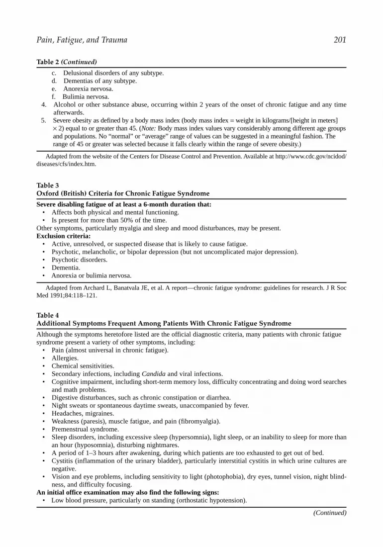
Pain, Fatigue, and Trauma 201
Table 2 (Continued)
c. Delusional disorders of any subtype.d. Dementias of any subtype.e. Anorexia nervosa.f. Bulimia nervosa.
4. Alcohol or other substance abuse, occurring within 2 years of the onset of chronic fatigue and any timeafterwards.
5. Severe obesity as defined by a body mass index (body mass index = weight in kilograms/[height in meters] × 2) equal to or greater than 45. (Note: Body mass index values vary considerably among different age groupsand populations. No “normal” or “average” range of values can be suggested in a meaningful fashion. Therange of 45 or greater was selected because it falls clearly within the range of severe obesity.)
Adapted from the website of the Centers for Disease Control and Prevention. Available at http://www.cdc.gov/ncidod/diseases/cfs/index.htm.
Table 3Oxford (British) Criteria for Chronic Fatigue Syndrome
Severe disabling fatigue of at least a 6-month duration that:• Affects both physical and mental functioning.• Is present for more than 50% of the time.
Other symptoms, particularly myalgia and sleep and mood disturbances, may be present.Exclusion criteria:
• Active, unresolved, or suspected disease that is likely to cause fatigue.• Psychotic, melancholic, or bipolar depression (but not uncomplicated major depression).• Psychotic disorders.• Dementia.• Anorexia or bulimia nervosa.
Adapted from Archard L, Banatvala JE, et al. A report—chronic fatigue syndrome: guidelines for research. J R SocMed 1991;84:118–121.
Table 4Additional Symptoms Frequent Among Patients With Chronic Fatigue Syndrome
Although the symptoms heretofore listed are the official diagnostic criteria, many patients with chronic fatiguesyndrome present a variety of other symptoms, including:
• Pain (almost universal in chronic fatigue).• Allergies.• Chemical sensitivities.• Secondary infections, including Candida and viral infections.• Cognitive impairment, including short-term memory loss, difficulty concentrating and doing word searches
and math problems.• Digestive disturbances, such as chronic constipation or diarrhea.• Night sweats or spontaneous daytime sweats, unaccompanied by fever.• Headaches, migraines.• Weakness (paresis), muscle fatigue, and pain (fibromyalgia).• Premenstrual syndrome.• Sleep disorders, including excessive sleep (hypersomnia), light sleep, or an inability to sleep for more than
an hour (hyposomnia), disturbing nightmares.• A period of 1–3 hours after awakening, during which patients are too exhausted to get out of bed.• Cystitis (inflammation of the urinary bladder), particularly interstitial cystitis in which urine cultures are
negative.• Vision and eye problems, including sensitivity to light (photophobia), dry eyes, tunnel vision, night blind-
ness, and difficulty focusing.An initial office examination may also find the following signs:
• Low blood pressure, particularly on standing (orthostatic hypotension).
(Continued)

Table 4 (Continued)
• Low oral temperatures (less than 97°F).• Slightly elevated oral temperatures (but less than 100°F), which are part of persistent flu-like symptoms.• Increased heart rate (tachycardia).• A positive Romberg test (unsteadiness when standing with eyes closed).
Adapted from the website of the Centers for Disease Control and Prevention. Available at http://www.cdc.gov/ncidod/diseases/cfs/index.htm.
Table 5Tests for Screening of Chronic Fatigue Syndrome Laboratory Tests for Evaluation of PossibleChronic Fatigue Syndrome
• Complete blood count with differential.• Complete metabolic panel.• Erythrocyte sedimentation rate.• Urinalysis.
Optional tests include:• Antinuclear antibodies and rheumatoid factor. • Thyroid tests (T3, T4, thyroid-stimulating hormone).• Adrenal tests (AM and PM cortisol levels).• Lyme titers and HIV serology.
Specific tests that support (but do not necessarily confirm) a diagnosis of chronic fatigue include:• Tests for viral infections, such as cytomegalovirus, Epstein-Barr virus, human herpesvirus 6, and
Coxsackie virus.• Immune system tests, including low natural-killer cell counts, elevated interferon-α, tumor necrosis-α,
interleukins 1 and 2, T-cell activation, altered T4/T8 cell ratios, low T-cell suppressor cell count,fluctuating B- and T-cell counts, antinuclear antibodies, immunoglobulin deficiency, and antithyroidantibodies.
• Exercise testing may show decreased cortisol levels after exercise, decreased cerebral blood flow after exercise,inefficient glucose utilization, and erratic breathing patterns.
Adapted from the website of the Centers for Disease Control and Prevention. Available at http://www.cdc.gov/ncidod/diseases/cfs/index.htm.
Table 6Diagnostic Criteria for Complex Regional Pain Syndrome
I. Type I (reflex sympathetic dystrophy)A. The presence of an initiating noxious event, or a cause of immobilization.B. Ongoing spontaneous pain, allodynia, or hyperalgesia is disproportionate to any inciting event and is not
limited to the distribution of a single peripheral nerve.C. Evidence at some time of autonomic dysfunction, such as edema, changes in skin blood flow,
hyperhidrosis, or abnormal sudomotor activity in the region of the pain.D. The diagnosis is excluded by the existence of conditions that would otherwise account for the degree of
pain and dysfunction.II. Complex regional pain syndrome type II (causalgia)
A. Type II is a syndrome that develops after a nerve injury. Spontaneous pain or allodynia/hyperalgesiaoccurs and is not necessarily limited to the territory of the injured nerve.
B. There is or has been evidence of edema, skin blood-flow abnormality, or abnormal sudomotor activity inthe region of the pain since the inciting event.
C. This diagnosis is excluded by the existence of conditions that would otherwise account for the degree ofpain and dysfunction.
Adapted from Stanton-Hicks M, Janig W, Hassenbusch S, Haddox JD, Boas R, Wilson P. Reflex sympathetic dystrophy:changing concepts and taxonomy. Pain 1995;63:127–133, with permission from Elsevier.
202 Diagnostic Criteria in Neurology

Pain, Fatigue, and Trauma 203
Table 7Signs and/or Symptoms on Complex Regional Pain Syndrome Checklist
1. “Burning” pain.2. Hyperesthesia.3. Temperature asymmetry.4. Color changes.5. Sweating changes.6. Edema.7. Nail changes.8. Hair changes.9. Skin changes.
10. Weakness.11. Tremor.12. Dystonia.13. Decreased range of motion.14. Hyperalgesia.15. Allodynia.
Adapted from Bruehl S, Harden RN, Galer BS, et al. External validation of IASP diagnostic criteria for complexregional pain syndrome and proposed research diagnostic criteria. Pain 1999;81:147–154.
Table 8Proposed Modified Research Diagnostic Criteria for Complex Regional Pain Syndrome
1. Continuing pain that is disproportionate to any inciting event.2. Must report at least one symptom in each of four following categories:
a. Sensoryi. Reported hyperesthesia.
b. Vasomotori. Temperature asymmetry.
ii. Skin color changes.iii. Skin color asymmetry.
c. Sudomotor/edemai. Edema.
ii. Sweating changes.iii. Sweating asymmetry.
d. Motor/trophici. Decreased range of motion.
ii. Motor dysfunction(1) Weakness.(2) Tremor.(3) Dystonia.
i. Trophic changes(1) Skin.(2) Nails.(3) Hair.
3. Must display at least one sign in two or more of the following categories:a. Sensory
i. Hyperalgesia to pinprick.ii. Allodynia to light touch.
iii. Vasomotor(1) Evidence of temperature asymmetry.(2) Evidence of skin color changes or asymmetry.
b. Sudomotor/edemai. Evidence of edema.
ii. Evidence of sweating changes.iii. Evidence of sweating asymmetry.
(Continued)

Chronic persistent fatigue is defined as fatigue that is present for any amount of time on 50% of thedays for 6 weeks and fatigue that limits functional activities or quality of life.
FIBROMYALGIA
204 Diagnostic Criteria in Neurology
Table 8 (Continued)
c. Motor/trophici. Evidence of decreased range of motion.
ii. Evidence of motor dysfunction(1) Tremor.(2) Weakness.(3) Dystonia.
d. Evidence of trophic changesi. Skin.
ii. Nails.iii. Hair.
Adapted from Bruehl S, Harden RN, Galer BS, et al. External validation of IASP diagnostic criteria for complexregional pain syndrome and proposed research diagnostic criteria. Pain 1999;81:147–154, with permission from Elsevier.
Table 91990 Criteria for the Classification of Fibromyalgia
1. History of widespread painDefinition: Pain is considered widespread when all of the following are present: pain in the left side of the body,pain in the right side of the body, pain above the waist, and pain below the waist. In addition, axial skeletal pain(cervical spine or anterior chest or thoracic spine or low back) must be present. In this definition, shoulder andbuttock pain is considered as pain for each involved side. “Low back pain” is considered lower-segment pain.
2. Pain in 11 of 18 tender point sites on digital palpationDefinition: Pain, on digital palpation, must be present in at least 11 of the following 18 sites:Occiput: bilateral, at the suboccipital muscle insertions.Low cervical: bilateral, at the anterior aspects of the intertransverse spaces at C5–C7.Trapezius: bilateral, at the midpoint of the upper border.Supraspinatus: bilateral, at origins above the scapula spine near the medial border.Second rib: bilateral, at the second costochondral junctions, just lateral to the junctions on upper surfaces.Lateral epicondyle: bilateral, 2 cm distal to the epicondyles.Gluteal: bilateral, in upper outer quadrants of buttocks in anterior fold of muscle.Greater trochanter: bilateral, posterior to the trochanteric prominence.Knee: bilateral, at the medial fat pad proximal to the joint line.Digital palpation should be performed with an approximate force of 4 kg.For a tender point to be considered “positive,” the subject must state that the palpation was painful. “Tender” isnot to be considered “painful.”
For classification purposes, patients will be said to have fibromyalgia if both criteria are satisfied. Widespread painmust have been present for at least 3 months. The presence of a second clinical disorder does not exclude the diagnosisof fibromyalgia.
(Adapted from Wolfe F, Smythe HA, Yunus MB, et al. The American College of Rheumatology 1990 criteria for theclassification of fibromyalgia: report of the multicenter criteria committee. Arthritis Rheum 1999;33:160–172, with per-mission from John Wiley and Sons, Inc.)
MINOR HEAD INJURY
Despite being one of the most common conditions involving trauma and the nervous system, thereremains wide variability in diagnostic criteria for minor head trauma. One method of categorizing headtrauma is by using the initial Glasgow Coma Scale. Minimal head injury corresponds to a score of 15,without loss of consciousness. A score of 14 to 15, with loss of consciousness lasting less than 5 minutes,constitutes mild head injury and no neurological deficit. Those with scores of 13 would be considered

Table 10Diagnostic Criteria for Minor Head Injury
Criterion n Percentage
Loss of consciousness 58 82Posttraumatic amnesia 30 42Impaired level of consciousness 5 7Absence of focal neurological deficit 6 8Other signs and symptomsa 29 41No definition 8 11
In all, 64 hospitals listed which diagnostic criteria they used to define minor head injury.aVertigo, nausea, vomiting, headache, confusion.(Adapted with permission from Bellner J, Jensen S-M, Romner B. Diagnostic criteria and use of ICD-10 codes to
define and classify minor head injury. J Neurol Neurosurg Psychiatry 2003;74:351–352.)
Table 11Recommended Definitions for Mild Traumatic Brain Injury
Incident cases of mild traumatic brain injury (MTBI)The conceptual definition of MTBI is an injury to the head as a result of blunt trauma or acceleration ordeceleration forces that result in one or more of the following conditions:
• Any period of observed or self-reported:o Transient confusion, disorientation, or impaired consciousness.o Dysfunction of memory around the time of injury.o Loss of consciousness lasting less than 30 minutes.
• Observed signs of neurological or neuropsychological dysfunction, such as:o Seizures acutely following injury to the head.o Among infants and very young children: irritability, lethargy, or vomiting following head injury.o Symptoms among older children and adults such as headache, dizziness, irritability, fatigue, or poor
concentration, when identified soon after injury, can be used to support the diagnosis of MTBI, butcannot be used to make the diagnosis in the absence of loss of consciousness or altered consciousness.Research may provide additional guidance in this area.
Based on this conceptual definition, separate operational definitions of MTBI are recommended for cases identi-fied from interviews and surveys, administrative health care datasets, and patient medical records.
Adapted from Center for Disease Control and Prevention, National Center for Injury Prevention and Control. Reportto Congress on mild traumatic brain injury in the United States: steps to prevent a serious public health problem.September 2003. Available at http://www.cdc.gov/Migrated_Content/Report/TBI_Report_to_Congress_on_MTBI_Sept_2003.pdf.
Pain, Fatigue, and Trauma 205
to have moderate head injury. However, the experience in the field of athletic injuries, especially asrelated to “concussion,” indicates that loss of consciousness may be hard to discern and of variableimport in injury severity.
In a survey of Swedish hospitals, Bellner et al. found wide variability in application of head injurydiagnostic criteria, as indicated by International Statistical Classification of Diseases and RelatedHealth Problems, 10th Edition, codes (Table 10).
In a report to the United States Congress, a committee reported on the epidemiology and preven-tion of mild head trauma. Their operational definition is presented in Table 11.
PAIN DISORDERS
The International Association for the Study of Pain defines pain as “an unpleasant sensory andemotional experience associated with actual or potential tissue damage, or described in terms ofsuch damage.”
Several aspects of this definition are worth noting. Pain is linked to the concept of consciousness, interms of the words “unpleasant experience.” This definition also puts the concept of pain squarely in terms

Table 12Diagnostic Criteria for Pain Disorder
• Pain in one or more anatomic sites is the predominant focus of the clinical presentation and is ofsufficient severity to warrant clinical attention.
• The pain causes clinically significant distress or impairment in social, occupational, or other importantareas of functioning.
• Psychological factors are judged to have an important role in the onset, severity, exacerbation, ormaintenance of the pain.
• The symptom or deficit is not intentionally produced or feigned (as in factitious disorder ormalingering).
• The pain is not better accounted for by a mood, anxiety, or psychotic disorder and does not meetcriteria for dyspareunia.
Code as follows:Pain disorder associated with psychological factors: Psychological factors are judged to have the major role in theonset, severity, exacerbation, or maintenance of the pain. (If a general medical condition is present, it does nothave a major role in the onset, severity, exacerbation, or maintenance of the pain.) This type of pain disorder is notdiagnosed if criteria are also met for somatization disorder.Specify if:Acute: duration of less than 6 months.Chronic: duration of 6 months or longer.Pain disorder associated with both psychological factors and a general medical condition: Both psychologicalfactors and a general medical condition are judged to have important roles in the onset, severity, exacerbation,or maintenance of the pain. The associated general medical condition or anatomic site of pain is coded on AxisIII.Specify if:Acute: duration of less than 6 months.Chronic: duration of 6 months or longer.
Adapted from American Psychiatric Association. Diagnostic and Statistical Manual of Mental Disorders, 4th rev. ed.Washington, DC: American Psychiatric Association, 1994.
206 Diagnostic Criteria in Neurology
of the patient’s perception. It does not specify any objective correlate in terms of physiological functioningor evidence of tissue damage. Understanding the concept may help some trainees and practitioners inevaluation of individuals presenting with pain. Implicit in the diagnosis is a central component of pain,whose biological basis involves multiple levels of the neuraxis and inputs from multiple neurotrans-mitter systems. However, the definition does not specify how this may change as related to the chronicityof the pain.
Several discrete pain syndromes have been described and are listed in Table 12.
Table 13
Diagnostic Criteria for Second-Impact Syndrome
Criteria Definition
(a) Medical review after a witnessed first impact(b) Documentation of ongoing symptoms following the first impact
up to the time of the second impact(c) Witnessed second head impact with a subsequent rapid
cerebral deterioration(d) Neuropathological or neuroimaging evidence of cerebral swelling without
significant intracranial hematoma or other cause for edema
Definite second-impact syndrome (SIS): (a), (b), (c), and (d).Probable SIS: (c) and (d) and either (a) or (b).Possible SIS: (c) and (d) only.Not SIS: (c) or (d) absent.
Adapted from McCrory PR, Berkovic SF. Second-impact syndrome. Neurology 1998;50:677–683, with permissionfrom Lippincott, Williams, and Wilkins.

Pain, Fatigue, and Trauma 207
SECOND-IMPACT SYNDROME
A syndrome of diffuse cerebral swelling with catastrophic results after a second head injury. It isseen most commonly in children and adolescents, particularly in the setting of sports injuries with abrief concussion, when a second impact occurs before the symptoms of the first injury have cleared,resulting in more severe cerebral injury.
SOURCES
Cancer-Related FatigueCella D, Peterman A, Passik S, et al. Progress toward guidelines for the management of fatigue. Oncology
1998;12:S369–S377.Cella D, Davis K, Breitbart W, et al. Cancer-related fatigue: prevalence of proposed diagnostic criteria in a United States
sample of cancer survivors. J Clin Oncol 2001;19:3385–3391.
Chronic Fatigue SyndromeFukuda K, Straus SE, Hickey I, et al. The chronic fatigue syndrome: a comprehensive approach to its definition and study. Ann
Intern Med 1994;121:953–959.Holmes G, Kaplan J, Gantz N, et al. Chronic fatigue syndrome: a working case definition. Ann Intern Med 1988;108:387–389.Reeves WC, Lloyd A, Vernon SD, et al. Identification of ambiguities in the 1994 chronic fatigue syndrome research case
definition and recommendations for resolution. BMC Health Serv Res 2003;3:25.Sharpe MC, Archard L, Banatvala JE, et al. A report—chronic fatigue syndrome: guidelines for research. J R Soc Med
1991;84:118–121.
Complex Regional Pain SyndromeBruehl S, Harden RN, Galer BS, et al. External validation of IASP diagnostic criteria for complex regional pain syndrome and
proposed research diagnostic criteria. Pain 1999;81:147–154.Hassantash SA, Afrakhteh M, Maier RV. Causalgia: a meta-analysis of the literature. Arch Surg 2003;138:1226–1231.Stanton-Hicks M, Janig W, Hassenbusch S, Haddox JD, Boas R, Wilson P. Reflex sympathetic dystrophy: changing concepts
and taxonomy. Pain 1995;63:127–133.
Fatigue in Multiple SclerosisMS Council for Clinical Practice Guidelines. Fatigue in Multiple Sclerosis. Washington, DC: Paralyzed Veterans Association, 1998.
FibromyalgiaWolfe F, Smythe HA, Yunus MB, et al. The American College of Rheumatology 1990 criteria for the classification of
fibromyalgia: report of the multicenter criteria committee. Arthritis Rheum 1990;33:160–172.
Minor Head InjuryBellner J, Jensen S-M, Romner B. Diagnostic criteria and use of ICD-10 codes to define and classify minor head injury.
J Neurol Neurosurg Psychiatr 2003;74:351– 352.Centers for Disease Control and Prevention, National Center for Injury Prevention and Control. Report to Congress on mild
traumatic brain injury in the United States: steps to prevent a serious public health problem. September 2003. Availablevia http://www.cdc.gov/Migrated_Content/Report/TBI_Report_to_Congress_on_MTBI_Sept_2003.pdf).
Pain DisordersAmerican Psychiatric Association. Diagnostic and Statistical Manual of Mental Disorders, 4th rev. ed. Washington, DC:
American Psychiatric Association, 1994.Fishbain DA, Goldberg M, Meagher BR, Steele R, Rosomoff H. Male and female chronic pain patients categorized by
DSM-III psychiatric diagnostic criteria. Pain 1986;26:181–197.
Second-Impact SyndromeMcCrory PR, Berkovic SF. Second impact syndrome. Neurology 1998;50:677–683.


14Sleep Disorders
NARCOLEPSY
Narcolepsy is a sleep disorder with prominent sleep attacks as a form of excessive daytime somnolence.The other associated features of narcolepsy occur with varying frequency. These include cataplexy,hypnagogic and hypnopompic hallucinations, and sleep paralysis.
The laboratory method of diagnosis is the multiple sleep latency test. Narcoleptics will show sleep-onset rapid eye movement with short latency. Biochemical markers such as human leukocyte antigenDQB1*0602 or DR2 type have very high sensitivity but low specificity. Cerebrospinal fluid analysismay show low levels of the protein hypocretin, but the diagnostic utility of this is unclear.
A number of diagnostic criteria have been developed. The Diagnostic and Statistical Manual ofMental Disorders, 4th edition, relies on clinical criteria alone. The American Academy of SleepMedicine’s most recent revision of the International Classification of Sleep Disorders adds sleep labparameters to the diagnosis. Silber et al., citing the ambiguity and complexity of the InternationalClassification of Sleep Disorders criteria and the lack of validation of these two guidelines, have pro-posed revised criteria. However, their nomenclature is cumbersome for routine clinical use, and likelywill find value primarily among sleep researchers.
From a practical standpoint, it is important to diagnose narcolepsy adequately. Many patients haveendured numerous evaluations, leading to delays in diagnosis. The sleep attacks may make work ordriving an automobile dangerous, and lead to significant disability. Treatment options include allow-ing for frequent naps, amphetamines, or modafinil. The first two treatments may themselves be asso-ciated with interference of work activities or the need to take stimulants on a chronic basis.
RESTLESS LEGS SYNDROME
Restless legs syndrome (RLS) features nocturnal involuntary limb movements that can causeinsomnia because of frequent sleep disruption, and often affects bed partners because of frequentmyoclonic-type jerking. It generally begins in early adulthood and affects from 2 to 5% of the popu-lation. RLS may run in families, with susceptibility genes identified on chromosomes 12q and 14q.RLS has also been associated with Parkinson’s disease, pregnancy, end-stage renal disease, iron defi-ciency anemia, peripheral neuropathy, and diabetes.
Treatment of RLS is based on individual patient needs, age, and comorbid conditions. Dopaminergicdrugs are generally used for initial treatment. Anticonvulsants, opioids, and sedative/hypnotics may alsobe effective in treating RLS.
SLEEP APNEA SYNDROME
Sleep apnea syndrome is a common cause of excessive daytime somnolence. Aside from its asso-ciation with obesity and smoking, obstructive sleep apnea has also been associated with increased riskfor many other disorders, including cardiovascular and cerebrovascular disease. The detailed diagnostic
From: Current Clinical Neurology: Diagnostic Criteria in NeurologyEdited by: A. J. Lerner © Humana Press Inc., Totowa, NJ
209

criteria for both obstructive and varieties of central sleep apnea are located in the section on sleep inthe criteria adapted from the American Sleep Disorders Associations International Classification ofSleep Disorders: Diagnosis and Coding Manual.
SLEEP DISORDERS
Tables 11–62 have been adapted from the American Sleep Disorders Association’s InternationalClassification of Sleep Disorders: Diagnosis and Coding Manual.
210 Diagnostic Criteria in Neurology
Table 1Diagnostic and Statistical Manual of Mental Disorders, 4th Revised Edition, Diagnostic Criteriafor Narcolepsy
A. Irresistible attacks of refreshing sleep that occur daily over at least 3 months.B. The presence of one or both of the following:
a. Cataplexy (i.e., brief episodes of sudden bilateral loss of muscle tone, most often in association withintense emotion).
b. Recurrent intrusions of elements of rapid eye movement sleep into the transition between sleep andwakefulness, as manifested by either hypnopompic or hypnagogic hallucinations or sleep paralysis atthe beginning or end of sleep episodes.
C. The disturbance is not because of the direct physiological effects of a substance (e.g., a drug of abuse, amedication) or another general medical condition.
Adapted from American Psychiatric Association. Diagnostic and Statistical Manual of Mental Disorders, 4th rev. ed.Washington, DC: American Psychiatric Association, 1994.
Table 2American Academy of Sleep Medicine Diagnostic Criteria: Narcolepsy
A. The patient has a complaint of excessive sleepiness or sudden muscle weakness.B. Recurrent daytime naps or lapses into sleep occur almost daily for at least 3 months.C. Sudden bilateral loss of postural muscle tone occurs in association with intense emotion (cataplexy).D. Associated features include:
1. Sleep paralysis.2. Hypnagogic hallucinations.3. Automatic behaviors.4. Disrupted major sleep episode.
E. Polysomnography demonstrates one or more of the following:1. Sleep latency <10 minutes;2. Rapid eye movement sleep latency <20 minutes;3. A multiple sleep latency test that demonstrates a mean sleep latency of <5 minutes; and4. Two or more sleep-onset rapid eye movement periods.
F. Human leukocyte antigen typing demonstrates DQB1*0602 or DR2 positivity.G. No medical or mental disorder accounts for the symptoms.H. Other sleep disorders (e.g., periodic limb movement disorder or central sleep apnea syndrome) may be
present but are not the primary cause of the symptoms.Minimal criteria: B plus C, or A plus D plus E plus G.Severity criteria:Mild: Mild sleepiness or rare cataplexy (less than once per week).Moderate: Moderate sleepiness or infrequent cataplexy (less than daily).Severe: Severe sleepiness or severe cataplexy (daily).Duration criteria:Acute: 6 months or less.Subacute: More than 6 months but less than 12 months.Chronic: 12 months or longer.
Adapted from The International Classification of Sleep Disorders, Revised: Diagnostic and Coding Manual.Rochester: American Sleep Disorders Association, 1997.

Sleep Disorders 211
Table 3Narcolepsy Diagnostic Criteria by Silber et al.
Diagnostic criteriaCategory A. Definite narcolepsyHistory of excessive daytime sleepiness.History of cataplexy, defined as definite bilateral weakness of brief duration brought on by emotion.Mean initial sleep latency of 8 minutes on multiple sleep latency test (MSLT).a
Two or more sleep-onset rapid eye movement periods (SOREMP) on MSLT,a or one SOREMP on MSLTa andone SOREMP on the preceding nocturnal polysomnography (PSG).Apnea–hypopnea index (AHI): 10/hour on nocturnal PSG preceding the MSLT.a
(The last three criteria can be replaced by cataplexy witnessed by a physician with documented recoverableareflexia, or cataplexy recorded by PSG and video recording.)Category B. Probable narcolepsy (laboratory confirmation)Subgroup B1History of excessive daytime sleepiness.A history of cataplexy, defined as definite bilateral weakness of brief duration brought on by emotion.Mean initial sleep latency of 8 minutes on MSLT.a
One or fewer SOREMP on MSLTa or on the preceding nocturnal PSG.AHI: 10/hour on the nocturnal PSG preceding the MSLT.a
Subgroup B2History of excessive daytime sleepiness.No history of cataplexy.Mean initial sleep latency of 8 minutes on MSLT.a
Two or more SOREMP on MSLT or one SOREMP on the MSLTa and one SOREMP on the preceding nocturnalPSG.AHI: 10/hour on the nocturnal PSG preceding the MSLT.a
Category C. Probable narcolepsy (clinical)History of excessive daytime sleepiness.History of cataplexy, defined as definite bilateral weakness of brief duration brought on by emotion.No or inadequate sleep studies performed.
aMSLT performed under standard conditions, including a total sleep time of 6 hours on the preceding night PSG.(Adapted from Silber MH, Krahn LE, Olson EJ. Diagnosing narcolepsy: validity and reliability of new diagnostic
criteria. Sleep Med 2002;3:109–113, with permission of Elsevier.)
Table 4Summary of Diagnostic Criteria for Narcolepsy According to Silber et al.
LaboratoryCategory Cataplexy confirmation of sleepiness SOREM periods
A Yes Yes YesB1 Yes Yes NoB2 No Yes YesC Yes No No
SOREM, sleep-onset rapid eye movement periods.(Adapted from Silber MH, Krahn LE, Olson EJ. Diagnosing narcolepsy: validity and reliability of new diagnostic
criteria. Sleep Med 2002;3:109–113, with permission of Elsevier.)
Table 5Essential Diagnostic Criteria for Restless Legs Syndrome
1. An urge to move the legs, usually accompanied or caused by uncomfortable and unpleasant sensations inthe legs. (Sometimes, the urge to move is present without the uncomfortable sensations and sometimes thearms or other body parts are involved in addition to the legs.)
2. The urge to move or unpleasant sensations begin(s) or worsen(s) during periods of rest or inactivity, suchas lying or sitting.
(Continued)

212 Diagnostic Criteria in Neurology
Table 5 (Continued)
3. The urge to move or unpleasant sensations are relieved partially or totally by movement, such as walkingor stretching, at least as long as the activity continues.
4. The urge to move or unpleasant sensations are worse in the evening or night than during the day or onlyoccur in the evening or night. (When symptoms are very severe, the worsening at night may not benoticeable but must have been previously present.)
Adapted from Allen RP, Picchietti D, Hening WA, Trenkwalder C, Walters AS, Montplaisi J. Restless legs syndrome:diagnostic criteria, special considerations, and epidemiology. A report from the restless legs syndrome diagnosis and epi-demiology workshop at the National Institutes of Health. Sleep Med 2003;4:101–119, with permission of Elsevier.
Table 6Supportive Clinical Features of Restless Leg Syndrome
Family history:The prevalence of restless leg syndrome (RLS) among first-degree relatives of people with RLS is three to fivetimes greater than in people without RLS.
Response to dopaminergic therapy:Nearly all people with RLS show at least an initial positive therapeutic response to either L-DOPA or adopamine-receptor agonist at doses considered very low in relation to the traditional doses of these medicationsused for the treatment of Parkinson’s disease. This initial response is not, however, universally maintained.
Periodic limb movements (during wakefulness or sleep):Periodic limb movements in sleep (PLMS) occur in at least 85% of people with RLS; however, PLMS alsocommonly occur in other disorders and in the elderly. In children, PLMS are much less common than in adults.
Adapted from Allen RP, Picchietti D, Hening WA, Trenkwalder C, Walters AS, Montplaisi J. Restless legs syn-drome: diagnostic criteria, special considerations, and epidemiology. A report from the restless legs syndrome diagnosisand epidemiology workshop at the National Institutes of Health. Sleep Med 2003;4:101–119, with permission fromElsevier.
Table 7Restless Legs Syndrome in Cognitively Impaired Elderly
Essential criteria for the diagnosis of probable restless leg syndrome in the cognitively impaired elderly (all fiveare necessary for diagnosis).
1. Signs of leg discomfort, such as rubbing or kneading the legs, and groaning while holding the lowerextremities are present.
2. Excessive motor activity in the lower extremities, such as pacing, fidgeting, repetitive kicking, tossing andturning in bed, slapping the legs on the mattress, cycling movements of the lower limbs, repetitive foot tapping,rubbing the feet together, and the inability to remain seated, are present.
3. Signs of leg discomfort are exclusively present or worsen during periods of rest or inactivity.4. Signs of leg discomfort are diminished with activity.5. Criteria 1 and 2 occur only in the evening or at night or are worse at those times than during the day.
Adapted from Allen RP, Picchietti D, Hening WA, Trenkwalder C, Walters AS, Montplaisi J. Restless legs syndrome:diagnostic criteria, special considerations, and epidemiology. A report from the restless legs syndrome diagnosis and epi-demiology workshop at the National Institutes of Health. Sleep Med 2003;4:101–119, with permission of Elsevier.
Table 8Supportive or Suggestive Criteria for the Diagnosis of Probable Restless Leg Syndrome in theCognitively Impaired Elderly
1. Dopaminergic responsiveness.2. Patient’s past history—as reported by a family member, caregiver, or friend—is suggestive of restless leg
syndrome.3. A first-degree, biological relative (sibling, child, or parent) has restless leg syndrome.
(Continued)

Table 8 (Continued)
4. Observed periodic limb movements while awake or during sleep.5. Periodic limb movements in sleep recorded by polysomnography or actigraphy.5. Significant sleep-onset problems.6. Better quality sleep in the day than at night.7. The use of restraints at night (for institutionalized patients).8. Low serum ferritin level.9. End-stage renal disease.
10. Diabetes.11. Clinical, electromyographic, or nerve-conduction evidence of peripheral neuropathy or radiculopathy.
Adapted from Allen RP, Picchietti D, Hening WA, Trenkwalder C, Walters AS, Montplaisi J. Restless legs syn-drome: diagnostic criteria, special considerations, and epidemiology. A report from the restless legs syndrome diagnosisand epidemiology workshop at the National Institutes of Health. Sleep Med 2003;4:101–119, with permission ofElsevier.
Table 9Criteria for the Diagnosis of Definite Restless Leg Syndrome in Children
1. The child meets all four essential adult criteria for restless leg syndrome (RLS); and2. The child relates a description in his or her own words that is consistent with leg discomfort. (The child
may use terms such as “oowies,” “tickle,” “spiders,” “boo-boos,” “want to run,” and “a lot of energy in mylegs” to describe symptoms. Age-appropriate descriptors are encouraged.)
or
1. The child meets all four essential adult criteria for RLS; and2. Two of the three following supportive criteria are present.
Supportive criteria for the diagnosis of definite RLS in childrena. Sleep disturbance for age.b. A biological parent or sibling has definite RLS.c. The child has a polysomnographically documented periodic limb movement index of 5 or more per
hour of sleep.
Adapted from Allen RP, Picchietti D, Hening WA, Trenkwalder C, Walters AS, Montplaisi J. Restless legs syndrome:diagnostic criteria, special considerations, and epidemiology. A report from the restless legs syndrome diagnosis and epi-demiology workshop at the National Institutes of Health. Sleep Med 2003;4:101–119, with permission of Elsevier.)
Table 10Criteria for the Diagnosis of Probable Restless Leg Syndrome in Children
1. Adult criterion no. 4 in Table 5 (the urge to move or sensations are worse in the evening or at night thanduring the day);
and
2. The child has a biological parent or sibling with definite restless leg syndrome.
Ora
1. The child is observed to have behavior manifestations of lower extremity discomfort when sitting or lying,accompanied by motor movement of the affected limbs, the discomfort has characteristics of adult criterianos. 2, 3, and 4 (i.e., is worse during rest and inactivity, relieved by movement, and worse during theevening and at night);
and
2. The child has a biological parent or sibling with definite restless leg syndrome.
aThis last probable category is intended for young children or cognitively impaired children who do not have suffi-cient language to describe the sensory component of restless leg syndrome.
(Adapted from Allen RP, Picchietti D, Hening WA, Trenkwalder C, Walters AS, Montplaisi J. Restless legs syndrome:diagnostic criteria, special considerations, and epidemiology. A report from the restless legs syndrome diagnosis and epi-demiology workshop at the National Institutes of Health. Sleep Med 2003;4:101–119, with permission of Elsevier.)
Sleep Disorders 213

Associated With Behavioral/Psychophysiological Disorders
214 Diagnostic Criteria in Neurology
Table 11Diagnostic Criteria for Adjustment Sleep Disorder
1. Complaint of insomnia or excessive sleepiness.2. Complaint is a reaction temporarily associated with an identifiable stressing event.3. The disorder is expected to remit if the stress is reduced or the level of adaptation is increased.
Table 12Diagnostic Criteria for Psychophysiological Insomnia
1. Complaint of insomnia combined with a complaint of diminished performance during waking hours.2. Learned associations that prevent sleep are identified:
a. Forcing sleep, which suggests inability to sleep at the desired time, but with the ability to sleep in thecourse of other relatively monotonous activities, such as watching television or reading.
b. Awakenings conditioned by the room or sleep-related activities, indicated by poor sleeping at home,but improved sleeping away from home or when no room routines are performed.
Table 13Diagnostic Criteria for Inadequate Sleep Hygiene
Complaint of insomnia or excessive sleepinessPresence of at least one of the following:
1. Daytime naps at least twice a week.2. Variable bedtime or awakening hours.3. Frequent episodes (two to three times a week) of extended time in bed.4. Routine use of products containing alcohol, tobacco, or caffeine in the period preceding bedtime.5. Performance of exercise near bedtime.6. Plans to become implicated in exciting or emotionally bothersome activities near bedtime.7. Frequent use of the bed for unrelated activities (e.g., to watch television, read, study, eat, etc.).8. Sleeps in an uncomfortable bed (mattress in poor condition, inadequate bed linen, etc.).9. Allows the room to be too bright, poorly ventilated, untidy, too warm, too cold or otherwise conditioned
to preclude sleep induction.10. Carries out activities requiring high levels of concentration shortly before bedtime.11. Allows mental activities in bed, such as thinking, planning, remembering, etc.
Table 14Diagnostic Criteria for Limit-Setting Sleep Disorder
1. Evasive or refuses to go to bed at appropriate time.2. Once sleep period has started, sleeping is of normal quality and duration.
Table 15Diagnostic Criteria for Sleep-Onset Association Disorder
1. Complaint of insomnia.2. Complaint is temporarily associated with the absence of certain conditions (e.g., being picked up in arms,
moved or breastfed, listening to radio or watching television, etc.).3. With the particular association present, sleep is normal in terms of onset, duration, and quality.4. No evidence of significant underlying medical or psychiatric disorder able to account for complaint.5. No other criteria for other sleep disorders able to cause difficulties in falling asleep (e.g., limit-setting
sleep disorder).
Table 16Diagnostic Criteria for Nocturnal Eating/Drinking Syndrome
1. Frequent and recurrent awakenings in order to eat or drink.2. Following food or drink intake, sleep onset is normal.

Sleep Disorders 215
Associated With Psychiatric DisordersTable 17Diagnostic Criteria for Sleep Disorder With Psychoses
1. Complaint of insomnia or excessive sleepiness.2. Clinical diagnosis of schizophrenia, schizophrenia-like disorder, or some other functional psychosis.
Table 18Diagnostic Criteria for Sleep Disorder With Mood Disorders
1. Complaint of insomnia or excessive sleepiness.2. Complaint is temporally associated with diagnosis of mood disorder.
Table 19Diagnostic Criteria for Sleep Disorder With Anxiety Disorders
1. Complaint of insomnia or excessive sleepiness.2. Presence of long-term generalized anxiety disorder or some other anxiety disorder.3. The sleep disorder has followed the course of the psychiatric problem without significant prolonged
periods of remission.
Table 20Diagnostic Criteria for Sleep Disorder With Panic Disorders
1. Complaint of sudden awakening or insomnia.2. Presence of panic disorder with or without agoraphobia.3. The sleep disorder has followed the course of the psychiatric problem without significant prolonged
periods of remission.
Table 21Diagnostic Criteria for Sleep Disorder With Alcoholism
1. Complaint of insomnia or excessive sleepiness.2. Diagnosis of alcoholism.
Associated With Environmental FactorsTable 22Diagnostic Criteria for Environmental Sleep Disorder
1. Complaint of insomnia or excessive sleepiness.2. Complaint is temporally associated with the introduction of an environmental stimulus or circumstance
that alters sleep and is physically measurable.3. The physical properties of the environmental factor explain the sleep complaint; the psychological signif-
icance of the environmental factor does not account for the complaint.4. Withdrawal of the causal environmental factor leads to immediate or gradual resolution with a return to
normal sleep.5. The disorder has been present for more than 3 weeks.
Table 23Diagnostic Criteria for Food Allergy Insomnia
1. Complaint of insomnia.2. Complaint is temporally associated with the introduction of a concrete food or drink.3. Withdrawal of the agent restores normal sleep and waking, either immediately or in the course of about 4
weeks. The diurnal behavior may improve before the sleep model.4. Recurrence of altered sleep and diurnal behavior when the suspected allergen is reintroduced in the diet.

216 Diagnostic Criteria in Neurology
Associated With Drug Dependencies
Table 24Diagnostic Criteria for Toxin-Induced Sleep Disorder
1. Complaint of insomnia or excessive sleepiness.2. Complaint is temporally associated with the presence of an environmental or ingested toxic agent (e.g., heavy
metals or organic toxins, etc.).3. No evidence of any other medical or psychiatric disorder other than that associated with the toxicity accounting
for the complaint.4. The diagnostic criteria for any other sleep disorder causing complaints of insomnia or excessive sleepiness
are not met.
Table 25Diagnostic Criteria for Hypnotic-Dependent Sleep Disorder
1. Complaint of insomnia or excessive sleepiness.2. Use of hypnotics practically daily for at least 3 weeks.3. Withdrawal of the hypnotic is associated with exacerbation of the primary complaint, which is often
judged as being worse than the original sleep problem.
Table 26Diagnostic Criteria for Stimulant-Dependent Sleep Disorder
1. Complaint of insomnia or excessive sleepiness.2. Complaint is temporally associated with the use or withdrawal of a stimulant medication.3. Use of stimulant medication alters the habitual sleep period, or more than one attempt to withdraw the
stimulant induces symptoms of excessive sleepiness.
Table 27Diagnostic Criteria for Alcohol-Dependent Sleep Disorder
1. Complaint of insomnia or excessive sleepiness.2. Complaint is temporally associated with more than one attempt to withdraw alcohol consumption before
bedtime.
Associated With Sleep-Induced Respiratory ImpairmentTable 28Diagnostic Criteria for Obstructive Sleep Apnea Syndrome
1. Complaint of insomnia or excessive sleepiness. The patient may occasionally be unaware of clinical factsthat are nevertheless apparent to others.
2. Frequent episodes of obstructed breathing during sleep.3. The associated conditions include:
a. Heavy snoring.b. Dry mouth on awakening.c. Chest retraction during sleep in young children.
Table 29Diagnostic Criteria for Central Sleep Apnea Syndrome
1. Complaint of insomnia or excessive sleepiness. The patient may occasionally be unaware of clinical factsthat are nevertheless apparent to others.
2. Frequent episodes of shallow breathing or absence of breathing during sleep.3. Polysomnography shows central apneic pauses lasting more than 10 seconds (20 seconds in infancy), with
one of the following:
(Continued)

Insomnias Associated With Movement Disorders
Sleep Disorders 217
Table 29 (Continued)
a. Frequent awakening from sleep associated with apnea.b. Bradycardia or tachycardia.c. Oxygen desaturation associated with the apneic episodes (criteria included in the International
Classification of Sleep Disorders).d. Multiple sleep latency test exhibiting a mean sleep latency of less than 10 minutes.
Table 30Diagnostic Criteria for Central Alveolar Hypoventilation Syndrome
1. Complaint of insomnia or excessive sleepiness. The patient may occasionally be unaware of clinical factsthat are nevertheless apparent to others, such as hypoventilation during sleep.
2. Frequent episodes of shallow breathing or absence of breathing during sleep.3. Absence of primary lung disease, skeletal malformations, or neuromuscular disorders affecting respiration.4. Polysomnography shows episodes of shallow breathing lasting more than 10 seconds, associated with oxygen
desaturation and one or more of the following:a. Frequent awakening from sleep associated with the breathing alterations.b. Bradycardia or tachycardia.c. Multiple sleep latency test exhibiting a mean sleep latency of less than 10 minutes.
Table 31Diagnostic Criteria for Sleep Disorder Associated With Chronic Obstructive Pulmonary Disease
1. Complaint of insomnia or excessive sleepiness.2. Complaint is temporally associated with the presence of chronic obstructive pulmonary disease.
Table 32Diagnostic Criteria for Sleep-Related Asthma
1. Complaint of insomnia or excessive sleepiness, and cough or dyspnea.2. Complaint is temporally associated with the presence of asthma.
Table 33Diagnostic Criteria for Altitude Insomnia
1. Complaint of insomnia.2. Complaint is temporally associated with elevations typically higher than 4000 m.
Table 34Sleep Starts (Hypnic Jerks)
1. Complaints of difficulties falling asleep, or of intense bodily movements at start of sleep.2. Sudden, brief jerks at start of sleep, affecting mainly arms or legs.
Table 35Diagnostic Criteria for Restless Leg Syndrome (see Table 5 for Full Criteria)
1. Complaint of unpleasant sensation in legs at night, or difficulty falling asleep.2. Unpleasant slipping sensation within gastrocnemius region often associated with generalized pain and leg pain.3. Discomfort is calmed with limb movements.
Table 36Diagnostic Criteria Periodic Limb Movement Disorder
1. Complaint of insomnia or excessive sleepiness. Occasionally the patient is asymptomatic, and the move-ments are observed by another person.
2. Repetitive and highly stereotyped limb muscle movements, characterized in the leg by extension of thebig toe in combination with partial flexion of the ankle, knee, and occasionally the hip.

218 Diagnostic Criteria in Neurology
Associated With Alterations of the Sleep–Wake Cycle Temporal Model
Table 37Diagnostic Criteria for Nocturnal Leg Cramps (Nocturnal Myoclonus)
1. Complaint of painful sensation in the leg associated with muscle stiffness or pressing feeling.2. Recurrent awakenings associated with painful leg sensation.
Table 38Diagnostic Criteria for Rhythmic Movement Disorder
1. Rhythmic body movements occurring during sleepiness period or actual sleep.2. At least one of the following alterations is present:
a. The head moves strongly in an anterior–posterior direction (head banging).b. The head moves laterally when in dorsal decubitus (head rolling).c. The entire body moves in jerks while supported by hands and knees (body rocking).d. The entire body moves laterally when in dorsal decubitus (body rolling).
Table 39Diagnostic Criteria for Rapid Eye Movement Sleep Behavior Disorder
1. Limb or body movements associated with dreaming.2. At least one of the following:
a. Hazardous or potentially hazardous sleep behaviors.b. Sleep appears to involve acting.c. The behaviors alter sleep continuity.
Table 40Diagnostic Criteria for Nocturnal Paroxysmal Dystonia
1. Abnormal motor activity during sleep.2. Dystonic or dyskinetic episodes occurring mainly during sleep.3. Not associated with any underlying medical or psychiatric disorder capable of accounting for the symp-
tom, e.g., frontal lobe epilepsy.4. Does not meet the diagnostic criteria for other sleep disorders, such as rapid eye movement sleep behavior
disorder or night terror.
Table 41Diagnostic Criteria for Time-Zone Change (Jet-Lag) Syndrome
a. Complaint of insomnia or excessive sleepiness.b. Symptom started 1-2 days after air travel across at least two time-zones.
Table 42Diagnostic Criteria for Shift-Work Sleep Disorder
a. Primary complaint of insomnia or excessive sleepiness.b. Primary complaint is temporarily associated with a work period (normally at night) taking place
during normal sleeping period.
Table 43Diagnostic Criteria for Delayed Sleep-Phase Syndrome
a. Complaint of inability to fall asleep at desire time, or inability to spontaneously wake up at desiredtime, or excessive tiredness.
b. Delay in main sleep phase with respect to desired sleeping time.c. Symptoms present for at least 1 month.
(Continued)

Sleep Disorders 219
Table 44Diagnostic Criteria for Advanced Sleep-Phase Syndrome
a. Inability to stay awake until the desired bedtime, or inability to continue sleeping until the desiredwaking up time.
b. The symptoms are present for at least 3 months.c. Evidence of time-advance in habitual sleeping period, as evidenced by polysomnographic monitoring
over a period of 24–36 hours.d. The diagnostic criteria for any other sleep disorder causing inability to maintain sleep or excessive
sleepiness are not met.
Table 45Diagnostic Criteria for Non-24-Hour Sleep–Wake Disorder
a. Principal complaint of difficulty falling asleep or waking up.b. Progressive delays in start and end of sleep, with inability to maintain stable entrainment of a 24-hour
sleep–waking model.c. Presence of the sleep–waking model for at least 6 weeks.
Table 46Diagnostic Criteria for Irregular Sleep–Wake Pattern
a. Complaint of insomnia or excessive sleepiness.b. Irregular model of at least three sleep episodes in the course of a 24-hour period.c. Presence of the sleep model for at least three months.d. Evidence of altered chronobiological rhythmicity attributable to any of the following:
Demonstration of loss of normal sleep–waking model via continuous polysomnographic monitoring for at least24 hours.Demonstration of normal temperature model loss via continuous polysomnographic monitoring for at least 24 hours.
Table 47Diagnostic Criteria for Confusional Awakenings (Sleep Drunkenness)
a. Complaint by patient or some observer of recurrent mental confusion with micro-awakening or fullawakening.
b. Spontaneous confusional episodes can be induced by forced awakening.c. Not associated with other medical disorders such as complex partial epilepsy.d. The diagnostic criteria for any other sleep disorder causing the complaint (e.g., night-time fears,
sleepwalking) are not met.
Associated With Parasomnias
Table 48Diagnostic Criteria for Confusional Awakenings (Sleep Drunkenness)
1. Complaint by patient or some observer of recurrent mental confusion with microawakening or full awakening.2. Spontaneous confusional episodes can be induced by forced awakening.3. Not associated with other medical disorders such as complex partial epilepsy.4. The diagnostic criteria for any other sleep disorder causing the complaint (e.g., nighttime fears,
sleepwalking) are not met.
Table 43 (Continued)
d. When no strict sleep model is required (e.g. during holidays), the patient:1. Has a habitual sleep period that is deep and of normal quality and duration.2. Wakes up spontaneously.3. Maintains stable coupling to the 24-hour sleep-waking model, though with a phase delay.4. Evidence of temporal delay of habitual sleep period in sleep diaries, for a period of at least 2 weeks.

Associated With Disorders of the Central Nervous System
220 Diagnostic Criteria in Neurology
Table 49Diagnostic Criteria for Night Terrors (Pavor Nocturnus, Incubus Attacks)
1. A sudden episode of intense terror during sleep.2. The episodes usually occur within the first third of the night.3. Produces partial or total amnesia of the events during the episode.
Table 50Diagnostic Criteria for Nightmares
1. At least one episode of sudden awakening from sleep with intense fear, anxiety, and imperative harm sensation.2. Immediate recall of terror contents of sleep.3. Alertness is complete immediately after awakening, with little confusion or disorientation.4. The associated conditions include at least one of the following:
a. Return to sleep after the episode is delayed and not rapid.b. The episode occurs during the last half of the habitual sleep period.
Table 51Diagnostic Criteria for Sleep Disorder With Parkinsonism
1. Frequent awakenings or episodes of daily sleeping with or without motor activity during the sleep period.2. Diagnosis of parkinsonism.3. Dementia.4. Frequent awakening, daily sleeping episodes, or nocturnal confusion associated with the diagnosis of
dementia (e.g., Alzheimer’s disease).5. Degenerative brain disease
a. Complaint by patient or some observer of insomnia or excessive sleepiness. There may be abnormalbody movements or alterations in the number of movements during sleep.
b. Associated with the diagnosis of degenerative central nervous disease (e.g., Huntington’s disease).c. The symptom is not associated with psychiatric disorders.
Table 52Diagnostic Criteria for Sleep-Related Epilepsy
1. Complaint of one of the following:a. Sudden awakening at night. b. Unaccounted urinary incontinence. c. Abnormal movements during sleep.
2. More than 75% of the episodes occur at night.3. At least two of the following conditions are present:
a. Generalized tonic–clonic movements of the limbs.b. Focal limb movement.c. Automatisms (lip sucking, sheet-grasping maneuvers, etc.)d. Urinary incontinence.e. Tongue biting.f. Forced expiratory epileptic crying.g. Poststroke lethargy and confusion.

Sleep Disorders 221
Associated With Indeterminate Sleep Disorders
Table 54Diagnostic Criteria for Sleep State Misperception
1. Complaint of insomnia.2. Normal duration and quality of sleep.
Table 55Diagnostic Criteria for Sleep Choking Syndrome
1. Sudden awakening during sleep.2. Frequent (almost daily) episodes of choking or suffocation during sleep.3. The associated conditions include at least one of the following:
a. Tachycardia.b. Intense anxiety.c. Imminent death sensation.
Idiopathic Insomnia
Table 56Diagnostic Criteria for Idiopathic Insomnia
1. Complaint of insomnia, associated with complaint of diminished performance during waking hours.2. Insomnia is of prolonged duration, typically commencing in early infancy or even after birth.3. No evidence of any other medical or psychiatric disorder capable of accounting for the early onset of
insomnia.
Other Causes Of Insomnia
Table 57Diagnostic Criteria for Sleep-Related Gastroesophageal Reflux
1. Complaint of recurrent awakening. The disorder may occasionally be asymptomatic.2. Episodes of chest discomfort or burning and substernal pain sensation during sleep.3. Other conditions occurring during sleep include one or more of the following:
a. Sour or bitter taste in mouth.b. Cough or choking.c. Heartburn.
4. Polysomnographic monitoring shows:a. Awakenings during sleep.b. The monitoring of pH reveals acid gastroesophageal reflux during sleep related to polysomnographic
monitoring.
Table 53Diagnostic Criteria for Fatal Familial Insomniaa
1. Insomnia complaint initially present.2. Autonomous hyperactivity with pyrexia, excessive salivation, hyperhidrosis or anhidrosis, and cardiac and
respiratory dysfunction.3. Familial model present.4. Progression to stupor, coma, and death in about 24 months.5. Not the result of some other medical or psychiatric disorder, e.g., Alzheimer’s dementia, Creutzfeldt-
Jakob syndrome, or schizophrenia.
aSee “Creutzfeldt-Jakob Disease” in Chapter 3.

222 Diagnostic Criteria in Neurology
Table 60Diagnostic Criteria for Pregnancy-Associated Sleep Disorder
1. Complaint of insomnia or excessive sleepiness.2. The sleep disorder begins with, and is present during, pregnancy.
Table 61Diagnostic Criteria for Terrifying Hypnagogic Hallucinations
1. Sudden awakening at the start of sleep, with immediate recall of terrifying hallucinations.2. Alertness is present immediately after awakening, with little confusion or disorientation.
Table 62Diagnostic Criteria for Sleep-Related Laryngospasm
1. Sudden awakening during sleep.2. Stridor associated with laryngeal spasm.
SOURCES
NarcolepsyAmerican Academy of Sleep Medicine. International Classification of Sleep Disorders, Revised: Diagnostic and Coding
Manual. Chicago: American Academy of Sleep Medicine, 2002.American Psychiatric Association. Task force on DSM-IV. Diagnostic and statistical manual of mental disorders, 4th ed.
Washington, DC: American Psychiatric Association, 1994.Silber MH, Krahn LE, Olson EJ. Diagnosing narcolepsy: validity and reliability of new diagnostic criteria. Sleep Med
2002;3:109–113.
Restless Legs SyndromeAllen RP, Picchietti D, Hening WA, Trenkwalder C, Walters AS, Montplaisi J. Restless legs syndrome: diagnostic criteria, spe-
cial considerations, and epidemiology. A report from the restless legs syndrome diagnosis and epidemiology workshopat the National Institutes of Health. Sleep Med 2003;4:101–119.
Garcia-Borreguero D, Odin P, Schwarz C. Restless legs syndrome: an overview of the current understanding and management.Acta Neurol Scand 2004;109:303–317.
Lesage S, Hening WA. The restless legs syndrome and periodic limb movement disorder: a review of management. SeminNeurol 2004;24:249–259.
Montplaisir J. Abnormal motor behavior during sleep. Sleep Med 2004;5:S31–S34.Taheri S. The genetics of sleep disorders. Minerva Med 2004;95:203–212.Zucconi M, Ferini-Strambi L. Epidemiology and clinical findings of restless legs syndrome. Sleep Med 2004;5:293–299.
Sleep DisordersEstivill A, Bov A, Garca-Borreguero J, et al. Consensus on drug treatment, definition and diagnosis for insomnia. Clin Drug
Invest 2003;23:351–385.
Table 58Diagnostic Criteria for Fibrositisa
1. Complaint of nonrestorative sleep and muscle pain.2. Muscle pain is not associated with other musculoskeletal disorders.3. Hard and tender zones are palpated in the muscles, particularly in the neck and shoulders.
aSee “Fibromyalgia” and “Chronic Fatigue Syndrome” in Chapter 13.
Table 59Diagnostic Criteria for Menstrual-Associated Sleep Disorder
1. Complaint of insomnia or excessive sleepiness.2. Complaint of insomnia or excessive sleepiness is temporally associated with menstrual cycle, or insomnia
complaint is temporally related to menopause.3. The disorder is present for at least 3 months.

Index 223
Index
223
14-3-3, Creutzfeldt-Jakob disease testing, 28, 32
A
Acquired immunodeficiency syndrome (AIDS),case definition of defining illnesses, 147–149human immunodeficiency virus,
classification of infection, 150surveillance case definition, 150, 151
progressive multifocal leukoencephalopathy,149, 152
Acute ischemic cerebrovascular syndrome(AICS), 14, 15
AD, see Alzheimer’s diseaseAdenylosuccinate lyase deficiency, 83–85ADHD, see Attention deficit hyperactivity
disorderAdjustment sleep disorder, 214Advanced sleep-phase syndrome, 219Aicardi-Goutieres syndrome, 79, 80AICS, see Acute ischemic cerebrovascular
syndromeAIDS, see Acquired immunodeficiency syndromeAlcohol,
dementia association, 21, 22intoxication, 29, 33sleep disorders, 215, 216withdrawal state, 38
Alexander’s disease, 83, 86, 87ALS, see Amyotrophic lateral sclerosisAltitude insomnia, 217Alzheimer’s disease (AD), 21, 23, 24, 25Amnestic syndrome, 41Amyotrophic lateral sclerosis (ALS), 183, 187,
190, 191Angelman syndrome, 83, 88, 89Arboviral infection, 152, 153Asperger’s syndrome, 24Asthma, sleep disorders, 217Attention deficit hyperactivity disorder
(ADHD), 23, 25, 26Autism, spectrum of disorders, 23, 24, 26, 27
B
Becker’s muscular dystrophy, 185Borrelia burgdorferi, see Lyme disease
Brain death,American Academy of Neurology criteria,
69–71children, 69, 72, 75state requirements, 69
C
CADASIL, see Cerebral autosomal-dominantarteriopathy with subcortical infarcts andleukoencephalopathy
California serogroup encephalitis/meningitis,152, 153
Cancer-related fatigue, 199, 200Cannabinoids,
intoxication, 34withdrawal state, 39
Central alveolar hypoventilation syndrome, 217Central sleep apnea syndrome, 216, 217Cerebral autosomal-dominant arteriopathy with
subcortical infarcts and leukoencephal-opathy (CADASIL), 11, 12
Cerebral palsy, 179Cerebrospinal fluid fistula headache, 119Cervicogenic headache, 111–113CHARGE syndrome, 89–91Childhood ataxia with central nervous system
hypomyelination/vanishing white matter,91, 92
Childhood disintegrative disorder, 24, 26, 28Chronic fatigue syndrome, 199–202Chronic inflammatory demyelinating polyneur-
opathy (CIDP), 179–184Chronic obstructive pulmonary disease, sleep
disorders, 217CIDP, see Chronic inflammatory demyelinating
polyneuropathyCJD, see Creutzfeldt-Jakob diseaseCluster headache, 113Cocaine,
intoxication, 35withdrawal state, 39
Coma, 76Complex regional pain syndrome (CRPS), 199,
202–204

224 Index
Computed tomography (CT), stroke imaging,17, 18
Confusional awakenings, 219Conversion disorder, 28Corticobasal degeneration, 161, 162Cowden syndrome, 92–94Creutzfeldt-Jakob disease (CJD),
electroencephalography, 29, 3214-3-3 testing, 28, 32neuropathological criteria, 31new variant disease, 30World Health Organization,
diagnostic criteria, 29subtypes, 30
CRPS, see Complex regional pain syndromeCT, see Computed tomography
D
Delayed sleep-phase syndrome, 218, 219Delirium, 28, 29, 32, 40Dementia with Lewy bodies (DLB), 33–35, 41–44Dependence syndrome, 38Dermatomyositis, 190, 193, 195Diagnosis,
contextual considerations, 3, 4definition, 1induction versus deduction, 2, 3modeling of process, 4probabilistic models, 6, 7standardized criteria,
barriers to development, 4, 5establishment, 4
Disease, definition, 1DLB, see Dementia with Lewy bodiesDuchenne’s muscular dystrophy, 185Dystonia, DYT1 mutations, 162DYT1, dystonia mutations, 162
E
Early myoclonic encephalopathy, 79, 80Eastern equine encephalitis/meningitis, 152, 153Electroencephalography,
brain death in children, 72Creutzfeldt-Jakob disease, 29, 32
Endocarditis, see Infective endocarditisEnvironmental sleep disorder, 215Epilepsy,
Aicardi-Goutieres syndrome, 79, 80early myoclonic encephalopathy, 79, 80
idiopathic localized-related epilepsies, 79, 80Lennox-Gastaut syndrome, 79, 80mesial temporal lobe epilepsy, 79–81sleep-related, 220sudden unexpected death, 81
Essential tremor (ET), 162, 163ET, see Essential tremorExternal compression headache, 114
F
Facioscapulohumeral muscular dystrophy(FSHD), 181, 186
Fahr’s disease, 163, 164Familial hemiplegic migraine, 121, 122Fatal familial insomnia, 221Fatigue,
cancer-related fatigue, 199, 200chronic fatigue syndrome, 199–202multiple sclerosis, 199, 204
Fibromyalgia, 204Fibrositis, 222FMR1, fragile X syndrome mutation, 94Food allergy insomnia, 215Fragile X syndrome, 93–95Frazer syndrome, 95Friedreich’s ataxia, 95, 96Frontotemporal dementia (FTD),
clinical criteria, 44clinical presentation, 42frontotemporal lobar degeneration, 45–48,
54–59gene mutations, 38, 42neuropathological subtypes, 45
FSHD, see Facioscapulohumeral musculardystrophy
FTD, see Frontotemporal dementia
G
Gastroesophageal reflux, sleep-related, 221GBS, see Guillain-Barre syndromeGuillain-Barre syndrome (GBS), 181, 182, 186, 187
H
Hachinski ischemia score, vascular dementia,15, 17
Hallucinogens,intoxication, 36withdrawal state, 40
Harmful use, 37

Index 225
Head injury, minor head injury, 204, 205Headache,
cerebrospinal fluid fistula headache, 119cervicogenic headache, 111–113cluster headache, 113external compression headache, 114hemicrania continua, 114–116idiopathic intracranial hypertension, 115, 118intrathecal injection headache, 114lumbar puncture headache, 114–116, 119medication-overuse headache, 116, 120migraine, see Migraineparoxysmal hemicrania, 117tension headache, 121–123
Hemicrania continua, 114–116Hereditary hemorrhagic telangiectasia, 96Human immunodeficiency virus, see Acquired
immunodeficiency syndromeHypnic jerks, 217Hypnotics,
intoxication, 35sleep disorders, 216withdrawal state, 39
I
IBM, see Inclusion body myositisIdiopathic intracranial hypertension, 115, 118Idiopathic localized-related epilepsy (ILRE),
79, 80Illness, definition, 1ILRE, see Idiopathic localized-related epilepsyInclusion body myositis (IBM), 182, 183Infective endocarditis, 153, 154Insomnia,
altitude insomnia, 217fatal familial insomnia, 221food allergy insomnia, 215idiopathic insomnia, 221inadequate sleep hygiene, 214movement disorders, 217, 218psychophysiological, 214sleep-related asthma, 217
Instruments, development for diagnosticmeasurement, 6, 7
Intoxication, 29, 33–37Intrathecal injection headache, 114
J
Jet-lag, 218
L
Laryngospasm, sleep-related, 222Leg cramps, nocturnal, 218Lennox-Gastaut syndrome, 79, 80Limit-setting sleep disorder, 214Locked-in syndrome, 76Lumbar puncture headache, 114–116, 119Lyme disease, 154, 155
M
Machado-Joseph disease, 96, 97Magnetic resonance imaging (MRI),
multiple sclerosis, 63, 65, 66stroke imaging, 18
MCI, see Mild cognitive impairmentMCS, see Minimally conscious stateMedication-overuse headache, 116, 120Menstrual-associated sleep disorder, 222Mesial temporal lobe epilepsy (MTLE), 79–81Migraine,
basilar type, 117, 118, 121, 122chronic, 117, 121familial hemiplegic migraine, 121, 122migraine with aura, 120migraine without aura, 120vestibulopathy, 121
Mild cognitive impairment (MCI), 42–44, 48Minimally conscious state (MCS), 73, 74Mitochondrial disorders,
biochemical criteria, 99combined biochemical and general criteria, 100general criteria, 97–99respiratory chain disorders, 100, 101
Monoclonal gammopathy of undeterminedsignificance, demyelinating polyneuropa-thy association, 183
Motor neuron disorders, clinical classification, 189MRI, see Magnetic resonance imagingMS, see Multiple sclerosisMSA, see Multiple system atrophyMTLE, see Mesial temporal lobe epilepsyMultifocal motor neuropathy, 187, 188, 192Multiple sclerosis (MS),
fatigue, 199, 204magnetic resonance imaging, 63, 65, 66McDonald criteria, 65Poser criteria, 64Schumacher criteria, 64
Multiple system atrophy (MSA), 164–166

226 Index
Munchausen syndrome by proxy, 44, 48, 49Muscular dystrophy,
Becker’s muscular dystrophy, 185Duchenne’s muscular dystrophy, 185facioscapulohumeral muscular dystrophy,
181, 186Myasthenia gravis, 188, 189, 192Myotonic dystrophy, 189, 192–194
N
Narcolepsy, 209–311Neuro-Sweet disease (NSD), 127, 128Neurocysticercosis, 155, 156Neurofibromatosis type 1, 100, 101Neurofibromatosis type 2, 101, 102Neuroleptic malignant syndrome, 164, 166–169Neuromyelitis optica (NMO), 63, 64, 66, 67Neurosyphilis, 156Nicotine,
intoxication, 37withdrawal state, 40
Night terrors, 220Nightmares, 220NMO, see Neuromyelitis opticaNocturnal eating/drinking syndrome, 214Nocturnal myoclonus, 218Nocturnal paroxysmal dystonia, 218Non-24-hour sleep–wake disorder, 219Normal pressure hydrocephalus (NPH), 48–51NOTCH3, CADASIL mutations, 11NPH, see Normal pressure hydrocephalusNSD, see Neuro-Sweet disease
O
Obsessive-compulsive disorder, 49, 52Obstructive sleep apnea syndrome, 216Olfactory reference syndrome, 52, 53Opioids,
intoxication, 34withdrawal state, 39
Osler-Weber-Rendu syndrome, 96
P
Pain,definition, 205, 206syndromes, 206
PANDAS syndrome, 164Paraneoplastic syndromes, 127–129Parkinson’s disease (PD), 165, 167, 171, 220
Paroxysmal hemicrania, 117Paroxysomal kinesiegenic dyskinesia (PKD),
167, 170, 172PD, see Parkinson’s diseasePEHO syndrome, 102Periodic limb movement disorder, 217Periventricular leukomalacia, 11Pervasive developmental disorder not otherwise
specified, 27PKD, see Paroxysomal kinesiegenic dyskinesiaPolio, post-polio syndrome, 194, 195Polyarteritis nodosa, 127, 129Polymyalgia rheumatica, 130, 143Polymyositis, 190, 193, 195Powassan encephalitis/meningitis, 152, 153Prader-Willi syndrome, 103, 104Pregnancy-associated sleep disorder, 222Primary lateral sclerosis, 187, 191Primary progressive aphasia, 52, 53Progressive multifocal leukoencephalopathy,
human immunodeficiency virus associa-tion, 149, 152
Progressive supranuclear palsy (PSP), 170,172–174
PSP, see Progressive supranuclear palsyPsychophysiological insomnia, 214Psychotic disorder,
diagnostic criteria, 40residual and late-onset psychotic disorder, 41
PTEN, mutation in disease, 92
R
Rabies, 156Rapid eye movement sleep behavior disorder, 218Restless legs syndrome (RLS),
children, 213clinical features, 212cognitively impaired elderly, 212, 213diagnostic criteria, 211–213epidemiology, 209etiology, 209sleep disorders, 217
Rett’s syndrome, 24, 103–105Rheumatic fever, Jones criteria for acute
disease, 127, 128Rheumatoid arthritis, 130, 131Rhythmic movement disorder, 218RLS, see Restless legs syndrome

Index 227
S
St. Louis encephalitis/meningitis, 152, 153Schwannomatosis, 106Sedatives,
intoxication, 35withdrawal state, 39
Shift-work sleep disorder, 218Silent cerebral infarction, 12Sjögren’s syndrome, 130–132SLE, see Systemic lupus erythematosusSleep apnea syndrome, 209, 210, 216, 217Sleep choking syndrome, 221Sleep disorder with alcoholism, 215Sleep disorder with anxiety disorders, 215Sleep disorder with mood disorders, 215Sleep disorder with panic disorders, 215Sleep disorder with psychoses, 215Sleep drunkenness, 219Sleep-onset association disorder, 214Sleep state misperception, 221Sleep–wake pattern, irregular, 219SMA, see Spinal muscular atrophySolvents,
intoxication, 37withdrawal state, 40
Spinal muscular atrophy (SMA), 194–196Stiff-person syndrome, 170, 175Stimulants,
intoxication, 36sleep disorders, 216withdrawal state, 40
Stroke,definition, 11imaging, 17, 18silent cerebral infarction, 12types,
acute ischemic cerebrovascularsyndrome, 14, 15
intracerebral hemorrhage, 13ischemic stroke, 13subarachnoid hemorrhage, 13, 14
Subacute sclerosing panencephalitis, 157Sudden unexpected death, epilepsy, 81
SUNCT syndrome, 118Syphilis, 156Systemic lupus erythematosus (SLE),
case definitions, 134–142classification, 133neuropsychiatric systemic lupus erythemato-
sus, 131, 134–143
T
Takayasu’s arteritis, 143TBI, see Traumatic brain injuryTemporal arteritis, 143, 144Tension headache, 121–123Terrifying hypnagogic hallucinations, 222Tic disorders,
chronic tic disorder, 174diagnostic guidelines, 171, 172ICD-10, 173Tourette syndrome, 173, 175transient tic disorder, 175, 176
Tourette syndrome, 173, 175Toxin-induced sleep disorder, 216Transient global amnesia, 52, 53Transverse myelitis, 64, 65, 67Traumatic brain injury (TBI)
minor head injury, 204, 205,second-impact syndrome, 206, 207
Tremor rating scale, 163Tuberous sclerosis, 106, 107
V
Vascular dementia,diagnostic criteria, 15–17Hachinski ischemia score, 15, 17
Vegetative state, 74–77Vogt-Koyanagi-Harada disease, 144, 145
W
West Nile encephalitis/meningitis, 152, 153Western equine encephalitis/meningitis, 152, 153Whipple’s disease, 157, 158Withdrawal states, 38–40
