world journal of neurology - bpg management system
TRANSCRIPT
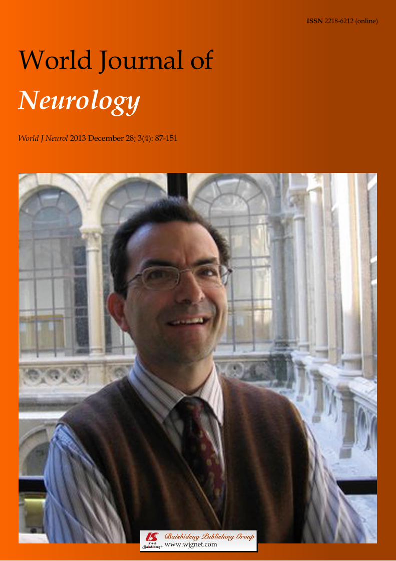
World Journal of NeurologyWorld J Neurol 2013 December 28; 3(4): 87-151
ISSN 2218-6212 (online)
www.wjgnet.com
World Journal of NeurologyW J N
EDITORS-IN-CHIEFFelipe Fregni, BostonVincenzo Solfrizzi, Bari
GUEST EDITORIAL BOARD MEMBERSFang-Chia Chang, TaipeiChun-Chuan Chen, TaoyuanSan-Yuan Huang, TaipeiSuh-Hang Hank Juo, Kaohsiung CityHsien-Yuan Lane, TaichungChing-Po Lin, TaipeiHung-Chuan Pan, TaichungBing-wen Soong, TaipeiKuo-Chen Wei, TaichungSheng-Nan Wu, Tainan
MEMBERS OF THE EDITORIAL BOARD
AustraliaRoy G Beran, SydneyLucette Cysique, SydneyJohn Gardiner, SydneyManuel B Graeber, SydneyXu-Feng Huang, WollongongGeorge Damion Mellick, BrisbaneSarah Spencer, Victoria
AustriaAndreas Gruber, ViennaJoerg Kraus, SalzburgGerlig Widmann, Anichstr
BelgiumAnna C Jansen, Brussels
Steve Majerus, LiegeRik RC Vandenberghe, Leuven
Brazil
Monica L Andersen, São PauloElisa Brietzke, São PauloPaulo A Lotufo, São PauloMarcelo Rodrigues Masruha, São Paulo
Canada
Dave Ellemberg, MontrealShirley Fecteau, QuebecAdel Gabriel, CalgaryBernard Le Foll, TorontoLaura-Ann Petitto, ScarboroughMubeen Rafay, WinnipegJosé M Trigo, Toronto
China
Hui-Sheng Chen, ShenYangMei-Chun Cheung, Hong KongWong Kwok Chu, Hong KongWan Feng, WuhanZhang Hao, ShanghaiDe-Wen Hu, ChangshaVincent Lai, Hong KongXiao-Li Li, BeijingJian-Min Liu, ShanghaiJun Liu, GuangzhouXiang-Dong Tang, ChengduPeng-Fei Yang, ShanghaiXiang Zhang, Xi’anYan Zhang, BeijingZhi-Ren Zhang, ChongqingJin-Xia Zhu, Beijing
Croatia
Nela Pivac, Zagreb
Cuba
Elena R Cuspineda Bravo, Habana
Cyprus
Kyproula Christodoulou, Nicosia
Czech Republic
Robert Mikulik, Brno
Denmark
Hong-You Ge, AalborgPeter Johannsen, Copenhagen
Egypt
Sherifa Ahmad Hamed, DaytonTaha M Mahmoud, ZagazigAhmed A K Abdel Razek, MansouraSahar N Saleem, Cairo
Finland
Seppo Antero Kahkonen, Helsinki
I
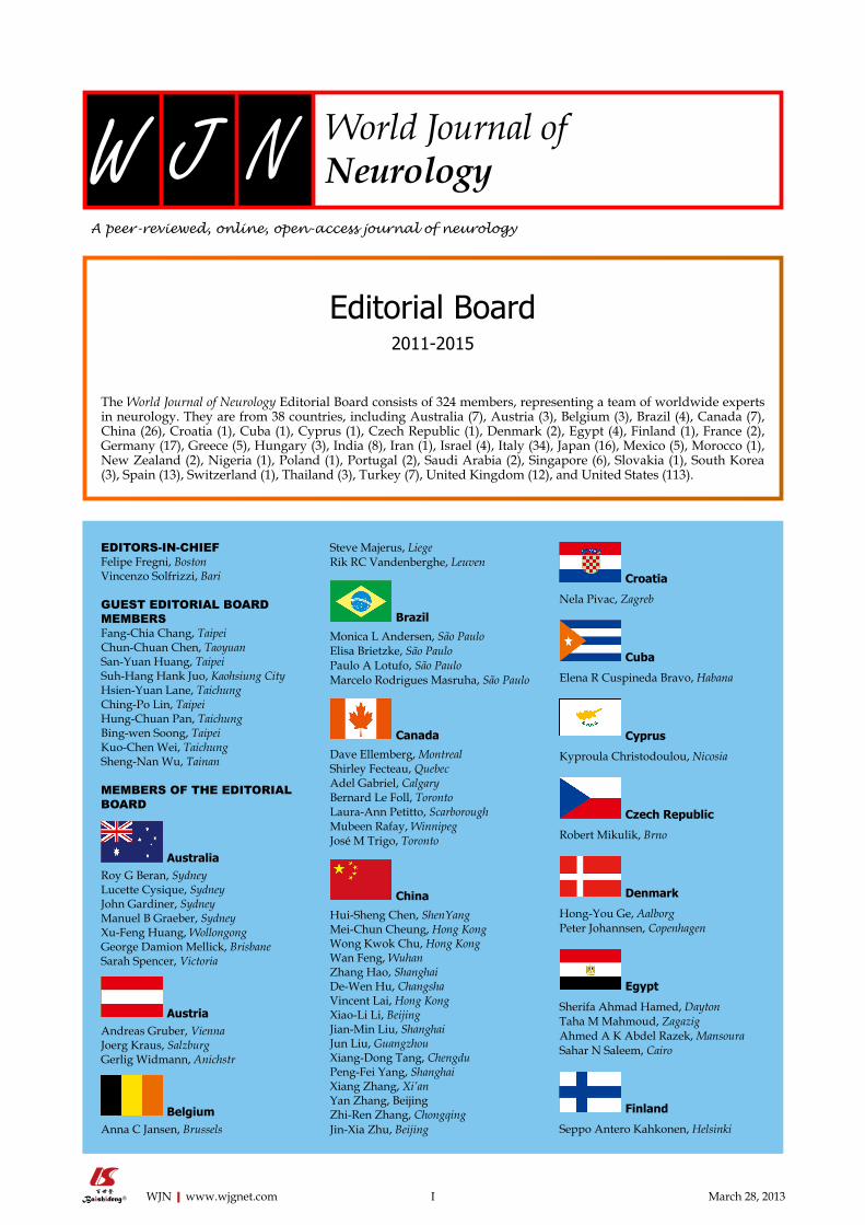
Editorial Board2011-2015
The World Journal of Neurology Editorial Board consists of 324 members, representing a team of worldwide experts in neurology. They are from 38 countries, including Australia (7), Austria (3), Belgium (3), Brazil (4), Canada (7), China (26), Croatia (1), Cuba (1), Cyprus (1), Czech Republic (1), Denmark (2), Egypt (4), Finland (1), France (2), Germany (17), Greece (5), Hungary (3), India (8), Iran (1), Israel (4), Italy (34), Japan (16), Mexico (5), Morocco (1), New Zealand (2), Nigeria (1), Poland (1), Portugal (2), Saudi Arabia (2), Singapore (6), Slovakia (1), South Korea (3), Spain (13), Switzerland (1), Thailand (3), Turkey (7), United Kingdom (12), and United States (113).
March 28, 2013WJN|www.wjgnet.com
France
Julien Dauguet, OrsayCyril Goudet, Cardonille
Germany
Boldizsar Czeh, MunichHans-Peter Hartung, DusseldorfJens Haueisen, IlmenauDirk M Hermann, EssenRaimund Kleiser, LinzUlrich Muller, GiessenWalter Paulus, GottingenNikolaus Plesnila, MunichMarc Röllinghoff, LandsbergJens Schittenhelm, Tuebingen Jens Schmidt, GottingenRudiger Seitz, DusseldorfThomas Straube, JenaPhilipp A Thomann, HeidelbergJohannes U V Thome, RostockMarcus Michael Unger, MarburgWolfgang Wick, Heidelberg
Greece
Constantoyannis Constantine, PatrasKostas n Fountas, LarissaSavas Grigoriadis, ThessalonikiIoannis N Mavridis, AthensGeorge Ntaios, Larissa
Hungary
Daniel Bereczki, BudapestXenia Gonda, BudapestNorbert Kovacs, Pecs
India
Ravindra Kumar Garg, LucknowRavi Gupta, DehradunBirendra Nath Mallick, New DelhiBalraj Mittal, LucknowHitesh N Modi, AhmedabadMona Ragothaman, BangaloreTN Sathyaprabha, BangaloreShirley Anne Telles, Haridwar
Iran
Seyyed Amirhossein Fazeli, Gorgan
Israel
Dimitrios M Karussis, JerusalemAsher Ornoy, HebrewAbraham Weizman, TikvaPerla Werner, Haifa
Italy
Alberto Albanese, MilanClaudia Altamura, RomeValeria Barresi, MessinaEmanuela Bartoccioni, RomeGabriella Bottini, MilanPaolo Brambilla, UdineAlfonso Cerase, SienaPolezzi David, PadovaLuigi De Gennaro, RomeStefano Diciotti, FlorenceMarina Fanin, PadovaMichele Ferrara, L’AquilaDaniele Focosi, PisaDaniela Galimberti, MilanValentina Garibotto, GenevaMarco Leonardi, BolognaMaria Liguori, MangoneLaura Mandelli, BolognaGianvito Martino, MilanGiovanni Martinotti, RomeMarianna Mazza, RomeAntonio Orlacchio, RomeMaurizio Paciaroni, PerugiaMarco Paoloni, RomeBernardo Perfetti, CheitiAlessandro Pezzini, BresciaGianfranco Puoti, NaplesNilo Riva, MilanPaolo Maria Rossini, RomeAndrea Serino, BolognaGianfranco Spalletta, RomeFabrizio Stocchi, RomePasquale Striano, Genova
Japan
Wataru Araki, TokyoKatsutoshi Furukawa, SendaiMasafumi Ihara, KyotoYuichi Inoue, TokyoHiroshi Kadotani, KyotoKazutaka Kobayashi, TokyoYoshihiro Kokubo, SuitaRyuichi Morishita, SuitaTetsu Niwa, YokohamaKatsunori Nonogaki, SendaiKunihiro Sakuma, AichiKatsuya Satoh, NagasakiAtsuyoshi Shimada, AichiHiroshi Takahashi, TottoriNaoyuki Takeuchi, SendaiKanato Yamagata, Tokyo
Mexico
Agnes Fleury, TlalpanDaniel San Juan, Mexico CityJulio Sotelo, Mexico CityTeresa Corona Vázquez, TlalpanRodrigo Ramos Zuniga, Guadalajara Jalisco
Morocco
Samir Ahboucha, Marrakesh
New ZealandJuan J Canales, ChristchurchValery Feigin, Auckland
NigeriaMayowa O Owolabi, Ibadan
PolandPawel Piotr Liberski, Lodz
PortugalNuno Martins Marques Canas, LisbonIsaura Ferreira Tavares, Porto
Saudi ArabiaAhmed S BaHammam, RiyadhEssam A Elgamal, Riyadh
SingaporeJustin HG Dauwels, SingaporeSteven Graham, SingaporeCharanjit Kaur, SingaporeVijay K Sharma, SingaporeFeng-Ru Tang, SingaporePhilip Lin Kiat Yap, Singapore
SlovakiaPeter Valkovič, Bratislava
South KoreaJi Soo Kim, Gyeonggi-doMyung Sik Lee, SeoulKyoungho Suk, Daegu
SpainAdria Arboix, BarcelonaPedro Emilio Bermejo, MadridRamon Cacabelos, CorunaIsidro Ferrer, Hospitalet de LLobregatJesús A García-Sevilla, Palma de MallorcaBernardo Hontanilla, PamplonaEsteban Pena Llamas, MadridFernando Maestu, MadridArturo Mangas, SalamancaGerman Moris, GijonJordi Perez-Tur, MadridJavier SCastresana, PamplonaJosé M Trigo, Toronto
SwitzerlandMarcel Arnold, Bern
II March 28, 2013WJN|www.wjgnet.com
III March 28, 2013WJN|www.wjgnet.com
Thailand
Suparerk Janjarasjitt, Ubon RatchathaniPanitha Jindahra, BangkokKittipan Rerkasem, Chiang Mai
Turkey
Ayse Aralasmak, IstanbulIsin Baral-Kulaksızoglu, IstanbulCengiz Cokluk, SamsunSaygin Salih Eker, BursaAtes Kadioglu, IstanbulSuleyman Kaplan, SamsunRifat Nuri Sener, Izmir
United Kingdom
Zubair Ahmed, BirminghamChris John Bushe, BasingstokeAndrea Eugenio Cavanna, LondonWhite Gables, NorwichValentina Gallo, LondonSanjay Kumar, BirminghamTarik F Massoud, CambridgeMike Modo, LondonMario Alfredo Parra, EdinburghCamillo Porcaro, NewcastleAnnette Sterr, SurreyJan Stochl, Cambridge
United States
Herve Abdi, DallasAbhishek Agrawal, New YorkAbass Alavi, PhiladelphiaQuincy J Almeida, WaterlooAlfredo Ardila, MiamiCarmel Armon, SpringfieldAndrew D Barreto, HoustonRaymond T Bartus, San DiegoArchit Bhatt, East LansingMargit L Bleecker, BaltimoreAnna-Liisa Brownell, CharlestownIgnazio Cali, Cleveland
Maren Carbon-Correll, ManhassetRudolph J Castellani, BaltimoreCarlos Cepeda, Los AngelesMunmun Chattopadhyay, Ann ArborRivka R Colen, BostonJames R Connor, HersheyLi Cui, Little RockYuri P Danilov, MadisonMukeshwar Dhamala, AtlantaDavid M Diamond, TampaDavid William Dodick, PhoenixRichard l Doty, PennsylvaniaChristopher L Drake, DetroitTimothy Q Duongtphd, San AntonioSherif M Elbasiouny, ChicagoAdam S Fleisher, San DiegoRobert Folmer, PortlandMajid Fotuhi, BaltimoreDheeraj Gandhi, BaltimoreYu-Lin Ge, New YorkHugo Geerts, BerwynAlexandros L Georgiadis, MinneapolisSrikanth Givvimani, LouisvilleCharles G Gross, PrincetonGeorge T Grossberg, Saint LouisZhen He, JeffersonMing-Xiong Huang, San DiegoSirous Jafarian, North VancouverXiangning Jiang, San FranciscoPeter B Kang, BostonJunghoon Kim, Elkins ParkDavid C Knight, BirminghamKennith F Layton, DallasAndrew G Lee, HoustonWalter S Lesley, TempleDavid Sigmund Liebeskind, Los AngelesTianming Liu, AthensYahia M Lodi, New YorkEdythe D London, Los AngelesJean-Pierre Louboutin, PhiladelphiaHanzhang Lu, DallasKenneth Maiese, NewarkSilva Markovic-Plese, Chapel HillMarlon Stephen Mathews, OrangeYousef Mohammad, ChicagoAmanda J Myers, MiamiJosef Novotny Jr, PittsburghArne M Nystuen, OmahaDarin T Okuda, PhoenixWei-hong Pan, Baton Rouge
Juliann M Paolicchi, NashvilleSpyros Papapetropoulos, Santa AnaSergio Paradiso, Iowa CityPaul Park, Ann ArborHemant A Parmar, Ann ArborGeorge Perry, San AntonioMarc Nicholas Potenza, New HavenYonglin Pu, ChicagoHaifa Qiao, TallahasseeLiya Qiao, RichmondRalph Rahme, CincinnatiAmit Ray, New YorkCatherine M Roe, MissouriTheodore H Schwartz, New YorkRobert J Schwartzman, PhiladelphiaThomas F Scott, Pittsburgh Souvik Sen, ColumbiaKhema Sharma, MiamiLi Shen, IndianapolisGabriel A Silva, La JollaElsayed Z Soliman, Winston SalemJoshua Goh Oon Soo, BaltimoreAshok Srinivasan, Ann ArborMassoud Stephane, MinneapolisShu-Wei Sun, Loma LindaEmi Takahashi, BostonThomas Thannickal, North HillsTimothy Adam Thrasher, HoustonGuochuan Emil Tsai, TorranceVassiliy Tsytsarev, FairfaxTanya Nadine Turan, CharlestonNeetu Tyagi, LouisvilleDenise A Valenti, BostonPiero Verro, SacramentoMarcela Votruba, CardiffJian Wang, BaltimoreKenneth L Weiss, JacksonHarry T Whelan, MilwaukeeKeith D White, GainesvillePeter Widdess-Walsh, LivingstonZhongcong Xie, BostonMidori Anne Yenari, San FranciscoAlbert J Yoo, BostonRobert J Young, New YorkBrad Evan Zacharia, New YorkT Thomas Zacharia, HersheyGabriel Zada, Los AngelesHaoqian Zhang, San FranciscoMing Zhang, PhiladelphiaYun Zhou, Baltimore
87 Cannabinoids:Dotheyhavethepotentialtotreatthesymptomsofmultiple
sclerosis?
Ahmed Z
97 Secondarypreventionofischaemicstroke
Volonghi I, Padovani A, Del Zotto E, Giossi A, Costa P, Morotti A, Poli L, Pezzini A
115 Moleculardiagnosisofautosomalrecessivecerebellarataxiainthewhole
exome/genomesequencingera
Votsi C, Christodoulou K
129 Puremotorstrokeasthemostfrequentlacunarsyndrome:Aclinicalupdate
Arboix A, Sánchez MJ, Martí-Vilalta JL
133 Overviewofbotulinumtoxinasatreatmentforspasticityinstrokepatients
Isoyama H, Takeuchi N
138 Neuritin:Atherapeuticcandidateforpromotingaxonalregeneration
Shimada T, Sugiura H, Yamagata K
144 Antiplateletstrategyforacuteischemicstroke:Aminireview
Zhou ZH, Chen HS
148 AtypicalneurologicalsymptomsassociatedwithCGGexpansionsoftheFMR1
gene
Peña E, Llanero M
Contents
FRONTIER
Quarterly Volume 3 Number 4 December 28, 2013
IWJN|www.wjgnet.com December 28, 2013|Volume 3|Issue 4|
World Journal of NeurologyW J N
REVIEW
MINIREVIEWS
CASE REPORT
ContentsWorld Journal of Neurology
Volume 3 Number 4 December 28, 2013
EDITORS FOR THIS ISSUE
Responsible Assistant Editor: Xin-Xin Che Responsible Science Editor: Xiu-Xia SongResponsible Electronic Editor: Su-Qing Liu Proofing Editor-in-Chief: Lian-Sheng Ma
Unit, University of Bari, P.zza G. Cesare, 11, 70124 Bari, Italy
EDITORIALOFFICEJin-Lei Wang, DirectorXiu-Xia Song, Vice DirectorWorld Journal of NeurologyRoom 903, Building D, Ocean International Center, No. 62 Dongsihuan Zhonglu, Chaoyang District, Beijing 100025, ChinaTelephone: +86-10-85381891Fax: +86-10-85381893E-mail: [email protected]://www.wjgnet.com
PUBLISHERBaishideng Publishing Group Co., LimitedFlat C, 23/F., Lucky Plaza, 315-321 Lockhart Road, Wan Chai, Hong Kong, ChinaFax: +852-65557188Telephone: +852-31779906E-mail: [email protected]://www.wjgnet.com
PUBLICATIONDATEDecember 28, 2013
COPYRIGHT© 2013 Baishideng. Articles published by this Open Access journal are distributed under the terms of the Creative Commons Attribution Non-commercial License, which permits use, distribution, and reproduc-tion in any medium, provided the original work is prop-erly cited, the use is non commercial and is otherwise in compliance with the license.
SPECIALSTATEMENTAll articles published in this journal represent the viewpoints of the authors except where indicated oth-erwise.
INSTRUCTIONSTOAUTHORSFull instructions are available online at http://www.wjgnet.com/2218-6212/g_info_20100722173918.htm
ONLINESUBMISSIONhttp://www.wjgnet.com/esps/
IIWJN|www.wjgnet.com
APPENDIX
ABOUT COVER
AIM AND SCOPE
INDEXINg/ABSTRACTINg
FLYLEAF
December 28, 2013|Volume 3|Issue 4|
NAMEOFJOURNALWorld Journal of Neurology
ISSNISSN 2218-6212 (online)
LAUNCHDATEDecember 28, 2011
FREQUENCYQuarterly
EDITORS-IN-CHIEFFelipe Fregni, MD, PhD, MPH, Associate Profes-sor, Department of Physical Medicine and Rehabili-tation, Spaulding Rehabilitation Hospital, Harvard Medical School, 125 Nashua St. Room 725, Boston, MA 02114, United States
Vincenzo Solfrizzi, MD, PhD, Professor, Depart-ment of Internal Medicine, Immunology and Infec-tious Diseases, Section of Geriatric Medicine-Memory
I-V Instructionstoauthors
EditorialBoardMemberofWorld JournalofNeurology ,AdriàArboix,MD,
PhD,HeadoftheCerebrovascularDivision,DepartmentofNeurology,Capio-
HospitalSagratCor,AssociateProfessorofNeurology,UniversityofBarcelona,
C/Viladomat288,E-08035Barcelona,Catalonia,Spain
World Journal of Neurology (World J Neurol, WJN, online ISSN 2218-6212, DOI: 10.5316) is a peer-reviewed open access academic journal that aims to guide clinical practice and improve diagnostic and therapeutic skills of clinicians.
WJN covers topics concerning neuro-oncology, electroneurophysiology, cerebrovas-cular diseases, epilepsy, cognitive impairment, myopathy and peripheral neuropathy, de-generative diseases, infectious diseases, demyelinating diseases, immunological diseases, genetic/metabolic diseases, affective disorders, headaches, sleep disorders, interventional neuroradiology, minimally invasive therapy, rehabilitation, diagnostic imaging, evidence-based medicine, epidemiology and nursing. Priority publication will be given to articles concerning diagnosis and treatment of neurological diseases. The following aspects are covered: Clinical diagnosis, laboratory diagnosis, differential diagnosis, imaging tests, pathological diagnosis, molecular biological diagnosis, immunological diagnosis, genetic diagnosis, functional diagnostics, and physical diagnosis; and comprehensive therapy, drug therapy, surgical therapy, interventional treatment, minimally invasive therapy, and robot-assisted therapy.
We encourage authors to submit their manuscripts to WJN. We will give priority to manuscripts that are supported by major national and international foundations and those that are of great basic and clinical significance.
World Journal of Neurology is now indexed in Digital Object Identifier.
I-III EditorialBoard
Cannabinoids: Do they have the potential to treat the symptoms of multiple sclerosis?
Zubair Ahmed
Zubair Ahmed, Neurotrauma and Neurodegeneration Sec-tion, School of Clinical and Experimental Medicine, College of Medical and Dental Sciences, University of Birmingham, Bir-mingham B15 2TT, United KingdomAuthor contributions: Ahmed Z participated in research de-sign, conducted experiments, performed data analysis and wrote the manuscript.Supported by The University of BirminghamCorrespondence to: Dr. Zubair Ahmed, Neurotrauma and Neurodegeneration Section, School of Clinical and Experimen-tal Medicine, College of Medical and Dental Sciences, Univer-sity of Birmingham, Institute of Biomedical Research (West), Edgbaston, Birmingham B15 2TT, United Kingdom. [email protected]: +44-121-4148859 Fax: +44-121-4148867Received: May 8, 2013 Revised: September 24, 2013Accepted: October 16, 2013Published online: December 28, 2013
AbstractThis article reviews the role of cannabinoids in inhibit-ing neurodegeneration in models of multiple sclerosis (MS). MS is a chronic, debilitating disease of the central nervous system (CNS), induced by autoimmunity-driven inflammation that leads to demyelination and thus dis-connection of the normal transmission of nerve impuls-es. Despite the use of an array of immune modulating drugs that restore blood brain barrier function, disability continues in patients concomitant with the loss of axons in the spinal cord. MS patients therefore suffer neuro-pathic pain, spasticity and tremor. Anecdotal evidence suggests that MS patients using cannabis, though il-legal, achieve symptomatic relief from neuropathic pain and spasticity associated with MS. The discovery of the endogenous cannabinoid (endocannabinoid) system that naturally exists in the body and which responds to cannabinoids to exert their effects has aided research into the therapeutic utility of cannabinoids. The endo-cannabinoid system consists of two G-protein coupled receptors cannabinoid receptor type-1 (CB1) and CB2.
CB1 is mainly expressed in the CNS and CB2 is predomi-nantly found in leukocytes, while an increasing number of potential ligands and endocannabinoid degradation molecules are being isolated. Several studies have highlighted the involvement of this system in regulating neurotransmission and its ability to prevent excessive neurotransmitter release, consistent with a capacity to provide symptomatic relief. In summary, antagonism of the CB1 receptor pathway contributes to neuronal dam-age in chronic relapsing experimental allergic encepha-lomyelitis (EAE) and suppresses tremor and spasticity. The addition of exogenous CB1 agonists derived from cannabis also afforded significant neuroprotection from the consequences of inflammatory CNS disease in EAE and experimental allergic uveitis models. Although clear neuroprotective benefits of cannabinoids have been demonstrated, the unwanted psychotropic effects need to be addressed. However, manipulating the endog-enous cannabinoid system may be one way of eliciting beneficial effects without some or all of the unwanted side effects.
© 2013 Baishideng Publishing Group Co., Limited. All rights reserved.
Key words: Multiple sclerosis; Axonal damage; Neuro-degeneration; Neuroprotection
Core tip: Multiple sclerosis (MS) is an inflammatory demyelinating disease of the central nervous system and causes disability, neuropathic pain, spasticity and tremor in affected patients. Although illegal, users of cannabis report relief from pain and spasticity, probably due to the endogenous cannabinoid system that ex-ists. Cannabinoid receptor type-1 (CB1)-deficient mice accrue greater levels of neurodegeneration and poorly tolerate inflammatory and excitotoxic insults after im-mune attack in a model of MS, experimental allergic encephalomyelitis. Treatment of animals affected by experimental allergic uveitis (EAU) with CB1 agonists also provided significant neuroprotection from the con-
FRONTIER
87 December 28, 2013|Volume 3|Issue 4|WJN|www.wjgnet.com
World Journal of NeurologyW J N
Online Submissions: http://www.wjgnet.com/esps/[email protected]:10.5316/wjn.v3.i4.87
World J Neurol 2013 December 28; 3(4): 87-96ISSN 2218-6212 (online)
© 2013 Baishideng Publishing Group Co., Limited. All rights reserved.
sequences of EAU, suggesting that cannabinoids may slow down neurodegeneration in MS.
Ahmed Z. Cannabinoids: Do they have the potential to treat the symptoms of multiple sclerosis? World J Neurol 2013; 3(4): 87-96 Available from: URL: http://www.wjgnet.com/2218-6212/full/v3/i4/87.htm DOI: http://dx.doi.org/10.5316/wjn.v3.i4.87
BIOGRAPHYDr Zubair Ahmed received his PhD from University Col-lege London, London, United Kingdom. He completed his first postdoctoral training period at the Institute of Neurology in London before moving to the University of Birmingham to continue his second postdoctoral training period. In 2007, he was awarded an Research Councils United Kingdom Academic Fellowship, the remit of which was to develop an independently funded research group before being promoted to Lecturer in 2011. He is currently a Senior Lecturer in Neuroscience. His research interests covers numerous aspects of the molecular biol-ogy of central nervous system (CNS) axon regeneration and degeneration and his most notable contribution is to the understanding of the role of caspase-2 in apoptosis of retinal and dorsal root ganglion neurons, the contribu-tion of an endogenous mechanism for receptor shedding, the identification of growth factors capable of promot-ing retinal ganglion cell survival and axon regeneration, identification of alternative neuronal receptors capable of interacting with key molecules in blocking CNS axon regeneration and the observations that cannabinoids in-hibit neurodegeneration in models of multiple sclerosis (MS). His curriculum vitae lists over 50 peer-reviewed publications, 2 book chapters and numerous presenta-tions at national and international meetings.
INTRODUCTIONMS is an inflammatory demyelinating disease of the CNS and results in disruption to the normal transmission of nerve impulses due to lesions in the CNS[1,2]. Despite im-mune modulating drugs that reduce blood brain barrier dysfunction, disability often continues in patients and suggests that neurodegenerative changes are key to the progression of disease[3-5]. MS patients thus display spas-ticity, neuropathic pain associated with neuroinflamma-tion, excitotoxicity and chronic neurodegeneration. It is also established that axonal/neuronal loss, which occurs early in the disease process, is an important contributor of permanent disability and is often associated with ac-tive inflammation. Disability only becomes evident when normal compensatory mechanisms are exhausted, while demyelinated axons are left vulnerable to further damage by electrical activity[6,7]. Therefore identifying neuropro-tective strategies are a key goal in the fight against MS.
Although the Cannabis sativa plant has been used for centuries as a medicinal preparation to relieve the
symptoms of inflammatory and neuropathic disorders, its use is illegal[8]. However, anecdotal accounts from MS patients indicated that cannabis might offer symptomatic relief of pain and spasticity associated with MS[9]. Plant-derived “cannabinoids” have provided important insights into the biology of cannabis and have led to a multitude of clinical trials using cannabinoids to control pain and spasticity in MS patients[10]. The main active ingredi-ent of Cannabis sativa was defined in 1964 as (-)-trans-delta-9-tetrahydrocannabinol [Δ9-THC or dronabinol (international non-proprietary name)][11]. Δ9-THC is not only responsible for the majority of the pharmacological actions of cannabis but also its psychoactive effects. Nu-merous other cannabinoids and phytochemicals also exist in the cannabis plant including cannabidiol (CBD) which is non-psychoactive and is not a cannabinoid receptor agonist[12].
CLINICAL TRIALS OF CANNABIS IN MSAlthough many patients self-medicated with cannabis, there were few clinical studies that demonstrated reliable clinical evidence of the benefits of cannabis use in MS. Some of the first few studies on the effect of Δ9-THC were not encouraging since small sample sizes were used and Δ9-THC had no effect on objective measures, despite patients reporting subjective improvements in the symp-toms of MS[13]. For example, the first systematic, placebo-controlled trial with Δ9-THC and a Cannabis sativa plant extract, given to 16 MS patients with severe spasticity, showed no effect of the cannabinoids on spasticity[14]. A much larger trial involving 660 MS patients receiving Δ9-THC or natural Cannabis oil or a placebo, the mean reported effect on spasticity was not significantly differ-ent between control and treatment groups[15]. However, patient-reported spasticity was reduced, agreeing with earlier studies that showed subjective improvements in spasticity related to MS[14,15]. However, in a 12-mo follow up study of 657 patients, Δ9-THC was reported to have a significant effect on the objective measures of spastic-ity[16]. Several later studies have also shown variable results depending on cannabis preparation, dosing regime and patient numbers, throwing into doubt the use of clinical measures of spasticity.
At present, the large number of clinical trials in MS have not clarified whether cannabis is beneficial in MS but have thrown up questions about the rating of “spas-ticity”, route of delivery, source and dosing regime[10]. Despite these reservations, self-medicated use of can-nabis continues. In a postal questionnaire involving the responses received from 110 MS patients in the South of England, 43% confirmed their use of cannabis with 68% of these patients specifically using cannabis to re-lieve the symptoms of MS[17]. Patients affirmed that their main reason for choosing to self-medicate with cannabis was due to pain and spasms, while a small proportion used cannabis for sleep related problems[17]. In a further study, over 90% of a cohort of 112 MS patients based in the United Kingdom and United States declared that
Ahmed Z. Cannabinoids in multiple sclerosis
88 December 28, 2013|Volume 3|Issue 4|WJN|www.wjgnet.com
self-medication with cannabis improved nocturnal pain, spasms and muscular pain[18].
A recent study showed that of the 572 MS patients enrolled in a clinical study, 272 (47.6%) responded to Sativex, an oral-mucosal spray that contains Δ9-THC and CBD, treatment within 4 wk with a response that was de-fined as > 20% decrease in spasticity[19]. A second phase of this study demonstrated that the cannabis extracts sig-nificantly reduced spasticity and the frequency of spasms while improving sleep quality over an extended 12-wk period, compared to placebo controls[19]. This has led to approval of the use of Sativex in the treatment of MS-related spasticity. These studies demonstrate the potential beneficial roles of cannabis use in MS patients, although in general its supply and use remains illegal.
ENDOCANNABINOID SYSTEMThe discovery of the endogenous cannabinoid (endocan-nabinoid) system that naturally exists in the body and responds to cannabinoids to exert their effects has aided research into the therapeutic utility of cannabinoids. This has fuelled the search for alternative modes of delivery into the human body, rather than the traditional method of “smoking”. The endocannabinoid system consists of at least two families of lipid signalling molecules (the N-acyl ethanolamines and the monoacyl-glycerols), mul-tiple enzymes in the biosynthesis and degradation of these lipids, as well as two G-protein coupled receptors [cannabinoid receptor type-1 (CB1) and CB2] (reviewed by[10]). CB1 is mainly expressed in the CNS while CB2 is predominantly found in leukocytes, while an increasing number of potential ligands and endocannabinoid deg-radation molecules are being isolated[20]. Several studies have highlighted the involvement of this system in regu-lating neurotransmission and its ability to prevent exces-sive neurotransmitter release, consistent with a capacity to provide symptomatic relief in MS[21,22].
Endocannabinoids are produced on demand and are retrogradely transported across the postsynaptic membrane to engage with CB1 receptors, suppressing neurotransmitter release. The pharmacology of endocan-nabinoids is rapidly evolving with the discovery of new cannabinoid mimetics and novel CB receptor interactions that include orphan receptors and other CB receptors be-ing proposed[23-26]. All these receptor functions are either sensitive to, or are regulated by CB receptors and culmi-nate in neuropathic pain and inflammation, suggesting that novel drug targets based around these proteins might be useful in treating neuroinflammation and neuropathic pain in different neurological conditions. In addition, activation of the CB receptor inhibits adenylate cyclase that then reduces the levels of the second messenger cyclic adenosine monophosphate (cAMP), thus regulat-ing cellular mechanisms such as cell fate[27]. CB receptor activation also inhibits voltage-dependent Ca2+ channels and activates inwardly rectifying K+ channels, a process that underlies CB-induced depression of excitatory neu-rotransmission[28,29]. Thus, the CB system is a potentially
useful target for exploitation in neurodegenerative dis-eases such as MS.
AXONAL DAMAGE IN MSAlthough MS is defined as an inflammatory demyelinat-ing disease of the CNS, axonal loss is also a key feature of the disease. In MS patients, analysis of their spinal cord lesions suggested that the permanent loss in func-tion was not primarily due to demyelination but due to axonal loss[30]. Axonal loss is generally associated with inflammatory macrophage infiltration but is variable in MS lesions and can be severe in certain cases. For ex-ample, axonal density is reduced by 60%-70% in actively demyelinating lesions and is characterised by the presence of axonal spheroids, endbulbs, or focal accumulation of proteins[31-33]. Although shadow plaques appear after injury that is consistent with an attempt to remyelinate axons, ongoing axonal injury is present[34]. This suggests that during the early stages of remyelination axons are more susceptible to damage, a process that is related to the patterns of Na+ channels at the widened surfaces of the nodes of Ranvier[35]. Axonal injury is also present in normal appearing white matter[36,37] and may occur as a result of secondary Wallerian degeneration[38]. However, this is not the only mechanism of axonal damage since two distinct patterns are observed: one that takes place within demyelinated lesions and correlates with lesional activity, while the other is diffuse axonal injury that is as-sociated with inflammation and can additionally affect non-demyelinated nerve fibres[33].
Axonal injury in MS is also selective to the size of axon fibres. Small calibre axons are more prone to injury compared to thick axons and may relate to thin axons requiring a higher energy demand in terms of their criti-cal mass of mitochondria[39]. Like MS, widespread axonal damage is also seen in experimental allergic encephalomy-elitis (EAE)[34,40]. However, experimental models of MS reveal different mechanisms of axonal injury and there is currently no agreement for which mechanism is relevant to MS patients. Axonal injury mechanisms may involve T-lymphocytes and antibodies as part of the adaptive immune response as well as components of the innate immune system, driven by macrophages and microglia. For example, axonal injury can be driven by an antigen-specific cytotoxic T cell response, induced in neurons and glia by the expression of pro-inflammatory cytokines, leading to neuronal death and axonal transection[41-43].
Another mechanism of axonal damage relies on the production of auto-antibodies against cell surface mol-ecules in neurons and axons. A subset of MS patients were described that mounted an antibody response against neurofascin, which is expressed on axons and oli-godendrocyte processes at the nodes of Ranvier[44]. Sys-temic injection of neurofascin during EAE exacerbates the clinical symptoms of the disease with severe levels of axonal injury within lesions[44]. These observations suggest that auto-antibodies can directly mediate axonal damage. In MS lesions, axon damage is closely linked to
89 December 28, 2013|Volume 3|Issue 4|WJN|www.wjgnet.com
Ahmed Z. Cannabinoids in multiple sclerosis
ing amount of residual deficit[49,56]. Whilst disease induc-tion in both CB1-deficient and CB1 wild-type mice were similar, CB1-deficient mice exhibited significantly higher levels of residual deficit. This deficit was quantitated in an open-field activity chamber and confirmed that CB1-deficient mice displayed significantly more immobility and paresis than wild type mice, accumulating significant-ly more axonal damage after relapses. CB1-deificent mice also developed spasticity after only a single attack, which is not seen in wild type mice until after three to four re-lapses of disease[49].
Numerous mechanisms cause neuronal death and axonal damage in EAE, including the influx of toxic ions such as Ca2+ and caspase-3-mediated apoptosis[57]. CB1-deficient mice demonstrated significantly lower levels of active caspase-3 during acute EAE compared with wild type mice, while caspase-3 was detected in dy-ing axons, consistent with that observed in MS[3]. These results suggested that the elevated neurodegeneration in CB1-deficient mice may be due to caspase-3-mediated apoptosis and axonal damage and hence agonism of the CB1 receptor pathway is neuroprotective and may control neurological symptoms such as tremor and spasticity[49].
We also investigated whether glutamate toxicity can be regulated by cannabinoids[55]. Glutamate excitotoxity causes neuronal damage in both MS and EAE. For ex-ample, the glutamate antagonist, amantadine, reduces the relapse rate in MS patients[58], while elevated glutamate levels have been observed in cerebrospinal fluid from MS patients[59]. In EAE, the enhanced levels of glutamate agonists may result in aberrant astrocyte function, since activated astrocytes normally regulate glutamate levels through enzymes such as glutamate dehydrogenase and glutamine synthetase, both of which are down-regulated during EAE[60,61]. The amount of CNS glutamate is also affected by abnormal changes in neuronal and glial glutamate transporters, all of which raise the levels of glutamate in the CNS during EAE and ultimately lead to the synthesis of mediators responsible for neuronal dys-function[59,62-64]. We reported that after in vitro stimulation of N-methyl-D-aspartic (NMDA) receptors in cerebellar granule cells, there was significantly more neuronal Ca2+ influx in CB1-deficient mice than in wild type controls suggesting that the cannabinoid receptors may tonically regulate Ca2+ influx. The NMDA receptor antagonist MK-801 took longer to reduce Ca2+ back to basal levels in CB1-deficient mice than in congenic controls while CB1 agonism with CP55, 940 inhibited NMDA-induced Ca2+ influx in wild type mice but had no effect on CB1-deficient mice. These results suggest that Ca2+ is not only dysregulated in the absence of CB1 receptors but that postsynaptic control of NMDA-receptor activation is also compromised. These in vitro results were confirmed by in vivo injections of kainic acid, a specific agonist of the kainate receptor that mimics the effects of glutamate, since injection of kainic acid in CB1-deficient mice in-duced seizures and mortality within 10 min, while no ef-fect of kainic acid was observed in wild type or congenic wild type control mice despite using 50-fold higher doses.
the presence of macrophages and microglia that are in intimate contact with axons[3,5] and are known to produce a number of cytotoxic molecules including reactive oxy-gen and nitric oxide intermediates[33]. The expression of these molecules, especially inducible nitric oxide synthase from macrophages, correlates with areas of axonal injury in acute EAE[45]. In summary, axonal damage is a feature of MS and EAE and correlates with functional deficits. EAE models demonstrate that axonal damage occurs through a variety of mechanisms. However, recent re-ports have highlighted the role of mitochondrial injury and subsequent energy failure, induced by oxygen and nitric oxide free radicals as a possible mechanism of axo-nal injury. Understanding of the pathway to axon injury will aid in the discovery of new molecules for therapeutic intervention.
CANNABINOIDS IN MSThere is an abundance of evidence suggesting that MS patients gain symptomatic relief from cannabis extracts[46]. For example, Sativex, an oral-mucosal spray containing Δ9-THC and CBD is anti-spasmodic and analgesic in MS patients[47], while neuropathic pain associated with MS is relieved by dronabinol, an oral preparation of Δ9-THC analog[15]. Meta-analysis has also revealed that CB-based preparations are superior in the treatment of MS-related neuropathic pain than the placebo, confirming their ben-eficial effects in symptomatic relief[48]. This is consistent with the animal model of MS, EAE where treatment with exogenous cannabinoids controlled spasticity in chronic relapsing EAE models[49].
Modulation of the endocannabinoid system is also apparent in MS and EAE such that brain levels of CB receptors are downregulated in EAE while plasma levels of endocannabinoids are increased in MS patients[50,51]. Synthetic CB, HU-211, reduced the clinical severity of acute EAE in female Lewis rats as well as reducing in-flammatory cell infiltration into the CNS[52], while the WIN55, 212-2, a CB1 and CB2 receptor agonist reduces T cell differentiation and hence reduces EAE severity in Thei-ler’s murine encephalomyelitis virus-induced demyelinat-ing disease, a mouse model of chronic-progressive MS[53], suggesting a key involvement of the CB receptors in the pathogenesis of EAE. Induction of EAE in CB1 recep-tor-deficient mice causes rapid and progressively more neurodegeneration than in wild-type counterparts[54].
In our highly cited study on the role of cannabi-noids in EAE, reported that the cannabinoid system was neuroprotective during EAE since CB1-deficient mice poorly tolerated inflammatory and excitotoxic insults and showed significant accumulation of neurodegenera-tion after EAE[55]. We induced chronic relapsing EAE (CREAE) in wild type ABH, CB1 gene (Cnr1)-deficient and congenic ABH. Cnr1+/+ mice with mouse spinal cord homogenate emulsified in complete Freund’s adjuvant on days 0 and 7 and monitored clinical disease progres-sion over time. Mice developed characteristic paralytic disease episodes followed by remission with an increas-
90 December 28, 2013|Volume 3|Issue 4|WJN|www.wjgnet.com
Ahmed Z. Cannabinoids in multiple sclerosis
Therefore, CB1-receptors clearly regulate ionotropic glu-tamate receptor activity, leading to enhanced susceptibil-ity to excitotoxic damage in CB1-deficient mice.
Furthermore, we reported that CB1 agonists protect-ed mice against the consequences of CNS inflammation in models of experimental allergic uveitis (EAU)[55]. EAU is an inflammatory disease of the eye and after sensitiza-tion with for example, interphotoreceptor retinal binding peptide in B10. RIII mice, the neuroretina is completely destroyed within 14-16 d[65]. CB1 receptor agonism with R(+)-WIN-55,212-2 and Δ9-THC both significantly inhibited photoreceptor damage without affecting in-flammatory infiltrates suggesting that agonism of CB1 is neuroprotective. Taken together, the results of our study demonstrated that cannabinoids protect against the neu-rodegenerative events in models of MS and EAU.
CANNABINOID-1 RECEPTOR-MEDIATED NEUROPROTECTION IN MODELS OF MSA feature of MS is neuronal loss, and in MS patients, loss of spinal cord axons correlate with neurological disability together with reduced N-acetyl aspartate levels in chronic MS patients[32]. Although axonal loss occurs early during the progression of MS[66], once a threshold of 15%-35% axonal loss in mice has been reached, permanent disabil-ity results[67,68]. In CB1-deficient mice, significant axonal loss is evident after a single acute attack of EAE, suggest-ing that the mere presence of CB1 is itself neuroprotec-tive[55]. In EAE and MS, the presence of axonal damage correlates with inflammation that produces a range of neurotoxic agents such as glutamate, cytokines as well as creating oxidative stress in the environment of the CNS and damaging the blood-brain barrier[3,5,52,69-72].
Cannabinoids, however, can protect against acute hypoxia, excitotoxicity, oxidative and traumatic insults both in vitro and in vivo[73-76]. Cannabinoids can also inhibit both pre- and post-synaptic glutamate induced calcium responses and thus inhibit neurotoxicity[21,55,76], an effect that we showed to be CB1-dependent[55]. In accord with our observations, CB1-deficient mice were more sus-ceptible to NMDA and α-amino-3-hydroxy-5-methyl-4-isoxazolepropionic acid (AMPA)/kainite glutamate receptor excitotoxicity[55], however, Δ9-THC and CBD protected against NMDA-, AMPA- and kainite-agonist-induced cell death[77,78]. Delta-9-THC also protected retinal neurons from death induced by peroxynitrite-mediated NMDA-induced toxicity[79]. These observations demonstrate a clear neuroprotective role of cannabinoids in MS.
CANNABINOID-2-MEDIATED EFFECTS IN MODELS OF MSAlthough much of the focus in MS is devoted to CB1 receptor pharmacology, the recognition that CB2 recep-tors possess immunomodulatory properties has led to an increased focus on CB2 receptors as potential thera-
peutic targets. Unlike CB1 receptors, activation of the CB2 receptor is not psychoactive and therefore target-ing CB2 receptors with selective agonists is a promising therapeutic avenue that is immunomodulatory without being psychoactive. For example, early indications came from experiments that showed that the administration of WIN55212-2, which functions as a CB1 and CB2 receptor agonist, attenuated the progression of EAE in C57BL/6 mice immunized with myelin oligodendrocyte-derived glycoprotein35-55 (MOG35-55)[80]. Furthermore, a selec-tive antagonist of the CB1 did not modulate the protec-tive effect of WIN55212-2 but a selective antagonist of the CB2 receptor blocked the effects of WIN55212-2. This led to the suggestion that the protective effects of WIN55212-2 was mediated through the CB2 receptor[80]. However, later studies showed that the CB1 receptor might play a neuroprotective role in the latter stages of EAE[49,54].
Other highly selective CB2 receptor agonists such as O-1966 are also useful in the fight against MS since they do not produce psychoactive effects, determined by its low affinity to CB1 receptors[81]. Administration of O-1966 in a chronic (C57BL/6/MOG), relapsing-remitting and an adoptive transfer model attenuated disease progression and improved motor function[82]. In addition, encepha-litogenic T cells derived from CB2-deficient mice were shown to be more aggressive in terms of CNS infiltration and increased severity of EAE, despite displaying similar levels of proliferation, apoptosis and cytokine production in the spleen as wild-type T cells[83]. Moreover, treatment of mice with CB2 receptor agonists attenuate white cell trafficking across the blood-brain barrier while dendritic cells differentiated in the presence of selective CB2 recep-tor agonist and inhibited T cell proliferation and shifted cytokine responses from inflammatory to anti-inflamma-tory molecules[82]. Recently, it has been shown that high concentrations of IFN-γ disrupts P2X7 purinergic recep-tor signalling and thus inhibit the neuroprotective effects of endogenous cannabinoids[84]. Therefore, inhibiting IFN-γ by exogenous CB2 agonists represents an obvious therapy to prevent the disruption of P2X7 signalling.
VALUE OF ANIMAL MODELS IN THE STUDY OF MULTIPLE SCLEROSISThe validity of EAE as a model of MS is a topic of active debate with some researchers contenting that it is unsuitable due to its inability to mimic some of the pathological, immunologic and chronic features of MS. For example, EAE is usually monophasic whereas MS displays chronic relapsing features. Histological and magnetic resonance imaging data demonstrate axonal and cortical damage in MS but not in some models of EAE[85]. The extravasation of red blood cells into the CNS of swiss jim lambert mice and Lewis rats with EAE are not typical of MS[86,87], while encephalitogenic regions associated with myelin basic protein (MBP) or proteolipid protein normally activate more CD4+ than CD8+ T cells
91 December 28, 2013|Volume 3|Issue 4|WJN|www.wjgnet.com
Ahmed Z. Cannabinoids in multiple sclerosis
but in inflammatory MS, CD8+ T cell predominate[88-90]. However, there are many other immunological differ-ences between mouse and human MS and these must be overcome if greater levels of success in drug develop-ment for human MS are to be achieved[91].
Several other points are worth considering in terms of the use of EAE as an animal model: (1) EAE pro-vides little insight into the progression of MS in terms of the small amounts of demyelinated axons in EAE compared to MS while mice with EAE rarely exhibit on-going functional deterioration that MS patients often dis-play[92,93]; (2) The use of C57BL/6 mice in EAE studies limits the investigation of the mechanism of relapsing-remitting forms that more commonly affect MS patients (Ransohoff et al[93], 2002); (3) Treatment with factors that exert neurobiological effects also impact on immune and inflammatory cells and thus making results difficult to interpret (Ransohoff et al[93], 2002); and (4) EAE is gen-erally generated using antigens that affect CD4+ T cells while CD8+ T cells that predominate in MS lesions are overlooked[92,94]. Likewise, the role of B cells is largely neglected despite recent data that demonstrate their im-portance in the pathogenesis of MS[95,96].
In summary, EAE has a long history in the fight against MS, however, its predictive value for treatment efficacy is poor. Nevertheless, it is widely used as a first-line animal model of MS and has provided mechanistic insights into the neuroinflammatory aspects of MS.
LIMITATIONS OF ANIMAL MODELS OF MSOne of the biggest limitations on the use of EAE as a model of MS is the fact that disease has to be induced with complete Freund’s adjuvant and heat-inactivated Mycobacterium tuberculosis rather than mimicking a spon-taneous disease like MS. This leaves very little room for disease pathways and fails to represent the complexity of disease inducing mechanisms in MS. Demyelination, a feature of MS is also not obvious in all EAE models while the time course for disease manifestation may be days, EAE more closely resembles post-infectious acute demyelinating events[97]. In contrast, MS develops over years with patients presenting with more protracted epit-ope spreading than that observed in EAE mice[98]. There are also many other immunological differences between EAE and MS that need consideration in the development of potential therapies for MS.
Therapeutic developments from EAE have translated poorly to human MS. For example, only a few molecules that showed efficacy in EAE have been successful in MS trials. One of these molecules is Glatiramer acetate, a synthetic amino acid copolymer originally designed to mimic encephalitogenic MBP, but instead suppresses EAE by other mechanisms and reduced MS relapses by 30%[99,100]. However, the efficacy of Glatiramer acetate has been questioned by a systematic Cochrane review which calls into question the use of Glatiramer acetate in
MS[101]. Tysabri (Natalizumab) and Gilenya (fingolimod) are the only two other drugs that have been licence for use in human MS. Natalizumab binds to the α4 subunit of α4β1 and α4β7 integrins and blocks binding to their endothelial receptors (VCAP-1 and mucosal addressin-cell adhesion molecule 1, thereby attenuating inflam-mation and ongoing inflammation[102,103]. Fingolimod is a sphingosine-1-phosphate-receptor modulator that prevents egress of lymphocytes from lymph nodes and significantly improved relapse rates compared to placebo controls[104]. At present therefore, it remains unclear why pre-clinical EAE studies predict treatment efficacy in hu-man MS so poorly, however, EAE is still an important first-line model system in the development of new treat-ments for MS.
BETTER ANIMAL MODELS OF MULTIPLE SCLEROSISTo make greater progress, better animals models of MS need to be considered when testing new drugs. One fact that to be borne in mind is the fact that MS is a heteroge-neous disease in terms of genetics, environmental effects, disease course, pathological treatments and treatment responsiveness[105]. Currently the majority of experiments are performed in inbred strains while clinically, the mo-lecular mechanisms that determine the efficacy during prolonged follow-up of hundreds of patients are far more complex. Thus, it is likely that genetic differences account for some of the inter-patient differences in clini-cal efficacy of various drugs. Improved animal models may take into account the therapeutic effect of drugs in more than one animal model, treatments to be instigated after disease onset, long-term disease periods, use spon-taneous disease models and incorporate human risk. The use of partially humanized mouse models to address the genetic and disease variability are a step in the right direc-tion towards developing better therapeutics for MS[106].
SUMMARY AND FUTURE PROSPECTSStudies have shown that antagonism of the CB1 recep-tor pathway contributes to neuronal damage in CREAE and the relative worsening of tremor and spasticity. The addition of exogenous CB1 agonists derived from can-nabis afforded significant neuroprotection from the consequences of inflammatory CNS disease in EAE and EAU models. Although clear neuroprotective benefits of cannabinoids have been demonstrated, the unwanted psychotropic effects need to be addressed. The adverse psychotropic effects, its role in appetite, pain and cogni-tion together with the observation that the CB1 receptor is downregulated in some neurodegenerative diseases limits the usefulness of cannabinoids in the treatment of MS. Alternative treatments may be more useful and includes the development of drugs based on CBD, the non-psychoactive part of cannabis that possess anti-inflammatory and anti-oxidant properties. Furthermore,
92 December 28, 2013|Volume 3|Issue 4|WJN|www.wjgnet.com
Ahmed Z. Cannabinoids in multiple sclerosis
the CB2 receptor is being recognised as a potential target since it regulates neuroinflammation and neurogenesis while being non-psychoactive.
In summary, the endocannabinoid system exerts mul-tiple actions and may be useful in the development of therapies to treat neurodegenerative diseases. However, the biggest challenge remains, namely the development of drugs that lack the adverse psychoactive side effects.
REFERENCES1 Compston A, Coles A. Multiple sclerosis. Lancet 2002; 359:
1221-1231 [PMID: 11955556 DOI: 10.1016/S0140-6736(02)08220-X]2 Compston A, Coles A. Multiple sclerosis. Lancet 2008; 372:
1502-1517 [PMID: 18970977 DOI: 10.1016/S0140-6736(08)61620-7]3 Trapp BD, Peterson J, Ransohoff RM, Rudick R, Mörk S, Bö
L. Axonal transection in the lesions of multiple sclerosis. N Engl J Med 1998; 338: 278-285 [PMID: 9445407 DOI: 10.1056/NEJM199801293380502]
4 Coles AJ, Wing MG, Molyneux P, Paolillo A, Davie CM, Hale G, Miller D, Waldmann H, Compston A. Monoclonal antibody treatment exposes three mechanisms underlying the clinical course of multiple sclerosis. Ann Neurol 1999; 46: 296-304 [PMID: 10482259]
5 Ferguson B, Matyszak MK, Esiri MM, Perry VH. Axonal damage in acute multiple sclerosis lesions. Brain 1997; 120 (Pt 3): 393-399 [PMID: 9126051]
6 Bjartmar C, Wujek JR, Trapp BD. Axonal loss in the pathol-ogy of MS: consequences for understanding the progressive phase of the disease. J Neurol Sci 2003; 206: 165-171 [PMID: 12559505]
7 Smith KJ, Kapoor R, Hall SM, Davies M. Electrically active axons degenerate when exposed to nitric oxide. Ann Neurol 2001; 49: 470-476 [PMID: 11310624]
8 Chaturvedi GN, Tiwari SK, Rai NP. Medicinal use of opium and cannabis in medieval India. Indian J Hist Sci 1981; 16: 31-35 [PMID: 11611777]
9 Pertwee RG. Cannabinoids and multiple sclerosis. Pharmacol Ther 2002; 95: 165-174 [PMID: 12182963]
10 Rog DJ. Cannabis-based medicines in multiple sclerosis--a review of clinical studies. Immunobiology 2010; 215: 658-672 [PMID: 20541836 DOI: 10.1016/j.imbio.2010.03.009]
11 Gaoni Y, Mechoulam R. The isolation and structure of delta-1-tetrahydrocannabinol and other neutral cannabinoids from hashish. J Am Chem Soc 1971; 93: 217-224 [PMID: 5538858]
12 Gowran A, Noonan J, Campbell VA. The multiplicity of ac-tion of cannabinoids: implications for treating neurodegen-eration. CNS Neurosci Ther 2011; 17: 637-644 [PMID: 20875047 DOI: 10.1111/j.1755-5949.2010.00195.x]
13 Smith PF. Cannabinoids for the treatment of multiple scle-rosis: no smoke without fire? Expert Rev Neurother 2003; 3: 327-334 [PMID: 19810900 DOI: 10.1586/14737175.3.3.327]
14 Killestein J, Hoogervorst EL, Reif M, Blauw B, Smits M, Uitdehaag BM, Nagelkerken L, Polman CH. Immuno-modulatory effects of orally administered cannabinoids in multiple sclerosis. J Neuroimmunol 2003; 137: 140-143 [PMID: 12667658]
15 Zajicek J, Fox P, Sanders H, Wright D, Vickery J, Nunn A, Thompson A. Cannabinoids for treatment of spasticity and other symptoms related to multiple sclerosis (CAMS study): multicentre randomised placebo-controlled trial. Lancet 2003; 362: 1517-1526 [PMID: 14615106 DOI: 10.1016/S0140-6736(03)14738-1]
16 Zajicek JP, Sanders HP, Wright DE, Vickery PJ, Ingram WM, Reilly SM, Nunn AJ, Teare LJ, Fox PJ, Thompson AJ. Cannabinoids in multiple sclerosis (CAMS) study: safety and efficacy data for 12 months follow up. J Neurol Neuro-surg Psychiatry 2005; 76: 1664-1669 [PMID: 16291891 DOI: 10.1136/jnnp.2005.070136]
17 Chong MS, Wolff K, Wise K, Tanton C, Winstock A, Silber E. Cannabis use in patients with multiple sclerosis. Mult Scler 2006; 12: 646-651 [PMID: 17086912]
18 Consroe P, Musty R, Rein J, Tillery W, Pertwee R. The per-ceived effects of smoked cannabis on patients with multiple sclerosis. Eur Neurol 1997; 38: 44-48 [PMID: 9252798]
19 Novotna A, Mares J, Ratcliffe S, Novakova I, Vachova M, Zapletalova O, Gasperini C, Pozzilli C, Cefaro L, Comi G, Rossi P, Ambler Z, Stelmasiak Z, Erdmann A, Montal-ban X, Klimek A, Davies P. A randomized, double-blind, placebo-controlled, parallel-group, enriched-design study of nabiximols* (Sativex(®) ), as add-on therapy, in subjects with refractory spasticity caused by multiple sclerosis. Eur J Neurol 2011; 18: 1122-1131 [PMID: 21362108 DOI: 10.1111/j.1468-1331.2010.03328.x]
20 Howlett AC, Barth F, Bonner TI, Cabral G, Casellas P, Dev-ane WA, Felder CC, Herkenham M, Mackie K, Martin BR, Mechoulam R, Pertwee RG. International Union of Pharma-cology. XXVII. Classification of cannabinoid receptors. Phar-macol Rev 2002; 54: 161-202 [PMID: 12037135]
21 Wilson RI, Nicoll RA. Endogenous cannabinoids mediate retrograde signalling at hippocampal synapses. Nature 2001; 410: 588-592 [PMID: 11279497 DOI: 10.1038/35069076]
22 Kreitzer AC, Carter AG, Regehr WG. Inhibition of interneu-ron firing extends the spread of endocannabinoid signaling in the cerebellum. Neuron 2002; 34: 787-796 [PMID: 12062024]
23 Thakur GA, Tichkule R, Bajaj S, Makriyannis A. Latest ad-vances in cannabinoid receptor agonists. Expert Opin Ther Pat 2009; 19: 1647-1673 [PMID: 19939187 DOI: 10.1517/13543770903436505]
24 Brown AJ. Novel cannabinoid receptors. Br J Pharmacol 2007; 152: 567-575 [PMID: 17906678 DOI: 10.1038/sj.bjp.0707481]
25 Sharir H, Abood ME. Pharmacological characterization of GPR55, a putative cannabinoid receptor. Pharmacol Ther 2010; 126: 301-313 [PMID: 20298715 DOI: 10.1016/j.pharmthera.2010.02.004]
26 Nevalainen T, Irving AJ. GPR55, a lysophosphatidylinositol receptor with cannabinoid sensitivity? Curr Top Med Chem 2010; 10: 799-813 [PMID: 20370712]
27 Miyamoto N, Tanaka R, Zhang N, Shimura H, Onodera M, Mochizuki H, Hattori N, Urabe T. Crucial role for Ser133-phosphorylated form of cyclic AMP-responsive element binding protein signaling in the differentiation and survival of neural progenitors under chronic cerebral hypoperfu-sion. Neuroscience 2009; 162: 525-536 [PMID: 19426786 DOI: 10.1016/j.neuroscience.2009.05.004]
28 Hampson AJ, Grimaldi M, Lolic M, Wink D, Rosenthal R, Axelrod J. Neuroprotective antioxidants from marijuana. Ann N Y Acad Sci 2000; 899: 274-282 [PMID: 10863546]
29 Nicholson RA, Liao C, Zheng J, David LS, Coyne L, Err-ington AC, Singh G, Lees G. Sodium channel inhibition by anandamide and synthetic cannabimimetics in brain. Brain Res 2003; 978: 194-204 [PMID: 12834914]
30 Kornek B, Lassmann H. Axonal pathology in multiple scle-rosis. A historical note. Brain Pathol 1999; 9: 651-656 [PMID: 10517504]
31 Mews I, Bergmann M, Bunkowski S, Gullotta F, Brück W. Oligodendrocyte and axon pathology in clinically silent multiple sclerosis lesions. Mult Scler 1998; 4: 55-62 [PMID: 9599334]
32 Bjartmar C, Kidd G, Mörk S, Rudick R, Trapp BD. Neuro-logical disability correlates with spinal cord axonal loss and reduced N-acetyl aspartate in chronic multiple sclerosis pa-tients. Ann Neurol 2000; 48: 893-901 [PMID: 11117546]
33 Lassmann H. Axonal and neuronal pathology in mul-tiple sclerosis: what have we learnt from animal models. Exp Neurol 2010; 225: 2-8 [PMID: 19840788 DOI: 10.1016/j.expneurol.2009.10.009]
34 Kornek B, Storch MK, Weissert R, Wallstroem E, Stef-ferl A, Olsson T, Linington C, Schmidbauer M, Lassmann H. Multiple sclerosis and chronic autoimmune encepha-lomyelitis: a comparative quantitative study of axonal
93 December 28, 2013|Volume 3|Issue 4|WJN|www.wjgnet.com
Ahmed Z. Cannabinoids in multiple sclerosis
injury in active, inactive, and remyelinated lesions. Am J Pathol 2000; 157: 267-276 [PMID: 10880396 DOI: 10.1016/S0002-9440(10)64537-3]
35 Smith KJ. Axonal protection in multiple sclerosis--a particu-lar need during remyelination? Brain 2006; 129: 3147-3149 [PMID: 17132643 DOI: 10.1093/brain/awl323]
36 Kutzelnigg A, Lucchinetti CF, Stadelmann C, Brück W, Rauschka H, Bergmann M, Schmidbauer M, Parisi JE, Lass-mann H. Cortical demyelination and diffuse white matter injury in multiple sclerosis. Brain 2005; 128: 2705-2712 [PMID: 16230320 DOI: 10.1093/brain/awh641]
37 Frischer JM, Bramow S, Dal-Bianco A, Lucchinetti CF, Rauschka H, Schmidbauer M, Laursen H, Sorensen PS, Lassmann H. The relation between inflammation and neu-rodegeneration in multiple sclerosis brains. Brain 2009; 132: 1175-1189 [PMID: 19339255 DOI: 10.1093/brain/awp070]
38 Evangelou N, Konz D, Esiri MM, Smith S, Palace J, Mat-thews PM. Regional axonal loss in the corpus callosum cor-relates with cerebral white matter lesion volume and distri-bution in multiple sclerosis. Brain 2000; 123 (Pt 9): 1845-1849 [PMID: 10960048]
39 Evangelou N, Konz D, Esiri MM, Smith S, Palace J, Mat-thews PM. Size-selective neuronal changes in the anterior optic pathways suggest a differential susceptibility to injury in multiple sclerosis. Brain 2001; 124: 1813-1820 [PMID: 11522583]
40 Linington C, Bradl M, Lassmann H, Brunner C, Vass K. Augmentation of demyelination in rat acute allergic en-cephalomyelitis by circulating mouse monoclonal antibodies directed against a myelin/oligodendrocyte glycoprotein. Am J Pathol 1988; 130: 443-454 [PMID: 2450462]
41 Neumann H, Cavalié A, Jenne DE, Wekerle H. Induction of MHC class I genes in neurons. Science 1995; 269: 549-552 [PMID: 7624779]
42 Medana IM, Gallimore A, Oxenius A, Martinic MM, Weker-le H, Neumann H. MHC class I-restricted killing of neurons by virus-specific CD8+ T lymphocytes is effected through the Fas/FasL, but not the perforin pathway,. Eur J Immunol 2000; 30: 3623-3633 [PMID: 11169405 DOI: 10.1002/1521-4141(200012)30: 12< 3623: : AID-IMMU3623> 3.0.CO; 2-F]
43 Medana I, Martinic MA, Wekerle H, Neumann H. Transection of major histocompatibility complex class I-induced neurites by cytotoxic T lymphocytes. Am J Pathol 2001; 159: 809-815 [PMID: 11549572 DOI: 10.1016/S0002-9440(10)61755-5]
44 Mathey EK, Derfuss T, Storch MK, Williams KR, Hales K, Woolley DR, Al-Hayani A, Davies SN, Rasband MN, Olsson T, Moldenhauer A, Velhin S, Hohlfeld R, Meinl E, Linington C. Neurofascin as a novel target for autoantibody-medi-ated axonal injury. J Exp Med 2007; 204: 2363-2372 [PMID: 17846150 DOI: 10.1084/jem.20071053]
45 Aboul-Enein F, Weiser P, Höftberger R, Lassmann H, Bradl M. Transient axonal injury in the absence of demyelination: a correlate of clinical disease in acute experimental autoim-mune encephalomyelitis. Acta Neuropathol 2006; 111: 539-547 [PMID: 16718350 DOI: 10.1007/s00401-006-0047-y]
46 Lakhan SE, Rowland M. Whole plant cannabis extracts in the treatment of spasticity in multiple sclerosis: a system-atic review. BMC Neurol 2009; 9: 59 [PMID: 19961570 DOI: 10.1186/1471-2377-9-59]
47 Rog DJ, Nurmikko TJ, Young CA. Oromucosal delta9-tetrahydrocannabinol/cannabidiol for neuropathic pain as-sociated with multiple sclerosis: an uncontrolled, open-label, 2-year extension trial. Clin Ther 2007; 29: 2068-2079 [PMID: 18035205 DOI: 10.1016/j.clinthera.2007.09.013]
48 Iskedjian M, Bereza B, Gordon A, Piwko C, Einarson TR. Meta-analysis of cannabis based treatments for neuropathic and multiple sclerosis-related pain. Curr Med Res Opin 2007; 23: 17-24 [PMID: 17257464 DOI: 10.1185/030079906X158066]
49 Baker AM, Grekova MC, Richert JR. EAE susceptibility in FVB mice. J Neurosci Res 2000; 61: 140-145 [PMID: 10878586]
50 Jean-Gilles L, Feng S, Tench CR, Chapman V, Kendall DA,
Barrett DA, Constantinescu CS. Plasma endocannabinoid levels in multiple sclerosis. J Neurol Sci 2009; 287: 212-215 [PMID: 19695579 DOI: 10.1016/j.jns.2009.07.021]
51 Centonze D, Bari M, Rossi S, Prosperetti C, Furlan R, Fezza F, De Chiara V, Battistini L, Bernardi G, Bernardini S, Mar-tino G, Maccarrone M. The endocannabinoid system is dys-regulated in multiple sclerosis and in experimental autoim-mune encephalomyelitis. Brain 2007; 130: 2543-2553 [PMID: 17626034 DOI: 10.1093/brain/awm160]
52 Achiron A, Miron S, Lavie V, Margalit R, Biegon A. Dex-anabinol (HU-211) effect on experimental autoimmune encephalomyelitis: implications for the treatment of acute re-lapses of multiple sclerosis. J Neuroimmunol 2000; 102: 26-31 [PMID: 10626663]
53 Croxford JL. Therapeutic potential of cannabinoids in CNS disease. CNS Drugs 2003; 17: 179-202 [PMID: 12617697]
54 Maresz K, Pryce G, Ponomarev ED, Marsicano G, Croxford JL, Shriver LP, Ledent C, Cheng X, Carrier EJ, Mann MK, Giovannoni G, Pertwee RG, Yamamura T, Buckley NE, Hillard CJ, Lutz B, Baker D, Dittel BN. Direct suppression of CNS autoimmune inflammation via the cannabinoid recep-tor CB1 on neurons and CB2 on autoreactive T cells. Nat Med 2007; 13: 492-497 [PMID: 17401376 DOI: 10.1038/nm1561]
55 Pryce G, Ahmed Z, Hankey DJ, Jackson SJ, Croxford JL, Pocock JM, Ledent C, Petzold A, Thompson AJ, Giovannoni G, Cuzner ML, Baker D. Cannabinoids inhibit neurode-generation in models of multiple sclerosis. Brain 2003; 126: 2191-2202 [PMID: 12876144 DOI: 10.1093/brain/awg224]
56 Baker D, O’Neill JK, Gschmeissner SE, Wilcox CE, Butter C, Turk JL. Induction of chronic relapsing experimental allergic encephalomyelitis in Biozzi mice. J Neuroimmunol 1990; 28: 261-270 [PMID: 2373763]
57 Ahmed Z, Doward AI, Pryce G, Taylor DL, Pocock JM, Leon-ard JP, Baker D, Cuzner ML. A role for caspase-1 and -3 in the pathology of experimental allergic encephalomyelitis : inflam-mation versus degeneration. Am J Pathol 2002; 161: 1577-1586 [PMID: 12414506 DOI: 10.1016/S0002-9440(10)64436-7]
58 Plaut GS. Effectiveness of amantadine in reducing relapses in multiple sclerosis. J R Soc Med 1987; 80: 91-93 [PMID: 3550084]
59 Stover JF, Pleines UE, Morganti-Kossmann MC, Kossmann T, Lowitzsch K, Kempski OS. Neurotransmitters in cerebrospi-nal fluid reflect pathological activity. Eur J Clin Invest 1997; 27: 1038-1043 [PMID: 9466133]
60 Hardin-Pouzet H, Krakowski M, Bourbonnière L, Didier-Bazes M, Tran E, Owens T. Glutamate metabolism is down-regulated in astrocytes during experimental allergic enceph-alomyelitis. Glia 1997; 20: 79-85 [PMID: 9145307]
61 Rothstein JD. Neurobiology. Bundling up excitement. Nature 2000; 407: 141, 143 [PMID: 11001037 DOI: 10.1038/35025170]
62 Ohgoh M, Hanada T, Smith T, Hashimoto T, Ueno M, Ya-manishi Y, Watanabe M, Nishizawa Y. Altered expression of glutamate transporters in experimental autoimmune en-cephalomyelitis. J Neuroimmunol 2002; 125: 170-178 [PMID: 11960654]
63 Piani D, Frei K, Do KQ, Cuénod M, Fontana A. Murine brain macrophages induced NMDA receptor mediated neurotox-icity in vitro by secreting glutamate. Neurosci Lett 1991; 133: 159-162 [PMID: 1687755]
64 Smith QR. Transport of glutamate and other amino acids at the blood-brain barrier. J Nutr 2000; 130: 1016S-1022S [PMID: 10736373]
65 Hankey DJ, Nickerson JM, Donoso LA, Lightman SL, Baker D. Experimental autoimmune uveoretinitis in mice (Biozzi ABH and NOD) expressing the autoimmune-associated H-2A(g7) molecule: identification of a uveitogenic epitope. J Neuroimmunol 2001; 118: 212-222 [PMID: 11498256]
66 Filippi M, Rocca MA. MRI aspects of the “inflammatory phase” of multiple sclerosis. Neurol Sci 2003; 24 Suppl 5: S275-S278 [PMID: 14652788 DOI: 10.1007/s10072-003-0173-4]
67 Confavreux C, Vukusic S, Moreau T, Adeleine P. Relapses
94 December 28, 2013|Volume 3|Issue 4|WJN|www.wjgnet.com
Ahmed Z. Cannabinoids in multiple sclerosis

and progression of disability in multiple sclerosis. N Engl J Med 2000; 343: 1430-1438 [PMID: 11078767 DOI: 10.1056/NEJM200011163432001]
68 Wujek JR, Bjartmar C, Richer E, Ransohoff RM, Yu M, Tuohy VK, Trapp BD. Axon loss in the spinal cord deter-mines permanent neurological disability in an animal model of multiple sclerosis. J Neuropathol Exp Neurol 2002; 61: 23-32 [PMID: 11829341]
69 Bolton C, Paul C. MK-801 limits neurovascular dysfunction during experimental allergic encephalomyelitis. J Pharmacol Exp Ther 1997; 282: 397-402 [PMID: 9223580]
70 Pitt D, Werner P, Raine CS. Glutamate excitotoxicity in a model of multiple sclerosis. Nat Med 2000; 6: 67-70 [PMID: 10613826 DOI: 10.1038/71555]
71 Koprowski H, Zheng YM, Heber-Katz E, Fraser N, Rorke L, Fu ZF, Hanlon C, Dietzschold B. In vivo expression of induc-ible nitric oxide synthase in experimentally induced neu-rologic diseases. Proc Natl Acad Sci USA 1993; 90: 3024-3027 [PMID: 7681993]
72 Lock C, Hermans G, Pedotti R, Brendolan A, Schadt E, Garren H, Langer-Gould A, Strober S, Cannella B, Allard J, Klonowski P, Austin A, Lad N, Kaminski N, Galli SJ, Oksen-berg JR, Raine CS, Heller R, Steinman L. Gene-microarray analysis of multiple sclerosis lesions yields new targets vali-dated in autoimmune encephalomyelitis. Nat Med 2002; 8: 500-508 [PMID: 11984595 DOI: 10.1038/nm0502-500]
73 Shen M, Thayer SA. Cannabinoid receptor agonists protect cultured rat hippocampal neurons from excitotoxicity. Mol Pharmacol 1998; 54: 459-462 [PMID: 9730904]
74 Nagayama T, Sinor AD, Simon RP, Chen J, Graham SH, Jin K, Greenberg DA. Cannabinoids and neuroprotection in global and focal cerebral ischemia and in neuronal cultures. J Neu-rosci 1999; 19: 2987-2995 [PMID: 10191316]
75 Sinor AD, Irvin SM, Greenberg DA. Endocannabinoids pro-tect cerebral cortical neurons from in vitro ischemia in rats. Neurosci Lett 2000; 278: 157-160 [PMID: 10653017]
76 Abood ME, Rizvi G, Sallapudi N, McAllister SD. Activation of the CB1 cannabinoid receptor protects cultured mouse spinal neurons against excitotoxicity. Neurosci Lett 2001; 309: 197-201 [PMID: 11514075]
77 Hampson AJ, Grimaldi M, Axelrod J, Wink D. Cannabidiol and (-)Delta9-tetrahydrocannabinol are neuroprotective an-tioxidants. Proc Natl Acad Sci USA 1998; 95: 8268-8273 [PMID: 9653176]
78 Marsicano G, Moosmann B, Hermann H, Lutz B, Behl C. Neuroprotective properties of cannabinoids against oxida-tive stress: role of the cannabinoid receptor CB1. J Neurochem 2002; 80: 448-456 [PMID: 11905991]
79 El-Remessy AB, Khalil IE, Matragoon S, Abou-Mohamed G, Tsai NJ, Roon P, Caldwell RB, Caldwell RW, Green K, Liou GI. Neuroprotective effect of (-)Delta9-tetrahydrocannabinol and cannabidiol in N-methyl-D-aspartate-induced retinal neurotoxicity: involvement of peroxynitrite. Am J Pathol 2003; 163: 1997-2008 [PMID: 14578199]
80 Ni X, Geller EB, Eppihimer MJ, Eisenstein TK, Adler MW, Tuma RF. Win 55212-2, a cannabinoid receptor agonist, at-tenuates leukocyte/endothelial interactions in an experimen-tal autoimmune encephalomyelitis model. Mult Scler 2004; 10: 158-164 [PMID: 15124761]
81 Wiley JL, Beletskaya ID, Ng EW, Dai Z, Crocker PJ, Mahade-van A, Razdan RK, Martin BR. Resorcinol derivatives: a nov-el template for the development of cannabinoid CB(1)/CB(2) and CB(2)-selective agonists. J Pharmacol Exp Ther 2002; 301: 679-689 [PMID: 11961073]
82 Zhang M, Martin BR, Adler MW, Razdan RJ, Kong W, Ganea D, Tuma RF. Modulation of cannabinoid receptor activation as a neuroprotective strategy for EAE and stroke. J Neuroimmune Pharmacol 2009; 4: 249-259 [PMID: 19255856 DOI: 10.1007/s11481-009-9148-4]
83 Dittel BN. Direct suppression of autoreactive lymphocytes in the central nervous system via the CB2 receptor. Br J
Pharmacol 2008; 153: 271-276 [PMID: 17922025 DOI: 10.1038/sj.bjp.0707493]
84 Whiting P, Harbord R, Main C, Deeks JJ, Filippini G, Eg-ger M, Sterne JA. Accuracy of magnetic resonance imaging for the diagnosis of multiple sclerosis: systematic review. BMJ 2006; 332: 875-884 [PMID: 16565096 DOI: 10.1136/bmj.38771.583796.7C]
85 Sriram S, Yao SY, Stratton C, Moses H, Narayana PA, Wo-linsky JS. Pilot study to examine the effect of antibiotic ther-apy on MRI outcomes in RRMS. J Neurol Sci 2005; 234: 87-91 [PMID: 15935383 DOI: 10.1016/j.jns.2005.03.042]
86 Raine CS, Traugott U, Nussenblatt RB, Stone SH. Optic neu-ritis and chronic relapsing experimental allergic encepha-lomyelitis: relationship to clinical course and comparison with multiple sclerosis. Lab Invest 1980; 42: 327-335 [PMID: 7189001]
87 Forge JK, Pedchenko TV, LeVine SM. Iron deposits in the central nervous system of SJL mice with experimental al-lergic encephalomyelitis. Life Sci 1998; 63: 2271-2284 [PMID: 9870713]
88 Booss J, Esiri MM, Tourtellotte WW, Mason DY. Immuno-histological analysis of T lymphocyte subsets in the central nervous system in chronic progressive multiple sclerosis. J Neurol Sci 1983; 62: 219-232 [PMID: 6607973]
89 Hauser SL, Bhan AK, Gilles F, Kemp M, Kerr C, Weiner HL. Immunohistochemical analysis of the cellular infiltrate in multiple sclerosis lesions. Ann Neurol 1986; 19: 578-587 [PMID: 3524414 DOI: 10.1002/ana.410190610]
90 Lassmann H, Ransohoff RM. The CD4-Th1 model for mul-tiple sclerosis: a critical [correction of crucial] re-appraisal. Trends Immunol 2004; 25: 132-137 [PMID: 15036040 DOI: 10.1016/j.it.2004.01.007]
91 Friese MA, Montalban X, Willcox N, Bell JI, Martin R, Fug-ger L. The value of animal models for drug development in multiple sclerosis. Brain 2006; 129: 1940-1952 [PMID: 16636022 DOI: 10.1093/brain/awl083]
92 Ransohoff RM. Animal models of multiple sclerosis: the good, the bad and the bottom line. Nat Neurosci 2012; 15: 1074-1077 [PMID: 22837037 DOI: 10.1038/nn.3168]
93 Ransohoff RM, Howe CL, Rodriguez M. Growth factor treatment of demyelinating disease: at last, a leap into the light. Trends Immunol 2002; 23: 512-516 [PMID: 12401395]
94 Huseby ES, Liggitt D, Brabb T, Schnabel B, Ohlén C, Gover-man J. A pathogenic role for myelin-specific CD8(+) T cells in a model for multiple sclerosis. J Exp Med 2001; 194: 669-676 [PMID: 11535634]
95 Hauser SL, Waubant E, Arnold DL, Vollmer T, Antel J, Fox RJ, Bar-Or A, Panzara M, Sarkar N, Agarwal S, Langer-Gould A, Smith CH. B-cell depletion with rituximab in re-lapsing-remitting multiple sclerosis. N Engl J Med 2008; 358: 676-688 [PMID: 18272891 DOI: 10.1056/NEJMoa0706383]
96 Berer K, Wekerle H, Krishnamoorthy G. B cells in spontane-ous autoimmune diseases of the central nervous system. Mol Immunol 2011; 48: 1332-1337 [PMID: 21146219 DOI: 10.1016/j.molimm.2010.10.025]
97 Steinman L. Assessment of animal models for MS and de-myelinating disease in the design of rational therapy. Neuron 1999; 24: 511-514 [PMID: 10595504]
98 Vanderlugt CL, Miller SD. Epitope spreading in immune-mediated diseases: implications for immunotherapy. Nat Rev Immunol 2002; 2: 85-95 [PMID: 11910899 DOI: 10.1038/nri724]
99 Teitelbaum D, Meshorer A, Hirshfeld T, Arnon R, Sela M. Suppression of experimental allergic encephalomyelitis by a synthetic polypeptide. Eur J Immunol 1971; 1: 242-248 [PMID: 5157960 DOI: 10.1002/eji.1830010406]
100 Johnson KP, Brooks BR, Cohen JA, Ford CC, Goldstein J, Lisak RP, Myers LW, Panitch HS, Rose JW, Schiffer RB. Co-polymer 1 reduces relapse rate and improves disability in relapsing-remitting multiple sclerosis: results of a phase III multicenter, double-blind placebo-controlled trial. The Co-
95 December 28, 2013|Volume 3|Issue 4|WJN|www.wjgnet.com
Ahmed Z. Cannabinoids in multiple sclerosis
polymer 1 Multiple Sclerosis Study Group. Neurology 1995; 45: 1268-1276 [PMID: 7617181]
101 Munari L, Lovati R, Boiko A. Therapy with glatiramer ac-etate for multiple sclerosis. Cochrane Database Syst Rev 2004; (1): CD004678 [PMID: 14974077 DOI: 10.1002/14651858.CD004678]
102 Yednock TA, Cannon C, Fritz LC, Sanchez-Madrid F, Steinman L, Karin N. Prevention of experimental autoim-mune encephalomyelitis by antibodies against alpha 4 beta 1 integrin. Nature 1992; 356: 63-66 [PMID: 1538783 DOI: 10.1038/356063a0]
103 Polman CH, O’Connor PW, Havrdova E, Hutchinson M, Kappos L, Miller DH, Phillips JT, Lublin FD, Giovannoni G, Wajgt A, Toal M, Lynn F, Panzara MA, Sandrock AW. A randomized, placebo-controlled trial of natalizumab for
relapsing multiple sclerosis. N Engl J Med 2006; 354: 899-910 [PMID: 16510744 DOI: 10.1056/NEJMoa044397]
104 Kappos L, Comi G, Panitch H, Oger J, Antel J, Conlon P, Steinman L. Induction of a non-encephalitogenic type 2 T helper-cell autoimmune response in multiple sclerosis after administration of an altered peptide ligand in a placebo-controlled, randomized phase II trial. The Altered Peptide Ligand in Relapsing MS Study Group. Nat Med 2000; 6: 1176-1182 [PMID: 11017151 DOI: 10.1038/80525]
105 Lassmann H, Brück W, Lucchinetti C. Heterogeneity of mul-tiple sclerosis pathogenesis: implications for diagnosis and therapy. Trends Mol Med 2001; 7: 115-121 [PMID: 11286782]
106 Gregersen JW, Holmes S, Fugger L. Humanized animal mod-els for autoimmune diseases. Tissue Antigens 2004; 63: 383-394 [PMID: 15104670 DOI: 10.1111/j.0001-2815.2004.00243.x]
P- Reviewer: Mellick GD S- Editor: Song XX L- Editor: A E- Editor: Liu SQ
96 December 28, 2013|Volume 3|Issue 4|WJN|www.wjgnet.com
Ahmed Z. Cannabinoids in multiple sclerosis
lifestyle modifications are strongly recommended. However, apart from the well-established indications to thrombolysis, studies in acute phase after a first stroke or TIA are scarce and evidence is lacking. More trials are available for long-term secondary prevention with different classes of drugs, including antithrom-botic medications for ischaemic events of arterial and cardiac origin, especially related to atrial fibrillation (antiplatelets and anticoagulants, respectively), lipid lowering agents (mainly statins), blood pressure lower-ing drugs, surgical and endovascular revascularization procedures.
© 2013 Baishideng Publishing Group Co., Limited. All rights reserved.
Key words: Stroke; Transient ischaemic attack; Second-ary prevention; Antiplatelets; Anticoagulants; Medical stroke treatment; Carotid stenosis
Core tip: Aggressive and combination treatments in the acute phase after transient ischaemic attack or minor stroke have been shown to be beneficial in few studies, but results of ongoing randomized trials are required. On the other side, in long-term prevention the most important innovation is the advent of new anticoagulant agents for stroke prevention in atrial fibrillation. Recent trials showed efficacy and safety of thrombin and factor Xa inhibitors, compared to vitamin K antagonists, whose use is hampered by several limi-tations. These new drugs will potentially increase the number of patients treated according to guidelines, thus preventing a remarkable proportion of strokes.
Volonghi I, Padovani A, Del Zotto E, Giossi A, Costa P, Morotti A, Poli L, Pezzini A. Secondary prevention of ischaemic stroke. World J Neurol 2013; 3(4): 97-114 Available from: URL: http://www.wjgnet.com/2218-6212/full/v3/i4/97.htm DOI: http://dx.doi.org/10.5316/wjn.v3.i4.97
Secondary prevention of ischaemic stroke
Irene Volonghi, Alessandro Padovani, Elisabetta Del Zotto, Alessia Giossi, Paolo Costa, Andrea Morotti, Loris Poli, Alessandro Pezzini
REVIEW
97 December 28, 2013|Volume 3|Issue 4|WJN|www.wjgnet.com
World Journal of NeurologyW J N
Online Submissions: http://www.wjgnet.com/esps/[email protected]:10.5316/wjn.v3.i4.97
World J Neurol 2013 December 28; 3(4): 97-114ISSN 2218-6212 (online)
© 2013 Baishideng Publishing Group Co., Limited. All rights reserved.
Irene Volonghi, Alessandro Padovani, Paolo Costa, Andrea Morotti, Loris Poli, Alessandro Pezzini, Dipartimento di Scien-ze Cliniche e Sperimentali, Clinica Neurologica, Università degli Studi di Brescia, 25123 Brescia, ItalyElisabetta Del Zotto, U.O. di Recupero e Rieducazione Funzio-nale, IRCCS Fondazione Don Gnocchi, 20149 Rovato (Brescia), ItalyAlessia Giossi, U.O. Neurologia, Istituto Clinico Sant’Anna, 25127 Brescia, ItalyAuthor contributions: Volonghi I and Pezzini A contributed to manuscript conception, data acquisition, revision, and wrote the paper; Padovani A contributed to manuscript conception, data acquisition and critical revision for important intellectual content; Del Zotto E, Giossi A, Costa P, Morotti A and Poli L contributed to manuscript conception and data acquisition; All the authors gave approval to final version of the manuscript.Correspondence to: Irene Volonghi, MD, Dipartimento di Scienze Cliniche e Sperimentali, Clinica Neurologica, Università degli Studi di Brescia, P.le Spedali Civili 1, 25123 Brescia, Italy. [email protected]: +39-30-3384086 Fax: +39-30-3384086Received: June 29, 2013 Revised: October 8, 2013Accepted: October 17, 2013Published online: December 28, 2013
AbstractIn spite of a documented reduction in incidence in high-income countries over the last decades, stroke is still a leading cause of death and disability worldwide. With the ageing of the population stroke-related economic burden is expected to increase, because of residual dis-ability and its complications, such as cognitive impair-ment, high risk of falls and fractures, depression and epilepsy. Furthermore, because of the substantial rate of early and long-term vascular recurrences after the first event, secondary prevention after cerebral isch-aemia is a crucial issue. This is even more important after minor stroke and transient ischaemic attack (TIA), in order to reduce the risk of potentially more severe and disabling events. To accomplish this aim, acute long-term medical and surgical treatments as well as
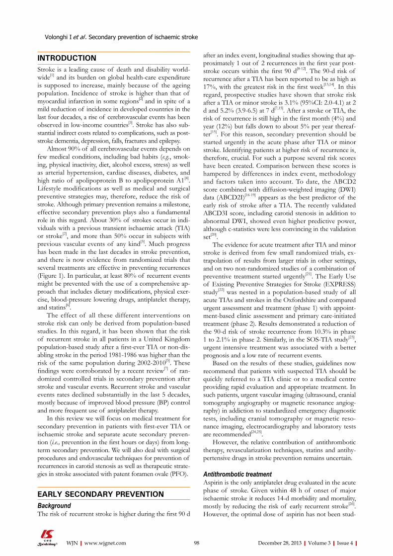
INTRODUCTIONStroke is a leading cause of death and disability world-wide[1] and its burden on global health-care expenditure is supposed to increase, mainly because of the ageing population. Incidence of stroke is higher than that of myocardial infarction in some regions[2] and in spite of a mild reduction of incidence in developed countries in the last four decades, a rise of cerebrovascular events has been observed in low-income countries[3]. Stroke has also sub-stantial indirect costs related to complications, such as post-stroke dementia, depression, falls, fractures and epilepsy.
Almost 90% of all cerebrovascular events depends on few medical conditions, including bad habits (e.g., smok-ing, physical inactivity, diet, alcohol excess, stress) as well as arterial hypertension, cardiac diseases, diabetes, and high ratio of apolipoprotein B to apolipoprotein A1[4]. Lifestyle modifications as well as medical and surgical preventive strategies may, therefore, reduce the risk of stroke. Although primary prevention remains a milestone, effective secondary prevention plays also a fundamental role in this regard. About 30% of strokes occur in indi-viduals with a previous transient ischaemic attack (TIA) or stroke[2], and more than 50% occur in subjects with previous vascular events of any kind[5]. Much progress has been made in the last decades in stroke prevention, and there is now evidence from randomized trials that several treatments are effective in preventing recurrences (Figure 1). In particular, at least 80% of recurrent events might be prevented with the use of a comprehensive ap-proach that includes dietary modifications, physical exer-cise, blood-pressure lowering drugs, antiplatelet therapy, and statins[6].
The effect of all these different interventions on stroke risk can only be derived from population-based studies. In this regard, it has been shown that the risk of recurrent stroke in all patients in a United Kingdom population-based study after a first-ever TIA or non-dis-abling stroke in the period 1981-1986 was higher than the risk of the same population during 2002-2010[5]. These findings were corroborated by a recent review[7] of ran-domized controlled trials in secondary prevention after stroke and vascular events. Recurrent stroke and vascular events rates declined substantially in the last 5 decades, mostly because of improved blood pressure (BP) control and more frequent use of antiplatelet therapy.
In this review we will focus on medical treatment for secondary prevention in patients with first-ever TIA or ischaemic stroke and separate acute secondary preven-tion (i.e., prevention in the first hours or days) from long-term secondary prevention. We will also deal with surgical procedures and endovascular techniques for prevention of recurrences in carotid stenosis as well as therapeutic strate-gies in stroke associated with patent foramen ovale (PFO).
EARLY SECONDARY PREVENTIONBackgroundThe risk of recurrent stroke is higher during the first 90 d
after an index event, longitudinal studies showing that ap-proximately 1 out of 2 recurrences in the first year post-stroke occurs within the first 90 d[8-12]. The 90-d risk of recurrence after a TIA has been reported to be as high as 17%, with the greatest risk in the first week[13,14]. In this regard, prospective studies have shown that stroke risk after a TIA or minor stroke is 3.1% (95%CI: 2.0-4.1) at 2 d and 5.2% (3.9-6.5) at 7 d[7,15]. After a stroke or TIA, the risk of recurrence is still high in the first month (4%) and year (12%) but falls down to about 5% per year thereaf-ter[15]. For this reason, secondary prevention should be started urgently in the acute phase after TIA or minor stroke. Identifying patients at higher risk of recurrence is, therefore, crucial. For such a purpose several risk scores have been created. Comparison between these scores is hampered by differences in index event, methodology and factors taken into account. To date, the ABCD2 score combined with diffusion-weighted imaging (DWI) data (ABCD2I)[16-19] appears as the best predictor of the early risk of stroke after a TIA. The recently validated ABCD3I score, including carotid stenosis in addition to abnormal DWI, showed even higher predictive power, although c-statistics were less convincing in the validation set[20].
The evidence for acute treatment after TIA and minor stroke is derived from few small randomized trials, ex-trapolation of results from larger trials in other settings, and on two non-randomized studies of a combination of preventive treatment started urgently[21]. The Early Use of Existing Preventive Strategies for Stroke (EXPRESS) study[22] was nested in a population-based study of all acute TIAs and strokes in the Oxfordshire and compared urgent assessment and treatment (phase 1) with appoint-ment-based clinic assessment and primary care-initiated treatment (phase 2). Results demonstrated a reduction of the 90-d risk of stroke recurrence from 10.3% in phase 1 to 2.1% in phase 2. Similarly, in the SOS-TIA study[23], urgent intensive treatment was associated with a better prognosis and a low rate of recurrent events.
Based on the results of these studies, guidelines now recommend that patients with suspected TIA should be quickly referred to a TIA clinic or to a medical centre providing rapid evaluation and appropriate treatment. In such patients, urgent vascular imaging (ultrasound, cranial tomography angiography or magnetic resonance angiog-raphy) in addiction to standardized emergency diagnostic tests, including cranial tomography or magnetic reso-nance imaging, electrocardiography and laboratory tests are recommended[24,25].
However, the relative contribution of antithrombotic therapy, revascularization techniques, statins and antihy-pertensive drugs in stroke prevention remains uncertain.
Antithrombotic treatmentAspirin is the only antiplatelet drug evaluated in the acute phase of stroke. Given within 48 h of onset of major ischaemic stroke it reduces 14-d morbidity and mortality, mostly by reducing the risk of early recurrent stroke[26]. However, the optimal dose of aspirin has not been stud-
Volonghi I et al . Secondary prevention of ischaemic stroke
98 December 28, 2013|Volume 3|Issue 4|WJN|www.wjgnet.com
ied in head-to-head comparison. A recent meta-analysis by Chen et al[27], including studies in which daily doses of aspirin ranged between 160 to 325 mg, found no heterogeneity of effect on different outcomes. Similarly, optimal timing of initiation of aspirin therapy has not been studied in randomized trials. Though unproven, it seems reasonable to start aspirin therapy as soon as pos-sible, ideally within 48 h of stroke onset. Furthermore, aspirin effect may be negligible in patients at high risk for early recurrence, such as those with stroke or TIA due to arterial thromboembolism[28]. Alternative approaches in these cases, based on the combination of multiple antiplatelet agents with different mechanisms of action, seem, therefore, reasonable. The combination of aspirin and clopidogrel is, actually, more effective than aspirin alone in preventing vascular recurrences and death in acute coronary syndromes[29]. In line with these findings, there is some evidence that a short course of more inten-sive antiplatelet treatment might be effective also in the acute phase after TIA and minor stroke[21]. Among 731 patients with acute ischaemic stroke or TIA (onset within 3 h) enrolled in five small trials, the addition of clopido-grel to aspirin was associated with a nonsignificant trend toward a reduction in recurrent stroke and an increase in major bleedings[30]. Although dual therapy seems not ef-fective in lacunar strokes[31], it may be more effective than single antiplatelet therapy in preventing early recurrence in patients with acute symptomatic atherothrombosis, according to the recent Clopidogrel in High-risk patients with Acute Nondisabling Cerebrovascular Event trial[32] conducted on a Chinese stroke population. Because of these inconsistencies, the results of three ongoing trials investigating the safety and efficacy of dual and triple an-tiplatelet therapy[33-35] are required before definite recom-mendations can be made.
Antihypertensive agentsAn increase of BP values (even in previously normo-tensive patients) during the acute phase of ischaemic stroke, followed by a spontaneous decline starting within 90 min of symptoms onset[36] is common finding after stroke. Although extreme arterial hypertension is clearly detrimental, as it might exacerbate oedema and favor the haemorrhagic transformation of ischemia, a moderate in-
crease of BP values during the acute phase of stroke may improve the perfusion of the ischaemic tissue, leading to better outcomes. Conversely, an impairment of cerebral perfusion by excessive BP lowering may exacerbate the ischaemic damage, an effect that is even more evident in conditions of chronically damaged autoregulation, such as in elderly subjects, or in cases of severe carotid ste-nosis[37]. Unfortunately, the ideal range of BP values to be maintained early after stroke has not yet been deter-mined. Larger trials with well-defined criteria are needed. At present, many clinicians in their daily clinical practice consider a BP greater than 220/120 mmHg an indication to active BP lowering[36]. However, this threshold might be different in cases with minor cerebral damage, such as TIA or minor strokes, where the reduction of cerebral perfusion is likely to be of less concern. In line with this view, antihypertensive therapy started immediately after the acute event did not negatively influence the outcome in the EXPRESS[22] and SOS-TIA[23] studies.
Although antihypertensive medications are effective in the long-term secondary prevention after stroke and TIA[38], the issue of early BP lowering after a cerebrovas-cular event is still uncertain, recent trials suggesting no benefit[39-41]. In the Controlling Hypertension and Hypo-tension Immediately Post-Stroke trial[39], oral nimodipine started within 48 h after ischaemic stroke onset in 350 patients was associated with a higher mortality rate and similar functional outcome. The Scandinavian Cande-sartan Acute Stroke Trial[41], comparing candesartan to placebo given within 30 h of ischaemic or haemorrhagic stroke, showed no difference in the composite endpoint of vascular death, myocardial infarction or stroke after 6-mo follow-up. Similarly, in the Continue or Stop Post-Stroke Antihypertensive Collaborative Study trial[40], continuation of antihypertensive therapy during hospi-talization for ischaemic stroke was not associated with a reduction of 6-mo mortality or cardiovascular event rate, when compared to discontinuation of preexisting antihy-pertensive drugs.
Lipid lowering agentsIn the acute phase after a cerebrovascular event statins might be beneficial because of their pleiotropic effects. These include a neuroprotective effect, limiting damage
99 December 28, 2013|Volume 3|Issue 4|WJN|www.wjgnet.com
1978: Aspirin
1991: CEA for severe symptomatic carotid stenosis
1996: Clopidogrel
2004: Cholesterol reduction with simvastatin2006: Cholesterol reduction with atorvastatin
1987: Aspirin + dypiridamole
1993: Warfarin for patients with AF
2001: Blood pressure reduction
2011: New OAC for AF
Secondary prevention
Figure 1 Medical and surgical treatments which have been proved to be effective for secondary prevention after stroke or transient ischaemic attacks in the last decades. OAC: Oral anticoagulants; CEA: Carotid endarterectomy; AF: Atrial fibrillation.
Volonghi I et al . Secondary prevention of ischaemic stroke
robust than for acute treatment.
Antithrombotic treatmentIschaemic stroke is a heterogeneous clinical condition and the end-result of different underlying mechanisms: atherothrombosis (30%), cardioembolism (20%), small-vessel disease (25%). Approximately 25% of cases have no detectable cause (cryptogenic). Elucidating the mecha-nism of stroke is crucial to select the most appropriate therapeutic strategy and prevent further events.
Cerebral ischaemia of arterial origin: Antiplatelet agents are the cornerstone for secondary prevention of ischaemic events in patients with noncardioembolic strokes. Commonly used antiplatelet agents are aspirin, dipyridamole , and clopidogrel. Aspirin is an irreversible inhibitor of cyclooxygenase-1, which in turn inhibits the formation of thromboxane A2. Dipyridamole increases cyclic AMP by inhibiting platelet phosphodiesterase E5. Clopidogrel is a thienopyridine P2Y12 adenosine dipho-spate receptor blocker. Ticlopidine, which has the same mechanism of action as clopidogrel, is no longer recom-mended because of its side effects.
Aspirin, the most commonly used antiplatelet drug, is recommended for secondary prevention after cere-bral ischaemia of arterial origin at a dose of 30-325 mg daily[24,25,52]. In order to reduce bleeding complications, after the acute phase (generally 1 or 2 wk after the event) the aspirin dose may be reduced to 75-100 mg[52]. How-ever, in a meta-analysis of trials for secondary prevention of stroke, the relative reduction in stroke risk achieved by aspirin is small, being only 13%[53]. To date, the three large trials[54-56] which investigated whether vitamin K antagonists (VKAs) are more effective than aspirin for this indication, showed no difference, irrespective of the intensity of anticoagulation. Several trials have stud-ied other antiplatelets as an alternative or in addition to aspirin. The second European Stroke Prevention Study (ESPS2) trial[57] and the European/Australasian Stroke Prevention in Reversible Ischaemia Trial (ESPRIT)[56] in-vestigated the efficacy of the combination of aspirin plus dipyridamole versus aspirin alone. Meta-analysis showed a relative risk reduction of 18% in the vascular events in favour of the combination therapy[58]. In the Clopidogrel versus Aspirin in Patients at Risk of Ischaemic Events (CAPRIE) study, clopidogrel achieved a non-significant 7.3% reduction in relative risk of the composite outcome of stroke, myocardial infarction or vascular death, when compared to aspirin[59]. The combination of aspirin plus clopidogrel was compared with clopidogrel alone in the Aspirin and Clopidogrel compared with Clopidogrel Alone after Recent Ischaemic Stroke or TIA in High-risk Patients (MATCH) trial[60] and with aspirin monotherapy in the Clopidogrel for High Atherothrombotic Risk and Ischaemic Stabilization, Management, and Avoidance (CHARISMA) trial[61]. Both trials showed higher rates of bleeding complications in the dual antiplatelet arm which overcome any beneficial effect. A more recent study, comparing clopidogrel plus aspirin with aspirin
and improving recovery (in particular for major strokes), as well as an antithrombotic effect, which in turns may prevent early recurrences (especially in TIAs and non disabling strokes). The benefits related to neuroprotective effects are perhaps more evident in the first 3 to 7 d after stroke and with higher doses of statins, as suggested by some observational studies and clinical data[42-44]. Howev-er, data from the fast assessment of stroke and transient ischaemic attack to prevent early recurrence pilot trial are discordant[45]. In this recent trial patients who presented with TIA or minor stroke within 24 h of symptoms on-set were randomly assigned to simvastatin or placebo. No evidence of benefit of simvastatin was shown in the primary outcome of recurrent strokes within 90 d. Fur-thermore, a recent meta-analysis, including 8 randomized controlled trials, found insufficient evidence to establish safety and effectiveness (also in terms of secondary pre-vention) of statin use in the acute phase after ischaemic stroke[46]. Similarly, initiation of statin therapy within 14 d following the onset of acute coronary syndromes does not reduce significantly the risk of death, myocardial in-farction or stroke up to 4 mo[47]. The major ongoing stud-ies aimed at further assessing the benefit of early statin administration mainly focus on functional outcomes in-stead on the prevention of early recurrences[46].
Glycemic controlHyperglycemia is common during acute ischaemic stroke, being present in more than 40% of patients, mostly among those with a history of diabetes[48,49], and it is associated with worse outcome. So far, only 1 random-ized efficacy trial of hyperglycemia treatment in acute stroke, the Glucose-Insulin-Stroke Trial-UK[50], has been reported. Nine hundred and thirty-three patients with acute ischaemic stroke within 24 h of symptom onset, not previously treated with insulin, were randomized to unblended intravenous treatment with insulin, potas-sium, and glucose versus saline for 24 h. Although the results of this trial in terms of clinical outcomes were neutral, the key questions remain unanswered because of the trial design. Thus, the definitive efficacy and safety of earlier and greater reductions in serum glucose levels during acute ischaemic stroke remain to be studied. In the meantime, it is reasonable to treat hyperglycemia to maintain blood glucose levels in a range of 140 to 180 mg/dL, preventing hypoglycemia with close monitoring, according to the current American Diabetes Association guidelines for glycemic contro[51]. No data are available for the treatment of diabetes in the setting of TIA or minor strokes, but it seems reasonable to apply the same recommendation as in major strokes. Furthermore, infor-mation concerning the value of hyperglycemic control in early secondary prevention of stroke, as well as in long-term secondary prevention, is lacking.
LONG-TERM SECONDARY PREVENTIONEvidence from randomized controlled trials for long-term secondary prevention of ischaemic stroke is more
100 December 28, 2013|Volume 3|Issue 4|WJN|www.wjgnet.com
Volonghi I et al . Secondary prevention of ischaemic stroke
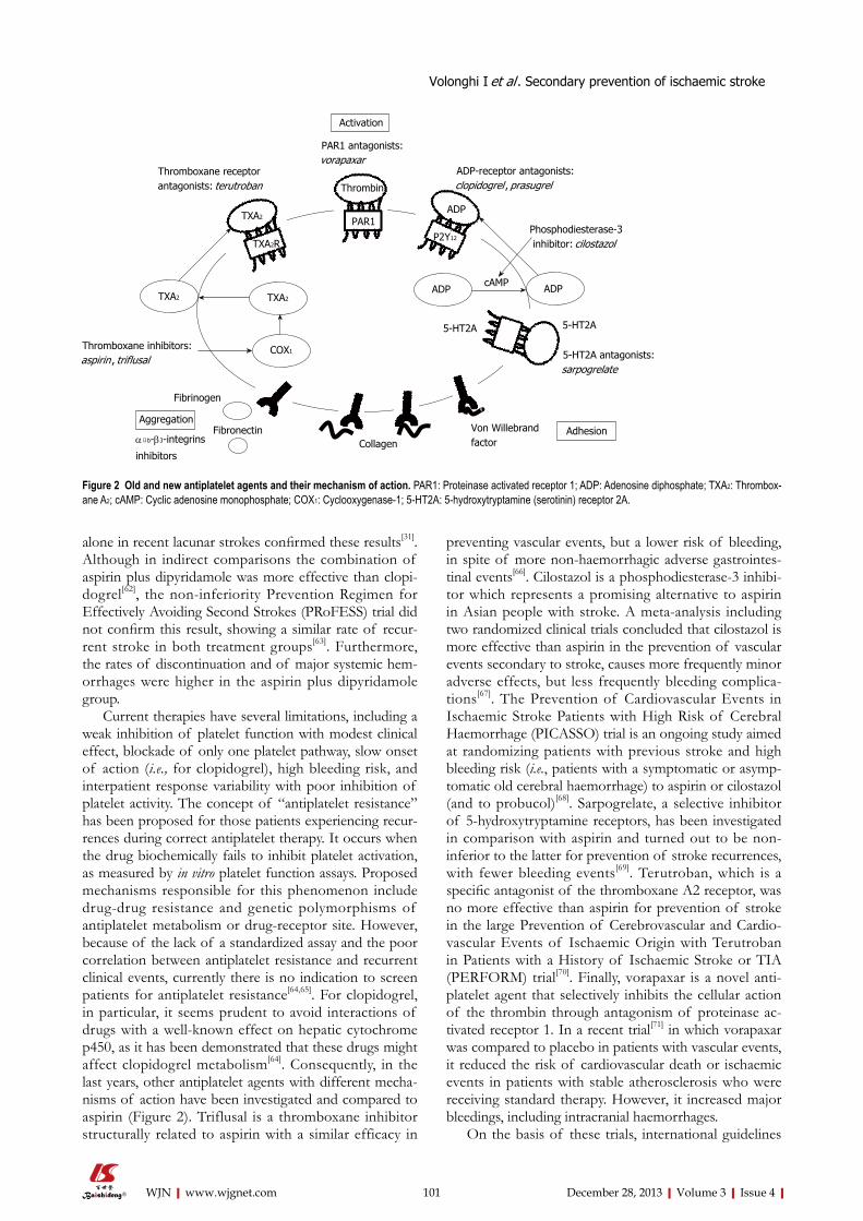
alone in recent lacunar strokes confirmed these results[31]. Although in indirect comparisons the combination of aspirin plus dipyridamole was more effective than clopi-dogrel[62], the non-inferiority Prevention Regimen for Effectively Avoiding Second Strokes (PRoFESS) trial did not confirm this result, showing a similar rate of recur-rent stroke in both treatment groups[63]. Furthermore, the rates of discontinuation and of major systemic hem-orrhages were higher in the aspirin plus dipyridamole group.
Current therapies have several limitations, including a weak inhibition of platelet function with modest clinical effect, blockade of only one platelet pathway, slow onset of action (i.e., for clopidogrel), high bleeding risk, and interpatient response variability with poor inhibition of platelet activity. The concept of “antiplatelet resistance” has been proposed for those patients experiencing recur-rences during correct antiplatelet therapy. It occurs when the drug biochemically fails to inhibit platelet activation, as measured by in vitro platelet function assays. Proposed mechanisms responsible for this phenomenon include drug-drug resistance and genetic polymorphisms of antiplatelet metabolism or drug-receptor site. However, because of the lack of a standardized assay and the poor correlation between antiplatelet resistance and recurrent clinical events, currently there is no indication to screen patients for antiplatelet resistance[64,65]. For clopidogrel, in particular, it seems prudent to avoid interactions of drugs with a well-known effect on hepatic cytochrome p450, as it has been demonstrated that these drugs might affect clopidogrel metabolism[64]. Consequently, in the last years, other antiplatelet agents with different mecha-nisms of action have been investigated and compared to aspirin (Figure 2). Triflusal is a thromboxane inhibitor structurally related to aspirin with a similar efficacy in
preventing vascular events, but a lower risk of bleeding, in spite of more non-haemorrhagic adverse gastrointes-tinal events[66]. Cilostazol is a phosphodiesterase-3 inhibi-tor which represents a promising alternative to aspirin in Asian people with stroke. A meta-analysis including two randomized clinical trials concluded that cilostazol is more effective than aspirin in the prevention of vascular events secondary to stroke, causes more frequently minor adverse effects, but less frequently bleeding complica-tions[67]. The Prevention of Cardiovascular Events in Ischaemic Stroke Patients with High Risk of Cerebral Haemorrhage (PICASSO) trial is an ongoing study aimed at randomizing patients with previous stroke and high bleeding risk (i.e., patients with a symptomatic or asymp-tomatic old cerebral haemorrhage) to aspirin or cilostazol (and to probucol)[68]. Sarpogrelate, a selective inhibitor of 5-hydroxytryptamine receptors, has been investigated in comparison with aspirin and turned out to be non-inferior to the latter for prevention of stroke recurrences, with fewer bleeding events[69]. Terutroban, which is a specific antagonist of the thromboxane A2 receptor, was no more effective than aspirin for prevention of stroke in the large Prevention of Cerebrovascular and Cardio-vascular Events of Ischaemic Origin with Terutroban in Patients with a History of Ischaemic Stroke or TIA (PERFORM) trial[70]. Finally, vorapaxar is a novel anti-platelet agent that selectively inhibits the cellular action of the thrombin through antagonism of proteinase ac-tivated receptor 1. In a recent trial[71] in which vorapaxar was compared to placebo in patients with vascular events, it reduced the risk of cardiovascular death or ischaemic events in patients with stable atherosclerosis who were receiving standard therapy. However, it increased major bleedings, including intracranial haemorrhages.
On the basis of these trials, international guidelines
101 December 28, 2013|Volume 3|Issue 4|WJN|www.wjgnet.com
Thrombin
PAR1ADP
P2Y12
TXA2
TXA2R
TXA2 TXA2
COX1
ADP ADPcAMP
PAR1 antagonists:vorapaxar
Thromboxane receptor antagonists: terutroban
Thromboxane inhibitors: aspirin , triflusal
Fibrinogen
AggregationFibronectin
αⅡb-β3-integrins
inhibitors Collagen
Von Willebrand factor
Adhesion
5-HT2A antagonists:sarpogrelate
5-HT2A
Phosphodiesterase-3 inhibitor: cilostazol
ADP-receptor antagonists: clopidogrel , prasugrel
Activation
Figure 2 Old and new antiplatelet agents and their mechanism of action. PAR1: Proteinase activated receptor 1; ADP: Adenosine diphosphate; TXA2: Thrombox-ane A2; cAMP: Cyclic adenosine monophosphate; COX1: Cyclooxygenase-1; 5-HT2A: 5-hydroxytryptamine (serotinin) receptor 2A.
5-HT2A
Volonghi I et al . Secondary prevention of ischaemic stroke
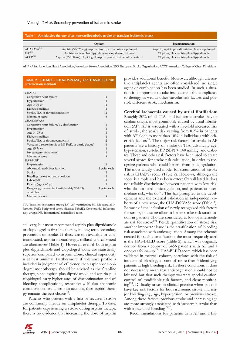
still vary, but most recommend aspirin plus dipyridamole or clopidogrel as first line therapy in long-term secondary prevention of stroke. If these are not available or con-traindicated, aspirin monotherapy, triflusal and cilostazol are alternatives (Table 1). However, even if both aspirin plus dipyridamole and clopidogrel alone are statistically superior compared to aspirin alone, clinical superiority is at best minimal. Furthermore, if tolerance profile is included in judgment of efficiency, then aspirin or clopi-dogrel monotherapy should be advised as the first-line therapy, since aspirin plus dipyridamole and aspirin plus clopidogrel carry higher rates of discontinuation and of bleeding complications, respectively. If also economic considerations are taken into account, then aspirin thera-py remains the best choice[72].
Patients who present with a first or recurrent stroke are commonly already on antiplatelet therapy. To date, for patients experiencing a stroke during aspirin therapy, there is no evidence that increasing the dose of aspirin
provides additional benefit. Moreover, although alterna-tive antiplatelet agents are often considered, no single agent or combination has been studied. In such a situa-tion it is important to take into account the compliance to therapy, as well as other vascular risk factors and pos-sible different stroke mechanisms.
Cerebral ischaemia caused by atrial fibrillation: Roughly 20% of all TIAs and ischaemic strokes have a cardiac origin, most commonly caused by atrial fibrilla-tion (AF). AF is associated with a five-fold increased risk of stroke, the yearly risk varying from 0.2% in patients with AF alone to more than 10% in individuals with oth-er risk factors[73]. The major risk factors for stroke in AF patients are a history of stroke or TIA, advancing age, hypertension, systolic BP (SBP) > 160 mmHg, and diabe-tes. These and other risk factors have been used to create several scores for stroke risk calculation, in order to rec-ognize patients who could benefit from anticoagulation. The most widely used model for stratification of stroke risk is CHADS2 score (Table 2). However, although the score is simple and has been externally validated it does not reliably discriminate between patients with low risk, who do not need anticoagulation, and patients at inter-mediate risk, who do[73]. This has prompted to the devel-opment and the external validation in independent co-horts of a new score, the CHA2DS2VASc score (Table 2). Because of the inclusion of newly recognized risk factors for stroke, this score allows a better stroke risk stratifica-tion in patients who are considered at low or intermedi-ate risk for stroke[74]. Beside quantification of stroke risk, another important issue is the stratification of bleeding risk associated with anticoagulation. Among the schemes created for such a stratification, the most frequently used is the HAS-BLED score (Table 2), which was originally derived from a cohort of 3456 patients with AF and a one-year follow-up[75]. HAS-BLED score, which has been validated in external cohorts, correlates with the risk of intracranial bleeding, a score of more than 3 identifying patients at high bleeding risk. In these conditions, it does not necessarily mean that anticoagulation should not be initiated but that such therapy warrants special caution, control of modifiable risk factors, and close monitor-ing[73]. Difficulty arises in clinical practice when patients have key risk factors for both ischaemic stroke and ma-jor bleeding (e.g., age, hypertension, or previous stroke). Among these factors, previous stroke and increasing age are more strongly associated with ischaemic stroke than with intracranial bleeding[76,77].
Recommendations for patients with AF and a his-
102 December 28, 2013|Volume 3|Issue 4|WJN|www.wjgnet.com
Options Recommendation
AHA/ASA[25] Aspirin (50-325 mg); aspirin plus dipyridamole; clopidogrel Aspirin, aspirin plus dipyridamole or clopidogrel ESO[24] Aspirin; aspirin plus dipyridamole; clopidogrel; triflusal Clopidogrel or aspirin plus dipyridamole ACCP[52] Aspirin (75-100 mg); clopidogrel; aspirin plus dipyridamole; cilostazol Clopidogrel or aspirin plus dipyridamole
Table 1 Antiplatelet therapy after non-cardioembolic stroke or transient ischaemic attack
AHA/ASA: American Heart Association/American Stroke Association; ESO: European Stroke Organisation; ACCP: American College of Chest Physicians.
CHADS2
Congestive heart failure 1 Hypertension 1 Age ≥ 75 yr 1 Diabetes mellitus 1 Stroke, TIA, or thromboembolism 2 Maximum score 6 CHA2DS2VASc Congestive heart failure/LV dysfunction 1 Hypertension 1 Age ≥ 75 yr 2 Diabetes mellitus 1 Stroke, TIA, or thromboembolism 2 Vascular disease (previous MI, PAD, or aortic plaque) 1 Age 65-74 yr 1 Sex category (female sex) 1 Maximum score 9 HAS-BLED Hypertension 1 Abnormal renal/liver function 1 point each Stroke 1 Bleeding history or predisposition 1 Labile INR 1 Elderly (age > 65 yr) 1 Drugs (e.g., concomitant antiplatelet/NSAID) or alcohol
1 point each
Maximum score 9
Table 2 CHADS2, CHA2DS2VASC, and HAS-BLED risk stratification methods
TIA: Transient ischaemic attack; LV: Left ventricular; MI: Myocardial in-farction; PAD: Peripheral artery disease; NSAID: Nonsteroidal inflamma-tory drugs; INR: International normalized ratio.
Volonghi I et al . Secondary prevention of ischaemic stroke
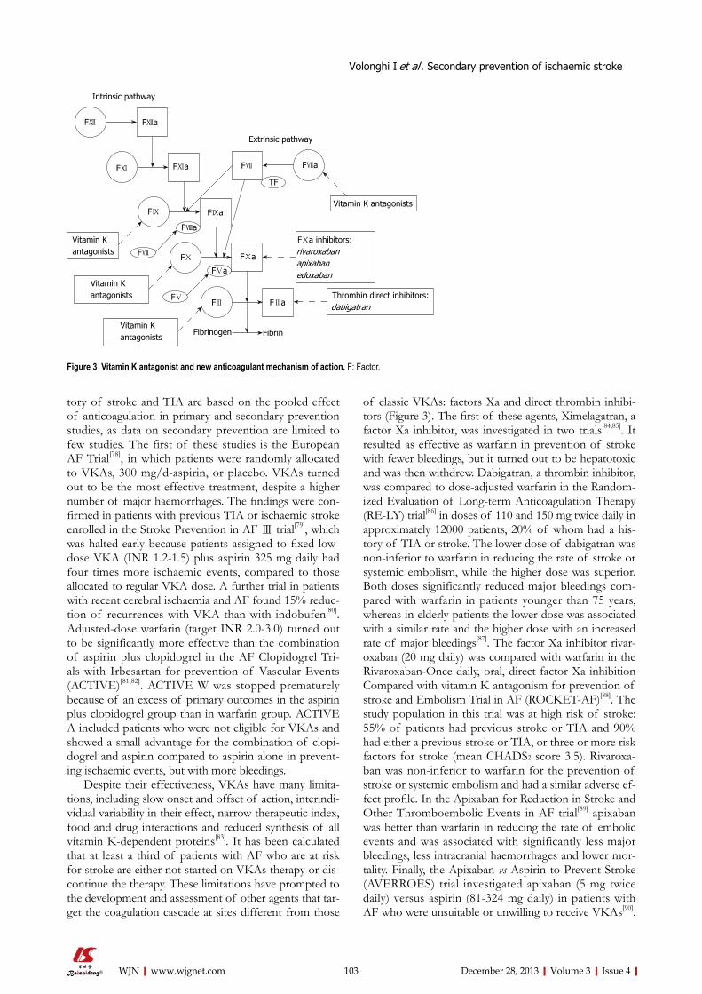
tory of stroke and TIA are based on the pooled effect of anticoagulation in primary and secondary prevention studies, as data on secondary prevention are limited to few studies. The first of these studies is the European AF Trial[78], in which patients were randomly allocated to VKAs, 300 mg/d-aspirin, or placebo. VKAs turned out to be the most effective treatment, despite a higher number of major haemorrhages. The findings were con-firmed in patients with previous TIA or ischaemic stroke enrolled in the Stroke Prevention in AF Ⅲ trial[79], which was halted early because patients assigned to fixed low-dose VKA (INR 1.2-1.5) plus aspirin 325 mg daily had four times more ischaemic events, compared to those allocated to regular VKA dose. A further trial in patients with recent cerebral ischaemia and AF found 15% reduc-tion of recurrences with VKA than with indobufen[80]. Adjusted-dose warfarin (target INR 2.0-3.0) turned out to be significantly more effective than the combination of aspirin plus clopidogrel in the AF Clopidogrel Tri-als with Irbesartan for prevention of Vascular Events (ACTIVE)[81,82]. ACTIVE W was stopped prematurely because of an excess of primary outcomes in the aspirin plus clopidogrel group than in warfarin group. ACTIVE A included patients who were not eligible for VKAs and showed a small advantage for the combination of clopi-dogrel and aspirin compared to aspirin alone in prevent-ing ischaemic events, but with more bleedings.
Despite their effectiveness, VKAs have many limita-tions, including slow onset and offset of action, interindi-vidual variability in their effect, narrow therapeutic index, food and drug interactions and reduced synthesis of all vitamin K-dependent proteins[83]. It has been calculated that at least a third of patients with AF who are at risk for stroke are either not started on VKAs therapy or dis-continue the therapy. These limitations have prompted to the development and assessment of other agents that tar-get the coagulation cascade at sites different from those
of classic VKAs: factors Xa and direct thrombin inhibi-tors (Figure 3). The first of these agents, Ximelagatran, a factor Xa inhibitor, was investigated in two trials[84,85]. It resulted as effective as warfarin in prevention of stroke with fewer bleedings, but it turned out to be hepatotoxic and was then withdrew. Dabigatran, a thrombin inhibitor, was compared to dose-adjusted warfarin in the Random-ized Evaluation of Long-term Anticoagulation Therapy (RE-LY) trial[86] in doses of 110 and 150 mg twice daily in approximately 12000 patients, 20% of whom had a his-tory of TIA or stroke. The lower dose of dabigatran was non-inferior to warfarin in reducing the rate of stroke or systemic embolism, while the higher dose was superior. Both doses significantly reduced major bleedings com-pared with warfarin in patients younger than 75 years, whereas in elderly patients the lower dose was associated with a similar rate and the higher dose with an increased rate of major bleedings[87]. The factor Xa inhibitor rivar-oxaban (20 mg daily) was compared with warfarin in the Rivaroxaban-Once daily, oral, direct factor Xa inhibition Compared with vitamin K antagonism for prevention of stroke and Embolism Trial in AF (ROCKET-AF)[88]. The study population in this trial was at high risk of stroke: 55% of patients had previous stroke or TIA and 90% had either a previous stroke or TIA, or three or more risk factors for stroke (mean CHADS2 score 3.5). Rivaroxa-ban was non-inferior to warfarin for the prevention of stroke or systemic embolism and had a similar adverse ef-fect profile. In the Apixaban for Reduction in Stroke and Other Thromboembolic Events in AF trial[89] apixaban was better than warfarin in reducing the rate of embolic events and was associated with significantly less major bleedings, less intracranial haemorrhages and lower mor-tality. Finally, the Apixaban vs Aspirin to Prevent Stroke (AVERROES) trial investigated apixaban (5 mg twice daily) versus aspirin (81-324 mg daily) in patients with AF who were unsuitable or unwilling to receive VKAs[90].
103 December 28, 2013|Volume 3|Issue 4|WJN|www.wjgnet.com
FⅫ
FⅪ
FⅫa
FⅪa
FⅨ FⅨa
FⅧ
FⅧa
FⅩ FⅩa
FⅤa
FⅤFⅡ FⅡa
FⅦ FⅦa
TF
Vitamin K antagonists
FⅩa inhibitors:rivaroxabanapixabanedoxaban
Thrombin direct inhibitors:dabigatran
FibrinFibrinogenVitamin K antagonists
Vitamin K antagonists
Vitamin K antagonists
Intrinsic pathway
Extrinsic pathway
Figure 3 Vitamin K antagonist and new anticoagulant mechanism of action. F: Factor.
Volonghi I et al . Secondary prevention of ischaemic stroke
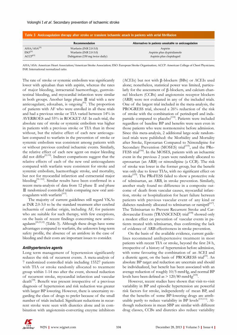
The rate of stroke or systemic embolism was significantly lower with apixaban than with aspirin, whereas the rates of major bleeding, intracranial haemorrhage, gastroin-testinal bleeding, and myocardial infarction were similar in both groups. Another large phase Ⅲ trial with a new anticoagulant, edoxaban, is ongoing[91]. The proportion of patients with AF who were enrolled in all these trials and had a previous stroke or TIA varied between 14% in AVERROES and 55% in ROCKET-AF. In each trial, the absolute rate of stroke or systemic embolism was higher in patients with a previous stroke or TIA than in those without, but the relative effect of each new anticoagu-lant compared to warfarin in the prevention of stroke or systemic embolism was consistent among patients with or without previous cerebral ischaemic events. Similarly, the relative effect of each new agent on major bleeding did not differ[92-95]. Indirect comparisons suggest that the relative effects of each of the new oral anticoagulants compared with warfarin were consistent for stroke and systemic embolism, haemorrhagic stroke, and mortality, but not for myocardial infarction and extracranial major bleeding[96-102]. Similar results have been confirmed in a recent meta-analysis of data from 12 phase Ⅱ and phase Ⅲ randomized controlled trials comparing new oral anti-coagulants with warfarin[103].
The majority of current guidelines still regard VKAs at INR 2.0-3.0 to be the standard treatment after cerebral ischaemia of cardiac origin, including AF, for patients who are suitable for such therapy, with few exceptions, on the basis of recent findings concerning new antico-agulants[24,25,52] (Table 3). Although these drugs have some advantages compared to warfarin, the unknown long-term safety profile, the absence of an antidote in the case of bleeding and their costs are important issues to consider.
Antihypertensive agentsLong term management of hypertension significantly reduces the risk of recurrent events. A meta-analysis of 7 randomized controlled trials including 15527 patients with TIA or stroke randomly allocated to treatment group within 1-14 mo after the event, showed reduction of recurrent stroke, myocardial infarction and vascular death[38]. Benefit was present irrespective of a previous diagnosis of hypertension and risk reduction was greater with larger BP lowering. However, there is uncertainty re-garding the class of drugs to prefer because of the small number of trials included. Significant reductions in recur-rent stroke were seen with diuretics alone and in com-bination with angiotensin-converting enzyme inhibitors
(ACEIs) but not with β-blockers (BBs) or ACEIs used alone; nonetheless, statistical power was limited, particu-larly for the assessment of β-blockers, and calcium chan-nel blockers (CCBs) and angiotensin receptor blockers (ARB) were not evaluated in any of the included trials. One of the largest trial included in the meta-analysis, the PROGRESS trial, showed a 26% reduction of the risk of stroke with the combination of perindopril and inda-pamide compared to placebo[104]. Patients were included regardless of baseline BP and benefits were seen even in those patients who were normotensive before admission. Since this meta-analysis, 2 additional large-scale random-ized trials were published: the Morbidity and Mortality after Stroke, Eprosartan Compared to Nitrendipine for Secondary Prevention (MOSES) trial[105], and the PRo-FESS trial[106]. In the MOSES, patients with an ischaemic event in the previous 2 years were randomly allocated to eprosartan (an ARB) or nitrendipine (a CCB). The risk of stroke was lower in the former group, but the benefit was only due to fewer TIAs, with no significant effect on stroke[105]. The PRoFESS failed to show a protective role of telmisartan, an ARB, in stroke prevention. Similarly, another study found no difference in a composite out-come of death from vascular causes, myocardial infarc-tion, stroke or hospitalization for heart failure between patients with previous vascular event of any kind or diabetes randomly allocated to telmisartan or ramipril[107]. The Telmisartan to Prevent Recurrent Stroke and Car-diovascular Events (TRANSCEND) trial[108] showed only a modest effect on prevention of vascular events in pa-tients treated with telmisartan, thus confirming the lack of evidence of ARB effectiveness in stroke prevention.
On the basis of the available evidence, current guide-lines recommend antihypertensive treatment in most patients with recent TIA or stroke, beyond the first 24 h, irrespective of a history of hypertension before admission, with some favouring the combination of an ACEI and a diuretic agent, on the basis of PROGRESS trial[25]. An absolute BP target and reduction are uncertain and should be individualized, but benefit has been associated with an average reduction of roughly 10/5 mmHg, and normal BP levels have been defined as > 120/80 mmHg[109].
However, recent studies have shown that visit-to-visit variability in BP and episodic hypertension are powerful risk factors for stroke, independently of mean BP, and that the benefits of some BP-lowering drugs are attrib-utable partly to reduce variability in BP levels[110,111]. Al-though reductions in mean SBP are similar with different drug classes, CCBs and diuretics also reduce variability
104 December 28, 2013|Volume 3|Issue 4|WJN|www.wjgnet.com
Recommendation Alternatives in patients unsuitable to anticoagulation
AHA/ASA[25] Warfarin (INR 2.0-3.0) Aspirin ESO[24] Warfarin (INR 2.0-3.0) Aspirin plus dypiridamole ACCP[52] Dabigatran (150 mg twice daily) Aspirin plus clopidogrel
Table 3 Anticoagulation therapy after stroke or transient ischaemic attack in patients with atrial fibrillation
AHA/ASA: American Heart Association/American Stroke Association; ESO: European Stroke Organisation; ACCP: American College of Chest Physicians; INR: International normalized ratio.
Volonghi I et al . Secondary prevention of ischaemic stroke
in SBP, while BBs increase variability[112,113]. In particular, it has been shown that higher doses of CCBs reduce BP variability more than lower doses, and higher doses of BBs increase BP variability[114]. This means that combina-tions containing a low dose of BBs will have less adverse effects on variability in SBP and the associated risk of stroke, but low doses of CCBs will be less effective in reducing BP variability. Drug class effects on variability persist even in combination therapy.
Another issue to consider is the heterogeneity of stroke patients. Elderly patients seem to benefit most from a combination therapy of ACEIs and diuretics[115], as well as patients with renal impairment or diabetic ne-phropathy, in whom ACEIs and ARBs are recommended. The detrimental effect of such drugs on BP variability could be offset by the combination use with CCBs, as suggested in the Avoiding Cardiovascular Events through Combination Therapy in Patients Living with Systolic Hypertension study[116], in whom patients with hyperten-sion and previous vascular disease, including stroke, were randomly allocated to a combination of benazepril plus amlodipine or a combination of benazepril plus hydro-chlorothiazide. The former group of patients had fewer combined cardiovascular deaths and chronic kidney dis-ease events compared to the latter.
However, direct comparisons of drugs for secondary prevention of stroke are needed in order to recommend the most appropriate BP-lowering drug regimen. More research is required to identify ideal combinations and doses of drugs for the prevention of stroke in specific patient groups with particular reference to their effect on BP variability.
Lipid lowering agentsAlthough the association between serum lipid concen-trations and ischaemic risk is weaker for stroke than for myocardial infarction[117], lipid levels modification is important in both primary and secondary prevention of stroke[118]. A recent observational study[44] showed a better functional outcome in stroke patients who received statin therapy before admission to hospital and who continued such a treatment during hospitalization. The prognosis was even better with higher doses of statins and when therapy was started earlier. Regarding secondary preven-tion, in a retrospective analysis of the Heart Protection Study (HPS) [119] restricted to subjects with a history of stroke, simvastatin did not significantly reduce the risk of stroke. In contrast, the Stroke Prevention by Aggres-sive Reduction in Cholesterol Levels (SPARCL) trial[120] showed a 16% reduction in stroke recurrence in patients with previous stroke or TIA, not of cardiac origin, ran-domized to atorvastatin 80 mg daily versus placebo, with-in 6 mo from index event. One concern in the SPARCL trial and in the subset of patients in HPS[119] who had a stroke before randomization was a significant increase in the risk of haemorrhagic stroke in patients allocated to statin treatment[121]. Further analysis showed that much of excess risk of haemorrhagic stroke on atorvastatin therapy was in patients with a small vessel disease stroke
as qualifying event, either of haemorrhagic or ischaemic type[122]. Although in the former group there was no over-all benefit, in the latter the net benefit was still in favour of treatment because of a reduction in ischaemic recur-rences[122]. This suggests that statin therapy should be initiate only in patients with atherosclerotic stroke or in presence of other noncerebrovascular indications, such as coronary heart disease or diabetes[123].
Another important clinical issue relates to low-density lipoprotein (LDL) concentration. In a meta-analysis of all statin trials that looked at intense LDL lowering versus standard treatment, achievement of an LDL concentra-tion of 100 mg/dL resulted in 16% relative reduction in risk of stroke. Subanalysis of the SPARCL trial showed a 28% reduction in stroke risk with a LDL concentration lower than 70 mg/dL in comparison with a level of 100 mg/dL[124,125]. The Treat Stroke to Target trial[126] is an on-going study aimed at unravelling the issue regarding the optimal LDL target to reach. The working hypothesis of this trial is that the achievement of an LDL concentra-tion of 70 mg/dL is better than 100 mg/dL in patients with previous atherosclerotic TIA or stroke. The Optimal BP and Cholesterol Targets for Preventing Recurrent Stroke in Hypertensive (ESH-CHL-SHOT) trial[127] will be started soon to address this issue. Its purpose, indeed, is to investigate both SBP and LDL cholesterol levels to achieve to optimize secondary prevention. Regarding lipid-lowering treatment in particular, the patients will be randomly allocated to one of two different LDL targets, 70 to 110 mg/dL or < 70 mg/dL, using different statins, including simvastatin, pravastatin, fluvastatin, atorvastatin or rosuvastatin.
Furthermore, there is still uncertainty regarding whether to start with high-dose or low-dose statin ther-apy, considering benefit and potential side effects. The SPARCL trial[120] reliably showed that high-dose atorv-astain at 80 mg/d reduces the risk of stroke and other cardiovascular recurrences in patients with non cardio-embolic stroke. It is however unclear, in the absence of randomized comparisons in patients with stroke, if lower doses of atorvastatin are equally or less effective than the high-dose regimen. There is evidence that atorvastatin at 80 mg/d provides significant reductions in the risk of cardiovascular events and stroke beyond that provided by a 10-mg dose in patients with coronary disease[128]. However, the high-dose regimen was complicated more frequently by higher rates of adverse events and discon-tinuation, both in Treating to New Targets (TNT) trial[128] and SPARCL trial[120]. Moreover, the Food and Drug Administration issued a warning regarding increased risk of myopathy in patients taking simvastatin at 80 mg/d, especially if taken in combination with CCBs, which are frequently used in patients with stroke. Although the SPARCL results support the use of high-dose atorvas-tatin for secondary prevention of stroke, it is unclear whether the highest currently approved doses of statins have neuroprotective effects. It is also unknown if other hydroxymethylglutaryl-coenzymeA reductase inhibitors, in different doses, could be potentially effective in pre-
105 December 28, 2013|Volume 3|Issue 4|WJN|www.wjgnet.com
Volonghi I et al . Secondary prevention of ischaemic stroke
vention of stroke recurrences. In this regard, two trials are ongoing to investigate the combined effect of this class of drugs[127] and that of pravastatin[129] in particular in long-term secondary prevention after stroke.
On the basis of available data the American Heart Association/American Stroke Association (AHA/ASA) guidelines recommend statin therapy with intensive lipid-lowering effect for secondary prevention among patients with ischemic stroke or TIA who have evidence of ath-erosclerosis, an LDL-level ≥ 100 mg/dL, and who are without known coronary heart disease. They suggest, as a target, a reduction of at least 50% in LDL or, alterna-tively, an LDL level of < 70 mg/dL[25].
Other medications used to treat dyslipidemia include niacin, fibrates, and cholesterol absorption inhibitors. These agents can be used by patients intolerant to statins, but the evidence of their efficacy for prevention of stroke recurrence is scarce. Niacin has been associated with a reduction in cerebrovascular events[130], whereas gemfibrozil reduced the rate of total strokes among men with coronary artery disease and low levels of high-densi-ty lipoprotein cholesterol (≤ 40 mg/dL) in the Veterans Affairs HDL Intervention Trial. The latter result was no more significant when adjudicated events alone were analyzed[131].
Surgical proceduresPercutaneous PFO closure: PFO is a common embry-onic defect of the interatrial septum, being present in up to 25% of healthy people. In a meta-analysis PFO was found to be significantly associated with increased risk of stroke in younger patients with cryptogenic stroke, especially when atrial septal aneurysm (ASA) is also pres-ent[132]. This leads to the assumption that PFO may be the cause somehow associated to stroke occurrence, although it may also be an incidental finding[133]. The role of PFO in determining the risk of recurrence is uncertain, as well. In a substudy of the PFO in Cryptogenic Stroke Study (PICSS)[134] there were no differences in the rates of recurrent strokes in those with or without PFO, as well as no demonstrated effect on outcomes based on PFO size or presence of ASA. In contrast, the study of Mas et al[135] reported higher recurrence rates of stroke associ-ated with both PFO and ASA, indicating that the pres-ence of both these cardiac abnormalities was a predictor of an increased risk of recurrent stroke, whereas isolated PFO, whether small or large, was not. Thus, the natural history after cerebrovascular events in patients with PFO remains insufficiently defined. As a consequence, the optimal management strategy for treating patients with cryptogenic stroke who are discovered to have a PFO remains to be determined. Observational studies on medical treatment in patients with PFO with either anti-platelets or warfarin reported a risk of recurrent stroke or TIA ranging from 3% to 12% during the first year[136]. Both larger PFO size and a greater degree of right-to-left shunt increased the risk of paradoxical embolism in one study[136]. Of note, observational data for the comparative effectiveness of anticoagulants versus antiplatelet agents
for secondary prevention after stroke associated to PFO show a significant benefit of warfarin, whereas the only relevant randomized study, coming from a subgroup of PICSS[134], found a non-significant trend in favor of war-farin over aspirin. On the basis of this evidence, AHA guidelines state that there are insufficient data to establish the superiority of warfarin over aspirin for secondary stroke prevention after stroke or TIA in patients with PFO[25]. However, warfarin might be more beneficial in presence of deep venous thrombosis or ASA, accord-ing to the European Stroke Organization guidelines[24]. Another therapeutic possibility is percutaneous PFO closure, which has supplanted surgical procedures. Case series and nonrandomized comparisons have long sug-gested that closure is a highly efficacious procedure and have led to the rapid off-label adoption of this interven-tion[137]. In contrast, recent randomized controlled trials failed to identify any statistically significant difference between closure and medical treatment for secondary prevention after stroke[138-140]. However, such trials have several limitations and low statistical power. In the Evalu-ation of the STARFlex Septal Closure System in Patients with a Stroke and/or TIA due to Presumed Paradoxi-cal Embolism through a PFO (CLOSURE) trial[138] the main limitation was the low outcome rate, compared to previous observational studies, thus pointing to a failure to select patients with PFO-related events. This may, in part, be attributable to a reluctance to randomize those patients with cryptogenic stroke who appear most likely to have had a PFO-related event (i.e., patients with clini-cal indicators of paradoxical embolism, such as large shunt, associated ASA, Valsalva maneuver at onset of stroke, or absence of any conventional stroke risk fac-tors). Other limitations of this trial include idiosyncratic effects, such as in situ thrombosis or other mechanical complications, with the use of the obsolete STARFlex device, which may have increased the outcome rates in the intervention arm, as well as the short follow-up, that may be inadequate to capture the benefit of the interven-tion[137]. The Randomized Evaluation of Recurrent Stroke Comparing PFO Closure to Established Current Stan-dard of Care Treatment (RESPECT) trial[139] confirmed the results of the CLOSURE trial in intention-to-treat analysis, in spite of important differences between the two studies with respect to the study design, the popula-tion included, and the device tested. In the RESPECT trial[139] the enrollment criteria were more stringent and the follow-up period was longer than in the CLOSURE. Moreover, the Amplatzer PFO Occluder, as compared to STARFlex used in the CLOSURE, was associated with higher effective closure rate. Finally, although different in terms of population and endpoints, the Percutaneous Closure of PFO in Cryptogenic Stroke (PC) trial[140] cor-roborated previous results. However, unlike the results of the CLOSURE trial, the results of the RESPECT and the PC trials have encouraged the supporters of PFO closure. The advocates of closure focus on the modest statistical power of these trials, the substantial relative ef-fect size of the point estimates in both trials, the signifi-
106 December 28, 2013|Volume 3|Issue 4|WJN|www.wjgnet.com
Volonghi I et al . Secondary prevention of ischaemic stroke
cance of the per-protocol and as-treated analyses in the RESPECT, and arbitrariness of the conventional P-value threshold of 0.05. Although the controversy over effi-cacy may not be settled, it is worth noting that the safety profile of Amplatzer device appeared to be superior to that of STARFLex device tested in the CLOSURE. This supports the hypothesis that PFO closure, in certain conditions, could be superior to medical treatment and reinforces the need to carry on trials with more selected populations. Indeed, other trials are ongoing, such as the PFO Closure or Anticoagulants Versus Antiplatelet Therapy to Prevent Stroke Recurrence (CLOSE) trial[141] and the GORE® HELEX® Septal Occluder/GORE® Septal Occluder for PFO Closure in Stroke Patients (RE-DUCE) trial[142]. However, like previous analyses, these new studies are hampered by a slow and skewed enroll-ment since most patients assumed to be at high risk un-dergo percutaneous closure. Beside, stroke pathophysiol-ogy is multifactorial, and the diagnosis of PFO-mediated paradoxical embolism is presumptive. Both a PFO and a cryptogenic stroke may coexist without causal relation in a given patient. In this case, obviously, PFO closure will not reduce the risk of recurrence. Current guidelines, which are antecedent to these recent trials, do not recom-mend closure because of the paucity of data[25]. However, pending other trials and based on RESPECT, it would be reasonable to discuss closure with Amplatzer device in young patients (< 50 years) with a substantial shunt and a cortical infarct, in the absence of known vascular risk factors[143].
Carotid endarterectomy and stenting for large-artery atherosclerosisBeside medical therapy, the mainstays of treatment op-tions for symptomatic large-artery atherosclerosis are ca-rotid endarterectomy (CEA) and carotid angioplasty and stenting (CAS), the latter being emerged more recently as an alternative to surgical intervention to prevent recur-rent strokes.
CEA plus medical therapy is more effective and safe than medical therapy alone in secondary prevention after stroke for symptomatic patients with > 70% carotid ste-nosis according to 3 major trials, the European Carotid Surgery Trial[144], the North American Symptomatic CEA Trial[145], and the Veterans Affairs Cooperative Study[146]. However, in all these trials medical therapy did not in-clude aggressive atherosclerotic medical management based on statins, antihypertensive agents, antiplatelets other than aspirin, and smoking cessation, thus meaning that the benefit of surgery today may be less than in the early trials. Uncertainty exists for patients with symptom-atic stenosis in the range of 50% to 69%, for whom CEA should be considered only with appropriate case selection when the risk-benefit ratio is favorable for the patient. The timing of CEA after an acute event is controversial, with experts advocating waiting anywhere from 2 to 6 wk[25]. However, early surgery (< 2 wk) turned out to be beneficial, according to pooled analysis of trials. Further-more, early intervention (< 3 wk) may be beneficial and
safe in low-risk patients with TIA and minor strokes, and in those without haemorrhagic transformation[25].
CAS is a percutaneous, less invasive procedure that has emerged as an interesting alternative to CEA for the treatment of extracranial carotid artery disease, especially thanks to the advances in endovascular technology, in-cluding embolic protection devices (EPD) and improved stent design. Complication rates are comparable to CEA. Although advantages related to the less invasive pro-cedure, CAS durability remains unproven. The largest randomized trial in symptomatic patients, the Carotid and Vertebral Artery Transluminal Angioplasty Study 2 trial[147], showed comparable outcome between the two procedures, also in the long-term follow-up. The Stent-Supported Percutaneous Angioplasty of the Carotid Artery Versus Endarterectomy trial[148] which was ended after the second interim analysis owing to the low rate of recruitment, showed similar rates of periprocedural stroke or death and ipsilateral ischaemic stroke up to 2 years between CEA and CAS. However, the fact that only 27% of patients with CAS in this study used EPD may have skewed the outcome. Because EPD reduce peripro-cedural stroke rates, they have been required in all stent-ing trials since then. The Stenting and Angioplasty with Protection in Patients at High Risk for Endarterectomy trial[149] randomized both symptomatic and asymptomatic high risk patients, showing that CAS was not inferior to CEA. However, a high percentage of patients were as-ymptomatic (70%), thus limiting the applicability of the results to symptomatic ones. The Endarterectomy Versus Angioplasty in Patients with Symptomatic Severe Carotid Stenosis trial[150] showed higher rates of periprocedural stroke or death at 30 d and within 4 years, as well. How-ever, the results of this trial are influenced by the limited experience of the operators, the use of five different stents and seven different cerebral protection devices, and the fact that EPD were optional in the early phase of the study. The most important trial on this topic is the Carotid Revascularization Endarterectomy Versus Stent-ing Trial[151], which included patients with both symptom-atic and asymptomatic carotid disease in similar propor-tions. It demonstrated that both procedures had similar safety and efficacy profiles for all patients, according to gender and the presence or not of previous cerebrovas-cular events. However, patients aged 70 years or older had better outcomes with CEA for the primary endpoint (any stroke, death or myocardial infarction (MI) within 30 d and ipsilateral stroke during long-term follow-up) and less adverse events. Younger patients had greater benefit from CAS than older subjects. The CAS group had a greater percentage of patients with stroke within the first 30 d after the procedure, but a lower rate of MI events. In trials of symptomatic patients, stenting was associated with a significantly higher risk of 30-d stroke or death, particularly in people over 70 years, compared to CEA, whereas the long-term rate of ipsilateral stroke were similar for both interventions[152]. However, stenting was associated with less MI and cranial nerve palsies.
On the basis of these studies, current AHA/ASA
107 December 28, 2013|Volume 3|Issue 4|WJN|www.wjgnet.com
Volonghi I et al . Secondary prevention of ischaemic stroke
guidelines recommend that patients who have experi-enced a TIA or stroke within the past 6 mo and have ipsi-lateral carotid stenosis of 70%-90% would be candidates for CEA if the perioperative morbidity and mortality risk is less than 6%. In symptomatic patients with a 50%-60% stenosis, the decision to perform CEA depends on fac-tors such as age, comorbidities, and sex. If a patient is a good candidate for CEA, intervention within 2 wk is rec-ommended[25]. To date, CAS is considered an alternative option for symptomatic patients with carotid stenosis of more than 70% as determined by noninvasive imaging or more than 50% as determined by catheter angiography. CAS is also an alternative in patients deemed at high risk for surgical intervention. High risk is defined by pres-ence of severe comorbidites and challenging anatomical features (such as prior neck intervention or irradiation, postendarterectomy restenosis, surgical inaccessible le-sion, contralateral carotid occlusion, contralateral vocal cord palsy, or the presence of tracheostomy)[25]. Patients are also recommended to optimize medical therapy with antiplatelets, statins and risk factor modifications.
Angioplasty and stenting for intracranial atherosclerosisAccording to the Stenting versus Aggressive Medical Therapy for Intracranial Arterial Stenosis (SAMMPRIS) trial[153], angioplasty and stenting of intracranial stenosis did not reduce the risk of recurrent stroke compared with aggressive medical therapy, including dual antiplate-lets, statins and antihypertensive agents.
CONCLUSIONSecondary prevention with antiplatelet and antihyper-tensives medications, statins and anticoagulants as ap-propriate, should be initiated urgently after TIA or minor strokes because of the high risk of early stroke recurrence. For long-term prevention, aspirin plus di-pyridamole or clopidogrel must be proposed as first-line approach after cerebral ischaemia of arterial origin. For cardioembolic strokes in patients with non-valvular AF new anticoagulation agents are challenging the current standards of VKAs. Lipid and blood-pressure-lowering treatments are warranted in all cerebral ischaemia, but recommendations about optimal drug regimen, includ-ing doses and when to start therapy, as well as target levels of LDL or BP to be achieved, are still debated. Revascularization procedures, including CEA or stenting, must be taken into consideration early after the event in patients with stroke or TIA related to carotid atheroscle-rotic disease.
REFERENCES1 Murray CJ, Lopez AD, Black R, Mathers CD, Shibuya K,
Ezzati M, Salomon JA, Michaud CM, Walker N, Vos T. Global burden of disease 2005: call for collaborators. Lan-cet 2007; 370: 109-110 [PMID: 17630021 DOI: 10.1016/S0140-6736(07)61064-2]
2 Rothwell PM, Coull AJ, Silver LE, Fairhead JF, Giles MF, Lovelock CE, Redgrave JN, Bull LM, Welch SJ, Cuthbert-
son FC, Binney LE, Gutnikov SA, Anslow P, Banning AP, Mant D, Mehta Z. Population-based study of event-rate, incidence, case fatality, and mortality for all acute vascular events in all arterial territories (Oxford Vascular Study). Lancet 2005; 366: 1773-1783 [PMID: 16298214 DOI: 10.1016/S0140-6736(05)67702-1]
3 Feigin VL, Lawes CM, Bennett DA, Barker-Collo SL, Parag V. Worldwide stroke incidence and early case fatality reported in 56 population-based studies: a systematic review. Lancet Neurol 2009; 8: 355-369 [PMID: 19233729 DOI: 10.1016/S1474-4422(09)70025-0]
4 O’Donnell MJ, Xavier D, Liu L, Zhang H, Chin SL, Rao-Mel-acini P, Rangarajan S, Islam S, Pais P, McQueen MJ, Mondo C, Damasceno A, Lopez-Jaramillo P, Hankey GJ, Dans AL, Yusoff K, Truelsen T, Diener HC, Sacco RL, Ryglewicz D, Czlonkowska A, Weimar C, Wang X, Yusuf S. Risk factors for ischaemic and intracerebral haemorrhagic stroke in 22 countries (the INTERSTROKE study): a case-control study. Lancet 2010; 376: 112-123 [PMID: 20561675 DOI: 10.1016/S0140-6736(10)60834-3]
5 Rothwell PM, Coull AJ, Giles MF, Howard SC, Silver LE, Bull LM, Gutnikov SA, Edwards P, Mant D, Sackley CM, Farmer A, Sandercock PA, Dennis MS, Warlow CP, Bamford JM, Anslow P. Change in stroke incidence, mortality, case-fa-tality, severity, and risk factors in Oxfordshire, UK from 1981 to 2004 (Oxford Vascular Study). Lancet 2004; 363: 1925-1933 [PMID: 15194251 DOI: 10.1016/S0140-6736(04)16405-2]
6 Giles MF, Rothwell PM. Risk of stroke early after transient ischaemic attack: a systematic review and meta-analysis. Lancet Neurol 2007; 6: 1063-1072 [PMID: 17993293 DOI: 10.1016/S1474-4422(07)70274-0]
7 Hong KS, Yegiaian S, Lee M, Lee J, Saver JL. Declining stroke and vascular event recurrence rates in secondary pre-vention trials over the past 50 years and consequences for current trial design. Circulation 2011; 123: 2111-2119 [PMID: 21536995 DOI: 10.1161/CIRCULATIONAHA.109.934786]
8 Hankey GJ, Jamrozik K, Broadhurst RJ, Forbes S, Burvill PW, Anderson CS, Stewart-Wynne EG. Long-term risk of first recurrent stroke in the Perth Community Stroke Study. Stroke 1998; 29: 2491-2500 [PMID: 9836757 DOI: 10.1161/01.STR.29.12.2491]
9 Burn J, Dennis M, Bamford J, Sandercock P, Wade D, Warlow C. Long-term risk of recurrent stroke after a first-ever stroke. The Oxfordshire Community Stroke Project. Stroke 1994; 25: 333-337 [PMID: 8303740 DOI: 10.1161/01.STR.25.2.333]
10 Petty GW, Brown RD, Whisnant JP, Sicks JD, O’Fallon WM, Wiebers DO. Survival and recurrence after first cerebral infarction: a population-based study in Rochester, Minne-sota, 1975 through 1989. Neurology 1998; 50: 208-216 [PMID: 9443482 DOI: 10.1212/WNL.50.1.208]
11 Sacco RL, Shi T, Zamanillo MC, Kargman DE. Predictors of mortality and recurrence after hospitalized cerebral infarc-tion in an urban community: the Northern Manhattan Stroke Study. Neurology 1994; 44: 626-634 [PMID: 8164815 DOI: 10.1212/WNL.44.4.626]
12 Hillen T, Coshall C, Tilling K, Rudd AG, McGovern R, Wolfe CD. Cause of stroke recurrence is multifactorial: pat-terns, risk factors, and outcomes of stroke recurrence in the South London Stroke Register. Stroke 2003; 34: 1457-1463 [PMID: 12750544 DOI: 10.1161/01.STR.0000072985.24967.7F]
13 Johnston SC, Gress DR, Browner WS, Sidney S. Short-term prognosis after emergency department diagnosis of TIA. JAMA 2000; 284: 2901-2906 [PMID: 11147987 DOI: 10.1001/jama.284.22.2901]
14 Rothwell PM, Warlow CP. Timing of TIAs preceding stroke: time window for prevention is very short. Neurol-ogy 2005; 64: 817-820 [PMID: 15753415 DOI: 10.1212/01.WNL.0000152985.32732.EE]
15 Hankey GJ. Long-term outcome after ischaemic stroke/tran-sient ischaemic attack. Cerebrovasc Dis 2003; 16 Suppl 1: 14-19
108 December 28, 2013|Volume 3|Issue 4|WJN|www.wjgnet.com
Volonghi I et al . Secondary prevention of ischaemic stroke
[PMID: 12698014 DOI: 10.1159/000069936]16 Lemmens R, Smet S, Thijs VN. Clinical scores for predict-
ing recurrence after transient ischemic attack or stroke: how good are they? Stroke 2013; 44: 1198-1203 [PMID: 23482596 DOI: 10.1161/STROKEAHA.111.000141]
17 Cucchiara BL, Messe SR, Sansing L, MacKenzie L, Taylor RA, Pacelli J, Shah Q, Pollak ES, Kasner SE. D-dimer, mag-netic resonance imaging diffusion-weighted imaging, and ABCD2 score for transient ischemic attack risk stratification. J Stroke Cerebrovasc Dis 2009; 18: 367-373 [PMID: 19717021 DOI: 10.1016/j.jstrokecerebrovasdis.2009.01.006]
18 Ay H, Arsava EM, Johnston SC, Vangel M, Schwamm LH, Furie KL, Koroshetz WJ, Sorensen AG. Clinical- and imaging-based prediction of stroke risk after transient isch-emic attack: the CIP model. Stroke 2009; 40: 181-186 [PMID: 18948609 DOI: 10.1161/STROKEAHA.108.521476]
19 Calvet D, Touzé E, Oppenheim C, Turc G, Meder JF, Mas JL. DWI lesions and TIA etiology improve the prediction of stroke after TIA. Stroke 2009; 40: 187-192 [PMID: 18988917 DOI: 10.1161/STROKEAHA.108.515817]
20 Coutts SB, Eliasziw M, Hill MD, Scott JN, Subramaniam S, Buchan AM, Demchuk AM. An improved scoring system for identifying patients at high early risk of stroke and func-tional impairment after an acute transient ischemic attack or minor stroke. Int J Stroke 2008; 3: 3-10 [PMID: 18705908 DOI: 10.1111/j.1747-4949.2008.00182.x]
21 Rothwell PM, Algra A, Amarenco P. Medical treatment in acute and long-term secondary prevention after transient isch-aemic attack and ischaemic stroke. Lancet 2011; 377: 1681-1692 [PMID: 21571151 DOI: 10.1016/S0140-6736(11)60516-3]
22 Rothwell PM, Giles MF, Chandratheva A, Marquardt L, Geraghty O, Redgrave JN, Lovelock CE, Binney LE, Bull LM, Cuthbertson FC, Welch SJ, Bosch S, Alexander FC, Silver LE, Gutnikov SA, Mehta Z. Effect of urgent treatment of tran-sient ischaemic attack and minor stroke on early recurrent stroke (EXPRESS study): a prospective population-based sequential comparison. Lancet 2007; 370: 1432-1442 [PMID: 17928046 DOI: 10.1016/S0140-6736(07)61448-2]
23 Lavallée PC, Meseguer E, Abboud H, Cabrejo L, Olivot JM, Simon O, Mazighi M, Nifle C, Niclot P, Lapergue B, Klein IF, Brochet E, Steg PG, Lesèche G, Labreuche J, Touboul PJ, Amarenco P. A transient ischaemic attack clinic with round-the-clock access (SOS-TIA): feasibility and effects. Lancet Neurol 2007; 6: 953-960 [PMID: 17928270 DOI: 10.1016/S1474-4422(07)70248-X]
24 European Stroke Organisation (ESO) Executive Commit-tee; ESO Writing Committee. Guidelines for management of ischaemic stroke and transient ischaemic attack 2008. Cerebrovasc Dis 2008; 25: 457-507 [PMID: 18477843 DOI: 10.1159/000131083]
25 Furie KL, Kasner SE, Adams RJ, Albers GW, Bush RL, Fagan SC, Halperin JL, Johnston SC, Katzan I, Kernan WN, Mitch-ell PH, Ovbiagele B, Palesch YY, Sacco RL, Schwamm LH, Wassertheil-Smoller S, Turan TN, Wentworth D. Guidelines for the prevention of stroke in patients with stroke or tran-sient ischemic attack: a guideline for healthcare profession-als from the american heart association/american stroke association. Stroke 2011; 42: 227-276 [PMID: 20966421 DOI: 10.1161/STR.0b013e3181f7d043]
26 Sandercock P, Gubitz G, Foley P, Counsell C. Antiplatelet therapy for acute ischaemic stroke. Cochrane Database Syst Rev 2003; (2): CD000029 [PMID: 12804384]
27 Chen ZM, Sandercock P, Pan HC, Counsell C, Collins R, Liu LS, Xie JX, Warlow C, Peto R. Indications for early aspirin use in acute ischemic stroke : A combined analysis of 40 000 randomized patients from the chinese acute stroke trial and the international stroke trial. On behalf of the CAST and IST collaborative groups. Stroke 2000; 31: 1240-1249 [PMID: 10835439 DOI: 10.1161/01.STR.31.6.1240]
28 Lovett JK, Coull AJ, Rothwell PM. Early risk of recurrence by subtype of ischemic stroke in population-based incidence
studies. Neurology 2004; 62: 569-573 [PMID: 14981172 DOI: 10.1212/01.WNL.0000110311.09970.83]
29 Yusuf S, Zhao F, Mehta SR, Chrolavicius S, Tognoni G, Fox KK. Effects of clopidogrel in addition to aspirin in patients with acute coronary syndromes without ST-segment eleva-tion. N Engl J Med 2001; 345: 494-502 [PMID: 11519503 DOI: 10.1056/NEJMoa010746]
30 Geeganage CM, Diener HC, Algra A, Chen C, Topol EJ, Dengler R, Markus HS, Bath MW, Bath PM. Dual or mono antiplatelet therapy for patients with acute ischemic stroke or transient ischemic attack: systematic review and meta-analy-sis of randomized controlled trials. Stroke 2012; 43: 1058-1066 [PMID: 22282894 DOI: 10.1161/STROKEAHA.111.637686]
31 Benavente OR, Hart RG, McClure LA, Szychowski JM, Cof-fey CS, Pearce LA. Effects of clopidogrel added to aspirin in patients with recent lacunar stroke. N Engl J Med 2012; 367: 817-825 [PMID: 22931315 DOI: 10.1056/NEJMoa1204133]
32 Wang Y, Wang Y, Zhao X, Liu L, Wang D, Wang C, Wang C, Li H, Meng X, Cui L, Jia J, Dong Q, Xu A, Zeng J, Li Y, Wang Z, Xia H, Johnston SC. Clopidogrel with aspirin in acute minor stroke or transient ischemic attack. N Engl J Med 2013; 369: 11-19 [PMID: 23803136 DOI: 10.1056/NEJMoa1215340]
33 ClinicalTrials.gov. Platelet-oriented inhibition in new tia and minor ischemic stroke (POINT) trial. Available from: URL: http: //clinicaltrials.Gov/ct2/show/nct00991029?Term=nct00991029&rank=1 (accessed may 31, 2013)
34 ClinicalTrials.gov. Combination of clopidogrel and aspirin for prevention of early recurrence in acute attherothrombotic stroke (COMPRESS). Available from: URL: http: //clinicaltri-als.Gov/ct2/show/nct00814268?Term=n-ct00814268&rank=1 (accessed may 31, 2013).
35 ClinicalTrials.gov. Triple antiplatelets for reducing depen-dency after ischaemic stroke (TARDIS). Available from: URL: http: //clinicaltrials.Gov/ct2/show/nct01661322?Term=tardis&rank=1 (accessed may 31, 2013)
36 Jauch EC, Saver JL, Adams HP, Bruno A, Connors JJ, De-maerschalk BM, Khatri P, McMullan PW, Qureshi AI, Rosen-field K, Scott PA, Summers DR, Wang DZ, Wintermark M, Yonas H. Guidelines for the early management of patients with acute ischemic stroke: a guideline for healthcare pro-fessionals from the American Heart Association/American Stroke Association. Stroke 2013; 44: 870-947 [PMID: 23370205 DOI: 10.1161/STR.0b013e318284056a]
37 Castilla-Guerra L, Fernández-Moreno Mdel C. Update on the management of hypertension for secondary stroke prevention. Eur Neurol 2012; 68: 1-7 [PMID: 22627064 DOI: 10.1159/000336836]
38 Rashid P, Leonardi-Bee J, Bath P. Blood pressure reduc-tion and secondary prevention of stroke and other vascular events: a systematic review. Stroke 2003; 34: 2741-2748 [PMID: 14576382 DOI: 10.1161/01.STR.0000092488.40085.15]
39 Potter JF, Robinson TG, Ford GA, Mistri A, James M, Cher-nova J, Jagger C. Controlling hypertension and hypotension immediately post-stroke (CHHIPS): a randomised, placebo-controlled, double-blind pilot trial. Lancet Neurol 2009; 8: 48-56 [PMID: 19058760 DOI: 10.1016/S1474-4422(08)70263-1]
40 Robinson TG, Potter JF, Ford GA, Bulpitt CJ, Chernova J, Jagger C, James MA, Knight J, Markus HS, Mistri AK, Poul-ter NR. Effects of antihypertensive treatment after acute stroke in the Continue or Stop Post-Stroke Antihypertensives Collaborative Study (COSSACS): a prospective, randomised, open, blinded-endpoint trial. Lancet Neurol 2010; 9: 767-775 [PMID: 20621562 DOI: 10.1016/S1474-4422(10)70163-0]
41 Sandset EC, Bath PM, Boysen G, Jatuzis D, Kõrv J, Lüders S, Murray GD, Richter PS, Roine RO, Terént A, Thijs V, Berge E. The angiotensin-receptor blocker candesartan for treatment of acute stroke (SCAST): a randomised, placebo-controlled, double-blind trial. Lancet 2011; 377: 741-750 [PMID: 21316752 DOI: 10.1016/S0140-6736(11)60104-9]
42 Alvarez-Sabín J, Huertas R, Quintana M, Rubiera M, Del-gado P, Ribó M, Molina CA, Montaner J. Prior statin use
109 December 28, 2013|Volume 3|Issue 4|WJN|www.wjgnet.com
Volonghi I et al . Secondary prevention of ischaemic stroke
may be associated with improved stroke outcome after tis-sue plasminogen activator. Stroke 2007; 38: 1076-1078 [PMID: 17272777 DOI: 10.1161/01.STR.0000258075.58283.8f]
43 Cimino M, Gelosa P, Gianella A, Nobili E, Tremoli E, Sironi L. Statins: multiple mechanisms of action in the ischemic brain. Neuroscientist 2007; 13: 208-213 [PMID: 17519364 DOI: 10.1177/1073858406297121]
44 Flint AC, Kamel H, Navi BB, Rao VA, Faigeles BS, Conell C, Klingman JG, Sidney S, Hills NK, Sorel M, Cullen SP, John-ston SC. Statin use during ischemic stroke hospitalization is strongly associated with improved poststroke survival. Stroke 2012; 43: 147-154 [PMID: 22020026 DOI: 10.1161/STROKEAHA.111.627729]
45 Kennedy J, Hill MD, Ryckborst KJ, Eliasziw M, Demchuk AM, Buchan AM. Fast assessment of stroke and transient ischaemic attack to prevent early recurrence (FASTER): a ran-domised controlled pilot trial. Lancet Neurol 2007; 6: 961-969 [PMID: 17931979 DOI: 10.1016/S1474-4422(07)70250-8]
46 Squizzato A, Romualdi E, Dentali F, Ageno W. Statins for acute ischemic stroke. Cochrane Database Syst Rev 2011; (8): CD007551 [PMID: 21833959]
47 Briel M, Schwartz GG, Thompson PL, de Lemos JA, Blaz-ing MA, van Es GA, Kayikçioglu M, Arntz HR, den Hartog FR, Veeger NJ, Colivicchi F, Dupuis J, Okazaki S, Wright RS, Bucher HC, Nordmann AJ. Effects of early treatment with statins on short-term clinical outcomes in acute coronary syndromes: a meta-analysis of randomized controlled trials. JAMA 2006; 295: 2046-2056 [PMID: 16670413 DOI: 10.1001/jama.295.17.2046]
48 Gentile NT, Seftchick MW, Huynh T, Kruus LK, Gaughan J. Decreased mortality by normalizing blood glucose after acute ischemic stroke. Acad Emerg Med 2006; 13: 174-180 [PMID: 16436794 DOI: 10.1111/j.1553-2712.2006.tb01668.x]
49 Williams LS, Rotich J, Qi R, Fineberg N, Espay A, Bruno A, Fineberg SE, Tierney WR. Effects of admission hyper-glycemia on mortality and costs in acute ischemic stroke. Neurology 2002; 59: 67-71 [PMID: 12105309 DOI: 10.1212/WNL.59.1.67]
50 Ribo M, Molina CA, Delgado P, Rubiera M, Delgado-Mede-ros R, Rovira A, Munuera J, Alvarez-Sabin J. Hyperglycemia during ischemia rapidly accelerates brain damage in stroke patients treated with tPA. J Cereb Blood Flow Metab 2007; 27: 1616-1622 [PMID: 17299452 DOI: 10.1038/sj.jcbfm.9600460]
51 American Diabetes Association. Standards of medical care in diabetes--2010. Diabetes Care 2010; 33 Suppl 1: S11-S61 [PMID: 20042772 DOI: 10.2337/dc10-S011]
52 Lansberg MG, O’Donnell MJ, Khatri P, Lang ES, Nguyen-Huynh MN, Schwartz NE, Sonnenberg FA, Schulman S, Vandvik PO, Spencer FA, Alonso-Coello P, Guyatt GH, Akl EA. Antithrombotic and thrombolytic therapy for ischemic stroke: Antithrombotic Therapy and Prevention of Thrombo-sis, 9th ed: American College of Chest Physicians Evidence-Based Clinical Practice Guidelines. Chest 2012; 141: e601S-e636S [PMID: 22315273 DOI: 10.1378/chest.11-2302]
53 Algra A, van Gijn J. Cumulative meta-analysis of aspirin efficacy after cerebral ischaemia of arterial origin. J Neurol Neurosurg Psychiatry 1999; 66: 255 [PMID: 10071120 DOI: 10.1136/jnnp.66.2.255]
54 A randomized trial of anticoagulants versus aspirin after ce-rebral ischemia of presumed arterial origin. The Stroke Pre-vention in Reversible Ischemia Trial (SPIRIT) Study Group. Ann Neurol 1997; 42: 857-865 [PMID: 9403477 DOI: 10.1002/ana.410420606]
55 Mohr JP, Thompson JL, Lazar RM, Levin B, Sacco RL, Furie KL, Kistler JP, Albers GW, Pettigrew LC, Adams HP, Jack-son CM, Pullicino P. A comparison of warfarin and aspirin for the prevention of recurrent ischemic stroke. N Engl J Med 2001; 345: 1444-1451 [PMID: 11794192 DOI: 10.1056/NEJ-Moa011258]
56 Halkes PH, van Gijn J, Kappelle LJ, Koudstaal PJ, Algra A. Aspirin plus dipyridamole versus aspirin alone after
cerebral ischaemia of arterial origin (ESPRIT): randomised controlled trial. Lancet 2006; 367: 1665-1673 [PMID: 16714187 DOI: 10.1016/S0140-6736(06)68734-5]
57 Diener HC, Cunha L, Forbes C, Sivenius J, Smets P, Lowen-thal A. European Stroke Prevention Study. 2. Dipyridam-ole and acetylsalicylic acid in the secondary prevention of stroke. J Neurol Sci 1996; 143: 1-13 [PMID: 8981292 DOI: 10.1016/S0022-510X(96)00308-5]
58 Halkes PH, Gray LJ, Bath PM, Diener HC, Guiraud-Chau-meil B, Yatsu FM, Algra A. Dipyridamole plus aspirin versus aspirin alone in secondary prevention after TIA or stroke: a meta-analysis by risk. J Neurol Neurosurg Psychiatry 2008; 79: 1218-1223 [PMID: 18535024 DOI: 10.1136/jnnp.2008.143875]
59 CAPRIE Steering Committee. A randomised, blinded, trial of clopidogrel versus aspirin in patients at risk of ischaemic events (CAPRIE). CAPRIE Steering Committee. Lancet 1996; 348: 1329-1339 [PMID: 8918275 DOI: 10.1016/S0140-6736(96)09457-3]
60 Diener HC, Bogousslavsky J, Brass LM, Cimminiello C, Csiba L, Kaste M, Leys D, Matias-Guiu J, Rupprecht HJ. As-pirin and clopidogrel compared with clopidogrel alone after recent ischaemic stroke or transient ischaemic attack in high-risk patients (MATCH): randomised, double-blind, placebo-controlled trial. Lancet 2004; 364: 331-337 [PMID: 15276392 DOI: 10.1016/S0140-6736(04)16721-4]
61 Bhatt DL, Fox KA, Hacke W, Berger PB, Black HR, Boden WE, Cacoub P, Cohen EA, Creager MA, Easton JD, Flather MD, Haffner SM, Hamm CW, Hankey GJ, Johnston SC, Mak KH, Mas JL, Montalescot G, Pearson TA, Steg PG, Steinhubl SR, Weber MA, Brennan DM, Fabry-Ribaudo L, Booth J, Topol EJ. Clopidogrel and aspirin versus aspirin alone for the prevention of atherothrombotic events. N Engl J Med 2006; 354: 1706-1717 [PMID: 16531616 DOI: 10.1056/NEJ-Moa060989]
62 Thijs V, Lemmens R, Fieuws S. Network meta-analysis: si-multaneous meta-analysis of common antiplatelet regimens after transient ischaemic attack or stroke. Eur Heart J 2008; 29: 1086-1092 [PMID: 18349026 DOI: 10.1093/eurheartj/ehn106]
63 Sacco RL, Diener HC, Yusuf S, Cotton D, Ounpuu S, Lawton WA, Palesch Y, Martin RH, Albers GW, Bath P, Bornstein N, Chan BP, Chen ST, Cunha L, Dahlöf B, De Keyser J, Donnan GA, Estol C, Gorelick P, Gu V, Hermansson K, Hilbrich L, Kaste M, Lu C, Machnig T, Pais P, Roberts R, Skvortsova V, Teal P, Toni D, Vandermaelen C, Voigt T, Weber M, Yoon BW. Aspirin and extended-release dipyridamole versus clopidogrel for recurrent stroke. N Engl J Med 2008; 359: 1238-1251 [PMID: 18753638 DOI: 10.1056/NEJMoa0805002]
64 Field TS, Benavente OR. Current status of antiplatelet agents to prevent stroke. Curr Neurol Neurosci Rep 2011; 11: 6-14 [PMID: 21104164 DOI: 10.1007/s11910-010-0162-y]
65 Yip S, Benavente O. Antiplatelet agents for stroke preven-tion. Neurotherapeutics 2011; 8: 475-487 [PMID: 21761240 DOI: 10.1007/s13311-011-0060-2]
66 Costa J, Ferro JM, Matias-Guiu J, Alvarez-Sabin J, Torres F. Triflusal for preventing serious vascular events in people at high risk. Cochrane Database Syst Rev 2005; (3): CD004296 [PMID: 16034926]
67 Kamal AK, Naqvi I, Husain MR, Khealani BA. Cilostazol versus aspirin for secondary prevention of vascular events after stroke of arterial origin. Cochrane Database Syst Rev 2011; (1): CD008076 [PMID: 21249700]
68 ClinicalTrials.gov. Prevention of cardiovascular events in ischemic stroke patients with high risk of cerebral hemor-rhage (PICASSO). Available from: URL: http: //clinicaltrials.Gov/ct2/show/study/nct01013532?Term=picasso&rank=1 (accessed may 31, 2013)
69 Shinohara Y, Nishimaru K, Sawada T, Terashi A, Handa S, Hirai S, Hayashi K, Tohgi H, Fukuuchi Y, Uchiyama S, Yamaguchi T, Kobayashi S, Kondo K, Otomo E, Gotoh F. Sarpogrelate-Aspirin Comparative Clinical Study for Ef-ficacy and Safety in Secondary Prevention of Cerebral In-
110 December 28, 2013|Volume 3|Issue 4|WJN|www.wjgnet.com
Volonghi I et al . Secondary prevention of ischaemic stroke
farction (S-ACCESS): A randomized, double-blind, aspirin-controlled trial. Stroke 2008; 39: 1827-1833 [PMID: 18388340 DOI: 10.1161/STROKEAHA.107.505131]
70 Bousser MG, Amarenco P, Chamorro A, Fisher M, Ford I, Fox KM, Hennerici MG, Mattle HP, Rothwell PM, de Cor-doüe A, Fratacci MD. Terutroban versus aspirin in patients with cerebral ischaemic events (PERFORM): a randomised, double-blind, parallel-group trial. Lancet 2011; 377: 2013-2022 [PMID: 21616527 DOI: 10.1016/S0140-6736(11)60600-4]
71 Morrow DA, Braunwald E, Bonaca MP, Ameriso SF, Dalby AJ, Fish MP, Fox KA, Lipka LJ, Liu X, Nicolau JC, Ophuis AJ, Paolasso E, Scirica BM, Spinar J, Theroux P, Wiviott SD, Strony J, Murphy SA. Vorapaxar in the secondary preven-tion of atherothrombotic events. N Engl J Med 2012; 366: 1404-1413 [PMID: 22443427 DOI: 10.1056/NEJMoa1200933]
72 Vanstreels L, Molenberghs G, Voigt JU. Secondary stroke prevention: misguided by guidelines? Acta Cardiol 2012; 67: 431-438 [PMID: 22997997]
73 Alberts MJ, Eikelboom JW, Hankey GJ. Antithrombotic therapy for stroke prevention in non-valvular atrial fibrilla-tion. Lancet Neurol 2012; 11: 1066-1081 [PMID: 23153406 DOI: 10.1016/S1474-4422(12)70258-2]
74 Camm AJ, Lip GY, De Caterina R, Savelieva I, Atar D, Hohn-loser SH, Hindricks G, Kirchhof P. 2012 focused update of the ESC Guidelines for the management of atrial fibrillation: an update of the 2010 ESC Guidelines for the management of atrial fibrillation--developed with the special contribution of the European Heart Rhythm Association. Europace 2012; 14: 1385-1413 [PMID: 22923145 DOI: 10.1093/europace/eus305]
75 Pisters R, Lane DA, Nieuwlaat R, de Vos CB, Crijns HJ, Lip GY. A novel user-friendly score (HAS-BLED) to assess 1-year risk of major bleeding in patients with atrial fibrillation: the Euro Heart Survey. Chest 2010; 138: 1093-1100 [PMID: 20299623 DOI: 10.1378/chest.10-0134]
76 Oldgren J, Alings M, Darius H, Diener HC, Eikelboom J, Ezekowitz MD, Kamensky G, Reilly PA, Yang S, Yusuf S, Wallentin L, Connolly SJ. Risks for stroke, bleeding, and death in patients with atrial fibrillation receiving dabigatran or warfarin in relation to the CHADS2 score: a subgroup analysis of the RE-LY trial. Ann Intern Med 2011; 155: 660-67, W204 [PMID: 22084332]
77 McGrath ER, Kapral MK, Fang J, Eikelboom JW, ó Conghaile A, Canavan M, O’Donnell MJ. Which risk factors are more as-sociated with ischemic stroke than intracerebral hemorrhage in patients with atrial fibrillation? Stroke 2012; 43: 2048-2054 [PMID: 22618379 DOI: 10.1161/STROKEAHA.112.654145]
78 Secondary prevention in non-rheumatic atrial fibrillation after transient ischaemic attack or minor stroke. EAFT (Euro-pean Atrial Fibrillation Trial) Study Group. Lancet 1993; 342: 1255-1262 [PMID: 7901582]
79 Adjusted-dose warfarin versus low-intensity, fixed-dose warfarin plus aspirin for high-risk patients with atrial fibril-lation: Stroke Prevention in Atrial Fibrillation III randomised clinical trial. Lancet 1996; 348: 633-638 [PMID: 8782752 DOI: 10.1016/S0140-6736(96)03487-3]
80 Morocutti C, Amabile G, Fattapposta F, Nicolosi A, Matteoli S, Trappolini M, Cataldo G, Milanesi G, Lavezzari M, Pam-parana F, Coccheri S. Indobufen versus warfarin in the sec-ondary prevention of major vascular events in nonrheumatic atrial fibrillation. SIFA (Studio Italiano Fibrillazione Atriale) Investigators. Stroke 1997; 28: 1015-1021 [PMID: 9158644 DOI: 10.1161/01.STR.28.5.1015]
81 Connolly S, Pogue J, Hart R, Pfeffer M, Hohnloser S, Chro-lavicius S, Pfeffer M, Hohnloser S, Yusuf S. Clopidogrel plus aspirin versus oral anticoagulation for atrial fibrillation in the Atrial fibrillation Clopidogrel Trial with Irbesartan for prevention of Vascular Events (ACTIVE W): a randomised controlled trial. Lancet 2006; 367: 1903-1912 [PMID: 16765759 DOI: 10.1016/S0140-6736(06)68845-4]
82 Connolly SJ, Pogue J, Hart RG, Hohnloser SH, Pfeffer M, Chrolavicius S, Yusuf S. Effect of clopidogrel added to aspi-
rin in patients with atrial fibrillation. N Engl J Med 2009; 360: 2066-2078 [PMID: 19336502 DOI: 10.1056/NEJMoa0901301]
83 Ansell J, Hirsh J, Hylek E, Jacobson A, Crowther M, Palareti G. Pharmacology and management of the vitamin K antago-nists: American College of Chest Physicians Evidence-Based Clinical Practice Guidelines (8th Edition). Chest 2008; 133: 160S-198S [PMID: 18574265]
84 Albers GW, Diener HC, Frison L, Grind M, Nevinson M, Partridge S, Halperin JL, Horrow J, Olsson SB, Petersen P, Vahanian A. Ximelagatran vs warfarin for stroke prevention in patients with nonvalvular atrial fibrillation: a random-ized trial. JAMA 2005; 293: 690-698 [PMID: 15701910 DOI: 10.1001/jama.293.6.690]
85 Olsson SB. Stroke prevention with the oral direct thrombin inhibitor ximelagatran compared with warfarin in patients with non-valvular atrial fibrillation (SPORTIF III): ran-domised controlled trial. Lancet 2003; 362: 1691-1698 [PMID: 14643116 DOI: 10.1016/S0140-6736(03)14841-6]
86 Connolly SJ, Ezekowitz MD, Yusuf S, Eikelboom J, Oldgren J, Parekh A, Pogue J, Reilly PA, Themeles E, Varrone J, Wang S, Alings M, Xavier D, Zhu J, Diaz R, Lewis BS, Darius H, Die-ner HC, Joyner CD, Wallentin L. Dabigatran versus warfarin in patients with atrial fibrillation. N Engl J Med 2009; 361: 1139-1151 [PMID: 19717844 DOI: 10.1056/NEJMoa0905561]
87 Eikelboom JW, Wallentin L, Connolly SJ, Ezekowitz M, Healey JS, Oldgren J, Yang S, Alings M, Kaatz S, Hohnloser SH, Diener HC, Franzosi MG, Huber K, Reilly P, Varrone J, Yusuf S. Risk of bleeding with 2 doses of dabigatran com-pared with warfarin in older and younger patients with atrial fibrillation: an analysis of the randomized evaluation of long-term anticoagulant therapy (RE-LY) trial. Circulation 2011; 123: 2363-2372 [PMID: 21576658 DOI: 10.1161/CIRCU-LATIONAHA.110.004747]
88 Patel MR, Mahaffey KW, Garg J, Pan G, Singer DE, Hacke W, Breithardt G, Halperin JL, Hankey GJ, Piccini JP, Becker RC, Nessel CC, Paolini JF, Berkowitz SD, Fox KA, Califf RM. Rivaroxaban versus warfarin in nonvalvular atrial fibrilla-tion. N Engl J Med 2011; 365: 883-891 [PMID: 21830957 DOI: 10.1056/NEJMoa1009638]
89 Granger CB, Alexander JH, McMurray JJ, Lopes RD, Hylek EM, Hanna M, Al-Khalidi HR, Ansell J, Atar D, Avezum A, Bahit MC, Diaz R, Easton JD, Ezekowitz JA, Flaker G, Garcia D, Geraldes M, Gersh BJ, Golitsyn S, Goto S, Hermosillo AG, Hohnloser SH, Horowitz J, Mohan P, Jansky P, Lewis BS, Lopez-Sendon JL, Pais P, Parkhomenko A, Verheugt FW, Zhu J, Wallentin L. Apixaban versus warfarin in patients with atrial fibrillation. N Engl J Med 2011; 365: 981-992 [PMID: 21870978 DOI: 10.1056/NEJMoa1107039]
90 Connolly SJ, Eikelboom J, Joyner C, Diener HC, Hart R, Golitsyn S, Flaker G, Avezum A, Hohnloser SH, Diaz R, Talajic M, Zhu J, Pais P, Budaj A, Parkhomenko A, Jansky P, Commerford P, Tan RS, Sim KH, Lewis BS, Van Mieghem W, Lip GY, Kim JH, Lanas-Zanetti F, Gonzalez-Hermosillo A, Dans AL, Munawar M, O’Donnell M, Lawrence J, Lewis G, Afzal R, Yusuf S. Apixaban in patients with atrial fibrilla-tion. N Engl J Med 2011; 364: 806-817 [PMID: 21309657 DOI: 10.1056/NEJMoa1007432]
91 Ruff CT, Giugliano RP, Antman EM, Crugnale SE, Bocaneg-ra T, Mercuri M, Hanyok J, Patel I, Shi M, Salazar D, McCabe CH, Braunwald E. Evaluation of the novel factor Xa inhibitor edoxaban compared with warfarin in patients with atrial fibrillation: design and rationale for the Effective aNticoaGu-lation with factor xA next GEneration in Atrial Fibrillation-Thrombolysis In Myocardial Infarction study 48 (ENGAGE AF-TIMI 48). Am Heart J 2010; 160: 635-641 [PMID: 20934556 DOI: 10.1016/j.ahj.2010.06.042]
92 Diener HC, Connolly SJ, Ezekowitz MD, Wallentin L, Reilly PA, Yang S, Xavier D, Di Pasquale G, Yusuf S. Dabigatran compared with warfarin in patients with atrial fibrillation and previous transient ischaemic attack or stroke: a subgroup analysis of the RE-LY trial. Lancet Neurol 2010; 9: 1157-1163
111 December 28, 2013|Volume 3|Issue 4|WJN|www.wjgnet.com
Volonghi I et al . Secondary prevention of ischaemic stroke
[PMID: 21059484 DOI: 10.1016/S1474-4422(10)70274-X]93 Hankey GJ, Patel MR, Stevens SR, Becker RC, Breithardt G,
Carolei A, Diener HC, Donnan GA, Halperin JL, Mahaffey KW, Mas JL, Massaro A, Norrving B, Nessel CC, Paolini JF, Roine RO, Singer DE, Wong L, Califf RM, Fox KA, Hacke W. Rivaroxaban compared with warfarin in patients with atrial fibrillation and previous stroke or transient ischaemic attack: a subgroup analysis of ROCKET AF. Lancet Neurol 2012; 11: 315-322 [PMID: 22402056 DOI: 10.1016/S1474-4422(12)70042-X]
94 Easton JD, Lopes RD, Bahit MC, Wojdyla DM, Granger CB, Wallentin L, Alings M, Goto S, Lewis BS, Rosen-qvist M, Hanna M, Mohan P, Alexander JH, Diener HC. Apixaban compared with warfarin in patients with atrial fibrillation and previous stroke or transient ischaemic at-tack: a subgroup analysis of the ARISTOTLE trial. Lancet Neurol 2012; 11: 503-511 [PMID: 22572202 DOI: 10.1016/S1474-4422(12)70092-3]
95 Diener HC, Eikelboom J, Connolly SJ, Joyner CD, Hart RG, Lip GY, O’Donnell M, Hohnloser SH, Hankey GJ, Shesta-kovska O, Yusuf S. Apixaban versus aspirin in patients with atrial fibrillation and previous stroke or transient ischaemic attack: a predefined subgroup analysis from AVERROES, a randomised trial. Lancet Neurol 2012; 11: 225-231 [PMID: 22305462 DOI: 10.1016/S1474-4422(12)70017-0]
96 Miller CS, Grandi SM, Shimony A, Filion KB, Eisenberg MJ. Meta-analysis of efficacy and safety of new oral anticoagu-lants (dabigatran, rivaroxaban, apixaban) versus warfarin in patients with atrial fibrillation. Am J Cardiol 2012; 110: 453-460 [PMID: 22537354 DOI: 10.1016/j.amjcard.2012.03.049]
97 Lip GY, Larsen TB, Skjøth F, Rasmussen LH. Indirect com-parisons of new oral anticoagulant drugs for efficacy and safety when used for stroke prevention in atrial fibrillation. J Am Coll Cardiol 2012; 60: 738-746 [PMID: 22575324 DOI: 10.1016/j.jacc.2012.03.019]
98 Schneeweiss S, Gagne JJ, Patrick AR, Choudhry NK, Avorn J. Comparative efficacy and safety of new oral anticoagulants in patients with atrial fibrillation. Circ Cardiovasc Qual Out-comes 2012; 5: 480-486 [PMID: 22787066 DOI: 10.1161/CIR-COUTCOMES.112.965988]
99 Harenberg J, Marx S, Diener HC, Lip GY, Marder VJ, We-hling M, Weiss C. Comparison of efficacy and safety of dabigatran, rivaroxaban and apixaban in patients with atrial fibrillation using network meta-analysis. Int Angiol 2012; 31: 330-339 [PMID: 22801398]
100 Mantha S, Ansell J. An indirect comparison of dabigatran, rivaroxaban and apixaban for atrial fibrillation. Thromb Haemost 2012; 108: 476-484 [PMID: 22740145 DOI: 10.1160/TH12-02-0093]
101 Testa L, Agnifili M, Latini RA, Mattioli R, Lanotte S, De Mar-co F, Oreglia J, Latib A, Pizzocri S, Laudisa ML, Brambilla N, Bedogni F. Adjusted indirect comparison of new oral antico-agulants for stroke prevention in atrial fibrillation. QJM 2012; 105: 949-957 [PMID: 22771555 DOI: 10.1093/qjmed/hcs114]
102 Baker WL, Phung OJ. Systematic review and adjusted indirect comparison meta-analysis of oral anticoagulants in atrial fibrillation. Circ Cardiovasc Qual Outcomes 2012; 5: 711-719 [PMID: 22912382 DOI: 10.1161/CIRCOUT-COMES.112.966572]
103 Dentali F, Riva N, Crowther M, Turpie AG, Lip GY, Ageno W. Efficacy and safety of the novel oral anticoagulants in atrial fibrillation: a systematic review and meta-analysis of the literature. Circulation 2012; 126: 2381-2391 [PMID: 23071159 DOI: 10.1161/CIRCULATIONAHA.112.115410]
104 PROGRESS Collaborative Group. Randomised trial of a perindopril-based blood-pressure-lowering regimen among 6,105 individuals with previous stroke or transient ischaemic attack. Lancet 2001; 358: 1033-1041 [PMID: 11589932 DOI: 10.1016/S0140-6736(01)06178-5]
105 Schrader J, Lüders S, Kulschewski A, Hammersen F, Plate K, Berger J, Zidek W, Dominiak P, Diener HC. Morbidity
and Mortality After Stroke, Eprosartan Compared with Nitrendipine for Secondary Prevention: principal results of a prospective randomized controlled study (MOSES). Stroke 2005; 36: 1218-1226 [PMID: 15879332 DOI: 10.1161/01.STR.0000166048.35740.a9]
106 Yusuf S, Diener HC, Sacco RL, Cotton D, Ounpuu S, Lawton WA, Palesch Y, Martin RH, Albers GW, Bath P, Bornstein N, Chan BP, Chen ST, Cunha L, Dahlöf B, De Keyser J, Donnan GA, Estol C, Gorelick P, Gu V, Hermansson K, Hilbrich L, Kaste M, Lu C, Machnig T, Pais P, Roberts R, Skvortsova V, Teal P, Toni D, VanderMaelen C, Voigt T, Weber M, Yoon BW. Telmisartan to prevent recurrent stroke and cardio-vascular events. N Engl J Med 2008; 359: 1225-1237 [PMID: 18753639 DOI: 10.1056/NEJMoa0804593]
107 Yusuf S, Teo KK, Pogue J, Dyal L, Copland I, Schumacher H, Dagenais G, Sleight P, Anderson C. Telmisartan, ramipril, or both in patients at high risk for vascular events. N Engl J Med 2008; 358: 1547-1559 [PMID: 18378520 DOI: 10.1056/NEJ-Moa0801317]
108 Yusuf S, Teo K, Anderson C, Pogue J, Dyal L, Copland I, Schumacher H, Dagenais G, Sleight P. Effects of the angiotensin-receptor blocker telmisartan on cardiovascu-lar events in high-risk patients intolerant to angiotensin-converting enzyme inhibitors: a randomised controlled trial. Lancet 2008; 372: 1174-1183 [PMID: 18757085 DOI: 10.1016/S0140-6736(08)61242-8]
109 Chobanian AV, Bakris GL, Black HR, Cushman WC, Green LA, Izzo JL, Jones DW, Materson BJ, Oparil S, Wright JT, Roccella EJ. The Seventh Report of the Joint National Com-mittee on Prevention, Detection, Evaluation, and Treatment of High Blood Pressure: the JNC 7 report. JAMA 2003; 289: 2560-2572 [PMID: 12748199 DOI: 10.1001/jama.289.19.2560]
110 Rothwell PM. Limitations of the usual blood-pressure hypothesis and importance of variability, instability, and episodic hypertension. Lancet 2010; 375: 938-948 [PMID: 20226991 DOI: 10.1016/S0140-6736(10)60309-1]
111 Rothwell PM, Howard SC, Dolan E, O’Brien E, Dobson JE, Dahlöf B, Sever PS, Poulter NR. Prognostic significance of visit-to-visit variability, maximum systolic blood pressure, and episodic hypertension. Lancet 2010; 375: 895-905 [PMID: 20226988 DOI: 10.1016/S0140-6736(10)60308-X]
112 Webb AJ, Fischer U, Mehta Z, Rothwell PM. Effects of anti-hypertensive-drug class on interindividual variation in blood pressure and risk of stroke: a systematic review and meta-analysis. Lancet 2010; 375: 906-915 [PMID: 20226989 DOI: 10.1016/S0140-6736(10)60235-8]
113 Rothwell PM, Howard SC, Dolan E, O’Brien E, Dobson JE, Dahlöf B, Poulter NR, Sever PS. Effects of beta blockers and cal-cium-channel blockers on within-individual variability in blood pressure and risk of stroke. Lancet Neurol 2010; 9: 469-480 [PMID: 20227347 DOI: 10.1016/S1474-4422(10)70066-1]
114 Webb AJ, Rothwell PM. Effect of dose and combination of antihypertensives on interindividual blood pressure vari-ability: a systematic review. Stroke 2011; 42: 2860-2865 [PMID: 21817143 DOI: 10.1161/STROKEAHA.110.611566]
115 Beckett NS, Peters R, Fletcher AE, Staessen JA, Liu L, Dumi-trascu D, Stoyanovsky V, Antikainen RL, Nikitin Y, Ander-son C, Belhani A, Forette F, Rajkumar C, Thijs L, Banya W, Bulpitt CJ. Treatment of hypertension in patients 80 years of age or older. N Engl J Med 2008; 358: 1887-1898 [PMID: 18378519 DOI: 10.1056/NEJMoa0801369]
116 Bakris GL, Sarafidis PA, Weir MR, Dahlöf B, Pitt B, Jam-erson K, Velazquez EJ, Staikos-Byrne L, Kelly RY, Shi V, Chiang YT, Weber MA. Renal outcomes with different fixed-dose combination therapies in patients with hypertension at high risk for cardiovascular events (ACCOMPLISH): a prespecified secondary analysis of a randomised controlled trial. Lancet 2010; 375: 1173-1181 [PMID: 20170948 DOI: 10.1016/S0140-6736(09)62100-0]
117 Lewington S, Whitlock G, Clarke R, Sherliker P, Emberson J, Halsey J, Qizilbash N, Peto R, Collins R. Blood cholesterol
112 December 28, 2013|Volume 3|Issue 4|WJN|www.wjgnet.com
Volonghi I et al . Secondary prevention of ischaemic stroke
and vascular mortality by age, sex, and blood pressure: a meta-analysis of individual data from 61 prospective stud-ies with 55,000 vascular deaths. Lancet 2007; 370: 1829-1839 [PMID: 18061058 DOI: 10.1016/S0140-6736(07)61778-4]
118 Baigent C, Blackwell L, Emberson J, Holland LE, Reith C, Bhala N, Peto R, Barnes EH, Keech A, Simes J, Collins R. Efficacy and safety of more intensive lowering of LDL cho-lesterol: a meta-analysis of data from 170,000 participants in 26 randomised trials. Lancet 2010; 376: 1670-1681 [PMID: 21067804 DOI: 10.1016/S0140-6736(10)61350-5]
119 Collins R, Armitage J, Parish S, Sleight P, Peto R. Effects of cholesterol-lowering with simvastatin on stroke and other major vascular events in 20536 people with cerebrovascular disease or other high-risk conditions. Lancet 2004; 363: 757-767 [PMID: 15016485 DOI: 10.1016/S0140-6736(04)15690-0]
120 Amarenco P, Bogousslavsky J, Callahan A, Goldstein LB, Hennerici M, Rudolph AE, Sillesen H, Simunovic L, Szarek M, Welch KM, Zivin JA. High-dose atorvastatin after stroke or transient ischemic attack. N Engl J Med 2006; 355: 549-559 [PMID: 16899775 DOI: 10.1056/NEJMoa061894]
121 Amarenco P, Labreuche J. Lipid management in the preven-tion of stroke: review and updated meta-analysis of statins for stroke prevention. Lancet Neurol 2009; 8: 453-463 [PMID: 19375663 DOI: 10.1016/S1474-4422(09)70058-4]
122 Goldstein LB, Amarenco P, Szarek M, Callahan A, Henne-rici M, Sillesen H, Zivin JA, Welch KM. Hemorrhagic stroke in the Stroke Prevention by Aggressive Reduction in Cho-lesterol Levels study. Neurology 2008; 70: 2364-2370 [PMID: 18077795 DOI: 10.1212/01.wnl.0000296277.63350.77]
123 Furie KL. High-dose statins should only be used in athero-sclerotic strokes. Stroke 2012; 43: 1994-1995 [PMID: 22581818 DOI: 10.1161/STROKEAHA.111.633339]
124 Amarenco P, Goldstein LB, Callahan A, Sillesen H, Henne-rici MG, O’Neill BJ, Rudolph AE, Simunovic L, Zivin JA, Welch KM. Baseline blood pressure, low- and high-density lipoproteins, and triglycerides and the risk of vascular events in the Stroke Prevention by Aggressive Reduction in Choles-terol Levels (SPARCL) trial. Atherosclerosis 2009; 204: 515-520 [PMID: 18962621 DOI: 10.1016/j.atherosclerosis.2008.09.008]
125 Amarenco P, Goldstein LB, Szarek M, Sillesen H, Rudolph AE, Callahan A, Hennerici M, Simunovic L, Zivin JA, Welch KM. Effects of intense low-density lipoprotein cholesterol re-duction in patients with stroke or transient ischemic attack: the Stroke Prevention by Aggressive Reduction in Choles-terol Levels (SPARCL) trial. Stroke 2007; 38: 3198-3204 [PMID: 17962589 DOI: 10.1161/STROKEAHA.107.493106]
126 ClinicalTrials.gov. Treat stroke to target (TST). Available from: URL: http: //clinicaltrials.Gov/ct2/show/nct01252875?Term=nct01252875&rank=1 (accessed may 31, 2013)
127 ClinicalTrials.gov. Optimal blood pressure and cholesterol targets for preventing recurrent stroke in hypertensive (ESH-CHL-SHOT). Available from: URL: http: //clinicaltrials.Gov/ct2/show/nct01563731?Term=esh-chl-shot&rank=1 (accessed may 31, 2013)
128 LaRosa JC, Grundy SM, Waters DD, Shear C, Barter P, Fruchart JC, Gotto AM, Greten H, Kastelein JJ, Shepherd J, Wenger NK. Intensive lipid lowering with atorvastatin in patients with stable coronary disease. N Engl J Med 2005; 352: 1425-1435 [PMID: 15755765 DOI: 10.1056/NEJMoa050461]
129 ClinicalTrials.gov. Japan statin treatment against recurrent stroke (J-STARS). Available from: URL: http: //clinicaltrials.Gov/ct2/show/nct00221104?Term=nct00221104&rank=1 (accessed may 31, 2013)
130 Clofibrate and niacin in coronary heart disease. JAMA 1975; 231: 360-381 [PMID: 1088963 DOI: 10.1001/jama.1975.03240160024021]
131 Bloomfield Rubins H, Davenport J, Babikian V, Brass LM, Collins D, Wexler L, Wagner S, Papademetriou V, Rutan G, Robins SJ. Reduction in stroke with gemfibrozil in men with coronary heart disease and low HDL cholesterol: The Veterans Affairs HDL Intervention Trial (VA-HIT). Circula-
tion 2001; 103: 2828-2833 [PMID: 11401940 DOI: 10.1161/01.CIR.103.23.2828]
132 Overell JR, Bone I, Lees KR. Interatrial septal abnormalities and stroke: a meta-analysis of case-control studies. Neurol-ogy 2000; 55: 1172-1179 [PMID: 11071496 DOI: 10.1212/WNL.55.8.1172]
133 Alsheikh-Ali AA, Thaler DE, Kent DM. Patent foramen ovale in cryptogenic stroke: incidental or pathogenic? Stroke 2009; 40: 2349-2355 [PMID: 19443800 DOI: 10.1161/STROKEAHA.109.547828]
134 Homma S, Sacco RL, Di Tullio MR, Sciacca RR, Mohr JP. Effect of medical treatment in stroke patients with patent foramen ovale: patent foramen ovale in Cryptogenic Stroke Study. Circulation 2002; 105: 2625-2631 [PMID: 12045168 DOI: 10.1161/01.CIR.0000017498.88393.44]
135 Mas JL, Arquizan C, Lamy C, Zuber M, Cabanes L, De-rumeaux G, Coste J. Recurrent cerebrovascular events as-sociated with patent foramen ovale, atrial septal aneurysm, or both. N Engl J Med 2001; 345: 1740-1746 [PMID: 11742048 DOI: 10.1056/NEJMoa011503]
136 Meier B, Frank B, Wahl A, Diener HC. Secondary stroke prevention: patent foramen ovale, aortic plaque, and carotid stenosis. Eur Heart J 2012; 33: 705-13, 713a, 713b [PMID: 22422912]
137 Kitsios GD, Dahabreh IJ, Abu Dabrh AM, Thaler DE, Kent DM. Patent foramen ovale closure and medical treatments for secondary stroke prevention: a systematic review of obser-vational and randomized evidence. Stroke 2012; 43: 422-431 [PMID: 22180252 DOI: 10.1161/STROKEAHA.111.631648]
138 Furlan AJ, Reisman M, Massaro J, Mauri L, Adams H, Albers GW, Felberg R, Herrmann H, Kar S, Landzberg M, Raizner A, Wechsler L. Closure or medical therapy for cryptogenic stroke with patent foramen ovale. N Engl J Med 2012; 366: 991-999 [PMID: 22417252 DOI: 10.1056/NEJMoa1009639]
139 Carroll JD, Saver JL, Thaler DE, Smalling RW, Berry S, MacDonald LA, Marks DS, Tirschwell DL. Closure of pat-ent foramen ovale versus medical therapy after cryptogenic stroke. N Engl J Med 2013; 368: 1092-1100 [PMID: 23514286 DOI: 10.1056/NEJMoa1301440]
140 Meier B, Kalesan B, Mattle HP, Khattab AA, Hildick-Smith D, Dudek D, Andersen G, Ibrahim R, Schuler G, Walton AS, Wahl A, Windecker S, Jüni P. Percutaneous closure of pat-ent foramen ovale in cryptogenic embolism. N Engl J Med 2013; 368: 1083-1091 [PMID: 23514285 DOI: 10.1056/NEJ-Moa1211716]
141 ClinicalTrials.gov. Patent foramen ovale closure or antico-agulants versus antiplatelet therapy to prevent stroke recur-rence (CLOSE). Available from: URL: http: //clinicaltrials.Gov/ct2/show/nct00562289?Term=nct00562289&rank1 (ac-cessed july 31, 2013)
142 ClinicalTrials.gov. Gore® helex® septal occluder / gore® sep-tal occluder for patent foramen ovale (pfo) closure in stroke patients - the gore reduce clinical study (hlx 06-03). Available from: URL: http: //clinicaltrials.Gov/ct2/show/nct00730094?Term=nct00730094&rank1 (accessed july 31, 2013)
143 Furlan AJ, Jauss M. Patent foramen ovale and cryptogenic stroke: the hole story. Stroke 2013; 44: 2676-2678 [PMID: 23908069 DOI: 10.1161/STROKEAHA.113.001676]
144 MRC European Carotid Surgery Trial: interim results for symptomatic patients with severe (70-99%) or with mild (0-29%) carotid stenosis. European Carotid Surgery Trialists’ Collaborative Group. Lancet 1991; 337: 1235-1243 [PMID: 1674060 DOI: 10.1016/0140-6736(91)92916-P]
145 North American Symptomatic Carotid Endarterectomy Trial Collaborators. Beneficial effect of carotid endarterectomy in symptomatic patients with high-grade carotid stenosis. N Engl J Med 1991; 325: 445-453 [PMID: 1852179 DOI: 10.1056/NEJM199108153250701]
146 Mayberg MR, Wilson SE, Yatsu F, Weiss DG, Messina L, Hershey LA, Colling C, Eskridge J, Deykin D, Winn HR. Carotid endarterectomy and prevention of cerebral ischemia
113 December 28, 2013|Volume 3|Issue 4|WJN|www.wjgnet.com
Volonghi I et al . Secondary prevention of ischaemic stroke
in symptomatic carotid stenosis. Veterans Affairs Coopera-tive Studies Program 309 Trialist Group. JAMA 1991; 266: 3289-3294 [PMID: 1960828 DOI: 10.1001/jama.1991.03470230047029]
147 Endovascular versus surgical treatment in patients with carotid stenosis in the Carotid and Vertebral Artery Trans-luminal Angioplasty Study (CAVATAS): a randomised trial. Lancet 2001; 357: 1729-1737 [PMID: 11403808 DOI: 10.1016/S0140-6736(00)04893-5]
148 Ringleb PA, Allenberg J, Brückmann H, Eckstein HH, Fraed-rich G, Hartmann M, Hennerici M, Jansen O, Klein G, Kunze A, Marx P, Niederkorn K, Schmiedt W, Solymosi L, Stingele R, Zeumer H, Hacke W. 30 day results from the SPACE trial of stent-protected angioplasty versus carotid endarterectomy in symptomatic patients: a randomised non-inferiority trial. Lancet 2006; 368: 1239-1247 [PMID: 17027729 DOI: 10.1016/S0140-6736(06)69122-8]
149 Yadav JS, Wholey MH, Kuntz RE, Fayad P, Katzen BT, Mishkel GJ, Bajwa TK, Whitlow P, Strickman NE, Jaff MR, Popma JJ, Snead DB, Cutlip DE, Firth BG, Ouriel K. Protect-ed carotid-artery stenting versus endarterectomy in high-risk patients. N Engl J Med 2004; 351: 1493-1501 [PMID: 15470212 DOI: 10.1056/NEJMoa040127]
150 Cambria RP. Commentary on: Mas JL, Chatellier G, Beys-
sen B, et al. Endarterectomy versus stenting in patients with symptomatic severe carotid stenosis. N Engl J Med. 2006; 355: 1660-1671. Perspect Vasc Surg Endovasc Ther 2007; 19: 201-203 [PMID: 17704496 DOI: 10.1177/1531003507301665]
151 Brott TG, Hobson RW, Howard G, Roubin GS, Clark WM, Brooks W, Mackey A, Hill MD, Leimgruber PP, Sheffet AJ, Howard VJ, Moore WS, Voeks JH, Hopkins LN, Cutlip DE, Cohen DJ, Popma JJ, Ferguson RD, Cohen SN, Blackshear JL, Silver FL, Mohr JP, Lal BK, Meschia JF. Stenting versus end-arterectomy for treatment of carotid-artery stenosis. N Engl J Med 2010; 363: 11-23 [PMID: 20505173 DOI: 10.1056/NEJ-Moa0912321]
152 Bonati LH, Lyrer P, Ederle J, Featherstone R, Brown MM. Percutaneous transluminal balloon angioplasty and stenting for carotid artery stenosis. Cochrane Database Syst Rev 2012; 9: CD000515 [PMID: 22972047]
153 Chimowitz MI, Lynn MJ, Derdeyn CP, Turan TN, Fiorella D, Lane BF, Janis LS, Lutsep HL, Barnwell SL, Waters MF, Hoh BL, Hourihane JM, Levy EI, Alexandrov AV, Harrigan MR, Chiu D, Klucznik RP, Clark JM, McDougall CG, Johnson MD, Pride GL, Torbey MT, Zaidat OO, Rumboldt Z, Cloft HJ. Stenting versus aggressive medical therapy for intracra-nial arterial stenosis. N Engl J Med 2011; 365: 993-1003 [PMID: 21899409 DOI: 10.1056/NEJMoa1105335]
P- Reviewers: Altamura C, Takeuchi N, Wang J S- Editor: Wen LL L- Editor: A E- Editor: Liu SQ
114 December 28, 2013|Volume 3|Issue 4|WJN|www.wjgnet.com
Volonghi I et al . Secondary prevention of ischaemic stroke
an improvement in the molecular diagnosis of ARCA is expected. Moreover, based on the fact that many patients still remain undiagnosed, additional forms of ataxia are expected to be identified. We hereby review the current knowledge on the ARCAs, focused on the genetic findings of the most common forms that were molecularly characterized before the whole exome/ge-nome era, as well as the most recently described forms that have been elucidated with the use of these novel technologies. The significant contribution of whole-exome sequencing or whole-genome sequencing in the molecular diagnosis of ARCAs is discussed.
© 2013 Baishideng Publishing Group Co., Limited. All rights reserved.
Key words: Autosomal recessive cerebellar ataxia; Whole-exome sequencing; Whole-genome sequencing; Homozygosity mapping; Next generation sequencing
Core tip: Molecular diagnosis of autosomal recessive cerebellar ataxias (ARCA) is challenging due to clinical overlap and increased genetic heterogeneity. Although use of traditional techniques led to the identification of causative mutations in the past, the recent employ-ment of novel technologies in this field, has initiated a new era in the molecular diagnosis of ARCA. Limitations such as small sized families, large numbers of candi-date genes within mapped intervals and large sized genes hindered the timely discovery of ARCA genes using conventional Sanger sequencing. ARCA gene dis-covery and molecular diagnosis should be achievable at a much faster rate through the use of whole-genome or whole-exome sequencing technologies.
Votsi C, Christodoulou K. Molecular diagnosis of autosomal re-cessive cerebellar ataxia in the whole exome/genome sequencing era. World J Neurol 2013; 3(4): 115-128 Available from: URL: http://www.wjgnet.com/2218-6212/full/v3/i4/115.htm DOI: http://dx.doi.org/10.5316/wjn.v3.i4.115
Molecular diagnosis of autosomal recessive cerebellar ataxia in the whole exome/genome sequencing era
Christina Votsi, Kyproula Christodoulou
Christina Votsi, Kyproula Christodoulou, Neurogenetics De-partment, The Cyprus Institute of Neurology and Genetics, Nico-sia 1683, CyprusAuthor contributions: Votsi C and Christodoulou K contributed equally to the design, drafting and revision of the manuscript and both approved the final version.Correspondence to: Kyproula Christodoulou, PhD, Profes-sor of the Cyprus School of Molecular Medicine, Head of the Neurogenetics Department, The Cyprus Institute of Neurology and Genetics, 6 International Airport Av., PO Box 23462, Nicosia 1683, Cyprus. [email protected]: +357-22-392649 Fax: +357-22-392615Received: June 28, 2013 Revised: September 10, 2013Accepted: October 16, 2013Published online: December 28, 2013
AbstractAutosomal recessive cerebellar ataxias (ARCA) are a clinically and genetically heterogeneous group of rare neurodegenerative disorders characterized by autoso-mal recessive inheritance and an early age of onset. Progressive ataxia is usually the prominent symptom and is often associated with other neurological or ad-ditional features. ARCA classification still remains con-troversial even though different approaches have been proposed over the years. Furthermore, ARCA molecular diagnosis has been a challenge due to phenotypic over-lap and increased genetic heterogeneity observed with-in this group of disorders. Friedreich’s ataxia and ataxia telangiectasia have been reported as the most frequent and well-studied forms of ARCA. Significant progress in understanding the genetic etiologies of the ARCA has been achieved during the last 15 years. The method-ological revolution that has been observed in genetics over the last few years has contributed significantly to the molecular diagnosis of rare diseases including the ARCAs. Development of high throughput technologies has resulted in the identification of new ARCA genes and novel mutations in known ARCA genes. Therefore,
REVIEW
115 December 28, 2013|Volume 3|Issue 4|WJN|www.wjgnet.com
World Journal of NeurologyW J N
Online Submissions: http://www.wjgnet.com/esps/[email protected]:10.5316/wjn.v3.i4.115
World J Neurol 2013 December 28; 3(4): 115-128ISSN 2218-6212 (online)
© 2013 Baishideng Publishing Group Co., Limited. All rights reserved.
INTRODUCTIONAutosomal recessive cerebellar ataxias (ARCA) constitute a subgroup of the hereditary cerebellar ataxias[1]. They are clinically and genetically heterogeneous neurodegen-erative disorders characterized by autosomal recessive inheritance and usually a clinical onset before the age of 20. Both the central and the peripheral nervous systems may be involved, as well as other systems and organs in rare cases[1,2]. In most cases the cerebellum, the spinocer-ebellar tract and/or the sensory tracts of the spinal cord are primarily affected[3]. Therefore, progressive ataxia is the main symptom and is often associated with other neurological or extra-neurological signs.
This group encompasses many rare diseases; Fried-reich’s ataxia and ataxia telangiectasia being the most fre-quent forms[2-4]. Their prevalence has been estimated to be 7 in 100000 inhabitants[5] with considerable variability observed in different geographic regions[3,6,7]. Many classi-fications have been proposed using different criteria and this issue still remains controversial. The first clinical clas-sification of inherited ataxias, also encompassing ARCA, was proposed by Harding in 1983 and was based on the age of onset and pathological mechanisms[8,9]. Through the next decade, genetic studies led to new insights and thus additional criteria were used in some classifications. Koenig[10] proposed a classification of ARCA based on topographical and pathophysiological criteria, while in the next year the group of Filla proposed a pathogenic clas-sification of the hereditary ataxias[11]. More recently, Palau and Espinos proposed a classification of ARCA that is based on inheritance patterns and natural history of the clinical symptoms[1].
Through the past 15 years, significant progress has been made in improving our understanding of the ge-netic etiology of the ARCA. Currently, more than 30 genes have been associated with ARCA (Table 1) and many more are expected to be discovered since the ge-netic causes still remain unknown, for a large number of patients/families. The identified ARCA genes are involved in variable cellular processes such as mitochon-drial energy generation, DNA repair, transcription, RNA processing, protein folding and ion channels[12].
Despite the existence of different classification schemes and the significant number of already identified genes, diagnosis of ARCA remains a challenge due to their vast clinical variability and genetic heterogeneity[13-15]. The methodological revolution of genetics observed over the last years contributed significantly to the proper diagnosis of ARCA. The recently developed next-generation se-quencing (NGS) technologies, whole-exome sequencing (WES)/whole-genome sequencing (WGS), or the strategy of using homozygosity linkage mapping combined with NGS, proved to be efficient for the identification of nov-el genes associated with rare diseases. In comparison to the traditional methods widely used in the past for gene identification, the new techniques are easier, cost and time-efficient since they offer the opportunity to screen the whole-genome or the coding part of the genome in
one experiment[14]. Limitations such as locus heterogene-ity, small family size and existence of numerous candidate genes within a mapped region are no longer an obsta-cle[16]. Although relatively new, these technologies proved to be powerful methods for gene identification in rare neurological disorders such as ataxia; 7 new genes were identified in the past 3 years. Moreover, exome sequenc-ing is gradually used as a screening tool thus enabling the identification of mutations in already known genes[17-21].
We hereby provide a review focused on the molecular diagnosis of ARCA with more emphasis on the recent discoveries observed in the whole exome/genome se-quencing era.
MOLECULAR DIAGNOSIS OFAUTOSOMAL RECESSIVE CEREBELLAR ATAXIAS WITH THE CONVENTIONAL METHODSThe most common ARCA forms that have been molecu-larly diagnosed through the use of traditional methods as well as the most common mutations identified are out-lined below. If an affected individual is clinically suspect-ed of having a specific type of the following ARCA, then investigation could begin with targeted mutation analysis.
Degenerative ataxiasFriedreich ataxia: Friedreich ataxia is the most frequent inherited ataxia in the Caucasian population[1] and the most common form of degenerative recessive ataxia with an estimated prevalence of 1-2/50000[22-24]. The disease has been associated with mutations in the FXN gene located on chromosome 9q13[25-27]. A homozygous GAA repeat expansion within the first intron of the gene is the causative mutation for 95% of the cases[28], while the rest 5% of the cases are compound heterozygous for a GAA expansion and a point mutation in the FXN gene[29]. The expansion mutation leads to an impairment of transcrip-tion and further to a significant reduction in the expres-sion of the encoded protein, frataxin. Although the function of frataxin has not been fully elucidated, its role in cellular iron homeostasis has been established. The deficiency caused by the expansion mutation results in mitochondrial dysfunction and oxidative damage[24,30].
Friedreich ataxia is considered as the first candidate for genetic testing of patients with ARCA in most popu-lations. Specific polymerase chain reaction (PCR)-based molecular tests for the GAA repeat expansion are avail-able in many laboratories[31-33]. Suspected compound heterozygote cases may be further investigated by Sanger sequencing of the FXN gene coding regions.
Mitochondrial recessive ataxia syndrome: Mitochon-drial recessive ataxia syndrome is the most frequent ju-venile- or adult-onset type of ataxia in Finland[34]. The disease has been associated with mutations in the POLG gene, encoding polymerase γ which is implicated in
Votsi C et al . ARCA molecular diagnosis
116 December 28, 2013|Volume 3|Issue 4|WJN|www.wjgnet.com
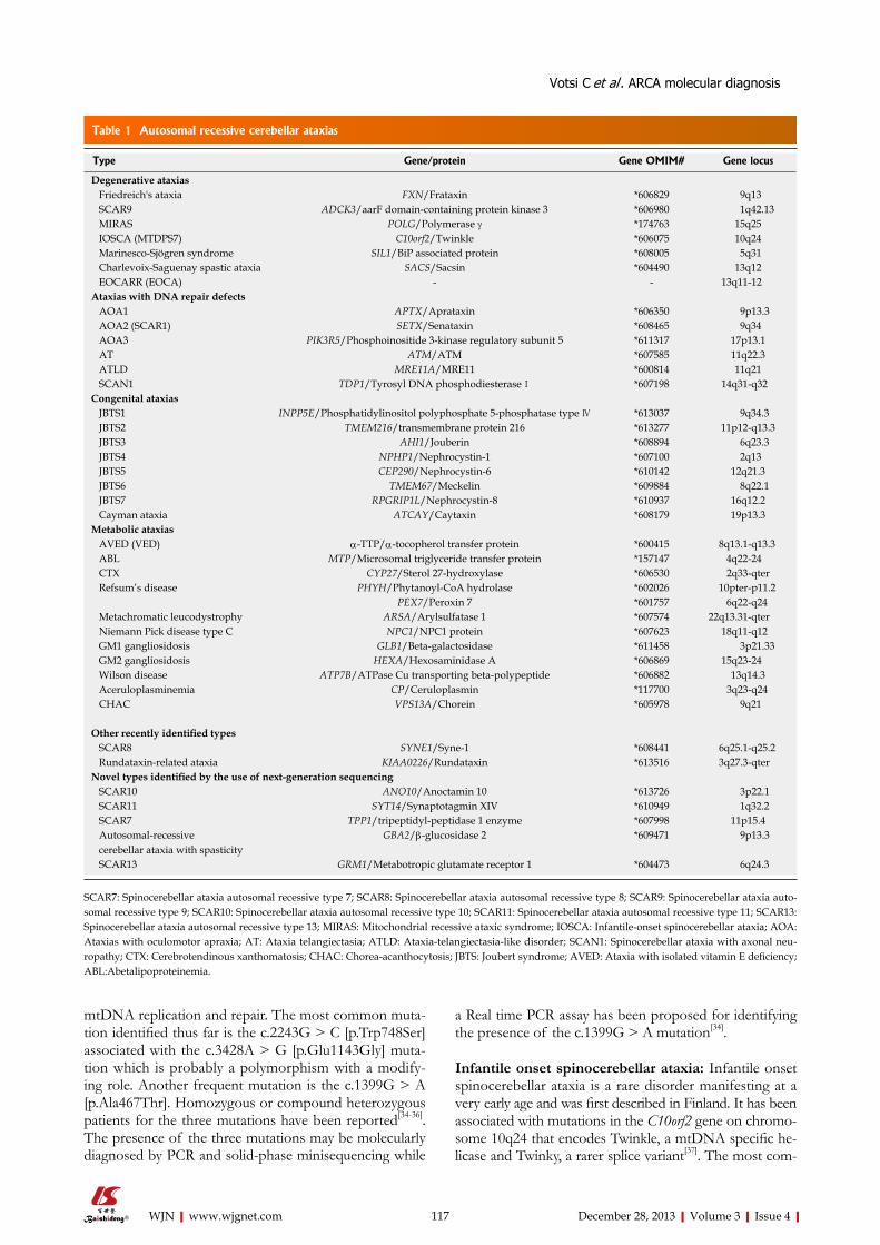
mtDNA replication and repair. The most common muta-tion identified thus far is the c.2243G > C [p.Trp748Ser] associated with the c.3428A > G [p.Glu1143Gly] muta-tion which is probably a polymorphism with a modify-ing role. Another frequent mutation is the c.1399G > A [p.Ala467Thr]. Homozygous or compound heterozygous patients for the three mutations have been reported[34-36]. The presence of the three mutations may be molecularly diagnosed by PCR and solid-phase minisequencing while
a Real time PCR assay has been proposed for identifying the presence of the c.1399G > A mutation[34].
Infantile onset spinocerebellar ataxia: Infantile onset spinocerebellar ataxia is a rare disorder manifesting at a very early age and was first described in Finland. It has been associated with mutations in the C10orf2 gene on chromo-some 10q24 that encodes Twinkle, a mtDNA specific he-licase and Twinky, a rarer splice variant[37]. The most com-
117 December 28, 2013|Volume 3|Issue 4|WJN|www.wjgnet.com
Type Gene/protein Gene OMIM# Gene locus
Degenerative ataxias Friedreich's ataxia FXN/Frataxin *606829 9q13 SCAR9 ADCK3/aarF domain-containing protein kinase 3 *606980 1q42.13 MIRAS POLG/Polymerase γ *174763 15q25 IOSCA (MTDPS7) C10orf2/Twinkle *606075 10q24 Marinesco-Sjögren syndrome SIL1/BiP associated protein *608005 5q31 Charlevoix-Saguenay spastic ataxia SACS/Sacsin *604490 13q12 ΕΟCARR (EOCA) - - 13q11-12 Ataxias with DNA repair defects ΑΟΑ1 APTX/Aprataxin *606350 9p13.3 ΑΟΑ2 (SCAR1) SETX/Senataxin *608465 9q34 AOA3 PIK3R5/Phosphoinositide 3-kinase regulatory subunit 5 *611317 17p13.1 AT ATM/ATM *607585 11q22.3 ATLD MRE11A/MRE11 *600814 11q21 SCAN1 TDP1/Tyrosyl DNA phosphodiesterase Ⅰ *607198 14q31-q32 Congenital ataxias JBTS1 INPP5E/Phosphatidylinositol polyphosphate 5-phosphatase type Ⅳ *613037 9q34.3 JBTS2 TMEM216/transmembrane protein 216 *613277 11p12-q13.3 JBTS3 ΑΗΙ1/Jouberin *608894 6q23.3 JBTS4 NPHP1/Nephrocystin-1 *607100 2q13 JBTS5 CEP290/Nephrocystin-6 *610142 12q21.3 JBTS6 TMEM67/Meckelin *609884 8q22.1 JBTS7 RPGRIP1L/Nephrocystin-8 *610937 16q12.2 Cayman ataxia ATCAY/Caytaxin *608179 19p13.3 Metabolic ataxias AVED (VED) α-TTP/α-tocopherol transfer protein *600415 8q13.1-q13.3 ABL MTP/Microsomal triglyceride transfer protein *157147 4q22-24 CTX CYP27/Sterol 27-hydroxylase *606530 2q33-qter Refsum’s disease PHYH/Phytanoyl-CoA hydrolase *602026 10pter-p11.2
PEX7/Peroxin 7 *601757 6q22-q24 Metachromatic leucodystrophy ARSA/Arylsulfatase 1 *607574 22q13.31-qter Niemann Pick disease type C NPC1/NPC1 protein *607623 18q11-q12 GM1 gangliosidosis GLB1/Beta-galactosidase *611458 3p21.33 GM2 gangliosidosis HEXA/Hexosaminidase A *606869 15q23-24 Wilson disease ATP7B/ΑΤPase Cu transporting beta-polypeptide *606882 13q14.3 Aceruloplasminemia CP/Ceruloplasmin *117700 3q23-q24 CHAC VPS13A/Chorein *605978 9q21
Other recently identified types SCAR8 SYNE1/Syne-1 *608441 6q25.1-q25.2 Rundataxin-related ataxia KIAA0226/Rundataxin *613516 3q27.3-qter Novel types identified by the use of next-generation sequencing SCAR10 ANO10/Anoctamin 10 *613726 3p22.1 SCAR11 SYT14/Synaptotagmin XIV *610949 1q32.2 SCAR7 TPP1/tripeptidyl-peptidase 1 enzyme *607998 11p15.4 Autosomal-recessive cerebellar ataxia with spasticity
GBA2/β-glucosidase 2 *609471 9p13.3
SCAR13 GRM1/Metabotropic glutamate receptor 1 *604473 6q24.3
Table 1 Autosomal recessive cerebellar ataxias
SCAR7: Spinocerebellar ataxia autosomal recessive type 7; SCAR8: Spinocerebellar ataxia autosomal recessive type 8; SCAR9: Spinocerebellar ataxia auto-somal recessive type 9; SCAR10: Spinocerebellar ataxia autosomal recessive type 10; SCAR11: Spinocerebellar ataxia autosomal recessive type 11; SCAR13: Spinocerebellar ataxia autosomal recessive type 13; MIRAS: Mitochondrial recessive ataxic syndrome; IOSCA: Infantile-onset spinocerebellar ataxia; AOA: Ataxias with oculomotor apraxia; AT: Ataxia telangiectasia; ATLD: Ataxia-telangiectasia-like disorder; SCAN1: Spinocerebellar ataxia with axonal neu-ropathy; CTX: Cerebrotendinous xanthomatosis; CHAC: Chorea-acanthocytosis; JBTS: Joubert syndrome; AVED: Ataxia with isolated vitamin E deficiency; ABL:Abetalipoproteinemia.
Votsi C et al . ARCA molecular diagnosis
tor apraxia (AOA), are characterized by cerebellar ataxia accompanied by ophthalmological and neurological symptoms[52,53]. Five distinct genetic forms have thus far been identified: ataxia-telangiectasia (A-T), ataxia-telan-giectasia-like disorder (ATLD), ataxia with oculomotor apraxia type 1 (AOA1), ataxia with oculomotor apraxia type 2 (AOA2) and ataxia with oculomotor apraxia type 3 (AOA3)[54].
(1) Ataxia telangiectasia: Ataxia telangiectasia is the sec-ond most common ARCA after Friedreich ataxia. It pres-ents in early childhood with progressive cerebellar ataxia, while later ataxia may be accompanied by a multitude of symptoms such as choreathetosis, dystonia, telangiectasia that is the guiding diagnostic feature, immunodeficiency, cancer predisposition, sinopulmonary infection, immuno-sensitivity and radiosensitivity[1,14]. The disease gene ATM is located on chromosome 11q23[55,56]. It is one of the largest genes, comprising 66 exons and encodes a mem-ber of the phosphoinositol-3 kinase family of proteins that are involved in cell cycle checkpoint control and the DNA repair through phosphorylation of substrates[1]. More than 500 mutations have been identified, that are positioned throughout all the coding exons of the gene[14]. Due to the large size of the gene, molecular di-agnosis with the traditional methods has been difficult. A multiplex scanning method, detection of virtually all mu-tations-SSCP has been proposed however with reduced sensitivity[57]. The newly developed NGS technologies are promising for the diagnosis of diseases such as A-T with associated mutations in large genes.
(2) Ataxia-telangiectasia-like disorder: Ataxia-telangiec-tasia-like disorder (ATLD) is a very rare disorder that resembles A-T in its clinical phenotype[58] with absence of telangiectases and immune deficiency. It has been mapped to chromosome 11q21 and a small number of causative mutations have been identified in the MRE11A gene[59-61]. The encoded protein interacts with RAD50 and NBS1 thus forming the core of the MRN (MRE11-RAD50-NBS1) complex which is involved in DNA repair pathways, in DNA recombination and in multiple cell-cycle checkpoints control[60].
(3) Ataxia with oculomotor apraxia type 1: Ataxia with oculomotor apraxia type 1 (AOA1) was first described in Portuguese and Japanese families. The disease gene has been mapped to chromosome 9p13.3[62] and mutations in the APTX gene have been identified[63]. The encoded protein aprataxin, localizes to the nucleus[64] and the mi-tochondria[65] and it is involved in the nuclear and the mtDNA repair machinery. AOA1 is the most frequent type of ARCA in Japan and the second most frequent in Portugal after Friedreich ataxia. The insertion c.689insT and the missense c.617C > T [p.Pro206Leu] mutations, are more common in the Japanese and the nonsense c.837G > A [p.W279X] mutation is more common in Portuguese patients. More than 20 mutations including missense/nonsense or splice site mutations, small dele-
mon mutation identified is c.1523A > G [p.Tyr508Cys]. Mutations in this gene may be detected either by direct sequencing or by solid-phase minisequencing.
Autosomal recessive spastic ataxia of Charlevoix-Sa-guenay: Autosomal recessive spastic ataxia of Charlevoix-Saguenay (ARSACS) is an early childhood onset disease characterized by spastic gait ataxia. It was first described in families from the Charlevoix-Saguenay region of northeastern Quebec and for many years it was thought to be restricted there. It has been mapped to chromo-some 13q11 and causative mutations have been identified in the SACS gene[38] that encodes sacsin, a chaperone protein. Almost 96% of the molecularly diagnosed indi-viduals from this region are homozygote or compound heterozygote for the two founder mutations c.8844delT [p.Pro2948fs] and c.7504C > T [p.Arg2502*] (origi-nally reported as 6594delT and 5254C > T) identified by sequence-based analyses[39,40]. More than 100 other pathogenic mutations in individuals outside Quebec were identified since mutation analysis became available, dem-onstrating that mutations in SACS are a frequent cause of the disease worldwide[41-43]. Molecular genetic testing is usually performed by targeted mutation analysis, by direct sequencing of all coding exons and their flanking intronic sequences and by deletion/duplication analysis using the multiplex ligation-dependent probe amplifica-tion method[44].
Marinesco-Sjögren syndrome: Marinesco-Sjögren syn-drome is a rare disorder with onset in infancy or child-hood. Homozygosity mapping in two large consanguine-ous families of Turkish and Norwegian origin mapped the disease gene on chromosome 5q31[45]. Causative mu-tations have been identified in patients of different ethnic origin, in the SIL1 gene encoding a protein that acts as a nucleotide exchange factor for the heat-shock protein 70 chaperone the 78-kDa glucose-regulated protein, mainly located at the endoplastic reticulum[46-48]. Molecular genet-ic testing is performed by direct sequencing of all coding exons and their flanking intronic sequences.
Spinocerebellar ataxia autosomal recessive type 9: Spinocerebellar ataxia autosomal recessive type 9 (SCAR9), also known as autosomal recessive cerebellar ataxia type 2 (ARCA2) refers to ARCA associated with muscle coenzyme Q10 deficiency[49]. Homozygosity mapping (GeneChip Human Mapping 10K 2.0 Xba Array, Af-fymetrix) in a large consanguineous Algerian family mapped the disease gene to chromosome 1q41-q42. ADCK3 gene mutations have been identified by direct sequencing in this family, in additional families and also in sporadic patients[49-51]. The ADCK3 protein is a mito-chondrial ancestral kinase which is involved in CoQ10 synthesis[49].
Ataxias with DNA repair defectsARCA with oculomotor apraxia: ARCA with oculo-motor apraxia (OMA), known as ataxias with oculomo-
118 December 28, 2013|Volume 3|Issue 4|WJN|www.wjgnet.com
Votsi C et al . ARCA molecular diagnosis
tions/insertions and gross deletions have been reported thus far in patients of different ethnicities, thus expand-ing the presence of AOA1 in other countries outside Japan and Portugal[3,66-70].
(4) Ataxia with oculomotor apraxia type 2: Ataxia with oculomotor apraxia type 2 (AOA2) is also referred to as autosomal recessive spinocerebellar ataxia 1 (SCAR1) since oculomotor apraxia is an occasional finding, not present in all the patients. It is the second most frequent recessive ataxia in Europe[4] and is usually (in 96% of cases) accompanied by a characteristic laboratory finding, the elevation of serum α-fetoprotein. AOA2 usually pres-ents within the second decade of life and has a higher mean age of onset compared to AOA1 and A-T[52]. The disease gene has been mapped to chromosome 9q34 and later on mutations in the SETX gene were identified[71]. SETX is a large gene of 26 exons and encodes sena-taxin, a nuclear protein, which contains at its C terminus a classic seven-motif domain found in the superfamily 1 of helicases. The protein is involved in the response to oxidative stress[53]. Acting as an RNA/DNA helicase, senataxin might have a significant role in the DNA repair pathway, in the splicing machinery[71] and also in the pro-cessing of noncoding RNAs[52]. Even though the SETX gene cannot be easily analysed by routine sequencing due to its size, several patients have been studied with this method and more than 75 different mutations have been identified thus far worldwide[52,72,73] including missense, nonsense and splice site mutations, as well as small/gross deletions or insertions[74].
(5) Ataxia with oculomotor apraxia type 3: Ataxia with oc-ulomotor apraxia type 3 has more recently been described in a consanguineous Saudi Arabian family with four af-fected individuals that present with a similar phenotype to AOA2. Elevated serum α-fetoprotein levels were identified in all the patients and 2 siblings had OMA. The homozygous missense c.1885C > T [p.Pro629Ser] mutation in exon 12 of the PIK3R5 gene has been associ-ated with the disease in this family[54]. PIK3R5 is located on chromosome 17p12-p13. The encoded protein is a p101 regulatory subunit that interacts with class 1B PI3K which is involved in cell survival and growth as well as in metabolism, immune and cardiac functions. Even though it is registered as rs61761068 in db-single-nucleotide poly-morphism (dbSNP), frequencies of the mutant allele in other populations indicate that it has only been detected in a heterozygous state, most likely representing non-affected carriers of the mutation[54].
Spinocerebellar ataxia with axonal neuropathy: Spi-nocerebellar ataxia with axonal neuropathy is a related syndrome to the AOA group. It is characterized by the co-occurrence of cerebellar ataxia and axonal neuropathy. A single locus on chromosome 14q31-q32 has been asso-ciated with the disease and a missense mutation (c.1478A > G [p.His493Arg]) in the TDP1 gene has been described in a large Saudi Arabian family[75]. The encoded protein
tyrosyl-DNA phosphodiesterase 1 is a member of the DNA single-strand break repair complex and plays a sig-nificant role in the DNA repair pathway[75,76].
Congenital and metabolic ataxiasOther ARCA are classified under the subgroups of con-genital and metabolic ataxias (Table 1). Associated genes and mutations have been identified and molecular genetic testing by targeted mutation analysis or direct sequenc-ing of the appropriate gene may be performed in ARCA patients based on the clinical picture. Moreover, in some cases of metabolic ataxias, biochemical analysis on spe-cific available markers could precede and orientate the genetic testing.
Congenital ataxias: Congenital ataxias that have been associated with specific chromosomal loci and genes include the Joubert syndrome and the Cayman ataxia. Joubert syndrome has thus far been associated with seven loci (JBTS1, JBTS2, JBTS3, JBTS4, JBTS5, JBTS6, and JBTS7) and mutations have been identified in the seven INPP5E[77], TMEM216[78], ΑΗΙ1[79], NPHP1[80], CEP290[81], TMEM67[82] and RPGRIP1L[83] genes respec-tively (Table 1). The Cayman ataxia has been described in an isolated population of the Cayman Island. A single locus on chromosome 19p13.3 has been associated with the disease and causative mutations in the ATCAY gene have thus far been identified[84].
Metabolic ataxias: The subgroup of metabolic ataxias includes disorders characterized by progressive ataxia, recurrent ataxia and ataxia as a minor feature. Progressive ataxia includes ataxia with isolated vitamin E deficiency, abetalipoproteinemia, cerebrotendinous xanthomatosis and Refsum disease[1]. Intermittent or minor ataxia in-cludes: metachromatic leukodystrophy (ARSA gene)[85,86], Niemann-Pick type C 1 (NPC1 gene)[87], GM1 (GLB1 gene)[88,89] and GM2 (HEXA gene)[90,91] gangliosidosis, Wilson’s disease (ATP7B gene)[92-94], aceruloplasminae-mia (CP gene)[95] and Chorea-acanthocytosis (VPS13A gene)[96].
(1) Ataxia with isolated vitamin E deficiency: Ataxia with isolated vitamin E deficiency has a similar clinical picture with Friedreich ataxia. It is characterized by a biochemical abnormality, the very low plasma level of vitamin E and hence the low level of the α-tocopherol in the serum. The disease is caused by mutations in the α-TTP gene, encoding the α-tocopherol transfer protein which is responsible for the transfer of vitamin E to the nascent very low-density lipoproteins[97]. Diagnosis may include the finding of low serum vitamin E values in the absence of malabsorption[1] and further by performing genetic testing.
(2) Abetalipoproteinemia: Abetalipoproteinemia is char-acterized by deficiency of the liposoluble vitamin E, the low levels of cholesterol and the absence of the low-density lipoproteins. The clinical features comprise the
119 December 28, 2013|Volume 3|Issue 4|WJN|www.wjgnet.com
Votsi C et al . ARCA molecular diagnosis
malabsorption syndrome, the pigmentary degeneration of the retina and progressive ataxic neuropathy. More-over, patients show a characteristic deformation of their red cells called acanthocytosis[98]. The disease is caused by mutations in the MTP gene which encodes the micro-somal triglyceride transfer protein catalyzing the trans-port of triglyceride, cholesteryl ester and phospholipid between phospholipid surfaces[99].
(3) Cerebrotendinous xanthomatosis: Cerebrotendinous xanthomatosis is a sterol storage disorder, characterized by abnor mal accumula t ion of choles tero l and cholestanol in neural and other tissues, thus causing tendon xanthomas, premature atherosclerosis, cataracts and neurological dysfunction which is manifested as cerebellar ataxia, dementia and spinal cord paresis[100]. The disease gene has been mapped to chromosome 2q33-qter and is caused by mutations in the CYP27 gene have been identified. CYP27 encodes sterol 27-hydroxylase, a mitochondrial cytochrome P450 enzyme that has an important role in cholesterol and bile acid metabolism[100,101].
(4) Refsum’s disease: Refsum’s disease is caused by defec-tive peroxisomal alpha-oxidation of phytanic acid, thus leading to its accumulation in blood and other tissues in-cluding neurons. It is characterized mainly by progressive retinitis pigmentosa, peripheral neuropathy and cerebellar ataxia. Other additional features include anosmia, deaf-ness, cardiomyopathy, ichthyosis and skeletal abnormali-ties. The disorder is genetically heterogeneous since it has been mapped to two different loci. In most families, link-age was established to the first identified locus on chro-mosome 10pter-p11.2. Causative mutations have been found in the PHYH (or PAHX) gene[102,103] encoding the phytanoyl-CoA hydroxylase enzyme that is involved in the catabolic pathway of phytanic acid. In a subset of pa-tients mutations in the PAHX gene have been excluded. Further linkage analysis of these families led to mapping of a second locus on chromosome 6q22-24. In this sub-group of patients causative mutations have been identi-fied in the PEX7 gene encoding the peroxin 7 protein which is implicated in the import of matrix proteins into the peroxisome[104].
Other known ARCA typesThe following ARCA forms have been reported more recently that have not been included in the classification scheme described by Palau and Espinos (2006).
Spinocerebellar ataxia autosomal recessive type 8: Spinocerebellar ataxia autosomal recessive type 8 (SCAR8), also known as autosomal recessive cerebellar ataxia type 1 (ARCA1) is an adult-onset slowly progressive cerebel-lar ataxia accompanied by dysarthria and other associated features[105]. It was first described and so far only reported in French-Canadian families most of them originating from defined regions of Quebec. A single locus on chro-mosome 6q25.1-q25.2 has been mapped and mutations
in the SYNE1 gene have been associated with the dis-ease. Syne-1 is involved in anchoring specialized myonu-clei underneath the neuromuscular junction[105] and plays a significant role in the development and maintenance of cerebellar functions. SYNE1 is a large gene of 147 exons and this makes mutation screening by traditional methods expensive and complicated. Thus far direct sequencing of all the exons and flanking intronic sequences revealed a small number of missense and splice site mutations as well as a small scale deletion[105,106].
Rundataxin-related ataxia: A new form of pure reces-sive cerebellar ataxia accompanied by epilepsy and mental retardation named ‘Salih ataxia’ has been described in a large consanguineous Saudi Arabian family with three af-fected individuals[107]. Homozygosity mapping with SNP array (Gene Chip Human Mapping 10K 2.0 Xba Array, Affymetrix) and further linkage analysis with microsatel-lite markers, mapped the disease gene on chromosome 3q27.3-qter. Direct sequencing of the candidate genes enabled identification of the causative mutation in the KIAA0226 gene. A single nucleotide deletion 2927delC [p.Ala943ValfsX146] frameshift mutation in this gene has thus far been identified. The encoded protein was named rundataxin based on its two conserved domains[107] and it is associated with vesicular trafficking and signaling path-ways.
NEXT-GENERATION SEQUENCING AND MOLECULAR DIAGNOSIS OF AUTOSOMAL RECESSIVE CEREBELLAR ATAXIASNGS technologies provide powerful tools for the iden-tification of new genes. During the last 3 years, use of homozygosity mapping in combination with WES or tar-geted sequencing using the NGS platforms, enabled the identification of 5 new genes associated with rare types of ARCA in small families. WES has also been used as a screening tool thus enabling the rapid identification of mutations in known genes and demonstrating that it can be an efficient diagnostic tool.
Identification of new genesSpinocerebellar ataxia autosomal recessive type 10: A new form of pure recessive ataxia accompanied by downbeat nystagmus and lower motor neuropathy, first described in a Dutch consanguineous family and named spinocerebellar ataxia autosomal recessive type 10 (SCAR10), was the first recessive ataxia molecularly diagnosed with homozygosity mapping and NGS[108]. It is also known as ARCA3[2]. Homozygosity mapping with a 10K SNP array (Affymetrix) combined with linkage stud-ies with short tandem repeat markers led to fine mapping of the disease gene to chromosome 3p21.32-p22.3. A large number of genes were included in the mapped re-gion thus making identification of the causative mutation
120 December 28, 2013|Volume 3|Issue 4|WJN|www.wjgnet.com
Votsi C et al . ARCA molecular diagnosis
with the traditional Sanger sequencing difficult. Use of Sanger sequencing to analyze 15 candidate genes failed to identify any mutation. Targeted NGS was subsequently performed that enabled identification of the causative mutation in the ANO10 gene, thus demonstrating the power of this method towards the identification of new genes. A homozygous c.1529T > G [p.Leu510Arg] mutation was identified in the Dutch family. Additional mutations have been identified in patients originat-ing from Serbia (a homozygous frameshift mutation c.1150_1151del [p.Leu384fs]) and France (compound heterozygous mutations c.1476 + 1G > T and c.1604del [p.Leu535X]) through direct analysis of the new gene by conventional Sanger sequencing[108]. More recently, a Japanese patient has been reported with a homozygous nonsense mutation (c.609C > G [p.Y203X]) identified by exome sequencing[19] indicating that this new method can be applied as a screening tool allowing the fast identifica-tion of mutations in known genes. The encoded protein ANO10 is a member of the human anoctamin family and similar to the other proteins of this family it is a putative calcium-dependent chloride channel, thus representing a new pathway implicated in cerebellar ataxias. However, this function remains to be confirmed[108].
Spinocerebellar ataxia autosomal recessive type 11: A slowly progressive new type of ARCA accompanied by psychomotor retardation has been recently described in a small consanguineous Japanese family with two affected individuals and is known as spinocerebellar ataxia autoso-mal recessive type 11 (SCAR11). Homozygosity mapping using the Human SNP Array 6.0 (Affymetrix) combined with multipoint linkage analysis using extracted SNP ar-ray data revealed three candidate regions of homozygos-ity. WES was subsequently employed and data analysis focused on the regions of homozygosity. Sanger sequenc-ing was used to further investigate filtered variants and the synaptotagmins 14 (SYT14) homozygous c.1451G > A [p.Gly484Asp] missense mutation has been associ-ated with the disease. Further screening of all the coding regions of the entire gene in sporadic and familial ARCA cases failed to identify additional mutations. The encoded protein is Synaptotagmin XIV, a member of SYTs which are transmembrane proteins associated with exocytosis of secretory vesicles. Expression studies on SYT14 show that this protein plays a significant role in the cerebellum and support previous suggestions about the possible in-volvement of disrupted SYTs in neurodegeneration[12].
Spinocerebellar ataxia autosomal recessive type 7: Spinocerebellar ataxia autosomal recessive type 7 (SCAR7) was first described in a Dutch family in 2004 as a dis-tinct ARCA type, based on its genetic locus, the onset of the disease and/or other clinical findings[109]. The dis-ease gene has been mapped on chromosome 11p15 by genome-wide linkage analysis with microsatellite markers from the Marshfield genetic map. Identification of the causative mutation/gene was impossible for many years due to the large number (> 200) of genes within the
candidate region. Recently, the use of WES enabled the association of the disease in the family with two TPP1 compound heterozygous mutations (the splice site c.509-1G > C and the missense c.1397T > G [p.Val466Gly])[110]. These mutations were also detected in a sporadic SCAR7 patient. Mutations in the TPP1 gene had already been as-sociated with another disorder, the late infantile lipofusci-nosis disease 2 (CLN2). Association of mutations in this gene with SCAR7 expands the phenotypes related to vari-ants of the TPP1 gene. Phenotype-genotype correlations proposed the hypothesis that CLN2 disease is due to variants causing the loss of enzyme activity, while SCAR7 is due to variants decreasing the enzyme activity. TPP1 encodes the tripeptidyl-peptidase 1 enzyme, a member of the sedolisin family of serine proteases. This enzyme cleaves the N-terminal tripeptides from substrates and has a weak endopeptidase activity[110].
Autosomal-recessive cerebellar ataxia with spastici-ty: A distinct form of ataxia previously described in three consanguineous Tunisian families with 7 affected individ-uals has been recently molecularly characterized with the combined strategy of homozygosity mapping and WES thus providing further evidence about the potential of these methods[111]. Childhood or juvenile onset cerebellar ataxia was the main feature in all affected individuals of the families but later they developed pronounced limb spasticity. High density SNP genotyping was performed with the OmniExp-12, v1.0 DNA Analysis BeadChip (Illumina) and one large region of homozygosity on chromosome 9 was identified. The APTX gene mutated in AOA1 was included in the mapped region, so muta-tions in APTX were first excluded by Sanger sequencing. WES was then performed that revealed 2 causative muta-tions in the GBA2 gene (the homozygous c.1018C > T [p.Arg340*] shared by two families and the homozygous c.2618G > A [p.Arg873His])[111]. Sanger sequencing con-firmed segregation of the identified mutations with the disease in the families. Furthermore, WES was used to screen 21 molecularly undiagnosed Tunisian individuals with ataxia. Through this procedure an additional GBA2 mutation (c.363C > A [p.Tyr121*]) was identified[111]. A second group associated 4 GBA2 mutations with the spastic paraplegia type 46 (SPG46) phenotype[112].
GBA2 encodes the non-lysosomal β-glucosidase 2 protein that was first recognized as a microsomal bile-acid β-glucosidase, while more recently it was reassigned to sphingolipid metabolism[113], a critical process for the nervous system[114]. The role of the enzyme in the central nervous system development was confirmed through the validation of the functional phenotype of some of the identified mutations causing HSP in vivo thus demon-strating the connection of sphingolipid metabolism and neurodegeneration[112]. HSPs are another heterogeneous group of neurodegenerative disorders having common features with ataxias and as a consequence, many times it is difficult for the clinician to decide whether the patient has hereditary ataxia with spasticity or HSP with cerebel-lar features. Identification of mutations in the same gene
121 December 28, 2013|Volume 3|Issue 4|WJN|www.wjgnet.com
Votsi C et al . ARCA molecular diagnosis
causing these two diseases demonstrates that a common pathway may be involved in neurodegeneration.
Spinocerebellar ataxia autosomal recessive type 13: A new form of congenital cerebellar ataxia identi-fied in Roma patients has recently been described and molecularly characterized[115]. This new form is known as spinocerebellar ataxia autosomal recessive type 13 (SCAR13). WES was used both for linkage analysis and mutation detection. A subgroup of SNP genotypes was first selected from the exome sequence data and used to perform genome wide linkage analysis, thus emphasizing an additional application of this method. Linkage analysis mapped the disease gene to a single locus on chromo-some 6q24. Data analysis was focused on the linked in-terval and finally resulted to the identification of two ho-mozygous mutations in the GRM1 gene (c.2652_2654del [p.Asn885del] and c.2660+2T > G). Sanger sequencing was used to verify the presence of mutations and their co-segregation with the disease within the families while PCR-based restriction-fragment-length-polymorphism assays and pyrosequencing were used to evaluate the mutation frequencies among control individuals. Both mutations interfere with a critical alternative splicing gene region. GRM1 encodes the metabotropic glutamate receptor mGluR1 which is implicated in the modulation of intracellular Ca2+ levels and neuronal excitability. It is important for the development of the cerebellar cortex and the cerebellar and hippocampal synaptic plasticity, memory and learning[115].
Identification of mutations in known genesThe combined strategy of homozygosity mapping and WES has been recently applied for the investiga-tion of a consanguineous Turkish family and revealed a novel homozygous missense mutation (c.1366C > G [p.Leu456Val]) in the C10orf2 gene, mutations in which have already been associated with infantile-onset spino-cerebellar ataxia (IOSCA)[16]. This rare type of ataxia was previously reported only in Finland. The recent identifi-cation of this novel mutation shows that IOSCA is not restricted to Finland.
WES was applied in another recent study to inves-tigate two affected non-consanguineous siblings with a multisystem neurological disorder of unknown genetic origin and revealed two novel heterozygous mutations in the known SACS gene. These patients had been pre-viously investigated with conventional methods which failed to identify the associated mutations in contrast to this novel approach which rapidly led to molecular diagnosis. Both patients shared the compound hetero-zygous mutations c.2076delG [p.Thr692Thrfs*713] and c.3965_3966delAC [pGly1322Valfs*1343] in the SACS gene that have been associated with ARSACS. Another novel homozygous mutation in SACS (c.2439-2440delAT [p.Val815Glyfs*4]) has been identified in a Tunisian fam-ily with two affected individuals, by homozygosity map-ping and WES[13]. A more recent study on a 4-year-old girl revealed an additional novel frameshift homozygous
mutation in the same gene by WES[18].WES also enabled the molecular diagnosis of the
disease in an early onset spastic ataxia-neuropathy syn-drome family. A homozygous missense mutation in the AFG3L2 gene (c.1847G > A [p.Y616C]) has been identified[116]. Heterozygous mutations in the AFG3L2 gene have previously been identified in autosomal domi-nant spinocerebellar ataxia type 28 patients[117,118]. The AFG3L2 gene encodes a subunit of the m-AAA class of mitochondrial proteases which forms either a homo-oligomeric isoenzyme or a hetero-oligomeric complex with paraplegin, encoded by the SPG7 gene[116]. Mutations in the SPG7 gene have been associated with hereditary SPG7, characterized by adult-onset spasticity and weak-ness of the lower extremities[119]. In contrast to SCA28, this new syndrome associated with a recessive pattern of inheritance, is characterized by a much earlier onset and combines phenotypic features of SCA28 and SPG7[116].
SYNOPSISThe diagnosis of ARCA is becoming progressively more complex and challenging since clinical and genetic heterogeneity is expanding. Mutations in more than 20 genes have been associated with ARCA through the last decade, some of them identified in single families. There exists a significant number of patients remaining without a molecular diagnosis thus leading to the hypotheses that many other novel ARCA genes remain to be discovered in the future and that a different gene might be associated with the disease in each family mainly in populations with frequent inbreeding. The development of NGS tech-nologies has already contributed significantly towards the molecular diagnosis of these rare diseases and appears very promising towards the diagnosis of additional cases in the future.
Sequencing of known candidate genes with conven-tional methods based on clinical findings has proved to be time-consuming and costly, except in the case of metabolic ataxias for which biochemical test results can precisely guide the molecular diagnosis. Routine inves-tigation of some already known genes such as SYNE1, ATM, SETX, POLG and SACS is prohibitive, due to their significantly large coding regions.
Homozygosity mapping in consanguineous families has proved to be a powerful tool and may precede se-quencing thus contributing to a more targeted analysis and facilitating the identification of novel ARCA genes. High density genome wide SNP genotyping with SNP ar-rays is the most rapid and effective method applied in the last years for homozygosity mapping. However, further analysis with more informative markers is often required in order to confirm or exclude some of the identified regions. Moreover, instead of using a SNP array, ex-tracted SNP genotypes from the WES data may be used for genome wide linkage analysis. If a small number of genes exists within a mapped interval, then investigation by conventional sequencing may be efficient. Otherwise, if a large number of candidate genes exists, conventional
122 December 28, 2013|Volume 3|Issue 4|WJN|www.wjgnet.com
Votsi C et al . ARCA molecular diagnosis
sequencing proves to be difficult, time consuming and expensive compared to the new methodologies.
WES enables the rapid sequencing of all coding re-gions of the genome and its usefulness has already been demonstrated in research and diagnostic applications. Conventional sequencing remains useful as a compli-mentary method for the investigation of the segregation pattern of candidate variants previously revealed by WES and verification of the causative mutations. Moreover, investigation of families for mutations in a known gene within a mapped region could be performed either by conventional sequencing or by WES depending on the size of the coding regions that need to be examined. For small coding regions, the first method may be more cost effective while for large genes, WES is less expen-sive since costs have significantly decreased over the last year[14].
Although the application of NGS technologies en-ables the identification of novel ARCA genes and the characterisation of novel mutations in known genes thus providing molecular diagnosis in many cases, there are some limitations due to their current reduced ability to sequence repetitive DNA expansions[14]. The most com-mon form of ARCA is caused by the GAA repeat ex-pansion in intron 1 of the FXN gene. Additional forms of ARCA with unknown genetic etiology may also be associated with a similar type of mutation and will not be identified with the NGS approach.
CONCLUSIONNovel high-throughput sequencing technologies such as WES/WGS are ideal for simultaneously sequencing groups of candidate genes, whole-genome coding regions or the whole-genome and have brought a revolution in the molecular diagnosis of human genetic diseases. These technologies are capable of producing large amounts of data in one experiment and have been successfully used for the diagnosis of many rare and complex diseases in-cluding rare neurodegenerative diseases such as the atax-ias. Their use for both research and diagnostic purposes has already contributed to the progress observed during the last years in the field of ataxias, including both the recessive and dominant forms. They are also very prom-ising for the identification of many other new forms of ataxia in the near future since this group of disorders is very heterogeneous and a significant number of patients have already been excluded from many of the known genes. Determination of the genetic cause of each form is essential in order to initiate further investigation re-garding the pathogenetic mechanisms and the potential therapies for these diseases.
REFERENCES1 Palau F, Espinós C. Autosomal recessive cerebellar atax-
ias. Orphanet J Rare Dis 2006; 1: 47 [PMID: 17112370 DOI: 10.1186/1750-1172-1-47]
2 Anheim M, Tranchant C, Koenig M. The autosomal reces-sive cerebellar ataxias. N Engl J Med 2012; 366: 636-646 [PMID:
22335741 DOI: 10.1056/NEJMra1006610]3 Anheim M, Fleury M, Monga B, Laugel V, Chaigne D, Ro-
dier G, Ginglinger E, Boulay C, Courtois S, Drouot N, Fritsch M, Delaunoy JP, Stoppa-Lyonnet D, Tranchant C, Koenig M. Epidemiological, clinical, paraclinical and molecular study of a cohort of 102 patients affected with autosomal recessive progressive cerebellar ataxia from Alsace, Eastern France: implications for clinical management. Neurogenetics 2010; 11: 1-12 [PMID: 19440741 DOI: 10.1007/s10048-009-0196-y]
4 Klockgether T. Update on degenerative ataxias. Curr Opin Neurol 2011; 24: 339-345 [PMID: 21734495 DOI: 10.1097/WCO.0b013e32834875ba]
5 Espinós-Armero C, González-Cabo P, Palau-Martínez F. [Autosomal recessive cerebellar ataxias. Their classification, genetic features and pathophysiology]. Rev Neurol 2005; 41: 409-422 [PMID: 16193447]
6 Dean G, Chamberlain S, Middleton L. Friedreich’s ataxia in Kathikas-Arodhes, Cyprus. Lancet 1988; 1: 587 [PMID: 2894517]
7 Zamba-Papanicolaou E, Koutsou P, Daiou C, Gaglia E, Georghiou A, Christodoulou K. High frequency of Fried-reich’s ataxia carriers in the Paphos district of Cyprus. Acta Myol 2009; 28: 24-26 [PMID: 19772192]
8 Harding AE. Classification of the hereditary ataxias and paraplegias. Lancet 1983; 1: 1151-1155 [PMID: 6133167]
9 Harding AE. Clinical features and classification of inherited ataxias. Adv Neurol 1993; 61: 1-14 [PMID: 8421960]
10 Koenig M. Rare forms of autosomal recessive neurodegen-erative ataxia. Semin Pediatr Neurol 2003; 10: 183-192 [PMID: 14653406 DOI: 10.1016/S1071-9091(03)00027-5]
11 De Michele G, Coppola G, Cocozza S, Filla A. A pathoge-netic classification of hereditary ataxias: is the time ripe? J Neurol 2004; 251: 913-922 [PMID: 15316795 DOI: 10.1007/s00415-004-0484-2]
12 Doi H, Yoshida K, Yasuda T, Fukuda M, Fukuda Y, Morita H, Ikeda S, Kato R, Tsurusaki Y, Miyake N, Saitsu H, Sakai H, Miyatake S, Shiina M, Nukina N, Koyano S, Tsuji S, Ku-roiwa Y, Matsumoto N. Exome sequencing reveals a homo-zygous SYT14 mutation in adult-onset, autosomal-recessive spinocerebellar ataxia with psychomotor retardation. Am J Hum Genet 2011; 89: 320-327 [PMID: 21835308 DOI: 10.1016/j.ajhg.2011.07.012]
13 Hammer MB, Eleuch-Fayache G, Gibbs JR, Arepalli SK, Chong SB, Sassi C, Bouhlal Y, Hentati F, Amouri R, Single-ton AB. Exome sequencing: an efficient diagnostic tool for complex neurodegenerative disorders. Eur J Neurol 2013; 20: 486-492 [PMID: 23043354 DOI: 10.1111/j.1468-1331.2012.03883.x]
14 Sailer A, Houlden H. Recent advances in the genetics of cerebellar ataxias. Curr Neurol Neurosci Rep 2012; 12: 227-236 [PMID: 22527681 DOI: 10.1007/s11910-012-0267-6]
15 H’mida-Ben Brahim D, M’zahem A, Assoum M, Bouhlal Y, Fattori F, Anheim M, Ali-Pacha L, Ferrat F, Chaouch M, Lagier-Tourenne C, Drouot N, Thibaut C, Benhassine T, Sifi Y, Stoppa-Lyonnet D, N’Guyen K, Poujet J, Hamri A, Hen-tati F, Amouri R, Santorelli FM, Tazir M, Koenig M. Molecu-lar diagnosis of known recessive ataxias by homozygosity mapping with SNP arrays. J Neurol 2011; 258: 56-67 [PMID: 20798953 DOI: 10.1007/s00415-010-5682-5]
16 Dündar H, Ozgül RK, Yalnızoğlu D, Erdem S, Oğuz KK, Tuncel D, Temuçin CM, Dursun A. Identification of a novel Twinkle mutation in a family with infantile onset spinocer-ebellar ataxia by whole exome sequencing. Pediatr Neurol 2012; 46: 172-177 [PMID: 22353293 DOI: 10.1016/j.pediatrneu-rol.2011.12.006]
17 Pyle A, Griffin H, Yu-Wai-Man P, Duff J, Eglon G, Pickering-Brown S, Santibanez-Korev M, Horvath R, Chinnery PF. Prominent sensorimotor neuropathy due to SACS muta-tions revealed by whole-exome sequencing. Arch Neurol 2012; 69: 1351-1354 [PMID: 22751902 DOI: 10.1001/archneu-rol.2012.1472]
123 December 28, 2013|Volume 3|Issue 4|WJN|www.wjgnet.com
Votsi C et al . ARCA molecular diagnosis
18 Liew WK, Ben-Omran T, Darras BT, Prabhu SP, De Vivo DC, Vatta M, Yang Y, Eng CM, Chung WK. Clinical application of whole-exome sequencing: a novel autosomal recessive spastic ataxia of Charlevoix-Saguenay sequence variation in a child with ataxia. JAMA Neurol 2013; 70: 788-791 [PMID: 23699708 DOI: 10.1001/jamaneurol.2013.247]
19 Maruyama H, Morino H, Miyamoto R, Murakami N, Ha-mano T, Kawakami H. Exome sequencing reveals a novel ANO10 mutation in a Japanese patient with autosomal re-cessive spinocerebellar ataxia. Clin Genet 2013 Apr 4; Epub ahead of print [PMID: 23551081 DOI: 10.1111/cge.12140]
20 Sailer A, Scholz SW, Gibbs JR, Tucci A, Johnson JO, Wood NW, Plagnol V, Hummerich H, Ding J, Hernandez D, Hardy J, Federoff HJ, Traynor BJ, Singleton AB, Houlden H. Exome sequencing in an SCA14 family demonstrates its utility in di-agnosing heterogeneous diseases. Neurology 2012; 79: 127-131 [PMID: 22675081 DOI: 10.1212/WNL.0b013e31825f048e]
21 Chen Z, Wang JL, Tang BS, Sun ZF, Shi YT, Shen L, Lei LF, Wei XM, Xiao JJ, Hu ZM, Pan Q, Xia K, Zhang QY, Dai MZ, Liu Y, Ashizawa T, Jiang H. Using next-generation sequenc-ing as a genetic diagnostic tool in rare autosomal recessive neurologic Mendelian disorders. Neurobiol Aging 2013; 34: 2442.e11-7 [PMID: 23726790 DOI: 10.1016/j.neurobiolaging.2013.04.029]
22 Cossée M, Schmitt M, Campuzano V, Reutenauer L, Moutou C, Mandel JL, Koenig M. Evolution of the Friedreich’s ataxia trinucleotide repeat expansion: founder effect and premu-tations. Proc Natl Acad Sci USA 1997; 94: 7452-7457 [PMID: 9207112 DOI: 10.1073/pnas.94.14.7452]
23 Delatycki MB, Williamson R, Forrest SM. Friedreich ataxia: an overview. J Med Genet 2000; 37: 1-8 [PMID: 10633128 DOI: 10.1136/jmg.37.1.1]
24 Santos R, Lefevre S, Sliwa D, Seguin A, Camadro JM, Lesu-isse E. Friedreich ataxia: Molecular mechanisms, redox con-siderations, and therapeutic opportunities. Antioxid Redox Sign 2010; 13: 651-690 [DOI: 10.1089/ars.2009.3015]
25 Hanauer A, Chery M, Fujita R, Driesel AJ, Gilgenkrantz S, Mandel JL. The Friedreich ataxia gene is assigned to chromo-some 9q13-q21 by mapping of tightly linked markers and shows linkage disequilibrium with D9S15. Am J Hum Genet 1990; 46: 133-137 [PMID: 2294745]
26 Raimondi E, Antonelli A, Driesel AJ, Pandolfo M. Regional localization by in situ hybridization of a human chromo-some 9 marker tightly linked to the friedreich’s ataxia locus. Hum Genet 1990; 85: 125-126 [PMID: 1972693 DOI: 10.1007/BF00276338]
27 Shaw J, Lichter P, Driesel AJ, Williamson R, Chamberlain S. Regional localisation of the Friedreich ataxia locus to human chromosome 9q13----q21.1. Cytogenet Cell Genet 1990; 53: 221-224 [PMID: 2209091 DOI: 10.1159/000132936]
28 Campuzano V, Montermini L, Moltò MD, Pianese L, Cossée M, Cavalcanti F, Monros E, Rodius F, Duclos F, Monticelli A, Zara F, Cañizares J, Koutnikova H, Bidichandani SI, Gellera C, Brice A, Trouillas P, De Michele G, Filla A, De Frutos R, Palau F, Patel PI, Di Donato S, Mandel JL, Cocozza S, Koe-nig M, Pandolfo M. Friedreich’s ataxia: autosomal recessive disease caused by an intronic GAA triplet repeat expansion. Science 1996; 271: 1423-1427 [PMID: 8596916 DOI: 10.1126/science.271.5254.1423]
29 Cossée M, Dürr A, Schmitt M, Dahl N, Trouillas P, Allin-son P, Kostrzewa M, Nivelon-Chevallier A, Gustavson KH, Kohlschütter A, Müller U, Mandel JL, Brice A, Koenig M, Cav-alcanti F, Tammaro A, De Michele G, Filla A, Cocozza S, Labu-da M, Montermini L, Poirier J, Pandolfo M. Friedreich’s ataxia: point mutations and clinical presentation of compound het-erozygotes. Ann Neurol 1999; 45: 200-206 [PMID: 9989622 DOI: 10.1002/1531-8249(199902)45: 2<200: : AID-ANA10>3.0.CO; 2-U]
30 Pandolfo M. Friedreich ataxia. Arch Neurol 2008; 65: 1296-1303 [PMID: 18852343 DOI: 10.1001/archneur.65.10.1296]
31 Filla A, De Michele G, Cavalcanti F, Pianese L, Monticelli A, Campanella G, Cocozza S. The relationship between trinucle-otide (GAA) repeat length and clinical features in Friedreich ataxia. Am J Hum Genet 1996; 59: 554-560 [PMID: 8751856]
32 Ciotti P, Di Maria E, Bellone E, Ajmar F, Mandich P. Triplet repeat primed PCR (TP PCR) in molecular diagnostic test-ing for Friedreich ataxia. J Mol Diagn 2004; 6: 285-289 [PMID: 15507666 DOI: 10.1016/S1525-1578(10)60523-5]
33 Warner JP, Barron LH, Goudie D, Kelly K, Dow D, Fitz-patrick DR, Brock DJ. A general method for the detection of large CAG repeat expansions by fluorescent PCR. J Med Genet 1996; 33: 1022-1026 [PMID: 9004136 DOI: 10.1136/jmg.33.12.1022]
34 Hakonen AH, Heiskanen S, Juvonen V, Lappalainen I, Luoma PT, Rantamaki M, Goethem GV, Lofgren A, Hack-man P, Paetau A, Kaakkola S, Majamaa K, Varilo T, Udd B, Kaariainen H, Bindoff LA, Suomalainen A. Mitochondrial DNA polymerase W748S mutation: a common cause of autosomal recessive ataxia with ancient European origin. Am J Hum Genet 2005; 77: 430-441 [PMID: 16080118 DOI: 10.1086/444548]
35 Van Goethem G, Luoma P, Rantamäki M, Al Memar A, Kaakkola S, Hackman P, Krahe R, Löfgren A, Martin JJ, De Jonghe P, Suomalainen A, Udd B, Van Broeckhoven C. POLG mutations in neurodegenerative disorders with ataxia but no muscle involvement. Neurology 2004; 63: 1251-1257 [PMID: 15477547 DOI: 10.1212/01.WNL.0000140494.58732.83]
36 Winterthun S, Ferrari G, He L, Taylor RW, Zeviani M, Turnbull DM, Engelsen BA, Moen G, Bindoff LA. Auto-somal recessive mitochondrial ataxic syndrome due to mitochondrial polymerase gamma mutations. Neurology 2005; 64: 1204-1208 [PMID: 15824347 DOI: 10.1212/01.WNL.0000156516.77696.5A]
37 Nikali K, Suomalainen A, Saharinen J, Kuokkanen M, Spelbrink JN, Lönnqvist T, Peltonen L. Infantile onset spinocer-ebellar ataxia is caused by recessive mutations in mitochon-drial proteins Twinkle and Twinky. Hum Mol Genet 2005; 14: 2981-2990 [PMID: 16135556 DOI: 10.1093/hmg/ddi328]
38 Richter A, Rioux JD, Bouchard JP, Mercier J, Mathieu J, Ge B, Poirier J, Julien D, Gyapay G, Weissenbach J, Hudson TJ, Melançon SB, Morgan K. Location score and haplotype analyses of the locus for autosomal recessive spastic ataxia of Charlevoix-Saguenay, in chromosome region 13q11. Am J Hum Genet 1999; 64: 768-775 [PMID: 10053011 DOI: 10.1086/302274]
39 Mercier J, Prévost C, Engert JC, Bouchard JP, Mathieu J, Richter A. Rapid detection of the sacsin mutations causing autosomal recessive spastic ataxia of Charlevoix-Saguenay. Genet Test 2001; 5: 255-259 [PMID: 11788093 DOI: 10.1089/10906570152742326]
40 Engert JC, Bérubé P, Mercier J, Doré C, Lepage P, Ge B, Bouchard JP, Mathieu J, Melançon SB, Schalling M, Lander ES, Morgan K, Hudson TJ, Richter A. ARSACS, a spastic ataxia common in northeastern Québec, is caused by muta-tions in a new gene encoding an 11.5-kb ORF. Nat Genet 2000; 24: 120-125 [PMID: 10655055 DOI: 10.1038/72769]
41 Synofzik M, Soehn AS, Gburek-Augustat J, Schicks J, Karle KN, Schüle R, Haack TB, Schöning M, Biskup S, Rudnik-Schöneborn S, Senderek J, Hoffmann KT, MacLeod P, Schwarz J, Bender B, Krüger S, Kreuz F, Bauer P, Schöls L. Autosomal recessive spastic ataxia of Charlevoix Saguenay (ARSACS): expanding the genetic, clinical and imaging spec-trum. Orphanet J Rare Dis 2013; 8: 41 [PMID: 23497566 DOI: 10.1186/1750-1172-8-41]
42 Ouyang Y, Takiyama Y, Sakoe K, Shimazaki H, Ogawa T, Nagano S, Yamamoto Y, Nakano I. Sacsin-related ataxia (ARSACS): expanding the genotype upstream from the gi-gantic exon. Neurology 2006; 66: 1103-1104 [PMID: 16606928 DOI: 10.1212/01.wnl.0000204300.94261.ea]
43 Vermeer S, Meijer RP, Pijl BJ, Timmermans J, Cruysberg
124 December 28, 2013|Volume 3|Issue 4|WJN|www.wjgnet.com
Votsi C et al . ARCA molecular diagnosis
JR, Bos MM, Schelhaas HJ, van de Warrenburg BP, Knoers NV, Scheffer H, Kremer B. ARSACS in the Dutch popula-tion: a frequent cause of early-onset cerebellar ataxia. Neu-rogenetics 2008; 9: 207-214 [PMID: 18465152 DOI: 10.1007/s10048-008-0131-7]
44 Vermeer S, van de Warrenburg BP, Kamsteeg EJ. ARSACS. 2003 dec 9 [updated 2012 oct 11]. In: Pagon RA, Adam MP, Bird TD, Dolan CR, Fong CT and Stephens K Genereviews [internet]. Seattle (WA). University of Washington, Seattle; 1993-2013
45 Lagier-Tourenne C, Tranebaerg L, Chaigne D, Gribaa M, Dollfus H, Silvestri G, Bétard C, Warter JM, Koenig M. Ho-mozygosity mapping of Marinesco-Sjögren syndrome to 5q31. Eur J Hum Genet 2003; 11: 770-778 [PMID: 14512967 DOI: 10.1038/sj.ejhg.5201068]
46 Anttonen AK, Mahjneh I, Hämäläinen RH, Lagier-Tourenne C, Kopra O, Waris L, Anttonen M, Joensuu T, Kalimo H, Paetau A, Tranebjaerg L, Chaigne D, Koenig M, Eeg-Olofs-son O, Udd B, Somer M, Somer H, Lehesjoki AE. The gene disrupted in Marinesco-Sjögren syndrome encodes SIL1, an HSPA5 cochaperone. Nat Genet 2005; 37: 1309-1311 [PMID: 16282978 DOI: 10.1038/ng1677]
47 Senderek J, Krieger M, Stendel C, Bergmann C, Moser M, Breitbach-Faller N, Rudnik-Schöneborn S, Blaschek A, Wolf NI, Harting I, North K, Smith J, Muntoni F, Brockington M, Quijano-Roy S, Renault F, Herrmann R, Hendershot LM, Schröder JM, Lochmüller H, Topaloglu H, Voit T, Weis J, Ebinger F, Zerres K. Mutations in SIL1 cause Marinesco-Sjögren syndrome, a cerebellar ataxia with cataract and my-opathy. Nat Genet 2005; 37: 1312-1314 [PMID: 16282977]
48 Anttonen AK, Siintola E, Tranebjaerg L, Iwata NK, Bijlsma EK, Meguro H, Ichikawa Y, Goto J, Kopra O, Lehesjoki AE. Novel SIL1 mutations and exclusion of functional candidate genes in Marinesco-Sjögren syndrome. Eur J Hum Genet 2008; 16: 961-969 [PMID: 18285827 DOI: 10.1038/ejhg.2008.22]
49 Lagier-Tourenne C, Tazir M, López LC, Quinzii CM, As-soum M, Drouot N, Busso C, Makri S, Ali-Pacha L, Benhas-sine T, Anheim M, Lynch DR, Thibault C, Plewniak F, Bianchetti L, Tranchant C, Poch O, DiMauro S, Mandel JL, Barros MH, Hirano M, Koenig M. ADCK3, an ancestral ki-nase, is mutated in a form of recessive ataxia associated with coenzyme Q10 deficiency. Am J Hum Genet 2008; 82: 661-672 [PMID: 18319074 DOI: 10.1016/j.ajhg.2007.12.024]
50 Horvath R, Czermin B, Gulati S, Demuth S, Houge G, Pyle A, Dineiger C, Blakely EL, Hassani A, Foley C, Brodhun M, Storm K, Kirschner J, Gorman GS, Lochmüller H, Holinski-Feder E, Taylor RW, Chinnery PF. Adult-onset cerebellar ataxia due to mutations in CABC1/ADCK3. J Neurol Neu-rosurg Psychiatry 2012; 83: 174-178 [PMID: 22036850 DOI: 10.1136/jnnp-2011-301258]
51 Mollet J, Delahodde A, Serre V, Chretien D, Schlemmer D, Lombes A, Boddaert N, Desguerre I, de Lonlay P, de Baulny HO, Munnich A, Rötig A. CABC1 gene mutations cause ubi-quinone deficiency with cerebellar ataxia and seizures. Am J Hum Genet 2008; 82: 623-630 [PMID: 18319072 DOI: 10.1016/j.ajhg.2007.12.022]
52 Anheim M, Monga B, Fleury M, Charles P, Barbot C, Salih M, Delaunoy JP, Fritsch M, Arning L, Synofzik M, Schöls L, Sequeiros J, Goizet C, Marelli C, Le Ber I, Koht J, Gazulla J, De Bleecker J, Mukhtar M, Drouot N, Ali-Pacha L, Benhass-ine T, Chbicheb M, M’Zahem A, Hamri A, Chabrol B, Pouget J, Murphy R, Watanabe M, Coutinho P, Tazir M, Durr A, Brice A, Tranchant C, Koenig M. Ataxia with oculomotor apraxia type 2: clinical, biological and genotype/phenotype correlation study of a cohort of 90 patients. Brain 2009; 132: 2688-2698 [PMID: 19696032 DOI: 10.1093/brain/awp211]
53 Suraweera A, Lim Y, Woods R, Birrell GW, Nasim T, Becher-el OJ, Lavin MF. Functional role for senataxin, defective in ataxia oculomotor apraxia type 2, in transcriptional regula-tion. Hum Mol Genet 2009; 18: 3384-3396 [PMID: 19515850
DOI: 10.1093/hmg/ddp278]54 Al Tassan N, Khalil D, Shinwari J, Al Sharif L, Bavi P, Ab-
duljaleel Z, Abu Dhaim N, Magrashi A, Bobis S, Ahmed H, Alahmed S, Bohlega S. A missense mutation in PIK3R5 gene in a family with ataxia and oculomotor apraxia. Hum Mutat 2012; 33: 351-354 [PMID: 22065524 DOI: 10.1002/humu.21650]
55 Gatti RA, Berkel I, Boder E, Braedt G, Charmley P, Con-cannon P, Ersoy F, Foroud T, Jaspers NG, Lange K, et al. Localization of an ataxia-telangiectasia gene to chromosome 11q22-23. Nature 1988; 336: 577-580 [PMID: 3200306 DOI: 10.1038/336577a0]
56 Gatti RA, Lange E, Rotman G, Chen X, Uhrhammer N, Li-ang T, Chiplunkar S, Yang L, Udar N, Dandekar S, Sheikha-vandi S, Wang Z, Yang HM, Polikow J, Elashoff M, Teletar M, Sanal O, Chessa L, McConville C, Taylor M, Shiloh Y, Porras O, Borresen AL, Wegner RD, Curry C, Gerken S, Lange K, Concannon P. Genetic haplotyping of ataxia-telangiectasia families localizes the major gene to an approximately 850 kb region on chromosome 11q23.1. Int J Radiat Biol 1994; 66: S57-S62 [PMID: 7836853 DOI: 10.1080/09553009414551861]
57 Buzin CH, Gatti RA, Nguyen VQ, Wen CY, Mitui M, Sanal O, Chen JS, Nozari G, Mengos A, Li X, Fujimura F, Sommer SS. Comprehensive scanning of the ATM gene with DOVAM-S. Hum Mutat 2003; 21: 123-131 [PMID: 12552559 DOI: 10.1002/humu.10158]
58 Taylor AM, Groom A, Byrd PJ. Ataxia-telangiectasia-like disorder (ATLD)-its clinical presentation and molecular ba-sis. DNA Repair (Amst) 2004; 3: 1219-1225 [PMID: 15279810 DOI: 10.1016/j.dnarep.2004.04.009]
59 Stewart GS, Maser RS, Stankovic T, Bressan DA, Kaplan MI, Jaspers NG, Raams A, Byrd PJ, Petrini JH, Taylor AM. The DNA double-strand break repair gene hMRE11 is mutated in individuals with an ataxia-telangiectasia-like disorder. Cell 1999; 99: 577-587 [PMID: 10612394 DOI: 10.1016/S0092-8674(00)81547-0]
60 Delia D, Piane M, Buscemi G, Savio C, Palmeri S, Lulli P, Carlessi L, Fontanella E, Chessa L. MRE11 mutations and impaired ATM-dependent responses in an Italian family with ataxia-telangiectasia-like disorder. Hum Mol Genet 2004; 13: 2155-2163 [PMID: 15269180 DOI: 10.1093/hmg/ddh221]
61 Fernet M, Gribaa M, Salih MA, Seidahmed MZ, Hall J, Koe-nig M. Identification and functional consequences of a novel MRE11 mutation affecting 10 Saudi Arabian patients with the ataxia telangiectasia-like disorder. Hum Mol Genet 2005; 14: 307-318 [PMID: 15574463 DOI: 10.1093/hmg/ddi027]
62 Moreira MC, Barbot C, Tachi N, Kozuka N, Mendonça P, Barros J, Coutinho P, Sequeiros J, Koenig M. Homozygosity mapping of Portuguese and Japanese forms of ataxia-oculo-motor apraxia to 9p13, and evidence for genetic heterogene-ity. Am J Hum Genet 2001; 68: 501-508 [PMID: 11170899]
63 Date H, Onodera O, Tanaka H, Iwabuchi K, Uekawa K, Iga-rashi S, Koike R, Hiroi T, Yuasa T, Awaya Y, Sakai T, Taka-hashi T, Nagatomo H, Sekijima Y, Kawachi I, Takiyama Y, Nishizawa M, Fukuhara N, Saito K, Sugano S, Tsuji S. Early-onset ataxia with ocular motor apraxia and hypoalbumin-emia is caused by mutations in a new HIT superfamily gene. Nat Genet 2001; 29: 184-188 [PMID: 11586299 DOI: 10.1038/ng1001-184]
64 Gueven N, Becherel OJ, Kijas AW, Chen P, Howe O, Ru-dolph JH, Gatti R, Date H, Onodera O, Taucher-Scholz G, Lavin MF. Aprataxin, a novel protein that protects against genotoxic stress. Hum Mol Genet 2004; 13: 1081-1093 [PMID: 15044383 DOI: 10.1093/hmg/ddh122]
65 Sykora P, Croteau DL, Bohr VA, Wilson DM. Aprataxin localizes to mitochondria and preserves mitochondrial func-tion. Proc Natl Acad Sci USA 2011; 108: 7437-7442 [PMID: 21502511 DOI: 10.1073/pnas.1100084108]
66 Amouri R, Moreira MC, Zouari M, El Euch G, Barhoumi C, Kefi M, Belal S, Koenig M, Hentati F. Aprataxin gene muta-tions in Tunisian families. Neurology 2004; 63: 928-929 [PMID:
125 December 28, 2013|Volume 3|Issue 4|WJN|www.wjgnet.com
Votsi C et al . ARCA molecular diagnosis
15365154 DOI: 10.1212/01.WNL.0000137044.06573.46]67 Castellotti B, Mariotti C, Rimoldi M, Fancellu R, Plumari M,
Caimi S, Uziel G, Nardocci N, Moroni I, Zorzi G, Pareyson D, Di Bella D, Di Donato S, Taroni F, Gellera C. Ataxia with oculomotor apraxia type1 (AOA1): novel and recurrent aprataxin mutations, coenzyme Q10 analyses, and clinical findings in Italian patients. Neurogenetics 2011; 12: 193-201 [PMID: 21465257 DOI: 10.1007/s10048-011-0281-x]
68 Nouri N, Nouri N, Aryani O, Kamalidehghan B, Sedghi M, Houshmand M. A novel mutation in the aprataxin (APTX) gene in an Iranian individual suffering early-onset ataxia with oculomotor apraxia type 1(AOA1) disease. Iran Biomed J 2012; 16: 223-225 [PMID: 23183622]
69 Rudenskaia GE, Kurkina MV, Zakharova EIu. [Ataxia with oculomotor apraxia: clinical-genetic characteristics and DNA-diagnostic]. Zh Nevrol Psikhiatr Im S S Korsakova 2012; 112: 58-63 [PMID: 23250602]
70 Yoon G, Westmacott R, MacMillan L, Quercia N, Koutsou P, Georghiou A, Christodoulou K, Banwell B. Complete dele-tion of the aprataxin gene: ataxia with oculomotor apraxia type 1 with severe phenotype and cognitive deficit. J Neurol Neurosurg Psychiatry 2008; 79: 234-236 [PMID: 18202221 DOI: 10.1136/jnnp.2007.127233]
71 Moreira MC, Klur S, Watanabe M, Németh AH, Le Ber I, Moniz JC, Tranchant C, Aubourg P, Tazir M, Schöls L, Pan-dolfo M, Schulz JB, Pouget J, Calvas P, Shizuka-Ikeda M, Shoji M, Tanaka M, Izatt L, Shaw CE, M’Zahem A, Dunne E, Bomont P, Benhassine T, Bouslam N, Stevanin G, Brice A, Guimarães J, Mendonça P, Barbot C, Coutinho P, Sequeiros J, Dürr A, Warter JM, Koenig M. Senataxin, the ortholog of a yeast RNA helicase, is mutant in ataxia-ocular apraxia 2. Nat Genet 2004; 36: 225-227 [PMID: 14770181 DOI: 10.1038/ng1303]
72 Hammer MB, El Euch-Fayache G, Nehdi H, Saidi D, Nasri A, Nabli F, Bouhlal Y, Maamouri-Hicheri W, Hentati F, Amouri R. Clinical and molecular findings of ataxia with oculomo-tor apraxia type 2 (AOA2) in 5 Tunisian families. Diagn Mol Pathol 2012; 21: 241-245 [PMID: 23111195 DOI: 10.1097/PDM.0b013e318257ad9a]
73 Nicolaou P, Georghiou A, Votsi C, Middleton LT, Zamba-Papa-nicolaou E, Christodoulou K. A novel c.5308_5311delGAGA mutation in Senataxin in a Cypriot family with an autoso-mal recessive cerebellar ataxia. BMC Med Genet 2008; 9: 28 [PMID: 18405395 DOI: 10.1186/1471-2350-9-28]
74 Bernard V, Minnerop M, Bürk K, Kreuz F, Gillessen-Kaesbach G, Zühlke C. Exon deletions and intragenic insertions are not rare in ataxia with oculomotor apraxia 2. BMC Med Genet 2009; 10: 87 [PMID: 2749023 DOI: 10.1186/1471-2350-10-87]
75 Takashima H, Boerkoel CF, John J, Saifi GM, Salih MA, Arm-strong D, Mao Y, Quiocho FA, Roa BB, Nakagawa M, Stockton DW, Lupski JR. Mutation of TDP1, encoding a topoisomer-ase I-dependent DNA damage repair enzyme, in spinocer-ebellar ataxia with axonal neuropathy. Nat Genet 2002; 32: 267-272 [PMID: 12244316 DOI: 10.1038/ng987]
76 Hirano R, Interthal H, Huang C, Nakamura T, Deguchi K, Choi K, Bhattacharjee MB, Arimura K, Umehara F, Izumo S, Northrop JL, Salih MA, Inoue K, Armstrong DL, Cham-poux JJ, Takashima H, Boerkoel CF. Spinocerebellar ataxia with axonal neuropathy: consequence of a Tdp1 recessive neomorphic mutation? EMBO J 2007; 26: 4732-4743 [PMID: 17948061 DOI: 10.1038/sj.emboj.7601885]
77 Bielas SL, Silhavy JL, Brancati F, Kisseleva MV, Al-Gazali L, Sztriha L, Bayoumi RA, Zaki MS, Abdel-Aleem A, Rosti RO, Kayserili H, Swistun D, Scott LC, Bertini E, Boltshauser E, Fazzi E, Travaglini L, Field SJ, Gayral S, Jacoby M, Schur-mans S, Dallapiccola B, Majerus PW, Valente EM, Gleeson JG. Mutations in INPP5E, encoding inositol polyphosphate-5-phosphatase E, link phosphatidyl inositol signaling to the ciliopathies. Nat Genet 2009; 41: 1032-1036 [PMID: 19668216 DOI: 10.1038/ng.423]
78 Edvardson S, Shaag A, Zenvirt S, Erlich Y, Hannon GJ, Shan-
ske AL, Gomori JM, Ekstein J, Elpeleg O. Joubert syndrome 2 (JBTS2) in Ashkenazi Jews is associated with a TMEM216 mutation. Am J Hum Genet 2010; 86: 93-97 [PMID: 20036350 DOI: 10.1016/j.ajhg.2009.12.007]
79 Ferland RJ, Eyaid W, Collura RV, Tully LD, Hill RS, Al-Nouri D, Al-Rumayyan A, Topcu M, Gascon G, Bodell A, Shugart YY, Ruvolo M, Walsh CA. Abnormal cerebellar de-velopment and axonal decussation due to mutations in AHI1 in Joubert syndrome. Nat Genet 2004; 36: 1008-1013 [PMID: 15322546 DOI: 10.1038/ng1419]
80 Parisi MA, Bennett CL, Eckert ML, Dobyns WB, Gleeson JG, Shaw DW, McDonald R, Eddy A, Chance PF, Glass IA. The NPHP1 gene deletion associated with juvenile nephro-nophthisis is present in a subset of individuals with Joubert syndrome. Am J Hum Genet 2004; 75: 82-91 [PMID: 15138899 DOI: 10.1086/421846]
81 Sayer JA, Otto EA, O’Toole JF, Nurnberg G, Kennedy MA, Becker C, Hennies HC, Helou J, Attanasio M, Fausett BV, Utsch B, Khanna H, Liu Y, Drummond I, Kawakami I, Kusakabe T, Tsuda M, Ma L, Lee H, Larson RG, Allen SJ, Wilkinson CJ, Nigg EA, Shou C, Lillo C, Williams DS, Hoppe B, Kemper MJ, Neuhaus T, Parisi MA, Glass IA, Petry M, Kispert A, Gloy J, Ganner A, Walz G, Zhu X, Goldman D, Nurnberg P, Swaroop A, Leroux MR, Hildebrandt F. The centrosomal protein nephrocystin-6 is mutated in Joubert syndrome and activates transcription factor ATF4. Nat Genet 2006; 38: 674-681 [PMID: 16682973 DOI: 10.1038/ng1786]
82 Baala L, Romano S, Khaddour R, Saunier S, Smith UM, Au-dollent S, Ozilou C, Faivre L, Laurent N, Foliguet B, Mun-nich A, Lyonnet S, Salomon R, Encha-Razavi F, Gubler MC, Boddaert N, de Lonlay P, Johnson CA, Vekemans M, Anti-gnac C, Attie-Bitach T. The Meckel-Gruber syndrome gene, MKS3, is mutated in Joubert syndrome. Am J Hum Genet 2007; 80: 186-194 [PMID: 17160906 DOI: 10.1086/510499]
83 Delous M, Baala L, Salomon R, Laclef C, Vierkotten J, Tory K, Golzio C, Lacoste T, Besse L, Ozilou C, Moutkine I, Hellman NE, Anselme I, Silbermann F, Vesque C, Gerhardt C, Ratten-berry E, Wolf MT, Gubler MC, Martinovic J, Encha-Razavi F, Boddaert N, Gonzales M, Macher MA, Nivet H, Champion G, Berthélémé JP, Niaudet P, McDonald F, Hildebrandt F, Johnson CA, Vekemans M, Antignac C, Rüther U, Schneider-Maunoury S, Attié-Bitach T, Saunier S. The ciliary gene RPGRIP1L is mutated in cerebello-oculo-renal syndrome (Joubert syndrome type B) and Meckel syndrome. Nat Genet 2007; 39: 875-881 [PMID: 17558409 DOI: 10.1038/ng2039]
84 Bomar JM, Benke PJ, Slattery EL, Puttagunta R, Taylor LP, Seong E, Nystuen A, Chen W, Albin RL, Patel PD, Kittles RA, Sheffield VC, Burmeister M. Mutations in a novel gene encoding a CRAL-TRIO domain cause human Cayman atax-ia and ataxia/dystonia in the jittery mouse. Nat Genet 2003; 35: 264-269 [PMID: 14556008 DOI: 10.1038/ng1255]
85 Gieselmann V, Fluharty AL, Tønnesen T, Von Figura K. Mutations in the arylsulfatase A pseudodeficiency allele causing metachromatic leukodystrophy. Am J Hum Genet 1991; 49: 407-413 [PMID: 1678251]
86 Polten A, Fluharty AL, Fluharty CB, Kappler J, von Figura K, Gieselmann V. Molecular basis of different forms of meta-chromatic leukodystrophy. N Engl J Med 1991; 324: 18-22 [PMID: 1670590 DOI: 10.1056/NEJM199101033240104]
87 Vanier MT, Millat G. Niemann-Pick disease type C. Clin Genet 2003; 64: 269-281 [PMID: 12974729 DOI: 10.1034/j.1399-0004.2003.00147.x]
88 Yoshida K, Oshima A, Shimmoto M, Fukuhara Y, Sakuraba H, Yanagisawa N, Suzuki Y. Human beta-galactosidase gene mutations in GM1-gangliosidosis: a common mutation among Japanese adult/chronic cases. Am J Hum Genet 1991; 49: 435-442 [PMID: 1907800]
89 Nishimoto J, Nanba E, Inui K, Okada S, Suzuki K. GM1-gangliosidosis (genetic beta-galactosidase deficiency): iden-tification of four mutations in different clinical phenotypes among Japanese patients. Am J Hum Genet 1991; 49: 566-574
126 December 28, 2013|Volume 3|Issue 4|WJN|www.wjgnet.com
Votsi C et al . ARCA molecular diagnosis
[PMID: 1909089]90 Myerowitz R, Costigan FC. The major defect in Ashkenazi
Jews with Tay-Sachs disease is an insertion in the gene for the alpha-chain of beta-hexosaminidase. J Biol Chem 1988; 263: 18587-18589 [PMID: 2848800]
91 Myerowitz R. Tay-Sachs disease-causing mutations and neutral polymorphisms in the Hex A gene. Hum Mutat 1997; 9: 195-208 [PMID: 9090523 DOI: 10.1002/(SICI)1098-1004(1997)9: 3<195: : AID-HUMU1>3.0.CO; 2-7]
92 Bull PC, Thomas GR, Rommens JM, Forbes JR, Cox DW. The Wilson disease gene is a putative copper transporting P-type ATPase similar to the Menkes gene. Nat Genet 1993; 5: 327-337 [PMID: 8298639 DOI: 10.1038/ng1293-327]
93 Tanzi RE, Petrukhin K, Chernov I, Pellequer JL, Wasco W, Ross B, Romano DM, Parano E, Pavone L, Brzustowicz LM. The Wilson disease gene is a copper transporting ATPase with homology to the Menkes disease gene. Nat Genet 1993; 5: 344-350 [PMID: 8298641 DOI: 10.1038/ng1293-344]
94 Thomas GR, Forbes JR, Roberts EA, Walshe JM, Cox DW. The Wilson disease gene: spectrum of mutations and their consequences. Nat Genet 1995; 9: 210-217 [PMID: 7626145 DOI: 10.1038/ng0295-210]
95 Harris ZL, Takahashi Y, Miyajima H, Serizawa M, MacGil-livray RT, Gitlin JD. Aceruloplasminemia: molecular charac-terization of this disorder of iron metabolism. Proc Natl Acad Sci USA 1995; 92: 2539-2543 [PMID: 7708681 DOI: 10.1073/pnas.92.7.2539]
96 Rampoldi L, Dobson-Stone C, Rubio JP, Danek A, Chalm-ers RM, Wood NW, Verellen C, Ferrer X, Malandrini A, Fabrizi GM, Brown R, Vance J, Pericak-Vance M, Rudolf G, Carrè S, Alonso E, Manfredi M, Németh AH, Monaco AP. A conserved sorting-associated protein is mutant in chorea-acanthocytosis. Nat Genet 2001; 28: 119-120 [PMID: 11381253 DOI: 10.1038/88821]
97 Ouahchi K, Arita M, Kayden H, Hentati F, Ben Hamida M, Sokol R, Arai H, Inoue K, Mandel JL, Koenig M. Ataxia with isolated vitamin E deficiency is caused by mutations in the alpha-tocopherol transfer protein. Nat Genet 1995; 9: 141-145 [PMID: 7719340 DOI: 10.1038/ng0295-141]
98 Zamel R, Khan R, Pollex RL, Hegele RA. Abetalipoprotein-emia: two case reports and literature review. Orphanet J Rare Dis 2008; 3: 19 [PMID: 18611256 DOI: 10.1186/1750-1172-3-19]
99 Sharp D, Blinderman L, Combs KA, Kienzle B, Ricci B, Wager-Smith K, Gil CM, Turck CW, Bouma ME, Rader DJ. Cloning and gene defects in microsomal triglyceride transfer protein associated with abetalipoproteinaemia. Nature 1993; 365: 65-69 [PMID: 8361539 DOI: 10.1038/365065a0]
100 Cali JJ, Hsieh CL, Francke U, Russell DW. Mutations in the bile acid biosynthetic enzyme sterol 27-hydroxylase under-lie cerebrotendinous xanthomatosis. J Biol Chem 1991; 266: 7779-7783 [PMID: 2019602]
101 Dubrac S, Lear SR, Ananthanarayanan M, Balasubramani-yan N, Bollineni J, Shefer S, Hyogo H, Cohen DE, Blanche PJ, Krauss RM, Batta AK, Salen G, Suchy FJ, Maeda N, Erickson SK. Role of CYP27A in cholesterol and bile acid metabolism. J Lipid Res 2005; 46: 76-85 [PMID: 15520450 DOI: 10.1194/jlr.M400219-JLR200]
102 Jansen GA, Ofman R, Ferdinandusse S, Ijlst L, Muijsers AO, Skjeldal OH, Stokke O, Jakobs C, Besley GT, Wraith JE, Wan-ders RJ. Refsum disease is caused by mutations in the phy-tanoyl-CoA hydroxylase gene. Nat Genet 1997; 17: 190-193 [PMID: 9326940 DOI: 10.1038/ng1097-190]
103 Mihalik SJ, Morrell JC, Kim D, Sacksteder KA, Watkins PA, Gould SJ. Identification of PAHX, a Refsum disease gene. Nat Genet 1997; 17: 185-189 [PMID: 9326939 DOI: 10.1038/ng1097-185]
104 van den Brink DM, Brites P, Haasjes J, Wierzbicki AS, Mitchell J, Lambert-Hamill M, de Belleroche J, Jansen GA, Waterham HR, Wanders RJ. Identification of PEX7 as the second gene involved in Refsum disease. Am J Hum Genet 2003; 72: 471-477 [PMID: 12522768 DOI: 10.1086/346093]
105 Gros-Louis F, Dupré N, Dion P, Fox MA, Laurent S, Ver-reault S, Sanes JR, Bouchard JP, Rouleau GA. Mutations in SYNE1 lead to a newly discovered form of autosomal recessive cerebellar ataxia. Nat Genet 2007; 39: 80-85 [PMID: 17159980 DOI: 10.1038/ng1927]
106 Dupré N, Gros-Louis F, Chrestian N, Verreault S, Brunet D, de Verteuil D, Brais B, Bouchard JP, Rouleau GA. Clinical and genetic study of autosomal recessive cerebellar ataxia type 1. Ann Neurol 2007; 62: 93-98 [PMID: 17503513 DOI: 10.1002/ana.21143]
107 Assoum M, Salih MA, Drouot N, H’Mida-Ben Brahim D, Lagier-Tourenne C, AlDrees A, Elmalik SA, Ahmed TS, Sei-dahmed MZ, Kabiraj MM, Koenig M. Rundataxin, a novel protein with RUN and diacylglycerol binding domains, is mutant in a new recessive ataxia. Brain 2010; 133: 2439-2447 [PMID: 20826435 DOI: 10.1093/brain/awq181]
108 Vermeer S, Hoischen A, Meijer RP, Gilissen C, Neveling K, Wieskamp N, de Brouwer A, Koenig M, Anheim M, Assoum M, Drouot N, Todorovic S, Milic-Rasic V, Lochmüller H, Stevanin G, Goizet C, David A, Durr A, Brice A, Kremer B, van de Warrenburg BP, Schijvenaars MM, Heister A, Kwint M, Arts P, van der Wijst J, Veltman J, Kamsteeg EJ, Schef-fer H, Knoers N. Targeted next-generation sequencing of a 12.5 Mb homozygous region reveals ANO10 mutations in patients with autosomal-recessive cerebellar ataxia. Am J Hum Genet 2010;87: 813-819 [PMID: 21092923 DOI: 10.1016/j.ajhg.2010.10.015]
109 Breedveld GJ, van Wetten B, te Raa GD, Brusse E, van Swi-eten JC, Oostra BA, Maat-Kievit JA. A new locus for a child-hood onset, slowly progressive autosomal recessive spino-cerebellar ataxia maps to chromosome 11p15. J Med Genet 2004; 41: 858-866 [PMID: 15520412]
110 Sun Y, Almomani R, Breedveld GJ, Santen GW, Aten E, Lefe-ber DJ, Hoff JI, Brusse E, Verheijen FW, Verdijk RM, Kriek M, Oostra B, Breuning MH, Losekoot M, den Dunnen JT, van de Warrenburg BP, Maat-Kievit AJ. Autosomal recessive spi-nocerebellar ataxia 7 (SCAR7) is caused by variants in TPP1, the gene involved in classic late-infantile neuronal ceroid lipofuscinosis 2 disease (CLN2 disease). Hum Mutat 2013; 34: 706-713 [PMID: 23418007 DOI: 10.1002/humu.22292]
111 Hammer MB, Eleuch-Fayache G, Schottlaender LV, Nehdi H, Gibbs JR, Arepalli SK, Chong SB, Hernandez DG, Sailer A, Liu G, Mistry PK, Cai H, Shrader G, Sassi C, Bouhlal Y, Houlden H, Hentati F, Amouri R, Singleton AB. Muta-tions in GBA2 cause autosomal-recessive cerebellar ataxia with spasticity. Am J Hum Genet 2013; 92: 245-251 [PMID: 23332917 DOI: 10.1016/j.ajhg.2012.12.012]
112 Martin E, Schüle R, Smets K, Rastetter A, Boukhris A, Loureiro JL, Gonzalez MA, Mundwiller E, Deconinck T, Wessner M, Jornea L, Oteyza AC, Durr A, Martin JJ, Schöls L, Mhiri C, Lamari F, Züchner S, De Jonghe P, Kabashi E, Brice A, Stevanin G. Loss of function of glucocerebrosidase GBA2 is responsible for motor neuron defects in hereditary spastic paraplegia. Am J Hum Genet 2013; 92: 238-244 [PMID: 23332916 DOI: 10.1016/j.ajhg.2012.11.021]
113 Yildiz Y, Matern H, Thompson B, Allegood JC, Warren RL, Ramirez DM, Hammer RE, Hamra FK, Matern S, Russell DW. Mutation of beta-glucosidase 2 causes glycolipid stor-age disease and impaired male fertility. J Clin Invest 2006; 116: 2985-2994 [PMID: 17080196 DOI: 10.1172/JCI29224]
114 Mencarelli C, Martinez-Martinez P. Ceramide function in the brain: when a slight tilt is enough. Cell Mol Life Sci 2013; 70: 181-203 [PMID: 22729185 DOI: 10.1007/s00018-012-1038-x]
115 Guergueltcheva V, Azmanov DN, Angelicheva D, Smith KR, Chamova T, Florez L, Bynevelt M, Nguyen T, Cherninkova S, Bojinova V, Kaprelyan A, Angelova L, Morar B, Chandler D, Kaneva R, Bahlo M, Tournev I, Kalaydjieva L. Autosomal-recessive congenital cerebellar ataxia is caused by mutations in metabotropic glutamate receptor 1. Am J Hum Genet 2012; 91: 553-564 [PMID: 22901947 DOI: 10.1016/j.ajhg.2012.07.019]
127 December 28, 2013|Volume 3|Issue 4|WJN|www.wjgnet.com
Votsi C et al . ARCA molecular diagnosis
116 Pierson TM, Adams D, Bonn F, Martinelli P, Cherukuri PF, Teer JK, Hansen NF, Cruz P, Mullikin For The Nisc Comparative Sequencing Program JC, Blakesley RW, Go-las G, Kwan J, Sandler A, Fuentes Fajardo K, Markello T, Tifft C, Blackstone C, Rugarli EI, Langer T, Gahl WA, Toro C. Whole-exome sequencing identifies homozygous AFG3L2 mutations in a spastic ataxia-neuropathy syndrome linked to mitochondrial m-AAA proteases. PLoS Genet 2011; 7: e1002325 [PMID: 22022284 DOI: 10.1371/journal.pgen.1002325]
117 Mariotti C, Brusco A, Di Bella D, Cagnoli C, Seri M, Gellera C, Di Donato S, Taroni F. Spinocerebellar ataxia type 28: a novel autosomal dominant cerebellar ataxia characterized by slow progression and ophthalmoparesis. Cerebellum 2008; 7:
184-188 [PMID: 18769991 DOI: 10.1007/s12311-008-0053-9]118 Cagnoli C, Stevanin G, Brussino A, Barberis M, Mancini
C, Margolis RL, Holmes SE, Nobili M, Forlani S, Padovan S, Pappi P, Zaros C, Leber I, Ribai P, Pugliese L, Assalto C, Brice A, Migone N, Dürr A, Brusco A. Missense mutations in the AFG3L2 proteolytic domain account for ∼1.5% of Europe-an autosomal dominant cerebellar ataxias. Hum Mutat 2010; 31: 1117-1124 [PMID: 20725928 DOI: 10.1002/humu.21342]
119 Casari G, De Fusco M, Ciarmatori S, Zeviani M, Mora M, Fer-nandez P, De Michele G, Filla A, Cocozza S, Marconi R, Dürr A, Fontaine B, Ballabio A. Spastic paraplegia and OXPHOS impairment caused by mutations in paraplegin, a nuclear-en-coded mitochondrial metalloprotease. Cell 1998; 93: 973-983 [PMID: 9635427 DOI: 10.1016/S0092-8674(00)81203-9]
P- Reviewers: Jeong BH, Yanamandra K S- Editor: Song XX L- Editor: A E- Editor: Liu SQ
128 December 28, 2013|Volume 3|Issue 4|WJN|www.wjgnet.com
Votsi C et al . ARCA molecular diagnosis
Pure motor stroke as the most frequent lacunar syndrome: A clinical update
Adrià Arboix, María José Sánchez, Josep Lluís Martí-Vilalta
Adrià Arboix, Cerebrovascular Division, Department of Neurol-ogy, Hospital Universitari del Sagrat Cor, Universitat de Barce-lona, E-08029 Barcelona, Catalonia, SpainMaría José Sánchez, Medical Library, Hospital Universitari Sa-grat Cor, E-08029 Barcelona, Catalonia, SpainJosep Lluís Martí-Vilalta, Acute Stroke Unit, Department of Neurology, Hospital de la Santa Creu i Sant Pau, Universitat Autònoma de Barcelona, E-08026 Barcelona, Catalonia, SpainAuthor contributions: Arboix A is the principal investigator, designed the minireview and wrote the paper, is also the corre-sponding author; Sánchez MJ contributed to write the paper, con-ducted the literature review, edited the manuscript and provided editorial assistance; Martí-Vilalta JL participated in the revision of the manuscript for intellectual content; All have read and ap-proved the final version to be published.Correspondence to: Adrià Arboix, MD, PhD, Cerebrovascular Division, Department of Neurology, Hospital Universitari del Sagrat Cor, Universitat de Barcelona, Viladomat 288, E-08029 Barcelona, Catalonia, Spain. [email protected]: +34–934948940 Fax: +34–934948906Received: September 6, 2013 Revised: October 9, 2013Accepted: October 11, 2013Published online: December 28, 2013
AbstractPure motor stroke (PMS), also known as pure motor hemiparesis, is the most common of any lacunar form (between one half and two thirds of cases, depending on the series). In an acute stroke registry, 733 patients presented a lacunar infarct and PMS accounted for 12.7% (n = 342) of all first-ever stroke patients and for 48% of all lacunar syndromes. The posterior limb of the internal capsule, corona radiata, and pons are the most frequent brain topographies. Infarcts in the mesen-cephalus or medullary pyramid have been exceptionally reported. This present update is focused on the clinical evidence and mechanisms underlying the relationship between PMS and different stroke etiologies.
© 2013 Baishideng Publishing Group Co., Limited. All rights reserved.
Key words: Pure motor stroke; Lacunar syndromes; La-cunar stroke; Cerebrovascular diseases; Stroke
Core tip: Pure motor stroke (PMS) is the most common of any lacunar form (between one half and two thirds of cases). The posterior limb of the internal capsule, corona radiata, and pons are the most frequent brain topographies. This present update is focused on the clinical evidence and mechanisms underlying the rela-tionship between PMS and different stroke etiologies.
Arboix A, Sánchez MJ, Martí-Vilalta JL. Pure motor stroke as the most frequent lacunar syndrome: A clinical update. World J Neurol 2013; 3(4): 129-132 Available from: URL: http://www.wjgnet.com/2218-6212/full/v3/i4/129.htm DOI: http://dx.doi.org/10.5316/wjn.v3.i4.129
CLINICAL DEFINITIONPure motor stroke (PMS), also known as pure motor hemiparesis, was first reported by Fisher and Curry in 1965 and it is considered the commonest lacunar syn-drome in clinical practice, accounting for between one half and two thirds of cases, depending on the series. Lacunar infarction accounts for one fourth of all cerebral infarctions[1,2] and generally develops in patients with hy-pertension and/or diabetes mellitus.
Definition of known pre-stroke hypertension in-cludes patients having systolic and diastolic blood pres-sure higher than 140/90 mmHg, respectively, measured at least twice by the general practitioner before stroke onset, or those with high blood pressure at the time of stroke onset with one or more of the following: hypertensive retinopathy, left ventricular hypertrophy diagnosed by electrocardiographic or echocardiographic criteria, or abnormal renal function (excluding diabetes or other alternative cause of nephropathy). In accordance with World Health Organization criteria, diabetes is diagnosed
MINIREVIEWS
129 December 28, 2013|Volume 3|Issue 4|WJN|www.wjgnet.com
World Journal of NeurologyW J N
Online Submissions: http://www.wjgnet.com/esps/[email protected]:10.5316/wjn.v3.i4.129
World J Neurol 2013 December 28; 3(4): 129-132ISSN 2218-6212 (online)
© 2013 Baishideng Publishing Group Co., Limited. All rights reserved.
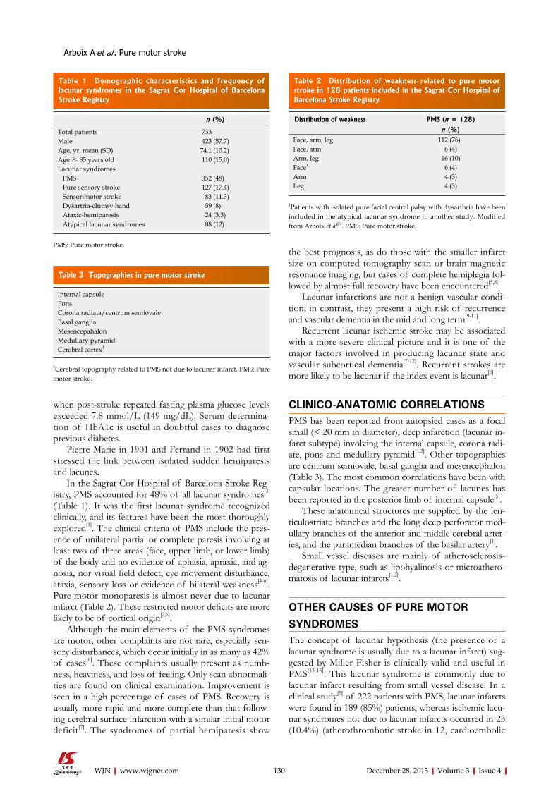
when post-stroke repeated fasting plasma glucose levels exceeded 7.8 mmol/L (149 mg/dL). Serum determina-tion of HbA1c is useful in doubtful cases to diagnose previous diabetes.
Pierre Marie in 1901 and Ferrand in 1902 had first stressed the link between isolated sudden hemiparesis and lacunes.
In the Sagrat Cor Hospital of Barcelona Stroke Reg-istry, PMS accounted for 48% of all lacunar syndromes[3] (Table 1). It was the first lacunar syndrome recognized clinically, and its features have been the most thoroughly explored[1]. The clinical criteria of PMS include the pres-ence of unilateral partial or complete paresis involving at least two of three areas (face, upper limb, or lower limb) of the body and no evidence of aphasia, apraxia, and ag-nosia, nor visual field defect, eye movement disturbance, ataxia, sensory loss or evidence of bilateral weakness[4-6]. Pure motor monoparesis is almost never due to lacunar infarct (Table 2). These restricted motor deficits are more likely to be of cortical origin[2,6].
Although the main elements of the PMS syndromes are motor, other complaints are not rare, especially sen-sory disturbances, which occur initially in as many as 42% of cases[6]. These complaints usually present as numb-ness, heaviness, and loss of feeling. Only scan abnormali-ties are found on clinical examination. Improvement is seen in a high percentage of cases of PMS. Recovery is usually more rapid and more complete than that follow-ing cerebral surface infarction with a similar initial motor deficit[7]. The syndromes of partial hemiparesis show
the best prognosis, as do those with the smaller infarct size on computed tomography scan or brain magnetic resonance imaging, but cases of complete hemiplegia fol-lowed by almost full recovery have been encountered[5,8].
Lacunar infarctions are not a benign vascular condi-tion; in contrast, they present a high risk of recurrence and vascular dementia in the mid and long term[9-11].
Recurrent lacunar ischemic stroke may be associated with a more severe clinical picture and it is one of the major factors involved in producing lacunar state and vascular subcortical dementia[7-12]. Recurrent strokes are more likely to be lacunar if the index event is lacunar[5].
CLINICO-ANATOMIC CORRELATIONSPMS has been reported from autopsied cases as a focal small (< 20 mm in diameter), deep infarction (lacunar in-farct subtype) involving the internal capsule, corona radi-ate, pons and medullary pyramid[1,2]. Other topographies are centrum semiovale, basal ganglia and mesencephalon (Table 3). The most common correlations have been with capsular locations. The greater number of lacunes has been reported in the posterior limb of internal capsule[5].
These anatomical structures are supplied by the len-ticulostriate branches and the long deep perforator med-ullary branches of the anterior and middle cerebral arter-ies, and the paramedian branches of the basilar artery[1].
Small vessel diseases are mainly of atherosclerosis-degenerative type, such as lipohyalinosis or microathero-matosis of lacunar infarcts[1,2].
OTHER CAUSES OF PURE MOTOR SYNDROMESThe concept of lacunar hypothesis (the presence of a lacunar syndrome is usually due to a lacunar infarct) sug-gested by Miller Fisher is clinically valid and useful in PMS[13-15]. This lacunar syndrome is commonly due to lacunar infarct resulting from small vessel disease. In a clinical study[5] of 222 patients with PMS, lacunar infarcts were found in 189 (85%) patients, whereas ischemic lacu-nar syndromes not due to lacunar infarcts occurred in 23 (10.4%) (atherothrombotic stroke in 12, cardioembolic
Arboix A et al . Pure motor stroke
130 December 28, 2013|Volume 3|Issue 4|WJN|www.wjgnet.com
n (%)
Total patients 733 Male 423 (57.7) Age, yr, mean (SD) 74.1 (10.2) Age ≥ 85 years old 110 (15.0) Lacunar syndromes PMS 352 (48) Pure sensory stroke 127 (17.4) Sensorimotor stroke 83 (11.3) Dysartria-clumsy hand 59 (8) Ataxic-hemiparesis 24 (3.3) Atypical lacunar syndromes 88 (12)
Table 1 Demographic characteristics and frequency of lacunar syndromes in the Sagrat Cor Hospital of Barcelona Stroke Registry
Distribution of weakness PMS (n = 128)
n (%) Face, arm, leg 112 (76) Face, arm 6 (4) Arm, leg 16 (10) Face1 6 (4) Arm 4 (3) Leg 4 (3)
Table 2 Distribution of weakness related to pure motor stroke in 128 patients included in the Sagrat Cor Hospital of Barcelona Stroke Registry
1Patients with isolated pure facial central palsy with dysarthria have been included in the atypical lacunar syndrome in another study. Modified from Arboix et al[6]. PMS: Pure motor stroke.
Internal capsule Pons Corona radiata/centrum semiovale Basal ganglia Mesencepahalon Medullary pyramid Cerebral cortex1
Table 3 Topographies in pure motor stroke
1Cerebral topography related to PMS not due to lacunar infarct. PMS: Pure motor stroke.
PMS: Pure motor stroke.
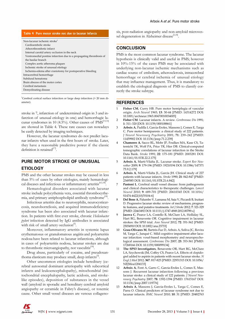
stroke in 7, infarction of undetermined origin in 3 and in-farction of unusual etiology in one) and hemorrhagic la-cunar syndromes in 10 (4.5%). Other causes of PMS[14,16] are showed in Table 4. These rare causes can nowadays be easily detected by imaging techniques.
However, the lacunar syndromes do not predict lacu-nar infarcts when used in the first hours of stroke. Later, they have a reasonable predictive power if the classic definition is retained[17].
PURE MOTOR STROKE OF UNUSUAL ETIOLOGYPMS and the other lacunar strokes may be caused in less than 5% of cases by other etiologies, mainly hematologi-cal diseases and infectious or inflammatory arteritis[4,5,18].
Hematological disorders associated with lacunar stroke include polycythemia vera, essential thrombocythe-mia, and primary antiphospholipid antibody syndrome[18].
Infectious arteritis due to neurosyphilis, neurocysticer-cosis, neuroborreliosis, and acquired immunodeficiency syndrome has been also associated with lacunar infarc-tion. In patients with first ever stroke, chronic Helicobacter pylori infection detected by IgG antibodies is associated with risk of small artery occlusion[5].
Moreover, inflammatory arteritis in systemic lupus erythematosus or granulomatous angiitis and polyarteritis nodosa have been related to lacunar infarctions, although in cases of polyarteritis nodosa, lacunar strokes are due to thrombotic microangiopathy, not vasculitis[18].
Drug abuse, particularly of cocaine, and pseudoxan-thoma elasticum may produce small, deep infarcts[5].
Other uncommon etiologies include hereditary (ce-rebral autosomal dominant arteriopathy with subcortical infarcts and leukoencephalopathy), mitochondrial (mi-tochondrial encephalopathy, lactic acidosis, and stroke-like episodes), deposition of substances in the vessel wall (amyloid in sporadic and hereditary cerebral amyloid angiopathy or ceramide in Fabry’s disease), or toxemic cause. Other small vessel diseases are venous collageno-
sis, post-radiation angiopathy and non-amyloid microves-sel degeneration in Alzheimer disease[5,18].
CONCLUSIONPMS is the most common lacunar syndrome. The lacunar hypothesis is clinically valid and useful in PMS; however in 10%-15% of the cases PMS may be associated with underlying non-lacunar ischemic mechanisms such as cardiac source of embolism, atherosclerosis, intracerebral hemorrhage or cerebral ischemia of unusual etiology that may influence management. Thus, it is mandatory to establish the etiological diagnosis of PMS to classify cor-rectly the stroke subtype.
REFERENCES1 Fisher CM, Curry HB. Pure motor hemiplegia of vascular
origin. Arch Neurol 1965; 13: 30-44 [PMID: 14314272 DOI: 10.1001/archneur.1965.00470010034005]
2 Fisher CM. Lacunar infarcts. A review. Cerebrovasc Dis 1991; 1: 311–320 [DOI: 10.1159/000108861]
3 Arboix A, Padilla I, García-Eroles, Massons J, Comes E, Targa C. Pure motor hemiparesis: a clinical study of 222 patients. J Neurol Neurosurg Psychiatry 2001; 71: 239–242 [PMID: 11459902 DOI: 10.1136/jnnp.71.2.239]
4 Chamorro A, Sacco RL, Mohr JP, Foulkes MA, Kase CS, Ta-temichi TK, Wolf PA, Price TR, Hier DB. Clinical-computed tomographic correlations of lacunar infarction in the Stroke Data Bank. Stroke 1991; 22: 175-181 [PMID: 2003281 DOI: 10.1161/01.STR.22.2.175]
5 Arboix A, Martí-Vilalta JL. Lacunar stroke. Expert Rev Neu-rother 2009; 9: 179-196 [PMID: 19210194 DOI: 10.1586/14737175.9.2.179]
6 Arboix A, Martí-Vilalta JL, García JH. Clinical study of 227 patients with lacunar infarcts. Stroke 1990; 21: 842-847 [PMID: 2349585 DOI: 10.1161/01.STR.21.6.842]
7 Pantoni L. Cerebral small vessel disease: from pathogenesis and clinical characteristics to therapeutic challenges. Lancet Neurol 2010; 9: 689-701 [PMID: 20610345 DOI: 10.1016/S1474-4422(10)70104-6]
8 Del Bene A, Palumbo V, Lamassa M, Saia V, Piccardi B, Inzitari D. Progressive lacunar stroke: review of mechanisms, prognos-tic features, and putative treatments. Int J Stroke 2012; 7: 321-329 [PMID: 22463492 DOI: 10.1111/j.1747-4949.2012.00789.x]
9 Jacova C, Pearce LA, Costello R, McClure LA, Holliday SL, Hart RG, Benavente OR. Cognitive impairment in lacunar strokes: the SPS3 trial. Ann Neurol 2012; 72: 351-362 [PMID: 23034910 DOI: 10.1002/ana.23733]
10 Grau-Olivares M, Bartrés-Faz D, Arboix A, Soliva JC, Rovira M, Targa C, Junqué C. Mild cognitive impairment after lacu-nar infarction: voxel-based morphometry and neuropsycho-logical assessment. Cerebrovasc Dis 2007; 23: 353-361 [PMID: 17268166 DOI: 10.1159/000099134]
11 The SPS3 Investigators, Benavente OR, Hart RG, McClure LA, Szychowski JM, Coffey CS, Pearce LA. Effects of clopido-grel added to aspirin in patients with recent lacunar stroke. N Engl J Med 2012; 367: 817-825 [PMID: 22931315 DOI: 10.1056/NEJMoa1204133]
12 Arboix A, Font A, Garro C, García-Eroles L, Comes E, Mas-sons J. Recurrent lacunar infarction following a previous lacunar stroke: a clinical study of 122 patients. J Neurol Neu-rosurg Psychiatry 2007; 78: 1392-1394 [PMID: 17615167 DOI: 10.1136/jnnp.2007.119776]
13 Arboix A, Massons J, García-Eroles L, Targa C, Comes E, Parra O. Clinical predictors of lacunar syndrome not due to lacunar infarcts. BMC Neurol 2010; 10: 31 [PMID: 20482763
131 December 28, 2013|Volume 3|Issue 4|WJN|www.wjgnet.com
Non-lacunar ischemic stroke1 Cardioembolic stroke Atherothrombotic infarct Internal carotid artery occlusion in the neck Ventromedial pontine infarction due to a propagating thrombosis of the basilar branch Complex aortic atheroma plaques Ischemic stroke of unusual etiology Ischemia-edema after craniotomy for postoperative bleeding Intracerebral hemorrhage Subdural hematoma Brain abscess of the motor cortex Cerebral metastasis Demyelinating disease
Table 4 Pure motor stroke not due to lacunar infarcts
1Cerebral cortical surface infarction or large deep infarction (> 20 mm di-ameter).
Arboix A et al . Pure motor stroke
C. Hemorrhagic lacunar stroke. Cerebrovasc Dis 2000; 10: 229-234 [PMID: 10773650 DOI: 10.1159/000016061]
17 Ferro JM. Patterns of ischaemic cerebral diseases. J Neurol 2004; 251: 1-10 [PMID: 14999483 DOI: 10.1007/s00415-004-0280-z]
18 Arboix A, Martí-Vilalta JL. New concepts in lacunar stroke etiology: the constellation of small-vessel arterial disease. Cerebrovasc Dis 2004; 17 Suppl 1: 58-62 [PMID: 14694281 DOI: 10.1159/000074796]
P- Reviewers: Altamura C, Garg RK, Papanas N S- Editor: Cui XM L- Editor: A E- Editor: Liu SQ
DOI: 10.1186/1471-2377-10-31]14 Arboix A, Martí-Vilalta JL. Lacunar syndromes not due
to lacunar infarcts. Cerebrovasc Dis 1992; 2: 287–292 [DOI: 10.1159/000109029]
15 Micheli S, Agnelli G, Palmerini F, Caso V, Venti M, Alberti A, Biagini S, Paciaroni M. Need for extensive diagnostic work-up for patients with lacunar stroke. J Neurol 2008; 255: 637-642 [PMID: 18283395 DOI: 10.1007/s00415-008-0762-5]
16 Arboix A, García-Eroles L, Massons J, Oliveres M, Targa
132 December 28, 2013|Volume 3|Issue 4|WJN|www.wjgnet.com
Arboix A et al . Pure motor stroke
Spasticity; Neurorehabilitation
Core tip: In this overview, we describe that botulinum toxins (BTXs) were very useful to reduce spasticity in stroke patients. Moreover, we introduce studies on ap-propriate effective and safe injection methods; pilot studies that evaluated combined rehabilitation and BTX treatment; and adverse events after BTX injection, in-cluding risk factors of neutralizing antibodies.
Isoyama H, Takeuchi N. Overview of botulinum toxin as a treatment for spasticity in stroke patients. World J Neurol 2013; 3(4): 133-137 Available from: URL: http://www.wjg-net.com/2218-6212/full/v3/i4/133.htm DOI: http://dx.doi.org/10.5316/wjn.v3.i4.133
INTRODUCTIONSpasticity is a common complication in patients who have experienced stroke. The incidence of spasticity is report-ed to be 17%-43% in post-stroke patients[1-3]. Spasticity induces limb deformity, functional disability and/or pain that limits activities of daily living (ADL) and deteriorates the quality of life (QOL). In order to reduce spasticity, clinicians prescribe oral medications, intrathecal baclofen therapy, nerve phenolization, casting or splinting, reha-bilitation therapy or a combination of these therapeutic approaches[4-8]. A recent study has shown that botulinum toxin (BTX) is an efficacious therapeutic agent for the treatment of spasticity[9].
BTX is poisonous in nature and is produced by the anaerobic bacterium Clostridium Botulinum[10]. BTX blocks vesicular acetylcholine release at neuromuscular junctions and reduces focal muscle tonus and contracture[11]. There-fore, BTX is used for treating muscular hyperactivity and excessive or inappropriate muscle contraction. Seven types of BTX exist in nature, but two toxin types, type A (BTX-A) and type B (BTX-B), are used in the clinical
Overview of botulinum toxin as a treatment for spasticity in stroke patients
Hirotaka Isoyama, Naoyuki Takeuchi
Hirotaka Isoyama, Department of Physical Medicine and Rehabilitation, Hospital of Hokkaido University, Hokkaido 060-8648, JapanNaoyuki Takeuchi, Department of Physical Medicine and Re-habilitation, Tohoku University Graduate School of Medicine, Sendai 980-8576, JapanAuthor contributions: Both authors contributed to the manu-script.Correspondence to: Hirotaka Isoyama, MD, Department of Physical Medicine and Rehabilitation, Hospital of Hokkai-do University, Kita14 Nishi5 Kita-ku, Sapporo, Hokkaido 060-8648, Japan. [email protected]: +81-11-7066066 Fax: +81-11-7066067Received: June 26, 2013 Revised: October 3, 2013Accepted: November 2, 2013Published online: December 28, 2013
AbstractSpasticity after the occurrence of stroke induces limb deformity, functional disability and/or pain in patients, which limits their activities of daily living and deterio-rates their quality of life. Botulinum toxin (BTX) has recently been reported as an efficacious therapeutic agent for the treatment of spasticity. Systematic review and meta-analysis studies have demonstrated that BTX therapy after stroke reduces spasticity and increases physical activity capacity and performance levels. More-over, BTX can be used as an adjuvant in physiotherapy. Several studies have confirmed that the combination of BTX therapy and physiotherapy improves motor recovery. However, to date, only a few such combina-tion studies have been conducted and their findings are considered preliminary and controversial. Therefore, fu-ture studies are required to determine the appropriate combination of treatment methods that will aid motor recovery.
© 2013 Baishideng Publishing Group Co., Limited. All rights reserved.
Key words: Botulinum toxin; Rehabilitation; Stroke;
MINIREVIEWS
133 December 28, 2013|Volume 3|Issue 4|WJN|www.wjgnet.com
World Journal of NeurologyW J N
Online Submissions: http://www.wjgnet.com/esps/[email protected]:10.5316/wjn.v3.i4.133
World J Neurol 2013 December 28; 3(4): 133-137ISSN 2218-6212 (online)
© 2013 Baishideng Publishing Group Co., Limited. All rights reserved.
setting. Most clinical trials have utilized BTX-A because it has a longer lasting effect than BTX-B[12,13]. BTX-B tends to be selected for spasticity treatment when patients have neutralizing antibodies against BTX-A or develop anti-bodies after repetitive BTX-A injection treatment[14,15].
Many studies have reported that BTX injection treat-ment reduces spasticity in stroke patients[16-25]. However, a few studies have focused on rehabilitation therapy after BTX treatment for improving motor function[26-28]. This review focuses on the following factors of BTX treat-ment in post-stroke patients: (1) the effectiveness of spasticity reduction, with regard to ADL and QOL; (2) the appropriate injection method; (3) combined BTX therapy and neurorehabilitation; and (4) adverse events. The purpose of this review is to provide a comprehen-sive overview of BTX treatment for muscular spasticity in post-stroke patients, to understand its mechanisms of action, and to suggest appropriate treatment approaches.
EFFECTIVENESS FOR SPASTICITYSeveral reports clarified the effectiveness of BTX treatment for upper limb spasticity experienced after stroke[16-21]. A recent systematic review and meta-analysis reported that BTX-A reduced spasticity and thereby im-proved physical activity capacity and performance levels in the upper limb in patients who experienced stroke[29]. BTX-A (Botox®) injection in the upper limb muscles are recommended at the following doses: 25-100 units for the flexor carpi radialis; 20-70 units for the flexor carpi ulnaris; 20-60 units for the flexor digitorum superficialis; 20-60 units for the flexor digitorum profundus; 10-30 units for the flexor pollicis longus; and 5-25 units for the adductor pollicis[30].
Furthermore, several studies have reported the ef-fectiveness of BTX therapy in stroke-induced lower limb spasticity[22-25]. However, a systematic review and meta-analysis reported that BTX-A treatment for stroke-induced lower limb spasticity was associated with a slight improvement in functional ambulation[19,31]. A multi-center, double-blind trial demonstrated the effectiveness of BTX-A in cases of lower limb spasticity, but improve-ments in gait pattern scale and speed were not significant, compared to that observed in the placebo group[32]. A previous study suggested that high-dose BTX-A treat-ment may induce excessive muscle weakening that may deteriorate limb function[25]. BTX-A (Botox®) injection doses in the lower limb is recommended at the following doses: 50-250 units for the medial and lateral head of the gastrocnemius respectively; 50-200 units for the soleus; and 50-150 units for the tibialis posterior[30]. However, to the best of our knowledge, no study has evaluated the appropriate BTX dose for treatment of upper and lower limb spasticity and therefore, future studies are required to clarify these dosages. Moreover, it is noted that clini-cians should consider the maximum dose of BTX per session, which differs in each country (e.g., 360 units in Japan and 600 units in Europe)[32-35].
Treatment of spasticity in patients who experience a
stroke with BTX-A may improve ADL and QOL[36,37]. A previous study reported that high-dose BTX-A signifi-cantly improved ADL scores for limb position and self-dressing[36]. Furthermore, another study in post-stroke patients reported that BTX-A therapy improved overall QOL scores and muscle tone, reduced disability, and im-proved the ability to function[37]. However, a recent meta-analysis study revealed that BTX-A did not significantly improve overall health-related QOL[38].
APPROPRIATE INJECTION METHODSeveral studies have reported on BTX-A injection tech-niques[39-41]. Francisco et al[39] compared the efficacy of high and low volumes of BTX at the same dose for wrist and finger flexor spasticity. They found that modified Ashworth Scale scores were significantly decreased in both high and low-volume injection groups. However, there was no significant difference between the groups[40]. Gracies et al[41] reported that injecting a small volume of BTX-A near the neuromuscular junction was more effec-tive than injecting a similar volume at a greater distance from the neuromuscular junction. However, injecting a large volume at a greater distance from the junction was as effective as a small volume injected near the neu-romuscular junction. Mayer et al [40] also reported that a single motor point and multisite low-dose, high-volume BTX-A injections produced a similar impact in spasticity reduction. Therefore, the appropriate injection methods according to BTX-A volume and injection points must be elucidated. Moreover, BTX-A injections are more ac-curate when the affected muscles are targeted via needle electromyography or under ultrasound guidance. This conventional method is not supported by evidence, but correct muscle selection has been confirmed to be a key feature in the efficacy of BTX treatment[42,43].
COMBINATION OF BTX THERAPY AND NEUROREHABILITATIONAs mentioned above, BTX improves motor function by reducing muscular spasticity in post-stroke patients. Therefore, BTX therapy may be a useful adjuvant therapy in combination with neurorehabilitation. In fact, several studies have reported that the combination of BTX therapy and physiotherapy improved motor recovery[26-28]. A recent study reported that BTX injection, followed by home-based functional training, may have the potential to improve active motor function of the affected up-per limb in post-stroke patients[26]. Moreover, previous studies have reported that casting and dynamic splinting following BTX-A injection improved range of motion as well as motor function[5,27,28]. Thus, BTX when combined with physiotherapy or casting may be useful for improv-ing motor function in stroke-induced spasticity.
Recently, various neurorehabilitation strategies have been established to improve neural plasticity and motor recovery. These approaches include constraint-induced
Isoyama H et al . Botulinum toxin for rehabilitation
134 December 28, 2013|Volume 3|Issue 4|WJN|www.wjgnet.com
movement therapy (CIMT), transcutaneous electrical neu-romuscular stimulation (TENS) and non-invasive brain stimulation (NIBS)[44-49]. A preliminary study demon-strated that the combination of BTX injection and CIMT may improve functional gains in post-stroke patients[44,45]. Moreover, a recent study reported that combined BTX-A and NIBS improves motor function in patients with stroke-induced upper limb hemiparesis[48]. However, a previous study reported that combining BTX injection with TENS did not have any additional effect on motor recovery[47]. The number of these combination studies remains small and their results are considered preliminary and controversial. Although BTX-A probably enhances the effects of neurorehabilitation strategies in post-stroke patients, future studies are required to determine the ap-propriate combination of treatments for optimum motor recovery.
ADVERSE EVENTSIn general, pain, rash, edema and muscle weakness have been noted at the injection site and remote muscle weak-ness, fatigue and allergic reaction have been reported as adverse events related to BTX therapy. To clarify the safety of BTX-A, Turkel et al[50] analyzed 9 double-blind, placebo-controlled studies in more than 500 patients who had experienced a stroke. The most frequent reported adverse events by the patients (> 5% but < 10% in either group) were respiratory infection, seizures, incoordina-tion and injection site pain, none of which occurred at a significantly high rate in the BTX-A group. Nausea alone was reported at a significantly higher rate in the BTX-A group than in the placebo group. In contrast, injection site pain, chest pain and allergic reaction were found at a significantly higher frequency in the placebo group. Other reviews have also reported no serious adverse events af-ter BTX-A administration in adult post-stroke patients[35]. Therefore, BTX has recently been considered a safe and efficacious treatment for spasticity in post-stroke patients.
However, care should be taken to try to avoid produc-ing neutralizing antibodies. A higher dose per treatment, repetitive treatment and shorter intervals between treat-ments are risk factors for the production of neutralizing antibodies[51-53]. Of these, the interval between treatments is the most important risk factor. Therefore, BTX-A treatment intervals of more than three months and BTX-B treatment intervals of two months are recom-mended[54].
CONCLUSIONMany reports have shown the usefulness of the BTX treatment for spasticity in stroke patients. However, a few studies have reported the effectiveness of rehabilitation after BTX treatment in stroke patients. Future studies are needed to evaluate the efficacy of BTX after treatment with rehabilitation.
REFERENCES1 Sommerfeld DK, Eek EU, Svensson AK, Holmqvist LW,
von Arbin MH. Spasticity after stroke: its occurrence and as-sociation with motor impairments and activity limitations. Stroke 2004; 35: 134-139 [PMID: 14684785 DOI: 10.1161/01.STR.0000105386.05173.5E]
2 Lundström E, Terént A, Borg J. Prevalence of disabling spas-ticity 1 year after first-ever stroke. Eur J Neurol 2008; 15: 533-539 [PMID: 18355307 DOI: 10.1111/j.1468-1331.2008.02114.x]
3 Urban PP, Wolf T, Uebele M, Marx JJ, Vogt T, Stoeter P, Bau-ermann T, Weibrich C, Vucurevic GD, Schneider A, Wissel J. Occurence and clinical predictors of spasticity after ischemic stroke. Stroke 2010; 41: 2016-2020 [PMID: 20705930 DOI: 10.1161/STROKEAHA.110.581991]
4 Ward AB. A summary of spasticity management--a treatment algorithm. Eur J Neurol 2002; 9 Suppl 1: 48-52; dicussion 53-61 [PMID: 11918650 DOI: 10.1046/j.1468-1331.2002.0090s1048.x]
5 Farina S, Migliorini C, Gandolfi M, Bertolasi L, Casarotto M, Manganotti P, Fiaschi A, Smania N. Combined effects of bot-ulinum toxin and casting treatments on lower limb spasticity after stroke. Funct Neurol 2008; 23: 87-91 [PMID: 18671909]
6 Reiter F, Danni M, Lagalla G, Ceravolo G, Provinciali L. Low-dose botulinum toxin with ankle taping for the treat-ment of spastic equinovarus foot after stroke. Arch Phys Med Rehabil 1998; 79: 532-535 [PMID: 9596394 DOI: 10.1016/S0003-9993(98)90068-5]
7 Verplancke D, Snape S, Salisbury CF, Jones PW, Ward AB. A randomized controlled trial of botulinum toxin on lower limb spasticity following acute acquired severe brain injury. Clin Rehabil 2005; 19: 117-125 [PMID: 15759526]
8 Richardson D. Physical therapy in spasticity. Eur J Neurol 2002; 9 Suppl 1: 17-22; dicussion 53-61 [PMID: 11918645 DOI: 10.1191/0269215505cr827oa]
9 Intiso D. Therapeutic use of botulinum toxin in neuroreha-bilitation. J Toxicol 2012; 2012: 802893 [PMID: 21941544 DOI: 10.1155/2012/802893]
10 Das Gupta BR, Sugiyama H. Role of a protease in natural activation of Clostridium botulinum neurotoxin. Infect Im-mun 1972; 6: 587-590 [PMID: 4564288]
11 Pearce LB, First ER, MacCallum RD, Gupta A. Pharmacolog-ic characterization of botulinum toxin for basic science and medicine. Toxicon 1997; 35: 1373-1412 [PMID: 9403963 DOI: 10.1016/S0041-0101(96)00180-8]
12 Keller JE. Recovery from botulinum neurotoxin poisoning in vivo. Neuroscience 2006; 139: 629-637 [PMID: 16490322 DOI: 10.1016/j.neuroscience.2005.12.029]
13 Comella CL, Jankovic J, Shannon KM, Tsui J, Swenson M, Leurgans S, Fan W. Comparison of botulinum toxin sero-types A and B for the treatment of cervical dystonia. Neurol-ogy 2005; 65: 1423-1429 [PMID: 16275831 DOI: 10.1212/01.wnl.0000183055.81056.5c]
14 Barnes MP, Best D, Kidd L, Roberts B, Stark S, Weeks P, Whita-ker J. The use of botulinum toxin type-B in the treatment of patients who have become unresponsive to botulinum toxin type-A -- initial experiences. Eur J Neurol 2005; 12: 947-955 [PMID: 16324088 DOI: 10.1111/j.1468-1331.2005.01095.x]
15 Factor SA, Molho ES, Evans S, Feustel PJ. Efficacy and safety of repeated doses of botulinum toxin type B in type A resis-tant and responsive cervical dystonia. Mov Disord 2005; 20: 1152-1160 [PMID: 15954134 DOI: 10.1002/mds.20531]
16 Brashear A, Gordon MF, Elovic E, Kassicieh VD, Marciniak C, Do M, Lee CH, Jenkins S, Turkel C. Intramuscular injec-tion of botulinum toxin for the treatment of wrist and finger spasticity after a stroke. N Engl J Med 2002; 347: 395-400 [PMID: 12167681 DOI: 10.1056/NEJMoa011892]
17 Bakheit AM, Pittock S, Moore AP, Wurker M, Otto S, Er-bguth F, Coxon L. A randomized, double-blind, placebo-
135 December 28, 2013|Volume 3|Issue 4|WJN|www.wjgnet.com
Isoyama H et al . Botulinum toxin for rehabilitation
32 Kaji R, Osako Y, Suyama K, Maeda T, Uechi Y, Iwasaki M. Botulinum toxin type A in post-stroke lower limb spastic-ity: a multicenter, double-blind, placebo-controlled trial. J Neurol 2010; 257: 1330-1337 [PMID: 20358216 DOI: 10.1007/s00415-010-5526-3]
33 Wissel J, Ward AB, Erztgaard P, Bensmail D, Hecht MJ, Lejeune TM, Schnider P, Altavista MC, Cavazza S, Del-tombe T, Duarte E, Geurts AC, Gracies JM, Haboubi NH, Juan FJ, Kasch H, Kätterer C, Kirazli Y, Manganotti P, Par-man Y, Paternostro-Sluga T, Petropoulou K, Prempeh R, Rousseaux M, Slawek J, Tieranta N. European consensus table on the use of botulinum toxin type A in adult spas-ticity. J Rehabil Med 2009; 41: 13-25 [PMID: 19197564 DOI: 10.2340/16501977-0303]
34 Ozcakir S, Sivrioglu K. Botulinum toxin in poststroke spas-ticity. Clin Med Res 2007; 5: 132-138 [PMID: 17607049 DOI: 10.3121/cmr.2007.716]
35 Elia AE, Filippini G, Calandrella D, Albanese A. Botulinum neurotoxins for post-stroke spasticity in adults: a systematic review. Mov Disord 2009; 24: 801-812 [PMID: 19224614 DOI: 10.1002/mds.22452]
36 Kaji R, Osako Y, Suyama K, Maeda T, Uechi Y, Iwasaki M. Botulinum toxin type A in post-stroke upper limb spasticity. Curr Med Res Opin 2010; 26: 1983-1992 [PMID: 20569068 DOI: 10.1185/03007995.2010.497103]
37 Elovic EP, Brashear A, Kaelin D, Liu J, Millis SR, Barron R, Turkel C. Repeated treatments with botulinum toxin type a produce sustained decreases in the limitations associated with focal upper-limb poststroke spasticity for caregivers and patients. Arch Phys Med Rehabil 2008; 89: 799-806 [PMID: 18452724 DOI: 10.1016/j.apmr.2008.01.007]
38 Pulman J, Buckley E, Clark-Carter D. A meta-analysis evalu-ating the effectiveness of two different upper limb hemipare-sis interventions on improving health-related quality of life following stroke. Top Stroke Rehabil 2013; 20: 189-196 [PMID: 23611859 DOI: 10.1310/tsr2002-189]
39 Francisco GE, Boake C, Vaughn A. Botulinum toxin in up-per limb spasticity after acquired brain injury: a randomized trial comparing dilution techniques. Am J Phys Med Rehabil 2002; 81: 355-363 [PMID: 11964576 DOI: 10.1097/00002060-200205000-00007]
40 Mayer NH, Whyte J, Wannstedt G, Ellis CA. Comparative impact of 2 botulinum toxin injection techniques for elbow flexor hypertonia. Arch Phys Med Rehabil 2008; 89: 982-987 [PMID: 18452749 DOI: 10.1016/j.apmr.2007.10.022]
41 Gracies JM, Lugassy M, Weisz DJ, Vecchio M, Flanagan S, Simpson DM. Botulinum toxin dilution and endplate target-ing in spasticity: a double-blind controlled study. Arch Phys Med Rehabil 2009; 90: 9-16.e2 [PMID: 19154823 DOI: 10.1016/j.apmr.2008.04.030]
42 Cordivari C, Misra VP, Vincent A, Catania S, Bhatia KP, Lees AJ. Secondary nonresponsiveness to botulinum toxin A in cervical dystonia: the role of electromyogram-guided in-jections, botulinum toxin A antibody assay, and the extensor digitorum brevis test. Mov Disord 2006; 21: 1737-1741 [PMID: 16874756 DOI: 10.1002/mds.21051]
43 Schnitzler A, Roche N, Denormandie P, Lautridou C, Par-ratte B, Genet F. Manual needle placement: accuracy of botulinum toxin A injections. Muscle Nerve 2012; 46: 531-534 [PMID: 22987693 DOI: 10.1002/mus.23410]
44 Levy CE, Giuffrida C, Richards L, Wu S, Davis S, Nadeau SE. Botulinum toxin a, evidence-based exercise therapy, and constraint-induced movement therapy for upper-limb hemi-paresis attributable to stroke: a preliminary study. Am J Phys Med Rehabil 2007; 86: 696-706 [PMID: 17709993 DOI: 10.1097/PHM.0b013e31813e2b4d]
45 Sun SF, Hsu CW, Hwang CW, Hsu PT, Wang JL, Yang CL. Application of combined botulinum toxin type A and modi-fied constraint-induced movement therapy for an individual with chronic upper-extremity spasticity after stroke. Phys
controlled study of the efficacy and safety of botulinum toxin type A in upper limb spasticity in patients with stroke. Eur J Neurol 2001; 8: 559-565 [PMID: 11784339 DOI: 10.1046/j.1468-1331.2001.00277.x]
18 Simpson DM, Alexander DN, O’Brien CF, Tagliati M, As-wad AS, Leon JM, Gibson J, Mordaunt JM, Monaghan EP. Botulinum toxin type A in the treatment of upper extremity spasticity: a randomized, double-blind, placebo-controlled trial. Neurology 1996; 46: 1306-1310 [PMID: 8628472 DOI: 10.1212/WNL.46.5.1306]
19 Rosales RL, Chua-Yap AS. Evidence-based systematic re-view on the efficacy and safety of botulinum toxin-A therapy in post-stroke spasticity. J Neural Transm 2008; 115: 617-623 [PMID: 18322637 DOI: 10.1007/s00702-007-0869-3]
20 Cardoso E, Rodrigues B, Lucena R, Oliveira IR, Pedreira G, Melo A. Botulinum toxin type A for the treatment of the up-per limb spasticity after stroke: a meta-analysis. Arq Neurop-siquiatr 2005; 63: 30-33 [PMID: 15830061 DOI: 10.1590/S0004-282X2005000100006]
21 Yablon SA, Brin MF, VanDenburgh AM, Zhou J, Garabedi-an-Ruffalo SM, Abu-Shakra S, Beddingfield FC 3rd. Dose re-sponse with onabotulinumtoxinA for post-stroke spasticity: a pooled data analysis. Mov Disord 2011; 26: 209-215 [PMID: 20960474 DOI: 10.1002/mds.23426]
22 Grazko MA, Polo KB, Jabbari B. Botulinum toxin A for spasticity, muscle spasms, and rigidity. Neurology 1995; 45: 712-717 [PMID: 7723960 DOI: 10.1212/WNL.45.4.712]
23 Burbaud P, Wiart L, Dubos JL, Gaujard E, Debelleix X, Jo-seph PA, Mazaux JM, Bioulac B, Barat M, Lagueny A. A ran-domised, double blind, placebo controlled trial of botulinum toxin in the treatment of spastic foot in hemiparetic patients. J Neurol Neurosurg Psychiatry 1996; 61: 265-269 [PMID: 8795597 DOI: 10.1136/jnnp.61.3.265]
24 Dunne J. Botulinum toxin type A (BOTOX) in the treatment of lower limb spasticity during stroke rehabilitation. Intern Med J 2003; 33 (Suppl): A41-A54
25 Mancini F, Sandrini G, Moglia A, Nappi G, Pacchetti C. A randomised, double-blind, dose-ranging study to evaluate efficacy and safety of three doses of botulinum toxin type A (Botox) for the treatment of spastic foot. Neurol Sci 2005; 26: 26-31 [PMID: 15877184 DOI: 10.1007/s10072-005-0378-9]
26 Takekawa T, Kakuda W, Taguchi K, Ishikawa A, Sase Y, Abo M. Botulinum toxin type A injection, followed by home-based functional training for upper limb hemiparesis after stroke. Int J Rehabil Res 2012; 35: 146-152 [PMID: 22453625 DOI: 10.1097/MRR.0b013e3283527f4a]
27 Yaşar E, Tok F, Safaz I, Balaban B, Yilmaz B, Alaca R. The efficacy of serial casting after botulinum toxin type A injec-tion in improving equinovarus deformity in patients with chronic stroke. Brain Inj 2010; 24: 736-739 [PMID: 20334469 DOI: 10.3109/02699051003610524]
28 Lai JM, Francisco GE, Willis FB. Dynamic splinting after treatment with botulinum toxin type-A: a randomized controlled pilot study. Adv Ther 2009; 26: 241-248 [PMID: 19194671 DOI: 10.1007/s12325-008-0139-2]
29 Foley N, Pereira S, Salter K, Fernandez MM, Speechley M, Se-queira K, Miller T, Teasell R. Treatment with botulinum toxin improves upper-extremity function post stroke: a systematic review and meta-analysis. Arch Phys Med Rehabil 2013; 94: 977-989 [PMID: 23262381 DOI: 10.1016/j.apmr.2012.12.006]
30 Francisco GE. Botulinum toxin for post-stroke spastic hy-pertonia: a review of its efficacy and application in clinical practice. Ann Acad Med Singapore 2007; 36: 22-30 [PMID: 17285183]
31 Foley N, Murie-Fernandez M, Speechley M, Salter K, Sequei-ra K, Teasell R. Does the treatment of spastic equinovarus deformity following stroke with botulinum toxin increase gait velocity? A systematic review and meta-analysis. Eur J Neurol 2010; 17: 1419-1427 [PMID: 20491885 DOI: 10.1111/j.1468-1331.2010.03084.x]
136 December 28, 2013|Volume 3|Issue 4|WJN|www.wjgnet.com
Isoyama H et al . Botulinum toxin for rehabilitation
Ther 2006; 86: 1387-1397 [PMID: 17012643 DOI: 10.2522/ptj.20050262]
46 Santamato A, Notarnicola A, Panza F, Ranieri M, Micello MF, Manganotti P, Moretti B, Fortunato F, Filoni S, Fiore P. SBOTE study: extracorporeal shock wave therapy versus electrical stimulation after botulinum toxin type a injection for post-stroke spasticity-a prospective randomized trial. Ultrasound Med Biol 2013; 39: 283-291 [PMID: 23245824 DOI: 10.1016/j.ultrasmedbio.2012.09.019]
47 Weber DJ, Skidmore ER, Niyonkuru C, Chang CL, Huber LM, Munin MC. Cyclic functional electrical stimulation does not enhance gains in hand grasp function when used as an adjunct to onabotulinumtoxinA and task practice therapy: a single-blind, randomized controlled pilot study. Arch Phys Med Rehabil 2010; 91: 679-686 [PMID: 20434603 DOI: 10.1016/J.apmr.2010.01.010]
48 Kakuda W, Abo M, Momosaki R, Yokoi A, Fukuda A, Ito H, Tominaga A, Umemori T, Kameda Y. Combined therapeutic application of botulinum toxin type A, low-frequency rTMS, and intensive occupational therapy for post-stroke spastic up-per limb hemiparesis. Eur J Phys Rehabil Med 2012; 48: 47-55 [PMID: 22071503 DOI: 10.1097/01.phm.0000193505.85874.61]
49 Bayram S, Sivrioglu K, Karli N, Ozcan O. Low-dose botu-
linum toxin with short-term electrical stimulation in post-stroke spastic drop foot: a preliminary study. Am J Phys Med Rehabil 2006; 85: 75-81 [PMID: 16357552]
50 Turkel CC, Bowen B, Liu J, Brin MF. Pooled analysis of the safety of botulinum toxin type A in the treatment of post-stroke spasticity. Arch Phys Med Rehabil 2006; 87: 786-792 [PMID: 16731213 DOI: 10.1016/j.apmr.2006.02.015]
51 Dressler D. Clinical features of antibody-induced complete secondary failure of botulinum toxin therapy. Eur Neurol 2002; 48: 26-29 [PMID: 12138306 DOI: 10.1159/000064953]
52 Greene P, Fahn S, Diamond B. Development of resistance to botulinum toxin type A in patients with torticollis. Mov Disord 1994; 9: 213-217 [PMID: 8196686 DOI: 10.1002/mds.870090216]
53 Hatheway CL, Dang C. Immunogenicity of the neurotoxins of Clostridium botulinum. In: Jankovic J, Hallett M, eds. Therapy with Botulinum Toxin. New York: Marcel Dekker, 1994: 93-107
54 Varghese-Kroll E, Elovic EP. Contralateral weakness and fatigue after high-dose botulinum toxin injection for management of poststroke spasticity. Am J Phys Med Rehabil 2009; 88: 495-499 [PMID: 19454855 DOI: 10.1097/PHM.0b013e3181a5b056]
P- Reviewers: Llamas EP, Lai V, Wong GKC S- Editor: Qi Y L- Editor: Roemmele A E- Editor: Liu SQ
137 December 28, 2013|Volume 3|Issue 4|WJN|www.wjgnet.com
Isoyama H et al . Botulinum toxin for rehabilitation
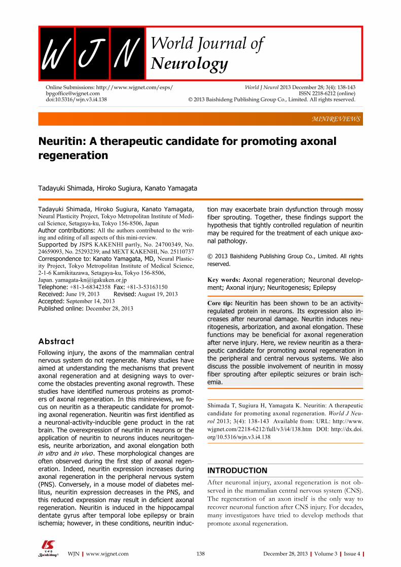
tion may exacerbate brain dysfunction through mossy fiber sprouting. Together, these findings support the hypothesis that tightly controlled regulation of neuritin may be required for the treatment of each unique axo-nal pathology.
© 2013 Baishideng Publishing Group Co., Limited. All rights reserved.
Key words: Axonal regeneration; Neuronal develop-ment; Axonal injury; Neuritogenesis; Epilepsy
Core tip: Neuritin has been shown to be an activity-regulated protein in neurons. Its expression also in-creases after neuronal damage. Neuritin induces neu-ritogenesis, arborization, and axonal elongation. These functions may be beneficial for axonal regeneration after nerve injury. Here, we review neuritin as a thera-peutic candidate for promoting axonal regeneration in the peripheral and central nervous systems. We also discuss the possible involvement of neuritin in mossy fiber sprouting after epileptic seizures or brain isch-emia.
Shimada T, Sugiura H, Yamagata K. Neuritin: A therapeutic candidate for promoting axonal regeneration. World J Neu-rol 2013; 3(4): 138-143 Available from: URL: http://www.wjgnet.com/2218-6212/full/v3/i4/138.htm DOI: http://dx.doi.org/10.5316/wjn.v3.i4.138
INTRODUCTIONAfter neuronal injury, axonal regeneration is not ob-served in the mammalian central nervous system (CNS). The regeneration of an axon itself is the only way to recover neuronal function after CNS injury. For decades, many investigators have tried to develop methods that promote axonal regeneration.
MINIREVIEWS
Neuritin: A therapeutic candidate for promoting axonal regeneration
Tadayuki Shimada, Hiroko Sugiura, Kanato Yamagata
Tadayuki Shimada, Hiroko Sugiura, Kanato Yamagata, Neural Plasticity Project, Tokyo Metropolitan Institute of Medi-cal Science, Setagaya-ku, Tokyo 156-8506, JapanAuthor contributions: All the authors contributed to the writ-ing and editing of all aspects of this mini-review.Supported by JSPS KAKENHI partly, No. 24700349, No. 24659093, No. 25293239; and MEXT KAKENHI, No. 25110737Correspondence to: Kanato Yamagata, MD, Neural Plastic-ity Project, Tokyo Metropolitan Institute of Medical Science, 2-1-6 Kamikitazawa, Setagaya-ku, Tokyo 156-8506, Japan. [email protected]: +81-3-68342358 Fax: +81-3-53163150Received: June 19, 2013 Revised: August 19, 2013Accepted: September 14, 2013Published online: December 28, 2013
AbstractFollowing injury, the axons of the mammalian central nervous system do not regenerate. Many studies have aimed at understanding the mechanisms that prevent axonal regeneration and at designing ways to over-come the obstacles preventing axonal regrowth. These studies have identified numerous proteins as promot-ers of axonal regeneration. In this minireviews, we fo-cus on neuritin as a therapeutic candidate for promot-ing axonal regeneration. Neuritin was first identified as a neuronal-activity-inducible gene product in the rat brain. The overexpression of neuritin in neurons or the application of neuritin to neurons induces neuritogen-esis, neurite arborization, and axonal elongation both in vitro and in vivo . These morphological changes are often observed during the first step of axonal regen-eration. Indeed, neuritin expression increases during axonal regeneration in the peripheral nervous system (PNS). Conversely, in a mouse model of diabetes mel-litus, neuritin expression decreases in the PNS, and this reduced expression may result in deficient axonal regeneration. Neuritin is induced in the hippocampal dentate gyrus after temporal lobe epilepsy or brain ischemia; however, in these conditions, neuritin induc-
World Journal of NeurologyW J N
138WJN|www.wjgnet.com December 28, 2013|Volume 3|Issue 4|
Online Submissions: http://www.wjgnet.com/esps/[email protected]:10.5316/wjn.v3.i4.138
World J Neurol 2013 December 28; 3(4): 138-143ISSN 2218-6212 (online)
© 2013 Baishideng Publishing Group Co., Limited. All rights reserved.
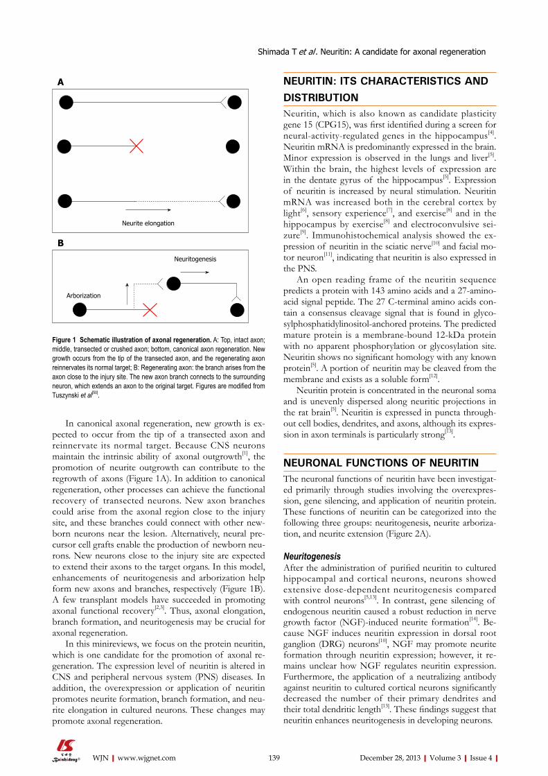
Shimada T et al . Neuritin: A candidate for axonal regeneration
In canonical axonal regeneration, new growth is ex-pected to occur from the tip of a transected axon and reinnervate its normal target. Because CNS neurons maintain the intrinsic ability of axonal outgrowth[1], the promotion of neurite outgrowth can contribute to the regrowth of axons (Figure 1A). In addition to canonical regeneration, other processes can achieve the functional recovery of transected neurons. New axon branches could arise from the axonal region close to the injury site, and these branches could connect with other new-born neurons near the lesion. Alternatively, neural pre-cursor cell grafts enable the production of newborn neu-rons. New neurons close to the injury site are expected to extend their axons to the target organs. In this model, enhancements of neuritogenesis and arborization help form new axons and branches, respectively (Figure 1B). A few transplant models have succeeded in promoting axonal functional recovery[2,3]. Thus, axonal elongation, branch formation, and neuritogenesis may be crucial for axonal regeneration.
In this minireviews, we focus on the protein neuritin, which is one candidate for the promotion of axonal re-generation. The expression level of neuritin is altered in CNS and peripheral nervous system (PNS) diseases. In addition, the overexpression or application of neuritin promotes neurite formation, branch formation, and neu-rite elongation in cultured neurons. These changes may promote axonal regeneration.
NEURITIN: ITS CHARACTERISTICS AND DISTRIBUTIONNeuritin, which is also known as candidate plasticity gene 15 (CPG15), was first identified during a screen for neural-activity-regulated genes in the hippocampus[4]. Neuritin mRNA is predominantly expressed in the brain. Minor expression is observed in the lungs and liver[5]. Within the brain, the highest levels of expression are in the dentate gyrus of the hippocampus[5]. Expression of neuritin is increased by neural stimulation. Neuritin mRNA was increased both in the cerebral cortex by light[6], sensory experience[7], and exercise[8] and in the hippocampus by exercise[8] and electroconvulsive sei-zure[9]. Immunohistochemical analysis showed the ex-pression of neuritin in the sciatic nerve[10] and facial mo-tor neuron[11], indicating that neuritin is also expressed in the PNS.
An open reading frame of the neuritin sequence predicts a protein with 143 amino acids and a 27-amino-acid signal peptide. The 27 C-terminal amino acids con-tain a consensus cleavage signal that is found in glyco-sylphosphatidylinositol-anchored proteins. The predicted mature protein is a membrane-bound 12-kDa protein with no apparent phosphorylation or glycosylation site. Neuritin shows no significant homology with any known protein[5]. A portion of neuritin may be cleaved from the membrane and exists as a soluble form[12].
Neuritin protein is concentrated in the neuronal soma and is unevenly dispersed along neuritic projections in the rat brain[5]. Neuritin is expressed in puncta through-out cell bodies, dendrites, and axons, although its expres-sion in axon terminals is particularly strong[13].
NEURONAL FUNCTIONS OF NEURITINThe neuronal functions of neuritin have been investigat-ed primarily through studies involving the overexpres-sion, gene silencing, and application of neuritin protein. These functions of neuritin can be categorized into the following three groups: neuritogenesis, neurite arboriza-tion, and neurite extension (Figure 2A).
NeuritogenesisAfter the administration of purified neuritin to cultured hippocampal and cortical neurons, neurons showed extensive dose-dependent neuritogenesis compared with control neurons[5,13]. In contrast, gene silencing of endogenous neuritin caused a robust reduction in nerve growth factor (NGF)-induced neurite formation[14]. Be-cause NGF induces neuritin expression in dorsal root ganglion (DRG) neurons[10], NGF may promote neurite formation through neuritin expression; however, it re-mains unclear how NGF regulates neuritin expression. Furthermore, the application of a neutralizing antibody against neuritin to cultured cortical neurons significantly decreased the number of their primary dendrites and their total dendritic length[13]. These findings suggest that neuritin enhances neuritogenesis in developing neurons.
139WJN|www.wjgnet.com December 28, 2013|Volume 3|Issue 4|
A
Neurite elongation
Neuritogenesis
Arborization
B
Figure 1 Schematic illustration of axonal regeneration. A: Top, intact axon; middle, transected or crushed axon; bottom, canonical axon regeneration. New growth occurs from the tip of the transected axon, and the regenerating axon reinnervates its normal target; B: Regenerating axon: the branch arises from the axon close to the injury site. The new axon branch connects to the surrounding neuron, which extends an axon to the original target. Figures are modified from Tuszynski et al[50].
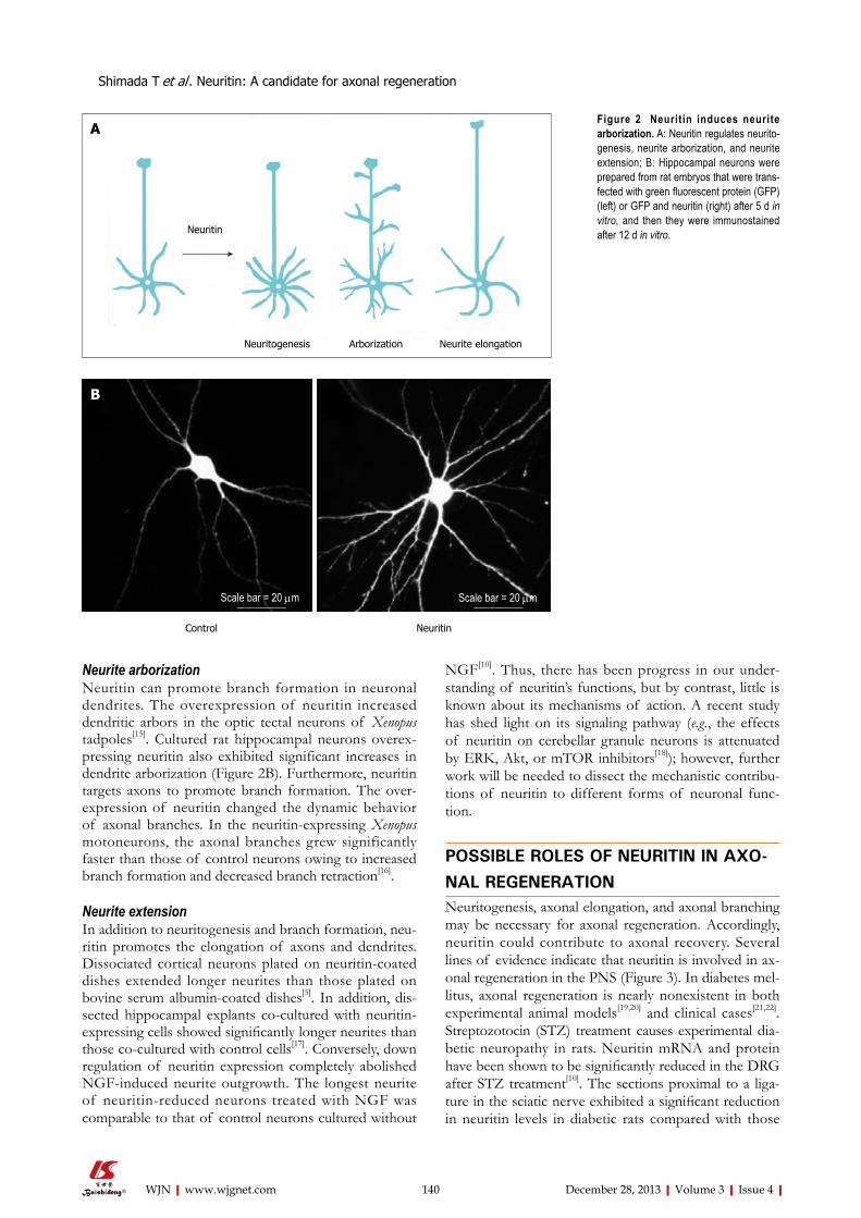
Neurite arborizationNeuritin can promote branch formation in neuronal dendrites. The overexpression of neuritin increased dendritic arbors in the optic tectal neurons of Xenopus tadpoles[15]. Cultured rat hippocampal neurons overex-pressing neuritin also exhibited significant increases in dendrite arborization (Figure 2B). Furthermore, neuritin targets axons to promote branch formation. The over-expression of neuritin changed the dynamic behavior of axonal branches. In the neuritin-expressing Xenopus motoneurons, the axonal branches grew significantly faster than those of control neurons owing to increased branch formation and decreased branch retraction[16].
Neurite extensionIn addition to neuritogenesis and branch formation, neu-ritin promotes the elongation of axons and dendrites. Dissociated cortical neurons plated on neuritin-coated dishes extended longer neurites than those plated on bovine serum albumin-coated dishes[5]. In addition, dis-sected hippocampal explants co-cultured with neuritin-expressing cells showed significantly longer neurites than those co-cultured with control cells[17]. Conversely, down regulation of neuritin expression completely abolished NGF-induced neurite outgrowth. The longest neurite of neuritin-reduced neurons treated with NGF was comparable to that of control neurons cultured without
NGF[10]. Thus, there has been progress in our under-standing of neuritin’s functions, but by contrast, little is known about its mechanisms of action. A recent study has shed light on its signaling pathway (e.g., the effects of neuritin on cerebellar granule neurons is attenuated by ERK, Akt, or mTOR inhibitors[18]); however, further work will be needed to dissect the mechanistic contribu-tions of neuritin to different forms of neuronal func-tion.
POSSIBLE ROLES OF NEURITIN IN AXO-NAL REGENERATIONNeuritogenesis, axonal elongation, and axonal branching may be necessary for axonal regeneration. Accordingly, neuritin could contribute to axonal recovery. Several lines of evidence indicate that neuritin is involved in ax-onal regeneration in the PNS (Figure 3). In diabetes mel-litus, axonal regeneration is nearly nonexistent in both experimental animal models[19,20] and clinical cases[21,22]. Streptozotocin (STZ) treatment causes experimental dia-betic neuropathy in rats. Neuritin mRNA and protein have been shown to be significantly reduced in the DRG after STZ treatment[10]. The sections proximal to a liga-ture in the sciatic nerve exhibited a significant reduction in neuritin levels in diabetic rats compared with those
140WJN|www.wjgnet.com December 28, 2013|Volume 3|Issue 4|
Neuritin
Neurite elongationNeuritogenesis Arborization
A
Shimada T et al . Neuritin: A candidate for axonal regeneration
Figure 2 Neuritin induces neurite arborization. A: Neuritin regulates neurito-genesis, neurite arborization, and neurite extension; B: Hippocampal neurons were prepared from rat embryos that were trans-fected with green fluorescent protein (GFP) (left) or GFP and neuritin (right) after 5 d in vitro, and then they were immunostained after 12 d in vitro.
Control Neuritin
B
Scale bar = 20 μmScale bar = 20 μm
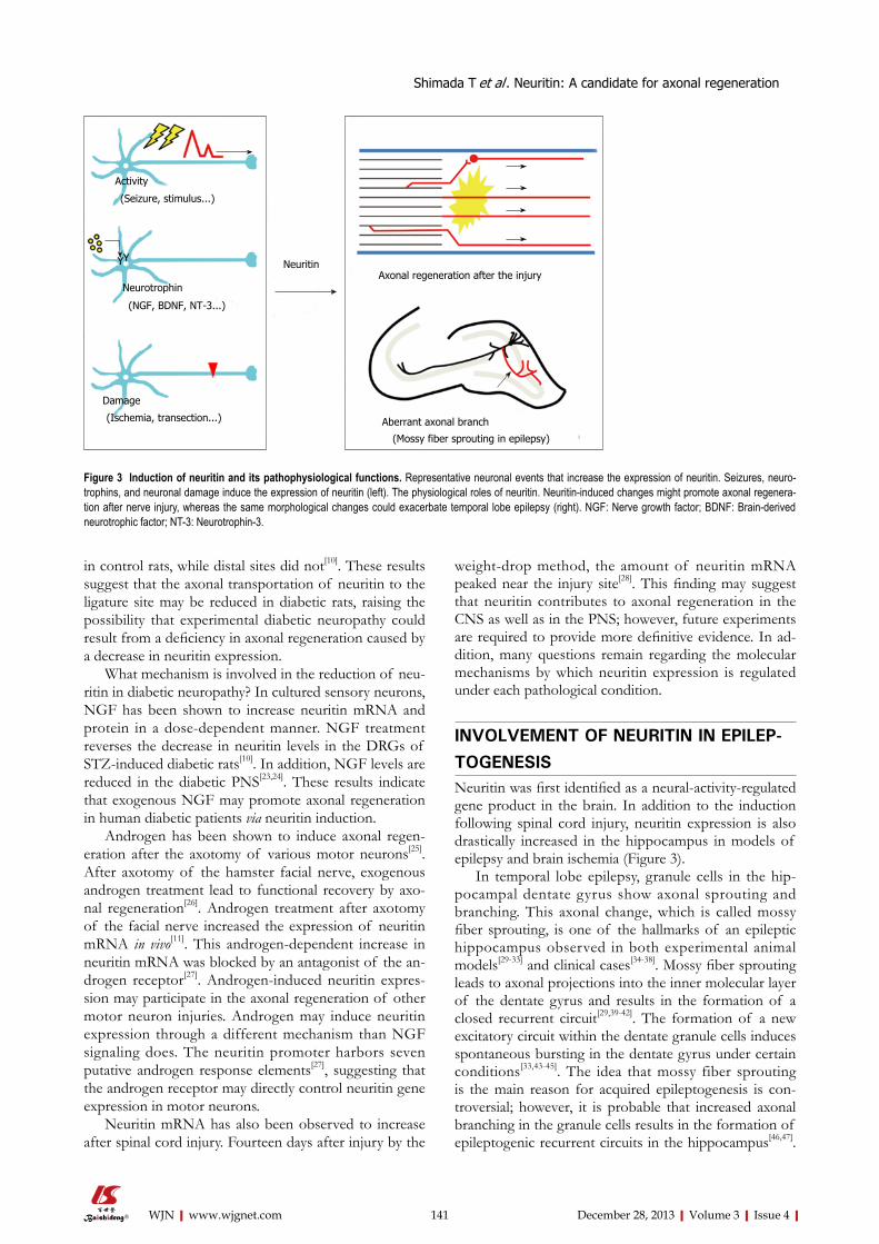
in control rats, while distal sites did not[10]. These results suggest that the axonal transportation of neuritin to the ligature site may be reduced in diabetic rats, raising the possibility that experimental diabetic neuropathy could result from a deficiency in axonal regeneration caused by a decrease in neuritin expression.
What mechanism is involved in the reduction of neu-ritin in diabetic neuropathy? In cultured sensory neurons, NGF has been shown to increase neuritin mRNA and protein in a dose-dependent manner. NGF treatment reverses the decrease in neuritin levels in the DRGs of STZ-induced diabetic rats[10]. In addition, NGF levels are reduced in the diabetic PNS[23,24]. These results indicate that exogenous NGF may promote axonal regeneration in human diabetic patients via neuritin induction.
Androgen has been shown to induce axonal regen-eration after the axotomy of various motor neurons[25]. After axotomy of the hamster facial nerve, exogenous androgen treatment lead to functional recovery by axo-nal regeneration[26]. Androgen treatment after axotomy of the facial nerve increased the expression of neuritin mRNA in vivo[11]. This androgen-dependent increase in neuritin mRNA was blocked by an antagonist of the an-drogen receptor[27]. Androgen-induced neuritin expres-sion may participate in the axonal regeneration of other motor neuron injuries. Androgen may induce neuritin expression through a different mechanism than NGF signaling does. The neuritin promoter harbors seven putative androgen response elements[27], suggesting that the androgen receptor may directly control neuritin gene expression in motor neurons.
Neuritin mRNA has also been observed to increase after spinal cord injury. Fourteen days after injury by the
weight-drop method, the amount of neuritin mRNA peaked near the injury site[28]. This finding may suggest that neuritin contributes to axonal regeneration in the CNS as well as in the PNS; however, future experiments are required to provide more definitive evidence. In ad-dition, many questions remain regarding the molecular mechanisms by which neuritin expression is regulated under each pathological condition.
INVOLVEMENT OF NEURITIN IN EPILEP-TOGENESISNeuritin was first identified as a neural-activity-regulated gene product in the brain. In addition to the induction following spinal cord injury, neuritin expression is also drastically increased in the hippocampus in models of epilepsy and brain ischemia (Figure 3).
In temporal lobe epilepsy, granule cells in the hip-pocampal dentate gyrus show axonal sprouting and branching. This axonal change, which is called mossy fiber sprouting, is one of the hallmarks of an epileptic hippocampus observed in both experimental animal models[29-33] and clinical cases[34-38]. Mossy fiber sprouting leads to axonal projections into the inner molecular layer of the dentate gyrus and results in the formation of a closed recurrent circuit[29,39-42]. The formation of a new excitatory circuit within the dentate granule cells induces spontaneous bursting in the dentate gyrus under certain conditions[33,43-45]. The idea that mossy fiber sprouting is the main reason for acquired epileptogenesis is con-troversial; however, it is probable that increased axonal branching in the granule cells results in the formation of epileptogenic recurrent circuits in the hippocampus[46,47].
141WJN|www.wjgnet.com December 28, 2013|Volume 3|Issue 4|
Figure 3 Induction of neuritin and its pathophysiological functions. Representative neuronal events that increase the expression of neuritin. Seizures, neuro-trophins, and neuronal damage induce the expression of neuritin (left). The physiological roles of neuritin. Neuritin-induced changes might promote axonal regenera-tion after nerve injury, whereas the same morphological changes could exacerbate temporal lobe epilepsy (right). NGF: Nerve growth factor; BDNF: Brain-derived neurotrophic factor; NT-3: Neurotrophin-3.
Neuritin
Activity
(Seizure, stimulus...)
Neurotrophin
(NGF, BDNF, NT-3...)
Damage
(Ischemia, transection...)
Axonal regeneration after the injury
Aberrant axonal branch
(Mossy fiber sprouting in epilepsy)
YY
Shimada T et al . Neuritin: A candidate for axonal regeneration
We have observed that neuritin promotes the arboriza-tion and elongation of mossy fibers in hippocampal slice cultures (unpublished data). This neuritin-induced mossy fiber sprouting may contribute to the formation of closed circuits between the granule cells (Figure 3). In this instance, an upregulation of neuritin may be harmful to brain function. Inhibition of neuritin function could be necessary for the suppression of epileptogenesis in temporal lobe epilepsy.
Transient middle cerebral artery occlusion (MCAO) leads to permanent damage of the brain that affects the lateral striatum and the lateral parietal, temporal and oc-cipital cortex[48]. Two hours after MCAO treatment in rats, an induction of neuritin mRNA was observed in the ipsilateral dentate gyrus of the hippocampus, as well as in the ipsilateral cerebral cortex[49]. The increase in the hippocampus was robust and confined to this area 6 h after reperfusion[49]. In this model, the significance of neuritin induction in the cortex remains unclear; however, the upregulation of neuritin in dentate granule cells might indicate its involvement in the pathogenesis of postischemic epilepsy through mossy fiber sprouting (Figure 3).
Thus, aberrant neuritogenesis, axonal branch forma-tion, and axonal elongation by neuritin could exacerbate neuronal dysfunction in the CNS. Tightly controlled regulation of neuritin expression may be required for the treatment of various CNS diseases.
CONCLUSIONNeuritin is a small extracellular protein that is expressed in neurons, and its expression is upregulated by neuronal activity. The expression of neuritin increases in response to neuronal damage and neurotrophin treatment in the CNS and the PNS. Neuritin expression leads to mor-phological changes in neurons. The induction of neuri-togenesis, the enhancement of neurite arborization, and the promotion of neurite extension are all elicited by neuritin. These morphological changes suggest that neu-ritin can contribute to axonal regeneration after axotomy. A variety of studies provide evidence that neuritin may contribute to axonal regeneration in PNS and CNS inju-ries. In contrast, neuritin induction in the hippocampus in response to epileptic seizures or brain ischemia may aggravate neuronal damage by enhancing mossy fiber sprouting. Thus, tightly controlled regulation of appro-priate neuritin levels will be useful in the treatment of axonal pathology.
REFERENCES1 David S, Aguayo AJ. Axonal elongation into peripheral ner-
vous system “bridges” after central nervous system injury in adult rats. Science 1981; 214: 931-933 [PMID: 6171034]
2 Ribotta MG, Provencher J, Feraboli-Lohnherr D, Rossignol S, Privat A, Orsal D. Activation of locomotion in adult chronic spinal rats is achieved by transplantation of embryonic raphe cells reinnervating a precise lumbar level. J Neurosci 2000; 20: 5144-5152 [PMID: 10864971]
3 Pearse DD, Marcillo AE, Oudega M, Lynch MP, Wood PM, Bunge MB. Transplantation of Schwann cells and olfactory en-sheathing glia after spinal cord injury: does pretreatment with methylprednisolone and interleukin-10 enhance recovery? J Neurotrauma 2004; 21: 1223-1239 [PMID: 15453992]
4 Nedivi E, Hevroni D, Naot D, Israeli D, Citri Y. Numerous can-didate plasticity-related genes revealed by differential cDNA cloning. Nature 1993; 363: 718-722 [PMID: 8515813]
5 Naeve GS, Ramakrishnan M, Kramer R, Hevroni D, Citri Y, Theill LE. Neuritin: a gene induced by neural activity and neu-rotrophins that promotes neuritogenesis. Proc Natl Acad Sci USA 1997; 94: 2648-2653 [PMID: 9122250]
6 Nedivi E, Fieldust S, Theill LE, Hevron D. A set of genes ex-pressed in response to light in the adult cerebral cortex and regulated during development. Proc Natl Acad Sci USA 1996; 93: 2048-2053 [PMID: 8700883]
7 Harwell C, Burbach B, Svoboda K, Nedivi E. Regulation of cpg15 expression during single whisker experience in the barrel cortex of adult mice. J Neurobiol 2005; 65: 85-96 [PMID: 16010668 DOI: 10.1002/neu.20176]
8 Hunsberger JG, Newton SS, Bennett AH, Duman CH, Rus-sell DS, Salton SR, Duman RS. Antidepressant actions of the exercise-regulated gene VGF. Nat Med 2007; 13: 1476-1482 [PMID: 18059283 DOI: 10.1038/nm1669]
9 Newton SS, Collier EF, Hunsberger J, Adams D, Terwilliger R, Selvanayagam E, Duman RS. Gene profile of electroconvulsive seizures: induction of neurotrophic and angiogenic factors. J Neurosci 2003; 23: 10841-10851 [PMID: 14645477]
10 Karamoysoyli E, Burnand RC, Tomlinson DR, Gardiner NJ. Neuritin mediates nerve growth factor-induced axonal regener-ation and is deficient in experimental diabetic neuropathy. Dia-betes 2008; 57: 181-189 [PMID: 17909094 DOI: 10.2337/db07-0895]
11 Fargo KN, Alexander TD, Tanzer L, Poletti A, Jones KJ. An-drogen regulates neuritin mRNA levels in an in vivo model of steroid-enhanced peripheral nerve regeneration. J Neurotrauma 2008; 25: 561-566 [PMID: 18419250 DOI: 10.1089/neu.2007.0466]
12 Putz U, Harwell C, Nedivi E. Soluble CPG15 expressed during early development rescues cortical progenitors from apoptosis. Nat Neurosci 2005; 8: 322-331 [PMID: 15711540 DOI: 10.1038/nn1407]
13 Sato H, Fukutani Y, Yamamoto Y, Tatara E, Takemoto M, Shimamura K, Yamamoto N. Thalamus-derived molecules promote survival and dendritic growth of developing cortical neurons. J Neurosci 2012; 32: 15388-15402 [PMID: 23115177 DOI: 10.1523/JNEUROSCI.0293-12.2012]
14 Cappelletti G, Galbiati M, Ronchi C, Maggioni MG, Onesto E, Poletti A. Neuritin (cpg15) enhances the differentiating effect of NGF on neuronal PC12 cells. J Neurosci Res 2007; 85: 2702-2713 [PMID: 17335086 DOI: 10.1002/jnr.21235]
15 Nedivi E, Wu GY, Cline HT. Promotion of dendritic growth by CPG15, an activity-induced signaling molecule. Science 1998; 281: 1863-1866 [PMID: 9743502 DOI: 10.1126/science.281.5384.1863]
16 Javaherian A, Cline HT. Coordinated motor neuron axon growth and neuromuscular synaptogenesis are promoted by CPG15 in vivo. Neuron 2005; 45: 505-512 [PMID: 15721237 DOI: 10.1016/j.neuron.2004.12.051]
17 Fujino T, Wu Z, Lin WC, Phillips MA, Nedivi E. cpg15 and cpg15-2 constitute a family of activity-regulated ligands ex-pressed differentially in the nervous system to promote neu-rite growth and neuronal survival. J Comp Neurol 2008; 507: 1831-1845 [PMID: 18265009 DOI: 10.1002/cne.21649]
18 Yao JJ, Gao XF, Chow CW, Zhan XQ, Hu CL, Mei YA. Neu-ritin activates insulin receptor pathway to up-regulate Kv4.2-mediated transient outward K+ current in rat cerebellar granule neurons. J Biol Chem 2012; 287: 41534-41545 [PMID: 23066017 DOI: 10.1074/jbc.M112.390260]
19 Bisby MA. Axonal transport of labeled protein and regenera-tion rate in nerves of streptozocin-diabetic rats. Exp Neurol 1980; 69: 74-84 [PMID: 6156080 DOI: 10.1016/0014-4886(80)90144-2]
20 Longo FM, Powell HC, Lebeau J, Gerrero MR, Heckman H,
142WJN|www.wjgnet.com December 28, 2013|Volume 3|Issue 4|
Shimada T et al . Neuritin: A candidate for axonal regeneration
Myers RR. Delayed nerve regeneration in streptozotocin dia-betic rats. Muscle Nerve 1986; 9: 385-393 [PMID: 2941683]
21 Sima AA, Bril V, Nathaniel V, McEwen TA, Brown MB, Lat-timer SA, Greene DA. Regeneration and repair of myelinated fibers in sural-nerve biopsy specimens from patients with dia-betic neuropathy treated with sorbinil. N Engl J Med 1988; 319: 548-555 [PMID: 3136331 DOI: 10.1056/NEJM198809013190905]
22 Britland ST, Young RJ, Sharma AK, Clarke BF. Association of painful and painless diabetic polyneuropathy with differ-ent patterns of nerve fiber degeneration and regeneration. Diabetes 1990; 39: 898-908 [PMID: 2373262]
23 Hellweg R, Hartung HD. Endogenous levels of nerve growth factor (NGF) are altered in experimental diabetes mellitus: a possible role for NGF in the pathogenesis of diabetic neu-ropathy. J Neurosci Res 1990; 26: 258-267 [PMID: 2142224]
24 Anand P, Terenghi G, Warner G, Kopelman P, Williams-Chest-nut RE, Sinicropi DV. The role of endogenous nerve growth factor in human diabetic neuropathy. Nat Med 1996; 2: 703-707 [PMID: 8640566]
25 Fargo KN, Foecking EM, Jones KJ, Sengelaub DR. Neuropro-tective actions of androgens on motoneurons. Front Neuro-endocrinol 2009; 30: 130-141 [PMID: 19393684 DOI: 10.1016/j.yfrne.2009.04.005]
26 Jones KJ, Brown TJ, Damaser M. Neuroprotective effects of gonadal steroids on regenerating peripheral motoneurons. Brain Res Brain Res Rev 2001; 37: 372-382 [PMID: 11744101 DOI: 10.1016/S0165-0173(01)00107-2]
27 Marron TU, Guerini V, Rusmini P, Sau D, Brevini TA, Mar-tini L, Poletti A. Androgen-induced neurite outgrowth is me-diated by neuritin in motor neurones. J Neurochem 2005; 92: 10-20 [PMID: 15606892 DOI: 10.1111/j.1471-4159.2004.02836.x]
28 Di Giovanni S, Faden AI, Yakovlev A, Duke-Cohan JS, Finn T, Thouin M, Knoblach S, De Biase A, Bregman BS, Hoffman EP. Neuronal plasticity after spinal cord injury: identification of a gene cluster driving neurite outgrowth. FASEB J 2005; 19: 153-154 [PMID: 15522907 DOI: 10.1096/fj.04-2694fje]
29 Wenzel HJ, Woolley CS, Robbins CA, Schwartzkroin PA. Kainic acid-induced mossy fiber sprouting and synapse formation in the dentate gyrus of rats. Hippocampus 2000; 10: 244-260 [PMID: 10902894 DOI: 10.1002/1098-1063(2000)10: 3<244: :AID-HIPO5>3.0.CO; 2-7]
30 Kotti T, Riekkinen PJ, Miettinen R. Characterization of tar-get cells for aberrant mossy fiber collaterals in the dentate gyrus of epileptic rat. Exp Neurol 1997; 146: 323-330 [PMID: 9270041 DOI: 10.1006/exnr.1997.6553]
31 Buckmaster PS, Dudek FE. Neuron loss, granule cell axon reorganization, and functional changes in the dentate gyrus of epileptic kainate-treated rats. J Comp Neurol 1997; 385: 385-404 [PMID: 9300766 DOI: 10.1002/(SICI)1096-9861(19970901)385:3<385::AID-CNE4>3.0.CO;2-#]
32 Okazaki MM, Evenson DA, Nadler JV. Hippocampal mossy fiber sprouting and synapse formation after status epilepti-cus in rats: visualization after retrograde transport of biocy-tin. J Comp Neurol 1995; 352: 515-534 [PMID: 7721998]
33 Represa A, Jorquera I, Le Gal La Salle G, Ben-Ari Y. Epi-lepsy induced collateral sprouting of hippocampal mossy fibers: does it induce the development of ectopic synapses with granule cell dendrites? Hippocampus 1993; 3: 257-268 [PMID: 8353609 DOI: 10.1002/hipo.450030303]
34 Kandratavicius L, Hallak JE, Young LT, Assirati JA, Carlotti CG, Leite JP. Differential aberrant sprouting in temporal lobe epi-lepsy with psychiatric co-morbidities. Psychiatry Res 2012; 195: 144-150 [PMID: 21741094 DOI: 10.1016/j.psychres.2011.06.005]
35 Lynd-Balta E, Pilcher WH, Joseph SA. AMPA receptor al-
terations precede mossy fiber sprouting in young children with temporal lobe epilepsy. Neuroscience 2004; 126: 105-114 [PMID: 15145077 DOI: 10.1016/j.neuroscience.2004.03.004]
36 El Bahh B, Lespinet V, Lurton D, Coussemacq M, Le Gal La Sal-le G, Rougier A. Correlations between granule cell dispersion, mossy fiber sprouting, and hippocampal cell loss in temporal lobe epilepsy. Epilepsia 1999; 40: 1393-1401 [PMID: 10528935]
37 Zhang N, Houser CR. Ultrastructural localization of dynorphin in the dentate gyrus in human temporal lobe epilepsy: a study of reorganized mossy fiber synapses. J Comp Neurol 1999; 405: 472-490 [PMID: 10098940 DOI: 10.1002/(SICI)1096-9861(19990322)405:4<472: :AID-CNE3>3.0.CO; 2-P]
38 Mathern GW, Pretorius JK, Babb TL, Quinn B. Unilateral hip-pocampal mossy fiber sprouting and bilateral asymmetric neu-ron loss with episodic postictal psychosis. J Neurosurg 1995; 82: 228-233 [PMID: 7815150]
39 Buckmaster PS, Zhang GF, Yamawaki R. Axon sprouting in a model of temporal lobe epilepsy creates a predominantly ex-citatory feedback circuit. J Neurosci 2002; 22: 6650-6658 [PMID: 12151544]
40 Cavazos JE, Zhang P, Qazi R, Sutula TP. Ultrastructural fea-tures of sprouted mossy fiber synapses in kindled and kainic acid-treated rats. J Comp Neurol 2003; 458: 272-292 [PMID: 12619081 DOI: 10.1002/cne.10581]
41 Isokawa M, Fried I. Extracellular slow negative transient in the dentate gyrus of human epileptic hippocampus in vitro. Neuro-science 1996; 72: 31-37 [PMID: 8730703 DOI: 10.1016/0306-4522(95)00544-7]
42 Patrylo PR, Schweitzer JS, Dudek FE. Abnormal responses to perforant path stimulation in the dentate gyrus of slices from rats with kainate-induced epilepsy and mossy fiber reorganiza-tion. Epilepsy Res 1999; 36: 31-42 [PMID: 10463848 DOI: 10.1016/S0920-1211(99)00022-4]
43 Sutula T, Cascino G, Cavazos J, Parada I, Ramirez L. Mossy fiber synaptic reorganization in the epileptic human temporal lobe. Ann Neurol 1989; 26: 321-330 [PMID: 2508534]
44 Wuarin JP, Dudek FE. Electrographic seizures and new recur-rent excitatory circuits in the dentate gyrus of hippocampal slices from kainate-treated epileptic rats. J Neurosci 1996; 16: 4438-4448 [PMID: 8699254]
45 Patrylo PR, Dudek FE. Physiological unmasking of new gluta-matergic pathways in the dentate gyrus of hippocampal slices from kainate-induced epileptic rats. J Neurophysiol 1998; 79: 418-429 [PMID: 9425210]
46 Hunt RF, Scheff SW, Smith BN. Regionally localized recurrent excitation in the dentate gyrus of a cortical contusion model of posttraumatic epilepsy. J Neurophysiol 2010; 103: 1490-1500 [PMID: 20089815 DOI: 10.1152/jn.00957.2009]
47 Sutula TP, Dudek FE. Unmasking recurrent excitation gen-erated by mossy fiber sprouting in the epileptic dentate gy-rus: an emergent property of a complex system. Prog Brain Res 2007; 163: 541-563 [PMID: 17765737 DOI: 10.1016/S0079-6123(07)63029-5]
48 Memezawa H, Minamisawa H, Smith ML, Siesjö BK. Ischemic penumbra in a model of reversible middle cerebral artery oc-clusion in the rat. Exp Brain Res 1992; 89: 67-78 [PMID: 1601103]
49 Rickhag M, Teilum M, Wieloch T. Rapid and long-term induc-tion of effector immediate early genes (BDNF, Neuritin and Arc) in peri-infarct cortex and dentate gyrus after ischemic in-jury in rat brain. Brain Res 2007; 1151: 203-210 [PMID: 17397810 DOI: 10.1016/j.brainres.2007.03.005]
50 Tuszynski MH, Steward O. Concepts and methods for the study of axonal regeneration in the CNS. Neuron 2012; 74: 777-791 [PMID: 22681683 DOI: 10.1016/j.neuron.2012.05.006]
P- Reviewer: Osaka H S- Editor: Zhai HH L- Editor: A E- Editor: Liu XM
143WJN|www.wjgnet.com December 28, 2013|Volume 3|Issue 4|
Shimada T et al . Neuritin: A candidate for axonal regeneration
MINIREVIEWS
Antiplatelet strategy for acute ischemic stroke: A mini review
Zhong-He Zhou, Hui-Sheng Chen
Zhong-He Zhou, Hui-Sheng Chen, Department of Neurol-ogy, General Hospital of Shen-Yang Military Region, Shenyang 110840, Liaoning Province, ChinaAuthor contributions: Zhou ZH contributed to writing the manuscript; Chen HS contributed to designing and revising the manuscript.Supported by The Science and Technology Project Plan of Liao Ning Province, No. 2011225021; the Key Project of the National 12th Five-Year Research Program of China, No. 2012ZX09303016-002Correspondence to: Hui-Sheng Chen, MD, Professor, De-partment of Neurology, General Hospital of Shen-Yang Military Region, 83 Wenhua Road, Shenyang 110840, Liaoning Prov-ince, China. [email protected]: +86-24-28897511 Fax: +86-24-28856448Received: July 2, 2013 Revised: August 9, 2013Accepted: September 14, 2013Published online: December 28, 2013
AbstractTransient ischemic attacks and minor ischemic strokes have a high risk of an unstable clinical course in the initial 48-72 h after symptom onset. Early antiplate-let treatment is recommended to treat most patients with acute ischemic stroke because few patients can be treated with thrombolysis due to the limit of strict indications, such as a time window. Antiplatelets aim to prevent recurrence or deterioration of stroke. The guidelines recommend the use of aspirin in the acute stage based on two clinical trials. However, some patients still developed recurrence or deterioration of stroke despite timely aspirin administration. Thus, the question remains unclear whether another effec-tive and safe antiplatelet strategy for the treatment of acute ischemic stroke exists. Growing evidence shows that combination antiplatelets may be superior to mono antiplatelets in the treatment of acute ischemic stroke.
© 2013 Baishideng Publishing Group Co., Limited. All rights reserved.
Key words: Antiplatelet; Acute ischemic stroke
Core tip: Patients with acute ischemic stroke have a high risk of deterioration in the 48-72 h after symptom onset. Although thrombolysis is an effective method, most patients are excluded due to the limit of strict indications. Early antiplatelets are recommended for most patients. However, the question remains unclear whether another effective and safe antiplatelet strat-egy for the treatment of acute ischemic stroke exists. Growing evidence shows that combination antiplatelets may be superior to mono antiplatelets in the treatment of acute ischemic stroke.
Zhou ZH, Chen HS. Antiplatelet strategy for acute ischemic stroke: a mini review. World J Neurol 2013; 3(4): 144-147 Available from: URL: http://www.wjgnet.com/2218-6212/full/v3/i4/144.htm DOI: http://dx.doi.org/10.5316/wjn.v3.i4.144
INTRODUCTIONIn the acute stage of ischemic stroke, one of the most important treatment strategies aims to prevent recur-rence or deterioration of stroke, because patients with transient ischemic attack (TIA) and minor ischemic stroke have a high risk of an unstable clinical course. Most of these unfavorable events occur in the initial 48-72 h after symptom onset and have obvious effects on the recovery of neurological functions in these pa-tients[1-4]. It is well demonstrated that thrombolysis is an effective method to treat acute ischemic stroke. Due to the limit of strict indications, such as a time window, most patients are excluded for thrombolysis. Thus, early antiplatelet treatment is recommended to treat most patients with acute ischemic stroke in the guidelines[5,6]. In acute ischemic stroke, 2 mega trials established the efficacy of aspirin[7,8]; thus, the guidelines recommend the use of aspirin in the acute stage[5,6]. However, some patients develop recurrence or deterioration of stroke despite timely aspirin administration. Thus, the ques-tion remains unclear whether another effective and safe
World Journal of NeurologyW J N
144WJN|www.wjgnet.com December 28, 2013|Volume 3|Issue 4|
Online Submissions: http://www.wjgnet.com/esps/[email protected]:10.5316/wjn.v3.i4.144
World J Neurol 2013 December 28; 3(4): 144-147ISSN 2218-6212 (online)
© 2013 Baishideng Publishing Group Co., Limited. All rights reserved.
Zhou ZH et al . Antiplatelets for acute ischemic stroke
antiplatelet strategy for the treatment of acute ischemic stroke exists.
In this article, we will briefly review the following topics: (1) the action mechanisms of different antiplate-let agents; (2) the evidence for antiplatelet therapy for acute ischemic stroke; and (3) the efficacy and safety of combination antiplatelet agents for acute ischemic stroke.
ANTIPLATELET DRUGS AND MECHA-NISMSCommonly used antiplatelet agents include aspirin, clopi-dogrel, cilostazol and dipyridamole. These agents act at different sites of the platelet aggregation pathway to inhibit platelet activation. Aspirin is a non-selective and irreversible cyclooxygenase (COX) inhibitor, which selectively acetylates the hydroxyl group of the COX enzyme, thus inhibiting conversion of arachidonate to prostaglandin G2/H2 and thromboxane A2. This leads to irreversible inhibition of platelet aggregation[9].
Clopidogrel is a thienopyridine derivative that pre-vents ADP-induced platelet activation and aggregation by irreversibly inhibiting the binding of adenosine 5-di-phosphate to glycoprotein Ⅱb/Ⅲa, with the result of preventing the binding of fibrinogen to this receptor[10].
Cilostazol is an inhibitor of phosphodiesterase 3 (PDE3) which results in increased concentrations of in-tracellular cAMP[11]. Cilostazol inhibits primary and sec-ondary platelet aggregation and high-shear stress platelet aggregation both in vivo and in vitro[12].
Dipyridamole is postulated to have multiple mecha-nisms of action. On one hand, its inhibition of platelet phosphodiesterase produces an increase in intraplatelet cyclic adenosine monophosphate level, resulting in the potentiation of the platelet inhibitory actions of prosta-cyclin. On the other hand, it also acts by directly releas-ing eicosanoid from vascular endothelium and inhibiting cellular uptake and metabolism of adenosine. This leads to the inhibition of platelet aggregation again[13].
ANTIPLATELET THERAPY FOR ACUTE ISCHEMIC STROKEA lot of studies have investigated the efficacy and safety of antiplatelet treatment in the secondary prevention of ischemic stroke[14]; however, only a few studies have investigated the efficacy and safety of antiplatelet treat-ment in acute ischemic stroke[7,8,15,16]. Of these studies, there are two powerful clinical trials: The Chinese Acute Stroke Trial (CAST)[8] and the International Stroke Trial (IST)[7]. In the CAST, 21,106 patients with acute ischemic stroke were enrolled in the clinical trial. Aspirin treat-ment (160 mg/d) was started within 48 h of the onset of a suspected acute ischemic stroke and continued in hospital for up to 4 wk. The results showed that aspirin treatment produced a significant reduction in mortality. Also, significantly fewer recurrent ischemic strokes oc-
curred in the aspirin group compared with the placebo-allocated group, but slightly more hemorrhagic strokes. In the IST, 19435 patients with acute ischemic stroke within 48 h of symptom onset were enrolled. Anti-thrombotic therapy was started as soon as possible after stroke onset. The results showed that aspirin treatment significantly reduced death or recurrent ischemic strokes.
In addition to the two clinical trials, there are some completed clinical trials that investigated the antiplatelet strategy for acute ischemic stroke[15,16]. In the FASTER (the fast assessment of stroke and transient ischemic at-tack to prevent early recurrence) trial, 392 patients with TIA or minor stroke within 24 h of symptom onset were enrolled[15]. The trial was designed to investigate the effect of the combination of aspirin + clopidogrel ver-sus aspirin monotherapy (with or without simvastatin) on the risk of recurrent stoke within 90 d after a TIA or minor stroke. The results showed the trend to lower recurrence or deterioration of stroke in the acute stage in the dual therapy group compared with mono anti-platelets. Another EARLY trial compared the safety and efficacy of aspirin + dipyridamole initiated within 24 h of stroke or TIA with that of aspirin + dipyridamole initiated after 7 d of aspirin monotherapy[16]. The results showed that early initiation of aspirin plus extended-release dipyridamole did not lead to differing disability at 90 d compared with late initiation, but the proportion of patients in the early initiation group who had no or mild disability on day 90 was higher than that in the late initiation group. There was also no significant difference in the composite adverse event endpoint between the two groups. These studies demonstrated the efficacy and safety of combined antiplatelet treatment in acute isch-emic stroke. Furthermore, the proposal also got support from the CLAIR study[17]. The study aimed to investigate whether treatment with clopidogrel plus aspirin reduced the number of microembolic signals detected with transcranial doppler ultrasound compared with aspirin alone in patients with recent stroke. The results showed that combination therapy with clopidogrel and aspirin is more effective than aspirin alone in reducing microem-bolic signals in patients with predominantly intracranial symptomatic stenosis.
In other clinical trials aiming to investigate the sec-ondary prevention for ischemic stroke, antiplatelet treat-ment was applied to some patients with acute ischemic stroke. For example, The ESPRIT Study compared aspirin with aspirin plus dipyridamole for secondary prevention after an ischemic stroke or transient ischemic attack[18]. The study recruited 96 patients during the acute stage of ischemic stroke and TIA. The Prevention Regimen for Effectively Avoiding Second Strokes (PRo-FESS) trial is designed to compared extended-release dipyridamole plus aspirin vs clopidogrel in secondary stroke prevention[19]. The study recruited 1360 patients during the acute stage of ischemic stroke. These studies also demonstrated the efficacy and safety of combined antiplatelet treatment. Furthermore, a recent meta-anal-ysis study show that dual antiplatelet therapy appears to
145WJN|www.wjgnet.com December 28, 2013|Volume 3|Issue 4|
be safe and effective in reducing stroke recurrence and combined vascular events in patients with acute isch-emic stroke or transient ischemic attack compared with monotherapy[20].
One powerful evidence comes from the Clopidogrel in High-risk Patients with Acute Nondisabling Cerebro-vascular Events (CHANCE) study, which was just pub-lished in the New England Journal of Medicine[21]. The study showed that among patients with a TIA or minor stroke who can be treated within 24 h after the onset of symp-toms, the combination of clopidogrel and aspirin is supe-rior to aspirin alone for reducing the risk of stroke in the first 90 d and does not increase the risk of hemorrhage.
BLEEDING EVENTS CAUSED BY ANTI-PLATELETSWhen considering using more intensive antiplatelet ther-apy, the balance between reducing recurrent stroke and increasing major bleeding events needs to be considered. Previous studies found that combination antiplatelet increased the bleeding events compared with mono an-tiplatelets. For example, the PRoFESS study found that aspirin plus extended-release dipyridamole produced more major hemorrhagic events than clopidogrel, in-cluding intracranial hemorrhage[22]. The MATCH study showed that life-threatening bleeding was higher in the group receiving aspirin and clopidogrel vs clopidogrel alone[23]. However, some studies found no difference between combination antiplatelets and mono antiplate-lets, for example, the EARLY, FASTER and CLAIR trials[15-17]. More importantly, the recent CHANCE study also did not find more bleeding events with combined antiplatelets compared with mono antiplatelets[21]. The discrepancy could be due to the duration of antiplatelet treatment. Obviously, combination antiplatelets in the long-term trials such as PRoFESS and MATCH[22,23] produced more bleeding events. Thus, a time window of combination antiplatelets should exist at which risk out-weighs benefit. Taken together, the evidence shows that combination antiplatelets in a short-term time window (maybe less than 3 wk) should be safe.
CONCLUSIONAs discussed above, although IST and CAST provided the most elegant evidence for aspirin in the acute stage of ischemic stroke, growing evidence, including CHANCE, points to the superiority of combination antiplatelets over mono antiplatelets. On one hand, due to their dif-ferent mechanisms of action, it is postulated that com-bined antiplatelet agents may provide different degrees of vascular protection. On the other hand, the superior-ity of combination therapy compared to monotherapy may be due to the synergy effects of different anti-platelet mechanisms in reducing vascular events. Thus, combined antiplatelet therapy, such as aspirin and clopi-dogrel, should be encouraged in the treatment of acute ischemic stroke.
REFERENCES1 Johnston SC, Gress DR, Browner WS, Sidney S. Short-term
prognosis after emergency department diagnosis of TIA. JAMA 2000; 284: 2901-2906 [PMID: 11147987 DOI: 10.1001/jama.284.22.2901]
2 Lovett JK, Dennis MS, Sandercock PA, Bamford J, War-low CP, Rothwell PM. Very early risk of stroke after a first transient ischemic attack. Stroke 2003; 34: e138-e140 [PMID: 12855835 DOI: 10.1161/01.STR.0000080935.01264.91]
3 Hill MD, Yiannakoulias N, Jeerakathil T, Tu JV, Svenson LW, Schopflocher DP. The high risk of stroke immedi-ately after transient ischemic attack: a population-based study. Neurology 2004; 62: 2015-2020 [PMID: 15184607 DOI: 10.1212/01.WNL.0000129482.70315.2F]
4 Coull AJ, Lovett JK, Rothwell PM. Population based study of early risk of stroke after transient ischaemic attack or minor stroke: implications for public education and organi-sation of services. BMJ 2004; 328: 326 [PMID: 14744823 DOI: 10.1136/bmj.37991.635266.44]
5 Adams RJ, Albers G, Alberts MJ, Benavente O, Furie K, Goldstein LB, Gorelick P, Halperin J, Harbaugh R, Johnston SC, Katzan I, Kelly-Hayes M, Kenton EJ, Marks M, Sacco RL, Schwamm LH. Update to the AHA/ASA recommenda-tions for the prevention of stroke in patients with stroke and transient ischemic attack. Stroke 2008; 39: 1647-1652 [PMID: 18322260 DOI: 10.1161/STROKEAHA.107.189063]
6 European Stroke Organisation (ESO) Executive Committee; ESO Writing Committee. Guidelines for management of ischaemic stroke and transient ischaemic attack 2008. Cerebrovasc Dis 2008; 25: 457-507 [PMID: 18477843 DOI: 10.1159/000131083]
7 The International Stroke Trial (IST): a randomised trial of as-pirin, subcutaneous heparin, both, or neither among 19435 patients with acute ischaemic stroke. International Stroke Trial Collaborative Group. Lancet 1997; 349: 1569-1581 [PMID: 9174558 DOI: 10.1016/S0140-6736(97)04011-7]
8 CAST: randomised placebo-controlled trial of early aspirin use in 20,000 patients with acute ischaemic stroke. CAST (Chinese Acute Stroke Trial) Collaborative Group. Lancet 1997; 349: 1641-1649 [PMID: 9186381 DOI: 10.1016/S0140-6736(97)04010-5]
9 Vane JR, Botting RM. The mechanism of action of aspi-rin. Thromb Res 2003; 110: 255-258 [PMID: 14592543 DOI: 10.1016/S0049-3848(03)00379-7]
10 Savi P, Nurden P, Nurden AT, Levy-Toledano S, Herbert JM. Clopidogrel: a review of its mechanism of action. Platelets 1998; 9: 251-255 [PMID: 16793712 DOI: 10.1080/09537109876799]
11 Bradbury AW. The role of cilostazol (Pletal) in the manage-ment of intermittent claudication. Int J Clin Pract 2003; 57: 405-409 [PMID: 12846346]
12 Reilly MP, Mohler ER. Cilostazol: treatment of intermit-tent claudication. Ann Pharmacother 2001; 35: 48-56 [PMID: 11197586 DOI: 10.1345/aph.19408]
13 Harker LA, Kadatz RA. Mechanism of action of dipyridam-ole. Thromb Res Suppl 1983; 4: 39-46 [PMID: 6356465 DOI: 10.1016/0049-3848(83)90356-0]
14 Tanne D. Stroke: secondary stroke prevention--personalized antiplatelet therapy. Nat Rev Neurol 2012; 8: 536-537 [PMID: 22926313 DOI: 10.1038/nrneurol.2012.180]
15 Kennedy J, Hill MD, Ryckborst KJ, Eliasziw M, Demchuk AM, Buchan AM. Fast assessment of stroke and transient ischaemic attack to prevent early recurrence (FASTER): a ran-domised controlled pilot trial. Lancet Neurol 2007; 6: 961-969 [PMID: 17931979 DOI: 10.1016/S1474-4422(07)70250-8]
16 Dengler R, Diener HC, Schwartz A, Grond M, Schumacher H, Machnig T, Eschenfelder CC, Leonard J, Weissenborn K, Kastrup A, Haberl R. Early treatment with aspirin plus extended-release dipyridamole for transient ischaemic at-tack or ischaemic stroke within 24 h of symptom onset (EARLY trial): a randomised, open-label, blinded-endpoint
146WJN|www.wjgnet.com December 28, 2013|Volume 3|Issue 4|
Zhou ZH et al . Antiplatelets for acute ischemic stroke
trial. Lancet Neurol 2010; 9: 159-166 [PMID: 20060783 DOI: 10.1016/S1474-4422(09)70361-8]
17 Wong KS, Chen C, Fu J, Chang HM, Suwanwela NC, Huang YN, Han Z, Tan KS, Ratanakorn D, Chollate P, Zhao Y, Koh A, Hao Q, Markus HS. Clopidogrel plus aspirin versus aspirin alone for reducing embolisation in patients with acute symptomatic cerebral or carotid artery stenosis (CLAIR study): a randomised, open-label, blinded-endpoint trial. Lancet Neurol 2010; 9: 489-497 [PMID: 20335070 DOI: 10.1016/S1474-4422(10)70060-0]
18 Halkes PH, van Gijn J, Kappelle LJ, Koudstaal PJ, Algra A. Aspirin plus dipyridamole versus aspirin alone after cerebral ischaemia of arterial origin (ESPRIT): randomised controlled trial. Lancet 2006; 367: 1665-1673 [PMID: 16714187 DOI: 10.1016/S0140-6736(06)68734-5]
19 Bath PM, Cotton D, Martin RH, Palesch Y, Yusuf S, Sacco R, Diener HC, Estol C, Roberts R. Effect of combined aspirin and extended-release dipyridamole versus clopidogrel on functional outcome and recurrence in acute, mild ischemic stroke: PRoFESS subgroup analysis. Stroke 2010; 41: 732-738 [PMID: 20181679 DOI: 10.1161/STROKEAHA.109.564906]
20 Geeganage CM, Diener HC, Algra A, Chen C, Topol EJ, Dengler R, Markus HS, Bath MW, Bath PM. Dual or mono antiplatelet therapy for patients with acute ischemic stroke or
transient ischemic attack: systematic review and meta-analy-sis of randomized controlled trials. Stroke 2012; 43: 1058-1066 [PMID: 22282894 DOI: 10.1161/STROKEAHA.111.637686]
21 Wang Y, Wang Y, Zhao X, Liu L, Wang D, Wang C, Wang C, Li H, Meng X, Cui L, Jia J, Dong Q, Xu A, Zeng J, Li Y, Wang Z, Xia H, Johnston SC. Clopidogrel with aspirin in acute minor stroke or transient ischemic attack. N Engl J Med 2013; 369: 11-19 [PMID: 23803136 DOI: 10.1056/NEJMoa1215340]
22 Sacco RL, Diener HC, Yusuf S, Cotton D, Ounpuu S, Lawton WA, Palesch Y, Martin RH, Albers GW, Bath P, Bornstein N, Chan BP, Chen ST, Cunha L, Dahlöf B, De Keyser J, Donnan GA, Estol C, Gorelick P, Gu V, Hermansson K, Hilbrich L, Kaste M, Lu C, Machnig T, Pais P, Roberts R, Skvortsova V, Teal P, Toni D, Vandermaelen C, Voigt T, Weber M, Yoon BW. Aspirin and extended-release dipyridamole versus clopidogrel for recurrent stroke. N Engl J Med 2008; 359: 1238-1251 [PMID: 18753638 DOI: 10.1056/NEJMoa0805002]
23 Diener HC, Bogousslavsky J, Brass LM, Cimminiello C, Csiba L, Kaste M, Leys D, Matias-Guiu J, Rupprecht HJ. Aspirin and clopidogrel compared with clopidogrel alone after recent ischaemic stroke or transient ischaemic attack in high-risk patients (MATCH): randomised, double-blind, placebo-controlled trial. Lancet 2004; 364: 331-337 [PMID: 15276392 DOI: 10.1016/S0140-6736(04)16721-4]
P- Reviewers: Cerase A, Zhang M S- Editor: Zhai HH L- Editor: Roemmele A E- Editor: Liu XM
147WJN|www.wjgnet.com December 28, 2013|Volume 3|Issue 4|
Zhou ZH et al . Antiplatelets for acute ischemic stroke
on the one hand, FMR1 premutation alleles might cause two phenotypes of parkinsonism, such as a presynaptic phenotype, indistinguishable from PD, and a postsyn-aptic phenotype, associated with clinical features of FXTAS. On the other hand, although FMR1 gray zone alleles carriers were believed to have no abnormal neu-rological conditions, our study supports that they could develop FXTAS and other neurological disorders such as orthostatic tremor which has not been reported be-fore associated with the FMR1 gene.
© 2013 Baishideng Publishing Group Co., Limited. All rights reserved.
Key words: Fragile X-associated tremor/ataxia syn-drome; Fragile X mental retardation; Gray zone; Par-kinsonism; Orthostatic tremor
Core tip: In this study we report two atypical cases as-sociated with CGG expansions of the fragile X mental retardation 1 (FMR1) gene. First, a FMR1 premutation alleles carrier presented with a parkinsonism indistin-guishable from Parkinson disease. Second, a FMR1 gray zone alleles carrier presented with orthostatic tremor and neurological features associated with the fragile X-associated tremor/ataxia syndrome, such as hand tremor, parkinsonism and ataxia.
Peña E, Llanero M. Atypical neurological symptoms associ-ated with CGG expansions of the FMR1 gene. World J Neu-rol 2013; 3(4): 148-151 Available from: URL: http://www.wjgnet.com/2218-6212/full/v3/i4/148.htm DOI: http://dx.doi.org/10.5316/wjn.v3.i4.148
INTRODUCTIONMore than 40 CGG expansions in the 5’ noncoding re-gion of the fragile X mental retardation 1 (FMR1) gene
Atypical neurological symptoms associated with CGG expansions of the FMR1 gene
Esteban Peña, Marcos Llanero
Esteban Peña, Marcos Llanero, Department of Neurology, Hospital Sanitas La Moraleja, 28050 Madrid, SpainAuthor contributions: Peña E contributed to writing and reviewing the manuscript; Llanero M contributed to neuro-psychological assessment and reviewing the manuscript.Correspondence to: Esteban Peña, MD, Department of Neurology, Hospital Sanitas La Moraleja, Avenida Fran-cisco Pi y Margall no 81, 28050 Madrid, Spain. [email protected]: +34-917-679100 Fax: +34-917-679346Received: June 21, 2013 Revised: September 16, 2013Accepted: October 11, 2013Published online: December 28, 2013
AbstractMore than 40 CGG expansions in the 5’ noncoding region of the fragile X mental retardation 1 (FMR1 ) gene of the X chromosome give rise to several dis-tinct clinical phenotypes, depending on the size of the expansion. First, more than 200 CGG expansions (full mutation) cause an inherited mental retardation called fragile X syndrome. Second, CGG expansions between 55 and 199 (premutation) cause a disorder called frag-ile X-associated tremor/ataxia syndrome (FXTAS) which typically includes intention tremor, ataxia and specific magnetic resonance imaging (MRI) findings. Indeed, it could develop parkinsonism although it usually shows features of postsynaptic parkinsonism. Finally, CGG expansions between 41 and 54 CGG (gray zone) are not consider normal but rarely develops abnormal neu-rological conditions. In this sense, the aim of this study is to report two atypical cases associated with CGG expansions of the FMR1 gene. First, a FMR1 premuta-tion alleles carrier with an unusual phenotype, such as a presynaptic parkinsonism indistinguishable from Par-kinson disease (PD) and a FMR1 gray zone alleles car-rier presented with neurological features, namely hand tremor, parkinsonism and ataxia, usually described in FXTAS, as well as orthostatic tremor. We conclude that,
CASE REPORT
148 December 28, 2013|Volume 3|Issue 4|WJN|www.wjgnet.com
World Journal of NeurologyW J N
Online Submissions: http://www.wjgnet.com/esps/[email protected]:10.5316/wjn.v3.i4.148
World J Neurol 2013 December 28; 3(4): 148-151ISSN 2218-6212 (online)
© 2013 Baishideng Publishing Group Co., Limited. All rights reserved.
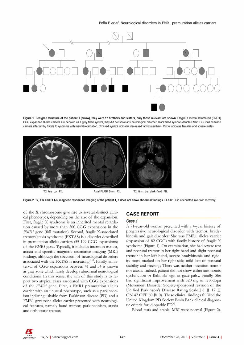
of the X chromosome give rise to several distinct clini-cal phenotypes, depending on the size of the expansion. First, fragile X syndrome is an inherited mental retarda-tion caused by more than 200 CGG expansions in the FMR1 gene (full mutation). Second, fragile X-associated tremor/ataxia syndrome (FXTAS) is a disorder described in premutation alleles carriers (55-199 CGG expansions) of the FMR1 gene. Typically, it includes intention tremor, ataxia and specific magnetic resonance imaging (MRI) findings, although the spectrum of neurological disorders associated with the FXTAS is increasing[1-3]. Finally, an in-terval of CGG expansions between 41 and 54 is known as gray zone which rarely develops abnormal neurological conditions. In this sense, the aim of this study is to re-port two atypical cases associated with CGG expansions of the FMR1 gene. First, a FMR1 premutation alleles carrier with an unusual phenotype, such as a parkinson-ism indistinguishable from Parkinson disease (PD) and a FMR1 gray zone alleles carrier presented with neurologi-cal features, namely hand tremor, parkinsonism, ataxia and orthostatic tremor.
CASE REPORTCase 1A 71-year-old woman presented with a 4-year history of progressive neurological disorder with tremor, brady-kinesia and gait disorder. She was FMR1 alleles carrier (expansion of 82 CGG) with family history of fragile X syndrome (Figure 1). On examination, she had severe rest and postural tremor in her right hand and slight postural tremor in her left hand, severe bradykinesia and rigid-ity more marked on her right side, mild lost of postural stability and freezing. There was neither intention tremor nor ataxia. Indeed, patient did not show either autonomic dysfunction or Babinski sign or gaze palsy. Finally, She had significant improvement with 520 mg of levodopa (Movement Disorder Society-sponsored revision of the Unified Parkinson's Disease Rating Scale Ⅰ 8 Ⅱ 17 Ⅲ ON 42 OFF 60 Ⅳ 0). These clinical findings fulfilled the United Kingdom PD Society Brain Bank clinical diagnos-tic criteria for idiopathic PD[4].
Blood tests and cranial MRI were normal (Figure 2).
Figure 1 Pedigree structure of the patient 1 (arrow), they were 12 brothers and sisters, only those relevant are shown. Fragile X mental retardation (FMR1) CGG expanded alleles carriers are denoted as a grey filled symbol, they did not show any neurological disorder. Black filled symbols denote FMR1 CGG full mutation carriers affected by fragile X syndrome with mental retardation. Crossed symbol indicates deceased family members. Circle indicates females and square males.
T2_tse_cor_FIL Axial FLAIR 5mm_FIL T2_tirm_tra_dark-fluid_FIL
Figure 2 T2, TIR and FLAIR magnetic resonance imaging of the patient 1, it does not show abnormal findings. FLAIR: Fluid attenuated inversion recovery.
149 December 28, 2013|Volume 3|Issue 4|WJN|www.wjgnet.com
Peña E et al . Neurological disorders in FMR1 premutation alleles carriers
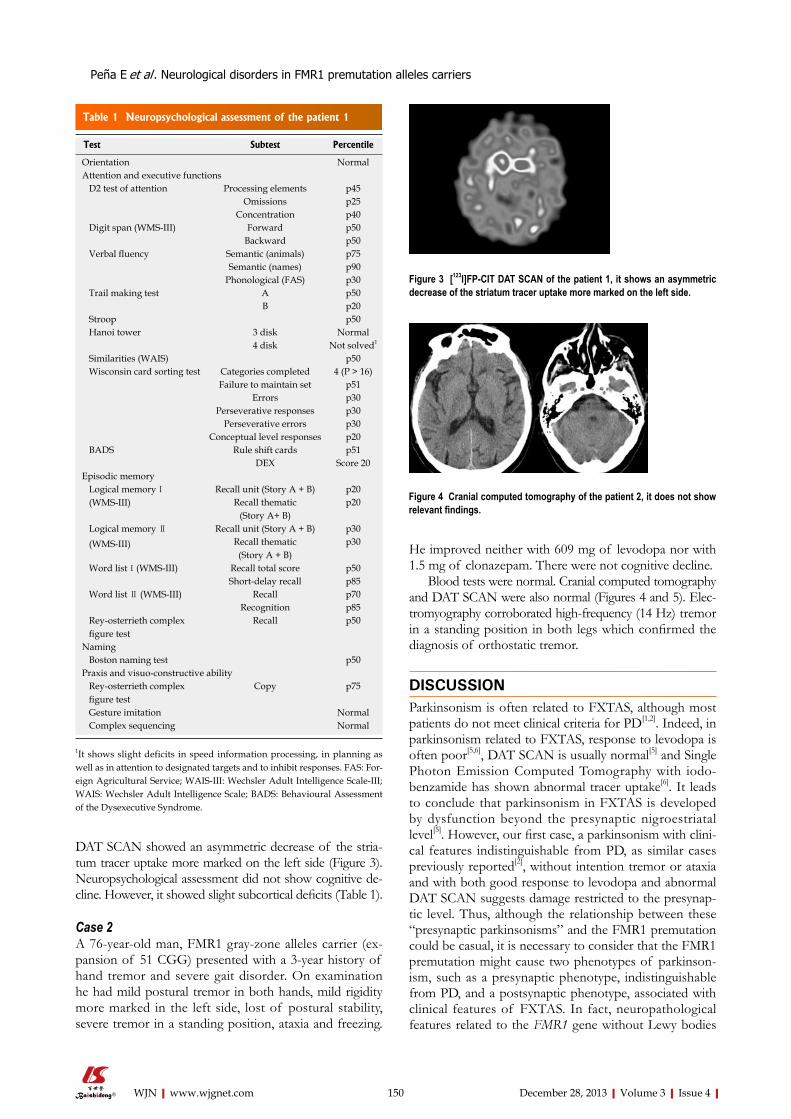
DAT SCAN showed an asymmetric decrease of the stria-tum tracer uptake more marked on the left side (Figure 3). Neuropsychological assessment did not show cognitive de-cline. However, it showed slight subcortical deficits (Table 1).
Case 2A 76-year-old man, FMR1 gray-zone alleles carrier (ex-pansion of 51 CGG) presented with a 3-year history of hand tremor and severe gait disorder. On examination he had mild postural tremor in both hands, mild rigidity more marked in the left side, lost of postural stability, severe tremor in a standing position, ataxia and freezing.
He improved neither with 609 mg of levodopa nor with 1.5 mg of clonazepam. There were not cognitive decline.
Blood tests were normal. Cranial computed tomography and DAT SCAN were also normal (Figures 4 and 5). Elec-tromyography corroborated high-frequency (14 Hz) tremor in a standing position in both legs which confirmed the diagnosis of orthostatic tremor.
DISCUSSIONParkinsonism is often related to FXTAS, although most patients do not meet clinical criteria for PD[1,2]. Indeed, in parkinsonism related to FXTAS, response to levodopa is often poor[5,6], DAT SCAN is usually normal[5] and Single Photon Emission Computed Tomography with iodo-benzamide has shown abnormal tracer uptake[6]. It leads to conclude that parkinsonism in FXTAS is developed by dysfunction beyond the presynaptic nigroestriatal level[5]. However, our first case, a parkinsonism with clini-cal features indistinguishable from PD, as similar cases previously reported[2], without intention tremor or ataxia and with both good response to levodopa and abnormal DAT SCAN suggests damage restricted to the presynap-tic level. Thus, although the relationship between these “presynaptic parkinsonisms” and the FMR1 premutation could be casual, it is necessary to consider that the FMR1 premutation might cause two phenotypes of parkinson-ism, such as a presynaptic phenotype, indistinguishable from PD, and a postsynaptic phenotype, associated with clinical features of FXTAS. In fact, neuropathological features related to the FMR1 gene without Lewy bodies
150 December 28, 2013|Volume 3|Issue 4|WJN|www.wjgnet.com
Test Subtest Percentile
Orientation Normal Attention and executive functions D2 test of attention Processing elements p45
Omissions p25Concentration p40
Digit span (WMS-III) Forward p50Backward p50
Verbal fluency Semantic (animals) p75Semantic (names) p90
Phonological (FAS) p30 Trail making test A p50
B p20 Stroop p50 Hanoi tower 3 disk Normal
4 disk Not solved1
Similarities (WAIS) p50 Wisconsin card sorting test Categories completed 4 (P > 16)
Failure to maintain set p51Errors p30
Perseverative responses p30Perseverative errors p30
Conceptual level responses p20 BADS Rule shift cards p51
DEX Score 20 Episodic memory Logical memory Ⅰ (WMS-III)
Recall unit (Story A + B) p20Recall thematic
(Story A+ B)p20
Logical memory Ⅱ (WMS-III)
Recall unit (Story A + B) p30Recall thematic (Story A + B)
p30
Word list Ⅰ (WMS-III) Recall total score p50Short-delay recall p85
Word list Ⅱ (WMS-III) Recall p70Recognition p85
Rey-osterrieth complex figure test
Recall p50
Naming Boston naming test p50 Praxis and visuo-constructive ability Rey-osterrieth complex figure test
Copy p75
Gesture imitation Normal Complex sequencing Normal
Table 1 Neuropsychological assessment of the patient 1
Figure 4 Cranial computed tomography of the patient 2, it does not show relevant findings.
Figure 3 [123I]FP-CIT DAT SCAN of the patient 1, it shows an asymmetric decrease of the striatum tracer uptake more marked on the left side.
1It shows slight deficits in speed information processing, in planning as well as in attention to designated targets and to inhibit responses. FAS: For-eign Agricultural Service; WAIS-III: Wechsler Adult Intelligence Scale-III; WAIS: Wechsler Adult Intelligence Scale; BADS: Behavioural Assessment of the Dysexecutive Syndrome.
Peña E et al . Neurological disorders in FMR1 premutation alleles carriers
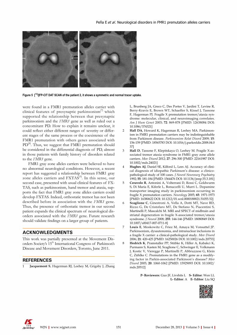
L, Brunberg JA, Greco C, Des Portes V, Jardini T, Levine R, Berry-Kravis E, Brown WT, Schaeffer S, Kissel J, Tassone F, Hagerman PJ. Fragile X premutation tremor/ataxia syn-drome: molecular, clinical, and neuroimaging correlates. Am J Hum Genet 2003; 72: 869-878 [PMID: 12638084 DOI: 10.1086/374321]
2 Hall DA, Howard K, Hagerman R, Leehey MA. Parkinson-ism in FMR1 premutation carriers may be indistinguishable from Parkinson disease. Parkinsonism Relat Disord 2009; 15: 156-159 [PMID: 18565783 DOI: 10.1016/j.parkreldis.2008.04.037]
3 Hall D, Tassone F, Klepitskaya O, Leehey M. Fragile X-as-sociated tremor ataxia syndrome in FMR1 gray zone allele carriers. Mov Disord 2012; 27: 296-300 [PMID: 22161987 DOI: 10.1002/mds.24021]
4 Hughes AJ, Daniel SE, Kilford L, Lees AJ. Accuracy of clini-cal diagnosis of idiopathic Parkinson’s disease: a clinico-pathological study of 100 cases. J Neurol Neurosurg Psychiatry 1992; 55: 181-184 [PMID: 1564476 DOI: 10.1136/jnnp.55.3.181]
5 Ceravolo R, Antonini A, Volterrani D, Rossi C, Goldwurm S, Di Maria E, Kiferle L, Bonuccelli U, Murri L. Dopamine transporter imaging study in parkinsonism occurring in fragile X premutation carriers. Neurology 2005; 65: 1971-1973 [PMID: 16380622 DOI: 10.1212/01.wnl.0000188821.51055.52]
6 Scaglione C, Ginestroni A, Vella A, Dotti MT, Nave RD, Rizzo G, De Cristofaro MT, De Stefano N, Piacentini S, Martinelli P, Mascalchi M. MRI and SPECT of midbrain and striatal degeneration in fragile X-associated tremor/ataxia syndrome. J Neurol 2008; 255: 144-146 [PMID: 18080849 DOI: 10.1007/s00417-007-0711-8]
7 Louis E, Moskowitz C, Friez M, Amaya M, Vonsattel JP. Parkinsonism, dysautonomia, and intranuclear inclusions in a fragile X carrier: a clinical-pathological study. Mov Disord 2006; 21: 420-425 [PMID: 16250026 DOI: 10.1002/mds.20753]
8 Hedrich K, Pramstaller PP, Stübke K, Hiller A, Kabakci K, Purmann S, Kasten M, Scaglione C, Schwinger E, Volkmann J, Kostic V, Vieregge P, Martinelli P, Abbruzzese G, Klein C, Zühlke C. Premutations in the FMR1 gene as a modify-ing factor in Parkin-associated Parkinson’s disease? Mov Disord 2005; 20: 1060-1062 [PMID: 15929093 DOI: 10.1002/mds.20512]
P- Reviewers: Guo JF, Livshits L S- Editor: Wen LL L- Editor: A E- Editor: Liu SQ
were found in a FMR1 premutation alleles carrier with clinical features of presynaptic parkinsonism[7] which supported the relationship between that presynaptic parkinsonism and the FMR1 gene as well as ruled out a concomitant PD. How to explain it remains unclear, it could reflect either different ranges of severity or differ-ent stages of the same process or the coexistence of the FMR1 premutation with others genes associated with PD[8]. Thus, we suggest that FMR1 premutation should be considered in the differential diagnosis of PD, almost in those patients with family history of disorders related to the FMR1 gene.
FMR1 gray zone alleles carriers were believed to have no abnormal neurological conditions. However, a recent report has suggested a relationship between FMR1 gray zone alleles carriers and FXTAS[3]. In this sense, our second case, presented with usual clinical features of FX-TAS, such as parkinsonism, hand tremor and ataxia, sup-ports the fact that FMR1 gray zone alleles carriers could develop FXTAS. Indeed, orthostatic tremor has not been described before in association with the FMR1 gene. Thus, the presence of orthostatic tremor in our second patient expands the clinical spectrum of neurological dis-orders associated with the FMR1 gene. Future research should validate findings on a larger group of patients.
ACKNOWLEDGMENTSThis work was partially presented at the Movement Dis-orders Society’s 15th International Congress of Parkinson’s Disease and Movement Disorders, Toronto, June 2011.
REFERENCES1 Jacquemont S, Hagerman RJ, Leehey M, Grigsby J, Zhang
151 December 28, 2013|Volume 3|Issue 4|WJN|www.wjgnet.com
Figure 5 [123I]FP-CIT DAT SCAN of the patient 2, it shows a symmetric and normal tracer uptake.
Peña E et al . Neurological disorders in FMR1 premutation alleles carriers
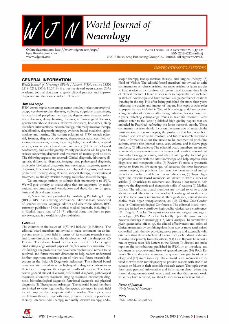
World Journal of NeurologyW J N
GENERAL INFORMATIONWorld Journal of Neurology (World J Neurol, WJN, online ISSN 2218-6212, DOI: 10.5316) is a peer-reviewed open access (OA) academic journal that aims to guide clinical practice and improve diagnostic and therapeutic skills of clinicians.
Aim and scopeWJN covers topics concerning neuro-oncology, electroneurophysi-ology, cerebrovascular diseases, epilepsy, cognitive impairment, myopathy and peripheral neuropathy, degenerative diseases, infec-tious diseases, demyelinating diseases, immunological diseases, genetic/metabolic diseases, affective disorders, headaches, sleep disorders, interventional neuroradiology, minimally invasive therapy, rehabilitation, diagnostic imaging, evidence-based medicine, epide-miology and nursing. The current columns of WJN include edito-rial, frontier, diagnostic advances, therapeutics advances, field of vision, mini-reviews, review, topic highlight, medical ethics, original articles, case report, clinical case conference (Clinicopathological conference), and autobiography. Priority publication will be given to articles concerning diagnosis and treatment of neurological diseases. The following aspects are covered: Clinical diagnosis, laboratory di-agnosis, differential diagnosis, imaging tests, pathological diagnosis, molecular biological diagnosis, immunological diagnosis, genetic diagnosis, functional diagnostics, and physical diagnosis; and com-prehensive therapy, drug therapy, surgical therapy, interventional treatment, minimally invasive therapy, and robot-assisted therapy.
We encourage authors to submit their manuscripts to WJN. We will give priority to manuscripts that are supported by major national and international foundations and those that are of great basic and clinical significance.
WJN is edited and published by Baishideng Publishing Group (BPG). BPG has a strong professional editorial team composed of science editors, language editors and electronic editors. BPG currently publishes 42 OA clinical medical journals, including 41 in English, has a total of 15 471 editorial borad members or peer reivewers, and is a world first-class publisher.
ColumnsThe columns in the issues of WJN will include: (1) Editorial: The editorial board members are invited to make comments on an im-portant topic in their field in terms of its current research status and future directions to lead the development of this discipline; (2) Frontier: The editorial board members are invited to select a highly cited cutting-edge original paper of his/her own to summarize ma-jor findings, the problems that have been resolved and remain to be resolved, and future research directions to help readers understand his/her important academic point of view and future research dir-ections in the field; (3) Diagnostic Advances: The editorial board members are invited to write high-quality diagnostic advances in their field to improve the diagnostic skills of readers. The topic covers general clinical diagnosis, differential diagnosis, pathological diagnosis, laboratory diagnosis, imaging diagnosis, endoscopic diag-nosis, biotechnological diagnosis, functional diagnosis, and physical diagnosis; (4) Therapeutics Advances: The editorial board members are invited to write high-quality therapeutic advances in their field to help improve the therapeutic skills of readers. The topic covers medication therapy, psychotherapy, physical therapy, replacement therapy, interventional therapy, minimally invasive therapy, endo-
scopic therapy, transplantation therapy, and surgical therapy; (5) Field of Vision: The editorial board members are invited to write commentaries on classic articles, hot topic articles, or latest articles to keep readers at the forefront of research and increase their levels of clinical research. Classic articles refer to papers that are included in Web of Knowledge and have received a large number of citations (ranking in the top 1%) after being published for more than years, reflecting the quality and impact of papers. Hot topic articles refer to papers that are included in Web of Knowledge and have received a large number of citations after being published for no more than 2 years, reflecting cutting-edge trends in scientific research. Latest articles refer to the latest published high-quality papers that are included in PubMed, reflecting the latest research trends. These commentary articles should focus on the status quo of research, the most important research topics, the problems that have now been resolved and remain to be resolved, and future research directions. Basic information about the article to be commented (including authors, article title, journal name, year, volume, and inclusive page numbers; (6) Minireviews: The editorial board members are invited to write short reviews on recent advances and trends in research of molecular biology, genomics, and related cutting-edge technologies to provide readers with the latest knowledge and help improve their diagnostic and therapeutic skills; (7) Review: To make a systematic review to focus on the status quo of research, the most important research topics, the problems that have now been resolved and re-main to be resolved, and future research directions; (8) Topic High-light: The editorial board members are invited to write a series of articles (7-10 articles) to comment and discuss a hot topic to help improve the diagnostic and therapeutic skills of readers; (9) Medical Ethics: The editorial board members are invited to write articles about medical ethics to increase readers’ knowledge of medical eth-ics. The topic covers international ethics guidelines, animal studies, clinical trials, organ transplantation, etc.; (10) Clinical Case Confer-ence or Clinicopathological Conference: The editorial board mem-bers are invited to contribute high-quality clinical case conference; (11) Original Articles: To report innovative and original findings in neurology; (12) Brief Articles: To briefly report the novel and in-novative findings in neurology; (13) Meta-Analysis: To summarize a given quantitative effect, e.g., the clinical effectiveness and safety of clinical treatments by combining data from two or more randomized controlled trials, thereby providing more precise and externally valid estimates than those which would stem from each individual dataset if analyzed separately from the others; (14) Case Report: To report a rare or typical case; (15) Letters to the Editor: To discuss and make reply to the contributions published in WJN, or to introduce and comment on a controversial issue of general interest; (16) Book Re-views: To introduce and comment on quality monographs of neur-ology; and (17) Autobiography: The editorial board members are in-vited to write their autobiography to provide readers with stories of success or failure in their scientific research career. The topic covers their basic personal information and information about when they started doing research work, where and how they did research work, what they have achieved, and their lessons from success or failure.
Name of journalWorld Journal of Neurology
ISSNISSN 2218-6212 (online)
Online Submissions: http://www.wjgnet.com/esps/[email protected]
World J Neurol 2013 December 28; 3(4): I-VISSN 2218-6212 (online)
© 2013 Baishideng Publishing Group Co., Limited. All rights reserved.
INSTRUCTIONS TO AUTHORS
IWJN|www.wjgnet.com December 28, 2013|Volume 3|Issue 4|
Instructions to authors
Launch dateDecember 28, 2011
FrequencyQuarterly
Editors-in-ChiefFelipe Fregni, MD, PhD, MPH, Associate Professor, Depart-ment of Physical Medicine and Rehabilitation, Spaulding Rehabilita-tion Hospital, Harvard Medical School, Room 725, 125 Nashua St. Boston, MA 02114, United States
Vincenzo Solfrizzi, MD, PhD, Professor, Department of In-ternal Medicine, Immunology and Infectious Diseases, Section of Geriatric Medicine-Memory Unit, University of Bari, P.zza G. Cesare, 11, 70124 Bari, Italy
Editorial officeJin-Lei Wang, DirectorXiu-Xia Song, Vice DirectorWorld Journal of NeurologyRoom 903, Building D, Ocean International Center,No. 62 Dongsihuan Zhonglu, Chaoyang District,Beijing 100025, ChinaTelephone: +86-10-85381891Fax: +86-10-85381893E-mail: [email protected]://www.wjgnet.com
PublisherBaishideng Publishing Group Co., LimitedFlat C, 23/F., Lucky Plaza, 315-321 Lockhart Road, Wan Chai, Hong Kong, ChinaTelephone: +852-58042046Fax: +852-31158812E-mail: [email protected]://www.wjgnet.com
Production centerBeijing Baishideng BioMed Scientific Co., LimitedRoom 903, Building D, Ocean International Center,No. 62 Dongsihuan Zhonglu, Chaoyang District,Beijing 100025, ChinaTelephone: +86-10-85381892Fax: +86-10-85381893
Representative officeUSA Office8226 Regency Drive, Pleasanton, CA 94588-3144, United States
Instructions to authorsFull instructions are available online at http://www.wjgnet.com/ 2218-6212/g_info_20100722173918.htm.
Indexed and Abstracted inDigital Object Identifier.
SPECIAL STATEMENTAll articles published in this journal represent the viewpoints of the authors except where indicated otherwise.
Biostatistical editingStatistical review is performed after peer review. We invite an ex-pert in Biomedical Statistics to evaluate the statistical method used in the paper, including t-test (group or paired comparisons), chi-squared test, Ridit, probit, logit, regression (linear, curvilinear, or stepwise), correlation, analysis of variance, analysis of covariance, etc. The reviewing points include: (1) Statistical methods should be described when they are used to verify the results; (2) Whether the
statistical techniques are suitable or correct; (3) Only homogeneous data can be averaged. Standard deviations are preferred to standard errors. Give the number of observations and subjects (n). Losses in observations, such as drop-outs from the study should be re-ported; (4) Values such as ED50, LD50, IC50 should have their 95% confidence limits calculated and compared by weighted probit analysis (Bliss and Finney); and (5) The word ‘significantly’ should be replaced by its synonyms (if it indicates extent) or the P value (if it indicates statistical significance).
Conflict-of-interest statementIn the interests of transparency and to help reviewers assess any po-tential bias, WJN requires authors of all papers to declare any compet-ing commercial, personal, political, intellectual, or religious interests in relation to the submitted work. Referees are also asked to indi-cate any potential conflict they might have reviewing a particular paper. Before submitting, authors are suggested to read “Uniform Requirements for Manuscripts Submitted to Biomedical Journals: Ethical Considerations in the Conduct and Reporting of Research: Conflicts of Interest” from International Committee of Medical Journal Editors (ICMJE), which is available at: http://www.icmje.org/ethical_4conflicts.html.
Sample wording: [Name of individual] has received fees for serv-ing as a speaker, a consultant and an advisory board member for [names of organizations], and has received research funding from [names of organization]. [Name of individual] is an employee of [name of or-ganization]. [Name of individual] owns stocks and shares in [name of organization]. [Name of individual] owns patent [patent identification and brief description].
Statement of informed consentManuscripts should contain a statement to the effect that all human studies have been reviewed by the appropriate ethics committee or it should be stated clearly in the text that all persons gave their informed consent prior to their inclusion in the study. Details that might disclose the identity of the subjects under study should be omitted. Authors should also draw attention to the Code of Ethics of the World Med-ical Association (Declaration of Helsinki, 1964, as revised in 2004).
Statement of human and animal rightsWhen reporting the results from experiments, authors should follow the highest standards and the trial should conform to Good Clinical Practice (for example, US Food and Drug Administration Good Clinical Practice in FDA-Regulated Clinical Trials; UK Medicines Research Council Guidelines for Good Clinical Practice in Clinical Trials) and/or the World Medical Association Declaration of Hel-sinki. Generally, we suggest authors follow the lead investigator’s na-tional standard. If doubt exists whether the research was conducted in accordance with the above standards, the authors must explain the rationale for their approach and demonstrate that the institutional review body explicitly approved the doubtful aspects of the study.
Before submitting, authors should make their study approved by the relevant research ethics committee or institutional review board. If human participants were involved, manuscripts must be accom-panied by a statement that the experiments were undertaken with the understanding and appropriate informed consent of each. Any per-sonal item or information will not be published without explicit con-sents from the involved patients. If experimental animals were used, the materials and methods (experimental procedures) section must clearly indicate that appropriate measures were taken to minimize pain or discomfort, and details of animal care should be provided.
SUBMISSION OF MANUSCRIPTSManuscripts should be typed in 1.5 line spacing and 12 pt. Book Antiqua with ample margins. Number all pages consecutively, and start each of the following sections on a new page: Title Page, Abstract, Introduction, Materials and Methods, Results, Discus-sion, Acknowledgements, References, Tables, Figures, and Figure Legends. Neither the editors nor the publisher are responsible for the opinions expressed by contributors. Manuscripts formally ac-cepted for publication become the permanent property of Baish-
IIWJN|www.wjgnet.com December 28, 2013|Volume 3|Issue 4|
Instructions to authors
ideng Publishing Group Co., Limited, and may not be reproduced by any means, in whole or in part, without the written permission of both the authors and the publisher. We reserve the right to copy-edit and put onto our website accepted manuscripts. Authors should follow the relevant guidelines for the care and use of laboratory ani-mals of their institution or national animal welfare committee. For the sake of transparency in regard to the performance and report-ing of clinical trials, we endorse the policy of the ICMJE to refuse to publish papers on clinical trial results if the trial was not recorded in a publicly-accessible registry at its outset. The only register now available, to our knowledge, is http://www.clinicaltrials.gov spon-sored by the United States National Library of Medicine and we en-courage all potential contributors to register with it. However, in the case that other registers become available you will be duly notified. A letter of recommendation from each author’s organization should be provided with the contributed article to ensure the privacy and secrecy of research is protected.
Authors should retain one copy of the text, tables, photo-graphs and illustrations because rejected manuscripts will not be returned to the author(s) and the editors will not be responsible for loss or damage to photographs and illustrations sustained dur-ing mailing.
Online submissionsManuscripts should be submitted through the Online Submission System at: http://www.wjgnet.com/esps/. Authors are highly recom-mended to consult the ONLINE INSTRUCTIONS TO AUTHORS (http://www.wjgnet.com/2218-6212/g_info_20100722173918.htm) before attempting to submit online. For assistance, authors encoun-tering problems with the Online Submission System may send an email describing the problem to [email protected], or by tele-phone: +86-10-85381892. If you submit your manuscript online, do not make a postal contribution. Repeated online submission for the same manuscript is strictly prohibited.
MANUSCRIPT PREPARATIONAll contributions should be written in English. All articles must be submitted using word-processing software. All submissions must be typed in 1.5 line spacing and 12 pt. Book Antiqua with ample mar-gins. Style should conform to our house format. Required informa-tion for each of the manuscript sections is as follows:
Title pageTitle: Title should be less than 12 words.
Running title: A short running title of less than 6 words should be provided.
Authorship: Authorship credit should be in accordance with the standard proposed by ICMJE, based on (1) substantial contribu-tions to conception and design, acquisition of data, or analysis and interpretation of data; (2) drafting the article or revising it critically for important intellectual content; and (3) final approval of the ver-sion to be published. Authors should meet conditions 1, 2, and 3.
Institution: Author names should be given first, then the complete name of institution, city, province and postcode. For example, Xu-Chen Zhang, Li-Xin Mei, Department of Pathology, Chengde Med-ical College, Chengde 067000, Hebei Province, China. One author may be represented from two institutions, for example, George Sgourakis, Department of General, Visceral, and Transplantation Surgery, Essen 45122, Germany; George Sgourakis, 2nd Surgical Department, Korgialenio-Benakio Red Cross Hospital, Athens 15451, Greece
Author contributions: The format of this section should be: Author contributions: Wang CL and Liang L contributed equally to this work; Wang CL, Liang L, Fu JF, Zou CC, Hong F and Wu XM designed the research; Wang CL, Zou CC, Hong F and Wu XM performed the research; Xue JZ and Lu JR contributed new
reagents/analytic tools; Wang CL, Liang L and Fu JF analyzed the data; and Wang CL, Liang L and Fu JF wrote the paper.
Supportive foundations: The complete name and number of sup-portive foundations should be provided, e.g., Supported by National Natural Science Foundation of China, No. 30224801
Correspondence to: Only one corresponding address should be provided. Author names should be given first, then author title, af-filiation, the complete name of institution, city, postcode, province, country, and email. All the letters in the email should be in lower case. A space interval should be inserted between country name and email address. For example, Montgomery Bissell, MD, Professor of Medicine, Chief, Liver Center, Gastroenterology Division, Uni-versity of California, Box 0538, San Francisco, CA 94143, United States. [email protected]
Telephone and fax: Telephone and fax should consist of +, coun-try number, district number and telephone or fax number, e.g., Tele-phone: +86-10-85381892 Fax: +86-10-85381893
Peer reviewers: All articles received are subject to peer review. Normally, three experts are invited for each article. Decision on acceptance is made only when at least two experts recommend publication of an article. All peer-reviewers are acknowledged on Express Submission and Peer-review System website.
AbstractThere are unstructured abstracts (no less than 200 words) and struc-tured abstracts. The specific requirements for structured abstracts are as follows:
An informative, structured abstract should accompany each manuscript. Abstracts of original contributions should be struc-tured into the following sections: AIM (no more than 20 words; Only the purpose of the study should be included. Please write the Aim in the form of “To investigate/study/…”), METHODS (no less than 140 words for Original Articles; and no less than 80 words for Brief Articles), RESULTS (no less than 150 words for Original Articles and no less than 120 words for Brief Articles; You should present P values where appropriate and must provide relevant data to illustrate how they were obtained, e.g., 6.92 ± 3.86 vs 3.61 ± 1.67, P < 0.001), and CONCLUSION (no more than 26 words).
Key wordsPlease list 5-10 key words, selected mainly from Index Medicus, which reflect the content of the study.
Core tip Please write a summary of less than 100 words to outline the most innovative and important arguments and core contents in your paper to attract readers.
TextFor articles of these sections, original articles and brief articles, the main text should be structured into the following sections: INTRO-DUCTION, MATERIALS AND METHODS, RESULTS and DISCUSSION, and should include appropriate Figures and Tables. Data should be presented in the main text or in Figures and Tables, but not in both.
IllustrationsFigures should be numbered as 1, 2, 3, etc., and mentioned clearly in the main text. Provide a brief title for each figure on a separate page. Detailed legends should not be provided under the figures. This part should be added into the text where the figures are applicable. Keep-ing all elements compiled is necessary in line-art image. Scale bars should be used rather than magnification factors, with the length of the bar defined in the legend rather than on the bar itself. File names should identify the figure and panel. Avoid layering type directly over shaded or textured areas. Please use uniform legends for the same subjects. For example: Figure 1 Pathological changes in atrophic gas-
IIIWJN|www.wjgnet.com December 28, 2013|Volume 3|Issue 4|
tritis after treatment. A: ...; B: ...; C: ...; D: ...; E: ...; F: ...; G: …etc. It is our principle to publish high resolution-figures for the E-versions.
TablesThree-line tables should be numbered 1, 2, 3, etc., and mentioned clearly in the main text. Provide a brief title for each table. Detailed legends should not be included under tables, but rather added into the text where applicable. The information should complement, but not duplicate the text. Use one horizontal line under the title, a second under column heads, and a third below the Table, above any footnotes. Vertical and italic lines should be omitted.
Notes in tables and illustrationsData that are not statistically significant should not be noted. aP < 0.05, bP < 0.01 should be noted (P > 0.05 should not be noted). If there are other series of P values, cP < 0.05 and dP < 0.01 are used. A third series of P values can be expressed as eP < 0.05 and fP < 0.01. Other notes in tables or under illustrations should be expressed as 1F, 2F, 3F; or sometimes as other symbols with a superscript (Arabic numer-als) in the upper left corner. In a multi-curve illustration, each curve should be labeled with ●, ○, ■, □, ▲, △, etc., in a certain sequence.
AcknowledgmentsBrief acknowledgments of persons who have made genuine con-tributions to the manuscript and who endorse the data and conclu-sions should be included. Authors are responsible for obtaining written permission to use any copyrighted text and/or illustrations.
REFERENCESCoding systemThe author should number the references in Arabic numerals ac-cording to the citation order in the text. Put reference numbers in square brackets in superscript at the end of citation content or after the cited author’s name. For citation content which is part of the narration, the coding number and square brackets should be typeset normally. For example, “Crohn’s disease (CD) is associated with increased intestinal permeability[1,2]”. If references are cited directly in the text, they should be put together within the text, for example, “From references[19,22-24], we know that...”
When the authors write the references, please ensure that the order in text is the same as in the references section, and also ensure the spelling accuracy of the first author’s name. Do not list the same citation twice.
PMID and DOIPleased provide PubMed citation numbers to the reference list, e.g., PMID and DOI, which can be found at http://www.ncbi.nlm.nih.gov/sites/entrez?db=pubmed and http://www.crossref.org/SimpleTextQuery/, respectively. The numbers will be used in E-version of this journal.
Style for journal referencesAuthors: the name of the first author should be typed in bold-faced letters. The family name of all authors should be typed with the in-itial letter capitalized, followed by their abbreviated first and middle initials. (For example, Lian-Sheng Ma is abbreviated as Ma LS, Bo-Rong Pan as Pan BR). The title of the cited article and italicized journal title (journal title should be in its abbreviated form as shown in PubMed), publication date, volume number (in black), start page, and end page [PMID: 11819634 DOI: 10.3748/wjg.13.5396].
Style for book referencesAuthors: the name of the first author should be typed in bold-faced letters. The surname of all authors should be typed with the initial letter capitalized, followed by their abbreviated middle and first initials. (For example, Lian-Sheng Ma is abbreviated as Ma LS, Bo-Rong Pan as Pan BR) Book title. Publication number. Publication place: Publication press, Year: start page and end page.
FormatJournals English journal article (list all authors and include the PMID where applicable)
1 Jung EM, Clevert DA, Schreyer AG, Schmitt S, Rennert J, Kubale R, Feuerbach S, Jung F. Evaluation of quantitative con-trast harmonic imaging to assess malignancy of liver tumors: A prospective controlled two-center study. World J Gastroenterol 2007; 13: 6356-6364 [PMID: 18081224 DOI: 10.3748/wjg.13. 6356]
Chinese journal article (list all authors and include the PMID where applicable)2 Lin GZ, Wang XZ, Wang P, Lin J, Yang FD. Immunologic
effect of Jianpi Yishen decoction in treatment of Pixu-diar-rhoea. Shijie Huaren Xiaohua Zazhi 1999; 7: 285-287
In press3 Tian D, Araki H, Stahl E, Bergelson J, Kreitman M. Signature
of balancing selection in Arabidopsis. Proc Natl Acad Sci USA 2006; In press
Organization as author4 Diabetes Prevention Program Research Group. Hyperten-
sion, insulin, and proinsulin in participants with impaired glu-cose tolerance. Hypertension 2002; 40: 679-686 [PMID: 12411462 PMCID:2516377 DOI:10.1161/01.HYP.0000035706.28494. 09]
Both personal authors and an organization as author 5 Vallancien G, Emberton M, Harving N, van Moorselaar RJ;
Alf-One Study Group. Sexual dysfunction in 1, 274 European men suffering from lower urinary tract symptoms. J Urol 2003; 169: 2257-2261 [PMID: 12771764 DOI:10.1097/01.ju. 0000067940.76090.73]
No author given6 21st century heart solution may have a sting in the tail. BMJ
2002; 325: 184 [PMID: 12142303 DOI:10.1136/bmj.325. 7357.184]
Volume with supplement7 Geraud G, Spierings EL, Keywood C. Tolerability and safety
of frovatriptan with short- and long-term use for treatment of migraine and in comparison with sumatriptan. Headache 2002; 42 Suppl 2: S93-99 [PMID: 12028325 DOI:10.1046/j.1526-4610.42.s2.7.x]
Issue with no volume8 Banit DM, Kaufer H, Hartford JM. Intraoperative frozen
section analysis in revision total joint arthroplasty. Clin Orthop Relat Res 2002; (401): 230-238 [PMID: 12151900 DOI:10.1097/00003086-200208000-00026]
No volume or issue9 Outreach: Bringing HIV-positive individuals into care. HRSA
Careaction 2002; 1-6 [PMID: 12154804]
BooksPersonal author(s)10 Sherlock S, Dooley J. Diseases of the liver and billiary system.
9th ed. Oxford: Blackwell Sci Pub, 1993: 258-296Chapter in a book (list all authors)11 Lam SK. Academic investigator’s perspectives of medical
treatment for peptic ulcer. In: Swabb EA, Azabo S. Ulcer disease: investigation and basis for therapy. New York: Marcel Dekker, 1991: 431-450
Author(s) and editor(s)12 Breedlove GK, Schorfheide AM. Adolescent pregnancy.
2nd ed. Wieczorek RR, editor. White Plains (NY): March of Dimes Education Services, 2001: 20-34
Conference proceedings13 Harnden P, Joffe JK, Jones WG, editors. Germ cell tumours V.
Proceedings of the 5th Germ cell tumours Conference; 2001 Sep 13-15; Leeds, UK. New York: Springer, 2002: 30-56
Conference paper14 Christensen S, Oppacher F. An analysis of Koza's computa-
tional effort statistic for genetic programming. In: Foster JA, Lutton E, Miller J, Ryan C, Tettamanzi AG, editors. Genetic programming. EuroGP 2002: Proceedings of the 5th Euro-pean Conference on Genetic Programming; 2002 Apr 3-5; Kinsdale, Ireland. Berlin: Springer, 2002: 182-191
Electronic journal (list all authors)
Instructions to authors
IVWJN|www.wjgnet.com December 28, 2013|Volume 3|Issue 4|
Instructions to authors
15 Morse SS. Factors in the emergence of infectious diseases. Emerg Infect Dis serial online, 1995-01-03, cited 1996-06-05; 1(1): 24 screens. Available from: URL: http://www.cdc.gov/ncidod/eid/index.htm
Patent (list all authors)16 Pagedas AC, inventor; Ancel Surgical R&D Inc., assignee.
Flexible endoscopic grasping and cutting device and position-ing tool assembly. United States patent US 20020103498. 2002 Aug 1
Statistical dataWrite as mean ± SD or mean ± SE.
Statistical expressionExpress t test as t (in italics), F test as F (in italics), chi square test as χ2 (in Greek), related coefficient as r (in italics), degree of freedom as υ (in Greek), sample number as n (in italics), and probability as P (in italics).
UnitsUse SI units. For example: body mass, m (B) = 78 kg; blood pres-sure, p (B) = 16.2/12.3 kPa; incubation time, t (incubation) = 96 h, blood glucose concentration, c (glucose) 6.4 ± 2.1 mmol/L; blood CEA mass concentration, p (CEA) = 8.6 24.5 mg/L; CO2 volume fraction, 50 mL/L CO2, not 5% CO2; likewise for 40 g/L formal-dehyde, not 10% formalin; and mass fraction, 8 ng/g, etc. Arabic numerals such as 23, 243, 641 should be read 23 243 641.
The format for how to accurately write common units and quantums can be found at: http://www.wjgnet.com/2218-6212/g_info_20100723222255.htm.
AbbreviationsStandard abbreviations should be defined in the abstract and on first mention in the text. In general, terms should not be abbrevi-ated unless they are used repeatedly and the abbreviation is helpful to the reader. Permissible abbreviations are listed in Units, Symbols and Abbreviations: A Guide for Biological and Medical Editors and Authors (Ed. Baron DN, 1988) published by The Royal Society of Medicine, London. Certain commonly used abbreviations, such as DNA, RNA, HIV, LD50, PCR, HBV, ECG, WBC, RBC, CT, ESR, CSF, IgG, ELISA, PBS, ATP, EDTA, mAb, can be used directly without further explanation.
ItalicsQuantities: t time or temperature, c concentration, A area, l length, m mass, V volume.Genotypes: gyrA, arg 1, c myc, c fos, etc.Restriction enzymes: EcoRI, HindI, BamHI, Kbo I, Kpn I, etc.Biology: H. pylori, E coli, etc.
Examples for paper writingAll types of articles’ writing style and requirement will be found in the link: http://www.wjgnet.com/esps/NavigationInfo.aspx?id=15
RESUBMISSION OF THE REVISED MANUSCRIPTSAuthors must revise their manuscript carefully according to the revision policies of Baishideng Publishing Group Co., Limited. The revised version, along with the signed copyright transfer agreement, responses to the reviewers, and English language Grade A certifi-cate (for non-native speakers of English), should be submitted to the online system via the link contained in the e-mail sent by the edi-tor. If you have any questions about the revision, please send e-mail to [email protected].
Language evaluation The language of a manuscript will be graded before it is sent for revision. (1) Grade A: priority publishing; (2) Grade B: minor lan-guage polishing; (3) Grade C: a great deal of language polishing needed; and (4) Grade D: rejected. Revised articles should reach Grade A.
Copyright assignment formPlease download a Copyright assignment form from http://www.wjgnet.com/2218-6212/g_info_20100723222139.htm.
Responses to reviewersPlease revise your article according to the comments/suggestions provided by the reviewers. The format for responses to the reviewers’ comments can be found at: http://www.wjgnet.com/2218-6212/g_info_20100723222041.htm.
Proof of financial supportFor papers supported by a foundation, authors should provide a copy of the approval document and serial number of the foundation.
STATEMENT ABOUT ANONYMOUS PUBLICA-TION OF THE PEER REVIEWERS’ COMMENTSIn order to increase the quality of peer review, push authors to carefully revise their manuscripts based on the peer reviewers' com-ments, and promote academic interactions among peer reviewers, authors and readers, we decide to anonymously publish the review-ers’ comments and author’s responses at the same time the manu-script is published online.
PUBLICATION FEEWJN is an international, peer-reviewed, OA online journal. Articles published by this journal are distributed under the terms of the Cre-ative Commons Attribution Non-commercial License, which per-mits use, distribution, and reproduction in any medium and format, provided the original work is properly cited. The use is non-com-mercial and is otherwise in compliance with the license. Authors of accepted articles must pay a publication fee. Publication fee: 600 USD per article. All invited articles are published free of charge.
VWJN|www.wjgnet.com December 28, 2013|Volume 3|Issue 4|

Published by Baishideng Publishing Group Co., LimitedFlat C, 23/F., Lucky Plaza, 315-321 Lockhart Road,
Wan Chai, Hong Kong, ChinaFax: 852-65557188
Telephone: +852-31779906E-mail: [email protected]
http://www.wjgnet.com
© 2013 Baishideng Publishing Group Co., Limited. All rights reserved.