click - child neurology society
TRANSCRIPT

Chi
ld N
euro
logy
Soc
iety
Presented atThe Duke Energy Convention CenterCincinnati, OHOctober 12 – October 15, 2022CME Joint Sponsorship Provided by Minnesota Medical Associationand the Child Neurology Society

Yasmin Khakoo, Chair New York, NYBhooma Aravamuthan, Co-Chair St. Louis, MOGyula Acsadi Farmington, CTClarimar Borrero-Mejias St. Petersburg, FLJ. Nicholas Brenton Charlottesville, VAAudrey C. Brumback Austin, TXMeeryo Choe Los Angeles, CAKeith A. Coffman Kansas City, MOAlexander Li Cohen Boston, MALouis Dang Ann Arbor, MIJay Desai Los Angeles, CADavid Dredge Boston, MAMatthew Elrick Baltimore, MDGrace Gombolay Atlanta, GAHoward P. Goodkin Charlottesville, VAAjay Gupta Cleveland OHKrisztina Harsanyi Birmingham, ALIsabella Herman Boys Town, NESaba Jafarpour Los Angeles, CAMonica Lemmon Durham, NCWilfreda Lindsey Houston, TX
Ariel M Lyons-Warren Houston, TXMelanie McNally Boston, MAGaneshwaran H. Mochida Boston, MAAndrew Ng Los Angeles, CAScott Otallah Winston Salem, NCRachit Patil MD Providence, RIMarc C. Patterson Rochester, MNToni Pearson St. Louis, MOE. Steve Roach Austin, TXJonathan D. Santoro Los Angeles, CADevorah Segal New York, NYRenée Shellhaas Ann Arbor, MIChristopher Smyser St. Louis, MOLiu Lin Thio St. Louis, MOKeith Van Haren Palo Alto, CAJennifer Vermilion Rochester, NYAmy Robichaux Viehoever St. Louis, MOVijay Vishwanath Los Angeles, CAElizabeth M. Wells Washington, DCElissa Yozawitz Bronx, NY
Fifty-First National Meeting of the Child Neurology Society
Planning CommitteeChild Neurology Society Executive Board
CNS Scientific Selection and Program Planning Committee
Child Neurology Society National OfficeRoger Larson, Executive DirectorSue Hussman, Associate DirectorKathy Pavel, Office Administrator
Emily McConnell, Professional Development Manager
Presented at the Duke Energy Convention CenterCincinnati, OH
October 12-15, 2022
ACCREDITATIONThis activity has been planned and implemented in accordance with the accreditation requirements and policies of the Accreditation Council for Continuing Medical Education (ACCME) through the joint providership of the Minnesota Medical Association and Child Neurology Society.
The Minnesota Medical Association (MMA) is accredited by the ACCME to provide continuing medical education for physicians.
The Minnesota Medical Association designates this live activity for a maximum of 30.75 AMA PRA Category 1 Credit(s)™. Physicians should claim only the credit commensurate with the extent of their participation in the activity.
Bruce H. Cohen, President Akron, OHPhillip Pearl, Past President Boston, MALori Jordan, Secretary-Treasurer Nashville, TNJanet Soul, Councillor Boston, MASucheta Joshi, Councillor Ann Arbor, MIAudrey Brumback, Councillor Austin, TXSonia Partap, Councillor Palo Alto, CA
Program and Abstracts, Child Neurology Society S1

PresidentKenneth Swaiman 1972-73Gerald Fenichel 1973-74Manuel Gomez 1974-75James Schwartz 1975-76Richard Allen 1976-77Bruce Berg 1977-78N. Paul Rosman 1978-79Arthur Prensky 1979-80Paul Dyken 1980-81Mary Anne Guggenheim 1981-82Raymond Chun 1982-83Robert Eiben 1983-85David Stumpf 1985-87Marvin Fishman 1987-89Darryl C. De Vivo 1989-91Peter H. Berman 1991-93Joseph J. Volpe 1993-95Michael E. Cohen 1995-97Alan K. Percy 1997-99Michael J. Painter 1999-2001Stephen Ashwal 2001-2003James Bale 2003-2005Ann Tilton 2005-2007John Bodensteiner 2007-2009Donna Ferriero 2009-2011E. Steve Roach 2011-2013Nina F. Schor 2013-2015Kenneth Mack 2015-2017Jonathan Mink 2017-2019Phillip Pearl 2019-2021Bruce Cohen 2021-
Secretary-TreasurerRichard Allen 1972-75Raymond Chun 1975-78Robert Eiben 1978-81Lawrence Lockman 1981-84Marvin Fishman 1984-86Ira Lott 1986-89Peggy Copple (Ferry) 1989-93Stephen Ashwal 1993-97Patricia Crumrine 1997-2002Ann Tilton 2003-2004Nina Schor 2004-2010Harvey Singer 2010-2015Bruce Cohen 2015-2020Lori Jordan 2020-
CouncillorIsabelle Rapin 1972-73Manuel Gomez 1972-73John Menkes 1972-74James Schwartz 1972-74Karin Nelson 1973-74Raymond Chun 1973-75Bruce Berg 1974-76Paul Dyken 1974-76Arthur Prensky 1975-77N. Paul Rosman 1975-77Jack Madsen 1976-78Peggy Copple (Ferry) 1976-78Joseph French 1977-79Francis Wright 1977-79Mary Anne Guggenheim 1978-80Gerald Golden 1978-80Gerald Erenberg 1979-81John Freeman 1979-81Marvin Weil 1980-82Marvin Fishman 1980-82Peter Huttenlocher 1981-83Michael Bresnan 1981-83David Stumpf 1982-84Gwendolyn Hogan 1982-84Joseph Volpe 1983-85Barry Russman 1983-85Russell Snyder 1984-86Ian Butler 1984-86W. Edwin Dodson 1985-87Michael Painter 1985-87Robert Zeller 1986-88Doris Trauner 1986-88Darryl De Vivo 1987-88Gary Goldstein 1987-89Robert Vannucci 1988-89Stephen Ashwal 1988-90Jack Pellock 1988-90Joseph Pasternak 1989-91Patricia Duffner 1989-91O. Carter Snead 1990-92Edwin Meyer 1990-92Israel Abroms 1991-93William Logan 1991-93Mary Johnson 1992-94Alan Percy 1992-94Phyllis Sher 1993-95Gregory Holmes 1993-95W. Donald Shields 1994-96John Bodensteiner 1994-96Patricia Crumrine 1995-97James Bale 1995-97
Alan Hill 1996-98Ann Tilton 1996-98Edward Kovnar 1997-99Richard Nordgren 1997-99Michael Goldstein 1998-2000E. Steve Roach 1998-2000Faye Silverstein 1999-2001Michael Johnston 1999-2001Carmela Tardo 2000-02Pauline Filipek 2000-02Michael Noetzel 2001-03Carl Crosley 2001-03Julie Parke 2002-04Roy Elterman 2002-04Marc Patterson 2003-05Douglas Nordli 2003-05Donna Ferriero 2004-06Leon Dure 2004-06Kenneth Mack 2005-07Laura Ment 2005-07Leslie Morrison 2006-08Anne Anderson 2006-08Steven Leber 2007-09Jonathan Mink 2007-09Robert Rust 2008-10Wendy Mitchell 2008-10Warren Lo 2009-11Sakkubai Naidu 2009-11Gary Clark 2010-12Sidney Gospe 2010-12Barry Kosofsky 2011-13Suresh Kotagal 2011-13Vinodh Narayanan 2012-14Jayne Ness 2012-14Bruce Cohen 2013-15Roger Packer 2013-15Kevin Ess 2014-16Kara Lewis 2014-16Phillip Pearl 2015-17Renee Shellhaas 2015-17Peter B. Kang 2016-18Mary Zupanc 2016-18Donald Gilbert 2017-2019Michael Shevell 2017-2019Lori Jordan 2018-2020Mark Wainwright 2018-2020Nigel Bamford 2019-2021Nancy Bass 2019-2021Audrey Brumback 2020-2022Sonia Partap 2020-2022Sucheta Joshi 2021-Janet Soul 2021-
Past Officers
S2 Annals of Neurology Vol 92 (suppl 28) 2022

2025October 8-11Charlotte, NC
2024November 11-14San Diego, California
2023October 4-7Vancouver, BC Canada
2022October 12-15Cincinnati, Ohio
2021Boston, Massachusetts
2020San Diego, CaliforniaThe Joint 16th ICNA Congress & 49th Annual CNS MeetingTogether • Apart Virtual
2019Charlotte, North Carolina
2018Chicago, Illinois
2017Kansas City, Missouri
2016Vancouver, British Columbia, Canada
2015Washington, DC
2014Columbus, Ohio
2013Austin, Texas2012 Huntington Beach, California
2011 Savannah, Georgia
2010Providence, Rhode Island
2009 Louisville, Kentucky
2008 Santa Clara, California
2007 Quebec City, PQ, Canada
2006 Pittsburgh, Pennsylvania
2005 Los Angeles, California
2004 Ottawa, Ontario, Canada
2003 Miami Beach, Florida
2002 Washington, DC
2001 Victoria, British Columbia, Canada
2000 St. Louis, Missouri
1999 Nashville, Tennessee
1998 Montreal, Quebec, Canada
1997 Phoenix, Arizona
1996 Minneapolis, Minnesota
1995 Baltimore, Maryland
1994 San Francisco, California
1993 Orlando, Florida
1992 New Orleans, Louisiana
1991Portland, Oregon
1990Atlanta, Georgia
1989San Antonio, Texas
1988 Halifax, Nova Scotia, Canada
1987 San Diego, California
1986 Boston, Massachusetts
1985 Memphis, Tennessee
1984 Phoenix, Arizona
1983Williamsburg, Virginia
1982 Salt Lake City, Utah
1981 Minneapolis, Minnesota
1980 Savannah, Georgia
1979 Hanover, New Hampshire
1978Keystone, Colorado
1977Charlottesville, Virginia
1976Monterey, California
1975Hamilton, Ontario, Canada
1974Madison, Wisconsin
1973Nashville, Tennessee
1972Ann Arbor, Michigan
Annual Meetings
Program and Abstracts, Child Neurology Society S3

2022Leon G. EpsteinChicago, IL
2021Jonathan W. MinkRochester, NY
2020Kenneth J. MackRochester, MN
2019James F. Bale, Jr. Salt Lake City
2018Bernard L. MariaMorristown, NJ
2017Nina F. SchorRochester, NY
2016Harvey SingerBaltimore
2015E. Steve RoachColumbus
2014Michael ShevellMontreal
2013John BodensteinerRochester, MN
2012Ann TiltonNew Orleans
2011Deborah HirtzBethesda
2010Sakkubai NaiduBaltimore
2009Peter CamfieldHalifax
2008Stephen AshwalLoma Linda
2007Robert S. RustCharlottesville
2006Michael PainterPittsburgh
2005Alan PercyBirmingham
2004John FreemanBaltimore
2003Michael E. CohenBuffalo
2002Peter H. BermanPhiladelphia
2001Charles BarlowBoston
2000Arthur PrenskySt. Louis
1999Marvin FishmanHouston
1998N. Paul RosmanBoston
1997Gerald FenichelNashville
1996William BellIowa City
1995Salvatore DiMauroNew York
1994Hugo MoserBaltimore
1993Bengt D. HagbergGoteborg
1992Darryl C. De VivoNew York
1991Karin B. NelsonBethesda
1990Joseph J. VolpwBoston
1989Manuel GomezRochester
1988Bruce BergSan Francisco
1987Isabelle RapinBronx
1986Jean AicardiParis
1985RaymondD.AdamsBoston
1984Peter HuttenlocherChicago
1983Betty Q. BankerCleveland
1982Patrick F. BraySalt Lake City
1981Kenneth F.SwaimanMinneapolis
1980John H. MenkesBeverly Hills
1979Paul I. YakovlevBoston
1978Philip R. DodgeSt. Louis
1977David B. ClarkLexington
1976Sidney CarterNew York
1975Randolph K. ByersBoston
1974Douglas BuchananChicago
CNS Hower Award Recipients
S4 Annals of Neurology Vol 92 (suppl 28) 2022

2022Steven Paul MillerVancouver, BC, Canada
2021Jerry MendellColumbus, OH
2020Joseph GleesonSan Diego
2019Scott PomeroyBoston
2018William B. DobynsSeattle
2017Solomon MoshéBronx, NY
2016Harvey SarnatCalgary
2015Harry T. ChuganiDetroit
2014Gabrielle deVeberToronto
2013Tallie Z. BaramIrvine
2012Roger PackerWashington, DC
2011Laura MentNew Haven
2010Thomas JessellNew York
2009Gregory HolmesLebanon, NH
2008Michael JohnstonBaltimore
2007Frederick AndermannMontreal
2006Donna FerrieroSan Francisco
2005O. Carter Snead IIIToronto
2004Karin NelsonBethesda
2003Darryl C. De VivoNew York
2002Francis CollinsBethesda
2001Huda ZoghbiHouston
2000Joseph VolpeBoston
1999Carla ShatzBerkeley
1998Andrew EngelRochester
1997Martha Bridge DencklaBaltimore
1996Verne S. CavinessBoston
1995Gerald D. FischbachBoston
1994David PrinceStanford
1993C. Thomas CaskeyHouston
1992Louis M. KunkelBoston
1991Marcus E. RaichleSt. Louis
1990Roscoe O. BradyBethesda
1989Salvatore DiMauroNew York
1988Victor DubowitzLondon
1987Hugo MoserBaltimore
1986Louis SokoloffBethesda
1985Patricia Goldman-RakicNew Haven
1984William L. NyhanLa Jolla
1983Roger N. RosenbergDallas
1982John O’BrienLa Jolla
1981Pasko RakicNew Haven
1980Dominick PurpuraNew York
1979Fred PlumNew York
1978W. Maxwell CowanSt. Louis
1977George CahillBoston
Bernard Sachs Lecturers Recipients
Program and Abstracts, Child Neurology Society S5

2022Bhooma AravamuthanSt. Louis, MO
2021Monica LemmonDurham, NC
2020Hsiao-Tuan ChaoHouston
2019Louis DangAnn Arbor
2018Christopher ElittBoston
2017Audrey C. BrumbackAustin
2016Diana Bharucha-GoebelBethesda
2015Jimmy Holder, Jr.Houston
2014Christopher SmyserSt. Louis
2013Peter TsaiBoston
2012Yoon Jae-ChoStanford
2011James DowlingAnn Arbor
2010Stephen MaricichCleveland
2009Jeffrey NeulHouston
2008Laura JansenSeattle
2007Mirjana Maletic-SavaticStony Brook
2006Elliott SherrSan Francisco
2005Mustafa SahinBoston
2004Terri InderMelbourne
2003Bradley SchlaggarSt. Louis
2002Nigel BamfordNew York
2001Daniel J. BonthiusIowa City
2000Stephen BackPortland
1999Amy Brooks-KayalPhiladelphia
1998Joseph GleesonBoston
1997William A. WeissSan Francisco
1996Michael RivkinBoston
1995Adre J. du PlessisBoston
1994Mia MacCollinBoston
1993Jeffrey J. NeilSt. Louis
1992Kelvin A. YamadaSt. Louis
1991Kenneth J. MackMadison
1990Evan Y. SnyderBoston
Harris GelbardRochester, NY
1989Scott L. PomeroySt. Louis
1988Huda ZoghbiHouston
1987Vinodh NarayananPittsburgh
1986Faye S. SilversteinAnn Arbor
1985Richard J. KonkolMilwaukee
1983Michael PranzatelliWashington
Philip R. Dodge Young Investigator Award Recipients
S6 Annals of Neurology Vol 92 (suppl 28) 2022

2022Jeff BuchhalterCalgary, AB, Canada
Roger LarsonSt. Paul, MN
Michael NoetzelSt. Louis, MO
2021Robert BaumannLexington, KY
Sidney Gospe, JrSeattle, WA
2019Carol CamfieldHalifax, Nova Scotia
W. Edwin Dodson St. Louis, MO
2018Gerald ErenbergCleveland, OH
William LoganToronto, Ontario
Alfred Spiro Bronx, NY
2017Abe ChutorianNew York, NY
W. Donald ShieldsLos Angeles, CA
2016Kalpathy KrishnamoorthyBoston, MA
Doris TraunerLa Jolla, CA
2015Pat CrumrinePittsburgh, PA
Suresh KotagalRochester, MN
2014G. Robert De LongDurham, NC
2013 Arthur RoseBrooklyn, NY
A. David RothnerCleveland, OH
2012 Bhuwan GargIndianapolis, IN
M. Richard KoenigsbergerDemarest, NJ
2011Warren Grover Philadelphia, PA
2010Russell Snyder Albuquerque, NM
2009Mary Anne Guggenheim Helena, MT G Dean Timmons Akron, OH
2008Cesare Lombroso Boston, MA
Niels Lowe Tenafly, NJ
2007William Kennedy Watertown, ME
Gordon Watters Montreal, Quebec
2006Raymond Chun Madison, WI
Barry Russman Portland, OR
2005 Robert EibenCleveland, OH
Arnold Gold New York, NY
2004Jean Holowach ThurstonSt. Louis, MO
Roger and Mary Brumback Lifetime Achievement Award Recipients
Program and Abstracts, Child Neurology Society S7

2022Jorge VidaurreColumbus, OH
2021Mary ZupancIrvine, CA
2019H. Terry HutchisonFresno, CA
2018Audrey Foster-BarberSan Francisco, CA
2017David CoulterBoston, MA
2016Oscar PapazianMiami, FL
2015Robert ZellerHouston, TX
2014Kenton HoldenMt. Pleasant, SC
2013Douglas PostelsEast Lansing, MI
2012Marvin FishmanHouston, TX
2011Shaul HarelTel Aviv, Israel
2010Ruth NessNew York, NY
Arnold P. Gold Foundation Humanism in Medicine Award Recipients
Martha Bridge Denckla Award Recipients2022Michael ShevellMontreal, QC, Canada
2021Elizabeth Berry-KravisChicago, IL
S8 Annals of Neurology Vol 92 (suppl 28) 2022

2022Robert K. SebunyaKampala, Uganda
Paulina C. TejadaSantiago, Chile
2019Nicolás Garófalo GómezHavana, Cuba
Jitendra Kumar SahuChandigarh, India
2018Suvasini SharmaNew Delhi, India
2017Charles HammondKumasi, Ghana
Aye Mya Min AyeYangon, Myanmar
2016Arushi Gahlot SainiChandigarh, Indian
Tipu SultanLahore, Pakistan
2015Edward KijaTanzania
2014Jithangi WanigasingheDehiwela, Sri Lanka
2013Samson GwerNairobi, Kenya
2012Inga TalvikTartu, Estonia
2011Kyaw LinnMaynmar
2010Parayil S. BinduBangalor, India
2009Uduak Mayen OffiongAbuja, Nigeria
2008Ikeolu LagunjuIbadan, Nigeria
2007David E. KomboDars Es Salaam, Tanzania
2006Gia MelikoshviliTbilisi, Georgia
2005Lusine KirakosyanYerevan, Armenia
2004Natalia A. YermolenkoVoronezh, Russia
2003David ChkhartishviliTbilisi, Georgia
2002Vedrana Milic RasicBelgrade, Serbia
2001Dimitrios ZafeiriouThessalonikki, Greece
2000Brahim Tabarki-MelaikiBrussels, Belgium
1999Magda L. NunesPorto Alegre, Brazil
1998Ana KelemeNovi Sad, Yugoslavia
1997Aleksandra DjukicBelgrade, Yugoslavia
1996Shan Wei SongBeijing, China
1995Nina BarisicZagreb, Croatia
1994Lai Choo OngKuala Lampur, Malaysia
1993Anu SootTartu, Estonia
1992Qin JiongBeijing, China
1991Sergi A. AntoniukCuritiba, Brazil
1990Najoua MiladiTunis, Tunisia
1989Meral OzmenIstanbul, Turkey
Bernard D’Souza International Fellowship Award
Program and Abstracts, Child Neurology Society S9

2022Mekka GarciaNYU Langone Health
Laura GilbertWashington University in St. Louis Riley KesslerChildren’s Hospital of Philadelphia
Ezgi SaylamNationwide Children’s Hospital
2021Rhandi ChristensenUniversity of Toronto
Darius Ebrahimi-FakhariBoston Children’s Hospital
Laura GilbertWashington University in St. Louis
Hannah WellmanUniversity of Colorado
2020Natalie K. KatzChildren’s Mercy Hospital
Travis LarshChildren’s Hospital Cleveland Clinic
Kshama OjhaUniversity of Louisville
Sonal SharmaChildren’s Mercy Hospital & Clinics
2019Lauren ChamberlainDuke University Medical Center
Darius Ebrahimi-FakhariBoston Children’s Hospital
Aram KimChildren’s Hospital of Pittsburgh
Youssef A. KousaChildren’s Hospital National Medical Center
2018Adrienne BruceCincinnati Children’s Hospital Medical Center
Sara FridingerChildren’s Hospital of Philadelphia
Melissa HutchinsonChildren’s Hospital of Philadelphia
Jeffrey A. StrelzikChildren’s National Health System
Elizabeth TroyUniversity of Colorado
2017Ka Ye Clara ChanLoma Linda University Children’s Hospital
Hsaio-Tuan ChaoBaylor College of Medicine
Rachel Goldstein HirschbergBoston Children’s Hospital
Carla WatsonChildren’s Hospital of Michigan
2016Sonika AgarwalBaylor College of Medicine
Darius Ebrahimi-FakhariBoston Children’s Hospital
Juliane GustSeattle Children’s Hospital
Manisha MalikEmory University
2015Robert BlakeCincinnati Children’s Hospital Medical Center
Dana MarafieTexas Children’s Hospital
Davut PehlivanTexas Children’s Hospital
Siddharth SrivastavaKennedy Krieger Institute
2014Jonathan KurzChildren’s National Medical Center
Neggy RismanchiUniversity of California San Diego
Siddharth SrivastavaKennedy Krieger Institute
Kavita ThakkarPittsburgh Children’s Hospital
Tauen Chang Junior Member Award Recipients
S10 Annals of Neurology Vol 92 (suppl 28) 2022

2013Anuja JindalPittsburgh Children’s Hospital
Archana PatelBoston Children’s Hospital
Pilar PichonLoma Linda University
Mark SchomerBoston Children’s Hospital
Mitchell WilliamsChildren’s Hospital of Michigan
2012Partha GhoshClevland Clinic Foundation
J.J. GoldUniversity of California San Diego
Gayatri MainaliCleveland Clinic Foundation
Christopher B. OakleyJohns Hopkins Medical Institute
2011Partha GhoshCleveland Clinic Foundation
Andrea PardoCincinnati Children’s Hospital Medical Center
Thitiwan SimasathienUniversity of Alabama-Birmingham
Syndi SeinfeldVirginia Commonwealth University
2010Dawn GanoUniversity of British Columbia
Radhika DhamijaMayo Clinic
Patricia MusolinoMassachusetts General Hospital
Thitiwan SimasathienUniversity of Alabama-Birmingham
2009Bennett GertzChildren’s National Medical Center
Ryan LeeKennedy Krieger Institute
John MytingerUniversity of Virginia
Brandon ZielinskiUniversity of California San Francisco
2008Gregory AaenLoma Linda University
Robert AveryChildren’s Hospital of Philadelphia
Joseph ScafidiChildren’s National Medical Center
Karen PowersVirginia Commonwealth University
2007Keith AbeStanford University Medical Center
Tarannum LateefChildren’s National Medical Center
Joseph ScafidiChildren’s National Medical Center
Marie-Pierre Thibeault-EybalinMcGill University
2006Nicholas AbendChildren’s Hospital of Philadelphia
Lori BillinghurstUniversity of Alberta
Holly Dudley-HarrellChildren’s Hospital of Cincinnati
Jena KheraThe Cleveland Clinic
2005William BenkoChildren’s National Medical Center
Alexander BassukChildren’s Memorial Hospital Chicago
Josh BonkowskyUniversity of Utah Medical Center
Robert SafierChildren’s Hospital of Pittsburgh
Renée ShellhaasChildren’s Hospital of Philadelphia
2004Ignacio ValenciaSt. Christopher’s Hospital
Brannon MorrisMayo Clinic
Haim BassanBoston Children’s Hospital
William BenkoChildren’s National Medical Center
2003Taeun ChangChildren’s National Medical Center
Yoshimi SogawaSchneider Children’s Hospital
Ignacio ValenciaSt. Christopher’s Hospital
Adeline VanderverChildren’s National Medical Center
2002Taeun ChangChildren’s National Medical Center
Mirjana Maletic-SavaticSUNY Stony Brook
Lauren PlawnerStanford University Medical Center
Michael SeyffertUniversity of Washington Medical Center
Tauen Chang Junior Member Award Recipients | continued
Program and Abstracts, Child Neurology Society S11

2001Maria AcostaChildren’s National Medical Center
Randa JarrarMayo Clinic
Steven MillerUC San Francisco
Jayne NessChildren’s Hospital of Alabama
2000Sucheta JoshiStanford University Medical Center
Lauren PlawnerStanford University Medical Center
Monique RyanBoston Children’s Hospital
Mustafa SahinBoston Children’s Hospital
1999June CarusoRhode Island Children’s Hospital
Debra HolderTexas Children’s Hospital
Carolyn MenacheBoston Children’s Hospital
1998June CarusoRhode Island Children’s Hospital
Andrea GropmanChildren’s National Medical Center
Alyssa ReddyChildren’s Hospital of Alabama
Janet SoulBoston Children’s Hospital
1997Gyula AcsadiChildren’s Hospital of Detroit
Ann BerginJohns Hopkins University
Edwin DemeritteChildren’s Hospital of Detroit
Sanford ShuLoma Linda University
1996Gyula AcsadiChildren’s Hospital of Detroit
Joseph GleesonBoston Children’s Hospital
Andrea GropmanChildren’s National Medical Center
Mary SuttonBoston Children’s Hospital
Tauen Chang Junior Member Award Recipients | continued
2022Travis LarshCincinnati Children’s Hospital Medical Center
Avantika SinghBoston Children’s Hospital
2021Eric M. Chin, MDKennedy Krieger Institute,
Thiviya SelvanathanThe Hospital for Sick Children
2019Giulia BenedittiSeattle Children’s Hospital
2018Bhooma AravamuthanBoston Children’s Hospital
Elana PinchefskyThe Hospital for Sick Children
Outstanding Junior Member Post Graduate Award Recipients
S12 Annals of Neurology Vol 92 (suppl 28) 2022

2022Stephen ChrzanowskiBoston Children’s Hospital
2021Jennifer Keene, MD, MS, MBAWashington University in St Louis
2020Katelyn Bricker North Carolina Memorial Hospital
2019David RitterCincinnati Children’s Hospital MedicalCenter
2018Tayyba AnwarChildren’s National Medical Center
2017Davut PehlivanBaylor College of Medicine
2016Ann McCarthyChildren’s Hospital Philadelphia
2015Vincent CarsonPittsburg Children’s Hospital
2014Joshua BearUniversity of California San Francisco
2013Louis DangChildren’s Hospital of Michigan
M. Richard Koenigsberger Scholarship RecipientsAwarded in memory of M. Richard Koenigsberger, MD to the CNS Junior Member submitting
the best abstract in genetics, neonatal neurology, HIV or metabolic disorders.
2022Alexis KarlinChildren’s Hospital of Philadelphia
2019E. Justine RecordChildren’s Hospital National Medical Center
2018Kerri NevilleMichigan Medicine
2017Audie EspinozaUniversity of Utah
2016Sharoon QaiserUniversity of Kentucky
2015Jennifer JaskiewiczWalter Reed National Military Medical Center
AAP Section on Neurology Trainee Travel Award Repients
2022TBD
2021Miya AsatoPittsburgh, PA
2019Karen Ballaban-GilBronx, NY
2018Bruce K. ShapiroBaltimore, MD
2017Sidney M. Gospe Jr.Seattle, WA
2016David K. UrionBoston, MA
2015Robert RustCharlottesville, VA
2014Steve LeberAnn Arbor, MI
2013Harvey SingerBaltimore, MD
CNS/PECN Training Director Award Recipient
Program and Abstracts, Child Neurology Society S13

2022Aliya FisherNew York, NY
2021Meagan RyanOssining, NY
2020Pratik VangalPortland, OR
2019Shan LateefManassas, VA
2018Amy ShteymanGreat Neck, NY
2017Lauren SingerScarsdale, NY
2016Ryan InfanteArmonk, NY
2015Amrita MohantyWoodbury, MN
2014Laura Mariah HermanFt. Lauderdale, FL
2013Anna ThomasSan Jose, CA
2012Vincent ShiehBronx, NY
2011Spencer ChanForest Hills, NY
2010Pragya KakaniJericho, NY
2009Inar ZhangMercer Island, WA
2008Lauren LisannDix Hills, NY
2007David ShiovitzBriarcliff Manor, NY
2006Shoshana TellCoral Springs, FL
2005Max ChristieBriarcliff Manor, NY
2004Debashish ZircarBronx, NY
2003Henry MarrAlhambra, CA
2002Corinna ZygourakisHouston, TX
2001Melanie NapierLaurelton, NY
2000Rishikesh DalalLenexa, KS
1999Nihar GuptaNew York, NY
1998Karla MalloyRichmond, VA
Bhuwan Garg High School Student Neuroscience Prize Recipients
S14 Annals of Neurology Vol 92 (suppl 28) 2022

2022TBD
2021Toni KavanaghPelham, New York
2020Jennifer McCraveBoston, MA
2019Courtney WellmanKansas City, MO
2018Cheryl CahillBoston, MA
2017Jennifer Boyd Toronto, ON
2016Kathryn O’HaraRichmond, VA
2015Nancy EllingWashington, DC
2014Jo Ellen LeeColumbus, OH
2013Cheryl Fischer New York, NY 2012Jane Lane Birmingham, AL
2011 Yolanda HarrisBirmingham, AL
2010Julie Sprague-McRaeFremont, CA
2009Christine O’Dell Bronx, NY
2008Irene M. Elliott Toronto, ON 2007Elizabeth TateSpringfield, IL
2006Amy VierhileRochester, NY 2004Jane MeyerCottage Grove, WI
2003Elizabeth F. Hobdell Chester Brook, PA
2002Rhonda Roell WernerNew Berlin, WI
2001Claire Chee Philadelphia, PA 2000Jan MimsMinneapolis, MN
Claire Chee Award for Excellence
2022TBD
2021Patricia McGoldrickPelham, New York
2020Dianne Kulasa-LukeAkron, OH
2019Erin FecskeKansas City, MO
2018Scott B. TurnerBirmingham, AL
2017Rebecca SchultzHouston, TX
2016 Sue YudovinLos Angeles, CA
2015 Regina LaineBoston, MA
Nurse Practitioner Excellence Award
Program and Abstracts, Child Neurology Society S15

51st Annual Meeting of the Child Neurology SocietyScientific Program
Cincinnati, OH
October 12 – October 15, 2022
Bruce H. Cohen, MD, FAAN, President, CNSYasmin Khakoo, MD; Chair & Bhooma Aravamuthan, MD, DPhil; Co-Chair,
CNS Scientific Selection and Program Planning Committee
This activity has been planned and implemented in accordance with the accreditation requirements and policies of theAccreditation Council for Continuing Medical Education (ACCME) through the joint providership of the MinnesotaMedical Association and Child Neurology Society. The Minnesota Medical Association (MMA) is accredited by theAccreditation Council for Continuing Medical Education to provide continuing medical education for physicians.
The Minnesota Medical Association designates this live and enduring activity for a maximum of 30.75 AMA PRA Category1 Credit(s)™. Physicians should claim only the credit commensurate with the extent of their participation in the activity.
NOTE: Program book went to press in July. Session times may have been revised. For most recent and reliable scheduleinformation, consult Fall/Annual Meeting edition of CNS Connections, CNS website, and CNS Annual Meeting Website
PROGRAM
Wednesday, October 12
8:00 AM-11:00 AM
SYMPOSIUMI:CHILDNEUROLOGYFOUNDATION SYMPOSIUM:Clinical Trials in Pediatric Neurology:Our Role in Improving Participationand OutcomesSupported by the ChildNeurology Foundation
Organizer: Child Neurology Foundation
WelcomeAnupD. Patel, MD, FAAN, FAES;Nationwide Children’s Hospital, The OhioState University, Columbus, OHErika Fullwood Augustine, MD,MS;Kennedy Krieger Institute,Baltimore, MD
The Importance of Clinical Trials toPatients:
How Clinical Trials can Impact to PatientOutcomesTracyDixon-Salazar,PhD;Lennox-GastautSyndrome (LGS) Foundation, San Diego,CA
Common Barriers to Patient Involvement inClinical TrialsKimbra Edwards, PhD; CISCRP, Boston,MA
The Critical Roles of the Provider:
The Importance of Clinician Involvement andPossible RolesBruce H. Cohen, MD, FAAN; AkronChildren’s Hospital; Akron, OH
S16 Annals of Neurology Vol 92 (suppl 28) 2022

Typical Barriers and Practical Considerationsto Clinicians in Fulfilling these RolesE.Martina Bebin, MD,MPA; University ofAlabama at Birmingham, Birmingham, AL
Supporting Patients: Best Practices forDiscussing Clinical Trials with PatientsShafali Spurling Jeste, MD; Children’sHospital Los Angeles, Los Angeles, CA
Avoiding CommonMistakes in DiscussionsAriel M. Lyons-Warren, MD, PhD; BaylorCollege ofMedicine, Houston, TX
11:30 AM-1:30 PM
KENNETH F. SWAIMANCNSLEGACY LUNCHEON
Welcome and Introduction:Bruce H. Cohen, MD, FAAN; President,Child Neurology Society
Presentation of 2022 Arnold P. GoldFoundation Humanism inMedicineAward
• Jorge Vidaurre, MD; NationwideChildren’s Hospital, The Ohio StateUniversity, Columbus, OH
Presentation of 2022D’Souza AwardRecipients
• Robert K. Sebunya, MD, M.phil;Uganda Martyrs University Nkozi,Mother Kevin Post Graduate School,Kampala, Uganda
• Paulina C. Tejada, MD; PontificiaUniversidad Católica de Chile,Santiago, Chile
Presentation of 2022 CNS/PECNTraining Director Award
• Tim Lotze, MD; Baylor College ofMedicine, Texas Children’s Hospital,Houston, TX
Presentation of 2022 Roger &MaryBrumback Lifetime Achievement Awards
• Jeffrey Buchhalter, MD, PhD;University of Calgary, CummingSchool of Medicine, Calgary, Canada
• Roger Larson, CAE, CNS ExecutiveDirector, St. Paul, MN
• Michael Noetzel (presented posthumously)
Presentation of 2022 Bhuwan GargHighSchool Student Neuroscience Award
• Aliya Fisher, New York, NY
Presentationof2022TaeunChangJuniorMember Awards
• Mekka Garcia, MD; NYU LangoneHealth, New York, NY
• Laura Gilbert, DO, MBA;Washington University in St. Louis,St. Louis, MO
• Riley Kessler, MD; Children’sHospital of Philadelphia,Philadelphia, PA
• Ezgi Saylam, MD; NationwideChildren’s Hospital, Columbus, OH
Presentation of 2022Outstanding JuniorMember Award-Post Graduate
• Travis Larsh, MD; CincinnatiChildren’s Hospital Medical Center,Cincinnati, OH
• Avantika Singh, MD; BostonChildren’s Hospital, Boston, MA
Presentation of 2022M. RichardKoenigsberger Scholarship Award
• Stephen Chrzanowski, MD, PhD;Boston Children’s Hospital,Boston, MA
Presentation of 2022 AAP Award
• Alexis Karlin, MA, MD; Children’sHospital of Philadelphia,Philadelphia, PA
2:00 PM-5:30 PM
Professors and Educators of ChildNeurology (PECN)
PECNBusiness Meeting(2:00 PM - 3:30 PM)
Organizer: Nancy Bass, MD; UniversityHospitalsofCleveland/RainbowBabies andChildren’s Hospital, CaseWestern ReserveUniversity School of Medicine, Cleveland,OH
Introduction and AgendaNancy Bass, MD; University Hospitals ofCleveland/ Rainbow Babies and Children’sHospital, CaseWestern Reserve UniversitySchool of Medicine, Cleveland, OH
Program and Abstracts, Child Neurology Society S17

Preference Signaling and the MatchMargie Ream,MD, PhD; NationwideChildren’s Hospital, Columbus, OH
Forgivable Family Leave for Trainees withQ&AMargie Ream,MD, PhD; NationwideChildren’s Hospital, Columbus, OH
RRC Change: Program Director MinimumFTE Support with Q&ADanny Rogers, MD, PhD; University ofNewMexico, Albuquerque, NM
Match ReportLeon Dure, MD; Heersink School ofMedicine, University of Alabama atBirmingham, Birmingham, AL
CNCDP-K12 ReportBradley L. Schlaggar MD PhD; KennedyKrieger Institute, Baltimore, MD
Minority Research Scholars ProgramErika Fullwood Augustine, MD,MS;Kennedy Krieger Institute, Baltimore, MD
Updates AAP Section of Pediatric NeurologyTim Lotze, MD; Baylor College ofMedicine, Texas Children’s Hospital,Houston, TX
Updates AANSection ofChildNeurologywithQ&ADavid E.Mandelbaum,MD, PhD; AlpertMedical School of Brown University,Providence, RI
PECN (CMEPortion): EducationalTools (3:30 PM -5:30 PM)
PECNDigital Committee and Social MediaToolsJaclynMartindale, DO;Wake ForestUniversity School of Medicine, Winston-Salem, NCKathryn Idol Xixis, MD; University ofVirginia, Charlottesville, VAJessica Goldstein, MD; University ofMinnesota, MHealth FairviewMasonicChildren’s Hospital, Minneapolis, MN
Development of a Child Neurology EthicsCurriculumWilliam D. Graf, MD; ConnecticutChildren’s, University of Connecticut,Farmington, CT
LGBTQ: Tools for Residency EducationJonathan Strober, MD; UCSF BenioffChildren’s Hospital, San Francisco, CA
2:00 PM-7:30 PM
EXHIBITHALL
6:00 PM-7:30 PM
WELCOMERECEPTION
8:00 PM-10:00 PM
MOVEMENTDISORDERS VIDEOROUNDS (Formerly MovementDisorders SIG)
Thursday, October 13
7:00 AM-9:00 AM
PLATFORM I, II & III
PLATFORM I
7:00 AM-7:15 AM
PL1-1: Juhasz et alDeep cerebral venous remodeling in Sturge-Weber syndrome: Hemispheric differences andclinical correlates
7:15 AM-7:30 AM
PL1-2: Felling et alThrombolysis in Pediatric Stroke ExtendedResults Study – Long term Outcomes afterMechanical Thrombectomy
7:30 AM-7:45 AM
PL1-3: Ihnen et alDevelopmental Profiles inEarly-LifeTuberousSclerosis Complex (TSC)
7:45 AM-8:00 AM
PL1-4: Garcia et alQuality Improvement Project: Number ofNeurofibromatosis Type I Patients withUnidentified Bright Objects (UBOs) onMRIthat Developed Subsequent Non-OpticGliomas
8:00 AM-8:15 AM
PL1-5: Karlin et alChildhood Cerebral Sinovenous Thrombosisand Risk for Epilepsy
8:15 AM-8:30 AM
PL1-6: Bruckert et alWhitematterpropertiesof theopticspathwayinchildren with neurofibromatosis type 1 withand without optic pathway gliomas
8:30 AM-8:45 AM
PL1-7: Song et alTargeting USP7 as a Novel Treatment inMalignant Glioma
8:45 AM-9:00 AM
PL1-8: Christensen et alCerebral Venous Sinus Thrombosis in PretermInfants
S18 Annals of Neurology Vol 92 (suppl 28) 2022

PLATFORM II
7:00 AM –7:15 AM
PL2-1: Sederman et alEstimatingUSPrevalenceandDiagnosisRatesfor Rare Developmental and EpilepticEncephalopathies (DEEs)
7:15 AM –7:30 AM
PL2-2: Yuskaitis et alLoss of DEPDC5 after cortex formation issufficient to cause focal seizures in a mousemodel
7:30 AM –7:45 AM
PL2-3: Turk et alMachine Learning approaches to classifyingand predicting disease progression inAdrenomyeloneuropathy
7:45 AM –8:00 AM
PL2-4: Gropman et alFrom bedside to bench and clinical practice:A comprehensive study of two raremitochondrial neurodegenerative diseasesMELAS and LHON-Plus and functionalInvestigations of Mitochondrial EnergyMetabolism
8:00 AM –8:15 AM
PL2-5: Calame et alGenetic variation in the DExH-box helicaseDHX9 perturbs neurodevelopment &peripheral nerve axon function
8:15 AM –8:30 AM
PL2-6: Saffari et alThe Clinical, Molecular and NeuroimagingSpectrum of ZFYVE26-Related HereditarySpastic Paraplegia (SPG15) – A Cross-Sectional Analysis of 36 Patients
8:30 AM –8:45 AM
PL2-7: Chrzanowski et alPreliminary creatine kinase muscle isoenzymevalues from a supplemental newborn screeningprogram for Duchenne muscular dystrophy
8:45 AM –9:00 AM
PL2-8: Kessler et alLow Diagnostic Yield from Biochemical CSFNeurotransmitter Testing in Infants
PLATFORM III
7:00 AM –7:15 AM
PL3-1: Eisner et alThe Relationship between Sleep, Cognitionand Behavior in Children with Newly-Diagnosed Epilepsy over 36 months
7:15 AM –7:30 AM
PL3-2: Singh et alEpilepsy Outcomes for Surgical Candidateswith Infantile Spasms
7:30 AM –7:45 AM
PL3-3:Wu et alRandomized Controlled Trial of
Erythropoietin forNeonatalHypoxic-IschemicEncephalopathy (HIE)
7:45 AM –8:00 AM
PL3-4: Eisman et alEarly Biomarkers in the Prediction of LaterFunctional Impairment in Term Childrenwith Cerebral Palsy
8:00 AM –8:15 AM
PL3-5: Larsh et alComparison of Impairment in Functional TicDisorders versus Tourette Syndrome
8:15 AM –8:30 AM
PL3-6: Saylam et alAssessing sleep quality in children withmigraines: Implementation of electronic healthrecord cue and using actigraphy
8:30 AM –8:45 AM
PL3-7: Gilbert et alIdentifying upper extremity features of dystoniain people with cerebral palsy
8:45 AM –9:00 AM
PL3-8: Saxena et alRespiratory rate variability atNICUdischargemay predict cerebral palsy risk
WELCOME & GENERAL SESSION
9:30 AM –12:15 PM
SYMPOSIUM II: PRESIDENTIALSYMPOSIUM:Quality and CapitatedCareOrganizer: Bruce H. Cohen, MD, FAAN;Akron Children’s Hospital, Akron, OH
Co-Organizer: Jeffrey Buchhalter, MD,PhD; University of Calgary, CummingSchool of Medicine, Calgary, Canada
Introduction andDiscussion of the Importanceof QI/QM to CNSMembersBruce H. Cohen,MD, FAAN; AkronChildren’s Hospital, Akron, OH
Creating a Quality Improvement Ecosystem atAANLyell K. Jones, Jr. MD;Mayo Clinic,Rochester, MN
DevelopmentofChildNeurologyQMsatAANBhooma AravamuthanMD, DPhil;WashingtonUniversity School ofMedicine,St. Louis, MO
Description of Rationale and Requirements fora Learning Health System (LHS)Jeffrey Buchhalter,MD, PhD;University ofCalgary, Cumming School of Medicine,Calgary, Canada
Program and Abstracts, Child Neurology Society S19

Descriptions of LHS in PediatricsAnupD. Patel, MD, FAAN, FAES;Nationwide Children’s Hospital,The Ohio State University, Columbus,OH
LHS for Peds/Adult Epilepsy: Early WinsZachary M. Grinspan, MD,MS;WeillCornell Medicine, New York, NY
Leveraging LHS to Study Health CareDisparitiesFiona Baumer, MD,MS; StanfordUniversity School of Medicine, Palo Alto,CA
Q&ABruce H. Cohen, MD, FAAN; AkronChildren’s Hospital, Akron, OH
11:30 AM –7:00 PM
EXHIBIT HALL
12:30 PM –2:00 PM
EXHIBITS, POSTERREVIEW&GUIDEDPOSTER TOUR #1
2:30 PM –3:00 PM
Martha Bridge Denckla Award Lecture:Looking Back but More Importantly LookingForward in Cerebral PalsyMichael Shevell, MDCM, FRCP, FCAHS;Montreal Children’s Hospital, McGillUniversity Montreal, Quebec, Canada
3:00 PM –5:15 PM
SYMPOSIUM III: GLOBALNEUROLOGY: The Global Situation ofChild Neurology Practice During theCOVID 19 Pandemic andOther NaturalDisasters. Clinical Care and EducationOrganizer: Jorge Vidaurre, MD;Nationwide Children’s Hospital, The OhioState University, Columbus, OH
Chikungunya, Zika and COVID:Neurological Consequences and Impact inChild Neurology Care Across Latin AmericaPaulina C. Tejada, MD; PontificiaUniversidad Católica de Chile, Santiago,Chile
Building Child Neurology Capacity in AfricaDuring Disruptive Disasters: Ideas for LowResourced CommunitiesRobert K. Sebunya, M.D,M.phil; UgandaMartyrs University Nkozi, Mother KevinPost Graduate School, Kampala, Uganda
Practicing ChildNeurology onConflict Zones.Lessons LearnedVolodymyrKharytonov,MDPhD;ClinicalHospital “Psychiatry”, Kyiv, Ukraine
The Potential for Device Technology use inHealthcare: Applicability During Times ofReduced AccessDave Clarke, MBBS; Dell Medical School,University of Texas at Austin, Austin, TX
5:30 PM –7:00 PM
EXHIBITS, POSTER REVIEW(WINE&CHEESE)&GUIDEDPOSTER TOUR #2
Friday, October 14
8:00 AM –8:15 AM
AWARD PRESENTATIONS&GENERAL SESSION
Child Neurology Foundation/PERFScientific Grant & AwardAnnouncements
ACNNAward Announcements
8:15 AM –8:45 AM
Philip R. Dodge Young InvestigatorAward Lecture: The diagnosis andpathophysiology of dystonia in cerebral palsyBhooma AravamuthanMD, DPhil;WashingtonUniversity School ofMedicine,St. Louis, MO
8:45 AM –9:30 AM
Bernard Sachs Award Lecture: BrainHealth in the Neonate: From Connectome toHomeSteven Paul Miller, MDCM,MAS,FRCPC; University of British Columbia(BC), BCChildren’s Hospital, Vancouver,British Columbia, Canada
9:45 AM –12:00 PM
SYMPOSIUM IV: ETHICS:Neuropalliative Care Across the AgeSpectrumOrganizer: William D. Graf, MD;Connecticut Children’s, University ofConnecticut, Farmington, CT
Neuropalliative Care in AdultsLynne P. Taylor,MD; AlvordBrain TumorCenter, University ofWashington, Seattle,WA
Antenatal Neuropalliative CareWilliamD. Graf, MD; ConnecticutChildren’s, University of Connecticut,Farmington, CT
Neuropalliative Care in NeonatesMonica Lemmon,MD, DukeUniversity School of Medicine,Durham, NC
S20 Annals of Neurology Vol 92 (suppl 28) 2022

Neuropalliative Care in Children with SevereNeurological Disorders andNeurodevelopmental DisabilitiesAudrey Foster-Barber, MD, PhD;University of California, San Francisco, SanFrancisco, CA
12:30 PM –1:45 PM
SEMINAR 1: CEREBRAL PALSY:Whatis CP? A Consensus-Based ApproachOrganizer: Bhooma AravamuthanMD,DPhil; Washington University School ofMedicine, St. Louis, MO
The Meaning of “Non-progressive”Michael Shevell, MDCM, FRCP, FCAHS;Montreal Children’s Hospital, McGillUniversity Montreal, Quebec, Canada
The Meaning of “Developing Fetal or InfantBrain”Ann Tilton, MD; LSUHealth SciencesCenter NewOrleans, NewOrleans, LA
Contributions of Different Etiologies to CPMichael Kruer, MD; Phoenix Children’sHospital, Phoenix, AZ
The Meaning of a CP Diagnosis forCommunityMembers andOther StakeholdersPaul Gross, BA, President, CEO&Co-Founder; Cerebral Palsy ResearchNetwork,Greenville, SC
12:30 PM –1:45 PM
SEMINAR 2:NEURODEVELOPMENTALDISORDERS:Neurological andNeurodevelopmental Challengesin Sickle Cell Disease: Strokeand BeyondOrganizer: Eboni Lance, MD, PhD,Kennedy Krieger Institute, Baltimore, MD
Update on Neurological Complications ofSickle Cell Disease: Stroke Risk andPrevention, Headaches, and SeizuresLori Jordan, MD, PhD; VanderbiltUniversity Medical Center, Nashville, TN
Advanced Neuroimaging and NewTherapeutics in Sickle Cell DiseaseMelanie Fields, MD,MSCI,WashingtonUniversity, St. Louis, MO
Neurodevelopmental Disorders andDevelopmental Screening in SickleCell DiseaseEboni Lance, MD, PhD, KennedyKrieger Institute, Baltimore, MD
12:30 PM –1:45 PM
SEMINAR 3: NEURO-ONCOLOGY:A Case-Based Approach to AcuteNeuro-toxicities in ChildhoodCancer PatientsOrganizer:CynthiaJ.Campen,MD,MScE;Stanford University, Stanford, CA
Moderator: Sonia Partap, MD,MS;Stanford University & Lucile PackardChildren’s Hospital, Palo Alto, CA
Traditional Chemotherapy AgentsNicole Ullrich, MD, PhD, FAAN; BostonChildren’s Hospital, Boston, MA
Targeted AgentsCynthia J. Campen, MD,MScE; StanfordUniversity, Stanford, CA
ImmunotherapyJuliane Gust,MD, PhD, Seattle Children’s,University ofWashington, Seattle, WA
2:15 PM –4:30 PM
SYMPOSIUMV:NEUROIMMUNOLOGY:Advancements in PediatricNeuroimmunological DiseasesOrganizer: Grace Gombolay, MD; EmoryUniversity, Children’s Healthcare ofAtlanta, Atlanta, GA
Multiple Sclerosis and Neuromyelitis OpticaSpectrumDisordersTanuja Chitnis, MD;Mass GeneralBrigham, Harvard Medical School, Boston,MA
Myelin Oligodendrocyte GlycoproteinAssociated DisordersGiulia Fadda, MD,McGill University,Montreal, Quebec, Canada
Acute Flaccid Myelitis and MimickersTeri Schreiner, MDMPH; Children’sHospital Colorado, University of Colorado,Aurora, CO
Anti-NMDAReceptorEncephalitis andOtherAutoimmune EncephalitisGrace Gombolay, MD; Emory University,Children’s Healthcare of Atlanta, Atlanta,GA
4:30 PM –5:00 PM
CNS BUSINESSMEETING
5:00 PM –6:30 PM
JUNIORMEMBER SEMINARS
Program and Abstracts, Child Neurology Society S21

6:15 PM –7:00 PM
SCIENTIFIC PROGRAM&PLANNINGCOMMITTEEMEETING
7:00 PM –9:00 PM
CLOSINGGALA
Saturday, October 15
7:00 AM –8:15 AM
SEMINAR 4: EDUCATION: StudyingWhatMatters: Incorporating Patientsand Families into Pediatric NeurologyResearchOrganizer: Monica Lemmon, MD, DukeUniversity School of Medicine, Durham,NC
The Power of Parents and Advocacy GroupsBetsy Pilon, Executive Director; Hope forHIE,West Bloomfield, MI
Incorporating Stakeholders into Study Designand Analysis: Lessons from the NeonatalSeizure RegistryRenée Shellhaas, MD,MS; MichiganMedicine, University of Michigan, AnnArbor, MI
Aligning Proposals with Funding Priorities inPatient-centered DesignAdam L. Hartman, MD; NINDS, NIH,Rockville, MD
7:00 AM –8:15 AM
SEMINAR 5: FETALNEUROLOGY:Advances in Fetal Neurology: EmergingIdeas and Future LandscapeOrganizer: Sonika Agarwal, MBBS, MD;Children’s Hospital of Philadelphia,Perelman School of Medicine at theUniversity of Pennsylvania, Philadelphia,PA
Fetal Neurology Consortium and RegistryWorkgroup–FetalNeurology ProgramSurveyResultsSonika Agarwal, MBBS, MD; Children’sHospital of Philadelphia, Perelman Schoolof Medicine at the University ofPennsylvania, Philadelphia, PA
Advances in Fetal Neurogenetics: EmergingIdeas and Future LandscapeLisa Emrick, MD; Baylor College ofMedicine, Houston, TX
Advances in Fetal Neuroimaging: EmergingIdeas and Future LandscapeTomo Tarui, MD; Tufts Medical Center,Boston, MA
Advances in Fetal Neurotherapeutics andInterventionsDavid Neal Franz, MD; CincinnatiChildren’s Hospital/University ofCincinnatiCollegeofMedicine,Cincinnati,OH
7:00 AM –8:15 AM
SEMINAR 6: DIVERSITY:Disability inChild Neurology: Society, Medicine andthe PersonOrganizer:DanielleGuezBarber,MDPhD,Children’s Hospital of Philadelphia,Philadelphia, PA
An Introduction to Disability and AbleismYoung-Min Kim,MD; Loma LindaUniversity Children’s Hospital, LomaLinda, CA
History of Ableism in Child NeurologyAlison Christy, MD, PhD; ProvidenceHealth and Services, Portland, OR
Ableism and the IndividualDianaM. Cejas, MD,MPH; University ofNorth Carolina at Chapel Hill, CarolinaInstitute for Developmental Disabilities,Chapel Hill, NC
Panel Discussion, Q&A and Case StudiesModerator: Danielle Guez Barber, MDPhD, Children’s Hospital of Philadelphia,Philadelphia, PA
• Diana M. Cejas• Alison Christy, MD, PhD• Young-Min Kim, MD
8:45 AM –9:30 AM
Hower Award Lecture: Lessons that Viruseshave Taught us about Fairness and Justice inMedicineLeonG. Epstein,MD; Ann&Robert LurieH. Children’s Hospital of Chicago,NorthwesternUniversityFeinbergSchoolofMedicine, Chicago, IL
9:45 AM –12:00 PM
SYMPOSIUMVI: BEHAVIORALNEUROLOGY: Spanning the Divide:Anxiety andMood DisordersCo-occurring with Neurologic DisordersOrganizer: Jennifer Vermilion, MD;University of Rochester, Rochester, NY
Overlapping Neural Circuits in MovementDisorders andMood Disorders: Implicationsfor Diagnosis and TreatmentJonathanW.Mink, MD, PhD; Universityof Rochester, Rochester, NY
S22 Annals of Neurology Vol 92 (suppl 28) 2022

Tourette syndrome: Bridging theBorder between Neurology andPsychiatryJennifer Vermilion, MD; University ofRochester, Rochester, NY
Tuberous Sclerosis Complex AssociatedNeuropsychiatric Disorders: Insights andOpportunitiesTanjala T. Gipson, MD; University ofTennessee Health Sciences Center,Memphis, TN
Understanding and addressing psychiatriccomorbidities in Child NeurologyDevin C.McNulty, PhD; Ann & RobertH.LurieChildren’sHospital,NorthwesternUniversity Feinberg School of Medicine,Chicago, IL
12:15 PM –4:15 PM
CNSClinicalResearchAnnualWorkshop2022 – Pediatric Neurology ClinicalTrials – Trial DesignOrganizer: Ariel Maia Lyons-Warren, MD,PhD;BaylorCollege ofMedicine,Houston,TX
Co-Organizer: JoshBonkowsky,MD,PhD;University of Utah School of Medicine,PrimaryChildren’sHospital, SaltLakeCity,UTCo-Organizer: Janet Soul, MDCM,FRCPC; Boston Children’s Hospital,HarvardMedical School, BostonMass,Boston, MACo-Organizer: Angela Hewitt, MD, PhD;University of Rochester Medical Center,Rochester, NYCo-Organizer: Daniel Calame, MD, PhD;Baylor College of Medicine,Houston, TX
WelcomeAriel Maia Lyons-Warren, MD, PhD;Baylor College of Medicine, Houston, TX
Introduction to Clinical ResearchStudy DesignJennifer Vermilion, MD; University ofRochester, Rochester, NY
Breakout Sessions
Finding the Right Grant for your ClinicalResearch StudyAdam L. Hartman,MD; NINDS, NIH,Rockville, MD
Statistics by Study Design: Selecting the RightType of Analysis for your Clinical ResearchStudyPaul S. Horn, PhD; Cincinnati Children’sHospital Medical Center, Cincinnati, OH
Coffee Break &Networking
How to Get Involved inMulti-Site ClinicalResearch TrialsDarcy Krueger, MD, PhD; CincinnatiChildren’s Hospital Medical Center,Cincinnati, OH
Q&A
12:15 PM –4:15 PM
Biomedical WritingWorkshopOrganizer and Presenter: E. Steve Roach,MD; University of Texas Dell MedicalSchool, Austin, TX
Introduction: Why Manuscripts are RejectedE. Steve Roach, MD
Outwitting Writer’s BlockE. Steve Roach, MD
Break
Revising Manuscripts & Responding toReviewsE. Steve Roach, MD
Rules of the Road: Permissions, Consents, andOther PotholesPhillip L. Pearl, MD, Boston Children’sHospital, Boston, MA
Meet the Editors
• Yasmin Khakoo, MD, FAAN;Memorial Sloan Kettering CancerCenter, Weill Cornell MedicalCollege, New York, NY
• E. Steve Roach, MD• Phillip L. Pearl, MD
Program and Abstracts, Child Neurology Society S23

2022 ACNN Conference
Cincinnati, OH
October 13 – October 14, 2022
PROGRAM
Thursday, October 13
8:00 AM –8:15 AM
Welcome
8:15 AM –9:30 AM
JanetBruckerKeynoteAddress:EverydayClinical Ethics in Pediatric NeurologyNursingElaineMeyer, PhD, RN,MBE; BostonChildren’s Hospital, Center for Bioethics,HarvardMedical School, Boston, MA
9:30 AM –10:00 AM
Strength for the Journey: Palliative Carefor the Neurology PopulationTristen Dinkel, Nurse Coordinator- RettClinic and Neurogenetic &Neurodegenerative Disease Clinic;Children’s Hospital Colorado, ColoradoSprings, CO
10:00 AM –10:15 AM
Break
10:15 AM –10:45 AM
Panel Discussion: Palliative Care
10:45 AM –11:15 AM
Post Intensive Care Syndrome inPediatricsRebecca Schultz, PhD, APRN, CPNP-PC;TexasWoman’s University,TexasChildren’s Hospital, Baylor College ofMedicine, Houston, TX
11:15 AM –12:00 PM
Early Recognition of the NeurologicPhenotype and Diagnostic Investigationsin Pediatric Patients withMEN2BJulie Hogan, CPNP-AC/PC; PediatricOncology Branch, Center for CancerResearch, National Cancer Institute,Bethesda, MD
12:00 PM –12:15 PM
Business Meeting and AwardsPresentation
12:15 PM –1:00 PM
Lunch
1:00 PM –1:30 PM
Innovative Clinical Practice AwardPresentation: TBD
1:30 PM –2:00 PM
QEEG: Past, Present, and FutureMichele Mills, RN,MSN; Ann and RobertH. Lurie Children’s Hospital of Chicago,Chicago, IL
Erica Prendergast, RN, DNP; Ann andRobert H. Lurie Children’s Hospital ofChicago, Chicago, IL
2:00 PM –2:15 PM
Break
2:15 PM –3:00 PM
Acute Self-Management with RescueTherapies in Epilepsy CareNancy Santilli, Consultant; Santilli Global,LLC, Sunnyvale, CA
Patty Osborne Shafer, RN,MN, FAES;Shafer Consulting, Wilmington, MA
Patricia Dean, APRN,MSN, CNRN;Comprehensive Epilepsy Program,Nicklaus Children’s Hospital,Miami, FL
3:00 PM –3:45 PM
Neurocardiogenic Syncope: Defining theFalls and FitsMarthaWillis, RN,MS, CNS, APRN-PC/AC;CincinnatiChildren’sHospitalMedicalCenter, Cincinnati, OH
S24 Annals of Neurology Vol 92 (suppl 28) 2022

Friday, October 14
8:15 AM –8:30 AM
Welcome
8:30 AM –9:15 AM
Pediatric Tic Disorders, an IntegrativeApproachand theExplosion of FunctionalTic-LikeMovementsChelsey Stillman,MPAS, PA-C; Children’sHospital Colorado, Aurora, CO
9:15 AM –10:00 AM
Functional NeurologicalDisorders:WhatDoWe Know?Elizabeth Rende, PNP; CentraCareHealth,Saint Cloud, MN
10:00 AM –10:30 AM
Break
10:30 AM –11:00 AM
Dopamine Responsive Dystonia in aGeneral Pediatric Neurology PracticeLinda Bucher, FNP; Children’s HospitalColorado, Colorado Springs, CO
11:00 AM –11:30 AM
Pediatric Abusive Head Trauma:Outpatient Care and OutcomesMedina Oikeh, APRN; Baylor College ofMedicine/Texas Children’s Hospital,Houston, TX
11:30 AM –12:15 PM
Bilateral Ptosis, ZosteriformRash and Flaccid Bladder in a10-Year-Old BoySara Adducchio, PNP; Dayton Children’sHospital, Dayton, OH
Irma Reyes, Pediatric Neurologist;Dayton Children’s Hospital,Dayton, OH
12:15 PM –1:00 PM
Lunch
1:00 PM –1:30 PM
Long-COVIDHeadaches andNeurological Symptoms in Pediatrics:What DoWe Know?Deanna Duggan, Assistant Professor/PediatricNurse Practitioner; Baylor Collegeof Medicine, Texas Children’s Hospital,TheWoodlands, TX
1:30 PM –2:15 PM
Post-Acute Sequelae of COVID 19:A Pediatric Neurology PerspectiveRasha Srouji, NP; Boston Children’sHospital, Boston, MA
MargaretWilson-Murphy; BostonChildren’s Hospital, Boston, MA
2:15 PM –2:30 PM
Break
2:30 PM –3:30 PM
From Triage to Treatment: When AcuteTreatmentatHomeDoesn’tStopaChild’sMigraine PainSusan LeCates, APRN-CNP; CincinnatiChildren’s Hospital Medical Center,Cincinnati, OH
Marielle Kabbouche, MD, FAHS; Directorof Infusion and Inpatient HeadachePrograms, Professor in Neurology, ChildNeurology and Headache Medicine;Cincinnati Children’s Hospital MedicalCenter, Cincinnati, OH
Paula Manning, RN, BSN; CincinnatiChildren’s Hospital Medical Center,Cincinnati, OH
Wendi Lopez, Psy.D.; CincinnatiChildren’s Hospital Medical Center,Cincinnati, OH
3:30 PM –4:00 PM
Posters
Program and Abstracts, Child Neurology Society S25

PLATFORM PRESENTATIONS
Platform Session 1:Thursday, October 13(7:00 AM – 9:00 AM)
PL1-1. Deep cerebral venous remodeling in Sturge-Webersyndrome: Hemispheric differences and clinical correlatesJuhasz C (Detroit, MI), Luat A, Behen M, Gjolaj N, JeongJ-W, Chalasani S, Chugani H, Kumar A
Objective: Enlarged deep medullary veins (EDMVs) inpatients with Sturge-Weber syndrome (SWS) often occurand expand during the early disease course and may providecompensatory venous drainage in brain regions affected bythe leptomeningeal venous malformation (LVM). We evalu-ated potential hemispheric differences and clinical correlatesof EDMVs in children with unilateral SWS.Methods: Fifty children (median age: 4.6 years) with unilat-eral SWS underwent brain MRI including susceptibility-weighted imaging (SWI); children ≥2.5 years of age (n=40)also had a formal neurocognitive evaluation. EDMVs in theaffected hemisphere were assessed on SWI in five deepvenous regions, a composite EDMV score (ranging 0-15) wascalculated, compared between patients with right and leftSWS, and correlated with clinical variables.Results: EDMVs were present in 89% (24/27) of right and78% (18/23) of left SWS brains. Extensive EDMVs (score>6, indicating EDMVs in >2 deep venous regions) were morecommon in right (37%) than in left SWS (9%; p=0.02). Allpatients with EDMV scores >4 (n=19) had rare (less thanmonthly) seizures, while 35% (11/31) of patients withEDMV scores ≤4 had monthly or more frequent seizures(p=0.003). In patients with right-hemispheric SWS and at
least two LVM-affected lobes, more extensive EDMVs wereassociated with higher IQ (Spearman’s rho=0.55, p=0.02).Conclusions: Enlarged deep medullary veins are common inunilateral SWS, but extensive deep veins appear to developmore commonly in children with right SWS. The data sup-port the concept that extensive deep venous remodeling dur-ing the early disease course may contribute to better clinicaloutcomes in unilateral SWS.Keywords: Neurocutaneous Disorders, Neuroimaging
PL1-2. Thrombolysis in Pediatric Stroke ExtendedResults Study – Long term Outcomes after MechanicalThrombectomyFelling R (Baltimore, MD), Abraham M, Hallam D, Barry D,Ichord R, Jordan L, Kansagra A, Lee S, Dowling M,Amlie-Lefond C
Objective: The Thrombolysis in Pediatric Stroke (TIPS)Extended Results Study is an international, multicenter, ret-rospective study arising from primary stroke centers devel-oped through the TIPS Trial. We aimed to describe clinicaloutcomes of children with stroke due to large vessel occlusionwho were treated with mechanical thrombectomy (MT).Methods: Retrospective data were collected on all patientsaged 18 and younger treated with MT at the enrolling site oran outside hospital prior to referral to study sites between2010-2019. Outcomes were defined by the pediatric strokeoutcome measure severity classification scale (PSOM-SCS) at3 and 12 months after stroke.Results: We enrolled 42 children with a median age 13.7 years.Anterior circulation (internal carotid, M1, or M2) was involvedin 32 patients and posterior circulation (basilar artery) in10 patients. The median time from stroke to MT was 6.5 hoursfor anterior circulation and 15.1 hours for posterior circulation(p=0.05). High rates of recanalization (>mTICI 2b) wereachieved (71.8% anterior vs 100% posterior). At 12 months,69% of children with anterior circulation strokes and 60% withposterior circulation strokes had no to mild disability (n.s.).
FIGURE 1. Neurological outcomes after MT based on PSOM-SCS. The dashed line indicates our cutpoint between favorable andunfavorable outcomes. The propotion of favorable versus unfavorable outcomes was not statistically different between anteriorand posterior circulation strokes. Abstract PL 1-2
S26 Annals of Neurology Vol 92 (suppl 28) 2022

There was one symptomatic ICH following MT. Subclinicalhemorrhagic transformation and vasospasm were more com-mon than adult reports (33% and 19%, respectively).Conclusions: MT is feasible in children with similar recanali-zation rates to adults. Favorable outcome was achieved inmost patients, with no difference between anterior and poste-rior circulation strokes. Further work needs to define predic-tors of poor outcome to guide patient and device selection.Keywords: Stroke (including other Vascular Disorders), Crit-ical Care
PL1-3. Developmental Profiles in Early-Life TuberousSclerosis Complex (TSC)Ihnen S.-K (Cincinnati, OH), Alperin S, Capal J, Horn P,Griffith M, Cohen A, Peters J, Warfield S, Kroeck M, Sahin M,Wu J, Bebin M, Northrup, Krueger D
Objective: As a precursor to developing individualized treat-ment strategies for patients with Tuberous Sclerosis Complex(TSC), we sought to identify early predictors of developmen-tal outcomes at 36 months of age.
Methods: TSC patients ages 0-12 months were enrolled andfollowed until 36 months in the TSC Autism Centers ofExcellence Research Network (TACERN). Mullen Scales ofEarly Learning (MSEL) and Vineland Adaptive BehaviorScales II (VABS) were administered serially. Hierarchicalclustering analysis was performed to identify sub-groupsbased on MSEL and VABS sub-scores at 36 months. Sub-groups were interrogated for differences in tuber volume andseizure burden, candidate biomarkers of development. Thesebiomarkers were then combined with MSEL and VABS sub-scores at 12 months to predict outcomes at 36 months of ageusing logistic regression.Results: Developmental quotients (DQs) from VABS andMSEL sub-scores were calculated for n=129 patients. Hierar-chical clustering yielded two sub-groups (Figs. 1 and 2), withthe higher-scoring group (n=74) associated with lower tubervolumes (p<0.001) and notably lower prevalence of epilepsy(p<0.05) compared to the lower-scoring group (n=55). Back-wards elimination on a logistic regression model yielded athree-variable solution for predicting eventual outcome at
FIGURE 1: Clustergram derived from hierarchical clustering of n=129 patients using n=14 developmental quotients (DQs) obtainedat 36 months of age, n=10 from the Vineland Adaptive Behavior Scales II (VABS) and n=4 from the Mullen Scales of Early Learning(MSEL). Each patient is represented as one tick on the colored dendrogram on the left, which splits into two large sub-groups. Theblue group at the top of the figure (n=55) is generally lower-scoring and the red group at the bottom of the figure (n=74) isgenerally higher-scoring. Each individual patient is furthermore assigned one row across the page, with that patient’s score foreach sub-scale depicted as a shaded cell in one of 14 columns, labeled across the x-axis (see abbreviation key below). Scores are Z-score transformed DQs, as shown in the color scale, with warm colors positive and cool colors negative. Patients who clustertogether on the dendrogram have similar developmental patterns, with the length of the line connecting two ticks proportional tothe overall dissimilarity between two individuals. Similarly, the relative similarity or dissimilarity between the 14 sub-scales can beinferred by examining the dendrogram at the top of the figure and noting the length of the line connecting any pair of columns.For example, the shortest distance connects the MSEL VR and MSEL RL sub-scales, suggesting that amongst the 14 sub-scales,scores on the MSEL visual reception and receptive language sub-scales are the most congruent within individuals in this sample.Abbreviation key: VABS DOM = domestic; VABS UNI = community; VABS COPE = coping skills; VABS PERS = personal; MSEL FM= fine motor; MSEL VR = visual receptive; MSEL RL = receptive language; MSEL EL = expressive language; VABS EL = expressivelanguage; VABS RL = receptive language; VABS FM = fine motor; VABS PLAY = play and leisure time; VABS REL = interpersonalrelationships; VABS GM = gross motor; VABS RL = receptive language. Abstract PL 1-3
Program and Abstracts, Child Neurology Society S27

36 months of age, with 91% specificity and 73% accuracy:1) parietal lobe tuber proportion, 2) VABS expressive lan-guage DQ at age 12 months and 3) VABS gross motor DQat 12 months.Conclusions: At age 36 months, two sub-groups diverge basedon multi-domain developmental subscales. Epilepsy burden,tuber volume and developmental test results ascertainable atearlier timepoints predict later sub-group assignment.Keywords: Neurocutaneous Disorders, Cognitive/BehavioralDisorders (including Autism), Epilepsy/Sleep
PL1-4. Quality Improvement Project: Number ofNeurofibromatosis Type I Patients with UnidentifiedBright Objects (UBOs) on MRI that DevelopedSubsequent Non-Optic GliomasGarcia M (New York, NY), Jandhyala N, Kim M, Segal D,Yohay K
Objective: To assess whether our current surveillance of Neu-rofibromatosis type I (NF1) patients with unidentified brightobjects and/or non-optic gliomas on imaging are appropriate.Methods: We reviewed a series of 1373 patients (ages8 months to 86 years) with NFI followed at our institutionfrom 2012-2021. All MR imaging (MRI) were screened forUBOs and non-optic gliomas which were then correlatedwith clinical records.Results: We found 538 (39.2%) patients with UBOs onMRI and 161 (11.7%) patients with non-optic glioma. Themean number of MRIs for those with UBOs or gliomas was
5.09 (range 1-50) over 6.24 years, with an average frequencyof 1.4 MRIs per year. UBOs were most frequently observedin the thalamus, basal ganglia, and cerebellum. Gliomas werecommonly located in the cerebellum and brainstem. Seventy-six (47.2%) gliomas were found in the location of priorUBOs. Those with UBOs were significantly more likely tohave gliomas than those without, with a frequency of 25.7%vs 4.7%, respectively (p<0.001). Of those with gliomas,35.4% received treatment: 70% had surgery and 60% hadother treatments, 50% of which was chemotherapy.Conclusions: Our data suggests that many NF1 patientsdevelop both UBOs and gliomas. Many gliomas develop inregions previously noted to have UBOs. Most importantly,those with UBOs are more likely to develop gliomas, withmany developing in the location of a prior UBO. This find-ing could indicate a need for changes in management andmonitoring of UBOs found on imaging.Keywords: Brain Tumors/Oncology, Neurcutaneous Disor-ders, Neuroimaging
PL1-5. Childhood Cerebral Sinovenous Thrombosis andRisk for EpilepsyKarlin A (Philadelphia, PA), Kumar N, Vossough A, Ichord R,Abend N, Beslow L
Objective: To determine the cumulative incidenceand clinical predictors of acute symptomatic seizures(AS) and epilepsy after pediatric cerebral sinovenous throm-bosis (CSVT).
FIGURE 2: Boxplots for individual sub-scales of the Mullen Scales of Early Learning (MSEL) and Vineland Adaptive BehaviorScales II (VABS) split and color-coded by cluster assignment at 36 months. Scores are plotted as developmental quotients. Asshown, the red group (n=74) is generally higher-scoring and the blue group (n=55) is generally lower-scoring. Abstract PL 1-3
S28 Annals of Neurology Vol 92 (suppl 28) 2022

Methods: We performed a retrospective analysis of 143 par-ticipants with neonatal or childhood CSVT confirmed onCT/CTV or MRI/MRV enrolled January 1, 2008 toDecember 31, 2020 from a single-center prospective con-secutive stroke cohort. Medical records were reviewed forneuroimaging, electroencephalogram, and clinical data. ASwere defined as seizures that occurred within 7 days ofCSVT symptom onset. Epilepsy was defined as two or moreunprovoked seizures at least 24 hours apart and >7 daysfrom CSVT symptom onset. Survival analysis was used toevaluate the cumulative incidence of epilepsy. Cox propor-tional hazards models were used to evaluate risk factors forepilepsy.Results: Of 143 subjects, 35 were neonates (24.5%), andamong non-neonates, the median age was 6.1 years (IQR1.6-11.8 years). AS occurred in 34 (24%) and epilepsy in16 (11%). One-year and three-year epilepsy-free survivalwere 89% (95%CI 80-94%) and 79% (95%CI 65-88%). Inmultivariable analysis, AS (HR 3.73, 95%CI 1.24-11.24)and deep thrombus location (HR 3.45, 95%CI 1.01-11.77)predicted epilepsy. Pediatric Stroke Outcome Measure scoresat last follow-up were worse in those with epilepsy (medianscore 1, IQR 1-5) compared to those without epilepsy(median score 0, IQR 0-1), p<0.001, rank-sum.Conclusions: AS occurred in approximately one quarter of ourcohort. At 3 years, nearly 80% were expected to be epilepsy-free. Those with epilepsy had worse outcomes than those with-out. Risk factors for epilepsy included AS and deep thrombosis.Keywords: Stroke (including other Vascular Disorders)
PL1-6. White matter properties of the optics pathway inchildren with neurofibromatosis type 1 with and withoutoptic pathway gliomasBruckert L (Stanford, CA), Lerma-Usabiaga G, Beres S,McKenna E, Yeom K, , Travis K, Campen C
Objective: Optic pathway glioma (OPG) is a childhoodtumor of the visual pathway, which is often associated withthe genetic disorder neurofibromatosis type 1 (NF1). Asmany as 15-20% of children with NF1 will develop an
OPG, which can occur anywhere along the visual pathwayincluding the optic nerves, chiasm, tracts, and radiations. Cli-nicians are unable to predict which gliomas will progress,needing treatment and which gliomas will stabilize or sponta-neously regress. Describing the white matter characteristics ofNF1- associated OPG is the first step in understandingtumor genesis in NF1. This study aims to characterize whitematter properties of the entire optic pathway in children withNF1 with and without optic pathway gliomas.Methods: Retrospective cohort study of 25 children withNF1 with OPGs (agemean= 9.5 y, 13 male), 21 childrenwith NF1 without OPGs (agemean= 9.7 y, 12 male), and31 age- and sex- matched controls (agemean= 9.5 y,16 male), who underwent diffusion MRI at 3T (25 directions,b=1000 s/mm2, 1x b=0 volumes). We developed an auto-mated pipeline to identify in each child the bilateral opticnerves, tracts, and radiations using probabilistic tractography.We extracted fractional anisotropy (FA), axial diffusivity(AD), and radial diffusivity (RD) of these pathways and com-pared them across groups using mixed analyses of variance
Figure Legend. Kaplan Meier curves demonstrating epilepsy-free survival among those with and without acutesymptomatic seizures Abstract PL 1-5
FIGURE 1. Tractography of the visual pathway. Thestreamlines from our automated tractography of the left andright optic nerves (A), optic tracts (B), and optic radiations(C) are overlaid on a T1-weighted image of a representativechild with NF1 with optic pathway glioma (OPG). (D) 3D tractrendering of all three parts of the visual pathway of the samerepresentative child. Red, green, and blue colors correspondto the diffusion RGB-color map and represent the directionof maximum diffusivity: red = left-right, green = anterior-posterior, blue = superior-inferior. Abstract PL 1-6
Program and Abstracts, Child Neurology Society S29

(ANOVA) with hemisphere (left vs. right) as the within-subject and group (NF1 with OPG vs. NF1 without OPGvs. controls) as the between-subject and factor. We correctedfor post-hoc multiple comparisons using Tukey’s test.Results: Using our automated tractography pipeline, we wereable to reconstruct the optic nerves, tracts and radiations inmost children (Figure 1). Compared to controls, childrenwith NF1 had significantly decreased FA in all three parts ofthe visual pathway (Figure 2). FA did not significantly differbetween children with NF1 with and without OPGs.Decreases in FA were predominantly driven by increase inRD in children with NF1 (Figure 2).Conclusions: Microstructural differences were observedalong the entire visual pathway in children with NF1 withand without OPGs compared to controls. Novel findingswere that these differences could be examined and observedin the most anterior part of the visual pathway.Keywords: Brain Tumors/Oncology, NeurocutaneousDisorders
PL1-7. Targeting USP7 as a Novel Treatment inMalignant GliomaSong H (New York, NY), Cho H, Hoxha E
Objective: CNS tumors are the most common solid tumor inchildren. Glioblastoma (GBM) represents the most commonprimary malignant CNS tumor and carries dismal prognosis.Apoptosis evasion and treatment resistance in GBM stem-likecell (GSC) contributes to treatment failure in GBM. USP7 (alsocalled HAUSP) deubiquitinase was recently implicated in selec-tive cancers; however, role of USP7 in glioma remain poorlyunderstood. Here, we report a novel role for USP7 in GBM.Methods: We compared USP7 expression and protein levelin glioma and patient- derived GSC to non-tumor counter-parts. We examined mechanisms and effects of USP7 usinggain- and loss-of-function manipulations on a series of func-tional assays using patient-derived GSC.Results: USP7 expression and level are elevated in GBMcompared to non-tumor brains, and high USP7 expression isassociated with poor survival. USP7 is preferentially expressed
FIGURE 2. Mean tract-diffusion metrics of the visual pathway. Top row shows renderings of the optic nerves (left), tracts(middle), and radiations overlaid on T1-weighted images of another representative child with NF1 with optic pathway glioma(1-year-old male). Bar graphs below show mean tract-fractional anisotropy (FA, top), radial diffusivity (RD, middle), and axialdiffusivity (AD, bottom) of the visual pathway plotted for each group (dark red = NF1 children with OPGs, light red = NF1children without OPGs, grey = control children). Error bars indicate the 95% confidence interval. Mixed analysis of variancerevealed a significant main effect of group for the entire visual pathway indicating that children with NF1 had lower FA buthigher RD values in the optic nerves (FA: p < 0.001; RD: p = 0.002 ), tracts (FA: p < 0.001; RD: p = 0.037), and radiations (FA: p< 0.001; RD: p < 0.001). Significant differences in mean tract-diffusion metrics among the three groups as assessed by post hoccomparisons are indicated by an asterisk (p <0.05, corrected for multiple comparison using Tukey’s test). Abstract PL 1-6
S30 Annals of Neurology Vol 92 (suppl 28) 2022

in GSC and is co-expressed with neural stem cell markers.Functional analyses show that knockdown of USP7 byshRNA (1) inhibits GSC growth, proliferation and tumorsphere formation and (2) induces apoptosis. Consistent withthese findings, pharmacological inhibition of USP7 promotesGSC growth arrest and induces cell death, in part throughdecreasing HDM2 and upregulating p53 and p21, identify-ing USP7 as a potential druggable target in GBM treatment.Conclusions: Collectively, these data define a novel role forUSP7 in GBM malignant behavior and propose USP7 as apromising target in currently incurable malignant gliomas.Keywords: Brain Tumors/Oncology, Translational/Experimental Therapeutics, Neuroscience
PL1-8. Cerebral Venous Sinus Thrombosis in PretermInfantsChristensen R (Toronto, ON, Canada), Krishnan P, deVeber G,Dlamini N, MacGregor D, Pulcine E, Moharir M
Objective: Neonatal cerebral venous sinus thrombosis(CVST) can lead to severe brain injury and long-term neu-rodevelopmental impairments. Previous studies of neonatalCVST have mainly included term infants. In this study, weexamined the clinical and radiological features, treatment andoutcome of CVST in preterm infants.Methods: This was a retrospective cohort study of preterminfants (gestational age <37 weeks) with radiologically confirmedCVST. All MRI/MRV and CT/CTV scans were re-reviewed bythe study authors. Clinical and radiological data were analysedusing descriptive statistics, ANOVA and chi-square tests.Results: A total of 26 preterm infants with CVST (males73%, mean gestational age: 33 � 3.3 weeks) were included.Of these, 65% were late preterm, 27% very preterm and 8%extreme preterm. Neonatal comorbidities included infection(35%), surgery (23%), hypoxic ischemic encephalopathy(19%) and thrombophilia (19%). Most were symptomatic at
presentation (seizures 50%, abnormal exam 50%). Radiologi-cal features included transverse sinus thrombosis (85%), per-iventricular (46%) and intraventricular (42%) hemorrhageand white matter lesions (19%). Most (69%) were treatedwith anticoagulants. Anticoagulation was not associated withnew or worsening intracranial hemorrhage. There were nodifferences between treatment groups (anticoagulation vs. notreatment) in recanalization, clinical outcome or death. Clini-cal outcome at follow-up ranged from no impairment (50%),mild impairment (25%) and severe impairment (25%).Conclusions: Preterm infants with CVST are often symp-tomatic, however up to one quarter may be asymptomatic.Preterm CVST was associated with a distinct pattern of braininjury including transverse sinus thrombosis, white matterinjury and intraventricular hemorrhage. Anticoagulationtreatment of preterm CVST appeared to be safe with nohemorrhagic complications.Keywords: Neonatal & Fetal Neurology, Stroke (includingother Vascular Disorders), Neuroimaging
PLATFORM SESSION 2:Thursday, October 13(7:00 AM – 9:00 AM)
PL2-1. Estimating US Prevalence and Diagnosis Rates forRare Developmental and EpilepticEncephalopathies (DEEs)Sederman R (Morristown, NJ), Oldham M, Mahalingam R,Sullivan J
Objective: Prevalence estimates for DEEs rely on small patientsamples or relative comparisons to pediatric epilepsy.1 Over
FIGURE 1. Prevalence, Diagnosed Prevalence, and Diagnosis Rates for Comparator Pediatric Seizure Conditions and Select DEEGenes Abstract PL 2-1
Program and Abstracts, Child Neurology Society S31

50% of patients with DEEs remain undiagnosed (lack of genetictesting, genetic heterogeneity, and phenotypic variability ofDEEs2,3). Our objective was to develop data-driven, supportableestimates for US prevalence and diagnosis rates for select DEEs.Methods: Using claims and genetic testing data (Nov 2019–Oct 2021), the model was initially developed for 5 pediatricseizure conditions with ICD-10 codes: TSC, Dravet, CDKL5,Angelman, Rett. Based on this analysis, we developed analogsand applied them to select genes associated with DEEs withoutan ICD-10: SCN2A, SCN8A, KCNQ2, PCDH19,SYNGAP1, as well as SCN1A (associated with Dravet).Results: 22.4K patients had claims for analogs with ICD-10s. 3,299 patients had a claim for genetic testing.545 patients had a pathogenic/likely pathogenic finding in agene of interest. Prevalence estimates ranged from 8,830(SYNGAP1) to 23,124 (KCNQ2). Calculated diagnosis rateranged from 18% (SCN8A) to 41% (KCNQ2). (SeeFigure 1.) Higher projected DEE diagnosis rates were corre-lated with a higher proportion of pathogenic/likely patho-genic results in the genetic testing data4-11.Conclusions: The methodology provides a defensible approachto quantify prevalence and diagnosis rate for conditions thatlack robust published epidemiology. This approach is calibratedfor DEEs specifically but may be considered for other geneticdiseases that have overlapping symptoms and are included onthe same multi-gene panel. Improved quantification of preva-lence and diagnosis rates can help with patient identificationand support the opportunity for new drug development.Keywords: Rare Diseases, Genetics
References:
1. Striano P, Minassian BA. From genetic testing to pre-cision medicine in epilepsy. Neurotherapeutics.2020;17:609-15.
2. Musante L, Costa P, Zanus C, Faletra F, Murru FM,Bianco AM, La Bianca M, Ragusa G, Athanasakis E,d’Adamo AP, Carrozzi M, Gasparini P. The GeneticDiagnosis of Ultrarare DEEs: An Ongoing Challenge.Genes. 2022; 13(3):500.
3. Kluckova D, Kolnikova M, Lacinova L et al. A Studyamong the Genotype, Functional Alternations, andPhenotype of 9 SCN1A Mutations in EpilepsyPatients. Scientific Reports. 2020; 10: 10288.
4. Inventory, classification, and encyclopaedia of raredisease, with genes involved. Orphanet. https://www.orpha.net/consor/cgi-bin/index.php. Published March28, 2022. Accessed February 2022.
5. Rare Disease Database. NORD (National Organiza-tion for Rare Disorders). https://rarediseases.org/for-patients-and-families/information-resources/rare-dis-ease-information/. Published September 29, 2021.Accessed February 2022.
6. What is TSC? TSC Alliance. https://www.tscalliance.org/about-tsc/what-is-tsc/. Published July 22, 2021.Accessed March 28, 2022.
7. Wu YW, Sullivan J, McDaniel SS, et al. Incidence ofDravet Syndrome in a US Population. Pediatrics.2015;136(5):e1310-e1315.
8. Rett syndrome. Genetic and Rare Diseases Informa-tion Center. https://rarediseases.info.nih.gov/dis-eases/5696/rett-syndrome/cases/56305#ref_11333.Published April 25, 2016. Accessed February 2022.
9. Olson HE, Demarest ST, Pestana-Knight EM, et al.Cyclin-Dependent Kinase-Like 5 Deficiency Disor-der: Clinical Review. Pediatr Neurol. 2019;97:18-25.
10. Wolff M, Brunklaus A, Zuberi AM. Phenotypic spec-trum and genetics of SCN2A-related disorders, treat-ment options, and outcomes in epilepsy and beyond.Epilepsia. 2019; 60(S3).
11. What is KCNQ2 epilepsy. KCNQ2 Cure. https://www.kcnq2cure.org/kcnq2-epilepsy/#:~:text=KCNQ2%20is%20rare%2C%20representing%20around,of%20Dravet%20Syndrome%2C%20the%20most. Publi-shed January 23, 2020. Accessed February 2022.
PL2-2. Loss of DEPDC5 after cortex formation issufficient to cause focal seizures in a mouse modelYuskaitis C (Boston, MA), Groff K, Morici C, Modasia J,Osipovich M, Liang Y, Sahin M
Objective: DEPDC5-related epilepsy is the most commoncause of familial focal epilepsy and an increase risk ofSudden-Unexplained Death in Epilepsy (SUDEP). It remainsunknown whether seizures due to DEPDC5 loss-of-functionare a result of in utero cortical developmental defects or laterneuronal dysfunction of mTORC1 signaling mechanisms.Methods: We developed a postnatal, adeno-associated virus(AAV) mediated focal cortical Depdc5 knockout mouse model.Viral vectors containing either 2/8AAV-GFP-Cre or control2/8AAV-GFP were injected into the unilateral motor cortex ofpostnatal day zero (P0) Depdc5 floxed (Depdc5f/f or Depdc5f/-)mouse pups. We analyzed cortical layer markers at P10 andmTOR activity by phosphorylation of S6 (pS6) andpentylenetetrazol(PTZ)-induced seizures at 6-10 weeks of age.Results: The AAV-Cre injected hemisphere of injectedDepdc5f/f mice resulted in a significant reduction of DEPDC5expression and increase in neuronal pS6 compared to the con-tralateral hemisphere by western blot and immunohistochemis-try. No changes were identified in control AAV-GFP injectedmice. Cortical lamination was not disrupted by AAV-Cre orAAV-GFP injection. Injection of PTZ (65mg/kg) lead to sei-zures in 95% (21/22) of AAV-Cre injected Depdc5 mice com-pared to 64% (9/14) of AAV-GFP injected Depdc5 mice(p=0.01, Chi-square). Of those with seizures, 38% (8/21) ofAAV-Cre injected mice had seizure-induced death comparedto none of the AAV-GFP injected mice (p=0.03, Chi-square).Conclusions: Postnatal DEPDC5 loss without disruption ofearly cortical defects is sufficient to cause epilepsy andSUDEP. Restoration of DEPDC5 function represents a via-ble treatment approach.Keywords: Epilepsy/Sleep, Genetics, Neuroscience
S32 Annals of Neurology Vol 92 (suppl 28) 2022

PL2-3. Machine Learning approaches to classifying andpredicting disease progression in AdrenomyeloneuropathyTurk B (Baltimore, MD), Fine A, Fan Y, Wei J, Keller J,Raymond G, Unberath M, Fatemi A
Objective: Adrenomyeloneuropathy (AMN) is the slow pro-gressive phenotype of adrenoleukodystrophy (ALD). Thechallenge in AMN is assessing the disease trajectory: Diseasecourse has both variable progression rates and differences insensory vs motor dysfunction. There is currently no prognos-tic tool for AMN disease progression. The objectives of thisstudy were 1) to explore and cluster disease trajectories, and2) develop tools to predict individual disease progression.
Methods: Walking-test, sensorimotor data from the labora-tory measured gait, sway, and strength as well as neurologicalfunction were used to generate mean individual patient tra-jectories from 148 patients. Feature selection and clusteringmethods were used to generate initial labels. This pseudo-gro-und-truth was then used to train and optimize a Bayesianneural network (BNN), using the cross-entropy between thenetwork’s class prediction and the unsupervised clusterassignment.Results: 2-year cluster analyses show ‘slow progressors’ dys-function increases in sensory domains i.e. sway amplitude,whereas ‘fast progressors’ show both sway, as well as hip
FIGURE 1 2-year mean delta of Adrenomyeloneuropathy (AMN) disease trajectories majority vote final clusters. EDSS and AADSnot used during clustering. Absolute values of each variable with, transformed direction, whereby higher values = improvement,lower = worse/dysfunction. n=148 Abstract PL 2-3
Program and Abstracts, Child Neurology Society S33

strength and 25ft walking tests. These groups did not corre-late with 2-year change in EDSS (Expanded Disability StatusScale). A BNN using 1 year of patient data demonstratedAUCs of predicting 1 year disease progression of 0.904,2-year disease progression of 0.821.Conclusions: This study demonstrates the feasibility of bothusing machine learning methods in creating trajectory labels,providing clinical insight into AMN progression, as well asusing deep learning to predict an individual’s rate and pheno-typic progression subtype. Using an AI-based tool to predictthese may assist therapeutic management and facilitatepatient selection for clinical trial.Keywords: Rare Diseases
PL2-4. From bedside to bench and clinical practice: Acomprehensive study of two rare mitochondrialneurodegenerative diseases MELAS and LHON-Plus andfunctional Investigations of Mitochondrial EnergyMetabolismGropman A (Washington, DC), Uittenbogaard M,Chiaramello A
Objective: MELAS (mitochondrial encephalopathy, lacticacidosis, stroke-like episodes) and LHON-Plus (Leber’shereditary optic neuropathy-Plus) are progressive neurode-generative diseases with overlapping and divergent clinicalneurological symptoms. Patients harbor maternallyinherited pathogenic variants affecting the oxidative phos-phorylation (OXPHOS) system and share a commonmolecular etiology of Complex I deficiency. The objec-tives were to elucidate the pathogenic molecular mecha-nisms of MELAS and LHON-Plus and unmask themolecular etiology shared by these two divergent patientpopulations, and discover shared biochemical endpointsbased on the patient’s signature of mitochondrial energymetabolism in the context of their clinical phenotypesand heteroplasmy.Methods: 20 MELAS patients and 20 LHON-Plus were rec-ruited. Heteroplasmy was measured by long-range PCRfollowed by Next-Generation Sequencing. Live-cell energy
metabolic assays were performed on patient-derived fibro-blasts using the Seahorse technology.Results: Our live-cell bioenergetic assays revealed a deficitin the spare energy capacity hindering the ability to avertbioenergetic exhaustion in MELAS and LHON-Plusfibroblasts. All the LHON-Plus fibroblasts had a reducedbasal OXPHOS. Regardless of their heteroplasmic loadand mitochondrial genotype, MELAS and LHON-Plusfibroblasts showed a decreased spare respiratory capacity,a key bioenergetic parameters to avert chronic energyexhaustion upon high energy expenditure. The amplitudeof this deficit is not dictated by the level ofheteroplasmy.Conclusions: Clinically, this deficit in spare energy capacityhas profound clinical implications in two systems known tobe targeted in these patient populations: muscle and brain.We conclude that spare energy capacity is a relevant bio-chemical endpoint to test therapeutic candidate molecules inthese patients.Keywords: Translational/Experimental Therapeutics, RareDiseases, Neurometabolic Disorders
PL2-5. Genetic variation in the DExH-box helicaseDHX9 perturbs neurodevelopment & peripheral nerveaxon functionCalame D (Houston, TX), Garrett L, Jolly A, Dawood M,Kurolap A, Henig N, Fatih J, Herman I, Du H, Mitani T,Becker L, Rathkolb B, Seisenberger C, Marschall S, Hunter J,Gerard A, Heidlebaugh A, Challman T, Spillmann R,Jhangiani S, Coban-Akdemir Z, Lalani S, Revah-Politi A,Iglesias A, Guzman E, Baugh E, Boddaert N, Rondeau S,Clothide O, Barcia G, Tan Q, Thiffault I, Sheikh K,Biliciler S, Mei D, Melani F, Shashi V, Yaron Y, UndiagnosedDiseases Network, Marafi D, Pehlivan D, Posey J, Gibbs R,Gailus-Durner V, Guerrini R, Fuchs H, Hrabe de Angelis M,Hölter S, Lupski J
Objective: The DExD/H-box superfamily consists of58 RNA helicases. Several paralogues underlie neu-rodevelopmental disorders (NDD), yet most lack diseaseassociations and have unknown functions in neuronalhomeostasis. DHX9, the DExH-box helicase 9 gene, ishighly expressed in the developing and adult nervous sys-tem and regulates fundamental processes including tran-scription, R-loop resolution, and homologousrecombination (HR); yet its function in the nervous systemremains enigmatic.Methods: To explore DHX9’s role in the human nervoussystem, we performed a primary analysis of >30,000 exomesand genomes and searched GeneMatcher for heterozygous,ultra-rare, predicted damaging DHX9 variants due toDHX9’s severe constraint against missense and loss-of-function (LoF) variation in gnomAD. Dhx9-/- mice were gen-erated using International Mouse Phenotyping Consortium‘knockout first’ targeting strategy and underwent detailedphenotypic characterization.Results: Genomic analyses identified 16 individualswith qualifying DHX9 variants and NDDs or axonalCharcot-Marie-Tooth disease (CMT2). Parental genotyping
FIGURE 2 – Bayesian Neural Network (BNN) architectureused to predict disease progression by reporting measure ofuncertainty, i.e. % chance of individual patient being fast orslow progressor. Abstract PL 2-3
S34 Annals of Neurology Vol 92 (suppl 28) 2022

demonstrated DHX9 variants occurred de novo (11 trios avail-able). Heterozygous DHX9 missense variants clustered infunctional protein domains required for helicase activity,nucleic acid binding, or nuclear localization. LoF variantscaused mild NDD, whereas missense variants caused CMT2and mild or severe NDD. Dhx9-/- mice demonstrate neuro-logic abnormalities including hypoactivity in novel environ-ments, tremor, reduced grip strength and body mass, andsensorineural hearing loss.Conclusions: These data provide compelling evidence forDHX9 variation as a cause of autosomal dominant(AD) NDD or CMT2, implicate biologically plausible dis-ease mechanisms, and dissect the role of DHX9, R-loops,HR, and single- and double-strand DNA break repair inneurodevelopment, neurodegeneration, & genomicstability.Keywords: Genetics, Rare Diseases, NeuromuscularDisorders
PL2-6. The Clinical, Molecular and NeuroimagingSpectrum of ZFYVE26-Related Hereditary SpasticParaplegia (SPG15) – A Cross-Sectional Analysis of36 PatientsSaffari A (Boston, MA), Neuser S, Strelko O, Mo A,Rosengarten H, Jordan C, Davis M, Sahin M, Blackstone C,Yang E, Ebrahimi-Fakhari D
Objective: To describe the clinical and molecular features ofZFYVE26-related hereditary spastic paraplegia (HSP) in orderto promote clinical trial readiness.Methods: 36 patients with bi-allelic variants in ZFYVE26were recruited from our Registry for Early-onset HereditarySpastic Paraplegia (NCT04712812). A cross-sectional analy-sis was conducted using standardized questionnaires. Diseaseseverity was quantified using the Spastic Paraplegia RatingScale (SPRS). Results were compared to 66 previously publi-shed cases.Results: While symptom onset was in early childhood, amolecular diagnosis was reached at a median age of14.5 years (IQR=5), indicating a significant diagnosticdelay. 39 distinct variants in ZFYVE26 were identified.Most patients presented with developmental delay or alearning disability. This preceded the onset of motorsymptoms by several years. Spasticity in the lower extrem-ities involved the ankles first, with subsequent progressionto proximal areas. Spasticity in the upper extremities wasseen in a subset (33%, mean age: 35.7�3.1 (SD) years).Extrapyramidal movement disorders and neurogenic blad-der dysfunction were common. Swallowing dysfunctionwas reported in 4 individuals with advanced disease. BrainMR imaging showed a thin corpus callosum and signalchanges of the anterior forceps as well as non-specific cor-tical and cerebellar atrophy in a subset of patients. Themean SPRS score was 23.2�12.5 (SD), with a medianspasticity subscore of 6 (IQR=5). Both scores showedmoderate correlation with disease duration (Radj
2=0.4 andRadj
2=0.41).Conclusions: We delineate the clinical, neuroimagingand molecular spectrum of ZFYVE26-related HSP,
demonstrate the progressive evolution of the disease and vali-date clinical rating scales as quantitative disease monitoringtools.Keywords: Movement Disorders (including Cerebral Palsy),Rare Diseases, Genetics
PL2-7. Preliminary creatine kinase muscle isoenzymevalues from a supplemental newborn screening programfor Duchenne muscular dystrophyChrzanowski S (Boston, MA), Iragavarapu M, Sheldon Y,Ghosh P, Darras B, Parad R
Objective: Duchenne muscular dystrophy (DMD) is notcurrently included in the federal Recommended UniversalScreening Panel for treatable newborn screening (NBS) dis-orders. With new modifying therapies and improved screen-ing methods, NBS for DMD may now be justified. Wedemonstrate the feasibility of using a DMD NBS algorithmbased on single dried bloodspot (DBS) creatine kinase mus-cle isoenzyme (CK-MM) and DMD next-generationsequencing.
Abstract PL 2-7
Abstract PL 2-7
Program and Abstracts, Child Neurology Society S35

Methods: An extra DBS was obtained when the routine statemandated NBS sample was collected. After an FDA approvedCK-MM immunoassay was performed, DMD NGS was per-formed for when [CK-MM] was greater than 97.5%. Wedescribe [CK-MM] as a function of age, APGAR score, bio-logic sex, and mode of delivery.Results: Approximately 80% of families (n=3,395) opted forDMD NBS between 6/7/21 and 2/3/21 at Brigham andWomen’s Hospital. [CK-MM] 25% and 75% interquartileranges were 223 ng/mL and 489 ng/mL, with a mean of396 � 278 ng/mL. Mean CK-MM decreased significantlyfrom 406 to 319 ng/mL between 24 and 48 hours of life.NGS was performed on 82 infants with [CK-MM] above thecutoffs, though, no NGS results have diagnosed DMD todate. Lower APGAR scores were associated with higher[CK-MM].Conclusions: Optional NBS for DMD is feasible in con-junction with current NBS infrastructure. CK-MM values innewborns unaffected by DMD decline between 24 and48 hours of life, during the state mandated times of collec-tion. Based on our cutoffs, we have not yet detected aDuchenne affected newborn, though optimization of thescreening algorithm and process continues.Keywords: Neuromuscular Disorders, Neonatal & FetalNeurology, Rare Diseases
PL2-8. Low Diagnostic Yield from Biochemical CSFNeurotransmitter Testing in InfantsKessler R (Philadelphia, PA), Patel A, Fung F, Kessler S
Objective: Neurotransmitter (NT) disorders arise fromdefects in neurotransmitter synthesis, metabolism, or trans-port. Diagnosis by biochemical analysis of cerebral spinalfluid (CSF) is labor intensive, costly, and requires lumbarpuncture. Widespread use of massively parallel geneticsequencing may obviate the need for CSF analysis. Wedescribe the diagnostic yield of CSF NT testing in an unse-lected population of infants undergoing testing for anypresentation.Methods: Children’s Hospital of Philadelphia electronicmedical records were queried to identify all CSF NT testssent for analysis between 1/1/2008 and 12/31/2017.Patients were included if they were less than one year ofage at the time of testing and excluded if they had inade-quate clinical information or missing test results. The pri-mary endpoint was the proportion of patients with adiagnostic test result.Results: Of the 192 patients analyzed, median age was3 months (range 0-11). Seizures were the primary pre-senting sign in 143 patients (75%) (Table 1). Sevenpatients (3.6%) had a potentially diagnostic abnormal CSFNT (Table 2). Of the total cohort, 86 (45%) received anetiologic diagnosis, and of these, 50 (58%) diagnoses camefrom genetic testing (2% SNP array, 32% gene panel,42% whole exome sequencing, 24% targeted genetictesting).Conclusions: The diagnostic yield of biochemical CSF NTtesting was low. Of patients without known etiologic diag-noses at the time of testing, CSF NT led to an etiologic
diagnosis in one patient (0.5%), and possibly one other.This study provides evidence that may be useful in craftingcontemporary infantile encephalopathy diagnosticguidelines.Keywords: Neurometabolic Disorders, Genetics
TABLE 1. Characteristics of the cohort (N=192).Abstract PL 2-8
CharacteristicPatientsN (%)
Demographics
Female 95 (49)
Race
White 117 (61)
Black 35 (18)
Asian 9 (4.7)
Native American/ Alaska Native 1 (0.5)
Other 27 (14)
Hispanic or Latino 19 (10)
Clinical Presentation
Gestational Age > 37Weeks 156 (85)
Microcephalic 12 (22)
Dysmorphic 12 (22)
Seizures at Time of Presentation 143 (75)
Focal 73 (51)
Generalized 69 (48)
Infantile Spasms 66 (96)
Unknown 1 (0.7)
Movement Disorder 34 (18)
Hypotonia 81 (44)
Hypertonia 38 (21)
Abnormal Eye Movements1 23 (13)
Developmental Delay 72 (39)
Dysautonomia 4 (2.2)
Apnea 24 (13)
Family History
Consanguinity 7 (4)
Epilepsy, Movement Disorder, orDevelopmental Delay
14 (8)
1Not related to seizures
S36 Annals of Neurology Vol 92 (suppl 28) 2022

PLATFORM SESSION 3:Thursday, October 13(7:00 AM – 9:00 AM)
PL3-1. The Relationship between Sleep, Cognition andBehavior in Children with Newly-Diagnosed Epilepsyover 36 monthsEisner J (Sacramento, CA), Oyegbile-Chidi T, Harvey D,Stone C, Dunn D, Jones J, Byars A, Hermann B, Austin J
Objective: Children with epilepsy experience more sleep,behavioral and cognitive challenges than healthy children.However, the literature is limited in describing the relationshipbetween sleep, epilepsy, cognition and behavior and their inter-actions over time. This study aims to understand the relation-ship between sleep, cognition, mood and behavior in childrenwith new-onset epilepsy as assessed by multiple informants atmultiple time periods using multiple dependent measures.Methods: 332 participants ages 6-16 years old were recruitedwithin 6 weeks of their first seizure. The comparison group
consisted of 266 healthy siblings. Participants underwentsleep evaluation by a parent, cognitive evaluation, and behav-ioral and mood evaluation. These evaluations were completedat baseline, 18 months later, and 36 months later.Results: Compared to controls, children with new-onset epi-lepsy had more sleep disturbance, higher rates of behavioralproblems and depression, and lower cognitive scores over the36-month study. Sleep was significantly correlated with behav-ior, cognitive scores and depression. Children with epilepsy weremore likely to have abnormal sleep and those children hadhigher rates of behavioral problems, depression and cognitiveimpairment compared to children in the normal sleep group.Conclusions: This is the first demonstration of the nature,strength and persistence of the relationship between sleep,cognition and behavior over time in a large cohort of childrenwith new-onset epilepsy, as assessed by multiple informants.The results indicate that children with epilepsy are at a highrisk of sleep, cognitive, behavioral and mood problems.Therefore, early screening may be essential for optimizingquality of life.Keywords: Epilepsy/Sleep, Cognitive/Behavioral Disorders(including Autism)
TABLE 2. Patients with potentially diagnostic CSF NT results. Abstract PL 2-8
Patient CSF NT abnormality Suggested DisorderPresumptivetreatment
Genetic Testingperformed
Finaldiagnosis
1 Low homovanillic acid Tyrosinehydroxylasedeficiency
Carbidopa/levodopa Targeted genesequencing
SMARD1pathogenicvariant
2 Elevated neopterin,tetrahydrobiopterin atupper limit of normal
Aicardi-GoutièresSyndrome
None Microarray, targetedgene sequencing
Aicardi-GoutieresSyndrome
3 Elevated 3-O-methyldopa, elevatedPyridoxal 5’-phosphate
Pyridoxal 5’-phosphate (P5P)oxidase deficiency
P5P supplementation Epilepsy panel,microarray, targeted genesequencing
SCN2apathogenicvariant
4 Low homovanillic acid(P5P not tested)
Dopaminemetabolism defect
None Epilepsy panel,microarray,mitochondrial panel,targeted gene sequencing
PNPOpathogenicvariant
5 Low homovanillic acid,5-methyltetrahydrofolateat lower limit of normal
Cerebral folatetransport deficiency
Folinic acid Epilepsy panel,microarray, targetedgenetic sequencing
None
6 Elevated neopterin Aicardes-Goutieressyndrome
None Microarray, targetedgene sequencing
Aicardi-GoutieresSyndrome
7 Low homovanilic acid,low5-hydroxyindoleaceticacid
Defect intetrahydrobiopterinmetabolism
L-dopa/tryptophanand synthetictetrahydrobiopterin
Microarray, wholeexome sequencing,mitochondrial panel,other gene panels
None
Program and Abstracts, Child Neurology Society S37

PL3-2. Epilepsy Outcomes for Surgical Candidates withInfantile SpasmsSingh A (Boston, MA), Briscoe Abath C, Hadjinicolaou A,Salussolia C, Yuskaitis C, Harini C
Objective: Epilepsy surgery is an option forpharmacoresistant infantile spasms (IS) with epileptogeniclesions. We compared seizure outcomes among lesional ISpatients (eligible for resective epilepsy surgery) who hadmedical-therapy alone versus those who underwent additionalsurgical therapy (lobar, multilobar or hemispherectomy).Methods: We conducted a retrospective case-control study ofall IS patients evaluated at our center born between2013-2020. Among 408 screened patients, 76 (19%) werefound to be candidates for resective epilepsy surgery. Datacollection included demographic, clinical, imaging, electroen-cephalographic, developmental and seizure outcome (Engel1-2 deemed favorable).Results: Among the 76 lesional IS patients (51% male), 19 wereexcluded due to insufficient data or inadequate length of follow-up (<12 months). For the remaining 57 patients, median age atspasm-onset was 5 months and median duration of follow-upsince spasm-onset was 43 months.53% (n=30) patients weremanaged medically-only, 18 (60%) were seizure/spasm-free,12 (40%) had ongoing seizures (including 5 with ongoingspasms).Epilepsy surgery (n=27) performed at median age of18 months resulted in favorable outcome in 22 (81%). Incom-plete resection had poorer seizure outcome (p<0.05). Hemi-spherectomy was the most common surgery (60%), followed bylobectomy/lesionectomy (30%), and multilobar resection/disconnection (10%). Seizure outcomes did not vary betweenearly (within 6 months of spasm onset) vs later surgery.Conclusions: Among IS patients eligible for epilepsy surgery,nearly half were medically managed with successful elimina-tion of seizures in 60%. After epilepsy surgery, favorable
seizure outcome occurred in 81% with significantly betterresults following complete resection.Keywords: Epilepsy/Sleep, Neuroimaging
PL3-3. Randomized Controlled Trial of Erythropoietinfor Neonatal Hypoxic-Ischemic Encephalopathy (HIE)Wu Y (San Francisco, CA), Maitre N, Chang T, Glass H,Kuban K, Gonzalez F, Mayock D, O’Shea M, Lowe J,Wisnowski J, Comstock B, Heagerty P, Juul S
Objective: To determine the safety and efficacy of erythro-poietin as a novel treatment in newborns undergoing thera-peutic hypothermia (TH) for HIE.Methods: In a phase III multicenter, randomized, double-blind trial (HEAL, NCT02811263), 501 infants ≥36 weeks’gestation undergoing TH for moderate/severe HIE receivederythropoietin 1000 U/kg or saline placebo IV within 26 hoursof birth, and at 2, 3, 4, and 7 days of age. Primary outcomewas death or neurodevelopmental impairment (NDI) of anyseverity at 22-36 months of age. NDI was defined as cerebralpalsy, GMFCS ≥1, or cognitive score < 90 on Bayley III.Results: Baseline features did not differ significantly betweenerythropoietin (N=257) and placebo (N=243) groups(Table 1). There was no significant difference in the rate ofdeath or NDI between erythropoietin and placebo groups(53% vs. 50%; RR 1.03, 95% CI 0.86-1.24, Figure 1).There was no modification of treatment effect according tosex or HIE severity. The percentage of infants that experi-enced each pre-specified type of serious adverse event (SAE)did not differ significantly by treatment group. However, theaverage number of SAEs per subject was higher in the eryth-ropoietin group (0.86 vs. 0.67; RR 1.26, 95% CI1.01-1.57). The proportion of infants with 1 or more SAEwas higher in the erythropoietin group (53% vs. 44%; RR,1.21, 95% CI 1.00 to 1.45).
FIGURE 1. Forest plot of effect of erythropoietin on primary outcome of death or any neurodevelopmental impairment,stratified by sex and severity of encephalopathy. Abstract PL 3-3
S38 Annals of Neurology Vol 92 (suppl 28) 2022

TABLE 1. Baseline characteristics of HEAL study participants, by treatment group. Abstract PL 3-3
Erythropoietin Placebo Overall
Maternal characteristics, n (%) N=257 N=243 N=500
Race
White 183 (71%) 173 (71%) 356 (71%)
Black 28 (11%) 38 (16%) 66 (13%)
Asian 18 (7.0%) 16 (6.2%) 33 (6.6%)
Other 28 (11%) 17 (6.0%) 45 (9.0%)
Hispanic ethnicity 63 (25%) 59 (24%) 122 (24%)
Age (years), mean (SD) 29.6 (6.3) 30.0 (6.6) 29.6 (6.3)
Education, high school or less 102 (40%) 83 (34%) 185 (37%)
Parity = 1 (including subject) 145 (56%) 141 (58%) 286 (57%)
Pregnancy and Delivery Complications
Maternal chorioamnionitis or fever 38 (15%) 27 (11%) 65 (13%)
Maternal pre-eclampsia or eclampsia 22 (8.6%) 23 (9.5%) 45 (9.0%)
Gestational diabetes 33 (13%) 25 (10%) 58 (12%)
Maternal obesity (body mass index >30) 47 (18%) 42 (17%) 89 (18%)
Sentinel event 71 (28%) 72 (30%) 143 (29%)
Shoulder dystocia 14 (5.4%) 18 (7.4%) 32 (6.4%)
Placental abruption 40 (16%) 31 (13%) 71 (14%)
Prolapsed cord 10 (3.9%) 13 (5.3%) 23 (4.6%)
Uterine rupture 13 (5.1%) 11 (4.5%) 24 (4.8%)
Cesarean section delivery 170 (66%) 159 (65%) 329 (66%)
Outborn delivery 214 (83%) 201 (83%) 415 (83%)
Infant Characteristics
Female 122 (48%) 103 (42%) 225 (45%)
Birth weight (grams), mean (SD) 3332 (572) 3414 (614) 3372 (594)
Gestational age (weeks), mean (SD) 39.1 (1.4) 39.2 (1.5) 39.1 (1.5)
5 minute Apgar, median (IQR) 3 (2, 5) 3 (2, 5) 3 (2, 5)
10 minute Apgar, median (IQR) 5 (3, 7) 5 (3.8, 6) 5 (3, 7)
Resuscitation > 10 minutes† 243 (95%) 217 (89%) 460 (92%)
Lowest pH‡, mean (SD) 6.95 (0.17) 6.91 (0.17) 6.93 (0.17)
Worst base deficit§, mean (SD) 18.0 (6.0) 18.5 (6.8) 18.3 (6.4)
Severe encephalopathy§ 59 (23%) 54 (22%) 113 (23%)
†Required ongoing resuscitation with chest compressions and/or mechanical ventilation at 10 minutes of age.‡Worst pH and base deficit among cord arterial, cord venous, and arterial blood gas samples taken before 60 minutes of age.§Severe encephalopathy as defined by Sarnat.
Program and Abstracts, Child Neurology Society S39

Conclusions: Erythropoietin administered to newbornsundergoing TH did not reduce the risk of death or NDI.The practice of adding erythropoietin to TH when treatingHIE is unwarranted.Keywords: Neonatal & Fetal Neurology, Translational/Experimental Therapeutics
PL3-4. Early Biomarkers in the Prediction of LaterFunctional Impairment in Term Children with CerebralPalsyEisman S (Montreal, QC, Canada), Husein N, Oskoui M,Kirton A, Shevell M
Objective: To identify possible early biomarkers that couldpredict later functional capabilities in children at-risk for cere-bral palsy (CP).Methods: Data from 869 term children with CP wereextracted from the Canadian Cerebral Palsy Registry (CCPR).Univariate analyses were conducted to measure the associationbetween readily available objective early biomarkers (neonatalencephalopathy [NE], cord or first hour of life pH, magneticresonance imaging [MRI]) and functional outcomes such asmobility and feeding status. This was followed by multivari-able regressions modeled to study whether adding predictorswould affect the strength of the observed association.Results: Patients with NE have higher odds of having anassigned GMFCS level of IV-V (odds ratio [OR] 2.87, 95%confidence interval [CI] 2.07-3.97), and are more likely to
require dependent tube feeding (OR 2.09, 95% CI1.12-3.88). This was similarly seen in patients with MRIfindings of deep gray matter injury, watershed injury, neartotal brain injury and/or cortical malformation [mobility sta-tus (OR 5.13, 95% CI 3.73-7.11) and feeding status(OR 4.87, 95% CI 2.57-9.75)]. Patients with cord or firsthour of life pH ≤ 7 were also more likely to predict depen-dent mobility status (OR 2.86, 95% CI 1.76-4.69), however,not significantly more likely to predict eventual dependentfeeding status (OR 1.47, 95% CI 0.58-3.32).Conclusions: This study demonstrates that NE, MRI find-ings and cord or first hour of life pH can reliably predict latercerebral palsy related functioning. These associations can beused to inform and clarify early prognosis discussionsbetween caregivers and health professionals.Keywords: Movement Disorders (including Cerebral Palsy),Neonatal & Fetal Neurology
PL3-5. Comparison of Impairment in Functional TicDisorders versus Tourette SyndromeLarsh T (Cincinnati, OH), Wu S, Gilbert D
Objective: The Mini-Child Tourette Syndrome ImpairmentScale (mini-CTIM) is a validated clinical tool for assessing, inchildren and adolescents with tic disorders, tic and non-ticrelated impairment in home, school, and social settings.Recently, in the context of the COVID-19 pandemic, therehas been a dramatic increase in youth presenting with new
TABLE 1. Clinical Characteristics and Results. Abstract PL 3-5
S40 Annals of Neurology Vol 92 (suppl 28) 2022

onset functional tics (FT). However, the degree of associatedimpairment is unknown. Thus, we sought to compareimpairment in FT versus Tourette Syndrome (TS).Methods: This is a retrospective, cross-sectional study of newvisit patients presenting during 2021. Eighty-nine new diag-noses of FT were identified and compared to a randomlyselected, age-matched cohort of 89 youth with TS. All fami-lies completed an intake questionnaire, which includes 4 ques-tions about the impact of movements (visit to the EmergencyDepartment-ED, home schooling, pain, injury), the mini-CTIM-P for parents, and mini-CTIM-C for children. Themini-CTIM has two parts (max score:42): motor/vocal ticimpairment (mini-CTIM Tic) and impairment related toADHD/OCD/anxiety/rages/other (mini-CTIM Non-Tic).Results: Diagnosis didn’t have a significant effect on mini-CTIMscores. However, specifically due to tic-like movements, signifi-cantly more FT respondents reported taking their child to theED, home schooling their child, or injury to their child’s body.Conclusions: Despite dramatic symptoms, FT patients’ impair-ment ratings at home, school and social environment do notappear to be elevated compared with TS. However, a higherpercentage of FT report ED visits, school environment removal,or injury due to movements. Adolescents with FT and TS bothexperience significant and highly variable level of impairment.Keywords: Movement Disorders (including Cerebral Palsy),COVID-19, Cognitive/Behavioral Disorders (including Autism)
PL3-6. Assessing sleep quality in children with migraines:Implementation of electronic health record cue and usingactigraphySaylam E (Columbus, OH), Ramani P, James B, Savage M,Jambhekar S, Veerapandiyan A
Objective: To implement an electronic health record (EHR)alert to screen pediatric patients with migraines seen in theneurology clinics for sleep quality using Child and AdolescentSleep Checklist (CASC), and to evaluate the effectiveness ofthe alert in screening children with migraines for sleep quality;to use actigraphy as a tool to study sleep quality in patientswho were identified to have poor sleep habits using CASCMethods: The first step of the study was a 3 month retrospec-tive review of patients less than 21 years of old with migrainesto determine the percentage of patients in whom sleep qualitywas assessed. We implemented the CASC questionnaire in theEHR and providers were encouraged to use the questionnaireto assess the sleep habits. Those who scored higher than18 were referred to see sleep medicine specialist to address thesleep habits. The percentage of patients in whom sleep qualitywas assessed and poor sleep quality was identified prior to andafter implementation of EHR alert was determined.Results: Prior to EHR alert implementation, 5/90 (5.5%)patients with migraines were assessed for sleep quality. Postimplementation, 122/135 (90.4%) patients were assessed forsleep quality using CASC implemented in EHR. 70/122patients scored higher than 18 on the questionnaire and werereferred to sleep medicine specialist / polysomnography.Actigraphy was completed in 14 subjects.Conclusions: Assessing sleep habits using CASC cued in theEHR demonstrated markedly improved identification of
children with migraine with poor sleep habits. Detailedresults will be discussed at the conference.Keywords: Headache/Migraine
PL3-7. Identifying upper extremity features of dystonia inpeople with cerebral palsyGilbert L (St. Louis, MO), Gandham S, Pearson T, Ueda K,Aravamuthan B
Objective: To identify expert-cited features of upper extrem-ity dystonia in people with cerebral palsy (CP).Methods: Dystonia in CP is debilitating yet under-diagnosed,particularly when co-existent with spasticity. Subjective expertconsensus remains the diagnostic gold standard, but the specificfeatures leading experts to make a dystonia diagnosis remainunclear. To determine expert-cited features of dystonia, we per-formed a conventional content analysis of consensus-buildingdiscussions between three pediatric movement disorder special-ists as they evaluated upper extremity dystonia severity in 26 neu-rologic exam videos of seated subjects with CP and spasticity.Results: 45.8% of discussion codes related to body region, move-ment, or examination features (with the remainder on severityscore deliberation and dystonia diagnostic difficulty). Experts cited“overflow” movements in the “shoulder,” “arm,” and “wrist” sig-nificantly more frequently when discussing dystonia presence(14.3% of codes) vs. absence (4.4%, p <0.0001, comparison ofproportions). In contrast, experts cited “brisk,” “fluid” move-ments with “no impact on function” and “mirror movements"significantly more frequently regarding dystonia absence (11.3%)vs. presence (2.6%, p<0.0001). In videos where diagnostic con-sensus was reached only after consensus-building discussion (4/26videos), the repetitive hand “open/close” exam maneuver wascited significantly more frequently (3.9%) than for videos whereconsensus was reached prior to any discussion (0.5%, p<0.0001).Conclusions: Experts use distinct movement features to diag-nose upper extremity dystonia in people with CP and spastic-ity. Efforts like this can be used to codify the definingfeatures of dystonia in people with CP and thus help refineand standardize dystonia diagnosis.Keywords: Movement Disorders (including Cerebral Palsy)
PL3-8. Respiratory rate variability at NICU dischargemay predict cerebral palsy riskSaxena A (St. Louis, MO), Stamer H, Smith E,Aravamuthan B
Objective: Absence of normal “fidgety” movements between2-5 months corrected age as per the Prechtl General Move-ments Assessment (GMA) has high sensitivity and specificityfor CP diagnosis. However, GMA scoring requires subjectiveassessment by trained experts, limiting broad standardizeduse. We sought to identify objective kinematic and physio-logic metrics corresponding to GMA scoring as potential aux-iliary facilitators of early CP diagnosis.Methods: Infants 8-25 weeks corrected age in a tertiary-careNICU follow-up clinic were assessed for ranges and variancesof: 1) Pulse and respiratory rate (RR) over the 24 hours beforeNICU discharge; and 2) Hand and foot movement in GMAvideos (quantified leveraging open-source pose estimation
Program and Abstracts, Child Neurology Society S41

software). Abnormal GMA score predictors were determinedusing binary logistic regression.Results: Compared to infants with normal GMA scores(N=18/31), those with abnormal scores (N=13/31) had: 1)Increased RR range (average of 44.5 breaths/min vs. 33.1,p<0.01, t-test) and variance (180.7 breaths2/min2 vs. 115.7,p<0.01); and 2) Decreased range of hand movement (0.96vs. 1.3, p=0.04) and foot movement (0.9 vs. 1.3, p= 0.04)normalized to nose-to-umbilicus distance (Fig 1). RR rangeand variance were the only significant predictors of abnormalGMA scores (p<0.01, binary logistic regression).Conclusions: Objective GMA analogue development hasfocused on quantifying “fidgety” movements. Vital sign assess-ment to predict CP risk has focused on hemodynamic variability.In contrast, we found RR variability at NICU discharge predictsabnormal GMA scores. Therefore, RR variability, likely due toneonatal brain injury, might be an auxiliary predictor of CP.Keywords: Movement Disorders (including Cerebral Palsy)
BRAIN TUMORS/ONCOLOGY
1. Tumor resident B-cell receptor characteristics areassociated with better overall survival for patients withneuroblastomaKacsoh D (Orlando, FL), Patel D, Hsiang M, Gozlan E,Chobrutskiy A, Blanck G
Objective: Neuroblastoma (NBL) is a common pediatriccancer affecting the sympathetic nervous system. Genetic and
demographic factors determine risk stratification. Patients inthe high-risk group have a 40% survival despite aggressivetreatment. Immune receptor complementarity-determiningregion-3 (CDR3) domains in the tumor microenvironmentrepresent a highly variable region of amino acid(AA) sequences often directly involved with antigen binding.Thus, we hypothesize that specific CDR3 chemical propertiesof B-cell receptors (BCRs) associate with better or worseoverall survival probabilities.Methods: We mined the TARGET project, tumor RNAseqfiles for BCR recombination reads. AA sequences of CDR3swere characterized based on single-value chemical parameters,chemical sequence motifs, and homology, defined by chemi-cally similar AA sequences.Results: (i) Variations of the CDR3s of IGK and IGL associ-ated with significant differences in overall survival based onthe omega parameter (a measure of the mixture of prolineand charged AAs). (ii) Survival-related differences in theinstability index (the measure of protein instability) werenoted for IGK. (iii) Variations in the polyproline index forIGH associated with survival distinctions. (iv) Differences inimmune checkpoint gene expression were noted among theIGL homology groups.Conclusions: Chemical properties of tumor resident CDR3sreflect survival distinctions, which may reflect differences in apatient’s immune response to NBL. Results indicate thatCDR3 AA sequence features may be useful as novel risk strat-ification parameters, and aid in decisions regarding therapies,e.g., CDR3 features correlating with immune checkpointgene expression may contribute to a decision to employimmune checkpoint blockade treatments.Keywords: Brain Tumors/Oncology, Infections/Neu-roimmunology, Genetics
2. The Use of Trametinib in Infants with SymptomaticNF1-associated Congenital Plexiform NeurofibromasRosser T (Los Angeles, CA), Powers K, Rangan K, Robison N
Objective: Plexiform neurofibromas are benign peripheralnerve sheath tumors that present in approximately 50% ofindividuals with neurofibromatosis type 1 (NF1). Their inva-sive nature has the potential to cause severe morbidity. InApril 2020, the U.S. FDA approved the use of selumetinib, aMEK inhibitor, for the treatment of NF1-associated, inopera-ble, symptomatic plexiforms in children 2-18 years of age butthe use of these medications in younger children has notbeen well studied.Methods: We present two cases of infants with NF1 andcongenital plexiform neurofibromas who have been success-fully treated with the MEK inhibitor trametinib, adminis-tered as a tablet dissolved in water.Results: Patient 1 presented at 2 months of age with poorfeeding, apparent pain and decreased right arm movement.His work up led to the diagnosis of NF1 with a large cervi-cal/thoracic spinal and brachial plexus mass consistent with aplexiform neurofibroma. Patient 2 presented at 2 weeks oflife with an apparent life-threatening event and was diagnosedwith right neck plexiform causing stridor and respiratory dis-tress in the setting of familial NF1. In both cases, treatment
FIGURE 1 Key points labeled in pose estimation software (nose,umbilicus, hands, feet). Hand movement and foot movementwere calculated across both limbs relative to umbilicusmovement, normalized to nose-umbilicus length. Abstract PL 3-8
S42 Annals of Neurology Vol 92 (suppl 28) 2022

with trametinib led to improved pulmonary and neurologicfunction as well as decreased pain within several months.Neuroimaging has shown shrinkage and stability of theplexiforms.Conclusions: Formal development of pediatric formula-tions and evaluation of the use of MEK inhibitors inchildren under 2 years of age with NF1 and plexiformneurofibromas are critical to safely expanding these effec-tive medical treatment options to this vulnerable patientpopulation.Keywords: Brain Tumors/Oncology
COGNITIVE/BEHAVIORALDISORDERS (INCLUDINGAUTISM)
3. Tuber involvement of the right fusiform face areapredicts autism spectrum disorder in children withtuberous sclerosis complexKroeck M (Boston, MA), Cohen A, Wall J, McManus P,Ovchinnikova A, Sahin M, Krueger D, Bebin M, Northrup H,Wu J, Warfield S, Peters J, Fox M
Objective: Tuberous Sclerosis Complex (TSC) is associatedwith focal non-malignant brain “tubers” and a high incidenceof autism spectrum disorder (ASD). While ASD is tradition-ally considered a non-lesional/distributed disorder, analysis ofthe tuber burden and locations associated with autism mayprovide insight into non-syndromic ASD. Here, we investi-gate the relationship between tuber location and ASD diag-nosis using detailed tuber tracing and spatial statisticalmodels in a large, well-characterized cohort.Methods: We delineated tuber locations for 115 TSC partic-ipants with and without ASD (n = 31 vs 84) from theTuberous Sclerosis Complex Autism Center of ExcellenceResearch Network (TACERN) and assessed overall tuberburden, lobar and a priori ASD region involvement, andvoxel-wise lesion symptom mapping (VLSM) associated withASD. We then calculated the risk of ASD associated withany findings.Results: There was no significant ASD-related differences intuber burden at the whole-brain, lobar level, or within aprioriregions of interest. However, VLSM analysis identifiedthat having tubers involving the right fusiform face area (FFA),an area of particular interest in non-syndromic autism, wasassociated with a 6-fold increased risk of developing ASD.Conclusions: While TSC is a rare cause of ASD, there is astrong association between tuber involvement of the rightFFA and ASD diagnosis. This highlights a potentially causa-tive mechanism for developing autism in TSC that may alsobe relevant in non-syndromic ASD. Further work will assesswhether individual symptoms of ASD map onto differentbrain circuits in TSC.Keywords: Cognitive/Behavioral Disorders (includingAutism), Neuroimaging, Rare Disesases
4. Coordinate Network Mapping of Disparate AtrophyPatterns in Attention Deficit Hyperactivity DisorderShows Convergence on the Intersection Between theReward and Salience/Attention Brain NetworksWall J (Brookline, MA), Taylor J, Kroeck M, McManus P,Fox M, Warfield S, Cohen A
Objective: While individual neuroimaging studies have iden-tified numerous brain differences associated with attentiondeficit hyperactivity disorder (ADHD), meta-analyses havetypically failed to yield consistent regional involvement. Thishas often been attributed to heterogenicity in the ADHDpopulation, however another explanation may be thatADHD is caused by network alterations rather than individ-ual regions. Here, we examine network-level convergence ofatrophy patterns in ADHD using a recently developed “coor-dinate network mapping (CNM)” technique.Methods: Using coordinates from 29 studies (N=1548 partic-ipants) of localized volume atrophy in individuals with ADHDcompared to healthy controls, we conducted anatomical likeli-hood estimate (ALE) analyses, a traditional method of deter-mining above-chance convergence between experiments.Then, in a separate CNM analysis, we generated connectivitymaps for each study using a pediatric normative connectometo identify regions of strong connections and statistically com-pared these maps to identify consistent areas of connectivity.Results: Reported atrophy patterns varied widely across thebrain and ALE analyses yielded no significant clusters. How-ever, CNM revealed that atrophy locations in ADHD wereconsistently located within a network connected to thebrainstem, cerebellum, thalamus, basal ganglia, anterior cin-gulate cortex, orbital/middle/inferior frontal gyri, hippocam-pus, and insula/frontal operculum.Conclusions: Our connectivity analyses indicate consistentnetwork involvement in ADHD atrophy patterns, specificallyin the reward and cingulo-opercular networks, potentiallyassociated with reward processing and attention deficits inthis population. Further work will clarify whether this atro-phy is a cause or down-stream effect of ADHD-related symp-toms, and whether these findings are specific to ADHD.Keywords: Cognitive/Behavioral Disorders (includingAutism), Neuroimaging, Neuroscience
5. Inhibitory Biomarkers of Action Stopping in Childrenwith ADHDGilbert D (Cincinnati, OH), Ewen J, Huddleston D, Wu S,Cecil K, Edden R, Horn P, Crocetti D, Mostofsky S
Objective: In children with Attention Deficit/HyperactivityDisorder (ADHD), a core domain of impaired cognitive con-trol is inefficient response inhibition. This multimodal studyinvestigated the relationship of response inhibition to two dom-inant motor cortex (M1) inhibitory biomarkers in children.Methods: In 61 right-handed, 8-to-12-year-old children(ADHD n=28, 68% male, 57% white); typically developing(TD) controls n=33, 64% male, 67% white), we evaluated1) response inhibition, using an anticipated-response stopsignal reaction time (SSRT) task; 2) left M1 inhibitory physi-ology, using Transcranial Magnetic Stimulation (TMS) tomeasure cortical silent period (CSP) duration; and 3) left M1
Program and Abstracts, Child Neurology Society S43

inhibitory neurochemistry, using Magnetic Resonance Spec-troscopy (MRS) to measure gamma-amino butyric acid(GABA+) levels in a sensorimotor (S1M1) voxel. Diagnosticgroups were compared and relationships between SSRT andbiomarkers were modeled with regression and bivariatecorrelations.Results: There were no diagnostic group differences in anyof these measures. However, regression modeling demon-strated a moderating effect of ADHD diagnosis (p < 0.01) onthe relationships of both dominant motor cortex inhibitoryphysiology (CSP duration; p = 0.02) and neurotransmitters(GABA+ concentration; p < 0.01) to response inhibition(SSRT). Post hoc correlation showed for TD children, butnot those with ADHD, associations between better responseinhibition (faster SSRT) and both longer CSP duration (r =-0.38; p = 0.03) and higher GABA+ concentration (r =-0.46; p < 0.01).
Conclusions: This multi-modal study suggests response inhi-bition may be reflected by measures of inhibitory physiologyand neurochemistry in dominant motor cortex and, further,that these relationships may be disrupted in childrenwith ADHD.Keywords: Cognitive/Behavioral Disorders (includingAutism), Neuroscience, Neuromaging
6. Cognitive and Adaptive Trajectories Across Age inFragile X SyndromeBerry-Kravis E (Chicago, IL), Potter S, Raspa M, Wheeler A,Gable J, Erickson C, Tartaglia N
Objective: To define the trajectories of adaptive behaviorand cognition across age in fragile X syndrome (FXS)Methods: Participants (N=196; 147M, 49F) enrolled atthree sites in the FORWARD natural history study of FXS
Abstract 5
S44 Annals of Neurology Vol 92 (suppl 28) 2022

underwent yearly assessments of cognitive ability using theStanford-Binet 5th Edition (SB-5) and adaptive functioningusing the Vineland Adaptive Behavior Scales 3rd Edition(Vineland-3/VABS-3) for 1-4 years of follow up. Spaghettiplots reflecting individual trajectories were generated, andoverall change per year calculated for composite scores formales and females.Results: The cohort was age 14�11 years, was 87% white(9% Hispanic) and 7% African American, and had co-morbid autism in 32% (males) and 18% (females). Baselineadaptive behavior composite (ABC) was 54�17 (males) withcommunication 48�21, daily living skills 56�19 and sociali-zation 58�20; and 71�22 (females) with communication66�25, daily living skills 75�25 and socialization 72�22.Multilevel models indicated the ABC declined by 1.3 pointsper year of age for males and females (Figure 1). BaselineSB5 full scale IQ was 45�8 (males) and 63�18 (females).FSIQ declined by 0.3 points per year for males and females,although this is an underestimate due to floor effects.Conclusions: Adaptive and cognitive standard scores declinewith age in FXS due to inability to keep pace with typicaldevelopment. The natural rate of change is important todefine for future disease-targeted interventions. Increasedaccuracy can be obtained using z-deviation-based scoring tominimize floor effects and growth score values are needed tomeasure developmental progress in FXS and distinguishslower-than-normal growth from regression.Keywords: Cognitive/Behavioral Disorders (includingAutism), Rare Diseases, Genetics
7. Developmental and EEG Characteristics ofSLC6A1-Related DisorderGoodspeed K (Dallas, TX), Cartwright J, Dooley K,Sirsi D, Lee M
Objective: SLC6A1 is becoming one of the leading causes ofepilepsy and autism spectrum disorder, but little is known ofthe full spectrum of disease severity. Single allelic variants ofSLC6A1 lead to impaired function of the GABA-TransporterType 1, and many precision therapeutics are in development.Here we present findings from our specialty clinic forSLC6A1-Related Disorder and prospective natural historystudy.Methods: Patients with confirmed SLC6A1-Related Disor-der by clinical genetic testing had a standardized medicalhistory and neurological exam. A subset enrolled in theprospective natural history study and completed a 4-hourvideo EEG.Results: We evaluated 24 SLC6A1-Related Disorderpatients. Of these, 17 completed a 4-hr EEG and 5 com-pleted a psychological evaluation. All patients presentedbetween birth and 24-months with developmental delay,hypotonia, or seizures. All but one had delayed milestones,and 7/24 had regression. Seizure semiologies includeabsence, atonic, and myoclonic seizures. Intermittent rhyth-mic delta activity (IRDA) and generalized spikes are themost prevalent finding on EEG. Levetiracetam, ValproicAcid, and Clobazam are the most frequently used anti-seizure medications, and a high prevalence of patients havebehavioral side effects with levetiracetam (9/12). On stan-dardized testing 3/5 met criteria for autism spectrum disor-der and all demonstrated adaptive abilities in the very lowto low average range.Conclusions: This is the first prospective observational studyof SLC6A1-Related Disorder. We demonstrated that absenceepilepsy with IRDA on EEG and deficits in adaptive abilitiesare common. We will continue to explore these findings inour prospective natural history study.Keywords: Cognitive/Behavioral Disorders (includingAutism), Genetics, Epilepsy/Sleep
Abstract 6FIGURE 1 - Qualitative EEG findings from prospective SRDnatural history study (n=17). Abstract 7
Program and Abstracts, Child Neurology Society S45

8. The New Frontier of Adult Neurodevelopmental Care:What do Patients and Caregivers Value?Sanders J (Denver, CO), Dafoe A, Glaros C, Holliman B
Objective: We aimed to explore patient and caregiver valuesaround adult neurodevelopmental care.Methods: In this qualitative study, a trained research assistantconducted 22 semi-structured virtual interviews from September2021 to February 2022 with randomly selected adults withNeurodevelopmental Disabilities (NDD) and/or their caregivers.Each patient had at least one appointment in the Adult NDD
Clinic, which started in October 2020. Interviews wererecorded and professionally transcribed. An inductive codebookwas developed and reconciled through an iterative process; tran-scripts were coded in Atlas.ti with 20% double-coding. Majorthemes were developed through team discussion.Results: Most interviewees were caregivers of patients withNDD (12); 9 interviews were with patient/caregiver dyads;1 interview was with a patient alone. Characteristics of theparticipants are detailed in Table 1. Four main themesemerged from the interviews (Table 2). Overall, familiesvalue knowledgeable providers who 1) take the whole patient
Table 1: SLC6A1-Related Disorder Specialty ClinicDemographics & Clinical Overview. Abstract 7
Demographics
Age Range 17m to 24yr 4m (mean 6yr)
Male : Female 11: 13
Age at First Concern Birth to 6m: Hypotonia andMotor Delay24m: Seizures
Clinical Presentation
DevelopmentalMilestonesMean Onset (range)*First Word 20/24,Phrase 13/23*DevelopmentalRegression 7/24
• Sitting – 9m (5-13m)• Walking – 19m (11-33m)• Babbling – 14m (6-36m)• First Word – 25m
(10-52m)• Phrase Speech – 34m
(23-54m)
Seizure Onset &Semiology(n=14)
Mean Onset 24m• Staring spells: 4-60m• Drop spells: 12-36m• Myoclonic Jerks: 24-30m
Psychological Assessments
Adaptive BehaviorAssessment Scale (n=4)Mean (Range)
• General AdaptiveComposite – 76.5 (75-79)
• Conceptual – 69 (60-75)• Social – 82 (74-89)• Practical – 81 (75-87)
Childhood Autism RatingScale(n=5)Mean (Range)
• Total Score – 29(23-37.5)
Differential Abilities Scale– II(n=3)Mean (Range)
• General ConceptualAbility – 72 (58-82)
Developmental Profile(n=3)Mean (Range)
• General Development –78 (69-84)
Table 1: Demographics of interviewees. Abstract 8
Demographic Variable Category Frequency
Interview participants Caregiver only* 12
Patient/CaregiverDyad **
9
Patient Only 1
Caregiver sex Female 20
Male 2
Caregiver relationshipto patient
Parent 17
Sibling 1
Child 1
Other caregiver# 2
Patient Information(Includes all 22 patients, even if they did not participate inthe interviews.)
Patient Age Mean: 29.6 years,Median: 25.5 years,Range 19-62 years
Patient Sex Female 8
Male 14
Patient Race/Ethnicity White 17
Asian 1
Black 1
Hispanic/Latino 2
Other 1
*One interview included two caregivers (mother and father). All othercaregiver-only interviews consisted of only one caregiver.**6 mother-and-patient dyads, 1 father-and-patient dyad, 1 sister-and- patient dyad, and 1-daughter-and-patient dyad. Patient partici-pation was variable: one communicated through caregiver, one partic-ipated in only first 30 minutes to limited extent, others participatedequally to caregivers.#Other caregivers: 1 host-home provider, 1 legal guardian through astate agency.
S46 Annals of Neurology Vol 92 (suppl 28) 2022

picture into account, 2) engage with patients and their needs,3) provide coordinated care. They also value 4) connectingwith and navigating community resources. Participants val-ued providers who directly engage with patients and are pre-pared for challenging behaviors. They also expressed theimportance of providers who know about available resourcesand can facilitate connections to other services.
Conclusions: The need for adult neurodevelopmental care isgrowing; more individuals with complex, childhood-onsetconditions are living into adulthood. Better understanding ofpatient and caregiver values can help shape this emergingfield to meet the needs of this unique, often overlooked andunderserved, population during the challenging transition toadulthood and beyond.
Table 2: Themes with representative quotations. Abstract 8
Theme Description of Theme Quotations
Whole PatientCare
Patients and caregivers valueproviders who see the “wholepicture,” and who take thedevelopmental disability intoaccount, but also do notoverlook other medical needs.
“We find people that will work with autism, or we find people that will workwith brain injury. For him, nothing is—if you use only interventions thatwould work for autism, it’s incomplete. If you use only the brain injurycomponent, it’s incomplete. They have to mesh. Sometimes one comes outmore than the other.”“I think when you’re talking development, I don’t know that the regulardoctors that we see for individual conditions or concerns ever reallyapproached it from a developmental kind of point of view. That was justtotally different approach, which of course, we appreciated that ’cause mostdoctors… don’t look at the big picture of why did this happen in the firstplace? I just felt like she connected on that level so much better and quicker.”
EngagementandCommunication
They valued engaging directlywith the patient, beingprepared for possiblechallenging behaviors, andsetting expectations.Caregivers and patientsvalued providers who got toknow the patient.
“Dr. Sanders talked to her. She touched her. A lotta doctors don’t evenphysically touch her, and so, those are things that I guess really hit homewith us.”“Well, I think it’s extremely important to have support staff that understandthe behaviors—potential behaviors of people with intellectual disabilities sothat they aren’t frightened or shocked when something happens that’s a littlebit out of the ordinary. It’s really important for staff people to conveyempathy, and understanding, and a knowledge that the way DD people canbehave isn’t because they’re bad people or that they’re not controlling theirbehavior, but just things can happen, and just a calm reassuring presence.”
CoordinatedCare
They valued providers andsystems that worked together,shared knowledge, and hadopen lines of communicationamong different specialties,and also in the transitionfrom pediatric to adult care.
“It would be nice if all the pieces—all the people, or all the specialties thatwe need to do to have a better life—were all in one place instead of, oh, youneed to see a cognitive behavior therapist, but you need to go to a whole‘nother clinic or hospital or—if everybody was in one place and they workedtogether, and maybe could discuss each client case in one place. Moreconnection, I guess, so that everybody had a little bit of knowledge of whatthe other person was working on and saw.”
Connectionsto Resources
Caregivers and patientsexpressed the importance ofsupporting them throughhelping them connect withand navigate access tocommunity resources.
“If there was a social worker that sat down and went through a bunch ofdifferent things like, "This is where you can get help with this, and this iswhere you can get help with that." If there were more connections like thatin various clinics or certain situations or, "Here’s a person that you can referto and—if you have questions." Sometimes I think that would be reallyhelpful as far as—’cause there’s a difference between a doctor telling yousomething and then going out and living it.”“I think in the perfect world, instead of Dr. Sanders sending us out with acatalog, she would have someone in her office contact me. She may send meout with a catalog, but she’ll have someone contact me and say, "I havecontacted <Service>. They are going to be in contact with you." I was sentout with the resources. The hope is that I initiate a phone call, and that startsthings rolling in the proper way. There’s that trust.”
Program and Abstracts, Child Neurology Society S47

Keywords: Cognitive/Behavioral Disorders (includingAutism), Equity, Diversity, Inclusion
9. Sex-Dependent Structure of Socioemotional Salience,Executive Control, and Default Mode Networks inPreschool-Aged Children with AutismZielinski B (Salt Lake City, UT), Andrews D, Lee J,Solomon M, Rogers S, Heath B, Nordahl C, Amaral D
Objective: To determine sex differences in large scale brainnetwork structure in preschool-aged children with autism(ASD, n=122) and typically-developing controls (TDC,n=122) within the Socioemotional Salience (SN), ExecutiveControl (ECN), and Default Mode (DMN) Networks.Methods: We used structural covariance MRI (scMRI) todetermine network-level differences in gray matter structurewithin the SN, ECN, and DMN, intrinsic connectivity
Table 1: Participant Demographics. Abstract 9
ASD Male (n=61) ASD Female (n=61) TD Male (n=61) TD Female (n=61)
Age (Months) 36.49 (6.17) 38.67 (6.32) 36.75 (6.34) 38.42 (6.90)
IQ 65.76 (23.24) 66.09 (23.93) 102.72 (11.65) 108.58 (11.69)
ADOS CSS 7.68 (1.91) 7.42 (1.81) - -
ADOS SA-CSS 7.01 (1.71) 6.93 (1.65) - -
ADOS RRB-CSS 8.50 (1.53) 8.18 (1.55) - -
Note: Values are given as mean (standard deviation). ASD = Autism spectrum disorder, TD = non-autistic typically developing controls, ADOS =
Autism Diagnostic Observation Schedule, CSS= calibrated severity score, SA-CSS= social affective calibrated severity score, RRB-CSS= restricted andrepetitive behavior calibrated severity score
FIGURE 1. Structural covariance map of the salience, executive-control, and default mode networks in males and females.Statistical parametric maps depict brain regions in which gray matter intensity covaried with that of the seed ROI in eachnetwork in males (red) and females (yellow; common regions depicted in orange). (Top) Salience Network covariance patternsappear more spatially distributed in females, with greater locally extensive frontal covariance seen in males. (Middle) Executive-Control Network scMRI map in females again reflects greater spatial distribution, whereas males show frontal midline extension.(Bottom) scMRI maps of Default Mode Network again demonstrate greater anterior-posterior distribution in females, versuslittle covariance in males. scMRI data are T-statistic maps (p<0.01, FWE-corrected) displayed on the average anatomical templateof all subjects. The left side of the image corresponds to the right side of the brain. n=122 per group. FWE, family-wise error;ROI, region of interest; scMRI, structural covariance MRI. Abstract 9
S48 Annals of Neurology Vol 92 (suppl 28) 2022

networks implicated in autism. Right hemisphere seedregions-of-interest were placed within core anatomical hubsof each network (hub): SN (anterior frontoinsula), ECN(dorsolateral prefrontal cortex), DMN (posterior cingulate).Using SPM, General Linear Model condition (diagnosis orsex)-by-covariate analyses were performed for each network,resulting in T-statistic (p<0.01, FWE corrected) covariancemaps for each condition as well as group-by-sexinteractions.Results: Overall, structural covariance network (SCN) topol-ogy was consistent with prior literature. In general, SN andECN were more extensive in TDC, whereas DMN was moreextensive in ASD, as well as ‘posteriorized’ and right-shifted.Males had similar volume but different topology in SN andECN compared to females, but females had greater covari-ance in DMN. Interaction effects demonstrated that ASDmales had greater covariance than TDC males, whereas ASDfemales had decreased covariance compared to TDC femalesin distinct network nodes.Conclusions: Consistent with prior reports, we found thatthe SN, ECN, and DMN are underdeveloped in early child-hood, although principal hubs and nodes are represented tovarying degrees. Our findings suggest that autism is associ-ated with atypical network structure in young children, andat least some of these atypicalities are influenced by biologicalsex.Keywords: Cognitive/Behavioral Disorders (includingAutism), Neuroimaging, Neuroscience
10. Withdrawn
11. Brain networks identified by Lesion NetworkMapping are altered in Autism Spectrum Disorderproportionate to symptom prevalenceMcManus P (Goffstown, NH), Edwards A, Drew W, Wall J,Fox M, Cohen A
Objective: Autism Spectrum Disorder (ASD) is associatedwith significant behavioral and social deficits. However, it ispoorly understood how ASD’s variety of etiologies lead to itsheterogeneous symptom presentation and severity. Abnor-malities in different brain regions have been identified, buthow these relate to specific symptoms remains unclear. Here,we use the Autism Brain Imaging Data Exchange (ABIDE)dataset to evaluate if abnormalities in networks implicated bycausal Lesion Network Mapping (LNM) data correlate withthe prevalence of similar symptoms in non-lesional ASD.Methods: ABIDE fMRI data was manually preprocessed forfunctional connectivity and demographic metadata extracted.Brain regions identified from LNM for approximately 30 neu-ropsychiatric conditions were used as seed regions to generateindividual functional connectivity maps for each participantfrom ABIDE. ASD-related differences (n=825) versus typi-cally developing controls (n=997) were identified, controllingfor age, sex, site, and scanner. The degree of differences foreach symptom was then compared to the prevalence of simi-lar symptoms in ASD.Results: ASD-related functional connectivity differences wereconcentrated in LNM-identified networks for symptomscommon in ASD, e.g., language difficulty, depression and
prosopagnosia, and rare in networks associated with lesion-induced symptoms uncommon in ASD. This correlation per-sisted after controlling for multiple important variables.Conclusions: The brain networks identified by LNM forspecific symptoms also appear to be relevant for non-lesionalconditions like ASD. This suggests that networks underlyingvarious neurological processes are at least partially dissociableand consistent across individuals. Further validation in otherdatasets/conditions will substantiate if symptom networks aretruly universal.Keywords: Cognitive/Behavioral Disorders (includingAutism), Neuroimaging
12. Neurofunctional and neuroanatomical markers ofEBF3-related neurodevelopmental disordersLerma V (Houston, TX), Serrallach B, Denghausen E,Franzen R, Deisseroth C, Amaya S, Corriveau M, Devara E,Goin-Kochel R, Meoded A, Chao H-T
Objective: Early B-cell factor 3 (EBF3) is a Collier/Olfac-tory/EBF (COE) transcription factor that regulates nervoussystem development. Heterozygous EBF3 whole-gene dele-tion cause 10q26-deletion syndrome, and heterozygoussingle-gene variants cause Hypotonia, Ataxia, Delayed Devel-opment Syndrome (HADDS). HADDS is a neu-rodevelopmental disorder (NDD) associated withneurological and psychiatric features, including autism. Inthis study, we quantified neurocognitive performance, neuro-behavioral function, and neuroanatomical findings in affectedindividuals to investigate functional deficits, vulnerable brainregions, and genotype-phenotype associations.Methods: Executive function, social function, adaptive func-tion, and autism comorbidity was assessed in 11 EBF3-relatedNDD and 4 neurotypical siblings (NT) (5-15 years,M=9.53, SD=3.66). Retrospective neuroimaging scans werecollected in a separate group of 15 affected individuals(0-15 years, M=4.06, SD=4.44). Cerebellar volume and2D-parameters of the cerebellar vermis and brainstem werecompared to normative values.Results: Affected individuals showed significant alterations inglobal executive function (regulation of behavior, emotion,and cognition), global social function (social awareness, socialcognition, and social communication), and global adaptivefunction (adaptive daily living and socialization skills). Inter-estingly, social motivation and adaptive communication wasintact. Moreover, individuals with EBF3 coding variants inthe zinc finger (ZNF) motif have smaller craniocaudal diame-ter of the cerebellar vermis compared to normative values.This neuroanatomical finding was absent in individuals withother EBF3 variant types, suggesting that EBF3 ZNF variantsare more deleterious.Conclusions: These results delineate neurogenetic substratesresponsible for cognition, behavior, and brain regionsimpacted by EBF3 dysfunction. The findings may helpimprove cognitive remediation strategies specific to EBF3-related NDD, facilitate prognostication, and aid personalizedtherapeutics.Keywords: Cognitive/Behavioral Disorders (includingAutism), Genetics, Neuroimaging
Program and Abstracts, Child Neurology Society S49

13. Need for Change! - State of services for children andyoung people with neurodisability in Abuja, Nigeria: Aprototype special centre studyOhiaeri C (Abuja, Nigeria), Takon I
Objective: To ascertain the state of services for children andyoung people with neurodisability in Abuja and the perspec-tive of providers and users, using a prototypecentre astemplate.Methods: Following literature review, questionnaires weredesigned and administered to the service providers and ser-vices users (patients and their parents). A 4-hour visit to theprototype centre was uundertaken with direct assessment andobservation and interview of staff. Information gathered wasanalysed and documentedResults: Childhood Neuro-disability is prevalent in Abujabased on anecdotal reports and case histories. Legislation andpolicy for special education exist with limitations. There islack of evidence of Government input in supporting servicesfor children with disability despite the Government ministriesand Statutory agencies having mandates for this service. Mostof the Specialist Child development centres are privatelyowned or run by NGO’s. The quality of the service from theprivate centres are dependent on their staff skill sets,resources and equipment.Referrals for assessments are mainlythrough self-referral, schools or hospitals. There is limitedmultidisciplinary assessment and limited access to funding forexisting services. Staff training and development is also verylimited and unregulated.Conclusions: Services for children with neurodisabilityremains suboptimal in Nigeria. Barriers to accessing ser-vices include high and unaffordable cost and limitedaccess to skilled and trained professionals. Government’ssupport in the establishment of public-funded centres inNigeria and training of professionals will help address thishuge gap.Keywords: Cognitive/Behavioral Disorders (includingAutism), Neurorehabilitation
14. Barriers to Adult Neurodevelopmental Care: Insightsfrom Qualitative Interviews with Patients and theirCaregiversSanders J (Denver, CO), Dafoe A, Glaros C, DorseyHolliman B
Objective: We aimed to identify and explore barriers thatpatients and/or their caregivers face when seeking adulthealthcare.Methods: We conducted 22 semi-structured interviews fromSeptember 2021 to February 2022. All interviews wererecorded and professionally transcribed. Participants wererandomly selected adults with NDD and/or their caregivers(Table 1) who had appointments in the new Adult NDDClinic in the past year. An inductive codebook was developedand reconciled through an iterative process and transcriptswere coded in Atlas.ti, with 20% double-coding. Coded datawas analyzed within and across interviews to identify majorthemes, with the team meeting frequently to discuss emerg-ing patterns.
Results: Characteristics of the patients and caregivers aredetailed in Table 1. Major barriers to care for adults withNDD and their caregivers included 1) difficulty transitioningfrom pediatric to adult care, 2) finding doctors and programswho accepted patients with NDD, 3) difficulty getting intoclinics, including long wait times and travel distance, and 4)limitations with the ability of the physical space to promotesuccessful visits for patients with NDD. Major themes andrepresentative quotes are in Table 2.Conclusions: While many barriers are rooted deeply in sys-temic issues of the U.S. healthcare and social support sys-tems, some of the discussed obstacles to adult care can beovercome with reasonable changes to clinic structures and
Table 1: Demographics of interviewees. Abstract 14
Demographic Variable Category Frequency
Interview participants Caregiver only* 12
Patient/CaregiverDyad **
9
Patient Only 1
Caregiver sex Female 20
Male 2
Caregiver relationshipto patient
Parent 17
Sibling 1
Child 1
Other caregiver# 2
Patient Age Mean: 29.6 years,Median: 25.5 years,Range 19-62 years
Patient Sex Female 8
Male 14
Patient Race/Ethnicity White 17
Asian 1
Black 1
Hispanic/Latino 2
Other 1
*One interview included two caregivers (mother and father). All othercaregiver-only interviews consisted of only one caregiver.**6 mother-and-patient dyads, 1 father-and-patient dyad, 1 sister-and- patient dyad, and 1-daughter-and-patient dyad. Patient partici-pation was variable: one communicated through caregiver, one partic-ipated in only first 30 minutes to limited extent, others participatedequally to caregivers.#Other caregivers: 1 host-home provider, 1 legal guardian through astate agency.
S50 Annals of Neurology Vol 92 (suppl 28) 2022

Table 2: Themes about Barriers to Neurodevelopmental Care with Representative Quotations. Abstract 14
Theme Description of Theme Quotations
Transitioning from Pediatricto Adult Care
Caregivers and patientsstruggled with the loss ofservices that occurs ineducational, social, andmedical settings when theirloved ones become adults.They also grieved the loss oflong-term trustingrelationships with pediatricproviders and practices.
“As he got to adult stage, a lot of services had fallen off. ’Causeeither you couldn’t find anybody or just that whole transition.”“I think the biggest barrier was not having a trained neurologisthelp us navigate everything that was going on with him as anadult. We had a trained neurologist and a pediatrician when hewas a child. Finding somebody—a trained neurologist wasvery—for him—was very, very difficult within the [Adult]Health system.”“They kind of dump you after school is out. It’s kind of like,there you go…. It was kind of like I belong to this autismparent group, and we all talk about how somebody didn’t getthe memo that autism ends at 18, when they leave school, or21.”
Finding a doctor They had difficulty accessingdoctors for many reasons,including insurance status(providers’ limitations onMedicaid) as well as limitedprovider knowledge andcomfort with adults withneurodevelopmentaldisabilities.
“Medicaid is a huge barrier for finding—it’s a huge barrier forfinding a doctor. However, finding a doctor that knows how,even sort of, to deal with somebody like <Patient> is huge aswell.”“I wish I could change the system in some way. I wish. Thereare so many families out there that are suffering because they’redealing with these same just scary, dangerous neuropsychiatricsymptoms, and they can’t find a psychiatrist who’s willing totreat their kid, and there’s no inpatient care when things getreally dangerous, and there is very little understanding in themedical community of how horrendous this situation is.”
Accessing clinics Families were split on theiropinions about tele-medicine.They noted barriers ofnavigating busy medicalcenters and mobilitylimitations with parking andtransportation. Also, longwait times, both for gettingan appointment and oncethey were in the waitingroom, were barriers to care.
“These virtual appointments are nice for not having to drivelike that.”“Also, we’ve had some experiences where apparentlyemergencies have come up when we’ve come in to her officefor a scheduled appointment, and we’ve had to wait a couple ofhours to see her in the—I guess you’d call it an examiningroom, but it’s more of an office. That is extremely hard whenyou have a person with neurodevelopmental issues to try tokeep them entertained, and distracted, and calm. I really thinkit was about a two-hour wait before we saw the doctor the lasttime we were in.”
Physical space limitations They wanted clinical spacesthat could accommodate theirloved ones withneurodevelopmentaldisabilities and supportsuccess getting through visits.
“The meeting rooms are very small, and the lobby wherepatients waiting to see the doctor is very small, so when I walkin there, I honestly am somewhat afraid of what could happenif <Name> all of a sudden gets anxious and is too close toanother patient that he could push, or shove, or slap someone,or whatever. I would think it would be really helpful fordevelopmentally disabled patients to have more space so thatthey’re spread out and it lessens the chance that there could beaggression going on.”“There were no fidget toys. There were no sensory swings.There was nothing that <Patient> could take off of a bookcaseand use to get through his appointment. It doesn’t cost a lot ofmoney and, boy, would that be wonderful.”
Program and Abstracts, Child Neurology Society S51

care coordination practices. Future clinic development andtraining should incorporate perspectives of patient and familyexperiences to address the barriers to high quality adult care,particularly, improving care coordination and access to adultspecialists.Keywords: Cognitive/Behavioral Disorders (includingAutism), Equity, Diversity, Inclusion
15. Fine Motor Abilities in Infants at Heightened Risk forAutism Spectrum DisorderDove K (Pittsburgh, PA), Iverson J
Objective: Research has shown that fine motor (FM) delaysare apparent in 6-month-old infants who are younger siblingsof children with Autism Spectrum Disorder (ASD) and aretherefore at heightened risk of ASD diagnosis. These FM def-icits have been illustrated in the Mullen Scales of EarlyLearning (MSEL), however the MSELprovides only presenceor absence information regarding motor behaviors. Thisstudy aims to evaluate what FM behaviors were exhibitedwhen infants failed FM items on the MSEL, and whetherpatterns of failed FM tasks may predict future ASD or Lan-guage Delay (LD) diagnosis or ASD symptom severity asmeasured by the Autism Diagnostic Observation Sched-ule (ADOS).Methods: Fifty-seven infants at heightened-risk for ASD with36-month ADOS assessments and diagnostic outcome mea-sures were included in the study. MSEL FM items fivethrough nine were selected as past heightened-risk infantstudies have shown poorer performance on these items.Reviewers were blinded to risk-level, ADOS results, and finaldiagnoses. Failed items were classified based on supplementalbehaviors including oral exploration, grasp quality, and playbehavior.Results: Due to small sample size, ASD and LD outcomeswere pooled to create a communication disorders group. Cat-egorical analysis with Fisher’s exact test yielded a statisticallysignificant difference between the ASD+LD and non-
diagnosis failure rates on ulnar palmar peg grasp (p=0.043),yielding higher rates of immature palmar grasp behaviors ininfants later diagnosed with ASD or LD.Conclusions: Performance in fine motor skills in 6-month-old infants at heightened-risk for ASD correlate with latercommunication disorder diagnoses.Keywords: Cognitive/Behavioral Disorders (includingAutism)
16. Adult Life in Fragile X SyndromeBerry-Kravis E (Chicago, IL), Gable J, Raspa M, Hunter J,Edwards A, Wheeler A, Weber J
Objective: To characterize transition services, employmentand program participation, living arrangements, and engage-ment in social activities in adults with fragile X syn-drome (FXS)Methods: A descriptive analysis was performed for the partic-ipants in the FORWARD natural history study of FXS whowere age ≥18 at any time during follow up (N=541; 414M,127F). The most recent evaluation was used for analyses forthose with >1 evaluation at age ≥18.Results: The cohort ranged in age from 18 to 65 years(N=157 age 18-20, N=237 age 21-30, N=102 age 31-40,N=45 age >40) and was 88% white (7% Hispanic) and 6%African American. In 50% of families there was more thanone child with special needs in the home. Behavior challengeswere mainly anxiety in 87%, and irritability/aggression in44%. Transition planning meetings were held for 83% andthese were variably helpful. In this cohort, 23% were inschool/transition programs, 25% day program/workshop,16% supported employment, 7% employment without sup-ports, 9% college/trade school, and 10% volunteering; 63%were in their program for over 20 hours a week. Residencewas with the parents for 80%, and in a group home for 8%,with a small percentage in each of multiple other settings.Social activities included Special Olympics (27%), informalsocialization with friends (27%), religious activities (26%),clubs (10%), park district programs (13%), and peer buddiesprograms (7%).Conclusions: This data helps understand adult life for indi-viduals with FXS, as well as identify gaps in support servicesand programs for adults with FXS and other intellectualdisabilities.Keywords: Cognitive/Behavioral Disorders (includingAutism), Rare Diseases, Genetics
17. A Comprehensive, Personalized, Medically Based CareModel Improves Adaptive Behavior Outcomes in Autismvs Standard of CareBrandes-Aitken A (New York, NY), DiMarino E, Marco E,Hattangadi N, Shapiro K
Objective: This study examined the effects of treatment atCortica on adaptive behaviors in children with Autism Spec-trum Disorder (ASD). Currently, the standard of care foradaptive behavior deficits in ASD is Applied Behavior Analy-sis (ABA). The Cortica model involves a comprehensive pro-gram including ABA in addition to multidisciplinary
FIGURE 1: MSEL Fine Motor Item Failure Rates by DiagnosticOutcome. ASD-LD: Autism Spectrum Disorder and LanguageDelay Infants. No Diagnosis: No developmental diagnosis.Item 5: hands held generally open. Item 6: Ulnar palmar peggrasp. Item 7: Radial palmar block grasp. Item 8:Observations of block play. Item 9: Radial palmar block graspwith complete thumb opposition. Abstract 15
S52 Annals of Neurology Vol 92 (suppl 28) 2022

developmental therapies, overseen by a neurodevelopmentalphysician. We analyzed longitudinal change in the Vine-land Adaptive Behavior Scales over the course of Corticacare compared to standard ASD care received by childrenincluded in the National Database of AutismResearch (NDAR).Methods: Children with an ASD diagnosis (Cortica N=343,NDAR N=103) were assessed up to 7 times between theages of 2-12 years. We constructed multilevel models to char-acterize growth curves within each adaptive behavior domain(composite score, communication, socialization, and daily liv-ing skills). We included a time X cohort interaction term tocompare relative rates of growth in the Cortica sample versusthe NDAR sample.Results: Multilevel models showed a main effect of time,such that adaptive behaviors demonstrated improvement overcourse of treatment for both cohorts in all domains (B≥.19,p≤.003). Further, we found a significant cohort X time inter-action for the adaptive behavior composite score (B=-0.15,p=.01), communication (B=-0.19, p=.04), socialization(B=-0.18, p=.01) but not daily living skills. All interactionsindicated that children in the Cortica cohort showedincreased growth in adaptive behaviors relative to children inthe NDAR cohort.
Conclusions: Children with ASD receiving care at Corticaresulted in improved adaptive behavior development com-pared to a cohort of children receiving typical care.Keywords: Cognitive/Behavioral Disorders (including Autism)
18. Subtle Neurologic Signs in Chromosome 16p11.2Deletion and DuplicationSprigg B (Seattle, WA), Steinman K
Objective: Copy number variants (CNVs) of chromosome16p11.2 are among the most common known genetic etiologiesfor neurodevelopmental disorders such as autism spectrum. Dif-ficulties with motor skills seen in this population have not beenexplained by identification of typical neurologic signs. The aimof the current study is to examine the frequency of subtle neu-rologic signs that may underlie difficulties with motor skills.Methods: Subjects are a subset of the Simons Variation inIndividuals Project participants with 16p11.2 deletion (n=9;6-12 years old) or duplication (n=6, ages 6-14). Participantswere assessed with the Physical and Neurological Exam forSubtle Signs (PANESS). Median z-scores on timed portionsof the PANESS were compared to norms (by age, gender,and handedness) from the battery using Wilcoxon signed-rank tests (threshold for significance p<0.05).Results: Regardless of CNV, subjects exhibited multiple sub-tle neurologic signs on all summary scores of the PANESS.Scores for dysrhythmia, overflow movements, gaits and sta-tions, and total PANESS were high in both groups, but with-out statistically significant differences between groups(Table 1). Subjects with duplication performed more poorly(p<0.05) on timed patterned movements compared to norms,while subjects with deletions exhibited primarily timed repet-itive movement slowness (Table 2).Conclusions: Among a cohort of subjects with 16p11.2 dele-tions and duplications, subtle neurologic findings were com-mon. The nature of speed-based motor challengesexperienced by those two groups seems to differ by CNV.These findings may point to specific neural pathways thatunderlie motor skills challenges in these 16p11.2 CNVs,which warrant further investigation.Keywords: Cognitive/Behavioral Disorders (including Autism)
Abstract 17
Table 1. PANESS summary scores by CNV. Abstract 18
Deletion Duplication
Median (range) Median (range) P-value
Total Dysrhythmia(scored 0-13)
7 (1, 11) 8 (3, 9) 0.50
Total Overflow Movements, Irrespective of Age(scored 0-31)
11 (2, 26) 15.5 (9, 20) 0.20
Total Gaits and Stations (scored 0-49) 21 (15, 23) 18 (11, 22) 0.08
Total PANESS (scored 0-119) 44.5 (30, 68) 49 (33, 60) 0.35
Program and Abstracts, Child Neurology Society S53

19. The association of quantitative EEG & heavy metallevels in children with Autism spectrum disorder: A crosssectional studyGulati S (New Delhi, India), Sharma S, Sharma R, Ahmed A,Y, Pandey R, Purkayastha K, Quadri J, Sherrif A
Objective: To correlate the underlying pathogenesis ofautism spectrum disorder(ASD) with mean blood levels ofmercury(Hg), Chromium(Cr), Manganese(Mn),Selenium(Se) &lead(Pb)& further correlation with quantita-tive EEG
Table 1. Comparison of blood levels of heavy metals between ASD and Control groups. Absract 19
Heavy Metals(ppb or μg/L)
Normal values ofheavy metals in Blood
ASD (N=180)Median (Range)
Controls (N=180)Median (Range) P value
Mercury (Hg) < 10 27.82 (.001-60.93) 0.001 (.001-51.8) < 0.001
Chromium (Cr) 20 to 30 65.5 (.001-128.0) 0.001 (.001-128.1) < 0.001
Manganese (Mn) 4.2 to 16.5 44.9 (.001-89.6) 11.35 (4 - 88.9) < 0.001
Nickel (Ni) 0.14 to 0.65 0.001 (.001-80.3) 0.001 (.001-.5) 0.051
Copper (Cu) 12 to 1400 1201.2 (.001-2063.3) 1014.8 (73.4-1913.5) 0.65
Zinc (Zn) 35 to 5500 5891.5 (135.3-9567.6) 6211.35 (437.4-9456.9) 0.91
Arsenic (As) <62 0.001 (.001-24) 0.001 (.001-21.9) 0.111
Lead (Pb) <100 102.8 (.001-414.1) 47.4 (10.1-181.7) < 0.001
Selenium 70 to 150 182.5 (0.91-353) 130(10.6-342.04) < 0.001
Iron (Fe) - 22672.79 (284.8-963821.6) 423219.6 (2609.2-1348394) ~50(non-significant)
Table 2. Z-scores for timed repetitive and patterned movements compared to age, gender, and handednessnorms. Abstract 18
Deletion Duplication
Side Median (range) P-value Median (range) P-value
Timed Repetitive Movements
Foot tap Right -2.6 (-11.0, -1.3) 0.01 -4.0 (-23.8, 1.3) 0.17
Left -0.6 (-10.4, 0.0) 0.03 -2.1 (-3.7, 1.0) 0.12
Hand pat Right -1.6 (-4.6, -0.2) 0.01 -1.4 (-14.5, 0.6) 0.12
Left -2.16 (-4.9, -0.5) 0.01 -1.8 (-16.6, 0.6) 0.12
Finger tap Right 0.285 (-2.2, 2.5) 0.40 -0.8 (-4.1, 0.9) 0.17
Left 0.5 (-1.9, 1.9) 0.78 -1.2 (-8.0, 1.8) 0.17
Timed Patterned Movements
Heel/toe tap Right 0.2 (-7.9, 1.9) 0.89 -1.8 (-6.3, 2.4) 0.17
Left -0.1 (-2.4, 1.5) 0.87 -1.9 (-3.9, 1.4) 0.07
Hand pronate/supinate Right -1.9 (-7.3, 1.0) 0.04 -2.9 (-5.2, -0.9) 0.03
Left -1.5 (-10.4, 1.7) 0.07 -2.6 (-5.2, -0.5) 0.03
Finger apposition Right -0.6 (-4.2, 1.4) 0.33 -3.7 (-19.8, -0.3) 0.03
Left -0.3 (-3.9, 1.2) 0.33 -3.4 (-26.0, -0.7) 0.03
S54 Annals of Neurology Vol 92 (suppl 28) 2022

Methods: In this study, 180 ASD children (aged 3-12years)diagnosed as per the DSM-5 and 180 age matched controlshaving developmental profile (DP3) with standard scoremore than 84 were recruited. Blood samples were collectedin EDTA metal-free tubes, were processed for Inductively-Coupled-Plasma-Mass-Spectroscopy. EEG was recordedfrom 128 electrodes using an Electrical Geodesics (EGI)high-density EEGsystem (Netstation) & digitized @1000Hz. Impedance was kept below 50kΩ. EEG wasrecorded at 0.1-Hz high-pass/100Hz low-pass filtering &50Hz notchResults: ASD subjects (153 males/27 females, 6.5�1.6 years,Childhood Autism Rating Scale (CARS): 36.59�2.38, Devel-opmental Quotient(DQ): 59.94�5.86) had significantly highlevels of Hg, Cr, Mn (p=0.01 for all three);Se & Pb(p=0.001) (Table 1). The spectral power of gamma(γ), beta(β), lower alpha1(lα1), theta(θ) & coherence of γ, lα1, dur-ing eyes-closed condition was significantly (p < 0.0005)low & the spectral power of θ & coherence of lα1, θ, δ, wassignificantly low (p < 0.0005) during eyes-open condition inASD compared to controls (Table 2). γ band had positivecorrelation with Cr, Zinc (Zn), Pb & negative correlationwith Nickel(Ni). β band had positive correlation with Fe, Se,Ni, Pb & negative correlation with Copper, Mn, Arsenic.Conclusions: The study is suggestive of impaired coherencepattern in children with ASD during attention task. ASDchildren have different qEEG correlates, as well as,
significantly high blood Hg, Cr, Mn, Se& Pb levels as com-pared age matched controlsKeywords: Cognitive/Behavioral Disorders (includingAutism), Neuroscience, Education
20. Comorbidities in children with cerebral palsy: Across-sectional studyJha R (Pune, India), Kurup A, Badal S, Kohli S, Parappil P,John B, Adhikari K, Sondhi V
Objective: To determine the prevalence of comorbiditiesamong children with cerebral palsy (CP), and characteristicspredictive of different comorbidities.Methods: Children of age 2-18 years with a confirmed diag-nosis of CP were enrolled in this cross-sectional study under-taken between April 2018 and October 2021 at a tertiarycare centre in India. Comorbidities were assessed in the fol-lowing domains: motor function, cognition, vision, hearing,communication, epilepsy, sleep, pain, gastrointestinal dys-function, and behaviour.Results: Among 436 children who were screened, 384 werestudy-eligible (Figure 1) and included hemiparetic CP(52, 13.5%), dyskinetic CP (58, 15.1%), spastic diplegia(70, 18.2%), spastic quadriparesis (92, 24%), and mixed CP(110, 28.6%) (Table 1). The most common comorbiditiesincluded visual impairment (357, 93%), epilepsy(245, 64%), cognitive impairment (241, 62.7%), poor
TABLE 2: Summary of spectral power and Coherence results: ASD vs Control. Abstract 19
Conditions Gamma Beta UA LA2 LA1 Theta Delta
Eyes closed " " NS NS " " NS
Eyes Open NS NS NS NS NS " NS
Picture memory (encoding) NS NS NS NS NS NS NS
DSFT NS NS " " " " #Word memory Number is too small for analysis #-Decrease
Verbal fluency "- Increasecoherenceresults
Conditions Gamma Beta UA LA2 LA1 Theta Delta
Eyes closed # NS NS NS # NS NS
Eyes Open NS NS NS NS # # #Picture memory (encoding) NS NS NS NS NS NS NS
DSFT " NS NS # #" NS NS
Word memory Number is too small for analysis #-Decrease
Verbal fluency "- IncreaseDSFT: digit span forward test, UA: Upper alpha, LA2: Lower alpha2, LA1: Lower alpha1, NS: not significant
Program and Abstracts, Child Neurology Society S55

FIGURE 1. Flow of patients. Abstract 20
TABLE 1. Baseline data. Abstract 20
CharacteristicOverall,N = 3841,2
DyskineticCP, N = 581
HemipareticCP, N = 521
Mixed CP,N = 1101
SpasticDiplegia,N = 701
SpasticQuadriplegia,N = 921
Age, months 78 (38) 93 (42) 72 (41) 82 (37) 66 (27) 77 (41)
Male 251 (65%) 50 (86%) 36 (69%) 77 (70%) 30 (43%) 56 (61%)
Gestational age
Mean (SD) 37(4) 38(3) 37(4) 38(2) 33(3) 38(3)
Term 231(60%) 43(74%) 32(62%) 81(74%) 7(10%) 66(72%)
Moderate to latepreterm
97 (25%) 13 (22%) 11 (21%) 25 (23%) 29 (41%) 19 (21%)
Very preterm 48 (13%) 1 (1.7%) 7 (13%) 4 (3.6%) 31 (44%) 5 (5.4%)
Extremely preterm 8 (2.1%) 1 (1.7%) 2 (3.8%) 0 (0%) 3 (4.3%) 2 (2.2%)
Mode of delivery
Vaginal 241 (63%) 36 (62%) 39 (75%) 58 (53%) 49 (70%) 58 (63%)
LSCS 130 (34%) 21 (36%) 13 (25%) 46 (42%) 21 (30%) 28 (30%)
Instrumental 13 (3.4%) 1 (1.7%) 0 (0%) 6 (5.5%) 0 (0%) 6 (6.5%)
Birth Weight, grams 2,343 (796) 2,715 (709) 2,300 (880) 2,563 (582) 1,451 (439) 2,548 (752)
Etiology/ Risk Factors
Antenatal factors 32 0 10 9 4 7
S56 Annals of Neurology Vol 92 (suppl 28) 2022

TABLE 1 (Continued)
CharacteristicOverall,N = 3841,2
DyskineticCP, N = 581
HemipareticCP, N = 521
Mixed CP,N = 1101
SpasticDiplegia,N = 701
SpasticQuadriplegia,N = 921
Genetic 23 (6.0%) 0 (0%) 10 (19%) 3 (2.7%) 1 (1.4%) 7 (7.6%)
Intrauterine infections 9 (2.3%) 0 (0%) 0 (0%) 6 (5.5%) 3 (4.3%) 0 (0%)
Birth related factors 320 58 40 82 66 74
Hypoglycemia 102 (26.6%) 0 (0%) 0 (0%) 61 (55%) 0 (0%) 41 (45%)
Perinatal asphyxia 90 (23.4%) 35 (60%) 7 (13%) 14 (13%) 7 (10%) 27 (29%)
Prematurity 81 (21.1%) 2 (3.4%) 11 (21%) 3 (2.7%) 59 (84%) 6 (6.5%)
Neonatal sepsis 74 (19.3%) 7 0 35 10 22
Neonatal jaundice 24 (6.3%) 21 (36%) 0 (0%) 3 (2.7%) 0 (0%) 0 (0%)
Perinatal stroke 23 (6.0%) 0 (0%) 22 (42%) 1 (0.9%) 0 (0%) 0 (0%)
Birth trauma 17 (4.4%) 4 (6.9%) 1 (1.9%) 2 (1.8%) 1 (1.4%) 9 (9.8%)
Etiological factors afterneonatal period
26 0 0 17 0 9
Febrileencephalopathy
13 (3.9%) 0 (0%) 0 (0%) 10 (9.1%) 0 (0%) 3 (5.4%)
Surgery relatedhypoxia
11 (2.9%) 0 (0%) 0 (0%) 7 (6.4%) 0 (0%) 4 (4.3%)
Traumatic braininjury/ drowning
2 0 0 0 0 2
Unknown 6 (1.6%) 0 (0%) 2 (3.8%) 2 (1.8%) 0 (0%) 2 (2.2%)
1Mean (SD); n (%)2Two patients from the cohort could not be classified into any of sub-categories.*One patient with intrauterine infection also had neonatal sepsis and later developed mixed CPAbbreviations: CP: cerebral palsy; LSCS: lower segment caesarean section;
TABLE 2. Prevalence of comorbidities among children with cerebral palsy. Abstract 20
CharacteristicOverall,N = 3841,2
DyskineticCP, N = 581
HemipareticCP, N = 521
Mixed CP,N = 1101
SpasticDiplegia,N = 701
SpasticQuadriplegia,N = 921
Functional status
GMFCS
1 30 (7.8%) 0 (0%) 27 (52%) 0 (0%) 3 (4.3%) 0 (0%)
2 59 (15%) 2 (3.4%) 18 (35%) 2 (1.8%) 35 (50%) 2 (2.2%)
3 56 (15%) 11 (19%) 2 (3.8%) 9 (8.2%) 29 (41%) 5 (5.4%)
4 65 (17%) 19 (33%) 5 (9.6%) 24 (22%) 3 (4.3%) 13 (14%)
5 174 (45%) 26 (45%) 0 (0%) 75 (68%) 0 (0%) 72 (78%)
Program and Abstracts, Child Neurology Society S57

TABLE 2 (Continued)
CharacteristicOverall,N = 3841,2
DyskineticCP, N = 581
HemipareticCP, N = 521
Mixed CP,N = 1101
SpasticDiplegia,N = 701
SpasticQuadriplegia,N = 921
No understanding of anycommunication
137 (36%) 28 (48%) 1 (1.9%) 53 (48%) 9 (13%)
Phrases understood 145 (38%) 28 (48%) 4 (7.7%) 45 (41%) 28 (40%)
Vocabulary understood 102 (27%) 2 (3.4%) 47 (90%) 12 (11%) 33 (47%)
Speech communication
No gestural communication 178 (46%) 38 (66%) 0 (0%) 73 (66%) 4 (5.7%) 62 (67%)
Normal verbal communication 8 (2.1%) 0 (0%) 4 (7.7%) 0 (0%) 4 (5.7%) 0 (0%)
Only gestural communication 109 (28%) 17 (29%) 8 (15%) 34 (31%) 25 (36%) 24 (26%)
Vocabulary produced withpartial verbal communication
89 (23%) 3 (5.2%) 40 (77%) 3 (2.7%) 37 (53%) 6 (6.5%)
Visual impairment (data for 383)
Visual Impairment 357 (93%) 50 (86%) 49 (94%) 99 (90%) 68 (99%) 89 (97%)
Impaired Visual acuity 246 (64%) 29 (50%) 27 (52%) 71 (65%) 52 (74%) 66 (72%)
Refractive error 94 (25%) 6 (10%) 13 (25%) 37 (34%) 14 (20%) 24 (26%)
Squint 150 (39%) 14 (24%) 19 (37%) 45 (41%) 25 (36%) 47 (51%)
Fundus abnormalities 293 (76%) 47 (81%) 35 (67%) 79 (72%) 54 (77%) 77 (84%)
Abnormal VEP 248 (72%) 22 (42%) 18 (44%) 79 (77%) 53 (88%) 74 (86%)
Abnormal BERA 113 (30%) 36 (62%) 2 (3.8%) 35 (32%) 14 (20%) 25 (27%)
Social Quotient, Mean (SD) 32 (29) 20 (21) 71 (22) 15 (14) 59 (24) 15 (15)
Normal 43 (11%) 3 (5.2%) 23 (44%) 1 (0.9%) 15 (22%) 1 (1.1%)
Borderline 10 (2.6%) 0 (0%) 5 (9.6%) 0 (0%) 5 (7.2%) 0 (0%)
Mild 41 (11%) 1 (1.7%) 11 (21%) 2 (1.8%) 24 (35%) 3 (3.3%)
Moderate 49 (13%) 10 (17%) 10 (19%) 8 (7.3%) 17 (25%) 4 (4.3%)
Severe 43 (11%) 6 (10%) 3 (5.8%) 20 (18%) 3 (4.3%) 11 (12%)
Profound 198 (51%) 38 (66%) 0 (0%) 79 (72%) 5 (7.2%) 73 (79%)
Dental caries 182 (48%) 25 (43%) 31 (60%) 53 (48%) 45 (64%) 28 (30%)
Difficulty in biting 204 (53%) 32 (55%) 6 (12%) 75 (68%) 23 (33%) 66 (72%)
Insufficient chewing 202 (52%) 36 (62%) 3 (5.8%) 76 (69%) 20 (29%) 65 (71%)
Persistent drooling 217 (57%) 38 (66%) 14 (27%) 70 (64%) 29 (41%) 65 (71%)
Lack of appetite 105 (27%) 18 (31%) 0 (0%) 36 (33%) 12 (17%) 38 (41%)
Swallowing difficulties 205 (53%) 33 (57%) 11 (21%) 62 (56%) 25 (36%) 72 (78%)
Nasal regurgitation 42 (11%) 9 (16%) 3 (6.2%) 20 (18%) 0 (0%) 10 (11%)
Coughing while feeding 140 (36%) 35 (60%) 3 (5.8%) 43 (39%) 10 (14%) 47 (51%)
Vomiting 50 (13%) 8 (14%) 0 (0%) 6 (5.5%) 11 (16%) 24 (26%)
S58 Annals of Neurology Vol 92 (suppl 28) 2022

TABLE 2 (Continued)
CharacteristicOverall,N = 3841,2
DyskineticCP, N = 581
HemipareticCP, N = 521
Mixed CP,N = 1101
SpasticDiplegia,N = 701
SpasticQuadriplegia,N = 921
Abdominal pain/ colic 56 (15%) 8 (14%) 0 (0%) 13 (12%) 12 (17%) 23 (25%)
≥2 episodes of wheezingrequiring hospitalization
83 (22%) 11 (19%) 5 (9.6%) 23 (21%) 14 (20%) 29 (32%)
Bowel frequency
>3 times a week 25 (6.5%) 7 (12%) 2 (3.8%) 2 (1.8%) 1 (1.4%) 11 (12%)
2-3 times a week 109 (28%) 20 (34%) 4 (7.7%) 49 (45%) 12 (17%) 24 (26%)
≤1 times a week 250 (65%) 31 (53%) 46 (88%) 59 (54%) 57 (81%) 57 (62%)
Meal duration (minutes permeal)
38 (18) 39 (20) 25 (10) 39 (20) 35 (12) 44 (17)
Overall GI dysfunction
Normal 88(23%) 7 (12%) 38 (73%) 6 (5.5%) 34 (49%) 3 (3.3%)
Mild dysfunction 72(19%) 19 (33%) 12(23%) 9 (8.2%) 23 (33%) 9 (9.8%)
Moderate dysfunction 134 (35%) 12 (21%) 1 (1.9%) 58 (53%) 13 (19%) 49 (53%)
Severe dysfunction 90 (23%) 20 (34%) 1 (1.9%) 37 (34%) 0 (0%) 31 (34%)
Pain
Significant pain 230 (60%) 37 (64%) 9 (17%) 88 (80%) 22 (31%) 72 (78%)
Epilepsy
Epilepsy 245 (64%) 27 (47%) 24 (46%) 88 (80%) 24 (34%) 81 (88%)
Age of onset of seizures 8 (15) 4 (14) 17 (21) 7 (16) 16 (23) 5 (8)
Types of seizures
Generalized seizures 119 (31%) 13 (22%) 13 (25%) 48 (44%) 7 (10%) 38 (41%)
Focal seizures 120 (31%) 23 (40%) 22 (42%) 33 (30%) 13 (19%) 28 (30%)
Multifocal seizures 225 (59%) 26 (45%) 15 (29%) 85 (77%) 18 (26%) 80 (87%)
Myoclonic seizures 157 (41%) 18 (31%) 2 (3.8%) 61 (55%) 7 (10%) 68 (74%)
Drug resistant epilepsy 163 (42%) 17 (29%) 7 (13%) 67 (61%) 6 (8.6%) 65 (71%)
West syndrome 145 (38%) 18 (31%) 2 (3.8%) 61 (55%) 7 (10%) 56 (61%)
Lennox Gastaut syndrome 128 (33%) 14 (24%) 0 (0%) 61 (55%) 5 (7.1%) 47 (51%)
Seizure free for 2 years 22 (5.7%) 2 (3.4%) 3 (5.8%) 6 (5.5%) 7 (10%) 4 (4.3%)
Sleep related comorbidities
Sleep impairment 176(60.7%)
38 (66%) 8 (15%) 70 (64%) 6 (8.6%) 54 (59%)
Bedtime resistance 28 (9.7%) 12 (21%) 8 (15%) 5 (4.5%) 3 (4.3%) 0 (0%)
Sleep onset delay 126(43.4%)
37 (64%) 13 (25%) 53 (48%) 7 (10%) 16 (17%)
Impaired sleep duration 102(35.2%)
34 (59%) 11 (21%) 36 (33%) 10 (14%) 11 (12%)
Program and Abstracts, Child Neurology Society S59

TABLE 3. Univariable analysis of risk of comorbidities based on diagnosis and GMFCS. Abstract 20
Variable Odds Ratio (95% Confidence Interval) p
Regression analysis, based on underlying type of CP, with reference to hemiparetic CP
Type ofCerebral palsy
HemipareticCP
Spastic Diplegia Dyskinetic CP Mixed CP Spasticquadriplegia
GMFCS Reference 24.1 (7.68, 107) >2.5 x 10⁹ (0, Inf) >2.5 x 10⁹ (0, Inf) >2.5 x 10⁹ (0, Inf) <0.001
Social quotient -11 (-18, -4.4) -51 (-58, -44) -55 (-61, -49) -55 (-62, -49) <0.001
Visual impairment 4.16 (0.52, 85.5) 0.38 (0.08, 1.41) 0.55 (0.12, 1.86) 1.82 (0.33, 10.1) 0.018
Hearingimpairment
6.25 (1.64, 41.1) 40.9 (11.2, 266) 11.7 (3.35, 73.9) 9.47 (2.65, 60.6) <0.001
Speechcomprehensionandcommunication
10.5 (4.04, 33.2) 263 (59.9, 1964) 76.8 (27.7, 258) 98.7 (33.3, 357) <0.001
Epilepsy 0.61 (0.29, 1.27) 1.02 (0.48, 2.16) 4.67 (2.30, 9.70) 8.59 (3.83, 20.5) <0.001
Drug resistantepilepsy
0.81 (0.22, 2.92) 4.13 (1.31, 14.1) 7.75 (2.93, 22.5) 9.87 (3.64, 29.5) <0.001
GI Dysfunction 0.61 (0.26, 1.37) 0.62 (0.26, 1.42) 3.37 (1.32, 8.85) 2.77 (1.08, 7.30) <0.001
Pain 1.99 (0.84, 5.00) 7.63 (3.21, 19.7) 17.3 (7.61, 43.2) 15.6 (6.74, 39.5) <0.001
Sleep impairment 0.46 (0.14, 1.46) 10.7 (4.01, 31.4) 13.9 (5.60, 38.0) 14.0 (5.41, 40.1) <0.001
Internalizing score 1.92 (0.71, 5.78) 5.04 (1.95, 14.9) 7.13 (3.00, 19.8) 4.49 (1.85, 12.7) <0.001
Externalizingbehavior
0.56 (0.24, 1.32) 0.64 (0.26, 1.54) 0.45 (0.20, 1.00) 0.41 (0.17, 0.94) 0.2
TABLE 2 (Continued)
CharacteristicOverall,N = 3841,2
DyskineticCP, N = 581
HemipareticCP, N = 521
Mixed CP,N = 1101
SpasticDiplegia,N = 701
SpasticQuadriplegia,N = 921
Sleep anxiety 40 (13.8%) 23 (40%) 7 (13%) 6 (5.5%) 2 (2.9%) 2 (2.2%)
Night waking 106(36.6%)
36 (62%) 11 (21%) 39 (35%) 6 (8.6%) 14 (15%)
Parasomnia 8 (2.8%) 2 (3.4%) 5 (9.6%) 0 (0%) 1 (1.4%) 0 (0%)
Sleep disordered breathing 80 (27.6%) 21 (36%) 5 (9.6%) 29 (26%) 5 (7.1%) 20 (22%)
Day time sleepiness 106(36.6%)
25 (43%) 5 (9.6%) 44 (40%) 3 (4.3%) 29 (32%)
Internalizing score ≥84th centile 132 (34%) 23 (40%) 6 (12%) 53 (48%) 14 (20%) 34 (37%)
Externalizing score ≥84th centile 70 (18%) 12 (21%) 15 (29%) 17 (15%) 13 (19%) 13 (14%)
Total score ≥ 84th centile 165 (43%) 29 (50%) 17 (33%) 57 (52%) 21 (30%) 39 (42%)
S60 Annals of Neurology Vol 92 (suppl 28) 2022

functional status (239, 62.2%), sleep impairment(176, 60.7%), pain (230, 60%), gastrointestinal dysfunction(224, 58.3%), language impairments (178, 46%) and behav-ioural abnormalities (165, 43%) (Table 2). As compared tohemiparetic CP, the odds (95% CI) of visual impairment,hearing impairment, communication abnormality, epilepsy,gastrointestinal dysfunction, pain, sleep impairment, andinternalizing behaviour in a child with spastic quadriparesiswere 1.82 (0.33-10.1), 9.47 (2.65-60.6), 98.7 (33.3-357),8.59 (3.83-20.5), 2.77 (1.08-7.30), 15.6 (6.74-39.5), 14.0(5.41-40.1), and 4.49 (1.85-12.7). Overall, the prevalenceand severity of comorbidities were worst for spastic qua-driparesis and mixed CP, and worsened with increasingGMFCS scales (Table 3).Conclusions: These findings demonstrate a high prevalenceof comorbidities beyond the functional domains. Identifica-tion of same is needed for holistic care of children with CP.Keywords: Cognitive/Behavioral Disorders (includingAutism), Neurorehabilitation
21. The analysis of Spectrum of Co-morbidities inchildren with ASD (Autism Spectrum Disorder): aretrospective studyGulati S (New Delhi, India), Kamila G, Sharma S,Purkayastha K, Chowdhury S, Tiwari R, Khan S,Chaudhary A, Rathi A, Bhattacharya A, Antony A, Gupta H,Singh S, Gupta J
Objective: To study the diverse spectrum of co-morbiditiesin pediatric with ASD and their association with the severityof symptoms in ASD children as measured on ChildhoodAutism Rating Scale (CARS).Methods: A retrospective analysis of ASD (DSM-5 criteria)children (aged 2-18 years) between January-2017 to March-2021 was done. Total of 1872 children [1582 males (84.5%)and 290 females (15.5%), median age of 5.6 + 3.3years withinterquartile-range 5.1-8.5years] were enrolled. Regularlyfollow-up was done at an interval of 1-3months. Symptomseverity and behavioural co-morbidities were done by CARSand Autism Behaviour Checklist (ABC). Behavioural therapy
TABLE 3 (Continued)
Variable Odds Ratio (95% Confidence Interval) p
Total score onCBCL
0.88 (0.41, 1.92) 2.06 (0.96, 4.53) 2.21 (1.12, 4.49) 1.51 (0.75, 3.13)
Regression analysis, based on underlying type of GMFCS level,with reference to hemiparetic CP
GMFCS 1 2 3 4 5
Social quotient Reference -14 -21, -7.7) -38 (-44, -31) -65 (-71, -59) -70 (-76, -65) <0.001
Visual impairment 0 0 0 0 -
Hearingimpairment
2.52 (0.60, 17.3) 7.19 (1.88, 47.5) 8.97 (2.40, 58.6) 6.94 (1.99, 43.9) <0.001
Speechcomprehensionandcommunication
4.36 (1.11, 29.1) 84 (20.3, 591) 114 (27, 813) 159 (42.1, 1052) <0.001
Epilepsy 1.44 (0.54, 4.18) 3.06 (1.17, 8.77) 7.78 (2.98, 22.5) 18.6 (7.57, 51) <0.001
Drug resistantepilepsy
GI dysfunction 2.05 (0.77, 5.41) 1.45 (0.56, 3.71) 2.51 (0.94, 6.70) 4.43 (1.81, 10.6) 0.004
Pain 0.60 (0.17, 2.23) 4.73 (1.67, 15.7) 11.2 (3.94, 37.8) 19.4 (7.33, 61.5) <0.001
Sleep impairment 1.70 (0.36, 12.2) 4.25 (1.01, 29.4) 24.3 (5.99, 166) 46.1 (12.1, 305) <0.001
Internalizing score 7.40 (1.35, 138) 10.6 (1.98, 197) 25.6 4.99, 470) 20.7 (4.27, 373) <0.001
Externalizing score 0.73 (0.27, 1.99) 0.71 (0.26, 1.95) 0.38 (0.13, 1.10) 0..39 (0.7, .99) 0..14
Total score 1.29 (0.51, 3.43) 1.51 (0.60, 4.02) 2.48 (1.01, 6.49) 1.92 (0.85, 4.63) 0.2
Program and Abstracts, Child Neurology Society S61

and medications were prescribed in accordance with co-morbidities and severity.Results: In this study 80% of the ASD children (1499/1872)had co-morbidity, of which 35% had more than one comorbid-ity. Attention deficit hyperactivity disorder was the most com-mon behavioural comorbidity (57.55%), followed by disruptivebehaviour (4.51%) and obsessive-compulsive disorder (1.42%).Amongst all children, 62% had a psychiatric comorbidity(Table-1). Systemic abnormalities, neurological co-morbiditieswere common (52.6%) and included global developmentaldelay/intellectual disability, sleep abnormalities, epilepsy, isolatedspeech/ language delay and sensory processing disorder. Theassessment at 3months showed significant (p<0.0001) (Table-2)improvement in core features as measured by mean CARS
score. Significantly high ABC scores were observed in patientswith speech/language delay, specific learning disability, sensoryprocessing disorder, wandering and genetic disorder.Conclusions: The comorbidities observed in 80% of theASD patients did not affect the severity of ASD. Post-therapysignificant improvement in their functional outcome wasobserved. A holistic scrupulous approach is necessary for bestoutcome.Keywords: Cognitive/Behavioral Disorders (including Autism)
COVID-19
22. Side effect profiles of the COVID-19 VaccineAmongst Patients with Mitochondrial DiseaseGordon-Lipkin E (Bethesda, MD), Kruk S, Thompson E,Marcum C, McGuire P
Objective: Patients with mitochondrial disease (MtD) haveexpressed hesitancy regarding the COVID-19 vaccine due toconcerns around safety and efficacy. This study documentsthe clinical experience of patients with MtD who havereceived the COVID-19 vaccine.Methods: An online questionnaire was distributed throughMtD advocacy groups from July 2021-March 2022.Results: 66 patients or their caregivers responded. Fourteenwere children. 65% had a known pathogenic variant. 20%had a diagnosis of immunodeficiency/immunocompromised.12% reported a previous adverse reaction to a vaccine. 85%received mRNA vaccines and 15% received other types.Within one week of dose #1, the most common symptomswere injection site reaction (62%),fatigue(47%),aches(26%),headache(24%),weakness(18%) and fever(10%); 15% had nosymptoms. 1/65 had severe allergic reaction and none hadmetabolic decompensation. After 7 days 10% reported pro-longed headache, fatigue or muscle pain. One patient had ametabolic crisis 30 days after.For those who received a second dose, symptoms <7 daysincluded injection site reaction (66%), fatigue (48%), aches
Table 2. Outcome of behavioural therapy inchildren with ASD. Abstract 21
Totalpatients(n=1872)
Severity of ASD
MeanCARSscore(�CI)
MeanABCscore(�CI)
Mild tomoderate(CARS≤37)
Severe(CARS>37)
Pre-therapy
1074 798 36.92� 5
83.34� 16.06
Post-therapy
1486 386 34.27� 4.07
73.74� 13.61
p-value < 0.001 <0.0001 <0.0001
Table 1. Comorbidities observed in patients withASD in our study population. Abstract 21
ComorbidityNumber ofindividuals (%)
Psychiatric and behaviouralcomorbidities:• Attention deficit hyperactivity
disorder• Specific learning disability• Disruptive behaviour and
aggression• Mood disorder• Food selectivity• Obsessive compulsive disorder• Adolescent issues• Wandering behaviour• Anxiety disorder
989 (53%)151 (8.1%)148 (7.9%)82 (4.4%)77 (4%)66 (3.5%)44 (2.4%)32 (1.7%)15 (0.8%)
Neurological comorbidities:• Intellectual disability/ global
developmental delay• Sleep abnormalities• Epilepsy• Isolated speech/ language delay• Sensory processing disorder• Locomotor impairment
984 (52.6%)788 (42.1%)215 (11.5%)151 (8.1%)126 (6.7%)56 (3%)
Other clinical comorbidities:• Dysmorphism• Gastrointestinal problems• Vision/ hearing impairment• Obesity
90 (4.8%)77 (4.1%)76 (4.1%)7 (0.4%)
Other genetic/metabolic disorders:• Genetic/ metabolicFragile XsyndromeRett syndromeTuberoussclerosisPropionic acidemiaOrnithineTranscarbamylase (OTC) deficiency• DMD• Unknown
23 (1.1%)17311131847
S62 Annals of Neurology Vol 92 (suppl 28) 2022

(36%), headache (34%), weakness (26%) and fever (18%);13% had no symptoms. 3 reported metabolic decompensa-tion. 12% reported prolonged symptoms more than 7 daysafter the second dose. For side effects related to the COVID-19 vaccine, only one patient required hospitalization and7 required a visit to a doctor’s office/ED.Conclusions: In this sample, most patients with MtD mildvaccine side effects and severe adverse events were rare. Onlyone patient required hospitalization for a metabolic crisis onemonth after vaccination with an unclear causative link.Keywords: COVID-19, Neurometabolic Disorders
23. Pediatric Small Fiber Neuropathy Following COVID-19 Infection: The First Reported CaseSquire M (Durham, NC), Trau S
Objective: To present the first reported pediatric case ofsmall fiber neuropathy (SFN) as a sequela of COVID-19infection.Methods: A case of a pediatric patient with “long-COVID”manifesting as SFN is presented.Results: A previously healthy 7-year-old female presented toour outpatient child neurology clinic with rapid-onset, pro-gressive pain beginning two weeks after acute COVID-19infection. The pain began in the lower extremities rapidlyprogressed over the next three weeks to involve the entirebody surface. An extensive work-up was completed includingserum and CSF studies for autoimmune, infectious, andinflammatory processes. MRI of the brain and spine as wellas EMG were also performed. All of these returned withinnormal limits. Gabapentin provided minimal relief. She was
admitted to the hospital for IVIG and methylprednisoloneand reported modest, but temporary, improvement of herpain. Skin biopsy was performed and was consistent withsmall fiber neuropathy. She developed sedation to gabapentinand was transitioned to duloxetine. Infusions of IVIG andmethylprednisolone were continued outpatient; however,they were discontinued after two months due to anaphylaxis.Due to continuing symptoms, duloxetine was increased; onfollow up one month later, her pain had essentially resolved.Conclusions: Small fiber neuropathy has been rarely reportedin adults as a manifestation of “long-COVID”. This is thefirst reported pediatric case. Similar to previously reportedadult cases, our patient responded exceptionally well to man-agement with duloxetine.Keywords: COVID-19, Neuromuscular Disorders
CRITICAL CARE
24. Trends in Quantitative EEG use in Pediatric CriticalCare Across North AmericaBenedetti G (Seattle, WA), Morgan L, Sansevere A, Harrar D,Guerrioro R, LaRovere K, Kielian A, Lalgudi Gansesan S,Wainwright M, Press C
Objective: Describe current use of quantitative electroen-cephalography (qEEG) in North American pediatric intensivecare units (ICUs).Methods: An electronic survey was distributed to membersof the Pediatric Neurocritical Care Research Group, Pediatric
FIGURE 1: Institutional ICU continuous and quantitative EEG monitoring protocols ECMO = extracorporeal membraneoxygenation; EEG = electroencephalogram; ICU = intensive care unit; TBI = traumatic brain injury *Moderate to severe TBI**Other cEEG protocols included post-operative neonates after high-risk congenital heart disease surgery, patients onneuromuscular blockade at high risk for seizures, intracranial hemorrhage, post-operative cardiac surgery patients on ECMO.Other qEEG protocols included perioperative monitoring of patients with moyamoya diseaseFigure 2: Clinical scenarios forwhich qEEG is applied and favored qEEG trends for clinical use in pediatric ICUs. Abstract 24
Program and Abstracts, Child Neurology Society S63

Status Epilepticus Research Group, and neurointensivists in16 Canadian pediatric ICUs. Questions addressed demo-graphics, qEEG acquisition and analysis for research, qEEGin clinical practice, training, and education.Results: Fifty respondents from 39 institutions completedthe survey (response rate 32% [39/121]). Most practiced atacademic institutions, 76% (37/50) in the United States,24% (12/50) in Canada. Over half of institutions utilizeqEEG in their ICUs (56% [22/39]). qEEG utilization wasassociated with institutions with an established neurocriticalcare (NCC) program, ≥200 NCC consults/year, ≥1500 ICUadmissions/year, and ≥4 ICU EEGs/days (p<0.05 for all).Neuromonitoring protocols for specific conditions includedqEEG at 41% (9/22) of centers (Figure 1). The most usefultrends included amplitude-integrated EEG, fast Fourier trans-form spectrogram, and rhythmicity spectrogram (Figure 2).Nearly all (92% [24/26]) respondents using qEEG endorsedit enhanced care of critically ill children, while 85% (22/26)identified lack of training as a major barrier to qEEG use.Standardized review varied and reporting was rare (1/22).Specific training was required by 14% (3/22) of institutions,while 32% (7/22) had established education curricula.Conclusions: Quantitative EEG was utilized in ICU care atmore than half of the institutions surveyed, but reviewing,reporting, and application of this tool remains highly vari-able. The high rate of perceived clinical value supports con-tinued investment into defining optimal pediatric trends,standardizing reporting, and expanding education.Keywords: Critical Care, Education
DEMYELINATING DISORDERS
25. MOG and AQP4 antibodies among children withmultiple sclerosis and controlsGaudioso C (St. Louis, MO), Mar S, Casper T, Codden R,Nguyen A, Aaen G, Benson L, Chitnis T, Francisco C,Gorman M, Goyal M, Graves J, Greenberg B, Hart J, Krupp L
Objective: To determine the frequency of myelin oligoden-drocyte glycoprotein (MOG)-IgG and aquaporin-4 (AQP4)-
IgG among pediatric-onset multiple sclerosis (POMS)patients and healthy controls; determine whether seropositivecases fulfilled their respective diagnostic criteria; comparecharacteristics and outcomes in seronegative vs seropositivechildren; and identify features associated with final diagnosis.Methods: POMS patients and healthy controls were enrolledat 14 US sites through a prospective case-control study fromNovember 2011 to March 2018. Serum AQP4-IgG andMOG-IgG were assessed using live cell-based assays.Results: AQP4-IgG was negative among all 1196 participants,493 POMS and 703 controls. MOG-IgG was positive in30/493 cases (6%) and zero controls. Twenty-five of30 MOG-IgG positives (83%) had MOG-IgG-associated dis-ease (MOGAD) while 5/30 (17%) maintained an MS diagno-sis on re-review of records. MOGAD cases were morecommonly female (21/25 [84%] vs 301/468 [64%]; p=0.044),younger (mean 8.2�4.2 vs 14.7�2.6 years; p<0.001), had ini-tial optic nerve symptoms (16/25 [64%] vs 129/391 [33%];p=0.002) or acute disseminated encephalomyelitis (ADEM)(8/25 [32%] vs9/468 [2%]; p<0.001); they less commonly hadinitial spinal cord symptoms (3/20 [15%] vs 194/381 [51%];p=0.002), Epstein-Barr virus (EBV) positivity (11/25 [44%] vs445/468 [95%]; p<0.001), or cerebrospinal fluid oligoclonalbands (5/25 [20%] vs 243/352 [69%]; p<0.001).Conclusions: MOG-IgG and AQP4-IgG were not identifiedamong controls confirming their high specificity for pediatricdemyelinating disease. Five percent (25/493) of those withprior POMS diagnoses ultimately had MOGAD; none hadAQP4-IgG positivity. Features associated with a finalMOGAD diagnosis included initial ADEM phenotype,younger age at disease onset, and lack of EBV exposure.Keywords: Demyelinating Disorders
26. Cerebral Visual Impairment in Children withAdrenoleukodystrophyCorre C (Rochester, NY), Bambery M, Nagy A, Bennett C,Andonian H, Kelly D, Merabet L, Eichler F
Objective: X-linked adrenoleukodystrophy is a single gene dis-order whose cerebral demyelinating phenotype in children,CCALD, causes rapid neurologic decline, ultimately pro-gressing to total neurologic disability. With advances in
FIGURE 2: Clinical scenarios for which qEEG is applied and favored qEEG trends for clinical use in pediatric ICUs aEEG =
amplitude-integrated electroencephalogram; CDSA = color density spectral array; FFT = fast Fourier transform **Macroperiodicoscillations and burst suppression. Abstract 24
S64 Annals of Neurology Vol 92 (suppl 28) 2022

newborn screening and hematopoietic stem cell transplantation(HSCT), it is now possible to halt the progression of CCALDat increasingly earlier timepoints in the disease course. Higherorder visual processing deficits are a particularly relevant focusof study as they may serve as an early marker of neurologicdecline and potentially identify an earlier window for treat-ment. The objective of this study was to characterize the prev-alence, presentation, and neuroimaging correlates of cerebralvisual impairment (CVI) associated with CCALD.Methods: Medical records were retrospectively reviewed forall male CCALD patients seen at Massachusetts GeneralHospital from June 2005-July 2020. Neurological assess-ments and neuropsychological tests were reviewed to assessfor signs and symptoms of CVI. Brain MRIs were reviewedto evaluate lesion burden.Results: Fifty-seven percent of CCALD patients had findingssuggestive of CVI, most commonly a deficit or decline invisual-motor integration (34%) or in visual perceptual reason-ing (34%), as assessed by formal neuropsychological testing.CVI was present in 32% of patients with very early lesions(Loes score ≤3) and in 31% of patients with no evidence ofstructural damage to visual pathways on brain MRI.Conclusions: CVI is prevalent not just among untreated,advanced symptomatic CCALD patients, but also in thosewith very early lesions who have undergone successfulHSCT. Even patients considered ideal transplant candidatesmay not experience functionally optimal outcomes.Keywords: Demyelinating Disorders, Neuroimaging,Neuroopthalmology
27. The phenotypic spectrum of pediatric onset TUBB4A-related leukoencephalopathies: A new classification systemamong the pediatric populationCharsar B (Philadelphia, PA), Gavazzi F, Hamilton E,Patel V, Shults J, D’Aiello III R, Stellingwerff M. Sherbini O,Simons C, Schmidt J, Pizzino A, Muirhead K, Adang L, vander Knaap M, Vanderver A
Objective: To conduct a natural history study of individualswith mutations in the b-tubulin gene, TUBB4A, and pediatriconset, to identify the most common disease features, distinctdisease cohorts, and existing genotype-phenotype correlations.Methods: A multi-centered retrospective chart review of indi-viduals with childhood onset of TUBB4A-related leukodys-trophy (n=180) under an IRB-approved protocol. Latentclass and survival analysis was performed on a subset of ourcohort (n=68), and Kaplan Meier curves were created todetermine the time-to-event for specific outcomes, includinggastrostomy tube (G-tube) placement.Results: Using latent class analysis, two features emerged forcohort classification: acquisition of head control and p.-Asp249Asn (D249N) genotype. Disease severity was closelyassociated with onset before 12 months (early infantile,n=104), which was further divided based on acquisition ofhead control. Individuals presenting after 12 months (n=76)presented within a range of 1 to 21 years of age (median 1.5,IQR 0.94), with a predominance of late infantile presenta-tions. The canonical Hypomyelination with Atrophy of theBasal Ganglia (H-ABC) syndrome with the D249N mutation
was associated with late infantile presentation and represented16% of total cases. D249N late infantile patients were ableto achieve more developmental milestones but lost themsooner and more quickly than non-D249N late infantilepatients. G-tube placement occurred in 42% (44/104), 36%(10/28), and 6% (3/48) of early infantile, D249N, and non-D249N late infantile affected individuals, respectively.Conclusions: The delineation of these three distinct cohortswill help predict the evolutionary trajectory of individualswith TUBB4A-related leukodystrophy and inform futureclinical trial design and efficacy.Keywords: Demyelinating Disorders, Rare Diseases, Genetics
28. Virtual Reality Testing in Boys withAdrenoleukodystrophy: Monitoring Cerebral VisualImpairmentBambery M (Boston, MA), Corre C, Kelly D, Nagy A,Becker C, Manley C, Bennett C, Mallack E, Merabet L,Eichler F
Objective: X-linked adrenoleukodystrophy (X-ALD) is aninherited single gene disorder that causes progressive inflamma-tory demyelination in 30-40% of boys. The demyelination pre-dominantly affects the visual pathways in brain but symptomsof visual processing remain poorly characterized. To define theextent of cerebral visual impairment (CVI) and visual processingdeficits, we performed prospective testing using virtual reality(VR) based visual search tasks in boys with childhood cerebralALD (CCALD). The objective of this study was to characterizethe extent of CVI in CCALD through VR testing.Methods: VR environments combined with a Tobii Eye Track-ing System were used to assess search performance among boyswith CCALD in the Massachusetts General Brigham Leukodys-trophy Clinic from April 2019-March 2022. All patients hadconfirmed X-ALD by genetic or biochemical testing and wereconfirmed by brain MRI to have CCALD. Two VR based tasks(one static and another dynamic) required searching for targetson a screen among varying levels of visual complexity.Results: Virtual reality testing was completed in 20 patientswith CCALD and 38 controls. We found a reduced task per-formance determined by success rate, reaction time, and loadsensitivity in CCALD patients compared to controls. The over-all reaction time for patients with CCALD was 24% longercompared to controls for the static task and 76% longer for thedynamic task. Visual acuity did not correlate with performance.Conclusions: Higher order visual processing is impaired in chil-dren with CCALD. VR tasks have the potential to detect andmeasure deficits at the bedside, even when visual acuity is intact.Keywords: Demyelinating Disorders, Neurometabolic Disor-ders, Genetics
29. Incidence of pediatric acute disseminatedencephalomyelitis during the COVID-19 pandemic: anationwide population-based retrospective cohort studyHwang J (Seoul, Republic of Korea), Ko YJ, Choi SA, Chae SA
Objective: Acute disseminated encephalomyelitis (ADEM) isan immune-mediated demyelinating disorder, usuallyfollowed by infections. It has been also reported as a neuro-logic manifestation related to coronavirus disease (COVID-
Program and Abstracts, Child Neurology Society S65

19). We aimed to determine the temporal trends in incidenceof ADEM before and during the pandemic and correlationbetween the incidence of ADEM and infectious pathogens.Methods: Pediatric (age < 19 years) ADEM hospitalizationswith discharge diagnosis were identified from the Health Insur-ance Review and Assessment Service database from 2016 to2020. We compared the incidence of ADEM before and dur-ing the COVID-19 pandemic by using general linear model. Anationwide data of infectious pathogens was extracted from theNational Infectious Disease Surveillance System. Pearson corre-lation analysis was used to determine the correlation betweenthe incidence of ADEM and infectious pathogens.Results: A total of 187 new-onset ADEM cases were identi-fied. The incidence of ADEM was 0.34-0.48/100,000 per-sons per year before the pandemic, which has been droppedto 0.22/100,000 persons per year during the COVID-19pandemic. A moderate correlation was found between theincidence of ADEM and acute respiratory virus infections(r = 0.28, p = 0.03), especially with rhinovirus (r = 0.27,p = 0.03), parainfluenza virus (r = 0.24, p = 0.06), adenovi-rus infection (r = 0.22, p = 0.09).
Conclusions: In this large nationwide pediatric cohort ofADEM hospitalization, the incidence of ADEM has beendecreased during the COVID-19 pandemic. This might berelated to a decline of acute respiratory virus infections,which are potential triggering factors for ADEM.Keywords: Demyelinating Disorders, COVID-19
30. iPSC-derived human neuronal and oligodendrocytemodels to unravel H-ABC pathophysiology and emerginganti-sense therapyGarcia L (Philadelphia, PA), Almad A, Sase S, Takanohashi A,Woidill S, Vanderver A
Objective: Hypomyelination with Atrophy of Basal Ganglia andCerebellum (H-ABC) is a progressive leukodystrophy with het-erozygous p.Asp249Asn mutation in tubulin beta class IVA(TUBB4A) gene which demonstrates deficits in oligodendrocytes(OLs), cerebellar, and striatal neurons. We aimed to differentiateOLs and neurons from human induced pluripotent stem cells(hiPSCs) from H-ABC individuals; assess pathophysiology afterTUBB4A deletion in hiPSC-derived neurons; and demonstrateantisense oligonucleotides (ASOs) as a therapeutic intervention.Methods: hiPSCs from affected (TUBB4AD249N), as well asnon-affected (TUBB4AWT) individuals, and isogenic controlsusing complete deletion of TUBB4A (TUBB4AKO) were differ-entiated towards medium spiny neurons (MSNs). Immuno-staining and QRT-PCR were used to determine neuronal andOLs differentiation and fate. To identify the best ASO candi-date, MSNs were treated for one week with ASOs and TUBB4Adownregulation, dose response, and cytotoxicity were assessed.Results: MSNs (CTIP2) and OLs (O4) from TUBB4AD249N
and TUBB4AWT expressed typical markers. However,TUBB4AD249NMSNs exhibited decreased CTIP2 counts, andincreased apoptosis compared to healthy control neurons (n=3,p<0.0001) at days 37-45. The deletion of TUBB4A partiallyameliorated cellular phenotype (n=16, p=0.0022). ASO treat-ment ofTUBB4AWT and TUBB4AD249N MSNs displayed sig-nificant and maximum TUBB4A downregulation (n=3,p<0.0001) with no induction of apoptosis (n=3, p>0.05).Conclusions: These studies establish TUBB4AD249N iPSC-MSNs and OLs as cellular models of H-ABC. Efficacy ofASO candidates tested on mutant neurons and OLs providesa unique translational therapeutic platform. This study willprovide proof-of-principle of TUBB4A silencing as a treat-ment strategy for H-ABC.Keywords: Demyelinating Disorders, Translational/Experimental Therapeutics, Rare Diseases
31. HLA DRB1 and DQBI allelic expression in an Indiancohort of pediatric onset central acquired demyelinationsyndrome: initial trends from an ambispectiveobservational studyChakrabarty B (New Delhi, India), Deepak R, Gulati S,Mitra D, Kumar A, Chaudhary P, Meena A, Pandey R,Upadhyay A
Objective: To describe HLA DRB1 and DQB1 allelicexpressions in a cohort of children and adolescents aged6 months to 18 years with acquired demyelination syndrome(ADS) of central nervous system (CNS)
FIGURE 2. Box plots presenting the monthly incidence ofADEM in each year. Abstract 29
FIGURE 1. Monthly incidence of ADEM per 100,000 personsin each year (2016-2020). Abstract 29
S66 Annals of Neurology Vol 92 (suppl 28) 2022

Methods: Design and setting: Ambispective longitudinalobservational study, tertiary care teaching centre in northIndiaPopulation: Suspected pediatric onset (6months-18 years) CNS-ADS cases.Investigations: MRI of brain, spineand orbits with contrast, serum HLA DRB1 and DQB1 typ-ing, cerebrospinal fluid oligoclonal bands and serum anti-aquaporin-4 (AQP-4) and myelin oligodendrocyte glycopro-tein (MOG) antibodiesTreatment: Acute episodes (pulsemethylprednisolone followed by oral steroids, non-responders: rituximab/plasma exchange/intravenous immuno-globulin) and relapse (azathioprine with/without rituximab).Results: Population: 45 cases, median age at disease onset120 months (IQR:78-144), 26/45 malesFinal diagnoses:MOG-ADS (17), clinically isolated syndrome (11), multiplesclerosis (MS, 8), AQP4-ADS (5) and acute disseminateencephalomyelitis (4)HLA typing prevalence:- HLADRB104in MOG-ADS (8/17 compared to 5/28 in others, p=0.04)-HLADRB104 predictive of recurrence in MOG-ADS (4/5 inrecurrent compared to 4/12 in monophasic, p=0.08)-HLADRB103 in MS (4/8 in MS and 3/37 in others,p=0.004).- HLADRB103 and DQB102 combination in MS(3/8 in MS and 3/37 in others, p=0.03).Conclusions: The HLA DRB1 and DQB1 expression froman Indian cohort of pediatric CNS-ADS shows discrimina-tory ability for monophasic versus recurrent subtypes, how-ever the numbers are small. This fact should be exploredfurther to identify recurrent variants at the earliest and initi-ate long-term immunomodulation for optimal outcome.Keywords: Demyelinating Disorders
32. Imaging Features and Clinical Characteristics ofRelapsing and Non-relapsing in Pediatric MOG AntibodyDiseaseMarcus L (Birmingham, AL), Ness J, Singh S
Objective: MOGAD is increasingly recognized in childrenwith a variety of imaging features. Some children have iso-lated events while others relapse. The study evaluates clinical,demographic and imaging features of 30 children withMOGAD and evaluates differences between relapsing andnon-relapsing groups.Methods: MOGAD is increasingly recognized in children.Some children have isolated disease while others relapse. Thestudy evaluates clinical, demographic and imaging features of30 children and seeks to evaluate differences between the sin-gle attack and relapse groups. Another purpose is to identifythe frequency of MOGAD associated imaging and clinicalfeatures in our patients.Results: There were 16/30 (53%) relapsing and 14/30(47%) single attack patients with average follow-up of58.4 months. Although not significant statistically, CIS wasmore common in non-relapsing group (64%), while relapsingpatients more often presented with ADEM (62%). Therelapsing group had a higher percentage of lesions in theoptic nerve, peripheral cortex, periventricular white matterand conus medullaris, while more patients in the non-relapsing group demonstrated restricted diffusion in brainparenchymal lesions. The imaging features between thegroups were not significantly different.
Conclusions: The most common phenotypes of MOGADpatients are CIS and ADEM. The most common imagingfeatures are long segment ON and perineural inflammation,brain cortical and peripheral white matter lesions, thalamichyperintensities, and long segment central cord lesions. Therewere no significant imaging, clinical or demographic differ-ences between relapsing and non-relapsing patients. Withoutpredictive characteristics for future relapse, patients shouldhave regular clinical and imaging follow-up.Keywords: Demyelinating Disorders, Neuroimaging
33. Diagnostic Dilemma in Pediatric CMTVeerapandiyan A (Little Rock, AR), Elumalai V
Objective: To study the delay in diagnosis of Charcot MarieTooth (CMT) disease in patients with and without knownfamily history.Methods: Retrospective chart review of patients with CMTdisease followed in neuropathy clinic at Arkansas Children’shospital. We analyzed medical records of 54 patients, docu-mented the time course and steps taken to reach a definitivediagnosis.Results: Our study population consists of 48% females.CMT type 1A type was the most common type (55%). Inpatients with family history, symptoms were first noted at amean age of 3.7 years, and concerns were expressed to pri-mary care providers at a mean age of 5 years. Mean ages atthe time of clinical diagnosis and genetic diagnosis were7.2 years and 8 years respectively. In patients without a fam-ily history, symptoms were first noted at a mean age of5.6 years, and concerns were expressed to primary care pro-viders at a mean age of 6.7 years. Mean ages at the time ofclinical diagnosis and genetic diagnosis were 9.3 years and10.3 years respectively. The most common initial symptomsare abnormal gait (46%), frequent falling (44.4%) and feetpain (33.3%). Patients first consulted orthopaedics (40%),genetics (24%) and physiotherapy (20%). Final diagnosis wasconfirmed by neurology (71%).Conclusions: A delay of about 4 years between symptomonset and diagnosis in children with family history and forchildren without family history it is about 5 years. In chil-dren with family history symptoms were recognized at earlierage. More data will be presented in the meeting.Keywords: Demyelinating Disorders
EDUCATION
34. Steps Towards Transition – Assessing knowledge andproviding education for adolescents with epilepsyMacDonald S (Dayton, OH), Heffelfinger K, Krysiak T,Kumar G
Objective: Our aim was to develop a process to provide edu-cation to adolescents with epilepsy based on the results of theSeizure Safety Questionnaire, which was implemented toassess self-care and transition readiness.
Program and Abstracts, Child Neurology Society S67

FIGURE 1. Abstract 34
FIGURE 2. Abstract 34
S68 Annals of Neurology Vol 92 (suppl 28) 2022

Methods: Quality Improvement Project (QIP) methodologywas used to administer the Seizure SAFETY questionnaireand to provide education to the patients. Teams nurses andclinic staff were educated on the questionnaire and educationto provide based on questionnaire results. Adolescent epilepsypatients 13-18 years were recruited. PDSA cycles includedhow to contact patients for education, how to chart the edu-cation and questionnaire results in EPIC and how to helppatients and staff understand the value of education.The out-come measure, percent of patients who completed a ques-tionnaire and received education, was calculated based on thenumber of adolescent patients with epilepsy who attended anin person visit and completed a questionnaire.Results: The number of completed questionnaires with edu-cation provided went from 0% in November 2020 to 100%over the course of one year. Barriers identified included fami-lies not returning phone calls or MyKid’s Chart messages.We established a plan to follow up with patients and familiesduring their clinic visit leading to a higher success rate. Bar-riers include time restraints for clinic staff and families.Conclusions: Self-care evaluation and transition readinesswas identified in adolescents with epilepsy. Education basedon this evaluation led to an improvement in their knowledgeof their diagnosis and overall scores on the questionnaire.This process facilitates preparing patients to transition to anadult provider.Keywords: Education
35. New ACGME program support requirements - Athreat to Child Neurology training in the U.S.Rogers D (Albuquerque, NM), Thompson Stone R, Goldstein J
Objective: The ACGME recently approved changes to theminimum required FTE for child and adult neurology resi-dency program directors, reducing required protected timefrom 0.35 to 0.2 for programs with 6 or fewer residents whilesimultaneously increasing required support for programs with7 or more residents to 0.4 or above. 90% of child neurologydivisions do not provide support for education beyond mini-mum ACGME requirements. We set out to investigateimplications of these changes.Methods: Existing databases(CNS, FRIEDA) andcrowdsourcing were used to verify the size of all U.S. childand adult neurology programs. We calculated the cumulativeFTE support before and after the ACGME change and madecomparisons within sub-groups.Results: The new ACGME requirements reduce the mini-mum protected time for 67% of child neurology residencyprogram directors. Cumulative required protected time for allU.S. child neurology programs decreased by 23% (28.15! 21.2 FTE). Conversely, adult neurology programs saw anincrease in protected time by 22% (59.1 ! 72.1 FTE). FTEsupport also shifted from smaller to larger programs with a43% decrease in support for small programs.Conclusions: The impact of ACGME required protectedtime favors adult over child neurology programs, and largeover small programs. For residencies with 6 or fewer total res-idents, there is regression to FTE levels lower than the pastdecade, despite increase in administrative burden on program
directors in recent years. Within institutions hosting smallprograms, this could direct GME money away from pediatricneurology and discourage faculty involvement in child neu-rology education.Keywords: Education
36. Procedural and neurophysiology education in childneurology residency programsChow Haws C (Phoenix, AZ), Jarrar R
Objective: There is no published literature regarding proce-dural and neurophysiology training in child neurology resi-dencies, including lumbar puncture, nerve injections,botulinum toxin injections, reading and interpreting electro-encephalograms (EEG), and performing and interpretingelectrodiagnostic studies (EMG). We sought to understandthe current state of this training, assess educational barriers,and discuss ways to improve future training.Methods: An anonymous REDCap survey was developed bythe authors. Questions included multiple choice, satisfactionscales, importance and confidence scales, and free text com-ments. Links to the survey were emailed to 84 child neurologyprogram coordinators in November 2021, and they were askedto forward the survey to their child neurology residents.Results: There were 43 total participants, of which 28 werePGY-4 and PGY-5 residents. Overall, 88% of child neurologyresidents think the quality of teaching at their institution isexcellent or good. When compared with EEG, there is lessdidactic and teaching specifically for EMG (88% vs 59%), andhow neurophysiology is taught varied greatly. The number ofprocedures performed by PGY-4 and PGY-5 residents appearto reflect their importance levels, as well as their confidence inperforming the procedure independently. Based on qualitativeresponses, educational barriers include not having enough expo-sure, not being taught how to technically perform, allowing res-idents to try these procedures, and limited number of patients.Conclusions: This survey demonstrated variability in trainingamong child neurology residency programs. Overall com-ments suggest that training in the future may include moreemphasis on these procedures and less emphasis on inpatientor adult neurology training.Keywords: Education
37. #CNSAM: The Past, the Present, and the FutureMartindale J (Winston Salem, NC), Lakhotia A, Hsieh D
Objective: In order to optimize outreach, visibility and edu-cational initiatives we aim to 1) analyze Twitter activity asso-ciated with the Child Neurology Society (CNS) account, and2) to analyze annual meeting hashtag use.Methods: We analyzed Twitter data from the official CNSTwitter account (@ChildNeuroSoc) between 2015-2021 aswell as the annual meeting hashtag #CNSAM. Using TwitterAnalytics1, Symplur Healthcare Hashtags2, and manualreview, we gathered data on numbers of tweets, impressions,profile visits, new followers, mentions, and hashtag use. Sur-rounding the annual meeting, we gathered hashtag usebefore, during and after meeting with categorization of par-ticipating accounts as societal, medical centers, pharmaceuti-cal, individual, partner organizations, journals and other.
Program and Abstracts, Child Neurology Society S69

Abstract 37
Abstract 37
S70 Annals of Neurology Vol 92 (suppl 28) 2022

Results: Between 2015-2021, there were 22 tweets and264 followers. In June 2021, the Twitter account re-launchedwith a new strategy and handle @ChildNeuroSoc to increasevisibility. Within 6 months there was 432% follower growth,574 original tweets, 32.5k profile visits, 914 mentions, and449.1k impressions. 71 (12%) tweets included Child Neurol-ogy Society website links. The annual meeting hashtag#CNSAM had 1,307 uses total with 25% surrounding annualmeeting (Figure 1). Majority of uses were from individualmembers. The meeting month (October 2021) had the mostimpressions, mentions and new followers with 128k impres-sions, 204 mentions and 310 new followers (Figure 2).Conclusions: There was significant engagement with theCNS Twitter account during the first 6 months of re-launching, particularly surrounding the annual meeting. Useof hashtags and live conference posts can share content anddrive engagement to and beyond our members3-6.Keywords: Education, History of Child Neurology, Neuro-science
References:
1. Twitter Analytics. Twitter. Updated 2/17/22. Accessed2/17/22, https://analytics.twitter.com/user/ChildNeuroSoc/home
2. Symplur. Healthcare Hashtag Project. Accessed2/15/2022, https://www.symplur.com/healthcare-hashtags/
3. Callister MN, Robbins MS, Callister NR, VargasBB. Tweeting the Headache Meetings: Cross-SectionalAnalysis of Twitter Activity Surrounding AmericanHeadache Society Conferences. Headache. Apr2019;59(4):518-531. doi:10.1111/head.13500
4. Hillman T, Sherbino J. Social media in medical educa-tion: a new pedagogical paradigm? Postgraduate MedicalJournal. Oct 2015;91(1080):544-545. doi:10.1136/postgradmedj-2015-133686
5. Robbins MS, Schwedt TJ, Ward TN, RobertsJ. Headache (@HeadacheJournal) and Twitter. Head-ache. Oct 2014;54(9):1437-8. doi:10.1111/head.12451
6. Schwenk ES, Jaremko KM, Park BH, et al. I Tweet,Therefore I Learn: An Analysis of Twitter Use AcrossAnesthesiology Conferences. Anesth Analg. Feb 2020;130(2):333-340. doi:10.1213/ANE.0000000000004036
EPILEPSY/SLEEP
38. Distinct human motor cortex single-cell transcriptomeof mitochondrial genes in inhibitory neurons comparedto excitatory neuronsJakkamsetti V (Dallas, TX), Ma Q, Pascual J
Objective: Our previous study implicated inhibitory neurondysfunction contributing to seizures in mitochondrial disor-der pyruvate dehydrogenase deficiency. To explore if inhibi-tory neurons could be more susceptible to deficits of other
mitochondrial genes too, we examined their neuron-specifictranscriptomes.Methods: 338 mitochondrial genes were examined for com-mon clinical manifestations in the Human Phenotype Ontol-ogy database. Of these, 280 mitochondrial and 26 tubulingenes were assayed for neuron-specific differences. Humanmotor cortex M1 single-cell RNA-seq data was obtained fromthe 10x genomics single-cell RNA database at Allen’s BrainMap. Gene sets were analyzed for Gene Ontology usingEnrichR.Results: Clinical manifestations for mitochondrial gene defi-cits were predominantly neurological with seizures promi-nent. Principal component analysis revealed distinct clustersof excitatory and inhibitory neuron mitochondrial geneexpression (MANOVA-Wilks test p-value<0.0001). Highermitochondrial gene expression was seen in parvalbumin-expressing interneurons but not in excitatory neurons whencompared to tubulin genes (reads per sample in millions:mitochondria 1.64�0.2, tubulin 1�34, p<0.01). Usingmitochondrial gene expression, a support vector machine-learning algorithm could predict cell type with high accuracy.Parvalbumin interneurons had an expression pattern distinctfrom other inhibitory neurons (MANOVA-Wilks test p-value<0.0001) with highest expression in layer 4. For mito-chondrial genes with highest expression in parvalbumin-positive interneurons, Gene Ontology analysis (p-value<0.0001) revealed them to be primarily involved in aerobicelectron transport chain and ATP synthesis.Conclusions: Inhibitory neurons, especially fast-spikingparvalbumin-expressing interneurons had higher expressionof mitochondrial genes. Our findings may offer insights intothe pathophysiology and future investigation of refractory sei-zures in patients with mitochondrial deficits.Keywords: Epilepsy/Sleep, Neurometabolic Disorders, Genetics
FIGURE 1. PCA analysis of single-cell RNA-seq expression of280 mitochondrial genes (normalized to average of26 tubulin gene’s expression in each cell) reveals distinctexpression in inhibitory neurons. Abstract 38
Program and Abstracts, Child Neurology Society S71

39. Targeting upstream open reading frames of SCN1A toboost translation of Nav1.1 in Dravet SyndromeSafran J (Ann Arbor, MI), Loughman A, Lin G, Vaid S,Dang L
Objective: Dravet Syndrome (DS) is typically caused byhaploinsufficiency of SCN1A, which encodes the proteinNav1.1. One therapeutic strategy is to boost expression ofthe non-mutated allele. We hypothesize that targetingupstream open reading frames (uORFs) with antisense oligo-nucleotides (ASOs) in SCN1A will result in augmented trans-lation of Nav1.1.Methods: We constructed luciferase reporter plasmids thatcontained the uORFs and the initial coding sequence ofSCN1A to quantify translation of the primary open readingframe (pORF). We disrupted the uORFs by mutating theupstream AUG start codons (uAUGs) of this reporter plas-mid. The mutant and wild-type plasmids were used for lucif-erase enzyme assays after transfecting them into HEK293cells or subjecting them to in vitro transcription and transla-tion reactions. We designed ASOs to bind uORFs and testedtheir effect on luciferase activity.Results: When the four uAUGs most proximal to the pri-mary AUG start site (pAUG) were mutated, there was a6.9-fold increase in luciferase expression in transfectedHEK293 cells (p<0.0001). Individually mutating either ofthe two uAUGs most proximal to the pAUG increased lucif-erase activity by 1.5-fold (p<0.001). Treatment with individ-ual and combinations of ASOs targeting the 4 uAUGs didnot significantly increase luciferase activity in vitro.Conclusions: The presence of uORFs interferes with translationfrom the pORF in SCN1A, and mutating the uAUGs increasedpORF translation efficiency. This suggests that targeting uORFsin SCN1A is a viable therapeutic strategy in DS. Treatment
with ASOs targeted to the uAUGs did not increase luciferaseactivity, leaving room for design optimization.Keywords: Epilepsy/Sleep, Genetics, Translational/Experimental Therapeutics
40. Extended duration ganaxolone treatment for seizuresassociated with CDKL5 Deficiency Disorder: 1-yearminimum open-label extension follow-upPestana-Knight E (Cleveland, OH), Demarest S, Devinsky O,Amin S, Aimetti A, Rybak E, Miller I, Hulihan J, Olson H
Objective: Cyclin-dependent kinase-like 5 (CDKL5) defi-ciency disorder (CDD) is a developmental and epilepticencephalopathy characterized by global developmentalimpairment and early-onset, refractory seizures. In a recentplacebo-controlled study, ganaxolone significantly reducedmajor motor seizure frequency (MMSF) in patients withCDD. Here we report on safety and seizure-related data at aminimum of 1year in the open-label extension (OLE).Methods: Patients with CDD (2-19 years) who completedthe double-blind phase were eligible to receive ganaxolone inthe OLE. Reductions in MMSF from pre-randomizationbaseline to 3-month intervals in the OLE, safety, and tolera-bility were assessed (data cut 22June2021).Results: Of the 101 patients randomized, 88 (87.1%) con-tinued into the OLE. Median age was 5 years and 79.5%were female. Median baseline 28-day MMSF was 50.6.Thirty-four subjects had discontinued due to lack of efficacy(n=12), adverse event (n=10), or withdrawal (n=10) as themost common reasons. During Months 1-3, 4-6, 7-9, and10-12 in the OLE, patients experienced a median reductionin MMSF of 24.7% (n=87), 32.1% (n=79), 30.0%(n=73), and 42.2% (n=67), respectively. During months13-24, MMSF reductions ranged from 44.2% to 56.1%.
Abstract 40
S72 Annals of Neurology Vol 92 (suppl 28) 2022

Seizure (22.7%), somnolence (20.5%), vomiting (18.2%),and pyrexia (17.0%) were the most commonly reportedadverse events. One death occurred due to sepsis, butdeemed unrelated to study treatment.Conclusions: Ganaxolone was generally well-tolerated in theOLE with safety findings consistent with the double-blindphase. Reductions in MMSF at 1year and beyond providesupportive evidence for the maintenance of effect ofganaxolone in seizures associated with CDD.Keywords: Epilepsy/Sleep, Rare Diseases, Genetics
41. Assessing determinants of ECG changes in pediatricepilepsyBartlett B (Houston, TX), Miyake C, Anderson A, Lai Y-C
Objective: Cardiovascular complications including suddencardiac death is increasingly recognized as an importantepilepsy-associated comorbidity. However, little is knownabout the progression of cardiac changes. We hypothesizethat cardiac alterations occur as function of epilepsy duration.Methods: We prospectively enrolled children 1 month to18 years of age presenting to an epilepsy center. An ECG wasobtained at the time of enrollment and reviewed by a pediatriccardiologist. Clinical information including demographics anddetails of the epilepsy course was collected. An ECG alterationwas defined as changes in axis, QRS morphology, ST segment,or T wave. We analyzed categorical variables using Chi squareand continuous variables using Mann-Whitney test. Values areexpressed as n (%) or median [interquartile range].Results: 213 patients were enrolled. 100 patients (47%)exhibited ECG changes, most commonly in the ST segment(17.4%) and T wave (11.3%). Compared to the normalECGs, the altered ECGs had a longer epilepsy duration(46.1 [17.5 - 90.7] months vs. 72.8 [32.5 - 128.2] months,p = 0.004). Specifically, longer epilepsy duration was associ-ated with T wave changes (58 [20.3 - 103.0] monthsvs. 93.9 [37.6 - 163.6] months, p = 0.03) and right axisdeviation (59.2 [21.5-104.1] months vs. 127.3 [83.7-161.1]months, p=0.01).Conclusions: ECG changes become more prevalent withlonger epilepsy duration in children. These changes canreflect an altered cardiac electrophysiology which may provideearly risk biomarkers for a future cardiovascular complication.Keywords: Epilepsy/Sleep
42. Examination of the Inter–Seizure-Cluster IntervalOver Time in Pediatric Patients in a Phase 3, Long-TermOpen-Label, Repeat-Dose Safety Study of DiazepamNasal Spray for the Treatment of Seizure ClustersMisra S (San Diego, CA), Sperling M, Rao V, Peters J,Wheless J, Carrazana E, Rabinowicz A
Objective: Inter-seizure–cluster intervals (ISCI; the periodbetween seizure clusters) have been studied for prophylacticantiseizure medications but not intermittent rescue treat-ments. Diazepam nasal spray (Valtoco®) is approved for acutetreatment of seizure clusters in patients with epilepsy age≥6 years. This novel post-hoc analysis explores ISCI in pedi-atric and adult patients with epilepsy and seizure clusters
from a long-term safety study of diazepam nasal spray toassess whether ISCI changes with treatment over time.Methods: Patients (6-65 years) were administered diazepam nasalspray in this 12-month safety study. ISCI was evaluated acrossfour consecutive 90-day periods; a pediatric subgroup (6-17 years)was also examined. Paired t test assessed statistical significance.Results: Of 175 patients enrolled, 163 received ≥1 dose ofdiazepam nasal spray (mean age, 23.1 years). Of 151 patientswith ISCI data, 120 had data in Period 1 and ≥1 otherperiod; 76 had ≥1 ISCI across all Periods 1-4. Mean ISCIincreased significantly in both cohorts for all periods(P<0.01); no effect of concomitant medication change wasobserved. Among patients 6-17 years with data across allPeriods 1-4 (n=32), mean ISCI increased from 13.0 (Period1) to 25.9 days (Period 4; P=0.024; Figure).Conclusions: Patients in the overall 12-month study and thepediatric subgroup using diazepam nasal spray for clustersdemonstrated a statistically significant increase in ISCI. Theincreasing time may reflect a beneficial effect of cluster treat-ment, raising the possibility that intermittent treatment altersthe underlying biology of clusters. This warrants further study.Keywords: Epilepsy/Sleep
43. Do Patients With Lennox-Gastaut SyndromeRespond Differently to Diazepam Nasal Spray (Valtoco®)Than Other Patients with Pediatric Encephalopathies?Final Safety and Effectiveness Results From a Phase3, Long-Term Open-Label, Repeat-Dose Safety StudyTarquinio D (Atlanta, GA), Wheless J, Segal E, Misra S,Rabinowicz A, Carrazana E
Objective: To explore possible differences in long-term safetyof diazepam nasal spray (Valtoco®) to treat seizure clusters
FIGURE Mean Inter–Seizure-Cluster Interval Over Time forPatients with Data Across Periods 1-4 (Sensitivity Analysis),Age 6–17 Years (n=32) *P=0.024, compared with Period 1Period 1 = Day 1–90; Period 2 = Day 91–180; Period 3 =Day 181–270; Period 4 = Day 271–360. Abstrct 42
Program and Abstracts, Child Neurology Society S73

between patients with Lennox-Gastaut syndrome (LGS) anda broader pediatric epileptic encephalopathies group.Methods: Patients aged 6–65 years with treated, drug-resistant epilepsy and frequent seizure clusters received age-and weight-based diazepam nasal spray, with second dosesgiven 4–12 hours later, if needed.Results: Of 163 treated patients, 78 were pediatric (age 6–17 years). Nine patients (7 pediatric, 9.0% of pediatricpatients; 2 adults) were diagnosed with LGS; 64 (82.1%) werein the pediatric encephalopathies group (Table 1). There were171 treated clusters in the LGS group and 1402 in theencephalopathies group. Second doses (proxy of effectiveness)were administered in 7 (4.1%) and 149 (10.6%) of treatedclusters, respectively. Retention rates (to study closure) were78.1% and 77.8%, respectively.Treatment-emergent adverseevents (TEAEs) were reported in 6 of 7 (85.7%) pediatricpatients with LGS and 57 (89.1%) patients with
TABLE 1. Demographics and Treatment Exposure.Abstract 43
Characteristics
Pediatric EpilepticEncephalopathies(n=64)
Lennox-GastautSyndrome(n=9a)
Sex, n (%)
Male 29 (45.3) 5 (55.6)
Female 35 (54.7) 4 (44.4)
Age, years
Mean (SD) 10.1 (3.2) 13.4 (10.4)
Range 6–17 6–38
Weight, kg
Mean (SD) 32.6 (15.7)b 38.1 (35.1)c
Duration ofexposure, n(%)
<6 months 3 (4.7) 1 (11.1)
6 to <12months
9 (14.1) 2 (22.2)
≥12 months 52 (81.3) 6 (66.7)
Seizureclusters, n (%)
Treated 1402 171
Treated withsecond dose
149 (10.6) 7 (4.1)
a2 patients >18 years of age.bn=63.cn=8.
TABLE 2. Treatment-Emergent Adverse Events(TEAEs) Abstract 43
Patients WithTEAEs, n (%)
PediatricEpilepticEncephalopathies(n=64)
PediatricLennox-GastautSyndrome(n=7)
≥1 TEAE 57 (89.1) 6 (85.7)
Serious TEAE 25 (39.1) 4 (57.1)
Treatment-related
0 0
TEAE leading todiscontinuation
0 0
Death 0 0
Most commonTEAEs
(≥5 patients) (≥2 patients)
Seizure 17 (26.6) 4 (57.1)
Nasopharyngitis 14 (21.9) 0
Pyrexia 13 (20.3) 2 (28.6)
Upperrespiratory tractinfection
10 (15.6) 1 (14.3)
Influenza 9 (14.1) 1 (14.3)
Pneumonia 8 (12.5) 1 (14.3)
Constipation 7 (10.9) 1 (14.3)
Vomiting 7 (10.9) 0
Diarrhea 6 (9.4) 1 (14.3)
Pharyngitisstreptococcal
6 (9.4) 1 (14.3)
Somnolence 6 (9.4) 0
Urinary tractinfection
6 (9.4) 2 (28.6)
Ear infection 5 (7.8) 0
Cough 5 (7.8) 1 (14.3)
Statusepilepticus
5 (7.8) 1 (14.3)
Nausea 3 (4.7) 2 (28.6)
Treatment-relatedTEAEs (≥2patients in eithergroup)
10 (15.6) 1 (14.3)
Epistaxis 2 (3.1) 1 (14.3)
S74 Annals of Neurology Vol 92 (suppl 28) 2022

encephalopathies (Table 2). The only treatment-related TEAEin ≥2 patients in either group was epistaxis (n=2). There wereno treatment-related serious TEAEs and no discontinuationsdue to TEAEs or deaths in either group.Conclusions: Safety and effectiveness of diazepam nasal spraywere similar in patients with LGS compared with pediatricepileptic encephalopathies overall in this long-term safetystudy. The low percentage of seizure clusters administered asecond dose suggests initial-dose effectiveness in thesetreatment-resistant patients. Results show no new safety sig-nals and high retention in both groups.Keywords: Epilepsy/Sleep
44. Significant delays to diagnosis and morbidityobserved in non-motor childhood-onset focal epilepsyFerrer M (New York, NY), Jandhyala N, Pellinen J, Dlugos D,Park K, Thio L, French J
Objective: To investigate delays to diagnosis in childhoodfocal epilepsy, the characteristics predictive of delays, and themorbidity associated with these.Methods: This was a retrospective analysis utilizing enrollmentdata from the Human Epilepsy Project, an international multi-institutional study that collected data from 34 sites. At enroll-ment subjects completed a seizure diary identifying onset, fre-quency, and characteristics of seizures prior to diagnosis.Results: A total of 444 participants were analyzed, 121 partic-ipants reported onset of focal seizures at age 18 or below. Ofthese, 74 participants reported onset of focal non-motor sei-zures and 47 participants reported onset of focal motor sei-zures. Focal non-motor seizures had 16 times longer delay todiagnosis compared to motor focal seizures (1,014 vs 63 days;p<0.001; Figure 1). The non-motor group experienced moreseizures prior to diagnosis than the motor group (p<0.0001).Notably, 69% of participants with focal non-motor epilepsy
Abstact 44
Program and Abstracts, Child Neurology Society S75

went undiagnosed until conversion to motor seizures. Thisgroup reported more injuries vs those who did not go on todevelop motor seizures (54.9% vs 4.3%; p<0.001, Figure 2)and experienced longer delays in diagnosis when compared toboth non-motor and motor only groups (median 1615 vs221 vs 61 days; p<0.0001; p=0.0001). Lack of recognitionby patients and doctors accounted for the delay in more than2/3 of participants.Conclusions: This study highlights the disproportionatedelay to diagnosis experienced by individuals with focal non-motor seizures and shows this disparity extends to childhood-onset individuals. These delays are associated with more sei-zures, but also potentially preventable injuries.Keywords: Epilepsy/Sleep
45. Predictive Value of Video alone in Diagnosis ofEpileptic vs Paroxysmal Non-Epileptic Events in ChildrenBurr T (Louisville, KY), Modiano Y, Raichur P, Barton C,Sah J, Farber D, Brock D, Karia S, Karakas C
Objective: To determine the diagnostic predictive value ofvideos of habitual events with or without additional clinicaldata in differentiating the paroxysmal non-epileptic events(PNE) from epileptic seizures (ES) in children.Methods: Admissions to our epilepsy monitoring unit betweenJune 2020 to December 2020 were analyzed for concerningevents. The diagnosis was made based on corroboration withsimultaneous EEG activity. Four child neurologists blinded tothe diagnosis of the patients formulated a diagnostic impression
Abstact 44
TABLE 1. Diagnostic accuracy for the entire samplecollapsed across raters for videos only and videosplus clinical data. Abstract 45
Video onlyVideo andClinical Data
Accuracy (95%CI)
74.54%(68.18 to80.20%)
77.31%(71.14 to82.72%)
Sensitivity(95% CI)
80.83%(72.64 to87.44%)
78.33%(69.89 to85.33%)
Specificity(95% CI)
66.67%(56.31 to75.96%)
76.04%(66.25 to84.17%)
PPV(95% CI)
75.19%(69.27 to80.30%)
80.34%(73.87 to85.53%)
NPV(95% CI)
73.56%(65.24 to80.49%)
73.74%(66.24 to80.07%)
LR+(95% CI)
2.42(1.80 to 3.26)
3.27(2.26 to 4.73)
S76 Annals of Neurology Vol 92 (suppl 28) 2022

based upon the review of the video alone and video plus clinicalinformation. Features of the video which helped to make adiagnosis were identified by the raters as a part of a survey.Results: A total of 54 patients were included (ES n=24,PNE n=30). Diagnostic accuracy was calculated for eachreviewer and combined across all the ratings. Diagnosticaccuracy by video alone was 74.5% with a sensitivity of80.8% and specificity of 66.7% (Table-1). Providing raterswith basic clinical information in addition to the videos didnot significantly improve diagnostic accuracy above thevideos alone (p>0.05). Inter-rater reliability between fourraters using Fleiss’ free-marginal kappa showed moderateagreement. The epileptic group was significantly more likelyto demonstrate changes in facial expression, generalized stiff-ening, repetitive eye blinks, and eye deviation when com-pared with the nonepileptic group which was more likely todisplay bilateral myoclonic jerking (Table-2).Conclusions: Video review of habitual events by Child Neu-rologists may be helpful in reliably distinguishing ES fromPNE in children, even without included clinical information.Keywords: Epilepsy/Sleep
46. Machine learning to predict response to the ketogenicdiet in pediatric epilepsy: a retrospective cohort studyLu S (Atlanta, GA), Holt P, Johnson M, Gedela S, Bhalla S,Zhang G, Gombolay M, Gombolay G
Objective: To identify specific clinical patient characteristicsthat predict if the ketogenic diet (KD) will improve seizurefrequency in pediatric patients with drug-resistant epi-lepsy (DRE).Methods: A retrospective study was performed in childrenfrom January 1, 2008 to June 30, 2021 in a single pediatricquaternary referral center.Children on the KD for one yearwere included. Patient and diet characteristics, including sei-zure frequency were obtained from medical records. Exclu-sion criteria included patients with Glucose Transporter1 (GLUT-1) deficiency or pyruvate dehydrogenase (PDH)deficiency. Primary exposures were body mass index (BMI),age at starting the diet, length of time between first seizureand starting the diet, and number of antiseizure medicationsprior to starting the diet.Results: At 12 months after diet initiation, 97 patients wereon the KD and had seizure numbers recorded. Seventy-one(73%) had at least 50% improvement, with 84/97 (87%)had any improvement at 12 months. Multivariate logisticregression modeling with backward stepwise selection andmachine learning (ML) identified that BMI and the numberof seizures per week prior to diet initiation were significantpredictors for response at one year. ML also identified thatseizure types (absence, clonic, and electrical status epilepticusduring slow-wave sleep (ESES)) as features of secondaryimportance.Conclusions: Number of seizures prior to the KD and BMIwere the most significant predictors for response toKD. Machine learning methods also identified potential sei-zure types that may predict successful response toKD. Further studies are needed to examine these findings.Keywords: Epilepsy/Sleep
TABLE 2. Common semiologic features identified invideos separated by the diagnostic group withsignificant testing for group differences. Abstract 45
Semiology
Non-Epileptic(n = 120)
Epileptic(n = 96)
Groupdifferences
Occurrence fromsleep
46(38.8%)
47(49.0%)
X2 = 2.46
Staring 32(26.7%)
20(20.8%)
X2 = 0.99
Change in facialexpression
7 (5.8%) 17(17.7%)
X2 = 7.62**
Behavioral arrest 16(13.3%)
17(17.7%)
X2 = 0.79
Generalizedstiffening
7 (5.8%) 16(16.7%)
X2 = 6.58*
Bilateralmyoclonicjerking
30(25.0%)
13(13.5%)
X2 = 4.39*
Unresponsiveness 9 (7.5%) 12(12.5%)
X2 = 1.52
Repetitive eyeblinks
5 (4.2%) 12(12.5%)
X2 = 5.11*
Eye deviation 3 (2.5%) 11(11.5%)
X2 = 7.06*
Head version 7 (5.8%) 11(11.5%)
X2 = 2.21
Notes: * p < 0.05, ** p < 0.01, *** p < 0.001. This table onlyincludes semiologic features that were present in 10% or more ofvideos for at least one diagnostic group.
TABLE 1 (Continued)
Video onlyVideo andClinical Data
LR-(95% CI)
0.29(0.19 to 0.43)
0.28(0.20 to 0.41)
Odds ratio(95% CI)
8.44(4.53 to 15.71)
11.48(6.06 to 21.74)
Notes: Accuracy, sensitivity, specificity, positive predictive value(PPV), negative predictive value (NPV), positive and negative likeli-hood ratios (LR+/LR-), and odds ratios for video ratings for videoratings for across the entire sample and home video ratings plus clini-cal information for the entire sample. All ratings are compared againstv-EEG diagnoses of epileptic seizures as compared with nonepilepticevents.
Program and Abstracts, Child Neurology Society S77

47. Questionnaire based prevalence of sleep problems innormal and diseased population of children andadolescents aged 2-18 years and their association withacademic impairment, an observational studyChakrabarty B (New Delhi, India), Zulfiqar L, Gulati S,Jauhari P, Chaudhary P, Meena A, Pandey R, Upadhyay A,Rajan A, Tripathi M, Kabra S, Jain V, Sikka K
Objective: To describe in children and adolescents aged2-18 years- Prevalence of sleep disorders in normal and dis-eased population, and- Association of academic performancewith sleep disorders.Methods: The study was conducted in a tertiary-care teach-ing hospital in north India. Normal population comprised ofchildren and adolescents attending a public school or the
FIGURE 1: Features predicting success of the ketogenic diet (KD) by machine learning (ML) by decrease in seizures at one-year(N=97). The higher frequency at which a characteristic is selected by the ML algorithm, the more likely that feature is to be asignificant predictor for KD response. BMI: body mass index, KD: ketogenic diet, GTC: generalized tonic clonic seizures, ESES:Electrical status epilepticus in sleep, MRI: magnetic resonance imaging, ASM: anti-seizure medications; 4:1: ratio of 4 fat gramsto 1 protein and carbohydrate gram. Abstract 46
TABLE 1: Age-wise and academic performance-wise percentage distribution of sleep related symptomsaccording to CASEQ (developed based on ICSD-3). Abstract 47
Age (years) Insomnia SRBD Hypersomnia CRSWD Parasomnia SRMD Overall
2-5 16.1 8 1.5 14.6 5.1 19.7 30.7
5-10 21.6 18 1.8 10.8 8.6 27 51.8
10-18 28.5 22 8 21 5 16 46.5
p value 0.02 0.003 0.002 0.01 0.3 0.02 0
Academic performance
Poor 46.7 22.2 3.3 27.8 4.4 33.3 60
Average 21.8 20.2 4.8 15.3 6.5 24.2 52.8
Good 14 11.3 3.9 10.41 3.2 13.1 29.4
p value 0 0.01 0.7 0.001 0.001 0 0
(SRBD: Sleep Related Breathing Disorder, CRSWD: Circadian Rhythm Sleep Wake Disorder, SRMD: Sleep Related Movement Disorder)
S78 Annals of Neurology Vol 92 (suppl 28) 2022

study-hospital with minor ailments whereas diseased popula-tion comprised of those with Duchenne muscular dystrophy(DMD), spinal muscular atrophy (SMA), asthma and cere-bral palsy (CP) attending the study hospital. They all under-went questionnaire-based evaluation as part of the CASEQ(Childhood and Adolescent Sleep Evaluation Questionnaire)development and validation study.1
Results: Overall, 750 subjects were enrolled (median age:9 years {IQR:6-12} with 65.2% males, 559 normal and191 diseased {Asthma:63, DMD:52, CP:50 and SMA:26)}.On questionnaire-based evaluation, in the normal popula-tion, sleep disorders were prevalent in 44.7% (commonestbeing insomnia and sleep related movement disorder{SRMD}) compared to 56% (commonest being insomnia inDMD, CP and SMA and sleep related breathing disorder{SRBD} in asthma) in the diseased population(p=0.007). Inthe normal population, those with academic impairment hadsignificantly more overall sleep problems as well as individualsleep disorders compared to those who were academicallygood(p=0.001). Sleep problems were significantly less preva-lent in 2-5 years (30.7%) compared to 5-10(51.8%) and10-18(46.5%) years group (p=0.002).Conclusions: Sleep related problems are common in childrenand adolescents, more so in diseased than normal population.Normal children with academic impairment have signifi-cantly more sleep problems compared to those without.Keywords: Epilepsy/Sleep
Reference1. Zulfiqar L, Chakrabarty B, Gulati S, Jauhari P, Pandey
RM, Tripathi M, et al. The Childhood and AdolescentSleep Evaluation Questionnaire (CASEQ): Develop-ment and validation of an ICSD-3-based screeninginstrument, a community and hospital-based study. JSleep Res. 2022;31:e13479.
48. In Tuberous Sclerosis Complex, Infantile SpasmsTypically Require More Than One Tuber Impacting theBrain Network Connected to the Globus PallidusOvchinnikova A (Boston, MA), Kroeck M, Wall J,McManus P, Bebin E.M., Wu J, Northrup H, Krueger D,Sahin M, Warfield S, Peters J, Cohen A
Objective: Tuberous Sclerosis Complex (TSC) is the mostcommon cause of Infantile Spasms (IS), a sudden-onset epi-lepsy syndrome associated with poor neurodevelopment.Recently, we found that overall connectivity between tuberlocations and the globi pallidi (GPi) is strongly associatedwith IS. Here, we test whether connectivity from a single“hot tuber” is a superior predictor of IS.Methods: Binary maps of tuber distributions from 123 chil-dren with (n=74) and without (n=49) infantile spasms werealgorithmically separated into individual tuber labels. Wethen calculated the connectivity between each tuber and theGPi using a pediatric normative connectome. We isolated thesingle tuber with the highest functional connectivity to theGPi, and used these pair-wise connections to perform: 1)multiple variable logistic regression to predict IS, and 2) k-fold cross validation prediction to determine the accuracy forpredicting IS.Results: Using the single tuber in each patient with thehighest connectivity to the GPi still predicts IS in TSC but isgenerally less predictive than using the overall tuber distribu-tion. However, for individual cases where the connectivitybetween a single tuber and the GPi is extremely high, indi-vidual tuber connectivity was more predictive than the overalltuber distribution.Conclusions: While connectivity between tubers and theGPi is strongly associated with IS, this study suggests thatonly in extreme cases, which are identifiable, is this due to asingle cortical tuber. If replicated, this information has signifi-cant importance for epilepsy planning in TSC patients andinforms models seeking to predict IS in TSC.Keywords: Epilepsy/Sleep, Neuroimaging, Neuroscience
49. Non-coding exon 1 deletions in the CDKL5 gene leadto CDKL5 deficiencyHaviland I (Boston, MA), Swanson L, Soucy A, Denny A-M,Percy A, Schreiber J, Yu T, Olson H
Objective: Deletions in exon 1 of the 5’-UTR of the cyclin-dependent kinase-like 5 (CDKL5) gene have variably beenreported by clinical laboratories and in the literature as(likely) pathogenic or of uncertain significance. Our aim wasto establish which deletions in this region are associated withCDKL5 deficiency disorder (CDD), a severe X-linked devel-opmental and epileptic encephalopathy.
Table 2: Percentage distribution of sleep related symptoms according to CASEQ (developed based on ICSD-3)which are significantly different in normal and diseased population. Abstract 47
Normal DMD SMA Asthma CP p value
Insomnia 22.7 38.5 23.1 38.1 24 0.02
SRBD 16.9 21.2 15.4 42.9 22 0
CRSWD 15.4 13.5 15.4 30.2 8 0.03
Overall 44.7 61.5 46.2 65.1 44 0.008
(SRBD: Sleep Related Breathing Disorder, CRSWD: Circadian Rhythm Sleep Wake Disorder, SRMD: Sleep Related Movement Disorder)
Program and Abstracts, Child Neurology Society S79

Methods: We describe genetic and phenotypic characteristicsfor four individuals with CDKL5 partial gene deletionsrestricted to the 5’-UTR enrolled in the CDKL5 ClinicStudy at Boston Children’s Hospital, based on review of clin-ical genetic test results and medical history. We performedresearch-based RNA sequencing on fibroblast samples fromtwo individuals.Results: Four individuals with chromosome X deletionsincluding a portion of the 5’-UTR of the CDKL5 gene(range: 2,354-54,900 bp) presented characteristic features ofCDD, including medically refractory infantile-onset epilepsy,global developmental delay, cortical visual impairment, andswallowing dysfunction. No alternate genetic diagnoses wereidentified, and RNA sequencing from one individual with adeletion encompassing the main exon 1 showed normalizedexpression of CDKL5 mRNA below 50% compared tomother’s sample. One individual with a small de novo dele-tion in an alternate 5’-UTR (between exons 1-2 of the pri-mary brain-expressed transcript) has been seizure-free from3-17 years of age; RNA sequencing results are pending.Conclusions: Deletions including the main exon 1 of theCDKL5 gene are associated with CDD. Smaller 5’-UTRdeletions may require further expert clinical and geneticassessments including RNA sequencing for precision diagno-sis and related disease-specific treatment approaches.Keywords: Epilepsy/Sleep, Genetics, Rare Diseases
50. Characterizing Connectivity of Seizure Onset Zonesusing CCEPsWu H (Cincinnati,OH), Ervin B, America O, Buroker J,Greiner H, Holland K, Arya R
Objective: Accurate delineation of the seizure onset zone(SOZ) is critical for patients with drug resistant epilepsyundergoing epilepsy surgery evaluation. Cortico-corticalevoked potentials (CCEPs) can be a useful technique to helpcharacterize the networks associated with SOZs, which we
hypothesize can act as network hubs with a tendency to gen-erate and/or amplify ictal activity.Methods: Data for 10 pediatric patients with drug resistantepilepsy who underwent CCEPs were analyzed. CCEPs wereelicited by a train of 10 stimulations from pairs of ster-eoelectroencephalopagrahy (SEEG) electrodes using biphasicsquare-wave pulses (duration 0.3ms, amplitude 4 mA). Corti-cal activity was recorded at all other recording sites. Absoluteareas under the curve of the averaged CCEPs N1 responserepresented magnitude of response. SOZ was ascertained byvisual analysis of SEEG per usual clinical process. Number ofelectrode pairs that produced a N1 response following stimu-lation of the SOZ (outbound connections) was compared thenumber of electrode pair stimulations that produced a N1response within SOZ (inbound connections).Results: SOZs tend to have greater number of outboundconnections compared to inbound connections. Stimulationof the seizure onset zones generated 175 N1 responses else-where in the brain compared to 145 total N1 responses gen-erated within the SOZs from stimulation elsewhere.Conclusions: SOZs may act as network amplifiers that prop-agate signal to other parts of the brain. CCEPs can assess thenumber of inbound vs outbound connections through a net-work hub. In the future, this can be a valuable biomarker ofSOZs in patients undergoing epilepsy surgery evaluation.Keywords: Epilepsy/Sleep
51. Electrical Stimulation Mapping in Patients withTuberous Sclerosis ComplexVedala K (Cincinnati, OH), Aungaroon G, Arya R
Objective: This study aims to compare the thresholds andincidences of functional and adverse responses in patientsundergoing ESM between those with TSC and those withMRI-negative epilepsy. We test the hypothesis that corticalexcitability is different in TSC patients compared to thosewith MRI-negative epilepsy.
TABLE 1: Scaled threshold current as a function of whether the patient TSC/CDIIB, age, and electrode type.Electrode type was excluded from the lower extremity motor and sensory response data due to low number ofresponses in SDE. Abstract 51
Threshold current (scaled) ~ factor(TSC or CDIIB) + age + electrode_type
Variable Slope 95% CI p-value
Language TSC/CDIIB (vs Unknown) -1.265 [(-2.027) - (-0.377)] 0.005 **
Age -0.073 [(-0.119) - (-0.022)] 0.078
SEEG (vs SDE) 0.559 [(-0.211) - (1.328)] 0.209
Face TSC/CDIIB (vs Unknown) -1.180 [(-2.042) - (-0.318)] 0.003 **
Age -0.046 [(-0.087) - (-0.005)] 0.049 *
SEEG (vs SDE) -0.239 [(-0.951) - (0.472)] 0.526
UE TSC/CDIIB (vs Unknown) 0.612 [(-0.291) - (1.514)] 0.220
Age -0.069 [(-0.152) - (0.015)] 0.148
S80 Annals of Neurology Vol 92 (suppl 28) 2022

TABLE 1 (Continued)
Threshold current (scaled) ~ factor(TSC or CDIIB) + age + electrode_type
Variable Slope 95% CI p-value
SEEG (vs SDE) 0.447 [(-0.498) - (1.391)] 0.381
LE TSC/CDIIB (vs Unknown) 0.895 [(-0.091) - (1.881)] 0.058
Age -0.050 [(-0.184) - (0.085)] 0.531
Sensory TSC/CDIIB (vs Unknown) 0.501 [(-1.412) - (2.414)] 0.626
Age -0.012 [(-0.134) - (0.111)] 0.859
AD TSC/CDIIB (vs Unknown) -0.160 [(-0.224) - (-0.097)] 0.008 **
Age -0.279 [(-0.320) - (-0.238)] 0.019 *
SEEG (vs SDE) 0.473 [(-0.119) - (1.065)] 0.130
Seizure TSC/CDIIB (vs Unknown) -0.592 [(-0.697) - (-0.486)] 0.029 *
Age -0.074 [(-0.163) - (0.016)] 0.133
SEEG (vs SDE) 0.408 [(-0.693) - (1.510)] 0.481
TABLE 2: (a) Scaled threshold current only for TSC/CDIIB patients as a function of age and electrode type.Electrode type was excluded from the lower extremity motor and sensory response data due to low number ofresponses in SDE. (b) Scaled threshold current only for unknown etiology patients as a function of age andelectrode type. Electrode type was excluded from motor and sensory responses due to low number ofresponses in SDE. Abstract 51
Threshold current (scaled) for TSC/CDIIB ~ age + electrode_type
Variable Slope 95% CI p-value
Language Age -0.084 [(-0.158) - (-0.010)] 0.006 **
SEEG (vs SDE) 0.192 [(-1.021) - (1.406)] 0.782
Face Age -0.063 [(-0.102) - (-0.023)] 0.016 *
SEEG (vs SDE) -0.249 [(-0.895) - (0.397)] 0.469
UE Age -0.069 [(-0.146) - (0.008)] 0.125
SEEG (vs SDE) 0.439 [(-0.330) - (1.209)] 0.299
LE Age -0.048 [(-0.199) - (0.103)] 0.603
Sensory Age -0.012 [(-0.128) - (0.104)] 0.857
AD Age -0.316 [(-0.359) - (-0.274)] 0.007 **
SEEG (vs SDE) 0.536 [(-0.069) - (1.142)] 0.102
Seizure Age -0.097 [(-0.211) - (0.016)] 0.077
SEEG (vs SDE) 0.544 [(-0.924) - (2.012)] 0.496
Program and Abstracts, Child Neurology Society S81

Table 3: Incidence of responses as a function of etiology type, electrode type, age, and scaled current.Electrode type was excluded from lower extremity motor and sensory responses due to low number ofresponses in SDE.
Incidence of Responses ~ factor(TSC+CDIIB vs unknown) + electrode type (SEEG vs SDE) + age + current (scaled)
Variable OR 95% CI p-value
Language TSC/CDIIB (vs Unknown) 0.224 (0.052 - 0.965) 0.031 *
SEEG (vs SDE) 1.022 (0.062 - 16.885) 0.293
Age 1.254 (1.036 - 1.517) 0.020 *
Current (scaled) 1.158 (0.880 - 1.523) 0.295
Face TSC/CDIIB (vs Unknown) 0.929 (0.180 - 4.788) 0.398
SEEG (vs SDE) 1.338 (0.298 - 6.005) 0.703
Age 1.069 (0.975 - 1.172) 0.056
Current (scaled) 1.077 (0.790 - 1.469) 0.111
UE TSC/CDIIB (vs Unknown) 0.820 (0.173 - 3.888) 0.802
SEEG (vs SDE) 4.794 (0.957 - 24.009) 0.057
Age 0.920 (0.821 - 1.030) 0.148
Current (scaled) 1.358 (1.068 - 1.727) 0.013 *
LE TSC/CDIIB (vs Unknown) 0.786 (0.050 - 12.412) 0.864
Age 0.817 (0.613 - 1.089) 0.168
Current (scaled) 1.520 (0.957 - 2.415) 0.076
Sensory TSC/CDIIB (vs Unknown) 0.033 (0.003 - 0.322) 0.003 **
Age 1.014 (0.876 - 1.175) 0.106
Current (scaled) 1.426 (1.058 - 1.922) 0.020 *
AD TSC/CDIIB (vs Unknown) 1.884 (1.581 - 2.244) 0.043 *
TABLE 2 (Continued)
Threshold current (scaled) for Unknown etiology ~ age + electrode_type
Variable Slope 95% CI p-value
Language Age -0.019 [(-0.175) - (0.313)] 0.731
SEEG (vs SDE) 0.723 [(-1.648) - (3.094)] 0.452
Face Age -0.038 [(-0.251) - (0.175)] 0.521
UE Age -0.054 [(-0.364) - (0.246)] 0.736
Sensory Age -0.005 [(-0.239) - (0.227)] 0.965
AD Age -0.094 [(-0.184) - (-0.004)] 0.058
SEEG (vs SDE) -0.184 [(-1.770) - (1.401)] 0.826
Seizure Age -0.018 [(-0.281) - (-0.083)] 0.018 *
SEEG (vs SDE) -0.015 [(-1.158) - (1.127)] 0.980
S82 Annals of Neurology Vol 92 (suppl 28) 2022

Methods: ESM recordings at the Cincinnati Children’sComprehensive Epilepsy Center were reviewed in 25 patientswith known TSC and/or focal cortical dysplasia type IIB(CDIIB) and 10 patients with unknown epilepsy etiology.Data were collected on age, type of electrodes, and the inci-dence and thresholds of functional response, after-discharges,and seizures. Data were compared using linear mixed modelregression analysis.Results: Patients with TSC/FCDIIB had lower thresholds forlanguage (-1.265; p=0.005) and face motor (-1.180;p=0.003) responses. They also had a lower threshold to incurADs (-0.160; p=0.008) and seizures (-0.592; p=0.029). Fur-thermore, they had lower incidence of all functional responses,with statistical significance in language (OR 0.224; 95% CI0.052-0.965; p=0.031) and sensory (OR 0.033; 95% CI0.003-0.322; p=0.003) responses. In addition, patients withTSC/FCDIIB had higher incidence of ADs (OR 1.884; 95%CI 1.581-2.244; p=0.043) and ESM-induced seizures(OR 1.498; 95% CI 0.562 - 3.990; p=0.050).Conclusions: Compared to patients with unknown epilepsyetiology, those with TSC/FCDIIB require lower stimulationthresholds to produce language responses, face motorresponses, ADs, and seizures, suggesting higher cortical excit-ability in TSC patients. However, these relative lower thresh-olds are not seen with sensory or upper and lower extremitymotor responses, suggesting possible regional differences incortical excitability.Keywords: Epilepsy/Sleep, Neurocutaneous Disorders
52. A pathogenic variant of the DNM1L mitochondrialgene associated with super-refractory status epilepticus(SE) in a 15-year-old femalePrabhu N (Little Rock, AR), Arya K, Drake P, Arulprakash N,Cobb S, Manbeck C, Willis E, Burrow T, Fry D, Samanta D,Perkins F
Objective: To report a challenging case of a rapid exomesequencing diagnosed DNM1L mitochondrial gene patho-genic variant presenting as new onset super-refractory SE in a15 year oldMethods: Case report
Results: A 15-year-old female, without prior seizures, pres-ented with new-onset convulsive SE. Pharmacological comawas induced as benzodiazepine and nonbenzodiazepine anti-seizure medicines (ASM) were unsuccessful to abortSE. Multiple attempts of variable duration of infusion cycles(midazolam, propofol, pentobarbital, ketamine) were unableto prevent recurrent SE despite adjunctive immuno-modulation (steroids, immunoglobulin, anakinra, and plas-mapheresis) and other ASMs. Rapid whole-exome sequencingwas performed, which revealed a heterozygous, autosomaldominant, c.1207 C>T p. (R403C) de-novo dynamin-1-like(DNM1L) variant associated with dysfunctional mitochon-drial and peroxisomal fusion. DNM1L pathogenic variantsmay cause childhood-onset super-refractory SE with anextremely poor prognosis and extensive global cortical atro-phy, as seen in our patient. In this context, as SE persistedfor >3 weeks, the family withdrew care. Our patient is theoldest such reported case and uniquely, had pre-existing highfunctioning autism and non-verbal learning disorder.Conclusions: Rapid exome sequencing for epilepsy is primar-ily used in early infancy when treatable metabolic disordersare prevalent. Although early-onset epilepsy is common inmitochondrial disease, de novo SE can manifest in a later agegroup. This case reveals the expanding phenotypic spectrumassociated with DNM1L variants and highlights the diagnos-tic importance of rapid whole-exome sequencing in caseswith refractory SE of uncertain etiology in a later age group,which may facilitate etiology-specific treatment (avoidance ofvalproate) and early prognostication.Keywords: Epilepsy/Sleep
53. Spectrum of disease caused by C191R, R157G, andA39V TBC1D24 mutations: A study of eleven patientsSondhi V (Pune, India), Dubey R, Saini L, Badal S,Sannalli K, Irshad M, Kurup A, Jha R, Patel H, Goswami J
Objective: To describe the phenotypic spectrum of early-onset epilepsy associated with mutations in TBC1D24Methods: A retrospective review of all genetic testing per-formed at five centers since Jan 2018 was performed. All chil-dren with TBC1D24 variants were considered eligible.
TABLE 3 (Continued)
Incidence of Responses ~ factor(TSC+CDIIB vs unknown) + electrode type (SEEG vs SDE) + age + current (scaled)
Variable OR 95% CI p-value
SEEG (vs SDE) 0.563 (0.124 - 2.552) 0.456
Age 1.015 (0.916 - 1.124) 0.078
Current (scaled) 1.208 (1.055 - 1.384) 0.006 **
Seizure TSC/CDIIB (vs Unknown) 1.498 (0.562 - 3.990) 0.050
SEEG (vs SDE) 0.452 (0.156 - 1.305) 0.142
Age 1.028 (0.950 - 1.112) 0.495
Current (scaled) 1.351 (0.875 - 2.085) 0.174
Program and Abstracts, Child Neurology Society S83

Patients were excluded if the mutations were non-pathogenic.Patient data, including details of clinical, laboratory, neuro-physiological, and neuroimaging features, genetic mutations,and therapeutic interventions, were chronicled using a caserecord form.Results: Eleven patients (six female) belonging to eight fami-lies were included. The patients were homozygous to571 T/C (n=3), 469 C/G (n=3), or 116 C/T (n=4) vari-ants, giving the amino acid changes of C191R, R157G, andA39V, respectively, or compound heterozygotes for either ofthese three mutations (n=1). Irrespective of the genotype,the patients exhibited early-onset epileptic encephalopathycharacterized by focal seizures (11/11), epilepsia partialis con-tinua (n=11/11), and infantile spasms (n=7/11). Themedian age of onset of seizures was one month (IQR= 1 to2 months). Brain MRI demonstrated cortical atrophy in 6/11patients; no structural abnormality was identified for otherpatients. Electrographic features included slow backgroundactivity (n=8/11), multifocal paroxysmal inter-ictal epilepti-form discharges (n=9/11), hypsarrhythmia (n=1), and focalmigrating discharges (n=2). All patients had drug-resistantepilepsy, and 10/11 patients died at a median age of14 months (IQR= 11 to 17 months).Conclusions: This study demonstrates that early-onset epi-leptic encephalopathy with predominantly focal seizures
should raise the suspicion of TBC1D24 mutation. Patientswith this disorder have drug-resistant epilepsy and are at highrisk of early death from status epilepticus.Keywords: Epilepsy/Sleep, Genetics
54. Improving Medication Adherence as Part of aLearning Heathcare SystemLin N (Cincinnati, OH), Tencer J, Buchhalter J, Grimm J,Cronin S, Whitesell K, Clifford L, Hagans C, Holland K
Objective: Medication non-adherence is associated withincreased risk of seizures, healthcare costs and morbidity/mortality in children with epilepsy. We know, however, thatnon-adherence rates in this population range from 30-60%in literature. The identification of medication nonadherenceis also crucial for appropriate provider treatment recommen-dations. Therefore, in partnership with Epilepsy LearningHealthcare System (ELHS), pediatric psychology and thepediatric epilepsy division at Cincinnati Children’s HospitalMedical Center, we aim to improve our medication non-adherence screening ratesMethods: This is a quality improvement project where weimplemented the 18-item Barrier to Medication AdherenceScreening Tool adopted by the Epilepsy Learning HealthcareSystem (ELHS) to increase our screening for follow up
Abstract 54
S84 Annals of Neurology Vol 92 (suppl 28) 2022

patients in our epilepsy clinic. All epilepsy providers havebeen included. Our Plan-Do-Study-Act (PDSA) cycles werefocused on interventions such as adjustment of the screeningtool wording and refining how to administer the screeningforms into the current clinic work flow in collaboration withour nurses and medical assistants.Results: We have increased our screening rates from baseline21% to 36%.Conclusions: Implementation of the formal ELHS Adher-ence Screening Tool allowed us to screen for barriers to non-adherence at a rate higher than our previous screeningmethods. There is a lot of room for improvement. FuturePDSA cycles include expanding to all providers in neurologyclinic, implementation of an automated electronic healthrecord survey on patient check-in and collection of longitudi-nal data including demographics and diversity data.Keywords: Epilepsy/Sleep
55. Pharmacoepidemiology Foundations for ComparativeEffectiveness Research in Pediatric Epilepsy -- First andSecond line therapy for children 0 to 5 years.Grinspan Z (New York, NY), Wu A, Axeen E, Coryell J,Demarest J, Demarest S, Gaillard W, Goodkin H, Morgan L,Sands T, Wu J, Yozawitz E, Zafar M, Patel A
Objective: Understanding which anti-seizure medications(ASMs) are selected first and second for children with epi-lepsy can prioritize questions for comparative effectivenessresearch.
Methods: We used electronic health record data from 15 cen-ters in the Pediatric Epilepsy Learning Healthcare System(2017-2020) to examine the first ASMs prescribed for chil-dren with epilepsy from age 0 to 5 years. In the subgroupwho first received levetiracetam, we examined whichASM was prescribed second. To estimate equipoise, wehighlighted questions where two or more ASMs were used atleast 15%.Results: 13,153 children had a first ASM prescribed (medianage 2.49 years [1.03, 3.79]; 44% F). For children 0-3months, phenobarbital was most common (45%; 479 of1058). For other ages, levetiracetam was most common(56%; 6796 of 12095). The second most prescribed wasoxcarbazepine (13%; 1616 of 12095). 1875 children wereinitially prescribed levetiracetam then a second ASM (medianage 2.54 [interquartile range 1.22 to 3.75] years; median of80 [20 to 223] days after the first prescription oflevetiracetam). Under 6 months, phenobarbital was mostcommon (36%; 71 of 196). Above 6 months, the four mostcommon ASMs were oxcarbazepine (31%; 514 of 1679),clobazam (17%; 293 of 1679), zonisamide (14%; 236 of1679), and valproic acid (10%; 166 of 1679).Conclusions: There are opportunities to compare the effec-tiveness of ASMs for first line (levetiracetam, oxcarbazepine)and second line (oxcarbazepine, clobazam, zonisamide,valproic acid). For second line, two ASMs were used at least15% (oxcarbazepine, clobazam) suggesting equipoise on theirrelative effectiveness.Keywords: Epilepsy/Sleep
Abstract 54
Program and Abstracts, Child Neurology Society S85

56. Development of Conceptual Models for DravetSyndrome and Lennox–Gastaut SyndromeAndrews S (Cambridge, MA), Nacson A, Sams L, Symonds T,Benitez A, Asgharnejad M, Hoffman D
Objective: Dravet syndrome (DS) and Lennox–Gastaut syn-drome (LGS) are rare developmental and epileptic
encephalopathies (DEEs) characterized by medically refrac-tory seizures, severe cognitive and neurological delays ordeclines, and a range of social, behavioral, and communica-tion problems. Clinical trial outcomes are needed that assessstakeholder priorities for such rare and heterogeneous condi-tions. Here, we describe the development of patient-centered
FIGURE. Distribution of first (panel A, B) and second (C, D) anti-seizure medication prescribed for children with epilepsy (anydiagnosis of ICD9 345 or ICD 10 G40), by absolute number of children (A, C) and as percentage of the whole (B, D), in three-month age ranges, 0 to 60 months. ACTH = Adrenocorticotropic Hormone, CBZ = Carbamazepine, ETH = Ethosuximide, LAC =Lacosamide, LEV = Levetiracetam, LTG = Lamotrigine, ONFI = clobazam, OXC = oxcarbazepine, PB = phenobarbital, PHT =
phenytoin, VIGAB – vigabatrin, VPA = valproic acid, ZNS = zonisamide. Abstract 55
FIGURE 1. Conceptual model of key features and impacts for developmental and epileptic encephalopathies (e.g., DS and LGS).Abstract 56
S86 Annals of Neurology Vol 92 (suppl 28) 2022

conceptual models defining the key features and impacts ofthese syndromes on patients, caregivers, and families.Methods: Literature searches were conducted in Embase,Medline, and PsycINFO to identify qualitative studies thatdescribe clinical features or patient and caregiver experiencewith DS and LGS. Owing to limited data availability, allstudy designs were considered from studies publishedbetween 2006–2020 in English. A draft conceptual modelwas developed for each condition using extracted data onsymptoms and impacts. These models were refined usingfeedback from semi-structured qualitative interviews with cli-nicians and patient advocates (PAs).Results: Eight relevant articles were identified from the litera-ture search (five on DS; three on LGS). Eight qualitative inter-views were conducted (five clinicians including neurologists andepileptologists, and three PAs). Key features identified for bothconditions were seizures, neurological and motor, behavioral,sleep, and communication (verbal and non-verbal). A combinedDEE model identified six key features across both conditionsand key impacts on patients, caregivers, and families (Figure 1).Conclusions: DS and LGS have profound impacts on thelives of patients, caregivers, and families. The DEE modelwill be used to inform the development of patient-centeredoutcome measures for clinical trials.Keywords: Epilepsy/Sleep, Rare Diseases
57. Similar Antiseizure Medication Refill Characteristicsin Hispanic and White Pediatric PatientsBaker M(Salt Lake City, UT), Olsen J, Wilkes J, Sweney M,Soisson S, Bonkowsky J
Objective: Previous work has shown that children of Hispanicethnicity have a reduced likelihood to achieve seizure remis-sion, but it was unknown whether this was related to
medication adherence. The purpose of this study was to deter-mine antiseizure medicine (ASM) refill characteristics, compar-ing Hispanic and non-Hispanic White pediatric patients.Methods: This was a retrospective population-based cohortstudy in children between ages 6 months and 15 years at theirinitial presentation. The cohort was assembled from clinic,emergency department, or hospital visits, in an integratedhealth care delivery system. Epilepsy outcome was determinedat the conclusion of the study period and categorized as seizurefree, treatment failure, or undetermined. ASM refill character-istics were determined from an insurance provider.Results: 247 patients were identified; all patients had 5 ormore years of follow-up. 52 (21%) patients were treatmentfailure; 181 (73%) were seizure free; and 14 (5.7%) wereundetermined. ASM prescription refills rates were similar inHispanic and in White patients (38.2, 32.1, respectively).Hispanic and White patients had similar numbers of differentASMs prescribed, 2.1 and 2.4, respectively. There was not asignificant difference in proportion of days covered betweenHispanic (0.99) and White (0.95) patients.Conclusions: We found no differences between pediatric His-panic and White epilepsy patients, for number of ASM refills,the number of ASMs prescribed, the proportion of days covered,or the lateness of refills. Our findings suggest that the observationof reduced likelihood to achieve seizure remission in pediatricHispanic patients is not associated with medication adherence.Keywords: Epilepsy/Sleep, Equity, Diversity, Inclusion
58. Quality of Life and Developmental Outcomes inSCN8A-Related EpilepsyXie V (Washington, DC), Kramer Z, Berl M, Schreiber J
Objective: Mutations in the SCN8A gene have beenassociated with an increasing spectrum of epileptic and
Abstract 58
Program and Abstracts, Child Neurology Society S87

neurodevelopmental disorders. Non-seizure outcomes, suchas quality of life and development, remain poorly understoodand lack standardized modalities for investigation. Throughan established cohort of patients with SCN8A variants, wesought to characterize these outcomes.Methods: Clinical histories for participants with SCN8A-related epilepsy were analyzed. The Vineland AdaptiveBehavioral Scores-III (VABS-III), Pediatric Quality of LifeInventory (PedsQL) Epilepsy Module, and Quality of LifeInventory-Disability (QI-Disability) forms were administeredvia parent interviews. VABS-III scores were analyzed forchange over time. Quality of life scores were analyzed forassociation with developmental scores, clinical phenotype,and seizure frequency.Results: 17 participants ages 2-23 years were includedand 10 participants underwent VABS-III administrationtwice. Participants were differentiated into mild, interme-diate, or developmental and epileptic encephalopathy(DEE) phenotypes, and seizure-free >6 months or activelyexperiencing seizures. There was no significant change instandardized scores in all developmental domains of theVABS-III over time. VABS-III score was positively corre-lated with multiple subdomains of the QI-Disability andPedsQL forms (p-value <0.001 for total QI-Disability
score). Clinical phenotype showed significant differencesin more QI-Disability subdomains than seizure frequencyphenotype.Conclusions: Participants demonstrated stable developmentalscores over time. Higher developmental scores correlated witha higher quality of life in multiple domains, and there weresignificant differences in quality of life scores between clinicalphenotypes. Emphasis on therapy services such as speech,PT, and OT may contribute to a higher quality of life inpatients with SCN8A-related epilepsy.Keywords: Epilepsy/Sleep, Genetics
59. Long-term treatment with intracerebroventricularcerliponase alfa for children with CLN2 disease: Safetyand efficacy after >5 yearsde los Reyes E (Columbus, OH), Schulz A, Gissen P,Specchio N, Slasor P, Bondade S, Cohen-Pfeffer J
Objective: A primary open-label study showed that intra-cerebroventricular (ICV) infusion of 300 mg cerliponase alfa(rhTPP1) biweekly for 48 weeks slowed deterioration inmotor and language function in children with CLN2 disease.This extension study assessed long-term safety and efficacy ofcerliponase alfa over an additional 240 weeks.
Abstract 58
S88 Annals of Neurology Vol 92 (suppl 28) 2022

Methods: Subjects completing the primary study continuedreceiving 300 mg cerliponase alfa biweekly for up to240 weeks in the open-label extension. Cumulative data fromboth studies were used to evaluate long-term safety andefficacy.Results: 24 subjects were treated in the primary study(9 male, 15 female; mean [SD] age 4.9 [1.3] years); 23 sub-jects enrolled in the extension and received 300 mgcerliponase alfa for a mean (range) of 272 (162–300) weeks.Treated subjects were less likely than natural history(NH) controls to experience to unreversed 2-point decline orscore of zero in ML score (HR, 0.14; 95% CI: 0.06, 0.33;p<0.0001). Mean (SD) rate of decline in ML score was 0.38(0.50) points/48 weeks for treated subjects and 2.13 (0.95)points/48 weeks for NH controls. All subjects experienced ≥1adverse event (AE); 21 (88%) experienced a seriousAE. There were no deaths and no study discontinuations dueto AEs. The most common drug-related AEs were pyrexia(46%), hypersensitivity (42%) and seizures (38%); device-related AEs were observed in 20 subjects (83%).Conclusions: ICV-administered cerliponase alfa for childrenwith CLN2 disease has an acceptable safety profile andsustained treatment effect over >5 years.Keywords: Epilepsy/Sleep, Rare Diseases, Genetics
60. Pathogenic and Likely Pathogenic Variants inKCNQ2 Underlie a Large Majority of Genetic Epilepsy inNeonates and Infants <6 Months of AgeGrayson C (Burnaby, BC, Canada), Johnson B, Willcock A,DeRienzo L, Butterfield N, Millichap J, Harden C,Sherrington R
Objective: Variants in the KCNQ2 gene underlie a spectrumof neonatal-onset epilepsies. KCNQ2-related epilepsy typi-cally presents during the first week of life, caused by loss ofKv7.2-mediated potassium current. The objective of thisanalysis was to determine the molecular diagnostic yield andage of diagnosis of patients with KCNQ2-related epilepsy in asponsored pediatric genetic testing program.Methods: The sponsored testing program uses a next-generation sequencing panel with simultaneous sequence andexonic copy number variant detection in up to 186 genespreviously associated with epilepsy. Eligible patients haveunprovoked seizures and were 0-48 (Feb/19-Jan/20) or 0-96months of age (Jan/20-present). Ordering physicians pro-vided a brief clinical history.Results: Data in children <24 months at testing was ana-lyzed. Causal variants in KCNQ2 stratified by age accountedfor 69.9%(n=109/156) of the positive molecular diagnoses(PosMD) in neonates (<1 month), and 19%(n=109/573) ofneonates tested. KCNQ2 variants were also the most frequentmolecular diagnosis at <6 months (32.8%; n=156/475PosMD). At <24 months, the most frequent PosMD werePRRT2(23.7%), KCNQ2(15.5%), SCN1A(13.9%), SCN2A(3.9%), CDKL5 and PCDH19(2.9%). The average age atdiagnosis of KCNQ2-related epilepsy in patients <24 months(n=176) was 1.9 months.Conclusions: Variants in KCNQ2 are the most commoncause of genetic epilepsy during early infancy. These data
support the early genetic testing of neonates and infants withseizure onset <6 months of age. Early diagnosis ofKCNQ2-related epilepsy has important implications for clini-cal management and for access to therapies in clinical devel-opment, such as XEN496 in the Phase 3 “EPIK” study.Keywords: Epilepsy/Sleep, Genetics, Rare Diseases
61. Findings from the Implementation of a Novel NeedsAssessment Survey in Children and Youth with Epilepsy;The Impact of Social Determinants of Health onUtilization of Medical ServicesWilson E (Boston, MA), Sheng H, Mumber H, Camayd C,Alvarado M, Niemann M, Jacobellis S, Sandel M, Garg A,Douglass L
Objective: This study aimed to characterize the relationshipbetween social determinants of health (SDOH) and epilepsy-specific outcomes through the use of a novel needs assess-ment survey, The Epilepsy Needs Assessment Sur-vey (ENAS).Methods: This was an observational quality improvementstudy. A novel screening survey (ENAS) was administered topatients with epilepsy at a large urban hospital. We assessedfor the prevalence of various SDOH and the association withindependence in activities of daily living, verbal status, preva-lence of intractable epilepsy, missed school/work days, missedneurology appointments and emergency department visits.Results: Forty-nine percent of participants reported at leastone insecurity. While disease burden did not appear to differsignificantly between families with and without hardships,families who reported any hardship had almost twice the per-centage of missed neurology appointments and were signifi-cantly more likely to have an ED visit related to seizure(RR = 5.44, CI 2.82-10.48, p < 0.001). Specifically, familieswith housing insecurity were significantly more likely to havean ED visit related to seizure (RR = 3.13, CI 1.25-7.89,p = 0.015)Conclusions: Several studies have suggested that social deter-minants of health (SDOH) impact various patient healthoutcomes such as developmental concerns, functional limita-tions and higher healthcare utilization. The impact of SDOHon patients with complex medical conditions in subspecialtyclinics has not been well documented, however. This is oneof the first studies to assess the effects of SDOH on epilepsy-specific outcomes using a low cost, novel screening toolwhich can be utilized better understanding this relationship.Keywords: Epilepsy/Sleep
62. Effect of ganaxolone on behaviors in children with theCDKL5 Deficiency DisorderDowns J (Perth, Australia), Aimetti A, Busse G, Jacoby P
Objective: CDKL5 deficiency disorder (CDD) is a raredevelopmental epileptic encephalopathy. A phase 3 random-ized, placebo-controlled trial found that ganaxolone, a neuro-active steroid recently approved by FDA for the treatment ofseizures associated with CDD, significantly reduced majormotor seizure frequency (MMSF) in children with CDD.This post-hoc analysis explored whether ganaxolone was asso-ciated with improved behaviors.
Program and Abstracts, Child Neurology Society S89

Methods: Children (2-19 years) with genetically confirmedCDD and >=16 major motor seizures per month wereenrolled. Ganaxolone or placebo was administered TID overa 17-week period. Behavior was measured with the Anxiety,Depression and Mood Scale (ADAMS) in five domains:Manic/Hyperactive Behavior, Depressed Mood, Social Avoid-ance, General Anxiety, and Compulsive Behavior (decreasedscores indicate improvement). Scores were compared usingANCOVA, adjusted for age, sex, number of anti-seizuremedications, baseline 28-day MMSF, and baseline develop-mental skills and behavior scores.Results: 101 children with CDD (39 clinical sites, 8 coun-tries) were randomized. Median (IQR) age was 6 (3-10)years, 79.2% were female, and 50 received ganaxolone. After17 weeks of treatment, Manic/Hyperactive scores were onaverage 1.36 points (95%CI –2.62,-0.11, p=0.034) lowerfor children in the ganaxolone group than in the placebogroup. Post-treatment Compulsive Behavior scores were 0.72points (95%CI -1.36,-0.09, p=0.027) lower in theganaxolone group. Depressed Mood, Social Avoidance andGeneral Anxiety scores were similar between the two groups.Conclusions: Non-seizure outcomes can contribute to thesuccess of anti-seizure interventions. Along with better seizurecontrol, children who received ganaxolone had improvedbehavioral scores in select domains compared to those receiv-ing placebo.Keywords: Epilepsy/Sleep, Genetics, Rare Diseases
63. Treatment-response and long-term outcome inpatients with Electrical Status Epilepticus in Sleep (ESES)Weisman H (Ramat Gan, Israel), Tzadok M, Heimer G,Zohar-Dayan E, Bar-Yosef O, Ben-Zeev B
Objective: We describe the etiology, clinical characteristics,treatment-response and long-term outcomes in patientswith ESES.Methods: We reviewed the medical records of children withESES at the tertiary pediatric epilepsy clinic in Edmond and
Lily SAFRA children’s hospital between the years2005-2019. The collected data includes patient characteris-tics, therapeutic regime, and outcomes.Results: The average time of follow-up was 82 months. Ofthe 101 children concluded in the study, 89 (88.1%) of thechildren had resolution of ESES under treatment. Seventychildren (69.3%) were treated with steroids. Among them,42 (60%) responded to treatment.The majority (71.3%) ofthe children had functional improvement at last follow upcompared to baseline. Nonetheless, 63.4% attend specialeducation programs or have learning disability. Children whohad resolution under steroids treatment had less functionalimprovement at last follow up (p-value<0.03).Conclusions: We show high resolution rate for ESES andgood functional outcome in long term follow up in a broadpatient population. Approximately half of the childrenresponded to steroid courses. Children who were treated withsteroids often have more refractory ESES and different under-lying pathology than the ones responded to AED’s, whichmay partially explain their inferior functional outcome.Keywords: Epilepsy/Sleep, Cognitive/Behavioral Disorders(including Autism)
64. Measuring Transition Readiness in Adolescents withEpilepsy: Opportunities and ChallengesKrysiak T (Dayton, OH), MacDonald S, Cates S, Kumar G
Objective: Our aim was to create a process to evaluate self-care knowledge and transition readiness in adolescents withepilepsy.Methods: Quality Improvement Project (QIP) methodologywas used to administer the Safety, Awareness, and Familiarityregarding Epilepsy in Teenage Years (SAFETY) question-naire. Staff education and training was addressed in the firstPDSA series. Adolescent epilepsy patients 13-18 years wererecruited. Other PDSA cycles included addressing loss offorms and forms without patient identifiers, inclusion excep-tions and modifying the scripting to identify the value of the
Table 1 PDSA Summary
Intervention Key Driver Cycle 3 Cycle 2 Cycle 3
Staff Educationand Training
Staff Awareness of SelfCare Assessment(SCA) Process
Plan-Summary:Educate focus group
Plan-Prediction:Receptive and helpfulin facilitatingStudy-Results: ADAPTAct: Include clinic staffwho will administer
Plan-Summary:Educate clinic staff whowill be responsible foradministration/collectionof SCA
Plan-Prediction:receptive staffStudy-Results:Receptive to process,identified possiblebarriersAct: ADAPT Include alldepartment staff tofoster commitment
Plan-Summary: Furthereducate providers/deptstaff to identify rolesand future implications
Plan-Prediction:receptive staffStudy-Results: StaffreceptiveAct: Adopt
S90 Annals of Neurology Vol 92 (suppl 28) 2022

TABLE 1 (Continued)
Intervention Key Driver Cycle 3 Cycle 2 Cycle 3
Initiation of SelfCare Assessment(SAFETY)
Available Self CareAssessment Tools(SAFETY)
Plan-Summary: InitiateSCA process 1 provider3 clinics
Plan-Prediction:Assessments completedStudy-Results:inconsistentintroduction, loss offormsAct: ADAPT- newlocation for forms
Plan-Summary: Initiateprocess for all providersat main clinic for 1week
Plan-Prediction:Assessments will becompleted at not lostStudy-Results:Unidentified patientcompleted assessmentformsAct: ADAPT – all formswill have identifiedpatient registrationsticker
Plan-Summary:Administer assessmentto all patients identified
Plan-Prediction:Administer SCA to alleligible patientsStudy-Results:Assessments completedby identified patientsAct: ADOPT- toinclude new age ranges13yo through 18yo –
AIM was amended
Rationale andValue Incentivefor Patient
Identified EducationalNeeds
Plan-Summary:Introduction of SCA topatient/family
Plan-Prediction:ReceptiveStudy-Results: SCAwas perceived as testingto patientAct: ADAPT- RevisedLanguage to be“seeking needs”
Plan-Summary:Introduce benefit ofSCA to patient
Plan-Prediction:Receptive family/waryadolescentStudy-Results: Patientstill worried aboutanswersAct: ADAPT- Warmhand off whenexplaining benefits
Plan-Summary: SCAintroduction withidentified benefits
Plan-Prediction:Patient and familyreceptiveStudy-Results: Patientand family receptiveAct: ADOPT
FIGURE 1. Abstract 64
Program and Abstracts, Child Neurology Society S91

assessment so patients felt more comfortable participating.(Table 1)The outcome measure, percentage of completedself-care assessment tool (SAFETY) was calculated based onthe number of adolescent epilepsy patients who kept theirappointment in the Neurology clinic and completed the sur-vey. Evaluation of the outcome measure was displayed via anannotated run chart. (Figure1)Results: The number of self-care assessments completedincreased from 0% to above 85% from November 2020through July 2020 (170 completed assessments out of193 possible opportunities). Barriers included assessment incognitively delayed patients, missed appointments and lan-guage barriers. The process was firmly adopted in the depart-ment continues to be in place two years later. Patients werewilling to complete the assessment when the intent and bene-fit were explained.Conclusions: Self-care evaluation and transition readinesscan be easily assessed and measured in adolescents with epi-lepsy and facilitates the implementation of additional inter-ventions that address education and transition of our epilepsypatients. Based on these evaluations, we initiated a self-careand transition readiness education plan.Keywords: Epilepsy/Sleep, Education
65. Optimal duration for recording pediatric EEG: anobservational studyWander A (New Delhi, India), Chakrabarty B, Gulati S,Jauhari P, Pandey R, Upadhyay A, Yadav S, Kumar S
Objective: To evaluate optimal duration for recording pedi-atric EEG in outpatient settings for children and adolescentsaged 1month-18years.Methods: Setting: Outpatient EEG Laboratory at Depart-ment of Pediatrics at a tertiary care teaching centre in northIndia.Exclusion criteria: Epileptic encephalopathy, non-epileptic indications, seizures within last 24 hours and criticalsicknessTwo categories of protocols- Category A (awake recordwith activation procedures followed by sleep, 60 minutes)- Cate-gory B (55 minutes sleep followed by 5 minutes awake, youngerchildren and those with impaired cognition who cannot undergodetailed awake study) Prospective EEG reporting at 20, 30,40, 50 and 60minute time-points with no retrospectivechanges allowed at previous time-points.Results: Population: 225 cases (category A, n=163, 140.6+/-38.7 months and B, n=62, 90.1+/-48.5 months) with 65%males.Indications: Tapering antiseizure medications (54.2%),Diagnostic (35.1%) and breakthrough seizures (10.7%).Diag-nosis within 20 minutes- Indication-wise (Category A, B):Tapering (85.3%, 77.8%), Diagnostic (68.4%, 95.5%) andBreakthrough seizures (72.7 %, 76.9%)- Diagnosis-wise(Category A, B): Generalised nonstructural (90.9%, 100%),generalised structural (100% in both), focal non-structural(71%, 85.7%), and focal structural (77.5%, 66.7%) epilepsy-Abnormal awake records with and without sleep in category A:With sleep (55%), only awake (81%) (p=0.03)Conclusions: Considerable number of EEGs can achieve cor-rect diagnosis within 20 minutes. Recording beyond20 minutes with sleep is particularly beneficial in newly diag-nosed epilepsies, more so for focal epilepsies. If awake
Table 2: Category B detailed data (Sleep only).Abstract 65
Within 20minutes
Beyond 20minutes
pvalue
Indication
Tapering 21 6 0.1
Diagnosis of newonset seizure
21 1
Breakthroughseizures
10 3
Final diagnosis
NGE 9 0 0.05
SGE 10 0
NFE 18 3
SFE 14 7
(NGE: Non-structural generalised epilepsy, SGE: Structural generalisedepilepsy, NFE: Non-structural focal epilepsy, SFE: Structural focalepilepsy)
Table 1: Category A detailed data (Awake withactivation procedures followed by sleep).Abstract 65
Within 20minutes
Beyond 20minutes
pvalue
Indication
Tapering 81 14 0.04
Diagnosis of newonset seizure
39 18
Breakthroughseizures
8 3
Final Diagnosis
NGE 40 4 0.06
SGE 4 0
NFE 44 18
SFE 38 11
With versus without sleep in abnormal records
Awake only 17 4 0.03
Awake with sleep 38 31
(NGE: Non-structural generalised epilepsy, SGE: Structural general-ised epilepsy, NFE: Non-structural focal epilepsy, SFE: Structuralfocal epilepsy)
S92 Annals of Neurology Vol 92 (suppl 28) 2022

recording is extended beyond 20 minutes, inclusion of sleepimproves yield of the EEG record.Keywords: Epilepsy/Sleep
66. Quality Improvement in Epilepsy Care:Characterization of the Population in a Mixed Rural andSmall Urban SettingTrescher W (Hershey, PA), Paudel S, Naik S, Byrnes C,Kandel P, Tariq S, Donahue M, Farrell K, Fureman B,Buchhalter J, Moura L
Objective: National guidelines endorse standardized docu-mentation of seizure frequency and type as quality measuresfor epilepsy care. Challenges persist in measuring outcomesin clinical practice due to variability of practitioner documen-tation and capturing results of an entire practice. Clinicalpractice characteristics need to be defined and outcomes mea-sured uniformly to improve outcomes. The goal of this studywas to use a low-cost paper form to collect epilepsy outcomesdata in a clinical practice to characterize the clinical popula-tion broadly.Methods: With the Epilepsy Learning Healthcare System(ELHS), we developed a standardized Epilepsy Health Assess-ment (EHA) form completed by patients and parents/caregivers at each visit and a corresponding Epilepsy Out-comes (EO) form completed by the clinicians providing carefor patients with epilepsy. Patients’ demographic data wereobtained from the Cerner-based EMR. The demographicdata and the EHA and EO data from July 2020 to March2021 were entered into an IRB-approved REDCap database.Results: The population consisted of 66% white, 7.6%Black, 1.8% Asian, and 24.6% other/unknown, with 19.6%Hispanic, 77.4% Non-Hispanic, and 3% not reported. Theclinicians characterized the seizures as Generalized in 41.5%,Focal in 48.7% of patients, and other/unknown in 9.9%.Outcomes were measured by the timing of the seizures(Figure 1) and frequency of the seizures (Figure 2). Eightanti-seizure medications accounted for 83% of medicationsprescribed. Other measures: side effects, emergency interven-tions, medication adherence.Conclusions: This work represents an important step tounderstanding the outcomes of epilepsy care in a clinical set-ting and may help guide formatting an EMR.Keywords: Epilepsy/Sleep
67. Beyond stopping the ‘Fits’ ! – Developing andDelivering an Epilepsy Awareness TrainingOhiaeri C (Abuja, Nigeria), Takon I
Objective: To improve Epilepsy awareness and emergencyresponse during a seizure in Health workers, teachers, andparents in Northern Nigeria.Methods: Epilepsy awareness workshop was conducted toexplore the knowledge of attendees and deliver training onepilepsy and improve participants’ awareness on epilepsy andimprove ability of the participants to give first aid to a childhaving seizures. Pre-workshop questionnaires were adminis-tered to explore the attitudes and knowledge of attendees.Face to face trainings using video’s and didactic lectures cov-ering causes, presentation, management of epilepsy andresources available for parents and professionals were coveredin the presentation. Post workshop questionnaire evaluationwas also administered to the attendees.Results: 30% of participants felt epilepsy was contagious andmost participants( 80%) felt lack of awareness, superstitiousbeliefs, myths, and misconceptions contributed to thestigmatisation seen in children with epilepsy. Above 90% ofthe participants did not know what first aid to give to chil-dren having seizures however most of the participants agreedthat the hospital was the best place for managing childrenwith epilepsy. The post-workshop evaluation showedimproved knowledge amongst the attendees with all attendeesrating the workshop as very good to excellent.Conclusions: Epilepsy awareness training is an effective wayof reducing morbidity and mortality in children with epi-lepsy. Training health workers, teachers, and parents shouldbe expanded nationally as part of the improvement in servicesfor children with epilepsy.Keywords: Epilepsy/Sleep, Education
68. Utilization of point of care data entry to provideinformation regarding rare epilepsy syndromesScantlebury M (Calgary, AB, Canada), Buchhalter J,D’Alfonso S, Appendino J, Ho A, Jacobs J
Objective: To demonstrate that data entry into the electronichealth record at the point of care for use as a registry isfeasible.Methods: Standardized data elements from 1/1/2016 to3/18/2022 included in a pediatric epilepsy note (Sunrise
Abstract 66
Abstract 66
Program and Abstracts, Child Neurology Society S93

Clinical Manager) at the Alberta Children’s Hospital wereexported into a data analysis and display platform (Tableau).The data was filtered for Continuous Spike Wave of Sleep(CSWS), Dravet Syndrome (DS) and Lennox Gastaut Syn-drome (LGS). Patient numbers for Age at Onset, Seizure Fre-quency Score (SFS; 0=seizure free, no medications,1=seizure free on medications, 2=1-3 seizures/year, 3=2-11seizures/year, 4=1-3 seizures/month, 5=1-6 seizures/week,6=1-3 seizures/day, 7=4-10 seizures/day, 8>10 seizures/day)and most used (non-rescue) medications are presented.Results: The database contains 3913 patients comprising6061 events. 1418 patients had multiple seizure types.CSWS:N=22.Age at onset: 1y<5 y (N= 9), 5y<10y (N=9), 10y<18y(N=3), not specified (N=2). SFS: 0(N=4), 1(N=14), 2(N=2), 4(N=1), 5(N=1), not specified (N=11). Medica-tions: ethosuximide (N=8), clobazam (N=5), levetiracetam(N=4).DS: N=20.Age at onset- 0-1mo (N=1), 1m<1y(N=12), 1y<5y (N=9), not specified (N=7); SFS: 1(N=1), 2(N=2), 3(N=4,), 4(N=5), 5(N=5), 6(N=2), 7(N=2). Med-ications: stiripentol (N=13), clobazam (N=13), valproate(N=7), levetiracetam (N=5).LGS: N=29. Age at onset: 0-1m(N=2), 1m<1y (N=4), 1y<5 y (N=10), 5y<10y (N=4),10y<18y (N=5). SFS: 1(N=18), 2(N=3), 4(N=3), 5(N=6),6(N=4), 7(N=5), 8(N=5), not specified (N=2). Medica-tions: lamotrigine (N=12), clobazam (N=7), valproate(N=6), levetiracetam (N=6).Conclusions: It is possible to record clinically useful infor-mation at the point of care without double entry into anotherplatform for analysis.Keywords: Epilepsy/Sleep, Rare Diseases
69. Challenges of determination in date of onset ofinfantile spasms: A tertiary health center’s experienceHadjinicolaou A (Boston, MA), Briscoe Abath C, Singh A,Donatelli S, Yuskaitis C, Harini C
Objective: Swift initiation of first-line treatment of infantilespasms (IS), is essential and as little as 1 week treatment delaycan affect developmental outcomes. Knowing this, we aimedto compare patients where date of IS onset (DOS) is clearlydetermined or estimated within 1 week of onset to those inwhom DOS could not be estimated to within 1 week. Wealso set out to examine the reasons for the inability to iden-tify DOS to the nearest week.Methods: Authors conducted a retrospective chart review ofall children with initial diagnosis of IS at a single tertiary carecenter between 2019-2022. Four independent reviewers col-lected information regarding DOS to verify accuracy. Tworeviewers collected and cross-verified other data points.Results: DOS could be determined accurately or estimatedto the nearest week in 50% (49/98), and DOS was withinone month in 78% (77/98). Parental (21/49), physician(14/49), and patient-related (24/49), factors contributed tothe difficulties in estimating the DOS to the nearest weekand in 12/49 (24%) multiple factors were at play. Lack ofadequate documentation contributed to the difficulty in10% (5/49).Conclusions: In nearly half of new onset IS cases, DOSdetermination to the nearest week was not possible. While
physician and parent related factors are modifiable to a cer-tain extent, patient factors are challenging to modify despiteevaluation at a tertiary care center. We propose interventionsthat could lead to improvement in DOS determination andthereby delays to treatment, ultimately promoting optimaldevelopmental outcomes.Keywords: Epilepsy/Sleep
70. Interhemispheric dysynchronization by deep brainstimulation rapidly improves seizure control in LennoxGastaut Syndrome: a proof of concept caseMarks W (Fort Worth, TX), Keator C, Shahani D, Song Y,Papadelis C, Honeycutt J
Objective: Generalized epilepsy may result from hyper-synchronization of interhemispheric neuronal activity. Thecentromedian nucleus (CMN) and subthalamic nucleus(STN) may serve as propagators of synchronization. Corpuscallosotomy results in rapid desynchronization in epileptiformdischarges. CMN and STN have demonstrated reduction ofseizures in Lennox-Gastaut Syndrome (LGS).We believe thata similar effect can be induced by dys-synchronous stimula-tion (DSS) of the CMN or STN by DBS.Methods: A patient with refractory LGS underwent stereo-tactic EEG monitoring and electrical stimulation with depthelectrodes in the CMN and STN. EEG and evoked potentialpropagation were monitored.Results: In the 24 months prior to DBS, the patient had28 hospitalizations for seizure management. During sEEGevaluation synchronous stimulation (SS) at 125 and 185 Hzor dyssynchronous (DSS) at125 Hz/185 Hz of STN andCMN were undertaken. Improved alertness and better speechand motor control were seen with dyssynchronous CMNstimulation. Subsequent placement of permanent leadsfollowed by periods of synchronous and disynchronous stim-ulation followed. Seizure control was modestly improved withsynchronous stimulation, but there were 4 more rescue hos-pitalizations over 3 months. Change to DSS resulted in dra-matic improvement in seizure control, with nohospitalization for seizures in 7 months. Cognition has dra-matically improved, gaining nearly two years of academicskills.Conclusions: Dysynchronous stimulation of the CMN mayproduce rapid and seemingly sustained seizure reduction inLennox Gastaut Syndrome. Cognition may also be improved.This may offer a lower morbidity option than corpus cal-losotomy. Further studies are needed to validate this proof ofconcept.Keywords: Epilepsy/Sleep, Translational/Experimental Ther-apeutics, Movement Disorders (including Cerebral Palsy)
71. Efficacy and safety of perampanel in pediatric patientsaged 2–<12 years with seizures associated with Lennox-Gastaut SyndromePorter B (Palo Alto, CA), Kira R, Lee J, Aeby A,Patten A, Ngo L
Objective: Lennox-Gastaut Syndrome (LGS) is a severerefractory childhood-onset epilepsy, characterized by various
S94 Annals of Neurology Vol 92 (suppl 28) 2022

seizure types, including drop seizures. We report the results ofa subgroup analysis in children (aged 2–<12 years) in a PhaseIII study (Study 338; NCT02834793) that evaluated the effi-cacy and safety of adjunctive perampanel in patients aged≥2 years with inadequately controlled seizures with LGS.Methods: Study 338 consisted of a randomized, double-blind, placebo-controlled Core Study, followed by an open-label Extension Phase. The Core Study comprised 4–8-weekScreening/Baseline and 18-week Treatment (6-week Titra-tion; 12-week Maintenance) Periods. Perampanel was initi-ated at 2 mg/day; up-titrated to ≤8 mg/day based ontolerability/efficacy. The primary endpoint was median per-cent change in drop seizure frequency/28 days during theTreatment Period (drop seizures included atonic, tonic, ormyoclonic seizures with fall or those that could have led to afall). Secondary endpoints included other efficacy outcomesand safety of perampanel.Results: The Safety Analysis Set included 35 patients aged2–<12 years (Table 1). The median percent reduction indrop seizure frequency/28 days from baseline for perampanelvs placebo was 54.3% vs 8.0%, and for total seizures was27.1% vs 5.2%. The 50% responder rate for drop seizuresfor perampanel vs placebo was 56.3% vs 31.6%, and for total
Table 2. Overview of TEAEs, most common TEAEs,and perampanel exposure and dosage (SafetyAnalysis Set). Abstract 71
Children (2–<12 years)(n=35)
Perampanel(n=16)
Placebo(n=19)
All TEAEs, n (%) 13 (81.3) 16 (84.2)
Treatment-related TEAEs,a
n (%)8 (50.0) 4 (21.1)
Serious TEAEs, n (%) 3 (18.8) 1 (5.3)
Deathsb 0 (0.0) 0 (0.0)
TEAEs leading to doseadjustment, n (%)
9 (56.3) 3 (15.8)
TEAEs leading to drugwithdrawal
0 (0.0) 0 (0.0)
TEAEs leading to doseincrease
1 (6.3) 0 (0.0)
TEAEs leading to dosereduction
8 (50.0) 3 (15.8)
TEAEs leading to doseinterruption
0 (0.0) 0 (0.0)
Most common TEAEs (≥2 patients in either group)c, n (%)
Upper respiratory tractinfection
4 (25.0) 0 (0.0)
Gait disturbance 3 (18.8) 0 (0.0)
Nasopharyngitis 2 (12.5) 2 (10.5)
Somnolence 2 (12.5) 2 (10.5)
Lethargy 2 (12.5) 0 (0.0)
Drooling 2 (12.5) 1 (5.3)
Decreased appetite 2 (12.5) 0 (0.0)
Fatigue 2 (12.5) 0 (0.0)
Hordeolum 2 (12.5) 0 (0.0)
Mean (SD) duration ofexposured, weeks
18.2 (0.7) 17.3 (2.4)
Mean (SD) modalperampanel dose, mg/day
6.4 (2.0) –
aIncludes TEAEs considered by the physician to be related to per-ampanel or TEAEs with missing causalitybIncludes all patients with serious AE resulting in deathcOnly TEAEs with greater incidence in the perampanel group thanthe placebo group are includeddDuration of exposure = (date of last dose of study drug – date offirst dose of study drug) + 1AE, adverse event; SD, standard deviation; TEAE, treatment-emergent adverse event
Table 1. Baseline patient demographics and clinicalcharacteristics (Safety Analysis Set). Abstract 71
Children (2–<12 years)(n=35)
Perampanel(n=16)
Placebo(n=19)
Mean (SD) age, years 6.2 (2.8) 7.2(2.7)
Female, n (%) 8 (50.0) 8 (42.1)
Mean (SD) weight, kg 21.5 (6.3) 22.7(8.3)
Mean (SD) time sincediagnosis, yearsa
3.4 (3.2) 5.4(3.1)
Most common seizure types (history) (≥5 patients inany group), n (%)
Tonic-atonic 15 (93.8) 19(100)
Atypical absence 8 (50.0) 6 (31.6)
Myoclonic without fall 9 (56.3) 8 (42.1)
Myoclonic with fall 6 (37.5) 5 (26.3)
Focal onset 7 (43.8) 3 (15.8)
Generalized tonic-clonic 5 (31.3) 3 (15.8)
aTime from diagnosis to date of informed consentSD, standard deviation
Program and Abstracts, Child Neurology Society S95

seizures was 37.5% vs 26.3%. Table 2 summarizestreatment-emergent adverse events, and perampanel exposureand dosage.Conclusions: Adjunctive perampanel reduced drop- andtotal-seizure frequency in patients aged 2–<12 years withLGS, and may be beneficial in controlling seizures associatedwith LGS in children.Keywords: Epilepsy/Sleep
72. EEG Findings Enhance the Yield of Epilepsy GenePanel Testing in ChildrenNordli III D (Jacksonville, FL), Nordli Jr. D, Sheth R
Objective: Epilepsies with encephalopathy are important topromptly and thoroughly evaluate because of their associatedrefractory nature and potential for neurologic regression.Genetic causes are increasingly recognized and the presenceof certain clinical features such as early age of presentation,developmental delay and normal brain imaging are known toincrease the yield of genetic testing, but the role of EEG hasnot been well studied. We sought to determine the associa-tion between EEG findings and informative epilepsy genepanel results in children.
Methods: The results of epilepsy gene panel testing wereobtained for 109 pediatric patients at Nemours Children’sCenter in Jacksonville, Florida from 04/2019-02/2021.Results were obtained from InvitaeTM via the offered epilepsygene panel test and the Behind the Seizure program. TheEEGs for each patient were independently analyzed and cate-gorized based on the presence or absence of features consis-tent with an epileptogenic encephalopathy as outlined below.Gene panel results for each patient were reviewed and weredeemed positive if a clinically relevant pathogenic variant waspresent or if a variant of undetermined significance was iden-tified and determined to be responsible for patients clinicalpicture.Results: Fisher exact test and percent yield of gene panel: SeeFigure 1p= 0.0006963Conclusions: The presence of certain EEG features are asso-ciated with a meaningful result obtained from epilepsy genepanel testing. EEG characterization can be incorporated intothe evaluation of patients with epilepsy in order to optimizethe selection of patients who are likely to benefit from an epi-lepsy gene panel test.Keywords: Epilepsy/Sleep
73. Pediatric Epilepsy Gene Panel Results and Long-TermSeizure-Free StatusBonkowsky J (Salt Lake City, UT), Russell K, Wilkes J,Tidwell T, Porter C, Olsen J, Sweney M
Objective: To determine whether results from epilepsygenetic testing panels correlate with seizure freedom orintractability.Methods: Prospective and retrospective epilepsy cohorts wereevaluated. In the prospective cohort, genetic epilepsy paneltesting was performed on patients with over 10 years offollow-up in whom their epilepsy status was known (seizurefree or treatment failure). In the retrospective cohort, geneticepilepsy panel results were evaluated in a cohort of patientspreviously seen and evaluated, and in whom epilepsy out-comes were evaluated by chart review.Results: Prospectively, 22 patients with over 10 years offollow-up had genetic panel testing performed: 11 seizure-freeand 11 treatment failure. Average CADD scores (higherCADD score indicates greater pathogenicity) of variants weresimilar (22 versus 23, seizure free versus treatment failure);and number of variants identified was lower in treatment fail-ure (15 versus 9). Retrospectively, in 80 patients previouslytested, there was no significant statistical correlation with sei-zure freedom status, using CADD, REVEL, or pathogenicitycall by the commercial testing company.Conclusions: We found that results from epilepsy geneticpanel testing do not correlate with long-term seizure out-comes in children. Continued work is needed to delineatethe utility of genetic testing in epilepsy; for example, whetherit is indicated for certain types of epilepsy; using cost/benefitanalyses to understand overall utility of testing; and how epi-lepsy test result translates into a clinical change that impactslong-term seizure freedom.Keywords: Epilepsy/Sleep, Genetics
Abstract 72
Category Name Spikes Background
1 FamilialEpilepsies
None Normal
2 GeneticGeneralizedEpilepsy
Stereotypedandgeneralized
Normal
3 Self LimitedEpilepsy
Focal ormultifocalandsterotyped
Normal
4a EpileptogenicEncephalopathy
Multifocalandpleomorphic
Slowed
4b EpilepticEncephalopathy
Multifocalandpleomorphic
Slowed,disorganizedanddiscontinuous
5 Focal Focalpleomorphic
Focal slowing/attenuation
EEGCategory
+ GenePanel
- GenePanel
%Yield
4a 12 3 80%
All Others 31 63 33%
Totals 43 66
S96 Annals of Neurology Vol 92 (suppl 28) 2022
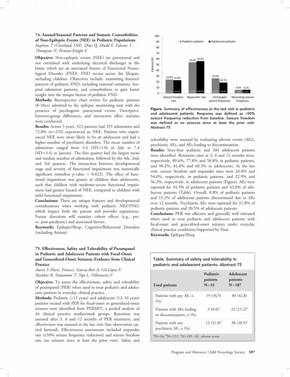
74. Annual/Seasonal Patterns and Somatic Comorbiditiesof Non-Epileptic Events (NEE) in Pediatric PopulationsStephens T (Cleveland, OH), Zhou Q, Mudd E, Falcone T,Thompson N, Pestana-Knight E
Objective: Non-epileptic events (NEE) are paroxysmal andnot correlated with underlying electrical discharges in thebrain, which are an associated feature of Functional Neuro-logical Disorder (FND). FND occurs across the lifespan,including children. Objectives include: examining features/patterns of pediatric FND, including seasonal variations, hos-pital admission patterns, and comorbidities to gain betterinsight into the unique factors of pediatric FND.Methods: Retrospective chart review for pediatric patients(8-18yo) admitted to the epilepsy monitoring unit with thepresence of psychogenic paroxysmal events. Descriptive,between-group differences, and interaction effect statisticswere conducted.Results: Across 5 years, 322 patients had 325 admissions and72.0% (n=232) experienced an NEE. Patients who experi-enced NEE were more likely to be an adolescent and had ahigher number of psychiatric disorders. The mean number ofadmissions ranged from 3.4 (SD=1.0) in July to 7.4(SD=3.4) in January. The first quarter had the largest meanand median number of admissions, followed by the 4th, 2nd,and 3rd quarters. The interaction between developmentalstage and severity of functional impairment was statisticallysignificant (omnibus p-value = 0.022). The effect of func-tional impairment was greater in children than adolescents,such that children with moderate-severe functional impair-ment had greater hazard of NEE, compared to children withmild functional impairment.Conclusions: There are unique features and developmentalconsiderations when working with pediatric NEE/FND,which impact both the patient and provider experiences.Future directions will examine cohort effects (e.g., pre-vs. post-pandemic) and associated factors.Keywords: Epilepsy/Sleep, Cognitive/Behavioral Disorders(including Autism)
75. Effectiveness, Safety and Tolerability of Perampanelin Pediatric and Adolescent Patients with Focal-Onsetand Generalized-Onset Seizures: Evidence from ClinicalPracticeAuvin S (Paris, France), Garcia-Ron A, Gil-L�opez F,Shankar R, Yamamoto T, Ngo L, Villanueva V
Objective: To assess the effectiveness, safety and tolerabilityof perampanel (PER) when used to treat pediatric and adoles-cent patients in everyday clinical practice.Methods: Pediatric (<12 years) and adolescent (12–16 years)patients treated with PER for focal-onset or generalized-onsetseizures were identified from PERMIT, a pooled analysis of44 clinical practice studies/work groups. Retention wasassessed after 3, 6 and 12 months of PER treatment, andeffectiveness was assessed at the last visit (last observation car-ried forward). Effectiveness assessments included responderrate (≥50% seizure frequency reduction) and seizure freedomrate (no seizures since at least the prior visit). Safety and
tolerability were assessed by evaluating adverse events (AEs),psychiatric AEs, and AEs leading to discontinuation.Results: Sixty-four pediatric and 204 adolescent patientswere identified. Retention rates at 3, 6 and 12 months were,respectively, 89.6%, 77.8% and 58.8% in pediatric patients,and 90.2%, 81.8% and 60.3% in adolescents. At the lastvisit, seizure freedom and responder rates were 24.0% and54.0%, respectively, in pediatric patients, and 22.9% and56.2%, respectively, in adolescent patients (Figure). AEs werereported for 34.5% of pediatric patients and 42.8% of ado-lescent patients (Table). Overall, 8.8% of pediatric patientsand 15.2% of adolescent patients discontinued due to AEsover 12 months. Psychiatric AEs were reported for 21.8% ofpediatric patients and 20.5% of adolescent patients.Conclusions: PER was effective and generally well toleratedwhen used to treat pediatric and adolescent patients withfocal-onset and generalized-onset seizures under everydayclinical practice conditions.Supported by EisaiKeywords: Epilepsy/Sleep
Table. Summary of safety and tolerability inpediatric and adolescent patients. Abstract 75
Total patients
PediatricpatientsN=55
AdolescentpatientsN=187
Patients with any AE, n(%)
19 (34.5) 80 (42.8)
Patients with AEs leadingto discontinuation, n (%)
3 (8.8)a 23 (15.2)b
Patients with anypsychiatric AE, n (%)
12 (21.8)c 38 (20.5)c
aN=34; bN=151; cN=185. AE, adverse event
Figure. Summary of effectiveness at the last visit in pediatricand adolescent patients. Response was defined as ≥50%seizure frequency reduction from baseline. Seizure freedomwas defined as no seizures since at least the prior visit.Abstract 75
Program and Abstracts, Child Neurology Society S97

76. Tethered, Online Education Improves Awareness ofthe Place in Care of New Treatments and Helps AlignPatient/Caregiver and Clinician Treatment Goals inDevelopmental and Epileptic EncephalopathiesDrexel C (Needham, MA), Bixler E, Meskis MA, Jaksha A,Conecker G, Dixon-Salazar T, Kowalski K, Sullivan J
Objective: An emerging concept in developmental and epi-leptic encephalopathies (DEEs) management is to accountfor seizure and non-seizure impacts of medication, such associal or functional benefits, in making treatment decisions.We evaluated the impact of aligned, online continuing medi-cal education on healthcare providers’ (HCPs’) ability todiagnose and treat DEEs and caregivers’ ability to engagein care.Methods: A 5-part HCP and 3-part caregiver series on symp-toms, treatments, team-based care, and supportive tools forDEE launched live-online between June 2021 and January2022; each activity will remain online for one year. Testquestions were administered pre, immediate post, and2 months post activity. Responses were analyzed to determinelessons learned and continuing gaps. Chi-square tests com-pared pre and post responses (P<0.05).Results: As of March 2022, 1,385 HCPs and 1,238 care-givers participated in at least one activity. Significant increasesoccurred related to HCPs’ awareness of the genetics of DS(+53%, P<0.05), impacts of treatment on seizure and non-seizure outcomes (+70%, P<0.05), and on caregivers’ QoL(+70%, P<0.05). HCPs (92%) reported benefits on practiceand patient experience (312 write-ins examples). Caregiversreported improved communication with HCPs (61%) andimproved ability to control healthcare decisions (100 write-ins examples).Conclusions: Live-online education for both HCPs and care-givers improves adoption of new treatments for DS andenhances caregivers’ engagement in care. Baseline scoreshighlight the need for ongoing education on the evolvingtreatment landscape for DEE to build upon acquired knowl-edge and improve shared decision-making.Keywords: Epilepsy/Sleep, Education, Rare Diseases
77. Polysomnographic parameters and comparison ofacademic performance in those with and without sleepdisorders, an observational studyChakrabarty B (New Delhi, India), Zulfiqar L, Gulati S,Jauhari P, Pandey R, Upadhyay A
Objective: To compare in children and adolescents aged2-18 years- Polysomnographic findings between those withand without sleep disorders- Academic performance in thosewith and without sleep disordersMethods: The study was conducted in a tertiary-care teach-ing hospital in north India. The study population comprisedof children and adolescents- attending a public school,- visit-ing the studyhospital with minor ailments or with Duchennemuscular dystrophy (DMD), spinal muscular atrophy(SMA), asthma and cerebral palsy (CP).They all underwentclinical evaluation and overnight polysomnography (PSG) aspart of the CASEQ (Childhood and Adolescent Sleep Evalua-tion Questionnaire) development and validation study.1
Results: Overall, 100 subjects (minor ailments:77, asthma:12,school:7, CP:2, DMD:2) were evaluated (mean age: 9+/-2 years, 74% males). The final diagnoses were sleep relatedbreathing disorder (SRBD, 56), sleep related movement disor-der (SRMD, 19), normal (20) and others (5). Apnea-hypopneaindex (AHI) was significantly higher in SRBD (4.8,3.3-10.6)compared to SRMD (1.2,0.8-1.8) and normal (2.2,1.4-3.7)cases (p=0.001). Significant positive correlation (r2=0.2) wasseen for AHI and periodic limb movement index with arousalindex (p=0.01 and 0.05). Good academic performers were sig-nificantly more prevalent in normal (55%) compared to SRBD(14.3%) and SRMD (21.1%) cases (p=0.004).Conclusions: Clinical evaluation and PSG play complimen-tary role in diagnosis of sleep disorders, as isolated PSG mayhave limitations related to first night effect. Those with com-mon sleep disorders like SRMD and SRBD have significantacademic impairment compared to normal children andadolescentsKeywords: Epilepsy/Sleep
Reference1. Zulfiqar L, Chakrabarty B, Gulati S, Jauhari P, Pandey
RM, Tripathi M, et al. The Childhood and AdolescentSleep Evaluation Questionnaire (CASEQ): Develop-ment and validation of an ICSD-3-based screeninginstrument, a community and hospital-based study. JSleep Res. 2022;31:e13479.
Table 2: Distribution of academic performance innormal and diseased population. Abstract 77
Academicperformance Normal SRBD SRMD
pvalue
Poor 10% 26.8% 42.1% 0.004
Average 35% 58.9% 36.8%
Good 55% 14.3% 21.1%
(SRBD: Sleep related breathing disorder, SRMD: Sleep related move-ment disorder)
Table 1: Distribution of polysomnographicparameters in normal and diseased population.Abstract 77
PSG parameters Normal SRBD SRMD p value
Sleep efficiency 80.6% 75.5% 81.5% 0.06
AHI 2.2 4.8 1.4 0.0001
AI 5.2 6.8 5.4 0.1
(SRBD: Sleep related breathing disorder, SRMD: Sleep related move-ment disorder, AHI: Apnea Hypopnea Index, AI: Arousal index)
S98 Annals of Neurology Vol 92 (suppl 28) 2022

78. Unusual Presentation of Phosphatidylinositol glycanbiosynthesis class A protein (PIGA) deficiency presentingas refractory pediatric epileptic encephalopathy; Amystery solved after 21 yearsSial GZ (Buffalo, NY), Mesha M, Lopez Weinstock A
Objective: To highlight the importance of comprehensiveepilepsy panel for diagnosing PIGA related epilepsy. Inheritedglycosylphosphatidylinositol (GPI) deficiency is caused bydecreased presentation of GPI anchor proteins (GPI-AP).PIGA enzyme facilitates the synthesis of GPI-AP. PIGA defi-ciency, an X linked recessive disorder, is associated with mul-tiple congenital anomalies. Neurological presentation consistof refractory seizures and intellectual disability.Methods: We present a 21-year-old male with static enceph-alopathy and refractory epilepsy, in whom comprehensiveepilepsy gene panel confirmed the diagnosis. Review of medi-cal records, electroencephalograms, laboratory, imaging, andgenetic testing was performed.Results: Patient presented with refractory myoclonic epi-lepsy at age 6 months. There was milestone regression atage 2 years. Childhood evaluation revealed normal karyo-type, Fragile X, serum and urine amino acids, urineorganic acids, and serum lactate. Seizure semiology con-sisted of daily prolonged tonic, tonic-clonic and myoclonicseizures in clusters, requiring daily rescue therapy and fre-quent ER visits. He was on 5 anti-seizure drugs (ASD)and VNS, and had failed multiple ASD and the ketogenicdiet. Video-EEG monitoring revealed continuous general-ized slowing, multiregional sharp waves, generalized spike-wave complexes, polyspikes, paroxysmal fast activity andmyoclonic seizures. MRI brain showed stable mild atrophy.Comprehensive epilepsy panel performed at age 21 revealedhemizygosity for PIGA, c.823C>T (p.Arg275Trp), a diag-nostic pathogenic variant.Conclusions: The diagnosis of PIGA related epilepsy canbe challenging based on clinical grounds. A comprehensivegene panel should be considered in adult patients withidiopathic severe childhood epilepsy and intellectualdisability.Keywords: Epilepsy/Sleep, Neurometabolic Disorders
79. Seizure during hemodialysis in a pediatric patientwith renal disease requires further neurological workupSandweiss A (Houston, TX), Pareek A, Moore M, Gill J
Objective: Seizures are reported as a common adverse eventduring hemodialysis, thought to occur in approximately7-10% of all hemodialysis patients. Seizures occur due to anunderlying epilepsy disorder or provoked by an external fac-tor such as fluctuating ionic potentials. Though prior reportssuggest most seizures are provoked in dialyzed patients, pedi-atric patients who have a seizure associated with dialysisrequire a complete neurological workup in order to rule outan underlying epilepsy disorder.Methods: We present a pediatric patient with end-stage renaldisease who had a seizure during his third hemodialysis ses-sion that uncovered a previously unknown epilepsysyndrome.
Results: An 8 year-old healthy male presented to the hospitalwith six weeks of malaise, ultimately found to have a newdiagnosis of chronic kidney disease secondary to renal dyspla-sia. BUN and creatinine were 175 and 7.63 respectively withnormal blood pressure. He was admitted and initiated on ahemodialysis regimen; he sustained a focal seizure lasting upto 5 minutes during his third hemodialysis session. Immedi-ate workup included a normal CT head and stable serumelectrolytes. Routine EEG revealed 3-4 Hz spike and waveactivity potentiated by photic stimulation. History revealedno other red-flag signs for epilepsy, though he had a clearabsence seizure during hyperventilation on the neurologyexam. He was diagnosed with juvenile myoclonic epilepsyand started on valproic acid, achieving seizure freedom duringsubsequent hemodialysis four days later.Conclusions: Seizures are not uncommon during hemodialy-sis, but all patients require a complete neurological evaluationto unmask a potential underlying epilepsy syndrome.Keywords: Epilepsy/Sleep
80. Successful treatment of status epilepticus in a patientwith a novel phenotype of an ATP1A2 missense mutationknown to cause hemiplegic migrainesTrivedi A (San Diego, CA), Yang J, Ladt K, Rho J
Objective: The ATP1A2 gene on chromosome 1 encodes thealpha-2 polypeptide subunit on the Na/K ATP-ase pump.Mutations in ATP1A2 have been associated with familialhemiplegic migraine type II. However, the associationbetween a particular mutation in this gene, and epilepsy isnot well described and limited literature exists on the relatedclinical management.Methods: A 12-year-old male with a history of seizures ininfancy and a known hemiplegic migraine-associatedATP1A2 mutation c.2563G>A , pGly.855Arg, was admittedfor status migrainosus, prolonged dysarthria, left sidedhemiparesis and sensory loss. His symptoms persisted despiteintravenous verapamil, intranasal ketamine, and trials of acet-azolamide, topiramate and valproic acid. He graduallyimproved after 6 days with return of left sided function andspeech. However, 24 hours later, he developed epilepsia par-tialis continua consisting of leftward gaze-evoked nystagmusand clonic movements of the left face and arm. His seizurespersisted despite lorazepam and increased valproic aciddosing.Results: Seizures improved after starting lacosamide and initi-ating a ketogenic diet. At the last follow up three monthsafter hospitalization, he remained seizure-free and hismigraines had been successfully aborted with naproxen andadditional lacosamide doses.Conclusions: We present the case of focal-onset statusepilepticus following an episode of hemiplegic migraine in apatient with an ATP1A2 mutation, a genotype-phenotypecorrelation that has not been previously described. He wassuccessfully treated with a combination of ketogenic diet andlacosamide, supporting the combined targeting of slow inacti-vation of voltage-gated sodium channels and altering the met-abolic milieu as a therapeutic approach.Keywords: Epilepsy/Sleep, Genetics, Headache/Migraine
Program and Abstracts, Child Neurology Society S99

81. Stiripentol for drug-resistant epilepsy treatment intuberous sclerosis complexAungaroon G (Cincinnati, OH), Franz D, Mehta A
Objective: This study assesses the efficacy and tolerability ofstiripentol (STP) in tuberous sclerosis complex (TSC)patients with drug-resistant epilepsy (DRE).Methods: A retrospective review of TSC patients at Cincin-nati Children’s Hospital from 2011 until 2021 was per-formed to identify patients with DRE receiving STP. Seizurefrequency was assessed 1 month before (baseline) and 1, 3,6, and 12 months after STP initiation.Results: Of the 1492 TSC patients, 13 (10 males) receivedSTP. The age range was 3.8 to 40 years (median,IQR = 15.2 years, 6.7 - 22.0). STP was initiated a medianof 13.5 years (IQR 5.0 - 20.3) after seizure onset. Themedian treatment duration was 12.8 months, and a medianSTP dose was 21.1 mg/kg/day (IQR 12.8 - 34.1). The num-ber of patients with > 50% seizure reduction was 6/13(46.2%), 4/13 (30.8%), 8/11 (72.7%), and 6/8 (75.0%) at1, 3, 6, and 12 months. Importantly, 6 patients (46.2%) hadpersistent seizure reduction from 1 through 12 months, withthe mean (�SD) percentage of reduction at 1, 3, 6, and12 months of 68.1% (�22.0), 71.3% (�23.2), 75.7%(�23.5), and 75.7% (�23.5), respectively. 11/13 patientsreported side effects with aggression being the most commonand 3 patients discontinued STP due to side effects.Conclusions: Most TSC patients with DRE treated withSTP experienced seizure reduction, with about half having apersistent seizure reduction. This suggests that STP could bean efficacious and tolerable treatment option for thispopulation.Keywords: Epilepsy/Sleep, Neurocutaneous Disorders,Genetics
82. Higher beta-hydroxybutyrate levels result in decreasedneutrophils in children with epilepsy on theketogenic dietGombolay G (Atlanta, GA), Elkins K, Lu S, Holt P,Johnson M, Gedela S
Objective: The ketogenic diet (KD) produces beta-hydroxybutyrate (BHB) which decreases neutrophils in adultsbut limited studies are available in children. BHB maydecrease inflammation by decreasing neutrophils. We com-pare the relationship of BHB with neutrophils in childrenwith epilepsy on the ketogenic diet.Methods: IRB approval was obtained. Children at a singleKD clinic from 2010-2021 were included. Data collectedincluded age when seizures started, age when KD started, sex,white blood cell count (WBC), and absolute neutrophilcounts (ANC). ANC was excluded if obtained in the settingof infection or medications such as steroids. Low normalBHB was defined as BHB < 4 mmol/L and high normalBHB was defined as BHB ≥ 4 mmol/L. Statistical analysiswas performed using SAS 16.0 (Cary, N.C.).Results: Forty-one patients were included and divided intolow BHB (N=20) and high BHB (N=21).One-tailed twosample t test to predict whether high BHB results in lower
ANC demonstrated that patients with BHB ≥ 4 had on aver-age 34.6% neutrophils (95% 29.3-39.8%) as compared to41.8% (95% CI 34.9 – 48.6) in BHB < 4 (p = 0.043).However, multivariate logistic regression modeling showedthat BHB levels and ANC did not correlate with probabilityof improvement in seizures at 12 months.Conclusions: Higher BHB levels are associated with lowerANC in children on KD. Further investigation is needed inlarger studies to investigate the effect of BHB on ANC inchildren.Keywords: Epilepsy/Sleep
Table 1: Patient characteristics between low normaland high normal BHB levels. Abstract 82
BHB< 4N=20
BHB≥ 4N=21
p-value
Sex M:F, N (%) 13:7(65:35)
13:8(62:38)
1.000
Age started diet, yMean (SD)
5.1(4.7)
3.4(2.5)
0.145
Age seizure started, yMean (SD)
0.8(1.2)
1.2(1.6)
0.347
Number of seizures perweek prior to starting KDMean (SD)
165.9(235.7)
143.3(160.7)
0.720
Number of seizures perweek at 12 mo, mean (SD)
16.9(23.3)
31.2(46.8)
0.213
Type of seizure N (%)
GTC 3 (15) 3 (14) 1.000
Focal seizures 6 (30) 6 (29) 0.920
Absence 3 (15) 1 (5) 0.343
IS 8 (40) 10 (48) 0.623
Myoclonic seizure 8 (40) 5 (24) 0.266
Drop 2 (10) 6 (29) 0.238
Tonic 10 (50) 4 (19) 0.039
ESES 1 (5) 0 (0) 0.488
WBC, 106/ml, mean (SD) 7.6(3.6)
6.8(2.6)
0.461
Neutrophil %, mean (SD) 41.8(14.6)
34.6(11.5)
0.043
Lymphocyte %, mean (SD) 47.4(14.1)
53.8(11.3)
0.057
S100 Annals of Neurology Vol 92 (suppl 28) 2022

83. Cognitive and Behavioral Effects and Tolerability ofAdjunctive Brivaracetam in Children and Adolescentswith Focal Seizures: Pooled Interim AnalysisLittle A (Smyrna, GA), Elshoff J-P, Fleyshman S, De LaLoge C, Nondonfaz X, Reichel C, Floricel F, Smeyers P
Objective: Evaluate cognitive/behavioral effects and tolerabil-ity of long-term adjunctive brivaracetam in children/adolescents with focal seizures.Methods: Interim post-hoc analysis (cut-off 14-Jul-2020) ofpooled data from Phase IIa, open-label trial (N01263/NCT00422422; patients aged ≥1 month-<16 years) andongoing, Phase III, open-label, long-term follow-up
(N01266/NCT01364597; direct enrollers aged≥4-<17 years). Cognitive/behavioral outcomes assessed withAchenbach CBCL (1.5-5/6-16 years) and BRIEF-P/BRIEF(<5/5-16 years).Results: 140 patients analyzed (mean age: 9.5 years; 57.1%male). Mean changes from baseline to final visit (raw subscalescores): CBCL 1.5-5 aggressive behavior/anxious&depressed/attention problems/emotionally reactive/sleep problems/somatic complaints/withdrawn/other problems: -1.4/-1.5/-0.5/-1.0/-1.1/-0.9/-0.7/-3.2; CBCL 6-18 aggressive behavior/anxious&depressed/attention problems/rule-breaking behav-ior/social problems/somatic complaints/thought problems/
FIGURE 1. Shifts in T-score categories (normala, borderline or clinical rangeb) in Achenbach Child Behavior Checklist (CBCL) subscalesfrom Baseline to last evaluation in patients with focal seizures: (A) Achenbach CBCL 1.5-5 years of age; (B) Achenbach CBCL6-18 years of age. BCR: Borderline or clinical range. Only patients providing CBCL data at both Baseline and last evaluation wereincluded. Baseline data were obtained from the core trial Screening Visit. Median time to last CBCL evaluation: 295.5 days (patients1.5-5 years of age, n=32), 1043.0 days (patients 6-16 years of age, n=102). aNormal: T-score <65; bBCR: T-score ≥65. Abstract 83
Program and Abstracts, Child Neurology Society S101

withdrawn&depressed: -1.8/-1.4/-1.2/-0.7/-0.5/-1.2/-0.8/-0.1; BRIEF-P inhibit/shift/emotional control/working mem-ory/plan&organize: -6.6/-2.1/-4.9/-4.0/-3.0; BRIEF inhibit/shift/emotional control/initiate/working memory/plan&-organize/organization of materials/monitor: -1.6/-0.5/-1.0/-0.6/-1.8/-1.4/-0.8/-0.9. Most patients in both age groupshad no shift in T-score category from baseline to last evalua-tion for each CBCL subscale (between normal andborderline or clinical range [BCR], Fig. 1) and BRIEF-P/BRIEF subscale (between normal and potential clinical
significance [PCS], Fig. 2). For all CBCL subscales, morepatients changed from BCR to normal than normal to BCR.Changes across BRIEF-P subscales were mostly from PCS tonormal. Across BRIEF subscales, similar proportions ofpatients changed from normal to PCS and PCS to normal.Incidences of treatment-emergent adverse events (TEAEs):overall (1.5-5/6-16 years: 100%/95.4%); drug-related(40.6%/34.3%); serious (18.8%/26.9%); discontinuationsdue to TEAEs (6.3%/7.4%); deaths (3.1% [n=1]/0.9% [n=1]).
Figure 2. Shifts in T-score categories (normala, potential clinical significanceb) in Behavior Rating Inventory of Executive Function(BRIEF) subscales from Baseline to last evaluation in patients with focal seizures: (A) BRIEF-Preschool Version (BRIEF-P) <5 yearsof age; (B) BRIEF 5-18 years of age. PCS: Potential clinical significance. Only patients providing BRIEF-P/BRIEF data at bothBaseline and last evaluation were included; some patients may have had missing subscale scores, and in such cases, the sum ofthe proportion of patients in the results shift groups is <100%. Baseline data were obtained from the core trial Screening Visit.Median time to last BRIEF-P/BRIEF evaluation: 204.0 days (patients <5 years of age, n=7), 1220.0 days (patients 5-16 years ofage, n=101). aNormal: T-score <65; bPCS: T-score ≥65. Abstract 83
S102 Annals of Neurology Vol 92 (suppl 28) 2022

Conclusions: In this pooled interim analysis, cognitive/behavioral functioning scores in children/adolescents withfocal seizures under long-term adjunctive brivaracetam weregenerally stable or slightly improved compared with baseline.Brivaracetam was generally well tolerated.Funding: UCBPharmaKeywords: Epilepsy/Sleep
84. Isoflurane controls Super Refractory StatusEpilepticus secondary to a Monoallelic DNM1LMutation: A Case ReportPark C (Fort Worth, TX), Luke R, Perry M
BACKGROUND: Variants in DNM1L are reported as rarecauses of refractory epilepsy and frequently present as superrefractory status epilepticus (SE). Specifically, the monoallelicR403C variant can present with epilepsia partialis continua(EPC), a form of focal SE which lacks definitive treatmentoptions. We report a patient with an R403C DNM1L muta-tion successfully treated with isoflurane for super refractoryEPC on two separate occasions.Methods: N/ACase Information: A 14-year-old male presented with superrefractory SE manifest as EPC secondary to a monoallelicR403C variant in DMN1L. Failed therapies included multi-ple loading doses of antiseizure medications in addition tocontinuous midalozam, pentobarbital, propofol, ketamine,and lidocaine infusions. Adminstration of 0.9% isoflurane for12 hours achieved burst suppression and resolution of statuswith the ability to wean off all continuous infusions over thefollowing week. He returned over two years later with a simi-lar presentation and failed interventions prior to initiation ofisoflurane for 30 hours, which again abortedSE. Unfortunately, SE returned 3 weeks later and he failedisoflurane as the initial treatment.Conclusions: DNM1L frequently presents with super refrac-tory SE rarely responding to conventional treatments. Short-term administration of inhaled isoflurane aborted SE in twoof three instances for our patient and may represent an effica-cious treatment that should be considered earlier in the treat-ment strategy to shorten duration of SE in this condition.The reason for failure of this therapy when used as initialintervention is unclear and may be secondary to progressionof disease, though further investigation is warranted.Keywords: Epilepsy/Sleep, Genetics, Rare Diseases
85. Novel missense variant in the SLC2A1 c.1162T>C(p.Trp388Arg) causing childhood onset intractablegeneralized epilepsy, microcephaly and cognitiveimpairmentAbdul Hamid O (Memphis, TN), Patterson A, Caron E,Weatherspoon S
Objective: Glucose transporter 1 deficiency syndrome(GLUT1-DS) is a rare inborn error of metabolism caused byimpaired glucose transport through blood brain barrier dueto mutation in SLC2A1 gene, encoding a glucose transporterprotein. Clinical symptoms, ranging from severe epilepticencephalopathy, microcephaly and movement disorders. Thediagnosis of GLUT1-DS requires hypoglycorrhachia in the
presence of normoglycaemia with a reduced cerebrospinalfluid (CSF):plasma glucose ratio.Methods: In this case, we describe a child who developedgeneralized tonic-clonic seizures and absence seizures at age6. Epilepsy gene panel identified a novel missense variant ofundetermined clinical significance in exon 9 of the SLC2A1gene c.1162T>C (p.Trp388Arg). The clinical features of thispatient include intractable epilepsy and epileptic encephalop-athy with severe language delay. Her head conference was inthe 10th percentile for age. The ictal electroencephalogram(EEG) was characterized by generalized paroxysmal 3.5 Hzspike-slow wave complexes.Results: She continued to have very frequent clinical andelectrographic seizures despite therapeutic serum levels ofDepakote, Ethosuximide and Keppra. Brain MRI wasunremarkable. Metabolic screening labs were normal. A chro-mosomal MicroArray was normal. Only the mother wasavailable for parental testing and she did not harbor thepatient’s variant. Allele frequency is extremely low in all data-bases. A lumbar puncture demonstrated a reduced CSF:plasma glucose ratio consistent with GLUT1-DS.Conclusions: Treatment with modified Atkins diet showedclinical improvement with the reduction of clinical seizuresand improvement in EEG. Given phenotypic overlap withpreviously reported individuals, we propose the pathogenicityof this variant.Keywords: Epilepsy/Sleep, Genetics, Cognitive/BehavioralDisorders (including Autism)
86. Patient Outcomes During Transition from Pediatricto Adult Care for EpilepsyNurre E (Cincinnati, OH), Brothers S, Olson J, Hornbach A,Fong S
Objective: To collect clinical outcomes data for patients withepilepsy during and after transition from pediatric to adulthealthcareMethods: For this retrospective chart review, in the Universityof Cincinnati electronic health record database, we collecteddata for patients who transitioned from Cincinnati Children’sHospital Medical Center to University of Cincinnati(UC) Health neurology department for epilepsy care. We col-lected patient information including age, sex, ethnicity, com-orbidities, substance abuse history and insurance type as wellas imaging and EEG data for diagnosis. We gathered clinicaloutcomes data for these patients including healthcare utiliza-tion measures (emergency department visits, outpatient clinicvisits, patient telephone calls, hospital admissions) as well aslaboratory testing, anti-epileptic drug information and surgicaldiscussions if medically refractory epilepsy. The results of thisanalysis will be used to identify gaps in care during transitionand target areas to improve clinical outcomes in this process.Results: Preliminary results revealed patients identified(N=139) with a wide range of ages and comorbidities.Conclusions: Patients transitioning from pediatric to adultcare in epilepsy have poor clinical outcomes and gaps in care.There may be target areas to improve clinical outcomes inthis process.Keywords: Epilepsy/Sleep
Program and Abstracts, Child Neurology Society S103

EQUITY, DIVERSITY,INCLUSION
87. Implementation of a Provider-to-Provider 24-7Hotline for Angelman Syndrome Improves Access toExpert CareDuis J (Aurora, CO), Nangia S, Walleigh D, Berry-Kravis E
Objective: We have implemented a provider-to-provider con-sultation hotline available 24-7 with access to expert pro-viders across the United States to improve health equity andaccess to care for all individuals with Angelman Syndrome(AS). Our goal was to develop evidence-based algorithms forthe management of common urgent and emergent concernsand to demonstrate novel methods for expert access for man-agement of rare disorders to improve health outcomes andstandards of care.Methods: The infrastructure for an on-call service for indi-viduals with AS was coordinated at Children’s Hospital Col-orado for access to an on-call Angelman specialist. Togetherwith key opinion leaders in the field, we developed clinicalpathways for the management of AS. We are collecting pro-spective data to understand outcomes with these standards ofmanagement. Calls are accepted nationally and internation-ally with interpreter services available.Results: Since July 1, 2020, we have received consultationsfrom across the world including Italy, France, and Australia.The primary concerns include seizure management, poorsleep, behavior concerns, motor changes, and non-epilepticmyoclonus. Outcomes have been positive with shared infor-mation on existing data and clinical pathways. Phone callsare more often from trainees than attending providers. Accep-tance of the emergency line by providers is a barrier to receiptof actionable calls.Conclusions: Consensus management convened by a groupof key opinion leaders and implementation of an infrastruc-ture to receive calls from providers to assist in local manage-ment of Angelman Syndrome has been successful. We have amet some barriers regarding acceptance and use of line.Keywords: Diversity, Equity, Inclusion, Rare Diseases,Genetics
88. Differential National Trends in Child NeurologyRamsy N (Champaign, IL), Peterman N, Yeo E, Kaptur B
Objective: There is a significant gap between the need forchild neurology and the number of available child neurolo-gists. This work utilized geospatial analysis to identify thecommunities where this gap is the highest and the demo-graphics of those most affected.Methods: US Census and Center for Medicare Services data-bases from 2015-2019 were utilized to obtain the socioeco-nomic demographics of each US county and thecorresponding number of practicing and child neurologists.Counties without neurologists were excluded. Physician racewas estimated using name-based predictive algorithms. Clus-ter analysis was conducted to classify countries as statistically
significant, p < 0.05, hotspots (High-High), coldspots (Low-Low), and geospatial outliers (Low-High and High-Low) ofchild neurology using Moran’s I analysis. Clusters were thencompared across socioeconomic variables using ANOVA.Results: 22,533 practicing neurologists, 70.94% white and36.14% female, and 2,378 child neurologists, 71.03% whiteand 50.25% female, were included. Southern California andthe Mid-Atlantic were child neurology hotspots with 110.63neurologists and 12.34 child neurologists per county. Col-dspots were predominantly located in Appalachia with anaverage of 4.30 neurologists and 0.09 child neurologists percounty. Compared to coldspots, hotspots were statisticallysignificantly less impoverished, less white, more urbanized,and had higher median income.Conclusions: Quantifying the gap in child neurology is criti-cal for properly allocating resources. Although Appalachia hasthe lowest access in the nation, even locations with highaccess to child neurology had what would be considered pooraccess compared to general neurology.Keywords: Diversity, Equity, Inclusion
89. Race and Gender-Associated Disparities in AcuteHeadache Management in a Pediatric EmergencyDepartmentShiswawala N (Cleveland, OH), Gao C, Goldstein J, Rose J,Bass N, Wyllie K, Malay S, Grube A
Objective: Racial and gender healthcare disparities in analge-sic use for headaches in emergency departments (ED) havebeen previously documented1. Literature is scarce for pediat-ric populations. This study’s aim was to evaluate for differ-ences in the acute management of headaches in apediatric ED.Methods: A retrospective cohort study of children within asingle hospital ED system with headaches during a 3-yearperiod was completed. Headache medications were groupedinto 3 categories: 1 (oral), 2 (first-line IV), and 3 (second-lineIV). Association of race and gender with multiple clinical var-iables were included. Assessment was performed usingunivariable Chi-squared tests.Results: 607 children were included; 269 White children(44%), 321 Black children (53%) and 17 children of otherraces (3%). Statistically significant differences in time to ini-tial analgesic were not observed based on race or gender. Arace-associated difference in the category of treatment wasnoted; black children received Category 1 medications morethan white children (31% vs 9.4%, p<0.0001). More whitechildren received Category 3 medications (15% vs 2.6%, p<0.0001). Females had a statistically significant greater reduc-tion in pain compared to males (4 vs 6, p=0.025).Conclusions: We observed a race-associated difference inthe analgesic agent category used and a greater reductionin pain scores following treatment for females comparedto males. No significant differences in time to initial anal-gesic for gender or race. These findings highlight the needfor further research in pediatric headache healthcare dis-parities to address racial and gender disparities in thispopulation.Keywords: Diversity, Equity, Inclusion, Headache/Migraine
S104 Annals of Neurology Vol 92 (suppl 28) 2022

Abstract 88
Program and Abstracts, Child Neurology Society S105

ABSTRACT 89References
1. Charleston L., 4th (2021). Headache Disparities inAfrican-Americans in the United States: A NarrativeReview. Journal of the National MedicalAssociation, 113(2), 223–229. https://doi.org/10.1016/j.jnma.2020.09.148
90. Reducing Implicit Bias and Promoting DEI Efforts inChild Neurology Residency Recruitment- An Updatefrom 2020Kumar I (Ann Arbor, MI), Gottlieb-Smith R, Leber S,Behnke T, McCaffrey H, Shellhaas R, McNamara N,Cartwright K, Fedak Romanowski E
Objective: The Michigan Medicine Pediatric NeurologyDivision used quality-improvement (QI) methodology to
reduce unconscious bias and increase objectivity byimplementing interventions during the selection process ofresidency applicants. The goal was to increase diversity ofinterviewed applicants by 2022 while sustaining foundationalprinciples for success in child neurology.Methods: We examined baseline demographic data from 2019recruitment season focusing on gender, self-identified race,hometown, degree, USMLE scores, and participation in minorityinterest groups. Our first intervention included an implicit biasself-assessment and review of education modules on hiring biasby faculty. For further PDSA cycles, we introduced new inter-ventions: 1) removing candidate photograph, name, and gender;2) changing our objective scoring system by broadening the defi-nition of scholarly work and putting more weight on interper-sonal skills; 3) adding standardized questions to interviews.Results: As previously reported, baseline data (2019) suggestedunconscious biases to interview more female, white, Midwest/Michigan, MD (vs. DO) candidates with minimal participa-tion in minority interest groups as previously reported. Aftertwo PDSA cycles, there was reduction of biases related to sexand geographic location (Tables 1 and 2).
TABLE 1: P values calculated by Wald’s test of eachindependent variable, and then of the interactionof year with each variable in logistic regressionmodels of interviewed status. Abstract 90
2019 2020 2021Yearinteraction
Gender(Female vsMale)
Eligible* .054 .839 .111 .425
Invited <.001 .338 .022 .037
Interviewed .002 .198 .180 .177
Race (white vsnonwhite)
Eligible .002 .170 .123 .425
Invited .001 .026 .003 .681
Interviewed .005 .012 <.001 .707
Region
Eligible <.001 <.001 <.001 .025
Invited <.001 <.001 <.001 .273
Interviewed <.001 <.001 <.001 .136
Degree (MD vsDO vsMD/PhD vsOther)
Eligible <.001 <.001 <.001 .981
Invited <.001 <.001 <.001 .736
Interviewed .002 <.001 <.001 .779
TABLE 1 (Continued)
2019 2020 2021Yearinteraction
Step 1
Eligible <.001 .511 <.001 .107
Invited <.001 <.001 <.001 .683
Interviewed <.001 .001 <.001 .683
Step 2
Eligible <.001 .018 <.001 .609
Invited <.001 <.001 <.001 .417
Interviewed <.001 <.001 <.001 .495
Minorityinterest group(No vs Yes)
Eligible <.001 <.001 .008 .049
Invited .033 <.001 <.001 .242
Interviewed .074 <.001 .003 .330
Age group
Eligible <.001 .001 .001 .182
Invited <.001 .011 <.001 .021
Interviewed <.001 .012 <.001 .086
*Applicants who graduate from US/Canadian medical schools, andhave successfully passed the USMLE are eligible for our program, asdetermined by the MM Pediatrics Department, thus the number ofeligible applicants is lower than the total number of applicants.
S106 Annals of Neurology Vol 92 (suppl 28) 2022

TABLE 2: Demographics from all applicants, all eligible applicants, all applicants invited to interview, and allinterviewed applicants of all three recruitment years. Abstract 90
Applicant
characteristics 2019 (Baseline) 2020 (after one PDSA cycle)
2021 (after two
PDSA cycles)
All
Applicants
N=144
Eligible
Applicants*
N=83
Invited to
Interview
N=47
Interviewed
N = 39
All
Applicants
N=137
Eligible
Applicants
N=81
Invited to
Interview
N=51
Interviewed
N = 45
All Applicants
N = 147
Eligible
Applicants
N = 111
Invited to
Interview
N = 60
Interviewed
N = 52
Sex
Female
Male
91 (63%)
53 (37%)
58 (70%)
25 (30%)
41 (87%)
6 (13%)
33 (85%)
6 (15%)
87 (64%)
59 (36%)
52 (64%)
29 (36%)
35 (69%)
16 (31%)
32 (71%)
13 (29%)
94 (64%)
53 (36%)
75 (68%)
36 (32%)
45 (75%)
15 (25%)
37 (71%)
15 (29%)
Self-identified
Race
White
Non-white
Not answered
54 (38%)
75 (52%)
15 (10%)
40 (48%)
39 (47%)
4 (5%)
27 (57%)
18 (38%)
2 (4%)
22 (56%)
15 (38%)
2 (4%)
61 (45%)
71 (52%)
5 (4%)
40 (49%)
39 (48%)
2 (2%)
29 (57%)
20 (39%)
2 (4%)
27 (57%)
16 (36%)
2 (4%)
57 (39%)
82 (56%)
8 (5%)
47 (42%)
57 (51%)
7 (6%)
32 (53%)
26 (43%)
2 (3%)
31 (60%)
19 (37%)
2 (4%)
Age
<25
26-30
31-35
36-40
41-60
Not answered
3 (2%)
81 (56%)
41 (28%)
14 (10%)
2 (1%)
30 (21%)
1 (1%)
58 (70%)
23 (28%)
1 (1%)
0 (0%)
0 (0%)
0 (0%)
36 (77%)
11 (23%)
0 (0%)
0 (0%)
0 (0%)
0 (0%)
35 (90%)
4 (10%)
0 (0%)
0 (0%)
0 (0%)
4 (3%)
93 (68%)
20 (15%)
9 (7%)
4 (3%)
7 (5%)
4 (5%)
62 (77%)
10 (12%)
2 (2%)
0 (0%)
3 (4%)
1 (2%)
43 (84%)
6 (12%)
0 (0%)
0 (0%)
1 (2%)
1 (2%)
38 (84%)
5 (12%)
0 (0%)
0 (0%)
1 (2%)
4 (3%)
91 (62%)
35 (24%)
12 (8%)
1 (1%)
4 (3%)
4 (4%)
78 (70%)
21 (19%)
6 (5%)
0 (0%)
2 (2%)
4 (7%)
51 (85%)
3 (5%)
1 (2%)
0 (0%)
1 (2%)
4 (8%)
43 (83%)
3 (6%)
1 (2%)
0 (0%)
1 (2%)
Hometown
region
Michigan
Midwest
Northeast
South
West
Puerto Rico
International
No
information
14 (10%)
24 (17%)
23 (16%)
15 (10%)
20 (14%)
2 (1%)
30 (21%)
16 (11%)
11 (13%)
21 (25%)
17 (20%)
12 (14%)
17 (20%)
2 (2%)
2 (2%)
1 (1%)
8 (17%)
12 (26%)
10 (21%)
6 (13%)
9 (19%)
0 (0%)
1 (2%)
1 (2%)
8 (21%)
12 (31%)
8 (21%)
3 (8%)
6 (15%)
0 (0%)
1 (3%)
1 (3%)
4 (3%)
19 (14%)
26 (19%)
34 (25%)
19 (14%)
1 (1%)
30 (21%)
4 (3%)
3 (4%)
18 (22%)
17 (21%)
26 (32%)
14 (17%)
1 (1%)
2 (2%)
0 (0%)
2 (4%)
9 (18%)
14 (27%)
17 (33%)
7 (14%)
0 (0%)
2 (4%)
0 (0%)
2 (4%)
9 (20%)
14 (33%)
12 (27%)
6 (16%)
0 (0%)
2 (4%)
0 (0%)
10 (7%)
25 (17%)
19 (13%)
40 (27%)
18 (12%)
1 (1%)
34 (23%)
0 (0%)
9 (8%)
20 (18%)
18 (16%)
34 (31%)
17 (15%)
1 (1%)
12 (11%)
0 (0%)
6 (10%)
11 (18%)
14 (23%)
19 (32%)
9 (15%)
1 (2%)
0 (0%)
0 (0%)
6 (12%)
9 (17%)
12 (23%)
18 (35%)
6 (12%)
1 (2%)
0 (0%)
0 (0%)
Degree
MD
DO
Other
No
information
107 (40%)
12 (8%)
23 (16%)
2 (1%)
76 (92%)
7 (8%)
0 (0%)
0 (0%)
45 (96%)
2 (4%)
0 (0%)
0 (0%)
37 (95%)
2 (5%)
0 (0%)
0 (0%)
93 (68%)
13 (10%)
31 (23%)
1 (1%)
72 (89%)
10 (12%)
0 (0%)
0 (0%)
49 (96%)
2 (4%)
0 (0%)
0 (0%)
43 (96%)
2 (4%)
0 (0%)
0 (0%)
100 (68%)
21 (14%)
1 (1%)
25 (17%)
89 (80%)
16 (14%)
0 (0%)
6 (5%)
53 (88%)
7 (12%)
0 (0%)
0 (0%)
47 (90%)
5 (10%)
0 (0%)
0 (0%)
USMLE Step
1
<200
200-220
221-240
>240
No
information
17 (12%)
46 (32%)
46 (32%)
10 (7%)
5 (3%)
4 (5%)
25 (30%)
30 (36%)
23 (28%)
0 (0%)
1 (2%)
4 (9%)
22 (47%)
6 (13%)
0 (0%)
1 (3%)
2 (5%)
19 (49%)
17 (44%)
0 (0%)
9 (7%)
51 (37%)
47 (34%)
26 (19%)
4 (3%)
4 (5%)
27 (33%)
30 (37%)
18 (22%)
2 (2%)
1 (2%)
10 (20%)
21 (41%)
17 (33%)
2 (4%)
1 (2%)
9 (20%)
20 (44%)
15 (33%)
0 (0%)
11 (7%)
52 (35%)
52 (35%)
27 (18%)
5 (3%)
3 (3%)
42 (38%)
41 (37%)
25 (23%)
0 (0%)
0 (0%)
11 (18%)
29 (48%)
20 (33%)
0 (0%)
0 (0%)
9 (17%)
25 (48%)
18 (35%)
0 (0%)
USMLE Step
2
<200
200-220
221-240
>240
No
information
1 (0.7%)
27 (19%)
54 (38%)
54 (38%)
8 (6%)
0 (0%)
6 (7%)
32 (39%)
43 (52%)
2 (2%)
0 (0%)
0 (0%)
10 (21%)
36 (77%)
1 (2%)
0 (0%)
0 (0%)
8 (21%)
30 (77%)
1 (3%)
1 (1%)
19 (14%)
54 (39%)
54 (39%)
9 (7%)
0 (0%)
7 (9%)
30 (37%)
40 (49%)
4 (5%)
0 (0%)
3 (6%)
9 (18%)
38 (75%)
1 (2%)
0 (0%)
3 (7%)
9 (20%)
32 (71%)
1 (2%)
1 (1%)
25 (27%)
47 (32%)
66 (45%)
8 (5%)
1 (1%)
12 (11%)
35 (32%)
60 (54%)
3 (3%)
0 (0%)
1 (2%)
12 (20%)
47 (78%)
0 (0%)
0 (0%)
1 (2%)
8 (15%)
43 (83%)
0 (0%)
Minority
Interest
Group
Involvement
37 (26%) 12 (14%) 7 (15%) 6 (15%) 34 (25%) 6 (7%) 3 (6%) 3 (7%) 54 (37%) 34 (31%) 12 (20%) 11 (21%)
**Applicants who graduate from US/Canadian medical schools, and have successfully passed the USMLE are eligible for our program, as determinedby the MM Pediatrics Department, thus the number of eligible applicants is lower than the total number of applicants.
Program and Abstracts, Child Neurology Society S107

Conclusions: In the past two application cycles, gender biaswas reduced in 2020, and among those interviewed in 2021,but not in the 2021 invited group. We broadened our geo-graphic reach to the South likely influenced by virtual inter-views. Further interventions will focus on specificallyreducing biases in race, degrees, and age. We continue toinvite and interview applicants with higher USMLE scoresthough shifting focus to interpersonal skills, QI and medicaleducation.Keywords: Diversity, Equity, Inclusion
91. Telephone vs Video Visits in Pediatric Neurologyduring the COVID-19 Pandemic: Outcomes and AccessGrefe A (Winston-Salem, NC), Ip E, Chen S-H, Kirkendall E,Nageswaran S
Objective: During the COVID pandemic, health insurancecompanies allowed reimbursement for both telephone and
video telehealth services in North Carolina (NC), on par within-person visits. Parity for telephone visits is now beingreversed. This study intended to evaluate the effects of videoversus telephone visits on children’s access to outpatientneurology.Methods: Using Electronic Health Record data, we collectedinformation about children who had outpatient pediatricneurology telehealth appointments in a tertiary care chil-dren’s hospital. Inclusion criteria were: Age < 18 years, NCresidence, and at least one pediatric telehealth neurologyappointment between 3/10/20 and 3/9/21. Appointmentoutcomes (completion, cancelation, and no-show rates) werecalculated. We used the General-Estimating Equation (GEE)to compare outcomes for video and telephone visits.Results: A total of 1,250 children had at least one neurologytelehealth visit scheduled during the study period (607 tele-phone only, 517 video only, 126 mixed). Telephone users,
TABLE 2. Pediatric Neurology Telehealth Appointment Outcomes for Telephone versus Video Visits. Abstract 91
Appointment Outcomes
All TelehealthVisits(n=2,014) Phone Visits (n=1,043)
Video Visits(n=971) p value*
Completed 1,519 (75%) 834 (80%) 685 (71%) <0.0001
Cancelled 294 (15%) 125 (12%) 169 (17%) 0.00023
No Show 201 (10%) 84 (8%) 117 (12%) 0.0023
*Based on GEE to adjust for correlation between repeated visits from the same individual
TABLE 1. Demographic Characteristics of Children. Abstract 91
All Telehealth Users(n=1250)
PhoneUsers (n=607)
VideoUsers (n=517)
Mixed(n=126) p value*
Age (mean), years 9.9 (5.1) 10.4 (5.1) 9.4 (5.1) 10.0 (5.1) 0.0045
Public Insurance (%)Private Insurance (%)Missing (%)
62.637.10.3
69.430.30.3
55.344.50.8
59.539.70.8
<0.0001
Race categoriesWhite (%)Black (%)American Indian/Pacific Islander,Asian and Other (%)Missing (%)
67.116.615.90.4
60.318.521.10.2
73.914.111.20.8
72.217.510.30
<0.0001
Hispanic ethnicity (%) 10.9 15.2 6.6 7.9 <0.0001
Rural residence (%) 35.4 35.6 35.4 34.1 0.952
Distance from the children’s hospital(mean), miles
31.3 (30.4) 31.7 (29.4) 31.1 (31.2) 30.5 (32.2) 0.896
*Chi-square tests for categorical variables and t-tests for continuous variables comparing telephone and video users and those using both modalities
S108 Annals of Neurology Vol 92 (suppl 28) 2022

compared to video users, were more likely to be of minorityrace/ethnicity and to have public health insurance (Table 1).Of the 2,014 neurology telehealth visits scheduled, comple-tion rates were higher for telephone visits compared to video(telephone 80%; video 71%; p<0.0001). The cancellationand no-show rates were significantly lower for telephone visits(8% and 12%, respectively) compared to video (17% and12%, respectively) (Table 2).Conclusions: Telephone visits improved access to outpatientpediatric neurology, especially for children of minority race/ethnicity and with public health insurance. Reversal of poli-cies to reimburse telephone visits could deepen the socio-economic divide for children’s access to neurology services.Keywords: Diversity, Equity, Inclusion, COVID-19
92. Missed Appointments and Socioeconomic TrendsAmong Child Neurology PatientsAlbor L (Cincinnati, OH), Ritter D, Venkatesan C
Objective: The purpose of the study is to assess the relation-ship between socioeconomic factors and missed appointmentsamong children who receive their outpatient neurology carein resident continuity clinic at a tertiary care academic medi-cal center.Methods: This study is a retrospective study of child neurol-ogy patients seen in resident continuity clinic from2018-2019. The prevalence of no-shows was determined; no-shows were defined as child neurology visits missed by thepatient without notification of cancellation. Patient demo-graphics were then analyzed including race, insurance,median household income, and distance from hospital.Results: The prevalence of no-shows among resident clinicencounters was 14% (312 missed appointments of 2257scheduled encounters). Black patients were disproportionatelyoverrepresented in the no-show population; while they com-prised 22% of scheduled patients, they represented 41% ofthe no-show population. However, white patients wereunderrepresented in the no-show group; while they com-prised 69% of scheduled patients, they represented only 47%of no-shows (p<.001). Patients with private insurance were
more likely to complete appointments than patients withpublic insurance. Median household income was inverselycorrelated with no-show rate. Patients who lived closer to thehospital (less than 10 miles) were more likely to miss theirappointment. The no-show rate for Black patients remainedhigher regardless of insurance status, median householdincome, or distance from the hospital.Conclusions: Racial and socioeconomic disparities are presentin access to outpatient pediatric neurology care. Future studiesare needed to determine the effect on health outcomes and rea-sons for these disparities to inform potential interventions.Keywords: Diversity, Equity, Inclusion, Education
93. Cure SMA Patient Data Collection: Comparing theCure SMA Clinical Data Registry, Membership Databaseand US SMA PopulationBelter L (Elk Grove Village, IL), Whitmire S, Monk A,Schroth M
Objective: Evaluate if two Cure SMA-managed databases(the spinal muscular atrophy [SMA] membership databaseand the clinical data registry [CDR]) are representative of theUS SMA population.Methods: A descriptive analysis evaluated similarities/differences between the membership database, CDR, and theUS SMA population based on published literature and USCensus data. Proportions of sex, race/ethnicity, age, andtreatment status with a SMA disease modifying therapy(DMT) were compared across all cohorts. Patients with anunknown SMA type or non-5q-SMA, international, ordeceased patients were removed from the analysis. Signifi-cance testing was not performed.Results: The breakdown of males was 46.5%, 51.4% and49.0% for the membership database, CDR, and US SMApopulation, respectively. There was a greater percentage ofWhites in the membership database compared with the CDRand US SMA populations: 75.2% vs 62.4% and 70.0%,respectively. There was a greater proportion of children(< 18 years old) in the CDR vs the membership database and
FIGURE 2: No Shows by Race and Median HouseholdIncome. Abstract 92
FIGURE 1: No Shows by Race. Abstract 92
Program and Abstracts, Child Neurology Society S109

US SMA populations: 73.1% vs 51.1% and 43.2%. Finally,the CDR reported greater use of DMTs 82.7% vs 62.0%than the US SMA population. Additional analyses will bepresented.Conclusions: The Cure SMA managed databases closely rep-resent the gender breakdown of the US SMA population, butthere is an over representation of pediatric individuals in theCure SMA managed databases and White race in the mem-bership database. Future outreach efforts from Cure SMAwill be tailored to close gaps including supporting under-served populations and adding adult care centers to the CureSMA Care Center Network.Keywords: Diversity, Equity, Inclusion, NeuromuscularDisorders
GENETICS
94. Genetic Causes and Biological Pathways Elucidatedby Exome Sequencing in Patients with Cerebral PalsySrivastava S (Boston, MA), Chopra M, Gable D, Poduri A
Objective: Although there are known risk factors for cerebralpalsy (CP), in many cases the cause is unclear (cryptogenic).To characterize the breadth of biological pathways implicatedin CP, we conducted exome sequencing (ES) in a largecohort of CP.Methods: We performed detailed phenotyping on individualswith a CP diagnosis and classified them as acquired (meetscriteria for CP; known risk factors), cryptogenic (meetscriteria for CP; no risk factors), or masqueraders (diagnosedwith CP, but has regression/progression). We performed ESon the probands/parents.Results: We enrolled 147 probands, comprising acquired(69), cryptogenic (69), and masquerader (9) groups. 32/147(22%) had pathogenic/likely pathogenic variants in 28 uniqueclinically relevant genes. The CP masquerader group had thehighest yield (67%, 6/9) followed by cryptogenic (32%,22/69) and acquired (6%, 4/69) CP. In logistic regressionmodel, the presence of a genetic disorder related to classifica-tion (p=0.002), but not primary motor phenotype, multiplecomorbidities, and MRI pattern (normal/non-normal). Thegenes identified were related to diverse biological functions,including metabolism, transcription, translation, G-protein/GTPases, and ion channels. The most common biologicalfunctions of the associated genes were metabolism-related(5 genes) and transcriptional regulation (5 genes) in patientswith various motor phenotypes. Additionally, 4 were G-pro-tein/GTPases (ATL1x2, GNAO1, GNB1), and all patientswith variants in these genes had spastic di/quadriplegia.Conclusions: A substantial portion of CP without risk fac-tors have Mendelian disorders related to diverse biologicalfunctions. Our data suggest emerging gene category-phenotype correlations. As more patients with CP undergogenetic testing, the genetic landscape of CP will expand.Keywords: Genetics, Movement Disorders (including Cere-bral Palsy)
95. Pathogenic Variants in SPTSSA DysregulateSphingolipid Synthesis and Cause a Complicated Form ofHereditary Spastic ParaplegiaSrivastava S (Boston, MA), Shaked H, Gable K, Gupta S,Somashekarappa N, Han G, Gotkine M, Cope H,Goldenberg P, Tan Q, Elpeleg O, Lee C-H, Shimon E,Eichler F, Dunn T
Objective: SPTSSA (Serine Palmitoyltransferase Small SubunitA) encodes an activating subunit of serine palmitoyltransferase(SPT), the enzyme that catalyzes the rate-limiting step ofsphingolipid (SL) synthesis. Neurological disorders associatedwith defects in SL degradation have been recognized fordecades, but only recently have disorders resulting from patho-genic variants in SL biosynthetic genes been discovered.Methods: We establish SPTSSA as a human disease gene,reporting two different pathogenic variants in SPTSSA caus-ing a complicated form of HSP in three unrelated patients.We provide functional evidence supporting pathogenicity ofboth variants including assessment of in vivo SPT activityand ORMDL-mediated inhibition of SPT.Results: Two of the patients (Pt 1, female, 4y; Pt 2, female,8y) have a recurrent de novo heterozygous missense variant(p.Thr51Ile), and one of the patients (Pt 3, male, 22y) has ahomozygous frameshift variant (p.Gln58AlafsTer10). Allthree exhibited progressive appendicular spasticity, motordysfunction, and epileptiform activity. Pt 1 has languageimpairment, Pt 2 has cognitive impairment, and Pts 1, 3have sensorineural hearing loss. The pathogenicity associatedwith both variants is due to excessive SL synthesis resultingfrom impaired regulation of SPT by the ORMDL proteins.The ORMDLs, which bind to SPT, feedback inhibit SPTwhen SL levels become excessively high and thereby play acrucial role in the maintenance of SL homeostasis and theelaboration of myelin membranes.Conclusions: Our evidence indicates that pathogenic variants inSPTSSA interfere with ORMDL regulation, supported by therecently solved structures of the SPT/ORMDL3 complex, whichshows that domains of SPTSSA directly contact ORMDL31,2.Keywords: Genetics, Movement Disorders (including Cere-bral Palsy)
References1. Han G, Gupta SD, Gable K, et al. Identification of small
subunits of mammalian serine palmitoyltransferase thatconfer distinct acyl-CoA substrate specificities. Proc NatlAcad Sci U S A. 2009;106:8186–8191.
2. Li S, Xie T, Liu P, Wang L, Gong X. Structural insights intothe assembly and substrate selectivity of human SPT-ORMDL3 complex. Nat Struct Mol Biol. 2021;28:249–257.
96. Demonstrating the Value of Genome Sequencing in aPediatric Neurology Cohort: A Successful PartnershipBetween a Patient Organization and IndustrySnyder H (San Diego, CA), Salz L, Cohen J, Hughes I,Helbig K, Park K, Koehn M, Poduri A, Patel A, Schmidt S
Objective: Evidence demonstrating utility of whole-genomesequencing (WGS) in rare disease continues to grow. In
S110 Annals of Neurology Vol 92 (suppl 28) 2022

2020, the Child Neurology Foundation (CNF) adopted aninitiative focused on shortening the diagnostic odyssey. Toaide in this initiative, industry partner Illumina, Inc., spon-sored a WGS project for a small cohort of probands withsuspected rare disease.Methods: An expert panel of neurologists selected by CNFdeveloped inclusion criteria for case submission. WGS wascompleted by one of two CAP/CLIA approved laboratories.Clinical reports were sent directly to the ordering provider.IRB exemption was obtained retrospectively.
FIGURE 1. Abstract 96
TABLE 1. Abstract 96
Demographics N=25
Gender (Male; Female) 12; 13
Average Age (range) 9.04 years(1.6-21.7)
Ethnicity
White 20 (80%)
African American 1 (4%)
Asian American or PacificIslander
1 (4%)
Middle Eastern 1 (4%)
First Nation American/NativeAmerican
1 (4%)
Not reported 1 (4%)
Prior genetic testing
No prior genetic testing 2 (8%)
One genetic test 2 (8%)
Two genetic tests 4 (16%)
Three genetic tests 4 (16%)
Four or more genetic tests 13 (52%)
Types of prior genetic tests
Whole-exome sequencing (WES) 20
Chromosomal microarray (CMA) 18
TABLE 1 (Continued)
Demographics N=25
Panel test 13
Angelman/Prader Willi 7
Mitochondrial sequencing 6
Other (ie. single gene, karyotype,telomeres)
13
Genome sequencing results
Positive 6 (24%)
Negative 9 (36%)
Variant of uncertain significance(VUS)
8 (32%)
Incidental finding* 3 (12%)
*G6PD variants in 2 probands; CLNC1 variant in 1 proband
Program and Abstracts, Child Neurology Society S111

Results: A total of 104 applications were received from39 sites. The panel selected 25 cases from five geographicallydiverse sites in the U.S. with an average age of 9.04 years.(Table 1) All probands had multiple phenotypes, the mostcommon being: seizures (18/25), global delay (17/25), hypo-tonia (13/25), and cognitive impairment (12/25). Priorgenetic testing was reported in 23/25 (92%) probands,including 20 probands with prior whole-exome sequencing.(Figure 1) The overall diagnostic yield for WGS was 24%(6/25). Information on medical management changes wasavailable for a limited number of cases and included cessationof unnecessary imaging, introduction to support groups, andaltered health screening. Clinician interviews conducted byCNF revealed positive feedback on the experience and per-ceived value of WGS.Conclusions: This experience demonstrates a unique waythat industry and patient organizations can collaborate toengage patients and providers. Continued follow up on theimpact of results on medical management is necessary to fur-ther highlight the value of WGS.Keywords: Genetics, Rare Diseases
97. Precision diagnostics for GLUT1 disorders using deepmutational scanningTayebi N (St. Louis, MO), McCall K, Leon Ricardo B,Gurnett C
Objective: Accurate genetic diagnosis is critical, particularlyas effective therapies become available for neurological dis-orders. The accumulation of variants of uncertain signifi-cance (VUS) creates a growing crisis that negativelyinfluence the ability to implement precision therapies. Thegoal of this research is to demonstrate the utility of agrowth assay to quantify the functional impact of variantsin SLC2A1, the gene responsible for GLUT1 deficiencysyndrome.Methods: 55 variants across three exons were evaluated usinga growth assay in HAP1-Lig4KO cell line. We created adonor library consisting of 15 variants for exon 10, 22 variantsfor exon 2 and 18 variants for exon 3 in pathogenic, benign,and VUS categories. Variant libraries were introduced intothe HAP1-lig4KO cell line using exon specific CRISPR/Cas9. Cell populations were harvested and sequenced at days5 and 11.Results: By sequencing pools of cells at days5 and 11, wecould quantitative determine the impact of variants on cellviability. As proof of principle, Nonsense and known patho-genic variants in all three exons dropped out of the popula-tion, while known benign variants were not depleted. Inaddition, SLC2A1 missense variants in exon 10 (p.R458P)and exon 3 (p.W65R and G76S) identified in the patientswith childhood onset epilepsy had intermediate functionaleffects in this assay.Conclusions: Our quantitative functional data correlatedwith clinical phenotypes, suggesting that it can also be clini-cally useful for prognosis. Future work is needed to scale theassay such that every possible variant in SLC2A1 can bequantitatively determined.Keywords: Genetics
98. PTC Pinpoint CP Spectrum: A sponsored no-chargeto patients 265-gene panel for patients with symptomssuggestive of CP and absence of risk factors for anacquired brain injuryMiller R (Liverpool, NY), Kopesky J, McLaughlin H
Objective: Cerebral palsy (CP) is one of the most commonneurodevelopmental disorders affecting 2-3/1,000 livebirths.Studies find that 10-30% of cases have a genetic etiology.Diagnostic yield is suspected to be increased in patients with-out an environmental cause, such as an acquired brain injury.Identifying a genetic etiology has a significant impact on clin-ical management decisions. We report the utilization patternsand molecular diagnostic yield of a targeted, no-charge topatients sponsored gene panel program, PTC Pinpoint CPSpectrum.Methods: Eligibility criteria: (I) symptoms suggestive of CP,(II) absence of risk factors for an acquired brain injury. Genecuration focused on genes associated with clinical phenotypesconsistent with cerebral palsy. Physician-reported clinical his-tory was collected. Diagnostic yield was calculated fromresults of tests ordered for 1683 individuals between9/15/2020 (program inception) and 12/15/21.Results: 166/1683 (10%) individuals had a definitive geneticcause of CP identified. There were causative variants identi-fied in 59 different genes, with SPAST and CTNNB1 beingthe most common cause in 19 patients and 18 patients,respectively. Providers from over 18 different specialties haveutilized the panel. Age at testing varied greatly (ages 0-74)but the average age was 8 years.Conclusions: PTC Pinpoint CP Spectrum is a valuable toolutilized by a variety of providers that can identify the under-lying genetic cause for patients with CP of unknown etiol-ogy. The diagnostic yield is expected to improve withcontinued gene curation. This no-charge to patients spon-sored gene panel program can be part of a tiered diagnosticapproach for patients with CP.Keywords: Genetics, Movement Disorders (including Cere-bral Palsy), Rare Diseases
99. Exome Sequencing in 18,911 Individuals Affected byAutism Spectrum Disorder Suggests Broad Based TestingApproachLindy A (Gaithersburg, MD), Torene R, Mullegama S,McGee S, Retterer K, Kruska P
Objective: Evaluate the time to diagnosis and diagnostic out-come of exome analysis for individuals with autism, with andwithout comorbidities, and highlight genes not previouslyassociated with autism spectrum disorders (ASD).Methods: Retrospective analysis of exome data was per-formed on 13,024 trios (proband and parents), 2,989 duos,and 2,898 singletons (2.4 Male:Female ratio) affected byASD, as designated by referring physician, from a commerciallaboratory. Individuals were grouped as isolated ASD or syn-dromic. Syndromic autism was defined as having two ormore comorbidities (dysmorphic features, seizures, develop-mental delay, muscle or skeletal abnormality).Results: Positive findings were identified in 698 genes,417 of which were absent from the Simon Foundation
S112 Annals of Neurology Vol 92 (suppl 28) 2022

Autism Research Initiative (SFARI) list. Individuals with syn-dromic and isolated ASD had a positive rate of 21%(3,147/14,957) and 7.9% (313/3,954), respectively. Only17% of positive cases utilized exome analysis as a first tiertest, whereas 83% of individuals with a positive finding hadprior negative testing that included FMR1 analysis (46%;1,585/3,460), array CGH (78%; 2,684/3,460), or FMR1and CGH (41%; 1,405/3,460). Findings were identified in1337 emerging genes not previously associated with ASD.Conclusions: This large study demonstrates the genetic het-erogeneity of ASD and the advantage of exome sequencing.Exome sequencing allows for gene discovery and new geneassociations with ASD by using an agnostic approach. Revers-ing the current testing strategy to utilize exome analysisbefore more focused genetic testing would reduce the time todiagnosis for 83% of individuals, and especially for individ-uals with a syndromic presentation.Keywords: Genetics, Cognitive/Behavioral Disorders (includ-ing Autism)
100. Natural History of Childhood Neurodegeneration inUBTF-Related DiseaseNagy A (Boston, MA), Molay F, Becker C, Gupta A, Eichler F
Objective: A specific variant in UBTF has recently beenimplicated in childhood-onset neurodegeneration character-ized by an initial period of typical or mildly delayed develop-ment followed by regression. Affected individuals frequentlyexperience movement disorders, including ataxia and dysto-nia, as well as epilepsy. This study aims to clarify the naturalhistory of this rare disorder.Methods: A REDCap survey of families of patients with apathogenic variant in UBTF was conducted. Between March2021 and February 2022, 52 surveys were started. Duplicateand incomplete responses were excluded. Ten uniqueresponses were fully submitted, and these were included foranalysis.Results: The median age of the 10 subjects was 12.5 years(range 5-20 years). Symptom onset occurred at a median of2.5 years. Six individuals received neurologic diagnoses,including autism, epilepsy, cerebral palsy, developmentaldelay, and ADHD, prior to their genetic diagnosis. Sleep dif-ficulties (80%), adverse reactions to temperature extremes(50%), and chronic pain (40%) were commonly reported.Three respondents were diagnosed with seizures, while afourth had clinical concern for seizures without a definitivediagnosis.All participants reported both motor and languageregression (median age 3.75 years at onset, range 2-5 years).Of the eight participants who had undergone a procedureunder anesthesia, three experienced subsequent regression.Three participants also reported regression following anillness.Conclusions: This study involves the largest cohort withUBTF-related disease to date. Developmental regression wasuniversal and in some cases was provoked by anesthesia or ill-ness, highlighting the importance of provider and patienteducation and stressor avoidance when feasible.Keywords: Genetics, Rare Diseases, Movement Disorders(including Cerebral Palsy)
101. A Cross-Sectional Study of the NeuropsychiatricPhenotype of CACNA1C-Related DisorderLevy R (Palo Alto, CA), Timothy K, Bernstein J, Pasca S
Objective: CACNA1C encodes the L-type calcium channelCav1.2. Specific gain of function pathogenic variants inCACNA1C cause Timothy syndrome (TS) with cardiac longQT, syndactyly, and neurologic and psychiatric symptoms.Recent publications highlight a broader spectrum ofCACNA1C-related disorder (CRD) that includes isolated car-diac disease, isolated neurologic deficits, and TS. We foundthat Cav1.2 is involved in activity-dependent signaling andneuronal migration, but it is unknown how this leads to neu-ropsychiatric symptoms. We surveyed individuals with CRDto define the neuropsychiatric and developmental phenotypeand thus guide research into the role of calcium channels inneural development.Methods: Caregivers of and individuals with CRD com-pleted an IRB-approved online survey of developmental mile-stones, neuropsychiatric symptoms, and diagnoses.Results: 20 participants with germline variants completed thesurvey. The most common neuropsychiatric symptoms weredevelopmental delay in 90%, incoordination in 70%, hypo-tonia in 65%, autism spectrum disorder in 50% (autistic fea-tures in 90%), epilepsy in 45%, and depression or anxiety in15% of participants. There were no significant differences inmost symptoms between participants with TS or non-cardiac CRD.Conclusions: In our CRD cohort there was an increasedincidence of multiple neuropsychiatric symptoms. Thesefindings indicate the key role of Cav1.2 in brain developmentand the clinical importance of screening and therapeuticallyaddressing neuropsychiatric symptoms in CRD. Future direc-tions include deep phenotyping of neuropsychiatric symp-toms and organoid and assembloid models of CACNA1Cvariants to elucidate shared molecular mechanisms and iden-tify therapeutic targets.Keywords: Genetics, Cognitive/Behavioral Disorders (includ-ing Autism), Rare Diseases
102. Muscle RNA sequencing facilitates detection of apathogenic LAMA2 variant not identified by routinewhole genome sequencingRoose J (Akron, OH), Okur V, Owen N, Liu P, Moore S,Ginsberg M
Objective: RNA sequencing (RNA-seq) of muscle tissue hasrecently been demonstrated in the research setting to aid indiagnosis of inherited myopathies. We aimed to identify themolecular diagnosis in a patient with merosin-deficient con-genital muscular dystrophy (MDC1A) following a negativetrio whole genome sequencing (WGS) using commerciallyavailable RNA-seq.Methods: This is a case report of a single patient. She pres-ented shortly after birth with hypotonia, weakness, talipeseqinovarus and dysphagia. Creatine kinase levels exceeded3,000 U/L (ULN: 190 U/L). Brain MRI at 3 months of agewas normal, and muscle ultrasound demonstrated diffuselyincreased muscle echogenicity. A next generation sequencingpanel revealed a single, maternally inherited pathogenic
Program and Abstracts, Child Neurology Society S113

variant in the LAMA2 gene (NM_000426.3),c.2049_2050del (p.Arg683Serfs*21). Muscle biopsy withimmunofluorescence staining confirmed the MDC1A diag-nosis. Clinical WGS did not initially reveal a second patho-genic variant in the LAMA2 gene, and RNA-seq of muscletissue was performed.Results: RNA-seq of muscle for the LAMA2 gene revealedreduced expression of exons 38-65. Targeted WGS data re-analysis identified a paternally inherited, approximately 8Mbbalanced inversion disrupting the LAMA2 gene, Chr6:g.[129393858-137420408inv], in trans with the aforemen-tioned pathogenic variant. The inversion was confirmed viafluorescence in situ hybridization.Conclusions: Despite clinical access to WGS, patients withconditions including inherited myopathies may fail to obtaina molecular diagnosis. Clinical RNA-seq may lead to diagno-ses in these challenging cases. We demonstrate the clinicalutility of commercially available RNA-seq in identifying apathogenic mutation in the LAMA2 gene, not easily detectedby conventional DNA sequencing technologies.Keywords: Genetics, Neuromuscular Disorders
103. Use of exome sequencing in the evaluation ofdevelopmental delay/intellectual disability among childneurologists: Current practices, perspectives, and barriersCole J (St. Louis, MO), Aravamuthan B
Objective: Per American College of Medical Genetics 2021guidelines, exome sequencing (ES) should be a first orsecond-line diagnostic test in people with developmentaldelay or intellectual disability (DD/ID). Noting this recentshift in diagnostic recommendations, we evaluated child neu-rologists’ perspectives on ES for diagnostic evaluationfor DD/ID.Methods: Select members of the Child Neurology Society(CNS) and American Academy of Neurology (AAN) (basedon relevant special interest group membership) were surveyedonline from July-September 2021 on their perspectivesregarding the use ES for diagnostic evaluation of people withDD/ID. Demographic predictors of their perspectives wereassessed using logistic regression. Conventional content analy-sis was used to determine the barriers they cited to the use ofES in clinical practice.Results: Of 184 respondents, 27% rarely include ES as partof their diagnostic evaluation for DD/ID (<5% of the time).However, 64% stated insurance should cover ES as a first-tiertest for DD/ID. Longer duration of practice and higher per-centage of patients seen with DD/ID were significant predic-tors of increased use of ES for DD/ID evaluation (p<0.001).The most commonly cited barriers to obtaining ES were lackof access to geneticists/counselors and lack of insuranceapproval.Conclusions: We demonstrate a gap between clinical guide-lines and clinical practice regarding the use of ES for DD/IDevaluation potentially due to systems-level access barriers.These results pose advocacy targets for our field, particularlywith regards to insurance coverage for ES.Keywords: Genetics, Cognitive/Behavioral Disorders (includ-ing Autism)
104. Phenotypic and Genotypic Heterogeneity Related toGene Defects in TBL1XR1Nagy A (Boston, MA), Molay F, Brito Pires C, Grant N,Becker C, Neumeyer A, Eichler F
Objective: Genetic causes for autism and other commonneurologic disorders are increasingly recognized. TBL1XR1 isimplicated in Pierpont Syndrome (PS), a disorder of develop-mental delay, poor growth, and dysmorphism, and in non-PSautism, epilepsy, and neurodevelopmental delay. TBL1XR1-related disease has not been fully characterized in the litera-ture. This study examines the spectrum of TBL1XR1-relateddisease.Methods: A REDCap survey of families of patients withTBL1XR1-related disease was conducted. Between January2021 and February 2022, 105 caregiver surveys were startedwith 35 completed and submitted.Results: The median age of the 35 subjects with completedand unique surveys was 8 years (range 1-25 years). Twenty-six individuals received a neurologic diagnosis, most com-monly autism (31.4%), prior to genetic diagnosis. Thirteenwere diagnosed with PS, despite genetic heterogeneity notpreviously reported in PS. Development ranged from near-normal to severe delay. A minority experienced regression(14.3% reporting motor regression, 22.9% language regres-sion, 8.6% social regression). Twelve experienced seizures(median age at onset 2.5 years, range 0.4-23.2 years). Overhalf of those with language regression also experienced sei-zures. Chiari malformations, hearing and vision issues, andADHD were commonly reported. Twenty-six respondentsprovided their genetic variants, the majority of which werenot previously reported in the literature.Conclusions: This study reports the largest cohort of patientswith TBL1XR1-related disease to date. Respondents describedevelopmental delay, with some reporting regression. Sei-zures, regardless of type, were associated with language regres-sion. The interplay between seizures and developmentalregression is not fully explained and remains an area forfuture exploration.Keywords: Genetics, Rare Diseases, Cognitive/BehavioralDisorders (including Autism)
105. The phenotypic spectrum of GTPBP3 mutationrelated disease: a case seriesBhayana K (Cleveland, OH), Parikh S
Objective: GTPBP3 is an evolutionarily conserved multi-domain protein, involved in posttranscriptional modificationof mt-tRNA. Prior studies identified individuals with cardio-myopathy, hypotonia, developmental delay, seizures, visualimpairment, and/or lactic acidosis. Most patients haddecreased mitochondrial complex (I, IV) activity on musclebiopsy. We described a case series of 8 patients with novelpresentations of GTPBP3 mutation.Methods: Clinicians were notified of the project via a geneticand metabolic disease message board. 8 new patients withthis ultra-rare disorder were identified. Data was collectedfrom 5 institutions via retrospective chart review.Results: 62.5% patients presented with developmental delayand/or fatigue, 37.5% had seizures, 25% had hypotonia,
S114 Annals of Neurology Vol 92 (suppl 28) 2022

12.5% had vision or hearing impairment. Other neurologicsymptoms included sleep disorders and headaches. 12.5%had a history of consanguinity. The inheritance was recessive.All patients were diagnosed by 18 years of age. 87.5%patients are alive. MRI showed T2-signal abnormality withrestricted diffusion in bilateral thalami in 62.5% patients;75% had elevated lactic acid, and 62.5% underwent musclebiopsy showing decreased complex I, IV activity. 75%patients had LVH on ECHO.Conclusions: The clinical features in this cohort were sim-ilar to those described previously. Sleep difficulties, head-aches and hearing impairment were some additionalsymptoms.The findings are often similar to Leigh syndrome, which canbe expected, based on defective mitochondrial energy produc-tion. Prior studies reported deaths in ~50% patients (cardiaccauses majorly), however, most patients in our cohort arealive (one status-post cardiac transplant). Continuedreporting for this gene is essential in determining long-termprognosis and potential for treatment.Keywords: Genetics, Rare Diseases
106. Qualitative studies on SMN1 gene and geneticcouselling of spinal muscular atrophyBouayed Abdelmoula N (Sfax, Tunisia), Abdelmoula B
Objective: Spinal muscular atrophy (SMA) is one of themost common autosomal recessive diseases, affecting approxi-mately one in 10,000 live births. SMA is caused by a deficitof the survival of motor neuron protein (SMN), which isencoded by two genes, SMN1 and SMN2 at 5q13. Due to aspecific SNP, SMN2 produces a transcript that cannotcompletely prevent neuronal cell death at physiologic genedosages. Whereas, PCR-RFLP analysis can detect the homo-zygous absence of SMN1 in approximately 95% of patientswith clinically typical SMA, carrier testing for SMA is rela-tively complex and requires quantitative PCR to determineSMN1 copy number. The aim of this study was to reportour SMA genetic testing experience, combined with appro-priate genetic counselling.Methods: DNA extraction was carried out using standardprocedures (Phenol-Chloroform method). A PCR techniquewas developed in our department, to amplify SMN1 exon7 and SMN1 exon 8. Upstream and downstream primerswere designed using PCR in silico platform. Restrictionenzyme digestion with respectively DraI and DdeI was usedto detect SMN1 deletions and to differentiate between thehighly homologous SMN1 and SMN2 genes. Ten SMApatients and their parents were analyzed.Results: All patients were detected positive with apparenthomozygous SMN1 exon 7 deletion. Additional exon 8 dele-tion was detected in 75% of cases. A comprehensive geneticcounselling was delivered to the families and molecular pre-natal diagnosis was considered for two families.Conclusions: We conclude that PCR-RFLP of SMNremains an accurate and reproducible method for detectionof SMA patients with a SMN1 deletion.Keywords: Genetics, Neuromuscular Disorders, Neonatal &Fetal Neurology
107. Hypocitrullinemia on Newborn Screening as an earlyindicator of Leigh Syndrome due to homoplasmicm.8993T>G variantTreitel R (New York, NY), Moore E, McLaughlin J, Frigeni M
Objective: Leigh Syndrome (LS) caused by homoplasmicm.8993T>G variant has been previously associated withhypocitrullinemia and multiple carboxylase deficiency-likebiochemical phenotypes. This case report further supportsthe crucial role of Newborn Screening (NBS) as a potentialearly indicator of LS.Methods: The authors report on a male infant with hypo-citrullinemia on NBS diagnosed with m.8993T>G associatedLS after developing intractable seizures at age 2 months.Results: The patient’s first and second NBS were notable forborderline hypocitrullinemia. A third NBS was obtained andnormal, thus no workup was pursued. At age 2 months, hewas hospitalized in the setting of perioral cyanosis and lacticacidosis (5.3 mmol/L). During hospitalization, he developedseizures of left temporal origin refractory to treatment. BrainMRI demonstrated large generalized region of non-enhancingsignal abnormality without significant diffusion restriction.Metabolic workup and rapid whole exome sequencing withmitochondrial DNA analysis were obtained. Results demon-strated hypocitrullinemia (5.7 mcmol/L), elevated3-hydroxyisovalerylcarnitine (0.18 mcmol/L) and pro-pionylcarnitine (0.98 mcmol/L), and urinary lactate andpyruvate excretion. Mitochondrial DNA testing demon-strated a homoplasmic m.8993T>G pathogenic variant in theMT-ATP6 gene, consistent with a diagnosis of LS.Conclusions: Hypocitrullinemia and elevation of3-hydroxyisovalerylcarnitine and propionylcarnitine havebeen previously reported in association with homoplasmicm.8993T>G variant [1, 2, 3]. In line with other reportedcases, patient was started on citrulline and biotin supple-ments, but passed away at age 4 months in the setting of asevere upper respiratory tract infection. Hypocitrullinemiaand 3-hydroxyisovalerylcarnitine elevation should promptevaluation for LS, as early diagnosis and prompt managementmay improve outcome.Keywords: Genetics, Rare Diseases
References[1] Mori M, Mytinger JR, Martin LC, Bartholomew D, Hickey
S. m.8993T>G-Associated Leigh Syndrome with Hypo-citrullinemia on Newborn Screening. JIMDRep. 2014;17:47-51. doi: 10.1007/8904_2014_332. Epub2014 Sep 21. PMID: 25240982; PMCID: PMC4241199.
[2] Balasubramaniam, S., Lewis, B., Mock, D. M., Said,H. M., Tarailo-Graovac, M., Mattman, A., van Karnebeek,C. D., Thorburn, D. R., Rodenburg, R. J., &Christodoulou, J. (2017). Leigh-Like Syndrome Due toHomoplasmic m.8993T>G Variant with Hypo-citrullinemia and Unusual Biochemical Features Suggestiveof Multiple Carboxylase Deficiency (MCD). JIMD reports,33, 99–107. https://doi.org/10.1007/8904_2016_559.
[3] Larson AA, Balasubramaniam S, Christodoulou J, Bur-rage LC, Marom R, Graham BH, et al. Biochemical sig-natures mimicking multiple carboxylase deficiency inchildren with mutations in MT-ATP6. Mitochondrion
Program and Abstracts, Child Neurology Society S115

2018, Volume 44, 2019, Pages 58-65, ISSN 1567-7249.https://doi.org/10.1016/j.mito.2018.01.001.
HEADACHE/MIGRAINE
108. The Efficacy of Nerve Block Injections in thePediatric PopulationKravitz A (Washington, DC), McCracken E, DiSabella M,Strelzik J
Objective: Migraine is a common disorder in the population.The use of nerve block injections to treat migraine has beenstudied in adults however their evidence in pediatrics is lim-ited. This study aims to evaluate the efficacy of nerve blockinjections in pediatric patients with migraine.Methods: Patients who presented at the Headache Clinic atChildren’s National Hospital between 2020 and 2022 andwere treated with occipital, supra-orbital, supratrochlear orauriculotemporal nerve block injections were asked to com-plete a survey about their symptoms before and after theprocedure.Results: Sixteen patients were included in the study whoranged in age from 10 to 19 years of age with a mean age of15. Participants were 75% female (n=12) and 25% male(n=4). The majority of patients treated were diagnosed withmigraine without aura (62%, n=10) or occipital neuralgia(44%, n=7). Headaches were reported to be most commonin the frontal (50%, n=8) and parietal (81%, n=13) headregions. The majority of patient’s received a greater occipital(N=13) or a lesser occipital nerve block injection (N=5).Pain scores were significantly decreased after the injection forall participants (p-value= .003, N=16), and those withoccipital neuralgia had the most significant reduction in pain(p-value= .007, N= 10).Conclusions: Nerve block injections are effective in reducingpain in pediatric patients with migraine without aura andoccipital neuralgia. Future large-scale studies including chil-dren and adolescents with primary and secondary headachedisorders would help to gain a better understanding ofresponse rates and indications for the procedure.Keywords: Headache/Migraine
109. An Unusual Presentation of Cerebrospinal FluidLeak Due to the Rupture of an Occult Myelomeningocelein a Typically Developing Adolescent.Avasarala P (Woodbridge, VA), Nasr L, Lateef T
Objective: Headaches presenting to pediatric emergencydepartments are mostly attributable to common self-limitedconditions such as upper respiratory illnesses or migraine andimaging is typically reserved for cases that may requireprompt specific management.Objective: We present a case ofa 17-year-old male with acute onset, severe positional head-ache that was alleviated by lying down.Methods: Case Report.Results: While his headache was suggestive of an internalspinal fluid leak, there was no obvious cause for a leak,
such as a spinal tap, spinal surgery or trauma to the heador neck. Brain and spine imaging revealed abnormal intra-spinal extrathecal fluid collection extending from the cervi-cal through lumbar regions and a large sacral meningocelewith scalloping of the sacral bone suggesting chronic, slow,growth. The meningocele had likely ruptured duringreported strenuous exercise, as CSF was tracking through-out the spinal canal in the extradural space. Myelogramwith serial imaging demonstrated a connection between theinferior thecal sac and the meningocele, as well as escape-ment of contrast dye through the wall of the meningoceleinto the epidural space. Surgery was successfully completedand included sacral laminectomies, exploration/ ligation ofsacral meningocele and closure of the congenital neuraltube defect. Since surgery, his headches have completelyresolved.Conclusions: This is the first reported case where a CSF leakdeveloped due to rupture of an undiagnosed myelo-meningocoele. This case highlights the importance of takinga good history and pursuing emergent MR imaging, espe-cially if the history suggests a secondary headache that mightrequire prompt intervention.Keywords: Headache/Migraine, Neuroimaging
FIGURE 1. Abstract 109
S116 Annals of Neurology Vol 92 (suppl 28) 2022

INFECTIONS/NEUROIMMUNOLOGY
110. Acute Flaccid Myelitis: role for enterovirus-inducednuclear pore complex dysfunction in pathogenesisElrick M (Baltimore, MD), Jayaram M, Rothstein J
Objective: Acute flaccid myelitis (AFM) is a polio-like infec-tious disorder of children linked to Enterovirus D68 (EV-D68)characterized by motor neuron toxicity resulting in paralysis.The mechanisms underlying selective motor neuron toxicity areunknown, and effective therapies are lacking. Dysfunction ofthe nuclear pore complex (NPC) has been implicated in pro-moting neurotoxicity in multiple neurodegenerative disordersof motor neurons. Enterovirus proteases are known to cleavethe nucleoporins that comprise the NPC. We therefore aim todetermine the extent to which this NPC disruption contributesto motor neuron toxicity in AFM.
Methods: We determined the impact of EV-D68 proteaseson nucleoporin levels and cleavage in cell lines and in vitro,respectively. In addition, we utilized reporters of protein andRNA transport to evaluate the impact of these deficits onnucleocytoplasmic transport in iPS-derived motor neurons.Further, we assayed the impact of EV-D68 protease expres-sion on iPS motor neuron toxicity.Results: The expression levels of 18 out of 30 nucleoporinswas altered by EV-D68 proteases. A subset of these weredirect protease substrates. These deficits resulted in perturba-tions of protein and RNA transport, and decreased survivalof motor neurons.Conclusions: NPC disruption is a feature of EV-D68 infec-tion of human motor neurons and causes toxicity that maycontribute to the pathogenesis of AFM. Further work to eval-uate the NPC as a therapeutic target for neuroprotection inAFM is warranted.Keywords: Infections/Neuroimmunology, NeuromuscularDisorders
111. Sleep Characteristics in Pediatric Anti-NMDAReceptor EncephalitisGombolay G (Atlanta, GA), Morris M, Loerinc L, Blackwell L,Howarth r
Objective: Discuss sleep dysfunction, its association withone-year outcomes and sleep/sedation medications used inpediatric anti-NMDA receptor encephalitis (pNMDARE).Methods: Institutional Review Board approved retrospectivedata collection at Children’s Healthcare of Atlanta (2010-2021). Inclusion criteria were: positive CSF anti-NMDAreceptor antibodies. One-year outcomes were assessed withthe pediatric modified Rankin score (mRS). Statistical ana-lyses were performed (SAS 16.0: Cary, N.C.).Results: Thirty-nine/forty-one (95.1%) had sleep problems(insomnia: 87.8% and hypersomnia: 7.3%). The most com-mon sleep medications were melatonin, trazodone, anddiphenhydramine. The most common sedating medicationsincluded midazolam, fentanyl, and propofol, but no differ-ences were observed in sedating medication type and pooroutcomes at one year.Thirty had one-year outcomes withgood (mRS ≤ 2) in 63.3% and poor outcomes (mRS ≥ 3) in36.7%. Twenty-two percent reported sleep problems at oneyear. Zolpidem was more commonly used in poor (45.5%)compared to good outcomes (5.3%) at one year (p=0.0156).Sleep problems at onset did not associate with one-year out-comes, but sleep complaints at one year was the only signifi-cant predictor for poor mRS at one year (Odds ratio 5.8,95% confidence interval: 1.2, 29.4) in a backwards elimina-tion logistic regression model. This model included sleepcomplaints at onset, sleep complaints at one year, and riskfactors for poor outcomes (abnormal EEG/MRI, treatmentdelay, second-line therapy, and ICU admission).Conclusions: Sleep problems are common in pNMDAREand one-year sleep problems may predict poor outcomes.Further investigation should investigate if sleep disturbance atone year reflects disease severity versus contributes to pooroutcomes.Keywords: Infections/Neuroimmunology
FIGURE 2. Abstract 109
Program and Abstracts, Child Neurology Society S117

TABLE 1: Demographic and clinical characteristics of pediatric anti-NMDA receptor encephalitis patients from2010 to 2021 (N=41). Sleep and sedation medications are also included. Abstract 111
mRS at one year (N=30)
Total(N=41) mRS good (N=19) mRS poor (N=11)
Age in years, Mean (SD) 11.1 (5.2) 12.0 (4.5) 11.5 (5.9)
Sex M:F, N (%) 12:29 (29.3:70.7) 5:14(26.3:73.7)
3:8(27.3:72.7)
Race N (%)
White 11 (26.8) 6 (31.6) 2 (18.2)
Black 21 (51.2) 8 (42.1) 8 (72.7)
Black/White 1 (2.4) 1 (5.3) 0 (0)
American Indian/Alaska Native 0 (0 0 (0) 0 (0)
Asian 0 (0) 0 (0) 0 (0)
Unknown 8 (19.5) 4 (21.1) 1 (9.1)
Ethnicity N (%)
Hispanic 13 (31.7) 8 (42.1) 1 (9.1)
Non-Hispanic 26 (63.4) 11 (57.9) 10 (90.9)
Unknown 2 (4.9) 0 (0) 0 (0)
MRI abnormal, N(%) 18 (43.9) 8 (42.1) 7 (63.6)
EEG abnormal, N(%) 35 (85.4)1 15 (79.0) 11 (100.0)
CSF WBC per mm3, median (IQR) 8 (3, 38)4 8 (3, 38)1 6 (2, 57)
Sleep problems at onset, N (%) 39 (95.1) 18 (94.7) 10 (90.9)
Sleep problems at one year, N (%) 9 (22.0)13 4 (21.1)2 5 (55.6)
First line treatmenta N (%) 41 (100.0) 19 (100.0) 11 (100.0)
Second line treatmentb N (%) 30 (73.2) 12 (63.2) 11 (100.0)
Hospital LOS in days, median (IQR) 23 (14, 40) 16 (12, 23) 31 (17,57)
Rehab LOS in days, median (IQR) 36 (23, 47) 24.5 (14.0, 35.5)3 51 (28, 64)
ICU admit, N (%) 29 (70.7) 10 (52.6) 10 (90.9)
Intubated, N (%) 19 (46.3) 7 (36.8) 6 (54.6)
Time to first linec in days, median (IQR) 13 (6, 16)8 13 (6, 15)4 19 (7, 24)2
Time to second lined in days, median (IQR) 29 (21, 39)16 25.5 (21.0, 31.0)9 29 (17, 41)2
Tumor present, N (%) 5 (12.2) 1 (5.3) 2 (18.2)
Time to improvemente in days, median (IQR) 16 (7, 39)6 8 (5, 18)4 33 (5, 57)1
Did not improve in 4 weeksf 11 (26.8) 1 (5.3) 5 (45.5)
Medications used for sleep
Melatonin 36 (87.8) 16 (84.2) 10 (90.9)
Zolpidem 10 (24.4) 1 (5.3) 5 (45.5)
S118 Annals of Neurology Vol 92 (suppl 28) 2022

TABLE 1 (Continued)
mRS at one year (N=30)
Total(N=41) mRS good (N=19) mRS poor (N=11)
Amitriptyline 4 (9.8) 1 (5.3) 3 (27.3)
Mirtazapine 4 (9.8) 1 (5.3) 2 (18.2)
Ramelteon 0 (0) 0 (0) 0 (0)
Chloral hydrate 6 (14.6) 2 (10.5) 3 (27.3)
Clonidine 13 (31.7) 6 (31.6) 3 (27.3)
Diphenydramine 14 (34.1) 6 (31.6) 5 (45.5)
Doxepin 0 (0) 0 (0) 0 (0)
Eszopiclone 2 (4.9) 0 (0) 1 (9.1)
Gabapentin 2 (4.9) 0 (0) 2 (18.2)
Guanfacine 1 (2.4) 0 (0) 1 (3.3)
Quetiapine 10 (24.4) 4 (21.1) 3 (27.3)
Trazodone 26 (63.4) 9 (63.3) 9 (81.8)
Risperidone 3 (7.3) 1 (5.3) 0 (0)
Ziprasidone 2 (4.9) 1 (5.3) 1 (9.1)
Medications used for sedation
Dexmedetomidine 20 (48.8) 5 (26.3) 6 (54.6)
Fentanyl 23 (56.1) 11 (57.9) 8 (72.7)
Ketamine 8 (19.5) 2 (10.5) 2 (18.2)
Midazolam 25 (61.0) 13 (68.4) 7 (63.6)
Propofol 21 (51.2) 9 (47.4) 6 (54.6)
Sevoflurane 0 (0) 0 (0) 0 (0)
mRS: modified Rankin score, SD: standard deviation, M: male, F: female, MRI: magnetic resonance imaging, CSF: cerebrospinal fluid, WBC: whiteblood cell count, LOS: length of stay, ICU: intensive care unitaFirst line treatment included steroids, intravenous immunoglobulin, plasmapheresisbSecond line treatments included: rituximab, cyclophosphamidecTime to first line treatments defined as number of days from symptom onset to initial date for first line treatmentsdTime to second line treatments defined as number of days from symptom onset to initial date for second line treatmentseTime to improvement defined as numbers of days from the initial date of first treatment administered to date that patient began to improvefDefined as improved after 4 weeks of treatment onset11 missing datapoint22 missing datapoints33 missing datapoints44 missing datapoints66 missing datapoints88 missing datapoints99 missing datapoints1313 missing datapoints1616 missing datapoints
Program and Abstracts, Child Neurology Society S119

112. Application of Optimization Methods to Distinguishbetween Definite Pediatric Infectious and AutoimmuneEncephalitis within One Week of Symptom OnsetKammeyer R (Aurora, CO), Pointon T, Ward R, Piquet A,Yeshokumar A, Schreiner T
Objective: To apply an optimization algorithm to distinguishbetween cases of definite infectious (IE) and autoimmuneencephalitis (AE) within the first week after neuropsychiatric(NP) symptom onsetMethods: Patients presenting with encephalitis to Chil-dren’s Hospital Colorado between Jan 2000 and Feb 2021were identified through retrospective chart review. Patientswere included if a definite infectious etiology was deter-mined or meeting Cellucci criteria for pediatric AE, and ifpresenting with seizure or AMS. Detailed temporal data foronset of each constitutional and NP symptom was recorded.The proportion of patients with individual symptoms/diagnostic findings at day 3 after NP symptom onset werecompared using a Z-test; day 3 was chosen as a focus givena goal for rapid antibiotic / immunotherapy decisions afterinitial presentation. A brute force optimization algorithmwas created in Matlab, set to optimize accuracy in identifi-cation of AE.Results: A total of 47 patients (25 AE and 22 IE) were iden-tified. Given the size of the data cohort, only the 4 most sig-nificant parameters were entered into optimization. Thecombination of two of negative infectious rapid PCR testing,absence of fever, or personality/behavioral change gave anaccuracy of 96% and specificity of 100% for identifying AEat day 3 after NP symptom onset.Conclusions: A combination of clinical and paraclinical fac-tors may be useful in discerning between AE and IE in earlystages of illness and allow early treatment initiation forAE. However, validation with a separate cohort will need tobe performed to confirm utility.Keywords: Infections/Neuroimmunology, Critical Care
113. Initial Disease Severity Predicts ExecutiveFunctioning in Children with anti-N-methyl-d-aspartatereceptor encephalitis (ANMDARE)Semerjian C (Washington, DC), Kahn I, Suslovic W, Doslea A,Matuska E, Fleming M, Wells E, Sepeta L
Objective: Executive functioning (EF) and memory deficitsare primary cognitive concerns following anti-N-methyl-d-aspartate receptor encephalitis (ANMDARE) in adults andchildren, although predictors of cognitive outcomes remainpoorly understood. Delay in treatment is predictive of worseoutcomes for adults (Finke et al., 2012), but this has notbeen reported with children (deBruijn et al., 2018). We aimto fill this gap by investigating predictors of memory and EFin children with ANMDARE.Methods: We retrospectively identified 12 pediatric patients(mean age=11.6, SD=4.2) with ANMDARE who under-went neuropsychological evaluation including measures ofmemory and EF (mean time since diagnosis=21.1 months).Linear regression models examined several predictors of out-come: premorbid neurodevelopmental concerns, length ofhospital stay (LOS), modified Rankin Scale (mRS at
admission), and other disease-related factors (e.g., abnormalMRI at admission).Results: Approximately one-third of parents reported ele-vated everyday EF concerns, while mean scores were broadlyaverage across objective memory and EF measures. MRSpredicted ratings of daily EF overall, accounting for 60% ofthe variance (p=.01, n=10), as well as metacognitive skills atthe trend level (p=.056, R2=.427, n=10). LOS predicted EFacross objective and parent-report measures (BRIEF-2 ERI,p=.02; TOL-Dx, Total Correct, p=.04), accounting for55-68% of variance respectively. Interestingly, history ofpremorbid neurodevelopmental concerns also predictedhigher-order executive planning (TOL-Dx Total Correct,p=.03, R2=.629). No variables predicted memoryperformance.Conclusions: Initial disease severity is helpful in predictingEF and self-regulation in children with ANMDARE. Furtherstudy with additional patients will be important to determineits utility in informing prognosis.Keywords: Infections/Neuroimmunology, Cognitive/Behavioral Disorders (including Autism)
114. Refractory Pediatric NMDA Receptor Encephalitis:A Case SeriesRospigliosi D (Houston, TX), Kannan V, Adeseye V, Lotze T,Lai Y-C, Muscal E, Shukla N
Objective: Management protocols for pediatric NMDAreceptor encephalitis (NMDARE) increasingly recommendanti-CD-20 agents following first-line anti-inflammatorytherapy. Even with early, aggressive treatment, some patientsexhibit refractory disease or recurrences. Our objective is tocharacterize clinical features of these patients to identifypotential predictive risk factors and treatment escalationtargets.Methods: We performed IRB-approved retrospective,descriptive review of patients in our institutional NMDAREdatabase (2011-2021). Refractory disease was defined as lackof neurological improvement within 1-3 months, or recurrentrelapse, after standardized treatment protocol (steroids, IVIg,pheresis for ICU patients, and two doses of rituximab500mg/m2). Demographics, clinical information, and diag-nostic results from refractory patients were collected.Results: 8/73 (10.9%) patients met criteria for refractoryNMDARE (median age 10.0 years, IQR 8.3-14.0. 2 male,6 female). Median days from symptom onset to first treat-ment was 12.5 (IQR 8.5-18.8), to detection of +NMDA Abwas 20.5 (17.8-24.0), and to rituximab was 27.5(26.5-31.0). All were critically ill with seizures and respiratoryfailure. 2 (25%) had associated ovarian teratoma. Oligoclonalbands were tested in 6, with 4 resulting positive (67%).7 had confirmed B-cell depletion after rituximab. Post-rituximab NMDA titers persisted in serum in 5 (63%) andCSF in 8 (100%). Immune therapy escalation is summarizedin Table 1.Conclusions: Severe initial presentation is consistent amongour refractory patients. The contribution of other factors suchas oligoclonal bands and time to diagnosis/treatment are lessclear, and warrant inter-group comparison with non-
S120 Annals of Neurology Vol 92 (suppl 28) 2022

refractory patients. Persistence of serum and CSF titersdespite B-cell depletion may inform future treatment escala-tion choices.Keywords: Infections/Neuroimmunology, Critical Care
115. Number of clinical seizures during hospitalization isassociated with long-term neurologic and neurocognitiveoutcomes in pediatric cerebral malariaClark D (Columbus, OH), Andrews A, Muller D, Bond C,Opoka R, Idro R, Bangirana P, Witten A, Sausen N,Birbeck G, John C, Postels D
Objective: Cerebral malaria (CM) incidence is highest inchildren less than 5 years old. Clinical seizures are common.Mortality is 15 – 50%. At hospital discharge, 10 – 30% ofsurvivors have neurologic abnormalities; 25 – 50% have neu-rodevelopmental impairment. We evaluated the associationbetween the number of clinical seizures during hospitalizationwith long-term neurologic and neurocognitive sequelae inCM survivors.Methods: We enrolled children 6 months to 12 years oldwith CM at Mulago Hospital (Kampala, Uganda) between2008 and 2013. Trained hospital personnel counted seizures.We performed neurological examinations and/or neuro-cognitive testing at hospital discharge, one week later, and 6-,12-, and 24-months. We assessed neurocognitive outcomesusing age-appropriate standardized neuropsychological tests.Results: We evaluated 144 CM survivors. The mean numberof seizures in children with and without neurologic deficits athospital discharge was similar (1.6 vs. 1.2, respectively,p=0.35). Children with more seizures had higher odds ofpersistent neurologic abnormalities. Children with 6-month
neurologic deficits had a mean of 4.9 seizures (OR 1.44;p=0.004). Those with 24-month deficits had a mean of 8.6seizures (OR 1.76; p=0.001). Seizures were associated with
Table 1. Timing and modalities of immune therapy escalation. Abstract 114
Patient
Days from 1st
rituximab toimmune therapyescalation Immune therapy agents Dosing
1 192 Repeat rituximabMycophenolate
500 mg/m2 x21000 mg BID
2 143 Cyclophosphamide 500 mg/m2 monthly
3 377 Repeat rituximab 500 mg/m2 x2
4 234 Repeat rituximab 500 mg/m2 x2
5 49 TocilizumabCyclophosphamide
8-12 mg/kg monthly500 mg/m2 monthly
6 44 TocilizumabCyclophosphamide
8 mg/kg monthly500 mg/m2 monthly
7 95 Tocilizumab 8 mg/kg monthly
8 393 Repeat rituximabTocilizumabMycophenolate
500mg/m2 x210 mg/kg monthly500mg BID
FIGURE 1: Relationship of clinical seizures with cognitiveoutcomes over a 24-month follow-up period in Ugandanchildren with cerebral malaria (CM) aged <5 years and≥5 years. Plots represent beta coefficients (95% CI) fromlinear mixed-effects models adjusted for age, socioeconomicstatus, and home environment z-score. Red indicatessignificant result. Abstact 115
Program and Abstracts, Child Neurology Society S121

worse cognition in children <5 years: each additional seizurechanged overall cognitive z-score by -0.18 (95% CI -0.29 to-0.07; p=0.001).Conclusions: Increasing number of clinical seizures duringhospitalization is associated with long-term neurologic deficitsand adverse neurocognitive outcomes in children withCM. To improve neurological and neurocognitive outcomesin CM survivors, consideration should be given to developingnovel methods to decrease clinical seizure burden in thesegravely ill children.Keywords: Infections/Neuroimmunology, Cognitive/Behavioral Disorders (including Autism)
116. Clinical and Demographic Characteristics ofPediatric anti-NMDAR EncephalitisSandweiss A (Houston, TX), Jiang Y, Erickson T, Muscal E,Murray K
Objective: Anti N-methyl-d-aspartate receptor (NMDAR)encephalitis is a specific autoimmune CNS disorder thatdecouples electrochemical synapses from their neuronal net-work, causing seizures, neuropsychiatric symptoms, move-ment problems, and autonomic dysfunction. Although wellstudied in the adult population, the clinical characteristicsand potential triggers in pediatric cases of anti-NMDARencephalitis are not well understood. Our objective is to char-acterize the patients with anti-NMDAR encephalitis andidentify the most common presenting symptoms andetiologies.Methods: We retrospectively analyzed patients with anti-NMDAR at Texas Children’s Hospital (TCH) between2010 and 2021.Results: Of the 65 pediatric cases at TCH, our cohort is65% female and 62% Hispanic, which is 1.6 times higherthan the demographics of our TCH patient population andthat of the Houston Metropolitan area at large (HarrisCounty, 39% Hispanic). The average age of onset in ourpediatric cohort was 7.2 years (range 3 months to17.9 years). Post-herpetic NMDAR encephalitis and ovarianteratoma associated encephalitis made up 12.3% and 4.6%respectively. Among the idiopathic NMDAR encephalitisgroup, the most common presenting symptom was focalweakness associated with altered gait and speech regression.
Within our cohort, 100% had behavioral/cognitive symp-toms, 79% had seizures, 73% had speech problems, 67%had movement disorder, and 61% had memory deficits.Conclusions: Our study describes the clinical characteristicswhich help define the presenting symptoms and potential eti-ologies in a heterogenous population from the largest singlecenter pediatric cohort of anti-NMDAR encephalitis to date.Keywords: Infections/Neuroimmunology
117. Steroid Un-Responsive Encephalopathy Associatedwith Autoimmune Thyroiditis (SUEAT) in PediatricPatients.Rathore G (Omaha, NE)
Objective: Steroid Responsive Encephalopathy associatedwith autoimmune thyroiditis (SREAT) is a rare condition,with only a few isolated cases reported in children. Markedclinical improvement following treatment with steroids, is ahallmark of SREAT. We report a series of children withencephalopathy associated with thyroid antibodies who arerefractory to steroid monotherapy.Methods: An IRB approved chart review was conducted onpatients <18 years diagnosed with autoimmune encephalitis.A retrospective analysis of clinical features, diagnostic tests,response to therapy and long term follow up was conductedon patients positive for Thyroperoxidase (TPO) antibodies.Results: 52 patients <18 years were diagnosed with autoim-mune encephalitis, 10 (19.2%) of these were positive forTPO antibodies. Median age at disease onset was 14.5 years(range 6-18 years) with only 1 male patient being. Mentalstatus (90%) and behavior changes (100%) were most com-mon presentations, seizures were detected in only 1 patient.MRI (20%) and EEG (30%) abnormalities were uncommon,and only 1 patient had evidence of inflammation in cerebro-spinal fluid (CSF). Autoimmune encephalitis and par-aneoplastic antibody panels were negative besides 2 (20%)patients having concomitant Thyroglobulin (TG) antibodies.All patients needed additional IVIG after steroids treatment,7(70%) patients received Rituximab and 3(30%) patientsneeded Plasmapheresis. All patients recovered at an average of4.4 years follow up.Conclusions: Encephalopathy associated with thyroid anti-bodies can be steroid unresponsive in the pediatric
Table 1: Association of number of clinical seizures during hospitalization with the presence of neurologicdeficits at various time-points. Abstract 115
Number of clinical seizures during admission
Neurologic Deficit (ND) ND, mean (SD) (n) No ND, mean (SD) (n) OR (95% CI) P-valuea
Discharge 1.6 (3.3) (55) 1.2 (1.6) (93) 1.07 (0.93, 1.23) 0.35
6 months 4.9 (6.6) (10) 1.1 (1.5) (133) 1.44 (1.13, 1.85) 0.004
12 months 7.2 (7.8) (6) 1.1 (1.5) (138) 1.59 (1.19, 2.13) 0.002
24 months 8.6 (7.8) (5) 1.1 (1.5) (138) 1.74 (1.25, 2.43) 0.001
aBinary logistic regression
S122 Annals of Neurology Vol 92 (suppl 28) 2022

population. Further immune therapy, including plasmaphere-sis, should be considered in these patients, even in theabsence of other para-clinical evidence of inflammation.Keywords: Infections/Neuroimmunology, Cognitive/Behavioral Disorders (including Autism)
118. The Role of Plasmapheresis in Children withAntibody- Negative Autoimmune EncephalitisRathore G (Omaha, NE)
Objective: Plasmapheresis is well established therapy for anti-body mediated autoimmune encephalitis (AIE). In patientswith no identified antibody, the role of plasmapheresis isunclear. Starting plasmapheresis becomes even more contro-versial in children with antibody negative AIE. We show theefficacy and relative safety of plasmapheresis as a treatmentoption for antibody negative AIE in children.Methods: An IRB approved chart review was conducted onpatients <18 years diagnosed with autoimmune encephalitis.A retrospective analysis of response to plasmapheresis andlong term follow up was conducted on patients that did nothave an identified antibody.Results: 52 patients <18 years were diagnosed with autoim-mune encephalitis, 14 (26.9%) of these tested negative forantibodies. 2 (14%) patients received only steroids, while allothers received Steroids plus IVIG. 7 (58%) patients receivedrituximab for poor response/relapse following Steroids plusIVIG. Of these, 3 (43%) patients further underwent plasma-pheresis for presumed refractory AIE. All patients hadimprovement after plasmapheresis and remained symptomfree, including seizure freedom, at 2 year follow up. Onepatient needed repeat plasmapheresis for presumed relapse.No adverse effects reported.Conclusions: Several studies have shown that timely deliveryof immunotherapies is crucial and delay in treatment due tonegative autoantibodies can lead to poorer outcomes. Plasma-pheresis is safe and should be considered for refractory/relapsing AIE in children, even in the absence of an identifiedantibody. Larger studies in future can help solidify the find-ings from our cohort.Keywords: Infections/Neuroimmunology, Cognitive/Behavioral Disorders (including Autism)
119. CASPR2 Autoimmune Encephalitis Triggered byAcute Mercury ToxicityMwangi M (St. Louis, MO), Ahmadi S, Garvey-Whatley M,Tabatabai G
Objective: Autoimmune encephalitis is a disease with vari-able clinical presentations including seizures and neuropsy-chological disorders. Infectious, paraneoplastic etiologies areamong the common causes, and rarely, mercury toxicity is apotential cause.The postulated mechanism is autoimmunitythrough a mercury-induced immunomodulatory effect caus-ing a defect in apoptosis, leading to a Th2-mediated humoralresponse. Prior cases describe patients presenting with hyper-tension, rash, musculoskeletal pain, paresthesia’s andmyokymia. Diagnosis is based on clinical history, elevatedmercury levels and autoimmune panel. Treatment is generallyresponsive to immunomodulation.
Methods: Case details were obtained through hospital medi-cal records system. Appropriate IRB approval and consentwas obtained.Results: Patient presented with rash and fever. Initially, atick-born illness work-up was unremarkable. Persistent mus-culoskeletal and gastrointestinal pain led to admission andtreatment with empiric steroids for a potential inflammatorycondition. Shortly after admission her course was compli-cated by generalized tonic-clonic seizures in the setting ofPRES and hypertension. CSF studies were notable for ele-vated protein, and an autoimmune encephalitis panel waspositive for CASPR2 autoantibodies. Retrospectively, mer-cury exposure was established. She was first treated withrituximab and methylprednisolone, though continued sei-zures led to daily oxcarbazepine. Given symptoms refractoryto rituximab, steroids, and chelation therapy she underwentplasmapheresis with gradual improvement.Conclusions: Acute mercury toxicity causing CASPR2 auto-immune encephalitis is rare but treatable. Previous casesdescribed responsiveness to chelation therapy, IV steroids andrituximab. We describe a case of canonical symptoms ofacute mercury toxicity with additional complication of sei-zures with PRES, refractory to standard treatment but ulti-mately responsive to plasmapheresis.Keywords: Infections/Neuroimmunology
120. The Utility of the Neutrophil-to-Lymphocyte ratioin Aquaporin-4 Neuromyelitis Optica Spectrum DisorderDevlin L (Atlanta, GA), Gombolay G
Objective: The Neutrophil-to-Lymphocyte ratio (NLR) is aninflammatory biomarker that may predict disease course inneuroinflammatory diseases. We examine whether NLR attime of symptom onset predicts longitudinal disease
Table 1: Summary of Patient Characteristics.Abstract 120
Aquaporin-4 positive, N (%) 7 (100)
Sex (Female:Male) 7:0
Age, years, Mean (SD) 12.3 (2.5)
WBC at onset, Median (IQR) 6.7 (5.7, 13.2)
Treatment within 6 weeks of onset
Steroids, N (%) 5 (71.4)
Intravenous Immunoglobulin, N (%) 4 (57.1)
Plasmapheresis, N (%) 3 (42.9)
Disease Modifying Therapy
Rituximab, N (%) 7 (100)
Mycophenolate mofetil, N (%) 2 (28.6)
Cyclophosphamide, N (%) 1 (14.3)
SD: Standard deviation, IQR: interquartile range
Program and Abstracts, Child Neurology Society S123

outcomes in pediatric (Neuromyelitis Optica Spectrum Dis-order) NMOSD.Methods: Institutional Review Board approval was obtained.Clinical data was collected retrospectively from patients at asingle institution between January 1, 2010 to November1, 2021. The NLR was calculated as the ratio of percent neu-trophils to percent lymphocytes of a pre-treatment completeblood count. The Expanded Disability Status Scale (EDSS)score, a clinical measure of disability, quantified disease sever-ity. Statistical analysis was performed using SAS 9.4 (Cary,NC). Descriptive statistics were reported including mean andstandard deviation or median and interquartile range (IQR).Linear regression models were performed with EDSS at6 months as the outcome variable and NLR as the primarypredictor variable.Results: Seven patients were included, all were female and allwere aquaporin-4 positive. Linear regression models demon-strated a relationship between NLR at presentation and EDSSat 6 months (R2 = 0.8529, p = 0.0029). Age at presentation(p = 0.2415) and BMI (p = 0.5749) were not significantlyassociated with EDSS at 6 months. NLR remained significantin a multivariate linear regression model even when adjustingfor age at presentation, as age can affect NLR.Conclusions: In our cohort, a higher NLR at presentationwas associated with greater disease severity at 6 months asevidenced by EDSS. The NLR in pediatric patients at firstpresentation of NMOSD may predict disease course.Keywords: Infections/Neuroimmunology
121. A case of refractory status dystonicus due to anti-NMDA receptor encephalitis responsive to intrathecalrituximabZolno R (St. Louis, MO), Mwangi M, Gaudioso C, Gilbert L,Super C, Tomko S, Mar S
Objective: We report the rapid cessation of intractable statusdystonicus with the use of intrathecal (IT) rituximab in a caseof pediatric anti-NMDA receptor (NMDAR) encephalitis.Methods: A 2-year-old female presented with seizures anddevelopmental regression, followed by mutism, orofacial dyski-nesias, status dystonicus, and autonomic instability, requiring
intubation. CSF panel returned positive for anti-NMDARantibodies. Despite treatment with high dose IV steroids, plas-mapheresis, IV rituximab, Intravenous Immunoglobulin(IVIG), and IV continuous precedex infusion with multipleblouses of IV Valium for weeks, she continued to have statusdystonicus. She was subsequently treated with IT rituximabplus IT hydrocortisone 12 mg weekly for five total doses. TheIT rituximab dose started at 3 mg, with the subsequent fourdoses at 6 mg, 6 mg, 10 mg, and 10 mg respectively.Results: Initial treatment with IT rituximab resulted in com-plete rapid resolution of dystonia, within 48 hours. FollowingIT rituximab, the patient was able to be transferred out ofthe ICU. The patient then received a course of IV cyclophos-phamide, after finishing 4 doses of IT rituximab for pro-longed neurological recovery.Conclusions: This case demonstrates rapid efficacy of ITrituximab with IT hydrocortisone in treating severe statusdystonicus resistant to standard therapy in severe refractoryanti-NMDA encephalitis. The role of IT rituximab in thetreatment of status dystonicus in this condition warrantsadditional consideration.Keywords: Infections/Neuroimmunology, Critical Care,Movement Disorders (including Cerebral Palsy)
122. Unusual presentation of transiently multiphasicpediatric MOGAD with CSF eosinophilia andhistiocytosisMoehlman M (Washington, DC), Genser I, Tochen L, Kahn I,Kornbluh A
Objective: Describe an unusual pediatric case of myelin oli-godendrocyte glycoprotein antibody-associated disease(MOGAD) presenting as transient multiphasic neurologicsymptoms over a one-month span associated with atypicalCSF composition including eosinophilia and histiocytosis.Methods: Chart ReviewResults: A 5-year-old boy initially presented with 9 days oflow-grade fever, vomiting, and headache. Lumbar punctureshowed CSF pleocytosis (WBC 35/μL with 67% lympho-cytes, 8% neutrophils, 6% eosinophils), and he was diag-nosed with presumed aseptic meningitis. He experiencedrapid recovery, but two days later returned with a focal sei-zure and encephalopathy. MRI of the brain revealed diffuseleptomeningeal enhancement with cortical and subcorticaledema. He received antibiotics with meningitic coverage for2 days and again returned to neurologic baseline. Two weekslater he developed right leg weakness, abnormal gait, andrecurrent headaches. Repeat MRI showed new multifocalcerebral white and grey matter hyperintensities, multilevelgray matter-predominant cervicothoracic cord hyper-intensities, and cauda equina enhancement. Evaluationshowed CSF pleocytosis of 28/μL, with 18% eosinophils,15% histiocytes, and serum eosinophilia of 14%. He wastreated with intravenous immunoglobulin (IVIG) and pulsemethylprednisolone for 3 days with complete neurologicrecovery. Extensive infectious work-up was negative. Serummyelin oligodendrocyte glycoprotein antibody (MOG-IgG)via fluorescence activated cell sorting assay was positive (titer1:100), confirming a diagnosis of MOGAD.
FIGURE 1: Linear regression of NLR at onset versus EDSS at6 months. Abstract 120
S124 Annals of Neurology Vol 92 (suppl 28) 2022

Conclusions: We present an interesting clinical presentationof pediatric MOGAD with a transiently multiphasic enceph-alomyelitis course and an atypical CSF and peripheral inflam-matory profile. Eosinophilia in the CSF of patients withMOGAD is rare and CSF histiocytosis has not been previ-ously documented.Keywords: Infections/Neuroimmunology
MOVEMENT DISORDERS(INCLUDING CEREBRALPALSY)
123. GABA and Glutamate Levels in SupplementaryMotor Area Modulate Lateralization of Motor CortexExcitability in Tourette SyndromeLarsh T (Cincinnati, OH), Huddleston D, Horn P, Cecil K,Jackson H, Edden R, Wu S, Mostofsky S, Gilbert D
Objective: Atypical lateralization of hemispheric function aswell as alterations in prefrontal control of motor functionmay underlie symptoms of Tourette Syndrome(TS). Weaimed to evaluate lateralization of motor circuits by compar-ing physiological properties of left and right motorcortex(M1) and evaluating associations of each with supple-mentary motor area(SMA) neurotransmitter levels in childrenwith TS and typically developing controls(TDC).Methods: We recruited thirty-three 8-12-year-oldright-handed children(17 TS, 16 TDC) and performed leftand right M1 single pulse Transcranial MagneticStimulation(TMS), with motor evoked potentials(MEPs)measured in contralateral first dorsal interosseous(FDI) mus-cles, to quantify global excitability(MEP amplitudes). Weused edited 3 Tesla magnetic resonance spectroscopy(MRS)to quantify GABA and Glx(glutamate + glutamine) levels ina bilateral SMA voxel.Results: Lateralization of M1 excitability: In both TS andTDC children, MEPs evoked by TMS in right M1 were sig-nificantly smaller than those evoked in left M1(p<0.0001).Influence of SMA GABA and Glx on M1: Neurotransmittereffects on hemispheric excitability asymmetries differedbetween TS and TDC(both p<0.0001). For TS, greater levelsof both SMA GABA and Glx were associated with lesserhemispheric asymmetry, which was driven by less excitabilityin the non-dominant hemisphere; whereas, for TDC, hemi-spheric motor excitability was similar across SMA GABA andGlx levels.Conclusions: In children with TS, GABA and glutamatelevels in SMA may differentially influence lateralization ofmotor cortex excitability. These results support further inves-tigation of the mechanistic roles of SMA GABAergic andglutamatergic neurotransmission and hemispheric lateraliza-tion in motor physiology in TS.Keywords: Movement Disorders (including Cerebral Palsy),Neuroimaging, Translational/Experimental Therapeutics
124. Prevalence of mental health symptoms within thecaregiver-child dyad of pediatric patients with cerebralpalsyClark G (Portland, OR), Crowder D
Objective: Assess the prevalence of concurrent mental healthsymptoms in the caregiver-child dyad of children with cere-bral palsy (CP) and their caregivers (CG).Methods: We queried the EHR of an academic medical sys-tem for patients aged ≥2 and ≤17 years with presence ofICD codes for CP and paralysis. We emailed subjects a sur-vey that queried demographics and the PedsQL and FamilyImpact Module, a validated survey, and data was anonymizedon export to REDCap. Respondent dyads were sorted intofour groups based on negative mental health (MH) or qualityof life (QoL) impacts: G1 (both impacted), G2 (child-onlyimpacted), G3 (CG-only impacted), and G4 (neitherimpacted). Demographics and CP characteristics were com-pared by groups.Results: 39 completed surveys met inclusion criteria. Groupdistribution was G1 (53.84%), G2 (0%), G3 (30.76%), andG4 (15.38%). Most caregivers felt at least somewhatsupported (46.2% Yes, 41% Somewhat, 12.8% No). Amajority in G1 and G3 reported ‘Somewhat’ or ‘No’
FIGURE 1. Single Pulse MEP Amplitudes for TDC (Left Panels)and TS (Right Panels) Plotted Against A.) SMA GABA Levelsand B.) SMA Glx Levels. Abstract 123
Program and Abstracts, Child Neurology Society S125

support compared to G4 (0% No). G4 CP severity was mildcompared to G1 and G3 (majority moderate to severe). CPtypes included spastic (51.3%), mixed-type (12.8%), ataxic(2.5%), and “unknown” or “not listed” (30.8%). The mostcommon health insurance was Medicaid (48.1%). The largestage group was 8-12 years (38.5%); 35.9% were girls and64.1% were boys.Conclusions: Caregiver-child dyads with CP have congruent,negative MH and QoL impacts in >50% of cases. Child MHor QoL impacts were only seen if the caregiver was impacted;thus, positive caregiver screening should trigger childscreening.Keywords: Movement Disorders (including Cerebral Palsy)
125. Altered Pain Network Structure and FunctionalConnectivity in Children with Bilateral Cerebral PalsyGorny N (Baltimore, MD), Damiano D, Hoon A, Jantzie L,Lindquist M, Pekar J, Robinson S, Stashinko E, VaconcellosFaria A, Ye X, Chin E
Objective: A majority of individuals with cerebral palsy(CP) report chronic pain, but functional brain networksresponsible for pain processing have not been examined. Ourobjectives were 1) to characterize structural and functional
alterations of the pain network in children with CP and 2) todetermine if alterations were associated with reported painlevels.Methods: As baseline assessment for a clinical exercise trial,22 children with bilateral CP and 19 typically-developingchildren underwent MRI (MPRAGE and 7-minute restingstate BOLD-fMRI). Scans with artifact, excessive headmotion, or malformation obscuring gyral patterns wereexcluded, leaving 30 children included in analyses; 13 ofthese children had CP (Age(mean�SD):12.0�3.2) and17 were typically-developing (13.6�3.2).Within the canoni-cal acute pain network, group differences in regional volumeand functional connectivity (FC) were examined using non-parametric rank sum tests corrected for multiple comparisons(Figure 1). For connections with significant group differ-ences, correlations versus caregiver-rated comfort/pain scoreswere examined for children with CP.Results: Children with CP had significant volume loss onlywithin somatosensory network regions (Figure 2). FC wasincreased between the insula and post-central gyrus (PoCG;corrected p=0.049). Gray matter volumes did not correlatewith pain, but greater insula-PoCG FC trended toward a cor-relation with more pain (negative correlation with comfort,ρ=-0.58, CI:[-0.85;0.02]).
Abstract 125
Abstract 125
S126 Annals of Neurology Vol 92 (suppl 28) 2022
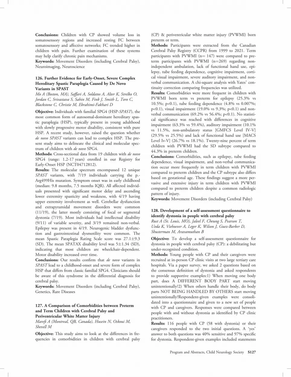
Conclusions: Children with CP showed volume loss insomatosensory regions and increased resting FC betweensomatosensory and affective networks; FC trended higher inchildren with pain. Further examination of these systemsmay help clarify chronic pain mechanisms.Keywords: Movement Disorders (including Cerebral Palsy),Neuroimaging, Neuroscience
126. Further Evidence for Early-Onset, Severe ComplexHereditary Spastic Paraplegia Caused by De NovoVariants in SPASTMo A (Boston, MA), Saffari A, Soldatos A, Alter K, Strelko O,Jordan C, Srivastava S, Sahin M, Fink J, Smith L, Toro C,Blackstone C, Christie M, Ebrahimi-Fakhari D
Objective: Individuals with familial SPG4 (HSP-SPAST), themost common form of autosomal-dominant hereditary spas-tic paraplegia (HSP), typically present in young adulthoodwith slowly progressive motor disability, consistent with pureHSP. A recent study, however, raised the question whetherde novo SPAST variants can lead to complex HSP. The pre-sent study aims to delineate the clinical and molecular spec-trum of children with de novo SPG4.Methods: Cross-sectional data from 19 children with de novoSPG4 (range: 1.2-17 years) enrolled in our Registry forEarly-Onset HSP (NCT04712812).Results: The molecular spectrum encompassed 12 uniqueSPAST variants, with 7/19 individuals carrying the p.-Arg499His mutation. Symptom onset was in early childhood(median: 9.8 months, 7.5 months IQR). All affected individ-uals presented with significant motor delay and ascendinglower extremity spasticity and weakness, with 4/19 havingupper extremity involvement as well. Cerebellar dysfunctionand extrapyramidal movement disorders were common(11/19), the latter mostly consisting of focal or segmentaldystonia (7/19). Most individuals had intellectual disability(9/11) of variable severity, and 3/19 remained non-verbal.Epilepsy was present in 4/19. Neurogenic bladder dysfunc-tion and gastrointestinal dysmotility were common. Themean Spastic Paraplegia Rating Scale score was 27.1�9.3(SD). The mean SPATAX disability level was 5�1.34 (SD),indicating that most children are wheelchair-dependent.Motor disability increased over time.Conclusions: Our results confirm that de novo variants inSPAST lead to a childhood-onset and severe form of complexHSP that differs from classic familial SPG4. Clinicians shouldbe aware of this syndrome in the differential diagnosis forcerebral palsy.Keywords: Movement Disorders (including Cerebral Palsy),Genetics, Rare Diseases
127. A Comparison of Comorbidities between Pretermand Term Children with Cerebral Palsy andPeriventricular White Matter InjuryMarefi A (Montreal, QB, Canada), Husein N, Oskoui M,Shevell M
Objective: This study aims to look at the differences in fre-quencies in comorbidities in children with cerebral palsy
(CP) & periventricular white matter injury (PVWMI) bornpreterm or term.Methods: Participants were extracted from the CanadianCerebral Palsy Registry (CCPR) from 1999 to 2021. Termparticipants with PVWMI (n= 147) were compared to pre-term participants with PVWMI (n=269) regarding non-independent ambulation, lack of functional hand use, epi-lepsy, tube feeding dependence, cognitive impairment, corti-cal visual impairment, severe auditory impairment, and non-verbal communication. A chi-square analysis with Yates’ con-tinuity correction comparing frequencies was utilized.Results: Comorbidities were more frequent in children withPVWMI born term vs preterm for epilepsy (25.3% vs10.5%; p<0.1), tube feeding dependence (4.8% vs 0.007%;p<0.1), visual impairment (19.0% vs 9.3%; p<0.1) and non-verbal communication (69.2% vs 56.4%; p<0.1). No statisti-cal significance was reached with differences in cognitiveimpairment (63.3% vs 59.4%), auditory impairment (10.1%vs 11.5%, non-ambulatory status [GMFCS Level IV-V](29.5% vs 25.5%) and lack of functional hand use [MACSLevel Iv-V] (26.7% vs 18.1%). Twenty-nine percent of termchildren with PVWMI had the SD subtype compared to44.3% in preterm children.Conclusions: Comorbidities, such as epilepsy, tube feedingdependence, visual impairment, and non-verbal communica-tion occur more frequently in term children with PVWMIcompared to preterm children and the CP subtype also differsbased on gestational age. These findings suggest a more per-vasive and extensive injury in term children with PVWMIcompared to preterm children despite a common radiologicpattern of injury.Keywords: Movement Disorders (including Cerebral Palsy)
128. Development of a self-assessment questionnaire toidentify dystonia in people with cerebral palsyRust A (St. Louis, MO), Jaleel F, Cheung S, Pearson T,Ueda K, Viehoever A, Leger K, Wilson J, Guez-Barber D,Shusterman M, Aravamuthan B
Objective: To develop a self-assessment questionnaire fordystonia in people with cerebral palsy (CP): a debilitating butunder-recognized condition.Methods: Young people with CP and their caregivers wererecruited at in-person CP clinic visits at two large tertiary carehospitals. Via a paper survey, we asked 2 questions based onthe consensus definition of dystonia and asked respondentsto provide supportive examples:1) When moving one bodypart, does A DIFFERENT BODY PART start movingunintentionally?2) When others handle their body, do bodyparts NOT BEING HANDLED BY OTHERS start movingunintentionally?Respondent-given examples were consoli-dated into a questionnaire and given to a new set of peoplewith CP and caregivers. Responses were compared betweenpeople with and without dystonia as identified by CP clinicpractitioners.Results: 116 people with CP (58 with dystonia) or theircaregivers responded to the two initial questions. A ‘yes’answer to both questions was 40% sensitive and 97% specificfor dystonia. Respondent-given examples included statements
Program and Abstracts, Child Neurology Society S127

like: “she stiffens up on transfers” and “when moving oneleg, the other leg wants to move”. These examples were con-solidated into a 22-item questionnaire administered to89 people with CP (32 with dystonia) or their caregivers.‘Yes’ answers to >22% of items yielded 84% sensitivity and70% specificity for dystonia.Conclusions: These results demonstrate the potential viabil-ity of a self-assessment questionnaire for identifying dystoniain people with CP. Such auxiliary dystonia identificationmethods could help address the important problem of dysto-nia under-recognition in people with CP.Keywords: Movement Disorders (including Cerebral Palsy)
129. White Matter Injury Predominates in People withDystonia and Cerebral Palsy Following Premature BirthChintalapati K (St. Louis, MO), Pearson T, Ueda K,Aravamuthan B
Objective: Dystonia in people with cerebral palsy (CP) isclassically associated with deep grey matter injury at term ges-tation. However, dystonia also occurs in people with CP fol-lowing premature birth without clear grey matter injury onMRI. Our objective was to determine MRI patterns of braininjury associated with dystonia in people with CP followingpremature birth.Methods: Subjects were people with CP following prematurebirth (gestational age <37 weeks), evaluated at a tertiary caremulti-disciplinary CP clinic with clinically-available MRIs. Sub-jects were assessed for dystonia in-person by a pediatric move-ment disorders specialist, with video confirmation in a subsetby two other movement disorders specialists blinded to subjectidentities. MRI T1 weighted scans were assessed in tandem bytwo investigators blinded to subjects’ motor phenotypes for sizemarkers of the basal ganglia, corpus callosum, lateral ventricles,and cerebellum, normalized to markers of total brain size.Results: 137 subjects were evaluated (ages 0-17 years). Com-pared to subjects without dystonia (N=96), subjects withdystonia (N=41) had greater thalamic size / total brain size(0.22 vs 0.24, P = 0.005) and lateral ventricular size / totalbrain size (0.12 vs 0.14, P=0.001, ANOVA), suggestive ofcomparatively diminished white matter volume in subjectswith dystonia.Conclusions: In people with CP after premature birth, ourresults suggest increased white matter injury best correlateswith the presence of dystonia. Therefore, white matter injurymay be a significant contributor to dystonia pathogenesis inpeople with CP following premature birth.Keywords: Movement Disorders (including Cerebral Palsy),Neuroimaging
130. Clinical and molecular phenotype ofPTRHD1-associated juvenile parkinsonismTavani J (Phoenix, AZ), Marchetto A, Stoffels J, Darvish H,Zech M, Shoukier M, Kruer M
Objective: Although Parkinson’s Disease is the most com-mon movement disorder in adults, juvenile parkinsonism(occurring in children and adolescents) is comparatively rare.Juvenile parkinsonism is often dominated by rigidity,
hypokinesia, and postural instability and less commonly fea-tures a resting tremor. Despite its rarity, several importantscientific insights have come from studies of early-onset par-kinsonism. We describe 5 previously unreported patientswith mutations in PTRHD1, an orphan gene of unknownfunction. Although mutations in this gene have recently beenreported in a few families with juvenile parkinsonism andintellectual disability, clinical phenotypes have been incom-pletely characterized. We sought to identify genotype-phenotype correlations in patients with homozygous missenseor genomic loss of function mutations in PTRHD1.Methods: Each family provided written informed consent forparticipation according to local ethics guidelines. Clinicalphenotypes, MRI images, patient photographs, and videoswere collected from 5 patients with homozygous mutationsin the PTRHD1gene. When possible, blood and/or skinpunch biopsies were also performed for transcriptomic andproteomic analysis.Results: Clinical presentations included dysarthria, rigidity,postural instability, gait dysfunction, and mild intellectualdisability. Resting tremor was observed in several patients.There was no evidence of oculomotor abnormalities. Ana-tomic MRI imaging was normal. Patients responded tocarbidopa-levodopa but developed peak-dose dyskinesias andwearing-off effects early in the treatment course.Conclusions: Biallelic PTRHD1 mutations represent anewly-recognized form of parkinsonism that should be con-sidered in patients presenting in childhood or adolescence.Keywords: Movement Disorders (including Cerebral Palsy),Genetics
131. Spectrum of Pediatric to Early Adulthood POLR3A-Associated Movement DisordersZea Vera A (Washington, DC), Bruce A, Larsh T, Jordan Z,Espay A, Brüggemann N, Gilbert D, Wu S
Objective: POLR3A mutations cause hypomyelination,hypodontia, hypogonadism, and cerebellar ataxia. We aim todescribe the range of other movement disorders seen in acohort of patients with POLR3A-related disorders.Methods: Six patients with POLR3A mutations evaluated inspecialized movement disorder centers were included.Results: Age of onset ranged from birth to early adulthood(Table 1). Four patients were females. Patient 1 presented atbirth with a progeroid syndrome. At 23 years she haspseudobulbar affect, parkinsonism, dystonia, ataxia, and spas-ticity. Patient 2 presented in infancy with rapidly progressivechorea, dystonia, and myoclonic seizures. She required venti-latory support/G-tube and died at 2 years 11 months. Patient3 presented at age 5 years with mild dystonia, ataxia, andspasticity. Patient 4 presented at 15 years old with slowlyprogressive cervical and arm dystonia with no additionalsymptoms up to 20 years old. Patients 5 and 6 developedspastic ataxia and peripheral neuropathy at age 20 years oldthat slowly progressed over more than 10 years. Patient 5 alsohad segmental dystonia. Vertical gaze palsy or slowed verticalsaccades were identified in 4 patients. Cognitive involvementvaried. Dentition abnormalities and short stature or amenor-rhea each occurred in 2 patients.
S128 Annals of Neurology Vol 92 (suppl 28) 2022

TABLE 1. Phenotypic characteristics of the POLR3A patient cohort. Abstract 131
ID Patient 1 Patient 2 Patient 3 Patient 4 Patient 5 Patient 6
Gender Female Female Female Female Male Male
Age of onset Birth <6 months 5 years 15 years 20 years 20 years
Last follow up 23 years 2 year 11 months(deceased)
10 years 20 years 59 years 30 years
CognitiveSymptoms
Yes Severe globaldevelopmentaldelay
No No No No
Independentwalking
Achieved butlost at 13 years
Never achieved Achieved andmaintained
Achieved andmaintained
Achievedandmaintained
Achieved andmaintained
Pyramidal signs Yes Yes Yes No Yes Yes
Cerebellar signs Gait ataxia,intentiontremor,dysmetria
No Mild gaitataxia,intentiontremor, anddysmetria
No Gaitataxia, anddysmetria
Gait ataxia
Dystonia OromandibularDystonia
Severe generalizeddystonia
Mild lowerextremitydystonia
Cervical andright armdystonia
Multifocaldystonia
No
Chorea No Severe generalizedchorea
No No No No
Tremor Rest andintentiontremor
No Intentiontremor
Dystonictremor
Intentiontremor
No
Parkinsonism Yes No No No No No
Eye movements Impaired down-gaze andlimited up-gaze
Impaired verticalup gaze
Round-the-house sign,nystagmus onlateral gaze,saccadicpursuitmovements
Normal Normal Saccadicpursuit, slowverticalsaccades
Sensorysymptoms
Sensoryneuropathy
None Hyporeflexia,but no sensorysymptoms
None Sensoryneuropathy
Sensoryneuropathy
Otherneurologicsymptoms
None Axial hypotonia,dysautonomia,myoclonic seizures
Mild axialhypotonia
None None Dysarthria
Dentitionabnormalities
No No Yes Yes No No
Endocrinologicabnormalities
Growth failure,amenorrhea
No Short stature No No No
Yes No Yes No No No
Program and Abstracts, Child Neurology Society S129

Conclusions: POLR3A mutations exbibit significant pheno-typic pleomorphism, often including dystonia and ataxia.Vertical gaze impairment, a feature seldom reported inPOLR3A-related disorders, was seen frequently in our cohortand may be explained by the involvement of midbrain struc-tures (riMLF or interstitial nucleus Cajal).Keywords: Movement Disorders (including Cerebral Palsy),Genetics, Rare Diseases
132. Dysphagia is a strong risk factor in predicting lowbone density in adults with cerebral palsyJung SH (Seoul, Republic of Korea),
Objective: Dysphagia is a common medical problem sufferedby persons with cerebral palsy (CP). Although dysphagia hasbeen linked to elevated risk of osteoporosis in the generalpopulation, there has been no quantitative study focusing ondysphagia in persons with CP and its impact on low bonedensity.Methods: This cross-sectional study investigated the relation-ship between dysphagia in the CP population and its effecton bone mineral density (BMD), T-score, and Z-score,adjusted for possible confounding risk factors.Results: Ninety-seven adults with CP were divided into twogroups: dysphagia and no dysphagia. The dysphagia groupshowed significantly lower BMD, T-score, and Z-score atevery anatomical site explored. Adjusted for age, sex, bodymass index, and physical activity, dysphagia was significantlynegatively correlated with BMD. Subjects with dysphagiashowed significantly higher odds ratio of age-adjusted lowBMD and osteopenia than those without dysphagia. Thisstudy found dysphagia to be an important risk factor inpredicting low BMD and osteopenia in adults with CP.
Conclusions: In the presence of dysphagia in adults withCP, it may be beneficial to screen for low BMD for appropri-ate clinical intervention. This study was supported by theTranslational R&D Program on Smart Rehabilitation Exer-cises (#TRSRE-PS01), National Rehabilitation Center, Min-istry of Health and Welfare, Korea.Keywords: Movement Disorders (including Cerebral Palsy),Neurorehabilitation
133. Reduced dominant motor cortex inhibition isassociated with increased tic severity in children withTourette syndromeBatschelett M (Baltimore, MD), He J, Crocetti D, Horn P,Puts N, Huddleston D, Mostofsky S, Gilbert D
Objective: Tourette syndrome (TS) is characterized by chronicmotor and/or vocal (phonic) tics, often preceded by premoni-tory urge perceptions. Hypotheses regarding TS pathophysiol-ogy suggest dysregulation within striatal-thalamic-corticalcircuits, contributing to disrupted signaling within motor cir-cuits (primary motor [M1] and premotor). The objective ofthis study was to identify how patterns of M1 inhibition orexcitation associate with TS symptom severity.Methods: Using Transcranial Magnetic Stimulation (TMS),we measured the physiology of resting left M1 innervatingright intrinsic hand muscle in sixty 8-12-year-old children:30 TS (including 15 with comorbid ADHD) and 30 typicallydeveloping (TD). Additionally, standard clinical scales for ticand premonitory urge severity (TS) and ADHD (all) wereperformed. We used repeated measures mixed model regres-sions to compare TMS measures between diagnostic groupsand to examine dimensional associations with TS symptoms(tics and urges).
TABLE (Continued)
ID Patient 1 Patient 2 Patient 3 Patient 4 Patient 5 Patient 6
Myopia orHyperopia
Othersymptoms
Progeroidsyndrome
Requiredventilatory supportand G-tube feeds
No No No No
Brain MRI Polymicrogyria Ventriculomegaly,and abnormalsignal and volumeloss in the bilateralcaudate andputamen.
T2hyperintensityof dentate,superiorcerebellarpeduncles,midbrain,thalamus, andposterior limbof internalcapsule
SubtleT2/FLAIRhyperintensityin the posteriorpons (in theregion of thesuperiorcerebellarpeduncles)
CerebellarAtrophy
BilateralsymmetricFLAIRhyperintensityin themidbrain(directlyunderneath rednucleus), andsuperiorcerebellarpeduncles
S130 Annals of Neurology Vol 92 (suppl 28) 2022

Results: There were no diagnostic differences (TS vs. TD)for any of the TMS measures. However, after accounting cat-egorically for comorbid ADHD, among TS children, reducedshort interval cortical inhibition (SICI) was associated withgreater severity of tics (p=0.002), but not urges. Compari-sons of single and paired pulse TMS-evoked potentials sug-gests distinctive physiological correlates of more severe motorvs. phonic tics.Conclusions: In children with TS, more severe tics, but noturges, are strongly associated with less SICI in left M1. Theseresults demonstrate a potential link between M1 physiologyin TS and tic symptomology. Taken together, these findingsprovide support for SICI as a biomarker for the targetedtreatment of TS symptoms.Keywords: Movement Disorders (including Cerebral Palsy),Neuroimaging, Neuroscience
134. Comorbidities of children with cerebral palsy bornat term compared to preterm in the Canadian CerebralPalsy RegistryPekeles H (Montreal, QB, Canada), Husein N, Kirton A,Oskoui M, Shevell M
Objective: Comorbidities may be a bigger challenge thanneuro-motor impairment for individuals with cerebral palsy(CP) and their caregivers. We set out to compare the comorbid-ity spectrum of children with CP who were born term or latercompared to those who were born preterm as these populationshave different causal mechanisms leading to the CP phenotype.Methods: Using data from the Canadian Cerebral Palsy Regis-try (CCPR) of 2217 children with CP, we compared thecomorbidity spectrum of term children (37 weeks and aboveGA, n=1263) to preterm children (born below 37 weeks GA,n=954) with CP. A chi-square analysis with Yates’ continuitycorrection comparing frequencies was utilized.Results: We found significant differences in comorbidities inchildren with cerebral palsy born preterm vs term for epilepsy(10.7 % vs 21.7%), visual impairment (11.2% vs 18.2%),auditory impairment (13.2% vs 8.0%), communicationimpairment (58.0% vs 67.5%) and lack of functional handuse [MACS Level IV-V] (22.1% vs 27.9%) (p value < 0.05).No significant difference based on GA was found for tubefeeding dependence, cognitive impairment, or non-ambulation[GMFCS Level IV-V], all of which were more frequent interm children approaching significance (p value 0.08-0.1).Conclusions: With the exception of auditory impairment,CP children born term had more frequent comorbiditiescompared to children born preterm. This suggests that thecausal mechanisms leading to CP in term children are morewidespread. This also suggests that both populations shouldhave similar surveillance mechanisms in place longitudinally.Keywords: Movement Disorders (including Cerebral Palsy),Neonatal & Fetal Neurology
135. Stereotactic EEG evaluation aids decision making inpatients with complex dystonia undergoing deep brainstimulationMarks W (Fort Worth, TX), Acord S, Honeycutt J, Song Y,Papadelis C
Objective: Deep Brain Stimulation (DBS) of the globuspallidus (GPi) has been effectively utilized for dystonia. Forcomplex or refractory patients, especially with hyperkinesis,secondary targets have been investigated. The use of depthelectrode recording has been implemented by Sanger et al.We report our experience in seven patients with dystoniausing stereotactic EEG (sEEG) depth electrode stimulationand recording to guide DBS decision making.Methods: Review of patients that have undergone sEEGrecordings as part of their DBS evaluation. sEEG electrodeswere implanted using robotic guidance. Targets were selectedbased on clinical symptoms. GPi was always included unlesspreviously implanted with permanent DBS leads. STN andVOA of thalamus are generally done using a single sEEGelectrode passing through both nuclei. Each contact is stimu-lated in bipolar fashion with monitoring of efficacy andadverse events. Prolonged stimulation is accomplished usingan external generator system.Results: Seven patients with dystonia underwent sEEG evalua-tion for DBS lead placement. Five patients had a confirmed orpresumed genetic etiology. Two were evaluated for initial DBSplacement; five for lead modification. Four of the five second-ary evaluations underwent placement of additional leads. Leadsites included GPi (7), Thalamus (24) and STN (8). Negativeside effects related to stimulation aided in ultimate target selec-tion. Monitoring time was 3-11 days. There were no surgicalcomplications related to the placement of sEEG leads.Conclusions: Stereotactic EEG testing of selected patientscan aid in the decision making process for DBS placement orrevision.Keywords: Movement Disorders (including Cerebral Palsy),Translational/Experimental Therapeutics, Rare Diseases
136. Characteristics of children with cerebral palsysecondary to intrapartum asphyxia in the post therapeutichypothermia eraPekeles H (Montreal, QB, Canada), Al Amrani F, Perez-Morgui M, Wintermark P, Shevell M
Objective: To explore the profile of children with cerebralpalsy (CP) secondary to intrapartum asphyxia (IAP) treatedwith therapeutic hypothermia (TH) after birth and to com-pare characteristics of children treated with TH with mildvs. severe CP outcome.Methods: We identified all children treated with TH for IAPin a single-center tertiary level neonatal intensive care unitfrom 2008 to 2018 with a CP outcome. We collected perina-tal and outcome measures from patient charts. We searchedthe literature for characteristics of children with CP prior toTH (historical cohort) to compare to our cohort. We sub-divided our cohort into mild vs. severe CP and compared neo-natal characteristics to identify predictors of severe phenotype.Results: 30 out of 355 cooled neonates (8%) developedCP. More children had spastic quadriparesis and epilepsy,and fewer had visual impairment in the post-TH era com-pared to the historical cohort, but had similar Gross MotorFunction Classification System scores. In our cohort, morechildren had severe (19/30, 63%) compared to mild CP(11/30, 37%). The severe group had higher mean birth
Program and Abstracts, Child Neurology Society S131

weight, lower 5- and 10-minute Apgar scores, and more oftenwhite matter injury with associated deep gray matter injuryor near-total injury pattern (p<0.05).Conclusions: Our data demonstrated more infants withsevere rather than mild CP in our cohort treated withTH. Birthweight, 5- and 10-minute Apgar scores and MRIfindings were significantly different between mild vs. severephenotype groups. Our findings can guide clinicians how tobetter weigh these factors, when counselling parents in theneonatal period.Keywords: Movement Disorders (including Cerebral Palsy),Neonatal & Fetal Neurology
137. PEDiDBS: The international registry of pediatricpatients undergoing deep brain stimulationMarks W (Fort Worth, TX), Sankpal U, Bailey L
Objective: Deep brain stimulation is being used with increasingfrequency in pediatric patients with dystonia and now epilepsy.Multiple devices from several manufacturers are in clinical useworldwide. There are differences in patient selection, surgicaltechniques, perioperative postoperative management. Thesequestions can best be addressed by collaborative data sharing.Methods: PEDiDBS is the result of the collaborative effortsof an international team of investigators working over the lastseveral years to develop a secure method of data sharingregarding children undergoing DBS. PEDiDBS is aREDCap-based secure multi-center database that meets thecompliance and international privacy laws.Results: Four centers are now fully enrolled and beginning dataentry. Approximately 2 dozen more sites have requested informa-tion and in start-up. Subjects are entered utilizing a de-identifiedlimited data set and is stored on a secure REDCap cloud server.Privacy is assured through the use of site numbers and a limiteddata set for patient information in accordance with applicableprivacy laws. Each site enters their own data and is responsiblefor ensuring accuracy. Individual sites retain access to their owndata. Requests for aggregate data for analysis by participating sitesare reviewed by the data sharing committee.Conclusions: PEDiDBS is a vehicle for information sharingbetween all interested centers implanting pediatric patientswith DBS. The data sharing allows for better utilization ofthis complex technology. Participation and enrollment infor-mation is available on the website www.PEDiDBS.org.Keywords: Movement Disorders (including Cerebral Palsy),Genetics, Translational/Experimental Therapeutics
138. Prevalence of Genetic Disease in Patients withCerebral PalsyBrooks A (Rootstown, OH), Mawby I, Rossman I
Objective: Cerebral palsy (CP) is a heterogenous disorderresulting from early brain injury or aberrant neuro-development, causing motor disability and a myriad of otherneurologic sequelae. Genetic mutations have been identifiedin some patients with CP; however, the prevalence of causalgenetic disorders in CP is unknown. We aimed to identifythe prevalence of genetic disorders and the most commongenetic mutations in pediatric patients with CP.
Methods: Using the EPIC SlicerDicer tool, the Akron Chil-dren’s Hospital patient population was sliced into twogroups: 1) the term “cerebral palsy” as diagnosis or medicalhistory, between January 1, 2021 and December 31, 2021,and 2) the slice of group 1 with “genetic disease” as diagnosisor medical history. Patients with genetic diseases unrelated toCP were manually removed and subgroups of genetic diseaseswere quantified.Results: We identified 2,633 CP patients treated in 2021.Of these patients, 170 (6.5%) were identified as having agenetic disease. Within this group, three siblings shared aCOL4A1 mutation; further identification of additionalgenetic mutations is ongoing.Conclusions: Considering genetic etiologies is critical whenCP patients present without pre- or perinatal trauma or lackbrain malformations or injury on neuroimaging. We identi-fied 6.5% of a CP population to have a genetic disorder, butthe prevalence may vary across populations. Early geneticdiagnoses in patients with CP may alter clinical managementand identify CP risk for future pregnancies. Clinical genetictesting may help to define the pathophysiology of CP andoffer targets for treatment in some patients.Keywords: Movement Disorders (including Cerebral Palsy),Genetics
139. Geographical Knowledge Disparities in CerebralPalsy EpidemiologyAvila-Soto F (St. Louis, MO), Aravamuthan B
Objective: Noting dramatic differences in CP prevalence andetiology across geographical regions, we aimed to identifygeographical disparities in the published knowledge of CPepidemiology.Methods: We conducted a PRISMA-style systematic reviewof primary literature on CP prevalence using the searchterms: "cerebral palsy"[title] AND (rate OR prevalence ORepidemiology) in PubMed and Web of Science. We excludedstudies not published in English.Results: Of 2720 studies, 91 met inclusion/exclusion criteria.68/91 (74.7%) studies reported CP prevalence in Europe,North America, and Australia (Region 1) and are cited an aver-age of 7.3 times/year since publication. The remaining 24/91(26.4%) studies reported CP prevalence in Asia, the MiddleEast, and Africa (Region 2) and are cited significantly lessoften, 3.3 times/year since publication (p=0.01, t-test). Nostudies from Latin America were identified. Geographical cita-tion disparities persist even when accounting for journalimpact. Normalized to a journal’s CiteScore (number of cita-tions in one year for a journal’s articles published in the previ-ous 3 years), Region 1 papers are cited 1.6x more often thantypical for the corresponding journal, while Region 2 papersare cited only 0.5x as often (p<0.001, t-test). Region 1 paperswere written exclusively by Region 1 authors (100%) whileRegion 2 papers significantly less frequently included Region2 authors (84.4%) (p<0.0001, comparison of proportions).Conclusions: Our results show geographical disparities inour published knowledge of CP epidemiology. Understand-ing CP globally requires increased global support of all CP
S132 Annals of Neurology Vol 92 (suppl 28) 2022

researchers, particularly in regions where CP prevalenceremains under-studied or unknown.Keywords: Movement Disorders (including Cerebral Palsy),Equity, Diversity, Inclusion
140. A Two-Part, International, Real-World,Observational Registry of Participants Diagnosed withAromatic L Amino Acid Decarboxylase Deficiency(AADC-d) With or Without Treatment With EladocageneExuparvovecGiugliani R (Porto Alegre, Brazil), Vincenzo V, Pearl P,Roubertie A, Johnson S, Li J, Lupo P, Penematsa V, Tang T
Objective: Aromatic L-amino acid decarboxylase deficiency(AADC-d) is a very rare autosomal-recessive neurotransmitterdisorder resulting in severe neurodevelopmental impairment;information about clinical presentation, prognostic factors,and treatment patterns is limited. AADCAware is a 2-partstudy; Part A aims to describe the natural history of AADC-din patients receiving standard of care. Part B aims to assesslong-term safety and effectiveness of eladocagene exuparvovecon motor function. Demographics and baseline characteristicsof patients in Part A of AADCAware, up to March 10, 2022,are described.Methods: AADCAware is an international, multicenter, lon-gitudinal, real-world, observational registry of patients withAADC-d. In Part A, participants will be followed up annuallyfor a minimum of 5 years.Results: Nineteen participants (10 male and 9 female) havebeen enrolled; their median (min, max) age was 4 (1, 41)years. Median (min, max) ages at: onset of first symptoms,genetic molecular diagnosis and AADC enzymatic activitydiagnosis were 3 (0, 6), 11.5 (5, 384) and 24 (4, 384)months, respectively. Of 18 participants with AADC-d sever-ity data available at enrollment; 13 (72.2%) had severe dis-ease; three (16.7%) had moderate disease and two (11.1%),mild disease. Motor development status was assessed usingthe Peabody Developmental Motor Scale-2. Of 12 partici-pants with available data, nine (75.0%) had not developedfull head control, the ability to sit unassisted or stand or walkwith support. Only 3 (25.0%) participants mastered all4 motor milestones.Conclusions: The majority of participants presented withsevere illness (72.2%) and failed to achieve the 4 measuredmotor milestones (75.0%).Keywords: Movement Disorders (including Cerebral Palsy)
NEONATAL & FETALNEUROLOGY
141. Neonatal encephalopathy treated with therapeutichypothermia: is absence of cord blood acidosis a markerfor causes other than hypoxic-ischemicencephalopathy (HIE)?Cornet M-C (San Francisco, CA), Kuzniewicz M, Forquer H,Hamilton E, Newman T, Scheffler A, Murtha A, Wu Y
Objective: To determine the prevalence of cord acidosisamong infants with presumed HIE, and to compare risk fac-tors and outcomes in infants with and without cord acidosis.Methods: We identified all neonates who received therapeu-tic hypothermia (TH) for presumed HIE within Kaiser Per-manente Northern California between 2011 to 2019. Cordacidosis was defined as pH <7.00 or base deficit ≥12. Weabstracted maternal demographics, prenatal and perinatalconditions and brain MRI findings from electronic records.We reviewed charts to confirm the presence of encephalopa-thy and seizures.Results: Of 426 infants with presumed HIE (populationincidence 0.14%), 394 (92%) underwent cord gas analysis.Cord acidosis was present in 196 of 394 (50%). Comparedto infants with acidosis, infants without cord acidosis weremore likely to have exposure to magnesium (12 vs 6%p=0.049), chorioamnionitis (37% vs. 19%, P<0.001), andepidural analgesia (69% vs. 51%, P<0.001). They were lessoften born by emergency cesarean section, had more respira-tory needs, and less end-organ injury (Table). The rates ofmoderate/severe encephalopathy, seizures, diffusion restric-tion on MRI, and length of stay were similar in the twogroups. Infants without cord acidosis were more likely tohave a diagnosis of acute ischemic stroke (20% vs5%; p=0.02).Conclusions: Half of all infants undergoing TH for HIE hadno evidence of cord acidosis. These infants may have distinctunderlying etiologies including stroke or chorioamnionitis.Further studies are needed to assess the efficacy of neuro-protective treatments for NE based on the underlyingpathophysiology.Keywords: Neonatal & Fetal Neurology
142. Seizure Burden, EEG Background, and Outcome inNeonates with Acute Intracranial Infections: AProspective Multicenter Cohort StudyMehta N (San Francisco, CA), Shellhaas R, McCulloch C,Chang T, Wusthoff C, Abend N, Lemmon M, Chu C,Massey S, Franck L, Thomas C, Soul J, Rogers E, Numis A,Glass H
Objective: To characterize the association between seizureburden, EEG background, and neurodevelopmental outcomeamong neonates with acute symptomatic seizures due tointracranial infection.Methods: This was a prospective, multicenter cohort studyof neonates enrolled in the Neonatal Seizure Registry with sei-zures due to intracranial infection. Continuous EEG moni-toring was performed based on ACNS guidelines. EEGbackground was categorized using standardized terminology.High seizure burden was defined a priori as >7 EEG con-firmed seizures. Primary outcome was neurodevelopment at24-months corrected age using Warner Initial DevelopmentalEvaluation of Adaptive and Functional Skills (WIDEA-FS).Secondary outcomes were post-neonatal epilepsy and motordisability.Results: Twenty-seven of 303 neonates (9%) had seizuresdue to intracranial infection, including 16(59.3%) bacterial,5(18.5%) viral, and 6(22.2%) unknown. Twenty-three
Program and Abstracts, Child Neurology Society S133

TABLE. Clinical characteristics of neonates with and without cord acidosis (pH <7.00 or base deficit ≥12) among neonatestreated with TH for suspected HIE. Abstract 141
S134 Annals of Neurology Vol 92 (suppl 28) 2022

patients (85.2%) had subclinical seizures. Among 23 infantswith 24-month follow-up, the WIDEA-FS was, on average,23 points lower in infants with high compared to low seizureburden (95% CI [-45,-0.6], p=0.04, Figure). After adjustingfor gestational age and etiology of infection, the association
no longer remained significant (p= 0.09); however, the effectsize remained unchanged(β=-23), and the potential con-founders were not significantly associated with outcome.EEG background was not significantly associated withWIDEA-FS. All infants with post-neonatal epilepsy (n=4,Table) and motor disability (n=6) had high seizure burden,although the association between high seizure burden, EEGbackground, and secondary outcomes was not significant.Conclusions: EEG monitoring in neonates with intracranialinfection may provide useful management and prognosticinformation. High seizure burden may be associated withworse neurodevelopmental outcomes in this population.Keywords: Neonatal & Fetal Neurology, Infections/Neu-roimmunology, Critical Care
143. Relationship between blood and cerebral glucoseconcentration - an in-vivo investigation of glycemic statusin newborns with hypoxic-ischemic encephalopathyGeyer Winkler Santos E (Los Angeles, CA), Tetarbe M,Wisnowski J, Blüml S, Wu T-W
Objective: To determine regional cerebral glucose concentra-tion (Glc) in relation to glucose infusion rate (GIR) andblood glucose concentration (BG) during therapeutic hypo-thermia (TH) in newborn hypoxic-ischemic encephalopathy,and to assess whether Glc is affected by injury severity andpattern or by sedative use.
TABLE: Clinical characteristics of neonates with acute symptomatic seizures secondary to acute intracranialinfections who developed post-neonatal epilepsy by 24 months. Abstract 142
Patient SexGestationalAge (weeks)
Etiology ofinfection(Organism)
Additionalseizureetiologies
EEGbackground
Seizureburden
Epilepsydiagnosis
Epilepsymanagement
WIDEA-FS Score
GMFCSScore
1 F 38.5 HSV N/A Mild/moderatelyabnormal
High Focalepilepsy
LEV, PHB 56 5
2 M 31 E. Coli IVH Mild/moderatelyabnormal
High Unknown OXC 112 3
3 M 39 GBS N/A Mild/moderatelyabnormal
High Focalepilepsy
LEV, OXC 148 0
4 F 40.3 GBS Ischemicstroke
Mild/moderatelyabnormal
High Focalepilepsy
LEV 99 4
E. Coli Escherichia coliGBS Group B StreptococcusIVH Intraventricular hemorrhageLEV LeviteracetamPHB PhenobarbitalOXC OxcarbazepineWIDEA- FS Warner Initial Developmental Evaluation of Adaptive and Functional Skills Score at 24 months corrected ageGMFCS Gross Motor Function Classification System Score
FIGURE: Distribution of WIDEA-FS scores among23 neonates with low and high seizure burden. Mean(SD) WIDEA-FS score for typically developing population is172 (10). Dashed line represents WIDEA-FS score offunctional impairment (2 SD below the mean). Abstract 142
Program and Abstracts, Child Neurology Society S135
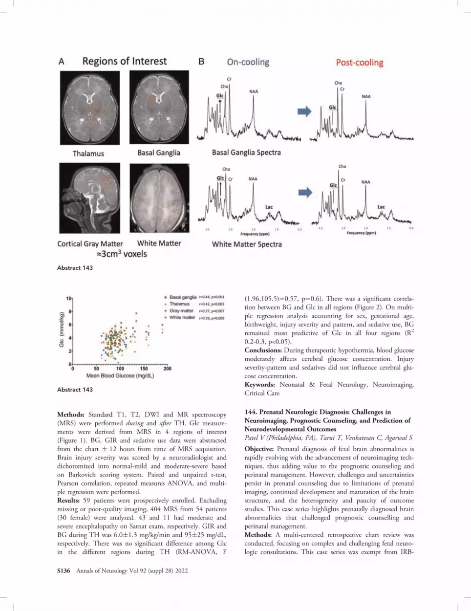
Methods: Standard T1, T2, DWI and MR spectroscopy(MRS) were performed during and after TH. Glc measure-ments were derived from MRS in 4 regions of interest(Figure 1). BG, GIR and sedative use data were abstractedfrom the chart � 12 hours from time of MRS acquisition.Brain injury severity was scored by a neuroradiologist anddichotomized into normal-mild and moderate-severe basedon Barkovich scoring system. Paired and unpaired t-test,Pearson correlation, repeated measures ANOVA, and multi-ple regression were performed.Results: 59 patients were prospectively enrolled. Excludingmissing or poor-quality imaging, 404 MRS from 54 patients(30 female) were analyzed. 43 and 11 had moderate andsevere encephalopathy on Sarnat exam, respectively. GIR andBG during TH was 6.0�1.3 mg/kg/min and 95�25 mg/dL,respectively. There was no significant difference among Glcin the different regions during TH (RM-ANOVA, F
(1.96,105.5)=0.57, p=0.6). There was a significant correla-tion between BG and Glc in all regions (Figure 2). On multi-ple regression analysis accounting for sex, gestational age,birthweight, injury severity and pattern, and sedative use, BGremained most predictive of Glc in all four regions (R2
0.2-0.3, p<0.05).Conclusions: During therapeutic hypothermia, blood glucosemoderately affects cerebral glucose concentration. Injuryseverity-pattern and sedatives did not influence cerebral glu-cose concentration.Keywords: Neonatal & Fetal Neurology, Neuroimaging,Critical Care
144. Prenatal Neurologic Diagnosis: Challenges inNeuroimaging, Prognostic Counseling, and Prediction ofNeurodevelopmental OutcomesPatel V (Philadelphia, PA), Tarui T, Venkatesan C, Agarwal S
Objective: Prenatal diagnosis of fetal brain abnormalities israpidly evolving with the advancement of neuroimaging tech-niques, thus adding value to the prognostic counseling andperinatal management. However, challenges and uncertaintiespersist in prenatal counseling due to limitations of prenatalimaging, continued development and maturation of the brainstructure, and the heterogeneity and paucity of outcomestudies. This case series highlights prenatally diagnosed brainabnormalities that challenged prognostic counselling andperinatal management.Methods: A multi-centered retrospective chart review wasconducted, focusing on complex and challenging fetal neuro-logic consultations. This case series was exempt from IRB-
Abstract 143
Abstract 143
S136 Annals of Neurology Vol 92 (suppl 28) 2022

approval due to low case numbers. Charts were reviewed forneuroimaging, genetic workup, prenatal prognostic discus-sion, postnatal imaging and testing, and infant outcome.Results: A series of cases with their perinatal course are pres-ented. This case series highlighted the variability in prenataltesting and counseling which leads to challenges in accuratelydetermining postnatal outcomes. Results showed that fetalMRI is critical to determining the full extent of anomalies,however, some anomalies may not be recognizable until latergestation or postnatally.Conclusions: These cases challenged various diagnostic andmanagement discussions and led to complexity in the prog-nostic counseling process. Advocating for a national collabo-rative fetal neurologic registry to help guide the ever-expanding world of prenatal diagnostics and counseling iscritical to this field. Study of large-scale outcomes data fromsuch a registry may help better counsel families and providebetter-informed multidisciplinary planning from an early age.Keywords: Neonatal & Fetal Neurology, Neuroimaging
145. Multivariate Approach to Predicting DevelopmentalDelay Among Children with Hypoxic-IschemicEncephalopathyStory J (Los Angeles, CA), Gang M, Rao L
Objective: Neonatal hypoxic-ischemic encephalopathy (HIE)is a significant source of neurological morbidity, though neu-rodevelopmental outcomes are difficult to predict. Priorinvestigations relating HIE with early development, as mea-sured through the Bayley Scales of Infant Development(BSID), show inconsistent results. Selection of risk factors foranalysis may contribute to these discrepancies. Our studyaims to compare clinical, laboratory, radiological, andelectrographic markers of HIE severity to cognitive, language,and motor development in full-term neonates that underwenttherapeutic hypothermia (TH) for HIE.Methods: Neonates that underwent TH for HIE between2008 and 2014 were included in analysis. Chart review wasperformed to obtain clinical information regarding theirNICU admission and BSID composite scores from clinicfollow-up. Cord blood pH and 10-minute APGAR werecompared to BSID scores using Pearson’s correlation coeffi-cient. Independent samples t-tests were used to compareBSID scores based on specific MRI, MRS, or cEEG findings.Results: Evidence of hypoxia on MRI (hyperintensities orabnormal diffusion-weighted imaging) was associated withcognitive delays, but not with language or motor delays, andlactate peaks on MRS were associated with motor delays.Excessive sharps and background attenuation on cEEG didnot predict BSID differences, though electrographic seizureswere associated with both cognitive and motor delays(Fig. 1). Arterial cord blood pH and 10-minute APGAR didnot correlate with BSID scores (Fig. 2).Conclusions: Risk of developmental delay after HIE cannotbe adequately assessed using any single risk-stratificationmodality. A holistic approach involving both imaging andcEEG may better predict long-term effects on multiple devel-opmental domains.Keywords: Neonatal & Fetal Neurology
146. Correlations between fetal sulcal braindevelopmental patterns and postnatalneurodevelopmental outcomes in children with prenatallydiagnosed isolated cerebral ventriculomegalyReid S (Boston, MA), Lee P, Madan N, Yun H, Graham G,Samura O, Kitano R, Akiyama S, Takeoka E, Craig A,Grant P, Im K, Tarui T
Objective: Isolated cerebral ventriculomegaly (IVM) is themost diagnosed fetal brain anomaly, but its significantly vari-able neurodevelopmental outcomes (normal to moderatedevelopmental delays) are difficult to predict. We determinedwhether the fetal brain’s regional growth or sulcal patterncorrelates with their 18-24 month neurodevelopmentaloutcomes.Methods: Twenty-three fetuses with fetal MRI diagnoses ofIVM (atrial diameter 10-15 mm) (17.7-37.7 gestationalweeks) were prospectively recruited. We obtained their post-natal neurodevelopmental outcomes at 18-24 months afterbirth through medical record review. We performed post-acquisition MRI processing of regional volumetrics and sulcalpattern analyses. In sulcal pattern analysis, each fetal case wasquantitatively compared with normal template brains to cal-culate the similarity indices of sulcal position, depth, basinarea and their combination. We compared regional volumegrowth trajectories and sulcal patterns between fetuses withabnormal (n=9) and normal neurodevelopment (n=14)using non-linear/linear regression models and unpaired t-tests(p<0.05).Results: Regional volumetric analyses found no significantgroup differences. Sulcal pattern analyses found lower similar-ity indices in the delayed neurodevelopment group for theleft intersulcal relationship of combined features (p=0.0125),left whole pattern area (p=0.0184), right intersulcal relation-ship of position (p=0.0281), right whole pattern area(p=0.0286), right corresponding basins area (p=0.0500) andright intersulcal relationship of area (p=0.0332).Conclusions: In fetuses with IVM, abnormal patterns of sul-cal position and basin area in the second to third trimesterwere significantly correlated with abnormal neuro-development at 18-24 months old, indicating the potentialuse for the sulcal pattern index as a quantitative neu-rodevelopmental outcome predictor.Keywords: Neonatal & Fetal Neurology, Neuroimaging
147. Metabolic reprogramming in a piglet modelof mild neonatal hypoxic ischemic encephalopathy(HIE)Lammert D (Baltimore, MD), Fernandez R, Liu X, Koehler R,Scafidi S, Scafidi J
Objective: Neonatal hypoxic ischemic encephalopathy (HIE)remains a leading cause of long-term neurologic morbidity.Emergent evidence demonstrates that up to 25% of childrenwith mild HIE suffer motor and developmental delay by18 months. There is a paucity of understanding regardingthe extent, duration and compensatory metabolic adaptationin the brain following mild neonatal HIE. We sought toinvestigate the nature of this metabolic reprogramming.
Program and Abstracts, Child Neurology Society S137

FIGURE 1. BSID Scores Based on MRI, MRS, and cEEG Findings. BSID scores <85 are typically considered abnormal. A. BSIDscores based on the presence of ischemia on MRI. Cognitive development is decreased when ischemia was observed. B. BSIDscores based on the presence of lactate peaks on MRS. Motor development is decreased when lactate peaks were observed.C. BSID scores based on presence of seizures on cEEG. Cognitive and motor development was decreased when electrographicseizures were detected. D. BSID scores based excessive sharps on cEEG. E. BSID scores based on background attenuation oncEEG. Abstract 145
S138 Annals of Neurology Vol 92 (suppl 28) 2022

FIGURE 1. Unbiased proteomics screen of piglet mild neonatal asphyxia. (A) Heatmap (heatmapper.ca) of scaled abundances ofproteins identified through 11-plex TMTpro labelling of peptides and LC-MS/MS. Proteins were identified as significantlyincreased or decreased at either 24 or 72 hours post recovery compared to sham if p value was <0.1, accepting a 10% falsepositive rate to enable discovery. The heatmap shows this subset of proteins. (B) Significantly changed proteins were thenanalyzed for common pathways using g:Profiler (https://biit.cs.ut.ee/gprofiler/gost). KEGG terms with p<0.05 are shown. Genesymbols for proteins associated with each of these KEGG terms are included to the right of the corresponding bar. Relevantenergy-associated terms are highlighted in blue. The Epstein-Barr virus infection KEGG term is representative of immune andinflammation pathway enrichment. (C, D) Proteins included in the KEGG metabolic pathways were divided by those increased ordecreased at 24 or 72 hours. Reactome (reactome.org) was then used to analyze the types of metabolic pathways. Metabolicpathways enriched in the increased protein expression set are represented in red (C) and decreased in green (D). Abstract 147
FIGURE 2. Arterial Cord Blood pH and 10-minute APGAR scores vs BSID scores. Neither cord blood pH nor 10-minute APGARscores showed significant correlation with the cognitive, language, or motor domains of the BSID score. Abstract 145
Program and Abstracts, Child Neurology Society S139

Methods: Methods were approved by JHH ACUC. Piglet(sus scrofa) brain gross anatomy, time to peak brain growth,and growth patterns are more consistent with human devel-opment than other animal models. Term neonatal asphyxiawas modeled in 3-day old piglets. Animals were intubated,subjected to whole-body hypoxia for 45 minutes to reach anSaO2 of 30-35%, after which the endotracheal tube wasoccluded for 7 minutes. No piglets demonstrated clinical sei-zures, and all were able to walk and eat after resuscitation.Piglets were sacrificed at 24 or 72 hours after recovery, andhippocampi were microdissected. Samples were analyzedusing TMTpro peptide labelling and LC-MS/MS andQRT-PCR.Results: Pathway analysis of protein expression demonstratedthe pathways involved in carbon metabolism from glucosewere upregulated, while down-regulated pathways were mostsignificantly associated with oxidative metabolism. At themRNA level, GAPDH, PDHA1, and LDHA were signifi-cantly decreased at both 24 and 72 hours. Taken togetherour data suggest differential regulation of glucose metabolismand a shift away from oxidative phosphorylation.Conclusions: Even in mild HIE, metabolic perturbations arepresent up to 72 hours after insult.Keywords: Neonatal & Fetal Neurology, Neuroscience
148. Radiographic Hypoxic-Ischemic Injury withoutClinical Signs of HIE in Term NeonatesAbbott M (Denver, CO), Nazario Malave M, Stence N,Neuberger I, Dingman A
Objective: To describe the imaging and outcomes for neo-nates without clinical neonatal hypoxic ischemic encephalop-athy (HIE) but with imaging consistent with HIE, referredto here as radiographically isolated HIE.Methods: We conducted a search of neonatal MRIs at ourhospital 2009-2019 using the following terms: hypoxic-ischemic injury, global hypoxia, anoxic, and neonatal enceph-alopathy. We included patients who presented within oneweek of life, and excluded patients who met criteria for clini-cal HIE/therapeutic hypothermia or had other clear cause ofinjury. MRIs were scored using the Weeke injury score basedon DWI and T1 sequences (scale of 0-28).
Results: Of 193 patients reviewed, 29 met inclusion criteria(15%). MRI’s were done at a median age of 2 days of life(range 0-6 days). Median injury score was 17.5(IQR10.5-23.5). T1 changes were present in 79%. A definitivediagnosis was reached in 3 patients: NKH, hyperinsulinism,and VLFA deficiency in one patient each. Outcome data wasavailable in 26 patients, with a mortality rate of 19%.Remote epilepsy occurred in 42%, and 4 patients developedinfantile spasms. Of surviving patients, 38% have corticalvisual impairment, 33% are non-verbal and 33% require ag-tube.Conclusions: Our cohort of neonates with radiographicallyisolated HIE have a guarded prognosis, with a substantialmortality rate and developmental challenges. Radiology datademonstrates these are severe injuries, with T1 changesalready present despite early MRI, suggesting likely antenatal,rather than perinatal timing of injury. Antenatal timing ofinjury may help explain this cohort’s unique presentationand outcomes.Keywords: Neonatal & Fetal Neurology, Neuroimaging
149. Heart rate variability at term is associated withCentral Autonomic Network connectivity in prematureneonatesChristoffel K (Washington, DC), Cruz J, Govindan R, Cook K,Niforatos-Andescavage N, Basu S, Limperopoulos C, duPlessis A
Objective: The autonomic nervous system (ANS) of a pre-mature neonate develops in an abnormal extrauterine envi-ronment. ANS dysfunction may contribute to developmentof neuropsychiatric disorders, which are more prevalent inprematurity survivors. The mature ANS is modulated by theCentral Autonomic Network (CAN), but has not been stud-ied in premature infants. To explore the relationship betweenANS tone and CAN connectivity, we measured heart ratevariability (HRV) and resting state functional MRI (rsfMRI),respectively, in premature neonates at term-correctedage (TCA).Methods: We prospectively enrolled 34 premature neonates(mean gestational age at birth: 27.9 +/- 2.5 weeks) whounderwent rsfMRI at TCA (39.5 +/- 1.9 weeks) and had
FIGURE 1. Heat map of Pearson correlation values for 37-week RMS1 with each ROI pairing. Starred values are statisticallysignificant on exploratory analysis. A single asterisk (*) represents a p-value less than .05, two asterisks (**) represent a p-valueless than .01. No relationships remained significant after adjustment for multiple comparisons. Abstract 149
S140 Annals of Neurology Vol 92 (suppl 28) 2022

continuous HRV data recorded over 37 weeks postmenstrualage. RMS1 was the primary HRV metric. Medulla, pons,midbrain, and total brainstem connectivity was assessed to8 other ROIs pertinent to the CAN: anterior cingulate,medial prefrontal cortex, precuneus, hippocampus, amygdala,insula, thalamus, and cerebellum.Results: Univariate linear regression revealed associationsbetween RMS1 and numerous ROI pairings, but noneremained significant after Bonferroni correction for multiplecomparisons. RMS1 was positively associated with CAN-medulla connectivity (R = .520, p = .002), CAN-pons con-nectivity (R = .483, p = .004), and CAN-midbrain connec-tivity (R = .522, p = .002). Association with total CAN-brainstem connectivity (R = .561, p = .001), is shown infigure 2.Conclusions: In premature neonates, term HRV is associatedwith connectivity between brainstem and the CAN. Furtherwork is needed to elucidate the development of CAN and itsrelationship to ANS tone among prematurity survivors.Key-words: Neonatal & Fetal Neurology, Neuroscience
150. Neonatal Tetanus Still Remains a DifferentialDiagnosis for Seizures in Developed Countries: CaseReportZhu Z (Hershey, PA), Bolt E, Paudel S, Paul D, Naik S
Objective: Neonatal tetanus presents clinically as muscularrigidity and spasms. Moderate to severe cases are associatedwith a mortality rate of 58-92%. MgSO4 used early in thedisease course improves survival by reducing ventilation time.Limited studies exist on the safety and efficacy of MgSO4.Methods: A 10-day-old term Amish female was hospitalizedfor a 3-day history of spasms, poor feeding with trismus,rigidity, opisthotonos and risus sardonicus. She was born athome without prenatal immunization. Essential oils wereused for umbilical stump hygiene.Results: The patient had episodes of rigidity and persistentmuscle spasms with no EEG correlates. Suspecting grade IIIneonatal tetanus, she received TIG. Testing for the BRAT1gene mutation, IEM and the CP panel were negative. CSF,
blood and urine culture were sterile. She was treated withmidazolam, pancuronium, morphine and antimicrobialswhile under ventilation. She was then transitioned to IVMgSO4, oral diazepam and oral baclofen. IV magnesium wasinfused at 40mg/kg/hr, and increased as needed, with moni-toring of serum magnesium and calcium and urine calcium.Patient was weaned off pancuronium, morphine andmidazolam and was extubated. Diazepam and baclofen weresubsequently discontinued, treating with only MgSO4.Conclusions: MgSO4 and diazepam have been reported tobe effective in treating tetanus, particularly in older patients>1 year old. MgSO4 works by causing muscle relaxation andis useful in alleviating autonomic symptoms. This protocolproved safe and advantageous in treating neonatal tetanus byreducing the use of paralytics and sedatives, while shorteningventilation time to avoid complications secondary to pro-longed ventilation.Keywords: Neonatal & Fetal Neurology, Infections/Neu-roimmunology, Neuromuscular Disorders
151. Ante-natal and post-natal factors affecting the rate ofgrowth of corpus callosum in preterm infants, aretrospective study.Angappan D (Beaverton, OR), Morales M, Murali D, Parimi P
Objective: The primary objective was to determine the effectof ante-natal steroids, post-natal narcotic/opiate use and pro-longed total parenteral nutrition on the rate of growth of cor-pus callosum in premature infants. The secondary objectivewas to examine the effect of IVH, BPD and NEC on corpuscallosal growth.Methods: Premature infants ≤ 32 weeks of gestation and≤1500 grams admitted to an academic level III NICUbetween January 2010 to December 2020 were included inthis retrospective study. Inner and outer lengths of corpuscallosum (CC) and diagonal measurements of globo-tha-lamic-ovoid (GTO) were measured serially on day 3,10,30, 60 and 90 days of life using cranial ultrasonogram. Pre-term infants were stratified into <26 weeks (n=47),26-28 weeks (n=38), and 29-32 weeks (n=66) gestationalage to examine the clinical factors responsible for perturba-tions in growth.Results: The inner and outer measurements of CC (cm) inpreterm infants <26 weeks gestational age was significantlydifferent (p<0.001) compared to infants in 26-28 week and29–32-week category. The GTO measurements were also sig-nificantly different between gestational age categories duringthe same time intervals (p<0.001). While the CC measure-ments in <26-week preterm infants at 32 weeks corrected ageare not different compared to preterm infants born at32 weeks, the GTO measurements were significantly differ-ent (p <0.01).Conclusions: Stepwise regression analysis demonstrated thatonly birth weight and gestational age explained 70% of varia-tion in the measurements of corpus callosum at birth. Nei-ther pre-natal nor post-natal factors negatively impacted thepost-natal corpus callosal growth.Keywords: Neonatal & Fetal Neurology, Neuroscience,Neurorehabilitation
FIGURE 2. Scatter plot showing positive association (R =.561, p = .001) of CAN-brainstem connectivity and HRVmetrics at TCA, where each dot represents one subject.Abstract 149
Program and Abstracts, Child Neurology Society S141

152. Implementation of a Neonatal Hypoxic-IschemicEncephalopathy Neurocritical Care Protocol: ImprovedCompliance with Management Guidelines and DecreasedAntiseizure Medications at DischargeHunter S (Chapel Hill, NC), Broman-Fulks J, Shiloh-Malawsky Y
Objective: Evaluation of the impact of implementing a neu-rocritical care protocol for neonatal hypoxic-ischemic enceph-alopathy (HIE)Methods: A multidisciplinary team (neonatologists, pediatricneurologists, epileptologists) developed a neurocritical care pro-tocol for neonates with HIE; incorporating electroencephalogra-phy (EEG) monitoring practice guidelines, data drivenrecommendations for MR imaging, and expert opinions forantiseizure medication management. Local protocol consensuswas achieved by discussions among team members and theirrespective divisions. The protocol implementation processrequired updating and educating neonatology, pediatric neurol-ogy, and neurophysiology teams. All neonates admitted withmoderate to severe HIE were studied retrospectively 18 monthsbefore and 18 months after protocol implementation. Primaryoutcomes: protocol adherence of EEG monitoring and imagingacquisition. Secondary outcomes: EEG and MRI abnormalitiesdetected, length of stay and antiseizure medication use.Results: Of the 60 patients with HIE, 28 were in the pre-protocol and 32 in the post-protocol groups. The groups were
similar in demographics and clinical presentation characteristics.Post-protocol adherence for MRI acquisition in the first 5 daysof life was 77%; a significant increase from the pre-protocolgroup (4%). EEG monitoring increased from 33 to 84%, andthe mean age at start of EEG decreased by a mean of 24.3 hours.In the post-protocol group, there was a statistically significantdecrease in antiseizure medications prescribed on discharge.There was no significant change in other outcome measures.Conclusions: Implementation of a neonatal HIE neurocriticalcare protocol was associated with a significant increase in EEGmonitoring, earlier EEG monitoring, earlier MRI acquisition,and decreased antiseizure medications at discharge.Keywords: Neonatal & Fetal Neurology, Critical Care, Epi-lepsy/Epilepsy
153. Male mice have larger lesions and more variablemacrophage response than females 3 days after P10hypoxia-ischemiaGuez-Barber D (Philadelphia, PA), Gillis S, Kiffer F, Yun S,Eisch A
Objective: In rodent models of hypoxia-ischemia (HI), malesshow larger anatomic injuries, distinct motor deficits, andworse cognition after HI relative to females. Microglia, thebrain’s resident macrophages, have sex-dependent develop-mental trajectories and gene expression patterns. Lesion andtransplantation studies show microglia play different roles in
Abstract 153
S142 Annals of Neurology Vol 92 (suppl 28) 2022

male and female brains after HI or stroke. We examined themacrophage response 3 days after postnatal day 10 (P10) HIin the hippocampus, a hypoxia-sensitive brain region criticalfor learning.Methods: We used a modified Vannucci model with P10right carotid artery ligation followed by hypoxia at FiO2 8%for 45 minutes. At P13, mice were euthanized and lesionsize, if present, was measured. Brains were fixed then sec-tioned for immunohistochemistry with Iba1 antibody.Images were analyzed in ImageJ for % area of Iba1+ pixels.Results: Of 95 mice treated with HI, 12 (27%) male HImice and 7 (18%) female HI mice had a grossly visible palelesion on the surface of the brain. Of these mice, the lesionsize was significantly larger in males (mean 12.3mm2) thanfemales (mean 5.5mm2); this is consistent with the literature.Iba1 in the hippocampus was increased after HI in both maleand female mice compared with Sham mice, but the variancein Iba1 response among HI Males was notably higher thanthe variance among HI Female mice.
Conclusions: Next steps include differentiating in both male andfemale mice how much of the Iba1 response is due to brain resi-dent microglia vs. macrophages infiltrating from the periphery.Keywords: Neonatal & Fetal Neurology, Neuroscience,Infections/Neuroimmunology
154. Short-term Treatment Response and Long-termOutcome in a Retrospective Neonatal Seizure CohortInitially Treated with Levetiracetam or PhenobarbitalKnox A (Madison, WI), Jordan E
Objective: In a previous study, we characterized several dif-ferent outcome measures in a retrospective cohort of neonateswith seizures who were monitored on video EEG before ini-tial treatment. Here we study how these short-term measurescorrelate with long-term neurodevelopmental outcomes.Methods: Long-term outcomes for a previously described ret-rospective cohort of 25 patients were determined by chartreview of their most recent office visit. Outcome was definedas either abnormal (a diagnosis of developmental delay,
Abstract 154
Association Between Demographics, Short-term Treatment Response and Normal Long-term Outcome
Long-term outcome Normal Abnormal
Variable Yes No Yes No Accuracy P-value
Pre-treatment variables Etiology other than HIE 9 7 3 6 0.60 0.41
Normal imaging 4 8 2 11 0.60 0.37
Initial treatment with levetiracetam 9 3 8 5 0.56 0.67
Pre-treatment seizure burden < 10% 9 3 7 6 0.60 0.41
Post-treatment measures Seizure burden <10% after treatment 9 3 7 6 0.60 0.41
Seizure free after treatment 4 8 4 9 0.52 1.00
20% decrease in seizure burden 3 10 6 7 0.38 0.41
Abstract 153
Program and Abstracts, Child Neurology Society S143

intellectual disability, cerebral palsy, epilepsy, or death) ornormal. We correlated long-term outcome with both short-term outcome measures and pretreatment variables. Compari-sons were made using Fisher’s exact test.Results: Patient had been followed a median of 3.4 years(interquartile range 2.4 - 4.1 years). 38% had developmentaldelay, 38% had cerebral palsy, 8% had epilepsy, and 48%were normal. There were no statistically significant associa-tions; neither outcome measures nor pretreatment variablesshowed good accuracy for predicting long-term outcomes.Sustained seizure burden < 10% better associated with nor-mal long-term outcomes than seizure freedom and decreasein seizure burden (accuracy 0.6, 0.52 and 0.38 respectively).Pretreatment seizure burden < 10%, initial treatment withlevetiracetam, normal imaging and an etiology other thanHIE also better associated with good long-term outcomes(0.6, 0.56, 0.6, and 0.6 respectively).Conclusions: Sustained low seizure burden after initial treat-ment with neonatal seizures may be a better surrogate oflong-term neurodevelopmental outcomes than seizure free-dom after initial treatment. Pretreatment seizure burden andetiology remain important predictors of long-term outcomes.A larger cohort is necessary to confirm these associations.Keywords: Neonatal & Fetal Neurology, Epilepsy/Sleep
NEUROCUTANEOUSDISORDERS
155. Variation in neuroimaging and outcomes in patientswith SWS type IIIHadjinicolaou A (Boston, MA), Quinlan A, Prabhu S, Pinto A
Objective: Sturge-Weber Syndrome (SWS) is a life-long neu-rovascular condition present in roughly one out of every20,000 live births. SWS Types I and II involve cutaneousand ophthalmological findings, in addition to neurologicalinvolvement in Type I, whereas Type III is exclusive to brainstigmata. Our study aims to describe the characteristics ofbrain MRI findings in patients with SWS Type III. Wehypothesize that SWS Type III has a unique radiological phe-notype and resultant different clinical manifestations. We willcorrelate neuroradiological features with seizure and cognitiveoutcomes in eleven patients with SWS Type III.Methods: This is a retrospective case series examining theclinical, radiological and cognitive characteristics of patientsreferred to the Comprehensive SWS Clinic at Boston Chil-dren’s Hospital with a diagnosis of SWS Type III. We ana-lyzed MRI findings based on vascular and brain parenchymalfeatures. The clinical and cognitive outcomes were based on avalidated assessment tool in this population (Neuroscore).Results: This is the largest case series of patients with TypeIII SWS from a single center. Eleven patients were identified.Stroke-like events and atypical headaches are likely to occurin conjunction predominantly with deep venous anomalies,whereas seizures are likely to be associated with brain paren-chymal abnormalities such as atrophy and calcifications.
Conclusions: Preliminary results indicated that predominantimaging features can predict neurological outcomes.Keywords: Neurocutaneous Disorders, Neuroimaging
NEUROIMAGING
156. Quantitative Volumetric Analysis of Brain MRI fromInfants and Children with NeuronopathicMucopolysaccharidosis type II (MPS II) using FreeSurferRoche F (Lyon, France), Clark K, Phillips D, Hagood J, Cho Y,Vincent F, Holland S, O’Leary DObjective: To develop a semi-automated method to quantifybrain volumes in children with neuronopathic MPS II, a dis-ease caused by a deficiency of iduronate-2-sulfatase leading toan accumulation of glycosaminoglycans. Radiologically,numerous cerebral abnormalities can be present in MPS II,but important hallmarks of the neuronopathic phenotype areatrophy and white matter changes.Methods: Brain structures were quantified using MR 3D-T1weighted images, processed with FreeSurfer (FS) and InfantFreeSurfer (IFS). IFS was required in patients younger than2 years old due to age-related inverted T1 contrast. A semi-automated preprocessing step was developed to handle enlargedperivascular spaces and to improve FS segmentation accuracy.Total brain, ventricular and brain parenchymal volumes were cal-culated for all patients. 53 images were processed from12 patients, ranging from 5 to 76 months of age. Intraclass cor-relation coefficients (ICC) were determined from a mixed modelwith Patient as random effect and Method as a fixed effect.Results: 7 patients were successfully processed with both FSand IFS. 3 infants could only be processed using IFS and2 patients with enlarged ventricles could only be processed onFS. Lesions greater than 3x3x3 mm were successfully seg-mented. Total brain and ventricular volumes were comparablebetween FS and IFS (ICC=1.0 and ICC=0.99, respectively).Conclusions: MPS II studies often include a wide patientage range and may necessitate a combination of MRI analyti-cal approaches. A strong relationship was demonstratedbetween IFS and FS for brain and ventricular volumes, pro-viding support for a mixed analysis strategy with MPS II.Keywords: Neuroimaging, Rare Diseases
157. Development of Wearable Magnetoencephalographyfor Detection and Localization of Brain Activation duringMovements for PediatricsXiang J (Cincinnati, OH)
Objective: Conventional magnetoencephalography (MEG)systems are very bulky and immobile. This study was todevelop a new wearable MEG system, which is based opti-cally pumped magnetometer (OPMs) and advanced noisecancellation techniques.Methods: A wearable MEG prototype was developed withcommercial OPMs and a custom helmet. The OPMs weredivided into primary sensors and reference sensors. For eachprimary sensor, a synthetic gradiometer (SG) was constructedby computing a secondary sensor that simulated noise with
S144 Annals of Neurology Vol 92 (suppl 28) 2022

signals from the reference sensors. Magnetic noise wasremoved with spatial filtering by using SG. MEG data from aphantom with known source signals and six human subjectswere used to assess the efficacy of SGs.Results: In comparison to conventional MEG, OPM MEGshowed higher signal amplitude and lower variations amongsubjects. SGs in OPMs significantly reduced magnetic noise(p < .01), enhanced signal-to-noise ratio (SNR) (p < .001)and improved the accuracy of source localization (p < .02).OPM MEG precisely revealed movement-evoked magneticfields in MEG data recorded from human subjects.Conclusions: Wearable OPM MEG supports all headanthropometrics, which is particularly beneficial for the appli-cations of MEG in pediatric settings. SGs in wearable MEGprovided an effective method to enhance SNR and improvethe accuracy of source localization by suppressing noise. Soft-ware simulated SGs may bring new opportunities to useOPM measurements in various clinical and research applica-tions. Pediatric patients, who cannot keep still for conven-tional MEG recordings, typically require sedation/anesthesia.Those patients can have wearable OPM MEG tests withoutsedation/anesthesia.Keywords: Neuroimaging, Neuroscience
NEUROMETABOLICDISORDERS
158. The viral exposome is marked by repertoireconstraints in children with primary mitochondrialdysfunctionGordon-Lipkin E (Bethesda, MD), Kruk S, Thompson E,McGuire P
Objective: Viral infection is a significant cause of metabolicdecompensation and neurologic decline in children withmitochondrial disease (MtD), yet there is limited informationregarding the the repertoire of viruses to which they areexposed and which viruses pose more or less of a threat.Building a compendium of the “viral exposome” in childrenwith MtD may help elucidate the relationship between theviral immune response and MtD for, ultimately, moretargeted therapy.Methods: In a cross-sectional serology study, we character-ized the viral exposome in 19 children with MtDand 16 controls using phage display and immunoprecipita-tion sequencing (PhIPseq) to define the antiviral antibodyrepertoire. All MtD patients had a known pathogenicvariant.Results: As a group, children with MtD recognized fewerviral proteins, and antiviral antibodies displayed less robustfold changes, as well as limited polyclonality. Using adeconvolution algorithm to determine viral exposures, bothchildren with MtD and controls experienced about 14 differ-ent viral exposures. Comparable viral species were identified.However, unlike controls, children with MtD failed to showa relationship between viral exposures and age. Finally,human alphaherpesvirus 1 (HSV-1) exposures were only seenin children with MtD.Conclusions: Our findings suggest that while childrenwith MtD have similar numbers and types of viral expo-sures, their antiviral antibody responses are qualitativelydistinctive. Additional longitudinal studies combining clin-ical data on infectious illnesses, neurologic functionscores, and PhIPseq, will be important steps in character-izing host-pathogen interactions in this vulnerablepopulation.Keywords: Neurometabolic Disorders, Infections/Neuroimmunology
Fig.1.A Photo of Our New Wearable OPM MEG Prototype and MEG Waveforms from a Participant during Finger Tapping. Thesynthetic gradiometer (SG) waveform shows three components (M1, M2 and M3), while the optically pumped magnetometer(OPM) waveform shows predominately M2. The waveform patterns in SG (OPMsg) are consistent with the waveform patternsrevealed by SQUID MEGs in previous reports. All signals are processed with a band-pass filter of 1- 100 Hz. Abstract 157
Program and Abstracts, Child Neurology Society S145

159. Small Molecule and Energy DeficiencyNeurometabolic Disorders in Pediatric Intensive CareUnit– Children’s National Experience over 10 YearsSen K (Washington, DC), Pariseau N, Zhang A, Harrar D,Gropman A
Objective: Metabolic crises in inborn errors of metabolism(IEMs) are life-threatening emergencies which result inadmission to pediatric intensive care units (PICUs). Amongthe relatively common IEMs; urea cycle disorders (UCD),organic acidemias (OA), Maple syrup urine disease (MSUD)and mitochondrial diseases (MD) constitute majority of theseadmissions.Methods: Data was collected by retrospective chart reviewfor patients with afore-mentioned disorders who were admit-ted at our institution from 2008-2018 and information wasanalyzed in REDCap.Results: Our cohort included 40 patients (8 UCD, 7 OA,3 MSUD and 22 MD) with 153 admissions. Presentingsymptoms included altered mentation (36%), seizures (41%),focal weakness (5%), and emesis (28%). Biochemical aberra-tions included hyperammonemia (37%), lactic acidosis(55%), and elevated leucine (6%). Continuous EEG was per-formed during 40% of these admissions for fluctuating men-tation, prolonged seizures, and raised intracranial pressuremonitoring. EEG background demonstrated slowing - mild(20%), moderate (52%), severe (14%); and 45% of record-ings showed seizures (46% clinical and 83% electrographic).Neuroimaging was ordered in 25% of admissions, revealingedema (2.4%) as well as cortical and basal ganglia injury(20% and 2.4%). Mean length of stay was 5.11 days and 4%had in-hospital mortality. 8% had iatrogenic complications,most commonly hyperglycemia or hypotension. Mean pediat-ric overall and cerebral performance score at discharge were3.32 and 3.39 respectively.Conclusions: This is the largest study till date which investi-gates recognition, stabilization and mortality in patients withneurometabolic disorders admitted to PICUs. This data canbe used to form standardized protocols to improve the neuro-logical care in IEMs.Keywords: Neurometabolic Disorders, Critical Care, RareDiseases
160. CANinform, a Retrospective and Prospective NaturalHistory Study of Canavan Disease: Status and InitialAnalysesEichler F (Boston, MA), Andonian H, Nagy A, Kirby K,Laforet G, Shaywitz A, Balser J, Bley A
Objective: Canavan disease (CD) is an ultra-rare leukodys-trophy caused by mutations in the ASPA gene leading to ele-vated brain N-acetylaspartate (NAA) and profound early-onset impairment of psychomotor development. CD has noapproved therapies and longitudinal natural history data aresparse. CANinform was initiated in 2019 to rigorously collectCD patient data to define meaningful clinical endpoints andprovide robust control data for interventional trials.Methods: Data are compiled via retrospective medical recordreview and prospective patient assessments. After rigoroustraining, developmental specialists apply a systematized data
extraction process to medical record review and expert phys-iotherapists perform a battery of standardized developmentalassessments. COVID-19 required adaptation of in-personassessments to remote conduct via video. A novel CD-specificscale is used to map disease progression. Data analysis usesdescriptive statistics according to a statistical analysis plan.Results: To date, data spanning age 3m-20.75y (median ageat enrollment 32m) have been collected from 46 participants.While variability occurs, early analysis suggests that somebasic motor milestones such as head control and independentsitting are almost never achieved, while other milestones(e.g. rolling over) are sometimes achieved but delayed. Inaddition, a subset of patients gain certain skills(e.g. voluntary grasp) that are subsequently lost. A smallminority of patients are outliers with relatively higher func-tion across multiple motor domains.Conclusions: Developmental and disease state parametersand patterns from the natural history study create a frame ofreference for guiding patient stratification, defining clinicalendpoints and discerning a meaningful treatment effect inthe CANaspire gene therapy trial.Keywords: Neurometabolic Disorders, Rare Diseases
161. Mitochondrial disease patients admitted to the ICUhave more severe neurologic phenotypesDoerfler M (Chicago, IL), Mithal D
Objective: Neurologic symptoms affect at least half ofpatients with mitochondrial disease but how many of thesepatients require critical care remains unclear. Stroke-likelesions, status epilepticus, metabolic decompensation andmore diffuse organ dysfunction can acutely alter neurologicfunction. With limited published literature on the frequencyand severity of neurocritical illness in mitochondrial diseasethe present study aims to compare mitochondrial diseasepatients that spent time in the ICU as compared to thosethat did not.Methods: A single center retrospective cohort of 102 childrenwith mitochondrial disease was divided into patients admittedto the ICU and those admitted to the hospital but never tothe ICU. Charts were reviewed and clinical and genetic char-acteristics were extracted.Results: 67 out of 102 patients (66%) were admitted to thehospital for at least one day. 45 patients (44%) were admit-ted to the ICU for at least 1 day. In addition to significantlyhigher neurologic disease burden, the ICU cohort had moreconcern for sepsis and a higher burden of multi-organ dis-ease. Lifespan was not different between the cohorts andduration of ICU stay was not associated with mortality.There was no difference in the distribution of nuclearv. mitochondrial DNA variants.Conclusions: Two thirds of mitochondrial disease patientswho are admitted to the hospital will have an ICU admissionwith high mortality rates. Those who survive may have longICU admissions with more severe neurologic disease burden.Further studies are required to understand which patients areat highest risk and what treatments might improve outcomes.Keywords: Neurometabolic Disorders, Critical Care, RareDiseases
S146 Annals of Neurology Vol 92 (suppl 28) 2022

NEUROMUSCULARDISORDERS
162. An Open-label, Phase 1/2a, AAV9-CLN3 GeneTransfer Clinical Trial For Juvenile Neuronal CeroidLipofuscinosisde los Reyes E (Columbus, OH), Aylward S, Islam M, Meyer K,Stefanelli E, Jiang H, Weimer J, Goldman M
Objective: CLN3 mutations cause Juvenile Neuronal CeroidLipofuscinosis (JNCL), a severe childhood onset neurodegen-erative disorder with urgent unmet treatment needs. Ourstudy is an open-label, dose-escalation, phase 1/2a study(NCT03770572) evaluating safety and efficacy of AT-GTX-502, an adenovirus-associated virus serotype 9 (AAV9)-mediated gene therapy.Methods: The trial includes subjects aged 3 to 11 years withJNCL genetic confirmation and Unified Batten Disease Rat-ing Scale (UBDRS) physical subscale impairment score of ≤7.Four subjects received a single intrathecal injection, threepatients at 6 x10¹³ vector genomes (vg) (low-dose) and onepatient at 1.2 x 10¹⁴ vg (high-dose). Subjects are evaluated atscreening, days 7, 14, 21, and 30; and months 3, 6, 9, 12,and every 6 months through month 60.Results: Low-dose subjects included two males and one femaleaged 114, 105, and 71 months at enrollment, respectively.The high-dose subject was a male aged 120 months. Durationsof follow-up was 38.6, 34.7, 33.5, and 26.6 months. Treat-ment was generally well tolerated and most treatment-emergent adverse events were mild or moderate and unrelatedto AT-GTX-502.Interim efficacy results show lack of diseaseprogression in 3 out of 4 as compared to natural history whichreports a gain of +2.86 points/year on the UBDRS physicalimpairment scale. In contrast, our patient cohort UBDRSphysical impairment scores were -2 (36 months), +7(24 months), -2 (30 months), 0 (24 months) respectively.Conclusions: Interim results demonstrate safety and suggestefficacy in 3 out of 4 patients, warranting further evaluationin a larger patient cohort.Sponsor: Amicus Therapeutics.Keywords: Neuromuscular Disorders, Translational/Experimental Therapeutics, Rare Disesases
163. Comparing the change in 6-minute walk distance innmDMD patients receiving ataluren: STRIDE Registrycompared with phase 3 clinical trialMuntoni F (London, United Kingdom), Tulinius M,Buccella F, Desguerre I, Kirschner J, Nascimento Osorio A,Johnson S, Werner C, Jiang J, Li J, Trifillis P, Wing J,Mercuri E
Objective: We investigated whether ataluren-treated non-sense mutation Duchenne muscular dystrophy (nmDMD)patients in real-world practice (STRIDE Registry;NCT02369731) experienced a similar decline in 6-minutewalk distance (6MWD) vs ataluren-treated patients in aphase 3 clinical trial (Study 020; NCT01826487). The6-minute walk test is a motor function assessment that allowsprogressive loss of ambulation to be recorded.
Methods: STRIDE patients (n=42) were assessed by theirfirst 48-week change (difference between their first ‘48-weekassessment’ [between 40 and 72 weeks] and first assessment);Study 020 patients (ataluren [n=45] and placebo [n=50])were assessed by change over 48 weeks. Only patients with a6MWD of ≥300 to ≤400 metres (m) at first assessment wereassessed. For patients who lost ambulation, 6MWD wasimputed as 0 m the day ambulation was lost.Results: Mean (95% CI) first baseline 6MWD assessmentfor STRIDE patients (349.7 [341.4, 358.0] m, n=42) wascomparable to that for patients in Study 020 (ataluren, 356.7[348.9, 364.5] m, n=47; placebo, 354.5 [346.3, 362.8] m,n=52). Mean (SD) age at first assessment was also compara-ble (STRIDE ataluren, 9.6 [3.1], n=42; 020 ataluren, 8.9[1.8], n=47; placebo, 9.0 [1.5], n=52).STRIDE patientsexperienced a mean (95% CI) decline in 6MWD of �3.5(�20.9, 13.8) m, performing better than ataluren-treatedStudy 020 patients (�28.3 [�45.1, �11.5] m). Placebo-allocated patients experienced a greater decline in 6MWD(�75.5 [�105.7, �45.3] m).Conclusions: In both the real-world and clinical trial setting,ataluren delays motor function decline in nmDMD patientsvs placebo, thus delaying disease progression.Keywords: Neuromuscular Disorders, Rare Diseases
164. Safety and Effectiveness of OnasemnogeneAbeparvovec (OA) Alone or with Other Disease-Modifying Therapies (DMTs): Findings from RESTORERaju D (Bannockburn, IL), Servais L, Benguerba K, DeVivo D, Kirschner J, Muntoni F, Proud C, Tizzano E, Saito K,Faulkner E, LaMarca N, Sun R, Anderson F, Finkel R
Objective: We aimed to describe real-world safety and effec-tiveness of OA alone or with other DMTs for patientswith SMA.Methods: RESTORE is an ongoing, prospective, multicen-ter, multinational, observational SMA patient registry. Weevaluated patients with two or three SMN2 gene copiesreceiving OA alone or with any other DMTs.Results: As of Nov. 23, 2021, data were available for247 patients who received OA: 121 (49.0%) received OAalone (Group 1); 48 (19.4%) received OA before (Group 2);and 78 (31.6%) received OA after (Group 3) another DMT.All patients in Group 3 received nusinersen, and nonereceived risdiplam. Clinical characteristics are presented bytreatment in Table 1. Of patients with ≥two milestone assess-ments (≥one post-OA administration; n=112), 71 (63.4%)achieved new milestones: 35 (49.3%) in Group1, 15 (21.1%) in Group 2, and 21 (29.6%) in Group3. Time to first milestone achievement after OA administra-tion was similar between groups (p=0.2). Median changes inmilestone assessment scores for Groups 1, 2, and 3 aredetailed in Table 2. Any-grade treatment-emergent AEs wererecorded in Groups 1, 2, and 3 for 51 (42.2%; 23 [19.0%]with Grade ≥3), 28 (58.3%; 15 [31.3%] with Grade ≥3),and 49 (62.8%; 26 [33.3%] with Grade ≥3) patients, respec-tively. No new safety signals were identified.Conclusions: Real-world effectiveness was observed forpatients receiving OA monotherapy, for patients who
Program and Abstracts, Child Neurology Society S147

switched to OA from nusinersen, and for patients whoreceived add-on DMT. In this patient cohort, those whoswitched from nusinersen to OA experienced more frequentand severe AEs.Keywords: Neuromuscular Disorders,
165. Endoplasmic reticulum (ER) stress in a cellularmodel of spinal muscular atrophyWhitinger J (Milwaukee, WI), Welby E, Allison R, Ebert A
Objective: To explore the mechanism of ER stress within amotor neuron and astrocyte cellular model.
Table 2. Change in motor milestone assessment scores by measure. Abstract 164
Group 1 Group 2 Group 3
Children’s Hospital of Philadelphia InfantTest of Neuromuscular Disorders (CHOPINTEND), median (range)
11 (IQR, 6–16) (n=43) 11 (IQR, 5–21) (n=19) 4 (IQR, 1–11) (n=33)
Hammersmith Functional Motor Scale—Expanded (HMFSE), median (range)
13.5 (IQR, 4.5–17.8)(n=12)
5 (IQR, 1–14) (n=9) 4 (IQR, 2–8) (n=10)
Hammersmith Infant NeurologicalExamination (HINE), median (range)
6.5 (IQR, 2–12.25)(n=14)
6.5 (IQR, 0.75–9.25)(n=6)
3.5 (IQR, 0.5–5.5)(n=8)
IQR, interquartile range.
Table 1. Clinical characteristics of the study patients by treatment regimen. Abstract 164
Group 1 (OAmonotherapy)
Group 2 (Add-onto OA)
Group 3 (Switchto OA) p value
n=121 n=48 n=78
Sex (n, %)
Male 61 (50.4) 17 (35.4) 29 (37.2) 0.09
Female 60 (49.6) 31 (64.6) 49 (62.8)
SMN2 copy number (n, %)
Two 71 (58.7) 44 (91.7) 59 (75.6) <0.0001
Three 50 (41.3) 4 (8.3) 19 (24.4)
SMA type (n, %)
Presymptomatic 32 (26.4) 2 (4.2) 9 (11.5) <0.0001
1 60 (49.6) 42 (87.4) 56 (71.8)
2 23 (19.0) 2 (4.2) 11 (14.1)
3 1 (0.8) 2 (4.2) 0 (5.0)
Missing 5 (4.2) 0 (0.0) 2 (2.6)
Age at diagnosis, months (median, IQR) 1 (0–6.5) 2 (0–5) 3 (1–8) 0.046
Age at first treatment, months (median,IQR)
3 (1–9) 3 (2–6.5) 4.5 (2–11) 0.19
Interval between diagnosis to treatment,months (median, IQR)
0.7 (0.5–1.2) 0.6 (0.4–1) 0.7 (0.4–1.2) 0.26
IQR, interquartile range; OA, onasemnogene abeparvovec; SMA, spinal muscular atrophy; SMN2, survival motor neuron 2 gene.
S148 Annals of Neurology Vol 92 (suppl 28) 2022

Methods: Induced pluripotent stem cells (iPSCs) previouslygenerated from SMA and healthy control individuals weregrown in cell culture. They were then differentiated into spi-nal cord motor neurons and astrocytes; both in monocultureand co-culture systems. Cells were harvested and markers ofER stress examined using RT-qPCR and Western blotting.Results: Several markers of the adaptive pathway of ER stresswere elevated in both motor neurons and astrocytes(Calnexin, PDI and BiP) in RNA expression levels with simi-lar trends on a protein level. Fold difference in motor neu-rons was 1.37, 1.62, and 1.14 respectively. In astrocytes folddifference was 1.96, 1.47 and 1.85 respectively. A majormarker of the apoptotic pathway, CHOP, was downregulatedin astrocytes (fold difference: 0.17) but was equivocal in
motor neurons (fold difference 0.98). Other factors wereequivocal or showed mixed trends. Similar trends were seenin co-culture RNA data.Conclusions: While variability is present there is a generaltrend towards upregulation of the ER Stress pathway. Thisupregulation does not appear to be specific to cell type(astrocyte or motor neuron), and is consistent when cellsare grown together. The response does seem to favor theactivity of the adaptive pathway of ER stress. More workis needed to further characterize the response as well asother pathways of cellular stress which may induce anSMA phenotype.Keywords: Neuromuscular Disorders, Translational/Experimental Therapeutics
Abstract 165
Program and Abstracts, Child Neurology Society S149

166. Caregiver or Patient Rationale for Pursuing MultipleSMA SMN-dependent Therapies – Data from the 2021and 2022 Cure SMA Community Update SurveysBelter L (Elk Grove Village, IL), Curry M, Schroth M, Jareck J
Objective: The objective of this analysis was to describe theopinions and perceptions of caregivers and patients withSMA leading to combination or sequential therapy.Methods: Data was provided from the Cure SMA annualcommunity update surveys (CUS).Results: The 5th annual CUS closed in June 2021 with633 completed surveys. Those on treatment, US residents, andnon-duplicated surveys were included within analysis(n=386). The final sample size included 154 (39.9%) affectedSMA adults and 232 (60.1%) caregivers of affected individ-uals. 148 (38.3%) indicated they have been treated with morethan one FDA-approved therapy. Respondents’ rationale forpursuing multiple SMN-dependent therapies included: 37.5%indicated not gaining function as expected, 8.3% indicated los-ing motor function, and 80% indicated they wanted all possi-ble SMA treatments. The final sample size, description, andrationale for use of multiple therapies from the 6thannual CUSwill be provided after the survey closes in May 2022. Resultsbetween 2021 and 2022 will be compared.Conclusions: There is growing interest in the SMA commu-nity for the use of multiple SMA treatments. Data from theannual CUS highlight that, despite a lack of safety or efficacyevidence, combination therapy is seen in clinical practice.This survey helps to identify caregivers’ and patients’ ratio-nales for pursuing combination treatment. Furthermore,these results emphasize the importance of data collection toanalyze safety and effectiveness.Keywords: Neuromuscular Disorders
167. Phase 1/2a Trial of Delandistrogene Moxeparvovecin Patients with DMD: 4-year UpdateMendell J (Columbus, OH), Sahenk Z, Lehman K, Lowes L,Reash N, Iammarino M, Alfano L, Lewis S, Church K, Shell R,Potter R, Griffin D, Pozsgai E, Hogan M, Hu L, Mason S,Darton E, Brandl K, Rodino-Klapac L
Objective: Delandistrogene moxeparvovec (SRP-9001) is aninvestigational gene transfer therapy developed for targetedskeletal and cardiac muscle expression of micro-dystrophin inpatients with Duchenne muscular dystrophy (DMD). ThisPhase 1/2a, single-dose, open-label clinical trial(NCT03375164) evaluates the safety of systemic delivery ofdelandistrogene moxeparvovec in patients with DMD.Methods: Four ambulatory patients with DMD (≥4 to≤8 years old) were enrolled. Patients received an intravenousinfusion of delandistrogene moxeparvovec at a dose of2.0x1014 vg/kg (supercoiled qPCR, linear plasmid standardequivalent of 1.33x10¹⁴ vg/kg) and prednisone (1 mg/kg/day) 1 day pre- to 30 days post-gene delivery. The primaryoutcome measure is safety. Secondary outcome measuresinclude micro-dystrophin expression in pre- and post-musclebiopsies (Week 12 post-infusion) by western blot and immu-nofluorescence. Key efficacy measures include North StarAmbulatory Assessment (NSAA) and timed function tests.
Results: Data from 3 years post-treatment were previouslypresented. Treatment-related adverse events (AEs) were mild-to-moderate, occurred mostly in the first 90 days of treat-ment, and resolved. No serious AEs, study discontinuations,or AEs associated with clinically relevant complement activa-tion were reported. All patients demonstrated clinically mean-ingful improvement on NSAA. Patients generally maintainedmuscle strength and demonstrated improvement in ambula-tion ability from baseline to Year 3.Conclusions: The observed safety profile and enduringresponse following treatment provide proof of concept for thecontinuation of clinical trials assessing single-dose gene ther-apy with delandistrogene moxeparvovec in patients withDMD. We present the latest long-term (4-year) safety andfunctional data from this study. This study is funded by Sar-epta Therapeutics.Keywords: Neuromuscular Disorders, Translational/Experimental Therapeutics, Rare Diseases
168. Improving Bone Health Screening in Patients withSpinal Muscular Atrophy: A Quality ImprovementInitiativeMcCoy E (Louisville, KY), Hiers P, Lakhotia A, Rogers A
Objective: Poor bone health and fractures are frequent com-orbidities of neuromuscular disorders, but are under-recognized in patients with spinal muscular atrophy (SMA).In one study, as many as 85% of SMA patients were foundto have low bone mineral density, yet there is limited litera-ture on measures to improve bone health in this population.We conducted a 5-year retrospective review of baseline bonehealth screening practices before implementing a qualityimprovement initiative.Methods: A retrospective chart review was conducted ofSMA patients up to age 21 seen at our pediatric neuromuscu-lar clinic from February 2016 to February 2022. Baselinebone health screening practices were assessed. Multi-disciplinary input between the divisions of Child Neurologyand Pediatric Endocrinology was sought to create a guidelinefor optimizing bone health, including protocols for laboratorystudies, dual-energy X-ray absorptiometry (DEXA), vitaminD supplementation and endocrinology referrals (seeAddendum).Results: Twenty-five living patients were identified; all wereincluded in review. Zero patients had previously undergonescreening comparable to that recommended in the proposedguideline. Only two eligible patients (10%) had previouslyundergone DEXA; both had Z-scores indicative of low bonemineral density. Ten patients (47%) had a previous vitaminD level, and zero had other labs to assess bone turnover. Twopatients had a history of fracture.Conclusions: We found that low bone mineral density islikely under-recognized in our SMA patients, emphasizingthe importance of a standardized screening protocol. Follow-ing implementation, we predict an increase in overall bonehealth screening and in the detection of low bone mineraldensity.Keywords: Neuromuscular Disorders
S150 Annals of Neurology Vol 92 (suppl 28) 2022

University of Louisville/Norton Children’s Medical Group Muscular Dystrophy Association (MDA) Clinic Guideline forOptimization of Bone Health in Spinal Muscular Atrophy (SMA) * Re-check 25-OH vit D level after 3 months. If normal (>30ng/mL), decrease to 1000 IU daily. If remains abnormal, refer to Endocrinology. **Re-check 25-OH vit D level after 3 months. Ifnormal (>30 ng/mL), decrease to 2000 IU daily. If remains abnormal, refer to Endocrinology. Abstracr 168
Program and Abstracts, Child Neurology Society S151

169. Casimersen in Patients with Duchenne MuscularDystrophy Amenable to Exon 45 Skipping: InterimResults from the Phase 3 ESSENCE TrialIannaccone S (Dallas, TX), Phan H, Straub V, Muntoni F,Wolf D, Malhotra J, Chu R, Darton E, Brandl K, Mercuri E
Objective: Casimersen is FDA approved for treatment ofDuchenne muscular dystrophy (DMD) in patients withDMD gene mutations amenable to exon 45 skipping.ESSENCE is an ongoing, double-blind, placebo-controlled,Phase 3 trial (NCT02500381) evaluating the efficacy andsafety of casimersen and golodirsen. We report results from aprespecified interim analysis of 48-week muscle biopsy datain casimersen- and placebo-treated patients.Methods: Eligible patients (aged 7–13 years, on stable doseof corticosteroids) were randomized 2:1 to casimersen30 mg/kg or placebo IV once weekly.Results: In the interim biopsy analysis, casimersen- but notplacebo- treated patients demonstrated significant increases inexon 45 skipping assessed by droplet digital PCR comparedwith baseline (n=27, P<0.001 and n=16, P=0.808 respec-tively). Western blot analysis showed significantly increasedmean dystrophin levels from baseline after 48 weeks ofcasimersen (0.93% vs 1.74% of normal; P<0.001) and com-pared with placebo (mean difference=0.59%; P=0.004).Increased dystrophin positively correlated with exon skipping(Spearman rank correlation=0.627; P<0.001), demonstratingmechanistic association between de novo dystrophin produc-tion and exon 45 skipping. Patients treated with casimersenhad significantly increased mean percent dystrophin-positivefibers from baseline to Week 48 (6.46% vs 15.26%;P<0.001) and compared with placebo (mean differ-ence=8.29%; P=0.002) as shown by immunofluorescence;mean fluorescence intensity was also significantly increased inthe casimersen- compared with placebo- treated patients atWeek 48 (P=0.003). Casimersen was well tolerated; mostadverse events were mild and unrelated to casimersen, withno suggestion of kidney toxicity.Conclusions: The safety and efficacy of casimersen will con-tinue to be evaluated.Keywords: Neuromuscular Disorders, Rare Diseases
170. Treatments and Outcomes for Patients with SpinalMuscular Atrophy (SMA) Type 2: Findings fromRESTORE RegistryRaju D (Bannockburn, IL), Servais L, Benguerba K, DeVivo D, Kirschner J, Mercuri E, Muntoni F, Proud C,Tizzano E, Quijano-Roy S, Saito K, Faulkner E, LaMarca N,Sun R, Anderson F, Finkel R
Objective: We aimed to describe real-world treatment pat-terns and outcomes for patients with SMA type 2.Methods: RESTORE is an ongoing SMA patient registry.We analyzed changes in motor milestones and disabilityscores and assessed treatment-emergent adverse events(TEAEs) for patients who received one or more disease-modifying treatments (DMTs; gene therapy [onasemnogeneabeparvovec (OA)] or SMN2 modulator [nusinersen/risdiplam]).
Results: As of Nov. 23, 2021, RESTORE included48 patients with SMA type 2. Clinical characteristics forpatients with SMA type 2 are presented in Table 1. Of26 patients with two or more motor milestone assessments(one or more post-DMT administration), all but four(OA only [n=3], nusinersen only [n=1]) maintained orachieved new milestones: 13 (59.1%) received OA mon-otherapy; 6 (27.3%) switched from nusinersen to OA;1 (4.5%) received add-on treatment to OA; and 2 (9.1%)received nusinersen only. Median HFMSE score change was5.5 (n=10), with median monthly change of 0.6 (n=10) forall patients, and 11.0 (n=5), with median monthly change of0.7 (n=6) for patients receiving OA monotherapy. Any-gradeTEAEs were recorded for 12/24 (50%) patients who receivedOA monotherapy, 7/11 (63.6%) who switched fromnusinersen to OA, and 1/2 (50%) who received add-on treat-ment to OA. Of patients who received OA, three requirednon-oral feeding support and two required BiPAP
Table 1. Clinical characteristics of patients withSMA type 2 in RESTORE. Abstract 170
SMA type 2patients (n=48)
Sex, n (%)
Female 29 (60.4)
SMN2 copy number, n (%)
Two 8 (16.7)
Three 37 (77.1)
Four 2 (4.2)
More than four 1 (2.1)
Median age at SMA diagnosis,months (range, IQR)
14.0 (9.5–19.25)
Median age at first DMTadministration, months (range, IQR)
16.0 (10.5–20.5)
Median age at first treatment, months (range, IQR)
Onasemnogene abeparvovecmonotherapy (n=24)
16.5 (10.5–19.5)
Nusinersen monotherapy (n=10) 25.0 (19–81)
Risdiplam monotherapy (n=1) 8.0
Switching to onasemnogeneabeparvovec (n=11)
12.0 (9.0–16.0)
Add-on therapy to onasemnogeneabeparvovec (n=2)
15.5 (8.0–23.0)
DMT, disease-modifying treatment; IQR, interquartile range. Medianinterval between diagnosis and first DMT was 1 month. Median ageat first DMT differed between groups (p=0.0033).
S152 Annals of Neurology Vol 92 (suppl 28) 2022

ventilation. AEs in RESTORE are consistent with OA expe-rience. No new safety signals were identified.Conclusions: These data suggest that OA is effective and hasan acceptable safety profile for patients with SMA type 2.Keywords: Neuromuscular Disorders
171. Clinical Utility of Serial CMAPs in Spinal MuscularAtrophyDeng S (Rochester, NY), Ferguson M, Logigian E,Ciafaloni E, Lee B
Objective: Compound muscle action potential (CMAP)amplitude may be a useful biomarker in spinal muscular atro-phy (SMA). We reviewed serial CMAP amplitudes in acohort of patients with SMA in the newborn screening andtreatment era.Methods: Retrospective chart review from March 2019 toMarch 2022 of infants with SMA receiving care at the Uni-versity of Rochester was performed.Results: 14 infants were diagnosed with SMA (n = 9 with2 SMN2 copies; n = 4 with 3 SMN2 copies, n = 1 with4 SMN2 copies). All infants received treatment. Mean pre-treatment ulnar and peroneal CMAPs were 1.7 and 1.6 mVfor 2-copy infants (n = 8); 5.4 and 3.0 mV for 3-copyinfants (n = 2); 6.1 and 4.2 mV for one 4-copy infant. Meanpost-treatment CMAPs were 1.8 and 2.7 mV for 2-copyinfants (n = 9); 6.0 and 6.6 mV for 3-copy infants (n = 4);7.5 and 9.0 mV for one 4-copy infant. Two premature2-copy infants had notably higher initial CMAPs (mean 5.7and 3.9 mV) compared to full-term 2-copy infants (n = 6,mean 1.3 and 1.4 mV). One of these infants became symp-tomatic and had a significant drop in CMAPs to 1.2 and0.8 mV prior to receiving treatment.Conclusions: CMAP amplitudes correlate with predicted dis-ease severity, with lower pre-treatment CMAP amplitudesseen in 2-copy infants compared to 3- or 4-copy infants.These findings illustrate that routine CMAP exams may helpdetect pre-clinical decline, monitor treatment response, andpotentially guide treatment strategies.Keywords: Neuromuscular Disorders
172. Comparison of timed function test results innmDMD patients receiving ataluren: STRIDE Registry vsphase 3 clinical trialMercuri E (Rome, Italy), Muntoni F, Tulinius M, Buccella F,Desguerre I, Kirschner J, Nascimento Osorio A, Johnson S,Werner C, Jiang J, Li J, Wing J, Trifillis P
Objective: We investigated whether ataluren-treated non-sense mutation Duchenne muscular dystrophy (nmDMD)patients in real-world practice (STRIDE Registry;NCT02369731) performed similarly in timed function tests(TFTs) vs ataluren-treated patients in a phase 3 clinical trial(Study 020; NCT01826487). TFTs included time to:run/walk 10 metres (m), climb four stairs, descend four stairsand stand from supine; each measuring progressive loss offunction.Methods: STRIDE patients were assessed by their ‘first48-week change’ (difference between their ‘48-week
assessment’ [between 40 and 72 weeks] and first assessment);Study 020 patients were assessed by change over 48 weeks.Patients who lost ambulation had time to perform TFTsimputed as 30 seconds (s).Results: Ataluren-treated Study 020 patients experiencedsmaller mean increases in time (s) to perform TFTs vsplacebo-allocated patients (run/walk 10m [95% CI]: ataluren,2.3 [1.3, 3.3], n=109; placebo, 3.5 [2.3, 4.7], n=110; climbfour stairs: ataluren, 2.7 [1.6, 3.7], n=105; placebo, 4.5 [3.0,5.9], n=103; descend four stairs: ataluren, 2.2 [1.1, 3.2],n=106; placebo, 4.0 [2.4, 5.5], n=100; stand from supine:ataluren, 3.8 [2.7, 5.0], n=101; placebo, 3.9 [2.5, 5.3],n=96). STRIDE patients experienced smaller mean increasesin time (s) to perform TFTs vs placebo-allocated Study020 patients (run/walk 10m [95% CI]: 1.3 [0.6, 2.0],n=113; climb four stairs: 0.4 [�0.3, 1.0], n=73; descendfour stairs: 0.3 [�0.1, 0.8], n=59; stand from supine: 1.7[0.6, 2.8], n=93).Conclusions: In the real-world and clinical trial setting,ataluren delays decline in TFT performance in nmDMDpatients vs placebo, indicating that ataluren delays diseaseprogression.Keywords: Neuromuscular Disorders, Rare Diseases
173. Integrated Analyses of Data from Clinical Trials ofDelandistrogene Moxeparvovec in DMDZaidman C (Washington, MO), Shieh P, Proud C,McDonald C, Day J, Mason S, Guridi M, Hu L, Yu L,Reid C, Darton E, Wandel C, Richardson J, Malhotra J,Singh T, Rodino-Klapac L, Brandl K, Mendell J
Objective: Delandistrogene moxeparvovec (SRP-9001) is aninvestigational gene transfer therapy developed for targetedskeletal and cardiac muscle expression of micro-dystrophin inpatients with Duchenne muscular dystrophy (DMD). Weanalyzed 1-year functional data from ambulatory patients(≥4–≤8 years old) with DMD who received the target dose(1.33x10¹⁴ vg/kg by linear qPCR) of delandistrogenemoxeparvovec in clinical studies. These data were comparedwith a propensity-score-weighted external comparator(EC) cohort (N=103) of patients with DMD from a naturalhistory study and two clinical trials.Methods: The dataset included patients with DMD whoreceived delandistrogene moxeparvovec clinical process mate-rial during Study 101 (Phase 1/2a; NCT03375164) or Study102 (Phase 2; NCT03769116), and patients who receivedcommercially representative delandistrogene moxeparvovecmaterial during ENDEAVOR (Phase 1b; NCT04626674).The primary endpoint is 1-year change from baseline inNorth Star Ambulatory Assessment total score. Exploratoryendpoints include 1-year change from baseline in timed func-tion tests.Results: Patient functional data (N=53) from Study101 (n=4), Study 102 (n=29 patients who received the tar-get dose in Part 1 or 2), and ENDEAVOR (Cohort1 [n=20]) have been assessed. In this analysis, data from alltreated patients will be compared with the propensity-score-weighted EC cohort to contextualize the results of the threeongoing delandistrogene moxeparvovec clinical studies.
Program and Abstracts, Child Neurology Society S153

Conclusions: Collective safety data (latest data-cut) from allpatient cohorts in Study 101, Study 102, and ENDEAVORwill be presented. Studies 101 and 102 and ENDEAVORare sponsored and funded by Sarepta Therapeutics.ENDEAVOR is funded by F. Hoffmann-La Roche.Keywords: Neuromuscular Disorders, Translational/Experimental Therapeutics, Rare Diseases
174. Onasemnogene Abeparvovec (OA) TreatmentOutcomes by Patient Weight at Infusion: Initial Findingsfrom the RESTORE RegistryRaju D (Bannockburn, IL), Servais L, Benguerba K, DeVivo D, Kirschner J, Mercuri E, Muntoni F, Proud C,Tizzano E, Saito K, Faulkner E, LaMarca N, Sun R,Anderson F, Finkel R
Objective: Interventional trials of OA demonstrated safetyand efficacy for infants typically weighing <8.5 kg at infusion.
We aimed to describe real-world outcomes for patients withSMA according to weight at time of OA infusion.Methods: We compared baseline characteristics, effectiveness(measured by CHOP INTEND), and safety of OA forpatients weighing <8.5 kg or ≥8.5 kg at the time of infusionin RESTORE (a prospective, multinational SMA patientregistry).Results: As of Nov. 23, 2021, RESTORE included221 patients with available data on weight at OA infusion:165 patients weighed <8.5 kg (Group 1); 56 weighed≥8.5 kg (Group 2). Clinical characteristics for these patientsare presented in Table 1. Monotherapy with OA was greaterin Group 1 (60.6%) vs. Group 2 (37.5%), and differentpolytherapy regimens were observed in both groups. Of75 Group 1 patients evaluable for CHOP INTEND,70 (93.3%) improved/maintained score, and 63 (84.0%)achieved increases of ≥4 points. All 14 evaluable patients inGroup 2 improved/maintained score. Eight (57.1%) achieved≥4-point increases. In Group 1, 79/165 (47.9%) patientsexperienced ≥1 treatment-emergent AEs (TEAEs) of anygrade, and 41 (24.8%) reported ≥1 serious AEs. In Group2, TEAEs of any grade were reported in 34/56 (60.7%)patients, and 12 (21.4%) reported ≥1 serious AE. No deathsor unexpected OA-related serious adverse events werereported.Conclusions: These results suggest patients weighing ≥8.5 kgat OA infusion may benefit from treatment. We did notobserve a difference in incidence of TEAEs or serious AEsbased on patient weight at OA infusion.Keywords: Neuromuscular Disorders
175. Assessment of Transition Plan Implementation andProvider Engagement Amongst Adults Affected by SpinalMuscular Atrophy (SMA)Colegrove L (Chicago, IL), Curry M, Eisenman L, Belter L,Jarecki J, Schroth M
Objective: Access to novel therapies and improvements insupportive care have increased life expectancy and more indi-viduals affected by spinal muscular atrophy (SMA) are enter-ing adulthood. The transition planning process helps patientsand families prepare for the shift from pediatric to adult care.This research explores perceptions and experiences of SMAaffected adults upon transition to adult care.Methods: Cure SMA, an SMA patient advocacy organiza-tion, distributed a survey from August through October2021 to affected adults aged 18-29 within the Cure SMAdatabase.Results: 52.5% (n=73) respondents had fully transitioned toadult care, 22.3% (n=31) had partially transitioned but stillsee pediatric providers, 13.7% (n=19) were preparing totransition, and 11.5% (n=16) had not initiated the process.48.8% of partially/fully transitioned respondents reportedpediatric providers assessed their transition readiness, 43.9%discussed transition process and/or development of a transi-tion plan, 16.7% received the plan. 27.6% of partially/fullytransitioned respondents reported “self-transitioning” to adultcare with no assistance. Suggestions on how providers canimprove the process were “have discussions about differences
Table 1. Clinical characteristics of study patients byweight. Abstract 174
Group 1: Group 2:
weight <8.5kg (n=165)
weight ≥8.5kg (n=56)
SMN2 copy number,%
One 1.3 1.8
Two 72.7 42.9
Three 20.6 48.2
Four 2.4 7.1
More than four 2.4 0
Unknown 0.6 0
SMA type, %
1 66.1 44.6
2 5.5 39.3
3 1.2 1.8
Unknown 1.7 5.4
Presymptomatic 25.5 8.9
Age <6 months atonasemnogeneabeparvovec infusion, n(%)
105 (63.6) 3 (5.4)
Age ≥6 months atonasemnogeneabeparvovec infusion, n(%)
60 (36.4) 53 (94.6)
S154 Annals of Neurology Vol 92 (suppl 28) 2022

between pediatric and adult care” (91.9%), and “encouragegreater involvement of adolescents/families in transition plan-ning process” (91.1%).Conclusions: This research provides insight into barriersexperienced by SMA affected adults as they transition toadult care. Providers are encouraged to collaborate with indi-viduals to initiate early planning discussions, identify adultproviders, arrange pretransition consults, and provide a tran-sition plan. Development of resources/guidelines detailinghow best to devise and implement the transition process mayalleviate variation in execution among pediatric providers.Keywords: Neuromuscular Disorders, Rare Diseases
176. Associations between deflazacort vs prednisone/prednisolone and disease progression markers insubgroups of patients with Duchenne muscular dystrophyMcDonald C (Davis, CA), Wing J, Powell A, Mastrandrea N,Signorovitch J, Marden J, Lane H, Zhang A, Nguyen H
Objective: To compare clinical outcomes in Duchenne mus-cular dystrophy (DMD) by steroid type stratified by baselineage, ambulatory function, and steroid duration.Methods: Placebo arms from four DMD clinical trials withassessments of 48-week change in six-minute walk distance(6MWD) as the primary outcome were studied. Meanchanges in 6MWD and secondary ambulatory outcomes werecompared between patients receiving daily deflazacortvs. daily prednisone, adjusting for baseline age, steroid dura-tion, 6MWD, and rise time.Results: A total of 328 patients were available across placeboarms, with 231 receiving daily steroids (n=127 deflazacort;n=104 prednisone). Stable steroid use for ≥6 months pre-baseline was required in all but one study; all had mean priorsteroid use > 1.5 years. Baseline characteristics were balancedacross steroid groups. In the overall study population,deflazacort was associated with preservation of 35.4 meters of6MWD over 48 weeks vs prednisone (P<0.01). When assessingby subgroups, differences between deflazacort vs. prednisonewere most pronounced among boys with the following baselinecharacteristics: aged ≥8 years (+44.5m, P<0.01), rise time≥5 seconds (+41.3m, P<0.01) and steroid duration >3 years(+57.5m, P<0.01). Overall differences between steroid groupsin timed function tests and in the linearized North StartAmbulatory Assessment were numerically consistent.Conclusions: Benefits of daily deflazacort vs daily prednisonefor preserving ambulatory function in DMD were most evi-dent among patients who were older, had been on steroidslonger, or were at a more progressed disease stage. Theseresults supplement the evidence for a potential cumulativebenefit of deflazacort versus prednisone.Keywords: Neuromuscular Disorders, Rare Diseases
177. Gene variant and neuromuscular findings from aLong-Chain Fatty Acid Oxidation Disorder gene panelprogramSimmons K (Novato, CA), Miller V, Baker II P, Japalaghi O,Longo N, Marsden D, McLaughlin H, Yong J, Miller N
Objective: Long-chain fatty acid oxidation disorders (LC-FAOD) are rare, life-threatening, autosomal recessive
conditions that may present with hypoglycemia, cardiomyop-athy, cardiac arrhythmias, and neuromuscular symptoms.This abstract summarizes findings from a molecular testingprogram for individuals who may have undiagnosedLC-FAOD.Methods: Patients with a clinical diagnosis or suspicion ofLC-FAOD are eligible for this no-charge, next-generationsequencing gene panel which includes 6 genes associated withLC-FAOD plus 18 genes associated with disorders that causeabnormal acylcarnitine profiles.Results: As of 28 October 2021, LC-FAOD gene variantswere identified in 153 (37%) of 417 patients tested, includ-ing 83 variants of uncertain significance (VUS), 8 likely path-ogenic (LP), and 102 pathogenic (P) variants. Twenty-threepatients had positive (2 P/LP) LC-FAOD results and 19 hadpotential positive (2 variants, at least 1 VUS) results. VUSresolution analysis led to the reclassification of 6 variantsfrom VUS to LP or P. Five patients had variants in ≥ twoLC-FAOD genes and 22 had variants in one LC-FAOD geneand ≥ one non-LC-FAOD genes. Fifty-one patients had onlyone LC-FAOD gene variant identified. The most commonneuromuscular symptoms among patients ages ≥13(76 reported) were myopathy (42), elevated creatine kinase(36), and rhabdomyolysis (30), and among patients <13 years(83 reported) were elevated creatine kinase (22), and myopa-thy (12).Conclusions: Program results demonstrate the diverse com-position of gene variants in patients referred for LC-FAODgenetic testing. Approaches to resolve VUS and identify pre-viously undetected variants in patients with suspected LC-FAOD are important and necessary.Keywords: Neuromuscular Disorders, Genetics, RareDiseases
178. Associations Between Daily Deflazacort orPrednisone and Ages at Disease Progression MilestonesAmong Patients with Duchenne Muscular DystrophyMcDonald C (Davis, CA), Wing J, Powell A, Mastrandrea N,Signorovitch J, Marden J, Frean M, Lane H, Zhang A,Nguyen H
Objective: This study compared ages at disease progressionmilestones between patients on daily regimens of prednisoneand deflazacort.Methods: A cohort of DMD patients was identified fromtwo natural history studies. Associations between daily steroidtreatment (deflazacort or prednisone) and disease progressionmilestones representing progressive loss of ambulatory func-tion were assessed using Kaplan-Meier analyses and Cox pro-portional hazards models adjusted for data source and age atsteroid initiation. Milestones included timed rise from floor(RFF) >=5 seconds, RFF >=10 seconds, inability to RFF,inability to complete 4-stair climb (4SC), and loss ofambulation.Results: 463 patients (mean age 9.86 years; n=288deflazacort; n=175 prednisone) were identified. At baseline,patients on deflazacort were slightly older (mean age 10.34vs. 9.08 [prednisone]; p<0.01); however, dose, duration ofsteroid therapy and baseline functional status were similar
Program and Abstracts, Child Neurology Society S155

between steroid groups. Relative to prednisone, deflazacortwas associated with significant delays in median age at allprogression milestones. Deflazacort patients experienced adelay in timed RFF >=10 seconds and RFF >=5 seconds of0.88 years (log-rank p<0.01) and 0.94 years (p<0.05), respec-tively. Delays in progression were also observed for inabilityto RFF (+1.61 years, p<0.001) and inability to complete4SC (+1.87 years; p < 0.01) for patients receiving deflazacortvs. prednisone. Median age at loss of ambulation was alsoolder for deflazacort (15.92 vs. 14.89 years; p<0.001).Conclusions: Use of daily deflazacort was associated withdelayed progression of multiple ambulatory milestones inpatients with DMD.Keywords: Neuromuscular Disorders, Rare Diseases
179. Associations between steroid treatment and clinicaloutcomes among non-ambulatory patients withDuchenne Muscular Dystrophy (DMD)Mayer O (Philadelphia, PA), McDonald C, Hor K, Wing J,Mastrandrea N, Powell A, Signorovitch J, Marden J,Freimark J, Lane H, Zhang A, Frean M
Objective: Compare outcomes by steroid treatment amongnon-ambulatory (NA) DMD patients.Methods: NA DMD patients were identified from a prospec-tive, observational study of DMD disease progression (PRO-DMD-01). Associations between steroid treatment (predni-sone, deflazacort, or no steroids) were assessed, includingchanges in forced vital capacity [FVC] %-predicted, left ven-tricular ejection fraction [LVEF], performance of upper limb[PUL] score, and loss of hand-to-mouth function. Kaplan-Meier analyses and Cox proportional hazards models assessedoutcomes for milestones, and mixed models with repeatedmeasures for longitudinal outcomes. Models adjusted forselected baseline characteristics.Results: 86 NA patients (mean age 13.4 years; n=40deflazacort; n=29 prednisone; n=17 no steroids) wereincluded. Relative to no steroids, both steroids were associ-ated with delays in median age at FVC %-predicted<60%(+0.9 [prednisone]; +2.3 [deflazacort]; log-rank p<0.01).Median ages at LVEF<55% were numerically prolonged, butnon-significant (+2.7 [prednisone]; +0.8 [deflazacort];p=0.65). While median ages at loss of hand-to-mouth func-tion were not consistently reached, higher proportions of ste-roid patients maintained function at age 15 (85%-deflazacort; 83%-prednisone; 78%-no steroids; p<0.001). Inadjusted Cox models, both steroids showed a significant delayin all three milestones relative to no steroids. In longitudinalmodels for change in PUL, prednisone patients had signifi-cantly slower decline compared to no steroids (+2.5 points/year; p=0.03), and deflazacort patients were significantlyslower than prednisone (+1.5 points/year; p=0.04). Changesin FVC %-predicted and LVEF indicated significantly slowerdecline for both steroids relative to none.Conclusions: Steroid use after loss of ambulation was associ-ated with delayed progression of important pulmonary, car-diac and functional deficits in DMD.Keywords: Neuromuscular Disorders, Rare Diseases
180. A Retrospective Chart Review Assessing the Effect ofthe COVID-19 Pandemic on the Quality of Life ofMyasthenia Gravis Patients Seen at CHLASosa N (Los Angeles, CA), Ramos-Platt L
Objective: This study sought to evaluate the effect of theCOVID-19 pandemic on quality of life (QOL) of pediatricMyasthenia Gravis (MG) patients followed at Children’sHospital Los Angeles. Changes in MG-QOL and MG-ADLscores were compared to documented changes in physicalexam and other motor measures.Methods: Chart review was performed to identify all MGpatients who had MG-QOL and MG-ADL documentedbetween March 2020 and December 2021 as well as at leastone prior MG-QOL and MG-ADL score documentedbetween January 2019 and February 2020. QOL scores,grip/pinch strength on physical examination, physicianimpressions, disease duration, treatment history, and demo-graphic information were collected. Scores were analyzedusing paired T-tests.Results: Out of 35 patients seen in the multidisciplinary MGclinic, 8 were found to meet inclusion criteria. While therewas no statistically significant difference in MG-QOL score,MG-ADL score, or grip/pinch strength between the twotimepoints on paired T-testing, MG-QOL scores trendedupward on visits conducted in March 2020 or afterward(t= 2.31, p=.054).Conclusions: Given findings in adult populations showingworsening QOL scores during the pandemic, and a similartrend in our data, our data is suggestive of a similar worsen-ing subjective QOL in pediatric patients with MG followingpandemic onset, although physical examination and motormeasures did not suggest a decline in function. The study islimited by the small sample size of 8 patients meeting inclu-sion criteria. Further investigation into the effects of the pan-demic on pediatric populations is warranted.Keywords: Neuromuscular Disorders, COVID-19
181. Childhood Onset Hereditary Spastic Paraplegia(HSP): A case series and review of literaturePanwala T (Boca Raton, FL), Garcia-Santibanez R, Vizcarra J,Gonzalez Garcia A, Verma S
Objective: Hereditary spastic paraplegia (HSP) is character-ized by progressive lower extremity spasticity and weaknesscaused by corticospinal tract degeneration. In this study, theauthors highlight the clinical heterogeneity of neurologicalcomorbidities in pediatric HSP patients and emphasize theutility of whole exome sequencing in increasing diagnosticyield of HSP.Methods: We conducted a retrospective review of childhood-onset HSP cases followed in the neuromuscular clinics atChildren’s and Emory Healthcare in Atlanta. Clinical presen-tation, physical examination, electrodiagnostic data, neuroim-aging, genetic test results, comorbidities, and treatment wererecorded.Results: Sixteen HSP patients (8 males, 8 females) with amean age of 19 years � 15.7 years were included. Ten(66%) presented with gait difficulty. Seven (44%) were
S156 Annals of Neurology Vol 92 (suppl 28) 2022

TABLE 1: Clinical, electrophysiological and genetic profile of Atlanta HSP cohort. Abstract 181
#
Age(years)/sex
Age (years)at onset,age (years)at diagnosis
Ambulatory(Yes or No)mRS score Clinical manifestations
Affectedgene/mutation
HSPtype
NCS/EMGfindings
1 10/M 4, 10 Yes, 3 Leg spasticity, brisk tendon jerks,Babinski+, ADHD
REEP1Deletion exon 6
SPG 31 Normal
2^ 10/M 8, 9 Yes, 2 Leg spasticity and weakness, brisktendon jerks, Babinski+
SPASTc.631G>A (p.V211I)
SPG4 Normal
3^ 14/M 7, 14 Yes, 3 Leg spasticity and weakness, brisktendon jerks, Babinski+, scoliosis
SPAST* SPG4 Notavailable
4 43/F 12, 41 No, 4 Leg spasticity and weakness,incontinence
SPASTDeletion of exon 1
SPG4 Normal
5 33/M 17, 30 Yes, 2 Arm and leg spasticity and weakness,Babinski+, clonus
SPASTc.1116 A>C(p.R372S)
SPG4 Notavailable
6 19/F 5, 6 No, 5 Arm and leg weakness incontinence,developmental delay, autism,epilepsy
MT-ATP6c. 8612T>C(p.L29P)
mt SPG SMAN
71 13/F 5, 11 No, 4 Arm and leg weakness, Babinski+,developmental delay, scoliosis,epilepsy
PNPT1c.448A>G(p.T150A)c.688G>C(p.E230Q)In trans
mtSPG
SAN
8 14/M 10, 14 No, 4 Arm spasticity, weakness, brisktendon jerks, scoliosis
SPG7c.1A>G (p.M1?)
SPG7 SMAN
9 19/F 13, 19 Yes, 2 Arm and leg spasticity and weakness,Babinski+, developmental delay,epilepsy, autism
KIF1Ac.1A>G(p.M1V)
SPG 30 SMAN
10 18/M 9, 16 Yes, 3 Leg spasticity, brisk tendon jerks,Babinski+, clonus
KIF5Ac.839G>A (p.R208H)
SPG 10 SMAN
11 8/M 2, 8 Yes, 3 Arm and leg spasticity and weakness,brisk tendon jerks, Babinski+,developmental delay, ADHD
KIF1Ac.38G>A(p.R13H)
SPG 30 SMAN
121 38/F 1, 37 No, 3 Leg spasticity and weakness, brisktendon jerks, Babinski+, dysmetria,developmental delay
SACSc.434C>G(p.S14X) 2 bp deletionc.2439_2440
ARSACS SMAN
13 58/M 12, 19 No, 3 Leg spasticity and weakness, brisktendon jerks, Babinski +, urinaryurgency, developmental delay,scoliosis
MARSc.31 G>A (p.G11S)
SPG 70 Notavailable
14^ 5/F 6 months, 1 No, 4 Leg spasticity and weakness, brisktendon jerks, Babinski +, tremors,incontinence autism, developmentaldelay
MARSc.2671 C>G(p.E453G)
SPG 70 Notavailable
Program and Abstracts, Child Neurology Society S157

ambulatory at the last clinic follow-up visit with an averagedisease duration of 7.4 years. Genetically confirmed etiologiesincluded SPAST (3 patients), MARS (2), KIF1A (2), KIF5A(1), SACS (1), SPG7(1), REEP1 (1), PNPT1 (1), MT-ATP6(1), ATL1 (1). Sensory motor axonal polyneuropathy(SMAN) was found in seven patients and two exhibited cere-bellar atrophy on magnetic resonance imaging (MRI) of thebrain. Neurological comorbidities included developmentaldelay (n=9), autism (n=5), epilepsy (n=3), and attentiondeficit hyperactivity disorder (n=2).
Conclusions: In our study, a significant proportion (70%) ofchildhood-onset HSP subjects had co-morbid neuro-cognitivedeficits, polyneuropathy with or without neuroimagingabnormalities, and rare genetic etiology. In our cohort,genetic diagnosis was established either through inheritedgenetic neuropathy panel or whole exome sequencing, whichsupports the utility of whole exome sequencing in aiding inHSP diagnosis.Keywords: Neuromuscular Disorders, Rare Diseases,Genetics
182. Beyond Becker and Duchenne: a Novel Phenotypein Dystrophin Gene Mutation - A Case ReportTaskin B (Detroit, MI), Serajee F, Huq A
Objective: The dystrophin gene (DMD) is the largestgene in the human genome, and encodes dystrophin,which bridges the inner cytoskeleton and extracellularmatrix. Missense pathogenic mutations in DMD gene arecauses of dystrophinopathies including Duchenne/BeckerMuscular Dystrophies, or DMD-associated dilatedcardiomyopathy.Methods: 10 year old boy, with mildly elevated liverenzymes, and a CPK level of 1225, was referred to Neurol-ogy for fatigue after exertion. His medical history was signifi-cant for ADHD with appropriate development. His familyhistory was negative. Physical examination elicited some diffi-culty with hopping on a single leg and forced gait on heel-walking.Whole exome sequencing was reported a missensepoint mutation, c.1724T>C (p.L575P) alteration, that resultsin a T to C substitution at nucleotide position 1724, causingthe Leucine at amino acid position 575 to be replaced by aproline (P). The proband was hemizygous for this mutation.Results: c.1724T>C (p.L575P) alteration has been describedin a single paper with three unrelated subjects who presentedwith fatigue and exertion-induced rhabdomyolysis and
TABLE 1 (Continued)
#
Age(years)/sex
Age (years)at onset,age (years)at diagnosis
Ambulatory(Yes or No)mRS score Clinical manifestations
Affectedgene/mutation
HSPtype
NCS/EMGfindings
15^ 3/F 6 months, 1 No, 4 Leg spasticity and weakness, brisktendon jerks, Babinski +,incontinence,autism, developmental delay
MARS* SPG 70 Notavailable
16 4/F 3 months, 1 No, 4 Leg spasticity and weakness, brisktendon jerks, Babinski +, tremors,incontinenceautism, developmental delay
ATL1c.2671 C>G(p.E453G)
SPG 3A Notavailable
^=non-twin siblings; *=presumptive; ADHD=attention deficit hyperactivity disorder; M=male; MRS=Modified Rankin Score; mt=mitochondrial;SAN=sensory axonal polyneuropathy; SMAN=sensorimotor axonal polyneuropathy1Cerebellar atrophy on MRI of the brain
FIGURE 1: Complex Hereditary Spastic Paraplegia Spectrum.Abstract 181
S158 Annals of Neurology Vol 92 (suppl 28) 2022

myoglobinuria. This variant is also reported in Clinvar in onepatient with BMD. dBSNP suggests a population frequencyof 0.002%. Our patient presented solely with complaints offatigue and elevated serum CPK levels.Conclusions: This case widens the clinical spectrum of dys-trophinopathies and furthermore shows that a patient with amissense point mutation, c.1724T>C (p.L575P) alterationcan develop a very mild phenotype only with fatigue afterexertion.Keywords: Neuromuscular Disorders
183. Rapid Exome/Genome Sequencing in the Diagnosisof Congenital Myasthenic Syndromes in Two HypotonicInfantsAbdul Hamid O (Philadelphia, PA), Gross B, Yum S
Objective: Congenital myasthenic syndromes (CMS) arechallenging diagnoses to make in neonates. Due to their clin-ical heterogeneity, which ranges from fetal akinesia deforma-tion sequence (FADS) to hypotonia and feeding difficulties,suspicion for CMS can be elusive. We present two infants inwhich rapid exome or genome provided a quick diagnosisand initiation of treatment.Methods: One infant had a rapid onset of hypotonia andfeeding difficulties in the first days of life and found to havecompound heterozygous variants in the receptor associatedprotein of the synapse (RAPSN) gene (maternal nonsensepathogenic p.Q21X (c.60C>T) and paternal missense patho-genic p.N88K (c.264C>A)). Electromyography (EMG)showed evidence of electrodecrement. The second infant hadrapid onset of respiratory failure and axial hypotonia at3 weeks of life requiring tracheostomy. Rapid whole genomeresulted in biallelic variants of uncertain significance in themuscle-specific kinase (MuSK) gene (maternal missensec.2365G>A (p.Gly789Ser) and paternal frameshift c.909dup(c.909dupA) p.Ala304fs). This infant’s deceased older siblingharbored the same variants and twin sibling harbored onlyone. EMG demonstrated myopathic features with motorunits of decreased amplitude, duration and increased poly-phasia with an early recruitment pattern. Muscle ultrasoundand biopsy were normal.Results: Both infants identified by rapid sequencing weretrailed on agents targeting neuromuscular junction within thefirst 2 months of life and had significant clinical improve-ments in their symptoms.Conclusions: These cases highlight how rapid geneticsequencing can greatly shorten the diagnostic odyssey forfamilies and lead to rapid initiation of treatment when initialconstellation of symptoms may not be obvious for CMS.Keywords: Neuromuscular Disorders, Genetics, Neonatal &Fetal Neurology
184. Chronic recurrent multifocal osteomyelitis, anuncommon cause of weakness masquerading as aneuromuscular disorder in a childPareek A (Houston, TX), Sandweis A, Lotze T
Objective: Chronic, progressive lower extremity weaknessand pain in children includes a heterogeneous differential
diagnosis, most frequently suggestive of a myopathic or neu-ropathic disorder. One underrecognized group of lowerextremity pain with objective weakness on neurologic exam isorthopedic in nature and may be secondary to a rheumato-logic process such as chronic recurrent multifocal osteomyeli-tis (CRMO). We present a patient who was referred toneurology for neuromuscular evaluation who ultimatelyhad CRMO.Methods: We present a pediatric patient with neuromusculardeficits who had a primary orthopedic disorder.Results: A 14-year old male presented to neurology clinicwith six years of fluctuating, progressive pain described as“cramping” in the thighs and knees that radiated to the calvesbilaterally. Pain was associated with progressive weakness ofhis lower extremities causing abnormal gait and significantlimitations in mobility. Notably, the pain was worse in themorning and triggered by cold weather. His family history ofmultiple rheumatologic disorders prompted a rheumatologyevaluation—which was unrevealing. Due to the chronic andprogressive nature of the debilitating symptoms, he had anextensive workup including MRI spine (normal), EMG(non-specific possibly myopathic changes) CK (normal multi-ple times), chromosomal evaluation and genetic sequencingwhich were unrevealing. We obtained MRI of the lowerextremities with the intent of evaluating for dermatomyositis,but revealed extensive multifocal bony edema of the ischialbones, proximal femoral intertrochanteric regions, and atmultiple muscle-bone insertion sites.Conclusions: CRMO is an uncommon orthopedic cause ofmuscle weakness and pain, but must remain on a pediatricneurologists’ differential diagnosis.Keywords: Neuromuscular Disorders
185. Novel Therapies in Spinal MuscularAtrophy: Experience from tertiary care centerin IndiaGulati S (New Delhi, India), Kabra M, Sinha R, Singh S,Chowdhury S, Wander A, Meena A, Choudhary P, Tiwari R,Sharma R, Ojha S, Ghanghoriya P, Aakash M, Kamila G,Gupta V
Objective: Novel therapies in spinal muscularatrophy(SMA) have shown promising short-term outcomesin terms of motor function scores and life expectancy. Thedrugs that have been approved by the US-FDA for SMAare Nusinersen, Zolgensma and Risdiplam. These are beingmostly provided to SMA patients under humanitarianaccess programs due to their prohibitive exorbitant costs.We present our data of children who have received thesemedications at our center over the last 2 years and3 months.Methods: Two groups of patients(12 and 35) have beenreceiving intrathecal Nusinersen and are under follow-up.Change in the Modified Hammersmith functionalmotor scale extended version(HFMSE) is being evaluated.Nine patients(humanitarian-access-7) received intravenousZolgensma and are under follow-up. Nine children(humanitarian-access-7) have been receiving Risdiplam, outof which 3 have a follow-up of more than 1-year.
Program and Abstracts, Child Neurology Society S159

Results: Twelve children have been receiving Nusinersen for>2 years, out of which 11 have shown improvement inHFMSE scores(non-compliance: 1). 35 children have a fol-low up of <6 months. Nine children who have receivedZolgensma, showed significant improvement in motor func-tion (those with SMA-II can now stand with orthoses andthose with SMA-I can sit independently without support).There has been reduction in the incidence of lower respira-tory tract infections. Three children who have been receivingRisdiplam for > 1 year, have shown significant improvementin HFMSE scores, with gain in motor milestones.Conclusions: Novel therapies in SMA have shown promisingresults in improving the short-term functional outcome.Keywords: Neuromuscular Disorders
NEUROOPTHALMOLOGY
186. Development of a pediatric papilledema protocolLax D (Cincinnati, OH), Kaur Dhillon P, Drakou E,Borda M, Ballaban-Gil K
Objective: A papilledema guideline was introduced to ourtertiary care pediatric emergency department (ED) with agoal of streamlining evaluation and management of suspectedintracranial hypertension. We describe an interim analysis ofinitial cohorts to guide interdisciplinary development of a for-mal protocol.Methods: After IRB approval, we searched ATLAS, ourresearch platform for health data, for “papilledema,” “idio-pathic intracranial hypertension” and related ICD-10 diagno-ses. All patients under 21-years-old presenting to our EDfrom 01/2015 through 06/2021 for initial workup ofsuspected papilledema were included.
Results: Of 175 unique records, 102 presented withsuspected papilledema of which 13 were diagnosed withpseudopapilledema. Eighty-nine patients required workup,32 prior to and 57 after introduction of pathway. Initialimaging was performed within 1 day on average thoughtype of imaging was inconsistent. After pathway introduc-tion, venous imaging increased from 81.2% to 94.7%,computed tomography decreased from 9.4% to 3.5%,admissions decreased from 70% to 63% and mean time tosuccessful lumbar puncture (LP) increased from 2.3 to5.6 days which was not statistically significant (independentsample t-test p = 0.197). Time to initial visual field test-ing improved from 35 to 26 days and time to ophthalmol-ogy follow-up improved from 5.96 to 3.61 weeks. Noavoidable clinically significant adverse visual outcomes werereported.Conclusions: Initial workup was inconsistent. Specific tar-gets for improvement include type of imaging, time to LP,visual testing and ophthalmology follow-up. Qualityimprovement team will include representatives from emer-gency medicine, neurology, neuro-ophthalmology, radiol-ogy, neuro-interventional radiology and neurosurgerydepartments.Keywords: Neuroopthalmology, Headache/Migraine,Neuroimaging
NEUROREHABILITATION
187. Accuracy of neuro-prognostication in pediatricsevere traumatic brain injury (sTBI)Sampat V (Wauwatosa, WI), Whitinger J, Flynn-O’Brien K,Kim I, Balakrishnan B, Mehta N, Sawdy R, Zvara K,Nallamothu R, Rehborg R, Farias-Moeller R
Abstract 187
S160 Annals of Neurology Vol 92 (suppl 28) 2022

Objective: To assess accuracy of neuro-prognosticationfor pediatric sTBI by providers across multiple specialties.Methods: Surveys were administered to providers of sTBIpatients who had neither died nor returned to baseline4-7 days post-injury. Providers were asked to predict neu-rologic outcome six months post-injury using the Func-tional Status Scale (FSS). Patient data included:demographics, injury mechanism, initial GCS, complica-tions, and interventions. Provider data included: specialtyand years of practice. Predicted FSS was compared toactual FSS.Results: 108 surveys were collected on 24 patients.Median age was 8 years [IQR1-14], Median initial GCSwas 5 [IQR3-7], 5 had abusive head trauma (AHT). Twodied before follow-up. Average FSS score was 8.5(SD=4). Surveys were collected from critical care (n=23),neurology (n=32), neurosurgery (n=26), physical medi-cine (n=21), trauma surgery (n=5). Average predictedFSS was 11.7 (SD=6.7). Provider data associated withmore accuracy included: complete recovery at follow-up(FSS=6), neurology specialty, and 0-5 years of practice.Patient data associated with less accuracy included:0-2 years, cardiac arrest, AHT, ischemic brain injury, andFSS>6 at follow-up. No differences were statisticallysignificant.Conclusions: Neuro-prognostication for children with sTBIis subject to inaccuracies; particularly in young children withAHT and complications of cardiac arrest and/or ischemia,and less so in children with full recovery. Providers predicteda more pessimistic outcome. Neurologists and junior facultywere the most accurate groups. Caution is needed whenpredicting neurologic outcome for children with sTBI. Moredata is necessary to assess factors associated with outcomeprediction.Keywords: Neurorehabilitation, Critical Care, Trauma(including Concussion)
188. Withdrawn
NEUROSCIENCE
189. Anti-seizure effects of acute fasting are dependent onintact DEPDC5-mediated mTORC1 signalingYuskaitis C (Boston, MA), Modasia J, Schroetter S, RossittoL-A, Groff K, Morici C, Mithal D, Chakrabarty R,Chandel N, Manning B, Sahin M
Objective: Caloric restriction and acute fasting are known toreduce seizures but through unclear mechanisms. mTOR sig-naling has been suggested as a potential mechanism for seizureprotection from fasting. The upstream mechanistic regulationof mTOR complex 1 (mTORC1) by nutrients and energy hasbeen largely studied outside of the nervous system.Methods: Fasting for 24h was performed in neuronal specificDepdc5 knockout (Depdc5cc+) and littermate control micefollowed by analyzing response to pentylenetetrazol(PTZ)-induced seizures, mTORC1 activity by phosphorylation ofS6 (pS6), and metabolomic data. To determine the upstreamregulation of mTORC1 in neurons, mTORC1 response togrowth factor and/or amino acid withdrawal was assessed inprimary cortical neuron cultures with or without shRNAknockdown of Depdc5 and/or Tsc2.Results: Brain mTORC1 signaling is reduced after acute 24hrfasting of mice. We confirm that neuronal mTORC1 inte-grates GATOR1 complex-mediated amino acid and TuberousSclerosis Complex (TSC)-mediated growth factor signaling.Neuronal mTORC1 is most sensitive to withdrawal of leucine,arginine, and glutamine, which are sensed through signalsdependent on DEPDC5, a component of the GATOR1 com-plex. Metabolomic analysis revealed Depdc5 neuronal specificknockout mice are resistant to sensing significant fluctuationsin brain amino acid levels after fasting. Depdc5 neuronal spe-cific knockout mice were resistant to the protective effects offasting on seizures or seizure-induced death.Conclusions: These results establish that acute fasting reducesseizure susceptibility in a DEPDC5-dependent manner.
Abstract 187
Program and Abstracts, Child Neurology Society S161

Modulation of nutrients upstream of GATOR1 and mTORC1could offer a rational therapeutic strategy for epilepsy treatment.Keywords: Neuroscience, Epilepsy/Sleep, Genetics
RARE DISEASES
190. Systemic Complications of Aicardi-GoutièresSyndromeIsaacs D (Philadelphia, PA), Arici S, Dixit A, D’Aiello R,Flores Z, Gavazzi F, Muirhead K, Slattery B, Sherbini O,Shults J, Vincent A, Vanderver A, Adang L
Objective: Aicardi-Goutières Syndrome (AGS) is a heritableinterferonopathy arising from abnormal nucleic acid metabo-lism. This leads to neurologic disability and severe systemiccomplications. While most cases have a neonatal onset,approximately 20% of individuals present with atypical orlate onset disease. This emerging spectrum challenges ourunderstanding of the course of AGS. The characterization ofgenotype-phenotype correlation and potential systemicsequelae is critical for anticipatory guidance and future clini-cal trial design.Methods: Our cohort included 126 individuals withmolecularly-confirmed AGS. Detailed information wasextracted from medical records and entered into an annotatedREDcap database along with source documentation. Key var-iables included demographics, genotype, and onset of clinicalcomplications (including neurologic, visual, cardiopulmonary,gastrointestinal, hematologic, endocrine, musculoskeletal, anddermatologic). This was used to characterize genotype-phenotype relationships and generate Kaplan-Meier curvesfor the onset of systemic complications.Results: We found genotype-specific differences of age atonset, time to diagnosis, and systemic complications. Among
the 5 most common genotypes (TREX1, RNASEH2B,SAMHD1, ADAR1, and IFIH1), TREX1 was associated withthe youngest onset. Across the population, the most com-monly involved organ systems were neurologic, gastrointesti-nal, cardiopulmonary, and hematologic. In particular,SAMHD1-related disease was associated with an increasedrisk of neutropenia.Conclusions: We found systemic dysfunction is more preva-lent than previously appreciated and that certain genotypesare associated with delayed onset and time to diagnosis. Witha better understanding of AGS-related dysfunction, we canbetter guide clinical care and monitoring through future clin-ical trials.Keywords: Rare Diseases
191. Efficacy and safety of trofinetide for the treatment offemales with Rett syndrome: results from the randomized,double-blind, phase 3 LAVENDER studyNeul J (Nashville, TN), Percy A, Benke T, Berry-Kravis E,Glaze D, Marsh E, Bishop K, Stankovic S, Youakim J
Objective: To investigate the efficacy and safety of trofinetidein females with Rett syndrome (RTT), a debilitating neu-rodevelopmental disorder that lacks an approved treatment.Methods: Females aged 5–20 years with RTT were randomized1:1 to twice-daily oral trofinetide (n=93) or placebo (n=94) for12 weeks. Coprimary efficacy endpoints: Rett Syndrome Behav-iour Questionnaire (RSBQ) and Clinical Global Impression–Improvement (CGI-I) scale. Key secondary endpoint: Commu-nication and Symbolic Behavior Scales Developmental Profile™Infant-Toddler Checklist-Social (CSBS-DP-IT Social) compos-ite score. Participants could enroll in a 40-week open-labelextension study (available data will be presented).Results: Trofinetide showed significant difference from pla-cebo after 12 weeks’ treatment for the coprimary (Figures 1and 2) and key secondary endpoints. Mean change frombaseline to Week 12 in the RSBQ for trofinetide vs. placebo
FIGURE 1. Mean (standard error of the mean [SE]) change from baseline in the RSBQ total score at each study visit (Full AnalysisSet). Abstract 191
S162 Annals of Neurology Vol 92 (suppl 28) 2022

was -5.1 vs. -1.7 (p=0.0175 for least squares mean [LSM];Cohen’s d effect size = 0.37), mean CGI-I scores at Week12 were 3.5 vs. 3.8 (p=0.0030 [LSM]; Cohens’ d effectsize = 0.47), and mean change from baseline to Week 12 inthe CSBS-DP-IT Social composite score was -0.1 vs. -1.1(p=0.0064 [LSM]; Cohen’s d effect size = 0.43). Serioustreatment emergent adverse events (TEAEs) were reported in3.2% of participants in both groups. Diarrhea was the mostcommon TEAE (80.6% [trofinetide], 19.1% [placebo]);98% of all cases were mild-to-moderate in severity.Conclusions: In this phase 3 study in RTT patients, statisti-cally significant differences between trofinetide and placebowere observed for efficacy endpoints relevant to RTT; nosafety concerns were identified.Keywords: Rare Diseases
192. A Phase 1/2 Open-label, Multiple-dose, Dose-escalation Clinical Trial of the Safety and Tolerability ofGTX-102 in Pediatric Patients with Angelman SyndromeSell E (Ottawa, ON, Canada), Servais L, Harijan P,Stromatt S, Brandabur M, Berry-Kravis E
Objective: Determine the safety and clinical activity ofGTX-102, an antisense oligonucleotide that targets theUBE3A antisense transcript, and allows transcription of pater-nal UBE3A in cynomolgus macaques.Methods: This is a Phase 1/2 open label, dose finding studywith intra-patient and inter-cohort dose escalation. Patientsreceive 3-4 monthly intrathecal injections followed by main-tenance dosing every 3 months.Results: Eighteen children have been treated with doses from2 mg to 36 mg. The study was initially stopped for lumbosacralradiculopathy in the first 5 patients, treated at doses of 20 mgor 36 mg. The majority of symptoms resolved within2-4 weeks, and all 5 children had a full clinical recovery. A tran-sient adverse event (AE) of ataxia occurred 2-6 hours after the
injection of GTX-102, resolved in 24-72 hours after onset, andappeared to be dose-dependent, starting at doses ≥ 10mg. Clinical activity for these 5 patients was assessed; all patientswere “much improved” or “very much improved” by the Clini-cal Global Impression-Improvement scale across multiple func-tional domains. Positive changes were also observed with othermeasures. The study was amended and lower starting doses(2 mg, 3.3 mg and 5 mg) have been tested in 13 additionalpatients. Radiculopathy or ataxia has not been reported.Conclusions: GTX-102 appears to have clinical activity withimprovement in several functional domains. Adverse eventsof radiculopathy and ataxia appear dose related. Results froma planned interim analysis will be presented.Keywords: Rare Diseases, Genetics, Translational/Experimental Therapeutics
193. Stress Granules and Staufen1 MediatePathophysiology of Vanishing White Matter DiseaseBonkowsky J(Salt Lake City, UT), Shih H-Y, Stevenson T,Scholl E
Objective: Leukodystrophy with Vanishing White Matter(VWM) is caused by mutations in the EIF2B1-5 genes andactivation of the integrated stress response (ISR), althoughmechanisms of disease are unclear. Our objective was to charac-terize disease mechanisms caused by ISR activation. We focusedon analysis of stress granules (SGs) (membraneless cytoplasmicribonucleoprotein aggregations), which have been identified asa key mediator of ISR activation and pathophysiology in adultchronic neurodegenerative diseases including ALS.Methods: We used zebrafish and mouse EIF2B2 and EIF2B5VWM models, and employed genetic and pharmacologicalmanipulation tools. We analyzed SGs and other immunohisto-chemical, protein, and RNA markers in the CNS.Results: We found an increase in SGs and in apoptosis inthe brains of zebrafish and mice with VWM; more severe
FIGURE 2. Mean (standard error of the mean [SE]) CGI-I score at each study visit (Full Analysis Set). Abstract 191
Program and Abstracts, Child Neurology Society S163

mutants had larger increases in SGs and apoptosis. Geneticrescue of EIF2B2 expression normalized SG and apoptosiscounts. Puromycin-induced SG formation worsened apopto-sis, whereas nocodazole-prevention of SG formation reducedSGs and apoptosis. Knock-down of staufen1, a double-stranded RNA binding protein component of SGs, led to areduction of SGs and apoptosis, and also reduced ISR activa-tion measured by chopII qRT-PCR.Conclusions: Our results indicate that SGs mediate VWMdisease pathophysiology, and that staufen1 acts as a feedbacklink between ISR activation and SG formation. Our findingssuggest novel opportunities to disrupt CNS pathophysiologyof VWM. Further, there may mechanisms of neu-rodegeneration shared from VWM with more common adultneurodegenerative conditions.Keywords: Rare Diseases, Demyelinating Disorders
194. Long term benefit of EryDex treatment in patientswith Ataxia Telangiectasia: delay of loss of autonomouswalkingPerlman S (Los Angeles, CA), Whitehouse W, Zielen S,Leuzzi V, Fornasari L, Lederman H
Objective: To assess the long-term efficacy and safety ofmonthly infusions of intra-erythrocyte dexamethasone-sodium-phosphate (EryDex) in patients with A-T.Methods: 175 patients were randomized to monthly EryDexhigh dose, low dose, or placebo. After the 12-month placebocontrolled period, patients continued in an open label exten-sion (OLE).Results: In a Kaplan-Meier analysis of loss of autonomouswalking during the ATTeST Study (n=97), a remarkable dif-ference was observed between treated groups and placebo witha median number of months to loss of autonomous walking of19.0 and 8.9, for low dose and placebo, respectively. For thehigh dose, >50% of patients retained autonomous walkingcapacity at last observation (23 months). As a result, themedian number of months could not be calculated, indicatinga stronger effect than seen in the low dose group.Analyses ofthe combined ATTeST and OLE trials suggest long term bene-fit with EryDex treatment. The analysis of high dose treatmentfor a mean of 31 months (n=43) after the initial 6-monthperiod showed for the mICARS, an average 6-months slope ofdeterioration of 1.00 point which is still remarkably differentfrom the 6-month slope of deterioration in the placebo group(2.45 points in initial treatment period). The pre-specified 6–9-year-old participants showed, for the long-term high dosegroup, a similar mean slope of 0.085. For those on placebo themean deterioration slope was 0.418 RmICARS points/month.Conclusions: The significant delay in loss of ambulation andpreservation of function, translate into long-term clinical ben-efit for AT patients treated with EryDex.Keywords: Rare Diseases
195. Development of Longitudinal QuantitativePerformance Measures in a Leukodystrophy FeaturingProgressive AtaxiaFine A (Baltimore, MD), Goodman J, Amos D, Turk B,Bastian A, Fatemi S, Keller J
Objective: To identify quantitative sensorimotor perfor-mance measures that can detect disease severity and progres-sion in leukoencephalopathy with brainstem and spinal cordinvolvement and lactate elevation (LBSL). LBSL is a leuko-dystrophy caused by biallelic mutations in DARS2, whichencodes mitochondrial aspartyl tRNA synthetase. White mat-ter changes in the cerebrum, cerebellum, and dorsal columnslead to worsening ataxia and spasticity with loss of indepen-dent ambulation. There are no targeted therapies for LBSL.Methods: The study analyzes longitudinal data from wear-able sensors and clinical scales used to assess walking and bal-ance in LBSL and age-matched control participants’ homesthrough video conferencing every 6 months over 3 years.Outcomes include parameters obtained from a wearableaccelerometer system for gait and static postural sway in4 standing conditions and the Scale for the Assessment andRating of Ataxia (SARA).Results: 11 LBSL participants and age-matched controls wereincluded in the study. Cross-sectionally, LBSL participantshad a slower gait speed, greater lateral step variability, andtook higher steps compared with controls. During standingconditions with eyes closed and feet together, LBSL partici-pants showed sway over a greater area than controls. Step var-iability and sway area correlated strongly with the SARA.Over 2.5 years, gait speed decreased, and double supporttime increased, indicating further gait instability.Conclusions: Remote testing of gait and sway in the homeusing wearable sensors is a feasible and sensitive method formonitoring sensorimotor changes in LBSL. This workaddresses the unmet need to establish quantitative outcomemeasures for clinical trials in LBSL.Keywords: Rare Diseases, Genetics, Neurometabolic Disorders
196. Neuropsychology and Quality of Life (QOL) Profilein SSADH Deficiency (SSADHD)DiBacco M (Boston, MA), Aygun D, Roullet J-B, Gibson KM,Pearl P
Objective: SSADHD, an autosomal recessive disorder ofGABA degradation, leads to a complex neuropsychologicalphenotype assessed in the NICHD/NIH natural historystudy.Methods: Subjects with confirmed SSADHD received com-prehensive assessments including parent/subject interviewsand age-appropriate cognitive and adaptive measures (MullenScales, DAS, Wechsler, VABS, REEL-3, Achenbach, CBCL,ADOS, MABC-II, PedsQL4.0). The PedsQL consists of23 questions separated into Physical, Emotional, Social, andSchool Domains. QOL scores were compared to cutoff valuesapplicable to other chronic conditions.Results: The study cohort consisted of 33 subjects in theUS, Spain, and Germany. Full neuropsychological evalua-tions were completed on 26 US patients: median FSIQ50 (IQR 49-60, range 30 [floor value] to 87), verbal49 (IQR 45-64, range 30 [floor] to 95), nonverbal 49 (IQR47-59, range 30 [floor] to 84). Of 20 evaluated with ADOS/AOSI, 10 (50%) scored as ASD. The majority of subjectshad low QOL scores across all domains: Physical 60.5, Psy-chosocial 64.8, Total 63.7. There was no statistically
S164 Annals of Neurology Vol 92 (suppl 28) 2022

significant difference in scores by age or gender. The major-ity, 28/33 (85%), tested below the cutoff for total healthscore, with 22/33(67%) inthe major impairment range.Conclusions: Intellectual disability and autism spectrum dis-order are common in SSADHD. Most patients show poorquality-of-life scores in all domains, regardless of age or gen-der, and score similarly to comparable chronic diseases,e.g. asthma 68.8, cancer 66.7, CP 51.3, IEMs 49.6 (59 withmild-moderate movement disorder and 33.7 with severemovement disorder). QOL in SSADHD shows major impair-ment, comparable to other chronic diseases.Keywords: Rare Diseases, Neurometabolic Disorders
197. Burden of Disease Exploration in TUBB4A-relatedLeukodystrophyPatel V (Philadelphia, PA), Gavazzi F, Cusack S, D’Aiello R,Vincent A, Vanderver A
Objective: TUBB4A-related leukodystrophy encompasses aspectrum of rare neurological disorders with varied clinicalseverity. Within the pediatric population, burden of diseasefrom the caregiver’s perspective is important to considerwhen assessing therapeutic efficacy. Currently, burden of dis-ease has not previously been studied in TUBB4A-related leu-kodystrophy. We applied quality of life questionnaires andadaptive behavior assessments to identify critical functionaldomains and assess overall challenges in daily life.Methods: Individuals with confirmed clinical and geneticdiagnosis of TUBB4A-related leukodystrophy were consentedunder an IRB-approved protocol. Participants completedhybrid in-person or remote administration of the VinelandAdaptive Behavior Scale 3rd edition and remote administra-tion of the Caregiver Priorities & Child Health Index of Lifewith Disabilities, Neuro Quality of Life, and Pediatric Qual-ity of Life Inventory TM. Results were collected in REDCapand analyzed through Prism-Graphpad 9.0.Results: Our cohort showed progressive decline in adaptivebehavior abilities with increasing chronological age. Commu-nication and social abilities are relatively spared when com-pared to motor abilities and daily living skills (p<0.01).Caregivers reported challenges associated with gross motorabilities limiting postural changes and independent mobility,challenges interpreting expressive communication, and overallmoderate reduction of quality of life, with wide heterogene-ity. Burden of disease assessment for caregivers showed socialand emotional strain associated with caring for affectedindividuals.Conclusions: TUBB4A-related leukodystrophy showed a dra-matic impact on the quality of life of affected individuals andcaregivers. These results will orient the selection of appropri-ate outcome measures to evaluate clinical trial efficacy, takingcaregiver feedback into consideration.Keywords: Rare Diseases
198. Clinical Improvements in the First Year FollowingEladocagene Exuparvovec Gene Therapy in Patients WithAromatic L-Amino Acid Decarboxylase DeficiencyHwu P (Taipei City, Taiwan), Wang A, Russell A, Turna J,Trifillis P, Tai C-H
Objective: The purpose of this post hoc analysis was to eval-uate clinical outcomes in patients with the rare genetic disor-der aromatic L-amino acid decarboxylase (AADC) deficiencyduring the first year after eladocagene exuparvovec gene ther-apy administration.Methods: Eladocagene exuparvovec was administered bilater-ally into the putamen of 28 patients with AADC deficiencyin 3 open-label clinical trials. Patients received 1.8 � 1011vg (n=21) or 2.4 � 1011 vg (n=7). Gross motor milestonedevelopment was evaluated using the Peabody Developmen-tal Motor Scale–Second Edition. Physical and neurologicalexam data were used to evaluate swallowing function, bodyweight, autonomic symptoms, movement disorder symptoms,and rate of respiratory infections at baseline and ≤12-monthfollow-up.Results: At baseline, 21% (6/28) of patients achieved partialhead control. Within 12 months, 89% (25/28) achieved par-tial or full head control, 43% (12/28) could sit assisted orunassisted, and 4% (1/28) were able to stand with support.Among patients with available feeding data, 80% (8/10)showed improvements in swallowing function. This improve-ment was mirrored by increases in body weight and reducedrates of respiratory infection. The proportion of patientsexperiencing symptoms related to autonomic dysfunctionand movement disorders also decreased from baseline to12 months.Conclusions: In this post hoc analysis of the ≤12 months fol-lowing eladocagene exuparvovec delivery, patients demon-strated improvements in various symptoms of AADCdeficiency. Specifically, swallowing function, body weight,rate of respiratory infection, autonomic dysfunction, andmovement disorder–related symptoms improved. Patientsalso attained critical gross motor milestones that were notpresent at baseline.Keywords: Rare Diseases, Genetics, Movement Disorders(including Cerebral Palsy)
199. Early and severe symptoms in children with de novovariants in ATL1 (SPG3A) – in silico predictions and asystematic cross-sectional analysis of the clinical spectrumAlecu J (Boston, MA), Srivastava S, Blackstone C, Ebrahimi-Fakhari D
Objective: To describe the clinical and molecular features ofchildren with ATL1-associated hereditary spastic paraplegia(ATL1-HSP, SPG3A) caused by de novo variants.Methods: Five patients with de novo variants in ATL1 wererecruited. A systematic literature review identified 495 publi-shed cases of ATL1-associated HSP. In silico and cross-sectional natural history analyses were performed.Results: Symptom onset was significantly earlier in patientswith de novo variants (median: 12 months) compared tofamilial variants (median: 48 months, Mann-Whitney-Up<0.0001). While most patients with non-de novo variantspresented with a pure form of HSP characterized by isolatedlower limb spasticity (96.8%) and slow disease progression,the majority of patients with de novo variants were initiallyevaluated for global developmental delay (64.7%,OR = 9.7). Patients with de novo variants showed rapid
Program and Abstracts, Child Neurology Society S165

disease progression, with significant spasticity in early child-hood and frequent upper extremity involvement (36.8%,OR = 45.5). Bulbar symptoms were frequent in patients car-rying de novo variants (26.7% and 16.7%, respectively) butabsent in familial cases. 80 distinct non-de novo and 24 denovo variants were found. In silico analyses revealed that mostmissense variants cluster in four regions along the linear pro-tein structure. Analyses of the 3-dimensional atlastin-1 struc-ture showed two hotspots for de novo variants in the middledomain.Conclusions: We delineate the clinical and molecular spec-trum of de novo ATL1-related HSP and demonstrate a dis-tinct natural history. Our findings add ATL1-related HSP tothe differential diagnosis of early-onset HSP with globaldevelopmental delay and rapid disease progression, whichsuggests a distinct molecular mechanism.Keywords: Rare Diseases, Movement Disorders (includingCerebral Palsy), Genetics
200. Time to Clinical Event Measures in Individuals withLeukodystrophiesMcCann J (Philadelphia, PA), D’Aiello R, Ramos M,Muirhead K, Pizzino A, Schmidt J, Gavazzi F, Shults J,Grundmeier R, Vanderver A
Objective: Leukodystrophies are heritable white matter disor-ders which profoundly affect motor function. Affected chil-dren present with loss of motor milestones, feedingdifficulties, tone abnormalities, orthopedic complications,and respiratory failure.Large data sets in these rare leukodys-trophies are lacking and an understanding of the frequency ofmedical complications is unknown. The EPIC ElectronicMedical Record (EMR) was used to evaluate these events ina cohort of patients with confirmed leukodystrophies whoreceived care at the Children’s Hospital of Philadelphia(CHOP). Goal of this project was to calculate event fre-quency in this cohort.Methods: Through an IRB-approved protocol, we identifieda cohort of 403 individuals with leukodystrophies. Individualdiagnoses were confirmed via genotype-phenotype correlationand medical complications were identified within the EMR.The verswlr command in Stata 17.0 was used to implement aversatile weighted log-rank test.Results: Among individuals in our cohort, 150 (37.2%) hada feeding tube placed, 137 (34.0%) had a hip complication,145 (36.0%) had a spine complication, and 66 (16.3%)required artificial ventilation outside of elective surgery. Themedian survival time was lowest in MLD (8.6 years, Table 1,
Figure 1). The time to feeding tube placement differed signif-icantly between diagnoses (p = 0.0001).Conclusions: Patients with leukodystrophies experience fre-quent medical complications with a significant rate of mor-bidity over time. These findings highlight the importance ofearly disease recognition and appropriate management as wellas the need to explore possible curative treatments, given thecurrent standard of care is most focused on the managementof symptoms associated with these disorders.Keywords: Rare Diseases, Genetics
201. Expanding the Phenotypic Spectrum of FLNA fromVentricle to CortexShao D (Boston, MA), Doan R, Lai A, Chen MH, Walsh C
Objective: Confluent bilateral periventricular nodular heter-otopias (PVNH) are a classic radiographic feature of individ-uals that harbor variants in FLNA. However, the fullspectrum of brain developmental abnormalities that arise isunclear, in part to poor detection of disease-relevant non-coding or somatic variants in FLNA. This study characterizesthe spectrum of brain malformations that arise due to FLNA.Methods: Using deep sequencing probes spanning intronicand exonic regions of FLNA, and validation by ultra-deepMIPP-seq, 38 individuals with FLNA variants were identifiedfor further review of brain MRI findings.Results: 4/25 (16%) of females and 5/13 (38%) of maleswith FLNA candidate variants had cortical malformations inaddition to PVNH. Malformations included cortical dyspla-sia, polymicrogyria, subcortical heterotopia, and corpus cal-losum abnormalities. Predicted splicing-altering intronicvariants were significantly enriched in the subset of individ-uals with cortical malformations (4/9; 44%; Fisher Exact P-value < 0.01).Conclusions: We describe an expansion of the FLNA spec-trum of brain malformations, multiple of which are associ-ated with novel non-coding variants in FLNA.Keywords: Rare Diseases, Genetics, Neuroimaging
FIGURE 1. Abstract 200
Abstract 200
S166 Annals of Neurology Vol 92 (suppl 28) 2022

202. Clinical and imaging correlates of behavioralproblems in children with Sturge-Weber SyndromeLuat A (Detroit, MI), Gjolaj N, Jeong J, Behen M, Juhasz C
Objective: While research has demonstrated elevated levels ofbehavioral problems in children with Sturge Weber Syn-drome (SWS), the nature/type of problems most commonlyidentified is unclear, and although seizure variables tend topredict the presence of problems, few studies have examinedlesion location and/or extent as predictors of behavioralconcerns.Methods: 40 children (mean age=5.75 years; range=2.2-12.8 years) with unilateral SWS underwent brain MRIand neuropsychological evaluations, including measurementof verbal intellect (VIQ), and caregiver-reported behavioralconcerns. Side of lesion, number of lobes affected, whetherthe frontal lobe was involved, and seizure frequency were cal-culated. Incidence of elevated levels (T-score>65) of anxiety(ANX), depression (DEP), hyperactivity (HYP), aggressivebehavior (AGG), attention problems (ATT), and atypicality(ATYP) were determined. Regressions (with age, seizure fre-quency, frontal involvement, # lobes affected, and VIQentered simultaneously) were used to test for significantunique contributions from imaging/seizure variables tobehavioral problems.Results: ATT (25%) and ATYP (25%) were the most fre-quently elevated concerns, followed by HYP (18%), DEP(15%), AGG (12%), ANX (7%). Overall tests were signifi-cant for ATT, HYP, AGG, ANX, and ATYP - with # lobesaffected (-), frontal involvement (+) and VIQ (-) making sig-nificant contributions to HYP; # lobes (-), seizure frequency(+), VIQ (-) to ATT; seizure frequency (+) to AGG; andVIQ (+) to ANX; right-sided lesions were associated withincreased ATYP.Conclusions: Seizures are known to be an important predic-tor of behavioral concerns in children with SWS; relation-ships between lesion location and extent and behavioralconcerns are novel and warrant further investigation.Keywords: Rare Diseases, Epilepsy/Sleep, Neuroimaging
203. Epileptic Encephalopathy Secondary to HomozygousTBC1 Domain-Containing Kinase (TBCK) Mutation inFour Patients of Puerto Rican DescentDe Luca-Ramirez J (Ponce, Puerto Rico), Rosado-Fern�andez S,Torres O
Objective: TBCK Syndrome is a rare autosomal recessive dis-order characterized by congenital hypotonia, intellectual dis-ability, motor impairment and intractable epilepsy.Mutations within TBCK lead to mTORC1 complex inacti-vation, causing subsequent accumulation of autophagic vesi-cles within fibroblasts. To date, there are 35 cases of TBCKreported worldwide. We present four TBCK cases with thep.R126X (p.ARG126*) mutation, a variant predominatingamongst children of Puerto Rican-descent, recently den-ominated as TBCK-Encephaloneuropathy or BoricuaSyndrome.Methods: We describe four unrelated Puerto-Rican patientswith no known consanguinity diagnosed with epilepsy during
the year 2021 at our institution. Patient 1 is an 8-month-oldfemale with global developmental delay, hypotonia, tonguefasciculations and epilepsy. Patient 2 is a 3-year-old malewith global developmental delay, and hypotonia, who pres-ented in status epilepticus. Patient 3 is a 3-year-old femalewith hypotonia, left hemiparesis, global developmental delay,presenting in status epilepticus. Patient 4 is a 2-year-oldfemale with global developmental delay, hypotonia, facialdiplegia, with a second episode of status epilepticus within6 months.Results: Genetic testing demonstrated the same homozygouspathogenic variants c.376C>T (p.ARG126*) within theTBCK gene in all four patients. All patients had congenitalhypotonia, delayed motor milestones, profound intellectualdisability, and expressive language delay prior to the diagnosisof epilepsy.Conclusions: Pediatric patients of Puerto-Rican descent witha history of hypotonia, global developmental delays and newonset epilepsy should be evaluated for TBCK mutations, asprompt recognition can help providers facilitate appropriatemedical management and prognosis counseling.Keywords: Rare Diseases, Genetics1. Beck-Wödl, S., Harzer, K., Sturm, M. et al. HomozygousTBC1 domain-containing kinase (TBCK) mutation causes anovel lysosomal storage disease – a new type of neuronalceroid lipofuscinosis (CLN15)?. acta neuropathol commun6, 145 (2018). https://doi.org/10.1186/s40478-018-0646-62. Ortiz-Gonz�alez, X. R., Tintos-Hern�andez, J. A., Keller,K., Li, X., Foley, A. R., Bharucha-Goebel, D. X., Kessler,S. K., Yum, S. W., Crino, P. B., He, M., Wallace, D. C., &Bönnemann, C. G. (2018). Homozygous boricua TBCKmutation causes neurodegeneration and aberrant autophagy.Annals of neurology, 83(1), 153–165. https://doi.org/10.1002/ana.251303. Ortiz-Gonzalez, X. R. (2020, July 1). TBCKEncephaloneuronopathy: establishing the role of mitochondrialdysfunction in promoting neurodegeneration. Grantome.https://grantome.com/grant/NIH/K02-NS112456-01A1.4. Abdelmoumen, I., Jimenez, S., Valencia, I., Melvin, J.,Legido, A., Diaz-Diaz, M. M., Griffith, C., Massingham,L. J., Yelton, M., Rodríguez-Hern�andez, J., Schnur, R. E.,Walsh, L. E., Cristancho, A. G., Bergqvist, C. A., McWalter,K., Mathieson, I., Belbin, G. M., Kenny, E. E., Ortiz-Gonzalez, X. R., & Schneider, M. C. (2021). Boricua Foun-der Variant in FRRS1L Causes Epileptic EncephalopathyWith Hyperkinetic Movements. Journal of Child Neurology,36(2), 93–98. https://doi.org/10.1177/08830738209530015. The Children’s Hospital of Philadelphia. (2019, December30). TBCK Syndrome. Children’s Hospital of Philadelphia.https://www.chop.edu/conditions-diseases/tbck-syndrome#:~:text=is%20TBCK%20syndrome%3F-,What%20is%20TBCK%20syndrome%3F,about%2035%20reported%20cases%20worldwide.6. Abdelmoumen, I., Jimenez, S., Valencia, I., Melvin, J.,Legido, A., Diaz-Diaz, M. M., Griffith, C., Massingham,L. J., Yelton, M., Rodríguez-Hern�andez, J., Schnur, R. E.,Walsh, L. E., Cristancho, A. G., Bergqvist, C. A., McWalter,K., Mathieson, I., Belbin, G. M., Kenny, E. E., Ortiz-
Program and Abstracts, Child Neurology Society S167

Gonzalez, X. R., & Schneider, M. C. (2021). Boricua Foun-der Variant in FRRS1L Causes Epileptic EncephalopathyWith Hyperkinetic Movements. Journal of Child Neurology,36(2), 93–98. https://doi.org/10.1177/0883073820953001
204. Genetic Mutations in Patients with Atypical Sturge-Weber SyndromeYeom S (Baltimore, MD), Cohen B, Weiss C, Sobreira N,Hammill A, Comi A
Objective: This study aimed to assess genetic variation inpatients presenting for evaluation of Sturge-Weber syn-drome (SWS).Methods: Five patients presented with facial capillary malfor-mation but atypical features leading us to obtain genetic test-ing. Neuroimaging and clinical information were gathered,including demographics, neurologic and ophthalmologicsymptoms, birthmarks characteristics, and family history.
The study was done with patient/parent consent and IRBapproval.Results: Patients were referred for facial capillary mal-formations suggestive of SWS spectrum; three also had glau-coma. All presented with neurologic symptoms associatedwith SWS, but only 3 had epilepsy. Atypical featuresincluded extensive capillary malformation body involvement,significant limb hypertrophy, vascular family history, lack ofseizures, or extensive dilated deep draining vessels on neuro-imaging rather than leptomeningeal vascular malformation.Somatic genetic testing in three patients noted a PIK3CA-p.-Met1043Ile pathogenic variant in the first, a GNA11-p.R183C pathogenic variant in the second, and a potentiallypathogenic 9p23 deletion including PTPRD and PTPRD-AS2 genes in the third patient. Germline testing in twopatients demonstrated a RASA1 frameshift pathogenic variantin one, and a germline ~580 kb deletion disruption theCOL3A1, COL5A2 and SLC40A1 genes in the other.
Abstract 204
S168 Annals of Neurology Vol 92 (suppl 28) 2022

Conclusions: While most patients with SWS have the GNAQ-p.R183 somatic variant, pathogenic variants in other genes maybe identified in atypical patients which have implications forprognosis, drug trial eligibility, and medical care. Here we pro-pose a workflow for genetic testing as indicated for patients pre-senting for a SWS evaluation and atypical features.Keywords: Rare Diseases, Neurocutaneous Disorders, Genetics
205. AXO-AAV-GM2 Gene Therapy for the Treatment ofGM2 Gangliosidosis: Preliminary Results from a Phase1/2 TrialEichler F (Boston, MA), Flotte T, Andonian H, Cataltepe O,Artinian R, Nagy A, Thorp B, Jameson J, Sheehan M,Vaughn T, Valencia D, De Boever E
Objective: To present interim study results of AXO-AAV-GM2 for treatment of GM2 gangliosidosis, known as Tay-Sachs (TSD) and Sandhoff (SD) disease.Methods: Ongoing, open-label, dose-ranging, Phase 1/2study including participants with infantile- (6-20 monthsold) and juvenile-onset (2-12 years old) GM2 gangliosidosis,enrolled in 4 cohorts.Results: Five participants have been dosed to-date. Pre-treatment MRIs in the infants revealed extensive signalabnormalities in white matter, thalami, caudate and putamen.Baseline MRIs in the juvenile participants ranged from nor-mal to extensive parenchymal volume loss and thalamic atro-phy.The procedure was generally well-tolerated, with MRIevidence of accurate targeting and resolution of focal hyper-intensities at thalamic injection sites. Most AEs have beenmild or moderate, and none have led to interruption or
Abstract 205
Abstract 204
Abstract 205
Program and Abstracts, Child Neurology Society S169

discontinuation of the procedure or study withdrawal. As ofNovember 2021, 5 serious AEs (SAEs) were reported in asingle participant who had extensive parenchymal volume lossand disease progression at baseline and succumbed to C. diffinfection 6 months post-dosing. The investigators and inde-pendent data safety monitoring board (DSMB) determined1 SAE of neurologic decompensation to be ‘Possibly Related’to AXO-AAV-GM2 and underlying disease, and the fatalSAE of ‘C. diff infection’ as ‘Unrelated’ to AXO-AAV-GM2. Following review, the DSMB recommended contin-ued enrollment, and FDA agreed with enhanced participantmonitoring.Conclusions: AXO-AAV-GM2 gene therapy using combinedBiTh/ICM/IT delivery was generally well-tolerated in all par-ticipants dosed to-date. The death of the participant with themost advanced disease points to a critical window for inter-vention, when disease stabilization may be possible.Keywords: Rare Diseases, Translational/Experimental Thera-peutics, Genetics
206. RGX-121 gene therapy for the treatment of severemucopolysaccharidosis type II (MPS II): Interim analysisof data from a Phase 1/2 studyInsert (Rockville, MD), Harmatz P, Giugliani R, Escolar M,Ficicioglu C, Fiscella M, Yang L, Cho Y, Falabella P
Objective: MPS II is an x-linked lysosomal storage diseasecaused by deficiency of iduronate-2-sulfatase (I2S) leading toaccumulation of glycosaminoglycans (GAGs). Severe MPS IIresults in irreversible neurodevelopmental decline notaddressed by intravenously administered enzyme replacementtherapy. RGX-121, a recombinant adeno-associated virusserotype 9 capsid containing a human I2S expression cassette(AAV9.CB7.hIDS), administered to the central nervous sys-tem (CNS) may provide a permanent source of secreted I2S,potentially improving neurologic and systemic diseasemanifestations.Methods: MPS II is an x-linked lysosomal storage diseasecaused by deficiency of iduronate-2-sulfatase (I2S) leading toaccumulation of glycosaminoglycans (GAGs). Severe MPS IIresults in irreversible neurodevelopmental decline notaddressed by intravenously administered enzyme replacementtherapy. RGX-121, a recombinant adeno-associated virusserotype 9 capsid containing a human I2S expression cassette(AAV9.CB7.hIDS), administered to the central nervous sys-tem (CNS) may provide a permanent source of secreted I2S,potentially improving neurologic and systemic diseasemanifestations.Results: MPS II is an x-linked lysosomal storage disease cau-sed by deficiency of iduronate-2-sulfatase (I2S) leading toaccumulation of glycosaminoglycans (GAGs). Severe MPS IIresults in irreversible neurodevelopmental decline notaddressed by intravenously administered enzyme replacementtherapy. RGX-121, a recombinant adeno-associated virusserotype 9 capsid containing a human I2S expression cassette(AAV9.CB7.hIDS), administered to the central nervous sys-tem (CNS) may provide a permanent source of secreted I2S,potentially improving neurologic and systemic diseasemanifestations.
Conclusions: RGX-121 has the potential to providesustained CNS clinical outcomes and additional systemiceffects in severe MPS II patients.Keywords: Rare Diseases, Neurometabolic Disorders
207. Meeting the care coordination needs of complextherapies for rare neuro-genetic disorders: thedevelopment of a complex drug program.Haug K (Aurora, CO), Schwab T, Caneva C, Coffman J,Demarest S, Parsons J
Objective: Creation and utilization of a combined, dedicatedoperational and clinical team dedicated to ensuring patientswith rare neuro-genetic conditions have timely access tonovel high cost, complex therapies with minimalcomplications.Methods: In 2016, the complex drug team of a lead physi-cian, one registered nurse, and one administrative profes-sional was assembled. The program has navigated theonboarding of new therapies and increasing patient counts bycreating standardized clinical pathways, order templates,expectation agreements with families, consistent clinical-teamtraining, financial team liaisons to assist with single casepayor agreements, predictable outreach with manufacturers,and connections with necessary departments and subspe-cialties to ensure all care is delivered in a timely manner.Guidelines have been developed to determine if a medicationis appropriate for the complex drugs program.Results: This specialized program has successfully treated50 patients with nusinersen, 12 patients with onasemnogeneabeparvovec, 14 patients with risdiplam, 4 patients withcerliponase alfa for CLN2 Battens disease, and 8 patientswith exon skipping therapies for DMD. SMA patients receivetherapy, on average, 26 days after initial newborn screeningidentification. Of 540 nusinersen doses, only 6 (1.1%) expe-rienced complications beyond a post-LP headache. Time totreatment with cerliponase alpha has reduced from 257 daysto 8 days. Of 209 intraventricular accesses for cerliponasealpha, only 12 adverse events (.057% incidence).Conclusions: A specialized clinical and administrative teamis beneficial in ensuring timely treatment with complex, noveltherapies with minimal complications.Keywords: Rare Diseases, Neuroscience
208. Neurosarcoidosis in children: a systematic reviewand meta-analysis of cases, imaging and managementYoung M (Chattanooga, TN), Goldman-Yassen A, Anderson M,Thakral A, Dutt M, Wolf D, Morris M, Gombolay G
Objective: Neurosarcoidosis is rare and has different presen-tations in children. We conducted a systematic review tohighlight common presentations, symptoms, disease course,imaging, and management of pediatric neurosarcoidosis.Methods: A systematic review was conducted of publicationsfrom January 1, 1990 until August 30, 2021. Databasessearched include: PubMed, Google Scholar, Lincoln Memo-rial University and Emory University Library Databases. Theterms “neurosarcoidosis,” “pediatric,” and “child” were usedand the Boolean “AND” was used to combine
S170 Annals of Neurology Vol 92 (suppl 28) 2022

Abstract 208
Case # AgeSex(M/F)
Neuroas firstsymptom
Cranialnerve* Seizure Headache Other neuro symptom
AbnMRIbrain
CSFWBC
CSFlymph%
CSFpro
Immunetx Other tx
Improved(Y/N)
1 7 M Y - Y N Weakness Y 10 60 Abn CS, IG,
CYC, AZA
PHT Y
2 17 F N - N Y Y 7 91.7 Nml MTX,
ADM,
IVMP, CS
Y
3 15 M Y - N Y Tandem gait ataxia Y 17 91 Abn MTX, CS Y
4 9 F Y VI Y Y Truncal ataxia Y 350 52 Abn CS PHT Y
5 12 F N - N N Y Nml ND Nml IVMP, CS FUR,
BIP
Y
6 17 M Y - N N Nystagmus, hyperreflexia,
vertigo
Y 21 ND Abn CS, IVMP Y
7 12 M Y I N N Mass lesion in brain Y ND ND ND CS Y
8 12 M Y - Y Y Aphasia, confusion Y 50 93 Abn None Y
9 9 F Y - N N Weakness, pain, urinary
dysfunction, hyperreflexia
and upgoing toes, sensory
deficits below T6
Y ND ND ND CS, MTX Y
10 15 M Y VIII N N tinnitus, abnormal tandem
gait
N Abn ND ND CS, IVMP Y
11 18 M Y - Y N Y ND ND ND IVMP, CS Y
12 11 M N - Y N Y 20 90 Nml CS CBZ,
PHT
Y
13 13 M Y - N N Paresthesias N Nml ND Abn CS Y
14 13 F Y - N N Hemiplegia and
hemineglect
Y ND ND ND CS, IVMP Y
15 16 M Y - N N Pyramidal neurological
signs and symptoms
Y ND ND ND MMF,
MTX, CS
Y
16 15 F N I N N - Y ND ND ND CS DDAVP,
OCP,
LT4
Y
17 13 M N IV N N Vision changes Y ND ND Nml CS Y
18 14 F Y I N Y Vision changes Y ND ND ND CS, IVMP Y
19 10 F N - N N - Y ND ND ND none N/A
20 15 M Y I N N - Y ND ND ND MTX +
INX; ADM
Y
21 3 M N - Y N aphasia Y ND ND ND CS, other+ Y
22 2 F N - Y N encephalitis N ND ND Abn CS, other+ Y
23 0.17 M Y - Y Y Somnolence N ND ND ND CS, other+ Y
24 3 F N - Y N spasticity N ND ND ND CS, other+ Y
25 16 F N - N N - Y ND ND ND CS N
26 17 F Y VII Y Y Meningitis, hydrocephalus,
temporal lobe mass
N ND ND ND CS PHT Y
27 17 F Y VII N N N ND ND ND CS Y
28 14 M Y - N N Y ND ND ND Y
Program and Abstracts, Child Neurology Society S171

“neurosarcoidosis” with “pediatric” or “child.” Cases wereincluded if they met the criteria of patients 18 years old oryounger, published before 1990 in English, had a diagnosisof neurosarcoidosis. All duplicate cases, as those found in pre-vious systemic reviews, were excluded.Results: Twenty-two (69%) had systemic sarcoidosis withneurological involvement and nine (31%) were primaryneurosarcoidosis. The most common organ systems involvedwere eyes (34%) including uveitis (22%) and conjunctivitis(10%), lymph nodes (34%), and lungs (28%). Brain MRIwas abnormal in 25 (78%). Hypothalamic involvement wasreported in 2/32 (6%). The most common neurologicalsymptoms were cranial neuropathy (41%), seizures (31%),and headaches (31%). Other neurological symptomsincluded weakness, areflexia or hyperreflexia, spinal cordinvolvement, encephalopathy, and speech changes.Conclusions: Neurosarcoidosis presents in different ways,which makes it challenging to diagnose. With neuro-sarcoidosis in children being so rare, additional research isneeded, including a multi-center international collaboration.The goal of future research and case reporting should be toidentify additional, and less invasive, techniques for diagnosisso allow for early intervention and even prevention of thisdebilitating disease.Keywords: Rare Diseases, Infections/Neuroimmunology,Neuroimaging
209. A Case Series of Pediatric Hemifacial SpasmsChu C (Los Angeles, CA), Reynolds T
Objective: Hemifacial spasm (HFS) is defined as a non-voluntary unilateral (rarely bilateral) spasm of muscles inner-vated by the facial nerve. Such spasms occur anytime, evenpersisting into sleep. HFS is an exceedingly rare disease inpediatrics, with cases becoming increasingly rare as agedecreases. Reported HFS incidences among those less than25-30 years of age have an estimated prevalence rate from0.9 to 6.5%.The most common etiologies of HFS includeneurovascular compression of the facial nerve at the root exitzone (REZ), due to a variety of causes, such as a vascularloop or arteriovenous malformation. Treatments are varied,with surgical microvascular decompression and botulinumtoxin injections as the most documented success among adultcases, with variable reported results in children.Methods: Herein, we present a case series report of three(five months, four year old, three year old) pediatric hemi-facial spasm cases, with varied presentations. HFS can havesignificant ramifications on pediatric patients’ quality of life,exacerbated by social situations and stress, which have led todocumented cases of secondary social anxiety. Even aftertreatment through surgical decompression, patients stillreported social adaptability problems, possibly owing to theirlong-term disease course, and continued need for psychiatric
TABLE (Continued)
Case # Age
Sex
(M/F)
Neuroas first
symptom
Cranial
nerve* Seizure Headache Other neuro symptom
AbnMRI
brain
CSF
WBC
CSFlymph
%
CSF
pro
Immune
tx Other tx
Improved
(Y/N)
right sided hemiplegia,
right sided hemianopsia
and disarticulated speech
IVMP,
MTX,
CYC, RTX,
ADM
29 16 F N I N N Clonus and focal deficits Y ND ND ND IVMP, CS,
INX +
MTX
Y
30 8 M Y VI, VII,
IX, X,
XI, XII
N Y Vision problem, afferent
pupillary defect, right
hemiparesis
Y Abn ND ND None N/A
31 15 M Y III, VI N Y diplopia, rapid vision loss,
weakness, areflexia
Y Nml ND ND IVMP, CS DDAVP Y
32 11 F N II N Y Vision loss, ocular pain,
weakness/myelopathy
Y Nml ND Nml IVMP, CS,
MTX,
CYC, RTX
Y
Total 20 (63) 13 (41) 10 (31) 10 (31) 25
(78)
9/12
(75)
6/6
(100)
7/12
(59)
*Cranial nerve includes papilledema + other cranial nerves+ Other immunosuppressants included methotrexate and azathioprine, but was not detailed for each patient.M = male, F = female, Y = yes, N = no; Nml = normal, abn = abnormal, ND = not done, MRI: magnetic resonance imaging, CS: corticosteroid,IG: immunoglobulin, CYC: cyclophosphamide, MTX: methotrexate, IVMP: methylprednisolone, RTX: rituximab, MMF: mycophenolate mofetil,AZA: azathioprine, ADM: adalimumab, INX: infliximab PHT: phenytoin, CBZ: carbamazepine, BIP: bisphosphonate, FUR: furosemide, DDAVP:desmopressin, OCP: oral contraceptive, LT4: levothyroxine
S172 Annals of Neurology Vol 92 (suppl 28) 2022

follow-up. Furthermore, given the exceedingly rare cases ofpediatric hemifacial spasms, each report is crucial to shedlight on this disease to further improve the long-term man-agement and improvement of diagnosis, to differentiate HFSfrom other movement disorders such as tics or psychogenicfacial spasms.Results: (please see above)Conclusions: (please see above)Keywords: Rare Diseases, Movement Disorders (includingCerebral Palsy), Education
210. COASY-related disorder: a new clinical phenotypeand a potential biochemical clue for diagnosisJohnson J (Rochester, NY), Rosati J, Lee B
Objective: Bi-allelic pathogenic variants in COASY have beenassociated with COASY-protein associated neurodegeneration(CoPAN) and pontocerebellar hypoplasia type 12 (PCH 12).CoPAN is clinically characterized by progressive, childhood-onset dystonia, and cognitive impairment with MRI findingssimilar to those seen in other disorders of neurodegenerationwith brain iron accumulation (NBIA)[1, 2]. A second distinct
phenotype characterized by prenatal-onset PCH with micro-cephaly, congenital contractures and early death has also beendescribed [3]. We report two siblings with neonatal-onsetsevere hypotonia, respiratory failure and striking brain MRIfindings, expanding the range of phenotypes associated withCOASY-related disorder.Methods: Retrospective chart reviews were performed fortwo siblings with homozygous variants in COASY after writ-ten consent was obtained from parents. Studies performed aspart of clinical care included newborn metabolic screening,serial brain MRI, metabolic and genetic testing, and in onepatient, electromyography (EMG).Results: Initial newborn screening results were suggestive ofcarnitine palmitoyltransferase (CPT1) deficiency but follow-upgenetic testing (CPT1A del/dup analysis and sequencing) werenegative. Serial brain MRIs show progressive diffuse parenchy-mal volume loss and persistent diffusion restriction in bilateralbasal ganglia (Figure 1). EMG on one sibling showed profusefibrillation potentials. Diagnosis was ultimately made by rapidgenome sequencing in sibling 2 which identified homozygousvariants (c.1664G>A) in the COASY gene.Conclusions: COASY-related disorder should be consideredin infants presenting with severe hypotonia, respiratory failureand suggestive brain MRI findings. Abnormal newbornscreen acylcarnitine profiles with subsequently negativeCPT1Atesting may be a diagnostic clue.Keywords: Rare Diseases, Genetics, Neurometabolic DisordersReferences:1. Gregory A, Hayflick S. Neurodegeneration with BrainIron Accumulation Disorders Overview. In: Adam MP,Ardinger HH, Pagon RA, et al., eds. GeneReviews®. Univer-sity of Washington, Seattle; 1993. Accessed March 27, 2022.http://www.ncbi.nlm.nih.gov/books/NBK121988/2. Dusi S, Valletta L, Haack TB, et al. Exome SequenceReveals Mutations in CoA Synthase as a Cause of Neu-rodegeneration with Brain Iron Accumulation. Am J HumGenet. 2014;94(1):11-22. doi:10.1016/j.ajhg.2013.11.0083. van Dijk T, Ferdinandusse S, Ruiter JPN, et al. Biallelicloss of function variants in COASY cause prenatal onsetpontocerebellar hypoplasia, microcephaly, and arthrogryposis.Eur J Hum Genet. 2018;26(12):1752-1758. doi:10.1038/s41431-018-0233-0
211. Genome Sequencing Enables Precision Clinical Carein Genetic LeukoencephalopathiesMuirhead K (Philadelphia, PA), Hacker J, Gavazzi F,Woidill S, Sherbini O, Helman G, Kramer Golinkoff J,Velasquez G, Pizzino A, Schmidt J, Taft R, Vanderver A
Objective: Leukoencephalopathies are rare, heritable whitematter disorders with variable presentations that encompassmore than thirty distinct phenotypes. While recent studieshave shown the efficacy of genome sequencing (GS) in thispopulation, the impact of a GS diagnosis on clinical care hasnot yet been investigated.Methods: The cohort consisted of 165 individuals with clini-cally ascertained leukoencephalopathies who were subse-quently enrolled in the IRB-approved Myelin DisordersBiorepository Project and received clinical GS. Clinical data
FIGURE 1: Serial brain MRI of sibling 1 demonstratingprogressive cortical and brainstem atrophy at (a) birth and(b) 4 months of age. Persistent diffusion restriction in thebilateral basal ganglia is also seen (thin white arrows).Midline sagittal images comparatively at (c) birth and(d) 12 months of age demonstrates atrophy of the pons andcerebellum (thick white arrows), consistent withpontocerebellar hypoplasia. Abstract 210
Program and Abstracts, Child Neurology Society S173

from pre- and post-GS office notes was extracted and assessedfor etiologic-specific screening, treatment and referrals to spe-cialist providers after achieving a GS diagnosis to assess foroutcomes of care.Results: As a result of GS, 68 individuals (41%) achieved apositive test finding, 28 individuals (17%) had suspicious var-iants in a gene(s) of interest reported and 69 individuals(42%) had negative or non-diagnostic results. Of the 68 indi-viduals who achieved a definitive diagnosis, 57 had sufficientclinical data available to investigate the impact of a GS diag-nosis on clinical care. Within this group, 54 (95%) werereferred to specialist providers for disease monitoring,38 (67%) received additional targeted screening and12 (21%) were eligible for disease-specific treatment.Conclusions: Individuals with clinically ascertainedleukoencephalopathies who achieved a diagnosis via GSreceived precision clinical care inclusive of specialist referral,screening and condition-specific treatment. These data sug-gest that GS should continue to be considered as a first-linetest for this population. Future implementation studies maybe necessary to understand barriers to adoption and wide-spread deployment.Keywords: Rare Diseases, Demyelinating Disorders,Genetics
212. A phase 1, ascending dose study to assess thepotential effects of trofinetide on QTc interval, safety andtolerability, and pharmacokinetics in healthy adultsDarwish M (San Diego, CA), Harlick J, Youakim J,DeKarske D, Stankovic S
Objective: Trofinetide is being developed as a treatment forRett syndrome, a debilitating neurodevelopmental disorder.This study evaluated potential effects of trofinetide on QTcinterval, ECG parameters, and safety and tolerability, inhealthy adult subjects.Methods: Healthy adult subjects (N=40) aged 18-45 yearswere randomized 1:1:1:1 to targeted single therapeutic (12 g)and supratherapeutic (18 g and 24 g) doses of trofinetide orplacebo, and 400 mg of moxifloxacin (positive control forassay sensitivity) and placebo over 5 dosing periods(Figure 1). Concentration-QTc (C-QTc) modeling assessed
QTc prolongation risk. Primary endpoints were to assesssafety and evaluate the relationship between trofinetide bloodconcentrations and time-matched change in corrected QTinterval using Fridericia’s correction method (C-ΔQTcF;placebo-adjusted change [C-ΔΔQTcF]). Change from base-line in ECG parameters and trofinetide pharmacokinetics inblood were also assessed.Results: Trofinetide underwent rapid systemic absorption(median Tmax 2.5-3 hours), with initial rapid decline, thenrelatively slow elimination (t1/2 ~11-12 hours). There was noclinically relevant effect on ECG parameters at trofinetidedoses up to 24 g. C-QTc analysis showed the upper 90%2-sided confidence bounds for C-ΔQTcF and C-ΔΔQTcFwere <10 ms (threshold of concern for cardiac repolarization;Figure 2). Trofinetide was well tolerated. TEAEs werereported in 28/40 (70%) subjects, with diarrhea and nauseathe most common (30% each) mostly at the supratherapeuticdoses.Conclusions: There was no evidence of cardiac repolarizationor clinically meaningful effects on ECG parameters followingtherapeutic or supratherapeutic trofinetide doses. Trofinetidewas well tolerated with no unexpected safety findings.Keywords: Rare Diseases
213. High Quality Anatomical and Physiological MRIs inYoung Children with Sickle Cell DiseaseLance E (Baltimore, MD), Lin Z, Gibbons B, Turney M,McIntyre T, McNeely S, Campbell A, Yang E, Lawrence C,Casella J, Tekes A, Lu H, Slifer K
Objective: Children with sickle cell disease (SCD), particu-larly children under 5 years of age, have a high risk of strokeand silent cerebral infarction (SCI). Sedation/anesthesia, nec-essary for diagnostic neuroimaging in this age group, maylead to complications. Our hypothesis is that obtaining highquality unsedated MRIs is feasible in young children withSCD using behavioral training.Methods: Children with SCD under 5 years of age with noprior neurological history were enrolled in a prospective studyrecruited from local pediatric hematology clinics. MRIs wereevaluated by a neuroradiologist and neurologist for diagnosticquality and by two physicists for quality metrics of
FIGURE 1. Schematic of study design. Abstract 212
S174 Annals of Neurology Vol 92 (suppl 28) 2022

T2-relaxation-under-spin-tagging (TRUST) and arterial spinlabeling (ASL) MRI sequences.Results: Twelve participants (mean age 4.93) completed ini-tial MRIs before 7 years of age; three participants completedadditional study exit MRIs (Table 1). Eight participants(67%) completed their initial MRIs before age 5 years. Twoparticipants failed behavioral training. Six MRIs (50%) wereabnormal.Fourteen out of 15 total MRIs (93%) met diagnos-tic quality criteria. One MRI was above the TRUST dR2
exclusion criteria, the 95% confidence interval of the T2 esti-mation. Three MRIs had elevated TRUST motion parame-ters, involving displacement across dynamics calculated fromSPM realign. Thirteen MRIs (87%) received an excellentASL quality rating by the ASL cloud pipeline. Mean ASLmotion was 1.21 mm.Conclusions: We obtained high quality neuroimaging in amajority of young children with SCD using behavioral train-ing. Avoiding the risk of sedation has the potential to make
Figure 2. Change from time-matched baseline in QTcF (ΔQTcF; top) and placebo-adjusted ΔQTcF (ΔΔQTcF; bottom) bloodconcentration regressions with trofinetide 24 g. Abstract 212
Program and Abstracts, Child Neurology Society S175

TABLE 1: MRI Training in Young Children with SCD. Abstract 213
Participant
Age at time ofMRIType ofSickle Cell Disease
Number oftrainings Notes
*1 4 years, 7 monthsHbSS
1 Participant had no difficulties with behavior at MRI training orduring real MRI. Abnormal exit MRI.
2 4 years, 8 monthsHbSS
1 Participant had no difficulties with behavior at MRI training orduring real MRI. Did not complete exit MRI due to lack ofresponse to scheduling requests.
3 4 years, 9 monthsHbSS
2 (two weeks apart) After first MRI training visit, the family was given instructions topractice at home and make it feel like a game. Normal initial andexit MRI.
4 4 years, 7 monthsHbSS
1 Participant completed MRI training the same day as real MRI andhad no difficulties with behavior during either. Normal initial andexit MRI.
*5 4 years, 1 monthHbSS
2 (one month apart) Participant was apprehensive, but was able to complete the trainingat the 1st visit. A second training was done after discussion withparent. Parent sat in real MRI with participant due to excessivemotion. Abnormal initial MRI.
6 3 years, 11 monthsHbSS
1 Study team member sat in real MRI with participant due toexcessive motion after multiple verbal reminders and breaks.Normal initial and exit MRI.
7 4 years, 5 monthsHbSC
1 During first MRI training visit, participant was able to sit still for~10 minutes. Participant was able to lie still during the real MRIfor a majority of the time, however was restless at last 5 minutes ofthe scan and was unable to complete the last few images. Normalinitial MRI.
8 5 yearsHbSS
7 Participant needed multiples trainings due to not liking the helmetcage over their head. They were motivated to complete the realMRI with a prize awaiting after the scan. COVID pandemicdelayed scheduling. Normal initial MRI.
*9 6 years, 4 monthsHbSS
1 Technical difficulties occurred during the MRI training.Participant did not like the sounds but was able to remain still,especially when the movie and headphones were working properly.Real MRI went smoothly with movie playing. COVID pandemicdelayed scheduling. Abnormal exit MRI.
*10 6 years, 11 monthsHbS-β+ thalassemia
3 Patient had significant difficulties with behavior at initial MRItrainings. Further training planned with both trainers were delayeddue to the pandemic. Patient passed first MRI training since thepandemic and completed real MRI within 3 months with nodifficulties. Abnormal exit MRI.
*11 5 years, 5 monthsHbSS
1 Participant had no difficulties with behavior at MRI training orduring real MRI. COVID pandemic delayed scheduling. Abnormalinitial MRI.
*12 4 years, 6 monthsHbSS
2 Participant had no difficulties with behavior at MRI training orduring real MRI. Abnormal initial MRI.
S176 Annals of Neurology Vol 92 (suppl 28) 2022

early diagnosis and intervention more practical in thispopulation.Keywords: Rare Diseases, Neuroimaging, Stroke (includingother Vascular Disorders)
STROKE (INCLUDING OTHERVASCULAR DISORDERS)
214. Model development for automatic segmentation ofchronic stroke lesionsVerma K (Austin, TX), Kumar S, Paydarfar D
Objective: Lesion studies are crucial in establishing brain-behavior relationships, and accurately segmenting the lesionrepresents the first step in achieving this. Manual lesion seg-mentation is the gold standard for chronic strokes. However,it is labor-intensive, subject to bias, and limits sample size.Therefore, our objective is to develop an automatic segmenta-tion algorithm for chronic stroke lesions on T1-weighted MRimages.Methods: To train our model, we utilized an open-sourcedataset: ATLAS v2.0 (Anatomical Tracings of Lesions AfterStroke). We partitioned the dataset of 655 T1 images withmanual segmentation labels into five subsets and performed5-fold cross-validation to avoid overfitting of the model. Weused a deep neural network architecture (figure 1) for modeltraining.
TABLE 1 (Continued)
Participant
Age at time ofMRIType ofSickle Cell Disease
Number oftrainings Notes
~13 5 years, 7 monthsHbSS
0 Participant was receiving behavioral therapy for clinical purposesand was not cleared to start study MRI training due to severebehavioral issues. Received clinical MRI under anesthesia. Has nowaged out of study protocol (> 7 years of age).
~14 Not completedat this timeHbS-β+ thalassemia
2 Participant is still undergoing behavioral training but was unable topass training prior to 5 years of age.
*abnormal initial or exit MRI ~did not complete study MRI
Abstract 214
Program and Abstracts, Child Neurology Society S177

Results: To evaluate the model performance, we used threemetrics that pertain to diverse aspects of volumetric segmen-tation including shape, location, and size (figure 2a). Dicesimilarity coefficient (DSC) compares the spatial overlapbetween manual and machine segmentation. The averageDSC was 0.65 (0.61- 0.67; 95% bootstrapped CI). Averagesymmetric surface distance (ASSD) measures contour dis-tances between the two segmentations. ASSD between man-ual and automatic segmentation was 12mm. Finally, wecompared the total lesion volumes. The Pearson correlationcoefficient (ρ) between the manual and automatically seg-mented lesion volumes was 0.97(p-value < 0.001) (fig-ure 2b).Conclusions: We present the first automated segmentationmodel trained on a large multicentric dataset. Overall, ourmodel enabled objective and automated assessment ofchronic lesions secondary to stroke in vascular neurology athigh throughput and will ultimately serve to improve clinicaldecision making.Keywords: Stroke (including other Vascular Disorders),Neuroimaging, Neuroscience
215. Vascular Endothelial Dysfunction, Cognition andStroke in Early Life Study (VECSELS)Vu M (Toronto, ON, Canada), Walker K, Dlamini N
Objective: The first objective of the study is to demonstratethat children with congenital heart disease (CHD) and a his-tory of arterial ischemic stroke (AIS) have abnormal perfusionin the contralesional hemisphere, and these observations areassociated with specific cognitive impairments. The secondobjective is to demonstrate a spectrum of vascular endothelialdysfunction (VED) associated with cognitive function amongchildren with CHD with and without a history of AIS.Methods: This study has two arms. The cross-sectional armaddresses the first objective and compares magnetic resonanceperfusion using the apparent diffusion coefficient (ADC)obtained for each subject and cognitive outcomes in a cohortof cardiac stroke patients. The prospective arm of the studyaddresses the second objective by assessing VED. VED andneuropsychological evaluation scores will be compared betweenpatients with CHD with and without a history of AIS.Results: Preliminary results from 3 patients in the cardiacstroke cohort and 3 age-matched controls in the cross-
Abstract 214
S178 Annals of Neurology Vol 92 (suppl 28) 2022

sectional arm show that the patient cohort scored below aver-age on most outcome sub-tests. Interestingly, processingspeed (M=87.7, SD=26.4) and global executive composite(M=61, SD=13.5) scores display a spectrum of impairmentthat correspond to a spectrum of ADCs. The ADC values(mm2/s x 10-3) in both the ipsilesional (M=0.83, SD=0.03)and contralesional (M=0.84, SD=0.05) hemisphere of car-diac stroke patients were elevated compared to controls.Conclusions: This preliminary data suggests that specificcognitive outcomes may be moderated by global hypo-perfusion and a gradient of VED in CHD patients with andwithout a history of AIS.Keywords: Stroke (including other Vascular Disorders),Neuroimaging, Neuroscience
216. Vertebral artery configuration as a risk factor fordissection in ChildrenLaheji F (Dallas, TX), Dowling M, Braga B
Objective: The pathogenesis of pediatric vertebral arterydissection(VAD) is poorly understood.We previouslydescribed 2 configurations of the vertebral artery(VA) at theV3 segment:type A & type B depending on the angle of cur-vature of V3.All patients with V3 dissection who were diag-nosed with bow hunter syndrome were found to have type Bconformation.The aim of this study was to investigate theseVA conformations.We hypothesized that type B was moreprevalent in patients with VAD.Methods: We conducted a retrospective chart review at Chil-dren’s Medical Center(UTSW).We reviewed MRA or IR angio-gram of 49 patients,19 with VAD(nontraumatic) & 30 controls.The V3 segment was classified into type A (presence of obtuseangle or no acute angulation) & type B (two consecutive acuteangles, <110 degrees).Chi-square test was used for analysis.Results: In 19 patients with VAD,15 were male,average agewas 12.3 years.Type B was present in 79% of total dissectedvessels(19/24) & type A in 20.83%(5/24).17 of thesepatients had a rotational angiogram with 12/17 showing ver-tebral artery compression or occlusion diagnostic of bow
hunter syndrome and underwent a C1C2 fusion. In the con-trol group,14 were male, average age was 13.9 years, &51 vertebral arteries were available for review. Configurationtype B was present in 25.5%(13/51) and type A in 74.5%(38/51). TypeB had a significantly higher prevalence in theV3 dissection group than controls (p= < 0.001).Conclusions: The type B (acute angle) configuration of V3seems to be a risk factor for VAD and warrants further inves-tigation with a dynamic angiogram in the right setting.Keywords: Stroke (including other Vascular Disorders),Neuroimaging
217. Role of Ethno-cultural Backgrounds in ChildhoodStroke OutcomesPai A (Toronto, ON, Canada), Nichol D, MacGregor D,Bhathal I, Moharir M, To T, Ertl-Wagner B, Kassner A,deVeber G, Dlamini N
Objective: To characterize the role of ethnic and immigrantbackgrounds in predicting outcomes following childhoodarterial ischemic stroke in the most populated province ofCanada, hypothesizing that ethnic and immigrant back-grounds influence outcome after stroke.Methods: Measures of ethnic and immigrant backgroundsincluded neighbourhood-level data on the proportion of non-White and non-Indigenous residents and the proportion ofimmigrants who arrived in Canada within the past five years.Patients’ postal codes were categorized as living in least, mod-erate, or most diverse neighbourhoods by using validatedmapping techniques. Demographic, clinical, and radiologicaloutcome predictors were examined along with measures ofethnic and immigrant backgrounds.Results: Childhood arterial ischemic stroke patients(N = 234) with stroke onset between January 1, 2004 andDecember 31, 2019 at a Level 2 (comprehensive) stroke centrewere included. Predictors of poor outcome at 5 years post-onset of stroke included residence in neighbourhoods with thelowest levels of diversity (OR 5.77, p < 0.05) and involvementof both the anterior and posterior circulation (OR 5.36,
a) Schematic drawing of the vertebral artery showing its V3 segments and subsegments and their relations to the C1 and C2Foramina. b) Confirmation Type B c) Confirmation Typa A Braga BP, SilleroR, Pereira RM, UrgunK, Swift DM, Rollins NK,HoggeAJ, Dowling MM. Dynamic compression in vertebral artery dissection in children: apropos of a new protocol. ChildsNervSyst. 2021 Apr;37(4):1285-1293. doi: 10.1007/s00381-020-04956-1. Epub2020 Nov 6. PMID: 33155060. Abstract 216
Program and Abstracts, Child Neurology Society S179

p < 0.05). Although, moderate-to-severe deficits at presentationwere not associated with poor outcome at 5 years post-onset ofstroke, however, a trend was observed (OR 4.44, p = 0.05).Conclusions: The association between the level of diversity andpoor outcome 5 years post-onset of stroke may be applicable toallocation of healthcare resources within the second-most diverseprovince in Canada. Thus, future studies exploring post-strokeoutcomes should also consider ethnic and immigrant back-grounds within the broader social determinants of health.Keywords: Stroke (including other Vascular Disorders),Equity, Diversity, Inclusion
218. Validation of the TelePSOM to Assess Recovery andFunction After Neonatal and Childhood StrokeGatti J (Baltimore, MD), Malone L, Gottesman R, Sun L
Objective: To validate a modified version of the Pediatric StrokeOutcome Measure (PSOM) for telehealth administration.Methods: Patients 2-18-years-old with imaging-confirmed peri-natal or childhood stroke were recruited. PSOM scores weredocumented at in-person clinical encounters. Participants com-pleted a modified neurologic exam within 90 days of in-personexamination using a two-way audiovisual platform, and modified
Table 1: Inter-Rater Reliability for Total and Domain PSOM Scores. Abstract 218
ICC Coefficient^ 95% CI p-value
Total PSOM Score
TelePSOM Raters 0.96 0.84-0.99 < 0.001
PSOM vs. TelePSOM 0.93 0.83-0.98 < 0.001
% Agreement Weighted Kappa† Standard Error p-value
Left Sensorimotor
TelePSOM Rater #1 vs Rater #2 100.0 1.00 0.24 <0.001
TelePSOM Rater #1 vs PSOM 97.0 0.93 0.24 <0.001
TelePSOM Rater #2 vs PSOM 97.0 0.93 0.24 <0.001
Right Sensorimotor
TelePSOM Rater #1 vs Rater #2 97.0 0.85 0.25 <0.001
TelePSOM Rater #1 vs PSOM 91.0 0.60 0.20 0.002
TelePSOM Rater #2 vs PSOM 94.0 0.93 0.24 <0.001
Expressive Language‡
TelePSOM Rater #1 vs Rater #2 100.0 -- -- --
TelePSOM Rater #1 vs PSOM 93.9 -- -- --
TelePSOM Rater #2 vs PSOM 93.9 -- -- --
Receptive Language‡
TelePSOM Rater #1 vs Rater #2 100.0 -- -- --
TelePSOM Rater #1 vs PSOM 93.9 -- -- --
TelePSOM Rater #2 vs PSOM 93.9 -- -- --
Cognitive/Behavioral
TelePSOM Rater #1 vs Rater #2 87.9 0.59 0.26 0.011
TelePSOM Rater #1 vs PSOM 94.0 0.81 0.27 0.002
TelePSOM Rater #2 vs PSOM 87.9 0.61 0.27 0.012
^Interpretation of ICC coefficient: <0.5, poor reliability; 0.5-0.75, moderate reliability; 0.75-0.9, good reliability; 0.9-1.0, excellent reliability†Interpretation of kappa coefficient: 0.0-0.2, slight reliability; 0.2-0.4, fair reliability; 0.4-0.6, moderate reliability; 0.6-0.9, substantial reliability;0.9-1.0, near perfect reliability‡Unable to calculate weighted kappa coefficients for expressive and receptive language due to limited heterogeneity in these outcomes in the cohort.Percent agreement demonstrates high concordance among raters.
S180 Annals of Neurology Vol 92 (suppl 28) 2022

PSOM (“TelePSOM”) scores were documented by two inde-pendent raters. Intraclass correlation (ICC) estimates based onabsolute agreement and a two-way random-effects model wereused to assess inter-rater reliability between total scores for 1)both TelePSOM raters and 2) PSOM and TelePSOM raters.Inter-rater reliability for individual domains was assessed usingpercent agreement and weighed kappa coefficients.Results: To date, 11 participants have undergone aTelePSOM exam: 8 (73%) with childhood and 3 (27%) withperinatal stroke; 10 arterial (91%) and one venous (9%)ischemic stroke. The median age at TelePSOM exam is6.4 years (IQR 4.4-10.1). Median time from in-person toTelePSOM exam is 7 days (IQR 6-18). Median in-personPSOM score is 1.0 (IQR 0.5-2.0; range 0-5.0). TheTelePSOM exhibits excellent inter-rater reliability (totalPSOM ICC 0.96, 95% CI: 0.84-0.99, p<0.001) and predictsthe in-person PSOM score with excellent reliability (totalPSOM ICC 0.93, 95% CI: 0.83-0.98, p<0.001).Conclusions: The TelePSOM predicts in-person PSOMscore with excellent reliability. Recruitment to more defini-tively validate the TelePSOM is ongoing. If validated, theTelePSOM will enable the PSOM to be used as a measure-ment across in-person and telehealth visits and facilitate out-comes assessment in pediatric stroke clinical research.Keywords: Stroke (including other Vascular Disorders)
TRANSLATIONAL/EXPERIMENTALTHERAPEUTICS
219. Germline and therapeutic suppression of Tubulinbeta 4A rescues H-ABC leukodystrophy in miceHacker J (Philadelphia, PA), Sase S, Woidill S, Takanohashi A,Padiath Q, Marsh E, Vanderver A
Objective: Classical Hypomyelination and Atrophy of Basalganglia and Cerebellum (H-ABC) is a rare leukodystrophyassociated with p.Asp249Asn mutation in gene tubulin alpha4A (TUBB4A). Assessment of whether Tubb4a mutationscause a toxic-gain-of function is important in understandingwhether Tubb4a suppression is a potential therapy forH-ABC.Methods: The study groups consisted of transgenic mousemodels with variable amounts of wild type and mutantTubb4a – (1) the H-ABC mouse model (Tubb4aD249N/D249N); (2) germline Tubb4a deletion (Tubb4aKO/KO), com-pound heterozygotes (3) Tubb4aD249N/KO and(4) Tubb4aD249N/WT. Models were characterized to assess thedose associated pathology of Tubb4a mutations. We addi-tionally screened antisense oligonucleotides (ASOs) that sup-press overall Tubb4a and performed singleintracerebroventricular (ICV) administration of ASOs inpostnatal Tubb4aD249N/D249N mice and controls.Results: We confirm that Tubb4aKO/KO mice exhibit normalmotor function, with no myelination or neuronal deficits.Tubb4aD249N/KO mice have improved motor deficits, reducedmyelination defects, decreased neuronal loss in the cerebel-lum and increased survival (~P108-110) relative toTubb4aD249N/D249N mice (n=15-20, p<0.001).Tubb4aD249N/WTmice have a further reduced phenotype withprolonged survival and limited myelination defects. A singleICV ASO administration postnatally improved motor deficitsand survival in H-ABC (Tubb4aD249N/D249N) micecomparedto controls (n=5, p<0.01). Additionally, current Tubb4aASO design demonstrate tolerability.Conclusions: Tubb4a mutation causes toxic gain-of-function.Disease severity correlates with overall expression of mutantTubb4a and relative preservation of wild-type tubulin. Thisreport is first to provide feasibility of pre-clinical suppressionof overall Tubb4a as a disease modifying treatment forH-ABC.Keywords: Translational/Experimental Therapeutics, Neuro-science, Movement Disorders (including Cerebral Palsy)
220. Antisense oligonucleotide as possible therapeuticagent in DARS2-related leukodystrophyAmanat M (Baltimore, MD), Guang S, Baek S, Ying M,Fine A, Nemeth C, Fatemi A
Objective: Most patients with leukoencephalopathy withbrainstem and spinal cord involvement and lactate elevation(LBSL) have compound heterozygous mutations in the mito-chondrial Aspartyl-tRNA synthetase gene, DARS2, with onemutation within splicing site of intron 2 resulting in lackingexon 3 in mature mRNA. Antisense oligonucleotides (ASOs)alter gene expression using different mechanisms (Figure 1).This study aimed to assess the role of ASOs in modulatingDARS2 splicing to increase exon 3 inclusion.Methods: Two ASOs were used to transfect induced pluripo-tent stem cells (iPSCs), derived from two LBSL patients andtwo healthy controls. The underlying mechanism includedinhibition of intronic splicing silencer (ASO1) or inducingexonic splicing enhancer (ASO2). The combination of ASOs(Cocktail) was also used. Three doses of oligonucleotides
FIGURE 1: Scatterplot depicting TelePSOM ratings (x-axis)and in-person PSOM ratings (y-axis) for the total PSOMscore. Red line depicts perfect agreement (y=x). PSOM,Pediatric Stroke Outcome Measure. Abstract 218
Program and Abstracts, Child Neurology Society S181

(250ng, 500ng, and 1000ng) were administered. RNAextraction, rt-qPCR, and agarose gel were conducted to eval-uate the level of DARS2 with/without exon 3.Results: Both ASOs and their combination increased theinclusion and reduced the exclusion of exon 3 in all cell lines.
The expression of DARS2 including exon 3 in LBSL1 iPSCsincreased up to three-folds after adding intron- and exon-targeted ASOs, and over four-fold with Cocktail ASOs, com-pared to baseline control1 DARS2 expression. Expression ofDARS2 including exon 3 in LBSL2 was similar to baseline
FIGURE 2. (A) Expression of DARS2 including exon 3 in two iPSC control cell lines relative to baseline control1 DARS2 expression(B) Expression of DARS2 without exon 3 in two iPSC control cell lines relative to baseline control1 DARS2 expression (C)Expression of DARS2 including exon 3 in two iPSC LBSL cell lines relative to baseline control1 DARS2 expression (D) Expressionof DARS2 without exon 3 in two iPSC LBSL cell lines relative to baseline control1 DARS2 expression. Abstract 220
FIGURE 1. (A) Most patients with LBSL have one mutation in the splicing site placed in DARS2 intron 2 that inhibits the inclusionof exon 3 in mature mRNA. (B) Oligonucleotides can increase exon 3 inclusion with targeting splicing silencer or activatingsplicing enhancer. Abstract 220
S182 Annals of Neurology Vol 92 (suppl 28) 2022

level of control1 after adding intron- and exon-targetedASOs, and about 1.5-fold higher with Cocktail ASO(Figure 2).Conclusions: Oligonucleotides are effective to modulatemRNA exon 3 in LBSL. Further assessments are needed toevaluate the safety and efficacy of ASOs in in-vitro and in-vivo studies.Keywords: Translational/Experimental Therapeutics, Neuro-science, Demyelinating Disorders
221. A Prospective, Longitudinal, Observational ClinicalTrial in Individuals with Angelman syndrome to EnableEndpoint Development for Interventional Trials(FREESIAS)Berry-Kravis E (Chicago, IL), Bacino C, Bichell T, Bird L,Jeste S, Ochoa C, Rotenberg A, Sadhwani A, Shen M, TanW-H, Wheeler A, Bustamante M, Crean B, Komorowski R,Krishnan M
Objective: Angelman syndrome (AS) is a neu-rodevelopmental disorder caused by absence of functionalUBE3A, characterized by severe cognitive and motor impair-ment, lack of speech and other symptoms. Currently, thereare no established feasible and suitable endpoints to holisti-cally measure meaningful change in AS. FREESIAS aimed toidentify such measures.Methods: We followed 55 individuals with AS (<5 years:n=16; 5-12 years: n=27; >18 years: n=12; deletion: n=40,non-deletion: n=15)) and 20 typically developing children(1-12 years) in FREESIAS, for one year. The study tested awide range of clinical outcome assessments (COAs), over-night electroencephalography (EEG), and digital health tech-nologies (DHTs) across key AS characteristics, performed in-clinic and at-home.Results: The completion-rate for the majority of COAs washigh with >94% at the first and >80% at the second clinic
visit. Adherence to and feasibility of the different DHTs var-ied by assessment. Only 85 (of 210) overnight-EEGs wereperformed, largely related to the Covid-pandemic. COAresults were compatible with previously published natural his-tory data, showing minimal developmental gain between the5-12 and >18 year groups on the Vineland-3 and Bayley-III,and better overall performance on virtually all measures inthe non-deletion group. The AS EEG phenotype of excessdelta-band power was found in the home EEG and fell wellwithin the range of previous reports.Conclusions: FREESIAS data will support measure choice toholistically measure change in future interventional clinicaltrials in AS. The study identified suitable COAs, overnightEEG and DHTs, yet simultaneously illustrated the need forimproved measures to assess change in AS.Keywords: Translational/Experimental Therapeutics, Cogni-tive/Behavioral Disorders (including Autism), Genetics
222. Neurocognitive outcomes after elivaldogeneautotemcel (eli-cel, Lenti-D) in the ALD-102 genetherapy studyPierpont E (Minneapolis, MN), King K, Duncan C, Eichler F,Sevin C, Kühl J, Thrasher A, Williams D, Sieker J, Pan L,Dietz A, Orchard P
Objective: Eli-cel is an investigational gene therapy treatmentfor cerebral adrenoleukodystrophy (CALD). Previouslyreported efficacy outcomes from the ALD-102 study include90.6% major functional disability-free survival at month24, with median follow-up of 42.3 months. Safety resultsshowed possibly related adverse events of viral cystitis (n=1)and vomiting (n=2). Three cases of myelodysplastic syndromehave been observed in ALD-102 and ALD-104. Herein arenewly reported neurocognitive outcomes for ALD-102.Methods: ALD-102 trial participants (followed long-term inLTF-304) completed neurocognitive assessment of
FIGURE 1: Longitudinal plots of Wechsler scales including (a) visual reasoning, (b) working memory, (c) verbal reasoning, and(d) processing speed, with participants identified by baseline Loes score (each line represents one participant, shaded gray arearepresents the normative mean plus or minus one standard deviation). Abstract 222
Program and Abstracts, Child Neurology Society S183

intellectual functioning (Wechsler scales), visual-motor inte-gration (VMI), and fine motor dexterity (Purdue Pegboard)at baseline and in follow-up. Data presented as: median(range).Results: Participants (n=32) were 6 (3-13) years old atscreening with Loes MRI severity score of 2 (1-9) at baseline.Over 42.3 (13.4-83.7) months post-treatment,neurocognitive measures generally appeared stable. Mostfavorable neurocognitive outcomes occurred among boyswith Loes ≤2 (25/32). In this subset, visual reasoning waswithin normal limits (i.e. score of ≥85) in 23/25 (92%)patients, with a normal processing speed in 19/25 (76%)patients at most recent evaluation. Similar to the experiencein allotransplantation, patients with Loes >2 at baseline(7/32) were more likely to experience neurocognitive deficits.Two participants had early study discontinuation andreceived subsequent allogeneic stem cell transplantation.
Conclusions: Patients with very low MRI severity (Loes ≤2)and healthy neurocognitive function at baseline are likely tomaintain neurocognitive stability after eli-cel gene therapy,reinforcing previous data showing that earlier treatment isassociated with better outcomes.Keywords: Translational/Experimental Therapeutics, RareDiseases, Neurometabolic Disorders
223. CANaspire, a First-in-Human Phase 1/2 ControlledOpen-Label Study of BBP-812, a RecombinantAAV9-hASPA Vector for the Treatment of CanavanDiseaseEichler F (Boston, MA), Laforet G, Andonian H, Kinane B,Kirby K, Nagy A, Shaywitz A, Balser J, Bley A
Objective: Canavan Disease (CD) is an ultra-rare leukodys-trophy caused by mutations in the ASPA gene leading to
FIGURE 2: Longitudinal plots of (a) Beery VMI, and (b) Purdue pegboard, with participants identified by baseline Loes score(each line represents one participant, shaded gray area represents the normative mean plus or minus one standard deviation).Abstract 222
S184 Annals of Neurology Vol 92 (suppl 28) 2022

brain accumulation of N-acetylaspartic acid (NAA) and pro-found early-onset impairment of psychomotor development.The objective of the CANaspire study is to evaluate the safetyand pharmacodynamic and clinical activity of BBP-812, asystemically administered recombinant broad-tropism AAV9hASPA vector for the treatment of CD.Methods: CANaspire is a Phase 1/2 first-in-human dose esca-lation/dose expansion trial evaluating safety, pharmacody-namic activity (NAA levels) and developmental and qualityof life assessments over a 1-year acute and 4-year long-termfollow-up period. Clinical measures mirror those in the natu-ral history study CANinform, enabling use of CANinformdata as a control and for establishment of a primary clinicalendpoint. Eligibility criteria include a confirmed diagnosis ofCD, age ≤30 months, and absence of clinically meaningfulmedical co-morbidities or manifestations of advanced CD.Results: Pre-treatment MRIs in the first 2 participantsshowed extensive abnormalities in white matter, globi pallidi,thalami and cerebellum as well as elevated NAA on MR spec-troscopy and in CSF. Initial findings have provided evidenceof tolerability as well as reduction of NAA levels in CNS andurine. Most AEs have been mild or moderate (none serious),and none have led to interruption or discontinuation of theinfusion or study withdrawal.Conclusions: CANaspire is the first clinical trial of a systemi-cally administered gene therapy candidate for CD. Preliminarydata have supported overall tolerability of this approach andhave demonstrated reduction of NAA levels in multiple com-partments, including CNS, after treatment.Keywords: Translational/Experimental Therapeutics, Neuro-metabolic Disorders, Rare Diseases
TRAUMA (INCLUDINGCONCUSSION)
224. Oxygen reduces apoptosis in retinal cells followingTBI in both adult and adolescent mice but only reversesvisual deficits in adolescent mice.Torrens J (Cincinnati, OH)
Objective: Vision impairment after head trauma can bemediated by traumatic injury to the optic nerve, termed trau-matic optic neuropathy (TON), which has few effective treat-ment options. We previously noted post-injury apnea led toincreased mortality in adolescents and brief 100% oxygenexposure significantly improved the mortality rate. Wehypothesized that oxygen exposure may have an effect on thepathophysiology and functional deficits associated with TONand age at injury might influence oxygen’s benefits.Methods: We performed a blunt, closed-head injury on 6 or8-week-old, male C57BL/6 mice and investigated visualimpairment via the optokinetic response (OKR), an involun-tary response to moving visual stimuli. Oxygen was adminis-tered for a total of five minutes immediately after injury.OKR was assessed over three days and retinal tissue or serum
was collected for western blotting, RNA sequencing, rtPCR,and ELISA 7 days post injury (DPI).Results: Although visual impairment after injury was similarbetween ages, oxygen more reliably improved OKR deficitsin adolescent mice. Preliminary data show that oxygen doesnot prevent retinal cell loss but does reduce pro-apoptoticmarkers in the retina up to 7 DPI in both groups. We antici-pate a greater increase in injury-associated mechanisms inadolescent mice compared to adults through analysis ofinflammatory markers (ELISA) and transcription of markersassociated with cell stress pathways (e.g., oxidative stress andendoplasmic reticulum stress).Conclusions: Ultimately, even when pro-apoptotic markersin retinal cells were reduced, visual deficits persisted. More-over, oxygen was more beneficial for adolescent mice.Keywords: Trauma (Including Concussion), Neuroscience,Neuroopthalmology
225. Adolescent traumatic optic neuropathy inducedretinal cell loss is associated with both endoplasmicreticulum and oxidative stress.Hetzer S (Cincinnati, OH), Evanson N, Shah V
Objective: Traumatic brain injury (TBI) is a leading cause ofdeath and disability and can lead to three-fold greater risk ofvisual impairment. TBI-induced optic nerve injury (traumaticoptic neuropathy, TON) is a significant cause of chronic visualdysfunction and retinal thinning. Adolescents have a high inci-dence of TBIs (~1 million/year) with a higher mortality andproportion of visual pathway injury compared to adults. Weseek to understand mechanisms of retinal cell loss using adoles-cent mice. Having implicated endoplasmic reticulum (ER) stressand oxygen toxicity in our TON model, we now hypothesizethat TON-induced retinal cell loss is mediated by axonal oxida-tive stress, which triggers somal ER stress and cellular apoptosis.Methods: We used a closed-head weight drop TBI modeland in vivo retinal imaging to plot a time course of reactiveoxygen species (ROS) generation and ex vivo immunohisto-chemistry and western blotting to determine whether ROSincrease in the axon without ER stress and whether ER stressin the soma follows this axonal ROS increase.Results: So far, we determined that axonal injury occurs2.5 mm from the cell body, proximal to the optic chiasm.ROS also increase in the retina within seven days of injurybut have yet to examine our earlier time points.Conclusions: This long distance provides us with a uniqueopportunity to trace the proposed mechanisms along theaxon, separate from the cell body. We anticipate that ROSwill arise at the site of injury preceding somal ER stress,which will initiate apoptotic cascades.Keywords: Trauma (Including Concussion), Neuro-opthalmology, Neuroscience
226. Emergency Department visits for mild traumaticbrain injury in early childhoodRose S (Columbus, OH), Levine D, Shi J, Wheeler K,Stanley R, Beauchamp M
Objective: Most research regarding mild traumatic braininjury (mTBI) has focused on school-age children. TBI
Program and Abstracts, Child Neurology Society S185

during early childhood may disrupt key periods of neuro-development. We sought to characterize the incidence andhealthcare utilization for mTBI in young children presentingto U.S. emergency departments (ED).Methods: The Nationwide Emergency Department Samplewas queried for children age 0-6 years with mTBI from2016-2018. Patients were excluded for focal or diffuse TBI,drowning or abuse mechanism, death in the ED or hospital,Injury Severity Score >15, neurosurgical intervention, intuba-tion, or blood product transfusion.Results: National estimates included 1,066,565 patient visits:63.6% were two years or younger, 57.6% were male, and69.1% were injured in falls. Most were seen at low pediatricvolume EDs (62.6%) and non-children’s hospital EDs(85.8%), and 64.7% were seen at a teaching hospital. Over98% were treated in the ED and discharged home. ED char-ges resulted in $540-599 million annually, and more than halfof patients utilized Medicaid. The most common head injurydiagnosis was “unspecified injury of head” (83.1%); this diag-nosis decreased in frequency as age increased, in favor of a con-cussion diagnosis. Computed tomography of the head wasperformed in 19.2% of patients, more often at non-children’shospitals and hospitals that treat fewer children (P<0.001).Conclusions: Early childhood mTBI is prevalent and resultsin high financial burden in the U.S. There is wide variationin diagnostic coding and CT scanning amongst EDs. Morefocused research is needed to identify optimal diagnostic toolsand management strategies.Keywords: Trauma (Including Concussion)
227. Macromolecular Dexamethasone ProdrugAmeliorates Neuroinflammation and Prevents Bone LossAssociated with Traumatic Brain InjuryWie X (Omaha, NE), Zhao G, Jia Z, Zhao Z, Chen N, Sun Y,Kelso M, Rathore G, Wang D
Objective: Traumatic brain injury is (TBI) a leading cause ofmorbidity and mortality in children and adults. The pathologyof TBI is heterogeneous, resulting from both primary and sec-ondary injury mechanisms, the latter being attributed to theinflammation. Use of conventional glucocorticoid formulations(e.g., dexamethasone or Dex) leads to limited distribution to thebrain, pulsatile brain tissue concentration and systemic sideeffects, with controversial benefit in neuroprotection. Wehypothesized that systemically administered N-(2-hydroxypropyl)methacrylamide (HPMA) copolymer-based Dex prodrug (P-Dex) will target the brain injury sites, substantially enhancingglucocorticoid therapy efficacy, and improve its systemic safety.Methods: Mice (C57BL/6, 9 weeks old, female) were ran-domly assigned to P-Dex, Dex, saline and healthy controlgroups (15 mice/group). After establishment of the TBI (cor-tical impact), mice received the treatments vs Saline controlintravenously. They then underwent neurological severityscore, static weight bearing test. Their brain tissues were eval-uated using optical imaging and immunohistochemistry.Bone quality was assessed using micro-CT.Results: We found that P-Dex passively targeted the trau-matic brain tissue and had sustained accumulation at theinflamed tissue for over 14 days. Histological evidence dem-onstrates the P-Dex’s therapeutic effect on the amelioration
Abstract 227
S186 Annals of Neurology Vol 92 (suppl 28) 2022

Abstract 227
Program and Abstracts, Child Neurology Society S187

of neuroinflammation and prevention of neurodegeneration.Behaviorally, the P-Dex treated animals showed better recov-ery from imbalance. Higher bone mineral density and betterbone microarchitecture were evident in the P-Dex treatedanimals, compared to the free Dex treated and Saline con-trols, confirming P-Dex’s bone protection effect.Conclusions: : P-Dex can be a potential therapy targetingneuro-inflammation and preserving the neurons from death,avoiding bone loss post-TBI.Keywords: Trauma (Including Concussion), Neurorehabilitation
228. Early EEG to Predict Severity of Injury in Infantswith Abusive Traumatic Brain InjuryVarughese N (Dallas, TX), Hasan A, Eaton D, Cohen N,Clarke R, Said R, Raman L, Sirsi D
Objective: To measure the utility of EEG during initial hos-pitalization to predict imaging and functional outcome ininfants with abusive TBI
Methods: A retrospective chart review was conducted at a pedi-atric Level 1 trauma center. The cohort included children <1 yearof age hospitalized with abusive TBI between 2015-2020 andfollowed outpatient by a child abuse specialist. Each of thesepatients was assigned a severity score for EEG background, MRIabnormalities, and motor and sensory neurologic outcome. Sei-zure burden was manually calculated (maximum seizure burdenper hour). All scoring systems were previously published and vali-dated for this age group. Spearman rank correlation was thenused to assess the correlation coefficient.Results: Out of 161 children with abusive head trauma,64 had EEG during the initial hospitalization, including28 with seizures. Clinic follow-up was obtained in 51 patientsat 1-6 months and 46 at 7-12 months post-trauma. Agesranged from 0.5 to 11 months, mean weight was 6 � 2 kg,and 16 were born prematurely. Significant positive correla-tions were discovered between background EEG scores andMRI scores (0.338, p = 0.011) as well as motor outcome at1-6 mo (0.332, p = 0.017) and 7-12 months (0.386,
Abstract 228
Abstract 228
S188 Annals of Neurology Vol 92 (suppl 28) 2022

p = 0.08). Seizure burden and sensory functional scoresshowed positive but insignificant correlations, possibly due tothe small number of patients available.Conclusions: In infants with abusive TBI, EEG may be use-ful as an early marker of prognosis. Higher background EEGscores correlated with worse MRI scores and neurologic out-comes during their first year post-trauma.Keywords: Trauma (Including Concussion), Critical Care
229. Traumatic brain injury and chronic variable stress inadolescenceUrig M (Cincinnati, OH)
Objective: Adolescence represents not only a critical periodfor development but also a period of increased vulnerabilityto stress that further increases in situations of chronic illness.One of these chronic conditions can include experiencing atraumatic brain injury (TBI), of which adolescents are thesecond largest group (~1 million annually). Yet, there is noresearch examining the effects of stress on TBI outcomes. Wehypothesize that chronic stress will prolong/exacerbate theneurodegenerative effects of TBI.Methods: Using adolescent, male C57Bl/J mice, we induced aclosed-head weight drop TBI followed by either return to homecage, or a chronic variable stress (CVS) paradigm (e.g., exposureto stressors like cage tilt, wet bedding, and hyperthermia) twiceper day for two weeks. Brain tissue was collected 2-, 5-, 20-,and 28- weeks post injury and was stained with FlouroJade-Bto asses axonal neurodegeneration, IBA1/CD68 to asses micro-glial phagocytosis/morphology, and GFAP/LAMP2 to assesastroglial reactivity. Because our TBI model induces traumaticoptic neuropathy, we focused on optic regions and the stress-associated paraventricular nucleus (PVN) for signs of degenera-tion that might be exacerbated after excess stress.Results: We show chronic degeneration in vision-associatedregions but not in the PVN. However, at 20 weeks TBI +CVS mice showed a significant reduction in degenerationexcept in the optic tract where degeneration persisted throughthe time course. Preliminary glial data show that astrocytes arereactive but perhaps not functioning in a protective manner.Conclusions: In contrast to our hypothesis, CVS may beneuroprotective despite persistent glial dysfunction.Keywords: Trauma (Including Concussion), Neuro-opthalmology, Neuroscience
LATE BREAKING ABSTRACTS
230. Cation leak through ATP1A3 causes spasticparaparesis with or without episodic progressionCalame D (Houston, TX), Yano S, Vadillo C, Berger S, LotzeT, Shinawi M, Cohen J, Person R, Telegrafi A, PhitsanuwongC, Lupski J, Holmgren M, Regier D
Objective: Heterozygous pathogenic variants in ATP1A3, thegene encoding the α3 subunit of sodium‐potassium ATPase,cause paroxysmal movement disorders including alternatinghemiplegia of childhood (AHC), rapid‐onset dystonia‐parkinson-ism (RDP), and cerebellar ataxia‐areflexia‐pes cavus‐optic atro-phy‐sensorineural hearing loss (CAPOS). However, ATP1A3
variants are increasingly identified in patients with atypical pheno-types without paroxysmal movement disorders. Here, we charac-terize the clinical and electrophysiologic properties of a recurrentATP1A3 variant, NM_152296.5:c.2324C>T; p.(P775L).Methods: Five unrelated patients with ATP1A3: p.(P775L)were identified through diagnostic laboratory queries and per-sonal communications. HEK293T cells were transfected withouabain‐resistant ATP1A3 expression vectors, and rescue ofcell survival was measured in ouabain‐containing medium.Ion transport and binding kinetics were characterized inXenopus oocytes using two‐electrode and cut‐open voltageclamp. Known pathogenic variants ATP1A3: p.(D801N) andATP1A1:p.(L104R) were studied as controls.Results: Key phenotypic features seen in all patients withATP1A3: p.(P775L) include developmental delay (DD),intellectual disability (ID), and spasticity, occasionally pro-gressing in a stepwise manner in the absence of neuroimagingabnormalities. No patients met diagnostic criteria for AHC,RDP, or CAPOS. p.(P775L) caused loss‐of‐function in theouabain complementation assay, abrogated normal ion trans-port in oocytes, and disrupted sodium binding kinetics.Additionally, p.(P775L) caused inward sodium and protonleak in the presence of physiological ion concentrations.Conclusions: We demonstrate a new ATP1A3‐related phe-notype associated with DD/ID, spastic paraplegia with orwithout episodic progression, resulting from cation leak dueto ATP1A3: c.2324C>T; p.(P775L). This mechanism hasnot been reported among ATP1A3 variants and supports adominant‐negative disease model in which cation leakopposes wildtype activity.Keywords: Genetics, Movement Disorders (including Cere-bral Palsy), Rare Diseases
231. A new self‐regulating gene therapy improvesmultiple potentially translatable phenotypic domains in amouse model of Rett SyndromeRoss P (Edinburgh, Scotland), Ross P, Hector R, Gadalla K,Thomson S, Selfridge J, Jaggumantri S, Cobb S
Objective: Rett syndrome (RTT) is a neurodevelopmental dis-order caused by mutations in the MECP2 gene, characterizedby a regression of previously acquired developmental mile-stones including loss of speech, gross and fine motor skills,purposeful hand function, and autonomic dysfunction. Toharness the potential of gene replacement for RTT, we devel-oped NGN‐401, a new self‐regulating MECP2 gene therapydesigned to deliver desired levels of MECP2, while limiting itsoverexpression to avoid resulting toxicities. We will presentNGN‐401 safety and efficacy data in mouse models of RTT.Methods: Equivalent NGN‐401 or vehicle doses, route andtiming of administration were employed in an acute efficacymodel (Mecp2‐/y mice) and tolerability study (Mecp2+/‐ micemosaic for MECP2 expression). A conventional MECP2 genetherapy product was similarly administered in the tolerabilitystudy as a control.Results: NGN‐401‐treated Mecp2‐/y mice demonstrated adose dependent and significant extension of survival vs. vehi-cle treated mice (median 23, 37 weeks low and high doserespectively vs 9 weeks for vehicle). Importantly, NGN‐401
Program and Abstracts, Child Neurology Society S189

treatment was also associated with significant amelioration ofcardinal RTT‐like phenotypes including mobility, gait,abnormal breathing, and limb clasping vs vehicle‐treated ani-mals. NGN‐401 was well tolerated with no adverse findingsin Mecp2+/‐ mice, while mice dosed with the conventionalconstruct showed severe toxicity, requiring euthanasia.Conclusions: NGN‐401 demonstrated significant therapeu-tic benefit while avoiding toxicity associated with conven-tional gene therapy in Rett mouse model. These data suggestNGN‐401 treatment has potential to improve clinically rele-vant domains, including motor and respiratory symptoms.Keywords: Translational/Experimental Therapeutics, Cogni-tive/Behavioral Disorders (including Autism), Genetics
232. Fenfluramine increases survival and reduces markersof neurodegeneration in a mouse model of Dravetsyndrome: neuroanatomical implications for diseasemodificationReeder T (Emeryville, CA), Cha J, Filatov G, Smith S, WongD, Gammaitoni A
Objective: To investigate the effect of fenfluramine on sur-vival and neuroinflammation in a Scn1a+/‐ mouse model ofDravet syndrome (DS mice).Methods: DS mice were treated subcutaneously once dailywith fenfluramine (15mg/kg), diazepam (10mg/kg), or vehi-cle until postnatal day (PND) 35‐37. Sagittal brain sectionswere evaluated semi‐quantitatively by immunohistochemistrywith fluorescence microscopy (scale, 0‐5) using primary anti-bodies to degraded myelin basic protein (D‐MBP;degenerated myelin) without antigen retrieval, or CD11bantibody (activated/inflammatory microglia). Apoptoticnuclei were quantified by TUNEL assay. Statistical signifi-cance was assessed by 2‐sided t‐test.Results: In DS mice, there was a significant reduction inmortality in the fenfluramine‐treated group (fenfluramine,24%, P<0.01 vs control [55%]; diazepam‐treated group,62%). Degenerated myelin were enriched in DS mouse cor-tex and hippocampus relative to wild‐type controls (Figure‐1;6.56 � 1.24 vs 5.00 � 0.71); D‐MBP immunostaining wassignificantly reduced with fenfluramine (5.15 � 0.80;
FIGURE 1: Reduction in neuroinflammatory markers in brains of Dravet syndrome (DS) mice after treatment with fenfluramine,but not diazepam (scale bars: 60 μm using a 20X objective). Abstract 232
S190 Annals of Neurology Vol 92 (suppl 28) 2022

P<0.05), but not with diazepam (5.67 � 1.41; P>0.05). Acti-vated CD11b+ microglia were enriched in DS mouse corpuscallosum and hippocampus relative to wild‐type controls(Figure‐1; 6.67 � 1.73 vs 4.56 � 1.24; P<0.01). CD11b+immunostaining was reduced with fenfluramine(5.31 � 0.75; P=0.05), but not with diazepam (6.67 � 0.87;P>0.05). TUNEL fluorescence was elevated in the corpus cal-losum of DS mice relative to wild‐type controls (Figure‐2;67.3 � 19.9 vs 36.9 � 18.9; P=0.004); TUNEL stainingwas reduced with either fenfluramine (48.0 � 19.6;P=0.038) or diazepam (35.9 � 20.0;P=0.004).Conclusions: This is the first report of increased survivalwith concurrent reduction in neuroinflammation in DS micetreated with fenfluramine. The results may provide neuroana-tomical context for the disease‐modifying potential of fenflur-amine in seizure control, executive function (co‐morbidities),and survival benefits.Keywords: Epilepsy/Sleep, Translational/ExperimentalTherapeutics
233. Unraveling shared and cell‐type specifictranscriptomic and epigenomic responses to prenatalhypoxia in the developing brainCristancho A (Philadelphia, PA), Gadra E, Zarrinnegar D,Marsh E
Objective: Prenatal hypoxia is common cause of neu-rodevelopmental disabilities. However, there is a mechanisticgap in understanding why hypoxia causes lasting functionaldeficits even when there is no significant cell death at thetime of injury. One hypothesis for lasting deficits is that pre-natal hypoxia alters the epigenome during critical periods ofdevelopment, disrupting cell maturation and function.Methods: We performed single nucleus RNA and assay fortransposase‐accessible chromatin sequencing from the cortexof mice immediately after 8 hours of hypoxia at embryonicday 17.5. Over 140,000 nuclei were analyzed from 16
samples (8 normoxia/hypoxia). We used Golgi staining toquantify pyramidal neuron spine density in 1 month‐oldanimals.Results: Genes that were dysregulated in all cell types wereenriched for regulators of mitochondrial function. By con-trast, pathway analyses of cell type‐specific differentiallyexpressed genes demonstrated enrichment that may corre-spond to lasting deficits. For example, glutaminergic neuronsdemonstrated dysregulation of genes related to neuron struc-ture and spine formation, which is correlated with decreasedspine density 1 month after hypoxia. Compellingly, we arefound that prenatal hypoxia leads to an uncoupling of chro-matin accessibility and transcription in most cell types anddisruptions of epigenetic maturation trajectories in differenti-ating cells.Conclusions: Together these data demonstrate that the earlycell type‐specific shifts in the epigenome after prenatal hyp-oxia may provide novel insights into the effects of hypoxiathat are independent of the direct, immediate effects on thetranscriptome. Ongoing analyses will be used to suggest novelpathways that are amenable to pharmacologic intervention toimprove neurodevelopmental outcomes.Keywords: Neuroscience, Neonatal & Fetal Neurology
234. An open‐label study of trofinetide for the treatmentof Rett syndrome in girls 2 to 4 years of agePercy A (Birmingham, AL), Ryther R, Marsh E, Feyma T,Lieberman D, Neul J, Benke T, Glaze D, Berry‐Kravis E,Ananth A, Buhrfiend C, Stankovic S, Bishop K, Darwish M,Youakim J, Arkilo D
Objective: To evaluate safety/tolerability, pharmacokinetics(PK), and preliminary efficacy of trofinetide in girls aged 2–4 years with Rett syndrome (RTT).Methods: This multicenter, open‐label study(NCT04988867) is ongoing and consists of 12‐week treat-ment period A and ~21‐month, long‐term treatment periodB. Twice‐daily trofinetide was given based on weight, orallyor by gastrostomy tube. Interim results from treatmentperiod A are presented for safety, PK, and exploratory effi-cacy endpoints (Clinical Global Impression–Improvement[CGI‐I], Caregiver Global Impression–Improvement[CaGI‐I], and the Overall Quality‐of‐Life Rating on theImpact of Childhood Neurologic Disability Scale[ICND‐QoL]).Results: As of the interim cut‐off date of 14 March 2022, 14participants were enrolled (aged 2 years [n=4], 3 years [n=5],4 years [n=5]); 10 had completed period A. Twelve (85.7%)participants reported at least 1 treatment‐emergent adverseevent (TEAE). There were no SAEs or deaths. The mostcommon TEAEs were diarrhea (64.3%) and vomiting(35.7%). Participants’ scores improved on the CGI‐I (mean[SE] 3.6 [0.19]) at Week 2 to 3.3 [0.24)] at Week 12),CaGI‐I (Week 12, 2.2 [0.13]), and the ICND‐QoL(increased from 3.9 [0.25] at baseline to 4.2 [0.44] at Week12). Population PK analysis confirmed that exposure totrofinetide was similar to that observed in the phase 3, dou-ble‐blind, pivotal study that demonstrated positive efficacyresults.
FIGURE 2: Reduction in apoptosis in the corpus callosum ofDravet syndrome (DS) mice after fenfluramine (FFA) ordiazepam (** P<0.01; * P≤0.05 relative to DS mice).Abstract 232
Program and Abstracts, Child Neurology Society S191

Conclusions: In this ongoing study, treatment withtrofinetide was safe and generally well tolerated over 12 weeksin girls aged 2−4 years with RTT and appeared to result inimprovements in RTT‐related efficacy measures.Keywords: Rare Diseases, Cognitive/Behavioral Disorders(including Autism)
235. Mutation and phenotypic spectrum of mitochondrialleukodystrophies using whole exome sequencing in acohort of 41 patientsTavasoli AR (Philadelphia, PA), Hosseinpour S, Razmara E,Heidari M, Zare Dehnavi A, Rezaei Z, Azizimalamiri R, SaketS, Garshasbi M, Ashrafi MR
Abstract 235
S192 Annals of Neurology Vol 92 (suppl 28) 2022

Objective: Mitochondrial Leukodystrophies (MLs) aremainly caused by impairments of the mitochondrial respira-tory chains. This study reports the mutation and phenotypicspectrum of a cohort of 41 pediatric patients from 39 distinctfamilies with MLs among 320 patients with a molecular diag-nosis of leukodystrophies.Methods: This study summarizes the clinical, imaging, andmolecular data of these patients for five years.Results: The most three common symptoms were neurologicregression (58.5%), pyramidal signs (58.5%), and extrapyra-midal signs. On analysis of 61 MRI series, cerebral whitematter (55.7%), basal ganglia (62.2%), periaqueductal graymatter (54.1%), corticospinal tract (39.3%), brain stem otherthan PAG and corticospinal tract (42.6%), and corpus cal-losum (42.6%) were the most common affected areas.Because nuclear DNA mutations are responsible for a highpercentage of pediatric MLs, whole exome sequencing wasperformed on all patients. In total, 39 homozygous mutationswere detected. Additionally, two reported mtDNA mutationswere identified with different levels of heteroplasmy in twopatients. Among 41 mutant alleles, 33 (80.4%) were mis-sense, 4 (9.8%) were frameshift (including 3 deletions andone duplication), and 4 (9.8%) were splicing mutations. Oxi-dative phosphorylation in 27 cases (65.8%) and mtDNAmaintenance pathways in 8 patients (19.5%) were the mostcommonly affected mitochondrial pathways. In total, 5 novelvariants in PDSS1, NDUFB9, FXBL4, SURF1, and NDUSF1were also detected. In silico analyses showed how each novelvariant may contribute to ML pathogenesis.Conclusions: The findings of this study suggest using whole‐exome sequencing as a strong genetic approach to identifythe causative variants in pediatric MLs.Keywords: Neurometabolic Disorders, Rare Diseases,Genetics
236. Autoimmune Encephalitis Clinical PracticeGuideline: Improving Time to Diagnosis and Treatmentand Decreasing Hospital Length of StayBarter K (Nashville, TN), Vater M, Hanzlik E, Pagano L,Fuchs C, Graham B
Objective: Anti‐N‐methyl‐D‐aspartate receptor (NMDAR)antibody encephalitis is the most common type of autoim-mune encephalitis (AE) in children. Early treatment leadsto improved outcomes. A clinical practice guideline (CPG)was created to standardize initial workup and empirictreatment for patients suspected to have anti‐NMDAR AE(Figures 1,2). This study evaluates the impact of the CPGon time to diagnosis, treatment, and hospital lengthof stay.Methods: Patients with an inpatient consult to pediatric rheu-matology for a three‐year period after CPG publication wereextracted from the medical record. Consults placed for AEwere included. Data including final diagnosis, time to treat-ment, and length of hospital stay were collected manually.This was compared to previously published data at the sameinstitution during a four‐year period prior to publication ofthe CPG.Results: Pre CPG, 34 patients received testing for AE. Diag-noses included 9 anti‐NMDAR AE, 4 antibody negative AE,21 other. Post CPG, 63 patients had pediatric rheumatologyconsults for AE. Diagnosis included 7 anti‐NMDAR AE, 2Hashimoto's thyroiditis, 4 antibody negative AE, 50 other.Average time from admission to treatment decreased from10.4 to 3.8 days. Length of hospitalization decreased from 50to 33 days.Conclusions: Creation of a CPG for patients with suspectedAE has led to improved time to diagnosis, treatment, anddecreased hospital length of stay. Since publication of theCPG, more patients underwent workups for AE, though the
FIGURE 1: Brain MRI features of patients with Mitochondrial LDs.Figures “A‐N” show MRI sequences in patients with variants in OXPHOS components pathway, and figures “O‐Q” show MRIsequences in patients with variants in mtDNA maintenance and gene expression pathway. A) Patient with NDUFS1 due toc.593G>C variant: an axial T2‐ weighted image (T2WI) with involvement of gracile nucleus. B‐D) Patient with NDUFS1 due to c.551G>C variant, B: Axial T2WI indicates hyperintensity of substantia nigra. C & D: Axial T2 and T1 weighted images, respectivelyshow involvement of genu of corpus callosum and periventricular white matter in frontal lobes. E‐F) Patient with NDUFV1 due toc.1156C>T variant, E: axial T2‐ weighted image with involvement of substantia nigra and central tegmental tract hyperintensity.F: A coronal T2‐ weighted image shows involvement of corpus callosum and subcortical white matter. G: Patient with NDUFV1due to c.1022C>T variant with white matter cystic degeneration on axial T2‐weighted sequence. H‐K) Patient with LYRM7 due toc.244+6T>G variant, H: An axial T2‐weighted image with involvement of middle cerebellar peduncle, central tegmental tractsand dentate nuclei. I: An axial T2‐weighted image shows corpus callosum and periventricular white matter hyperintensity withcystic rarefaction. J & K: Apparent Diffusion Coefficient (ADC map) and Diffusion weighted image (DWI) sequences, respectivelydemonstrate restricted diffusion of involved regions on I. L) Patient with SURF1 due to c.751C>T variant with involvement ofpyramidal tract, medial longitudinal fasciculus, inferior olivary complex and internal arcuate fiber on axial T2‐weighted image. M‐
N) Patient with SURF1 due to c.532A>T variant, M: an axial T2‐weighted image shows dentate, medial lemniscus and cerebellarwhite matter hyperintensities. N: Coronal Fluid Attenuated inversion recovery (FLAIR) sequence shows involvement of basalganglia and dentate nuclei. O) Patient with SUCLA2 due to c.997G>C variant shows involvement of putamen and caudate headsbilaterally. P‐Q) Patient with FBXL4 due to c.1538G>T variant. P: A coronal T2‐weighted sequence with dentate nucleus diffusecerebral white matter involvement. Q: A sagittal T2‐weighted image shows pontine, corpus calosum and cerebral atrophy. ,Figure “R” shows MRI sequence in a patient with a variant in metabolism of cofactors pathway. R) Patient with SLC19A3 due toc.905T>C variant, significant cerebral atrophy and involvement of bilateral basal ganglia and thalami are visible on axial T2 ‐
weighted image. Figure “S” shows MRI sequence in a patient with a variant in mitochondrial dynamics and homeostasis pathway.S) Patient with APOPT1 due to c.360+2T>A variant, axial T2‐weighted sequence shows corpus calosum involvement andperiventricular white matter with cystic changes. Figure “T” shows MRI sequence in a patient with a variant in metabolism oftoxic compounds pathway. T) Patient with ECHS1 due to c. 476A>G variant, axial T2‐weighted image shows ventriculomegaly,cerebral atrophy and basal ganglia hyperintensities. Abstract 235
Program and Abstracts, Child Neurology Society S193

number of AE diagnoses has remained the same. Next stepsinclude reviewing patient clinical information and educatingproviders on clinical manifestations of the disease.Keywords: Infections/Neuroimmunology
237. Potential Role of NOD‐2 Receptor SignalingPathway in Pediatric Autoimmune CNS DiseasesJafarpour S (Los Angeles, CA), Banerjee A, Paulsen K, AhsanN, Mitchell W, Jeste S, Santoro J
FIGURE 2: A. A summary of all identified genes in this study and showing how they may play roles in MLs pathophysiology.Novel variants have been shown in red color. B. Identified DNA alternations in patient showing maternal inheritance. MT‐ATP6;m.8993T>G and MTND5; m.12706T>C were identified in two unrelated families. Abstract 235
S194 Annals of Neurology Vol 92 (suppl 28) 2022

Abstract 236
Program and Abstracts, Child Neurology Society S195

Abstract 236
S196 Annals of Neurology Vol 92 (suppl 28) 2022

Objective: The genetic basis of autoimmune and neuro-inflammatory central nervous system (CNS) disorders is notfully understood. We aimed to explore rare variants of genesimplicated in immune dysregulation within pediatric autoim-mune and inflammatory CNS disorders.Methods: This was an observational study of patients pre-senting to Pediatric Neuroimmunology Disorders Program ofa tertiary referral pediatric hospital between July 2019 andDecember 2021. We included patients <21 years with a diag-nosis of autoimmune inflammatory CNS disorder who hadgenetic testing through next generation focused exomesequencing targeting 155 genes associated with primary disor-ders of innate or adaptive immunity. Lists of identified genes
were analyzed using Kyoto Encyclopedia of Genes andGenomes (KEGG) pathway analysis.Results: Of 54 patients identified, 42 (77.8%) carried vari-ant(s) in immune dysregulation genes, among which 12(22.2%) had 3 to 8 variants. Eighty‐eight unique variants of55 genes were identified. The highest number of variantswere detected in UNC13D, LRBA, LYST, NOD2, DOCK8,RNASEH2A, STAT5B, and AIRE. KEGG pathway analysisrevealed enrichment of primary immunodeficiency (Ben-jamini: 1.40E‐06), NOD‐like receptor signaling (Benjamini:4.10E‐04), Inflammatory Bowel Disease (Benjamini: 9.80E‐03), and NF‐kappa B signaling pathways (Benjamini:1.90E‐02).
Table 1: Demographics and clinical diagnosis. Abstract 237
Age Mean (year) 13.4 ± 5.31
Sex (n, %) Male 24 (44.4%)
Female 30 (55.6%)
Ethnicity (n, %) Hispanic/Latino 27 (50.0%)
Not Hispanic/Latino 11 (20.4%)
Not reported 16 (29.6%)
Diagnosis (n, %) MS 15 (27.8%)
MOGAD 13 (24.0%)
Autoimmune Encephalitis 5 (9.25%)
CNS vasculitis 3 (5.56%)
ADEM 2 (3.70%)
Idiopathic Transverse Myelitis 2 (3.70%)
Meningoencephalitis of unknown etiology 2 (3.70%)
Post‐infectious meningoencephalitis 2 (3.70%)
CIS 1 (1.85%)
Down Syndrome Regression Disorder 1 (1.85%)
Hemispheric inflammation 1 (1.85%)
Inflammatory Stroke 1 (1.85%)
MFS/Bickerstaff's Brainstem Encephalitis 1 (1.85%)
Neuropychiatric SLE 1 (1.85%)
Neurosarcoidosis 1 (1.85%)
RIS 1 (1.85%)
SLE cerebritis 1 (1.85%)
Susac Syndrome 1 (1.85%)
ADEM: Acute disseminated encephalomyelitis, CIS: Clinically isolated syndrome, CNS: Central nervous system, MFS: Miller Fisher syndrome,MOGAD: Myelin oligodendrocyte glycoprotein antibody‐associated disease, MS: Multiple Sclerosis, RIS: Radiographically isolated syndrome, SLE:Systemic Lupus Erythematous.
Program and Abstracts, Child Neurology Society S197

Table 2: List of rare variants, allele frequency, and results of in silico predictions categorized by diagnosis.Abstract 237
Dx Gene Variant dbSNPExACAF PolyPhen SIFT Conserv CADD
ADEM ADAR c.577C>G(p.Pro193Ala)
rs145588689 0.003 NA NA Mod 23.5
AIRE c.722G>T(p.Ser241Ile)
rs1260665653 NA Probablydamaging
Deleterious Weak 5.897
DEF6 c.1745T>A(p.Leu582Gln)
rs751075162 0.0001 Possiblydamaging
Deleterious High 27.7
ITGB2 c.1358G>A(p.Ser453Asn)
rs138659490 0.0008 NA NA High 9.234
NOD2 c.1151T>A(p.Phe384Tyr)
rs777343284 0.0003 Probablydamaging
Tolerated High 25.9
AutoimmuneEncephalitis
AIRE c.1256G>A(p.Cys419Tyr)
rs756933733 NA Possiblydamaging
Tolerated Mod 19.08
IL21R c.585C>G(p.Ser195Arg)
rs773814550 NA Possiblydamaging
Tolerated Mod 24.6
RNASEH2A c.871C>T(p.Arg291Cys)
rs771858022 0.00006 Probablydamaging
Deleterious High 24.6
STAT1 c.1632+6G>A(Intronic)
rs185216067 0.0008 NA NA NA 5.658
TNFRSF1A c.271G>A(p.Ala91Thr)
NA Possiblydamaging
Tolerated Mod 21.7
XIAP c.844G>C(p.Glu282Gln)
NA Probablydamaging
Deleterious High 37
CIS CYBA c.553G>A(p.Val185Ile)
rs1158937022 NA Tolerated Tolerated Weak 15.57
CNSvasculitis
DOCK8 c.4276A>G(p.Ser1426Gly)
rs755182322 0.00009 Tolerated Tolerated High 23.6
IL21 c.470A>T(p.His157Leu)
rs1326239267 NA Tolerated Tolerated Weak 12.68
SLC7A7 c.187C>T(p.Leu63Phe)
NA Probablydamaging
Deleterious High 26
UNC13D c.652G>T(p.Gly218Trp)
rs775666284 0.00001 Possiblydamaging
Deleterious Mod 26.1
DownSyndromeRegressionDisorder
CTLA4 c.23G>A(p.Arg8Gln)
rs138279736 0.0005 Tolerated Tolerated` Mod 17.97
IRF7 c.1405T>C(p.Trp469Arg)
rs746725871 0.00009 Benign Tolerated Mod 4.558
LYST c.1676G>A(p.Arg559His)
rs138011756 0.0008 Benign Tolerated Mod 16.15
SMARCAL1 c.488C>A(p.Thr163Asn)
rs748188404 0.0003 Tolerated Tolerated Weak 6.197
S198 Annals of Neurology Vol 92 (suppl 28) 2022

TABLE 2 (Continued)
Dx Gene Variant dbSNPExACAF PolyPhen SIFT Conserv CADD
HemisphericInflammation
RBCK1 c.69T>G(p.Asp23Glu)
rs748386516 0.0007 Possiblydamaging
Tolerated High 13.08
UNC13D c.419T>C(p.Ile140Thr)
rs1181554837 NA Probablydamaging
Deleterious Mod 25.9
Meningoencephalitisof UnknownEtiology
CARD14 c.652C>T(p.Arg218Cys)
NA NA NA Weak 24.7
CYBA c.274G>A(p.Val92Ile)
rs202179890 0.0002 Benign Tolerated Weak 7.442
DOCK8 c.1817G>A(p.Ser606Asn)
rs778451048 0.0003 Benign Tolerated High 21.3
PLCG2 c.3092A>G(p.Asn1031Ser)
rs747605077 0.00001 Benign Deleterious High 2.114
PSTPIP1 c.831G>T(p.Glu277Asp)
rs990986006 NA Tolerated Tolerated Mod 6.831
RMRP n.189C>T(RNA change)
NA NA
STAT5B c.799C>T(p.Pro267Ser)
NA Probablydamaging
Tolerated Mod 24.3
TNFRSF13B c.41G>A(p.Arg14His)
rs200309474 0.002 Tolerated Tolerated Weak 0.258
TNFSF12 c.610G>A(p.Gly204Arg)
rs746979506 0.00009 Probablydamaging
Tolerated Weak 14.18
TREX1 c.24G>A (Silent) rs147463121 0.0001 NA NA NA 3.279
MOGAD ACP5 c.249C>G(p.Asp83Glu)
rs563929774 0.0001 Probablydamaging
Deleterious High 24.3
ADA2 c.1033G>A(p.Ala345Thr)
rs752798667 0.0002 Benign Tolerated Mod 26.6
AIRE c.1438A>G(p.Thr480Ala)
NA Benign Tolerated Mod 21.6
CTLA4 c.309C>T(Silent)
NA NA NA NA 35
IFIH1 c.1745C>T(p.Ala582Val)
rs889262310 NA Benign Tolerated Weak 12.41
LRBA c.40A>G(p.Thr14Ala)
rs1200143430 NA Probablydamaging
Tolerated Weak 21.4
LRBA c.8479A>G(p.Met2827Val)
rs1276578449 NA Probablydamaging
Tolerated Mod 19.67
LRBA c.8476G>A(p.Ala2826Thr)
rs779604273 0.00009 Probablydamaging
Tolerated Weak 23.7
MEFV c.828A>C(p.Glu276Asp)
rs775020273 0.0005 NA NA Weak 0.1
NOD2 c.2104C>T(p.Arg702Trp)
rs2066844 0.03 Probablydamaging
Deleterious Mod 8.082
Program and Abstracts, Child Neurology Society S199

TABLE 2 (Continued)
Dx Gene Variant dbSNPExACAF PolyPhen SIFT Conserv CADD
RAG1 c.656G>A(p.Arg219Gln)
rs764179803 0.0001 Benign Tolerated Mod 10.03
RBCK1 c.700G>C(p.Glu234Gln)
rs756811010 0.0001 Benign NA High 40
STAT5B c.2348C>T(p.Pro783Leu)
NA Possiblydamaging
Tolerated Mod 23.6
STIM1 c.1367T>C(p.Ile456Thr)
NA Benign Deleterious Mod 5.025
STXBP2 c.1453‐9G>A(Intronic)
rs372742473 0.00002 NA NA NA 6.059
TNFRSF13B c.21C>G(p.Ser7Arg)
rs780461208 0.00002 NA Tolerated Weak 13.14
UNC13D c.3022A>C(p.Thr1008Pro)
rs753816739 0.0002 Probablydamaging
Tolerated Weak 24.5
UNC13D c.2783G>A(p.Arg928His)
rs113461073 0.002 Benign Tolerated Weak 0.44
ZAP70 c.790+5C>T(Intronic)
rs56133341 0.0004 NA NA NA 0.239
MS ACP5 c.131C>T(p.Thr44Met)
rs369804864 0.00003 Probablydamaging
NA High 7.842
ADAM17 c.53C>T(p.Pro18Leu)
rs144458353 0.0006 Benign Tolerated Mod 21.4
BACH2 c.2230A>G(p.Ile744Val)
rs1321699864 NA Benign Tolerated Weak 13.41
CARD14 c.2140G>A(p.Gly714Ser)
rs151150961 0.0007 NA NA Weak 6.068
DOCK8 c.268_270del(p.Asp90del)
rs776468911 0.0003 NA NA NA 26.2
DUOX2 c.1295G>A(p.Arg432His)
rs530736554 0.0007 NA NA High 24.3
DUOX2 c.1825C>T(p.Pro609Ser)
rs201221237 0.0009 NA NA High 25.6
G6PC3 c.1001T>C(p.Met334Thr)
rs746741551 0.0002 Benign Tolerated Weak 1.205
G6PC3 c.413G>A(p.Arg138His)
rs763535974 0.0001 Benign Tolerated Mod 15.17
IL10 c.434C>T(p.Ala145Val)
rs774072665 0.00001 Benign Tolerated Weak 14.84
IL1RN c.28G>C(p.Gly10Arg)
rs770976676 0.0002 Benign Deleterious Weak 33
LRBA c.5149G>A(p.Val1717Met)
rs143003767 0.0007 Benign Tolerated Weak 16
LYST c.2465C>T(p.Thr822Ile)
rs199746236 0.0003 Probablydamaging
Deleterious Mod 26.4
S200 Annals of Neurology Vol 92 (suppl 28) 2022

TABLE 2 (Continued)
Dx Gene Variant dbSNPExACAF PolyPhen SIFT Conserv CADD
LYST c.6454A>C(p.Ser2152Arg)
rs201317160 0.0003 Tolerated Tolerated Mod 14.68
NLRC4 c.443G>T(p.Arg148Leu)
rs377088692 NA Possiblydamaging
Deleterious High 15.23
NOD2 c.1295C>T(p.Ala432Val)
rs2076754 0.0002 Probablydamaging
Deleterious High 16.34
ORAI1 c.14C>T(p.Pro5Leu)
rs549883296 NA Tolerated Tolerated Weak 24.7
RAB27A c.543A>G(p.Ile181Met)
rs139025012 0.0001 Possiblydamaging
Deleterious High 22.4
RFXANK c.661G>A(p.Asp221Asn)
NA Possiblydamaging
Deleterious Mod NA
RMRP n.*70G>A(Non‐coding)
NA NA NA NA NA
SH3BP2 c.1135C>T(p.Pro379Ser)
rs759054470 0.00003 Benign Tolerated High 23.4
STAT5B c.2358A>G(Silent)
rs568497349 0.0002 NA NA NA 23.6
STIM1 c.1773C>G(p.Asp591Glu)
rs776241052 0.0002 Benign Tolerated Weak 13.73
TBX1 c.1039C>A(p.Arg347Ser)
NA Possiblydamaging
Tolerated Mod NA
TNFAIP3 c.2117G>A(p.Arg706Gln)
rs3734553 0.0001 Benign Deleterious High 22.4
UNC13D c.2795T>C(p.Leu932Pro)
rs760552006 0.003 Probablydamaging
Deleterious High 26.4
UNC13D c.681C>T(Silent)
rs779543680 0.0003 NA NA NA 11.53
NeuropychiatricSLE
SLC29A3 c.146G>C(p.Arg49Pro)
rs201610819 0.001 Probablydamaging
Tolerated Weak 24.3
Neurosarcoidosis RNASEH2A c.101A>G(p.Asp34Gly)
rs762516714 0.001 Probablydamaging
Deleterious High 27.5
InflammatoryStroke
NOD2 c.2722G>C(p.Gly908Arg)
rs2066845 0.014 Probablydamaging
Deleterious Mod 29.7
RTEL1 c.2306G>A(p.Arg769His)
0.0001 Tolerated Tolerated Weak 11.48
RIS NOD2 c.2722G>C(p.Gly908Arg)
rs2066845 0.014 NA NA Mod 29.7
TTC7A c.563G>A(p.Arg188His)
rs147471840 0.0002 Probablydamaging
Deleterious Mod 25.2
SLE Cerebritis CARD8 c.803A>G(p.Asn268Ser)
NA Tolerated Tolerated Mod NA
LYST c.7157A>G(p.His2386Arg)
rs758888571 0.0002 Probablydamaging
Tolerated High 23.2
Program and Abstracts, Child Neurology Society S201

Conclusions: We observed a high rate of identification ofrare variants in immune regulatory genes in pediatric neuro-inflammatory CNS disorders. We identified 88 unique singlenucleotide variants of 55 genes. Pathway analysis revealed anenrichment of NOD2‐receptor signaling within this patientcohort, consistent with involvement of the pathway withinother autoinflammatory conditions and warranting furtherinvestigation.Keywords: Infections/Neuroimmunology, DemyelinatingDisorders, Genetics
238. Prevalence and characteristics of children withepilepsy in a pediatric primary care comprehensive clinic.Clarke D (Austin, TX), Rosenbloom A, Alexander M‐A, Das R,Koch K, Berhane R
Objective: Children with Medical Complexity (1‐3% of thepediatric population) are responsible for an inordinateamount of pediatric cost. For children with intractable epi-lepsy, outcomes of care in medical homes has not been fullyanalyzed. We reviewed Children’s Comprehensive Care(CCC) for epilepsy related care cost.Methods: Data obtained from regional emergency rooms,EMRs, home health, patient reported and Durable MedicalEquipment companies was reviewed over 2020, for epilepsy
related variables. Claims data from a large Medicaid managedcare organization (MCO) allowed for comparison of childrenwith epilepsy versus those without.Results: Of 825 children attending the CCC, 335 childrenwere insured by a MCO, with annual net paid amount of$33,380,541 in 2020. Of these 106 had at least one primarydiagnosis that included epilepsy. MCO patients with an Epi-lepsy diagnosis had a total net payment of $11,974,924 in2020, with an average net payment of $11,297.00 perpatient (13.4% higher than the net pay for all children). Thetop 3 highest MCO epilepsy patient costs accounted for61.5% of epilepsy cost, and the top 10 MCO patients’ costsaccounted for 89.2% of the epilepsy cost (33% of the totalnet MCO payment). The highest net payment in MCO epi-lepsy cohort was $534,254.Conclusions: Enhanced comprehensive medical homes man-age a significant proportion of patients with complex epi-lepsy. These children carry more fiscal demand than otherchildren with complex medical needs. Full integration of acomprehensive epilepsy program with rigorous implementa-tion of a standardized protocol may have the highest impacton cost and utilization.Keywords: Epilepsy/Sleep, Equity, Diversity, Inclusion,Education
TABLE 2 (Continued)
Dx Gene Variant dbSNPExACAF PolyPhen SIFT Conserv CADD
NOD2 c.2104C>T(p.Arg702Trp)
rs2066844 0.03 NA NA Mod 8.082
SusacSyndrome
DCLRE1C c.212C>T(p.Thr71Met)
rs147013097 0.0003 Tolerated Tolerated Mod 24.6
TransverseMyelitis
IFIH1 c.2973C>A(p.Phe991Leu)
rs763358277 NA Possiblydamaging
Tolerated Mod 21.6
RNASEH2A c.821A>G(p.Asn274Ser)
rs373169862 0.0007 Benign Tolerated Weak 2.817
Variants for which available results of all platforms were in agreement predicting detrimental effect are bolded.ADEM: Acute disseminated encephalomyelitis, CIS: Clinically isolated syndrome, CNS: Central nervous system, MOGAD: Myelin oligodendrocyteglycoprotein antibody‐associated disease, MS: Multiple Sclerosis, RIS: Radiographically isolated syndrome, SLE: Systemic Lupus Erythematous.
S202 Annals of Neurology Vol 92 (suppl 28) 2022

INDEX OF AUTHORS
Aaen Gregory G 25Aakash MM 185Abbott Megan M 148Abdelmoula Balkiss B 106Abdul Hamid Omer O 85, 183Abend Nicholas N PL1-5, 142Abraham Michael M PL1-2Acord Stephanie S 135Adang Laura L 27, 190Adeseye Victoria V 114Adhikari Krishna K 20Aeby Alec A 71Agarwal Sonika S 144Ahmadi Samuel S 119Ahmed Asfa A 19Aimetti Alex A 40, 62Akiyama Shizuko S 146Al Amrani Fatema F 136Albor Lauren L 92Alecu Julian J 199Alfano Lindsay L 167Allison Reilly R 165Almad Akshata A 30Alperon Samuel S PL1-3Alter Katharine K 126Alvarado Manuel M 61Amanat Man M 220Amaral David D 9Amaya Sabrina S 12America Owen O 50Amin Sam S 40Amlie-Lefond Catherine C
PL1-2Amos Dandre D 195Anderson Anne A 41Anderson Frederick F 164,
170, 174Anderson Monique M 208Andonian Haley H 26,
160, 205, 223Andrews Derek D 9Andrews Scott S 56Andrews Alexander A 115Angappan Dhanalakshmi D 151Antony Abin A 21Appendino Juan J 68Aravamuthan Bhooma B PL3-7,
PL3-8, 103, 128, 129, 139Aricii Serap S 190Arkilo 234Artinian Rebecca R 205Arulprakash Naren N 52Arya Ravindra R 50, 51
Arya Kapil K 52Ashgharnejad Mahnaz M 56Aungaroon Gewalin G 51, 81Austin Joan J PL3-1Auvin Stéphane S 75Avasarala Pallavi P 109Avila-Soto Frances F 139Axeen Erika E 55Aygun Deniz D 196Aylward Shawn S 162Bacino Carlos C 221Badal Sachendra S 20, 53Baek Seung S 220Bailey Laurie L 137Baker Monika M 57Baker II Peter P 177Balakrishnan Binod B 187Ballaban-Gil Karen K 186Balser John J 160, 223Bambery Melissa M 26, 28Bangirana Paul P 115Bar-Yosef Omer O 63Barcia Giulia G PL2-5Barry Dwight D PL1-2Bartlett Brittnie B 41Barton Christopher C 45Bass Nancy N 89Bastian Amy A 195Basu Sudeepta S 149Batschelett Mitchell M 133Baugh Evan E PL2-5Beauchamp Miriam M 226Bebin E. Martina M
PL1-3, 3, 48Becker Lore L PL2-5Becker Catherine C 28, 100,
104Behen Michael M PL1-1, 202Behnke Teri T 90Belter Lisa L 93, 166, 175Ben-Zeev Bruria B 63Benedetti Giulia G 24Benguerba Kamal K 164,
170, 174Benitez Arturo A 56Benke Timothy T 191, 234Bennett Christopher C 26, 28Benson Leslie L 25Beres Shannon S PL1-6Berl Madison M 58Bernstein Jonathan J 101Berry-Kravis Elizabeth E 6,
16, 87, 191, 192, 221, 234
Beslow Lauren L PL1-5Bhalla Sonam S 46Bhathal Ishvinder I 217Bhattacharya Arunangshu A 21Bhayana Kriti K 105Bichell Terry T 221Biliciler Suur S PL2-5Birbeck Gretchen G 115Bird Lynne L 221Bishop Kathie K 191, 234Bixler Emily E 76Blackstone Craig C PL2-6,
126, 199Blackwell Laura L 111Blanck George G 1Bley Annette A 160, 223Blüml Stefan S 143Boddaert Nathalie N PL2-5Bolt Elizabeth E 150Bond Caitlin C 115Bondade Shailesh S 59Bonkowsky Joshua J 57, 73, 193Borda Mauricio M 186Bouayed AbdelmoulaNouha N 106Braga Bruno B 216Brandabur Melanie M 192Brandes-Aitken Annie A 17Brandl Katherine K 167,
169, 173Briscoe Abath Christina C
PL3-2, 69Brito Pires Cláudia C 104Brock Dylan D 45Broman-Fulks Jordan J 152Brooks Alexandra A 138Brothers Shannon S 86Bruce Adreienne A 131Bruckert Lisa L PL1-6Brüggemann Norbert N 131Buccella Filippo F 163, 172Buchhalter Jeffrey J 54, 66, 68Buroker Jason J 50Burr Tyler T 45Burrow Thomas T 52Busse Gregory G 62Bustamante Mariana M 221Butterfield Noam N 60Byars Anna A PL3-1Byrnes Courtney C 66Calame Daniel D PL2-5, 230Camayd Cristina C 61Campbell Andrew A 213
Program and Abstracts, Child Neurology Society S203

Campen Cynthia C PL1-6Caneva Christine C 207Capal Jamie J-W PL1-3Caron Elena E 85Carrazana Enrique E 42, 43Cartwright Julia J 7Cartwright Kaitlyn K 90Casella James J 213Casper T T 25Cataltepe Oguz O 205Cates Shawnda S 64Cecil Kim K 5, 123Chae Soo Ahn SA 29Chakrabarty Biswaroop B 31,
47, 65, 77Chakrabarty Ram R 189Chalasani Shreya S PL1-1Challman Thomas T PL2-5Chandel Navdeep N 189Chang Taeun T PL3-3, 142Chao Hsiao-Tuan H-T 12Charsar Brittany B 27Chaudhary Archana A 21Chaudhary Puneet P 31, 47Chen Shyh-Huei S-H 91Chen MingHui MH 201Chen Ningrong N 227Cheung Shirley S 128Chiaramello Anne A PL2-4Chin Eric E 125Chintalapati Keerthana K 129Chitnis Tanuja T 25Cho Hannah H PL1-7Cho Yoonjin Y 156, 206Chobrutskiy Andrea A 1Chobrutskiy Boris B 1Choi Sun Ah SA 29Chopra Maya M 94Choudhary Puneet P 185Chow Haws Clara C 36Chowdhury Sayoni S 21, 185Christensen Rhandi R PL1-8Christie Michelle M 126Christoffel Kelsey K 149Chrzanowski Stephen S PL2-7Chu Catherine C 142Chu Rong R 169Chu Hsini Cindy C 209Chugani Harry H PL1-1Church Kathleen K 167Ciafaloni Emma E 171Clark Daniel D 115Clark Grace G 124Clark Kristi K 156Clarke Rebekah R 228Clifford Lisa L 54
Clothide Ormieres O PL2-5Coban-Akdemir Zeynep Z
PL2-5Cobb Sarah S 52Codden Rachel R 25Coffman Jennifer J 207Cohen Alex A PL1-3,
3, 4, 11, 48Cohen Julie J 96Cohen Bernard B 204Cohen Naomi N 228Cohen-Pfeffer Jessica J 59Cole Jordan J 103Colegrove Lynn L 175Comi Anne A 204Comstock Bryan B PL3-3Conecker Gabrielle G 76Cook Kevin K 149Cope Heidi H 95Cornet Marie-Coralie M-C 141Corre Camille C 26, 28Corriveau Melina M 12Coryell Jason J 55Craig Alexa A 146Crean Becky B 221Crocetti Deanna D 5, 133Cronin Susan S 54Crowder Daniel D 124Cruz Josepheen J 149Curry Mary M 166, 175Cusack Stacy S 197D’Aiello Russel R 190, 197, 200D’Aiello III Russell R 27D’Alfonso Sabrina S 68Dafoe Ashley A 8, 14Damiano Diane D 125Dang Louis L 39Darras Basil B PL2-7Darton Eddie E 167, 169, 173Darvish Hossein H 130Darwish Mona M 212, 234Davis Marie M PL2-6Dawood Moez M PL2-5Day John J 173De Boever Erika E 205De La Loge Christine C 83de los Reyes Emily E 59, 162De Vivo Darryl D164, 170, 174Deepak Rakesh R 31Deisseroth Cole C 12DeKarske Daryl D 212Demarest Scott S 40, 55, 207Deng Stella S 171Denghausen Elise E 12Denny Anne-Marie A-M 49DeRienzo Lauren L 60
Desguerre Isabelle I 163, 172Devara Ethan E 12deVeber Gabrielle G PL1-8, 217Devinsky Orrin O 40Devlin Lily L 120DiBacco Melissa M 196Dietz Andrew A 222DiMarino Ernesto E 17Dingman Andra A 148DiSabella Marc M 108Dixit Agrani A 190Dixon-Salazar Tracy T 76Dlamini Nomazulu N PL1-8,
215, 217Dlugos Dennis D 44Doan Ryan R 201Doerfler Matthew M 161Donahue Maria M 66Donatelli Stephanie S 69Dooley Kimberly K 7Dorsey Holliman Brooke B 14Doslea Alyssa A 113Douglass Laurie L 61Dove Katherine K 15Dowling Michael M PL1-2, 216Downs Jenny J 62Drake Paul P 52Drakou Eleni E 186Drew William W 11Drexel Carole C 76Du Haowei H PL2-5du Plessis Adre A 149Dubey Rachana R 53Duis Jessica J 87Duncan Christine C 222Dunn David D PL3-1Dutt Monideep M 208Eaton Deja D 228Ebert Allison A 165Ebrahimi-Fakhari Darius D
PL2-6, 126, 199Edden Richard R 5, 123Edwards Alyssa A 11Edwards Anne A 16Eichler Florian F 26, 28, 100,
104, 160, 205, 222, 223Eisch Amelia A 153Eisenman Lauren L 175Eisman Samantha S PL3-4Eisner Jordan J PL3-1Elkins Kathryn K 82Elpeleg Orly O 95Elrick Matthew M 110Elshoff Jan-Peer J-P 83Elumalai Vimala V 33Erickson Craig C 6
S204 Annals of Neurology Vol 92 (suppl 28) 2022

Erickson Timothy T 116Ertl-Wagner Birgit B 217Ervin Brian B 50Escolar Maria M 206Espay Alberto A 131Evanson Nathan N 225Ewen Joshua J 5Falabella Paulo P 206Falcone Tatiana T 74Fan Yue Y PL2-3Farber Darren D 45Farias-Moeller Raquel R 187Farrell Kathleen K 66Fatemi Ali A PL2-3, 220Fatemi S S 195Fatih Jawid J PL2-5Faulkner Eric E 164, 170, 174Fedak Romanowski Erin E 90Felling Ryan R PL1-2Ferguson Michele M 171Fernandez Regina R 147Ferrer Monica M 44Ficicioglu Can C 206Fine Amena A PL2-3, 195, 220Fink John J 126Finkel Richard R 164, 170, 174Fiscella Michele M 206Fleming Melissa M 113Fleyshman Sofia S 83Flores Zaida Z 190Floricel Florin F 83Flotte Terence T 205Flynn-O’Brien Katherine K 187Fong Susan S 86Fornasari Lorenzo L 194Forquer Heather H 141Fox Michael M 3, 4, 11Francisco Carla C 25Franck Linda L 142Franz David D 81Franzen Ronald R 12Frean Molly M 178, 179Freimark Jonathan J 179French Jacqueline J 44Frigeni Marta M 107Fry Deanna D 52Fuchs Helmut H PL2-5Fung France F PL2-8Fureman Brandy B 66Gable Julia J 6, 16Gable Dustin D 94Gable Kenneth K 95Gaillard William W 55Gailus-Durner Valerie V PL2-5Gandham Sushma S PL3-7Gao Calvin C 89
Garcia Mekka M PL1-4Garcia Luis L 30Garcia-Ron Adrián A 75Garcia-Santibanez Rocio R 181Garg Arvin A 61Gang Meena M 145Garrett Lillian L PL2-5Garvey-Whatley Megan M 119Gatti John J 218Gaudioso Cristina C 25, 121Gavazzi Francesco F 27,
190, 197, 200, 211Gedela Satyanarayana S 46, 82Genser Ilyse I 122Gerard Amanda A PL2-5Geyer Winkler Santos
Eduardo E 143Ghanghoriya Pawan P 185Ghosh Partha P PL2-7Gibbons Bridget B 213Gibbs Richard R PL2-5Gibson K Michael KM 196Gil-López Francisco F 75Gilbert Donald DPL3-5, 5, 123,
131, 133Gilbert Laura L PL3-7, 121Gill Jason J 79Gillis Sheyenne S 153Ginsberg Matthew M 102Gissen Paul P 59Giugliani Roberto R 140, 206Gjolaj Nore N PL1-1, 202Glaros Chloe C 8, 14Glass Hannah H PL3-3, 142Glaze Daniel D 191, 234Goin-Kochel Robin R 12Goldenberg Paula P 95Goldman Mitchell M 162Goldman-Yassen Adam A 208Goldstein Jessica J 35, 89Gombolay Matthew M 46Gombolay Grace G 46, 82, 111,
129, 208Gonzalez Fernando F PL3-3Gonzalez Garcia Aixa A 181Goodkin Howard H 55Goodman Jordan J 195Goodspeed Kimberly K 7Gordon-Lipkin Eliza E 22, 158Gorman Mark M 25Gorny Nicole N 125Goswami Jyotindra K 53Gotkine Marc M 95Gottesman Rebecca R 218Gottlieb-Smith Rachel R 90Govindan Rathinaswamy R 149
Goyal Manu M 25Gozlan Etienne E 1Graham George G 146Grant Natalie N 104Grant P Ellen P 146Graves Jennifer J 25Grayson Celene C 60Greenberg Benjamin B 25Grefe Annette A 91Greiner Hansel H 50Griffin Danielle D 167Griffith Molly M PL1-3Grimm Julianne J 54Grinspan Zachary Z 55Groff Karenna K PL2-2, 189Gropman Andrea A PL2-4, 159Gross Brianna B 183Grube Amy A 89Grundmeier Robert R 200Guang Shiqi S 220Guerrini Renzo R PL2-5Guerrioro Rejean R 24Guez-Barber Danielle D
128, 153Gulati Sheffali S 19, 21, 31,
47, 65, 77, 185Gupta Y Y 19Gupta Hardik H-T 21Gupta Juhi J 21Gupta Sita S 95Gupta Anoopum A 100Gupta Vipsa V 185Guridi Maitea M 173Gurnett Christina C 97Guzman Edwin E PL2-5Hacker Julia J 211, 219Hadjinicolaou Aristides A PL3-2,
69, 155Hagans Crystal C 54Hagood Joe J 156Hallam Daniel D PL1-2Hamilton Eline E 27Hamilton Emily E 141Hammill Adrienne A 204Han Gongshe G 95Harden Cynthia C 60Harijan Pooja P 192Harini Chellamani C PL3-2, 69Harlick Jim J 212Harmatz Paul P 206Harrar Dana D 24, 159Hart Janace J 25Harvey Danielle D PL3-1Hasan Ali A 228Hattangadi Neil N 17Haug Kaitlin K 207
Program and Abstracts, Child Neurology Society S205

Haviland Isabel I 49He Jason J 133Heagerty Patrick P PL3-3Heath Brianna B 9Heffelfinger Kaitlin K 34Heidlebaugh Alexis A PL2-5Heimer Gali G 63Helbig Katherine K 96Helman Guy G 211Henig Noa N PL2-5Herman Isabella I PL2-5Hermann Bruce B PL3-1Hetzer Shelby S 225Hiers Paul P 168Ho Alice A 68Hoffman Deborah D 56Hogan Mark M 167Holland Katherine K 50, 54Holland Scott S 156Holliman Brooke B 8Holt Philip P 46, 82Hölter Sabine S PL2-5Honeycutt John J 70, 135Hoon Alexander A 125Hor Kan K 179Horn Paul P PL1-3, 5, 123, 133Hornbach Amanda A 86Howarth Robyn R 111Hoxha Edlira E PL1-7Hrabě de Angelis Martin M
PL2-5Hsiang Monica M 1Hsieh David D 37Hu Larry L 167, 173Huddleston David D 5,
123, 133Hughes Inna I 96Hulihan Joseph J 40Hunter Jill J PL2-5Hunter Jessica J 16Hunter Senyene S 152Huq Ahm A 182Husein Nafisa N PL3-4,
127, 134Hwang Junho J 29Hwu Paul P 198Iammarino Megan M 167Iannaccone Susan S 169Ichord Rebecca R PL1-2, PL1-5Idro Richard R 115Iglesias Alejandro A PL2-5Ihnen S. Katie S.-K PL1-3Im Kiho K 146Ip Edward E 91Iragavarapu Meghana M PL2-7Irshad Mohammad M 53
Isaacs David D 190Islam Monica M 162Iverson Jana J 15Jackson Hannah H 123Jacobellis Sara S 61Jacobs Julia J 68Jacoby Peter P 62Jain Vandana V 47Jakkamsetti Vikram V 38Jaksha Amanda A 76Jaleel Fayza F 128Jambhekar Supriya S PL3-6James Brett B PL3-6Jameson John J 205Jandhyala Nora N PL1-4, 44Jantzie Lauren L 125Japalaghi Omid O 177Jarecki Jill J 166, 175Jarrar Randa R 36Jauhari Prashant P 47, 65, 77Jayaram Malavika M 110Jeong Jeong-Won J-W PL1-1Jeong Jeong J 202Jeste Shafali S 221, 237Jha Ruchika R 20, 53Jhangiani Shalini S PL2-5Jia Zhenshan Z 227Jiang Yike Y-C 116Jiang Hai H 162Jiang Joel J 163, 172John Biju B 20John Chandy C 115Johnson Meredith M 46, 82Johnson Brittnie B 60Johnson Shelley S 140, 163, 172Johnson Jessica J 210Jolly Angad A PL2-5Jones Jana J PL3-1Jordan Lori L PL1-2Jordan Catherine C PL2-6, 126Jordan Zachary Z 131Jordan Elizabeth E 154Juhasz Csaba C PL1-1, 202Jung Se Hee SH 132Juul Sandra S PL3-3Kabra Sushil S 47Kabra Madhulika M 185Kacsoh Dorottya D 1Kahn Ilana I 113, 122Kamila Gautam G 21, 185Kammeyer Ryan R 112Kandel Prakash P 66Kannan Varun V 114Kansagra Akash A PL1-2Kaptur Bradley B 88Karakas Cemal C 45
Karia Samir S 45Karlin Alexis A PL1-5Kassner Andrea A 217Kaur Dhillon Parmpreet P 186Keator Cynthia C 70Keller Jen J PL2-3, 195Kelly Daniel D 26, 28Kelso Matthew M 227Kessler Riley R PL2-8Kessler Sudha S PL2-8Khan Sanjeeda S 21Kielian Agnieszla A 24Kiffer Fred F 153Kim Monica M PL1-4Kim Irene I 187Kinane Bernard B 223King Kelly K 222Kira Ryutaro R 71Kirby Kathleen K 160, 223Kirkendall Eric E 91Kirschner Janbernd J 163,
164, 170, 172, 174Kirton Adam A PL3-4, 134Kitano Rie R 146Knox Andrew A 154Ko Young Ah YA 29Koehler Raymond R 147Koehn Monica M 96Kohli Sarvesh S 20Komorowski Robert R 221Kopesky Jeff J 98Kornbluh Alexandra A 122Kowalski Katie K 76Kramer Zachary Z 58Kramer Golinkoff Julian J 211Kravitz Alana A 108Krishnan Pradeep P PL1-8Krishnan Michelle M 221Kroeck Mallory M PL1-3,
3, 4, 48Krueger Darcy D PL1-3, 3, 48Kruer Michael M 130Kruk Shannon S 22, 158Krupp Lauren L 25Kruszka Paul P 99Krysiak Tina T 34, 64Kuban Karl K PL3-3Kühl Jörn-Sven J 222Kumar Ajay A PL1-1Kumar Nankee N PL1-5Kumar Atin A 31Kumar Gogi G 34, 64Kumar Suresh S 65Kumar Ishani I 90Kumar Satwant S 214Kurolap Alina A PL2-5
S206 Annals of Neurology Vol 92 (suppl 28) 2022

Kurup Arjun A 20, 53Kuzniewicz Michael M 141Ladt Kelsey K 80Laforet Genevieve G 160, 223Laheji Fiza F 216Lai Yi-Chen Y-C 41, 114Lai Abbe A 201Lakhotia Arpita A 37, 168Lalani Seema S PL2-5Lalgudi Gansesan Saptharishi S
24LaMarca Nicole N 164, 170, 174Lammert Dawn D 147Lance Eboni E 213Lane Henry H 176, 178, 179LaRovere Kerri K 24Larsh Travis T PL3-5, 123, 131Lateef Tarannum T 109Lawrence Courtney C 213Lax Daniel D 186Leber Steven S 90Lederman Howard H 194Lee Sarah S PL1-2Lee MinJae M 7Lee Joshua J 9Lee Jee J 71Lee Chia-Hsueh C-H 95Lee Patricia P 146Lee Bo B 171Lee BoHoon B 210Leger Katie K 128Lehman Kelly K 167Lemmon Monica M 142Leon Ricardo Brian B 97Lerma Vanessa V 12Lerma-Usabiaga Garikoitz G
PL1-6Leuzzi Vincenzo V 194Levine Deborah D 226Levy Rebecca R 101Lewis Sarah S 167Li James J 163, 172Liang Yini Y PL2-2Limperopoulos Catherine C 149Lin Grace G 39Lin Nan N 54Lin Zixaun Z 213Lindquist Martin M 125Lindquist Jim J 140Lindy Amanda A 99Little Allison A 83Liu Pengfei P 102Liu Xiuyun X 147Loerinc Leah L 111Logigian Eric E 171Longo Nicola N 177
Lopez Jonathan J 78Lotze Timothy T 114, 184, 230Loughman Anna A 39Lowe Jean J PL3-3Lowes Linda L 167Lu Shennon S 46, 82Lu Hanzhang H 213Luat Amy A PL1-1Luat Aimee A 202Luca-Ramirez Johanna J 203Luke Rebbeca R 84Lupo Paul P 140Lupski James J PL2-5, 230Ma Qian Q 38MacDonald Sarah S 34, 64MacGregor Duane D
PL1-8, 217Madan Neel N 146Mahalingam R PL2-1Maitre Nathalie N PL3-3Malay Sindhoosha S 89Malhotra Jyoti J 169, 173Mallack Eric E 28Malone Laura L 218Manbeck Christopher C 52Manley Claire C 28Manning Brendan B 189Mar Soe S 25, 121Marafi Dana D PL2-5Marchetto Aruna A 130Marco Elysa E 17Marcum Christopher C 22Marcus Lydia L 32Marden Jessica J 176, 178, 179Marefi Amaar A 127Marks Warren W 70, 135, 137Marschall Susan S PL2-5Marsden Deborah D 177Marsh Eric E 191, 219, 233, 234Martindale Jaclyn J 37Mason Stefanie S 167, 173Massey Shavonne S 142Mastrandrea Nicholas N 176,
178, 179Matuska Emily E 113Mawby Isabelle I 138Mayer Oscar O 179Mayock Dennis D PL3-3McCaffrey Harlan H 90McCall Kevin K 97McCann Jacob J 200McCoy Erin E 168McCracken Emily E 108McCulloch Charles C 142McDonald Craig C 173,
176, 178, 179
McGee Stephen S 99McGuire Peter P 22, 158McIntyre Tiffany T 213McKenna Emily E PL1-6McLaughlin Heather H 98, 177McLaughlin Julie J 107McManus Peter P 3, 4, 11, 48McNamara Nancy N 90McNeely Sa’Rah S 213Meena Ankit A 31, 47, 185Mehta Amar A 81Mehta Nehali N 142Mehta Niyati N 187Mei Davide D PL2-5Melani Federico F PL2-5Mendell Jerry J 167, 173Meoded Avner A 12Merabet Lotfi L 26, 28Mercuri Eugenio E 163, 169,
170, 172, 174Mesha Moustafa M 78Meskis Mary Anne MA 76Meyer Kathrin K 162Miller Ian I 40Miller Ryan R 98Miller Vanessa V 177Miller Nicole N 177Millichap John J 60Misra Sunita S 42, 43Mitani Tadahiro T PL2-5Mithal Divakar D 161, 189Mitra D D 31Miyake Christina C 41Mo Alisa A PL2-6, 126Modasia Jinita J PL2-2, 189Modiano Yosefa Y 45Moehlman Molly M 122Moharir Mahendranath M
PL1-8, 217Molay Francine F 100, 104Monk Alesa A 93Moore Michael M 79Moore Steven S 102Moore Emily E 107Morales Miguel M 151Morgan Lindsey L 24, 55Morici Christopher C
PL2-2, 189Morris Morgan M 111, 208Mostofsky Stewart S 5, 123, 133Moura Lidia L 66Mudd Emily E 74Muirhead Kayla K 27, 190,
200, 211Mullegama Sureni S 99Muller Daniel D 115
Program and Abstracts, Child Neurology Society S207

Mumber Hannah H 61Muntoni Francesco F 163,
164, 169, 170, 172, 174Murali Damayanthi D 151Murray Kristy K 116Murtha Amy A 141Muscal Eyal E 114, 116Mwangi Martin M 119, 121Nacson Alise A 56Nageswaran Savithri S 91Nagy Amanda A 26, 28, 100,
104, 160, 205, 223Naik Sunil S 66, 150Nallamothu Rupa R 187Nangia Srishti S 87Nascimento Osorio Andrés A
163, 172Nasr Layah L 109Nazario Malave Maricarmen M
148Nemeth Christina C 220Ness Jayne J 32Neuberger Ilana I 148Neul Jeffrey J 191, 234Neumeyer Ann A 104Neuser Sonja S PL2-6Newman Thomas T 141Ngo Leock L 71, 75Nguyen Adam A 25Nguyen Ha H 176, 178Nichol Dan D 217Niemann Madeline M 61Niforatos-AndescavageNickie N 149Nondonfaz Xavier X 83Nordahl Christine C 9Nordli III Douglas D 72Nordli Jr. Douglas D 72Northrup Hope H PL1-3,
3, 48Numis Adam A 142Nurre Emily E 86O’Leary Daniel D 156O’Shea Michael M PL3-3Ochoa Cesar C 221Ohiaeri Chinatu C 13, 67Ojha Sakshi S 185Okur Volkan V 102Oldham Michael M PL2-1Olsen Jaxon J 57, 73Olson Heather H 40, 49Olson Jennifer J 86Opoka Robert R 115Orchard Paul P 222Osipovich Maria M PL2-2
Oskoui Maryam M PL3-4,127, 134
Ovchinnikova Arina A 3, 48Owen Nichole N 102Oyegbile-Chide Temitayo
T PL3-1Padiath Quasar Q 219Pai Akshat A 217Pan Lin L 222Pandey Ravindra R 19,
31, 47, 65, 77Panwala Tanya T 181Papadelis Christos C 70, 135Parad Richard R PL2-7Parappil Parvathi P 20Pareek Aishwarya A 79, 184Parikh Sumit S 105Parimi Parbhu P 151Pariseau Nicole N 159Park Kristen K 44, 96Park Chanhyun C 84Parsons Julie J 207Pasca Sergiu S 101Pascual Juan J 38Patel Amisha A PL2-8Patel Dhruv D 1Patel Harshkumar H 53Patel Anup A 55, 96Patel Virali V 27, 144, 197Patten Anna A 71Patterson Amy A 85Paudel Sita S 66, 150Paul Dustin D 150Paydarfar David D 214Pearl Phillip P 140, 196Pearson Toni T PL3-7, 128, 129Pehlivan Davut D PL2-5Pekar James J 125Pekeles Heather H 134, 136Pellinen Jacob J 44Penematsa Vinay V 140Percy Alan A 49, 191, 234Perez-Morqui Marta M 136Perkins Freedom F 52Perlman Susan S 194Perry M. Scott M 84Pestana-Knight Elia E 40, 74Peterman Nicholas N 88Peters Jurriaan J PL1-3,
3, 42, 48Phan Han H 169Phillips Dawn D 156, 206Pierpont Elizabeth E 222Pinto Anna A 155Piquet Amanda A 112
Pizzino Amy A 27, 200, 211Poduri Annapurna A 94, 96Pointon Tiffany T 112Porter Brenda B 71Porter Caleb C 73Posey Jennifer J PL2-5Postels Douglas D 115Potter Sarah S 6Potter Rachael R 167Powell Audrey A 176, 178, 179Powers Kelly K 2Pozsgai Eric E 167Prabhu Nayana N 52Prabhu Sanjay S 155Press Craig C 24Proud Crystal C 164,
170, 173, 174Pulcine Elizabeth E PL1-8Purkayastha Kakali K 19, 21Puts Nicolaas N 133Quadri Javed J 19Quinlan Aisling A 155Quinlano-Roy Susana S 170Rabinowicz Adrian A 42, 43Raichur Prachi P 45Rajan Aswani A 47Raju Dheeraj D 164, 170, 174Raman Lakshmi L 228Ramani Praveen P PL3-6Ramos Mark M 200Ramos-Platt Leigh L 180Ramsy Natalie N 88Rangan Kasey K 2Rao Vikram V 42Rao Lekha L 145Raspa Melissa M 6, 16Rathi Anushka A 21Rathkolb Birgit B PL2-5Rathore Geetanjali G 117,
118, 227Raymond Gerald G PL2-3Reash Natalie N 167Rehborg Rebecca R 187Reichel Christoph C 83Reid Sophie S 146Reid Carol C 173Retterer Kyle K 99Revah-Politi Anya A PL2-5Reynolds Thomas T 209Rho Jong J 80Richardson James J 173Ritter David D 92Robinson Shenandoah S 125Robison Nathan N 2Roche Florent F 156
S208 Annals of Neurology Vol 92 (suppl 28) 2022

Rodino-Klapac Louise L167, 173
Rogers Sally S 9Rogers Danny D 35Rogers Elizabeth E 142Rogers Amanda A 168Rondeau Sophie S PL2-5Roose Jacob J 102Rosado-Fernández Sofia S 203Rosati Justine J 210Rose Jerri J 89Rose Sean S 226Rosengarten Helena H PL2-6Rospigliosi Delia D 114Rosser Tena T 2Rossitto Leigh-Ana L-A 189Rossman Ian I 138Rotenberg Alexander A 221Rothstein Jeffrey J 110Roubertie Agathe A 140Roullet Jean-Baptiste J-B 196Russell Kristi K 73Russell Alexis A 198Rust Alyssa A 128Rybak Eva E 40Sadhwani Anjali A 221Saffari Afshin A PL2-6, 126Safran Jordan J 39Sah Jeetendra J 45Sahenk Zarife Z 167Sahin Mustafa M PL1-3, PL2-2,
PL2-6, 3, 48, 126, 189Said Rana R 228Saini Lokesh L 53Saito Kayoko K 164, 170, 174Salussolia Catherine C PL3-2Salz Lisa L 96Samanta Debopam D 52Sampat Varun V 187Sams Lara L 56Samura Osamu O 146Sandel Megan M 61Sanders Jessica J 8, 14Sands Tristan T 55Sandweiss Alexander A 79,
116, 184Sankpal Umesh U 137Sannalli Kiran K 53Sansevere Arnold A 24Sase Sunetra S 30, 219Sausen Nicholas N 115Savage Matthew M PL3-6Sawdy Rachel R 187Saxena Arohi A PL3-8Saylam Ezgi E PL3-6Scafidi Susanna S 147
Scafidi Joseph J 147Scantlebury Morris M 68Scheffler Aaron A 141Schmidt Johanna J 27, 200, 211Schmidt Sarah S 96Scholl Erika E 193Schreiber John J 49, 58Schreiner Teri T 112Schroetter Sandra S 189Schroth Mary M 93, 166, 175Schulz Angela A 59Schwab Taylor T 207Sederman Robert R PL2-1Segal Devorah D PL1-4Segal Eric E 43Seisenberger Claudia C PL2-5Sell Erick E 192Semerjian Claire C 113Sen Kuntal K 159Sepeta Leigh L 113Serajee Fatema F 182Serrallach Bettina B 12Servais Laurent L 164,
170, 174, 192Sevin Caroline C 222Shah Veeral V 225Shahani Dave D 70Shaked Hagar H 95Shankar Rohit R 75Shao Diane D 201Shapiro Kevin K 17Sharma Shobha S 19, 21Sharma Ratna R 19Sharma Rishi R 185Shashi Vandana V PL2-5Shaywitz Adam A 160, 223Sheehan Michael M 205Sheikh Kazim K PL2-5Sheldon Yvonne Y PL2-7Shellhaas Renee R 90, 142Shell Richard R 167Shen Mark M 221Sheng Heping H 61Sherbini Omar O 27, 190, 211Sherrif A A 19Sherrington Robin R 60Sheth Raj R 72Shevell Michael M PL3-4,
127, 134, 136Shi Junxin J 226Shieh Perry P 173Shih Hung-Yu H-Y 193Shiloh-Malawsky Yael Y 152Shimon Edvardson E 95Shiswawala Naini N 89Shoukier Moneef M 130
Shukla Nikita N 114Shults Justine J 27, 190, 200Shusterman Michele M 128Sial Gull Zareen GZ 78Sieker Jakob J 222Signorovitch James J 176,
178, 179Sikka Kapil K 47Simmons Kate K 177Simons Cas C 27Singh Avantika A PL3-2, 69Singh Sonali S 21, 185Singh Sumit S 32Singh Teji T 173Sinha Rahul R 185Sirsi Deepa D 7, 228Slasor Peter P 59Slattery Bridget B 190Slifer Keith K 213Smeyers Patricia P 83Smith Erin E PL3-8Smith Linsley L 126Snyder Holly H 96Sobreira Nara N 204Soisson Sean S 57Soldatos Ariane A 126Solomon Marjorie M 9SomashekarappaNiranjanakumari N 95Sondhi Vishal V 20, 53Song Hae-Ri H PL1-7Song Yanlong Y 70, 135Sosa Natasha N 180Soucy Aubrie A 49Soul Janet J 142Specchio Nicola N 59Sperling Michael M 42Spillmann Rebecca R PL2-5Sprigg Brittany B 18Squire Michaela M 23Srivastava Siddharth S 94,
95, 126, 199Stamer Heather H PL3-8Stankovic Serge S 191, 212, 234Stanley Rachel R 226Stashinko Elaine E 125Stefanelli Ernie E 162Steinman Kyle K 18Stellingwerff Menno M 27Stence Nicholas N 148Stephens Taylor T 74Stevenson Tamara T 193Stoffels Johannes J 130Stone Colleen C PL3-1Story Jacob J 145Straub Volker V 169
Program and Abstracts, Child Neurology Society S209

Strelko Oleksandr O PL2-6, 126Strelzik Jeffrey J 108Stromatt Scott S 192Sullivan Joseph J PL2-1, 76Sun Rui R 164, 170, 174Sun Lisa L 218Sun Yuanyuan Y 227Super Chloe C 121Suslovic William W 113Swanson Lindsay L 49Sweney Matthew M 57, 73Symonds Tara T 56Tabatabai Grace G 119Taft Ryan R 211Tai Chun-Hwei C-H 198Takanohashi Asako A 30, 219Takeoka Emiko E 146Takon Inyang I 13, 67Tan Queenie Q PL2-5, 95Tan Wen-Hann W-H 221Tang Tammy T 140Tariq Sahar S 66Tarquinio Daniel D 43Tartaglia Nicole N 6Tarui Tomo T 144, 146Taskin Birce B 182Tavani Jennifer J 130Tayebi Naeimeh N 97Taylor Joseph J 4Tekes Aylin A 213Tencer Jaclyn J 54Tetarbe Manas M 143Thakral Amit A 208Thiffault Isabelle I PL2-5Thio Liu L 44Thomas Cameron C 142Thompson Elizabeth E 22, 158Thompson Nicolas N 74Thompson Stone Robert R 35Thorp Benjamin B 205Thrasher Adrian A 222Tidwell Timothy T 73Timothy Katherine K 101Tiwari Richa R 21, 185Tizzano Eduardo E 164,
170, 174Tochen Laura L 122Tomko Stuart S 121Torene Rebecca R 99Toro Camilo C 126Torrens Jordyn J 224Torres Orlando O 203To Teresa T 217Trau Steven S 23Travis Katherine K PL1-6Treitel Ramya R 107
Trescher William W 66Trifillis Panayiota P 163,
172, 198Tripathi Manjari M 47Trivedi Aditi A 80Tuliniu Mar M 163, 172Turk Bela B PL2-3, 195Turna Jasmine J 198Turney Merideth M 213Tzadok Michal M 63Ueda Keisuke K PL3-7,
128, 129Uittenbogaard Martine M PL2-4Unberath Mathias M PL2-3Upadhyay Ashish A 31,
47, 65, 77Urig Macy M 229Vaconcellos Faria Andreia A 125Vaid Shivanshi S 39Valencia Donna D 205van der Knaap Marjorie M 27Vanderver Adeline A 27,
30, 190, 197, 200, 211, 219Varughese Natasha N 228Vaughn Toby T 205Vedala Kishore K 51Veerapandiyan Aravindhan A
PL3-6, 33Velasquez Gerardo G 211Venkatesan Charu C 92, 144Verma Sumit X 181Verma Khushboo K 214Viehoever Amy A 128Villanueva Vicente V 75Vincent Fabrice F 156Vincent Ariel A 190, 197Vincenzo Vincenzo V 140Vizcarra Joaquin J 181Vossough Arastoo A PL1-5Vu Maria M 215Wainwright Mark M 24Walker Kirstin K 215Wall Juliana J 3, 4, 11, 48Walleigh Diana D 87Walsh Christopher C 201Wandel Christoph C 173Wander Arvinder A 65, 185Wang Antonia A 198Wang Dong D 227Ward Ryan R 112Warfield Simon S PL1-3,
3, 4, 48Weatherspoon Sarah S 85Weber Jayne J 16Wei Jinchi J PL2-3Weimer Jill J 162
Weinstock Arie A 78Weisman Hadar H 63Weiss Clifford C 204Wei Xin X 227Welby Emily E 165Wells Elizabeth E 113Werner Christian C 163, 172Wheeler Anne A 6, 16, 221Wheeler Krista K 226Wheless James J 42, 43Whitehouse William W 194Whitesell Kim K 54Whitinger John J 165, 187Whitmire Sarah S 93Wilkes Jacob J 57, 73Willcock Andrew A 60Williams David D 222Willis Erin E 52Wilson Elizabeth E 61Wilson Jenny J 128Wing John J 163, 172, 176,
178, 179Wintermark Pia P 136Wisnowski Jessica J PL3-3, 143Witten Andrew A 115Woidill Sarah S 30, 211, 219Wolf Daniel D 169Wolf David D 208Wu Joyce J PL1-3, 3, 48, 55Wu Yvonne Y PL3-3, 141Wu Steve S PL3-5, 5, 123, 131Wu Helen H 50Wu Alan A 55Wu Tai-Wei T-W 143Wusthoff Courtney C 142Wyllie Kendall K 89Xiang Jing J 157Xie Vivien V 58Yadav Sushila S 65Yamamoto Takamichi T 75Yang Edward E PL2-6Yang Jennifer J 80Yang Lin L 206Yang Elizabeth E 213Yaron Yuval Y PL2-5Yeo Eunhae E 88Yeom Kristen K PL1-6Yeom SangEun S 204Yeshokumar Anusha A 112Ye Xiaobu X 125Ying Mingyao M 220Yohay Kaleb K PL1-4Yong Jillian J 177Youakim James J 191, 212, 234Young Morgan M 208Yozawitz Elissa E 55
S210 Annals of Neurology Vol 92 (suppl 28) 2022

Yu Timothy T 49Yu Lixi L 173Yum Sabrina S 183Yun HyukJin H 146Yun Sanghee S 153Yuskaitis Christopher C PL2-2,
PL3-2, 69, 189Zafar Muhammad M 55Zaidman Craig C 173
Zea Vera Alonso A 131Zech Michael M 130Zhang Guojun G 46Zhang Anqing A 159Zhang Adina A 176, 178, 179Zhao Gang G 227Zhao Zhifeng Z 227Zhou Qin Q 74Zhu Zahra Z 150
Zielen Stefan S 194Zielinski Brandon B 9Zohar-Dayan Efrat E 63Zolno Rachel R 121Zulfiqar Luhar L 47, 77Zvara Kimberley K 187Undiagnosed Diseases Network
PL2-5
Program and Abstracts, Child Neurology Society S211