chapter ii: diagnostic methods
TRANSCRIPT
European Journal of Vascular and Endovascular Surgery (2011) 42(S2), S13–S32
Chapter II: Diagnostic Methods
P. Caoa,*,‡, H.H. Ecksteinb,‡, P. De Rangoc,‡, C. Setaccid, J.-B. Riccoe,G. de Donatof, F. Beckerg, H. Robert-Ebadig, N. Diehmh, J. Schmidli i,M. Teraab,j, F.L. Moll j, F. Dicki, A.H. Daviesk, M. Lepantalol,m, J. Apelqvistn,o
a Unit of Vascular Surgery, Department of Cardiosciences, Hospital S. Camillo-Forlanini, Rome, Italyb Clinic for Vascular Surgery, Klinikum rechts der Isar, Technische Universitat Munchen, Munich, Germanyc Unit of Vascular and Endovascular Surgery, Hospital S. M. Misericordia, Loc. S. Andrea delle Fratte, Perugia, Italyd Department of Surgery, Unit of Vascular and Endovascular Surgery, University of Siena, Italye Department of Vascular Surgery, University Hospital of Poitiers, Poitiers, Francef Department of Vascular Surgery, University Medical Center Utrecht, The Netherlandsg Division of Angiology and Hemostasis, Geneva University Hospitals, Geneva, Switzerlandh Clinical and Interventional Angiology, Swiss Cardiovascular Centre, University Hospital Berne, Switzerlandi Department of Cardiovascular Surgery, Swiss Cardiovascular Centre, University Hospital Berne, Switzerlandj Department of Nephrology & Hypertension, University Medical Center Utrecht, The Netherlandsk Academic Section of Vascular Surgery, Imperial College School of Medicine, Charing Cross Hospital, London, United
Kingdoml Department of Vascular Surgery, Helsinki University Central Hospital, Helsinki, Finlandm Institute of Clinical Medicine, Faculty of Medicine, University of Helsinki, Helsinki, Finlandn The Diabetic Foot Center at the Department of Endocrinology, Skane University Hospital, Malmo, Sweden.o Division for Clinical Sciences, University of Lund, Sweden
KEYWORDSAnkle-brachial index;Doppler ultrasound;Computed tomography;Magnetic resonance;Angiography
Abstract Non-invasive vascular studies can provide crucial information on the presence,location, and severity of critical limb ischaemia (CLI), as well as the initial assessment ortreatment planning.
Ankle-brachial index with Doppler ultrasound, despite limitations in diabetic and end-stage renal failure patients, is the first-line evaluation of CLI. In this group of patients,toe-brachial index measurement may better establish the diagnosis. Other non-invasivemeasurements, such as segmental limb pressure, continuous-wave Doppler analysis andpulse volume recording, are of limited accuracy. Transcutaneous oxygen pressure (TcPO2)measurement may be of value when rest pain and ulcerations of the foot are present.Duplex ultrasound is the most important non-invasive tool in CLI patients combininghaemodynamic evaluation with imaging modality.
Computed tomography angiography (CTA) and magnetic resonance angiography (MRA)are the next imaging studies in the algorithm for CLI. Both CTA and MRA have been proveneffective in aiding the decision-making of clinicians and accurate planning of intervention.The data acquired with CTA and MRA can be manipulated in a multiplanar and 3D fashion andcan offer exquisite detail. CTA results are generally equivalent to MRA, and both comparefavourably with contrast angiography. The individual use of different imaging modalities
* Corresponding author. Piergiorgio Cao, MD, FRCS, Unit of Vascular Surgery, Department of Cardiosciences, Hospital S. Camillo-Forlanini,Via Ramazzini, 00100 Rome, Italy. Tel.: +39 06 58705418; fax: +39 06 58704529. E-mail address: [email protected] (P. Cao).‡These authors contributed equally to this chapter.
1078-5884/$36 © 2011 European Society for Vascular Surgery. Published by Elsevier Ltd. All rights reserved.

S14 P. Cao et al.
depends on local availability, experience, and costs. Contrast angiography represents thegold standard, provides detailed information about arterial anatomy, and is recommendedwhen revascularisation is needed.© 2011 European Society for Vascular Surgery. Published by Elsevier Ltd. All rights reserved.
1. Non-invasive vascular laboratory tests
In patients with critical limb ischaemia (CLI) an accuratediagnosis can be established with modern non-invasivevascular diagnostic techniques to provide adequate infor-mation for creation of a therapeutic plan. When required,non-invasive physiological and anatomical data will besupplemented by the use of more accurate imaging tech-niques, such as computed tomography angiography (CTA) ormagnetic resonance angiography (MRA), and selective use oflower extremity angiographic techniques.
The objectives of modern non-invasive testing of patientswith peripheral arterial disease are:• to confirm the presence of the disease• to provide reproducible physiological data concerning
disease severity• to document the location and haemodynamic importance
of vascular lesions• to make a detailed plan in case intervention is needed.
These tests can be repeated over time to follow diseaseprogression and results of treatment.
Non-invasive assessment of patients with CLI can bebroadly grouped into three general categories of tech-niques:• physiologic or haemodynamic measurements• measurements of tissue perfusion• anatomic imaging.
Each modality has advantages, disadvantages and limita-tions.
Non-invasive techniques assessing physiological parame-ters of pressure and flow can provide an initial assessmentof the location and severity of arterial disease. Dopplerultrasonography and plethysmography, each with variousforms and techniques, are the two most commonly usedhaemodynamic methods to evaluate patients with CLI.
Measurements of tissue perfusion include microcirculationtechniques; the most commonly employed is transcutaneouspartial pressure of oxygen (TcPO2) measurements.
Non-invasive anatomic imaging is usually based ona combination of Doppler haemodynamic and B-modeultrasonography imaging and will be detailed in thesecond part of this chapter (“Imaging techniques. Duplexultrasound”).
2. Physiological and haemodynamicmeasurements (Table 1)
2.1. Doppler ultrasonography
Doppler ultrasonography is the single most importantmodality in non-invasive evaluation of vascular diseaseextent. Ultrasound techniques are based on the principlethat sound waves emitted from a probe are reflected atthe interface of two surfaces; the observation that anultrasound wave undergoes a frequency shift proportionalto the velocity of any moving object encountered (e.g. redblood cells) is known as Doppler principle. Both quantitative
and qualitative measurements of flow are allowed byDoppler ultrasonography. Quantitative analyses are basedon pressure measurements and include ankle-brachial andtoe-brachial indices and segmental pressure; qualitativemeasurements are based on the analysis of the shape andmorphology of Doppler waveforms.
2.1.1. Ankle-brachial index (ABI)The single most valuable and commonly used diagnostic testin the evaluation of peripheral arterial occlusive diseaseis measurement of the ankle-to-brachial systolic bloodpressure ratio, termed ankle-brachial index (ABI). The ABIis a simple, inexpensive, non-invasive test that can beperformed easily in most clinical settings; it is measuredwith a handheld continuous-wave Doppler ultrasound probeand a blood pressure cuff: the highest systolic pressuremeasured from either the posterior tibial or dorsalis pedisartery (in each leg) is divided by the highest brachial arterypressure taken from either arm. Optimal recordings areobtained with blood pressure cuffs that are appropriatelysized to the patient’s lower calf, immediately above theankle. Systolic pressures are recorded with a Doppler probeafter the patient has been at rest in supine position for5 minutes. Pulse wave reflection in healthy individualscauses the ankle pressure to be 10––15 mmHg higher thanthe brachial artery systolic pressure.2––4 If the arm bloodpressures are not equal, a subclavian/axillary stenosis orocclusion might be present, and the arm with the highestblood pressure is used for subsequent blood pressure ratiocalculations. In patients with ischaemic ulcers, the anklepressure is typically 50––70 mmHg, and in patients withischaemic rest pain it is typically 30––50 mmHg. However,falsely high values can be recorded in CLI patients, in whomthe ABI test is not reliable due to incompressible calcifiedvessels as in patients with long-standing diabetes, advancedage or end-stage renal disease.
The ABI provides objective data that serve as the first-line assessment for the diagnosis of lower limb vasculardisease and has been used either as a baseline diagnostictool for patients with CLI (foot ulcer or rest pain) orto monitor the efficacy of therapeutic interventions. Thenormal range of ABI is quoted as 0.91––1.31.4,5 The cut-off point for diagnosis of vascular disease is typically setat �0.90 at rest.1 ABI values between 0.41 and 0.90 areconsidered “mildly to moderately” diminished and an ABIof 0.40 or less as “severely” decreased. Although it hasbeen suggested that patients with an ABI <0.40 are morelikely to develop ischaemic rest pain, ischaemic ulceration,or gangrene, compared to patients with an ABI �0.5, thereis no general consensus regarding the prognostic value ofthese ABI categories for peripheral disease.1,2,6––11 In diabeticpatients, Clairotte et al. reported that the cut-off values forthe highest sensitivity and specificity for vascular diseasescreening were between 1.0 and 1.1.11
ABI measurement is claimed to be a simple and repro-ducible test. Reporting standards require a change of 0.15to be considered clinically relevant, or >0.10 if associated
Chapter II: Diagnostic Methods S15
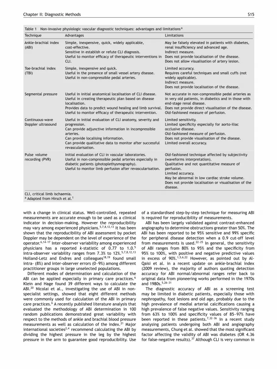
Table 1 Non-invasive physiologic vascular diagnostic techniques: advantages and limitationsa
Technique Advantages Limitations
Ankle-brachial index(ABI)
Simple, inexpensive, quick, widely applicable,cost-effective.Sensitive in establish or refute CLI diagnosis.Useful to monitor efficacy of therapeutic interventions inCLI.
May be falsely elevated in patients with diabetes,renal insufficiency and advanced age.Indirect measure.Does not provide localisation of the disease.Does not allow visualisation of artery lesion.
Toe-brachial index(TBI)
Simple, inexpensive and quick.Useful in the presence of small vessel artery disease.Useful in non-compressible pedal arteries.
Limited accuracy.Requires careful techniques and small cuffs (notwidely applicable).Indirect measure.Does not provide localisation of the disease.
Segmental pressure Useful in initial anatomical localisation of CLI disease.Useful in creating therapeutic plan based on diseaselocalisation.Provides data to predict wound healing and limb survival.Useful to monitor efficacy of therapeutic intervention.
Not accurate in non-compressible pedal arteries asin very old patients, in diabetics and in those withend-stage renal disease.Does not provide direct visualisation of the disease.Old-fashioned measure of perfusion.
Continuous-waveDoppler ultrasound
Useful in initial evaluation of CLI anatomy, severity andprogression.Can provide adjunctive information in incompressiblearteries.Can provide localising information.Can provide qualitative data to monitor after successfulrevascularisation.
Limited sensitivity.Limited specificity especially for aorto-iliacocclusive disease.Old-fashioned measure of perfusion.Does not provide visualisation of the disease.Limited overall accuracy.
Pulse volumerecording (PVR)
Initial evaluation of CLI in vascular laboratories.Useful in non-compressible pedal arteries especially indiabetic patients (photoplethysmography).Useful to monitor limb perfusion after revascularisation.
Old-fashioned technique affected by subjectivity(waveforms interpretation).Qualitative and not quantitative measure ofperfusion.Limited accuracy.May be abnormal in low cardiac stroke volume.Does not provide localisation or visualisation of thedisease.
CLI, critical limb ischaemia.a Adapted from Hirsch et al.1
with a change in clinical status. Well-controlled, repeatedmeasurements are accurate enough to be used as a clinicalindicator in decision-making. However the reproducibilitymay vary among experienced physicians.5,7,8,12,13 It has beenshown that the reproducibility of ABI assessment by pocketDoppler may be dependent on the level of experience of theoperator.6,14––17 Inter-observer variability among experiencedphysicians has a reported k-statistic of 0.77 to 1.0.5
Intra-observer variability ranges from 7.3% to 12%.5,7,8,12,13
Holland-Letz and Endres and colleagues18,19 found smallintra- (8%) and inter-observer errors (0––9%) among differentpractitioner groups in large unselected populations.
Different modes of determination and calculation of theABI can be applied especially in primary care practices.6
Klein and Hage found 39 different ways to calculate theABI.20 Nicolai et al., investigating the use of ABI in non-specialist settings, showed that eight different methodswere commonly used for calculation of the ABI in primarycare practices.6 A recently published literature analysis thatevaluated the methodology of ABI determination in 100random publications demonstrated great variability withrespect to the methods of ankle and brachial blood pressuremeasurements as well as calculation of the index.21 Majorinternational societies2,4 recommend calculating the ABI bydividing the highest pressure in the leg by the highestpressure in the arm to guarantee good reproducibility. Use
of a standardised step-by-step technique for measuring ABIis required for reproducibility of measurements.
ABI has been largely validated against contrast-enhancedangiography to determine obstructions greater than 50%. TheABI has been reported to be 95% sensitive and 99% specificfor peripheral disease detection when a 0.9 cut-off levelfrom measurements is used.22––25 In general, the sensitivityof ABI ranges from 80% to 95% and the specificity from95% to 100%, with positive and negative predictive valuesin excess of 90%.1,5,6,22 However, as pointed out by Al-Qaisi et al. in a recent update on ankle-brachial index(2009 review), the majority of authors quoting detectionaccuracy for ABI normal/abnormal ranges refer back tooriginal data from pioneering works performed in the 1970sand 1980s.5,26––31
The diagnostic accuracy of ABI as a screening testmay be limited in diabetic patients, especially those withnephropathy, foot lesions and old age, probably due to thehigh prevalence of medial arterial calcifications causing ahigh prevalence of false negative values. Sensitivity rangingfrom 63% to 100% and specificity values of 85––97% havebeen reported in these patients.7,32––36 In a recent studyanalysing patients undergoing both ABI and angiographymeasurements, Chung et al. showed that the most significantfactor affecting the validity of ABI was diabetes (OR 4.36for false-negative results).37 Although CLI is very common in
S16 P. Cao et al.
diabetic patients (prevalence of vascular disease estimatedaround 13.6% vs. 4% in the general population),7,38 it oftenremains under-recognised in this population.7,39 Diagnosisis often difficult because the co-existence of peripheralneuropathy could mask the ischaemic pain. In these settingssensitivity in detecting vascular disease of ABI ranges from50% to 71% and specificity from 30% to 96.8%.7,11,34,40––42
The ABI is relatively insensitive for determining progres-sion of lower limb occlusive disease when compared toarteriography or duplex ultrasonography. McLafferty et al.found an ABI sensitivity of 41%, specificity of 84%, positivepredictive value of 59% and overall accuracy of 68% indetecting disease progression.43
In addition to its use in evaluating symptomatic patientsaffected by peripheral vascular disease, decreased ABI isa strong predictor of cardiovascular events and prematuremortality.44––47 An ABI <0.90 is associated with a 3- to 6-foldincreased risk of cardiovascular mortality.45––48 A meta-analysis in 2008 by Fowkes et al.49 found that abnormalABI was associated with approximately twice the 10-yeartotal mortality, cardiovascular mortality, and major coronaryevent rate compared with the overall rate in each Framing-ham risk score category. The American Diabetes Associationrecommends screening for ABI in all diabetic patients aged>50 years, as well as in younger insulin-dependent patientswith other vascular risk factors.35,45,47,50,51 ABI measurementcan be used as a prognostic index to facilitate initiationof treatment (hypertension, dyslipidaemia, diabetes, etc.)to reduce cardiovascular events and should be routinelyperformed in patients aged �70 years, with rest pain orulcers, and those with a history of diabetes or smoking.47
Although the ABI has gained widespread acceptance asa single, accurate and reproducible first-line method toevaluate arterial occlusive disease and a valid cardiovascularprognostic instrument, the test has definite limitations andit should be associated with duplex ultrasound imaging.
Summary messages (advantages and limitations of ABI):• ABI can be useful as a routine measurement in primary
care practices providing objective and reproducible first-line assessment of CLI. It is sensitive and specific in thedifferential diagnosis of leg symptoms to identify or ruleout a vascular aetiology.
• ABI measurement is a widely applicable, simple, quick,cost-effective and non-invasive tool to establish or dis-prove the baseline for CLI and to follow revascularisationresults.
• ABI can provide objective data that serve as the standard inoffice practice, vascular laboratories and epidemiologicalsurveys.
• ABI provides indirect information on arterial disease butcannot localise the anatomical level of a pressure-reducingobstruction.
• The reproducibility of ABI measurements is dependent onthe level of experience of the operator and may varyamong experienced physicians, university hospitals and incommunity settings.
• ABI may not be accurate in the presence of incompressiblelower extremity arteries as occurs in very elderlyindividuals, diabetics, or patients with long-standing renaldisease.
• Use of a standardised step-by-step technique for measuringABI is required to ensure reproducibility of measure-ments.
RecommendationsThe resting ABI is useful in the initial evaluation for CLIand can be easily and quickly measured on both legs toconfirm diagnosis and establish the severity of disease inpatients with rest leg/foot symptoms as well as individualswith foot non-healing ulcer and lower limb rest pain.(Level 2b; Grade B)The resting ABI in all new patients with CLI can be usedto establish the baseline to evaluate the effect afterrevascularisation procedures. (Level 2b; Grade B)ABI is less reliable in CLI diagnosis in patients withincompressible arteries (long-standing diabetes, end-stagerenal disease, advanced age) and should be supportedby more reliable techniques in these settings. (Level 2b;Grade B)
2.1.2. Toe-brachial index
Since the presence of CLI is higher in patients with diabetesand end-stage renal disease, this may preclude accurateassessment of ABI in most subsets of these patients.7,38
Incompressible arteries are suggested when the ABI isgreater than 1.3. The digital vessels are usually sparedfrom calcification; therefore, toe systolic blood pressuresare often more accurate in quantifying vascular diseasein diabetic, dialysis-dependent or very old patients. Toepressures are obtained by placing small occlusive cuffsaround each toe (usually at the proximal portion of digitI and II) with a digital flow sensor beyond the cuff. Toesystolic pressure can be expressed as a ratio of the toepressure to the highest pressure recorded in either arm toobtain the toe-brachial index (TBI).
Normally the toe pressure is approximately 30 mmHg lessthan the ankle pressure and TBI should be >0.75. Values <0.7are considered abnormal and TBI <0.25 is consistentwith severe CLI.52,53 Absolute toe pressures <30 mmHg arerequired to diagnose CLI in patients with rest pain.2 Forpatients with ulcers or gangrene, the presence of CLI isdiagnosed by toe systolic pressure <50 mmHg. Absolute toepressure of 55 mmHg or greater has been correlated tobe predictive for foot ulcer healing in diabetic patients,and TASC requires toe pressure <50 mmHg (critical level) toconfirm CLI diagnosis in diabetic patients.2
Toe pressure measurements were shown to be morereliable than ABI measurements in patients with diabetesassociated with falsely high ABI values and peripheralneuropathy.34,54,55 Brooks et al. compared TBI and ABIin 174 diabetics and 53 controls and found comparableindices when ABI was low or normal (84% and 78%agreement, respectively), but not when ABI was elevated.54
In the presence of clinical peripheral neuropathy, toepressure sensitivity has been evaluated to be 100% and ABIsensitivity 53%.34
However, TBI measurement requires appropriate tech-nique and tools (small cuffs), therefore it is not widelyapplicable and the overall accuracy may be limited.Furthermore, it may be impossible to measure toe pressuresin patients with inflammatory lesions, ulceration or loss oftissue.

Chapter II: Diagnostic Methods S17
Summary messages (advantages and limitations of toe-brachial index):• Toe-brachial index is a quick way to non-invasively
establish or disprove the CLI diagnosis in patients withlower limb rest pain or non-healing ulcers.
• Toe-brachial measurements are particularly useful inindividuals with incompressible crural and pedal arteries.
• The test requires small cuffs and a careful technique topreserve accuracy.
RecommendationToe-brachial index is useful to establish CLI diagnosis inpatients in whom CLI is clinically suspected (non-healingulcer, rest pain) but the ABI test is not reliable dueto incompressible vessels as in patients with diabetes,advanced age or long-standing renal failure. (Level 2b;Grade B)
2.1.3. Segmental limb pressureThe location and extent of CLI can be indirectly definedin a non-invasive laboratory by segmental limb systolicpressure measurements, recorded with a Doppler instrumentand blood pressure cuffs placed over the brachial arteriesand sequentially at various points on the lower limbs,including the upper and lower thigh, the upper calf, theankle, and metatarsal. Theoretically, the cuff width shouldbe 20% greater than the diameter of the limb at thepoint where it is applied. Narrow cuffs may be associatedwith the appearance of falsely high pressures and do notpermit accurate disease localisation.1,2 The examinationis performed by placing a Doppler probe over the mostprominent arterial signal at the ankle with the patient insupine position. In most laboratories a 20 mmHg gradientbetween adjacent segment cuffs is regarded as indicativeof a significant occlusive lesion. Thus, by comparing thepressures obtained at different levels, segmental pressuremeasurement can detect the location of arterial occlusivelesions with reasonable accuracy. Segmental pressuremeasurements can provide information in patients withmulti-level disease and predict ulcer healing, limb survivalor the need for further additional revascularisation.54––56
However, as already stated by the TASC II and AHAGuidelines,1,2 there are a number of limitations andpotential problems in the analysis of segmental limbpressure that render it an old-fashioned diagnostic techniquefor CLI evaluation.• Isolated moderate stenosis (usually iliac) that produces
little pressure gradient may be missed.• Calcified arteries may lead to falsely elevated ankle
pressures.• In patients with multi-level disease, decreased proximal
pressures may mask more distal gradients.• Segmental pressure gradients are not suitable to differen-
tiate between short- and long-segment lesions or betweenhighly stenotic arteries and occlusions.
• Reduced thigh pressure is usually indicative of a pressure-reducing obstruction along the aorto-iliac axis; however,similar findings may be produced by an obstruction of thecommon femoral or proximal superficial femoral and deepfemoral arteries.
• Artefacts on measurements due to inappropriate cuffsize/position are common.
Summary messages (advantages and limitations of seg-mental limb pressure):• Segmental pressure can provide first-line information on
anatomical localisation of lower limb vascular disease inpatients with CLI.
• Segmental pressure measurement can be helpful toestablish more sophisticated imaging techniques todetermine a detailed localisation of the disease.
• The test may not be accurate in the presence ofincompressible arteries.
• Measurements of segmental pressure can provide onlyindirect information on vascular disease and results canbe biased by a number of artefacts and drawbacks. Thetest should not be used as the sole diagnostic techniquebut should be associated with ultrasound imaging.
RecommendationLower limb segmental pressure measurements can providea first-line localisation of arterial lesion along lower limb inpatients with CLI. Segmental pressure should not be usedas the sole diagnostic technique. (Level 2b; Grade B)
2.1.4. Continuous-wave Doppler ultrasoundQuantitative and qualitative analysis performed by continu-ous-wave Doppler ultrasound remains an old-fashionedtechnique which is no longer routinely used in manymodern diagnostic vascular laboratories. The AHA guidelinesrecognise that Doppler waveforms analysis needs to becombined with ultrasound visualisation of arterial vessel(“duplex imaging”) to maximise the benefits of thistechnique.1
Analysis of morphology of the continuous-wave Dopplerwaveform was suggested to provide useful information inlocalising and quantifying vascular disease in patients withpoorly compressible arteries. Many patients with diabetesor end-stage renal disease, without palpable pulses andmonophasic Doppler signals, may have an ABI greaterthan 1.0, which is a deceptive and misleading quantitativeassessment of the severity of vascular disease.57
The most commonly used “pulsatility index” is definedas the peak systolic velocity (or “frequency shift”) dividedby the mean blood flow velocity. Normally, the pulsatilityindex increases from the most proximal to the most distalsegments of the lower extremities; a decrease betweenadjacent segments implies the presence of occlusive diseasebetween these two locations.57,58
However, reversion to a normal waveform below aproximal stenosis may often occur. This phenomenon of“pulse normalisation” distal to some arterial stenosis is arecognised major diagnostic limitation of the technique thatmay occur especially in the presence of multi-level diseasewith high-resistance flow.
Furthermore, quantitative assessment of the pulsatilityindex is weakened in the presence of arterial calcifications.
Currently, the benefits of continuous Doppler waveformanalysis are limited, and it should always be combined withultrasound greyscale or colour visualisation of the arterialwall. Such “duplex imaging” represents one of the mostwidely used non-invasive vascular laboratory techniquesreplacing the traditional continuous-wave Doppler velocityanalysis.

S18 P. Cao et al.
Summary messages (advantages and limitations ofcontinuous-wave Doppler ultrasound):• Continuous-wave Doppler ultrasound can be used as
an initial step to indirectly assess lower limb vasculardisease. The test enables indirect qualitative evaluationof blood flow, vessel localisation and flow detectionin nonpalpable arteries, and quantitative systolic bloodpressure measurements along lower limb vessels.
• Continuous-wave Doppler ultrasound does not providevisualisation of vessel anatomy.
• Continuous-wave Doppler ultrasound is limited in accuracyand is relatively insensitive especially for iliac arterialdisease detection. “Pulse normalisation” downstream fromstenosis can diminish test sensitivity.
• Continuous Doppler waveform should be combined withother imaging (ultrasound greyscale or colour visualisationof the arterial wall: “duplex ultrasound imaging”).
• Continuous-wave Doppler ultrasound remains an old-fashioned technique no longer routinely used in manymodern laboratories.
RecommendationsContinuous-wave Doppler ultrasound is of limited use inproviding initial qualitative and quantitative assessmentof lower limb vascular disease location and severity, andin following outcomes of vascular disease with or withoutrevascularisation. (Level 3b; Grade C)Since continuous Doppler ultrasound does not allow directarterial visualisation, this test should always be combinedwith real imaging through ultrasound greyscale or colourvisualisation of the arterial wall (“duplex ultrasoundimaging”). (Level 2c; Grade B)
2.2. Plethysmography
Plethysmography in CLI evaluation has been introduced inthe past to detect changes in limb volume by “pulse volumerecording” (PVR) which produces recordings that are similarto continuous Doppler waveforms.59,60
However, the lack of reliable, reproducible quantitativedata limits the utility of plethysmography for diagnosisor arterial disease and CLI in most modern vascularlaboratories today. With more widespread utilisation ofultrasound methods, the use of plethysmography hasdeclined substantially.
The main value of PVR waveform analysis may be that itis not affected by medial calcification and therefore it isrelatively useful in the diabetic population.61
Accuracy of PVR and photoplethysmography has beentested against Doppler ultrasound in several studies,56,62,63
indicating that the techniques might be useful in diabeticpatients with CLI, including those with oedema, but themethod may have poor accuracy in vascular diseases locatedin distal limb segments.64
PVR tracings at the foot level have been used as anindicator of healing potential for foot wound or amputationprocedures.8,45,56,65,66
Limitations of PVR include that it may be a rathersubjective tool for evaluation of CLI since measurementsare based on subjective waveform analysis. PVR may beabnormal in patients with low cardiac stroke volume andoverall accuracy is limited. Although quantitative criteria
have been proposed in PVR, they are not widely usedclinically owing to limited accuracy.1
Summary messages (advantages and limitations of pulsevolume recording):• PVR remains an old-fashioned technique no longer
routinely used in many modern laboratories.• PVR may be useful as an initial diagnostic test for patients
with foot pain or ulcers and suspected CLI, to assess limbperfusion and predict risk of amputation in CLI patients.
• PVR can provide a tool to evaluate individuals withincompressible vessels in whom ABI and segmentalpressures are spuriously elevated.
• PVR does not allow reliable quantitative measure ofperfusion and may not be accurate.
• PVR may be abnormal in patients with low cardiac strokevolume.
• PVR in evaluation of limb perfusion is affected by“subjective influence” and is less accurate than other non-invasive tests in providing arterial anatomical localisationof disease.
• Although PVR may be useful and cost-effective as abaseline tool in office practice or vascular laboratories,other non-invasive techniques can today provide morequantitative and accurate information on perfusion andanatomical localisation of lower limb disease in CLI.
RecommendationsPulse volume recording may be used as an initial step inthe evaluation of patients with foot pain and ulcer andsuspected CLI and can be applied to establish diagnosis,assess localisation or severity of the disease and followstatus of revascularisation procedures, but accuracy islimited. (Level 3b; Grade C)Pulse volume recording may be applied to establish theinitial lower limb CLI diagnosis in diabetic patients andpatients with incompressible arteries, but it should becombined with additional tests (e.g., “duplex ultrasoundimaging”). (Level 2a; Grade B)
Advantages and limitations of each non-invasive physio-logical and haemodynamic diagnostic test are summarisedin Table 1.
3. Measurements of tissue perfusionDifferent non-invasive measurements of tissue perfusionhave been used to assess the severity of lower limbischaemia. The applicability and reliability are generallylimited with respect to Doppler ultrasonography.
Measurements of transcutaneous oxygen pressure (TcPO2)reflect the metabolic state of lower limbs with CLI anddiabetic feet. Small electrodes consisting of a circularsilver––silver chloride anode surrounding a central platinumcathode are placed on the skin; oxygen diffusing to thesurface of the skin is reduced at the cathode to producea current proportional to the partial pressure of oxygen(PO2) within the sensor. In patients with foot ulcers,tissue loss or rest pain, TcPO2 values can be used toassess the presence and severity of vascular disease, theneed for revascularisation, and to predict the successof healing with or without revascularisation. This test isperformed by placing probes with electrodes on the foot
Chapter II: Diagnostic Methods S19
and the leg, using the chest as a reference site. Commonlocations for assessment are the dorsum of the foot, theanteromedial aspect of the calf 10 cm below the knee,and the thigh 10 cm above the knee. Normal TcPO2 valuesdepend on age (higher for younger) and position (higher forproximal). Normal TcPO2 levels are approximately 60 mmHg,while levels of 20 mmHg or less strongly suggest thatrevascularisation will be required to achieve healing. TASC IIrequires a critical level of TcPO2 <30 mmHg to confirmdiagnosis of CLI in patients with non-healing foot ulcers ordiabetic foot.2
Measurement of TcPO2 is most helpful for evaluating casesof severe limb ischaemia, while it is relatively insensitiveto mild or moderate degrees of peripheral vascular diseasebecause the oxygen supply to the skin is far greater thanthe demand. TcPO2 measurements combined with clinicaldetermination may be of value to predict healing atvarious levels of amputation, especially in diabetic patients,because it is not affected by arterial calcification.67––69
Nevertheless, measurements of TcPO2 must be interpretedcautiously, since the test is often unreliable because itis affected by many factors that are difficult to control,including skin temperature, sympathetic tone, cellulitis,hyperkeratosis, obesity, oedema, metabolic activity, oxygendiffusion through tissue, age, vertical position of the siteof measurements. In addition, when values are low, TcPO2
is not linearly related to flow: a value of zero does notmean that there is no flow to the area of interest; rather itindicates that all the available oxygen has been consumed.Therefore, TcPO2 is not routinely used in most vascularlaboratories.
Measurement of skin perfusion pressure (SPP) is anothermicrocirculatory assessment tool that can be utilised toassess foot healing potential.70 SPP is measured with laserDoppler and represents the blood pressure required torestore microcirculatory or capillary flow after inducingcontrolled occlusion and return of flow. The ability of thistest to predict amputation healing is not as good as thatof TcPO2 measurements. Normal pressures of 50––70 mmHgare decreased to 10––20 mmHg in limbs with severe limbischaemia. Pressures below 30 mmHg are predictive of CLI.
Laser Doppler is not widely used in vascular laboratories,mainly because of an inability to calibrate the instrumentto actual levels of blood flow and the availability of moreaccurate, direct methods for assessing CLI.
Hyperspectral tissue oxygenation measurements have alsobeen used to predict healing of diabetic foot ulcers. The testshould identify microvascular abnormalities in the diabeticfoot, but this technology is currently being utilised mainlyas a research tool.71
Summary messages (advantages and limitations of tissueperfusion measurements):• Tissue perfusion measurement can be useful to assess the
severity of lower limb ischaemia.• These techniques can be used in monitoring and/or re-
evaluating patients following endovascular or surgicalrevascularisation.
• Microcirculatory assessment of perfusion can be utilised toassess wound healing potential.
• Transcutaneous oxygen pressure (TcPO2) is valuable toexamine the metabolic state of the target tissue.
• Measurements of TcPO2 are time-consuming and maybe unreliable because influenced by many physiological,methodological and technical factors (skin temperature,sympathetic tone, cellulitis, hyperkeratosis, obesity,oedema, metabolic activity, oxygen diffusion throughtissue, age, etc.).
• TcPO2 could not be measured in advanced CLI becauseof intolerable pain during the examination in the supineposition.
RecommendationsPatients with ischaemic rest pain or foot ulcers can beinvestigated with objective tests of tissue perfusion toconfirm diagnosis of CLI. (Level 2a; Grade B)These may include TcPO2, laser Doppler and hyperspectralmeasurements to assess metabolic state of tissueperfusion. (Level 3b; Grade C)Tissue perfusion tests (TcPO2, laser Doppler, spectralimaging) can be used to assess healing potential of ul-cers/amputation in patients with CLI (Level 3b; Grade C ––Level 4; Grade D)
4. Imaging techniques
The purpose of vascular imaging for patients with CLI is toassess the anatomical location, morphology and extent ofdisease to determine suitability for open or endovascularrevascularisation. Major technical advances have beenaccomplished in recent years in the development of non-invasive imaging modalities. Today, the following options forimaging are available:• duplex ultrasound (DUS)• magnetic resonance angiography (MRA)• computed tomography angiography (CTA)• digital subtraction angiography (DSA).
The main characteristics of these imaging modalities,including their principal advantages and disadvantages, aresummarised in Table 2.
4.1. Duplex ultrasound
Duplex ultrasound (DUS) enables identification of theanatomical location and the degree of stenosis in lowerextremity peripheral arterial disease (PAD) by combiningboth B-mode ultrasound and colour Doppler ultrasound.Haemodynamic assessment is performed by measuring peaksystolic velocity (PSV) and PSV ratios within or beyond anobstruction compared with the adjacent upstream segment,the presence or absence of turbulence, and preservationof pulsatility. A PSV ratio of greater than 2:1 is consideredto indicate a >50% stenosis, a PSV ratio greater than 4:1a >75% stenosis and a PSV ratio of greater than 7:1 a>90% stenosis.74
Accuracy of DUS: Several studies have reported a highaccuracy of DUS in comparison with DSA. A recent meta-analysis of studies published between 1996 and 2005produced a pooled sensitivity of 88% (84––91%) and apooled specificity of 94% (93––96%) for DUS, confirming datafrom a former meta-analysis75––77 (Table 3). When used byexperienced operators and in suitable patients, DUS canproduce a map of significant obstructive disease from theabdominal aorta to the feet.78

S20 P. Cao et al.
Table 2 Comparison of different imaging modalities for patients with PADa
DUS CTA MRA Angiography
Availability +++ ++ ++ +++
Appointment time (minutes) 40+ (bothlegs)
15 30 30
Equipment cost + ++ +++ +++
Operator expertise +++ + ++ ++
Arteriographic map Yes, byexperiencedoperators
Yes (requirespost-processing)
Yes (immediately available) Yes (immediatelyavailable)
Diagnostic accuracy
Aorto-iliac ++ +++ +++ +++
Femoro-popliteal +++ +++ +++ +++
tibial + + ++ +++
Stent assessment ++ + Steel: poorNitinol: fair
+++
Limitations by vascular calcification ++ ++ None Almost none
Complications and risks
Access site None None None Rare
Ionising radiation exposure None 7.5––13.7 mSv None Higher than CTA
Contrast-enhanced nephropathy None ++ Extremely rare ++
Nephrogenic systemic fibrosis None None Very rare None
Allergic reaction None Rare Very rare Rare
Contraindications None Severe renalimpairment,known allergy tocontrast agents
Cerebrovascular clips, electronicimplants (infusion or monitoringdevices, neurostimulationdevices), pace-makers,cardioverter-defibrillators,claustrophobia
Severe renalimpairment, knownallergy to contrastagents
CTA, computed tomography angiography; DUS, duplex ultrasound; MRA, magnetic resonance angiography; mSv, millisievert;PAD, peripheral arterial disease.a Modified from Norgren et al.,2 Owen and Roditi72 and Kramer et al.73
DUS can be used for pre-intervention decision-makingby predicting whether a patient has anatomy suitablefor femoro-popliteal angioplasty with an accuracy of84––94%.81,82 It has also been used as a substitute forDSA for infrainguinal bypass grafting to select the mostappropriate tibial vessel for distal anastomosis, althoughsome studies have suggested that DUS alone is inferior to DSAfor evaluation of tibial arteries for distal bypass surgery.83––90
Another study has demonstrated no difference in patencyof infrapopliteal bypass grafts in non-randomised cohorts ofpatients evaluated by pre-operative DUS vs. angiographicmethods.85
DUS can also be used for post-revascularisation surveil-lance of venous and prosthetic grafts. Venous grafts mayfail due to de novo obstructions either within the body ofthe graft or at the anastomoses (intimal hyperplasia), ordue to progression of atherosclerotic obstructions upstreamor downstream from the graft. DUS surveillance studies candetect these obstructions during impeding graft thrombosiswith greater sensitivity than evaluation by clinical history,physical examination, or use of the resting ABI.1,91––96 Ingeneral, low velocities indicate poor arterial inflow, proximalstenosis, or large graft diameter. One study showed thatpresence of a PSV less than 45 cm/s within a graft indicatesthat subsequent graft failure is likely to occur.97,98 Another
study found that vein grafts that were revised on the basisof positive DUS findings had a 90% 1-year patency rate,similar to grafts with initially normal duplex examinations.Grafts that were not revised despite the presence of a DUS-detected stenosis had a patency rate of only 66% at 1 year.92
Unfortunately, three RCTs offered conflicting results, with a3-year primary assisted patency rate of vein grafts monitoredwith DUS of 78% vs. 53% for those followed up clinicallyand with the ABI in one study and no improved patencyin the others.99,100 The Vein Graft Surveillance RandomisedTrial (VGST)101 assessed the benefits of DUS compared withclinical vein graft surveillance in terms of amputation rates,quality of life and healthcare costs in patients after femoro-popliteal and femorocrural vein bypass grafts. A total of 594patients with a patent vein graft at 30 days after surgerywere randomised to either a clinical or a duplex follow-upprogramme at 6 weeks, then 3, 6, 9, 12, and 18 monthspost-operatively. Both groups had similar amputation rates(7% for each group) and vascular mortality rates (3% vs. 4%)over 18 months. More patients in the clinical group hadvein graft stenoses at 18 months (19% vs. 12%, p = 0.04),but primary patency, primary assisted patency and secondarypatency rates, respectively, were similar in the clinical group(69%, 76% and 80%) and the duplex group (67%, 76% and 79%).There were no apparent differences in health-related quality

Chapter II: Diagnostic Methods S21
Table 3 Systematic reviews and meta-analyses to assess the diagnostic accuracy of duplex ultrasound (DUS), magnetic resonanceangiography (MRA) and computed tomography angiography (CTA) in the detection of >50% stenosis or occlusion in patients withperipheral arterial disease (PAD)
Reference Modality Characteristics Anatomicalregion
Pooledsensitivity
Pooledspecificity
Visser, 200078 Contrast-enhancedMRA
• 9 studies (1990––1998) with a total of 216 pts (11––30)• CLI in only 3 studies, in 2 studies no clinical data)
Whole leg 98%(96––99%)
96%(94––98%)
DUS • 18 studies (1984––1998) with a total of 1059 pts (12––167)• CLI in 9 studies (10––84% of the study population), in 7
studies no clinical data
Whole leg 88%(84––91%)
95%(93––96%)
Collins et al.,200776,77
DUS • 7 studies (1996––2005) with a total of 369 pts (20––76)• 134––3108 segments per study (median 404 segments)• CLI in about 10% (range 0––19%) of pts
Whole leg 88%(80––98%)
96%(89––99%)
2D time-of-flight MRA
• 5 studies (1996––2005) with a total of 287 pts (20––155)• 206––1188 segments/study (median 378 segments)• CLI in 82––100% of pts (no clinical data in 3 studies)
Whole leg 92%(79––94%)
88%(74––92%)
Contrast-enhancedMRA
• 7 studies (1996––2005) with a total of 279 pts (20––76)• 418––1780 segments/study (median 520 segments)• CLI in 0––92% of pts (no data in 3 studies)
Whole leg 95%(92––100%)
97%(64––99%)
Menke et al.,201079
Contrast-enhancedMRA
• 32 studies (2004––2009) with a total of 1022 pts (10––76)• 120––1780 segments per study (median 384 segments)• 24% of all investigated arterial segments had stenoses or
occlusions• CLI in 26% (range 0––100%) of pts
Aorto-iliac
Femoro-poplitealTibial
94%(91––96%)95%(91––98%)92%(90––94%)
96%(94––97%)96%(95––98%)93%(90––96%)
Met et al.,200980
CTA • 20 studies (1966––2008) with a total of 957 pts (16––279)• 167––4743 segments per study (median 730 segments)• 29% of all investigated arterial segments had stenoses or
occlusions• CLI in <20% (range 0––100%) of all pts (68% IC, 10––20% no
data)
Aorto-iliac
Femoro-poplitealTibial
96%(91––99%)97%(95––99%)95%(85––99%)
98%(95––99%)94%(85––99%)91%(79––97%)
of life, but the average health service costs incurred by theDUS surveillance programme were greater by £495 (95% CI£83––807) per patient. The authors concluded that intensivesurveillance with DUS did not show any additional benefitin terms of limb salvage rates for patients undergoingvein bypass graft operations, but it did incur additionalcosts.101 In a further prospective study, a normal DUS scan6 weeks subsequent to infrainguinal vein bypass grafting wasassociated with a 40-month cumulative patency rate of 82%,indicating that further DUS surveillance in these patients isnot beneficial.102 There is an ongoing transatlantic discussionwhether or not DUS surveillance is beneficial in patients withinfrainguinal venous bypass grafting, with a tendency forroutine DUS in these patients in North America,103––105 whilethe situation in Europe remains equivocal.102,106,107
DUS surveillance of synthetic grafts is of questionablevalue. Several studies have found no improvement inpatency of grafts, whereas other studies have successfullydetected stenoses and found some improvement in patency.This lack of evidence may be due to DUS-associated technicalchallenges (inability to visualise the stenosis, vascularanatomic challenges) or procedural challenges, such that thesubsequent graft revision does not help to improve long-termgraft patency.1,100,101,108––111
DUS surveillance after angioplasty (PTA) proceduresis also of questionable value. Immediately after PTA,several studies suggested that velocities in the treated
segment may be abnormally elevated and do not predictdecreased subsequent patency rates. This may be dueto angioplasty-induced vessel dissections that successfullyremodel over time. DUS is useful in evaluations for recurrentobstructions.1,112––119 Although it is reasonable to assumethat revisions of post-PTA restenoses that are detected byDUS studies might improve patency, there are no publishedstudies confirming this approach on a high evidence level.1
Summary messages: Advantages and disadvantages ofduplex ultrasound:• DUS is non-invasive, relatively inexpensive and as an
outpatient procedure well tolerated by patients.• DUS can also be performed in emergency situations on the
ward or in the operating theatre.• There are limitations to the visualisation of iliac vessels
in the pelvis (due to body habitus and bowel gas),very distal arteries and collaterals. In addition, extensivecalcification may produce incomplete examinations and inpatients in whom multi-level PAD downstream stenoses aredetected the sensitivity is decreased, perhaps owing toslow flow.120,121
• The technique is highly operator-dependent and propertraining is mandatory.98
• Since the vast majority of DUS studies were performedin mixed populations, the validity of DUS imaging forCLI patients alone is still uncertain.
• No side effects or adverse events have been reported.

S22 P. Cao et al.
Table 4 Recommendations in current guidelines for duplex ultrasound imaging in patients with CLI
Grade ofrecommendation
Level ofevidence
Duplex ultrasound of the extremities is useful to diagnose anatomical location and degree ofobstruction in PAD patients a.
A 1a
Duplex ultrasound may be considered for routine surveillance after femoropopliteal orfemorotibial-pedal venous bypass grafts a.
B 2b
Duplex ultrasound of the extremities can be useful to select patients as candidates for endovascularinterventiona.
B 2b
Duplex ultrasound may be useful to select patients as candidates for surgical bypass and to select thesites of surgical anastomosis a.
B 2b
Duplex ultrasound may be considered for routine surveillance after femoropopliteal bypass with asynthetic conduit a.
B 3b
The use of duplex ultrasound is not well established to assess long-term patency of percutaneoustransluminal angioplastya.
B 3b
CLI, critical limb ischaemia; PAD, peripheral arterial disease.a Adapted from Hirsch et al.1
Recommendations in current guidelines (Table 4)The current ACC/AHA Practice guidelines for PAD patientsgive a strong recommendation that DUS is usefulto diagnose the anatomical location and degree ofobstruction of PAD. (Level 1a; Grade A)Despite the discrepancies mentioned above, DUS surveil-lance of venous grafts is also recommended with regularfollow-up intervals (3, 6, 12 months, and then yearly aftergraft placement).1 (Level 2b; Grade B)The guidelines for non-invasive vascular laboratory testingfrom the American Society of Echocardiography and theSociety for Vascular Medicine and Biology recommendDUS evaluation of the graft twice during the first post-operative year, and annually thereafter.98 The ACC/AHAguidelines consider DUS also as useful to select patientsas candidates for endovascular intervention or surgicalbypass (Grade B) and state that DUS may also beconsidered for routine surveillance after femoropoplitealbypass with a synthetic graft (Level 2b; Grade B).Finally, DUS is not well established to assess long-termpatency of PTA.1 (Level 2b; Grade B)
Critical issues• The majority of DUS studies are more than 10 years
old. New studies should consider following the STARDguidelines for reporting of diagnostic accuracy studies andshould also consider reporting results by patient or bylimb, as well as by segment.122––124
• Future reviews should make use of the QUADAS as a qualityassessment tool specifically developed for systematicreviews of diagnostic accuracy studies.125
• Further research should consider comparing DUS directlywith MRA and/or CTA as the reference standard.
• The value of the operative or endovascular correctionof DUS-detected post-PTA lesions has to be evaluated infurther studies.
• Future studies should identify patients with infrainguinalvein or prosthetic bypasses, who benefit from astandardised DUS surveillance programme.
• The validity of DUS imaging for patients with CLI needs tobe evaluated in patient cohorts suffering from rest painor non-healing ischaemic lesions in the foot.
4.2. Computed tomography angiography
Computed tomography angiography (CTA) is increasinglyattractive due to rapid technical developments. Shorter ac-quisition times, thinner slices, higher spatial resolution, andimprovement of multidetector computed tomographic (CT)scanners enable scanning of the entire vascular tree in alimited period with a decreasing amount of contrast mediumand radiation burden.
Accuracy of CTA: In a recent meta-analysis, 20 studiespublished between 1966 and 2008 (957 patients) werereviewed systematically by use of the QUADAS checklist.126
Between 167 and 4743 arterial segments were analysed ineach study (median 730 segments) and 29% of all segmentshad stenoses or occlusions. Slice thickness varied between0.75 and 5.0 mm (median 2.0 mm). Various contrast mediawere used for the CTA (Iomeprol in 6 studies, iopromidein 4 studies, and the remaining studies used other iodine-based contrast media). The iodine concentration variedbetween 300 and 400 mg/mL. The amount of contrastvolume administered per scan varied between 88 and 170 mL(median 130 mL). Interpretation of CTA was always based onthe axial images. Other image reconstructions used weremaximum-intensity projections (n = 17), volume-renderingtechnique (n = 15), multiplanar reformation (n = 6), curved-planar reformation (n = 4), and virtual endoscopy (n = 1). Thepooled sensitivity to detect a >50% stenosis or occlusionwas 95% (92––97%) and the pooled specificity 96% (93––97%).CTA correctly identified occlusions in 94% of segments, thepresence of >50% stenosis in 87% of segments, and absence ofsignificant stenosis in 96% of segments. Overstaging occurredin 8% of segments and understaging in 15%. The data includedtrials of CTA vs. DSA across three different generations ofCT technology (i.e., scanners with 4, 16, and 64 detectorrows) and with technological advancement there hasbeen a corresponding improvement in diagnostic accuracy(sensitivity and specificity has increased from 75––99% and83––99% with 4 detector rows to 98––99% and 96––99% for64 detector rows). Diagnostic accuracy was lower for smallerarteries compared with proximal lesions, but the diagnosticperformance below the knee remains good (sensitivity85––99%, specificity 79––97%) (Table 3). Inter-observer agree-ment is good to excellent (k values >0.8) in most studies.126
The great majority of patients in trials of peripheral arterial

Chapter II: Diagnostic Methods S23
Table 5 Recommendations in current guidelines for CT angiography imaging in patients with CLI
Grade ofrecommendation
Level ofevidence
CTA of the extremities may be considered to diagnose anatomic location and presence of significantstenosis in patients with lower extremity PADa.
B 3a
CTA of the extremities may be considered as a substitute for MRA for those patients withcontraindications to MRAa.
B 3a
Patients with baseline renal insufficiency should receive hydration before undergoing CTAa. A 2b
CLI, critical limb ischaemia; CTA, computed tomography angiography; MRA, magnetic resonance angiography; PAD, peripheral arterialdisease.a Adapted from Hirsch et al.1
imaging are claudicants and there are limited data in pa-tients with CLI. In a recent study of 28 patients with CLI whowere evaluated with 16-detector-row CTA, 23 had treatmentplans confidently formulated on the basis of the CTA alone.
Side effects/adverse events: The average radiation dosereported in the CTA literature is 7.47 mSv,127 although aver-age doses as high as 13.7 mSv have been reported in someseries.128 In a trial of 16-detector-row CTA vs. DSA, Willmannet al. reported a four-fold higher radiation dose for DSAcompared with CTA.129 To place these doses in context, theaverage annual background radiation exposure is between2 and 3 mSv.80 It has been suggested that patient radiationdose issues are of limited concern in patients with advancedPAD, as their life expectancy is significantly less than thelatent period of a radiation-induced malignancy.130 The lateeffects of radiation exposure are more important in youngerpatients, however; physicians should be aware of this issueand strive to keep dosing as low as reasonably possible.
Iodinated contrast agents are associated with an increasedrisk for contrast-induced nephropathy (CIN), defined as anincrease in serum creatinine level >25% or >0.5 mg/dL abovebaseline within 3 days of contrast administration inthe absence of other causes.73,131 Patients who areconsidered at highest risk are those with baseline renalinsufficiency, especially those with concomitant diabetesmellitus. Other risk factors for CIN include multiplemyeloma, proteinuria, concomitant nephrotoxic drug use,hypertension, congestive heart failure, hyperuricaemia, anddehydration. The risk of CIN is dose-dependent and ishigher when contrast is administered intra-arterially thanwhen given intravenously.132 A systematic review revealed anoverall risk of CIN in high-risk patients of 16.8%,132 althoughthe clinical implications for the development of CIN are notfully understood. Only a minority go on to require renalreplacement therapy (<1%), but in a retrospective reviewof over 16,000 inpatients exposed to contrast media, in-hospital mortality rates were five-fold higher (34% vs. 7%)among patients who developed CIN, even after adjustmentfor comorbidity.133 High-osmolar contrast puts patients withpre-existing renal impairment at twice as high a risk ofdeveloping CIN as low-osmolar contrast.127 However, in areview from 2004 it was concluded that all patients withpre-existing renal insufficiency were at higher risk for CIN,no matter what type of contrast was used.134 To preventCIN pre-emptive hydration is recommended, especially forthose patients with renal insufficiency. The optimal type,route, volume, and timing of hydration are not well defined.1
Likewise, given the ability of angiotensin-converting enzymeinhibitors and angiotensin receptor antagonists to induce
efferent arteriole vasodilatation, these medications shouldbe withheld the morning of contrast exposure and restartedafter monitoring of normal renal function. Administration ofantioxidants, such as mannitol, advocated as renoprotectiveagents, is not supported by evidence.
Further information is provided by the European Societyof Urogenital Radiology (http://www.esur.org).
Summary messages: Advantages and disadvantages ofcomputed tomography angiography:• CTA in comparison to MRA offers better patient
acceptance, a higher speed of examination, a betterspatial resolution, and the ability to evaluate previouslystented arteries. It is mostly applicable in patients withcontraindications for MRA (Table 5).
• Disadvantages of CTA include image interference fromcalcified arteries and the need for potentially nephrotoxiccontrast agents and radiation exposure.80
Recommendations from other guidelines (Table 5):The current ACC/AHA Practice Guidelines give a moderaterecommendation for CTA of the extremities to diagnoseanatomic location and presence of significant stenosis inpatients with lower extremity PAD. (Level 2B; Grade B)In addition, CTA of the extremities may be consideredas a substitute for MRA for those patients withcontraindications to MRA.2 (Level 2B; Grade B)TASC II stated that DUS, MRA and CTA are suitable fordecision-making. The individual use may depend on localavailability, experience, and costs.1 (Level 2B; Grade B)
Critical issues• Patients with CLI who require a complete assessment of
their lower extremity arteries for planning an open orendovascular intervention are under-represented in thecurrent studies. More research is needed to determine theclinical value of CTA in the CLI target population.80
• CTA assessment of aorto-iliac and femoral lesions seemsto be sufficient for decision planning, whereas this maynot be the case for smaller calcified arteries.
• Specificity is probably overestimated due to the fact thatall studies divided the vascular tree into segments with arelatively high proportion of segments without a significantstenosis (segments that are likely to be correctly identifiedby CTA). From a clinical standpoint, it is more useful todivide the vascular tree into clinically relevant segments(eg, aorto-iliac, femoropopliteal, and distal runoff).
S24 P. Cao et al.
• The statistical power of the available meta-analyses islimited by the relatively small sample size of mostincluded studies. Larger studies are needed.
• New CTA studies should consider to follow the STARDguidelines for reporting of diagnostic accuracy studies andshould also consider reporting results by patient or bylimb, as well as by segment.123––125
• Future reviews should make use of the QUADAS as a qualityassessment tool specifically developed for systematicreviews of diagnostic accuracy studies.126
4.3. Magnetic resonance angiography
There have been major technical advances in recentyears including 3D contrast enhanced magnetic resonanceangiography (ce-MRA) and the development of movingtabletops which enable whole limb examinations with asingle contrast injection.
Accuracy of MRA: A number of meta-analyses andsystematic reviews support the diagnostic accuracy ofMRA when compared to DSA.78,135,136 Two meta-analysesdetermined that 3D ce-MRA is superior to 2D time-of-flightMRA.123,137 The meta-analysis by Collins et al.123 detected apooled sensitivity of 95% (92––100%) and a pooled specificityof 97% (64––99%) for MRA which was superior to CTA andDUS when compared separately to DSA. There was nodirect comparison between MRA and DUS in any of thestudies.78,123 A well-conducted systematic review concludedthat MRA is also cost-effective in comparison to DSAwhen both are available locally.137 The most recent meta-analysis included 32 studies published between 1998 and2009 (120––1780 segments per study, median 384 segments,altogether 1022 patients, 26% with CLI). The pooledsensitivity of MRA was 95% (92––96%) and the specificitywas 96% (94––97%) for diagnosing segmental stenosis >50%or occlusions. The accuracy for tibial lesions was slightlyworse compared to aorto-iliac and femoropopliteal lesions(Table 3). MRA correctly classified 95.3%, overstaged 3.1%,and understaged 1.6% of arterial segments.138 Some studiesclaim that MRA is superior to DSA in the detection of outflowvessels suitable for distal bypass in patients with CLI.79,139
Kreitner et al. found that in 24 diabetic patients withCLI, 38% had pedal vessels detected by MRA that were notdetected by catheter angiography.139 Such vessels treatedwith surgical bypass may enjoy satisfactory patency.140
The claim that MRA is more sensitive than DSA for distalvessels is controversial and is affected by the quality ofthe comparative catheter angiogram.141 At least one studyhas shown MRA to be inferior to catheter angiography,particularly for patients with CLI.142 However, other studieshave demonstrated agreement between pre-operative plansbased on MRA vs. DSA of at least 90%, and many centres nolonger perform DSA before revascularisation.
MRA has been used anecdotally for the assessment ofsurgical and endovascular revascularisation. Several seriesof small numbers of patients have shown that the sensitivityand specificity of MRA compared with catheter angiographyfor detection of stenoses in vein or synthetic bypassgrafts is 90––100%.143––146 For immediate post-proceduralevaluation of angioplasty sites, agreement with catheterangiography is 80––95%.147,148 There have been no publishedstudies that validate improved patient outcomes from post-revascularisation MRA surveillance.
Side effects/adverse events: Gadolinium-enhanced MRAavoids radiation and gadolinium chelates cause anaphylacticreactions less often than iodinated contrast medium (<1% ofall patients).149 The US Food and Drug Administration hasrecommended that patients not receive gadolinium-basedcontrast agents if they have acute or chronic severe renalinsufficiency (glomerular filtration rate <30 mL/min per 1.73m2) or renal dysfunction due to the hepatorenal syndrome,or are in the peri-operative liver transplantation period,because of the risk for nephrogenic systemic fibrosis (NSF).150
Apart from other factors, this risk seems to depend on thestability and the dose of the applied gadolinium chelates.However, most patients with PAD do not belong to theserisk groups and do not have a specific risk for NSF accordingto current knowledge.
Summary messages: Advantages and disadvantages ofmagnetic resonance angiography• MRA, in comparison with DSA and CTA, eliminates exposure
to ionising radiation and there is no risk of CIN whengadolinium is used in recommended doses.
• Unlike DUS and CTA, MRA is unaffected by arterialcalcification.
• MRA is performed as a fast non-invasive outpatientprocedure (<15 minutes).
• Three-dimensional images of the entire arterial tree arepresented in a maximum intensity projection formatproduced on a workstation.
• Relative disadvantages include a tendency to overestimatestenosis. Venous contamination can obscure arteries belowthe knee. Claustrophobia and the presence of metallicimplants (such as pacemakers) or foreign bodies maypreclude the examination or produce artefacts.
• MRA tends to overestimate the degree of stenosis becauseof turbulence and metal clips can cause artefacts thatmimic vessel occlusions. Similarly, some metal stents willobscure vascular flow.151
• Patients with pacemakers and defibrillators and somecerebral aneurysm clips cannot be scanned safely.147,152
Recommendations from other guidelines (Table 6):The current ACC/AHA Practice Guidelines give a strongrecommendation for MRA to diagnose anatomical locationand presence of significant stenosis in patients withlower extremity PAD. (Level 1a; Grade A) In addition,strong recommendations are given to perform MRA withgadolinium enhancement (Level 1a; Grade A) and touse MRA in selecting patients with lower extremity PADas candidates for endovascular intervention. (Level 1a;Grade A)The ACC/AHA gives moderate recommendations for MRAas a suitable tool to select the sites of surgicalanastomosis for surgical bypass and to consider MRA forpostrevascularisation (endovascular and surgical bypass)surveillance in patients with lower extremity PAD.1
(Level 2b; Grade B)The Scottish Guideline also gives a strong recommendationthat non-invasive imaging modalities should be employedin the first instance for patients with intermittentclaudication in whom intervention is being considered. Norecommendation is given for patients with CLI.153

Chapter II: Diagnostic Methods S25
Table 6 Recommendations in current guidelines for MR angiography imaging in patients with CLI
Grade ofrecommendation
Level ofevidence
MRA of the extremities is useful to diagnose anatomic location and degree of stenosis of PAD and toselect patients for endovascular or open surgical interventiona.
A 1a
MRA of the extremities should be performed with gadolinium enhancementa. A 2a
MRA of the extremities is useful in selecting patients with lower extremity PAD as candidates forendovascular interventiona.
A 2a
MRA of the extremities may be considered for post-revascularisation (endovascular and surgicalbypass) surveillance in patients with lower extremity PADa.
B 3b
CLI, critical limb ischaemia; MRA, magnetic resonance angiography; PAD, peripheral arterial disease.a Adapted from Hirsch et al.1
Critical issues• Patients with CLI who require a complete assessment of
their lower extremity arteries for planning an open orendovascular intervention are under-investigated in thecurrent studies. More research is needed to determine theclinical value of ce-MRA in the CLI target population.79
• Specificity is probably overestimated due to the fact thatall studies divided the vascular tree into segments with arelatively high proportion of segments without a significantstenosis (segments that are likely to be correctly identifiedby MRA). From a clinical standpoint, it is more useful todivide the vascular tree into clinically relevant segments(e.g. aorto-iliac, femoropopliteal, and distal runoff).
• The statistical power of the available meta-analyses islimited by the relatively small sample size of mostincluded studies. Larger studies are needed.
• New MRA studies should consider to follow the STARDguidelines for reporting of diagnostic accuracy studies andshould also consider reporting results by patient or bylimb, as well as by segment.123––125
• Future MRA reviews should make use of the QUADASas a quality assessment tool specifically developed forsystematic reviews of diagnostic accuracy studies.126
4.4. Intra-arterial angiography
Digital subtraction angiography (DSA) has been the tra-ditional first-line imaging investigation for patients withPAD for many years and, although it is a two-dimensionaltechnique, is still considered the gold standard against whichother techniques are compared.
Accuracy of DSA: Angiography served as reference toolfor new non-invasive diagnostic tools, such as DUS, MRAand CTA. Even though non-invasive modalities are usedas first-line diagnostic methods for patients with PAD bymany physicians, DSA is still the only universally acceptedmethod for guiding percutaneous peripheral interventionalprocedures.
Even though DSA is still considered to be the gold standard,there are a number of flaws:1,154
• It may not be possible to determine haemodynamicsignificance even with multiple projections.
• It may overestimate the length of occlusions.• It may not always demonstrate patent crural vessels.• Eccentric lesions are sometimes difficult to quantify; axial
imaging techniques (e.g., MRA and CTA) may offer an
advantage for visualising these pathologies, because thesetechniques offer a 3D view.Side effects/adverse events: Although it has been
estimated that 1.7% of complications may be severe,improvements in catheter and guidewire technology havereduced their incidence significantly.154,155 According to theTASC II Consensus, angiography carries an approximately0.1% risk of severe reaction to contrast medium, a 0.7% riskof complications severe enough to alter patient manage-ment, and 0.16% mortality risk and significant expense.2
Contrast agents are also associated with a small butimportant incidence of nephrotoxicity. Patients who are atincreased risk of contrast nephropathy include those withsevere baseline renal dysfunction, diabetes, low cardiacoutput state, or dehydration. Recent studies have suggestedthat use of low-osmolar contrast agents (e.g. iodixanol)may reduce the incidence of renal compromise.156––159 Inpatients who are high risk for nephrotoxicity, data suggestthat vigorous hydration before administration of contrastmay serve as the most important strategy to preventpost-procedural deterioration in renal function. Becausethe occurrence of nephrotoxicity appears to be dose-dependent, it is also important to minimise contrast usage.This dose minimisation can be accomplished by usingDSA techniques and placing catheters close to the site tobe imaged (selective angiography). The dose––nephrotoxicityrelationship is complex and cannot be calculated precisely.Preliminary data suggest that nephrotoxicity might befurther minimised by use of preprocedural haemofiltration inindividuals with chronic renal failure (defined as a creatininelevel >2.0 mg/dL).160
The procedure involves exposure to ionising radiation andshort-stay recovery facilities. Other complications includearterial dissection, atheroemboli and access site compli-cations (e.g. pseudoaneurysm, arteriovenous fistula andhaematoma). These problems have been greatly mitigatedby technological improvements in the procedure, includingthe use of non-ionic contrast agents, DSA, intra-arterialpressure measurements across a stenosis with and withoutvasodilator (significance peak systolic difference 5––10 mmHgpre-vasodilatation and 10––15 mmHg post-vasodilatation),and more sophisticated image projection and retention.Alternatively, carbon dioxide and magnetic resonancecontrast agents (e.g. gadolinium) can be used insteadof conventional contrast media. In high-risk (e.g. renalimpairment) patients, restriction to a partial study withselected views rather than visualising the entire infrarenal

S26 P. Cao et al.
Table 7 Recommendations in current guidelines for catheter angiography in patients with CLI
Grade ofrecommendation
Level ofevidence
DSA is not recommended as the primary imaging modality for patients with PADa. A 1a
Contrast angiography provides detailed information about arterial anatomy and is recommended forevaluation of patients with lower extremity PAD when revascularisation is contemplated.
A 2a
A history of contrast reaction should be documented before the performance of contrast angiographyand appropriate pretreatment administered before contrast is givenb. The opportunity to replace DSAwith MRA could be considered.
A 2a
Decisions regarding the potential utility of invasive therapeutic interventions (percutaneous orsurgical) in patients with lower extremity PAD should be made with a complete anatomic assessmentof the affected arterial territory, including imaging of the occlusive lesion, as well as arterial inflowand outflow with angiography or a combination of angiography and non-invasive vascular techniquesb.
A 2a
DSA is recommended for contrast angiographic studies because this technique allows for enhancedimaging capabilities compared with conventional unsubtracted contrast angiographyb.
A 2a
Before performance of contrast angiography, a full history and complete vascular examination shouldbe performed to optimise decisions regarding the access site, as well as to minimise contrast dose andcatheter manipulationb.
A 3b
Selective or superselective catheter placement during lower extremity angiography is indicatedbecause this can enhance imaging, reduce contrast dose, and improve sensitivity and specificity of theprocedureb.
A 2b
The diagnostic lower extremity arteriogram should image the iliac, femoral, and tibial bifurcations inprofile without vessel overlapb.
A 2b
When conducting a diagnostic lower extremity arteriogram in which the significance of an obstructivelesion is ambiguous, transstenotic pressure gradients and supplementary angulated views should beobtainedb.
A 2b
Patients with baseline renal insufficiency should receive hydration before undergoing contrastangiographyb.
A 2b
Follow-up clinical evaluation, including a physical examination and measurement of renal function,is recommended within 2 weeks after contrast angiography to detect the presence of delayedadverse effects, such as atheroembolism, deterioration in renal function, or access site injury (e.g.,pseudoaneurysm or arteriovenous fistula) b.
A 3a
Non-invasive imaging modalities, including MRA, CTA, and colour flow duplex imaging may be usedin advance of invasive imaging to develop an individualised diagnostic strategic plan, includingassistance in selection of access sites, identification of significant lesions, and determination of theneed for invasive evaluationb.
A 2a
CLI, critical limb ischaemia; CTA, computed tomography angiography; DSA, digital subtraction angiography; MRA, magnetic resonanceangiography; PAD, peripheral arterial disease.a Adapted from Hessel et al.154
b Adapted from Hirsch et al.1
arterial tree has decreased the contrast load, length of studyand associated risks. Despite this, full angiography, withvisualisation from the level of the renal arteries to the pedalarteries using DSA techniques, remains the choice in mostcases.
Summary messages: Advantages and disadvantages ofdigital subtraction angiography• DSA provides a complete arterial map of the lower
limb circulation that is easily interpretable. Images areeasily displayed and interpreted by the vast majority ofphysicians caring for patients with PAD.
• Pressure gradients can be measured to determinehaemodynamic significance and it can be used to guideendovascular intervention.
• Disadvantages include complications of catheterisationwhich may occur both within the vessel and at thepuncture site.
Recommendations from other guidelines (Table 7):The current ACC/AHA Practice Guidelines give thefollowing Grade A recommendations:
(1) DSA provides detailed information about arterialanatomy and is recommended for evaluation ofpatients with lower extremity PAD when revasculari-sation is contemplated. (Level 2b; Grade B)
(2) A history of contrast reaction should be documentedbefore the performance of contrast angiographyand appropriate pretreatment administered beforecontrast is given. (Level 2b; Grade B)
(3) Decisions regarding the potential utility of invasivetherapeutic interventions (percutaneous or surgical)in patients with lower extremity PAD should bemade with a complete anatomic assessment of theaffected arterial territory, including imaging of theocclusive lesion, as well as arterial inflow and outflowwith angiography or a combination of angiographyand non-invasive vascular techniques. (Level 2b;Grade B)
continued on next page

Chapter II: Diagnostic Methods S27
Recommendations from other guidelines (cont’d):(4) DSA is recommended for contrast angiographic
studies because this technique allows for enhancedimaging capabilities compared with conventionalunsubtracted contrast angiography. (Level 1a;Grade A)
(5) Before performance of contrast angiography, a fullhistory and complete vascular examination shouldbe performed to optimise decisions regarding theaccess site, as well as to minimise contrast dose andcatheter manipulation. (Level 3b; Grade C)
(6) Selective or superselective catheter placementduring lower extremity angiography is indicated sincethis can enhance imaging, reduce contrast dose, andimprove sensitivity and specificity of the procedure.(Level 3b; Grade C)
(7) The diagnostic lower extremity arteriogram shouldimage the iliac, femoral, and tibial bifurcations inprofile without vessel overlap. (Level 2b; Grade B)
(8) When conducting a diagnostic lower extremityarteriogram in which the significance of an ob-structive lesion is ambiguous, trans-stenotic pressuregradients and supplementary angulated views shouldbe obtained. (Level 2b; Grade B)
(9) Patients with baseline renal insufficiency shouldreceive hydration before undergoing contrast angiog-raphy. (Level 2b; Grade B)
(10) Follow-up clinical evaluation, including a physicalexamination and measurement of renal function,is recommended within 2 weeks after contrastangiography to detect the presence of delayedadverse effects, such as atheroembolism, deterio-ration in renal function, or access site injury (e.g.,pseudoaneurysm or arteriovenous fistula). (Level 3b;Grade C)
The ACC/AHA also suggests that non-invasive imagingmodalities, including MRA, CTA, and colour flow dupleximaging, may be used in advance of invasive imaging todevelop an individualised diagnostic strategic plan, includ-ing assistance in selection of access sites, identificationof significant lesions, and determination of the need forinvasive evaluation. (Level 2b; Grade B)
Funding/Conflicts of Interest
None
References
1 Hirsch AT, Haskal ZJ, Hertzer NR, et al. ACC/AHA 2005 practiceguidelines for the management of patients with peripheralarterial diseases (lower extremity, renal, mesenteric andabdominal aortic): a collaborative report from the AmericanAssociation for Vascular Surgery/Society for Vascular Surgery,Society for Cardiovascular Angiography and Interventions,Society for Interventional Radiology, Society for VascularMedicine and Biology, and the American College ofCardiology/American Heart Association Task Force on PracticeGuidelines (Writing Committee to develop guidelines for the
management of patients with peripheral arterial disease).Circulation 2005;113(11): e463––654.
2 Norgren L, Hiatt WR, Dormandy JA, Nehler MR, Harris KA,Fowkes FG, et al.; TASC II Working Group. Inter-SocietyConsensus for the Management of Peripheral Arterial Disease(TASC II). Eur J Vasc Endovasc Surg 2007;33(Suppl 1):S1––75.
3 Greenland P, Abrams J, Aurigemma GP, Bond MG, Clark LT,Criqui MH, et al. Prevention Conference V: Beyond secondaryprevention: identifying the high-risk patient for primaryprevention: noninvasive tests of atherosclerotic burden: WritingGroup III. Circulation 2000 Jan 4;101(1):E16––22.
4 American Diabetes Association. Peripheral arterial disease inpeople with diabetes. Diabetes Care 2003 Dec;26(12):3333––41.Review.
5 Al-Qaisi M, Nott DM, King DH, Kaddoura S. Ankle brachial pressureindex (ABPI): An update for practitioners. Vasc Health RiskManag 2009;5:833––41.
6 Nicolaï SP, Kruidenier LM, Rouwet EV, Bartelink ML, Prins MH,Teijink JA. Ankle brachial index measurement in primary care:are we doing it right? Br J Gen Pract 2009 Jun;59(563):422––7.
7 Potier L, Abi Khalil C, Mohammedi K, Roussel R. Use and utilityof ankle brachial index in patients with diabetes. Eur J VascEndovasc Surg 2011 Jan;41(1):110––6. Review.
8 Andersen CA. Noninvasive assessment of lower extremityhemodynamics in individuals with diabetes mellitus. J Vasc Surg2010 Sep;52(3 Suppl):76S––80S. Review.
9 Gornik HL. Rethinking the morbidity of peripheral arterialdisease and the “normal” ankle-brachial index. J Am CollCardiol 2009 Mar 24;53(12):1063––4.
10 McDermott MM, Liu K, Criqui MH, Ruth K, Goff D, Saad MF,Wu C, Homma S, Sharrett AR. Ankle-brachial index andsubclinical cardiac and carotid disease: the multi-ethnic studyof atherosclerosis. Am J Epidemiol 2005 Jul 1;162(1):33––41.
11 Clairotte C, Retout S, Potier L, Roussel R, Escoubet B. Automatedankle-brachial pressure index measurement by clinical staff forperipheral arterial disease diagnosis in nondiabetic and diabeticpatients. Diabetes Care 2009 Jul;32(7):1231––6.
12 Holland T. Utilizing the ankle brachial index in clinical practice.Ostomy Wound Manage 2002 Jan;48(1):38––40, 43––6, 48––9.
13 Dachun Xu, Jue Li, Liling Zou, Yawei Xu, Dayi Hu, Pagoto SL,Yunsheng Ma. Sensitivity and specificity of the ankle––brachialindex to diagnose peripheral artery disease: a structured review.Vasc Med 2010 Oct;15(5):361––9. Review.
14 Mohler ER 3rd, Treat-Jacobson D, Reilly MP, Cunningham KE,Miani M, Criqui MH, Hiatt WR, Hirsch AT. Utility and barriersto performance of the ankle-brachial index in primary carepractice. Vasc Med 2004 Nov;9(4):253––60.
15 Kaiser V, Kester AD, Stoffers HE, et al. The influence ofexperience on the reproducibility of the ankle-brachial systolicpressure ratio in peripheral arterial occlusive disease. Eur JVasc Endovasc Surg 1999;18(1):25––9.
16 Matzke S, Franckena M, Alback A, et al. Ankle brachial indexmeasurements in critical leg ischaemia –– the influence ofexperience on reproducibility. Scand J Surg 2003;92(2):144––7.
17 Ray SA, Srodon PD, Taylor RS, Dormandy JA. Reliability ofankle:brachial pressure index measurement by junior doctors.Br J Surg 1994 Feb;81(2):188––90.
18 Holland-Letz T, Endres HG, Biedermann S, Mahn M, Kunert J,Groh S, Pittrow D, von Bilderling P, Sternitzky R, Diehm C.Reproducibility and reliability of the ankle-brachial index asassessed by vascular experts, family physicians and nurses. VascMed 2007 May;12(2):105––12.
19 Endres HG, Hucke C, Holland-Letz T, Trampisch HJ. A newefficient trial design for assessing reliability of ankle-brachialindex measures by three different observer groups. BMCCardiovasc Disord 2006 Jul 27;6:33.
20 Klein S, Hage JJ.Measurement, calculation, and normal

S28 P. Cao et al.
range of the ankle-arm index: a bibliometric analysisand recommendation for standardization. Ann Vasc Surg2006;20:282––92.
21 Caruana MF, Bradbury AW, Adam DJ. The validity, reliability,reproducibility and extended utility of ankle to brachialpressure index in current vascular surgical practice. Eur J VascEndovasc Surg 2005 May;29(5):443––51.
22 Jelinek HF, Austin M.The ankle––brachial index in clinical decisionmaking. The Foot 2006;16(3):153––7.
23 McDermott MM, Kerwin DR, Liu K, Martin GJ, O’Brien E, Kaplan H,Greenland P. Prevalence and significance of unrecognizedlower extremity peripheral arterial disease in general medicinepractice. J Gen Intern Med 2001 Jun;16(6):384––90.
24 Bhasin N, Scott DJ. Ankle Brachial Pressure Index: identifyingcardiovascular risk and improving diagnostic accuracy. J R SocMed 2007 Jan;100(1):4––5.
25 Allen J, Overbeck K, Nath AF, Murray A, Stansby G. A prospectivecomparison of bilateral photoplethysmography versus the ankle-brachial pressure index for detecting and quantifying lower limbperipheral arterial disease. J Vasc Surg 2008 Apr;47(4):794––802.
26 Male S, Coull A, Murphy-Black T. Preliminary study to investigatethe normal range of Ankle Brachial Pressure Index in youngadults. J Clin Nurs 2007 Oct;16(10):1878––85.
27 Yao ST. Haemodynamic studies in peripheral arterial disease. BrJ Surg 1970 Oct;57(10):761––6.
28 Cornwall JV, Dore CJ, Lewis JD. Leg ulcers: epidemiology andaetiology. Br J Surg 1986 Sep;73(9):693––6.
29 Ouriel K, Zarins CK. Doppler ankle pressure: an evaluation ofthree methods of expression. Arch Surg 1982 Oct;117(10):1297––300.
30 Carter SA. Clinical measurement of systolic pressures in limbswith arterial occlusive disease. JAMA 1969 Mar 10;207(10):1869––74.
31 Allen J, Oates CP, Henderson J, Jago J, Whittingham TA,Chamberlain J, Jones NA, Murray A. Comparison of lowerlimb arterial assessments using color-duplex ultrasound andankle/brachial pressure index measurements. Angiology 1996Mar;47(3):225––32.
32 Feigelson HS, Criqui MH, Fronek A, Langer RD, Molgaard CA.Screening for peripheral arterial disease: the sensitivity,specificity, and predictive value of noninvasive tests in a definedpopulation. Am J Epidemiol 1994 Sep 15;140(6):526––34.
33 Lijmer JG, Hunink MG, van den Dungen JJ, Loonstra J, Smit AJ.ROC analysis of noninvasive tests for peripheral arterial disease.Ultrasound Med Biol 1996;22(4):391––8.
34 Williams DT, Harding KG, Price P. An evaluation of the efficacyof methods used in screening for lower-limb arterial disease indiabetes. Diabetes Care 2005 Sep;28(9):2206––10.
35 Alnaeb ME, Crabtree VP, Boutin A, Mikhailidis DP, Seifalian AM,Hamilton G. Prospective assessment of lower-extremityperipheral arterial disease in diabetic patients using anovel automated optical device. Angiology 2007 Oct––Nov;58(5):579––85.
36 Parameswaran GI, Brand K, Dolan J. Pulse oximetry as apotential screening tool for lower extremity arterial diseasein asymptomatic patients with diabetes mellitus. Arch InternMed 2005 Feb 28;165(4):442––6.
37 Chung SN, Han SH, Lim SH, Hong YS, Won JH, Bae JI, Jo J.Factors affecting the validity of ankle-brachial index in thediagnosis of peripheral arterial obstructive disease. Angiology2010 May;61(4):392––6.
38 Selvin E, Erlinger TP. Prevalence of and risk factors forperipheral arterial disease in the United States: results fromthe National Health and Nutrition Examination Survey, 1999––2000. Circulation 2004 Aug 10;110(6):738––43.
39 Gregg EW, Sorlie P, Paulose-Ram R, Gu Q, Eberhardt MS, Wolz M,
et al.; 1999––2000 national health and nutrition examinationsurvey. Prevalence of lower-extremity disease in the US adultpopulation �40 years of age with and without diabetes: 1999––2000 national health and nutrition examination survey. DiabetesCare 2004 Jul;27(7):1591––7.
40 Janssen A. Pulsatility index is better than ankle-brachial Dopplerindex for non-invasive detection of critical limb ischaemia indiabetes. Vasa 2005 Nov;34(4):235––41.
41 Potier L, Halbron M, Bouilloud F, Dadon M, Le Doeuff J, HaVan G, et al. Ankle-to-brachial ratio index underestimates theprevalence of peripheral occlusive disease in diabetic patientsat high risk for arterial disease. Diabetes Care 2009 Apr;32(4):e44.
42 Wu CK, Yang CY, Tsai CT, Chiu FC, Huang YT, Lee JK, et al.Association of low glomerular filtration rate and albuminuriawith peripheral arterial disease: the National Health andNutrition Examination Survey, 1999––2004. Atherosclerosis 2010Mar;209(1):230––4.
43 McLafferty RB, Moneta GL, Taylor Jr LM, Porter JM. Ability ofankle-brachial index to detect lower-extremity atheroscleroticdisease progression. Arch Surg 1997 Aug;132(8):836––40.
44 Ostergren J, Sleight P, Dagenais G, Danisa K, Bosch J, Qilong Y,et al.; HOPE study investigators. Impact of ramipril in patientswith evidence of clinical or subclinical peripheral arterialdisease. Eur Heart J 2004 Jan;25(1):17––24.
45 Kiernan TJ, Hynes BG, Ruggiero NJ, Yan BP, Jaff MR.Comprehensive evaluation and medical management ofinfrainguinal peripheral artery disease: “when to treat, whennot to treat”. Tech Vasc Interv Radiol 2010 Mar;13(1):2––10.Review.
46 Suominen V, Uurto I, Saarinen J, Venermo M, Salenius J. PADas a risk factor for mortality among patients with elevatedABI –– a clinical study. Eur J Vasc Endovasc Surg 2010 Mar;39(3):316––22.
47 Olin JW, Allie DE, Belkin M, Bonow RO, Casey Jr DE, Creager MA,et al.; American College of Cardiology Foundation; AmericanHeart Association; American College of Radiology; Society forCardiac Angiography Interventions; Society for InterventionalRadiology; Society for Vascular Medicine; Society for VascularNursing; Society for Vascular Surgery. ACCF/AHA/ACR/SCAI/SIR/SVM/SVN/SVS 2010 performance measures for adults withperipheral artery disease: a report of the American College ofCardiology Foundation/American Heart Association Task Forceon Performance Measures, the American College of Radiology,the Society for Cardiac Angiography and Interventions, theSociety for Interventional Radiology, the Society for VascularMedicine, the Society for Vascular Nursing, and the Societyfor Vascular Surgery (Writing Committee to Develop ClinicalPerformance Measures for Peripheral Artery Disease). J Am CollCardiol 2010 Dec 14;56(25):2147––81.
48 Resnick HE, Lindsay RS, McDermott MM, Devereux RB, Jones KL,Fabsitz RR, Howard BV. Relationship of high and low anklebrachial index to all-cause and cardiovascular disease mortality:the Strong Heart Study. Circulation 2004 Feb 17;109(6):733––9.
49 Fowkes FG, Murray GD, Butcher I, Heald CL, Lee RJ,Chambless LE, et al.; Ankle Brachial Index Collaboration. Anklebrachial index combined with Framingham Risk Score to predictcardiovascular events and mortality: a meta-analysis. JAMA2008 Jul 9;300(2):197––208.
50 Orchard TJ, Strandness Jr DE. Assessment of peripheralvascular disease in diabetes: report and recommendationsof an international workshop sponsored by the AmericanDiabetes Association and the American Heart Association,September 18––20, 1992, New Orleans, Louisiana. Circulation1993;88:819––28.
51 Muhs BE, Gagne P, Sheehan P. Peripheral arterial disease: clinical

Chapter II: Diagnostic Methods S29
assessment and indications for revascularization in the patientwith diabetes. Curr Diab Rep 2005 Feb;5(1):24––9. Review.
52 Raines JK. The pulse volume recorder in peripheral arterialdisease. In: Bernstein EF (editor), Vascular Diagnosis. St Louis:Mosby; 1993: pp. 534––43.
53 Williams DT, Price P, Harding KG. The influence of diabetes andlower limb arterial disease on cutaneous foot perfusion. J VascSurg 2006 Oct;44(4):770––5.
54 Brooks B, Dean R, Patel S, Wu B, Molyneaux L, Yue DK. TBIor not TBI: that is the question. Is it better to measure toepressure than ankle pressure in diabetic patients? Diabet Med2001 Jul;18(7):528––32.
55 Romanos MT, Raspovic A, Perrin BM. The reliability of toe systolicpressure and the toe brachial index in patients with diabetes.J Foot Ankle Res 2010 Dec 22;3:31.
56 Lewis JE, Owens DR. The pulse volume recorder as a measureof peripheral vascular status in people with diabetes mellitus.Diabetes Technol Ther 2010 Jan;12(1):75––80.
57 Johnston KW, Kassam M, Cobbold RS. Relationship betweenDoppler pulsatility index and direct femoral pressuremeasurements in the diagnosis of aortoiliac occlusive disease.Ultrasound Med Biol 1983;9:271––81.
58 Johnston KW, Taraschuk I. Validation of the role of pulsatilityindex in quantitation of the severity of peripheral arterialocclusive disease. Am J Surg 1976;131:295––7.
59 Rutherford RB, Lowenstein DH, Klein MF. Combining segmentalsystolic pressures and plethysmography to diagnose arterialocclusive disease of the legs. Am J Surg 1979;138:211––8.
60 Raines JK. The pulse volume recorder in peripheral arterialdisease. In: Bernstein EF (editor), Noninvasive DiagnosticTechniques in Vascular Disease. St. Louis: Mosby; 1985:pp. 513––44.
61 Carter SA, Tate RB. The value of toe pulse waves in determinationof risks for limb amputation and death in patients withperipheral arterial disease and skin ulcers or gangrene. J VascSurg 2001;33:708––14.
62 Khandanpour N, Armon MP, Jennings B, Clark A, Meyer FJ.The association between ankle brachial pressure index andpulse wave velocity: clinical implication of pulse wave velocity.Angiology 2009 Dec––2010 Jan;60(6):732––8.
63 Allen J, Overbeck K, Nath AF, Murray A, Stansby G. A prospectivecomparison of bilateral photoplethysmography versus the ankle-brachial pressure index for detecting and quantifying lower limbperipheral arterial disease. J Vasc Surg 2008 Apr;47(4):794––802.
64 Symes JF, Graham AM, Mousseau M. Doppler waveformanalysis versus segmental pressure and pulse-volume recording:assessment of occlusive disease in the lower extremity. Can JSurg 1984;27:345––7.
65 Gibbons GW, Wheelock Jr FC, Siembieda C, Hoar Jr CS,Rowbotham JL, Persson AB. Noninvasive prediction ofamputation level in diabetic patients. Arch Surg 1979;114:1253––7.
66 Makisalo H, Lepantalo M, Halme L, Lund T, Peltonen S,Salmela K, Ahonen J. Peripheral arterial disease as apredictor of outcome after renal transplantation. Transpl Int1998;11(Suppl 1):S140––3.
67 Abou-Zamzam Jr AM, Gomez NR, Molkara A, Banta JE, Teruya TH,Killeen JD, Bianchi C. A prospective analysis of critical limbischemia: factors leading to major primary amputation versusrevascularization. Ann Vasc Surg 2007;21:458––63.
68 Mills JL, Beckett WC, Taylor SM. The diabetic foot:consequences of delayed treatment and referral. South Med J1991;84:971––8.
69 Frykberg RG, Armstrong DG, Giurini J, Edwards A, Kravette M,Kravitz S, et al. Diabetic foot disorders: a clinical practice
guideline. American College of Foot and Ankle Surgeons. J FootAnkle Surg 2000;39(5 Suppl):S1––60.
70 Yamada T, Ohta T, Ishibashi H, Sugimoto I, Iwata H, Takahashi M,Kawanishi J. Clinical reliability and utility of skin perfusionpressure measurement in ischemic limbs –– comparison withother noninvasive diagnostic methods. J Vasc Surg 2008 Feb;47(2):318––23.
71 Khaodhiar L, Dinh T, Schomacker K. The use of medicalhyperspectral technology to evaluate microcirculatory changesin diabetic foot ulcers and to predict clinical outcomes. DiabetesCare 2007;30:903––10.
72 Owen AR, Roditi GH. Peripheral arterial disease: the evolvingrole of non-invasive imaging. Postgrad Med J 2011;87(1025):189––98.
73 Kramer CM, Budoff MJ, Fayad ZA, Ferrari VA, Goldman C,Lesser JR, et al. ACCF/AHA 2007 clinical competence statementon vascular imaging with computed tomography and magneticresonance: a report of the American College of CardiologyFoundation/American Heart Association/American College ofPhysicians Task Force on Clinical Competence and Training:developed in collaboration with the Society of AtherosclerosisImaging and Prevention, the Society for CardiovascularAngiography and Interventions, the Society of CardiovascularComputed Tomography, the Society for Cardiovascular MagneticResonance, and the Society for Vascular Medicine and Biology.Circulation 2007;116(11):1318––35.
74 Deutsche Gesellschaft fur Angiologie. Leitlinien zur Diagnostikund Therapie der peripheren arteriellen Verschlusskrankheit(PAVK). 2009.
75 Collins R, Burch J, Cranny G, Aguiar-Ibanez R, Craig D, Wright K,et al. Duplex ultrasonography, magnetic resonance angiography,and computed tomography angiography for diagnosis andassessment of symptomatic, lower limb peripheral arterialdisease: systematic review. BMJ 2007;334(7606):1257.
76 Collins R, Cranny G, Burch J, Aguiar-Ibanez R, Craig D,Wright K, et al. A systematic review of duplex ultrasound,magnetic resonance angiography and computed tomographyangiography for the diagnosis and assessment of symptomatic,lower limb peripheral arterial disease. Health Technol Assess2007;11(20):iii––xiii, 1.
77 Visser K, Hunink MG. Peripheral arterial disease: gadolinium-enhanced MR angiography versus color-guided duplex US ––a meta-analysis. Radiology 2000;216(1):67––77.
78 Lowery AJ, Hynes N, Manning BJ, Mahendran M, Tawfik S,Sultan S. A prospective feasibility study of duplex ultrasoundarterial mapping, digital-subtraction angiography, and magneticresonance angiography in management of critical lower limbischemia by endovascular revascularization. Ann Vasc Surg2007;21(4):443––51.
79 Kreitner KF, Kalden P, Neufang A, Duber C, Krummenauer F,Kustner E, et al. Diabetes and peripheral arterial occlusivedisease: prospective comparison of contrast-enhanced three-dimensional MR angiography with conventional digital subtrac-tion angiography. AJR Am J Roentgenol 2000;174(1):171––9.
80 Josephs SC, Rowley HA, Rubin GD. Atherosclerotic PeripheralVascular Disease Symposium II: vascular magnetic resonanceand computed tomographic imaging. Circulation 2008;118(25):2837––44.
81 Edwards JM, Coldwell DM, Goldman ML, Strandness Jr DE.The role of duplex scanning in the selection of patients fortransluminal angioplasty. J Vasc Surg 1991;13(1):69––74.
82 van der Heijden FH, Legemate DA, van Leeuwen MS, Mali WP,Eikelboom BC. Value of Duplex scanning in the selection ofpatients for percutaneous transluminal angioplasty. Eur J VascSurg 1993;7(1):71––6.
83 Elsman BH, Legemate DA, van der Heijden FH, de Vos HJ, Mali WP,Eikelboom BC. Impact of ultrasonographic duplex scanning on

S30 P. Cao et al.
therapeutic decision making in lower-limb arterial disease. BrJ Surg 1995;82(5):630––3.
84 Proia RR, Walsh DB, Nelson PR, Connors JP, Powell RJ, Zwolak RM,et al. Early results of infragenicular revascularization basedsolely on duplex arteriography. J Vasc Surg 2001;33(6):1165––70.
85 Ligush Jr J, Reavis SW, Preisser JS, Hansen KJ. Duplex ultrasoundscanning defines operative strategies for patients with limb-threatening ischemia. J Vasc Surg 1998;28(3):482––90.
86 Ascher E, Mazzariol F, Hingorani A, Salles-Cunha S, Gade P. Theuse of duplex ultrasound arterial mapping as an alternativeto conventional arteriography for primary and secondary infra-popliteal bypasses. Am J Surg 1999;178(2):162––5.
87 Mazzariol F, Ascher E, Hingorani A, Gunduz Y, Yorkovich W,Salles-Cunha S. Lower-extremity revascularisation withoutpreoperative contrast arteriography in 185 cases: lessonslearned with duplex ultrasound arterial mapping. Eur J VascEndovasc Surg 2000;19(5):509––15.
88 Mazzariol F, Ascher E, Salles-Cunha SX, Gade P, Hingorani A.Values and limitations of duplex ultrasonography as the soleimaging method of preoperative evaluation for popliteal andinfrapopliteal bypasses. Ann Vasc Surg 1999;13(1):1––10.
89 Wain RA, Berdejo GL, Delvalle WN, Lyon RT, Sanchez LA,Suggs WD, et al. Can duplex scan arterial mapping replacecontrast arteriography as the test of choice before infrainguinalrevascularization? J Vasc Surg 1999;29(1):100––7.
90 Larch E, Minar E, Ahmadi R, Schnurer G, Schneider B,Stumpflen A, et al. Value of color duplex sonographyfor evaluation of tibioperoneal arteries in patients withfemoropopliteal obstruction: a prospective comparison withanterograde intraarterial digital subtraction angiography. J VascSurg 1997;25(4):629––36.
91 Mattos MA, van Bemmelen PS, Hodgson KJ, Ramsey DE,Barkmeier LD, Sumner DS. Does correction of stenoses identifiedwith color duplex scanning improve infrainguinal graft patency?J Vasc Surg 1993;17(1):54––64.
92 Bandyk DF, Schmitt DD, Seabrook GR, Adams MB, Towne JB.Monitoring functional patency of in situ saphenous veinbypasses: the impact of a surveillance protocol and electiverevision. J Vasc Surg 1989;9(2):286––96.
93 Mills JL, Harris EJ, Taylor Jr LM, Beckett WC, Porter JM. Theimportance of routine surveillance of distal bypass grafts withduplex scanning: a study of 379 reversed vein grafts. J VascSurg 1990;12(4):379––86.
94 Laborde AL, Synn AY, Worsey MJ, Bower TR, Hoballah JJ,Sharp WJ, et al. A prospective comparison of ankle/brachialindices and color duplex imaging in surveillance of thein situ saphenous vein bypass. J Cardiovasc Surg (Torino)1992;33(4):420––5.
95 Taylor PR, Tyrrell MR, Crofton M, Bassan B, Grigg M, Wolfe JH,et al. Colour flow imaging in the detection of femoro-distalgraft and native artery stenosis: improved criteria. Eur J VascSurg 1992;6(3):232––6.
96 Golledge J, Beattie DK, Greenhalgh RM, Davies AH. Have theresults of infrainguinal bypass improved with the widespreadutilisation of postoperative surveillance? Eur J Vasc EndovascSurg 1996;11(4):388––92.
97 Gerhard-Herman M, Gardin JM, Jaff M, Mohler E, Roman M,Naqvi TZ. Guidelines for noninvasive vascular laboratory testing:a report from the American Society of Echocardiography and theSociety for Vascular Medicine and Biology. Vasc Med 2006;11(3):183––200.
98 Bandyk DF, Cato RF, Towne JB. A low flow velocity predicts failureof femoropopliteal and femorotibial bypass grafts. Surgery1985;98(4):799––809.
99 Lundell A, Lindblad B, Bergqvist D, Hansen F. Femoropopliteal-crural graft patency is improved by an intensive surveillance
program: a prospective randomized study. J Vasc Surg 1995;21(1):26––33.
100 Ihlberg L, Luther M, Tierala E, Lepantalo M. The utility of duplexscanning in infrainguinal vein graft surveillance: results froma randomised controlled study. Eur J Vasc Endovasc Surg 1998;16(1):19––27.
101 Davies AH, Hawdon AJ, Sydes MR, Thompson SG. Is duplexsurveillance of value after leg vein bypass grafting? Principalresults of the Vein Graft Surveillance Randomised Trial (VGST).Circulation 2005;112(13):1985––91.
102 Mofidi R, Kelman J, Berry O, Bennett S, Murie JA, Dawson AR.Significance of the early postoperative duplex result ininfrainguinal vein bypass surveillance. Eur J Vasc Endovasc Surg2007;34(3):327––32.
103 Tinder CN, Chavanpun JP, Bandyk DF, Armstrong PA, Back MR,Johnson BL, et al. Efficacy of duplex ultrasound surveillanceafter infrainguinal vein bypass may be enhanced byidentification of characteristics predictive of graft stenosisdevelopment. J Vasc Surg 2008;48(3):613––8.
104 Bui TD, Mills J. Commentary. Efficacy of duplex ultrasoundsurveillance after infrainguinal vein bypass may be enhancedby identification of characteristics predictive of graft stenosisdevelopment. Perspect Vasc Surg Endovasc Ther 2009;21(3):196––7.
105 Bandyk DF, Chauvapun JP. Duplex ultrasound surveillance canbe worthwhile after arterial intervention. Perspect Vasc SurgEndovasc Ther 2007;19(4):354––9.
106 Hobbs SD, Pinkney T, Sykes TC, Fox AD, Houghton AD. Patencyof infra-inguinal vein grafts –– effect of intraoperative Dopplerassessment and a graft surveillance program. J Vasc Surg 2009;49(6):1452––8.
107 Carter A, Murphy MO, Halka AT, Turner NJ, Kirton JP, Murray D,et al. The natural history of stenoses within lower limb arterialbypass grafts using a graft surveillance program. Ann Vasc Surg2007;21(6):695––703.
108 Woodburn KR, Murtagh A, Breslin P, Reid AW, Leiberman DP,Gilmour DG, et al. Insonation and impedance analysis in graftsurveillance. Br J Surg 1995;82(9):1222––5.
109 Calligaro KD, Musser DJ, Chen AY, Dougherty MJ, McAffee-Bennett S, Doerr KJ, et al. Duplex ultrasonography to diagnosefailing arterial prosthetic grafts. Surgery 1996;120(3):455––9.
110 Dunlop P, Sayers RD, Naylor AR, Bell PR, London NJ. The effectof a surveillance programme on the patency of synthetic infra-inguinal bypass grafts. Eur J Vasc Endovasc Surg 1996;11(4):441––5.
111 Lalak NJ, Hanel KC, Hunt J, Morgan A. Duplex scan surveillance ofinfrainguinal prosthetic bypass grafts. J Vasc Surg 1994;20(4):637––41.
112 Sacks D, Robinson ML, Summers TA, Marinelli DL. The valueof duplex sonography after peripheral artery angioplastyin predicting subacute restenosis. AJR Am J Roentgenol1994;162(1):179––83.
113 Sacks D, Robinson ML, Marinelli DL, Perlmutter GS. Evaluationof the peripheral arteries with duplex US after angioplasty.Radiology 1990;176(1):39––44.
114 Spijkerboer AM, Nass PC, de Valois JC, van der Graaf Y,Eikelboom BC, Mali WP. Evaluation of femoropopliteal arterieswith duplex ultrasound after angioplasty. Can we predict resultsat one year? Eur J Vasc Endovasc Surg 1996;12(4):418––23.
115 Spijkerboer AM, Nass PC, de Valois JC, Eikelboom BC,Overtoom TT, Beek FJ, et al. Iliac artery stenoses afterpercutaneous transluminal angioplasty: follow-up with duplexultrasonography. J Vasc Surg 1996;23(4):691––7.
116 Mewissen MW, Kinney EV, Bandyk DF, Reifsnyder T, Seabrook GR,Lipchik EO, et al. The role of duplex scanning versus angiographyin predicting outcome after balloon angioplasty in the femoro-popliteal artery. J Vasc Surg 1992;15(5):860––5.

Chapter II: Diagnostic Methods S31
117 Vroegindeweij D, Kemper FJ, Tielbeek AV, Buth J, Landman G.Recurrence of stenoses following balloon angioplasty andSimpson atherectomy of the femoro-popliteal segment.A randomised comparative 1-year follow-up study using colourflow duplex. Eur J Vasc Surg 1992;6(2):164––71.
118 Vroegindeweij D, Tielbeek AV, Buth J, Vos LD, van den Bosch HC.Patterns of recurrent disease after recanalization of femoro-popliteal artery occlusions. Cardiovasc Intervent Radiol 1997;20(4):257––62.
119 Tielbeek AV, Rietjens E, Buth J, Vroegindeweij D, Schol FP. Thevalue of duplex surveillance after endovascular intervention forfemoropopliteal obstructive disease. Eur J Vasc Endovasc Surg1996;12(2):145––50.
120 Sacks D, Robinson ML, Marinelli DL, Perlmutter GS.Peripheral arterial Doppler ultrasonography: diagnostic criteria.J Ultrasound Med 1992;11(3):95––103.
121 Allard L, Cloutier G, Durand LG, Roederer GO, Langlois YE.Limitations of ultrasonic duplex scanning for diagnosing lowerlimb arterial stenoses in the presence of adjacent segmentdisease. J Vasc Surg 1994;19(4):650––7.
122 Collins R, Burch J, Cranny G, Aguiar-Ibanez R, Craig D, Wright K,et al. Duplex ultrasonography, magnetic resonance angiography,and computed tomography angiography for diagnosis andassessment of symptomatic, lower limb peripheral arterialdisease: systematic review. BMJ 2007;334(7606):1257.
123 Bossuyt PM, Reitsma JB, Bruns DE, Gatsonis CA, Glasziou PP,Irwig LM, et al. [Towards complete and accurate reporting ofstudies of diagnostic accuracy: the STARD initiative.] Dtsch MedWochenschr 2011;136(15):e24.
124 Bossuyt PM, Reitsma JB, Bruns DE, Gatsonis CA, Glasziou PP,Irwig LM, et al. The STARD statement for reporting studiesof diagnostic accuracy: explanation and elaboration. TheStandards for Reporting of Diagnostic Accuracy Group. CroatMed J 2003;44(5):639––50.
125 Whiting P, Rutjes AW, Reitsma JB, Bossuyt PM, Kleijnen J. Thedevelopment of QUADAS: a tool for the quality assessment ofstudies of diagnostic accuracy included in systematic reviews.BMC Med Res Methodol 2003;3:25.
126 Met R, Bipat S, Legemate DA, Reekers JA, Koelemay MJ.Diagnostic performance of computed tomography angiographyin peripheral arterial disease: a systematic review and meta-analysis. JAMA 2009;301(4):415––24.
127 Kock MC, Dijkshoorn ML, Pattynama PM, Hunink MG. Multi-detector row computed tomography angiography of peripheralarterial disease. Eur Radiol 2007;17(12):3208––22.
128 Fraioli F, Catalano C, Napoli A, Francone M, Venditti F, Danti M,et al. Low-dose multidetector-row CT angiography of the infra-renal aorta and lower extremity vessels: image quality anddiagnostic accuracy in comparison with standard DSA. Eur Radiol2006;16(1):137––46.
129 Willmann JK, Baumert B, Schertler T, Wildermuth S,Pfammatter T, Verdun FR, et al. Aortoiliac and lower extremityarteries assessed with 16-detector row CT angiography:prospective comparison with digital subtraction angiography.Radiology 2005;236(3):1083––93.
130 Muluk SC, Muluk VS, Kelley ME, Whittle JC, Tierney JA,Webster MW, et al. Outcome events in patients withclaudication: a 15-year study in 2777 patients. J Vasc Surg2001;33(2):251––7.
131 Thomsen HS, Morcos SK. ESUR guidelines on contrast media.Abdom Imaging 2006;31(2):131––40.
132 Solomon R. The role of osmolality in the incidence of contrast-induced nephropathy: a systematic review of angiographiccontrast media in high risk patients. Kidney Int 2005;68(5):2256––63.
133 Levy EM, Viscoli CM, Horwitz RI. The effect of acute renal failureon mortality. A cohort analysis. JAMA 1996;275(19):1489––94.
134 Spinazzi A, Pozzi MR. [Administration of iodinated contrast inpatients with pre-existing renal failure: a review.] Radiol Med2004;107(1––2):88––97.
135 Koelemay MJ, Lijmer JG, Stoker J, Legemate DA, Bossuyt PM.Magnetic resonance angiography for the evaluation oflower extremity arterial disease: a meta-analysis. JAMA2001;285(10):1338––45.
136 Nelemans PJ, Leiner T, de Vet HC, van Engelshoven JM. Peripheralarterial disease: meta-analysis of the diagnostic performanceof MR angiography. Radiology 2000;217(1):105––14.
137 Berry E, Kelly S, Westwood ME, Davies LM, Gough MJ,Bamford JM, et al. The cost-effectiveness of magneticresonance angiography for carotid artery stenosis and peripheralvascular disease: a systematic review. Health Technol Assess2002;6(7):1––155.
138 Menke J, Larsen J. Meta-analysis: Accuracy of contrast-enhancedmagnetic resonance angiography for assessing steno-occlusionsin peripheral arterial disease. Ann Intern Med 2010;153(5):325––34.
139 Owen AR, Robertson IR, Annamalai G, Roditi GH, Edwards RD,Murray LS, et al. Critical lower-limb ischemia: thediagnostic performance of dual-phase injection MR angiography(including high-resolution distal imaging) compared with digitalsubtraction angiography. J Vasc Interv Radiol 2009;20(2):165––72.
140 Dorweiler B, Neufang A, Kreitner KF, Schmiedt W, Oelert H.Magnetic resonance angiography unmasks reliable target vesselsfor pedal bypass grafting in patients with diabetes mellitus.J Vasc Surg 2002;35(4):766––72.
141 Hartnell G. MR angiography compared with digital subtractionangiography. AJR Am J Roentgenol 2000;175(4):1188––9.
142 Leyendecker JR, Elsass KD, Johnson SP, Diffin DC, Cull DL,Light JT, et al. The role of infrapopliteal MR angiographyin patients undergoing optimal contrast angiography forchronic limb-threatening ischemia. J Vasc Interv Radiol1998;9(4):545––51.
143 Huber TS, Back MR, Ballinger RJ, Culp WC, Flynn TC,Kubilis PS, et al. Utility of magnetic resonance arteriographyfor distal lower extremity revascularization. J Vasc Surg1997;26(3):415––23.
144 Loewe C, Cejna M, Schoder M, Loewe-Grgurin M, Wolf F,Lammer J, et al. Contrast material-enhanced, moving-tableMR angiography versus digital subtraction angiography forsurveillance of peripheral arterial bypass grafts. J Vasc IntervRadiol 2003;14(9 Pt 1):1129––37.
145 Loewe C, Cejna M, Lammer J, Thurnher SA. Contrast-enhancedmagnetic resonance angiography in the evaluation of peripheralbypass grafts. Eur Radiol 2000;10(5):725––32.
146 Dorenbeck U, Seitz J, Volk M, Strotzer M, Lenhart M, Feuerbach S,et al. Evaluation of arterial bypass grafts of the pelvic and lowerextremities with gadolinium-enhanced magnetic resonanceangiography: comparison with digital subtraction angiography.Invest Radiol 2002;37(2):60––4.
147 Davis CP, Schopke WD, Seifert B, Schneider E, Pfammatter T,Debatin JF. MR angiography of patients with peripheral arterialdisease before and after transluminal angioplasty. AJR Am JRoentgenol 1997;168(4):1027––34.
148 Bertschinger K, Cassina PC, Debatin JF, Ruehm SG. Surveillanceof peripheral arterial bypass grafts with three-dimensional MRangiography: comparison with digital subtraction angiography.AJR Am J Roentgenol 2001;176(1):215––20.
149 Hunt CH, Hartman RP, Hesley GK. Frequency and severity ofadverse effects of iodinated and gadolinium contrast materials:retrospective review of 456,930 doses. AJR Am J Roentgenol2009;193(4):1124––7.
150 U.S. Food and Drug Administration. Information for HealthcareProfessionals: Gadolinium-Based Contrast Agents for Magnetic

S32 P. Cao et al.
Resonance Imaging (marketed as Magnevist, MultiHance,Omniscan, OptiMARK, ProHance) on 15 July 2010.
151 Maintz D, Tombach B, Juergens KU, Weigel S, Heindel W,Fischbach R. Revealing in-stent stenoses of the iliac arteries:comparison of multidetector CT with MR angiography anddigital radiographic angiography in a Phantom model. AJR AmJ Roentgenol 2002;179(5):1319––22.
152 Lee VS, Martin DJ, Krinsky GA, Rofsky NM. Gadolinium-enhancedMR angiography: artifacts and pitfalls. AJR Am J Roentgenol2000;175(1):197––205.
153 Scottish Intercollegiate Guidelines Network. Diagnosis andmanagement of peripheral arterial disease. A national clinicalguideline. 2006.
154 Hessel SJ, Adams DF, Abrams HL. Complications of angiography.Radiology 1981;138(2):273––81.
155 Waugh JR, Sacharias N. Arteriographic complications in the DSAera. Radiology 1992;182(1):243––6.
156 Aspelin P, Aubry P, Fransson SG, Strasser R, Willenbrock R,Berg KJ. Nephrotoxic effects in high-risk patients undergoingangiography. N Engl J Med 2003;348(6):491––9.
157 Baker CS, Wragg A, Kumar S, De PR, Baker LR, Knight CJ.A rapid protocol for the prevention of contrast-inducedrenal dysfunction: the RAPPID study. J Am Coll Cardiol2003;41(12):2114––8.
158 Kay J, Chow WH, Chan TM, Lo SK, Kwok OH, Yip A,et al. Acetylcysteine for prevention of acute deteriorationof renal function following elective coronary angiographyand intervention: a randomized controlled trial. JAMA2003;289(5):553––8.
159 Isenbarger DW, Kent SM, O’Malley PG. Meta-analysis ofrandomized clinical trials on the usefulness of acetylcysteinefor prevention of contrast nephropathy. Am J Cardiol2003;92(12):1454––8.
160 Marenzi G, Marana I, Lauri G, Assanelli E, Grazi M,Campodonico J, et al. The prevention of radiocontrast-agent-induced nephropathy by hemofiltration. N Engl J Med2003;349(14):1333––40.