diagnostic values of mammography
TRANSCRIPT
RESEARCH ARTICLE/ARAŞTIRMA YAZISI
118
The Journal of Breast Health 2011 Vol: 7 • No: 2 Meme Sağlığı Dergisi 2011 Cilt: 7 • Sayı: 2
DIAGNOSTIC VALUES OF MAMMOGRAPHY, ULTRASONOGRAPHY AND DYNAMIC ENHANCED MAGNETIC RESONANCE IMAGING IN BREAST LESIONS
Işıl Başara1, Şebnem Örgüç1, Teoman Coşkun2 1Celal Bayar Üniversitesi Tıp Fakültesi, Radyoloji Anabilim Dalı, MANİSA, Türkiye2Celal Bayar Üniversitesi Tıp Fakültesi, Genel Cerrahi Anabilim Dalı, MANİSA, Türkiye
ABSTRACT
Purpose: Convantional mammography (MM) and ultrasonography (US) are the first and easily performed diagnostic techniques in characteriza-tion of breast lesions. Convantional enhanced magnetic resonance imag-ing (MRI) is used as a problem solving tool to identify and characterize breast lesions in selected cases. In this study, we aimed to determine diagnostic values of these techniques when they used either on by their own or together.
Patients and Methods: We included 50 female patients who applied to Celal Bayar University Radiology- Mammography Department between November 2009- April 2010 with different indications. In this study, we evaluated 52 dif-ferent breast lesions.
Results: Prevalance of malign breast lesions were calculated and all the breast lesions were classified according to “Breast Imaging Reporting and Data System” (BI-RADS) characteristics. For every diagnostic technique, sen-sitivity, specificity, negative predictive and positive predictive values were found. Additionally, every diagnostic techniques were compared between each other, sensitivities and specificities were calculated.
Conclusion: After all results dynamic enhanced MRI, was evaluated as superior than the other techniques. However, the difficulties such as cost, attainability, contrast material usage and evaluation, breast MRI should be used as a prob-lem solving technique together with MM and US techniques.
Key words: breast lesions, mammography, ultrasonography, magnetic resonance
imaging
Gönderilme Tarihi: 03 Ocak 2011 Kabul Tarihi: 11 Şubat 2011
MEME LEZYONLARINDA MAMOGRAFİ, ULTRASONOGRAFİ VE DİNAMİK KONTRASTLI MANYETİK REZONANS GÖRÜNTÜLEMENİN TANI DEĞERLERİ
ÖZET
Amaç: Meme lezyonlarının karakterize edilmesinde konvansiyonel mamogra-fi (MM) ve ultrasonografi (US) korelasyonu ilk tercih edilecek yaygın ve ko-lay uygulanabilir radyolojik inceleme yöntemleridir. Ancak seçilmiş olgularda meme lezyonlarının tanımlanmasında konvansiyonel kontrastlı manyetik re-zonans görüntüleme (MRG) problem çözücü yöntem olarak kullanılmaktadır. Çalışmamızda amacımız, tek başlarına ve birlikte kullanıldıklarında her üç in-celeme yöntemlerinin tanı değerlerinin araştırılmasıdır.
Hastalar ve Yöntem: Kasım 2009 – Nisan 2010 tarihleri arasında Celal Bayar Üni-versitesi Radyoloji Anabilim Dalı Mamografi Ünitesi’ne başvuran, 50 kadın olgu-ya, farklı endikasyonlarla MM, US ve dinamik kontrastlı MRG uygulamaları ger-çekleştirildi. Çalışmamızda değerlendirmeye 52 lezyon dahil edildi.
Bulgular: Çalışmaya dahil edilen malign lezyonların prevalansı hesaplandı. Bulu-nan tüm lezyonlar “Breast Imaging Reporting and Data System” (BI-RADS) (Meme Görüntüleme Raporlama ve Veri Sistemi) sınıflamasına göre sınıflandırılıp her in-celeme için duyarlılık, özgüllük, negatif prediktif değer, pozitif prediktif değerler bulundu. Ayrıca her inceleme yöntemi kendi arasında karşılaştırılıp duyarlılık ve özgüllükleri hesaplandı.
Sonuç: Tüm bu bulgulara bakıldığında malign meme kitlelerinin tanısında di-namik kontrastlı MRG tüm tetkiklerden üstün bulundu. Ancak maliyet, ulaşı-labilirlik, kontrast madde kullanımı, uygulama ve değerlendirme güçlüğü ne-deniyle meme MRG’nin tanısal amaçlı değil de MM ve US’a ek olarak problem çözücü olarak kullanımı uygundur.
Anahtar sözcükler: meme lezyonları, mamografi, ultrasonografi, manyetik
rezonans görüntüleme
Sunulduğu Kongre: 31. Ulusal Radyoloji Kongresi
Purpose
Both early diagnosis and determination of malignant poten-tials of breast lesions are very important in treatment. Dynamic enhanced magnetic resonance imaging (MRI) is a very useful technique in determination of breast lesions which can not be
diagnosed efficiently by mammography (MM), ultrasonography (US) and physical examination. In MRI examination, discrimina-tion of benign and malignant breast lesions can be made ac-cording to their morphologic and kinetic pattens. In fact, biopsy and histopathologic examinations should be applied to possibly

119
The Journal of Breast Health 2011 Vol: 7 • No: 2 Meme Sağlığı Dergisi 2011 Cilt: 7 • Sayı: 2
9 additional lesions which could not be evaluated by MM and US were determined by conventional enhanced breast MRI (Table 1).
The distrubition of the lesions were included in the study accord-ing to their typical radiological features or histopathologic results are shown in Table 2 and Table 3.
When the 52 lesions included in this study were evaluated, the prevalence for the malignancy was calculated as 44.2 %.
malignant breast lesions for definitive diagnosis (1, 2, 3). In this study, we aimed to investigate the diagnostic values of US, MM and dynamic enhanced MRI separately and when they are used in combination.
Material and methods
Between November 2009 – April 2010, MM, US and dynamic en-hanced MRI examinations were appilied to 50 different female pa-tients who appilied to Celal Bayar University School of Medicine, Radiology Department, Mammography Unit with various indica-tions. 23 malignant, 29 benign, totally 52 lesions were included to the study.
MM examiantion was not appilied to 1 patient with abscess findings who was operated with transverse rectus abdomi-nis myocutaneous (TRAM) technique, 1 patient with silicon im-plants and 9 patients younger than 35-years-old. MM examination was appilied to remaining 39 patients who were included to this study with routine protocol [with cranio-caudal (CC) and medio-lateral oblique (MLO) positions].
All the patients in the study were examined by US with linear 7,5 MHz. probe.
Conventional breast MRI (enhanced) with 1.5 Tesla (T) MR device (Signa HDx; General Electric, Milwaukee, WI, USA) was appilied to 48 patients. The routine sequences include axial Short TI Inversion Recovery (STIR), sagittal Fast Spin Echo (FSE), Fat Saturated T2 weighted (W), sagittal 3D VIBRANT (Postcontrast Fat Saturated T1W) and substraction. After MRI examination postprocessing applications were applied. Non- enhanced MRI was appilied to 2 patients with a history of contrast material allergy.
Lesions were classified using “Breast Imaging Reporting and Data System” (BI-RADS) according to their MM, US and dynamic en-hanced MRI findings.
Specificity, sensitivity, negative predictive value (-pv) and positive predictive value (+pv) of methods were calculated with the SPSS 16.0 (for Windows version) statistics software program.
For this study we had consent from Celal Bayar University School of Medicine Ethic Committee and it was realized with Celal Bayar University Scientific Investigation Project support.
Findings
52 lesions of 50 patients included to the study according to their typical radiological features or histopathologic results were evalu-ated. The ages of patients varied between 26-76 with an avarage of 46,8(+/- 12.3). Avarage size determined by MM, US and con-ventional MRI were 16,5mm for benign and 23mm for malignant lesions.
Table 1. Distrubiton of lesions which can not be determined by the imaging modalities
MM US MM and US MRI
Number of lesions which can not be imagined
2 2 5 NONE
*11 different patients with 15 different lesions were not examined with MM because of different contrindications. In fact all the lesions were examined with US and MRI. Lesions which were not examined with MM were not included to the table.
Table 2. Distrubition of the lesions diagnosed according to their typical radiological features or histopathologic results
MALIGNANT LESIONS (n= 23)
Lesions n
Invasive ductal Ca 19
Invasive ductal Ca +Invasive lobulary Ca 2
Invasive ductal Ca +Invasive lobulary carsinoma in situ
1
Mixed invasive ductal Ca +Pleomorphic invasive lobular Ca
1
BENIGN LESIONS (n= 29)
Enhanced normal breast tissue foci stable in follow-up
8
Fibroadenoma 6
Post-op tissue stable in follow-up 5
Mastitis 2
Infected galactocele 2
Simle cyst 2
Abcess 1
Hemorrhagic cyst 1
Post-op hemorrhagic fat tissue necrosis 1
Fat tissue necrosis 1
*n: Lesion number
Table 3. Numerical distribution and percentage of the lesions according to pathologic results
Frequency Percent Cumulative percent
Benign 29 55,8 55,8
Malignant 23 44,2 100,0
Total 52 100,0
120
The Journal of Breast Health 2011 Vol: 7 • No: 2 Meme Sağlığı Dergisi 2011 Cilt: 7 • Sayı: 2
The lesions were classified with BI-RADS classification, accord-ing to their MM, US and dynamic enhanced MRI findings. When BI-RADS 2 and BI-RADS 3 lesions were determined as benign, BI-RADS 4 and BI-RADS 5 lesions were determined as malignant, the specificity and the sensitivity of MM were calculated 66.7 % and 90,0 % consecutively. –Pv and +pv of MM were calculated 83.3 % and 80 % consecutively. The specificity of US was found 66.7 % and the sensitivity was found 86.4 %. –Pv of US was calculated 76.9 % and +pv of US was calculated 79.2 %. The specificity, sensi-tivity and -pv of enhanced MRI were determined 100 %, +pv was determined 95.7 % (Table 4).
When only BI-RADS 5 lesions were determined as malignant, the specificity and the sensitivity of MM were calculated 100 % and 68.2 % consecutively. –Pv and +pv of MM were calculated 68 % and 100 % consecutively. The specificity of US was found 93.3
% and the sensitivity was found 77,3 %. –Pv and +pv of US were calculated 100 %. The specificity, sensitivity, -pv and +pv of en-hanced MRI were determined 100 % (Table 5).
The prevalence of malignant lesions in 37 patients whose all ex-aminations were complete was calculated as 59,4 %. In this group when US and MM were examined together, sensitivity and and specificity were found 95.4 % and 66.6 % consecutively. When US and conventional breast MRI were examined together sensitiv-ity and specificity were calculted 100 % and 66 % consecutively. When MM, US and conventional breast MRI were examined to-gether sensitivity and andspecificity were determined 100 % and 66 % consecutively (Table 6).
MM, US and MRI findings of 4 different patients of the 50 patients included in our study are presented in cases 1-4.
Conclusion and results
Breast cancer has high morbidity and mortality rate worldwide. For this reason it is an important source of interest and anxiety for clinicians and researchers. Early diagnosis and determination of malignancy possibilities of breast lesions are very important in treatment. Because of this reason, standardizations in termi-nology of description and reporting system must be achieved. This system that provides the convenience of communication between clinic-radiology as well as standardization of the stud-ies, was offered by American Collage of Radiology (ACR) in 1993 for standardization of MM reporting terminology in the name BI-RADS. This system was accepted internationally. Finally it was reviewed in 2003 with the addition of US and MRI classification.
BI-RADS classification
Category 0- There is no need for additional imaging technique.
Category 1- Negative, all the findings are normal.
Category 2- Benign findings, radiologic and clinical follow-up are recommended according to the age. These findings include, simple cysts, calcified fibroadenomas, multiple secretuary calcifications, fatty cysts, lipomas, galactoceles, the lesions including fatty con-tent such as mixed density hamartoma, intramammarian lymph nodes, implants, vasculary calcifications or tissue distortions which were proved that they are because of previous operations, bilaterally scattered asymmetrical enhanced breast foci smaller than 5mm.
Category 3- Possibly benign findings, short interval follow-up is recommended. This category includes, non-calcified, well-defined solid lesions, non-calcified fibroadenomas, focal asymmetry, clus-tered punctat microcalcifations, well-defined lesions with type 1 (benign) enhancement pattern, regional enhancement, and coin-cidental enhanced foci.
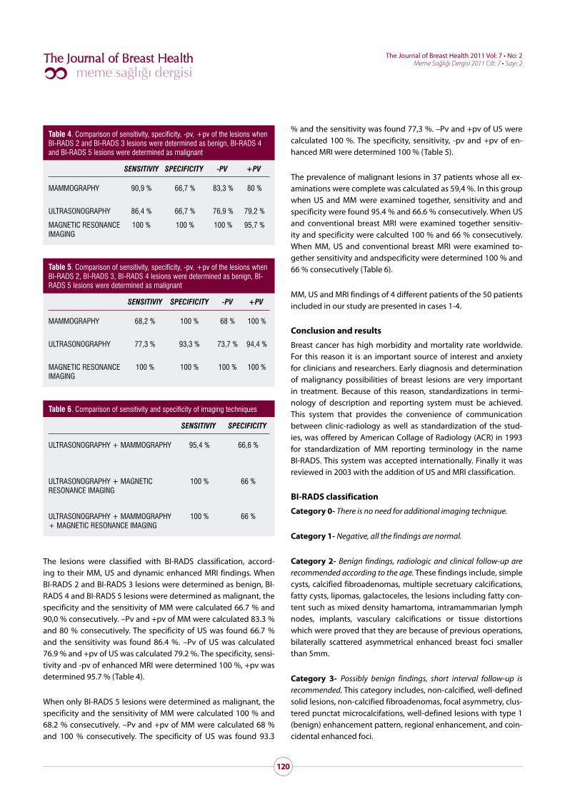
Table 4. Comparison of sensitivity, specificity, -pv, +pv of the lesions when BI-RADS 2 and BI-RADS 3 lesions were determined as benign, BI-RADS 4 and BI-RADS 5 lesions were determined as malignant
SENSITIVIY SPECIFICITY -PV +PV
MAMMOGRAPHY 90,9 % 66,7 % 83,3 % 80 %
ULTRASONOGRAPHY 86,4 % 66,7 % 76,9 % 79,2 %
MAGNETIC RESONANCE IMAGING
100 % 100 % 100 % 95,7 %
Table 5. Comparison of sensitivity, specificity, -pv, +pv of the lesions when BI-RADS 2, BI-RADS 3, BI-RADS 4 lesions were determined as benign, BI-RADS 5 lesions were determined as malignant
SENSITIVIY SPECIFICITY -PV +PV
MAMMOGRAPHY 68,2 % 100 % 68 % 100 %
ULTRASONOGRAPHY 77,3 % 93,3 % 73,7 % 94,4 %
MAGNETIC RESONANCE IMAGING
100 % 100 % 100 % 100 %
Table 6. Comparison of sensitivity and specificity of imaging techniques
SENSITIVIY SPECIFICITY
ULTRASONOGRAPHY + MAMMOGRAPHY 95,4 % 66,6 %
ULTRASONOGRAPHY + MAGNETIC RESONANCE IMAGING
100 % 66 %
ULTRASONOGRAPHY + MAMMOGRAPHY + MAGNETIC RESONANCE IMAGING
100 % 66 %
121
The Journal of Breast Health 2011 Vol: 7 • No: 2 Meme Sağlığı Dergisi 2011 Cilt: 7 • Sayı: 2
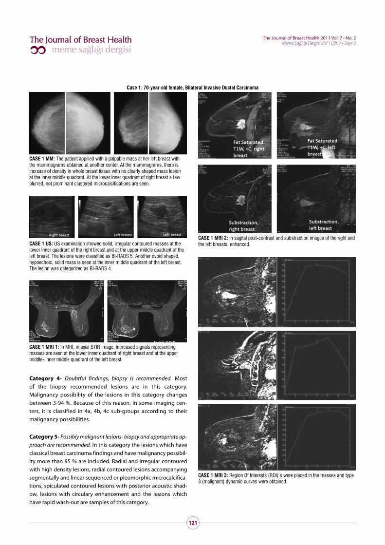
Case 1: 70-year-old female, Bilateral Invasive Ductal Carcinoma
CASE 1 MM: The patient appilied with a palpable mass at her left breast with the mammograms obtained at another center. At the mammograms, there is increase of density in whole breast tissue with no clearly shaped mass lesion at the inner middle quadrant. At the lower inner quadrant of right breast a few blurred, not prominant clustered microcalcifications are seen.
CASE 1 US: US examination showed solid, irregular contoured masses at the lower inner quadrant of the right breast and at the upper middle quadrant of the left breast. The lesions were classified as BI-RADS 5. Another ovoid shaped, hypoechoic, solid mass is seen at the inner middle quadrant of the left breast. The lesion was categorized as BI-RADS 4.
CASE 1 MRI 1: In MRI, in axial STIR image, increased signals representing masses are seen at the lower inner quadrant of right breast and at the upper middle- inner middle quadrant of the left breast.
CASE 1 MRI 2: In sagital post-contrast and substraction images of the right and the left breasts, enhanced.
CASE 1 MRI 3: Region Of Interests (ROI)’s were placed in the masses and type 3 (malignant) dynamic curves were obtained.
Category 4- Doubtful findings, biopsy is recommended. Most of the biopsy recommended lesions are in this category. Malignancy possibility of the lesions in this category changes between 3-94 %. Because of this reason, in some imaging cen-ters, it is classified in 4a, 4b, 4c sub-groups according to their malignancy possibilities.
Category 5- Possibly malignant lesions- biopsy and appropriate ap-proach are recommended. In this category the lesions which have classical breast carcinoma findings and have malignancy possibil-ity more than 95 % are included. Radial and irregular contoured with high density lesions, radial contoured lesions accompanying segmentally and linear sequenced or pleomorphic microcalcifica-tions, spiculated contoured lesions with posterior acoustic shad-ow, lesions with circulary enhancement and the lesions which have rapid wash-out are samples of this category.
122
The Journal of Breast Health 2011 Vol: 7 • No: 2 Meme Sağlığı Dergisi 2011 Cilt: 7 • Sayı: 2
that the proportion of lesion determinations in breasts with high density has increased (7). Also, with the useage of high resolu-tion probes in US, with the developments in spatial and temporal resolution in breast MRI, the sensitivity of diagnostic techniques has risen (8, 9). It is reported that, breast MRI is a problem solving method when MM and breast US techniques remain insufficient for the diagnosis (10).
MM is the most effective method in the approach of breast dis-ease investigations. Other diagnostic methods are used as com-plementary to MM when it is needed. The usage of MM is for two different purposes; diagnosis and malignancy screening. In fact,
Category 6- Known malignancy- appropriate approach is needed. This category is added for the lesions which the malignancy of these lesions proved by biopsy before the exact treatments such as surgical excision, radiotherapy, chemotherapy or mastectomy. This category is different from BI-RADS category 4 and category 5 and it is not concerned with interventional techniques which are done in order to prove malignancy. This category is appropri-ate for secondary diagnosis findings before biopsy and in order to follow the answer of neoadjuvant chemotherapy before surgical excision. It should not be used after malignancy excision (lumpec-tomy) (4).
In recent years, with development of techniques in MM, breast US and breast MRI, the proportion of malignancy determination has risen. With the increase of these early diagnostic techniques, al-ternative surgical methods to mastectomy have taken over (5, 6). After the digital MM has been put in to routine use, it is reported
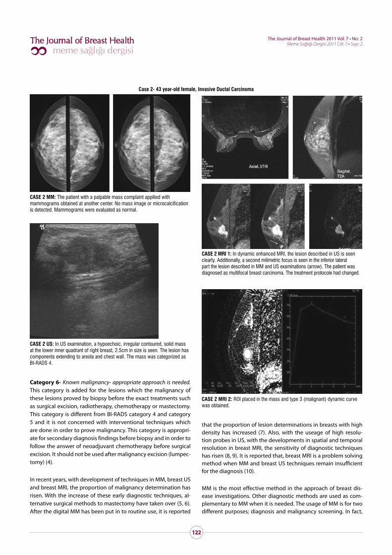
Case 2- 43 year-old female, Invasive Ductal Carcinoma
CASE 2 MM: The patient with a palpable mass complaint appilied with mammograms obtained at another center. No mass image or microcalcification is detected. Mammograms were evaluated as normal.
CASE 2 US: In US examination, a hypoechoic, irregular contoured, solid mass at the lower inner quadrant of right breast, 2.5cm in size is seen. The lesion has components extending to areola and chest wall. The mass was categorized as BI-RADS 4.
CASE 2 MRI 1: In dynamic enhanced MRI, the lesion described in US is seen clearly. Additionally, a second milimetric focus is seen in the inferior lateral part the lesion described in MM and US examinations (arrow). The patient was diagnosed as multifocal breast carcinoma. The treatment protocole had changed.
CASE 2 MRI 2: ROI placed in the mass and type 3 (malignant) dynamic curve was obtained.
123
The Journal of Breast Health 2011 Vol: 7 • No: 2 Meme Sağlığı Dergisi 2011 Cilt: 7 • Sayı: 2
breast MRI application, the sensitivity varies between 83-100 % and the specificity varies between 29-100 % (18- 20). Breast MRI has disadvantages such as long examination time. Moreover, dif-ficulties can be seen in characterization of lesion determination from time to time. The criteria of conventional breast MRI evalua-tion include lesion morphologies, enhancement dynamics of the lesions and T2 signal features. As the findings in benign and ma-lignant lesions overlap, it is reported that the specificity of MRI can decrease to 40-80 % (23- 25). Recently, enhanced dynamic breast MRI is accepted as a very sensitive imaging method in breast can-cer diagnosis. In selected cases, it is offered as a complementary
the specificity of MM is low and biopsy is needed in order to make discrimination between benign and malignant lesions which have determined by MM. Only 10-35 % of the biopsies that are done according to mammographic findings yielded the final diagnosis of malignancy (11).
The usage of gray scale US additional to MM, especially after the developments in US technology, helps in solid-cystic lesion dis-crimination, guides aspiration biopsies and also helps to discrimi-nate benign and malignant lesions (12, 13).
MRI has been started to use from the outset of 1980’s. In the first applications, longitudinal relaxation time (T1), transverse relax-ation time (T2) and hydrogen spin densities were used (14- 17). MRI should be done necessarily with the application of contrast material containing gadopentetate dimeglumine (Gd DTPA). By this way, it provides high sensitivity in diagnosis of breast cancer and lesion discrimination. Enhanced breast MRI is used in the con-trol and diagnosis of breast cancer for more than 15 years (1- 3, 18-22). According to the technical and diagnostic criterions in
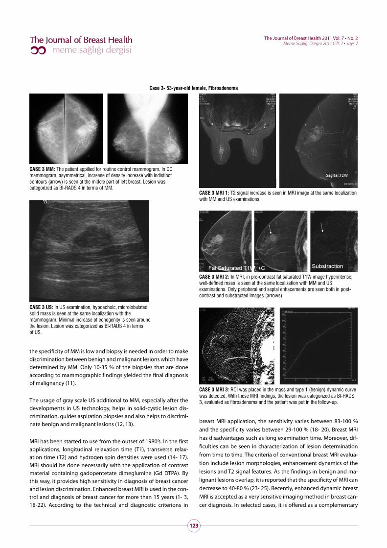
Case 3- 53-year-old female, Fibroadenoma
CASE 3 MM: The patient appilied for routine control mammogram. In CC mammogram, asymmetrical, increase of density increase with indistinct contours (arrow) is seen at the middle part of left breast. Lesion was categorized as BI-RADS 4 in terms of MM.
CASE 3 US: In US examination, hypoechoic, microlobulated solid mass is seen at the same localization with the mammogram. Minimal increase of echogenity is seen around the lesion. Lesion was categorized as BI-RADS 4 in terms of US.
CASE 3 MRI 1: T2 signal increase is seen in MRI image at the same localization with MM and US examinations.
CASE 3 MRI 2: In MRI, in pre-contrast fat saturated T1W image hyperintense, well-defined mass is seen at the same localization with MM and US examinations. Only peripheral and septal enhacements are seen both in post-contrast and substracted images (arrows).
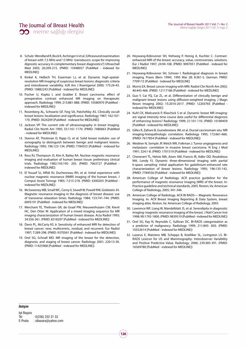
CASE 3 MRI 3: ROI was placed in the mass and type 1 (benign) dynamic curve was detected. With these MRI findings, the lesion was categorized as BI-RADS 3, evaluated as fibroadenoma and the patient was put in the follow-up.

124
The Journal of Breast Health 2011 Vol: 7 • No: 2 Meme Sağlığı Dergisi 2011 Cilt: 7 • Sayı: 2
7. In determination of post-op scar tissues and possible residual that is difficult to evaluate by MM and US.
8. Evaluation of cases with high malignancy possibility.
9. In control of cases with high recurrent malignancy possibility.
10. In preoperative tumor staging.
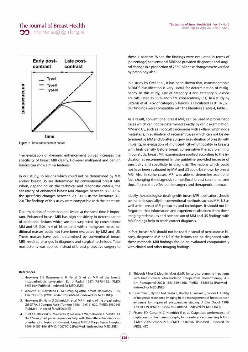
Cinetic curves (time-signal intensity curves) show the changes of intensities of the tissue by time as a reflection of enhancement. Cinetic curves are evaluated in two stages: in the early stage after bolus injection (first 2-3 min) the speed of signal increase and in the late stage, the changes of signal intensity. In the early stage, the speed of signal increase can be slow, moderate and fast. In the late stage, the signal intensity can continue to increase (persis-tant- type 1- benign type), can stay at the same level (plateau- type 2- doubtful for malignancy type) or can decrease (wash-out- type 3- malignant type) (24, 29). In malignant lesions, signal intensity mostly increases to its double level of beginning level in 90- 120 sec or more. In 3- 5 min., contrast material wash-out can be seen (24). The curves are shown in Figure 1.
diagnostic modality (26, 27). When MRI is used together with MM and US, it is a very powerful method in imaging and characteriza-tion of breast masses.
ACR has determined the indications of enhanced breast MRI (28). In all these indications except for the evaluation of the silicon im-planted breasts with the doubt of rupture, enhanced MRI is used. The indications of enhanced breast MRI are;
1. In determination of breast lesions which can not be evaluated by MM, US, physical examination separately or together with all these methods.
2. In neoadjuvant chemotherapy applied cases.
3. In determination of cases who have malignant axillary lymph nodes and whose breast lesions can not be diagnosed by MM, US and physical examination.
4. In determination of the cases with post-operative reconstruction.
5. In determination of the cases with doubtful deep pectoral fa-sia invasion.
6. In determination of contraterally breast lesion.
Case 4- 39-year-old female, Invasive Lobulary Carcinoma-Invasive Ductal Carcinoma
CASE 4 MM: The patient applied with a palpable mass and skin changes of the breast. In MM, in the right breast there is increase of density at whole breast except the lower inner quadrant.
CASE 4 US: In US examination, irregulary contoured, hypoechoic, solid masses with posterior acoustic shadow are seen at the right breast. Masses were categorized as BI-RADS 5.
CASE 4 MRI 1: MRI examination was done in order to evaluate the masses and contrlateral breast. In MRI images, diffuse signal increase and mass enhancement with chest wall invasion are seen at the right breast. Type 3 (malignant type) curve was drawn from the foci of the mass. The patient was diagnosed as stage 4 breast carcinoma. Tru-cut biyopsy was appilied under US guidence. Pathologic result was invasive lobulary carcinoma-invasive ductal carcinoma. The patient was referred to onchology clinic for chemotherapy treatment.
CASE 4 MRI 2: After neoadjuvant chemotherapy, MRI was repeated. In MRI images, the decrease in size of the lesion and the regression of chest wall invasion are seen. The patient was referred to surgery.
125
The Journal of Breast Health 2011 Vol: 7 • No: 2 Meme Sağlığı Dergisi 2011 Cilt: 7 • Sayı: 2
these 4 patients. When the findings were evaluated in terms of ‘percentage’, conventional MRI had provided diagnostic and surgi-cal change in a proportion of 25 %. All these changes were verified by pathology also.
In a study by Orel et al., it has been shown that, mammographic BI-RADS classification is very useful for determination of malig-nancy. In this study, +pv of category 4 and category 5 lesions are calculated as 30 % and 97 % consecutively (31). In a study by Lazarus et al., +pv of category 5 lesions is calculated as 91 % (32). Our findings were compatible with the literature (Table 4, Table 5).
As a result, conventional breast MRI; can be used in problematic cases which can not be determined exactly by clinic examination, MM and US, such as in occult carcinomas with axillary lymph node metastasis, in evaluation of recurrent cases which can not be de-termined by MM and US after surgery, in evaluation of breasts with implants, in evaluation of multicentricity-multifocality in breasts with high density before breast conservative therapy planning. In our study, breast MRI examination applied according to the in-dication as recommended in the guideline provided increase of sensitivity and specificity in diagnosis. The lesions which could not have been evaluated by MM and US could be shown by breast MRI. Also in some cases, MRI was able to determine additional foci, changing the diagnosis to multifocal breast carcinoma, and thuseffected thus effected the surgery and therapeutic approach.
Ideally the radiologists dealing with breast MRI application, should be trained especially for conventional methods such as MM, US as well as for breast MRI protocols and techniques. It should not be forgotten that information and experiences obtained from these imaging techniques and comparison of MM and US findings with MRI findings help to reach correct diagnosis.
In fact, breast MRI should not be used in stead of percutaneus bi-opsy, diagnostic MM or US if the lesions can be diagnosed with these methods. MRI findings should be evaluated comparatively with clinical and other imaging findings.
The evaluation of dynamic enhancement curves increases the specificity of breast MRI clearly. However malignant and benign lesions can show similar features.
In our study, 15 lesions which could not be determined by MM and/or breast US are determined by conventional breast MRI. When, depending on the technical and diagnostic criteria, the sensitivity of enhanced breast MRI changes between 83-100 %, the specificity changes between 29-100 % in the literature (18- 20). The findings of this study were compatible with the literature.
Determination of more than one lesion at the same time is impor-tant. Enhanced breast MRI has high sensitivity in determination of additional lesions which are not suspected by conventional MM and US (30). In 4 of 16 patients with a malignant mass, ad-ditional masses could not have been evaluated by MM and US. These masses have been determined by conventional breast MRI, resulted changes in diagnosis and surgical technique. Total mastectomy was applied instead of breast protective surgery to
Figure 1. Time-enhancement curves
References
1. Heywang SH, Bassermann R, Fenzl G, et al. MRI of the breast: histopathologic correlation. Eur J Radiol 1987; 7:175-182. (PMID: 3653109 [PubMed - indexed for MEDLINE])
2. Weinreb JC, Newstead G. MR imaging ofthe breast. Radiology 1995; 196:593- 610. (PMID: 7644617 [PubMed - indexed for MEDLINE])
3. Heywang SH, Hahn D, Schmidt H, et al. MR imaging of the breast using Gd-DTPA. J Comput Assist Tomogr 1986; 10:615- 620. (PMID: 3950145 [PubMed - indexed for MEDLINE])
4. Kuhl CK, Klaschik S, Mielcarek P, Gieseke J, Wardelmann E, Schild HH. Do T2-weighted pulse sequences help with the differential diagnosis of enhancing lesions in dynamic breast MRI? J Magn Reson Imaging 1999; 9:187-196. (PMID: 10077012 [PubMed - indexed for MEDLINE])
5. Thibault F, Nos C, Meunier M, et al. MRI for surgical planning in patients with breast cancer who undergo preoperative chemotherapy. AJR Am Roentgenol 2004; 183:1159-1168. (PMID: 15385323 [PubMed - indexed for MEDLINE])
6. Esserman L, Hylton NM, Yassa L, Barclay J, Frankel S, Sickles E. Utilitiy of magnetic resonance imaging in the management of breast cancer: evidence for improved preoperative staging. J Clin Oncol 1999; 17:110-119. (PMID: 10458224 [PubMed - indexed for MEDLINE])
7. Pisano ED, Gatsonis C, Hendrick E et al. Diagnostic performance of digital versus film mammography for breast-cancer screening. N Engl J Med 2005; 26:209-215. (PMID: 16169887 [PubMed - indexed for MEDLINE])
126
The Journal of Breast Health 2011 Vol: 7 • No: 2 Meme Sağlığı Dergisi 2011 Cilt: 7 • Sayı: 2
8. Schulz- Wendland R, Bock K, Aichinger U et al. [Ultrasound examination of breast with 7,5 MHz and 13 MHz- transducers: scope for improving dignostic accuracy in complementary breast diagnostics?] Ultraschall Med 2005; 26:209-215. (PMID: 15948057 [PubMed - indexed for MEDLINE])
9. Kinkel K, Helbich TH, Esserman LJ, et al. Dynamic high-spatial-resolution MR imaging of suspicious breast lesions: diagnostic criteria and interobsever variability. AJR Am J Roentgenol 2000; 175:24-43. (PMID: 10882243 [PubMed - indexed for MEDLINE])
10. Fischer U, Kopka L and Grabbe E. Breast carcinoma: effect of preoperative contrast enhanced MR imaging on therapeutic approach. Radiology 1999; 213:881-888. (PMID: 10580970 [PubMed - indexed for MEDLINE])
11. Rosenberg AL, Schwartz GF, Feig SA, Patchefsky AS. Clinically occult breast lesions: localization and significance. Radiology 1987; 162:167-170. (PMID: 3024209 [PubMed - indexed for MEDLINE])
12. Jackson VP. The current role of ultrasonography in breast imaging. Radiol Clin North Am 1995; 33:1161-1170. (PMID: 7480663 [PubMed - indexed for MEDLINE])
13. Stavros AT, Thickman D, Rapp CL et al. Solid breast nodules: use of sonography to distinguish between benign and malignant lesions. Radiology 1995; 196:123-134. (PMID: 7784555 [PubMed - indexed for MEDLINE])
14. Ross RJ, Thompson JS, Kim K, Bailey RA. Nuclear magnetic resonance imaging and evaluation of human breast tissue: preliminary clinical trials. Radiology 1982;143:195- 205. (PMID: 7063727 [PubMed - indexed for MEDLINE])
15. El Yousef SJ, Alfidi RJ, Duchesneau RH, et al. Initial experience with nuclear magnetic resonance (NMR) imaging of the human breast. J Comput Assist Tomogr 1983; 7:215-218. (PMID: 6300203 [PubMed - indexed for MEDLINE])
16. McSweeney MB, Small WC, Cerny V, Sewell W, Powell RW, Goldstein JH. Magnetic resonance imaging in the diagnosis of breast disease: use of transverse relaxation times. Radiology 1984; 153:741-744. (PMID: 6093191 [PubMed - indexed for MEDLINE])
17. Merchant TE, Thelissen GR, de Graaf PW, Nieuwenhuizen CW, Kievit HC, Den Otter W. Application of a mixed imaging sequence for MR imaging characterization of human breast disease. Acta Radiol 1993; 34:356-361. (PMID: 8318297 [PubMed - indexed for MEDLINE])
18. Davis PL, McCarty KS Jr. Sensitivity of enhanced MRI for detection of breast cancer: new, multicentric, residual, and recurrent. Eur Radiol 1997; 7:289-298. (PMID: 9370561 [PubMed - indexed for MEDLINE])
19. Orel SG, Schnall MD. MR imaging of the breast for the detection, diagnosis, and staging of breast cancer. Radiology 2001; 220:13-30. (PMID: 11425968 [PubMed - indexed for MEDLINE])
20. Heywang-Köbrunner SH, Viehweg P, Heinig A, Kuchler C. Contrast-enhanced MRI of the breast: accuracy, value, controversies, solutions. Eur J Radiol 1997; 24:94-108. (PMID: 9097051 [PubMed - indexed for MEDLINE])
21. Heywang-Köbrunner SH, Schreer I. Radiological diagnosis in breast imaging. Praxis (Bern 1994). 1995 Mar 28; 8:361-5. German. PMID: 7709115 [PubMed - indexed for MEDLINE]
22. Morris EA. Breast cancer imaging with MRI. Radiol Clin North Am 2002; 40:443-466. (PMID: 12117186 [PubMed - indexed for MEDLINE])
23. Guo Y, Cai YQ, Cai ZL, et al. Differentiation of clinically benign and malignant breast lesions using diffusion-weighted imaging. J Magn Reson Imaging 2002; 15:2010-2017. (PMID: 12203765 [PubMed - indexed for MEDLINE])
24. Kuhl CK, Mielcareck P, Klaschick S et al. Dynamic breast MR imaging: are signal intensity time course data useful for differential diagnosis of enhancing lesions? Radiology 1999; 21:101-110. (PMID: 10189459 [PubMed - indexed for MEDLINE])
25. Gilles R, Zafrani B, Guinebretiere JM, et al. Ductal carcinomain situ: MR imaging-histopathologic correlation. Radiology 1995; 172:661-666. (PMID: 7617854 [PubMed - indexed for MEDLINE])
26. Weidner N, Semple JP, Welch WR, Folkman J. Tumor angiogenesis and metastasis- correlation in invasive breast carcinoma. N Eng J Med 1991; 324:1-8. (PMID: 1701519 [PubMed - indexed for MEDLINE])
27. Chenevert TL, Helvie MA, Aisen AM, Francis IR, Adler DD, Roubidoux MA, Londy FJ. Dynamic three-dimensional imaging with partial k-space sampling: initial application for gadolinium-enhanced rate characterization of breast lesions. Radiology 1995; 196:135-142. (PMID: 7784556 [PubMed - indexed for MEDLINE])
28. American College of Radiology. ACR practice guideline for the performance of magnetic resonance imaging (MRI) of the breast. In: Practice guideline and technical standards, 2005. Reston, Va: American College of Radiology, 2005; 341-346.
29. American College of Radiology. ACR BI-RADS— Magnetic Resonance Imaging. In: ACR Breast Imaging Reporting & Data System, breast imaging atlas. Reston, Va: American College of Radiology, 2003.
30. Lawrence WF, Liang W, Mandelblatt JS, et al. Serendipity in diagnostic imaging: magnetic resonance imaging of the breast. J Natl Cancer Inst 1998; 90:1792-1800. (PMID: 9839519 [PubMed - indexed for MEDLINE])
31. Orel SG, Kay N, Reynolds C, Sullivan DC. BI-RADS categorization as a predictor of malignancy. Radiology 1999; 211:845- 850. (PMID: 10352614 [PubMed - indexed for MEDLINE])
32. Lazarus E, Mainiero MB, Schepps B, Koelliker SL, Livingston LS. BI-RADS Lexicon for US and Mammography: Interobserver Variability and Positive Predictive Value. Radiology, 2006; 239:385-391. (PMID: 16569780 [PubMed - indexed for MEDLINE])
İletişim
Işıl BaşaraTel : 0(236) 232 31 33 E-Posta : [email protected]