detection of human cytomegalovirus in motile spermatozoa and spermatogenic cells in testis...
TRANSCRIPT
RESEARCH Open Access
Detection of human cytomegalovirus in motilespermatozoa and spermatogenic cells in testisorganotypic cultureVictor A Naumenko1*, Yurii A Tyulenev1, Sergei A Yakovenko2, Lubov’ F Kurilo3, Ludmila V Shileyko3,Aleksander S Segal4, Larisa E Zavalishina5, Regina R Klimova1, Anton S Tsibizov1, Sergei V Alkhovskii1 andAlla A Kushch1
Abstract
Background: The presence of human cytomegalovirus (HCMV) in male genital tract suggests its verticaltransmission with spermatozoa and the development of a potentially dangerous fetal infection. The objective ofthe present study was to evaluate the possibility of intracellular HCMV localization in male germ cells and toexamine the effect of the virus on human spermatogenesis.
Methods: Semen samples from 91 infertile and 47 fertile men were analyzed. HCMV was detected by real timePCR, rapid culture method and PCR in situ. Human testis organotypic culture and quantitative karyological analysiswere used to investigate viral effects on spermatogenesis. Localization of HCMV in immature germ cells andspermatozoa was studied by immunostaining with monoclonal antibodies and ultrastructural analysis of infectedorganotypic culture.
Results: Viral DNA was detected in 12.3% samples of motile spermatozoa, while infectious activity only in 2.9%infertile and fertile men without statistically significant intergroup difference. According to PCR in situ, the meanpercentage of infected cell in both groups was 1.5% (0.25%-15%), which can serve as a criterion for evaluatingthe risk of HCMV transmission. In HCMV-infected organotypic culture viral antigens were identified inspermatides on day 4, in spermatogonia and spermatocytes on day 8, and in spermatozoa on day 14. Emptyand full capsides and virions were visualized in germ cells by electron microscopy. The number of cells beforeintroduction in culture was taken for 100%. On day 14 infected culture contained 36.8% spermatogonia, 18.7%spermatocytes, 27.6% round spermatides and 42.5% elongated spermatides; in comparison with 82.2%, 51.5%,70.4% and 65.7% in uninfected culture, respectively (all p < 0.05). There were no changes in the number andviability of spermatozoa.
Conclusions: HCMV was detected in male germ cells, both in sperm samples and in testis organotypic culture. Thevirus may infect immature germ cells which develop to mature HCMV-carrying spermatozoa. A considerabledecrease in the number of immature germ cells indicates that HCMV produces a direct gametotoxic effect and cancontribute to male infertility.
Keywords: human cytomegalovirus, infertility, spermatogenesis, testis organotypic culture
* Correspondence: [email protected] D. I. Ivanovsky Institute of Virology, Ministry of Health and SocialDevelopment of the Russian Federation, 123098 Gamaleya str. 16, Moscow,RussiaFull list of author information is available at the end of the article
Naumenko et al. Herpesviridae 2011, 2:7http://www.herpesviridae.org/content/2/1/7
© 2011 Naumenko et al; licensee BioMed Central Ltd. This is an Open Access article distributed under the terms of the CreativeCommons Attribution License (http://creativecommons.org/licenses/by/2.0), which permits unrestricted use, distribution, andreproduction in any medium, provided the original work is properly cited.
BackgroundMale infertility accounts for 20-50% infertile couples and isoften associated with genital infections [1]. Negative effectson reproductive function have been proposed for suchviruses as the human immunodeficiency virus, humanpapillomavirus, herpes simplex virus, Epstein-Barr virus[1-4]. Human cytomegalovirus (HCMV) is widespread inhuman population and can be transmitted sexually. Theeffects of HCMV on spermatogenesis and its vertical trans-mission with sperm cells have not been investigated in suf-ficient detail due to the low detection rate of HCMV insemen - no more than 2.9% by the culture method [5,6]and 1.4-8.7% by PCR [5,7,8]. At the same time Neofytou etal. have detected HCMV DNA by PCR in the semen of56.9% asymptomatic fertile and infertile patients [9].There is a controversy over the effects of HCMV on
the major parameters of sperm quality - concentration,motility and morphology of gametes. A correlationbetween high concentration of HCMV in ejaculate anda transient decrease in the spermatozoa motility havebeen established [10], and the concentration of spermcells has been found to decrease in patients with HCMVin semen [11]. However, the majority of researchershave found no HCMV effect on sperm quality [3,7,12].The issue of intragamete localization of HCMV is
open to debate. The attempts to infect spermatozoa invitro have been unsuccessful [13]. Investigation of inter-actions between HCMV and testicular cells is hamperedby the possibility of autoimmune orchitis after biopsy.High species-specificity of HCMV impedes the investi-gation of processes occurring in human organism onanimal models. In an attempt to overcome these diffi-culties we developed the model of HCMV infection inan organotypic culture of human testis. Using thismodel we demonstrated the possibility of intracellularHCMV localization in immature and mature male germcells and HCMV influence on spermatogenesis.
Materials and methodsPatientsSemen samples were obtained from 138 men, including91 infertile men (Group I) and 47 healthy donorsenrolled in the sperm donor program (Group II). Theinformed consent was obtained from all patients.
Clinical materialSemen samples were fractionated by gradient centrifuga-tion with SupraSperm reagent (Origio, Jyllinge, Den-mark) according to the World Health OrganizationLaboratory Manual for the Examination and Processingof Human Semen (2010). The fraction of motile sperma-tozoa (MS) was washed two times in 2 ml of Dulbeccomodified Eagle Medium (DMEM; Paneko, Moscow, Rus-sia) by centrifugation and used as described below.
Virus and cell cultureHCMV AD 169 strain was provided by the Russian Fed-eration State Collection of Viruses. The virus was propa-gated and titrated in human embryo lung fibroblasts(HEF).
Rapid culture method (RCM)RCM was used for detection of HCMV infectious activ-ity in the samples. The material (0.2 ml) was injectedinto each well of 24-well culture plate (Costar, Washing-ton DC, USA) with HEF confluent monolayer, incubatedfor 1 h at 37°C in an atmosphere of 95% air/5% CO2.The cells were washed 2 times in serum-free culturemedium, incubated for 48 h in 1 ml of DMEM with 2%fetal calf serum (FCS; Gibco, Carlsbad, CA, USA),washed 2 times in PBS and fixed in cold methanol.HCMV was identified by immunoperoxidase stainingwith monoclonal antibody (Mab) against HCMV pp65protein (DAKO, Glostrup, Denmark). Immunolabeledcells were calculated in an inverse light microscopeLABOVEWRT FS (Leitz, Oberkochen, Germany).
HCMV DNA detection and quantification by real-time PCRHCMV DNA extraction was performed from 200 μl ofsamples using QIAamp DNA mini kit (QIAGEN, Hilden,Germany) according to the manufacturer’s protocol. Inbrief, 200 μl of sample and 10 μl of the STI-87 positiveinternal control (PIC; Interlabservice, Moscow, Russia)were added to 200 μl of AL-buffer and heated at 56°C for15 min. 200 μl of 96% ethanol was added, applied to col-umns and washed as per the manufacturer’s instructionwith the final elution in 200 μl of the kit AE-buffer pre-heated to 50°C. Real-time PCR was performed using theAmplisense CMV-screen/monitor-FL kit (Interlabservice)according to the manufacturer’s protocol. Real-time ampli-fication was carried out using 10 μl DNA eluate combinedwith 10 μl PCR-mix-1-FL and 5 μl PCR-mix-2-FL usingRotor-Gene 6000 Instrument (Corbett Research, Doncas-ter, Australia) with the following cycling parameters: pre-denaturation at 95°C for 15 min, 95°C for 5 s, 60°C for 20s and 72°C for 15 s for 45 cycles. Data acquisition was per-formed in both JOE/Yellow (for HCMV DNA) and ROX/Orange (for the STI-87 PIC) channels during the anneal-ing (60°C) stage. For quantification of HCMV DNA twostandard positive sample KSG1 (104 copies per reactionmixture) and KGS2 (102 copies per reaction mixture)(Interlabservice) were included in the run. Calculations ofCt, preparation of standard curve and quantification ofDNA in each sample were performed by Rotor-GeneOperating Software, version 1.8 (Corbett Research).
PCR in situWashed-off MS were transferred to glass slides, centri-fuged for 5 min at 1500 rpm in Cytospin 4 (Thermo
Naumenko et al. Herpesviridae 2011, 2:7http://www.herpesviridae.org/content/2/1/7
Page 2 of 8
Electron, Waltham, USA), air dried, fixed in 10% formal-dehyde for 4 h and washed twice in 0.05 M Tris-HCl.The preparations were then incubated with proteinase K(DAKO) for 30 min at 37°C. Amplification with biotini-lated primers (Gentech, Moscow, Russia) was carriedout using T1 cycler (Biometra, Goettingen, Germany).Viral DNA was detected with the biotin-streptavidin-peroxidase complex (DAKO) and diaminobenzidene(Sigma-Aldrich, St Louis, MO, USA). The proportion ofsperm cells containing HCMV DNA was calculated afteranalysis of at least 2000 cells.
Spermiological and quantitative karyological analysisSpermiological analysis was performed according to theWorld Health Organization Laboratory Manual for theExamination and Processing of Human Semen (2010).The immature germ cells (IGC) in sperm samples wereidentified by morphological criterions under a lightmicroscope BX51 (Olympus, Tokyo, Japan). At least200-300 IGC were calculated in each slide. The propor-tions of spermatids and primary spermatocytes at earlystages (leptotene, zygotene, pachytene and diplotene)and cells that could not be identified (classified as uni-dentified and/or degenerated) were calculated asdescribed previously [14].
Organotypic culture of human testis explantsThe procedures followed were in accordance with ethi-cal standards of the Helsinki Declaration and wereapproved by the local Ethics Committee of the D.I. Iva-novsky Institute of Virology of Ministry of Health andSocial Development of Russian Federation; informedconsent was obtained from all patients. Testis samplesfrom 3 patients with prostate cancer (62, 65 and 67years old) were transported in fresh medium on iceimmediately following orchidectomy. Testicular tissueswere carefully dissected with scissors into 3 mm3 frag-ments. In each well of a six-well plate, two fragmentswere placed onto a permeable membrane insert (FalconLabware, Mt Pritchard, NSW, Australia) and incubatedat the interface between air and 2 ml of DMEM with10% FCS (Gibco), 1 mmol/l sodium piruvate, 100 ng/mlvitamin A, 50 ng/ml vitamin C and 200 ng/ml vitamin E(all from Sigma-Aldrich), 4 mmol/l glutamine, 10 μg/mlinsulin, 5 μg/ml transferrin and 50 μg/ml gentamycin(all from Paneko) at 37°C in an atmosphere of 95% air/5% CO2. Culture medium was replaced every other day.
HCMV-infection of testicular explantsFragments were incubated with 0.025 ml of HCMVinoculate for 1 h at 37°C. Multiplicity of infection(MOI) was 0.0001-0.001 plaque forming units (PFU) percell. Control uninfected cultures were incubated inDMEM under the same conditions. Then explants were
washed three times in 1 ml DMEM and the culture wasestablished for up to 14 days as described above. Viralload was estimated every other day in culture mediumby PCR and RCM starting from Day 2. Three explantswere analyzed in each point.
Light microscopyFor histological analysis, testicular explants (3 mm3)were fixed in neutral buffered 10% formaldehyde for 24h at 4°C, dehydrated in a series of graduated ethanolconcentrations (70%, 96% and 100%), embedded in par-affin, sectioned at 4.0 μm and stained with Caracci’shematoxylin (BisVitrum, S.-Peterburg, Russia) for exami-nation. The following morphological criteria were usedfor analysis of germ cells viability and explants architec-ture: cell number, cell size and location, signs of apopto-sis, and histological characteristics of seminiferoustubules, including the basal membrane structure.
ImmunostainingTo reveal HCMV proteins in the testis explants immu-nostaining with Mab to HCMV pp65 was performed onformaldehyde-fixed, paraffin-embedded tissues. Antigenretrieval was performed as follows: deparaffinized anddehydrated sections were treated for 20 min at 750 Wtin microwave oven (Sanyo, Moriguchi, Osaka, Japan) in10 mM citrate buffer (pH = 6.0, DAKO) and thenwashed in 0.05 mol/l phosphate-buffer saline (PBS, pH7.6; Gibco). Endogenous peroxidase was inactivated indeparaffinized sections by 5-min treatment in PBS with3% H2O2. Slides were processed by PBS supplementedwith 2% bovine serum albumin (BSA, Sigma-Aldrich) toblock the non-specific sites, before overnight incubationat 4°C with the Mab to HCMV pp65 (0.02 ug/ml)(DAKO) diluted in PBS with 1% BSA. Next steps wereperformed at room temperature. Reagents from UltraVi-sion LP Large Volume Detection System kit (ThermoScientific, Fremont, USA) were added according to themanufacturer’s protocol after washing 4 times in PBS.The following substrate was used: 0.5 mg/ml diamino-benzidine (Sigma-Aldrich) in 0.05M TRIS-HCL, pH 8.0with 3% H2O2. Sections were prepared immediatelyafter tissue dissection and on days 2, 4, 7 and 14 afterintroducing in culture. Stained cells were identified andphotographed with a BX51 microscope coupled to adigital macro camera U-CMAD3 (Olympus).
Transmission electron microscopy (TEM)TEM was performed immediately after tissue dissectionand on days 2, 4, 7 and 14 of culturing. Two infectedand two uninfected explants were analyzed in eachpoint. The explants were fixed in 2.5% glutaraldehyde in0.1 M buffered sodium cacodylate (pH 7.4) for 24 h at4°C, postfixed in 1% OsO4 in 0.1 M sodium cacodylate
Naumenko et al. Herpesviridae 2011, 2:7http://www.herpesviridae.org/content/2/1/7
Page 3 of 8

buffer at room temperature for 40 min, then dehydratedin gradual ethanol concentrations (70%, 96% and 100%)and, subsequently, submitted to progressive impregna-tions in epon resin (Sigma-Aldrich). Polymerization wascarried out at 60°C for 48 h. Ultrathin sections were cutin an ultratome III (LKB, Bromma, Sweden), stainedwith uranyl acetate and lead citrate (Sigma-Aldrich) andexamined in a JEM-100 S electron microscope (JEOL,Tokyo, Japan) at 80 kV.
Statistics and data analysisThe analysis was performed in StatXact 8 (Cytel, Cam-bridge, MA, USA) using Student’s paired t-test, c2 testand Mann-Whitney test. P < 0.05 was considered statis-tically significant.
ResultsHCMV detection in motile spermatozoaWe have identified HCMV in the fraction of motilespermatozoa by several methods (Table 1). The fre-quency of HCMV DNA detection by PCR was higherthan frequency of infectious activity by RCM (p = 0.006)but without significant differences between groups irre-spective of method. The percentage of HCMV-infectedcells determined by PCR in situ was 1.7% in average(maximum 15%) among infertile men and 0.5% in aver-age (maximum 5%) among healthy men (Table 1). Inspite of a 3-fold increase in the number of infected cellsin Group I comparing with Group II, this difference wasnot significant (p > 0.05).
Spermiological and quantitative karyological analysis ofHCMV-infected spermAccording to the results of HCMV detection in sperm, allsamples were divided into two groups: with and withoutHCMV infection. Each group included infertile patientsand sperm donors. A comparative spermiological analysis
in these groups did not reveal any viral effect on the con-centration of sperm cells (50.6 × 10⁶ cells/ml vs. 69 × 106
cells/ml, p > 0.05), the percentage of motile spermatozoa(20% vs. 12.7%, p > 0.05) and morphologically normalgerm cells (9% vs. 20%, p > 0.05). Quantitative karyologi-cal investigation enabled us to assess viral effect on sper-matogenesis without invasive intervention. The numberof unidentified and/or degenerated germ cells in HCMV-infected semen samples was higher (p < 0.05) while thepopulation of spermatids was decreased (p < 0.05) com-paring with uninfected samples (Table 2).
HCMV-infection of testis organotypic cultureIn order to study the effect of HCMV on spermatogen-esis in more detail, we developed a model of HCMVinfection in the human testis organotypic culture. It wasdemonstrated in preliminary experiments that generalarchitecture of explants as well as viability of all germcells preserved at least up to 14 days in culture.Table 3 illustrates changes in HCMV markers which
reflect the dynamics of viral infection. Due to highHCMV content in the inoculate (4.2 × 10⁸ DNA copies/ml) the virus was not completely removed from theexplants during washing procedure: after 12 h viralDNA content in the culture medium was 1.7 × 10⁴
copies/ml. It gradually decreased within a 6-day periodand increased starting from Day 8, indicating HCMVreplication in the culture. Infectious activity reached themaximum on Day 12, while DNA HCMV being accu-mulated up to Day 14.
HCMV detection in testicular cellsOn Day 4, viral antigens were identified by immunos-taining in the interstitial cells (fibroblasts and Leydigcells) and in individual spermatids. Foci of infectionwere located in the superficial layers of explants whichcontacted with viral inoculate. By Day 7 HCMV spreadinto deeper layers and infected spermatocytes andspermatogonia were revealed. On Day 14, we observedfibroblasts with typical features of HCMV-infection:enlarged nuclei and huge inclusion bodies in cyto-plasm. At later stages of infection, typical HCMV pp65staining was detected in spermatids, spermatocytes,spermatogonia (Day 7) and individual spermatozoa(Day 14). Figure 1 illustrates the presence of HCMVprotein in spermatogonium adjacent to the basal mem-brane of the tubule and in two large round cells identi-fied as spermatocytes. Immunohistochemical data wereconfirmed by electron microscopy. A great number ofvirions contained empty and full capsides and elec-tron-dense bodies were identified in germ cells. Figure2 shows the spermatogonium with capsids in thenucleus and virions with typical herpes-virus morphol-ogy in transport vacuole.
Table 1 Detection of HCMV in the fraction of motilespermatozoa
Patients Frequency of HCMVdetection by
Percentage of spermatozoacontaining HCMV DNA byPCR in situ, median % [min,
max]
rapidculturemethod
PCR
Group I(Infertile)
3/91a(3.3%) 10/91 b (11%) 1.7 c [0.25; 15]
Group II(Fertile)
1/47 a (2.1%) 7/47 b (15%) 0.5 c [0.35; 5]
Groups Iand II
4/138 (2.9%) 17/138 (12.3%) 1.5 [0.25; 15]
a, b p > 0.05 (c2 test).c p > 0.05 (Mann-Whitney test).
Naumenko et al. Herpesviridae 2011, 2:7http://www.herpesviridae.org/content/2/1/7
Page 4 of 8

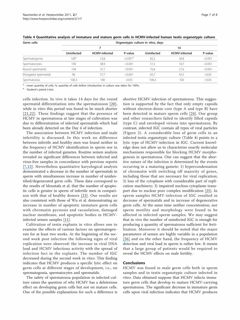
Decrease of the germ cells population in HCMV- infectedtestis explantsThe effect of HCMV on spermatogenesis was studiedhistologically by comparing of infected and uninfectedtestis explants at different time in culture. The resultsfor germ cell populations on Days 7 and 14 are sum-marized in Table 4. It was shown that the number ofspermatogonia, spermatocytes, round and elongatedspermatids decreased considerably starting from Day 7of infection. It should be noted that there were nochanges in the number of spermatozoa. By Day 14,changes in immature cell population were more pro-nounced, reflecting gradual destruction in testiculararchitecture, loosening and vacuolization of germinativeepithelium.
DiscussionThe possibility of vertical transmission of herpes viruseswith male gametes has been declared by several investi-gators [15,16]. In the first part of this work we studiedintracellular HCMV localization in male gametes as apotential transmission vector of infection. HCMV DNAwas found in 12.3% of sperm cells (mean for Groups Iand II), and infectious virus - in 2.9% of all cases. Thepercentage of infected cells reached 15% in infertilepatients and 5% in healthy donors, while the mean valuein both groups was found to be 1.5%.There is no direct data concerning the correlation of
HCMV infection of human spermatozoa and miscar-riages and fetal maldevelopment. The results obtained inanimal experiments are controversial. According toNeighbour et al. mouse CMV produced no effect on fer-tilization and embryogenesis in mice [16]. At the sametime inhibition of blastocyst formation after infection oftwo-cell embryos with mouse CMV was observed [17].There is evidence that herpes virus infection of malesplays a role in fetal loss in goats [18]. Statistical analysis
has been used to evaluate the role of herpes viruses inhuman reproduction. The frequency of herpes simplexvirus detection in sperm samples of partners of womenwith repeated miscarriages was higher than in the con-trol (p < 0.05) [19]. A correlation between the presenceof herpes viruses in ejaculate and negative outcome ofpregnancy can be regarded as an indirect evidence for avertical herpes virus transmission and associated preg-nancy loss. The percentage of gametes carrying HCMVmay serve as a criterion for estimation of the risk of ver-tical transmission of infection.The ability of the virus to replicate in male germ cells
was confirmed in the second part of the study using tes-tis organotypic culture. Intensive viral accumulationoccurred in the testicular interstitium, especially infibroblasts where HCMV was detected starting from theDay 4 of infection. On Day 8, viral antigens were identi-fied in spermatogonia and spermatocytes, and on Day14 - in spermatozoa. Infection of germ cells was con-firmed by electron microscopy.Data obtained supposes that the presence of HCMV
markers in the mature spermatozoa, which was demon-strated both in organotypic culture and in sperm sam-ples, is a consequence of the precursor immature germ
Table 2 Analysis of immature germ cells population in the HCMV-infected sperm
Patients Spermatocytes, median % Spermatides, median % Unidentified/degenerative cells, median %
Leptotene, zygotene Pachytene Diplotene
Uninfected (n = 25) 3.3 0 0.43 86.5a 7.6 b
HCMV-infected (n = 20) 2.9 0 0 78.3a 16.4 b
a, b- p < 0.05 (Mann-Whitney test).
Table 3 HCMV markers in human testis organotypicculture
Method (viral load) Days post infection
2 4 6 8 10 12 14
RCM (PFU/ml) nda 0 nd 2,5 nd 60 22,5
PCR (DNA copies/ml) 4730 3480 0 490 8600 8600 51900a nd - not determined.
Figure 1 HCMV detection in male germ cells on Day 14 postinfection in testis organotypic culture. Immunoreactivity withmonoclonal antibodies specific to HCMV pp65 protein is shown onsection of infected testis explants in organotypic culture on Day 14post infection. Spermatogonia (sg) and spermatocytes (sc) are foundto contain viral antigen.
Naumenko et al. Herpesviridae 2011, 2:7http://www.herpesviridae.org/content/2/1/7
Page 5 of 8

Figure 2 HCMV in human spermatogonium on Day 14 post infection in testis organotypic culture. Ultrathin section was obtained fromHCMV-infected testis explants in organotypic culture on Day 14 post infection (A). Full and empty viral capsids (insert B) and virions (insert C)were demonstrated in the nucleus of spermatogonium.
Naumenko et al. Herpesviridae 2011, 2:7http://www.herpesviridae.org/content/2/1/7
Page 6 of 8
cells infection. In vivo it takes 14 days for the roundspermatid differentiation into the spermatozoon [20],while in vitro this period was found to be much shorter[21,22]. These findings suggest that the presence ofHCMV in spermatozoa at late stages of cultivation wasdue to differentiation of infected spermatids which hadbeen already detected on the Day 4 of infection.The association between HCMV infection and male
infertility is discussed. In this work no differencebetween infertile and healthy men was found neither inthe frequency of HCMV identification in sperm nor inthe number of infected gametes. Routine semen analysisrevealed no significant differences between infected andvirus-free samples in concordance with previous reports[7,12]. Nevertheless quantitative karyological analysisdemonstrated a decrease in the number of spermatids insperm with simultaneous increase in number of uniden-tified/degenerated germ cells. These data correspond tothe results of Moustafa et al. that the number of apopto-tic cells is greater in sperm of infertile men in compari-son with that of healthy donors [23]. Our results arealso consistent with those of Wu et al. demonstrating anincrease in number of apoptotic immature germ cellswith chromatin pycnosis and vacuolation, damagednuclear membrane, and apoptotic bodies in HCMV-infected semen samples [11].Cultivation of testis explants in vitro allows one to
examine the effects of various factors on spermatogen-esis for at least two weeks. At the beginning of the sec-ond week post infection the following signs of viralreplication were observed: the increase in viral DNAload and HCMV infectious activity with the spread ofinfection foci in the explants. The number of IGCdecreased during the second week in vitro. This findingindicates that HCMV produces a specific lytic effect ongerm cells at different stages of development, i.e., onspermatogonia, spermatocytes and spermatids.The safety of spermatozoa population in infected cul-
ture raises the question of why HCMV has a deleteriouseffect on developing germ cells but not on mature cells.One of the possible explanations for such a difference is
abortive HCMV infection of spermatozoa. This sugges-tion is supported by the fact that only empty capsidswithout electron-dense core (type A and type B) havebeen detected in mature sperm cells [24]. Our groupand other researchers failed to identify filled capsids(type C) and enveloped virions into spermatozoa. Bycontrast, infected IGC contain all types of viral particles(Figure 2). A considerable loss of germ cells in aninfected testis organotypic culture (Table 4) points to alytic type of HCMV-infection in IGC. Current knowl-edge does not allow us to characterize exactly molecularmechanisms responsible for blocking HCMV morpho-genesis in spermatozoa. One can suggest that the abor-tive nature of the infection is determined by the eventsoccurring in a maturing gamete: 1) hypercondensationof chromatin with switching off majority of genes,including those that are necessary for viral replication;2) loss of the cytoplasm with considerable part of repli-cation machinery; 3) impaired nucleus-cytoplasm trans-port due to nuclear pore complex modification [25]. Insperm samples HCMV-infection of IGC resulted indecrease of spermatids and in increase of degenerativegerm cells. At the same time neither concentration, norsperm motility and morphology were found to beaffected in infected sperm samples. We may suggestthat in vivo the number of uninfected IGC is enough forproducing a quantity of spermatozoa sufficient for ferti-lization. Moreover it should be noted that the majorparameters of semen are highly variable in a population[26] and on the other hand, the frequency of HCMVdetection and viral load in sperm is rather low. It meansthat a large group of patients would be required toreveal the HCMV effects on male fertility.
ConclusionsHCMV was found in male germ cells both in spermsamples and in testis organotypic culture infected invitro. Data obtained suppose that HCMV infects imma-ture germ cells that develop to mature HCMV-carryingspermatozoa. The significant decrease in immature germcells upon viral infection indicates that HCMV produces
Table 4 Quantitative analysis of immature and mature germ cells in HCMV-infected human testis organotypic culture
Germ cells Organotypic culture in vitro, days
7 14
Uninfected HCMV-infected P-value Uninfected HCMV-infected P-value
Spermatogonia 100a 53,8 <0.001b 82,2 36,8 <0.001
Spermatocytes 100 38,9 <0.001 51,5 18,7 <0.001
Round spermatids 104 37,7 <0.001 70,4 27,6 <0.001
Elongated spermatids 96 57,7 <0.001 65,7 42,5 <0.05
Spermatozoa 106,3 100 >0.05 106,3 100 >0.05a - mean quantity of cells, %; quantity of cells before introduction in culture was taken for 100%.b - Student’s paired t-test.
Naumenko et al. Herpesviridae 2011, 2:7http://www.herpesviridae.org/content/2/1/7
Page 7 of 8

a direct gametotoxic effect and can contribute in maleinfertility.
AbbreviationsBSA: bovine serum albumin; DMEM: Dulbecco modified Eagle medium; FCS:fetal calf serum; HCMV: human cytomegalovirus; HEF: human embryo lungfibroblasts; IGC: immature germ cells; Mab: monoclonal antibody; MOI:multiplicity of infection; MS: motile spermatozoa; PBS: phosphate-bufferedsaline; PFU: plaque forming units; PIC: positive internal control; RCM: rapidcultural method; TEM: transmission electron microscopy.
AcknowledgementsWe thank Prof. L’vov D.K. for general support; Gadgieva Z.S. and Chichev E.V.for technical assistance.
Author details1The D. I. Ivanovsky Institute of Virology, Ministry of Health and SocialDevelopment of the Russian Federation, 123098 Gamaleya str. 16, Moscow,Russia. 2Altravita IVF Clinic, 117186 Nagornaya str. 4a, Moscow, Russia.3Medical & Genetic Research Center of Russian Academy of MedicalSciences, 115478 Moscworechye str. 1, Moscow, Russia. 4Department ofUrology, Moscow State Medical Dental University, 127473 Delegatskaya str.20/1, Moscow, Russia. 5P.A. Herzen Research Oncological Institute, 1252842nd Botkinsky pr. 3, Moscow, Russia.
Authors’ contributionsVAN performed the TEM, analyzed data and drafted the paper. YATperformed cultural work. SAY and ASS performed clinical investigation ofsperm donors and infertile patients. LFK and LVS performed quantitativekaryological investigation and morphological analysis of testis organotypicculture. LEZ performed immunostaining. RRK and SVA performed PCR in situand PCR-rt. AAK designed the study, analyzed data and edited manuscript.All authors read and approved the final manuscript.
Competing interestsThe authors declare that they have no competing interests.
Received: 11 April 2011 Accepted: 28 June 2011Published: 28 June 2011
References1. Kapranos N, Petrakou E, Anastasiadou C, Kotronias D: Detection of herpes
simplex virus, cytomegalovirus, and Epstein-Barr virus in the semen ofmen attending an infertility clinic. Fertil Steril 2003, 79(Suppl 3):1566-1570.
2. Dejucq N, Jegou B: Viruses in the mammalian male genital tract andtheir effects on the reproductive system. Microbiol Mol Biol Rev 2001,65:208-231.
3. Klimova RR, Chichev EV, Naumenko VA, Gadzhieva ZS, Tsibisov AS,Adieva AA, L’vov DK, Kurilo LF, Shileiko LV, Ostroumova TV, Sorokina TM,Gavrilov IuA, Levchuk TN, Iakovenko SA, Vasil’eva SG, Voznesenskaia IuV,Simonenko Eiu, Kushch AA, Sukhikh GT: Herpes simplex virus andcytomegalovirus in male ejaculate: herpes simplex virus is morefrequently encountered in idiopathic infertility and correlates with thereduction in sperm parameters. Vopr virusol 2010, 55:27-31.
4. Lai YM, Lee JF, Huang HY, Soong YK, Yang FP, Pao CC: The effect ofhuman papillomavirus infection on sperm cell motility. Fertil Steril 1997,67:1152-1155.
5. Bantel-Schaal U, Neumann-Haefelin D, Schleferstein G: Cytomegalovirus isabsent from semen of a population of men seeking fertility evaluation. JInfect Dis 1993, 168:518-519.
6. Levy R, Najioullah F, Keppi B, Thouvenot D, Bosshard S, Lornage J, Lina B,Guerin JF, Aymard M: Detection of cytomegalovirus in semen from apopulation of men seeking infertiliry evaluation. Fertil Steril 1997,68:820-825.
7. Bezold G, Politch JA, Kiviat NB, Kuypers JM, Wolff H, Anderson DJ:Prevalence of sexually transmissible pathogens in semen fromasymptomatic male infertility patients with and withoutleukocytospermia. Fertil Steril 2007, 87:1087-1097.
8. Bresson JL, Clavequin MC, Mazeron MC, Mengelle C, Scieux C, Segondy M,Houhou N: Risk of cytomegalovirus transmission by cryopreservedsemen: a study of 635 semen samples from 231 donors. Hum Reprod2003, 18:1881-1886.
9. Neofytou E, Sourvinos G, Asmarianaki M, Spandidos DA, Makrigiannakis A:Prevalence of human herpes virus types 1-7 in the semen of menattending an infertility clinic and correlation with semen parameters.Fertil Steril 2009, 91:2487-2494.
10. Lang DJ, Kummer JF, Hartley DP: Cytomegalovirus in semen. Persistenceand demonstration in extracellular fluids. N Engl J Med 1974, 291:121-123.
11. Wu KH, Zhou QK, Huang JH, Lai RQ, Lin FH, Li B, Zhang CB, Zhou WN,Zhu ZP: Infection of cytomegalovirus and herpes simplex virus andmorphology of the infected spermatogenic cells in infertile men.Zhonghua Nan Ke Xue 2007, 13:1075-1079.
12. Eggert-Kruse W, Reuland M, Johannsen W, Strowitzki T, Schlehofer JR:Cytomegalovirus (CMV) infection–Related to male and/or femaleinfertility factors? Fertil Steril 2009, 91:67-82.
13. Pallier C, Tebourbi L, Chopineau-Proust S, Schoevaert D, Nordmann P,Testart J, Courtot AM: Herpesvirus, cytomegalovirus, human sperm andassisted fertilization. Hum Reprod 2002, 17:1281-1287.
14. Bocharova EN, Kurilo LF, Shileiko LV, Bragina EE, Iurov YuB, Vorsanova SG,Iurov Iyu, Klimova RR, Kuchsh AA: Analysis of germ cell populations inejaculate of men infected with herpes simplex virus. Ontogenez 2008,39:47-57.
15. Baskar JF, Furnari B, Huang ES: Demonstration of developmentalanomalies in mouse fetuses by transfer of murine cytomegalovirus DNA-injected eggs to surrogate mothers. J Infect Dis 1993, 167:1288-1295.
16. Neighbour PA, Fraser LR: Murine cytomegalovirus and fertility:potentional sexual transmission and effect of this virus on fertilization invitro. Fertil Steril 1978, 30:216-222.
17. Heggie AD, Gaddis L: Effects of viral exposure of the two-cell mouseembryo on cleavage and blastocyst formation in vitro. Pediatr Res 1979,13:937-941.
18. Uzal FA, Woods L, Stillian M, Nordhausen R, Read DH, Van Campen H,Odani J, Hietala S, Hurley EJ, Vickers ML, Gard SM: Abortion and ulcerativeposthitis associated with caprine herpesvirus-1 infection in goats inCalifornia. J Vet Diagn Invest 2004, 16:478-484.
19. Bocharova EN, Zavalishina LE, Bragina EE, Klimova RR, Gusak YK, Kurilo LF,Shileiko LV, Petrov AN, Frank GA, Kushch AA: Detection of herpes simplexvirus genomic DNA in spermatozoa of patients with fertility disorders byin situ hybridization. Dokl Biol Sci 2007, 412:82-86.
20. Heller CG, Clermont Y: Kinetics of the germinal epithelium in man. RecentProgr Horm Res 1964, 20:545-575.
21. Cremades N, Bernabeu R, Barros A, Sousa M: In vitro maturation of roundspermatids using coculture on Vero cells. Hum Reprod 1999, 14:1287-1293.
22. Tesarik J, Greco E, Rienzi L, Ubaldi F, Guido M, Cohen-Bacrie P, Mendoza C:Differentiation of spermatogenic cells during in vitro culture of testicularbiopsy samples from patients with obstructive azoospermia: effect ofrecombinant follicle stimulating hormone. Hum Reprod 1998,13:2772-2781.
23. Moustafa MH, Sharma RK, Thornton J, Mascha E, Abdel-Hafez MA,Thomas AJ, Agarwal A: Relationship between ROS production, apoptosisand DNA denaturation in spermatozoa from patients examined forinfertility. Hum Reprod 2004, 19:129-138.
24. Bocharova EN, Abdumalikov RA, Bragina EE, Klimova RR, Adueva SM,Medzhidova MG, Kurilo LF, Kushch AA: Determination of the proteins andcapsids of herpes simplex virus in human spermatozoa. Dokl Biol Sci2003, 391:379-383.
25. Hermo L, Pelletier R-M, Cyr DG, Smith CE: Surfing the wave, cycle, lifehistory, and genes/proteins expressed by testicular germ cells. Part 4:intercellular bridges, mitochondria, nuclear envelope, apoptosis,ubiquitination, membrane/voltage-gated channels, methylation/acetylation, and transcription factors. Microsc Res Tech 2010, 73(4):364-408.
26. Keel BA: Within- and between-subject variation in semen parameters ininfertile men and normal semen donors. Fertil Steril 2006, 85:128-134.
doi:10.1186/2042-4280-2-7Cite this article as: Naumenko et al.: Detection of humancytomegalovirus in motile spermatozoa and spermatogenic cells intestis organotypic culture. Herpesviridae 2011 2:7.
Naumenko et al. Herpesviridae 2011, 2:7http://www.herpesviridae.org/content/2/1/7
Page 8 of 8