katp channels in mouse spermatogenic cells and sperm, and their role in capacitation
TRANSCRIPT

lsevier.com/locate/ydbio
Developmental Biology 2
KATP channels in mouse spermatogenic cells and sperm, and their
role in capacitation
Juan Jose Acevedo a,c, Irene Mendoza-Lujambio a, Jose Luis de la Vega-Beltran a,
Claudia L. Trevino a, Ricardo Felix b, Alberto Darszon a,*
a Department of Developmental Genetics and Molecular Physiology, Institute of Biotechnology, UNAM, Cuernavaca, Mexicob Department of Cell Biology, CINVESTAV, Mexico City, Mexico
c Department of Physiology and Pathophysiology, School of Medicine, UAEM, Cuernavaca, Mexico
Received for publication 16 March 2005, revised 1 November 2005, accepted 4 November 2005
Abstract
Mammalian sperm must undergo a series of physiological changes after leaving the testis to become competent for fertilization. These changes,
collectively known as capacitation, occur in the female reproductive tract where the sperm plasma membrane is modified in terms of its
components and ionic permeability. Among other events, mouse sperm capacitation leads to an increase in the intracellular Ca2+ and pH as well as
to a hyperpolarization of the membrane potential. It is well known that ion channels play a crucial role in these events, though the molecular
identity of the particular channels involved in capacitation is poorly defined. In the present work, we report the identification and potential
functional role of KATP channels in mouse spermatogenic cells and sperm. By using whole-cell patch clamp recordings in mouse spermatogenic
cells, we found K+ inwardly rectifying (Kir) currents that are sensitive to Ba2+, glucose and the sulfonylureas (tolbutamide and glibenclamide) that
block KATP channels. The presence of these channels was confirmed using inhibitors of the ATP synthesis and KATP channel activators.
Furthermore, RT-PCR assays allowed us to detect transcripts for the KATP subunits SUR1, SUR2, Kir6.1 and K ir6.2 in total RNA from elongated
spermatids. In addition, immunoconfocal microscopy revealed the presence of these KATP subunits in mouse spermatogenic cells and sperm.
Notably, incubation of sperm with tolbutamide during capacitation abolished hyperpolarization and significantly decreased the percentage of AR
in a dose-dependent fashion. Together, our results provide evidence for the presence of KATP channels in mouse spermatogenic cells and sperm
and disclose the contribution of these channels to the capacitation-associated hyperpolarization.
D 2005 Elsevier Inc. All rights reserved.
Keywords: Sperm; Mammalian sperm; Spermatogenic cells; Ion channels; Sperm capacitation; Sperm acrosome reaction; K+ channels; KATP channels; Inward-
rectifying channels; SUR subunits
Introduction
Mammalian sperm are unable to fertilize eggs immediately
after ejaculation. They acquire fertilization capacity after
residing in the female tract for a finite period of time. The
physiological changes occurring in the female reproductive
tract rendering sperm able to fertilize constitute the process of
‘‘capacitation’’. Capacitation is associated with changes in
0012-1606/$ - see front matter D 2005 Elsevier Inc. All rights reserved.
doi:10.1016/j.ydbio.2005.11.002
* Corresponding author. Departamento de Genetica y Fisiologıa Molecular,
Instituto de Biotecnologıa, UNAM, Avenida Universidad #2001, Col.
Chamilpa, CP 62210, Cuernavaca, Mor., Mexico. Fax: +52 77 73 17 23 88.
E-mail address: [email protected] (A. Darszon).
tyrosine (tyr) phosphorylation of a subset of sperm proteins
(Visconti et al., 1995a, 2002; Baker et al., 2004). Both
capacitation and tyr-phosphorylation have been shown to be
regulated by a cAMP-dependent pathway involving protein
kinase A (PKA) (Visconti et al., 1995b; Galantino-Homer et
al., 1997; Nolan et al., 2004). In addition, capacitation in
mammalian sperm is accompanied by a hyperpolarization of
the membrane potential (Em) (Zeng et al., 1995; Arnoult et
al., 1999) and increases in intracellular pH (pHi) (Parrish et
al., 1989; Zeng et al., 1996; Galantino-Homer et al., 2004)
and Ca2+ ([Ca2+]i) (Baldi et al., 1991; DasGupta et al., 1993).
Mouse spermatogenic cells and mature sperm have voltage
dependent Ca2+ (CaV) channels of the T-type (CaV3) that
participate in the acrosome reaction (AR) (Arnoult et al.,
89 (2006) 395 – 405
www.e
J.J. Acevedo et al. / Developmental Biology 289 (2006) 395–405396
1996; Lievano et al., 1996). A sperm hyperpolarization
associated to capacitation could be important to remove
inactivation from T-channels, driving them from an inactive
state to a closed state from which they could be activated by
zona pellucida (ZP) to trigger AR (Darszon et al., 2001;
Florman et al., 1998).
The hyperpolarization that accompanies mouse sperm
capacitation is influenced by external K+ and K+ channel
blockers. It is thus thought that a K+ permeability contributes to
this process (Zeng et al., 1995; Arnoult et al., 1999; Munoz-
Garay et al., 2001). Molecular and functional studies of K+
channels in mammalian male germ cells and mature sperm
have indicated the presence of voltage-gated (Hagiwara and
Kawa, 1984; Schreiber et al., 1998; Wu et al., 1998; Jacob et
al., 2000; Felix et al., 2002) and inward rectifier (Kir) channels
(Salvatore et al., 1999; Munoz-Garay et al., 2001; Felix et al.,
2002). However, little is known about the regulation of the
capacitation-associated hyperpolarization.
It was suggested that a pH-regulated K+ channel with
strong inward rectification contributes to the capacitation-
associated hyperpolarization. The addition of Ba2+, a Kir
channel blocker, eliminated inwardly rectifying K+ currents in
spermatogenic cells and prevented both the development of
membrane hyperpolarization and partially the sperm AR
(Munoz-Garay et al., 2001). Therefore, an elevation in pHi,
as it occurs during capacitation, could increase the open
probability of these channels (Munoz-Garay et al., 2001)
driving Em towards the K+ equilibrium potential and
hyperpolarizing sperm. Considering that the Kir family has
7 subgroups (1–7) at the present time (Coetzee et al., 1999;
Bichet et al., 2003), it seemed important to identify the
molecular identity of those present in spermatogenic cells and
their possible influence on sperm physiology, particularly
during capacitation.
Here, we report what is to our knowledge the first functional
evidence for the presence of a weak inwardly rectifying KATP
channel in spermatogenic cells. These channels are ubiquitously
expressed in a variety of cell types, including pancreatic h cells,
cardiac myocytes, skeletal muscle cells, neurons and pituitary
cells (Ashcroft and Gribble, 1998). Molecular studies indicate
that KATP channels are double tetramers formed from four Kir
channel (6.1 and/or 6.2) and four sulfonylurea receptor (SUR1,
SUR2 A and B) subunits. When the ATP/ADP ratio rises in the
cytoplasm, KATP channels close and the cell depolarizes. It is
well known that in pancreatic h-cells this mechanism
regulates insulin release (Nichols and Koster, 2002). In the
current work we report Kir currents from mouse spermato-
genic cells that are sensitive to Ba2+, glucose and sulfonylur-
eas (tolbutamide and glibenclamide). Notably, these currents
were augmented by KATP openers (pinacidil and diazoxide) as
well as by agents that inhibit the production of ATP (2-
deoxyglucose and 2,4-dinitrophenol). Furthermore, RNA
messengers for SUR1, SUR2, Kir6.1 and 6.2 were found in
mouse spermatogenic cells, and specific antibodies to these
KATP subunits detected all the proteins in this cells and mature
sperm. The possible role of these channels during sperm
capacitation is discussed.
Materials and methods
Chemicals
Tolbutamide, glibenclamide, 2-deoxyglucose, 2,4-dinitrophenol, diazoxide
and pinacidil were purchased from Sigma-Aldrich (St. Louis, MO). All other
reagents were analytical grade.
Cell preparation
Spermatogenic cells for electrophysiological recording were obtained as
previously described (Munoz-Garay et al., 2001). Briefly, testes from adult
CD1 mice were excised and suspended in ice-cold dissociation solution
containing (in mM) 130 NaCl; 3 KCl; 10 CaCl2, 2 MgCl2; 1 NaHCO3; 0.5
NaH2PO4; 5 HEPES; 10 glucose (pH 7.4/NaOH). The tunica albuginea was
removed and the seminiferous tubules separated. Tubules were dispersed into
individual cells or symplasts using Pasteur pipettes. The cells were stored at
8-C until assayed. Subsequently, 100 Al aliquots of cell suspension were
dispensed into a recording chamber (500-Al total volume) and subjected to
electrophysiological recording.
Electrophysiology
K+ currents were recorded according to thewhole-cell patch-clamp technique
(Marty and Neher, 1995). All recordings were performed at room temperature
using an Axopatch 200A patch-clamp amplifier (Axon Instruments, Foster City,
CA) and 2- to 4-MVmicropipettes. Cells were clamped at a holding potential of 0
mVand currents were evoked by 200 ms voltage steps (0.5 Hz) to test potentials
ranging from �100 to +40 mV. Pulse protocols, data capture, and analysis of
recordings were performed using pCLAMP software (Axon). Current records
were captured on-line and digitized at a sampling rate of 5–10 kHz following
filtering of the currents (2 kHz; internal 4-pole Bessel filter) using a personal
computer attached to a DigiData 1200 interface (Axon). To record inwardly
rectifyingK+ currents, cells were bathed in a solution containing (in mM): 150K-
methanesulfonate (MeSO3); 6.5 CaCl2; 1 MgCl2; 10 HEPES; 10 Glucose (pH
7.4/KOH). The internal solution consisted of (mM): 122 K-MeSO3; 20 KF;
8 KCl; 2.5 CaCl2; 1 MgCl2; 5 EGTA; 10 HEPES; (pH 7.3/KOH). Tolbutamide,
glibenclamide and pinacidil were prepared as 100 mM stock solutions in DMSO.
Ba2+, 2-deoxyglucose and 2,4-dinitrophenol were made at 100 mM in external
solution and diazoxide 50mM in 0.1 NNaOH. These compounds were diluted in
external solution to the indicated concentration and perfused by gravity at¨1ml/
min. Controls with the highest DMSO volume used in the experiments with
inhibitors or activators had no effect on the recorded currents.
Assay for capacitation and acrosome reaction
Caudal epididymal mouse sperm were collected from CD1 mice and placed
in capped 1.5-ml microcentrifuge tubes containing medium 199 supplemented
with BSA (0.4% wt/vol), Na+ pyruvate (30 mg/l) and NaHCO3 (2.2 g/l) at 37-C
(4–5 � 106 cells/ml). The swim-up method (Henkel and Schill, 2003) was used
to separate sperm with >90% motility. The sperm suspension was incubated for
10 min and the top¨1 ml separated and capacitated incubating it 30 min at 37-C
(Visconti et al., 1999). AR was induced after capacitation in a 30 Al aliquot byadding 5 eq/Al zona pellucida. The percentage of AR, which also measures
capacitation indirectly, was determined 30min later, after adding an equal volume
of fixative (10% formaldehyde in phosphate-buffered saline). Following fixation,
10 Al aliquots of the sperm suspensionwere spread onto glass slides and air-dried.
The slides were stained with 0.22%Coomassie Blue G-250 in 50%methanol and
10% glacial acetic acid for¨5 min, rinsed and mounted with 50% (v/v) glycerol
in phosphate-buffered saline (Munoz-Garay et al., 2001). At least 100 spermwere
assayed per experimental condition to calculate the percentage of AR.
Measurement of membrane potential
Mature sperm were capacitated in vitro as mentioned above. After a 30-min
incubation, the potential sensitive dye 3,3V-dipropylthiocarbocyanine iodide

Table 1
KATP channel subunit specific primers
Primer
name
Primer sequence Product
size (bp)
GenBank
Acc. no.
Kir6.1 F: CGCATCCAGGTGGTCAAG 325 NM_008428
R: TGACTGAGGAGGAGGGCGT
Kir6.2 F: CACCCTGCGCCATGGCCGCC 685 BC057006
R: TCGCACCCCACCACTCTACA
SUR1 F: GGTCAGCGTCAGCGAATC 552 NM_011510
R: CAAGCAGGGACAAGAGCAG
SUR2 F: TGGTCAGATTCGCAGTCA SUR2A 340 MMU97066
R: AGAAGGCTCAGATTCATTTAC SUR2B 445
J.J. Acevedo et al. / Developmental Biology 289 (2006) 395–405 397
(DiSC3(5); Molecular Probes, Eugene, OR) was added to the cell suspension at
a final concentration of 1 AM. Fluorescence was then monitored with a
Hansatech MkII fluorometer (Norfolk, UK) at a 620/670-nm excitation/
emission wavelength pair (Espinosa and Darszon, 1995). Mitochondrial
membrane potential was dissipated with 100 nM carbonyl cyanide m-
chlorophenylhydrazone (CCCP; Sigma). Cell hyperpolarization decreases the
dye fluorescence. Recordings were initiated after reaching steady-state
fluorescence (1–3 min) and were converted to Em as described previously
(Munoz-Garay et al., 2001).
RNA isolation and RT-PCR experiments
Total RNAwas prepared from isolated mouse elongated spermatids (Bellve,
1993) using TRIzol Reagent (Sigma) as previously described (Serrano et al.,
1999). cDNA was synthesized from total RNA samples by random hexamer-
primed reverse transcription (Superscript II RNase H-Reverse Transcriptase,
Invitrogen). cDNA was then subjected to PCR amplification using Taq DNA
Polymerase (Invitrogen). The primers used to amplify PCR fragments for the
four KATP subunits are summarized in Table 1. The absence of genomic
contamination in the RNA samples was confirmed with reverse transcription-
negative controls (no RT) for each experiment. Amplified products were
analyzed by DNA sequencing in order to confirm their identity.
Immunolocalization
Mouse epididymal sperm were obtained as described elsewhere (Espinosa
et al., 1998). For immunodetection, the cells were smeared on slides and
allowed to air-dry, fixed with formalin (Merck, 5% final concentration),
permeabilized with 0.1% Triton X-100 (Sigma) for 10 min, washed three times
with PBS 1� and blocked with 2% BSA-PBS. Then, they were treated with anti-
Kir6.1, anti-SUR1, anti-SUR2B (SantaCruz Biotechnologies) and anti-K ir6.2
(Alomone Labs) antibodies at a 1:50 dilution in 2% BSA in PBS overnight at
4-C. Immunofluorescence labeling for confocal microscopy was performed
treating the cells with Alexa Fluor 594 (goat anti-rabbit) or Alexa Fluor 647
(donkey anti-goat) antibodies (Molecular probes) using a 1:100 dilution. Control
experiments performed by pre-incubation of the primary antibody with the
respective antigenic peptide (1:5 Kir6.2, SUR1 and SUR2B) or with the
secondary antibody alone (Kir6.1), did not show positive staining under the same
experimental conditions utilized (see Supplementary Figs. 1–4). Identical
settings were used for all the specimens. Similarly, aliquots of mouse
spermatogenic cells were obtained and processed for immunolocalization as
described above (Serrano et al., 1999), with the exception of using 4%
paraformaldehyde to fix, no permeabilization and 1:100 dilutions of the KATP
subunit antibodies. Control experiments were processed likewise except by the
replacement of the primary antibody with peptide-blocked antibody prepared by
incubation with its corresponding peptide antigen (1:5 ratio), and no significant
staining was observed (not shown).
Results
Previously, our group showed that strong Kir channels are
expressed in mouse spermatogenic cells and proposed that they
participate in the capacitation-associated hyperpolarization
(Munoz-Garay et al., 2001). Since several types of Kir channels
exist (Coetzee et al., 1999), it is necessary to identify which of
them are present in the later stages of spermatogenic cells and
in sperm, to understand how they are regulated and if they
influence sperm capacitation.
As pointed out earlier, KATP channels are octamers formed
from four SUR subunits and four Kir subunits. Sulfonylureas
such as tolbutamide and glibenclamide, inhibit KATP channels by
interacting with their SUR subunits. Hence, we initially used
these compounds at >3 fold their IC50s (Seino, 1999) to
investigate the expression of KATP channels in mouse spermato-
genic cells. As shown in Fig. 1, repetitive voltage steps from
�100 mV to +40 mV, from a holding potential of 0 mV, revealed
weak inward rectification K+ currents (IK). These experiments
were performed using symmetrical K+ concentrations (see
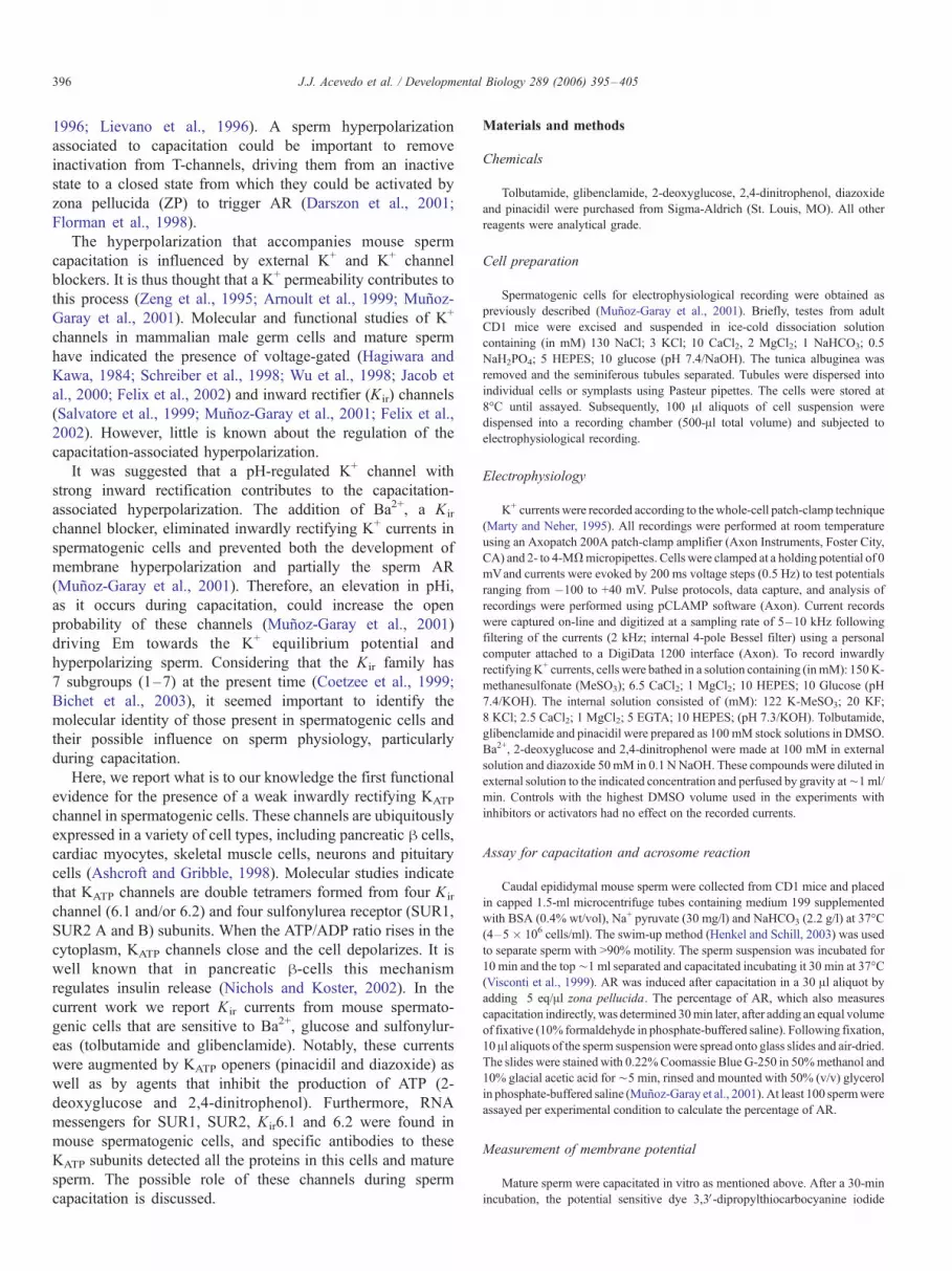
Methods). Tolbutamide (300 AM) reversibly decreased the K+
whole-cell current at the voltages tested (Figs. 1A and C). At
�100 mV this drug reduced membrane current to 45% T 12%
(n = 13) of the control. Glibenclamide (20 AM) also decreased
the K+ whole-cell current at the voltages tested (38% T 13%; n =
16) but in contrast to tolbutamide, the inhibition was not
reversible (Figs. 1B andD). Fig. 2 summarizes the time course of
IK decay when perfusing 300 AM tolbutamide (A) or 20 AMglibenclamide (B) and their degree of reversibility. The ratio of
KATP currents in the presence and absence of the drug
(Isulfonylurea/Icontrol) did not vary considerably from cell to cell
and its average was only mildly dependent on Em between�100
and +40 mV (not shown). The irreversible nature of the
glibenclamide inhibition of KATP currents has been reported in
other systems (Allen and Brown, 2004; Lim et al., 2004).
A notable feature of KATP channels is precisely their
inhibition by increased intracellular ATP levels (Aguilar-Bryan
and Bryan, 1999). Hence, in order to decrease the cellular ATP
content, spermatogenic cells were superfused several minutes
with a glucose-free solution. This maneuver resulted in a
significant increase in the amplitude of the whole-cell currents
recorded (Figs. 3A and B). As expected, the addition of
glibenclamide attenuated the response evoked by the glucose-
free solution (Figs. 3C and D). The involvement of ATP in the
regulation of the K+ currents was corroborated in experiments
where the patch pipette contained 1 mM ATP. Under this
condition, glucose removal was unable to stimulate the current
(Figs. 3C and D).
Kir channels are known to be susceptible to blockade by AMconcentrations of external Ba2+ (Hagiwara et al., 1978). In
particular, KATP channels are blocked by 100–200 AM Ba2+
(Bonev and Nelson, 1993; Takano and Ashcroft, 1996).
Therefore, we determined what percentage of the K+ current
was sensitive to this cation. Fig. 4A shows that Ba2+ blocked
¨49% T 5% (n = 13) of the current in spermatogenic cells in
a dose-dependent fashion, revealing the possible presence of
two classes of Kir channels with different sensibility. The
magnitude of the Ba2+ blockade was partially reversible
(Fig. 4B), only mildly voltage dependent (not shown) and
similar in magnitude to that found with tolbutamide and
glibenclamide.
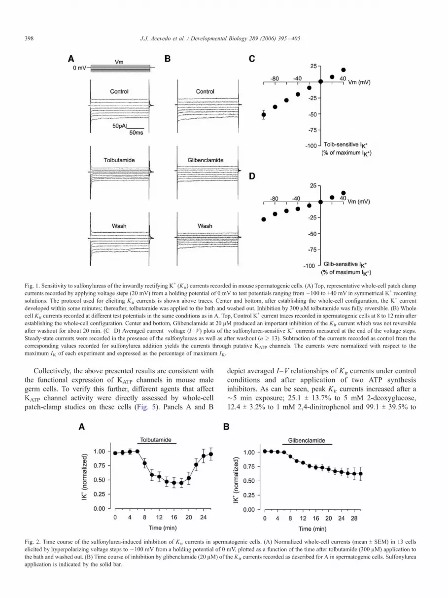
Fig. 1. Sensitivity to sulfonylureas of the inwardly rectifying K+ (Kir) currents recorded in mouse spermatogenic cells. (A) Top, representative whole-cell patch clamp
currents recorded by applying voltage steps (20 mV) from a holding potential of 0 mV to test potentials ranging from �100 to +40 mV in symmetrical K+ recording
solutions. The protocol used for eliciting Kir currents is shown above traces. Center and bottom, after establishing the whole-cell configuration, the K+ current
developed within some minutes; thereafter, tolbutamide was applied to the bath and washed out. Inhibition by 300 AM tolbutamide was fully reversible. (B) Whole
cell Kir currents recorded at different test potentials in the same conditions as in A. Top, Control K+ current traces recorded in spermatogenic cells at 8 to 12 min after
establishing the whole-cell configuration. Center and bottom, Glibenclamide at 20 AM produced an important inhibition of the Kir current which was not reversible
after washout for about 20 min. (C–D) Averaged current–voltage (I –V) plots of the sulfonylurea-sensitive K+ currents measured at the end of the voltage steps.
Steady-state currents were recorded in the presence of the sulfonylureas as well as after washout (n � 13). Subtraction of the currents recorded as control from the
corresponding values recorded for sulfonylurea addition yields the currents through putative KATP channels. The currents were normalized with respect to the
maximum IK of each experiment and expressed as the percentage of maximum IK.
J.J. Acevedo et al. / Developmental Biology 289 (2006) 395–405398
Collectively, the above presented results are consistent with
the functional expression of KATP channels in mouse male
germ cells. To verify this further, different agents that affect
KATP channel activity were directly assessed by whole-cell
patch-clamp studies on these cells (Fig. 5). Panels A and B
Fig. 2. Time course of the sulfonylurea-induced inhibition of K ir currents in sperm
elicited by hyperpolarizing voltage steps to �100 mV from a holding potential of 0
the bath and washed out. (B) Time course of inhibition by glibenclamide (20 AM) of
application is indicated by the solid bar.
depict averaged I–V relationships of Kir currents under control
conditions and after application of two ATP synthesis
inhibitors. As can be seen, peak Kir currents increased after a
¨5 min exposure; 25.1 T 13.7% to 5 mM 2-deoxyglucose,
12.4 T 3.2% to 1 mM 2,4-dinitrophenol and 99.1 T 39.5% to
atogenic cells. (A) Normalized whole-cell currents (mean T SEM) in 13 cells
mV, plotted as a function of the time after tolbutamide (300 AM) application to
the Kir currents recorded as described for A in spermatogenic cells. Sulfonylurea

Fig. 3. Superfusion with a glucose-free solution increases Kir currents in spermatogenic cells. (A) Three sets of current records in response to voltage steps between
�100 and +40 mV (in 20 mV increments) from a holding potential of 0 mVare shown. Recordings were made at about 10 min (control), 20 min (0 glucose) and 30
min (washout) after the whole-cell mode was established. (B) I –V relationships constructed from current records evoked by the voltage step protocol described in
panel A. The curves show almost a linear current–voltage relationship and reverse at 0 mV (n = 15). (C) Effects of glucose depletion on the currents elicited by
hyperpolarizing voltage step in the presence of ATP and glibenclamide. The normalized current amplitude in response to voltage-clamp steps between �100 and +40
mV from a holding potential of 0 mV is plotted as a function of time. Glibenclamide and ATP strongly antagonize the increases in membrane conductance produced
by extracellular depletion of glucose. (D) Normalized I –V relationships for control, glucose depletion (0 glucose) plus 20 AM glibenclamide and washout (n = 10).
J.J. Acevedo et al. / Developmental Biology 289 (2006) 395–405 399
5 mM of this later compound, respect to controls measured at
�100 mV). In a similar manner, panels C and D show the
effects of ¨5 min exposure to 100 AM pinacidil or diazoxide,
two KATP channel activators, on the Kir currents recorded from
spermatogenic cells. The mean Kir current amplitudes were
Fig. 4. Effects of Ba2+ on the Kir currents expressed in spermatogenic cells. (A) Re
voltage steps to �100 mV from a holding potential of 0 mV at various concentrat
amplitudes just before Ba2+ application. (B) Averaged time course of Kir current b
increased by 34.6 T 6.2% and 24.7 T 7.1% of the control
measured at �100 mV, respectively.
Having determined the presence of KATP channels in
functional assays, we next looked for the corresponding RNA
messengers for the Kir and SUR subunits known to constitute
lative amplitude of inwardly rectifying K+ currents evoked by hyperpolarizing
ions of BaCl2 as listed. Kir current amplitudes were normalized to the current
lock by Ba2+ (300 AM). Ba2+ application is indicated by the solid bar.
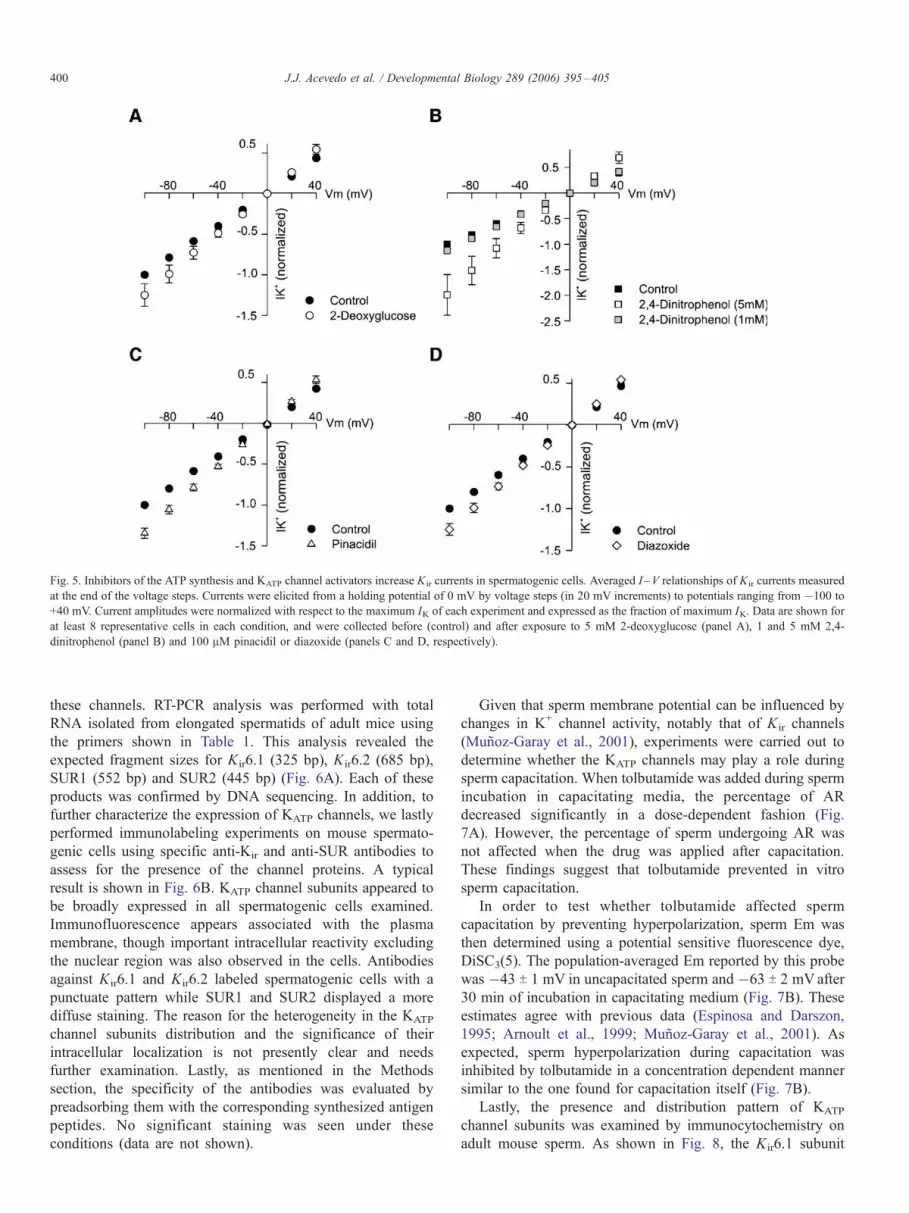
Fig. 5. Inhibitors of the ATP synthesis and KATP channel activators increase Kir currents in spermatogenic cells. Averaged I –V relationships of Kir currents measured
at the end of the voltage steps. Currents were elicited from a holding potential of 0 mV by voltage steps (in 20 mV increments) to potentials ranging from �100 to
+40 mV. Current amplitudes were normalized with respect to the maximum IK of each experiment and expressed as the fraction of maximum IK. Data are shown for
at least 8 representative cells in each condition, and were collected before (control) and after exposure to 5 mM 2-deoxyglucose (panel A), 1 and 5 mM 2,4-
dinitrophenol (panel B) and 100 AM pinacidil or diazoxide (panels C and D, respectively).
J.J. Acevedo et al. / Developmental Biology 289 (2006) 395–405400
these channels. RT-PCR analysis was performed with total
RNA isolated from elongated spermatids of adult mice using
the primers shown in Table 1. This analysis revealed the
expected fragment sizes for Kir6.1 (325 bp), Kir6.2 (685 bp),
SUR1 (552 bp) and SUR2 (445 bp) (Fig. 6A). Each of these
products was confirmed by DNA sequencing. In addition, to
further characterize the expression of KATP channels, we lastly
performed immunolabeling experiments on mouse spermato-
genic cells using specific anti-Kir and anti-SUR antibodies to
assess for the presence of the channel proteins. A typical
result is shown in Fig. 6B. KATP channel subunits appeared to
be broadly expressed in all spermatogenic cells examined.
Immunofluorescence appears associated with the plasma
membrane, though important intracellular reactivity excluding
the nuclear region was also observed in the cells. Antibodies
against Kir6.1 and Kir6.2 labeled spermatogenic cells with a
punctuate pattern while SUR1 and SUR2 displayed a more
diffuse staining. The reason for the heterogeneity in the KATP
channel subunits distribution and the significance of their
intracellular localization is not presently clear and needs
further examination. Lastly, as mentioned in the Methods
section, the specificity of the antibodies was evaluated by
preadsorbing them with the corresponding synthesized antigen
peptides. No significant staining was seen under these
conditions (data are not shown).
Given that sperm membrane potential can be influenced by
changes in K+ channel activity, notably that of Kir channels
(Munoz-Garay et al., 2001), experiments were carried out to
determine whether the KATP channels may play a role during
sperm capacitation. When tolbutamide was added during sperm
incubation in capacitating media, the percentage of AR
decreased significantly in a dose-dependent fashion (Fig.
7A). However, the percentage of sperm undergoing AR was
not affected when the drug was applied after capacitation.
These findings suggest that tolbutamide prevented in vitro
sperm capacitation.
In order to test whether tolbutamide affected sperm
capacitation by preventing hyperpolarization, sperm Em was
then determined using a potential sensitive fluorescence dye,
DiSC3(5). The population-averaged Em reported by this probe
was �43 T 1 mV in uncapacitated sperm and �63 T 2 mVafter
30 min of incubation in capacitating medium (Fig. 7B). These
estimates agree with previous data (Espinosa and Darszon,
1995; Arnoult et al., 1999; Munoz-Garay et al., 2001). As
expected, sperm hyperpolarization during capacitation was
inhibited by tolbutamide in a concentration dependent manner
similar to the one found for capacitation itself (Fig. 7B).
Lastly, the presence and distribution pattern of KATP
channel subunits was examined by immunocytochemistry on
adult mouse sperm. As shown in Fig. 8, the Kir6.1 subunit
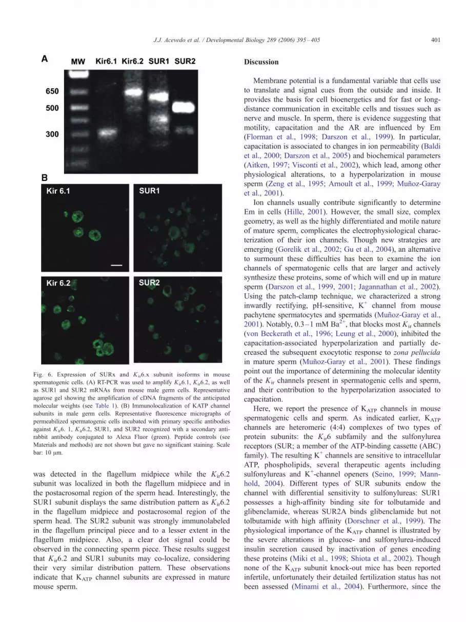
Fig. 6. Expression of SURx and K ir6.x subunit isoforms in mouse
spermatogenic cells. (A) RT-PCR was used to amplify Kir6.1, Kir6.2, as well
as SUR1 and SUR2 mRNAs from mouse male germ cells. Representative
agarose gel showing the amplification of cDNA fragments of the anticipated
molecular weights (see Table 1). (B) Immunolocalization of KATP channel
subunits in male germ cells. Representative fluorescence micrographs of
permeabilized spermatogenic cells incubated with primary specific antibodies
against K ir6. 1, K ir6.2, SUR1, and SUR2 recognized with a secondary anti-
rabbit antibody conjugated to Alexa Fluor (green). Peptide controls (see
Materials and methods) are not shown but gave no significant staining. Scale
bar: 10 Am.
J.J. Acevedo et al. / Developmental Biology 289 (2006) 395–405 401
was detected in the flagellum midpiece while the Kir6.2
subunit was localized in both the flagellum midpiece and in
the postacrosomal region of the sperm head. Interestingly, the
SUR1 subunit displays the same distribution pattern as Kir6.2
in the flagellum midpiece and postacrosomal region of the
sperm head. The SUR2 subunit was strongly immunolabeled
in the flagellum principal piece and to a lesser extent in the
flagellum midpiece. Also, a clear dot signal could be
observed in the connecting sperm piece. These results suggest
that Kir6.2 and SUR1 subunits may co-localize, considering
their very similar distribution pattern. These observations
indicate that KATP channel subunits are expressed in mature
mouse sperm.
Discussion
Membrane potential is a fundamental variable that cells use
to translate and signal cues from the outside and inside. It
provides the basis for cell bioenergetics and for fast or long-
distance communication in excitable cells and tissues such as
nerve and muscle. In sperm, there is evidence suggesting that
motility, capacitation and the AR are influenced by Em
(Florman et al., 1998; Darszon et al., 1999). In particular,
capacitation is associated to changes in ion permeability (Baldi
et al., 2000; Darszon et al., 2005) and biochemical parameters
(Aitken, 1997; Visconti et al., 2002), which lead, among other
physiological alterations, to a hyperpolarization in mouse
sperm (Zeng et al., 1995; Arnoult et al., 1999; Munoz-Garay
et al., 2001).
Ion channels usually contribute significantly to determine
Em in cells (Hille, 2001). However, the small size, complex
geometry, as well as the highly differentiated and motile nature
of mature sperm, complicates the electrophysiological charac-
terization of their ion channels. Though new strategies are
emerging (Gorelik et al., 2002; Gu et al., 2004), an alternative
to surmount these difficulties has been to examine the ion
channels of spermatogenic cells that are larger and actively
synthesize these proteins, some of which will end up in mature
sperm (Darszon et al., 1999, 2001; Jagannathan et al., 2002).
Using the patch-clamp technique, we characterized a strong
inwardly rectifying, pH-sensitive, K+ channel from mouse
pachytene spermatocytes and spermatids (Munoz-Garay et al.,
2001). Notably, 0.3–1 mM Ba2+, that blocks most Kir channels
(von Beckerath et al., 1996; Leung et al., 2000), inhibited the
capacitation-associated hyperpolarization and partially de-
creased the subsequent exocytotic response to zona pellucida
in mature sperm (Munoz-Garay et al., 2001). These findings
point out the importance of determining the molecular identity
of the Kir channels present in spermatogenic cells and sperm,
and their contribution to the hyperpolarization associated to
capacitation.
Here, we report the presence of KATP channels in mouse
spermatogenic cells and sperm. As indicated earlier, KATP
channels are heteromeric (4:4) complexes of two types of
protein subunits: the Kir6 subfamily and the sulfonylurea
receptors (SUR; a member of the ATP-binding cassette (ABC)
family). The resulting K+ channels are sensitive to intracellular
ATP, phospholipids, several therapeutic agents including
sulfonylureas and K+-channel openers (Seino, 1999; Mann-
hold, 2004). Different types of SUR subunits endow the
channel with differential sensitivity to sulfonylureas: SUR1
possesses a high-affinity binding site for tolbutamide and
glibenclamide, whereas SUR2A binds glibenclamide but not
tolbutamide with high affinity (Dorschner et al., 1999). The
physiological importance of the KATP channel is illustrated by
the severe alterations in glucose- and sulfonylurea-induced
insulin secretion caused by inactivation of genes encoding
these proteins (Miki et al., 1998; Shiota et al., 2002). Though
none of the KATP subunit knock-out mice has been reported
infertile, unfortunately their detailed fertilization status has not
been assessed (Minami et al., 2004). Furthermore, since the

Fig. 7. Effects of tolbutamide on the zona pellucida (ZP)-induced sperm acrosome reaction (AR) and membrane hyperpolarization during capacitation. (A)
Tolbutamide inhibits the ZP-induced AR by acting on capacitation. Capacitated mouse sperm were incubated 30 min in the presence of ZP and AR was measured.
Bars represent the percentage of acrosome-reacted sperm after ZP-induction in the presence of the drug during capacitation or the AR (5 s before ZP addition). The
ZP-induced AR values were normalized with respect to the control corrected for spontaneous AR which included the maximum DMSO volume used in the studies
with the inhibitor. Tolbutamide did not significantly affect spontaneous AR: average values after capacitation were 16.6 T 2.4 and 12 T 3.7% after drug application.
Data represent mean T SEM of at least four independent experiments. One hundred sperm were evaluated per assay. (B) Sperm membrane hyperpolarization
associated to capacitation. Bars represent mean T SEM membrane potentials in uncapacitated as well as in capacitated sperm when increasing concentrations of
tolbutamide are added to the capacitating medium. Sperm membrane potential was measured using the fluorescence dye DiSC3(5) (n = 4).
J.J. Acevedo et al. / Developmental Biology 289 (2006) 395–405402
four KATP channel subunits are found in sperm, establishing the
physiological role of KATP channels in these cells would
require double knock-outs of Kir6.1 and Kir6.2 or SUR1 and
SUR2 to rule out possible compensation.
We used three strategies to determine the presence and
possible function of KATP channels in mouse spermatogenic
cells and sperm. (1) Patch-clamp studies of Kir currents in
spermatogenic cells were carried out to explore their sensitivity
to KATP channel inhibitors, to ATP and ATP synthesis
inhibitors as well as to channel openers. (2) Membrane
potential and AR measurements were determined in capacitated
sperm populations in the absence and presence of tolbutamide.
Fig. 8. Immunolocalization of KATP channel subunits in mature mouse sperm. Per
specific antibodies and were recognized with a secondary anti-rabbit antibody conju
not shown but gave no significant staining. Scale bar: 10 Am.
(3) KATP channel subunit mRNAs and proteins were identified
using RT-PCR and immunolocalization.
(1) Under whole-cell patch-clamp conditions, we recorded a
weak inwardly rectifying K+ current that was sensitive to the
sulfonylureas tolbutamide and glibenclamide (¨40% inhibition
at �100 mV; Figs. 1 and 2). The ability of these agents to
significantly inhibit the current at micromolar concentrations,
together with their partial reversibility, resembles their effects
on both the native and cloned pancreatic h cell KATP channels
(Aguilar-Bryan et al., 1995, 1998; Inagaki et al., 1995, 1996).
As anticipated, these currents in spermatogenic cells increased
(64 T 14%) when glucose was removed from the external
meabilized cells were treated with Kir6.1, Kir6.2, SUR1, and SUR2B primary
gated to Alexa Fluor (green). Peptide controls (see Materials and methods) are
J.J. Acevedo et al. / Developmental Biology 289 (2006) 395–405 403
media (Fig. 3), a maneuver to reduce internal ATP. This
increase was prevented by glibenclamide. The presence of
these KATP channels was confirmed using blockers of ATP
synthesis and channel activators. Furthermore, these currents
were also inhibited by BaCl2 (Fig. 4) in a mildly voltage-
dependent manner. The Ba2+ concentration dependence of the
blockade suggests that more than one K ir is present in
spermatogenic cells, consistent with our findings that these
cells display strongly rectifying Kir (Munoz-Garay et al., 2001)
and KATP channels (this work). It is worth noting that in our
earlier work studying the strongly rectifying Kir current,
spermatogenic symplasts displaying significant rectification
were selected and the initial linear positive outward currents
were subtracted (Munoz-Garay et al., 2001). In the present
work to characterize the weakly rectifying Kir currents, all
symplasts were recorded and no subtraction was carried out.
This is the reason why under the present conditions Ba2+
blocks only 49% of the current. The remaining current
probably includes non-inactivating, nonselective cation chan-
nels for instance of the TRP family (Darszon et al., 2005)
and/or leak K+ channels of the two pore family (Buckingham
et al., 2005). Interestingly, the sulfonylureas inhibited ¨38%
of the current, indicating that the majority of the Kir current
flows through KATP channels.
(2) As reported previously (Arnoult et al., 1996, 1999;
Espinosa and Darszon, 1995; Muþoz-Garay et al., 2001) and
confirmed here (Fig. 5B), the resting Em in sperm is around
�45 mV and after capacitation it hyperpolarizes to about �65
mV. The sperm resting Em, as in other somatic cells, is
considerably less negative than the equilibrium potential for
K+ ions (EK) (¨�90 to �110 mV depending on the cell
type). This indicates that K+ channels do not completely
dominate the permeability of the cell membrane at rest. As
EK is negative to the resting Em, opening K+ channels will
displace Em towards EK resulting in a hyperpolarization;
while closure of K+ channels will cause a depolarization.
Tolbutamide inhibited the sperm hyperpolarization that
accompanies capacitation and capacitation itself (Fig. 5B),
suggesting the involvement of KATP channels in this process.
Sulfonylureas can also block other proteins of the ABC
superfamily, such as the cystic fibrosis transmembrane
conductance regulator (CFTR), a Cl� channel (Chang,
2003). Though CFTR mRNA has been detected in testis
(Sheppard and Welsh, 1999) and specifically in spermatogen-
ic cells, Western blot analysis performed with a CFTR
specific antibody revealed immunoreactivity in membranes
of round and elongated spermatids, but not in the fully
developed sperm (Gong et al., 2001). Considering these
findings and the blockade that Ba2+ causes on the capacitation
associated hyperpolarization, the inhibition of sperm capaci-
tation by tolbutamide suggests that indeed KATP channels
participate in this important maturing process. However, our
results do not rule out the involvement of other tolbutamide
sensitive hyperpolarizing transporters in capacitation.
Recently, it has been shown that during capacitation the
ATP content of sperm decreases (Baker et al., 2004). This
would lead to an increase in the open time of KATP channels
and would cause sperm hyperpolarization, which is consistent
with the observed behavior of Em during capacitation. In
addition, it is worth noting that Kir channels and specifically
KATP channels are regulated by CO2/pH. The maximal
activation occurs at pH 6.5 to 6.8, and the current through
these channels seems to be inhibited at pH 6.2 to 5.9 (Wang et
al., 2003). Since pHi increases during sperm capacitation from
¨6.5 to ¨6.7 (Vredenburgh-Wilberg and Parrish, 1995; Zeng
et al., 1996), KATP channels would also be activated by this
change.
(3) As a first step in the molecular identification of the
different subunits that compose the sperm KATP channel, we
searched for the presence of transcripts that correspond to the
most likely composition. We found that transcripts for Kir6.1
and Kir6.2 and SUR1 and SUR2 are expressed in mouse
spermatogenic cells (Fig. 6A). In addition, our immunocyto-
chemical data show that Kir6.1 and Kir6.2 as well as SUR1
and SUR2B subunits are expressed in both spermatogenic
cells and mature sperm (Figs. 6B and 8). Interestingly, the
analysis of the regional expression of these subunits may give
clues on the molecular entity responsible for KATP currents in
these cells. A careful examination of the overall expression
pattern and cellular localization of KATP channel subunits in
mature sperm suggested that Kir6.2 may co-localize and
therefore associate predominantly with the SUR1 subunit.
This points out to the presence of sulfonylurea-sensitive
channels with molecular similarities to those found in the
pancreatic h cells. KATP channels in these cells comprise
Kir6.2 and SUR1 subunits, while those of cardiac and skeletal
muscle appear to be composed of Kir6.2 and a splice variant
of SUR2, SUR2A (Aguilar-Bryan and Bryan, 1999). Never-
theless, our results show that Kir6.1 and SUR2 are also
present in mature sperm, therefore other combinations of Kir
and SUR subunits are possible and may contribute to the
hyperpolarization associated to capacitation or to other sperm
functions such as hyperactivation and/or the AR. The
possibility that KATP could be formed from heterotetramers
has not been discarded (Cui et al., 2001). Furthermore, recent
findings indicate SUR subunits may associate to other Kir
channels such as Kir1 (ROMK) (Dong et al., 2001). Thus, a
more detailed characterization of these channels both in
spermatogenic cells and in sperm is required to fully define
their molecular identity and function.
In summary, this work provides the fist evidence for the
presence of KATP channels in mouse spermatogenic cells and
sperm. In addition, evidence is presented for their contribution
to the hyperpolarization that is associated to sperm capacita-
tion. Evidently, these channels could participate also in other
important functions such as spermatogenesis, sperm hyper-
activation and the AR.
Acknowledgments
This work was supported by grants from DGAPA (UNAM)
to A.D., from CONACyT to A.D. and to R.F. and from FIRCA
RO3 TW 006121 to Pablo Visconti and A.D., and from
Wellcome Trust to A.D.

J.J. Acevedo et al. / Developmental Biology 289 (2006) 395–405404
Appendix A. Supplementary data
Supplementary data associated with this article can be found
in the online version at doi:10.1016/j.ydbio.2005.11.002.
References
Aguilar-Bryan, L., Bryan, J., 1999. Molecular biology of adenosine triphos-
phate-sensitive potassium channels. Endocr. Rev. 20, 101–135.
Aguilar-Bryan, L., Nichols, C.G., Wechsler, S.W., Clement IV, J.P., Boyd III,
A.E., Gonzalez, G., Herrera-Sosa, H., Nguy, K., Bryan, J., Nelson, D.A.,
1995. Cloning of the beta cell high-affinity sulfonylurea receptor: a
regulator of insulin secretion. Science 268, 423–426.
Aguilar-Bryan, L., Clement IV, J.P., Gonzalez, G., Kunjilwar, K., Babenko, A.,
Bryan, J., 1998. Toward understanding the assembly and structure of KATP
channels. Physiol. Rev. 78, 227–245.
Aitken, R.J., 1997. Molecular mechanisms regulating human sperm function.
Mol. Hum. Reprod. 3, 169–173.
Allen, T.G., Brown, D.A., 2004. Modulation of the excitability of
cholinergic basal forebrain neurones by KATP channels. J. Physiol. 554,
353–570.
Arnoult, C., Cardillo, R.A., Lemos, J.R., Florman, H.M., 1996. Activation of
mouse sperm T-type Ca2+ channels by adhesion to the egg zona pellucida.
Proc. Natl. Acad. Sci. U. S. A. 93, 13004–13009.
Arnoult, C., Kaza, I.G., Visconti, P.E., Kopf, G.S., Villaz, M., Florman, H.M.,
1999. Control of the low voltage-activated calcium channel of mouse sperm
by egg ZP3 and by membrane hyperpolarization during capacitation. Proc.
Natl. Acad. Sci. U. S. A. 96, 6757–6762.
Ashcroft, F.M., Gribble, F.M., 1998. Correlating structure and function in ATP-
sensitive K+ channels. Trends Neurosci. 21, 288–294.
Baker, M.A., Hetherington, L., Ecroyd, H., Roman, S.D., Aitken, R.J.,
2004. Analysis of the mechanism by which calcium negatively regulates
the tyrosine phosphorylation cascade associated with sperm capacitation.
J. Cell Sci. 117, 211–222.
Baldi, E., Casano, R., Falsetti, C., Krausz, C., Maggi, M., Forti, G., 1991.
Intracellular calcium accumulation and responsiveness to progesterone in
capacitating human spermatozoa. J. Androl. 12, 323–330.
Baldi, E., Luconi, M., Bonaccorsi, L., Muratori, M., Forti, G., 2000.
Intracellular events and signaling pathways involved in sperm acquisi-
tion of fertilizing capacity and acrosome reaction. Front. Biosci. 5,
E110–E123.
Bellve, A.R., 1993. Purification, culture, and fractionation of spermatogenic
cells. Methods Enzymol. 225, 84–113.
Bichet, D., Haass, F.A., Jan, L.Y., 2003. Merging functional studies with
structures of inward-rectifier K+ channels. Nat. Rev., Neurosci. 4, 957–967.
Bonev, A.D., Nelson, M.T., 1993. ATP-sensitive potassium channels in
smooth muscle cells from guinea pig urinary bladder. Am. J. Physiol.
264, C1190–C1200.
Buckingham, S.D., Kidd, J.F., Law, R.J., Franks, C.J., Sattelle, D.B., 2005.
Structure and function of two-pore-domain KC channels: contributions
from genetic model organisms. Trends Pharmacol. Sci. 26, 361–367.
Chang, G., 2003. Multidrug resistance ABC transporters. FEBS Lett. 555,
102–105.
Coetzee, W.A., Amarillo, Y., Chiu, J., Chow, A., Lau, D., McCormack, T.,
Moreno, H., Nadal, M.S., Ozaita, A., Pountney, D., Saganich, M., Vega-
Saenz de Miera, E., Rudy, B., 1999. Molecular diversity of K+ channels.
Ann. N. Y. Acad. Sci. 868, 233–285.
Cui, Y., Giblin, J.P., Clapp, L.H., Tinker, A., 2001. A mechanism for ATP-
sensitive potassium channel diversity: functional coassembly of two pore-
forming subunits. Proc. Natl. Acad. Sci. U. S. A. 98, 729–734.
Darszon, A., Labarca, P., Nishigaki, T., Espinosa, F., 1999. Ion channels in
sperm physiology. Physiol. Rev. 79, 481–510.
Darszon, A., Beltran, C., Felix, R., Nishigaki, T., Trevino, C.L., 2001. Ion
transport in sperm signaling. Dev. Biol. 240, 1–14.
Darszon, A., Nishigaki, T., Wood, C.D., Trevino, C.L., Felix, R., Beltran, C.,
2005. Ca2+ channels and Ca2+ fluctuations in sperm physiology. Int. Rev.
Cytol. 243, 79–172.
DasGupta, S., Mills, C.L., Fraser, L.R., 1993. Ca2+-related changes in the
capacitation state of human spermatozoa assessed by a chlortetracycline
fluorescence assay. J. Reprod. Fertil. 99, 135–143.
Dong, K., Xu, J., Vanoye, C.G., Welch, R., MacGregor, G.G., Giebisch,
G., Hebert, S.C., 2001. An amino acid triplet in the NH2 terminus of
rat ROMK1 determines interaction with SUR2B. J. Biol. Chem. 276,
44347–44353.
Dorschner, H., Brekardin, E., Uhde, I., Schwanstecher, C., Schwanstecher, M.,
1999. Stoichiometry of sulfonylurea-induced ATP-sensitive potassium
channel closure. Mol. Pharmacol. 55, 1060–1066.
Espinosa, F., Darszon, A., 1995. Mouse sperm membrane potential: changes
induced by Ca2+. FEBS Lett. 372, 119–125.
Espinosa, F., de la Vega-Beltran, J.L., Lopez-Gonzalez, I., Delgado, R.,
Labarca, P., Darszon, A., 1998. Mouse sperm patch-clamp recordings
reveal single Cl-channels sensitive to niflumic acid, a blocker of the sperm
acrosome reaction. FEBS Lett. 426, 47–51.
Felix, R., Serrano, C.J., Trevino, C.L., Munoz-Garay, C., Bravo, A.,
Navarro, A., Pacheco, J., Tsutsumi, V., Darszon, A., 2002. Identification
of distinct K+ channels in mouse spermatogenic cells and sperm. Zygote
10, 183–188.
Florman, H.M., Arnoult, C., Kazam, I.G., Li, C., O’Toole, C.M., 1998. A
perspective on the control of mammalian fertilization by egg-activated ion
channels in sperm: a tale of two channels. Biol. Reprod. 59, 12–16.
Galantino-Homer, H.L., Visconti, P.E., Kopf, G.S., 1997. Regulation of
protein tyrosine phosphorylation during bovine sperm capacitation by a
cyclic adenosine 3V5V-monophosphate-dependent pathway. Biol. Reprod.
56, 707–719.
Galantino-Homer, H.L., Florman, H.M., Storey, B.T., Dobrinski, I., Kopf, G.S.,
2004. Bovine sperm capacitation: assessment of phosphodiesterase activity
and intracellular alkalinization on capacitation-associated protein tyrosine
phosphorylation. Mol. Reprod. Dev. 67, 487–500.
Gong, X.D., Li, J.C.H., Cheung, K.H., Laung, G.P.H., Cheng-Chew, S.B.,
Wong, P.Y.D., 2001. Expression of the cystic fibrosis transmembrane
conductance regulator in rat spermatids: implication for the site of action of
antispermatogenic agents. Mol. Hum. Reprod. 7, 705–713.
Gorelik, J., Gu, Y., Spohr, H.A., Shevchuk, A.I., Lab, M.J., Harding, S.E.,
Edwards, C.R., Whitaker, M., Moss, G.W., Benton, D.C., Sanchez, D.,
Darszon, A., Vodyanoy, I., Klenerman, D., Korchev, Y.E., 2002. Ion
channels in small cells and subcellular structures can be studied with a
smart patch-clamp system. Biophys. J. 83, 3296–3303.
Gu, Y., Kirkman-Brown, J.C., Korchev, Y., Barratt, C.L., Publicover, S.J.,
2004. Multi-state, 4-aminopyridine-sensitive ion channels in human
spermatozoa. Dev. Biol. 274, 308–317.
Hagiwara, S., Kawa, K., 1984. Calcium and potassium currents in
spermatogenic cells dissociated from rat seminiferous tubules. J. Physiol.
356, 135–149.
Hagiwara, S., Miyazaki, S., Moody, W., Patlak, J., 1978. Blocking effects of
barium and hydrogen ions on the potassium current during anomalous
rectification in the starfish egg. J. Physiol. 279, 167–185.
Henkel, R.R., Schill, W.B., 2003. Sperm preparation for ART. Reprod. Biol.
Endocrinol. 1, 108.
Hille, B., 2001. Ion Channels of Excitable Membranes. 3rd ed. Sinauer,
Sunderland, MA, pp. 814.
Inagaki, N., Gonoi, T., Clement IV, J.P., Namba, N., Inazawa, J., Gonzalez,
G., Aguilar-Bryan, L., Seino, S., Bryan, J., 1995. Reconstitution of IKATP:
an inward rectifier subunit plus the sulfonylurea receptor. Science 270,
1166–1170.
Inagaki, N., Gonoi, T., Clement IV, J.P., Wang, C.Z., Aguilar-Bryan, L., Bryan,
J., Seino, S., 1996. A family of sulfonylurea receptors determines the
pharmacological properties of ATP-sensitive K+ channels. Neuron 16,
1011–1017.
Jacob, A., Hurley, I.R., Goodwin, L.O., Cooper, G.W., Benoff, S., 2000.
Molecular characterization of a voltage-gated potassium channel expressed
in rat testis. Mol. Hum. Reprod. 6, 303–313.
Jagannathan, S., Punt, E.L., Gu, Y., Arnoult, C., Sakkas, D., Barratt, C.L.,
Publicover, S.J., 2002. Identification and localization of T-type voltage-
operated calcium channel subunits in human male germ cells. Expression of
multiple isoforms. J. Biol. Chem. 277, 8449–8456.

J.J. Acevedo et al. / Developmental Biology 289 (2006) 395–405 405
Leung, Y.M., Kwan, C.Y., Daniel, E.E., 2000. Block of inwardly rectifying K+
currents by extracellular Mg2+ and Ba2+ in bovine pulmonary artery
endothelial cells. Can. J. Physiol. Pharmacol. 78, 751–756.
Lievano, A., Santi, C.M., Serrano, C.J., Trevino, C.L., Bellve, A.R.,
Hernandez-Cruz, A., Darszon, A., 1996. T-type Ca2+ channels and a1E
expression in spermatogenic cells, and their possible relevance to the sperm
acrosome reaction. FEBS Lett. 388, 150–154.
Lim, J.G., Lee, H.Y., Yun, J.E., Kim, S.P., Park, J.W., Suh, S.I., Jang, B.C.,
Cho, C.H., Bae, J.H., Kim, S.S., Han, J., Park, M.J., Song, D.K., 2004.
Taurine block of cloned ATP-sensitive K+ channels with different
sulfonylurea receptor subunits expressed in Xenopus laevis oocytes.
Biochem. Pharmacol. 68, 901–910.
Mannhold, R., 2004. KATP channel openers: structure–activity relationships
and therapeutic potential. Med. Res. Rev. 24, 213–266.
Marty, A., Neher, E., 1995. Tight-seal whole-cell recording. In: Sakmann,
B., Neher, E. (Eds.), Single-Channel Recording. Plenum Press, New York,
pp. 31–52.
Miki, T., Nagashima, K., Tashiro, F., Kotake, K., Yoshitomi, H., Tamamoto, A.,
Gonoi, T., Iwanaga, T., Miyazaki, J., Seino, S., 1998. Defective insulin
secretion and enhanced insulin action in KATP channel-deficient mice.
Proc. Natl. Acad. Sci. U. S. A. 95, 10402–10406.
Minami, K., Miki, T., Kadowaki, T., Seino, S., 2004. Roles of ATP-sensitive K+
channels as metabolic sensors: studies of Kir6.x null mice. Diabetes 53
(Suppl. 3), S176–S180.
Munoz-Garay, C., De la Vega-Beltran, J.L., Delgado, R., Labarca, P., Felix, R.,
Darszon, A., 2001. Inwardly rectifying K+ channels in spermatogenic cells:
functional expression and implication in sperm capacitation. Dev. Biol. 234,
261–274.
Nichols, C.G., Koster, J.C., 2002. Diabetes and insulin secretion: whither
KATP? Am. J. Physiol.: Endocrinol. Metab. 283, E403–E412.
Nolan, M.A., Babcock, D.F., Wennemuth, G., Brown, W., Burton, K.A.,
McKnight, G.S., 2004. Sperm-specific protein kinase A catalytic subunit
Calpha2 orchestrates cAMP signaling for male fertility. Proc. Natl. Acad.
Sci. U. S. A. 101, 13483–13488.
Parrish, J.J., Susko-Parrish, J.L., First, N.L., 1989. Capacitation of bovine
sperm by heparin: inhibitory effect of glucose and role of intracellular pH.
Biol. Reprod. 41, 683–699.
Salvatore, L., D’Adamo, M.C., Polishchuk, R., Salmona, M., Pessia, M., 1999.
Localization and age-dependent expression of the inward rectifier K+
channel subunit Kir5.1 in a mammalian reproductive system. FEBS Lett.
449, 146–152.
Schreiber, M., Wei, A., Yuan, A., Gaut, J., Saito, M., Salkoff, L., 1998. Slo3, a
novel pH-sensitive K+ channel from mammalian spermatocytes. J. Biol.
Chem. 273, 3509–3516.
Seino, S., 1999. ATP-sensitive potassium channels: a model of heteromulti-
meric potassium channel/receptor assemblies. Annu. Rev. Physiol. 61,
337–362.
Serrano, C.J., Trevino, C.L., Felix, R., Darszon, A., 1999. Voltage-dependent
Ca2+ channel subunit expression and immunolocalization in mouse
spermatogenic cells and sperm. FEBS Lett. 462, 171–176.
Sheppard, D.N., Welsh, M.J., 1999. Structure and function of the CFTR
chloride channel. Physiol. Rev. 79, S23–S45.
Shiota, C., Larsson, O., Shelton, K.D., Shiota, M., Efanov, A.M., Hoy, M.,
Lindner, J., Kooptiwut, S., Juntti-Berggren, L., Gromada, J., Berggren,
P.O., Magnuson, M.A., 2002. Sulfonylurea receptor type 1 knock-out mice
have intact feeding-stimulated insulin secretion despite marked impairment
in their response to glucose. J. Biol. Chem. 277, 37176–37183.
Takano, M., Ashcroft, F.M., 1996. The Ba2+ block of the ATP-sensitive K+
current of mouse pancreatic beta-cells. Pflugers Arch. 431, 625–631.
Visconti, P.E., Bailey, J.L., Moore, G.D., Pan, D., Olds-Clarke, P., Kopf, G.S.,
1995a. Capacitation of mouse spermatozoa. I. Correlation between the
capacitation state and protein tyrosine phosphorylation. Development 121,
1129–1137.
Visconti, P.E., Moore, G.D., Bailey, J.L., Leclerc, P., Connors, S.A., Pan, D.,
Olds-Clarke, P., Kopf, G.S., 1995b. Capacitation of mouse spermatozoa. II.
Protein tyrosine phosphorylation and capacitation are regulated by a cAMP-
dependent pathway. Development 121, 1139–1150.
Visconti, P.E., Galantino-Homer, H., Ning, X., Moore, G.D., Valenzuela, J.P.,
Jorgez, C.J., Alvarez, J.G., Kopf, G.S., 1999. Cholesterol efflux-mediated
signal transduction in mammalian sperm. h-cyclodextrins initiate trans-
membrane signaling leading to an increase in protein tyrosine phosphory-
lation and capacitation. J. Biol. Chem. 274, 3235–3242.
Visconti, P.E., Westbrook, V.A., Chertihin, O., Demarco, I., Sleight, S.,
Diekman, A.B., 2002. Novel signaling pathways involved in sperm
acquisition of fertilizing capacity. J. Reprod. Immunol. 53, 133–150.
von Beckerath, N., Dittrich, M., Klieber, H.G., Daut, J., 1996. Inwardly
rectifying K+ channels in freshly dissociated coronary endothelial cells from
guinea-pig heart. J. Physiol. 491, 357–365.
Vredenburgh-Wilberg, W.L., Parrish, J.J., 1995. Intracellular pH of bovine
sperm increases during capacitation. Mol. Reprod. Dev. 40, 490–502.
Wang, X., Wu, J., Li, L., Chen, F., Wang, R., Jiang, C., 2003. Hypercapnic
acidosis activates KATP channels in vascular smooth muscles. Circ. Res.
92, 1225–1232.
Wu, W.L., So, S.C., Sun, Y.P., Zhou, T.S., Yu, Y., Chung, Y.W., Wang, X.F.,
Bao, Y.D., Yan, Y.C., Chan, H.C., 1998. Functional expression of a Ca2+-
activated K+ channel in Xenopus oocytes injected with RNAs from the rat
testis. Biochim. Biophys. Acta 1373, 360–365.
Zeng, Y., Clark, E.N., Florman, H.M., 1995. Sperm membrane potential:
hyperpolarization during capacitation regulates zona pellucida-dependent
acrosomal secretion. Dev. Biol. 171, 554–563.
Zeng, Y., Oberdorf, J.A., Florman, H.M., 1996. pH regulation in mouse sperm:
identification of Na+-, Cl�, and HCO3� dependent and arylaminobenzoate-
dependent regulatory mechanisms and characterization of their roles in
sperm capacitation. Dev. Biol. 173, 510–520.