putative subunits of the rat mesangial katp: a type 2b sulfonylurea receptor and an inwardly...
TRANSCRIPT

Kidney International, Vol. 61 (2002), pp. 1739–1749
ION CHANNELS – MEMBRANE TRANSPORT – INTEGRATIVE PHYSIOLOGY
Putative subunits of the rat mesangial KATP: A type 2Bsulfonylurea receptor and an inwardly rectifying K� channel
BALAZS SZAMOSFALVI, PEDRO CORTES, REBECCA ALVIANI, KENICHIRO ASANO, BRUCE L. RISER,GARY ZASUWA, and JERRY YEE
Department of Medicine, Division of Nephrology and Hypertension, Henry Ford Hospital, Detroit, Michigan, USA
Complementary studies showed abundant gene expression ofPutative subunits of the rat mesangial KATP: A type 2B sulfonyl-Kir6.1, thereby establishing gene expression of both compo-urea receptor and an inwardly rectifying K� channel.nents of KATP.Background. Sulfonylurea agents exert their physiological
Conclusions. Based upon analogy to vascular smooth muscleeffects in many cell types via binding to specific sulfonylureacells (VSMC), there are at least two putative mesangial KATPreceptors (SUR). SUR couple to inwardly-rectifying K� chan-that most likely represent hetero-octamers, comprised of eithernel (Kir6.x) to form tetradimeric ATP-sensitive K� channelsrSUR2B or mcSUR2 in complex with Kir6.1. Our results define(KATP). The SUR subunits confer ATP-sensitivity on KATP andthe mesangial SUR2B as the possible first link in a chain ofalso provide the binding sites for sulfonylureas and other phar-cellular events that culminates in MC contraction and alteredmacological agents. Our previous work demonstrated that theextracellular matrix metabolism following exposure to sulfo-exposure of mesangial cells (MC) to sulfonylureas generated
profound effects on MC glucose uptake and matrix metabolism nylureas. In addition, our results serve as the basis for theand induced heightened cell contractility in association with future elucidation of the electrophysiologic characteristics ofCa2� transients. Because these responses likely resulted from the mesangial KATP and the study of endogenous regulators ofthe binding of sulfonylurea to a mesangial SUR2, we subse- mesangial cell contractility.quently documented [3H]-glibenclamide binding to MC andthe gene expression of several mesangial SUR2 transcripts.From these data, we inferred that MC expressed the compo-
Sulfonylureas such as tolazamide and glibenclamidenents of a mesangial KATP and sought to establish their presenceare oral hypoglycemic agents that are frequently utilizedin primary MC.
Methods. To obtain mesangial SUR2 cDNA sequences, during the treatment of type 2 diabetes mellitus [1]. Therapid amplification of cDNA ends (RACE) was utilized. DNA elucidation of the mechanism by which these agents aug-sequences were established by the fluorescent dye termination ment pancreatic � cell insulin secretion via their highmethod. Gene expression of mesangial SUR2 and Kir6.1/2 was
affinity type-1 sulfonylurea receptors (SUR1) has impelledexamined by reverse transcription polymerase chain reactiona burgeoning field of investigation into pancreatic and(RT-PCR) and Northern analysis. SUR2 proteins were identi-
fied by immunoblotting of mesangial proteins from membrane- non-pancreatic SUR [2]. These receptors are membersenriched fractions with polyclonal antiserum directed against of the cystic fibrosis transmembrane regulator (CFTR)/SUR2.
multidrug resistance protein (MRP) subfamily that isResults. RACE cloning yielded two mesangial SUR2 cDNAssubsumed within the adenosine 5�-triphosphate (ATP)-of 4.8 and 6.7 kbp whose open reading frames translated pro-
teins of 964 and 1535 aa, respectively. Using probes specific to binding cassette (ABC) superfamily [3, 4]. The two SUReach cDNA, the presence of a unique, 5.5 kbp serum-regulated genes, SUR1 and SUR2, are encoded on separate chro-mesangial SUR2 splice variant was established. The sequence mosomes and have been respectively designated, ABCC8of this mesangial SUR2 (mcSUR2B) shares identity with the
and ABCC9 [5]. The SUR are predicted to have 17 trans-recently cloned rat SUR2B (rSUR2B), but, in comparison tomembrane-spanning helices organized into three do-rSUR2B, is truncated by 12 exons at the N-terminus where it
contains a unique insert of 16 aa. Immunoblotting studies with mains designated TM0, TM1, and TM2, separated byanti-SUR2 antiserum demonstrated SUR2 proteins of 108 and two intracytoplasmic loops labeled NBF1 and NBF2,170 kD in membrane-enriched fractions of MC protein extracts. each containing nucleotide-binding motifs. The SUR dif-
fer in their affinity for sulfonylureas and in their tissuedistribution. SUR1 is a high-affinity receptor expressedKey words: mesangial cell contraction, hypoglycemia, cell contraction,
potassium channels, glucose uptake, SUR2B, Kir6.1. by pancreatic � cells and brain, while the low-affinitySUR2 is more ubiquitously expressed [2]. There are twoReceived for publication December 12, 2000major alternatively spliced SUR2 variants, SUR2A andand in revised form December 4, 2001
Accepted for publication December 6, 2001 SUR2B, and the former is chiefly found in heart andskeletal muscle, while the latter is localized to brain, 2002 by the International Society of Nephrology
1739

Szamosfalvi et al: Mesangial KATP1740
heart, liver, kidney, intestine, and vascular smooth mus- Dimethyl sulfoxide (DMSO), glibenclamide, antibio-tics, Tween-20, and morpholinopropane sulfonic acid-cle [6, 7].
The ATP-sensitive K� channels (KATP) are hetero- EDTA-sodium acetate (MESA) buffer were purchasedfrom Sigma-Aldrich Co. (St. Louis, MO, USA). RPMIoctamers of two subunits, a SUR and a weak inwardly-
rectifying K� channel (Kir6.x) that associate with each 1640 culture medium, fetal calf serum (FCS) and RNAsizing ladders were obtained from Life Technologiesother in a stoichiometry of 4:4. Ion-channel closure is
mediated by sulfonylurea binding or by increases of the (Gaithersburg, MD, USA). QuikHyb solution was pur-chased from Stratagene (La Jolla, CA, USA). SMART-cytoplasmic ATP:ADP ratio. In KATP, SUR1 or SUR2
regulates the Kir6.x ion pore to which it is coupled [8–16]. RACE� cloning kits and Advantage cDNA PCR en-zyme mixes were obtained from Clontech (Palo Alto,The prototypical � cell KATP is a tetradimer of SUR1
and Kir6.2, whereas the cardiac and vascular smooth CA, USA). TOPO TA cloning kits and pCR2.1/TOPOvectors were purchased from Invitrogen (Carlsbad, CA,muscle KATP are represented by (SUR2A:Kir6.2)4 and
(SUR2B:Kir6.1/2)4, respectively [17–24]. In previous stud- USA). RNA STAT-60 reagent was obtained from Tel-Test, Inc. (Friendswood, TX, USA). Poly(A)� RNA wasies, we established that the sulfonylurea tolazamide stim-
ulated rat mesangial cells (MC) to secrete increased isolated by magnetic bead separation (Roche Molecu-lar Biochemicals, Indianapolis, IN, USA). [�-32P]dCTPamounts of collagen [25, 26]. As in other extrapancreatic
tissues in which sulfonylureas amplify glucose uptake, we (3000 Ci/mmol) was purchased from New England Nu-clear (Boston, MA, USA). Bicinchoninic acid proteinestablished that the enhancement of MC glucose uptake
occurred coincident with increased cytosol-to-membrane assay kits were purchased from Pierce Labs (Rockford,IL, USA). Sodium dodecyl sulfate-polyacrylamide gelstranslocation of the glucose transporter, GLUT1 [26, 27].
The augmentation of hexose uptake was associated with (10%) were obtained from Bio-Rad (Hercules, CA,USA). Chemiluminescent detection system kits (ECL)an increased secretion of autocrine transforming growth
factor beta-1 (TGF-�1) and collagen accumulation. In were purchased from Amersham Pharmacia Biotech(Piscataway, NJ, USA). The 4.3 kbp Eco RI restrictionaddition, glibenclamide induced sustained oscillatory
Ca2� transients in MC that coincided with heightened of rat SUR2 cDNA (pJGAR7) was generously providedby Dr. Charles Burant (University of Michigan, Anncell contractility [28–33]. Further postulation that these
biological effects resulted from binding of sulfonylureas Arbor, MI, USA). Kir6.1/2 primer sets were gifts of Dr.Stephen C. Hebert (Yale Univ., New Haven, CT, USA).to membranous receptors resulted in the initial charac-
terization of several low-affinity MC SUR2. One of these Rabbit anti-SUR2 immune serum was kindly donated toour laboratory by Dr. Susumu Seino (Chiba University,was represented by a unique and dominant 5.5 kb SUR2
transcript, which we identified as mesangial SUR2B Tokyo, Japan).(mcSUR2B) [28]. With the prospect that this transcript
Tissue cultureand possibly others encoded the SUR2 subunit of a mes-angial KATP, we initiated a series of investigations to The cells used in all studies were primary MC out-
growths of glomerular explants of Munich-Wistar ratscharacterize the components that would comprise thischannel. The initial molecular characterization of the or our well characterized cloned rat mesangial cell line
16KC2 [26–28]. The latter are spindle-shaped cells thatmesangial KATP would thus render greater insight towardour understanding of the cell-signaling events underlying form characteristic hillocks in confluent tissue cultures
and stain positively for vimentin, desmin and �-smoothour previously observed functional data regarding sulfo-nylureas and MC. muscle actin, but not for cytokeratin or factor VIII. Cel-
lular contraction occurs following exposure to either an-giotensin II or arginine vasopressin. These cells also bear
METHODSatrial natriuretic peptide (ANP) receptors and the Thy-1
Animals antigen on their surfaces. Except where noted, primaryMC or 16KC2 were seeded at a density of 5,000 cellsMale Munich-Wistar rats, weighing 150 to 200 g, were
obtained from Charles River Laboratories (Wilmington, per cm2 and cultured to near-confluence in RPMI 1640medium, containing 8 mmol/L glucose, antibiotics (peni-MA, USA). Animals were housed in accordance with good
practice guidelines established by AALAC-approved cillin 100 U/mL; streptomycin 100 �g/mL) and 20%serum.animal care facilities of Henry Ford Hospital. Animals
were fed standard Purina rat chow (Ralston Purina Co.,Cloning by rapid amplification of cDNASt. Louis, MO, USA) and permitted free access to water.ends (RACE)
Reagents Poly(A)� RNA was isolated on magnetic beads fromprimary cell cultures at passage 8. RACE cloning wasInsulin-transferrin-selenium pre-mix was obtained from
Becton Dickinson Biosciences (Franklin Lakes, NJ, USA). carried out with the SMART-RACE� system [7]. Briefly,
Szamosfalvi et al: Mesangial KATP 1741
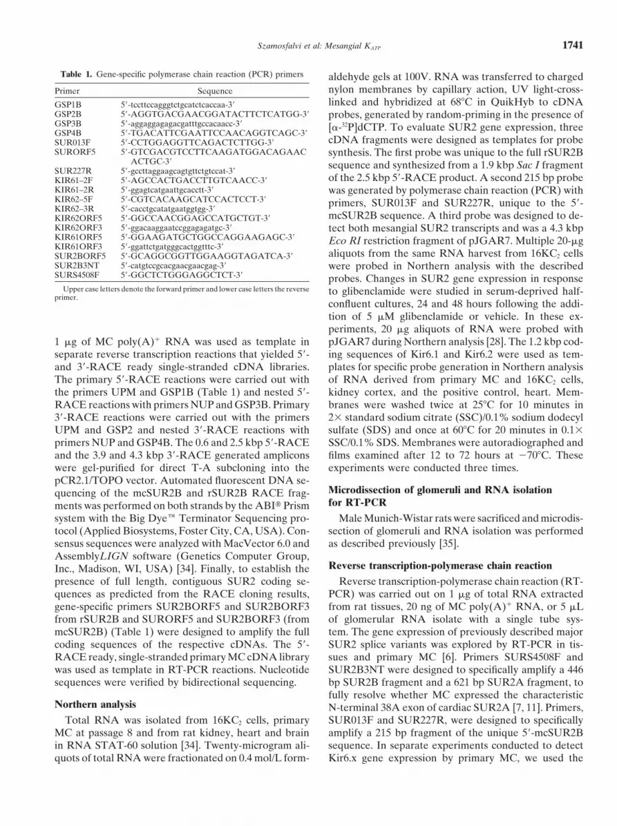
Table 1. Gene-specific polymerase chain reaction (PCR) primers aldehyde gels at 100V. RNA was transferred to chargednylon membranes by capillary action, UV light-cross-Primer Sequencelinked and hybridized at 68�C in QuikHyb to cDNAGSP1B 5�-tccttccagggtctgcatctcaccaa-3�
GSP2B 5�-AGGTGACGAACGGATACTTCTCATGG-3� probes, generated by random-priming in the presence ofGSP3B 5�-aggaggagagacgatttgccacaacc-3� [�-32P]dCTP. To evaluate SUR2 gene expression, threeGSP4B 5�-TGACATTCGAATTCCAACAGGTCAGC-3�
cDNA fragments were designed as templates for probeSUR013F 5�-CCTGGAGGTTCAGACTCTTGG-3�SURORF5 5�-GTCGACGTCCTTCAAGATGGACAGAAC synthesis. The first probe was unique to the full rSUR2B
ACTGC-3� sequence and synthesized from a 1.9 kbp Sac I fragmentSUR227R 5�-gccttaggaagcagtgttctgtccat-3�
of the 2.5 kbp 5�-RACE product. A second 215 bp probeKIR61–2F 5�-AGCCACTGACCTTGTCAACC-3�KIR61–2R 5�-ggagtcatgaattgcacctt-3� was generated by polymerase chain reaction (PCR) withKIR62–5F 5�-CGTCACAAGCATCCACTCCT-3� primers, SUR013F and SUR227R, unique to the 5�-KIR62–3R 5�-cacctgcatatgaatggtgg-3�
mcSUR2B sequence. A third probe was designed to de-KIR62ORF5 5�-GGCCAACGGAGCCATGCTGT-3�KIR62ORF3 5�-ggacaaggaatccggagagatgc-3� tect both mesangial SUR2 transcripts and was a 4.3 kbpKIR61ORF5 5�-GGAAGATGCTGGCCAGGAAGAGC-3� Eco RI restriction fragment of pJGAR7. Multiple 20-�gKIR61ORF3 5�-ggattctgatgggcactggtttc-3�
aliquots from the same RNA harvest from 16KC2 cellsSUR2BORF5 5�-GCAGGCGGTTGGAAGGTAGATCA-3�SUR2B3NT 5�-catgtccgcacgaacgaacgag-3� were probed in Northern analysis with the describedSURS4508F 5�-GGCTCTGGGAGGCTCT-3� probes. Changes in SUR2 gene expression in response
Upper case letters denote the forward primer and lower case letters the reverse to glibenclamide were studied in serum-deprived half-primer.
confluent cultures, 24 and 48 hours following the addi-tion of 5 �M glibenclamide or vehicle. In these ex-periments, 20 �g aliquots of RNA were probed withpJGAR7 during Northern analysis [28]. The 1.2 kbp cod-1 �g of MC poly(A)� RNA was used as template in
separate reverse transcription reactions that yielded 5�- ing sequences of Kir6.1 and Kir6.2 were used as tem-plates for specific probe generation in Northern analysisand 3�-RACE ready single-stranded cDNA libraries.
The primary 5�-RACE reactions were carried out with of RNA derived from primary MC and 16KC2 cells,kidney cortex, and the positive control, heart. Mem-the primers UPM and GSP1B (Table 1) and nested 5�-
RACE reactions with primers NUP and GSP3B. Primary branes were washed twice at 25�C for 10 minutes in2� standard sodium citrate (SSC)/0.1% sodium dodecyl3�-RACE reactions were carried out with the primers
UPM and GSP2 and nested 3�-RACE reactions with sulfate (SDS) and once at 60�C for 20 minutes in 0.1�SSC/0.1% SDS. Membranes were autoradiographed andprimers NUP and GSP4B. The 0.6 and 2.5 kbp 5�-RACE
and the 3.9 and 4.3 kbp 3�-RACE generated amplicons films examined after 12 to 72 hours at �70�C. Theseexperiments were conducted three times.were gel-purified for direct T-A subcloning into the
pCR2.1/TOPO vector. Automated fluorescent DNA se-Microdissection of glomeruli and RNA isolationquencing of the mcSUR2B and rSUR2B RACE frag-for RT-PCRments was performed on both strands by the ABI� Prism
system with the Big Dye� Terminator Sequencing pro- Male Munich-Wistar rats were sacrificed and microdis-section of glomeruli and RNA isolation was performedtocol (Applied Biosystems, Foster City, CA, USA). Con-
sensus sequences were analyzed with MacVector 6.0 and as described previously [35].AssemblyLIGN software (Genetics Computer Group,
Reverse transcription-polymerase chain reactionInc., Madison, WI, USA) [34]. Finally, to establish thepresence of full length, contiguous SUR2 coding se- Reverse transcription-polymerase chain reaction (RT-
PCR) was carried out on 1 �g of total RNA extractedquences as predicted from the RACE cloning results,gene-specific primers SUR2BORF5 and SUR2BORF3 from rat tissues, 20 ng of MC poly(A)� RNA, or 5 �L
of glomerular RNA isolate with a single tube sys-from rSUR2B and SURORF5 and SUR2BORF3 (frommcSUR2B) (Table 1) were designed to amplify the full tem. The gene expression of previously described major
SUR2 splice variants was explored by RT-PCR in tis-coding sequences of the respective cDNAs. The 5�-RACE ready, single-stranded primary MC cDNA library sues and primary MC [6]. Primers SURS4508F and
SUR2B3NT were designed to specifically amplify a 446was used as template in RT-PCR reactions. Nucleotidesequences were verified by bidirectional sequencing. bp SUR2B fragment and a 621 bp SUR2A fragment, to
fully resolve whether MC expressed the characteristicNorthern analysis N-terminal 38A exon of cardiac SUR2A [7, 11]. Primers,
SUR013F and SUR227R, were designed to specificallyTotal RNA was isolated from 16KC2 cells, primaryMC at passage 8 and from rat kidney, heart and brain amplify a 215 bp fragment of the unique 5�-mcSUR2B
sequence. In separate experiments conducted to detectin RNA STAT-60 solution [34]. Twenty-microgram ali-quots of total RNA were fractionated on 0.4 mol/L form- Kir6.x gene expression by primary MC, we used the

Szamosfalvi et al: Mesangial KATP1742
Kir6.1 gene-specific primer pair, KIR61-2F and KIR61- bp sequence in the 2.5 kbp 5�-RACE product’s 5�-UTR.In addition, the 0.6 kbp 5�-RACE and 4.3 kbp 3�-RACE2R, and the Kir6.2 gene-specific primer pair, KIR62-5F
and KIR62-3R, which were designed to amplify, respec- contigs comprise the novel 4.8 kbp mcSUR2B. The pres-ence of the two separate mesangial SUR2B cDNAs wastively, a 402 bp and a 318 bp fragment. Amplicons were
visualized on ethidium bromide stained gels. Finally, to confirmed by end-to-end PCR with primers specific toeach coding region in reactions with cDNA templatesobtain templates for radiolabeling in Northern blotting
studies, the 1.2 kbp coding sequences of Kir6.1 and Kir6.2 from a single-stranded 5�-RACE rat cDNA library.The 6.7 kbp SUR2B reading frame is 4635 bp and thewere amplified by RT-PCR of MC poly(A)� RNA and
heart total RNA with sequence-specific primer pairs for translated protein of 1535 aa is identical to rSUR2B.The 4.8 kbp mcSUR2B reading frame is 2892 bp andKir6.1, KIR61ORF5 and KIR613ORF3, and for Kir6.2,
KIR62ORF5 and KIR62ORF3, respectively [15, 36]. Nu- translates a protein of 964 aa. This peptide has a novel16 aa insert at the beginning of its amino terminus andcleotide sequences of the amplicons were verified by
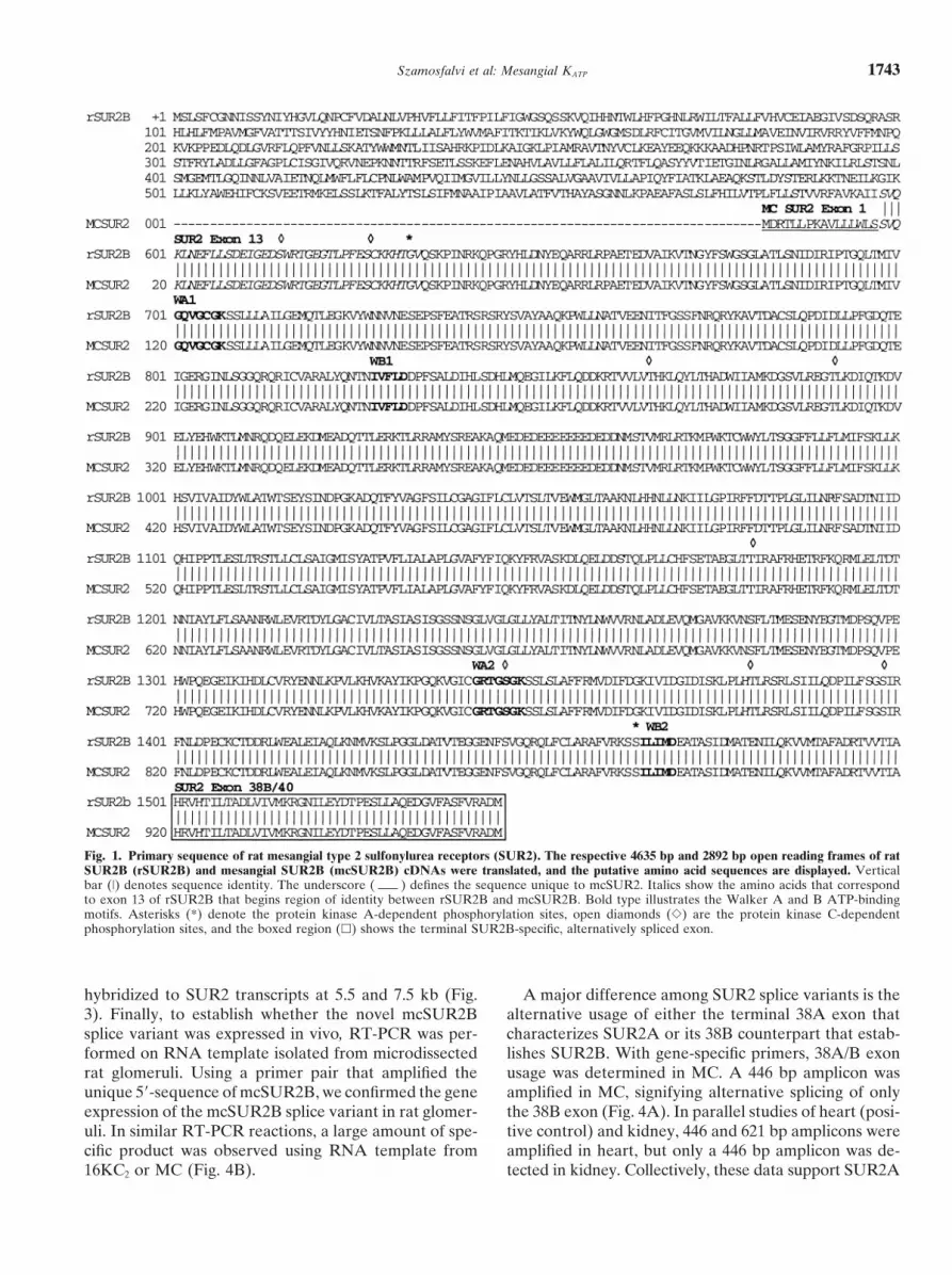
DNA sequencing. Three independent experiments were is succeeded by 948 aa that are identical to rSUR2Bin the region spanning exons 13 to 38B. The peptideconducted.alignment of the two mesangial SUR2 is displayed in
Immunoblotting Figure 1. Based on sequence identity between mcSUR2Band rSUR2B, the predicted topology of mcSUR2B asConfluent primary MC and 16KC2 monolayers were
scraped into an ice-cold buffer (pH 7.4), containing 20 compared to rSUR2B is displayed in Figure 2 and dem-onstrates that mcSUR2B lacks the initial 12 rSUR2Bmmol/L HEPES, 2 mmol/L ethylenediaminetetraacetic
acid (EDTA), 250 mmol/L sucrose, 0.1 mmol/L phenyl- exons [4]. This deletion renders a model whereby themcSUR2B N-terminus is intracytoplasmic. The putativemethylsulfonyl fluoride (PMSF), 1 mmol/L pepstatin,
and 1 mmol/L leupeptin, then immediately homogenized mcSUR2B retains the endoplasmic reticulum retentionsignal RKQ that prevents cell surface expression of in-in a glass dounce [26]. Brain tissue was snap-frozen in
liquid nitrogen immediately following sacrifice of the completely assembled KATP. It also has two nucleotidebinding folds, NBF1 and NBF2, that contain the Walkeranimal and pulverized in a liquid nitrogen-cooled mortar,
then homogenized. Tissue debris and nuclei were pel- A and B motifs common to all SUR, and the terminalexon 38B that imparts diazoxide sensitivity to SUR2B-leted out by centrifugation at 1000 � g for 10 minutes
three times. Crude membrane preparations were ob- containing KATP [38]. McSUR2B is predicted to have sixtransmembrane helices whose peptide sequence is identi-tained by centrifugation of clarified supernatants at
100,000 � g for 60 minutes. Subsequently 20-�g aliquots cal to the N-terminal transmembrane domain (TM2) ofrSUR2B. The binding sites for the potassium channelof total protein from membrane-enriched fractions of
MC, 16KC2 cells, or brain were separated by electropho- openers pinacidil and cromakalim were previously local-ized to this area in SUR2A and SUR2B [39–42]. Theresis on 10% SDS polyacrylamide slab gels. Membranes
were blocked in 5% dry milk/0.1% Tween/1� PBS solu- cytoplasmic side of the homologous domain in SUR1provides a high affinity-binding site for the sulfonylureation for 60 minutes and incubated with anti-SUR2 rabbit
antiserum (1:1000 dilution) that reacts with a conserved agents, tolbutamide and glibenclamide. Compared toSUR1 the TM2 in SUR2 has a significantly lesser affinityregion of rat SUR2A/B close to the C-terminus. Follow-
ing incubation with anti-rabbit HRP-conjugated second- for sulfonylureas [43–45].ary antibody, SUR2 proteins were demonstrated by a
Mesangial SUR2 gene expressionchemiluminescent assay [26, 34].These studies were performed to determine whether
the smaller SUR2 4.8 kbp cDNA obtained by RACERESULTS
cloning represents the dominant 5.5 kbp transcript,RACE cloning of mesangial SUR2 which we previously detected by Northern analysis with
a SUR2 probe (pJGAR7, 28). We generated a 1.9 kbpThe nested 5�-RACE reaction generated amplicons of0.6 kbp and 2.5 kbp and the nested 3�-RACE reaction Sac I fragment from the 2.5 kbp 5�-RACE product,
unique to the rSUR2B cDNA. Predictably, this fragmentyielded amplicons of sizes 3.9 kbp and 4.3 kbp. The 3.9kbp 3�-RACE amplicon was identical to the 4.3 kbp hybridized only to the mesangial SUR2 transcript at 7.5
kbp. Using PCR, a 215 bp probe was generated corre-product but was truncated by 400 bp in the 3� untrans-lated region (UTR). Alignments of the 2.5 kbp 5�-RACE sponding to the unique 5�-sequence of mcSUR2B. This
probe preferentially hybridized to the novel 5.5 kb SUR2and 4.3 kbp 3�-RACE contigs spanned the 4.6 kbprSUR2B cDNA coding region and reconstructed a 6.7 message, confirming that the 4.8 kbp cDNA represented
the 5.5 kb SUR2 mesangial transcript. Finally, the 4.3kbp SUR2B sequence. Within the coding sequence,100% identity with the rSUR2B sequence was estab- kbp Eco RI fragment of pJGAR7 that contains sequence
common to both the 4.8 and 6.7 kbp cDNAs predictablylished [37]. Further analysis demonstrated a unique 160
Szamosfalvi et al: Mesangial KATP 1743
Fig. 1. Primary sequence of rat mesangial type 2 sulfonylurea receptors (SUR2). The respective 4635 bp and 2892 bp open reading frames of ratSUR2B (rSUR2B) and mesangial SUR2B (mcSUR2B) cDNAs were translated, and the putative amino acid sequences are displayed. Verticalbar (|) denotes sequence identity. The underscore ( ) defines the sequence unique to mcSUR2. Italics show the amino acids that correspondto exon 13 of rSUR2B that begins region of identity between rSUR2B and mcSUR2B. Bold type illustrates the Walker A and B ATP-bindingmotifs. Asterisks (*) denote the protein kinase A-dependent phosphorylation sites, open diamonds (�) are the protein kinase C-dependentphosphorylation sites, and the boxed region (�) shows the terminal SUR2B-specific, alternatively spliced exon.
hybridized to SUR2 transcripts at 5.5 and 7.5 kb (Fig. A major difference among SUR2 splice variants is thealternative usage of either the terminal 38A exon that3). Finally, to establish whether the novel mcSUR2B
splice variant was expressed in vivo, RT-PCR was per- characterizes SUR2A or its 38B counterpart that estab-lishes SUR2B. With gene-specific primers, 38A/B exonformed on RNA template isolated from microdissected
rat glomeruli. Using a primer pair that amplified the usage was determined in MC. A 446 bp amplicon wasamplified in MC, signifying alternative splicing of onlyunique 5�-sequence of mcSUR2B, we confirmed the gene
expression of the mcSUR2B splice variant in rat glomer- the 38B exon (Fig. 4A). In parallel studies of heart (posi-tive control) and kidney, 446 and 621 bp amplicons wereuli. In similar RT-PCR reactions, a large amount of spe-
cific product was observed using RNA template from amplified in heart, but only a 446 bp amplicon was de-tected in kidney. Collectively, these data support SUR2A16KC2 or MC (Fig. 4B).

Szamosfalvi et al: Mesangial KATP1744
Fig. 2. Putative topology of the mesangialSUR2 protein and rat SUR2B. (Top) The ratmesangial SUR2B (mcSUR2B) protein iscomprised of a single transmembrane span-ning domain (TM) with 6 helices that areflanked by two cytoplasmic loops. Each loopcontains nucleotide-binding folds (NBF) withWalker A and B motifs that bind and hy-drolyze ATP. The N-terminus is predicted asintracytoplasmic, distinct from other SUR.The putative endoplasmic reticulum retentionsignal RKQ is shown. Protein kinase A (�)-and protein kinase C (�)-dependent phos-phorylation sites and the proposed bindingsites of various sulfonylurea compounds areindicated. (Bottom) Rat SUR2B topology.
and SUR2B isoform expression in heart, but only SUR2B Mesangial Kir6.1 and 6.2 gene expressionin kidney [6]. To establish mesangial Kir6.1/2 gene expression, RT-
Because our previous observations implied that sulfo- PCR was performed with gene-specific primers. In MC,nylureas, possibly through SUR2B, regulate MC contrac- a 402 bp fragment corresponding to Kir6.1 was robustlytility and extracellular matrix metabolism, investigations amplified, whereas the 318 bp fragment of Kir6.2 waswere carried out to determine whether glibenclamide minimally evident (Fig. 7A). As the Kir6.2 gene is in-regulated the mesangial expression of the SUR2B gene. tronless [11], the possibility existed that amplification ofIn two independent studies, glibenclamide (5 �M) did contaminating genomic DNA had contributed to thesenot influence SUR2B expression in serum-deprived MC results. However, PCR, in the absence of reverse tran-after 48 hours of drug exposure (Fig. 5). Longer incuba- scription generated no Kir6.2 products negating this pos-tion times were not evaluated. Because the 5.5 kb sibility. Lastly, in Northern analysis, transcripts of Kir6.1SUR2B mesangial transcript became more evident dur- were strongly detected at 1.7 and 2.7 kb in primary MC,ing growth of cells in culture (data not shown) and be- kidney cortex, whole kidney, and heart. The Kir6.2 tran-cause Kir6.1 transcripts increased during embryonic de- script of 4.1 kb was only identified in the positive controlvelopment, we sought to determine whether serum heart (Fig. 7B, C).regulated SUR2B expression. Interestingly, the level ofthe major 5.5 kbp transcript became undetectable follow-
DISCUSSIONing a 24-hour period of serum deprivation and thischange persisted for at least 48 hours. In contrast, serum- Data from Metzger and colleagues has previously im-
plied the presence of glomerular SUR [46, 47]. Thisdeprivation did not alter expression of the 7.5 kb tran-script (Fig. 5). group demonstrated both low- and high-affinity gliben-
clamide binding in metabolically active preparations ofMesangial SUR2 protein detection rat glomeruli. However, the cell type(s) responsible for
the binding were not unequivocally described and theUsing polyclonal anti-SUR2 antibody, multiple bandswere detected from Mr 50 to 170 kD in MC (Fig. 6). By presence of contaminating vasculature in their prepara-
tions may have accounted for the drug binding. This iscontrast, in the positive control brain tissue only a 170kD band was visualized. In membrane-enriched prepara- plausible because the existence of glomerular SUR, as
a component of afferent arteriolar KATP, has been estab-tions of primary MC and 16KC2 cells, a dominant bandwas detected at 108 kD, consistent with the predicted lished [48]. Moreover, sulfonylurea drug binding to glo-
merular KATP-bearing renin-producing cells also maysize of the translated mcSUR2B peptide. Lastly, lessersignal intensity was evident at �170 kD. have accounted for their results [49].

Szamosfalvi et al: Mesangial KATP 1745
Fig. 3. Mesangial SUR2 gene expression. Differential identification of rat mesangial cell transcripts for SUR2B was carried out with gene-specificprobes in northern analysis as described in the Methods section. (A) Rat kidney SUR2B (rSUR2B) and mesangial SUR2B (mcSUR2B) cDNAsare shown. Stippled portions correspond to regions of identity. Sac I sites (�34 to �1939 bp) define an rSUR2B-specific probe. Eco RI sites(�2440 to �6715 bp) define a probe that detects rSUR2B and mcSUR2B transcripts (pJGAR7). A 215 bp 5�-terminal fragment (�13 to �227bp) defines the mcSUR2B-specific probe. (B) The Sac I probe only detects the 7.5 kb SUR2B transcript. The mcSUR2B-specific probe stronglyhybridizes to the 5.5 kb transcript. The pJGAR7 probe hybridizes to transcripts at 5.5 and 7.5 kb.
Our data provide convincing evidence for the exis- of this clone may be attributable to an inability of ourRACE-based system to negotiate complex topology intence of at least two mesangial SUR2B, which likely
heterodimerize with Kir6.1 to form mesangial KATP. Two the 5�-UTR. Nevertheless, the sequence of mcSUR2B,the smaller mesangial SUR2B that corresponds to therat mesangial SUR2B cDNAs were RACE-cloned, and
the cDNAs corresponding to the two most abundantly 5.5 kb transcript, clearly identifies it as a member ofthe SUR2 family. From aa 17 to 964, mcSUR2B sharesexpressed mesangial SUR2 transcripts of 5.5 and 7.5 kb
characterized by Asano et al [28]. To date, the longest identity with the region of rSUR2B that spans exons 13to 38B. However, mcSUR2B is truncated at its N-ter-SUR2B clone that has been isolated is the 6.6 kb rSUR2B
cDNA obtained from a rat kidney library [37]. The cod- minus. This gene has a deletion of the 12 proximal N-ter-minal rSUR2B exons and a unique 16 aa insert at itsing region of our 6.7 kb SUR2B cDNA was 4635 bp
and essentially identical to rSUR2B, except for several amino terminus, discriminating it from other SUR2(Figs. 2 and 3). Our topological analysis of mcSUR2Bnucleotide differences that may represent species-spe-
cific variations and that do not affect the primary se- reveals a single, 6-helix transmembrane spanning do-main. By contrast, rSUR2B has three transmembranequence. In addition, the 5�-UTR of the 6.7 kb clone
contains a novel 160 bp sequence, which we presume domains (Fig. 3) [4, 37]. Additionally, the mcSUR2BN-terminus is predicted to be intracytosolic, as opposedresulted from cell-specific alternative splicing.
Despite the 0.7 kbp difference in length compared to to rSUR2B and other SUR2 wherein the N-termi-nus resides extracellularly [4]. Lastly, mcSUR2B retainsthe 5.5 kb mesangial SUR2B transcript, we ascertained
that our 4.8 kbp cDNA contains the same open reading the transmembrane domain corresponding to TM2 ofrSUR2B. The cytosolic side of this domain binds theframe as the larger message. Perhaps the foreshortening

Szamosfalvi et al: Mesangial KATP1746
Fig. 4. Expression of SUR2 splice variants in rat glomerulus and mes-angial cells (MC). RT-PCR reactions were carried out in rat heart,kidney, primary MC, and microdissected glomeruli, with gene-specificprimers that define expression of mcSUR2B, SUR2A, or SUR2B asdescribed in the Methods section. Gene products were visualized onethidium bromide stained gels. (A) SUR2A and SUR2B are detectedin heart. In kidney and MC, only SUR2B is expressed. No gene productis seen in the negative control (water). (B) The unique 215 bp 5�-ter-minal fragment of mcSUR2B is detected in 16KC2 and MC and microdis-sected glomeruli. No gene product is detected in the negative control(water).
Fig. 6. Detection of mesangial SUR2 proteins. Immunoblotting ofSUR2 proteins was carried out on membrane-enriched fractions of ratbrain, mesangial cells (MC) and 16KC2 cells. The rabbit polyclonalSUR2 antiserum detects common epitopes of SUR2A and SUR2B neartheir C-termini as described in the Methods section. In the positivecontrol tissue brain, the rat SUR2B is strongly detected at 170 kD. InMC and 16KC2 cells, the mesangial SUR2B is detected at Mr 108 kD,while the rat SUR2B is less strongly detected at Mr 170 kD.
lar mass of 108 kD of the dominant band agrees withthe size predicted from translation of the 2892 bp openreading frame of the 4.8 kbp mcSUR2B clone. The less
Fig. 5. Serum deprivation induces differential gene expression of strongly evident 170 kD band most likely characterizesSUR2B. Gene regulation of SUR2B by glibenclamide and serum was expression of the 6.7 kbp rSUR2B cDNA. Previous im-examined. Subconfluent mesangial cell cultures were maintained in 20%
munoblotting studies of protein isolates from cell linesserum or serum-deprived and exposed to glibenclamide (5 �M) fortimes shown. In Northern analysis, a probe that detects both the 7.5 transformed by SUR2A or SUR2B cDNA clearly dem-kb rat SUR2B and the 5.5 kb mesangial SUR2B transcript was used onstrated proteins with an apparent mass of 170 kD(pJGAR7). Serum-stimulated cells express 5.5 and 7.5 kb transcripts.
[50]. Interestingly, these full-length proteins were notAfter a 24-hour serum deprivation, the 5.5 kb mesangial-specific tran-script is undetectable and this down-regulation persists for at least detected from cardiac sarcolemma [51] or whole mouse48 hours. Exposure to glibenclamide did not influence SUR2 gene kidney crude membrane preparations [52], althoughexpression.
these tissues express full size SUR2A and SUR2B, re-spectively [7, 20, 37, 52]. Presumably, immunodetectionof full length SUR2 in MC (and murine cardiac sarco-lemma and whole kidney crude membrane preparations)potassium channel opener agents, pinacidil and croma-is difficult. On the other hand, the 108 kD band corre-kalim, with high affinity [39–42], and also contains a low-sponding to the abundant 5.5 kb mcSUR2B transcriptaffinity sulfonylurea binding site [43–45].
In immunoblotting experiments, the apparent molecu- is convincingly demonstrated and the relative intensities

Szamosfalvi et al: Mesangial KATP 1747
Fig. 7. Mesangial Kir6.x gene expression.RT-PCR reactions with gene-specific primersof Kir6.1 and Kir6.2 were carried out on 16KC2
cells and mesangial cells (MC). Reaction prod-ucts were visualized on an ethidium bromide-stained gel. (A) Strong Kir6.1 gene expressionis shown for MC and 16KC2 cells, while onlyminimal Kir6.2 gene expression is detected.(B) Kir6.1 cDNA was used as probe in north-ern analysis. Hybridization to transcripts atsizes shown is seen in renal cortex, MC, 16KC2
cells, and the positive control, heart. (C) Paral-lel studies with Kir6.2 cDNA only detected atranscript in the heart.
of the 170 kD and 108 kD signals on immunoblotting pertension and the concomitant distensile forces. How-ever, there was no alteration of MC SUR2 gene expres-roughly correspond to their hybridization signal intensi-
ties previously determined by Northern blotting [28]. sion by either stimulus, even after 48 hours of exposureto glibenclamide and stretch. However, we did documentThe human SUR2 gene spans more than 100 kbp and
contains no less than 39 exons [11]. Multiple tissue-spe- that serum deprivation of 24 to 48 hours potently down-regulated gene expression of the 5.5 kb mcSUR2B tran-cific splice variants of SUR2 have been documented in
Northern assays and by RT-PCR, and these variations script, while transcript levels of the 7.5 kb message re-mained unchanged. The physiological consequence ofmay contribute to the functional diversity of KATP [11].
The cDNAs characterized by these studies shared exten- this differential expression is unknown.In VSMC, SUR2B partners with Kir6.1 to form KATP,sive homology with mcSUR2B; however, the 5.5 kb tran-
script of mcSUR2B was not detected previously in cells and more rarely, Kir6.2 may represent the ion pore-forming subunit. Analogously, for MC to furnish an ionor tissues evaluated, suggesting that MC carry out a
unique alternative splicing event. The two splice variants, pore channel to mesangial KATP, they must also expressKir6.x. In MC, we established robust gene expression ofcardiac SUR2A and the vascular smooth muscle cell
(VSMC) SUR2B, differ in their expression of the 38A/B Kir6.1, and minimal Kir6.2 expression in RT-PCR andNorthern blotting studies. Consequently, our data favorC-terminal exon. A deletion of exon 38A, with preferen-
tial expression of exon 38B in SUR2B, renders VSMC the assembly of two mesangial KATP, heterodimers of(rSUR2B:Kir6.1)4 or (mcSUR2B:Kir6.1)4 [22]. SeveralKATP sensitive to the K� channel opener and vasodilator,
diazoxide [41, 42]. In contrast, cardiac KATP, a tetradimer investigations have established that the various SUR2exhibit differential electrophysiological characteristicsof SUR2A and KIR6.2, is diazoxide-insensitive. Our re-
sults indicate the exclusive usage of exon 38B by MC [54, 55]. Based on a recent study of Kir6.2 knockout mice,Kir6.1 represents the functional pore-forming subunit ofand kidney, consonant with the report of Beesley and
colleagues who characterized the expression of murine VSMC KATP [56]. With analogy to VSMC KATP, one ofthe mesangial KATP (rSUR2B:Kir6.1)4 would be predictedkidney SUR2 [52].
Thus far, regulation of the constitutively expressed to be activated by the potassium channel openers, pinaci-dil, nicorandil and diazoxide [39–42] and inhibited bySUR2 gene has not been defined. However, prolonged
exposure of pancreatic � cells to glibenclamide has been sulfonylureas [43–45]. In addition, this particular KATP
may be nucleoside diphosphate-regulated rather thanshown to increase SUR1 protein expression and to af-fects its translocation from the cytosol to membrane [53]. ATP-inhibited [22, 57, 58]. Finally, the second mesangial
would be assembled as (mcSUR2B:Kir6.1)4 and couldWe examined the effects of cyclical mechanical stretch(data not shown) and glibenclamide on mesangial SUR2 manifest an electrophysiologic profile different than
(rSUR2B:Kir6.1)4.gene expression because sulfonylureas are typically uti-lized as therapy in diabetic individuals whose glomeruli Given the ubiquitous expression of KATP and their
regulation by sulfonylureas, the question arises as toare exposed to the pernicious effects of glomerular hy-

Szamosfalvi et al: Mesangial KATP1748
receptors determines the pharmacological properties of ATP-sen-whether endogenous ligands for these channels exist [59].sitive K� channels. Neuron 16:1011–1017, 1996
At least for pancreatic � cell KATP (SUR1:Kir6.2)4, there 10. Quayle JM, Nelson MT, Standen NB: ATP-sensitive and in-wardly rectifying potassium channels in smooth muscle. Physiolis one such “endogenous sulfonylurea:” �-endosulfine.Rev 77:1165–1232, 1997This 121 aa polypeptide binds SUR1 with high affinity
11. Babenko AP, Aguilar-Bryan L, Bryan J: A view of SUR/KIR6.x,and stimulates insulin release through KATP inhibition KATP channels. Ann Rev Physiol 60:667–687, 1998
12. Aguilar-Bryan L, Clement JP, 4th, Gonzalez G, et al: Toward[60–62]. We have recently demonstrated that �-endosul-understanding the assembly and structure of KATP channels. Physiolfine gene (ENSA) is expressed in rat MC and glomeruliRev 78:227–245, 1998
(abstract, J Am Soc Nephrol 11:448A, 2000). We specu- 13. Aguilar-Bryan L, Nichols CG, Rajan AS, et al: Co-expressionof sulfonylurea receptors and KATP channels in hamster insulinomalate that autocrine endosulfine mediates sulfonylurea-tumor (HIT) cells. J Biol Chem 267:14934–14940, 1992type effects via SUR2B. The existence of such an auto-
14. Inagaki N, Inazawa J, Seino S: cDNA sequence, gene structure,crine, and possibly paracrine, system could have broad and chromosomal localization of the human ATP-sensitive potas-
sium channel, uKATP-1, gene (KCNJ8). Genomics 30:102–104,implications for diabetic glomerulopathy with its atten-1995dant pathophysiologic alterations of mesangial cell con-
15. Inagaki N, Gonoi T, Clement JP IV, et al: Reconstitution of IKATP:tractility and matrix metabolism. An inward rectifier subunit plus the sulfonylurea receptor. Science
270:1166–1170, 199516. Philipson LH, Steiner DF: Pas de deux or more: The sulfonylureaACKNOWLEDGMENTS receptor and K� channels. Science 268:372–373, 199517. Seino S: ATP-sensitive potassium channels: A model of heteromul-We wish to acknowledge the support of Dr. Balazs Szamosfalvi,
timeric potassium channel/receptor assemblies. Ann Rev PhysiolM.D., through a National Kidney Foundation of Michigan Fellowship61:337–362, 1999grant. We thank Drs. S.C. Hebert, C. Burant and S. Seino for their
18. Clement JP, IV, Kunjilwar K, Gonzalez G, et al: Associationkind provision of reagents used in the conduct of these studies.and stoichiometry of KATP channel subunits. Neuron 18:827–838,1997Reprint requests to Jerry Yee, M.D., Division of Nephrology and
19. Shyng S-L, Nichols CG: Octameric stoichiometry of the KATPHypertension, Henry Ford Hospital, 2799 West Grand Boulevard, CFP-channel complex. J Gen Physiol 110:655–664, 1997514, Detroit, Michigan 48202, USA.
20. Lorenz E, Terzic A: Physical association between recombinantE-mail: [email protected] ATP-sensitive K� channel subunits Kir6.2 and SUR2A. JMol Cell Cardiol 31:425–434, 1999
21. Nelson MT, Quayle JM: Physiological roles and properties ofAPPENDIX potassium channels in arterial smooth muscle. Am J Physiol 268:
C799–C822, 1995Abbreviations used in this article are: ABC, ATP-binding cassette;22. Yamada M, Isomoto S, Matsumoto S, et al: Sulphonylurea receptorANP, atrial natriuretic peptide; ATP, adenosine 5�-triphosphate; 2B and Kir6.1 form a sulphonylurea-sensitive but ATP-insensitiveCFTR, cystic fibrosis transmembrane regulator; DMSO, dimethyl sulf- K� channel. J Physiol 499:715–720, 1997oxide; ECL, enhanced chemiluminescence; FCS, fetal calf serum; KATP, 23. Isomoto S, Horio Y, Matsumoto S, et al: SUR2 and KCNJ8 genesATP-sensitive K� channel; Kir6.1, inwardly-rectifying K� channel 6.1; are tightly linked on the distal region of mouse chromosome 6.MC, mesangial cell; MESA, morpholinopropane sulfonic acid-EDTA- Mammal Genome 8:790–791, 1997
sodium acetate; MRP, multidrug resistance protein; RACE, rapid am- 24. Russ U, Hambrock A, Artunc F, et al: Coexpression with theplification of cDNA ends; RT-PCR, reverse transcription-polymerase inward rectifier K� channel Kir6.1 increases the affinity of thechain reaction; SUR, sulfonylurea receptor; SUR2B, type 2B sulfonyl- vascular sulfonylurea receptor SUR2B for glibenclamide. Molurea receptor; UTR, untranslated region; VSMC, vascular smooth Pharmacol 56:955–961, 1999muscle cells. 25. Anonymous: Tolazamide (Tolinase). Off Lit New Drugs 10:423–
427, 196626. Cortes P, Riser BL, Asano K, et al: Effects of oral antihyperglyce-REFERENCES
mic agents on extracellular matrix synthesis by mesangial cells.1. Gerich JE: Oral hypoglycemic agents. N Engl J Med 321:1231– Kidney Int 54:1985–1998, 1998
1245, 1989 27. Heilig CW, Concepcion LA, Riser BL, et al: Overexpression of2. Aguilar-Bryan L, Nichols CG, Wechsler SW, et al: Cloning of glucose transporters in rat mesangial cells cultured in a normal
the beta cell high-affinity sulfonylurea receptor: A regulator of glucose milieu mimics the diabetic phenotype. J Clin Invest 96:insulin secretion. Science 268:423–426, 1995 1802–1814, 1995
3. Higgins CF: The ABC of channel regulation. Cell 82:693–696, 1995 28. Asano K, Cortes P, Garvin JL, et al: Characterization of the rat4. Tusnady GE, Bakos E, Varadi A, Sarkadi B: Membrane topol- mesangial cell type 2 sulfonylurea receptor. Kidney Int 55:2289–
ogy distinguishes a subfamily of the ATP-binding cassette (ABC) 2298, 1999transporters. FEBS Lett 402:1–3, 1997 29. Zhao H, Loessberg PA, Sachs G, Muallem S: Regulation of
5. Schriml LM, Dean M: Identification of 18 mouse ABC genes intracellular Ca2� oscillation in AR42J cells. J Biol Chem 265:and characterization of the ABC superfamily in Mus musculus. 20856–20862, 1990Genomics 64:24–31, 2000 30. Hajjar RJ, Bonventre JV: Oscillations of intracellular calcium
6. Isomoto S, Kondo C, Yamada M, et al: A novel sulfonylurea induced by vasopressin in individual Fura-2-loaded mesangial cells.receptor forms with BIR (Kir6.2) a smooth muscle type ATP- J Biol Chem 266:21589–21594, 1991sensitive K� channel. J Biol Chem 271:24321–24324, 1996 31. Loessberg PA, Zhao H, Muallem S: Synchronized oscillation of
7. Chutkow WA, Simon MC, Le Beau MM, Burant CF: Cloning, Ca2� entry and Ca2� release in agonist-stimulated AR42Jcells. Jtissue expression, and chromosomal localization of SUR2, the puta- Biol Chem 2661:163–166, 1991tive drug-binding subunit of cardiac, skeletal muscle, and vascular 32. Mene P, Pugliese G, Pricci F, et al: High glucose inhibits cytosolicKATP channels. Diabetes 45:1439–1445, 1996 calcium signaling in cultured rat mesangial cells. Kidney Int 43:585–
8. Ammala C, Moorhouse A, Gribble F, et al: Promiscuous coupling 591, 1993between the sulfonylurea receptor and inwardly rectifying potas- 33. Mene P, Teti A, Pugliese F, Cinotti GA: Calcium release-acti-sium channels. Nature 379:545–548, 1996 vated calcium influx in cultured human mesangial cells. Kidney Int
46:122–128, 19949. Inagaki N, Gonoi T, Clement JP, IV, et al: A family of sulfonylurea

Szamosfalvi et al: Mesangial KATP 1749
34. Yee J, Kuncio GS, Bhandari B, et al: Identification of promoter impact of ATP-sensitive K� channels on afferent arteriolar diame-ter in diabetes mellitus. J Am Soc Nephrol 11:1199–1207, 2000activity and differential expression of transcripts encoding the mu-
rine stromelysin-1 gene in renal cells. Kidney Int 52:120–129, 1997 49. Russ U, Rauch U, Quast U: Pharmacological evidence for aKATP channel in renin-secreting cells from rat kidney. J Physiol35. Riser BL, Denichilo M, Cortes P, et al: Regulation of connective517:781–790, 1999tissue growth factor activity in cultured rat mesangial cells and its
50. Matsuo M, Tanabe K, Kioka N, et al: Different binding propertiesexpression in experimental diabetic glomerulosclerosis. J Am Socand affinities for ATP and ADP among sulfonylurea receptorNephrol 11:25–38, 2000subtypes, SUR1, SUR2A, and SUR2B. J Biol Chem 275:28757–36. Inagaki N, Tsuura Y, Namba N, et al: Cloning and functional28763, 2000characterization of a novel ATP-sensitive potassium channel ubiq-
51. Bienengraeber M, Alekseev AE, Abraham MR, et al: ATPaseuitously expressed in rat tissues, including pancreatic islets, pitu-activity of the sulfonylurea receptor: A catalytic function for theitary, skeletal muscle, and heart. J Biol Chem 270:5691–5694, 1995KATP channel complex. FASEB J 14:1943–1952, 200037. Tanemoto M, Vanoye CG, Dong K, et al: Rat homolog of sulfonyl-
52. Beesley AH, Qureshi IZ, Giesberts AN, et al: Expression ofurea receptor 2B determines glibenclamide sensitivity of ROMK2sulphonylurea receptor protein in mouse kidney. Pflugers Archin Xenopus laevis oocyte. Am J Physiol 278:F659–F666, 2000438:1–7, 199938. Matsuoka T, Matsushita K, Katayama Y, et al: C-terminal tails
53. Kawaki J, Nagashima K, Tanaka J, et al: Unresponsiveness toof sulfonylurea receptors control ADP-induced activation and di-glibenclamide during chronic treatment induced by reduction ofazoxide modulation of ATP-sensitive K� channels. Circ Res 87:ATP-sensitive K� channel activity. Diabetes 48:2001–2006, 1999873–880, 2000
54. Chutkow WA, Makielski JC, Nelson DJ, et al: Alternative splic-39. Shindo T, Yamada M, Isomoto S, et al: SUR2 subtype (A anding of SUR2 exon 17 regulates nucleotide sensitivity of the ATP-B)-dependent differential activation of the cloned ATP-sensitivesensitive potassium channel. J Biol Chem 274:13656–13665, 1999K� channels by pinacidil and nicorandil. Br J Pharmacol 124:985–
55. Davis Taber R, Choi W, Feng J, et al: Molecular characterization991, 1998of human SUR2-containing K(ATP) channels. Gene 256:261–270,40. Babenko AP, Gonzalez G, Bryan J: Pharmacotopology of sulfo-2000nylurea receptors. Separate domains of the regulatory subunits of
56. Suzuki M, Li R, Miki T, et al: Functional roles of cardiac andKATP channel isoforms are required for selective interaction with vascular ATP-sensitive potassium channels clarified by Kir6.2-K� channel openers. J Biol Chem 275:717–720, 2000 knockout mice. Circ Res 88:570–577, 200141. Lawson K: Potassium channel openers as potential therapeutic 57. Hambrock A, Loffler-Walz C, Kloor D, et al: ATP-Sensitiveweapons in ion channel disease. Kidney Int 57:838–845, 2000 K� channel modulator binding to sulfonylurea receptors SUR2A
42. Moreau C, Jacquet H, Prost AL, et al: The molecular basis of and SUR2B: Opposite effects of MgADP. Mol Pharmacol 55:832–the specificity of action of KATP channel openers. EMBO J 19:6644– 840, 19996651, 2000 58. Fujita A, Kurachi Y: Molecular aspects of ATP-sensitive K�
43. Dorschner H, Brekardin E, Uhde I, et al: Stochiometry of sulfo- channels in the cardiovascular system and K� channel openers.nylurea-induced ATP-sensitive potassium channel closure. Mol Pharmacol Therap 85:39–53, 2000Pharmacol 55:1060–1066, 1999 59. Virsolvy-Vergine A, Leray H, Kuroki S, et al: Endosulfine, an
44. Babenko AP, Gonzalez G, Bryan J: The tolbutamide site of endogenous peptidic ligand for the sulfonylurea receptor: Purifica-SUR1 and a mechanism for its functional coupling to KATP channel tion and partial characterization from ovine brain. Proc Natl Acadclosure. FEBS Lett 459:367–376, 1999 Sci USA 89:6629–6633, 1992
45. Gribble FM, Ashcroft FM: Sulfonylurea sensitivity of adenosine 60. Heron L, Virsolvy A, Peyrollier K, et al: Human alpha-endosul-triphosphate-sensitive potassium channels from beta cells and ex- fine, a possible regulator of sulfonylurea-sensitive KATP channel:trapancreatic tissues. Metabolism 49(Suppl 2):3–6, 2000 Molecular cloning, expression and biological properties. Proc Natl
46. Metzger F, Quast U: Binding of [3H]-P1075, an opener of ATP- Acad Sci USA 95:8387–8391, 1998sensitive K� channels, to rat glomerular preparations. Naunyn 61. Peyrollier K, Heron L, Virsolvy-Vergine A, et al: �-endosulfineSchmiedebergs Arch Pharmacol 354:452–459, 1996 is a novel molecule, structurally related to a family of phosphopro-
47. Metzger F, Loffler C, Quast U: Sulphonylurea binding in rat teins. Biochem Biophys Res Commun 223:583–586, 1996isolated glomeruli: Pharmacological characterization and depen- 62. Heron L, Virsolvy A, Apiou F, et al: Isolation, characterization,dence on cell metabolism and cytoskeleton. Naunyn Schmiedebergs and chromosomal localization of the human ENSA gene that en-Arch Pharmacol 355:141–149, 1997 codes alpha-endosulfine, a regulator of beta-cell KATP channels.
Diabetes 48:1873–1876, 199948. Ikenaga H, Bast JP, Fallett RW, Carmines PK: Exaggerated