effects of perinatal asphyxia on cell proliferation and neuronal phenotype evaluated with...
TRANSCRIPT
EAH
PPMa
Fb
PS
Aavoo
gwvpa(iwgvfiuc2bTGRpgaso
gmye
*EACigap
FFECTS OF PERINATAL ASPHYXIA ON CELL PROLIFERATIONND NEURONAL PHENOTYPE EVALUATED WITH ORGANOTYPIC
IPPOCAMPAL CULTURESKn
Hdatonbeetn2
ld1hiMpa1gtleitna
fhaes2cdIg1a
. MORALES,a P. REYES,a V. KLAWITTER,a
. HUAIQUÍN,a D. BUSTAMANTE,a J. FIEDLERb AND. HERRERA-MARSCHITZa*
Programme of Molecular and Clinical Pharmacology, ICBM, Medicalaculty, University of Chile, P.O. Box 70.000 Santiago 7, Chile
Department of Biochemistry and Molecular Biology, Chemical andharmaceutical Sciences Faculty, University of Chile, P.O. Box 233,antiago 1, Chile
bstract—The present report summarizes studies combiningn in vivo and in vitro approach, where asphyxia is induced inivo at delivery time of Wistar rats, and the long term effectsn hippocampus neurocircuitry are investigated in vitro withrganotypic cultures plated at postnatal day seven.
The cultures preserved hippocampus layering and re-ional subdivisions shown in vivo, and only few dying cellsere observed when assayed with a viability test at day initro 27. When properly fixed, cultures from asphyxia-ex-osed animals showed a decreased amount of microtubule-ssociated protein-2 immunocytochemically positive cells�30%), as compared with that from controls. The decreasen microtubule-associated protein-2 immunocytochemistryas particularly prominent in Ammon’s horn 1 and dentateyrus regions (�40%). 5-Bromo-2=deoxyuridine labeling re-ealed a two-fold increase in cellular proliferation in culturesrom asphyxia-exposed, compared with that from control an-mals. Furthermore, confocal microscopy and quantificationsing the optical disector technique demonstrated that inultures from asphyxia-exposed animals �30% of 5-bromo-=deoxyuridine-positive cells were also positive to microtu-ule-associated protein-2, a marker for neuronal phenotype.hat proportion was �20% in cultures from control animals.lial fibrillary acidic protein-immunocytochemistry and Fasted nuclear staining revealed that the core of the hippocam-us culture was surrounded by a well-developed network oflial fibrillary acidic protein-positive cells and glial fibrillarycidic protein-processes providing an apparent protectivehield around the hippocampus. That shield was less devel-ped in cultures from asphyxia-exposed animals.
The increased mitotic activity observed in this study sug-ests a compensatory mechanism for the long-term impair-ent induced by perinatal asphyxia, although it is not clear
et if that mechanism leads to neurogenesis, astrogliogen-sis, or to further apoptosis.
Corresponding author. Tel: �56-26786050; fax: �56-27372783.-mail address: [email protected] (M. Herrera-Marschitz).bbreviations: AM, Acetomethoxy; BrdU, 5-bromo-2=deoxyuridine;A1, CA2, CA3, Ammon’s horn 1, 2, 3; DG, dentate gyrus; DIV, days
n vitro; DMEM, Dulbecco’s modified Eagle medium; FGF2, fibroblastrowth factor; GFAP, glial fibrillary acidic protein; MAP, microtubule-
2ssociated protein; NGS, normal goat serum; P, days after birth; PBS,hosphate-buffered saline; PF, paraformaldehyde.
421
ey words: neonatal, anoxia, hippocampus, neurocircuitry,eurogenesis, rat.
ypoxia/ischemia at birth induces severe long-term neuro-evelopment impairments, resulting in spasticity, epilepsynd mental retardation when the insult is severe, or atten-ion-deficit hyperactivity syndrome and minimal brain dis-rder when it is mild (Boksa and El-Khodor, 2003). Theeurocircuitries of the basal ganglia have been shown toe particularly vulnerable to hypoxia/ischemia (Pasternakt al., 1991), but there is clinical (van Erp et al., 2001) andxperimental (Pulsinelli et al., 1982) evidence indicatinghat circuitries of the hippocampus are also extremely vul-erable to that type of insult (Harry and d’Hellencourt,003).
Perinatal asphyxia is a major cause of death and neuro-ogical injury in newborn babies, frequently associated toifficult or elongated birth processes (Berger and Garnier,999; Volpe, 2001). At the Karolinska Institutet, Stock-olm, Sweden, a model for investigating perinatal asphyxia
n the rat was proposed (Bjelke et al., 1991; Herrera-arschitz et al., 1993), demonstrating the effects on do-amine (Chen et al., 1997a,b,c; Kohlhauser et al., 1999),nd amino acid (Chen et al., 1997b; Kohlhauser et al.,999; Engidawork et al., 2001) neurocircuitries of the basalanglia. While the hippocampus is perhaps the most plas-ic structure of the CNS, playing a key role in memory andearning, little attention has been given in that model to theffect of perinatal asphyxia on the hippocampus, although
n the original paper by Bjelke et al. (1991), it was shownhat severe perinatal asphyxia induced a reduction in theumber of neural cell bodies in the Ammon’s horn (CA) 1nd CA3 regions, reflecting neuronal death.
Following global (Kirino, 1982; Kirino et al., 1984), orocal (Nakano et al., 1990) anoxia/ischemia, neurons of theippocampus show a delayed death that can occur daysfter the insult, involving neuronal cells in CA1 (Johansent al., 1992) and dentate gyrus (DG) (Wang et al., 1999),uggesting an apoptotic mechanism (Nakajima et al.,000). It has also been shown that anoxic/ischemic insultsan trigger several compensatory mechanisms to neuronaleath including neurogenesis (Gould and Tanapat, 1997).ndeed, neurogenesis has been observed in several re-ions of the brain (Gage, 2000), including DG (Liu et al.,998; Jin et al., 2001; Kee et al., 2001; Daval et al., 2004)nd the CA1 region (Nakatomi et al., 2002; Daval et al.,
004) of the hippocampus.
chcHwmfittwas
P
Pkwbtmwantm
O
DlciT(cpv(LpHCMh(tTtcmL
3
I
Gelcmnul
Ltp
I
FsAmi(a
m0nmaaFTipwtwtpaFTwrc
wiGPwbfLew(wciD
C
Cfscudcstdwbwt
P. Morales et al.
Thus, the present study investigated the long-termonsequences of perinatal asphyxia performed in vivo onippocampus organotypic cultures (Gähwiler, 1981), fo-using on: (i) in vitro cell survival, by direct monitoring withoffman’s microscopy, and labeling alive and dead cellsith a viability test; (ii) neuronal phenotype, by labelingicrotubule-associated protein (MAP)-2 positive cells in
xed material, and (iii) postnatal neurogenesis, by treatinghe cultures with 5-bromo-2=-deoxyuridine (BrdU) and ul-erior immunocytochemistry. (iv) Astrocyte proliferationas also examined using an antibody against glial fibrillarycidic protein (GFAP), together with Fast Red nucleartaining.
EXPERIMENTAL PROCEDURES
erinatal asphyxia
regnant Wistar rats within the last day of gestation (G22) wereilled by neck dislocation and hysterectomized. One or two pupsere removed immediately from a uterine horn and stimulated toreathe to be used as non-asphyxiated caesarean-delivered con-rols. The remaining fetus-containing uterine horns were im-ersed in a water bath at 37 °C for 20 min, and then the fetusesere removed from the uterine horns, stimulated to breathe andfter a 60 min observation period given to surrogate dams forursing, pending further experiments. Seven days after birth (P7),he pups were used for preparing organotypic cultures using aodification of a protocol developed by Gähwiler (1981).
rganotypic cultures
ifferent rat series (�10) were used for preparing cultures. Fol-owing decapitation, the brain was rapidly removed under sterileonditions and stored in a Petri dish containing Dulbecco’s mod-fied Eagle medium (DMEM; GIBCO BRL, Life Technologies AB,äby, Sweden). Coronal sections were cut with a microslicerDTK-2000, Dosaka, CO, Japan) at 350 �m thick and stored inold DMEM. Sections from the hippocampus were dissected andlaced on a coverslip (Nunc Thermanox Coverslips; Nunc, Naper-ille, IL, USA), containing a spread layer of chicken plasma25 �l), and further coagulated by a bovine thrombin ([Sigma, St.ouis, MO, USA]; 20 �l of a 20 �l/450 �l DMEM solution, freshlyrepared from frozen aliquots containing 1000 NIH units in 0.75 ml2O). The coverslips were then transferred to sterile Nunc flatT-tubes containing an un-buffered culture medium [50% Basaledium Eagle, 25% Hanks’ Balanced Salt Solution and 25%orse serum (GIBCO BRL), 0.5% glucose, 0.5 mM of L-glutamineSigma), and 0.1% antibiotic/anti-mycotic (GIBCO BRL)]. The cul-ures were grown at 35 °C, 10% CO2 in a Cell Incubator (ModelC2323, ShelLab, USA), with a roller device exposing the cultures
o gaseous or water phases every minute. After 3 days, theultures were transferred to a serum-free medium (Neurobasal-Aedium with B27 complement [GIBCO BRL], glucose 5 mM,
-glutamine 2.5 mM [Sigma]). The medium was changed every–4 days.
n vitro and ex vivo monitoring
rowth was periodically monitored with an inverted microscopequipped with Hoffmann optic (Nikon T100). Pictures were regu-
arly taken (4, 7, 14 and 21 days in vitro, DIV), and then theultures were treated with 10 �M of BrdU (Sigma), added to theedium for three days, and fixed with a formalin solution. Alter-atively, the cultures were analyzed for cell viability at DIV 27,sing ethidium-homodimer-1 and calcein-Acetomethoxy (AM) for
abeling dead and alive cells, respectively (Molecular Probes c
3224, Eugene, OR, USA). For quantification two samples wereaken from the body and border of the tissue, focusing on areasresenting the majority of ethidium-homodimer positive cells.
mmunocytochemistry
or immunocytochemistry, the cultures were fixed in a formalinolution (4% paraformaldehyde, PF; Sigma) for 45 min at 4 °C.fter rinsing cycles, the tissue was detached from the coverslip,ounted onto a gelatin-coated microscope slide for immunostain-
ng. Cellular proliferation was labeled with the mitotic marker BrdUMegabase, Lincoln, NB, USA), and neuronal phenotype with anntibody against MAP-2 (Sigma).
For MAP-2 immunocytochemistry, cultures were post-fixed inethanol 100% (30 min), rinsed three times and pre-incubated in.1 M phosphate-buffered saline (PBS), 0.1% Triton and 5%ormal goat serum (NGS) (Calbiochem, CA, USA) for 1 h. Aouse monoclonal antibody against MAP-2, immunospecific forll forms of mature and immature neurons (1:2000, Sigma), waspplied overnight at 4 °C in 0.1 M PBS, 0.1% Triton and 5% NGS.ollowing extensively washings, cultures were incubated in ayramide Amplification Kit #3 (TSATM, Molecular Probes), accord-
ng to the instructions of the supplier. After that, the cultures wereost-fixed in 4% PF for 15 min at 4 °C. The cultures were thenashed extensively. DNA denaturation was achieved by treating
he slices with 2 N HCL for 30 min at 37 °C. They were extensivelyashed in 0.1 M PBS before pre-incubation for 1 h at room
emperature in 0.1 M PBS, 0.1% Triton and 5% NGS. A rabbitolyclonal antibody against anti-BrdU (1:4000, Megabase) waspplied overnight at 4 °C in 0.1 M PBS, 0.1% Triton and 5% NGS.ollowing extensively washings, cultures were incubated in theSATM kit #12. The sections were washed again, coverslippedith DAKO fluorescent mounting medium (DAKO Corp, Carpinte-
ia, CA, USA) and examined in an epi-fluorescence inverted mi-roscope.
For GFAP, PF fixed tissue was washed in PBS, pre-incubatedith 5% of NGS, 0.1% Triton X-100, in PBS, for 1 h at 37 °C, and
ncubated overnight with a mouse monoclonal antibody againstFAP (Sigma) (1:2000 diluted in 5% NGS, 0.1% Triton X-100, inBS). Following extensively washings, the slices were treatedith a biotinylated anti-mouse IgG (1:500 in PBS) for 1 h, followedy a further incubation with a streptavidin phosphatase complexor 1 h, rinsed and incubated with a levamisole solution (Vectoraboratories, Burlingame, CA, USA) for 15 min, to inhibit thendogenous alkaline phosphatases. The reaction was visualizedith a 5-bromo-4-chloro-3-indoyl-phosphate/nitroblue tetrazolium
BCIP/NBT) substrate kit (Vector Laboratories). Then, the slicesere counterstained with Fast Red (Sigma) for labeling cell nu-leus. Sections were dehydrated through graded alcohols, clearedn xylene and coverslipped in entellan mounting medium (Merck,armstadt, Germany).
onfocal microscopy–optical disector quantification
onfocal microscopy was performed using a Zeiss LSM410 con-ocal laser-scanning microscope with a 633 (1.4 N.A.) oil immer-ion objective lens. MAP-2 or BrdU-positive cells in hippocampalultures were counted by an investigator blinded to the treatment,sing the optical disector technique described in detail by Gun-ersen et al. (1988). Briefly, MAP-2 or BrdU-positive nuclei wereounted as they came into focus while scanning through theection. The disector height (h) was set at 10 �m and nuclei withinhe first 3 �m of the section were not counted. The area ofissector (adis) was set at 4.5�104 �m2. The area of culture (a)as measured through an image J 1.32 software. The total num-er of MAP-2 or BrdU-positive nuclei in each hippocampus cultureas then estimated as N��Q��t/h�a/adis; where �Q� is the
otal number of counted MAP-2 or BrdU positive nuclei in each
ulture; t, the average slice thickness; a, the area of culture; adis,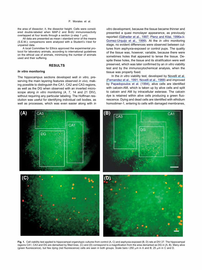
teo
(u
tou
I
Tsiaswow
vprGstossptt
(bwtdrh
Fr(
P. Morales et al.
he area of dissector; h, the dissector height. Cells were consid-red double-labeled when MAP-2 and BrdU immunoreactivityverlapped at four levels through a section (z-step 1 �m).
All data are presented as meanstandard error of the meansS.E.M.); comparisons were analyzed with a Student’s t-test fornpaired data.
A local Committee for Ethics approved the experimental pro-ocol for laboratory animals, according to international guidelinesn the ethical use of animals, minimizing the number of animalssed and their suffering.
RESULTS
n vitro monitoring
he hippocampus sections developed well in vitro, pre-erving the main layering features observed in vivo, mak-ng possible to distinguish the CA1, CA2 and CA3 regions,s well as the DG when observed with an inverted micro-cope along in vitro monitoring (4, 7, 14 and 21 DIV),ithout requiring any particular labeling. The Hoffman res-lution was useful for identifying individual cell bodies, asell as processes, which was even easier along with in
ig. 1. Cell viability test applied to hippocampal organotypic cultures fr
egions CA1, CA3 and DG are demarked by filled lines. (C) and (D) correspondgreen fluorescence), but few dying (red fluorescence) cells are seen in both gitro development, because the tissue became thinner andresented a quasi monolayer appearance, as previouslyeported (Gähwiler et al., 1997; Plenz and Kitai, 1996a,b;omez-Urquijo et al., 1999). At the in vitro monitoring
tage, no evident differences were observed between cul-ures from asphyxia-exposed or control pups. The qualityf the tissue was, however, variable, because there wereometimes holes that appeared to tense the tissue. De-pite these holes, the tissue and its stratification were wellreserved, which was later confirmed by an in vitro viabilityest and by the immunocytochemical analysis, when theissue was properly fixed.
In the in vitro viability test, developed by Novelli et al.Fernandez et al., 1991; Novelli et al., 1988) and improvedy Papadopoulos et al. (1994), alive cells are identifiedith calcein-AM, which is taken up by alive cells and split
o calcein and AM by intracellular esterase. The calceinye is retained within alive cells producing a green fluo-escence. Dying and dead cells are identified with ethidiumomodimer-1, entering to cells with damaged membranes,
l (A, C) and asphyxia-exposed (B, D) rats at DIV 27. The hippocampal
om contro to a magnification from the area demarked as DG in (A, B). Many aliveroups. Scale bars�250 �m in A and B; 25 �m in C and D.
btibswFscidooiots
M
Wasw(ccp(ovehi(
B
ABiscteB
Dm
Dpf(i
t5ptmoraoMoav
G
IaccmmwrpwecGaf
TouqdVla12cs
awop
Tcmr
S
BB
P. Morales et al.
inding to nucleic acids to produce a red fluorescence. Inhe present study, the majority of the cells showed anntensively green fluorescence surrounded by a greenishackground (Fig. 1). Some migrating cells could be ob-erved, surrounding the body of the tissue or in the holes,hich stretched strips of well-preserved greenish tissue.ew ethidium-homodimer positive cells (red) were ob-erved in cultures from both asphyxia-exposed (n�3) andontrol (n�2) pups, often on the top of the tissue, but alson the holes (Fig. 1). For quantification, ethidium-homo-imer positive cells were counted in 1-mm2 samples ofrganotypic cultures at DIV 27, from the body and borderf the tissue. Table 1 shows that there was an apparent
ncrease in the number of red cells in the body of the tissuef cultures from asphyxia-exposed animals compared withhat seen in controls, but the difference did not reach thetatistic level.
AP-2 immunostaining
hen PF-fixed (24–27 DIV), and treated with antibodygainst MAP-2, it was evident that many of the cells ob-erved in the cultures presented a neuronal phenotype,ith cell bodies in regions identified as CA1, CA3 and DG
Fig. 2A, B). When inspected with confocal microscopy,ultures from asphyxia-exposed pups showed a de-reased number of MAP-2 positive cells (n�4), as com-ared with that seen in cultures from control animalsn�5) (c.f. Fig. 2C versus 2D). Quantification, by using theptical disector technique (Gundersen et al., 1988), re-ealed that the decrease was significant, whether consid-ring the number of MAP-2 positive cells in the wholeippocampus tissue (3216%) (Fig. 3A), or cells counted
n CA1 (408%) and DG (4214%) regions, respectivelyFig. 3B).
rdU immunostaining
fter in vitro treatment with the mitotic marker BrdU (10 �MrdU to the culture medium for three days), fixation and
mmunocytochemistry, many BrdU positive cells wereeen in all cultures, but the total amount of BrdU positiveells was larger in cultures from asphyxia-exposed (n�5)han that from control animals (n�5) (Fig. 4). That differ-nce was even larger when expressed as the number of
able 1. Number of ethidium-homodimer positive cells (red) in hippo-ampal organotypic cultures from control and asphyxia-exposed ani-als at DIV 27, measured in 1 mm2 samples from body and border
egions of the tissue
ampled region Cultures from controlanimals (n�2)
Cultures from asphyxia-exposed animals (n�3)
ody 5537 7646order 9926 9224
rdU positive cells/mm3 (�two-fold). 2
ouble BrdU/MAP-2 immunostaining: confocalicroscopy
ouble staining revealed that approximately 25% of BrdUositive cells were also positive for MAP-2, in culturesrom both control (216%; n�5) and asphyxia-exposed3211%, n�4) animals, respectively (Fig. 4). The major-ty of doubled-stained cells were observed in DG.
Fig. 5 shows confocal cases observed in DG of cul-ures from control (Fig. 5A–C), and asphyxia-exposed (Fig.D–F) animals. Several BrdU positive nuclei and MAP-2erikarya can be seen in panels A, D and B, E, respec-ively, showing double staining when the pictures areerged (C, F). The double staining was confirmed whenbserved at four levels through a section. Double stainingevealed that BrdU/MAP-2-positive cells in cultures fromsphyxia-exposed animals (Fig. 5F) formed clusters of twor more cells. When compared with the controls, BrdU/AP-2 positive cells showed a reduced dendritic devel-pment, indicating, perhaps, that cells in cultures fromsphyxia-exposed animals are immature (c.f. Fig. 5Cersus 5F).
FAP- immunostaining
n both groups, GFAP immunostaining (DIV 27) revealedbundant cell bodies and processes at the border of theultures, with few GFAP-positive cells in the core of theultures, core that was intensively labeled with the nucleararker Fast Red (Fig. 6). GFAP-positive cells showed aultipolar feature, with processes extending through aide area of the tissue. The GFAP-positive network sur-
ounded the core of the cultures, appearing to provide arotective shield to the cultures. No apparent differencesere observed between cultures from control and asphyxia-xposed animals regarding the amount of GFAP-positiveells, but in cultures from control animals (Fig. 6A), theFAP network was well organized, while in cultures fromsphyxia-exposed animals (Fig. 6B) that network wasound to be less tight and the border less outlined.
DISCUSSION
he experimental model used in the present study wasriginally developed by Bjelke et al. (1991), proposing aseful model for studying the short- and long-term conse-uences of perinatal asphyxia, still a main pediatric issue,espite the improvements in neonatal care (Hill, 1991;olpe, 2001). A main feature of the model is that it is
argely non-invasive, allowing to use the asphyxia-exposednimal for long-term studies (Loidl et al., 1994; Chen et al.,997a,b,c; Kohlhauser et al., 1999; Gross et al., 2000,005; Bernert et al., 2003), or for preparing organotypicultures (Morales et al., 2003; Klawitter et al., 2005), ashown in the present study.
We show here that hippocampus tissue from controlnd asphyxia-exposed animals survived and developedell when explanted at P7 and kept alive for approximatelyne month as organotypic cultures following a protocolroposed by Gähwiler and collaborators (Raineteau et al.,
004). The cultures preserved the typical layering and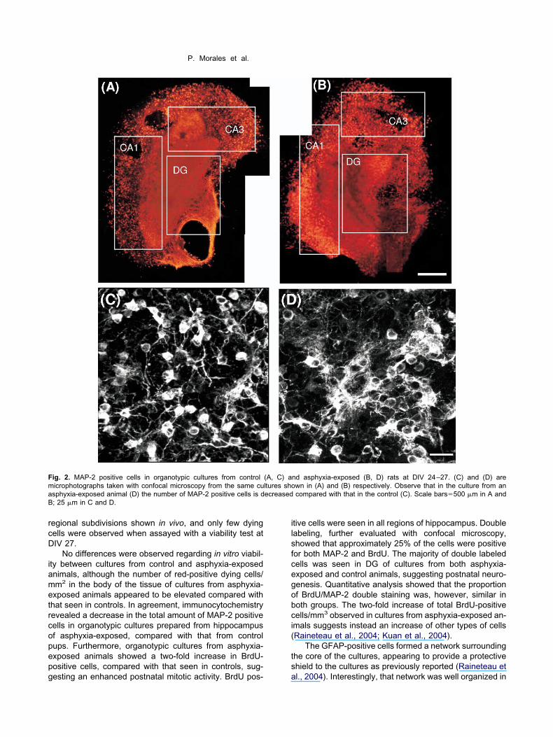
rcD
iametrcopepg
ilsfcegobci(
ts
FmaB
P. Morales et al.
egional subdivisions shown in vivo, and only few dyingells were observed when assayed with a viability test atIV 27.
No differences were observed regarding in vitro viabil-ty between cultures from control and asphyxia-exposednimals, although the number of red-positive dying cells/m2 in the body of the tissue of cultures from asphyxia-xposed animals appeared to be elevated compared withhat seen in controls. In agreement, immunocytochemistryevealed a decrease in the total amount of MAP-2 positiveells in organotypic cultures prepared from hippocampusf asphyxia-exposed, compared with that from controlups. Furthermore, organotypic cultures from asphyxia-xposed animals showed a two-fold increase in BrdU-ositive cells, compared with that seen in controls, sug-
ig. 2. MAP-2 positive cells in organotypic cultures from controlicrophotographs taken with confocal microscopy from the same cusphyxia-exposed animal (D) the number of MAP-2 positive cells is d; 25 �m in C and D.
esting an enhanced postnatal mitotic activity. BrdU pos- a
tive cells were seen in all regions of hippocampus. Doubleabeling, further evaluated with confocal microscopy,howed that approximately 25% of the cells were positiveor both MAP-2 and BrdU. The majority of double labeledells was seen in DG of cultures from both asphyxia-xposed and control animals, suggesting postnatal neuro-enesis. Quantitative analysis showed that the proportionf BrdU/MAP-2 double staining was, however, similar inoth groups. The two-fold increase of total BrdU-positiveells/mm3 observed in cultures from asphyxia-exposed an-mals suggests instead an increase of other types of cellsRaineteau et al., 2004; Kuan et al., 2004).
The GFAP-positive cells formed a network surroundinghe core of the cultures, appearing to provide a protectivehield to the cultures as previously reported (Raineteau et
nd asphyxia-exposed (B, D) rats at DIV 24–27. (C) and (D) arewn in (A) and (B) respectively. Observe that in the culture from ancompared with that in the control (C). Scale bars�500 �m in A and
(A, C) altures shoecreased
l., 2004). Interestingly, that network was well organized in
camnmSd
Ga
a((m
F2ai ared with
Facoa
P. Morales et al.
ultures from control animals, but appeared to be less tightnd less outlined in cultures from asphyxia-exposed ani-als, suggesting perhaps an increased vulnerability tooxious substances present in the surrounding environ-ent. As previously reported (Strasser and Fischer, 1995;cheepens et al., 2003; Bartley et al., 2005), no apparentifferences were observed regarding the total amount of
0
50
100
150
200
250
CA1
0
200
400
600
800
1000
1Con
Tota
l n
um
be
r o
f M
AP
-2
positiv
e la
belle
d c
ells
(x1
03)(A)
(B)
*
MA
P-2
positiv
e lab
elle
d
ce
lls/m
m3
(x10
3)
CA1
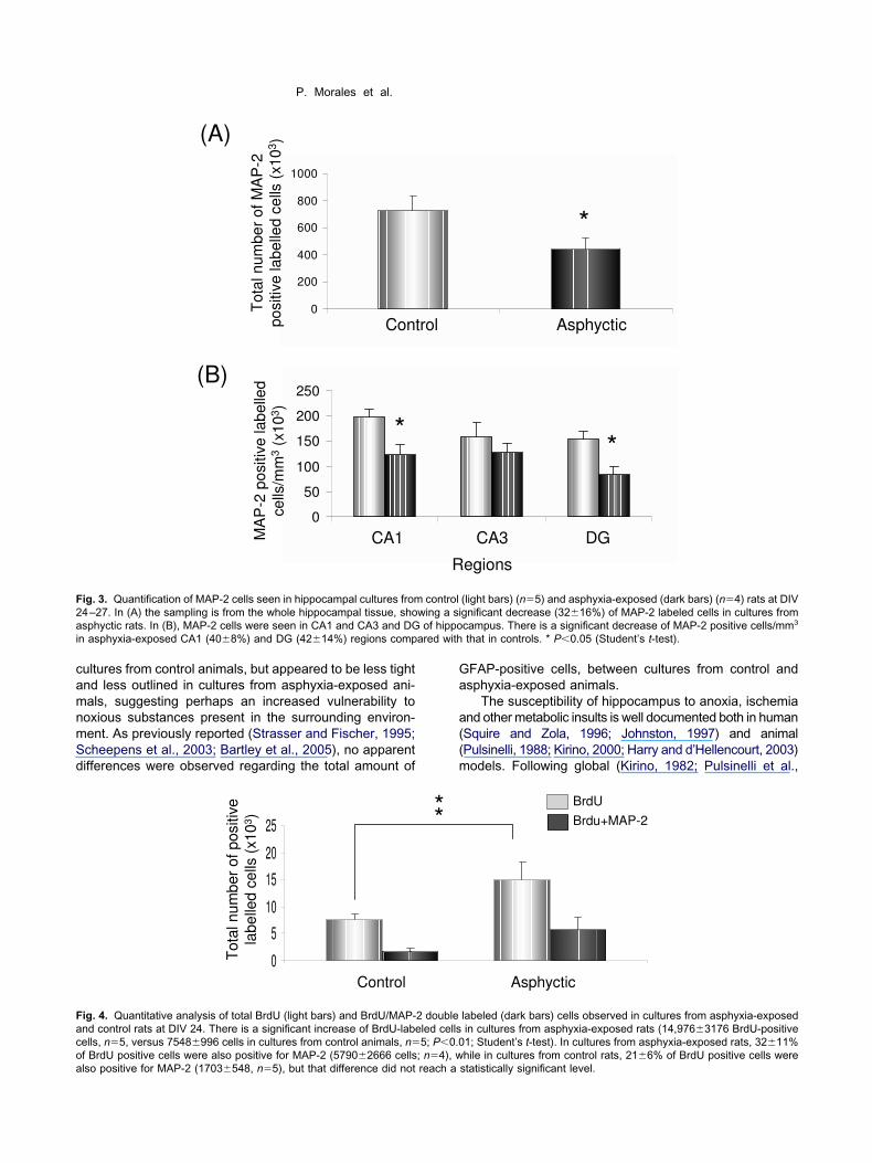
ig. 3. Quantification of MAP-2 cells seen in hippocampal cultures from4–27. In (A) the sampling is from the whole hippocampal tissue, shosphyctic rats. In (B), MAP-2 cells were seen in CA1 and CA3 and DG
n asphyxia-exposed CA1 (408%) and DG (4214%) regions comp
0
5
10
15
20
25
Control
Tota
l num
ber
of positiv
e
labelle
d c
ells
(x10
3)
ig. 4. Quantitative analysis of total BrdU (light bars) and BrdU/MAP-nd control rats at DIV 24. There is a significant increase of BrdU-labeells, n�5, versus 7548996 cells in cultures from control animals, n�
f BrdU positive cells were also positive for MAP-2 (57902666 cells; n�4), wlso positive for MAP-2 (1703548, n�5), but that difference did not reach aFAP-positive cells, between cultures from control andsphyxia-exposed animals.
The susceptibility of hippocampus to anoxia, ischemiand other metabolic insults is well documented both in humanSquire and Zola, 1996; Johnston, 1997) and animalPulsinelli, 1988; Kirino, 2000; Harry and d’Hellencourt, 2003)odels. Following global (Kirino, 1982; Pulsinelli et al.,
CA3 DG
2
*
Asphyctic
*
egions
CA3 DG
(light bars) (n�5) and asphyxia-exposed (dark bars) (n�4) rats at DIVgnificant decrease (3216%) of MAP-2 labeled cells in cultures fromcampus. There is a significant decrease of MAP-2 positive cells/mm3
that in controls. * P0.05 (Student’s t-test).
1 Asphyctic
BrdU
Brdu+MAP-2
labeled (dark bars) cells observed in cultures from asphyxia-exposedin cultures from asphyxia-exposed rats (14,9763176 BrdU-positive1; Student’s t-test). In cultures from asphyxia-exposed rats, 3211%
trol
R
controlwing a siof hippo
**
2 doubleled cells5; P0.0
hile in cultures from control rats, 216% of BrdU positive cells werestatistically significant level.

11ai((sseab(tdap(wa
itdpadsab(
tp
te2cpEwpppcaCThmrNppa
BegM
Fa ly. (B) ant
P. Morales et al.
982; Kirino et al., 1984), or focal ischemia (Nakano et al.,990) in adult animals, neurons of the hippocampus show
delayed death that can be evident days after thensult, mainly seen in CA1 (Johansen et al., 1992) and DGWang et al., 1999), suggesting an apoptotic mechanismJohnston, 2000; Nakajima et al., 2000). It has also beenhown that anoxia/ischemia can trigger several compen-atory mechanisms to neuronal death, including neurogen-sis (Gould and Tanapat, 1997; Liu et al., 1998; Kee etl., 2001; Daval et al., 2004). These changes have alsoeen observed following perinatal or neonatal asphyxiaJohnston et al., 2001; Volpe, 2001). Furthermore, usinghe present model for inducing perinatal asphyxia, it wasemonstrated that the hippocampus suffers a significantmount of neuronal loss following severe perinatal as-hyxia without changes in hippocampal volume at P21van de Berg et al., 2001). A similar result was reportedhen analyzing asphyxia-exposed guinea-pig three monthsfter the perinatal insult (Bernert et al., 2003).
The effect of global hypoxia/ischemia has been furthernvestigated with a stereological method, demonstratinghe peculiar vulnerability of CA1 pyramidal neurons andentate hilar interneurons (Larsson et al., 2001). Cell lossrobably relates to (i) energy failure; (ii) free radical dam-ge; (iii) cytokines and excitotoxicity and/or (iv) caspase-ependent cell death, occurring immediately after the in-ult, or progressing with time upon reperfusion/reoxygen-tion. It has been suggested that the immediate cell loss isy necrosis, while delayed neuronal death is by apoptosis
ig. 5. Cell proliferation and neuronal phenotype evaluated at DIV 24 wsphyxia-exposed (D–F) rats. (A) and (D) show BrdU positive cells onhe pictures. Scale bar�10 �m.
Northington et al., 2001), and that neuronal loss can fur- l
her lead to damage by target deprivation of trophic sup-ort (Miller and Kuhn, 1997).
Apoptosis has been shown to play a prominent role inhe neurodegeneration observed following hypoxia/isch-mia in newborn rats (Johnston, 2000; Nakajima et al.,000). Thus, a significant increase in the activity ofaspase-3, a cysteine protease involved in the executionhase of apoptosis (Yuan et al., 1993; Chen et al., 1998;ndres et al., 1998; Namura et al., 1998; Ni et al., 1998)as reported to occur in hippocampus one week aftererinatal asphyxia (Daval et al., 2004; van de Berg et al., inreparation). In our own in vivo studies (Morales et al., inreparation), using Hoechst DNA and terminal deoxynu-leotidyl transferase-mediated UTP nick labeling, we havelso found an enhanced amount of apoptotic nuclei in CA1,A3 and GD regions, seven days after perinatal asphyxia.his peak in apoptotic cell loss observed one week after aypoxic/ischemic insult appears to reflect a strict molecularechanism, associated with a spontaneous anatomical
ecovery reported by several authors (Sharp et al., 2002;akatomi et al., 2002) and with a shift in the expression ofro-apoptotic (BAX and caspase-3), to the anti-apoptoticrotein Bcl-2, recorded at 20 days post-hypoxia (Daval etl., 2004).
In the present study, we found a two-fold increase inrdU-positive cells in organotypic cultures from asphyxia-xposed compared with that seen in control animals, sug-esting an enhanced postnatal neurogenesis, althoughAP-2 positive cells corresponded only to a 1⁄4 of those
cal microscopy in DG of hippocampal cultures from control (A–C) andd (E) show MAP-2 positive cells only. (C) and (F) are the merging of
ith confo
abeled for BrdU. However, we used here a Sigma mono-

cniapic
imafmDpcren7tNG
o(tFGsnDtfsh(pciro
pb
Fatbs cting thei ) that net
P. Morales et al.
lonal antibody that reacts with all forms of MAP-2, a to c,ot being able to differentiate among juvenile and adult
soforms of MAP-2. It is possible that with a more specificntibody we could demonstrate that some of the BrdU-ositive cells observed here represent neurons in a more
mmature developmental stage, or even immature astro-ytes or other types of glial cells (Raineteau et al., 2004).
Several studies have shown that experimental anoxia/schemia can lead to neurogenesis as a compensatory
echanism for neuronal cell death in hippocampus (Liu etl., 1998; Kee et al., 2001). New neurons can generaterom the subgranular cell layer of the DG, perhaps evenigrating to CA1 and CA3 regions (Nakatomi et al., 2002;aval et al., 2004). While less possible in the presentreparation, cells migrating from the periventricular zonean also contribute to re-placement of neurons of the CA1egion (Daval et al., 2004). In adult animals, global isch-mia produces a several-fold increase in the birth rate ofew cells in the subgranular zone of the DG, beginning atdays after the insult, peaking at 11 days and decreasing
hereafter (Sharp et al., 2002), news cells becomingeuN-, calbindin- and MAP-2-immunostained neurons, or
ig. 6. GFAP-immunostaining (blue) and Fast Red nuclear staininsphyxia-exposed (B) rats. Observe that the nuclear staining (red) is chat core are clearly distinguished, showing a multipolar feature, witetween cultures from control and asphyxia-exposed animals. In thurrounding the core of the cultures, providing an apparent barrier protes well organized, while in cultures from asphyxia-exposed animals (B
FAP-labeled astrocytes (Kee et al., 2001). m
It has been shown that there is an increase in the levelsf basic fibroblast growth factor (FGF2) following hypoxiaAndersson et al., 1995), a protein expressed by astrogliahroughout the forebrain (Ganat et al., 2002). However,GF2-positive astrocytes do not show any detectableFAP staining, suggesting a reversion to an immature cell
tage, perhaps re-entering to the cell cycle and leading toeuronal regeneration, or resuming apoptosis-associatedNA synthesis (Kuan et al., 2004). In the present study,
here was a wide amount of BrdU-positive cells in culturesrom both asphyxia-exposed and control animals, not re-tricted to DG, but also comprising regions of the Ammon’sorn, in agreement with that reported by Rietze et al.2000). Since only 1⁄4 of BrdU-positive cells were alsoositive to MAP-2, a significant amount of BrdU-positiveells shown in the present study may represent cells at an
mmature stage, potentially leading to neurogenesis via aadial glia pathway (Ganat et al., 2002; Kuan et al., 2004),r just gliogenesis.
Perinatal asphyxia may trigger gliogenesis, but no ap-arent increase in the number of GFAP-positive cells coulde demonstrated in cultures from asphyxia-exposed ani-
bserved at 27 DIV in hippocampal cultures from control (A) andted in the core of the cultures. Individual GFAP positive (blue) cells ines extending for �50 �m. No apparent differences were observedery of the cultures a strong GFAP-positive network can be seen,border of the cultures. In cultures from control animals (A) that barrier
work is less tight and the border is less outlined. Scale bar�250 �m.
g (red) ooncentrah processe periph
als, compared with that from controls. Using the same

mhEacst
ondcIiMopcruatitGwepbhs2lfirc((sKdpifashn
Tvvhonoc
DstBaeicdcfifevfo
AFeMca
A
B
B
B
B
B
C
C
C
C
P. Morales et al.
odel, Scheepens et al. (2003) did not find any change inippocampus GFAP content assayed in vivo with anLISA method (O’Callaghan, 1991), within a 15 days’ post-sphyxia period. A lack of effect on GFAP-stained astro-ytes was also reported by Strasser and Fischer (1995),tudying the effect of combined oxygen/glucose depriva-ion in organotypic hippocampal cultures.
Interestingly, however, using a chronic neonatal hyp-xia model, Ganat et al. (2002) observed a decreasedumber of GFAP-immunoreactive cells, but an increasedensity of vimentin-positive cells, a marker for radial glialells and immature astrocytes (Pixley and de Villis, 1984).n the same line, it was reported that transient forebrainschemia in adult rats can induce the expression of
usashi1 (Msi1) and nestin proteins by reactive astrocytesf the hippocampus (Yagita et al., 2002). Msi1 is a proteinresent in ependymal and subependymal cells and astro-ytes, but not in microglia, oligodendrocyte or mature neu-ons (Sakakibara et al., 1996), while nestin is a proteinsed as a neural stem/progenitor cell marker (Lendhal etl., 1990). While almost all Msi1 and nestin immunoreac-ivity was co-localized with GFAP in the CA1 region afterschemia, Msi1-, but not nestin-positive cells were showno form clusters after ischemia, co-expressing BrdU but notFAP, in the subgranular zone of the hippocampal DG,hereas almost all nestin-positive cells in that region alsoxpressed GFAP (Yagita et al., 2002). The increased ex-ression of nestin in subgranular zone of the DG followingrain injury has led to suggest that reactive astrocytesave the potential ability to revert to an undifferentiatedtage (Doetsch et al., 1999; Seri et al., 2001; Yagita et al.,001, 2002). Raineteau et al. (2004) have monitored cel-
ular proliferation in hippocampal organotypic cultures,nding numerous BrdU-positive cells at the surface of allegions of the slices, the larger proportion in DG (�22ells/250 �m2), many positive to GFAP, showing type-Idifferentiated during embryonic development) and type-2reactive astrocytes) morphology, co-expressing nestin, aseen in organotypic cultures from rat cortex (Schmidt-astner and Humpel, 2002). Raineteau et al. (2004) alsoemonstrated, as shown here, that astrocytes could form arotective shield around the hippocampal tissue. Interest-
ngly, that shield appears to be less developed in culturesrom asphyxia-exposed animals, suggesting that there isn increased vulnerability for noxious substances from theurrounding environment, as well as an impairment in theomeostasis of the extracellular compartment required foreurotransmission.
CONCLUSIONS
he present report summarizes studies combining an inivo and in vitro approach, where asphyxia is induced inivo at the time of delivery and the long-term effects onippocampus neurocircuitry are investigated in vitro withrganotypic cultures plated at P7. A major effect of peri-atal asphyxia was a decrease in MAP-2 positive cells,bserved in cultures prepared from asphyxia-exposed,
ompared with that from control animals, both in CA1 andG regions of the hippocampus, indicating a regionallypecific neuronal death. That effect was accompanied by awo-fold increase in mitotic activity, as evaluated withrdU-staining in cultures from asphyxia-exposed animals,lthough it was not evident that that increase led to annhanced number of MAP-2-labeled neurons. The increase
n mitotic activity observed in this study can represent aompensatory attempt to revert a long-term impairment in-uced by perinatal asphyxia, although that compensationan equally lead to neurogenesis, astrogliagenesis, or to anal tagging for fulfilling an apoptotic program. The two-old increase in the expression of BrdU can provide, how-ver, a therapeutic opportunity, used for interventions pre-enting, or even reverting, the long-term neurological ef-ects induced by a severe metabolic insult like anoxiaccurring at birth time.
cknowledgments—This study was supported by grants fromONDECYT (1030521), DID (I2-02/8–2). We are grateful for thexcellent technical and secretarial help from Mr. Juan Santibañez,s Carmen Almeyda and Ms Rosa Ross. The support from the
onfocal unit (CESAT, ICBM), led by Dr. Jorge Sans is kindlycknowledged.
REFERENCES
ndersson K, Blum M, Chen Y, Eneroth P, Gross J, Herrera-MarschitzM, Bjelke B, Bolme P, Diaz R, Jamison L, Loidl F, Ungethüm U,Åström G, Ögren SÖ (1995) Perinatal asphyxia increases bFGFmRNA levels and DA cell body number in mesencephalon of rats.Neuroreport 6:375–378.
artley J, Soltau T, Wimborne H, Kim S, Martin-Studdard A, Hess D,Hill W, Waller J, Carrol J (2005) BrdU-positive cells in the neonatalmouse hippocampus following hypoxic-ischemic brain injury. BMCNeurosci 6:15–24.
erger R, Garnier Y (1999) Pathophysiology of perinatal brain dam-age. Brain Res Brain Res Rev 30:107–134.
ernert G, Hoeger H, Mosgoeller W, Stolzlechner D, Lubec B (2003)Neurodegeneration, neuronal loss and neurotransmitter changesin the adult guinea pig with perinatal asphyxia. Pediatr Res 54:1–6.
jelke B, Andersson K, Ögren S, Bolme P (1991) Asphyctic lesion:proliferation of tyrosine hydroxylase immunoreactivity nerve cellbodies in the rat substantia nigra and functional changes in dopa-mine neurotransmission. Brain Res 543:1–9.
oksa P, El-Khodor BF (2003) Birth insult interacts with stress atadulthood to alter dopaminergic function in animal models: possi-ble implications for schizophrenia and other disorders. NeurosciBiobehav Rev 27:91–101.
hen J, Nagayama T, Jin K, Stettler RA, Zhu RL, Graham SH, SimonRP (1998) Induction of caspase-3-like protease may mediate de-layed neuronal death in the hippocampus after transient cerebralischemia. J Neurosci 18:4914–4928.
hen Y, Herrera-Marschitz M, Bjelke B, Blum M, Gross J, AnderssonK (1997a) Perinatal asphyxia-induced changes in rat brain tyrosinehydroxylase-immunoreactive cell body number: effects of nicotinetreatment. Neurosci Lett 221:77–80.
hen Y, Engidawork E, Loidl F, Dell’Anna E, Goiny M, Lubec G,Andersson K, Herrera-Marschitz M (1997b) Short- and long-termeffects of perinatal asphyxia on monoamine, amino acid and gly-colysis product levels measured in the basal ganglia of the rat. DevBrain Res 104:19–30.
hen Y, Hillefors-Berglund M, Herrera-Marschitz M, Bjelke B, Gross J,Andersson K, von Euler G (1997c) Perinatal asphyxia induceslong-term changes in dopamine D1, D2, and D3 receptor binding in
the rat brain. Exp Neurol 146:74–80.
D
D
E
E
F
G
G
G
G
G
G
G
G
G
H
H
H
J
J
J
J
J
K
K
KK
K
K
K
L
L
L
L
M
M
N
N
N
P. Morales et al.
aval J-L, Pourie G, Grojean S, Lievre V, Strazielle G, Vert P (2004)Neonatal hypoxia triggers transient apoptosis followed by neuro-genesis in the rat CA1 hippocampus. Pediatr Res 55:561–567.
oetsch F, Caille I, Lim DA, Garcia-Verdugo JM, Alvarez-Buylla A(1999) Subventricular zone astrocytes are neural stem cells in theadult mammalian brain. Cell 97:703–716.
ndres M, Namura S, Shimuzu-Sasamata N, Waeber C, Zhang L,Gomez-Isla T, Hyman BT, Moskowitz MA (1998) Attenuation ofdelayed neuronal death after mild focal ischemia in mice by inhi-bition of the caspase family. J Cereb Blood Flow Metab 18:238–247.
ngidawork E, Loidl F, Chen Y, Kohlhauser C, Stoeckler S, Dell’AnnaE, Lubec B, Lubec G, Goiny M, Gross J, Andersson K, Herrera-Marschitz M (2001) Comparison between hypothermia and gluta-mate antagonism treatments on the immediate outcome of perina-tal asphyxia. Exp Brain Res 138:375–383.
ernandez MT, Zitko V, Gascon S, Novelli A (1991) The marine toxinokadaic acid is a potent neurotoxin for cultured cerebellar neurons.Life Sci 49:PL157–PL162.
age FH (2000) Mammalian neural stem cells. Science 287:1433–1438.
ähwiler BH. (1981) Organotypic monolayer cultures of nervous tis-sue. J Neurosci Methods 4:329–342.
ähwiler BH, Capogna M, Debanne D, McKinney RA, Thompson SM(1997) Organotypic slice cultures: a technique has come of age.Trends Neurosci 20:471–477.
anat Y, Soni S, Chacon M, Schwartz ML, Vaccarino FM (2002)Chronic hypoxia up-regulates fibroblast growth factor ligands in theperinatal brain and induces fibroblast growth factor-responsiveradial glial cells in the sub-ependymal zone. Neuroscience 112:977–991.
omez-Urquijo S, Hökfelt T, Ubink R, Lubec G, Herrera-Marschitz M(1999) Neurocircuitries of the basal ganglia studied in organotypiccultures: focus on tyrosine hydroxylase, nitric oxide synthase andneuropeptide immunocytochemistry. Neuroscience 94:1133–1151.
ould E, Tanapat P (1997) Lesion-induced proliferation of neuronalprogenitors in the dentate gyrus of the adult rat. Neuroscience80:427–436.
ross J, Müller I, Chen Y, Elizalde M, Leclere N, Herrera-Marschitz M,Andersson K (2000) Perinatal asphyxia induces region-specificlong-term changes in mRNA levels of tyrosine hydroxylase anddopamine D1 and D2 receptors in rat brain. Mol Brain Res79:110–117.
ross J, Andersson K, Chen Y, Müller I, Andreeva N, Herrera-Mars-chitz M (2005) Effect of perinatal asphyxia on tyrosine hydroxylaseand D2 and D1 dopamine receptor mRNA levels expressed duringearly postnatal development in rat brain. Mol Brain Res 134:275–281.
undersen NJ, Bagger P, Bendtsen TF, Evans SM, Korbo L, Marcus-sen H, Møller A, Nielsen K, Nyengaard JR, Pakkenberg B, Sø-rensen FB, Vesterby A, West MJ (1988) The new stereologicaltools: disector, fractionator, nucleator and point sampled inter-ecepts and their use in pathologic research and diagnosis. ActaPathol Microbial Immunol Scand 96:857–881.
arry JG, d’Hellencourt CL (2003) Dentate gyrus: alterations thatoccur with hippocampal injury. Neurotoxicology 24:343–356.
errera-Marschitz M, Loidl CF, Andersson K, Ungerstedt U (1993)Prevention of mortality induced by perinatal asphyxia: hypothermiaor glutamate antagonism? Amino Acids 5:413–419.
ill A (1991) Current concepts of hypoxic-ischemic cerebral injury inthe term newborn. Pediatr Neurol 7:317–325.
in K, Minami M, Lan JQ, Mao XO, Batteur S, Simon RP, GreenbergA (2001) Neurogenesis in dentate subgranular zone and rostralsubventricular zone after focal cerebral ischemia in the rat. ProcNatl Acad Sci U S A 98:4710–4715.
ohansen FF, Sorensen T, Tonder N, Zimmer J, Diemer NH (1992)Ultrastructure of neurons containing somatostatin in the dentate
hilus of the rat hippocampus after cerebral ischaemia, and a noteon their commissural connections. Neuropathol Appl Neurobiol18:145–157.
ohnston MV (1997) Hypoxic and ischemic disorders of infants andchildren. Lecture for 38th meeting of Japanese Society of childneurology, Tokyo, Japan, July 1996. Brain Dev 19:235–239.
ohnston MV (2000) Hypoxic-ischemic encephalopathy. Curr treatoptions Neurol 2:109–116.
ohnston MV, Trescher WH, Ishida A, Nakajima W (2001) Neurobiol-ogy of hypoxic-ischemic injury in the developing brain. Pediatr Res49:735–741.
ee NJ, Preston E, Wojtowicz JM (2001) Enhanced neurogenesisafter transient global ischemia in the dentate gyrus of the rat. ExpBrain Res 136:313–320.
irino T (1982) Delayed neuronal death in the gerbil hippocampusfollowing ischemia. Brain Res 6:57–69.
irino T (2000) Delayed neuronal death. Neuropathology 20:95–97.irino T, Tamura A, Sano K (1984) Delayed death in the rat hippocam-
pus following transient forebrain ischemia. Acta Neuropathol (Berl)64:139–147.
lawitter V, Morales P, Johansson S, Bustamante D, Goiny M, GrossJ, Luthman J, Herrera-Marschitz M (2005) Effects of perinatalasphyxia on cell survival, neuronal phenotype and neurite growthevaluated with organotypic triple cultures. Amino Acids 28:149–155.
ohlhauser C, Mosgoeller W, Hoeger H, Lubec G, Lubec B (1999)Cholinergic, monoaminergic and glutamatergic changes followingperinatal asphyxia in the rat. Cell Mol Life Sci 55:1491–1501.
uan C-Y, Schloemer AJ, Lu A, Burns KA, Weng W-L, Williams MT,Strauss KI, Vorhees CV, Flavell RA, Davis RJ, Sharp FR, Rakic P(2004) Hypoxia-ischemia induces DNA synthesis without cell pro-liferation in dying neurons in adult rodent brain. J Neurosci 24:10763–10772.
arsson E, Lindvall O, Kokaia Z (2001) Stereological assessment ofvulnerability of immunocytochemically identified striatal and hippo-campal neurons after global cerebral ischemia in rats. Brain Res913:117–132.
endhal V, Zimmermann LB, McKay RDG (1990) CNS stem cellsexpress a new class of intermediate filament protein. Cell60:585–595.
iu J, Solway K, Messing RO, Sharp FR (1998) Increased neurogen-esis in the dentate gyrus after transient global ischemia in gerbils.J Neurosci 18:7768–7778.
oidl F, Herrera-Marschitz M, Andersson K, You Z-B, Goiny M,O’Connor WT, Silveira R, Rawal R, Chen Y, Ungerstedt U (1994)Long-term effects of perinatal asphyxia on basal ganglia neuro-transmitter systems studied with microdialysis in rat. Neurosci Lett175:9–12.
iller MW, Kuhn PE (1997) Neonatal transaction of the infraorbitalnerve increases the expression of proteins related to neuronaldeath in the principal sensory nucleus of trigeminal nerve. BrainRes 769:233–244.
orales P, Klawitter V, Johansson S, Huaiquin P, Barros VG, AvalosAM, Fiedler J, Bustamante D, Gomez-Urquijo S, Goiny M, Herrera-Marschitz M (2003) Perinatal asphyxia impairs connectivity anddopamine neurite branching in organotypic triple culture from ratsubstantia nigra, neostriatum and neocortex. Neurosci Lett 348:175–179.
akajima W, Ishida A, Lange MS, Gabrielson KL, Wilson MA, MartinLJ, Blue ME, Johnston MV (2000) Apoptosis has a prolonged rolein the neurodegeneration after hypoxic ischemia in the newbornrat. J Neurosci 20:7994–8004.
akano S, Kogure K, Fujikura H (1990) Ischemia-induced slowlyprogressive neuronal damage in the rat brain. Neuroscience 38:115–124.
akatomi H, Kuriu T, Okabe S, Yamamoto S, Hatano O, Kawahara N,Tamura A, Kirino T, Nakafuku M (2002) Regeneration of hippo-campal pyramidal neurons after ischemic brain injury by recruit-
ment of endogenous neural progenitors. Cell 110:429–441.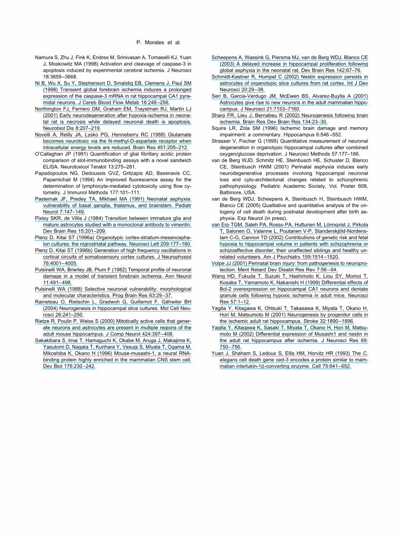
N
N
N
N
O
P
P
P
P
P
P
P
R
R
S
S
S
S
S
S
S
v
v
v
V
W
Y
Y
Y
P. Morales et al.
amura S, Zhu J, Fink K, Endres M, Srinivasan A, Tomaselli KJ, YuanJ, Moskowitz MA (1998) Activation and cleavage of caspase-3 inapoptosis induced by experimental cerebral ischemia. J Neurosci18:3659–3668.
i B, Wu X, Su Y, Stephenson D, Smalstig EB, Clemens J, Paul SM(1998) Transient global forebrain ischemia induces a prolongedexpression of the caspase-3 mRNA in rat hippocampal CA1 pyra-midal neurons. J Cereb Blood Flow Metab 18:248–256.
orthington FJ, Ferriero DM, Graham EM, Traystman RJ, Martin LJ(2001) Early neurodegeneration after hypoxia-ischemia in neona-tal rat is necrosis while delayed neuronal death is apoptosis.Neurobiol Dis 8:207–219.
ovelli A, Reilly JA, Lysko PG, Henneberry RC (1988) Glutamatebecomes neurotoxic via the N-methyl-D-aspartate receptor whenintracellular energy levels are reduced. Brain Res 451:205–212.
’Callaghan JP (1991) Quantification of glial fibrillary acidic proteincomparison of slot-immunobinding assays with a novel sandwichELISA. Neurotoxicol Teratol 13:275–281.
apadopoulos NG, Dedoussis GVZ, Gritzapis AD, Baxenavis CC,Papamichail M (1994) An improved fluorescence assay for thedetermination of lymphocyte-mediated cytotoxicity using flow cy-tometry. J Immunol Methods 177:101–111.
asternak JF, Predey TA, Mikhael MA (1991) Neonatal asphyxia:vulnerability of basal ganglia, thalamus, and brainstem. PediatrNeurol 7:147–149.
ixley SKR, de Villis J (1984) Transition between immature glia andmature astrocytes studied with a monoclonal antibody to vimentin.Dev Brain Res 15:201–209.
lenz D, Kitai ST (1996a) Organotypic cortex-striatum-mesencepha-lon cultures: the nigrostriatal pathway. Neurosci Lett 209:177–180.
lenz D, Kitai ST (1996b) Generation of high frequency oscillations incortical circuits of somatosensory cortex cultures. J Neurophysiol76:4001–4005.
ulsinelli WA, Brierley JB, Plum F (1982) Temporal profile of neuronaldamage in a model of transient forebrain ischemia. Ann Neurol11:491–498.
ulsinelli WA (1988) Selective neuronal vulnerability: morphologicaland molecular characteristics. Prog Brain Res 63:29–37.
aineteau O, Rietschin L, Gradwoh G, Guillemot F, Gähwiler BH(2004) Neurogenesis in hippocampal slice cultures. Mol Cell Neu-rosci 26:241–250.
ietze R, Poulin P, Weiss S (2000) Mitotically active cells that gener-ate neurons and astrocytes are present in multiple regions of theadult mouse hippocampus. J Comp Neurol 424:397–408.
akakibara S, Imai T, Hamaguchi K, Okabe M, Aruga J, Makajima K,Yasutomi D, Nagata T, Kurihara Y, Vesugi S, Miyata T, Ogama M,Mikoshiba K, Okano H (1996) Mouse-musashi-1, a neural RNA-binding protein highly enriched in the mammalian CNS stem cell.
Dev Biol 176:230–242.cheepens A, Wassink G, Piersma MJ, van de Berg WDJ, Blanco CE(2003) A delayed increase in hippocampal proliferation followingglobal asphyxia in the neonatal rat. Dev Brain Res 142:67–76.
chmidt-Kastner R, Humpel C (2002) Nestin expression persists inastrocytes of organotypic slice cultures from rat cortex. Int J DevNeurosci 20:29–38.
eri B, Garcia-Verdugo JM, McEwen BS, Alvarez-Buylla A (2001)Astrocytes give rise to new neurons in the adult mammalian hippo-campus. J Neurosci 21:7153–7160.
harp FR, Lieu J, Bernabeu R (2002) Neurogenesis following brainischemia. Brain Res Dev Brain Res 134:23–30.
quire LR, Zola SM (1996) Ischemic brain damage and memoryimpairment: a commentary. Hippocampus 6:546–552.
trasser V, Fischer G (1995) Quantitative measurement of neuronaldegeneration in organotypic hippocampal cultures after combinedoxygen/glucose deprivation. J Neurosci Methods 57:177–186.
an de Berg WJD, Schmitz HE, Steinbusch HE, Schuster D, BlancoCE, Steinbusch HWM (2001) Perinatal asphyxia induces earlyneurodegenerative processes involving hippocampal neuronalloss and cyto-architectonal changes related to schizophrenicpathophysiology. Pediatric Academic Society, Vol. Poster 608,Baltimore, USA.
an de Berg WDJ, Scheepens A, Steinbusch H, Steinbusch HWM,Blanco CE (2005) Qualitative and quantitative analysis of the on-togeny of cell death during postnatal development after birth as-phyxia. Exp Neurol (in press).
an Erp TGM, Saleh PA, Rosso PA, Huttunen M, Lönnqvist J, PirkolaT, Salonen O, Valanne L, Poutanen V-P, Standerskjöld-Nordens-tam C-G, Cannon TD (2002) Contributions of genetic risk and fetalhypoxia to hippocampal volume in patients with schizophrenia orschizoaffective disorder, their unaffected siblings and healthy un-related volunteers. Am J Psychiatry 159:1514–1520.
olpe JJ (2001) Perinatal brain injury: from pathogenesis to neuropro-tection. Ment Retard Dev Disabil Res Rev 7:56–64.
ang HD, Fukuda T, Suzuki T, Hashimoto K, Liou SY, Momoi T,Kosaka T, Yamamoto K, Nakanishi H (1999) Differential effects ofBcl-2 overexpression on hippocampal CA1 neurons and dentategranule cells following hypoxic ischemia in adult mice. NeurosciRes 57:1–12.
agita Y, Kitagawa K, Ohtsuki T, Takasawa K, Miyata T, Okano H,Hori M, Matsumoto M (2001) Neurogenesis by progenitor cells inthe ischemic adult rat hippocampus. Stroke 32:1890–1896.
agita Y, Kitagawa K, Sasaki T, Miyata T, Okano H, Hori M, Matsu-moto M (2002) Differential expression of Musashi1 and nestin inthe adult rat hippocampus after ischemia. J Neurosci Res 69:750–756.
uan J, Shaham S, Ledoux S, Ellis HM, Horvitz HR (1993) The C.elegans cell death gene ced-3 encodes a protein similar to mam-
malian interlukin-1�-converting enzyme. Cell 75:641–652.