depletion of anti-gal antibodies by the intravenous infusion of gal type 2 and 6 glycoconjugates in...
TRANSCRIPT

Depletion of anti-Gal antibodies by theintravenous infusion of Gal type 2 and 6glycoconjugates in baboons
Teranishi K, Alwayn IPJ, Buhler L, Gollackner B, Knosalla C, Huck J,Duthaler R, Katopodis A, Sachs DH, Schuurman H-J, Awwad M,Cooper DKC. Depletion of anti-Gal antibodies by the intravenousinfusion of Gal type 2 and 6 glycoconjugates in baboons.Xenotransplantation 2003; 10: 357–367. � Blackwell Munksgaard, 2003
Abstract: Background: Natural anti-Gal antibodies (NAb) to Gal epi-topes play a key role in the rejection of pig cells or organs transplantedinto primates. We have investigated the effect on NAb return afterextracorporeal immunoadsorption (EIA) of the continuous intravenous(i.v.) infusion of (i) bovine serum albumin conjugated to Gal type 6oligosaccharides (BSA-Gal) or (ii) a poly l-lysine backbone conjugatedto Gal type 2 or 6 oligosaccharides (PLL-Gal).Methods: Porcine mobilized peripheral blood progenitor cells(PBPC) obtained by leukapheresis from MHC-inbred miniatureswine (n ¼ 9) were infused intravenously (i.v.) into baboons: Group 1baboons (n ¼ 4) received whole body and thymic irradiation,splenectomy, antithymocyte globulin, cobra venom factor, cyclo-sporine, mycophenolate mofetil, anti-CD154mAb, porcine hemato-poietic growth factors, and EIA before transplantation of high doses(2 to 4 · 1010 cells/kg) of PBPC; Group 2 baboons (n ¼ 3) receivedthe Group 1 regimen plus a continuous i.v. infusion of BSA-Gal forup to 30 days; Group 3 baboons (n ¼ 5) received the Group 1 regi-men plus a continuous i.v. infusion of PLL-Gal type 2 (n ¼ 2) orboth PLL-Gal types 2 and 6 (n ¼ 3) for up to 30 days.Results: Group 1: NAb returned to pre-PBPC levels within 20–30 days,but therewasno inductionofantibody toGalornon-Galdeterminants;Group 2: NAb was undetectable or at very low level during BSA-Galtherapy. In one baboon, however, IgG toGal type 2, but not to type 6,returned during BSA-Gal therapy; Group 3: NAb was undetectable orat very low level duringPLL-Gal therapy. In twobaboons that receivedPLL-Gal type 2, NAb to Gal type 6, but not to type 2, returned duringPLL-Gal treatment.Twooffivebaboons, however, developed systemicinfection. Four of five baboons died within 14 days; autopsy revealedfocal hemorrhagic injury to their hearts, lungs, and small intestines,with histologic abnormalities that varied between animals from hem-orrhageand/or thrombosis in someorgans (heart, lungs, or intestine) tosigns of infections (bacteria in intestine, cytomegalovirus in liver).Conclusions: (i) BSA-Gal and PLL-Gal therapy maintained deple-tion of NAb. (ii) Some heterogeneity in specificity of NAb wasidentified, indicating that the infusion of a combination of Gal type 2and 6 glycoconjugates may be required. (iii) The addition of PLL-Galto the immunosuppressive regimen was associated with a high inci-dence of morbidity and mortality without a clear histopathologicentity underlying the cause of death.
Katsuhito Teranishi,1 Ian P.J.Alwayn,1 Leo B�hler,1 BerndGollackner,1 Christoph Knosalla,1
Jennifer Huck,2 Rudolf Duthaler,3
Andreas Katopodis,3 David H.Sachs,1 Henk-Jan Schuurman,2
Michel Awwad2 and David K.C.Cooper11Transplantation Biology Research Center,Massachusetts General Hospital/Harvard MedicalSchool, Boston, MA, USA, 2ImmergeBioTherapeutics, Charlestown, MA, USA, and3Transplantation Research, Novartis Pharma, Basel,Switzerland
Key words: anti-CD154 monoclonal antibody –anti-pig antibody – anti-Gal antibody –BSA-galactose conjugate – pig-to-primate model –xenotransplantation
Address reprint requests to D.K.C. Cooper, M.D.,Ph.D., F.R.C.S., Transplantation Biology ResearchCenter, Massachusetts General Hospital,MGH East, Building 149-9019, 13th Street, Boston,MA 02129, USA(E-mail: [email protected])
Received 1 May 2002;Accepted 26 June 2002
Xenotransplantation 2003: 10: 357–367Printed in UK. All rights reserved
Copyright � Blackwell Munksgaard 2003
XENOTRANSPLANTATION
ISSN 0908-665X
357

Introduction
Natural antibodies (NAb) in primates directedagainst Gala1-3Gal (Gal) determinants on pigcells form a major barrier to the successfulxenotransplantation of cells or organs in the pig-to-primate model [1–3]. These anti-Gal immuno-globulin M (IgM) and immunoglobulin G (IgG)NAbs can be selectively depleted by extracorpor-eal immunoadsorption (EIA) of blood throughimmunoaffinity columns of a Gal oligosaccharide.Effective depletion up to >97–99% is achievedusing a course of three EIAs in baboons [4].Although this depletion of NAb prevents hyper-acute rejection, return of NAb results in humoralrejection (acute humoral xenograft rejection;AHXR) occurring within a few days [5], andmay be associated with the development ofdisseminated intravascular coagulation. Althoughintensive immunosuppressive therapy and/or com-plement depletion or inhibition can delay the onsetof this humoral-mediated rejection, no therapyhas to date either prevented return of NAb orcompletely suppressed its production in thepig-to-primate model [6].There are two commonly occurring structural
forms of Gal, type 2 (Gala1-3Galb1-4GlcNAc)and type 6 (Gala1-3Galb1-4Glc). In naıvebaboons, almost all NAb are directed towardsepitopes shared between these two structuralforms [7].We have previously investigated the use of
bovine serum albumin conjugated to Gal type 6trisaccharide (BSA-Gal; Alberta Research Coun-cil, Edmonton, Alberta, Canada; ARC) to adsorbanti-Gal NAb [7]. We have now investigatedconjugates of Gal type 2 or Gal type 6 trisacch-aride and poly l-lysine (PLL; Novartis PharmaAG, Basel, Switzerland). The chemical and bio-logical efficacy of the PLL-Gal type 2 conjugate(also called GAS 914) is being reported elsewhere(R. Duthaler et al., manuscript submitted;A. Katopodis et al., manuscript submitted). Thenumber of Gal residues per weight equivalent forPLL-Gal type 2 and PLL-Gal type 6 is similar.We postulate that complexes between solubleglycoconjugates and NAb are cleared from thecirculation largely in the liver [8–10], and alsopossibly in the lungs and kidneys [8,10]. Theresults obtained from the addition of PLL-Galtherapy to our standard non-myeloablative condi-tioning regimen [aimed at inducing tolerance toporcine mobilized peripheral blood progenitorcells (PBPC)] are now compared with our previousstudies in which BSA-Gal was or was not infused[7,11].
Materials and methods
Animals
Baboons (Papio anubis, n ¼ 12) of known ABOblood group and of body weight 6–14 kg (Biolo-gical Resources, Houston, TX, USA) were used asrecipients. Massachusetts General Hospital majorhistocompatibility complex (MHC)-inbred minia-ture swine (n ¼ 9) of blood group O, 2–14 monthsold, 18–100 kg of body weight (Charles RiverLaboratories, Wilmington, MA, USA) served asdonors of PBPC. All experiments were conductedaccording to the NIH Guidelines for Care and Useof Laboratory Animals and were approved by theMassachusetts General Hospital Subcommittee onResearch Animal Care.
Surgical procedures
Anesthesia, intravenous (i.v.) line placement in pigsand baboons, and intra-arterial line placement andsplenectomy in baboons have been described pre-viously [5,12].
Mobilization and collection of porcine blood progenitor cells(PBPC)
Hematopoietic growth factor-mobilization of por-cine progenitor cells and leukapheresis was per-formed as described previously [13]. Leukapheresisresulted in the collection of large numbers ofleukocytes (30 to 60 · 1010 cells) containingapproximately 2% progenitor cells, designatedPBPC [13].
Extracorporeal immunoadsorption in baboons
Natural antibodies (NAb) was depleted from thebaboon’s circulation by the perfusion of plasmathrough immunoaffinity columns containing syn-thetic Gala1-3Galß1-4Glc-X-Y (Gal type 6 trisac-charide, ARC), as reported previously [4,14,15].
Conditioning regimen in baboons
The conditioning regimen in baboons has beendescribed previously [7,11]. In short, all recipientbaboons underwent splenectomy on day 8 or day19 (four baboons in Group 3), non-myeloablativewhole body irradiation in two fractions on days 6and 5 (total dose 300 cGy), thymic irradiation(700 cGy) on day 1, horse antihuman thymocyteglobulin (ATGAM, Upjohn, Kalamazoo, MI,USA) 50 mg/kg/day i.v. on days 3, 2 and 1, andEIA on days 6, 3 and 1 [5]. This was followed byinfusion of porcine PBPC (total dose 2 to
Teranishi et al.
358

4 · 1010 cells/kg) [11] on day 0, 1 and 2. Allbaboons also received mycophenolate mofetil(donated by Roche, Nutley, NJ, USA) from day8, and cyclosporine (CyA; donated by NovartisPharma AG, Basel, Switzerland) (at approximately15 mg/kg/day) except for two baboons in Group 1,from days 8 to 28 by continuous i.v. infusion(administered with an Abbott Omniflow 4000infusion device). All baboons also received cobravenom factor i.v. at dose levels between 35 and105 units/kg/day on days 1 to 14 or 28 to maintainthe CH50 at 0% [16], and murine antihumanCD154 monoclonal antibody (mAb) (5C8; ATCC,Rockville, MD, USA) (20 mg/kg/day i.v.) admin-istered on alternate days [11]. To avoid a throm-botic microangiopathic state [17,18], all baboonsreceived additional therapy comprising prostacy-clin (PGI2; 20 ng/kg/min by continuous i.v. infu-sion), heparin (10 U/kg/h by continuous i.v.infusion), and methylprednisolone (2 mg/kg twicedaily i.v. for 7 days, followed by tapering anddiscontinuation over the next 7 or 21 days). Thisadditional therapy to avoid a thrombotic micro-angiopathic state was started immediately beforethe first PBPC infusion and continued for 14 or28 days.Porcine interleukin-3 [100 to400 lg/kg/daysubcutaneously (s.c.) or i.v.] and porcine stem cellfactor (100 to 2000 lg/kg/day s.c. or i.v.) wereadministered to all baboons from days 0 to 14 or28 as described previously [7,11].
Assays for detection of anti-Gal NAb and Ab againstporcine non-Gal determinants
Details of these methods have been reportedpreviously [14,19]. IgM and IgG Ab reactive withGal types 2 and 6 were determined by ELISA, andantipig IgM and IgG were determined by flowcytometric analysis (FACS). The lowest detectionlimits for IgM and IgG were 5.0 and 0.5 lg/ml,respectively.
FACS for detection of baboon T and B cells
Flow cytometry to detect T and B cells wasperformed on blood, aspirated bone marrowobtained from the iliac crests, and lymph nodesobtained from either inguinal or axillary regions.Details of these methods have been reportedpreviously [7]. In short, anti-CD3 FITC (Bio-source, Amarillo, CA, USA) and anti-CD2 PE(Leu-5b, Becton Dickinson, San Jose, CA, USA)were used as T-cell markers, and anti-CD20 FITC(Leu-16, Becton Dickinson) and anti-CD22 PE(Clone RFB4, Caltag Laboratories, Burlingame,CA, USA) as B-cell markers. Acquisition was
performed using the FACScan (Becton Dickinson),and samples were analyzed using WinList software(Verity Software House, Topsham, ME, USA).
FACS and polymerase chain reaction (PCR) for detection of pigcell chimerism in blood
Details of these techniques have been reportedpreviously [20]. In short, FACS for detection of pigcells was performed with the following mAbs; M11(mouse antiprimateMHCClass 1 IgG), 1030H-1–19or pan-pig (mouse antipig leukocyte IgM), 76-7-4(mouse anti-pigCD1 IgG), 898H2-6-15 (mouse anti-pig CD3 IgG), 74-22-15 or SWC3a (mouse anti-piggranulocytes–monocytes), 4-6 (mouse anti-pigCD9), 36-7-5 (mouse anti-mouse MHC Class IIgG) and 12-2-2 (mouse anti-mouse MHC Class IIgM) as isotype controls. A bulk PCR assay thatamplifies the porcine cytochrome-b genewas used todetect pig microchimerism in baboon samples.
Monitoring for toxicity and supportive therapy
Blood cell counts, chemistry, coagulation para-meters, and levels of immunosuppressive drugswere determined by routine methods. Erythropoie-tin was administered to some baboons at a dose of100 units/kg s.c. or i.v. If the hematocrit fell<20%, washed irradiated red blood cells fromABO-matched baboon donors were administered.Thrombocytopenia of <10 000 platelets/mm3 wascorrected by the transfusion of fresh washedand irradiated baboon platelets. All pigs andbaboons received daily cefazolin sodium (500 to1000 mg/day i.v.) or levofloxacin (20 mg/kg/dayi.v.) throughout the periods of leukapheresis andtherapy, respectively. Blood cultures were monit-ored twice weekly and inoculated into an aerobicbottle and an anaerobic bottle. If growth wasdetected in the blood culture bottle, antibiotictherapy was modified.
Experimental groups
Group 1 baboons (n ¼ 4) received the non-myelo-ablative regimen outlined in Materials and meth-ods. In two baboons, a course of eight doses ofanti-CD154 mAb was administered without CyA,and, in another two baboons, a course of only twodoses was combined with 36 days of treatmentwith CyA (Table 1).Group 2 baboons (n ¼ 3) received the Group 1
regimen that included both a 16 to 23-dose courseof anti-CD154 mAb and 36 days of CyA treat-ment. In addition, they received BSA-Gal bycontinuous i.v. infusion, beginning immediately
Depletion of antibodies by Gal glycoconjugates
359

after the final EIA and continued for 19–30 days atrates between 20 and 250 mg/kg/day. To preventsensitization to BSA-Gal, anti-CD154 mAb ther-apy was continued throughout the period of BSA-Gal infusion and then for a further 7 days afterdiscontinuation of BSA-Gal (Table 1).Group 3 baboons (n ¼ 5) received the Group 1
regimen, including CyA. Two of five baboons(B69-379 and B69-263) received PLL-Gal type 2(5 mg/kg) as a bolus on days 17, 14, 11, 5, and 3,followed by a continuous i.v. infusion of PLL-Galtype 2, beginning immediately after the final EIA,at a rate between 5 and 10 mg/kg/day. Anothertwo baboons (B69-363 and B69-231) received PLL-Gal type 2 (5 mg/kg) as a bolus on days 17, 14, and11, and both PLL-Gal type 2 (5 mg/kg) and type 6(1 mg/kg) as a bolus on days 7, 5 and 3. This wasfollowed by continuous i.v. infusions of PLL-Galtypes 2 (1–10 mg/kg/day) and 6 (1–2 mg/kg/day).One baboon (B69-180) received both PLL-Galtypes 2 (3 mg/kg/day) and 6 (1 mg/kg/day) by i.v.infusion from day 1 without prior bolus therapy(Table 1).
Results
Hematopoietic parameters and clinical course
The results in Group 1 [11] and Group 2 [7] havebeen reported previously. The non-myeloablativeregimen was well-tolerated in baboons in all threegroups, although there were significant reductionsin white blood cell (to <2000/mm3) and platelet(to <20 000/mm3) counts. Maximum suppressionof white blood cell counts was approximately atdays 10 to 12 in Groups 1 and 2, and at day 14 inthose animals in Group 3 that survived until thistime. Hematocrit was maintained rather higher inGroup 1, and this was thought to be because ofrather higher body weight in this group (bodyweight 10–15 kg). It was rather lower in Group 3
(body weight 6–11 kg). Platelet counts were lowestaround day 9 in all groups, and platelet transfusionwas required occasionally. It was our impressionthat the baboons in Group 2 maintained betterappetite and activity throughout the leukopenicperiod, even though the degree and period ofleukopenia was not notably different in this group.White blood cell counts in three baboons inGroup 3decreased to £100/mm3 around days 12 to 14.
Morbidity and mortality
There was no mortality in Group 1 or 2. Three ofthe five baboons in Group 3 died suddenly on days3 (B69-231, cardiac tamponade), 6 (B69-180,uncertain cause), and 14 (B69-379, uncertaincause), respectively (Table 3). The remaining twobaboons (B69-263 and B69-363) developed sys-temic infections (with blood cultures positive forEnterococcus spp. and/or Escherichia coli), andone of these (B69-363) was euthanized on day 14because of poor general condition. Two baboons inthis group (B69-363 and B69-180) had melenastool, indicating gastrointestinal bleeding. Autopsywas performed in the four baboons that died (seebelow).
Antibody responses to porcine PBPC infusion
The results in Groups 1 and 2 have been reportedpreviously [7,11]. No Ab against porcine non-Galdeterminants was detected in any baboon in thethree groups. In Group 1, anti-Gal IgM showed noincrease above pre-EIA baseline in two baboonsand, at maximum, a four-fold increase in the othertwo; anti-Gal IgG also showed no increase in twobaboons and an 8-30-fold increase in the other twobaboons. In Group 2, anti-Gal NAb remainedundetectable or at levels just above the detectionlimit during the period of BSA-Gal administration.However, in one baboon, BSA-Gal (which is madeup of Gal type 6) failed to maintain depletion ofanti-Gal NAb reactive with Gal type 2 even thoughprior EIA (performed with an immunoaffinitycolumn of Gal type 6) was successful in removingNAb reactive with both Gal types 2 and 6(Fig. 1A). In Group 3, pre-EIA bolus therapy withPLL-Gal type 2 with/without type 6 was largelysuccessful in depleting anti-Gal IgM to both Galtypes, but less successful in depleting anti-Gal IgGto these Gal types (Table 2). Using an immuno-affinity column of a type 6 oligosaccharide, EIAwas successful in removing NAb to both Gal types(Table 2). During the period of continuous i.v.PLL-Gal type 2 infusion, NAb to Gal type 6, butnot to type 2, returned to baseline values in both
Table 1. Summary of therapy
GroupBasic
therapya EIA CyA CD154 mAb BSA-Gal PLL
1 + + +/–b + (8 doses)b – –2 + + + + + –3 + + +c + – + (type 2 or types 2 + 6)
(see Tables 2 and 3)
aBasic therapy included splenectomy (day 8), whole body irradiation (150 cGy ondays )6 and )5), thymic irradiation (700 cGy on day )1), extracorporealimmunoadsorption (days )6, )3 and )1), and immunosuppressive therapy withmycophenolate mofetil and antithymocyte globulin (see text).bCyclosporine was administered to two of the four baboons. When cyclosporinewas given, only two doses of CD154 mAb were administered.cOne baboon did not receive EIA because no anti-Gal Ab was detected.
Teranishi et al.
360

baboons in which this therapy was administered(B69-379 and B69-263) (Table 2 and Fig. 1B). Inone baboon that received PLL-Gal types 2 and 6
(B69-363), NAb to both Gal types remainedundetectable until day 14 when the animal waseuthanized (Table 2 and Fig. 1C). In the other two
: IgM type 6: IgG type 6: IgM type 2: IgG type 2
0
20
40
60
80
0
50
100
150
200
250
300
0
20
40
60
80
0
2
4
6
8
10
-20 -10 0 10 20 30 400
20
40
60
80
0
2
4
6
8
10
Co
ncen
trat
ion
(µg
/ml)
Co
nce
ntr
atio
n(µ
g/m
l)
Co
nce
ntr
atio
n(µ
g/m
l)
Co
nce
ntr
atio
n(µ
g/m
l)B
SA
-Gal(m
g/kg
/day)
PL
L(m
g/kg
/day)
PL
L(m
g/kg
/day)
Time (days) Time (days)
BSA-Gal PLL type 2
PLL type 2
(D)
(B)
(C)
(A)
PLL type 6
-20 -10 0 10 20 30 400
20
40
60
80
0
2
4
6
8
10
PLL type 2PLL type 6
PL
L(m
g/kg
/day)
Fig. 1. IgM and IgG antibody reactive with Gal types 2 and 6 in baboons receiving BSA-Gal (A) or PLL-Gal type 2 (B) or PLL-Galtypes 2 and 6 (C and D). (A) Course in one baboon in Group 2. BSA-Gal (type 6) failed to maintain depletion of Ab reactive to Galtype 2 even though prior EIA (performed with an immunoaffinity column of Gal type 6) was successful in removing Ab reactive withboth Gal type 6 and Gal type 2. (The reason for the subsequent spontaneous fall in Ab reactive with Gal type 2 is not clear). (B)Course in one baboon in Group 3 that received PLL-Gal type 2 (B69-263). NAb to Gal type 6, but not to type 2, returned duringPLL-Gal treatment. (C) Course in one baboon (with initially very low levels of NAb) that received PLL types 2 and 6 (B69-363). Abreactive with Gal types 2 and 6 were depleted after a bolus infusion of PLL type 2, and remained undetectable until day 14 when theanimal died. (D) Course in one of two baboons that received PLL types 2 and 6 (B69-180). Although IgM reactive with Gal types 2and 6 were removed, IgG remained detectable.
Table 2. Group 3 baboons – detection of Gal-reactive NAb
Pre-PBPC bolusPLL-Gal infusion
NAb detectedimmediately after
final bolusPLL-Gal infusiona
Pre-PBPC NAbdetection after
final EIA (day 0)PBPC
infusion
Post-PBPCcontinuous PLL-Gal
infusion therapy
NAb detectedduring continuousPLL-Gal infusion
therapy
Increase of NAb tobaseline level
during continuousPLL-Gal infusion
therapy
Type 2 Type 6 Type 2 Type 6 Type 2 Type 6 Type 2 Type 6 Type 2 Type 6 Type 2 Type 6
B69-379 + – IgG IgMIgG
– – + – – IgMIgG
– IgG
B69-263 + – IgG IgG IgG IgG + – IgGb IgMIgG
– IgMIgG
B69-363 + + – – – – + + – – – –
B69-231 + + IgG IgMIgG
– – + + IgGb – –
B69-180 – – NA NA – – + + IgG IgG – –
a Low level of NAb.b Almost undetectable levels of NAb.NA: not applicable.
Depletion of antibodies by Gal glycoconjugates
361

baboons that received PLL-Gal type 2 and 6 (B69-231 and B69-180), anti-Gal IgM reactive with bothGal types became undetectable, but anti-Gal IgGto both oligosaccharides continued to be detect-able, albeit below baseline levels (Table 2 andFig. 1D).
FACS and PCR for detection of pig cell chimerism in blood
In all Groups, macrochimerism was maximum ondays when PBPC were being infused.Group 1: Pig cell macrochimerism (detectable by
FACS) was detected for 5 days, with maximumvalues of 33 and 73% when CyA was omitted orincluded in the regimen, respectively [20]. Only onebaboon subsequently showed the reappearance ofpig cells by FACS; presumed pig cell fragments(based on low forward-side scatter property),identified using a pan-pig Ab, were detected fromdays 9 to 16. Microchimerism (detectable by PCR)was continuously present in the blood of all fourbaboons for a maximum of 33 days, and thereafterwas intermittently detected for >100 days [20].Group 2: Macrochimerism reached a maximum
of 36% while PBPC were being infused, after whichit was lost in one baboon. In the other two baboons,macrochimerism was detected continuously at lowlevels (<1%) until days 14 and 17, respectively. Inall three baboons, microchimerism was continu-ously detectable for a maximum of 32 days, andwas intermittently present for >75 days [7].Group 3: Macrochimerism was detected for
6 days, with a maximum of 26% while PBPC werebeing infused. However, macrochimerism was lostin all baboons after day 6. Four baboons died byday 14, and were not examined for microchimer-ism; in the surviving baboon, microchimerism wasdetected for only 18 days.
Autopsy and histopathology
Autopsy was performed in the four Group 3baboons that died between days 3 and 14
(Table 3). In one baboon that received only PLL-Gal type 2 (B69-379, died on day 14), macroscopicinspection at autopsy revealed minor hemorrhagicinjury to the lungs. On microscopy, there was focalmassive hemorrhage in the lung, infectious foci inthe liver, and focal interstitial fibrosis/necrosis inthe heart, observations that could fit with systemicinfection, with a suspicion of cytomegalovirusinfection. In the three baboons that received bothPLL-Gal types 2 and 6 (B69-363, B69-231 andB69-180, surviving 14, 3 and 6 days, respectively),autopsy revealed focal hemorrhagic injury to theheart (Fig. 2A) and lungs (Fig. 2B) and, in twocases, to the small intestine (Fig. 2C). In one case(B69-231), hemorrhagic cardiac injury was associ-ated with a heavily blood-stained pericardial effu-sion that had resulted in cardiac tamponade. Notechnical complication, e.g. rupture of a bloodvessel by an indwelling catheter, was found thatmight have accounted for the tamponade. At themicroscopic level, B69-231 (survival 3 days)showed focal hemorrhage in the heart (Fig. 2D),thrombosis and focal hemorrhage in lung tissue(Fig. 2E), and focal necrosis in the kidney; inaddition, there were signs of bacterial infection inthe intestine. However, none of these pathologicfeatures was sufficiently severe to explain the deathof this animal. B69-180 (survival 6 days) showedthrombi and fibrinoid necrosis in submucosalarteries in the large intestine, along with ischemicdamage to the mucosa; in addition, there was focalnecrosis and fibrinoid necrosis in some small bloodvessels in the heart. B69-363 (survival 14 days)revealed hemorrhage along the intestinal surface(Fig. 2F) related to infection, thrombosis in somelung vessels, signs of infectious peritonitis, andindications of cytomegalovirus infection (inclusionbodies) in the liver.
Discussion
In early studies at our center, a return of detectableNAb was uniformly observed within 18 h after a
Table 3. Group 3 baboons – clinical course
Baboon
Treatment
Survival (days) Systemic infectiona Melena
Autopsy: hemorrhagic injury
PLL-Gal 2 PLL-Gal 6 Heart Lungs Small intestine
B69-379 + – 14 – – – + –B69-263 + – >90 + – NA NA NAB69-363 + + 14 + + + + ++B69-231 + + 3 – – ++b + –B69-180 + + 6 – + + + ++
a Enterococcus spp. and/or Escherichia coli.b Died from hemorrhagic cardiac tamponade.NA: not applicable.
Teranishi et al.
362

course of EIA, followed by recovery to pre-EIAlevels within 3 to 5 days despite pharmacologicimmunosuppressive therapy [15,21]. Even multipleEIAs over a 2 to 3-week period did not prevent thereturn of NAb that normally began within hoursafter EIA [21]. Our previous report [7] demonstra-ted that a bolus i.v. infusion of BSA-Gal coulddeplete NAb without the need for EIA. Thisobservation is confirmed in the present study
because infusions of PLL-Gal were efficacious inthe depletion of NAb. The continuous i.v. infusionof either BSA-Gal or PLL-Gal was largely suc-cessful in maintaining depletion of NAb, in con-trast to our earlier studies [11,19]. By depletion ofanti-Gal Ab with subsequent reduction of thepossible binding of NAb to the transplanted pigorgan, soluble glycoconjugates like BSA-Gal orPLL-Gal may therefore have potential in resolving
Fig. 2. Autopsy findings in two representative baboons of the four that died or were euthanized within 14 days in Group 3. In threebaboons that received PLL-Gal type 2 with or without type 6, autopsy revealed focal hemorrhagic injury in the heart (A), lungs (B),and small intestine (C). Histological examination revealed focal myocardial hemorrhage and necrosis in the heart (D), thrombosis insmall vessels in the lungs (E), and mucosal congestion and swelling of the small intestine with thrombosis in the vessels and fibrincoating of the mucosa (F) (Hematoxylin & eosin, ·40).
Depletion of antibodies by Gal glycoconjugates
363
the problem of anti-Gal NAb-mediated rejection inthe pig-to-baboon xenotransplantation model, atleast while they are being infused.Gal trisaccharides have been demonstrated to be
more efficient in blocking Gal-reactive NAb thanGal disaccharide [22,23]. Furthermore, the redu-cing end of the Gal oligosaccharide contributes toits efficiency in blocking anti-Gal NAb [23]. In ourextensive experience of EIA using a Gal trisacch-aride type 6 immunoaffinity column [4], NAbreactive with both type 2 and type 6 Gal oligosac-charides have almost invariably been successfullyadsorbed. However, in one of the baboons inGroup 2 in the present study, NAb directed againstthe type 2 saccharide returned during BSA-Gal(type 6) treatment while type 6-reactive NAbremained undetectable (Fig. 1A). In two baboonsin Group 3 that received PLL-Gal type 2, NAb toGal type 6, but not to type 2, returned duringtreatment (Fig. 1B). These data suggest that, inaddition to anti-Gal NAb reacting to sharedepitopes, there exist Ab directed to Gal type-specific determinants. Although only a few experi-ments were performed using BSA-Gal or PLL-Gal,our results suggest that a combination of type 2and type 6 glycoconjugates may be required insome instances to completely remove circulatinganti-Gal NAb. This requirement could possibly bepredicted by measuring the levels of type 2- andtype 6-reactive NAb before and after a bolus i.v.infusion of type 2 or type 6 oligosaccharide beforethe initiation of full treatment.Gal type-specific Ab became detectable within
14 days after PBPC infusion, but the levels of type-specific Ab remained lower than the pre-EIA levels.No sensitization to Gal occurred and no anti-pigAb of specificity other than anti-Gal reactivitydeveloped. Details of the ability of this regimen toprevent sensitization have been reported previously[11,20], and will not be discussed further here. Thecombination of the continuous infusion of one ormore Gal glycoconjugates and the anti-CD154mAb-based immunosuppressive regimen, however,both prevented the return of anti-Gal NAb andinhibited the generation of other anti-pig Ab. Thiscombination may therefore provide a �window�during which pig hematopoietic cell engraftmentcan be achieved. In addition, if high levels of anti-Gal IgG and non-Gal IgG do not emerge followinganti-CD154 mAb therapy, and anti-pig IgM isallowed to return only slowly to a subpretransplantlevel, these conditions may allow accommodationto develop [24,25]. However, longer periods ofNAb inhibition may be required to ensure thattolerance or accommodation can be induced andmaintained.
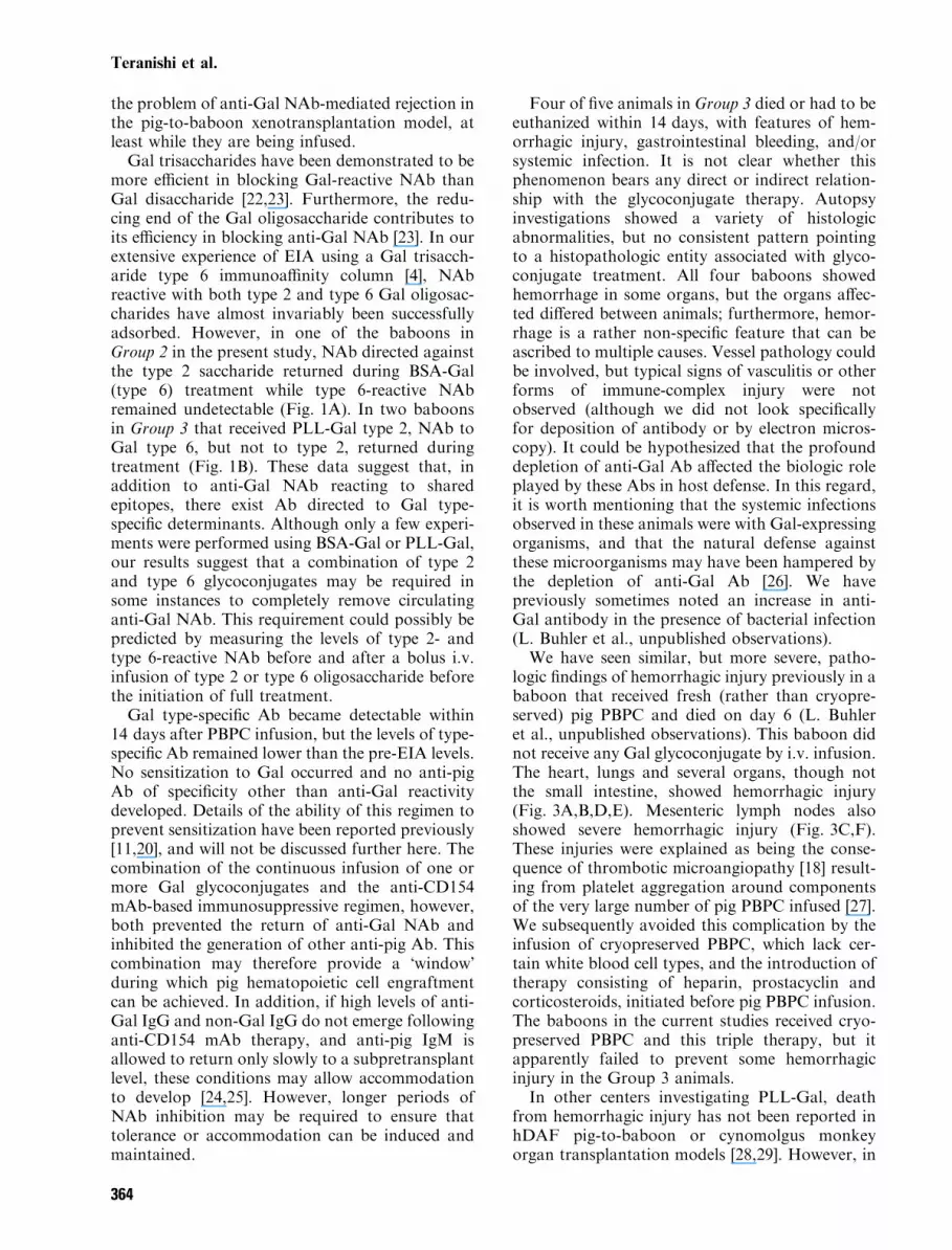
Four of five animals in Group 3 died or had to beeuthanized within 14 days, with features of hem-orrhagic injury, gastrointestinal bleeding, and/orsystemic infection. It is not clear whether thisphenomenon bears any direct or indirect relation-ship with the glycoconjugate therapy. Autopsyinvestigations showed a variety of histologicabnormalities, but no consistent pattern pointingto a histopathologic entity associated with glyco-conjugate treatment. All four baboons showedhemorrhage in some organs, but the organs affec-ted differed between animals; furthermore, hemor-rhage is a rather non-specific feature that can beascribed to multiple causes. Vessel pathology couldbe involved, but typical signs of vasculitis or otherforms of immune-complex injury were notobserved (although we did not look specificallyfor deposition of antibody or by electron micros-copy). It could be hypothesized that the profounddepletion of anti-Gal Ab affected the biologic roleplayed by these Abs in host defense. In this regard,it is worth mentioning that the systemic infectionsobserved in these animals were with Gal-expressingorganisms, and that the natural defense againstthese microorganisms may have been hampered bythe depletion of anti-Gal Ab [26]. We havepreviously sometimes noted an increase in anti-Gal antibody in the presence of bacterial infection(L. Buhler et al., unpublished observations).We have seen similar, but more severe, patho-
logic findings of hemorrhagic injury previously in ababoon that received fresh (rather than cryopre-served) pig PBPC and died on day 6 (L. Buhleret al., unpublished observations). This baboon didnot receive any Gal glycoconjugate by i.v. infusion.The heart, lungs and several organs, though notthe small intestine, showed hemorrhagic injury(Fig. 3A,B,D,E). Mesenteric lymph nodes alsoshowed severe hemorrhagic injury (Fig. 3C,F).These injuries were explained as being the conse-quence of thrombotic microangiopathy [18] result-ing from platelet aggregation around componentsof the very large number of pig PBPC infused [27].We subsequently avoided this complication by theinfusion of cryopreserved PBPC, which lack cer-tain white blood cell types, and the introduction oftherapy consisting of heparin, prostacyclin andcorticosteroids, initiated before pig PBPC infusion.The baboons in the current studies received cryo-preserved PBPC and this triple therapy, but itapparently failed to prevent some hemorrhagicinjury in the Group 3 animals.In other centers investigating PLL-Gal, death
from hemorrhagic injury has not been reported inhDAF pig-to-baboon or cynomolgus monkeyorgan transplantation models [28,29]. However, in
Teranishi et al.
364

these studies, only PLL-Gal type 2 (GAS 914) wasadministered. In our studies reported here, somehemorrhagic injury was seen in all three baboonsthat received both type 2 and type 6 PLL-Galtreatment, but this pathology was more limited andmild in the single baboon that died while receivingonly the type 2 PLL-Gal. Vasculitis and fibrinoidnecrosis of the vessels of the small bowel (similar tothe observations made in B69-180) have beenobserved in some non-human primates undergoingpig organ transplantation, but this observation has
not been exclusively associated with PLL-Galinfusion (H.-J. Schuurman, unpublished data).Furthermore, gastrointestinal bleeding, from
viral or fungal ulcers or from graft-vs.-host disease,has been observed in humans after bone marrowtransplantation, with an approximate incidence of2 to 11% [30]. Hemorrhagic injury of the heart andsmall intestine [30] may also be related to graft-vs.-host disease, but we do not consider this as a likelyexplanation for the pathology seen in the presentstudies.
Fig. 3. Autopsy findings in a baboon that died on day 6 after fresh pig PBPC (3 · 1010 cells/kg) were administered on days 0–5. Theheart (A), lungs (B), and mesenteric lymph nodes (C) showed severe hemorrhagic injury. Histological examination revealed inter-stitial hemorrhage in the heart (D), lungs (E), and the cortex of mesenteric lymph nodes (F) (Hematoxylin & eosin, ·40).
Depletion of antibodies by Gal glycoconjugates
365
We conclude that hemorrhagic injury of variousorgans can occur in non-human primates followingthe i.v. infusion of large numbers of pig PBPC withor without concomitant treatment with Gal glyco-conjugates. However, the continuous infusion ofPLL-Gal of both types 2 and 6 may increase therisk of this injury. The exact mechanism by whichthis injury occurs remains uncertain, and it remainsto be established whether immune complexesbetween NAb, PBPC, PLL-Gal, and platelets, area causative factor.In conclusion, the regimen used in the Group 1
baboons consistently prevented an induced humor-al response to transplanted pig cells (or tissues) inprimates (11;31;32). The addition of BSA-Gal orPLL-Gal resulted in depletion of anti-Gal NAb,providing a period in which transplanted pig cells(and possibly organs) might survive without under-going NAb-mediated rejection. However, there wasa higher incidence of morbidity and mortalitywhen PBPC infusion was associated with PLL-Gal therapy.
Acknowledgments
We thank J. Fishman, M.D. for considerableadvice throughout this study, and S. Burguete,L. Correa, H. DeAngelis, T. Ericsson, K. Moran,D. Newman, D. Shea and G. Solomon for excellenttechnical help. We also thank T. Kawai, M.D andS. Yamamoto, M.D. for their reviews of themanuscript. C. Knosalla is a recipient of a grantfrom the Deutsche Forschungsgemeinschaft(KN518,1-1). B. Gollackner is a recipient of agrant from the Max Kade Foundation of theAustrian Academy of Science. We wish to recordour gratitude to the following companies forgenerous gifts of their products – Abbott Labor-atories (Hetastarch 6%), Baxter Healthcare (Albu-min 5%), Novartis Pharmaceuticals (Sandimmunei.v.), and Roche Laboratories Inc. (CellCept i.v.).The work at our own center has been supported byNIH Program Project 1PO1 A145897 and by aSponsored Research Agreement between ImmergeBioTherapeutics and the Massachusetts GeneralHospital.
References
1. Good AH, Cooper DKC, Malcolm AJ et al. Identifica-tion of carbohydrate structures that bind human antipor-cine antibodies: implications for discordant xenograftingin humans. Transplant Proc 1992; 24: 559.
2. Sandrin MS, Vaughan HA, Dabkowski PL, McKenzie
IFC. Anti-pig IgM antibodies in human serum react pre-dominantly with Gal(alpha 1-3)Gal epitopes. Proc NatlAcad Sci USA 1993; 90: 11 391.
3. Galili U. Interaction of the natural anti-Gal antibodywith alpha-galactosyl epitopes: a major obstacle for xeno-transplantation in humans. Immunol Today 1993; 14: 480.
4. Watts A, Foley A, Awwad M et al. Plasma perfusion byapheresis through a Gal immunoaffinity column success-fully depletes anti-Gal antibody: experience with 320aphereses in baboons. Xenotransplantation 2000; 7: 181.
5. Kozlowski T, Shimizu A, Lambrigts D et al. Porcinekidney and heart transplantation in baboons undergoing atolerance induction regimen and antibody adsorption.Transplantation 1999; 67: 18.
6. Lambrigts D, Sachs DH, Cooper DKC. Discordantorgan xenotransplantation in primates: world experienceand current status. Transplantation 1998; 66: 547.
7. Teranishi K, Gollackner B, Buhler L et al. Depletionof anti-Gal antibodies in baboons by intravenous therapywith bovine serum albumin conjugated to Gal oligosac-charides. Transplantation 2002; 73: 129.
8. Davies KA, Chapman PT, Norsworthy PJ et al.Clearance pathways of soluble immune complexes in thepig. Insights into the adaptive nature of antigen clearancein humans. J Immunol 1995; 155: 5760.
9. Nardin A, Lindorfer MA, Taylor RP. How areimmune complexes bound to the primate erythrocytecomplement receptor transferred to acceptor phagocyticcells? Mol Immunol 1999; 36: 827.
10. Gonzalez ML, Waxman FJ. Glomerular deposition ofimmune complexes made with IgG2a monoclonal anti-bodies. J Immunol 2000; 164: 1071.
11. Buhler L, Awwad M, Basker M et al. High-dose porcinehematopoietic cell transplantation combined with CD40ligand blockade in baboons prevents an induced anti-pighumoral response. Transplantation 2000; 69: 2296.
12. Cooper DKC, Ye Y, Niekrasz M. Heart transplantationin primates. In: Cramer DV, Podesta LG, Makowka L,eds. Handbook of Animal Model in TransplantationResearch. Boca Raton: CRC Press, 1994, pp 173.
13. Nash K, Chang Q, Watts A et al. Peripheral bloodprogenitor cell mobilization and leukapheresis in pigs. LabAnim Sci 1999; 49: 645.
14. Xu Y, Lorf T, Sablinski T et al. Removal of anti-porcinenatural antibodies from human and nonhuman primateplasma in vitro and in vivo by a Galalpha1-3Galbe-ta1-4betaGlc-X immunoaffinity column. Transplantation1998; 65: 172.
15. Kozlowski T, Ierino FL, Lambrigts D et al. Depletionof anti-Gal(alpha)1-3Gal antibody in baboons by specificalpha-Gal immunoaffinity columns. Xenotransplantation1998; 5: 122.
16. Kobayashi T, Taniguchi S, Neethling FA et al.Delayed xenograft rejection of pig-to-baboon cardiactransplants after cobra venom factor therapy. Transplan-tation 1997; 64: 1255.
17. Buhler L, Basker M, Alwayn IPJ et al. Coagulationand thrombotic disorders associated with pig organ andhematopoietic cell transplantation in nonhuman primates.Transplantation 2000; 70: 1323.
18. Buhler L, Goepfert C, Kitamura H et al. Porcinehematopoietic cell xenotransplantation in nonhuman pri-mate is complicated by a thrombotic microangiopathy.Bone Marrow Transplant 2001; 27: 1227.
19. Kozlowski T, Monroy R, Xu Y et al. Anti-a Gal anti-body response to porcine bone marrow in unmodifiedbaboons and baboons conditioned for tolerance induction.Transplantation 1998; 66: 176.
20. Buhler L, Awwad M, Treter S et al. Pig hemato-poietic cell chimerism in baboons conditioned with a
Teranishi et al.
366

nonmyeloablative regimen and CD154 blockade. Trans-plantation 2002; 73: 12.
21. Lambrigts D, Van Calster P, Xu Y et al. Pharmaco-logic immunosuppressive therapy and extracorporealimmunoadsorption in the suppression of anti-alphaGalantibody in the baboon. Xenotransplantation 1998; 5: 274.
22. Goldberg LC, Lee J, Cairns T et al. Polymorphismwithin the human anti-pig repertoire. Transplant Proc1996; 28: 549.
23. Neethling FA, Joziasse D, Bovin N, Cooper DKC,Oriol R. The reducing end of alpha Gal oligosaccharidescontributes to their efficiency in blocking natural anti-bodies of human and baboon sera. Transpl Int, 1996; 9:98.
24. Dorling A, Stocker C, Tsao T, Haskard DO, Lechler
RI. In vitro accommodation of immortalized porcineendothelial cells: resistance to complement mediated lysisand down-regulation of VCAM expression induced by lowconcentrations of polyclonal human IgG antipig anti-bodies. Transplantation 1996; 62: 1127.
25. Dalmasso AP, He T, Benson BA. Human IgM xenore-active natural antibodies can induce resistance of porcineendothelial cells to complement-mediated injury. Xeno-transplantation 1996; 3: 54.
26. Galili U, Mandrell RE, Hamadeh RM, Shohet SB,Griss JM. Interaction between human natural anti-alpha-galactosyl immunoglobulin G and bacteria of the humanflora. Infect Immun 1988; 56: 1730.
27. Alwayn IPJ, Buhler L, Appel JZ et al. Mechanisms ofthrombotic microangiopathy following xenogeneic hema-topoietic progenitor cell transplantation. Transplantation,2001; 71: 1601.
28. Manez R, Centeno A, Lopez-Pelaez E et al. GAS914, apolylysine containing alphaGal, reduces the severity ofacute humoral rejection in hDAF pig to primate hetero-topic heart xenotransplantation. Am J Transplant 2001; 1(Suppl. 1): 152.
29. Hausen B, Lam T, Hook L et al. The combined effects ofpharmacologic neutralization of anti-alphaGal antibodiesand complement inhibition on survival of hDAF trans-genic pig renal grafts in cynomolgus monkeys. AmJ Transplant 2001; 1 (Suppl. 1): 152.
30. Schwartz JM, Wolford JL, Thornquist MD et al.Severe gastrointestinal bleeding after hematopoietic celltransplantation, 1987–1997: incidence, causes, and out-come. Am J Gastroenterol 2001; 96: 385.
31. Alwayn IP, Basker M, Buhler L, Cooper DKC. Theproblem of anti-pig antibodies in pig-to-primate xeno-grafting: current and novel methods of depletion and/orsuppression of production of anti-pig antibodies. Xeno-transplantation 1999; 6: 157.
32. Buhler L, Yamada K, Kitamura H et al. Pig kidneytransplantation in baboons: anti-Gal(alpha)1-3Gal IgMalone is associated with acute humoral xenograft rejectionand disseminated intravascular coagulation. Transplanta-tion 2001; 72: 1743.
Depletion of antibodies by Gal glycoconjugates
367