cranial muscle defects of pitx2 mutants result from specification defects in the first branchial...
TRANSCRIPT
Cranial muscle defects of Pitx2 mutants result fromspecification defects in the first branchial archHung Ping Shih*†, Michael K. Gross†, and Chrissa Kioussi*‡
*Department of Pharmaceutical Sciences, College of Pharmacy, and †Department of Biochemistry and Biophysics, College of Sciences,Oregon State University, Corvallis, OR 97331
Communicated by Michael G. Rosenfeld, University of California at San Diego, La Jolla, CA, February 7, 2007 (received for review December 1, 2006)
Pitx2 expression is observed during all states of the myogenic pro-gression in embryonic muscle anlagen and persists in adult muscle.Pitx2 mutant mice form all but a few muscle anlagen. Loss ordegeneration in muscle anlagen could generally be attributed to theloss of a muscle attachment site induced by some other aspect of thePitx2 phenotype. Muscles derived from the first branchial arch wereabsent, whereas muscles derived from the second branchial arch weremerely distorted in Pitx2 mutants at midgestation. Pitx2 was ex-pressed well before, and was required for, initiation of the myogenicprogression in the first, but not second, branchial arch mesoderm.Pitx2 was also required for expression of premyoblast specificationmarkers Tbx1, Tcf21, and Msc in the first, but not second, branchialarch. First, but not second, arch mesoderm of Pitx2 mutants failed toenlarge after embryonic day 9.5, well before the onset of the myo-genic progression. Thus, Pitx2 contributes to specification of first, butnot second, arch mesoderm. The jaw of Pitx2 mutants was vestigialby midgestation, but significant size reductions were observed asearly as embryonic day 10.5. The diminutive first branchial arch ofmutants could not be explained by loss of mesoderm alone, suggest-ing that Pitx2 contributes to the earliest specification of jaw itself.
homeobox gene � muscle development
Craniofacial skeletal muscles include four groups: the branchial,extraocular, laryngoglossal, and axial (1). Vertebrate craniofa-
cial muscles originate from preotic somitic, unsegmented cranialparaxial, and prechordal mesoderm (2). Axial muscles derive fromthe preotic somites and move the head with respect to the body.Laryngoglossal muscles arise from preotic somites and branchialarch (BA) mesoderm and move the larynx and tongue. Extraocularmuscles are derived from prechordal and first BA mesoderm andmove the eye. Other BA-derived muscles are associated with jaw,hyoid cartilage, and caudal BA derivatives (3–5). The first BA givesrise to mandibular adductors, intermandibular muscles, suprahyoidmuscles, and at least two extraocular muscles. The second BA givesrise to mandibular depressors, stapedial muscle, and facial expres-sion muscles (2).
Trunk muscles are derived from a relatively uniform source, thesomites, whereas head muscles are of diverse origin. Despite thesevaried origins, the classic myogenic progression seems to be quitesimilar for most, if not all, muscles. In both trunk and head, earlystages of the myogenic progression can be followed by observing theexpression of the myogenic regulatory factors (MRFs). Prolifera-tive myoblasts, which have undergone initial myogenic commit-ment, are marked by the expression Myf5 or MyoD. Later myogenicdifferentiation is marked by myogenin. Myogenin expression marksthe stage of the myogenic progression when cells pull out of the cellcycle and terminally differentiate into contractile cells. More ma-ture stages can be followed by proteins specific to the contractileapparatus.
Although myogenic progression is similar in all developingmuscle groups, it seems that the specification of cells just before themyoblast differs significantly between head and trunk, (6, 7). Thetranscription factors that mark progenitor cells just before theexpression of MyoD or Myf5 differ greatly in different parts ofthe embryo. In limb level somites, Lbx1 and Pax3 mark the
premyoblast cells that will enter the limb, diaphragm, or intrinsictongue (8). Somites that produce body wall muscles are marked byPax3 but not Lbx1. Both Lbx1 and Pax3 are required for limb muscleformation (9, 10). Pax3 is required for activating the myogenicprogression in this developmental field. However, these two factorsare not required for head muscle formation and do not seem to beexpressed in the premyoblast mesoderm that gives rise to the headmusculature.
In contrast, Tbx1 is expressed in the premyoblast mesoderm inthe first and second BA and is required for the development ofsome head muscles (11). Tbx1 is required for activating the myo-genic progression in this developmental field but is expressed onlyafter the onset of myogenetic commitment in the trunk. Similarly,expression of at least one of the basic helix–loop–helix repressorsTcf21 (capsulin) or Msc (MyoR) is required for activation of Myf5in the premyoblast BA mesoderm that gives rise to facial muscles(12). Double mutant mice lack first BA-derived muscle groups, suchas the temporalis, masseter, and pterygoids. It seems that differentpremyoblastic regions of the embryo require different combina-tions of transcription factors to activate either MyoD or Myf5, andthereby initiate the myogenic progression. The myogenic progres-sion can be viewed as a plug-in module that can be accessed by cellswith various specifications. Specifications are defined by combina-torial codes of expressed transcription factors. Indeed, differentelements control Myf5 expression in trunk and head muscle (13, 14)consistent with the view that different combinations of transcriptionfactors activate this plug-in to the myogenic progression.
Pitx2 is a bicoid–related homeobox gene that is specificallyexpressed in all MyoD�, Myf5�, and myogenin� cells of embryonicmuscle anlagen. Pitx2 therefore marks the myogenic progressionmore completely than any of the MRFs alone and provides the mostcomprehensive marker of muscle anlagen to date. Pitx2 labelsvirtually all muscle anlagen throughout embryogenesis and musclesin adults. Regions surrounding the anlagen generally lack Pitx2 (15).However, unlike the MRFs, Pitx2 also has expression domainsoutside of the muscle lineage where it plays critical roles indevelopment. Ablation of all three Pitx2 isoforms (Pitx2abc�/�)(16–19) causes lethality in mouse at embryonic day (E) 10.5–E14.5with axial malformations, open body wall, laterality and heartdefects, and arrest of organ development.
In this article, we examine the muscle anlagen of Pitx2 mutantembryos to determine what function is associated with the nearuniversal expression of Pitx2 in muscle anlagen. Surprisingly, Pitx2null mutants form all but a few muscle anlagen. Many muscleanlagen are distorted, and these distortions are generally associatedwith the malformation of a body part onto which the muscle
Author contributions: C.K. designed research; H.P.S. performed research; H.P.S., M.K.G.,and C.K. contributed new reagents/analytic tools; H.P.S., M.K.G., and C.K. analyzed data;and H.P.S., M.K.G., and C.K. wrote the paper.
The authors declare no conflict of interest.
Abbreviations: BA, branchial arch; En, embryonic day n; MRF, myogenic regulatory factor.
‡To whom correspondence should be addressed. E-mail: [email protected].
This article contains supporting information online at www.pnas.org/cgi/content/full/0701122104/DC1.
© 2007 by The National Academy of Sciences of the USA
www.pnas.org�cgi�doi�10.1073�pnas.0701122104 PNAS � April 3, 2007 � vol. 104 � no. 14 � 5907–5912
DEV
ELO
PMEN
TAL
BIO
LOG
Y
attaches. Loss of muscle anlagen was observed only in the eye, jaw,and body wall. Much of the periocular and jaw musculature isderived from the first BA. The first BA of mutants was reduced atE10.5 and vestigial by midgestation, indicating that muscle loss wasdue to the loss of this structure. Pitx2 was expressed in themesodermal cores of all BA at E10.5 when the first myoblasts weredetected. However, it was expressed before the onset of themyogenic progression only in the first BA. Pitx2 was required forinitiation of the myogenic progression in the first but not the otherBA. Furthermore, Pitx2 was required for the expression of thepremyoblast specification markers Tbx1, Tcf21, and Msc in the firstBA, but not in the second BA. Thus, Pitx2 is required to set up thepremyoblast specification in the first BA. It is also required forproper development of teeth, which derive from the Pitx2 express-ing surface ectoderm that covers the early mesodermal core of thefirst BA (18). Pitx2 therefore seems to be involved in specifying thefirst BA itself, before the specification of muscle anlagen from themesodermal core or teeth from the overlying ectoderm.
ResultsLoss of First Branchial Arch and Deformation of Second Branchial ArchMuscle Anlagen. The morphology of muscle anlagen in Pitx2 mutantembryos was examined in detail to discover defects in muscleformation. Whole-mount X-Gal staining of Pitx2LacZ mice pro-vided a convenient means to compare muscle anlagen at manystages of development. Mutant (Pitx2LacZ/LacZ) and heterozygotes(Pitx2�/LacZ) embryos were initially compared at E13.5. Anlagenfor the deep back musculature showed no apparent defects. Musclesassociated with the body wall, which fails to form in Pitx2 mutants,were deformed or absent (data not shown). Limb muscle anlagenalso showed apparent morphological defects. The distortion of limbmuscle anlagen was greater in those limbs that showed larger overallmalformation because of failed body wall closure. Thus, the lefthindlimb, which projected dorsally and caudally from the body ofmutants, showed the greatest distortion in anlagen shapes. Incontrast, the right forelimb, which was situated quite normally withrespect to the body, showed no significant distortions of anlagenshapes. Although many limb muscle anlagen were distorted inmutants, no loss of anlagen was apparent (data not shown). Muscleanlagen in the head and neck appeared grossly distorted, particu-larly in the region between the eye, otic vesicle, and jaw. Thedigastic, masseter, platysma, and temporalis branchiomeric muscleswere significantly smaller in mutants [Fig. 1 E and J and supportinginformation (SI) Table 1]. It was difficult to associate the defects inhead muscle anlagen with defects in body wall closure.
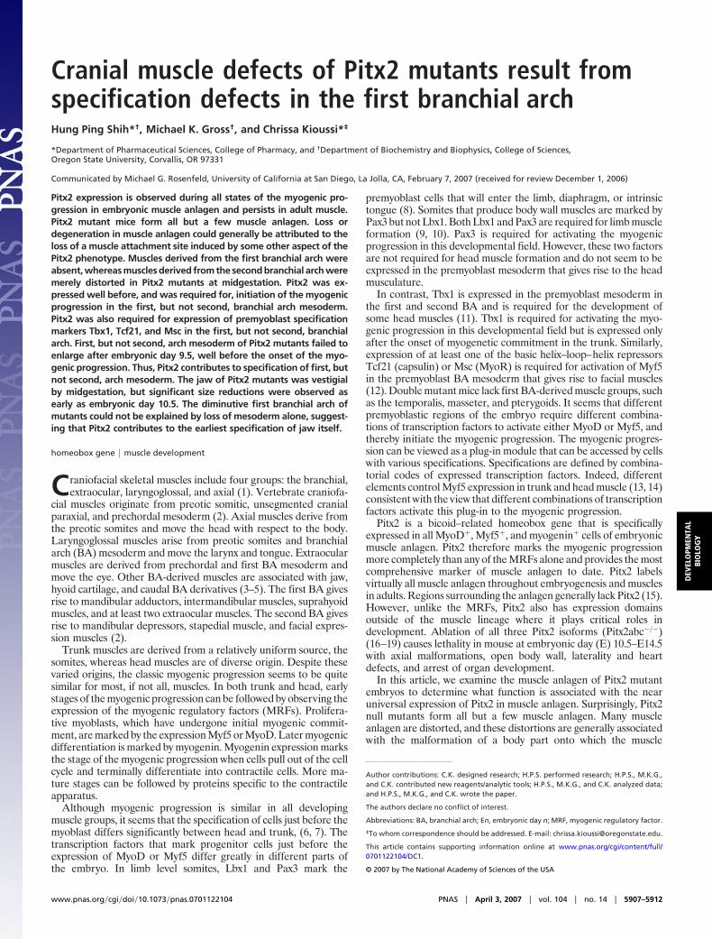
The ontogeny of head muscle anlagen was therefore comparedin mutant and heterozygote embryos between E9.5 and E13.5 todetermine how the apparent anlagen defects observed at E13.5arise (Fig. 1). The severe deformity and complexity of the anlagendefects made it difficult to identify corresponding anlagen in
mutants and heterozygotes at E13.5 (Fig. 1 E and J). At E12.5, thepattern of X-Gal staining was simpler and allowed equivalentanlagen staining to be traced in the areas posterior and anterior tothe jaw. However, the mandibular and maxillary components of thejaw were vestigial in mutants, and only a few residual blue spotswere observed in these regions, indicating that most jaw-associatedmuscle anlagen were absent. A fan-shaped anlage originatingventral to the otic vesicle and inserting on the ventral aspect of themandible seemed to be the anlage for a mandibular depressor,which derives from the second BA (11). This anlage was present butis no longer fan-shaped in mutants (Fig. 1 D and I). One crescent-shaped anlage just anterior to the eye seemed to extend toward thedorsal aspect of the jaw. This anlage was significantly shorter inmutants.
At E11.5, it was still possible to identify the mandibular andmaxillary components of the developing jaw, or first BA, inmutants. However, both components were much smaller thannormal. In contrast, the size of the second, or hyoid, arch showedno significant reduction (Fig. 1 C and H, asterisk). Pitx2 wasnormally expressed in a broad domain in the posterior half of themaxillary component and in a smaller more dorsal domain betweenthe maxillary and mandibular components. These expression do-mains were not, or were, vestigial in mutants (Fig. 1 C and H,arrow). Instead, the maxillary component showed ectopic expres-sion that resembled second arch expression. The Pitx2 expressiondomains in the second BA showed no significant defects in mutants.
At E10.5, the first BA of mutants was only slightly smaller, butstriking differences in the X-Gal stain were still observed (Fig. 1 Band G). Some of the X-Gal stain at E10.5 is likely to correspond tomuscle anlagen. However, the broad diffuse staining observed atE9.5 (Fig. 1 A and F) was due to Pitx2 expression in surfaceectoderm. Ectoderm expression has also been reported at E10.5 inthis region. The loss or malformation of jaw associated muscleanlagen was obvious at later stages when the jaw was vestigial. Thefirst BA was still present at earlier stages but showed strikingchanges in the pattern of Pitx2 expression, suggesting that muscleanlagen were defective before loss of the structure. In contrast,hyoid arch-associated muscle anlagen, which showed deformitiesonly at later stages, were not absent. No significant changes in thesecond BA size or Pitx2 expression were observed at earlier stages.Taken together, these results suggest that second BA anlagen wereformed and became distorted, whereas first BA muscle anlagenwere not properly formed in Pitx2 mutants.
Pitx2 Is Required for Initiation of Myogenic Progression in the First butNot Second Branchial Arch. The expression of Pitx2 in nonmyogenictissues of the first BA at the earliest stages suggests that thewhole-mount X-Gal analysis may not show a true picture of muscleanlagen in the developing jaw. No clusters of myogenin� cells thatlacked Pitx2(�-gal) expression were observed in serial sections of
Fig. 1. Loss of head muscle in Pitx2 mutants. Whole-mountX-Gal staining was performed to trace and compare the headmuscle anlagen in Pitx2�/LacZ (A–E) and Pitx2LacZ/LacZ (F–J)mouse embryos. Branchial arch structures are outlined. (A andF) At E9.5, Pitx2 was expressed in the first BA. No significantanatomical change was observed in the mutants. (B and G) AtE10.5, Pitx2(�-Gal) was detected in both first and second BA (B,arrows and asterisk). In the Pitx2 mutant, the size of first BAwas slightly smaller but the X-Gal-positive area was largelyreduced (G, arrow). (G, asterisk) No significant changes in thesecond BA size or Pitx2 expression were observed. (C and H) AtE11.5, Pitx2 mutants were characterized by hypocellular firstBA. (H, asterisk) No significant changes in the second BA wereobserved. (H, arrow) Only a residual presumptive muscle anlage was found in first BA of the mutant. (D and I) At E12.5, first BA-derived muscles were absent(arrow), and the second BA-derived muscles were deformed in the mutant (arrowheads). (E and J) At E13.5, severe deformity and complexity of muscle anlagenwas observed in the mutant. The maxillary and mandibular muscles were not properly formed (asterisks), and the second BA-derived muscles were significantlydeformed in the mutant (arrows). e, eye; mb, mandibular component; mx, maxillary component; ov, otic vesicle; 1, first BA; 2, second BA.
5908 � www.pnas.org�cgi�doi�10.1073�pnas.0701122104 Shih et al.
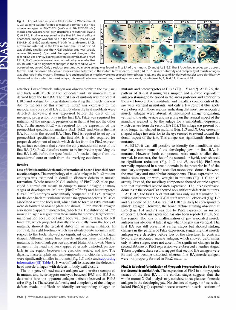
the head (data not shown). Thus, Pitx2 still marked the muscleanlagen of the jaw. However, immunohistochemical studies atE12.5 revealed that the first BA contained many Pitx2� cells outsidethe myogenin� territories (Fig. 2I). These Pitx2�/myogenin� cellsmay be either surface ectoderm or neural crest derivatives. Pitx2expression in neural crest derivatives has also been observed inother regions of the head (20–23). Pitx2 expression was generallynot observed in nonmyogenic territories surrounding anlagen inother regions of the body (17) Severe losses in myogenin�/Pitx2�
territories were observed in the region adjacent to the tongue thatrepresents the vestigial jaw (Fig. 2 I and M). Thus, muscle anlagenof the first BA were severely reduced or absent in Pitx2 mutants.
The severe reduction in first BA size was associated with the lossof first BA muscle anlagen. Arch size reduction could result fromdecreased proliferation or increased cell death in either the muscleanlagen, the nonmyogenic components of the arch, or both. If theloss of first BA muscle anlagen was due to reduced cell proliferationor increased apoptosis, then one should observe decreased BrdUincorporation and PH3� cells or increased TUNEL in the relevantmyogenin�/Pitx2� territory. Section across the jaw at E9.5 (Fig. 2A, B, E, and F) and E10.5 (Fig. 2 C, D, G, and H) showed increasedTUNEL staining in the mutants (Fig. 2 E and H). No change in thePH3 staining was detected. Matched sections across the jaw atE12.5 show myogenin�/Pitx2� territories that represent muscleanlagen in the tongue and the surrounding jaw. No significantdifference in BrdU labeling was observed in these territories. Incontrast, the regions outside these territories showed a slightlyhigher density of BrdU labeling in heterozygotes (Fig. 2 I and M,asterisk). TUNEL staining showed no significant differences insideor outside of these territories (Fig. 2 J and N). Thus, the reductionin first BA size in Pitx2 mutants was likely caused by increased celldeath inside and outside of the muscle anlagen at E9.5-E10.5. Thereduction in size of myogenin� muscle anlagen did not seem to beproportional to the overall size reduction. The loss of muscle cannotalone account for the deformation of jaw. The shape of themyogenin� muscle anlagen was also very different in mutants andheterozygotes. Taken together, these observations indicate that thejaw was incorrectly patterned and all components were smaller. Theloss of Pitx2 resulted increased cell death and in the loss of nearlyall myogenin� cells, or muscle anlagen, in the first BA.
Loss of myogenin� muscle anlagen in the first BA may haveresulted from a failure to specify a population of myoblasts in thisregion. Myf5 and MyoD label the onset of myogenic progression,and their expression in the first BA begins at E10 and E10.5,respectively (24) (25). Immunohistochemical analyses of the firstBA at E10 and E10.5 demonstrate that heterozygotes produceMyf5� and MyoD� cells in the core of the first BA (Fig. 2 K andL) where X-Gal staining was observed (Fig. 1B). Both MyoD andMyf5 label subpopulations of the Pitx2� cell cluster at the meso-dermal cores of both the first and second BA, consistent withprevious studies (15). Pitx2 staining was also observed in theoverlying ectoderm of the first but not second BA (Fig. 2 I and K).At E10, the Pitx2(�-Gal)� first BA core was already much smallerin mutants and Myf5� cells were not detected in it (Fig. 2 L and P).A few MyoD� cells were observed in the first BA core of mutantsat E10.5, but their total number and the fraction of Pitx2(�-Gal)�
cells that was MyoD� was dramatically reduced (Fig. 2 L and P).Drastic reduction of MyoD expression was also observed by whole-mount RNA in situ (Fig. 3 E and J). In contrast, the Pitx2� core ofthe second BA of mutants was normal in size and no defects inMyoD or Myf5 expression were observed in them (data not shown).
Fig. 2. Pitx2 specifies first BA myoblasts. (A, B, E, and F) TUNEL/PH3/�-Gal(Pitx2) triple labeling immunohistochemistry on transverse head sectionsof E9.5 Pitx2�/LacZ (A and B) and Pitx2LacZ/LacZ (E and F). (C, D, G, and H)TUNEL/PH3 double labeling on frontal head sections of E10.5 Pitx2�/LacZ (C andD) and Pitx2LacZ/LacZ (G and H). (E–H) Significant programmed cell death in-crease was observed in the mutant mice at E9.5 and E10.5. (B, F, D, and H) High-magnification images. (I–P) BrdU/myogenin/�-Gal(Pitx2) triple labeling immu-nohistochemistry on frontal head sections of E12 Pitx2�/LacZ (I) and Pitx2LacZ/LacZ
(M). Muscle anlagen were outlined by Pitx2(�-Gal)� territory. No significantdifference in BrdU labeling was observed in this territory. Myogenin wasexpressed only in a residual Pitx2(�-gal)� territory in the mutant, indicating amassive muscle reduction in the jaws (M, arrow). (J and N) TUNEL/Pitx2(�-Gal)double labeling immunohistochemistry on frontal head sections of E12.5Pitx2�/LacZ (J) and Pitx2LacZ/LacZ (N). No significant change in TUNEL signal wasobserved within the outlined Pitx2(�-Gal)� territory in the heterozygote andmutant mice. (K and O) Myf5/�-Gal(Pitx2) double labeling immunohistochem-istry on transverse head sections of E10 Pitx2�/LacZ (K) and Pitx2LacZ/LacZ (O). Inthe heterozygote, Myf5 was colocalized with a Pitx2(�-gal)� cell subpopula-tion in the first BA muscle anlagen (K, arrow). (O, arrow) Expression of Myf5was not observed in the residual Pitx2(�-gal)� territory in the mutant. (L andP) Six2/MyoD/�-gal(Pitx2) triple labeling immunohistochemistry on sagittalhead sections of E10.5 Pitx2�/LacZ (L) and Pitx2LacZ/LacZ (P). (Q–X) EGFP/Tbx1/Pitx2(�-Gal) (Q, R, U, and V) and EGFP/Tcf21/�-gal(Pitx2) (S, T, W, and X) triplelabeling immunohistochemistry on transverse head sections of E10.5Wnt1Cre R26EGFP Pitx2�/LacZ (Q–T) and Wnt1Cre R26EGFP Pitx2LacZ/LacZ (U–X).Tbx1� cells were colocalized with the Pitx2(�-Gal)� cells in the mesodermalcores of the first and second BA in the heterozygote mice (R, arrows). Thesecells were surrounded with the GFP� neural crest cells. Expression of Tbx1 wasbarely detectable in the mesodermal core of first BA in the mutant mice (U andV). Tcf21�/Pitx2(�-Gal)� cells were detected in the mesoderm core of the firstBA in the heterozygote mouse (S and T). This cell population was not observed
in the mutant mouse (W and X). Expression of Tcf21 was also observed in theectoderm-derived component of the first BA, which was located inside thecore (S and W, arrow). (R, T, V, and X) Higher-magnification images of outlinedarea. 1, first BA; 2, second BA; V, trigeminal ganglion; tg, tongue.
Shih et al. PNAS � April 3, 2007 � vol. 104 � no. 14 � 5909
DEV
ELO
PMEN
TAL
BIO
LOG
Y
These observations indicate that Pitx2 lies genetically upstream ofthe myoblast markers Myf5 and MyoD in the first, but not second,BA and that it plays a role in initiating myogenic progression onlyin the first BA.
Pitx2 Regulates Transcription Factors That Specify First Branchial ArchMyoblasts. Pitx2 expression precedes and is required for the ex-pression of both Myf5 and MyoD in the mesodermal core of the firstBA. Several other transcription factors have similar properties.Tcf21(capsulin) and Msc(MyoR) seem to encode a somewhatredundant pair of basic helix–loop–helix transcription factors, forwhich at least one needs to be present to form first BA associatedmastication muscles(masseter, pterygoid, and temporalis) (12).Loss of both genes results in absence of Myf5 and severe reductionof MyoD in the first BA. Tbx1 is required for Myf5 and MyoDexpression in the first and second BA and its loss leads to defectsin first and second arch associated muscles (11). Six2 is specificallyexpressed in the first BA core at E9 and in the second BA core atslightly later stages (26). Functional analyses for this gene in the BAregions have not been reported. The expression of these fourmarkers was examined in Pitx2 mutants.
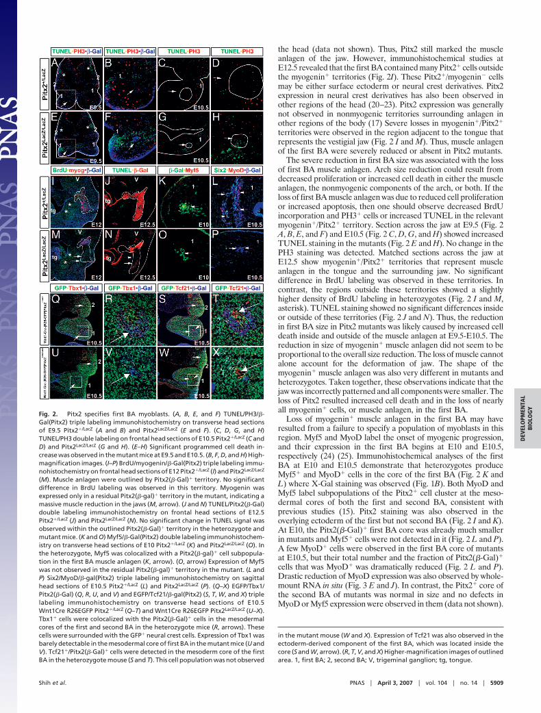
Tbx1 was expressed in most, if not all, Pitx2�(�-Gal)� cells in themesodermal cores of the first and second BA of heterozygotes atE10.5 (Fig. 2 Q and R). Similarly, virtually all Tbx1� cells werePitx2� (�-Gal)�. Pitx2 and Tbx1 therefore label identical cellpopulations in these two regions. This cell population was severelyreduced in size in the first BA of mutants. Furthermore, the levelof Tbx1 expression in the residual population, which could still beidentified by X-Gal staining, was only barely detectable at high gain.In contrast, the core Tbx1�/Pitx2(�-Gal)� cell population showedno significant difference in size or Tbx1 expression in the second BA(Fig. 2 U and V). The size of the Pitx2� core populations was moresimilar in the first BA of heterozygotes and mutants at E9.5 (Fig.2 A and E). Whole-mount RNA in situ analyses indicated that Tbx1RNA was expressed in stripes of similar intensity in the first andsecond BA (Fig. 3A). In mutants, the stripe representing themesodermal core of the first BA was not detected when the striperepresenting the mesodermal core of the second BA was clearlyvisible (Fig. 3F).
Tcf21 was coexpressed with Pitx2(�-Gal)� cells of the BA coresin a manner similar to Tbx1, indicating that Tcf21 and Pitx2 alsolabel the same mesodermal cells. One important difference wasnoted. Tcf21 also labels the neural crest-derived cells that resideinside the mesoderm core (Fig. 2 S and W). Pitx2(�-Gal)�/Tcf21�
cells were not detected in the residual �-Gal� core of the first BAof mutants (Fig. 2 W and X). However, a group of Pitx2(�-Gal)�/Tcf21� cells that expressed the neural crest lineage tracer were
detected. No significant changes in Tcf21 were observed in thesecond BA core of mutants. Whole-mount RNA in situ analyses atE9.5 indicated that Tcf21 RNA was expressed in stripes of similarintensity in the first and second BA (Fig. 3B). In mutants, Tcf21 wasstill expressed in the first and second BA however the expressionpattern in the first BA became thinner (Fig. 3 B and G, arrow),consistent with the maintenance of Tcf21 in the neural crest-derivedpopulation at the center of the mesodermal core.
Msc and Six2 RNAs were also expressed in a central stripe in thefirst BA at E9.5 (Fig. 3 C and D). A weak central stripe was observedfor Msc, but not for Six2, in the second BA. Expression of Msc andSix2 RNA were not observed in the first BA core of mutants (Fig.3 C and H). The weak Msc expression was not significantly alteredin the second BA. Functional Pitx2 was cell autonomously requiredfor proper Tbx1, Tcf21, Msc, and Six2 expression in the premyoblastprecursors of first, but not second, BA. Pitx2 acted geneticallyupstream of all four transcription factors in the first BA. Three ofthese factors have been invoked in the specification or commitmentof first BA myoblasts.
Pitx2 Specifies Premyoblast Mesoderm in First Branchial Arch. Im-munohistochemical detection of the mesodermal core of the firstBA currently requires expression of either Tbx1, Tcf21, or Pitx2(�-Gal) in the core. The results above indicate that loss of Pitx2 resultsin severe reduction or loss of Tbx1 and Tcf21 expression. They alsoshow that the �-Gal-labeled area, referred to as the residualmesodermal core, was severely reduced in mutants. However, it ispossible that Pitx2 was also required for its own expression in apositive feedback loop and that the reduced number of �-Gal� cellsin mutants reflected a loss of Pitx2(�-Gal) expression rather thana loss of the mesodermal core itself. A Pitx2 independent means todetect the mesodermal core was needed to test this hypothesis.
In the developing BA, neural crest cells fill the space between thesurface ectoderm and the enclosed mesoderm at early stages.Neural crest cells generate bone, cartilage, and neuronal cells, butnot muscle cells, in the developing jaws. The Wnt1-Cre�Rosa-EGFPsystem indelibly labels the neural crest lineage from the time it iscreated (27). Examination of embryos bearing this tracing systemrevealed GFP� holes in the BA. Double labeling with GFP andTbx1, Tcf21, or Pitx2(�-Gal) showed that these holes were filled bythe mesodermal cores (Fig. 2 J and L). If the loss of Pitx2 functionmerely resulted in down-regulation of Pitx2, then one would expectto see a Pitx2(�-Gal)� hole defined by the neural crest lineagetracing system. This Pitx2(�-Gal)� hole was not observed. Thesimplest interpretation of the data are therefore that the mesoder-mal cores of Pitx2 mutants are severely reduced between E9.5 andE10.5. Myogenic progression normally begins at E10 to E10.5.
Fig. 3. Pitx2 regulated transcription factors in first BA. (A–J)RNA whole-mount in situ hybridization for tbx1 (A and F), tcf21(B and G), msc (C and H), six2 (D and I), and myod1 (E and J) inPitx2�/LacZ (A–E) and Pitx2LacZ/LacZ (F–J) E9.5 or E10.5 mice. Theexpression of tbx1 RNA was not observed in the first BA in thePitx2 mutants (F, arrow), but no significant change was observedin the second BA (F, asterisk). The expression of tcf21 RNA wasreduced in the first BA (G, arrow), but no significant change wasobserved in the second BA (G, asterisk). The expression of msc (Cand H) and six2 (E and J) RNA was not observed in the first BA ofthePitx2mutants. (K) InvivoChIPassays fromfirstandsecondBAchromatin extracts of E12 heterozygote mice indicated the pres-ence of Pitx2a on the Pitx1 and Tbx1 promoters. The chromatinextracts from Pitx2LacZ/LacZ mice were used as a negative control.(L–Q) Overexpression of Pitx2a-IRES-EGFP under the control ofCMV resulted in activation of Tbx1 expression (L–N, arrows).Nuclear staining of Tbx1 was not observed in C2C12 myoblaststransfected with an empty vector (O–Q). 1, first BA; 2, second BA.
5910 � www.pnas.org�cgi�doi�10.1073�pnas.0701122104 Shih et al.
Thus, Pitx2 seems to be required for specification premyoblastmesoderm in the first but not second BA.
Pitx2 Directly Interacts with Tbx1 Regulatory Elements. Chromatinimmunoprecipitation using BA tissue and Tbx1 expression inmyoblast cell cultures were used to test whether the regulation ofTbx1 by Pitx2 was due to a direct molecular interaction betweenPitx2 protein and Tbx1 regulatory sequences. Five potential Pitx2binding sites were identified in the 3 kb of genomic sequence thatlie between the transcription initiation site of the Ensembl genemodel for Tbx1 and the initiation site of a gene model for adivergent transcript of unknown function (Fig. 3K). This sequencehas promoter activity in cell culture studies (11). Primers weredesigned to encompass these putative binding sites. Chromatinprepared from the BA of heterozygotes and mutants at E12.5 wassheared and immunoprecipitated with anti-Pitx2a antibodies. Tbx1promoter fragments were amplified from heterozygote, but notfrom mutant precipitates (Fig. 3K). Pitx1 promoter fragments werealso selectively amplified in heterozygous precipitates. Pitx1 isexpressed in the first BA and is down-regulated in Pitx2 mutants(20) (28). These data indicate that Pitx2 occupies sequences up-stream of Tbx1 in BA. A bicistronic expression vector containingthe Pitx2a cDNA and IRES-GFP under the control of the CMVpromoter was used to transiently overexpress Pitx2a in the C2C12mouse myoblast cell line. Transfected cells that over-expressedPitx2a were identified by GFP expression. These cells expressedhigh levels of Tbx1 that were not detected in cells transfected by thecontrol plasmid, CMV-IRES-GFP (Fig. 3 L and Q). Taken to-gether, the results are consistent with the idea that the Pitx2transcription factor regulates Tbx1 by directly interacting with itspromoter.
DiscussionHomeobox genes generally display discrete zones of expression andare thought to engage in different molecular mechanisms in eachof the zones. Pitx2 is strongly expressed in muscle (15), neural crest(20–22), cardiac fields (23) (C.K., unpublished data) and brain celllineages (29). Mutants of the Pitx2 homeobox gene have pheno-types in body parts corresponding to each of the gene’s embryonicexpression domains. However, the reported muscle phenotypes inPitx2 mice have defied a consistent explanation. Whereas it is clearthat virtually all muscle anlagen in embryos express Pitx2 in allstages of the myogenic progression (15), only a few muscles show
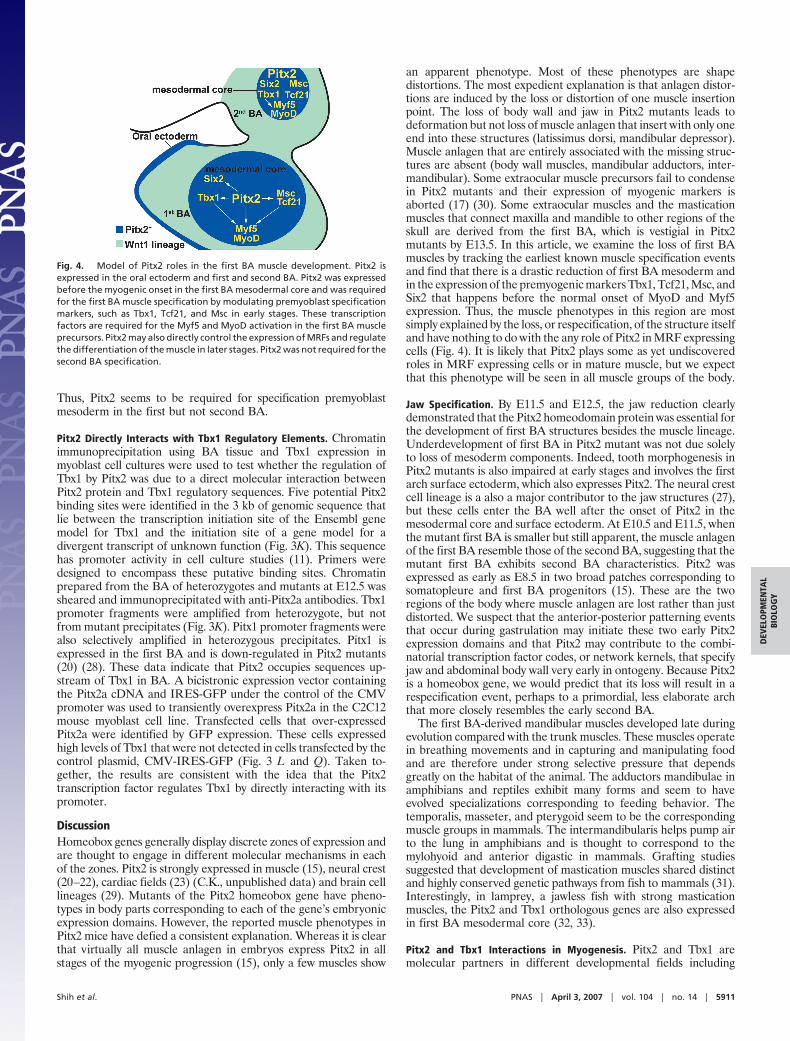
an apparent phenotype. Most of these phenotypes are shapedistortions. The most expedient explanation is that anlagen distor-tions are induced by the loss or distortion of one muscle insertionpoint. The loss of body wall and jaw in Pitx2 mutants leads todeformation but not loss of muscle anlagen that insert with only oneend into these structures (latissimus dorsi, mandibular depressor).Muscle anlagen that are entirely associated with the missing struc-tures are absent (body wall muscles, mandibular adductors, inter-mandibular). Some extraocular muscle precursors fail to condensein Pitx2 mutants and their expression of myogenic markers isaborted (17) (30). Some extraocular muscles and the masticationmuscles that connect maxilla and mandible to other regions of theskull are derived from the first BA, which is vestigial in Pitx2mutants by E13.5. In this article, we examine the loss of first BAmuscles by tracking the earliest known muscle specification eventsand find that there is a drastic reduction of first BA mesoderm andin the expression of the premyogenic markers Tbx1, Tcf21, Msc, andSix2 that happens before the normal onset of MyoD and Myf5expression. Thus, the muscle phenotypes in this region are mostsimply explained by the loss, or respecification, of the structure itselfand have nothing to do with the any role of Pitx2 in MRF expressingcells (Fig. 4). It is likely that Pitx2 plays some as yet undiscoveredroles in MRF expressing cells or in mature muscle, but we expectthat this phenotype will be seen in all muscle groups of the body.
Jaw Specification. By E11.5 and E12.5, the jaw reduction clearlydemonstrated that the Pitx2 homeodomain protein was essential forthe development of first BA structures besides the muscle lineage.Underdevelopment of first BA in Pitx2 mutant was not due solelyto loss of mesoderm components. Indeed, tooth morphogenesis inPitx2 mutants is also impaired at early stages and involves the firstarch surface ectoderm, which also expresses Pitx2. The neural crestcell lineage is a also a major contributor to the jaw structures (27),but these cells enter the BA well after the onset of Pitx2 in themesodermal core and surface ectoderm. At E10.5 and E11.5, whenthe mutant first BA is smaller but still apparent, the muscle anlagenof the first BA resemble those of the second BA, suggesting that themutant first BA exhibits second BA characteristics. Pitx2 wasexpressed as early as E8.5 in two broad patches corresponding tosomatopleure and first BA progenitors (15). These are the tworegions of the body where muscle anlagen are lost rather than justdistorted. We suspect that the anterior-posterior patterning eventsthat occur during gastrulation may initiate these two early Pitx2expression domains and that Pitx2 may contribute to the combi-natorial transcription factor codes, or network kernels, that specifyjaw and abdominal body wall very early in ontogeny. Because Pitx2is a homeobox gene, we would predict that its loss will result in arespecification event, perhaps to a primordial, less elaborate archthat more closely resembles the early second BA.
The first BA-derived mandibular muscles developed late duringevolution compared with the trunk muscles. These muscles operatein breathing movements and in capturing and manipulating foodand are therefore under strong selective pressure that dependsgreatly on the habitat of the animal. The adductors mandibulae inamphibians and reptiles exhibit many forms and seem to haveevolved specializations corresponding to feeding behavior. Thetemporalis, masseter, and pterygoid seem to be the correspondingmuscle groups in mammals. The intermandibularis helps pump airto the lung in amphibians and is thought to correspond to themylohyoid and anterior digastic in mammals. Grafting studiessuggested that development of mastication muscles shared distinctand highly conserved genetic pathways from fish to mammals (31).Interestingly, in lamprey, a jawless fish with strong masticationmuscles, the Pitx2 and Tbx1 orthologous genes are also expressedin first BA mesodermal core (32, 33).
Pitx2 and Tbx1 Interactions in Myogenesis. Pitx2 and Tbx1 aremolecular partners in different developmental fields including
Fig. 4. Model of Pitx2 roles in the first BA muscle development. Pitx2 isexpressed in the oral ectoderm and first and second BA. Pitx2 was expressedbefore the myogenic onset in the first BA mesodermal core and was requiredfor the first BA muscle specification by modulating premyoblast specificationmarkers, such as Tbx1, Tcf21, and Msc in early stages. These transcriptionfactors are required for the Myf5 and MyoD activation in the first BA muscleprecursors. Pitx2 may also directly control the expression of MRFs and regulatethe differentiation of the muscle in later stages. Pitx2 was not required for thesecond BA specification.
Shih et al. PNAS � April 3, 2007 � vol. 104 � no. 14 � 5911
DEV
ELO
PMEN
TAL
BIO
LOG
Y
cranial, limb and heart muscle lineages. Recent studies indicatedthat Tbx1 and Pitx2 are in the same genetic pathway during cardiacdevelopment (34). Tbx1 is expressed in both first and second BA atE9.5 and when mutated leads to severe perturbation or absence ofboth first and second BA muscles (11). Tbx1 also maintains thenumber of myocytes in the head and limb (35). Interestingly, likePitx2, expression of Tbx1 follows the onset of myogenic commit-ment in the limb muscle anlagen but precedes the speciation eventin the first BA muscle precursors (15). Our data show that Pitx2 isnot only required but is also sufficient to activate Tbx1. Therefore,it is possible that Pitx2 controls the number of muscle precursorsthrough Tbx1 in the first BA mesodermal core (Fig. 4). Further-more, our microarray and real-time PCR data from myoblastsindicated that Tbx1 was down-regulated in the limb muscle anlagenof Pitx2 mutant (H.P.S., unpublished data) and places Pitx2 up-stream of Tbx1 in the skeletal muscle lineages.
Materials and MethodsMouse Strains. Pitx2-LacZ knockin mice (18) on ICR (outbred stockfrom the Institute of Cancer Research) background were used.
X-Gal Staining, Immunohistochemistry, BrdU Labeling, and TUNELStains. For general immunohistochemical studies and X-Gal stain-ing, mouse embryos were dissected free of membranes; then weproceeded as described (15). Specific antibodies against MyoD,myogenin, Myf5 (Santa Cruz Biotechnology, Santa Cruz, CA),�-galactosidase (Cappel, Aurora, OH), PH3 (Upstate, Lake Placid,NY), BrdU (Accurate Chemical Scientific Corporation, Westbury,NY), and EGFP (H.P.S., Oregon State University) were applied atempirically determined optimal titers. BrdU/PBS solution (50 �g/gof body weight) was injected i.p. 2 hr before killing. BrdU immu-nohistochemistry was performed in postfixed immunolabeled tissuefollowed by acid depurination and neutralization. TUNEL assaywas also performed as recommended by the manufacturer (DeadEnd kit; Promega).
RNA Whole-Mount in Situ Hybridization. RNA in situ hybridiza-tion was performed according to standard procedures (26).
Digoxigenin-labeled antisense RNA riboprobes were generated byin vitro transcription kit (Roche Molecular Biochemicals). AP-conjugated anti-DIG antibody was used to detect the hybridizationsignals (Roche Molecular Biochemicals).
Tissue Culture and Transient Transfection. C2C12 cells were grown on10-cm dishes in DMEM (Cellgro) supplement with 10% FBS(HyClone), 5 mM glutamine, penicillin, and streptomycin. TheC2C12 cells were then seeded onto poly(L-lysine) (0.1 mg/ml)-coated glass coverslips in 12-well plates. Cells at 50% confluencewere transfected and then maintained in the medium withoutantibiotics according to the instructions of the manufacturer (Li-pofectamine 2000; Invitrogen). After 24 hr, the transfected cellswere examined by immunohistochemical staining.
In Vivo ChIP Assays. BA were dissected and collected from E12.5embryos. Dissected BA were mechanically triturated several timesby using 1-ml tip on ice to dissociate the tissue. Dissociated cellswere cross-linked with 1% formaldehyde/PBS solution for 10 minat 25°C, resuspended in lysis buffer, and then sonicated (averagelength of sheared fragments was �300–1,000 bp). Ten percent ofthe soluble chromatin complex was saved for positive control.Soluble chromatin complexes were diluted and then incubated withspecific IgGs against Pitx2a (15, 20) overnight at 4°C. Chromatin/antibody complexes were pulled down by protein-A Sepharosebeads and then eluted. Eluted immunoprecipitates were heated at65°C for 6–18 hr to reverse the formaldehyde cross-linking. DNAfragments were purified with a QIAquick spin kit (Qiagen, Chat-worth, CA).
We thank Merveen Appu, Jane Menino, John Loflin, and Shelley Brownfor technical assistance and R. Kelly and E. Olson for RNA in situ probes.This publication was made possible by American Heart AssociationGrant 0550179Z, March of Dimes Grant 1-FY05-120, National Instituteon Environmental Health Sciences Grant P30 ES00210, and OregonState University (OSU) funds (to C.K.). The confocal microscopy facilityof the Center for Gene Research and Bioinformatics and the Environ-mental Health Sciences Center at OSU was instrumental in completingthis research.
1. Noden DM, Francis-West P (2006) Dev Dyn 235:1194–1218.2. Kaufman MH, Bard JBL (1999) The Anatomical Basis of Mouse Development
(Academic, San Diego).3. Noden DM (1983) Dev Biol 96:144–165.4. Couly GF, Coltey PM, Le Douarin NM (1992) Development (Cambridge, UK)
114:1–15.5. Trainor PA, Tan SS, Tam PP (1994) Development (Cambridge, UK) 120:2397–2408.6. Rawls A, Olson EN (1997) Cell 89:5–8.7. Mootoosamy RC, Dietrich S (2002) Development (Cambridge, UK) 129:573–583.8. Gross MK, Moran-Rivard L, Velasquez T, Nakatsu MN, Jagla K, Goulding M (2000)
Development (Cambridge, UK) 127:413–424.9. Bober E, Brand-Saberi B, Ebensperger C, Wilting J, Balling R, Paterson BM, Arnold
HH, Christ B (1994) Development (Cambridge, UK) 120:3073–3082.10. Goulding M, Lumsden A, Paquette AJ (1994) Development (Cambridge, UK)
120:957–971.11. Kelly RG, Jerome-Majewska LA, Papaioannou VE (2004) Hum Mol Genet 13:2829–
2840.12. Lu JR, Bassel-Duby R, Hawkins A, Chang P, Valdez R, Wu H, Gan L, Shelton JM,
Richardson JA, Olson EN (2002) Science 298:2378–2381.13. Carvajal JJ, Cox D, Summerbell D, Rigby PW (2001) Development (Cambridge, UK)
128:1857–1868.14. Hadchouel J, Carvajal JJ, Daubas P, Bajard L, Chang T, Rocancourt D, Cox D,
Summerbell D, Tajbakhsh S, Rigby PW, Buckingham M (2003) Development(Cambridge, UK) 130:3415–3426.
15. Shih HP, Gross MK, Kioussi C (2006) (2007) Gene Expr Patterns 7:441–451.16. Gage PJ, Suh H, Camper SA (1999) Development (Cambridge, UK) 126:4643–4651.17. Kitamura K, Miura H, Miyagawa-Tomita S, Yanazawa M, Katoh-Fukui Y, Suzuki R,
Ohuchi H, Suehiro A, Motegi Y, Nakahara Y, et al. (1999) Development (Cambridge,UK) 126:5749–5758.
18. Lin CR, Kioussi C, O’Connell S, Briata P, Szeto D, Liu F, Izpisua-Belmonte JC,Rosenfeld MG (1999) Nature 401:279–282.
19. Lu MF, Pressman C, Dyer R, Johnson RL, Martin JF (1999) Nature 401:276–278.20. Kioussi C, Briata P, Baek SH, Rose DW, Hamblet NS, Herman T, Ohgi KA, Lin C,
Gleiberman A, Wang J, et al. (2002) Cell 111:673–685.21. Gage PJ, Rhoades W, Prucka SK, Hjalt T (2005) Invest Ophthalmol Vis Sci
46:4200–4208.22. Evans AL, Gage PJ (2005) Hum Mol Genet 14:3347–3359.23. Ai D, Liu W, Ma L, Dong F, Lu MF, Wang D, Verzi MP, Cai C, Gage PJ, Evans S,
et al. (2006) Dev Biol 296:437–449.24. Summerbell D, Ashby PR, Coutelle O, Cox D, Yee S, Rigby PW (2000) Development
(Cambridge, UK) 127:3745–3757.25. Goldhamer DJ, Brunk BP, Faerman A, King A, Shani M, Emerson CP, Jr (1995)
Development (Cambridge, UK) 121:637–649.26. Oliver G, Wehr R, Jenkins NA, Copeland NG, Cheyette BN, Hartenstein V, Zipursky
SL, Gruss P (1995) Development (Cambridge, UK) 121:693–705.27. Chai Y, Jiang X, Ito Y, Bringas P, Jr, Han J, Rowitch DH, Soriano P, McMahon AP,
Sucov HM (2000) Development (Cambridge, UK) 127:1671–1679.28. Liu W, Selever J, Lu MF, Martin JF (2003) Development (Cambridge, UK) 130:6375–
6385.29. Sclafani AM, Skidmore JM, Ramaprakash H, Trumpp A, Gage PJ, Martin DM
(2006) Genesis 44:336–344.30. Diehl AG, Zareparsi S, Qian M, Khanna R, Angeles R, Gage PJ (2006) Invest
Ophthalmol Vis Sci 47:1785–1793.31. von Scheven G, Alvares LE, Mootoosamy RC, Dietrich S (2006) Development
(Cambridge, UK) 133:2731–2745.32. Boorman CJ, Shimeld SM (2002) Evol Dev 4:354–365.33. Sauka-Spengler T, Le Mentec C, Lepage M, Mazan S (2002) Gene Expr Patterns
2:99–103.34. Nowotschin S, Liao J, Gage PJ, Epstein JA, Campione M, Morrow BE (2006)
Development (Cambridge, UK) 133:1565–1573.35. Dastjerdi A, Robson L, Walker R, Hadley J, Zhang Z, Rodriguez-Niedenfuhr M,
Ataliotis P, Baldini A, Scambler P, Francis-West P (2007) Dev Dyn 236:353–363.
5912 � www.pnas.org�cgi�doi�10.1073�pnas.0701122104 Shih et al.