cpg methylation analysis—current status of clinical assays and potential applications in molecular...
TRANSCRIPT
JMD
CME Pro
gram
Special ArticleCpG Methylation Analysis—Current Status of ClinicalAssays and Potential Applications in MolecularDiagnostics
A Report of the Association for Molecular Pathology
Antonia R. Sepulveda,*† Dan Jones,*‡
Shuji Ogino,*§ Wade Samowitz,*¶
Margaret L. Gulley,*� Robin Edwards,*,**Victor Levenson,*†† Victoria M. Pratt,*‡‡
Bin Yang,*§§ Khedoudja Nafa,*¶¶ Liying Yan,*��
and Patrick Vitazka*,***From The Methylation Working Group of the Association
for Molecular Pathology Clinical Practice Committee *;
the University of Pennsylvania School of Medicine,† Philadelphia,
Pennsylvania; the University of Texas M.D. Anderson Cancer
Center,‡ Houston, Texas; the Brigham and Women’s Hospital
and Harvard Medical School,§ Dana-Farber Cancer Institute,
Harvard School of Public Health, Boston, Massachusetts; ARUP
Laboratories,¶ University of Utah, Salt Lake City, Utah; the
University of North Carolina,� Chapel Hill, North Carolina;
Oncology Wyeth Research,** Collegeville, Pennsylvania; Rush
University Medical Center,†† Chicago, Illinois; Quest Diagnostics,‡‡
Nichols Institute, Chantilly, Virginia; the Cleveland Clinic
Foundation,§§ Cleveland, Ohio; the Memorial Sloan-Kettering
Cancer Center,¶¶ New York, New York; EpigenDx,�� Worcester, MA;
and Virginia Commonwealth University, Medical College of
Virginia,*** Richmond, Virginia
Methylation of CpG islands in gene promoter regionsis a major molecular mechanism of gene silencingand underlies both cancer development and progres-sion. In molecular oncology, testing for the CpGmethylation of tissue DNA has emerged as a clinicallyuseful tool for tumor detection, outcome prediction,and treatment selection, as well as for assessing theefficacy of treatment with the use of demethylatingagents and monitoring for tumor recurrence. In ad-dition, because CpG methylation occurs early in pre-neoplastic tissues, methylation tests may be useful asmarkers of cancer risk in patients with either infec-tious or inflammatory conditions. The MethylationWorking Group of the Clinical Practice Committee of
the Association of Molecular Pathology has reviewedthe current state of clinical testing in this area. Wereport here our summary of both the advantages anddisadvantages of various methods, as well as the needsfor standardization and reporting. We then conclude bysummarizing the most promising areas for future clin-ical testing in cancer molecular diagnostics. (J Mol Di-
agn 2009, 11:266–278; DOI: 10.2353/jmoldx.2009.080125)
CpG-island methylation of gene promoter regions plays amajor role in regulation of gene expression. CpG islandshave been defined as genomic regions with a minimum of200 bp, with % G�C greater than 50 and with observed/expected CpG ratio above 60%.1 More recently, studieshave further defined CpG islands as regions of DNAgreater than 500 bp with a G�C equal to or greater than55% and observed CpG/expected CpG of 0.65.2 In ac-tively transcribed genes the CpG sites in CpG islandsof promoter regions are unmethylated, whereas in-creased cytosine methylation in the island CpG sites isassociated with reduced gene expression and possi-ble gene silencing.
Gene regulation by CpG methylation is involved in alarge spectrum of biological processes, from develop-ment to aging, including inflammatory and infectious dis-eases, and cancer. The availability of molecular tech-
The Methylation Working Group is a subcommittee of the AMP ClinicalPractice Committee. The 2006–2008 AMP Clinical Practice Committeeconsisted of Aaron Bossler, Deborah Dillon, Michelle Dolan, WilliamFunkhouser, Julie Gastier-Foster, Dan Jones, Elaine Lyon (Chair 2005–2006), Victoria M. Pratt (Chair 2007–2008), Daniel Sabath, Antonia R.Sepulveda, Kathleen Stellrecht, and Daynna A. Wolff.
Standard of practice is not being defined by this article, and there maybe alternatives.
Accepted for publication April 9, 2009.
Address reprint requests to Association for Molecular Pathology (AMP),9650 Rockville Pike, Bethesda, MD 20814, E-mail: [email protected].
Journal of Molecular Diagnostics, Vol. 11, No. 4, July 2009
Copyright © American Society for Investigative Pathology
and the Association for Molecular Pathology
DOI: 10.2353/jmoldx.2009.080125
266
niques to evaluate the methylation status of CpG islandsin cancer related genes has prompted an explosion ofstudies in this area, and CpG methylation tests areemerging as clinically useful tests. CpG hypermethylationis critical to silencing of the expression of tumor suppres-sor genes, such as those that encode CDKN2B (p15),CDKN2A (p16), and O-6-methylguanine-DNA methyl-transferase (MGMT), as well as globally regulating differ-entiation programs in many tumor types. The levels ofCpG methylation have thus been used to subclassifytumors,3 predict response to chemotherapeutic agentsthat are metabolized or antagonized by cellular enzymesregulated by promoter methylation,4 and to assess theeffects of methylating and demethylating therapies. Intumors in which CpG methylation silencing of particularsuppressor genes is highly prevalent, the levels ofsuch methylated DNA in blood or body fluids may beindicative of the presence of cancer cells or their cir-culating DNA.5,6
In this report we compare the current techniques andmethodological considerations for assessing DNA CpGmethylation and summarize the current status of CpGmethylation testing with emphasis on neoplasia. Specificsections cover: 1) current methods for clinical testing ofCpG methylation and the decision-making criteria forassay selection and validation requirements; 2) applica-tions of CpG methylation testing for cancer detection,prognosis, and monitoring using tumor tissue, cell-freeplasma and serum, and cytological and other biologicalsamples; and, 3) the potential use of methylation interfer-ence and monitoring of CpG methylation status for pre-diction of tumors related to bacterial and viral infections.
To promote standardization in clinical reporting, wehave used Human Genome Organization (HUGO) genenomenclature throughout the text (http://www.genenames.org/index.html). A table listing the standard gene namesused in this document with the common names is pro-vided in Table 1.
Current Methods Used for CpG MethylationTesting
Analysis of CpG methylation requires some method ofdiscriminating between the methylated and unmethylatedDNA sequences, usually following PCR amplification oftargeted sequence(s). Post-PCR detection techniquesroutinely used to differentiate methylated and unmethyl-ated DNA include capillary electrophoretic separation,dideoxynucleotide sequencing,7 pyrosequencing,8 massspectrometry,3,9 high performance liquid chromatogra-phy,10 and array hybridization.11–17
Technical Considerations in the BisulfiteConversion Step
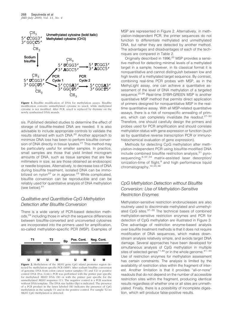
The majority of methods for methylation analysis beginwith the conversion by sodium bisulfite of unmethylatedcytosine to uracil (and then to thymine following in vitroDNA synthesis). By contrast, methylated cytosines are
largely protected from this conversion process (Figure 1).Bisulfite treatment thus creates different sequences inmethylated and unmethylated fragments, which can bedetected by a variety of techniques.
However, the effects of bisulfite treatment on DNA areharsh and difficult to control and often result in significantDNA degradation of up to 85% to 95% of target se-quences.18 This reduction in DNA template can greatlyaffect assay performance, including introducing PCRbias in amplification of sequences.19 Furthermore, thestability of bisulfite-treated DNA is reduced due to nucle-otide mispairing and incomplete complementarity. There-fore, in this initial step, one needs to optimize the condi-tions required for full bisulfite conversion of unmethylatedcytosine to uracil and yet minimize the degradative ef-fects of this treatment on DNA.
Although there are minimal data on the effects of tem-perature and time of storage on the stability of bisulfite-treated DNA, most laboratories analyze bisulfite-con-verted DNA soon after conversion to minimize furtherDNA degradation. Until more data are available, ultra-lowtemperature storage conditions (�70°C or below) shouldbe used if converted DNA must be stored before analy-
Table 1. HUGO Gene Nomenclature Committee-ApprovedSymbols for Genes Discussed in the Text
Symbol Common name
APC adenomatous polyposis coliCACNA1G calcium channel, voltage-dependent, T type
alpha-1G subunitCADM1 cell adhesion molecule 1 (IGSF4)CCND2 cyclin D2CDH1 cadherin 1 (E-cadherin)CDH13 cadherin 13 (H-cadherin)CDKN2A cyclin-dependent kinase inhibitor 2A (p16)CDKN2B cyclin-dependent kinase inhibitor 2B (p15)CRABP1 cellular retinoic acid binding protein 1DAPK1 death-associated protein kinase 1DNMT DNA methyltransferaseESR1 estrogen receptor alphaFHIT fragile histidine triadFRBP3 fatty acid binding protein 3, muscle and heart
(mammary-derived growth inhibitor, MDGI)GSTP1 glutathione S-transferase piHIC1 hypermethylated in cancer 1HSD17B4 hydroxysteroid (17-beta) dehydrogenase 4HSIL high-grade squamous intraepithelial lesionIGF2 insulin-like growth factor 2LATS1 large tumor suppressor, homolog 1LATS2 large tumor suppressor, homolog 2LINE long interspersed nucleotide elementMGMT O-6-methylguanine-DNA methyltransferaseMYOD1 myogenic differentiation 1NEUROG1 neurogenin 1PGR progesterone receptorPSA prostate specific antigenRARB retinoic acid receptor betaRASSF1 RAS association (RalGDS/AF-6) domain
family 1RUNX3 runt-related transcription factor 3SFRP1 secreted frizzled-related protein 1 (SARP2)SOCS1 suppressor of cytokine signaling 1TMEFF2 transmembrane protein with EGF-like and two
follistatin-like domains 2 (HPP1,hyperplastic polyposis 1)
TWIST1 twist homolog 1
CpG Methylation Assays 267JMD July 2009, Vol. 11, No. 4
sis. Published detailed studies to determine the effect ofstorage of bisulfite-treated DNA are needed. It is alsoadvisable to include appropriate controls to validate theresults obtained with such DNA.20 Another approach tominimize DNA loss has been to perform bisulfite conver-sion of DNA directly in tissue lysates.21 This method maybe particularly useful for smaller samples. In practice,small samples are those that yield limited microgramamounts of DNA, such as tissue samples that are fewmillimeters in size, as are those obtained as endoscopicor needle biopsies. Alternatively, to decrease loss of DNAduring bisulfite treatment, isolated DNA can be immo-bilized on nylon22 or in agarose.23 While complicated,bisulfite conversion can be reproducible and can bereliably used for quantitative analysis of DNA methylation(see below).21
Qualitative and Quantitative CpG MethylationDetection after Bisulfite Conversion
There is a wide variety of PCR-based detection meth-ods,24 including those in which the sequence differencesbetween bisulfite-converted and unconverted cytosinesare incorporated into the primers used for amplification,so-called methylation-specific PCR (MSP). Examples of
MSP are represented in Figure 2. Alternatively, in meth-ylation-independent PCR, the primer sequences do notfunction to differentiate methylated and unmethylatedDNA, but rather they are detected by another method.The advantages and disadvantages of each of the tech-niques are compared in Table 2.
Originally described in 1996,20 MSP provides a sensi-tive method for detecting minimal levels of a methylatedtarget in a sample; however, in its classical format it isnonquantitative and cannot distinguish between low andhigh levels of a methylated target sequence. By contrast,combining real-time PCR probes with MSP, as in theMethyLight assay, one can achieve a quantitative as-sessment of the level of DNA methylation of a targetedsequence.25,26 Real-time SYBR-GREEN MSP is anotherquantitative MSP method that permits direct applicationof primers designed for nonquantitative MSP in the real-time quantitative assay. With all MSP-related quantitativeassays, there is a risk of nonspecific annealing of prim-ers, which can completely invalidate the readout.27–30
Therefore, one should carefully design the primers andprobes used for PCR amplification and should correlatemethylation status with gene expression or function (suchas by quantitative reverse transcription PCR or immuno-histochemical evaluation of gene expression).
Methods for detecting CpG methylation after meth-ylation-independent PCR using bisulfite-modified DNAinclude combined bisulfite restriction analysis,31 pyro-sequencing,8,32–34 matrix-assisted laser desorption/ionization-time of flight,9 and high performance liquidchromatography.10,35,36
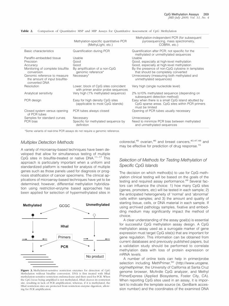
CpG Methylation Detection without BisulfiteConversion: Use of Methylation-SensitiveRestriction Enzymes
Methylation-sensitive restriction endonucleases are alsoroutinely used to discriminate methylated and unmethyl-ated CpG sites.37–39 The technical bases of combinedmethylation-sensitive restriction enzymes and PCR fordetection of CpG methylation are illustrated in Figure 3.One advantage of restriction enzyme-based analysisover bisulfite treatment methods is that it does not requiremodification of DNA sequences, which makes down-stream analysis relatively simple, and avoids target DNAdamage. Several approaches have been developed forsimultaneous analysis of CpG methylation in multiplesites of selected genes11,40 or in the whole genome.41–44
Use of restriction enzymes for methylation assessmenthas certain constraints. The analysis is limited by theavailability of restriction sites within the fragment of inter-est. Another limitation is that it provides “all-or-none”readouts that do not depend on the number of accessiblerestriction sites within the fragment, producing identicalresults regardless of whether one or all sites are unmeth-ylated. Finally, there is a possibility of incomplete diges-tion, which will produce false-positive results.
5’CCGG3’3’GGCC5’
CH3
CH3
Unmethylated cytosine (bold italic)Methylated cytosine (CH3)
Sodiumbisulfite
PCRCycle 1
3’AGCC5’
3’GGCU5’5’CCGA3’PCRCycle 25’UCGG3’
3’GGCU5’
CH3
CH3
5’UCGG3’CH3
5’TCGG3’
5’CCGA3’3’GGCT5’
3’AGCC5’
CH3
5’CCGG3’3’GGCC5’
CH3
CH3
Unmethylated cytosine (bold italic)Methylated cytosine (CH3)
Sodiumbisulfite
PCRCycle 1
3’AGCC5’
3’GGCU5’5’CCGA3’PCRCycle 25’UCGG3’
3’GGCU5’
CH3
CH3
5’UCGG3’CH3
5’TCGG3’
5’CCGA3’3’GGCT5’
3’AGCC5’
CH3
5’CCGG3’3’GGCC5’
CH3
CH3
Unmethylated cytosine (bold italic)Methylated cytosine (CH3)
Sodiumbisulfite
PCRCycle 1
3’AGCC5’
3’GGCU5’5’CCGA3’PCRCycle 25’UCGG3’
3’GGCU5’
CH3
CH3
5’UCGG3’CH3
5’TCGG3’
5’CCGA3’3’GGCT5’
3’AGCC5’
CH3Figure 1. Bisulfite modification of DNA for methylation assays. Bisulfitemodification converts unmethylated cytosine to uracil, while methylatedcytosine is not modified. After PCR, uracil is replaced by thymine on thenewly synthesized DNA strands.
Figure 2. Methylation of the MLH1 gene CpG island promoter region de-tected by methylation specific PCR (MSP). After sodium bisulfite conversionof genomic DNA from colon cancer tumor samples (T1 and T2) or positivecontrol DNA (Pos. Cont.), PCR was performed with the primer pair specificfor methylated MLH1 DNA (M) or with the primer pair specific for theunmethylated MLH1 sequence (U). The negative control is a PCR reactionwithout DNA template. The DNA size ladder (Bp) is indicated. The presenceof a PCR product in the lanes labeled (M) indicates the presence of CpGmethylation in the sample T1 and in the positive control. For sample T2 noMLH1 CpG methylation is detected.
268 Sepulveda et alJMD July 2009, Vol. 11, No. 4
Multiplex Detection Methods
A variety of microarray-based techniques have been de-veloped that allow for simultaneous testing of multipleCpG sites in bisulfite-treated or native DNA.11–17 Thisapproach is particularly important when a uniform andstandardized platform is needed for analysis of multiplegenes such as those panels used for diagnosis or prog-nosis stratification of cancer specimens. The clinical ap-plications of microarray-based techniques have yet to bedetermined; however, differential methylation hybridiza-tion using restriction-enzyme based approaches hasbeen applied for selection of hypermethylated sites in
colorectal,45 ovarian,46 and breast cancers,40,47,48 andmay be effective for prediction of drug response.15
Selection of Methods for Testing Methylation ofSpecific CpG Islands
The decision on which method(s) to use for CpG meth-ylation clinical testing will be based on the goals of thetesting and required assay performance.49 Several fac-tors can influence the choice: 1) how many CpG sites(genes, promoters, etc) will be tested in each sample; 2)the anticipated heterogeneity of ‘normal’ and ‘abnormal’cells within samples; and 3) the amount and quality ofstarting tissue, cells, or DNA material in each sample. Ifusing archived pathology samples, fixative and embed-ding medium may significantly impact the method ofchoice.
A clear understanding of the assay goal(s) is essentialfor successful CpG methylation assay design. A CpGmethylation assay used as a surrogate marker of geneexpression must target CpG site(s) that are important forgene regulation. This information can be obtained fromcurrent databases and previously published papers, buta validation study should be performed to correlatemethylation data with loss of protein expression ormRNA levels.
A number of online tools can help in primer/probeselection including MethPrimer,50 (http://www.urogene.org/methprimer, the University of California at Santa Cruzgenome browser, McArdle CpG analyzer, and MethylPrimerExpress (Applied Biosystems, Foster City, CA).When reporting CpG sites used in an assay, it is impor-tant to indicate the template source (ie, GenBank acces-sion number) and the coordinates of the examined DNA
Table 2. Comparison of Quantitative MSP and MIP Assays for Quantitative Assessment of CpG Methylation
Methylation-specific quantitative PCR(MethyLight, etc.)
Methylation-independent PCR (for subsequentpyrosequencing, mass spectrometry,
COBRA, etc.)
Basic characteristics Quantification during PCR Quantification after PCR; not specific for themethylated or unmethylated sequences
Paraffin-embedded tissue Usable UsablePrecision Good Good, especially at high-level methylationAccuracy Good Good, especially at high-level methylationMonitoring of complete bisulfite
conversionBy amplification of a non-CpG
genomic reference*By the presence of non-CpG cytosine in templates
that should be completely convertedGenomic reference to measure
the amount of input bisulfite-converted DNA
Necessary* Unnecessary (measuring both methylated andunmethylated sequences)
Resolution Lower; block of CpG sites coincidentwith primer and/or probe sequences
Very high (single nucleotide level)
Analytical sensitivity Very high (1% methylated sequence) 2% to10% methylated sequence (depending onsubsequent detection method)
PCR design Easy for high density CpG sites(applicable to most CpG islands)
Easy when there is a small CpG island abutted byCpG sparse areas. CpG sites within PCR primersmust be limited
Closed system versus openingof PCR tubes
PCR tubes always closed Opening of PCR tubes usually necessary
Samples for standard curves Necessary UnnecessaryPCR bias Specific for methylated sequence by
definitionNeed to minimize PCR bias between methylated
and unmethylated sequences
*Some variants of real-time PCR assays do not require a genomic reference.
GCGCMethylated Unmethylated
HhaI
PCR
Primers
No product
Figure 3. Methylation-sensitive restriction enzymes for detection of CpGMethylation without bisulfite conversion. DNA is first treated with HhaImethylation-sensitive restriction endonuclease and then used for PCR. Whenthe CpG locus being amplified is not methylated, HhaI cleaves its restrictionsite, resulting in lack of PCR amplification; whereas, if it is methylated, theHhaI restriction sites are protected from restriction enzyme digestion, allow-ing for PCR amplification.
CpG Methylation Assays 269JMD July 2009, Vol. 11, No. 4
segment (see Weisenberger et al51 for guidelines). Forbisulfite-based techniques, highlighting the location ofthe primer and, if pertinent, probe sequences within thebisulfite-converted sequence is recommended. Formethylation-sensitive restriction enzyme-based tech-niques, a map of the examined region and the number ofrestriction sites assessed by the assay should beincluded.
For gene expression applications, quantitative assaysare preferred when homogeneous samples are available.Since low levels of CpG methylation detected in tumorsamples may not correlate directly with silencing of geneexpression,21,52 quantitative methods can allow the useof cutoff-values established through a validation studywith a comparison technique (eg, immunohistochemistryand quantitative reverse transcription PCR).
In contrast, CpG methylation assays that detect char-acteristic tumor-related genomic changes may requiredetection of any level of abnormal methylation as a cor-relative biomarker of the neoplastic process. Nonquanti-tative MSP may be useful for these applications, espe-cially when quantitative levels would have little value dueto variable sample composition, such as may occur withvery small biopsies or cytologic specimens. Finally, ge-nome-wide comparative analysis of CpG methylation pat-terns in normal tissues and tumors may require microar-ray approaches,53 although validation requirements forsuch techniques are inherently complex and not yetwell-established.
Elements of Assay Reporting, Validation, andQuality Control in CpG Methylation Assays
The essential elements of a clinical report for CpG meth-ylation testing are summarized in Table 3. In all circum-stances, the assay validation and reporting requirementsneed to be considered in light of the goals of testing. IfCpG methylation is assessed as a surrogate marker forgene silencing (or loss of function) then assays must bevalidated by comparing methylation status of the se-lected CpG(s) with observed levels of RNA or protein
expression. Discordant false-negative (ie, loss of expres-sion of an unmethylated gene) or false-positive results (ie,intact expression of a methylated gene) may be to due tounusual biology of the examined gene (eg, in cases whenmethylation increases expression,54,55) alternative mech-anisms of gene silencing, or technical issues (eg, heter-ogeneity of the cellular constituents, incorrect sampling,or selection of a less informative CpG site). Alternatively,when CpG methylation data are being used as correlativebiomarkers, including their use as diagnostic markers5 oras markers for the CpG island methylator phenotype(CIMP),56 correlation with gene expression is not alwaysapparent, since a positive methylation status may notcorrelate with loss of gene expression examined by meth-ods such as immunohistochemistry.
Regardless of the application, validation of qualitativeclinical assays such as MSP still requires establishmentof the dynamic range and analytic sensitivity of the assay.Use of parallel quantitative techniques such as Meth-yLight can provide such data.21,57 To establish assayprecision and provide ongoing quality control, availabilityof well-characterized controls is essential, but these haveproven difficult to standardize. Completely unmethylatedfragments can be easily recovered from cloned or PCR-amplified DNA. Completely methylated fragments can bemade from unmethylated DNA after treatment with SssImethylase. A heterogeneous control with a pre-deter-mined ratio of fully methylated and fully unmethylatedDNA can be made by mixing SssI-treated and un-treated fragments. This control, however, cannot beconsidered partially methylated because each frag-ment is either methylated or unmethylated; currentlythere is no acceptable procedure to make partiallymethylated control samples.
The most important consideration in interpretation andreporting of CpG methylation analyses in neoplastic tis-sues is the heterogeneity of clinical samples. Spuriousresults might be explained by the scantiness of neoplas-tic cells or by the presence of too many non-neoplasticcells (eg, lymphocytes, fibroblasts, stromal cells, etc).This is particularly problematic for cytologic samples,where tumor cells can be severely degenerated or sig-nificantly diluted by the background of numerous inflam-matory cells, benign reactive cells, and microorganisms.
Heterogeneity of the CpG methylation profiles of theneoplastic cells related to clonal evolution, differentiationstate, or histological grade may also skew results. Whena portion of the sample is selected for analysis, the extentof errors associated with observer-dependent tissuesampling is difficult to predict. The heterogeneity issuesremain for cell-free plasma DNA as well, but they aredefined by the nature of the specimen and not by ob-server-dependent selection of starting material. The influ-ence of this heterogeneity on test interpretation is alsounknown.
Finally, DNA sample quality issues can greatly influ-ence assay results. Cytologic materials, which are liq-uid-based and obtained fresh or fixed with nonformalinfixatives, represent good quality samples, whereas for-malin-fixed tissues sections can show much greatervariation in DNA quality.
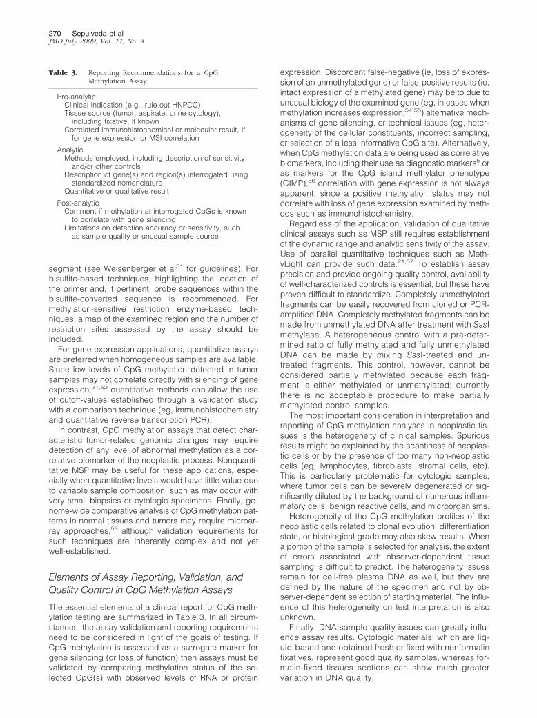
Table 3. Reporting Recommendations for a CpGMethylation Assay
Pre-analyticClinical indication (e.g., rule out HNPCC)Tissue source (tumor, aspirate, urine cytology),
including fixative, if knownCorrelated immunohistochemical or molecular result, if
for gene expression or MSI correlation
AnalyticMethods employed, including description of sensitivity
and/or other controlsDescription of gene(s) and region(s) interrogated using
standardized nomenclatureQuantitative or qualitative result
Post-analyticComment if methylation at interrogated CpGs is known
to correlate with gene silencingLimitations on detection accuracy or sensitivity, such
as sample quality or unusual sample source
270 Sepulveda et alJMD July 2009, Vol. 11, No. 4
Applications of CpG Methylation Testing inNeoplastic Disorders
There has been increased recognition that tumor-associ-ated epigenetic changes play an important role in theinitiation and progression of human cancers. Below, wereview reported applications of CpG methylation analysisin detection, classification, and monitoring treatment re-sponse of various human cancers.
Classification of Colorectal Cancer
The most common clinical application for CpG methyl-ation testing in colorectal neoplasia is as part of thework-up of hereditary non-polyposis colorectal cancer(HNPCC/Lynch syndrome),58 which produces microsat-ellite instability (MSI) by germline mutation of one of sev-eral DNA mismatch repair genes. Tumors resulting fromHNPCC can be distinguished from most cases of the MSIhigh (-H) subset of sporadic colorectal cancer by ab-sence of CpG methylation of the MLH1 promoter, whichcharacterizes most cases of sporadic MSI-H colorectalcancer.51,58–64 However, assessment of MLH1 methyl-ation by itself is probably not adequate to distinguishbetween all sporadic colon cancers and HNPCC-associ-ated MSI-H cancers, since methylation of MLH1 canbeen seen as a “second hit” in individuals with a germlineMLH1 mutation.65 Another limitation is that there are rarecases of heritable germline MLH1 methylation (epimuta-tion), which can be a cause of hereditary MSI-H colorec-tal cancer mimicking HNPCC/Lynch syndrome.66
The CpG island methylator phenotype (CIMP), definedas widespread promoter CpG island methylation, hasbeen established as a unique epigenetic phenotype incolorectal cancer that is correlated with MLH1 methyl-ation and MSI phenotype.51,62,67 CIMP-positive colorec-tal tumors have a distinct clinical, pathological, and mo-lecular profile. Typically, they are associated with olderage, proximal tumor location, female gender, poor differ-entiation, BRAF mutations, wild-type TP53, inactive WNT/�-catenin, stable chromosomes, and high-level LINE-1methylation, independent of MSI status.62,67–71 Particu-larly, CIMP status may help distinguish sporadic andHNPCC-related tumors with MSI, because most sporadicMSI-H colon cancers exhibit CIMP, while this is typicallynot seen in HNPCC-associated cancers.59,62,72–74 Re-cent studies have suggested the existence of KRAS mu-tation-associated CIMP (CIMP2 or CIMP-low), separatefrom CIMP-negative (CIMP-0), and BRAF mutation-asso-ciated CIMP (CIMP1 or CIMP-high).75–77 Additional stud-ies support a molecular difference between CIMP-low,CIMP-negative, and CIMP-high in colorectal cancer.78–80
A recent study suggested that all sporadic MSI-H tumorswere explained by CIMP and MLH1 methylation,51 whileother studies have suggested that there may be a subsetof sporadic MSI-H tumors that do not exhibit MLH1 meth-ylation and/or CIMP.62,81
Observed differences may be due to the fact that thepanel of CpG markers and method of assessment forcategorizing CIMP are not yet standardized. Use of quan-
titative MethyLight technology and evaluation of a newpanel of four to eight CpG islands, including RUNX3,CACNA1G, IGF2, MLH1, NEUROG1, CRABP1, SOCS1,and CDKN2A, may be the most promising approach.51,56
Currently, it is probably best to regard CIMP as we regardthe p.V600E BRAF mutation, which is also commonlyseen in sporadic MSI colon cancers and is only rarelyseen in HNPCC-associated tumors: the presence of ei-ther CIMP or the p.V600E mutation is strong evidencethat an MSI-H tumor is sporadic, while the absence ofboth of these findings indicates that the tumor could beeither HNPCC-associated or sporadic.
Determination of CIMP status may also be useful inevaluating the prognosis of colon cancer. A relationshipof CIMP with prognosis of microsatellite stable coloncancers has been reported. While previous studies haveeither found no relationship or a very small relation-ship,60,82 one study demonstrated a poor prognosis as-sociated with CIMP in microsatellite stable tumors, butnot in MSI-H tumors.83 BRAF mutations have also beenassociated with poor prognosis in microsatellite stabletumors, although in the same study, no effect was seenon the good prognosis of MSI-H tumors.82 Since micro-satellite stable tumors with BRAF mutations are usuallyvery heavily methylated,75 it is possible that the relation-ship between prognosis and BRAF is actually a relation-ship between prognosis and high levels of methylation. Adifferent CIMP panel that only detects extensive methyl-ation may show such a relationship with prognosis. Fu-ture studies are necessary to resolve this question.
Tumor Progression in Esophageal Carcinoma
The stepwise progression to esophageal adenocarci-noma involves an initial stage of intestinal metaplasia(Barrett’s esophagus), followed by low-grade and high-grade dysplasia, and finally adenocarcinoma. Shulmannet al characterized the CpG methylation status of 10genes (HPP1, RUNX3, RIZ1, CRBP1, 3-OST-2, APC,TIMP3, P16, MGMT, P14) by real-time quantitativeMSP.84 Their studies demonstrated that hypermethylationof P16, RUNX3, and HPP1 in Barrett’s esophagus orlow-grade dysplasia may represent independent risk fac-tors for the progression of Barrett’s esophagus to high-grade dysplasia or adenocarcinoma.
Diagnosis of Biliary and Pancreatic Malignancieson Cytologic Specimens
Due to its often cryptic location, early detection of cholan-giocarcinoma is paramount in improving clinical manage-ment and patient’s survival. Yang et al have shown thatconcurrent methylation of multiple CpG islands is a hallmarkfor cholangiocarcinoma.85 Using a panel of 12 tumor sup-pressor genes, they reported that DNA methylation profilesaccurately differentiated malignant cells from reactive cellsin biliary brushings.86 Similarly, Watanabe et al87 found thataberrant methylation of SFRP1 (SARP2) was seen in 79% ofpancreatic carcinoma and 56% of malignant intraductalpapillary mucinous neoplasms, but was rarely seen in
CpG Methylation Assays 271JMD July 2009, Vol. 11, No. 4
chronic pancreatitis and healthy controls. Hypermethyl-ation of SFRP1 in pancreatic juice may be a highlysensitive and useful marker in differentiating pancre-atic carcinoma from chronic pancreatitis.87
Diagnosis and Outcome Prediction in BreastCancer
Abnormal CpG methylation in breast cancer has beenfound in the promoters and first exons of genes, includingESR1 (estrogen receptor �),88,89 PGR (progesterone recep-tor),90 FRBP3 (MDGI, mammary-derived growth inhibitor),91
CALCA (calcitonin),92 MUC1, 93 and known proto-oconco-gene HRAS, 94 and tumor suppressor CDKN2A95 genes.The first systematic screen to detect all abnormally methyl-ated genes used a differential methylation hybridization ap-proach11 and identified multiple methylated fragments incultured tumor cells and in breast cancer tumors,12 includ-ing transcribed domains of ribosomal DNA.96
Detection of abnormal CpG methylation specific forbreast cancer can be done using fine needle aspirates,97
nipple aspirate fluid,98 and ductal lavage,99 as reviewedby Dua et al.100 MSP was reported to have high analyticalspecificity and moderate analytical sensitivity (100% and67%, respectively) for diagnosis of malignancy whenthree genes (RARB, RASSF1, and CCND2) were ana-lyzed in fine needle aspirate samples.101 Fackler et al102
evaluated methylation profiles of nine CpG islands inductal lavages from 37 cancer patients undergoing mas-tectomy. A cumulative methylation index had an analyti-cal sensitivity of 71% and specificity of 83% in the detec-tion of cancer cells, compared with an analyticalsensitivity of 33% and specificity of 99% by cytomorphol-ogy alone. This study provides proof-of-principle byshowing the advantages of using methylation analyses toquery cytologic specimens and indicates its potential usein diagnosis and risk stratification.102
Other studies have found methylation of the CDH1gene to be associated with breast tumor invasion andlymph node infiltration,103,104 and methylation of LATS1and LATS2 has been associated with aggressive can-cer.105 Nevertheless, currently, there are insufficient datato determine the clinical usefulness of methylation testsfor diagnosis and prognosis of breast cancer so addi-tional studies are warranted.
Progression in Cervical Carcinoma
The progression from precursor squamous intraepitheliallesions to cervical carcinoma requires additional geneticand epigenetic alterations that have not been character-ized fully. Gustafson et al examined aberrant promotermethylation of 15 tumor suppressor genes using a multi-plex, nested-MSP approach in 11 high-grade squamousintraepithelial lesions, 17 low-grade squamous intraepi-thelial lesions, and 11 negative tissues from liquid-basedcervical cytology samples.106 Aberrant promoter methyl-ation of DAPK1 and CADM1 (IGSF4) occurred at a highfrequency in high-grade squamous intraepithelial lesionsand was absent in low-grade squamous intraepithelial
lesions and negative samples. Also, the mean number ofmethylated genes was significantly higher in high-gradesquamous intraepithelial lesions, as compared with low-grade squamous intraepithelial lesions and negative sam-ples.106 Aberrant CDKN2A (p16) methylation was signifi-cantly higher in invasive cervical cancers (61%) ascompared with high-grade squamous intraepithelial lesions(20%) or normal cytologic specimens (7.5%).107 DNAmeth-ylation profiling will likely add a new dimension in the appli-cation of molecular biomarkers for prediction of diseaseprogression and risk assessment in cervical squamouslesions, but again others studies are warranted.
Diagnosis of Urothelial Carcinoma in UrineCytology
Urine cytology is the initial method used for screening ofbladder urothelial carcinoma. Although high-grade urothe-lial carcinoma can be readily detected in urine cytology,cytologic detection of low-grade papillary urothelial car-cinoma in urine is challenging due to the overlappingcytomorphologic features with benign reactive pro-cesses. Wang et al,108 using a panel of nine CpG islands,found that concurrent methylation of three or more CpGislands can differentiate low-grade papillary urothelialcarcinoma lesions from benign/reactive urothelium inurine. The analytical sensitivity to detect low-gradeurothelial carcinoma by DNA methylation profiling was80% in comparison with 13% by cytology alone.108 Thesestudies demonstrate that analysis of methylation profilingin certain cytologic specimens can be a useful ancillarytool in facilitating early and accurate detection of urothe-lial cancer cells.
Predicting Response to Chemotherapy: MGMTProfiling in Glioblastoma and Lymphoma
MGMT is a DNA repair enzyme that is frequently methylatedin human cancers, including glioblastoma and diffuse largeB-cell lymphoma. MGMT functions to repair O6-methylgua-nine DNA adducts generated by both endogenous andexogenous exposure to alkylating agents.109–111 Repair ofO6-methylguanine is critical to prevent accumulation ofG�A transition mutations in important growth regulatorygenes, including KRAS and TP53.112 CpG islands within thepromoter and coding region of MGMT are aberrantly hyper-or hypomethylated, respectively, resulting in transcriptionalrepression.79,113–117 Loss of MGMT expression and/orMGMT promoter methylation are associated with a worseprognosis in several tumor types,118–120 possibly due to anincreased mutation rate.
Since unrepaired O6-methylguanine signals apopto-sis,121 low MGMT expression would be expected to pre-dict an improved clinical response to chemotherapeuticalkylating agents. Thus, MGMT promoter methylation sta-tus can impact the degree of signaling for apoptosisfollowing alkylating agent therapy. In glioblastoma multi-forme, loss of MGMT expression predicts greater efficacyof treatment with temozolimide and other alkylating
272 Sepulveda et alJMD July 2009, Vol. 11, No. 4

agents. Several studies have shown a compelling directcorrelation between MGMT promoter methylation anddrug response that translates into increased overall pa-tient survival.122–125 Consequently, MGMT promotermethylation analysis using MSP is being used in theclinical laboratory to predict outcome and response totherapy in glioblastoma. MGMT promoter methylationalso predicts improved outcome in patients with diffuselarge B-cell lymphoma treated with the alkylating agentcyclophosphamide.126 In addition to predicting drug re-sponse, MGMT promoter methylation is an independentpredictor of better outcome in glioblastoma and diffuselarge B-cell lymphoma.124,127
CpG Methylation Profiling of Free DNA in BodyFluids as a Screening Tool
Tumor cells that are undergoing necrosis or apoptosisrelease fragments of genomic DNA, which may enter thecirculation or be released in the urine or stool where theycan be used as biomarkers for the diagnosis, staging, orpost-treatment monitoring of cancer. There is tremen-dous variability in the amount and half-life of cell-freeDNA released into the circulation128,129; however, theease of obtaining serial serum or plasma has stimulatedtremendous interest in the potential utility of detectingtumor-associated methylated DNA in such samples.
Several studies have addressed whether CpG methyl-ation of tumor biomarkers in serum cell-free DNA is in factcorrelated with tumor status. Bastian et al evaluated cir-culating serum cell-free DNA CpG methylation of GSTP1,which is hypermethylated in prostate cancer.130 Theyfound that circulating cell-free DNA with GSTP1 hyper-methylation was not detected in the serum of men with anegative prostate biopsy but was detected in 12% withclinically localized disease and in 28% with metastaticcancer. Detection of hypermethylated GTSP1 DNA inserum was the most significant predictor of increasedprostate specific antigen levels.130
Using MethyLight MSP, Muller et al analyzed 215 serumsamples from patients with cervical or breast cancer toidentify multigene associated CpG methylation changes. Incervical cancer, hypermethylation of three genes (MYOD1,CDH1, and CDH13) in pretreatment sera was significantlyassociated with a poor disease outcome.131 Methylation ofa similar set of genes (RASSF1, ESR1, APC, HSD17B4, andHIC1) selected from a panel of 39 genes in serum wasfound to be informative for prediction of metastasis, withAPC and RASSF1 being the most important.132
Koyanagi et al studied the association between DNAmethylation of RASSF1 and RARB in circulating tumor cellsin peripheral blood of melanoma patients with response tobiochemotherapy (a treatment modality that includes bio-logical agents such as interferon and interleukin-2).133 Pa-tients with methylated RASSF1 and RARB showed a signif-icantly poorer response to biochemotherapy, shorter time toprogression, and lower overall survival.133
Grady et al studied CpG methylation of MLH1 pro-moter DNA in the serum of patients with microsatelliteunstable colon cancers.134 In a panel of sera from 19
colon cancer cases, methylation of MLH1 was detectedin sera in three out of nine patients whose primary tumorsharbored MLH1 methylation. The assay proved 33% an-alytically sensitive and 100% specific.134
Detection of hypermethylated DNA in stool sampleshas been proposed as a screening tool for colorectalcancer.135,136 Lenhard et al analyzed promoter methyl-ation of HIC1 in stools of patients with colorectal cancer oradenomas.136 They found that 97% of samples had am-plifiable DNA and HIC1 was methylated in 42% of colo-rectal cancer patients and 31% of patients with adeno-mas, and was not methylated in normal samples.Belshaw et al135 compared methylation of a panel of CpGislands using MSP and combined bisulfite restrictionanalysis and found similar methylation frequencies ofESR1 and MGMT between tumor tissue samples andfecal DNA from the same patients.
The above reported data identify potential clinical ap-plications of CpG methylation testing; however, futureprospective studies are required to validate these find-ings and to refine guidelines for clinical practice.
Monitoring Treatment Response toDemethylating Agents
One of the most promising clinical applications for CpGmethylation analysis is in monitoring the response todemethylating agents. 5-aza-2�-deoxycytidine/decitab-ine (Dacogen) and azacitidine (Vidaza) are agents ap-proved by the U. S. Food and Drug Administration fortreatment of myelodysplastic syndrome. They function byreversing hypermethylation of tumor suppressors, includ-ing the cell cycle regulator p15. Demethylating agentsalso have variable activity in a wide variety of other tumortypes, especially in combination with other agents.
Several clinical studies have now used CpG methyl-ation profiling of pre- and post-treatment blood samplesto monitor the therapeutic effects of demethylatingagents. The effects of these drugs on both global meth-ylation (eg, LINE repeats) and the CpG methylation ofspecific target genes have been studied. In a phase I/IIstudy of decitabine in acute myelogenous leukemia/my-elodysplastic syndrome, transient and reversible de-creases in the level of DNA methylation at LINE andCDKN2B (p15) promoter were observed by a quantitativepyrosequencing assay over a 10-day course of treat-ment.137 Transcriptional up-regulation of CDKN2B (p15)was observed in parallel with decreases in CpG methyl-ation. Lower pretreatment levels of CDKN2B (p15) pro-moter methylation were correlated with clinical responsesto decitabine. Changes in the levels of CpG methylationfollowing treatment were modest (shifts of 10% to 20%)strongly indicating the need for reproducible quantitativeassays for monitoring methylation levels.137 As dis-cussed above, such techniques include real-time PCR(eg, MethyLight) and pyrosequencing methodologies.138
Given the current wide use of demethylating agents inmyelodysplastic syndrome and myeloid leukemias,CDKN2B (p15) methylation assays have the potential tobe used up-front to predict which patients will respond
CpG Methylation Assays 273JMD July 2009, Vol. 11, No. 4
to demethylation therapies. However, given the ability tomonitor response in these tumors based solely on bloodcounts, empirical use of demethylating agents in theabsence of pretreatment testing may well continue. Ifdemethylating therapy becomes common in solid tumorswhere treatment response is more difficult to assess,blood monitoring of re-expression of blood proteins, suchas fetal hemoglobin due to CpG demethylation, mayserve as a useful surrogate marker of drug response.139
CpG Methylation and Inflammatory andInfectious Diseases Related to CancerDevelopment
Viruses and CpG Methylation
Some viruses appear to use methylation to regulateexpression of their own viral genes as well as host cellulargenes. Diseased tissues that harbor viruses might, there-fore, be responsive to therapies that alter methylationpatterns.140–143 For example, Epstein-Barr virus, which isassociated with selected histological subtypes of lym-phomas and carcinomas, may repress certain viral genes(nuclear antigens EBNA 1-6, and latent membrane pro-teins LMP 1 and 2) in an effort to elude immune destruc-tion.144,145 In other examples, hepatocellular carcinomasappear to silence certain tumor suppressor genes in thepresence of heptatitis B virus infection,146 and HPV ap-pears to use methylation to exert its effects on viral andcellular gene expression.147 JC virus T antigen expres-sion is also associated with widespread CpG methylationreferred to as CIMP in colorectal cancer.148 Dysregula-tion of DNA methyltranferases may be responsible, atleast in part, for the effects of viruses on host genepromoter methylation.146,149 The first protein ever shownto bind to and activate a methylated promoter was avirally encoded factor, demonstrating that viruses haveevolved mechanisms to overcome methylation to theirselective advantage.150 To the extent that host cellularmethylation patterns are altered in virus-specific ways, itmay be possible to use expression patterns or methyl-ation patterns to identify virus-related subclasses of can-cers. Furthermore, a promising novel targeted therapeu-tic strategy involves demethylation/activation of viralgene expression in a way that triggers immune recogni-tion and destruction of virally infected tumor cells withlittle adverse effect on uninfected normal cells.
Bacterial Infection and CpG Methylation
In contrast to CpG methylation in tumors, the CpGmethylation status of genes in non-neoplastic tissues hasreceived little attention.151 However, several studies haveshown that CpG island hypermethylation of genes knownto be methylated in cancers can be detected in thenon-neoplastic tissues.152–154 One of the most remark-able examples of methylation in non-neoplastic tissue isthe hypermethylation of multiple CpG islands in the mu-cosal tissues of patients with inflammatory conditions,such as chronic gastritis associated with Helicobacter
pylori infection and inflammatory bowel diseases (ulcer-ative colitis and Crohn’s disease), conditions with in-creased risk of cancer development.
Increased CpG methylation of several genes has beenidentified in the gastric mucosa of patients with H. pylorigastritis, reviewed by Gologan et al.151 Chan et al155
demonstrated that CDH1 (E-cadherin) methylation wasmore frequent in the gastric mucosa of patients with H.pylori infection as compared with those without. Anotherstudy,154 where the methylation status of several geneswas examined, reported that CpG methylation was up to303-fold higher in H. pylori-positive than in H. pylori-neg-ative gastric mucosal tissue. MLH1 CpG methylation ingastric epithelial cells associated with reduced RNA andprotein levels of MLH1 were reported after exposure ofgastric cells to H. pylori organisms.156 Studies to-datehave not provided conclusive evidence regarding thepotential role of CpG methylation in inflammatory cellspresent in the gastric mucosa of H. pylori gastritis.
The potential implications of these reported findings aretwo-fold: first, CpG methylation may become useful in clin-ical practice to determine the risk of gastric cancer, andsecond, demethylating agents by restoring the CpG meth-ylation levels in the gastric mucosa may become useful incancer chemoprevention. Prospective studies forthese potential applications of CpG methylation asso-ciated with H. pylori gastritis and other inflammatorydiseases are warranted.
Summary
There are numerous promising clinical applications ofCpG methylation testing for tumors and preneoplasticlesions. However, if CpG methylation testing is to becomeroutine in clinical molecular diagnostics, there is a criticalneed for cross-laboratory comparisons of different meth-odologies, for the development of standardized qualitycontrol materials for assays and for the adoption of stan-dard reporting formats. While significant advances havebeen made in the analysis of methylation patterns invarious clinical tissues and other samples, with the ex-ception of MGMT in gliomas, the selection of optimalgene targets for prognostic methylation panels in specifictumor types remains to be established. Genome-widemethylation screens are currently identifying new markergene panels for prognosis and therapy-response predic-tors in other tumors. Application of such techniques incarefully controlled clinical trials, where favorable num-bers of samples can be compared with patient outcomesdata, is an essential component for the development ofclinically meaningful targets for CpG methylation analy-sis. Comparative studies are clearly essential to furtheradvancement of this field, therefore, we urge researchersto identify and define the CpG sites analyzed in publishedreports by listing DNA sequences and/or describing thelocation of the tested CpG(s) in relation to the transcriptionalstart site. Such advances will be instrumental in attainingclinically valuable and reliable CpG methylation assays formolecular diagnosis for the years to come.
274 Sepulveda et alJMD July 2009, Vol. 11, No. 4
References
1. Gardiner-Garden M, Frommer M: CpG islands in vertebrate ge-nomes. J Mol Biol 1987, 196:261–282
2. Takai D, Jones PA: Comprehensive analysis of CpG islands inhuman chromosomes 21 and 22. Proc Natl Acad Sci USA 2002,99:3740–3745
3. Ehrich M, Nelson M, Stanssens P, Zabeau M, Liloglou T, XinarianosG, Cantor C, Field J, van den Boom D: Quantitative high-throughputanalysis of DNA methylation patterns by base-specific cleavageand mass spectrometry. Proc Natl Acad Sci USA 2005, 102:15785–15790
4. Avramis V, Mecum R, Nyce J, Steele D, Holcenberg J: Pharmacody-namic and DNA methylation studies of high-dose 1-beta-D-arabino-furanosyl cytosine before and after in vivo 5-azacytidine treatment inpediatric patients with refractory acute lymphocytic leukemia. CancerChemother Pharmacol 1989, 24:203–210
5. Hoque MO, Feng Q, Toure P, Dem A, Critchlow CW, Hawes SE,Wood T, Jeronimo C, Rosenbaum E, Stern J, Yu M, Trink B, KiviatNB, Sidransky D: Detection of aberrant methylation of four genes inplasma DNA for the detection of breast cancer. J Clin Oncol 2006,24:4262–4269
6. Leung WK, To KF, Man EP, Chan MW, Bai AH, Hui AJ, Chan FK,Sung JJ: Quantitative detection of promoter hypermethylation inmultiple genes in the serum of patients with colorectal cancer. Am JGastroenterol 2005, 100:2274–2279
7. Frommer M, McDonald LE, Millar DS, Collis CM, Watt F, Grigg GW,Molloy PL, Paul CL: A genomic sequencing protocol that yields apositive display of 5-methylcytosine residues in individual DNAstrands. Proc Natl Acad Sci USA 1992, 89:1827–1831
8. Mikeska T, Bock C, El-Maarri O, Hubner A, Ehrentraut D, SchrammJ, Felsberg J, Kahl P, Buttner R, Pietsch T, Waha A: Optimization ofquantitative MGMT promoter methylation analysis using pyrose-quencing and combined bisulfite restriction analysis. J Mol Diagn2007, 9:368–381
9. Schatz P, Dietrich D, Schuster M: Rapid analysis of CpG methylationpatterns using RNase T1 cleavage and MALDI-TOF. Nucleic AcidsRes 2004, 32:e167
10. Matin MM, Baumer A, Hornby DP: An analytical method for thedetection of methylation differences at specific chromosomal lociusing primer extension and ion pair reverse phase HPLC. HumMutat 2002, 20:305–311
11. Huang TH, Perry MR, Laux DE: Methylation profiling of CpG islandsin human breast cancer cells. Hum Mol Genet 1999, 8:459–470
12. Yan PS, Perry MR, Laux DE, Asare AL, Caldwell CW, Huang TH:CpG island arrays: an application toward deciphering epigeneticsignatures of breast cancer. Clin Cancer Res 2000, 6:1432–1438
13. Yan P, Efferth T, Chen H, Lin J, Rodel F, Fuzesi L, Huang T: Use ofCpG island microarrays to identify colorectal tumors with a highdegree of concurrent methylation. Methods 2002, 27:162–169
14. Bibikova M, Lin Z, Zhou L, Chudin E, Garcia E, Wu B, Doucet D,Thomas N, Wang Y, Vollmer E, Goldmann T, Seifart C, Jiang W,Barker D, Chee M, Floros J, Fan J: High-throughput DNA meth-ylation profiling using universal bead arrays. Genome Res 2006,16:383–393
15. Cheng YW, Shawber C, Notterman D, Paty P, Barany F: Multiplexedprofiling of candidate genes for CpG island methylation statususing a flexible PCR/LDR/Universal Array assay. Genome Res2006, 16:282–289
16. Fukasawa M, Kimura M, Moritas S, Matsubara K, Yamanaka S, EndoC, Sakurada A, Sato M, Kondo T, Horii A, Sasaki H, Hatada I:Microarray analysis of promoter methylation in lung cancers. J HumGenet 2006, 51:368–374
17. Schumacher A, Kapranov P, Kaminsky Z, Flanagan J, AssadzadehA, Yau P, Virtanen C, Winegarden N, Cheng J, Gingeras T, PetronisA: Microarray-based DNA methylation profiling: technology and ap-plications. Nucleic Acids Res 2006, 34:528–542
18. Grunau C, Clark SJ, Rosenthal A: Bisulfite genomic sequencing:systematic investigation of critical experimental parameters. NucleicAcid Research 2001, 29:E65
19. Warnecke PM, Stirzaker C, Melki JR, Millar DS, Paul CL, Clark SJ:Detection and measurement of PCR bias in quantitative methyl-ation analysis of bisulphite-treated DNA. Nucleic Acids Res 1997,25:4422–4426
20. Herman JG, Graff JR, Myohanen S, Nelkin BD, Baylin SB: Methyla-tion-specific PCR: a novel PCR assay for methylation status of CpGislands. Proc Natl Acad Sci USA 1996, 93:9821–9826
21. Ogino S, Kawasaki T, Brahmandam M, Cantor M, Kirkner GJ,Spiegelman D, Makrigiorgos GM, Weisenberger DJ, Laird PW, LodaM, Fuchs CS: Precision and performance characteristics of bisulfiteconversion and real-time PCR (MethyLight) for quantitative DNAmethylation analysis. J Mol Diagn 2006, 8:209–217
22. Wang Y, Zheng W, Luo J, Zhang D, Zuhong L: In situ bisulfitemodification of membrane-immobilized DNA for multiple methylationanalysis. Anal Biochem 2006, 359:183–188
23. Kerjean A, Vieillefond A, Thiounn N, Sibony M, Jeanpierre M, JouannetP: Bisulfite genomic sequencing of microdissected cells. Nucleic AcidsRes 2001, 29:E106–E106
24. Laird PW: The power and the promise of DNA methylation markers.Nat Rev Cancer 2003, 3:253–266
25. Eads CA, Danenberg KD, Kawakami K, Saltz LB, Blake C, ShibataD, Danenberg PV, Laird PW: MethyLight: a high-throughput assay tomeasure DNA methylation. Nucleic Acids Res 2000, 28:E32
26. Eads CA, Danenberg KD, Kawakami K, Saltz LB, Danenberg PV,Laird PW: CpG island hypermethylation in human colorectal tumorsis not associated with DNA methyltransferase overexpression. Can-cer Res 1999, 59:2302–2306
27. Akey DT, Akey JM, Zhang K, Jin L: Assaying DNA methylation basedon high-throughput melting curve approaches. Genomics 2002,80:376–384
28. Thomassin H, Kress C, Grange T: MethylQuant: a sensitive methodfor quantifying methylation of specific cytosines within the genome.Nucleic Acids Res 2004, 32:e168
29. Zeschnigk M, Bohringer S, Price EA, Onadim Z, Masshofer L,Lohmann DR: A novel real-time PCR assay for quantitative analysisof methylated alleles (QAMA): analysis of the retinoblastoma locus.Nucleic Acids Res 2004, 32:e125
30. Cottrell SE, Distler J, Goodman NS, Mooney SH, Kluth A, Olek A,Schwope I, Tetzner R, Ziebarth H, Berlin K: A real-time PCR assayfor DNA-methylation using methylation-specific blockers. NucleicAcids Res 2004, 32:e10
31. Xiong Z, Laird PW: COBRA: a sensitive and quantitative DNA meth-ylation assay. Nucleic Acids Res 1997, 25:2532–2534
32. Colella S, Shen L, Baggerly KA, Issa JP, Krahe R: Sensitive andquantitative universal pyrosequencing methylation analysis of CpGsites. Biotechniques 2003, 35:146–150
33. Uhlmann K, Brinckmann A, Toliat MR, Ritter H, Nurnberg P: Evalu-ation of a potential epigenetic biomarker by quantitative methyl-single nucleotide polymorphism analysis. Electrophoresis 2002,23:4072–4079
34. Tost J, Dunker J, Gut IG: Analysis and quantification of multiplemethylation variable positions in CpG islands by pyrosequencing.Biotechniques 2003, 35:152–156
35. EL-Maarri O, Herbibiaux U, Walter J, Oldenburg J: A rapid, quanti-tative, non-radioactive bisulfite-SNuPE-IP RP HPLC assay for meth-ylation analysis at specific CpG sites. Nucleic Acids Research 2002,30:E25
36. Deng D, Deng G, Smith MF, Zhou J, Xin H, Powell SM, Lu Y:Simultaneous detection of CpG methylation and single nucleotidepolymorphism by denaturing high performance liquid chromatogra-phy. Nucleic Acids Res 2002, 30:E13
37. Waalwijk C, Flavell RA: DNA methylation at a CCGG sequence in thelarge intron of the rabbit beta-globin gene: tissue-specific variations.Nucleic Acids Res 1978, 5:4631–4634
38. Shen CK, Maniatis T: Tissue-specific DNA methylation in a cluster ofrabbit beta-like globin genes. Proc Natl Acad Sci USA 1980,77:6634–6638
39. Singer-Sam J, Grant M, LeBon JM, Okuyama K, Chapman V, MonkM, Riggs AD: Use of a HpaII-polymerase chain reaction assay tostudy DNA methylation in the Pgk-1 CpG island of mouse embryosat the time of X-chromosome inactivation. Mol Cell Biol 1990,10:4987–4989
40. Melnikov AA, Scholtens DM, Wiley EL, Khan SA, Levenson VV.Array-based multiplex analysis of DNA methylation in breast cancertissues. J Mol Diagn 2008, 10:93–101.
41. Hayashizaki Y, Hirotsune S, Okazaki Y, Hatada I, Shibata H, Kawai J,Hirose K, Watanabe S, Fushiki S, Wada S, Sugimoto T, Kobayakawa K,Kawara T, Katsuki M, Shibuya T, Mukai T: Restriction landmark
CpG Methylation Assays 275JMD July 2009, Vol. 11, No. 4
genomic scanning method and its various applications. Electro-phoresis 1993, 14:251–258
42. Plass C, Shibata H, Kalcheva I, Mullins L, Kotelevtseva N, Mullins J,Kato R, Sasaki H, Hirotsune S, Okazaki Y, Held WA, Hayashizaki Y,Chapman VM: Identification of Grf1 on mouse chromosome 9 as animprinted gene by RLGS-M. Nat Genet 1996, 14:106–109
43. Toyota M, Ho C, Ahuja N, Jair KW, Li Q, Ohe-Toyota M, Baylin SB,Issa JP: Identification of differentially methylated sequences in colo-rectal cancer by methylated CpG island amplification. Cancer Res1999, 59:2307–2312
44. Gonzalgo ML, Liang G, Spruck CH, 3rd, Zingg JM, Rideout WM, 3rd,Jones PA: Identification and characterization of differentially meth-ylated regions of genomic DNA by methylation-sensitive arbitrarilyprimed PCR. Cancer Res 1997, 57:594–599
45. Estecio MR, Yan PS, Ibrahim AE, Tellez CS, Shen L, Huang TH, IssaJP: High-throughput methylation profiling by MCA coupled to CpGisland microarray. Genome Res 2007, 17:1529–1536
46. Wei SH, Balch C, Paik HH, Kim YS, Baldwin RL, Liyanarachchi S, LiL, Wang Z, Wan JC, Davuluri RV, Karlan BY, Gifford G, Brown R, KimS, Huang TH, Nephew KP: Prognostic DNA methylation biomarkersin ovarian cancer. Clin Cancer Res 2006, 12:2788–2794
47. Chen CM, Chen HL, Hsiau TH, Hsiau AH, Shi H, Brock GJ, Wei SH,Caldwell CW, Yan PS, Huang TH: Methylation target array for rapidanalysis of CpG island hypermethylation in multiple tissue genomes.Am J Pathol 2003, 163:37–45
48. Melnikov AA, Gartenhaus RB, Levenson AS, Motchoulskaia NA,Levenson Chernokhvostov VV: MSRE-PCR for analysis of gene-specific DNA methylation. Nucleic Acids Res 2005, 33:e93
49. Kagan J, Srivastava S, Barker P, A. S., Belinsky S, Cairns P: Towardsclinical application of methylated DNA sequences as cancerbiomarkers: a Joint NCI’s EDRN and NIST workshop on stan-dards, methods, assays, reagents, and tools. Cancer Res 2007,67:4545–4549
50. Li L, Dahiya R: MethPrimer: designing primers for methylation PCRs.Bioinformatics 2002, 18:1427–1431
51. Weisenberger DJ, Siegmund KD, Campan M, Young J, Long TI,Faasse MA, Kang GH, Widschwendter M, Weener D, Buchanan D,Koh H, Simms L, Barker M, Leggett B, Levine J, Kim M, French AJ,Thibodeau SN, Jass J, Haile R, Laird PW: CpG island methylatorphenotype underlies sporadic microsatellite instability and is tightlyassociated with BRAF mutation in colorectal cancer. Nat Genet2006, 38:787–793
52. Toyooka KO, Toyooka S, Maitra A, Feng Q, Kiviat NC, Smith A,Minna JD, Ashfaq R, Gazdar AF: Establishment and validation ofreal-time polymerase chain reaction method for CDH1 promotermethylation. Am J Pathol 2002, 161:629–634
53. Brena RM, Morrison C, Liyanarachchi S, Jarjoura D, Davuluri RV,Otterson GA, Reisman D, Glaros S, Rush LJ, Plass C: Aberrant DNAmethylation of OLIG1, a novel prognostic factor in non-small celllung cancer. PLoS Med 2007, 4:e108
54. Kominato Y, Hata Y, Takizawa H, Tsuchiya T, Tsukada J, YamamotoF: Expression of human histo-blood group ABO genes is dependentupon DNA methylation of the promoter region. J Biol Chem 1999,274:37240–37250
55. Kelavkar UP, Harya NS, Hutzley J, Bacich DJ, Monzon FA, ChandranU, Dhir R, O’Keefe DS: DNA methylation paradigm shift: 15-lipoxygen-ase-1 upregulation in prostatic intraepithelial neoplasia and prostatecancer by atypical promoter hypermethylation. Prostaglandins OtherLipid Mediat 2007, 82:185–197
56. Ogino S, kawasaki T, Kirkner GJ, Kraft P, Loda M, Fuchs CS:Evaluation of markers for CpG island methylator phenotype (CIMP)in colorectal cancer by a large population-based sample. J MolDiagn 2007, 9:305–314
57. Coleman WB, Rivenbark AG: Quantitative DNA methylation analysis:the promise of high-throughput epigenomic diagnostic testing inhuman neoplastic disease. J Mol Diagn 2006, 8:152–156
58. Samowitz W: The CpG island methylator phenotype in colorectalcancer. J Mol Diagn 2007, 9:281–283
59. Toyota M, Ahuja N, Ohe-Toyota M, Herman JG, Baylin SB, Issa JP:CpG island methylator phenotype in colorectal cancer. Proc NatlAcad Sci USA 1999, 96:8681–8686
60. Hawkins N, Norrie M, Cheong K, Mokany E, Ku SL, Meagher A,O’Connor T, Ward R: CpG island methylation in sporadic colorectal
cancers and its relationship to microsatellite instability. Gastroenter-ology 2002, 122:1376–1387
61. Whitehall VL, Wynter CV, Walsh MD, Simms LA, Purdie D, PandeyaN, Young J, Meltzer SJ, Leggett BA, Jass JR: Morphological andmolecular heterogeneity within nonmicrosatellite instability-highcolorectal cancer. Cancer Res 2002, 62:6011–6014
62. Samowitz WS, Albertsen H, Herrick J, Levin TR, Sweeney C, MurtaughMA, Wolff RK, Slattery ML: Evaluation of a large, population-basedsample supports a CpG island methylator phenotype in colon cancer.Gastroenterology 2005, 129:837–845
63. Grady WM: CIMP and colon cancer gets more complicated. Gut2007, 56:1498–1500
64. Teodoridis JM, Hardie C, Brown R. CpG island methylator pheno-type (CIMP) in cancer: causes and implications. Cancer Lett 2008,268:177–186
65. Deng G, Bell I, Crawley S, Gum J, Terdiman JP, Allen BA, Truta B,Sleisenger MH, Kim YS: BRAF mutation is frequently present insporadic colorectal cancer with methylated hMLH1, but not in he-reditary nonpolyposis colorectal cancer. Clin Cancer Res 2004,10:191–195
66. Hitchins MP, Wong JJ, Suthers G, Suter CM, Martin DI, Hawkins NJ,Ward RL: Inheritance of a cancer-associated MLH1 germ-lineepimutation. N Engl J Med 2007, 356:697–705
67. Ogino S, Cantor M, Kawasaki T, Brahmandam M, Kirkner G,Weisenberger DJ, Campan M, Laird PW, Loda M, Fuchs CS: CpGisland methylator phenotype (CIMP) of colorectal cancer is best char-acterized by quantitative DNA methylation analysis and prospectivecohort studies. Gut 2006, 55:1000–1006
68. Goel A, Nagasaka T, Arnold CN, Inoue T, Hamilton C, NiedzwieckiD, Compton C, Mayer RJ, Goldberg R, Bertagnolli MM, Boland CR:The CpG island methylator phenotype and chromosomal instabilityare inversely correlated in sporadic colorectal cancer. Gastroenter-ology 2007, 132:127–138
69. Samowitz WS, Slattery ML, Sweeney C, Herrick J, Wolff RK, AlbertsenH: APC mutations and other genetic and epigenetic changes in coloncancer. Mol Cancer Res 2007, 5:165–170
70. Kawasaki T, Nosho K, Ohnishi M, Suemoto Y, Kirkner GJ, MeyerhardtJA, Fuchs CS, Ogino S: Correlation of beta-catenin localization withcyclooxygenase-2 expression and CpG island methylator phenotype(CIMP) in colorectal cancer. Neoplasia 2007, 9:569–577
71. Ogino S, Kawasaki T, Nosho K, Ohnishi M, Suemoto Y, Kirkner GJ,Fuchs CS: LINE-1 hypomethylation is inversely associated with mi-crosatellite instability and CpG methylator phenotype in colorectalcancer. Int J Cancer 2008, 122:2767–2773
72. Toyota M, Issa JP: The role of DNA hypermethylation in humanneoplasia. Electrophoresis 2000, 21:329–333
73. Kambara T, Simms LA, Whitehall VL, Spring KJ, Wynter CV, WalshMD, Barker MA, Arnold S, McGivern A, Matsubara N, Tanaka N,Higuchi T, Young J, Jass JR, Leggett BA: BRAF mutation is associ-ated with DNA methylation in serrated polyps and cancers of thecolorectum. Gut 2004, 53:1137–1144
74. McGivern A, Wynter CV, Whitehall VL, Kambara T, Spring KJ, WalshMD, Barker MA, Arnold S, Simms LA, Leggett BA, Young J, Jass JR:Promoter hypermethylation frequency and BRAF mutations distin-guish hereditary non-polyposis colon cancer from sporadic MSI-Hcolon cancer. Fam Cancer 2004, 3:101–107
75. Ogino S, Kawasaki T, Kirkner GJ, Loda M, Fuchs CS: CpG islandmethylator phenotype-low (CIMP-low) in colorectal cancer: possibleassociations with male sex and KRAS mutations. J Mol Diagn 2006,8:582–588
76. Shen L, Toyota M, Kondo Y, Lin E, Zhang L, Guo Y, Hernandez NS,Chen X, Ahmed S, Konishi K, Hamilton SR, Issa JP: Integratedgenetic and epigenetic analysis identifies three different subclassesof colon cancer. Proc Natl Acad Sci USA 2007, 104:18654–18659
77. Ogino S, Goel A: Molecular classification and correlates in colorectalcancer. J Mol Diagn 2008, 10:13–27
78. Ogino S, Kawasaki T, Kirkner GJ, Ohnishi M, Fuchs CS: 18q loss ofheterozygosity in microsatellite stable colorectal cancer is corre-lated with CpG island methylator phenotype-negative (CIMP-0) andinversely with CIMP-low and CIMP-high. BMC Cancer 2007, 7:72
79. Ogino S, Kawasaki T, Kirkner GJ, Suemoto Y, Meyerhardt JA, FuchsCS. Molecular correlates with MGMT promoter methylation and si-lencing support CpG island methylator phenotype-low (CIMP-low) incolorectal cancer. Gut 2007, 56:1564–1571
276 Sepulveda et alJMD July 2009, Vol. 11, No. 4
80. Kawasaki T, Ohnishi M, Nosho K, Suemoto Y, Kirkner GJ, MeyerhardtJA, Fuchs CS, Ogino S: CpG island methylator phenotype-low (CIMP-low) colorectal cancer shows not only fewer methylated CIMP-high-specific CpG island, but also low-level methylation in individual loci.Mod Pathol 2008, 21:245–255
81. Oliveira C, Westra JL, Arango D, Ollikainen M, Domingo E, FerreiraA, Velho S, Niessen R, Lagerstedt K, Alhopuro P, Laiho P, Veiga I,Teixeira MR, Ligtenberg M, Kleibeuker JH, Sijmons RH, Plukker JT,Imai K, Lage P, Hamelin R, Albuquerque C, Schwartz S, Jr., LindblomA, Peltomaki P, Yamamoto H, Aaltonen LA, Seruca R, Hofstra RM:Distinct patterns of KRASmutations in colorectal carcinomas accordingto germline mismatch repair defects and hMLH1 methylation status.Hum Mol Genet 2004, 13:2303–2311
82. Samowitz W, Sweeney C, Herrick J, Albertsen H, Levin T, MurtaughM, Wolff R, Slattery M: Poor survival associated with the BRAF V600Emutation in microsatellite-stable colon cancers. Cancer Res 2005,65:6063–6069
83. Ward RL, Cheong K, Ku SL, Meagher A, O’Connor T, Hawkins NJ:Adverse prognostic effect of methylation in colorectal cancer is re-versed by microsatellite instability. J Clin Oncol 2003, 21:3729–3736
84. Schulmann K, Sterian A, Berki A, Yin J, Sato F, Xu Y, Olaru A, WangS, Mori Y, Deacu E, Hamilton J, Kan T, Krasna MJ, Beer DG, PepeMS, Abraham JM, Feng Z, Schmiegel W, Greenwald BD, Meltzer SJ:Inactivation of p16. RUNX3, and HPP1 occurs early in Barrett’s-associated neoplastic progression and predicts progression riskOncogene 2005, 24:4138–4148
85. Yang B, Guo M, Herman J, Clark D: Aberrant promoter methylationprofiles of tumor suppressor genes in hepatocellular carcinoma.Am J Pathol 2003, 163:1101–1107
86. Yang B, House M, Guo M, Herman J, Clark D: Promoter methylationprofiles of tumor suppressor genes in intrahepatic and extrahepaticcholangiocarcinoma. Mod Pathol 2005, 18:412–420
87. Watanabe H, Okada G, Ohtsubo K, Yao F, Jiang P, Mouri H,Wakabayashi T, Sawabu N: Aberrant methylation of secreted apopto-sis-related protein 2 (SARP2) in pure pancreatic juice in diagnosis ofpancreatic neoplasms. Pancreas 2006, 32:382–389
88. Falette NS, Fuqua SA, Chamness GC, Cheah MS, Greene GL,McGuire WL: Estrogen receptor gene methylation in human breasttumors. Cancer Res 1990, 50:3974–3978
89. Ottaviano YL, Issa JP, Parl FF, Smith HS, Baylin SB, Davidson NE:Methylation of the estrogen receptor gene CpG island marks loss ofestrogen receptor expression in human breast cancer cells. CancerRes 1994, 54:2552–2555
90. Lapidus RG, Ferguson AT, Ottaviano YL, Parl FF, Smith HS, WeitzmanSA, Baylin SB, Issa JP, Davidson NE: Methylation of estrogen andprogesterone receptor gene 5� CpG islands correlates with lack ofestrogen and progesterone receptor gene expression in breast tumors.Clin Cancer Res 1996, 2:805–810
91. Huynh H, Alpert L, Pollak M: Silencing of the mammary-derivedgrowth inhibitor (MDGI) gene in breast neoplasms is associated withepigenetic changes. Cancer Res 1996, 56:4865–4870
92. Hakkarainen M, Wahlfors J, Myohanen S, Hiltunen MO, Eskelinen M,Johansson R, Janne J: Hypermethylation of calcitonin gene regula-tory sequences in human breast cancer as revealed by genomicsequencing. Int J Cancer 1996, 69:471–474
93. Zrihan-Licht S, Weiss M, Keydar I, Wreschner DH: DNA methylationstatus of the MUC1 gene coding for a breast-cancer-associatedprotein. Int J Cancer 1995, 62:245–251
94. Kass DH, Shen M, Appel NB, Anderson DE, Saunders GF: Exami-nation of DNA methylation of chromosomal hot spots associated withbreast cancer. Anticancer Res 1993, 13:1245–1251
95. Herman JG, Merlo A, Mao L, Lapidus RG, Issa JP, Davidson NE,Sidransky D, Baylin SB: Inactivation of the CDKN2/p16/MTS1 geneis frequently associated with aberrant DNA methylation in all com-mon human cancers. Cancer Res 1995, 55:4525–4530
96. Yan PS, Rodriguez FJ, Laux DE, Perry MR, Standiford SB, HuangTH: Hypermethylation of ribosomal DNA in human breast carcinoma.Br J Cancer 2000, 82:514–517
97. Jeronimo C, Costa I, Martins MC, Monteiro P, Lisboa S, Palmeira C,Henrique R, Teixeira MR, Lopes C: Detection of gene promoterhypermethylation in fine needle washings from breast lesions. ClinCancer Res 2003, 9:3413–3417
98. Krassenstein R, Sauter E, Dulaimi E, Battagli C, Ehya H, Klein-Szanto
A, Cairns P: Detection of breast cancer in nipple aspirate fluid byCpG island hypermethylation. Clin Cancer Res 2004, 10:28–32
99. Evron E, Dooley WC, Umbricht CB, Rosenthal D, Sacchi N, GabrielsonE, Soito AB, Hung DT, Ljung B, Davidson NE, Sukumar S: Detection ofbreast cancer cells in ductal lavage fluid by methylation-specific PCR.Lancet 2001, 357:1335–1336
100. Dua RS, Isacke CM, Gui GP: The intraductal approach to breastcancer biomarker discovery. J Clin Oncol 2006, 24:1209–1216
101. Pu RT, Laitala LE, Alli PM, Fackler MJ, Sukumar S, Clark DP: Meth-ylation profiling of benign and malignant breast lesions and itsapplication to cytopathology. Mod Pathol 2003, 16:1095–1101
102. Fackler M, Malone K, Zhang Z, Schilling E, Garrett-Mayer E, Swift-Scanlan T, Lange J, Nayar R, Davidson N, Khan S, Sukumar S:Quantitative multiplex methylation-specific PCR analysis doublesdetection of tumor cells in breast ductal fluid. Clin Cancer Res 2006,12:3306–3310
103. Shinozaki M, Hoon DS, Giuliano AE, Hansen NM, Wang HJ, TurnerR, Taback B: Distinct hypermethylation profile of primary breastcancer is associated with sentinel lymph node metastasis. ClinCancer Res 2005, 11:2156–2162
104. Caldeira JR, Prando EC, Quevedo FC, Neto FA, Rainho CA, RogattoSR: CDH1 promoter hypermethylation and E-cadherin protein ex-pression in infiltrating breast cancer. BMC Cancer 2006, 6:48
105. Takahashi Y, Miyoshi Y, Takahata C, Irahara N, Taguchi T, Tamaki Y,Noguchi S: Down-regulation of LATS1 and LATS2 mRNA expressionby promoter hypermethylation and its association with biologicallyaggressive phenotype in human breast cancers. Clin Cancer Res2005, 11:1380–1385
106. Gustafson K, Furth E, Heitjan D, Fansler Z, Clark D: DNA methylationprofiling of cervical squamous intraepithelial lesions using liquid-based cytology specimens: an approach that utilizes receiver-op-erating characteristic analysis. Cancer 2004, 102:259–268
107. Feng Q, Balasubramanian A, Hawes S, Toure P, Sow P, Dem A,Dembele B, Critchlow C, Xi L, Lu H, McIntosh M, Young A, Kiviat N:Detection of hypermethylated genes in women with and withoutcervical neoplasia. J Natl Cancer Inst 2005, 97:273–282
108. Wang L, Weber D, Deeds D, Biscotti C, Yang B. DNA methylationfrofiling distinguishes low grade papillary urothelial carcinoma fromreactive urothelia in urine. Mod Pathol 2006, 19:74A (supplement)
109. Bartsch H: Studies on biomarkers in cancer etiology and prevention:a summary and challenge of 20 years of interdisciplinary research.Mutat Res 2000, 462:255–279
110. Georgiadis P, Samoli E, Kaila S, Katsouyanni K, Kyrtopoulos SA:Ubiquitous presence of O6-methylguanine in human peripheraland cord blood DNA. Cancer Epidemiol Biomarkers Prev 2000,9:299–305
111. Kyrtopoulos SA: DNA adducts in humans after exposure to methy-lating agents. Mutat Res 1998, 405:135–143
112. Margison GP, Povey AC, Kaina B, Santibanez Koref MF: Variabilityand regulation of O6-alkylguanine-DNA alkyltransferase. Carcino-genesis 2003, 24:625–635
113. Danam RP, Howell SR, Brent TP, Harris LC: Epigenetic regulation ofO6-methylguanine-DNA methyltransferase gene expression by his-tone acetylation and methyl-CpG binding proteins. Mol Cancer Ther2005, 4:61–69
114. Soejima H, Zhao W, Mukai T: Epigenetic silencing of the MGMTgene in cancer. Biochem Cell Biol 2005, 83:429–437
115. von Wronski MA, Brent TP: Effect of 5-azacytidine on expression ofthe human DNA repair enzyme O6-methylguanine-DNA methyl-transferase. Carcinogenesis 1994, 15:577–582
116. Wang Y, Kato T, Ayaki H, Ishizaki K, Tano K, Mitra S, Ikenaga M:Correlation between DNA methylation and expression of O6-meth-ylguanine-DNA methyltransferase gene in cultured human tumorcells. Mutat Res 1992, 273:221–330
117. Fox EJ, Leahy DT, Geraghty R, Mulcahy HE, Fennelly D, Hyland JM,O’Donoghue DP, Sheahan K: Mutually exclusive promoter hyper-methylation patterns of hMLH1 and O6-methylguanine DNA methyl-transferase in colorectal cancer. J Mol Diagn 2006, 8:68–75
118. Hayashi H, Yazawa T, Okudela K, Nagai J, Ito T, Kanisawa M,Kitamura H: Inactivation of O6-methylguanine-DNA methyltrans-ferase in human lung adenocarcinoma relates to high-grade histol-ogy and worse prognosis among smokers. Jpn J Cancer Res 2002,93:184–189
119. Komine C, Watanabe T, Katayama Y, Yoshino A, Yokoyama T,
CpG Methylation Assays 277JMD July 2009, Vol. 11, No. 4
Fukushima T: Promoter hypermethylation of the DNA repair geneO6-methylguanine-DNA methyltransferase is an independent pre-dictor of shortened progression free survival in patients with low-grade diffuse astrocytomas. Brain Pathol 2003, 13:176–184
120. Matsukura S, Miyazaki K, Yakushiji H, Ogawa A, Harimaya K,Nakabeppu Y, Sekiguchi M: Expression and prognostic significance ofO6-methylguanine-DNA methyltransferase in hepatocellular, gastric,and breast cancers. Ann Surg Oncol 2001, 8:807–816
121. Kaina B, Ochs K, Grosch S, Fritz G, Lips J, Tomicic M, Dunkern T,Christmann M: BER, MGMT, and MMR in defense against alkylation-induced genotoxicity and apoptosis. Prog Nucleic Acid Res Mol Biol2001, 68:41–54
122. Esteller M, Risques RA, Toyota M, Capella G, Moreno V, PeinadoMA, Baylin SB, Herman JG: Promoter hypermethylation of the DNArepair gene O(6)-methylguanine-DNA methyltransferase is associ-ated with the presence of G:C to A:T transition mutations in p53 inhuman colorectal tumorigenesis. Cancer Res 2001, 61:4689–4692
123. Hegi ME, Diserens AC, Godard S, Dietrich PY, Regli L, Ostermann S,Otten P, Van Melle G, de Tribolet N, Stupp R: Clinical trial substan-tiates the predictive value of O-6-methylguanine-DNA methyltrans-ferase promoter methylation in glioblastoma patients treated withtemozolomide. Clin Cancer Res 2004, 10:1871–1874
124. Hegi ME, Diserens AC, Gorlia T, Hamou MF, de Tribolet N, Weller M,Kros JM, Hainfellner JA, Mason W, Mariani L, Bromberg JE, Hau P,Mirimanoff RO, Cairncross JG, Janzer RC, Stupp R: MGMT genesilencing and benefit from temozolomide in glioblastoma. N EnglJ Med 2005, 352:997–1003
125. Paz MF, Yaya-Tur R, Rojas-Marcos I, Reynes G, Pollan M, Aguirre-Cruz L, Garcia-Lopez JL, Piquer J, Safont MJ, Balana C, Sanchez-Cespedes M, Garcia-Villanueva M, Arribas L, Esteller M: CpG islandhypermethylation of the DNA repair enzyme methyltransferase pre-dicts response to temozolomide in primary gliomas. Clin Cancer Res2004, 10:4933–4938
126. Esteller M, Gaidano G, Goodman SN, Zagonel V, Capello D, Botto B,Rossi D, Gloghini A, Vitolo U, Carbone A, Baylin SB, Herman JG:Hypermethylation of the DNA repair gene O(6)-methylguanine DNAmethyltransferase and survival of patients with diffuse large B-celllymphoma. J Natl Cancer Inst 2002, 94:26–32
127. Ohno T, Hiraga J, Ohashi H, Sugisaki C, Li E, Asano H, Ito T, NagaiH, Yamashita Y, Mori N, Kinoshita T, Naoe T: Loss of O6-methylgua-nine-DNA methyltransferase protein expression is a favorable prog-nostic marker in diffuse large B-cell lymphoma. Int J Hematol 2006,83:341–347
128. Invernizzi P, Biondi ML, Battezzati PM, Perego F, Selmi C, CecchiniF, Podda M, Simoni G: Presence of fetal DNA in maternal plasmadecades after pregnancy. Hum Genet 2002, 110:587–591
129. Silva JM, Garcia JM, Dominguez G, Silva J, Miralles C, Cantos B,Coca S, Provencio M, Espana P, Bonilla F: Persistence of tumor DNAin plasma of breast cancer patients after mastectomy. Ann SurgOncol 2002, 9:71–76
130. Bastian PJ, Palapattu GS, Lin X, Yegnasubramanian S, Mangold LA,Trock B, Eisenberger MA, Partin AW, Nelson WG: Preoperativeserum DNA GSTP1 CpG island hypermethylation and the risk ofearly prostate-specific antigen recurrence following radical prosta-tectomy. Clin Cancer Res 2005, 11:4037–4043
131. Muller H, Fiegl H, Widschwendter A, Widschwendter M: PrognosticDNA methylation marker in serum of cancer patients. Ann NY AcadSci 2004, 1022:44–49
132. Muller HM, Widschwendter A, Fiegl H, Ivarsson L, Goebel G, Perk-mann E, Marth C, Widschwendter M: DNA methylation in serum ofbreast cancer patients: an independent prognostic marker. CancerRes 2003, 63:7641–5
133. Koyanagi K, Mori T, O’Day S, Martinez S, Wang H, Hoon D: Asso-ciation of circulating tumor cells with serum tumor-related methyl-ated DNA in peripheral blood of melanoma patients. Cancer Res2006, 66:6111–6117
134. Grady WM, Rajput A, Lutterbaugh JD, Markowitz SD: Detection ofaberrantly methylated hMLH1 promoter DNA in the serum of pa-tients with microsatellite unstable colon cancer. Cancer Res 2001,61:900–902
135. Belshaw NJ, Elliott GO, Williams EA, Bradburn DM, Mills SJ, MathersJC, Johnson IT: Use of DNA from human stools to detect aberrant
CpG island methylation of genes implicated in colorectal cancer.Cancer Epidemiol Biomarkers Prev 2004, 13:1495–1501
136. Lenhard K, Bommer GT, Asutay S, Schauer R, Brabletz T, Goke B,Lamerz R, Kolligs FT: Analysis of promoter methylation in stool: anovel method for the detection of colorectal cancer. Clin Gastroen-terol Hepatol 2005, 3:142–149
137. Garcia-Manero G, Kantarjian H, Sanchez-Gonzalez B, Yang H, Ros-ner G, Verstovsek S, Rytting M, Wierda W, Ravandi F, Koller C, XiaoL, Faderl S, Estrov Z, Cortes J, O’brien S, Estey E, Bueso-Ramos C,Fiorentino J, Jabbour E, Issa J: Phase I/II study of the combination of5-aza-2� -deoxycytidine with valproic acid in patients with leukemia.Blood 2006, 108:3271–3279
138. Mund C, Hackanson B, Stresemann C, Lubbert M, Lyko F: Charac-terization of DNA demethylation effects induced by 5-Aza-2�-deoxy-cytidine in patients with myelodysplastic syndrome. Cancer Res2005, 65:7086–7090
139. Ruter B, Wijermans PW, Lubbert M: DNA methylation as a therapeu-tic target in hematologic disorders: recent results in older patientswith myelodysplasia and acute myeloid leukemia. Int J Hematol2004, 80:128–135
140. Tao Q, Young L, Woodman C, Murray P: Epstein-Barr virus (EBV)and its associated human cancers–genetics, epigenetics, pathobi-ology and novel therapeutics. Front Biosci 2006, 11:2672–2713
141. Israel B, Kenney S: Virally targeted therapies for EBV-associatedmalignancies. Oncogene 2003, 22:5122–5130
142. Verma M: Viral genes and methylation. Ann NY Acad Sci 2003,983:170–180
143. Varier R, Kundu T: Chromatin modifications (acetylation/ deacetyla-tion/ methylation) as new targets for HIV therapy. Curr Pharm Des2006, 12:1975–1993
144. Tao Q, Robertson K: Stealth technology: how Epstein-Barr virusutilizes DNA methylation to cloak itself from immune detection. ClinImmunol 2003, 109:53–63
145. Minarovits J: Epigenotypes of latent herpesvirus genomes. Curr TopMicrobiol Immunol 2006, 310:61–80
146. Li H, Leu Y, Chang Y: Epigenetic changes in virus-associated hu-man cancers. Cell Res 2005, 15:262–271
147. Zheng Z, Baker C: Papillomavirus genome structure, expression, andpost-transcriptional regulation. Front Biosci 2006, 11:2286–2302
148. Goel A, Li MS, Nagasaka T, Shin SK, Fuerst F, Ricciardiello L,Wasserman L, Boland CR: Association of JC virus T-antigen expres-sion with the methylator phenotype in sporadic colorectal cancers.Gastroenterology 2006, 130:1950–1961
149. Etoh T, Kanai Y, Ushijima S, al. e. Increased DNA methyltransferase1 (DNMT1) protein expression correlates significantly with poorertumor differentiation and frequent DNA hypermethylation of multipleCpG islands in gastric cancers. Am J Pathol 2004, 164:689–699
150. Bhende P, Seaman W, Delecluse H, Kenney S: The EBV lytic switchprotein. Z, preferentially binds to and activates the methylated viralgenome Nat Genet 2004, 36:1099–1104
151. Gologan A, Graham DY, Sepulveda AR: Molecular markers in heli-cobacter pylori-associated gastric carcinogenesis. Clin Lab Med2005, 25:197–222
152. Issa JP: Age-related epigenetic changes and the immune system.Clin Immunol 2003, 109:103–108
153. Hsieh CJ, Klump B, Holzmann K, Borchard F, Gregor M, PorschenR: Hypermethylation of the p16INK4a promoter in colectomy spec-imens of patients with long-standing and extensive ulcerative colitis.Cancer Res 1998, 58:3942–3945
154. Maekita T, Nakazawa K, Mihara M, Nakajima T, Yanaoka K, IguchiM, Arii K, Kaneda A, Tsukamoto T, Tatematsu M, Tamura G, Saito D,Sugimura T, Ichinose M, Ushijima T: High levels of aberrant DNAmethylation in Helicobacter pylori-infected gastric mucosae and itspossible association with gastric cancer risk. Clin Cancer Res 2006,12:989–995
155. Chan AO, Lam SK, Wong BC, Wong WM, Yuen MF, Yeung YH, HuiWM, Rashid A, Kwong YL: Promoter methylation of E-cadherin genein gastric mucosa associated with Helicobacter pylori infection andin gastric cancer. Gut 2003, 52:502–506
156. Yao Y, Tao H, Park DI, Sepulveda JL, Sepulveda AR: Demonstra-tion and characterization of mutations induced by helicobacterpylori organisms in gastric epithelial cells. Helicobacter 2006,11:272–286
278 Sepulveda et alJMD July 2009, Vol. 11, No. 4