correlation of gene and mediator expression with clinical endpoints in an acute...
TRANSCRIPT

Osteoarthritis and Cartilage (2009) 17, 790e797
ª 2008 Osteoarthritis Research Society International. Published by Elsevier Ltd. All rights reserved.doi:10.1016/j.joca.2008.09.016
InternationalCartilageRepairSociety
Correlation of gene and mediator expression with clinical endpointsin an acute interleukin-1b-driven model of joint pathologyI. Scotty*, A. Midhaz, U. Rashidyx, S. Bally, A. Waldingy, P. Kerryk, S. Delaneyy and S. CruwyszyDepartment of Molecular Biology, AstraZeneca Charnwood, Loughborough, UKzDiscovery Bioscience, AstraZeneca Charnwood, Loughborough, UKxDepartment of Molecular Pathology and Toxicology, University of Leicester, UKkSafety Assessment, AstraZeneca Charnwood, Loughborough, UK
Summary
Objective: Interleukin-1b (IL-1b) plays a key role in the pathogenesis of chronic joint diseases, including osteoarthritis (OA), and drives a cas-cade of inflammatory and destructive responses within the synovial joint. Animal models of arthritis support the role of IL-1b in joint pathology,however, the molecular changes downstream of IL-1b are poorly understood in vivo. This study aimed to evaluate the intra-articular (i.a.) in-jection of IL-1b in the rat joint as an acute model of joint disease and associate gene and mediator expression with clinical endpoints andpathological changes.
Methods: The effects of i.a. administration of a pathologically relevant dose of IL-1b on joint swelling, mechanical hyperalgesia, histopathol-ogy, gene expression and biochemical changes were measured from 2 to 24 h.
Results: IL-1b-induced joint swelling and mechanical hyperalgesia. Gene expression analysis of joint tissue and biochemical analysis of jointlavage fluid identified pro-inflammatory and destructive mediators induced by IL-1b. Histopathology of joint tissues showed evidence of syno-vitis and connective tissue inflammation, but not cartilage destruction. However, biochemical analysis of glucosaminoglycan levels in jointlavage fluids indicated cartilage breakdown and might be a sensitive marker of cartilage pathology.
Conclusions: Intra-articular injection of IL-1b is a reproducible acute model of joint pathology that is potentially useful to evaluate IL-1 pathwayinhibitors. The correlation of molecular events with clinical and pathological changes in this model has enhanced the understanding of the roleof IL-1b in joint disease. Methods developed for gene expression analysis using multi-gene microfluidics cards and for biochemical analysis ofjoint lavage fluid might have utility for characterisation of other arthritis models and corresponding human disease.ª 2008 Osteoarthritis Research Society International. Published by Elsevier Ltd. All rights reserved.
Key words: Arthritis model, Cartilage, Interleukin-1b (IL-1b), Joint pathology, Synovitis.
Osteoarthritis (OA) is one of the most common forms ofmusculo-skeletal disease and is estimated to affect 300 mil-lion people worldwide with an increasing prevalence1. Theclinical symptoms of OA are pain and functional disabilitythat can prevent normal activities of daily life and can leadto isolation and depression2. OA is a disease of the wholejoint principally characterised by a chronic degeneration ofarticular cartilage but pathological changes also include sy-novial inflammation, bone remodelling, and muscle weak-ness3. Evidence suggests that synovitis is a cause of painand can contribute to structural deterioration of the joint inOA4e6. Current therapies for OA are analgesics and anti-inflammatory drugs that reduce the symptoms of OA butthere remains a high unmet medical need for treatmentsthat address the underlying causes of OA7.
The cytokine interleukin-1b (IL-1b) drives a wide range ofpro-inflammatory and destructive responses in a number ofcell types in the synovial joint and plays a key role in the
*Address correspondence and reprint requests to: Ian Scott,Department of Molecular Biology, AstraZeneca Charnwood,Loughborough, LE11 5RH, UK. Tel: 44-1509-647018; Fax: 44-1509-645557; E-mail: [email protected]
Received 21 July 2008; revision accepted 30 September 2008.
790
progression and outcome of chronic joint diseases includingOA7e9. IL-1 signalling has been suggested as an attractivetherapeutic target for reducing symptomatic and structuralchanges in OA9,10. A limited number of agents that targetIL-1 have been evaluated in OA clinical trials and intra-articular (i.a.) injection of IL-1 receptor antagonist (Anakinra;Kineret, Amgen) has shown a significant benefit in reducingpain and synovitis in OA patients11e13.
Animal models of arthritis, whilst not fully representativeof human joint diseases, can be used to increase under-standing of the mechanisms underlying joint pathologyand for the evaluation of potential therapeutic agents14.Profiling the pro-inflammatory and destructive mediatorsinvolved in animal models of arthritis can provide insightinto the relationship with human joint disease and poten-tially help identify the appropriate model to evaluate spe-cific therapeutic agents15e18. Although IL-1b has beendemonstrated to have a pro-inflammatory and pro-catabolic role in animal models of arthritis, the molecularbasis of pathological changes are still poorly characteri-sed19e23. In particular, IL-1b-driven models of arthritishave demonstrated a role of IL-1b in synovialinflammation and cartilage destruction but have not sur-veyed the pro-inflammatory and destructive mediatorsinvolved24e26.
791Osteoarthritis and Cartilage Vol. 17, No. 6
The objective of this study was to evaluate i.a. injection ofIL-1b in rat ankle joint as an acute model of joint destructionand characterise pathological events relevant to human ar-thritis including pain, synovitis and cartilage destruction.A particular aim was to explore the cascade of molecularevents induced by IL-1b in vivo and correlate with clinicalendpoints and histopathological changes. The identificationof a reproducible model with short duration, when comparedto more complex models of arthritis, would be useful for ini-tial evaluation of antagonists of the IL-1 pathway. In addi-tion, this acute model might be appropriate to developgene expression and biochemical methodologies with utilityin other models of arthritis.
Materials and methods
ANIMALS
Female Lewis rats (175e200 g, Charles River, UK) were group housed (5per cage), allowed food and water ad libitum, and acclimatised for a minimumof 1 week prior to sensitisation. Animals were housed in light/dark cycles of12 h. All in vivo work in rats was subject to internal ethical review and con-ducted strictly in accordance with Home Office requirements under the Ani-mals (Scientific Procedures) Act 1986.
INTRA-ARTICULAR INJECTION OF IL-1b
4.80A
The rats were anaesthetised with isoflurane prior to an i.a. injection of 10 ngor 100 ng of rat IL-1b (R&D Systems) in 20 ml of phosphate buffer saline (PBS,Dulbecco A) into the ankle joint space. Contra-lateral ankle joints injected with20 ml of PBS served as vehicle controls (five animals per treatment group). An-kle joint diameters were measured using vernier callipers and mechanical hy-peralgesia measured using von Frey filaments. Animals were terminated 2, 4,8 and 24 h after i.a. injection. In vivo procedures and the subsequent measure-ment of clinical parameter were repeated in three independent experiments.
4.60
mm
)
**
HISTOLOGY
3.60
3.80
4.00
4.20
4.40
An
kle D
iam
eter (
Ankle joints were fixed in 10% neutral buffered formalin, decalcified andprocessed to wax according to standard protocols. Tibialetalus joint sectionsof 4 mm thickness were stained with hematoxylin and eosin (H&E). Sectionswere examined microscopically and a blind assessment of pathology wasmade using a subjective semi-quantitative scoring system. Individual sec-tions were graded as follows: grade 0¼ no lesion, grade 1¼minimal, grade2¼ slight, grade 3¼moderate, grade 4¼marked/severe. Additional jointsections were stained with toluidine blue to further assess potential changesto articular cartilage. Histological analysis was performed on samples fromtwo independent experiments.
0 5 10 15 20 25Time post injection (hours)
JOINT LAVAGE FLUID
5.30
g
)
*
B
An incision was made between the calcaneum and talus bones and thejoint lavaged with 100 ml PBS containing 0.05 mM EDTA at 2, 4 and 24 hpost-i.a. injection. Biochemical analyses of joint lavage fluid were performedon samples from three independent experiments.
5.40
5.50
old
(lo
g
INTERLEUKIN-6 AND PROSTAGLANDIN E2 (PGE2) ASSAYS
5.60
hresh
Joint lavage fluids were assayed for IL-6 and PGE2 by enzyme-linked immu-nosorbent assay (ELISA) using manufacturer’s instructions (R&D Systems).
5.70
F
rey T
GLUCOSAMINOGLYCAN (GAG) ASSAY
5.80
5.90
vo
n
Lavage fluids were digested in 500 mg/ml papain solution at pH 6.5 at60 �C for 18 h. GAG concentrations were measured using a dimethyl meth-ylene blue (DMMB) assay using a chondroitin sulphate standard curve27.
0 5 10 15 20 25Time post injection (hours)
RNA ISOLATION
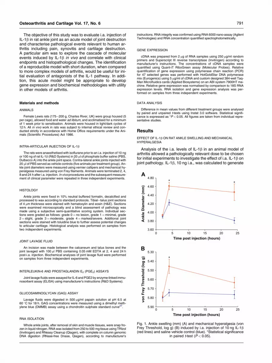
Fig. 1. Ankle swelling (mm) (A) and mechanical hyperalgesia (vonFrey Threshold, log g) (B) induced by i.a. injection of 10 ng IL-1b(red lines) and saline vehicle control (blue). *Statistical significance
in paired t-test (P< 0.05).
Whole ankle joints, after removal of skin and muscle tissues, were snap fro-zen in liquid nitrogen. RNA was isolated from 250 to 500 mg tissue using TRIzol(Invitrogen) and RNeasy Cleanup (Qiagen), with complete on column genomicDNA digestion (RNase-free Dnase, Qiagen), according to manufacturer’s
instructions. RNA integrity was confirmed using RNA 6000 nano-assay (AgilentTechnologies) and RNA concentration quantified spectrophotometrically.
GENE EXPRESSION
cDNA was prepared from 2 mg of RNA samples using 250 mg/ml randomprimers and Superscript III reverse transcriptase (Invitrogen) according tomanufacturer’s instructions. The concentrations of cDNA samples werequantified using Quant-iT RiboGreen assay (Molecular Probes). Relativequantification of gene expression using polymerase chain reaction (PCR)for 47 selected genes was performed with HotGoldStar DNA polymerasemix (Eurogenics) using 5 mg/ml of cDNA and custom designed 384-well Taq-Man Microfluidics cards (Applied Biosystems) on an ABI system 7900HT ma-chine. Relative gene expression was normalised by comparison to 18S RNAexpression levels. RNA isolation and gene expression analysis was per-formed on samples from three independent experiments.
DATA ANALYSIS
Difference in mean values from different treatment groups were analysedby paired and unpaired t-tests using Instat 3.0 software. Statistical signifi-cance is expressed as *P< 0.05. All figures are taken from individual repre-sentative studies.
Results
EFFECT OF IL-1b ON RAT ANKLE SWELLING AND MECHANICAL
HYPERALGESIA
Analysis of the i.a. levels of IL-1b in an animal model ofarthritis allowed a pathologically relevant dose to be chosenfor initial experiments to investigate the effect of i.a. IL-1b onjoint pathology. IL-1b, 10 ng i.a., was calculated to generate

Table ISummary of the relative gene expression of 47 selected genes in whole joint tissue 4 h after i.a. IL-1b or vehicle control. Fold increase anddecrease refer to comparison of relative expression level in IL-1b treated and vehicle control treated joint tissue. *Statistical significance in
paired t-test (P< 0.05). Threshold cycle (Ct) for gene of interest e Ct for 18S RNA (DCt), not detected (ND)
Gene symbol Encoded protein DCt vehicle DCtIL-1b
Foldincrease
Folddecrease
P-value
ADAMTS5 A disintegrin-like and metalloprotease, Aggrecanase-2 15.0 13.1 3.3 0.042*AGC1 Aggrecan 11.4 11.0 1.4 0.589Alp1 Alkaline phosphatase 13.3 6.2 22356.0 0.308B2M Beta-2 microglobulin 8.6 7.6 1.9 0.209BMP Bone morphogenetic protein 2 18.6 23.7 1.3 0.563CACNA2D2 Voltage-dependent calcium channel alpha 2 delta subunit 2 ND NDCASP3 Caspase-3 15.3 16.1 1.7 0.609CNR1 Cannabinoid receptor 1 ND NDCNR2 Cannabinoid receptor 2 ND NDCOL1A1 Procollagen chain, type I, alpha 1 7.2 6.8 1.4 0.449COL2A1 Procollagen chain, type II, alpha 1 16.0 15.7 1.5 0.661COL3A1 Procollagen chain, type III, alpha 1 8.7 8.7 1.1 0.682COMP Cartilage oligomeric matrix protein 10.6 18.5 1.4 0.422CTSK Cathepsin K 11.9 11.7 1.1 0.564FGF2 Fibroblast growth factor 2 17.1 17.4 1.1 0.993FN1 Fibronectin 7.1 6.5 1.5 0.398HEXA Hexosaminidase A 16.6 16.6 1.1 0.819HPRT Hypoxanthine guanine phosphoribosyl transferase 10.2 12.5 7.2 0.520HYAL1 Hyaluronidase 1 15.1 15.1 1.3 0.777HYAL2 Hyaluronidase 2 17.4 16.5 1.7 0.462IGF1 Insulin-like growth factor 1 12.5 11.8 1.7 0.543IGFBP-4 Insulin-like growth factor binding protein-4 13.3 12.5 1.8 0.036*IL-18 Interleukin 18 14.3 13.7 2.1 0.423IL-1a Interleukin 1 alpha 13.3 16.0 7.8 0.079IL-1b Interleukin 1 beta 16.6 13.0 13.6 0.006*IL-1r1 Interleukin 1 receptor, type I 13.6 12.3 2.6 0.026*IL-1r2 Interleukin 1 receptor, type II 16.0 14.5 2.4 0.069IL-1rN Interleukin 1 receptor antagonist 15.6 9.5 1033.0 0.150IL-6 Interleukin-6 16.4 11.8 21.8 0.006*MMP3 Matrix metalloproteinase 3 14.5 12.2 12.0 0.300MMP7 Matrix metalloproteinase 7 ND 36.0NFkB1 Nuclear factor of kappa light chain gene enhancer 14.7 13.0 3.1 0.029*NOS2 Nitric oxide synthase 2, inducible 17.7 10.2 120.0 0.003*P2RX7 Purinergic receptor P2X, ligand-gated ion channel, 7 15.3 15.8 1.3 0.856PDK4 Pyruvate dehydrogenase kinase, isoenzyme 4 22.1 15.8 3.1 0.003*PLAU Plasminogen activator, urokinase 14.9 14.0 1.8 0.169PTGS2 Prostaglandin synthase 2 17.4 14.2 7.6 0.022*RUNX2 Runt related transcription factor 2 17.7 16.9 1.8 0.438S100A4 S100 calcium-binding protein A4 11.0 9.9 2.2 0.101SLC20A1 Solute carrier family 20 phosphate transporter, member 1 16.2 14.6 3.1 0.092SLC7A2 Solute carrier family 7, Cationic amino acid transporter,
yþ system, member 213.7 11.8 3.1 0.089
TGFB1 Transforming growth factor, beta 1 13.1 12.3 1.8 0.145TIMP-1 Tissue inhibitor of metalloproteinase-1 12.8 10.3 5.1 0.0001*TIMP-3 Tissue inhibitor of metalloproteinase-3 10.6 9.5 1.9 0.01*TNF Tumour necrosis factor-alpha 28.9 17.6 9.0 0.001*TRPV4 Transient receptor potential cation channel, subfamily V,
member 414.7 13.9 1.5 0.098
VCAM-1 Vascular cell adhesion molecule-1 14.9 13.6 1.9 0.034*VEGFA Vascular endothelial growth factor 14.5 13.6 1.9 0.015*
792 I. Scott et al.: Acute IL-1b model of joint pathology
a concentration of IL-1b in the joint equivalent to peak con-centrations measured in ankle joint lavage fluid during ratstreptococcal cell wall (SCW) model of arthritis16. At thisdose, IL-1b induced significant ankle joint swelling com-pared to vehicle controls 4 and 24 h after i.a. injection[Fig. 1(a)]. IL-1b also induced a significant increase in me-chanical hyperalgesia compared to vehicle controls at 4 hbut this had declined by 24 h [Fig. 1(b)].
EFFECT OF IL-1b ON GENE EXPRESSION IN RAT
ANKLE JOINTS
Quantitative gene expression of rat ankle joint tissue,using TaqMan-PCR microfluidics, was used to explore
the molecular basis for IL-1b-induced joint pathology.The expression of 47 selected genes, encoding pro-in-flammatory and pro-degradative mediators, regulatory pro-teins, and joint extracellular matrix components wassurveyed 4 h after a 10 ng dose of i.a. IL-1b or vehiclecontrol to correlate with the peak of joint swelling and me-chanical hyperalgesia. Significantly increased expressionof genes encoding the pro-inflammatory and pro-degrada-tive mediators IL-6, IL-1b, prostaglandin synthase 2(PTGS2), inducible nitric oxide synthase 2 (NOS2), tu-mour necrosis factor-a (TNFa), a distintegrin-like and met-alloprotease with thrombospondin motif-5 (ADAMTS5 orAggrecanase-2) and nuclear factor kappa light chain(NFkb1) were identified (Table I). IL-1b also significantly
0
200
400
600
800
1000
1200
1400
1600
1800
2000
2 4 24Time post injection (hours)
IL
-6 C
on
cen
tratio
n (p
g/jo
in
t lavag
e)
*
*
B
0
10
20
30
40
50
60
70
80
Relative g
en
e exp
ressio
n
*
IL-1βVehicle
IL-6
0
20
40
60
80
100
120
Relative g
en
e exp
ressio
n
*
IL-1βVehicle
NOS2
0
2
4
6
8
10
12
14
16
Relative g
en
e exp
ressio
n
*
IL-1βVehicle
PTGS2
IL-1βVehicle
IL-1β
0
2
4
6
8
10
12
14
16
Relative g
en
e exp
ressio
n
*
A
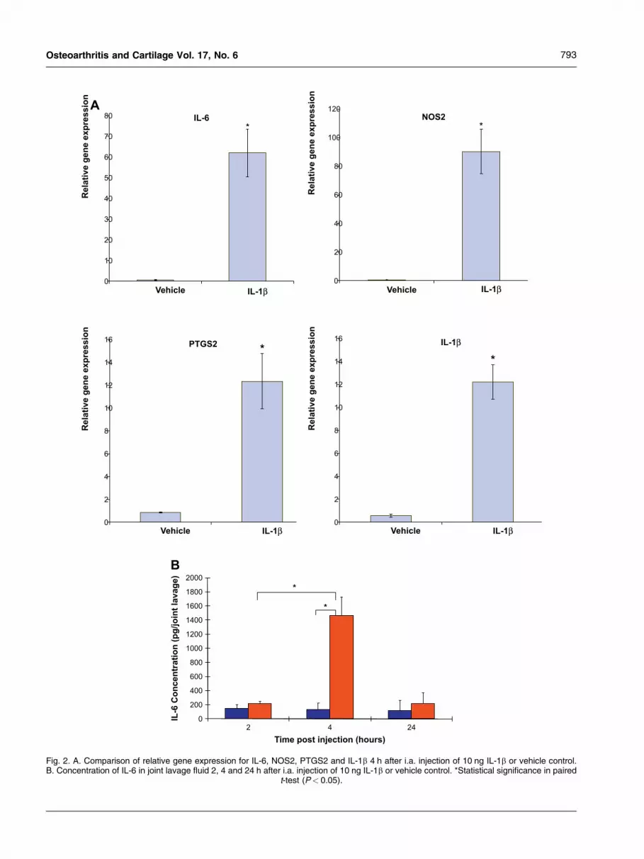
Fig. 2. A. Comparison of relative gene expression for IL-6, NOS2, PTGS2 and IL-1b 4 h after i.a. injection of 10 ng IL-1b or vehicle control.B. Concentration of IL-6 in joint lavage fluid 2, 4 and 24 h after i.a. injection of 10 ng IL-1b or vehicle control. *Statistical significance in paired
t-test (P< 0.05).
793Osteoarthritis and Cartilage Vol. 17, No. 6
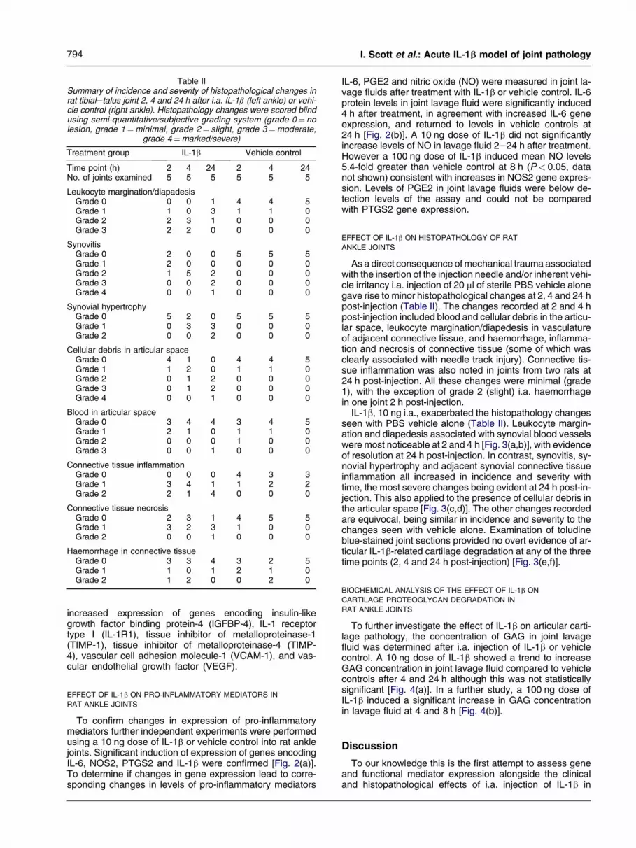
Table IISummary of incidence and severity of histopathological changes inrat tibialetalus joint 2, 4 and 24 h after i.a. IL-1b (left ankle) or vehi-cle control (right ankle). Histopathology changes were scored blindusing semi-quantitative/subjective grading system (grade 0¼ nolesion, grade 1¼minimal, grade 2¼ slight, grade 3¼moderate,
grade 4¼marked/severe)
Treatment group IL-1b Vehicle control
Time point (h) 2 4 24 2 4 24No. of joints examined 5 5 5 5 5 5
Leukocyte margination/diapadesisGrade 0 0 0 1 4 4 5Grade 1 1 0 3 1 1 0Grade 2 2 3 1 0 0 0Grade 3 2 2 0 0 0 0
SynovitisGrade 0 2 0 0 5 5 5Grade 1 2 0 0 0 0 0Grade 2 1 5 2 0 0 0Grade 3 0 0 2 0 0 0Grade 4 0 0 1 0 0 0
Synovial hypertrophyGrade 0 5 2 0 5 5 5Grade 1 0 3 3 0 0 0Grade 2 0 0 2 0 0 0
Cellular debris in articular spaceGrade 0 4 1 0 4 4 5Grade 1 1 2 0 1 1 0Grade 2 0 1 2 0 0 0Grade 3 0 1 2 0 0 0Grade 4 0 0 1 0 0 0
Blood in articular spaceGrade 0 3 4 4 3 4 5Grade 1 2 1 0 1 1 0Grade 2 0 0 0 1 0 0Grade 3 0 0 1 0 0 0
Connective tissue inflammationGrade 0 0 0 0 4 3 3Grade 1 3 4 1 1 2 2Grade 2 2 1 4 0 0 0
Connective tissue necrosisGrade 0 2 3 1 4 5 5Grade 1 3 2 3 1 0 0Grade 2 0 0 1 0 0 0
Haemorrhage in connective tissueGrade 0 3 3 4 3 2 5Grade 1 1 0 1 2 1 0Grade 2 1 2 0 0 2 0
794 I. Scott et al.: Acute IL-1b model of joint pathology
increased expression of genes encoding insulin-likegrowth factor binding protein-4 (IGFBP-4), IL-1 receptortype I (IL-1R1), tissue inhibitor of metalloproteinase-1(TIMP-1), tissue inhibitor of metalloproteinase-4 (TIMP-4), vascular cell adhesion molecule-1 (VCAM-1), and vas-cular endothelial growth factor (VEGF).
EFFECT OF IL-1b ON PRO-INFLAMMATORY MEDIATORS IN
RAT ANKLE JOINTS
To confirm changes in expression of pro-inflammatorymediators further independent experiments were performedusing a 10 ng dose of IL-1b or vehicle control into rat anklejoints. Significant induction of expression of genes encodingIL-6, NOS2, PTGS2 and IL-1b were confirmed [Fig. 2(a)].To determine if changes in gene expression lead to corre-sponding changes in levels of pro-inflammatory mediators
IL-6, PGE2 and nitric oxide (NO) were measured in joint la-vage fluids after treatment with IL-1b or vehicle control. IL-6protein levels in joint lavage fluid were significantly induced4 h after treatment, in agreement with increased IL-6 geneexpression, and returned to levels in vehicle controls at24 h [Fig. 2(b)]. A 10 ng dose of IL-1b did not significantlyincrease levels of NO in lavage fluid 2e24 h after treatment.However a 100 ng dose of IL-1b induced mean NO levels5.4-fold greater than vehicle control at 8 h (P< 0.05, datanot shown) consistent with increases in NOS2 gene expres-sion. Levels of PGE2 in joint lavage fluids were below de-tection levels of the assay and could not be comparedwith PTGS2 gene expression.
EFFECT OF IL-1b ON HISTOPATHOLOGY OF RAT
ANKLE JOINTS
As a direct consequence of mechanical trauma associatedwith the insertion of the injection needle and/or inherent vehi-cle irritancy i.a. injection of 20 ml of sterile PBS vehicle alonegave rise to minor histopathological changes at 2, 4 and 24 hpost-injection (Table II). The changes recorded at 2 and 4 hpost-injection included blood and cellular debris in the articu-lar space, leukocyte margination/diapedesis in vasculatureof adjacent connective tissue, and haemorrhage, inflamma-tion and necrosis of connective tissue (some of which wasclearly associated with needle track injury). Connective tis-sue inflammation was also noted in joints from two rats at24 h post-injection. All these changes were minimal (grade1), with the exception of grade 2 (slight) i.a. haemorrhagein one joint 2 h post-injection.
IL-1b, 10 ng i.a., exacerbated the histopathology changesseen with PBS vehicle alone (Table II). Leukocyte margin-ation and diapedesis associated with synovial blood vesselswere most noticeable at 2 and 4 h [Fig. 3(a,b)], with evidenceof resolution at 24 h post-injection. In contrast, synovitis, sy-novial hypertrophy and adjacent synovial connective tissueinflammation all increased in incidence and severity withtime, the most severe changes being evident at 24 h post-in-jection. This also applied to the presence of cellular debris inthe articular space [Fig. 3(c,d)]. The other changes recordedare equivocal, being similar in incidence and severity to thechanges seen with vehicle alone. Examination of toludineblue-stained joint sections provided no overt evidence of ar-ticular IL-1b-related cartilage degradation at any of the threetime points (2, 4 and 24 h post-injection) [Fig. 3(e,f)].
BIOCHEMICAL ANALYSIS OF THE EFFECT OF IL-1b ON
CARTILAGE PROTEOGLYCAN DEGRADATION IN
RAT ANKLE JOINTS
To further investigate the effect of IL-1b on articular carti-lage pathology, the concentration of GAG in joint lavagefluid was determined after i.a. injection of IL-1b or vehiclecontrol. A 10 ng dose of IL-1b showed a trend to increaseGAG concentration in joint lavage fluid compared to vehiclecontrols after 4 and 24 h although this was not statisticallysignificant [Fig. 4(a)]. In a further study, a 100 ng dose ofIL-1b induced a significant increase in GAG concentrationin lavage fluid at 4 and 8 h [Fig. 4(b)].
Discussion
To our knowledge this is the first attempt to assess geneand functional mediator expression alongside the clinicaland histopathological effects of i.a. injection of IL-1b in

IA
IASI
Ti
Ta
1 mm
CDC D
1 mm
Ti
Ta
Cal
Car
E F
50 µm
B
50 µm
A
100 µm
100 µm
Fig. 3. Representative tissue sections illustrating the effect of IL-1b on histopathology of rat tibialetalus joints; (A) 2 h after i.a. injection of IL-1bleukocytes, predominantly neutrophils, are lining the vessel against the endothelium (margination). The inserted image shows evidence ofinflammatory cells passing through a vessel wall (diapadesis, arrow); (B) 2 h after injection of PBS margination and diapadesis were less ev-ident compared to injection of IL-1b. (C) and (D) Images illustrate inflammatory changes in a representative tibialetalus joint 4 h after injectionof IL-1b; (D) shows a high power image of the boxed region in (C). Synovitis (synovial inflammation, SI) and inflammatory cell debris (CD) inthe i.a. space (IA) are apparent. Inflammatory changes and their sequele were less apparent at 2 h and were more pronounced at 24 h afterinjection of IL-1b (images not shown). Tibia (Ti), Talus (Ta). (E) and (F) Images show a representative tibialetalus joint 24 h after injection of IL-1b stained with toludine blue. Panel F shows a high power image of the boxed region in (E). There is no overt histological evidence of cartilage
degradation/damage. Calcaneum (Cal), articular cartilage (Car).
795Osteoarthritis and Cartilage Vol. 17, No. 6
a single study. We have shown that i.a. injection of IL-1b caninduce a cascade of pro-inflammatory and pro-degradativemediators in the rat ankle joint that potentially lead to the in-duction of an acute hyperalgesia, synovial inflammation andcartilage breakdown. The correlation of the pathological ef-fects of IL-1b with changes in pro-inflammatory and pro-deg-radative mediator expression provide a new insight into therole of the IL-1 pathway in joint disease.
This study demonstrated that a single dose of IL-1b, equiv-alent to levels measured in the SCW model of chronic arthri-tis16, was sufficient to drive an acute joint pathology.Although the doses of IL-1 identified in this inflammatorymodel potentially exceed those found in an OA joint, thisstudy, by utilising physiologically relevant doses of IL-1, con-trasts with previous studies in which the relevance of the
chosen IL-1b doses were unclear24e26,28e30. Intra-articularinjection of IL-1b into the rat ankle joint induced an acuteswelling of the joint with similarities to a previous study inrats24. IL-1b also produced a sequence of clinical changesincluding early evidence of acute mechanical hyperalgesiawhich is previously unreported. IL-1b induced a time courseof histopathological changes including early evidence of leu-kocyte margination and diapadesis in synovial blood vesselsleading to synovial inflammation (with synovial hypertrophy)and the accumulation of cellular debris in the articular space.These effects extend previous reports that i.a. IL-1b can in-duce a cellular infiltrate in the synovial lining and fluid, and sy-novitis26,29. There was no histological evidence that IL-1binduced acute cartilage destruction, however, biochemicalanalysis of joint lavage fluid revealed that IL-1b increased

0
1
2
3
4
5
6
7
IL-1β 10ng4
IL-1β 100ng4
IL-1β 10ng8
IL-1β 100ng8
GA
G C
on
cen
tratio
n
(μg
/jo
in
t lavag
e)
Time post injection (hours)
*
*
0
1
2
3
4
5
6
7
8
2 4 24Time post injection (hours)
GA
G C
on
cen
tratio
n
(μg
/jo
in
t lavag
e)
A
B
Fig. 4. (A) Concentration of GAG in joint lavage fluid 2, 4 and 24 hafter i.a. injection of 10 ng IL-1b (shown in red) or saline vehiclecontrol (blue). (B) Concentration of GAG in joint lavage fluid 4and 8 h after i.a. injection of 10 ng or 100 g IL-1b (red) or vehiclecontrol (blue). *Statistical significance in paired t-test (P< 0.05).
796 I. Scott et al.: Acute IL-1b model of joint pathology
the release of GAG, a marker of cartilage breakdown27,within 4e8 h. This confirms that analysis of GAG in joint la-vage fluid might be a more sensitive method of detecting car-tilage breakdown than histological analysis26. In addition, theDMMB assay for GAG has the advantage that it can be com-pleted in a few hours after collection of lavage fluid comparedto the time required to process and section joints for micro-scopic examination (about 6 weeks, mainly taken up by thedecalcification procedure). A single i.a. injection of IL-1b inrat joints has been shown to induce cartilage pathology 7days after treatment but GAG analysis of rat lavage fluidhas not been reported previously24.
Delineating the molecular pathways involved in an animalmodel of arthritis can provide insight into the associationswith human joint disease and could be used to identify the ap-propriate model to evaluate therapeutic agents15,17,18. Thisstudy is the first to identify IL-1b-induced changes in gene ex-pression and mediator release in the synovial joint and corre-late these with joint inflammation, pain, and histopathologicalchanges. As IL-1b was likely to affect a number of cell typesin the joint, we analysed changes in gene expression inwhole joint tissue as used in models of arthritis for gene mi-croarray analysis15,17,18. IL-1b induced the gene expressionof a range of pro-inflammatory and pro-degradative media-tors, including IL-6, PTGS2, NOS2, TNFa, NFkB1
ADAMTS5 and IL-1b, simultaneously with the peak of joint in-flammation and hyperalgesia. IL-1b also increased the geneexpression of IGFBP4, TIMP-1 and -4 and IL-1r1 encodingregulators of growth factor, protease and cytokine activitywith roles in joint tissue remodelling. IL-1b increased expres-sion of genes encoding VCAM-1 and VEGF, which haveroles in endothelial cell adhesion and function, respec-tively31,32, and might contribute to the IL-1b-induced leuko-cyte margination, diapedesis and cell infiltration observedin this study. This study has demonstrated the utility of quan-titative-PCR, using customised multi-gene microfluidiccards, to explore gene expression changes in an experimen-tal model of arthritis. Future work could extend gene expres-sion analysis to include additional key degradativemediators, including the collagenases MMP-13 and -8, andpotential pain mediators such as nerve growth factor. Al-though microfluidic cards are focused on analysis of10se100s of selected genes, compared to 1000s analysedby global gene microarrays, they have the advantage of be-ing able to analyse a large number of samples with high sen-sitivity and dynamic range33. Thus microfluidics-based PCRmight be a useful addition to the range of tools to study bothanimal models and the associated human disease.
Previous studies detailing gene expression changes inmodels of arthritis did not confirm that they lead to changesin protein levels or biological activity15,17,18. In this study, wedemonstrated that IL-1b induced the accumulation of IL-6protein and NO in joint lavage fluid consistent withincreases in IL-6 and NOS2 gene expression in joint tissue.Induction of NO levels in rat joint lavage fluid was consistentwith a previous study using a 1000 ng dose of i.a. IL-1b30. Incontrast, levels of PGE2 in joint lavage fluid were below de-tectability of the assay used and could not be correlatedwith increases in PTGS2 gene expression. Overall these re-sults support the utility of gene expression analysis in ani-mal models, particularly when limited amounts of lavagefluids, assay sensitivity, or the lack of availability of suitablereagents restrict biochemical analysis.
In conclusion, the present study has demonstrated thatIL-1b can drive an acute joint pathology that is mediatedby a range of the pro-inflammatory and destructive media-tors. The reproducibility of this acute model indicates thatit could be used for initial evaluation of the therapeutic po-tential of novel antagonists of the IL-1 pathway before com-mitting to complex arthritis models of increased durationand variability. In addition, consistent responses in thismodel have enabled the development of methods to char-acterise molecular changes, alongside clinical endpoints,which might have utility in studies of other animal modelsand human disease. Future work will aim to extend thetime course of the model to further understand disease pro-gression. Genome-wide microarray techniques will be usedto increase knowledge of molecular events downstream ofIL-1 in the synovial joint.
Conflict of interest
The authors declare no conflict of interests.All authors were employees of AstraZeneca plc during
work for this manuscript.
Acknowledgements
We gratefully acknowledge the statistical expertise ofDr Michael Dymond.

797Osteoarthritis and Cartilage Vol. 17, No. 6
References
1. Harris ED. The bone and joint decade: a catalyst for progress. ArthritisRheum 2004;44:1969e70.
2. Kean WF, Kean R, Buchanan WW. Osteoarthritis: symptoms, signs andsource of pain. Inflammopharmacology 2004;12:3e31.
3. Felson DT. An update on the pathogenesis and epidemiology of osteo-arthritis. Radiol Clin N Am 2004;42:1e9.
4. Pelletier JP, Martel-Pelletier J, Abramson SB. Osteoarthritis, an inflam-matory disease: potential implication for the selection of new thera-peutic targets. Arthrits Rheum 2001;44:1237e47.
5. Rau H, Frank C, Goretzki G, Spitz J. Radiosynoviothesis in osteoarthritisand other disorders with concomitant synovitis in comparison to rheu-matoid arthritis. Cancer Biother Radiopharm 2005;20:349e55.
6. Zhang Y, Nevitt M, Niu Y, Lewis C, Torner J, Guermazi A, et al. Revers-ible MRI features and knee pain fluctuation: the MOST study. Osteo-arthr Cartil 2007;15: S3, C5.
7. Wieland HA, Michaelis M, Kirschbaum JB, Rudolphi KA. Osteoarthritis ean untreatable disease? Nat Rev Drug Discov 2005;4:331e45.
8. Rudolphi K, Gerwin N, Verzijl N, van der Kraan P, van den Berg W. Pral-nacasan, an inhibitor of interleukin-1beta converting enzyme, reducesjoint damage in two murine models of osteoarthritis. Osteoarthr Cartil2003;11:738e46.
9. Zhang X, Mao Z, Yu C. Suppression of early experimental osteoarthritisby gene transfer of interleukin-1 receptor antagonist and interleukin-10. J Orthop Res. 2004;22:742e50.
10. Blom AB, van der Kraan PM, van den Berg WB. Cytokine targeting inosteoarthritis. Curr Drug Targets 2007;8:283e92.
11. Braddock M, Quinn A. Targeting IL-1 in inflammatory disease: new oppor-tunities for therapeutic intervention. Nat Rev Drug Discov 2004;3:1e10.
12. Iqbal I, Fleischmann R. Treatment of osteoarthritis with anakinra. CurrRheumatol Rep 2007;9:31e5.
13. Loeuille D, Chary-Valckenaere I, Goebel C, Rat AC, Blum A, Kiefer P,et al. MRI evaluation of the synovial membrane after a single intraar-ticular injection of anakinra in 7 patients with osteoarthritis of the knee.Arthritis Rheum 2005;9:S70.
14. Brandt KD. Animal models of osteoarthritis. Biorheology 2002;39:221e35.15. Ibrahim SM, Koczan D, Thiesen HJ. Gene-expression profile of colla-
gen-induced arthritis. J Autoimmun 2002;18:159e67.16. Midha A, Rendall E, Nicol S, Cruwys S. Characterisation of the intra-ar-
ticular cytokine profile in streptococcal cell wall-induced arthritis.Rheumatology 2003;42:33e8.
17. Rioja I, Clayton CL, Graham SJ, Life PF, Dickson MC. Gene expressionprofiles in the rat streptococcal cell wall-induced arthritis model iden-tified using microarray analysis. Arthritis Res Ther 2005;7:R101e17.
18. Thornton S, Sowders D, Aronow B, Witte DP, Brunner HI, Giannini EH,et al. DNA microarray analysis reveals novel gene expression profilesin collagen-induced arthritis. Clin Immunol 2002;105:155e68.
19. Bakker AC, Joosten LAB, Arntz OJ, Helsen MMA, Bendele AM, van deLoo FAJ, et al. Prevention of murine collagen-induced arthritis in the
knee and ipsilateral paw by local expression of human interleukin-1receptor antagonist protein in the knee. Arthritis Rheum 1997;40:893e900.
20. Caron JP, Fernandes JC, Martel-Pelletier J, Tardiff G, Mineau F, Geng C,et al. Chondroprotective effect of intra articular injections of interleukin-1receptor antagonist in experimental osteoarthritis: suppression of colla-genase-1 expression. Arthritis Rheum 1996;39:1535e44.
21. Pelletier JP, Caron JP, Evans C, Robbins PD, Georgescu HI,Jovanovic D, et al. In vivo suppression of early experimental osteoar-thritis by interleukin-1 receptor antagonist using gene therapy. ArthritisRheum 1997;40:1012e9.
22. Schwab JH, Anderle SK, Brown RR, Dalldorf FG, Thompson RC. Pro-and anti-inflammatory roles of interleukin-1 in recurrence of bacterialcell wall-induced arthritis in rats. Infect Immun 1991;59:4436e42.
23. Van de Loo FAJ, Arntz OJ, Otterness IG, van den Berg WB. Protectionagainst cartilage proteoglycan synthesis inhibition by anti-interleukin-1antibodies in experimental arthritis. J. Rheumatol 1992;19:348e56.
24. Chandrasekhar S, Harvey AK, Hrubey PS, Bendele AM. Arthritisinduced by interleukin-1 is dependent on the site and frequency ofintra-articular injection. Clin Immunol Immunopathol 1990;55:382e400.
25. Chandrasekhar S, Panetta JA, Bendele AM. Inhibition of IL-1 inducedarticular inflammation of rats by 4-aminothiazolidines, LY 221068and LY 269415. Agents Actions 1994;42:67e70.
26. Henderson B, Thompson RC, Hardingham T, Lewthwaite J. Inhibition ofinterleukin-1 induced synovitis and articular cartilage proteoglycanloss in the rabbit knee by recombinant human interleukin-1 receptorantagonist. Cytokine 1991;3:246e9.
27. Farndale RW, Buttle BJ, Barrett AJ. Improved quantitation and discrim-ination of sulphated glycosaminoglycans by use of dimethylmethyleneblue. Biochemica et Biophysica Acta 1986;883:173e7.
28. Dingle JT, Page-Thomas DP, King B, Bard DR. In vivo studies of artic-ular tissue damage mediated by catabolin/interleukin-1. Ann RheumDis 1987;46:527e33.
29. Pettipher ER, Higgs GA, Henderson B. Interleukin-1 induces leukocyteinfiltration and cartilage proteoglycan degradation in the synovial joint.Proc Natl Acad Sci 1986;83:8749e53.
30. Presle N, Cipolletta C, Jouzeau JY, Abid A, Netter P, Terlain B. Cartilageprotection by nitric oxide synthase inhibitors after intra-articular injec-tion of interleukin-1b in rats. Arthritis Rheum 1999;42:2094e102.
31. Carter RA, Wicks IP. Vascular cell adhesion molecule-1 (CD106): a mul-tifaceted regulator of joint inflammation. Arthritis Rheum 2001;44:985e94.
32. Haywood L, McWilliams DF, Pearson CI, Gill SE, Ganeasan A, WilsonD, et al. Inflammation and angiogenesis in osteoarthritis. ArthritisRheum 2003;48:2173e77.
33. Sauer S, Lange BM, Gobom J, Nyarsik L, Seitz H. Miniaturisation infunctional genomics and proteomics. Nat Rev Genet 2005;6:465e76.