contrasting roles for tlr ligands in hiv-1 pathogenesis
TRANSCRIPT
Contrasting Roles for TLR Ligands in HIV-1 PathogenesisBeda Brichacek1*, Christophe Vanpouille1, Yana Kiselyeva1, Angelique Biancotto1, Melanie Merbah1,
Ivan Hirsch2, Andrea Lisco1, Jean Charles Grivel1, Leonid Margolis1
1 Section of Intercellular Interactions, Program in Physical Biology, National Institute of Child Health and Human Development, National Institutes of Health, Bethesda,
Maryland, United States of America, 2 Centre de Recherche en Cancerologie de Marseille, UMR 891 INSERM, Institut Paoli-Calmettes, Universite de la Mediterrane,
Marseille, France
Abstract
The first line of a host’s response to various pathogens is triggered by their engagement of cellular pattern recognitionreceptors (PRRs). Binding of microbial ligands to these receptors leads to the induction of a variety of cellular factors thatalter intracellular and extracellular environment and interfere directly or indirectly with the life cycle of the triggeringpathogen. Such changes may also affect any coinfecting microbe. Using ligands to Toll-like receptors (TLRs) 5 and 9, weexamined their effect on human immunodeficiency virus (HIV)-1 replication in lymphoid tissue ex vivo. We found markeddifferences in the outcomes of such treatment. While flagellin (TLR5 agonist) treatment enhanced replication of CCchemokine receptor 5 (CCR 5)-tropic and CXC chemokine receptor 4 (CXCR4)-tropic HIV-1, treatment witholigodeoxynucleotide (ODN) M362 (TLR9 agonist) suppressed both viral variants. The differential effects of these TLRligands on HIV-1 replication correlated with changes in production of CC chemokines CCL3, CCL4, CCL5, and of CXCchemokines CXCL10, and CXCL12 in the ligand-treated HIV-1-infected tissues. The nature and/or magnitude of thesechanges were dependent on the ligand as well as on the HIV-1 viral strain. Moreover, the tested ligands differed in theirability to induce cellular activation as evaluated by the expression of the cluster of differentiation markers (CD) 25, CD38,CD39, CD69, CD154, and human leukocyte antigen D related (HLA)-DR as well as of a cell proliferation marker, Ki67, and ofCCR5. No significant effect of the ligand treatment was observed on apoptosis and cell death/loss in the treated lymphoidtissue ex vivo. Our results suggest that binding of microbial ligands to TLRs is one of the mechanisms that mediateinteractions between coinfected microbes and HIV-1 in human tissues. Thus, the engagement of appropriate TLRs bymicrobial molecules or their mimetic might become a new strategy for HIV therapy or prevention.
Citation: Brichacek B, Vanpouille C, Kiselyeva Y, Biancotto A, Merbah M, et al. (2010) Contrasting Roles for TLR Ligands in HIV-1 Pathogenesis. PLoS ONE 5(9):e12831. doi:10.1371/journal.pone.0012831
Editor: Jorg Hermann Fritz, University of Toronto, Canada
Received March 6, 2010; Accepted August 20, 2010; Published September 20, 2010
This is an open-access article distributed under the terms of the Creative Commons Public Domain declaration which stipulates that, once placed in the publicdomain, this work may be freely reproduced, distributed, transmitted, modified, built upon, or otherwise used by anyone for any lawful purpose.
Funding: This research was supported by the Intramural Research Program of the National Institute of Child Health and Human Development (NICHD), NationalInstitutes of Health (NIH), Bethesda, Maryland. The funders had no role in study design, data collection and analysis, decision to publish, or preparation of themanuscript.
Competing Interests: The authors have declared that no competing interests exist.
* E-mail: [email protected]
Introduction
HIV-1 infection, as well as the progression of HIV-1 disease,
usually occurs in the presence of other microbes [1–5]. These
infectious agents typically serve as copathogens, facilitating HIV-1
transmission and aggravating the clinical course of HIV-1 disease.
However, some infectious agents seem to have an opposite effect
and can alleviate the course of HIV-1 disease [6–10]. Evidence
exists that these microbes interact locally with HIV-1 by up- or
down-regulating HIV-1 coreceptors [11–13] and receptors [14] as
well as by inducing changes in the production of various cytokines
[11,13]. While the mechanisms by which microbes interact with
HIV-1 are not fully understood, it is clear that their invasion into
the human body triggers a cascade of events that can affect HIV-1
pathogenesis.
One of the first events in the interaction of the human body with
an invading microorganism is the engagement of cellular PRRs
(for review see [15–17]). The best-characterized class of PRRs are
the Toll-like receptors (TLRs), a family of pathogen sensors that
trigger local inflammation, recruitment of effector cells, and
secretion of cytokines that modulate both the innate and adaptive
immune responses [18–20]. TLRs are expressed in a wide variety
of cells, located in different tissues and organs including lymphoid
organs [20–25]. Activation of TLRs engages multiple intracellular
adaptor and signaling proteins and triggers several cascades of
innate immune response [17]. Since the pattern of expression of
TLRs depends on a particular cell subtype and on the engagement
of a ligand to a particular TLR receptor, the system responds to
different pathogens differently [17,26]. In general, this response
may include cell activation and differentiation [27,28], secretion of
type I IFNs, and/or production of proinflamatory cytokines
[17,29]. These responses to TLRs engagement together with the
response to the engagement of other PRRs and various cellular
factors also regulate adaptive immunity [16]. Because of the
profound role of TLRs engagement in immune response, TLR
agonists, antagonists, specific antibodies or shRNA are now being
tested as novel therapeutics and adjuvants for vaccines [26,30–40].
As first responders to an invading virus, TLRs and other PRRs
play a critical role in directing the anti-viral host responses [41–
46]. These responses can be affected by simultaneous signaling
from PRRs engaged by other coinfecting microbes. These effects
have been studied by applying TLR ligands to HIV-1-infected cell
lines, isolated primary cells, human lymphocyte aggregate cultures
and experimental animals [47–53].
PLoS ONE | www.plosone.org 1 September 2010 | Volume 5 | Issue 9 | e12831
However, critical events in HIV-1 pathogenesis occur in the
context of structured human lymphoid organs [54–57]. Therefore,
here we describe the effects of TLR engagement on HIV-1
replication in lymphoid tissue using explants of palatine tonsils,
where TLRs are broadly expressed [24,25]. This system was
developed in our laboratory and has been used to address HIV-1
tissue pathogenesis as well as HIV-1 interactions with various
microbes [12,14,58–60]. We report that in human lymphoid
tissue, different TLR ligands modulate HIV replication both
positively and negatively and that a TLR-triggered cell activation
and change of cytokine spectra are involved in these modulations.
Results
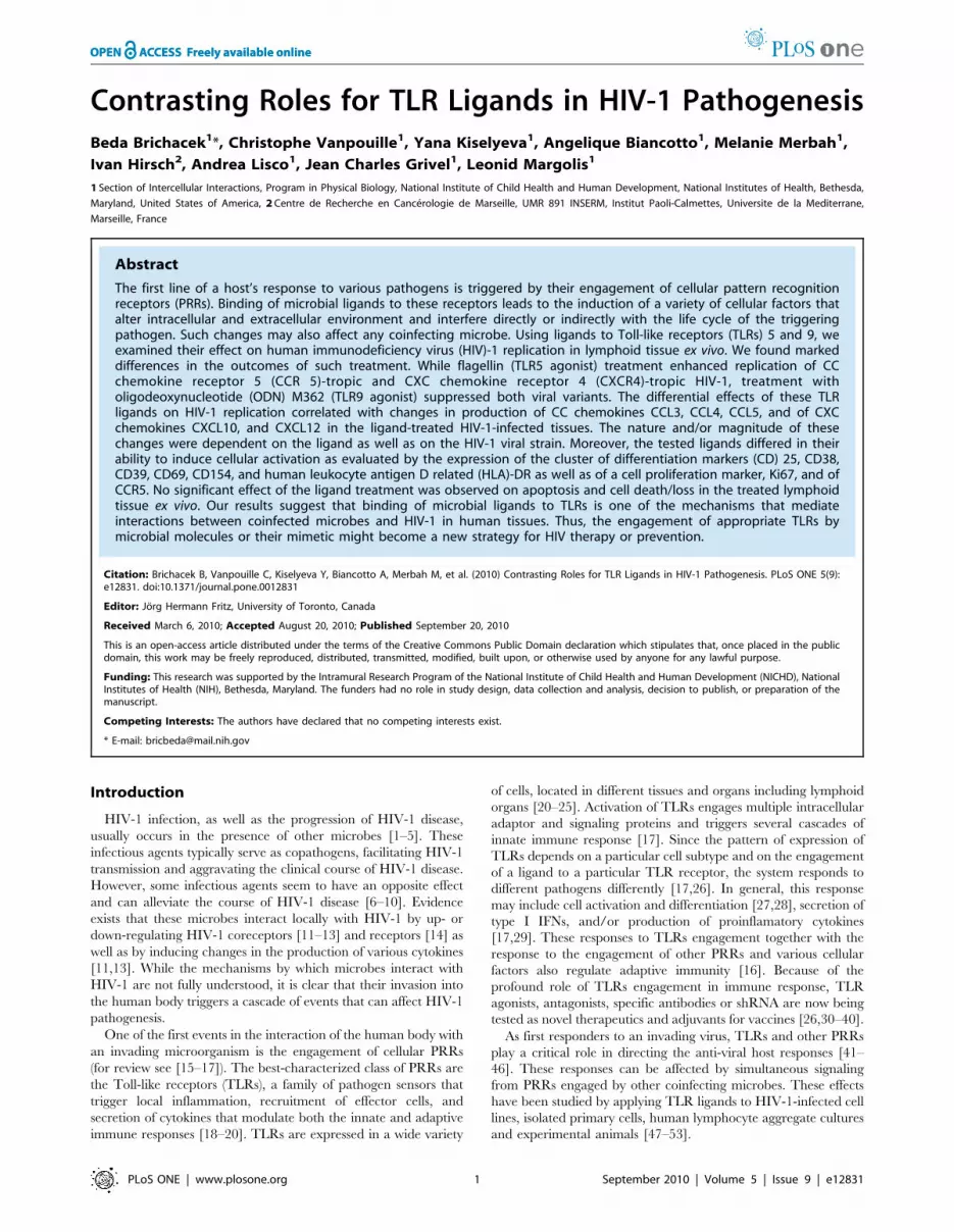
TLR ligands modulate HIV-1 replication in humanlymphoid tissue ex vivo
Blocks of human lymphoid tissue were pretreated overnight
with ligands for TLR2 (LPS from Porphyromonas gingivalis), TLR3
(poly (I:C)), TLR4 (LPS from E. coli K12), TLR5 (flagellin from S.
typhimurium), TLR7 (Loxoribine), TLR8 (ssRNA40/LyoVec), and
TLR9 (CpG oligonucleotide type C: ODN M362). The pretreated
tissues were infected with R5 or X4 HIV-1 (X4LAI.04 or R5SF162).
Viral replication was evaluated from tissues’ release of p24 into the
culture medium over 15 days of infection. Results of these
experiments are shown in Figure 1. TLR ligands had diverse
effects on HIV-1 replication ranging from suppression of both R5
and X4 HIV-1 variants, through differential effects on R5 and X4
HIV-1 variants, to upregulation of both R5 and X4 HIV-1. To
investigate the mechanisms of modulation of HIV-1 replication by
TLR ligands, we further focused on two ligands that affect HIV in
opposite ways: flagellin and ODN M362. The TLR5 ligand
flagellin enhanced replication of both R5SF162 and X4LAI.04 HIV-1
(n = 4, p = 0.034 and n = 6, p = 0.016, for R5 and X4 HIV-1,
respectively), whereas TLR9 ligand ODN M362 suppressed both
HIV-1 variants (n = 5, p = 0.021 and n = 5, p = 0.008, for R5 and
X4, respectively) (Fig. 1).
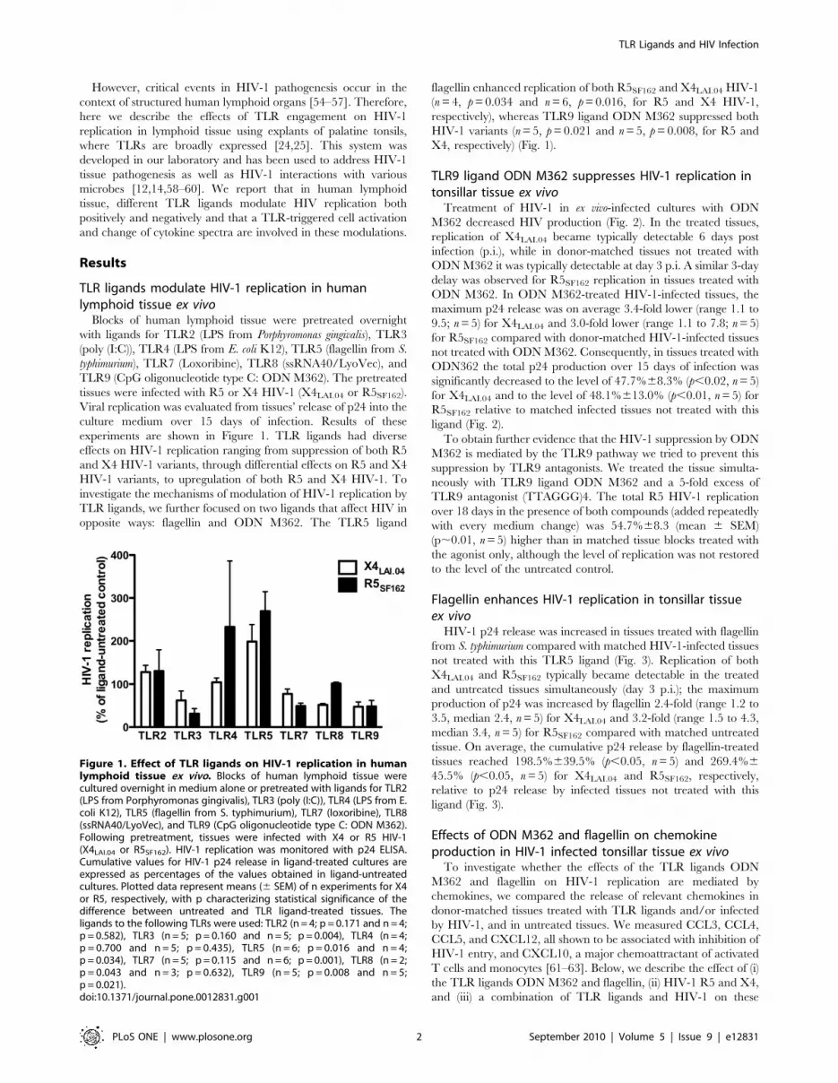
TLR9 ligand ODN M362 suppresses HIV-1 replication intonsillar tissue ex vivo
Treatment of HIV-1 in ex vivo-infected cultures with ODN
M362 decreased HIV production (Fig. 2). In the treated tissues,
replication of X4LAI.04 became typically detectable 6 days post
infection (p.i.), while in donor-matched tissues not treated with
ODN M362 it was typically detectable at day 3 p.i. A similar 3-day
delay was observed for R5SF162 replication in tissues treated with
ODN M362. In ODN M362-treated HIV-1-infected tissues, the
maximum p24 release was on average 3.4-fold lower (range 1.1 to
9.5; n = 5) for X4LAI.04 and 3.0-fold lower (range 1.1 to 7.8; n = 5)
for R5SF162 compared with donor-matched HIV-1-infected tissues
not treated with ODN M362. Consequently, in tissues treated with
ODN362 the total p24 production over 15 days of infection was
significantly decreased to the level of 47.7%68.3% (p,0.02, n = 5)
for X4LAI.04 and to the level of 48.1%613.0% (p,0.01, n = 5) for
R5SF162 relative to matched infected tissues not treated with this
ligand (Fig. 2).
To obtain further evidence that the HIV-1 suppression by ODN
M362 is mediated by the TLR9 pathway we tried to prevent this
suppression by TLR9 antagonists. We treated the tissue simulta-
neously with TLR9 ligand ODN M362 and a 5-fold excess of
TLR9 antagonist (TTAGGG)4. The total R5 HIV-1 replication
over 18 days in the presence of both compounds (added repeatedly
with every medium change) was 54.7%68.3 (mean 6 SEM)
(p,0.01, n = 5) higher than in matched tissue blocks treated with
the agonist only, although the level of replication was not restored
to the level of the untreated control.
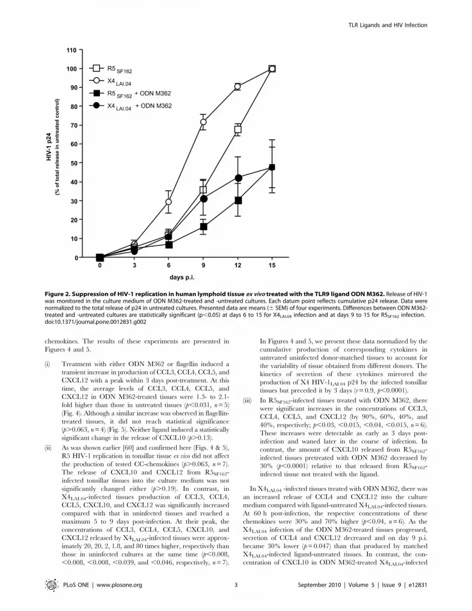
Flagellin enhances HIV-1 replication in tonsillar tissueex vivo
HIV-1 p24 release was increased in tissues treated with flagellin
from S. typhimurium compared with matched HIV-1-infected tissues
not treated with this TLR5 ligand (Fig. 3). Replication of both
X4LAI.04 and R5SF162 typically became detectable in the treated
and untreated tissues simultaneously (day 3 p.i.); the maximum
production of p24 was increased by flagellin 2.4-fold (range 1.2 to
3.5, median 2.4, n = 5) for X4LAI.04 and 3.2-fold (range 1.5 to 4.3,
median 3.4, n = 5) for R5SF162 compared with matched untreated
tissue. On average, the cumulative p24 release by flagellin-treated
tissues reached 198.5%639.5% (p,0.05, n = 5) and 269.4%6
45.5% (p,0.05, n = 5) for X4LAI.04 and R5SF162, respectively,
relative to p24 release by infected tissues not treated with this
ligand (Fig. 3).
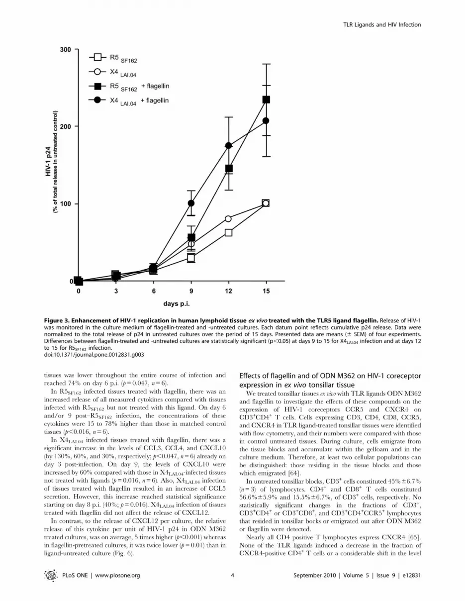
Effects of ODN M362 and flagellin on chemokineproduction in HIV-1 infected tonsillar tissue ex vivo
To investigate whether the effects of the TLR ligands ODN
M362 and flagellin on HIV-1 replication are mediated by
chemokines, we compared the release of relevant chemokines in
donor-matched tissues treated with TLR ligands and/or infected
by HIV-1, and in untreated tissues. We measured CCL3, CCL4,
CCL5, and CXCL12, all shown to be associated with inhibition of
HIV-1 entry, and CXCL10, a major chemoattractant of activated
T cells and monocytes [61–63]. Below, we describe the effect of (i)
the TLR ligands ODN M362 and flagellin, (ii) HIV-1 R5 and X4,
and (iii) a combination of TLR ligands and HIV-1 on these
Figure 1. Effect of TLR ligands on HIV-1 replication in humanlymphoid tissue ex vivo. Blocks of human lymphoid tissue werecultured overnight in medium alone or pretreated with ligands for TLR2(LPS from Porphyromonas gingivalis), TLR3 (poly (I:C)), TLR4 (LPS from E.coli K12), TLR5 (flagellin from S. typhimurium), TLR7 (loxoribine), TLR8(ssRNA40/LyoVec), and TLR9 (CpG oligonucleotide type C: ODN M362).Following pretreatment, tissues were infected with X4 or R5 HIV-1(X4LAI.04 or R5SF162). HIV-1 replication was monitored with p24 ELISA.Cumulative values for HIV-1 p24 release in ligand-treated cultures areexpressed as percentages of the values obtained in ligand-untreatedcultures. Plotted data represent means (6 SEM) of n experiments for X4or R5, respectively, with p characterizing statistical significance of thedifference between untreated and TLR ligand-treated tissues. Theligands to the following TLRs were used: TLR2 (n = 4; p = 0.171 and n = 4;p = 0.582), TLR3 (n = 5; p = 0.160 and n = 5; p = 0.004), TLR4 (n = 4;p = 0.700 and n = 5; p = 0.435), TLR5 (n = 6; p = 0.016 and n = 4;p = 0.034), TLR7 (n = 5; p = 0.115 and n = 6; p = 0.001), TLR8 (n = 2;p = 0.043 and n = 3; p = 0.632), TLR9 (n = 5; p = 0.008 and n = 5;p = 0.021).doi:10.1371/journal.pone.0012831.g001
TLR Ligands and HIV Infection
PLoS ONE | www.plosone.org 2 September 2010 | Volume 5 | Issue 9 | e12831
chemokines. The results of these experiments are presented in
Figures 4 and 5.
(i) Treatment with either ODN M362 or flagellin induced a
transient increase in production of CCL3, CCL4, CCL5, and
CXCL12 with a peak within 3 days post-treatment. At this
time, the average levels of CCL3, CCL4, CCL5, and
CXCL12 in ODN M362-treated tissues were 1.3- to 2.1-
fold higher than those in untreated tissues (p,0.031, n = 5)
(Fig. 4). Although a similar increase was observed in flagellin-
treated tissues, it did not reach statistical significance
(p.0.063, n = 4) (Fig. 5). Neither ligand induced a statistically
significant change in the release of CXCL10 (p.0.13).
(ii) As was shown earlier [60] and confirmed here (Figs. 4 & 5),
R5 HIV-1 replication in tonsillar tissue ex vivo did not affect
the production of tested CC-chemokines (p.0.063, n = 7).
The release of CXCL10 and CXCL12 from R5SF162-
infected tonsillar tissues into the culture medium was not
significantly changed either (p.0.19). In contrast, in
X4LAI.04-infected tissues production of CCL3, CCL4,
CCL5, CXCL10, and CXCL12 was significantly increased
compared with that in uninfected tissues and reached a
maximum 5 to 9 days post-infection. At their peak, the
concentrations of CCL3, CCL4, CCL5, CXCL10, and
CXCL12 released by X4LAI.04-infected tissues were approx-
imately 20, 20, 2, 1.8, and 80 times higher, respectively than
those in uninfected cultures at the same time (p,0.008,
,0.008, ,0.008, ,0.039, and ,0.046, respectively, n = 7).
In Figures 4 and 5, we present these data normalized by the
cumulative production of corresponding cytokines in
untreated uninfected donor-matched tissues to account for
the variability of tissue obtained from different donors. The
kinetics of secretion of these cytokines mirrored the
production of X4 HIV-1LAI.04 p24 by the infected tonsillar
tissues but preceded it by 3 days (r = 0.9, p,0.0001).
(iii) In R5SF162-infected tissues treated with ODN M362, there
were significant increases in the concentrations of CCL3,
CCL4, CCL5, and CXCL12 (by 90%, 60%, 40%, and
40%, respectively; p,0.03, ,0.015, ,0.04, ,0.015, n = 6).
These increases were detectable as early as 3 days post-
infection and waned later in the course of infection. In
contrast, the amount of CXCL10 released from R5SF162-
infected tissues pretreated with ODN M362 decreased by
30% (p,0.0001) relative to that released from R5SF162-
infected tissue not treated with the ligand.
In X4LAI.04 -infected tissues treated with ODN M362, there was
an increased release of CCL4 and CXCL12 into the culture
medium compared with ligand-untreated X4LAI.04-infected tissues.
At 60 h post-infection, the respective concentrations of these
chemokines were 30% and 70% higher (p,0.04, n = 6). As the
X4LAI.04 infection of the ODN M362-treated tissues progressed,
secretion of CCL4 and CXCL12 decreased and on day 9 p.i.
became 30% lower (p = 0.047) than that produced by matched
X4LAI.04-infected ligand-untreated tissues. In contrast, the con-
centration of CXCL10 in ODN M362-treated X4LAI.04-infected
Figure 2. Suppression of HIV-1 replication in human lymphoid tissue ex vivo treated with the TLR9 ligand ODN M362. Release of HIV-1was monitored in the culture medium of ODN M362-treated and -untreated cultures. Each datum point reflects cumulative p24 release. Data werenormalized to the total release of p24 in untreated cultures. Presented data are means (6 SEM) of four experiments. Differences between ODN M362-treated and -untreated cultures are statistically significant (p,0.05) at days 6 to 15 for X4LAI.04 infection and at days 9 to 15 for R5SF162 infection.doi:10.1371/journal.pone.0012831.g002
TLR Ligands and HIV Infection
PLoS ONE | www.plosone.org 3 September 2010 | Volume 5 | Issue 9 | e12831
tissues was lower throughout the entire course of infection and
reached 74% on day 6 p.i. (p = 0.047, n = 6).
In R5SF162 infected tissues treated with flagellin, there was an
increased release of all measured cytokines compared with tissues
infected with R5SF162 but not treated with this ligand. On day 6
and/or 9 post–R5SF162 infection, the concentrations of these
cytokines were 15 to 78% higher than those in matched control
tissues (p,0.016, n = 6).
In X4LAI.04 infected tissues treated with flagellin, there was a
significant increase in the levels of CCL3, CCL4, and CXCL10
(by 130%, 60%, and 30%, respectively; p,0.047, n = 6) already on
day 3 post-infection. On day 9, the levels of CXCL10 were
increased by 60% compared with those in X4LAI.04-infected tissues
not treated with ligands (p = 0.016, n = 6). Also, X4LAI.04 infection
of tissues treated with flagellin resulted in an increase of CCL5
secretion. However, this increase reached statistical significance
starting on day 8 p.i. (40%; p = 0.016). X4LAI.04 infection of tissues
treated with flagellin did not affect the release of CXCL12.
In contrast, to the release of CXCL12 per culture, the relative
release of this cytokine per unit of HIV-1 p24 in ODN M362
treated cultures, was on average, 5 times higher (p,0.001) whereas
in flagellin-pretreated cultures, it was twice lower (p = 0.01) than in
ligand-untreated culture (Fig. 6).
Effects of flagellin and of ODN M362 on HIV-1 coreceptorexpression in ex vivo tonsillar tissue
We treated tonsillar tissues ex vivo with TLR ligands ODN M362
and flagellin to investigate the effects of these compounds on the
expression of HIV-1 coreceptors CCR5 and CXCR4 on
CD3+CD4+ T cells. Cells expressing CD3, CD4, CD8, CCR5,
and CXCR4 in TLR ligand-treated tonsillar tissues were identified
with flow cytometry, and their numbers were compared with those
in control untreated tissues. During culture, cells emigrate from
the tissue blocks and accumulate within the gelfoam and in the
culture medium. Therefore, at least two cellular populations can
be distinguished: those residing in the tissue blocks and those
which emigrated [64].
In untreated tonsillar blocks, CD3+ cells constituted 45%66.7%
(n = 3) of lymphocytes. CD4+ and CD8+ T cells constituted
56.6%65.9% and 15.5%66.7%, of CD3+ cells, respectively. No
statistically significant changes in the fractions of CD3+,
CD3+CD4+ or CD3+CD8+, and CD3+CD4+CCR5+ lymphocytes
that resided in tonsillar bocks or emigrated out after ODN M362
or flagellin were detected.
Nearly all CD4 positive T lymphocytes express CXCR4 [65].
None of the TLR ligands induced a decrease in the fraction of
CXCR4-positive CD4+ T cells or a considerable shift in the level
Figure 3. Enhancement of HIV-1 replication in human lymphoid tissue ex vivo treated with the TLR5 ligand flagellin. Release of HIV-1was monitored in the culture medium of flagellin-treated and -untreated cultures. Each datum point reflects cumulative p24 release. Data werenormalized to the total release of p24 in untreated cultures over the period of 15 days. Presented data are means (6 SEM) of four experiments.Differences between flagellin-treated and -untreated cultures are statistically significant (p,0.05) at days 9 to 15 for X4LAI.04 infection and at days 12to 15 for R5SF162 infection.doi:10.1371/journal.pone.0012831.g003
TLR Ligands and HIV Infection
PLoS ONE | www.plosone.org 4 September 2010 | Volume 5 | Issue 9 | e12831

Figure 4. Effects of ODN M362 on chemokines production in HIV-1-infected lymphoid tissue ex vivo. Blocks of tonsillar tissue weretreated with ODN M362 and subsequently infected with HIV-1 X4LAI.04 or R5SF162. Release of CCL3, CCL4, CCL5, CXCL10, and CXCL12 was monitored inthe culture medium with the multiplexed fluorescent microsphere immunoassay. Concentrations of released chemokines at individual time points
TLR Ligands and HIV Infection
PLoS ONE | www.plosone.org 5 September 2010 | Volume 5 | Issue 9 | e12831

of expression of CXCR4 on their surface as measured by the
intensity of the staining.
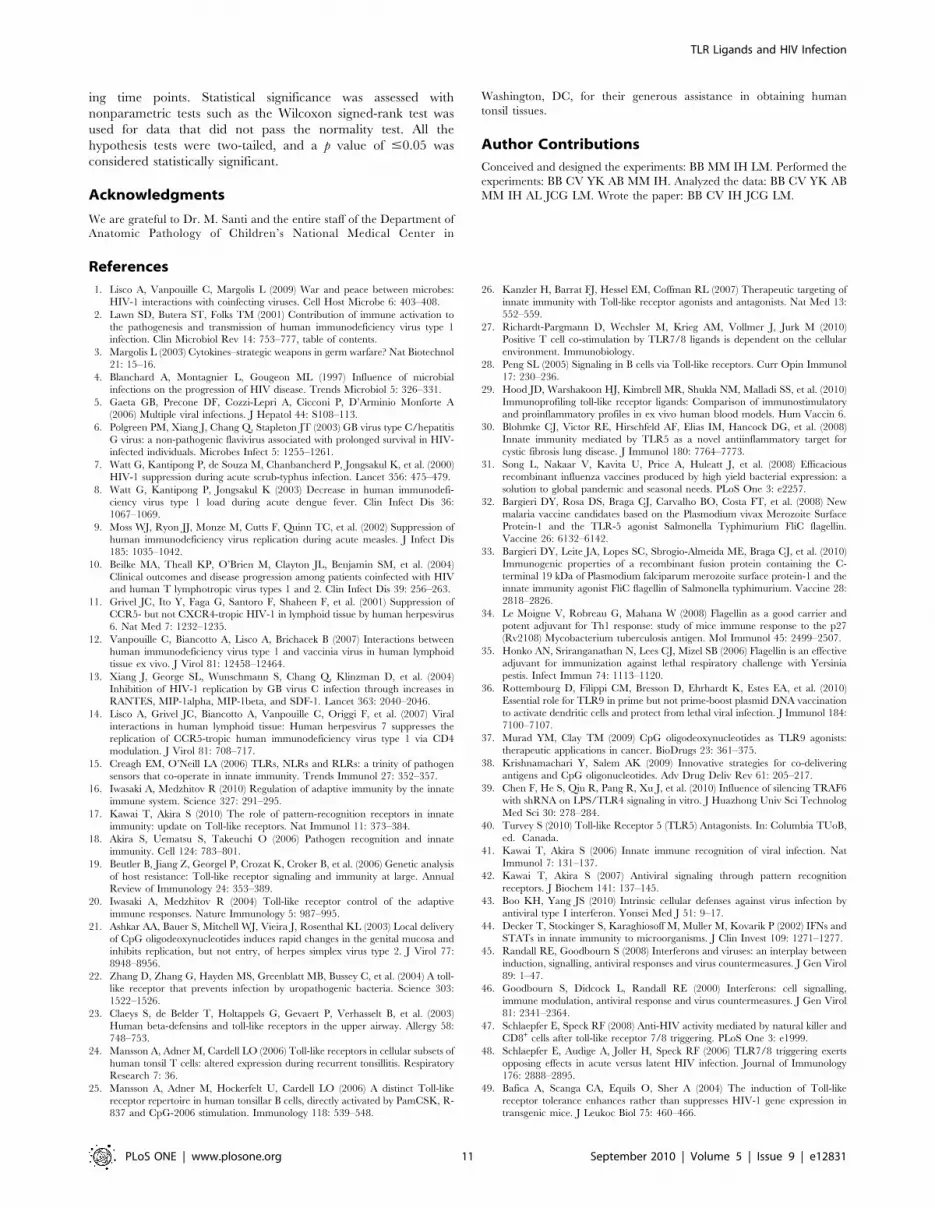
Flagellin modulates the expression of activation markerson CD4+CD82 T lymphocytes
Since HIV-1 replicates predominantly in activated T cells [66–
68], we evaluated the effect of flagellin and ODN M362 on the
expression of activation markers on CD4+ T cells. In particular,
we stained tissue cells for CD3, CD4, CD8, CD25, HLA-DR,
CD38, CD39, CD69, CD154, as well as for Ki67.
Six days after the treatment of tonsillar tissue blocks with ODN
M362 or flagellin, the cells were analyzed with flow cytometry. In
general, in flagellin-treated tissues the fraction of cells expressing
the activation markers was increased (Fig. 7). In cells that were
isolated from the tissue blocks, the increases reached statistical
significance for CD25 and CD38 (13767% and 156617% of the
untreated tissue, respectively, p,0.05). In cells that migrated from
the tissue blocks, the increases reached statistical significance for
CD25, CD39 and CD154 (12767%, 12862%, 146612% of the
untreated tissue, respectively, p,0.03). In contrast, in tissues
treated with ODN M362 there were no statistically significant
changes in the fraction of CD4 T cells expressing the activation
markers, except for HLA-DR which was increased on the
CD3+CD4+CD82 cells that migrated out of the tissue blocks
(12267 of the untreated tissue, p,0.002). As the result of these
changes, there were statistically significant differences between
tissues treated with the two ligands in the fractions of cells
expressing CD25, CD38, HLA-DR, CD39, and Ki67 (p,0.04).
Similarly, there were significant differences in expression of these
markers, except for HLA-DR, between the tissues treated with
flagellin and those treated with ODN M362 in the fractions of
CD4 T cells that emigrated from the tissue blocks (p,0.04).
Also, we analyzed the cycling of CD4 T cells in tissues treated
with flagellin or ODN M362. As shown in Figure 7, flagellin
treatment resulted in almost doubling of the number of Ki67+
CD4 T cells remaining in the blocks and which emigrated from
them. Thus, in contrast to ODN M362, both the fraction of
cycling cells and cells expressing most of the activation markers
were increased by flagellin treatment (Fig. 7).
Finally, in flagellin-treated tissues, there were more CD3+ cells
with higher forward side scatter (1.94%60.2%) (‘‘lymphoblasts’’)
than in ODN M362-treated tissue (1.15%60.3%) (p = 0.0132,
n = 3).
Thus, flagellin leads to higher activation of CD3+CD4+
lymphocytes in tonsillar tissue than ODN M362.
Effects of ODN M362 and flagellin on CD4+CD82
apoptosis and cell loss in tonsillar tissues ex vivoNeither annexin V staining nor cell counting revealed
statistically significant differences between tissues treated with
ODN M362 or flagellin and untreated tissues. In untreated
tonsillar blocks 6%62.6% of CD3+CD4+ lymphocytes bound
annexin V, while in ODN M362- and flagellin-treated tissues
4.4%60.5% and 3.5%61.1% of CD3+CD4+ lymphocytes,
respectively, bound this apoptotic marker. Although these
differences were more pronounced in cells that emigrated from
the tissue blocks, they did not reach statistical significance
(0.075,p,0.59, for all comparisons). The fractions of annexin
V-binding CD3+CD4+ lymphocytes among the emigrated cells
were 7.4%61.9%, 5.1%60.3%, and 2.9%60.9% in untreated,
ODN M362-treated, and flagellin-treated cultures, respectively.
Moreover, no significant differences in cell loss were detected
between untreated tissues and tissues treated with either ligand
(0.16,p,0.77).
Discussion
Clinical and experimental evidence suggests that various
microbes are able to affect the HIV-1 life cycle by changing the
systemic and/or local environment [69–75] or by interacting with
HIV-1 directly [76,77]. For example, microbial infection may lead
to the modulation of cell activation (HSV-2) [78], to changes in
receptor expression (HSV-2, HHV-7, GBV-C) [13,14,79–81], and
to the release of chemokines (HHV-6, GBV-C) [11,13] that affect
HIV cell entry and replication. Moreover, R5 and X4 HIV-1 may
be affected in different ways [82,83], raising the possibility that
coinfecting microbes may play an important role in the switch
from R5 to X4 dominance [11].
Nevertheless, the molecular mechanisms of microbial interac-
tions with host cells and especially their effects on HIV-1 infection
remain largely unknown. During the last few years it has become
evident that the first contact of an invading microbe with the host
cell involves engagement of TLRs and other PRRs with microbial
molecules inducing an innate immune response.
Here, we tested whether engagement of TLRs by invading
microorganisms affects HIV-1 replication in human lymphoid
tissues. Because the effects of microbes on host cells are
complicated, we used a reductionist approach: instead of whole
microbes, we treated human lymphoid tissue ex vivo with TLR
ligands. We administered various TLR ligands prior to and during
HIV-1 infection and monitored HIV-1 replication, cytokine
release, expression of selected surface molecules, and cell
activation. For these experiments, we used blocks of ex vivo human
tonsillar tissue in which cytoarchitecture and cellular repertoire, as
well as some tissue functions, are preserved. This approach differs
from that of previous studies of the effect of TLR engagement on
HIV-1 replication in vitro that were performed in cell lines, isolated
primary cells, human aggregate lymphocyte cultures [47–53]. It
was shown that at least some events critical for HIV-1 infection
(e.g., secretion of cytokines upon infection) significantly differ
between ex vivo tissue and high density cell aggregates or isolated
cells [84].
Tonsils were obtained from patients 2 to 7 years old. At this age,
approximately 30% of tonsillar lymphocytes are T cells (CD3+)
and 70% are B cells (CD19+) [85]. Most of the tonsillar
lymphocytes express TLR receptors [25,86]. The majority of
other cells present in tonsils, including dendritic cells, NK cells,
macrophages, stromal cells, and epithelial cells, are known to
express TLRs as well.
Here, we found that TLR ligands affect HIV-1 infection in
human lymphoid tissue ex vivo. Different TLR ligands differentially
affect HIV-1 replication, either up- or downregulating it. We
chose TLR5 and TLR9 ligands to investigate the effects of their
were normalized to their total cumulative release in matched untreated cultures. Presented data are means of these normalized values (6 SEM) from4 to 7 experiments. Statistical analysis revealed significant difference between: (i) uninfected and X4LAI.04 –infected (but not R5-infected) tissues in therelease of CCL3, CCL4, CCL5, CXCL10, and CXCL12 (p,0.046, n = 5 to 7); (ii) uninfected mock- and ODN M362-treated tissues in production of CCL3,CCL4, CCL5, and CXCL12 (p,0.031, n = 5); (iii) R5SF162-infected tissues and these tissues treated with ODN M362 in the production of CCL3, CCL4,CCL5, and CXCL12 (p,0.04; n = 6); (iv) X4LAI.04- infected tissues and these tissues treated with ODN M362, in production of CCL4 and CXCL12(p,0.046, n = 6).doi:10.1371/journal.pone.0012831.g004
TLR Ligands and HIV Infection
PLoS ONE | www.plosone.org 6 September 2010 | Volume 5 | Issue 9 | e12831
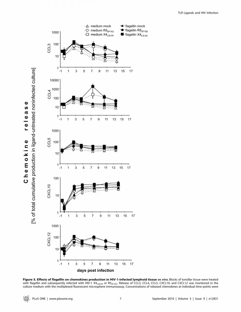
Figure 5. Effects of flagellin on chemokines production in HIV-1-infected lymphoid tissue ex vivo. Blocks of tonsillar tissue were treatedwith flagellin and subsequently infected with HIV-1 X4LAI.04 or R5SF162. Release of CCL3, CCL4, CCL5, CXCL10, and CXCL12 was monitored in theculture medium with the multiplexed fluorescent microsphere immunoassay. Concentrations of released chemokines at individual time points were
TLR Ligands and HIV Infection
PLoS ONE | www.plosone.org 7 September 2010 | Volume 5 | Issue 9 | e12831
engagement on HIV-1 infection and the mechanisms for this
phenomenon, because of their opposite effects on HIV-1
irrespectively of its coreceptor specificity.
As a TLR5 ligand we used flagellin from S. typhimurium. As a
TLR9 ligand we used ODN M362 (CpG oligonucleotide type C).
There are three types of ODN M362, A, B and C with slightly
different structures and eliciting different immune responses [55–
58]. In the current work we used type C since type C triggers all
the pathways triggered by A and B [87–90]. Tissue treatment with
flagellin led to the increase of both R5 and X4 HIV-1 replication.
In contrast, addition of ODN M362 suppressed replication of
HIV-1 of both phenotypes. This suppression was partially reversed
by a simultaneous addition of a TLR9 antagonist (TTAGGG)4 to
R5SF162-infected tissues treated with ODN M362.
Treatment of the tonsillar blocks with ODN M362 led to an
increase, although transient, of CCL3, CCL4, CCL5, and CXCL12
secretion but not of CXCL10. Also, these cytokines were upregulated
when the ODN M362-treated tissues were infected with R5SF162.
Upregulation of cytokines that bind to HIV coreceptors may be one
of the mechanisms mediating ODN M362-induced suppression of
HIV-1 replication in the treated tissues.
We found a positive correlation between HIV-1 production and
the release of CXCL12 both in ODN M362-treated and in
untreated tissues. It seems that it is HIV-1 replication that induces
CXCL12 release. In ODN M362-treated cultures, this effect is
augmented, i.e., cytokine release in response to HIV-1 replication
is increased.
Within the first 60 h post-infection, X4LAI.04 virus induced a
significantly higher release of CXCL12 from ODN M362-
pretreated compared with a ligand-untreated tissue. Since the
replication of X4LAI.04 virus induces a large release of CXCL12,
the effect of ODN M362 on CXCL12 release is masked later in
the infection, when significant viral replication occurs. Thus, at
that time, most of the observed changes in CXCL12 concentra-
tions reflect changes due to viral replication. To distinguish
between contributions of the ligand treatment and X4LAI.04
replication to the secretion of CXCL12, we expressed CXCL12
release as a function of HIV-1 p24 release. Our data (see Figure 6)
suggest that flagellin suppresses, while ODN M362 enhances, the
induction of CXCL12 in response to X4 HIV-1 replication in
tonsillar tissue ex vivo. The concentrations of the above chemokines
detected in the culture media of ODN M362-treated tissue were
sufficient to cause suppression of HIV-1 replication [11,60].
Differences in the production of CXCL10 could also contribute
to the modulatory effect of chemokines on HIV-1 replication,
since CXCL10 is a potent attractant of activated CD4 T cells [91],
the major HIV-1 targets. The increase of this chemokine in HIV-
1–infected tissues caused by flagellin and its decrease caused by
ODN M362 may be related to their stimulatory or suppressive
effects on HIV-1 replication in human lymphoid tissue ex vivo.
In general, upregulation of cytokines that bind to HIV coreceptors
may be one of the mechanisms mediating ODN M362-induced
suppression of HIV-1 replication in the treated tissues.
Cell activation by flagellin, as evidenced by the increase in the
fractions of CD4-positive T cells expressing CD25, CD38, and
CD39 and especially increase in the number of cycling (Ki67+)
CD4 T cells, also may contribute to flagellin-related upregulation
of replication of HIV-1 of both coreceptor tropisms in lymphoid
tissue ex vivo. Moreover, it has been reported that flagellin
engagement of TLR5 leads to HIV-1 reactivation [52].
ODN M362 inhibitory effect on HIV-1 replication may be
mediated by soluble factors induced by this TLR ligand. In
particular, it was shown that in response to ODN M362, pDCs
produce type 1 IFNs [17], which suppress HIV-1 replication via
death of infected cell [92], production of tetherin [93], and/or
increased expression of APOBEC3G [94]. In our experiments, we
did not detect type 1 IFNs in the culture supernatants of the
lymphoid tissue ex vivo although, we readily detected it in
supernatants from the tissues spiked with various concentrations of
type 1 IFNs. It is nevertheless possible that type 1 IFN is quickly
consumed by the neighboring cells without being released into the
media in detectable quantities.
Neither of the tested TLR agonists induced a statistically
significant loss of CD3+CD4+CD82 cells such as that reported for
TLR ligand-treated PBMC cultures [51], suggesting that different
regulations occur in tissues and in isolated cells. Moreover, in
tissues TLRs of many cell types can be affected and the final effect
of TLR ligands on HIV-1-infected tissues and HIV infection itself
could be a superimposition of the responses of various tissue cells
that are not necessarily direct HIV-1 targets [16].
In conclusion, we showed that TLR5 and TLR9 ligands affect
HIV-1 replication in opposite ways. These ligands changed the
cytokine spectra, the cellular activation status, and the expression of
cellular receptors in agreement with their up- or downregulation of
HIV-1. It is important to keep in mind that although the
compounds we and others are using are called TLR ligands, it is
possible that they act not only through the TLRs [17]. Nevertheless,
TLR seems to be the main pathway triggered by these compounds,
and similar pathways may be triggered by actual microbes in
infected tissues. Thus, we think that the model described in our
paper seems to reflect adequately the mechanism of HIV interaction
with other microbes in coinfected tissues. Our results suggest that
interactions between coinfecting microbes and HIV-1 can be
mediated by PRRs, which trigger the innate immunity response. A
particular PRR engaged by a microbe may account for various
microbial effects on HIV-1 replication in coinfected tissues and
eventually affect the course of HIV-1 disease. Use of engagement of
different PRRs by microbial molecules or their mimetics could
become a new strategy for HIV therapy or prevention.
Materials and Methods
Ethics statementThis study was conducted according to the principles expressed in
the Declaration of Helsinki. The study was approved by the Human
Subjects Institutional Review Board (IRB) of Children’s National
Medical Center (Protocol #3204) and by the Office of Human
Subjects Research of NIH (OHSR #2264). Since anonymous tissue
samples were used, no written informed consent was required.
Tissue culturesAnonymous samples of human tonsillar tissues were obtained
from patients undergoing routine tonsillectomy at the Children’s
National Medical Center (Washington, DC). Tissues were
normalized to their total cumulative release in matched untreated cultures. Presented data are means of these normalized values (6 SEM) from 4 to 7experiments. Statistical analysis (see the Results) revealed significant difference between (i) uninfected and X4LAI.04 –infected (but not R5-infected)tissues in the release of CCL3, CCL4, CCL5, and CXCL10 (p,0.046, n = 5 to 7); (ii) R5SF162-infected tissues and these tissues treated with flagellin in therelease of CCL3, CCL4, CCL5, CXCL12, and CXCL10 (p,0.016, n = 6); (ii) X4LAI.04 -infected tissues and these tissues treated with flagellin in the release ofCCL3, CCL4, and CXCL10 (p,0.046, n = 6).doi:10.1371/journal.pone.0012831.g005
TLR Ligands and HIV Infection
PLoS ONE | www.plosone.org 8 September 2010 | Volume 5 | Issue 9 | e12831

dissected into 8- to 27-mm3 blocks and placed onto collagen
sponge gels in culture medium at the air-liquid interface, as
described earlier [95–98]. Tissue blocks were cultured in RPMI
1640 (GIBCO BRL, Grand Island, NY) medium containing 15%
heat-inactivated fetal calf serum (FCS; Gemini Bio-Products,
Woodland, CA), nonessential amino acids (1 mM), sodium
pyruvate (1 mM), amphotericin B (2.5 mg/mL; GIBCO BRL),
and gentamicin (50 mg/mL; Quality Control, Inc., Rockville,
MD). The culture medium was supplemented with Timentin
(GlaxoSmithKline, Research Triangle Park, NC) for the first 24 h
and then changed regularly. For each condition of infection, 27
blocks were prepared (9 blocks/well/3 ml of complete medium).
HIV-1 infectionsTissue blocks were infected by application of 5–7.5 ml of HIV
stock (0.5–1.0 ng of p24) on top of each tissue block as described
previously [11,98]. X4 isolate LAI.04 (X4LAI.04) and R5 isolate
SF162 (R5SF162) were obtained from the Rush University Virology
Quality Assurance Laboratory (Chicago, IL). HIV-1 replication
was monitored from measurements of p24 in the culture medium
with the Alliance HIV-1 p24 ELISA (PerkinElmer Life Sciences,
Inc., Boston, MA).
TLR ligand treatmentOn the second day after cutting, tissue blocks were treated with
ligands either by complete replacement of tissue culture media
with medium containing TLR ligand (ligands for TLR2, 3, 4, and
7) or by preincubation of the tissue blocks in culture medium
containing 250 ml of TLR ligand per 18 blocks for 2 h at 37uCfollowed by a transfer of the blocks onto collagen sponge gels and
subsequent incubation in CO2 incubator overnight (ligands for
TLR5, 8, and 9). All TLR ligands were obtained from InvivoGen
(Chicago, IL) and used at concentrations recommended by the
manufacturer as follows: TLR2 agonist LPS from Porphyromonas
gingivalis (3 mg/ml), TLR3 agonist poly(I:C) (25 mg/ml), TLR4
agonist LPS from E. coli K12 (5 mg/ml), TLR5 agonist flagellin (S.
typhimurium) (5 mg/ml), TLR7 agonist loxoribine (1 mM), TLR8
agonist ssRNA (ssRNA40/LyoVec) (10 mg/ml), and TLR9 agonist
CpG oligonucleotide type C (ODN M362) (5 mM). TLR9
antagonist (TTAGGG)4 was used at 5-fold excess (25 mM) over
the TLR9 agonist ODN M362. Ligands were kept in culture
medium during the whole course of the experiment (ligands for
TLR2, 3, 4, and 7) or reapplied in 10-ml quantities on the top of
every tissue block after each medium change (ligands for TLR5, 8,
and 9). In control cultures, tissue blocks were treated with culture
medium only.
Cytokine measurementsThe levels of CCL3, CCL4, CCL5, CXCL10, and CXCL12
were measured in culture supernatants from TLR ligand and/or
HIV-1-infected tissues with a multiplexed fluorescent microsphere
immunoassay using the Luminex 100 system (Luminex Corpora-
tion, Austin, TX). Cytokines, capture antibodies, and biotinylated
detection antibodies were obtained from R&D System (Minnea-
polis, MN). We coupled 100 mg of cytokine capture antibodies
covalently to 12.106 carboxylated microspheres using sulfo-NHS
and EDC according to the Luminex standard protocol. We mixed
1,200 coupled microspheres from each set with 50 ml of standards
or culture medium and incubated them overnight at 4uC in a
multiscreen filter plate (Millipore corporation, Billerica, MA).
After three washes by vacuum aspiration, 50 ml of biotinylated
polyclonal anti-cytokine antibodies were added to each well and
incubated for one hour at 37uC. Following three additional
washes, the bound cytokines were detected by the addition of 50 ml
of a 16-mg/ml solution of streptavidin-phycoerytrin (Molecular
Probes, Carlsbad, CA). Data were collected and analyzed with
Bioplex Manager v3.0 software (Bio-Rad, Hercules, CA) using a 5-
parameter fitting algorithm.
Flow cytometryFlow cytometry of cells isolated from tissue blocks and stained
for cell surface markers with specific antibodies was performed as
described earlier [99]. Briefly, cell that migrated from the tissue
blocks were collected from the collagen sponge gels and tissue
culture medium. Additionally, single-cell suspensions were pre-
pared from tonsillar tissue blocks by digestion with Collagenase IV
(GIBCO BRL) at 2.5 mg/ml in RPMI supplemented with 5%
FCS for 30 min. Cell suspensions were passed through a 40 mm
nylon mesh (Falcon) and stained with labeled monoclonal
antibodies coupled to fluorochromes. Staining was performed in
three groups. The first group was stained with annexin V labeled
with PE and with combinations of the following labeled
monoclonal antibodies: anti-CD3-APC-Cy5.5, anti-CD4-PE-
Alexa 610, anti-CD8-PacificBlue, anti-CD38-PE-Cy7, anti-
CD45RO-APC, and anti-CD69-PE-Cy5.5. The second group
was stained with annexin V labeled with PE and with com-
binations of the following labeled monoclonal antibodies: anti-CD3-
APC-Cy5.5, anti-CD4-PE-Alexa 610, anti-CD8-PacificBlue, anti-
CD14-PE-Cy5.5, anti-CD19-Tricolor, anti-CCR5-APC-Cy7, and
anti-CXCR4-APC. A third group was stained with combinations of
the following labeled monoclonal antibodies: anti-CD3-NC650,
anti-CD4-NC605, anti-CD8-eFluor 450, anti-CD45-AlexaFluor
Figure 6. ODN M362 and flagellin modulate CXCL12 releaseHIV-1 X4LAI.04 -infected lymphoid tissue ex vivo. Blocks of tonsillartissue were treated either with flagellin (m) or with ODN M362 (&) ormock-treated (N), and subsequently infected with HIV-1 X4LAI.04.Concentrations of CXCL12 and p24 were determined in the culturesupernatants every 3rd day for 12 days following the infection. Datawere normalized to the maximums released in matched X4LAI.04-infected ligand-untreated cultures. Each datum point represents a ratiobetween released CXCL12 and HIV-1 p24 in each of 47 HIV-1 positivesupernatants. On average, in ODN M362 treated cultures, the relativerelease of CXCL12 per unit of HIV-1 p24 was 5 times higher (p,0.001)whereas in flagellin-pretreated cultures, it was twice lower (p = 0.01)than in ligand-untreated culture.doi:10.1371/journal.pone.0012831.g006
TLR Ligands and HIV Infection
PLoS ONE | www.plosone.org 9 September 2010 | Volume 5 | Issue 9 | e12831
780, anti-CD38-AlexaFluor 700, anti-CD39-APC, anti-CD25-
Cy7-PE, anti-HLA DR-Cy5.5-PE, anti CD154-Cy5-PE, anti-
CD69-PE. Antibodies used in the first and second staining group
were purchased as follows: Antibodies to CD3, CD4, CD8, CD14,
CD19, CD45RO, and CD69 were products of Invitrogen/Caltag
Laboratories, Burlingame, CA. Annexin V and antibodies to
CD38, CD184 (CXCR4), and CD195 (CCR5) were purchased
from Becton-Dickinson (Pharmingen, San Jose, CA). Antibodies
used in the third staining group were purchased from eBioscience
(antibodies to CD3, CD4, CD8, CD45, CD39, CD25), from
Biolegend (antibodies to CD38, CD154, CD69), and from
Invitrogen (antibodies to HLA DR). After staining, cells were
washed and fixed in PBS containing 2% formaldehyde. Data were
acquired with an LSRII flow cytometer equipped with 355-, 407-,
488-, 532-, and 638-nm LASER lines using DIVA 4.1.2 software
(Becton Dickenson). Data were compensated and analyzed with
FlowJo version 9 (Treestar, Ashland, OR, USA). Cell depletion was
quantified by addition of True Count beads (Invitrogen/Caltag
Laboratories) to each tube prior to acquisition as a volumetric
control and by normalization of the number of cells by tissue-block
weight.
Expression of data and statistical analysisViral replication is expressed as the concentration of p24 in
culture medium pooled from 27 blocks (three wells) accumulated
during the particular time period. As reported earlier [95–98],
there was a substantial donor-to-donor variability in HIV
replication. To be able to pool and compare the results obtained
from tissues of different donors, we adjusted the results using the
data from matched control blocks as the basis of normalization.
Cytokine secretions are expressed as fold increase (the ratio of
cytokine production in infected or coinfected tissue to cytokine
production in control cultures) or as actual concentrations. The
data obtained in treated tissues from different donors were pooled
after being normalized and expressed as percent of the data from
matched uninfected untreated control blocks at the correspond-
Figure 7. Effect of ODN M362 and flagellin on CD4+ T-cell activation. Blocks of tonsillar tissue were treated with either flagellin or ODN M362for 7 days. Expression of activation markers CD25, HLA-DR, CD38, CD69, CD39, CD154 on CD4+CD82 CD3+ lymphocytes (Panels A & C) and of cell-proliferation marker Ki67 in these cells (panels B & D) were monitored with flow cytometry in tissue blocks (Panels A & B) and in the fraction of cellsemigrated from the tonsillar tissues (Panels C & D). Panels A & B show representative staining of CD3+CD4+CD82 lymphocytes for CD38 and Ki67.Data were compensated and analyzed with FlowJo version 9 (Treestar, Ashland, OR, USA). The plotted data represent means (6 SEM) of fractions ofCD4+CD82 CD3+ lymphocytes positive for particular activation marker from four experiments. Data are normalized to the corresponding fraction ofCD4+CD82 CD3+ lymphocytes from matched untreated control tissue. + marks statistically significant (p,0.04) difference between flagellin- and ODNM362-treated tissue for a particular activation marker.doi:10.1371/journal.pone.0012831.g007
TLR Ligands and HIV Infection
PLoS ONE | www.plosone.org 10 September 2010 | Volume 5 | Issue 9 | e12831
ing time points. Statistical significance was assessed with
nonparametric tests such as the Wilcoxon signed-rank test was
used for data that did not pass the normality test. All the
hypothesis tests were two-tailed, and a p value of #0.05 was
considered statistically significant.
Acknowledgments
We are grateful to Dr. M. Santi and the entire staff of the Department of
Anatomic Pathology of Children’s National Medical Center in
Washington, DC, for their generous assistance in obtaining human
tonsil tissues.
Author Contributions
Conceived and designed the experiments: BB MM IH LM. Performed the
experiments: BB CV YK AB MM IH. Analyzed the data: BB CV YK AB
MM IH AL JCG LM. Wrote the paper: BB CV IH JCG LM.
References
1. Lisco A, Vanpouille C, Margolis L (2009) War and peace between microbes:
HIV-1 interactions with coinfecting viruses. Cell Host Microbe 6: 403–408.
2. Lawn SD, Butera ST, Folks TM (2001) Contribution of immune activation to
the pathogenesis and transmission of human immunodeficiency virus type 1
infection. Clin Microbiol Rev 14: 753–777, table of contents.
3. Margolis L (2003) Cytokines–strategic weapons in germ warfare? Nat Biotechnol
21: 15–16.
4. Blanchard A, Montagnier L, Gougeon ML (1997) Influence of microbial
infections on the progression of HIV disease. Trends Microbiol 5: 326–331.
5. Gaeta GB, Precone DF, Cozzi-Lepri A, Cicconi P, D’Arminio Monforte A
(2006) Multiple viral infections. J Hepatol 44: S108–113.
6. Polgreen PM, Xiang J, Chang Q, Stapleton JT (2003) GB virus type C/hepatitis
G virus: a non-pathogenic flavivirus associated with prolonged survival in HIV-
infected individuals. Microbes Infect 5: 1255–1261.
7. Watt G, Kantipong P, de Souza M, Chanbancherd P, Jongsakul K, et al. (2000)
HIV-1 suppression during acute scrub-typhus infection. Lancet 356: 475–479.
8. Watt G, Kantipong P, Jongsakul K (2003) Decrease in human immunodefi-
ciency virus type 1 load during acute dengue fever. Clin Infect Dis 36:
1067–1069.
9. Moss WJ, Ryon JJ, Monze M, Cutts F, Quinn TC, et al. (2002) Suppression of
human immunodeficiency virus replication during acute measles. J Infect Dis
185: 1035–1042.
10. Beilke MA, Theall KP, O’Brien M, Clayton JL, Benjamin SM, et al. (2004)
Clinical outcomes and disease progression among patients coinfected with HIV
and human T lymphotropic virus types 1 and 2. Clin Infect Dis 39: 256–263.
11. Grivel JC, Ito Y, Faga G, Santoro F, Shaheen F, et al. (2001) Suppression of
CCR5- but not CXCR4-tropic HIV-1 in lymphoid tissue by human herpesvirus
6. Nat Med 7: 1232–1235.
12. Vanpouille C, Biancotto A, Lisco A, Brichacek B (2007) Interactions between
human immunodeficiency virus type 1 and vaccinia virus in human lymphoid
tissue ex vivo. J Virol 81: 12458–12464.
13. Xiang J, George SL, Wunschmann S, Chang Q, Klinzman D, et al. (2004)
Inhibition of HIV-1 replication by GB virus C infection through increases in
RANTES, MIP-1alpha, MIP-1beta, and SDF-1. Lancet 363: 2040–2046.
14. Lisco A, Grivel JC, Biancotto A, Vanpouille C, Origgi F, et al. (2007) Viral
interactions in human lymphoid tissue: Human herpesvirus 7 suppresses the
replication of CCR5-tropic human immunodeficiency virus type 1 via CD4
modulation. J Virol 81: 708–717.
15. Creagh EM, O’Neill LA (2006) TLRs, NLRs and RLRs: a trinity of pathogen
sensors that co-operate in innate immunity. Trends Immunol 27: 352–357.
16. Iwasaki A, Medzhitov R (2010) Regulation of adaptive immunity by the innate
immune system. Science 327: 291–295.
17. Kawai T, Akira S (2010) The role of pattern-recognition receptors in innate
immunity: update on Toll-like receptors. Nat Immunol 11: 373–384.
18. Akira S, Uematsu S, Takeuchi O (2006) Pathogen recognition and innate
immunity. Cell 124: 783–801.
19. Beutler B, Jiang Z, Georgel P, Crozat K, Croker B, et al. (2006) Genetic analysis
of host resistance: Toll-like receptor signaling and immunity at large. Annual
Review of Immunology 24: 353–389.
20. Iwasaki A, Medzhitov R (2004) Toll-like receptor control of the adaptive
immune responses. Nature Immunology 5: 987–995.
21. Ashkar AA, Bauer S, Mitchell WJ, Vieira J, Rosenthal KL (2003) Local delivery
of CpG oligodeoxynucleotides induces rapid changes in the genital mucosa and
inhibits replication, but not entry, of herpes simplex virus type 2. J Virol 77:
8948–8956.
22. Zhang D, Zhang G, Hayden MS, Greenblatt MB, Bussey C, et al. (2004) A toll-
like receptor that prevents infection by uropathogenic bacteria. Science 303:
1522–1526.
23. Claeys S, de Belder T, Holtappels G, Gevaert P, Verhasselt B, et al. (2003)
Human beta-defensins and toll-like receptors in the upper airway. Allergy 58:
748–753.
24. Mansson A, Adner M, Cardell LO (2006) Toll-like receptors in cellular subsets of
human tonsil T cells: altered expression during recurrent tonsillitis. Respiratory
Research 7: 36.
25. Mansson A, Adner M, Hockerfelt U, Cardell LO (2006) A distinct Toll-like
receptor repertoire in human tonsillar B cells, directly activated by PamCSK, R-
837 and CpG-2006 stimulation. Immunology 118: 539–548.
26. Kanzler H, Barrat FJ, Hessel EM, Coffman RL (2007) Therapeutic targeting of
innate immunity with Toll-like receptor agonists and antagonists. Nat Med 13:
552–559.
27. Richardt-Pargmann D, Wechsler M, Krieg AM, Vollmer J, Jurk M (2010)
Positive T cell co-stimulation by TLR7/8 ligands is dependent on the cellular
environment. Immunobiology.
28. Peng SL (2005) Signaling in B cells via Toll-like receptors. Curr Opin Immunol
17: 230–236.
29. Hood JD, Warshakoon HJ, Kimbrell MR, Shukla NM, Malladi SS, et al. (2010)
Immunoprofiling toll-like receptor ligands: Comparison of immunostimulatory
and proinflammatory profiles in ex vivo human blood models. Hum Vaccin 6.
30. Blohmke CJ, Victor RE, Hirschfeld AF, Elias IM, Hancock DG, et al. (2008)
Innate immunity mediated by TLR5 as a novel antiinflammatory target for
cystic fibrosis lung disease. J Immunol 180: 7764–7773.
31. Song L, Nakaar V, Kavita U, Price A, Huleatt J, et al. (2008) Efficacious
recombinant influenza vaccines produced by high yield bacterial expression: a
solution to global pandemic and seasonal needs. PLoS One 3: e2257.
32. Bargieri DY, Rosa DS, Braga CJ, Carvalho BO, Costa FT, et al. (2008) New
malaria vaccine candidates based on the Plasmodium vivax Merozoite Surface
Protein-1 and the TLR-5 agonist Salmonella Typhimurium FliC flagellin.
Vaccine 26: 6132–6142.
33. Bargieri DY, Leite JA, Lopes SC, Sbrogio-Almeida ME, Braga CJ, et al. (2010)
Immunogenic properties of a recombinant fusion protein containing the C-
terminal 19 kDa of Plasmodium falciparum merozoite surface protein-1 and the
innate immunity agonist FliC flagellin of Salmonella typhimurium. Vaccine 28:
2818–2826.
34. Le Moigne V, Robreau G, Mahana W (2008) Flagellin as a good carrier and
potent adjuvant for Th1 response: study of mice immune response to the p27
(Rv2108) Mycobacterium tuberculosis antigen. Mol Immunol 45: 2499–2507.
35. Honko AN, Sriranganathan N, Lees CJ, Mizel SB (2006) Flagellin is an effective
adjuvant for immunization against lethal respiratory challenge with Yersinia
pestis. Infect Immun 74: 1113–1120.
36. Rottembourg D, Filippi CM, Bresson D, Ehrhardt K, Estes EA, et al. (2010)
Essential role for TLR9 in prime but not prime-boost plasmid DNA vaccination
to activate dendritic cells and protect from lethal viral infection. J Immunol 184:
7100–7107.
37. Murad YM, Clay TM (2009) CpG oligodeoxynucleotides as TLR9 agonists:
therapeutic applications in cancer. BioDrugs 23: 361–375.
38. Krishnamachari Y, Salem AK (2009) Innovative strategies for co-delivering
antigens and CpG oligonucleotides. Adv Drug Deliv Rev 61: 205–217.
39. Chen F, He S, Qiu R, Pang R, Xu J, et al. (2010) Influence of silencing TRAF6
with shRNA on LPS/TLR4 signaling in vitro. J Huazhong Univ Sci Technolog
Med Sci 30: 278–284.
40. Turvey S (2010) Toll-like Receptor 5 (TLR5) Antagonists. In: Columbia TUoB,
ed. Canada.
41. Kawai T, Akira S (2006) Innate immune recognition of viral infection. Nat
Immunol 7: 131–137.
42. Kawai T, Akira S (2007) Antiviral signaling through pattern recognition
receptors. J Biochem 141: 137–145.
43. Boo KH, Yang JS (2010) Intrinsic cellular defenses against virus infection by
antiviral type I interferon. Yonsei Med J 51: 9–17.
44. Decker T, Stockinger S, Karaghiosoff M, Muller M, Kovarik P (2002) IFNs and
STATs in innate immunity to microorganisms. J Clin Invest 109: 1271–1277.
45. Randall RE, Goodbourn S (2008) Interferons and viruses: an interplay between
induction, signalling, antiviral responses and virus countermeasures. J Gen Virol
89: 1–47.
46. Goodbourn S, Didcock L, Randall RE (2000) Interferons: cell signalling,
immune modulation, antiviral response and virus countermeasures. J Gen Virol
81: 2341–2364.
47. Schlaepfer E, Speck RF (2008) Anti-HIV activity mediated by natural killer and
CD8+ cells after toll-like receptor 7/8 triggering. PLoS One 3: e1999.
48. Schlaepfer E, Audige A, Joller H, Speck RF (2006) TLR7/8 triggering exerts
opposing effects in acute versus latent HIV infection. Journal of Immunology
176: 2888–2895.
49. Bafica A, Scanga CA, Equils O, Sher A (2004) The induction of Toll-like
receptor tolerance enhances rather than suppresses HIV-1 gene expression in
transgenic mice. J Leukoc Biol 75: 460–466.
TLR Ligands and HIV Infection
PLoS ONE | www.plosone.org 11 September 2010 | Volume 5 | Issue 9 | e12831
50. Schlaepfer E, Audige A, Joller H, Speck RF (2006) TLR7/8 triggering exerts
opposing effects in acute versus latent HIV infection. J Immunol 176:2888–2895.
51. Funderburg N, Luciano AA, Jiang W, Rodriguez B, Sieg SF, et al. (2008) Toll-
like receptor ligands induce human T cell activation and death, a model for HIVpathogenesis. PLoS One 3: e1915.
52. Thibault S, Imbeault M, Tardif MR, Tremblay MJ (2009) TLR5 stimulation issufficient to trigger reactivation of latent HIV-1 provirus in T lymphoid cells and
activate virus gene expression in central memory CD4+ T cells. Virology 389:
20–25.53. Thibault S, Fromentin R, Tardif MR, Tremblay MJ (2009) TLR2 and TLR4
triggering exerts contrasting effects with regard to HIV-1 infection of humandendritic cells and subsequent virus transfer to CD4+ T cells. Retrovirology 6:
42.54. Pantaleo G, Graziosi C, Demarest JF, Butini L, Montroni M, et al. (1993) HIV
infection is active and progressive in lymphoid tissue during the clinically latent
stage of disease. Nature 362: 355–358.55. Pantaleo G, Graziosi C, Butini L, Pizzo PA, Schnittman SM, et al. (1991)
Lymphoid organs function as major reservoirs for human immunodeficiencyvirus. Proc Natl Acad Sci U S A 88: 9838–9842.
56. Pantaleo G, Cohen OJ, Schacker T, Vaccarezza M, Graziosi C, et al. (1998)
Evolutionary pattern of human immunodeficiency virus (HIV) replication anddistribution in lymph nodes following primary infection: implications for
antiviral therapy. Nat Med 4: 341–345.57. Cohen OJ, Pantaleo G, Lam GK, Fauci AS (1997) Studies on lymphoid tissue
from HIV-infected individuals: implications for the design of therapeuticstrategies. Springer Semin Immunopathol 18: 305–322.
58. Grivel JC, Biancotto A, Ito Y, Lima RG, Margolis LB (2003) Bystander CD4+ T
lymphocytes survive in HIV-infected human lymphoid tissue. AIDS Res HumRetroviruses 19: 211–216.
59. Grivel JC, Garcia M, Moss WJ, Margolis LB (2005) Inhibition of HIV-1replication in human lymphoid tissues ex vivo by measles virus. J Infect Dis 192:
71–78.
60. Ito Y, Grivel JC, Chen S, Kiselyeva Y, Reichelderfer P, et al. (2004) CXCR4-tropic HIV-1 suppresses replication of CCR5-tropic HIV-1 in human lymphoid
tissue by selective induction of CC-chemokines. J Infect Dis 189: 506–514.61. Taub DD, Lloyd AR, Conlon K, Wang JM, Ortaldo JR, et al. (1993)
Recombinant human interferon-inducible protein 10 is a chemoattractant forhuman monocytes and T lymphocytes and promotes T cell adhesion to
endothelial cells. J Exp Med 177: 1809–1814.
62. Luster AD, Leder P (1993) IP-10, a -C-X-C- chemokine, elicits a potent thymus-dependent antitumor response in vivo. J Exp Med 178: 1057–1065.
63. Weng Y, Siciliano SJ, Waldburger KE, Sirotina-Meisher A, Staruch MJ, et al.(1998) Binding and functional properties of recombinant and endogenous
CXCR3 chemokine receptors. J Biol Chem 273: 18288–18291.
64. Alfano M, Grivel JC, Ghezzi S, Corti D, Trimarchi M, et al. (2005) Pertussistoxin B-oligomer dissociates T cell activation and HIV replication in CD4 T
cells released from infected lymphoid tissue. Aids 19: 1007–1014.65. Jekle A, Keppler OT, De Clercq E, Schols D, Weinstein M, et al. (2003) In vivo
evolution of human immunodeficiency virus type 1 toward increasedpathogenicity through CXCR4-mediated killing of uninfected CD4 T cells.
J Virol 77: 5846–5854.
66. Stevenson M, Stanwick TL, Dempsey MP, Lamonica CA (1990) HIV-1replication is controlled at the level of T cell activation and proviral integration.
Embo J 9: 1551–1560.67. Tong-Starksen SE, Luciw PA, Peterlin BM (1987) Human immunodeficiency
virus long terminal repeat responds to T-cell activation signals. Proc Natl Acad
Sci U S A 84: 6845–6849.68. Bukrinsky MI, Stanwick TL, Dempsey MP, Stevenson M (1991) Quiescent T
lymphocytes as an inducible virus reservoir in HIV-1 infection. Science 254:423–427.
69. Bentwich Z, Kalinkovich A, Weisman Z (1995) Immune activation is a dominant
factor in the pathogenesis of African AIDS. Immunol Today 16: 187–191.70. Borkow G, Teicher C, Bentwich Z (2007) Helminth-HIV Coinfection: Should
We Deworm? PLoS Negl Trop Dis 1: e160.71. Casoli C, Pilotti E, Bertazzoni U (2007) Molecular and cellular interactions of
HIV-1/HTLV coinfection and impact on AIDS progression. AIDS Rev 9:140–149.
72. Karp CL, Auwaerter PG (2007) Coinfection with HIV and tropical infectious
diseases. II. Helminthic, fungal, bacterial, and viral pathogens. Clin Infect Dis45: 1214–1220.
73. Karp CL, Auwaerter PG (2007) Coinfection with HIV and tropical infectiousdiseases. I. Protozoal pathogens. Clin Infect Dis 45: 1208–1213.
74. Deayton JR, Prof Sabin CA, Johnson MA, Emery VC, Wilson P, et al. (2004)
Importance of cytomegalovirus viraemia in risk of disease progression and deathin HIV-infected patients receiving highly active antiretroviral therapy. Lancet
363: 2116–2121.
75. Ramaswamy M, Geretti AM (2007) Interactions and management issues in HSV
and HIV coinfection. Expert Rev Anti Infect Ther 5: 231–243.
76. Huang LM, Chao MF, Chen MY, Shih H, Chiang YP, et al. (2001) Reciprocal
regulatory interaction between human herpesvirus 8 and human immunodefi-ciency virus type 1. J Biol Chem 276: 13427–13432.
77. Ghassemi M, Novak RM, Khalili MF, Zhou J (2003) Viable Mycobacterium
avium is required for the majority of human immunodeficiency virus-induced
upregulation in monocytoid cells. J Med Microbiol 52: 877–882.
78. Koelle DM, Corey L, Burke RL, Eisenberg RJ, Cohen GH, et al. (1994)
Antigenic specificities of human CD4+ T-cell clones recovered from recurrentgenital herpes simplex virus type 2 lesions. J Virol 68: 2803–2810.
79. Zhu J, Hladik F, Woodward A, Klock A, Peng T, et al. (2009) Persistence of
HIV-1 receptor-positive cells after HSV-2 reactivation is a potential mechanism
for increased HIV-1 acquisition. Nat Med 15: 886–892.
80. Chang Q, McLinden JH, Stapleton JT, Sathar MA, Xiang J (2007) Expression
of GB virus C NS5A protein from genotypes 1, 2, 3 and 5 and a 30 aa NS5Afragment inhibit human immunodeficiency virus type 1 replication in a CD4+ T-
lymphocyte cell line. J Gen Virol 88: 3341–3346.
81. Xiang J, McLinden JH, Chang Q, Jordan EL, Stapleton JT (2008)
Characterization of a peptide domain within the GB virus C NS5Aphosphoprotein that inhibits HIV replication. PLoS One 3: e2580.
82. Kannangara S, DeSimone JA, Pomerantz RJ (2005) Attenuation of HIV-1infection by other microbial agents. J Infect Dis 192: 1003–1009.
83. Giacaman RA, Asrani AC, Gebhard KH, Dietrich EA, Vacharaksa A, et al.
(2008) Porphyromonas gingivalis induces CCR5-dependent transfer of infectious
HIV-1 from oral keratinocytes to permissive cells. Retrovirology 5: 29.
84. Giger B, Bonanomi A, Odermatt B, Ladell K, Speck RF, et al. (2004) Humantonsillar tissue block cultures differ from autologous tonsillar cell suspension
cultures in lymphocyte subset activation and cytokine gene expression. Journal of
Immunological Methods 289: 179–190.
85. Bergler W, Adam S, Gross HJ, Hormann K, Schwartz-Albiez R (1999) Age-
dependent altered proportions in subpopulations of tonsillar lymphocytes. ClinExp Immunol 116: 9–18.
86. Mansson A, Adner M, Cardell LO (2006) Toll-like receptors in cellular subsets of
human tonsil T cells: altered expression during recurrent tonsillitis. Respir Res 7:
36.
87. Bauer S, Pigisch S, Hangel D, Kaufmann A, Hamm S (2008) Recognition of
nucleic acid and nucleic acid analogs by Toll-like receptors 7, 8 and 9.Immunobiology 213: 315–328.
88. Hartmann G, Battiany J, Poeck H, Wagner M, Kerkmann M, et al. (2003)
Rational design of new CpG oligonucleotides that combine B cell activation with
high IFN-alpha induction in plasmacytoid dendritic cells. Eur J Immunol 33:1633–1641.
89. Marshall JD, Fearon K, Abbate C, Subramanian S, Yee P, et al. (2003)
Identification of a novel CpG DNA class and motif that optimally stimulate B
cell and plasmacytoid dendritic cell functions. J Leukoc Biol 73: 781–792.
90. Vollmer J, Weeratna R, Payette P, Jurk M, Schetter C, et al. (2004)
Characterization of three CpG oligodeoxynucleotide classes with distinctimmunostimulatory activities. Eur J Immunol 34: 251–262.
91. Lane BR, King SR, Bock PJ, Strieter RM, Coffey MJ, et al. (2003) The C-X-C
chemokine IP-10 stimulates HIV-1 replication. Virology 307: 122–134.
92. Herbeuval JP, Nilsson J, Boasso A, Hardy AW, Kruhlak MJ, et al. (2006)
Differential expression of IFN-alpha and TRAIL/DR5 in lymphoid tissue of
progressor versus nonprogressor HIV-1-infected patients. Proc Natl AcadSci U S A 103: 7000–7005.
93. Casartelli N, Sourisseau M, Feldmann J, Guivel-Benhassine F, Mallet A, et al.
(2010) Tetherin restricts productive HIV-1 cell-to-cell transmission. PLoS
Pathog 6: e1000955.
94. Chen K, Huang J, Zhang C, Huang S, Nunnari G, et al. (2006) Alpha interferonpotently enhances the anti-human immunodeficiency virus type 1 activity of
APOBEC3G in resting primary CD4 T cells. J Virol 80: 7645–7657.
95. Glushakova S, Baibakov B, Margolis LB, Zimmerberg J (1995) Infection of
human tonsil histocultures: a model for HIV pathogenesis. Nat Med 1:
1320–1322.
96. Grivel JC, Margolis LB (1999) CCR5- and CXCR4-tropic HIV-1 are equallycytopathic for their T-cell targets in human lymphoid tissue. Nat Med 5:
344–346.
97. Fletcher PS, Elliott J, Grivel JC, Margolis L, Anton P, et al. (2006) Ex vivo
culture of human colorectal tissue for the evaluation of candidate microbicides.
Aids 20: 1237–1245.
98. Grivel JC, Elliott J, Lisco A, Biancotto A, Condack C, et al. (2007) HIV-1pathogenesis differs in rectosigmoid and tonsillar tissues infected ex vivo with
CCR5- and CXCR4-tropic HIV-1. Aids 21: 1263–1272.
99. Biancotto A, Grivel JC, Iglehart SJ, Vanpouille C, Lisco A, et al. (2007)
Abnormal activation and cytokine spectra in lymph nodes of people chronically
infected with HIV-1. Blood 109: 4272–4279.
TLR Ligands and HIV Infection
PLoS ONE | www.plosone.org 12 September 2010 | Volume 5 | Issue 9 | e12831