characterization of burn wound healing gel prepared from
TRANSCRIPT
RESEARCH ARTICLE Open Access
Characterization of burn wound healing gelprepared from human amniotic membraneand Aloe vera extractMd Shaifur Rahman1,3 , Rashedul Islam2, Md Masud Rana3, Lucas-Sebastian Spitzhorn1,Mohammad Shahedur Rahman2, James Adjaye1 and Sikder M. Asaduzzaman3*
Abstract
Background: Skin burn wound is a notable medical burden worldwide. Rapid and effective treatment of burnt skinis vital to fasten wound closure and healing properly. Amniotic graft and Aloe vera are widely used as woundmanaging biomaterials. Sophisticated processing, high cost, availability, and the requirement of medics fortransplantation limit the application of amnion grafts. We aim to prepare a novel gel from amnion combinedwith the Aloe vera extract for burn wound healing which overcome the limitations of graft.
Methods: Two percent human amniotic membrane (AM), Aloe vera (AV) and AM+AV gels were prepared. Invitro cytotoxicity, biocompatibility, cell attachment, proliferation, wound healing scratch assays were performedin presence of the distinct gels. After skin irritation study, second-degree burns were induced on dorsal region ofWistar rats; and gels were applied to observe the healing potential in vivo. Besides, macroscopical measurement ofwound contraction and re-epithelialization; gel treated skin was histologically investigated by Hematoxylin and eosin(H&E) staining. Finally, quantitative assessment of angiogenesis, inflammation, and epithelialization was done.
Results: The gels were tested to be non-cytotoxic to nauplii and compatible with human blood and skin cells. Mediacontaining 500 μg/mL AM+AV gel were observed to promote HaCaT and HFF1 cells attachment and proliferation. Invitro scratch assay demonstrated that AM+AV significantly accelerated wound closure through migration of HaCaTcells. No erythema and edema were observed in skin irritation experiments confirming the applicability of the gels. AVand AM+AV groups showed significantly accelerated wound closure through re-epithelialization and woundcontraction with P < 0.01. Macroscopically, AM and AM+AV treated wound recovery rates were 87 and 90%respectively with P < 0.05. Histology analysis revealed significant epitheliazation and angiogenesis in AM+AVtreated rats compared to control (P < 0.05). AM+AV treated wounds had thicker regenerated epidermis, increasednumber of blood vessels, and greater number of proliferating keratinocytes within the epidermis.
Conclusion: We demonstrated that a gel consisting of a combination of amnion and Aloe vera extract has highefficacy as a burn wound healing product. Amniotic membrane combined with the carrier Aloe vera in gelformat is easy to produce and to apply.
Keywords: Amniotic membrane, Aloe vera, Angiogenesis, Burn, Epithelialization, Gel, Healing, Inflammation, Wound
© The Author(s). 2019 Open Access This article is distributed under the terms of the Creative Commons Attribution 4.0International License (http://creativecommons.org/licenses/by/4.0/), which permits unrestricted use, distribution, andreproduction in any medium, provided you give appropriate credit to the original author(s) and the source, provide a link tothe Creative Commons license, and indicate if changes were made. The Creative Commons Public Domain Dedication waiver(http://creativecommons.org/publicdomain/zero/1.0/) applies to the data made available in this article, unless otherwise stated.
* Correspondence: [email protected] of Tissue Banking and Biomaterial Research, Atomic EnergyResearch Establishment, Dhaka 1349, BangladeshFull list of author information is available at the end of the article
Rahman et al. BMC Complementary and Alternative Medicine (2019) 19:115 https://doi.org/10.1186/s12906-019-2525-5
BackgroundGlobally, burn injury is rated as the fourth frequent outof all injuries. Approximately 500,000 burn patients haveto be treated in the USA each year [1]. Despite recentadvances in burn wound skin management, the deathrates remain high throughout the world [2] with a vastmajority (eleven fold higher) in low-income countries[3]. For instance, in 2003, about 173,000 children wereburned in Bangladesh where burn is the fifth leadingcause of childhood illness [4]. However, in particular,peoples of rural areas are more prone to the burden ofburn related mortalities and morbidities. Fast aid andswift treatment for burn patients are vital to increase thesurvivability by closuring and protecting the burnwounds as immediately as possible to mitigate disabil-ities and fatalities. In most of the developing countries,peoples would like to treat burn injuries immediately athome before going to a clinic [3]. Nevertheless, there arestill lacking of a low cost and effective fast aid productto manage of burn injuries in an efficient and rapidmanner [5].Pathophysiologically, burn is considered one of the
most severe types of wound as it is easily susceptible toinfection due to vascular necrotic tissue and looseningof epidermal integrity [6]. Healing of wounds is a dy-namic process including various overlapping phases [7]such as ‘early inflammatory phase’ that inhibits infectionduring healing as well as destroys necrotic tissue andtriggers signals essential for wound repair [8], the ‘prolif-erative phase’ involves wound closure and restoration ofvascular network [9] and finally the wound scar maturesduring the ‘wound remodeling phase’ [10]. Nonetheless,burn wound healing is often interrupted by excess in-flammation leading to delayed healing and increasedpain. Furthermore, scar tissue formation, incompletere-epithelialization and absence of complete collagenremodeling are also hindering issues of burn healing[11, 12]. During skin wound healing two cell types,namely keratinocytes and fibroblasts, interact in theproliferative phase [9]. By a feedback loop keratino-cytes and fibroblasts increase cell proliferation rateand wound contraction vigorously [13]. The immunecell- macrophages that stimulate keratinocytes and fibro-blasts to release the factors for increasing angiogenesis,collagen production, and epithelialization [7]. Due to hav-ing these features, keratinocytes based burn wound heal-ing products such as single cell keratinocyte spraysolution and keratinocyte cell sheet are available in the de-veloped world [13, 14]. In 2013, the possibility of amnioticfluid derived mesenchymal cells (AF-MSCs) as a sourcefor cell based wound healing therapy has been reported[15, 16]. But the usefulness of these highly sophisticatedand costly therapeutic products remains out of affordabil-ity for third world people.
Human amniotic membrane graft is one of the mostmedically accepted and widely used biomaterials in burnwound healing treatment from 1910 on [17, 18]. It actsas a scaffold for proliferation and differentiation of newepithelial cells due to presence of factors such as fibro-nectin, elastin, nidogen, collagen types I, III, IV, V, VI,and hyaluronic acid [19–21]. Alongside lacking of histo-compatibility antigens HLA-A, B and DR [22], it pos-sesses an anti-inflammatory effect [23]. However, theprocessing, transportation and storage of intact thinsheet of amniotic membrane has limited clinicalapplications due to associated cost. In 2017, the Atalagroup reported that dissolved amniotic membrane withhyaluronic acid gel can speed up the skin wound healingprocess [24]. Besides the human materials, plants extractare also experimented to have burn healing properties.For instance, Aloe vera (AV) has been used in treatingburn associated wound and observed to be effective inburn wound management [25, 26]. Because of anti-inflammatory effects, AV is of high usefulness in thetreatment of skin wounds and first to second degreeburns [27]. Additionally, AV treatment significantly in-creased the collagen synthesis and remodels collagencomposition (type III) to promote wound healing, con-traction and the breaking strength of resulting scar tis-sue [28, 29]. Importantly, it has been demonstrated thatAV has greater efficacy over silver sulfadiazine cream inthe treatment of second-degree burns [30, 31]. In thedeveloped world, recombinant growth factors andcellular tissue-engineered skin substitutes-based woundtreatments are available and clinically practiced [32].However, this sophisticated approach is associated withhigh costs for patients in the low-income countries [33].Some reported commercial skin grafts such as integraand biobrane are available which have been shown toimprove wound healing but they are also expensive andsometimes do not deliver optimal outcomes [34, 35].Thus, there is a need for a wound healing product withhigh clinical efficiency, which can be used rapidly, butretains the activity of a biological treatment.Clinically, amnion has been applied as a wound cover-
ing bioactive material to heal split thickness skin burnwounds as well as for children with partial-thickness fa-cial burns [36, 37]. From our experience, amnioticmembrane as a graft for burn wounds enclosure inBangladesh appears to be advantageous [38]. But thelimited number of membrane donors and the lack oftrained personnel in amniotic graft processing are majorchallenges. Further, amniotic membrane grafting serviceis available only in city areas at a very limited scale.Other limitations including instant requirement of phy-sicians to do the transplantation of the graft and thesheet of amnion is generally held in place with suturesor additional bandaging [24]. Considering the described
Rahman et al. BMC Complementary and Alternative Medicine (2019) 19:115 Page 2 of 15
treatment limitations on the one side and the advantagesof wound healing properties of human amnion and Aloevera on the other side; this study aimed to develop anovel cost efficient product which in fact should be easyto produce and to store, physiologically effective andwhich application does not require a medic. Thus, weprepared three novel gel products from the extract ofamniotic membrane (AM), Aloe vera (AV) and the com-bination (AM+AV) which were later characterized bothin vitro and in vivo. We have demonstrated the useful-ness of AM, AV and AM+AV gels as wound healing bio-materials which can accelerate burn wound closurethrough contraction, re-epithelialization, reduced inflam-mation and increasing angiogenesis in an animal modelfor skin burn.
MethodsEthical approvalThe collection and use of cesarean sections derived am-niotic membrane for research and grafting purpose wasapproved by the ethical committee of Atomic EnergyResearch Establishment, and permitted by the “HumanOrgan / Tissue Donation and Transplantation Act,1999” Govt. of Bangladesh. Written consent from theamniotic membrane donor was taken for amniotic mem-brane collection for use in research purpose. The ethicscommittee of Jahangirnagar University recommendedand approved the animal model (Wistar Rats) for thisstudy of skin irritation, burn induction following AR-RIVE guidelines. All efforts were made to prevent anyunnecessary and harmful animal handling.
Collection of placenta and preparation of human amnioticmembraneHuman placenta/amniotic sacs were collected duringcesarean sections and kept in 4 °C. Within 24 h we proc-essed and prepared the membrane as described before[24, 38]. Amniotic membranes were separated carefullyfrom chorionic membrane manually and washed withPBS (Gibco) repeatedly until a complete elimination ofblood clots was achieved. After that, the whitish mem-branes were transferred into sterile petri dishes and fro-zen at subzero temperature overnight and freeze-dried(Alpha1-4LD, CHRIST, Germany) at − 55 °C for 24 h.Dried amniotic membranes were sterilized using gammaradiation at 10KGy with cobalt-60γ radiation sources.The membranes were then aseptically processed intopowder form which was later used for gel preparation.
Preparation of Aloe vera extractFresh Aloe vera leaves were collected from the medicinalplant garden of NIB (National Institute of Biotechnol-ogy), Bangladesh by Rashedul Islam and Md MasudRana. The plant Aloe vera were identified and collection
of leaves kindly permitted by Md Moniruzzaman, ScientificOfficer, NIB, Bangladesh. First of all, the leaves were washedwith distilled water (DW) and wiped with 70% ethanol. Thelower part of the leaves was cut to allow Aloe latex to be re-moved. After removal of latex, the leaves were taken into alaminar chamber and cut in equal pieces of about 4cm2.The leaves were merged in absolute alcohol for 5min tofurther sterilize. Leaves were then peeled off and the juicewas collected by scraping. Afterwards, the juice was pouredinto dishes and allowed to freeze; and finally freeze dried at− 55 °C for 48 h to obtain powder form. It was possible toextract 635 g juice from 1 kg of leaves which finally led to 8g of powder.
Formulation and evaluation of physico-chemical propertiesof gelFrom the dried amniotic membrane powder and Aloevera powder, 2 gram of each sample was used for gelpreparation. In total three types of gel formulations wereprepared (i) AM (6% CMC-Na (Loba Chemie), 2% AM(2 g of amniotic membrane powder), 0.02% methylparaben (SUPELCO-Sigma Aldrich), 5% glycerine (CP,China), 0.05% triethanol-amine (Merck), and DW up to100 ml), (ii) AV (6% CMC-Na, 2% AV (2 g of Aloe verapowder), 0.02% methyl paraben, 5% glycerine, 0.05%triethanol-amine, and DW up to 100 ml), and (iii)AM+AV (6% CMC-Na, 1% AM (1 g of amniotic mem-brane powder), 1% AV (1 g of Aloe vera powder), 0.02%methyl paraben, 5% glycerine, 0.05% triethanol-amine,and DW up to 100 ml). The homogeneity of all formu-lated gels was confirmed by visual analysis. For assessingthe pH of the different gel preparations, 2.5 g of each gelwas dissolved in 25 ml of DW and incubated for 2 h.The measurements were done in triplicates and the aver-age values were taken into consideration. The pH ofthese gels ranged from 6.5 to 6.9.
In vitro biocompatibility and cytotoxicity assayIn vitro biocompatibility and cytotoxicity test were doneas described by Khan et al., (2012) [39]. To do the hepa-rinized human blood biocompatibility assay, AM, AVand AM+AV gels were diluted with different ratios ofblood. A blood sample of the same donor diluted withDW and saline water (SW) at the same ratios as donefor the gels was used as a control. After an incubationtime of 2 h at room temperature, the blood/gel mixtureswere spread on glass slides, and observed under a lightmicroscope for possible morphological changes in theblood cells.In vitro cytotoxicity tests of the AM, AV and AM+AV
gels were performed using the brine shrimp (Artemiasalina) lethality bioassay method. Artemia salina eggswere hatched in a 1 L conical flask, filled with sterileartificial sea water (pH = 8.5) and constant aeration for
Rahman et al. BMC Complementary and Alternative Medicine (2019) 19:115 Page 3 of 15
48 h. After hatching, active nauplii free from egg shellswere collected and used for the assay. All three gels weredissolved in artificial seawater at 2.0, 1.0, 0.75, 0.50, and0.25 mg/mL concentration in petri dishes in which theactive nauplii were inoculated. After overnight incuba-tion, the viability of the nauplii was counted. Artificialsea water without additions served as negative controland 0.50 mg/mL of vincristine sulfate (Sigma) was con-sidered as positive control.Human skin cell biocompatibility tests were performed
by exposing HaCaT cells (CLS Heidelberg, Germany) for48 h in the specific culture medium containing the for-mulated gels at distinct concentrations. Cells were cul-tured in DMEM (Gibco/Life Technologies) with 10%Fetal Bovine Serum (FBS) (Gibco/Life Technologies) and1% Penicillin/Streptomycin (Gibco) at 37 °C in 5%CO2.
These tests resulted in an optimal gel concentration of500 μg/mL.
In vitro cell attachment and proliferating assayFor cell attachment study, 50 thousand of humankeratinocytes (HaCaT) and human fibroblast (HFF1cell line (ATCC; SCRC-1041)) cells were seeded in 2ml media containing the previously prepared gels ata concentration of 500 μg/mL. The cells wereallowed to attach to the culture dish undisturbed for2, 4, 6, and 8 h in case of HFF1; for 3, 6, 9 and 12 hin case of HaCaT, respectively. At each time intervalmicroscopic images were taken to evaluate the at-tachment rate of the cells in the different conditions.To assess the proliferation of HaCaT and HFF1,equal numbers of cells were expanded on plates inthe media containing AM, AV and AM+AV gels. Theimages were taken from day 2 to day 6 to visualizethe proliferation of the cells. Media were replacedevery other day.
In vitro wound healing scratch assayThe scratch assay was performed as described byD’Agostino and co-workers to study cell migration andto determine the time period required for wound closurein vitro [40] in presence of the three gel formulations(500 mg/mL). When cells attained 95–100% confluency,HaCaT and HFF1 were serum starved for 24 h beforeinitiation of the scratch wound. Scratch wounds werecreated in confluent cell monolayers using a sterile p200pipette tip ensuring that each wound had the same di-mensions. After that, the cells which were detached bythis process were removed from the culture dish bythree times washing with PBS. Cell migration andwound closure were observed at 0 h, 18 h and 30 h andimages were taken by light microscopy.
In vivo irritability studyTo assess in vivo irritability and applicability of thegels, the dorsal skin hairs of female Wistar rats wereshaved on the date of experiment [41]. Total 12 ani-mals were experimented and randomly were assignedto three groups (AM, AV and AM+AV). The animalswere treated with 1 ml gel daily up to 7 days and fi-nally the treated skin was visually examined for ery-thema and edema.
Rat model for artificial burn induction, re-epithelizationand wound contractionIn total 40 healthy female Wistar rats of 180–200 g bodyweight were used in this study and randomly assignedinto four experimental groups (control/no gel, treatedwith AV, treated with AM and treated with AM+AV).All animals received human care according to the guide-line for the care and use of laboratory animals publishedby NIH. Rats were fed a standard rat chow and tap waterad libitum. The rats were kept in the animal quarter at atemperature of 25 ± 2 °C, humidity 50–55% and with 14h light/10 h dark cycles. Each rat was anesthetized withKetamine HCl solution (Gonoshasthaya PharmaceuticalsLtd., Bangladesh) of 100 mg/Kg body weight by intraperitoneal injection. Subsequently, the hair at dorsal re-gion was trimmed using electric hair clipper and thenshaved with sharp blade. The shaved areas were cleansedwith alcohol swab.Burns were created using a piece of aluminum
(981.875mm2) heated to 100 °C for 5mins which was ap-plied for 15 s on the shaved area of rats [42]. The ani-mals were treated with 1 ml of the distinct gel on dailybasis, topically, for a period of 30 days. Re-epithelizationwas monitored by recording the number of days re-quired for crust to fall away, leaving no raw wound be-hind [43]. To monitor wound contraction, progressivechanges in wound area were measured. Using the for-mula [42] below, the percentage of wound contractionwas calculated on the respective day.
% Wound Contraction ¼ ð Initial Wound Size−Final Wound Sizeð Þ=Initial Wound SizeÞ � 100
At the specific days, the gel treated and non-treatedanimals were anesthetized by intraperitoneally adminis-tration of 100 mg/kg ketamine, skin tissue/biopsy weretaken and finally rats were sacrificed by cervical disloca-tion. Skin tissues were collected for histopathologicalanalysis.
Histology of skin: hematoxylin and eosin (H&E) stainingSkin specimens from each group were collected on the6th, 12th, 18th, 24th, and 30th day after burn induction.
Rahman et al. BMC Complementary and Alternative Medicine (2019) 19:115 Page 4 of 15
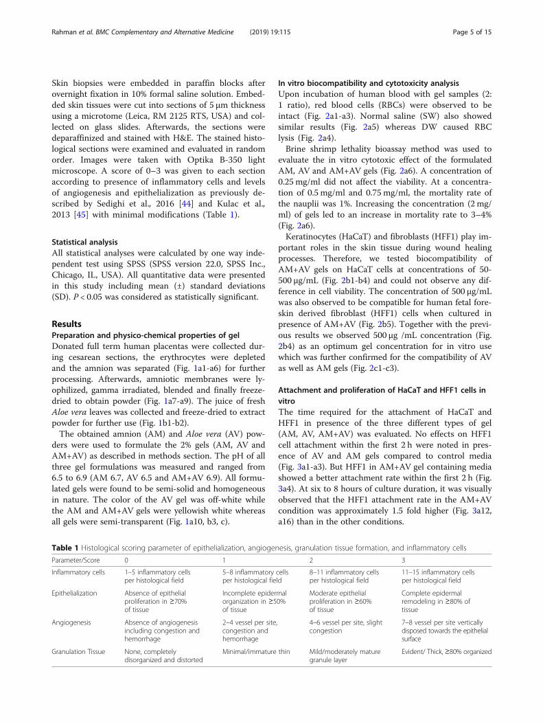
Skin biopsies were embedded in paraffin blocks afterovernight fixation in 10% formal saline solution. Embed-ded skin tissues were cut into sections of 5 μm thicknessusing a microtome (Leica, RM 2125 RTS, USA) and col-lected on glass slides. Afterwards, the sections weredeparaffinized and stained with H&E. The stained histo-logical sections were examined and evaluated in randomorder. Images were taken with Optika B-350 lightmicroscope. A score of 0–3 was given to each sectionaccording to presence of inflammatory cells and levelsof angiogenesis and epithelialization as previously de-scribed by Sedighi et al., 2016 [44] and Kulac et al.,2013 [45] with minimal modifications (Table 1).
Statistical analysisAll statistical analyses were calculated by one way inde-pendent test using SPSS (SPSS version 22.0, SPSS Inc.,Chicago, IL, USA). All quantitative data were presentedin this study including mean (±) standard deviations(SD). P < 0.05 was considered as statistically significant.
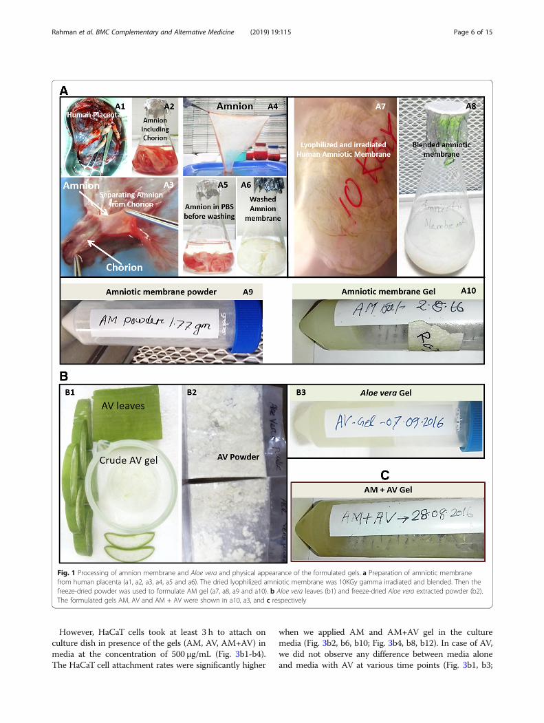
ResultsPreparation and physico-chemical properties of gelDonated full term human placentas were collected dur-ing cesarean sections, the erythrocytes were depletedand the amnion was separated (Fig. 1a1-a6) for furtherprocessing. Afterwards, amniotic membranes were ly-ophilized, gamma irradiated, blended and finally freeze-dried to obtain powder (Fig. 1a7-a9). The juice of freshAloe vera leaves was collected and freeze-dried to extractpowder for further use (Fig. 1b1-b2).The obtained amnion (AM) and Aloe vera (AV) pow-
ders were used to formulate the 2% gels (AM, AV andAM+AV) as described in methods section. The pH of allthree gel formulations was measured and ranged from6.5 to 6.9 (AM 6.7, AV 6.5 and AM+AV 6.9). All formu-lated gels were found to be semi-solid and homogeneousin nature. The color of the AV gel was off-white whilethe AM and AM+AV gels were yellowish white whereasall gels were semi-transparent (Fig. 1a10, b3, c).
In vitro biocompatibility and cytotoxicity analysisUpon incubation of human blood with gel samples (2:1 ratio), red blood cells (RBCs) were observed to beintact (Fig. 2a1-a3). Normal saline (SW) also showedsimilar results (Fig. 2a5) whereas DW caused RBClysis (Fig. 2a4).Brine shrimp lethality bioassay method was used to
evaluate the in vitro cytotoxic effect of the formulatedAM, AV and AM+AV gels (Fig. 2a6). A concentration of0.25 mg/ml did not affect the viability. At a concentra-tion of 0.5 mg/ml and 0.75 mg/ml, the mortality rate ofthe nauplii was 1%. Increasing the concentration (2 mg/ml) of gels led to an increase in mortality rate to 3–4%(Fig. 2a6).Keratinocytes (HaCaT) and fibroblasts (HFF1) play im-
portant roles in the skin tissue during wound healingprocesses. Therefore, we tested biocompatibility ofAM+AV gels on HaCaT cells at concentrations of 50-500 μg/mL (Fig. 2b1-b4) and could not observe any dif-ference in cell viability. The concentration of 500 μg/mLwas also observed to be compatible for human fetal fore-skin derived fibroblast (HFF1) cells when cultured inpresence of AM+AV (Fig. 2b5). Together with the previ-ous results we observed 500 μg /mL concentration (Fig.2b4) as an optimum gel concentration for in vitro usewhich was further confirmed for the compatibility of AVas well as AM gels (Fig. 2c1-c3).
Attachment and proliferation of HaCaT and HFF1 cells invitroThe time required for the attachment of HaCaT andHFF1 in presence of the three different types of gel(AM, AV, AM+AV) was evaluated. No effects on HFF1cell attachment within the first 2 h were noted in pres-ence of AV and AM gels compared to control media(Fig. 3a1-a3). But HFF1 in AM+AV gel containing mediashowed a better attachment rate within the first 2 h (Fig.3a4). At six to 8 hours of culture duration, it was visuallyobserved that the HFF1 attachment rate in the AM+AVcondition was approximately 1.5 fold higher (Fig. 3a12,a16) than in the other conditions.
Table 1 Histological scoring parameter of epithelialization, angiogenesis, granulation tissue formation, and inflammatory cells
Parameter/Score 0 1 2 3
Inflammatory cells 1–5 inflammatory cellsper histological field
5–8 inflammatory cellsper histological field
8–11 inflammatory cellsper histological field
11–15 inflammatory cellsper histological field
Epithelialization Absence of epithelialproliferation in ≥70%of tissue
Incomplete epidermalorganization in ≥50%of tissue
Moderate epithelialproliferation in ≥60%of tissue
Complete epidermalremodeling in ≥80% oftissue
Angiogenesis Absence of angiogenesisincluding congestion andhemorrhage
2–4 vessel per site,congestion andhemorrhage
4–6 vessel per site, slightcongestion
7–8 vessel per site verticallydisposed towards the epithelialsurface
Granulation Tissue None, completelydisorganized and distorted
Minimal/immature thin Mild/moderately maturegranule layer
Evident/ Thick, ≥80% organized
Rahman et al. BMC Complementary and Alternative Medicine (2019) 19:115 Page 5 of 15
However, HaCaT cells took at least 3 h to attach onculture dish in presence of the gels (AM, AV, AM+AV) inmedia at the concentration of 500 μg/mL (Fig. 3b1-b4).The HaCaT cell attachment rates were significantly higher
when we applied AM and AM+AV gel in the culturemedia (Fig. 3b2, b6, b10; Fig. 3b4, b8, b12). In case of AV,we did not observe any difference between media aloneand media with AV at various time points (Fig. 3b1, b3;
Fig. 1 Processing of amnion membrane and Aloe vera and physical appearance of the formulated gels. a Preparation of amniotic membranefrom human placenta (a1, a2, a3, a4, a5 and a6). The dried lyophilized amniotic membrane was 10KGy gamma irradiated and blended. Then thefreeze-dried powder was used to formulate AM gel (a7, a8, a9 and a10). b Aloe vera leaves (b1) and freeze-dried Aloe vera extracted powder (b2).The formulated gels AM, AV and AM + AV were shown in a10, a3, and c respectively
Rahman et al. BMC Complementary and Alternative Medicine (2019) 19:115 Page 6 of 15
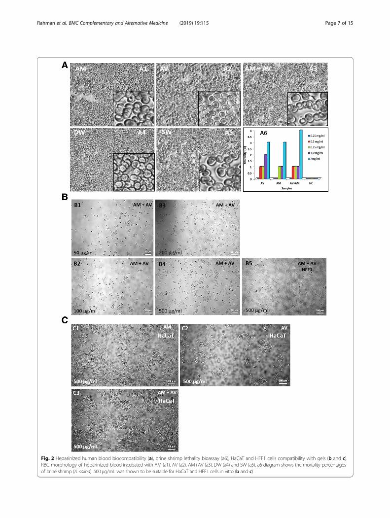
Fig. 2 Heparinized human blood biocompatibility (a), brine shrimp lethality bioassay (a6); HaCaT and HFF1 cells compatibility with gels (b and c).RBC morphology of heparinized blood incubated with AM (a1), AV (a2), AM+AV (a3), DW (a4) and SW (a5). a6 diagram shows the mortality percentagesof brine shrimp (A. salina). 500 μg/mL was shown to be suitable for HaCaT and HFF1 cells in vitro (b and c)
Rahman et al. BMC Complementary and Alternative Medicine (2019) 19:115 Page 7 of 15
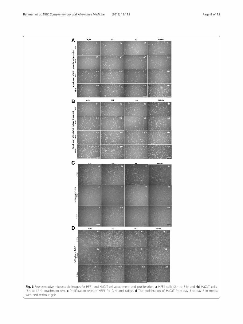
Fig. 3 Representative microscopic images for HFF1 and HaCaT cell attachment and proliferation. a HFF1 cells (2 h to 8 h) and (b) HaCaT cells(3 h to 12 h) attachment test. c Proliferation tests of HFF1 for 2, 4, and 6 days. d The proliferation of HaCaT from day 3 to day 6 in mediawith and without gels
Rahman et al. BMC Complementary and Alternative Medicine (2019) 19:115 Page 8 of 15
Fig. 3b5, b7; Fig. 3b9, b11; Fig. 3b13, b15). Beside the cellattachment study, we also qualitatively examined theproliferation of HFF1 and HaCaT when incubatedwith the gels from day two to day six. We did notobserve any significant effect of the gels on the prolif-eration of HFF1 cells during the examination periods(Fig. 3c1-c12). However, media with AM and AV +AM gels were noticed to increase the proliferationrate of HaCaT cells two fold (Fig. 3d1-d12).
In vitro wound healing and in vivo irritability study of thegelsThe in vitro scratch assay was performed to measure cellmigration in the scratching zone. Consequently, wewanted to detect whether these formulated gels (concen-tration 500 μg/mL) can promote the rate of wound heal-ing in human keratinocytes (HaCaT) and fibroblasts(HFF1) via scratch assays. The cells were serum starvedfor at least 24 h before producing the scratch wound.Thus, the cells’ ability to proliferate was inhibited, and itwas assured that wound closure was only due to cell mi-gration. Our result showed that the healing velocity ofHaCaT and HFF1 with AM and AM+AV gel treatmentwas higher than for untreated cells and AV treated(Fig. 4a, b). HaCaT cells filled the scratch area fasterwhen compared to HFF1 after 30 h.To analyze the applicability and irritability of the pre-
pared gel, skin irritation assay were performed applyinga rat model. After topical application of all gels for aperiod of 7 days, it was observed that the gel did not in-duce any edema or erythema (Fig. 4c). This result indi-cated the safety of gels to be applied topically. We alsoobserved that the hair formation was also normal com-pared to non-treated rats.
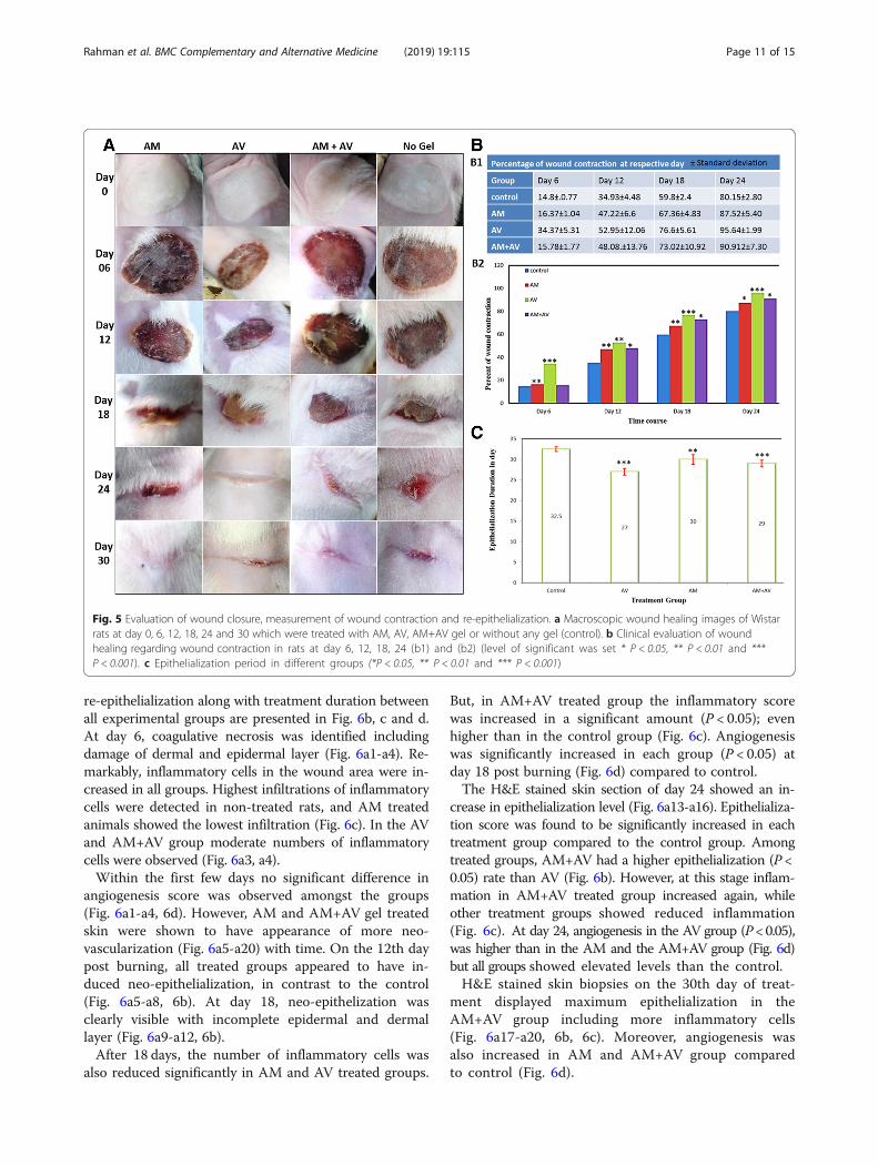
Macroscopic evaluation of wound closure and quantitativemeasurement of wound contraction and re-epithelializationAfter few hours of the second degree burn induction,the rats were restless indicating the pain. No bubbleswere observed on the burn area, however, it was noticedthat white color burn damaged the skin barrier. Subse-quently, hyperemia occurred into the damaged tissuearea. After treating the rats with gels at day one, withinfew hours, white color burn turned into a fullyhyperemic zone in each group which indicated the pres-ence of red blood cells undergoing extravasation (Fig. 5a,day 0 panel). At day six, all groups showed the presenceof thick dry crusts but the group treated with AM+AVgel had a slightly wet crust (Fig. 5a day 6 panel). Edgesof crust were found to be partially detached. At 12 daysafter burn induction, control group showed discrete de-tachment of crust while other treatment groups showedcontinuous detachment of edges. In this stage of injurystaining of crusts were almost same. As the full burn
wound was covered with crust scar tissue was not ob-served until the 12th day (Fig. 5a, day 12 panel). On day18, scar tissue became clearly visible in the edges of thewounds in each group and contractions were clearly vis-ible (Fig. 5a, day 18 panel). At day 24, crusts disappearedfrom all groups and scar tissue became clearly visible.The AV treated group showed faster healing but thisgroup left more scar tissue than AM and control groups(Fig. 5a, day 24 panel). Re-epithelialization had beencompleted in all treatment groups leaving scar tissuesafter 4 weeks (Fig. 5a, day 30 panel). Macroscopically,the AV group showed a better healing rate but includedmore scar tissue while other groups showed less healingrate than the AV group with minimal scar formation.However, a variation in wound contraction rates were
noticed from group to group (Fig. 5b1, b2). At day 6, theAV treated group showed significantly (P < 0.001) betterhealing rate which was two folds higher than in theother groups. The healing rate of the AM treated groupwas also found to be significantly increased (P < 0.01)when compared to AM+AV (P > 0.05). Twelve days postburning, the control group had the lowest healing rate(34.93%) whereas AV treated group wound healing wasabout 50% (P < 0.01). At the same time point, the healingrate of the AM treated group was 47.22% (P < 0.01) andof the AM+AV 48.08% of the wound were found to behealed (P < 0.05) (Fig. 5b1, b2). On the 18th day, AVtreated group reached a healing rate of 76.6% (P < 0.001). It was observed that the wound healing rate of theAM+AV group (73%) was better than in the AM treatedgroup (67%). But, statistical evaluation showed a highersignificance in the AM treated group (P < 0.01) in com-parison with AM+AV (P < 0.05). At day 24, the percentof wound contraction in the AV group also showed bet-ter results compared with the other two groups. Interest-ingly, wounds from the AV group were demonstrated tobe healed about 95% (P < 0.001) whereas the controlgroup showed a healing of 80%. Nevertheless, AMtreated and AM+AV treated wound recovery rate were87 and 90% respectively with P < 0.05 (Fig. 5b1, b2).Average re-epithelialization period in all groups
were observed (Fig. 5c). In AV treated animals meanepithelialization was visualized within 27 days of postburning, control group required at least 32.5 days.The re-epithelialization period of AM and AM+AVtreated rats took 30 and 29 days, respectively. How-ever, both AV and AM+AV groups were statisticallysignificant (P < 0.001) (Fig. 5c).
Histological analysis in terms of skin tissue organization,angiogenesis, and re-epithelializationFigure 6a shows representative photomicrograph ofskin sections stained with H&E from all groups. Thecomparison of angiogenesis, inflammation score and
Rahman et al. BMC Complementary and Alternative Medicine (2019) 19:115 Page 9 of 15
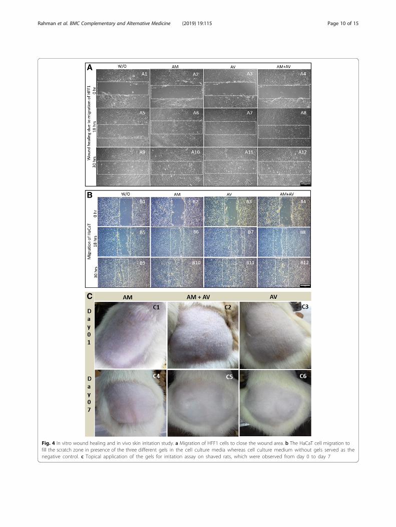
Fig. 4 In vitro wound healing and in vivo skin irritation study. a Migration of HFF1 cells to close the wound area. b The HaCaT cell migration tofill the scratch zone in presence of the three different gels in the cell culture media whereas cell culture medium without gels served as thenegative control. c Topical application of the gels for irritation assay on shaved rats, which were observed from day 0 to day 7
Rahman et al. BMC Complementary and Alternative Medicine (2019) 19:115 Page 10 of 15
re-epithelialization along with treatment duration betweenall experimental groups are presented in Fig. 6b, c and d.At day 6, coagulative necrosis was identified includingdamage of dermal and epidermal layer (Fig. 6a1-a4). Re-markably, inflammatory cells in the wound area were in-creased in all groups. Highest infiltrations of inflammatorycells were detected in non-treated rats, and AM treatedanimals showed the lowest infiltration (Fig. 6c). In the AVand AM+AV group moderate numbers of inflammatorycells were observed (Fig. 6a3, a4).Within the first few days no significant difference in
angiogenesis score was observed amongst the groups(Fig. 6a1-a4, 6d). However, AM and AM+AV gel treatedskin were shown to have appearance of more neo-vascularization (Fig. 6a5-a20) with time. On the 12th daypost burning, all treated groups appeared to have in-duced neo-epithelialization, in contrast to the control(Fig. 6a5-a8, 6b). At day 18, neo-epithelization wasclearly visible with incomplete epidermal and dermallayer (Fig. 6a9-a12, 6b).After 18 days, the number of inflammatory cells was
also reduced significantly in AM and AV treated groups.
But, in AM+AV treated group the inflammatory scorewas increased in a significant amount (P < 0.05); evenhigher than in the control group (Fig. 6c). Angiogenesiswas significantly increased in each group (P < 0.05) atday 18 post burning (Fig. 6d) compared to control.The H&E stained skin section of day 24 showed an in-
crease in epithelialization level (Fig. 6a13-a16). Epithelializa-tion score was found to be significantly increased in eachtreatment group compared to the control group. Amongtreated groups, AM+AV had a higher epithelialization (P <0.05) rate than AV (Fig. 6b). However, at this stage inflam-mation in AM+AV treated group increased again, whileother treatment groups showed reduced inflammation(Fig. 6c). At day 24, angiogenesis in the AV group (P< 0.05),was higher than in the AM and the AM+AV group (Fig. 6d)but all groups showed elevated levels than the control.H&E stained skin biopsies on the 30th day of treat-
ment displayed maximum epithelialization in theAM+AV group including more inflammatory cells(Fig. 6a17-a20, 6b, 6c). Moreover, angiogenesis wasalso increased in AM and AM+AV group comparedto control (Fig. 6d).
Fig. 5 Evaluation of wound closure, measurement of wound contraction and re-epithelialization. a Macroscopic wound healing images of Wistarrats at day 0, 6, 12, 18, 24 and 30 which were treated with AM, AV, AM+AV gel or without any gel (control). b Clinical evaluation of woundhealing regarding wound contraction in rats at day 6, 12, 18, 24 (b1) and (b2) (level of significant was set * P < 0.05, ** P < 0.01 and ***P < 0.001). c Epithelialization period in different groups (*P < 0.05, ** P < 0.01 and *** P < 0.001)
Rahman et al. BMC Complementary and Alternative Medicine (2019) 19:115 Page 11 of 15
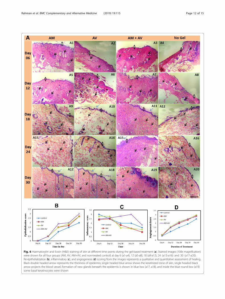
Fig. 6 Haematoxylin and Eosin (H&E) staining of skin at different time points during the gel-based treatment (a). Stained images (100x magnification)were shown for all four groups (AM, AV, AM+AV, and non-treated control) at day 6 (a1-a4), 12 (a5-a8), 18 (a9-a12), 24 (a13-a16) and 30 (a17-a20).Re-epithelialization (b), inflammatory (c), and angiogenesis (d) scoring from the images represents a qualitative and quantitative assessment of healing.Black double headed arrow represents the thickness of epidermis; single headed blue arrow shows the keratinized zone of skin, single headed blackarrow projects the blood vessel, formation of new glands beneath the epidermis is shown in blue box (a17, a18), and inside the blue round box (a19)some basal keratinocytes were shown
Rahman et al. BMC Complementary and Alternative Medicine (2019) 19:115 Page 12 of 15
DiscussionBeside active ingredients, the applicability of burnwound healing gel is dependent on the properties suchas pH, appearance, homogeneity, and viscosity. Carbopolis a widely used gelling agent for producing burn-healinggels for skin application. CMC-Na salt is another agentused alongside with carbopol [46]. However, in thisstudy the gel was prepared from human amniotic mem-brane extract (AM), Aloe vera extracts (AV), and a com-bination of both AM+AV using 6% CMC-Na salt as agelling agent. Extensive studies confirm the effectivenessand safety of lyophilized amnion as a wound dressingand grafting materials for promoting the healing processand preventing infections [47]. Khorasani et al., (2009)reported that AV cream or gel could be more effectivethan silver sulfadiazine cream in treating burn woundhealing [30]. The end products (gels) in all three condi-tions evaluated in this study were homogenous, granuleless and of a whitish creamy color (Fig. 1a10, 1b3, 1c). Inaddition, pH of all formulations ranged between 6.5–6.9,and this pH does not interfere with skin physiology [48].Erythema and edema are the common symptoms of skinirritation which lasts for three to 7 days [49]. Our for-mulated gels did not induce any irritation including noerythema or edema on rat skin upon topical applicationfor a period of 7 days (Fig. 4c). Thus, CMC-Na salt, AMand AV extract formulated gel could be useful as woundhealing gel as the gels have good spreadability andconsistency. In vitro and in vivo studies clearly and col-lectively demonstrated the potentials of the formulatedgels from amnion when combined with Aloe vera. In thispreliminary research, we found that the formulated gelscan induce and accelerate the proliferation and at-tachment of HaCaT and HFF1 cells (Fig. 3), and pro-mote wound healing in vitro (Fig. 4a, b) [50]. Amnionhas been reported to facilitate the migration of epi-thelial cells, reinforces attachment, and promotes pro-liferation [51].Macroscopic morphological analysis demonstrated the
gel-based acceleration of re-epithelialization and woundcontraction in vivo (Fig. 5a, b, c). Microscopic observa-tion of AM+AV gel treated tissue sections allowed us toappreciate the effectiveness of the formulated gel in re-gard to epidermis and dermis formation and the thick-ness of epidermis (Fig. 6a). In present study, weobserved that upon completion of wound healing fewscar tissues remained in all treated rats. Scar formationin all kinds of wound healing are normal [52] and existeven after complete healing. However, the AM treatedexperimental group had the lowest scar formation. Re-gardless of the variation in treatment procedures, seconddegree burn required 25–35 days to heal completely [53].Due to presence of anti-inflammatory characteristics[23, 44], amniotic gel treated rats had lower inflammation
than AV and AM+AV (Fig. 6c). Inflammation is an earlystage event for burn healing which should be decreasedwithin a certain healing period [54]. During wound heal-ing, at early phases inflammatory cells increased but inlater stages decreased gradually due to granulation, theformation of new capillaries, and deposition of collagen[2]. Excluding collagen level determination, other inci-dences are similar to our study [55]. Histological analysisrevealed that the AM+AV group had a higher epitheliali-zation rate apart from inflammation (Fig. 6a19, 6b).Wound healing is related to wound contraction andwound re-epithelialization [56] which has been shown forgel treatment in a mouse model [50].Angiogenesis is another important event in burn heal-
ing where endothelial cells’ proliferation rate in thewound area is rapidly increased after burning to formblood vessels. Hamid and Soliman (2015) reported thatAV can increase angiogenesis [2] for a better supply withnutrients and oxygen because of acemannan in AV [57].Histologically, AM, AV and AM+AV treated woundshad increased numbers of blood vessels, particularlysmall and newly formed (Fig. 6a17-a19, 6d). Besides sup-porting in vitro keratinocyte proliferation, we also ob-served some proliferating basal keratinocytes in vivoresiding in the intermediate zone of the epidermis anddermis (Fig. 6a19). These properties of the tested gelscould be explained by the presence of stimulatory factorsin amniotic membrane which also been supported fromthe results of Murphy et al. (2017) [24].Amniotic membrane has been reported to provide a
niche for the cells to adhere, grow, proliferate, migrateand differentiate, and could possibly contribute to theproduction of angiogenic micro-environment indirectlywhich allows AM to improve burn healing [58]. Wefound that a combination of both AM and AV synergis-tically improved epithelialization. On day 30, epitheliali-zation profile was significantly higher in the AM+AVgroup. Amniotic membrane is composed of collagentype IV, V and VII which promotes growth of epithelialcells, facilitates epithelial cell migration, strengthensbasal epithelial cell adhesion, promotes differentiation ofepithelial cell, and prevents apoptotic cell death [59]. Inprincipal, we have prepared gels from medically dis-carded materials, at low cost that provides excellentburn wound coverage. These formulated gels showedpotential to be used as fast aid ointment in burn woundmanagement. Although we demonstrated significant im-provement of burn wound healing in rats treated withAM+AV gel, however, some limitations are associatedwith this animal model such as the remarkable native re-generation potential of the rat skin. For future applica-tion, it would be crucial to identify the key factors in theamnion that are responsible for the acceleration of thewound healing process.
Rahman et al. BMC Complementary and Alternative Medicine (2019) 19:115 Page 13 of 15
ConclusionTaken together the in vitro and in vivo data, our findingsclearly demonstrate that amniotic membrane combinedwith Aloe vera extract significantly enhances burnwound healing, thus indicating that the amniotic mem-brane and Aloe vera possesses potent wound healingactivities. Amniotic membrane is a globally accepted bio-logical biomaterial for second and third degree burn.Aloe vera gel has been reported to have burn healingcapacity as well. The combination of both AM and AVhas been shown to have promising effect in internal epi-thelialization with less scar formation. Gels containingAM extract individually and in combination with AVcould be used alternatively in the treatment of burn.However, further investigation is required to assess opti-mal concentration of the used extracts and key factorspresent in AM and AV to find out the best combinationfor burn healing. Moreover, it is also of great importanceto unfold the underlying molecular mechanisms. As afurther investigation step, cell based therapies using skinprogenitor cells in combination with the tested gels,could be another way to accelerate wound healing.
AbbreviationsAM: Amniotic membrane; AV: Aloe vera; CMC-Na: Sodium carboxymethylcellulose; C-sections: Caesarean sections; H&E: Hematoxylin and eosin;HaCaT: (Ha = human adult, Ca = calcium, T = temperature); HCl: Hydrogenchloride; HFF1: Human foreskin fibroblast 1; HLA: Human leukocyte antigens;HLA-DR: Human leukocyte antigen – antigen D related; KGy: Kilo gray;PBS: Phosphate buffer saline; SD: Standard deviations
AcknowledgementsThe authors acknowledge the technical support from Institute of TissueBanking and Biomaterial Research (ITBBR), Atomic Energy ResearchEstablishment (AERE) and Animal facilities from Dept. of Pharmacy,Jahangirnagar University, Bangladesh. Dr. SM Asaduzzaman acknowledgesthe support from Govt. of Bangladesh, International Atomic EnergyAgency (IAEA) and the technical supports from Prof. Dr. Shahadath andDr. Matin throughout the work. Prof. Dr. Adjaye acknowledges the support fromthe Medical Faculty, Heinrich-Heine-University, Düsseldorf, Germany. In addition,Prof. Dr. Adjaye and Md Shaifur Rahman acknowledge support from theGerman Academic Exchange Service (DAAD-91607303).
Authors’ contributionsMSR1 and MSR2 conceived the idea. MSR1, RI, and MSR2 designed theexperiment. MSR1, MMR, LSS and RI performed the experimental workand analysis the data. MSR1 and RI wrote the initial draft and SMA,MSR2, LSS, JA edited the manuscript. SMA and MSR2 supervised thework from start to end. JA supervised the in vitro study. All authors readand approved the final manuscript.
FundingFor the purchasing of lab instruments and reagents, funds had beenprovided by ADP Govt. of Bangladesh and International Atomic EnergyAgency. RI was supported from the NSICT Fellowship, MoST, Govt. ofBangladesh. MSR1 was supported by DAAD-91607303, Germany. No fund-ing has been provided for publication charge. Funding agency did nothave any contribution on experimental idea, design, work, manuscriptwriting, and publication.
Availability of data and materialsThe data and materials have been presented in the main manuscript andcan be given upon request.
Ethics approval and consent to participateThe study was conducted according to the protocol approved by the ethicalcommittee of the Atomic Energy Research Establishment and JahangirnagarUniversity, Bangladesh. The research work on/with human organ, tissue, andcell has been permitted in Institute of Tissue Banking and BiomaterialResearch under the Human Organ / Tissue Donation and Transplantation Act,1999, Govt. of Bangladesh. Post delivered amniotic membrane was collected forresearch purpose with the written consent from the healthy donor.
Consent for publicationNot Applicable.
Competing interestsThe authors declare that they have no competing interest.
Author details1Institute for Stem Cell Research and Regenerative Medicine, Medical Faculty,Heinrich Heine University, 40225 Düsseldorf, Germany. 2Bio-resourceTechnology and Industrial Biotechnology Laboratory, Department ofBiotechnology and Genetic Engineering, Jahangirnagar University, Dhaka1342, Bangladesh. 3Institute of Tissue Banking and Biomaterial Research,Atomic Energy Research Establishment, Dhaka 1349, Bangladesh.
Received: 1 September 2017 Accepted: 17 May 2019
References1. Miller SF, Bessey P, Lentz CW, Jeng JC, Schurr M, Browning S. ABA NBR
committee. National burn repository 2007 report: a synopsis of the 2007 callfor data. J Burn Care Res. 2008;29:862–70.
2. Hamid AAA, Soliman MF. Effect of topical Aloe vera on the process ofhealing of full-thickness skin burn: a histological and immunohistochemicalstudy. J of Histology and Histopathology. 2015;2:3.
3. He S, Alonge O, Agrawal P, et al. Epidemiology of burns in rural Bangladesh:an update. tchounwou PB edt. Int. J. Environ. Res. Public Health. 2017;14:381.
4. Mashreky SR, Rahman A, Chowdhury SM, Giashuddin S, SvanstrOm L, LinnanM, Shafinaz S, Uhaa IJ, Rahman F. Epidemiology of childhood burn: yield oflargest community based injury survey in Bangladesh. Burns. 2008;34:856–62.
5. Atiyeh B, Masellis A, Conte C. Optimizing burn treatment in developing low-and middle-income countries with limited health care resources (part 1).Ann Burns Fire Disasters. 2009;22:121–5.
6. Tehrani S, Lotfi P, Tehrani S, et al. Healing effect of sesame ointment onsecond-degree burn wound in rats. Galen Medical J. 2016;5:56–62.
7. Gurtner GC, Werner S, Barrandon Y, Longaker MT. Wound repair andregeneration. Nature. 2008;453:314–21.
8. Tiwari VK. Burn wound: how it differs from other wounds? Indian J of PlasticSurgery. 2012;45:364.
9. Werner S, Krieg T, Smola H. Keratinocyte–fibroblast interactions in woundhealing. J of Invest Dermatol. 2007;127(5):998–1008.
10. Pastar I, Stojadinovic O, Yin NC, Ramirez H, Nusbaum AG, Sawaya A, Tomic-Canic M. Epithelialization in wound healing: a comprehensive review. Advin Wound Care. 2014;3:445–64.
11. Guo S, Dipietro LA. Factors affecting wound healing. J Dent Res. 2010;89:219–29.
12. Xue M, Jackson CJ. Extracellular matrix reorganization during wound healingand its impact on abnormal scarring. Adv Wound Care (New Rochelle).2015;4:119–36.
13. Ter Horst B, Chouhan G, Moiemen NS, Grover LM. Advances in keratinocytedelivery in burn wound care. Adv Drug Deliv Rev. 2017;S0169-409X(17):30096–0.
14. Lootens L, Brusselaers N, Beele H, Monstrey S. Keratinocytes in thetreatment of severe burn injury: an update. Int Wound J. 2013;10:6–12.
15. Yang JD, Choi DS, Cho YK, Kim TK, Lee JW, Choi KY, Chung HY, ChoBC, Byun JS. Effect of amniotic fluid stem cells and amniotic fluid cellson the wound healing process in a white rat model. Arch Plast Surg.2013;40:496–504.
16. Skardal A, Mack D, Kapetanovic E, et al. Bioprinted amniotic fluid-derivedstem cells accelerate healing of large skin wounds. Stem Cells Transl Med.2012;1:792–802.
Rahman et al. BMC Complementary and Alternative Medicine (2019) 19:115 Page 14 of 15
17. Davis JW. Skin transplantation with a review of 550 cases at the JohnsHopkins Hospital. Johns Hopkins Med J. 1910;15:307.
18. Bujang-Safawi E, Halim AS, et al. Dried irradiated human amnioticmembrane as a biological dressing for facial burns-a 7-year case series.Burns. 2010;36:876–82.
19. Tehrani FA, Ahmadiani A, et al. The effects of preservation procedures onantibacterial property of amniotic membrane. Cryobiology. 2013;67:293–8.
20. Riau AK, Beuerman RW, Lim LS, et al. Preservation, sterilization and de-epithelialization of human amniotic membrane for use in ocular surfacereconstruction. Biomaterials. 2010;31:216–25.
21. Higa K, Shimmura S, et al. Hyaluronic acid-CD44 interaction mediates theadhesion of lymphocytes by amniotic membrane stroma. Cornea. 2005;24:206–12.
22. Shimazaki J, Shinozaki N, Tsubota K. Transplantation of amniotic membraneand limbal autograft for patients with recurrent pterygium associated withsymblepharon. Br J Ophthalmology. 1998;82:235–40.
23. Solomon A, Rosenblatt M, Monroy D. Suppression of interleukin 1 alpha andinterleukin 1 beta in the human limbal epithelial cells cultured on theamniotic membrane stromal matrix. Br J Ophthalmol. 2001;85:444–9.
24. Murphy SV, Skardal A, Song L, et al. Solubilized amnion membranehyaluronic acid hydrogel accelerates full-thickness wound healing. StemCells Transl Med. 2017;6:2020–32.
25. Biswas TK, Mukherjee B. Plant medicines of Indian origin for wound healingactivity: a review. The Int J of Lower Extremity Wounds. 2003;2:25–39.
26. Khan AW, Kotta S, Ansari SH, et al. Formulation development, optimization andevaluation of Aloe vera gel for wound healing. Pharmacogn Mag. 2013;9:S6–S10.
27. Ajmera N, Chatterjee A, Goyal V. Aloe vera: It's effect on gingivitis. J of IndSociety of Periodontology. 2013;17:435–8.
28. Chithra R, Sajithlal GB, Chandrakasan G. Influence of Aloe vera on collagencharacteristics in healing dermal wounds in rats. Mol Cell Biochem. 1998;181:71–6.
29. Heggers JP, Kucukcelebi A, Listengarten D, Stabenau J, Ko F, Broemeling LD,Robson MC, Winters WD. Beneficial effect of aloe on wound healing in anexcisional wound model. J Altern Complement Med. 1996;2:271–7.
30. Khorasani G, Hosseinimehr SJ, Azadbakht M, Zamani A, Mahdavi MR. Aloeversus silver sulfadiazine creams for second-degree burns: a randomizedcontrolled study. Surg Today. 2009;39(7):587–91.
31. Akhoondinasab MR, Akhoondinasab M, Saberi M. Comparison of healingeffect of Aloe vera extract and silver sulfadiazine in burn injuries in experimentalrat model. World J of Plastic Surgery. 2014;3:29–34.
32. Hirsch T, Rothoeft T, Teig N, Bauer JW, Pellegrini G, De Rosa L, Scaglione D,Reichelt J, et al. Regeneration of the entire human epidermis using transgenicstem cells. Nature. 2017;551:327–32.
33. Pereira RF, Bártolo PJ. Traditional therapies for skin wound healing. Adv inWound Care. 2016;5:208–29.
34. Lesher AP, Curry RH, Evans J, Smith VA, Fitzgerald MT, Cina RA, Streck CJ,Hebra AV. Effectiveness of biobrane for treatment of partial-thickness burnsin children. J Pediatr Surg. 2011;46:1759–63.
35. Rahmanian-Schwarz A, Beiderwieden A, Willkomm L-M, et al. A clinicalevaluation of biobrane® and suprathel® in acute burns and reconstructivesurgery. Burns. 2011;37:1343–8.
36. Mohammadi AA, Seyed Jafari SM, Kiasat M, Tavakkolian AR, Imani MT, AyazM, Tolide-ie HR. Effect of fresh human amniotic membrane dressing ongraft take in patients with chronic burn wounds compared with conventionalmethods. Burns. 2013;39:349–53.
37. Branski LK, Herndon DN, Celis MM, Norbury WB, Masters OE, Jeschke MG.Amnion in the treatment of pediatric partial-thickness facial burns. Burns.2008;34:393–9.
38. Akhtar N, Rahman MS, Jamil HM, Arifuzzaman M, Miah MM, AsaduzzamanSM. Tissue banking in Bangladesh: 12 years of experience (2003-2014). CellTissue Bank. 2016;17:189–97.
39. Khan MN, Islam JM, Khan MA. Fabrication and characterization of gelatin-based biocompatible porous composite scaffold for bone tissueengineering. J Biomed Mater Res A. 2012;100:3020–8.
40. D'Agostino A, Stellavato A, Busico T, Papa A, Tirino V, Papaccio G, La GattaA, De Rosa M, Schiraldi C. In vitro analysis of the effects on wound healingof high- and low-molecular weight chains of hyaluronan and their hybridH-HA/L-HA complexes. BMC Cell Biol. 2015;16:19.
41. Choi J, Kim H, Choi J, Oh SM, Park J, Park K. Skin corrosion and irritation testof sunscreen nanoparticles using reconstructed 3D human skin model.Environmental Health and Toxicology. 2014;29:e2014004.
42. Guo H-F, Ali RM, Hamid RA, Zaini AA, Khaza’ai H. A new model for studyingdeep partial-thickness burns in rats. Int J Burns Trauma. 2017;7:107–14.
43. Babu B, Ravi M, Kumar BN, Sudheendra VH, Ravishankar B. Burn woundhealing potential of plain ghrita and Sahasradhauta ghrita on wistar albinorats. Indian J Tradit Knowl. 2015;14:273–8.
44. Sedighi A, Mehrabani D, Shirazi R. Histopathological evaluation of thehealing effects of human amniotic membrane transplantation in third-degree burn wound injuries. Comp Clin Pathol. 2016;25:381–5.
45. Kulac M, Aktas C, Tulubas F, et al. The effects of topical treatment withcurcumin on burn wound healing in rats. J of Mol Histology. 2013;44:83–90.
46. Amit S, Saraswati B, Kamalesh U, Kumud U. Formulation and evaluation of anovel herbal gel of Equisetum arvense extract. J of Pharmacognosy andPhytochemistry. 2013;1:80–6.
47. John T. Human amniotic membrane transplantation. Ophthalmol Clin. 2003;16:43–65.
48. Ramane SB, Syed VN, Biyani KR. Evaluation of wound healing activity ofpolyherbal gel–a novel herbal formulation. Int J of Res in Pharmaceuticaland Biomedical Sciences. 2013;4:788–94.
49. Das T, Debnath J, Nath B, Dash S. Formulation and evaluation of an herbalcream for wound healing activity. Int J Pharm Pharm Sci. 2014;6:693–7.
50. Lien LT, Tho NT, Ha DM, Hang PL, Nghia PT, Thang ND. Influence ofphytochemicals in piper betle Linn leaf extract on wound healing. Burnsand Trauma. 2015;3:1–8.
51. Gupta A, Keshri GK, Yadav A, et al. Superpulsed (Ga-as, 904 nm) low-levellaser therapy (LLLT) attenuates inflammatory response and enhanceshealing of burn wounds. J Biophotonics. 2014;8:489–501.
52. Pereira Ddos S, Lima-Ribeiro MH, Santos-Oliveira R, Cavalcanti Cde L, dePontes-Filho NT, Coelho LC, Carneiro-Leão AM, Correia MT. Topicalapplication effect of the isolectin hydrogel (Cramoll 1,4) on second-degree burns: experimental model. J Biomed Biotechnol. 2012;2012:184538.
53. Ramli NA, Wong TW. Sodium carboxymethylcellulose scaffolds and theirphysicochemical effects on partial thickness wound healing. Int J Pharm.2011;403:73–82.
54. Broughton G, Jeffrey EJ, Attinger CE. The basic science of wound healing.Plast Reconstr Surg. 2006;117(7):12S–34S.
55. Busuioc CJ, Popescu FC, Mogoşanu GD, et al. Angiogenesis assessment inexperimental third degree skin burns: a histological and immunohistochemicalstudy. Romanian J of Morphology and Embryology. 2010;52:887–95.
56. Zielins ER, Atashroo DA, Maan ZN, Duscher D, Walmsley GG, Hu M, et al.Wound healing: an update. Regen Med. 2014;9:817–30.
57. Yagi A, Egusa T, Arase M, Tanabe M, Tsuji H. Isolation and characterizationof the glycoprotein fraction with a proliferation-promoting activity onhuman and hamster cells in vitro from Aloe vera gel. Planta Med. 1997;63:18–21.
58. Hashim SNM, Yusof MFH, Zahari W, Noordin KBAA, Kannan TP, Hamid SSA,Mokhtar KI, Ahmad A. Angiogenic potential of extracellular matrix of humanamniotic membrane. Tissue Eng Regen Med. 2016;13:211–7.
59. Malhotra C, Jain AK. Human amniotic membrane transplantation: differentmodalities of its use in ophthalmology. World J Transplant. 2014;4:111–21.
Publisher’s NoteSpringer Nature remains neutral with regard to jurisdictional claims inpublished maps and institutional affiliations.
Rahman et al. BMC Complementary and Alternative Medicine (2019) 19:115 Page 15 of 15