ciprofloxacin-collagen conjugate in the wound healing treatment
TRANSCRIPT
J. Funct. Biomater. 2012, 3, 361-371; doi:10.3390/jfb3020361
Journal of
Functional Biomaterials
ISSN 2079-4983 www.mdpi.com/journal/jfb/
Article
Ciprofloxacin-Collagen Conjugate in the Wound Healing Treatment
Francesco Puoci 1,*, Cristiana Piangiolino 2, Francesco Givigliano 3, Ortensia Ilaria Parisi 1,
Roberta Cassano 1, Sonia Trombino 1, Manuela Curcio 1, Francesca Iemma 1, Giuseppe Cirillo 1,
Umile Gianfranco Spizzirri 1, Donatella Restuccia 1, Rita Muzzalupo 1 and Nevio Picci 1
1 Department of Pharmaceutical Sciences, University of Calabria, Edificio Polifunzionale,
Arcavacata di Rende (CS) 87036, Italy; E-Mails: [email protected] (O.I.P.);
[email protected] (R.C.); [email protected] (S.T.); [email protected] (M.C.);
[email protected] (F.I.); [email protected] (G.C.); [email protected](U.G.S.);
[email protected] (D.R.); [email protected] (R.M.); [email protected] (N.P.); 2 Principium Europe, Via Como 45, Solaro (MI) 20020, Italy;
E-Mail: [email protected] 3 Department of Thoracic Surgery, Policlinico Universitario Germaneto-Fondazione Tommaso
Campanella, Campus Universitario “Salvatore Venuta” Viale Europa, Località Germaneto 88100,
Italy; E-Mail: Catanzaro [email protected]
* Author to whom correspondence should be addressed; E-Mail: [email protected];
Tel.: +39-0984-493151; Fax: +39-0984-493298.
Received: 22 March 2012; in revised form: 7 May 2012 / Accepted: 7 May 2012 /
Published: 15 May 2012
Abstract: The synthesis of a novel functional biomaterial for wound healing treatment was
carried out by adopting a free-radical grafting procedure in aqueous media. With this aim,
ciprofloxacin (CFX) was covalently incorporated into collagen (T1C) chains employing an
ascorbic acid/hydrogen peroxide redox pair as biocompatible initiator system. The covalent
insertion of CFX in the polymeric chains was confirmed by FT-IR and UV analyses, while
an antibacterial assay demonstrated the activity of the synthesized conjugate against
Staphylococcus aureus and Escherichia coli, microorganisms that commonly infect
wounds. A catechin blended conjugate was also tested in order to evaluate the ability to
influence fibroblast cell growth. The observed antibacterial activity and stimulation of
fibroblast growth support the applicability of CFX-T1C conjugate in wound treatment
encouraging the healing process.
OPEN ACCESS
J. Funct. Biomater. 2012, 3
362
Keywords: protein; ciprofloxacin; quinolones; biomedical application; antibiotic;
antimicrobial; infection; wound healing; fibroblast proliferation
1. Introduction
Collagen is the most abundant protein in the human body [1,2], accounting for 30% of the total
proteins. In normal tissues, collagen provides tensile strength and structural integrity. The structural
complexity involves numerous types of collagen associated with tissue-specific expression,
organization into heteropolymeric or homopolymeric fibrils of a uniform size, unique post-translational
processing and interaction with proteoglycans, adhesion glycoproteins, cells, and other collagens.
Collagen provides structural integrity for connective tissues, such as dermis, bone, cartilage, tendon,
ligament, and internal organs. Due to the wide distribution throughout various tissues, collagen
represents one of the most abundant naturally occurring proteins on earth and also one of the most
important macromolecules for different biomedical applications: drug delivery as drug carrier, tissue
engineering as polymeric scaffold, natural biodegradable sutures and wound dressing [3,4]. In the
literature, several studies report on the application of collagen as a device for the release of antibiotic
drugs [5], but the possibility to couple this protein with intrinsic antibacterial activity represents an
important challenge. In this study, we report the development of an ecofriendly strategy to achieve a
fluoroquinolone/collagen conjugate in which the drug is covalently bound to the protein.
Modern surgical practices have markedly reduced the rate of wound infections. However, no
reduction in infection rates has occurred during the late 20th century, despite further developments in
surgical techniques and antimicrobial prophylaxis. Surgical patients tend to be sicker and to undergo
more complex operations. Surgical site infections (SSIs) have enormous clinical and financial
implications. Higher infection rates translate into higher morbidity and mortality, as well as higher
costs to the hospital, patient, and society as a whole.
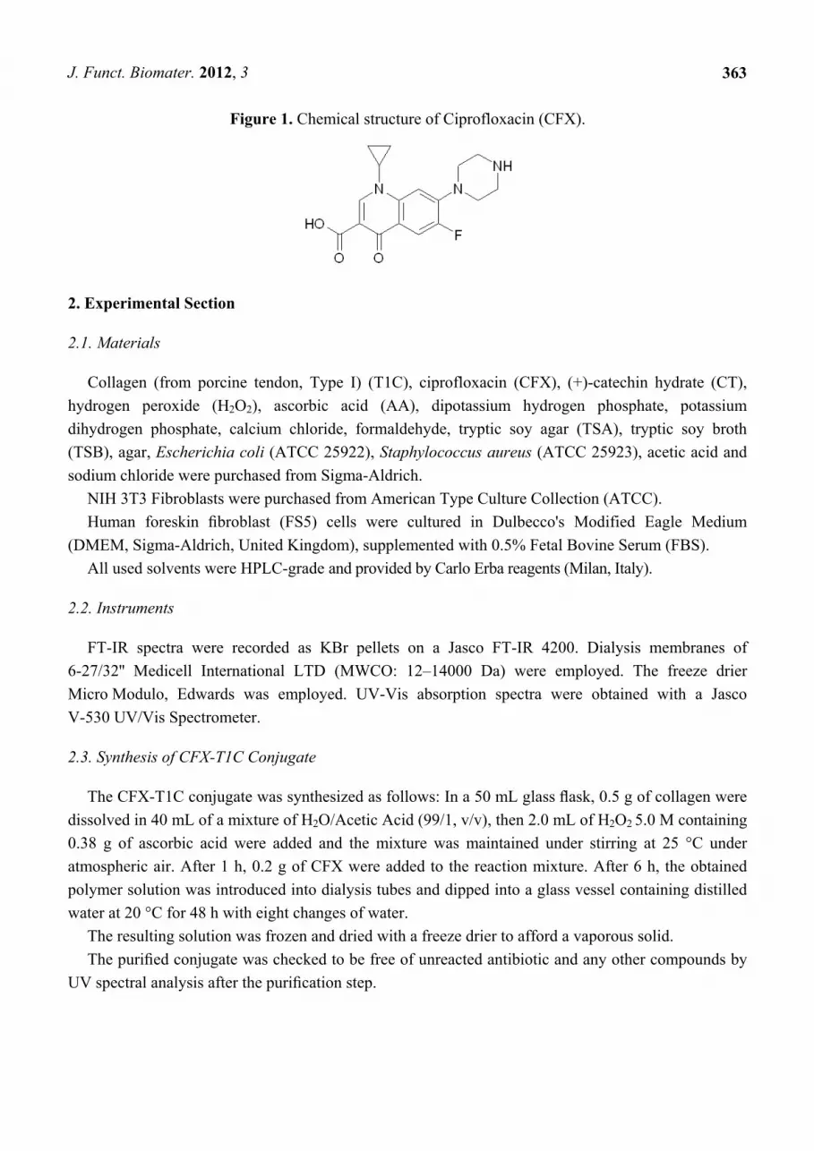
In this work the synthesis and of a Ciprofloxacin-Collagen conjugate (CFX-T1C) for wound healing
applications was performed. Ciprofloxacin (CFX, Figure 1) is a synthetic broad-spectrum antibiotic
belonging to the fluoroquinolone class, and it is effective against many gram-positive and
gram-negative bacteria, including some strains resistant to other agents such as penicillins.
Although CFX is commonly administrated orally, several adverse effects have been recorded in
treated patients [6]. CFX-T1C could be an alternative medical approach in wound healing, such as in
diabetic foot. The local administration, indeed, achieves very high concentrations of the therapeutic
agent at the site of action while the serum concentrations remain well below toxic levels.
The in vitro ability of CFX-T1C conjugate was also evaluated in the presence of a natural
compound, such as catechin (CT), in order to stimulate fibroblast growth.
Specifically, we answered to the following questions: Is it possible to prepare an antibiotic-collagen
conjugate (CFX-T1C) in an ecofriendly and easy way? Does CFX-T1C show antibacterial activities?
Can a CFX-T1C device (combining with catechin) successfully increase fibroblast proliferation?
J. Funct. Biomater. 2012, 3
363
Figure 1. Chemical structure of Ciprofloxacin (CFX).
2. Experimental Section
2.1. Materials
Collagen (from porcine tendon, Type I) (T1C), ciprofloxacin (CFX), (+)-catechin hydrate (CT),
hydrogen peroxide (H2O2), ascorbic acid (AA), dipotassium hydrogen phosphate, potassium
dihydrogen phosphate, calcium chloride, formaldehyde, tryptic soy agar (TSA), tryptic soy broth
(TSB), agar, Escherichia coli (ATCC 25922), Staphylococcus aureus (ATCC 25923), acetic acid and
sodium chloride were purchased from Sigma-Aldrich.
NIH 3T3 Fibroblasts were purchased from American Type Culture Collection (ATCC).
Human foreskin fibroblast (FS5) cells were cultured in Dulbecco's Modified Eagle Medium
(DMEM, Sigma-Aldrich, United Kingdom), supplemented with 0.5% Fetal Bovine Serum (FBS).
All used solvents were HPLC-grade and provided by Carlo Erba reagents (Milan, Italy).
2.2. Instruments
FT-IR spectra were recorded as KBr pellets on a Jasco FT-IR 4200. Dialysis membranes of
6-27/32'' Medicell International LTD (MWCO: 12–14000 Da) were employed. The freeze drier
Micro Modulo, Edwards was employed. UV-Vis absorption spectra were obtained with a Jasco
V-530 UV/Vis Spectrometer.
2.3. Synthesis of CFX-T1C Conjugate
The CFX-T1C conjugate was synthesized as follows: In a 50 mL glass flask, 0.5 g of collagen were
dissolved in 40 mL of a mixture of H2O/Acetic Acid (99/1, v/v), then 2.0 mL of H2O2 5.0 M containing
0.38 g of ascorbic acid were added and the mixture was maintained under stirring at 25 °C under
atmospheric air. After 1 h, 0.2 g of CFX were added to the reaction mixture. After 6 h, the obtained
polymer solution was introduced into dialysis tubes and dipped into a glass vessel containing distilled
water at 20 °C for 48 h with eight changes of water.
The resulting solution was frozen and dried with a freeze drier to afford a vaporous solid.
The purified conjugate was checked to be free of unreacted antibiotic and any other compounds by
UV spectral analysis after the purification step.
J. Funct. Biomater. 2012, 3
364
2.4. Antibacterial Assay
S. aureus and E. coli were streaked out on TSA plates and incubated at 37 °C for 24 h [7]. A
representative colony was lifted off with a wire loop and placed in 5 mL of TSB, which was then
incubated with shaking at 37 °C for 24 h. At this stage, the cultures of S. aureus and E. coli contained
approximately 109 CFU/mL. Cultures of S. aureus and E. coli containing 107 CFU/mL were prepared
by dilution with TSB and were used for antibacterial tests.
The antibacterial activities of CFX-T1C were determined through the testing of different
concentrations of the polymeric conjugate against S. aureus and E. coli. A range of concentrations
(from 1,000 to 0.5 µg/mL) was prepared with sterile, double-deionized water (autoclaved) in 96-well
microtiter plates. The test organisms (5 × 10⁵ CFU, 50 µL of TSB) were added to each well. In the end,
each well contained 350 μL of water, 50 μL of TSB, and the test organism. The microtiter plates were
incubated at 37 °C for 24 h in a shaker. At the end of this period, a small amount of the mixture from
each well was pulled out and spread on agar plates with a swap, and the plates were incubated at 37 °C.
The growth of bacterial cells was observed on agar plates after 48 h incubation and the minimum
inhibitory concentration (MIC), defined as the lowest concentration to inhibit visible growth,
was determined.
The antibacterial test was repeated at least four times employing water/TSB mixture (350 μL/50 μL)
and water/TSB mixture (350 μL/50 μL) inoculated with each test bacterium in the microtiter plates as
negative and positive controls, respectively. The growth of the test bacterium was observed in all wells
with positive controls; on the contrary no growth was observed in the wells with the negative controls.
2.5. Cell Proliferation and Viability Assay
Human foreskin fibroblast (FS5) cells were seeded at a density of 1 × 10³ cells per well in 96-well
plates maintained at 37 °C in a humidified incubator of 5% CO2, 95% air atmosphere [8]. The medium
was replaced after 48 h with 100 µL of Dulbecco’s Modified Eagle Medium (DMEM) containing 0.5%
Fetal Bovine Serum (FBS). The CFX-T1C conjugate, doped with different concentrations of catechin,
was initially dissolved in 1 mL of dimethyl sulfoxide (DMSO), filtered to give the sterile stock solution
and further diluted to give a final concentration of 10 mg/mL of CFX-T1C in the wells. The CT
concentration covers the range between 5–500 ng/mL. The final volume of the medium was 200 µL
per well. Two well plate columns were maintained on DMEM/0.5% FBS and DMEM/10% FBS as
maintenance (negative) and positive growth stimulation controls, respectively. The cells were
incubated for 48 h and cell growth determined using a neutral red uptake assay. After incubation the
cells were washed with phosphate buffered saline (PBS), 100 µL freshly prepared neutral red solution
was added to each well and the cells were incubated at 37 °C for 4 h. The neutral red was then
removed by washing with 1% HCHO/1% CaCl2 and neutral red in the lysosomes was eluted with
100 µL of 1% acetic acid/50% ethanol over 30 min in an orbital shaker and the optical density
measured at 540 nm using a 96-well plate reader.
Three independent experiments were conducted and the results obtained are expressed as
mean ± standard error of the mean of the absorbance.
J. Funct. Biomater. 2012, 3
365
The data from the experiments were compared with the control (0.5% FBS) by one-way analysis of
variance and Dunnet’s test. Differences at p < 0.05 were considered to be significant.
3. Results and Discussion
3.1. Synthesis and Characterization of CFX-T1C Conjugate
The covalent insertion of ciprofloxacin in the collagen chains was performed by a free
radical-induced grafting reaction. To this aim, a biocompatible and water-soluble system, ascorbic
acid/hydrogen peroxide redox pair, was chosen as initiator system.
The interaction mechanism between the two components of the redox pair involves the oxidation of
ascorbic acid by H2O2 at room temperature with the formation of ascorbate and hydroxyl radical
intermediates, which initiate the reaction [9–12].
Compared to conventional initiator systems (i.e., condense agents, esterification etc.) which require
relatively high reaction temperatures to ensure rapid esterification, the grafting procedure shows
several advantages. First of all, this kind of system does not generate toxic reaction products and
allows the performance of the grafting process in aqueous media without any organic solvent;
moreover, it is possible to perform the reaction at lower temperatures, reducing the risks of
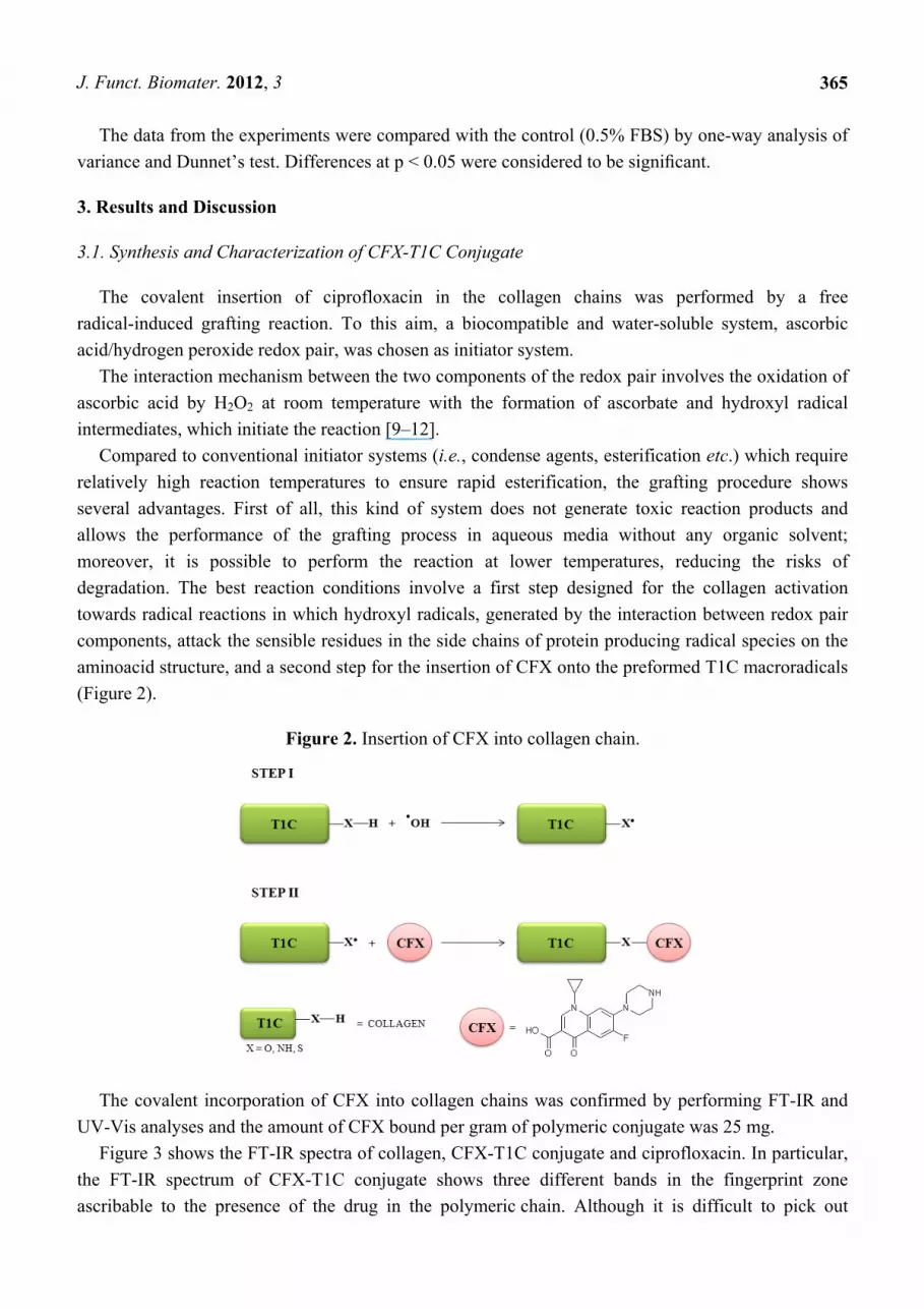
degradation. The best reaction conditions involve a first step designed for the collagen activation
towards radical reactions in which hydroxyl radicals, generated by the interaction between redox pair
components, attack the sensible residues in the side chains of protein producing radical species on the
aminoacid structure, and a second step for the insertion of CFX onto the preformed T1C macroradicals
(Figure 2).
Figure 2. Insertion of CFX into collagen chain.
The covalent incorporation of CFX into collagen chains was confirmed by performing FT-IR and
UV-Vis analyses and the amount of CFX bound per gram of polymeric conjugate was 25 mg.
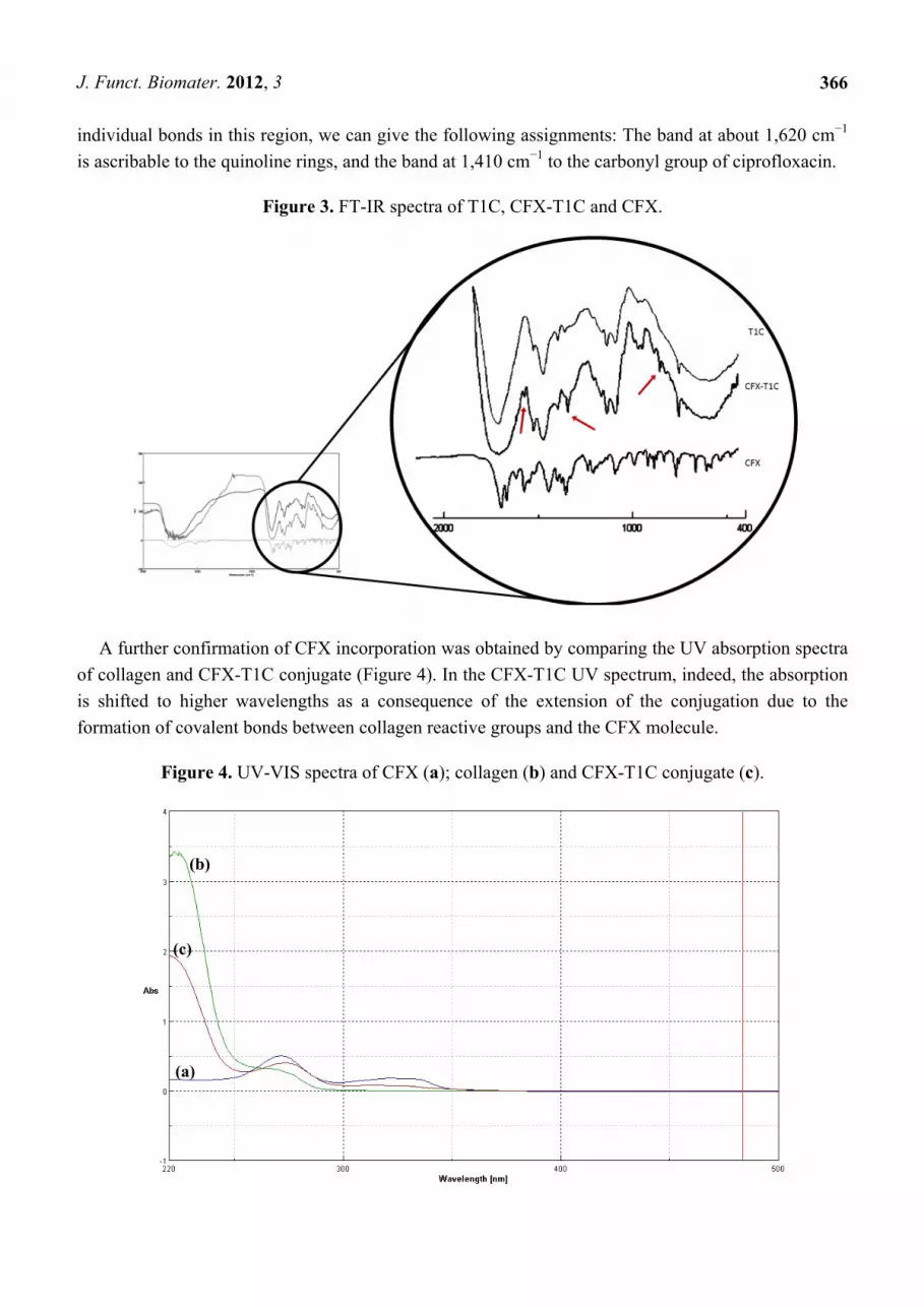
Figure 3 shows the FT-IR spectra of collagen, CFX-T1C conjugate and ciprofloxacin. In particular,
the FT-IR spectrum of CFX-T1C conjugate shows three different bands in the fingerprint zone
ascribable to the presence of the drug in the polymeric chain. Although it is difficult to pick out
J. Funct. Biomater. 2012, 3
366
individual bonds in this region, we can give the following assignments: The band at about 1,620 cm−1
is ascribable to the quinoline rings, and the band at 1,410 cm−1 to the carbonyl group of ciprofloxacin.
Figure 3. FT-IR spectra of T1C, CFX-T1C and CFX.
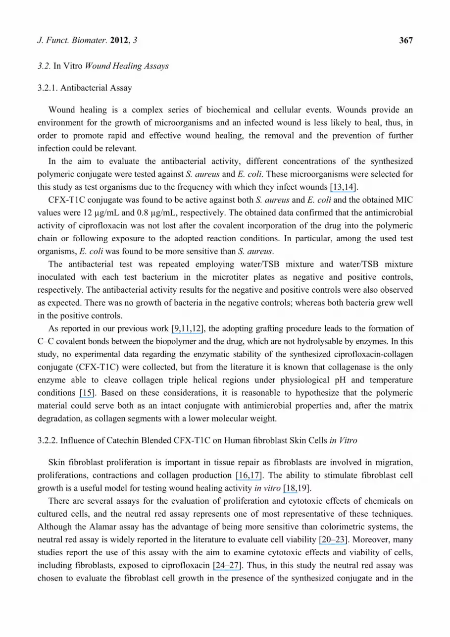
A further confirmation of CFX incorporation was obtained by comparing the UV absorption spectra
of collagen and CFX-T1C conjugate (Figure 4). In the CFX-T1C UV spectrum, indeed, the absorption
is shifted to higher wavelengths as a consequence of the extension of the conjugation due to the
formation of covalent bonds between collagen reactive groups and the CFX molecule.
Figure 4. UV-VIS spectra of CFX (a); collagen (b) and CFX-T1C conjugate (c).
(a)
(b)
(c)
J. Funct. Biomater. 2012, 3
367
3.2. In Vitro Wound Healing Assays
3.2.1. Antibacterial Assay
Wound healing is a complex series of biochemical and cellular events. Wounds provide an
environment for the growth of microorganisms and an infected wound is less likely to heal, thus, in
order to promote rapid and effective wound healing, the removal and the prevention of further
infection could be relevant.
In the aim to evaluate the antibacterial activity, different concentrations of the synthesized
polymeric conjugate were tested against S. aureus and E. coli. These microorganisms were selected for
this study as test organisms due to the frequency with which they infect wounds [13,14].
CFX-T1C conjugate was found to be active against both S. aureus and E. coli and the obtained MIC
values were 12 µg/mL and 0.8 µg/mL, respectively. The obtained data confirmed that the antimicrobial
activity of ciprofloxacin was not lost after the covalent incorporation of the drug into the polymeric
chain or following exposure to the adopted reaction conditions. In particular, among the used test
organisms, E. coli was found to be more sensitive than S. aureus.
The antibacterial test was repeated employing water/TSB mixture and water/TSB mixture
inoculated with each test bacterium in the microtiter plates as negative and positive controls,
respectively. The antibacterial activity results for the negative and positive controls were also observed
as expected. There was no growth of bacteria in the negative controls; whereas both bacteria grew well
in the positive controls.
As reported in our previous work [9,11,12], the adopting grafting procedure leads to the formation of
C–C covalent bonds between the biopolymer and the drug, which are not hydrolysable by enzymes. In this
study, no experimental data regarding the enzymatic stability of the synthesized ciprofloxacin-collagen
conjugate (CFX-T1C) were collected, but from the literature it is known that collagenase is the only
enzyme able to cleave collagen triple helical regions under physiological pH and temperature
conditions [15]. Based on these considerations, it is reasonable to hypothesize that the polymeric
material could serve both as an intact conjugate with antimicrobial properties and, after the matrix
degradation, as collagen segments with a lower molecular weight.
3.2.2. Influence of Catechin Blended CFX-T1C on Human fibroblast Skin Cells in Vitro
Skin fibroblast proliferation is important in tissue repair as fibroblasts are involved in migration,
proliferations, contractions and collagen production [16,17]. The ability to stimulate fibroblast cell
growth is a useful model for testing wound healing activity in vitro [18,19].
There are several assays for the evaluation of proliferation and cytotoxic effects of chemicals on
cultured cells, and the neutral red assay represents one of most representative of these techniques.
Although the Alamar assay has the advantage of being more sensitive than colorimetric systems, the
neutral red assay is widely reported in the literature to evaluate cell viability [20–23]. Moreover, many
studies report the use of this assay with the aim to examine cytotoxic effects and viability of cells,
including fibroblasts, exposed to ciprofloxacin [24–27]. Thus, in this study the neutral red assay was
chosen to evaluate the fibroblast cell growth in the presence of the synthesized conjugate and in the
J. Funct. Biomater. 2012, 3
368
presence of catechin blended conjugate. The obtained results indicate that the effects on fibroblast
proliferation are dose-dependent confirming the efficiency of the performed assay.
In the literature, many studies have highlighted the involvement of flavonoid compounds in wound
healing processes by stimulating fibroblast growth [28,29].
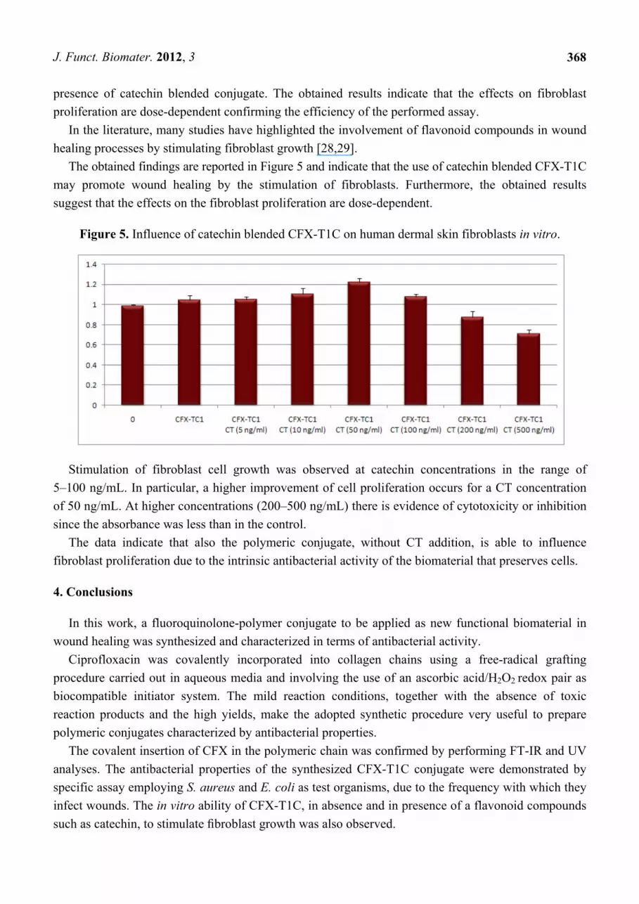
The obtained findings are reported in Figure 5 and indicate that the use of catechin blended CFX-T1C
may promote wound healing by the stimulation of fibroblasts. Furthermore, the obtained results
suggest that the effects on the fibroblast proliferation are dose-dependent.
Figure 5. Influence of catechin blended CFX-T1C on human dermal skin fibroblasts in vitro.
Stimulation of fibroblast cell growth was observed at catechin concentrations in the range of
5–100 ng/mL. In particular, a higher improvement of cell proliferation occurs for a CT concentration
of 50 ng/mL. At higher concentrations (200–500 ng/mL) there is evidence of cytotoxicity or inhibition
since the absorbance was less than in the control.
The data indicate that also the polymeric conjugate, without CT addition, is able to influence
fibroblast proliferation due to the intrinsic antibacterial activity of the biomaterial that preserves cells.
4. Conclusions
In this work, a fluoroquinolone-polymer conjugate to be applied as new functional biomaterial in
wound healing was synthesized and characterized in terms of antibacterial activity.
Ciprofloxacin was covalently incorporated into collagen chains using a free-radical grafting
procedure carried out in aqueous media and involving the use of an ascorbic acid/H2O2 redox pair as
biocompatible initiator system. The mild reaction conditions, together with the absence of toxic
reaction products and the high yields, make the adopted synthetic procedure very useful to prepare
polymeric conjugates characterized by antibacterial properties.
The covalent insertion of CFX in the polymeric chain was confirmed by performing FT-IR and UV
analyses. The antibacterial properties of the synthesized CFX-T1C conjugate were demonstrated by
specific assay employing S. aureus and E. coli as test organisms, due to the frequency with which they
infect wounds. The in vitro ability of CFX-T1C, in absence and in presence of a flavonoid compounds
such as catechin, to stimulate fibroblast growth was also observed.
J. Funct. Biomater. 2012, 3
369
The obtained findings support the idea that the synthetic strategy allows improving the properties of
a natural polymer, such as collagen, and that CFX-T1C conjugate represents an effective antibacterial
device showing the ability to influence fibroblast proliferation. Based on these considerations, the
synthesized conjugate could be applied as a new functional biomaterial in wound treatment
encouraging the healing process.
Conflict of Interest
The authors declare no conflict of interest.
Acknowledgments
This work was financially supported by University funds.
References
1. Shoulders, M.D.; Raines, R.T. Collagen structure and stability. Annu. Rev. Biochem. 2009, 78,
929–958.
2. Bailey, A.J.; Paul, R.G. Collagen is not so simple protein. J. Soc. Leathre Technol. Chem. 1998,
82, 104–118.
3. Lee, C.H.; Singla, A.; Lee, Y. Biomedical applications of collagen. Int. J. Pharmaceut. 2001, 221,
1–22.
4. Antonio, F.; Guillem, R.; Sonia, T.; Clara, M.; Piergiorgio, G.; Valeria, C.; Gianluca, C.; Tzanov, T.
Cross-linked collagen sponges loaded with plant polyphenols with inhibitory activity towards
chronic wound enzymes. Biotechnol. J. 2011, 6, 1208–1218.
5. Ueng, S.W.N.; Yuan, L.J.; Lin, S.S.; Liu, S.J.; Chan, E.C.; Chen, K.T.; Lee, M.S. In vitro and
in vivo analysis of a biodegradable poly(lactide-co-glycolide) copolymer capsule and collagen
composite system for antibiotics and bone cells delivery. J. Trauma 2011, 70, 1503–1509.
6. Beberok, A.; Buszman, E.; Wrzesniok, D.; Otreba, M.; Trzcionka, J. Interaction between
ciprofloxacin and melanin: The effect on proliferation and melanization in melanocytes. Eur. J.
Pharmacol. 2011, 669, 32–37.
7. Dizman, B.; Elasri, M.O.; Mathias, L.J. Synthesis and antibacterial activities of water-soluble
methacrylate polymers containing quaternary ammonium compounds. J. Polym. Sci. Pol. Chem.
2006, 44, 5965–5973.
8. Adetutua, A.; Morgana, W.A.; Corcorana, O. Ethnopharmacological survey and in vitro evaluation
of wound-healing plants used in south-western Nigeria. J. Ethnopharmacol. 2011, 137, 50–56.
9. Curcio, M.; Puoci, F.; Iemma, F.; Parisi, O.I.; Cirillo, G.; Spizzirri, U.G.; Picci, N. Covalent
insertion of antioxidant molecules on chitosan by a free radical grafting procedure. J. Agric. Food
Chem. 2009, 57, 5933–5938.
10. Curcio, M.; Cirillo, G.; Parisi, O.I.; Iemma, F.; Spizzirri, U.G.; Altimari, I.; Picci, N.; Puoci, F.
Poly(2-hydroxyethyl methacrylate)-quercetin conjugate as biomaterial in ophthalmology: An “ab initio”
study. J. Funct. Biomater. 2011, 2, 1–17.
J. Funct. Biomater. 2012, 3
370
11. Spizzirri, U.G.; Iemma, F.; Puoci, F.; Cirillo, G.; Curcio, M.; Parisi, O.I.; Picci, N. Synthesis of
antioxidant polymers by grafting of gallic acid and catechin on gelatin. Biomacromolecules 2009, 10,
1923–1930.
12. Spizzirri, U.G.; Parisi, O.I.; Iemma, F.; Cirillo, G.; Puoci, F.; Curcio, M.; Picci, N.
Antioxidant-polysaccharide conjugates for food application by eco-friendly grafting procedure.
Carbohyd. Polym. 2010, 79, 333–340.
13. Mertz, P.M.; Ovington, L.G. Wound-healing microbiology. Dermatol. Clinics 1993, 11, 739–747.
14. Bowler, P.G.; Duerden, B.I.; Armstrong, D.G. Wound microbiology and associated approaches to
wound management. Clinical Microbiol. Rev. 2001, 14, 2244–2269.
15. Albu, M.G.; Ferdes, M.; Kaya, D.A.; Ghica, M.V.; Titorencu, I.; Popa, L.; Albu, L. Collagen
wound dressings with anti-inflammatory activity. Mol. Cryst. Liq. Cryst. 2012, 555, 271–279.
16. Woodley, D.T.; O’Keefe, E.J.; Prunerias, M. Cutaneous wound healing: A model for cell-matrix
interaction. J. Am. Acad. Dermatol. 1985, 12, 420–433.
17. Mimura, Y.; Ihn, H.; Jinnin, M.; Asano, Y.; Yamane, K.; Tamaki, K. Epidermal growth factor
induces fibronectin expression in human dermal fibroblasts via protein kinase C δ-signaling
pathway. J. Invest. Dermatol. 2004, 122, 1390–1398.
18. Graham, M.F.; Diegelman, R.F.; Cohen, I.K. An in vitro model of fibroplasia: Simultaneous
quantification of fibroblast proliferation, migration, and collagen synthesis. P. Soc. Exp. Biol.
Med. 1984, 176, 302–308.
19. Mensah, A.Y.; Sampson, J.; Houghton, P.J.; Hylands, P.J.; Westbrook, J.; Dunn, M.M.;
Hughes, A.; Cherry, G.W. Effects of Buddlejaglobosa leaf and its constituents relevant to wound
healing. J. Ethnopharmacol. 2001, 77, 219–226.
20. Muzzarelli, R.A.A.; Guerrieri, M.; Goteri, G.; Muzzarelli, C.; Armeni, T.; Ghiselli, R.;
Cornelissen, M. The biocompatibility of dibutyryl chitin in the context of wound dressings.
Biomaterials 2005, 26, 5844–5854.
21. Paasche, G.; Ceschi, P.; Löbler, M.; Rösl, C.; Gomes, P.; Hahn, A.; Rohm, H.W.; Sternberg, K.;
Lenarz, T.; Schmitz, K.-P.; Barcikowski, S.; Stöver, T. Effects of metal ions on fibroblasts and
spiral ganglion cells. J. Neurosci. Res. 2011, 89, 611–617.
22. Idris, S.B.; Dånmark, S.; Finne-Wistrand, A.; Arvidson, K.; Albertsson, A.-C.; Bolstad, A.I.;
Mustafa, K. Biocompatibility of polyester scaffolds with fibroblasts and osteoblast-like cells for
bone tissue engineering. J. Bioact. Compat. Pol. 2010, 25, 567–583.
23. Ranzato, E.; Martinotti, S.; Burlando, B. Wound healing properties of jojoba liquid wax: An
in vitro study. J. Ethnopharmacol. 2011, 134, 443–449.
24. Hincal, F.; Gürbay, A.; Favier, A. Biphasic response of ciprofloxacin in human fibroblast cell
cultures. Nonlinearity Biol. Toxicol. Med. 2003, 1, 481–492.
25. Seto, Y.; Inoue, R.; Ochi, M.; Gandy, G.; Yamada, S.; Onoue, S. Combined use of in vitro
phototoxic assessments and cassette dosing pharmacokinetic study for phototoxicity
characterization of fluoroquinolones. AAPS J. 2011, 13, 482–492.
26. Gürbay, A.; Gonthier, B.; Barret, L.; Favier, A.; Hıncal, F. Cytotoxic effect of ciprofloxacin in
primary culture of rat astrocytes and protection by Vitamin E. Toxicology 2007, 229, 54–61.
27. Kautzky, F.; Hartinger, A.; Kohler, L.D.; Vogt, H.-J. In vitro cytotoxicity of antimicrobial agents
to human keratinocytes. J. Eur. Acad. Dermatol. 1996, 6, 159–166.

J. Funct. Biomater. 2012, 3
371
28. Kim, S.Y.; Kwak, J.S.; Shin, J.P.; Lee, S.H. Protection of the retina from ischemic injury by the
free radical scavenger EGb-761 and zinc in the cat retina. Ophthalmologica 1998, 212, 268–274.
29. Stevenson, P.C.; Simmonds, M.S.; Sampson, J.; Houghton, P.J.; Grice, P. Wound healing activity
of acylated iridoid glycosides from Scrophularianodosa. Phytother. Res. 2002, 16, 33–35.
© 2012 by the authors; licensee MDPI, Basel, Switzerland. This article is an open access article
distributed under the terms and conditions of the Creative Commons Attribution license
(http://creativecommons.org/licenses/by/3.0/).