methods and compositions for wound healing
TRANSCRIPT
Printed by Jouve, 75001 PARIS (FR)
(19)EP
3 57
4 90
9A
1*EP003574909A1*
(11) EP 3 574 909 A1(12) EUROPEAN PATENT APPLICATION
(43) Date of publication: 04.12.2019 Bulletin 2019/49
(21) Application number: 19183660.0
(22) Date of filing: 30.01.2009
(51) Int Cl.:A61K 33/38 (2006.01) A61K 9/00 (2006.01)
A61L 15/44 (2006.01) A61P 17/02 (2006.01)
(84) Designated Contracting States: AT BE BG CH CY CZ DE DK EE ES FI FR GB GR HR HU IE IS IT LI LT LU LV MC MK MT NL NO PL PT RO SE SI SK TRDesignated Extension States: AL BA RS
(30) Priority: 30.01.2008 US 24725
(62) Document number(s) of the earlier application(s) in accordance with Art. 76 EPC: 09748927.2 / 2 262 474
(71) Applicant: Imbed Biosciences, Inc.Madison, WI 53711 (US)
(72) Inventors: • MCANULTY, Jonathan
Oregon, WI Wisconsin 53575 (US)• MURPHY, Christopher
Madison, WI Wisconsin 53705 (US)• ABBOTT, Nicholas
Madison, WI Wisconsin 53711 (US)
(74) Representative: Witte, Weller & Partner Patentanwälte mbBPostfach 10 54 6270047 Stuttgart (DE)
Remarks: This application was filed on 01.07.2019 as a divisional application to the application mentioned under INID code 62.
(54) METHODS AND COMPOSITIONS FOR WOUND HEALING
(57) The present invention relates to methods andcompositions for wound healing. In particular, the presentinvention relates to promoting and enhancing woundhealing by utilizing cross-linker covalent modificationmolecules to attach and deliver wound active agents toa wound. In addition, the present invention provides
methods and compositions utilizing oppositely chargedpolymers to form a polyelectrolyte layer on a wound sur-face. The invention further relates to incorporating woundactive agents into a polyelectrolyte layer for delivery to awound.
EP 3 574 909 A1
2
5
10
15
20
25
30
35
40
45
50
55
Description
[0001] This application claims the benefit of U.S. Prov. Appl. 61/024,725, filed January 30, 2008, the entire contentsof which are incorporated herein by reference.
FIELD OF THE INVENTION
[0002] The present invention relates to methods and compositions for the modulation of wound healing. In particular,the present invention relates to promoting and enhancing wound healing by changing the intrinsic chemical compositionand/or physical features of the wound bed.
BACKGROUND OF THE INVENTION
[0003] The primary goal in the treatment of wounds is to achieve wound closure. Open cutaneous wounds representone major category of wounds and include burn wounds, wounds resulting from chemical (especially alkali) burns,wounds from physical trauma, neuropathic ulcers, pressure sores, venous stasis ulcers, and diabetic ulcers. Opencutaneous wounds routinely heal by a process which comprises six major components: i) inflammation, ii) fibroblastproliferation, iii) blood vessel proliferation, iv) connective tissue synthesis, v) epithelialization, and vi) wound contraction.Wound healing is impaired when these components, either individually or as a whole, do not function properly. Numerousfactors can affect wound healing, including but not limited to malnutrition, systemic debility due to a variety of causes,wound infection, local lack of progenitor cells, local and/or systemic pharmacological agents (e.g., numerous chemo-therapeutic agents, actinomycin and steroids), repeated local trauma, diabetes and other endocrine/metabolic diseases(e.g., Cushing’s disease), and advanced age (Hunt and Goodson, 1988, Current Surgical Diagnosis & Treatment, Ap-pleton & Lange, pp. 86-98). Additionally, wounds that are extensive in size, regardless of the initiating cause, presentspecial challenges due to the large surface area that must be re-epithelialized to reestablish surface integrity.[0004] Delayed wound healing causes substantial morbidity in subjects with diabetes. Diabetes mellitus is a chronicdisorder of glucose metabolism and homeostasis that damages many organs. It is the eighth leading cause of death inthe United States (Harris et al., 1987, Diabetes 36:523). In persons with diabetes, vascular disease, neuropathy, infec-tions, and recurrent trauma predispose the extremities, especially the foot, to pathologic changes. These pathologicalchanges can ultimately lead to chronic ulceration, which may necessitate amputation. Chronic wounds and wounds withpathological or dysregulated healing represent a major health burden and drain on health care resources. Chronicwounds have major impacts on the physical and mental health, productivity, morbidity, mortality and cost of care foraffected individuals. The most common types of chronic wounds are caused by systemic diseases such as diabetes,vascular problems such as venous hypertension and by immobility-induced pressure sores; accounting for 70% of allchronic wounds. Statistics on the prevalence of chronic wounds varies, however studies report that 0.2% to 1% of thepopulation suffer from venous ulcers, 0.5% from pressure ulcers, and 5% to 10% of people with diabetes experienceneuropathic ulcers. The economic impact of chronic wounds for these conditions alone in the United States has beenestimated to be well over $15 billion, annually. With the population growing older, cases of diabetes mellitus will increaseas will the magnitude of the problem associated with chronic wounds in these patients.[0005] Normal wound healing is an enormously complex process involving the coordinated interplay between fibrob-lasts, vascular cells, extracellular matrix and epithelial cells to result in a seamless progression through an inflammatoryreaction, wound repair, contracture and coverage by an epithelial barrier. However, in many patients, due to either thelocal wound environment or systemic disease or other factors, the wound healing processes can become asynchronous(i.e., loss of connectivity with triggering mechanisms associated with prior cellular events) and are unable to progressto closure, resulting in a chronic ulcer.[0006] Wounds that do not readily heal can cause the subject considerable physical, emotional, and social distressas well as great financial expense (Richey et al., 1989, Annals of Plastic Surgery 23:159). Indeed, wounds that fail toheal properly and become infected may require excision of the affected tissue. A number of treatment modalities havebeen developed as scientists’ basic understanding of wounds and wound healing mechanisms has progressed.[0007] The most commonly used conventional modality to assist in wound healing involves the use of wound dressings.In the 1960s, a major breakthrough in wound care occurred when it was discovered that wound healing with moist,occlusive dressings was, generally speaking, more effective than the use of dry, non-occlusive dressings (Winter, 1962,Nature 193:293). Today, numerous types of dressings are routinely used, including films (e.g., polyurethane films),hydrocolloids (hydrophilic colloidal particles bound to polyurethane foam), hydrogels (crosslinked polymers containingabout at least 60% water), foams (hydrophilic or hydrophobic), calcium alginates (nonwoven composites of fibers fromcalcium alginate), and cellophane (cellulose with a plasticizer) (Kannon and Garrett, 1995, Dermatol. Surg. 21:583;Davies, 1983, Burns 10:94). Unfortunately, certain types of wounds (e.g., diabetic ulcers, pressure sores) and the woundsof certain subjects (e.g., recipients of exogenous corticosteroids) do not heal in a timely manner (or at all) with the use
EP 3 574 909 A1
3
5
10
15
20
25
30
35
40
45
50
55
of such dressings.[0008] Several pharmaceutical modalities have also been utilized in an attempt to improve wound healing. For example,some practitioners have utilized treatment regimens involving zinc sulfate. However, the efficacy of these regimens hasbeen primarily attributed to their reversal of the effects of sub-normal serum zinc levels (e.g., decreased host resistanceand altered intracellular bactericidal activity) (Riley, 1981, Am. Fam. Physician 24:107). While other vitamin and mineraldeficiencies have also been associated with decreased wound healing (e.g., deficiencies of vitamins A, C and D; andcalcium, magnesium, copper, and iron), there is no strong evidence that increasing the serum levels of these substancesabove their normal levels actually enhances wound healing. Thus, except in very limited circumstances, the promotionof wound healing with these agents has met with little success.[0009] Current clinical approaches used to promote healing in dysregulated wounds include protection of the woundbed from mechanical trauma (e.g. splinting, bandaging), meticulous control of surface microbial burden (antibiotics,antimicrobial peptides, bacteriophages, antiseptics and other antimicrobial compounds that broadly inhibit wound path-ogens (e.g. silver sulfadiazine) combined with topical application of soluble cytoactive factors (e.g. growth factors ex-emplified by but not limited to epidermal growth factor-EGF, exogenous extracellular matrix constituents such as fi-bronectin), surgical excision of the wound margin or entire bed and surgical placement of tissue flaps and/or autografts,allografts and xenografts. All of these approaches fall short of promoting optimal healing conditions in many of the mostchallenging wounds. It is likely that a major contributing factor to the failure of these traditional approaches is the factthat they do not alter the intrinsic chemistry/structure of the wound bed itself that has been shown in many cases tocontribute significantly to their persistence. Additionally, the historical use of a single factor or set of factors to treat allwounds often falls short due to the great heterogeneity found in wound beds themselves and the complex environmentof the wound itself containing a community of signaling molecules that frequently modulate the activity of individualmolecules.[0010] The complex nature of pathologic wounds, and the lack of significant clinical progress based on current therapies,indicates the urgent need for new and unconventional approaches. What is needed is a safe, effective, and interactivemeans for enhancing the healing of chronic and severe wounds. The methods should be adaptable without regard tothe type of wound, or the nature of the patient population, to which the subject belongs.
SUMMARY OF THE INVENTION
[0011] The present invention relates to methods and compositions for the modulation of wound healing. In particular,the present invention relates to promoting and enhancing wound healing by changing the intrinsic chemical compositionand/or physical attributes of the wound bed. Accordingly, in some embodiments the present invention provides formu-lations that alter the physical attributes (compliance, topography, charge), as well as cross-linker covalent modificationmolecules to attach and deliver antimicrobial compounds as well as extracellular matrices and other scaffolds andcytoactive agents to a wound. In addition, the present invention provides methods and compositions utilizing oppositelycharged polyelectrolytes to form a polyelectrolyte layer on a wound surface. The invention further relates to the incor-poration of non-charged polymers into the wound bed via interactions such as but not limited to hydrogen bonding. Thescope of the invention includes incorporation of nanoparticles and microparticles into the wound bed to engineer thephysical characteristics and chemical composition of the wound bed. The invention further relates to incorporating woundactive agents into a polyelectrolyte layer, nanoparticle or microparticle for delivery to a wound. In some embodiments,the present invention provides formulations that promote a favorable and stable pH, surface charge, surface energy,osmotic environment, surface functionalities that enhance galvano- and magneto- positive effects on wound healing,supply of nitric oxide, provide energy sources for cells and/or provide a balance of MMP/other peptidase/protease activity.[0012] In one embodiment, the compositions and methods of the present invention provide approaches to promotehealing in pathologic wounds by altering the surface chemistry and structure of the pathologic wound bed itself so as toultimately enable differential modulation of key cellular elements customizable to the specific patient’s health woundtype and anatomic location in a subject.[0013] In one embodiment, the present invention provides for the covalent immobilization of factors to the wound bed.In some embodiments, the present invention provides methods of treatment, comprising providing a subject having awound, at least one covalent modification agent and at least one wound active agent, and contacting the wound withthe at least one covalent modification agent and the at least one wound active agent under conditions such that the atleast one wound active agent is covalently attached to the wound. In some embodiments, the subject is a human. Inother embodiments, the subject is a non-human vertebrate. In some embodiments, the at least one covalent modificationagent is a homobifunctional cross-linker. In other embodiments, the at least one covalent modification agent is a heter-obifunctional cross-linker. For example, in some embodiments, the homobifunctional cross-linker is an N-hydroxysuc-cinimidyl ester (e.g., including, but not limited to, disuccinimidyl ester, dithiobis(succinimidylpropionate), 3,3’-dithiobis(sul-fosuccinimidylpropionate), disuccinimidyl suberate, bis(sulfosuccinimidyl)suberate, disuccinimidyl tartarate, disulfosuc-cinimidyl tartarate, bis[2-(succinimidyloxycarbonyloxy)ethyl]sulfone, bis[2-(sulfosuccinimidooxycarbonyloxy)ethyl]sul-
EP 3 574 909 A1
4
5
10
15
20
25
30
35
40
45
50
55
fone, ethylene glycolbis(succinimidylsuccinate), ethylene glycolbis(sulfosuccinimidylsuccinate), disuccinimidyl glutarate,and N,N’-disuccinimidylcarbonate). In some embodiments, the homobifunctional cross-linker is at a concentration be-tween 1 nanomolar and 10 millimolar. In some preferred embodiments, the homobifunctional cross-linker is at a con-centration between 10 micromolar and 1 millimolar. In other embodiments, the at least one covalent modification agentis a heterobifunctional cross-linker (e.g., including, but not limited to, N-succinimidyl 3-(2-pyridyldithio)propionate, suc-cinimidyl 6-(3-[2-pyridyldithio]-propionamido)hexanoate, sulfosuccinimidyl 6-(3’-[2-pyridyldithio]-propionamido)hex-anoate, succinimidyl oxycarbonyl-α-methyl-α-(2-pyridyldithio)toluene, sulfosuccinimidyl-6-[α-methyl-α-(2-pyri-dyldithio)toluamido]hexanoate, succinimidyl 4-(N-maleimidomethyl)cyclohexane-1-carboxylate, sulfosuccinimidyl 4-(N-maleimidomethyl)cyclohexane-1-carboxylate, m-maleimidobenzoyl-N-hydroxysuccinimide ester, m-maleimidobenzoyl-N-hydroxy-sulfosuccinimide ester, N-succinimidyl(4-iodoacetyl)aminobenzoate, sulfo-succinimidyl(4-iodoacetyl)ami-nobenzoate, succinimidyl-4-(p-maleimidophenyl)butyrate, sulfosuccinimidyl-4-(p-maleimidophenyl)butyrate, N-(γ-male-imidobutyryloxy)succinimide ester, N-(γ-maleimidobutyryloxy)sulfosuccinimide ester, succinimidyl 6-((iodoacetyl)ami-no)hexanoate, succinimidyl 6-(6-(((4-iodoacetyl)amino)hexanoyl)amino)hexanoate, succinimidyl 4-(((iodoacetyl)ami-no)methyl)cyclohexane-1-carboxylate, succinimidyl 6-((((4-iodoacetyl)amino)methyl)cyclohexane-1-carbonyl)ami-no)-hexanoate, and p-nitrophenyl iodoacetate). In some embodiments, the heterobifunctional cross-linker is modifiedwith functional groups, rendering it soluble in aqueous solvents for delivery as an aqueous solution. Furthermore, insome embodiments, the aqueous solution contains additives (e.g., including, but not limited to, surfactants and blockcopolymers). In other embodiments, a multiplicity of heterobifunctional cross-linkers can be attached to a molecule,polymer or particle to serve as the cross-linking agent. In other embodiments, the heterobifunctional cross-linker isdissolved in an organic solvent (e.g., including, but not limited to, dimethyl sulfoxide). In some embodiments, the at leastone wound active agent includes, but is not limited to, trophic factors, extracellular matrices, enzymes, enzyme inhibitors,defensins, polypeptides, anti-infective agents, buffering agents, vitamins and minerals, analgesics, anticoagulants, co-agulation factors, anti-inflammatory agents, vasoconstrictors, vasodilators, diuretics, and anti-cancer agents. In someembodiments, the at least one wound active agent contains one or more free -SH groups.[0014] The present invention also provides a kit for treating a subject having a wound, comprising at least one covalentmodification agent, at least one wound active agent, and instructions for using the kit to covalently link the at least onewound active agent to the wound. In some embodiments, the at least one covalent modification agent is a homobifunctionalcross-linker. In some embodiments, the homobifunctional cross-linker is an N-hydroxysuccinimidyl ester (e.g., including,but not limited to, disuccinimidyl ester, dithiobis(succinimidylpropionate), 3,3’-dithiobis(sulfosuccinimidylpropionate), di-succinimidyl suberate, bis(sulfosuccinimidyl)suberate, disuccinimidyl tartarate, disulfosuccinimidyl tartarate, bis[2-(suc-cinimidyloxycarbonyloxy)ethyl]sulfone, bis[2-(sulfosuccinimidooxycarbonyloxy)ethyl]sulfone, ethylene glycolbis(succin-imidylsuccinate), ethylene glycolbis(sulfosuccinimidylsuccinate), disuccinimidyl glutarate, and N,N’-disuccinimidylcar-bonate). In some embodiments, the at least one covalent modification agent is a heterobifunctional cross-linker (e.g.,including, but not limited to, N-succinimidyl 3-(2-pyridyldithio)propionate, succinimidyl 6-(3-[2-pyridyldithio]-propionami-do)hexanoate, sulfosuccinimidyl 6-(3’-[2-pyridyldithio]-propionamido)hexanoate, succinimidyloxycarbonyl-α-methyl-α-(2-pyridyldithio)toluene, sulfosuccinimidyl-6-[α-methyl-α-(2-pyridyldithio)toluamido]hexanoate, succinimidyl4-(N-maleimidomethyl)cyclohexane-1-carboxylate, sulfosuccinimidyl 4-(N-maleimidomethyl)cyclohexane-1-carboxy-late, m-maleimidobenzoyl-N-hydroxysuccinimide ester, m-maleimidobenzoyl-N-hydroxy-sulfosuccinimide ester, N-suc-cinimidyl(4-iodoacetyl)aminobenzoate, sulfo-succinimidyl(4-iodoacetyl)aminobenzoate, succinimidyl-4-(p-maleimido-phenyl)butyrate, sulfosuccinimidyl-4-(p-maleimidophenyl)butyrate, N-(γ-maleimidobutyryloxy)succinimide ester, N-(γ-maleimidobutyryloxy)sulfosuccinimide ester, succinimidyl 6-((iodoacetyl)amino)hexanoate, succinimidyl 6-(6-(((4-io-doacetyl)amino)hexanoyl)amino)hexanoate, succinimidyl 4-(((iodoacetyl)amino)methyl)cyclohexane-1-carboxylate,succinimidyl 6-((((4-iodoacetyl)amino)methyl)cyclohexane-1-carbonyl)amino)-hexanoate, and p-nitrophenyl iodoace-tate). In some embodiments, the at least one wound active agent includes, but is not limited to, trophic factors, includingpolypetide growth factors, neuropeptides, neurotrophins, extracellular matrices and their individual native constituents(exemplified by but not limited to laminin, fibronectin, vitronectin, collagens, also select amino acid sequences found inthese proteins known to promote cell behaviors favorable to wound healing, e.g., integrin binding sequences exemplifiedby but not limited to RGD, EILDV,VCAM-1 and their recombined or synthetic analogs, enzymes, enzyme inhibitors, andpolypeptides), antimicrobial peptides (exemplified by but not limited to defensins, magainins, cathelocidins, bactenicin)anti-infective agents including silver containing compounds, buffering agents, vitamins and minerals, compounds thatpromote generation/stabilization of nitric oxide, energy sources for cells, analgesics, anticoagulants, coagulation factors,anti-inflammatory agents, vasoconstrictors, vasodilators, diuretics, and anti-cancer agents. In other embodiments thekits include small interfering RNAs (siRNAs-also referred to as micro RNAs) that are capable of promoting cellularbehaviors conducive to the wound healing process. In other embodiments, the kits include compounds that promote/sta-bilize a favorable pH, osmotic environment, surface energy, surface charge, surface functionalities that enhance galvano-and magneto- positive effects on wound healing, or balance of MMP/other peptidase/protease activity.[0015] In some embodiments, the present invention provides a method of treatment, comprising providing a subjecthaving a wound, at least one cationic polyelectrolyte, at least one anionic polyelectrolyte, at least one covalent modification
EP 3 574 909 A1
5
5
10
15
20
25
30
35
40
45
50
55
agent, and at least one wound active agent, and contacting the wound with the at least one cationic and the at least oneanionic polyelectrolytes, the at least one covalent modification agent, and the at least one wound active agent so thatthe at least one wound active agent is covalently linked to the wound by incorporation into a polyelectrolyte layer formedby the at least one cationic and the at least one anionic polyelectrolytes. In some preferred embodiments, the polyelec-trolytes are sequentially and repeatedly layered on the wound, then the at least one covalent modification agent and theat least one wound active agent are added. In some embodiments, the at least one cationic polyelectrolyte (e.g., including,but not limited to, poly(L-lysine), poly(ethylene imine), and poly(allylamine hydrochloride and dendrimers and multi-armed polymers that present multiple amine groups) is the top layer of the polyelectrolyte layers. Furthermore, in someembodiments, the at least one cationic polyelectrolyte harbors a primary amine group which allows attachment of theat least one covalent modification agent to the at least one cationic polyelectrolyte. For example, in some preferredembodiments, the at least one cationic polyelectrolyte is polylysine and the at least one anionic polyelectrolyte is poly-glutamic acid. In some embodiments, the at least one anionic polyelectrolyte (e.g., including, but not limited to, poly(L-glutamic acid), poly(sodium 4-styrenesulfonate), poly(acrylic acid), poly(maleic acid-co-propylene), hyaluronic acid, chon-droitin, and poly(vinyl sulfate)) is the top layer of the polyelectrolyte layers. In a preferred embodiment, the at least oneanionic polyelectrolyte is polyglutamic acid. In some embodiments, the at least one covalent modification agent is aheterobifunctional cross-linker (e.g., including, but not limited to, N-succinimidyl 3-(2-pyridyldithio)propionate, succinim-idyl 6-(3- [2-pyridyldithio] -propionamido)hexanoate, sulfosuccinimidyl 6-(3’- [2-pyridyldithio] - propionamido)hexanoate,succinimidyloxycarbonyl-α-methyl-α-(2-pyridyldithio)toluene, sulfosuccinimidyl-6-[α-methyl-α-(2-pyridyldithio)toluami-do]hexanoate, succinimidyl 4-(N-maleimidomethyl)cyclohexane-1-carboxylate, sulfosuccinimidyl 4-(N-maleimidome-thyl)cyclohexane-1-carboxylate, m-maleimidobenzoyl-N-hydroxysuccinimide ester, m-maleimidobenzoyl-N-hydroxy-sulfosuccinimide ester, N-succinimidyl(4-iodoacetyl)aminobenzoate, sulfo-succinimidyl(4-iodoacetyl)aminobenzoate,succinimidyl-4-(p-maleimidophenyl)butyrate, sulfosuccinimidyl-4-(p-maleimidophenyl)butyrate, N-(γ-maleimidobutyry-loxy)succinimide ester, N-(γ-maleimidobutyryloxy)sulfosuccinimide ester, succinimidyl 6-((iodoacetyl)amino)hexanoate,succinimidyl 6-(6-(((4-iodoacetyl)amino)hexanoyl)amino)hexanoate, succinimidyl 4-(((iodoacetyl)amino)methyl)cy-clohexane-1-carboxylate, succinimidyl 6-((((4-iodoacetyl)amino)methyl)cyclohexane-1-carbonyl)amino)-hexanoate,and p-nitrophenyl iodoacetate). In some embodiments, the at least one covalent modification agent is a homobifunctionalcross-linker. In some embodiments, the homobifunctional cross-linker is an N-hydroxysuccinimidyl ester (e.g., including,but not limited to, disuccinimidyl ester, dithiobis(succinimidylpropionate), 3,3’-dithiobis(sulfosuccinimidylpropionate), di-succinimidyl suberate, bis(sulfosuccinimidyl)suberate, disuccinimidyl tartarate, disulfosuccinimidyl tartarate, bis[2-(suc-cinimidyloxycarbonyloxy)ethyl]sulfone, bis[2-(sulfosuccinimidooxycarbonyloxy)ethyl]sulfone, ethylene glycolbis(succin-imidylsuccinate), ethylene glycolbis(sulfosuccinimidylsuccinate), disuccinimidyl glutarate, and N,N’-disuccinimidylcar-bonate). In some embodiments, the at least one covalent modification agent can be at a concentration between 1nanomolar and 10 millimolar. In some preferred embodiments, the at least one covalent modification agent is at aconcentration between 10 micromolar to 1 millimolar. In some preferred embodiments, the at least one covalent modi-fication agent is bis(sulfosuccinimidyl)suberate (for example, in aqueous solution at a concentration between 1 nM and10 mM). In some embodiments, the at least one covalent modification agent comprises N-hydroxysuccinimidyl ester. Insome preferred embodiments, the at least one wound active agent contains the peptide sequence gly-arg-gly-asp-ser-pro-lys.[0016] In some embodiments, a first at least one anionic or cationic polyelectrolyte is contacted with the at least onecovalent modification agent and the at least one wound active agent before contacting with the wound bed and oppositelycharged at least one cationic or anionic polyelectrolyte. In some preferred embodiments, the first at least one anionicpolyelectrolyte is polyglutamic acid, the at least one covalent modification agent is N-hydroxysuccinimidyl ester, and theat least one wound active agent contains a primary amine (e.g., including, but not limited to, the peptide sequence gly-arg-gly-asp-ser-pro-lys). In some preferred embodiments, the first at least one cationic polyelectrolyte is polylysine, theat least one covalent modification agent is N-succinimidyl 3-(2-pyridyldithio)propionate, and the at least one wound activeagent contains the peptide sequence gly-arg-gly-asp-ser-pro-cys.[0017] The present invention further provides kits for treating a subject having a wound, comprising at least one cationicpolyelectrolyte, at least one anionic polyelectrolyte, at least one covalent modification agent, at least one wound activeagent; and instructions for using the kit to covalently link the at least one wound active agent to the wound by incorporationinto a polyelectrolyte layer that is formed by the at least one cationic and anionic polyelectrolytes. In some embodiments,the incorporation of the at least one wound active agent is achieved by sequential and repeated layering of the polye-lectrolytes, followed by addition of the at least one covalent modification agent and the at least one wound active agent.In other embodiments, a first at least one anionic or cationic polyelectrolyte is contacted with the at least one covalentmodification agent and the at least one wound active agent before contacting with the wound bed and oppositely chargedat least one cationic or anionic polyelectrolyte. In some embodiments, the at least one cationic polyelectrolyte includes,but is not limited to, poly(L-lysine), poly(ethylene imine), and poly(allylamine hydrochloride). In some embodiments, theat least one anionic polyelectrolyte includes, but is not limited to, poly(L-glutamic acid), poly(sodium 4-styrenesulfonate),poly(acrylic acid), poly(maleic acid-co-propylene), and poly(vinyl sulfate). In some embodiments, the at least one covalent
EP 3 574 909 A1
6
5
10
15
20
25
30
35
40
45
50
55
modification agent is a homobifunctional cross-linker. In some embodiments, the homobifunctional cross-linker is an N-hydroxysuccinimidyl ester (e.g., including, but not limited to, disuccinimidyl ester, dithiobis(succinimidylpropionate), 3,3’-dithiobis(sulfosuccinimidylpropionate), disuccinimidyl suberate, bis(sulfosuccinimidyl)suberate, disuccinimidyl tartarate,disulfosuccinimidyl tartarate, bis[2-(succinimidyloxycarbonyloxy)ethyl]sulfone, bis [2-(sulfosuccinimidooxycarbony-loxy)ethyl]sulfone, ethylene glycolbis(succinimidylsuccinate), ethylene glycolbis(sulfosuccinimidylsuccinate), disuccin-imidyl glutarate, and N,N’-disuccinimidylcarbonate). In other embodiments, the at least one covalent modification agentis a heterobifunctional cross-linker (e.g., including, but not limited to, N-succinimidyl 3-(2-pyridyldithio)propionate, suc-cinimidyl 6-(3 - [2-pyridyldithio] -propionamido)hexanoate, sulfosuccinimidyl 6-(3’-[2-pyridyldithio]-propionamido)hex-anoate, succinimidyloxycarbonyl-α-methyl-α-(2-pyridyldithio)toluene, sulfosuccinimidyl-6-[α-methyl-α-(2-pyri-dyldithio)toluamido]hexanoate, succinimidyl 4-(N-maleimidomethyl)cyclohexane-1-carboxylate, sulfosuccinimidyl 4-(N-maleimidomethyl)cyclohexane-1-carboxylate, m-maleimidobenzoyl-N-hydroxysuccinimide ester, m-maleimidobenzoyl-N-hydroxy-sulfosuccinimide ester, N-succinimidyl(4-iodoacetyl)aminobenzoate, sulfo-succinimidyl(4-iodoacetyl)ami-nobenzoate, succinimidyl-4-(p-maleimidophenyl)butyrate, sulfosuccinimidyl-4-(p-maleimidophenyl)butyrate, N-(γ-male-imidobutyryloxy)succinimide ester, N-(γ-maleimidobutyryloxy)sulfosuccinimide ester, succinimidyl 6-((iodoacetyl)ami-no)hexanoate, succinimidyl 6-(6-(((4-iodoacetyl)amino)hexanoyl)amino)hexanoate, succinimidyl 4-(((iodoacetyl)ami-no)methyl)cyclohexane-1-carboxylate, succinimidyl 6-((((4-iodoacetyl)amino)methyl)cyclohexane-1-carbonyl)ami-no)-hexanoate, and p-nitrophenyl iodoacetate). In some embodiments, the at least one wound active agent includes,but is not limited to, trophic factors, extracellular matrices, enzymes, enzyme inhibitors, defensins, polypeptides, anti-infective agents, buffering agents, vitamins and minerals, analgesics, anticoagulants, coagulation factors, anti-inflam-matory agents, vasoconstrictors, vasodilators, diuretics, and anti-cancer agents.[0018] The present invention also provides a method of treatment, comprising providing a subject having a wound, atleast one cationic polyelectrolyte, at least one anionic polyelectrolyte, at least one DNA delivery agent, and at least oneDNA species, and contacting the wound with the at least one cationic polyelectrolyte, the at least one anionic polyelec-trolyte, the at least one DNA delivery agent, and the at least one DNA species under conditions such that the at leastone DNA species is delivered to the wound. In some preferred embodiments, the at least one DNA species includes,but is not limited to, DNA encoding vascular endothelial growth factor and/or epidermal growth factor. In some preferredembodiments, the at least one cationic polyelectrolyte is polylysine.[0019] The present invention further provides a kit for treating a subject having a wound, comprising: at least onecationic polyelectrolyte, at least one anionic polyelectrolyte, at least one DNA delivery agent, at least one DNA species,and instructions for using the kit to deliver the at least one DNA species to the wound using the at least one DNA deliveryagent and a polyelectrolyte layer formed by the at least one cationic and anionic polyelectrolytes.[0020] The present invention also provides a method of treatment, comprising providing a subject having a wound, acationic and anionic polyelectrolyte mixture, a deprotection agent, at least one covalent modification agent, and at leastone wound active agent; contacting the wound with the polyelectrolyte mixture to form a polyelectrolyte layer on thewound; applying the deprotection agent to the polyelectrolyte layer to form a deprotected polyelectrolyte layer; applyingthe at least one covalent modification agent to the deprotected polyelectrolyte layer to form a modified deprotectedpolyelectrolyte layer; and applying the at least one wound active agent to the modified deprotected polyelectrolyte layerunder conditions such that the at least one wound active agent is covalently attached to the wound. In some preferredembodiments, the anionic and cationic polyelectrolyte mixture is made up of polylactic acid and poly(epsilon-CBZ-L-lysine), blended at an 80:20 ratio. In some embodiments, the deprotection occurs by acid hydrolysis. In some embodi-ments, the at least one covalent modification agent is SP3. The present invention also provides a composition comprisinga deprotected polyelectrolyte, functionalized with at least one covalent modification agent containing an active groupthat is exposable to at least one wound active agent. In some preferred embodiments, the deprotected polyelectrolyteis comprised of polylactic acid and poly(epsilon-CBZ-L-lysine), blended at an 80:20 ratio. In a preferred embodiment,the at least one covalent modification agent is SP3.[0021] The present invention also provides a method of treatment, comprising providing a subject having a wound, acationic and anionic polyelectrolyte mixture, a deprotection agent, at least one covalent modification agent, a first polypep-tide, and at least one wound active agent linked to a second polypeptide; contacting the wound with the polyelectrolytemixture to form a polyelectrolyte layer on the wound; applying the deprotection agent to the polyelectrolyte layer to forma deprotected polyelectrolyte layer; applying the at least one covalent modification agent to the deprotected polyelectrolytelayer to form a modified deprotected polyelectrolyte layer; applying the first polypeptide to the modified deprotectedpolyelectrolyte layer to covalently link the first polypeptide to the modified deprotected polyelectrolyte layer; and applyingthe at least one wound active agent linked to the second polypeptide to the modified deprotected polyelectrolyte layercovalently linked to the first polypeptide, under conditions such that the at least one wound active agent is attached tothe wound by specific protein binding between the first polypeptide and the second polypeptide. In some preferredembodiments, the specific protein binding occurs between biotin and a polypeptide (e.g., including, but not limited to,avidin, neutravidin, and streptavidin). In other preferred embodiments, the specific protein binding occurs betweenglutathione-S-transferase and glutathione. In yet other preferred embodiments, the specific protein binding occurs be-
EP 3 574 909 A1
7
5
10
15
20
25
30
35
40
45
50
55
tween nickel-nitrilotriacetic acid and polyhistidine. In some embodiments, the at least one covalent modification agentis SP3.[0022] The present invention further provides a composition comprising a deprotected polyelectrolyte functionalizedwith at least one covalent modification agent linked to a first polypeptide that interacts by specific binding with a secondpolypeptide linked to at least one wound active agent. In some preferred embodiments, the specific binding occursbetween biotin and a polypeptide (e.g., including, but not limited to, avidin, neutravidin, and streptavidin). In other preferredembodiments, the specific binding occurs between glutathione-S-transferase and glutathione. In still other preferredembodiments, the specific protein binding occurs between nickel-nitrilotriacetic acid and polyhistidine.[0023] The present invention also provides a method of treatment, comprising providing a subject having a wound, acationic and anionic polyelectrolyte mixture, a deprotection agent, at least one covalent modification agent, at least onemolecule containing an azide group, and at least one wound active agent containing an alkyne group; contacting thewound with the polyelectrolyte mixture to form a polyelectrolyte layer on the wound; applying the deprotection agent tothe polyelectrolyte layer to form a deprotected polyelectrolyte layer; applying the at least one covalent modification agentto the deprotected polyelectrolyte layer to form a modified deprotected polyelectrolyte layer; applying the at least onemolecule containing an azide group to the modified deprotected polyelectrolyte layer to covalently link the at least onemolecule containing an azide group to the modified deprotected polyelectrolyte layer; and applying the at least onewound active agent containing an alkyne group to the modified deprotected polyelectrolyte layer covalently linked to theat least one molecule containing an azide group, so that the at least one wound active agent is attached to the woundby click chemistry. In some preferred embodiments, the at least one molecule containing an azide group has the formulaH2N(CH2CH2O)2N3. In some preferred embodiments, the at least one wound active agent is L-propargylglycine. In somepreferred embodiments, the reaction is carried out in the presence of Cu(I).[0024] The present invention further provides a composition comprising a deprotected polyelectrolyte functionalizedwith at least one covalent modification agent linked to at least one molecule containing an azide group that is attachedby click chemistry to at least one wound active agent containing an alkyne group.[0025] The present invention further provides a kit for treating a subject having a wound, comprising a cationic andanionic polyelectrolyte mixture, a deprotection agent, at least one covalent modification agent, at least one wound activeagent, and instructions for using the kit to covalently link the at least one wound active agent to the wound by deprotectionof the polyelectrolyte mixture contacted to the wound, followed by addition of the at least one covalent modification agentand the at least one wound active agent. In some preferred embodiments, the polyelectrolyte mixture comprises polylacticacid and poly(epsilon-CBZ-L-lysine). In some preferred embodiments, the deprotection agent causes deprotection byacid hydrolysis of the polyelectrolyte mixture. In some preferred embodiments, the at least one covalent modificationagent is SP3. In some embodiments, the kit also contains a first and a second polypeptide that interact with each otherby specific protein binding. In some preferred embodiments, the first polypeptide is linked to either the at least onecovalent modification agent or the at least one wound active agent and the second polypeptide is linked to the other atleast one covalent modification agent or the at least one wound active agent. In some preferred embodiments, the firstpolypeptide is biotin and the second polypeptide is avidin, neutravidin, or streptavidin. In some other preferred embod-iments, the first polypeptide is glutathione-S-transferase and the second polypeptide is glutathione. In still other preferredembodiments, the first polypeptide is nickel-nitrilotriacetic acid and the second polypeptide is polyhistidine. In someembodiments, the kit contains at least one molecule containing an azide group that is attached to the polyelectrolytemixture after deprotection so that at least one wound active agent containing an alkyne group can be attached to thewound by click chemistry.[0026] The present invention further provides a method of treatment, comprising providing a subject having a wound,at least one cationic polyelectrolyte, at least one anionic polyelectrolyte, at least one modifying agent, and at least onewound active agent containing an amino terminal cysteine residue; and contacting the wound with the polyelectrolytes,the at least one modifying agent and the at least one wound active agent so that the at least one wound active agent isattached to the wound by native chemical ligation. In some preferred embodiments, the polyelectrolytes are sequentiallyand repeatedly layered on the wound. In some embodiments, the at least one anionic polyelectrolyte (e.g., including,but not limited to, polyglutamic acid) is the top layer of the polyelectrolyte layers. In some embodiments, the at least onemodifying agents are ethylene dichloride and HSCH2Ph.[0027] The present invention also provides a composition comprising a polyelectrolyte layer that is functionalized withat least one modifying agent that is exposable to native chemical ligation with at least one wound active agent containingan amino terminal cysteine residue.[0028] The present invention also provides a kit for treating a subject having a wound, comprising at least one cationicpolyelectrolyte, at least one anionic polyelectrolyte, at least one modifying agent, at least one wound active agentcontaining an amino terminal cysteine residue, and instructions for using the kit to form a polyelectrolyte layer on thewound, followed by treatment with the at least one modifying agent and attachment of the at least one wound activeagent by native chemical ligation. In some preferred embodiments, the at least one anionic polyelectrolyte is polyglutamicacid. In some preferred embodiments, the at least one modifying agents are ethylene dichloride and HSCH2Ph.

EP 3 574 909 A1
8
5
10
15
20
25
30
35
40
45
50
55
[0029] In some embodiments, the present invention provides methods of treatment comprising: a) providing a subjecthaving a wound, at least one wound modifying agent, and at least one wound active agent; b) contacting the woundwith the at least one wound modification agent to provide a modified wound bed, and c) contacting the modified woundbed with the at least one wound active agent under conditions such that the at least one wound active agent is incorporatedinto the modified wound bed and healing of the wound is enhanced. In some embodiments, the modified wound bed ismodified to present a functionalized surface reactive with the at least one wound active agent. In some embodiments,the wound active agent is applied to the modified wound bed to form a gradient. In some embodiments, the woundmodifying agent alters a property of the wound bed selected from the group consisting of compliance, pH, alkalinity, andoxidative or reductive strength, net charge, hydrophilicity, osmotic strength, nanoscale or submicron topographic features,electroconductivity, MMP production, phagocytosis, and transglutaminase activity. In some embodiments, the woundmodifying agent is applied to the wound bed by a method selected from the group consisting of stamping, spraying,pumping, painting, smearing and printing. In some embodiments, the at least one wound modification agent is at leastone crosslinker. In some embodiments, the at least one crosslinker is selected from the group consisting of a homobi-functional crosslinker, a heterobifunctional cross linker, a hetero- and homo-multifunctional crosslinker, and a photoac-tivatable crosslinker and combinations thereof. In some embodiments, the heterobifunctional crosslinker comprises analkyne group. In some embodiments, the wound modifying agent comprises at least one polymer. In some embodiments,the at least one polymer is applied to the wound bed to form a polymer multilayer. In some embodiments, the at leastone polymer is selected from the group consisting of a cationic polymer, an anionic polymer, a nonionic polymer anamphoteric polymer and combinations thereof. In some embodiments, the methods further comprise contacting thepolymer multilayer with a crosslinker to form a functionalized surface reactive with the at least one wound active agent.In some embodiments, the at least one polymer is a polyelectrolyte multilayer preformed on a support so that thepolyelectrolyte multilayer can be transferred from the support to the wound bed to form a modified wound bed. In someembodiments, the support is an elastomeric support. In some embodiments, the polymer multilayer is from 1 nm to 250nm thick. In some embodiments, the polymer multilayer has a compliance of from 3 to 500 kPa. In some embodiments,the wound modifying agent is selected from the group consisting of nanoparticles and microparticles. In some embod-iments, the nano- and microparticles are functionalized. In some embodiments, the functionalized bead is a mesoscopiccross-linker. In some embodiments, the at least one wound active agent is selected from the group consisting of trophicfactors, extracellular matrices, enzymes, enzyme inhibitors, defensins, polypeptides, anti-infective agents, bufferingagents, vitamins and minerals, analgesics, anticoagulants, coagulation factors, anti-inflammatory agents, vasoconstric-tors, vasodilators, diuretics, and anti-cancer agents.[0030] In some embodiments, the present invention provides kits for treating a subject having a wound, comprising:a) at least one wound modification agent, b) at least one wound active agent; and c) instructions for using the kit to treata wound bed with the at least one wound modification agent to provide a modified wound bed and for incorporating thewound active agent into the modified wound bed. In some embodiments, the at least one wound modification agent isselected from the group consisting of a covalent modifying agent, at least one polyelectrolyte, microparticle, nanoparticle,and combinations thereof.[0031] In some embodiments, the present invention provides methods of treatment comprising: a) providing a subjecthaving a wound, i) a cationic and anionic polyelectrolyte mixture, ii)a deprotection agent, iii) at least one covalentmodification agent, iv) at least one molecule containing an azide group or alkyne group, and v) at least one wound activeagent containing the other of an azide group or an alkyne group; b) contacting the wound with the polyelectrolyte mixtureto form a polyelectrolyte layer on the wound; c) applying the deprotection agent to the polyelectrolyte layer to form adeprotected polyelectrolyte layer; d) applying the at least one covalent modification agent to the deprotected polyelec-trolyte layer to form a modified deprotected polyelectrolyte layer; e) applying the at least one molecule containing anazide group or an alkyne group to the modified deprotected polyelectrolyte layer to covalently link the molecule containingan azide or an alkyne group to the modified deprotected polyelectrolyte layer; f) applying the at least one wound activeagent containing the other of an azide group or an alkyne group to the modified deprotected polyelectrolyte layer covalentlylinked to the molecule containing an azide group, under conditions such that the at least one wound active agent isattached to the wound by click chemistry.[0032] In some embodiments, the present invention provides a composition comprising a deprotected polyelectrolytefunctionalized with at least one covalent modification agent linked to at least one molecule containing an azide groupthat is attached by click chemistry to at least one wound active agent containing an alkyne group.[0033] In some embodiments, the present invention provides methods comprising a) providing a subject having awound, a plurality of functionalized beads and at least one cytoactive factor, b) contacting the at least one cytoactivefactor with the at least one functionalized biocompatible bead such that the at least one cytoactive factor is linked to theat least one functionalized biocompatible bead and c) applying the at least one functionalized biocompatible bead linkedto the at least one cytoactive factor to a wound bed of the subject.[0034] In some embodiments, the present invention provides methods of treatment comprising: a) providing a subjecthaving a wound, at least one polymer, and at least one wound active agent b) applying the at least one polymer to the
EP 3 574 909 A1
9
5
10
15
20
25
30
35
40
45
50
55
wound so that a polymer multilayer is formed on the wound; and c) incorporating the at least one wound active agentinto the polymer multilayer during application of the at least one polymer, wherein the at least one wound active agentforms a gradient in the polymer multilayer.[0035] In some embodiments, the present invention provides an article comprising a matrix formed from a biocompatiblematerial, the matrix comprising at least one wound active agent, wherein the matrix is from 1 to 500 nm in thickness andhas a compliance of from 3 to 500 kPa. In some embodiments, the matrix is functionalized. In some embodiments, thebiocompatible material is selected from the group consisting of proteins and polymers. In some embodiments, thepolymers are selected from the group consisting of polyanionic polymers, polycationic polymers, uncharged polymers,and amphoteric polymers and combinations thereof, and wherein the polymers from a multilayer. In some embodiments,the proteins are extracellular matrix proteins selected from the group consisting of laminin, vitronectin, fibronection,keratin, collagen, and combinations thereof. In some embodiments, the matrix is supported by a solid support selectedfrom the group consisting of silicone, siloxane elastomers, latex, nylon, nylon mesh, biological tissue, silk, polyurethane,Teflon, polyvinyl alcohol membranes, and polyethylene oxide membranes. In some embodiments, the at least one woundactive agent is distributed on the matrix so that a gradient is formed. In some embodiments, the matrix is at least partiallyPEGylated. In some embodiments, the present invention provides methods comprising, applying the articles describedabove to a wound on a subject, wherein the article enhances healing of the wound. In some embodiments, the presentinvention provides kits comprising the articles described above in a sterile package.[0036] In some embodiments of the present invention, the compositions and methods described above enhance woundhealing. The present invention contemplates that wound healing may be enhanced in a variety of ways. In some em-bodiments, the compositions and methods minimize contracture of the wound as to best favor function and cosmesis.In some embodiments, compositions and methods promote wound contracture to best favor function and cosmesis. Insome embodiments, the compositions and methods promote vascularization. In some embodiments, the compositionsand methods inhibit vascularization. In some embodiments, the compositions and methods promote fibrosis. In someembodiments, the compositions and methods inhibit fibrosis. In some embodiments, the compositions and methodspromote epithelial coverage. In some embodiments, the compositions and methods inhibit epithelial coverage. In someembodiments, the compositions and methods of the present invention modulates one or properties of cells in the woundenvironment or in the immediate vicinity of the wound. The properties that are modulated, e.g., are increased or decreased,include, but are not limited to adhesion, migration, proliferation, differentiation, extracellular matrix secretion, phagocy-tosis, MMP activity, contraction, and combinations thereof.[0037] In some embodiments, the present invention provides for the use of any of the compositions described aboveor elsewhere herein to enhance healing of a wound or modulate one or more properties of cells in the wound environmentor in the immediate vicinity of the wound. In some embodiments, the present invention provides for the use of a combinationof a wound modifying agent and wound active agent to enhance healing of a wound to enhance healing of a wound ormodulate one or more properties of cells in the wound environment or in the immediate vicinity of the wound. In someembodiments, the present invention provides for the use of the articles described above to enhance healing of a woundor modulate one or more properties of cells in the wound environment or in the immediate vicinity of the wound.
DESCRIPTION OF THE FIGURES
[0038]
Figure 1 provides a schematic of a wound bed modified with a polyelectrolyte multilayer.Figure 2 provides a schematic of a wound bed modified with covalent modifying agents.Figure 3 exemplifies the covalent immobilization of proteins to model amine-terminated treated glass surfaces.Figure 4 shows ex vivo results for multilayer deposition of polyelectrolytes polystyrene sulfonate (PSS) and FITC-labelled poly (allylamine hydrochloride) (FITC-PAH).Figure 5 demonstrates the difference in healing of full thickness cutaneous wounds in diabetic (db/db) mice comparedto control (wild type) mice.Figure 6 is exemplary of the use of beads for healing a wound bed. In this example, carboxylic acid-terminatedbeads are activated outside the wound bed by using NHS and EDC. The activated beads are introduced into thewound bed and are immobilized in the wound bed via reaction of the activated surfaces of the beads with aminegroups present in the wound bed. Growth factors are introduced into the wound bed and immobilized via reactionwith the exposed surfaces of the activated beads.Figure 7 provides a list of antimicrobial polypeptides, identified by name and AMSDb database ID number.
DEFINITIONS
[0039] To facilitate an understanding of the invention set forth in the disclosure that follows, a number of terms are
EP 3 574 909 A1
10
5
10
15
20
25
30
35
40
45
50
55
defined below.[0040] The term "wound" refers broadly to injuries to the skin and subcutaneous tissue initiated in different ways (e.g.,pressure sores from extended bed rest and wounds induced by trauma) and with varying characteristics. The methodsand compositions described herein are useful for treatment of all types of wounds, including wounds to internal andexternal tissues. Wounds may be classified into one of four grades depending on the depth of the wound: i) Grade I:wounds limited to the epithelium; ii) Grade II: wounds extending into the dermis; iii) Grade III: wounds extending into thesubcutaneous tissue; and iv) Grade IV (or full-thickness wounds): wounds wherein bones are exposed (e.g., a bonypressure point such as the greater trochanter or the sacrum).[0041] The term "partial thickness wound" refers to wounds that encompass Grades I-III; examples of partial thicknesswounds include burn wounds, pressure sores, venous stasis ulcers, and diabetic ulcers. The term "deep wound" is meantto include both Grade III and Grade IV wounds. The present invention contemplates treating all wound types, includingdeep wounds and chronic wounds.[0042] The term "chronic wound" refers to a wound that has not healed within 30 days.[0043] The phrases "promote wound healing," "enhance wound healing," and the like refer to either the induction ofthe formation of granulation tissue of wound contraction and/or the induction of epithelialization (i.e., the generation ofnew cells in the epithelium). Wound healing is conveniently measured by decreasing wound area.[0044] The term "wound active agent" refers to compounds that induce a desired pharmacological, physiological effectuseful in the treatment and healing of a wound, wherein the effect may be prophylactic or therapeutic. The terms alsoencompass pharmaceutically acceptable, pharmacologically active derivatives of those active agents specifically men-tioned herein, including, but not limited to, trophic factors, extracellular matrices, enzymes, enzyme inhibitors, defensins,polypeptides, anti-infective agents, buffering agents, vitamins and minerals, analgesics, anticoagulants, coagulationfactors, anti-inflammatory agents, vasoconstrictors, vasodilators, diuretics, and anti-cancer agents.[0045] The term "polyelectrolyte multilayer" refers to the composition formed by sequential and repeated applicationof anionic and cationic polyelectrolytes to form a multilayered structure. For examples, these layers are formed by thealternating addition of anionic and cationic polyelectrolytes to a wound or to a solid support. The term "polyelectrolytelayer" also refers to the composition formed by sequential and repeated application of anionic and cationic polyelectrolytesto a wound or support. In addition, the term "polyelectrolyte layer" can refer to a single layer composed of anionic orcationic polyelectrolyte molecules, existing either as one layer within multiple layers, or as a single layer of only one typeof polyelectrolyte molecules on a wound or support. While the delivery of the polyelectrolytes to the wound bed issequential in preferred embodiments, the use of the term "polyelectrolyte multilayer" is not limiting in terms of the resultingstructure of the coating. It is well understood by those skilled in the art that inter-diffusion of polyelectrolytes can takeplace leading to structures that may be well-mixed in terms of the distribution of anionic and cationic polyelectrolytes. Itis also understood that the term polyelectrolyte includes polymer species as well as nanoparticulate species, and thatit is not limiting in scope other than to indicate that the species possesses multiple charged or partially charged groups.It is also well understood by those skilled in the art that multilayer structures can be formed through a variety of interactions,including electrostatic interactions and others such as hydrogen bonding. Thus, the use of the term "polyelectrolyte" isnot limiting in terms of the interactions leading to the formation of the wound bed constructs.[0046] The term "crosslinked" herein refers to a composition containing intermolecular crosslinks and optionally in-tramolecular crosslinks as well, arising from the formation of covalent bonds. Covalent bonding between two crosslinkablecomponents may be direct, in which case an atom in one component is directly bound to an atom in the other component,or it may be indirect, through a linking group. A crosslinked structure may, in addition to covalent bonds, also includeintermolecular and/or intramolecular noncovalent bonds such as hydrogen bonds and electrostatic (ionic) bonds.[0047] The term "covalent modification agent" refers to any molecule that covalently links molecules to each other.Covalent modification agents include homobifunctional and heterobifunctional cross-linkers as well as photoactivatablecross linkers.[0048] The term "homobifunctional cross-linker" refers to a molecule used to covalently link identical or similar moleculesto each other. Homobifunctional cross-linkers have two identical reactive groups; thus, a homobifunctional cross-linkercan only link molecules of the same type to each other. Conversely, a "heterobifunctional cross-linker" refers to a moleculeused to covalently link dissimilar molecules to each other, because it has two or more different reactive groups that caninteract with various molecules of different types. Hetero- and homo-multifunctional crosslinkers refers to multivalentcrosslinkers with both hetero- and homo-crosslinking functionalities. Activated dendrimers are an example of multifunc-tional crosslinkers.[0049] The term "subject" refers to any animal (e.g., a mammal), including, but not limited to, humans, non-humanprimates, rodents, dogs, cats, and the like, which is to be the recipient of a particular treatment. Typically, the terms"subject" and "patient" are used interchangeably herein.[0050] The term "surfactant" refers to an amphiphilic material that modifies the surface and interface properties ofliquids or solids. Surfactants can reduce the surface tension between two liquids. Detergents, wetting agents, emulsifyingagents, dispersion agents, and foam inhibitors are all surfactants.
EP 3 574 909 A1
11
5
10
15
20
25
30
35
40
45
50
55
[0051] The term "block copolymer" refers to a polymer consisting of at least two monomers. In a block copolymer,adjacent blocks are constitutionally different, i.e. adjacent blocks comprise constitutional units derived from differentspecies of monomer or from the same species of monomer but with a different composition or sequence distribution ofconstitutional units. A block copolymer can be thought of as two homopolymers joined together at the ends.[0052] The term "solvent" refers to a liquid that can dissolve a substance. The term "organic solvent" refers to a solventderived from a petroleum-based product.[0053] The term "polyelectrolyte" refers to a water-soluble macromolecular polymer substance containing many re-peating ionic constituent units, including cations and anions.[0054] The term "primary amine" refers to a derivative of ammonia in which a hydrogen has been replaced by ahydrocarbon unit. Primary amines have the general formula RNH2 and examples include, but are not limited to, aniline,methylamine, and 1-propylamine.[0055] The term "DNA delivery agent" refers to any molecule that can bring DNA into contact with an identified target.In some instances, a DNA delivery agent causes uptake of DNA into a cell or cells, in vitro or in vivo. DNA deliveryagents can be viruses including, but not limited to, adenoviruses and retroviruses. DNA delivery agents can also be non-viral agents including, but not limited to, plasmids, lipids, liposomes, polymers and peptides.[0056] The term "exposable" refers to anything that is capable of being exposed. An exposable surface or moleculeis one that is made available to interaction with other surfaces or molecules. For example, in the context of the presentinvention, a covalent modification agent is exposable to a wound active agent; thus, the two agents can interact witheach other and form covalent bonds.[0057] The term "functionalized" refers to a modification of an existing molecular segment to generate or introduce anew reactive functional group (e.g., a maleimido or succinimidyl group) that is capable of undergoing reaction withanother functional group (e.g., a sulfhydryl group) to form a covalent bond. For example, a component containingcarboxylic acid (--COOH) groups can be functionalized by reaction with N-hydroxy-succinimide or N-hydroxysulfosuc-cinimide using known procedures, to form a new reactive functional group in the form of an activated carboxylate (whichis a reactive electrophilic group), i.e., an N-hydroxysuccinimide ester or an N-hydroxysulfosuccinimide ester, respectively.In another example, carboxylic acid groups can be functionalized by reaction with an acyl halide, e.g., an acyl chloride,again using known procedures, to provide a new reactive functional group in the form of an anhydride.[0058] As used herein, the term "aqueous solution" includes solutions, suspensions, dispersions, colloids, and the likecontaining water.[0059] As used herein, the term "click chemistry" refers to the use of chemical building blocks with built-in high-energycontent to drive a spontaneous and irreversible linkage reaction with appropriate complementary sites in other blocks.These chemical reactions (e.g., including, but not limited to, those between azide and acetylene groups that combinereadily with each other) are specific and result in covalent linkage between the two molecules.[0060] The term "native chemical ligation" refers to a chemoselective reaction of two unprotected peptide segments.The reaction results in an initial thioester-linked species, then spontaneous rearrangement of this transient intermediateoccurs, yielding a full-length product with a native peptide bond at the ligation site.[0061] The term "specific protein binding" refers to an interaction between two or more proteins that have high affinityand specificity for each other. Proteins must bind to specific other proteins in vivo in order to function. The proteins arerequired to bind to only one or a few other proteins of the few thousand proteins typically present in vivo; these interactionsare employed in vitro in the present invention to attach wound active agents to the wound. In the context of the presentinvention, specific protein binding interactions include, but are not limited to, those between biotin and avidin, neutravidin,or streptavidin; glutathione-S-transferase and glutathione; and nickel-nitrilotriacetic acid and polyhistidine.
DETAILED DESCRIPTION OF THE INVENTION
[0062] The complex nature of wounds, and the lack of significant clinical progress based on current therapies, indicatesthe urgent need for new and unconventional approaches. The microenvironment of the pathologic/chronic wound bedis dysregulated with alterations in extracellular matrix constituents, degradative enzymes, growth factor and other cy-toactive factor activity. The present invention provides compositions and methods for engineering of the wound bed itselfby, for example, altering the surface chemistry/structure of the wound bed to promote favorable cell behaviors thataccelerate wound healing. In some embodiments, the wound bed is first treated with a priming agent (i.e., primer) thatprovides a uniform, reactive bed on the wound surface. The primed wound bed is then treated with a desired agent,such a wound active agent.[0063] In normal wound healing, the coordinated interplay between fibroblasts, vascular cells, extracellular matrixcomponents and epithelial cells results in a seamless progression through an inflammatory reaction, wound repair,contracture and coverage by an epithelial barrier. However, in many subjects with dysregulated wound microenvironment,systemic disease or other confounding circumstances, the wound healing processes become asynchronous resultingin an indolent ulcer (Pierce, 2001, Am. J. Pathol. 159:399). In other subjects, a loss or lack of regulatory responses to
EP 3 574 909 A1
12
5
10
15
20
25
30
35
40
45
50
55
appropriately modulate cellular behaviors during healing causes an exuberant proliferative response that in itself is aproblem for the subject. This is particularly true for patients prone to keloid formation or in burn patients where excessivefibroblastic proliferation and collagen production result in disfiguring and disabling scar formation.[0064] It is clear that across the spectrum of non-healing wounds that there are a variety of inciting mechanisms andwound microenvironments. These wounds exhibit pathology at many junctures in the progression to closure. Deficits inangiogenesis, fibroblastic responses and re-epithelialization all play a role in chronic wounds. Thus, a single factortreatment approach to chronic wounds is not likely to be fruitful across the disparate array of wounds presented in theclinical milieu. In such a heterogeneous environment, a more promising strategy involves the identification of compoundsthat are able to modulate specific aspects of the cellular response and behavior in the wound environment, providingthe potential for custom crafting of a healing response tailored to the individual wound and patient.[0065] Due to the heterogeneous spectrum of wound environments, it is contemplated that stimulating a "desirable"healing response is best achieved by a differential modulation of the cellular responses within the wound. The presentinvention provides for the selection of a subset of cytoactive compounds from a list of candidates for immobilization intothe wound bed so as to differentially modulate the endothelial, fibroblastic and epithelial components within the healingresponse. As such, the present invention provides the potential to achieve high quality healing responses in a varietyof clinical conditions, thereby providing a strategy for engineering the wound bed for personalized therapeutics for eachunique wound healing challenge encountered by a clinician. Specific examples where differential modulation of thecellular responses within the wound would yield substantial benefit include chronic wounds in diabetics or venous stasisulcers, where pancellular promotion of healing responses is desired but in particular a vibrant angiogenic response isneeded to support all of the other aspects of wound healing. In other wounds, where the strength of the healed woundis a key concern, modulation to promote a fibroblastic response with a normal angiogenic and epithelial component isdesirable.[0066] In contrast, in some burns, such as deep second degree burns where dermal and hair shaft epithelial elementspersist to replace lost tissues, a rich angiogenic and epithelial response is needed, but it is desirable to mitigate thefibroblastic reaction to reduce scar hypertrophy, contracture and disfigurement. A similar mitigation is desirable in healingsubjects prone to keloid formation where the proliferative fibroblastic response in these wounds must be suppressed. Itis also advantageous in wounds near joints or orifices to be able to promote rapid healing and coverage with epitheliumbut modulate the fibroblastic response so that improved suppleness of the tissue is retained. Modulation of the fibroblasticresponse in this way has the potential to provide superior clinical outcomes and reduce the need for subsequent recon-structive procedures targeted at recovery of limb or other critical bodily functions. The feasibility of such an approachhas been demonstrated previously, such as in the report by Muehlberger and colleagues on the effects tretinoin onincisional wounds (Muehlberger et al., 2005, J. Am. Acad. Derm. 52:583). In that study, application of tretinoin resultedin an increased fibroblastic proliferation but the production of collagen was diminished.[0067] The modification of surfaces has become an important challenge in the last decade for applications in implantmaterials, prostheses, and artificial organs, allowing broad medical applications for implant and tissue engineering(Langer and Vacanti, 1993, Science 260:920); Peppas and Langer, 1994, Science 263:1715; Angelova and Hunkeler,1999, Trends Biotechnol. 17:409). For the improved integration efficiency of implants, several approaches, involving thealteration of physicochemical, morphological, and biochemical properties of device surfaces, have been investigated inan effort to obtain a suitable bone-implant interface. Self-assembled monolayers (SAMs) or Langmuir-Blodgett techniqueshave been commonly employed to produce new interfaces (Mrksich, 1997, Curr. Opin. Colloid Interface Sci. 2:83; Lösche,1997, Curr. Opin. Solid State Mater. Sci. 2:546).[0068] More recently, a new versatile method of self assembled architectures based on the alternate deposition ofpolyanions and polycations has been developed for the buildup of multilayered polyelectrolyte films (Decher, 1997,Science 277:1232). Besides varying film thickness, roughness, and porosity, it is also possible to incorporate in the filmarchitecture functionalized macromolecules (Caruso et al., 1997, Langmuir 13:3427; Cassier et al.,1998, Supramol. Sci.5:309). It has also been demonstrated that the layer-by-layer deposition process is not limited in applicability to polye-lectrolytes, but can be applied to viruses, nanoparticles, non-ionic polymers, proteins and other forms of microscopicand nanoscopic matter. A recent review provides information on a wide range of species and interfacial structures thatcan be formed by the layer-by-layer deposition procedure. The scope of the invention described herein is not limited topolyelectrolytes but applies to all species that have been demonstrated to be incorporated into interfacial structures bythe layer-by-layer deposition process.[0069] The present invention provides a variety of embodiments for altering the composition of the wound bed. Insome embodiments, a wound modifying agent is applied to prime the wound bed. As described in detail below, woundmodifying agents are agents that applied to a wound bed and either covalently or noncovalently modify the wound bed.Examples of wound modifying agents include homobifunctional and heterobifunctional linkers, polyelectrolytes, non-ionic polymers, combinations of polyelectrolytes and non-ionic polymers, and nano- and micro-particles including beadsand needles. In some embodiments, the wound modifying agent alters a property of the wound bed selected from thegroup consisting of compliance, pH, alkalinity, and oxidative or reductive strength, net charge, hydrophilicity, osmotic

EP 3 574 909 A1
13
5
10
15
20
25
30
35
40
45
50
55
strength, nanoscale or submicron topographic features, electroconductivity, MMP production, phagocyticis, and trans-glutaminase activity. In preferred embodiments, the wound modifying agents are used to incorporate wound activeagents so that the wound active agents are localized to the wound bed. The wound active agents can be covalently ornoncovalently attached to the wound modifying agent. Furthermore, the wound active agents may form a gradient viathe wound modifying agent. In further embodiments, the wound modifying agents alter the compliance of the woundbed. In these embodiments, the polymers or nano- or micro-particles have a predetermined hardness. In further em-bodiments, the compliance gradients with varying levels of hardness may be formed by the polymers, nano- or micro-particles.[0070] In one embodiment, the present invention provides for the deposition and immobilization of cytoactive factorsand extracellular matrices (ECMs) on the wound bed by using, for example, polyelectrolyte multilayers or beads. Figure1 provides a schematic diagram 100 of a wound bed 110 on which a polyelectrolyte multilayer 130 has been deposited.The diagram 100 depicts that the wound bed 110 comprises a heterogeneous surface depicted by shapes 120 whichrepresent different chemical moieties. The polyelectrolyte multilayer 130 provides a homogenous surface onto whichfunctional groups can 140 can be attached to form a homogenous functionalized surface 150. In the embodiment depicted,the functional groups are uniform, however, in some preferred embodiments, different functional groups are utilized. Awide variety of active agents can then be attached to the surface via the functional groups 140. In other embodiments,the wound bed is covalently modified with covalent modification agents. The covalent modification agents include, butare not limited to, homobifunctional and heterobifunctional cross-linkers as well as photoactivatable cross linkers. Figure2 provides a schematic diagram 200 of a wound bed 210 comprising a heterogeneous surface depicted by shapes 220which represent different chemical moieties. The wound bed is covalently modified by reacting covalent modificationagents 230 with the different chemical moieties to provide a relatively homogenous functionalized surface 250. In preferredembodiments, covalent modification agents 230 present functional groups 240. In the embodiment depicted, the functionalgroups are uniform, however, in some preferred embodiments, different functional groups are utilized. A wide variety ofactive agents can then be attached to the surface via the functional groups 240. These embodiments are discussed inmore detail below.[0071] It is contemplated that the wound bed is an extremely heterogeneous environment. The compositions andmethods of the present invention are designed to modify the wound bed to provide a homogenous environment thatprovides for uniform and predictable delivery or uniform incorporation of active agents into the wound bed. Surprisingly,it has been found that modification of wound beds in this manner greatly reduces the amount of active agent which isneeded; i.e., the effective amount of active agent needed is reduced. Surface functionalization allows for precise controlof the amount of active agent used or delivered and further allows for the formation of gradients of active agents on thewound bed. In some preferred embodiments, the priming agent is optimized to react with the wound bed. In someembodiments, the priming agent provides an optimized surface for enhanced or optimized delivery of an active agent.In some embodiments, a chemical parameter of the wound bed is changed. For example, the wound bed may be modifiedto be more or less acidic, basic, alkaline, reducing, or oxidizing or have a higher or lower ionic strength.[0072] There is a wide array of candidate molecules for improving healing chronic wounds. For example, basementmembrane constituents and growth factors promote wound healing. In some embodiments, the extracellular matricesdeposited and immobilized are constituents of native basement membrane. Native ECMs comprise a mixture of glyco-proteins, proteoglycans and hyaluronic acid. These glycoproteins and proteoglycans include, but are not limited to, fibrin,elastin, fibronectin, laminins, nidogens and collagens. Cytoactive factors such as growth factors are also part of ECMs.In some embodiments, extracellular matrix components, such as collagen, laminin, or hyaluronic acid are deposited andimmobilized on a wound bed. In some embodiments, a synthetic matrix such as MATRIGELtm is deposited on a woundbed. MATRIGELtm is a commercially available basement membrane like complex that retains many characteristics of anative basement membrane, including a three-dimensional nanoscale topographic surface. The present invention is notlimited to a particular mechanism. Indeed, an understanding of the mechanism is not necessary to practice the presentinvention. Nonetheless, it is contemplated that the nanoscale topographic features of a basement membrane modulatefundamental cell behaviors including migration, adhesion, proliferation and differentiation (Abrams et al., 2002, Biomi-metic Materials and Design: Interactive Biointerfacial, Tissue Engineering, and Drug Delivery, Eds. Dillow and Lowman;Diehl et al., 2005, J. Biomed. Mater. Res. A 75:603; Foley et al., 2005, Biomaterials 26:3639; Karuri et al., 2004, J. CellSci. 117:3153; Liliensiek et al., 2006, J. Biomed. Mater. Res. A 79:185). In some embodiments, the present inventionfurther provides methods, formulations, compositions and kits for altering the compliance of the wound surface. In someembodiments, the local compliance (the compliance that cells see) is altered by immobilizing a thin layer of extracellularmatrix constituents such as MATRIGELtm or an appropriate hydrogel or other synthetic matrix or by enzymatic treatmentof the wound bed. In other embodiments, compliance is altered by the addition of cross-linking agents to the wound bedto cross-link components already presenting the wound bed, or components deliberately introduced into the wound bed.It has also been demonstrated by Picart and coworkers that it is possible to control the compliance of multilayer structuresformed from polyelectrolytes by the addition of cross-linking agents.[0073] In some embodiments, the cytoactive factors that are deposited and immobilized on a wound bed include, but

EP 3 574 909 A1
14
5
10
15
20
25
30
35
40
45
50
55
are not limited to, those factors that are mitogenic for epithelial cells and vascular endothelial cells and other factors thatare elements in wound closure. For example, in some embodiments, growth factors such as platelet derived growthfactor (PDGF), and/or epidermal growth factor (EGF), known to be mitogenic for epithelial cells are deposited in a woundbed. In other embodiments, vascular endothelial growth factor (VEGF), known to be mitogenic for vascular endothelialcells, comprise the cytoactive factors immobilized on the wound bed. It is contemplated that the present invention is notlimited by the ECM components or cytoactive factors immobilized on the wound bed, indeed any extracellular matrixcomponents and/or cytoactive factor that improves wound healing is equally applicable. Additional cytoactive and woundactive agents that can be incorporated are provided below.
I. Priming the wound bed
[0074] In some embodiments, the present invention provides compositions and methods for priming the wound bedto provide uniform reactive surface for later modification. Suitable compositions for priming a wound bed include, butare not limited to, polyelectrolytes and chemical crosslinking agents that react covalently with functional groups foundin wound beds such as carboxy, thiol, amide and sugar groups.
A. Multilayer deposition on wound bed
[0075] In some embodiments, the present invention provides compositions, formulations, methods and kits for depos-iting a multilayer structure on a wound bed. In some embodiments, the multilayer structures comprise layers of polymersthat form polyelectrolytes, while in other embodiments, the multilayers comprise polymers that do not have a charge(i.e., non-ionic polymers) or a combination of charged and uncharged polymer layers. In some embodiments, it is con-templated that polyelectrolyte films built-up by the alternated adsorption of cationic and anionic polyelectrolyte layersconstitute a novel and promising technique to modify wound surfaces in a controlled way [(Decher et al., 1992, ThinSolid Films 210/211:831; Decher, 1997, Science 277:1232). One of the most important properties of such multilayersis that they exhibit an excess of alternatively positive and negative charges (Caruso et al., 1999, J Am Chem Soc121:6039; Ladam et al., 2000, Langmuir 16:1249). Not only can this constitute the motor of their buildup (Joanny, 1999,Eur. Phys. J. Biol. 9:117), but it allows, by simple contact, to adsorb a great variety of compounds such as dyes,particles(Cassagneau et al., 1998, J. Am. Chem. Soc. 120:7848; Caruso et al., 1999, Langmuir 15:8276; Lvov et al.,1997, Langmuir 13:6195), clay microplates (Ariga et al., 1999, Appl. Clay Sci. 15:137) and proteins (Keller et al., 1994,J. Am. Chem. Soc. 116:8817; Lvov et al., 1995, J. Am. Chem. Soc. 117:6117; Caruso et al., 1997, Langmuir 13:3427).[0076] In some embodiments, the polymer multilayers, such as polyelectrolyte multilayers, are nanoscale in dimension.Accordingly, in some embodiments, the polymer multilayers are from about 1 nm to 1000 nm thick, from about 1 nm to500 nm thick, from about 1 nm to 100 nm thick, from about 1 nm to about 25 nm thick, from about 1 nm to about 10 nmthick, or less than about 500 nm, 100 nm, 25 nm or 10 nm thick. It is contemplated that the nanoscale dimension of thepolymer multilayers (i.e., the nanoscale thickness) allows for the loading of a lower total amount of an active agent whilestill allowing delivery of an effective amount (i.e., an amount of active agent that accelerates wound healing as comparedto controls) of the active agent as compared to matrix structures with greater thickness. It is contemplated that the lowertotal loading levels result in reduced toxicity in the wound environment, especially when antimicrobial compounds areincorporated into the polymer multilayer.[0077] In some embodiments, the compliance of the polymer multilayers is adjusted to facilitate cell migration in thewound. In some embodiments, the polymer multilayers exhibit a compliance, measured in kilopascals (kPa) of fromabout 3 to about 500 kPa, about 7 to about 250 kPa, about 10 to about 250 kPA or from about 10 to about 200 kPa.
1. Formation of polyelectrolyte layers
[0078] The cationic polyelectrolyte poly(L-lysine) (PLL) interacts with anionic sites on cell surfaces and in the extra-cellular matrix (Elbert and Hubbell, 1998, J. Biomed. Mater. Res. 42:55). In some embodiments, the present inventionprovides a method of treating a wound with the sequential application of a cationic polyelectrolyte, an anionic polyelec-trolyte, and a wound active agent to the wound. In other embodiments, the treatment includes the sequential and repeatedapplication of a cationic polyelectrolyte, an anionic polyelectrolyte, and a wound active agent to the wound.[0079] Polyelectrolyte layers are formed by alternating applications of anionic polyelectrolytes and cationic polyelec-trolytes to surfaces to form a polyelectrolyte layer. The layers can be used to deliver a wound active agent to a wound.Preferably, at least four layers, and, more preferably, at least six layers are used to form the polyelectrolyte multilayer.[0080] The method of treatment of the present invention is not limited to use on a wound surface. The formation of apolyelectrolyte layer that includes a wound active agent may be formed on any surface to which delivery of a woundactive agent is desirable.[0081] In some embodiments, the cationic polyelectrolyte used is PLL and the anionic polyelectrolyte used is poly(L-
EP 3 574 909 A1
15
5
10
15
20
25
30
35
40
45
50
55
glutamic acid) (PGA). Indeed, the use of a variety of polyelectrolytes is contemplated, including, but not limited to,poly(ethylene imine) (PEI), poly(allylamine hydrochloride) (PAH), poly(sodium 4-styrenesulfonate) (PSS), poly(acrylicacid) (PAC), poly(maleic acid-co-propylene) (PMA-P), and poly(vinyl sulfate) (PVS). It is also possible to use naturallyoccurring polyelectrolytes, including hyaluronic acid and chondroitin sulfate.[0082] In still further embodiments, the polymer is a dendrimer, grafted polymer, or star architecture polymer. In otherembodiments, the multilayer responds to or is organized in the presence of an electric field, for example an electric fieldformed by placing electrodes on either side of a wound.[0083] Referring to Figure 1, in some embodiments, the polymer multilayer 130 can be comprised of polystyrenesulfonate, an amphiphillic polyelectrolyte with an affinity for hydrophobic regions of the wound bed, and an amphotericpolymer. The polymer multilayer is preferably functionalized with one or more crosslinking agents presenting an alkyneso that a uniform surface is presented (e.g., 140 in Figure 1 where x represents an alkyne group). From this point, widelyavailable click chemistries can be used to add desired wound active agents to the modified surface of the wound. Insome embodiments, the wound modifying agent is an azide conjugate or otherwise comprises an azide group and isreacted with the alkyne groups displayed on the wound bed in a Huisgen Cycloaddition.
2. Cationic Polymers
[0084] Cationic polymers useful in the present invention can be any biocompatible water-soluble polycationic polymer,for example, any polymer having protonated heterocycles attached as pendant groups. As used herein, "water soluble"means that the entire polymer must be soluble in aqueous solutions, such as buffered saline or buffered saline withsmall amounts of added organic solvents as co-solvents, at a temperature between 20 and 37° Centigrade. In someembodiments, the material will not be sufficiently soluble (defined herein as soluble to the extent of at least one gramper liter) in aqueous solutions per se but can be brought into solution by grafting the polycationic polymer with water-soluble polynonionic materials such as polyethylene glycol.[0085] Representative cationic polymers include natural and unnatural polyamino acids having net positive charge atneutral pH, positively charged polysaccharides, and positively charged synthetic polymers. Examples of suitable poly-cationic materials include polyamines having amine groups on either the polymer backbone or the polymer side chains,such as poly-L-lysine (PLL) and other positively charged polyamino acids of natural or synthetic amino acids or mixturesof amino acids, including, but not limited to, poly(D-lysine), poly(ornithine), poly(arginine), and poly(histidine), and non-peptide polyamines such as poly(aminostyrene), poly(aminoacrylate), poly (N-methyl aminoacrylate), poly (N-ethylami-noacrylate), poly(N,N-dimethyl aminoacrylate), poly(N,N-diethylaminoacrylate), poly(aminomethacrylate), poly(N-methylamino-methacrylate), poly(N-ethyl aminomethacrylate), poly(N,N-dimethyl aminomethacrylate), poly(N,N-diethyl ami-nomethacrylate), poly(ethyleneimine), polymers of quaternary amines, such as poly(N,N,N-trimethylaminoacrylate chlo-ride), poly(methyacrylamidopropyltrimethyl ammonium chloride), and natural or synthetic polysaccharides such as chi-tosan. In some embodiments, PLL is a preferred material.[0086] In general, the polymers must include at least five charges, and the molecular weight of the polycationic materialmust be sufficient to yield the desired degree of binding to a tissue or other surface, having a molecular weight of atleast 1000 g/mole.
3. Anionic Polymers
[0087] Polyanionic materials useful in the present invention can be any biocompatible water-soluble polyanionic pol-ymer, for example, any polymer having carboxylic acid groups attached as pendant groups. Suitable materials includealginate, carrageenan, furcellaran, pectin, xanthan, hyaluronic acid, heparin, heparan sulfate, chondroitin sulfate, der-matan sulfate, dextran sulfate, poly(meth)acrylic acid, oxidized cellulose, carboxymethyl cellulose and crosmarmelose,synthetic polymers and copolymers containing pendant carboxyl groups, such as those containing maleic acid or fumaricacid in the backbone. Polyaminoacids of predominantly negative charge are also suitable. Examples of these materialsinclude polyaspartic acid, polyglutamic acid, and copolymers thereof with other natural and unnatural amino acids.Polyphenolic materials such as tannins and lignins can be used if they are sufficiently biocompatible. Preferred materialsinclude alginate, pectin, carboxymethyl cellulose, heparin and hyaluronic acid.
4. Nonionic Polymers
[0088] In some embodiments, the multilayer structures are formed from uncharged polymers or from a combinationof charged and uncharged polymers. Examples of uncharged polymers include, but are not limited to, dextran, dextransulfate, diethylaminoethyl (DEAE)-dextran, hydroxyethyl cellulose, ethyl(hydroxyethyl) cellulose, acrylamide, polyethyl-ene oxide, polypropylene oxide, polyethylene oxide - polypropylene oxide copolymers, PAANa, Ficoll, polyvinylpyrolidine,and polyacrylic acid.
EP 3 574 909 A1
16
5
10
15
20
25
30
35
40
45
50
55
5. Amphoteric polymers
[0089] In some embodiments, the multilayer structures are formed from one or more amphoteric polymers, alone incombination with the other polymers described herein. In some embodiments, the amphoteric polymers comprise oneor more of acrylic acid (AA), DMAEMA (dimethylaminoethyl methacrylate), APA (2-aminopropyl acrylate), MorphEMA(morpholinoethyl methacrylate), DEAEMA (diethylaminoethyl methacrylate), t-ButylAEMA (t-butylaminoethyl methacr-ylate), PipEMA (piperidinoethyl methacrylate), AEMA (aminoethyl methacrylate), HEMA (2-hydroxyethyl methacrylate),MA (methyl acrylate), MAA (methacrylic acid) APMA (2-aminopropyl methacrylate), AEA (aminoethyl acrylate). In someembodiments, the amphoteric polymer comprises (a) carboxylic acid, (b) primary amine, and (c) secondary and/or tertiaryamine. The amphoteric polymers have an isoelectric point of 4 to 8, preferably 5 to 7 and have a number averagemolecular weight in the range of 10,000 to 150,000.
6. Application of multilayers
[0090] The wound modifying agent, such as a polymer multilayer, can be applied to the wound by a variety of methods.In some embodiments, it is contemplated that the polymer or polymer multilayers is applied, preferably sequentially, tothe wound using either a pump (including syringes, ink jet printers, and electrojets) or aerosol spray. In other embodiments,particle bombardment is utilized. In other embodiments, the use of a brush including an air brush is contemplated. Inother embodiments, a sponge is utilized. In other embodiments a solid support or stamp such as an elastomeric material,for example, PDMS (polydimethylsiloxane), silicone, hydrogel or latex, is used to support the wound modifying agentsand mechanically transfer the agent into the wound bed. In these embodiments, the polymer multilayers are pre-formedon the stamp. In further embodiments, nano- or micro-particles are arranged on the stamp for delivery to the wound. Inother approaches, electric fields or magnetic fields are used to facilitate transfer of the wound modifying agents into thewound bed.[0091] In some embodiments, the polymers are applied to a wound by a spray, such as via a pump or aerosol device.In some embodiments, the polymer layers are applied sequentially. In one embodiment, a solution, dispersion, or sus-pension of the wound modifying agent and wound active agent is sprayed onto the wound to form the polyelectrolytelayer. In embodiments where the wound modifying agent and wound active agent are supplied as aerosols, a propellantis used to provide the force for expulsion from the container. In some embodiments, the wound modifying agent or thewound active agent and the propellant form a single liquid phase so that the composition is delivered consistently.[0092] In general, for aerosol delivery, the ingredients of the composition are mixed to form a substantially homogenoussolution, slurry, dispersion, or the like. For two-part systems, each part is mixed. The compositions are placed in anappropriate container and the propellant is added using conventional techniques, such as cold filling or pressure fillingtechniques. The composition can be delivered using a commercially available aerosol sprayer such as, for example, thePreval™ aerosol spray unit available from Precision Valve Corporation, NY, USA, which has a modular power unit andrefillable container jar. The propellant is a mixture of propane, isobutane, and dimethyl ether.[0093] The composition can also be delivered using a syringe outfitted with a spray head, or a dual spray deviceoutfitted with a spray head and, optionally, a mixing chamber. The device may include a meter so that the quantity ofapplied composition can be controlled.[0094] Any of a number of propellants known to those skilled in the art can be used, provided that it is chemically inertto the other ingredients of the composition. Suitable propellants include vinyl chloride and mixtures of vinyl chloride anddichlorodifluoromethane, other fluorochlorohydrocarbons known as the Freons and the Genetrons, and blends of fluor-ochlorohydrocarbons, chlorinated hydrocarbons, and hydrocarbons. Examples of fluorochlorohydrocarbons includetrichloromonofluoromethane, dichlorodifluoromethane, dichloromonofluoromethane, 2-tetrafluoroethane, 1,1-dichloro-1,2,2-tetraf- luoroethane, 1-chloro-1,1-difluoroethane, 1,1-difluoroethane, and octofluorocyclobutane, and mixturesthereof. Examples of hydrocarbons include liquefied petroleum gases like propane, isobutane, and N-butane and mixturesthereof. Dimethyl ether is another propellant. Compressed gas propellants that are preferably non-toxic, non-flammable,and inert can be used. Examples include carbon dioxide, nitrous oxide and N2 and the like. Mixtures of the above areoften used.[0095] The quantity of propellant used is critical only in that if an insufficient amount is used, the driving force to expelthe entire composition from the container will be lacking. Generally, the composition will comprise from 75% to 95% byweight propellant.[0096] In some embodiments, the invention contemplates a kit comprising a container with a cationic polyelectrolyte,a second container containing an anionic polyelectrolyte, and a third container containing at least one wound activeagent. In still other embodiments, in addition to a container with a cationic polyelectrolyte, a second container containingan anionic polyelectrolyte, and a third container containing at least one wound active agent, the kit provides an applicationdevice for administering the polyelectrolytes and at least one wound active agent to the wound. In some preferredembodiments, the containers comprise a propellant. In other embodiments, the containers comprise a pump.
EP 3 574 909 A1
17
5
10
15
20
25
30
35
40
45
50
55
[0097] In some embodiments, contacting comprises delivery of the polymer and the at least one wound active agentto the wound using a reaction chamber dressing (RCD). In some embodiments, it is contemplated that the RCD willcomprise a solid barrier adhered to an area completely surrounding the wound. Within the solid barrier reside inflow andoutflow orifices to which tubes from solution reservoirs can be connected. The use of a variety of solid barriers iscontemplated, including, but not limited to, Gortex, latex, Teflon, PVDF, plastic and rubber.[0098] In some embodiments, the wound modification agents are applied to the wound by microfluidics printing, micro-stamping (U.S. Pat. Nos. 5,512,131 and 5,731,152, both of which are incorporated by reference herein in their entirety),or microcontact printing (PCT Publication WO 96/29629, incorporated by reference herein in its entirety). In other em-bodiments, the wound modifying agents are applied to the wound via a pulse het such as an inkjet printer. Examples ofsuitable inkjet printers for application of biological fluids include those described in U.S. Pat. No. 7,128,398, WO 95/25116and WO 98/41531, each of which is incorporated by reference in its entirety. In other embodiments, the wound modifyingagents are applied by a drop dispensers such as the tip of a pin or in an open capillary and, touch the pin or capillary tothe surface of the substrate. Such a procedure is described in U.S. Pat. No. 5,807,522. When the fluid touches thesurface, some of the fluid is transferred. In other embodiments, the wound modifying agents are applied be pipetting,micro-pipetting or positive displacement pumps such as the Biodot equipment (available from Bio-Dot Inc., Irvine Calif.,USA).[0099] One or more wound active agents are preferably applied to the wound along with the wound modification agent.In some embodiments, the wound active agent is covalently attached to the wound modification agent. In other embod-iments, the wound active agent is incorporated into the wound modifying agent, for example, by application duringsequential application of polymer layers in a polymer multilayer. In other embodiments, the wound active agents aredelivered by a microarrayer, pipette, positive displacement pump, inkjet printer, microneedle array, elastomeric stamp,gene gun, electrojet, or air brush as described above in relation to the wound modifying agents.
B. Modification of wound beds with biocompatible particles
[0100] In some embodiments, the wound modifying agent is a nano- or micro-particle. In some embodiments, nanom-eter to submicrometer sized biocompatible particles, such as spherical (e.g., beads) and/or non-spherical (e.g., oblongs,needles, cubes, tetrahedral, mushroom-like structures, haybale-like structures) particles or electrospun polymers, areapplied (e.g., either directly or indirectly) to the wound bed, thereby creating a three-dimensional topographic woundbed. For example, application of biocompatible particles to a wound bed results in the creation of knobs, ridges, spikes,undulations, and the like in the wound bed. Microbeads have a size generally ranging from about 1 to about 500micrometers, while nanobeads have a size generally ranging from about 1 to about 1000 nanometers. Microbeads mayfurther comprise micro- (i.e., from 1 to 100 micrometers) or nano-scale (0.1 to 1000 nanometer) features on the surfaceof the bead. Nanobeads may further comprise nanoscale features (i.e., 0.1 to 500 nanometer features) on the surfaceof the bead.[0101] In some embodiments, the biocompatible particles are biodegradable. In some embodiments, the biocompatibleparticles are not biodegradable. The biocompatible particles, for example, are modified with surface chemistry cross-linkers that allow attachment and immobilization of wound active agents, extracellular matrix compounds, polyelectrolytes,etc. as described further herein. It is contemplated that the incorporation of modified biocompatible particles, for example,facilitates the presentation of ECM constituents that support epithelial coverage of the wound, provides cytoactive factorsfor wound healing, and introduces topographic features that support cellular behaviors for promoting wound healing. Insome embodiments, the biocompatible particles are functionalized with mesoscopic cross-linkers to, for example, broadlyenable covalent chemistry in the wound bed. In some embodiments, cross-linkers are attached to negatively chargedbiocompatible particles. In some embodiments, the biocompatible particles comprise layers of multiple different constit-uents for wound healing, or a single constituent for wound healing. For example, spherical particles such as beadsplaced in a wound bed comprise, or upon which are layered, one or more intermixed species of polyelectrolytes, ECMagents, proteins with hydrolysable bonds, proteins with enzymatically cleavable bonds, and the like for wound healingas previously described. In some embodiments, antimicrobials, either alone or in combination with other wound healingcompositions, are also applied to the biocompatible particles. Antimicrobials include, but are not limited to, antibiotics,metal based antimicrobial compositions comprising silver, copper, selenium, triclosan-based antimicrobials, thiabenda-zole-based antimicrobials, isothiazolinone-based antimicrobials, zinc-pyrithione-based antimicrobials, and/or 10’-oxybi-sphenoxarsine-based antimicrobials (OBPA). The present invention is not limited by the type of antimicrobial used.[0102] In some embodiments, the size of the biocompatible particles, such as beads, are at least 1nm, at least 5nm,at least 10nm, at least 20nm, at least 50nm, at least 100nm, at least 200nm, at least 300nm, at least 500nm, or at least800nm in diameter. In some embodiments, beads and other spherical and non-spherical particles contain their ownsurface topography. For example, the bead surface may comprise undulations, indentations, tunnels, holes, knobs,ridges, etc. Spherical particles, such as beads, may or may not be inert and may, for example, be magnetic. Magneticbeads comprise, for example, a magnetic core or magnetic particles. Examples of different bead compositions are found
EP 3 574 909 A1
18
5
10
15
20
25
30
35
40
45
50
55
at, for example, US Patents 5,268,178, 6,458,858, 6,869,858, and 5,834,121, each of which is incorporated herein byreference in its entirety.[0103] In some embodiments, biocompatible particles of the present invention used for wound healing are coated withlayers of polyelectrolytes, ECM components, antimicrobials, proteins with hydrolysable bonds, proteins with enzymaticallycleavable bonds, etc. and other wound healing compositions wherein the surface compliance of the beads is maintainedfor optimal wound healing. For example, the compliance of the layers for wound healing as deposited on the biocompatibleparticles are contemplated to be of optimal stiffness (e.g., not too soft, not too hard) such that cells within the woundbed are capable of optimal anchorage and spreading. Engler et al. (2004, Biophys. J. 86:617-628) discusses substratecompliance for cell anchorage and spreading. In some embodiments, the biocompatible particles are layered with pol-yelectrolytes, ECM components, antimicrobials, etc. wherein substrate stiffness of at least 1 kPa, at least 5 kPa, at least8kPa, at least 10 kPa, at least 20 kPa, at least 40 kPa, at least 60 kPa, at least 80 kPa is realized as measured usingthe methods of Engler et al. (2004). In preferred embodiments, substrate compliance of the wound healing compositionsas applied to biocompatible particles is at least 8 kPa to about 80 kPa or 100 kPa. It will be recognized that hardnesswithin these limit are contemplated without specifically listing all such hardnesses, for example, 10, 20, 30 , 40, 50, 60,or 70 kPa limits as upper or lower ranges are all contemplated by the present invention. In some embodiments, the ECMcomposition preferentially used in conjunction with polyelectrolytes, antimicrobials, and other wound healing compositionsis collagen.[0104] In some embodiments, the layers as applied to a wound bed or to a biocompatible particle comprise gradientsof wound healing compositions, for example, wound active agents. For example, the concentrations of the compositionsare layered in a wound bed or on a biocompatible particle in a gradient such that higher concentrations of a particularcomposition is greater proximal to the wound bed than distal to the wound bed in a vertical fashion. The converse, whereconcentrations of compositions is greater distal to the wound bed than proximal, is also contemplated. Concentration ofcompositions in a wound bed or on a biocompatible particle wherein a horizontal gradient is deposited is also contem-plated. Topographical gradients are also contemplated, wherein compositions are deposited such that the concentrationsof compositions in a wound bed or on a biocompatible particle follow the topography of the substrate, for example, ahigher concentration of compositions is deposited in the valleys of undulations of an exemplary substrate compared tothe peaks of the undulations. Likewise, the present invention contemplates that compliance gradients can be formed inthe wound bed by controlled application of micro- or nano-beads of varying hardness. For example, beads having one,two, three, four or more different hardnesses are applied to form a vertical gradient (i.e., a depth gradient) on the woundor a horizontal gradient (i.e., a gradient along the length or width of the wound), or a combination of vertical and horizontalgradients. Horizontal and vertical gradients can also be formed with beads functionalized with 1, 2, 3, 4 or more differentwound active agents. See Ichinose et al., Biomaterials. Volume 27, Issue 18, June 2006, Pages 3343-3350; Stefoneket al., Immobilized gradients of epidermal growth factor promote accelerated and directed keratinocyte migration. WoundRepair and Regeneration (OnlineEarly Articles). doi:10.1111/j.1524-475X.2007.00288.x; Kapur et al., Immobilized con-centration gradients of nerve growth factor guide neurite outgrowth. Journal of Biomedical Materials Research Part A.Volume 68A, Issue 2, Pages 235 - 243; DeLong et al., Covalently immobilized gradients of bFGF on hydrogel scaffoldsfor directed cell migration. Biomaterials 26 (2005) 3227-3234; Liu, et al., 2007 Sep 25; 17892312 Endothelial CellMigration on Surface-Density Gradients of Fibronectin, VEGF, or Both Proteins.[0105] In some embodiments, wound healing compositions applied to the wound bed or biocompatible particles (e.g.,beads, needles, etc.) are time released, such that wound healing compositions that are applied to the wound bed orbiocompatible particles are released over a period of time, for example 3 hours, 5 hours, 10 hours, 1 day, several days,one week, several weeks, etc. thereby providing short and/or long term treatment for the wound bed.[0106] In some embodiments, biocompatible particles used in wound bed healing applications of the present inventionare not, for example, spherical as previously described, but take on shapes such as oblongs, needles, mushroom-likestructures, haybale-like structures, etc. In some embodiments, the biocompatible particle shape is preferably needles.The manufacture of needles useful in embodiments of the present invention can be accomplished through microfabricationtechniques, such as those found in, for example, McAllister et al., 2003, Proc. Natl. Acad. Sci. 100:13755-13760. Insome embodiments, biocompatible particles are coated and/or layered with wound healing compositions, anti-microbials,proteins that are hydrolyzably cleavable, proteins that are enzymatically cleavable, antimicrobials, etc. as describedherein. In some embodiments, the coated and/or layered biocompatible particles are applied to a wound bed by stamping,spraying, or other methods as described herein.[0107] In some embodiments, the layers of wound healing compositions as described herein are deposited on a woundbed or on biocompatible particles by electrostatic bonding. In other compositions, non-electrostatic interactions areutilized, such as van der Waals interactions, metal ion-ligand coordination or hydrogen bonding. In some embodiments,the layers of wound healing compositions as described herein are deposited by piezoelectric application, using methodsand systems as described in, for example, US Patent 6,368,079 and Sumerel et al., 2006, Biotechnol. J. 1:976-987. Inother embodiments, the wound healing compositions are directly or indirectly deposited into the wound bed by usingelectrojetting technologies, including electroextrusion, and electrospraying and electrospinning. In some embodiments,
EP 3 574 909 A1
19
5
10
15
20
25
30
35
40
45
50
55
the layers deposited comprise one or more wound healing compositions. In some embodiments, the layers are biocom-patible particles (e.g. spherical beads, needles, and the like) intermixed with wound healing compositions. In otherembodiments, layered deposition is sequential. For example, sequential deposition of wound healing compositions alone,or sequential deposition of wound healing compositions followed by biocompatible particles, followed by wound healingcompositions, etc. The present invention is not limited by the constituents that make up a layer of deposition on abiocompatible particle, and many permutations of wound healing compositions and biocompatible particle layering wouldbe apparent to a skilled artisan.[0108] In some embodiments, the deposition of layers in the wound bed or on a biocompatible particle or other substrateto be added to a wound bed is accomplished by "stamping", also referred to as "contact printing". A stamp substrate isany substrate that can be used to transfer one or more than one entity or composition (e.g., polyelectrolytes, ECMcomponents, proteins with hydrolysable or enzymatically cleavable bonds, etc.) that is (are) covalently or non-covalentlybound to the surface of the stamp substrate to another surface. Examples of suitable stamp substrates include, but arenot limited to, polydimethylsiloxane (PDMS) and other elastomeric (e.g. pliable) materials. In some embodiments, differentconcentrations of the same wound healing composition are arrayed in different areas of the stamp substrate. In otherembodiments, a variety of different compositions are arrayed on the stamp substrate surface. In some embodiments,multiple wound healing compositions in multiple concentrations are arrayed on the stamp substrate surface. The woundhealing compositions are then introduced to the stamp substrate surface, which are in turn stamped into the wound bedor onto another substrate for introduction into the wound bed (e.g., biocompatible particles).[0109] In other embodiments, the biocompatible particles, for example needles, are coated and/or layered with woundhealing compositions, antimicrobials, proteins that are hydrolyzably cleavable, proteins that are enzymatically cleavable,etc., and applied to a pad which is subsequently applied to a wound. Pads include, but are not limited to, band-aids,gauze, surgical packings, and the like. In some embodiments, the pad is impregnated with wound healing compositions,anti-microbials, proteins that are hydrolyzably cleavable, proteins that are enzymatically cleavable, etc. In some embod-iments, the biocompatible particles (e.g., covered and/or layered with wound healing compositions, anti-microbials,proteins that are hydrolyzably cleavable, proteins that are enzymatically cleavable, etc.) are affixed in a detachable ornon-detachable manner to the pad prior to applying the pad to a wound. In some embodiments, when the pad is removedfrom the wound the biocompatible particles are retained (e.g., not detached) on the pad, whereas in other embodimentswhen the pad is removed from the wound the nanostructures are retained (e.g., detached) in the wound bed. For example,a pad is impregnated with compositions as described and further affixed with biocompatible particles that are themselvescoated and/or layered with compositions as described for wound healing. The pad with biocompatible particles is appliedto the wound and while the pad is superficial to the wound bed, the biocompatible particles thereon reach down inproximity with the wound bed as compared to the location of the pad. The pad releases (e.g., either over a short periodof time or in a time-release manner) the impregnated wound healing compositions on or near the surface of the woundwhereas the affixed biocompatible particles release the coated and/or layered wound healing compositions (e.g., eitherover a short period of time or in a time-release manner) more proximal to the wound bed. Alternatively, a pad with affixedbiocompatible particles (both pad and nanostructures comprising wound healing compositions as previously described)is first applied to a wound bed. The pad is subsequently removed; however the detachable biocompatible particlesremain in the wound bed. The pad serves not only to deliver wound healing compositions to a wound bed, but furtherserves a purpose of delivering biocompatible particles, such as needles, into a wound bed. The biocompatible particlesleft in the wound bed, for example, continue releasing wound healing compositions to the wound bed for further healing.In some embodiments, a new (e.g., second, third, fourth, etc.) pad comprising impregnated wound healing compositionsas previously described is re-applied to the wound wherein resides the biocompatible particles retained by the originalapplication of the first pad/nanostructure. The application of the new pad, for example, serves not only to impart freshwound healing compositions to a wound but also serves to recoat the biocompatible particles with fresh would healingcompositions that are released (e.g., either over a short period of time or in a time-release manner) proximal to thewound bed.[0110] The present invention is not limited to a particular mechanism. Indeed, an understanding of the mechanism isnot necessary to practice the present invention. Nonetheless, it is contemplated that the negatively charged macromo-lecular species and beads do not readily pass across cell membranes, and therefore serve as a non-toxic means ofwound bed healing. As well, the size of the beads used determines the nanostructure of the wound bed, thereby allowingfor personalized wound bed healing therapies. Figure 6 provides an exemplary embodiment of the use of beads forwound bed healing. However, the present invention is not limited by the size of bead, the reactive groups found on thebeads (e.g., carboxylic acid groups, thiol groups, amine groups, etc.), the cross-linkers used, or the cytoactive factorsand other wound healing compositions that are associated with the beads. Indeed, a myriad of combinations of compounds(e.g., small molecule, peptides, proteins, drugs, etc) for wound healing is contemplated for use with beads applied to awound bed.[0111] In some embodiments, the particles modulate a wound response following an application of an electric ormagnetic field to the wound bed. Accordingly, in some embodiments, the particles are charged or magnetized so that
EP 3 574 909 A1
20
5
10
15
20
25
30
35
40
45
50
55
the particles migrate or reorganize in an electric or magnetic field. In other embodiments, the beads are dielectric sothat the beads experience dielectrophoresis in an AC electrical field. In other embodiments, the beads are magnetic andmigrate and/or transmit mechanical forces when exposed to a magnetic field. In other embodiments, the beads arecharged so that mechanical forces are applied to the wound bed when an electric field is applied, for example, by placingelectrodes on either side of a wound.[0112] In some embodiments, the nano- or micro-beads are applied to the wound by microfluidics printing, micros-tamping (U.S. Pat. Nos. 5,512,131 and 5,731,152, both of which are incorporated by reference herein in their entirety),or microcontact printing (PCT Publication WO 96/29629, incorporated by reference herein in its entirety). In other em-bodiments, the nano- or micro-beads are applied to the wound via a pulse jet such as an inkjet printer. Examples ofsuitable inkjet printers for application of biological fluids include those described in U.S. Pat. No. 7,128,398, WO 95/25116and WO 98/41531, each of which is incorporated by reference in its entirety. In other embodiments, the nano- or micro-beads are applied by drop dispensers such as the tip of a pin or in an open capillary tube and, touch the pin or capillarytube to the surface of the substrate. Such a procedure is described in U.S. Pat. No. 5,807,522. When the fluid touchesthe surface, some of the fluid is transferred. In other embodiments, the wound modifying agents are applied be pipetting,micro-pipetting or positive displacement pumps such as the Biodot equipment (available from Bio-Dot Inc., Irvine Calif.,USA). In still other embodiments, the nano- or micro-beads are applied to the wound via a microneedle array.
C. Modification with covalent modifiers
[0113] In some embodiments, the present invention provides for the covalent immobilization of factors to the woundbed. In some preferred embodiments, the covalent modification occurs via homobifunctional or heterobifunctional linkers.In some embodiments, the covalent modifiers are used to directly modify the wound bed by covalently attaching thewound bed. In some embodiments, the covalent modifiers are used to covalently 1, 2, 3, 4 or more different wound activeagents to the wound bed. In some embodiments, the covalent modifiers are used to establish gradients of the 1, 2, 3,4 or more different wound active agents in a vertical or horizontal plane with respect to the wound. In some embodiments,the covalent modifiers are used in conjunction with the polymer layers or beads describes above. In some embodiments,the covalent modifies are used to attach wound active agents to the polymer layers or beads, while in other embodiments,the covalent modifies are used to cross link the polymers or beads.[0114] In some embodiments, the present invention provides methods of treatment, comprising providing a subjecthaving a wound, at least one covalent modification agent and at least one wound active agent, and contacting the woundwith the at least one covalent modification agent and the at least one wound active agent under conditions such that theat least one wound active agent is covalently attached to the wound. In some embodiments, the subject is a human. Inother embodiments, the subject is a non-human vertebrate.[0115] In some embodiments, the at least one covalent modification agent is a homobifunctional cross-linker. In otherembodiments, the at least one covalent modification agent is a heterobifunctional cross-linker. For example, in someembodiments, the homobifunctional cross-linker is an N-hydroxysuccinimidyl ester (e.g., including, but not limited to,disuccinimidyl ester, dithiobis(succinimidylpropionate), 3,3’-dithiobis(sulfosuccinimidylpropionate), disuccinimidyl suber-ate, bis(sulfosuccinimidyl)suberate, disuccinimidyl tartarate, disulfosuccinimidyl tartarate, bis[2-(succinimidyloxycarbo-nyloxy)ethyl]sulfone, bis[2-(sulfosuccinimidooxycarbonyloxy)ethyl]sulfone, ethylene glycolbis(succinimidylsuccinate),ethylene glycolbis(sulfosuccinimidylsuccinate), disuccinimidyl glutarate, and N,N’-disuccinimidylcarbonate). In some em-bodiments, the homobifunctional cross-linker is at a concentration between 1 nanomolar and 10 millimolar. In somepreferred embodiments, the homobifunctional cross-linker is at a concentration between 10 micromolar and 1 millimolar.In other embodiments, the at least one covalent modification agent is a heterobifunctional cross-linker (e.g., including,but not limited to, N-succinimidyl 3-(2-pyridyldithio)propionate, succinimidyl 6-(3-[2-pyridyldithio]-propionamido)hex-anoate, sulfosuccinimidyl 6-(3’-[2-pyridyldithio]-propionamido)hexanoate, succinimidyl oxycarbonyl-α-methyl-α-(2-pyri-dyldithio)toluene, sulfosuccinimidyl-6-[α-methyl-α-(2-pyridyldithio)toluamido]hexanoate, succinimidyl 4-(N-maleimid-omethyl)cyclohexane-1-carboxylate, sulfosuccinimidyl 4-(N-maleimidomethyl)cyclohexane-1-carboxylate, m-maleimi-dobenzoyl-N-hydroxysuccinimide ester, m-maleimidobenzoyl-N-hydroxy-sulfosuccinimide ester, N-succinimidyl(4-io-doacetyl)aminobenzoate, sulfo-succinimidyl(4-iodoacetyl)aminobenzoate, succinimidyl-4-(p-maleimidophenyl)butyrate,sulfosuccinimidyl-4-(p-maleimidophenyl)butyrate, N-(γ-maleimidobutyryloxy)succinimide ester, N-(γ-maleimidobutyry-loxy)sulfosuccinimide ester, succinimidyl 6-((iodoacetyl)amino)hexanoate, succinimidyl 6-(6-(((4-iodoacetyl)amino)hex-anoyl)amino)hexanoate, succinimidyl 4-(((iodoacetyl)amino)methyl)cyclohexane-1-carboxylate, succinimidyl 6-((((4-io-doacetyl)amino)methyl)cyclohexane-1-carbonyl)amino)-hexanoate, and p-nitrophenyl iodoacetate). In some embodi-ments, the heterobifunctional cross-linker is modified with functional groups, rendering it soluble in aqueous solventsfor delivery as an aqueous solution. Furthermore, in some embodiments, the aqueous solution contains additives (e.g.,including, but not limited to, surfactants and block copolymers). In other embodiments, a multiplicity of heterobifunctionalcross-linkers can be attached to a molecule, polymer or particle to serve as the cross-linking agent. In other embodiments,the heterobifunctional cross-linker is dissolved in an organic solvent (e.g., including, but not limited to, dimethyl sulfoxide).
EP 3 574 909 A1
21
5
10
15
20
25
30
35
40
45
50
55
[0116] In some embodiments, the covalent modifier is a photoactivatable crosslinker. Suitable photoactivatablecrosslinkers include, but are not limited to, aryl azide N-((2-pyridyldithio)ethyl)-4-azidosalicylamide, 4-azido-2,3,5,6-tetrafluorobenzoic acid, succinimidyl ester, 4-azido-2,3,5,6-tetrafluorobenzoic acid, STP ester, benzophenone maleim-ide, succinimidyl ester of 4-benzoylbenzoic acid, N-5-Azido-2-Nitrobenzoyloxysuccinimide, N-Hydroxysulfosuccinimidyl-4-azidobenzoate, N-Hydroxysuccinimidyl-4-azidosalicylic acid, and (4-[p-Azidosalicylamido]butylamine).[0117] Referring again to Figure 2, spatial heterogeneity is depicted in a wound by shapes 720 which depict differentchemical moieties found in a wound beds such as amine groups and sulfhydryl groups. Typical areas in a wound willbe amine and or sulfhydryl-rich. In some embodiments, a crosslinker (e.g., 230 in Figure 2) comprising an activated acidis used to react with amine groups, for example nHS (n-hydroxysuccimydal) and a second crosslinker (e.g., 235 in Figure2) comprising a malemide is used to react with sulfhydryl groups. In some embodiments, the malimide and activatedacid are conjugated to an alkyne so that a uniform surface is presented (e.g., 240 in Figure 2 where x represents analkyne group). From this point, widely available click chemistries can be used to add desired wound active agents to themodified surface of the wound. In some embodiments, the wound modifying agent is an azide conjugate or otherwisecomprises an azide group and is reacted with the alkyne groups displayed on the wound bed in a Huisgen Cycloaddition.[0118] The present invention also provides kits for treating a subject having a wound, comprising at least one covalentmodification agent, at least one wound active agent, and instructions for using the kit to covalently link the at least onewound active agent to the wound. In some embodiments, the at least one covalent modification agent is a homobifunctionalcross-linker, heterobifunctional crosslinker, or photoactivatable crosslinker as described above. In some embodiments,the at least one wound active agent includes, but is not limited to, wound active agents described in detail below.
II. Wound Active Agents
[0119] In some embodiments, wound active agents are delivered to a wound bed or incorporated into a wound bedusing the systems described above. In some embodiments, the wound active agent is bound covalently or non-covalentlywith the polyelectrolyte layer, bead, non-covalent modifier, etc. The present invention is not limited to a particular mech-anism by which the wound active agent binds to the polyelectrolyte layer, bead, non-covalent modifier, etc.[0120] In some embodiments, the polyelectrolyte layer, beads, or non-covalent modifier may function as a drug deliveryscaffold to deliver one or more wound active agents to the wound. Wound active agents that may be desirable to deliverinclude, but are not limited to, trophic factors, extracellular matrices (ECMs), ECM fragments or synthetic constructs,enzymes, enzyme inhibitors, defensins, polypeptides, anti-infective agents (including antimicrobials, antivirals and an-tifungals), buffering agents, vitamins and minerals, analgesics, anticoagulants, coagulation factors, anti-inflammatoryagents, vasoconstrictors, vasodilators, diuretics, and anti-cancer agents. In addition, would active agents include iodinebased antimicrobials such as PVP-iodine; selenium based antimicrobials such as 7-azabenzisoselenazol-3(2H)-ones,selenium disulfide, and selenides; and silver based antimicrobials such as silver sulfadiazine. With respect to selenides,with the use of standard and variations of typical protein and carbohydrate attachment chemistries, carboxyl and aminocontaining selenides may be routinely attached to many polymers, peptides, antibodies, steroids and drugs. Polymersand other molecules with attached selenides generate superoxide in a dose dependent manner in biological solutions,in cells or attached to insoluble matrixes such as silicones.[0121] A wide variety of wound active agents can be incorporated into the polyelectrolyte layer beads, or covalent ornon-covalent modifier. The present invention is not limited to a particular mechanism by which one or more wound activeagents are released from the polyelectrolyte layer, beads, or covalent or non-covalent modifier into the wound. Indeed,an understanding of the mechanism is not necessary to practice the present invention. Nonetheless, in some embodi-ments, the present invention contemplates release of the one or more incorporated agents from the polyelectrolyte layer,beads, or non-covalent modifier to the wound by diffusion from the polyelectrolyte layer. In other embodiments, the oneor more wound active agents may be released from the polyelectrolyte layer, beads, or non-covalent modifier over timeor in response to an environmental condition. The one or more wound active agents may be attached by a degradablelinkage, such as a linkage susceptible to degradation via hydrolysis or enzymatic degradation. The linkage may be onethat is susceptible to degradation at a certain pH, for example.[0122] In some embodiments, the one or more wound active agents are applied to form a gradient with respect to thewound modifying agent. In general, the gradients present a higher contraction of wound active agent at one or more firstdesired locations in the wound following application of the wound modifying agent to the wound and a lower concentrationof wound active agent at one or second location in the wound following application of the wound modifying agent to thewound. For example, the concentrations of the wound active agents are layered in a wound bed in a gradient such thathigher concentrations of a particular composition is greater proximal to the wound bed than distal to the wound bed ina vertical fashion. The converse, where concentrations of compositions is greater distal to the wound bed than proximal,is also contemplated. Concentration of compositions in a wound bed wherein a horizontal gradient is deposited is alsocontemplated. Topographical gradients are also contemplated, wherein compositions are deposited such that the con-centrations of compositions in a wound bed or on a biocompatible particle follow the topography of the substrate, for
EP 3 574 909 A1
22
5
10
15
20
25
30
35
40
45
50
55
example, a higher concentration of compositions is deposited in the valleys of undulations of an exemplary substratecompared to the peaks of the undulations.[0123] In some embodiments, the gradient comprises a higher concentration of the wound active agent in the centerof the wound modifying agent which transitions to a lower concentration of the wound active agent away from the centerof the wound modifying agent. Accordingly, when the wound modifying agent is applied to a wound, the gradient resultsin a higher concentration of wound active agent in the center of the wound and a lower concentration of wound activeagent as one moves to the periphery of the wound. In some embodiments, the gradient comprises a lower concentrationof the wound active agent in the center of the wound modifying agent which transitions to a higher concentration of thewound active agent away from the center of the wound modifying agent. Accordingly, the gradient results in a lowerconcentration of wound active agent in the center of the wound and a higher concentration of wound active agent asone moves to the periphery of the wound. If two or more wound active agents are utilized, they can be presented assimilar gradients or the gradients can be varied so that the concentrations of the two or more wound active agents varyacross the wound. The gradients of high or low concentration can be any shape, such as circular, square, rectangular,oval, oblong, etc. so that the matrix and gradient can conform to a variety of wound shapes. For example, for long,incision type wound, the gradient may be centered on a longitudinal axis that extends along the length of the wound andcan be centered on the wound. As another example, the gradient can be circular or oval-shaped for application to opentype wounds, burns, sores and ulcers that are roughly circular or oval. In other embodiments, the gradients comprise aseries of features arranged in a pattern. For example, the gradients can form a series of stripes or high and low con-centrations of one or more wound active agents along a longitudinal axis of the matrix. Alternatively, the gradients canform a checkerboard pattern, array, concentric circles, overlapping circles or oval, etc.[0124] The present invention contemplates delivery of a wide variety of wound active agents to the wound. In someembodiments, the present invention provides the delivery of trophic factors, including, but not limited to, agrin, amphireg-ulin, artemin, cardiotrophin-1, epidermal growth factors including EGF; fibroblast growth factors (e.g., FGF-1, FGF-2,FGF-3, FGF-4, FGF-5, FGF-6, and FGF-7); LIF, CSF-1, CSF-2, CSF-3, erythropoietin, endothelial cell growth factorsincluding ECGF; FGF- and ECGF-related growth factors (e.g., endothelial cell stimulating angiogenesis factor, tumorangiogenesis factor, retina-derived growth factor (RDGF), vascular endothelium growth factor (VEGF), brain-derivedgrowth factors (BDGF-A and B), astroglial growth factors (AGF 1 and 2), omentum-derived growth factor, fibroblast-stimulating factor (FSF), and embryonal carcinoma-derived growth factor (ECDGF)); neurotrophic growth factors (e.g.,nerve growth factors (NGFs), neurturin, brain-derived neurotrophic factor (BDNF), neurotrophin-3, neurotrophin-4, andciliary neurotrophic factor (CNTF)); glial growth factors (e.g., GGF-I, GGF-II, GGF-III, glia maturation factor (GMF), andglial-derived neurotrophic factor (GDNF)); liver growth factors (e.g., hepatopoietin A, hepatopoietin B, and hepatocytegrowth factors including HGF); prostate growth factors including prostate-derived growth factors (PGFs); mammarygrowth factors including mammary-derived growth factor 1 (MDGF-1) and mammary tumor-derived factor (MTGF); heartgrowth factors including nonmyocyte-derived growth factor (NMDGF); melanocyte growth factors including melanocyte-stimulating hormone (MSH) and melanoma growth-stimulating activity (MGSA); angiogenic factors (e.g., angiogenin,angiotropin, platelet-derived ECGF, VEGF, and pleiotrophin); transforming growth factors including TGF-α and TGF-β;TGF-like growth factors (e.g., TGF-beta1, TGF-beta2, TGF-beta3, GDF-1, CDGF, tumor-derived TGF-like factors, ND-TGF, and human epithelial transforming factor); regulatory peptides with growth factor-like properties (e.g., bombesinand bombesin-like peptides ranatensin and litorin, angiotensin, endothelin, atrial natriuretic factor, vasoactive intestinalpeptide, and bradykinin); platelet-derived growth factors including PDGF-A, PDGF-B, and PDGF-AB; neuropeptides(e.g., substance P, calcitonin gene-regulated peptide (CGRP), and neuropeptide Y); neurotransmitters and their analogsincluding norepinephrine, acetylcholine and carbachol; hedgehog, heregulin/neuregulin, IL-1, osteoclast-activating factor(OAF), lymphocyte-activating factor (LAF), hepatocyte-stimulating factor (HSF), B-cell-activating factor (BAF), tumorinhibitory factor 2 (TIF-2), keratinocyte-derived T-cell growth factor (KD-TCGF), IL-2, IL-3, IL-4, IL-5, IL-6, IL-7, IL-8, IL-9, IL-10, IL-11, stromal cell-derived cytokine (SCDC), IL-12, IL-13, IL-14, IL-15, insulin, insulin-like growth factors includingIGF-1, IGF-2, and IGF-BP; interferons including INF-alpha, INF-beta, and INF-gamma; leptin, midkine, tumor necrosisfactors (TNF-alpha and beta), netrins, saposins, semaphorins, somatrem, somatropin, stem cell factor, VVGF, bonemorphogenetic proteins (BMPs), adhesion molecules, other cytokines, heparin-binding growth factors, and tyrosinekinase receptor ligands. In some embodiments, the wound active agent is a peptide such as AcEEED, which is the Nterminal peptide for alpha smooth muscle actin and has been shown to inhibit contractile properties of myofibroblasts.[0125] In some embodiments, the present invention provides the delivery of ECMs, including, but not limited to nativeconstructs, fragments of native constructs and synthetic analogs of: extracellular matrix proteins, reconstituted basementmembrane-like complexes derived from eukaryotic cell lines, collagens, fibronectin, laminin, VCAM-1, vitronectin andgelatin, a bacterial extracellular matrix, a gel matrix, and polymeric matrices. In some embodiments, the wound activeagents are integrin binding sequences exemplified by, but not limited to RGD, EILDV, VCAM-1 and their recombined orsynthetic analogs, enzymes, enzyme inhibitors, and polypeptides.[0126] In some embodiments, the present invention provides the delivery of enzymes, including, but not limited to,exopeptidases and endopeptidases (also known as proteases and proteinases), including but not limited to the serine
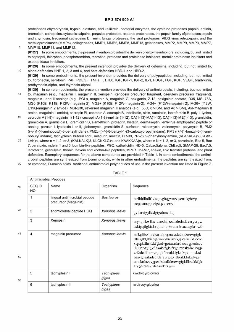
EP 3 574 909 A1
23
5
10
15
20
25
30
35
40
45
50
55
proteinases chymotrypsin, trypsin, elastase, and kallikrein, bacterial enzymes, the cysteine proteases papain, actinin,bromelain, cathepsins, cytosolic calpains, parasitic proteases, aspartic proteinases, the pepsin family of proteases pepsinand chymosin, lysosomal cathepsins D, renin, fungal proteases, the viral proteases, AIDS virus retropepsin, and themetalloproteinases (MMPs), collagenases, MMP1, MMP2, MMP8, MMP13, gelatinases, MMP2, MMP9, MMP3, MMP7,MMP10, MMP11, and MMP12.[0127] In some embodiments, the present invention provides the delivery of enzyme inhibitors, including, but not limitedto captopril, thiorphan, phosphoramidon, teprotide, protease and proteinase inhibitors, metalloproteinase inhibitors andexopeptidase inhibitors.[0128] In some embodiments, the present invention provides the delivery of defensins, including, but not limited to,alpha-defensins HNP 1, 2, 3 and 4, and beta-defensins HBD-1 and HBD-2.[0129] In some embodiments, the present invention provides the delivery of polypeptides, including, but not limitedto, fibronectin, serotonin, PAF, PDEGF, TNFa, IL1, IL6, IGF, IGF-1, IGF-2, IL-1, PDGF, FGF, KGF, VEGF, bradykinin,prothymosin-alpha, and thymosin-alphal.[0130] In some embodiments, the present invention provides the delivery of antimicrobials, including, but not limitedto, magainin (e.g., magainin I, magainin II, xenopsin, xenopsin precursor fragment, caerulein precursor fragment),magainin I and II analogs (e.g., PGLa, magainin A, magainin G, pexiganin, Z-12, pexigainin acetate, D35, MSI-78A,MG0 (K10E, K11E, F12W-magainin 2), MG2+ (K10E, F12W-magainin-2), MG4+ (F12W-magainin 2), MG6+ (f12W,E19Q-magainin 2 amide), MSI-238, reversed magainin II analogs (e.g., 53D, 87-ISM, and A87-ISM), Ala-magainin IIamide, magainin II amide), cecropin PI, cecropin A, cecropin B, indolicidin, nisin, ranalexin, lactoferricin B, poly-L-lysine,cecropin A (1-8)-magainin II (1-12), cecropin A (1-8)-melittin (1-12), CA(1-13)-MA(1-13), CA(1-13)-ME(1-13), gramicidin,gramicidin A, gramicidin D, gramicidin S, alamethicin, protegrin, histatin, dermaseptin, lentivirus amphipathic peptide oranalog, parasin I, lycotoxin I or II, globomycin, gramicidin S, surfactin, ralinomycin, valinomycin, polymyxin B, PM2((+/-)1-(4-aminobutyl)-6-benzylindane), PM2c ((+/-)-6-benzyl-1-(3-carboxypropyl)indane), PM3 ((+/-)1-benzyl-6-(4-ami-nobutyl)indane), tachyplesin, buforin I or II, misgurin, melittin, PR-39, PR-26, 9-phenylnonylamine, (KLAKKLA)n, (KLAK-LAK)n, where n = 1, 2, or 3, (KALKALK)3, KLGKKLG)n, and KAAKKAA)n, wherein N = 1, 2, or 3, paradaxin, Bac 5, Bac7, ceratoxin, mdelin 1 and 5, bombin-like peptides, PGQ, cathelicidin, HD-5, Oabac5alpha, ChBac5, SMAP-29, Bac7.5,lactoferrin, granulysin, thionin, hevein and knottin-like peptides, MPG1, IbAMP, snakin, lipid transfer proteins, and plantdefensins. Exemplary sequences for the above compounds are provided in Table 1. In some embodiments, the antimi-crobial peptides are synthesized from L-amino acids, while in other embodiments, the peptides are synthesized from,or comprise, D-amino acids. Additional antimicrobial polypeptides of use in the present invention are listed in Figure 7.
TABLE 1
Antimicrobial Peptides
SEQ ID NO:
Name Organism Sequence
1 lingual antimicrobial peptide precursor (Magainin)
Bos taurus
2 antimicrobial peptide PGQ Xenopus laevis
3 Xenopsin Xenopus laevis
4 magainin precursor Xenopus laevis
5 tachyplesin I Tachypleus gigas
kwcfrvcyrgicyrrcr
6 tachyplesin II Tachypleus gigas
rwcfrvcyrgicyrkcr
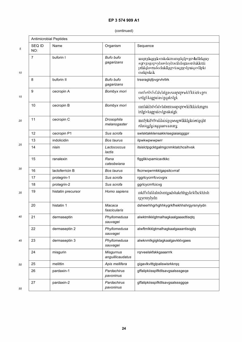
EP 3 574 909 A1
24
5
10
15
20
25
30
35
40
45
50
55
(continued)
Antimicrobial Peptides
SEQ ID NO:
Name Organism Sequence
7 buforin I Bufo bufo gagarizans
8 buforin II Bufo bufo gagarizans
trssraglqfpvgrvhrllrk
9 cecropin A Bombyx mori
10 cecropin B Bombyx mori
11 cecropin C Drosophila melanogaster
12 cecropin P1 Sus scrofa swlsktakklensakkrisegiaiaiqggpr
13 indolicidin Bos taurus ilpwkwpwwpwrr
14 nisin Lactococcus lactis
itsislctpgcktgalmgcnmktatchcsihvsk
15 ranalexin Rana catesbeiana
flgglikivpamicavtkkc
16 lactoferricin B Bos taurus fkcrrwqwrmkklgapsitcvrraf
17 protegrin-1 Sus scrofa rggrlcycrrrfcvcvgrx
18 protegrin-2 Sus scrofa ggrlcycrrrfcicvg
19 histatin precursor Homo sapiens
20 histatin 1 Macaca fascicularis
dsheerhhgrhghhkygrkfhekhhshrgyrsnylydn
21 dermaseptin Phyllomedusa sauvagei
alwktmlkklgtmalhagkaalgaaadtisqtq
22 dermaseptin 2 Phyllomedusa sauvagei
alwftmlkklgtmalhagkaalgaaantisqgtq
23 dermaseptin 3 Phyllomedusa sauvagei
alwknmlkgigklagkaalgavkklvgaes
24 misgurin Misgurnus anguillicaudatus
rqrveelskfskkgaaarrrk
25 melittin Apis mellifera gigavlkvlttglpaliswisrkkrqq
26 pardaxin-1 Pardachirus pavoninus
gffalipkiissplfktllsavgsalsssgeqe
27 pardaxin-2 Pardachirus pavoninus
gffalipkiisspifktllsavgsalsssggqe
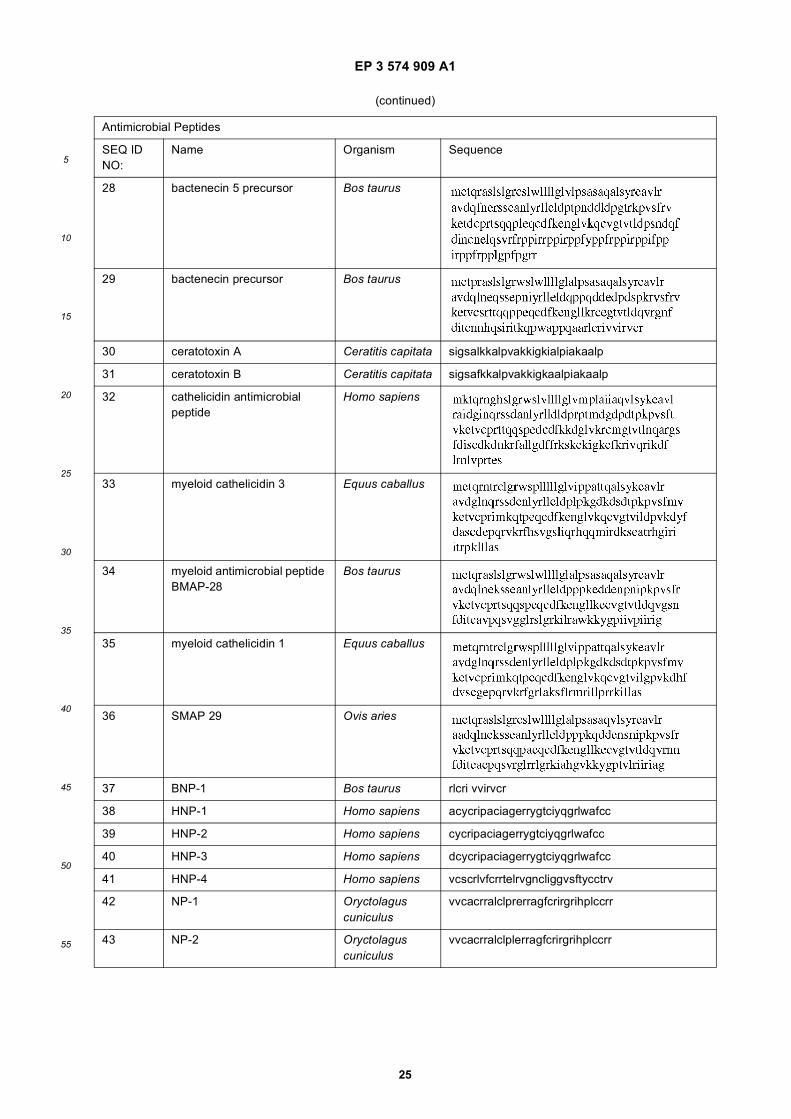
EP 3 574 909 A1
25
5
10
15
20
25
30
35
40
45
50
55
(continued)
Antimicrobial Peptides
SEQ ID NO:
Name Organism Sequence
28 bactenecin 5 precursor Bos taurus
29 bactenecin precursor Bos taurus
30 ceratotoxin A Ceratitis capitata sigsalkkalpvakkigkialpiakaalp
31 ceratotoxin B Ceratitis capitata sigsafkkalpvakkigkaalpiakaalp
32 cathelicidin antimicrobial peptide
Homo sapiens
33 myeloid cathelicidin 3 Equus caballus
34 myeloid antimicrobial peptide BMAP-28
Bos taurus
35 myeloid cathelicidin 1 Equus caballus
36 SMAP 29 Ovis aries
37 BNP-1 Bos taurus rlcri vvirvcr
38 HNP-1 Homo sapiens acycripaciagerrygtciyqgrlwafcc
39 HNP-2 Homo sapiens cycripaciagerrygtciyqgrlwafcc
40 HNP-3 Homo sapiens dcycripaciagerrygtciyqgrlwafcc
41 HNP-4 Homo sapiens vcscrlvfcrrtelrvgncliggvsftycctrv
42 NP-1 Oryctolagus cuniculus
vvcacrralclprerragfcrirgrihplccrr
43 NP-2 Oryctolagus cuniculus
vvcacrralclplerragfcrirgrihplccrr
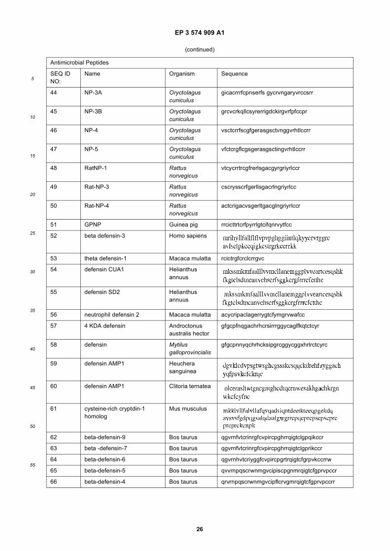
EP 3 574 909 A1
26
5
10
15
20
25
30
35
40
45
50
55
(continued)
Antimicrobial Peptides
SEQ ID NO:
Name Organism Sequence
44 NP-3A Oryctolagus cuniculus
gicacrrrfcpnserfs gycrvngaryvrccsrr
45 NP-3B Oryctolagus cuniculus
grcvcrkqllcsyrerrigdckirgvrfpfccpr
46 NP-4 Oryctolagus cuniculus
vsctcrrfscgfgerasgsctvnggvrhtlccrr
47 NP-5 Oryctolagus cuniculus
vfctcrgflcgsgerasgsctingvrhtlccrr
48 RatNP-1 Rattus norvegicus
vtcycrrtrcgfrerlsgacgyrgriyrlccr
49 Rat-NP-3 Rattus norvegicus
cscrysscrfgerllsgacrlngriyrlcc
50 Rat-NP-4 Rattus norvegicus
actcrigacvsgerltgacglngriyrlccr
51 GPNP Guinea pig rrcicttrtcrfpyrrlgtcifqnrvytfcc
52 beta defensin-3 Homo sapiens
53 theta defensin-1 Macaca mulatta rcictrgfcrclcrrgvc
54 defensin CUA1 Helianthus annuus
55 defensin SD2 Helianthus annuus
56 neutrophil defensin 2 Macaca mulatta acycripaclagerrygtcfymgrvwafcc
57 4 KDA defensin Androctonus australis hector
gfgcpfnqgachrhcrsirrrggycaglfkqtctcyr
58 defensin Mytilus galloprovincialis
gfgcpnnyqchrhcksipgrcggycggxhrlrctcyrc
59 defensin AMP1 Heuchera sanguinea
60 defensin AMP1 Clitoria ternatea
61 cysteine-rich cryptdin-1 homolog
Mus musculus
62 beta-defensin-9 Bos taurus qgvrnfvtcrinrgfcvpircpghrrqigtclgpqikccr
63 beta -defensin-7 Bos taurus qgvrnfvtcrinrgfcvpircpghrrqigtclgprikccr
64 beta-defensin-6 Bos taurus qgvrnhvtcriyggfcvpircpgrtrqigtcfgrpvkccrrw
65 beta-defensin-5 Bos taurus qvvrnpqscrwnmgvcipiscpgnmrqigtcfgprvpccr
66 beta-defensin-4 Bos taurus qrvrnpqscrwnmgvcipflcrvgmrqigtcfgprvpccrr
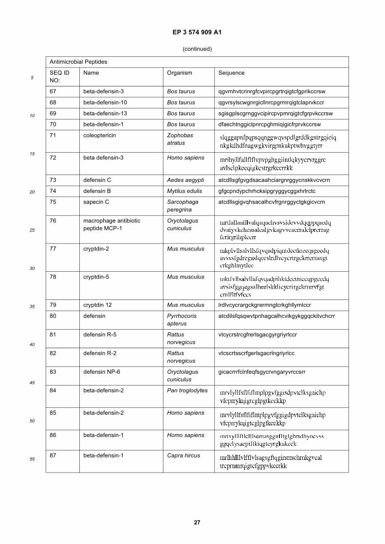
EP 3 574 909 A1
27
5
10
15
20
25
30
35
40
45
50
55
(continued)
Antimicrobial Peptides
SEQ ID NO:
Name Organism Sequence
67 beta-defensin-3 Bos taurus qgvrnhvtcrinrgfcvpircpgrtrqigtcfgprikccrsw
68 beta-defensin-10 Bos taurus qgvrsylscwgnrgicllnrcpgrmrqigtclaprvkccr
69 beta-defensin-13 Bos taurus sgisgplscgrnggvcipircpvpmrqigtcfgrpvkccrsw
70 beta-defensin-1 Bos taurus dfaschtnggiclpnrcpghmiqigicfrprvkccrsw
71 coleoptericin Zophobas atratus
72 beta defensin-3 Homo sapiens
73 defensin C Aedes aegypti atcdllsgfgvgdsacaahciargnrggycnskkvcvcrn
74 defensin B Mytilus edulis gfgcpndypchrhcksipgryggycggxhrlrctc
75 sapecin C Sarcophaga peregrina
atcdllsgigvqhsacalhcvfrgnrggyctgkgicvcrn
76 macrophage antibiotic peptide MCP-1
Oryctolagus cuniculus
77 cryptdin-2 Mus musculus
78 cryptdin-5 Mus musculus
79 cryptdin 12 Mus musculus lrdlvcycrargckgrermngtcrkghllymlccr
80 defensin Pyrrhocoris apterus
atcdilsfqsqwvtpnhagcalhcvikgykggqckitvchcrr
81 defensin R-5 Rattus norvegicus
vtcycrstrcgfrerlsgacgyrgriyrlccr
82 defensin R-2 Rattus norvegicus
vtcscrtsscrfgerlsgacrlngriyrlcc
83 defensin NP-6 Oryctolagus cuniculus
gicacrrrfclnfeqfsgycrvngaryvrccsrr
84 beta-defensin-2 Pan troglodytes
85 beta-defensin-2 Homo sapiens
86 beta-defensin-1 Homo sapiens
87 beta-defensin-1 Capra hircus
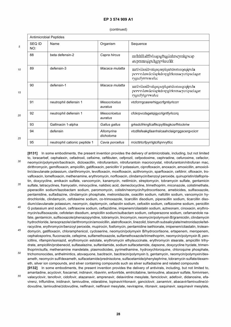
EP 3 574 909 A1
28
5
10
15
20
25
30
35
40
45
50
55
[0131] In some embodiments, the present invention provides the delivery of antimicrobials, including, but not limitedto, loracarbef, cephalexin, cefadroxil, cefixime, ceftibuten, cefprozil, cefpodoxime, cephradine, cefuroxime, cefaclor,neomycin/polymyxin/bacitracin, dicloxacillin, nitrofurantoin, nitrofurantoin macrocrystal, nitrofurantoin/nitrofuran mac,dirithromycin, gemifloxacin, ampicillin, gatifloxacin, penicillin V potassium, ciprofloxacin, enoxacin, amoxicillin, amoxicil-lin/clavulanate potassium, clarithromycin, levofloxacin, moxifloxacin, azithromycin, sparfloxacin, cefdinir, ofloxacin, tro-vafloxacin, lomefloxacin, methenamine, erythromycin, norfloxacin, clindamycin/benzoyl peroxide, quinupristin/dalfopris-tin, doxycycline, amikacin sulfate, vancomycin, kanamycin, netilmicin, streptomycin, tobramycin sulfate, gentamicinsulfate, tetracyclines, framycetin, minocycline, nalidixic acid, demeclocycline, trimethoprim, miconazole, colistimethate,piperacillin sodium/tazobactam sodium, paromomycin, colistin/neomycin/hydrocortisone, amebicides, sulfisoxazole,pentamidine, sulfadiazine, clindamycin phosphate, metronidazole, oxacillin sodium, nafcillin sodium, vancomycin hy-drochloride, clindamycin, cefotaxime sodium, co-trimoxazole, ticarcillin disodium, piperacillin sodium, ticarcillin diso-dium/clavulanate potassium, neomycin, daptomycin, cefazolin sodium, cefoxitin sodium, ceftizoxime sodium, penicillinG potassium and sodium, ceftriaxone sodium, ceftazidime, imipenem/cilastatin sodium, aztreonam, cinoxacin, erythro-mycin/sulfisoxazole, cefotetan disodium, ampicillin sodium/sulbactam sodium, cefoperazone sodium, cefamandole na-fate, gentamicin, sulfisoxazole/phenazopyridine, tobramycin, lincomycin, neomycin/polymyxin B/gramicidin, clindamycinhydrochloride, lansoprazole/clarithromycin/amoxicillin, alatrofloxacin, linezolid, bismuth subsalicylate/metronidazole/tet-racycline, erythromycin/benzoyl peroxide, mupirocin, fosfomycin, pentamidine isethionate, imipenem/cilastatin, trolean-domycin, gatifloxacin, chloramphenicol, cycloserine, neomycin/polymyxin B/hydrocortisone, ertapenem, meropenem,cephalosporins, fluconazole, cefepime, sulfamethoxazole, sulfamethoxazole/trimethoprim, neomycin/polymyxin B, pen-icillins, rifampin/isoniazid, erythromycin estolate, erythromycin ethylsuccinate, erythromycin stearate, ampicillin trihy-drate, ampicillin/probenecid, sulfasalazine, sulfanilamide, sodium sulfacetamide, dapsone, doxycycline hyclate, trimen-thoprim/sulfa, methenamine mandelate, plasmodicides, pyrimethamine, hydroxychloroquine, chloroquine phosphate,trichomonocides, anthelmintics, atovaquone, bacitracin, bacitracin/polymyxin b, gentamycin, neomycin/polymyxin/dex-ameth, neomycin sulf/dexameth, sulfacetamide/prednisolone, sulfacetamide/phenylephrine, tobramycin sulfate/dexam-eth, silver ion compounds, and silver containing compounds such as silver sulfadiazine and related compounds[0132] In some embodiments, the present invention provides the delivery of antivirals, including, but not limited to,amantadine, acyclovir, foscarnet, indinavir, ribavirin, enfuvirtide, emtricitabine, lamivudine, abacavir sulfate, fomivirsen,valacyclovir, tenofovir, cidofovir, atazanavir, amprenavir, delavirdine mesylate, famciclovir, adefovir, didanosine, efa-virenz, trifluridine, inidinavir, lamivudine, vidarabine, lopinavir/ritonavir, ganciclovir, zanamivir, abacavir/lamivudine/zi-dovudine, lamivudine/zidovudine, nelfinavir, nelfinavir mesylate, nevirapine, ritonavir, saquinavir, saquinavir mesylate,
(continued)
Antimicrobial Peptides
SEQ ID NO:
Name Organism Sequence
88 beta defensin-2 Capra hircus
89 defensin-3 Macaca mulatta
90 defensin-1 Macaca mulatta
91 neutrophil defensin 1 Mesocricetus auratus
vtcfcrrrgcasrerhigycrfgntiyrlccrr
92 neutrophil defensin 1 Mesocricetus auratus
cfckrpvcdsgetqigycrlgntfyrlccrq
93 Gallinacin 1-alpha Gallus gallus grksdcfrkngfcaflkcpyltlisgkcsrfhlcckriw
94 defensin Allomyrina dichotoma
vtcdllsfeakgfaanhslcaahclaigrrggscergvcicrr
95 neutrophil cationic peptide 1 Cavia porcellus rrcicttrtcrfpyrrlgtcifqnrvytfcc
EP 3 574 909 A1
29
5
10
15
20
25
30
35
40
45
50
55
rimantadine, stavudine, docosanol, zalcitabine, idoxuridine, zidovudine, zidovudine/didanosine, valganciclovir, penci-clovir, lamivudine, and oseltamivir.[0133] In some embodiments, the present invention provides the delivery of antifungals, including, but not limited to,amphotericin B, nystatin, nystatin/triamcinolone, itraconazole, ketoconazole, miconazole, sulconazole, clotrimazole, clot-rimazole/betamethasone, enilconazole, econazole, oxiconazole, tioconazole, terconazole, butoconazole, thiabendazole,flucytosine, butenafine, ciclopirox, haloprogin, naftifine, tolnaftate, natamycin, undecylenic acid, mafenide, dapsone,clioquinol, clioquinol/hydrocortisone, potassium iodide, silver sulfadiazine, gentian violet, carbol-fuchsin, cilofungin, sert-aconazole, voriconazole, fluconazole, terbinafine, caspofungin, other topical azole drugs, and griseofulvin.[0134] In some embodiments, the present invention provides the use and delivery of buffering agents, including, butnot limited to, Maleic acid, Phosphoric acid, Glycine, Chloroacetic acid, Formic acid, Benzoic acid, Acetic acid, Pyridine,Piperazine, MES, Bis-tris, Carbonate, ACES, ADA MOPSO, PIPES, Phosphoric acid, BES, MOPS, TES, HEPES, DIPSO,TAPSO, Triethanolamine, HEPSO, Tris, Tricine, Bicine, TAPS, Borate, Ammonia, CHES, Ethanolamine, CAPSO, Gly-cine, Carbonate, CAPS, Methylamine, Piperidine, and Phosphoric acid.[0135] In some embodiments, the present invention provides the delivery of vitamins and minerals, including, but notlimited to, Vitamin A, Carotenoids, Vitamin D, Vitamin E, Vitamin K, Vitamin C/ascorbic acid, B1/thiamin, B2/riboflavin,B3/niacin, B5/pantothenic acid, B6/pyridoxine, B12/cobalamin, Biotin, Calcium, Magnesium, Phosphorus, Sodium, Chlo-ride, Potassium, Boron, Chromium, Copper, Iodine, Iron, Manganese, Selenium, and Zinc.[0136] In some embodiments, the present invention provides the delivery of analgesics, including, but not limited to,acetaminophen, anileridine, acetylsalicylic acid, buprenorphine, butorphanol, fentanyl, fentanyl citrate, codeine, rofecox-ib, hydrocodone, hydromorphone, hydromorphone hydrochloride, levorphanol, alfentanil hydrochloride, meperidine,meperidine hydrochloride, methadone, morphine, nalbuphine, opium, levomethadyl, hyaluronate sodium, sufentanilcitrate, capsaicin, tramadol, leflunomide, oxycodone, oxymorphone, celecoxib, pentazocine, propoxyphene, benzocaine,lidocaine, dezocine, clonidine, butalbital, phenobarbital, tetracaine, phenazopyridine, sulfamethoxazole/phenazopyrid-ine, and sulfisoxazole/phenazopyridine.[0137] In some embodiments, the present invention provides the delivery of anticoagulants, including, but not limitedto, coumarins, 1,3-indandione, anisindione, fondaparinux, heparin, lepirudin, antithrombin, warfarin, enoxaparin, dipyri-damole, dalteparin, ardeparin, nadroparin, and tinzaparin.[0138] In some embodiments, the present invention provides the delivery of coagulation factors, including, but notlimited to, Factor I (fibrinogen), Factor II (prothrombin), Factor III (thromboplastin, tissue factor), Factor IV (calcium),Factor V (labile factor), Factor VII (stable factor), Factor VIII (antihemophilic globulin, antihemophilic globulin, antihemo-philic factor A), Factor IX (plasma thromboplastin component, Christmas factor, antihemophilic factor B), Factor X (Stuartfactor, Prower factor, Stuart-Prower factor), Factor XI (plasma thromboplastin antecedent, antihemophilic factor C),Factor XII (Hageman factor, surface factor, contact factor), and Factor XIII (fibrin stabilizing factor, fibrin stabilizingenzyme, fibri-nase).[0139] In some embodiments, the present invention provides the delivery of anti-inflammatory agents, including, butnot limited to, non steroidal anti-inflammatory drugs (NSAIDs) including diclofenac (also known as Voltaren, Abitren,Allvoran, Almiral, Alonpin, Anfenax, Artrites, Betaren, Blesin, Bolabomin, Cataflam, Clofec, Clofen, Cordralan, Curinflam,Diclomax, Diclosian, Dicsnal, Difenac, Ecofenac, Hizemin, Inflamac, Inflanac, Klotaren, Lidonin, Monoflam, Naboal,Oritaren, Remethan, Savismin, Silino, Staren, Tsudohmin, Voltarol, Voren, Voveran, and Vurdon), diflunisal (also knownas Dolobid, Adomal, Diflonid, Diflunil, Dolisal, Dolobis, Dolocid, Donobid, Dopanone, Dorbid, Dugodol, Flovacil, Fluniget,Fluodonil, Flustar, Ilacen, Noaldol, Reuflos, and Unisal), etodolac (also known as Lodine), fenoprofen (also known asNalfon, Fenoprex, Fenopron, Fepron, Nalgesic, and Progesic), flurbiprofen (also known as Ansaid and Ocuflur), ibuprofen(also known as Rufen, Motrin, Aches-N-Pain, Advil, Nuprin, Dolgesic, Genpril, Haltran, Ibifon, Ibren, Ibumed, Ibuprin,Ibupro-600, Ibuprohm, Ibu-Tab, Ibutex, Ifen, Medipren, Midol 200, Motrin-IB, Cramp End, Profen, Ro-Profen, Trendar,Alaxan, Brofen, Alfam, Brufen, Algofen, Brufort, Amersol, Bruzon, Andran, Buburone, Anflagen, Butacortelone, Apsifen,Deflem, Artofen, Dolgit, Artril, Dolocyl, Bloom, Donjust, Bluton, Easifon, Ebufac, Emflam, Emodin, Fenbid, Fenspan,Focus, Ibosure, Ibufen, Ibufug, Ibugen, Ibumetin, Ibupirac, Imbun, Inabrin, Inflam, Irfen, Librofen, Limidon, Lopane,Mynosedin, Napacetin, Nobafon, Nobgen, Novogent, Novoprofen, Nurofen, Optifen, Paduden, Paxofen, Perofen, Proar-tinal, Prontalgin, Q-Profen, Relcofen, Remofen, Roidenin, Seclodin, Tarein, and Zofen), indomethacin (also known asIndameth, Indocin, Amuno, Antalgin, Areumatin, Argilex, Artherexin, Arthrexin, Artrinovo, Bavilon, Bonidon, Boutycin,Chrono-Indocid, Cidalgon, Confortid, Confortind, Domecid, Durametacin, Elemetacin, Idicin, Imbrilon, Inacid, Indacin,Indecin, Indocap, Indocen, Indocid, Indoflex, Indolag, Indolar, Indomed, Indomee, Indometacinum, Indometicina, In-dometin, Indovis, Indox, Indozu, Indrenin, Indylon, Inflazon, Inpan, Lauzit, Liometace, Metacen, Metindon, Metocid,Mezolin, Mobilan, Novomethacin, Peralgon, Reflox, Rheumacid, Rheumacin, Salinac, Servindomet, Toshisan, and Von-um), ketoprofen (also known as Orudis, Alrheumat, Alrheumun, Alrhumat, Aneol, Arcental, Dexal, Epatec, Fastum,Keduril, Kefenid, Keprofen, Ketofen, Ketonal, Ketosolan, Kevadon, Mero, Naxal, Oruvail, Profenid, Salient, Tofen, andTreosin), ketorolac (also known as Toradol), meclofenamate (also known as Meclofen, Meclomen, and Movens),mefenamic acid (also known as Ponstel, Alpain, Aprostal, Benostan, Bonabol, Coslan, Dysman, Dyspen, Ecopan,
EP 3 574 909 A1
30
5
10
15
20
25
30
35
40
45
50
55
Lysalgo, Manic, Mefac, Mefic, Mefix, Parkemed, Pondex, Ponsfen, Ponstan, Ponstyl, Pontal, Ralgec, and Youfenam),nabumetone (also known as Relafen), naproxen (also known as Naprosyn, Anaprox, Aleve, Apranax, Apronax, Arthrisil,Artrixen, Artroxen, Bonyl, Congex, Danaprox, Diocodal, Dysmenalgit, Femex, Flanax, Flexipen, Floginax, Gibixen, Head-lon, Laraflex, Laser, Leniartil, Nafasol, Naixan, Nalyxan, Napoton, Napren, Naprelan, Naprium, Naprius, Naprontag,Naprux, Napxen, Narma, Naxen, Naxid, Novonaprox, Nycopren, Patxen, Prexan, Prodexin, Rahsen, Roxen, Saritilron,Sinartrin, Sinton, Sutony, Synflex, Tohexen, Veradol, Vinsen, and Xenar), oxaprozin (also known as Daypro), piroxicam(also known as Feldene, Algidol, Antiflog, Arpyrox, Atidem, Bestocam, Butacinon, Desinflam, Dixonal, Doblexan, Dolo-nex, Feline, Felrox, Fuldin, Indene, Infeld, Inflamene, Lampoflex, Larapam, Medoptil, Novopirocam, Osteral, Pilox, Pi-raldene, Piram, Pirax, Piricam, Pirocam, Pirocaps, Piroxan, Piroxedol, Piroxim, Piton, Posidene, Pyroxy, Reucam, Rex-icam, Riacen, Rosic, Sinalgico, Sotilen, Stopen, and Zunden), sulindac (also known as Clinoril, Aflodac, Algocetil, Antribid,Arthridex, Arthrocine, Biflace, Citireuma, Clisundac, Imbaral, Lindak, Lyndak, Mobilin, Reumofil, Sudac, Sulene, Sulic,Sulindal, Suloril, and Sulreuma), tolmetin (also known as Tolectin, Donison, Midocil, Reutol, and Safitex), celecoxib (alsoknown as Celebrex), meloxicam (also known as Mobic), rofecoxib (also known as Vioxx), valdecoxib (also known asBextra), aspirin (also known as Anacin, Ascriptin, Bayer, Bufferin, Ecotrin, and Excedrin) and steroidal anti-inflammatorydrugs including cortisone, prednisone and dexamethasone.[0140] In some embodiments, the present invention provides the delivery of vasoconstrictors, including, but not limitedto, epinephrine (adrenaline, Susphrine), phenylephrine hydrochloride (Neo-Synephrine), oxymetazoline hydrochloride(Afrin), norepinephrine (Levophed), and caffeine.[0141] In some embodiments, the present invention provides the delivery of vasodilators, including, but not limited to,bosentan (Tracleer), epoprostenol (Flolan), treprostinil (Remodulin), sitaxsentan, nifedipine (Adalat, Procardia), nica-rdipine (Cardene), verapamil (Calan, Covera-HS, Isoptin, Verelan), diltiazem (Dilacor XR, Diltia XT, Tiamate, Tiazac,Cardizem), isradipine (DynaCirc), nimodipine (Nimotop), amlodipine (Norvasc), felodipine (Plendil), nisoldipine (Sular),bepridil (Vascor), hydralazine (Apresoline), minoxidil (Loniten), isosorbide dinitrate (Dilatrate-SR, Iso-Bid, Isonate, Isorb-id, Isordil, Isotrate, Sorbitrate), isorbide mononitrate (IMDUR), prazosin (Minipress), cilostazol (Pletal), treprostinil (Re-modulin), cyclandelate, isoxsuprine (Vasodilan), nylidrin (Arlidin), nitrates (Deponit, Minitran, Nitro-Bid, Nitrodisc, Nitro-Dur, Nitrol, Transderm-Nitro), benazepril (Lotensin), benazepril and hydrochlorothiazide (Lotensin HCT), captopril (Ca-poten), captopril and hydrochlorothiazide (Capozide), enalapril (Vasotec), enalapril and hydrochlorothiazide (Vaseretic),fosinopril (Monopril), lisinopril (Prinivil, Zestril), lisinopril and hydrochlorothiazide (Prinzide, Zestoretic), moexipril (Uni-vasc), moexipril and hydrochlorothiazide (Uniretic), perindopril (Aceon), quinapril (Accupril), quinapril and hydrochloro-thiazide (Accuretic), ramipril (Altace), trandolapril (Mavik), papaverine (Cerespan, Genabid, Pavabid, Pavabid HP, Pa-vacels, Pavacot, Pavagen, Pavarine, Pavased, Pavatine, Pavatym, Paverolan).[0142] In some embodiments, the present invention provides the delivery of diuretics, including, but not limited to,acetazolamide (Diamox), dichlorphenamide (Daranide), methazolamide (Neptazane), bendroflumethiazide (Naturetin),benzthiazide (Exna), chlorothiazide (Diuril), chlorthalidone (Hygroton), hydrochlorothiazide (Esidrix, HydroDiuril, Micro-zide), hydroflumethiazide (Diucardin), indapamide (Lozol), methyclothiazide (Enduron), metolazone (Zaroxolyn, Mykrox),polythiazide (Renese), quinethazone (Hydromox), trichlormethiazide (Naqua), bumetanide (Bumex), ethacrynic acid(Edecrin), furosemide (Lasix), torsemide (Demadex), amiloride (Midamor), amiloride and hydrochlorothiazide (Moduret-ic), spironolactone (Aldactone), spironolactone and hydrochlorothiazide (Aldactazide), triamterene (Dyrenium), triam-terene and hydrochlorothiazide (Dyazide, Maxzide).[0143] In some embodiments, the present invention provides the delivery of anti-cancer agents, including, but notlimited to, aldesleukin, alemtuzumab, alitretinoin, allopurinol, altretamine, amifostine, anagrelide, anastrozole, arsenictrioxide, asparaginase, bexarotene, bicalutamide, bleomycin, busulfan, calusterone, capecitabine, carboplatin, carmus-tine, celecoxib, chlorambucil, cisplatin, cladribine, cyclophosphamide, cytarabine, dacarbazine, dactinomycin, darbepo-etin alpha, daunorubicin, daunomycin, dexrazoxane, docetaxel, doxorubicin, epoetin alpha, estramustine, etoposide,etoposide phosphate, exemestane, filgrastim, floxuridine, fludarabine, flutamide, fulvestrant, gemcitabine, gemtuzumabozogamicin, goserelin acetate, hydroxyurea, ibritumomab tiuxetan, idarubicin, ifosfamide, imatinib mesylate, interferonalpha-2a, interferon alpha-2b, irinotecan, leflunomide, letrozole, leucovorin, levamisole, lomustine, meclorethamine (ni-trogen mustard), megestrol acetate, melphalan, mercaptopurine, mesna, methotrexate, methoxsalen, mitomycin C,mitotane, mitoxantrone, mycophenolate mofetil, nandrolone phenpropionate, nilutamide, nofetumomab, oprelvekin,oxaliplatin, paclitaxel, pamidronate, pegademase, pegaspargase, pegfilgrastim, pentostatin, pipobroman, plicamycin,porfimer sodium, procarbazine, quinacrine, rasburicase rituximab, sargramostim, streptozocin, tacrolimus, tamoxifen,temozolomide, teniposide, testolactone, thioguanine, thiotepa, topotecan, toremifene, tositumomab, trastuzumab, tretin-oin, uracil mustard, valrubicin, vinblastine, vincristine, vinorelbine, and zoledronate.[0144] In other embodiments, the wound active agent is an siRNA. The RNAi constructs of the present invention aregene(s) that express RNAs that base pair to form a dsRNA RNA region. The RNAs may be a part of the same moleculeor different molecules. In preferred embodiments, the RNAi construct comprises a promoter operably linked to a nucleicacid sequence encoding two complementary sequences separated by a loop sequence. The complementary regionscorrespond to a target RNA sequence separated by a loop sequence. When the RNAi construct is expressed, the
EP 3 574 909 A1
31
5
10
15
20
25
30
35
40
45
50
55
complementary regions of the resulting RNA molecule pair with one another to form a double stranded RNA region. Thepresent invention is not limited to loop sequences of any particular length. In some preferred embodiments, the loopsequences range from about 4 to about 20 nucleotides in length. In more preferred embodiments, the loop sequencesare from about 6 to about 12 nucleotides in length. In other preferred embodiments, the dsRNA regions are from about19 to about 23 in length.[0145] In other embodiments, the dsRNA is formed from RNA transcribed from a vector as two separate stands. Inother embodiments, the two strands of DNA used to form the dsRNA may belong to the same or two different duplexesin which they each form with a DNA strand of at least partially complementary sequence. When the dsRNA is thus-produced, the DNA sequence to be transcribed is flanked by two promoters, one controlling the transcription of one ofthe strands, and the other that of the complementary strand. These two promoters may be identical or different. In someembodiments, a DNA duplex provided at each end with a promoter sequence can directly generate RNAs of definedlength, and which can join in pairs to form a dsRNA. See, e.g., U.S. Pat. No. 5,795,715, incorporated herein by reference.RNA duplex formation may be initiated either inside or outside the cell.[0146] It will be recognized that after processing the resulting siRNA can comprise two blunt ends, one blunt end andone end with an overhang, or two ends with overhangs. In some embodiments, the end or ends with overhangs comprisean overhang of either one or two nucleotides. As a none limiting example, a siRNA of 23 nucleotides in length comprisestwo 19mers with a two nucleotide overhang at each end. As another non-limiting example, a siRNA of 21 nucleotidesin length comprises two 19mers with a single nucleotide overhang at each end. As still another non-limiting example, asiRNA of 22 nucleotides in length comprises two 22mers with no overhangs at either end.[0147] Inhibition is sequence-specific in that nucleotide sequences corresponding to the duplex region of the RNA aretargeted for genetic inhibition. RNA molecules containing a nucleotide sequence identical to a portion of the target geneare preferred for inhibition. RNA sequences with insertions, deletions, and single point mutations relative to the targetsequence have also been found to be effective for inhibition. Thus, sequence identity may optimized by sequencecomparison and alignment algorithms known in the art (see Gribskov and Devereux, Sequence Analysis Primer, StocktonPress, 1991, and references cited therein) and calculating the percent difference between the nucleotide sequences by,for example, the Smith-Waterman algorithm as implemented in the BESTFIT software program using default parameters(e.g., University of Wisconsin Genetic Computing Group). Greater than 90% sequence identity, or even 100% sequenceidentity, between the inhibitory RNA and the portion of the target gene is preferred. Alternatively, the duplex region ofthe RNA may be defined functionally as a nucleotide sequence that is capable of hybridizing with a portion of the targetgene transcript.[0148] There is no upper limit on the length of the dsRNA that can be used. For example, the dsRNA can range fromabout 21 base pairs (bp) of the gene to the full length of the gene or more. In one embodiment, the dsRNA used in themethods of the present invention is about 1000 bp in length. In another embodiment, the dsRNA is about 500 bp inlength. In yet another embodiment, the dsRNA is about 22 bp in length.In some preferred embodiments, the sequences that mediate RNAi are from about 21 to about 23 nucleotides. Theisolated iRNAs of the present invention mediate degradation of the target RNA.[0149] The double stranded RNA of the present invention need only be sufficiently similar to natural RNA that it hasthe ability to mediate RNAi for the target RNA. In one embodiment, the present invention relates to RNA molecules ofvarying lengths that direct cleavage of specific mRNA to which their sequence corresponds. It is not necessary that therebe perfect correspondence of the sequences, but the correspondence must be sufficient to enable the RNA to directRNAi cleavage of the target mRNA. In a particular embodiment, the RNA molecules of the present invention comprisea 3’ hydroxyl group.
III. Matrix compositions
[0150] In some embodiments, the present invention provides compositions comprising a matrix that can be appliedto a wound. In some embodiments, the matrix is functionalized. In some embodiments, the matrix comprises one ormore polymers, preferably biocompatible, or is formed from one more proteins, or is a combination of polymers andproteins. The matrix is preferably functionalized to allow for covalent interaction and/or binding to the wound bed, or toallow application of wound active agents to the matrix. In some embodiments, a wound active agent, for example anantimicrobial agent such as a solver compound, is incorporated into the matrix. In some embodiments, the matrix isformed or provided on a solid support. The solid support can form an outer permeable, semi-permeable, or impermeablebarrier after application of the matrix to a wound or can serve as a removed support for transfer and application of thematrix to the wound followed by removal of the support. In some embodiments, the matrix is a commercially availablematrix such as Biobranetm.[0151] In some embodiments, the matrices, such as polymer multilayers, are nanoscale in dimension. Accordingly, insome embodiments, the matrices are from about 1 nm to 1000 nm thick, from about 1 nm to 500 nm thick, from about1 nm to 100 nm thick, from about 1 nm to about 25 nm thick, from about 1 nm to about 10 nm thick, or less than about
EP 3 574 909 A1
32
5
10
15
20
25
30
35
40
45
50
55
500 nm, 100 nm, 25 nm or 10 nm thick. It is contemplated that the nanoscale dimension of the matrices (i.e., the nanoscalethickness) allows for the loading of a lower total amount of an active agent while still allowing delivery of an effectiveamount (i.e., an amount of active agent that accelerates wound healing as compared to controls) of the active agent ascompared to matrix structures with greater thickness. It is contemplated that the lower total loading levels result inreduced toxicity in the wound environment, especially when antimicrobial compounds are incorporated into the polymermultilayer.[0152] In some embodiments, the compliance of the matrices, such as polymer multilayers, is adjusted to facilitatecell migration in the wound. In some embodiments, the matrices have a compliance, measured in kilopascals (kPa) offrom about 3 to about 500 kPa, about 7 to about 250 kPa, about 10 to about 250 kPA or from about 10 to about 200 kPa.
A. Polymer matrix materials
[0153] In some embodiments, the matrix is a polymer multilayer. In some embodiments, the multilayer structurescomprise layers of polyelectrolytes (i.e., forming a polyelectrolyte multilayer), while in other embodiments, the multilayerscomprise polymers that do not have a charge (i.e., non-ionic polymers) or a combination of charged and unchargedpolymer layers. In some embodiments, it is contemplated that polyelectrolyte films built-up by the alternated adsorptionof cationic and anionic polyelectrolyte layers constitute a novel and promising technique to modify wound surfaces in acontrolled way [(Decher et al., 1992, Thin Solid Films 210/211:831; Decher, 1997, Science 277:1232). One of the mostimportant properties of such multilayers is that they exhibit an excess of alternatively positive and negative charges(Caruso et al., 1999, J Am Chem Soc 121:6039; Ladam et al., 2000, Langmuir 16:1249). Not only can this constitutethe motor of their buildup (Joanny, 1999, Eur. Phys. J. Biol. 9:117), but it allows, by simple contact, to adsorb a greatvariety of compounds such as dyes, particles(Cassagneau et al., 1998, J. Am. Chem. Soc. 120:7848; Caruso et al.,1999, Langmuir 15:8276; Lvov et al., 1997, Langmuir 13:6195), clay microplates (Ariga et al., 1999, Appl. Clay Sci.15:137) and proteins (Keller et al., 1994, J. Am. Chem. Soc. 116:8817; Lvov et al., 1995, J. Am. Chem. Soc. 117:6117;Caruso et al., 1997, Langmuir 13:3427).[0154] Polyelectrolyte layers are formed by alternating applications of anionic polyelectrolytes and cationic polyelec-trolytes to surfaces to form a polyelectrolyte multilayer. In some embodiments, one or more wound active agents, suchas those described above, are incorporated into the multilayer. Preferably, at least four layers, and, more preferably, atleast six layers are used to form the polyelectrolyte multilayer.[0155] Cationic polyelectrolytes useful in the present invention can be any biocompatible water-soluble polycationicpolymer, for example, any polymer having protonated heterocycles attached as pendant groups. As used herein, "watersoluble" means that the entire polymer must be soluble in aqueous solutions, such as buffered saline or buffered salinewith small amounts of added organic solvents as co-solvents, at a temperature between 20 and 37° Centigrade. In someembodiments, the material will not be sufficiently soluble (defined herein as soluble to the extent of at least one gramper liter) in aqueous solutions per se but can be brought into solution by grafting the polycationic polymer with water-soluble polynonionic materials such as polyethylene glycol.[0156] Representative cationic polyelectrolytes include natural and unnatural polyamino acids having net positivecharge at neutral pH, positively charged polysaccharides, and positively charged synthetic polymers. Examples of suitablepolycationic materials include polyamines having amine groups on either the polymer backbone or the polymer sidechains, such as poly-L-lysine (PLL) and other positively charged polyamino acids of natural or synthetic amino acids ormixtures of amino acids, including, but not limited to, poly(D-lysine), poly(ornithine), poly(arginine), and poly(histidine),and nonpeptide polyamines such as poly(aminostyrene), poly(aminoacrylate), poly (N-methyl aminoacrylate), poly (N-ethylaminoacrylate), poly(N,N-dimethyl aminoacrylate), poly(N,N-diethylaminoacrylate), poly(aminomethacrylate), po-ly(N-methyl amino-methacrylate), poly(N-ethyl aminomethacrylate), poly(N,N-dimethyl aminomethacrylate), poly(N,N-diethyl aminomethacrylate), poly(ethyleneimine), polymers of quaternary amines, such as poly(N,N,N-trimethylaminoacr-ylate chloride), poly(methyacrylamidopropyltrimethyl ammonium chloride), and natural or synthetic polysaccharides suchas chitosan.[0157] In general, the polymers must include at least five charges, and the molecular weight of the polycationic materialmust be sufficient to yield the desired degree of binding to a tissue or other surface, having a molecular weight of atleast 1000 g/mole.[0158] Polyanionic materials useful in the present invention can be any biocompatible water-soluble polyanionic pol-ymer, for example, any polymer having carboxylic acid groups attached as pendant groups. Suitable materials includealginate, carrageenan, furcellaran, pectin, xanthan, hyaluronic acid, heparin, heparan sulfate, chondroitin sulfate, der-matan sulfate, dextran sulfate, poly(meth)acrylic acid, oxidized cellulose, carboxymethyl cellulose and crosmarmelose,synthetic polymers and copolymers containing pendant carboxyl groups, such as those containing maleic acid or fumaricacid in the backbone. Polyaminoacids of predominantly negative charge are also suitable. Examples of these materialsinclude polyaspartic acid, polyglutamic acid, and copolymers thereof with other natural and unnatural amino acids.Polyphenolic materials such as tannins and lignins can be used if they are sufficiently biocompatible. Preferred materials
EP 3 574 909 A1
33
5
10
15
20
25
30
35
40
45
50
55
include alginate, pectin, carboxymethyl cellulose, heparin and hyaluronic acid.[0159] In some embodiments, the cationic polyelectrolyte used is PLL and the anionic polyelectrolyte used is poly(L-glutamic acid) (PGA). Indeed, the use of a variety of polyelectrolytes is contemplated, including, but not limited to,poly(ethylene imine) (PEI), poly(allylamine hydrochloride) (PAH), poly(sodium 4-styrenesulfonate) (PSS), poly(acrylicacid) (PAC), poly(maleic acid-co-propylene) (PMA-P), and poly(vinyl sulfate) (PVS). It is also possible to use naturallyoccurring polyelectrolytes, including hyaluronic acid and chondroitin sulfate. In still further embodiments, the polymer isa dendrimer, grafted polymer, or star architecture polymer.[0160] In some embodiments, the multilayer structures are formed from uncharged polymers or from a combinationof charged and uncharged polymers. Examples of uncharged polymers include, but are not limited to, dextran, dextransulfate, diethylaminoethyl (DEAE)-dextran, hydroxyethyl cellulose, ethyl(hydroxyethyl) cellulose, acrylamide, polyethyl-ene oxide, polypropylene oxide, polyethylene oxide - polypropylene oxide copolymers, PAANa, Ficoll, polyvinylpyrolidine,and polyacrylic acid.[0161] In some embodiments, the multilayer structures are formed from one or more amphoteric polymers, alone incombination with the other polymers described herein. In some embodiments, the amphoteric polymers comprise oneor more of acrylic acid (AA), DMAEMA (dimethylaminoethyl methacrylate), APA (2-aminopropyl acrylate), MorphEMA(morpholinoethyl methacrylate), DEAEMA (diethylaminoethyl methacrylate), t-ButylAEMA (t-butylaminoethyl methacr-ylate), PipEMA (piperidinoethyl methacrylate), AEMA (aminoethyl methacrylate), HEMA (2-hydroxyethyl methacrylate),MA (methyl acrylate), MAA (methacrylic acid) APMA (2-aminopropyl methacrylate), AEA (aminoethyl acrylate). In someembodiments, the amphoteric polymer comprises (a) carboxylic acid, (b) primary amine, and (c) secondary and/or tertiaryamine. The amphoteric polymers have an isoelectric point of 4 to 8, preferably 5 to 7 and have a number averagemolecular weight in the range of 10,000 to 150,000.[0162] Polymer layers may be formed by a variety of methods. In some embodiments, the polymer layers are formedon solid supports as described in detail below. In some embodiments, it is contemplated that the polymer or polymermultilayers is formed by sequential application of polymers using either a pump (including syringes, inkjet printers, andelectrojets) or spray, such as an aerosol spray. In other embodiments, particle bombardment is utilized. In other em-bodiments, the use of a brush including an air brush is contemplated. In other embodiments, a sponge is utilized. Inother embodiments a solid support or stamp such as an elastomeric material, for example, PDMS (polydimethylsiloxane),silicone, hydrogel or latex, is used to support the polymer layer and mechanically transfer the polymer layer into or ontothe wound bed.[0163] In some embodiments, the matrix comprises one or more proteins. In preferred embodiments, the proteinsform a hydrogel. In some preferred embodiments, the matrix comprises one or more extracellular matrix proteins. Insome embodiments, the matrix comprises at least one of collagen, laminin, vitronectin, fibronectin, keratin, and combi-nation thereof. As described above, the protein matrix may preferably be formed by a variety of methods. In someembodiments, the protein matrix is formed on solid supports as described in detail below. In some embodiments, it iscontemplated that the protein matrix is formed by application of proteins or solutions or gels of proteins using either apump (including syringes, ink jet printers, and electrojets) or spray, such as an aerosol spray. In other embodiments,the use of a brush including an air brush is contemplated. In other embodiments, a sponge is utilized. In other embodimentsa solid support or stamp such as an elastomeric material, for example, PDMS (polydimethylsiloxane), silicone, hydrogelor latex, is used to support the protein matrix and mechanically transfer the protein matrix into or onto the wound bed.[0164] In some embodiments, the matrix is further modified to include cells, including, but not limited to, stem cells,keratinocytes, 3-D skin constructs, corneal epithelial cells, conjunctival cells, corneal limbal stem cell, human embryonicstem cells, pluripotential stem cells, adult induced stem cells, hematopoietic stem cells, hepatocytes, pancreatic cellsand the like. In some embodiments, the compositions and methods described herein are used to functionalize harvestedand processed (frozen, freeze dried, fixed and stored dry or wet, fresh tissue for direct transplant) animal and humantissue (pig/human aortic valve, harvested human amniotic membrane, human cadaver skin, harvested sclera and cornea,harvested cadaver bone, harvested blood vessels and the like. In some embodiments, the compositions and methodsdescribed herein are used to functionalize skin constructs, including, but not limited to, autograft skin (i.e., skin is harvestedfrom a patient, functionalized as described herein, and returned to the patient), organotypically cultured human skinequivalents, and other keratinocyte products such as Dermagrafttm.
B. Functionalization agents
[0165] In some embodiments, the matrices described above are functionalized. In preferred embodiments, the matricesare functionalized with one or more covalent modification agents. In some preferred embodiments, the crosslinkerscomprise either an azide group or an alkyne group so that suitable click chemistries can be utilized. In some embodiments,the at least one covalent modification agent is a homobifunctional cross-linker. In other embodiments, the at least onecovalent modification agent is a heterobifunctional cross-linker. For example, in some embodiments, the homobifunctionalcross-linker is an N-hydroxysuccinimidyl ester (e.g., including, but not limited to, disuccinimidyl ester, dithiobis(succin-
EP 3 574 909 A1
34
5
10
15
20
25
30
35
40
45
50
55
imidylpropionate), 3,3’-dithiobis(sulfosuccinimidylpropionate), disuccinimidyl suberate, bis(sulfosuccinimidyl)suberate,disuccinimidyl tartarate, disulfosuccinimidyl tartarate, bis[2-(succinimidyloxycarbonyloxy)ethyl]sulfone, bis [2-(sulfosuc-cinimidooxycarbonyloxy)ethyl]sulfone, ethylene glycolbis(succinimidylsuccinate), ethylene glycolbis(sulfosuccinimidyl-succinate), disuccinimidyl glutarate, and N,N’-disuccinimidylcarbonate). In some embodiments, the homobifunctionalcross-linker is at a concentration between 1 nanomolar and 10 millimolar. In some preferred embodiments, the homob-ifunctional cross-linker is at a concentration between 10 micromolar and 1 millimolar. In other embodiments, the at leastone covalent modification agent is a heterobifunctional cross-linker (e.g., including, but not limited to, N-succinimidyl3-(2-pyridyldithio)propionate, succinimidyl 6-(3-[2-pyridyldithio]-propionamido)hexanoate, sulfosuccinimidyl 6-(3’-[2-py-ridyldithio]-propionamido)hexanoate, succinimidyloxycarbonyl-α-methyl-α-(2-pyridyldithio)toluene, sulfosuccinimidyl-6-[α-methyl-α-(2-pyridyldithio)toluamido]hexanoate, succinimidyl 4-(N-maleimidomethyl)cyclohexane-1-carboxylate,sulfosuccinimidyl 4-(N-maleimidomethyl)cyclohexane-1-carboxylate, m-maleimidobenzoyl-N-hydroxysuccinimide ester,m-maleimidobenzoyl-N-hydroxy-sulfosuccinimide ester, N-succinimidyl(4-iodoacetyl)aminobenzoate, sulfo-succinimi-dyl(4-iodoacetyl)aminobenzoate, succinimidyl-4-(p-maleimidophenyl)butyrate, sulfosuccinimidyl-4-(p-maleimidophe-nyl)butyrate, N-(γ-maleimidobutyryloxy)succinimide ester, N-(γ-maleimidobutyryloxy)sulfosuccinimide ester, succinimi-dyl 6-((iodoacetyl)amino)hexanoate, succinimidyl 6-(6-(((4-iodoacetyl)amino)hexanoyl)amino)hexanoate, succinimidyl4-(((iodoacetyl)amino)methyl)cyclohexane-1-carboxylate, succinimidyl 6-((((4-iodoacetyl)amino)methyl)cyclohexane-1-carbonyl)amino)-hexanoate, and p-nitrophenyl iodoacetate). In some embodiments, the heterobifunctional cross-linkeris modified with functional groups, rendering it soluble in aqueous solvents for delivery as an aqueous solution. Further-more, in some embodiments, the aqueous solution contains additives (e.g., including, but not limited to, surfactants andblock copolymers). In other embodiments, a multiplicity of heterobifunctional cross-linkers can be attached to a molecule,polymer or particle to serve as the cross-linking agent. In other embodiments, the heterobifunctional cross-linker isdissolved in an organic solvent (e.g., including, but not limited to, dimethyl sulfoxide).[0166] In some embodiments, the covalent modifier is a photoactivatable crosslinker. Suitable photoactivatablecrosslinkers include, but are not limited to, aryl azide N-((2-pyridyldithio)ethyl)-4-azidosalicylamide, 4-azido-2,3,5,6-tetrafluorobenzoic acid, succinimidyl ester, 4-azido-2,3,5,6-tetrafluorobenzoic acid, STP ester, benzophenone maleim-ide, succinimidyl ester of 4-benzoylbenzoic acid, N-5-Azido-2-Nitrobenzoyloxysuccinimide, N-Hydroxysulfosuccinimidyl-4-azidobenzoate, N-Hydroxysuccinimidyl-4-azidosalicylic acid, and (4-[p-Azidosalicylamido]butylamine).[0167] The covalent modification agent may be applied to the matrix by any suitable method.. In some embodiments,it is contemplated that the covalent modification agent is applied using either a pump (including syringes, ink jet printers,and electrojets) or spray, such as an aerosol spray. In other embodiments, the use of a brush including an air brush iscontemplated. In other embodiments, a sponge is utilized.[0168] Following application of the covalent modification agent to the matrix, a functionalized matrix is formed. In someembodiments, the functionalized matrix displays one or more reactive moieties, for example, azide moieties, succinimidylmoieties, alkyne moieties or any of the other reactive groups of the compounds described above.[0169] In some embodiments, the matrix is modified by PEGylation with polyethylene glycol. It is contemplated thatPEGylation can be used to control interaction of the matrix with the wound bed and to prevent nonspecific adsorptionas desired. PEGylation can also be used to control delivery of the wound active agent from the matrix.
C. Wound active agents
[0170] In some embodiments, the matrices comprise one or more wound active agents as described in detail above.In some embodiments, the wound active agent or agents are noncovalently incorporated into the matrix. In some preferredembodiments, silver containing antimicrobials are incorporated into the functionalized matrix, for example, a polyelec-trolyte multilayer as described above. In some embodiments, the wound active agent or agents are covalently immobilizedon the matrix, for example, via a covalent modification agent.[0171] In some embodiments, the one or more wound active agents are applied to form a gradient with respect to thewound modifying agent. In general, the gradients present a higher contraction of wound active agent at one or more firstdesired locations in the wound following application of the wound modifying agent to the wound and a lower concentrationof wound active agent at one or second location in the wound following application of the matrix to the wound. Forexample, the concentrations of the wound active agents are layered in a wound bed in a gradient such that higherconcentrations of a particular composition is greater proximal to the wound bed than distal to the wound bed in a verticalfashion. The converse, where concentrations of compositions is greater distal to the wound bed than proximal, is alsocontemplated. Concentration of compositions in a wound bed wherein a horizontal gradient is deposited is also contem-plated. Topographical gradients are also contemplated, wherein compositions are deposited such that the concentrationsof compositions in a wound bed or on a biocompatible particle follow the topography of the substrate, for example, ahigher concentration of compositions is deposited in the valleys of undulations of an exemplary substrate compared tothe peaks of the undulations.[0172] In some embodiments, the gradient comprises a higher concentration of the wound active agent in the center
EP 3 574 909 A1
35
5
10
15
20
25
30
35
40
45
50
55
of the matrix which transitions to a lower concentration of the wound active agent away from the center of the matrix.Accordingly, when the matrix is applied to a wound, the gradient results in a higher concentration of wound active agentin the center of the wound and a lower concentration of wound active agent as one moves to the periphery of the wound.In some embodiments, the gradient comprises a lower concentration of the wound active agent in the center of the matrixwhich transitions to a higher concentration of the wound active agent away from the center of the matrix. Accordingly,the gradient results in a lower concentration of wound active agent in the center of the wound and a higher concentrationof wound active agent as one moves to the periphery of the wound. If two or more wound active agents are utilized, theycan be presented as similar gradients or the gradients can be varied so that the concentrations of the two or more woundactive agents vary across the wound. The gradients of high or low concentration can be any shape, such as circular,square, rectangular, oval, oblong, etc. so that the matrix and gradient can conform to a variety of wound shapes. Forexample, for long, incision type wound, the gradient may be centered on a longitudinal axis that extends along the lengthof the wound and can be centered on the wound. As another example, the gradient can be circular or oval-shaped forapplication to open type wounds, burns, sores and ulcers that are roughly circular or oval. In other embodiments, thegradients comprise a series of features arranged in a pattern. For example, the gradients can form a series of stripesor high and low concentrations of one or more wound active agents along a longitudinal axis of the matrix. Alternatively,the gradients can form a checkerboard pattern, array, concentric circles, overlapping circles or oval, etc.
D. Support materials
[0173] In some embodiments, the matrices are formed on a support material. Suitable support materials include, butare not limited to, silicon, especially medical grade silicon, nylon, nylon mesh, Gortex, silk, polyurethane, Teflon, poly-ethylene oxide membranes, PVDF membranes, polyvinyl alcohol membranes, cotton gauze and biological materialssuch as amniotic membranes, cadaver skin, organotypically cultured skin equivalents, Dermagrafttm, and the like. Insome embodiments, the matrix and the support material are applied to the wound, while in other embodiments, thesupport material is used to apply the matrix to the wound and then removed in a stamping process.
E. Use of matrices
[0174] In some embodiments, a matrix as described above is applied to a wound under conditions such that woundhealing, as measured by wound contraction, is accelerated. In some embodiments, the matrix is functionalized just priorto application to a wound, while in other embodiments, the matrix is provided in a sterile package and is functionalizedso that it is ready for application following removal from the sterile packaging. In some embodiments, the wound ispretreated or primed with a covalent modification agent that is reactive with the functionalization agent displayed by thefunctionalized matrix. For example, the wound may be modified as described in detail above with a covalent modificationagent displaying a reactive azide group, while the matrix may be modified with a covalent modification agent displayingan alkyne group. In general, the wound may be modified with covalent modification agent A which displays reactivegroup X and the matrix displays covalent modification agent B which displays reactive group Y, wherein X and Y reactwith each other to form a covalent bond.[0175] In some embodiments, the matrices are provided as kits, preferably with the matrix in a sterile package. Insome embodiments, the matrix in the kit is pre-functionalized, while in other embodiments, the matrix is not functionalizedand the kit comprises at least one functionalization agent along with instructions on how to functionalize the matrix. Insome embodiments, the kits comprise a functionalization agent for functionalizing the wound bed prior to application ofthe matrix. In some embodiments the matrix providing in the kit comprises at least one wound active agent. In otherembodiments, the kits comprise a wound active agent and instructions from applying the wound active agent to thematrix prior to application to a wound.
IV. Methods of treating wounds
[0176] A wound modifying agent with one or more wound active agents or matrix, as described above can be appliedto all types of wounds. Furthermore, the compositions of the present invention can be applied to skin, mucous membranes,body cavities, and to internal surfaces of bones, tissues, etc. that have been damaged. A composition of the presentinvention can be used on wounds such as cuts, abrasions, ulcers, surgical incision sites, burns, and to treat other typesof tissue damage. In some embodiments of the present invention, the compositions and methods described aboveenhance wound healing. The present invention contemplates that wound healing may be enhanced in a variety of ways.In some embodiments, the compositions and methods minimize contracture of the wound as to best favor function andcosmesis. In some embodiments, compositions and methods promote wound contracture to best favor function andcosmesis. In some embodiments, the compositions and methods promote vascularization. In some embodiments, thecompositions and methods inhibit vascularization. In some embodiments, the compositions and methods promote fibro-
EP 3 574 909 A1
36
5
10
15
20
25
30
35
40
45
50
55
sis. In some embodiments, the compositions and methods inhibit fibrosis. In some embodiments, the compositions andmethods promote epithelial coverage. In some embodiments, the compositions and methods inhibit epithelial coverage.In some embodiments, the compositions and methods of the present invention modulates one or properties of cells inthe wound environment or in the immediate vicinity of the wound. The properties that are modulated, e.g., are increasedor decreased, include, but are not limited to adhesion, migration, proliferation, differentiation, extracellular matrix secre-tion, phagocytosis, MMP activity, contraction, and combinations thereof.[0177] The compositions of the present invention can be covered with a secondary dressing, or bandage, if desiredto protect the layer or to provide additional moisture absorption, for example.[0178] If desirable, the wound modifying agent with one or more wound active agents can be reapplied at a later time.It may be desirable to wound modifying agents having different formulations of wound active agents at different stagesof wound healing.[0179] In some embodiments, the immobilization of cytoactive factors and matrices immobilized on a wound bed using,for example, polyelectrolytes are evaluated in vitro and in vivo. In some embodiments, in vitro analysis comprisingstudying the ability of cytoactive factors to proliferate cell growth is evaluated in corneal epithelial cells and humanvascular endothelial cells. In some embodiments, in vitro analysis comprises the construction of synthetic surfaces thatmimic a wound bed, wherein said surfaces are created to comprise surface chemistries such as primary amine groups,carboxylic acids and thiol groups that are representative of the diversity of chemical functionality present in a woundbed. For example, ECM constituents and cytoactive factors are applied to the synthetic surfaces, and immobilization isconfirmed by, for example, fluorescently labeled antibodies to the ECMs and cytoactive factors thereby immobilized.Further in vitro analysis includes, but is not limited to, proliferation dose response of the cytoactive factors and EMCson the synthetic surfaces. In some embodiments, the in vitro assays are used to evaluate cytoactive factors that inhibitspecific cell growth in a wound bed, thereby allowing for preferential recruitment and growth of those cell types optimalfor wound healing and rapid return of tissue function and non-recruitment of antagonistic cell species.[0180] In one embodiment, assays to evaluate immobilization strategies are performed ex vivo. In some embodiments,cutaneous wounds are harvested from mice. For example, a 6mm surgical skin punch is used to create a cutaneousdefect post-mortem, followed by removal of the entire wound bed (e.g., with underlying stromal muscular elements).Similar to the model in vitro system as previously described, the ex vivo model system includes linker chemistries andthe like, and the immobilization strategies are evaluated, for example, using fluoresceinated proteins. In some embod-iments, the ex vivo model system comprises the immobilization of biotinylated BSA. In some embodiments, the surfaceof the wound bed is activated with a NHS ester by exposure to, for example, 1mM BS3. Biotinylated BSA (at, for example,2mg/ml) is subsequently added to the activated wound bed and allowed to attach for 2 hours. The biotinylated BSAimmobilization is determined by FITC-labeled anti-biotin antibody. As such, once immobilization is verified (via fluores-cence detection), evaluation of optimal chemical functionalities is determined for wound bed immobilization. For example,chemical functionalities are determined by replacing BS3 in the ex vivo model system with heterobifunctional cross-linkers as described herein that react with, for example, carboxylic acids, amines, and thiol groups found in the woundbed. Further, upon determination of optimal cross-linkers, the immobilization of cytoactive agents and extracellular matrixcompounds as described herein are evaluated (e.g., using fluorescently labeled antibodies to immobilized compounds).It is contemplated that key parameters to a successful ex vivo evaluation include, but are not limited to, concentrationof all immobilization compositions, buffer systems, and incubation times. Ex vivo evaluations constitute additional vali-dation of in vivo wound bed healing systems.[0181] In one embodiment, in vivo experiments further validate the wound bed healing methods as described herein.In some embodiments, the diabetic transgenic mouse db/db are used, as a phenotype of the diabetic mice is impairedwound healing. Polyelectrolytes, cytoactive agents, and extracellular matrices as described herein are applied to thedb/db mice for optimization of wound healing. The present invention is not limited to a particular mechanism. Indeed,an understanding of the mechanism is not necessary to practice the present invention. Nonetheless, it is contemplatedthat optimized wound healing in diabetic mice provides a model system that correlates to wound healing for normal,cutaneous wounds. In one example, skin wounds are created in tandem on diabetic mice using a surgical skin punch.After wounding, test compounds are immobilized in one of the wounds, leaving the other as a control (e.g., BSA only).The test compounds are evaluated, for example as described in Example 3, thereby providing further information foruseful in vivo compositions for wound bed healing. Information on the impact of the test compounds on wound healingis also obtained by, for example, evaluation of the wound bed for epithelial coverage, extent of formation of granulationtissue, collagen content, inflammation, fibroblast population and vascularization. Test compounds include, but are notlimited to, the polyelectrolytes as described herein, extracellular matrix components (e.g., collagen, laminin, MATRIGEL,fibronectin, etc.) and cytoactive agents (e.g., EGF, VEGF, PDGF). To further characterize the wound bed, confocalmicroscopy is used to visualize cellular components in a three-dimensional space, thereby allowing for the visualizationof the wound bed treatments in a native state.[0182] In some embodiments, the present invention provides for the development of personalized therapeutic protocolsfor would healing. For example, the compositions as described herein for wound healing are adapted for each individual
EP 3 574 909 A1
37
5
10
15
20
25
30
35
40
45
50
55
wound, taking into account, for example, environmental factors, incubation times, application dynamics, wound bedstructure, associated disease state (e.g., diabetes, etc.) and the like that make each wound bed unique. As such, thepresent invention provides for the alteration of surface chemistry/structure for each unique wound bed, using the com-positions and methods as described herein.
EXPERIMENTAL
[0183] The examples below serve to further illustrate the invention, to provide those of ordinary skill in the art with acomplete disclosure and description of how the compounds, compositions, articles, devices, and/or methods claimedherein are made and evaluated, and are not intended to limit the scope of the invention. The examples are not intendedto restrict the scope of the invention.
Example 1. Covalent immobilization of a protein to a surface
[0184] To demonstrate that proteins can be covalently immobilized on model surfaces, bovine serum albumin (BSA)was used as the protein in the model system. Bovine serum albumin was biotinylated (BSA used as control) and covalentlyattached to gamma-amino propyl silanes (GAPS)-treated glass surfaces using the homobifunctional bifunctional cross-linker BS3 (ImM for 15min.). BS3 contains an amine-reactive N-hydroxysulfosuccinimide (NHS) ester at each end of an8-carbon spacer arm. The NHS esters react with primary amines at pH 7-9 to form stable amide bonds, along withrelease of the N-hydroxysulfosuccinimide leaving group. For detection purposes, the biotinylated BSA was labeled withFITC-labeled anti-biotin antibodies and the BSA control was probed using FITC-antigoat antibodies.[0185] The large fluorescent signal seen in Figure 3 from the surfaces treated with biotinylated BSA and FITC-antibiotinantibody (diamonds) relative to controls (vertical lines, diagonal lines, bricks) confirms covalent attachment of the bioti-nylated BSA to the surface.
Example 2. Layer by layer deposition of polyelectrolytes in ex vivo wound beds
[0186] Experiments depositing polyelectrolytes in wound bed explants from mice were performed. Cutaneous woundsfrom euthanized mice were harvested. The excised wounds were sequentially treated with aqueous solutions (0.5MNaCl, PBS at pH 7.0) containing polystyrene sulfonate (PSS, 1mg/ml) and FITC-labeled poly allylamine hydrochloride(FITC-PAH, 1mg/ml). Between adsorption steps, the wounds were treated rinsed with PBS. After each treatment withFITC-PAH, the intensity of fluorescence was recorded. As seen in Figure 4, the growth in fluorescence after eachtreatment cycle with PSS and FITC-PAH demonstrates growth of a multilayered polyelectrolyte film on the wound bed,relative to control treatments.
Example 3. Use of Mesoscopic Cross-linkers in ex vivo wound beds
[0187] Skin wounds are created in tandem on diabetic mice using a surgical skin punch. After wounding, one of thewounds is treated by contact with activated 10-micrometer diameter polystyrene beads with surfaces terminated incarboxylic acid groups. The activation of the carboxylic acid groups is achieved by incubation in EDC/NHS solution (200mM/50 mM) in phosphate buffered saline (PBS) (10 mM phosphate, 120 mM NaCl, 2.7 mM KCl; pH 7.6) for 1 h at roomtemperature. Following activation, the beads were pipetted into the wound beads and allowed to incubate within thewound beds for 1 hr. After incubation, the wound beds are washed exhaustively with PBS, and then incubated withcollagen (10 micromolar in PBS). The remaining wound is used as a control and treated as described above, but withoutactivation of the beads with NHS/EDC. The size of the wound is measured after 2, 4, 6, 8 and 10 days. It is observedthat the treated wound decreases in size faster than the control wound.
Example 4. Delivery of the wound modifying agents using aerosol sprays
[0188] Experiments depositing polyelectrolyes in wound bed explants from mice are performed using aerosol sprays.Cutaneous wounds from euthanized mice are harvested. The excised wounds are sequentially treated by the spraying(using aerosol spray cans) of aqueous solutions (0.5M NaCl, PBS at pH 7.0) containing polystyrene sulfonate (PSS,lmg/ml) or FITC-labeled poly allylamine hydrochloride (FITC-PAH, lmg/ml). Between spraying steps, the wounds aretreated rinsed with PBS. After each treatment with FITC-PAH, the intensity of fluorescence is recorded. A measurementof the growth in fluorescence after each treatment cycle with PSS and FITC-PAH demonstrates growth of a multilayeredpolyelectrolyte film on the wound bed, relative to control treatments.
EP 3 574 909 A1
38
5
10
15
20
25
30
35
40
45
50
55
Example 5. Delivery of the wound modifying agents using pumps
[0189] Experiments depositing polyelectrolyes in wound bed explants from mice are performed by sequentially deliv-ering the wound healing agent to the wound beds using pumps. Cutaneous wounds from euthanized mice are harvested.A peristaltic pump (Fischer Scientific) is used to pump the liquid. The excised wounds are sequentially treated by pumpingof aqueous solutions (0.5M NaCl, PBS at pH 7.0) containing polystyrene sulfonate (PSS, lmg/ml) or FITC-labeled polyallylamine hydrochloride (FITC-PAH, 1mg/ml). Between pumping steps involving the polyelectrolytes, the wounds aretreated rinsed with PBS using pumps. After each treatment with FITC-PAH, the intensity of fluorescence is recorded. Ameasurement of the growth in fluorescence after each treatment cycle with PSS and FITC-PAH demonstrates growthof a multilayered polyelectrolyte film on the wound bed, relative to control treatments.
Example 6. Delivery of the wound modifying agents using stamps
[0190] Experiments depositing polyelectrolyes in wound bed explants from mice are performed by stamping of poly-electrolytes into the wound bed from elastomeric stamps. Elastomeric stamps are prepared from PDMS using parts Aand B of a kit purchased from Dow-Corning. After curing the stamps in an oven at 100oC for 12 hrs, the surface of thePDMS stamp is incubated with aqueous solutions (0.5M NaCl, PBS at pH 7.0) containing polystyrene sulfonate (PSS,lmg/ml) or FITC-labeled poly allylamine hydrochloride (FITC-PAH, lmg/ml). Between incubation steps involving thepolyelectrolytes, the PDMS stamp is rinsed with PBS. After 10 layers each of PSS and PAH are deposited onto thestamp, the stamp is mechanically contacted with the cutaneous wounds harvested from euthanized mice. After removalof the stamp from the wound bed, the intensity of fluorescence of the wound bed is recorded. A measurement of thefluorescence relative to control treatments (PBS free of polyelectrolytes is incubated with the stamps) verifies the dep-osition of the wound modifying agent into the wound bed.
Example 7. Delivery of the wound modifying agents to surfaces using inkjet technology
[0191] To demonstrate that inkjet technology can be used to deliver wound modifying agents to surfaces, aqueoussolutions (in PBS) of FITC-labelled collagen are inkjet printer onto gamma-amino propyl silanes (GAPS)-treated glasssurfaces that are activated using the homobifunctional bifunctional cross-linker BS3 (ImM for 15min.). BS3 contains anamine-reactive N-hydroxysulfosuccinimide (NHS) ester at each end of an 8-carbon spacer arm. The NHS esters reactwith primary amines at pH 7-9 to form stable amide bonds, along with release of the N-hydroxysulfosuccinimide leavinggroup. The surfaces are exhaustively washed with PBS. The fluorescence intensity of the surface is measurements toindicate attachment of the FITC-labelled collagen to the surface relative to a control experiment in which the activatedsurface is pre-exposed to BSA (lmg/ml in PBS for 2 hrs), prior to inject printing of the FITC-labelled collagen.
Example 8. Delivery of the wound modifying agents using air brushes
[0192] Experiments depositing polyelectrolyes in wound bed explants from mice are performed using airbrushes.Cutaneous wounds from euthanized mice are harvested. The excised wounds are sequentially treated by the airbrushingof aqueous solutions (0.5M NaCl, PBS at pH 7.0) containing polystyrene sulfonate (PSS, 1mg/ml) or FITC-labeled polyallylamine hydrochloride (FITC-PAH, lmg/ml). The airbrushes are prepared by pouring the aqueous polyelectrolytesolutions into the air brushes. Between brushing steps, the wounds are rinsed with PBS. After each treatment with FITC-PAH, the intensity of fluorescence is recorded. A measurement of the growth in fluorescence after each treatment cyclewith PSS and FITC-PAH demonstrates growth of a multilayered polyelectrolyte film on the wound bed, relative to controltreatments in which PBS solutions free of polyelectrolytes are air brushed onto the surfaces.
Example 9. Method to modify the topography of the wound bed
[0193] Skin wounds are created in tandem on diabetic mice using a surgical skin punch. After wounding, one of thewounds is treated by contact with a 1:1 mixture of activated 100-nanometer and 1 micrometer diameter polystyrenebeads with surfaces terminated in carboxylic acid groups. The activation of the carboxylic acid groups is achieved byincubation in EDC/NHS solution (200 mM/50 mM) in phosphate buffered saline (PBS) (10 mM phosphate, 120 mM NaCl,2.7 mM KCl; pH 7.6) for 1 hour at room temperature. Following activation, the mixture of beads are pipetted into thewound beads and allowed to incubate within the wound beds for 1 hr. After incubation, the wound beds are washedexhaustively with PBS, and then incubated in albumin (10 micromolar in PBS). The remaining wound is used as a controland treated as described above, but without activation of the beads with NHS/EDC. The size of the wound is measuredafter 2, 4, 6, 8 and 10 days. It is observed that the treated wound decreases in size faster than the control wound.
EP 3 574 909 A1
39
5
10
15
20
25
30
35
40
45
50
55
Example 10. Method to covalently modify the wound bed using a combination of polyelectrolytes and covalent cross-linking agents
[0194] Skin wounds are created in tandem on diabetic mice using a surgical skin punch. After wounding, one of thewounds is treated by contact with activated 1-micrometer diameter polystyrene beads with surfaces terminated in car-boxylic acid groups. The activation of the carboxylic acid groups is achieved by incubation in EDC/NHS solution (200mM/50 mM) in phosphate buffered saline (PBS) (10 mM phosphate, 120 mM NaCl, 2.7 mM KCl; pH 7.6) for 1 hour atroom temperature. Following activation, the beads are pipetted into the wound beds and allowed to incubate within thewound beds for 1 hr. After incubation, the wound beds are washed exhaustively with PBS, and then incubated with PAH,and then washed again in PBS. After PAH, the wound bed is sequentially treated with PSS and FITC-labelled PAH (asdescribed above). Between each adsorption step, the wound bed is rinsed with PBS. Fluorescence measurements ofthe wound bed after each FITC-PAH adsorption step confirm immobilization of the PSS and PAH to the wound bed.
Example 11. Method to modify the mechanical compliance of the wound bed
[0195] The mechanical compliance of the wound bed is modified by depositing polyelectrolytes into the wound bedand cross-linking the polyelectrolyte films. Experiments depositing polyelectrolyes in wound bed explants from mice areperformed. Cutaneous wounds from euthanized mice are harvested. The excised wounds are sequentially treated withaqueous solutions (0.5M NaCl, PBS at pH 7.0) containing polystyrene sulfonate (PSS, lmg/ml) and FITC-labeled polyallylamine hydrochloride (FITC-PAH, 1mg/ml). Between adsorption steps, the wounds are treated and rinsed with PBS.After treatment with the polyelectrolytes, BS3 (ImM in PBS) is added to the wound bed and incubated for lhr. Uponpoking with the end of a spatula, the rigidity of the wound bed is noticeably greater than a wound bed treated withpolyelectrolytes without the final cross-linking step. The increase in ridigidity (decreased compliance) is confirmed byAtomic Force Microscopy.
Example 12. Method to alter intrinsic compliance of wound bed
[0196] The mechanical compliance of the wound bed is altered by controlled application of enzyme(s) capable ofdegrading constituents of the extracellular matrix. Cutaneous wounds from euthanized mice are harvested. 10 mMconcentration of collagenase is applied to the wound and allowed to incubate at room temperature for times rangingfrom 0 min - 1 hour. The wound beds are then rinsed copiously with PBS. Wet Field force microscopy of the wound bedsconfirms that application of degradative enzymes increases the compliance (decreases rigidity) of the wound beds in atime dependent fashion.[0197] All publications and patents mentioned in the above specification are herein incorporated by reference. Variousmodifications and variations of the described method and system of the invention will be apparent to those skilled in theart without departing from the scope and spirit of the invention. Although the invention has been described in connectionwith specific preferred embodiments, it should be understood that the invention as claimed should not be unduly limitedto such specific embodiments. Indeed, various modifications of the described modes for carrying out the invention thatare obvious to those skilled in relevant fields are intended to be within the scope of the following claims.[0198] The present invention comprises the aspects defined in the following clauses which form part of the presentdescription, but are not claims, in accordance with decision J 15/88 of the Legal Board of Appeal of the European PatentOffice.
(1) A method of treatment comprising:
a) providing a subject having a wound, at least one wound modifying agent, and at least one wound active agent;b) contacting said wound with said at least one wound modification agent to provide a modified wound bed, andc) contacting said modified wound bed with said at least one wound active agent under conditions such thatsaid at least one wound active agent is incorporated into said modified wound bed and healing of said woundis enhanced.
(2) The method of clause (1), wherein said modified wound bed is to modified to present a functionalized surfacereactive with said at least one wound active agent.
(3) The method of clause (1), wherein said wound active agent is applied to said modified wound bed to form a gradient.
(4) The method of clause (1), wherein said wound modifying agent alters a property of the wound bed selected fromthe group consisting of compliance, pH, alkalinity, and oxidative or reductive strength.

EP 3 574 909 A1
40
5
10
15
20
25
30
35
40
45
50
55
(5) The method of clause (1), wherein said wound modifying agent is applied to said wound bed by a method selectedfrom the group consisting of stamping, spraying, pumping, painting, smearing and printing.
(6) The method of clause (1), wherein said at least one wound modification agent is at least one crosslinker.
(7) The method of clause (6), wherein said at least one crosslinker is selected from the group consisting of ahomobifunctional crosslinker, a heterobifunctional cross linker, a hetero- and homo-multifunctional crosslinker, anda photoactivatable crosslinker and combinations thereof.
(8) The method of clause (7), wherein said heterobifunctional crosslinker comprises an alkyne group.
(9) The method of clause (1), wherein said wound modifying agent comprises at least one polymer.
(10) The method of clause (9), wherein said at least one polymer is applied to said wound bed to form a polymermultilayer.
(11) The method of clause (9), wherein said at least one polymer is selected from the group consisting of a cationicpolymer, an anionic polymer, a nonionic polymer an amphoteric polymer and combinations thereof.
(12) The method of clause (10), further comprising contacting said polymer multilayer with a crosslinker to form afunctionalized surface reactive with said at least one wound active agent.
(13) The method of clause (9), wherein said at least one polymer is a polyelectrolyte multilayer preformed on asupport so that said polyelectrolyte multilayer can be transferred from said support to said wound bed to form amodified wound bed.
(14) The method of clause (13), wherein said support is an elastomeric support.
(15) The method of clause (10), wherein said polymer multilayer is from 1 nm to 250 nm thick.
(16) The method of clause (10), wherein said polymer multilayer has a compliance of from 3 to 500 kPa.
(17) The method of clause (1), wherein said wound modifying agent is selected from the group consisting of nano-particles and microparticles.
(18) The method of clause (17), wherein said nano- and microparticles are functionalized.
(19) The method of clause (18), wherein said functionalized bead is a mesoscopic cross-linker.
(20) The method of clause (1), wherein said at least one wound active agent is selected from the group consistingof trophic factors, extracellular matrices, enzymes, enzyme inhibitors, defensins, polypeptides, anti-infective agents,buffering agents, vitamins and minerals, analgesics, anticoagulants, coagulation factors, anti-inflammatory agents,vasoconstrictors, vasodilators, diuretics, and anti-cancer agents.
(21) A kit for treating a subject having a wound, comprising:
a) at least one wound modification agent,b) at least one wound active agent; andc) instructions for using said kit to treat a wound bed with said at least one wound modification agent to providea modified wound bed and for incorporating said wound active agent into said modified wound bed.
(22) The kit of clause (21), wherein said at least one wound modification agent is selected from the group consistingof a covalent modifying agent, at least one polyelectrolyte, microparticle, nanoparticle, and combinations thereof.
(23) A method of treatment comprising:
a) providing a subject having a wound
EP 3 574 909 A1
41
5
10
15
20
25
30
35
40
45
50
55
i) a cationic and anionic polyelectrolyte mixture,ii) a deprotection agent,iii) at least one covalent modification agent,iv) at least one molecule containing an azide group or alkyne group,andv) at least one wound active agent containing the other of an azide group or an alkyne group;
b) contacting said wound with said polyelectrolyte mixture to form a polyelectrolyte layer on said wound;c) applying said deprotection agent to said polyelectrolyte layer to form a deprotected polyelectrolyte layer;d) applying said at least one covalent modification agent to said deprotected polyelectrolyte layer to form amodified deprotected polyelectrolyte layer;e) applying said at least one molecule containing an azide group or an alkyne group to said modified deprotectedpolyelectrolyte layer to covalently link said molecule containing an azide or an alkyne group to said modifieddeprotected polyelectrolyte layer;f) applying said at least one wound active agent containing the other of an azide group or an alkyne group tosaid modified deprotected polyelectrolyte layer covalently linked to said molecule containing an azide group,under conditions such that said at least one wound active agent is attached to said wound by click chemistry.
(24) A composition comprising a deprotected polyelectrolyte functionalized with at least one covalent modificationagent linked to at least one molecule containing an azide group that is attached by click chemistry to at least onewound active agent containing an alkyne group.
(25) A method of treatment comprising:
a) providing a subject having a wound,
i) a plurality of functionalized beads,ii) at least one cytoactive factor,
b) contacting said at least one cytoactive factor with said at least one functionalized biocompatible bead suchthat said at least one cytoactive factor is linked to said at least one functionalized biocompatible bead, andc) applying said at least one functionalized biocompatible bead linked to said at least one cytoactive factor toa wound bed of said subject.
(26) A method of treatment comprising:
a) providing a subject having a wound, at least one polymer, and at least one wound active agentb) applying said at least one polymer to said wound so that a polymer multilayer is formed on said wound; andc) incorporating said at least one wound active agent into said polymer multilayer during application of said atleast one polymer, wherein said at least one wound active agent forms a gradient in said polymer multilayer.
(27) An article comprising a matrix formed from a biocompatible material, said matrix comprising at least one woundactive agent, wherein said matrix is from 1 to 500 nm in thickness and has a compliance of from 3 to 500 kPa.
(28) The article of clause (27), wherein said matrix is functionalized.
(29) The article of clause (27), wherein said biocompatible material is selected from the group consisting of proteinsand polymers.
(30) The article of clause (29), wherein said polymers are selected from the group consisting of polyanionic polymers,polycationic polymers, uncharged polymers, and amphoteric polymers and combinations thereof, and wherein saidpolymers from a multilayer.
(31) The article of clause (29), wherein said proteins are extracellular matrix proteins selected from the groupconsisting of laminin, vitronectin, fibronection, keratin, collagen, and combinations thereof.
(32) The article of clause (27), wherein said matrix is supported by a solid support selected from the group consistingof silicone, siloxane elastomers, latex, nylon, nylon mesh, biological tissue, silk, polyurethane, Teflon, polyvinyl
EP 3 574 909 A1
42
5
10
15
20
25
30
35
40
45
50
55
alcohol membranes, and polyethylene oxide membranes.
(33) The article of clause (27), wherein said at least one wound active agent is distributed on said matrix so that agradient is formed.
(34) The article of clause (27), wherein said matrix is at least partially PEGylated.
(35) A method comprising,applying the article of clause (27) to a wound on a subject, wherein said article enhances healing of said wound.
(36) A kit comprising the article of clause (27) in a sterile package.
(37) Use of a combination of a wound modifying agent and wound active agent to enhance healing of a wound.
(38) Use of a composition of clause (24) to enhance healing of a wound.
(39) Use of the article of clause (27) to enhance healing of a wound.
EP 3 574 909 A1
43
5
10
15
20
25
30
35
40
45
50
55
EP 3 574 909 A1
44
5
10
15
20
25
30
35
40
45
50
55
EP 3 574 909 A1
45
5
10
15
20
25
30
35
40
45
50
55
EP 3 574 909 A1
46
5
10
15
20
25
30
35
40
45
50
55
EP 3 574 909 A1
47
5
10
15
20
25
30
35
40
45
50
55
EP 3 574 909 A1
48
5
10
15
20
25
30
35
40
45
50
55
EP 3 574 909 A1
49
5
10
15
20
25
30
35
40
45
50
55
EP 3 574 909 A1
50
5
10
15
20
25
30
35
40
45
50
55

EP 3 574 909 A1
51
5
10
15
20
25
30
35
40
45
50
55
EP 3 574 909 A1
52
5
10
15
20
25
30
35
40
45
50
55
EP 3 574 909 A1
53
5
10
15
20
25
30
35
40
45
50
55
EP 3 574 909 A1
54
5
10
15
20
25
30
35
40
45
50
55
EP 3 574 909 A1
55
5
10
15
20
25
30
35
40
45
50
55
EP 3 574 909 A1
56
5
10
15
20
25
30
35
40
45
50
55
EP 3 574 909 A1
57
5
10
15
20
25
30
35
40
45
50
55
EP 3 574 909 A1
58
5
10
15
20
25
30
35
40
45
50
55
EP 3 574 909 A1
59
5
10
15
20
25
30
35
40
45
50
55
EP 3 574 909 A1
60
5
10
15
20
25
30
35
40
45
50
55
EP 3 574 909 A1
61
5
10
15
20
25
30
35
40
45
50
55
EP 3 574 909 A1
62
5
10
15
20
25
30
35
40
45
50
55
EP 3 574 909 A1
63
5
10
15
20
25
30
35
40
45
50
55
EP 3 574 909 A1
64
5
10
15
20
25
30
35
40
45
50
55
EP 3 574 909 A1
65
5
10
15
20
25
30
35
40
45
50
55
EP 3 574 909 A1
66
5
10
15
20
25
30
35
40
45
50
55
EP 3 574 909 A1
67
5
10
15
20
25
30
35
40
45
50
55
EP 3 574 909 A1
68
5
10
15
20
25
30
35
40
45
50
55
EP 3 574 909 A1
69
5
10
15
20
25
30
35
40
45
50
55
EP 3 574 909 A1
70
5
10
15
20
25
30
35
40
45
50
55
EP 3 574 909 A1
71
5
10
15
20
25
30
35
40
45
50
55
EP 3 574 909 A1
72
5
10
15
20
25
30
35
40
45
50
55
EP 3 574 909 A1
73
5
10
15
20
25
30
35
40
45
50
55
EP 3 574 909 A1
74
5
10
15
20
25
30
35
40
45
50
55
EP 3 574 909 A1
75
5
10
15
20
25
30
35
40
45
50
55
EP 3 574 909 A1
76
5
10
15
20
25
30
35
40
45
50
55
EP 3 574 909 A1
77
5
10
15
20
25
30
35
40
45
50
55
EP 3 574 909 A1
78
5
10
15
20
25
30
35
40
45
50
55
EP 3 574 909 A1
79
5
10
15
20
25
30
35
40
45
50
55
Claims
1. An article comprising a biocompatible polyelectrolyte multilayer from 1 to 500 nm in thickness supported on a solidremovable support, said polyelectrolyte multilayer comprising silver containing antimicrobial compounds in anamount effective to reduce microbial growth with reduced toxicity in the wound environment.
2. Article of claim 1, wherein the polyelectrolyte multilayer comprises alternating layers of poly(allylamine hydrochloride)and poly(acrylic acid).
3. Article of claims 1 or 2, wherein the article is provided in a sterile package.
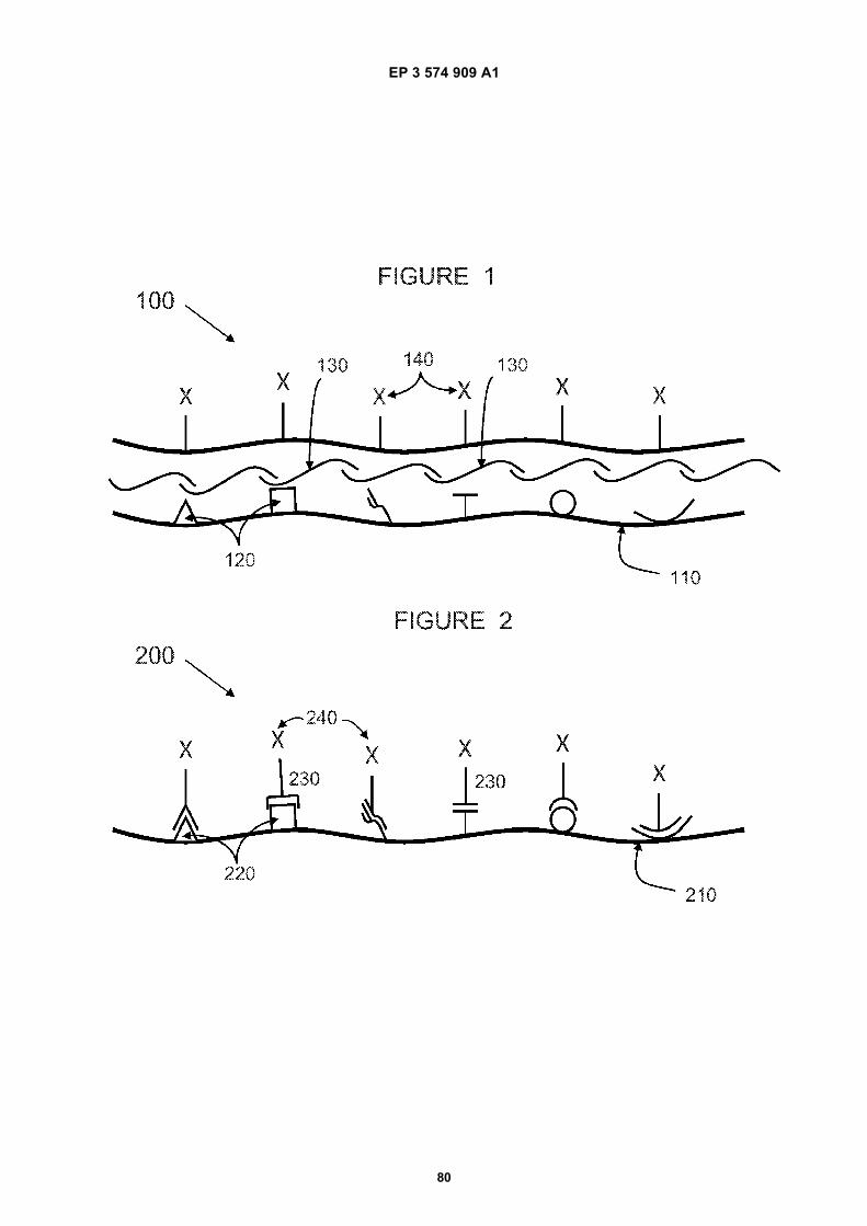
EP 3 574 909 A1
80
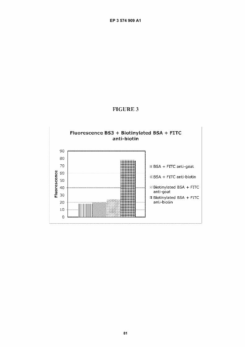
EP 3 574 909 A1
81
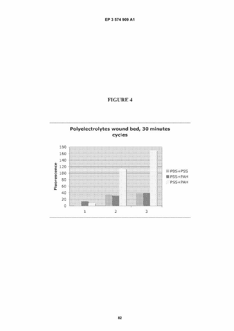
EP 3 574 909 A1
82
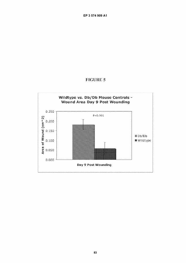
EP 3 574 909 A1
83
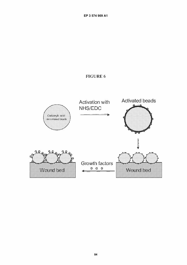
EP 3 574 909 A1
84
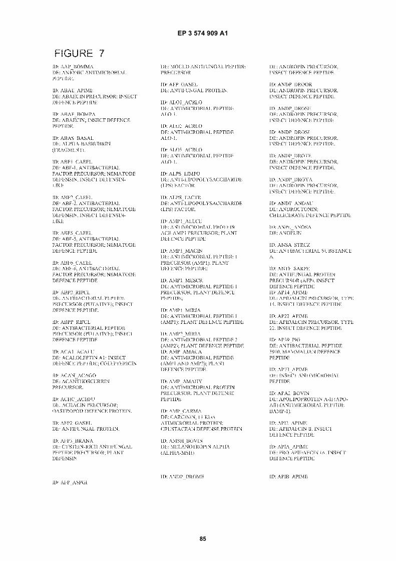
EP 3 574 909 A1
85
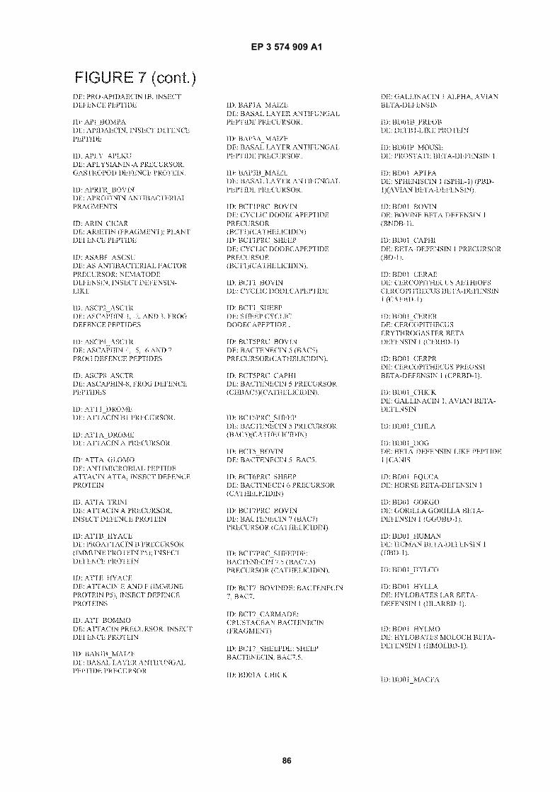
EP 3 574 909 A1
86
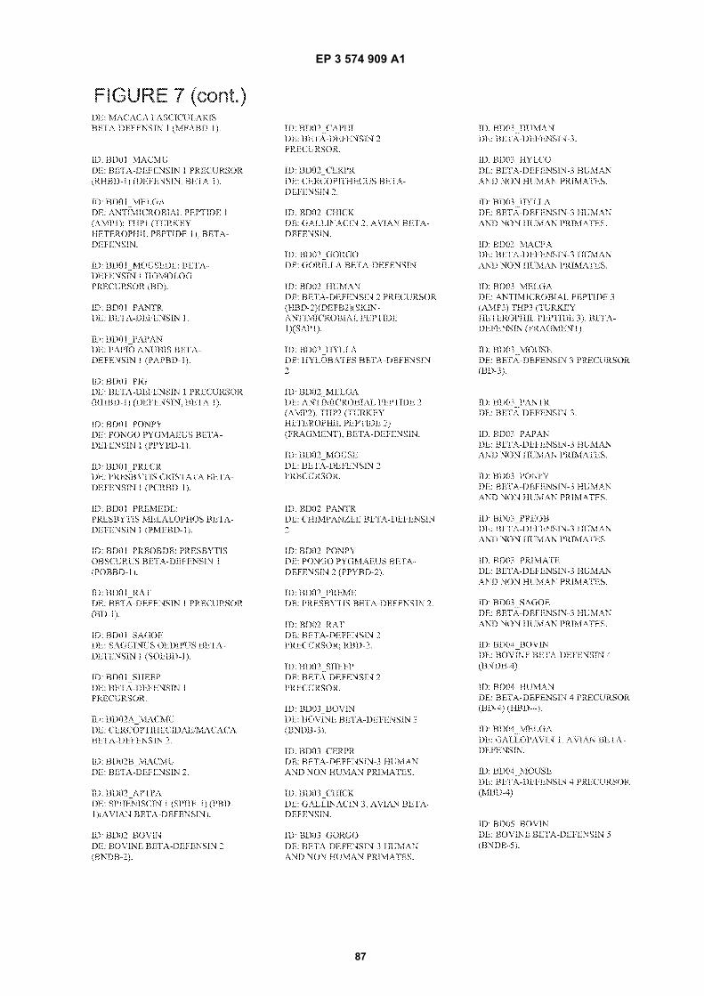
EP 3 574 909 A1
87
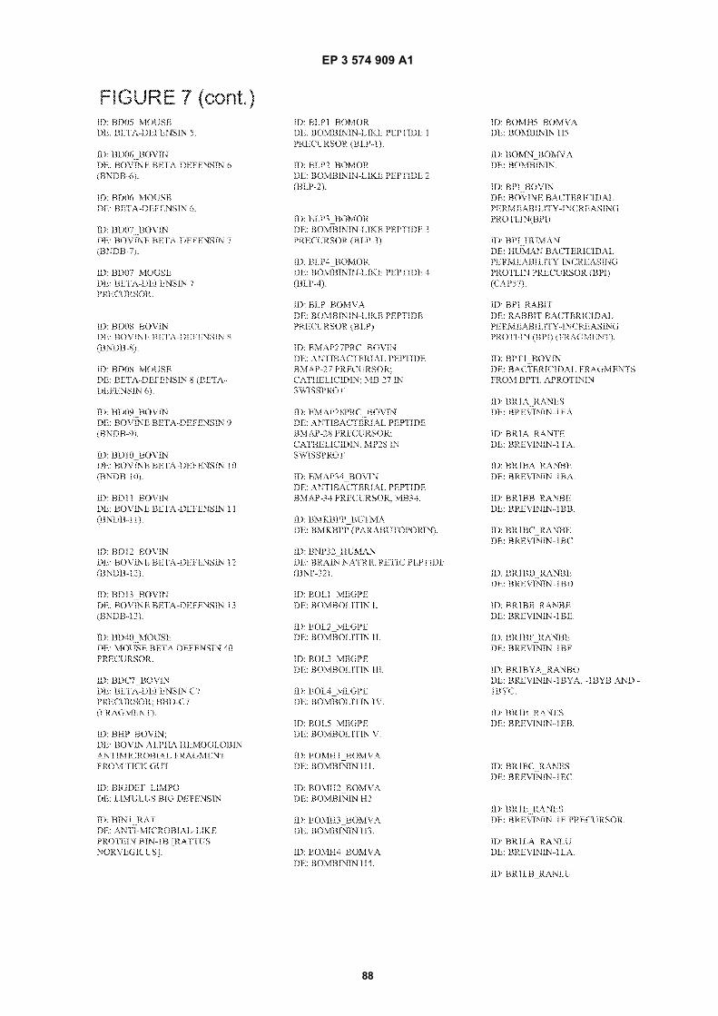
EP 3 574 909 A1
88
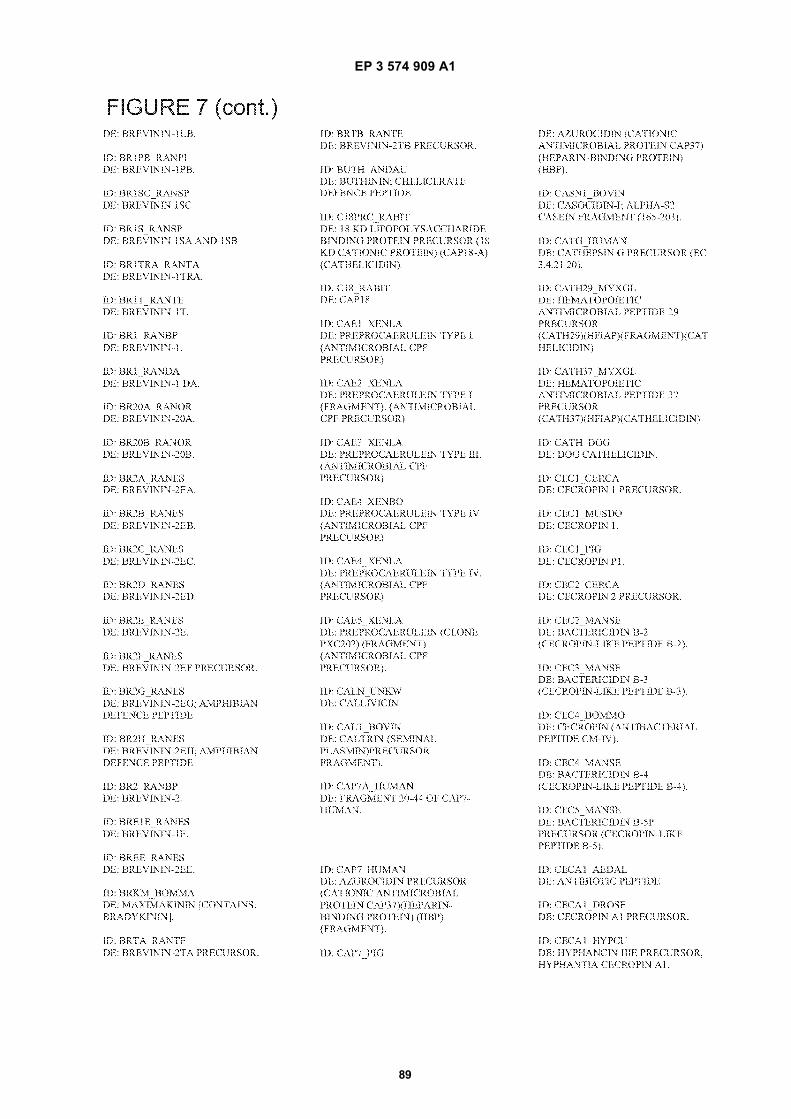
EP 3 574 909 A1
89
EP 3 574 909 A1
90
EP 3 574 909 A1
91
EP 3 574 909 A1
92
EP 3 574 909 A1
93
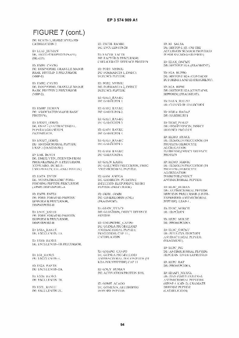
EP 3 574 909 A1
94
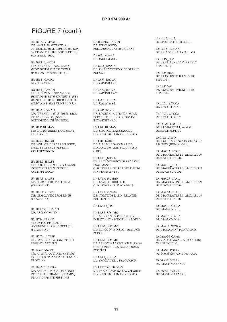
EP 3 574 909 A1
95
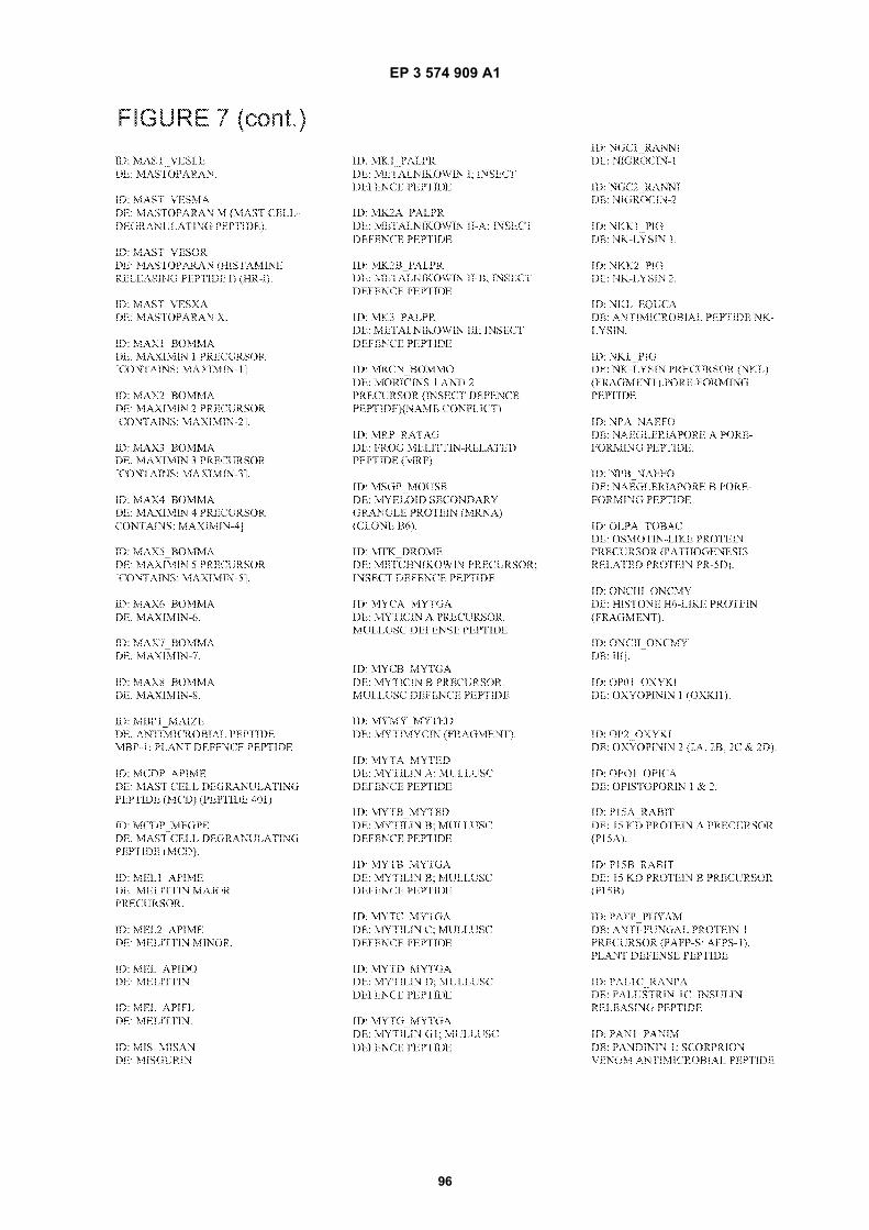
EP 3 574 909 A1
96
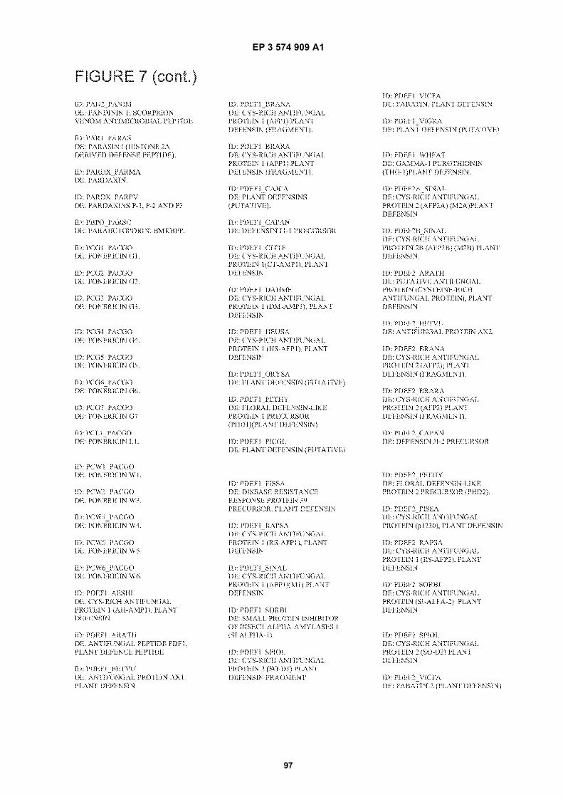
EP 3 574 909 A1
97
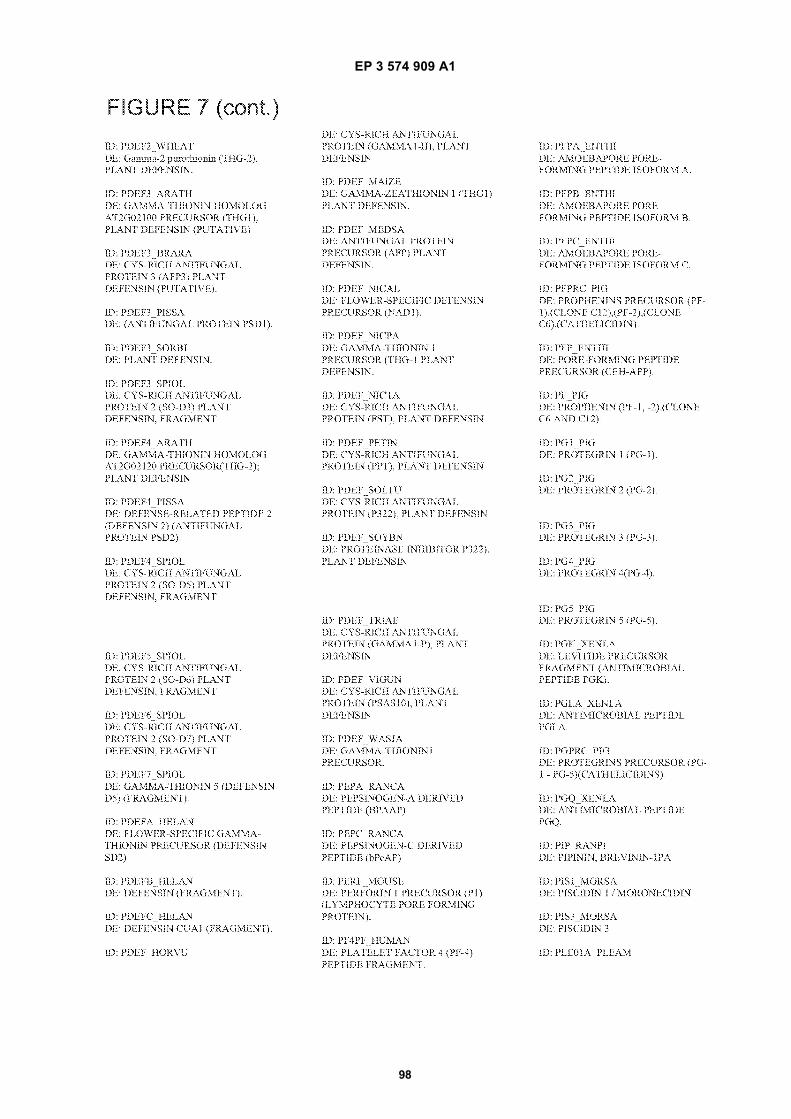
EP 3 574 909 A1
98
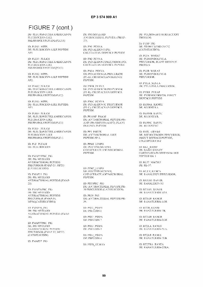
EP 3 574 909 A1
99
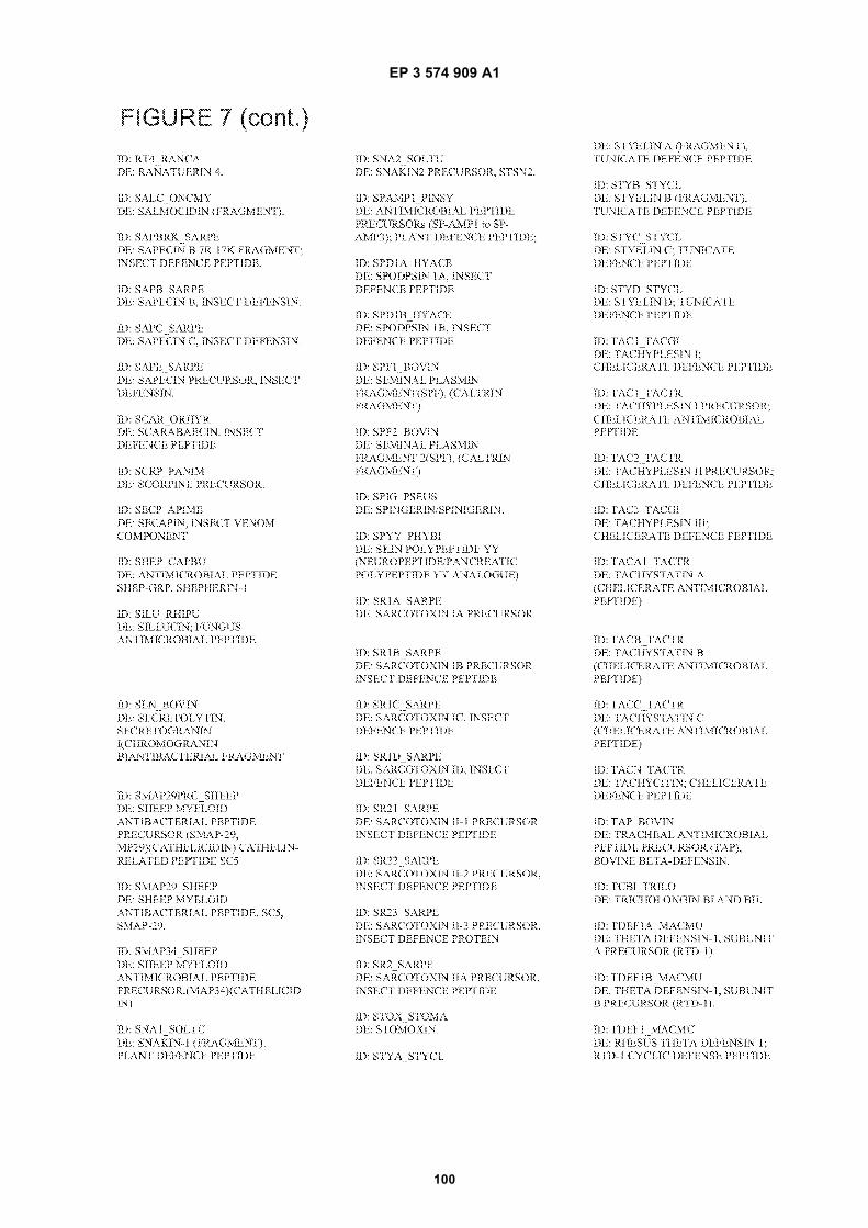
EP 3 574 909 A1
100
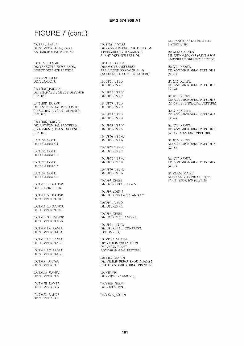
EP 3 574 909 A1
101
EP 3 574 909 A1
102
5
10
15
20
25
30
35
40
45
50
55
EP 3 574 909 A1
103
5
10
15
20
25
30
35
40
45
50
55
EP 3 574 909 A1
104
5
10
15
20
25
30
35
40
45
50
55
EP 3 574 909 A1
105
5
10
15
20
25
30
35
40
45
50
55

EP 3 574 909 A1
106
REFERENCES CITED IN THE DESCRIPTION
This list of references cited by the applicant is for the reader’s convenience only. It does not form part of the Europeanpatent document. Even though great care has been taken in compiling the references, errors or omissions cannot beexcluded and the EPO disclaims all liability in this regard.
Patent documents cited in the description
• US 61024725 A [0001]• US 5512131 A [0098] [0112]• US 5731152 A [0098] [0112]• WO 9629629 A [0098] [0112]• US 7128398 B [0098] [0112]• WO 9525116 A [0098] [0112]• WO 9841531 A [0098] [0112]
• US 5807522 A [0098] [0112]• US 5268178 A [0102]• US 6458858 B [0102]• US 6869858 B [0102]• US 5834121 A [0102]• US 6368079 B [0107]• US 5795715 A [0145]
Non-patent literature cited in the description
• HUNT ; GOODSON. Current Surgical Diagnosis &Treatment. Appleton & Lange, 1988, 86-98 [0003]
• HARRIS et al. Diabetes, 1987, vol. 36, 523 [0004]• RICHEY et al. Annals of Plastic Surgery, 1989, vol.
23, 159 [0006]• WINTER. Nature, 1962, vol. 193, 293 [0007]• KANNON ; GARRETT. Dermatol. Surg., 1995, vol.
21, 583 [0007]• DAVIES. Burns, 1983, vol. 10, 94 [0007]• RILEY. Am. Fam. Physician, 1981, vol. 24, 107
[0008]• PIERCE. Am. J. Pathol., 2001, vol. 159, 399 [0063]• MUEHLBERGER et al. J. Am. Acad. Derm., 2005,
vol. 52, 583 [0066]• LANGER ; VACANTI. Science, 1993, vol. 260, 920
[0067]• PEPPAS ; LANGER. Science, 1994, vol. 263, 1715
[0067]• ANGELOVA ; HUNKELER. Trends Biotechnol.,
1999, vol. 17, 409 [0067]• MRKSICH. Curr. Opin. Colloid Interface Sci., 1997,
vol. 2, 83 [0067]• LÖSCHE. Curr. Opin. Solid State Mater. Sci., 1997,
vol. 2, 546 [0067]• DECHER. Science, 1997, vol. 277, 1232 [0068]
[0075] [0153]• CARUSO et al. Langmuir, 1997, vol. 13, 3427 [0068]
[0075] [0153]• CASSIER et al. Supramol. Sci., 1998, vol. 5, 309
[0068]• Biomimetic Materials and Design: Interactive Bio-
interfacial. ABRAMS et al. Tissue Engineering, andDrug Delivery. 2002 [0072]
• DIEHL et al. J. Biomed. Mater. Res. A, 2005, vol. 75,603 [0072]
• FOLEY et al. Biomaterials, 2005, vol. 26, 3639 [0072]• KARURI et al. J. Cell Sci., 2004, vol. 117, 3153 [0072]
• LILIENSIEK et al. J. Biomed. Mater. Res. A, 2006,vol. 79, 185 [0072]
• DECHER et al. Thin Solid Films, 1992, vol. 210/211,831 [0075] [0153]
• CARUSO et al. J Am Chem Soc, 1999, vol. 121, 6039[0075] [0153]
• LADAM et al. Langmuir, 2000, vol. 16, 1249 [0075][0153]
• JOANNY. Eur. Phys. J. Biol., 1999, vol. 9, 117 [0075][0153]
• CASSAGNEAU et al. J. Am. Chem. Soc., 1998, vol.120, 7848 [0075] [0153]
• CARUSO et al. Langmuir, 1999, vol. 15, 8276 [0075][0153]
• LVOV et al. Langmuir, 1997, vol. 13, 6195 [0075][0153]
• ARIGA et al. Appl. Clay Sci., 1999, vol. 15, 137 [0075][0153]
• KELLER et al. J. Am. Chem. Soc., 1994, vol. 116,8817 [0075] [0153]
• LVOV et al. J. Am. Chem. Soc., 1995, vol. 117, 6117[0075] [0153]
• ELBERT ; HUBBELL. J. Biomed. Mater. Res., 1998,vol. 42, 55 [0078]
• ENGLER et al. Biophys. J., 2004, vol. 86, 617-628[0103]
• ICHINOSE et al. Biomaterials, June 2006, vol. 27(18), 3343-3350 [0104]
• STEFONEK et al. Immobilized gradients of epider-mal growth factor promote accelerated and directedkeratinocyte migration. Wound Repair and Regener-ation (OnlineEarly Articles) [0104]
• KAPUR et al. Immobilized concentration gradientsof nerve growth factor guide neurite outgrowth. Jour-nal of Biomedical Materials Research Part A, vol. 68A(2), 235-243 [0104]
EP 3 574 909 A1
107
• DELONG et al. Covalently immobilized gradients ofbFGF on hydrogel scaffolds for directed cell migra-tion. Biomaterials, 2005, vol. 26, 3227-3234 [0104]
• LIU et al. 17892312 Endothelial Cell Migration onSurface-Density Gradients of Fibronectin, VEGF, orBoth Proteins, 25 September 2007 [0104]
• MCALLISTER et al. Proc. Natl. Acad. Sci., 2003, vol.100, 13755-13760 [0106]
• SUMEREL et al. Biotechnol. J., 2006, vol. 1, 976-987[0107]
• GRIBSKOV ; DEVEREUX. Sequence Analysis Prim-er. Stockton Press, 1991 [0147]