characterization and comparative analyses of mitochondrial
TRANSCRIPT
International Journal of
Molecular Sciences
Article
Characterization and Comparative Analyses of MitochondrialGenomes in Single-Celled Eukaryotes to Shed Light on theDiversity and Evolution of Linear Molecular Architecture
Tengteng Zhang 1,2,3, Chao Li 1, Xue Zhang 1 , Chundi Wang 1, Andrew J. Roger 3 and Feng Gao 1,2,4,*
�����������������
Citation: Zhang, T.; Li, C.; Zhang, X.;
Wang, C.; Roger, A.J.; Gao, F.
Characterization and Comparative
Analyses of Mitochondrial Genomes
in Single-Celled Eukaryotes to Shed
Light on the Diversity and Evolution
of Linear Molecular Architecture. Int.
J. Mol. Sci. 2021, 22, 2546. https://
doi.org/10.3390/ijms22052546
Academic Editor: Carlo Vascotto
Received: 2 February 2021
Accepted: 26 February 2021
Published: 3 March 2021
Publisher’s Note: MDPI stays neutral
with regard to jurisdictional claims in
published maps and institutional affil-
iations.
Copyright: © 2021 by the authors.
Licensee MDPI, Basel, Switzerland.
This article is an open access article
distributed under the terms and
conditions of the Creative Commons
Attribution (CC BY) license (https://
creativecommons.org/licenses/by/
4.0/).
1 Institute of Evolution & Marine Biodiversity and College of Fisheries, Ocean University of China,Qingdao 266003, China; [email protected] (T.Z.); [email protected] (C.L.);[email protected] (X.Z.); [email protected] (C.W.)
2 Key Laboratory of Mariculture (OUC), Ministry of Education, Qingdao 266003, China3 Centre for Comparative Genomics and Evolutionary Bioinformatics, Department of Biochemistry and
Molecular Biology, Dalhousie University, Halifax, NS B3H 4R2, Canada; [email protected] Laboratory for Marine Biology and Biotechnology, Qingdao National Laboratory for Marine Science and
Technology, Qingdao 266033, China* Correspondence: [email protected]
Abstract: Determination and comparisons of complete mitochondrial genomes (mitogenomes) areimportant to understand the origin and evolution of mitochondria. Mitogenomes of unicellularprotists are particularly informative in this regard because they are gene-rich and display highstructural diversity. Ciliates are a highly diverse assemblage of protists and their mitogenomes(linear structure with high A+T content in general) were amongst the first from protists to becharacterized and have provided important insights into mitogenome evolution. Here, we reportnovel mitogenome sequences from three representatives (Strombidium sp., Strombidium cf. sulcatum,and Halteria grandinella) in two dominant ciliate lineages. Comparative and phylogenetic analysesof newly sequenced and previously published ciliate mitogenomes were performed and revealeda number of important insights. We found that the mitogenomes of these three species are linearmolecules capped with telomeric repeats that differ greatly among known species. The genomesstudied here are highly syntenic, but larger in size and more gene-rich than those of other groups.They also all share an AT-rich tandem repeat region which may serve as the replication origin andmodulate initiation of bidirectional transcription. More generally we identified a split version ofccmf, a cytochrome c maturation-related gene that might be a derived character uniting taxa inthe subclasses Hypotrichia and Euplotia. Finally, our mitogenome comparisons and phylogeneticanalyses support to reclassify Halteria grandinella from the subclass Oligotrichia to the subclassHypotrichia. These results add to the growing literature on the unique features of ciliate mitogenomes,shedding light on the diversity and evolution of their linear molecular architecture.
Keywords: ciliated protists; mitochondrial genome; phylogeny; split genes; synteny; tandem repeat
1. Introduction
Mitochondria are double-membraned semiautonomous organelles that contain theirown genomes (mitogenomes) that originated from an endosymbiotic α-proteobacteriumthat evolved prior to the last eukaryotic common ancestor [1–3]. Although they are bestknown for generating ATP by aerobic respiration to supply the energy needs of eukaryoticcells, these organelles perform a much wider range of functions that vary substantiallyamong diverse eukaryotic lineages [1]. Mitochondria also have biomedical significance, asmutations affecting mitochondrial function are implicated in ageing and diseases, includingParkinson’s, Alzheimer’s, and Huntington’s diseases [4,5].
Mitogenomes usually evolve more rapidly than their nuclear counterparts in mostspecies [6,7]. Additionally, each cell possesses multiple mitogenome copies and mi-
Int. J. Mol. Sci. 2021, 22, 2546. https://doi.org/10.3390/ijms22052546 https://www.mdpi.com/journal/ijms

Int. J. Mol. Sci. 2021, 22, 2546 2 of 20
togenome recombination is a widespread process occurring in plants, fungi, protists, andanimals [8–10]. In general, unicellular protists possess the most gene-rich and structurallyvaried mitogenomes [11]. Ciliates are a diverse and well-studied protistan assemblage withunusual cellular and molecular features (nuclear dimorphism and conjugation) [12–23].Their mitogenomes were the first amongst protists to be characterized providing novelinsights into the structure, function, and evolution of organellar genomes [24]. The mi-togenomes of the ciliates Tetrahymena and Paramecium were the first confirmed to be lineardouble-stranded DNA molecules and to have their telomeric sequences identified, whichprovided novel insights into the replication of linear mitochondrial DNA [25–27].
In the years since those seminal discoveries, more ciliate mitogenomes have beencharacterized, including 20 mitogenomes from species in the class Oligohymenophorea(Tetrahymena pyriformis, T. thermophila, T. malaccensis, T. paravorax, T. pigmentosa, T. rostrata,Ichthyophthirius multifiliis, Uronema marinum, Paramecium caudatum, P. aurelia, P. tetraurelia,P. sexaurelia, P. multimicronucleatum, P. biaurelia, P. octaurelia, P. novaurelia, P. decaurelia, P. do-decaurelia, P. quadecaurelia, and P. jenningsi) [28–36], eight mitogenomes of species in theclass Spirotrichea (Oxytricha trifallax, Laurentiella strenua, Stylonychia lemnae, Paraurostyla sp.,Urostyla grandis, Pseudourostyla cristata, Euplotes minuta, and E. crassus) [37–39], two mi-togenomes in the class Heterotrichea (Stentor coeruleus and Gruberia lanceolata) [20,40], aswell as seven hydrogenosomal genomes of anerobic ciliates (Nyctotherus ovalis, Metopuscontortus, Metopus es, Metopid sp., Heterometopus sp., Parablepharisma sp., and Muranoth-rix gubernata) [41–43]. Ciliate mitogenomes are generally in size of 20–70 kb, with highA+T content (58.53% for N. ovalis ~81.51% for I. multifiliis) and relatively large gene com-plements (20–30 protein-coding genes) [39,41,44]. Mitochondrial DNA has been provenextremely useful in phylogenetic analyses to determine ciliate relationships and speciesdelimitation [45].
In this study, we report the analysis of complete mitogenomes of three ciliates: twospecies of the subclass Oligotrichia (Strombidium sp. and Strombidium cf. sulcatum) and arepresentative species of Halteriidae (Halteria grandinella) whose phylogenetic position iscontroversial (i.e., morphological data contradicts molecular data [46–48]). These species allbelong to the class Spirotrichea, which is a highly differentiated and species-rich assemblageamong ciliates [49]. We annotated and compared these three genomes with the previouslypublished ciliate mitogenomes, particularly the mitogenomes among Spirotrichea, shed-ding light on the diversity and evolution of mitochondrial genomes within this group.Phylogenetic analysis based on 14 mitochondrial ortholog proteins of 34 ciliates was per-formed in order to further investigate and clarify the evolutionary relationships amongOligotrichia, Halteriidae, and Hypotrichia.
2. Results2.1. Mitogenome Overview
For each species, mitochondrial contigs (Strombidium sp.: one contig; Halteria grandinella:one contig; Strombidium cf. sulcatum: 9 contigs) are recovered from the assembled mito-chondrial or genomic data by BLASTN searches. They are assembled into linear moleculesfor the mitogenomes of Strombidium sp., S. cf. sulcatum, and H. grandinella with coverageof 194.2, 492.2–564.8, and 40.2 respectively (Figures S1 and S2). The lengths of the mi-togenomes are 51,232 bp for Strombidium sp., 54,912 bp for S. cf. sulcatum, and 50,085 bp forH. grandinella with the A+T contents of 77.32%, 71.68%, and 80.20%, respectively (Table 1).Furthermore, the three mitogenomes are all capped with telomeric tandem repeats at bothends, which are reverse complements of each other. The telomeric repeat sequences aredivergent among the three species (18 bp in Strombidium sp.: CTC CCT TAT CTA GTC TTT;34 bp in S. cf. sulcatum: TTA TAT CCT TTC TCC CCT ATA TCT CTA TAG TAC T; and31 bp in H. grandinella: AAA ACA GCT CCG TTC CAA TAC TAC TAA CTA A) (Table 2).A central repeat region found in all three genomes is made up of tandem repeat units, andoccurs at the same position between tRNA_Phe (trnF) and tRNA_Tyr (trnY) in each genome.However, the central repeats differ from one another in terms of length and sequence. In
Int. J. Mol. Sci. 2021, 22, 2546 3 of 20
Strombidium sp., the central repeat region is 170 bp long, composed of 11 repeats of theunit ATA ATA TAA TAA TAT. This region is 142 bp long in S. cf. sulcatum, composed oftwo repeats of the unit ATA AAT TTA ATT TTA and an irregular sequence for the rest. InH. grandinella, this region is 168 bp long and composed of nine repeats of the unit TAT ACATAT AAT ATA TA (Table 2). Diverging and starting from this central repeat region, all thegenes in the three mitogenomes are arranged in two opposite transcriptional directions(Figure 1).
Figure 1. Mitochondrial and morphological information of the three species with newly characterized mitogenomes.(A) Mitogenome map of Strombidium sp. (B) Mitogenome map of Strombidium cf. sulcatum. (C) Mitogenome map ofHalteria grandinella. (D) Photomicrographs of the three species in vivo and the legend of mitochondrial maps. Genesare represented by different colored blocks as indicated in the legend of diagram D. Outside and inside blocks of eachmitochondrial map indicate the genes on the positive and reverse strands, respectively.
Int. J. Mol. Sci. 2021, 22, 2546 4 of 20
Table 1. Main features of all the available mitogenomes of aerobic ciliates and a representative hydrogenosome genome of anaerobic ciliates. Newly characterized mitogenomes are in bold.
Species Accession No. Genome Size (bp) Gene Region(bp)
IntergenicRegion (bp)
Overall A+TContent (%)
Known ProteinGenes tRNA Genes rRNA Genes
Strombidium sp. MT471315 51,232 48,614 2618 77.32 29 8 2Strombidium cf. sulcatum MT471316 54,912 52,218 2694 71.68 29 9 2
Halteria grandinella MT471317 50,085 45,401 4684 80.20 29 9 2Oxytricha trifallax JN383843 69,800 61,685 8115 76.17 29 11 2Stylonychia lemnae KX524144 67,745 51,501 16,244 74.05 29 12 3Laurentiella strenua KX529838 66,721 48,266 18,455 75.75 30 7 3
Paraurostyla sp. KX524143 65,186 42,149 23,037 80.59 29 8 2Urostyla grandis KX494929 60,924 37,327 23,597 61.12 27 7 2
Pseudourostyla cristata MH888186 76,660 59,952 16,708 78.27 29 9 2Euplotes minuta GQ903130 41,978 40,257 1721 64.74 20 6 2Euplotes crassus GQ903131 33,688 32,273 1415 65.61 17 5 2
Tetrahymena pyriformis AF160864 47,296 45,285 2011 78.68 24 8 3Tetrahymena thermophila AF396436 47,577 45,619 1958 79.24 21 8 3Tetrahymena malaccensis DQ927303 47,691 45,528 2163 80.10 21 8 3Tetrahymena paravorax DQ927304 47,496 44,812 2684 81.51 21 8 3
Tetrahymena pigmentosa DQ927305 46,990 44,889 2101 81.46 21 8 3Tetrahymena rostrata MN025427 47,235 45,336 1899 78.23 23 8 3
Ichthyophthirius multifiliis JN227086 51,686 43,469 8217 83.62 21 5 3Uronema marinum MG272262 39,845 35,544 4301 81.00 24 6 2
Paramecium caudatum FN424190 43,660 41,091 2569 77.62 24 3 2Paramecium aurelia NC001324 40,469 26,808 13,661 58.76 20 4 2
Paramecium tetraurelia - 40,267 39,204 1063 58.34 23 3 2Paramecium sexaurelia - 40,015 37,474 2541 60.24 24 3 2
Paramecium multimicronucleatum - 39,460 33,539 5921 80.38 21 34 2Paramecium biaurelia - 39,870 36,297 3573 59.89 24 3 2Paramecium octaurelia - 39,850 37,784 2066 59.66 23 3 2Paramecium novaurelia - 59,002 36,996 22,006 65.02 23 3 2Paramecium decaurelia - 42,742 37,779 4963 58.64 24 3 2
Paramecium dodecaurelia - 40,335 37,947 2388 58.52 23 3 2Paramecium quadecaurelia - 41,844 37,450 4394 58.75 23 3 2
Paramecium jenningsi - 40,161 38,010 2151 59.44 24 3 2Nyctotherus ovalis GU057832 41,666 32,511 9155 58.52 16 3 2Stentor coeruleus MPUH01000652 41,645 36,154 5491 80.15 22 5 2
Gruberia lanceolata MK301177 39,988 30,910 9078 79.58 25 4 2
- indicates the mitogenome data of ten Paramecium species which are not available on the NCBI database but can be accessed on Zenodo (https://doi.org/10.5281/zenodo.2539699 (accessed on 23 February 2021)).

Int. J. Mol. Sci. 2021, 22, 2546 5 of 20
Table 2. Information of mitogenome repeats within the class Spirotrichea. * Denotes absence.
SpeciesCentral Repeat
Telomeric Repeat (5′–3′) Terminal Inverted RepeatLength (bp) A+T Content Repeat Unit (Number of Repeats)
Strombidium sp. 170 100.00% ATAATATAATAATAT (11) CTCCCTTATCTAGTCTTT(both ends) *
Strombidium cf. sulcatum 142 96.48% ATAAATTTAATTTTA (2) +irregular sequence for the rest
TTATATCCTTTCTCCCCTATATCTCTATAGTACT(both ends) *
Halteria grandinella 168 94.05% TATACATATAATATATA (9) AAAACAGCTCCGTTCCAATACTACTAACTAA(both ends) *
Oxytricha trifallax ~285 96.76%TATATAAA (11) +
TATAAATAAA (3) +AAAAAG (5)
CGACTCCTCTATCCTCATCCTAGACTCCGCTTACT(both ends) ~1800 bp
Stylonychia lemnae ~607 92.29% TATARTAGTTATATTATA (27) TTCATACCTTTACTAGATACCCGCCTCCGGCTCTCC(3′ end) ~3100 bp
Laurentiella strenua 733 98.91%ATATAAATGTATATAA (7) +
ATAAA(TA)nT (49) +TTT(AT)n (4), n = 0–8
CCTACTACGCTTCATACGCTAAA (partial)(both ends) ~2400 bp
Paraurostyla sp. ~802 98.86% ATATAACAAATA (7) +AAATAA(TA)nAT (20), n = 2–29 * *
Urostyla grandis ~279 95.91% ATATATTTATTAATATATAGTAT (10) GTAGCACATGTAG(3′ end) *
Pseudourostyla cristata 80 86.25% TATATATACATATAC (3) +(TA)nC (3), n = 3 or 5 * *
Euplotes minuta ~1596 83.36%ATAGTATATAATGTATAC (63) +
ATAGTATATAATGTTAC (1) +ATAGTATATAATTGTTAC (18)
* *
Euplotes crassus ~416 83.54% ATAGTATATAATGTATAC (15) * *
Int. J. Mol. Sci. 2021, 22, 2546 6 of 20
For Strombidium sp., 94.89% of the mitogenome is coding sequence, including 29known protein genes, 8 tRNA genes, 2 rRNA genes, and 12 open reading frames (ORFs)with unknown function. The mitogenome of S. cf. sulcatum also contains 29 known proteingenes, but instead has 9 tRNA genes, 2 rRNA genes, and 11 ORFs, with the coding regioncomprising 95.09%. The proportion of coding sequence is 90.65% in the H. grandinellamitogenome, the lowest among the three taxa, which comprises 29 known protein genes,9 tRNA genes, 2 rRNA genes, and 9 ORFs (Table 1, Figures 1 and 2). The mitochondrialgenes of the three species are tightly packed and some genes are partially overlapping(Strombidium sp.: seven cases (10–46 bp, avg. 24 bp), S. cf. sulcatum: seven cases (7–82 bp,avg. 46 bp), H. grandinella: fifteen cases (4–190 bp, avg. 39 bp)) (Table S3, Figure 1). Threegenes of nad1, nad2 (NADH dehydrogenase genes), and rps3 (small subunit ribosomalprotein gene) are separated into two parts for all the three mitogenomes, while the ccmf(cytochrome c maturation-related gene) is split only for H. grandinella. Two duplicated genesof nad1_a and tRNA_Met (trnM) are found in S. cf. sulcatum mitogenomes (Figures 1 and 2).No introns are detected in any of the three mitogenomes.
Figure 2. Protein and tRNA genes encoded in mitogenomes of the class Spirotrichea. The solid circles represent presenceand the hollow circles represent absence. The asterisk “*” indicates a split gene that is split into two parts. The letter “a”indicates the gene is duplicated into two copies, while “b” represents that one copy of tRNA_Cys (trnC) is a pseudogene.The number indicates the number of unknown ORFs.
2.2. Mitogenome Comparison among Species in the Class Spirotrichea
We compared the three newly sequenced mitogenomes with eight other representa-tive mitogenomes available in the class Spirotrichea. Among these mitogenomes, only thethree that were newly sequenced and those of Oxytricha trifallax and Laurentiella strenuaare complete, with both ends capped by telomeres (Table 2, Figure 3). To our knowl-
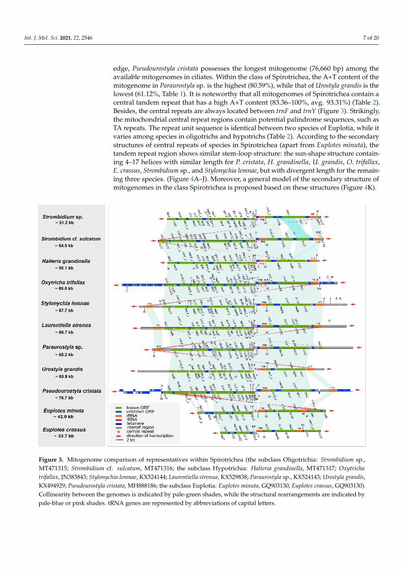
Int. J. Mol. Sci. 2021, 22, 2546 7 of 20
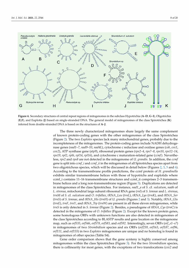
edge, Pseudourostyla cristata possesses the longest mitogenome (76,660 bp) among theavailable mitogenomes in ciliates. Within the class of Spirotrichea, the A+T content of themitogenome in Paraurostyla sp. is the highest (80.59%), while that of Urostyla grandis is thelowest (61.12%, Table 1). It is noteworthy that all mitogenomes of Spirotrichea contain acentral tandem repeat that has a high A+T content (83.36–100%, avg. 93.31%) (Table 2).Besides, the central repeats are always located between trnF and trnY (Figure 3). Strikingly,the mitochondrial central repeat regions contain potential palindrome sequences, such asTA repeats. The repeat unit sequence is identical between two species of Euplotia, while itvaries among species in oligotrichs and hypotrichs (Table 2). According to the secondarystructures of central repeats of species in Spirotrichea (apart from Euplotes minuta), thetandem repeat region shows similar stem-loop structure: the sun-shape structure contain-ing 4–17 helices with similar length for P. cristata, H. grandinella, U. grandis, O. trifallax,E. crassus, Strombidium sp., and Stylonychia lemnae, but with divergent length for the remain-ing three species. (Figure 4A–J). Moreover, a general model of the secondary structure ofmitogenomes in the class Spirotrichea is proposed based on these structures (Figure 4K).
Figure 3. Mitogenome comparison of representatives within Spirotrichea (the subclass Oligotrichia: Strombidium sp.,MT471315; Strombidium cf. sulcatum, MT471316; the subclass Hypotrichia: Halteria grandinella, MT471317; Oxytrichatrifallax, JN383843; Stylonychia lemnae, KX524144; Laurentiella strenua, KX529838; Paraurostyla sp., KX524143; Urostyla grandis,KX494929; Pseudourostyla cristata, MH888186; the subclass Euplotia: Euplotes minuta, GQ903130; Euplotes crassus, GQ903130).Collinearity between the genomes is indicated by pale-green shades, while the structural rearrangements are indicated bypale-blue or pink shades. tRNA genes are represented by abbreviations of capital letters.
Int. J. Mol. Sci. 2021, 22, 2546 8 of 20
Figure 4. Secondary structures of central repeat regions of mitogenomes in the subclass Hypotrichia (A–D, G–I), Oligotrichia(E,F), and Euplotia (J) based on single-stranded DNA. The general model of mitogenomes of the class Spirotrichea (K)inferred from double-stranded DNA is based on the structures of A–J.
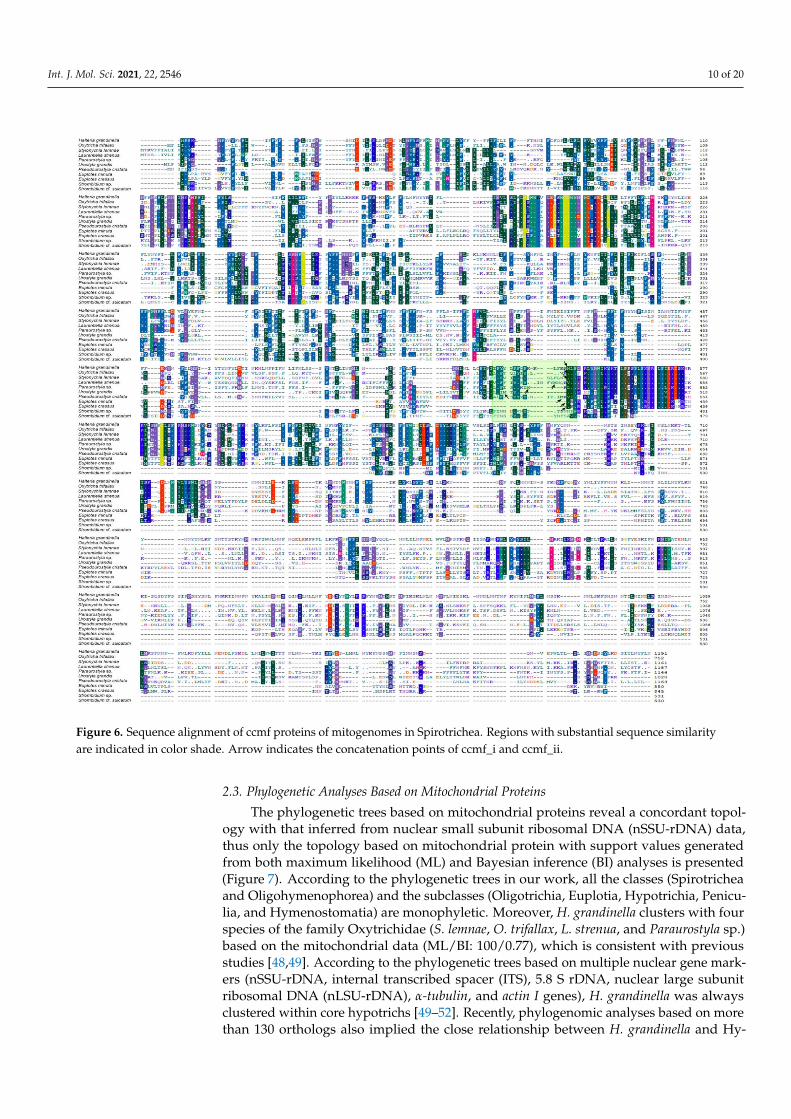
The three newly characterized mitogenomes share largely the same complementof known protein-coding genes with the other mitogenomes of the class Spirotrichea(Figure 2). The two Euplotes species lack many mitochondrial genes, probably due to theincompleteness of the mitogenomes. The protein-coding genes include NADH dehydroge-nase genes (nad1–7, nad9–10, nad4L), cytochrome c reductase and oxidase genes (cob, cox1,cox2), ATP synthase gene (atp9), ribosomal protein genes (rps2–4, rps7–8, rps10, rps12–14,rps19, rpl2, rpl6, rpl14, rpl16), and cytochrome c maturation-related gene (ccmf ). Neverthe-less, rps2 and rps8 are not detected in the mitogenome of U. grandis. In addition, the ccmfgene is split into ccmf_i and ccmf_ii in the mitogenomes of all Spirotrichea species apart fromtwo oligotrichous species, which will be discussed in detail below (Figures 2, 3, 5 and 6).According to the transmembrane profile predictions, the ccmf protein of H. grandinellaexhibits similar transmembrane helices with those of hypotrichs and euplotids whereccmf_i contains 11–16 transmembrane structures and ccmf_ii comprises 2–3 transmem-brane helices and a long non-transmembrane region (Figure 5). Duplications are detectedin mitogenomes of the class Spirotrichea. For instance, nad1_a of S. cf. sulcatum, nad6 ofL. strenua, mitochondrial large subunit ribosomal RNA gene (rnl) of S. lemnae and L. strenua,trnM of S. cf. sulcatum and O. trifallax, tRNA_Leu (trnL), tRNA_Lys (trnK), and tRNA_Ser(trnS) of S. lemnae, and tRNA_His (trnH) of U. grandis (Figures 2 and 3). Notably, tRNA_Glu(trnE), trnF, trnY, and tRNA_Trp (trnW) are present in all these eleven mitogenomes, whiletrnS is only detected in S. lemnae (Figure 2). Besides, a pseudogene of tRNA_Cys (trnC) isdetected in the mitogenome of O. trifallax (Figure 2). Except for the known protein genes,some homologous ORFs with unknown functions are also detected in mitogenomes ofthe class Spirotrichea according to BLASTP results and gene location on the mitogenomemap, such as orf535, orf546, orf578, orf583, and orf592. Interestingly, seven ORFs (orf_s1–s7)in mitogenomes of two Strombidium species and six ORFs (orf259, orf163, orf187, orf96,orf111, and orf155) in two Euplotes mitogenomes are unique and no homolog is found inmitogenomes of other species (Table S4).
Gene order comparison shows that the gene synteny is highly conserved amongmitogenomes within the class Spirotrichea (Figure 3). For the two Strombidium species,there is collinearity for most genes, with the exceptions of two translocations (cox2 and

Int. J. Mol. Sci. 2021, 22, 2546 9 of 20
orf_s6) and one inversion (nad6). The mitogenomes of S. cf. sulcatum and H. grandinellaare largely collinear, except that two terminal genes of nad6 and rpl14 in S. cf. sulcatumare moved to the position between cox1 and cox2 in H. grandinella. The gene orders in H.grandinella and O. trifallax mitogenomes are identical. The mitogenome synteny is alsosignificant among the hypotrichs, except for the inversion of rps2 in Paraurostyla sp. andthe gene duplication of rnl in S. lemnae, L. strenua, and Paraurostyla sp. The two Euplotesmitogenomes are collinear and are largely collinear with those of the hypotrichs in the coreregion, from nad3 to rnl, except for the inversion of nad5, ccmf, and cob. Besides, the geneorder of tRNA, tRNA_Gln (trnQ)-trnL-trnE-trnF-trnY-trnW, is conserved in all the speciesexcept for the missing trnQ in E. crassus and trnL in U. grandis, E. minuta, and E. crassus.
Similar to the extensive collinearity within the class Spirotrichea, the mitogenomes arelargely collinear within the class Oligohymenophorea (mainly represented by Tetrahymenaand Paramecium), with the exception of one large inversion and translocation from cob tornl [39]. The collinearity between the classes Spirotrichea and Oligohymenophorea is lowerthan within classes, and no collinearity is observed between the classes Spirotrichea andHeterotrichea, which is consistent with the higher ciliate taxonomic classification [39].
Figure 5. Transmembrane (TM) profiles for the concatenated ccmf proteins of eleven representative mitogenomes in theclass Spirotrichea. The x and y axes denote amino acid length and posterior probabilities detected by TMHMM v.2.0,respectively. The green blocks represent TM regions. The arrow indicates concatenation point of ccmf_i and ccmf_ii. Thedouble arrowheads denote the pseudo split of ccmf_ii in Euplotes minuta.
Int. J. Mol. Sci. 2021, 22, 2546 10 of 20
Figure 6. Sequence alignment of ccmf proteins of mitogenomes in Spirotrichea. Regions with substantial sequence similarityare indicated in color shade. Arrow indicates the concatenation points of ccmf_i and ccmf_ii.
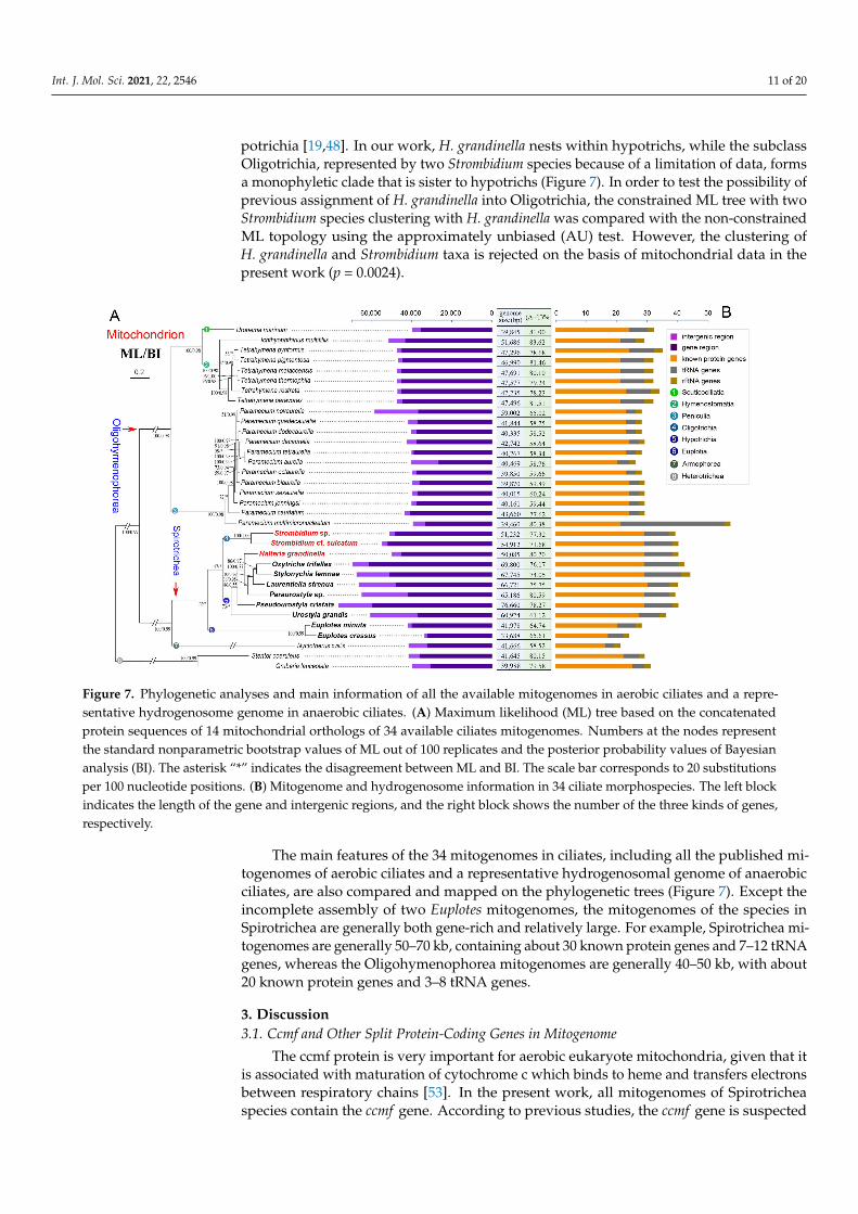
2.3. Phylogenetic Analyses Based on Mitochondrial Proteins
The phylogenetic trees based on mitochondrial proteins reveal a concordant topol-ogy with that inferred from nuclear small subunit ribosomal DNA (nSSU-rDNA) data,thus only the topology based on mitochondrial protein with support values generatedfrom both maximum likelihood (ML) and Bayesian inference (BI) analyses is presented(Figure 7). According to the phylogenetic trees in our work, all the classes (Spirotricheaand Oligohymenophorea) and the subclasses (Oligotrichia, Euplotia, Hypotrichia, Penicu-lia, and Hymenostomatia) are monophyletic. Moreover, H. grandinella clusters with fourspecies of the family Oxytrichidae (S. lemnae, O. trifallax, L. strenua, and Paraurostyla sp.)based on the mitochondrial data (ML/BI: 100/0.77), which is consistent with previousstudies [48,49]. According to the phylogenetic trees based on multiple nuclear gene mark-ers (nSSU-rDNA, internal transcribed spacer (ITS), 5.8 S rDNA, nuclear large subunitribosomal DNA (nLSU-rDNA), α-tubulin, and actin I genes), H. grandinella was alwaysclustered within core hypotrichs [49–52]. Recently, phylogenomic analyses based on morethan 130 orthologs also implied the close relationship between H. grandinella and Hy-
Int. J. Mol. Sci. 2021, 22, 2546 11 of 20
potrichia [19,48]. In our work, H. grandinella nests within hypotrichs, while the subclassOligotrichia, represented by two Strombidium species because of a limitation of data, formsa monophyletic clade that is sister to hypotrichs (Figure 7). In order to test the possibility ofprevious assignment of H. grandinella into Oligotrichia, the constrained ML tree with twoStrombidium species clustering with H. grandinella was compared with the non-constrainedML topology using the approximately unbiased (AU) test. However, the clustering ofH. grandinella and Strombidium taxa is rejected on the basis of mitochondrial data in thepresent work (p = 0.0024).
Figure 7. Phylogenetic analyses and main information of all the available mitogenomes in aerobic ciliates and a repre-sentative hydrogenosome genome in anaerobic ciliates. (A) Maximum likelihood (ML) tree based on the concatenatedprotein sequences of 14 mitochondrial orthologs of 34 available ciliates mitogenomes. Numbers at the nodes representthe standard nonparametric bootstrap values of ML out of 100 replicates and the posterior probability values of Bayesiananalysis (BI). The asterisk “*” indicates the disagreement between ML and BI. The scale bar corresponds to 20 substitutionsper 100 nucleotide positions. (B) Mitogenome and hydrogenosome information in 34 ciliate morphospecies. The left blockindicates the length of the gene and intergenic regions, and the right block shows the number of the three kinds of genes,respectively.
The main features of the 34 mitogenomes in ciliates, including all the published mi-togenomes of aerobic ciliates and a representative hydrogenosomal genome of anaerobicciliates, are also compared and mapped on the phylogenetic trees (Figure 7). Except theincomplete assembly of two Euplotes mitogenomes, the mitogenomes of the species inSpirotrichea are generally both gene-rich and relatively large. For example, Spirotrichea mi-togenomes are generally 50–70 kb, containing about 30 known protein genes and 7–12 tRNAgenes, whereas the Oligohymenophorea mitogenomes are generally 40–50 kb, with about20 known protein genes and 3–8 tRNA genes.
3. Discussion3.1. Ccmf and Other Split Protein-Coding Genes in Mitogenome
The ccmf protein is very important for aerobic eukaryote mitochondria, given that itis associated with maturation of cytochrome c which binds to heme and transfers electronsbetween respiratory chains [53]. In the present work, all mitogenomes of Spirotricheaspecies contain the ccmf gene. According to previous studies, the ccmf gene is suspected
Int. J. Mol. Sci. 2021, 22, 2546 12 of 20
to be split into two parts in the mitogenomes of O. trifallax, L. strenua, U. grandis, andP. cristata [37,39,54]. According to De Graaf et al. 2009 [38], ccmf could also be a splitgene for E. minuta and E. crassus, but have three parts (an additional split of ccmf_i forE. crassus and an additional split of ccmf_ii for E. minuta). Nevertheless, we found thatthe split of ccmf_i for E. crassus results from the insertion of an adenine nucleotide basedon the comparison of our mitogenomes of E. crassus and E. vannus (unpublished) withthe published data. Similarly, many insertions and deletions of ccmf_ii of E. minuta weredetected. Although the insertion and deletion might come from the interspecific divergence,we think the additional split of ccmf_ii of E. minuta might not be a true split but instead is theproduct of sequencing errors. This speculation is consistent with the length, transmembrane(TM) profile, and sequence alignment of ccmf proteins as discussed below (Figures 5 and 6).
The ccmf genes of H. grandinella, S. lemnae, and Paraurostyla sp. might also be splitinto two parts, with the following four findings supporting this hypothesis. Firstly, for themitogenome in each species, two adjacent ORFs align with two parts of the ccmf protein(ccmf_i and ccmf_ii, respectively) based on a prediction with the NCBI Open ReadingFrame Finder and SmartBLAST, with the best BLAST hit generally coming from one of theciliate mitogenomes. Secondly, each of the two hypothetical split parts (ccmf_i and ccmf_ii)possess similar lengths among Hypotrichia and Euplotia mitogenomes, respectively: thelengths of ccmf_i are 543 amino acids for H. grandinella, 480–548 (avg. 524) amino acidsfor Hypotrichia, and 405 amino acids for Euplotia; the lengths of ccmf_ii are 648 aminoacids for H. grandinella, 219–645 (avg. 547) amino acids for Hypotrichia, and 440–445(avg. 442) amino acids for Euplotia. Thirdly, ccmf is a mitochondrial integral membraneprotein containing multiple transmembrane helices [55]. In our work based on TM profiles,the transmembrane helix structure of hypothetical ccmf split proteins is similar amongH. grandinella, Hypotrichia, and Euplotia (Figure 5). Fourthly, according to the alignmentresult of ccmf proteins in Spirotrichea, the split sites of ccmf_i and ccmf_ii are almostidentical (Figure 6). All of these suggest that the ccmf gene is split into two parts in themitogenomes of H. grandinella, Hypotrichia, and the Euplotia species described here.
However, as for the two Strombidium species, the ccmf gene is non-split becausewe only find one ORF that is homologous to cytochrome c maturation-related genes(Figures 1–3 and 5). According to the TM profiles, the ccmf of the two Strombidium speciespossesses 13–16 transmembrane helices, which is similar to that of other spirotrichousspecies (14–19 transmembrane helices). Besides, the split phenomenon was not detectedbased on the ccmf sequence of two Strombidium species, nearby the conserved split po-sitions of Hypotrichia and Euplotia ccmf proteins (Figure 6). Hence, it appears that themitochondrial ccmf gene is likely split for Hypotrichia and Euplotia species but correspondsto a single polypeptide for Oligotrichia species.
According to previous studies [37,39], the protein-coding genes nad1, nad2 and rps3were reported as split genes in most sequenced ciliate mitogenomes. These three genes arealso found split in the newly characterized three mitogenomes in the present study. Thissplit appears to be ancestral and shared by the phylum Ciliophora. The non-split of thegenes nad2 and rps3 in Euplotes mitogenomes could be due to sequencing and annotationerrors or they were fused again in the Euplotes species. By contrast, the ccmf gene isfound split only in Hypotrichia and Euplotia species, while it is not split in Oligotrichiaand Oligohymenophorea, which indicates that the split of ccmf gene in Hypotrichia andEuplotia species is a derived character and probably evolved independently in these twogroups. Notably, the protein-coding genes nad4, nad5, nad9, and rpl6 were also predicted aspotential split genes in O. trifallax [39]. However, these genes are not split in other ciliatemitogenomes. The split of nad4, nad5, nad9, and rpl6 in O. trifallax may be not accuratedue to sequencing errors, because of the fact that this mitogenome was sequenced witherror-prone 454 sequencing technology. In conclusion, the split in genes nad1, nad2, andrps3 could be an ancestral characteristic among all ciliates. Meanwhile the split in ccmf is aspecific feature within the subclasses Hypotrichia and Euplotia, which could be used forclarifying the phylogenetic relationships for these groups.
Int. J. Mol. Sci. 2021, 22, 2546 13 of 20
3.2. ORFs with Unknown Functions in Mitogenome
The ORFs with unknown functions have always been a problem for protist mi-togenomes, and result in the trouble of annotation for mitochondrial genes. However,the high evolutionary rate of mitochondrial genes and low sequence identity betweenORFs and known genes make it difficult to define these ORFs with unknown functionsby BLAST searches [7,31]. Specifically, compared to known protein-coding genes, theORFs with unknown functions have significantly higher Ka/Ks values and lower sequencesimilarities, suggesting a lower selective pressure on these ORFs [31,37]. Nevertheless,there are some ORFs that are homologous and reside in the same relative positions amongthe Spirotrichea mitogenomes (e.g., orf535, orf546, orf578, orf583, and orf592 in Table S4).
Except for the ORFs that are homologous among ciliates, there are also some taxonor group-specific ORFs. For example, no homologous sequence was detected in otherciliate mitogenomes for the orf_s1–s7 in the two Strombidium mitogenomes or for the sixORFs in the two Euplotes mitogenomes based on BLASTP searches (Figure 1, Table S4).Orf192 is only detected in the mitogenomes of H. grandinella and P. cristata, and orf549 isonly detected in H. grandinella, O. trifallax, and S. lemnae. These taxon or group-specificORFs might represent novel mitochondrial proteins or perform similar functions but betoo divergent to find homologies [33,39].
3.3. Repeat Regions in Mitogenomes
In ciliate mitogenomes, there are three kinds of repeat regions: terminal invertedrepeats (TIR) (repeat sequences with opposite directions at each end of the linear molecule),telomeric repeats (a telomeric sequence composed of repeat unit that is usually repeatedseveral times), and central repeats (a sequence comprised of repeat units at approximatelya central position of the linear molecule), though some mitogenomes may contain one,two, or all of them. TIR is a common characteristic of linear mitogenomes from diverseeukaryotes as reviewed in [39] and is proposed as a solution of the incomplete 5′ endreplication problem for linear molecules [56]. For ciliate mitogenomes, the TIR is onlydetected in several ciliate species, including Tetrahymena species, O. trifallax, S. lemnae, andL. strenua, with the size ranging from ~1800–3100 bp (Table 2). Notably, the TIR region inO. trifallax mitogenome appears to be largely comprised of non-annotated ORFs, whilethe TIR in S. lemnae’s and L. strenua’s is largely comprised of the large subunit ribosomalRNAs and tRNAs, which is similar to that in Tetrahymena species. Coincidentally, a highfrequency of structural rearrangement events is also observed within the mitogenomesof Spirotrichea, i.e., the existence of similar gene sets in both ends of the mitogenomes inO. trifallax, S. lemnae, and L. strenua (Figure 3). These suggest that, instead of as a solutionof the 5′ end replication problem, the TIR in Spirotrichea mitogenomes may play a role inits structural rearrangements, which may result from terminal inverted gene duplicationsand subsequent successive gene losses through homologous recombination between bothtermini.
According to Nosek and Tomáška [56], we speculate that the telomeric repeats in theSpirotrichea mitogenomes may play important roles in solving the end replication problem.Most Spirotrichea mitogenomes are capped by telomeric repeats, with the same sequenceat both ends and an unknown repeat number (Table 2). The undetected telomeric repeat insome Spirotrichea mitogenomes is probably due to the incompleteness of the mitogenomes.However, compared to the highly conserved telomeric sequences in the nuclear genome,the telomeric repeats in the Spirotrichea mitogenomes are highly variable, ranging from13–36 bp, even among closely related species (e.g., two Strombidium species). The telomericrepeats are also found in Tetrahymena mitogenomes (31–53 bp), but even different at eachend in the T. pigmentosa mitogenome [28]. It was proposed that mitochondrial telomeresare derived from mobile elements (transposons or plasmids) [56]. A linear mitochondrialplasmid with the 3′ end containing the same telomeric repeats as the primary mitogenomewas found in O. trifallax, which supports the previous proposal of the telomere transferbetween mitogenomes and linear plasmids [39].
Int. J. Mol. Sci. 2021, 22, 2546 14 of 20
An AT-rich central tandem repeat is discovered in all the available Spirotrichea mi-togenomes, where a bidirectional transcription initiates from this region. The repeat regioncomprises a palindromic sequence and could be predicted to form stable stem-loop struc-tures (Figures 1, 3 and 4). Similarly, Tetrahymena mitogenomes (Cl.: Oligohymenophorea)also contain a conserved putative GC-box control region located in the highly AT-richstretch, which is speculated to be significant to bidirectional transcription [28]. Exceptfor the ciliated protists, tandem repeat and stem-loop structures are also reported in mi-togenomes of numerous organisms, for instance, unicellular cryptophyte algae (Rhodomonassalina [57] and Hemiselmis andersenii [58]), plants (Chondrus crispus [59] and Porphyra pur-purea [60]) and animals (chickens [61] and lagomorphs [62]). According to previous studies,the tandem repeat and hairpin structure may be associated with a promoter and involvedin the regulation of transcription initiation [57,58,61]. Additionally, the transcription con-trol region of mitogenomes was demonstrated to be AT-rich [28,61]. All of these pointsmentioned above imply that the central tandem repeat may play an important role in thebidirectional transcription of the mitochondrial genes in Spirotrichea species.
Like the mitogenomes of diverse organisms [28,30,57,58], the position of transcriptioninitiation also corresponds to origin of DNA replication, which means that the centralrepeat regions in the Spirotrichea mitogenomes could also be the replication origin. InParamecium mitogenomes, the possession of covalently-closed single-stranded hairpins(consisting of direct tandem repeats) at one end and an open structure at the opposite sidewas found. The hairpin structure was demonstrated to be associated with a replicationorigin [27,63]. In these structural features, replication is initiated through the hairpin loopgenerating a dimer molecule, which is then processed to two cross-linked monomers [27,56].Recently, more Paramecium mitogenomes were published [33]. We found that, instead ofan open structure at the opposite end, a linear molecule terminates with telomeric repeatsranging from 23–33 bp. For the Spirotrichea mitogenomes, if we fold them from the centralregions, their structures are surprisingly analogous to that in Paramecium mitogenomes:with covalently-closed sun-shape central repeat at one end and telomeric repeats at theother end (Figure 4K). As a consequence, we speculate that the central repeat region mayserve as the replication initiation in Spirotrichea mitogenomes. More molecular informationand experimental evidence are needed to test this speculation.
3.4. Phylogenetic Position of Halteria grandinella
The family Halteriidae, represented by H. grandinella, was assigned into the subclassof Oligotrichia in consideration of its similar morphological characteristics with oligotrichs,for example, the globular/ovoid cell shape, planktonic life style, the reduced somaticciliature, apical oral membranelles, de novo-derived undulating membrane, and enan-tiotropy pattern for cell division [46,47]. All these characteristics are divergent with thosein hypotrichs, such as mostly dorsoventrally compressed, mostly benthic life style, well-developed compound ventral cirri and dorsal kineties, oral membranelles on left-anteriorportion of the ventral surface, as well as a synclastic pattern for cell division [48]. However,H. grandinella was always closely related to hypotrichs based on phylogenetic trees inferredfrom nuclear data [19,48–52]. This is concordant with our phylogenetic analyses based onmitochondrial data (Figure 7).
Except for the phylogenetic analyses, the characteristics of the mitogenome alsosupport the close relationship of H. grandinella and hypotrichs. In the present study, themitogenome of H. grandinella exhibits more extensive synteny with those of hypotrichsthan oligotrichs, especially the complete collinearity with that of O. trifallax (Figure 3).Furthermore, the ccmf of H. grandinella is split into two parts, which is consistent withhypotrichs but different from the two oligotrichs whose ccmf is non-split (Figure 5). It isnoteworthy that the mitogenomes of Strombidium species contain specific ORFs (orf_s1–s7)which are not detected in H. grandinella. The foregoing findings demonstrate that Halteriahas a closer relationship with hypotrichs than oligotrichs.
Int. J. Mol. Sci. 2021, 22, 2546 15 of 20
According to Lynn and Kolisko [19] and Wang et al. 2019 [48], the morphologicalfeatures shared between halteriids and oligotrichs are likely convergent characteristicsfor planktonic lifestyle and H. grandinella might be a “oligotrich-like” hypotrich. Bothour mitogenome information and phylogenetic analyses support this hypothesis, thoughlimited mitochondrial data in the subclass Oligotrichia is available now. Besides, asdiscussed in Wang et al. 2019 [48], H. grandinella shared more nuclear orthologous geneswith oxytrichids than with oligotrichs. Considering that Halteria is the type genus of thefamily Halteriidae, we agree with Lynn [64] that Halteriidae should be classified into thesubclass Hypotrichia and it has a closer relationship with the family Oxytrichidae thanother families within Hypotrichia.
4. Materials and Methods4.1. Cell Cultures
Halteria grandinella was collected from the pond of the Baihuayuan Park in Qingdao(36◦04′ N, 120◦22′ E), China. Several cells were isolated by glass micropipette undermicroscope and washed 4–5 times, and then were cultured in filtered and autoclaved in situwater at room temperature, with Escherichia coli as food source. Strombidium sp. was kindlysupplied by Dr. Sheng-Fang Tsai of the Institute of Environmental Biology and FisheriesScience in National Taiwan Ocean University. Strombidium cf. sulcatum was sampled fromDaya Bay in Guangdong (22◦37′ N, 114◦38′ E), China. Both Strombidium species werecultured in filtered and autoclaved marine water at room temperature with E. coli as foodsource. All of the three morphospecies were identified by live microscopic observation andsilver impregnations [65].
4.2. Mitochondrial DNA, Genomic DNA Extraction, and High-Throughput Sequencing
For the cultures of H. grandinella and Strombidium sp., cells were starved and treatedwith antibiotic-antimycotic (2 mL for 1 L, ThermoFisher Scientific, Waltham, MA, USA;Cat No. 15,240,062) for about 24 h before mitochondria isolation. Samples were collectedby centrifugation at 200× g for 3 min. The total numbers of cells were approximately3 × 104 for H. grandinella and 5 × 10⁴ for Strombidium sp. Mitochondria were isolatedusing a Qproteome Mitochondria Isolation Kit (Qiagen, Hilden, Germany; Cat No. 37,612),following the manufacturer’s protocol without washing with 0.9% sodium chloride solution.Subsequently, the DNA extraction was performed by DNeasy Blood & Tissue Kit (Qiagen,Hilden, Germany; Cat No. 69,506). Finally, the mitochondrial DNA was amplified using aREPLI-g® Mitochondrial DNA Kit (Qiagen, Hilden, Germany; Cat No. 151,023) with 10 µLtemplate DNA.
For the culture of S. cf. sulcatum, cells were starved and treated with the antibiotic-antimycotic as described above. After that, cells were harvested by filtering through a 5 µmmembrane (Shanghai Xin Ya Purification Equipment, Shanghai, China). Subsequently, cells(ca. 2 × 10⁵) were lysed using Proteinase K solution from DNeasy Blood & Tissue Kit(Qiagen, Hilden, Germany; Cat No. 69,506) and a lysis buffer (10 mM Tris-HCl, 25 mMEDTA, 0.5% SDS) at 56 ◦C in a water bath for 3 h. Genomic DNA was then extracted byPhenol-Chloroform method. RNA was eliminated with RNase A (Omega Bio-tek, Norcross,GA, USA; Lot No. L10RG).
Three illumina libraries (350 bp) were constructed using NEBNext® Ultra™ DNA Li-brary Prep Kit (NEB, Ipswich, MA, USA; Cat No. E7370L) according to the manufacturer’sinstructions: mitochondrial DNA of H. grandinella, mitochondrial DNA of Strombidium sp.,and genomic DNA of S. cf. sulcatum. Paired-end sequencing (150 bp reads length) wasperformed using an Illumina HiSeq4000 sequencer (Novogene, Beijing, China). Sequencingadapters and low-quality reads (more than 50% bases with Q value = < 5) were removedby FASTX-Toolkit with the parameters of -q 5 -p 0.5 [66].

Int. J. Mol. Sci. 2021, 22, 2546 16 of 20
4.3. Mitogenome Assembly and Annotation
Mitogenomes of H. grandinella and Strombidium sp. as well as the whole genomeof S. cf. sulcatum, were assembled using SPAdes v.3.11 with default parameters varyingk-values: 21, 33, 55, and 77 [13,67]. Afterwards, assemblies were screened by BLASTN(e-value = < 1 × 10⁻⁵) using eight mitogenomes downloaded from the National Centerfor Biotechnology Information (NCBI) as a reference database, including Oxytricha trifallax(acc. no. JN383843), Tetrahymena pyriformis (acc. no. AF160864), Tetrahymena thermophila(acc. no. AF396436), Paramecium aurelia (acc. no. NC001324), Paramecium caudatum (acc. no.FN424190), Euplotes minuta (acc. no. GQ903130), Euplotes crassus (acc. no. GQ903131), andNyctotherus ovalis (acc. no. GU057832).
For H. grandinella and Strombidium sp., one resulting contig (i.e., mitogenome) possess-ing high similarity with the reference data was found in each species (similarity: >72.24%in H. grandinella; >72.73% in Strombidium sp.). For S. cf. sulcatum, three contigs wererecovered (similarity: >72.82%). These three contigs were subsequently used as referencesfor the second search in the genomic data with BLASTN (using an e-value threshold of= < 1 × 10⁻⁵) and another six contigs were obtained. Nine mitochondrial contigs of S. cf.sulcatum with sizes ranging from about 1.2 kb to 19 kb were assembled into two scaffolds bySeqMan v.7.1.0 (DNAStar). Polymerase chain reactions (PCRs) were performed to confirmthe junctions of these contigs and link the two scaffolds together into a linear mitogenome(Figure S1). The PCR amplifications were performed using the 2× EasyTaq® PCR SuperMix(Transgen Biotech, Beijing, China; Cat No. AS111–14), with primers designed according tothe nine mitochondrial contigs (Table S1). PCR products were purified by EasyPure QuickGel Extraction Kit (Transgen Biotech, Beijing, China; Cat No. EG101–01) and cloned usinga pEASY-T1 Cloning Kit (Transgen Biotech, Beijing, China; Cat No. CT101–01), or directlysequenced bidirectionally in Tsingke Biological Technology Company (Beijing, China). Rawreads data of the three taxa were respectively remapped onto the mitogenome to calculatethe coverage through HISAT2 v.2.1.0 [68] and SAMtools [69]. Integrative Genomics Viewerv.2.4.6 [70] was employed to visualize coverage results (Figure S2).
Three mitogenomes were annotated firstly using both MITOS and MFannot withgenetic code 4 of “the mold, protozoan, and coelenterate mitochondrial code and the my-coplasma/spiroplasma code” [71,72]. Then, the protein-coding genes were further verifiedby the NCBI Open Reading Frame Finder (https://www.ncbi.nlm.nih.gov/orffinder (ac-cessed on 23 February 2021)) and annotated by searching against the NCBI non-redundantprotein sequences (nr) database with SmartBLAST or BLASTP. The ribosomal RNA (rRNA)genes were confirmed by BLASTN searches against the NCBI nucleotide collection (nr/nt)database. The transfer RNA (tRNA) genes were also predicted using tRNAscan-SEv.2.0 with default mode and other mitogenomes as reference sources [73]. Mitogenomemaps were drawn using OGDRAW [74]. The secondary structures of the central re-peated portion of the mitogenomes were predicted using the Mfold web server (http://www.unafold.org/mfold/applications/rna-folding-form.php (accessed on 23 February2021)) with default parameters. RnaViz v.2.0 [75] was used for aesthetic purposes. Thetransmembrane (TM) structure of the ccmf protein which is involved in cytochrome cmaturation in mitochondria was predicted by TMHMM v.2.0 [76].
4.4. Phylogenetic Analyses and Topology Testing
Fourteen conserved mitochondrial protein sequences were used to construct phy-logenetic trees. Newly characterized sequences were combined with relevant orthologsof the other 31 ciliates downloaded from the GenBank database, resulting in fourteenmitochondrial datasets (Table S2). Stentor coeruleus and Gruberia lanceolata were selectedas the outgroup taxa. The fourteen datasets were aligned using the MAFFT algorithm onthe GUIDANCE2 Server (http://guidance.tau.ac.il/ver2/ (accessed on 23 February 2021))and trimmed by trimAl v.1.4 with default parameters [77]. Subsequently, fourteen mito-chondrial alignments were concatenated using SeaView v.4 [78]. The final alignments usedfor phylogenetic analyses included 3730 sites. Maximum likelihood (ML) analysis was
Int. J. Mol. Sci. 2021, 22, 2546 17 of 20
performed by IQ-tree v.1.5.5 [79] with the model LG+C60+F+G4. In total, 100 bootstrapreplicates were run to calculate the standard nonparametric bootstrap supports. Bayesianinference (BI) analysis was carried out by PhyloBayes v.4.1 [80] under the model CAT+GTR.Four Markov chain Monte Carlo (MCMC) simulations were run for 1,000,000 generations.The bpcomp program of PhyloBayes was performed to compare the discrepancy of biparti-tion frequencies, generating a consensus tree. The tree topologies were visualized usingFigTree v.1.4.4 (http://tree.bio.ed.ac.uk/software/figtree/ (accessed on 23 February 2021)).
To test the monophyly of H. grandinella and the subclass Oligotrichia, the constrainedML tree was generated with a constraint block by enforcing the monophyly of H. grandinellaand two Strombidium species. Subsequently, the approximately unbiased (AU) test [81] wasperformed using IQ-tree v.1.5.5 [79].
Supplementary Materials: The following are available online at https://www.mdpi.com/1422-0067/22/5/2546/s1.
Author Contributions: Conceptualization, supervision, and funding acquisition, F.G.; investigation,T.Z., C.L., X.Z., and C.W.; writing—review and editing, T.Z., C.L., X.Z., C.W., A.J.R., and F.G. Allauthors have read and agreed to the published version of the manuscript.
Funding: This research was funded by the National Natural Science Foundation of China (ProjectNo. 31922013, 32030015 and 31772428), and the Natural Science Foundation of Shandong Province(ZR2020JQ13).
Institutional Review Board Statement: Not applicable.
Informed Consent Statement: Not applicable.
Data Availability Statement: Three new mitogenome assemblies and annotation data in this studyhave been submitted to the GenBank database under accession numbers as follows: MT471315for mitogenome of Strombidium sp., MT471316 for mitogenome of Strombidium cf. sulcatum, andMT471317 for mitogenome of Halteria grandinella.
Acknowledgments: We are grateful to Sheng-Fang Tsai at the Institute of Environmental Biologyand Fisheries Science, National Taiwan Ocean University, who kindly supplied us the isolate ofStrombidium sp. We thank Weibo Song (OUC) for the helpful suggestions in the manuscript andillustrations. Our thanks are also given to Wen Song (PhD graduate of OUC) for sample collection andspecies identification, Liping Lv (graduate student of OUC) for his help with data analyses and ShelbyWilliams (PhD student of Dalhousie University) for her help with the IQ-tree. We acknowledge thecomputing resources provided on IEMB-1, a high-performance computing cluster operated by theInstitute of Evolution and Marine Biodiversity, OUC.
Conflicts of Interest: The authors declare no conflict of interest.
References1. Roger, A.J.; Muñoz-Gómez, S.A.; Kamikawa, R. The origin and diversification of mitochondria. Curr. Biol. 2017, 27, R1177–R1192.
[CrossRef] [PubMed]2. Hampl, V.; Cepicka, I.; Eliáš, M. Was the mitochondrion necessary to start eukaryogenesis? Trends Microbiol. 2019, 27, 96–104.
[CrossRef] [PubMed]3. Gray, M.W. Rickettsia, typhus and the mitochondrial connection. Nature 1998, 396, 109–110. [CrossRef] [PubMed]4. Mottis, A.; Herzig, S.; Auwerx, J. Mitocellular communication: Shaping health and disease. Science 2019, 366, 827–832. [CrossRef]5. Hämäläinen, R.H.; Landoni, J.C.; Ahlqvist, K.J.; Goffart, S.; Ryytty, S.; Rahman, M.O.; Brilhante, V.; Icay, K.; Hautaniemi, S.;
Wang, L. Defects in mtDNA replication challenge nuclear genome stability through nucleotide depletion and provide a unifyingmechanism for mouse progerias. Nat. Metab. 2019, 1, 958–965. [CrossRef] [PubMed]
6. Avise, J.C.; Arnold, J.; Ball, R.M.; Bermingham, E.; Lamb, T.; Neigel, J.E.; Reeb, C.A.; Saunders, N.C. Intraspecific phylogeography:The mitochondrial DNA bridge between population genetics and systematics. Annu. Rev. Ecol. Syst. 1987, 18, 489–522. [CrossRef]
7. Rand, D.M. The units of selection on mitochondrial DNA. Annu. Rev. Ecol. Syst. 2003, 32, 415–448. [CrossRef]8. Cole, L.W.; Guo, W.; Mower, J.P.; Palmer, J.D. High and variable rates of repeat-mediated mitochondrial genome rearrangement
in a genus of plants. Mol. Biol. Evol. 2018, 35, 2773–2785. [CrossRef] [PubMed]9. Fritsch, E.S.; Chabbert, C.D.; Klaus, B.; Steinmetz, L.M. A genome-wide map of mitochondrial DNA recombination in yeast.
Genetics 2014, 198, 755–771. [CrossRef]
Int. J. Mol. Sci. 2021, 22, 2546 18 of 20
10. Ladoukakis, E.D.; Zouros, E. Direct evidence for homologous recombination in mussel (Mytilus galloprovincialis) mitochondrialDNA. Mol. Biol. Evol. 2001, 18, 1168–1175. [CrossRef]
11. Gray, M.W.; Lang, B.F.; Burger, G. Mitochondria of protists. Annu. Rev. Genet. 2004, 38, 477–524. [CrossRef]12. Li, X.; Huang, J.; Filker, S.; Stoeck, T.; Bi, Y.; Yu, Y.; Song, W. Spatio-temporal patterns of zooplankton in a main-stem dam
affected tributary: A case study in the Xiangxi River of the Three Gorges Reservoir, China. Sci. China Life Sci. 2019, 62, 1058–1069.[CrossRef] [PubMed]
13. Chen, X.; Jiang, Y.; Gao, F.; Zheng, W.; Krock, T.J.; Stover, N.A.; Lu, C.; Katz, L.A.; Song, W. Genome analyses of the new modelprotist Euplotes vannus focusing on genome rearrangement and resistance to environmental stressors. Mol. Ecol. Resour. 2019, 19,1292–1308. [CrossRef]
14. Cheng, T.; Wang, Y.; Huang, J.; Chen, X.; Zhao, X.; Gao, S.; Song, W. Our recent progress in epigenetic research using the modelciliate, Tetrahymena thermophila. Mar. Life Sci. Technol. 2019, 1, 4–14. [CrossRef]
15. Wang, Y.Y.; Sheng, Y.; Liu, Y.; Zhang, W.; Cheng, T.; Duan, L.; Pan, B.; Qiao, Y.; Liu, Y.; Gao, S. A distinct class of eukaryoticMT-A70 methyltransferases maintain symmetric DNA N6-adenine methylation at the ApT dinucleotides as an epigenetic markassociated with transcription. Nucleic Acids Res. 2019, 47, 11771–11789. [CrossRef] [PubMed]
16. Wang, Y.R.; Wang, C.; Jiang, Y.; Katz, A.L.; Gao, F.; Yan, Y. Further analyses of variation of ribosome DNA copy number andpolymorphism in ciliates provide insights relevant to studies of both molecular ecology and phylogeny. Sci. China Life Sci. 2019,62, 203–214. [CrossRef] [PubMed]
17. Yan, Y.; Maurer-Alcalá, X.X.; Knight, R.; Pond, S.L.K.; Katz, L.A. Single-cell transcriptomics reveal a correlation between genomearchitecture and gene family evolution in ciliates. mBio 2019, 10, e02524-19. [CrossRef]
18. Zhao, X.; Xiong, J.; Mao, F.; Sheng, Y.; Chen, X.; Feng, L.; Dui, W.; Yang, W.; Kapusta, A.; Feschotte, C. RNAi-dependent Polycombrepression controls transposable elements in Tetrahymena. Genes Dev. 2019, 33, 348–364. [CrossRef] [PubMed]
19. Lynn, D.H.; Kolisko, M. Molecules illuminate morphology: Phylogenomics confirms convergent evolution among ‘oligotrichous’ciliates. Int. J. Syst. Evol. Microbiol. 2017, 67, 3676–3682. [CrossRef] [PubMed]
20. Slabodnick, M.M.; Ruby, J.G.; Reiff, S.B.; Swart, E.C.; Gosai, S.; Prabakaran, S.; Witkowska, E.; Larue, G.E.; Fisher, S.; Freeman,R.M. The macronuclear genome of Stentor coeruleus reveals tiny introns in a giant cell. Curr. Biol. 2017, 27, 569–575. [CrossRef][PubMed]
21. Sheng, Y.; Duan, L.; Cheng, T.; Qiao, Y.; Stover, N.A.; Gao, S. The completed macronuclear genome of a model ciliate Tetrahymenathermophila and its application in genome scrambling and copy number analyses. Sci. China Life Sci. 2020, 63, 1534–1542.[CrossRef]
22. Wang, Y.R.; Jiang, Y.; Liu, Y.; Li, Y.; Katz, L.A.; Gao, F.; Yan, Y. Comparative studies on the polymorphism and copy numbervariation of mtSSU rDNA in ciliates (Protista, Ciliophora): Implications for phylogenetic, environmental, and ecological research.Microorganisms 2020, 8, 316. [CrossRef] [PubMed]
23. Ma, M.; Li, Y.; Ma, H.; Al-Rasheid, K.A.; Warren, A.; Wang, Y.; Yan, Y. Morphology and molecular phylogeny of four trachelocercidciliates (Protozoa, Ciliophora, Karyorelictea) found in marine coastal habitats of northern China, with description of a new genus,two new species and a new combination. Front. Mar. Sci. 2021, 7, 615903. [CrossRef]
24. Gray, M.W.; Lang, B.F.; Cedergren, R.; Golding, G.B.; Lemieux, C.; Sankoff, D.; Turmel, M.; Brossard, N.; Delage, E.; Littlejohn, T.G.Genome structure and gene content in protist mitochondrial DNAs. Nucleic Acids Res. 1998, 26, 865–878. [CrossRef] [PubMed]
25. Suyama, Y.; Miura, K. Size and structural variations of mitochondrial DNA. Proc. Natl. Acad. Sci. USA 1968, 60, 235–242.[CrossRef]
26. Morin, G.B.; Cech, T.R. The telomeres of the linear mitochondrial DNA of Tetrahymena thermophila consist of 53 bp tandem repeats.Cell 1986, 46, 873–883. [CrossRef]
27. Pritchard, A.E.; Cummings, D.J. Replication of linear mitochondrial DNA from Paramecium: Sequence and structure of theinitiation-end crosslink. Proc. Natl. Acad. Sci. USA 1981, 78, 7341–7345. [CrossRef] [PubMed]
28. Moradian, M.M.; Beglaryan, D.; Skozylas, J.M.; Kerikorian, V. Complete mitochondrial genome sequence of three Tetrahymenaspecies reveals mutation hot spots and accelerated nonsynonymous substitutions in Ymf genes. PLoS ONE 2007, 2, e650.[CrossRef] [PubMed]
29. Burger, G.; Zhu, Y.; Littlejohn, T.G.; Greenwood, S.J.; Schnare, M.N.; Lang, B.F.; Gray, M.W. Complete sequence of the mito-chondrial genome of Tetrahymena pyriformis and comparison with Paramecium aurelia mitochondrial DNA. J. Mol. Biol. 2000, 297,365–380. [CrossRef]
30. Brunk, C.F.; Lee, L.C.; Tran, A.B.; Li, J. Complete sequence of the mitochondrial genome of Tetrahymena thermophila andcomparative methods for identifying highly divergent genes. Nucleic Acids Res. 2003, 31, 1673–1682. [CrossRef]
31. Barth, D.; Berendonk, T.U. The mitochondrial genome sequence of the ciliate Paramecium caudatum reveals a shift in nucleotidecomposition and codon usage within the genus Paramecium. BMC Genom. 2011, 12, 272. [CrossRef] [PubMed]
32. Valverde, J.R.; Marco, R.; Garesse, R. A conserved heptamer motif for ribosomal RNA transcription termination in animalmitochondria. Proc. Natl. Acad. Sci. USA 1994, 91, 5368–5371. [CrossRef] [PubMed]
33. Johri, P.; Marinov, G.K.; Doak, T.G.; Lynch, M. Population genetics of Paramecium mitochondrial genomes: Recombination,mutation spectrum, and efficacy of selection. Genome Biol. Evol. 2019, 11, 1398–1416. [CrossRef]
34. Johri, P.; Krenek, S.; Marinov, G.K.; Doak, T.G.; Berendonk, T.U.; Lynch, M. Population genomics of Paramecium species. Mol. Biol.Evol. 2017, 34, 1194–1216. [CrossRef] [PubMed]

Int. J. Mol. Sci. 2021, 22, 2546 19 of 20
35. Li, R.; Gao, Y.; Hou, Y.; Ye, S.; Wang, L.; Sun, J.; Li, Q. Mitochondrial genome sequencing and analysis of scuticociliates (Uronemamarinum) isolated from Takifugu rubripes. Mitochondrial DNA Part B 2018, 3, 736–737. [CrossRef] [PubMed]
36. Watt, A.; Haites, R.; Young, N.; Billman-Jacobe, H. The mitochondrial genome of Tetrahymena rostrata. Mitochondrial DNA Part B2020, 5, 53–54. [CrossRef]
37. Chen, X. Genome Architecture, Rearrangement and Evolution in the Ciliate Oxytricha Trifallax. Ph.D. Thesis, Princeton University,Princeton, NJ, USA, 2015.
38. De Graaf, R.M.; Van Alen, T.A.; Dutilh, B.E.; Kuiper, J.W.; Van Zoggel, H.J.; Huynh, M.B.; Görtz, H.-D.; Huynen, M.A.; Hackstein,J.H. The mitochondrial genomes of the ciliates Euplotes minuta and Euplotes crassus. BMC Genom. 2009, 10, 514. [CrossRef]
39. Swart, E.C.; Nowacki, M.; Shum, J.; Stiles, H.; Higgins, B.P.; Doak, T.G.; Schotanus, K.; Magrini, V.J.; Minx, P.; Mardis, E.R. TheOxytricha trifallax mitochondrial genome. Genome Biol. Evol. 2011, 4, 136–154. [CrossRef]
40. Park, M.-H.; Min, G.-S. The complete mitochondrial genome of Gruberia lanceolata (Gruber, 1884) Kahl, 1932 (Ciliophora:Heterotrichea). Mitochondrial DNA Part B 2019, 4, 3443–3445. [CrossRef]
41. De Graaf, R.M.; Ricard, G.; Van Alen, T.A.; Duarte, I.; Dutilh, B.E.; Burgtorf, C.; Kuiper, J.W.; Van Der Staay, G.W.; Tielens, A.G.;Huynen, M.A. The organellar genome and metabolic potential of the hydrogen-producing mitochondrion of Nyctotherus ovalis.Mol. Biol. Evol. 2011, 28, 2379–2391. [CrossRef] [PubMed]
42. Rotterová, J.; Salomaki, E.; Pánek, T.; Bourland, W.; Žihala, D.; Táborský, P.; Edgcomb, V.P.; Beinart, R.A.; Kolísko, M.; Cepicka, I.Genomics of new ciliate lineages provides insight into the evolution of obligate anaerobiosis. Curr. Biol. 2020, 30, 2037–2050.[CrossRef]
43. Lewis, W.H.; Lind, A.E.; Sendra, K.M.; Onsbring, H.; Williams, T.A.; Esteban, G.F.; Hirt, R.P.; Ettema, T.J.; Embley, T.M. Convergentevolution of hydrogenosomes from mitochondria by gene transfer and loss. Mol. Biol. Evol. 2020, 37, 524–539. [CrossRef][PubMed]
44. Coyne, R.S.; Hannick, L.; Shanmugam, D.; Hostetler, J.B.; Brami, D.; Joardar, V.S.; Johnson, J.; Radune, D.; Singh, I.; Badger, J.H.Comparative genomics of the pathogenic ciliate Ichthyophthirius multifiliis, its free-living relatives and a host species provideinsights into adoption of a parasitic lifestyle and prospects for disease control. Genome Biol. 2011, 12, R100. [CrossRef] [PubMed]
45. Zhang, T.; Fan, X.; Gao, F.; Al-Farraj, S.A.; El-Serehy, H.A.; Song, W. Further analyses on the phylogeny of the subclassScuticociliatia (Protozoa, Ciliophora) based on both nuclear and mitochondrial data. Mol. Phylogenet. Evol. 2019, 139, 106565.[CrossRef]
46. Agatha, S. A cladistic approach for the classification of oligotrichid ciliates (Ciliophora: Spirotricha). Acta Protozool. 2004, 43,201–217.
47. Foissner, W.; Muller, H.; Agatha, S. A comparative fine structural and phylogenetic analysis of resting cysts in oligotrich andhypotrich Spirotrichea (Ciliophora). Eur. J. Protistol. 2007, 43, 295–314. [CrossRef] [PubMed]
48. Wang, C.; Yan, Y.; Chen, X.; Al-Farraj, S.A.; El-Serehy, H.A.; Gao, F. Further analyses on the evolutionary “key-protist” Halteria(Protista, Ciliophora) based on transcriptomic data. Zool. Scr. 2019, 48, 813–825. [CrossRef]
49. Gao, F.; Warren, A.; Zhang, Q.; Gong, J.; Miao, M.; Sun, P.; Xu, D.; Huang, J.; Yi, Z.; Song, W. The all-data-based evolutionaryhypothesis of ciliated protists with a revised classification of the phylum Ciliophora (Eukaryota, Alveolata). Sci. Rep. 2016, 6,24874. [CrossRef] [PubMed]
50. Hewitt, E.A.; Muller, K.M.; Cannone, J.; Hogan, D.J.; Gutell, R.; Prescott, D.M. Phylogenetic relationships among 28 spirotrichousciliates documented by rDNA. Mol. Phylogenet. Evol. 2003, 29, 258–267. [CrossRef]
51. Paiva, T.S.; Borges, B.N.; Harada, M.L.; Silva-Neto, I.D. Comparative phylogenetic study of Stichotrichia (Alveolata: Ciliophora:Spirotrichea) based on 18S-rDNA sequences. Genet. Mol. Res. 2009, 8, 223–246. [CrossRef]
52. Zheng, W.; Wang, C.; Lynch, M.; Gao, S. The compact macronuclear genome of the ciliate Halteria grandinella: A transcriptome-likegenome with 23,000 nanochromosomes. mBio 2021, 12, e01964-20. [CrossRef]
53. Nishimura, Y.; Tanifuji, G.; Kamikawa, R.; Yabuki, A.; Hashimoto, T.; Inagaki, Y. Mitochondrial genome of Palpitomonas bilix:Derived genome structure and ancestral system for cytochrome c maturation. Genome Biol. Evol. 2016, 8, 3090–3098. [CrossRef]
54. Park, K.M.; Min, G.S.; Kim, S. The mitochondrial genome of the ciliate Pseudourostyla cristata (Ciliophora, Urostylida). MitochondrialDNA Part B 2019, 4, 66–67. [CrossRef]
55. Allen, J.W.; Jackson, A.P.; Rigden, D.J.; Willis, A.C.; Ferguson, S.J.; Ginger, M.L. Order within a mosaic distribution of mitochon-drial c-type cytochrome biogenesis systems? FEBS J. 2008, 275, 2385–2402. [CrossRef] [PubMed]
56. Nosek, J.; Tomáška, L’. Mitochondrial genome diversity: Evolution of the molecular architecture and replication strategy. Curr.Genet. 2003, 44, 73–84. [CrossRef]
57. Hauth, A.M.; Maier, U.G.; Lang, B.F.; Burger, G. The Rhodomonas salina mitochondrial genome: Bacteria-like operons, compactgene arrangement and complex repeat region. Nucleic Acids Res. 2005, 33, 4433–4442. [CrossRef] [PubMed]
58. Kim, E.; Lane, C.E.; Curtis, B.A.; Kozera, C.; Bowman, S.; Archibald, J.M. Complete sequence and analysis of the mitochondrialgenome of Hemiselmis andersenii CCMP644 (Cryptophyceae). BMC Genom. 2008, 9, 215. [CrossRef] [PubMed]
59. Richard, O.; Bonnard, G.; Grienenberger, J.M.; Kloareg, B.; Boyen, C. Transcription initiation and RNA processing in themitochondria of the red alga Chondrus crispus: Convergence in the evolution of transcription mechanisms in mitochondria. J. Mol.Biol. 1998, 283, 549–557. [CrossRef] [PubMed]
60. Burger, G.; Saint-Louis, D.; Gray, M.W.; Lang, B.F. Complete sequence of the mitochondrial DNA of the red alga Porphyra purpurea:Cyanobacterial introns and shared ancestry of red and green algae. Plant Cell 1999, 11, 1675–1694. [CrossRef] [PubMed]

Int. J. Mol. Sci. 2021, 22, 2546 20 of 20
61. L’abbé, D.; Duhaime, J.F.; Lang, B.F.; Morais, R. The transcription of DNA in chicken mitochondria initiates from one majorbidirectional promoter. J. Biol. Chem. 1991, 266, 10844–10850. [CrossRef]
62. Casane, D.; Dennebouy, N.; De Rochambeau, H.; Mounolou, J.; Monnerot, M. Nonneutral evolution of tandem repeats in themitochondrial DNA control region of lagomorphs. Mol. Biol. Evol. 1997, 14, 779–789. [CrossRef] [PubMed]
63. Pritchard, A.; Laping, J.; Seilhamer, J.; Cummings, D. Inter-species sequence diversity in the replication initiation region ofParamecium mitochondrial DNA. J. Mol. Biol. 1983, 164, 1–15. [CrossRef]
64. Lynn, D.H. The Ciliated Protozoa: Characterization, Classification and Guide to the Literature, 3rd ed.; Springer: Dordrecht, TheNetherlands, 2008.
65. Wilbert, N. Eine verbesserte Technik der Protargolimprä gnation für Ciliaten. Mikrokosmos 1975, 64, 171–179.66. Gordon, A.; Hannon, G. Fastx-toolkit. FASTQ/A short-reads preprocessing tools. 2010, unpublished.67. Chen, X.; Wang, C.; Pan, B.; Lu, B.; Li, C.; Shen, Z.; Warren, A.; Li, L. Single-cell genomic sequencing of three peritrichs (Protista,
Ciliophora) reveals less biased stop codon usage and more prevalent programmed ribosomal frameshifting than in other ciliates.Front. Mar. Sci. 2020, 7, 602323. [CrossRef]
68. Kim, D.; Langmead, B.; Salzberg, S.L. HISAT: A fast spliced aligner with low memory requirements. Nat. Methods 2015, 12,357–360. [CrossRef]
69. Li, H.; Handsaker, B.; Wysoker, A.; Fennell, T.; Ruan, J.; Homer, N.; Marth, G.; Abecasis, G.; Durbin, R. The sequence align-ment/map format and SAMtools. Bioinformatics 2009, 25, 2078–2079. [CrossRef]
70. Robinson, J.T.; Thorvaldsdóttir, H.; Wenger, A.M.; Zehir, A.; Mesirov, J.P. Variant review with the integrative genomics viewer.Cancer Res. 2017, 77, e31–e34. [CrossRef] [PubMed]
71. Valach, M.; Burger, G.; Gray, M.W.; Lang, B.F. Widespread occurrence of organelle genome-encoded 5S rRNAs including permutedmolecules. Nucleic Acids Res. 2014, 42, 13764–13777. [CrossRef]
72. Bernt, M.; Donath, A.; Jühling, F.; Externbrink, F.; Florentz, C.; Fritzsch, G.; Pütz, J.; Middendorf, M.; Stadler, P.F. MITOS:Improved de novo metazoan mitochondrial genome annotation. Mol. Phylogenet. Evol. 2013, 69, 313–319. [CrossRef]
73. Lowe, T.M.; Chan, P.P. tRNAscan-SE On-line: Integrating search and context for analysis of transfer RNA genes. Nucleic Acids Res.2016, 44, W54–W57. [CrossRef]
74. Lohse, M.; Drechsel, O.; Bock, R. OrganellarGenomeDRAW (OGDRAW): A tool for the easy generation of high-quality customgraphical maps of plastid and mitochondrial genomes. Curr. Genet. 2007, 52, 267–274. [CrossRef] [PubMed]
75. De Rijk, P.; De Wachter, R. RnaViz, a program for the visualisation of RNA secondary structure. Nucleic Acids Res. 1997, 25,4679–4684. [CrossRef]
76. Krogh, A.; Larsson, B.; Von Heijne, G.; Sonnhammer, E.L. Predicting transmembrane protein topology with a hidden Markovmodel: Application to complete genomes. J. Mol. Biol. 2001, 305, 567–580. [CrossRef]
77. Capella-Gutiérrez, S.; Silla-Martínez, J.M.; Gabaldón, T. trimAl: A tool for automated alignment trimming in large-scalephylogenetic analyses. Bioinformatics 2009, 25, 1972–1973. [CrossRef] [PubMed]
78. Gouy, M.; Guindon, S.; Gascuel, O. SeaView version 4: A multiplatform graphical user interface for sequence alignment andphylogenetic tree building. Mol. Biol. Evol. 2010, 27, 221–224. [CrossRef] [PubMed]
79. Nguyen, L.-T.; Schmidt, H.A.; Von Haeseler, A.; Minh, B.Q. IQ-TREE: A fast and effective stochastic algorithm for estimatingmaximum-likelihood phylogenies. Mol. Biol. Evol. 2015, 32, 268–274. [CrossRef] [PubMed]
80. Lartillot, N.; Lepage, T.; Blanquart, S. PhyloBayes 3: A Bayesian software package for phylogenetic reconstruction and moleculardating. Bioinformatics 2009, 25, 2286–2288. [CrossRef]
81. Shimodaira, H. An approximately unbiased test of phylogenetic tree selection. Syst. Biol. 2002, 51, 492–508. [CrossRef] [PubMed]