chapter 7 concentric bodies 7.1 introduction the term
TRANSCRIPT
150
Chapter 7
Concentric Bodies
7.1 INTRODUCTION
The term 'concentric body' applies to a distinctive spherical or ellipsoidal organelle, visible only at the
ultrastructural level and of unknown function, initially found in a number of lichenized ascomycetes
(Brown & Wilson, 1968; Griffiths & Greenwood, 1972) and more recently in certain non-lichenized
ascomycetes (Griffiths & Greenwood, 1972; Granett, 1974; Beilharz, 1985) and anamorphs of ascomycetes
including the cercosporoid fungus Cercospora beticola (Pons et al., 1984). These authors pointed out
that although C. beticola has no known teleomorph, several other Cercospora species have been
connected with Mycosphaerella sexual states, and it is likely that C. beticola has, or at least had at some
time in the past, a similar connection with a member of the Dothideales. Other members of the Dothideales
known to contain concentric bodies are Venturia inaequalis (Cke) Wint. (both anamorph and teleomorph)
(Granett, 1974), Rhopographus Nitschke (Griffiths & Greenwood, 1972) and Hysterographium Cda
(Bellemère, 1973). By 1984, concentric bodies had been found in only a few orders of Ascomycetes
(Dothideales, Ostropales, Helotiales, Lecanorales, Verrucariales, Caliciales, Teloschistales and
Peltigerales), all of which contain some lichen-forming taxa, and the last two of which contain only lichen-
forming taxa (Pons et al., 1984). The connection between concentric bodies and ascomycetes is so firmly
established that the occurrence of concentric bodies in fungi whose sexual state is unknown can now be
taken as a reliable indication that these fungi have ascomycetous connections. Examples of such fungi
are Ampelomyces quisqualis Ces., a coelomycete parasitic on members of the Erysiphales (Hashioka &
Nakai, 1980), and the pycnothyrial coelomycete Brefeldiopycnis Petrak & Cif. (Beilharz, Giles & Joannides,
unpublished data).
Further examples of concentric bodies occurring in cercosporoid fungi are presented in this chapter. In
addition, their distribution within the fungal thallus is recorded, and discussed with reference to the
observation that in several fungi concentric bodies occur mainly in cells involved, or soon to be involved,
in the production of ascospores or conidia, and less commonly in vegetative hyphae or the propagules
themselves (Beilharz, 1985; Bellemère, 1973; Granett, 1974; Philipson, 1989 Pons et al., 1984; Rushing &
Latham, 1991).
7.2 MATERIALS AND METHODS
A list of specimens examined by transmission microscopy and the method of their preparation are found in
Appendices C and E, respectively.
7.3 RESULTS
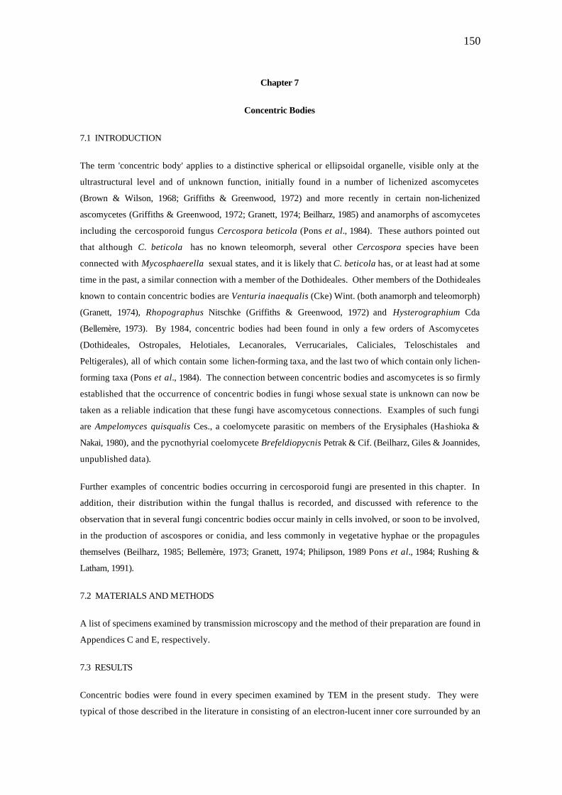
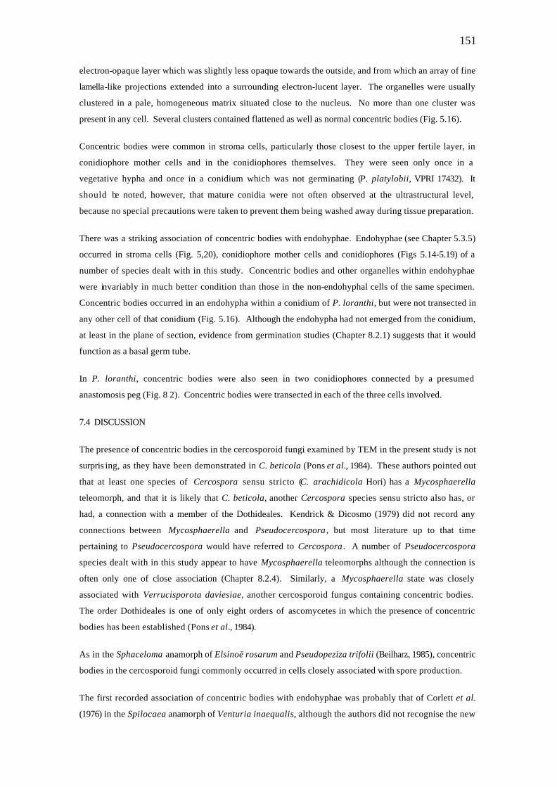
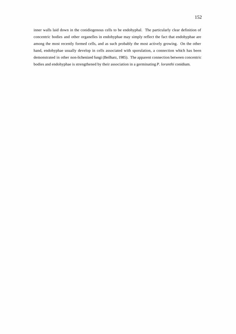
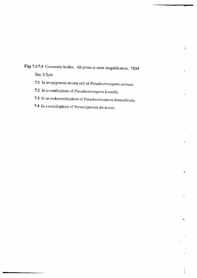
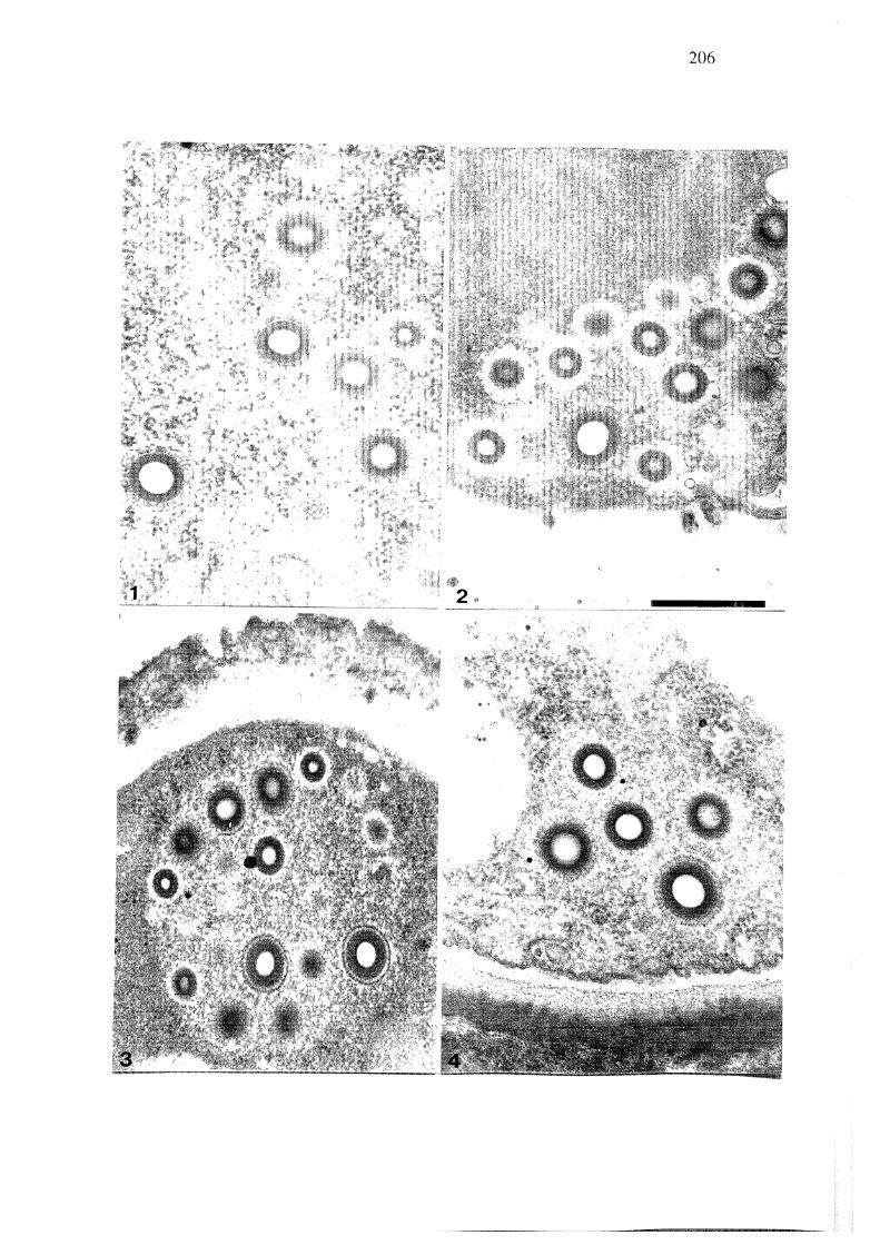
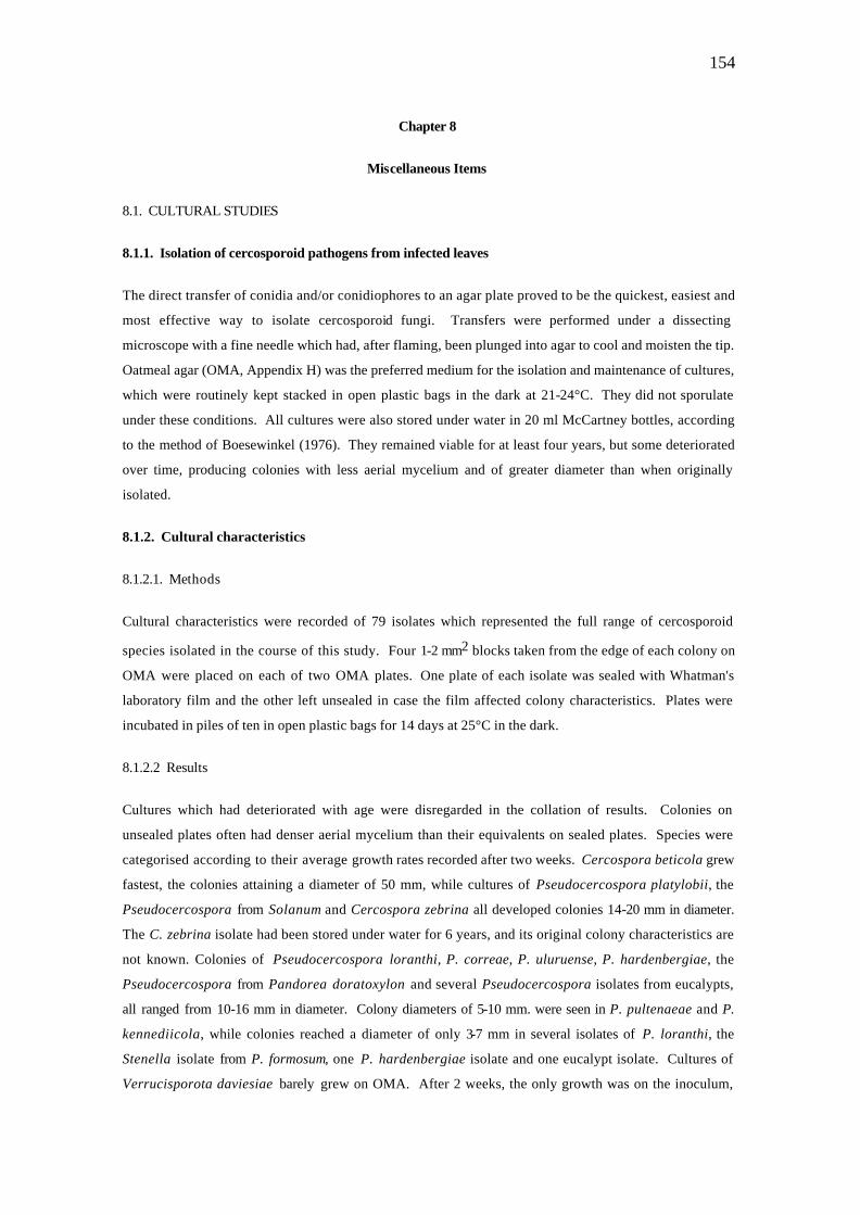
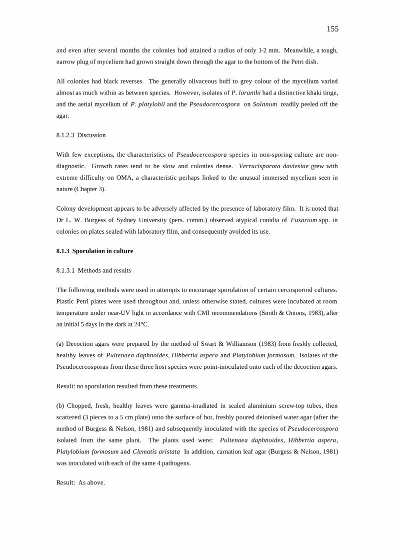
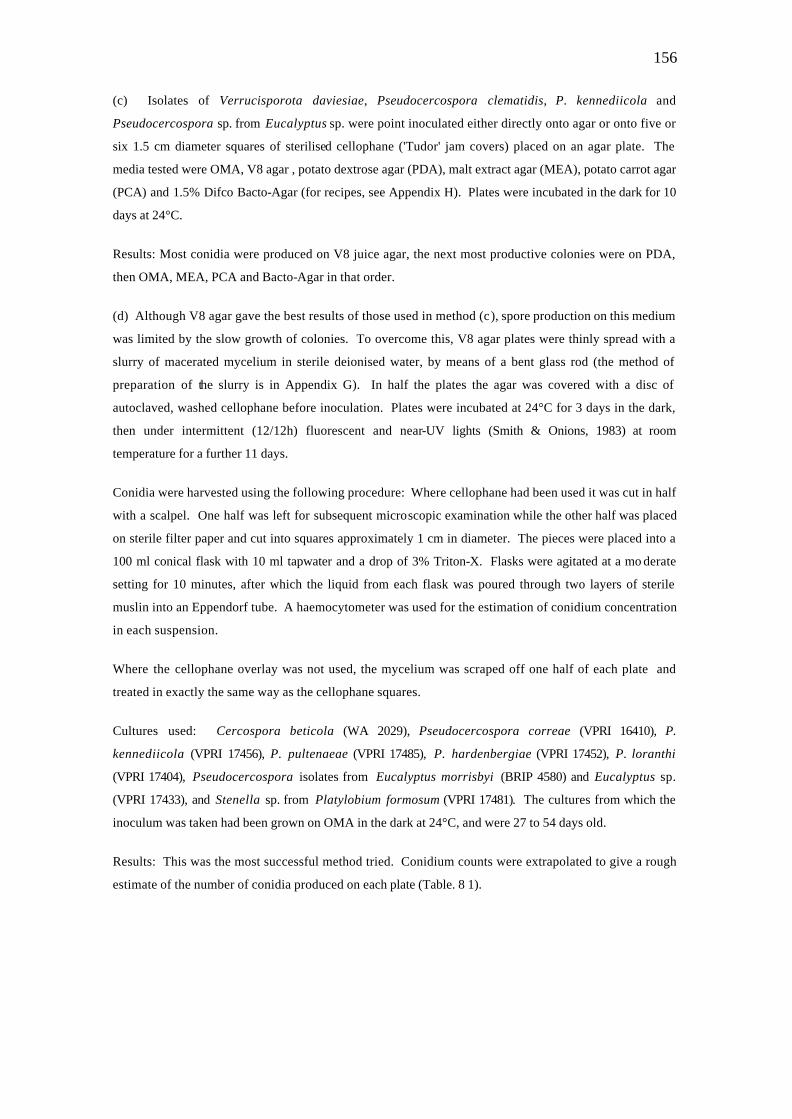
Concentric bodies were found in every specimen examined by TEM in the present study. They were
typical of those described in the literature in consisting of an electron-lucent inner core surrounded by an
151
electron-opaque layer which was slightly less opaque towards the outside, and from which an array of fine
lamella-like projections extended into a surrounding electron-lucent layer. The organelles were usually
clustered in a pale, homogeneous matrix situated close to the nucleus. No more than one cluster was
present in any cell. Several clusters contained flattened as well as normal concentric bodies (Fig. 5.16).
Concentric bodies were common in stroma cells, particularly those closest to the upper fertile layer, in
conidiophore mother cells and in the conidiophores themselves. They were seen only once in a
vegetative hypha and once in a conidium which was not germinating (P. platylobii, VPRI 17432). It
should be noted, however, that mature conidia were not often observed at the ultrastructural level,
because no special precautions were taken to prevent them being washed away during tissue preparation.
There was a striking association of concentric bodies with endohyphae. Endohyphae (see Chapter 5.3.5)
occurred in stroma cells (Fig. 5,20), conidiophore mother cells and conidiophores (Figs 5.14-5.19) of a
number of species dealt with in this study. Concentric bodies and other organelles within endohyphae
were invariably in much better condition than those in the non-endohyphal cells of the same specimen.
Concentric bodies occurred in an endohypha within a conidium of P. loranthi, but were not transected in
any other cell of that conidium (Fig. 5.16). Although the endohypha had not emerged from the conidium,
at least in the plane of section, evidence from germination studies (Chapter 8.2.1) suggests that it would
function as a basal germ tube.
In P. loranthi, concentric bodies were also seen in two conidiophores connected by a presumed
anastomosis peg (Fig. 8 2). Concentric bodies were transected in each of the three cells involved.
7.4 DISCUSSION
The presence of concentric bodies in the cercosporoid fungi examined by TEM in the present study is not
surpris ing, as they have been demonstrated in C. beticola (Pons et al., 1984). These authors pointed out
that at least one species of Cercospora sensu stricto (C. arachidicola Hori) has a Mycosphaerella
teleomorph, and that it is likely that C. beticola, another Cercospora species sensu stricto also has, or
had, a connection with a member of the Dothideales. Kendrick & Dicosmo (1979) did not record any
connections between Mycosphaerella and Pseudocercospora , but most literature up to that time
pertaining to Pseudocercospora would have referred to Cercospora . A number of Pseudocercospora
species dealt with in this study appear to have Mycosphaerella teleomorphs although the connection is
often only one of close association (Chapter 8.2.4). Similarly, a Mycosphaerella state was closely
associated with Verrucisporota daviesiae, another cercosporoid fungus containing concentric bodies.
The order Dothideales is one of only eight orders of ascomycetes in which the presence of concentric
bodies has been established (Pons et al., 1984).
As in the Sphaceloma anamorph of Elsinoë rosarum and Pseudopeziza trifolii (Beilharz, 1985), concentric
bodies in the cercosporoid fungi commonly occurred in cells closely associated with spore production.
The first recorded association of concentric bodies with endohyphae was probably that of Corlett et al.
(1976) in the Spilocaea anamorph of Venturia inaequalis, although the authors did not recognise the new
152
inner walls laid down in the conidiogenous cells to be endohyphal. The particularly clear definition of
concentric bodies and other organelles in endohyphae may simply reflect the fact that endohyphae are
among the most recently formed cells, and as such probably the most actively growing. On the other
hand, endohyphae usually develop in cells associated with sporulation, a connection which has been
demonstrated in other non-lichenized fungi (Beilharz, 1985). The apparent connection between concentric
bodies and endohyphae is strengthened by their association in a germinating P. loranthi conidium.
154
Chapter 8
Miscellaneous Items
8.1. CULTURAL STUDIES
8.1.1. Isolation of cercosporoid pathogens from infected leaves
The direct transfer of conidia and/or conidiophores to an agar plate proved to be the quickest, easiest and
most effective way to isolate cercosporoid fungi. Transfers were performed under a dissecting
microscope with a fine needle which had, after flaming, been plunged into agar to cool and moisten the tip.
Oatmeal agar (OMA, Appendix H) was the preferred medium for the isolation and maintenance of cultures,
which were routinely kept stacked in open plastic bags in the dark at 21-24°C. They did not sporulate
under these conditions. All cultures were also stored under water in 20 ml McCartney bottles, according
to the method of Boesewinkel (1976). They remained viable for at least four years, but some deteriorated
over time, producing colonies with less aerial mycelium and of greater diameter than when originally
isolated.
8.1.2. Cultural characteristics
8.1.2.1. Methods
Cultural characteristics were recorded of 79 isolates which represented the full range of cercosporoid
species isolated in the course of this study. Four 1-2 mm2 blocks taken from the edge of each colony on
OMA were placed on each of two OMA plates. One plate of each isolate was sealed with Whatman's
laboratory film and the other left unsealed in case the film affected colony characteristics. Plates were
incubated in piles of ten in open plastic bags for 14 days at 25°C in the dark.
8.1.2.2 Results
Cultures which had deteriorated with age were disregarded in the collation of results. Colonies on
unsealed plates often had denser aerial mycelium than their equivalents on sealed plates. Species were
categorised according to their average growth rates recorded after two weeks. Cercospora beticola grew
fastest, the colonies attaining a diameter of 50 mm, while cultures of Pseudocercospora platylobii, the
Pseudocercospora from Solanum and Cercospora zebrina all developed colonies 14-20 mm in diameter.
The C. zebrina isolate had been stored under water for 6 years, and its original colony characteristics are
not known. Colonies of Pseudocercospora loranthi, P. correae, P. uluruense, P. hardenbergiae, the
Pseudocercospora from Pandorea doratoxylon and several Pseudocercospora isolates from eucalypts,
all ranged from 10-16 mm in diameter. Colony diameters of 5-10 mm. were seen in P. pultenaeae and P.
kennediicola, while colonies reached a diameter of only 3-7 mm in several isolates of P. loranthi, the
Stenella isolate from P. formosum, one P. hardenbergiae isolate and one eucalypt isolate. Cultures of
Verrucisporota daviesiae barely grew on OMA. After 2 weeks, the only growth was on the inoculum,
155
and even after several months the colonies had attained a radius of only 1-2 mm. Meanwhile, a tough,
narrow plug of mycelium had grown straight down through the agar to the bottom of the Petri dish.
All colonies had black reverses. The generally olivaceous buff to grey colour of the mycelium varied
almost as much within as between species. However, isolates of P. loranthi had a distinctive khaki tinge,
and the aerial mycelium of P. platylobii and the Pseudocercospora on Solanum readily peeled off the
agar.
8.1.2.3 Discussion
With few exceptions, the characteristics of Pseudocercospora species in non-sporing culture are non-
diagnostic. Growth rates tend to be slow and colonies dense. Verrucisporota daviesiae grew with
extreme difficulty on OMA, a characteristic perhaps linked to the unusual immersed mycelium seen in
nature (Chapter 3).
Colony development appears to be adversely affected by the presence of laboratory film. It is noted that
Dr L. W. Burgess of Sydney University (pers. comm.) observed atypical conidia of Fusarium spp. in
colonies on plates sealed with laboratory film, and consequently avoided its use.
8.1.3 Sporulation in culture
8.1.3.1 Methods and results
The following methods were used in attempts to encourage sporulation of certain cercosporoid cultures.
Plastic Petri plates were used throughout and, unless otherwise stated, cultures were incubated at room
temperature under near-UV light in accordance with CMI recommendations (Smith & Onions, 1983), after
an initial 5 days in the dark at 24°C.
(a) Decoction agars were prepared by the method of Swart & Williamson (1983) from freshly collected,
healthy leaves of Pultenaea daphnoides, Hibbertia aspera and Platylobium formosum. Isolates of the
Pseudocercosporas from these three host species were point-inoculated onto each of the decoction agars.
Result: no sporulation resulted from these treatments.
(b) Chopped, fresh, healthy leaves were gamma-irradiated in sealed aluminium screw-top tubes, then
scattered (3 pieces to a 5 cm plate) onto the surface of hot, freshly poured deionised water agar (after the
method of Burgess & Nelson, 1981) and subsequently inoculated with the species of Pseudocercospora
isolated from the same plant. The plants used were: Pultenaea daphnoides, Hibbertia aspera ,
Platylobium formosum and Clematis aristata In addition, carnation leaf agar (Burgess & Nelson, 1981)
was inoculated with each of the same 4 pathogens.
Result: As above.
156
(c) Isolates of Verrucisporota daviesiae, Pseudocercospora clematidis, P. kennediicola and
Pseudocercospora sp. from Eucalyptus sp. were point inoculated either directly onto agar or onto five or
six 1.5 cm diameter squares of sterilised cellophane ('Tudor' jam covers) placed on an agar plate. The
media tested were OMA, V8 agar , potato dextrose agar (PDA), malt extract agar (MEA), potato carrot agar
(PCA) and 1.5% Difco Bacto-Agar (for recipes, see Appendix H). Plates were incubated in the dark for 10
days at 24°C.
Results: Most conidia were produced on V8 juice agar, the next most productive colonies were on PDA,
then OMA, MEA, PCA and Bacto-Agar in that order.
(d) Although V8 agar gave the best results of those used in method (c), spore production on this medium
was limited by the slow growth of colonies. To overcome this, V8 agar plates were thinly spread with a
slurry of macerated mycelium in sterile deionised water, by means of a bent glass rod (the method of
preparation of the slurry is in Appendix G). In half the plates the agar was covered with a disc of
autoclaved, washed cellophane before inoculation. Plates were incubated at 24°C for 3 days in the dark,
then under intermittent (12/12h) fluorescent and near-UV lights (Smith & Onions, 1983) at room
temperature for a further 11 days.
Conidia were harvested using the following procedure: Where cellophane had been used it was cut in half
with a scalpel. One half was left for subsequent microscopic examination while the other half was placed
on sterile filter paper and cut into squares approximately 1 cm in diameter. The pieces were placed into a
100 ml conical flask with 10 ml tapwater and a drop of 3% Triton-X. Flasks were agitated at a mo derate
setting for 10 minutes, after which the liquid from each flask was poured through two layers of sterile
muslin into an Eppendorf tube. A haemocytometer was used for the estimation of conidium concentration
in each suspension.
Where the cellophane overlay was not used, the mycelium was scraped off one half of each plate and
treated in exactly the same way as the cellophane squares.
Cultures used: Cercospora beticola (WA 2029), Pseudocercospora correae (VPRI 16410), P.
kennediicola (VPRI 17456), P. pultenaeae (VPRI 17485), P. hardenbergiae (VPRI 17452), P. loranthi
(VPRI 17404), Pseudocercospora isolates from Eucalyptus morrisbyi (BRIP 4580) and Eucalyptus sp.
(VPRI 17433), and Stenella sp. from Platylobium formosum (VPRI 17481). The cultures from which the
inoculum was taken had been grown on OMA in the dark at 24°C, and were 27 to 54 days old.
Results: This was the most successful method tried. Conidium counts were extrapolated to give a rough
estimate of the number of conidia produced on each plate (Table. 8 1).
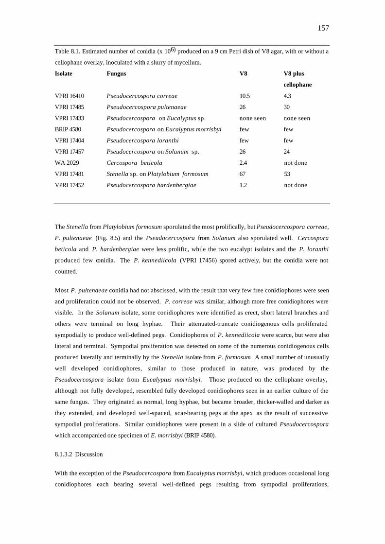
157
Table 8.1. Estimated number of conidia (x 106) produced on a 9 cm Petri dish of V8 agar, with or without a
cellophane overlay, inoculated with a slurry of mycelium.
Isolate Fungus V8 V8 plus
cellophane
VPRI 16410 Pseudocercospora correae 10.5 4.3
VPRI 17485 Pseudocercospora pultenaeae 26 30
VPRI 17433 Pseudocercospora on Eucalyptus sp. none seen none seen
BRIP 4580 Pseudocercospora on Eucalyptus morrisbyi few few
VPRI 17404 Pseudocercospora loranthi few few
VPRI 17457 Pseudocercospora on Solanum sp. 26 24
WA 2029 Cercospora beticola 2.4 not done
VPRI 17481 Stenella sp. on Platylobium formosum 67 53
VPRI 17452 Pseudocercospora hardenbergiae 1.2 not done
The Stenella from Platylobium formosum sporulated the most prolifically, but Pseudocercospora correae,
P. pultenaeae (Fig. 8.5) and the Pseudocercospora from Solanum also sporulated well. Cercospora
beticola and P. hardenbergiae were less prolific, while the two eucalypt isolates and the P. loranthi
produced few conidia. The P. kennediicola (VPRI 17456) spored actively, but the conidia were not
counted.
Most P. pultenaeae conidia had not abscissed, with the result that very few free conidiophores were seen
and proliferation could not be observed. P. correae was similar, although more free conidiophores were
visible. In the Solanum isolate, some conidiophores were identified as erect, short lateral branches and
others were terminal on long hyphae. Their attenuated-truncate conidiogenous cells proliferated
sympodially to produce well-defined pegs. Conidiophores of P. kennediicola were scarce, but were also
lateral and terminal. Sympodial proliferation was detected on some of the numerous conidiogenous cells
produced laterally and terminally by the Stenella isolate from P. formosum. A small number of unusually
well developed conidiophores, similar to those produced in nature, was produced by the
Pseudocercospora isolate from Eucalyptus morrisbyi. Those produced on the cellophane overlay,
although not fully developed, resembled fully developed conidiophores seen in an earlier culture of the
same fungus. They originated as normal, long hyphae, but became broader, thicker-walled and darker as
they extended, and developed well-spaced, scar-bearing pegs at the apex as the result of successive
sympodial proliferations. Similar conidiophores were present in a slide of cultured Pseudocercospora
which accompanied one specimen of E. morrisbyi (BRIP 4580).
8.1.3.2 Discussion
With the exception of the Pseudocercospora from Eucalyptus morrisbyi, which produces occasional long
conidiophores each bearing several well-defined pegs resulting from sympodial proliferations,
158
conidiophores produced in culture were generally 0-1 septate and similar to those produced in nature on
external hyphae.
Sporulation in culture was generally enhanced by the use of V8 agar and intermittent near-UV light. Fewer
conidia were produced when the cellophane overlay was in place on the agar, but its presence marginally
simplified the clean remo val of the colony from the agar plate. In an earlier study, Pseudocercospora
correae produced few conidia on carnation leaf agar and none on potato dextrose agar (Sutton et al.,
1987), but those cultures were held in the dark (Pascoe, pers. comm.). Near-UV light seems to be an
essential stimulus for the induction of sporulation, at least for Pseudocercospora , and the use of a
mycelial slurry as inoculum contributed significantly to the success of the technique. A spore suspension
could produce even better results (Ekpo & Esuruoso, 1978).
The eucalypt isolates and P. loranthi did not respond to any great extent to any of the stimuli applied,
and more work is needed to determine their requirements.
In summary, the combination of V8 agar, slurry inoculation and near-UV light induced conidium
production in most cultures sufficient for confirmation of the identity of the isolate and in some cases also
for experimental work. These findings are similar to those of El-Gholl et al. (1982) and Ekpo & Esuruoso
(1978) for species of Cercospora .
8 2. FUNGAL STRUCTURES AND BEHAVIOUR
8.2.1 Conidium germination
8.2.1.1 Observations
Because the position of germ tubes has been found useful in the taxonomy of imperfect fungi (e.g. Alcorn,
1983), conidium germination was studied in several cercosporoid fungi in order to determine the
consistency of germination patterns within spore populations. The fungi studied were P. loranthi (VPRI
17407, VPRI 17408), P. platylobii (VPRI 17432) and the Pseudocercospora species from Solanum sp.
(VPRI 17499) and Rubus fruticosus spp. agg. (VPRI 17508). Conidia brushed off infected leaves with a
camel-hair brush were germinated either in water drops on a glass slide in a humid chamber or on sterile
cellophane overlying water agar in a Petri dish.
Most conidia produced a germ tube from each end, after which lateral germ tubes emerged from any cell(s)
including the terminal cells. The first polar germ tube was most commonly basal but could be apical, and
in some conidia the first-formed germ tube was lateral.
Apical germ tubes were usually holoblastic and continuous with the long axis of the conidium, whereas
basal germ tubes were generally enteroblastic and semi-axial. Basal germ tubes either pushed the scar to
one side and grew on in line with the long axis of the conidium, or else emerged at an angle with little
effect on the orientation of the scar. Most basal germ tubes of P. loranthi (VPRI 17407, VPRI 17408)
conidia emerged directly through the basal scars. However, in conidia from a specimen of P. loranthi
159
(VPRI 17406) germinated on cellophane overlying water agar, most basal germ tubes were semi-axial, and
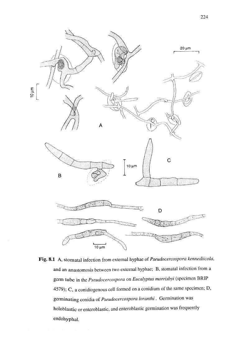
the only germ tubes seen emerging directly through a scar were endohyphal (Fig. 8.1).
Lateral germ tubes appeared to be always holoblastic.
8.2.1.2 Discussion
I had thought that basal germ tubes might breach unthickened scars, such as the very thin scars of P.
platylobii, and that this would provide a means of distinguishing thickened from unthickened scars.
However in all but P. loranthi the basal germ tubes mostly emerged beside the scar, showing that even
unthickened scars present more resistance than the nearby conidium wall. The weakest point at the base
of the conidium is evidently where the scar joins the anticlinal wall.
Basal conidium germination in cercosporoid fungi is equivalent to apical proliferation of the
corresponding conidiogenous cell. This is not surprising, given that the conidium base is a mirror image
of the conidiogenous cell apex.
The pattern of spore germination was generally as variable within as between samples. There was no
indication that the character might be useful in the taxonomy of this group of fungi.
8.2.2 The infection process
8.2.2.1 Observations
Infection studies were not attempted, but natural infection was observed in some specimens. In each
case, an external hypha or germ tube formed a small coil over a stoma, and infection was directly through
the stoma. The fungi in which stomatal penetration was observed were: P. kennediicola (VPRI 17460)
(Fig. 8.1), P. pultenaeae (VPRI 17485), Pseudocercospora on Rubus sp. (VPRI 15918) and the
Pseudocercospora on Eucalyptus morrisbyi (Fig. 8.1). In the first three examples the coils formed on an
external hypha, while in the last the penetration coil formed on a conidium germ tube. A hypha of P.
kennediicola formed successive coils over three stomata as it grew across the leaf surface (Fig. 8.1).
8.2.2.2 Discussion
Stomatal penetration resembling that described here was reported by Solheim (1930), and Berger &
Hanson (1963b) referred to similar observations by Baxter, and Latch & Hanson. Some fungi form
appressoria over stomata, possibly facilitating ingress by forcing apart the lips of closed stomata (Aist,
1981). Hyphal coils formed by cercosporoid fungi may serve the same function. The mode of ingress
directly affects the pattern of leaf tissue colonisation. It has been noted (Chapter 6) that in hosts with
only abaxial stomata, immersed mycelium is initially present only near the abaxial surface. Later, usually
after the onset of stomatal sporulation, hyphae may breach the palisade mesophyll and epidermis on the
opposite side of the leaf, and this can lead to the formation of erumpent conidiomata at the adaxial surface.
Stomatal penetration would naturally lead to the sequence of events just described and is probably the
160
most common or even the sole mode of infection in these fungi. Pseudocercospora phebalii is the only
fungus in this study which was not observed to sporulate from the stomatophorous leaf surface. Hyphae
were, however, seen in the spongy mesophyll close to the lower epidermis of a leaf of BRIP 8764 opposite
a lesion on the upper surface, suggesting that colonisation could conceivably have started on the
underside of the leaf.
8.2.3 Anastomoses
Anastomoses, or hyphal fusions, can occur within or between mycelia. In general terms, they enable a
compounding of cell contents (nuclei and cytoplasm) from genetically different mycelia, leading to the
establishment of heterokaryons and making possible parasexual reproduction (Burnett, 1975).
8.2.3.1 Anastomoses of hyphae in culture
Cultured cercosporoid fungi tend to produce restricted, raised, colonies which are dense right to the
margin. To encourage sparser colony margins, selected isolates growing on oatmeal agar were point-
inoculated onto the centres of 1.5 cm2 squares of cellophane (Cuprophan-Dialysierfolie type 250-pm, from
Enka Glanzstoffresting) lain on 1.5% Difco Bacto-Agar. The Petri dishes were sealed with Nescofilm and
kept at 22°C for 7 days in the dark. The inoculum and the fluffy growth surrounding it was lifted off with
forceps, leaving a flat, sparse colony on the cellophane. The cellophane squares were then mounted in
50% glycerine (the cellophane did not buckle in this medium), warmed to dispel air bubbles and examined
under the light microscope.
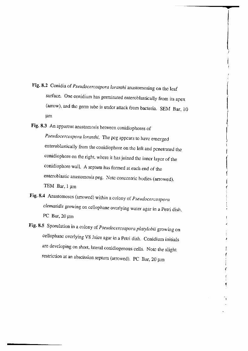
Inter-hyphal anastomoses were seen in Pseudocercospora loranthi (VPRI 17500, VPRI 17408), P.
clematidis (VPRI 17414) (Fig. 8.3), P. hardenbergiae (VPRI 17462), P. kennediicola (VPRI 17461), P.
pultenaeae (VPRI 17486) and a Pseudocercospora from Eucalyptus sp. (VPRI 17444). Anastomoses had
been seen in Verrucisporota daviesiae (VPRI 17431) grown on cellophane over PCA and OMA at 22°C in
another experiment. In addition, anastomoses occurred between germ tubes from naturally produced
conidia of P. loranthi (VPRI 17406) germinating on cellophane overlying WA in a Petri dish.
Anastomoses were not seen in P. platylobii (VPRI 17451, VPRI 17432), in the Pseudocercosporas on
Solanum sp. (VPRI 17457) and Hibbertia aspera (VPRI 17454) or in the Stenella from Platylobium
formosum (VPRI 17507).
8.2.3.2 Anastomoses in nature
Most anastomoses were between conidia lying on the leaf surface, but conidiophores and external hyphae
were also involved. Anastomoses between conidia were observed in the Pseudocercosporas on Acacia
suaveolens (BRIP 8844), Amyema pendulum (VPRI 16059) (Fig. 8.1), Ceratopetalum gummiferum (DAR
1502), Clematis sp. (VPRI 15360), Dodonaea triquetra (DAR 29778), Eucalyptus spp. (VPRI 15334, VPRI
17439), E. exserta (BRIP 4578) (Fig. 3.25), E. macrorhyncha (VPRI 15509), E. morrisbyi (BRIP 4580) (Fig. 3.
25), E. regnans (VPRI 15450), E. saligna (DAR 4149), Kennedia sp. (VPRI 17463), Lophostemon confertus
161
(DAR 5918, VPRI 13661, VPRI 17492), Pultenaea daphnoides (VPRI 14935), and Solanum sp. (VPRI 15477)
and in the cercosporoid pathogen of Strychnos lucida (DAR 30050).
A conidium of Pseudocercospora sp. on E. macrorhyncha had anastomosed twice with an external hypha
(Fig. 3.26). External hyphae had anastomosed in the Pseudocercospora on Acacia complanata (VPRI
15101), in Pseudocercospora correae (VPRI 16411, 16420) and in P. hardenbergiae (VPRI 15756). In P.
kennediicola, anastomoses occurred between conidia, between a conidium and a conidiophore, and in
abundance between hypogenous external hyphae. Conidiophores had anastomosed in the
Pseudocercospora species on Solanum sp. (VPRI 15477), Pultenaea daphnoides (VPRI 14760) and E.
regnans (VPRI 15450 and VPRI 17439). In addition, a conidium in VPRI 15450 had anastomosed with an
external hypha, and in VPRI 17439 a conidium had anastomosed with a thin hypha which had grown on
from the end of a conidiophore. In P. loranthi, anastomosing conidiophores were seen in VPRI 17406. An
ultrathin section through P. loranthi (VPRI 17404) appears to have transected an anastomosis between
two conidiophores. The presumed anastomosis peg is continuous with the inner wall layer of each
conidiophore, and is defined by a newly formed septum at each end.
8.2.3.3 Discussion
Anastomoses were seen in most of the cercosporoid fungi under study, both in culture and in nature.
Cultures were not derived from single spore isolates, so must be regarded as mixed mycelia. However,
anastomoses between different fungal parts are so common that individual conidia are unlikely ever to be
homokaryotic in this group of fungi. No particular significance is attached to the tendency of these fungi
to anastomose, as anastomoses within and between mycelia are well recognised in a number of fungi
(Burnett, 1975). No attempt was made to study anastomoses between opposing mycelia, as hyphal fusion
is not a reliable indicator of genetic equivalence (Talbot, 1971; Burnett, 1975; Subramanian & Jain, 1978).
8.2.4. The occurrence of teleomorphs
8.2.4.1. Observations
Ascocarps containing unequally 2-celled ascospores were associated with the cercosporoid
hyphomycetes under study on the following hosts: Acaena anserinifolia (DAR 18953), Daviesia latifolia
holotype (Wm. Martin No. 438) (Fig. 3.14, 3.49) and other Daviesia collections including VPRI 17435 (on
D. mimosoides var laxiflora) (Fig. 3.14), Hardenbergia violacea (VPRI 16032), Hibbertia scandens (VPRI
14848) and Platylobium formosum (in association with Pseudocercospora platylobii) (VPRI 13805).
Ascocarps with 2-celled ascospores occurred on Eucalyptus morrisbyi (BRIP 4579), but not in the same
lesions as the Pseudocercospora anamorph; immature ascocarps were observed on Lophostemon
confertus (DAR 5629).
8.2.4.2 Discussion
The ascocarps all appeared to be represent species of Mycosphaerella. Mycosphaerella states are
already recorded for cercosporoid species within the genera Cercospora , Cercosporella, Cercoseptoria,
162
Ramularia, Ovularia, Cladosporium, Heterosporium, and Polythrincium, and for species of the
coelomycetous genus Septoria. The ascomycetous teleomorph Sphaerulina has also been connected
with Cercospora and Septoria.
The association of Mycosphaerella ascocarps with cercosporoid hyphomycetes in the present study is
therefore in accordance with the literature. The most convincing connection of those mentioned here was
in Verrucisporota daviesiae, where darkly pigmented assimilative hyphae, coated with a thick, pale
brown, smooth or bubbly mucilaginous material (Figs 3.1) led to all three types of fruiting structures
(conidiomata, spermogonia and ascocarps).
8.2.5 The occurrence of spermogonia
Spermogonia were associated with the cercosporoid anamorph under study on the following hosts:
Daviesia latifolia (Fig. 3.50), Lophostemon confertus (DAR 5629, 5630), Amyema pendulum (VPRI 15513)
and Acacia suaveolens (DAR 4356). The connection of the spermogonial, ascal and hyphomycetous
states of Verrucisporota daviesiae was mentioned in Chapter 3.11. Spermogonia associated with
Pseudocercospora platylobii develop within the stromata of old conidiomata, which still bear typical
conidiophores at the top (Fig 3.39). A similar phenomenon was described for Pseudocercospora pini-
densiflorae (Hori & Nambu) Deighton on Pinus oocarpa (Evans, 1984) and Pseudocercospora
hiratsukana (Togashi & Katsuki) Deighton, although spermogonia of the latter fungus also formed
adjacent to the conidiomata, evidently connected by the same mycelial hyphae (Deighton, 1976).
8.2.6 Sporulation from external conidiophores
8.2.6.1 Observations.
Conidium production from conidiogenous cells on external hyphae was recorded by Deighton (1976) in
Pseudocercospora atrovelutina, P. actinidae, P. asclepiadina, P. insueta, P. bakeriana, P. phyllanthi-
reticulati, P. ghanensis, P. cecropiae, P. cecropiicola, P. ligustri, P. puderi, P. salicina, P. trichophila,
P. triumfettae, P. triumfettigena and P. kiagweensis. In several of these fungi, the production of
conidiophores on external mycelium was the only means of sporulation, whereas stomatal and/or
erumpent sporulation also occurred in others. In the present study, external conidiophores were observed
in Pseudocercospora species on the following hosts: Acacia complanata (VPRI 15101), Amyema
pendulum (VPRI 17502, VPRI 16031) (Fig. 3.12), Correa spp. (most specimens diagnosed as P. correae,
and all of those diagnosed as P. correicola) (Figs 3.15, 3.16), Desmodium brachypodium (DAR 16962),
Eucalyptus spp. (VPRI 17442, VPRI 17443, VPRI 17433, VPRI 17446), E. exserta (BRIP 4578), E. maculata
(BRIP 8909) (FIg. 3.26), E morrisbyi (BRIP 4580) (Fig. 3.25), E. ?obliqua (VPRI 17439) (Fig. 3.54),
Hardenbergia violacea (VPRI 15756), Kennedia rubicunda (DAR 30034), Kennedia sp. (VPRI 17461) (Fig.
3.30), Lophostemon confertus (VPRI 17492, VPRI 15728, DAR 5918, DAR 60463), Platylobium formosum
(DAR 16072), Pultenaea daphnoides (VPRI 17486) (Fig. 3.44) and Santalum lanceolatum (VPRI 17487). In
addition, a conidiogenous cell formed laterally on a conidium which had anastomosed with two others in a
Pseudocercospora on Eucalyptus (VPRI 17439), and a conidiogenous cell formed on shed conidia of the
163
Pseudocercosporas on Lophostemon confertus (DAR 60463) and Eucalyptus morrisbyi (BRIP 4579); these
conidiogenous cells resembled those formed on external hyphae.
8.2.6.2. Discussion
The incidence of external sporulation varies within and between hosts. Deighton (1976) commented that
'the production of a conidiophore-bearing external mycelium is not a constant character', and decided that
it was therefore not possible to subdivide Pseudocercospora on this basis. Furthermore, external hyphae
can be overlooked when they are sparse or on hairy leaves, as in the original description of P. correae
(Sutton et al., 1987). While external sporulation is sparse in some specimens of P. correae, it is the
dominant mode of sporulation in others, whether among the abaxial leaf hairs in specimens diagnosed as
P. correicola or on the leaf surface.
Similar variation is displayed by three specimens of Pseudocercospora on Lophostemon confertus. In
specimen DAR 5918, immersed stromata are reduced but conidiophores are abundant on a prolific external
hypogenous mycelium. By contrast, dense clusters of conidiophores were borne on relatively large
hypogenous stromata on VPRI 13661, and although hyphae were seen on the leaf surface no external
conidiophores were found. The growth on DAR 60463 was intermediate in type, most of the scant
sporulation originating from immersed stromata but some also from external hyphae.
Whereas hypogenous external sporulation is typical of the Pseudocercospora species already discussed,
and is often clearly visible under the dissecting microscope, it is of minor importance in species such as P.
platylobii, and detected only rarely under the compound microscope.
The production of external hyphae on a well-developed external mycelium which has emerged through the
stomata is typical of species such as P. correae and the Pseudocercospora on Lophostemon confertus.
The character is nevertheless too variable to be of more than limited value in the taxonomy of the genus
because most, perhaps all, species have the capacity to form fertile external mycelium under appropriate
conditions.
8.2.7. Immersed hyphae
The internal mycelium of all specimens examined in this study consisted of septate, branched hyphae
ranging from hyaline to pale olivaceous brown. With very few exceptions, assimilative hyphae were
intercellular. Intracellular hyphae were seen within one spongy mesophyll cell and several epidermal cells
of Platylobium formosum infected with Pseudocercospora platylobii (Fig. 3.7), and one intracellular
hypha was observed by TEM in a specimen of Amyema pendulum infected with P. loranthi; the
intercellular spaces of this specimen were packed with hyphae. None of the intracellular hyphae was
associated with stroma initiation, which was strictly intercellular.
The relationship between the immersed mycelium and host cells has received little attention in the
cercosporoid fungi. It was described as intracellular in Pseudocercospora correae (Sutton et al., 1987),
and both intra- and inter-cellular in Pseudocercospora correicola (Sutton & Pascoe, 1988). Present
164
studies, including those of ultrathin sections, leave no doubt that the immersed mycelium of specimens
identified as P. correae is strictly intercellular. There is no reason to believe the situation is any different
in specimens identified as P. correicola, which is considered here to be synonymous with P. correae.
Similarly, the immersed mycelium was described as intracellular in species of Asperisporium,
Pseudocercospora and Anaphysmene on conifers (Sutton & Hodges, 1990). In several of the illustrations,
particularly that of Asperisporium juniperum, the immersed mycelium and stromata appear to be
intercellular.
Although the immersed hyphae were all intercellular, their relationship with the host cells still varied
between specimens, some tending to maintain close contact with host cell walls and others more inclined
to freely cross the intercellular spaces. In the former type, ultrastructural studies showed a mucilaginous
layer between the hyphae and the host cell walls they contacted. Examples were seen in
Pseudocercospora loranthi, P. uluruensis, Cercosporidium duboisiae and the Pseudocercospora on
Clematis. These fungi all enjoy a prolonged biotrophic relationship with their respective hosts. In the
first three, the immersed hyphae are often profusely branched, the branches being short and flattened
against the cell walls and also filling the intercellular spaces. The branched nature of the hyphae can be
observed only in very young infections of P. loranthi and C. duboisiae, before the hyphal mass builds
up. In C. duboisiae, the massed hyphae adjacent to the vascular bundles were of the same type.
The Pseudocercospora on Eucalyptus macrorhyncha was of the second type, its hyphae not being
closely associated with the cell walls. The host tissue was necrotic and the mesophyll cells were
collapsed by the time the specimen was fixed, so it is possible that the hyphae were more closely
associated with the host cell walls earlier in the colonisation process However, the eucalypt fungus
probably has only a short biotrophic phase.
Hyphae in close contact with host cell walls probably obtain nutrients directly from the apoplast, and this
may help them, at least early in their development, to maintain a biotrophic relationship with their host.
The eucalypt fungus, on the other hand, may depend on the rapid leaching of the contents of damaged
cells into intercellular spaces, and may behave as a necrotroph from an earlier stage.
167
Chapter 9
General Discussion
'So often the complaint is that we could develop an adequate understanding of a fungus if
only we could find some way to grow it in culture. In truth, it is a blessing that large
numbers of fungi refuse to perform in culture and have to be studied in the field where they
grow naturally.'
Luttrell, 1989
Ultrastructural studies have demonstrated the presence of varying amounts of externally deposited
material on abscission scars of fungi which otherwise fit into the broadly circumscribed genus
Pseudocercospora . Given that Pseudocercospora is distinguished from Cercospora largely on the basis
of its unthickened scars, this finding, foreshadowed by B. C. Sutton (pers. comm.) is of considerable
taxonomic significance in the subdivision of cercosporoid fungi.
The thickened scars found in some species of Pseudocercospora are generally paler and less
conspicuous than those of, for example, Cercospora beticola, a species which is regarded as typical of
the genus and which has been studied in detail at the ultrastructural level (Pons et al., 1985). In certain
specimens of Pseudocercospora loranthi and the Pseudocercospora on Eucalyptus morrisbyi,
specimens in which substantial thickening has been demonstrated in this study, thickened scars can be
quite difficult to find under the light microscope.
The Pseudocercospora species exhibiting external scar thickening do not, therefore, form a readily
identifiable natural group, and they can not be recombined into a genus such as Cercosporidium which is
typified by conidial scars which are 'always thickened and conspicuous' (Deighton, 1967; Ellis, 1971). In
response to these findings, the limits of the already broadly circumscribed genus Pseudocercospora must
be further expanded to admit species with thickened scars. These results support Thaung's (1984)
contention that scar thickening can be regarded as only a secondary characteristic in the cercosporoid
fungi, one which is more applicable at the specific than at the generic level. In practice, the character
distinguishes most species of Pseudocercospora from most species of Cercospora , and can be retained
as a useful secondary characteristic for this reason.
There is some association in Pseudocercospora between the presence of substantial scar thickening and
ready conidium dehiscence on the one hand, and lack of scar thickening and delayed dehiscence on the
other. Maturation of the abscission septum has been shown to follow the delimitation of conidia in
several species of Cercospora , resulting in the shedding of conidia of variable size but always with a
prominent basal scar (Pons et al., 1985). The same sequence of events occurs in Pseudocercospora
specimens with visibly thickened scars and in Verrucisporota daviesiae. Present results suggest that
species of Pseudocercospora in which there is marked attenuation towards the scar often exhibit some
168
scar thickening and a readiness to secede, while a lack of attenuation is associated with a lack of
thickening, and conidium persistence. The type of P. eucalyptorum, which was collected in South Africa,
and several eucalypt specimens collected in Australia exhibit the latter characteristics, while the remaining
Australian collections from eucalypts mostly exhibit the former. The Pseudocercospora on Eucalyptus
macrorhyncha is an exception to the rule in that its conidia are markedly attenuated towards the scar yet
usually persist after delimitation until they are several-septate and have assumed a mature, obclavate
shape. Precisely the same situation obtains in Pseudocercospora hardenbergiae. The persistence of
conidia in these fungi appears to contradict the pattern just mentioned, because slight peripheral
thickening was demonstrated in both fungi at the ultrastructural level, and slight thickening was seen
occasionally in both fungi by light optics. These fungi may be intermediate in type, with thickening which
is either only occasionally present, which is too slight to promote ready dehiscence or which is deposited
only after a period of delay. The link between attenuation towards the scar and the presence of external
scar thickening holds, nevertheless.
As already mentioned, the various specimens of Pseudocercospora collected from eucalypts were highly
variable in conidium shape, which was either obclavate or cylindrical, and in the degree of attenuation
towards the basal scar. The shape of the base of the conidium was, as always in the cercosporoid fungi,
reflected in the shape of the apex of the conidiogenous cell. Pseudocercospora eucalyptorum, which
includes the fungi previously known as Cercospora eucalypti and C. epicoccoides, is described as
having conidium bases which are subtruncate to long obconically truncate (Crous et al., 1989). The
diversity of the Australian collections, however, is inconsistent with the notion that they all belong in the
same species. Several Australian specimens from eucalypts resemble the type of P. eucalyptorum (PREM
49112) in having more or less cylindrical conidiogenous cells and conidia terminating in broad,
unthickened scars. Although there is variation among these Australian specimens, they all have larger
conidia and conidiophores than PREM 49112, and the fungi are not necessarily all conspecific. The
remaining Australian specimens from eucalypts all differ from PREM 49112 in producing more or less
obclavate conidia which are attenuated towards the hilum. Thickened scars have been seen in some of
these specimens, notably those from certain eucalypts growing in Queensland. It is likely that a new
species will be described to accommodate these particular specimens, but a great deal more collecting is
needed if the Australian specimens are to be sorted out. Present results of an isozyme analysis applied to
some cultured Pseudocercosporas from eucalypts indicate that the technique may prove useful in this
regard.
In the type of Pseudocercospora , P. vitis, scars are narrow and associated with a marked attenuation of
the conidiogenous cell apices and conidium bases; conidia of P. vitis are obclavate and readily shed.
Species of Pseudocercospora described by Deighton (e.g. 1976, 1987) all exhibited attenuation towards
scars which were narrow relative to the cells on which they occurred. On the basis of these
characteristics, P. pultenaeae, P. correae, P. platylobii, P. uluruensis, P. kennediae and the type of P.
eucalyptorum are borderline members of the genus. Sutton et al. (1987) rejected the possibility of placing
the fungus they named Pseudocercospora correae into the genus Cercoseptoria because its stromata are
subepidermal rather than substomatal, because its conidia are not strictly acicular, and because the scars
169
left on the conidiogenous cells are of similar width to the conidiogenous cell rather than narrower. The
latter concern is not valid, because Sutton et al., were comparing conidiophores of similar morphology but
at different stages of development. P. correae also often forms substomatal stromata. Of the fungi
described here, P. platylobii would probably have been placed in the genus Cercoseptoria had the genus
not been reduced to synonomy with Pseudocercospora (Deighton, 1987). It appears to differ from
Cercoseptoria in having erumpent as well as stomatal conidiomata, and in the fact that its stomatal
conidiomata eventually become protuberant, disrupting the stomata in the process. However, we can not
be sure that the type of Cercoseptoria, C. chamaesyces, might not also develop in this manner (the type
specimen shows only very immature conidiomata). The cercosporoid pathogens on Ceratopetalum
gummiferum Sm (DAR 1502) and Nephrolepis sp. (DAR 56066) are also close to Cercoseptoria. Although
Deighton (1987) saw a need to recombine Cercoseptoria into Pseudocercospora , the concept of the
genus clearly has value, if only at the subgeneric level.
Pseudocercospora correae and P. platylobii produce narrow, filiform conidia, but the conidia of P.
pultenaeae, P. uluruensis, P. phebalii and the truncate-based eucalypt specimens are broader, shorter
and more or less cylindrical. They resemble neither Cercoseptoria nor the type of Pseudocercospora , P.
apii, which has obclavate conidia with relatively narrow scars. This group of fungi may form the basis of
another subgeneric division within Pseudocercospora. It is to be wondered whether fungi with these
characteristics are peculiar to the southern hemisphere. The type of P. eucalyptorum belongs in this
group, but although it was collected in South Africa it could have originated in Australia.
On the other hand, the Pseudocercospora from some of the Queensland eucalypts, which has thickened
scars, is relatively close to P. vitis with regard to conidiophore and conidium shape. There is clearly a
need for ultrastructural examination of P. vitis to establish its scar structure. It would be a strange
outcome indeed if true members of Pseudocercospora were to be eventually distinguished from those
with broad abscission scars on the basis of their narrow, thickened scars!
Pseudocercospora exhibits a wide variety of conidioma types. Conidiomata can form on fully immersed or
partly protuberant stromata which in turn can be substomatal, erumpent or both. They can form on a small
aggregation of substomatal hyphae or on external hyphae which emerge from stomata. This variation is
well recognised, but recognition has not always been given to the variety of ways in which stromata form
and gain egress. Although Deighton (1976) considered that all stromata formed by species of
Pseudocercospora were substomatal, a number of recent authors have described subepidermal or
epidermal stromata (Evans, 1984; Pons & Sutton, 1988; Sutton et al., 1987; Verma et al., 1989; Goh &
Hsieh, 1989).
I have suggested that it would be useful if the process of stroma development were considered as
comprising three successive steps. The first is the development of a stroma initial, which is usually
substomatal or subepidermal but in P. loranthi occasionally also subcuticular. The second step involves
the invasion of other tissues. For example, a subepidermal stroma initial acts as the base from which
hyphae invade the epidermal layer, either individually or en masse. This can involve either direct invasion
of one or more epidermal cells or the passage of hyphae between the anticlinal cell walls. In either case
170
the hyphae may proceed through to the subcuticular region where additional stromatic material may be
formed. The third step is one of stroma maturation, and this involves an increase in volume of all portions
of the stroma, although not necessarily all to the same extent. The subepidermal stroma initial develops
along with the rest and is an integral part of the mature stroma.
Details of stroma development vary considerably between species of Pseudocercospora , and although
the character is no doubt partly host-related, it also reflects the ability of each fungus to physically and/or
chemically breach barriers imposed by host tissues, particularly the epidermis. Although these
characteristics may be of limited use in the taxonomy of the group, the observation that epigenous
stromata of P. correicola dissolve a passage through the epidermal cell walls in exactly the same way as
do those of P. correae was an early indication that P. correae and P. correicola might be the one fungus.
(Quite naturally, other cercosporoid fungi can be expected to share this feature. A micrograph of the
Pseudocercospora on Hakea (Fig. 6.7), shows hyphae entering an epidermal cell, apparently by
dissolution of the inner wall).
Because stroma formation is so variable within Pseudocercospora , labels such as 'epidermal' or
'subcuticular', for example, are inadequate to convey a true impression of stroma morphology. In the first
place, 'epidermal' could mean intraepidermal or interepidermal. Even the term 'intraepidermal' is
inappropriate for these fungi, because the stroma initial is necessarily intercellular, whether or not an
epidermal cell is subsequently invaded. The term 'epidermal stroma' should, I believe, be restricted to
fungi with an intracellular growth habit, because their stromata (if intracellular) are initiated from hyphae
which are already present within the cell, and develop to maturity within the cell. When the immersed
mycelium is intercellular, as in the cercosporoid fungi studied here, stroma formation is best described in
terms of the several developmental steps outlined above. The associated character 'mode of egress' also
needs to be carefully observed and recorded.
It is also important to recognise that the distribution of conidiomata of a cercosporoid fungus is partly
dependent on the ways in which the particular fungus is able to breach host tissues, and partly on the
anatomy of the host leaf. For example, if egress is obligatorily stomatal it can occur from one or both leaf
surfaces depending on the distribution of stomata; if the fungus also has the capacity for erumpent egress
it may sporulate on the upper leaf surface, even though there may be no stomata present, as well as the
lower surface. Whether sporulation is epigenous, hypogenous or amphigenous is therefore as much a
host-related characteristic as it is a characteristic of the fungus.
A further source of variation in Pseudocercospora is found in the mode of proliferation of the
conidiogenous cell. Pons & Sutton (1988) stated that provided the presence of annellations can be
proved in P. vitis (the type of Pseudocercospora), the fungus should be described as showing 'frequent
discontinuities in the outer wall due to sympodial holoblastic or enteroblastic or percurrent enteroblastic
proliferations', and further, 'The absence of one of these features in some species would provide sufficient
reason to separate them into a different generic taxon.' One objection to this rather strict requirement
hinges on the notion that the environment could influence the mode of proliferation by affecting cell wall
plasticity (Madelin, 1979). Perhaps of greater significance is the fact that I have been unable to identify
171
any instance of enteroblastic percurrent proliferation in the Pseudocercospora specimens described here.
While this may be my failing, it serves to emphasise the difficulties facing the investigator should a genus
be defined in these terms.
Enteroblastic sympodial proliferations, on the other hand, are very common, as seen in scanning electron
micrographs of a variety of taxa. These proliferations emerge either through the conidiogenous cell wall,
below the scar left after the secession of the previously formed conidium, or at the periphery of the scar,
which is clearly a weak point. If the point of emergence is at the periphery of the scar or very close to it,
the scar is often pushed to the side and left lying flat against the conidiogenous cell wall. If the scar were
to fall off, or be obscured because of unfavourable orientation under the microscope, or if the fungal parts
were very pale, it would be difficult to distinguish 'pseudopercurrent' from 'percurrent' proliferation.
Scanning electron micrographs presented here show enteroblastic proliferations in a number of species of
Pseudocercospora . The thin and fragile annellations left on external conidiophores (Figs 3.29, 3.54)
following proliferation may be considered comparable with the first annellations left on young, caespitose
conidiophores. Pseudocercospora vitis has been attributed with often exhibiting true percurrent
proliferations on young conidiophores, but the ragged outlines of the annellations seen here suggest that
the proliferations were pseudopercurrent, even though laterally displaced scars are in view only in the
pathogens of eucalypt and Kennedia. The precise nature of proliferations in P. vitis can be resolved only
by use of the scanning electron microscope.
I have provided ultrastructural evidence of conidiogenous cell proliferation by means of wide-diameter
endohyphae in several species of Pseudocercospora , and believe that it justifies the recognition of
endohyphal proliferation as a distinctive process leading to the normal production of successions of
conidia in these fungi. This mechanism may be the only means of proliferation available to some species,
or it may operate in tandem with other modes of proliferation. Endohyphal proliferation has already been
demonstrated in a small number of other dematiaceous hyphomycetes and will almost certainly be found
in other taxa once they are viewed in ultrathin section.
The variation inherent in the genus Pseudocercospora , as presently understood, extends to the problem
of variation within species, which in turn complicates decisions on species delimitation. I agree with
Chupp (1954), who considered the shape of the conidium base to be the most constant character in the
broadly defined genus Cercospora . Most species examined in this study proved remarkably uniform in
this regard, but problems arose in two particular areas. The first involved variation among specimens of
Pseudocercospora from eucalypts, which has already been commented on. The second problem involved
the two foliar pathogens of Correa spp., Pseudocercospora correae and P. correicola, which were
distinguished largely on the basis of growth habit (Sutton & Pascoe, 1987). I conclude that differences in
habit are largely host-related, and that P. correicola should be reduced to synonomy with P. correae.
This recommendation was made even though the conidium bases were often slightly more attenuated and
the scars narrower in P. correicola than in P. correae. Because the conidia were formed on different
types of conidiogenous cells, in different locations on the leaf (epigenous sporodochial in P. correae and
hypogenous external in P. correicola), because several specimens produced both wide-based epigenous
172
conidia and narrow-based hypogenous conidia, and because the conidia from the two species were very
similar in overall shape, I concluded that the distinction was insufficient to justify the separation of P.
correicola from P. correae.
Teleomorphs of cercosporoid species have been placed almost exclusively in the genus Mycosphaerella
(Loculoascomycetes), the exception being a species of Cercospora which was connected with a species
of Sphaerulina (Kendrick & Dicosmo, 1979). Mycosphaerella-like ascocarps were associated with a
number of the anamorphs examined in this study. The most convincing connection was between
anamorph and teleomorph in Verrucisporota daviesiae, in which distinctively-coated, immersed hyphae
led to the spatially associated conidiomata, ascocarps and spermogonia alike.
Concentric bodies are commonly found in lichenized ascomycetes and discomycetes, and there is an
increasing number of records of their occurrence in non-lichenized representatives of these groups of
fungi and their anamorphs. Given the close link between the cercosporoid fungi and Mycosphaerella, and
their proven presence in Cercospora beticola (Pons et al., 1984), it is not surprising that concentric
bodies were present in all the cercosporoid specimens I examined by means of the TEM. As in several
fungi studied earlier (Beilharz, 1985), concentric bodies usually occurred in cells concerned with
reproduction while being absent from assimilative hyphae. They were found in conidiophores, conidia
and stroma cells, and in endohyphae within any of these. They were more common in the outermost
stromatic cells (that is, nearer the reproductive layer) than in cells situated deeper in the stroma. So far,
however, there is no indication of their origin or function.
Minerva Access is the Institutional Repository of The University of Melbourne
Author/s:
Beilharz, Vyrna Caldwell
Title:
Cercosporoid fungi on Australian native plants
Date:
1994-05
Citation:
Beilharz, V. C. (1994). Cercosporoid fungi on Australian native plants. PhD thesis,
Department of Agriculture, The University of Melbourne.
Publication Status:
Unpublished
Persistent Link:
http://hdl.handle.net/11343/39430
File Description:
Chapter 7-9
Terms and Conditions:
Terms and Conditions: Copyright in works deposited in Minerva Access is retained by the
copyright owner. The work may not be altered without permission from the copyright owner.
Readers may only download, print and save electronic copies of whole works for their own
personal non-commercial use. Any use that exceeds these limits requires permission from
the copyright owner. Attribution is essential when quoting or paraphrasing from these works.