biochemical and physiological characterization of the gtp-binding protein obg of mycobacterium...
TRANSCRIPT
RESEARCH ARTICLE Open Access
Biochemical and physiological characterization ofthe GTP-binding protein Obg of MycobacteriumtuberculosisSmitha J Sasindran, Sankaralingam Saikolappan, Virginia L Scofield, Subramanian Dhandayuthapani*
Abstract
Background: Obg is a highly conserved GTP-binding protein that has homologues in bacteria, archaea andeukaryotes. In bacteria, Obg proteins are essential for growth, and they participate in spore formation, stressadaptation, ribosome assembly and chromosomal partitioning. This study was undertaken to investigate thebiochemical and physiological characteristics of Obg in Mycobacterium tuberculosis, which causes tuberculosis inhumans.
Results: We overexpressed M. tuberculosis Obg in Escherichia coli and then purified the protein. This protein bindsto, hydrolyzes and is phosphorylated with GTP. An anti-Obg antiserum, raised against the purified Obg, detects a55 kDa protein in immunoblots of M. tuberculosis extracts. Immunoblotting also discloses that cultured M.tuberculosis cells contain increased amounts of Obg in the late log phase and in the stationary phase. Obg is alsoassociated with ribosomes in M. tuberculosis, and it is distributed to all three ribosomal fractions (30 S, 50 S and70 S). Finally, yeast two-hybrid analysis reveals that Obg interacts with the stress protein UsfX, indicating that M.tuberculosis Obg, like other bacterial Obgs, is a stress related protein.
Conclusions: Although its GTP-hydrolyzing and phosphorylating activities resemble those of other bacterial Obghomologues, M. tuberculosis Obg differs from them in these respects: (a) preferential association with the bacterialmembrane; (b) association with all three ribosomal subunits, and (c) binding to the stress protein UsfX, rather thanto RelA. Generation of mutant alleles of Obg of M. tuberculosis, and their characterization in vivo, may provideadditional insights regarding its role in this important human pathogen.
BackgroundGTP-binding proteins are found in all living organisms,and they play critical roles in fundamental processessuch as cell proliferation, development, signal transduc-tion and protein translation [1,2]. In general, these pro-teins are hydrolase enzymes that convert GTP intoGDP, allowing transfer of the GTP terminal phosphategroup to a target protein. As a consequence of thistransfer, the highly conserved domains (G1, G2, G3, G4and G5) of GTP-binding proteins undergo conforma-tional changes that are detected by downstream effectorproteins [3,4], leading to specific outcomes.
Comparison of bacterial genomes, across all taxa, hasshown that at least eleven highly conserved GTP-bind-ing proteins are present in prokaryotes [5]. Amongthese, the Obg/GTP1 subfamily of monomeric GTPbinding proteins is of special significance, because theseproteins exist not only in prokaryotes but also in eukar-yotes [6]. The gene encoding Obg was first identified inBacillus subtilis [7]. Obg orthologues were subsequentlydiscovered in Streptomyces griseus [8], Streptomyces coe-licolor [9], Caulobacter crescentus [10], Echerichia coli[11] and Vibrio harveyi [12]. While orthologues of Obgin C. crescentus and V. harveyi are known as CgtA, theorthologue of Obg in E. coli is called ObgE. BacterialObg display intrinsic GTPase activity and autophosphor-ylate with GTP, as does the eukaryotic signaling mole-cule Ras, which is a GTP-binding protein. Because of
* Correspondence: [email protected] Academic Health Center and Department of Microbiology andImmunology, The University of Texas Health Science Center at San Antonio,Edinburg, Texas, 78541, USA
Sasindran et al. BMC Microbiology 2011, 11:43http://www.biomedcentral.com/1471-2180/11/43
© 2011 Sasindran et al; licensee BioMed Central Ltd. This is an Open Access article distributed under the terms of the CreativeCommons Attribution License (http://creativecommons.org/licenses/by/2.0), which permits unrestricted use, distribution, andreproduction in any medium, provided the original work is properly cited.
this, Obg has been considered to be a potential bacterialsignaling molecule [8,13].Several published studies have attributed diverse func-
tions to Obg in different bacterial species. In B. subtilis,for example, Obg is necessary for the transition fromvegetative growth to stage 0 or stage II of sporulation[14]. Sporulation is a complex process in this speciesand is controlled by multiple components includingphosphorelay. It appears that Obg is one of the compo-nents that modulate the sporulation-related phosphore-lay by an undefined mechanism [15]. In addition to itsactivity in B. subtilis, Obg plays critical roles in develop-mental events in other bacteria, e.g. aerial mycelium for-mation and sporulation in Streptomyces griseus [8] andS. coelicolor [9]. In these two species, sporulation has atight relationship with changes in the intracellular GTP-to-GDP ratio, and bacterial Obgs are considered to bestress sensors for intracellular GTP-GDP changesreflecting energy balance in the cells. It has been pro-posed that high levels of Obg-GTP maintain vegetativedivision of sporulating bacteria and prevent sporulation,while high levels of Obg-GDP promote sporulation [9].Obg is required for the activation of B. subtilis SigB in
response to physical stress. This activation occurs viaObg’s physical interaction with upstream Rsb regulatorsof SigB [16]. Further, the GTP-binding pocket of crystal-lized Obg of B. subtilis contains guanosine 5’ dipho-sphate, 3’ phosphate (ppGpp) [16]. ppGpp is aguanosine nucleotide known as an alarmone in bacteria.Alarmones are produced in response to amino acid star-vation, and they act as signaling intermediates to slowcell growth or to initiate stress-induced differentiationpathways, including sporulation. In bacteria, the synth-esis of ppGpp is performed by two enzymes, called RelAand SpoT [17-19]. In E. coli, SpoT is one of the proteinsknown to interact with Obg [20]. In V. cholerae, deple-tion of the Obg homologue CgtA results in a globalgene expression pattern reflecting the low-nutrientstress reaction called the “stringent” response [21]. InV. cholerae, CgtA interacts with SpoT, and this interac-tion decreases SpoT activity leading to the repression ofthe stringent response [21]. Another interesting exampleof Obg’s association with stress comes from the patho-gen Legionella pneumophila, where its expression iselevated during intracellular survival [22].Recent studies indicate that Obg associates with ribo-
somes of bacteria and interacts with ribosomal proteins.In B. subtilis, Obg coelutes with ribosomal proteins andinteracts specifically with the ribosomal protein L13, acomponent of the 50 S ribosomal subunit [23]. The Obgorthologues of C. crescentus [24], V. harveyi [25] andE. coli [20,26] also cofractionate with the 50 S ribosomalsubunit. Finally, bacterial Obg has also been implicated
in chromosomal partitioning [11] and replicationregulation [27].Mycobacterium tuberculosis is an intracellular patho-
gen and causative agent of tuberculosis in humans. Therecent emergence of multidrug (MDR-TB) and extre-mely drug resistant (XTR-TB) M. tuberculosis strainsnow poses serious threats to people in the developingworld [27], and combating the disease requires thedevelopment of new anti-tuberculosis drugs. However,design and development of new drugs for TB largelydepends upon the identification and characterization ofnovel drug targets in M. tuberculosis. The fact that Obgis an essential protein for growth in bacteria, includingM. tuberculosis [28], and its association with ribosomesmakes it a potential target for future antimicrobials[29,30]. Thus, this study was undertaken to understandthe basic properties of Obg of M. tuberculosis.
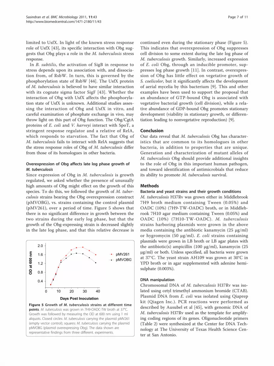
Results and DiscussionOverexpressed M. tuberculosis Obg binds to, andhydrolyzes, GTPA single copy of the gene coding for Obg (Rv2440c) ispresent in the genome of M. tuberculosis, between thegenes proB (Rv2439c) and rpmA (Rv2441c). Thededuced amino acid sequence of the M. tuberculosisObg protein shows significant similarities with the Obgproteins of B. subtilis, S. coelicolor and other bacterialspecies (Additional file 1). To study the properties ofObg of M. tuberculosis, the plasmid constructpTBOBGE was made to overexpress Obg in E. coli. Logphase E. coli cells (strain BL21) bearing the plasmidpTBOBGE were induced by IPTG to overexpress a pro-tein that migrates at around 55 kDa in SDS-PAGE gels.This overexpressed protein, purified as detailed in theMethods section, showed a single protein in SDS-PAGE(Figure 1A). This was designated as His10-Obg, to dis-tinguish it from the native, normally expressed Obg pro-tein in M. tuberculosis.To verify whether the overexpressed Obg of M.tuberculosis can interact with GTP, we performed GTP-UV-crosslinking experiments [31]. The autoradiogramin Figure 1B shows that His10-Obg binds physically to[a32P]-GTP. Exposure of the reaction mixtures to UVirradiation for 0, 30 and 60 min revealed that binding ofGTP with His10-Obg is increased between 0 and 30 minof exposure, but not after 30 min (Figure 1B). When thereactions were performed in the presence of unlabeledGTP (5 mM), crosslinking of His10-Obg to GTP isinhibited, while addition of large amounts of unlabeledATP (500 mM) have little effect on His10-Obg bindingwith labeled GTP (Figure 1B). This observation adds toexisting evidence that M. tuberculosis Obg has an inher-ent specificity for guanine nucleotides, as do the Obg
Sasindran et al. BMC Microbiology 2011, 11:43http://www.biomedcentral.com/1471-2180/11/43
Page 2 of 11

orthologues in C. crescentus [32], B. subtilis [13] andS. griseus [8].To determine whether the overexpressed Obg can
hydrolyze GTP, we incubated His10 -Obg with radiola-beled GTP ([g-32P] GTP), and measured the release ofphosphate (32Pi) after 3 hours. Figure 1C shows thatHis10-Obg readily hydrolyzes GTP, and that this hydro-lysis is inhibited by the addition of unlabeled GTP(5 mM), indicating that unlabeled GTP competes withlabeled GTP for the enzyme. Addition of unlabeled ATP(5 mM) has no effect on the hydrolysis of labeled GTP(Figure 1C), indicating that Obg hydrolyzes specificallyGTP. The effect of cold GTP in inhibiting the hydrolysisof radiolabeled GTP was not as pronounced as its effectin inhibition of GTP crosslinking (Compare Figure 1Band Figure 1C). This is most likely due to the differ-ences in the positions of the radiolabeled phosphatesused in these two reactions. While the reaction mixturein the crosslinking experiment (Figure 1B) had 10 μCi(0.033 μM) of [a-32P] GTP, the reaction mixture in thehydrolysis experiment had 25 μCi (0.040 μM) of [g-32P]
GTP. In addition, the incubation times for these twoexperiments were different (1 h for GTP crosslinking vs.3 h for GTP hydrolysis).
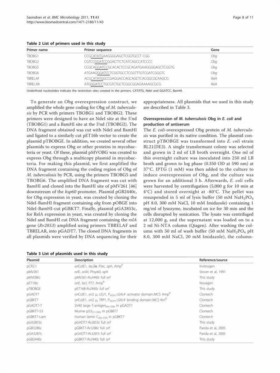
Autophosphorylation of His10-ObgAutophosphorylation by GTP is a defining characteristicof eukaryotic GTP-binding proteins, e.g. Ras [33], and ofprokaryotic GTP-binding proteins, including Era ofE. coli [34] and Obg of B. subtilis (22). We thereforeasked whether His10-Obg of M. tuberculosis is autopho-sphorylated by GTP. Figure 2A shows that purifiedHis10-Obg from M. tuberculosis is autophosphorylatedby [g-32P] GTP, in a time-dependent manner. Thisautophosphorylation is fully dependent upon Mg2+ ions,since reactions conducted in the absence of MgCl2 inthe buffer show almost zero phosphorylation activity(Figure 2B). By contrast, no autophosphorylation ofHis10-Obg occurs with [g-32P] ATP, even after 60 minof incubation. Further, addition of unlabeled ATP to thereaction mixture fails to produce any effect on His10-Obg phosphorylation with [g-32P] GTP (Figure 2C). As
kDa 1 2 3 4 5
173
80
1925
3649
61
A
0
1000
2000
3000
4000
1 2 3 4 5 6
B
32P i
rele
ased
(cpm
)//g
prot
ein/
hr
*
Reaction
0 30 60
1 2 3
II
I
C
Figure 1 Analysis of overexpressed Obg and its GTP binding and hydrolysis activities. A. SDS-PAGE protein profile showingoverexpression and purification of M. tuberculosis Obg. E. coli was grown in LB broth at 37°C, and lysates were prepared by sonication. Lane 1,Molecular markers; Lanes 2 and 3, extracts of E. coli strain BL21 carrying the overexpression plasmid pTBOBGE in the absence (Lane 2) andpresence (Lane 3) of 1 mM IPTG; Lane 4, supernatant of E. coli lysate after 10,000 g centrifugation; Lane 5, His10-Obg after Ni-NTA affinitychromatography. The arrow points to the His10-Obg band. B. Autoradiogram of SDS-PAGE-separated M. tuberculosis His10-Obg after UV-crosslinking with [a32P]GTP. UV-cross-linking was performed by incubating 5 μg of His10-Obg with 10 μCi of [a32P]GTP in the binding buffer asdescribed in the Methods section I. Crosslinking of His10-Obg with [a32P]GTP after 0, 30 and 60 minutes of exposure to UV light (256 nm).II. Crosslinking of His10-Obg with [a32P]GTP for 30 min without any additional GTP or ATP in the reaction mixture (Lane 1) or with 5 mM ofunlabeled GTP (Lane 2), or with 500 mM of unlabeled ATP (Lane 3). C. GTPase activity of His10-Obg. GTP hydrolysis of His10-Obg was performedusing [g-32P] GTP at 37°C. The GTPase activity is expressed as 32Pi released (cpm)/μg protein/hour. Columns indicate GTPase activity in theabsence of [g-32P]GTP and His10-Obg (Column 1), in the presence of His10-Obg alone (Column 2), in the presence of both [g-32P]GTP and His10-Obg (Column 3), in the presence of [g -32P]GTP, His10-Obg and 5 mM unlabeled GTP (Column 4), in the presence of [g -32P]GTP, His10-Obg and5 mM unlabeled GDP (Column 5) and in the presence of [g-32P]GTP, His10-Obg and 5 mM unlabeled ATP (Column 6). * indicates value significantfrom column 3 (paired t-test P = 0.0163).
Sasindran et al. BMC Microbiology 2011, 11:43http://www.biomedcentral.com/1471-2180/11/43
Page 3 of 11

expected, both unlabeled GTP and GDP significantlyaffect the phosphorylation of [g-32P] GTP from His10-Obg (Figure 2C), indicating that both molecules serve ascompetitors for the phosphorylation site. The eukaryoticRas protein, which is encoded by the p21ras oncogene,controls cell proliferation, cell stress signaling and apop-tosis. The autophosphorylaiton of Ras is independent ofits GTPase activity [33], which means that GTP hydroly-sis and GTP phosphorylation of Ras occur at two differ-ent sites. At present it is unclear whether GTPhydrolysis and GTP-mediated autophosphorylation areindependent events for prokaryotic Obgs, and no onehas identified a phsophorylation site on any Obgmolecule.
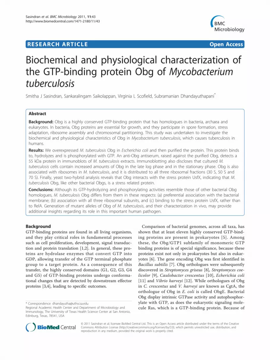
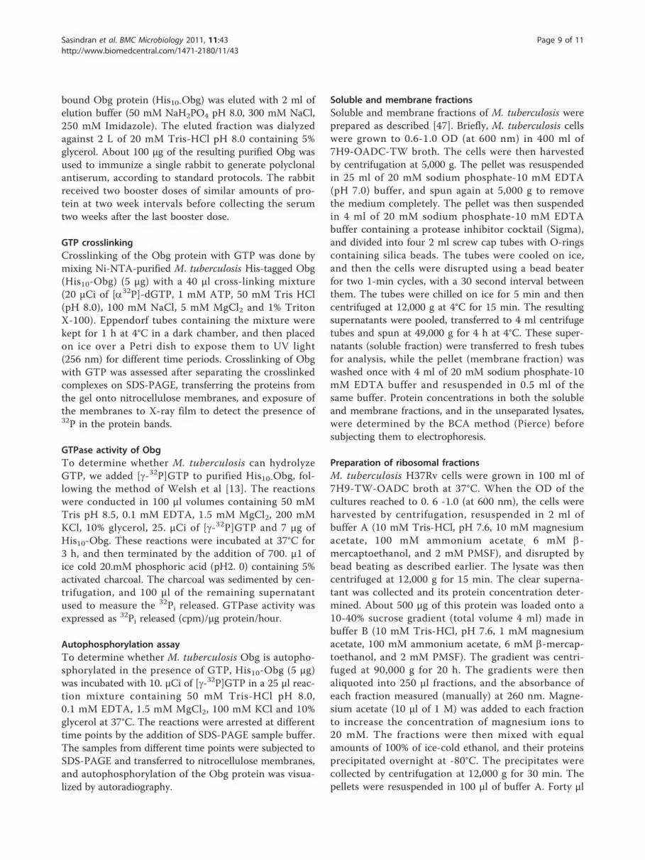
Expression of M. tuberculosis Obg is growth-dependent,and Obg is associated with the membrane fractionIn the sporulating bacterium S. coelicolor, the expres-sion of Obg is regulated developmentally and is linkedto the onset of sporulation [9]. By contrast, no suchchange in expression of Obg occurs in C. crescentus,although it also has a clear developmental cycle invol-ving sporulation [10]. M. tuberculosis is a slow growingbacterium which exhibits neither sporulation nor adevelopmental cell cycle during its growth in culture.To determine whether the expression of Obg changesduring the growth of M. tuberculosis in culture,we developed a rabbit anti-Obg antiserum against M.tuberculosis His10-Obg, and used it in Western blots ofM. tuberculosis protein extracts. This antiserum detectsmultiple bands in immunoblotted extracts of M.
tuberculosis, particularly at 55 kDa and 75 kDa. Toconfirm that the 55 kDa protein reacting with anti-Obg antiserum is in fact Obg, we cloned the codingregion of Obg downstream of the hsp60 promoter inthe plasmid pMV261, and transformed the resultingconstruct (pMVOBG) into M. tuberculosis to overpro-duce Obg. Figure 3A shows that protein extracts of M.tuberculosis strains harboring plasmid pMVOBG, butnot strains bearing the vector plasmid pMV261, revealstrong 55 kDa protein bands, indicating that the pro-tein at 55 kDa is Obg. Further analysis revealed thatthe 75 kDa band was a false reactivity due to the sec-ond antibody, and that it is not an Obg protein.Notably, Obg expression does change in cultures of
M. tuberculosis over the course of cell growth. Obgexpression is markedly increased from early log phase tothe stationary phase, with a drop in expression at latestationary phase (Figure 3B). Comparison of the Obgband densities discloses that expression of Obg at latergrowth phases (1.645 OD600 nm ) is approximately fivefold higher than it is at earlier phases (0.220 OD600 nm),even before the drop in expression at late stationaryphase. Together these results indicate that the expres-sion of Obg in M. tuberculosis is growth-regulated,being increased as the cells begin rapid division in thelog phase, and maintained at high levels until late in thestationary phase. However, whether increased levels ofObg with increased growth of M. tuberculosis is due toincreased expression of Obg, or to accumulation of Obg,remains to be determined. Obg expression in E. coli isalso high in log phase growth, but decreased in the sta-tionary phase [26].In S. griseus [8] and E. coli [11], Obg and its ortholo-
gues are found in both the cytoplasmic and membranefractions. In B. subtilis, however, Obg is mainly asso-ciated with the cytoplasm [23]. To determine whereObg resides in M. tuberculosis, we isolated soluble andmembrane fractions from whole bacteria, and subjectedthem to immunoblot analysis. Figure 3C shows thatObg is associated mostly (over 90%) with the membranefraction, although detectable amounts are also presentin the soluble fraction. In contrast, SigH of M. tubercu-losis, which was used as a control here, exhibits almostequal distribution between these two fractions. It hasbeen reported that membrane fraction-bound Obg inS. coeliocolor [9] and in E. coli [11] is lost from this frac-tion if the extraction buffer contains 5 mM EDTA. Thebuffer we use for M. tuberculosis membrane prepara-tions has 10 mM EDTA, however, and Obg is associatedwith this fraction whether or not EDTA is present (notshown). The EDTA-resistant association of M. tubercu-losis Obg to the membrane fraction may reflect a func-tion associated with signaling, and involving divalentcations. Interestingly, Obg is absent from detergent-
[ -32P]GTP [ -32P]ATP
Minutes
0 15 30 60 30 60
A
Mg++g
B
ATP GTP GDP1 2 3 1 2 3 1 2 3
CFigure 2 Autoradiogram of SDS-PAGE-separated M. tuberculosisHis10-Obg after autophosphorylation. Autophosphorylationreactions were set up by incubating 5 μg of His10-Obg with 10 μCiof [g-32P] GTP in autophosphorylation buffer, as detailed in theMethods section. A. Autophosphorylation of His10-Obg by [g-32P]GTP or [g-32P]ATP after 0, 15, 30 and 60 minutes of incubation at 37°C. B. Autophosphorylation of His10-Obg by [g-32P]GTP in thepresence (+ lane) and absence of (- lane) 1.5 mM MgCl2 . C.Autophosphorylation of His10-Obg by [g-32P]GTP in the presence of5 mM (Lane 1), 50 mM (Lane 2) and 500 mM (Lane 3) ATP; 5 mM(Lane 1), 50 mM (Lane 2) and 500 mM (Lane 3) of GTP; 5 mM (Lane1), 50 mM (Lane 2) and 500 mM (Lane 3) of GDP.
Sasindran et al. BMC Microbiology 2011, 11:43http://www.biomedcentral.com/1471-2180/11/43
Page 4 of 11
extracted M. tuberculosis membrane [35] and cell wall[36] proteins, suggesting that Obg’s association with themembrane may be due to its interaction with othermembrane protein(s).
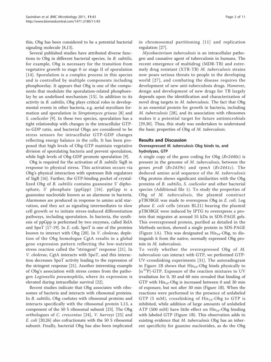
M. tuberculosis Obg associates with ribosomal fractionsIn B. subtilis [23], C. crescentus [24], V. harveyi [25] andE. coli [20,26], Obg has been shown to be associatedwith ribosomes. In these species, Obg orthologuescofractionate primarily with the 50 S ribosomal subunit[23,24,26]. To determine whether this is also true ofM. tuberculosis Obg, we isolated ribosomes from M.tuberculosis using sucrose gradient centrifugation, asdetailed in the Methods section (Figure 4A). Immuno-blots of the separated ribosomal fractions (Figure 4B)show that Obg is present in all three (30 S, 50 S and 70
S) ribosomal fractions, in more or less equal amounts.By contrast, this discrepancy does not appear to be dueto improper separation of ribosomal proteins in oursucrose gradient, because analysis of the ribosomal frac-tions in SDS-PAGE reveals that separation of proteinsoccurred in the expected line (Additional file 2). TheObg/CgtA of E. coli and C. crescentus has been shownto interact with specific 50 S ribosomal proteins, and itis the opinion of the investigators in this area that Obgplays a critical role in ribosome assembly. Evidence insupport of this hypothesis has been provided withstrains producing mutant Obg/CgtA. For example,C. crescentus [37] and E. coli [26] strains expressingmutated Obg have perturbed ribosomal protein profiles.A genetic basis for the involvement of Obg in ribosomalassembly has also been provided in E. coli by studies in
kDa 1 2
A B
173
80
49
19
61
25
360.
465
0.72
0
0.95
0
1.27
6
0.22
0
1.43
7
1.64
5
1.88
2
1.32
5
OD/600 nm
5000
15000
25000
Obg
Den
sity
in A
rea
C
Who
le
Sup
erna
tant
Pel
let
Obg
SigH
Figure 3 Immunoblot analysis of Obg of M. tuberculosis. A. Immunoblot analysis of Obg from M. tuberculosis strains harboring plasmids.M. tuberculosis strains were grown in 7H9-OADC-TW broth at 37°C to early log phase and lysates prepared using a bead beater and separated(100 μg protein for each lane) on SDS-PAGE. The immunoblots were probed with anti-Obg antiserum (1:500 dilution) followed by alkalinephosphatase labeled anti-rabbit IgG (1:1000 dilution, Zymed). The antibody-incubated blots were then developed with NBT/BCIP substrates. Lane1, M. tuberculosis carrying the plasmid pMV261(empty vector control); Lane 2, M. tuberculosis carrying the plasmid pMVOBG (plasmidoverexpressing Obg). B. Immunoblot analysis of Obg at different growth points in M. tuberculosis culture. Wild type M. tuberculosis was grown in7H9-OADC-TW broth at 37°C. Lysates were prepared from wild-type M. tuberculosis grown to different ODs at 600 nm, separated (200 μg proteinfor each lane) on SDS-PAGE, and probed with anti-Obg antiserum (1:500 dilution) followed by peroxidase-labeled anti-rabbit IgG (1:10,000dilution, Sigma). The blots were developed with an ECL kit (Amersham) and autoradiographed. “Obg” indicates the Obg protein reacting withanti-Obg antiserum. Values below each band indicate the OD value at 600 nm at the time of harvest. The graph above the bands gives thelevels of Obg, based on density of the bands using Image J software. C. Immunoblots of Obg in separated soluble vs membrane fractions ofM. tuberculosis lysates. The bacteria were grown in 7H9-OADC-TW broth at 37°C to mid-log phase. Lysates were prepared using a bead beater,and the soluble and pellet fractions separated by centrifugation. The protein fractions (200 μg protein for each lane) were separated by SDS-PAGE, blotted and probed with anti-Obg antiserum (1:500 dilution) (marked as Obg) or anti-SigH antiserum (1:1000 dilution) (marked as SigH),followed by peroxidase-labeled anti-rabbit IgG (1:10,000 dilution, Sigma). The blots were developed with an ECL kit (Amersham) andautoradiographed. In the figure, lanes labeled Whole, Supernatant and Pellet represent extracts of whole M. tuberculosis, of the 49,000 gsupernatant, and of the 49,000 g pellet, respectively.
Sasindran et al. BMC Microbiology 2011, 11:43http://www.biomedcentral.com/1471-2180/11/43
Page 5 of 11
which Obg was overexpressed in an rrmJ mutant strain[38]. Notably, rrmJ encodes an RNA methyltransferasewhich is involved in the assembly of 50 S ribosomes[38]. In line with these observations in bacteria, Obghomologues in yeast (Mtg2P) [39] and mice (Nog1) [40]also show association with ribosome maturation andassembly. Interestingly, in our studies shown here in
Figure 4, lanes 4-6 (30 S region) and lanes 9 and 10 (50S region) show an additional band above and belowObg, respectively. We do not know whether these bandsrepresent modified forms of Obg. Work in progressincludes studies toward identification of these bands.
M. tuberculosis Obg interacts with UsfXScott et al [41] were the first to observe that B. subtilisObg interacts with upstream regulators of the stresssigma factor SigB. In this respect, this bacterium’s Obgresembles B. subtilis RsbT and RsbW, both of which alsointeract with SigB in this species [41]. More recently, theObg proteins of E. coli [20] and V. harveyi [21] have beenshown to interact with SpoT, a stringent response regula-tor. Since SigB, RsbW and SpoT-related genes are presentin M. tuberculosis, we asked whether M. tuberculosis Obginteracts with any or all of these proteins, in the yeasttwo-hybrid system. The M. tuberculosis genes coding forObg (Rv2240c), UsfX (homologue of RsbW, Rv3287c),SigF (homologue of SigB of B. subtilis, Rv3286c) andRelA (a stringent response regulator related to SpoT,Rv2853c) were cloned in yeast vectors, and transformedinto the yeast strain AH109. Table 1 shows that M. tuber-culosis Obg strongly interacts with UsfX, but not with theSpoT-related RelA protein. The strength of this interac-tion is comparable to the interaction of M. tuberculosisUsfX with its cognate sigma factor SigF. In the sameexperiment, we looked for interaction of M. tuberculosisObg with various other putative anti-anti sigma factorsthat we have described earlier for this bacterium [42],including RsbU (Rv1364c), RsfA (Rv1365c), RsfB(Rv3687c), Rv0516c, Rv1904 and Rv2638. However, weobserved no significant interaction of Obg with any ofthe above anti-anti sigma factors (data not shown), indi-cating that the interaction of M. tuberculosis Obg is
1
2
3
4
5
OD
260
nm
70S
30S50S
1 2 3 4 5 6 7 8 9 10 11 12 13 14 15C
Sucrose 10% 40%
A
B
Figure 4 Obg cofractionation with ribosomal subunits. M.tuberculosis was grown in 7H9-OADC-TW broth at 37°C, and lysatesprepared using a bead beater. About 500 g protein was separatedin 10-40% sucrose gradient. A. The ODs of the separated fractionswere measured (manually) at 260 nm. B. The proteins in thefractions were then precipitated with ethanol and separated onSDS-PAGE, transferred to nitrocellulose membranes, and probedwith anti-Obg antiserum (1:500 dilution), followed by peroxidase-labeled anti-rabbit IgG (1:10,000 dilution, Sigma). The blots weredeveloped with an ECL kit (Amersham) and autoradiographed. LaneC is a whole-cell extract from M. tuberculosis. Lanes 1-15 representfractions from the top (10% sucrose) to the bottom (40% sucrose)of the sucrose gradient. Fraction 16 was not analyzed inimmunoblot.
Table 1 Interaction of Obg with stress related proteins in the yeast two-hybrid system
*Plasmids SD Minimal Medium Mel-l (a-gal) inSD plates
Mel-1 (a-gal) inSD broth**
-Leu/-Trp
-His/-Leu/-Trp
-Ade/-His/-Leu/-Trp
1. pGADT7-T + + + +++ 3.512 ± 0.709
pGBKT7-53
2. pGADT7-T + - - - -
pGBKT7-Lam
3. pGA3287c + + + ++ 2.367 ± 0.354
pGB3286c
4. pGA3287c + + + ++ 2.172 ± 0.448
pGB2440c
5. pGA2853c + - - - -
pGB2440c
Abbreviations. SD, synthetic drop out medium; Ade, Adenine; His, Histidine; Leu, Leucine; Trp, Tryptophan; Mel-1, a-galactosidase.*1-5 indicate plasmids cotransformed into yeast strain AH109 (HIS3, ADE2, MEL1) (Clontech).
**a-galactosidase (Mel-1) expressed as Mean ± SD milli units/A600. Plasmids are described in Table 3.
Sasindran et al. BMC Microbiology 2011, 11:43http://www.biomedcentral.com/1471-2180/11/43
Page 6 of 11
limited to UsfX. In light of the known stress responserole of UsfX [43], its specific interaction with Obg sug-gests that Obg plays a role in the M. tuberculosis stressresponse.In B. subtilis, the activation of SigB in response to
stress depends upon its association with, and dissocia-tion from, of RsbW. In turn, this is governed by thephosphorylation state of RsbW [44]. The UsfX proteinof M. tuberculosis is believed to have similar interactionwith its cognate sigma factor SigF [43]. Whether theinteraction of Obg with UsfX affects the phosphoryla-tion state of UsfX is unknown. Additional studies asses-sing the interaction of Obg and UsfX in vitro, andcareful examination of phosphate exchange in vivo, maythrow light on this part of Obg function. The Obg/CgtAproteins of E. coli and V. harveyi interact with SpoT, astringent response regulator and a relative of RelA,which responds to starvation. The fact that Obg ofM. tuberculosis fails to interact with RelA suggests thatthe stress response roles of Obg of M. tuberculosis differfrom those of its homologues in other bacteria.
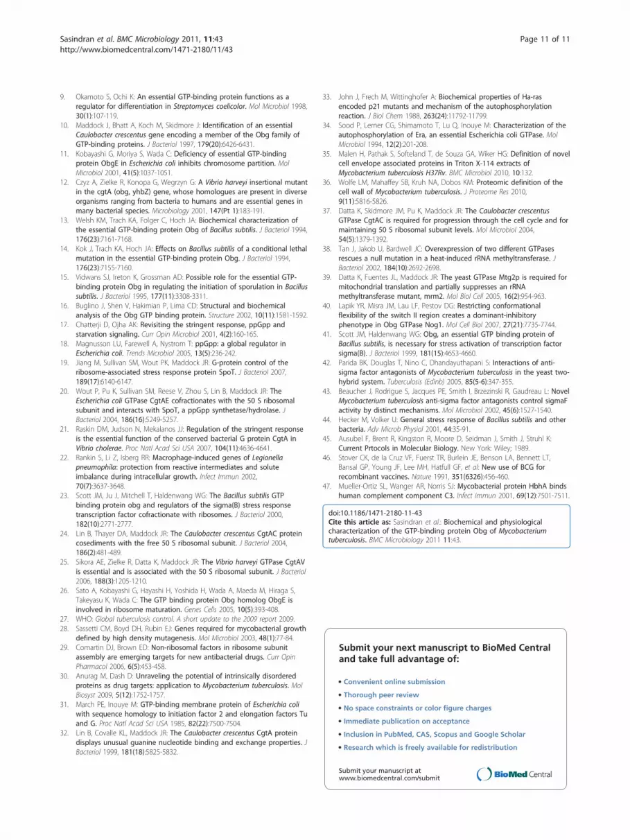
Overexpression of Obg affects late log phase growth ofM. tuberculosisSince expression of Obg in M. tuberculosis is growthregulated, we asked whether the presence of unusuallyhigh amounts of Obg might effect on the growth of thisspecies. To do this, we followed the growth of M. tuber-culosis strains bearing the Obg overexpression construct(pMVOBG), vs. strains containing the control plasmid(pMV261), over a period of time. Figure 5 shows thatthere is no significant difference in growth between thetwo strains during the early log phase, but that thegrowth of the Obg-expressing strain is decreased slightlyin the late log phase, and that this relative decrease is
continued even during the stationary phase (Figure 5).This indicates that overexpression of Obg suppressescell division to some extent during the late log phase ofM. tuberculosis growth. Similarly, increased expressionof E. coli Obg, through an inducible promoter, sup-presses log phase growth [11]. In contrast, overexpres-sion of Obg has little effect on vegetative growth ofS. coelicolor, but it significantly affects the developmentof aerial mycelia by this bacterium [9]. This and otherexamples have been used to support the proposal thatan abundance of GTP-bound Obg is associated withvegetative bacterial growth (cell division), while a rela-tive abundance of GDP-bound Obg promotes stationarydevelopment (viability in stationary growth, or differen-tiation leading to nonvegetative reproduction) [9].
ConclusionOur data reveal that M. tuberculosis Obg has character-istics that are common to its homologues in otherbacteria, in addition to properties that are unique.Generation and characterization of mutant alleles ofM. tuberculosis Obg should provide additional insightsto the role of Obg in this important human pathogen,and toward identification of antimicrobials that reduceits ability to promote M. tuberculosis survival.
MethodsBacteria and yeast strains and their growth conditionsM. tuberculosis H37Rv was grown either in Middlebrook7H9 broth medium containing Tween (0.05%) andOADC (10%) (7H9-TW-OADC) broth, or in Middleb-rook 7H10 agar medium containing Tween (0.05%) andOADC (10%) (7H10-TW-OADC). M. tuberculosisstrains harboring plasmids were grown in the abovemedia containing the antibiotic kanamycin (25 μg/ml)or hygromycin (50 μg/ml). E. coli strains containingplasmids were grown in LB broth or LB agar plates withthe antibiotic(s) ampicillin (100 μg/ml), kanamycin (25μg/ml) or both. Unless specified, all bacteria were grownat 37°C. The yeast strain AH109 was grown at 30°C inYPD broth or in agar supplemented with adenine hemi-sulphate (0.003%).
DNA manipulationChromosomal DNA of M. tuberculosis H37Rv was iso-lated using cetyl trimethyl ammonium bromide (CTAB).Plasmid DNA from E. coli was isolated using Qiaprepkit (Qiagen Inc.). PCR reactions were performed asdescribed by Ausubel et al [45], with genomic DNA ofM. tuberculosis H37Rv used as the template for amplify-ing coding regions of its genes. Oligonucleotide primers(Table 2) were synthesized at the Center for DNA Tech-nology at The University of Texas Health Science Cen-ter at San Antonio.
0 10 20 30 40
0.4
0.8
1.2
1.6
2.0
OD
at 6
00 n
m
Days Post Inoculation
pMVOBGpMV261
Figure 5 Growth of M. tuberculosis strains at different timepoints. M. tuberculosis was grown in 7H9-OADC-TW broth at 37°C.Growth was followed by measuring the OD at 600 nm using 1 mlaliquots. Closed circles: M. tuberculosis carrying the plasmid pMV261(empty vector control); squares: M. tuberculosis carrying the plasmidpMVOBG (plasmid overexpressing Obg). The data shown arerepresentative findings from three different. experiments.
Sasindran et al. BMC Microbiology 2011, 11:43http://www.biomedcentral.com/1471-2180/11/43
Page 7 of 11
To generate an Obg overexpression construct, weamplified the whole gene coding for Obg of M. tuberculo-sis by PCR with primers TBOBG1 and TBOBG2. Theseprimers were designed to have an NdeI site at the 5’nd(TBOBG1) and a BamHI site at the 3’nd (TBOBG2). TheDNA fragment obtained was cut with NdeI and BamHIand ligated to a similarly cut pET16b vector to create theplasmid pTBOBGE. In addition, we created several otherplasmids to express Obg or other proteins in mycobac-teria or yeast. Of these, plasmid pMVOBG was created toexpress Obg through a multicopy plasmid in mycobac-teria. For making this plasmid, we first amplified theDNA fragment containing the coding region of Obg ofM. tuberculosis by PCR, using the primers TBOBG5 andTBOBG6. The amplified DNA fragment was cut withBamHI and cloned into the BamHI site of pMV261 [46]downstream of the hsp60 promoter. Plasmid pGB2440c,for Obg expression in yeast, was created by cloning theNdeI-BamHI fragment containing obg from pOBGE intoNdeI-BamHI-cut pGBKT7. Finally, plasmid pGA2853c,for RelA expression in yeast, was created by cloning theNdeI and BamHI cut DNA fragment containing the relAgene (Rv2853) amplified using primers TBRELAF andTBRELAR, into pGADT7. The cloned DNA fragments inall plasmids were verified by DNA sequencing for their
appropriateness. All plasmids that we used in this studyare described in Table 3.
Overexpression of M. tuberculosis Obg in E. coli andproduction of antiserumThe E. coli-overexpressed Obg protein of M. tuberculo-sis was purified in its native condition. The plasmid con-struct pTBOBGE was transformed into E. coli strainBL21(DE3). A single transformant colony was selectedand grown in 2 ml of LB broth overnight. One ml ofthis overnight culture was inoculated into 250 ml LBbroth and grown to log phase (0.350 OD at 590 nm) at37°C. IPTG (1 mM) was then added to the culture toinduce overexpression of Obg, and the culture wasgrown for an additional 3 h. Afterwards, E. coli cellswere harvested by centrifugation (5,000 g for 10 min at4°C) and stored overnight at -80°C. The pellet wasresuspended in 5 ml of lysis buffer (50 mM NaH2PO4
pH 8.0, 300 mM NaCl, 10 mM Imidazole) containing 1mg/ml of lysozyme, incubated on ice for 30 min and thecells disrupted by sonication. The lysate was centrifugedat 12,000 g, and the supernatant was loaded on to a2 ml Ni-NTA column (Qiagen). After washing the col-umn with 50 ml of wash buffer (50 mM NaH2PO4 pH8.0, 300 mM NaCl, 20 mM Imidazole), the column-
Table 2 List of primers used in this study
Primer name Primer sequence Gene
TBOBG1 CCGCATATGAAGGGGAGCTCGGTGCCT CGG Obg
TBOBG2 CGTCCGGATCCGGACTTCTCATCAGCCATCCCC Obg
TBOBG5 CCGCAGGATCCGCACACTCCGCAGATGAAGGGGAGCTCGGTG Obg
TBOBG6 ATGAAGGGATCCTCGGTGCCTCGGTTTGTCGATCGGGTC Obg
TBRELAF ACGCATATGGCCGAGGACCAGCAGCTCACGGCGCAAGCG RelA
TBRELAR ATGGGATCCTGCGTCTGCTCGGCGGAGAAAAGCGCG RelA
Underlined nucleotides indicate the restriction sites created in the primers. CATATG, NdeI and GGATCC, BamHI.
Table 3 List of plasmids used in this study
Plasmid Description Reference/source
pCR2.1 oriColE1, lacZa, Plac, aph, AmpR Invitrogen
pMV261 oriE, oriM, Phsp60, aph Stover et al, 1991
pMVOBG pMV261-Rv2440c full orf This study
pET16b oriE, lacI, PT7, AmpR Novagen
pTBOBGE pET16B-Rv2440c full orf This study
pGADT7 oriColE1, ori2 μ, LEU1, PADH1::GAL4’ activator domain::MCS AmpR Clontech
pGBKT7 oriColE1, ori2 μ, TRP1, PADH1::GAL4’ binding domain::MCS KmR Clontech
pGADT7-T SV40 large T-antigen(84-708) in pGADT7 Clontech
pGBKT7-53 Murine p53(72-390) in pGBKT7 Clontech
pGBKT7-Lam Human lamin C(66-230) in pGBKT7 Clontech
pGA2853c pGADT7-Rv2853c full orf This study
pGB3286c pGBKT7-Rv3286c full orf Parida et al, 2005
pGA3287c pGADT7-Rv3287c full orf Parida et al, 2005
pGB2440c pGBKT7-Rv2440c full orf This study
Sasindran et al. BMC Microbiology 2011, 11:43http://www.biomedcentral.com/1471-2180/11/43
Page 8 of 11
bound Obg protein (His10-Obg) was eluted with 2 ml ofelution buffer (50 mM NaH2PO4 pH 8.0, 300 mM NaCl,250 mM Imidazole). The eluted fraction was dialyzedagainst 2 L of 20 mM Tris-HCl pH 8.0 containing 5%glycerol. About 100 μg of the resulting purified Obg wasused to immunize a single rabbit to generate polyclonalantiserum, according to standard protocols. The rabbitreceived two booster doses of similar amounts of pro-tein at two week intervals before collecting the serumtwo weeks after the last booster dose.
GTP crosslinkingCrosslinking of the Obg protein with GTP was done bymixing Ni-NTA-purified M. tuberculosis His-tagged Obg(His10-Obg) (5 μg) with a 40 μl cross-linking mixture(20 μCi of [a32P]-dGTP, 1 mM ATP, 50 mM Tris HCl(pH 8.0), 100 mM NaCl, 5 mM MgCl2 and 1% TritonX-100). Eppendorf tubes containing the mixture werekept for 1 h at 4°C in a dark chamber, and then placedon ice over a Petri dish to expose them to UV light(256 nm) for different time periods. Crosslinking of Obgwith GTP was assessed after separating the crosslinkedcomplexes on SDS-PAGE, transferring the proteins fromthe gel onto nitrocellulose membranes, and exposure ofthe membranes to X-ray film to detect the presence of32P in the protein bands.
GTPase activity of ObgTo determine whether M. tuberculosis can hydrolyzeGTP, we added [g-32P]GTP to purified His10-Obg, fol-lowing the method of Welsh et al [13]. The reactionswere conducted in 100 μl volumes containing 50 mMTris pH 8.5, 0.1 mM EDTA, 1.5 mM MgCl2, 200 mMKCl, 10% glycerol, 25. μCi of [g-32P]GTP and 7 μg ofHis10-Obg. These reactions were incubated at 37°C for3 h, and then terminated by the addition of 700. μ1 ofice cold 20.mM phosphoric acid (pH2. 0) containing 5%activated charcoal. The charcoal was sedimented by cen-trifugation, and 100 μl of the remaining supernatantused to measure the 32Pi released. GTPase activity wasexpressed as 32Pi released (cpm)/μg protein/hour.
Autophosphorylation assayTo determine whether M. tuberculosis Obg is autopho-sphorylated in the presence of GTP, His10-Obg (5 μg)was incubated with 10. μCi of [g-32P]GTP in a 25 μl reac-tion mixture containing 50 mM Tris-HCl pH 8.0,0.1 mM EDTA, 1.5 mM MgCl2, 100 mM KCl and 10%glycerol at 37°C. The reactions were arrested at differenttime points by the addition of SDS-PAGE sample buffer.The samples from different time points were subjected toSDS-PAGE and transferred to nitrocellulose membranes,and autophosphorylation of the Obg protein was visua-lized by autoradiography.
Soluble and membrane fractionsSoluble and membrane fractions of M. tuberculosis wereprepared as described [47]. Briefly, M. tuberculosis cellswere grown to 0.6-1.0 OD (at 600 nm) in 400 ml of7H9-OADC-TW broth. The cells were then harvestedby centrifugation at 5,000 g. The pellet was resuspendedin 25 ml of 20 mM sodium phosphate-10 mM EDTA(pH 7.0) buffer, and spun again at 5,000 g to removethe medium completely. The pellet was then suspendedin 4 ml of 20 mM sodium phosphate-10 mM EDTAbuffer containing a protease inhibitor cocktail (Sigma),and divided into four 2 ml screw cap tubes with O-ringscontaining silica beads. The tubes were cooled on ice,and then the cells were disrupted using a bead beaterfor two 1-min cycles, with a 30 second interval betweenthem. The tubes were chilled on ice for 5 min and thencentrifuged at 12,000 g at 4°C for 15 min. The resultingsupernatants were pooled, transferred to 4 ml centrifugetubes and spun at 49,000 g for 4 h at 4°C. These super-natants (soluble fraction) were transferred to fresh tubesfor analysis, while the pellet (membrane fraction) waswashed once with 4 ml of 20 mM sodium phosphate-10mM EDTA buffer and resuspended in 0.5 ml of thesame buffer. Protein concentrations in both the solubleand membrane fractions, and in the unseparated lysates,were determined by the BCA method (Pierce) beforesubjecting them to electrophoresis.
Preparation of ribosomal fractionsM. tuberculosis H37Rv cells were grown in 100 ml of7H9-TW-OADC broth at 37°C. When the OD of thecultures reached to 0. 6 -1.0 (at 600 nm), the cells wereharvested by centrifugation, resuspended in 2 ml ofbuffer A (10 mM Tris-HCl, pH 7.6, 10 mM magnesiumacetate, 100 mM ammonium acetate, 6 mM b-mercaptoethanol, and 2 mM PMSF), and disrupted bybead beating as described earlier. The lysate was thencentrifuged at 12,000 g for 15 min. The clear superna-tant was collected and its protein concentration deter-mined. About 500 μg of this protein was loaded onto a10-40% sucrose gradient (total volume 4 ml) made inbuffer B (10 mM Tris-HCl, pH 7.6, 1 mM magnesiumacetate, 100 mM ammonium acetate, 6 mM b-mercap-toethanol, and 2 mM PMSF). The gradient was centri-fuged at 90,000 g for 20 h. The gradients were thenaliquoted into 250 μl fractions, and the absorbance ofeach fraction measured (manually) at 260 nm. Magne-sium acetate (10 μl of 1 M) was added to each fractionto increase the concentration of magnesium ions to20 mM. The fractions were then mixed with equalamounts of 100% of ice-cold ethanol, and their proteinsprecipitated overnight at -80°C. The precipitates werecollected by centrifugation at 12,000 g for 30 min. Thepellets were resuspended in 100 μl of buffer A. Forty μl
Sasindran et al. BMC Microbiology 2011, 11:43http://www.biomedcentral.com/1471-2180/11/43
Page 9 of 11

of the suspension from each fraction was mixed with10 μl 4× loading buffer and boiled, after which 25 μl ofeach sample was loaded onto each well for SDS-PAGE.After electrophoresis, the proteins were transferred tonitrocellulose membranes, probed with anti-Obg anti-serum, and the blots probed by ECL chemiluminescencemethod (Amersham). Association of Obg with ribosomalsubunits was determined by comparing the immunoblotfor each fraction with its absorbance at 260 nm.
Yeast two-hybrid assayProtein-protein interactions were performed using theMatchmaker Gal4 two-hybrid system 3 (Clontech, PaloAlto, CA) as described previously [42]. The yeast strainAH109, which has the reporter genes ADE2 (adenine),HIS3 (histidine), and MEL1 (a-galactosidase), was usedas the host strain. Yeast plasmids (Table 2) were trans-formed into AH109 in appropriate combinations(Table 1) using standard protocols provided by Clon-tech. Expression of proteins by plasmids created foryeast two-hybrid analysis was assessed by the TNTQuick transcription and translation system (Promega),before transformation of the plasmids into yeast. Pro-tein-protein interactions were determined by positivegrowth of yeast in synthetic drop out medium (SD)plates lacking adenine and histidine, and by the pre-sence of blue color, which identifies a- galactosidaseactivity. To rule out false activation of the reportergene, we transformed each of the constructs separatelyinto yeast strain AH109, and assessed reporter geneactivation. The strength of the interaction was verifiedby measuring the a-galactosidase released into thegrowth medium, again using protocols provided byClontech.
SDS-PAGE and immunoblotSDS-PAGE and immunoblotting were performed fol-lowing the methods of Ausubel et al [45]. Protein con-tents in extracts of E. coli or M. tuberculosis, obtainedthrough sonication or bead-beating techniques, weredetermined by BCA (bicinchoninic acid) method(Pierce). Proteins were separated on 12% SDS-PAGEand transferred to nitrocellulose membranes. The blotswere probed with rabbit anti-M. tuberculosis Obg anti-serum (1:500 dilution) or rabbit anti-M. tuberculosisSigH antiserum (1:1000), developed against recombi-nant His10-Obg or His10-SigH proteins, respectively.Alkaline phosphatase-conjugated anti-rabbit IgG(Zymed, 1:1000 dilution) or peroxidase-conjugatedanti-rabbit IgG (Sigma, 1:10,000 dilution) were used assecondary antibodies. The blots were developed eitherwith 5-bromo-4-chloro-3-indolyl phosphate (BCIP)/nitroblue tetrazolium (NBT) substrate (Sigma, for
alkaline phosphatase), or with an ECL kit (Amersham,for peroxidase).
Additional material
Additional file 1: Amino acid alignment of Obg proteins fromdifferent bacterial species. MTOBG, Mycobacterium tuberculosis Obg;SCOBG, Streptomyces coelicolor Obg; BSOBG,Bacillus subtilis Obg; ECOBG,Escherichia coli ObgE; CCOBG, Caulobacter crescentus Obg (CgtA).Asterisks (*) indicate high amino acid identity, colons (:) indicate mediumamino acid identity, and dots (.) indicate low amino acid identity. GTP-binding motifs G1, G2, G3, G4, switch I and switch II are marked.
Additional file 2: SDS-PAGE analysis of total proteins associatedwith different ribosomal fractions. Ribosomal fractions (1-15) fromwild-type M. tuberculosis extracts were separated on a 10%-40% sucrosegradient. M. tuberculosis was grown in 7H9-OADC-TW broth at 37°C, andextracts for ribosomal isolation prepared using a bead beater. Fivehundred μg of protein was separated in 10-40% sucrose gradient bycentrifugation. The sucrose gradient was then aliquoted into 250 μlfractions and their ODs measured at 260 nm. The proteins in thefractions were precipitated with ethanol and separated on SDS-PAGE,stained with Coomassie blue and destained with 10% acetone. The gelpicture shown here is modified from its original to eliminate and correctmis-loaded and incorrectly loaded lanes.
AcknowledgementsThis study was partly supported by Institutional Research Grant and SanAntonio Area foundation.
Authors’ contributionsSJS performed the construction of plasmids and isolation of ribosomalfractions. SS carried out the overexpression of Obg and its biochemicalanalysis. VLS read the manuscript critically, participated in interpretation ofthe data, and worked with the other authors to prepare the final version ofthe paper. SD conceived the study, participated in its design andinterpretation of results and wrote the manuscript. All authors read andapproved the manuscript.
Received: 18 August 2010 Accepted: 25 February 2011Published: 25 February 2011
References1. Bourne HR, Sanders DA, McCormick F: The GTPase superfamily: a
conserved switch for diverse cell functions. Nature 1990,348(6297):125-132.
2. Kaziro Y, Itoh H, Kozasa T, Nakafuku M, Satoh T: Structure and function ofsignal-transducing GTP-binding proteins. Annu Rev Biochem 1991,60:349-400.
3. Bourne HR, Sanders DA, McCormick F: The GTPase superfamily: conservedstructure and molecular mechanism. Nature 1991, 349(6305):117-127.
4. Sprang SR: G protein mechanisms: insights from structural analysis. AnnuRev Biochem 1997, 66:639-678.
5. Pandit SB, Srinivasan N: Survey for g-proteins in the prokaryotic genomes:prediction of functional roles based on classification. Proteins 2003,52:585-597.
6. Hirano Y, Ohniwa RL, Wada C, Yoshimura SH, Takeyasu K: Human small Gproteins, ObgH1, and ObgH2, participate in the maintenance ofmitochondria and nucleolar architectures. Genes Cells 2006, 11:1295-1304.
7. Ferrari FA, Trach K, Hoch JA: Sequence analysis of the spo0B locus revealsa polycistronic transcription unit. J Bacteriol 1985, 161(2):556-562.
8. Okamoto S, Itoh M, Ochi K: Molecular cloning and characterization of theobg gene of Streptomyces griseus in relation to the onset ofmorphological differentiation. J Bacteriol 1997, 179(1):170-179.
Sasindran et al. BMC Microbiology 2011, 11:43http://www.biomedcentral.com/1471-2180/11/43
Page 10 of 11
9. Okamoto S, Ochi K: An essential GTP-binding protein functions as aregulator for differentiation in Streptomyces coelicolor. Mol Microbiol 1998,30(1):107-119.
10. Maddock J, Bhatt A, Koch M, Skidmore J: Identification of an essentialCaulobacter crescentus gene encoding a member of the Obg family ofGTP-binding proteins. J Bacteriol 1997, 179(20):6426-6431.
11. Kobayashi G, Moriya S, Wada C: Deficiency of essential GTP-bindingprotein ObgE in Escherichia coli inhibits chromosome partition. MolMicrobiol 2001, 41(5):1037-1051.
12. Czyz A, Zielke R, Konopa G, Wegrzyn G: A Vibrio harveyi insertional mutantin the cgtA (obg, yhbZ) gene, whose homologues are present in diverseorganisms ranging from bacteria to humans and are essential genes inmany bacterial species. Microbiology 2001, 147(Pt 1):183-191.
13. Welsh KM, Trach KA, Folger C, Hoch JA: Biochemical characterization ofthe essential GTP-binding protein Obg of Bacillus subtilis. J Bacteriol 1994,176(23):7161-7168.
14. Kok J, Trach KA, Hoch JA: Effects on Bacillus subtilis of a conditional lethalmutation in the essential GTP-binding protein Obg. J Bacteriol 1994,176(23):7155-7160.
15. Vidwans SJ, Ireton K, Grossman AD: Possible role for the essential GTP-binding protein Obg in regulating the initiation of sporulation in Bacillussubtilis. J Bacteriol 1995, 177(11):3308-3311.
16. Buglino J, Shen V, Hakimian P, Lima CD: Structural and biochemicalanalysis of the Obg GTP binding protein. Structure 2002, 10(11):1581-1592.
17. Chatterji D, Ojha AK: Revisiting the stringent response, ppGpp andstarvation signaling. Curr Opin Microbiol 2001, 4(2):160-165.
18. Magnusson LU, Farewell A, Nystrom T: ppGpp: a global regulator inEscherichia coli. Trends Microbiol 2005, 13(5):236-242.
19. Jiang M, Sullivan SM, Wout PK, Maddock JR: G-protein control of theribosome-associated stress response protein SpoT. J Bacteriol 2007,189(17):6140-6147.
20. Wout P, Pu K, Sullivan SM, Reese V, Zhou S, Lin B, Maddock JR: TheEscherichia coli GTPase CgtAE cofractionates with the 50 S ribosomalsubunit and interacts with SpoT, a ppGpp synthetase/hydrolase. JBacteriol 2004, 186(16):5249-5257.
21. Raskin DM, Judson N, Mekalanos JJ: Regulation of the stringent responseis the essential function of the conserved bacterial G protein CgtA inVibrio cholerae. Proc Natl Acad Sci USA 2007, 104(11):4636-4641.
22. Rankin S, Li Z, Isberg RR: Macrophage-induced genes of Legionellapneumophila: protection from reactive intermediates and soluteimbalance during intracellular growth. Infect Immun 2002,70(7):3637-3648.
23. Scott JM, Ju J, Mitchell T, Haldenwang WG: The Bacillus subtilis GTPbinding protein obg and regulators of the sigma(B) stress responsetranscription factor cofractionate with ribosomes. J Bacteriol 2000,182(10):2771-2777.
24. Lin B, Thayer DA, Maddock JR: The Caulobacter crescentus CgtAC proteincosediments with the free 50 S ribosomal subunit. J Bacteriol 2004,186(2):481-489.
25. Sikora AE, Zielke R, Datta K, Maddock JR: The Vibrio harveyi GTPase CgtAVis essential and is associated with the 50 S ribosomal subunit. J Bacteriol2006, 188(3):1205-1210.
26. Sato A, Kobayashi G, Hayashi H, Yoshida H, Wada A, Maeda M, Hiraga S,Takeyasu K, Wada C: The GTP binding protein Obg homolog ObgE isinvolved in ribosome maturation. Genes Cells 2005, 10(5):393-408.
27. WHO: Global tuberculosis control. A short update to the 2009 report 2009.28. Sassetti CM, Boyd DH, Rubin EJ: Genes required for mycobacterial growth
defined by high density mutagenesis. Mol Microbiol 2003, 48(1):77-84.29. Comartin DJ, Brown ED: Non-ribosomal factors in ribosome subunit
assembly are emerging targets for new antibacterial drugs. Curr OpinPharmacol 2006, 6(5):453-458.
30. Anurag M, Dash D: Unraveling the potential of intrinsically disorderedproteins as drug targets: application to Mycobacterium tuberculosis. MolBiosyst 2009, 5(12):1752-1757.
31. March PE, Inouye M: GTP-binding membrane protein of Escherichia coliwith sequence homology to initiation factor 2 and elongation factors Tuand G. Proc Natl Acad Sci USA 1985, 82(22):7500-7504.
32. Lin B, Covalle KL, Maddock JR: The Caulobacter crescentus CgtA proteindisplays unusual guanine nucleotide binding and exchange properties. JBacteriol 1999, 181(18):5825-5832.
33. John J, Frech M, Wittinghofer A: Biochemical properties of Ha-rasencoded p21 mutants and mechanism of the autophosphorylationreaction. J Biol Chem 1988, 263(24):11792-11799.
34. Sood P, Lerner CG, Shimamoto T, Lu Q, Inouye M: Characterization of theautophosphorylation of Era, an essential Escherichia coli GTPase. MolMicrobiol 1994, 12(2):201-208.
35. Malen H, Pathak S, Softeland T, de Souza GA, Wiker HG: Definition of novelcell envelope associated proteins in Triton X-114 extracts ofMycobacterium tuberculosis H37Rv. BMC Microbiol 2010, 10:132.
36. Wolfe LM, Mahaffey SB, Kruh NA, Dobos KM: Proteomic definition of thecell wall of Mycobacterium tuberculosis. J Proteome Res 2010,9(11):5816-5826.
37. Datta K, Skidmore JM, Pu K, Maddock JR: The Caulobacter crescentusGTPase CgtAC is required for progression through the cell cycle and formaintaining 50 S ribosomal subunit levels. Mol Microbiol 2004,54(5):1379-1392.
38. Tan J, Jakob U, Bardwell JC: Overexpression of two different GTPasesrescues a null mutation in a heat-induced rRNA methyltransferase. JBacteriol 2002, 184(10):2692-2698.
39. Datta K, Fuentes JL, Maddock JR: The yeast GTPase Mtg2p is required formitochondrial translation and partially suppresses an rRNAmethyltransferase mutant, mrm2. Mol Biol Cell 2005, 16(2):954-963.
40. Lapik YR, Misra JM, Lau LF, Pestov DG: Restricting conformationalflexibility of the switch II region creates a dominant-inhibitoryphenotype in Obg GTPase Nog1. Mol Cell Biol 2007, 27(21):7735-7744.
41. Scott JM, Haldenwang WG: Obg, an essential GTP binding protein ofBacillus subtilis, is necessary for stress activation of transcription factorsigma(B). J Bacteriol 1999, 181(15):4653-4660.
42. Parida BK, Douglas T, Nino C, Dhandayuthapani S: Interactions of anti-sigma factor antagonists of Mycobacterium tuberculosis in the yeast two-hybrid system. Tuberculosis (Edinb) 2005, 85(5-6):347-355.
43. Beaucher J, Rodrigue S, Jacques PE, Smith I, Brzezinski R, Gaudreau L: NovelMycobacterium tuberculosis anti-sigma factor antagonists control sigmaFactivity by distinct mechanisms. Mol Microbiol 2002, 45(6):1527-1540.
44. Hecker M, Volker U: General stress response of Bacillus subtilis and otherbacteria. Adv Microb Physiol 2001, 44:35-91.
45. Ausubel F, Brent R, Kingston R, Moore D, Seidman J, Smith J, Struhl K:Current Prtocols in Molecular Biology. New York: Wiley; 1989.
46. Stover CK, de la Cruz VF, Fuerst TR, Burlein JE, Benson LA, Bennett LT,Bansal GP, Young JF, Lee MH, Hatfull GF, et al: New use of BCG forrecombinant vaccines. Nature 1991, 351(6326):456-460.
47. Mueller-Ortiz SL, Wanger AR, Norris SJ: Mycobacterial protein HbhA bindshuman complement component C3. Infect Immun 2001, 69(12):7501-7511.
doi:10.1186/1471-2180-11-43Cite this article as: Sasindran et al.: Biochemical and physiologicalcharacterization of the GTP-binding protein Obg of Mycobacteriumtuberculosis. BMC Microbiology 2011 11:43.
Submit your next manuscript to BioMed Centraland take full advantage of:
• Convenient online submission
• Thorough peer review
• No space constraints or color figure charges
• Immediate publication on acceptance
• Inclusion in PubMed, CAS, Scopus and Google Scholar
• Research which is freely available for redistribution
Submit your manuscript at www.biomedcentral.com/submit
Sasindran et al. BMC Microbiology 2011, 11:43http://www.biomedcentral.com/1471-2180/11/43
Page 11 of 11