biochemical pharmacology - air unimi
TRANSCRIPT
Contents lists available at ScienceDirect
Biochemical Pharmacology
journal homepage: www.elsevier.com/locate/biochempharm
Profiling Vaccinium macrocarpon components and metabolites in humanurine and the urine ex-vivo effect on Candida albicans adhesion and biofilm-formationGiovanna Barona, Alessandra Altomarea, Luca Regazzonia, Laura Fumagallia, Angelica Artasensia,Elisa Borghib, Emerenziana Ottavianob, Cristian Del Boc, Patrizia Risoc, Pietro Allegrinid,Giovanna Petrangolinid, Paolo Morazzonid, Antonella Rivad, Lolita Arnoldid, Marina Carinia,Giancarlo Aldinia,⁎
a Department of Pharmaceutical Sciences, Università degli Studi di Milano, Via Luigi Mangiagalli 25, 20133 Milan, ItalybDepartment of Health Sciences, Università degli Studi di Milano, Via Di Rudinì 8, 20142 Milan, Italyc Department of Food, Environmental and Nutritional Sciences, Università degli Studi di Milano, Via Luigi Mangiagalli 25, 20133 Milan, Italyd Indena S.p.A, Viale Ortles 12, 20139 Milan, Italy
A R T I C L E I N F O
Keywords:Mass spectrometryCranberryVaccinium macrocarponCandida albicansUrine metabolitesUTIs
A B S T R A C T
The aim of this work was to profile, by using an HPLC-MS/MS method, cranberry compounds and metabolitesfound in human urine after ingestion of a highly standardized cranberry extract (Anthocran®). Two differentstrategies were adopted for the data analysis: a targeted and an untargeted approach. These strategies allowedthe identification of 42 analytes including cranberry components, known metabolites and metabolites hithertounreported in the literature, including six valerolactones/valeric acid derivatives whose presence in urine aftercranberry consumption has never been described before. Absolute concentrations of 26 over 42 metabolites wereobtained by using pure available standards. Urine collected at different time points after the last dosage ofAnthocran® were tested on the reference strain C. albicans SC5314, a biofilm-forming strain. Fractions collectedafter 12 h were found to significantly reduce the adhesion and biofilm formation compared to the control(p < 0.05). A similar effect was then obtained by using Anthocran™ Phytosome™, the lecithin formulationcontaining 1/3 of standardized cranberry extract and formulated to enhance the absorption of the cranberrycomponents. The urinary profile of cranberry components and metabolites in the urine fractions collected at 1 h,6 h and 12 h after the last capsule intake were then reproduced by using the pure standards at the concentrationranges found in the urine fraction, and tested on C. albicans. Only the mixture mimicking the urinary fractioncollected at 12 h and containing as main components, quercetin and 5-(3′,4′-dihydroxyphenyl)-γ-valerolactonewas found effective thus confirming the ex-vivo results.
1. Introduction
Candida albicans is one of the most common fungi causing disease inhumans and the most frequently isolated fungal pathogen in nosoco-mial urinary tract infections (UTIs) [1,2]. Urological devices, urologicalprocedures, diabetes and being female are the main factors linked tocandiduria [3]. Catheters, which are used in up to 20% of hospitalizedsubjects [4], represent an adhesion substrate for microorganisms thatcan easily develop biofilm on plastic or silicone surfaces. The mostimportant feature of microbial biofilms is their tolerance to
antimicrobial therapies [5], leading to recurrent or persistent infec-tions. Therefore, alternative approaches to conventional antifungaltherapy are desirable and among these the search of botanical productsprovides opportunities for new therapeutic approaches.
Cranberry (Vaccinium macrocarpon) is a rich source of polyphenols,which possess beneficial properties towards pathogenic infections in-cluding urinary tract infections (UTIs), dental caries and stomach ulcers[6]. Moreover, berry phenolics showed antioxidant, anti-inflammatoryand anticancer properties [7,8]. A synergy of all the phytochemicalscould explain the great health benefits of cranberry reported in in vitro
https://doi.org/10.1016/j.bcp.2019.113726Received 30 September 2019; Accepted 18 November 2019
Abbreviations: UTIs, urinary tract infections; PACs, proanthocyanidins; CFM-ID, competitive fragmentation modeling for metabolite identification⁎ Corresponding author.E-mail address: [email protected] (G. Aldini).
Biochemical Pharmacology xxx (xxxx) xxxx
0006-2952/ © 2019 The Authors. Published by Elsevier Inc. This is an open access article under the CC BY license (http://creativecommons.org/licenses/BY/4.0/).
Please cite this article as: Giovanna Baron, et al., Biochemical Pharmacology, https://doi.org/10.1016/j.bcp.2019.113726
studies [9–11]. Despite all these potential applications, thus far pre-vention of UTIs remains the main application for cranberry basedproducts [12]. The major constituents of cranberry are flavonols, an-thocyanins, proanthocyanidins (PACs), flavan-3-ols and phenolic acidsand derivatives [13]. Several in vitro studies have supported the hy-pothesis that the antiadhesive properties of cranberry are due to PACs,and in particular to the A-type [14]. However, controversial resultsregarding their presence in human urine are reported: many in vivostudies have demonstrated that they are not detected after cranberryintake [15–19], but two works have shown their presence in urine at avery low concentration [20,21]. The use of different dosages and non-standardized cranberry products could explain these controversial re-sults. The purpose of the present work is to profile the components andmetabolites in human urine after ingestion of a highly standardizedcranberry extract (Anthocran®, Indena S.p.A.) which has been foundeffective in human studies [22–24]. Furthermore, the evaluation of theactivity of urinary fractions on C. albicans adhesion collected at dif-ferent times following cranberry intake was performed. The quantita-tive analysis of the metabolites identified in each urine fraction com-bined with the urine activity on C. albicans adhesion has permitted theidentification of an array of compounds responsible for inhibitingfungal adherence. Finally, the ex-vivo activity of Anthocran™ Phyto-some™, lecithin formulation of the standardized cranberry extract wastested in order to evaluate whether the lipid matrix can improve thebioavailability and bioactivity of the extract.
2. Materials and methods
2.1. Reagents
Formic acid, ethyl gallate, protocatechuic acid, p-coumaric acid, gallicacid, sinapinic acid, 2-hydroxybenzoic acid, 3-hydroxybenzoic acid, 4-hydroxybenzoic acid, 2,3-dihydroxybenzoic acid, 2,5-dihydroxybenzoicacid, 2,4-dihydroxybenzoic acid, 3-(4-hydroxyphenyl)-propionic acid, 3,4-dihydroxyphenylacetic acid, hippuric acid, 3,4-dihydroxyhydrocinnamicacid, 2-hydroxyhippuric acid, quinic acid, 2-methylhippuric acid, YPDmedium, Roswell Park Memorial Institute 1640 medium (RPMI), phos-phate buffered saline (PBS), crystal violet, methanol and LC–MS gradesolvents were purchased from Merck KGaA, Darmstadt, Germany.Kaempferol, quercetin, syringetin, quercetin-3-O-rhamnoside, quercetin-3-O-galactoside were from Extrasynthese (Genay Cedex, France). Quercetin-3-O-arabinofuranoside, 3-hydroxyhippuric acid and 4-hydroxyhippuricacid were from Carbosynth (Compton Berkshire, UK). LC-grade H2O (18MΩ cm) was prepared with a Milli-Q H2O purification system (Millipore,Bedford, MA, USA). SPE Hypersep C18 column (100 mg/mL) were fromThermo Scientific (Milan, Italy). Standardized cranberry extract (V. mac-rocarpon) and the capsules containing 36 mg PACs/capsule (Anthocran®),12 mg PACs/capsule Anthocran™ Phytosome™ and placebo capsule weresupplied by Indena S.p.A (Milan, Italy).
2.2. Synthesis of 5-(3′,4′-dihydroxyphenyl)-γ-valerolactone (I)
1H NMR spectra were recorded operating at 300 MHz while 13CNMR at 75.43 MHz. Chemical shifts are reported in ppm relative toresidual solvent (CHCl3 or DMSO) as internal standard. Signal multi-plicity is designed according to the following abbreviations: s = singlet,d = doublet, dd = doublet of doublets, t = triplet, m = multiplet, brs = broad singlet, br t = broad triplet. Purifications were performed byflash chromatography using silica gel (particle size 40–63 μm, Merck)on IsoleraTM (Biotage, Uppsala, Sweden) apparatus.
Palladium on carbon, 3,4-bis(benzyloxy)benzaldehyde, 2(5H)-fur-anone, tert-butyldimethylsilyl trifluoromethanesulfonate (TBDMSOTf),1,8-Diazabicyclo[5.4.0]undec-7-ene (DBU), sodium bisulfite, 37% HCl,cyclohexane, ethyl acetate, tetrahydrofuran, ethanol and methanolwere purchased from Merck KGaA, Darmstadt, Germany.
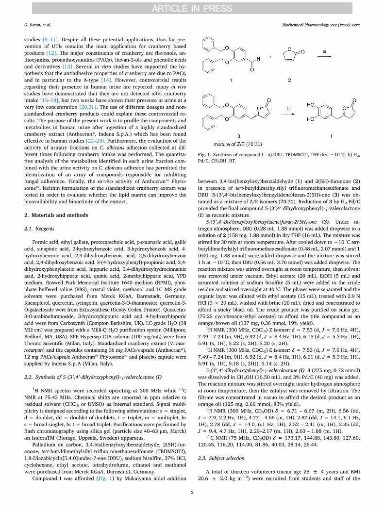
Compound I was afforded (Fig. 1) by Mukaiyama aldol addition
between 3,4-bis(benzyloxy)benzaldehyde (1) and 2(5H)-furanone (2)in presence of tert-butyldimethylsilyl trifluoromethanesulfonate andDBU. 5-(3′,4′-bis(benzyloxy)benzylidene)furan-2(5H)-one (3) was ob-tained as a mixture of Z/E isomers (70:30). Reduction of 3 by H2 Pd/Cprovided the final compound 5-(3′,4′-dihydroxyphenyl)-γ-valerolactone(I) as racemic mixture.
5-(3′,4′-Bis(benzyloxy)benzylidene)furan-2(5H)-one (3). Under ni-trogen atmosphere, DBU (0.28 mL, 1.88 mmol) was added dropwise to asolution of 2 (158 mg, 1.88 mmol) in dry THF (16 mL). The mixture wasstirred for 30 min at room temperature. After cooled down to −10 °C tert-butyldimethylsilyl trifluoromethanesulfonate (0.48 mL, 2.07 mmol) and 1(600 mg, 1.88 mmol) were added dropwise and the mixture was stirred1 h at −10 °C, then DBU (0.56 mL, 3.76 mmol) was added dropwise. Thereaction mixture was stirred overnight at room temperature, then solventwas removed under vacuum. Ethyl acetate (20 mL), EtOH (5 mL) andsaturated solution of sodium bisulfite (5 mL) were added to the cruderesidue and stirred overnight at 40 °C. The phases were separated and theorganic layer was diluted with ethyl acetate (15 mL), treated with 2.9 NHCl (3 × 20 mL), washed with brine (20 mL), dried and concentrated toafford a sticky black oil. The crude product was purified on silica gel(75:25 cyclohexane/ethyl acetate) to afford the title compound as anorange/brown oil (137 mg, 0.36 mmol, 19% yield).
1H NMR (300 MHz, CDCl3) Z isomer: δ = 7.53 (d, J = 7.0 Hz, 4H),7.49 – 7.24 (m, 9H), 6.92 (d, J= 8.4 Hz, 1H), 6.15 (d, J= 5.3 Hz, 1H),5.91 (s, 1H), 5.22 (s, 2H), 5.20 (s, 2H).
1H NMR (300 MHz, CDCl3) E isomer: δ = 7.53 (d, J = 7.0 Hz, 4H),7.49 – 7.24 (m, 9H), 6.92 (d, J= 8.4 Hz, 1H), 6.21 (d, J= 5.3 Hz, 1H),5.91 (s, 1H), 5.18 (s, 2H)), 5.14 (s, 2H).
5-(3′,4′-dihydroxyphenyl)-γ-valerolactone (I). 3 (275 mg, 0.72 mmol)was dissolved in CH3OH (16.50 mL), and 3% Pd/C (40 mg) was added.The reaction mixture was stirred overnight under hydrogen atmosphereat room temperature, then the catalyst was removed by filtration. Thefiltrate was concentrated in vacuo to afford the desired product as anorange oil (125 mg, 0.60 mmol, 83% yield).
1H NMR (300 MHz, CD3OD) δ = 6.71 – 6.67 (m, 2H), 6.56 (dd,J = 7.9, 2.2 Hz, 1H), 4.77 – 4.66 (m, 1H), 2.87 (dd, J = 14.1, 6.1 Hz,1H), 2.78 (dd, J = 14.0, 6.1 Hz, 1H), 2.52 – 2.41 (m, 1H), 2.35 (dd,J = 9.4, 4.7 Hz, 1H), 2.29–2.17 (m, 1H), 2.03 – 1.88 (m, 1H).
13C NMR (75 MHz, CD3OD) δ = 173.17, 144.88, 143.80, 127.60,120.45, 116.20, 114.90, 81.86, 40.03, 28.14, 26.44.
2.3. Subject selection
A total of thirteen volunteers (mean age 25 ± 4 years and BMI20.6 ± 2.0 kg m−2) were recruited from students and staff of the
Fig. 1. Synthesis of compound I – a) DBU, TBDMSOTf, THF dry, −10 °C; b) H2,Pd/C, CH3OH, RT.
G. Baron, et al. Biochemical Pharmacology xxx (xxxx) xxxx
2
University of Milan according to the following inclusion criteria:women, 18–40 years of age, normal weight for height (18–25 kg m−2),non-smokers, no history of cardiovascular, diabetes, hepatic, renal, orgastrointestinal diseases. Subjects with allergy and/or aversion tocranberry and/or cranberry products were excluded. Other exclusioncriteria were as follow: consumption of any dietary supplement, drug ormedication for at least one month before the beginning of the study.The study was performed in accordance with the ethical standards es-tablished in the 2013 Declaration of Helsinki and approved by theEthics Committee of the University of Milan (December 18, 2018, ref.57/18). The study was registered at www.isrctn.org asISRCTN32556347. All participants signed an informed consent form.
2.4. Study design on healthy volunteers
Subjects were instructed to limit the consumption of polyphenols atleast 72 h before experimentation and during the trial. A list of foods tobe avoided has been provided to the volunteers. The list included: fruitsand vegetables rich in polyphenols (e.g. berries, red/purple fruits/ve-getables), chocolate and some beverages such as coffee, tea, wine andfruit juice. The study consisted of a randomized, double blind, 2-armrepeated measure cross-over design. One group of subjects consumed 2capsules of Anthocran® per day, while the other group 2 the capsules ofAnthocran™ Phytosome™ per day. The experiment was 7-day long.Urine samples were collected before starting supplementation (day 1,time 0) and after the last dosage (day 7) at the following time-points: 1,2, 4, 6, 10, 12, 24 h. After one week of wash-out the groups inverted thetreatment. Each subject received a box containing the number of cap-sules to consume during the experiment. Capsules were provided in ablind condition. Subjects were instructed to swallow two capsules perday, the first one in the morning before breakfast and the second onebefore dinner with a glass of water. Three volunteers consumed twoplacebo capsules with the same shape, size, colour, flavour and ex-cipients of the products tested. Urine samples were collected at thesame time-points as previously reported and used as control to verifythe influence of diet and circadian rhythm on C. albicans activity. Ethylgallate 10 µM was added as internal standard in the sample used for theMS analysis and all the samples were stored at −80 °C until analysis.
2.5. Sample preparation
An aliquot (1 mL) of each of the 10 subjects’ urine was centrifugedat 10000 × g for 5 min and the supernatant was extracted on the SPEcolumn, working at 1 mL/min. Salts were removed with water and thenall the compounds retained were eluted with 1 mL 100% acetonitrile.The fractions collected were dried under vacuum and then solubilizedin 100 µL H2O-CH3OH-HCOOH (90:10:0.1, v/v). For the quantitativeanalysis, the stock solutions of the standards were prepared in methanoland then diluted in a pool of the pre-treatment urine samples to obtainthe final concentrations for each calibration curve. The samples werethen added with ethyl gallate 10 µM as internal standard and processedas described.
2.6. Chromatographic conditions
Cranberry components and urine metabolite separation was per-formed on a reversed-phase Agilent Zorbax SB-C18 column(150 × 2.1 mm, i.d. 3.5 µm, CPS analitica, Milan, Italy), protected byan Agilent Zorbax guard column, kept at 40 °C, by an UltiMate 3000system (Dionex) equipped with an autosampler kept at 4 °C working ata constant flow rate (200 µL/min). Each sample (10 µL) was injectedinto the column and both cranberry components and urine metaboliteswere eluted with an 80 min multistep gradient of phase A H2O-HCOOH(100:0.1, v/v) and phase B CH3CN-HCOOH (100:0.1, v/v): 0–45 min,from 10% B to 20% B; 45–65 min, from 20% B to 60% B; 65–66 min,from 60% B to 90% B; 66–70 min, isocratic of 90% B; 70–71 min, from
90% B to 10% B, and then 71–80 min of isocratic 10% B.
2.7. Polyphenol class identification by HPLC-UV analysis
The identification of polyphenol classes was carried out by HPLC-UV analysis on a HPLC Surveyor LC system (Thermo Fisher Scientific,Milan, Italy) equipped with a quaternary pump, UV–VIS detector (PDA)and an autosampler. The scan range was set from 200 nm to 600 nm. Asolution of 4 mg/mL of cranberry extract in H2O/CH3OH/HCOOH (90/10/0.1% v/v) was used for the analysis.
2.8. Cranberry component profiling and urine metabolite characterizationby high resolution mass spectrometry
Each sample (10 µL) was injected into the RP column as previouslydescribed: the cranberry extract was analyzed at a concentration of4 mg/mL in H2O-CH3OH-HCOOH (90:10:0.1, v/v), while the urinesamples were analyzed after the treatment described in section 2.3. Theanalyses were performed on a LTQ-Orbitrap XL mass spectrometerusing an ESI source. Mass spectra were acquired in positive and innegative ion modes. A list of 20 background ions was adopted as lockmass values for real time mass calibration [25]. The source parametersused for the positive mode are: spray voltage 4 kV, capillary tempera-ture 300 °C, capillary voltage 30 V, tube lens offset 90 V; for the ne-gative ion mode: spray voltage 4 kV, capillary temperature 300 °C,capillary voltage −23 V, tube lens offset −140 V. The instrument wasset up to work in a data-dependent scan mode to acquire both full MSand MS/MS spectra. Full MS spectra were acquired in profile mode bythe FT analyzer in a scan range of m/z 100–1200, using AGC scan target5 × 105 and resolution 30,000 FWHM at m/z 400. Tandem massspectra were acquired by the linear ion trap (LTQ) which was set up tofragment the 3 most intense ions exceeding 1 × 10⁴ counts. Mass ac-quisition settings were: centroid mode, AGC scan target 5 × 10⁴, pre-cursor ion isolation width of m/z 3, and collision energy (CID) of 35 eV.Dynamic exclusion was enabled to reduce redundant spectra acquisi-tion: 2 repeat counts, 20 sec repeat duration, 30 sec of exclusionduration. Moreover, only singly and unassigned charged ions werefragmented. Instrument control and spectra analysis were provided bythe software Xcalibur 2.0.7 and Chromeleon Xpress 6.80.
2.9. Targeted and untargeted analyses of cranberry components andmetabolites in human urine
An in-house database was created for the targeted analysis byadding all the characterized cranberry extract components as well asknown cranberry metabolites identified in other studies and cranberrycomponents deriving from other cranberry sources even if not presentin the extract under investigation. The identification was carried out onthe QualBrowser tool of Xcalibur 2.0.7 by using the accurate mass andthe isotopic and fragmentation patterns.
The untargeted analysis consisted of searching for all the ions pre-sent in the urine samples collected after the cranberry consumption thatwere not present or present at intensity relative to noise (< 5*102
counts) in the pre-treatment sample. Spectra analyses were carried outon the QualBrowser tool of Xcalibur 2.0.7 by screening the full MSspectra acquired in negative ion mode in mass ranges of m/z 5 with10 min as acquisition time for each sample. Each ion detected withthese filters was exported with the relative MS/MS spectrum, if present.Identification was performed by following two different approachesbased on the accurate mass and isotopic and fragmentation patterns.The first approach consists of giving the precursor ion and the MS/MSspectrum list as inputs in the Compound Identification tool of CFM-ID[26], using as mass tolerance error 10 ppm for the precursor ion and0.3 Da for the fragments. CFM-ID performs a search for candidates inavailable databases (HMDB and KEGG) based on the accurate mass,then generating in-silico MS/MS spectra of all the candidates and then
G. Baron, et al. Biochemical Pharmacology xxx (xxxx) xxxx
3
comparing the experimental data with those obtained in-silico. The topcandidates were ranked (Jaccard Score) according to how closely theymatched and returned to a list. The second approach was initially fo-cused on the calculation of the elemental composition performed on theElemental Composition page of Xcalibur 2.0.7 by using the followingparameters: mass tolerance 10 ppm, charge −1, C, H, O, N, P, S aselements in use. The top 5 formulae were searched in available data-bases such as PubChem. METLIN, MassBank and in the literature inorder to obtain a list of candidates. Following this, the Peak Assignmenttool of CFM-ID was used to predict the MS/MS spectra of the putativeidentified compounds and to compare the in-silico spectra obtained withthe experimental spectra.
2.10. Quantitative analysis
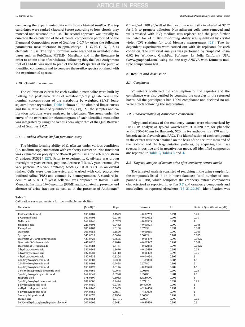
The calibration curves for each available metabolite were built byplotting the peak area ratios of metabolite/ethyl gallate versus thenominal concentrations of the metabolite by weighted (1/x2) least-squares linear regression. Table 1 shows all the obtained linear curvesand the relative limit of quantification (LOQ). All the samples and ca-libration solutions were analyzed in triplicates. The areas under thecurve of the extracted ion chromatogram of each identified metabolitewas integrated by using the Genesis peak algorithm of the Qual Browsertool of Xcalibur 2.0.7.
2.11. Candida albicans biofilm formation assay
The biofilm-forming ability of C. albicans under various conditions(i.e. medium supplementation with cranberry extract or urine fractions)was evaluated on polystyrene 96-well plates using the reference strainC. albicans SC5314 [27]. Prior to experiments, C. albicans was grownovernight in yeast extract, peptone, dextrose (1% w/v yeast extract, 2%w/v peptone, 2% w/v dextrose) broth (YPD) at 30 °C in an orbitalshaker. Cells were then harvested and washed with cold phosphate-buffered saline (PBS) and counted by hemocytometer. A standard in-oculum of 5 × 10⁵ yeast cells/mL was prepared in Roswell ParkMemorial Institute 1640 medium (RPMI) and incubated in presence andabsence of urine fractions as well as in the presence of Anthocran™
0.1 mg/mL. 100 µL/well of the inoculum was firstly incubated at 37 °Cfor 1 h to promote adhesion. Non-adherent cells were removed andwells washed with PBS; medium was replaced and the plate furtherincubated for 24 h. Biofilm-forming ability was quantified by crystalviolet (CV) staining for total biomass measurement [28]. Two in-dependent experiments were carried out with six replicates for eachcondition. The statistical analysis was performed by GraphPad Prism6.02 for Windows, GraphPad Software, La Jolla California USA,(www.graphpad.com) using the one-way ANOVA with Dunnet’s mul-tiple comparisons test.
3. Results and discussion
3.1. Compliance
Volunteers confirmed the consumption of the capsules and thecompliance was also verified by counting the capsules in the returnedboxes. All the participants had 100% compliance and declared no ad-verse effects following the intervention.
3.2. Characterization of Anthocran® components
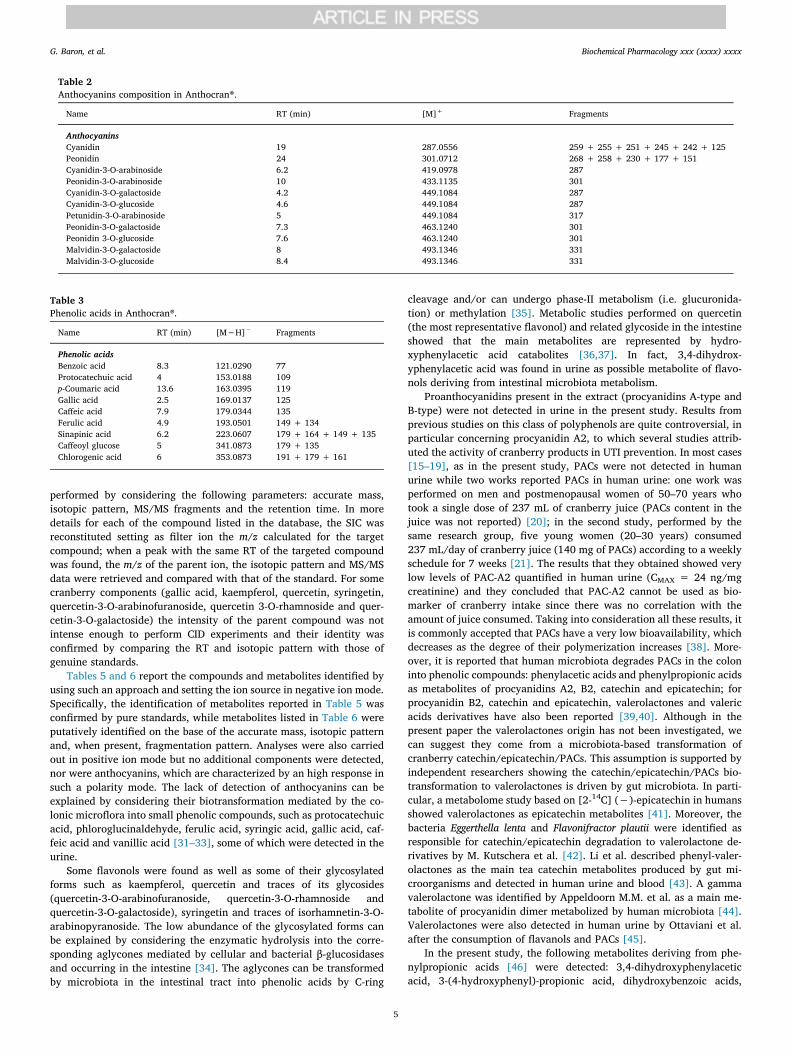
Polyphenol classes of the cranberry extract were characterized byHPLC-UV analysis at typical wavelength: 310–320 nm for phenolicacids, 350–370 nm for flavonols, 520 nm for anthocyanins, 278 nm forbenzoic acids, flavanols and PACs. The identification of each compoundin the extract was then obtained on the basis of the accurate mass and ofthe isotopic and the fragmentation patterns, by acquiring the massspectra in positive and in negative ion mode. All identified compoundsare reported in Table 2, Tables 3 and 4.
3.3. Targeted analysis of human urine after cranberry extract intake
The targeted analysis consisted of searching in the urine samples forthe compounds listed in an in-house database (total number of com-pounds = 138), which comprises the cranberry extract componentscharacterized as reported in section 3.2 and cranberry compounds andmetabolites as reported elsewhere [15–21,29,30]. Identification was
Table 1Calibration curve parameters for the available metabolites.
Metabolite [M−H]− Slope Intercept R2 Limit of Quantification (µM)
Protocatechuic acid 153.0189 0.1529 −0.04789 0.991 0.25p-Coumaric acid 163.0408 0.2786 −0.03032 0.995 0.01Gallic acid 169.0146 0.0253 −0.00585 0.991 1Sinapinic acid 223.0608 0.0452 −0.00523 0.990 0.005Kaempferol 285.0407 1.0160 0.07999 0.991 0.001Quercetin 301.0353 1.1520 −0.05015 0.999 0.005Syringetin 345.0618 0.6626 0.00924 0.981 0.005Quercetin-3-O-arabinofuranoside 433.0731 0.7625 −0.01439 0.997 0.0025Quercetin 3-O-rhamnoside 447.0920 0.9010 −0.02547 0.997 0.001Quercetin-3-O-galactoside 463.0884 0.5151 −0.02053 0.996 0.00252-hydroxybenzoic acid 137.0243 1.1470 −0.13480 0.998 0.253-hydroxybenzoic acid 137.0231 0.1113 −0.01402 0.996 0.054-hydroxybenzoic acid 137.0232 0.1204 −0.04054 0.999 12,3-dihydroxybenzoic acid 153.0201 1.2380 −1.89800 0.984 1.52,5-dihydroxybenzoic acid 153.0194 0.2438 0.67780 0.998 52,4-dihydroxybenzoic acid 153.0175 0.3176 −0.33540 0.989 1.53-(4-hydroxyphenyl)-propionic acid 165.0561 0.0048 0.00106 0.999 0.253,4-dihydroxyphenylacetic acid 167.0349 0.0228 0.05486 0.981 1.5Hippuric acid 178.0509 0.3032 120.80000 0.993 13,4-dihydroxyhydrocinnamic acid 181.0506 0.2474 0.37710 0.997 0.25p-Hydroxyhippuric acid 194.0450 0.2756 10.42000 0.995 1m-Hydroxyhippuric acid 194.0446 0.3605 −2.94400 0.991 1o-Hydroxyhippuric acid 194.0461 0.6801 −4.23000 0.996 12-methylhippuric acid 192.0670 0.7954 3.60500 0.999 1Quinic acid 191.0554 0.01012 0.0097 0.999 0.055-(3′,4′-dihydroxyphenyl)-γ-valerolactone 207.0666 0.2411 −0.47450 0.999 0.1
G. Baron, et al. Biochemical Pharmacology xxx (xxxx) xxxx
4
performed by considering the following parameters: accurate mass,isotopic pattern, MS/MS fragments and the retention time. In moredetails for each of the compound listed in the database, the SIC wasreconstituted setting as filter ion the m/z calculated for the targetcompound; when a peak with the same RT of the targeted compoundwas found, the m/z of the parent ion, the isotopic pattern and MS/MSdata were retrieved and compared with that of the standard. For somecranberry components (gallic acid, kaempferol, quercetin, syringetin,quercetin-3-O-arabinofuranoside, quercetin 3-O-rhamnoside and quer-cetin-3-O-galactoside) the intensity of the parent compound was notintense enough to perform CID experiments and their identity wasconfirmed by comparing the RT and isotopic pattern with those ofgenuine standards.
Tables 5 and 6 report the compounds and metabolites identified byusing such an approach and setting the ion source in negative ion mode.Specifically, the identification of metabolites reported in Table 5 wasconfirmed by pure standards, while metabolites listed in Table 6 wereputatively identified on the base of the accurate mass, isotopic patternand, when present, fragmentation pattern. Analyses were also carriedout in positive ion mode but no additional components were detected,nor were anthocyanins, which are characterized by an high response insuch a polarity mode. The lack of detection of anthocyanins can beexplained by considering their biotransformation mediated by the co-lonic microflora into small phenolic compounds, such as protocatechuicacid, phloroglucinaldehyde, ferulic acid, syringic acid, gallic acid, caf-feic acid and vanillic acid [31–33], some of which were detected in theurine.
Some flavonols were found as well as some of their glycosylatedforms such as kaempferol, quercetin and traces of its glycosides(quercetin-3-O-arabinofuranoside, quercetin-3-O-rhamnoside andquercetin-3-O-galactoside), syringetin and traces of isorhamnetin-3-O-arabinopyranoside. The low abundance of the glycosylated forms canbe explained by considering the enzymatic hydrolysis into the corre-sponding aglycones mediated by cellular and bacterial β-glucosidasesand occurring in the intestine [34]. The aglycones can be transformedby microbiota in the intestinal tract into phenolic acids by C-ring
cleavage and/or can undergo phase-II metabolism (i.e. glucuronida-tion) or methylation [35]. Metabolic studies performed on quercetin(the most representative flavonol) and related glycoside in the intestineshowed that the main metabolites are represented by hydro-xyphenylacetic acid catabolites [36,37]. In fact, 3,4-dihydrox-yphenylacetic acid was found in urine as possible metabolite of flavo-nols deriving from intestinal microbiota metabolism.
Proanthocyanidins present in the extract (procyanidins A-type andB-type) were not detected in urine in the present study. Results fromprevious studies on this class of polyphenols are quite controversial, inparticular concerning procyanidin A2, to which several studies attrib-uted the activity of cranberry products in UTI prevention. In most cases[15–19], as in the present study, PACs were not detected in humanurine while two works reported PACs in human urine: one work wasperformed on men and postmenopausal women of 50–70 years whotook a single dose of 237 mL of cranberry juice (PACs content in thejuice was not reported) [20]; in the second study, performed by thesame research group, five young women (20–30 years) consumed237 mL/day of cranberry juice (140 mg of PACs) according to a weeklyschedule for 7 weeks [21]. The results that they obtained showed verylow levels of PAC-A2 quantified in human urine (CMAX = 24 ng/mgcreatinine) and they concluded that PAC-A2 cannot be used as bio-marker of cranberry intake since there was no correlation with theamount of juice consumed. Taking into consideration all these results, itis commonly accepted that PACs have a very low bioavailability, whichdecreases as the degree of their polymerization increases [38]. More-over, it is reported that human microbiota degrades PACs in the coloninto phenolic compounds: phenylacetic acids and phenylpropionic acidsas metabolites of procyanidins A2, B2, catechin and epicatechin; forprocyanidin B2, catechin and epicatechin, valerolactones and valericacids derivatives have also been reported [39,40]. Although in thepresent paper the valerolactones origin has not been investigated, wecan suggest they come from a microbiota-based transformation ofcranberry catechin/epicatechin/PACs. This assumption is supported byindependent researchers showing the catechin/epicatechin/PACs bio-transformation to valerolactones is driven by gut microbiota. In parti-cular, a metabolome study based on [2-14C] (−)-epicatechin in humansshowed valerolactones as epicatechin metabolites [41]. Moreover, thebacteria Eggerthella lenta and Flavonifractor plautii were identified asresponsible for catechin/epicatechin degradation to valerolactone de-rivatives by M. Kutschera et al. [42]. Li et al. described phenyl-valer-olactones as the main tea catechin metabolites produced by gut mi-croorganisms and detected in human urine and blood [43]. A gammavalerolactone was identified by Appeldoorn M.M. et al. as a main me-tabolite of procyanidin dimer metabolized by human microbiota [44].Valerolactones were also detected in human urine by Ottaviani et al.after the consumption of flavanols and PACs [45].
In the present study, the following metabolites deriving from phe-nylpropionic acids [46] were detected: 3,4-dihydroxyphenylaceticacid, 3-(4-hydroxyphenyl)-propionic acid, dihydroxybenzoic acids,
Table 2Anthocyanins composition in Anthocran®.
Name RT (min) [M]+ Fragments
AnthocyaninsCyanidin 19 287.0556 259 + 255 + 251 + 245 + 242 + 125Peonidin 24 301.0712 268 + 258 + 230 + 177 + 151Cyanidin-3-O-arabinoside 6.2 419.0978 287Peonidin-3-O-arabinoside 10 433.1135 301Cyanidin-3-O-galactoside 4.2 449.1084 287Cyanidin-3-O-glucoside 4.6 449.1084 287Petunidin-3-O-arabinoside 5 449.1084 317Peonidin-3-O-galactoside 7.3 463.1240 301Peonidin 3-O-glucoside 7.6 463.1240 301Malvidin-3-O-galactoside 8 493.1346 331Malvidin-3-O-glucoside 8.4 493.1346 331
Table 3Phenolic acids in Anthocran®.
Name RT (min) [M−H]− Fragments
Phenolic acidsBenzoic acid 8.3 121.0290 77Protocatechuic acid 4 153.0188 109p-Coumaric acid 13.6 163.0395 119Gallic acid 2.5 169.0137 125Caffeic acid 7.9 179.0344 135Ferulic acid 4.9 193.0501 149 + 134Sinapinic acid 6.2 223.0607 179 + 164 + 149 + 135Caffeoyl glucose 5 341.0873 179 + 135Chlorogenic acid 6 353.0873 191 + 179 + 161
G. Baron, et al. Biochemical Pharmacology xxx (xxxx) xxxx
5
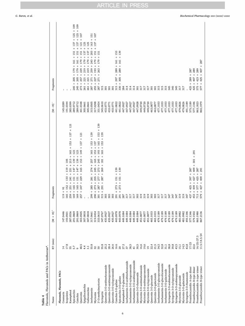
Table4
Flavon
ols,Flavan
olsan
dPA
Csin
Antho
cran
®.
Nam
eRT
(min)
[M+
H]+
Fragments
[M−H]−
Fragments
Flavonols,Flavanols,PACs
Coum
arin
1414
7.04
4611
9+
9114
5.02
89–
Scop
oletin
17.8
193.05
0116
5+
152+
133+
119+
105
191.03
44–
Kaem
pferol
5528
7.05
5625
9+
251+
241+
231+
213+
165+
153+
137+
121
285.03
99–
Epicatechin
5.7
291.08
6916
9+
165+
151+
147+
139+
123
289.07
1224
5+
205+
179+
161+
151+
137+
125+
109
Catechin
929
1.08
6916
9+
165+
151+
147+
139+
123
289.07
1224
5+
205+
179+
161+
151+
137+
125+
109
Quercetin
48.5
303.05
0525
7+
247+
229+
165+
153+
149+
137+
121
301.03
4827
3+
257+
229+
179+
151+
121+
107
Epigallocatechin
4.4
307.08
18–
305.06
6126
1+
221+
219+
179+
137+
125
Gallocatechin
3.1
307.08
18–
305.06
6126
1+
221+
219+
179+
137+
125
Isorha
mnetin
55.8
317.06
6129
9+
285+
281+
274+
257+
165+
153+
139
315.05
0528
7+
271+
259+
243+
203+
163+
151
Myricetin
3231
9.04
5429
0+
273+
255+
245+
165+
153+
137
317.02
9825
5+
227+
193+
179+
151+
137+
107
3′-O-m
ethy
lmyricetin
5033
3.06
1030
1+
287+
277+
273+
245+
193+
165+
153+
139
331.04
5428
7+
271+
263+
179+
151
Syring
etin
55.8
347.07
6731
5+
291+
287+
269+
181+
165+
153+
139
345.06
1031
5Quercetin-3-O-arabino
furano
side
25.2
435.09
2730
343
3.07
7130
1Quercetin-3-O-arabino
pyrano
side
28.4
435.09
2730
343
3.07
7130
1Quercetin-3-O-xylop
yran
oside
26.6
435.09
2730
343
3.07
7130
1Ca
techin-3-O-gallate
16.6
443.09
7829
1+
273+
151+
139
441.08
2231
5+
297+
289+
161+
153
Epicatechin-3-O-gallate
2044
3.09
7829
1+
273+
151+
139
441.08
2233
0+
305+
289+
161+
139
Kaem
pferol-7-O-glucoside
27.2
449.10
8428
744
7.09
2728
4Isorha
mnetin
-3-O-arabino
furano
side
3744
9.10
8431
744
7.09
2731
4Isorha
mnetin
-3-O-xylop
yran
oside
38,5
449.10
8431
744
7,09
2731
4Isorha
mnetin
-3-O-arabino
pyrano
side
4144
9.10
8431
744
7,09
2731
4Quercetin-3-O-rha
mno
side
30,5
449.10
8430
344
7,09
2830
1Myricetin-3-O-arabino
furano
side
16,6
451.08
7731
944
9,07
2031
7Myricetin-3-O-arabino
pyrano
side
19,6
451.08
7731
944
9,07
2031
7Myricetin-3-O-xylop
yran
oside
1945
1.08
7731
944
9,07
2031
7Quercetin
3-O-glucoside
22,4
465.10
3330
346
3,08
7730
1Quercetin-3-O-galactoside
21,5
465.10
3330
346
3,08
7730
1Isorha
mnetin
-3-O-glucopy
rano
side
34,5
479.11
8931
747
7,10
3331
5Isorha
mnetin
-3-O-glucofurano
side
32,8
479.11
8931
747
7,10
3331
5Isorha
mnetin
-3-O-galactoside
31,1
479.11
8931
747
7,10
3331
5Syring
etin-3-O-arabino
furano
side
38,8
479.11
8934
747
7,10
3334
5Syring
etin-3-O-xylop
yran
oside
39,8
479.11
8934
747
7,10
3334
5Syring
etin-3-O-arabino
pyrano
side
43,5
479.11
8934
747
7,10
3334
5Myricetin-3-O-glucoside
15,1
481.09
8231
947
9,08
2631
7Myricetin-3-O-galactoside
14,5
481.09
8231
947
9,08
2631
7Syring
etin-3-O-rha
mno
side
43,8
493.13
4634
749
1,11
89–
Proantho
cyan
idin
A-ty
pedimer
17/23
577.13
4643
7+
425+
397+
287
575,11
8942
5+
289+
287
Proantho
cyan
idin
B-type
dimer
22,6
579.15
0245
3+
439+
427+
409+
301+
291
577,13
4642
5+
407+
289
Proantho
cyan
idin
A-ty
petrim
er16
/25/27
.386
5.19
8071
3+
577+
425+
287
863,18
2357
5+
423+
289
Proantho
cyan
idin
B-type
trim
er11
.5/14.3/24
867.21
3657
9+
427+
409+
291
865,19
7957
7+
425+
407+
287
G. Baron, et al. Biochemical Pharmacology xxx (xxxx) xxxx
6
hydroxybenzoic acid and hydroxyhippuric acids, which presence can berelated to PACs metabolism.
Phenolic acids represent the main class of identified polyphenols:protocatechuic acid, p-coumaric acid, gallic acid and sinapinic acidwere found to be already present in the extract, but they can also derivefrom the metabolism of other polyphenols as mentioned above; 3,4-dihydroxyhydrocinnamic acid, dihydroxyhydrocinnamic acid-3-O-
glucuronide and 3-(4-hydroxyphenyl)-propionic acid can derive fromthe metabolism of PACs, chlorogenic acid or anthocyanins [17,39,46],while hydroxybenzoic acid, dihydroxybenzoic acids, hippuric acid andhydroxyhippuric acids can derive from the metabolism of all the otherflavonoid components [46]. Catechol and pyrogallol derivatives, suchas 4-methylcatechol-O-sulphate and pyrogallol-O-2-sulphate that weidentified, can be generated from phenolic acids or anthocyanins [46].
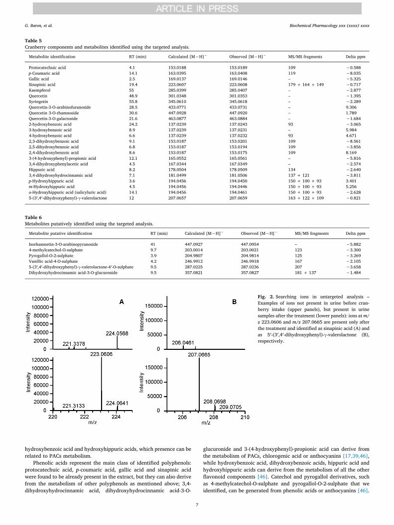
Table 5Cranberry components and metabolites identified using the targeted analysis.
Metabolite identification RT (min) Calculated [M−H]− Observed [M−H]− MS/MS fragments Delta ppm
Protocatechuic acid 4.1 153.0188 153.0189 109 −0.588p-Coumaric acid 14.1 163.0395 163.0408 119 −8.035Gallic acid 2.5 169.0137 169.0146 – −5.325Sinapinic acid 19.4 223.0607 223.0608 179 + 164 + 149 −0.717Kaempferol 55 285.0399 285.0407 – −2.877Quercetin 48.9 301.0348 301.0353 – −1.395Syringetin 55.8 345.0610 345.0618 – −2.289Quercetin-3-O-arabinofuranoside 28.5 433.0771 433.0731 – 9.306Quercetin 3-O-rhamnoside 30.6 447.0928 447.0920 – 1.789Quercetin-3-O-galactoside 21.6 463.0877 463.0884 – −1.6842-hydroxybenzoic acid 24.2 137.0239 137.0243 93 −3.0653-hydroxybenzoic acid 8.9 137.0239 137.0231 – 5.9844-hydroxybenzoic acid 6.6 137.0239 137.0232 93 4.6712,3-dihydroxybenzoic acid 9.1 153.0187 153.0201 109 −8.5612,5-dihydroxybenzoic acid 6.8 153.0187 153.0194 109 −3.8562,4-dihydroxybenzoic acid 8.6 153.0187 153.0175 109 8.1693-(4-hydroxyphenyl)-propionic acid 12.1 165.0552 165.0561 – −5.8163,4-dihydroxyphenylacetic acid 4.5 167.0344 167.0349 – −2.574Hippuric acid 8.2 178.0504 178.0509 134 −2.6403,4-dihydroxyhydrocinnamic acid 7.1 181.0499 181.0506 137 + 121 −3.811p-Hydroxyhippuric acid 3.6 194.0456 194.0450 150 + 100 + 93 3.401m-Hydroxyhippuric acid 4.5 194.0456 194.0446 150 + 100 + 93 5.256o-Hydroxyhippuric acid (salicyluric acid) 14.1 194.0456 194.0461 150 + 100 + 93 −2.6285-(3′,4′-dihydroxyphenyl)-γ-valerolactone 12 207.0657 207.0659 163 + 122 + 109 −0.821
Table 6Metabolites putatively identified using the targeted analysis.
Metabolite putative identification RT (min) Calculated [M−H]− Observed [M−H]− MS/MS fragments Delta ppm
Isorhamnetin-3-O-arabinopyranoside 41 447.0927 447.0954 – −5.8824-methylcatechol-O-sulphate 9.7 203.0014 203.0021 123 −3.300Pyrogallol-O-2-sulphate 3.9 204.9807 204.9814 125 −3.269Vanillic acid-4-O-sulphate 4.2 246.9912 246.9918 167 −2.1055-(3′,4′-dihydroxyphenyl)-γ-valerolactone-4′-O-sulphate 9.5 287.0225 287.0236 207 −3.658Dihydroxyhydrocinnamic acid-3-O-glucuronide 9.5 357.0821 357.0827 181 + 137 −1.484
Fig. 2. Searching ions in untargeted analysis –Examples of ions not present in urine before cran-berry intake (upper panels), but present in urinesamples after the treatment (lower panels): ions at m/z 223.0606 and m/z 207.0665 are present only afterthe treatment and identified as sinapinic acid (A) andas 5′-(3′,4′-dihydroxyphenyl)-γ-valerolactone (B),respectively.
G. Baron, et al. Biochemical Pharmacology xxx (xxxx) xxxx
7
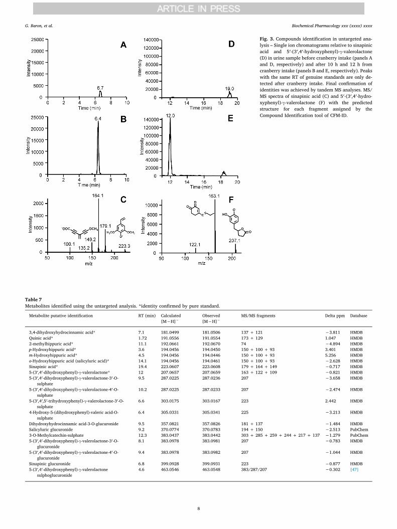
Fig. 3. Compounds identification in untargeted ana-lysis – Single ion chromatograms relative to sinapinicacid and 5′-(3′,4′-hydroxyphenyl)-γ-valerolactone(D) in urine sample before cranberry intake (panels Aand D, respectively) and after 10 h and 12 h fromcranberry intake (panels B and E, respectively). Peakswith the same RT of genuine standards are only de-tected after cranberry intake. Final confirmation ofidentities was achieved by tandem MS analyses. MS/MS spectra of sinapinic acid (C) and 5′-(3′,4′-hydro-xyphenyl)-γ-valerolactone (F) with the predictedstructure for each fragment assigned by theCompound Identification tool of CFM-ID.
Table 7Metabolites identified using the untargeted analysis. *identity confirmed by pure standard.
Metabolite putative identification RT (min) Calculated[M−H]−
Observed[M−H]−
MS/MS fragments Delta ppm Database
3,4-dihydroxyhydrocinnamic acid* 7.1 181.0499 181.0506 137 + 121 −3.811 HMDBQuinic acid* 1.72 191.0556 191.0554 173 + 129 1.047 HMDB2-methylhippuric acid* 11.1 192.0661 192.0670 74 −4.894 HMDBp-Hydroxyhippuric acid* 3.6 194.0456 194.0450 150 + 100 + 93 3.401 HMDBm-Hydroxyhippuric acid* 4.5 194.0456 194.0446 150 + 100 + 93 5.256 HMDBo-Hydroxyhippuric acid (salicyluric acid)* 14.1 194.0456 194.0461 150 + 100 + 93 −2.628 HMDBSinapinic acid* 19.4 223.0607 223.0608 179 + 164 + 149 −0.717 HMDB5-(3′,4′-dihydroxyphenyl)-γ-valerolactone* 12 207.0657 207.0659 163 + 122 + 109 −0.821 HMDB5-(3′,4′-dihydroxyphenyl)-γ-valerolactone-3′-O-
sulphate9.5 287.0225 287.0236 207 −3.658 HMDB
5-(3′,4′-dihydroxyphenyl)-γ-valerolactone-4′-O-sulphate
10.2 287.0225 287.0233 207 −2.474 HMDB
5-(3′,4′,5′-trihydroxyphenyl)-γ-valerolactone-3′-O-sulphate
6.6 303.0175 303.0167 223 2.442 HMDB
4-Hydroxy-5-(dihydroxyphenyl)-valeric acid-O-sulphate
6.4 305.0331 305.0341 225 −3.213 HMDB
Dihydroxyhydrocinnamic acid-3-O-glucuronide 9.5 357.0821 357.0826 181 + 137 −1.484 HMDBSalicyluric glucuronide 9.2 370.0774 370.0783 194 + 150 −2.513 PubChem3-O-Methylcatechin-sulphate 12.3 383.0437 383.0442 303 + 285 + 259 + 244 + 217 + 137 −1.279 PubChem5-(3′,4′-dihydroxyphenyl)-γ-valerolactone-3′-O-
glucuronide8.1 383.0978 383.0981 207 −0.783 HMDB
5-(3′,4′-dihydroxyphenyl)-γ-valerolactone-4′-O-glucuronide
9.4 383.0978 383.0982 207 −1.044 HMDB
Sinapinic glucuronide 6.8 399.0928 399.0931 223 −0.877 HMDB5-(3′,4′-dihydroxyphenyl)-γ-valerolactone
sulphoglucuronide4.6 463.0546 463.0548 383/287/207 −0.302 [47]
G. Baron, et al. Biochemical Pharmacology xxx (xxxx) xxxx
8
3.4. Untargeted analysis of human urine after cranberry extract intake
As described in the method session, the untargeted approach con-sists of searching for all the ions present in the urine samples collectedafter cranberry consumption that were not present or present at in-tensity relative to noise less than 5*102 counts in the pre-treatmentsample. The analysis was performed using the negative ion mode be-cause all the compounds identified using the targeted analysis weremainly detected in this polarity mode. The unidentified compounds, orthose recognized as coming from the human basal metabolism (e.g.amino acids), were not included on the list. The search of ions wasperformed by screening the full MS spectra acquired in negative ionmode using a mass ranges of m/z 5 and with 10 min as acquisition timefor each sample. As an example, Fig. 2A reports the MS spectra resultingby setting a MS range between m/z 220 and 225 and considering a timewindow between 0 and 10 min and relative to urine collected before(upper panel) and after 10 h (lower panel) the cranberry intake; Fig. 2Breports the MS spectra resulting by setting a MS range between m/z 205and 210 and considering a time window between 10 and 20 min and
relative to urine collected before (upper panel) and after 12 h (lowerpanel) the cranberry intake. The ions at m/z 223.0606 and m/z207.0665 are well evident only in the urine samples collected after thecranberry administration but not before. Identification of the unknowncompounds was carried out by setting the precursor ion and the MS/MSfragment ions as inputs in the Compound Identification tool of CFM-ID.Fig. 3C and Fig. 3F reports the experimental MS/MS spectra used asinput for the Compound Identification tool of CFM-ID which gave si-napinic acid and 5′-(3′,4′-dihydroxyphenyl)-γ-valerolactone as bestmatched results. Final attribution was obtained by comparing RT, MSisotopic pattern and MS/MS fragmentation with those of pure standards(when commercially available). Fig. 3 shows the SIC chromatograms ofsinapinic acid (Fig. 3B) and 5′-(3′,4′-dihydroxyphenyl)-γ-valerolactone(Fig. 3E) in the urine fraction in which they reached their maximumconcentration (10 h for sinapinic acid and 12 h for 5′-(3′,4′-dihydrox-yphenyl)-γ-valerolactone), while in the control sample (Figs. 3A and3D, respectively) they were not present.
Several metabolites were identified in the untargeted analysis asreported in Table 7. Some of them had already been identified using the
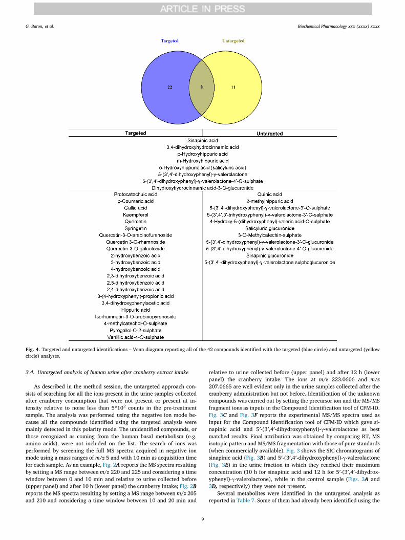
Fig. 4. Targeted and untargeted identifications – Venn diagram reporting all of the 42 compounds identified with the targeted (blue circle) and untargeted (yellowcircle) analyses.
G. Baron, et al. Biochemical Pharmacology xxx (xxxx) xxxx
9
targeted analysis, such as 3,4-dihydroxyhydrocinnamic acid, dihy-droxyhydrocinnamic acid-3-O-glucuronide, sinapinic acid, the threehydroxyhippuric acid isomers, 5-(3′,4′-dihydroxyphenyl)-γ-valer-olactone and 5-(3′,4′-dihydroxyphenyl)-γ-valerolactone-4′-O-sulphate.Fig. 4 shows an overview of all the 42 compounds which have beenidentified in the present study with the targeted (blue circle) and un-targeted (yellow circle) analyses.
Besides quinic acid, which can derive from chlorogenic acid, and 2-methylhippuric acid, a methyl derivative of hippuric acid, differentvalerolactones and one valeric acid derivative were identified and listedin Table 7. Among these metabolites, 5-(3′-hydroxyphenyl)-γ-valer-olactone-4′-O-sulphate had been identified in previous studies [17,18].Also 5-(3′,4′-dihydroxyphenyl)-γ-valerolactone has already been re-ported as cranberry metabolite [19]. None of the other valerolactoneshere described had previously been identified in urine after cranberryintake, although they are known as PACs metabolites [39,47].
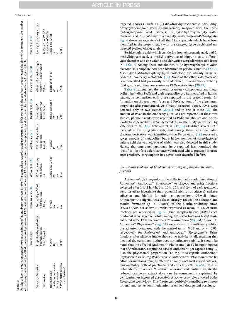
Table 8 summarizes the overall cranberry components and meta-bolites, including PACs and their metabolites, so far identified in humanstudies, in comparison with those reported in the present study. In-formation on the treatment (dose and PACs content of the given cran-berry) are also summarized. As already discussed above, PACs weredetected only in two studies [20,21] and in one of these [20] theamount of PACs in the cranberry juice was not reported. In these twostudies, phenolic acids were reported as PACs metabolites and no va-lerolactone derivatives were detected as in the study performed byValentova et al. [15]. Feliciano et al. [17,18] identified several PACmetabolites by using standards, and among these only one valer-olactone derivative was identified, while Peron et al. [19] reported alower amount of metabolites but a higher number of valerolactone/valeric acid derivatives, one of which was also detected in this study.Hence, the untargeted approach here reported has permitted theidentification of six valerolactones/valeric acid whose presence in urineafter cranberry consumption has never been described before.
3.5. Ex-vivo inhibition of Candida albicans biofilm-formation by urinefractions
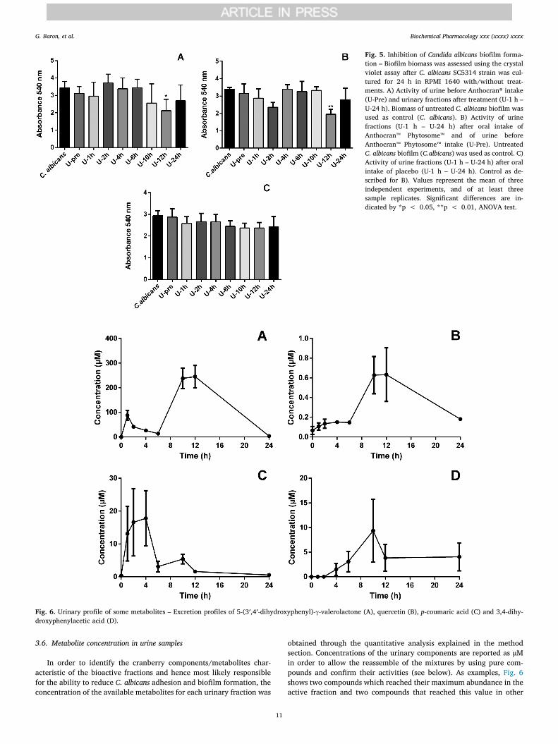
Anthocran® (0.1 mg/mL), urine collected before administration ofAnthocran®, Anthocran™ Phytosome™ or placebo and urine fractionscollected after 1 h, 2 h, 4 h, 6 h, 10 h, 12 h and 24 h of each treatmentwere tested to investigate their potential ability to reduce C. albicansadhesion and biofilm formation on polystyrene 96-well plates.Anthocran® 0.1 mg/mL was able to strongly reduce the adhesion andbiofilm formation (p < 0.0001) of the biofilm-producing strainSC5314 (data not shown). Results expressed as mean ± SD of urinefractions are reported in Fig. 5. Urine samples before (U-Pre) eachtreatment were inactive, while among the seven fractions tested thosecollected after 12 h the Anthocran® consumption (Fig. 5A) as well asAnthocran™ Phytosome™ (Fig. 5B) were shown to significantly inhibitthe adhesion compared with the control (p < 0.05 and p < 0.01,respectively for Anthocran® and Anthocran™ Phytosome™). Urinefractions after placebo intake showed no activity at all, meaning thatdiet and the cyrcadian rhythm does not influence activity. It should benoted that the effect of Anthocran™ Phytosome™ at 12 hr superimposesthat of Anthocran®, despite the dose of Anthocran® per capsule being 1/3 in the phytosomal preparation (12 mg PACs/capsule Anthocran™Phytosome™ vs 36 mg PACs/capsule Anthocran®). Phytosomes are le-cithin formulations demonstrated to enhance botanical ingredients oralbioavailability both at preclinical and clinical levels [48–51]. The si-milar ability to reduce C. albicans adhesion and biofilm despite thereduced cranberry extract dose can be consequently explained byconsidering an increased absorption of active principles allowed bythePhytosome technology. This figure can positively contribute to a morerational and convenient modulation of clinical dosage and posology.
Table8
Results
ofkn
ownstud
ieson
metab
olite
characterizatio
nafterc
ranb
erry
prod
uctsintake.T
hecompa
risonregards:cran
berrytreatm
ent,PA
Cscontentinthecran
berryprod
uctu
sed,
thetim
eof
thetreatm
ent,thenu
mber
ofcran
berrymetab
olite
sidentifi
ed,the
eventual
presence
ofPA
Csan
dthenu
mberof
know
nPA
Csmetab
olite
sinclud
ingph
enolic
acid
andvalerolacton
ederivativ
es.N
A:n
otavailable.
Presentstud
yVa
lentov
aet
al.[15
]McK
ayet
al.[20
]Walsh
etal.[21
]Iswaldi
etal.[16
]Felic
iano
etal.[17
,18]
Peronet
al.[19
]
Cran
berryTreatm
ent
2capsules/day
stan
dardized
cran
berryextract
1200
mg/da
yof
dried
cran
berryjuice
237mLcran
berry
juicecocktail
237mL/da
ycran
berry
juicecocktail
0.6mL/kg
ofcran
berrysyrup
450mLof
asing
le-strength
cran
berryjuicebeverage
360mgof
cran
berryextract
PACs
content
36mg/capsule
14.4
mg
NA
140mg
0.71
%(w
/v)
710mg
42.6%
w/w
ofPA
C-A/
14.6%
w/w
ofPA
C-B
Treatm
enttim
e7da
ys8weeks
Sing
ledo
se(24h)
7weeks
Sing
ledo
se(24h)
Sing
ledo
se(24h)
Sing
ledo
se(24h)
Num
berof
metab
olite
s42
NA
2619
3267
14PA
Cspresence
nono
yes
yes
nono
noNPA
Csmetab
olite
s(N
valerolacton
es)
15(8)
NA
8(0)
7(0)
3(0)
17(1)
12(6)
G. Baron, et al. Biochemical Pharmacology xxx (xxxx) xxxx
10
3.6. Metabolite concentration in urine samples
In order to identify the cranberry components/metabolites char-acteristic of the bioactive fractions and hence most likely responsiblefor the ability to reduce C. albicans adhesion and biofilm formation, theconcentration of the available metabolites for each urinary fraction was
obtained through the quantitative analysis explained in the methodsection. Concentrations of the urinary components are reported as µMin order to allow the reassemble of the mixtures by using pure com-pounds and confirm their activities (see below). As examples, Fig. 6shows two compounds which reached their maximum abundance in theactive fraction and two compounds that reached this value in other
Fig. 5. Inhibition of Candida albicans biofilm forma-tion – Biofilm biomass was assessed using the crystalviolet assay after C. albicans SC5314 strain was cul-tured for 24 h in RPMI 1640 with/without treat-ments. A) Activity of urine before Anthocran® intake(U-Pre) and urinary fractions after treatment (U-1 h –U-24 h). Biomass of untreated C. albicans biofilm wasused as control (C. albicans). B) Activity of urinefractions (U-1 h – U-24 h) after oral intake ofAnthocran™ Phytosome™ and of urine beforeAnthocran™ Phytosome™ intake (U-Pre). UntreatedC. albicans biofilm (C.albicans) was used as control. C)Activity of urine fractions (U-1 h – U-24 h) after oralintake of placebo (U-1 h – U-24 h). Control as de-scribed for B). Values represent the mean of threeindependent experiments, and of at least threesample replicates. Significant differences are in-dicated by *p < 0.05, **p < 0.01, ANOVA test.
Fig. 6. Urinary profile of some metabolites – Excretion profiles of 5-(3′,4′-dihydroxyphenyl)-γ-valerolactone (A), quercetin (B), p-coumaric acid (C) and 3,4-dihy-droxyphenylacetic acid (D).
G. Baron, et al. Biochemical Pharmacology xxx (xxxx) xxxx
11
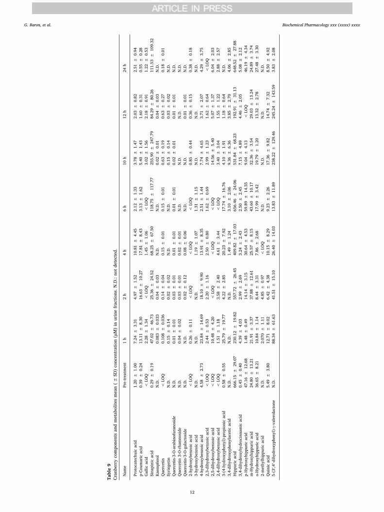
Table9
Cran
berrycompo
nentsan
dmetab
olite
smean(±
SD)concentration(µM)in
urinefractio
ns.N
.D.:no
tdetected.
Nam
ePre-treatm
ent
1h
2h
4h
6h
10h
12h
24h
Protocatechu
icacid
1.20
±1.00
7.24
±3.31
4.97
±1.52
10.81
±4.45
2.12
±1.33
3.78
±1.47
2.03
±0.82
2.51
±0.94
p-Co
umaric
acid
0.39
±0.24
13.11
±8.30
16.63
±10
.27
17.81
±8.43
3.13
±1.62
5.40
±1.43
1.60
±0.31
0.55
±0.28
Gallic
acid
<LO
Q2.28
±1.34
<LO
Q1.45
±1.06
<LO
Q3.02
±1.56
2.18
±0.91
1.22
±0.53
Sina
pinicacid
0.29
±0.19
47.02
±46
.73
25.36
±24
.52
68.25
±67
.50
118.75
±11
7.77
255.90
±24
7.79
84.29
±80
.26
111.53
±10
9.32
Kaem
pferol
N.D.
0.08
3±
0.03
30.04
±0.03
N.D.
N.D.
0.02
±0.01
0.04
±0.03
N.D.
Quercetin
<LO
Q0.10
8±
0.03
60.14
±0.04
0.15
±0.01
0.15
±0.01
0.63
±0.19
0.63
±0.27
0.18
±0.01
Syring
etin
N.D.
0.15
±0.14
0.02
±0.02
N.D.
N.D.
0.15
±0.14
0.03
±0.02
N.D.
Quercetin-3-O-arabino
furano
side
N.D.
0.01
±0.01
0.03
±0.01
0.01
±0.01
0.01
±0.01
0.02
±0.01
0.01
±0.01
N.D.
Quercetin
3-O-rha
mno
side
N.D.
0.04
±0.02
0.03
±0.01
0.02
±0.01
N.D.
N.D.
N.D.
N.D.
Quercetin-3-O-galactoside
N.D.
N.D.
0.02
±0.12
0.08
±0.06
N.D.
N.D.
0.01
±0.01
N.D.
2-hy
droxyb
enzoic
acid
<LO
Q0.26
±0.11
<LO
Q<
LOQ
<LO
Q0.85
±0.44
0.36
±0.15
0.38
±0.18
3-hy
droxyb
enzoic
acid
N.D.
N.D.
N.D.
1.19
±1.07
1.31
±1.15
N.D.
N.D.
N.D.
4-hy
droxyb
enzoic
acid
4.58
±2.73
23.84
±14
.69
18.10
±9.90
13.91
±8.35
2.51
±1.44
7.74
±4.65
3.71
±2.07
4.29
±3.75
2,3-dihy
droxyb
enzoic
acid
<LO
Q2.44
±0.53
2.20
±1.16
2.50
±0.80
1.62
±0.69
2.99
±1.23
1.62
±0.64
<LO
Q2,5-dihy
droxyb
enzoic
acid
<LO
Q10
.48
±4.20
<LO
Q<
LOQ
<LO
Q14
.56
±5.40
5.07
±1.37
6.04
±2.03
2,4-dihy
droxyb
enzoic
acid
<LO
Q1.51
±1.18
3.58
±2.40
4.61
±3.44
<LO
Q3.40
±3.04
1.55
±1.22
2.88
±2.57
3-(4-hyd
roxyph
enyl)-prop
ionicacid
0.58
±0.55
25.79
±19
.77
4.22
±2.85
20.23
±7.92
17.74
±16
.76
4.10
±1.91
1.18
±0.64
N.D.
3,4-dihy
droxyp
heny
lacetic
acid
N.D.
N.D.
N.D.
1.49
±1.24
3.10
±2.06
9.36
±6.36
3.85
±2.70
4.06
±2.85
Hippu
ricacid
666.15
±29
.07
230.12
±19
.82
557.72
±26
.45
409.82
±17
.03
656.46
±24
.06
531.84
±68
.23
192.91
±31
.13
648.52
±27
.88
3,4-dihy
droxyh
ydrocinn
amic
acid
0.45
±0.40
4.39
±4.03
2.99
±2.69
3.24
±2.43
2.50
±2.45
7.15
±4.89
4.46
±2.05
5.08
±2.12
p-Hyd
roxyhipp
uric
acid
47.16
±12
.68
1.48
±0.49
14.14
±3.15
30.64
±4.53
59.89
±14
.55
9.04
±4.13
<LO
Q46
.19
±4.34
m-H
ydroxyhipp
uric
acid
24.80
±13
.21
21.91
±3.37
37.88
±12
.61
25.57
±8.15
43.60
±10
.17
32.36
±3.54
29.03
±13
.24
24.89
±3.74
o-Hyd
roxyhipp
uric
acid
36.65
±8.21
10.84
±1.14
8.44
±3.31
7.86
±2.68
17.99
±3.42
19.79
±1.20
13.32
±2.78
27.48
±3.30
2-methy
lhippu
ricacid
N.D.
2.07
0±
1.11
4.85
±0.97
<LO
QN.D.
N.D.
N.D.
N.D.
Quinicacid
5.49
±3.80
12.71
±8.02
6.42
±4.38
10.15
±8.29
9.23
±2.26
17.36
±9.82
14.74
±7.32
8.50
±4.92
5-(3′,4
′-dihyd
roxyph
enyl)-γ-valerolacton
eN.D.
88.34
±61
.63
41.51
±15
.10
26.40
±14
.03
13.83
±11
.89
238.22
±12
9.46
245.24
±14
3.59
3.83
±2.08
G. Baron, et al. Biochemical Pharmacology xxx (xxxx) xxxx
12
urine fractions: Fig. 6A and B are relative to 5-(3′,4′-dihydroxyphenyl)-γ-valerolactone and quercetin, respectively, whose TMAX was 12 h;Fig. 6C is relative to p-coumaric acid, with a TMAX reached after 4 h;Fig. 6D shows the excretion profile of 3,4-dihydroxyphenylacetic acid(TMAX = 10 h). Table 9 shows the mean cranberry metabolite con-centrations (µM) in the control and in the urine fractions after cran-berry intake.
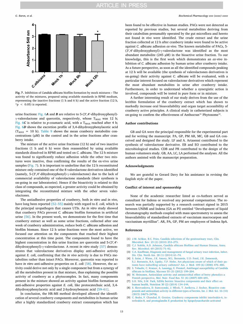
The mixture of the active urine fractions (12 h) and of two inactivefractions (1 h and 6 h) were then reassembled by using availablestandards dissolved in RPMI and tested on C. albicans. The 12 h mixturewas found to significantly reduce adhesion while the other two mix-tures were inactive, thus confirming the results of the ex-vivo urinesamples (Fig. 7). It is important to underline that the 12 h reconstitutedmixture only contained one of the 8 valerolactone derivatives identified(namely, 5-(3′,4′-dihydroxyphenyl)-γ-valerolactone) due to the lack ofcommercial availability of valerolactone standards (their synthesis ison-going in our laboratories). Hence if the bioactivity is related to thisclass of compounds, as expected, a greater activity could be obtained byintegrating the reconstituted mixture with the other seven valer-olactones.
The antiadhesive properties of cranberry, both in vitro and in vivo,have long been reported [52–55] mainly with regard to E. coli, which isthe principal uropathogen that causes UTIs. An in vitro study showedthat cranberry PACs prevent C. albicans biofilm formation in artificialurine [56]. In the present work, we demonstrate for the first time thatcranberry extract as well as some urine fractions, collected after oneweek of cranberry administration, reduce both C. albicans adhesion andbiofilm biomass. Since 12 h urine fractions were the most active, wefocused our attention on the components that reached their highestconcentration at this time point. The components found to have thehighest concentration in this urine fraction are quercetin and 5-(3′,4′-dihydroxyphenyl)-γ-valerolactone. A recent in vitro study [57] demon-strates that valerolactone derivatives display anti-adhesive activityagainst E. coli, confirming that the in vivo activity is due to PACs me-tabolites rather than intact PACs. Moreover, quercetin was reported tohave in vitro anti-adhesive properties on E. coli [58]. However, the ac-tivity could derive not only by a single component but from a synergy ofall the metabolites present in that mixture, thus explaining the possibleactivity of cranberry as a phytocomplex. In fact, many componentspresent in the mixture showed an activity against biofilm formation oranti-adhesive properties against E. coli, like protocatechuic acid, 3,4-dihydroxyphenylacetic acid and 2-hydroxybenzoic acid [59–61].
In conclusion, the HR-MS method developed allowed the identifi-cation of several cranberry components and metabolites in human urineafter a highly standardized cranberry extract consumption which has
been found to be effective in human studies. PACs were not detected asreported by previous studies, but several metabolites deriving fromtheir catabolism presumably operated by the gut microflora and heretonot found in vivo were identified. The crude extract and the urinefraction collected at 12 h after cranberry intake were found to be activeagainst C. albicans adhesion ex-vivo. The known metabolite of PACs, 5-(3′,4′-dihydroxyphenyl)-γ-valerolactone was identified as the mostabundant metabolite (245 µM) in the bioactive urine fraction. To ourknowledge, this is the first work which demonstrates an ex-vivo in-hibition of C. albicans adhesion by human urine after cranberry intake.As a future perspective, as soon as all the identified compounds peakingat 12 h will be available (the synthesis of valerolactones derivatives ison-going) their activity against C. albicans will be evaluated, with aparticular interest focused on valerolactone derivatives which representthe most abundant metabolites in urine after cranberry intake.Furthermore, in order to understand whether a synergistic action isinvolved, compounds will be tested in pure form or in mixture.
A further interesting result of our study derives from the use of thelecithin formulation of the cranberry extract which has shown tomarkedly increase oral bioavailability and organ target accessibility ofcranberry active principles. A clinical study in catheterized subjects ison-going to confirm the effectiveness of Anthocran™ Phytosome™.
Author contributions
GB and GA were the principal responsible for the experimental partand for writing the manuscript. PA, GP, PM AR, MC, GB and GA con-ceived and designed the study. LF and A. Artasensi contributed to thesynthesis of valerolactone derivative. EB and EO contributed to themicrobiological studies. CDB and PR contributed to the design of thehuman volunteers study. GB, AA, LC, LA performed the analyses. All theauthors assisted with the manuscript preparation.
Acknowledgments
We are grateful to Gerard Davy for his assistance in revising theEnglish style of the paper.
Conflict of interest and sponsorship
None of the academic researcher listed as co-Authors served asconsultant for Indena or received any personal compensation. The re-search was partially supported by a research contract signed in 2015between UNIMI and Indena (GA as PI) entitled “Development of liquidchromatography methods coupled with mass spectrometry to assess thebioavailability of standardised extracts of vaccinium macrocarpon andvacciunium myrtillius”. AR, PA, GP, PM are employees of Indena SpA.
References
[1] J.M. Achkar, B.C. Fries, Candida infections of the genitourinary tract, Clin.Microbiol. Rev. 23 (2) (2010) 253–273.
[2] C.J. Nobile, A.D. Johnson, Candida albicans Biofilms and Human Disease, Annu.Rev. Microbiol. 69 (2015) 71–92.
[3] C.A. Kauffman, Diagnosis and management of fungal urinary tract infection, Infect.Dis. Clin. North Am. 28 (1) (2014) 61–74.
[4] S. Saint, J. Wiese, J.K. Amory, M.L. Bernstein, U.D. Patel, J.K. Zemencuk,S.J. Bernstein, B.A. Lipsky, T.P. Hofer, Are physicians aware of which of their pa-tients have indwelling urinary catheters? Am. J. Med. 109 (6) (2000) 476–480.
[5] S. Tobudic, C. Kratzer, A. Lassnigg, E. Presterl, Antifungal susceptibility of Candidaalbicans in biofilms, Mycoses 55 (3) (2012) 199–204.
[6] M. Heinonen, Antioxidant activity and antimicrobial effect of berry phenolics–aFinnish perspective, Mol. Nutr. Food Res. 51 (6) (2007) 684–691.
[7] S.H. Nile, S.W. Park, Edible berries: bioactive components and their effect onhuman health, Nutrition 30 (2) (2014) 134–144.
[8] S. Skrovankova, D. Sumczynski, J. Mlcek, T. Jurikova, J. Sochor, Bioactive com-pounds and antioxidant activity in different types of berries, Int. J. Mol. Sci. 16 (10)(2015) 24673–24706.
[9] C. Bodet, F. Chandad, D. Grenier, Cranberry components inhibit interleukin-6, in-terleukin-8, and prostaglandin E production by lipopolysaccharide-activated
Fig. 7. Inhibition of Candida albicans biofilm formation by mock mixtures – Theactivity of the mixtures, prepared using available standards in RPMI medium,representing the inactive fractions (1 h and 6 h) and the active fraction (12 h,*p < 0.05) is reported.
G. Baron, et al. Biochemical Pharmacology xxx (xxxx) xxxx
13
gingival fibroblasts, Eur. J. Oral Sci. 115 (1) (2007) 64–70.[10] N.P. Seeram, L.S. Adams, M.L. Hardy, D. Heber, Total cranberry extract versus its
phytochemical constituents: antiproliferative and synergistic effects against humantumor cell lines, J. Agric. Food Chem. 52 (9) (2004) 2512–2517.
[11] X. Yan, B.T. Murphy, G.B. Hammond, J.A. Vinson, C.C. Neto, Antioxidant activitiesand antitumor screening of extracts from cranberry fruit (Vaccinium macrocarpon),J. Agric. Food Chem. 50 (21) (2002) 5844–5849.
[12] I. Vasileiou, A. Katsargyris, S. Theocharis, C. Giaginis, Current clinical status on thepreventive effects of cranberry consumption against urinary tract infections, Nutr.Res. 33 (8) (2013) 595–607.
[13] E. Pappas, K.M. Schaich, Phytochemicals of cranberries and cranberry products:characterization, potential health effects, and processing stability, Crit. Rev. FoodSci. Nutr. 49 (9) (2009) 741–781.
[14] A.B. Howell, J.D. Reed, C.G. Krueger, R. Winterbottom, D.G. Cunningham,M. Leahy, A-type cranberry proanthocyanidins and uropathogenic bacterial anti-adhesion activity, Phytochemistry 66 (18) (2005) 2281–2291.
[15] K. Valentova, D. Stejskal, P. Bednar, J. Vostalova, C. Cíhalík, R. Vecerova,D. Koukalova, M. Kolar, R. Reichenbach, L. Sknouril, J. Ulrichova, V. Simanek,Biosafety, antioxidant status, and metabolites in urine after consumption of driedcranberry juice in healthy women: a pilot double-blind placebo-controlled trial, J.Agric. Food Chem. 55 (8) (2007) 3217–3224.
[16] I. Iswaldi, D. Arráez-Román, A.M. Gómez-Caravaca, M.E.M. Contreras, J. Uberos,A. Segura-Carretero, A. Fernández-Gutiérrez, Identification of polyphenols andtheir metabolites in human urine after cranberry-syrup consumption, Food Chem.Toxicol. 55 (2013) 484–492.
[17] R.P. Feliciano, A. Boeres, L. Massacessi, G. Istas, M.R. Ventura, C. Nunes Dos Santos,C. Heiss, A. Rodriguez-Mateos, Identification and quantification of novel cranberry-derived plasma and urinary (poly)phenols, Arch. Biochem. Biophys. 599 (2016)31–41.
[18] R.P. Feliciano, E. Mecha, M.R. Bronze, A. Rodriguez-Mateos, Development andvalidation of a high-throughput micro solid-phase extraction method coupled withultra-high-performance liquid chromatography-quadrupole time-of-flight massspectrometry for rapid identification and quantification of phenolic metabolites inhuman plasma and urine, J. Chromatogr. A 1464 (2016) 21–31.
[19] G. Peron, S. Sut, A. Pellizzaro, P. Brun, D. Voinovich, I. Castagliuolo, S. Dall'Acqua,The antiadhesive activity of cranberry phytocomplex studied by metabolomics:intestinal PAC-A metabolites but not intact PAC-A are identified as markers in ac-tive urines against uropathogenic Escherichia coli, Fitoterapia 122 (2017) 67–75.
[20] D.L. McKay, C.Y. Chen, C.A. Zampariello, J.B. Blumberg, Flavonoids and phenolicacids from cranberry juice are bioavailable and bioactive in healthy older adults,Food Chem. 168 (2015) 233–240.
[21] J.M. Walsh, X. Ren, C. Zampariello, D.A. Polasky, D.L. McKay, J.B. Blumberg,C.Y. Chen, Liquid chromatography with tandem mass spectrometry quantificationof urinary proanthocyanin A2 dimer and its potential use as a biomarker of cran-berry intake, J. Sep. Sci. 39 (2) (2016) 342–349.
[22] A. Ledda, A. Bottari, R. Luzzi, G. Belcaro, S. Hu, M. Dugall, M. Hosoi, E. Ippolito,M. Corsi, G. Gizzi, P. Morazzoni, A. Riva, L. Giacomelli, S. Togni, Cranberry sup-plementation in the prevention of non-severe lower urinary tract infections: a pilotstudy, Eur. Rev. Med. Pharmacol. Sci. 19 (1) (2015) 77–80.
[23] A. Ledda, G. Belcaro, M. Dugall, B. Feragalli, A. Riva, S. Togni, L. Giacomelli,Supplementation with high titer cranberry extract (Anthocran®) for the preventionof recurrent urinary tract infections in elderly men suffering from moderate pro-static hyperplasia: a pilot study, Eur. Rev. Med. Pharmacol. Sci. 20 (24) (2016)5205–5209.
[24] A. Ledda, G. Belcaro, M. Dugall, A. Riva, S. Togni, R. Eggenhoffner, L. Giacomelli,Highly standardized cranberry extract supplementation (Anthocran®) as prophy-laxis in young healthy subjects with recurrent urinary tract infections, Eur. Rev.Med. Pharmacol. Sci. 21 (2) (2017) 389–393.
[25] B.O. Keller, J. Sui, A.B. Young, R.M. Whittal, Interferences and contaminants en-countered in modern mass spectrometry, Anal. Chim. Acta 627 (1) (2008) 71–81.
[26] F. Allen, A. Pon, M. Wilson, R. Greiner, D. Wishart, CFM-ID: a web server for an-notation, spectrum prediction and metabolite identification from tandem massspectra, Nucl.Acids Res. 42 (Web Server issue) (2014) W94–W99.
[27] D. Cirasola, R. Sciota, L. Vizzini, V. Ricucci, G. Morace, E. Borghi, Experimentalbiofilm-related Candida infections, Future Microbiol. 8 (6) (2013) 799–805.
[28] C.G. Pierce, P. Uppuluri, A.R. Tristan, F.L. Wormley, E. Mowat, G. Ramage,J.L. Lopez-Ribot, A simple and reproducible 96-well plate-based method for theformation of fungal biofilms and its application to antifungal susceptibility testing,Nat. Protoc. 3 (9) (2008) 1494–1500.
[29] R. Ohnishi, H. Ito, N. Kasajima, M. Kaneda, R. Kariyama, H. Kumon, T. Hatano,T. Yoshida, Urinary excretion of anthocyanins in humans after cranberry juice in-gestion, Biosci. Biotechnol. Biochem. 70 (7) (2006) 1681–1687.
[30] R. Rajbhandari, N. Peng, R. Moore, A. Arabshahi, J.M. Wyss, S. Barnes, J.K. Prasain,Determination of cranberry phenolic metabolites in rats by liquid chromatography-tandem mass spectrometry, J. Agric. Food Chem. 59 (12) (2011) 6682–6688.
[31] R.M. de Ferrars, C. Czank, Q. Zhang, N.P. Botting, P.A. Kroon, A. Cassidy, C.D. Kay,The pharmacokinetics of anthocyanins and their metabolites in humans, Br. J.Pharmacol. 171 (13) (2014) 3268–3282.
[32] S.C. Forester, A.L. Waterhouse, Identification of Cabernet Sauvignon anthocyaningut microflora metabolites, J. Agric. Food Chem. 56 (19) (2008) 9299–9304.
[33] A. Rodriguez-Mateos, D. Vauzour, C.G. Krueger, D. Shanmuganayagam, J. Reed,L. Calani, P. Mena, D. Del Rio, A. Crozier, Bioavailability, bioactivity and impact onhealth of dietary flavonoids and related compounds: an update, Arch. Toxicol. 88(10) (2014) 1803–1853.
[34] A. Scalbert, G. Williamson, Dietary intake and bioavailability of polyphenols, J.Nutr. 130 (8S Suppl) (2000) 2073S–2085S.
[35] K. Kawabata, R. Mukai, A. Ishisaka, Quercetin and related polyphenols: new in-sights and implications for their bioactivity and bioavailability, Food Funct. 6 (5)(2015) 1399–1417.
[36] I.B. Jaganath, W. Mullen, C.A. Edwards, A. Crozier, The relative contribution of thesmall and large intestine to the absorption and metabolism of rutin in man, FreeRadic Res 40 (10) (2006) 1035–1046.
[37] H. Schneider, A. Schwiertz, M.D. Collins, M. Blaut, Anaerobic transformation ofquercetin-3-glucoside by bacteria from the human intestinal tract, Arch. Microbiol.171 (2) (1999) 81–91.
[38] R.P. Feliciano, C.G. Krueger, J.D. Reed, Methods to determine effects of cranberryproanthocyanidins on extraintestinal infections: Relevance for urinary tract health,Mol. Nutr. Food Res. 59 (7) (2015) 1292–1306.
[39] M. Monagas, M. Urpi-Sarda, F. Sánchez-Patán, R. Llorach, I. Garrido, C. Gómez-Cordovés, C. Andres-Lacueva, B. Bartolomé, Insights into the metabolism and mi-crobial biotransformation of dietary flavan-3-ols and the bioactivity of their me-tabolites, Food Funct. 1 (3) (2010) 233–253.
[40] K. Ou, P. Sarnoski, K.R. Schneider, K. Song, C. Khoo, L. Gu, Microbial catabolism ofprocyanidins by human gut microbiota, Mol. Nutr. Food Res. 58 (11) (2014)2196–2205.
[41] J.I. Ottaviani, G. Borges, T.Y. Momma, J.P. Spencer, C.L. Keen, A. Crozier,H. Schroeter, The metabolome of [2-(14)C](-)-epicatechin in humans: implicationsfor the assessment of efficacy, safety, and mechanisms of action of polyphenolicbioactives, Sci. Rep. 6 (2016) 29034.
[42] M. Kutschera, W. Engst, M. Blaut, A. Braune, Isolation of catechin-convertinghuman intestinal bacteria, J. Appl. Microbiol. 111 (1) (2011) 165–175.
[43] C. Li, M.J. Lee, S. Sheng, X. Meng, S. Prabhu, B. Winnik, B. Huang, J.Y. Chung,S. Yan, C.T. Ho, C.S. Yang, Structural identification of two metabolites of catechinsand their kinetics in human urine and blood after tea ingestion, Chem. Res. Toxicol.13 (3) (2000) 177–184.
[44] M.M. Appeldoorn, J.P. Vincken, A.M. Aura, P.C. Hollman, H. Gruppen, Procyanidindimers are metabolized by human microbiota with 2-(3,4-dihydroxyphenyl)aceticacid and 5-(3,4-dihydroxyphenyl)-gamma-valerolactone as the major metabolites,J. Agric. Food Chem. 57 (3) (2009) 1084–1092.
[45] J.I. Ottaviani, C. Kwik-Uribe, C.L. Keen, H. Schroeter, Intake of dietary procyanidinsdoes not contribute to the pool of circulating flavanols in humans, Am. J. Clin. Nutr.95 (4) (2012) 851–858.
[46] D. Del Rio, A. Rodriguez-Mateos, J.P. Spencer, M. Tognolini, G. Borges, A. Crozier,Dietary (poly)phenolics in human health: structures, bioavailability, and evidenceof protective effects against chronic diseases, Antioxid. Redox Signal. 18 (14)(2013) 1818–1892.
[47] R. Llorach, I. Garrido, M. Monagas, M. Urpi-Sarda, S. Tulipani, B. Bartolome,C. Andres-Lacueva, Metabolomics study of human urinary metabolome modifica-tions after intake of almond (Prunus dulcis (Mill.) D.A. Webb) skin polyphenols, J.Proteome Res. 9 (11) (2010) 5859–5867.
[48] J. Hüsch, J. Bohnet, G. Fricker, C. Skarke, C. Artaria, G. Appendino, M. Schubert-Zsilavecz, M. Abdel-Tawab, Enhanced absorption of boswellic acids by a lecithindelivery form (Phytosome(®)) of Boswellia extract, Fitoterapia 84 (2013) 89–98.
[49] A. Babazadeh, M. Zeinali, H. Hamishehkar, Nano-phytosome: a developing platformfor herbal anti-cancer agents in cancer therapy, Curr. Drug Targets 19 (2) (2018)170–180.
[50] A. Riva, M. Ronchi, G. Petrangolini, S. Bosisio, P. Allegrini, Improved OralAbsorption of Quercetin from Quercetin Phytosome®, a New Delivery System Basedon Food Grade Lecithin, Eur. J. Drug Metab. Pharmacokinet. 44 (2) (2019)169–177.
[51] G.S. Ravi, R.N. Charyulu, A. Dubey, P. Prabhu, S. Hebbar, A.C. Mathias, Nano-lipidComplex of Rutin: Development, Characterisation and In Vivo Investigation ofHepatoprotective, Antioxidant Activity and Bioavailability Study in Rats, AAPSPharmSciTech 19 (8) (2018) 3631–3649.
[52] G. Ermel, S. Georgeault, C. Inisan, M. Besnard, Inhibition of adhesion of ur-opathogenic Escherichia coli bacteria to uroepithelial cells by extracts from cran-berry, J. Med. Food 15 (2) (2012) 126–134.
[53] J.P. Lavigne, G. Bourg, C. Combescure, H. Botto, A. Sotto, In-vitro and in-vivoevidence of dose-dependent decrease of uropathogenic Escherichia coli virulenceafter consumption of commercial Vaccinium macrocarpon (cranberry) capsules,Clin. Microbiol. Infect. 14 (4) (2008) 350–355.
[54] K. Gupta, M.Y. Chou, A. Howell, C. Wobbe, R. Grady, A.E. Stapleton, Cranberryproducts inhibit adherence of p-fimbriated Escherichia coli to primary culturedbladder and vaginal epithelial cells, J. Urol. 177 (6) (2007) 2357–2360.
[55] A. Turner, S.N. Chen, M.K. Joike, S.L. Pendland, G.F. Pauli, N.R. Farnsworth,Inhibition of uropathogenic Escherichia coli by cranberry juice: a new anti-adherence assay, J. Agric. Food Chem. 53 (23) (2005) 8940–8947.
[56] M.L. Porter, C.G. Krueger, D.A. Wiebe, D.G. Cunningham, J.D. Reed, Cranberryproanthocyanidins associate with low-density lipoprotein and inhibit in vitroCu2+-induced oxidation, J. Sci. Food Agric. 81 (2001) 1306–1313.
[57] P. Mena, D. Gonzalez de Llano, N. Brindani, A. Esteban-Fernandez, C. Curti,M.V. Moreno-Arribas, D. Del Rio, 5-(3′,4′-Dihydroxyphenyl)-γ-valerolactone and itssulphate conjugates, representative circulating metabolites of flavan-3-ols, exhibitanti-adhesive activity against uropathogenic Escherichia coli in bladder epithelialcells, J. Funct. Food 29 (2017) 275–280.
[58] C. Rodríguez-Pérez, R. Quirantes-Piné, J. Uberos, C. Jiménez-Sánchez, A. Peña,A. Segura-Carretero, Antibacterial activity of isolated phenolic compounds fromcranberry (Vaccinium macrocarpon) against Escherichia coli, Food Funct. 7 (3)(2016) 1564–1573.
[59] A.T. Bernal-Mercado, F.J. Vazquez-Armenta, M.R. Tapia-Rodriguez, M.A. Islas-Osuna, V. Mata-Haro, G.A. Gonzalez-Aguilar, A.A. Lopez-Zavala, J.F. Ayala-Zavala,Comparison of single and combined use of catechin, protocatechuic, and vanillic
G. Baron, et al. Biochemical Pharmacology xxx (xxxx) xxxx
14
acids as antioxidant and antibacterial agents against uropathogenic, Molecules 23(11) (2018).
[60] D.G. de Llano, A. Esteban-Fernández, F. Sánchez-Patán, P.J. Martínlvarez,M.V. Moreno-Arribas, B. Bartolomé, Anti-adhesive activity of cranberry phenoliccompounds and their microbial-derived metabolites against uropathogenic
escherichia coli in bladder epithelial cell cultures, Int. J. Mol. Sci. 16 (6) (2015)12119–12130.
[61] C. Cattò, G. Grazioso, S. Dell'Orto, A. Gelain, S. Villa, V. Marzano, A. Vitali, F. Villa,F. Cappitelli, F. Forlani, The response of Escherichia coli biofilm to salicylic acid,Biofouling 33 (3) (2017) 235–251.
G. Baron, et al. Biochemical Pharmacology xxx (xxxx) xxxx
15