abstract book - air unimi
TRANSCRIPT

O r g a n i z e d b y
w w w . e s c v 2 0 1 8 . c o m
EUROPEAN SOCIETY FORCLINICAL VIROLOGY
21ESCV
ST
23-26 SEPTEMBER - ZAPPEION MEGARON
ATHENΣ 2018
Secretarial Support Financial Management
Under the Auspices
AbstractBook

Keynote Lectures
ATHENΣ 2018EUROPEAN SOCIETY FORCLINICAL VIROLOGY
21ESCV
ST
Keynote LecturesPrevention of Cancer: The Perspective of Novel Cancer-Linked InfectionsHarald zur Hausen and Ethel-Michele de Villiers Deutsches Krebsforschungszentrum, Im Neuenheimer Feld 280, 69120 Heidelberg, Germany
We identified a novel class of small circular single-stranded DNA agents as human pathogens. They are related (but not identical) to specific bacterial plasmids. Most of them were isolated, sequenced and characterized from Eurasian dairy cattle sera or milk products and designated as Bovine Milk and Meat Factors - BMMF. Some of them have been directly isolated from human colon and breast cancers and a lesion from a multiple sclerosis brain autopsy. Human infections occur by nutrition early in life, resulting in persistent chronic infections for lifetime. Foci of infected cells can be detected by specific monoclonal antibodies, directed against a replication-linked protein (rep), commonly in close proximity to the actively replicating cells of Lieberkühn’s crypts in the colon. We developed a model of indirect carcinogenesis by BMMF for colon and for breast cancer development. This model includes infection early in life, during and after the weaning period, and long-time persistence of infected foci in lamina propria cells of the colon. BMMFs as specific triggers Induce chronic inflammation with oxygen and nitrogen radical formation. This leads to random mutational events over a period of 40 to 70 years.
A number of correlative observations published in the past for colon and breast cancer incidence find an explanation and will be discussed.
The identification of BMMFs has risk factors for very common human diseases should have important consequences for prevention, diagnosis, and also hopefully also for novel therapeutic approaches.
Respiratory viruses: Progress and gapsLaurent KaiserProfessor, Dr. Med., Head of the Division of Infectious Diseases, Geneva University Hospital, Laboratory of Virology & Geneva Centre for Emerging Viral Diseases, Geneva, Switzerland
Respiratory viruses comprise a large group of diverse agents with a broad range of clinical severity. Thanks to the increasing use of molecular testing over recent decades, the importance, the impact and epidemiology of respiratory viruses has been largely characterized. These studies have revealed that classical respiratory viruses, such as influenza and RSV, are responsible for only a fraction of all respiratory viral infections. Indeed, rhinovirus and coronavirus cause the lion’s share of these diseases, albeit with limited clinical impact and consequences on the lower respiratory tract compared to influenza or RSV. In parallel the recent technological advances in virus detection have motivated support for the use of rapid and large molecular panels in routine diagnostics. However, the clinical utility of the syndromic approach in hospitalized patients needs to be questioned as most molecular panels do not reflect real-life situations or treatable clinical conditions; the interest to screen for all respiratory viruses is in part limited by the absence of specific therapeutic interventions or clear impact on medical interventions. Currently, approved antivirals are available for influenza (and in some degree for RSV) but the available options are largely imperfect and new antiviral therapies are needed.
3

ATHENΣ 2018EUROPEAN SOCIETY FORCLINICAL VIROLOGY
21ESCV
ST
Keynote LecturesNew vaccines and antiviral drugs for CMVPaul GriffithsInstitute for Immunity and Transplantation, University College London, UK.
Although the first antiviral drug active against CMV was licensed in 1989, it took a quarter of a century for the next three drugs to become available virtually simultaneously. Studies of the natural history of CMV, the real-time measurement of viral load parameters and the establishment of principles for managing transplant patients through pre-emptive therapy with ganciclovir and its prodrug valganciclovir informed evaluation of the three new drugs. I will outline the trials and tribulations of this process with one of the drugs (letermovir) licensed in 2017. There are important lessons to be learned from the evaluation of each of these three drugs which will inform the next series of clinical trials. Important issues to study include: combination antiviral therapy; when to choose prophylaxis versus pre-emptive therapy; the potential use of broad-spectrum prophylaxis against several opportunist viruses.
There has also been progress with vaccines to protect transplant patients from CMV. A pharmacodynamic design administers vaccine or placebo to recipients awaiting solid organ transplant and compares their viral load parameters post-transplant. Evidence of control of viraemia can identify correlates of immunity leading to modified clinical trials. Recent evidence shows that humoral immunity is important in this patient group. In contrast, stem cell transplant patients have evidence of adoptive transfer of immunity from the donor coupled with low transmission of CMV itself. These differences in natural history and immunology should inform the design of vaccine studies that will hopefully lead to improved control of this important pathogen.
Vaccination - challenges in realizing their potentialMike CatchpoleEuropean Centre for Disease Prevention and Control (ECDC), Sweden
The benefits of vaccination are clear but cannot be taken for granted. Realising their full potential requires the achievement of sustained high vaccination coverage rates across all age and social groups. As yet, the coverage rates that are widely held to be necessary to achieve the prevention and control of vaccine preventable diseases have not been achieved within the EU as a whole, with recent recurring outbreaks of measles serving as a prime example of the impact of not achieving these rates. Improving levels of coverage requires being able to identify the right context-specific barriers to vaccinations and develop tailored interventions to increase uptake, including among geographical or social (and often sizeable) sub-pockets of the population that still under-immunised in the EU. In particular, we need novel targeted approaches to address or pre-empt issues of vaccine hesitancy, by improving confidence and access, and reducing complacency towards disease risks. EU-level initiatives are ongoing to tackle these issues. Healthcare workers, who are central to these initiatives, are also affected by issues affecting the general public (e.g. safety concerns or complacency). Other issues to be addressed include the need for robust systems for monitoring of impact/effectiveness of vaccination programmes, responding to demographic shift (ageing, migration) and the consequent need for a life-long approach to vaccination, and risks to continuity of vaccine supply.
4

ATHENΣ 2018EUROPEAN SOCIETY FORCLINICAL VIROLOGY
21ESCV
ST
Keynote LecturesUsing novel viruses and proteomics to understand the interaction between HCMV and the immune systemRichard StantonDr., Senior Lecturer, Cardiff University, Cardiff, United Kingdom
Like all herpesviruses, HCMV persists lifelong following primary infection. In order to prevent clearance of infected cells in vivo, it has evolved to encode a large repertoire of proteins that manipulate the immune system. Investigating these proteins, and the way that they function, has revealed details of the underlying functioning of the immune system, as well as ways in which HCMV infected cells make themselves resistant to recognition.
To determine the impact of these functions on virus pathogenesis, it is important that the virus recapitulates clinical HCMV as closely as possible. That has been difficult, because HCMV genomes mutate within a few weeks of isolation in vitro. The resulting viruses differ in terms of tropism, growth rates, method of spread, and recognition by NK cells. We have addressed this issue by genetically manipulating BAC cloned HCMV strains, to generate viruses that can be used without risk of mutation, and faithfully represent the phenotype of clinical virus. Using these viruses to investigate host/pathogen interactions, we found that wildtype HCMV spreads between cells in a way that enables it to more readily overcome intrinsic, innate, and humoral immunity, and thus has implications for the design of optimal antiviral strategies. Furthermore, these viruses have enabled us to investigate how wildtype HCMV evades both NK and T-cell killing. Quantitative multiplexed proteomics has been crucial to this aim, enabling us to interrogate the entire proteome, in order to determine how multiple viral proteins manipulate the surface of infected cells to avoid NK and T-cell activation.
Emerging arboviruses in the Mediterranean regionAnna PapaNational Reference Centre for Arboviruses, Department of Microbiology, Medical School, Aristotle University of Thessaloniki, Greece
Arboviruses are viral pathogens transmitted via arthropod vectors. Weather conditions play a critical role to the extent of geographic distribution and abundance of arthropods in a region. Mediterranean region, located in a transitional zone where the climate is highly affected by changes in the intensity and extension of global-scale climate patterns, such as NAO, ENSO and the monsoons, presents a vulnerable environment for arboviral emergence. West Nile virus (WNV) is the most important virus regarding the number of human cases during the last decade. In contrast to previous outbreaks, the recent cases in southern Europe are associated with WNV lineage 2. An unusually early start of the WNV transmission season with increased number of human cases was observed in 2018. The first cases were notified by Greece in week 26, and notifications from Italy and Serbia followed soon after, while cases this year were reported also in France (the first since 2003). In addition, equine outbreaks were also reported. The tick-borne Crimean-Congo hemorrhagic fever virus emerged in Spain in 2016 (two cases, one fatal), while one additional fatal case occurred in 2018. The mosquito-borne Chikungunya virus emerged in 2007 in Italy, and ten years later, France and Italy reported unrelated outbreaks of autochthonous chikungunya cases. Dengue virus is a threat in Europe since warmer temperatures and increasing populations of Aedes albopictus are observed in the Mediterranean region. A large dengue outbreak occurred in Madeira in 2012, while sporadic autochthonous cases were reported in France and Croatia. Regarding the sandfly-transmitted phleboviruses, several novel strains have been identified during the recent years. A One Health approach with clinical awareness and enhance surveillance are needed to tackle the emerging diseases.
5
ATHENΣ 2018EUROPEAN SOCIETY FORCLINICAL VIROLOGY
21ESCV
ST
Keynote LecturesHEV infection in EuropeProf. Jacques IzopetToulouse University & INSERM UMR1043 / CNRS UMR5282National Reference Center of HEV and HAV - France
Hepatitis E virus (HEV) is the most common cause of acute viral hepatitis worldwide. New, better tools for diagnosing and genotyping current and previous infections and their systematic use in patients with biochemical evidence of hepatitis and in specific populations such as blood donors have completely changed our understanding of the epidemiology and clinical consequences of HEV infection. Although most cases in Europe were once thought to be imported from low-income countries, we now know that HEV is largely locally acquired from contact with a large animal reservoir that includes pigs, wild boar, deer and rabbits. HEV genotypes 1 and 2 are obligate human pathogens that are transmitted via contaminated water in developing countries. Zoonotic genotypes (HEV genotypes 3-8) are mainly food-borne or transmitted by direct contact with infected animals, but recent data also suggest that infection can be water-borne or even iatrogenic throught contamined blood products, hematopoetic stem cells, or solid organ transplants.
HEV-3 is the most prevalent genotype in Europe but the geographic distributions of the 3 major clades and subgenotypes (HEV-3efg, HEV-3abjkchi, and HEV-3ra) differ. HEV-3 infections are frequently asymptomatic (>75%) but can result in severe acute hepatitis in patients with chronic liver disease. There have been no reports of severe disease during pregnancy, in contrast to HEV-1 infections. HEV-3 infections can also lead to chronic hepatitis in immunocompromised patients and to extra-hepatic manifestations such as neurological symptoms that are more frequent in immunocompetent patients.
Despite increased reporting of symptomatic hepatitis E cases across Europe, systems for monitoring HEV infections vary greatly. Severe HEV-associated illnesses, hospitalizations and deaths are probably underestimated and the contribution of food-borne transmission is unknown. The seroprevalence and incidence of locally acquired hepatitis E varies between and within European countries and over time. The precise origin of these variations is uncertain but may be linked to environmental factors or the degree to which HEV contaminates the human food chain. Collaborative initiatives such as the establishment of the One Health platform for HEV sequences from human, animal, environmental, and food sources and the accompanying metadata (HEVnet database) will be very useful for a better understanding of the epidemiology of HEV in Europe and the development of effective prevention strategies. Recent EASL clinical practice guidelines on hepatitis E virus infection are key for the management of this infection but many knowledge gaps remains to be filled.
New insights into congenital and postnatal HCMV infectionK. HamprechtInstitute of Medical Virology and Epidemiology of Viral Diseases, University Hospital of Tuebingen, D-72076 Tuebingen, Germany
Vertical HCMV mother-to-infant transmission can take place via HCMV primary and non-primary infection, and postnatal HCMV transmission during lactation. Prenatal HCMV infection occurs in low-seroprevalence countries as primary infection (PI) while non-primary infection (NPI) is most frequent in high-seroprevalence countries with maternal exposure to additional viral strains. Prevalence of congenital HCMV infection ranges from 2-20/1000 live births. HCMV PI during pregnancy is characterized by HCMV IgG seroconversion with low IgG-avidity, and anti-IE1/CM2/p150 IgM antibodies in the absence of an anti gB2-IgG reactivity. Clinical Flu-like symptoms are detectable <20% of cases. Since there is no therapeutic treatment option, general HCMV screening of pregnant women at first diagnosis of pregnancy is not recommended. Therefore, most cases of HCMV PI in pregnancy in northern Europe are not identified. Since transmission rate is increasing with gestational age (GA), and symptomatic HCMV infections of the newborn with severe neurological sequelae and hearing loss are acquired mainly from maternal HCMV PI in first trimenon, prevention of PI has highest priority in low-seroprevalence countries. In the absence of an effective vaccine, hygiene counseling for prevention of virus transmission for both seronegative and seropositive pregnant women is necessary and effective. Based on recent findings, PI-treatment options like hyperimmunoglobulins will be critically reevaluated and main results from ECCI 2018 will be summarized.
Concerning postnatal mother-to-preterm infant HCMV transmission, new insights of immune control of maternal HCMV reactivation during lactation as well as prevention strategies of postnatal HCMV transmission to preterm neonates under risk will be presented.
6
ATHENΣ 2018EUROPEAN SOCIETY FORCLINICAL VIROLOGY
21ESCV
ST
Keynote LecturesAdvances of molecular testing in clinical virologyEric C.J. ClaasDept. Medical Microbiology, Leiden University Medical Centre, Leiden, The Netherlands
Over the last two decades, new developments have significantly changed diagnostic microbiology. Implementation of molecular methods and advances in robotization and automation have resulted in replacement of conventional microbiological methods, especially in virology and parasitology. True molecular point-of-care systems, i.e. a turnaround time of 10 to 20 minutes, extended syndromic test panels and medium to high throughput sample-in, result-out platforms will further revolutionize the microbiological laboratory and increase the service level. New developments will be presented in an historical perspective. Although complicated by national differences in diagnostic laboratory organisation, uncertainty on the impact of the upcoming In Vitro Diagnostic Regulation, and the rapid development of next generation sequencing applications, the diagnostic lab of the future will provide rapid, optimized results that will greatly improve management and prevention of infectious diseases.
Taking gastro-surveillance into the 21st centuryThea Kølsen FischerProfessor, MD, DMSC (PhD), MPG, Virology Surveillance and Research Section, Department of Microbiological Diagnostics and Virology, Statens Serum Institut, Copenhagen, Denmark
Enteric viruses, particularly rotaviruses and noroviruses, are leading causes of gastroenteritis worldwide. Whereas rotaviruses primarily affect young children, noroviruses affect people of all ages and are leading causes of foodborne outbreaks. Rota- and noroviruses account for ~40% and ~17% of diarrhea-associated hospitalizations, respectively, and each for ~200,000 deaths annually, with the majority of deaths occurring in developing countries. Since licensure, two rotavirus vaccines have so far been implemented in ~95 countries and several norovirus vaccine candidates are currently in development and/or clinical testing.
Surveillance of enteric viruses is an important part of outbreak investigations as well as pre- and post-vaccine impact studies, but is even in developed countries often limited to investigation of sporadic cases or comprehensive outbreaks. Conventional methods for enteric virus detection relies on standard RT-PCR-based methods often supplemented with Sanger-sequencing for subtyping. However, for viruses with even moderate mutation-rates, PCR-based-typing of only a limited part of the virus genome is challenging, and requires regular update of primers. Full-genome-characterization ! technologies such as next-generation sequencing(NGS), has demonstrated great potential for enteric virus detection and/or typing in both clinical and environmental samples. However, cost-benefit has to balance in order for such methods to be widely accepted for public health purposes.
In Europe as globally, development of NGS-based surveillance methods for enteric viruses is currently limited to very few national public health laboratories and full implementation of NGS-based surveillance to even fewer. What important lessons can be learned from these? Is NGS-based surveillance a relevant or superfluous concept for the future?
7

ATHENΣ 2018EUROPEAN SOCIETY FORCLINICAL VIROLOGY
21ESCV
ST
Keynote LecturesHuman Antibodies in the treatment of viral infectionsFlorian KleinInstitute of Virology, University of Cologne, Germany
Antibody therapy has become widely used and over fifty monoclonal antibodies have been approved in Europe and the US. While most of these antibodies have been developed to treat cancer and autoimmune diseases, only a few monoclonal antibodies are approved to treat infectious pathogens. However, the advent of novel B cell cloning techniques resulted in the discovery of potent antibodies that target viral pathogens and numerous of these antibodies are now being investigated in clinical trials. For instance, a new generation of highly potent and broadly neutralizing antibodies targeting HIV-1 was shown to be well-tolerated and to significantly reduce viremia in HIV-1-infected individuals. Moreover, strategies to improve their antiviral-activity, increase half-life, and to limit the development of viral resistance will further foster the potential of antibody-mediated therapy approaches in viral infections. The lecture will focus on the results of pre-clinical and clinical studies of broadly HIV-1 neutralizing antibodies, discuss their implications, and highlight approaches for the ongoing advancements into humans.
Metagenomics for pathogen detection in encephalitisJudith Breuer Professor, Division of Infection and Immunity , University College London, London, United Kingdom
The majority of cases of encephalitis are thought to be viral in origin, however in 40-60% of cases a causative agent is not found. Such cases present a particular diagnostic dilemma in immunocompromised recipients of solid and stem cell transplants. For these patients the differential diagnosis of drug induced toxicity or immunopathological processes resulting from graft versus host disease (GVHD) present management difficulties since treating these may worsen an infectious process. We have used deep sequencing of RNA and DNA from brain biopsies to investigate cases of encephalitis for which neither an infectious nor immunological cause could be found. Over 30% (17/50) of cases were found to be positive for a pathogen, including 4 that were positive for a new mink/ovine astrovirus. Many cases have been confirmed by immunohistochemistry and PCR. In three cases, previous testing of CSF had been negative for the pathogen subsequently detected in brain tissue. Comparison of the virus causing infection and the brain virus in a case of mumps vaccine encephalitis revealed similar changes in the brain virus as are seen in cases of measles subacute sclerosing panencephalitis. Our findings confirm the likelihood that SSPE and related encephalitides are are associated with adaptation of the RNA virus to cell to cell spread in the brain and not by neurotropic strains. The success of this technology has led to our establishing it as a routine diagnostic tool for pathogen identification in both tissue and low biomass samples.
8
ATHENΣ 2018EUROPEAN SOCIETY FORCLINICAL VIROLOGY
21ESCV
ST
Keynote LecturesFrom HPV infection to carcinomaStina SyrjänenDepartment of Oral Pathology, Faculty of Medicine, University of Turku, and Department of Pathology, Turku University Hospital, Finland
Ancient papillomaviruses with mucosal tropism started developing some 90 million years ago. During evolution, human papillomaviruses (HPV) have acquired the capacity to utilize human cellular proteins for viral replication and to remain silent by hijacking the cellular and immune systems at several levels. The manifestations of HPV infections can be multiple, varying from asymptomatic infections to benign warty or potentially malignant lesions, intraepithelial neoplasia and invasive carcinomas.HPV16 in Alpha -genus is the most powerful oncovirus.
The major oncoproteins of high risk (HR) HPVs are E5,E6 and E7. E5 is hydrophobic, transmembrane protein that forms dimers and interacts with and activates receptor tyrosine kinase receptors, including EGF and PDGF receptors. HPVE5 proteins play a role in apoptosis and in evasion of the immune response. HPVE6 and E7 proteins both drive cell cycle entry to allow genome amplification. E6 binds and degrades p53. HPVE7 proteins bind and degrade pRB, and contribute to the malignant progression by inducing genomic instability. Both E6 and E7 have protein-protein interaction domains (PDZ proteins), and they activate telomerase. HPV might integrate in early lesions, which results in permanent over-expression of HPV16E6 and E7 aiding in immune evasion, inhibiting apoptosis, suppressing the expression of tumor suppressor and care-taker genes and upregulating other oncogenes such as MYC and Ras.
Genomic alterations in HPV-associated carcinomas include recurrent PIK3CA mutations, loss of TRAF3, and amplification of the cell cycle gene E2F1. Recurrent integrations in RAD51B, NR4A2, and TP63, are observed in both HPV-positive head and neck squamous cell carcinoma and cervical carcinoma.
Syndromic diagnosis - is this always the best approach?Kate TempletonConsultant Clinical Scientist, Head of Molecular Diagnostics, Royal Infirmary, Edinburgh - Honorary Senior Lecturer in Medical Microbiology, University of Edinburgh, Edinburgh, United Kingdom
Using the example of community acquired pneumonia (CAP), this can be caused by a range of different bacterial pathogens, respiratory specimens from these patients are sent to the central laboratory for routine viral and microbiological investigation to determine the cause of infection and patients are given broad-spectrum empirical antibiotic treatment while awaiting results.
However, it is now possible to consider many of these being done as a rapid bedside test.
In addition, national guidelines emphasise that de-escalation to narrow-spectrum treatment should occur wherever possible, especially once a pathogen is identified, because inappropriate antibiotic prescribing has several side-effects, particularly increasing the likelihood of developing C. difficile infection.
Unfortunately, current standard diagnostic methods for common respiratory bacteria take 24-72 hours and have low sensitivity. Often actual diagnosis is around 30% with some studies managing to achieve 50%.Therefore, in reality, patients with CAP are treated empirically and changes in antimicrobials are infrequent as diagnosis is too slow or inconclusive. The development of improved diagnostics is therefore integral to national efforts to improve patient care through antibiotic stewardship; reducing the risks of drug toxicity, the emergence of drug-resistant bacteria and C. difficile infection.
Over the last decade, the use of molecular methods for pathogen detection has led to a revolution in the field of clinical virology. For example, molecular diagnostic methods like real-time PCR were instrumental in the rapid detection of Influenza A vH1N1 “swine flu” in patients with respiratory illness during the recent pandemic, enabling diagnostic laboratories to provide a result in around 4 hours. The introduction of multiplex real-time polymerase-chain reaction (mRT-PCR) assays into the routine diagnostic virology service enables a specimen to be rapidly screened for a number of viral pathogens in a single reaction. This is because up to four different gene targets can be detected through the use of four different fluorescent probes. However, although rapid and sensitive molecular methods like mRT-PCR are part of routine diagnosis for viral agents causing respiratory infection, they are not widely used for atypical bacteria (mycoplasma, legionella and Chlyamdophila and almost never used reliably for common bacterial pathogens such as S. pneumonia. There are how huge advantages of making a combined molecular test which can be done in all patients presenting with pneumonia symptoms. The use of this approach in Edinburgh, Scotland has resulted in
9

ATHENΣ 2018EUROPEAN SOCIETY FORCLINICAL VIROLOGY
21ESCV
ST
Keynote Lecturessimplification of diagnostics algorithms, faster results and enabling patients to be given diagnosis within hours of hospital admission.
The possibility of creating panels to perform syndromic testing has been achieved with molecular assays however, the best approach in clinical pathways is still in many cases to be developed. This talk will explore the best evidence of use fo this approach.
10

OralPresentations
ATHENΣ 2018EUROPEAN SOCIETY FORCLINICAL VIROLOGY
21ESCV
ST
Ora
l Pre
sent
atio
nsSu
nday
23rd
Sep
tem
ber 2
018 O01
Influenza B virus of the Yamagata-lineage was the cause of an epidemic 2017/18 influenza season in DenmarkR. Trebbien*, B. Andersen, M.W. Poulsen, J. Rønn, K. Vorborg, T.K. FischerNational Influenza Center (NIC), Virological Surveillance and Research, Statens Serum Institut (SSI), Denmark*Presenting author
Introduction: Influenza seasons vary in magnitude. Denmark experienced an extraordinary high influenza activity in the 2017/18 influenza season. This study describes the virological characteristics of the season, where the trivalent seasonal-vaccine contained an influenza B virus (IBV) of the Victoria-lineage.
Methods and Materials: Samples for national surveillance; sentinel-samples from patients with influenza-like-illness, and influenza positive samples from regional clinical microbiological departments were subtyped at the NIC, SSI, with multiplex qRT-PCRs detecting: influenza A virus (IAV) matrix-gene, N1pdm09-gene, H3-gene, IBV neuraminidase-gene and hemagglutinin-gene discriminating Victoria and Yamagata-lineages. A selection of positive samples were genetically characterized by whole genome sequencing, and antigenically characterized by hemagglutination-inhibition-test.
Results: A total of 5516 influenza positive samples were submitted for surveillance in 2017/18. In 2016/17 the number was 1952 samples. Of the 5516 positive samples, 68 % were IBV and 32 % IAV. Of subtyped IBVs, 99.9 % belonged to the Yamagata-lineage, and all genetically characterized viruses were attributed to clade 3. The viruses were antigenically like the Yamagata-lineage reference-virus B/Phuket/3073/2013, and antigenically distinct from the Victoria-lineage vaccine-virus B/Brisbane/60/2008. Of the subtyped IAVs 45 % were subtype H1N1pdm09 and 55 % H3N2.
Conclusions: The 2017/18 influenza season in Denmark was considered an influenza epidemic due to the high activity, and influenzapositive rate was considerably higher than previous seasons. The season was dominated by IBV Yamagata, belonging to the genetic clade 3, and antigenically B/Phuket/3073/2013-like. A likely explanation for the epidemic appearance of the season may be attributed to the mismatched vaccine containing the IBV Victoria-lineage.
O02Spanish enteroviruses surveillance in respiratory infections and molecular epidemiology of EV-D68, 2014-2018R. González1, I. Taravillo1, J. Reina2, A. Navascuences3, A. Moreno-Docón4, M. Aranzamendi5, MP. Romero6, M. del Cuerpo7, N. Rabella7, MC. Pérez8, S. Pérez9, A. Martínez-Sapiña10, A. Otero1, M. Cabrerizo1
1 Laboratory of Enterovirus and Viral Gastroenteritis, National Centre for Microbiology, Instituto de Salud Carlos III, Madrid, Spain
2 Hospital Son Espases, Palma de Mallorca, Spain3 Hospital Navarra, Pamplona, Spain4 Hospital Virgen de la Arrixaca, Murcia, Spain5 Hospital Cruces, Bilbao, Spain6 Hospital La Paz, Madrid, Spain7 Hospital Santa Creu i Sant Pau, Barcelona, Spain8 Hospital Dr Negrin, Las Palmas de Gran Canaria, Spain9 Hospital Meixoeiro, Vigo, Spain10 Hospital Miguel Servet, Zaragoza, Spain
Introduction: Enterovirus 68 (EV-D68) belonged to species D of Enterovirus genus within Picornaviridae family. During 2014, several outbreaks were described in North America, causing hospitalization of children with severe respiratory diseases, and in some cases subsequent paralysis. In Spain there were few studies of EV-D68 infection prevalence.
Objectives: To characterize the type of EV detected in respiratory illnesses and to investigate the epidemiology and clinical association of EV-D68 infections over a 4 years-period.
Results: Firstly, a specific RT-PCR for EV-D68 typing was designed. A total of 469 EV-positive samples (April 2014-March 2018) from hospitalized patients with respiratory infections were analyzed. EV-D68 was the most frequently detected type (44%, 155/356 typed EV). Other types from species A (25%), B (30%) and C (1%) were also identified. EV-D68 infections occurred during cold months except in 2016 when there was an epidemic during spring and summer. Most of patients were children (88%). Clinically, EV-D68 infections were more associated with bronchospasm and bronchitis than with other respiratory diseases (p<0.005), whereas EV-A/B were prevalent in upper respiratory infections. In addition, EV-D68 was detected in 11 children with neurological symptoms (6 paralysis and 5 meningoencephalitis). Phylogenetic analysis showed most of Spanish EV-D68 sequences belonged to clade B formed by the American and European strains circulating since 2014 to nowadays. However, EV-D68 detected in winter season 2017-2018 clustered to clade A.
Conclusions: In Spain, different EV-D68 strains are co-circulating since 2014 at least, causing different respiratory illnesses. Furthermore, association between EV-D68 infection and neurological disease is confirmed.
Respiratory VirusesSession 1
12
ATHENΣ 2018EUROPEAN SOCIETY FORCLINICAL VIROLOGY
21ESCV
ST
Ora
l Pre
sent
atio
nsSu
nday
23rd
Sep
tem
ber 2
018O03
Molecular characterization of Influenza strains circulated in patients admitted to ICU during the 2017-18 season in ItalyA, Piralla1, E. Pariani2, F. Giardina1, F. Rovida1, D. Sapia1, C. Galli2, L. Pellegrinelli2, F. Baldanti1,3 1 Molecular Virology Unit, Microbiology and Virology
Department, Fondazione IRCCS Policlinico San Matteo, Pavia, Italy
2 Department of Biomedical Sciences for Health, University of Milan, Milan, Italy
3 Department of Clinical, Surgical, Diagnostic and Pediatric Sciences, University of Pavia, Pavia, Italy
Background: The 2017/18 influenza season has been characterized by the uncommon early high rate circulation of influenza B virus. In fact, based on epidemiological surveillance data of ECDC the majority of influenza viruses detected were type B (63%), representing a high level of circulation of influenza B viruses compared to recent seasons [1].
Material and Methods: A total of 764 influenza laboratory-confirmed cases in ICUs patients have reported in Italy during the last influenza season and 172 (22.5%) of them occurred in Lombardy region. The typing of Influenza positive samples (A and B strains) and sequencing of HA gene were performed as previously described [2,3].
Results: Eighty-three out of 172 (48.3%) cases were typed as A/H1N1pdm09, 71 (41.3%) as influenza B, 1 (0.6%) as influenza A/H3N2 and from 10 (20.2%) no typing results were obtained. HA sequencing was performed for 58/83 (69.9%) influenza A and 33/71 (46.5%) influenza B strains. Forty-two (72.4%) Influenza A/H1N1pdm09 strains were characterized by the T120A change as observed. Overall, mutations (G/N/A) at codon 222 were observed in 5/58 (8.6%) influenza A/H1N1pdm09 strains. All influenza B strains belonged to the B/Phuket/3073/2013 clade and were characterized by L172Q and M251V changes.
Conclusions: Un upsurge of influenza B cases in patients admitted to ICU with a severe respiratory infections has been observed during the last influenza season. No molecular signatures associated with increased severity were observed among Influenza B strains. On the contrary, Influenza A/H1N1pdm09 virus strains harbored, also in samples of URTI, the more aggressive 222G/N mutations. References:[1] https://ecdc.europa.eu/en/publications-data/weekly-influenza-
update-week-20-may-2018 (last access 05 June 2018)[2] Piralla A, Lunghi G, Ruggiero L, Girello A, Bianchini S, Rovida F,
Caimmi S, Marseglia GL, Principi N, Baldanti F, Esposito S. Molecular epidemiology of influenza B virus among hospitalized pediatric patients in Northern Italy during the 2015-16 season. PLoS One. 2017 Oct 19;12(10):e0185893.
[3] Piralla A, Rovida F, Girello A, Premoli M, Mojoli F, Belliato M, Braschi A, Iotti G, Pariani E, Bubba L, Zanetti AR, Baldanti F. Frequency of respiratory virus infections and next-generation analysis of influenza A/H1N1pdm09 dynamics in the lower respiratory tract of patients admitted to the ICU. PLoS One. 2017 Jun 7;12(6):e0178926.
O04Dual-RNAseq enables full genome assembly of measles virus, minority-variant detection and host-pathogen interactions delineationT. Karamitros, V. Pogka, A. MentisPublic Health Laboratories, Department of Microbiology, Hellenic Pasteur Institute, Athens, Greece
Measles virus (MV) has a negative-sense 15,894nt-long RNA genome, which is generally conserved. Recent advances in high throughput sequencing (HTS) and Dual-RNAseq allow the analysis of viral RNA genomes and the discovery of viral infection biomarkers, via the simultaneous characterisation of the host transcriptome. However, these host-pathogen interactions remain largely unexplored in MV infections.
We performed untargeted Dual-RNAseq in 6 pharyngeal specimens from patients with laboratory confirmed MV infection and from 5 clinically healthy individuals as confirmed by routine real-time PCR testing for viral infections and by downstream viral-metagenomics analyses. Following optimised DNase treatment of total nucleic acids, we used both poly-A and ribo-depletion library enrichment strategies. We reconstructed the viral genomes using both mapping-alignment and denovo assembly bioinformatics pipelines. We further conducted differential expression, gene-ontology and pathways enrichment analysis to compare the pharyngeal transcriptomes of the two study groups.
We obtained 6 MV-genotype-B3 full-genome sequences and multiple quasispecies, reflecting substantial intra-sample variability. Poly-A enrichment resulted in sub-representation of 3’ genomic regions, due to the gradient in the viral genes expression, whereas Ribo-depletion HTS secured the direct viral genome reconstruction and enhanced coverage uniformity. Furthermore, we minutely depicted the transcriptome of the MV infected pharyngeal epithelium, detecting all known viral infection biomarkers, but also revealing a functional cluster of local anti-viral and inflammatory immune responses.
The application of Dual-RNAseq technologies in MV infected patients can potentially provide valuable information on the virus genome structure and the cellular innate immune responses and drive the discovery of new targets for antiviral therapy.
Funding information: This work was made possible through the grant from the Stavros Niarchos Foundation to the Hellenic Pasteur Institute, as part of the Foundation’s initiative to support the Greek research center ecosystem.
Respiratory VirusesSession 1
13
ATHENΣ 2018EUROPEAN SOCIETY FORCLINICAL VIROLOGY
21ESCV
ST
Ora
l Pre
sent
atio
nsSu
nday
23rd
Sep
tem
ber 2
018
Respiratory VirusesSession 1
O05Detailed analysis of influenza B virus infections diagnosed at the Leipzig University Hospital during three consecutive yearsM. Hönemann, S. Bergs, C. Pietsch, M. Maier, U. G. Liebert, Institute of Virology, Leipzig University and University Hospital
The impact on public health of annual influenza epidemics and prevailing strains varies worldwide and regional. During the 2017/2018 season in Germany, 68% of all influenza cases were caused by influenza B virus after 55% in 2015/2016 and 6% in 2016/2017. The majority of available vaccines contain two influenza A and one influenza B strain (trivalent vaccine).
Methods: The aim of the study was to compare laboratory confirmed influenza B cases during three consecutive years in respect to vaccination history, clinical symptoms and molecular virology. Nucleic acid sequences of partial HA gene were determined in all cases, and complete HA sequence in those with influenza B despite reported vaccination.
Results: In total, 75 influenza B cases were retrieved during the 2015/2016 season, 11 in 2016/2017, and 274 in 2017/2018. Yamagata-like strains prevailed at 8.2%, 90.9% and 100% in the three respective seasons. During the 2017/2018 season, 22% (43/198) of adult cases were vaccinated; 79% (n=33) with a trivalent influenza vaccine that lacked the Yamagata-like influenza B strain. No difference was detected in the relative frequency of co-morbidities, nosocomial (influenza virus) infections, influenza virus-related fatality rate, and use of neuraminidase inhibitors. Phylogenetically, Yamagata strains clustered differently in 2015/2016 when compared to the ensuing two influenza seasons.
Conclusions: While the relative quantity of influenza B cases differed, the clinical symptoms remained similar. Future influenza vaccines should be quadrivalent and should contain two influenza B strains (Yamagata-like and Victoria-like) in addition to the two influenza A strains (H1N1 and H3N2).
O06Optimization and collection efficiency of different air-samplers to collect infectious aerosolized respiratory viruses from the air.J. Kutter, T. M. Bestebroer, D. de Meulder, A. Mulders, R. A. M. Fouchier and S. Herfst.Department of Viroscience, Postgraduate School Molecular Medicine, Erasmus MC, Rotterdam, The Netherlands.
Despite the continuous health threat of exposure to aerosolized respiratory viruses, little is known about the transmission kinetics of respiratory viruses, especially via the air. Therefore, efficient air sampling techniques that determine the quantity of infectious virus in the air are indispensable in order to improve intervention strategies to prevent spreading of respiratory viruses. To date, however, effective techniques to collect viruses from air are still missing. Therefore, we have optimized and compared the collection efficiency of three different air sampling systems for aerosolized respiratory viruses: the BioSampler, the Andersen Cascade Impactor and an in-house developed electrostatic precipitator. Within a BSL-II cabinet, we artificially aerosolized H1N1 influenza virus or human metapneumovirus (HMPV) into an air-tight chamber that was connected to the air samplers. Futhermore, different collection medium was used to further optimize the collection efficiency of the Impactor. Collection efficiencies of up to a 100% could be reached when using either the BioSampler with liquid collection medium, or the Impactor with an in-house developed semi-solid gelatin layer. Air sampling with the electrostatic precipitator resulted mainly in collection of non-viable virus RNA. To summarize, we successfully developed an in-vitro system with which we were able to aerosolize and efficiently collect H1N1 influenza virus and HMPV, both well-known respiratory viruses. With this set-up we are now able to sample air in hospital settings or in animal transmission models to gain more insight in the transmission kinetics of respiratory viruses as well as the viral phenotypic determinants of efficient transmission via the air.
14

ATHENΣ 2018EUROPEAN SOCIETY FORCLINICAL VIROLOGY
21ESCV
ST
Ora
l Pre
sent
atio
nsSu
nday
23rd
Sep
tem
ber 2
018
Viruses in the immunosuppressed hostSession 2
O07Plasma levels of Torque Teno Virus in HIV-1 infected patients at diagnosis of infection and after one year under treatment. C. Pronier1,2, J, Bomo2, C. Arvieux3, A. Maillard1, V. Thibault1,2
1 CHU Pontchaillou, Rennes, France2 IRSET - INSERM UMR 1085 - University of Rennes 1, Infection,
immunité, facteurs environnementaux et foie (2IFEF), Rennes, France
3 COREVIH Bretagne, France
Torque Teno Viruses (TTV) belong to the human virome. TTV-viral load (VL) in plasma may reflect the individual’s immune status. The aims of this study were (i)to assess TTV-VL among patients diagnosed with HIV-1 infection, (ii)to investigate a link between TTV-VL and the immune status at diagnosis and after 1 year on antiretroviral-therapy (ART).
Frozen stored plasmas (n=43 HIV-seropositive subjects), collected at HIV baseline evaluation, were selected according to CD4-levels and retrospectively tested for TTV-VL. For fifteen of 43 subjects (with suppressed HIV-VL, median CD4 restoration of 203), TTV-VL was tested on a second sample after 1-year of ART. Amplification was done with TTV R-gene® (BioMerieux).
At baseline, median HIV-1-RNA was 5.3 log10copies/mL and median CD4-cell count was 185 (range 1-876). TTV-DNA was detected in the plasma from 41 (95%) patients (median VL 4.7 log10 copies/mL). There was no correlation between CD4-cell counts and TTV-VL nor with any other available viro-immunological parameters (HIV-VL, CD4, CD8, CD4/CD8 ratio, total lymphocyte count). When comparing the groups of patients with CD4<50 (n=9) and those with CD4>200, no significant difference in median TTV-VL (5.1 and 4.8 log10copies/mL respectively) was observed. For 12 of 15 (80%) patients with quantifiable TTV-VL at baseline, 5 evolved towards a TTV-VL below the limit of quantification (2.2 log10copies/mL) and 7 had a significant diminished TTV-VL (median TTV-VL difference 1.3 log10copies/mL) after 1 year of ART.
TTV replication is prevalent among HIV-infected patients at diagnosis. Contrary to transplanted patients, TTV viremia does not directly reflect HIV induced level of immunosuppression.
O08 Metagenomic virus sequencing of living donor-recipient kidney transplant pairs reveals JC Polyomavirus transmissionP. W. Schreiber1*, V. Kufner2*, K. Hübel3, S. Schmutz2, O. Zagordi2, A. Kaur4, C. Bayard1, M. Greiner1, A. Zbinden2, R. Capaul2, J. Böni2, H. H. Hirsch4, T. Müller3, N. J. Mueller1*, A. Trkola2*, M. Huber2*
1 Division of Infectious Diseases and Hospital Epidemiology, University Hospital Zurich, Zurich, Switzerland
2 Institute of Medical Virology, University of Zurich, Zurich, Switzerland
3 Department of Nephrology, University Hospital Zurich, Zurich, Switzerland
4 Department of Biomedicine, University of Basel, Basel, Switzerland*contributed equally
Background: Before kidney transplantation, donors and recipients are routinely screened for several viral infections using specific tests. Little is known about viruses not included in routine test panels which a donor carries and could be transmitted by solid-organ transplantation. Here, we report on the characterization of the viral metagenome of both, donor and recipient using metagenomic sequencing that overcomes this limitation.
Methods: Kidney graft recipients and corresponding donors were enrolled at the time of transplantation. Followup study visits for recipients were scheduled 4-6 weeksand 1 year thereafter. At each visit, plasma and urine samples were collected and signs of infection or transplant-related complications evaluated. For metagenomic analysis, samples were enriched for viruses, amplified by anchored random PCR and analyzed using high-throughput metagenomic sequencing. Viruses detected by sequencing were confirmed using PCR.
Results: We analyzed a total of 30 living kidney donor/recipient pairs. Post-transplant routine virus monitoring mainly detected Cytomegalovirus and BK polyomavirus replication in blood and respiratory viruses in throat swabs. In addition to routine diagnostics, metagenomic sequencing detected JC polyomavirus in urine of 7 recipients as well as all corresponding donors. Although recipients were seropositive for JCPyV at time of transplantation, phylogenetic analysis confirmed infection with the donor strain in 6 cases, suggesting transmission from transplant donor to recipient.
Discussion: Using metagenomic sequencing, we detected transmission of JCPyV from kidney transplant donors to recipients in several cases. Future studies within larger cohorts are needed to define the relevance of the donor’s virome for transplant outcome in the recipient.
15
ATHENΣ 2018EUROPEAN SOCIETY FORCLINICAL VIROLOGY
21ESCV
ST
Ora
l Pre
sent
atio
nsSu
nday
23rd
Sep
tem
ber 2
018
Viruses in the immunosuppressed hostSession 2
O09NKG2C Deletion is a Risk Factor for Human Cytomegalovirus-Viremia and Disease after Lung TransplantationVietzen H.1 Pollak K.1, Honsig C.2, Jaksch P.3, Puchhammer-Stöckl E.11 Center for Virology, Medical University of Vienna, Austria2 Division of Clinical Virology, Medical University of Vienna,
Austria3 Division of Thoracic Surgery, Medical University of Vienna,
Austria
Human Cytomegalovirus (HCMV) may cause severe infections in lung transplant recipients (LTRs). ‘Adaptive’ NKG2Cbright NK-cells, which expand in response to HCMV-infections, can limit HCMV-spread and reduce the severity of disease. Both, homozygous and heterozygous NKG2C deletion was recently identified as potential risk factor for HCMV-infection in transplant recipients.
The aim of the present study was to investigate whether heterozygous NKG2C deletion was associated (i) with the development of HCMV-viremia or disease in LTRs in the post-transplantation follow-up and (ii) with the level of HCMV-viremia developing either in plasma or in bronchoalveolar lavage (BAL) samples.
The NKG2Cwt/del variants were analysed in two study cohorts of in total 234 LTRs, within 9 months after stop of post-transplant HCMV-prophylaxis. The first study cohort (N=98) was used to compare the frequency of the NKG2Cwt/del variants between non-viremic, viremic (>1000 copies/mL) and viremic patients with HCMV-disease. Within a second study cohort (N=136), NKG2Cwt/del variant frequencies were compared with the level of HCMV-viremia developing in plasma or BAL samples.
The NKG2Cwt/wt genotype of the LTRs was significantly associated with freedom from HCMV-viremia (P=0.0002) and from disease (P=0.02), compared to the NKG2Cwt/del genotype. Significant differences in NKG2C genotype of LTRs were only found between non-viremic LTRs and high level (>1000 copies/mL) HCMV-viremia in plasma (P=0.0003), but not between patients without viremia and that with lower level-viremia or with HCMV-load in BAL samples.
LTRs expressing the homozygous NKG2C wildtype receptor seem to have a selective advantage in HCMV-defence within the first 9 months after stop of antiviral prophylaxis.
O10Pretransplantation BK polyomavirus genotyping by a Luminex immunoassay in donor-recipient pairs shows that infection after kidney transplantation is of donor originH. F. Wunderink1*#, C. S. van der Blij-de Brouwer1, E. van der Meijden1, L. Gard2, E. W. van Zwet3, J. W. de Fijter4, A. C. M. Kroes1, J. I. Rotmans4, M. C.W. Feltkamp1
1 Department of Medical Microbiology, Leiden University Medical Center, Leiden, the Netherlands
2 Department of Medical Microbiology, University Medical Center Groningen, Groningen, the Netherlands
3 Department of Medical Statistics and Bioinformatics, Leiden University Medical Center, Leiden, the Netherlands
4 Department of Nephrology, Leiden University Medical Center, Leiden, the Netherlands
#Present address: Department of Medical Microbiology, University Medical Center Utrecht, Utrecht, the Netherlands
Background: BK polyomavirus (BKPyV) associated nephropathy is a major burden for kidney transplant recipients. BKPyV isolates represent several genotypes, but their role in the course and severity of BKPyV infection after kidney transplantation (KTx) is unknown. Specific BKPyV genotypes of kidney donors or donor-recipient pair BKPyV genotype (mis)matching could be associated with BKPyV infection after KTx.
Methods: In a retrospective cohort of 386 living kidney donor-recipient pairs, pretransplantation donor and recipient sera were tested with a Luminex BKPyV genotyping immunoassay detecting BKPyV genotype-specific VP1 directed IgG-levels. The replicative BKPyV genotypes of viremic recipients after KTx were determined by sequencing and BKPyV genotyping real-time PCR, and compared to pretransplantation donor, recipient and donor-recipient pair seroreactivity.
Results: Specific donor or recipient pretransplantation BKPyV genotypes showed no significant differences between viremic and non-viremic recipients or viremic recipients with and without BKPyVAN. Pretransplantation donor-recipient pair BKPyV genotype (mis)matching also showed no difference in incidence of viremia or BKPyVAN. The pretransplantation donor genotype was significantly correlated to the replicative strain of the corresponding recipient (p < 0.001), pretransplantation recipient genotype was not. Both sequencing (p = 0.010) and real-time PCR (p < 0.001) results showed a significant correlation between the pretransplantation donor-recipient pair seroreactivity and the replicative recipient BKPyV genotype after transplantation. No specific recipient replicative BKPyV genotype was associated with BKPyVAN.
Conclusions: The results indicate that the replicative recipient strain after KTx is of donor origin and that all BKPyV genotypes give an equal risk for BKPyV viremia or BKPyVAN.
16
ATHENΣ 2018EUROPEAN SOCIETY FORCLINICAL VIROLOGY
21ESCV
ST
Ora
l Pre
sent
atio
nsSu
nday
23rd
Sep
tem
ber 2
018
Viruses in the immunosuppressed hostSession 2
O11Single measurements of TTV as a marker of long-term immunosuppression in renal transplantation patientsE.Gore1, S. Bakker2, A. de Joode2, J. Westra3, C. Van Leer1, H.G.M. Niesters1 1 Department of Medical Microbiology2 Department of Nephrology3 Department of ImmunologyUniversity Medical Center Groningen, University of Groningen, the Netherlands
Background: Multiple studies have used longitudinal TTV measurements to assess the immune status after transplantation. Longitudinal measurements generally show a TTV load increase initially, before stabilizing 3 to 6 months later.
We hypothesised that a single measurement of TTV, taken at least 3 months after transplantation, reflects the immune status in long term. Therefore, we investigated the relationship between TTV levels, rejection and infection episodes.
Methods: Participants were recruited from the Transplant Lines study. Stored serum was used for the detection of TTV using the R-gene kit (bioMerieux, France). One serum sample per patient, at one time point on average 8 years after transplant (range 3 months-39 years) was measured. Patient data focusing on rejection episodes and infections during a period between two years prior and two years after the point measurement of TTV was analysed. As a control group, potential donors were also tested.
Results: 584 renal transplant patients had a mean TTV of 2.56 log copies/mL and 188 potential donors had a mean TTV of 1.14 log copies/mL, a significant reduction compared to patients (p<0.001, Wilcoxon rank test). 16% of patients and 36% of donors were negative for TTV. Our data suggests that there is no difference in TTV loads between patients with (mean TTV 2.54 log copies/mL) and without rejection (mean TTV 2.57 log copies/mL). Renal transplant patients with higher TTV loads (>3.2 log copies/mL) had significantly more infections during this period (p=0.012) and were at greater risk of death through infections (OR 2.196, 95% CI 1.146-4.208).
Conclusion: This study shows that a single measurement of TTV, taken at least 3 months after transplantation, may be useful in assessing over immunosuppression in renal transplant patients.
O12Cytomegalovirus (CMV)-Specific Polyfunctional T-Cell Responses after Liver Transplantation (LT) quantified by Intracelular Cytokine Staining (ICS)Â. Carvalho-Gomes1,2, V. Aguilera3, C. Pallarés2,4, A. Cubells4, M. Berenguer1,2,3,6, F.X. López-Labrador7,8 1 Instituto Investigación Sanitaria La Fe, Valencia, Spain2 Ciber-Ehd, Instituto De Salud Carlos III, Madrid, Spain3 Hepatology and Liver Transplantation, Hospital La Fe,
Valencia, Spain4,6 University of Valencia7 Virology Laboratory, Genomics and Health Area, Fisabio-
Salud Pública, Conselleria de Sanitat,Valencia, Spain8 Ciber-ESP, Instituto De Salud Carlos III, Madrid, Spain
Objective/Introduction: Quantification of the CMV-specific cell-mediated immune response (CMI) and polyfunctional T-cells simultaneously secreting several cytokines with ICS may provide specific markers to identify patients that achieve spontaneous CMV control after LT.
Methods: CMV-specific CMI was monitored during the first year after LT in 44 anti-HCV positive patients and 5 with alcoholic liver disease; 10 with high (D+/R-) and 39 with low or intermediate risk (36 D+/R+, 3 D-/R+) for CMV reactivation. CMV-specific polyfunctional T-cells (CD3/CD4/CD8/CD69/CD107a/IFNγ/TNFα) were characterized using ICS after stimulation with overlapping peptide pools for CMV pp65, IE-1 and IE-2. All possible combinations of functional T-cell subpopulations were analyzed using boolean gates with the FlowJo (V10.2) software.
Results: CMV reactivation after LT occurred in 24/49 patients (18 D+/R+ and 6 D+/R-) and 16 did not have indication for preemptive treatment. In CMV pp65 stimulated pre-LT samples (n= 43), 55.5% of the patients had a detectable immune response before LT produced by CD8+ T-cells (at least one cytokine). These patients had less severe CMV replication episodes (p=0.05). Only 27.9% or 25.6% of patients were able to produce CMV-specific CD8+ or CD4+ polyfunctional (more than one cytokine) T-cells, respectively. Patients with CMV-specific polyfunctional immunity pre-LT had less CMV reactivation episodes (p=0.042 and p=0.002 for CD8+ or CD4+ T-cells, respectively).
Conclusion: The quantification of CMV-specific polyfunctional T-cell responses with ICS assays warrant further evaluation as a marker for (i) spontaneous control of CMV reactivation and (ii) favorable outcome of the graft after liver transplantation.
17
ATHENΣ 2018EUROPEAN SOCIETY FORCLINICAL VIROLOGY
21ESCV
ST
Ora
l Pre
sent
atio
nsSu
nday
23rd
Sep
tem
ber 2
018 O13
Persistent systemic rotavirus vaccine infection in a child with X-linked SCIDT. Yoshikawa1, M. Ihira2, F. Hattori1, H. Miura1, K. Sugata1, S. Komoto3, K. Taniguchi3, A. Iguchi4, M. Yamada4, T. Ariga4
1 Department of Pediatrics and 3 Virology and Parasitology, Fujita Health University School of Medicine
2 Faculty of Clinical Engineering, Fujita Health University School of Health Sciences, Toyoake, Aichi, Japan
4 Department of Pediatrics, Faculty of Medicine and Graduate School of Medicine, Hokkaido University, Sapporo, Japan
Background: It is well established that group A rotavirus (RVA) vaccination can cause persistent diarrhea and failure to thrive in severe combined immunodeficiency (SCID) patients. The genetic changes associated with persistent infection of SCID patients remain unclear.
Material and Methods: The case is a seven-month-old boy presenting with chronic diarrhea following the third administration of RotaTeq vaccine (RV5) and failure to thrive. He was diagnosed with X-linked SCID and successfully underwent cord blood transplantation. Viral dsRNAs were extracted from stool suspensions and sera. RV5-specific real-time RT-PCRs were carried out for monitoring genotype-specific viral RNA loads. The dsRNA extracted from stool sample were subjected to Illumina MiSeq sequencing. The extracted viral genomic dsRNAs were also used for polyacrylamide gel electrophoresis (PAGE) analysis. In order to confirm rearrangement of the viral genomes, NSP5 gene was amplified and sequenced by using the ABI 3130 Genetic Analyzer.
Results: High copy numbers of RV5 genotype G1 RNA were detected in serially collected stool and serum samples, such that the kinetics of viral RNA loads corresponded to the patient’s clinical course. Next-generation sequence analysis revealed genetic reassortment between strains WI79-9/G1P7[5] and WI79-4/G6P7[8] among the vaccine-derived rotavirus strains. Additionally, genetic rearrangements were demonstrated based on PAGE analysis, and confirmed in the NSP5 gene by sequence analysis.
Conclusions: High copy numbers of the G1 genotype RV5 vaccine genome were detected in the patient’s serum. Reassortment between two of the vaccine strains G1P[5] and G6P[8] and genetic rearrangements at least in the NSP5 gene were demonstrated.
O14Antiviral activity of Brincidofovir against Parvovirus B19G. Bua1, I. Conti1, E. Manaresi1, P. Sethna2, S. Foster2, F. Bonvicini1, G. Gallinella1
1 Department of Pharmacy and Biotechnology, University of Bologna, Italy
2 Chimerix, Durham, North Carolina, USA
Parvovirus B19 (B19V), a single-stranded DNA virus in the family Parvoviridae, is a human pathogenic virus, responsible for a wide range of clinical manifestations. There is no approved antiviral therapy for parvovirus infection. Previous work has demonstrated that the acyclic nucleoside phosphonate cidofovir (CDV) was able to inhibit replication of B19V in vitro. The aim of the present study was to evaluate whether brincidofovir (BCV), novel lipid conjugate that delivers CDV intracellularly, could also inhibit B19V replication. Experiments were carried out in erythroid progenitor cells (EPCs) and UT7/EpoS1 cells, infected with B19V and cultured in the presence of different concentrations of test compounds. Viral replicati on and the extent of inhibition of viral replication exerted by the compounds was evaluated by a qPCR-based assay, along with evaluation of cell viability and proliferation. Results showed that BCV is significantly more active against B19V compared to CDV. For BCV, the calculated EC50 values were 6.6-14.3 µM in EPCs and 0.22-0.63 µM in UT7/EpoS1 cells. In comparison, the EC50 values for CDV were >300 µM in EPCs and 16.1 µM in UT7/EpoS1 cells. Concurrently, negative effects on cell viability were observed at much higher concentrations of BCV, with calculated CC50 values of 93.4-102.9 µM in EPCs and 59.9-66.8 µM in UT7/EpoS1. Antiviral activity was observed specifically with the metabolically active stereoisomer of BCV suggesting that CDV-PP is the antiviral moiety. Our results support a selective role for BCV in the inhibition of B19 viral replication.
Prevention / VaccinesSession 3
18
ATHENΣ 2018EUROPEAN SOCIETY FORCLINICAL VIROLOGY
21ESCV
ST
Ora
l Pre
sent
atio
nsSu
nday
23rd
Sep
tem
ber 2
018O15
Measles virus genotyping reveals two importations events during the measles outbreak (2017-8) in Greece.
E. Horefti1, V. Pogka1, B. Martinez1, T. Karamitros1, T. Georgakopoulou2, A. Mentis1 1 Measles and Rubella National Reference Laboratory, Hellenic
Pasteur Institute, Athens, Greece2 Hellenic Center for Disease Control and Prevention,
Department of Epidemiological Surveillance and Intervention, Athens, Greece
Background: A measles outbreak started in Greece at the end of May 2017, affecting initially Roma children and later Greek non-minority young adults. The outbreak expanded in various parts of Greece, reached its peak in January 2018 and is still ongoing (June 2018). The aim of this study was the genotyping of the measles strains that caused the outbreak.
Materials and Methods: In total, 710 specimens were positive for measles virus by rRT-PCR. Of those, 116 specimens representative of the temporal and spatial distribution of measles cases in Greece, were selected for genotype determination by sequencing of the nucleoprotein gene of the virus and phylogenetic analysis.
Results: The measles virus genotype B3 was identified in all of the 116 specimens tested. Of those, 17 strains from specimens collected mostly in Northern Greece at the beginning of the outbreak and until December 2017, were identical to the MVs/Niger.NGA/8.13 strain. The rest 99 strains were identified as MVs/Ljubljana.SVN/27.17 and have been isolated since July 2017 and throughout the outbreak. MVs/Ljubljana.SVN/27.17 shows most resemblance to the MVs/Dublin.IRL/8.16 strain, which along with MVs/Niger.NGA/8.13 are still circulating in Europe and are two of the 13 B3 sequence variants responsible for the endemic transmission of measles in Europe [Santibanez et al., 2017].
Conclusions: As in other EU countries with confirmed measles cases or measles outbreaks, the genotype observed was B3 with two distinct sequence variants which indicate at least two different importation events of the virus in the country.
O16Determining the mechanism of transport and excretion of Epstein-Barr virus encoded small RNAs from infected cellsG. Khan1*, W. Ahmed1, S. Tariq2
1 Departments of Microbiology and Immunology2 and Anatomy, College of Medicine and Health SciencesUnited Arab Emirates University, Al Ain, United Arab Emirates
Epstein-Barr virus-encoded RNAs (EBER1 and EBER2) are two highly abundant, non-protein coding RNAs consistently expressed in all EBV infected cells, but their function remains poorly understood. Conventional in situ hybridization studies have indicated that these RNAs are present exclusively in the nucleus. We have recently demonstrated that EBERs can be excreted from infected cells via exosomes. However, the details of the steps involved in their excretion remain unknown. In this study, we aimed to directly track the journey of EBERs from the nucleus to the excretory exosomes using a combination of molecular and immuno-gold labelled electron microscopy (EM) based techniques. Using EM EBER in situ hybridization (EM EBER-ISH), we demonstrate the presence of EBERs, not only in the nucleus, but also in the cytoplasm of EBV infected cells. EBERs were also seen in exosomes shed from infected cells. qRT-PCR revealed that both EBER1 and EBER2 were excreted in exosomes, but at different levels. Importantly, using immunoblotting and immuno-EM, the EBER-binding protein La, was shown to be present within secreted exosomes. Finally, by EM double staining, both EBERs and La were localized directly in the same exosomes. Our results show, for the first time, that at least a proportion of EBERs are transported from the nucleus to the cytoplasm where they appear to be loaded into multi-vesicular bodies for eventual excretion via exosomes. The fact that La protein co-localized with EBERs in the same exosomes, suggests that this protein probably plays a role in their excretion.
Prevention / VaccinesSession 3
Viral Pathogenesis and Immune Responses
Session 4
19
ATHENΣ 2018EUROPEAN SOCIETY FORCLINICAL VIROLOGY
21ESCV
ST
Ora
l Pre
sent
atio
nsM
ond
ay 2
4th
Sep
tem
ber 2
018
Viral Pathogenesis and Immune ResponsesSession 4
O17Identification of an NK inhibitory function by an HCMV protein of the RL11 family.V.M. Vlachava, S. Seirafian, C.A. Fielding, R.J. Aicheler, S.K. Forbes, G.W. WilkinsonDivision of Infection and Immunity, Cardiff University School of Medicine, Cardiff CF14 4XN, United Kingdom
Human cytomegalovirus (HCMV) is a human pathogen of the β subfamily of herpesviruses with a high seroprevalence worldwide. The virus is a cause of morbidity and mortality particularly in immunocompromised hosts and it can cause congenital disease in foetuses. Its coevolution with its human host has resulted in an intricate network of interactions between the virus and the immune system. Several HCMV genes have been described to inhibit NK and T cell functions. In this study we characterize a member of the RL11 gene family and we demonstrate its ability to inhibit NK cell degranulation. To that end, an adenovirus vector has been utilized to enable purification and biochemical characterization of the protein. This RL11 family member is a secreted, viral glycoprotein with the ability to block NK degranulation in a number of NK cell lines as wells as in PBMC-isolated NK cells. The later denotes an ability of that protein to bind to a central NK receptor. Current work involves preparing more sensitive reagents to identify this receptor and determine the mechanism of action of the protein.
O18Effect of viral genetic variability in Hepatitis C virus pathogenesisS. Moustafa2,3, M. Potiri1, K. Kassela1,3, S. Veletza1, G. Grammatikos4, P. Mavromara2,3, I. Karakasiliotis1,3
1 Department of Medicine, Democritus University of Thrace, Greece
2 Department of Molecular Biology and Genetics, Democritus University of Thrace, Greece
3 Laboratory of Molecular Virology, Hellenic Pasteur Institute, Greece
4 Universitätsklinikum Frankfurt, Germany.
Hepatocellular carcinoma (HCC) is one of the most common human cancers accounting for more than 700,000 deaths per year while, infection with hepatitis B virus (HBV) or hepatitis C virus (HCV) remains the major cause of HCC worldwide. HCV is associated mostly with chronic hepatitis which often progresses to severe liver diseases, including fibrosis, cirrhosis and HCC. Hepatocyte death is the underlying cause of fibrosis and cirrhosis that occurs during chronic hepatitis C and is a key part of the pathology. HCV core protein is the basic viral protein associated with modification hepatocyte response to TNFα but also other TNF superfamily ligands such as FasL and TRAIL. The role of core protein in TNF signaling has been controversial. Expression of core protein (genotype HCV 4a) in mice, in a liver specific manner, showed that core protein exacerbated pathology in TNF-dependent acute liver failure model. Importantly expression of core protein from a different strain (genotype HCV 4f) showed non-significant effect on acute liver failure model. In the liver of mice expressing HCV 4a core protein increased expression of myeloid cell recruitment chemokines was observed. Sequence analysis of HCV core genomic region from clinical isolates revealed a correlation between specific mutations, serum TNFα levels and liver disease severity (MELD score). Thus, genetic variability of core protein may have a direct impact on pathogenesis and may determine prognosis of liver pathology.
20
ATHENΣ 2018EUROPEAN SOCIETY FORCLINICAL VIROLOGY
21ESCV
ST
Ora
l Pre
sent
atio
nsM
ond
ay 2
4th
Sep
tem
ber 2
018
Emerging VirusesSession 5
Ο19 Cutavirus DNA in skin of cutaneous T-cell lymphoma and organ transplant patients but not in healthy adultsE. Väisänen1, Y. Fu1, S. Koskenmies2, N. Fyhrquist3, Y. Wang1, H. Mäkisalo4, L.Väkevä2, S. Pitkänen2, A. Keinonen2, A. Ranki2, K. Hedman1,4, M. Söderlund-Venermo1
1 Department of Virology, University of Helsinki, Finland 2 Department of Dermatology, Skin Cancer Center Unit,
Helsinki University Hospital, Finland3 University of Helsinki, Finland and Karolinska Institute,
Sweden4 Helsinki University Hospital, Finland
Modern sequencing methods have revealed an expanding range of novel emerging viruses. Bufavirus (BuV), tusavirus (TuV) and cutavirus (CuV) of the genus Protoparvovirus, are the newest human parvoviruses discovered in diarrheal stools. CuV was further detected in a proportion of cutaneous T-cell lymphoma (CTCL) skin lesions and in one melanoma.
To study the association of CuV, BuV and TuV with CTCL and other diseases, we searched for viral DNA in skin with a novel multiplex qPCR, and for IgG with in-house EIA, in three patient groups: CuV DNA was detected in skin biopsies of 4/25 (16%) CTCL and of 4/137 (2.9%) organ transplant patients, but not in any of 159 skin samples of 98 healthy adults. We further revealed a CuV seroprevalence of 9.5% (4/42) among CTCL patients, 6.5% (8/124) among transplant recipients, and 3.8% (3/78) among healthy adults. BuV and TuV DNAs were absent and antibodies infrequent in all cohorts. Parvoviral antibodies were shown to persist from 5 to over 20 years. All patients with CuV DNA-positive skin and available serum, were CuV-IgG positive. In three patients we further detected CuV DNA in both malignant (CTCL, melanoma, carcinoma) and non-malignant skin.
In conclusion, both dermal CuV-DNA prevalence and load were significantly higher in CTCL patients than in the other two cohorts. CuV DNA was found also in healthy and/or malignant skin of four immunosuppressed organ transplant recipients whereas all healthy skin biopsies of immunocompetent subjects were CuV negative.
Dermal CuV DNA carriage is significantly associated with CTCL.
O20Development of ex-vivo ELISpot assay for the evaluation of DENV-specific T-cell responseI. Cassaniti1, E.Percivalle1, K.M.G. Adzasehoun1, G. Comolli1,2, F. Baldanti1,3
1 Molecular Virology Unit, Microbiology and Virology Department, Fondazione IRCCS Policlinico San Matteo, Pavia, Italy.
2 Experimental Research Laboratories, Biotechnology Area, Fondazione IRCCS Policlinico San Matteo, Pavia, Italy.
3 Department of Clinical, Surgical, Diagnostic and Pediatric Sciences, University of Pavia, Pavia, Italy.
Background: Dengue virus (DENV), a member of the family Flaviviridae, is the causative agent of dengue fever, the most prevalent mosquito-borne viral illness in humans, representing a major public health concern in the tropical countries. While humoral immunity to DENV has been extensively studied, little is known about the corresponding T-cell response.
Material and Methods: The aim of our study was to characterize the T-cell immunity against the immunodominant NS3 DENV protein in subjects with acute or past DENV infection, using an original and easy to perform ex-vivo ELISpot assay and to evaluate the role of cross-reactivity between the four DENV serotypes and between DENV and Zika virus (ZIKV). As controls, DENV-seronegative healthy subjects have been enrolled and a cut off of positive DENV-specific T-cell response was calculated.
Results: DENV-specific T-cell response for at least one DENV serotype was detected among all DENV-specific neutralization positive subject. Furthermore, our data showed that in acute DENV infection, the DENV-specific effector memory T-cell response against the relevant serotype was predominant. In these patients DENV-specific T-cell response was mainly CD8+ mediated. However, a high level of cross-reactivity among all DENV serotypes was also documented. DENV-specific T-cell response was almost undetectable among DENV-seronegative subjects with ZIKV acute infection, supporting the hypothesis that the assay could be useful in differential diagnosis between ZIKV and DENV infection.
Conclusion: We observed that both neutralizing antibodies and T-cell mediated immunity could be insufficient for the differential diagnosis between DENV and ZIKV in subjects previously exposed to DENV.
21
ATHENΣ 2018EUROPEAN SOCIETY FORCLINICAL VIROLOGY
21ESCV
ST
Ora
l Pre
sent
atio
nsM
ond
ay 2
4th
Sep
tem
ber 2
018
Emerging VirusesSession 5
O21Diseases transmitted by Aedes spp. mosquitoes - Urgent prevention and response measures in GreeceD. Pervanidou1, A. Vakali1, A. Baka1, A. Michaelakis 2, G. Koliopoulos2, T. Staurou3, E. Patsoula4, G. Balatsos2, S. Beleri4, T. Georgakopoulou1, S. Tsiodras1, Anna Papa5, C. Hadjichristodoulou6, N. Vakalis4
1 Hellenic Center for Disease Control and Prevention, Athens2 Benaki Phytopathological Institute, Athens3 Ministry of Health4 National School of Public Health, Athens5 Department of Microbiology, Medical School, Aristotle
University of Thessaloniki, Thessaloniki,6 Medical School of Thessaly, Larissa.
Introduction: During last decade, autochthonous transmission of Dengue (DENV) and Chikungunya virus (CHIKV) by Aedes albopictus mosquitoes was recorded in south Europe. Since Ae.albopictus is established in several parts of Greece, risk of local transmission of these viruses -following importation- exist. In 2016, a preparedness plan on vector management in case of Dengue, Chikungunya or Zika virus (ZIKV) importation was published by the Ministry of Health and a dedicated multisectoral advisory working group (AWG) was established for urgent risk assessment and guidance on the proper prevention and response measures.
Methodology: Following the notification of imported cases of Aedes-borne diseases, case investigation was performed to identify all areas the viremic patients visited. Entomological and environmental investigation was conducted to identify areas “at risk” for local transmission. Aedes mosquitoes were tested for the specific virus. The AWG assessed the risk for local transmission and recommended to local authorities urgent response measures, including vector control.
Results: During 2016-2017, three DENV, two CHIKV and three ZIKV cases were recorded, all imported. All cases were immediately investigated and guidelines on mosquito personal protection of viremic patients were communicated. Entomological investigation identified one or two “at risk” areas per patient, where urgent mosquito control was performed. All collected mosquitoes tested negative for the specific viruses. No local transmission was recorded.
Conclusions: Since the risk of local transmission of Aedes-borne diseases exists in Greece, enhanced surveillance and prevention measures are implemented. It is crucial to sustain the implementation of the preparedness plan for minimizing this risk.
O22Severe encephalitis caused by mammalian 1 bornavirusK. Korn1, R. Coras2, P. Steininger1, T. Bobinger3, S.M. Herzog4, H. Lücking5, R. Stöhr6, H.B. Huttner3, A. Hartmann6, A. Ensser1
1 Virologisches Institut, Universitätsklinikum Erlangen, Germany2 Neuropathologisches Institut, Universitätsklinikum Erlangen,
Germany3 Neurologische Klinik, Universitätsklinikum Erlangen, Germany4 Institut für Virologie, Justus-Liebig-Universität, Gießen,
Germany5 Neuroradiologische Abteilung im Radiologischen Institut,
Universitätsklinikum Erlangen, Germany6 Pathologisches Institut, Universitätsklinikum Erlangen,
Germany
Recently, we detected mammalian 1 bornavirus (Borna disease virus 1, BoDV-1) in post-mortem brain tissue of a patient with fatal encephalitis of unknown origin using next-generation sequencing (RNA-Seq). The etiological role of BoDV-1 in this case could be confirmed by immunohistochemistry from brain tissue, detection of viral RNA in CSF, and demonstration of seroconversion in two serum samples. Analysis of other tissues by real-time quantitative reverse-transcriptase
polymerase chain reaction (RT-qPCR) showed that besides brain and CSF, BoDV-1 RNA was only present in the pituitary gland. Starting from this observation, we selected a small number of archived brain biopsy samples from other encephalitis cases of unknown etiology and about 700 archived CSF samples from routine diagnostics for testing by BoDV-1 RT-qPCR. Whereas all 200 CSF samples tested until now were negative, we could detect BoDV-1 RNA in a very high copy number in one brain biopsy sample obtained from a patient with encephalitis of unknown cause in 1996. Further analysis of the brain sample allowed us to recover a full genome sequence of BoDV-1 also from this case. The two sequences from human brain were closely related to each other. Phylogenetic analysis revealed clustering with BoDV-1 sequences from horses and shrews from southern Germany, whereas they were clearly distinct from the recently described variegated squirrel bornavirus or endogenous bornavirus sequences. These acute cases of encephalitis are conclusive evidence that BoDV-1 is indeed pathogenic in humans.
22
ATHENΣ 2018EUROPEAN SOCIETY FORCLINICAL VIROLOGY
21ESCV
ST
Ora
l Pre
sent
atio
nsM
ond
ay 2
4th
Sep
tem
ber 2
018
Hepatitis VirusesSession 6
O23Prevelance of Occult Hepatitis B infection in Chronic Hepatitis C and Cryptogenic Hepatitis PatientsB. ÇakalDepartment of Microbiology, Istanbul Faculty of Medicine, Istanbul University, Istanbul, Turkey
Background: Occult Hepatitis B Virus (HBV) infection (OBI) is considered as the possible a phase of the HBV natural history but it remains unclear the molecular mechanisms and clinical impact and epidemiology aspect of OBİ (1,2,3). We investigated the prevalence of OBI and its clinical impact among patients with Hepatitis C virus (HCV) infection and with cryptogenic hepatitis.
Materials/methods: This study protocol was approved by the ethics committee of İstanbul University İstanbul School of Medicine (No: 2015/1519). This prospective cohort study included a total of 60 HBsAg-negative patients (27 patients with chronic HCV and 33 patients with cryptogenic hepatitis) were enrolled in department of gastroenterology, Istanbul Faculty of Medicine. Liver tissue samples had been obtained by percutaneous needle liver biopsy and immediately frozen and stored at -80°C. Total nucleic acids were extracted from frozen liver biopsies using QIAamp DNA Mini Kit (Qiagen) according to the manufacturer’s instructions. OBI was defined as HBV DNA positivity in 2 or more different viral genomic regions by nested polymerase chain reaction PCR using 4 sets of primers in preS-S (S), precore-core (C), Pol, and X viral regions of the HBV genome. Plasmid HBV DNA 4.1 kb and liver biopsy samples obtained from patients with chronic HBV infection (positive control) were used. Statistical analyses were evaluated using Mann-Whitney, Chi-square test and Kruskal Wallis tests. Results:
The prevalence of OBI was %25.9 (7/26) with %27.3 (9/33), %26.7 (16/60) in patients anti-HCV (+),cryptogenic hepatitis, and totaly respectively. There wasn’t significant differences for prevelance of OBİ between patients with Chronic HCV infection and cryptogenic hepatitis (P=0.907). Patients with anti-HCV (+), OBİ (+) were older compared with patients anti-HCV (+), OBİ (-), (P: 0.033). As it is expected that cryptogenic hepatitis patients had higher serum alkaline phosphatase and gamma-glutamyltransferase level (P<0.05).
Conclusions: Clinical signifance and role of OBI in patients with chronic HCV infection is controversial (4,5,6). According first results of the study to prevelance of OBİ is correlated with endemicity of Hepatitis B infection moreover OBİ can be associated with liver injury rather than chronic HCV infection. Therefore, it appears that host factors rather than viral factors are more responsible for OBI.
References1. ASL 2017 Clinical Practice Guidelines on the management of hepatitis B virus
infection. J Hepatol 2017; 67: 370-98.2. Raimondo G, Allain JP, Brunetto MR, Buendia MA, Chen DS, Colombo M, et
al. Statements from the Taormina expert meeting on occult hepatitis B virus infection. J Hepatol 2008; 49: 652-657.
3. Torbenson M, Thomas DL. Occult hepatitis B. Lancet Infect Dis. 2002; 2: 479-86.4. Pollicino T, Saitta C. Occult hepatitis B virus and hepatocellular carcinoma.
World J Gastroenterol 2014; 20(20): 5951-5961.5. Lok AS, Everhart JE, Di Bisceglie AM, Kim HY, Hussain M,Morgan TR. Occult
and previous hepatitis B virus infection are not associated with hepatocellular carcinoma in United States patients with chronic hepatitis C. Hepatology 2011; 54: 434-442.
6. Cho J, Soo L. S, Suk C. Y, Jeon Y, Wha C. J, Baeg. J ,. et al. Occult hepatitis B virus infection is not associated with disease progression of chronic hepatitis C virus infection. World J Gastroenterol 2016 14; 22: 9427-9436.
23
ATHENΣ 2018EUROPEAN SOCIETY FORCLINICAL VIROLOGY
21ESCV
ST
Ora
l Pre
sent
atio
nsM
ond
ay 2
4th
Sep
tem
ber 2
018
Hepatitis VirusesSession 6
O24Evaluation of the detection and quantification of HBV RNA in serum as a potential biomarker in HBeAg-negative chronic hepatitis B virus (HBV) infectionA. Laras1, M. Papatheodoridi1,2, E. Panopoulou1, G. V. Papatheodoridis2, S. J. Hadziyannis1, E. Hadziyannis1
1 2nd Department of Internal Medicine and Laboratory, Medical School of National and Kapodistrian University of Athens, Hippokratio General Hospital, Athens, Greece
2 Department of Gastroenterology, Medical School of National and Kapodistrian University of Athens, Laiko General Hospital, Athens, Greece
Background: HBV cccDNA in the liver is responsible for viral persistence of chronic HBV infection. Production of HBV pgRNA, which can be detected in serum of HBV DNA positive or negative chronic HBV patients, reflects the transcriptional and replicative activity of cccDNA.
Aim: To evaluate whether measurement of serum HBV RNA along with routine determination of HBV DNA would provide a more precise marker for monitoring HBV replication and persistence of cccDNA in the liver of HBeAg-negative chronic HBV patients.
Methods: Serum HBV pgRNA was measured using a transcript-specific RT-PCR assay which monitors HBV core promoter activity (sensitivity 350 copies/ml). Total HBV DNA levels were also determined by real-time PCR.
Results: HBV RNA was detected in 36/151 serum samples, median value 7.9x10⁴ copies/ml (range 3.5x10² -2.3x10⁸). Its levels in untreated patients correlated with HBV DNA and ALT and were higher in untreated patients (18/33 HBV RNA positive, median 1.9x10⁵ copies/ml) vs treated patients (18/118 HBV RNA positive, median 2.3x10³ copies/ml). From 75 HBV DNA negative sera collected from HBeAg-negative patients at discontinuation of long-term nucleos(t)ide analogue therapy, 7 were HBV RNA positive and there was a strong correlation of its detectability with clinical relapse (71% vs 16%, p=0.001) especially in the first three months after treatment discontinuation.
Conclusions: Measurement of serum HBV pgRNA levels provides additional information regarding the replicative activity and persistence of cccDNA, especially during nucleos(t)ide analogue therapy. Monitoring serum HBV RNA levels before treatment discontinuation may be a useful tool in predicting post-treatment HBV reactivation.
O25Hepatitis E infection in solid organ transplant recipients in TurkeyS. Öğüt1, A.A. Sayiner1, B. Otlu2, G. Bozdayi3, A. Zeytinoğlu4, S. Aksaray5, D. Çolak6, S. Gökahmetoğlu7, Murat Aysin8
1 Dokuz Eylul University, Faculty of Medicine, Medical Microbiology Department, Izmir, Turkey
2 Inonu University, Faculty of Medicine, Medical Microbiology Department, Malatya, Turkey
3 Gazi University, Faculty of Medicine, Medical Microbiology Department, Ankara, Turkey
4 Ege University, Faculty of Medicine, Medical Microbiology Department, Izmir, Turkey
5 Haydarpasa Numune Research and Training Hospital, Medical Microbiology, Istanbul, Turkey
6 Akdeniz University, Faculty of Medicine, Medical Microbiology Department, Antalya, Turkey
7 Erciyes University, Faculty of Medicine, Medical Microbiology Department, Kayseri, Turkey
8 Dokuz Eylul University, Faculty of Medicine, Public Health Department, Izmir, Turkey
Turkey is located between HEV endemic and non-endemic regions with a HEV-IgG prevalence of 3 to 34%. After recognition of chronic HEV infection, increasing number of studies are published regarding HEV infection in solid organ transplant recipients. However, there are no data regarding HEV infection in transplant recipients in Turkey.
The aim of this study was to investigate the prevalence of HEV infection in liver and kidney transplant recipients in Turkey.
Material-Method: Seven transplantation centers from five different regions of Turkey participated and 390 serum/plasma samples from solid organ transplant recipients were collected. Sera were tested for HEV IgM, IgG (recomWell HEV IgG and IgM, Mikrogen Diagnostik, Germany) and HEV RNA (RealStar HEV RT-PCR, Altona Diagnostics, Germany) at the Dokuz Eylul University Hospital.
Results: The study group had 153 liver and 237 renal tx recipients (66.4% male, 33.6% female) with the mean age of 41.9+/-16.6. Of the 390 patients 3 (0,8%) were HEV-IgM and 49 (12.6%) were HEV-IgG positive, while none was HEV-RNA positive. HEV-IgG was positive in 15.4% of the male, 6.9% of the female patients (p: 0,016). HEV-IgG prevalence was highest in samples coming from East-Southeast Turkey with a rate up to 30.3%. Other factors associated with higher IgG prevalence were older age and liver transplantation (p<0,001).
Conclusion: The prevalence of HEV-IgM was 0.8% while HEV-IgG was 15.4% and associated with male gender, liver tx and older age in this multicenter study of solid organ transplant recipients in Turkey. None of the patients was HEV-RNA positive.
24
ATHENΣ 2018EUROPEAN SOCIETY FORCLINICAL VIROLOGY
21ESCV
ST
Ora
l Pre
sent
atio
nsM
ond
ay 2
4th
Sep
tem
ber 2
018
Session 7Hepatitis VirusesSession 6
O26Hepatitis E infection as a secondary condition in hepatitis C patients and as a possible risk factor in the development of hepatocellular carcinomaC. Crossan & O. BlatchfordDepartment of Life Sciences, Glasgow Caledonian Unversity, Glasgow, UK
In a survey of liver transplant recipients (n=317) in Scotland, we found a notable association between hepatitis C virus (HCV) infection and prior HEV infection (OR 1.8, 95% CI 0.74 to 4.5, n=30) and a borderline association between prior HEV infection and the development of hepatocellular carcinoma (p=0.043 by chi-squared analysis, p=0.058 by Fisher’s exact test). To investigate these possible associations of HEV with HCV we screened a cohort of HCV patients for evidence of HEV infection and examined different variables including; intravenous drug use (IVDU), HCC and cirrhosis development.
The antibody screening (by Wantai ELISA) revealed an overall HEV seroprevalence of 6% in the entire cohort population (n=350), a HEV seroprevalence of 2% in non-IVDU (n=100) vs 5.2% in IVDU (n=250) (p=0.2477), 3.1% vs 4.7% in patients with cirrhosis (n=94) vs patients without (n=256) (p=0.7673). This would indicate that IVD use is not a route of HEV transmission and that HEV superinfection in HCV patients does not lead to acceleration in cirrhosis development. However, the data set is complicated with additional variables such as; country of origin, time since diagnosis, length of drug injecting period and medication administration, with each of these relevant to outcomes such as cirrhosis. Indeed if the non-UK born citizens are removed, the association with IVDU becomes borderline- statistically significant. To address this, the cohort is being expanded.
Within the cohort of HCV patients, only 8 developed HCC, so it was not possible to investigate any possible association. Instead primary human hepatocytes were infected in vitro with HEV and cell lysates are currently undergoing qRT-PCR array analysis for tumour suppressor gene and oncogene expression.
O27Functional impairment of CMV-reactive cellular immunity during pregnancyE. Reuschel1, S. Barabas2, F. Zeman3, H. Bendfeldt2, A. Rascle2, L. Deml2, B. Seelbach-Goebel1
1 Department of Obstetrics and Gynecology, University of Regensburg, Hospital of the Barmherzige Brueder, Clinic St. Hedwig, Regensburg, Germany
2 Lophius Biosciences GmbH, Regensburg, Germany3 Center for Clinical Studies, University of Regensburg,
Germany
Background and Objectives: Cytomegalovirus (CMV) is the most common congenital viral infection in developed countries. Mother-to-child transmission can cause severe child disabilities, such as psychomotor retardation and hearing loss. Intact CMV-specific cell-mediated immunity prevents uncontrolled CMV replication in healthy individuals. This study aimed to determine whether CMV-specific cell-mediated immunity is impaired in pregnant women, thus potentially increasing the overall risk of active CMV replication and transmission.
Methods: CMV-specific cell-mediated immunity in peripheral blood of 60 pregnant women was determined using T-Track® CMV, a novel immune-monitoring IFN-γ ELISpot assay quantifying CMV-reactive effector cells in response to T-activated® pp65 and IE-1 CMV proteins. T-Track® CMV results were analyzed in relation to CMV-IgG and CMV-IgM serostatus.
Results: CMV-specific cell-mediated immunity was detected in 65% of CMV-seropositive pregnant women. Interestingly, the overall number of CMV-reactive cells in pregnant women was significantly lower compared to that of a matched non-pregnant control group (p<0.001). No significant difference in CMV-specific cell-mediated immunity was detected in the course of the three trimesters of pregnancy in CMV-IgG seropositive women. Remarkably, IE-1- and pp65-specific cellular immunity remained significantly lower postpartum (median days postnatal = 123) compared to the non-pregnant control group (p<0.001 and p<0.0032 for IE-1- and pp65-specific responses, respectively).
Conclusions: Functional analysis of CMV-reactive immune cells using T-Track® CMV suggests a systemic down-regulation of CMV-specific cell-mediated immunity in pregnant women. Further studies are needed to investigate whether this may be indicative of a higher susceptibility to CMV reactivation and transmission to the fetus.
Congenital & Neonatal Viral Infections
25
ATHENΣ 2018EUROPEAN SOCIETY FORCLINICAL VIROLOGY
21ESCV
ST
Ora
l Pre
sent
atio
nsM
ond
ay 2
4th
Sep
tem
ber 2
018
Congenital & Neonatal Viral InfectionsSession 7
O28Prolonged Parvovirus B19 DNAemia in pregnant women: Prevalence, comprehensive serological analysis and fetal complication riskA. Mörz1, L. Hedman2,3, J. Netz1, K. Hedman2,3, L. Weseslindtner1
1 Center for Virology, Medical University of Vienna, Austria2 Department of Virology, University of Helsinki, Finland 3 Helsinki University Hospital, HUSLAB, Helsinki, Finland
Background: Prolonged Parvovirus B19 (B19V) DNAemia after acute infection may occur in pregnant women, but its prevalence and clinical implications are unknown.
Aims and Methods: (1) Determination of B19V DNAemia prevalence in a large cohort of pregnant women, routinely tested for B19V-specific DNA, IgM and IgG over a 15-year period, by retrospective chart review. (2) Assessment of the infection stage in women with DNAemia and Anti-B19V-IgG by anti-VP1 IgG avidity/anti-VP-2 epitope type specificity (ETS) and commercial Immunoblot (IB) assays. (3) Evaluation of the fetal complication risk in relation to the infection stage by questionnaire.
Results: (1) Out of 7413 women tested for B19V DNA, Anti-B19-IgM and IgG, 1679 (22.6%) were not-infected, 5496 (74.1%) were immune after resolved infection and 238 (3.2%) displayed detectable B19V DNAemia. Of those, 138/238 showed typical results for acute infection, while prolonged DNAemia after loss of Anti-B19V-IgM was detected in 100/238. (2) Results of avidity/ETS and IB, performed in 119 women, correlated with each other and further differentiated between acute and past infection. (3) Questionnaires were sent to all 238 women in whom B19V DNaemia had been detected, 64 were returned and 13 of those women (20.3%) reported the occurrence of fetal complications after diagnosis of B19V DNAemia. Interestingly, 12 complications emerged after acute infection, while only one occurred in a woman with a serological profile of past infection and prolonged DNAaemia (p=0.01).
Conclusion: Prolonged B19V DNAemia in pregnant women can be identified by serology and is associated with a lower complication risk than acute infection.
O29Cytomegalovirus DNAemia in Pregnant Women with High IgG Avidity Index; is it a Valuable Tool for Diagnosing Nonprimary Infections? I. Karageorgou1, A. Voulgari-Kokota1, A. Kalliaropoulos1, V. Papaevangelou2, A. Mentis1 1 Diagnostic Services Laboratory, Public Health Laboratories,
Hellenic Pasteur Institute, Athens, Greece2 Third Department of Pediatrics, National and Kapodistrian
University of Athens Medical School, “ATTIKON” University Hospital, Athens, Greece
Introduction: The majority of symptomatic congenital cytomegalovirus infections are due to maternal nonprimary infections (NPI, reactivation or reinfection) in developed countries, including Greece [1]. Although serologic diagnosis of primary infection is reliable, that of NPI is questionable. We examined CMV DNAemia in relation to CMV serology and the potential role of CMV DNAemia as a marker of NPI.
Materials and Methods: A total of 960 pregnant women referred to our laboratory with suspected CMV infection were tested for CMV in blood by quantitative rPCR targetin g the UL55gene, anti-CMV specific IgM antibodies by ELISA (Enzygnost Anti-CMV IgM, Siemens) and anti-CMV specific IgG avidity index (AI) in serum by enzyme immunoassay.
Results: The majority of women tested (97.7%) had high IgG AI and negative PCR result. Half of them had anti-CMV specific IgM antibodies. CMV was detected by rPCR in 14 cases with high IgG AI; 13/14 were IgM positive. CMV DNAemia present in those women indicate reinfection or reactivation of the virus. Finally, from cases with low AI, 38.4% were CMV DNA positive.
Conclusion: DNAemia in pregnant women with high IgG AI may indicate NPI. Given that the absence of DNAemia cannot exclude NPI and that the risk of vertical transmission of CMV in NPI is low, how to advice CMV seropositive women in pregnancy remains unclear.
Citations / references:1. Papaevangelou V, Christoni Z, Vliora C, Kottaridi C, Fotiou A, Malamitsi-
Puchner A, Mentis A, Karakitsos P, Syggelou A. “Neonatal screening for congenital CMV infection stresses the importance of maternal nonprimary infection even in an area where prenatal serology testing is common” J Matern Fetal Neonatal Med. 2017 Dec 27:1-4
26
ATHENΣ 2018EUROPEAN SOCIETY FORCLINICAL VIROLOGY
21ESCV
ST
Ora
l Pre
sent
atio
nsTu
esda
y 25
th S
epte
mbe
r 201
8
Diagnostic Αdvances in Clinical Virology I / Point-of-Care Diagnostics
Session 8
O30Clinical, neuroimaging and audiological findings in young infants with congenital cytomegalovirus infection referred at neonatal hearing screening.F. Schornagel¹, A. Oudesluys-Murphy², A. Kroes¹, L. de Vries³, A. Vossen¹¹ Department of Medical Microbiology, ² Willem-Alexander Children’s Hospital, Leiden University Medical Center, the Netherlands³ Department of Neonatology,University Medical Center Utrecht, the Netherlands
Cytomegalovirus (CMV) is the leading cause of congenital infections worldwide with a birth prevalence of congenital CMV infection (cCMV) in the Netherlands of 0,5 %.
The objective of our study was to assess clinical, audiological and neuroimaging data in infants diagnosed with cCMV after hearing screening referral. In the period between July 2012 and October 2016 parents of infants that were referred in the neonatal hearing screening (NHS) were asked for informed consent to perform CMV PCR on their child’s DBS.
Of 1377 tested infants, 59 tested positive for CMV (4,3%). Of the 54 infants with complete data on hearing, four had no sensorineural hearing loss (SNHL), 27 had unilateral and 23 had bilateral SNHL, mostly moderate to severe. In our cohort, three infants were small for gestational age and 6 had microcephaly. Cranial ultrasound (cUS) and/or MRI was performed in 47 infants (23 cUS, 5 MRI and 19 both cUS and MRI). Of the 47 infants, 39 had abnormalities on neuroimaging, ranging from mild (n=33), to severe (n=3 Alarcon score 2, n=3 Alarcon score 3). There was no clear correlation between the presence and severity of SNHL and the severity of neuroimaging abnormalities.
Targeted CMV screening at NHS results in cCMV diagnosis in a significant number of infants (4,3 %). As these children did not have clinically apparent disease at birth, they would not have been diagnosed otherwise. Future studies should clarify what will be the long-term outcome of these infants and whether postnatal antiviral treatment is justified.
O31Rapid influenza A/B and RSV testing to improve outbreak management in long-term and acute care facilitiesC. Lowe1,2,3, V. Leung1,2,3, L. Karakas1, L. Merrick1, T. Lawson1, M. G. Romney1,3, G. Ritchie1,3, M. Payne1,2,3
1 Division of Medical Microbiology and Virology, Providence Health Care, Vancouver, Canada
2 Infection Prevention and Control, Providence Health Care, Canada
3 Department of Pathology and Laboratory Medicine, University of British Columbia, Vancouver, Canada
Introduction: Suspected influenza outbreaks are often declared based on a cluster of residents/patients presenting with influenza-like illness (ILI) and later confirmed with laboratory testing for influenza A/B. Long-term care facilities (LTC) typically have limited access to rapid virology testing. This can contribute to inappropriate declaration of influenza outbreaks for non-influenza ILI, leading to excess costs, antiviral prophylaxis and resident/patient isolation.
Methods: During the 2017-18 influenza season, we implemented a rapid PCR (cobas® Influenza A/B and RSV) in a virology laboratory serving 1 tertiary care hospital, 1 community hospital, and 5 LTC. For every suspected outbreak assessed by Infection Prevention and Control, nasopharyngeal swabs from suspect residents/patients were collected and run on the rapid PCR. Management of suspected outbreaks using the rapid PCR were compared to expected management based on the daily batched laboratory-developed (LDT) multiplex PCR.
Results: Twenty suspected outbreaks were reported, and 11 were confirmed as influenza A (4) or B (7). Outbreaks occurred in 2 acute care and 9 LTC facilities. The daily LDT run would have missed 30% (6/20) of NP swabs, delaying outbreak confirmation by 24 hours. For outbreak investigation samples which would have made the daily LDT run, the rapid PCR still would have improved the average reporting time by 4.1 hours. For same-day results, the ~4 hour difference translated to outbreak management in the middle of the day, rather than the end.
Conclusions: Rapid PCR for outbreak investigations enabled timely management. Targeted use of the rapid PCR can improve resident/patient care and healthcare utilization.
Session 7Congenital & Neonatal Viral Infections
27
ATHENΣ 2018EUROPEAN SOCIETY FORCLINICAL VIROLOGY
21ESCV
ST
Ora
l Pre
sent
atio
nsTu
esda
y 25
th S
epte
mbe
r 201
8
Diagnostic Αdvances in Clinical Virology I / Point-of-Care DiagnosticsSession 8
032Interpretation software: is there an added value for users of in-house multiplex real time PCR based diagnostics? K. Dierickx, A. Vankeerberghen, K. Van Vaerenbergh, A. Boel, H. De Beenhouwer.Laboratory of Clinical Microbiology, OLVZ Aalst, Moorselbaan 164, 9300 Aalst, Belgium
Respiratory samples with a request for viral or atypical bacterial pathogens are analyzed daily in our lab using 8 in-house multiplex RT-qPCRs panels covering 24 targets. Customized prefilled 96-well plates and 8-well strips containing optimal primer/probe concentrations (Kaneka, Eurogentec’s Dispensing Service) allow us to work standardized and efficiently. To further optimize the post-analytic steps of the workflow, software to standardize, interprete, validate and automatically transfer the results to the LIS system was needed.
The Fastfinder (FF) Software from Ugentec came into picture since it not only improves data transfer and communication but also allows automation and optimization of raw data interpretation. For each assay a custom made assay plugin was developed. For each target a learning algorithm is used that can be adapted in time to minimize false positives and negatives. The FF software is able to combine results from multiple targets and generate overall conclusions based on well-defined decision trees. Besides analyzing the data this software can also be used to monitor QC requirements of each assay. Messages are displayed when results are not in compliance with the criteria defined in the assay specifications and runs can be rejected or accepted accordingly.
After an intensive and time consuming validation, the FF software was implemented last winter season (2017-2018). Although experience is still needed to interprete questionable amplification curves, a clear reduction of the time needed to analyze, validate and report the results from an average of 25 minutes to an average of 10 minutes per run was observed. The bidirectional connection of the software with the LIS accounts for most of the time gain. Because transcription and interpretation is done automatically, errors are reduced and non-compliances are more easily noticed.
Conclusion: Introducing intelligent software for interpretation of RT-qPCR respiratory panel results has proven to be of great value. Major advantages are the important gain in time, the avoidance of transcription errors and the standardization of interpretation.
O33Evaluation of Panther Fusion® Respiratory kit in a hospitalized populationM. Pichon1,2,3*, A. Balandine1, M. Valette1,2, I. Schuffenecker1,4, F. Morfin1,2,3, B. Lina1,2,3, G. Billaud1
1 Laboratoire de Virologie, Infectious Agents Institute, Hospices Civils de Lyon, Lyon, France
2 National Reference Center for Respiratory viruses, Infectious Agents Institute, Hospices Civils de Lyon, Lyon, France
3 Lyon University, Virpath, CIRI, INSERM U1111, CNRS UMR5308, ENS Lyon, Université Claude Bernard Lyon 1, F-69372 Lyon, France
4 National Reference Centre for Enteroviruses and Parechoviruses, Infectious Agents Institute, Hospices Civils de Lyon, Lyon, France
Virus is the main cause of respiratory infections. During the 2017-2018 epidemic season, we compared Panther Fusion® Respiratory kits (Influenza virus A&B (IAV and IBV), Respiratory Syncytial virus (RSV), Adenovirus (ADV), Metapneumovirus (MPV), Human Rhinovirus (HRV), Parainfluenzae virus (PIV), to our standard of care process (Respiratory Multi Well System r-gene®). Kappa concordances (κ), Sensitivity (Se), Specificity (Sp) were statistically compared.
Specimens (n=726) collected from patients presenting to the hospital with respiratory symptoms were tested retrospectively (n=269) and prospectively (n= 457). Samples were concomitantly characterized at the National Reference Centers for Enteroviruses or Respiratory viruses, Lyon, France. Our referential consider concordance of ≥2 molecular testing specific of each viral target. In case of discrepant results (discordance were not considered when Ct value was higher than 37), a new extraction was performed with our standard of care process.
Se, Sp, and κ obtained after prospective and retrospective testing, were quantified as showed thereafter.
i) 98.6%, 99.37% and 0.97 for IAV viral target.ii) 98.6%, 99.4% and 0.97 for IBV viral target.iii) 93.1%, 99.1% and 0.93 for RSV viral target.iv 81.8%, 98.8% and 0.83 for ADV viral target.v) 89.5%, 99.2% and 0.90 for MPV viral target.vi) 92.7%, 98.66% and 0.92 for HRV viral target.vii) 96.6%, 98.9% and 0.93 for PIV viral target.
Analysis of the performances was above 92% for all viruses except for MPV and ADV, confirming the analytical performance of the Panther fusion system, a high throughput system with reduced Turn-around-Time, when compared to non-automated systems.
28
ATHENΣ 2018EUROPEAN SOCIETY FORCLINICAL VIROLOGY
21ESCV
ST
Ora
l Pre
sent
atio
nsTu
esda
y 25
th S
epte
mbe
r 201
8
Viral Gastroenteritis/CNS infectionsSession 9
O34Etiology of Gastrointestinal Pathogens Causing Acute Gastroenteritis in Children from Kansas City, USA, 2011 to 2016R. Selvarangan1, F. Hassan1, C.J. Harrison1, M. Moffatt1, M.E. Wikswo2, D.C. Payne2 U. Parashar2
1 Children’s Mercy, Kansas City, Missouri, USA and University of Missouri-SOM, Kansas City, USA
2 Centers for Disease Control and Prevention, Atlanta, Georgia
Background and Aim: Acute gastroenteritis (AGE) remains a leading cause of morbidity and hospitalization in US children. Rotavirus vaccines have led to substantial reductions in AGE-related emergency room (ER) visits and hospitalization in US children. We evaluated the etiology of gastrointestinal (GI) pathogens associated with ER visits for pediatric AGE.
Methods: We prospectively enrolled 14 days-11 year old children with AGE during ER visits at Children’s Mercy, Kansas City, USA during 2011-2016. Stool samples were collected and tested by the Luminex xTAG GI pathogen panel (GPP) multiplex PCR for 15 GI pathogens.
Results: Overall 3241 children were enrolled and 2524 (78%) stool samples were collected and tested by GPP assay. GI pathogens were detected in 1308/2503 (52%) samples with valid results. Viruses (55.6%) predominated over bacteria (42.6%) and parasites (1.8%). Among viral etiologies, norovirus (n=575, 36%) was predominant, followed by rotavirus (n=197, 12.3%) and adenovirus (n=120, 7.5%). The majority of C. difficile detected were in children <2 years (n=272/330, 82%), who are predisposed to high colonization rates. Salmonella (n=159, 9.9%) and, Shigella (n=113, 7%) were leading bacterial agents with other bacteria accounting for 81 (5.1%) detections. Parasites were rarely detected (Giardia (n=16, 1%) and Cryptosporidium spp (n=13, 0.8%)).
Conclusion: Viruses are the leading cause of AGE, and noroviruses predominated during all years. Rotavirus incidence has dramatically declined, with transmission now occurring biennially and lower than the pre-vaccine era. Among bacteria, Salmonella and Shigella are most common. Continued AGE surveillance and laboratory confirmation is important to inform future preventive efforts.
O35High viral shedding in hospitalized patients infected with emerging recombinant norovirus GII.P16-GII.2 S.K.C. Cheung1, K. Kwok1, L.-Y. Zhang1, K.N. Mohammad1, G.C.Y. Lui2, N. Lee2, E.A.S. Nelson3, R.W.M. Lai1, T.F. Leung3, P.K.S. Chan1,4, M.C.-W. Chan1,4
1 Department of Microbiology, Faculty of Medicine, The Chinese University of Hong Kong, Hong Kong, China
2 Department of Medicine and Therapeutics, Faculty of Medicine, The Chinese University of Hong Kong, Hong Kong, China
3 Department of Paediatrics, Faculty of Medicine, The Chinese University of Hong Kong, Hong Kong, China
4 Stanley Ho Centre for Emerging Infectious Diseases, Faculty of Medicine, The Chinese University of Hong Kong, Hong Kong, China
Introduction: Norovirus is the leading cause of acute gastroenteritis and evolves through mutation and recombination. In 2016, a new recombinant norovirus genotype called GII.P16-GII.2 emerged and spread widely in Asia and Europe. We studied fecal viral shedding of this new norovirus.
Methods: This was a 5-year observational cohort study. Stool samples were collected from in-patients with norovirus gastroenteritis between August 2012 and June 2017. Fecal viral load (VL) of GII.P16-GII.2 (n=136) and pandemic GII.Pe-GII.4 (n=657) in norovirus-positive samples was measured by quantitative real-time RT-PCR assay. Lower cycle threshold (Ct) value represents higher VL. We compared VL among age groups and norovirus genotypes with statistical Mann-Whitney U test.
Results: GII.P16-GII.2 had the highest VL. In young children aged <5 years, the median VL of GII.P16-GII.2 was indistinguishably high to that of GII.Pe-GII.4 (median Ct [IQR]: 15.2 [12.9-18.8] versus 16.7 [14.8-19.0]; p=0.200). The median VL of GII.P16-GII.2 was 28 and 45 folds higher than that of GII.Pe-GII.4 in those aged 5-65 years and >65 years, respectively. Unlike GII.Pe-GII.4, high VL of GII.P16-GII.2 did not reduce with age. Similar trends were confirmed by subgroup analysis according to first season of emergence. RT-PCR amplification efficiency did not differ between genotypes (mean ≥94%). Over 96% of low VL samples were free of primer-probe mismatch, excluding quantification artefacts.
Conclusion: High viral shedding of GII.P16-GII.2 was observed in all age groups, including older children and adults previously less vulnerable to severe gastroenteritis. High viral shedding in children explains the recent upsurge of GII.P16-GII.2 outbreaks in schools.
29
ATHENΣ 2018EUROPEAN SOCIETY FORCLINICAL VIROLOGY
21ESCV
ST
Ora
l Pre
sent
atio
nsTu
esda
y 25
th S
epte
mbe
r 201
8
Viral Gastroenteritis/CNS infectionsSession 9
O36Genetic diversity of noroviruses circulating in longitudinal birth cohorts in low-resource countriesJ. Vinjé1, M. Kosek2, M. I. Nelson3, P. Peñataro Yori2, M. Paredes Olortegui4, T. Ahmed5, P. Chhabra1
1 Division of Viral Diseases, CDC, Atlanta, GA, USA2 Bloomberg School of Public Health, Johns Hopkins University,
Baltimore, MD, USA3 Fogarty International Center, National Institutes of Health,
USA4 AB PRISMA, Investigacion Biomedica, Iquitos, Peru5 icddr,b, Dhaka, Bangladesh
Human noroviruses are genetically diverse RNA viruses that are responsible for a large burden of epidemic and endemic acute gastroenteritis globally. Recent diagnostic improvements have demonstrated a high disease burden in children in low and middle income countries. Our understanding of the evolution of noroviruses is derived primarily from outbreaks in high-income countries. We tested all diarrheal stools and 10% of randomly selected surveillance stool samples from a longitudinal birth cohort of children <2 years old in Peru (n=294) and Bangladesh (n=265) as part of the MALED study for norovirus and other enteric pathogens. A selection of norovirus positive samples were genotyped. At least 1 norovirus infection was detected in 82% of the Peruvian and 20.3% of the Bangladesh children. In the Peruvian cohort, all 9 known GI genotypes and 15 GII genotypes were detected whereas in the Bangladesh cohort 6 GI and 18 GII genotypes were identified. Although in both cohorts the globally dominant GII.4 genotype was most commonly detected (26% in Peru, 20% in Bangladesh), the majority of infections belonged to one of the other 26 genotypes. Reinfection of children with same genotype was found as early as 5 month after the date of first infection. Our data demonstrate that children in settings with a high endemic norovirus burden are exposed to multiple different genotypes early in life. In conclusion, to significantly reduce norovirus illness, a vaccine needs to be administered early in life and should provide cross-protection against multiple GI and GII genotypes.
O37Emergence of rotavirus G12 in a neonatal intensive care unit in AnkaraA. Altay-Kocak1, E. Kudret-Ozcan2, C. Turkyilmaz2, K. Caglar3, G. Bozdayi3
1 Baskent University, Faculty of Medicine, Department of Medical Microbiology, Ankara, TURKEY
2 Gazi University, Faculty of Medicine, Department of Pediatrics, Neonates Unit, Ankara, TURKEY
3 Gazi University, Faculty of Medicine, Department of Medical Microbiology, Ankara, TURKEY
Objectives: A gastroenteritis outbreak occurred in neonatal intensive care unit (NICU) of our hospital in March 2017.The aim of this study was to determine the agent of the outbreak and afterwards genotyping of detected agent rotavirus.
Methods: Stool specimens from 15 neonates were tested via immunochromatographic (IC) test (Immunochromatographic, Orient gene Biotech, China) and ELISA (Rotaclone, Meridian Diagnostics, Inc., Cincinnati, USA) for rotavirus antigen.dsRNA was extracted using a commercial kit (QIAmp Viral RNA MiniKit, Qiagen, Germany). Extracted RNA was transcribed to cDNA using AccessQuick RT-PCR kit and consensus primers Beg9, End9 and Con-2, Con-3 for VP7 and VP4 gene amplifications and PCR MasterMix (Promega Corporation, Madison, WI, USA) for genotyping. The nucleotide sequence were determined by BigDye terminatorv3.1 cycle sequencing kit (Applied Biosystems, USA) for non-typeable samples.
Results: Among 15 stool samples, 8 and 9 were RV Ag positive via IC test and ELISA, respectively. While a sample was negative IC, it was detected as positive by ELISA. All Ag positive samples were also positive by RT-PCR. Furthermore, one ELISA negative sample was detected positive by PCR. Totally 6 samples were genotyped by PCR as 4 G9P[6],1 G1P[6],1 G3P[6]. The other 4 samples were non-typeable by PCR and typed as G12P[6] by sequence analysis.
Conclusion: Rotavirus G12 was detected first time in Ankara and there are only a few studies that detected G12 in Turkey. Recently, increase of G12 is reported all over the world and its importance is rising.In the present outbreak in our NICU, high-risk complications such as necrotizing enterocolitis, secondary bacterial sepsis, hemodynamic instability, severe feeding intolerance are experienced. Fortunately there is no mortality related to this Rotavirus outbreak in our NICU. During outbreaks, strict infection control precautions such as hand washing, isolation of cases, elimination of diapers are important.
30
ATHENΣ 2018EUROPEAN SOCIETY FORCLINICAL VIROLOGY
21ESCV
ST
Ora
l Pre
sent
atio
nsTu
esda
y 25
th S
epte
mbe
r 201
8
HIV/Sexually transmitted viral infectionsSession 10
O38How can next generation sequencing improve the surveillance of transmitted antiretroviral resistance?J. Fonager1, K. Vorborg1 and T. Kølsen Fischer1
1 Virus & Microbiological Special Diagnostics, Statens Serum Institut, Copenhagen, Denmark
Background: Transmitted drug resistance mutations (TDRM) decrease the therapeutic options available for recently HIV-1 infected therapy naïve patients and increase the risk of both treatment failure and further spread. The current golden standard is Sanger based sequencing (SBS), which can detect minority variants (MV) constituting ~20% of the viral population. Here, we used the more sensitive Next Generation Sequencing (NGS) method to evaluate if this altered the prevalence estimates of TDRM. Furthermore, we investigated whether detected MV drug resistance mutations (DRM) belonged to transmission clusters containing the same DRM and thus were likely to be TDRM.
Methods: Samples from 93 newly HIV-1 diagnosed therapy naïve patients were used in this study. The pol gene was amplified with RT-PCR in duplicate and was analyzed on an Illumina MiSeq. CLCbio was used for the analysis of NGS sequences and for the generation of consensus sequences at various cut-off levels. DRM were identified using the HIVDB version 8.4 algorithm. Alignment and phylogenetic analysis were made with Mafft and Mega 6.0 (Maximum likelihood GTR model and 100 bootstrap replicates), respectively. Clusters were identified using Clusterpicker at default settings (Genetic distance=4.5 and bootstrap ≥90).
Results: DRM prevalence increased from 17.2% with SBS to 46.2% with NGS. 31 DRM from 18 patients were localized in clusters and eight DRM were identified among other patients in shared clusters. Despite analyzing 11 additional samples from three clusters with NGS for shared MV TDRM, all DRM confirmed as TDRM were detected at ≥20% cut-off levels and by SBS.
O39Adenosine causes read-through into the late region of the HPV16 genome in a guanosine-dependent mannerH.Yu, C.Wu, K.Nilsson, N.Kajitani, S.SchwartzDepartment of Laboratory Medicine, Lund University, 221 84 Lund, Sweden
Adenosine plays an important role in cell death and differentiation and tumour progression, and can induce apoptosis in a variety of cancer cells including human papillomavirus positive cervical cancer cells.
Here we have investigated if adenosine modulates HPV16 gene expression. We found that adenosine itself had very little effect on HPV16 late gene expression, whereas adenosine in the presence of guanosine, significantly activated HPV16 late gene expression in a dose- and time-dependent manner.
Our RNA analysis shows guanosine and adenosine acted on the early polyadenylation signal and caused read-through of early polyadenylation site to the late polyadenylation site, which resulted in late gene expression. Further data showed that adenosine is mainly taking into the cells by ENT transporters rather than acting on the adenosine receptors. The dramatic effect is ended by inhibiting two metabolic pathways suggesting that is mainly adenosine metabolites that play a role in HPV16 late gene induction. Cross-linking immunoprecipitation results suggested that the binding to HPV16 mRNA of some polyadenylation and splicing factors such as HuR, hnRNP C1/C2 and U2AF65 were affected by guanosine and adenosine as well as the localizations.
We speculate that in the presence of guanosine, adenosine can induce HPV16 late gene expression through the adenosine metabolites together with cellular protein HuR.
Furthermore, the antiviral drug and nucleoside analogue ribavirin could also activate HPV16 late gene expression in the presence of guanosine, suggesting that the antiviral activity of ribavirin on DNA viruses could be mediated by effects on viral gene expression.
31
ATHENΣ 2018EUROPEAN SOCIETY FORCLINICAL VIROLOGY
21ESCV
ST
Ora
l Pre
sent
atio
nsTu
esda
y 25
th S
epte
mbe
r 201
8
HIV/Sexually transmitted viral infections
Diagnostic Advances in Clinical Virology II / Clinical Metagenomics
Session 10 Session 11
O40“All-in-One” HIV-1 pol-PCR and Next-Generation-Sequencing for the analysis of drug resistant HIV-1 pol quasispecies R. Folgnandt1, K. Ebner1, A. Thielen2, M. Däumer2, A. Walker1, E Knops3, E. Heger3, R. Kaiser3, O. Adams1, J. Timm1 and N. Lübke1
1 Institute of Virology, Heinrich-Heine University, University Hospital, Düsseldorf, Germany
2 Institute of Immunology and Genetics, Kaiserslautern, Germany
3 Institute of Virology, University of Cologne, Germany
Background: Integrase inhibitors are currently the most recommended combination partners for HIV therapies. Thus, the aim of this work was to one the development of a subtype-independent PCR of the complete pol-gene which allows the co-analysis of the integrase and to the other the validation of Next-Generation-Sequencing (NGS) for quantification of drug resistant HIV variants.
Material & Methods: The pol-PCR was established using HIV-1 plasma samples obtained from the RESINA cohort with a broad variety of HIV-1 subtypes and viral loads.
Quantification of drug resistant HIV-strains was performed by comparison of NGS with Clone-Based-Sequencing (CBS) using pol-fragments of ten multi-resistant HIV strains. Sequencing was performed by classical Sanger and NGS using the Illumina MiSeq. For statistics Pearson correlation coefficient and two-sided Fisher’s exact test was used.
Results: The established HIV pol-PCR detects a broad variety of HIV-1 subtypes circulating in Germany. The sensitivity varied depending on the HIV-1 subtype and viral loads.
The comparison of NGS and CBS was done by clonal analysis and included 30 clones of each pol-PCR product. The resistance profiles of all POL-clones of the ten patient samples showed a significant correlation between the quantity of detected substitutions by CBS and by NGS, R = 0.9577 (IQR 0.93-0.998), p <0.0001%.
Conclusions: The all-in-one pol PCR facilitates a resistance analysis of the complete pol gene presenting a broad HIV-1 subtype coverage and an adequate sensitivity. NGS delivers not only a sensitive detection rate of RAMs but also a representation of the heterogeneity of the HIV-1 pol quasispecies.
O41Genotyping of enterovirus and adenovirus using next generation amplicon sequencing discloses frequent infections with two virus strains in children from the Norwegian MIDIA studyO. Cinek1, L. Kramna1, K. Kunteova1, K. Mazankova1, K. Chuda1, G. Tapia2, LC. Stene2
1 Department of Paediatrics and Department of Microbiology, Charles University, Prague, Czech Republic
2 Norwegian Institute of Public Health, Oslo, Norway
Objectives: Direct genotyping of adenovirus or enterovirus from clinical material using Sanger sequencing is often complicated by the presence of multiple serotypes in a sample, or by varying efficacy of PCR amplifying the capsid gene. Here we present a simple protocol for virus genotyping using massive parallel amplicon sequencing.
Methods: The adenovirus sequencing utilized a set of 16 tailed degenerate primers flanking the seventh hypervariable region of the hexon gene, whereas enterovirus was genotyped by reverse transcription and nested PCR amplification by 9 tailed degenerate primers targeted to the VP1 gene. The samples were then sequenced in batches of 384 or less on an Illumina MiSeq instrument. The ensuing reads were remapped to a panel of references using a bioinformatic analytic tool implemented as a virtual machine on an ordinary desktop computer.
Results: The sequencing protocol was applied on 301 adenovirus-positive stool samples and 350 enterovirus-positive samples from 83 healthy children in the Norwegian project MIDIA that studies early stages of type 1 diabetes. We detected 7 different adenovirus serotypes (by frequency in descending order: HAdV 2, 1, 5, 3, 41, 31, and 57) and 27 different enterovirus serotypes (mostly CV-A2, A4, A6, A10, A16, E-25, CV-A9, B2). There were 37 (6.2%) samples containing more than one type of enterovirus or adenovirus - these would otherwise be undistinguishable by conventional Sanger sequencing unless arduously subcloned.
Conclusions: Amplicon sequencing with a multiplex set of degenerate primers seems to be a rapid and reliable technical solution for an automated genotyping of large collections of samples where simultaneous infections with multiple strains can be expected.
The work was financed by grant from Czech Ministry of Health, AZV 15-29078A.
32

ATHENΣ 2018EUROPEAN SOCIETY FORCLINICAL VIROLOGY
21ESCV
ST
Ora
l Pre
sent
atio
nsTu
esda
y 25
th S
epte
mbe
r 201
8
Diagnostic Advances in Clinical Virology II / Clinical MetagenomicsSession 11
O42Results from metagenomic sequencing of clinical samples during 2016-2018.N. Edner*, M. Lind Karlberg, E. Alm, N. Lagerqvist, R. Advani, G. Östlund, O. Svartström, H. Gaines, T. Lier, A. Risberg, and K. Tegmark-Wisell.Public Health Agency of Sweden, Dept of Microbiology, Solna, Sweden. *[email protected]
The diagnosis of infectious diseases for severely ill patients can be challenging, especially in immunocompromised individuals. When traditional microbiological methods are all negative, inflammation may take precedence over infection in the diagnostic workup. We are routinely offering a metagenomics-based analysis which has the ability to unbiased detect mutated viruses, rare pathogens or a generally considered non-pathogen rendered aggressive by a hampered immune response.
Methods: Samples sent to us for metagenomic sequencing during 2016-2018 were reviewed. From 2017 and 2018 all referred samples are part of the review.
Results: A mutated sapovirus causing a foodborne outbreak in Stockholm was detected in a fecal sample(1). A case of endophthalmitis had an unexpected finding of coxsackievirus A4 (CV-A4) in the vitreous body.
Nineteen samples of cerebrospinal fluid (CFS) were analyzed with two positive findings (Streptococcus pneumonia and Epstein-Barr virus). Three patients with a negative finding in CSF had a brain biopsy performed of which two were positive (human coronavirus OC43, and astrovirus VA1/HMO-C).
Discussion: Referrals for our metagenomics-based analysis has primarily been for unconscious encephalitic patients were routine infectious diagnostic resources have been negative. As has been demonstrated by us and others, positive findings in brain tissue can be demonstrated although CSF is negative. Although deduced from a limited number of samples we conclude that the metagenomics approach, can be a useful complement in identifying the causative agent of infection in immunocompromised individuals, with mutations in viral genome and for ordinary pathogens in unexpected locations.
1. Hergens M-P, Nederby Öhd J, Alm E, Askling HH, Helgesson S, Insulander M, et al. Investigation of a food-borne outbreak of gastroenteritis in a school canteen revealed a variant of sapovirus genogroup V not detected by standard PCR, Sollentuna, Sweden, 2016. Eurosurveillance. 2017;22(22):30543.
O43Implementing Next Generation Sequencing to Enhance Virological Surveillance of Influenza during the 2017/18 Season in WalesC. Moore1, S. Cottrell2, T. Connor3 and S. Corden4
1 Wales Specialist Virology Centre2 Public Health Wales Communicable Disease Surveillance
Centre3 Cardiff University School of Biosciences4 Public Health Wales Pathogen Genomics Unit
There is increasing investment into next generation sequencing (NGS) technology to begin to fully assess the role the method might have in determining host and pathogen interactions.
The method is acknowledged to have a role in epidemiology, particularly to map outbreaks and to monitor global pathogen circulation. What is less clear is the value of NGS when applied in real-time to ongoing epidemics and whether the data generated can play a role in informing the local situation.
In 2017, funding was secured to pilot NGS as a part of the Welsh influenza surveillance scheme. The aim was to provide baseline data of the influenza season as it occurred in Wales and also to assess the feasibility and the usefulness of producing real-time data to support the national influenza response.
The 2017/18 influenza season in Wales lasted for 14 weeks peaking in week 5 with a consultation rate of 74.5 per 100000. The early part of the season was dominated by influenza B followed by H3N2.
Despite initial challenges, NGS data was being produced within 6 weeks of the initial validation work. The intensity of the season across Wales meant that sample selection was not easily optimised to provide a local or regional picture of virus circulation and impact particularly in at-risk and immunised populations.
Data analysis is ongoing to determine the added benefits NGS might bring during influenza epidemics and whether real-time data and interpretation in light of host factors is indeed feasible during the peak of the influenza season.
33

ATHENΣ 2018EUROPEAN SOCIETY FORCLINICAL VIROLOGY
21ESCV
ST
Ora
l Pre
sent
atio
nsW
edne
sday
26th
Sep
tem
ber 2
018 O44
Searching HPV genome for methylation sites involved in molecular progression to cervical precancer. Results from E6, E7 and UTR genesC. Kottaridi1, D. Leventakou1, A. Pouliakis1, V. Pergialiotis2, E. Patsouri1, A. Zacharatou1, E. Panopoulou1, V. Damaskou1, C. Chrelias2, I. G. Panayiotides1
1 2nd Department of Pathology2 3rd Department of Obstetrics and GynaecologyNational and Kapodistrian University of Athens Medical School, “Attikon” University Hospital
Human Papilloma Virus has been considered as the main cause for cervical cancer [1]. In this study we investigated epigenetic changes and especially methylation of specific sites of HPV genome. Th main goal was to correlate methylation status with histological grade as well as to determine its accuracy in predicting the disease severity by establishing optimum methylation cutoffs.
In total, sections from 145 cases genotyped as HPV16 were obtained from formalin-fixed, paraffin-embedded tissue of cervical biopsies, conization or hysterectomy specimens. Highly accurate pyrosequencing of bisulfite converted DNA, was used to quantify the methylation percentages of UTR promoter, enhancer and 5’ UTR, E6 CpGs 494, 502, 506 and E7 CpGs 765, 780, 790. The samples were separated in different groupings based on the histological outcome. Statistical analysis was performed by SAS 9.4 for Windows [2, 3] and methylation cutoffs were identified by MATLAB programming language.
The most important methylation sites were at the enhancer and especially UTR 7535 and 7553 sites. Specifically for CIN3+ (i.e. HSIL or SCC) discrimination, a balanced sensitivity vs. specificity (68.1%, 66.2% respectively) with positive predictive value (PPV) and negative predictive value (NPV) (66.2%,68.2% respectively) was achieved for UTR 7535 methylation of 6.1% cutoff with overall accuracy 67.1%, while for UTR 7553 a sensitivity 60.9%, specificity 69.0%, PPV=65.6%, NPV=64.5% and overall accuracy=65.0% at threshold 10.1% was observed.
Viral HPV16 genome was found methylated in NF-1 binding sites of UTR in cases with high grade disease. Methylation of E6 and E7 CpG sites seems to have no impact to cervical carcinogenesis.
O45Human endogenous retroviruses are overexpressed and demethylated in right colon cancer tissue, and packaged in plasma extracellular vesicles of colon cancer patients M. Dolci1, C. Favero2, L. Tarantini3, S. Villani1, M. Bregni4, L. Signorini1, A. Della Valle5, F. Crivelli6, P. Ferrante1, V. Bollati2,3, S. Delbue1
1 Department of Biomedical, Surgical and Dental Sciences, University of Milan, Via Carlo Pascal, 36, Milano, Italy
2 EPIGET - Epidemiology, Epigenetics and Toxicology Lab, Department of Clinical Sciences and Community Health, University of Milan, Via San Barnaba 8, Milano, Italy
3 Department of Preventive Medicine, Fondazione IRCSS Ca’ Granda, Ospedale Maggiore Policlinico, Via San Barnaba 8, Milan, Italy.
4 Hematology and 6 Pathology Unit, ASST Valle Olona, Ospedale di Circolo di Busto Arsizio, Via Arnaldo da Brescia 3, Busto Arsizio, Italy
5 General Surgery Unit, Istituto Clinico Città Studi, Via Jommelli 19, Milan, Italy
Background: Human endogenous retroviruses (HERV) are remnants of exogenous retroviral infections, representing 8% of the human genome. Their regulation is based on the DNA methylation of promoters, the long terminal repeats (LTRs). Transcripts from HERV have been associated with cancers, but reports concerning HERV expression in colorectal cancer remain sporadic.
Methods: Sixty-three patients with colorectal cancer were enrolled in this study. The expressions of HERV-H, -K, -R, and -P env gene, HERVs LTRs and Alu, LINE-1 methylation levels were investigated in the tumor and normal adjacent tissues and, when possible, in the blood. HERV expression was evaluated in the plasmatic extracellular vesicles (EVs). We further evaluated the associations among clinical characteristics and HERV expression and methylation levels.
Results: No differences were observed in HERV expression levels among the tumor, normal adjacent tissues and blood. Alu, LINE-1, HERV-H and -K LTRs were demethylated in the tumor compared to the normal adjacent tissues (p<0.05). The env gene was expressed in the EVs at low levels of 54% (-H), 38% (-K), 31% (-R), and 4% (-P) patients. Associations were found between HERV expression and right tumor colon location and between HERV methylation and vascular invasion.
Conclusions: Our findings demonstrate specificity of the changes in DNA methylation of retroelements in colorectal cancer, without reactivation of HERV expression, suggesting liberation of the LTRs from epigenetic control. The HERV sequences packaged in the EVs might be transferred from one cell to another, favoring cellular transformation. Investigations regarding the use of HERV expression/methylation as markers of prognosis are warranted.
Viral OncogenesisSession 12
34
Mond
ay 2
4th
Sep
tem
ber 2
018
ATHENΣ 2018EUROPEAN SOCIETY FORCLINICAL VIROLOGY
21ESCV
ST
Ora
l Pre
sent
atio
ns
Viral OncogenesisSession 12
O46Molecular characterization of human papilloma virus isolates from head and neck squamous cell carcinomaR. Dhodapkar1, R. Nalli2, A. Dhanapalan1, N.G. Rajesh3, P. Parida2, S.K. Saxena2
1 Department of Microbiology, JIPMER, Puducherry, India2 Department of Otolaryngology, JIPMER, Puducherry, India3 Department of Pathology, JIPMER, Puducherry, India
Background: Human papilloma virus (HPV) related head and neck squamous cell carcinomas have evolved as a distinct entity during last few decades [1]. In India; head and neck cancers are account for 40% of all cancer types, and are mainly attributed to tobacco use [2]. This study was conducted to assess the role of HPV and its genetic variants in head and neck cancers in India.
Methods: This study included 104 cases of biopsy proven head and neck cancers. Molecular diagnosis for HPV was carried out on the formalin fixed tissue blocks targeting the E6/E7 gene. Sanger sequencing was performed on HPV DNA isolated from the clinical samples and analyzed using MEGA version 6.
Results: HPV was detected in 14 of 104 (13.4%) cases. Sequencing was performed for 2 and 8 amplicons from HPV 16 and 18 respectively. HPV 16 sequences were compared with reference sequence NC_001526.4, T7392G substitution was observed in both the strains. HPV 18 sequences were compared with reference sequence NC_001357.1; C287G, T485C, C549A and A568C substitutions were observed [3]. Of these A568C substitution is non-synonymous (E155A).
Conclusions: This study documents the prevalence of HPV associated head and neck cancers in India; HPV 18 was found to be the predominant strain. Sequence variants associated with genital isolates of HPV were also observed in our strains. There is paucity of sequence data on HPV associated HNSCC, further molecular studies may unravel HPV genetic variations specific to head and neck cancers.
References:1) Suzuki H, Sugimura H (2005)An updated overview of HPV-
associated head and neck carcinomas Int J Cancer. 60:420-4.2) Chaturvedi AK. Epidemiology and Clinical Aspects of HPV in
Head and Neck Cancers (2012) Head Neck Pathol. 6:16-24.3) Chen AA, Gheit T, Franceschi S, Tommasino M, Clifford GM.
Human papillomavirus 18 genetic variation and cervical cancer risk worldwide. Journal of virology. 2015 Oct 15;89(20):10680-7.
O47Analysis of Human Papillomavirus (HPV) 16 E6, E7 genes and Long Control Region in cervical samples of Italian women M. Martinelli1, G. Brenna1, C. Villa1, A. Rizzo1, I. Sechi2, N. Muresu2, A. Cossu2, R. Combi, R. Musumeci1, A. Piana2, C. Cocuzza1
1 Department of Medicine and Surgery, University of Milano-Bicocca, Monza, Italy.
2 Department of Medical Surgery and Experimental Sciences, University of Sassari, Sassari, Italy
Background: Four phylogenetic lineages A, B, C, D have been reported for HPV16. Lineage A comprises four sublineages: A1, A2, A3 (includes European sequences) and A4 (Asian sequences); Lineage B into sublineages B1 and B2, which include African sequences, as lineage C. Lineage D is classified into D1, D2 and D3 and comprises Asian-American and North-American sequences. This study aims to evaluate HPV16 variants distribution in Italian women living in two different regions (Lombardy and Sardinia) by sequence analyses of the Long Control Region (LCR) and E6, E7 oncogenes, and to reconstruct the phylogenetic relationships among variants.
Methods: HPV16 positive cervical samples collected from women with different cytological lesions (HSIL, ASC-H, LSIL, ASCUS, ACG-US and NILM) were analysed for LCR, E6 and E7 sequences, up to now. Sequence were obtained using an already reported PCR and the ABI Prism 3100 analyser (Applied Biosystems). Sequence comparison and phylogenetic analysis were made using specific bioinformatics software.
Results: Preliminary results on 20 HPV16 sequences, 10 from women living in Lombardy and Sardinia respectively, indicate that Italian sequences mainly belong to the European lineage, mainly to sublineage A2. Only 1 sequence clustered in non-European branch in particular in North-American linage, sublineage D1, isolated from a Sardinian woman.
Conclusion: Predominance of European variants was detected, with variability among strains. A higher number of sequences will give a clearer picture of variants circulating in Italy. These data will contribute to better understand the molecular epidemiology of HPV and the natural history of this viral infection.
35
ATHENΣ 2018EUROPEAN SOCIETY FORCLINICAL VIROLOGY
21ESCV
ST
Ora
l Pre
sent
atio
nsW
edne
sday
26th
Sep
tem
ber 2
018
Clinical SyndromesSession 13
O48Frequency and clinical characteristics of Epstein-Barr virus (EBV), cytomegalovirus (CMV), human herpesvirus-6A (HHV-6A), HHV-6B, HHV-7 infections in children visiting emergency room (ER)F. Hattori1, Y. Kawamura1, K. Kozawa1, H. Miura1, M. Ihira2, T. Yoshikawa1
1 Department of Pediatrics, Fujita Health University School of Medicine, Toyoake, Aichi, Japan
2 Faculty of Clinical Engineering, Fujita Health University School of Health Sciences, Toyoake, Aichi, Japan
Background: It is well known that most of infants and young children with primary EBV or CMV infection are inapparent. Primary HHV-6B or HHV-7 infection causes exanthema subitum (ES). However, the precise incidence of apparent infection and its clinical features remain unclear. Therefore, we sought to elucidate these two issues in children under 5 years old with primary EBV and β-herpesviruses infections.
Material and Methods: Between June 2015 and December 2017, febrile children under 5 years old, who visited ER and received hematological examination, were enrolled in this study. Detection of viral DNAs using real-time PCR and measurement of antibody titers in acute phase serum were carried out. Clinical information was collected from the medical records.
Results: In total of the 905 cases, EBV, CMV, HHV-6B and HHV-7 were detected in 18 cases (2%), 12 cases (1.3%), 104 cases (11.5%) and 23 cases (2.5%), respectively. No HHV-6A DNA was detected. Frequencies of primary infection were 44% of EBV, 25% of CMV, 91% of HHV-6B and 57% of HHV-7, respectively. Clinical characteristics were compared between primary HHV-6B and HHV-7 infections. Average age (1.5 vs 2.8 years ; P<.0001), duration of fever (4.5 vs 2.9 days; P=0.0008), the highest body temperature (40.2 vs 39.6 ºC; P=0.0019), and the frequency of typical skin rash (ES) (87% vs 54% ; P=0.0009) were statistically different between two viral infections.
Conclusions: The frequency and clinical characteristics of the viral infections were studied in febrile children under 5 years old visiting ER.
O49Fat embolism syndrome in sickle cell disease in association with acute parvovirus B19 infectionT. Lampejo1, I. Farmer2, A. Botgros1, J. Howard2, R. Kesse-Adu2, A. Danaee2, K. Brown3, E. MacMahon1
1 Department of Infection, Guy’s and St Thomas’ NHS Foundation Trust, UK
2 Department of Haematology, Guy’s and St Thomas’ NHS Foundation Trust, UK
3 Immunisation and Countermeasures Department, National Infection Service, Public Health England, UK
Patients with haemoglobinopathy are at risk of transient aplastic crises if acutely infected with parvovirus B19 (B19V). Fat embolus syndrome (FES), is a rarely identified but important clinical syndrome in patients with sickle cell disease (SCD) and an association with B19V infection has been postulated. We present life-threatening acute B19V infection complicated by FES in two patients with SCD, one requiring ECMO.
Patient 1, a 23-year-old woman with HbS/B+ thalassaemia and patient 2, a 30-year-old woman with HbSS were admitted with excruciating bone pain and worsening cytopaenias. B19V viraemia was detected in both patients at a level of 1.2 x 10⁶ and 9 x 10⁷ IU/mL, respectively. Both required ICU admission; patient 1 developed respiratory failure requiring ECMO whilst patient 2 suffered respiratory and neurological deterioration. Both underwent automated red cell exchange transfusion and patient 1 also received intravenous immunoglobulin to aid recovery of her erythrocyte counts. They progressively improved and were subsequently discharged.
Our patients developed life-threatening B19V-associated FES which raises concerns regarding circulation of particularly pathogenic B19V strains. However, differing B19V gentoypes (1 and 3) in our patients implies genotype is an unlikely determinant of disease severity. A high index of suspicion is required in patients with haemoglobinopathy who develop rapidly worsening cytopaenias with acute respiratory/neurological deterioration. Early exchange transfusion and critical care support may improve clinical outcome. We highlight the need for strategies to minimise their risk of B19V infection such as through increased awareness of risks of transmission and heightened efforts to develop an effective vaccine.
36
ATHENΣ 2018EUROPEAN SOCIETY FORCLINICAL VIROLOGY
21ESCV
ST
Ora
l Pre
sent
atio
ns
Clinical SyndromesSession 13
Wed
nesd
ay 2
6th S
epte
mbe
r 201
8O50Evaluating biochemical signatures of viral infections in the central nervous systemBosworth A.1, Nance A.2, Smit E.1, Osman H.1, Atabani S. F.11 Regional Public Health Laboratory, Public Health England,
Birmingham Heartlands Hospital , Birmingham, B9 5SS2 Department of Clinical Biochemistry, Birmingham Heartlands
Hospital, University Hospitals Birmingham, Birmingham, B9 5SS
Introduction: Current clinical guidelines advocate the use of cerebrospinal fluid (CSF) biomarkers to aid diagnosis of viral meningitis and encephalitis. The relative levels of protein and glucose levels in the CSF are a reliable indicator of infective cause. CSF lactate levels above >2mmol/L is a promising differentiating marker of bacterial infection in the CSF. CSF lactate measurement is recommended in the United Kingdom, for the diagnosis and management of bacterial meningitis and may be helpful in the diagnosis of viral meningitis and encephalitis.
Method: In this study we evaluated the adherence to the guidelines for clinical requesting of protein and glucose levels in instances of suspected meningitis and encephalitis, and considered the additive benefit of including lactate levels in biochemical testing.
Results: Retrospective analysis of 2068 patient’s clinical data collected between 2014-2016 revealed 10.93% of all patients who had CSF biochemistry results had received a lactate measurement. Only 7.6% of patients with confirmed meningitis had CSF lactate levels available. Where lactate levels were available, expected normal levels of <2mmol/L were found in 93.3% of patients with an isolated viral cause of encephalitis or meningitis. Lactate correlated better with the levels of lymphocytes reported in the CSF (R2=0.53) compared to glucose (R2=-0.083) and protein (R2=0.051), The levels of lymphocytes and protein were significantly greater (p=<0.01) in Herpes simplex virus 2 (HSV2) infection compared with other viral causes of central nervous system infection studied. Both enterovirus and HSV1 infection resulted in the lowest CSF lactate measurement.
Conclusion: Whilst CSF lactate is an effective diagnostic signature of bacterial infection in the central nervous system, the utility of lactate in the diagnosis of viral meningitis and encephalitis remains uncertain In combination these biochemical markers can give reassurance to clinicians of proper diagnosis, aiding in assessing the clinical significance of virological laboratory findings, .
O51Detection of Parvovirus B19 DNA in Blood of Patients with Dilated Cardiomyopathy Not Compatible with Active or Recent Viral InfectionA. Russcher1, J. Verdonschot2, M. W. A. Molenaar-de Backer3, S. R. B. Heymans2, A. C. M. Kroes1, H. L. Zaaijer3
1 Leiden University Medical Center, Department of Medical Microbiology, Leiden, The Netherlands
2 Maastricht University Medical Centre, Department of Cardiology, Maastricht, The Netherlands
3 Sanquin Blood Supply Foundation, Department of Blood-borne Infections, Amsterdam, the Netherlands
Introduction: Parvovirus B19 (B19V) is often assumed to be the cause of dilated cardiomyopathies (DCM), based on the presence of B19V DNA in endomyocardial biopsies (EMB). However, after initial infection, B19V DNA persists in various body tissues, including myocardial tissue in subjects without DCM. Endonuclease treatment of viral DNA enables differentiation between active and past infection. In this study, the susceptibility to degradation by endonuclease of B19V DNA in blood was compared between DCM patients and a control group of recent B19V infections.
Methods: Twenty blood samples were selected from 20 adult patients with DCM who previously tested positive for B19V DNA in EMB and/or blood. Ten control patients with (semi)recent B19V infection were included. Samples were tested with B19V PCR, before and after endonuclease treatment.
Results: Six of the 20 DCM samples tested positive for B19V DNA (mean viral load 2.3 x 10⁴ IU/mL). In 5 patients B19V DNA became undetectable after endonuclease; one remained detectable but showed a 23% log10 load reduction. All 10 control patients tested positive with B19V PCR (mean viral load 2.0 x 10¹¹ IU/mL), showing a 0.5% log10 load reduction after endonuclease. After standardizing viral loads to 104 IU/mL before endonuclease treatment in the control group the mean reduction was 1.4%.
Conclusion: During acute or recent B19V infection DNA levels remained high after endonuclease treatment. In contrast, B19V DNA in patients with DCM became undetectable after endonuclease treatment. This finding is not compatible with active viral infection in DCM. Combined with data from previous studies, the current data do not support a causative role for B19V in DCM.
37

PosterPresentations
ATHENΣ 2018EUROPEAN SOCIETY FORCLINICAL VIROLOGY
21ESCV
ST
Poster Presentations
Resp
irato
ry V
iruse
s P001IN vitro inhibitory effect of pomegranate (punica granatum L.) peel extract and the correspond fractions on influenza virus replication Withdrawn by the author
P002Point of care testing for the detection of influenza viruses: does speed compromise analytical performance?B. Vanmassenhove, A. Hervent, L. Persijn, L. Vynckier, G. Alliet Laboratory of Clinical Biology, Az Damiaan, Oostende, Belgium
Background: A new automated, qualitative point of care test (POCT), the cobas® Influenza A/B & RSV Nucleic acid test for use on the cobas® Liat® System, can detect INFA, INFB, and RSV RNA in nasopharyngeal swab specimens in about 20 minutes. The analytical performance was compared with our validated routine laboratory developed test (Ward et al., J Clinical Virol, 2004).
Materials and Methods: 33 external quality controls (INSTAND) were analysed following the manufacturer’s instructions.
One INFA positive sample (H3N2) and one INFB were analysed during three days by three different operators to check the reproducibility of the system.
Results: Accuracy: 20 samples were positive for INFA (six different strains,) 11 for INFB (4 different strains) and 2 were negative. There was a 100% agreement with the reference method. No sample was inhibited. Reproducibility: The two positive samples gave the same result on all three days.
Conclusions: The need for batching the samples and a turnaround time of minimum one day are the main disadvantage of our current method. The high speed of the cobas® Influenza A/B & RSV Nucleic acid test does not compromise the analytical performance and is very easy to perform with almost no hands on time (less than 1 minute). The system can easily be used as a POCT system but is less suitable for a high throughput laboratory as the test is performed one by one. The possibility to perform immediately bedside molecular diagnostics testing, will have an improved impact on clinical decision making.
P003Fifteen years of evolution of human respiratory syncytial virus subgroup A in Normandy, FranceJ. Moriceau, A. Vabret, J. DinaNormandie Univ, UNICAEN, GRAM EA2656, CHU de Caen, Virology Department, 14000 Caen, France
Human respiratory syncytial virus (hRSV) is one of the leading agents involved in acute respiratory infections in pediatric patients. There is a great diversity in hRSV genotypes between and within subgroups A and B. hRSV is a RNA virus and experiences frequent genetic modifications. Therefore, molecular epidemiology studies are important to have information about circulating genotypes and virus evolution.
In this study, complete G gene, coding for surface glycoprotein, of hRSVA was sequenced. 524 respiratory samples collected during 15 winters (2003-2018) in Normandy region in France were included. hRSVA were detected by multiplex PCR assay or duplex RT-PCR and then amplified into 2 fragments with 2 primer pairs. These 2 fragments were sequenced with Sanger method and gathered with Sequencher software into one sequence (about 950bp). Phylogenetic analysis of 297 complete G gene sequences using MEGA6, with neighbor joining, maximum likelihood and parsimony methods were used to infer trees.
From 2003 to 2010, several genotypes were identified, GA2, GA5, NA1, NA2, NA3, NA4. The major genotype circulating was GA2. ON1 strain was first reported in 2010 in Ontario1. In the present study, this genotype was detected for the first time in 2011 winter season. One of the 25 samples analyzed in 2011 clustered with ON1 strains. The percentage of ON1 strains increased to reach 100% in 2014. Evolutionary studies were estimates from the sequences stamped with the date of identification using Bayesian approach.
This is the first epidemiologic study reporting RSVA evolution in France based on complete G gene sequences.
1. Eshaghi, A. et al. Genetic variability of human respiratory syncytial virus A strains circulating in Ontario: a novel genotype with a 72 nucleotide G gene duplication. PLoS One 7, e32807 (2012).
39
ATHENΣ 2018EUROPEAN SOCIETY FORCLINICAL VIROLOGY
21ESCV
ST
Poster Presentations
Resp
irato
ry V
iruse
sP004The Sigma-Virocult® specimen collection system is highly compatible with molecular syndromic panel-based testing of respiratory pathogens E. Xenaki, A. Dermitzaki, N. Malliarakis, M. Stamataki, M. Ergazaki, G. SourvinosLaboratory of Clinical Virology, University Hospital of Crete, Heraklion, Crete, Greece
The use of an adequate transport system which preserves genome integrity is critical for the rapid and accurate diagnosis of viral infections by molecular testing. Syndromic panel-based molecular assays are being widely introduced for routine viral diagnostics, including pathogens of the upper respiratory tract. In the current study, we evaluated the compatibility of Σ-Virocult® transport swab (Medical Wire & Equipment) both with the FILMARRAY Respiratory Panel® and the QIAGEN RespiFinder RG® assays. A total of 104 nasopharyngeal samples were obtained using the Σ-Virocult® transport swab (61 adults and 43 children, 56 male and 48 female) and were subjected to syndromic molecular testing (86 samples by FILMARRAY Respiratory Panel and 18 samples by RespiFinder RG) between November 2017-May 2018. All samples tested by both molecular panels passed the internal and external quality controls successfully. Forty-nine out of 86 samples tested were positive for at least one respiratory pathogen by FILMARRAY: 45 samples positive for one pathogen, 3 specimens for two pathogens and 1 sample for 3 pathogens. Employing the RespiFinder RG, 14/18 specimens were positive for at least a single pathogen: 11 specimens were positive for one pathogen, 2 samples for two pathogens and 1 sample for 3 pathogens. All types of pathogens included in the both panels were detected, except Coronavirus OC43, Parainfluenza 1 & 4 and Bordetella pertussis. The prevalent pathogen in children was the Respiratory Syncytial Virus followed by human Metapneumovirus while Influenza A/H1-2009 prevailed in adults, followed by Influenza A/H3, Parainfluenza 3, Influenza B and Rhinovirus/Enterovirus. The Σ-Virocult® transport swab proved highly compatible with both syndromic panel-based assays, and can be recommended as a reliable transport device.
P005Influenza activity in Izmir, Turkey, October 1, 2017-February 20, 2018C. Cicek1, M. Kayin1, O. Altun Koroglu2, E. U. Saz2, F. Bacakoglu3, H. Pullukcu4, A. Zeytinoglu1
1 Ege University Medical Faculty Department of Medical Microbiology
2 Department of Pediatrics3 Department of Pulmonology4 Department of Infectious DiseasesIzmir, Turkey
Objectives: In this study, we aimed to investigate influenza activity during October 1, 2017-February 20, 2018.
Methods: Respiratory tract specimens were collected from 926 patients [72 (7.8%) outpatients, 770 (83.2%) inpatients, and 84 (9.1%) intensive care patients] with acute respiratory tract infections between October 1st 2017 and February 20th 2018.Of the patients, 513 (55.4%) were male and 413 (44.6%) were female, while 291 (31.4%) were adults and 635 (68.6%) were children.The age range of patients is between six days to 91 years (median: 5 years).Respiratory specimens were tested by a real time multiplex PCR (Allplex™ Respiratory Panel Assays, Panel 1-3- influenza virus type A [H1N1pmd09,human H1,H3], B ,and other respiratory viruses,Seegene,South Korea).
Results: Of the 926 specimens tested, 560 (60.5%) were positive for one or more respiratory viruses. A total of 107 influenza viruses detected in 105 patients.Among these, 105 were tested positive for influenza virus, 59 (56.2%) for influenza A, 44 (41.9%) for influenza B viruses, and two (1.9%) for dual infection.Among the 62 seasonal influenza A viruses subtyped, 49 (79.0%) were INF-A (H1N1)pdm09 (PDM), 3 (4.8%) were INF-A human H1 (HH1), 3 (4.8%) were INF-A human H3 (HH3), and 7 (11.3%) were INF-A no type.The rate of positivity for the influenza viruses in pediatric and adult groups were 10.1% and 6.5%, respectively. Influenza viruses were detected positive in 8.6% of outpatients, in 83.8% of inpatients, and 7.6% intensive care patients(Table).Influenza infections were found 6.7% in December, 55.2% in January, and 38.1% in February.The overall prevalence of influenza A and B virus,and other respiratory viruses were 6.7%,4.9%,and 49.1%, respectively.
Conclusion: Influenza activity in the Izmir began to increase in late December 2017 and rose sharply from January through February, 2018. Influenza viruses were identified approximately 12% in patients with acute respiratory tract infection. Influenza A viruses have been most commonly identified, with INF-A (H1N1)pdm09 viruses predominating. Influenza B viruses were also most commonly reported. INF-A virus was detected more frequently in the pediatric group whereas INF-A and B were detected in the adult group at almost the same rates. The majority of patients with influenza have been hospitalized.
40
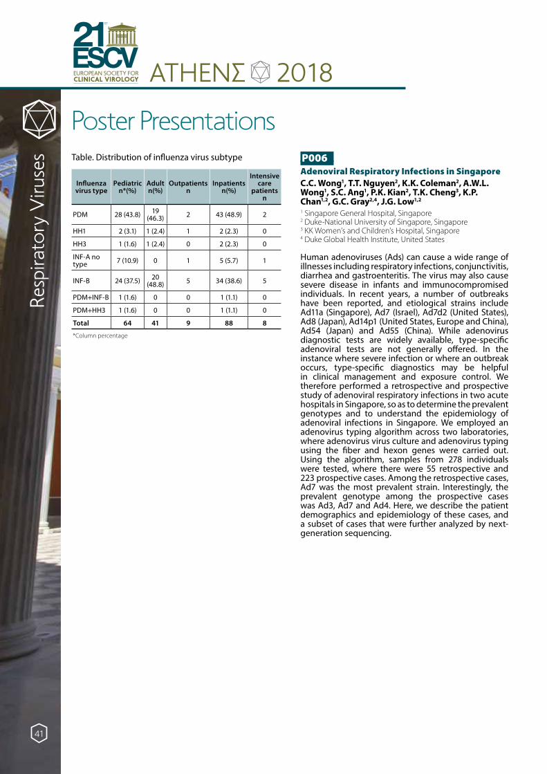
ATHENΣ 2018EUROPEAN SOCIETY FORCLINICAL VIROLOGY
21ESCV
ST
Poster Presentations
Resp
irato
ry V
iruse
s Table. Distribution of influenza virus subtype
Influenza virus type
Pediatric n*(%)
Adult n(%)
Outpatients n
Inpatients n(%)
Intensive care
patients n
PDM 28 (43.8) 19 (46.3) 2 43 (48.9) 2
HH1 2 (3.1) 1 (2.4) 1 2 (2.3) 0
HH3 1 (1.6) 1 (2.4) 0 2 (2.3) 0
INF-A no type 7 (10.9) 0 1 5 (5.7) 1
INF-B 24 (37.5) 20 (48.8) 5 34 (38.6) 5
PDM+INF-B 1 (1.6) 0 0 1 (1.1) 0
PDM+HH3 1 (1.6) 0 0 1 (1.1) 0
Total 64 41 9 88 8
*Column percentage
P006Adenoviral Respiratory Infections in SingaporeC.C. Wong1, T.T. Nguyen2, K.K. Coleman2, A.W.L. Wong1, S.C. Ang1, P.K. Kian2, T.K. Cheng3, K.P. Chan1,2, G.C. Gray2,4, J.G. Low1,2
1 Singapore General Hospital, Singapore2 Duke-National University of Singapore, Singapore3 KK Women’s and Children’s Hospital, Singapore4 Duke Global Health Institute, United States
Human adenoviruses (Ads) can cause a wide range of illnesses including respiratory infections, conjunctivitis, diarrhea and gastroenteritis. The virus may also cause severe disease in infants and immunocompromised individuals. In recent years, a number of outbreaks have been reported, and etiological strains include Ad11a (Singapore), Ad7 (Israel), Ad7d2 (United States), Ad8 (Japan), Ad14p1 (United States, Europe and China), Ad54 (Japan) and Ad55 (China). While adenovirus diagnostic tests are widely available, type-specific adenoviral tests are not generally offered. In the instance where severe infection or where an outbreak occurs, type-specific diagnostics may be helpful in clinical management and exposure control. We therefore performed a retrospective and prospective study of adenoviral respiratory infections in two acute hospitals in Singapore, so as to determine the prevalent genotypes and to understand the epidemiology of adenoviral infections in Singapore. We employed an adenovirus typing algorithm across two laboratories, where adenovirus virus culture and adenovirus typing using the fiber and hexon genes were carried out. Using the algorithm, samples from 278 individuals were tested, where there were 55 retrospective and 223 prospective cases. Among the retrospective cases, Ad7 was the most prevalent strain. Interestingly, the prevalent genotype among the prospective cases was Ad3, Ad7 and Ad4. Here, we describe the patient demographics and epidemiology of these cases, and a subset of cases that were further analyzed by next-generation sequencing.
41
ATHENΣ 2018EUROPEAN SOCIETY FORCLINICAL VIROLOGY
21ESCV
ST
Poster Presentations
Resp
irato
ry V
iruse
sP007Epidemiological and molecular investigation of non-seasonal influenza-related viral respiratory infections in Northern Italy during four consecutive influenza seasons (2014-2018)C. Galli, L. Pellegrinelli, G. Anselmi, V. Primache, S. Binda, E. ParianiDepartment of Biomedical Sciences for Health, University of Milan, Milan, Italy
Background: Besides seasonal influenza A and B viruses (IAV and IBV), several pathogens are involved in clinically undistinguished respiratory infections, including respiratory syncytial virus (RSV), enterovirus (EV), parechovirus (PeV) and influenza C virus (ICV).
This study aimed at investigating the contribution of these viruses in influenza-like illness (ILI) in Lombardy (Northern Italy) during four consecutive influenza seasons (2014-2018).
Materials and Methods: 1047 IAV/IBV-negative respiratory swabs collected from ILI outpatients in Lombardy within the Italian Influenza Surveillance Network during four seasons (from 2014/2015 to 2017/2018) were tested to detect RSV, EV, PeV and ICV by specific real-time RT-PCR. Positive samples were sequenced and molecularly characterised.
Results: 19.5% of specimens tested positive to at least one virus: RSV, EV, PeV and ICV contributed to 12.9%, 6.5%, 1.8% and 0.3% of IAV/IBV-negative ILIs, respectively. Multiple virus detection was observed in 2% of cases, most (66.7%) was RSV+EV. Except for ICV that was identified sporadically and only in two seasons, the other viruses were detected in every season: the broadest RSV and EV circulation was in 2016/2017 (15.4% and 8.9%, respectively) and 2017/2018 (15.2% and 8.8%, respectively), while PeV in 2015/2016 (2.5%). These viruses circulated mainly in November/December (RSV, EV, PeV) and February/March (RSV). The highest risk of infection from RSV/EV/PeV was observed in children <4 years. Molecular characterisation is ongoing.
Conclusions: RSV and EV significantly contributed to ILIs, especially among the youngest children. Routine monitoring systems targeting respiratory viruses could be a valuable tool to increase the epidemiological-molecular knowledge of respiratory infections.
P008Respiratory syncytial virus surveillance in the Czech Republic: present and future.L. Novakova, H. Jirincova, D.Trnka, R. Limberkova, J. Kyncl, A. Nagy, M. HavlickovaNational Institute of Public Health, Prague, Czech Republic
Introduction: Respiratory syncytial virus (RSV) has long been monitored in the Czech Republic within ARI sentinel surveillance. However, in view of the current WHO criteria, this system is not sufficient to meet the needs for a complex RSV surveillance including hospitalized patients and assessment of vaccination outcomes.
Material and Methods: The sentinel surveillance requires 20-50 swabs to be collected per week according to the following criteria: acute upper airways infection (J00, J02, J04, J05, J06) and influenza (J10.1, J10.8, J11.1, J11.8). Patient history and clinical data are available. The detection of RSV is performed using multiplex RT-qPCR assay.
Results: During five epidemic seasons (2013/2014 - 2017/2018), 2622 nasopharyngeal swabs from patients of all ages were tested, with RSV being identified in 89 cases (3.39 %). We repeatedly observed inverse correlation between the RSV and influenza peaks, i.e., the highest incidence of RSV infections was registered during the decline of the influenza wave, usually between calendar weeks 8 and 12. The most afflicted age group were children under five years of age (43/89 - 48.3 %).
Conclusion: The proportion of RSV in the etiology of common ARI included in the surveillance in the Czech Republic reaches 3.39% on average over years, which is less than in other European countries, but only outpatient data are available. Extending the scope of surveillance, adjusting the case definition according to the WHO criteria, targeting specific age groups and hospitalized cases of RSV in line with the SARI definition would improve RSV surveillance in the Czech Republic.
42

ATHENΣ 2018EUROPEAN SOCIETY FORCLINICAL VIROLOGY
21ESCV
ST
Poster Presentations
Resp
irato
ry V
iruse
s P009Phylogenetic characterization of human rhinoviruses from infants in Sarlahi, NepalJ. Kuypers1, G. A. Perchetti1, H. Y. Chu1, A. S. Magaret1, J. Katz2, S. K. Khatry3, S. C. LeClerq2,3, K. R. Jerome1, J. M. Tielsch4, M. C. Steinhoff5, J. A. Englund6
1 University of Washington, Seattle, WA, USA2 Johns Hopkins University, Baltimore, MD, USA3 Nepal Nutrition Intervention Project, Kathmandu, Nepal4 George Washington University, Washington, DC, USA5 Cincinnati Children’s Hospital Medical Center, Cincinnati, OH,
USA6 Seattle Children’s Hospital and Research Foundation, Seattle,
WA USA
Human rhinoviruses (HRV), the most common cause of acute respiratory infections in young children and infants, are highly diverse genetically. The aim of the study was to characterize the HRV genotypes detected with respiratory illness episodes in infants in rural southern Nepal.
Infants born to women enrolled in a randomized trial of maternal influenza immunization in southern Nepal were followed with household-based weekly surveillance from birth until 180 days of age. Infants with respiratory symptoms had swabs tested for twelve respiratory viruses by RT-PCR. A fragment of the VP4/2 gene from a subset of HRV-positive samples was PCR amplified and sequenced to identify HRV genotypes.
Among 582 HRV-only positive specimens collected from December 2012 through April 2014, 285 (49%) were sequenced. HRV-A, B, and C species were detected in 193 (68%), 18 (6%), and 74 (26%) specimens, respectively. Ninety-four unique genotypes were identified from the 285 sequenced samples, including 52 HRV-A, 11 HRV-B, and 31 HRV-C. Multiple species and genotypes circulated simultaneously throughout the study period. No seasonality was observed. The median age at illness onset was 88, 104, and 88 days for HRV-A, B, and C. The median PCR Ct values did not differ between HRV species. No differences between HRV species were observed for reported respiratory symptoms, including pneumonia, or for medical care-seeking.
Among very young, symptomatic infants in rural Nepal, all three species and many genotypes of HRV were identified; HRV-A was detected most frequently. There was no association between HRV species and disease severity.
P010Detection and Characterisation of Measles Virus in Ireland 2017/2018Tuite G.1, Neary M.1, Cotter S.2, Ronayne A.1, De Gascun CF.1, Connell J.11 National Virus Reference Laboratory, University College
Dublin, Belfield, Dublin 42 Health Protection Surveillance Centre, 25-27 Middle Gardiner
St., Dublin 1
Background: Measles is a highly contagious disease, preventable by vaccination. Since early 2017, Europe has experienced significant outbreaks with many cases linked to suboptimal vaccination. This study describes the investigation of suspected measles cases at the National Virus Reference Laboratory (NVRL) Dublin, a WHO National Measles Reference Laboratory, during 2017/2018.
Methods: Samples (oral fluids, throat swabs, sera and urine) from suspected cases were primarily investigated for measles RNA using an RT-PCR assay that amplifies the Nucleoprotein (N) and the Haemagglutinin (H) measles genes and includes β2M detection as a sample quality control. N450 sequencing was used for genetic characterisation. IgM detection was also used for serum samples and reflex testing for oral fluid.
Results: The NVRL tested 718 samples for measles RNA and 1265 specimens for measles IgM. Sixty nine measles cases were identified with median age 18 years (range 7 months - 57 years). Twenty cases had concordant positive serological and molecular tests. Fifty-seven cases (82.6%) had measles RNA detected, 51 (89%) of which were genotyped. All cases in 2017 were genotype B3 while 97% of those in 2018 were genotype D8. The index cases in all outbreaks had epi-links to European measles cases and most cases had no, or incomplete, vaccination against measles.
Conclusions: These data show the importance of rapid laboratory investigation to detect and genotype cases, to epi-link cases, and to discriminate between endemic transmission and imported infection. This provides valuable information to target vaccination policies.
43
ATHENΣ 2018EUROPEAN SOCIETY FORCLINICAL VIROLOGY
21ESCV
ST
Poster Presentations
Resp
irato
ry V
iruse
sP011Epidemiology of WUPyV among hospitalized children with respiratory tract infections in Beijing, China L.S. ZhengNational Institute for Viral Disease Control and Prevention, Chinese Center for Disease Control and Prevention, Beijing, China
As one of novel Human Polyomaviruses (HPyVs), WU Polyomaviruse (WUPyV) was discovered in nasopharyngeal aspirates (NPA) of children with respiratory tract infections (RTI) in 2007, and the link between WUPyV and respiratory diseases remains speculative. Thus, we investigated the epidemiology of WUPyV in children with RTI in China from 2017 to 2018. 1232 NPA samples were collected from hospitalized children aged ≤14 years old with RTI in Beijing Friendship Hospital. VP1 gene of WUPyV was detected with an established TaqMan real-time PCR and confirmed by sequencing. To assess the prevalence of co-infection with WUPyV, all the WUPyV-positive specimens were screened for 15 other common respiratory viruses. The patient demographic and clinical data were reviewed. Of the 1232 children (male 651, female 581) with RTI included in the study, the prevalence of WUPyV was 6.25% (77/1232), there was no significant difference between genders (6.76% vs. 5.68%, P = 0.44) and there is no obvious seasonality. The WUPyV infected children ranged in age from 2 month to 13 years and children ≤5 years of age accounted for 93.5% (72/77) of cases. The most common symptoms were cough, fever and asthma. Additionally, most of WUPyV-positive patients were diagnosed with bronchopneumonia. 46 WUPyV-positive patients were coinfected with other respiratory viruses, of which human rhinovirus were most common. In conclusion, WUPyV was common in hospitalized children with RTI, and the infection might occur early in life. Further serological study and case-control studies were needed to clarify the link between WUPyV and RTI.
P012Classifying viral respiratory infections through human transcriptome analysis on a rapid, easy-to-use, molecular platformR.J. Crisp1, C. Gritzen1, T. Wilson1, J. Nawrocki1, M. Deneris1, C.L. Baird1, E.M. Ott1, J. Jones1, J. Bastar1, H. Kim1, S. House2, D. Cohen3, A. Leber3, A.C. Hemmert1
1 BioFire Diagnostics, LLC, Salt Lake City, UT, USA2 Washington University School of Medicine, St. Louis, MO, USA3 Nationwide Children’s Hospital, Columbus, OH, USA
More than 50% of patients reporting to the emergency department (ED) for acute respiratory illness (ARI) are prescribed antibiotics, although most respiratory infections are viral in origin. A novel approach to improve antibiotic stewardship in ARI patients is unbiased human transcriptome analysis, where differential gene expression, measured in blood, is used to identify classes of infection such as viral and/or bacterial.
Here we report results of an RUO rapid, easy-to-use, multiplex PCR test that measures the transcriptome response to a viral/bacterial infection in ARI patients reporting to the ED. Following an IRB approved protocol, blood was collected from patients with signs and symptoms consistent of an ARI. This sample, collected in an RNA-stabilizing reagent, was tested using custom BioFire pouches, which contained 42 assays targeting differentially-expressed mRNA transcripts to distinguish viral and bacterial infections. Assay results were normalized and the probabilities of each infection class were independently determined.
In 200 samples, the BioFire test was 86% accurate (CI95% 80%, 91%) in classifying viral infections relative to direct pathogen multiplex PCR with an area under the receiver operator curve of 0.87 (CI95% 0.76,0.95). For several samples, strong viral probabilities were observed although the comparator method identified no pathogen. These could be from viruses not present in the direct-pathogen test and require further investigation.
These results show that viral classification through transcriptome analysis performs similarly to current direct pathogen PCR methods. Additional research is needed to understand the bacterial ARI transcriptome response and how these results impact antibiotic stewardship in outpatient settings.
44
ATHENΣ 2018EUROPEAN SOCIETY FORCLINICAL VIROLOGY
21ESCV
ST
Poster Presentations
Resp
irato
ry V
iruse
s P013Presence of respiratory viruses in ICU patients with community acquired-pneumonia (CAP): a one-year retrospective single center studyM. Giry1, B. Misset1, D. Boyer1, A. Baron2, L. Lemée2, J.C. Plantier2, M. Gueudin2
1 Rouen University Hospital, Medical ICU, F-76000, Rouen, France
2 Rouen University Hospital, Department of Microbiology, F-76000, Rouen, France
Background: Pneumonia is the most frequent community-acquired infection responsible for ICU admission. Rapid multiplex PCR enables early diagnosis of respiratory infection in daily routine testing. Few studies have been described since this technique has been made available in routine.
Objectives: We aimed at assessing the prevalence and distribution of pathogens among ICU patients with community-acquired pneumonia (CAP) and the relationship of severity and outcome.
Methods: During November 2016 to October 2017, 223 patients were sampled within 72h after admission to ICU and tested for 24 pathogens using the sample to answer ePlex Respiratory Pathogen Panel which delivered results in 1.5 hours. Patients with a diagnosis of CAP were split into 4 groups according to causal agent: none, viruses, bacteria, combination of both. Comparisons were made with Kruskall-Wallis test.
Results: A total of 109 patients had CAP, 39 aspiration/opportunistic pneumonia, 22 non-pulmonary infections, 11 pulmonary edema, 19 exacerbations of chronic lung disease, and 24 other diagnoses. Patients with CAP had the following characteristics: age 60+/-16 y, male sex 60.2%, length of stay (LOS) in ICU 8.7+/-9.0 days, mortality 10.1%. All received antibiotics. No infectious agent was found in 32 patients, whereas a virus was detected in 28 patients, bacteria in 33 and both in 16. The LOS in ICU was 5.2 (no agent), 7.7 (virus), 10.1 (bacteria) and 14.8 (mixed) days respectively (p=0.040). Mortality was similar among groups.
Conclusion: In our ICU population, 55% of CAP had no infectious agent or respiratory viruses without bacteria. Patients with coinfections had longer LOS.
P014Estimating the burden of human coronaviruses during the last 5 seasons at National Influenza Centre SloveniaN. Berginc, K. ProsencNational Laboratory of Health, Environment and Food, Slovenia
Human coronaviruses(HCoVs) cause acute respiratory infections(ARI) of various severity. At National Influenza Centre Slovenia data were analysed to estimate burden and epidemiology.
From weeks 40/2013-20/2018, 12852 nasal/throat swabs from patients with ARI, their personal, clinical data were collected all-year-round from 50 primary healthcare clinics and 2 hospitals. Nucleic acids were extracted. Multiplex-RT-RT-PCRs were used to detect influenza and other respiratory viruses, including HCoVs (229E, OC43, NL63, HKU1). Age groups(AGs) were defined: /0-2/3-6/7-14/15-19/20-64/≥65/ years old(YO).
7,4% of specimens tested for HCoVs were positive. Majority of infections were in infants, young children and elderly (29%, 15%, 24% in 0-2YO, 3-6YO, ≥65YO respectively). Most of them were hospitalized (84%, 76%, 98% in 0-2YO, 3-6YO, ≥65YO respectively). In other AGs hospitalisation was lower (64%, 53%, 56% in 7-14YO, 15-19YO, 20-64YO respectively).
All AGs reported high rates of cough (63%-91%), fever (38%-78%). Breathing difficulties were reported with significant rates (17%-38%) in 0-2YO, 3-6YO, 7-14YO, 15-19YO, 20-64YO, but higher (65%) in elderly. Bronchiolitis was reported in 3%-13% of 0-2YO, 3-6YO, 7-14YO, 15-19YO, 20-64YO and higher (16%) in elderly. Pneumonia was reported in 2%-10% of 0-2YO, 3-6YO, 20-64YO and elevated (25%) in elderly.
HCoVs present high burden in infants and young children. Burden is elevated in elderly with higher rates of hospitalization, breathing difficulties, bronchiolitis, pneumonia in comparison to other AGs.
Seasonal circulation of HCoV was observed (December-April). All types circulated simultaneously, but usually two types predominated: in 2013/2014, 2015/2016, 2017/2018, 229E and HKU1, in 2014/2015, 2016/2017, OC43 and NL63. This data show biannual peaks of types.
45
ATHENΣ 2018EUROPEAN SOCIETY FORCLINICAL VIROLOGY
21ESCV
ST
Poster Presentations
Resp
irato
ry V
iruse
sP015Antiviral effect of an essential oil derived from three aromatic plants against viruses causing infections of the upper respiratory systemM. Tseliou1, SA. Pirintsos2,3, C. Lionis4, E. Castanas5, G. Sourvinos1
1 Laboratory of Clinical Virology, Medical School, University of Crete, Heraklion, Crete, Greece
2 Department of Biology, School of Sciences and Technology, University of Crete, 71003, Heraklion, Greece
3 Botanical Garden, University of Crete, 74100, Rethymnon, Greece
4 Clinic of Social and Family Medicine, School of Medicine, University of Crete, P.O. Box 2208, 71003, Heraklion, Greece
5 Laboratory of Experimental Endocrinology, School of Medicine, University of Crete, 71003, Heraklion, Greece
An essential oil extract based on three aromatic plants (Coridothymus capitatus L, Origanum dictamnus L and Salvia fruticosa Mill, in extra-virgin olive oil, herein denoted as CAPex) has been shown to reduce the duration and severity of symptoms of patients with upper respiratory tract viral infections. Here, we tested whether CAPex exhibits a direct antiviral activity against a wide range of respiratory viruses in vitro. Our study revealed a remarkable antiviral activity of the extract against influenza A/H1N1 virus strains, influenza B and human rhinovirus 14 (HRV14) whereas no viral inhibition was found for influenza A/H3N2, Respiratory Syncytial Virus (RSV) and Adenovirus 5. Both influenza A/H1N1 and HRV14 replication and progeny virus were significantly decreased after the treatment with CAPex. Pre-treatment with the plant extract demonstrated that CAPex exerts its antiviral activity after A/H1N1 or HRV14 entry in host cells whereas it confers a preventive reactivity against RSV. Furthermore, CAPex resulted in a defective trafficking of influenza A Nucleoprotein, suggesting NP as a valid target of this extract. We conclude that the extract possesses antiviral activity and has the potential to be used as an herbal agent against influenza viruses and rhinovirus.
P016Unexpected dominance of influenza type B during the 2005/2006 and 2017/2018 epidemic seasons in the Czech RepublicM.Havlickova, H.Jirincova, D.Trnka, J.Kyncl, R.Limberkova, L.Novakova, A.NagyNational Institute of Public Health, Prague, Czech Republic
Introduction: A typical pattern of an influenza season is defined by a predominance of influenza A accompanied by a minor fraction of influenza B. The altered seasonal dominance is very seldom. Here we present clinical and virological data from two influenza seasons with influenza B dominance: 2005/2006 (B/Victoria-like) and 2017/2018 (B/Yamagata-like).
Material/methods: Sentinel physicians collect swabs from their ILI/ARI patients between week 40 of each year and week 20 of the following year. Since 2009, non-sentinel data and samples have been routinely collected from all individuals hospitalized with severe ARI. Laboratory diagnosis is based on virus isolation, indirect immunoperoxidase assay, and/or PCR.
Results: Outpatients with influenza type B/Yamagata (147 patients) and influenza type B/Victoria (119 patients) were compared for the following symptoms: fever, weakness, rhinitis, cough, breathlessness, chest pain, headache, abdominal pain, muscle pain, vomiting and diarrhoea, conjunctivitis, tonsillitis, tracheitis, bronchitis, and rush. Influenza type B/Yamagata cases in comparison with influenza type B/Victoria showed a statistically significantly higher rates of tracheitis (38.10 % vs. 26.05 %, p=0.048), chest pain (28.57 % vs. 15.97 %, p=0.019), breathlessness (22.45 % vs. 5.88 %, p<0.001), and tonsillitis (18.37 % vs. 8.40 %, p=0.021).
Discussion/conclusion: The clinical course of influenza can vary considerably in terms of symptoms depending on virus type/subtype and currently circulating variant. The analysis of symptoms in hospitalized patients and outpatients will be of relevance to the differential diagnosis of ARI and epidemiological surveillance.
Supported by the Ministry of Health, Czech Republic - conceptual development of research organization (NIPH, IN 75010330).
46
ATHENΣ 2018EUROPEAN SOCIETY FORCLINICAL VIROLOGY
21ESCV
ST
Poster Presentations
Resp
irato
ry V
iruse
s P017Comparison of throat swabs, oral fluid collection devices (Oracol) and FTA® cards for the molecular detection and genotyping of measles virusB. Bankamp*1, C. Sein2, E. Pukuta Simbu3, R. Anderson1, E. Abernathy1, M. Chen1, K. A. Wannemuehler2, D. Waku1, E. N. Lopareva1, J. P. Icenogle1, P. A. Rota1, J. L. Goodson2
1 Division of Viral Diseases, Centers for Disease Control and Prevention, Atlanta, GA, USA
2 Global Immunization Division, Centers for Disease Control and Prevention, Atlanta, GA, USA
3 Institut National de Recherche Biomédicale, Kinshasa, DR Congo
The genetic characterization of measles viruses is an important tool for measles surveillance. Among the obstacles to genotyping are the reverse cold chain requirements for transportation of samples to reference laboratories and the restrictions placed on shipping infectious material. FTA® cards facilitate transport of virologic samples at ambient temperature as non-infectious material; however, the utility of FTA® cards for detection and genotyping of measles virus from clinical samples had not been evaluated. Throat swabs (TS) and oral fluid samples (OF) were collected from 238 suspected measles cases in the Democratic Republic of the Congo. Virus detection by RT-qPCR and genotyping were compared for samples that were either transported using the reverse cold chain or on FTA® cards. Virus detection by RT-qPCR showed excellent positive agreement for TS and OF (95.3%, CI [91.6, 97.4]), while the positive agreement for TS and OF on FTA® cards was 79.4% (CI 73.5, 84.3) and 85.5% (CI 80.2, 89.6) respectively, compared to TS or OF. Based on genotyping results obtained for a subset of samples, an estimated 77.3% of all TS samples and 71.0% of OF samples would have had sufficient viral loads for genotyping, compared to 41.6% of TS and 41.3% of OF samples on FTA® cards. Similar results were found for a small subset of 16 measles-negative samples that were serologically positive for rubella infection. In outbreak settings, FTA® cards can be used to transport virologic samples if the reverse cold chain is not available; however, this method has limited utility for transportation of virologic samples of sporadic cases of measles.
P018Molecular detection of fifteen respiratory viruses in hospitalized children - first year results of four-year prospective study from CroatiaS. Ljubin-Sternak1,2, I. Ivković-Jureković3,4, M. Mijac1, I. Kale2, D. Slacanac2, B. Kolarić1,5, A. Lukić-Grlić2,6, J. Vraneš1,2
1 Clinical Microbiology Department, Teaching Institute of Public Health “Dr Andrija Štampar”, Croatia
2 Medical Microbiology Department, School of Medicine, University of Zagreb, Croatia
3 Department of Pulmonology, Allergy, Immunology and Rheumatology, Children’s Hospital Zagreb, Croatia
4 Pediatric Department, School of Medicine, University of Osijek, Croatia
5 Epidemiology Department, Faculty of Medicine, University of Rijeka, Croatia
6 Clinical Microbilogy Department, Children’s Hospital Zagreb, Croatia
Background: To determine the viral incidence, and clinical significance of viral detection in hospitalized children with respiratory tract infection (RTI), four-year prospective study was started in March 2017.
Material and Methods: During one-year period, a total of 239 children aged from one week to 18 years admitted to the Children’s hospital Zagreb with RTI of suspected viral aetiology were included. Nasopharyngeal swabs were collected and tested for the 15 most common respiratory viruses. Multiplex PCR and cDNA synthesis in one-step reaction, followed by detection of PCR amplicons using microchip electrophoresis was performed.
Results: Viral aetiology was proved in 75.3 % of the patients. The median age of children with detected respiratory virus was 3.5 years, and male to female ratio 1.6:1. The highest positive detection rate was recorded in the 3 to 5 years old group of children. Ninety-six patients showed symptoms of upper RTI, and 84 had symptoms/signs of lower RTI. A single virus was diagnosed in 67.2 % of the patients, while coinfection with two and three or more viruses in 25 % and 7.8% of the patients, respectively. The most commonly detected virus was rhinovirus (56.1%), followed by adenovirus (24.4%), respiratory syncytial virus A and B (17.7%), coronaviruses 229/NL63 and OC43 (11.1%), influenza viruses A and B (7,7%), human bocavirus and enteroviruses with equal frequencies (7.2%), parainfluenza viruses 1-4 (6.6%), and metapneumovirus (5.5%).
Conclusion: Observed incidence of some respiratory viruses was related to the age of the patient, the localization of the infection and the season.
47

ATHENΣ 2018EUROPEAN SOCIETY FORCLINICAL VIROLOGY
21ESCV
ST
Poster Presentations
Resp
irato
ry V
iruse
sP019Etiology and seasonality of viral respiratory tract infections in Antalya, TurkeyO.M. Parkan1, A.E. Daloglu1, B.O. Peker1, I. Saglik2, O. Koyuncu Ozyurt3, D. Mutlu1, D. Colak1
1 Division of Virology, Department of Medical Microbiology, Akdeniz University Medical Faculty, Antalya, Turkey
2 Department of Medical Microbiology, Uludag University Medical Faculty, Bursa, Turkey
3 Department of Medical Microbiology, Akdeniz University Medical Faculty, Antalya, Turkey
The aim of this study is to investigate the etiology and seasonality of viral respiratory tract infections over a 12-month period retrospectively.
Nasopharyngeal swabs were collected between May 2017 and April 2018 and analysed by using Verigene® Respiratory Pathogens Flex Nucleic Acid Test (Nanosphere, Northbrook, IL). Patients were not included a second time within 30-days if they were repeatedly negative, or positive with the same virus. Patients were stratified into six age groups: <1 year, 1-5 years, 6-18 years, 19-45 years, 46-64 years and ≥65 years.
A total of 2061 specimens from 1921 patients were tested. Of the specimens, 1223 (59,3%) were from pediatric patients. Positivity rate was higher in children (62,1%) than adults (46,5%) (p<0,01). Children aged between 1-5 years had the highest positivity rate (65,4%). Co-infections were found to be more common in children (14,4%) than adults (5,8%) ( p<0,01).
Rhinovirus (RV) was found to be the most common pathogen in all age groups and it was followed by Respiratory Syncytial Virus (RSV) in <1 year of age and Adenovirus (AdV) in other pediatric age groups. In adults, Influenza viruses were found to be the second most common agents.
RV was the most prevalent virus all year long except for June and July when AdV was more common and also in September when Parainfluenza viruses (PIVs) were more frequently detected. Influenza B, RSV, and Human Metapneumovirus had seasonal peaks in February, while Influenza A, AdV, PIVs, and RV had their peaks in January, June, September, and October respectively.
P020Investigation of respiratory infections with the film Array RP in adults and children G. Chronopoulou, C. Vossou, A. Kouki, E. PagonidouCentral Laboratories of Athens Euroclinic Hospital, Greece
Introduction: Transmission of viral infections of the respiratory system is particularly high during the winter months due to the cohabitation of population in enclosed spaces. Viruses commonly implicated are influenza viruses A and B, parainfluenza viruses, corona viruses, respiratory syncytial virus (RSV), human metapneumovirus (hMPV) and adenoviruses. The severity of the clinical picture and the infestation of the lower respiratory course vary.
Objective: Contribution of the Film Array system Multiplex PCR in the rapid diagnosis of the most common pathogens involved in hospitalized children and adult respiratory infections.
Material and methods: Material of the study was 453 patients (340 adults and 113 children) admitted to our hospital from October 2017 to April 2018, with respiratory infection symptoms (fever, cough, breathing difficulties etc.) From all patients nasopharyngeal swab specimens were examined using respiratory panel of the Film Array System, which detects 17 viruses and 3 bacteria in about 1 hour.
Results: During the study period out the 453 patients samples tested, (60.04 %) proved positive. Viruses that were detected more frequently in children were rhino/enteroviruses (40.5%) followed by parainfluenza (18%) and adenovirus (16%). Only (7.5%) of the samples were positive for influenza B, as well as RSV and no hMPV was detected. In contrast, in adults (35.5%) were positive for Influenza B followed by rhino/enteroviruses (22%) and hMPV (11.5%). Mixed overlapping infections were more common in children than in adults (29% vs 20%).
Conclusions: The increased incidence of viral respiratory infections during the winter months requires an accurate method to achieve effective therapeutic approach and limitation of the transmission. The automated Film Array system ensures rapid detection of respiratory pathogens promoting initiation of targeted treatment, antibiotic stewardship and hygiene measures in hospital setting.
48
ATHENΣ 2018EUROPEAN SOCIETY FORCLINICAL VIROLOGY
21ESCV
ST
Poster Presentations
Resp
irato
ry V
iruse
s P021Rapid detection of respiratory pathogens with the ePlex multiplex platformT. Chane-Teng1 & L. Avramis1, C. Dimeglio1, J Izopet1,2, C. Mengelle1, J-M Mansuy1
1 Department of Virology, Federative Institute of Biology, CH Toulouse, France
2 Inserm U1043, CPTP, CH Toulouse, France
Aim: The purpose of this survey is to evaluate two multiplex assays for the detection of respiratory pathogens and analyze the potential impact of rapid diagnosis on flu treatment.
Material and methods: Nasopharyngeal samples collected from adults (n=93) attending the emergency care unit for acute respiratory syndrome (mean age 67.5) were tested routinely with the Allplex™ Respiratory Full Panel Assay (Seegene) that detects 16 viral targets, including Influenza A/B and RSV A/B.
Samples were also tested with the ePlex® Respiratory Pathogen Panel (Genmark) , detecting the same viral targets as Allplex Assay plus two viruses and four bacteria (B. pertussis, L. pneumophila, M. pneumoniae and C. pneumoniae) in a single cartridge.
Results: Concordances were 91.3% and 93.5% for Influenza (A/B) and RSV (A/B) detection and 91.4 % for the other viruses. Sixteen samples gave discordant results: four were ePlex® positive/Allplex™ negative including one sample positive for M. pneumoniae.
Nine samples gave invalid ePlex® results. There were two positive samples, one for Influenza A and one for Human Metapneumovirus, and seven negative samples according to Allplex™ RFP assay.
The ePlex® results were more rapidly available than those of Allplex™ (four vs 36 hours).
There were no statistical differences for the prescription of Tamiflu according to the Influenza detection: p=0.42 and p=0.25 for ePlex® and Allplex™ assays respectively.
Conclusion: Large multiplex panels are very useful for the rapid detection of respiratory pathogens. The cost remains elevated but it could be potentially counterbalanced by the reduction of patient’s care expenses.
P022Biennial outbreak pattern of enterovirus D68 in NorwayJ.L. Dembinski1,2, N. Milhano1,2, A. Christensen3, A.M.B. Kran4, T. Kofstad5, N.E. Nyquist6, T.B Riste7, U. Houge8, S.H. Hartzen9, N. Grude10, S. Dudman1,2
1 Domain for Environmental Health and Infectious Disease Control, Norwegian Institute of Public Health, Oslo, Norway
2 WHO National Polio Entero Reference Laboratory3 St. Olav’s Hospital, Trondheim, Norway 4 Department of Microbiology, Oslo University Hospital, Ullevål,
Norway 5 Department of Microbiology, Drammen Hospital, Vestre Viken
Hospital Trust, Norway 6 Department of Microbiology and Infection control, Akershus
University Hospital, Norway 7 Department of Microbiology, Helse Førde Health Trust,
Norway 8 Department of Microbiology Sørlandet Hospital, Norway 9 Department of Microbiology, Innlandet Hospital Trust,
Norway 10 Department of Microbiology, Vestfold Hospital Trust, Norway
Background: Enteroviruses (EV) cause a wide variety of human infections, from mild respiratory illnesses to severe neurological diseases. In the autumn of 2014 a high number of EV-D68 infections were observed among children with severe respiratory infections in the US, Canada and Europe, many of which presented with acute flaccid paralysis (AFP). The aim of this study was to monitor the circulation of EV-D68 from 2015 to 2018, through enhanced surveillance of EV-D68 in Norway, and collect clinical data from positive patients for analysis of potential associations with AFP.
Material and methods: We collected data from the main laboratories performing EV diagnostics in Norway, with a focus on EV-D68, from nine laboratories in 2015, six in 2016, three in 2017, and three ongoing surveillance in 2018.
Results: In 2015, only three cases of EV-D68 were detected, none of which presented with AFP, followed by a sharp increase to 302 cases in 2016, three of which were associated with AFP. In 2017, only four EV-D68 cases were detected, with no AFP association. Surveillance is currently ongoing in 2018, with an expected increase of cases in autumn.
Conclusions: The surveillance revealed a biennial outbreak pattern of EV-D68 virus. The findings in this study highlight the importance of continued EV surveillance, particularly towards EV-D68 given its impact and significance on children’s’ health, to appropriately guide specific public health recommendations.
P023Development and characterization of an Aerodynamic system for pulmonary delivery of Influenza vaccineWithdrawn by the author
49
ATHENΣ 2018EUROPEAN SOCIETY FORCLINICAL VIROLOGY
21ESCV
ST
Poster Presentations
Resp
irato
ry V
iruse
sP024Respiratory Syncytial Virus Genotypes Circulating Amongst Children and Elderly in SloveniaN. Volmajer, M. Cimerman, B. Blažič, K. Soršak, A. Golle, D. DuhNational Laboratory of Health, Environment and Food, Department for Medical Microbiology Maribor, Prvomajska 1, 2000 Maribor, Slovenia
Respiratory Syncytial Virus (RSV) is classified into A and B antigenic groups, which correlate with at least 12 genotypes of RSV-A and 23 genotypes of RSV-B. Although molecular characterization reveals considerable genetic diversity of RSV, usually one genotype within RSV-A and RSV-B predominate in the pediatric population. In recent years, RSV became important pathogen of elderly patients. The information about RSV genotypes in elderly is however scarce. We describe RSV genotypes circulating amongst children and elderly in two consecutive years in Slovenia. RSV was detected with FILMARRAY® Respiratory Panel in 284 samples from 2016 and 2017. Positive respiratory swabs from children up to 2 years (192) and elderly aged >65 (35) were selected for genotyping. Viral RNA was extracted from 144 stored swabs. Semi-nested PCR was used to amplify C-terminal region of G protein. Amplicons were Sanger sequenced and analyzed with DNASTAR Lasergene software. Finally, we obtained 45 nucleotide sequence contigs. We classified 19 as RSV-A: 15 from children and 4 from elderly. All but one sample (GA2 genotype) of RSV-A clustered with the reference ON1 genotype (similarity >95%), which is currently the dominant RSV-A genotype worldwide. The single GA2 genotype was from an infected elderly diagnosed in the beginning of 2016. 26 samples were classified as RSV-B: 14 from children and 12 from elderly. All of RSV-B samples clustered with BA genotype having the highest similarity with BA10 strain (>94%). The same RSV genotypes circulate in children and elderly, but elderly appear to be more often infected with RSV-B.
P025UK NEQAS Microbiology: First year review of the Molecular Detection of Respiratory viruses EQA schemeS. Whitmore1, B. Kele1, E. Elenoglou1, H. Seyedzadeh1, and S. Rughooputh1
UK NEQAS Microbiology, UK
Introduction: The UK NEQAS Microbiology Molecular detection of respiratory viruses EQA scheme was introduced in April 2016. This is a review of participant results after the first year of introduction.
Materials and Methods: 300 participant results were reviewed from the first 3 distributions (4095, 4167 and 4227) distributed between May 2017 and January 2018.
Results: A median consensus of 96.9% was identified for Influenza A intended specimens, 97.2% Influenza B, 97.5% RSV, 91.5% rhinovirus, 94.0% adenovirus and 96.4% parainfluenza 2. 93.8% of participants are typing Influenza A positive specimens, with 0.42% reporting incorrect typing results. Incorrect typing results were identified for H3N2 specimens (2/2) and 0/2 on H1N1 specimens. An incorrect virus was reported on 8/12 specimens and identified on 27 occasions (9%). Influenza A was incorrectly reported 6/27 (22.22%), with majority (66.7%) occurring on an influenza B intended result. Influenza B and enterovirus were both reported incorrectly 4/27 (14.81%). False negative results were reported on 16 occasions (5.53%) with 62.5% observed when the intended result was influenza A, B or RSV. An additional virus to the intended result was reported on 8/12 specimens and occurred on 27 occasions (9%). Rhinovirus (7/27), influenza B (7/27) and enterovirus (5/27) were the most common additional viruses reported.
Summary: Overall an excellent consensus to the intended results was identified. Participants are typing Influenza A positive specimens and reporting this correctly.
50
ATHENΣ 2018EUROPEAN SOCIETY FORCLINICAL VIROLOGY
21ESCV
ST
Poster Presentations
Resp
irato
ry V
iruse
s P026Genetic characterization of respiratory syncytial virus circulating in Lyon, France, between 2010 and 2018A. Gaymard1,2,*, M. Pichon1,2, D. Barthelemy1, Q. Semanas1, B. Simon1, S. Etievant1, E. Frobert1,2, M. Valette1, M. Bouscambert1, G. Billaud1, JS. Casalegno1,2, B. Lina1,2, L. Josset1,2, F. Morfin1,2
1 Hospices Civils de Lyon, Laboratoire de Virologie, Institut des Agents Infectieux (IAI) de Lyon, Centre National de Référence des virus Respiratoires France Sud, Centre de Biologie Et de Pathologie Nord, Groupement Hospitalier Nord, F-69317 Lyon, France
2 Université de Lyon, Virpath, CIRI, INSERM U1111, CNRS UMR5308, ENS Lyon, Université Claude Bernard Lyon 1, F-69372 Lyon, France
Background: Respiratory syncytial virus (RSV) is a well-recognised cause of respiratory tract infections in young children. These viruses are divided into two groups, A and B, based on antigenic variations in the G-glycoprotein. This surface protein can sustain large genetic variations leading to the emergence of new genotypes. So far, 11 RSV-A genotypes and 38 RSV-B genotypes have been described. During the 2010-2011 winter season, a novel genotype of RSV-A, named ON1, with a 72 nucleotides duplication in the G-gene was firstly detected in Ontario, then spread worldwide. This study aimed to investigate the RSV molecular epidemiology in Lyon, France between 2010 and 2018.
Methods: Between 2010 and 2018, 58,953 samples were routinely screened for RSV by PCR using Respiratory MWS RSV/hMPV r-gene® (bioMérieux). Among the RSV-positive cases, 300 samples were further investigated for phylogenetic and genotype characterisation by sequencing (second hypervariable region of the G-gene).
Results: In this study, 5,914 samples (10%) were tested positive for RSV. RSV-A was predominant during six winter seasons. RSV-B proportion ranged from 15% to 61% and was predominant only during the 2016-2017 winter. The ON1 variant was first detected during the 2010-2011 winter then reached 100% of all RSV-A-positive cases in 2014-2015. Almost all RSV-B were classified as BA9 or BA10 genotype.
Conclusion: RSV is characterized by a constant evolution of circulating genotypes. As for most countries, ON1 variants are now the predominant genotype in Lyon, France. Implementation of RSV molecular epidemiology surveillance is relevant as RSV vaccine will soon become available.
P027Production of a quality control reagent for the molecular detection of Enterovirus D-68C. Santirso-Margaretto, M. Madjumar, T. Wilton, B. Valdazo Gonzalez, J. Martin, R. Anderson and N. AlmondNational Institute of Biological Standards and Control (NIBSC), Blanche Lane, South mimms, Potters Bar, Hertforshire, EN6 3QG, UK
Enteroviruses are implicated in a diverse set of clinical manifestations from mild febrile illness to severe neurological complications. NIBSC produces reference materials that contribute to ongoing monitoring of assay performance in clinical diagnostic laboratories. We have developed an IVD-CE marked lyophilised control reagent for molecular diagnostic assay for Enterovirus D-68.
A fully sequenced clinical isolate of Enterovirus D68 was lyophilised in a universal buffer (10mM Tris-HCl pH=7.4), 0.5 mM EDTA and a combination of 2% trehalose - 4% mannitol. Target stability was measured by qPCR comparison of pre- and post-lyophilised formulations and homogeneity of lyophilised material was evaluated by measuring inter-vial dispersion by standard deviation (SD).
In-house evaluation of candidate material was conducted against three different IVD-CE marked commercial panels and external performance evaluation was evaluated by a collaborative study involving five different clinical diagnostic laboratories.
All participants received two vials and were asked to reconstitute the material and then process it as a clinical sample. Each laboratory performed a different process of nucleic acid extraction and amplification and targeted 5’ UTR and VP1.
Data returned highlighted a large inter-laboratory variability and confirmed the suitability of the material as external quality control for all assay applied. A larger preparation of lyophilised material is currently ongoing and NIBSC is looking for laboratories to participate in a phase II collaborative study.
This initial pilot evaluation confirms the suitability of the lyophilised candidate material as a working run reagent for daily monitoring of assay performance as a reagent for external proficiency testing panels.
51
ATHENΣ 2018EUROPEAN SOCIETY FORCLINICAL VIROLOGY
21ESCV
ST
Poster Presentations
Resp
irato
ry V
iruse
sP028Evaluation of geneLEAD VIII (GLVIII) system for viral respiratory infectionsM. Pichon1,2,3*, A. Balandine1, M. Valette1,2, G. Billaud1, B. Lina1,2,3, F. Morfin1,2,3
1 Laboratoire de Virologie, Infectious Agents Institute, Hospices Civils de Lyon, Lyon, France
2 National Reference Center for Respiratory viruses, Infectious Agents Institute, Hospices Civils de Lyon, Lyon, France
3 Lyon University, Virpath, CIRI, INSERM U1111, CNRS UMR5308, ENS Lyon, Université Claude Bernard Lyon 1, F-69372 Lyon, France
Viruses are the main cause of respiratory infections. Our study compared performances of real-time RT-qPCR searching for major viral respiratory pathogens on the geneLEAD VIII (GLVIII) system, using the 3600-FluRSV real-time PCR assay kit (Influenza virus A (IAV) and B (IBV), or Respiratory Syncytial virus (RSV)) to our standard of care process (MWS r-gene® (BioMérieux)). Concordances (κ), sensitivity (Se), specificity (Sp) were determined.
Assays were retrospectively performed on -80°C-freezed respiratory samples (141 nasal swabs (NS), and 141 nasopharyngeal aspirates (NPA)) collected during 2017-2018 winter season, from patients hospitalized in the Hospices Civils de Lyon, France. Samples with discordant results were tested with a 3rd method: a homebrew PCR method for RSV (published method) and the FluA/B CDC recommended method. Our referential consider concordance of ≥2 molecular testing specific of each viral target, or the result of the third method if needed.
Performances could be obtained for each viral target after retrospective testing. On NPA, Se, Sp, and κ were: i) 100%, 100%, and 1 for IAV; ii) 100%, 100%, and 1 for IBV; iii) 89,47%, 100%, and 0.93 for RSV. On NS, performances were: i) 100%, 100%, and 1.00 for IAV; ii) 100%, 96,77%, and 0.98 for IBV; iii) 100%, 96,40%, and 0.92 for RSV.
With 4 false negative and 4 false positive results out of 282 samples tested, performances for RSV detection are very good but remain perfectible. With only 1 false negative result, performances of GLVIII were very impressive for influenza detection.
P029Entero/rhinovirus species in respiratory samples in Belgium in 2017E. Wollants1, A. Rector1, E. Keyaerts1,2 and M. Van Ranst1,2,3
1 Rega institute, Laboratory of clinical & epidemiological virology, KU Leuven, Belgium.
² Department of laboratory medicine, University hospitals Leuven, Belgium.
3 National reference center for enteroviruses, University hospitals Leuven, Belgium
Background: Human enteroviruses and rhinoviruses, both belonging to the genus Enterovirus of Picornaviridae, infect millions of people worldwide each year and have been associated with a wide spectrum of illnesses. Rhinoviruses primarily infect the nose and throat and are the predominant cause of the common cold, whereas enteroviruses mostly infect the enteric tract. Some enteroviruses can infect the respiratory tract and cause respiratory and general symptoms. Enterovirus D68 (EV-D68) is associated with severe respiratory illness and neurological complications, and enterovirus A71 (EV-A71) is one of the major causes of hand foot and mouth disease, and can potentially cause severe neurological disease. We investigated the enterovirus species present in respiratory samples in Belgium in 2017.
Methods: Respiratory samples, including pharyngeal or nose swabs, bronchoalveolar lavages, and bronchial or endotracheal aspirates, that tested positive for rhinovirus/enterovirus at the University Hospital Gasthuisberg, Leuven, Belgium in 2017, were typed. Enterovirus species identification was done by RT-PCR and sequencing of part of the 5’ non-coding region. Molecular typing up to the genotype level was done by sequencing part of the VP1 gene.
Results & Discussion: In total, 134 enterovirus/rhinovirus positive respiratory samples were typed for epidemiological surveillance. As expected the large majority contained rhinovirus species, predominantly Rhinovirus C (RV-C) (56 samples, 41.8%). RV-A and RV-B were present in 39 (29.1%) and 17 (12.7%) samples respectively. Enteroviruses were detected in 22 (16.4%) samples: 11 EV-A (8.2%), 10 EV-B (7.5%) and 1 EV-D. None of the EV-A’s was an EV-A71. The more pathogenic genotype EV-D68 was detected in one sample.
52
ATHENΣ 2018EUROPEAN SOCIETY FORCLINICAL VIROLOGY
21ESCV
ST
Poster Presentations
Resp
irato
ry V
iruse
s P030Predominance of nonrecombinant and recombinant CV-A6 related to hand, foot and mouth disease and herpangina at primary care centers (Barcelona, Spain) during the 2017-2018 seasonAndrés C.1, Fernandes P.1, Piñana M.1, Gimferrer L.1, Guasch E.2, Van Esso D.2, Codina M.G.1, Esperalba J.1, Martín M.C.1, Fuentes F.1, Rubio S.1, Pumarola T.1, Group for EV Surveillance in Paediatric Primary Care2, Antón A.11 Respiratory Viruses Unit, Virology Section, Microbiology
Department, Hospital Universitari Vall d’Hebron, Vall d’Hebron Research Institute, Universitat Autònoma de Barcelona, Barcelona, Spain
2 Primary Care Service Muntanya, Institut Català de la Salut, Barcelona, Spain
Background: Hand, foot and mouth disease (HFMD) and herpangina are usually mild diseases caused by enteroviruses (EVs). Coxsackievirus 6 (CV-A6) has gradually become the predominant EV type causing HFMD, in replacement of EV-A71 and CV-A16. The aim was to describe the genetic diversity of EV causing HFMD and herpangina, especially of CV-A6, from patients attended at paediatric primary care (PPC) in Barcelona (Spain) during the 2017-2018 season.
Material and methods: Pharyngeal swabs were collected for EV laboratory-confirmation by a specific real-time multiplex RT-PCR. Phylogenetic analysis of partial VP1 protein-coding region was performed for EVs genetic characterisation. The complete VP1 and 3Dpol proteins were sequenced for lineage determination and detection of recombination events by phylogenetic analysis. Clinical features were prospectively recorded.
Results: A total of 164 samples were collected, of which a 79% (129) were EV laboratory-confirmed. Phylogenetic analysis (117; 91%) revealed that EVs mainly belonged to EV-A (109; 93%) specie. CV-A6 was the most detected (80; 73%) and mainly associated with HFMD (1) and aHFMD (75/98; 76%), followed by CV-A10 and EV-A71. The complete VP1 sequences revealed that all CV-A6 belonged to lineage D3. The comparison of VP1 and 3Dpol phylogenies showed evidence of recombination in three strains, in which two shifted to CV-A16 3Dpol.
Conclusions: The study provides information regarding the nonrecombinant and recombinant EVs related to HFMD and aHFMD. CV-A6 was the most detected, which showed a high genetic diversity including some recombination events. The likelihood of the emergence of novel recombinant strains highlights the interest to strengthen the virological surveillance in PPC.
P031Incidence of respiratory viruses in a tertiary hospital of the north of SpainM. Larrea Ayo, M. Gallego Rodrigo, P. Iraurgui Arcarazo, O. Martínez Expósito, I. Martínez Rienda, J. Escudero Argaluza, M. Sota Busselo, M. Aranzamendi ZaldumbideUniversity Hospital Cruces, Barakaldo, Spain
Background: Implementation of multiplex molecular respiratory viral panels, raises the paramount to underscore the significance of clinical positive test results. Nonetheless, the role of co-infection, pathogenesis, severity and outcome often remains unclear. Therefore, we aimed to analyze the broad spectrum of respiratory viruses identified from patients with acute respiratory tract infections in a tertiary hospital of the north of Spain.
Materials/methods: Between January 2010 and November 2017, a total of 10.312 patients aged 0-99 years with symptoms of respiratory infection were enrolled into the study. 16 viruses were amplified and detected by CLART® PneumoVir DNA array assay (Genomica, Spain), or one of the combos Seegene Anyplex™ II RV16 /Allplex™ Respiratory Panel Assays (Allplex™ RP 1, 2, 3) (Seegene) (Seegene Inc. Seoul, Korea) plus CFX 96 Real-Time PCR Thermal cycler (Bio-Rad).
Results: RSV (24%), Flu (18%) and Rhinoviruses (5.3%) were found to be the most common pathogens; the first one specially in pediatric patients whereas, Influenza was by far predominant in adults. Viral pathogens were detected in 4332 samples of which 77.8% were single positive, 17.4% dual and less than 5% had multiple (3-5) respiratory viruses. The most prominent co-pathogens included Rhinovirus (63%), and RSV (48.7%). Children in the youngest age group (1-3 months) exhibited the highest frequencies of infection.
Conclusions: Our study provides information regarding the circulatory patterns and seasonal distribution of human respiratory pathogens in our region. After all, the multiplex and quantified diagnostic approach may increase our understanding of viral etiology for better management, control and treatment.
53

ATHENΣ 2018EUROPEAN SOCIETY FORCLINICAL VIROLOGY
21ESCV
ST
Poster Presentations
Resp
irato
ry V
iruse
sP032Human metapneumovirus: are the new duplications within the G gene responsible for doubling its prevalence?Piñana M., Andrés C., Gimferrer L., Codina MG., Martín MC., Esperalba J., Fuentes F., Rubio S., Alcubilla P., Pumarola T. and Antón A.1Respiratory Viruses Unit, Virology Section, Microbiology Department, Hospital Universitari Vall d’Hebron, Vall d’Hebron Research Institute, Universitat Autònoma de Barcelona, Barcelona, Spain
Background: HMPV is an important aetiologic agent of respiratory tract infection (RTI). This virus belongs to the Pneumoviridae family and is classified into two genotypes (A and B), which generally alternate in predominance biannually. Glycosylate (G) protein is one of the major envelope glycoproteins, for which two duplications have recently been described (180 and 111 nucleotides) in the HMPV-A genotype.
Methods: Respiratory specimens from patients with RTI suspicion at Hospital Universitari Vall d’Hebron (Barcelona, Spain) were collected from October 2014 to May 2018 for laboratory-confirmation of respiratory viruses. Partial G gene from all detected HMPV was sequenced for molecular characterisations with MEGA v6.0 (until week 11/2018).
Results: A total of 29,556 specimens (21,926 patients) were collected, of which 761 samples (729 patients) were HMPV laboratory-confirmed. HMPV prevalence increased from 2.4% to 5.0% throughout the seasons, with an alternate predominance of genotypes (61% HMPV-B in 2014-2015; 62% HMPV-A in 2015-2016 and about 50% each in 2016-2017 and 2017-2018). Regarding HMPV-A viruses, the prevalence of variants carrying either of the two described duplications increased from a 15% (2014-2015) to a 98% (2017-2018), having substituted almost all wild-type viruses in the last season.
Conclusions: Opposite to what was expected, HMPV-B did not predominate in the 2017-2018 season due to the increase of HMPV-A genotype in the population, which might be due to the acquisition of the duplications in HMPV-A G gene. These duplications probably contribute to an immune evasion mechanism, which would explain such an increase in the prevalence.
P033Analytical and clinical validation of the Fast Track Diagnostics FLU/HRSV test. J. Breyne, R. De Smedt, S. VervaekeDienst laboratoriumgeneeskunde, AZ delta Roeselare, Belgium
Introduction: Laboratory identification of influenza virus and RSV is commonly performed using rapid, cheap and easy-to-perform direct antigen detection tests. Although these tests are fairly specific, their sensitivity is usually a lot lower, making PCR testing a more reliable option. We performed an analytical and clinical validation of the high throughput Fast Track Diagnostics (FTD) FLU/HRSV test.
Methods: 35 archived patient samples (9 influenza A, 10 influenza B, 12 RSV and 4 negative samples - analyzed using Diagenode Resp’Easy test or an in-house influenza PCR), the QCMD RESPI 2017 panel (n=10) and 356 nasopharyngeal swabs (testing negative with Espline Influenza A&B-N rapid test during the peak of the 2017-2018 season) were analyzed by the FTD test. Extraction was executed on Qiasymphony SP, using the DSP VIRUS/Pathogen Midi kit, followed by amplification and detection on ViiA 7.
Results: There was full correlation of test results for both influenza and RSV on the archived and QC samples. The FTD test showed a good reproducibility and repeatability (%CV on Ct values: < 5%) with a significant linear behavior for all tested parameters (influenza A, influenza B and RSV; R² > 0,9). Furthermore, influenza RNA was detected in 99 Espline negative samples (10 influenza A, 1 influenza A and B and 88 influenza B). All samples correlated with a compatible clinical presentation of an influenza infection, confirming the low sensitivity of rapid antigen tests.
Conclusion: The FTD FLU/HRSV assay is an accurate, specific and precise detection method for influenza and RSV in nasopharyngeal specimens.
54
ATHENΣ 2018EUROPEAN SOCIETY FORCLINICAL VIROLOGY
21ESCV
ST
Poster Presentations
Resp
irato
ry V
iruse
s P034Human Parechoviruses in Children with AFP and Asymptomatic ChildrenI. Karageorgou1, V. Poga, V. Labropoulou, A. Voulgari-Kokota, M. Emmanouil, A. MentisNational Poliovirus Reference Laboratory, Public Health Laboratories, Hellenic Pasteur Institute, Athens, Greece
Introduction: Human Parechoviruses (HPeVS) are widespread viruses causing mild respiratory and gastrointestinal illness and also more severe diseases in children such as aseptic meningitis, myocarditis, encephalitis, acute flaccid paralysis (AFP), and neonatal sepsis.
Aims and Objectives: The detection and genotyping of HPeVs in specimens from asymptomatic Roma children and from children with AFP.
Materials and Methods: A total of 313 stool samples (November 2010-May 2014) from asymptomatic children (aged <1 year to 15 years) from 13 different prefectures of Greece and 113 stool samples from 71 children (months to 4 years old) with AFP symptoms (January 2010-April 2015) were tested for HPeVs by rRT-PCR (5’ UTR gene). For genotyping and phylogenetic analysis the sequence of the VP1 protein gene was used.
Results: Fifteen out of 313 (4.8%) children (mean age 3.5 years) from minority populations were HPeV positive. Three different genotypes were revealed: 1, 5 and 6. In each prefecture a distinct HPeV genotype was circulating. Four out of 71 (5.6%) children with AFP (mean age 2 years) were HPeV positive (genotypes 1 and 5). There was no seasonality motive of HPeV circulation observed. Phylogenetic analysis revealed that HPeV1 strains were genetically related between the two populations whereas HpeV5 not.
Conclusion: Parechoviruses are detected in a considerable number of specimens from children with AFP, as well as from asymptomatic children. Further studies are needed to provide information on HPeV epidemiology and the role of HPeV carriage in asymptomatic children.
P035Active Surveillance of Influenza Viruses in Children with Influenza-like-illness in 2017-2018 Influenza SeasonsWithdrawn by the author
P036Genetic variability of the fusion protein and circulation patterns of the respiratory syncytial virus in Spain (2013-2018)L. Gimferrer, M. Piñana, C. Andrés, G. Chavarria-Miró, M. Gema Codina, MC. Martín, J. Esperalba, F. Fuentes, S. Rubio, T. Pumarola, and A. Antón1 Respiratory Viruses Unit, Virology Section, Microbiology
Department, Hospital Universitari Vall d’Hebron, Vall d’Hebron Research Institute, Universitat Autònoma de Barcelona, Barcelona, Spain
Introduction: Human respiratory syncytial virus (HRSV) is the major causative agent of acute respiratory infection among children. Since the fusion protein (F) is the most antigenic protein and the major target for vaccines development, in the present study the F variability was studied.
Materials and methods: From October 2013 to May 2018, respiratory specimens were collected for respiratory viruses laboratory-confirmation. Phylogenetic classification of laboratory-confirmed HRSV strains was performed based on the partial G protein-coding sequence. The entire F protein-coding sequence characterisation was performed from a representative sampling of HRSV strains belonging to the different genotypes and lineages. The variability (mean p-distance and dN/dS ratio) was further studied.
Results: BA9 (HRSV-B) was the predominant genotype all seasons, except for the 2015-2016 season when ON1 (HRSV-A) was mostly detected. Several lineages within both genotypes were distinguished. F sequences showed an intra-group p-distance of 0,0063 (HRSV-A) and 0,0128 (HRSV-B), and a dN/dS ratio of 0,077 (HRSV-A) and 0,165 (HRSV-B). Most non-synonymous changes were located within non-structural domains. No relevant differences in antigenic regions were found between HRSV-A strains, in comparison to HRSV-B genotypes or lineages, in which changes in their antigenic domains were found. No mutations on target sequences of developing vaccines or prophylactic treatments were found.
Conclusion: The genetic diversity of the F sequences might explain the variable circulation of the groups, genotypes and lineages. Virological surveillance should be maintained based not only in G but also in F sequences to provide recent data of circulating strains in the pre-vaccine era.
55
ATHENΣ 2018EUROPEAN SOCIETY FORCLINICAL VIROLOGY
21ESCV
ST
Poster Presentations
Resp
irato
ry V
iruse
sP037Respiratory pathogens detected by multiplex PCR in an university hospitalR. Can Sarinoglu1, B.Aksu2, G.Söyletir2
1 Marmara University Pendik Research and Training Hospital, Department of Medical Microbiology, Istanbul, Turkey
2 Marmara University, Faculty of Medicine, Department of Medical Microbiology, Istanbul, Turkey
Background-Aim: The clinical picture of respiratory viral infections may be similar, therefore identification of the causative agent(s) is necessary for treatment and infection control measures. In this study we retrospectively analysed the incidence and distribution of respiratory pathogens in our institution.
Methods: Study was conducted in Marmara University Pendik Research and Training Hospital, a 700 bed tertiary care facility. In the first 6 months of 2018, total of 894 patients’ respiratory samples were analyzed by using the FilmArray Respiratory Panel (Biomérieux, France). This assay can detect 20 of the most common upper respiratory pathogens simultaneously by using multiplex PCR.
Results: One or more targets detected as positive in 52.4% (468/894) of samples. Twenty-eight percent (132/468) of these were positive for more than one pathogen. Rhinoviruses/enteroviruses were the most prevalent pathogens encountered in the study overall (37.8%), followed by influenza A (12.6%), RSV and adenovirus (11.9% for both). The rates of other pathogens were detected as follows coronaviruses, 9.2%; influenza B, 8.2% and the bacterial pathogens (C.pneumoniae, M.pneumoniae and B.pertussis) 4.7%.
Conclusion: Rapid identification of the pathogens in respiratory tract infections is important for patient management and isolation measures. The FilmArray Respiratory Panel has advantages of the rapid detection of respiratory viruses (about 1 hour) and the detection of broad spectrum of pathogens in a single assay with 5 min hands-on time.
P038Phylogenetic Analysis of Mumps SH Sequences from Clinical Specimens in the United States: 2015 - 2017RJ McNall1, AK Wharton1, R Anderson1, EN Lopareva1, JK Hacker2, A Strain3, PW Bryant4, K St George4, P Shult5, N Clemmons1, B Bankamp1, CJ Hickman1, K Bradley6, and PA Rota1
1 Division of Viral Diseases, Centers for Disease Control and Prevention, Atlanta, GA
2 California Department of Public Health, Richmond, CA3 Minnesota Department of Health, St Paul, MN4 New York State Department of Health, Wadsworth Center,
Albany, NY5 Wisconsin State Laboratory of Hygiene, Madison, WI 6 Association of Public Health Laboratories, Silver Spring, MD
Mumps is a human viral illness characterized by swelling of the parotid glands. Mumps infection is typically confirmed by detection of virus specific IgM in an acute phase serum sample or detection of viral RNA by real-time reverse transcription PCR (RT-qPCR). Twelve genotypes are currently recognized by the World Health Organization, and the standard protocol for genotyping requires sequencing of the entire gene coding for the small hydrophobic (SH) protein. Despite high coverage of the MMR vaccine in the U.S., outbreaks still occur especially where people are in close contact. Starting in late 2015, outbreaks were reported from several universities and by the end of 2017 more than 13750 cases from 49 states and the District of Columbia had been reported.
In 2015-2017, 1045 mumps SH sequences were obtained, representing 7.6% of the number of reported cases during that time. Only 0.8% of sequences were identified as genotypes C, H, J, K and 0.4% were identified as vaccine strains A or N, whereas the majority (98.6%) were identified as genotype G. Phylogenetic analysis of the genotype G sequences showed that most were included into one of two large groups of identical SH sequences. Within genotype G, a small number of phylogenetically significant outlier sequences were associated with epidemiologically distinct chains of transmission. These results suggest that molecular and epidemiologic data can be used to track transmission pathways of mumps virus; however, sequencing other regions of the mumps genome may be required to increase the resolution of molecular epidemiologic studies.
56
ATHENΣ 2018EUROPEAN SOCIETY FORCLINICAL VIROLOGY
21ESCV
ST
Poster Presentations
Resp
irato
ry V
iruse
s P039Respiratory Syncytial Virus infection in southern Brazil: a possible new BA genotypeH.I.G.Giamberardino1, S.M.Raboni2, J. Stadinicki3 M.C.Debur4
1 Postgraduate Program in Internal Medicine and Health Science, Universidade Federal do Paraná, Brazil. Epidemiology and Infection Prevention Department, Hospital Pequeno Principe, Paraná,Brazil
2 Virology Laboratory, Universidade Federal do Paraná, Brazil3 Postgraduate Program in Internal Medicine and Health
Science, Universidade Federal do Paraná, Brazil4 Health Public Laboratory, Secretaria de Estado da Saúde do
Paraná, Brazil
Background: Respiratory Syncytal virus (RSV) is the leading cause of pediatric respiratory infections worldwide. This study evaluated the genetic variability and epidemiological data of RSV-infected pediatrics patients at a hospital from Southern Brazil, during 4 consecutive seasons, from 2014 to 2017.
Methods: We analyzed epidemiological data and results of samples of oro- and nasopharyngeal swabs of hospitalized patients with Acute Respiratory Infections (ARIs) at Pequeno Principe Hospital, a pediatric hospital in south of Brazil. Samples were tested by multiplex real-time polymerase chain reaction. G and F genes of RSV positive samples were analyzed by nucleotide sequencing.
Results: Out of 1441 cases of SARI, 351 (24%) were RSV positive, 205 (58%) male, 331 (94%) < 2years old, 133 (38%) were hospitalized in a Pediatric Intensive Care Unit, and 5 (0.28%) evolved to death. Overall, among the sequenced samples (207/351 (59%)), RSVB was detected in 109 cases (53%) and RSVA in 98 cases (47%) with changes in frequencies over the period. RSVB was predominant in 2014 (78%), and RSVA was predominant in 2016 (65%). All RSVA strains clustered with ON1 genotype. The RSVB strains were classified as BA9, BA10, BA13, BA like, and with a possible transient new genotype in 2014 according with the common criteria for RSV genotypes classification.
Conclusions: Our study demonstrates a different temporal distribution patterns in RSV cases, with a possible transient new BA genotype, and highlights the importance of continuous surveillance of RSV strains.
P040Assessing the utility of routine respiratory virus testing in managing adult cystic fibrosis (CF) patientsWithdrawn by the author
P041Factors affecting compliance with tuberculosis treatment in Pakistani peopleWithdrawn by the author
P042Biomolecular characteristics properties of human influenza A(H1N1) Virus in KazakhstanKlivleyeva N., Glebova T., Shamenova M., Lukmanova G., Saktaganov N., Ongarbaeva N., Kalkozhaeva M., Baymuhametova A., Baiseiit S.Institute of Microbiology and Virology, Republic of Kazakhstan, Almaty
In 2014-2017 epidseasons 2373 nasopharyngeal swabs and 448 sera were collected in the healthcare institutions in the different regions of Kazakhstan. The initial Screening of the swabs using RT-PCR isolated influenza A(H1N1) virus RNA in 51smears (2.17%). As a result of primary infection of CE and MDCK nine HAAs were isolated and identified as influenza A(H1N1) viruses by HIA, NIA and RT-PCR assay. The serological examination of 448 sera using ELISA and HIA confirmed circulation of influenza A(H1N1) virus across the population of Kazakhstan. The investigation of the main biological properties of Kazakhstan strains of influenza A/H1N1 virus identified in 2015-2016 season has revealed that by a number of features they present a substantially homogeneous group within a A(H1N1) subtype, but differ in their infectivity and resistance to antivirals.
Sequencing of NA gene sequences of influenza A/Atyrau/64/15 and A/Aktobe/2/15 viruses has revealed that they are up to 98-99% identical with the classical swine-origin viruses (A/swine/USA/1976/1931 and A/swine/Iowa /15/30).M-protein gene has demonstrated a high level of similarity with the swine-origin influenza viruses circulating in China 2002-2009, as well as the human influenza viruses isolated in 2009. This is indicative of the circulation of swine influenza viruses across the population of Kazakhstan.
P043The impact of molecular point-of-care testing for respiratory viruses: further analysis from a large pragmatic randomised controlled trialWithdrawn by the author
57

ATHENΣ 2018EUROPEAN SOCIETY FORCLINICAL VIROLOGY
21ESCV
ST
Poster Presentations
Resp
irato
ry V
iruse
sP044Clinical characteristics associated with Respiratory Syncytial Virus in children hospitalized with acute respiratory tract infectionB. Manuel1,2,3, C. Ihling1,2, S. Schnee1,2,R. Peters1,2, J. Hoos1,2, J. Tabatabai1,2,3, P. Schnitzler1, J. Pfeil2,3
1 Centre of Infectious Diseases Heidelberg/Germany 2 German Centre for Infection Research (DZIF)3 Centre for Childhood and Adolescent Medicine Heidelberg/
Germany
Respiratory Syncytial Virus (RSV) is a frequent cause of hospitalization in young children. Unfortunately, RSV is difficult to diagnose on a clinical basis. First, RSV causes a wide spectrum of clinical disease, ranging from relatively mild upper respiratory tract infection to severe pneumonia. Second, bronchiolitis, which is the classical clinical manifestation of RSV infection in young infants, is not specific to RSV infection but can be caused by other viruses like e.g. metapneumovirus. The objective of our study was to identify clinical parameters associated with RSV in children hospitalized with acute respiratory tract infection.
Methods: We prospectively collected medical data and multiplex real-time PCR results from children hospitalized with acute respiratory tract infection (ARTI). Logistic regression was applied to identify clinical parameters independently associated with RSV infection.
Results: Between November 2014 and April 2018, 1545 children with ARTI were enrolled in our study. RSV infection was identified in 39% of 1545 children (n=606). Duration of clinical symptoms ≥ 2 days on admission, month of admission, cough, younger age, admission for lower respiratory tract infection (LRTI) and rale were independently associated with RSV infection. We developed a RSV risk score with a maximum score of 21. The higher the score, the higher is the probability of an RSV infection.
Conclusions: A simple clinical score identifies children with high pretest probability of RSV infection, and might thus guide clinicians in the responsible application of RSV test assays.
P045Striking difference between children and adults in prevalence of Influenza A and B during the 2017-2018 influenza season in BelgiumA. Vankeerberghen, K. Dierickx, A. Boel, L. Cattoir, K. Van Vaerenbergh and H. De BeenhouwerLaboratory of Clinical Microbiology, OLV Hospital Aalst, Moorselbaan 164, 9300 Aalst, Belgium
In Belgium the last influenza season, 2017-2018, was very severe with a high incidence of Influenza B. Vaccination of people older than 65, pregnant women, chronically ill persons and employees of patient care facilities was recommended by the Public Health Authorities. Both the trivalent and the quadrivalent vaccine (contains extra B/Phuket/3073/2013-like (Yamagata strain)) were used.
In our hospital all samples from patients suffering from severe respiratory disease (mainly hospitalized patients) are analyzed routinely on a daily basis by in house multiplex real time PCR for a panel of viral and bacterial pathogens. The panel includes adenovirus, bocavirus, human metapneumovirus (hMPV), respiratory syncytial virus (RSV), para-influenzavirus (PIV) 1, 2, 3 and 4, Influenza virus A and B, enterovirus, rhinovirus, coronaviruses, Bordetella pertussis and parapertussis, Mycoplasma pneumoniae and Chlamydia pneumoniae.
From the start of the Influenza season (week 51 (2017)) until the end (week 14 (2018)), 3136 respiratory samples were analyzed, 781 samples from young children (< 6 years) and 2355 from patients older than 6 years. In the latter population, 42.9% of the samples was positive for a respiratory pathogen, 8.7% Influenza A and 18.5 % Influenza B. For the children, the opposite was found: 17.8% Influenza A and 8.8% Influenza B.
The overall positivity rate with the multiplex panel in the young children population was very high, 84.5%. This implies that during the Influenza season in 57.9% of the samples another viral and/or bacterial pathogen than Influenza was found. In the older patient group overall positivity was 42.9%. Here, 36.8% of the PCR positive samples were negative for Influenza A or B.
Thus even in a severe Influenza season, a syndromic approach for the analysis of respiratory samples is necessary to provide adequate patient care and hospital disease control.
58
ATHENΣ 2018EUROPEAN SOCIETY FORCLINICAL VIROLOGY
21ESCV
ST
Poster Presentations
Resp
irato
ry V
iruse
s P046A high throughput robust platform for respiratory pathogen detection using TaqMan and OpenArray technologiesA. J. Chmura* I. Pagani*, P. Brzoska, K. Li, D. Keyes, N. Fantin, S. Patel, B. Huang, J. Li, E. Zeringer, N. Puri, K. VarmaThermo Fisher Scientific, South San Francisco, CA, USA
Background: The already established need for better surveillance of respiratory pathogens asides influenza, adenoviruses and respiratory syncytial viruses, as well as the limitation of current non-molecular methods or molecular methods to either fixed panels or low throughput, calls for a robust platform for respiratory pathogen detection.
Methods: Here we describe the development of a novel panel of TaqMan™ real-time qPCR Assays for over 30 distinct respiratory DNA and RNA viruses, including 14 bacteria and fungi implicated in respiratory infections. We have developed new sample-prep and molecular protocols, optimized for medium and high throughput applications that also allow customization of the both the size and the content of the test panel. Customized qPCR panels for any subset of the targets can be built on either Applied Biosystems™ TaqMan™ Array Cards® or TaqMan™ OpenArray® Plates microfluidics platforms. These enable medium throughput testing (tens of samples per day) or high throughput (hundreds of samples per day) respectively.
Results: We report test data for specificity, sensitivity and detection efficiency using synthetic RNA and DNA controls in the presence and absence of complex genomic background, as well as full genome nucleic acid controls and clinical samples. Results were highly reproducible and highly concordant with data from orthogonal platforms and single tube qPCR tests.
Conclusions: Novel TaqMan™ real-time qPCR Assays and TaqMan™ Array Cards® or TaqMan™ OpenArray® Plates enable a robust platform for pathogen detection. Combined with the ability to customize target panels and process samples in high-throughput, this platform has high potential for respiratory research.
P047Evaluation of Alere™ i for rapid detection of RSV and influenza A&B compared to a multi-parameter customized respiratory TaqMan® array cardA. Muyldermans, P. Descheemaeker, M. ReyndersLaboratory Medicine, Medical Microbiology, Algemeen Ziekenhuis Sint-Jan Brugge-Oostende AV, Bruges, Belgium
Introduction: Rapid and accurate diagnosis of RSV and influenza A&B is essential for infection control measures and treatment decisions. The Alere™ i isothermal nucleic acid amplification test for the qualitative detection of RSV and influenza A&B was therefore evaluated in comparison with a customized TaqMan® array card (TAC).
Methods: Analysis with the Alere™ i system was performed using fresh samples, within 24 hours from TAC analysis (based on singleplex RT-rtPCR, targeting 22 viruses, 9 bacteria, 2 fungi simultaneously).
For RSV, sensitivity was evaluated on 20 nasopharyngeal samples [RSV A (n=8), RSV B (n=10), RSV unspecified (n=2)], cross-reactivity with other Paramyxoviridae on 3 nasopharyngeal samples [hPIV4 (n=1), hMPV (n=2)]. For influenza, sensitivity was evaluated on 18 nasopharyngeal samples [A(H1N1) (n=3), A(H3N2) (n=3), B (n=12)], 2 bronchoalveolar lavages [A(H1N1) (n=1), B (n=1)] and 3 eQC strains [A (n=2), B (n=1)].
Results: The sensitivity of the Alere™ i RSV was 80% with four false negative results (Ct=32-36), no false positives. The sensitivity of the Alere™ i influenza was 87% with 3 false negatives [A(H1N1) (Ct=29), A(H3N2) (Ct=30), B (Ct=34)].
Results were delivered within 20 minutes from sample receipt. Positive RSV samples were identified within 4 minutes after starting.
Conclusion: Alere™ i assays for RSV and influenza A&B show acceptable performance characteristics compared to TAC, are easy to perform and results are achieved rapidly; making it a favorable test to perform outside of normal working hours, providing 24/7 diagnosis. However, TAC analysis should be performed consecutively to increase the diagnostic yield and detect co-infections.
59
ATHENΣ 2018EUROPEAN SOCIETY FORCLINICAL VIROLOGY
21ESCV
ST
Poster Presentations
Resp
irato
ry V
iruse
sP048Retrospective epidemiological study of respiratory tract infections in children using Automated Multianalyte Maripoc System (AMMS)C. Vossou, A. Kouki, M. Zafeiropoulou, G. ChronopoulouCentral Laboratories of Athens Euroclinic Hospital, Greece
Background: Respiratory viruses can cause seasonal epidemics during winter months presenting with symptoms of variable severity and are sometimes related with high mortality and morbidity rates especially in young children.
Aim: Evaluation of the performance of AMMS using two-proton excitation technique as a primary method for rapid pathogens detection from nasopharyngeal specimens in children.
Material and Method: Data for our study were collected from 949 children admitted in our clinic from October 2016 to April 2018.
All patients presented with various symptoms related to respiratory tract infection namely fever, cough, stuffy nose etc. Nasopharyngeal swabs obtained from all the patients were further processed by AMMS which provided preliminary results within 20 min covering a significant proportion of respiratory pathogens (10 viruses and Streptococcus pneumoniae)
Results: The results of our study showed that RSV, influenza A,B and adenovirus were recovered in (17.7%), (11.6%) and (7%) of the cases respectively being the major etiologic agents of respiratory tract infections in both sexes ,with a predominance in younger children (<7 years). Other types of viruses were detected in lower percentages, whereas (3%) of the clinical samples were associated with more than one pathogens.
Conclusion: AMMS uses an accurate and low cost validated methodology for the detection of the most common pathogens linked to respiratory tract infections in children. It provides rapid pathogen detection as (¾) of the positive samples are detected within twenty minutes and the final report within two hours. Rapid pathogens detection is critical for administration of proper treatment and limitation of infection transmission with appropriate control measures especially in the very sensitive population of children and infants.
P049Antibody binding Vs non-antibody binding regions: comparison of mutation rate in A(H3N2) viruses A. Kossyvakis1, V. Pogka1, A. Kalliaropoulos1, I. Karagiannis1, E. Antalis2, S. Tsiodras2, T. Lytras3,4,5, A. F. Mentis1
1 N ational Influenza Reference Laboratory of Southern Greece, Hellenic Pasteur Institute, Athens, Greece
2 4th Academic Department of Internal Medicine and Infectious Diseases, Attikon University Hospital, University of Athens Medical School, Athens, Greece
3 Department of Epidemiological Surveillance and Intervention, Hellenic Centre for Disease Control and Prevention, Athens, Greece
4 Centre for Research in Environmental Epidemiology (CREAL), Barcelona, Spain
5 Department of Experimental and Health Sciences, Universitat Pompeu Fabra (UPF), Barcelona, Spain
Introduction: Genetic characterization of H3N2 influenza viruses based on haemagglutinin (HA) gene depends on the detection of amino acid alterations within antibody binding regions (AB), the primary virus segment under host immune pressure. Mutations within non-antibody binding sites are expected to be under negative selection and influence protein stability and other conserved functions. Mutation status in non-immunogenic regions could support prediction of influenza viruses antigenic drift. The present study aimed to compare mutation rates between AB and non-AB regions.
Materials and Methods: Representative viruses from 2011 to 2017 surveillance periods in Southern Greece (N=122) were selected. The complete HA gene sequences were aligned and compared with the vaccine and reference viruses for mutations identification. Mixed-effects Poisson regression analysis was used to calculate ratio of mutation rates and rate of rates ratio using a baseline winter season for comparisons.
Results: The highest mutation rates ratio was exhibited by 2011-12 viruses (25.16 times higher in AB than non-AB regions, 95%CI 18.97-33.38) and the lowest by viruses circulating during 2016-17 winter period (rates ratio: 2.31, 95%CI 1.81-2.95). In addition, 2011-12 viruses exhibited the highest rate of rates ratio, (2.11, 95%CI 1.39-3.19), i.e., 2.11 times higher than the baseline. Low mutation rates ratios were consistent with accelerating antigenic drift, whereas high mutations rate ratios corresponded to progressive antigenic drift.
Conclusion: Significant differences were observed between the two distinct HA regions. Mutation patterns involving antibody and non-antibody binding sites may prove useful in developing algorithms to better predict future changes in influenza antigens.
60
ATHENΣ 2018EUROPEAN SOCIETY FORCLINICAL VIROLOGY
21ESCV
ST
Poster Presentations
Resp
irato
ry V
iruse
s P050Viral genomic diversity of influenza virus in a retrospective cohort of pregnant women in Lyon, FranceDestras G.1,2,3, Pichon M.1,2,3, Simon B1.,2,3, Valette M.1,2,3, Bal A.1,2,3, Escuret V.1,2,3, Gaucherand P.4, Bolze P.A.4, Lina B.1,2,3, Josset L.1,2,3 1 Virologie et Pathologie Humaine (Virpath) - CIRI- INSERM
U1111 - Université de Lyon - Lyon - France2 Laboratoire de Virologie - Institut des Agents Infectieux -
Hospices Civils de Lyon - Lyon - France3 Centre National de Référence des Virus Respiratoires -
Hospices Civils de Lyon - Lyon - France4 Service de gynécologie-obstétrique - Groupement
Hospitalier Est - Hôpital Femme Mère Enfant - Lyon - France
Introduction. During influenza infection, intra-host viral genomic diversity is associated with increased transmission, increased risk for antiviral resistance and, potentially, with increased severity. Pregnant women belong to at-risk populations for severe outcomes during influenza. This at risk status, potentially linked with immunomodulation, remains incompletely understood. Using mice models, a study has suggested that increased intra-host genomic diversity may be associated to an increase of virulence during pregnancy. This study aimed to compare influenza genome diversity in a cohort of pregnant and non-pregnant women presenting with severe and non-severe influenza infection.
Methods. We retrospectively analyzed the clinical files of a population of 15-45 years-old women admitted to the Hospices Civils de Lyon with a documented influenza A infection between 2015 to 2018 influenza epidemics. These patients had nasal swabs collected during hospitalization (n=366). Whole influenza genomes were obtained from the available clinical specimens using a NextSeq 500 Illumina platform, and subsequently analyzed for genomic diversity.
Results. Whole viral sequences (>1000X) were obtained from 94 women (57 pregnant and 37 non-pregnant including 1 and 11 severe cases, respectively). No phylogenic clustering of consensus viral sequences could be observed regarding to clinical outcome. No differences in genomic diversity were observed between pregnant and non-pregnant women (p>0.05). However, increase viral genomic diversity was positively associated with severe presentation (p<0.05).
Discussion. Even if a higher viral genomic diversity was associated with severe influenza in this cohort of 15-45 years-old women, our results suggest that pregnancy do not modify within-host viral genomic diversity.
P051Incorporating whole-genome next-generation sequencing (NGS) to Influenza virus surveillance in Public HealthF.X. López-Labrador1,2, L. Cano1, M.M. Mosquera3, P. de Molina3, R. Vendrell3, G. d’Auria2,4, L. Martínez-Priego4, J. Díez-Domingo5, M. Jané6, N. Torner 6, A.I. Martinez4, J. Vila3, M.A. Marcos3
1 Virology Laboratory, Genomics and Health Area4 Bioinformatics and Sequencing Service 5 Vaccine Research AreaFundación para el Fomento de la Investigación Sanitaria y Biomédica de la Comunitat Valenciana (FISABIO-Public Health), Generalitat Valenciana, Valencia, Spain2 CIBERESP, Instituto de Salud Carlos III, Madrid, Spain 6 Public Health Agency of Catalonia, Barcelona, Spain3 Microbiology Service, Center of Biomedical Diagnostics,
Hospital Clínic i Provincial, Universitat de Barcelona, Spain
Objective/Introduction: We aimed at incorporating a rapid assay for Influenza whole-genome next-generation sequencing to the Public Health routine virus characterisation in the context of Influenza epidemiological surveillance and vaccine effectiveness studies.
Material and Methods: Nucleic acids were extracted from 20 nasopharyngeal samples, and a single-tube RT-PCR protocol was followed using universal primers for all viral segment RNAs. PCRs were fragmented and barcode identifiers added (Nextera XT, Illumina), and libraries were pooled in one run on a Illumina MiSeq sequencer with the v2 micro kit (300cycles). We designed an automated bioinformatic pipeline that, 1) identifies samples by the barcodes; 2) filters the output by quality control (prinseq-lite); 3) combines contiguous sequences (FLASH4; 4) assembles the sequences to the reference genome (VarSeq); and 5) obtains a table of variants (vcf) for each sample and segment of the viral genome.
Results: We obtained 5.518.903 reads, 1.965.593 combined. They covered all the virus genome (mean >1.000 reads/nt.; rank 20-14.000), with some samples with lower coverage in large genes (PA, PB1 and PB2). Finally, our automated pipeline allowed for rapid identification of hemagultinin variants and clades compared to the reference vaccine virus, neuraminidase mutations, and variation in internal genes.
Conclusions: We implemented a rapid Influenza complete genome NGS protocol, with a total turnaround time of only three days including automated variant analysis.
These new assay is suitable for epidemiological surveillance and for vaccine effectiveness studies, and will allow for an exhaustive characterisation of Influenza genomes, up to now limited to the hemaglutinin and neuraminidase genes.
61
ATHENΣ 2018EUROPEAN SOCIETY FORCLINICAL VIROLOGY
21ESCV
ST
Poster Presentations
Resp
irato
ry V
iruse
sP052Improved patient diagnosis with multiplex respiratory pathogen testing: closing the diagnostic gap A. Waters, D. Waldron, O. Loughlin, C. De GascunUCD National Virus Reference Laboratory, University College Dublin, Belfield, Dublin 4, Ireland
Acute respiratory infections cause significant morbidity and mortality worldwide; however, the etiological agent of disease remains undetected in a large proportion of patients. The advent of commercial multiplex PCRs specific for a large panel of pathogens while maintaining high throughput capabilities in the laboratory presents opportunities to close the diagnostic gap. The aim of this study was to determine the change in detection frequencies of respiratory pathogens following the introduction of the NxTAG Respiratory Pathogen Panel (RPP; Luminex) at the NVRL in January 2017.
Overall, 60% of specimens were positive for at least one respiratory pathogen. Co-infections were routinely identified (22% positive for ≥ 2 pathogens). Distinct seasonal peaks of infection were observed for influenza, respiratory syncytial virus (RSV A & B), parainfluenza and coronavirus. Interestingly, no obvious seasonal peaks of infection were evident for adenovirus, pan-rhino/enterovirus or bocavirus, and these were most frequently detected as co-infections. This test enabled detection of an additional 30% of pathogens that would otherwise have remained undiagnosed by our in-house real-time screening assay.
The use of the NxTAG RPP assay offered a significant improvement in respiratory pathogen detection and a greater insight into the circulation of a diverse panel of pathogens. However, the frequency of co-infections and the possible detection of inactive virus still poses challenges for the interpretation of results. The results of the present study show that targeted laboratory testing algorithms are required to maximise the clinical utility of multiplex assays.
P053Molecular epidemiology of respiratory syncytial virus in children, Greece 2016-2018S. Pappa1, K. Tsergouli1, M. Gogou2, K. Haidopoulou2, A. Papa1
1 Department of Microbiology, Medical School, Aristotle University of Thessaloniki, Greece
2 2nd Department of Pediatrics, School of Medicine, Aristotle University of Thessaloniki, University General Hospital AHEPA, Thessaloniki, Greece
Background: Respiratory syncytial virus (RSV) (Orthopneumovirus genus, Pneumoviridae family) is the leading cause of bronchiolitis in infants and young children. Two RSV subtypes are known, RSV-A and RSV-B, each consisting of several genotypes. Aim of the study was to gain a first insight into the molecular epidemiology of RSV infections in children hospitalized with acute bronchiolitis in Greece.
Materials and Methods: Pharyngeal swabs were collected during two winter seasons (2016-2018) from 73 hospitalized children (40 male) aged 0.57-54 months (median age 4 months). Following RSV detection and typing, randomly selected RSV-A and RSV-B strains were further processed for genotyping. Multivariate statistical analysis was performed using SPSS v25.
Results: RSV was detected in 37/73 (50.7%) acute bronchiolitis cases. All RSV-positive children, except one, aged under 12 months and the prevalence of RSV infection decreased significantly in older patients (p=0.014). Compared to non-RSV cases, no significant differences were found regarding gender, prematurity, hospitalization days and days with oxygen supply. Twenty cases (54%) were caused by RSV-A and 17 (46%) by RSV-B (p>0.01). RSV-A predominated during 2016-2017 (13/18, 72.2%), while RSV-B predominated during 2017-2018 (12/19, 63.2%) (p=0.031). All RSV-A sequences clustered together with ON1 sequences, while all RSV-B sequences clustered into the BA9 genotype. A clear year-dependent clustering was seen among RSV strains of both types.
Conclusion: During 2017-2018, the RSV-A and RSV-B Greek strains clustered into the ON1 and BA9 genotypes, respectively. Knowledge on the molecular diversity of circulating RSV strains may provide useful information for vaccine development.
62
ATHENΣ 2018EUROPEAN SOCIETY FORCLINICAL VIROLOGY
21ESCV
ST
Poster Presentations
Resp
irato
ry V
iruse
s P054Influenza virus type/subtype and different infection profiles by age group during 2017/2018 seasonPortuguese Laboratory Network for the Diagnosis of Influenza InfectionNational Institute Of Health Dr. Ricardo Jorge (Coordination)
Contact: R. Guiomar ([email protected]). National Influenza Reference Laboratory. Infectious Diseases Department, National Institute of Health Dr. Ricardo Jorge, Lisbon, Portugal
Background: Influenza has a major impact in hospitalization during each influenza season. We analysed the influenza type/subtype distribution by age group and medical care wards (ambulatory, hospital, intensive care unit).
Material and Methods: During 2017/2018 season, 14 hospitals from Portugal mainland and Atlantic Island (Azores and Madeira) reported to the National Influenza Centre 13747 cases of respiratory infection, all tested for influenza type and/or subtype. Epidemiological data: age, sample collection, hospital dwelling service and patient outcome were reported.
Results: From the 13747 reported cases, 3717(27%) were influenza positive of which 2033 (55%) were influenza B, 722 (19%) A unsubtyped, 505 (14%) AH3, 442 (12%) AH1pdm09 and 15(0,1%) mixed infections. Influenza A was detected in 71% (204/208) of toddlers(<5 years) although in the remaining age groups influenza B was detected in more than 50% of the confirmed flu cases. Influenza B was the predominant virus in hospitalized and ICU influenza cases between 5-14 years (69% and 75%, respectively) and played a major role in elderly (65+ years) hospitalized and ICU cases(57% and 67%, respectively). AH1pdm09 virus was detected in 30% of the influenza confirmed ICU patients, 2.1 times more than in hospitalized cases in other wards and 3.3 times more than influenza AH1pdm09 cases in ambulatory care. Influenza mixed infection were detected sporadically,mainly in hospitalized and ICU patients. From 2080 known outcomes, 40(1.9%) patients deceased, influenza was confirmed in 11(28%) of these cases.
Conclusions: Cocirculation of different influenza virus type/subtype may indicate different infection profiles by age groups and should guide influenza preventive/treatment measures.
P055Virological surveillance of influenza B viruses during 2017/18 influenza season and the newly emergent B/Victoria lineage variant in the European Union/European Economic Area (EU/EEA) countriesMelidou A.1, Adlhoch C.1, Snacken R.1, P. Mooks2, D. Pereyaslov2, Penttinen P.1, European Region Influenza Surveillance Network1 European Centre for Disease Prevention and Control2 World Health Organisation
The influenza season 2017/18 was dominated by influenza B viruses in Europe. The objective of this study was to describe detections and genetic characteristics of B viruses that circulated in 2017/18, in view of the recommendations for the 2017/18 and 2018/19 influenza vaccines [1,2].
We used surveillance data from sentinel and non-sentinel sources reported by EU/EEA countries to the European Surveillance System for weeks 40/2017-20/2018 to describe the detections and genetic characteristics of circulating B viruses based on their haemagglutinin gene sequence. We calculated the weekly proportion of the two B/Victoria subclades among all influenza B/Victoria virus characterisations.
Of all influenza virus detections, 116 516 (57%) were type B, of which 14 580 (13%) were genotyped: 14 319 (98%) belonged to the B/Yamagata and 261 (2%) to the B/Victoria lineage. Of all genotyped B viruses by lineage, 1 460 (10%) B/Yamagata and 123 (47%) B/Victoria viruses were genetically characterised to the clade/subclade level and all belonged to phylogenetic clades 3 and 1A, respectively. Sixty-six (54%) B/Victoria viruses carried a two-amino acid deletion Δ162-163 in the HA , characteristic of a new subclade, antigenically distinct from the 2017/18 vaccine virus component B/Brisbane/60/2008 [3]. The proportions of detections by subclade by week varied across the countries that reported both detection and genetic characterisation data.
A wider circulation of B/Yamagata compared to B/Victoria-lineage viruses was observed. All of the genetically characterised B/Yamagata viruses were similar to B/Phuket/3073/2013, included in the quadrivalent 2017/18 and recommended for quadrivalent 2018/19 vaccines, while 47% of B/Victoria viruses were similar to B/Brisbane/60/2008, included in the trivalent and quadrivalent 2017/18 vaccines. In light of the 2017/18 season, wider circulation of the new B/Victoria subclade may be anticipated in the 2018/19 season in Europe. Vaccination ahead of the 2018/2019 season, is expected to be effective against the new variant, which is recommended to be represented by a B/Colorado/06/2017-like component in trivalent and quadrivalent 2018/19 influenza vaccines. Quadrivalent vaccines are expected to be mostly effective in case B/Yamagata lineage viruses again dominate during 2018/19 season.
63
ATHENΣ 2018EUROPEAN SOCIETY FORCLINICAL VIROLOGY
21ESCV
ST
Poster Presentations
Resp
irato
ry V
iruse
s | V
iruse
s in
the
imm
unos
uppr
esse
d ho
stReferences1. World Health Organisation. Recommended composition of
influenza virus vaccines for use in the 2017-2018 northern hemisphere influenza season: World Health Organisation; 2017 [cited 2018]. Available from: http://www.who.int/influenza/vaccines/virus/recommendations/201703_recommendation.pdf?ua=1.
2. World Health Organisation (WHO). Recommended composition of influenza virus vaccines for use in the 2018-2019 northern hemisphere influenza season 2018. Available from: http://www.who.int/influenza/vaccines/virus/recommendations/201802_recommendation.pdf?ua=1.
3. European Centre for Disease Prevention and Control. Influenza virus characterisation, Summary Europe, March 2018 [Internet]. Stockholm: ECDC; [cited 2018]. Available from: https://ecdc.europa.eu/en/publications-data/influenza-virus-characterisation-march-2018.
P056Evaluation of the analytical performances of the HHV6 R-GENE® assay (ARGENE®, bioMerieux)Turc A.1, Arlaud C.1, Kappler C.1, Kulifaj D.2, Muntaner M.1, Vidal C.1, Derome A.1, Gelas F.1, Joannes M.11 bioMérieux, Centre Christophe Mérieux, 5 rue des berges,
38024 Grenoble Cedex 01, France2 bioMérieux, Parc Technologique Delta Sud, 138 rue Louis
Pasteur, 09340 Verniolle, France
Human herpesvirus 6 (HHV6) causes infection in early childhood (roseola) with lifelong latency. Reactivations are most of time asymptomatic in immunocompetent people. However, the HHV6 infections can induce serious diseases in immunocompromised patients (such as encephalitis in hematopoietic stem cells transplant). bioMérieux has improved an existing kit to enable detection and quantification of HHV6-A and HHV6-B in duplex amplification with an internal control, generic to the whole ARGENE® transplant range.
Performances of the HHV6 R-GENE®1 were determined in 4 specimen types: whole blood, plasma, CSF and BAL following extraction on EMAG® or NUCLISENS® easyMAG® (shown to be equivalent) and amplification on ABI 7500 Fast (Dx). Viral strains were used for determination of analytical sensitivity, linearity and precision. Panels from QCMD and Exact Diagnostics panels were also tested.
The limit of detection of HHV6 R-GENE® is claimed at 200 cp/mL for all matrices. The quantification range is demonstrated on the range 2.7 to 8.0 log10 cp/mL. For the precision, the standard deviations were inferior to 0.20 log10 at 8.0 log10 and to 0.40 log10 at 2.7 log10. The results on the QCMD and Exact Diagnostics panels have a maximum absolute difference of 0.3 log10 cp/mL versus the expected values.
Results show good analytical performances of the HHV6 R-GENE® assay of both HHV6-A and HHV6-B for the 4 claimed specimens types (whole blood, plasma, CSF and BAL).1 kit under development
64
ATHENΣ 2018EUROPEAN SOCIETY FORCLINICAL VIROLOGY
21ESCV
ST
Poster Presentations
Viru
ses
in t
he im
mun
osup
pres
sed
host P057
Interest of using Validation Manager™ (Finbiosoft) in comparative studies: case of performance validation of HHV6 R-GENE® and CMV R-GENE® assays (ARGENE®, bioMérieux) on four amplification platformsBourdin J.1, Drevet C.2, Turc A.1, Arlaud C.1, Kappler C.1, Derome A.1, Gelas F.1, Joannes M.1, Hirvonen T.3, Virtanen A.3
1 bioMerieux, Centre Christophe Merieux, 5 rue des berges, 38024 Grenoble Cedex 01, France
2 bioMerieux, La Balme, 3 route de Port Michaud, 38390 la Balme-les-grottes, France
3 Finbiosoft Oy, Linnoitustie 4A, 02600 Espoo, Finland
Verification and/or validation of methods in laboratories are nowadays of primary importance. Validation Manager™ software, commercialized by Finbiosoft company, allows clinical laboratories and diagnostic test manufacturers to easily plan and manage this activity according to the latest regulatory requirements (based on CLSI protocols), through the automation, digitalization and standardisation of the data handling including the final reporting for both quantitative and qualitative diagnostic tests.
Comparison studies were done to validate the performances of both the HHV6 R-GENE®1 assay and CMV R-GENE® assay on the following amplification platforms: ABI 7500 Fast (used as reference in the study), LightCycler 480 system II, CFX96 and Rotor-Gene Q. Design relies on testing of 20 plasma samples positive for HHV6 and 20 whole blood samples positive for CMV, and the whole dynamic range of each assay. Extraction was performed on easyMAG® (HHV6 study) and EMAG® (CMV study) and the same eluate was used for amplification on each of the four amplification platforms. To plan, conduct, analyze and report the results of these comparison studies, bioMérieux has used the web-based software, Validation Manager™.
Results obtained had shown similar performances between the four amplification platforms tested for both HHV6 R-GENE® and CMV R-GENE® assays. The use of Validation ManagerTM provides a high level of results analysis automation, traceability and user friendliness for time-consuming and error sensitive data-handling of verification data compared to manual analysis and reporting. 1 Kit under development
P058Production of multivirus specific T cells for treatment of immunosuppressed patient after hematopoietic stem cells transplantation S. Nemeckova, J. Krystofova, P. Hainz, J. Musil Department of Immunology, Institute of Hematology and Blood Transfusion, Prague, Czech Republic
Background: Reactivation of latent infections of herpetic viruses such as cytomegalovirus (HCMV) and Epstein-Barr virus (EBV), respiratory viruses such as adenovirus (AdV) and human polyomavirus BKV has been recognized to cause morbidity in patients after hematopoietic stem cells transplantation. If antiviral therapy is available it is associated with toxicity and the risk of selection of drug resistant virus mutants. Albeit still limited, the results of clinical studies demonstrated that adoptive cellular immunotherapy with virus specific T cells has proved as a curative approach for the treatment of EBV, HCMV, AdV and BKV. Previously we optimized conditions for expansion of virus specific memory T cells from donor buffy coats or frozen lymphocyte infusions. Now we finalized the protocol and established the conditions for production of the off-the-shelf MVST. We show evaluation of the effect of IL21 on functionality and differentiation state of cells, optimization of conditions for cryopreservation of cells and estimation of further proliferative potential of expanded cells.
Methods: Expanded cells were characterized by flow cytometry and by ELISPOT IFN-gamma.
Results: Expansion of T cells in the presence of IL21 resulted in lower yields of MVST but antigen specific cells were less differentiated. Recovery (IFN-gamma response) of MSVT cryopreserved in Cryostor CS10 medium varied between 60-16% for individual antigens. Recovered MVST further expanded and retained their antigen specific activity for additional 15 days.
Conclusions: In vitro expanded off-the-shelf MVST according to the presented protocol can be prepared.
Supported by grant NV15-34498A from AZV, Czech Republic.
65

ATHENΣ 2018EUROPEAN SOCIETY FORCLINICAL VIROLOGY
21ESCV
ST
Poster Presentations
Viru
ses
in t
he im
mun
osup
pres
sed
hostP059
Recombinant phenotyping of herpes simplex virus UL23 thymidine kinase sequence variants for acyclovir resistance D. Boutolleau1,2, T. Goupil-Gouyette1, R. Bellone1, J. Marlet1, S. Burrel1,2 1 Sorbonne Université, Centre d’Immunologie et des Maladies
Infectieuses (CIMI-Paris), INSERM U1135, Eq1, Paris, France 2 AP-HP, University Hospital La Pitié-Salpêtrière - Charles
Foix, Virology Department, National Reference Centre for Herpesviruses, Paris, France
Treatment of herpes simplex virus (HSV) infections with (val)acyclovir ([V]ACV) can lead to the emergence of drug-resistance among 2% to 15% of immunocompromised patients. Alterations in HSV UL23 thymidine kinase (TK) gene account for 95% of ACV-resistance. However, molecular diagnosis of HSV resistance to antivirals is challenging since the contribution of many mutations remains to be determined. In this study, the role of unknown TK mutations was investigated by the use of a bacterial artificial chromosome (BAC) vector: S66P and A72S for HSV-2, and G129R for HSV-1. The 2 well-known mutations R221H and M183stop in HSV-2 TK conferring ACV-resistance were used as controls. For HSV-2, S66P and A72S mutations were generated by site-directed mutagenesis into a wild-type TK gene which was then transferred into the BAC vector. For HSV-1, the entire TK gene from the clinical isolate with G129R mutation was directly transferred into the BAC vector. Recombinant HSV strains were generated after BAC transfection into Vero cells. The presence of the desired mutations was validated by TK gene sequencing. ACV susceptibility of recombinant viruses was evaluated by plaque reduction assay in comparison to the unmodified BAC-derived HSV strain. R221H and M183stop control mutations conferred high levels of ACV-resistance. Concerning HSV-2 TK, S66P conferred ACV-resistance whereas A72S maintained ACV-susceptibility. G129R within HSV-1 TK conferred ACV-resistance. In conclusion, the significance of 3 novel TK mutations was assessed by recombinant phenotyping: A72S in HSV-2 TK is a natural polymorphism, whereas S66P in HSV-2 TK and G129R in HSV-1 TK mediate ACV-resistance.
P060Trends in herpes simplex virus resistance to antivirals over the last decade in France S. Burrel1,2, O. Roger2, JC. Piot2, M. Thévenin2, F. Mousnier2, L. Hermet2, D. Boutolleau1,2
1 Sorbonne Université, Centre d’Immunologie et des Maladies Infectieuses (CIMI-Paris), INSERM U1135, Eq1, Paris, France
2 AP-HP, University Hospital La Pitié-Salpêtrière - Charles Foix, Virology Department, National Reference Centre for Herpesviruses, Paris, France
Herpes simplex virus (HSV) resistance to antivirals constitutes a therapeutic challenge, especially among immunocompromised patients. The aim of this retrospective survey was to provide an update on HSV antiviral resistance since our previous report covering a 4-year period (June 2008-May 2012) (Burrel et al. 2013). Over the past 6-year period (June 2012-May 2018), a total of 517 HSV-positive clinical samples (310 HSV-1 and 207 HSV-2) recovered from 388 patients, mostly immunocompromised (67.5%), with suspected HSV drug-resistance were analyzed for antiviral susceptibility (median, 1 sample per patient; range, 1-10). The study population (median age 53, range 1-95) included 208 (53.6%) males and 180 (46.4%) females. Antiviral resistance testing consisted in a two-step procedure including a first-step genotypic assay, based on UL23 (thymidine kinase) and UL30 (ADN polymerase) gene sequencing, and a second-step phenotypic plaque reduction assay performed when unpreviously described mutations were detected. As a whole, susceptibility and resistance to antivirals were evidenced for 255 (49.3%) and 189 (36.6%; HSV-1, 36.5%; HSV-2, 40.6%) HSV, respectively, whereas antiviral profile remained undetermined for 73 (14.1%) HSV. The majority (i.e., 59.1% for HSV-1 and 70.4% for HSV-2) of cases of ACV resulted from UL23 gene frameshift reading. Numerous unpreviously reported potential natural polymorphisms and mutations likely conferring antiviral resistance have been identified and need now to be assessed by recombinant phenotyping. Trends in HSV resistance were similar between the 2 study periods. However, it is worth noting that the determination of HSV antiviral profile improved from 76% to 86% of samples tested.
66
ATHENΣ 2018EUROPEAN SOCIETY FORCLINICAL VIROLOGY
21ESCV
ST
Poster Presentations
Viru
ses
in t
he im
mun
osup
pres
sed
host P061
Varicella-zoster virus resistance to antivirals: results from a 9-year survey in France D, Boutolleau1,2, O. Roger2, J.-C. Piot2, M. Thévenin2, F. Mousnier2, L. Hermet2, S. Burrel1,2
1 Sorbonne Université, Centre d’Immunologie et des Maladies Infectieuses (CIMI-Paris), INSERM U1135, Eq1, Paris, France
2 AP-HP, University Hospital La Pitié-Salpêtrière - Charles Foix, Virology Department, National Reference Centre for Herpesviruses, Paris, France
Varicella-zoster virus (VZV) resistance to antivirals may constitute a therapeutic challenge, especially among immunocompromised patients. We report herein the results obtained in our laboratory from an observational retrospective survey of VZV resistance to antivirals during a 9-year period. Between 2010 and 2018, 124 clinical samples from 80 patients (males, 40; women, 40; median age, 51 years) with suspected drug-resistant VZV infection were analyzed for antiviral susceptibility (median, 1 sample/patient; range, 1-12). The majority of the patients (71%) were immunocompromised: hemopathy, solid cancer, transplantation, primary immunodeficiency, HIV-infection, corticosteroids. Antiviral genotypic resistance assay consisted in full-length ORF36 (thymidine kinase, TK) and ORF28 (ADN polymerase) sequencing. VZV resistance to acyclovir was evidenced among 10 patients: 4 immunocompetent patients with VZV keratitis and 6 immunocompromised patients with extensive herpes zoster. VZV acyclovir resistance resulted from the presence of a stop codon within viral TK for 9 patients, and from a 4-amino-acid deletion for the last patient. No resistance mutation was identified in the DNA polymerase, and no foscarnet resistance was observed. Numerous unpreviously reported mutations in both TK and DNA polymerase have been identified. Their roles as natural polymorphisms or resistance mutations need now to be assessed by functional enzymatic assay or recombinant phenotyping. This work supports the need for the continuous surveillance of VZV resistance to antivirals and for the characterization of unknown mutations. Moreover, it highlights the possible emergence of VZV resistance to antivirals among immunocompetent patients with keratitis.
P062Comparison of whole blood and plasma for detection of cytomegalovirus DNA in transplant patients.L. Duus, K. Handberg, S. Ellermann-EriksenDepartment of Clinical Microbiology, Aarhus University Hospital, Denmark
Background: Cytomegalovirus (CMV) remains one of the most important viral pathogens in hematopoietic stem cell transplant (HSCT) and solid organ transplant (SOT) patients. When whole blood is analysed rather than plasma, the intracellular CMV DNA is measured in addition to the extracellular CMV DNA in plasma.
Aim: To compare whole blood and plasma for detection of CMV DNA in transplant patients in relation to clinical data.
Methods: Whole blood and plasma from 196 SOT or HSCT patients were analysed for CMV on cobas® 6800. Before the analysis, whole blood was diluted 1:5 in Specimen Pre-Extraction Reagent (Roche Molecular Systems, Inc.) and heated on a thermomixer for 10 min, 56°C, 1000 rpm. Clinical data were collected from medical records.
Results: Until now we have enrolled 196 SOT or HSCT patients and 1200 corresponding plasma and whole blood samples. CMV DNA was detected in 25 % of the plasma samples. Viral load was found to be 7.3 times higher in whole blood than in plasma in SOT patients, but only 2.9 times higher in HSCT patients. The mean number of leucocytes in SOT patients is 7.9*109/l but only 4.6*109/l in HSCT patients. Viral loads were seen in relation to antiviral treatment, immunosuppression and symptomatic disease.
Conclusions: SOT and HSCT patients have different patterns in their viral loads in whole blood and plasma. The reason could be the difference in number of leucocytes. This must be taken into account when cutoff values for antiviral treatment are settled.
67
ATHENΣ 2018EUROPEAN SOCIETY FORCLINICAL VIROLOGY
21ESCV
ST
Poster Presentations
Viru
ses
in t
he im
mun
osup
pres
sed
hostP063
Investigation of EBV infection in patients with acute lymphoblastic leukemia and lymphomaM. Colak1, S. Sarzhanova2,3, Z.N. Ozkurt4, Z.A. Yegin4, I. Fidan2, K. Caglar2, G. Bozdayi2
1 Karabuk University, Vocational School of Health Services, Karabuk, Turkey
2 Gazi University Faculty of Medicine, Department of Medical Microbiology, Ankara, Turkey
3 Ahmet Yesevi International Kazakh-Turkish University, Faculty of Medicine,Department of Medical Microbiology, Turkistan, Kazakhstan
4 Gazi University Faculty of Medicine, Department of Haematology, Ankara, Turkey
Background: EBV associated with different types of human malignancies include Burkitt’s lymphoma, nasopharyngeal carcinoma and post-transplant lymphomas. The aim of our study was to investigate the presence and viral loads of EBV DNA retrospectively in patients from Haematology and Oncology Clinic to Molecular Virology Laboratory.
Methods: Total of 30 clinical samples obtained from 21 patients who applied to Gazi University, Haematology and Oncology clinics between November 2013 and March 2018. EBV IgM, IgG antibodies were investigated by ELISA (DIA PRO Milano, Italy). Serological profiles of patients were determined and EBV DNA was investigated by Real-Time PCR. DNAs were extracted from the samples with QIAamp DSP Virus Kit in EZ1 Advanced(Qiagen,Germany) device. Isolated DNAs were amplified with artus®EBV RG PCR Kit in Rotor-GeneQ(Altona,Germany) device.
Results: Twenty-one patients, 13(62%) male and 8(38%) female, between nineteen years and seventy-five years were included to the study. Total EBV DNA positivity were found as 20%(6/30). EBV DNA positive two patients’ serologic results were available. Both of them were VCA lgM negative and VCA IgG positive. EBNA IgM result was ‘grey zone’ for 1 patient. Three of five patients with EBV DNA positivity were diagnosed acute lymphoblastic leukemia and two patients were diagnosed with lenfoma. EBV DNA were found between 102 and 105 copies/ml.
Conclusions:EBV infection is a high risk factor in patient with hematological malignancy specially ALL and lymphoma. EBV was important for these patients’ follow-up and prognosis. Monitoring EBV DNA levels by Real-Time PCR methods is helpful for evaluating the changes in the clinical course.
P064Investigation of JCV positivity in patients with bone marrow transplantM. Colak1, A. Altay-Kocak2, H. Muftah3, Z.N. Ozkurt4, Z.A. Yegin4, G. Bozdayi3
1 Karabuk University, Vocational School of Health Services, Karabuk, Turkey
2 Baskent University, Faculty of Medicine, Department of Medical Microbiology, Ankara, Turkey
3 Gazi University Faculty of Medicine, Department of Medical Microbiology, Ankara, Turkey
4 Gazi University Faculty of Medicine, Department of Haematology, Ankara, Turkey
Background: In immunocompromised hosts JCV can reactivate and cause a lytic infection of oligodendrocytes, resulting in progressive multifocal leukoencephalopathy (PML). The bone marrow is an important reservoir and possible site of neurotropic transformation for JCV.The aim of this retrospective study was to investigate the prevalance of JCV infection by Real-Time PCR in patients with bone marrow transplant in our hospital.
Methods:A total of 153 clinical samples obtained from 62 patients with bone marrow transplant, between December 2013 - April 2018 were included in the study. Viral nucleic acids were extracted from the samples with QIAamp DSP Virus Kit in EZ1 Advanced (Qiagen,Germany) device. Isolated viral DNA were amplified with RealStar® JCV PCR Kit in Rotor-GeneQ(Altona,Germany) and JCV DNA was detected with qualitative method.
Results: Sixty two patients, 35(56.5%) male and 27(43.5%) female, between twenty two years and seventy two years were included to the study. Total JCV DNA positivity rate was found as 11.1% (17/153). Patients’ diagnosis, respectively; 45% AML, 29.2% ALL, 7.1% myeloblastic sendrome, 5.1% Non-hodgkin lenfoma, 5.3% Hodgkin disease, 5.3% multiple myeloma. and 3.1% for anemie. The distribution of JCV DNA positivity rates, respectively; 53% AML, 23.5% multiple myeloma, 11.7% Hodgkin disease, 5.9% ALL and 5.9% for Non-hodgkin lenfoma.
Conclusions: It is not possible to diagnose JCV infections clinically, because they are usually asymptomatic. However, up to 50 percent of those diagnosed with PML die within the first few months receiving a diagnosis. Detecting and clinical surveillance JCV DNA by Real-Time PCR for bone marrow transplantation patients was important for the early diagnosis and treatment.
68
ATHENΣ 2018EUROPEAN SOCIETY FORCLINICAL VIROLOGY
21ESCV
ST
Poster PresentationsP065Development of a Combined CMV-UL97 C592F and CMV-UL54 T503I Resistance Mutation during Prophylactic Ganciclovir Treatment in a Kidney Transplant RecipientM. Karrasch1, D. Michel2, S. Schneider3, M. Baier1, M. Busch3
1 Institute of Medical Microbiology, University Hospital Jena, Jena, Germany
2 Institute for Virology, University Hospital, Ulm, Germany3 Department of Nephrology, University Hospital Jena, Jena,
Germany
Background: Cytomegalovirus (CMV) infection and ganciclovir (GCV) drug resistance is an emerging clinical problem in kidney transplant recipients (KTR). Thus, optimized diagnostic procedures for early prediction of late-onset CMV disease and GCV stewardship programs are urgently needed.
Material & Methods: The QuantiFERON®-CMV (Qiagen®) IGRA is able to measure interferon-γ levels following in vitro stimulation with CMV antigens. This method might serve as an early predictor for late-onset CMV disease following kidney transplantation (KTx). Here, we report the emergence of consecutive, combined CMV-UL97 C592F and CMV-UL54 T503I mutations conferring GCV resistance under prophylactic GCV treatment, which cause CMV colitis in a high risk transplant setting [(donor CMV positive (D+)/recipient CMV negative (R-)] under immunosuppressive therapy.
Results: Patient received a CMV-positive kidney transplant in Feb 2016 (CMV high risk; donor CMV positive (D+), recipient CMV negative (R-) and was put on GVC prophylactic treatment. At this time, patient was non-reactive in the QuantiFERON®-CMV IGRA (not available in USA). The immunosuppressive treatment consisted of Ciclosporin A, CellCept und steroids. In Sep 2016, CMV PCR became positive (7.2 - 5.3e+03 IU/ml) in EDTA full blood (questionable CMV hepatitis/nephritis) and QuantiFERON®-CMV IGRA turned reactive. At the same time, a mutation conferring resistance against GCV was detected in the UL97 gene (C592F). ValGCV dose continued in a higher dose and human CMV IgG-enriched IVIG (Cytotect) was started. Following this treatment, patient became CMV negative (Dec 2016), valGCV&IVIG treatment was stopped. In early March 2017, CMV PCR was again positive (4.7e+03 IU/ml) and valGCV was re-started. In mid March 2017, a new Ganciclovir resistance mutation was found in the UL54 gene (T5031). Patient was switched to Foscarnet & CMV IgG-enriched IVIG (Cytotect) treatment for a duration of 3 weeks. After this treatment, patient became CMV PCR negative (stop of Foscarnet/Cytotect). QuantiFERON®-CMV IGRA repeated twice (March/April 2017) and remained reactive at both time points. Patient acquired CMV infection despite reactive QuantiFERON®-CMV IGRA results, probably due to the immunosuppressive treatment (ciclosporin A, MMF, steroids) following kidney transplantation (KTx).
Patient was discharged home, remained negative for CMV ever since with good kidney transplant function.
Conclusion: Overuse of valGCV prophylaxis might have lead to the development of two consecutive GCV resistance mutations within six months, with the need for toxic and expensive Foscarnet/Cytotect treatment in this patient. CMV immunity, measured through QuantiFERON®-CMV IGRA, was established 7 months after KTx following guideline-recommended valGCV prophylaxis in this CMV high risk patient, but did not prevent CMV infection, probably due to the drug-induced immunosuppression. Further studies on the establishment of CMV cell mediated immunity are urgently needed in KTx patients.
Viru
ses
in t
he im
mun
osup
pres
sed
host
69
ATHENΣ 2018EUROPEAN SOCIETY FORCLINICAL VIROLOGY
21ESCV
ST
Poster Presentations
Viru
ses
in t
he im
mun
osup
pres
sed
hostP066
QuantiFERON®-CMV Gamma Interferon-releasing Assay as an Early Diagnostic Predictor for Late Phase Cytomegalovirus Infection following Kidney TransplantationM. Karrasch1, S. Schneider2, M. Baier1, M. Busch2
1 Institute of Medical Microbiology, University Hospital Jena, Jena, Germany
2 Department of Nephrology, University Hospital Jena, Jena, Germany
Background: Optimized diagnostic procedures for early prediction of late-onset Cytomegalovirus (CMV)-disease are urgently needed in patients after kidney transplantation (KTx). In a CMV high risk transplant setting [(donor CMV positive (D+)/recipient CMV negative (R-)], CMV prophylaxis (ganciclovir/valganciclovir) is recommended for 3-6 months with the disadvantage of drug-induced nephrotoxicity.
Material & Methods: An increasing number of reports have focused on gamma interferon-releasing assays (IGRAs) as the diagnostic standard for detecting CMV cell mediated immunity (CMI) toward infectious agents in humans. The QuantiFERON®-CMV (Qiagen®) IGRA is able to measure interferon-γ levels following in vitro stimulation with CMV antigens and might thus serve as an early predictor for late-onset CMV infection following KTx. We studied the QuantiFERON-CMV IGRA when added to standard CMV testing (QuantiFERON-CMV + CMV serology (CMV IgG/IgM) + quantitative CMV PCR (CMV qPCR) in patients after KTx, especially in CMV high risk transplant patient [(donor CMV positive (D+) /recipient CMV negative (R-)]. Patients having completed universal CMV prophylaxis (oral ganciclovir/valganciclovir) following KTx for a duration of 3-6 months, patients having received pre-emptive therapy upon detection of positive CMV replication with oral ganciclovir/valganciclovir following KTx. Patients under maintenance immunosuppresive treatment schemes/combinations (tacrolimus, azathioprine, ciclosporin A, corticosteroids, mycophenolate mofetil) during regular follow-up post KTx, 18-80 years of age with signed written informed consent. We studied the sensitivitiy/specificity of the QuantiFERON-CMV assay, comparison to standard CMV-testing results, correlation with clinical CMV parameters (e.g. CMV-induced pneumonia, gastroenteritis, BM-infection), the detection of late-onset CMV infection risk factors, and the potential impact of the QuantiFERON-CMV assay on cost reduction in KTx patients.
Results: We performed 256 QuantiFERON®-CMV IGRA measurements in 82 different KTx patients, with different CMV risk profiles (D-/R+: n=15; D+/R-: N= 8; D+/R+: n=24; D-/R-: n=9).of which 178 measurements were valid for statistical evaluation (29 patients with one IGRA result, 10x 2 IGRA, 16x 3 IGRA, 12x 4 IGRA; 13x 5 IGRA; 4x 6IGRA, 2x 7 IGRA; 1x 8 IGRA). From these patients, 50 patients had two or more QuantiFERON®-CMV IGRA measurements, and were thus eligible for this evaluation. All patients underwent
KTx within the last 6 months of study inclusion and received follow-up examinations in our nephrology unit QuantiFERON®-CMV IGRA results showed stable reactivity in 28 patients, non-reactivity in 12 patients, and a changing profile (non-reactive -> reactive) in 10 patients. Completed study results and comparison to conventional virological and clinical data will be presented at the meeting.
Conclusion: QuantiFERON®-CMV IGRA represents a valuable and promising new diagnostic tool for the assessment of CMV CMI in KTx patients, and might be helpful for the assessment of late onset post-transplant CMV infection probability. In a subset of patients with stable CMI and negative CMV PCR, potentially unneeded prophylactic Ganciclovir prophylaxis might lead to the reduction of drug-induced nephrotoxicities and consecutive cost reduction of prophylactic treatment, but further clinical studies are needed.
70
ATHENΣ 2018EUROPEAN SOCIETY FORCLINICAL VIROLOGY
21ESCV
ST
Poster Presentations
Viru
ses
in t
he im
mun
osup
pres
sed
host P067
When EBV serology needs a blot to concludeClaire Perillaud-Dubois1,2,3, Elise Bouthry1,2, Ilias Kounis4, Anne-Marie Roque-Afonso1,3, Christelle Vauloup-Fellous1,2,3
1 AP-HP, hôpital Paul-Brousse, Department of Virology, WHO Rubella National Reference Laboratory, 94804 Villejuif, France
2 Groupe de Recherche sur les Infections pendant la Grossesse (GRIG), France
3 University Paris Sud, INSERM U1193, 94804 Villejuif, France4 AP-HP, hôpital Paul-Brousse, Centre Hépato-Biliaire, 94804
Villejuif, France
Case report: A 14-year-old patient underwent liver transplant because of cirrhosis due to biliary atresia. Three months after transplantation, EBV associated lymphoma was suspected because of duodenitis and ileo-colitis persisting up to 6 months after transplantation.
EBV serology results performed before and after transplantation with Liaison XL (Diasorin) were particularly discrepant:
- One year before transplantation: positive IgG anti-VCA, positive IgM anti-VCA, negative IgG anti-EBNA
- Four months before transplantation: negative IgG anti-VCA, negative IgM anti-VCA, positive IgG anti-EBNA
- Day of transplantation: negative IgG anti-VCA, negative IgM anti-VCA, equivocal IgG anti-EBNA
- Five days after transplantation: positive IgG anti-VCA, negative IgM anti-VCA, equivocal IgG anti-EBNA
- Six months after transplantation: positive IgG anti-VCA, negative IgM anti-VCA, negative IgG anti-EBNA
EBV PCR was performed on all samples and was negative. In order to conclude on the EBV immune status of this patient, immunoblots IgG anti-EBV (Mikrogen) were performed and showed no anti-EBV antibody before and until the day of transplantation. Five days after transplantation, slight bands of both IgG anti-p18 (VCA) and anti-EBNA were observed, and six months later, only slight bands of IgG anti-p18 were left. These bands were probably due to passive transfer of IgG by blood transfusion during. There was therefore no argument for an immunization against EBV before or after liver transplantation for this patient.
Surprisingly, 8 months after transplantation, the patient made an EBV primary infection (viremia: 5 log copies/mL).
This case illustrates that immunoblots may be useful to assess EBV immune status in case of discrepant serologic markers.
P068Seroprevalence of Cytomegalovirus in hemodialysis patientsA. Bakossi1, M. Demetriou3, I. Daniil1, N. Zachos4, T. Peppas2, P. Karle1
1 Laboratory of Microbiology GH Nikea “Agios Panteleimon”, Greece
2 Third Medical, GH Nikea “Agios Panteleimon”, Greece3 Laboratory of Microbiology “Metaxa” Anticancer Hospital,
Greece4 Infection Control Team, GH Nikea, “Agios Panteleimon”,
Greece
Cytomegalovirus (CMV) is a cause of severe infection in immunocompromised persons such as hemodialysis patients.
Material and Methods: 110 serum samples from hemodialysis patients of General Hospital of Nikea, Greece, were tested for the presence of IgM/IgG antibodies to CMV virus.In addition, 110 serum samples from patients presenting for routine check up were also tested as control group.Serologic tests were performed using a commercial enzyme linked immunosorbent assay (ELISA).
Results: CMV IgG antibodies were detected in 95/110 hemodialysis patients (86%) and 70/110 in control group (64%).CMV IgM antibodies were detected in three hemodialysis patients (2.7%) and one (0.9%) in control group.
Conclusions: Hemodialysis patients showed significantly higher CMV IgG and IgM seropositivity. This is very important because infections in these patients may be due to reactivation of latent virus.
71
ATHENΣ 2018EUROPEAN SOCIETY FORCLINICAL VIROLOGY
21ESCV
ST
Poster Presentations
Viru
ses
in t
he im
mun
osup
pres
sed
hostP069
Detection of cytomegalovirus DNA in bone marrow transplant recipients by quantitative real time pcr I. Fidan1, Z. Koc1, G. Bozdayı1, ZN. Ozkurt2, L. Kaynar2, F. Can2
1 Gazi University Faculty of Medicine, Department of Medical Microbiology
2 Gazi University Faculty of Medicine, Department of Hematology
Introduction: Cytomegalovirus (CMV) infection is a major cause of morbidity and mortality for patients undergoing bone marrow transplantation. Early detection and effective prevention of CMV infection greatly increases the chances of a successful transplantation. Then aim of our study was to investigate the presence and viral loads of CMV DNA retrospectively from bone marrow transplant recipients to Molecular Virology Laboratory.
Material and Methods: This study included 8849 samples from 431 patients who underwent bone marrow transplantation in Gazi University Hospital between May 2013 and May 2018. The CMV DNA load was measured by using quantitative real-time polymerase chain reaction (artus®CMV QS-RGPCR Kit in Rotor-GeneQ,Altona, Germany).
Results: CMV DNA was detected in 2377 (26.8%) out of 8849 samples obtained from 431 patients who underwent bone marrow transplantation. Of these, 1415 (59.5%) were <100 copies/mL, 681 (28.6%) were between 100 and 1000 copies/mL, 235 (9.8%) were between 1000 and 10000 copies/mL, and 46 (1.9%) were between 10000 and 100000 copies/Ml
Conclusion: CMV infection is a high risk factor in patients with bone marrow transplant recipients. CMV is important for these patients’ follow-up and prognosis. Monitoring CMV DNA levels by Real-Time PCR methods are helpful for rapid detection, reactivation, evaluating the changes in the clinical course of bone marrow transplant recipients.
P070Stability of nucleic acid in various specimen types for molecular applications : case of ARGENE® product (bioMérieux)Monnot V., Beaufet N., Chiazza K., Moutin S., Muller M., Perrod A., Joannes M.bioMerieux, Centre Christophe Merieux, 5 rue des berges, 38024 Grenoble Cedex 01, France
For molecular diagnostic test, the stability of specimens shall be defined to ensure accurate qualitative and quantitative results.
The stability of whole blood, plasma, whole blood before centrifugation, cerebrospinal fluid, broncho-alveolar lavage (BAL), stool and swab was tested for detection of nucleic acid from viral or bacterial targets.
Three selected targets (virus ARN, DNA, and bacteria) were co-spiked at low concentration (close to the limit of detection) in negative fresh specimens (n=20). Fresh specimens were used to keep integrity of potential inhibitors /nucleases known to be impacted by freezing.
Four storage conditions were tested over time: frozen at <-15°C and <-60°C including freezing cycles, +2/+8°C, ambient temperature (+25°C).
Analysis was based on comparison of detection and/or quantification between tested time points and initial test with qualitative (same status) or quantitative (+/- 0.5 log cp/ml or +/- 1.67 Ct) criteria.
On tested condition, the specimen types were stable except:
- The BAL for which a freezing has impacted the detection of viral targets with 15% of false negative results. The bacteria detection was not impacted.
- The stool until 45% of under-quantified samples ( > -1 log cp/ml) of DNA virus (at any storage temperature). Again bacteria was not impacted.
- In addition, we observed for some swabs containing high bacterial load, that a bacterial growing is possible despite of the presence of antibiotics, which impacted the detection of others micro-organism.
The study shows that plasma should be prepared within the day of blood collection, while plasma and blood remains stable.
72
ATHENΣ 2018EUROPEAN SOCIETY FORCLINICAL VIROLOGY
21ESCV
ST
Poster Presentations
Viru
ses
in t
he im
mun
osup
pres
sed
host P071
Cytomegalovirus infection in non-immunocompromised critically ill patientsH. Naija1, W. Sellemi2, Z. Hajjej2, M. Amara1, M. Ferjani2, M. Ben Moussa1
1 Department of Microbiology, Military Hospital of Tunis, Tunisia2 Intensive care unit, Military Hospital of Tunis, TunisiaEmail: [email protected]
Background: In recent years, several studies have focused on Cytomegalovirus (CMV) infection in immunocompetent patients hospitalized in intensive care unit (ICU), with discordant results. Indeed, the impact of this virus on prognosis is unclear. The aim of this study was to assess risk factors and consequences of CMV infection in non-immunocompromised ICU patients.
Methods: A case-control study was performed to identify risk factors for CMV infection in immunocompetent hosts. Medical records of non-immunocompromised patients who were admitted in ICU and diagnosed with CMV infection between the years 2010 and 2015 were reviewed. Two non-CMV infected immunocompetent patients were selected as controls for each case. To determine CMV morbidity and mortality factors, another comparison within CMV infected patients between the deceased and the living patients was performed.
Results: A total of 27 immunocompetent patients with CMV infection were included in this study along with 54 control patients. After performing univariate and multivariate regression analysis, the main risk factor for CMV infections in immunocompetent hosts was transfusion history (p=0.003). CMV infection was not associated with significant mortality (p=0.84). The comparison between the deceased and the living CMV infected patients reveals that simplified severity index (p=0.02), use of corticosteroids (p=0.002), mechanical ventilation (p=0.023), as well as sepsis (p=0.001) were associated with mortality.
Conclusion: CMV infection is common in immunocompetent patients hospitalized in ICU. The risk factors for the infection remain unclear. Therefore, we recommend discussing the diagnosis of CMV infection in immunocompetent critically ill patients.
P072Validation of the cobas® 4800 CMV assay on bronchoalveolar lavage samples.J. Breyne, S. VervaekeDienst laboratoriumgeneeskunde, AZ delta Roeselare, Belgium
Background: CMV pneumonitis is a life-threatening infection in immunocompromised patients. This disease is typically associated with significant viral replication in blood and bronchoalveolar lavage (BAL). Plasma or whole blood are the preferred specimens for CMV detection, but BAL and urine are increasingly used. As the cobas® 4800 CMV assay is only approved for EDTA plasma, we validated the assay on BAL.
Methods: The cobas® x480 was used for extraction, followed by amplification and detection on the cobas® z480. Archived patient samples (analyzed with the Diagenode CMV PCR kit) were used to asses accuracy (n=19 CMV positive samples) and specificity (n=22 CMV negative samples). Analytical sensitivity, precision and linearity was evaluated using serial dilutions of the WHO international standard.
Results: The cobas test demonstrated a better detection limit (LoD 75 IU/ml; LoQ 100 IU/ml) as compared to the Diagenode assay (LoD 125 IU/ml; LoQ 5 000 IU/ml). This was confirmed on the clinical samples: the cobas test detected CMV in all Diagenode positive samples but also in one negative sample. A linear behavior is seen from 100 to 500 000 IU/ml (regression analysis, R²=0.98 (n=20), p<0.0001). Low standard deviations and CV show a good repeatability and reproducibility of the assay.
Conclusions: The cobas® 4800 CMV assay performs well on BAL samples, providing an accurate, specific, precise and rapid method for the diagnosis of CMV infections of the lower respiratory tract.
73
ATHENΣ 2018EUROPEAN SOCIETY FORCLINICAL VIROLOGY
21ESCV
ST
Poster Presentations
Viru
ses
in t
he im
mun
osup
pres
sed
hostP073
Investigation of BK Virus DNA Positivities by Real-Time PCR in Immunosuppressive PatientsH. Muftah1, A. Kocak-Altay2, S. Yigit1, K. Yuce1, G. Bozdayı1
1 Gazi University Faculty of Medicine, Department of Medical Microbiology, Ankara
2 Baskent University Faculty of Medicine, Department of Medical Microbiology, Ankara
Aim: BKV is a small double-stranded DNA virus. Most commonly presist in healthy individuals after primary infection and may reactive when immune system suppressed such as solid organ transplantation, bone marrow transplantation and cancer. The aim of this retrospective study was to investigate the presence of BKV DNA by Real-Time PCR in the clinical samples of immune suppressive patients.
Material and Method: A total of 1262 samples obtained from 413 patients hospitalized in Gazi University Hospital between January 2013 to May 2018, were included to the study. Viral nucleic acids were extracted from the samples with QIAamp DSP Isolation Kit (Qiagen, Germany) in EZ1 Advanced (Qiagen, Almanya) device. BKV DNA were detected by using polymerase chain reaction PCR(Artus®BK virüs RG PCR,Qiagen,Almanya) Kit in Rotor-Gene Q (Qiagen, Almanya) instrument.
Results: Four hundred thirteen patients 352 (%61) male and 161 (%38.9) female, between 1 year and 79 years were included to the study.Total BKV DNA positivity rate was found as 28.3% (117/413).The distribution of BKV DNA positivity rates,respectively; 47.8% (56/117) for adult hematology and bone marrow unit, 23% (27/117) for adult nephrology, 14.5% (17/117) for paediatric haematology and bone marrow, 11.1% (13/117) for paediatric nephrology, 1.7% (2/117) for paediatric oncology, 0.8 % (1/117) for intensive care unit, in samples from paediatric intensive care unit BKV positivity was 0.8 % (1/117).
Conclusion: BKV infection is high risk factor among immunosuppressive patients especially adult hematology and bone marrow transplantation patients. Detection of BKV by real-time PCR assay is useful for early diagnosis and monitoring of clinical courses.
P074Comparison of two commercial quantitative Cytomegalovirus PCR testsWithdrawn by the author
P075Distribution of torque teno virus DNA in the cellular fractions of the bronchoalveolar lavage of lung transplant patientsI. Görzer1, C. Lambers2, A. Popovitsch1, D. Bajons1, M. Segura-Wang1, N. Perkmann-Nagele1, J. Aberle1, E. Puchhammer-Stöckl1
1 Medical University of Vienna, Center for Virology, Vienna, Austria
2 Medical University of Vienna, Department of Thoracic Surgery, Vienna, Austria
Introduction: Torque Teno Virus (TTV), a non-enveloped, genetically highly diverse, single-stranded DNA virus, is comprised in the genus Alphatorqueviruses of Anelloviridae. TTV is highly prevalent in human blood with T-lymphocytes being the main replication-competent cells. Plasma TTV-DNA load severely increases upon iatrogenic immunosuppression and first observations in lung transplant patients showed that also bronchoalveolar lavage (BAL) TTV levels are increased under immunosuppression. This prompted us to investigate to which extent the different cell subsets present in BAL samples contribute to total BAL-TTV.
Materials and Methods: Nineteen paired plasma/BAL samples were obtained from 16 patients between 2 and 6 months after lung transplantation. All BAL samples (10ml per sample) were immediately processed to separate cellular from cell-free fractions. Cells were sorted into CD14+CD45+ (alveolar macrophages), CD14-CD45+ (lymphocytes) and CD14-CD45- (mainly neutrophils) subsets. TTV-DNA was quantitatively assessed in all fractions by real-time PCR.
Results: All samples and all cell subsets were TTV-DNA positive. Plasma and BAL TTV loads showed a highly significant correlation (p=0.0026). Absolute number of BAL cells and ratio of the distinct cell subsets, however, substantially varied between and within patients. Mean TTV-DNA load was lowest in CD14+CD45+ and highest in CD14-CD45+ cells with a significant difference between CD14+CD45+ and CD14-CD45+cells (p=0.02). No significant correlation was found between the sum of TTV-DNA in all cell subsets and the overall BAL TTV load.
Discussion: These data suggest that lymphocytes are also the predominant replication-competent cell type in the BAL. Cell-associated TTV, however, seems to have only a limited contribution to total BAL-TTV.
74
ATHENΣ 2018EUROPEAN SOCIETY FORCLINICAL VIROLOGY
21ESCV
ST
Poster Presentations
Viru
ses
in t
he im
mun
osup
pres
sed
host P076
Comparison of a new quantitative real-time PCR assay (HHV6 R-GENE®, ARGENE®, BioMérieux) to the CMV HHV6,7,8 R-GENE® assay for detection and quantification of Human Herpes Virus 6 (HHV-6) DNA in various clinical specimensA. Larivière1, P. Magnier2, C. Chessa1, A. Bourgoin1, N. Lévêque1, A. Gautheret-Dejean2, A. Beby-Defaux1
1 Unit of Virology, University Hospital La Miletrie, University of Poitiers, Poitiers, France
2 Laboratory of Virology, University Hospital La Pitié Salpêtrière-Charles Foix, Paris, France
Background - Aim: HHV-6 can cause various clinical syndromes including severe infections in immunocompromised patients that can be detected and monitored using real-time quantitative PCR.
This study aims to compare the performance characteristics of the new HHV6 R-GENE® assay to those of the CMV HHV-6,7,8 R-GENE® assay (ARGENE®, Biomérieux).
Methods: The study included 435 samples submitted for routine HHV-6 testing for diagnosis or follow-up of HHV-6 infection (138 whole blood [WB]), 96 plasma, 130 bronchoalveolar lavage [BAL] and 71 cerebrospinal fluid [CSF] samples), 60 spiked samples (30 BAL, 30 CSF), and 212 WB from 20 immunocompromised patients for patient monitoring. Only one extraction per sample (easyMAG®, BioMérieux) was performed and the eluate was split to be tested with both methods simultaneously into a common PCR run.
Results: The overall clinical agreement was of 87% [CI 95%: 83.6%; 89.8%] (Kappa=0,74). All the discrepancies (n=59) except one were below the limit of detection. The quantitative agreement showed a good correlation between both assays (R2=0.99; slope of Deming regression 1.0260 [CI 95%: 1.0037-1.0483] and y-intercept -0.0498 [CI 95%: -0.1378-0.0381]). The mean virus load was slightly higher using the new HHV-6 assay. The overall mean of differences between the two methods over the quantification range was 0.05 log10 cp/mL. The Bland-Altman plot analysis showed that all the differences were within +/- 0.5 log10 cp/mL. Patient monitoring showed similar trends between the two assays.
Conclusions: These results demonstrated that the new HHV6 R-GENE® assay was as efficient and reliable as the CMV HHV6,7,8 R-GENE® assay for HHV-6 DNA detection and quantification.
P077The Assessment of Laboratory Performance in the Molecular Detection of Transplant-Associated Pathogens Through International EQA TestingO’Donnell L.1, McCulloch E.1, Wallace P.S.1, Niesters H.G2
1 Quality Control for Molecular Diagnostics, UK2 University Medical Centre Groningen, The Netherlands
Transplant patients have an increased risk of developing clinical illness caused by opportunistic viruses as a result of immunosuppression. Patients may become infected from the donor, viral re-activation or acquired from the community. A number of viruses are of particular concern including, human herpes virus 6 (HHV6), human cytomegalovirus (CMV) Epstein-Barr virus (EBV), human adenovirus (ADV) and the polyomaviruses JC (JCV) and BK (BKV). Molecular diagnostics have become a critical diagnostic tool allowing accurate pathogen detection and quantification. This in turn leads to early, pre-emptive intervention, through immune modulation and antiviral drug therapy.
The introduction of international standards (IS) for HHV6, CMV, EBV, JCV and BKV has considerably helped in improving test reliability and reproducibility within the laboratory as it allows for comparison across multiple laboratories using a diverse range of molecular technologies. Here we report the results of ten years of international EQA programmes focused on the detection and quantification of selected transplant-associated viral pathogens.
Overall, the general trend from the EQA panels showed a trend towards an improvement in performance. The majority of laboratories used assays that showed a high level of sensitivity with a reduced percentage of false positive results reported. An improvement in the precision of quantitative assays was also observed in recent years. Commercial assays have increasingly become the preferred method of choice, for many transplant-associated pathogen targets. Nevertheless, in-house tests still continue to be a significant part of the diagnostic arsenal, where commercial assays are not widely accessible.
75
ATHENΣ 2018EUROPEAN SOCIETY FORCLINICAL VIROLOGY
21ESCV
ST
Poster Presentations
Viru
ses
in t
he im
mun
osup
pres
sed
hostP078
Prevalence of BKPyV genotypes in kidney transplant recipientsM. Salakova1, M. Wohlfahrtová2, M. Fajfr3, K. Klečáková1, V. Šroller1, R. Tachezy1
1 Department of Genetics and Microbiology, Faculty of Science, Charles University, Prague, Czech Republic
2 Department of Nephrology, Transplant Centre, Institute for Clinical and Experimental Medicine, Prague, Czech Republic.
3 Department of Clinical Microbiology, Charles University and University Hospital in Hradec Kralove, Hradec Kralove, Czech Republic
Seroepidemiological study revealed that about 70 % of the general Czech population is infected with human BK polyomavirus (BKPyV). An asymptomatic infection occurs in childhood followed by persistent infection of renal tissue controlled by immune system. During state of immunosuppression, virus reactivation and uncontrolled replication may occur. BKPyV reactivation in immunosuppressed kidney transplant patients is a cause of polyomavirus associated nephropathy (PVAN) which may lead to graft dysfunction and loss.
In the prospective study we collect plasma and urine samples from kidney transplant recipients and donors. Viral load is determined by qPCR targeting BKPyV VP1 protein. In BKPyV positive samples, VP1 typing region and NCCR were amplified by PCR and sequenced to identify genotypes and NCCR rearrangement.
Here we present the results from enrolment samples collected before transplantation. The BKPyV prevalence in urine of donors and recipients was 7.0 % and 6.2 %, respectively. In recipients as well as in donors all BKPyV isolates were either subtypes Ib-2 or IVc-2. and the rearrangements in NCCR were rare.
The urine shedding in donors as well as in recipients before kidney transplantation is relatively low. Since the BKPyV reactivation is frequent, the kidney transplant recipients will be followed and the impact of BKPyV genetic polymorphism in both the donor and transplant recipient, in the context of other clinical indicators, on the course of BKPyV infections after transplantation will be evaluated.
This work is supported by grant 17-29992A from Czech health research council, Ministry of Health of the Czech Republic.
P079Quantitative PCR for monitoring polyomavirus BK infection in kidney transplantation T. Trujillo-Soto, J. Arca-Suarez, I. Guerrero-Lozano, F. Galan-Sanchez, M. Rodriguez-IglesiasClinical Microbiology Lab, Puerta del Mar Univ Hosp, Cadiz, Spain
In recipients of kidney transplants, the emergence of polyomavirus BK (BKV)-associated clinical syndromes, such as viruria, viremia, and BK nephropathy, coincided with the advent of potent immunosuppressive therapy. Screening for BK viremia monthly for the first 3-6 months after transplantation then every 3 months until month 12 allows early detection of most cases of BKV viral replication in kidney transplant recipients. Our objective has been to review the use of quantitative PCR and analyze its performance for the control of renal transplant patients.
We studied 262 renal transplant patients (173 males, 89 females), with a median age of 58 years (range 21-83). Only patients with more than 5 determinations were included, with a median of 8 determinations (range 5-28). The determination of BKV viremia was performed on plasma samples. The DNA was extracted in an automated MagCore HF16 System. Controls and clinical specimens were performed by RealStar BKV PCR Kit (Altona DiagnosticsGmbH, Hamburg, Germany) according to manufacturer’s instructions.
Sixteen patients presented persistent viremia (6.1%) and another 13 were viremia in more than 50% of samples (4.9%). The proportion of males was much higher in patients with usual or persistent viremia (23 vs 6.79.3%) with a median age of 53 years. Sporadic viremia was detected in 86 patients and viremia was never detected in the follow-up of 147 patients, with a median age of 58 years.
Intensive monitoring of serum BKV using PCR andimmunological containment of BKV replication should remain the mainstay of therapy in BKV-associated clinical syndromes
76
ATHENΣ 2018EUROPEAN SOCIETY FORCLINICAL VIROLOGY
21ESCV
ST
Poster Presentations
Viru
ses
in t
he im
mun
osup
pres
sed
host P080
HCMV pUL105 helicase, a promising antiviral targetG. Ligat1,2,3, S. Alain1,2,3, S. Hantz1,2,3
1 Univ. Limoges, UMR 1092, Limoges, France2 INSERM, UMR 1092, Limoges, France3 CHU Limoges, Laboratoire de Bactériologie-Virologie-
Hygiène, Centre National de Réference des Herpesvirus, Limoges, France
Background: A novel and promising class of inhibitors of the HSV helicase-primase complex has been described. Although these molecules have no activity against HCMV, the HCMV helicase-primase complex could be a promising antiviral target. This complex (pUL105-pUL102-pUL70) is essential for replication but the subunits structures are not well characterized.
Methods: Identification of conserved regions was performed with ClustalW. Homology modeling was performed with CPHmodels-3.2 Server. To highlight essential amino acids for ATP binding in pUL105 helicase, conserved amino acids were mutated by «en-passant» mutagenesis (BAC technology with AD169 backbone). The impact of these mutations on viral growth was assessed using transfection of mutated CMV-BAC into human fibroblasts.
Results: Using sequences alignments of herpesviruses and comparison with a template structure (human helicase Upf1; PDB 2GJK), we detected and localized several amino acids (H82, G120, G123, K126, T127, D261, E262, Q306) that could participate in pUL105 ATP binding site. We didn’t observe any cytopathic effect for all recombinant viruses except for H82K or H82 R; both of which, however, showed reduced viral fitness.
Discussion: Our work highlighted homologous domains between herpesviruses helicases and human helicase Upf1 essential for DNA replication as demonstrated by amino acids mutagenesis. Moreover, we identified a functional site of HCMV helicase pUL105 that could be implied in ATPase activity. Better understanding of the structure-function relationships of HCMV helicase-primase complex would allow to develop specific new anti-HCMV drugs, without toxicity for human helicases.
P081New approaches for quantitative EBV DNA monitoring of transplant patients and patients with EBV-associated malignanciesM.K. Thomsen, P. Bøttger, S. Ellermann-Eriksen, L.U.P. Ludvigsen, K. Jensen-HandbergAarhus University Hospital - Skejby, Denmark
Background: Epstein-Barr virus (EBV) has proven valuable as a tool in monitoring of transplant patients and patients with certain cancers. However, its use is hampered by the lack of consensus regarding optimal matrix and lack of standardization.
Aim: To propose and characterize an easily obtained whole blood (WB) substitute designated monocyte/lymphocyte (M/L)-enriched plasma as a promising, superior matrix for quantitative PCR analysis of EBV DNA (qPCR(EBV DNA)).
Materials and Methods: We did extended analysis of three matrices derived from EDTA-stabilized whole blood (WB) samples received for routine qPCR(EBV DNA). Peripheral venous blood was collected in 5 mL BD Vacutainer® PPT™ Plasma Preparation Tubes. From each tube, WB was subtracted and prepared for nucleic acids extraction by addition of 0.9% NaCl. Subsequently, the PPT tubes were centrifuged, and plasma subtracted. The remaining plasma in the tube was vortexed, and hereof the M/L-enriched plasma was subtracted. Nucleic acids extraction was performed from all matrices, and subsequent qPCR(EBV DNA) with results in IU/mL in accordance with the WHO International Standard.
Results and Discussion: 390 samples from 289 patients were tested. Measurement of EBV DNA in M/L-enriched plasma results in increased sensitivity and significantly higher EBV loads compared to plasma. In contrast to WB the M/L-enriched plasma provides the opportunity for parallel testing of plasma yielding quantifications which are not subjected to inter-assay differences. Monitoring of patients suffering from EBV-associated malignancies is challenging, and efforts which improve sensitivity and facilitate standardization across routine laboratories may contribute to faster diagnosis and better monitoring during disease.
77
ATHENΣ 2018EUROPEAN SOCIETY FORCLINICAL VIROLOGY
21ESCV
ST
Poster Presentations
Viru
ses
in t
he im
mun
osup
pres
sed
hostP082
Limitations and usefulness of CMV serological markers in transplanted patientsD. Alexandrova, D. Ilin, Y. Petrov, B. Spassov Specialized Hospital for Active Treatment of Hematological Diseases (Bulgaria)
CMV disease is an object of strict observation, monitoring and preemptive therapy in transplant patients. One hundred-seventeen adult, 70 men and 47 women, pre- and post- allogenic stem cell transplantation (HSCT) were monitored for CMV. They were divided into three groups: low risk (D-/R-), high risk (D+/R-) and intermediate risk (D-/R+ or D+/R+). Plasma samples used to detect anti-CMV-antibodies by ELFA, and detected viral load by quantitative PCR. 74% of patients had conventional myeloablative transplant and 26% had reduced intensity conditioning (RIC).
CMV reactivation was seen in 41 patients at a median of 56 days post HSCT (range 8 - 95). Fourteen patients developed CMV disease. All of them were positive for anti-CMV IgG and no one was positive for anti-CMV IgM, while the regular weekly monitoring showed high CMV DNA levels in the recipients (25 000 - 50 000 IU/ml).
Our results showed low levels of CMV IgM antibodies in five of 117 recipients but negative viral load /replication in the pre-transplant period. Simultaneously, we observed low levels of replication (CMV viral load) 200-600 IU/ml but negative IgM ab in 4 of 117 recipients during the whole period of monitoring before and after the transplantation.
This confirmed the necessity for mandatory pre-transplantation testing for the quantification of CMV DNA level in peripheral blood but does not exclude the determination of the serological status for belonging to a risk group.
P083Clinical validation of a novel ELISpot-based in vitro diagnostic assay to monitor CMV-specific cell-mediated immunity in immunocompromised transplant recipients B. Banas1, D. Steubl2, L. Renders2, D. Chittka1, M. C. Banas1, T. Wekerle3, M. Koch4, O. Witzke5, M. Lindemann5, A. Mühlfeld6, C. Sommerer7, A. Habicht8, C. Hugo9, T. Hünig10, S. Mielke10, M. Schreder10, E. Wagner11, D. Teschner11, S. Klein12, D. Heidenreich12, S. Kreil12, K. Schäfer-Eckart13, J. Gärtner13, M. Verbeek2, S. Graß2, C. Wolschke4, D. Janson4, G. Kobbe14, M. Kondakci14, M. Ditschkowski5, T. Gromke5, I. Hilgendorf15, M. von Lilienfeld-Toal15, T. Schmidt16, A. Rascle16, S. Barabas16, L. Deml16, R. Wagner1,16, B. K. Krämer12, B. Krüger12, D. Wolff1
1 University Medical Center Regensburg, Germany, 2 Klinikum rechts der Isar, Technical University Munich,
Germany3 Medical University of Vienna, Austria4 UKE University Medical Center Hamburg-Eppendorf,
Germany5 University Hospital Essen, Germany6 Uniklinik RWTH Aachen, Germany7 University Hospital Heidelberg, Germany8 Ludwig-Maximilians-University Medical Center Munich,
Germany9 Carl-Gustav-Carus-University Medical Center Dresden,
Germany10 University Medical Center Würzburg, Germany11 University Medical Center of the Johannes Gutenberg
University Mainz, Germany12 UMM University Medical Center Mannheim, University of
Heidelberg, Germany13 Klinikum Nord, Nürnberg, Germany14 University Medical Center Düsseldorf, Germany15 University Hospital Jena, Germany16 Lophius Biosciences, Regensburg, Germany
Impaired CMV-specific cell-mediated immunity (CMV-CMI) is a major cause of uncontrolled CMV reactivation after SOT and HSCT. Reliably assessing CMV-CMI is desirable to individually adjust therapy. We demonstrate here the suitability of a novel IFN-γ ELISpot assay (T-Track® CMV), based on the stimulation of PBMC with pp65 and IE-1 CMV proteins, to monitor CMV-CMI in immunocompromised SOT and HSCT patients.
Two independent prospective, longitudinal, observational, multicenter studies were conducted: in 86 intermediate-risk (D-/R+, D+/R+) kidney transplant recipients [KiTx; completed], and in 175 intermediate- or high-risk (D+/R+, D+/R-, D-/R+) HSCT recipients [ongoing]. Patients underwent pre-emptive antiviral therapy and were monitored over ~six months post-transplantation.
In KiTx, 88-92% IFN-γ ELISpot tests were positive post-transplantation (vs. 95% pre-transplantation). CMV-specific response was reduced following immunosuppressive treatment and increased in
78
ATHENΣ 2018EUROPEAN SOCIETY FORCLINICAL VIROLOGY
21ESCV
ST
Poster Presentations
Viru
ses
in t
he im
mun
osup
pres
sed
host patients with graft rejection. Interestingly, median
pp65-specific response was 9-fold higher in patients with self-clearing CMV compared to antivirally-treated patients prior to first detection of CMV (p<0.001), suggesting that pp65 reactivity is a potential immunocompetence marker. In HSCT patients (interim data), pp65-specific CMI measured after resolution of a primary CMV reactivation was a fair predictor of occurrence of recurrent CMV reactivation. Out of 71 patients (25 D+/R+, 3 D+/R-, 43 D-/R+) who experienced a primary CMV reactivation, 27 encountered a recurrent CMV reactivation. Interestingly, 39/44 (89%) patients free of recurrent reactivation had a positive pp65-test result following primary CMV reactivation.
Altogether, T-Track® CMV is a highly sensitive immune-monitoring tool with a potential use for the risk assessment of CMV-related clinical complications after SOT and HSCT.
P084Human herpesvirus 6B infection in pediatric hematopoietic stem cell transplant (HSCT) recipientsH. Miura1, Y. Kawamura1, F. Hattori1, K. Kozawa1, M. Tanaka1, K. Kudo1, M. Ihira2, H. Yatsuya3, T. Yoshikawa1#
1 Department of Pediatrics, Fujita Health University School of Medicine, Toyoake, Japan
2 Faculty of Clinical Engineering, Fujita Health University School of Health Sciences, Toyoake, Japan
3 Department of Public Health, Fujita Health University School of Medicine, Toyoake, Japan
Introduction: It has been suggested that human herpesvirus 6 (HHV-6) B infection after HSCT is associated with post-transplant acute limbic encephalitis (PALE). Most of the cases with HHV-6B associated PALE were adult. Meanwhile, an association between HHV-6B infection and central nervous system complications remains unclear in pediatric HSCT recipients. Therefore, in this study, we conducted retrospective analysis of clinical features of HHV-6B infection in pediatric HSCT recipients.
Material and Methods: Pediatric HSCT recipients virologically monitored more than 50 days after transplant were enrolled in this study. Viral isolation and quantification of viral DNA load in whole blood by real-time PCR were carried out. If either positive viral isolation or detection of more than 1 × 10⁴ copies/ml of viral DNA was demonstrated, the patient was defined as HHV-6B infection. Clinical information was collected retrospectively from the medical record.
Results: HHV-6B infection was observed in 74 (33.8%) of the 219 recipients at 3-47days (median 18, IQR13-20). Multivariate analysis identified hematological malignancies, solid tumors, and unrelated donor transplant as risk factors for HHV-6B infection. Encephalitis was demonstrated in only one (0.45%) HHV-6B infected patient. That patient demonstrated typical clinical course of posterior reversible encephalopathy syndrome, and high copies of HHV-6 DNA was detected in his cerebrospinal fluid.
Conclusions: We identified the three risk factors for HHV-6B infection after HSCT. An incidence of HHV-6B associated PALE was low in pediatric HSCT recipients in comparison to adult, and clinical manifestation was different.
79
ATHENΣ 2018EUROPEAN SOCIETY FORCLINICAL VIROLOGY
21ESCV
ST
Poster Presentations
Viru
ses
in t
he im
mun
osup
pres
sed
hostP085
Human cytomegalovirus viruria in hematopoietic stem cell transplant recipients: context and impactJ. Node1, L. Puget1, A. Berceanu2, A. Overs1, A. Coaquette1, JL. Prétet3,4, E. Deconinck2, Q. Lepiller1,4 1 Laboratoire de Virologie, CHU Besançon, France2 Service d’Hématologie, CHU Besançon, France3 Laboratoire de Biologie Cellulaire et Moléculaire, CHU
Besançon, France4 EA3181, Université de Franche-Comté, France
Background: Episodes of CMV-viruria have been described in patients receiving hematopoietic stem cell transplantation (HSCT), but their context of occurrence and clinical significance remain poorly understood. Here, we characterized the context and the clinical impact of CMV-viruria in HSCT-patients.
Methods: 5016 urine samples (from 824 patients) from the Department of Hematology, Besancon Hospital, were collected over 12 years for CMV analysis (MRC5 cell culture). Clinical features of HSCT-patients with (n=89) or without (n=674) documented episodes of CMV-viruria were retrospectively compared.
Results: CMV was detectable in 206 samples (4.1% of collected samples) from 102 patients (12.4% of patients), including 89 HSCT-patients with available clinical data. Age, sex, HLA-matching (relative vs. unrelated), and type of conditioning regimen (myeloablative or not) did not significantly differ between HSCT-patients with and without documented episodes of CMV-viruria. CMV-seropositive status (D+/R+ or D-/R+) was more frequent among HSCT-recipients with a subsequent episode of CMV-viruria than in the control group (p<0.001), suggesting that CMV-viruria was primarily related to the inability of the graft immune system to contain CMV-replication. Cumulated mortality did not differ between the two groups but Graft-versus-host diseases (GvHD) occurred more frequently in HSCT-patients with CMV-viruria (p=0.003). In patients with CMV-viruria, no reduction of the estimated glomerular filtration rates was noticed over 1 year.
Conclusion: CMV-viruria episodes primarily occur in CMV-seropositive HSCT-recipients and are not significantly related to the type of HLA-matching and the conditioning regimen. CMV-viruria occurrence has no impact on mortality, does not impair the kidney function, but is associated with GvHD.
P086Origin and pattern of Human Polyomaviruses replication after kidney transplantation: a 9-months prospective observational studyS. Villani1, E. Favi2, C. Colico2, M. Dolci1, P. Ferrante1, A. Giussani2, R. Ticozzi1, L. Clementoni2, C. Alfieri2, P. Messa2, M. Ferraresso2, S. Delbue1 1 Department of Biomedical, Surgical and Dental Sciences,
University of Milan, Italy2 Kidney Transplantation, Fondazione IRCCS Ca’ Granda,
Ospedale Maggiore Policlinico, Italy
Human Polyomaviruses (HPyVs) establishes latent infections in the host. Immunosuppression is a risk factor for their reactivation. Besides BK virus (BKPyV), other HPyVs, such as JC virus (JCPyV), Merkel Cell PyV (MCPyV), and Polyomavirus 9 (HPyV9), have been detected in Kidney Transplant (KTx) recipients. Origin, natural history, and clinical significance of HPyVs remain unclear.
Urine, blood, and kidney biopsy samples from 43 KTx donor/recipient pairs were collected before KTx and, periodically, from 1 up to 270 days post KTx. Samples were tested for BKPyV, JCPyV, MCPyV, HPyV7, and HPyV9 genome by virus-specific duplex TaqMan Real Time PCR. Molecular characterization of the amplified strains was conducted by automated sequencing.
HPyVs viruria was detected in 22/43 (51.2%) donors and 28/43 (65.1%) recipients. JCPyV DNA was detected in 14/43 (32.6%) donor/recipient pairs, showing identical viral strains. MCPyV DNA was detected in 1 donor and 10 recipients, whereas BKPyV genome was detected in 2 donors and 7 recipients. The median time of JCPyV, MCPyV and BKPyV reactivation was 1 (range 1-301), 76 (range 24-180) e 14 (range 1-267) days post KTx, respectively. No relationship between replication and clinical course was identified during the 9 months of follow up.
JCPyV reactivation occurs in the early KTx phase and is due to the strain transmitted from the donor. MCPyV and BKPyV replications occur later post KTx, and is likely due to reactivation of recipient strains or primary infection. Extended follow up is needed to rule out the clinical impact of early JCPyV infection after KTx.
80

ATHENΣ 2018EUROPEAN SOCIETY FORCLINICAL VIROLOGY
21ESCV
ST
Poster Presentations
Viru
ses
in t
he im
mun
osup
pres
sed
host P087
Can the QuantiferonCMV assay distinguish which patients post-HSCT with CMV viraemia need treatment?Li K.K.1, Clark A.2, Suarez N.3, Davison A.3, Aitken C.11 West of Scotland Specialist Virology Centre, National Health
Service, Greater Glasgow and Clyde, United Kingdom2 Beatson Oncology Centre, National Health Service, Greater
Glasgow and Clyde, United Kingdom3 MRC-University of Glasgow Centre for Virus Research, United
Kingdom
The QuantiFERON-CMV assay (Qiagen) is an ELISA that detects CMV-specific T-cell immune response by measuring IFN production against CMV antigens [1]. This may identify patients who will clear CMV infection without requiring toxic antiviral therapy.
Aim: To assess the clinical utility of the QuantiFERON-CMV assay in the management of HSCT patients undergoing CMV reactivation.
Method: A cohort of 47 HSCT patients attending the Beatson Oncology Centre, transplanted between December 2014 and December 2016, were followed up for 2 years. All patients had QuantiFERON-CMV samples taken on the day of transplant, then monthly for 6 months, in addition to twice weekly quantitative CMV PCR. Patients undergoing reactivation had additional weekly QuantiFERON-CMV samples. High-throughput sequencing (HTS) (Illumina MiSeq) was performed on high-titre samples from viraemic patients.
Results: 31 patients (72%) reactivated CMV, and 17 (55%) required antiviral therapy (according to local algorithms). All QuantiFERON-CMV tests were indeterminate on the day of transplant, indicating no residual T-cell immunity post-conditioning. A reactive QuantiFERON-CMV result at day 56 post-transplant offered protective odds of 0.23 (95% CI 0.06, 0.93) against antiviral use (p=0.047, Fisher’s exact test). An indeterminate QuantiFERON-CMV at the time of CMV reactivation resulted in a mortality hazard of 44.4%, in comparison to no mortality in those with a detectable T-cell response (p=0.145). No resistance-associated mutations were detected after treatment in samples from 3 patients using HTS.
Conclusion: These early results suggest that this assay may be useful in deciding which viraemic patients require treatment and more importantly which do not.
Reference: Lisboa, L., Kumar, D., Wilson, L.E., Humar, A. Clinical utility of cytomegalovirus cell-mediated immunity in transplant recipients with cytomegalovirus viraemia. Transplantation. 2012; 93(2):195-200.
P088The prevalence of viral esophagitis in childrenM. Bordea1, A. Pirvan2, Prof. L.M. Junie1
1 Microbiology Department, University of Medicine and Pharmacy Iuliu Hatieganu, Cluj-Napoca, Romania
² Pediatrics Clinics II, Emergency Hospital for Children Cluj-Napoca, Romania
Background: The viral esophagitis prevalence in children increased over the last decades. Risk factors such as antibiotics use and immunodeficiency syndromes are involved in pathogenesis.
Materials and Methods: The aim of this retrospective study is to identify the etiology and predisposing factors of viral esophagitis in our geographic area. A representative number of patients with histologically proven infectious esophagitis were identified from 2005 to 2018 in our hospital. Some patients were confirmed only post-mortem. Esophagogastroduodenoscopy (EGD) and biopsies performed revealed typical endoscopic and histopathologic findings, including cytopathic ephitelial changes.
Results: The prevalence of viral esophagitis was 1.5%. Cytomegalovirus (CMV) caused the majority of infections (63.6%), followed by Herpes simplex virus (HSV) (36.4%). Median age for CMV infection was 6 years. The HSV esophagitis peak incidence was in 2 years old children presenting gingivostomatitis. The common pathology for children diagnosed post-mortem was DiGeorge Syndrome (a congenital immunodeficiency disease). In 81.8% of patients with endoscopically proven esophagitis, immunological tests (Ig and T-lymphocytes), showed a severe acquired immunodeficiency. CMV esophagitis was documented in immunocompromised patients, only. From all AIDS cases reported in Romania, about 60%, are children 1-7 years old. We report some cases of endoscopically-diagnosed HSV esophagitis in immunocompetent children with long time antibiotherapy.
Conclusions: Until nowadays, few pediatric data, including same case reports, involves viral esophagitis as important mortality factor, in Eastern Europe. Our study proves that the main risk factor involved in viral esophagitis was congenital or acquired infection. CMV was the most common etiological agent for HIV infected children.
81

ATHENΣ 2018EUROPEAN SOCIETY FORCLINICAL VIROLOGY
21ESCV
ST
Poster Presentations
Viru
ses
in t
he im
mun
osup
pres
sed
hostP089
Nadir CD4+ T-cell count predicts infectious risk in kidney transplant recipients I. Cassaniti1, M. Degli Antoni1, K.M.G. Adzasehoun1, M. Prestia1, G. Comolli1,2, M. Gregorini3, F. Baldanti1,4
1 Molecular Virology Unit, Microbiology and VirologyDepartment, Fondazione IRCCS Policlinico San Matteo, Pavia, Italy
2 Experimental ResearchLaboratories, Biotechnology Area, Fondazione IRCCS Policlinico San Matteo, Pavia, Italy
3 Department of Nephrology, Dialysis and Transplantation, Fondazione IRCCS Policlinico San Matteo, University of Pavia, Pavia, Italy
4 Department of Clinical, Surgical, Diagnostic and Pediatric Sciences, University of Pavia, Pavia, Italy
Background: In this prospective longitudinal study, the predictive role of nadir CD4+ T-cell count in the stratification of infectious risk among kidney transplant (KTR) was investigated.
Patients and methods: In 110 KTR, lymphocytes T-cell count during the first year post-transplant, as well as viral and fungal infections were strictly monitored.
Results: Twenty-nine patients out of 110 (26,4%) showed nadir CD4+ T-cell count <200 cells/µl while in the other 81 (73,6%) nadir CD4+ T-cell count was ≥200 cells/µl. In the first group a significantly lower and slower CD4+ and CD8+ T-cell reconstitution was observed. Moreover,twenty out of 29 (69%) patients with nadir CD4+ T-cell count <200 cells/µl showed multiple infections, while only 34/81 patients with nadir CD4+ T-cell count was ≥200 cells/µl presented multiple infections (p= 0.0170). The occurrence of CMV infection was higher in the first group (29/29; 100% vs 67/81; 82,7%; p=0.0191) as well as the number of treated patients (18/29; 62,1% vs 23/67; 34,3% p=0.0143). No difference was observed in terms of EBV infection between the two groups (23/20; 79,3% vs 53/81;64,4% p=0.2415), although sporadic EBV infections was more frequently observed in patients with nadir CD4+ T-cell count ≥200 cells/µl (32/53; 60,4%) than in the other group (7/23;30,4%; p=0.0242). Finally, number of patients with fungal infections was higherin the group of patients with CD4+ T-cell count <200 cells/µl (13/29; 44,8% vs 20/81; 24,7% p=0.0585).
Conclusion: Evaluation of nadir CD4+ T-cell count representsa preliminary approach to identify transplanted patients with high risk of opportunistic infections.
P090Chronic infection with novel GII.P26-GII.26 norovirus in an immunocompromised patientC. Pietsch, N. Ennuschat, S. Härtel, U.G. LiebertInstitute of Virology, Leipzig University, Leipzig, Germany
Background: Noroviruses are a leading cause of acute gastroenteritis in children and adults. They generally cause an acute, rapidly self-limiting illness with vomiting and diarrhea. However, chronic norovirus diarrheal disease occurs in immunocompromised individuals, and is accompanied by persistent shedding of infectious norovirus in stool.
Material and Methods: During more than three years, stool samples were collected from an immunocompromised patient and screened for the presence of norovirus RNA by real-time PCR. Complete coding sequences and sequences of individual VP1 amplicons were assessed by Sanger and Next Generation sequencing at different time points.
Results: Real-time PCR yielded high amounts of norovirus RNA in the stool. Nucleic acid sequences of the viral RdRp and the major capsid protein VP1 clustered with a previously not-assigned Nicaraguan norovirus strain detected in 2005. Accordingly, both strains were designated novel GII.P26 and GII.26 genotypes. Sequencing pointed to a heterogeneous viral population with enhanced evolution in non-structural protein p22 gene and the N-terminal arm of VP1. In contrast, viral evolution was restricted in the RdRp. Intermittent non-synonymous substitutions in the protruding domain of the VP1 reverted fully over time.
Conclusions: Norovirus real-time RT-PCR approach was successful in detecting a novel GII.P26-GII.26 strain in a chronically infected immunocompromised individual. The provided data on within-host evolution contributes to the insight of the mechanisms of viral persistence and pathogenesis in chronic norovirus infections.
82
ATHENΣ 2018EUROPEAN SOCIETY FORCLINICAL VIROLOGY
21ESCV
ST
Poster Presentations
Viru
ses
in t
he im
mun
osup
pres
sed
host P091
Temporal dynamics of the lung and plasma viromes in lung transplant recipientsM. Segura-Wang1, I. Görzer1, P. Jaksch2 and E. Puchhammer-Stöckl1
1 Center for Virology, Medical University of Vienna, Vienna, Austria
2 Division of Thoracic Surgery, Medical University of Vienna, Vienna, Austria
Background: The human virome has an important influence on human health and disease, especially in immunocompromised individuals, such as lung transplant (LuTx) recipients. Viral communities vary over time in different compartments of the human body. However, the virome dynamics in LuTx and its relation to clinical variables is not fully understood.
Materials and methods: We used metagenomics to characterize the viromes in the lung and plasma of LuTx patients over time. We sequenced paired bronchoalveolar lavage (BAL) and blood samples from LuTx patients taken at several time points post-transplantation. Patients were grouped into having developed severe bacterial/viral infections or transplant rejection. We assessed the relative composition of viruses at different taxonomic levels over time in these groups and evaluated the association of the viral content and transplant outcome.
Results: Torque Teno Viruses (TTV) of the Anelloviridae family were the most abundant viruses in BAL and blood from LuTx. A higher TTV abundance (Wilcoxon rank sum test, p<0.0001) and Shannon diversity (Wilcoxon rank sum test, p<0.01) were observed in blood than in BAL. We detected mixes of the major TTV genogroups with up to 38 different strains detected within a single individual. No significant difference was observed in the viral diversity according to preoperative diagnosis (Wilcoxon rank sum test, p>0.05). A relation between the highly variable anellovirus population and other virus/bacterial infections.
Conclusions: Our analyses revealed a broad spectrum of virus strains, especially of TTV, within single patients. These populations are very dynamic, change constantly over time and are associated with other virus infections. The biological effects of the highly dynamic viromes remain to be elucidated.
P092Progressive Multifocal Leukoencephalopathy (PML) in immunocompetent patient secondary to Carbamazepine-induced hypogammaglobulinaemiaA. Khan, N. ElsakkaDepartment of Medical Microbiology and Virology, Aberdeen Royal Infirmary, UK
We present a 65 year old lady who presented with 2 month history of left arm clumsiness, ataxia and dysarthria following a brief non-specific viral infection. She had no significant past medical history, however had been on Carbamazepine for well-controlled long term epilepsy.
Examination revealed left-sided cerebellar signs. MRI showed left-sided inflammatory changes in her cerebellum, and lumbar puncture showed a slight rise in protein with no oligoclonal bands. A course of steroids was commenced for presumed post-infective cerebellitis, however her symptoms worsened.
A repeat MRI 4 weeks later revealed worsening cerebellar changes. PET CT showed no occult malignancy. She was treated for possible listeria,
Carbamazepine was gradually withdrawn because of its associated with ataxia. Immunoglobulins showed reduced IgG and IgM. CSF and blood were positive for JC Virus at 1635.8 copies/ml, and 3903.6 copies/ml respectively. JC antibody was raised in both CSF and blood, confirming the diagnosis of PML.
PML is a fatal demyelinating disease of the brain caused by reactivation of the JC virus, and is classically described in patients with profound cellular immunosuppression, such as AIDS and haematological malignancies. JC reactivation in our patient appears to be related to hypogammaglobulinaemia secondary to longstanding Carbamazepine use. This association was not described in the literature before. Our patient has shown clinical improvement of her neurological manifestations on withdrawal of Carbamazepine, and continues to improve.
83
ATHENΣ 2018EUROPEAN SOCIETY FORCLINICAL VIROLOGY
21ESCV
ST
Poster Presentations
Viru
ses
in t
he im
mun
osup
pres
sed
hostP093
Adoptive immunotherapy as a new therapeutic perspective for patients who underwent allogenic stem cells transplantation.Destras G.1,2, Ducastelle S.3, Gaymard A.1,2, Lina B.1,2, Morfin F.1,2, Ader F.4,5, Frobert E.1,2
1 Laboratoire de Virologie, Institut des Agents Infectieux, Groupement Hospitalier Nord, Hospices Civils de Lyon, Lyon, France
2 Virologie et Pathologie Humaine, CIRI INSERM U1111 UCBL1 CNRS UMR 5308 ENS Lyon, Faculté de Médecine RTH Laënnec, Lyon, France
3 Service d’hématologie Clinique, Centre Hospitalier Lyon Sud, Hospices Civils de Lyon, Lyon
4 Service des Maladies Infectieuses et Tropicales, Groupement Hospitalier Nord, Hospices Civils de Lyon, Lyon, France
5 CIRI INSERM U111 UCBL1 CNRS UMR 5308 ENS Lyon
Background: Allogeneic stem cells transplantation (ASCT) is a curative treatment for patients affected by hematologic malignancies with poor prognosis. Although survival has been improved, patients who underwent ASCT may have three major interlinked complications: disease relapse, Graft Versus Host Disease (GVHD), and infections. Due to the cell-immunity deficiency within the first months post-graft, reactivation of latent viruses has to be prevented by protocolized monitoring and prophylactic antiviral treatment. However, because of resistant strains, side effects and low availability of treatments, infection management can reach a therapeutic “dead end”. New approaches like adoptive immunotherapy have to be used to overcome this difficulty. We report here the first implementation of this virus specific T-cells (VST) transfer at the Hospices Civils de Lyon in 2017.
Case-report: A 64-year-old man diagnosed with an acute leukemia secondary to myelodisplasic syndrome underwent a phenoidentical ASCT (9/10). Two weeks later, viral reactivations occurred and were successfully managed for HHV-6 and CMV by foscarnet and ganciclovir, respectively. However, infection by adenovirus was impossible to manage even after cidofovir and brincidofovir treatment. Thus, VST transfer induced a significant viral load decrease after five weeks. Despite this success during one week, a digestive GVHD led to a fatal outcome.
Conclusion: Adoptive immunotherapy could improve management of viral infection or reactivation after ASCT especially when there is no more therapeutic option. Even if allogeneic reactivity of lymphocytes transfused could increase the risk of GVHD occurrence, this problem seems to decrease drastically thanks to the new synthesis technologies.
P094Using a rabbit model to understand the biology of EBV infection and associated pathology in immunocompromised hostG. Khan*, A. Hassani, N. Reguraman, P. PhilipDepartments of Microbiology and Immunology, College of Medicine and Health Sciences, United Arab Emirates University, Al Ain, United Arab Emirates
Epstein Barr virus (EBV) is a human herpesvirus that infects and persists in memory B-cells without causing disease in the vast majority of infected immunocompetent individuals. However, if the virus-host immune balance is perturbed, as typically seen in children with XLP, AIDS patients and allograft recipients, EBV-induced lymphoproliferative disorder can develop. Unfortunately, due to the lack of a suitable animal model, the dynamics of EBV infection and the steps leading to EBV-induced pathology remain enigmatic. We have recently reported that New Zealand White (NZW) rabbits are susceptible to EBV infection. In this study, we used our rabbit model to investigate the interplay between the virus and the host immune system in 3 different immune settings; namely (1) EBV infection in healthy immunocompetent animals, (2) EBV infection in immunocompromised animals, and (3) EBV infection in healthy immunocompetent animals which later in life become immunocompromised. All animals were sacrificed after the respective procedure and major organs collected and tested for EBV infection. Our analysis revealed that although all animals exposed to the virus became infected, the degree of infection differed considerably amongst the 3 groups. The heaviest burden of infection was seen in animals that were immunocompromised prior to exposure to EBV. These animals had marked gross splenomegaly and infected cells expressed a number of latent viral proteins, including EBNA1 and LMP1. We believe this novel animal model recapitulates closely to EBV infection in humans and it could be used to study the biology of EBV and associated diseases.
84
ATHENΣ 2018EUROPEAN SOCIETY FORCLINICAL VIROLOGY
21ESCV
ST
Poster Presentations
Viru
ses
in t
he im
mun
osup
pres
sed
host P095
Human CMV antiviral resistance mutation detection: diagnostic application in the setting of bone marrow transplantation in pediatric patientsM. Emmanouil1, A. Paisiou2, I. Peristeri2, S. Grafakos2, A. Mentis1
1 Public health laboratories, Hellenic Pasteur Institute, Athens, Greece
2 Bone marrow transplant unit, Children’s Hospital “Aghia Sofia”, Athens, Greece
Background: Human cytomegalovirus (HCMV) reactivation in bone-marrow transplant recipients can cause a life-threatening disease. Subsequent treatment with antiviral agents may sometimes cause development of HCMV resistance mutations in UL97 kinase gene.
Methods: During one year period, we obtained 754 whole blood samples from 72 paediatric patients admitted for haematological malignancy at the Bone-Marrow Transplant Unit in Children’s Hospital “Aghia Sofia” in Athens, Greece. Patients were monitored for HCMV viral load by qPCR one day before and weekly up to 100 days after transplantation. The diagnostic approach applied for genotype-resistance testing of the viral UL97 gene consisted of multiplex, real-time PCR and sequencing of the highly mutated region 1314-2089nt.
Results: In 25% of the 72 patients, there was at least one positive result for HCMV viral DNA detection. An increased viral load of >104 copies/ml was detected in 10.1% of patients and only 6 (7,8%) showed CMV reactivation with consistently high viral loads. Treatment with ganciclovir (GCV) followed positive HCMV DNA results. In 3 out of the 6 patients (3,9%) at least one HCMV UL97 sequence variation was detected. One known ganciclovir (GCV)-resistant mutation, A594V, was detected in the two patients and the H520Q GCV-resistant mutation was detected in the third patient. GCV-resistant mutations identified by qPCR were further confirmed by Sanger sequencing analysis, along with silent-polymorphisms.
Conclusion: All three patients with known GCV-resistant mutations finally cleared the virus after modification of the antiviral treatment. Overall, monitoring of resistance to antiviral treatment is important for the long term outcome of patients.
P096BK polyomavirus serotyping using a Luminex bead-based multiplex immunoassayH. F. Wunderink1#, C. S. van der Blij-de Brouwer1, E. van der Meijden1, Diane V. Pastrana2, Hans. L. Zaaijer3, A. C. M. Kroes1, Christopher B. Buck2, M.C. W. Feltkamp1
1 Department of Medical Microbiology, Leiden University Medical Center, Leiden, the Netherlands
2 Laboratory of Cellular Oncology, Center for Cancer Research, National Cancer Institute, Bethesda, MD 20892-4263, USA
3 Sanquin Blood Supply Foundation, Amsterdam, The Netherlands
#Present address: Department of Medical Microbiology, University Medical Center Utrecht, Utrecht, the Netherlands
Background: BK polyomavirus (BKPyV) infection represents a major threat for kidney transplant recipients (KTRs). BKPyV isolates can be divided into four genotypes, including several subtypes. The role of specific BKPyV genotypes and of donor-recipient BKPyV genotype-matching in the development of BKPyV-related complications remain unclear.
Objectives: To develop and evaluate a BKPyV serotyping assay that can be used to determine the BKPyV genotype infecting kidney transplant donors and recipients.
Study design: VP1 proteins of six different BKPyV subtypes (Ib1, Ib2, Ic, II, III and IVb) were expressed as recombinant glutathione-s-transferase-fusion proteins linked to fluorescent Luminex beads. Sera from healthy blood donors and immunosuppressed KTRs were used to analyze seroreactivity and serospecificity against the different BKPyV genotypes. For a selection of sera, cross-reactivity was determined and compared to cross-neutralizing activity measured in a BKPyV genotype-specific pseudovirus neutralization assay.
Results: Seroreactivity was observed against all BKPyV genotypes analyzed, in KTRs as well as in blood donors. Cross-reactivity was observed among genotype I subtypes, and among genotypes II, III and IVb. Luminex geometric mean titers and neutralization assay IC50 values showed good agreement in determination of the genotype that obtained the strongest seroresponse within an individual.
Conclusions: Despite a considerable degree of cross-reactivity, the described serotyping assay seems a useful tool to identify the main infecting BKPyV genotype within an individual, which cannot be obtained otherwise from nonviremic and nonviruric individuals. Serotyping could provide valuable information regarding the prevalent BKPyV genotype in kidney donors and recipients, and the related risk of BKPyV-associated disease after transplantation.
85

ATHENΣ 2018EUROPEAN SOCIETY FORCLINICAL VIROLOGY
21ESCV
ST
Poster Presentations
Viru
ses
in t
he im
mun
osup
pres
sed
hostP097
In-vitro comparison of antiviral agents against major human dsDNA viruses: A systematic literature reviewR.F. Chemaly1, J.A. Hill2, S. Voigt3, K. Peggs4
1 University of Texas MD Anderson Cancer Center, Houston, TX, USA
2 Fred Hutchinson Cancer Research Center, Seattle, WA, USA3 Charité-Universitätsmedizin Berlin, Berlin, Germany4 University College London, London, UK
Background: Double-stranded (ds) DNA viruses can cause serious infections and often occur concomitantly in immunocompromised patients. A comprehensive understanding of the spectrum of in vitro activity of available and investigational antivirals is essential to guide clinical practice and developmental research.
Objective: To compare in vitro activity of currently available and investigational antiviral agents against dsDNA viruses.
Methods: A comprehensive literature search was performed using PubMed and the ISI Core Collection using keywords related to: 1) nine approved/developmental antivirals (acyclovir, artesunate, brincidofovir, cidofovir, cyclopropavir, foscarnet, ganciclovir, letermovir, and maribavir); 2) pathogenic dsDNA viruses; 3) in vitro activity (cell-based, DNA-based and reporter gene assays). Analysis of in vivo efficacy was not within the scope of the literature search.
Results: Of the final 295 articles selected in our search, antiviral activity against ≤3 viruses was documented for letermovir (CMV), maribavir (CMV, EBV) cyclopropavir (CMV) and artesunate (CMV, EBV, HHV6). Antiviral activity against >3 viruses was documented for ganciclovir, cidofovir, acyclovir, foscarnet, and brincidofovir. Activity of ganciclovir, cidofovir, foscarnet and acyclovir ranged between 0.1 µM to >10 µM for CMV, HSV and AdV. For brincidofovir, the activity against all the viruses tested, except papillomaviruses, was between 0.001 to 0.27 µM including all herpesviruses, polyomaviruses, poxviruses and AdV.
Conclusion: Our study consolidates important published data on antiviral activity for nine approved or developmental antiviral agents. Our review indicates that most agents have good in vitro activity against individual or small subsets of dsDNA viruses, while brincidofovir and cidofovir have a broad spectrum of activity.
P098Additional N-Glycosylation sites in HBV surface antigen characterizes immunosuppression-driven HBV reactivation and alter HBsAg recognition in vitroR. Salpini1, L. Piermatteo1, A. Battisti1, L. Colagrossi1, M. Aragri1, L. Fabeni1, A. Bertoli1, C. Mastroianni2, M. Marignani3, S. Maylin4, C. Delaugerre4, F. Morisco5, N. Coppola6, A. Marrone7, N. Iapadre8, M. Angelico9, L. Sarmati10, M. Andreoni10, J. Verheyen11, F. Ceccherini-Silberstein1, M. Levrero12,13, C.F. Perno14, L. Belloni13, V. Svicher1
1 Tor Vergata University, Department of Experimental Medicine and Surgery, Rome, Italy
2 ”Sapienza” University, Department of Public Health and Infectious Disease, Rome, Italy
3 “S. Andrea Hospital”, Department of Gastroenterology, Rome, Italy
4 Laboratoire de Virologie, Hôpital Saint-Louis, AP-HP, Paris, France; Université Paris-Diderot, Paris, France; INSERM U941, Paris, France
5 Department of Clinical Medicine and Surgery, Gastroenterology Unit, University of Naples “Federico II”, Naples, Italy
6 Department of Mental Health and Public Medicine, Second University of Naples, Naples, Ital
7 Department of Medical, Surgical, Neurological, Metabolic and Aging Sciences, University of Campania “Luigi Vanvitelli”, Naples, Italy
8 “San Salvatore Hospital”, L’Aquila, Italy9 Tor Vergata University Hospital, Hepatology Unit, Rome, Italy10 Tor Vergata University Hospital, Infectious Diseases Unit,
Rome, Italy11 Institute of Virology, University Hospital of Essen, University
of Duisburg-Essen, Essen, Germany12 INSERM, U1052, Cancer Research Center of Lyon (CRCL),
Université de Lyon (UCBL1), CNRS UMR-5286, Centre Léon Bérard, Lyon, France
13 Center for Life NanoScience@Sapienza, Istituto Italiano di Tecnologia, Italy
14 University of Milan, Department of Oncology and Haemato-oncology, Milan, Italy
Aim: To investigate N-linked-glycosylation sites (N-Glyc) patterns of HBsAg in immunosuppression-driven HBV-reactivation (HBV-R) in vivo and to evaluate their impact on HBsAg antigenicity and HBV replicative-transcriptional potential in vitro.
Methods: Μutations associated with N-Glyc were identified by analysing HBsAg sequences of 55 patients with HBV-R. N-Glyc impact on pgRNA, core-particle-associated HBV-DNA and extracellular HBsAg were assessed by transfecting Huh7 cells with plasmids encoding WT or mutated HBV genotype-D full-genome. N-Glyc impact on HBsAg antigenicity was analyzed transfecting Huh7 cells with plasmids encoding for WT and mutated HBsAg linked to Streptavidin-tag (Strep-Tag-HBsAg). The Strep-Tag-HBsAg amount in supernatants was quantified through home-made ELISA targeting Streptavidin-Tag and also by two different commercial assays targeting HBsAg. Tunicamycin treatment (N-Glyc inhibitor) on Strep-Tag-
86
ATHENΣ 2018EUROPEAN SOCIETY FORCLINICAL VIROLOGY
21ESCV
ST
Poster Presentations
Viru
ses
in t
he im
mun
osup
pres
sed
host HBsAg transfected cells confirms N-Glyc role in HBsAg
antigenicity.
Results: At HBV-R, median[IQR] serum HBV-DNA was 6.7[5.3-8.0]logIU/mL while ALT 146[42-630]U/L. Notably, 7/55(12.7%) patients remained HBsAg-negative despite HBV-R(HBV-DNA:2.9-7.6logIU/mL). >1 additional N-Gly in HBsAg was detected in 5/7 HBsAg-negative patients.In vitro, N-Glyc strongly reduced HBsAg titer without affecting viral replication. Specifically, S113N+T131N+M133T, ins114N, T115N and T123N caused a reduction of 80%,68%,62%,32% in HBsAg titer, respectively. Similarly, N-Glyc decreased Strep-Tag-HBsAg titre by commercial assays, but not in home-made ELISA, suggesting that N-glyc sites hamper HBsAg-recognition by antibodies without affecting HBsAg-release. Tunicamycin treatment confirms N-Glyc role in hampering HBsAg recognition by antibodies.
Conclusions: Additional N-Glycosylation sites in HBsAg correlate with HBsAg-negativity in HBV-reactivation and alter HBsAg antigenicity in vitro without affecting viral replication, supporting their role in immune-escape and highlighting the importance of HBV-DNA for a proper diagnosis of HBV-reactivation.
P099Detection of VZV DNA in serum collected from pediatric acute lymphoblastic leukemia patients during chemotherapyK. Kozawa, H. Miura, F. Hattori, M. Ihira, T. YoshikawaDepartment of pediatrics, Fujita health university school of medicine, Japan
Background: In this study, we conducted retrospective analysis to elucidate an incidence and clinical features of VZV reactivation in pediatric patients with acute lymphoblastic leukemia (ALL) during chemotherapy.
Materials and methods: Fourteen ALL patients received chemotherapy at our university hospital between September 2013 and April 2018 were enrolled. Serum samples were collected weekly during hospitalization, and collected once every 3 months during outpatient maintenance chemotherapy. Real-time PCR was carried out to measure VZV DNA load in serum.
Results: VZV DNA was detected in 3 (2 cases) of the 468 samples (14 cases). In case 1, skin eruption appeared on 38 days after hospital admission (during induction treatment), and serum VZV DNA was detected on day 39. This patient had received varicella vaccine 12 days before hospitalization. The patient had zoster during outpatient maintenance chemotherapy. VZV DNA was detected in serum one week after starting skin eruption. These VZV DNAs were Oka vaccine strain. In case 2, zoster occurred during outpatient maintenance chemotherapy, and VZV DNA was detected in serum at the time of onset of zoster. No gastrointestinal symptom was demonstrated at the time of VZV reactivation in the two cases. No VZV DNA was detected in any of other samples.
Conclusion: Detection of serum VZV DNA was demonstrated only three episode of active VZV infection that induced skin eruptions. There were no cases suggesting an association between VZV reactivation and paralytic ileus.
87
ATHENΣ 2018EUROPEAN SOCIETY FORCLINICAL VIROLOGY
21ESCV
ST
Poster Presentations
Viru
ses
in t
he im
mun
osup
pres
sed
hostP100
Seroprevalence of fourteen human polyomaviruses determined in blood donorsS. Kamminga1,2, P.Z. van der Meijden2, M.C.W. Feltkamp2¶ and H.L. Zaaijer1¶
1 Department of Blood-borne Infections, Sanquin Research, Amsterdam, the Netherlands
2 Department of Medical Microbiology, Leiden University Medical Center, Leiden, The Netherlands
¶ These authors contributed equally to this paper
The polyomavirus family currently includes thirteen human polyomavirus (HPyV) species. In immunocompromised and elderly persons HPyVs are known to cause disease, such as progressive multifocal leukoencephalopathy (JCPyV), haemorrhagic cystitis and nephropathy (BKPyV), Merkel cell carcinoma (MCPyV), and trichodysplasia spinulosa (TSPyV). Some recently discovered polyomaviruses are of still unknown prevalence and pathogenic potential. Because HPyVs infections persist and might be transferred by blood components to immunocompromised patients, we studied the seroprevalence of fourteen polyomaviruses in adult Dutch blood donors. For most polyomaviruses the observed seroprevalence was high (60-100%), sometimes slightly increasing or decreasing with age. Seroreactivity increased with age for JCPyV, HPyV6 and HPyV7 and decreased for BKPyV and TSPyV. The most recently identified polyomaviruses HPyV12, NJPyV and LIPyV showed low overall seroprevalence (~5%) and low seroreactivity, questioning their human tropism. Altogether, HPyV infections are common in Dutch blood donors, with an average of nine polyomaviruses per subject.
P101HCMV-specific T-cell immune response among kidney transplant recipients (KTRs) measured at pre-transplantI. Cassaniti1, F. Sidoti2, K.M.G. Adzasehoun1, A. Curtoni2, L. Vizio2, G. Comolli1,3, C. Costa2, R. Cavallo2 and F. Baldanti1,4
1 Molecular Virology Unit, Microbiology and Virology Department, Fondazione IRCCS Policlinico San Matteo, 27100 Pavia, Italy
2 Microbiology and Virology Unit, Laboratory of Virology, AOU; Città della Salute e della Scienza di Torino, Turin, Italy
3 Experimental Research Laboratories, Biotechnology Area, Fondazione IRCCS Policlinico San Matteo, 27100 Pavia, Italy
4 Department of Clinical, Surgical, Diagnostic and Pediatric Sciences, University of Pavia, 27100 Pavia, Italy
Background: Human Cytomegalovirus (HCMV) is one of the most relevant viral infection among solid organ transplant recipients. The aim of this study was to analyze pre-transplant HCMV-specific T-cell response in kidney transplant recipients (KTRs) in relation to the risk of HCMV reactivation.
Patients and Methods: 100 KTRs were recruited in two Italian centers. HCMV serostatus was assessed at pre-transplant and HCMV DNAemia was weekly monitored. PBMC were collected at pre-transplant before induction therapy and stimulated with peptide pools (15 aminoacids in length with 11 overlapping) representative of HCMV proteins pp65, IE1 and IE2. IFN-g- producing T cells were quantified by ELISpot assay as net spots/million PBMC. Responses were normalized on positive control (PHA).
Results: Ten HCMV seronegative patients (10%) developed primary infection, while the other 90 (90%) were HCMV seropositive at transplant. Analysis of HCMV protein-specific T-cell responses at pre-transplant showed that pp65 was the most immunogenic antigen, followed by IE1; IE2-specific T-cell response was almost undetectable. Seventy patients treated with pre-emptive therapy were classified according to HCMV DNAemia in two groups (viral load ≥100.000 copies/mL; n=21 and viral load <100.000 copies/mL; n=49). We observed a significantly lower pp65 and IE1-specific T-cell response in the first group than in the second one (respectively p=0.0422 and p=0.0214).
Conclusions: Evaluation of HCMV-specific T-cell response at pre-transplant is useful to predict the development of HCMV infections at risk for disease. The role of pp65 and IE1-specific T-cell response as a prognostic predictor requires confirm in a larger group of patients.
88

ATHENΣ 2018EUROPEAN SOCIETY FORCLINICAL VIROLOGY
21ESCV
ST
Poster Presentations
Viru
ses
in t
he im
mun
osup
pres
sed
host P102
Adenoviruses in immunocompromised paediatric patients in Faculty Hospital Motol: an outbreak of adenovirus A31 revealed by whole genome sequencingL. Kramná1, A. Briksí2, P. Hubáček2, O. Cinek1
1 Department of Paediatrics and Department of Medical Microbiology, Charles University in Prague and Faculty Hospital Motol, Czech Republic
2 Department of Virology, Faculty Hospital Motol, Czech Republic
Adenovirus infections result in significant morbidity and mortality in immunocompromised patients. They arise as latent virus reactivation or as a result of nosocomial transmission. We registered rapid increase of adenovirus infections in paediatric transplantation unit in Faculty Hospital Motol during the years 2012 - 2014, with peak incidence in 2013 (70% of patients). This study was conducted to assess the origin of these infections by comparing whole viral genomes.
Our study covers period from 2006 to 2016. In this period 369 paediatric patients were hospitalized in transplantation unit of Faculty Hospital Motol and in 137 (37%) of them adenovirus infection was detected. In 83 of them adenovirus serotype was characterized by Sanger sequencing of hexon gene. Adenoviruses isolated from stool samples of 69 patients (provided with 1 - 22 longitudinal samples) were subjected to whole genome sequencing. The technique is based on random fragments amplification and next-generation sequencing on MiSeq, Illumina.
The most prevalent serotype identified by Sanger sequencing was HAdv31 (55 of patients, 66%) followed by HAdv2 (8 patients, 10%), HAd41 (6 patients, 7%), HAdv1 (4 patients, 5%) and HAdv5 (3 patients, 4%). The whole genome sequences were obtained for serotype 31 and fragments of genome for serotype 2. Phylogenetic analysis revealed complete sequence similarity of adenovirus 31 shared by 10 different patients hospitalized in transplantation unit in years 2012 - 2015. We did not observe any change in adenovirus genome by comparing longitudinal samples from the same patient.
Our study supports both ways of origin of adenovirus infection in transplantation unit in the studied period.
P103Hemorrhagic cystitis as a complication after allogenic hematopoietic stem cell transplantation - a single center experienceV. Vasilyeva1, E. Parovichnikova1, M. Drokov1, L. Kuzmina1, G. Klyasova1, D. Tikhomirov1, T. Tupoleva1, E. Domonova2, S. Matosova2, O. Shipulina2, O. Koroleva1, D. Dubnyak1, E. Mikhaltsova1, N. Popova1, Z. Konova1, V. Savchenko1
and CIC 930 1 National research center for Hematology, Moscow, Russian
Federation2 Central Research Institute of Epidemiology, Russian Federal
Service for Supervision of Consumer Rights Protection and Human Welfare, Moscow, Russian Federation
Introduction: Hemorrhagic cystitis (HC) is one of the most severe complications after allogenic hematopoietic stem cell transplantation (alloHSCT). Historically it’s associated with using cyclophosphamide (CY) as a component of chemotherapy. Also HC might be caused of bacterial or viral infection especially Polyomaviridae viruses family.
Patients and methods: 324 patients with different hematological malignancies underwent alloHSCT in BMT-department of NRCH, Moscow, RF (November 2011 - October 2017). HC occurred in 50 (15,4%) cases of them. Most of patients was suffering with acute leukemia - 36 (AML - 21, ALL - 15). There were 7 patients with lymphoproliferative neoplasms, 5 - MDS and MPD, 1 - aplastic anemia and 1 - CML. Median of age was 33 years (19-61 years). 34 (68%) patients were transplanted in remission and 16 (32%) patients in progressed disease. Related donors were used in 12 (24%) cases, unrelated donors - in 38 (76%). Full matched transplantations (10 of 10) were performed in 28 (56%) patients. 22 (44%) patients were transplanted from partly matched donor, among them haploidentical (n=2). Myeloablative conditioning regimen (MAC) was administered in 21 (42%) cases, RIC was applied in 29 (58%). CY was used as a part of conditioning regimen and/or after transplantation on day +3, +4 in 42 (84%) patients. 18 (36%) patients with HC developed acute GVHD, in 44,4% grade 3-4. Simple urine test, urine culture, PCR analyses for detection of DNA BK, JC, CMV, HHV-6, HSV 1,2 were performed for differential diagnostics of HC.
Results: The onset of HC ranged from 1 to 139 days after alloHSCT (median 37,5 days) and continued for 6-133 days (median 19,5 days). Based on Droller’s clinical classification (1982y.) we observed HC of grade I in 11 (22%) patients, grade II - 21 (42%), grade III - 14 (28%), grade IV - 4 (8%). Early HC was established less than 48-72 hours after alloHSCT in 4 (8%) patients. Late occurrence of HC was detected in 46 (92%) cases. Etiology factors for late HC were viral infection (just one type of virus) in 24 patients, bacterial infection - 5; mixed infection (consist of combination bacteria+viruses or combination of different types of viruses) - 13. Mostly Gr+ bacteria from Enterococcaceae
89
ATHENΣ 2018EUROPEAN SOCIETY FORCLINICAL VIROLOGY
21ESCV
ST
Poster Presentations
Viru
ses
in t
he im
mun
osup
pres
sed
host
| Pr
even
tion
/ Vac
cine
sfamily were detected in bacterial HC and in mixed infection cases of HC. We detected positive tests for diagnosis of HC among performed analyses: Bacteria 12/45 (26,7%) (alone 5/45 (11,1%); in combination 7/45 (16,6%));Herpesviruses 24/43 (55,8%) (alone 14/43 (32,5%); in combination 10/43 (23,3%));Polyomaviruses 22/25 (88%) (alone 10/25 (40%); in combination 12/25 (48%)).
We didn’t detect any etiology factor in 4 patients but polyomaviruses were not checked due to technical problems in these cases. We used multi-factor analysis for identifying factors that associated with significant high risk of HC. According to our data most significant factors were alloHSCT from unrelated partly matched donor (p=0,008), alloHSCT from unrelated HLA-identical donor (p=0,024), progression of disease before alloHSCT (p=0,024) and using high dose of CY (p=0,000).The majority of patients with HC 44 (88%) from 50 required antibiotics or antiviral therapy. HC was successfully treated in 43 patients. 7 patients died without resolving clinical symptoms of HC (main reason of death: aGVHD - 4, progression disease - 2, graft failure - 1). At the present time 32 patients alive.
Conclusion: HC is a frequent complication (15.4%) in BMT practice. Early HC is a rare complication in our days due to common prophylaxis (intravenous hyperhydration and Mesna). The majority of patients with late HC had viral infection as an etiological factor, and needed to administer antibiotics or antiviral drugs.
P104Knowledge about vaccination against human papillomavirus (HPV) among Greek female adolescentsI.N. Mammas1, M. Theodoridou2, C. Koutsaftiki3, G. Sourvinos1, D.A. Spandidos1
1 Laboratory of Clinical Virology, School of Medicine, University of Crete, Heraklion, Greece
2 First Department of Paediatrics, ‘Aghia Sophia’ Children’s Hospital, University of Athens School of Medicine, Athens, Greece
3 First Department of Paediatrics, ‘Penteli’ Children’s Hospital, Athens, Greece
Background: Lack of knowledge about human papillomavirus (HPV) and cervical cancer, the second most common female cancer, can affect an adolescent’s participation in prevention health strategies against cervical cancer.
Objective: This study aimed to explore the level of knowledge about HPV infection and vaccination against HPV among high school female students in Greece.
Methods: In the context of the ELEFTHERIA study, a self-administered questionnaire was distributed to 632 Greek female adolescents in Athens, Greece. A subgroup of 69 female adolescents (aged 11-16 years) was interviewed and their knowledge on HPV infection and vaccination against HPV was assessed. Multivariate (logistic regression) analyses were performed to study potential associations between different variables.
Results: More than two thirds of the study’s participants had a good level of knowledge on cervical cancer and knew about the risk factors (72.6%), symptoms (69.5%), outcomes of cervical cancer (67.1%) and the Papanicolaou smear test as a screening method (74.1%). Despite almost all participants (96.1%) having heard about HPV vaccination, only 45.7% felt inadequately informed about it. Vaccinated participants (or those with vaccination intent) had a higher knowledge overall score for HPV infection and HPV vaccination than the unvaccinated adolescents (p=0.02 and p=0.004, respectively).
Conclusion: Our study highlights the need to provide effective education regarding HPV-associated diseases and vaccination against HPV to Greek adolescents. Increasing HPV knowledge will empower them to make informed choices regarding their participation in the current vaccination programme against HPV.
90
ATHENΣ 2018EUROPEAN SOCIETY FORCLINICAL VIROLOGY
21ESCV
ST
Poster Presentations
Prev
entio
n / V
acci
nes P105
Monitoring of severe dengue cases and presence of adverse events after public vaccination campaign against dengue in the state of Paraná, BrazilS. M. Raboni1#, H. I. G. Giamberardino2, J. Cordellini3, S. P. A. Paz3, J. L. Crivellaro3
1 Universidade Federal do Paraná, Curitiba, Brazil.2 Hospital Pequeno Príncipe, Curitiba, Brazil. 3 Secretaria do Estado de Saúde do Paraná, Curitiba, Brazil
Brazil accounting for approximately 60% of Dengue reported in Americas. In the state of Paraná (PR), southern Brazil, endemic dengue has been occurring interposed with epidemic peaks since 1994. In 2015-2016, the scenario worsened, with > 50,000 cases and 63 deaths.
Based on these data, the Health Department of PR introduced the dengue vaccine (Dengvaxia) in the region. In municipalities where >3 epidemics occurred, and in which the incidence of dengue was >500 cases/100,000, individuals aged 15 to 27 years were selected for vaccination. In 2 municipalities where the incidence was 8,000 cases/100,000, the age range was increased from 9 to 45 years.
The vaccination was carried out among a target population of 500,000 inhabitants; the vaccine was administered in 3 doses (from August 2016 to October 2017), with a total of 639,579 doses administered and with coverage rates of 62%, 70%, and 50%, respectively.
A total of 763 adverse events (AEs) in 311,053 (0.24%) were reported, of which 756 were non-serious and 5 were serious. The serious AEs, all of them co-occurring with the vaccine administration, were: 1 septic shock, 2 hospitalization due to underlying hematological pathologies, 1 social risk, and 1 of post-application cellulitis. From August 2016 to December 2017, 1,108 cases of dengue were reported in PR. Of these, 47 (4.24%) in vaccinated patients, and no required hospitalization. In 2017, a low circulation of dengue virus was observed in all regions independent of the specific vaccination. No severe cases of dengue were reported in the region.
P106Environmental sampling for Norovirus GII using NRSII™ Transwab® O. Maksimovic1,2, M. D’Agostino1
1 Campden BRI, Chipping Campden, UK2 KU Leuven, Gent, Belgium
Human norovirus causes 18% of diarrheal illnesses globally. Due to its high infectivity and persistence outside hosts, environmental monitoring represents an important tool in the investigation and prevention of disease outbreaks. The purpose of this study is to assess the suitability of an environmental swab system (NRSII™ Transwab®, MWE, United Kingdom) to recover Norovirus Genotype II (Lenticule DISC®, PHE, United Kingdom) from two artificially contaminated surfaces (stainless steel and polypropylene) using ISO/TS 15216 - 2:2013 Microbiology of food and animal feed — Horizontal method for determination of hepatitis A virus and norovirus in food using real-time RT-PCR Part 2: Method for qualitative detection standard. Surfaces were inoculated with LOW and HIGH concentrations (corresponding to 17 and 170 genome copies (GC) respectively) and were analysed with NucliSENSE MiniMag system (BioMerieux, France) to a final elution of 100μl and 25μl. Elution volume of 100μl resulted in 100% detection on stainless steel at HIGH concentration and 0% detection at LOW, and for both HIGH and LOW on polypropylene. Elution volume of 25μl generated 100% detection for HIGH concentration in polypropylene and 33% of detection for LOW. This study confirms the suitability of NRSII™ Transwab® for the detection of Norovirus GII on environmental surfaces with a limit of detection (LOD) of 170 GC. Further research is needed to fully determine the LOD for each material and for other viruses.
91
ATHENΣ 2018EUROPEAN SOCIETY FORCLINICAL VIROLOGY
21ESCV
ST
Poster Presentations
Prev
entio
n / V
acci
nesP107
Novel combined antigen M2e-NP-HAs is potential candidate for research and development of universal influenza vaccinesW. Wang1, B. Huang1, X. Wang1, Y. Deng1, W. Tan1, L. Ruan1
1 National Institute for Viral Disease Control & Prevention, Chinese Center for Disease Control and Prevention ( China CDC) 155 Changbai Road, Changping District, Beijing 100052, China
Introduction: Conventional vaccine is highly effective in the general population when well matched to circulating influenza virus strains. However it is difficult to prevent flu epidemics or pandemic induced by a new type of influenza A virus. Therefore, universal influenza virus vaccines based on conserved antigens/epitopes of the influenza A virus are urgently needed. The hemagglutinin stalk domain, 23-amino acid extracellular domain of matrix 2 (M2e) and the internal nucleoprotein (NP) are highly conserved among influenza A viruses, and thus are promising candidate antigens for the development of universal influenza vaccines.
Materials and Methods: We have designed a novel influenza virus immunogen encoding M2e, NP and the first 130-residue of HA2 chain of H3 influenza virus (referred as M2e-NP-HAs) for expression in vaccinia virus (Tiantan strain) vector. The protective efficacy of the vaccinia virus recombinant expressing M2e-NP-HAs was tested in BALB/c mice.
Results: We observed an increase in titers of antibody against NP, M2e and HAs and an increase in the number of influenza virus-specific IFN-γ-secreting splenocytes in mice immunized with M2e-NP-HAs. More importantly, M2e-NP-HAs induced H3-specific neutralization antibody in mice. BALB/c mice immunized with the recombinant vaccinia virus M2e-NP-HAs survived the challenge of lethal dose of influenza virus A/PR/8/34 (H1N1).
Conclusions: The combined immunogen M2e-NP-HAs is a promising candidate antigen for the development of universal influenza vaccines.
P108Evaluation of Immunity in mice elicited by recombinant DNA-based and Non-replicating Vaccinia Virus-based Zika Vaccine candidates expressing structural proteinsY. Deng, Y. Zhan, H. Chen, WL. Wang, WJ. TanMOH Key Laboratory of Medical Virology, National Institute for Viral Disease Control and Prevention, Chinese Center for Disease Control and Prevention, Beijing 102206, People’s Republic of China
Zika virus (ZIKV) have recently expanded their range in the world and caused serious and widespread outbreaks of near pandemic proportions. There are no licensed vaccines that protect against ZIKV. We report here on the development of novel recombinant DNA-based and Non-replicating Vaccinia Virus Tiantan strain(NTV)-based Zika Vaccine candidates, which expresses the precursor membrane-envelope (prME) or envelope(E) glycoproteins of ZIKV, respectively. ZIKV-E antigen expression analysis by immunofluorescence assay and Western blot. After immunization of Balb/C mice with vaccine by a homologous protocol (DNA/DNA, NTV/NTV), similar level of anti-E IgG (ELISA) and neutralizing antibodies (microneutralization test) were detected in mice of DNA-based and NTV-based ZIKV vaccines. However, significant higher level of E-specific T cell responses (about 1000 SFU/million splenocytes detected by ELISpot) was elicited in mice with DNA-based vaccine than that of NTV-based vaccine. Furthermore, mice with vaccine by a heterogenous prime-boost protocol (DNA/ NTV) shown highest level of E-specific T cell responses (about 2800 SFU/million splenocytes), despite of inducing similar level of anti-E IgG and neutralizing antibodies response to that by a homologous protocol (DNA/DNA).
These results support the consideration of DNA-prME prime and NTV-E boost as a potential vaccine candidate against ZIKV infection.
92
ATHENΣ 2018EUROPEAN SOCIETY FORCLINICAL VIROLOGY
21ESCV
ST
Poster Presentations
Prev
entio
n / V
acci
nes P109
Molecular characterization of RNA-protein interactions during the initiation of translation of wild-type and live attenuated mutant of Coxsackievirus B3J. GharbiUR17ES30 «Virology & Antiviral Strategies », Higher Institute of Biotechnology - University of Monastir. Tahar Hadded Street, BP 74, Monastir 5000; Tunisia
Translation initiation of Coxsackievirus B3 (CVB3) RNA is directed by an internal ribosome entry site (IRES) within the 5’ untranslated region. Host cell factors involved in this process include some canonical translation factors and additional RNA-binding proteins. We have, previously, described that the Sabin3-like mutation (U473 to C) introduced in CVB3 genome led to a defective mutant with a serious reduction in translation efficiency. With the aim to identify proteins interacting with CVB3 wild-type and Sabin3-like IRESes and to study interactions between either HeLa cell or BHK-21 protein extracts and CVB3 RNAs, UV cross-linking assays were performed. We have observed a number of proteins that specifically interact with both RNAs. In particular, molecular weights of five of these proteins resemble to those of the eukaryotic translation initiation factors 4G, 3b, 4B and PTB. We have demonstrated a better affinity of CVB3 RNA binding to BHK-21 proteins and a reduced interaction of the mutant RNA with almost cellular polypeptides compared to the wild-type IRES. On the basis of phylogeny of some initiation factors and on the knowledge of the initiation of translation process, we focused on the interaction of both IRESes with eIF3, p100 (eIF4G) and 40S ribosomal subunit by Filter Binding assays. We have demonstrated a better affinity of binding to the wild-type CVB3 IRES. Thus, the reduction efficiency of the mutant RNA to bind to cellular proteins involved in the translation initiation could be the reason behind inefficient IRES function.
P110Cost-effectiveness of prevaccination measles screening of Health Care Workers at a Northern Greek HospitalA. Karyoti¹,G. Petasis¹,V. Pentsioglou², E. Chatzidrosou¹, K. Tsioni¹, A. Pyrpasopoulou², O. Tsiatsiou², E. Vagdatli¹1 Biopathology Laboratory2 Hospital Infection Control CommitteeIppokrateio General Hospital of Thessaloniki Greece
Objectives: Vaccination of susceptible to measles Health Care Workers (HCWs) is the most effective strategy for preventing nosocomial measles outbreaks.The study’s aims are to identify and immunize susceptible HCWs and to determine whether prevaccination antibody screening of HCWs is cost-effective.
Material and Methods: A structured questionnaire was administered to all HCWs of Ippokrateio General Hospital of Thessaloniki, during 2017-2018 outbreak of measles in Greece. Prevaccination serological tests were performed to HCWs with unknown immunization status and to ones with a single dose vaccinated. Serum IgG measles antibodies were measured, for 343 HCWs, using Chemiluminescence immunoassay(CLIA) method.
In the cost-effectiveness analysis we calculate only the direct cost of CLIA and vaccines.
Results: Among the 343 HCWs, 331(96.5%) were immune to measles, in 82 % of them high titles were measured. Only 12 (3.5%) were found seronegative. Amongst the 24 HCWs who had been vaccinated with a single dose, only 5 (21%) were seronegative and 10 (41%), had relatively lower titles of antibodies. Each test costed 4.1€, although the cheapest commercially available MMR vaccine costs 12.3 €.
Conclusions: The need for measles vaccination of HCWs can be reduced by combining questionnaire and serological screening. In our study the direct cost of vaccination without screening is much more expensive. If we also consider the indirect costs associated with potential side effects of vaccination, we conclude that prevaccination screening of HCWs is a cost-effective method in countries with high immunization coverage.
93

ATHENΣ 2018EUROPEAN SOCIETY FORCLINICAL VIROLOGY
21ESCV
ST
Poster Presentations
Prev
entio
n / V
acci
nesP111
Analysis of the antibody specific titre for Measles viral antigens in vaccinated subjects.Semprini S, Pierro A, Schiavone P, Denicolò A, Sambri V.UO Microbiologia, Lab. Unico AUSL Romagna, Pievesestina, Italy
Introduction: Τhe antibody response to the administration of the vaccine can be of different intensity depending on the subject. We conducted a study in Italy on a vaccinated population, to check if the diagnostic kit used in our laboratory was able to be used effectively in the evaluation of the immune status and in the evaluation of the quality of the vaccination for Measles virus.
Method: Τhe specific IgG antibody responses for Measles of 105 individuals previously vaccinated at different times were evaluated. All 105 patients were reported to have received one or more Measles specific- or MMR vaccine through 1978 to early 2018. The method used routinely in our laboratory is the LIAISON® Measles IgG (DiaSorin S.p.A Saluggia -Italy) a chemiluminescence kit for LIAISON®XL automatic instrumentation. This diagnostic kit employs recombinant proteins (NP), and returns a quantitative response in arbitrary units, even if the system cutoff (15 AU / mL) is also measured on WHO standard (NIBSC code: 97/648) at 175 IU / mL.
Results: the 105 subjects show levels of specific antibodies ranging from 16 to >300 AU / mL, then all interpreted by the assay as positive for the presence of anti-measles antibodies. No relation for the concentration vs days from last vaccination was observed,.
Conclusions: anti-Measles antibody responses were also present at several years from the administration of the vaccine, but there is no evidence of direct correlation between vaccination time and antibody titre. However, the immunodiagnostic kit used in this study was able to detect the presence of Measles antibodies triggered by the vaccine. It should be also noted that there is no direct correlation between the titre and the degree of individual protection due to the Measles vaccine.
P112Evaluation of cellular and humoral immunity of live BIKEN varicella-zoster vaccine in adults assessed by using an interferon-γ enzyme-linked immunosorbent assay and immune adherence hemagglutination assay (IAHA).Hata A., Sakakibara A., Hori A., Kobayashi Y, Watanabe T, Katayama T.Department of Infectious Diseases, Department of Pediatrics, Division of Infectious Diseases Kitano hospital, The Tazuke Kofukai Medical Research Institute2-4-20 Ohgimachi, Kitaku Osaka, 530-8480, Japan
Background: On March 18, 2016, the BIKEN varicella vaccine was additionally approved for shingles prevention in people older than 50 years in Japan. This is the same Oka strain-derived vaccine as Zostavax, which is widely used worldwide. Evidence of immune enhancement for varicella-zoster virus (VZV) of BIKEN varicella vaccine in adults remains extremely limited.
Objective: This study evaluated changes in cellular and humoral immunity to VZV by BIKEN varicella vaccination in healthy adults.
Method: From June through December 2015, after receiving consent, 20 healthy volunteers (24-80 years old, average age 49.9 years old, male 10, female 10) were administered 0.5 ml of BIKEN live varicella vaccine subcutaneously. Their blood samples were collected before and 3 months after vaccination. IFN-γ was measured using ELISA (J Immunol Methods. 351: 71-4. 2009); The immune adherence hemagglutination assay (IAHA) antibody titers were measured. This study was approved by the Tazuke Kofukai Medical Research Institute Ethics Committee.
Results: Geometric mean concentrations (GMCs) of IFN-γ of pre and 3 months after immunization and geometric mean concentration ratios (GMCRs) were, respectively, 34.70 ± 6.24, 41.97 ± 4.39 and 1.21 ± 2.29. Geometric mean titers (GMTs) of IAHA antibodies and geometric mean fold rises (GMFRs) of those titers were, respectively, 46.85 ± 2.14 (2 5.55 ± 2 1.10), 57.68 ± 1.83 (2 5.85 ± 2 0.88) and 1.23 ± 1.49.
Conclusion: Results demonstrated that BIKEN varicella vaccine moderately boosted the virus-specificity of both cellular and humoral immunity in healthy adults.
94
ATHENΣ 2018EUROPEAN SOCIETY FORCLINICAL VIROLOGY
21ESCV
ST
Poster Presentations
Prev
entio
n / V
acci
nes P113
Measles, mumps and rubella (MMR) IgG seroprevalence in the Portuguese population, 2015-2016 P. Palminha1*, E. Vinagre1, T. Lourenço1, L. Almeida1, S. Moura1, S. Pinto2, C. Ribeiro1, R. Roquette2, B. Nunes2
1 National Institute of Health, Department of Infectious Diseases, Portugal
2 National Institute of Health, Department of Epidemiology, Portugal
Background: The MMR vaccination rate has been high since its inclusion in the Portuguese National Vaccination Programme in 1987.
Aims: Determining measles, mumps and rubella IgG seroprevalence in Portugal in 2015-2016.
Material and Methods: The population-based sample, N=1442, included vaccinated and natural immunity cohorts aged >23 months-old; both genders were equally distributed.
Commercial tests were used to detect MMR specific IgG in sera.
Absolute and relative frequencies were weighted for the Portuguese population (5% significance level).
Results: The highest seroprevalences for measles, 99% and 100%, and mumps, 90,7% and 97.8%, were observed in 5-9 yo children and >54 yo adults, respectively. For rubella the highest seroprevalences were detected in 2-9 yo children (98.2%) and 20-29 yo adults (99.2%).
The lowest proportion of immunized individuals occurred in 20-29 yo adults (measles), 2-4 yo children (mumps), and 15-19 yo adolescents (rubella).
IgG positivity for MMR was found in 83.4% participants; 15-19 yo adolescents evidenced the lowest (67.5%) and adults >54 years the highest (94.8%) proportions.
Discussion and Conclusion: The high MMR IgG seroprevalence in children may be rely on a recent second MMR vaccine-dose; for adults, natural immunity is a plausible explanation. The lowest measles and rubella seroprevalences were found in potentially vaccinated cohorts; it may be due to >10 years since last vaccine uptake. For mumps, this observation in 2-4 yo children may relate to a lower efficacy of the vaccine.
Immunity against measles and rubella and their elimination programmes can be at risk in Portugal, constituting a public health challenge.
P114Increase in variability of mumps virus genotype G strains in Spain, 2005-2017 A. Fernández-García1,4, A.M. Gavilán1, A. Castellanos1,4, J. C. Sanz2,4, A. Rueda1, F. de Ory1,4, J. Masa-Calles3,4, N. López-Perea3,4, M. de Viarce Torres de Mier3,4, J. E. Echevarría1,4 1 Centro Nacional de Microbiología (CNM), Instituto de Salud
Carlos III2 Laboratorio Regional de Salud Pública de la Comunidad de
Madrid3 Centro Nacional de Epidemiología (CNE), Instituto de Salud
Carlos III4 CIBER de Epidemiología y Salud Pública
Introduction: Mumps is a re-emergent disease in several parts of the world. In Spain, despite a high vaccination coverage, 3 epidemic waves(EWs) has been described since 2005(2005-2009, 2010-2015, 2016-now). WHO recommends genotyping of mumps virus(MV) using the SH gene[1] in order to reveal circulation patterns. The aim of this work was to revise the epidemiology of MV genotype G in Spain between 2005 and 2017[2].
Material and Methods: Comparison of MV genotype G SH sequences obtained in the CNM with GenBank(Blast) to group them in variants. Sequencing of N-P and P-M variable regions to increase the phylogenetic resolution[3]. Phylogenetic analysis through maximum likelihood(PhyML).
Results: 666 SH Spanish sequences were analysed(357 sequences from the 2 past EWs and 309 from the current EW). 30 different variants were identified on the 2 previous EWs and 278 sequences (78%) were identical and belonged to the same variant(MuVi/ Sheffield.GBR.1.05/[G]). Among them, 45 samples from different EWs and geographic origin were selected for sequencing of N-P and M-F. 24 sub-variants were identified grouped in two phylogenetic clades according to their EW, suggesting a replacement of strains[3].
During the current EW, 20 different variants have been identified on 309 sequences. While the frequency of the former main variant has decreased to 2.58%, two new dominant variants have emerged:MuVs/Ávila.ESP/11.16/[G](59.94%) and MuVs/NewYork.USA/45.15/[G](17.15%) and other phylogenetically related.
Conclusions: Results suggest genetic drift on MV with increase of diversity, first with hidden replacement between EWs of strains of the same dominant SH variant and further emergence of new dominant SH variants.
The biological characteristics of these MV variants circulating within highly immunized populations should be studied. 1 World Health Organization (WHO). Mumps virus
nomenclature update. Wkly Epidemiol. 2012;87:217-24. 2 Royuela E, Castellanos A, Sánchez-Herrero C, Sanz JC, de Ory
95
ATHENΣ 2018EUROPEAN SOCIETY FORCLINICAL VIROLOGY
21ESCV
ST
Poster Presentations
Prev
entio
n / V
acci
nesF, Echevarría JE. Mumps virus diagnosis and genotyping using
a novel single RT-PCR. J Clin Virol. 2011;52(4):359-62. 3 A. M. Gavilán , A. Fernández-García, A. Rueda, A. Castellanos,
J. Masa-Calles, N. López-Perea, M.V. Torres de Mier, F. de Ory, J.E. Echevarría. Genomic non-coding regions reveal hidden patterns of mumps virus circulation in Spain. Euro surveill. 2018; 23(15).
P115Rotavirus vaccines: current status and future trendsWithdrawn by the author
P116Rubella virus infections in Austria 2017E.M. Geringer¹, S. Aberle¹, D. Schmid², H. Holzmann¹¹ Center for Virology, Medical University of Vienna, Austria² Department of Infectious Disease Epidemiology &
Surveillance, AGES, Austria
In contrast to many other European countries, Austria experienced a marked increase in rubella virus (RV) activity in 2017. Thirty-nine rubella cases were reported to the national surveillance system, the highest number since 2010. With an incidence rate of 4.5 rubella infections per 1 million population, Austria was second only to Poland (incidence rate 13/1 Mio.) compared to 29 or 53 European countries according to ECDC or WHO, respectively. At our center, the National Reference Center for Measles/Mumps/Rubella, 32 (82%) of the 39 rubella virus infections were confirmed by specific antibody and avidity testing and PCR, respectively.
The majority of cases (n=32) were linked to two outbreaks: The first one included 21 rubella cases, of which 19 were laboratory confirmed, and occurred in an anthroposophical school in Vienna. Most of the patients (n=18, 90%) were non-vaccinated. RV genotype (GT) 2B could be identified in six cases. The second outbreak with 19 cases occurred in Upper Austria and lasted until January 2018 (11 cases diagnosed in 2017), caused by RV imported from Bali. None of the patients was vaccinated against rubella. In four of these 19 cases, RV-GT 1E was detected.
In 2017, mainly non-vaccinated children from 5 to 14 years, followed by young adults >20 years of age were affected by RV infections in Austria.
As RV is highly contagious, rapid diagnosis and outbreak control are extremely important. Detection of the RV by PCR and subsequent genotyping is of importance for outbreak investigation and for tracing of chains of infection.
P117What is the ideal HPV screening method in the oropharyngeal region? SHIO StudyWithdrawn by the author
96
ATHENΣ 2018EUROPEAN SOCIETY FORCLINICAL VIROLOGY
21ESCV
ST
Poster Presentations
Prev
entio
n / V
acci
nes P118
Barriers for non-vaccination against human papillomavirus (HPV) among Greek female adolescents: the ELEFTHERIA projectI.N. Mammas1, M. Theodoridou2, C. Koutsaftiki3, G. Sourvinos1, D.A. Spandidos1
1 Laboratory of Clinical Virology, School of Medicine, University of Crete, Heraklion, Greece
2 First Department of Paediatrics, ‘Aghia Sophia’ Children’s Hospital, University of Athens School of Medicine, Athens, Greece
3 First Department of Paediatrics, ‘Penteli’ Children’s Hospital, Athens, Greece
Background: In 2008 vaccination against human papillomavirus (HPV) was introduced into the national vaccination programme in Greece; however, HPV vaccination uptake among Greek female adolescents still remains low compared to other recommended vaccines.
Objective: This study aimed to explore socio-demographic factors and barriers that affect non-vaccination against HPV among high school female students in Greece.
Methods: In the context of the ELEFTHERIA study, a self-administered questionnaire was distributed to 632 Greek female adolescents (aged 11-16 years) in Athens, Greece. Multivariate regression analyses were performed to study potential associations between different variables.
Results: The most common reasons for non-vaccination included fears for side effects (67.2%), which decreased gradually from 92.4% in 2008 to 50.6% in 2014 (p=0.024), and financial issues (19.2%), which increased statistically significantly from 16.3% in 2008 to 65.9% in 2014 (p=0.012). Other reasons included lack of knowledge (10.6%), busy schedule (0.8%), vaccination not deemed necessary (0.8%), religious taboos (0.8%), fear of exposure to needles (0.3%), medical contraindications (0.3%), and healthcare professional advising against vaccination (0.2%). The main source of information about HPV vaccination were internet information (90.3%), mass media (75.8%) and friends’ advice (40.3%) followed by relatives’ advice (30.6%), family physician or paediatrician’s advice (12.9%), gynaecologist’s advice (3.2%) and school information (1.6%).
Conclusion: Our study addresses the novel topic of the association of financial issues with HPV vaccination compliance in Greece, a country under financial crisis. Our results also highlight the need to inform further Greek adolescents about HPV, its association with cancer and its prevention strategies that are currently in place in the clinical practice.
P119Considerations for performance assessment and quality assurance for primary cervical screening based on molecular HPV testingK. Cuschieri1, A. Wilson2, T. Palmer3 & S. Coughlan4
1 Scottish HPV Reference Laboratory, Royal Infirmary of Edinburgh, 51 Little France Crescent, EH16 4SA, Scotland
2 Department of Cytopathology, Monklands Hospital, Monkscourt Avenue, Airdire ML6 0JS, Scotland
3 Division of Pathology, University of Edinburgh, EH4 2XR, Scotland
4 UCD National Virus Reference Laboratory, University College Dublin, Belfield, Dublin 4, Ireland
Background: As cervical screening will rely increasingly on molecular HPV testing, it is essential that appropriate validation of molecular testing platforms occurs in addition to longitudinal quality monitoring of analytical, technical and clinical performance. In this paper we discuss the various considerations and challenges that face the international community, the national services which run programme(s) and the individual laboratory commissioned to deliver the work in achieving this.
Methods: We considered reviews on validation and quality assessment of HPV testing from the peer reviewed literature, in addition to international guidelines and country-specific reports/guidance related to HPV quality assessment, where available. As an example of national-endeavour to inform appropriate quality assessment criteria and inform platform-eligibility for use in primary screening , a Scotland-wide audit of cervical samples taken for cervical screening purposes but found to be acellular when submitted to the laboratory is presented.
Results & Conclusions: The proportion of samples that were acellular when submitted to screening laboratories was reassuringly low (1 in 10,000). Use of platforms which meet internationally accepted criteria mitigate against over detection of a prevalent virus. However, such criteria would benefit from update to incorporate evaluation of non-DNA tests, type-specific performance and the evaluation of biomarkers. Internal validation (and subsequent verification) of platforms are supported by a national approaches with respect to design, resource and monitoring. The accredited EQA schemes are varied and unlikely to adjudicate clinical performance which arguably should be assessed by KPI using national data. Finally, international efforts to share best practice on the various aspects of quality monitoring will be essential.
97
ATHENΣ 2018EUROPEAN SOCIETY FORCLINICAL VIROLOGY
21ESCV
ST
Poster Presentations
Prev
entio
n / V
acci
nes
| Vira
l Pat
hoge
nesis
and
Imm
une
Resp
ons
esP120Temporary regional eradication of rotavirus following introduction of vaccinationC. Mansell Waikato Hospital, New Zealand
Background: Rotavirus became absent for a period of 9 months from the Waikato region of New Zealand (21,000 km2, population 390,000) soon after the introduction of pentavalent vaccine in July 2014. This report describes the epidemiology of this dramatic success.
Methods: Patient level data on rotavirus faecal antigen testing from laboratory records were analysed.
Results: Pre vaccine period: Over 92 months, an average of 42 patients were tested, with 7.6 positive (SD 7.9). Mean age was 4.39y. Epidemics were irregular in size and occurred in autumn, winter or spring. Vaccination: Introduction coincided with a 6 month rotavirus epidemic. Mean monthly tests were 84, positives 19.0 and age 4.93y. Vaccination rate was estimated at 80%.Eradication: From January to December 2015, mean tests were 54 per month but only 4 cases were found in the year. Rotavirus continued to circulate in the adjacent regions of Auckland (93% urban population) and Wellington (88%), but not in Waikato (51%). Vaccine Control: From January 2016 to March 2018, 49 patients were tested per month, averaging 2.1 positives (SD 2.4), mean age 7.19y. Cases were sporadic, with two small peaks in mid summer. 71% of cases occurred in children too old to have been vaccinated.
Conclusion: The complete eradication of rotavirus is likely to have been because: 1. Circulation was unstable pre vaccine, perhaps due to low urbanisation; 2. Vaccination of newborn children coincided with a large winter epidemic among older children, resulting in a very small, scattered susceptible population after the winter epidemic.
P121Cytokine responses of human astrocyte and lung cell lines to several human parechovirusesE. Vuorinen, A. Siponen, A. Susi, S. TauriainenInstitute of Biomedicine, University of Turku, Finland
Background: Human parechoviruses (HPeVs) can cause severe central nervous system infections in new-born babies. Mostly these are caused by HPeV type 3, but in 2012 we discovered several cases with HPeV4 caused sepsis like disease. So far there is little data on HPeV induced cytokine profiles.
Aims of the study: Our goal was to study which cytokines are induced by HPeVs and how the cytokine profiles differ between different virus types and isolates.
Materials and Methods: We used two HPeV4 isolates, one HPeV3, one HPeV1 and one Coxsackievirus B3 (CBV3). As a control we used polyIC to study which cytokines are induced by dsRNA in cell lines. We used the human lung carcinoma cell line A549 and the glioblastoma cell line T98G. Infected cells and supernatant were collected at 24h, 48h and 72h post infection. Induced cytokines were screened using Proteome Profiler™ Human Cytokine Array Kit (R&DSystems, Inc.). Selected cytokines were analyzed by qRT-PCR from extracted RNA and by double staining using antibodies against viral protein, dsRNA and selected cytokines from fixed cells.
Preliminary results: As expected, there were big differences in growth rate of viruses in both cell lines. All virus isolates showed somewhat different cytokine profiles which also differed between cell lines. Similarities were seen between HPeVs and CBV3. The biggest difference was seen with IL-6; its expression was induced by all viruses in T98G, but repressed in A549.
Conclusions: The results suggest that HPeVs induce similar cytokine profiles as CBV3
98
ATHENΣ 2018EUROPEAN SOCIETY FORCLINICAL VIROLOGY
21ESCV
ST
Poster Presentations
Vira
l Pat
hoge
nesis
and
Imm
une
Resp
ons
es P122Molecular epidemiological analysis of rotaviruses obtained from rotavirus-associated encephalitis /encephalopathy patientsM. Ihira1, Y. Kawamura3, F. Hattori3, H. Miura3, Y. Higashimoto3, K. Sugata3, S. Komoto2, K. Taniguchi2, T. Yoshikawa3 1 Faculty of Clinical Engineering, Fujita Health University School
of Health Sciences, Toyoake, Aichi, Japan2 Virology and Parasitology, Fujita Health University School of
Medicine3 Department of Pediatrics, Fujita Health University School of
Medicine, Toyoake, Aichi, Japan
Background: The rotavirus (RV) can rarely cause severe complications such as encephalitis/encephalopathy. However, the pathophysiology is not fully understood. We compared entire genome sequences of RVs detected from encephalitis/encephalopathy and gastroenteritis patients based on next-generation sequence analysis.
Material and Methods: Eight RV encephalitis/encephalopathy and 10 RV gastroenteritis (GE) cases, who were treated at the hospitals in several prefectures (Aichi, Kyoto and Fukuoka) between February 2013 and March 2015, were subjected in this study. Viral RNAs were extracted from patient’s stool and whole genome sequencing analysis were carried out by using Illumina MiSeq.
Results: Among 8 encephalitis /encephalopathy cases, 6 strains were DS-1-like G1P[8] (G1-P[8]-I2-R2-C2-M2-A2-N2-T2-E2-H2), and the remaining 2 strains were Wa-like G1P[8] (G1-P[8]-I1-R1-C1-M1-A1-N1-T1-E1-H1). Meanwhile, 8 of the 10 viruses detected from RVGE patients were-DS-1 like G1P[8], and the remaining 2 RVs were Wa-like G1P[8]. We further characterized these strains by conducting phylogenetic tree analysis using the full-length gene sequence for each of the 11 segments. No specific clustering was demonstrated in RVs detected from encephalitis/ encephalopathy patients. Additionally, neither remarkable sequence difference nor single nucleotide polymorphism was demonstrated in RVs detected form encephalitis/ encephalopathy patients.
Conclusions: Although DS-1-like G1P[8] strains were detected from both encephalitis/encephalopathy and RVGE patients, no remarkable molecular revolution was detected in RVs from patients with severe central nervous complications.
P123Role of microvesicles in the viral spread of Herpes simplex type 1J. A. López-Guerrero1,2, C. de la Nuez3, B. Praena1,2 and R. Bello-Morales1,2
1 Universidad Autónoma de Madrid, Departamento de Biología Molecular, 28049 Madrid, Spain
2 Centro de Biología Molecular Severo Ochoa, UAM-CSIC, 28049 Madrid, Spain
3 Facultad de Biología, Ciencias Ambientales y Química, Universidad de Alcalá de Henares, Madrid, Spain
Cells infected with several different types of viruses may secrete microvesicles containing viral proteins and RNAs. Occasionally, extracellular microvesicles containing infectious virus have been isolated from cells. In this study we describe the features of microvesicles released by the human oligodendroglial HOG cell line infected with Herpes simplex virus type 1 (HSV-1), a neurotropic pathogen that can infect many types of cells and establishes latent infections in neurons of sensory ganglia. In addition, we have studied their participation in the viral cycle. Using transmission electron microscopy, we have detected for the first time microvesicles containing HSV-1 virions. Interestingly, Chinese hamster ovary (CHO) cell line, which is non-permissive to infection by HSV-1 virions, was susceptible to HSV-1 infection after being exposed to virus-containing microvesicles. These results suggest for the first time that MVs released by infected cells contain virions, may be endocytosed by naïve cells and lead to a productive infection. Furthermore, infection of CHO cells was not completely neutralized when virus-containing microvesicles were pre-incubated with neutralizing anti-HSV-1 antibodies, suggesting a novel way for HSV-1 to spread to and enter target cells. Taken together, our results suggest that HSV-1 could spread through microvesicles to expand its tropism and shield the virus from neutralizing antibodies as a possible mechanism to escape the host immune response.
99
ATHENΣ 2018EUROPEAN SOCIETY FORCLINICAL VIROLOGY
21ESCV
ST
Poster Presentations
Vira
l Pat
hoge
nesis
and
Imm
une
Resp
ons
esP124Acute Epstein Barr Virus Infection with Atypical Serological ProfileI.Altuglu1, A. Zeytinoglu1, A. Donmez2
1 Ege University Medical Faculty Department of Medical Microbiology
2 Department of HaematologyIzmir, Turkey
Serological tests for antibodies specific for Epstein-Barr virus antigens are frequently used to define infection status and for the differential diagnosis of other pathogens responsible for mononucleosis syndrome. Using only three parameters [viral capsid antigen (VCA) IgG, VCA IgM and EBV nuclear antigen (EBNA)-1 IgG],it is normally possible to distinguish acute from past infection. Here, we report a patient with acute Epstein-Barr virus infection with atypical serological profile.
A 38-year-old female referred to Ege University Hospital, with chief complaints of fever, sore throat, cervical lymphadenopathy, fatigue, anorexia for the past 10 days. Primary laboratory data revealed WBC:9900 (Lymphocyte: 75%, PMN: 20.3%, monocyte: 3.2%, others: 1.5%, and atypical lymphocytes -Downey cells-were present in peripheral blood smear), AST: 251 IU/L, ALT: 395IU/L, Pertinent viral serologic tests were as follows: HBsAg: negative, anti HBc Ab IgG: negative, Anti HCV Ab: negative, HCV-RNA (RT-PCR) negative, anti HEV Ab IgM negative , anti HEV Ab IgG negative, VCA Ab for EBV (IgM) negative , VCA/EA Ab for EBV (IgG) positive and EBV EBNA IgG negative, CMV IgM negative, CMV IgG 125 Au/ml positive.
Since EBV infection was suspected, four days after the initial testing, EBV testing was repeated and the results were:EBV VCA IgM grayzone , EBV (VCA)/EA Ab IgG reactive and EBV EBNA IgG negative, EBV DNA 29500 copies/ml. Ten days after the second test, transaminases began to decrease and the EBV VCA IgM became positive and anti-EBNA was grayzone indicating acute EBV infection with a late seroconversion of VCA IgM antibodies.
P125Evaluation of abbott architect, siemens immulite, biomerieux vidas and euroimmune assays for determination of epstein-barr virus serological diagnosisO.Appak, M.H. Ozkaratas, A.A. SayinerDepartment of Medical Microbiology, Faculty of Medicine, Dokuz Eylul University, Izmir, Turkey
We compared the performance of Architect (Abbott), Immulite (Siemens), Vidas (BioMerieux) and Euroimmune IFA/ELISA assays for the detection of EBV VCA IgM, VCA IgG, EBNA-1 IgG. A total of 143 immunocompetent patient sera were used for the study representing different EBV serological profiles and collected in Dokuz Eylul University Hospital. The agreement between assays was calculated for each marker individually and for determination of the EBV infection profile, based on the combination of three markers. Biochip and/or Euroline IgM- IgG immunoblot (Euroimmun) were used as confirmatory assays to resolve discrepancies.
The best concordance for VCA IgM was between Immulite and Vidas (κ: 0,92); for VCA IgG and EBNA-1 IgG was between Architect and Vidas (κ: 0,89 and 0,94 respectively). The matching result obtained from at least four of the assays was accepted as the gold standard for the evaluation of sensitivity and specificity. The sensitivities and specificities for VCA IgM were 97% and 88% for IFA, 100% and 94% for Architect, 100% and 99% for Vidas and 100% and 100% for Immulite, respectively. The most problematic marker was EBNA-1 IgG with a 68,1% specificity by Immulite. Vidas panel had a perfect performance (100%) for determining acute infection and seronegative profile while detecting 95.6% of the past infection status.
In summary, evaluated assays had a comparable performance although there were more discordant VCA IgG and EBNA-1 IgG results than VCA IgM results. The agreement between Architect and Vidas was better than other assays.
100
ATHENΣ 2018EUROPEAN SOCIETY FORCLINICAL VIROLOGY
21ESCV
ST
Poster Presentations
Vira
l Pat
hoge
nesis
and
Imm
une
Resp
ons
es P126Prevalence of DNA viruses in oral specimens.Villani Sonia1, Signorini Lucia1, Dolci Maria1, Del Fabbro Massimo1, Corbella Stefano1, Ticozzi Rosalia1, Francetti Luca Angelo1, Ferrante Pasquale1, Delbue Serena1
1 Department of Biomedical, Surgical and Dental Sciences, University of Milan, Italy
Background: Mouth environment includes various microorganisms that naturally occupy the ecological niche of the oral cavity. Mammalian viruses are frequent inhabitants of oral-pharyngeal lesions, but data regarding the prevalence of the Human Polyomaviruses (HPyVs) in the oral specimens are completely lacking.
Methods: Saliva and buccal swabs, and when possible blood samples, were collected from 20 HIV positive patients (HIV+), 30 healthy subject (HS) and 8 HIV negative periodontal patients (PP). DNA was isolated from the samples and tested for the presence of human Polyomaviruses (JCPyV, HPyV6, BKVPyV, MCPyV, HPyV7 and HPyV9) by quantitative Taqman Real Time PCR.
Results: Among the HIV+ patients, HPyVs genome was detected, as single infection or coinfection, in 12/20 (60%) saliva samples, in 5/20 (25%) buccal swab, and in 1/20 (5%) blood samples. Among the HS, HPyVs genome was found in 15/30 (50%) saliva samples, in 2/30 (7%) buccal swab samples, and in 3/20 (15%) blood samples. Finally, in the PP group, one buccal swab sample was positive for the presence of HPyV genome. Overall, MCPyV (24/156 tested samples, 15%) and JCPyV (13/156 tested samples, 8%) were the most prevalent viruses, among all the patients.
Conclusion: The presence of HPyVs in the oral cavity is widespread, in particular MCPyV and JCPyV seem to be part of the oral-pharyngeal cavity, both in HIV positive and HIV negative subjects.
P127Association of TAP1 gen polymorphisms in recurrent respiratory papillomatosis in population of the west of Mexico Palomares-Marín J.1, Araujo V.3, Govea-Camacho L.3, Rodríguez-Preciado SY.1, Narváez A.1, Ponce-Quesney B.1, Morales, J.Siller-Lopez, F., Muñoz-Valle J. F.2, Hernández-Cañaveral, I.11 Dpto. Microbiología y Patología, 2 Instituto de Ciencias
Biomédicas, CUCS, Universidad de Guadalajara, México3 Otolaryngology Service, Centro Médico Nacional de
Occidente/IMSS, México4 Universidad Católica de Manizales, Colombia
Introduction: Recurrent respiratory papillomatosis (RRP) is a chronic disease associated with human papilloma virus (HPV) and characterized by exophytic lesions in the respiratory tract. Effective T-cell-mediated clearance of HPV-infected cells may be defective in patients with RRP, leading to recurrent disease. This work describes the principal HPV genotypes, the viral load, and the presence of SNPs (I333V, D637G) in the transporter associated with antigen presentation (TAP1) via the major histocompatibility complex (MHC) class I in population from the west of Mexico.
Objective: Describe HPV genotypes, the viral load and associate the SNPs (I333V, D637G) with RRP patients.
Materials and Methods: Patients from a public hospital with RRP. Analysis of severity according Derkay scale, screening by PCR using primers MY09/MY11, L1C1/L1C2 and GP5/GP6 then HPV genotyping and the measurement of viral load with Aniplex II kit from Seegene. SNPs was performance employing Applied Byosistems TaqMan probes.
Results: Samples were collected from 55 patients from 2014 to 2017; with an age range 2-73 years. Regarding the HPV were found HPV6, 11, 31, 56, and 58; HPV6 & 11 were the most common genotypes. 15 patients presented co-infections. In terms of severity, a range of 1-56 surgical interventions, with average of 10.1 per patient. SNPs (I333V, D637G) were associated with increased risk of RRP.
Conclusions: SNPs I333V and D637G, and HPV11 are risk’s factors associated with RRP. There is significant correlation between the severity of the disease with HPV11 genotype and also severity with co-infections.
101
ATHENΣ 2018EUROPEAN SOCIETY FORCLINICAL VIROLOGY
21ESCV
ST
Poster Presentations
Vira
l Pat
hoge
nesis
and
Imm
une
Resp
ons
esP128Evaluation of new IgM Measles assay T. D. Ly, C. CoignardEurofins Biomnis, Ivry sur Seine, France
Objectives: In this study, the performance of a new IgM Measles assay BioPlex® 2200 MMV (BioRad) was compared to that of LIAISON® XL Measles IgM (DiaSorin), used routinely in our laboratory.
Methods: 140 unselected serum samples, submitted to the laboratory for IgM Measles testing, were examined by these 2 assays. 106 frozen serum specimens from 64 patients of primary infection were used for sensitivity of primary infection study.
Results: Among 140 unselected samples, 136 were negative. Three and 1 samples were detected positive and equivocal by the BioPlex 2200 and LIAISON® XL respectively. These 2 assays showed a high concordance in samples collected from IgM Measles diagnostic routine. Of the 106 samples from 64 patients in primary infection, 101 were positive with both assays. Among the 5 other samples 3 and 4 samples were negative, 2 and 1 were equivocal with BioPlex® and LIAISON® XL respectively. The BioPlex® 2200 had an overall agreement of 99.6% versus LIAISON® XL with 100% negative agreement and 99.1% positive agreement (equivocal results were included).
Conclusions: BioPlex® 2200 MMV IgM showed a good overall correlation with LIAISON® XL Measles IgM. Moreover with the advantages due to automation, this new assay can be considered a suitable tool for routine diagnosis of Measles primary infection.
P129Herpes Simplex Virus type-1 infection affects the expression of extracellular matrix components in human nucleus pulposus cellsK. Alpantaki, A. Zafiropoulos, M. Tseliou, E. Vasarmidi, G. SourvinosLaboratory of Virology, Faculty of Medicine, University of Crete, Heraklion, Crete, Greece
Intervertebral disc (IVD) degeneration has a complex multifactorial origin and it is tightly associated with changes in the secretion of proteoglycans and collagen of the Nucleus Pulposus (NP) extracellular matrix. Chronic infection by Herpes virus has been previously associated with disc degeneration after detection of Herpes simplex virus type-1 (HSV-1) and CMV DNA in human excised disc samples. The aim of the present study was to assess the effect of HSV-1 infection on proteoglycan synthesis employing human Nucleus Pulposus (HNPC) cells as a model of intervertebral disc degeneration. During lytic HSV-1 infection, a significant reduction of Decorin expression was observed 8 hours post infection (h.p.i) which furthered deteriorated at 24 h.p. Biglycan was also reduced but only 24 h.p.i. Collagen type II, although demonstrated a downward trend, it was not statistically significant whereas both Versican and Aggrecan showed a substantial decrease at 24 h.p.i. Hyaluronan production was not significantly affected. In a latent-like HSV-1 infection, a substantial reduction of Decorin, Biglycan, Versican and Aggrecan expression was found, similarly to our findings from the lytic infection. Furthermore, collagen type II expression was completely abolished. HAS1 expression was not affected, whereas HAS 2 and 3 were found to be significantly reduced. These results indicate that HSV-1 infection of human NP cells yields a complex effect on host extracellular cell function. The viral-induced changes in proteoglycan and collagen II concentration may affect cell-matrix interactions and lead to a dysfunctional intervertebral disc which may trigger or promote the degeneration process.
102
ATHENΣ 2018EUROPEAN SOCIETY FORCLINICAL VIROLOGY
21ESCV
ST
Poster Presentations
Vira
l Pat
hoge
nesis
and
Imm
une
Resp
ons
es P130Human Polyomaviruses other than JCPyV are found in the Cerebrospinal Fluid from patients with neurological diseasesM. Ciotti, S. Villani, M. Dolci, R. Ticozzi, P. Ferrante, S Giannella, C. Favalli and S. DelbueLaboratoty of Microbiology and Virology, Polyclinic Tor Vergata Foundation, Rome Department of Biomedical, Surgical and Dental Sciences, University of Milano
Introduction: Human Polyomaviruses (HPyVs) have a worldwide seroprevalence of about 90%. After primary infection, they establish latency in the host and reactivate in case of immunosoppression. The neurotropism of JCPyV is well known, since it is the etiological agent of the progressive multifocal leukoencephalopathy (PML). Data regarding the neurotropism of all the other HPyVs are completely lacking.
Methods: Cerebrospinal fluid (CSF) was collected from 88 patients affected by several neurological disorders. Viral DNA was isolated and subjected to quantitative duplex TaqMan qPCR assay specific for six different HPyVs (JCPyV/HPyV6; HPyV7/HPyV9; BKPyV/MCPyV).
Results: Overall, HPyVs DNA was detected in 14/88 (16%) CSF samples. JCPyV genome was isolated from 3/88 (3%) samples with a mean viral load of 3.7*10^7 copies/mL (range: 2.2*10^3- 1.1*10^8 copies/mL). BKPyV genome was amplified in 5/88 (6%) samples with a mean viral load of 1.1*10^4 copies/mL (range: 4.4*10^3- 2.6*10^4 copies/mL). Surprisingly, MCPyV genome was the prevalent, since it was detected in 6/88 (7%) samples with a mean viral load of 1.4*10^4 copies/mL (range: 2.3*10^3- 4.7*10^4 copies/mL).
Conclusion: Differently from what it is commonly believed, MCPyV and BKPyV genomes were found more frequently than JCPyVs in the tested CSF. More investigations are needed to verify whether they play a role in neurological pathogenesis or if they are simple bystander.
P131Th17 cytokine profile in a study of the molecular epidemiology of respiratory virusesE. Antalis1, A. Spathis2, C. Kottaridi2, A. Kossyvakis3, A. Mentis3, C. Kroupis4, S. Tsiodras1
1 4th Department of Internal Medicine, National and Kapodistrian University of Athens Medical School, Athens, Greece
2 2nd Department of Pathology, National and Kapodistrian University of Athens Medical School, Athens, Greece3 National Influenza Reference Laboratory for Southern Greece,
Hellenic Pasteur Institute, Athens, Greece4 Department of Clinical Biochemistry, University of Athens
Medical School, Athens, Greece
Background: Th17 cells play an important role in maintaining mucosal barrier and contributing to pathogen clearance at mucosal surfaces. Th17 cytokines have been proposed to be associated with modulation of inflammation and may be beneficial in clearance of influenza infection in experimental models. The Th17 cytokine profile was evaluated in a subset of serum samples from a study of the molecular epidemiology of respiratory tract infections.
Methods: Consecutive patients (pts) with symptoms of respiratory tract infection visiting the ER of a tertiary care hospital were evaluated during the 2014-15 consecutive winter seasons. CLART® PneumoVir kit, (Genomica, Spain) was used for viral detection of all known respiratory viruses. Th17 cytokine profile was evaluated with the MILLIPLEX® MAP Human TH17 Magnetic Bead Panel. Correlation of the TH17 profile with viral detection was performed with univariate and multivariate analysis.
Results: 76 pts were evaluated (median age 56 yrs, IQR 39-78yrs, 51.3% female); a respiratory virus was identified in 59 (77.6%) pts; 48% had confirmed influenza. Any virus detection was not associated with Th17 related cytokines. Influenza detection was associated with higher IL-2, and IL-17F serum levels (p<0.05). Influenza A(H3N2) correlated with higher levels of IL-17A, IL-17F, IL-21 and IL-22 (p<0.05).
Conclusion: Main Th17 cell effector cytokines were upregulation in laboratory confirmed A (H3N2) influenza. Excessive amounts of Th17 cells may be implicated in the pathogenesis and immune control of acute influenza infection.
103
ATHENΣ 2018EUROPEAN SOCIETY FORCLINICAL VIROLOGY
21ESCV
ST
Poster Presentations
Vira
l Pat
hoge
nesis
and
Imm
une
Resp
ons
esP132Inhibition of enveloped and non-enveloped viruses by sodium chloride: evidence of myeloperoxidase-mediated intrinsic immunity in non-myeloid cellsH. McCaughan, T. Boote, S. Griffiths, S. Ramalingam, J. HaasDivision of Infection and Pathway Medicine, University of Edinburgh, ScotlandDepartment of Laboratory Medicine, Royal Infirmary of Edinburgh, NHS Lothian, Edinburgh, Scotland
Introduction: Sodium Chloride (NaCl) accumulation in the epithelial layer has an antimicrobial effect against bacteria and parasites. We previously demonstrated chloride ion (Cl-) dependent inhibition of HSV, RSV and Influenza A viruses mediated by increased hypochlorous acid (HOCl) production. We now demonstrate NaCl dependent inhibition of Coxsackievirus B3 (CVB3) and Coronavirus 229E (CoV-229E) and confirm HSV-1 inhibition by plaque assay. We demonstrate the role of myeloperoxidase (MPO) in the antiviral response by converting Cl- to HOCl.
Methods: eGFP-expressing CoV-229E and CVB3 were grown in HUH7.5 cells and HSV-1 in HeLa cells with increasing concentrations of NaCl. CoV-229E and CVB3 replication was measured by eGFP fluorescence relative to untreated cells, and HSV-1 by plaque assay. A GFP-labelled HSV-1 was cultured in increasing concentrations of NaCl with the myeloperoxidase inhibitor (MPOI) 4-aminobenzoichydrazide. Fluorescence values were normalised to controls without MPOI. All experiments were repeated thrice.
Results: There was a dose dependent reduction in viral replication of COV229E and CVB3. Cell viability was within acceptable limits. Plaque assay confirmed dosedependent inhibition of HSV-1. NaCl augmented HSV-1 inhibition is reversed by MPOI.
Discussion and Conclusions: In keeping with our hypothesis that NaCl augments intrinsic immunity in non-myeloid cells, enveloped/non-enveloped/DNA/RNA viruses are all inhibited in a dose-dependent manner in the presence of NaCl. Since MPO is needed to convert Cl- to HOCl, the recovery of the viral replication when MPO is inhibited confirms the important role for HOCl in NaCl-induced antiviral effects. In summary, NaCl has antiviral activity against a broad range of viruses in cells derived from a variety of tissues.
P133Monitoring and Characterization of Clinical Isolates of Varicella-Zoster Virus (VZV) in Czech Republic Using Molecular Genetics MethodsP. Bostik, N. Bostik and V. Bostik Faculty of Military Health Sciences, University of Defense, Hradec Kralove, Czech Republic
Varicella is a highly infectious disease, which is, however, preventable and an effective vaccine is available. Samples from a cohort of ambulatory and hospitalized patients were analyzed as part of our molecular genetic study of varicella-zoster virus during years 2009 - 2016.
Altogether, 463 persons with varicella were younger than 18 years and 160 persons (25,7 %) were older. A total of 18 patients (2,9 %) were immunocompromised. Fifty patients reported the second incidence and 1 patient even third incidence of varicella.
Patients reported with varicella during the entire calendar year with two peaks in incidence - in winter (January, February) and late spring (May and June). This is contrary to the previous reports in literature, which describe this disease being seasonal in the temperate climate countries with a peak during winter months. While chickenpox is typically childhood disease in our country, we also report a detailed analysis of 160 adults. Genotypic analysis confirmed our previous findings and showed a higher prevalence of the European VZV wild type strains 2 (E2 - 65,6 %) compared to European VZV wild type strains 1 (E1 - 33,7 %).
During the past eight years we have analyzed samples from a uniquely large cohort of patients treated on both the inpatient and outpatient bases in the Faculty Hospital Hradec Kralove. The size of the cohort, presence of patients from varying age groups and the availability of the clinical data, all contribute to the validity of the results.
104
ATHENΣ 2018EUROPEAN SOCIETY FORCLINICAL VIROLOGY
21ESCV
ST
Poster Presentations
Emer
ging
Viru
ses P134
Guillain barré syndrome and zika outbreak in Veracruz, México, 2016-2017Withdrawn by the author
P135Mosquito-borne viruses in Southern and Coastal KenyaEssi M. Korhonen1,2, Moses Masika1,3, Ruut Uusitalo1,2, C. Lorna Culverwell1,2, Teemu Smura1, Anne Jääskeläinen1,4, Dufton Mwaengo3, Omu Anzala3, Olli Vapalahti1,2,4, Eili Huhtamo1,2
1 Department of Virology, Medicum, Faculty of Medicine, University of Helsinki, Helsinki, Finland
2 Department of Veterinary Biosciences, Faculty of Veterinary Medicine, University of Helsinki, Helsinki, Finland
3 Department of Medical Microbiology and KAVI Institute of Clinical Research, University of Nairobi, Nairobi, Kenya
4 Virology and Immunology, University of Helsinki and Helsinki University Hospital, Helsinki, Finland
Background: Many mosquito-borne viruses (MBVs) originate in Africa where humans and wildlife are in close contact. In Kenya the lack of laboratory diagnostics is likely to result in underestimates of MBVs and their impact to public health. Additionally, surveys of wildlife are needed to obtain reliable distribution data for MBVs.
Aims of the study: We aim to obtain information on the prevalence and diversity of MBVs and mosquito species transmitting these viruses in Southern and Coastal Kenya. We also aim to predict the risk areas for MBV transmission.
Methods: We have collected mosquitoes (n=15000) and human samples from 560 febrile patients from southern and coastal regions of Kenya in years 2015-2017. The collections are still ongoing. Human and mosquito samples are screened for viral nucleic acids and human samples also for antibodies. Virus isolation and NGS methods will be used to characterize the detected viruses.
Results: The laboratory analysis of the samples are ongoing. Of the tested 358 human sera, 9% were positive for dengue (DENV) IgG (IFA) and 0,5% for DENV IgM (ELISA). Additionally, 6% of the tested sera were positive for Chikungunya virus (CHIKV) IgG, and 5% for CHIKV IgM. In pan-flavi RT-PCR 1,1% of the human samples were positive, representing DENV-2 Cosmopolitan genotype. From mosquitoes, preliminary results show 55% positivity in pan-flavi RT-PCR. The preliminary sequencing results from 14/292 samples represented insect-specific flaviviruses (13/14) and one DENV-2 sequence from a mosquito pool collected from the Mombasan outbreak in 2017.
P136In silico strategy of an antiviral agent with therapeutic potential against ZIKA virusKarina Salvatierra1, Marcos Vera2, Hector Florez3
1 Faculty of Exact, Chemical and Natural Sciences, Universidad Nacional de Misiones, Av. Mariano Moreno 1375, Posadas, Argentina; [email protected]
2 Faculty of Exact, Chemical and Natural Sciences, Universidad Nacional de Misiones, Av. Mariano Moreno 1375, Posadas, Argentina;
3 Faculty of Technology, Universidad Distrital Francisco José de Caldas, Carrera 7 No. 40B - 53, Bogotá, Colombia; [email protected]
Background: The Zika virus is a member of the Flaviviridae family, as well as other agents of clinical importance, such as dengue and the hepatitis C virus. After its first appearance in 1947, the Zika virus reappeared in 2016 causing an international public health emergency. The Zika virus has been considered a mild human pathogen, but recently it has become a threat to global health, showing association with microcephaly and serious neurological complications such as Guillain-Barré syndrome. Consequently, antivirals capable of inhibiting the replication of the Zika virus are necessary, because vaccines are not yet available to prevent the disease. The structure of the Zika virus is very similar to that of the hepatitis C virus, which shows a probable advance that anti-hepatitis C virus agents can provide alternatives in treatments against the Zika virus.
Objetive: The aim of this work was to determine a non-nucleoside analog antiviral “Dasabuvir” as a possible antiviral with therapeutic potential against Zika virus.
Material and Methods: Computational methods were used for the analysis of Docking and the modeling of the NS5 polymerase of Zika virus and antiviral.
Result: It was possible to determine in silico a correct chemical interaction of Dasabuvir with the active site of the NS5 protein of Zika virus. The result offer therapeutic variable for the treatment of Zika infection.
Conclusion: The bioinformatic prediction of anti-Zika activity of Dasabuvir in the NS5 polymerase, would be an advance in the possible development of the treatment that could prevent the spread of the disease.
105

ATHENΣ 2018EUROPEAN SOCIETY FORCLINICAL VIROLOGY
21ESCV
ST
Poster Presentations
Emer
ging
Viru
sesP137
Hepatitis G virus in multi-transfused individuals in N. GreeceM. Exindari1, P. Klonizakis2, E. Vlahaki2, G. Gioula1
1 Aristotle University of Thessaloniki, School of Medicine, Department of Microbiology, Thessaloniki, Greece
2 Aristotle University of Thessaloniki, School of Medicine, 2nd Department of Internal Medicine, Hippokration General Hospital of Thessaloniki, Greece
Background: Hepatitis G virus is a single stranded positive RNA virus presenting genomic similarities to hepatitis C virus. It belongs to Flaviviridae and infects humans, associated with hepatitis. It is parenterically transmitted, thus mostly affecting individuals receiving whole blood or blood products, intravenous drug users and negligent sexual partners. HGV is circulating worldwide and anti-HGV specific antibodies are generally detected in 3% to 14% of apparently healthy blood donors. Epidemiological data for Greece are lacking, therefore such a study would be important.
Materials/Methods: Blood specimens were obtained from Thalassaemia Unit of Hippokration General Hospital of Thessaloniki and belonged to 93 multi-transfused individuals (37 [39.8%] male, 56 [60.2%] female) from N. Greece, aged 26-46 years. During 14 years before the specimen collection the patients had totally received 45895 blood units. The deriving blood serum samples were serologically tested. HGV specific IgG antibodies were measured through the enzyme-linked immunosorbent method (ELISA kit, Cusabio Biotech Co., Ltd, Wuhan, China).
Results: Blood serum was examined for the existence of HGV specific IgG antibodies, which would reveal recent or early contact with the virus. All 93 (100%) of the tested specimens were found negative.
Conclusion: HGV infection was observed neither to the 93 patients tested, nor to the 45895 connected blood units, belonging to a large donor cohort. Considering that multi-transfused patients belong to a high-risk group for blood-transmitted pathogens, they could function as an epidemiological mirror of larger populations. It is therefore assumed that HGV is not widespread in N. Greece.
P138Assay performance of a new automated Zika-IgG specific immunoassay in patients from prevalence areasMario Gloeck, Stephanie Lepa, Elke Stadlbauer, Heike Girgnhuber, Tina Laengin, Alexander RiedelRoche Diagnostics GmbH, Penzberg, Germany
Background: Increasing presence of Aedes mosquito species as disease-vectors worldwide may lead to emergence of Zika epidemics in urban areas. A change in general perception of worldwide distribution occurred when Zika virus emerged in Brazil in 2015. Since then, Zika virus has spread considerably in the Americas and is increasingly reported in Europe. Clinically, infections may be difficult to distinguish from diseases caused by other viruses, including Dengue, Chikungunya and West Nile virus. Serology-based diagnosis poses a challenge due to the well-known problem of potential cross-reactivity with antibodies produced against related pathogens, particularly flaviviruses.
Aim: To evaluate sensitivity and specificity of an automated Zika-IgG immunoassay for detection of specific antibodies, using populations from prevalence areas.
Methods: Sample cohorts were tested with Elecsys® Zika IgG and a commercially available Zika IgG ELISA assay. To establish a resolved result, a resolution algorithm was performed.
Results: Observed diagnostic sensitivity in samples from prevalence areas with suspected Zika infection (resolved as positive) was detected 93.11% (284/305; 95%CI:89.67-95,69%).
Observed diagnostic specificity in samples from prevalence areas with suspected Zika infection (resolved as negative) was detected 100% (55/55; 95%CI:93.51-100%).
Summary: Zika virus infection being considered an emerging and epidemic-prone disease, increasing demand for appropriate diagnostic tools. Ideally, a serology assay for detection of Zika IgG during routine diagnosis would be considered useful when revealing high diagnostic specificity and good sensitivity, even in populations pre-exposed to potentially cross-reacting pathogens/diseases.
106

ATHENΣ 2018EUROPEAN SOCIETY FORCLINICAL VIROLOGY
21ESCV
ST
Poster Presentations
Emer
ging
Viru
ses P139
Presence of human bocaviruses and parvovirus 4 genomic sequences in post mortem individuals with unspecified encephalopathyZ. Nora-Krukle1, A. Vilmane1, I. Sedleniece1, S. Rasa1, S. Roga2 and M. Murovska1
1 August Kirchenstein Institute of Microbiology and Virology, Riga Stradins University, Riga, Latvia
2 Department of Pathology, Riga 1st Hospital, Riga, Latvia
Background: Encephalopathy is rather a syndrome of global brain dysfunction than a single disease of central nervous system. This syndrome can have many different causes, including viral infections, but in most cases the aetiology and pathogenesis remain unclear. There is a hypothesis that human bocaviruses (HBoVs) and parvovirus 4 (hPARV4) could be involved in the aetiopathogenesis of inflammatory neurological diseases although it has yet to be proven.
Materials and Methods: To determine the presence of HBoVs and hPARV4 genomic sequences in brain tissues (frontal lobe and temporal lobe) and peripheral blood samples of 24 post mortem individuals with unspecified encephalopathy (UEP) and 17 individuals in control group, nested PCR method was used.
Preliminary results: HBoVs genomic sequences were detected in 11/24 (45.8%) individuals with UEP (two - in blood only, three - in blood and tissue, six - in tissue only) and in 5/17 (29.4%) control group individuals (two - in blood and three in tissue). In turn hPARV4 specific DNA was detected in 3/24 (12.5%) individuals with UEP (one - in blood and two - in tissue) and in 2/17 (11.8%) control group individuals’ tissue samples.
Conclusions: This particular study results show that HBoVs genomic sequence in individuals with UEP is frequently detected. Our findings suggest that at least in part of cases HBoVs may be potential contributors in development of encephalopathy. To our knowledge, this is a first time when the presence of hPARV4 and HBoVs is shown in the brain tissues of individuals with encephalopathy.
P140Incidence and clinical relevance of human bocavirus (HBoV) infections in pediatric patientsV. Verbeke1, M. Reynders2, K. Floré3, S. Debulpaep4, K. Sauer5, F. Cardoen6, E. Padalko1
1 Departments of Medical Microbiology of the Ghent University Hospital, 2 AZ Sint-Jan Bruges and 3 AZ Sint-Lucas Bruges, Belgium
4 Departments of Pediatrics of the Ghent University Hospital, 5 AZ Sint-Jan Bruges, 6 AZ Sint-Lucas Bruges, Belgium
Human bocavirus (HBoV) has been detected primarily in children with acute lower respiratory tract disease (LRTD), but its occurrence, clinical profile, and role as a causative agent for RTD are not clear. The aim of this study was to investigate the incidence and the potential clinical relevance of HBoV in pediatric patients.
We tested 1352 nasopharyngeal samples (NPS) obtained between 01/10/2017 and 30/04/2018 from children until the age of 16 with acute RTD for the presence of HBoV DNA among 19 other respiratory viruses and Mycoplasma pneumoniae at three different hospitals in Belgium: Ghent University Hospital, AZ Sint-Jan and AZ Sint-Lucas Bruges, using molecular tests.
HBoV was detected in 77 children with a median age of 10.6 months. Monoinfection was observed in 6 infants, 4 of them were born prematurely. Two infants required oxygen therapy at the ICU of the University Hospital because of HBoV bronchiolitis at high viral load. Coinfections, most frequently with rhinovirus (52.1%) and adenovirus (49.3%), were observed in 72 patients with variable HBoV viral load. Seventeen of them were diagnosed with viral bronchiolitis due to HBoV that was present at a markedly higher viral load than its copathogens. Half of these 17 patients experienced previous episodes of bronchiolitis.
Our results suggest that HBoV infection at high viral load in infants, especially in those born prematurely or with a history of LRTD, is associated with bronchiolitis. Low viral loads in coinfection with viruses at higher viral load indicates more asymptomatic shedding.
107
ATHENΣ 2018EUROPEAN SOCIETY FORCLINICAL VIROLOGY
21ESCV
ST
Poster Presentations
Emer
ging
Viru
sesP141
Increased levels of TXA2 induced by Dengue virus infection in IgM positive individuals is related to the mild symptoms of dengueOliveira, E. S., Colombarolli, S. G., Nascimento, C. S., Batista, I. C. A., Ferreira, J. G. G., Assis, R. R., Alves, E. A. R., Calzavara-Silva, C. E. Imunologia Celular e Molecular, Instituto René Rachou, Belo Horizonte, Minas Gerais, Brazil
Introduction: The clinical course of dengue is influenced by multiple factors. The inflammatory process plays a major role in the prognosis of dengue. In this context, the eicosanoids may have considerable influence on the regulation of the Dengue virus-induced inflammatory process.
Material and Methods: To quantify the molecules involved in the cyclooxygenase and lipoxygenase pathways during dengue infection, blood samples were collected from patients with mild dengue for an analysis of plasma levels of thromboxane A2, prostaglandin E2 and leukotriene B4, and mRNA levels of thromboxane A2 synthase, prostaglandin E2 synthase, leukotriene A4 hydrolase, cyclooxygenase-2 and 5-lipoxygenase. Moreover, since lipid bodies are organelles engaged in the synthesis of eicosanoids, we measured the levels of lipid bodies in peripheral blood leukocytes. We also measured the Dengue virus load.
Results: Forty volunteers were enrolled in this study (12 non-infected volunteers and 28 dengue patients confirmed by serological and/or molecular tests) and divided into two groups according to the presence IgM. Dengue virus infection increases the levels of thromboxane A2 in IgM-positive individuals as well as the amount of lipid bodies in monocytes in IgM-negative individuals.
Discussion: We are suggesting that the balance between thromboxane A2 and IgM levels plays a protective role against the development of severe symptoms of dengue, such as vascular leakage.
P142Virome of californian culex mosquitoesR. Sadeghi1,2,3, E. Altan1,2, X. Deng1,2, C. Barker4, Y. Fang4, L. Coffey4, E. Delwart1,2 1 Blood Systems Research Institute, San Francisco, CA, USA2 Department Of Laboratory Medicine, University Of California
San Francisco, San Francisco, CA, USA3 Department Of Virology, University Of Turku, Turku, Finland4 Department Of Pathology, Microbiology And Immunology,
School Of Veterinary Medicine, University Of California, Davis, CA, USA
Mosquitoes of the genus Culex can vector multiple pathogens of human and animals. Metagenomic analysis of human-biting mosquitoes allows the genetic characterization of all associated viruses, including arboviruses as well as commensals, and pathogenic viruses, plus those in their diet or infecting parasites in or on Culex mosquitoes. We describe characterize here the virome in ~20,000 Culex tarsalis and Cx. quinquefasciatus collected from 124 sites throughout California, US in 2016. The near complete genomes of 54 different virus species including 28 novel species, including some from potentially novel RNA and DNA viral families are described and phylogenetically analyzed, significantly expanding the known Culex-associated virome. The vast majority of viral sequences detected originated from single stranded RNA viruses. Some viruses were found distributed throughout California while others were more geographically restricted. These reference genomes will facilitate monitoring changes in the virome of Culex mosquito populations to better understand factors influencing their transmission and impact on their hosts and to identify similar or closely related viruses in other mosquitoes species.
108
ATHENΣ 2018EUROPEAN SOCIETY FORCLINICAL VIROLOGY
21ESCV
ST
Poster Presentations
Emer
ging
Viru
ses P143
Serological evidence of West Nile virus infection in the Portuguese populationA.S. Ferreira1, N.L. Santos-Ferreira1, J.R. Mesquita2,3, M.S.J. Nascimento1,2
1 Laboratório de Microbiologia, Faculdade de Farmácia da Universidade do Porto (FFUP), Porto, Portugal
2 Epidemiology Research Unit (EPIUnit), Institute of Public Health, University of Porto, Porto, Portugal
3 Escola Superior Agrária de Viseu (ESAV), Centro de Estudos em Educação, Tecnologias e Saúde (CI&DETS), Instituto Politécnico de Viseu, Viseu, Portugal
West Nile virus (WNV) infection is mainly asymptomatic but can produce a severe or even fatal neuroinvasive disease, particularly in the immunosuppressed and in the elderly [1]. In Portugal, human WNV infection has been rarely reported however, studies show WNV circulation in horses and birds, with particular prevalence in the south regions [2]. The aim of this work was to study the risk for WNV infection in the Portuguese population living in the center versus south of the country.
Sera samples from individuals of the south (N=184) and center (N=148) of Portugal, aging between 35-95 years of age, matched by 5 years age groups and sex, were collected between 2015 and 2017 and screened for IgG anti-WNV by a commercial ELISA.
A total of 73 individuals from the south (N=39.7%) showed to be positive for anti-WNV IgG, while only 21 (N=14.2%) from the center were positive. This difference in anti-WNV IgG seroprevalence was statistically significant (P < 0.001; Fisher’s exact test, Graphpad Prism version 5.0). No significant differences in anti-WNV seropositivity were found concerning gender and age groups.
These results show that WNV is circulating not only in the south but also in the center of Portugal. Since seropositive horses and birds have been detected in both center and north of Portugal, further studies in the Portuguese population deserve to be done, including individuals across all territory.
In conclusion, this study provides evidence for the northwards spread of the WNV in Portugal. Clinicians should be aware of the widespread presence of WNV and consider WNV in undiagnosed encephalitis.
[1] Petersen LR, Brault AC, Nasci RS. West Nile virus: review of the literature. JAMA. 2013 Jul 17;310(3):308-15.
[2] Barros SC, Ramos F, Fagulha T, Duarte M, Henriques M, Luís T, Fevereiro M. Serological evidence of West Nile virus circulation in Portugal. Vet Microbiol. 2011 Sep 28;152(3-4):407-10.
P144Presence of human parvovirus B19, human bocaviruses and human parvovirus 4 genomic sequences in DNA of patients with meningitis/meningoencephalitisA. Vilmane1, A. Terentjeva1,2, I. Sedleniece1, S. Rasa1, N. Suna1,3, M. Murovska1 and Z. Nora-Krukle1
1 August Kirchenstein Institute of Microbiology and Virology, Rīga Stradiņš University, Riga, Latvia
2 Department of Infectology and Dermatology, Rīga Stradiņš University, Riga, Latvia
3 Department of Neurology and Neurosurgery, Riga East Clinical University Hospital “Gailezers”, Riga, Latvia
Background: Meningitis and meningoencephalitis are inflammatory diseases of central nervous system and can be caused by bacteria, viruses, fungi and even protozoa. Human parvovirus B19 (B19V), human bocaviruses (HBoVs) and human parvovirus 4 (hPARV4) are viruses belonging to Parvoviridae family and are associated with different clinical conditions including neurological manifestations. The aim of the study was to determine the presence of B19V, HBoVs and hPARV4 genomic sequences in peripheral blood (PB) and cerebrospinal fluid (CSF) DNA samples of patients with meningitis/meningoencephalitis.
Methods: Patients (n=38) with meningitis/meningoencephalitis of known/unknown aetiology were included in this study. PB (n=38) and CSF (n=32) samples were collected and DNA extracted. Presence of B19V, HBoVs and hPARV4 genomic sequences in DNA samples were determined using nPCR. DNA extracted from PB of apparently healthy blood donors (n=28) were analysed as a control group.
Preliminary results: In the patients group HBoVs genomic consensus sequence was detected in DNA samples extracted from blood and CSF - 42.11% and 18.75%, respectively, and in 10.71% of blood DNA samples in control group. B19V genomic sequence was found only in patients and only in DNA extracted from blood (13.16%). Genomic sequence of hPARV4 was not detected in any of DNA samples.
Conclusion: Genomic sequences of B19V and HBoVs are present in DNA samples of patients with meningitis/meningoencephalitis indicating their involvement in aetiology of these diseases. Further research is carried out by determining the viral load, virus activity and virus specific antibodies in patients and control groups.
109
ATHENΣ 2018EUROPEAN SOCIETY FORCLINICAL VIROLOGY
21ESCV
ST
Poster Presentations
Emer
ging
Viru
sesP145
Characterization of Viral Replication Rates of Dengue Virus Strains in SingaporeC.C. Wong1 and K.P. Chan1,2
1 Singapore General Hospital, Singapore2 Duke-National University of Singapore, Singapore
Dengue is endemic and poses a significant health problem in Singapore, causing morbidity and even death. Studies on dengue virus (DENV) in Singapore have focused on understanding the epidemiology of dengue infections and the genetic diversity of the virus. It has also been suggested that increased transmissions leading to outbreaks are due to a switch in the predominant serotype, or the replacement of a predominant viral clade. However, little is known about the growth characteristics of the circulating DENV strains. Recently, characterization of DENV strains carried out in a few countries suggests that virus growth rates could be serotype or genotype dependant. The question therefore arises as to whether the outbreaks in Singapore could have been caused by rapidly replicating serotype strains. Here, we study the virus replication rates of 16 archived DENV strains across 4 DENV serotypes isolated in outbreak (2013) and non-outbreak (2011, 2015) years. DENV plaque area assays, production of NS1 antigen, and quantification of DENV RNA levels were carried out. We show that DENV2 strains, one of the two dominant serotypes in Singapore, had the highest mean plaque areas, DENV RNA levels and DENV NS1 levels as compared to strains of other serotypes, suggesting that DENV serotype is associated with virus replication rates. No obvious difference in growth rates was observed for strains isolated in outbreak and non-outbreak years. Selected DENV strains with increased DENV NS1 and RNA levels as compared to strains within their serotype have also been selected for further phylogenetic studies.
P146Emergence of variant strain of DENV 4 and 2 causing severe dengue in South IndiaPFM. Sharmila, R.DhodapkarRegional Viral Research And Diagnostic Laboratory, Department of Microbiology, JIPMER, Pondicherry, India
Background: Four Dengue virus serotypes (DENV 1-4) exist causing a febrile illness with potentially severe symptoms in humans. In India, incidence of dengue has grown steadily since its circulation from 1950s [1]. Research has shown that emergence of new serotype or lineage \ clade shifts in circulating DENV genotypes leads to enhanced severity [2,3]. Hence this study aimed to perform molecular characterization and phylogenetic analysis on samples from the recent outbreak (Aug-Oct 2017).
Methods: The study included 162 positive samples, confirmed by NS1 ELISA. Extracted viral RNA was amplified using published CprM gene-specific primers [5] and sequencing using Sanger’s method. Genotyping was done through BLAST analysis and phylogenetic tree constructed using MEGA7 software.
Preliminary results: Phylogentic analysis of CprM sequences classified 83% (n=44) of samples as Dengue 4 and 16% (n=9) as Dengue 2 serotypes. The DENV 4 samples were clustered close to Pune 2016 strains (MG272272-4) but had unique aminoacid substitutions in the capsid (I102M, V111A) and prM (R134K, T128A). The DENV 2 were close towards Srilankan 2003 strain (GQ252676) and had unique aminoacid substitutions (capsid - E19V,K76R, A102V,V104M & prM- K130R,M147V, I163V) forming a distinct clade within genotype IV B (Cosmopolitan).
Discussion: This study documents co-circulation of DENV2 and 4 during the 2017 outbreak and this is the first report of DENV-4 from Tamilnadu and Pondicherry, where previously only DENV 1 has caused severe dengue outbreak [4]. The aminoacid substitutions in this study were correlated with severe dengue suggesting synthesis of modified viral proteins which may interfere with the normal immune response.
References: [1] Shepard DS, Halasa YA, et al. Economic and disease burden
of dengue illness in India. Am J Trop Med Hyg 2014; 91 : 1235-42
[2] Messer WB, Gubler DJ, et al. Emergence and global spread of a dengue serotype 3, subtype III virus. Emerg Infect Dis. 2003; 9 (7):800-9.
[3] Dash PK, Parida MM, et al. Reemergence of dengue virus type-3 (subtype-III) in India: implications for increased incidence of DHF & DSS. Virol J. 2006.
[4] Cecilia D, Patil JA, et al. Emergence of the Asian genotype of DENV-1 in South India. Virology 2017; 510:40-45.
[5] Lanciotti RS, Calisher CH, et al. Rapid detection and typing of dengue viruses from clinical samples by using reverse transcriptase-polymerase chain reaction. J Clin Microbiol 1992; 30: 545-51.
110
ATHENΣ 2018EUROPEAN SOCIETY FORCLINICAL VIROLOGY
21ESCV
ST
Poster Presentations
Emer
ging
Viru
ses P147
External Quality Assessment (EQA) Programme for Molecular Diagnostics of Middle Eastern Respiratory Syndrome Virus (MERS) a Four Year ReviewE. McCulloch1, H. G. M. Niesters2 and P. Wallace1
1 Quality Control for Molecular Diagnostics, Glasgow, UK2 University Medical Centre Groningen
Background: With increasing world travel emerging disease outbreaks are of major public health concern. Rapid pathogen detection is important for patient management and outbreak control. One major challenge for laboratories is to perform assays which are both sensitive and specific to ensure accurate detection, of such pathogens. MERS coronavirus was first identified in 2012 and following outbreaks in 2014 QCMD introduced an EQA programme in 2015.Here we review the results from 2015 to 2018.
Methods: QCMD distributes panels containing different concentrations of virus along with specificity samples of closely related pathogens to registered laboratories annually, to allow assessment of sensitivity and specificity of their routinely used molecular assays.
Results: Participation has steadily increased since 2015 from 63 to 95 laboratories worldwide, in line with this increase the number of countries represented has increased. The majority of laboratories used in-house (64%) assays. Commercial assays represented 36% of assays used for testing. Qualitative assessment of the results showed that both types of assays performed similarly. However, specificity issues producing false-positive results were seen in some assays. The percent overall of core samples correct the period were 93.5% for MERS.
Conclusions: The EQA demonstrates that the overall performance of participating laboratories are acceptable. However, specificity remains a challenge. As viruses spread globally it is important that laboratories can accurately detect and differentiate between closely related pathogens. Laboratories should be aware of the limitation of their assays and perform their own validation and verification in line with ISO 15189 and other requirements.
P148Belgian patient returning from Hungary with a confirmed West Nile Virus infection using next-generation sequencingE. Wollants1, D. Smolders2, R. Naesens2, P. Bruynseels2, K. Lagrou4, J. Matthijnssens3 and M. Van Ranst1,4
1 Rega Institute, Laboratory of Clinical & Epidemiological Virology, KU Leuven, Belgium
2 ZNA Hospital Middelheim, Department of Microbiology, Antwerp, Belgium
3 Rega Institute, Laboratory of Viral Metagenomics, KU Leuven, Belgium
4 National Reference Center for Respiratory Pathogens, University hospitals Leuven, Belgium
West Nile virus (WNV) is a widespread reemerging global pathogen. This arthropod-borne flavivirus can be accidentally transmitted to humans. To date, autochthonous WNV infections have not been reported in Belgium and imported cases are exceptional with no sequence data currently available.
A 83-year-old Belgian man residing in Antwerp, Belgium, who had a medical history of moderate chronic kidney disease, travelled to Hungary in 2017. One day after returning from Hungary, the patient presented to the emergency room and 5 days later he was transferred to the intensive care unit with loss of consciousness and clinical and biochemical signs of multi-organ failure.
When all routine tests were negative, cell culture of a broncheo alveolaire lavage (BAL) samples were sent to the University Hospitals Leuven and subjected to the NetoVIR protocol in combination with Illumina deep-sequencing.
A complete genome of a WNV was found and phylogenetic analyses showed a lineage II strain clustering together with other Southeast European WNV strains. This strain (WNV-2|Belgium|2017|Antwerp) was most similar to a strain in Hungary, and the first complete Belgian WNV genome.
We here describe a confirmed case of WNV infection imported by a traveler returning from Hungary. The NetoVIR protocol in combination with deep-sequencing allowed us to identify the complete genome of a WNV lineage II strain. This case emphasizes that the combined use of cell culture and Next Generation Sequencing can be a very powerful tool to identify unknown pathogens in clinical specimens, when al routine test are negative and the patient has no diagnosis.
111
ATHENΣ 2018EUROPEAN SOCIETY FORCLINICAL VIROLOGY
21ESCV
ST
Poster Presentations
Emer
ging
Viru
sesP149
Whole genome sequencing of Chikungunya virus strains from two cases imported to GreeceA. Papa1, E. Papadopoulou1, A. Melidou1, D. Pervanidou2, S. Tsiodras2
1 Department of Microbiology, Medical School, Aristotle University of Thessaloniki, Greece
2 Hellenic Center for Disease Control and Prevention, Athens, Greece
Background: Chikungunya virus (CHIKV) is a mosquito-transmitted alphavirus (family Togaviridae) which causes to humans a febrile illness accompanied by arthralgias and rash. CHIKV strains are currently classified into three major lineages: West African (WA); East, Central, and South African (ECSA); and Asian. Aim of the present study was the analysis of whole-genome sequences of two CHIKV strains isolated from two cases imported to Greece.
Materials and Methods: The analysis included two CHIKV strains isolated in 2014 and 2016 from patients who returned to Greece from the Dominican Republic and Brazil, respectively. Initial phylogenetic analysis based on partial genome fragment showed that the first strain clustered into the Asian and the second into the ESCA genotype. Whole genome sequencing was achieved from the culture supernatant on a genetic analyser Ion Torrent PGM. A library was prepared and amplified using the Ion Xpress Plus Fragment Library Kit and analysis was performed by SPADES.
Results: The total length of the Caribbean and the Brazilian strains were 12,011 and 11,747 nucleotides long, respectively. The Caribbean isolate was identical to other CHIKV strains isolated in the Dominican Republic in 2014 (e.g. strain with Ac. No. KR559498), while the Brazilian isolate was 99% similar to a strain isolated in Brazil in 2014 (Acc. No. KP164568), having a 69-nt deletion in the 3΄ non-coding region.
Conclusion: Whole genome sequences provide the basis to clarify the epidemiology of the diseases and study the evolution of the viral strains.
P150West Nile Virus Nucleic Acid TestingS.Kempster, R.Minhas, C.Morris, N.AlmondNIBSC, Potters Bar, UK, EN6 3QG
Background: West Nile Virus (WNV) is a flavivirus that can have neurological mainfestations when transmitted to humans. Infection is predominantly via mosquitos but can occur via blood transfusion or solid organ transplantation. Given the increasing global distribution of WNV and its potential for encephalitis infections nucleic acid testing (NAT) of clinical samples is required. There is also a need to standardise WNV NAT assays and NIBSC are working with the WHO to prepare reference standards for this purpose. Materials for a candidate International Standard are being prepared and evaluated.
Materials/methods: Five WNV isolates representative of both Lineage 1 (NY-99, Dakar B310, ISS P60 Sardinia 2011) and Lineage 2 (Madagascar TC907, 341/2010-Greece) were propagated in Vero cells and cell culture supernatants clarified by centrifugation. Supernatants were heat treated to reduce viral infectivity. WNV infectivity was assessed by plaque assay and qRT-PCR. Theree WNV qRT-PCR assays were identified from the literiture and used to determine each viral stock suitability as a reference reagent.
Results: Heat treatment reduced detectable viral infectivity providing a suitable method for viral inactivation. Only one in-house qRT-PCR detected all viral isolates (both Lineage 1 and 2) and two qRT-PCRs detected only Lineage 1 isolates.
Conclusions: Further research is required to determine the effects of different extraction, amplification and detection systems on WNV NAT assays. The development of WNV NAT reference standards will contribute to the production of an International Standard and harmonize WNV NAT assays. We would like to invite interested groups to participate in the collaborative study.
112
ATHENΣ 2018EUROPEAN SOCIETY FORCLINICAL VIROLOGY
21ESCV
ST
Poster Presentations
Emer
ging
Viru
ses P151
Two imported cases with hemorrhagic fever with renal syndromeEdmond Puca¹, Migena Qato¹, Iris Koshovari, Joana Majko, Pellumb Pipero¹, Majlinda Kote², Dhimiter Kraja¹1 Service of infectious Diseases, University Hospital Center,
Tirana, Albania2 Institute of Public Health, Tirana, Albania
Introduction: Hemorrhagic fever with renal syndrome is one of the neglected tropical zoonosis. Imported cases are not strange in literature. Our goal is to evidence 2 cases with HFRS, imported by two immigrants incoming from neighbors’ countries.
Method: Case presentations:Case I: Male, 23 years old, presented in August 2017 with a history of 5 days with abdominal pain, headache, lumbar pain, fever and myalgia. He worked for three months in a village Spas, Debar in Macedonia and had never left the village. He worked in fields where he had been in contact with rats.
Case II: Another 22-year-old Albanian patient was referred as a febrile syndrome with thrombocytopenia. The patient referred that he had been working as a seasonal immigrant in Greece and that he had not traveled to Albania for the last 6 months. He worked as a woodcutter in Veria, Greece (region of Northern Greece) and slept in inappropriate conditions. He referred having had contact with rats. In both patients the laboratory data resulted with: trombocitopenia, impairment of renal function, hight level of erythrocyte sedimentation rate and C-reactive protein; low level of total proteinemia. Ultrasonography showed edematous kidneys and abdominal liquid. Urinalysis revealed protein +++. Chest radiograph were normal.
Results: Serology identification was performed by indirect-ELISA using Hantavirus IgG and IgM and resulted positive for Hantan virus infection
Conclusions: Reports of imported HFRS cases are limited. Anyway clinicians need to be suspicion when they are in front with cases with fever, trombocitopenia, lumbar pain and travel history.
P152Molecular analysis of Measles F protein to identify molecular signatures associated with neurological infectionsA.Piralla1, F.Giardina1, F.Rovida1, F.Baldanti1,21 Molecular Virology Unit, Microbiology and Virology
Department, Fondazione IRCCS Policlinico San Matteo, Pavia, Italy
2 Department of Clinical, Surgical, Diagnostic and Pediatric Sciences, University of Pavia, Pavia, Italy
Background: Measles fusion (F) protein is involved in early stages of infection because it mediated the fusion with host’s cell membrane. A single substitution in F gene could produce an “hyperfusogenic” form of the protein which contributes to CNS invasion causing encephalitis [1,2]. The aim of study was to analyze F gene sequence of a measles D8 strain detected in CSF specimen as well as in a series of clinical specimens collected from patients without neurological syndrome.
Methods: Amplification and sequencing of F gene was performed on measles-positive cerebrospinal fluid, urine and saliva collected from a immunocompromised pediatric patient with severe encephalitis. F gene was also sequenced in 20 measles D8 strains from patients without brain infection. In addition, a comparative analysis was also extended to F gene sequences retrieved from GenBank (n=36 strains).
Results: Only one amino acid change (L454W) was observed in F gene sequence obtained from CSF as compared to those from saliva and urine samples. In this case, complete genome sequencing is still ongoing to extent the intra-host analysis to other genomic regions. L454W was not present in control F gene sequences analyzed (n=56).
Conclusions: A specific intra-host evolution of the F protein had occurred and resulted in virus that was “CNS adapted” and harbored the L454W mutation. This mutation was not present in all the measles D8 strains analyzed as control.
[1] Jurgens EM, Mathieu C, Palermo LM, Hardie D, Horvat B, Moscona A, Porotto M. Measles fusion machinery is dysregulated in neuropathogenic variants. MBio. 2015 Feb 10;6(1). pii: e02528-14.
[2] Hardie DR, Albertyn C, Heckmann JM, Smuts HE. Molecular characterisation of virus in the brains of patients with measles inclusion body encephalitis (MIBE). Virol J. 2013 Sep 12;10:283.
113
ATHENΣ 2018EUROPEAN SOCIETY FORCLINICAL VIROLOGY
21ESCV
ST
Poster Presentations
Vira
l Onc
ogen
esisP153
Clinical evaluation of the novel GeneProof HPV DNA assayJansová E.1., Hojsíková I.2, Janda P.1, Tomášová R.2, Babánková I.3, Sittová M.1, Dendis M.11 GeneProof a.s., Czech Republic2 Cytopatos spol s.r.o, Slovakia3 Masaryk Memorial Cancer Institute, Czech Republic
Background: HPV represents one of the most common sexually transmitted virus worldwide and is a well-established cause of cervical cancer. Current estimates indicate that every year 58,373 women are diagnosed with cervical cancer and 24,404 die in Europe. These facts lead to recommendation of HPV DNA test for first-line testing in cervical screening. The aim of this study was to evaluate clinical performance characteristics of the novel GeneProof HPV DNA assay intended for primary cervical cancer screening which enables detection of 24 hr-HPV types and differentiation HPV 16, 18 and 45.
Results: The clinical validation was performed on 311 women aged between 20 and 70. The clinical samples in LBC medium intended for cytology were tested for hr-HPV genotypes both with GP HPV test and Digene HC2 High-Risk HPV DNA test. The diagnostic sensitivity of GP HPV in comparison with HC2 was 97.62% and the diagnostic specificity was 89.13%. The clinical evaluation of GP HPV assay enabled to set the sensitivity 92.68% and the specificity 51.90% to H-SIL in women aged between 30 and 65 with ASCUS clinical findings. The negative predictive value for H-SIL detection was 96.47%. The clinical parameters were also set for ≥CIN3 (sensitivity 100%, specificity 45.45%, negative predictive value 100%).
Summary: The results have proven the strong concordance between HC2 and GP HPV assay. The study enabled to validate the whole diagnostics process from sampling to HPV detection. The GP HPV assay could be used as first-line screening method in cervical carcinoma screening.
P154Molecular analysis of the exon 19 of RB1 gene and its flanking intronic sequences in HPV16-associated precancerous lesions in the Greek populationM. Daskou1, D. Tsakogiannis1, G.D. Moschonas1, Z. Stylianidou1, Z. Kyriakopoulou2, C. Kottaridi3, T.G. Dimitriou1, C. Gartzonika4 and P. Markoulatos1
1 Microbiology-Virology Laboratory, Department of Biochemistry & Biotechnology, School of Health Sciences, University of Thessaly, Biopolis, 41500 Larissa, Greece
2 Department of Medical Laboratories, School of Health and Welfare Professions, TEI of Thessaly, 41110, Larissa, Greece
3 Department of Cytopathology, National and Kapodistrian University of Athens, Medical School, “ATTIKON” University Hospital, 1 Rimini, Haidari, 12462, Athens, Greece.
4 Department of Microbiology, Medical School, University of Ioannina, Ioannina, Greece
Background-Aim: The tumor suppressor protein RB plays a decisive role in the negative control of cell cycle, inhibiting tumor development. The present analysis investigated the prevalence of the nucleotide polymorphism A153104G that is located at intron 18 of the RB1 gene and investigated the impact of the polymorphic variability in the exon 19 and its flanking intronic sequences on the severity of cervical disease in HPV16- positive Greek women.
Methods: The nucleotide polymorphism A153104G was detected by PCR-RFLP assay, while amplicons were further subjected to cloning and sequencing. Moreover, molecular evolutionary analysis was performed using ML and EB methods, in order to evaluate the selective pressure acting on exon 19 of the RB1 gene.
Results: The A153104G nucleotide polymorphism was detected only in one control case. Moreover, sequence analysis of amplicons revealed that the polymorphic variability in the RB1 gene increases with the severity of cervical dysplasia. The link between the observed polymorphic variability and the progress of cervical disease is reflected in the molecular evolutionary analysis that was performed in the exon 19 of the RB1 gene, since negative selective pressure is acting upon exon 19 in control and LSIL cervical samples, while positive selective pressure is acting upon exon 19 in HSIL specimens.
Conclusion: The A153104G nucleotide polymorphism does not emerge as a potential biomarker for the development of precancerous lesions in the Greek patients, while the accumulation of sequence variations in RB1 gene might influence patients’ susceptibility towards the progression of cervical neoplasia.
114
ATHENΣ 2018EUROPEAN SOCIETY FORCLINICAL VIROLOGY
21ESCV
ST
Poster Presentations
Vira
l Onc
ogen
esis P155
Prevalence of HPV Infection in Squamous Cell Carcinoma of the Head and Neck in Brazilian PatientsM. E. G. Portugal1,2, M. N. Bordignon2, L. R. R. Vidal2, J. Jung3, C. B. Ramos2,4, G. H. A. Ramos4, S. M. Raboni1,2#
1 Postgraduate Program in Internal Medicine and Health Science, Universidade Federal do Paraná, Brazil
2 Laboratory of Virology, Universidade Federal do Paraná, Brazil3 Pathology Division, Hospital Erasto Gaertner, Brazil4 Head and Neck Surgery Division, Hospital Erasto Gaertner,
Brazil
Head and neck squamous cell carcinomas (HNSCC) are among the world’s most common cancers. Although tobacco and alcohol consumption are associated with the majority of these tumors, increasing trends in the incidence at specific sites suggest that infection by certain high-risk types of human papilloma viruses (HPV) is responsible for a distinct set of head and neck tumors. HPV is a DNA virus that relies on the integration of its DNA into the host genome to establish carcinogenesis. The aim of this study was to investigate the prevalence of HPV infection in head and neck tumors cases in southern Brazil. HPV DNA of 43 formalin-fixed paraffin-embedded (FFPE) tissue samples was assessed through touchdown PCR, and positive samples were genotyped. The median age of patients was 54y (19-88) and 44% were under 50y old. HPV DNA was detected in 49% (21/43) of patients. Mixed infections were detected in 95% of the HPV+ samples. The most prevalent HPV types found were 6 (100%), 11 (43%) and 45 (19). HPV types 33, 35, 53 and 56 were present in 10% of the HPV+ samples. Additional prospective studies are needed to know the natural history of mixed HPV infection and its association with the development of head and neck neoplasm, in order to guide the selection of the treatment protocol, improving the quality and life expectancy of these patients.
P156Withdrawn by the author
P157Prevelance and type distribution of HPV 66 in Turkish women with and without cervical cytology abnormalF. Kazanci1, A. Altay-Kocak2, M. Colak3, M.A. Onan1, G. Bozdayi4
1 Gazi University Faculty of Medicine, Department of Gynaecology, Ankara, Turkey
2 Baskent University Faculty of Medicine, Department of Medical Microbiology, Ankara, Turkey
3 Karabuk University, Vocational School of Health Services, Karabuk, Turkey
4 Gazi University Faculty of Medicine, Department of Medical Microbiology, Ankara, Turkey
Aim: Cervical cancer is the second most common cancer in the world and papilloma virus (HPV) is responsible 99.7% of them. The aim was to assess the positivity rate of HPV66 in cases of HPV DNA positive cervical swabs taken from patients with different gynaecological complaints and to report the first HPV66 result in our country.
Methods: Sixty-six HPV DNA positive patients that were 19-66 years old with post-coital bleeding,leucore,dyspareunia,groin pain admitted to Gazi University Clinical Virology Laboratory between January 2017-February 2018 were included in the study. DNA extraction and qualitative real time PCR of cervical swabs was done by Cobas 4800 System Software Version 2.2 (Roche Diagnostics, Switzerland).This system can detect type 16 and 18 separately; results of the other 12 high risk HPV types (31,33,35,39,45,51,52,56,58, 59,66,68) are given together as HR-HPV. HR-HPV positive specimens of our study were typed separately by NML Genotypes 14 Real-TM Quant (NML Diagnostic, Italy) in Rotor-Gene Q(Qiagen, Germany).
Results: HPV 66 was detected in 13.6% of HPV DNA positive patients, ages were between 29-62.Two patients had abnormal pap smear results(LSIL, ASC-H/HSIL). Colposcopic examination was done to these two patients. Histopatological report of the ASC-H/HSIL patient who was co-infected with syphilis was benign. The other patient with LSIL showed no pathological finding.
Conclusion: The frequency of HPV 66 infection was found to be higher in our study compared to previous reports. However, HPV 66 positive patients of our study showed no pathologic findings. Type 66 is classified as ‘probably carcinogenic’ HPV type and our results support this information.
115
ATHENΣ 2018EUROPEAN SOCIETY FORCLINICAL VIROLOGY
21ESCV
ST
Poster Presentations
Vira
l Onc
ogen
esis
| H
epat
itis
Viru
sesP158
Human Papillomavirus (HPV) DNA detection in plasma and in peripheral blood mononuclear cells (PBMCs) samples of women with cervical dysplasia.G. Brenna1, M. Martinelli1, A. Rizzo1, C. Crotti1, F. Sina2, R. Musumeci1, S. Chiari2, F. Landoni1,2, C. Cocuzza1
1 Department of Medicine and Surgery, University of Milano-Bicocca, Monza, Italy
2 Obstetrics and Gynaecology Department, San Gerardo Hospital, ASST, Monza, Italy
Background: Some studies have reported the presence of HPV DNA in the bloodstream of women with cervical cancer, indicating the possible circulation of cancer cells. Less is known regarding HPV DNA in blood of women with precancerous lesions. The aim of this pilot study is to investigate the presence of HPV DNA in cervical, plasma and PBMCs samples of 100 women with a recent history of cervical dysplasia.
Methods: Presently blood and cervical samples have been collected from 53 women referred to colposcopy. Nucleic acid extraction was performed using NucliSENS easyMAG (bioMérieux). HPV detection in cervical samples was assessed by real-time PCR using AnyplexII HPV28 (Seegene). HPV 16, 18, 31, 33, 45, 51 and 52 DNA detection on plasma and PBMCs was performed using “in house” real-time PCR assays.
Results: Positivity for one or more HPV types was demonstrated in 81% (43/53) of cervical samples with the most prevalent genotypes identified being HPV16 and HPV31. Seven women (13.2%) were found to be HPV DNA positive in plasma. HPV16 and HPV31 also resulted the most prevalent types in plasma. However, only one sample reported the identification of the same type in both samples. Preliminary results of DNA detection on PBMCs have shown a positivity of 4% (2/53, both HPV-16).
Conclusion: These preliminary results confirm that HPV DNA can be detected in blood samples of women with a recent history of cervical dysplasia. Further studies are required to evaluate the significance of this detection in women with early stages of cervical dysplasia.
P159Evaluation of a qualitative/semi-quantitative test for the detection of significant variations of HBsAg titer in monitoring patients with chronic HBV hepatitisM. De Paschale, O. Arpino, T. Re*, M.T. Manco, L. Belvisi, A. Paganini, A. Cianflone, T. Cerulli, D. Cagnin, P. Mirri, P. Viganò*, P. Clerici.Microbiology Unit, Infectious Diseases Department*, Hospital of Legnano - ASST Ovest Milanese, Legnano (Mi), Italy
Introduction: The HBsAg levels in serum could be used to monitor the response to antiviral therapy. Our aim was to assess whether an appropriately modified qualitative test could give the same information as a quantitative test in monitoring patients with chronic HBV hepatitis.
Materials and Methods: The VITROS HBsAgES from Ortho Clinical Diagnostics was modified by introducing a 1:400 dilution for eliminating the plateau effect. The sample/cut off (S/CO) signal ratio, multiplied by the dilution factor, was compared with the IU/mL of the quantitative LIAISON XL HBsAg Quant test from Diasorin in 60 patient sera divided into two groups based on the HBV-DNA trend.
Group 1: 40 patients with a significant decrease in HBV-DNA after antiviral therapy. For each subject two samples were examined, one with high HBV-DNA before therapy and one HBV-DNA negative after therapy.
Group 2: 20 patients with stable or fluctuating HBV-DNA values. For each patient two samples were examined. The decrease of HBV-DNA was lower than a log10 in IU/mL.
Results: Group 1: 20 subjects had a decrease in HBsAg levels greater than a log10 with either quantitative assay (in IU/mL) or VITROS test (in S/CO). For the other 20 patients the decrease in values in both IU/mL and S/CO was less than one log10. Group 2: no result showed a significant decrease either as IU/mL or as S/CO.
Conclusion: The VITROS HBsAg test, when modified using an appropriate dilution, was found to be suitable for monitoring the viral load of patients with chronic HBV infection.
116
ATHENΣ 2018EUROPEAN SOCIETY FORCLINICAL VIROLOGY
21ESCV
ST
Poster Presentations
Hep
atiti
s V
iruse
s P160Reactivation of occult hepatitis B virus infection in patients undergoing allogeneic stem cell transplantation.V. Stepanova1, L. Pliskova2, M. Fajfr1, A. Zavrelova3, E. Vejrazkova3, R. Bolehovska2, S. Plisek4
1 Inst. Clin. Microbiology, University Hospital, Hradec Kralove 2 Inst. Clin. Biochemistry and Diagnostics, University Hospital,
Hradec Kralove3 4th Clinic of Internal Diseases, University Hospital, Hradec
Kralove4 Clinic of Infectious Diseases, University Hospital, Hradec
KraloveCzech Republic
Background: Reactivation of HBV after allogeneic stem cell transplantation (HSCT) is important complication in patients with resolved hepatitis B (VHB). Occult HBV carriers harbor HBV DNA integrated in hepatocytes, immune system effectively controls virus replication and only serological markers of previous infection and normal serum transaminases are present. Immunosuppressive treatment (IS) after HSCT may result in loss of virus replication control and HBV rapidly replicates in hepatocytes. Occult HBV carriers are in high risk of HBV reactivation according to IS treatment. Our aim was retrospective follow-up of HBV markers in patients with occult HBV infection (anti-HBc, anti-HBs positivity) undergoing HSCT.
Materials and methods: 152 males, 129 females (age 19 - 72) with hematological malignancies underwent allogeneic HSCT in Hradec Kralove University Hospital in 2010 - 2017. Prior to HSCT all but 4 were examined for HBV serology (Architect, Abbott). In any serological positivity HBV viraemia was tested by real-time PCR (GeneProof).
Results: 9 males and 4 females prior to HSCT were anti-HBc total and 8 anti-HBs positive, 2 HBsAg positive. 2 females and 1 male HBsAg negative (all anti-HBc total, 2 anti-HBs positive) reactivated occult VHB with HBsAg and HBV DNA high levels in 14th, 16th and 38th months after HSCT. All treated (Lamivudin, Tenofovir), 1 cleared HBV within 6 months, 2 have viraemia after 1 and 7 months of therapy.
Conclusion: Also patients with anti-HBs positivity reactivated HBV without elevated transaminases. Adequate HBsAg and/or HBV DNA monitoring is crucial during and after IS therapy. Our patients reactivated HBV long after HSCT.
P161Patient tolerability of Interferon and Interferon free regimen for hepatitis C infectionWithdrawn by the author
P162Analytical Performance and Workflow Comparisons of Multiple Molecular Platforms used for Hepatitis Viral Load Determinations M. García-Álvarez1, G. Areztweiler2, S. Leuchter2, E. Paxinos3, E. Marins3, D. Duncan3, J. Canchola3, R. Delgado1 and A. Frontzek2
1 Hospital Universitario 12 de Octubre, Madrid, Spain2 Limbach Group, Labor Mönchengladbach,
Mönchengladbach, Germany3 Roche Molecular Systems, Pleasanton, CA, USA
We assessed analytical performance of HBV and HCV viral load assays run on 4 different systems, including the Hologic Panther®, the Beckman VERIS®, and two Roche systems (the cobas® 6800 and cobas® 4800 Systems). Performance was evaluated using WHO standards and clinical materials. We also reported workflow impacts of consolidating five different molecular assays on the two high-throughput systems: the cobas® 6800 System (“c6800”) or the Hologic Panther System (“Panther”), simulating average daily test volumes of a mid-size lab.
Precision, Accuracy, Inclusivity and Matrix Equivalency were highest for the cobas® tests on c6800 for HCV and HBV. Across viral loads and genotypes, the Standard Deviation (SD) among replicate measures was greater for Aptima in 76% of comparisons between Aptima and either cobas® test. Aptima and VERIS assays showed significant underquantification of HCV GT3 viral loads (>0.5 log). Both the Aptima and VERIS HCV assays significantly underestimated several viral loads from serum: 5% of Aptima and 10% of VERIS serum results were more than 0.5 log10 IU/mL of the paired plasma result.
System set-up time for c6800 was 22 min and 51 min for Panther. Time to first results was 2h and 29 min on c6800 for 57 samples and 2h and 42 min on Panther for 5 samples. Time to the last result was 7h and 31 min for c6800 and 9h and 6 min for Panther. After set-up, c6800 required 3 manual interventions, while Panther required 9, resulting in 30 min (57%) more hands on time for Panther.
117
ATHENΣ 2018EUROPEAN SOCIETY FORCLINICAL VIROLOGY
21ESCV
ST
Poster Presentations
Hep
atiti
s V
iruse
sP163HEV Serology - The Need for Quality Control ReagentsF. Phimister, C. Santirso Margaretto, D. Padley, N. AlmondNational Institute of Biological Standards and Control, Hertfordshire, United Kingdom
Background: Hepatitis E is a liver disease that can result in a self-limited, acute illness. In endemic regions it can be acquired from person-to-person contact and vertical transmission, though in Europe undercooked pork is usually identified as the cause of infection. Water contaminated by the faecal-oral transmission of HEV is also identified as another cause of infection.
External QC reagents can be implemented as part of a quality system for a laboratory to assess the reliability and reproducibility of their results based on their testing algorithms. Previous research has highlighted the discrepancies in current HEV serology tests, with variation from 5.7% to 14.3% for HEV IgM and from 15.7% to 20.0% for IgG, illustrating the need for standardisation. In response to this, NIBSC have prepared and evaluated materials for use as anti-HEV QC reagents.
Results: During production of the HEV reagents, the variability in testing a single sample across various ELISAs was seen. For the HEV IgG reagent, the results varied across four separate CE-marked kits from 1.1 - 19.6 OD/CO. For the HEV IgM reagent, they varied across three separate CE-marked kits from 1.4 - 9.6 OD/CO.
Conclusion: Though QC reagents are not intended to be used to compare assay sensitivities, they can highlight variances in test results. Without routine application of anti-HEV external controls a clinical laboratory risks the potential of misdiagnosis of patient results.
P164Evaluation of HEV RNA detection in an automated Real Time PCR system by two specialized laboratories in GreeceG. Gioula1, M. Sinakos2, E. Gigi3, E. Panopoulou4, F. Spanou4, E. Hadziyannis4
1 Laboratory of Hepatology, B΄ Department of Internal Medicine, Medical School, Aristotle University of Thessaloniki
2 D΄ Department of Internal Medicine, Medical School, Aristotle University of Thessaloniki
3 B΄ Department of Internal Medicine, Medical School, Aristotle University of Thessaloniki
4 2nd Department of Medicine and Laboratory, School of Medicine, National and Kapodistrian University of Athens, Hippokration General Hospital, Athens, Greece
Introduction: Hepatitis E virus (HEV) is a major cause of acute hepatitis, while the risk chronic infection exists among immunocompromised patients. The aim of this study was the evaluation of HEV-RNA detection and quantification on the Vesrant kPCR platform, Siemens which is widely used for Hepatitis B and C viral load measurement, in two specialized laboratories in southern and northern part of Greece.
Materials and Methods: 27 sera from patients suspected of HEV infection (26 chronic-1 acute) were included. 15 patients were liver transplant recipients, 11 had autoimmune liver diseases and one presented with acute hepatitis.
RNA was extracted by QIAamp Viral Mini kit (Qiagen, Hilden, Germany) or the Versant kPCR extraction system and HEV-RNA was detected by Real-Time RT-PCR (Clonit quantyHEV- kPCR, Siemens) according to manufactures’ instructions.
Results: In three different runs the calibrator curves met all quality control criteria demonstrating compatibility of reagents with the instrument. Positive HEV-RNA was detected in 2 of 27 patients (7.4%), one in each laboratory. One was 60-year-old male liver transplant due to alcoholic cirrhosis, while the second was a male with recent travel to India; both were characterized as acute HEV infections.
Conclusion: Hepatitis E is considered an emerging disease that may be a threat in both developing and industrialized countries all over the world. This is the reason why there is a great need for robust and accurate HEV methods. The Clonit HEV test on the Vesant kPCR system showed repeatable good performance and reliable results in two different laboratories.
118
ATHENΣ 2018EUROPEAN SOCIETY FORCLINICAL VIROLOGY
21ESCV
ST
Poster Presentations
Hep
atiti
s V
iruse
s P165Determination of IL-28B polymorphism and IL-28B serum levels in Turkish patients with hepatitis B and hepatitis CZ. Koc1, E. Tug2, I. Fidan1
1 Department of Medical Microbiology, Faculty of Medicine, Gazi University, Ankara, Turkey
2 Department of Medical Genetics, Faculty of Medicine, Gazi University, Ankara, Turkey
Introduction: It is important to determine the factors that lead to chronicity in hepatitis B virus (HBV) and hepatitis C virus (HCV) infection so that we can take measures to prevent chronic infection. In this study, we aimed to investigate the frequency of Interleukin-28B (IL-28B) rs12979860, rs8099917 and rs12980275 single nucleotide polymorphisms (SNP) and to detect IL-28B serum levels among Turkish patients with HBV and HCV infection.
Material and Methods: A total of 64 patients with HBV infection, 76 patients with HCV infection, and 70 healthy control were included in the study. IL-28B SNPs were investigated by real time PCR. Serum levels of IL-28B were measured by ELISA.
Results: The rs12979860CT genotype and T allele and the rs12980275AG genotype and A allele were significantly lower in patients with HBV infection when compared with controls. However, the TG genotype and G allele frequencies of the rs8099917 in patients with HCV infection were significantly higher than that in controls. The serum IL-28B levels were lower in patients with HBV or HCV infection compared to controls.
Conclusion: rs12979860CT and rs12980275AG genotypes may play a role in preventing the chronicization of HBV infection. In the HCV infection, the rs8099917TG genotype may be contributing to the chronicity of HCV infection. Considering the results, we believe that these SNPs could be used as biomarkers for predicting clinical outcomes and be useful to take precautions to prevent progression of hepatitis infection and to improve new molecular targeted therapies with further investigations.
P166Use of Chemiluminescent Immunoassay Liaison® XL Murex Anti—HDV (DiaSorin) for Serologic Diagnosis of Hepatitis DB. Rivaya1, A. Velilla1, R. Tenllado1, N. Romani1, L. Matas1,2
1 Microbiology Service, University Hospital “Germans Trias i Pujol”, Department of Genetics and Microbiology, Autonomous University of Barcelona, Badalona, Spain
2 CIBER in Epidemiology and Public Health (CIBERESP), Madrid, Spain
Background and Objectives: Hepatitis delta virus (HDV) is a satellite sub-virus affecting people with hepatitis B virus infection.
Liaison® XL Murex Anti--HDV (DiaSorin S.p.A, Italy) is a chemiluminescent immunoassay (CLIA) for the qualitative determination of antibodies to HDV in human serum samples.
The goal of this study is to evaluate Liaison® XL Murex Anti-HDV in comparison with the immunoenzymatic assay ETI-AB-DELTAK-2 (DiaSorin S.p.A, Italy)
Material and Methods: A observational study from prospective and selected retrospective serum samples divided into 3 groups was conducted: group 1 were 30 samples from HBsAg positive subjects and expected HDV negative, group 2 were 12 samples from patients with serological diagnosis of HDV and group 3 were 30 samples with other serological markers.
Results: For the first group, 100% agreement between both tests was observed, obtaining values <0.100 (RLU) in most of Liaison® results.For group 2, positive results were obtained in ten samples while and two were negative. For group 3 all samples were negative, except for one that was weak positive (close to the CLIA cut-off). Overall agreement was excellent (Kappa 0.845)
Conclusions: Liaison® XL Murex Anti-HDV is a suitable test for Delta hepatitis diagnosis.
119

ATHENΣ 2018EUROPEAN SOCIETY FORCLINICAL VIROLOGY
21ESCV
ST
Poster Presentations
Hep
atiti
s V
iruse
sP167The changes of IL-17A and IL-23 levels in chronic hepatitis B and hepatitis C infection and their association with viral loadZ. Koc1, H. Kahraman Durak1, AS. Yar Saglam2, A. Kalkanci1, I. Fidan1
1 Department of Medical Microbiology, Faculty of Medicine, Gazi University, Ankara, Turkey
2 Department of Medical Biology and Genetics, Faculty of Medicine, Gazi University, Ankara, Turkey
Objectives: The immune response is thought to play a crucial role in the pathogenesis of hepatitis B virus (HBV) and hepatitis C virus (HCV) infection. During viral infection, various cytokines, which are produced by a broad range of immune cells, play a role both in viral clearance and tissue damage. The aim of the study was to evaluate the serum levels and gene expression of IL-17A and IL-23 in patients with chronic HBV and HCV infections.
Materials and Methods: A total of 40 HBV-infected patients, 40 HCV-infected patients, and 40 healthy controls were enrolled in the study. Serum levels of cytokines were measured by ELISA techniques. We used Real-time PCR to measure IL-17A and IL-23 mRNA expression in peripheral blood mononuclear cells.
Results: Our results showed that the IL-17A and IL-23 serum levels increased significantly in patients with HBV infection. HBV-infected patients exhibited the highest IL-17A mRNA levels. Compared to controls, serum IL-23 levels and IL-17A mRNA expression were significantly higher in HCV-infected patients.
Conclusions: According to the results of our study the levels of inflammatory cytokines (IL-23 and IL-17A) were elevated in patients with chronic viral hepatitis. These results suggested that IL-17A and IL-23 could be associated with the pathogenesis of chronic viral hepatitis. Thus, these cytokines could serve as potential targets for immunomodulatory therapies of chronic viral hepatitis.
P168Seroprevalance of HBsag, anti-HCV and anti-HIV in patients using illicit drugs in İzmir, TurkeyI. Altuglu1, S. Tanyeri2, E. Altın Toprak2, A. Zeytinoglu1
1 Ege University Medical Faculty Department of Medical Microbiology
2 Institute on Drug Abuse, Toxicology and Pharmaceutical Science and Department of Psychiatry
Izmir, Turkey
Introduction: Illicit drug abuse and co-occurring infections are associated with significant morbidity and mortality. In regions with high rate of illicit drug usage infections like HIV and HCV are highly prevalent. Epidemiological studies of HCV, HBV and HIV infection in users of illicit drugs are scarce in Turkey. The aim of this cross-sectional and retrospective study was to determine the HBsAg, anti-HCV and anti-HIV seroprevalence in drug users attending to Ege University Hospital.
Materials and Methods: Serum samples of all the patients attending to Ege University Institute on Drug Abuse, Toxicology and Pharmaceutical Science outpatient clinic between the dates January 2013 to December 2017 and sent to Department of Medical Microbiology for serological testing is included in the study. 4345 patients (age range 14-74 , mean 29.98+ 8.9 were screened for HBsAg; 4453 patients (age range 17-74, mean 30.01+9) were screened for HCV antibodies and 4465 patients (age range 17-74, mean 29.99+9) were screened for HIV antibodies. Serum samples were tested by automated commercial enzyme immunoassay system (Architect i2000SR,Abbott, USA).
Results: Among the studied individuals, the ELISA results demonstrated the existence of HBsAg, anti-HCV and anti-HIV in 92 out of 4345 patients (2.1%) , 27 out of 4453 patients (0.6%) and one (0.02%) out of 4465 patients respectively.
Conclusion: Illicit drugs users are vulnerable to multiple infections, such as HBV, HCV and HIV infection. In order to plan prevention and harm reduction services for this high-risk population more national data is needed on HBV, HCV and HIV rates among this group.
120
ATHENΣ 2018EUROPEAN SOCIETY FORCLINICAL VIROLOGY
21ESCV
ST
Poster Presentations
Hep
atiti
s V
iruse
s P169Changing epidemiology of hepatitis c virus genotype distribution: seven-year dataO. Appak, A. Gulmez, A.A. SayinerDepartment of Medical Microbiology, Faculty of Medicine, Dokuz Eylul University, Izmir, Turkey
HCV genotypes (gt) are important for managing and treating HCV infection. The distribution of HCV genotypes varies with geographical regions and time.
The aim of the study was to determine the changes of HCV genotype distribution in Izmir, Turkey by evaluating seven-year genotyping results, retrospectively.
Material - Method: Between January 2010 and May 2018, there were 777 serum samples tested for HCV genotype which had been sent by different clinics and local hospitals. Three different assays were used during this period. Genotype was determined by an in-house RFLP test between 2010 and 2012, by “Real-Time HCV genotype II” (Abbott, USA) between 2012-2013 and “Bosphore HCV Genotyping Kit v3” (Anatolia Geneworks, Turkey) since 2014.
Results: The study group was consisted of, 52.4% female and 47.6% male patients. The mean age was 56,07 ± 16,12 years. Genotype 1 was the most prevalent and detected in 74.1% (581/777) of the patients. The majority (73%) of these samples were subtype 1b, 14% were 1a while subtype could not be identified in 10%. Other detected types were genotype 3 [% 6.8, (53/777)], genotype 4 [% 4, (31/777)], genotype 2 [1.3%, (10/777) and genotype 5 [0.6%, (5/777)], respectively. Mixed genotypes were detected in 1,8% of the samples.
Conclusion: This study shows that although gt 1b dominance continues, there is an increase in gt 1a prevalence. Interestingly, gt 5 was detected for the first time in our region in 5 Syrian patients showing the effects of migration due to Syrian conflict in the region.
P170Next Generation Sequencing of 5’ NCR and NS5B Regions For Hepatitis C Virus Genotyping - Results From Western Part Of TurkeyS. Aktakka1, S. Erensoy1, H. Onay2, D. Tekin, I. Altuglu1, R. Sertoz1
1 Ege University Medical Faculty Department of Medical Microbiology
2 Department of Medical GeneticsIzmir, Turkey
Objectives: It is aimed to evaluate the efficiency of genotyping by using HCV 5’NCR and NS5B regions with next generation sequencing (NGS), to determine the appropriate HCV genotyping method in our laboratory and investigate gt determining regions in NS5B.
Methods: Plasma samples of 60 archived HCV positive patients were included in our study. RNA extraction, reverse transcription steps followed by nested PCR and analysis steps for the 5’NCR region and NS5B region separately. PCR products were analyzed with the ABI3500 GeneticAnalyzer (Applied Biosystems Foster City, CA, USA) and the MiSeq (Illumina, USA), and the nucleic acid sequences were genotyped at the NCBI GeneBank (https://www.ncbi.nlm.nih.gov/projects/genotyping/formpage.cgi) genotyping tool.
Results: Sanger and NGS genotype results were the same in all 60 samples, while 5’NCR analysis could not determine gt1subtype in three samples; misidentified two samples as gt1a and two as gt6 which were gt1b with NS5B. Whole evaluation of the data determined 43(71.67%) genotype(gt)1b, three gt1a (5%), seven gt4(11.67%), four gt3(6.67%), two gt5(3.33%), and one gt2(1.67%).
NGS consensus sequence and Sanger sequence identity range was 92%-100%; with 100% identity in 53(88,3%) 5’NCR and in 45(73.3%) NS5B sequences. NS5B genotype discriminating sequences could be distinguished for all six genotypes and gt1a and gt1b.
Conclusion: Analysing NS5B region with a reliable method such as NGS is efficient in determining the genotypes which is clinically important to guide the treatment.Cost was reduced with NGS by pooling and multiplexing the samples.
Acknowlogements: Y.Gokce, E.Utkun for technical support.
121
ATHENΣ 2018EUROPEAN SOCIETY FORCLINICAL VIROLOGY
21ESCV
ST
Poster Presentations
Hep
atiti
s V
iruse
sP171Serum Hepatitis B surface antibody (HBs-Ab) levels in iranian autistic children and evaluation of immunological memory after booster dose injection in comparison with controlsWithdrawn by the author
P172Development and characterization of surface modified chitosan nanoparticles for selective targeting of lamivudine to hepatocyteWithdrawn by the author
P173Bipolymer based Novel Nanoparticles in Microsphere System as Vaccine AdjuvantWithdrawn by the author
P174Hepatitis E virus infection study among health individuals in Bulgaria, Pazardjik regionWithdrawn by the author
P175Prognostic significance of the new criteria for acute kidney injury in HBV-related cirrhotic patients with ascitesC. Jin1, S. Bi1 and J. Sheng1 State Key Laboratory for Diagnosis and Treatment of Infectious Diseases, The First Affiliated Hospital, School of Medicine, Zhejiang University, Hangzhou, China
Background: The Acute Kidney Injury Network (AKIN) criteria was evaluated for the prediction of in-hospital mortality in hepatitis B virus (HBV)-related cirrhotic patients with ascites presenting with acute kidney injury (AKI).
Methods: Between 2012-2017, HBV-related patients with cirrhosis and ascites who met the AKIN criteria were retrospectively selected.
Results: 124 (55.9±13.0 years, 73% males, mean baseline serum creatine: 1.0±0.5 mg/dl) patients were enrolled for assessment of in-hospital survival. At diagnosis, 71 (62%) patients had stage 1, 31 (24%) had stage 2, and 18 (14%) stage 3. 77 (62%) patients died in hospital and presence of hepatic encephalopathy (HE) (p<0.001) when AKI developed and AKI progression (p<0.001) were independent predictive factors. The AKIN criteria had a bad predictability (AUC=0.595) and inclusion of AKI progression or HE to AKIN criteria were found to improve AUC values as well as LR+ in the prediction of in-hospital mortality, among which, inclusion of both HE and AKI progression achieved the best prediction accuracy with an AUC of 0.870 with a LR+ of 8.952 with a sensitivity and specificity of 57.1 and 93.6%.
Conclusion: AKIN criteria was not a good tool comparing with the inclusion of HE or progression for predicting in-hospital mortality in HBV-related cirrhotic patients with ascites and AKI.
122
ATHENΣ 2018EUROPEAN SOCIETY FORCLINICAL VIROLOGY
21ESCV
ST
Poster Presentations
Hep
atiti
s V
iruse
s P176Prognostic factors of mortality in acute viral hepatitisE. Qyra¹, E. Puca¹, M. Dede¹, M. Qato¹, G. Stroni¹, P. Pipero², A. Harxh².1 Service of Infectious Diseases, University Hospital Center,
Tirane, Albania2 Department of Infectious Disease, University Hospital Center,
Tirane, Albania
Clinical forms of viral hepatitis vary from light to severe and fulminant forms that have high mortality. Severe hepatitis and hepatic insufficiency are the rarest and most severe complications of acute hepatitis. The patient can present the clinical signs of sever hepatitis after a short incubation period, indicating this factor for a reserved prognosis. Clinically it is presented with encephalopathy, jaundice, hemorrhage (hematemesis, melena, epistaxis), hypotension and tachycardia, flapping tremor, ascitis and changes in liver size.
Aim: To describe the clinical course and poor prognostic factors in 37 hepatitis virus infection patients treated in a tertiary hospital in Tirana, Albania.
Methods: We enrolled 2078 cases with viral hepatitis from 2006-2015 diagnosed with viral hepatitis A, B, C and co-infections. Laboratory findings include increase of bilirubin, SGOT, SGPT, ALP, glucose decrease, thrombopenia, and decrease of prothrombin level.
Results: The total death number was 37 patients (19 women and 18 men), mean age 37.6, the average prothrombin was level 12.6%, the average level of transaminases resulted 2689 and bilirubin 17.2. The average time to install severe symptoms after hospitalization ranged 2-5 days. All cases started with fleping tremor, agitations and coma.
Conclusions: Fulminate hepatitis and the hepatic insufficiency are the most rare and severe complications of acute hepatitis. The prognostic factors are clinical and laboratorical. We had a mortality rate of 1.78%, which correlated with the biological, cinical frame of a severe hepatitis or hepatic insufficiency.
P177Serum aspartate and alanine aminotransferase levels in relation to hepatitis B and C virus infections among psychiatric patients.I. Daniil1, T. Pitsia1, M. Demetriou1, E. Laskos1, A. Bakossi1, M. Balachouti2, V. Voulgaris3, V. Mamakou3, A. Dimitriadis4, M. Dimoutsos1
1 Department of Microbiology and Biochemistry, Psychiatric Hospital of Attica “Dromokaition”, Haidari, Greece
2 Committee for Healthcare-associated Infections, Psychiatric Hospital of Attica “Dromokaition”, Haidari, Greece
3 Department of Internal Medicine, Psychiatric Hospital of Attica “Dromokaition”, Haidari, Greece
4 Psychiatric Department, Psychiatric Hospital of Attica “Dromokaition”, Haidari, Greece
Introduction: There is evidence that patients with mental illness are at an increased risk of hepatitis B virus (HBV) and hepatitis C virus (HCV) infections. Aspartate transaminase (AST) and alanine transaminase (ALT) are enzymes commonly used as markers of hepatic inflammation and damage.
Aim: To assess serum AST and ALT activity in relation to HBV and HCV infections among infected psychiatric patients.
Material and Methods: During a 3-year period (2015-2017) all serum samples of 433 hospitalized patients of Psychiatric Hospital of Attica “Dromokaition” in Greece who were serologically positive for the presence of HbsAg and Anti-HCV - CMIA method was used (Architect i1000SR, Abbott) - examined for AST and ALT levels. AST and ALT measurements were performed by UV method (Cobas Integra 800, Roche).
Results: 377/433 (87.1%) seropositive patients - 310 (82.2%) males, 42.6 ± 10.0 years and 67 (17.8%) females 42.8 ± 10.4 years - were infected with HCV, and 56/433 (12.9%) - 38 (67.9%) males, 47.2 ± 13.2 years and 18 (32.1%) females, 52.2 ± 18.7 years - with HBV. The HCV-positive patients had more raised levels of ALT and AST (55.4 ± 72.4 ΙU/L and 47.3 ± 47.8 IU/L, respectively) than HBV-positive patients (34.4 ± 47.8 ΙU/L and 32.3 ± 27.0 IU/L, respectively; t-test, p=0.001 and p=0.005, respectively). Furthermore, the rate of patients with abnormal levels of ALT and AST was statistically significant higher (t-test, p<0.001) among HCV-positive patients compare to this among HBV-positive patients (54.1% vs 30.4%, respectively).
Conclusions: HCV infection was related with markedly higher serum AST and ALT levels than HBV infection in our psychiatric population.
123
ATHENΣ 2018EUROPEAN SOCIETY FORCLINICAL VIROLOGY
21ESCV
ST
Poster Presentations
Hep
atiti
s V
iruse
sP178Specific HBsAg immune escape mutations correlate with increased viremia, HBsAg and ALT: potential role as enhancers of HBV pathobiologyR. Salpini1, L. Piermatteo1, M. Aragri1, A. Battisti1, V. Malagnino2, A. Bertoli1, M. De Cristofaro3, S. Romano3, R. Longo3, M. Visca3, L. Sarmati2, C. Pasquazzi, S. Francioso4, M. Ciotti5, A. Spanò3, G. Cappiello2, M. Andreoni2, M. Angelico4, F. Ceccherini-Silberstein1, C.F. Perno6, V. Svicher1
1 University of Rome “Tor Vergata”, Department of Experimental Surgery, Rome, Italy;
2 Tor Vergata University Hospital, Infectious Disease Unit, Rome, Italy
3 Sandro Pertini Hospital, Microbiology Unit, Rome, Italy4 Tor Vergata University Hospital, Hepatology Unit, Rome, Italy5 Tor Vergata University Hospital, Molecular Virology Unit,
Rome, Italy6 University of Milan, Department of Oncology and Haemato-
oncology, Milan, Italy
Background:Limited information is available on the impact of immune-escape (IE) mutations in affecting virological and biochemical parameters in-vivo.
Methods:This study includes 66 drug-naïve patients with serum HBV-DNA>1000IU/mL and available HBs-sequence: 48 with >1IE-mutation in the major-hydrophilic region (MHR) (according to http://hbv.geno2pheno.org) and 18 with WT HBsAg as controls. HBsAg was quantified by LIAISON®(DiaSorin), targeting epitopes localized also outside MHR. Differences in virological/biochemical parameters were assessed by Mann-Whitney test.
Results:HBsAg was quantified in 48/48(100%) patients with >1IE-mutation: 31 harbouring genotype-D and 17 genotype-A (median[IQR] HBsAg:2,600[440-14,000]IU/mL vs 29,000[9,000-115,000]IU/mL, P<0.001). In genotype-A (constitutively harbouring IE 122K and 131N), 23.5% patients were HBsAg+/Anti-HBs+. This atypical-serological-profile correlated with higher viremia (8.5[8.2-9.0]logIU/mL with vs 6.9[4.5-8.1]logIU/mL without, P=0.036) and with a trend towards higher HBsAg (75,000[2,559-155,000]IU/mL vs 29,000[9,000-88,000]IU/mL).
In genotype-D, the most prevalent IE-mutations were: 131I/N/S(22.6%), 133I/L/T(22.6%), 120S/T(19.4%), 134H/N/S(19.4%), 144A/E(19.4%), 129H/R(12.9%), 145A/R(9.7%). IE-mutations at position 120 and 144 determined an increased viremia compared to genotype-D WT (120S/T:7.1[6.1-7.7]logIU/mL, 144A/E:6.8[5.9-8.3]logIU/mL vs WT:4.0[3.4-5.6]logIU/mL, P=0.006 and 0.008). Similarly, these IE-mutations determined a 1.7- and 3.6-fold HBsAg increase vs WT (8,150[4.375-31,500]IU/mL and 17,500[2,936-31,500] IU/mL vs 4,700[1,275-9,370]IU/mL). Moreover, 120S/T correlated with increased ALT respect to WT (321[122-404]U/L vs 46[32-106]U/L P=0.045). An opposite scenario was observed for 131I/N/S and 133I/L/T, determining 84% and 90% HBsAg decrease (131I/N/S:790[70-2,006]IU/mL, 133I/L/T:440[70-820]IU/
mL vs WT:4,700[1,275-9,370]IU/mL, P=0.025 and 0.004), without affecting viremia, supporting their capability to impair HBsAg-secretion.
Conclusions:The assay allows HBsAg-detection despite the presence of IE-mutations. Specific IE-mutations tightly correlate with higher viremia, HBsAg and ALT, supporting their role in enhancing HBV-pathobiology and in turn in promoting disease-progression.
124
ATHENΣ 2018EUROPEAN SOCIETY FORCLINICAL VIROLOGY
21ESCV
ST
Poster Presentations
Hep
atiti
s V
iruse
s P179Characterization of HBeAg levels in patients experiencing HBV-reactivation: potential role in monitoring virological response in iatrogenic immunosuppression.L. Piermatteo1, R. Salpini1, A. Battisti1, M. Aragri1, C. Cerva2, A. Bertoli1, M. De Cristofaro3, S. Romano3, R. Longo3, M. Visca3, L. Sarmati2, S. Francioso4, A. Spanò3, G. Cappiello3, M. Andreoni2, M. Angelico4, F. Ceccherini-Silberstein1, C. F. Perno5, V. Svicher1
1 University of Rome “Tor Vergata”, Department of Experimental Surgery, Rome, Italy;
2 Tor Vergata University Hospital, Infectious Disease Unit, Rome, Italy
3 Sandro Pertini Hospital, Microbiology Unit, Rome, Italy4 Tor Vergata University Hospital, Hepatology Unit, Rome, Italy5 University of Milan, Department of Oncology and Haemato-
oncology, Milan, Italy
Aim: To investigate HBeAg-levels in the setting of HBV-reactivation (HBV-R) and their impact on virological-response to anti-HBV therapy under iatrogenic immunosuppression.
Methods: This study includes 29 HBeAg+ patients: 20 with HBV-R, 6 with acute(AI) and 3 with HBeAg+ chronic HBV-infection (eAg+CI). Among 20 HBV-R patients, 12 were followed-up for ≥12months after starting potent anti-HBV therapy (median[IQR]:18[12-33]months). HBeAg was quantified by chemiluminescent LIAISON®(DiaSorin) assay (Higher-lower limit of HBeAg-detection 120-0.010PEIU/mL). Assay linearity was tested by quantifying serial dilutions of 6 serum samples. Dilutions (1:10-1:25-1:50-1:100-1:250-1:500-1:1000) were performed in duplicate. HBeAg-levels among different patients’groups were compared by Mann-Whitney test.
Results: The assay showed excellent linearity in the range of concentrations encompassing the lower and higher limit of detection (R2>0.994). Duplicate analysis showed a very good reproducibility (Rho=0.995, P<0.001). At HBV-R diagnosis, median[IQR] serum HBV-DNA and ALT were 6.5[4.8-7.7]IU/mL and 118[29-428]IU/L, while median[IQR] serum HBsAg and HBeAg were 24000[4075-57000]IU/mL and 482[24-1455]PEIU/mL.
The comparison with AI and eAg+CI showed that HBV-R is characterized by significantly lower HBeAg-levels than AI (482[24-1455]vs3203[2010-4625], P=0.03). Lower HBeAg-levels were also observed in HBV-R compared to eAg+CI, (482[24-1455]vs1620[54-1620] although not significant).
By follow-up analysis, serum HBV-DNA<20IU/mL was achieved in 5/12(41.7%) patients. Interestingly, HBeAg at HBV-R tended to be lower in patients achieving HBV-DNA<20IU/mL than in those not achieving virological-response (354[27-683]PEIU/mLvs925[19-1690]PEIU/mL). Similarly, lower HBeAg at HBV-R was found in patients achieving HBsAg-loss.
Conclusions: The assay for HBeAg quantification
provides reliable and reproducible results and can be useful to optimize the management of patients experiencing HBV-R. HBeAg role in monitoring virological-response to anti-HBV therapy warrants further investigation.
P180HBsAg, Anti-HBs and Anti-HCV Seropositivity Rates in Pregnant Refugee and Turkish WomenWithdrawn by the author
P181miRNA in Hepatitis B and Hepatitis C Related Hepatocellular Carcinoma and CirrhosisWithdrawn by the author
125
ATHENΣ 2018EUROPEAN SOCIETY FORCLINICAL VIROLOGY
21ESCV
ST
Poster Presentations
Hep
atiti
s V
iruse
sP182Description of hepatitis A outbreak-confirmed cases, France, 2016 to 2017 L. Mouna1,2, E. Boutry1, G. Decombe1, A.-M. Roque-Afonso1-2,3 1 AP-HP, Hôpital Paul Brousse, Virologie, CNR des Virus des
hépatites à transmission entérique, INSERM U1993, Villejuif, 94804 France
2 INSERM U1193, Villejuif, 94804 France 3 Univ Paris-Sud, Villejuif, 94804 France
Recently, France described the characteristics of HAV cases reported during the first 10 years of the French mandatory notification (2006-2015) showing a regular decrease in incidence. HAV has affected mainly the youngest age group (<15 years), with a slightly higher male predominance (57% on average) and risk exposures being other cases in close circle (46%) and travel outside mainland France (38%). Since mid-2016, outbreaks involving mainly MSM have been reported in Europe and were associated with three genotype IA strains. In France, the number of reported cases increased from 666 in 2016 to 3400 in 2017. To define the impact of the outbreak on the French HAV epidemiology, the characteristics of sequence-confirmed HAV cases in 2016 and 2017 were compared, and trends for 2018 were explored
Methods: Serum, plasma or Feces samples from cases with positive anti-HAV IgM are sent to the National Reference Centre (NRC), on a voluntary basis. On RNA positive samples a 500-nt fragment of the VP1/2A region is sequenced according to a shared protocol available through the HAVNET network.
Results: The number of sequence-confirmed cases increased from 173 in 2016 to 1224 in 2017, with an increased % of males (52.1 vs. 83.2%, p<0.0001) and higher mean age (27+/-19 vs. 35+/- 14 years, p<0.0001). MSM Outbreak strains were detected in 6.9 % and 84.3% of cases in 2016 and 2017, respectively, and were responsible for changes in demographic characteristics. Indeed, patients infected with one of the 3 epidemic strains were older (36.6+/- 13 vs. 26.8+/-18, p<0.0001) and more frequently male (87.1 vs. 54.7%, p<0.0001) than those infected with non-epidemic strains, while patients infected with non-epidemic strains had similar age (26.5+/- 18 vs. 27.1+/- 18) and sex characteristics (% male: 51 vs. 57%) in 2016 and 2017. In 2018, among the 131 sequence-confirmed cases up to May, MSM outbreak strains still represented 65% of cases, but were still involving a large majority of males (86%), suggesting minimal spread towards general population
Conclusion: Following their emergence in 2016, MSM-outbreak strains still appear, by May 2018, largely confined to the MSM sub-population and superimposed to HAV strains circulating in the general population. HAV sequencing shows that there is a minimal diffusion of epidemic strains to other age groups or precarious groups. This outbreak highlights the key role of sequencing to identify transmission networks and to implement targeted preventive measures for high risk groups.
P183Genetic Complexity of Hepatitis C Virus NS5A Epitope Positions and a Possible Association With Host Geographical OriginAlves da Silva R.1,2,Kamitani F. L.2, Vrancken B.3, Lemey P.3, de Carvalho I.M.V.G.1,2,*
1 Laboratório de Hepatologia Molecular Aplicada, LHEMA, Disciplina de Gastroenterologia, Departamento de Medicina, Universidade Federal de São Paulo, Av. Pedro de Toledo n° 669, 5° Andar, SP, Brazil
2 Laboratório de Parasitologia, Instituto Butantan, Av. Vital Brazil, n° 1500, SP, Brazil
3 Rega Institute for Medical Research, Clinical and Epidemiological Virology, Department of Microbiology and Immunology, KU Leuven-University of Leuven, 3000 Leuven, Belgium.
* Presenting author
Hepatitis C Virus (HCV) is a rapid evolving flavivirus with several genotypes and subtypes with different characteristics demonstrating complex population genetics even between same quasispecies. Viral evolution can be observed in phylogenetic and phylogeographic analysis by assessing genetic relations between different sequences. The aim of this work was to detect patterns of evolution in three well known NS5A HCV recognition CD8 epitopes and its association with phylogenetic and geographical characteristics. To assess this, 861 sequences of NS5A HCV protein from subtype 1a were retrieved from public databases. Maximum likelihood phylogenetic reconstruction analysis were performed to determine phylogenetic characteristics. Genetic diversity (PhyML 3.0) and genetic complexity (BioEdit, Informational Entropy) were obtained from specific aminoacids positions at epitope regions. Phylogenetic reconstruction showed two major clades differing in geographical origin distribution (US and non-US sequences). Geographic association with epitope genetic complexity was more evident at the genetic diversity and entropy results. All three epitopes analyzed showed higher genetic conservation when compared with the mean genetic diversity of the protein. Epitope functional structure on HCV appears to be linked with genetic conservation at that regions. Sequences from the same geographical origin (US) presented lower values of entropy at epitope regions (informational entropy) than non-US sequences. These results indicates a possible correlation between the genetic complexity of epitopes recognition regions and geographical origin. Further analyses are needed to determine if the different rates of evolution at these sites are linked to host-parasite interactions and selective pressure from the immune system actions.
Financial support: CAPES: PDSE 88881.132760/2016-01:DS1763574 FAPESP: 2016/05370-9
126
ATHENΣ 2018EUROPEAN SOCIETY FORCLINICAL VIROLOGY
21ESCV
ST
Poster Presentations
Hep
atiti
s V
iruse
s P184Hepatitis E virus genotype 3: analysis of variability of whole coding genome M. Muñoz-Chimeno, C. Polo, A. Cenalmor, L. Morago, S. Arroyo, A. AvellonNational Center of Microbiology (ISCIII). Spain
Introduction: Hepatitis E virus genotype 3 (HEV-3) viral genome has 3 ORF within which various domains and functions have been described and some other remains unknown. Less than a hundred complete genome sequences are available in ncbi database and variability data of each protein have few been detailed. HEV-3 has been divided in genetic groups.
Objectives: HEV-3 whole genome variability including the analysis of viral proteins from obtained sequences of hepatitis human cases together with ncbi sequences. Analysis of polymorphisms related to HEV-3 genetic groups.
Material and methods: HEV-3 whole genome was obtained from clinical samples through 12 overlapping nested PCR systems and Sanger sequencing, as described previously. At the time of writing this abstract a total of 9 whole genome sequences were obtained (8 acute + 1 chronic infections). 96 ncbi complete HEV-3 sequences were also retrieved for analysis. Variability (V) was expressed as percentage of variable amino acid position (>=2 sequences differing the consensus) from the total residues of each protein.
Results: V: ORF1: Methyltransferase:8%; Y domain:1%; X domain:19%; Helicase:11%, RNA polymerase:11% (GDD motif fully conserved); Protease:23%. ORF2: S domain (N-terminal):34%; S domain (C-terminal):3%; M domain:4%; P domain:9%. ORF3: D1:18%; D2:13%; P1:28%; P2:68%. Polymorphisms related to genetic groups were identified in ORF1, ORF2 and ORF3.
Conclusions: ORF1 shows high variability except in Y domain, of unknown function, which has high degree of conservation. ORF2 has higher number of mutations in the S domain N-terminal. ORF3 proline rich regions P1 y P2 have high number of variable residues.
P185Hepatitis A virus strains identified in the Czech Republic between the years 2007-2018V. Nemecek, Z.Cabartova, P. ButovicovaNational Institute of Public Health, Prague, Czech Republic
The trend of incidence of hepatitis A declined for many decades in the Czech Republic (from 32.2 cases/100 000 inhabitants in the year 1984 to minimum 0.7 cases/100 000 in the year 2004). The incidence in individual years fluctuated depending on extend of local outbreaks. In the past decade the outbreak in Prague and adjacent area in 2008/2009 increased the incidence to 15.7/10.5 in these years.
Regional public health authorities can send serum/stool samples from outbreak cases for nucleic acid sequencing to National reference laboratory for viral hepatitis (NRL-VH). In total 232 such samples were analyzed. Seven HAV positive serum samples archived in NRL-VH were also sequenced. Region of HAV VP1/2A was sequenced following recommended protocol of HAVNET. Genotyping and phylogenetic analysis was performed.
Since the year 2007, 239 serum/stool samples of patients with hepatitis A were collected and analyzed. Altogether 49 different strains were found, 32 of them once only, 17 strains repeatedly. One strain CR 2008/2009 found in 127 samples was responsible for the outbreak in years 2008/2009 in Prague and adjacent area. This outbreak strain was also found in one archived sample taken in Prague in the year 2007, five months before the beginning of this outbreak.
In four samples the strains spread in Europe in MSM: rivm 16-090 (2 samples), vrd 521 20106 (1 sample), V 16-25801 (1 sample) were identified.
Predominant genotype in 49 different strains was IA (78%), genotype IB was found in 20% of strains and 1 strain was genotype IIIA (2%).
P186Analysis of clinical and epidemiological characteristics of hepatitis A and E among bulgarien patientsWithdrawn by the author
127
ATHENΣ 2018EUROPEAN SOCIETY FORCLINICAL VIROLOGY
21ESCV
ST
Poster Presentations
Hep
atiti
s V
iruse
sP187Molecular epidemiology and phylogenetic analysis of hepatitis B virus in Saudi ArabiaA. A. Al-Qahtani1,2,*, M. R. Pourkarim3, N. S. Trovão3, V. Vergote3, G. Li4, M. Thijssen3, A. A. Abdo5, F. M. Sanai6, D. Dela Cruz1, M. Fe F. Bohol1, M. R. Al-Anazi1, M. N. Al-Ahdal1,2
1 Department of Infection and Immunity, Research Center, King Faisal Specialist Hospital & Research Center, Riyadh, Saudi Arabia
2 Department of Microbiology and Immunology, Alfaisal University, School of Medicine, Riyadh, Saudi Arabia
3 KU Leuven, Department of Microbiology and Immunology, Laboratory of Clinical and Epidemiological Virology, 3000 Leuven, Belgium
4 Department of Metabolism and Endocrinology, Metabolic Syndrome Research Center, Key Laboratory of Diabetes Immunology, The Second Xiangya Hospital, Central South University, Changsha 410011, Hunan, China
5 Section of Gastroenterology, Department of Medicine, College of Medicine, King Saud University, Riyadh, Saudi Arabia
6 Gastroenterology Unit, Department of Medicine, King Abdulaziz Medical City, Jeddah, Saudi Arabia
Background: Despite the implementation of the vaccination program, Hepatitis B Virus (HBV) remains a considerable health problem in Saudi Arabia. There is limited insight of HBV evolutionary history in the region. For the first time, we performed a comprehensive epidemiological and phylogenetic reconstructions based on a large cohort of HBV infected patients.
Materials and Methods: Three hundred and nineteen HBV-infected patients with different clinical manifestations including inactive, active chronic carriers, patients with cirrhosis and hepatocellular carcinoma (HCC) were enrolled in this study. Full-length of large S gene were amplified and sequenced. Phylogenetic analysis was applied to determine the genotype and subgenotypes of the isolates. Different Open Reading Frames (ORFs) of Large S genes were mapped, and genome diversity of isolated strains from the diverse group of patients were compared.
Results: The phylogenetic tree analysis revealed that genotype D is the most dominant genotype circulating among patients in Saudi Arabia. Surprisingly, this analysis identified two strains with genotype E isolated from chronic active carriers. Detailed phylogenetic analyses confirmed the presence of the HBV subgenotypes D1 (94%, n=300), D2 (0.62%, n=2) and D3 (0.31%, n=1). Also, six genotype D strains were not assigned to any existing HBV D subgenotype. Furthermore, the large S gene of eight strains showed signatures of genotype recombination between genotypes D and A, and also between D and E. Several strains harbored medically important point mutations at the protein level. The nucleotide divergence in and between isolates from patients groups was different.
Discussion: Together with the dominance of HBV genotype D, the isolation of genotype E and several
recombinant strains from patients with Saudi Arabian origin, is an essential finding for decisions involving therapeutic measures of patients. Detection of vaccine and diagnostic escape mutations at antigenic epitopes at the HBsAg will be valuable to public health authorities. Furthermore, diversity at nucleotide and amino acid levels and different proportions of dN/dS at the PreS1, PreS2 and HBsAg reveals the selective pressure trend from inactive status towards advanced liver diseases.
128
ATHENΣ 2018EUROPEAN SOCIETY FORCLINICAL VIROLOGY
21ESCV
ST
Poster Presentations
Hep
atiti
s V
iruse
s P188Hepatitis D virus prevalence in hospitalized patients infected with the Hepatitis B virus Andreea Benea2 Lucia Stanca Pandrea1, Chiorean E.2, Bandi S.2, Junie L.M.11 University of Medicine and Pharmacy „Iuliu Hatieganu” Cluj
Napoca, Romania2 Regional Institute of Gastroenterology Hepatology Prof. Dr. ”O
Fodor”Cluj-Napoca, Romania
Hepatitis D virus (HDV) is a defective RNA virus that depends on the hepatitis B surface antigen (HBsAg) of hepatitis B virus for its replication, developing only in patients suffering from acute or chronic hepatitis B. The risk factors for HDV infection are blood transfusion, surgical manipulation, family history, tatooing, dental interventions and mother-infant transmission.
Objectives: We performed this study to determine the prevalence of hepatitis D infection in hepatitis B infected patients adressed to our hospital.
Patients and methods: 249 HBsAg-positive patients were enroled in this study. All cases were evaluated for the presence of anti-HDV antibodies using commercially available enzyme-linked immunoabsorbent assay kits.
Results: From the 249 HBsAg-positive patients, 25 subjects (10.04%) were detected positive for the HDV antibody. The mean age was 44.68, 11 of them (44%) being females and 14 (56%) males.
Conclusions: The prevalence of hepatitis D among patients adressed to our hospital is high, similar tot hat observed in other studies.
P189The automated DiaSorin LIAISON® XL Murex Anti-HDV: an innovation for HDV infection diagnosis and screening C. Rocco, R. Bonavolta, L. Vallefuoco, R. Sorrentino, U. Braschi, and G. PortellaDipartimento di Scienze Mediche Traslazionali Università Federico II Napoli -U.O.S.D. Virologia D.A.I. Medicina Interna e Patologia Clinica A.O.U. Federico II Napoli
HBV/HDV co-infection or superinfection leads to more severe liver disease, however, HDV infection is not routinely investigated due to the lack of automated assays.
DiaSorin LIAISON® XL Murex Anti-HDV is an automated assay assuring the standardization of the results and a rapid response. The aim of this study was to investigated the performance of this new assay.
Samples from inpatients positive for Hepatitis B surface Antigen (HBsAg) were evaluated for HDV serology. The Anti-HDV evaluation was carried out using EIA ETI-AB-DELTAK-2 in use in the laboratory (reference assay), and the new DiaSorin LIAISON® XL Murex Anti-HDV assay.
116 HBsAg-positive samples were evaluated with the reference assay with 39 (33.6%) positive and 77 (66.4%) negative results.
The samples were evaluated with DiaSorin LIAISON® XL Murex Anti-HDV, with 42 positive samples (36.2%) and 74 negative (63.8%), 3 discordant specimens (2.9%), reacted negative with EIA and positive DiaSorin LIAISON® XL Murex Anti-HDV. The overall concordance between the assays was 97.4%.
Overall DiaSorin LIAISON® XL Murex Anti-HDV allowed a more rapid diagnosis of HDV infection with a remarkable reduction of TAT.
The diagnosis of HDV infection is based on serological EIAs methods, time and labour consuming and not available in all laboratories. Therefore, not all HBsAg carriers are evaluated for HDV infection and HDV infection is largely underdiagnosed. LIAISON® XL Murex Anti-HDV assay showed an excellent concordance with the reference method. This new accurate diagnostic tool could be useful for a more efficient approach to the HDV diagnosis and evaluation of HDV epidemiology.
P190Patient acceptability and Efficacy of Simeprevir in addition to peginterferon and ribavirin hepatitis C genotype 1 infectionWithdrawn by the author
129
ATHENΣ 2018EUROPEAN SOCIETY FORCLINICAL VIROLOGY
21ESCV
ST
Poster Presentations
Hep
atiti
s V
iruse
sP191How do we investigate an EQA failure? Lessons learnt from a Hepatitis C RNA detection distributionB. Kele1, P. Jovanovic2, E. Elenoglou1, H. Seyedzadeh1, S. Whitmore1 and S. Rughooputh1
1 UK NEQAS for Microbiology, Public Health England London, 61 Colindale Avenue, London, NW9 5EQ United Kingdom
2 Blood Transfusion Centre of Slovenia, Slajmerjeva 6, Ljubljana, Slovenia
Introduction: The United Kingdom National External Quality Assessment Service (UK NEQAS) for Microbiology provides external quality assessment (EQA) for Hepatitis C RNA detection and quantification. Three distributions are dispatched in each year containing two specimens in each distribution. The scoring is based separately on the relevant markers reported. Qualitative: presence or absence of RNA, quantitative detection: based on log difference in viral load between the specimen pair and correct genotyping.
Materials and Methods: Freeze-dried human plasma specimens are dispatched with a request to report on HCV RNA qualitative detection, quantification and genotyping. Participant’s results are collated after the distribution’s closing date and summarised in the distribution report.
Results: In distribution 3788, 19 (21.1%) participants failed to report the correct genotype (1a) for specimen 2935. Similarly, 22.5% of participating laboratories reported incorrect genotype for specimen 2936. Qualitative and quantitative results were excellent for specimens 2935 and 2936 as a correct qualitative result was reported by 100% and 98.4% of participants and 95.8% of participants reported a difference in concentration to within 0.3 log IU/mL.
Summary: As an EQA provider we always have our participants’ interests in mind by ensuring they work and deliver to very high standards, therefore we occasionally provide challenging and educational samples so our participants can reflect and learn, as well as help in evaluating their testing methodologies.
P192Viral hepatitis during pregnancy E. Puca¹, G. Stroni¹, I. Akshia², E. Qyra¹, M. Qato¹, M. Deda¹, E. Muco¹, P. Pipero¹1 Service of Infectious Diseases, University Hospital Center,
Tirane, Albania2 Statistic Service, University Hospital Center, Tirane, Albania
Viral hepatitis infection during pregnancy is usually benign, but it can stop the pregnancy with spontaneous abort. The causes of transmission of the disease to pregnant women are the same as in other people. However, there are different opinions about her clinical progress.
Purpose: The purpose of this study is to provide an overview of pregnant women, hospitalized with viral hepatitis in the Infectious Disease Service, QSUT
Material and Methods: Retrospective study of female patients with viral hepatitis has been undertaken. Of these, 18-40 years of age are selected and then those pregnant or in the period of delivery.
Results: From January 2006 to December 2015, 2078 cases of viral hepatitis have been treated in our service. Out of these 873 (42%) were women. Total mortality resulted 1.78%. Mortality in the female group was 2.29% or 20 patients. The 18-40 age groups resulted in 439 female patients (50.2%). Pregnant or in the period of maternity resulted in 11 patients or 2.5% of cases of female group. Out of these 10 patients were with hepatitis B and one with hepatitis C. In the group of these 9 patients resulted in pregnancy and two after delivery. Four women were in the ninth month, two in the seventh. We had 6 provoked births and 3 spontaneous abortions.
Conclusion: Acute hepatitis has a very low incidence disease during pregnancy, but in any case it still remains a problematic pathology for the health system
130
ATHENΣ 2018EUROPEAN SOCIETY FORCLINICAL VIROLOGY
21ESCV
ST
Poster Presentations
Hep
atiti
s V
iruse
s P193Trends of hepatitis C virus genotypes in chronic HCV patients in Greece during the 2004-2016 period. Genotype association with viral load.A. Kalliaropoulos1, B. Martinez-Gonzalez2, D. N. Sgouras2 & A.F. Mentis1
1 Diagnostic Service Department, Hellenic Pasteur Institute, Athens, Greece
2 Laboratory of Medical Microbiology, Hellenic Pasteur Institute, Athens, Greece
Background: Hepatitis C virus (HCV) genotypes and viral load are considered crucial factor for the selection of appropriate HCV treatment regimens. This study aimed to evaluate the HCV genotype distribution trends in Greece during a thirteen year period, according to age and gender, and the association of genotypes with viral load.
Materials and Methods: A total of 4,647 sera from chronic HCV patients (3,179 male and 1,468 female; mean age of 47.0±14.2 years) were assayed for HCV RNA by the COBAS® AmpliPrep/COBAS® TaqMan HCV Quantitative Test and genotyped by line probe assay (LiPA) Versant HCV-Genotype 2.0 assay.
Results: The most frequent HCV genotype was 3(41.9%) followed by genotype 1 (38.9%), 4 (12.3%), 2 (6.6%), 5 (0.3%) and 6 (0.02%), respectively. The predominant subtypes were 3a (38.0%) and 1b (24.9%). The prevalence of genotype 3 was significantly higher in younger patients (19-39 age-group), while genotype 1 was more common in adults (40-59 age-group). HCV genotype distribution revealed a decreasing trend in the proportion of genotype 3 and an increasing trend of genotype 1. HCV viral load was significantly high (p<0.001) in genotype 3 as compared to genotypes 1 and 2.
Conclusions: Genotype 3 remains the most frequent HCV genotype, especially in male and among younger patients. Changing patterns of HCV genotype distribution are observed, with a decrease in the proportion of genotype 3, while genotype 1 is gradually increasing. Patients infected with genotype 3 were more likely to have higher viral load of HCV.
P194HBV serological markers as affordable predictors of intrahepatic HBV reservoir and liver fibrosis in HBeAg-negative Chronic Hepatitis B Infection R. Salpini1, U.S. Gill2, A. Battisti1, L. Piermatteo1, L. Colagrossi1,3, N. Hansi2, C. F. Perno3, F. Ceccherini-Silberstein1, P.T.F. Kennedy1, V. Svicher1
1 University of Rome Tor Vergata, Department of Experimental Medicine and Surgery, Rome, Italy
2 Hepatology, Centre for Immunobiology, Blizard Institute, Barts and The London SMD, QMUL, London, United Kingdom.
3 University of Milan, Milan, Italy
Introduction & Aim: To define how serum HBV markers (HBV-DNA, HBsAg, HBcrAg) reflect intrahepatic HBV reservoir(total-HBV-DNA [itHBV-DNA], cccDNA, pgRNA) and liver fibrosis in HBeAg-negative chronic HBV-infection.
Materials & Methods: 84 HBeAg-negative drug-naïve patients were included. In liver biopsies, cccDNA and itHBV-DNA were quantified by real-time-PCR and pgRNA by droplet-digital-PCR. Serum HBcrAg and HBsAg were measured by LumipulseG1200(Fujirebio) and COBAS®HBsAgII(Roche). Correlations between peripheral and intrahepatic compartments were defined by Spearman-test. Thresholds of peripheral parameters with the best performance in predicting intrahepatic reservoir and liver-fibrosis were assessed by AUROC.
Results: Overall, serum HBV-DNA and HBcrAg positively correlate with cccDNA, (Rho=0.46 and 0.48,P<0.001), itHBV-DNA (Rho=0.49 and 0.59,P<0.001) and pgRNA (Rho=0.74 and 0.45,P<0.001 and 0.004). Conversely, HBsAg correlates weakly with cccDNA (Rho=0.31,P=0.007) and itHBV-DNA (Rho=0.39,P=0.001). Moreover, no correlation is observed between HBsAg and pgRNA (Rho=0.13,P=0.43), underlining that HBsAg reflect scarcely the metabolic activity of intrahepatic reservoir. HBsAg maintains a role as surrogate marker of intrahepatic reservoir, mainly in patients with serum HBV-DNA<2,000IU/ml(Rho=0.65,P=0.03). By AUROC, serum HBV-DNA<20,000 IU/ml, together with HBsAg<1000IU/ml and HBcrAg<2.0logU/ml allows to accurately predict a limited reservoir (ccc-DNA<1.5logcopies/1000cells) with 75% of Positive-predictive-value(PPV), 80% of Negative-predictive-value(NPV) and 80% as diagnostic accuracy. Furthermore, the combined quantification of serum HBcrAg>4.3 logU/ml and HBV-DNA>5.3 logIU/ml identifies patients Ishak ≥3 with 78%PPV, 91%NPV and 89% as diagnostic accuracy, while no contribution in assessing liver-fibrosis is given by HBsAg.
Conclusions: The combined evaluation of serum HBV-DNA, HBcrAg and HBsAg can enhance the estimation of intrahepatic HBV reservoir and can be helpful in disease stratification, optimizing treatment decisions in the setting of HBeAg-negative infection.
131
ATHENΣ 2018EUROPEAN SOCIETY FORCLINICAL VIROLOGY
21ESCV
ST
Poster Presentations
Hep
atiti
s V
iruse
sP195Hepatitis A virus genotype IIIA in Denmark; A Christmas tradition?S. Midgley1*, L. Müller2, K. Vorborg1, H. T. Vestergaard3, T. K. Fischer1,4
*Corresponding and presenting author1 Virus surveillance and research laboratory, Statens serum
institut, Copenhagen, Denmark2 Infectious disease epidemiology, Statens serum institut,
Copenhagen, Denmark3 Virus and microbiological specialist diagnostics, Statens
serum institut, Copenhagen, Denmark4 Department of infectious diseases and centre for global
health, University of southern Denmark, Denmark
Background: In January 2018, Denmark experienced a multi-strain hepatitis A virus (HAV) outbreak caused by type IIIA linked to the consumption of fresh dates imported from Iran. Surveillance and routine typing of HAV were implemented in Denmark in 2006, though some data from 2004 and 2005 have also been collected. In this study the characteristics of HAV IIIA infection in Denmark were investigated.
Materials and Methods: Sequences and descriptive data relating to Danish HAV IIIA cases were extracted from the Danish HAV database. Phylogenetic analysis was carried out, and clusters investigated using the epidemiological data.
Results: Between 2004 and June 2018 184 type IB cases, 83 type IA cases, and 81 type IIIA cases were registered in the Danish HAV sequence database. Limited epidemiological data were available for the majority cases. Sixty-nine per cent of all IIIA infections acquired in Denmark (25/36) had onset of symptoms in January or February, suggesting that infection was acquired in the Christmas period. In contrast, travel related infections occurred more frequently in August to December (29/45). Sequence comparison revealed the detection of strains identical to two of the 2018 outbreak strains in February 2016 and January 2017, respectively. The majority of strains acquired in Denmark were closely related to strains acquired in Afghanistan and Pakistan. Strains acquired in other countries were more distantly related.
Conclusion: HAV IIIA in Denmark is either travel related, or as suggested in this study, linked to consumption of foods traditionally eaten in the Christmas period (such as dates), and imported from endemic areas.
P196The hcv core+1/arfp may serve as a novel marker for the progression of liver diseaseK. Kassela1,2 , I. Karakasiliotis2,3, T. Mylopoulou4, N. Intzes5, M. Bikou5, J. Koskinas6, K. Mimidis4, C. Sarrazin7, G. Grammatikos7, P. Mavromara1,2
1 Department of Molecular Biology and Genetics, Democritus University of Thrace, Alexandroupolis, Greece
2 Laboratory of Molecular Virology, Hellenic Pasteur Institute, Athens, Greece
3 Laboratory of Medical Biology, Department of Medicine, Democritus University of Thrace, Alexandroupolis, Greece
4 First Department of Internal Medicine, Democritus University of Thrace, Alexandroupolis, Greece
5 Department of Medicine, Democritus University of Thrace, Alexandroupolis, Greece
6 Department of Internal Medicine, Medical School of Athens, Hippokration Hospital Athens, Greece
7 Medizinische Klinik 1, Universitätsklinikum Frankfurt, Frankfurt Am Main, Germany
Hepatitis C Virus (HCV) infection represents a major risk factor for the development of liver cirrhosis and hepatocellular carcinoma (HCC). HCV possesses a second open reading frame within the core gene encoding an additional protein, designated ARFP (alternative reading frame protein), F or core+1. The biological significance of the core+1/ARFP expression remains elusive. However, a significant number of independent studies have reported the presence of anti-core+1/ARFP antibodies in chronically HCV-infected patients. Moreover, a higher prevalence of anti-core+1/ARFP antibodies was detected in patients with HCV-associated HCC. In the present study we investigated the incidence of anti-core+1/ARFP antibodies in chronically HCV-infected patients at different stages of cirrhosis in comparison to chronically HCV-infected patients at earlier stages of disease. Using ELISA, we assessed the prevalence of anti-core+1/ARFP antibodies in 30 patients with advanced cirrhosis [model for end-stage liver disease (MELD)≥15] in comparison with 50 patients with mild cirrhosis (MELD <15) and 164 chronic HCV patients without cirrhosis. 28.7% of HCV patients with cirrhosis were positive for anti-core+1/ARFP antibodies, in contrast with 16.5% of non-cirrhotic HCV patients. Furthermore, the prevalence of anti-core+1 antibodies was significantly higher in HCV patients with advanced cirrhosis (36.7%) as compared to those with mild cirrhosis (24%) (P<0.05). These findings, combined with the high prevalence of anti-core+1/ARFP antibodies in HCV patients with HCC, suggest a potential role of core+1/ARF protein in virus-associated pathogenesis, and provide evidence to suggest that the levels of anti-core+1/ARFP antibodies may serve as a marker for disease progression.
132
ATHENΣ 2018EUROPEAN SOCIETY FORCLINICAL VIROLOGY
21ESCV
ST
Poster Presentations
Hep
atiti
s V
iruse
s P197Low prevalence of hepatitis C antibodies and active infection in a population-based survey using a point-of-care test in Valencia, Spain, 2015-2017Â. Carvalho-Gomes1,2, A. Cubells1, C. Pallarés1,2, V. Hontangas1, I. Conde3, T. Di Maira1, S. Peiró4, G. Sanfélix-Gimeno4, F.X. López-Labrador5,6, M. Berenguer1,2,3,7
1 Liver Transplantation and Hepatology Laboratory, Instituto de Investigación Sanitaria La Fe, 46009 València, Spain
2 CIBERehd, Centro de Investigación Biomédica en Red en Enfermedades Hepáticas y Digestivas, Instituto de Salud Carlos III, 28029 Madrid, Spain
3 Liver Transplantation and Hepatology Unit, Hospital Universitario y Politécnico La Fe, 46009 València, Spain
4 Health Services Research Area, Fundación para el Fomento de la Investigación Sanitaria y Biomédica de la Comunidad Valenciana (FISABIO-Public Health), 46020 València, Spain
5 Virology Laboratory, Genomics and Health Area, Fundación para el Fomento de la Investigación Sanitaria y Biomédica de la Comunidad Valenciana (FISABIO-Public Health), 46020 València, Spain
6 CIBEResp, Centro de Investigación Biomédica en Red en Epidemiología y Salud Pública, Instituto de Salud Carlos III, 28029 Madrid, Spain
7 Department of Medicine, Universitat de València, 46010 València, Spain
Background/Aims: Data on the true prevalence of hepatitis C virus (HCV) infection in the general population are imperative in Spain and other EU countries, to support screening strategies. We analysed HCV prevalence in the general population from an urban area using a non-invasive point-of-care (POC) anti-HCV test (OraQuick-HCV rapid test) in oral mucosal transudate (OMT).
Methodology: A cross-sectional study of all census residents in a Health Department in Valencia (Spain) was performed during 2015-2017. Proposals for free HCV screening was offered by regular mail to 11,500 individuals aged 18 and over, randomly selected from all residents in the Health Department. All participants filled in a questionnaire about HCV infection risk factors. Positive OraQuick-HCV test results were confirmed by enzyme immunoassay and/or HCV-RNA.
Results: 1,206 persons agreed to participate in the study (response rate: 11.16%). HCV antibodies were detected in 19 (1.60%) cases (age-sex standardized rate: 1.31%; 95%CI: 0.82-2.07), but only 8 showed positive HCV-RNA (age-sex standardized rate: 0.56%; 95%CI: 0.28-1.14). All anti-HCV positive individuals were already aware of their infection, and no silent HCV cases were detected. The majority (89%) of the cases were born before 1965 and 74% had at least one known risk factor for HCV infection.
Conclusions: The prevalence of HCV antibodies and active HCV infection in the Valencia La Fe Department (Spain) was low, with no new diagnoses made. These preliminary data suggest that underdiagnosis may be uncommon in our setting, and calls into question the generalization of population-based screening strategies to identify silent cases.
Fellowship Program (GLD1400269), CIBER-EHD and CIBER-ESP.
P198Potential contribution of more sensitive hepatitis B surface antigen assays to detect and monitor hepatitis B infectionC. Pronier1-2, L. Boizeau3, D. Candotti3, J, Bomo2, S. Laperche3, V. Thibault1,2
1 CHU Pontchaillou, Rennes, France2 IRSET - INSERM UMR 1085 - University of Rennes 1, Infection,
immunité, Facteurs Environnementaux et Foie (2IFEF), Rennes, France
3 INTS, Département d’Étude des Agents Transmissibles par le Sang / Centre National de Référence Risques Infectieux Transfusionnels, Paris, France
Hepatitis B surface antigen (HBsAg) remains the main viral marker for screening and monitoring hepatitis B virus (HBV) infection. The limit of quantification (LoQ) of most current HBsAg quantification assays is around 0.05 IU/mL. The new Lumipulse G HBsAg-Quant assay (Fujirebio) claims a 10-time improved sensitivity. This study aims to assess the performance of this assay in detecting low HBsAg levels in clinical samples.
Three independent panels of frozen stored samples were selected on the basis of HBV-DNA and HBsAg values obtained through routine techniques. Panels 1 (n=13) and 2 (n=58) consisted of DNA positive, HBsAg negative samples, corresponding to ramping phase and occult HBV infection samples, respectively. Panel 3 was composed of 23 samples with low or previously tested discrepant HBsAg results. Samples were retrospectively tested with HBsAg Lumipulse and Liaison-XL (DiaSorin) assays according to manufacturer’s instructions.
Overall, 18 of 71 samples (25%), initially screened HBsAg-negative, were found Lumipulse reactive with a median value of 0.015 IU/mL, 16 being below the Liaison-XL assay LoQ. Three (23%) samples from panel 1 and 15 (26%) from panel 2 tested positive on Fujirebio, modifying significantly the serological profile interpretation. On panel 3, 22 (96%) samples could be quantified by Lumipulse (median: 0.32 IU/mL) and 19 (83%) with the Liaison-XL (median: 0.31 IU/mL). Concentrations obtained with both assays, below 2 IU/mL (n=16), showed good correlation (r=0.893, Spearman).
Assays like the Lumipulse with improved sensitivity to measure HBsAg will likely modify the HBV serological profile interpretation and possibly lead to different clinical management.
133
ATHENΣ 2018EUROPEAN SOCIETY FORCLINICAL VIROLOGY
21ESCV
ST
Poster Presentations
Hep
atiti
s V
iruse
s | C
ong
enita
l & N
eona
tal V
iral I
nfec
tionsP199
Cytomegalovirus induced hepatitis in immunocompetent adult patientsA.Mathioudaki1, G. Vougiouklakis1, E. Barbounakis1, E. Xenaki2, G. Sourvinos2, D.P. Kofteridis1
1 Department of Internal Medicine-Infectious Disease Unit2 Department of Clinical Virology University Hospital of
Heraklion, Crete, Greece
Objectives: CMV hepatitis in immunocompetent adults is generally asymptomatic. The present study describes clinical characteristics and changes of liver function tests during the course of CMV infection.
Patients and methods: All documented cases of CMV infection with hepatic dysfunction cared for at the outpatient clinic or at the Department of Internal Medicine of the University Hospital of Heraklion, Crete, Greece, over a 6-year period, were evaluated.
Results: The study included 18 patients with a median age of 41.2 (17-92) years. Aspartate-aminotrasferase (AST) and alanine-aminotrasferase (ALT) were increased in an average maximum of 4-fold and 6-fold respectively. Both transaminase levels started to rise 5 days after the clinical onset of the disease and returned to normal after a period of 90 days. Alkaline-phosphatase (ALP), γ-glutamyltransferase (γ-GT) and bilirubin levels also increased above the normal values during the course of the disease and returned to normal after a period of 4, 90 and 15 days respectively. The changes of mean AST, ALT and γ-GT levels over time were statistically significant while those of mean ALP and bilirubin levels over time were not. Anicteric cholestatic liver disease was observed in 17 patients (94.5%), while icteric only in 1 (5.5%).
Conclusion: The present study, despite its limitations, namely the small number of patients and its retrospective nature, has shown that liver involvement in CMV infection is a mild, self-limited, predominantly cholestatic liver disease, and should be considered in the differential diagnosis of febrile anicteric cholestatic illness in immunocompetent adults.
P200A single centre real-world evaluation of nine infectious disease assays on the Roche cobas e 801 versus the Abbott Architect or Diasorin Liaison XL platform E. Anckaert1 J. Schiettecatte1 M. Depypere2, M. Klinkicht3, W. Melchior3 1 Laboratory of Clinical Chemistry and Radio-immunology,
University Hospital, Universitair Ziekenhuis Brussel, Brussels, Belgium
2 Microbiology Laboratory, University Hospital, Universitair Ziekenhuis Brussel, Brussels, Belgium
3 Roche Diagnostics, Penzberg, Germany
Background: This study aimed to compare the performance in routine diagnostic samples of nine infectious disease assays on different high-throughput platforms.
Methods: Samples for fertility/pregnancy and trisomy screening were assessed using the cobas e 801 platform and either the Architect, Liaison XL, or VIDAS for HBsAg, anti-HBc, anti-HCV, HIV (4th generation assay), rubella IgM and IgG, CMV IgM and IgG and CMV avidity. Indeterminate/discrepant samples were confirmed using Biomerieux VIDAS and/or Immublot testing, or LiPA (for hepatitis).
Results: The specificity of the HBsAg assays in two sample groups was 100% for cobas and 99.71% for Architect (N=1052 fertility/pregnancy samples), and was 100% for both platforms (N=200 trisomy-testing samples). For anti-HBc specificity was 100% (N=1051 fertility/pregnancy samples and N=200 trisomy-testing samples) for both cobas and Architect. For anti-HCV specificity was 100% for cobas and 99.5% for Architect (N=200 trisomy-testing samples) and 99.81% for both (N=1051 fertility/pregnancy samples). For HIV, the specificity was 99.82% and 99.73% for cobas and Architect (N=1102). For rubella IgG (N=429), specificity was 100% for cobas and Liaison XL. For rubella IgM (N=92) both cobas and Liaison XL had 99% specificity. For CMV IgG (N=687), specificity was 98.94% and 97.18% for cobas and Architect. CMV IgM (N=680) had 97.16% specificity for both assays. CMV avidity low (N=25) and high (N=29), had 96% and 93% correlation between cobas and VIDAS.
Conclusion: In routine samples, the performance of the cobas e 801 assay was superior or equal to the performance of the Architect, Liaison XL, or VIDAS assay.
134
ATHENΣ 2018EUROPEAN SOCIETY FORCLINICAL VIROLOGY
21ESCV
ST
Poster Presentations
Co
ngen
ital &
Neo
nata
l Vira
l Inf
ectio
ns P201Sepsis-like disease in neonates and young infants caused by human parechovirusT. Uršič1, M. Sagadin1, U. Glinšek Biškup1, T. Avramoska2, P. Fister3 and M. Petrovec1
1 Institute of Microbiology and Immunology, Faculty of Medicine, University of Ljubljana, Zaloška 4, 1000 Ljubljana, Slovenia
2 Department of Pediatrics, Health Center Novo Mesto, Šmihelska cesta 1, 8000 Novo Mesto, Slovenia
3 Department of Neonatology, Division of Pediatrics, University Medical Centre Ljubljana, Bohoričeva 20, 1000 Ljubljana, Slovenia
Background: HPeV1, HPeV2 and HPeV 4-8 mainly causes, mild respiratory and gastrointestinal illness and only occasionally serious diseases. In contrast, severe illnesses in infants and young children, are most common presentations of HPeV3 infections. Diagnostic method of choice is HPeV RT-PCR from different microbiological samples depending on the site of infection, including nasopharyngeal swab, blood, stool and CSF.
Case Presentation Summary: Between July 2017 and October 2017 eight patients with suspected late-onset neonatal sepsis were admitted to department of neonatal pediatrics. All of them were born healthy at term. The age at admission was 6 to 33 days (median: 18 days). Fever (100%), tachycardia (100%), tachypnea (87.5%) signs of poor perfusion (87.5%) and severe irritability (100%) were the most common clinical presentation. Blood, urine and CSF bacterial cultures were negative. Lumbar puncture was performed in 5 of 8 patients. HPeV was detected in 4 out of 5 patients CSF. Three patients in which HPeV was detected in CSF also have positive nasopharyngeal swab and one of those patients also have HPeV detected in stool. The remaining three patients in which CSF was not obtained had positive PHeV result in nasopharyngeal swabs and stool samples.
Discussion: HPeV should be considered in sepsis-like presentation in patients <6 months of age. Confirmation of HPeV by RT-PCR can shorten hospital stay, limit cost of potential broader laboratory diagnostics and affects the decision on the application of antibiotics and/or the duration of antibiotic therapy. For confirmation of HPeV infection the samples of choice are nasopharyngeal swab, stool, blood and CSF.
P202What positive predictive value of seroconversion or rubella igm in diagnosis of maternal rubella infection? Four-years review of french national reference laboratory for rubellaE. Bouthry1,2, C. Perillaud-Dubois1,2, P. Lebraud1, MP. Soutière1, L. Grangeot-Keros1,2, C. Vauloup-Fellous1,2,3
1 AP-HP, Hôpital Paul-Brousse, Department of Virology, WHO Rubella National Reference Laboratory, 94804 Villejuif, France
2 Groupe de Recherche sur les Infections pendant la Grossesse (GRIG), France
3 University Paris Sud, INSERM U1193, 94804 Villejuif, France
Introduction: French National Reference Laboratory (FNRL) for Rubella performs serological complementary investigations in case of suspected rubella infection during pregnancy. We report here 4-years activity of FNRL in order to assess the positive predictive value of seroconversion or positive IgM in diagnosis of maternal rubella infection.
Materials and methods: For all sera sent to FNRL, additional serological tests included rubella IgG (RV-IgG), IgM (RV-IgM), RV-IgG avidity and RV-IgG immunoblot.
Results: Between 2013 and 2017, 3702 sera collected from 2829 pregnant women were tested in FNRL because of RV-IgG seroconversion (N = 517) or positive RV-IgM (N = 2312) observed in their routine laboratory.
Concerning seroconversions, in 512 (99%) cases RV-IgG immunoblot was positive on the first sample excluding rubella primary-infection, 3 (0.6%) cases were vaccinations, and only 2 (0.4%) women had rubella primary-infection.
Concerning patients with positive RV-IgM, 1663 (71.9%) had high RV-IgG avidity index excluding primary-infection during pregnancy, and 369 (16.0%) had a serologic profile consistent with vaccination. Re-infection was assessed in 4 (0.2%) cases and primary infection confirmed for 20 patients (0.9%). For 256 (11.1%) women it was not possible to conclude formally (additional serum not available).
Discussion / Conclusion: Over 4 years in France, 2829 pregnant women were suspected to be infected by rubella virus during their pregnancy, but rubella primary-infection was only confirmed in 22 (0.8%) cases. For 2551 (90%) women, rubella primary-infection was excluded. These observations illustrate that seroconversion and positive RV-IgM should always be interpreted with caution and confirmed with complementary serological tests.
135
ATHENΣ 2018EUROPEAN SOCIETY FORCLINICAL VIROLOGY
21ESCV
ST
Poster Presentations
Co
ngen
ital &
Neo
nata
l Vira
l Inf
ectio
nsP203Integration of Congenital Cytomegalovirus testing via Dried Blood Spot into neonatal hearing screening program: a valuable further approach?Pellegrinelli L.1, Galli C.1, Primache V.1, Aldé M.2, Pariani E.1, Ambrosetti U.2, Binda S.11 Department of Biomedical Sciences for Health, University of
Milan, Milan, Italy2 Department of Clinical Sciences and Community Health,
University of Milan, Audiology Unit, Fondazione “IRCCS Ca’ Granda”, Ospedale Maggiore Policlinico di Milano, Italy
Background: Congenital Cytomegalovirus (cCMV) is the most common cause of non-genetic hearing loss in childhood. Currently, universal CMV screening is not routinely conducted in Italy. This study aimed at investigating the contribution of cCMV in children with hearing loss identified via integrated neonatal hearing screening (NHS) program conducted in Lombardy (Northern Italy) from 2014 to 2017.
Materials and Methods: Infants who failed NHS assessed via otoacustic emissions (OAE) and auditory brainstem response testing (ABR) in Lombardy (birth cohort of 2014-2017) and investigated for CMV-DNA by using nucleic acid extraction and PCR in-house protocol on stored newborns screening card (Dried Blood Spot-test; DBS-test) were included in this study. Deafness was defined by a threshold of hearing ≥20 dB by ABR; all investigated DBS were collected within 3 days of life.
Results: Overall, 71 children (median age: 3.4 months; lower-upper quartile: 2-5.3 months; male: 57.7%) were included. Most of them (51/71; 71.8%) presented bilateral hearing loss that showed a symmetrical pattern in 78.4% (40/51) of case. ABR resulted ≥70 dB (severe/profound deafness) in half of children (36/71; 50.7%). 7% (5/71) of children tested positive for cCMV. The percentage of severe/profound deafness was statistically higher in children with cCMV infection than in the others (100% vs 47%, p= 0.028).
Conclusions: Using DBS-test based on NHS program allowed to identify cCMV infection in 7% of children failed NHS in their first months of life. Move forward on a target-CMV screening strategy could help clinicians in differential diagnosis and baby management. Further investigation on broader birth cohort will be planned.
P204Evaluation of new rubella igg assay on liaison XL NRL laboratory for rubella, France, 2017E. Bouthry1,2, C. Perrillaud-Dubois1,2, MF. Lepetitcorps1, A. Jadoui1, AM Roque-Afonso1,3, C. Vauloup-Fellous1,2,3
1 AP-HP, hôpital Paul-Brousse, Department of Virology, WHO Rubella National Reference Laboratory, 94804 Villejuif, France
2 Groupe de Recherche sur les Infections pendant la Grossesse (GRIG), France
3 University Paris Sud, INSERM U1193, 94804 Villejuif, France
Background: Immunity to Rubella virus is determined by measuring specific-IgG (RV-IgG). The aim of this study was to evaluate sensitivity and specificity of a recently developed RV-IgG assay (grey zone: 7-10 IU/ml) (LIAISON Rubella IgG II, DiaSorin).
Materials/methods: Panels of serum samples, previously tested with the current LIAISON assay (grey zone: 5-10 UI/mL) were selected:
- Panel 1: 181 negative samples (< 3 UI/mL)- Panel 2: 104 equivocal samples (5-10 UI/mL)- Panel 3: 103 low positive samples (20-30 UI/mL) - Panel 4: 67 samples negative with another RV-IgG
assay but positive with immunoblot
All samples with discordant results were investigated with immunoblot (Mikrogen recomBlot).
Results:
Panel 1: only one result was discordant but negativity was confirmed by immunoblot.Panel 2: 42 samples were found positive and 43 samples were found equivocal with the new assay. All these samples were confirmed positive with immunoblot. Among the 19 samples negative with the new assay, only two were confirmed negative with immunoblot.Panel 3: all samples were found positive with the new assay. Panel 4: only 15 samples were found positive, 15 were equivocal and 37 negative.
Conclusion: Overall, the newly developed assay showed better sensitivity compared to the current assay. If equivocals are considered as negative, concordance between the new assay and the expected result was 84% compared to 73.7% with the current assay. Additionally, if we modify the equivocal zone (7-8 IU/ml), concordance between LIAISON Rubella IgG II and the expected result increases to 90.7% without losing specificity.
136
ATHENΣ 2018EUROPEAN SOCIETY FORCLINICAL VIROLOGY
21ESCV
ST
Poster Presentations
Co
ngen
ital &
Neo
nata
l Vira
l Inf
ectio
ns P205Congenital Cytomegalovirus infection and maternal varicella during pregnancy… coincidence?C. Périllaud-Dubois1, 2, E. Bouthry1, 2, F.-C. Javaugue1, A. Letourneau2,3, A. Bonnin2,3, E. Letamendia2,3, A. Benachi2,3, A.-M. Roque-Afonso1,4, C. Vauloup-Fellous1,2,4
1 AP-HP, Hôpital Paul-Brousse, Department of Virology, WHO Rubella National Reference Laboratory, 94804 Villejuif, France
2 Groupe de Recherche sur les Infections pendant la Grossesse (GRIG), France
3 AP-HP, Hôpital Antoine Béclère, Service de Gynécologie-Obstétrique, 92140 Clamart, France
4 University Paris Sud, INSERM U1193, 94804 Villejuif, France
Case report: We report a case of congenital CMV occurring following a maternal varicella during pregnancy. A 24-years-old pregnant woman was referred at 33 weeks of gestation (WG) because of a rash clinically identified as a varicella. Varicella primary-infection was confirmed with a positive VZV PCR on blood and IgG VZV seroconversion. She received acyclovir and no complication was observed. Regular ultrasound examinations were performed and were all normal. At 38 WG the patient delivered an eutrophic girl after spontaneous labor.
In our maternity, systematic screening of congenital CMV infection is performed at birth on all neonates (research protocol). For this baby, the screening was positive on day one (6,63 log copies/mL on saliva), and confirmed positive on day three (6,19 log copies/mL on urine). Congenital CMV infection was therefore diagnosed in this baby girl. She was asymptomatic and all complementary examinations performed were normal. Consequently, prognosis of this congenital infection is excellent but a specific follow-up was set up because of the risk of hearing impairment.
Retrospectively, we investigated maternal serum samples collected during pregnancy. CMV IgM were always negative, CMV IgG avidity was high, and a positive CMV PCR on the serum collected at time of varicella rash was observed indicating a CMV non primary infection.
This case illustrates the risk of CMV non primary infection during another acute infection which is a well know phenomenon particularly for Herpesviridae. This can lead us to discuss a targeted congenital CMV screening in case of maternal history of systemic infection.
P206Severe symptomatic congenital Cytomegalovirus infection due to maternal CMV primary infection after 20 weeks of gestationC. Périllaud-Dubois1,2, A. Bonnin2,3, E, Bouthry1,2, A. Letourneau2,3, E. Letamendia2,3, A. Benachi2,3, A.-M. Roque-Afonso1,4, C. Vauloup-Fellous1,2,4
1 AP-HP, Hôpital Paul-Brousse, Department of Virology, WHO Rubella National Reference Laboratory, 94804 Villejuif, France
2 Groupe de Recherche sur les Infections pendant la Grossesse (GRIG), France
3 AP-HP, Hôpital Antoine Béclère, Service de Gynécologie-Obstétrique, 92140 Clamart, France
4 University Paris Sud, INSERM U1193, 94804 Villejuif, France
Case report: We report a case of symptomatic congenital CMV infection following a maternal primary-infection after 20 weeks of gestation (WG). A 33-year-old pregnant woman, with no relevant medical history was referred at 33 WG for an ultrasound examination, performed because of a decrease of fetal movements. The three systematic ultrasound exams performed during pregnancy were normal but at 33 WG a left ventricular dilatation was observed. The MRI brain scan performed at 35 WG confirmed ventricular dilatation and cerebellar asymmetry. Retrospective maternal CMV serologies were performed on serum samples collected during pregnancy. On the serum sample collected at 25 WG, IgM anti-CMV was positive and IgG anti-CMV negative. On the next serum sample collected 1 month later, positive IgM anti-CMV, IgG anti-CMV seroconversion and a low avidity of IgG anti-CMV were observed confirming a primary-infection around 23 WG. Amniocentesis performed at 35 WG was positive for CMV PCR and termination of pregnancy was accepted few days later. Foetopathologic examination of the fetus showed macrosomia, cerebral abnormalities and lymphoplasmocytic villitis.
This unusual case of severe congenital CMV infection following maternal CMV primary infection after 20 WG, underlines the importance of late ultrasound examination restrospective analysis of serum samples collected during pregnancy.
137
ATHENΣ 2018EUROPEAN SOCIETY FORCLINICAL VIROLOGY
21ESCV
ST
Poster Presentations
Co
ngen
ital &
Neo
nata
l Vira
l Inf
ectio
nsP207Clinical and economical impact of an adenovirus keratoconjunctivitis outbreak in a neonatology unitM. Echavarria1,2, D. Marcone1, F. Angueyra 1, F. Desmery3, D. Albas4, Y. Rubies4, C. Ricarte1, C. Videla2, V. Romano2, J. Dominguez5, S. Vidaurreta3, G. Carballal1,2 1 Virology Unit (UNVIR-CONICET), Centro de Educación Médica
e Investigaciones Clínicas (CEMIC) University Hospital, Buenos Aires, Argentina
2 Clinical Virology Laboratory, Centro de Educación Médica e Investigaciones Clínicas (CEMIC) University Hospital, Buenos Aires, Argentina
3 Department of Pediatrics, Centro de Educación Médica e Investigaciones Clínicas (CEMIC) University Hospital, Buenos Aires, Argentina
4 Neonatology Unit, Centro de Educación Médica e Investigaciones Clínicas (CEMIC) University Hospital, Buenos Aires, Argentina
5 Opthalmology Service, Centro de Educación Médica e Investigaciones Clínicas (CEMIC) University Hospital, Buenos Aires, Argentina
Introduction: Adenoviruses (AdV), a main cause of epidemic keratoconjunctivitis, are unusually stable and can persist for long periods (3 weeks) in the environment. Objective: To describe a keratoconjunctivitis outbreak in a neonatal intensive care unit (NICU) and to determine the associated economical loss.Materiales and Methods: Conjunctival swabs from 22 neonates and 60 health care workers were tested for AdV by Real-time PCR, AdV typing was done by sequencing a portion of the hexon gene. Infection control measures and structural changes were implemented. Hospital associated costs were calculated from billing records. Results: From February 28-March 25, 2018, 11/22 (50%) neonates were AdV positive. Patients developed bilateral conjunctivitis with pseudomembranes. Opthalmological symptoms had a median of 23.6 days (IQR 18-36). All 11 samples were AdV genotype-D8. A total of 15/60 (25%) healthcare workers were AdV PCR positive and had to be licensed. The NICU was divided into two separate areas (AdV-positive patients and AdV-negative patients). A different room was assigned for the general nursery (healthy new borns). High-risk pregnancy deliveries were redirected to other hospitals. The cost increase for implementing the new nursery room and extra personnel was U$S 30.350. The estimated loss for not addmitting deliveries from high-risk pregnancies during one month was U$S 175.000.Conclusions: Laboratory diagnosis confirmed the cause of this outbreak as AdV genotype-D8. The immediate adoption and reinforcement of rigorous infection control measures permitted to limit the nosocomial spread of such a resistant virus. This outbreak represented a serious institutional problem, causing remarkable morbidity, significant increased costs and absenteeism.
P208Unexpected detection of human herpesvirus type 6 (HHV-6) DNA in the CSF of one of two twins with perinatal herpes simplex virus type 1 (HSV-1) infectionS.A.Nordbø1,2, S.Krokstad1 and H.Bergseng2,3
1 Department of Medical Microbiology, St. Olavs Hospital, Trondheim University Hospital, Trondheim, Norway
2 Department of Clinical and Molecular Medicine, Norwegian University of Science and Technology (NTNU), Trondheim, Norway
3 Department of Pediatrics, St. Olavs Hospital, Trondheim University Hospital, Trondheim, Norway
Five days after birth HSV-1 DNA was detected in skin lesions of one of dizygotic twins (Twin 1). CFS collected two days later was negative for HSV-1 DNA in both twins, but HHV-6 DNA was detected by FilmArray in Twin 2, but not in Twin 1. Blood sample from Twin 2 contained 179000 copies/ml of HHV-6 DNA, but no HHV-6 was detected in blood from Twin 1. One day later (Day 6) HSV-1 DNA was also detected in skin lesions of Twin 2, and both twins were treated with acyclovir.
There was no history of genital herpesvirus infection during pregnancy, but vaginal specimen collected shortly after birth was positive for both HSV-1 and HHV-6 DNA. HHV-6 DNA was also detected in sera from the mother collected 5, 38 and 46 months before birth. Hair follicles from the mother tested positive for HHV-6 indicating chromosomally integrated human herpesvirus-6 (ciHHV-6). No HHV-6 DNA was detected in serum or hair follicles from the father. Hair follicles from Twin 2 did not contain detectable human DNA and the PCR failed to detect HHV-6 DNA.
In this case ciHHV-6 was transmitted from the mother to her daughter, but not to her son. Since ganciclovir is the drug of choice to treat HHV-6 and acyclovir has no effect on HHV-6 infections, it is important to sort out the possibility of ciHHV-6 before initiation of antiviral treatment.
138
ATHENΣ 2018EUROPEAN SOCIETY FORCLINICAL VIROLOGY
21ESCV
ST
Poster Presentations
Co
ngen
ital &
Neo
nata
l Vira
l Inf
ectio
ns P209Primary CMV infection in pregnancy: diagnostic value of CMV-PCR in neonatal urine and saliva at birthS. Exler, A. Daiminger, G. Schalasta, M. Enders Laboratory Prof. Gisela Enders & Colleagues MVZ; Stuttgart, Germany
Introduction: Detection of CMV(-DNA) in urine has been the accepted gold standard for diagnosis of congenital CMV (cCMV) infection in newborns. Due to its ease of collection saliva was recently recommended as the preferred specimen not only for screening but also for diagnosis. We compared the diagnostic performance of CMV-PCR in both specimens.
Methods: Saliva swabs and urine samples were collected within the first 12 days of life. Saliva swabs were usually sent to our laboratory in virus transport medium or air dried. DNA was extracted with MagNa Pure and PCR was performed using RealStar® CMV-PCR Kit (altona DIAGNOSTICS). PCR results were reported quantitatively (urine) or semi-quantitatively (saliva). Infectious virus was detected in urine by rapid cell culture.
Results: Between April 2017 and May 2018 we received saliva-urine-pairs from 96 newborns following maternal primary CMV infection in pregnancy. CMV-DNA was detected in 19 urine and 23 saliva samples. Concordantly positive and negative results were observed in 16 and 70 sample-pairs, respectively. All seven newborns with CMV-DNA negative urine (and a negative urine-culture) but positive saliva had low viral load, whereas 15/16 concordantly positive sample pairs showed moderate to high viral load in saliva. Two of 3 newborns with negative saliva but positive urine, received antenatal valganciclovir treatment. Both cases showed very low viral load (<125 and 1.800 IU/ml) and negative urine-culture.
Conclusions: Neonatal urine remains the optimal type of specimen for diagnosis of cCMV. Positive CMV-PCR results in saliva, especially low positive, have to be confirmed by urine testing.
P210Pregnancy in a woman with persistently high EBV viral loads in association with Hydroa VacciniformeA. Botgros1, H. Fassihi2, E. MacMahon1
1 Department of Infectious Diseases2 Department of DermatologyGuy’s & St Thomas’ NHS Foundation Trust
Hydroa vacciniforme (HV) is a rare, photosensitive papulo-vesicular dermatosis of childhood associated with persistently high EBV viral loads and the risk of lymphoma. Although primary EBV infection in pregnancy does not seem to pose a risk to the foetus or neonate, the outcome of pregnancy has not been documented in women with high viral loads throughout pregnancy. Here we outline the risk assessment, management, and outcome in a 19 year old woman with HV.
Classical HV was first diagnosed when the patient presented with a photosensitive rash, aged seven. Since first tested at 13, she has had sustained EBV viral loads in whole blood in the order of log 5-6 IU/ml. She has been well otherwise, with no evidence of immunodeficiency.
Pregnancy was managed as normal and delivery was uneventful, bar induction at 36 weeks for reduced foetal movement. A healthy male infant was delivered. EBV DNA was present in placenta but Epstein Barr encoded RNAs (EBER) positive cells were not detected in placental tissue. EBV was not detected in the cord blood, neonatal whole blood, nor in a neonatal mouth swab. EBV DNA was detected in colostrum collected post-partum. Breast feeding was advised. The baby is well, with ongoing follow up including sequential mouth swabs for EBV infection.
In this, the first case studying peripartum maternal and neonatal samples, congenital EBV infection did not occur, despite high level maternal viraemia. This may help inform management of pregnant women with HV or other conditions associated with high EBV viral loads.
139
ATHENΣ 2018EUROPEAN SOCIETY FORCLINICAL VIROLOGY
21ESCV
ST
Poster Presentations
Co
ngen
ital &
Neo
nata
l Vira
l Inf
ectio
nsP211Universal neonatal cytomegalovirus screening raises awareness in pediatricians and parents?E. Vanlaere1, C. Verfaillie1, A. Van den Abeele1, I. Dierickx2,3, V. Staelens4, and J. Van Acker1
1 Laboratory of Clinical Microbiology, AZ Sint-Lucas/Volkskliniek, Groenebriel 1, B-9000 Ghent, Belgium
2 Department of Obstetrics & Gynecology, AZ Sint-Lucas/Volkskliniek, Groenebriel 1, B-9000 Ghent, Belgium
3 Department of Physiology, Hasselt University, Martelarenlaan 42, B-3500 Hasselt, Belgium
4 Department of Pediatrics , AZ Sint-Lucas/Volkskliniek, Groenebriel 1, B-9000 Ghent, Belgium
Cytomegalovirus (CMV) is the most common congenital viral infection. Most affected children are born asymptomatic and go undiagnosed, although the infection may cause late sequelae. The awareness of CMV is low among parents.
From February 2nd, 2015 onwards, a universal neonatal CMV screening program was implemented in our hospital. Parents are informed by a leaflet. Within 48 hours postpartum, saliva of the newborn is analyzed for CMV DNA by PCR. Positive tests are confirmed by CMV PCR on a urine sample of the newborn and/or by comparison with CMV serology of the mother. Positive babies are further investigated, including blood analysis, MRI, brain ultrasound, audiometry and ophthalmologic consult within the first month of life. Results were analyzed until February 1st, 2018.
98.6% (6914/7013) of all newborns were screened. 28 babies (0.4%) were considered true cases of congenital CMV. 39.3% (11/28) showed manifestations of CMV infection at birth. During 2017, a marked decline of CMV positive samples was noted.
The neonatal CMV screening is unequivocal and diagnoses infected children. The number of CMV affected children is high. A positive PCR result can possibly increase the awareness of the pediatrician for the detection and monitoring of non apparent symptoms. It remains unclear whether the screening program itself influenced the congenital CMV incidence by indirect promotion of an adjusted preventive behaviour. Efforts should be made to increase the awareness on CMV neonatal disease, in future parents.
P212Analysis of the prevalence of anti CMV and anti Toxoplasma gondii IgG antibody at prenatal screening with three fully automated assaysGenco F.1, Meroni V.1,2, Bouthry E.3
1 Microbiology and Virology Unit, San Matteo Hospital Foundation, Pavia,Italy]
2 Department of Internal Medicine and Medical Therapy,University of Study of Pavia,Italy
3 AP-HP, Assistance Publique - Hôpitaux de Paris
Introduction: Toxoplasma gondii and cytomegalovirus (CMV) infections are typically asymptomatic infections, both of which can have serious consequences mainly in newborns and immunocompromised patients. In many parts of the world, these infections are routinely screened during pregnancy and in other high-risk individuals, using fully automated screening assays.
Method: This was a comparison study, conducted in a large European laboratory in pregnant women and in patients undergoing routine TORCH screening (n=501). All 501 samples were tested in parallel with three automated assays. For Cytomegalovirus IgG: LIAISON® CMV immunoglobulin IgG II, ARCHITECT CMV IgG and Cobas CMV IgG , for Toxoplasma IgG: LIAISON® Toxo IgG ,ARCHITECT Toxo IgG, Cobas Toxo IgG . In case of discordant CMV IgG samples bioMérieux Vidas CMV IgG and Mikrogen RecomLine CMV IgG as confirmatory tests have been performed. In case of discordant Toxo IgG samples, Biomerieux Isaga IgM and LD Bio Toxo II (Lyon -France ) Immunoblot as confirmatory test have been performed
Results: The seroprevalence of CMV IgG among the 501 samples tested ranged from 54.9% (95%CI 50.4-59.3%) and 55.1% (95%CI 50.6-59.5%) for LIAISON® and Architect respectively to 67.5% (95%CI 63.2-71.6%) with Cobas® The seroprevalence of Toxoplasma gondii IgG among the samples tested in this study ranged from 35.7% (95%CI 33.3-41.9%) for ARCHITECT and 35.7% (95%CI 31.5-40.1%) for LIAISON to 46.3% (95%CI 41.9-50.8%) with Cobas®.A total of 66 samples had discrepant results with three automated assay for cytomegalovirus IgG. After resolution with Mikrogen recomline CMV IgG blot, 5 samples were confirmed positive, 5 doubt and 57 samples were confirmed CMV IgG negative. A total of 62 samples had discrepant results with three automated assay for Toxo IgG. After resolution 10 samples were confirmed positive, 9 doubt and 43 samples were confirmed Toxo IgG negative.
Conclusions: The fully automated assays are still the preferencial tools to asses the serological status of pregnant women, nevertheless the specificity could be method related and need to be carefully assessed by the users in order to allow a proper clinical management.
140
ATHENΣ 2018EUROPEAN SOCIETY FORCLINICAL VIROLOGY
21ESCV
ST
Poster Presentations
Co
ngen
ital &
Neo
nata
l Vira
l Inf
ectio
ns P213Positive predictive value of CMV IgMC. Perillaud-Dubois1,2, E. Bouthry1,2, A. Jadoui1, A. Ling Leng1, A.-M. Roque-Afonso1,3, C. Vauloup-Fellous1,2,3
1 AP-HP, hôpital Paul-Brousse, Department of Virology, WHO Rubella National Reference Laboratory, 94804 Villejuif, France
2 Groupe de Recherche sur les Infections pendant la Grossesse (GRIG), France
3 University Paris Sud, INSERM U1193, 94804 Villejuif, France
Introduction: CMV serology is performed in our laboratory in three different situations: systematic screening during pregnancy, ultrasound abnormalities observed during pregnancy and clinical signs of CMV infection in general population.
Material and methods: CMV IgG and IgM were performed with Liaison XL (Diasorin). CMV IgG avidity was performed in case of positive IgM, to confirm or exclude a recent CMV primary infection, with Liaison XL and/or VIDAS (bioMérieux) assays. The positive predictive value (PPV) of CMV IgM to diagnose recent primary-infection was evaluated in the three different situations.
Results: Between 2013 and 2017, in our laboratory, 4761 samples with positive IgM were tested for avidity:
- 721/4401 samples collected from systematic screening during pregnancy had low or moderate CMV IgG avidity (PPV = 16.4%)
- 52/93 samples collected from ultrasound abnormalities had low or moderate CMV IgG avidity (PPV = 52.7%)
- 70/267 samples collected from symptomatic patients had low CMV IgG avidity (PPV = 26.2%)
Discussion: Overall, when positive IgM are observed, CMV primary infection only occurred in 16.4 to 52.7% cases depending on the clinical situation. Remarkably, in case of systematic screening during pregnancy at least 83% samples with positive CMV IgM are not related to a recent CMV primary infection. Even in case of clinical signs of CMV infection, positive IgM are related to a primary infection in 26.2% case. Our observations highlight the major importance of performing CMV IgG avidity to confirm or exclude a primary infection in case of positive CMV IgM.
P214Detection of Zika Virus, Cytomegalovirus and Human Papilloma Virus in Cervical Cytology Samples of Pregnant women from Guayaquil, Ecuador, using three molecular assays.H. Zambrano1, A. Lee2, K. Vera3, M. Schetino3, L.Rivera4, C. Ordoñez4, V. Hutse5, D. Vanden Broek1
1 Universitiet Gent, Belgium/Ecuagenlabs, Ecuador2 Darmonth University, School of Medicine, USA3 Ecuagenlabs, Ecuador4 Hospital Luis Vernaza, Ecuador5 Belgian Scientific Institute for Public Health, Belgium
Background: Preeclampsia and Preterm delivery risk (PDR) may be caused by virus infections. Zika virus (ZIKV), Cytomegalovirus ( CMV) and Human papillomavirus (HPV), are viruses associated with genital and reproductive problems and their relationship with PDR have not been fully addressed. The relationship between ZIKV and CMV with birth defects has been documented. HPV and CMV are viruses highly prevalent worldwide . In early 2016 the first cases of ZIKV transmission were document in Ecuador. The aim of this work was to assess the relationship of these viruses with PDR in a distinct group of pregnant women.
Methods: In late 2016, a case control study was performed using cervical cytology samples from low-income, pregnant women in Guayaquil, Ecuador diagnosed with PDR, compared to matched controls. Three Real Time-PCR techniques were used to analyze the presence of HPV, ZIKV and CMV.
Results: The incidence of ZIKV was 45.7% (27/59) overall: 15/31 (48.3%) in cases and 12/28 (42.8%) in controls. The general incidence of CMV was 37.2% (22/59): 12/31 (38.7) in cases and 10/28 (35.7) in controls. The general incidence of HPV was 16.9% (10/59) overall: 4/31 (12.9%) in cases and 2/28 (7.14%) in controls. There were no significant differences in the outcomes of neonates among the infected and uninfected populations. Two neonates were born with microcephaly to ZIKA positive case mothers.
Conclusions: While no statistically significant differences were found between the controls and cases, taken together, the incidence of all three infections was extremely high in this set of pregnant women.
141
ATHENΣ 2018EUROPEAN SOCIETY FORCLINICAL VIROLOGY
21ESCV
ST
Poster Presentations
Co
ngen
ital &
Neo
nata
l Vira
l Inf
ectio
nsP215Evaluation of the effectiveness of Hepatitis B (HB) vaccination of newborns from HBsAg-positive mothers followed by the national enlarged vaccination program including HB in the health district of Tokombéré, Cameroon.F. Lunel-Fabiani1, J. Birguel2, A. Pivert1, R. Njouom3, A. Ducancelle1, A. Gautheret4, T. Yaouba3, P. Amta3, A. Deudeu3, J.M. Huraux†4, J.P. Adoukara2, H. Le Guillou-Guillemette1, P. Veillon1
1 Laboratoire Virologie, CHU Angers, 49933 Angers, France2 Hôpital de Tokombéré, Cameroun3 Centre Pasteur du Cameroun, Yaoundé, Cameroun4 Laboratoire de Virologie CHU Pitié-Salpêtrière, 75013 Paris,
France
HBV infection is highly endemic in Africa, without intervention, the risk of mother-to-child transmission (MTCT) is high. Vaccination of newborns (NB) is recommended (WHO). In Cameroon, HB vaccination is started at 6 weeks of life (national enlarged vaccination program) (EPI-HB). However, high levels of HBV DNA in mothers expose to a vaccination failure. The aim of our study was to evaluate our prevention program of screening pregnant women (PW) for HBsAg (Vikia HBs) and vaccination of the NB from HBsAg+ mothers, followed by the EPI-HB. Mother HBsAg is screened at the first prenatal visit. If positive, HBeAg and HBV DNA are determined. HBsAg test is done in children born from HBsAg+ mothers.
606 children born from HBsAg+ mothers and vaccinated at birth were included. 36 were found HBsAg+ (5.9%), the majority (91.7%) were born from HBeAg+ mothers, with high viral load (7.76 ± 1.99; median = 8.47 log IU/mL). 115 PW (20.3%) were HBeAg+. HBV DNA was significantly higher in mothers who transmitted HBV (7.76 ± 1.99 vs. 2.65 ± 2.64 log10
IU/mL; P<0.001). However, 21/36 children received two boost vaccination into 6 months after birth.
Our results are encouraging but show that transmission still occurs, despite vaccination at birth, especially when mothers are highly viremic and when vaccination and boosters delay are not respected. Thus, pre-emptive anti-HBV treatment, from the 6th month of pregnancy, of HBs and HBeAg+ PW, associated with birth immunization against HBV, is warranted in highly endemic region like Africa, to reduce more efficiently HBV transmission.
P216High prevalence of anti-ZIKV antibodies in asymptomatic newborns in North region of BrazilRRG. Machado1, DB. Araujo1, DBL. Oliveira1, VB. Silveira1, BS. Costacurta1, CP. Soares1, FBL. Oliveira1, LCS. Ferreira2, LMA. Camargo3, BFR. Ribeiro4, EL. Durigon1
1 Laboratory of Clinical and Molecular Virology, Institute of Biomedical Sciences (ICB), University of São Paulo (USP), São Paulo, Brazil
2 Laboratory of Vaccine Development, Institute of Biomedical Sciences (ICB), University of São Paulo (USP), São Paulo, Brazil
3 Institute of Biomedical Sciences (ICB), University of São Paulo (USP), Monte Negro, Rondônia, Brazil
4 Hospital of the Child of Acre, Secretary of State of Health, Rio Branco, Acre, Brazil
e-mail: [email protected]
Zika virus (ZIKV) is an arbovirus associated with several clinical complications, but few studies objected to analyses epidemiologically the serological status of pediatric patients. Therefore, this study aims to investigate the frequency of ZIKV infection in newborns, determining their serological profile and the molecular characterization of the ZIKV. In this context, 35 serum samples from newborns who presented or not some neurological malformation were collected in North region of Brazil from 2016 to 2018. Total nucleic acids were extracted by automated method followed by the qRT-PCR for the endogenous gene RNP and for ZIKV. Also, the presence of anti-ZIKV antibodies was evaluated by ΔNS1-ELISA for ZIKV-specific IgG antibodies and PRNT. All samples tested had satisfactory amplification for the endogenous RNP gene and were negative for the presence of viral RNA of ZIKV. Besides that, 15 samples (43%) were positive for anti-ZIKV antibodies by ELISA being all confirmed by PRNT. Five positive patients (33%) were symptomatic, presenting some neurological changes, like microcephaly, encephalitis and cerebral calcifications. The other ten positive newborns (67%), were asymptomatic, not presenting any neurological alterations. In addition, all mothers were not diagnosed with any arboviroses during pregnancy. These results indicate that even without ZIKV RNA, it was possible to detect that these individuals received antibodies against ZIKV from their mothers due to an infection by ZIKV at some point during pregnancy. These data also suggest that there was circulation of this virus in the North region of Brazil, being the majority asymptomatic, as described in the literature.
Financial Support: This study was supported by grants from the São Paulo Research Foundation- FAPESP (2016/20045-7 and 2017/24769-2).
142
ATHENΣ 2018EUROPEAN SOCIETY FORCLINICAL VIROLOGY
21ESCV
ST
Poster Presentations
Co
ngen
ital &
Neo
nata
l Vira
l Inf
ectio
ns P217Congenital Cytomegalovirus infection in Denmark; risk factors and outcome. H. Vestergaard1*, I. Johanson2, I. Panum3, B. Edholm4, L. Nielsen5, M. Kragh Thomsen6, H. Owen7, L. Jensen8, M. Bille9
1 Department of Virus & Microbiological Special Diagnostics, Statens Serum Institut, Denmark
2 Statens Serum Institut, Denmark3 Department of Clinical Microbiology, Hvidovre Hospital,
Denmark4 Ear Nose and Throat Department, Odense University Hospital,
Odense, Denmark5 Department of Clinical Microbiology, Herlev Hospital,
Denmark6 Aarhus University Hospital, Aarhus, Denmark7 Ear Nose and Throat Department, Aarhus University Hospital,
Aarhus, Denmark8 Pediatric Department, Zealand University Hospital9 Rigshospitalet, Copenhagen, Denmark*presenting author
Background: Although congenital Cytomegalovirus infection (cCMV) is assumed the most frequent inherited disease in Denmark, the infection is not notifiable.
Material and Methods: In a national register-based retrospective case-control study, we will analyze risk factors and outcome for cCMV infection in Denmark.
The case group consists of 360 cCMV children born and diagnosed in the period from 1982 to 2018. All cases has been laboratory confirmed mainly by PCR or by serology within 21 days of birth. The majority of cases have been diagnosed retrospectively by PCR on dried blood spots.
Maternal risk factors under considered are occupation, educational level, ethnicity, number of children in the household and information on other diseases at the time of the pregnancy.
Further, we wish to analyze if cCMV infection may lead to a changed outcome in grades from the final public school exams.
Results and Conclusion to come: Currently, we are in the process of matching the laboratory based cCMV data with data from Statistics Denmark. The analyzing data process will be finalized before the ESCV meeting.
P218Evaluation of two molecular systems and assays for the quantitative detection of HIV-1 RNA H.T. Tjhie, I.S. Kwa2, L.I.P.M Donders, P.T.W. van Alphen, R.T.J.M. Roymans , J.H.B. van de Bovenkamp PAMM Laboratory of Medical Microbiology, Veldhoven The Netherlands; 2. OLVG Lab BV, Amsterdam
Introduction: We compared the Aptima® HIV-1 Quant DX Assay on the Panther system (Hologic) and the Versant® HIV-1 RNA 1.0 Assay on the kPCR system (Siemens) on two external quality assessment (EQA) HIV-1 panels and 50 clinical samples originally tested positive by the COBAS® AmpliPrep/COBAS® TaqMan® HIV-1 Test, v2.0 (Roche).
Materials and Methods: EQA HIV-1 panels from Qnostics and BioQControl were tested once and twice on both systems, respectively. A tenfold dilution series was made (1,0 -1,0E5 cps/ml) of a HIV-1 RNA stock solution to compare the sensitivity. Reproducibility was determined by testing 6 clinical samples in three different runs on both systems. Retrospectively, 50 clinical HIV-1-positive samples (range 4,7E2 - 5,1E5 cps/ml) were tested once on both systems. In addition 30 clinical samples were diluted in different ranges: (i) limit of detection (LOD) - 20 cps/ml and (ii) 21-200 cps/ml. For dilutions AcroMetrix EDTA plasma Dilution matrix was used.
Results: The two EQA HIV-1 panels were detected with both assays. All clinical samples were detected by both assays. Reproducibility of the Aptima® HIV-1 Quant DX Assay in the range of 600-4000 cps/ml is 8-21% (coefficient variation; CV) and for the Versant® HIV-1 RNA 1.0 Assay 11-36% (CV). All 50 clinical samples were detected by both assays.
Conclusion: The performance of both HIV-1 RNA assays are comparable. The two automated systems could be easily used in a routine diagnostic setting.
143

ATHENΣ 2018EUROPEAN SOCIETY FORCLINICAL VIROLOGY
21ESCV
ST
Poster Presentations
Dia
gnos
tic Α
dvan
ces
in C
linic
al V
irolo
gy I
/ Poi
nt-o
f-C
are
Dia
gnos
ticsP219
Varicella-Zoster VirClia® IgG Monotest Vs. VIDAS®R. van Gessel, R.T.J.M. Roymans and H.T. Tjhie PAMM Laboratory of Medical Microbiology, Veldhoven The Netherlands
Background: VirClia® Monotest is a new diagnostic system developed and commercialized by Vircell for the detection of antibodies against infectious diseases. This automated system combines chemiluminescence technology and monotest format offering several advantages such as standardization and simplicity, shorter incubation times, high levels of sensitivity and specificity, versatility and accuracy. This IgG varicella zoster test was evaluated against our routine BioMérieux VIDAS® Varicella-Zoster IgG.
Materials and methods: The VirClia® IgG Monotest was used to test routine serum samples (n = 96) which already had been tested with the BioMérieux VIDAS® Varicella-Zoster IgG. Discrepancies found (n = 23) were retested using the SERION ELISA classic Varicella-Zoster virus IgG.
Results: The positive results (n = 65) obtained with the VIDAS® were all confirmed with the VirClia® IgG Monotest. Two equivocal results obtained with the VIDAS® IgG were found positive in both Varicella-Zoster VirClia® IgG Monotest and the SERION ELISA classic Varicella-Zoster IgG. From the negative results (n = 29) obtained with the VIDAS® only 27,59% were confirmed as negative result with the VirClia® IgG Monotest (table 1).
Table 1. VIDAS® Varicella-Zoster IgG 29 negative results
Virclia Serion
Positive 20 17 positive, 3 equivocal
Negative 8 not done
Equivocal 1 1 equivocal
Conclusion: The sensitivity of the Varicella-Zoster VirClia® IgG Monotest is better than the sensitivity of the BioMérieux VIDAS® Varicella-Zoster IgG. The Varicella-Zoster VirClia® IgG Monotest is an high quality easy to use assay.
P220Evaluation of the MultiCode®-RTx PCR assays for diagnosis of CMV, EBV, HSV and VZV infection disease in clinical samplesK.H. Chan, Kitty H.K. KUDepartment of Microbiology, The University of Hong Kong and Queen Mary Hospital Compound, Hong Kong
Introduction: Herpesviruses including CMV, EBV, HSV and VZV can cause severe infections and complications which results in high mortality and morbidity in immunocompromised patients. Several studies have shown that real-time PCR is important for detection of herpesviruses diseases. This study aimed to evaluate using MultiCode®-RTx Assay technology from Luminex for detection of CMV, EBV, HSV and VZV in patient samples.
Materials and Methods: Total 240 specimens including plasma and CSF from hospitalized patients were subjected to MultiCode®-RTx assays for detection of CMV, EBV, HSV and VZV. The MultiCode®-RTx assay is based upon the unique MultiCode bases, isoC and isoG [1]. This property enables site-specific incorporation of the isobases during amplification. The obtained results were compared with reference assays. The limit of detection of each virus was also determined.
Results: The sensitivity of MultiCode®-RTx assays for CMV, EBV, HSV and VZV detection was 74.19%, 73.33%, 100% and 100% respectively. Both of the specificity and positive predictive valve of MultiCode®-RTx for CMV, EBV, HSV and VZV detection was 100%. The NPV of the MultiCode®-RTx for CMV, EBV, HSV and VZV detection was 88.15%. A good agreement between MultiCode®-RTx and reference assays was observed (Cohen’s kappa value = 0.867). The limit of detection of each assay was comparable as reference assay.
Conclusion: Our data indicates MultiCode®-RTx PCR assays are sensitive and specific, and are suitable for diagnosis of CMV, EBV, HSV and VZV diseases in clinical samples.
Reference: (1) Luminex corporation. MultiCode®-RTx Technology 2016. Available: https://www.luminexcorp.com/clinical/our-technology/multicode-technology/
144
ATHENΣ 2018EUROPEAN SOCIETY FORCLINICAL VIROLOGY
21ESCV
ST
Poster Presentations
Dia
gnos
tic Α
dvan
ces
in C
linic
al V
irolo
gy I
/ Poi
nt-o
f-C
are
Dia
gnos
tics P221
Evaluation of Panther Fusion respiratory panel assays (Hologic) on respiratory samplesK. Beuselinck, K. Bruyninckx, K. LagrouUZ Leuven, Leuven, Belgium
In this study, we compared the performance of 3 new Hologic Panther Fusion respiratory panel assays and their Panther Fusion instrument to a lab developed respiratory multiplex real-time PCR panel assay as the reference method. The 3 Panther Fusion respiratory panel assays enable the detection of 10 respiratory viruses (influenza A, influenza B, RSV, hMPV, adenovirus, rhinovirus and parainfluenzavirus-1, -2, -3 and -4) in 3 separate real-time PCR reactions from one nucleic acid (NA) extraction in 2.5 hours. The Panther Fusion instrument is a fully automated random access analyzer with a 60 days on-board reagent stability.
A total of 245 samples (170 nasopharyngeal swabs, 63 BAL, 11 aspirates and 1 sputum) were tested on Panther Fusion within 24 hours after initiation of the NA-extraction of the lab developed respiratory assay. For 49 samples only the influenza A/B results were determined and for 195 samples the results for all 10 respiratory viruses were determined. The agreement ranged from 95.3% to 100%, depending on the virus. On the total of 2018 results, we observed 30 (1.5 %) and 11 (0.6%) discordant results that were only positive in a lab developed assay or in a Panther Fusion assay, respectively. Almost all discordant results had a high Ct-value, indicating a low viral load in the sample. Additionally 16 respiratory EQC samples were also tested on the Panther respiratory assays, all giving a correct result.
We conclude that the Panther Fusion respiratory panel assays provide a fully automated and sensitive detection of 10 respiratory viruses.
P222Clinical performance of a new highly multiplexed respiratory virus assay developed by SpeeDxS. Bone1, S. Nair1, S. Erskine1, N. Lima1, P. Thailavappil1, R. Seehoo1, L. Tan1, I. Carter2, J. Kok2, S. Chen2 and E. Mokany1
1 SpeeDx Pty Ltd, National Innovation Centre, Sydney, Australia, 2015
2 Centre for Infectious Diseases and Microbiology Laboratory Services (CIDMLS) - Pathology West, Darcy Rd, Westmead Hospital, NSW, Australia 2145
Background: Economic and seasonally-driven pressures have increased the need for highly sensitive and specific assays to detect respiratory illnesss with high throughput. The Respi-Virus 11 (beta) assay (SpeeDx, Australia) detects 11 virus-specific targets in a 2-well format and utilises a novel qPCR technology that provides a powerful tool for multiplexing [1]. Here we evaluated this platform in comparison with an in-house 10-virus targeted multiplex PCR assay (Centre for Infectious Diseases and Microbiology Laboratory Services; CIDMLS) which detects a broad range of respiratory viruses in a 4-well format [2].
Material and Methods: A prospective study was performed in August 2016 on 204 upper respiratory tract, nasopharyngeal swabs, comparing the Respi-Virus 11 (beta) assay with the in-house mutliplex PCR. Results were compared for concordance and discrepant results were resolved using a commercial assay (GeneXpert FA/FB/RSV, Cepheid) and/or DNA sequencing.
Results: In comparison to the CIDMLS in-house assay, the Respi-Virus 11 (beta) assay had a final sensitivity/specificity of: Influenza A 100%/100%, Influenza B 100%/100%, RSV 100%/100%, Rhinovirus 100%/98.9%, hMPV 100%/98.5%, HPIV1-4 100%/99.5% and Adenovirus 100%/98.5%. Processing of the 204 samples required 4.25/8.5 plates equivalent to 6.4/12.8 hours for Respi-Virus 11/in-house assay respectively.
Conclusions: The Respi-Virus 11 (beta) assay demonstrated high sensitivity and specificity compared to the in-house method. Additional advantages also included a reduction of reaction wells from 4 to 2 which halved the turn-around-time required to process samples.
[1] Mokany et al (2013) Clin. Chem. 59:419[2] Ratnamohan et al (2014) Vir J. 11:13
145
ATHENΣ 2018EUROPEAN SOCIETY FORCLINICAL VIROLOGY
21ESCV
ST
Poster Presentations
Dia
gnos
tic Α
dvan
ces
in C
linic
al V
irolo
gy I
/ Poi
nt-o
f-C
are
Dia
gnos
ticsP223
Direct comparison of the Cobas® Liat® System for Influenza A/ B and Respiratory Syncytial Virus with the FastTrack Respiratory syndromic panel on the Roche FLOW system utilising Sigma Virocult® transport media and nasal pharyngeal aspirates.J. LaughlinSouth West London Pathology, United Kingdom
Background: Influenza and respiratory syncytial a virus (RSV) is a significant cause of morbidity and mortality world-wide. Although the diagnosis is often made by clinical signs and symptoms alone, laboratory testing typically can guide anti-viral therapy and inform on isolation requirements. The evolution of lab testing to point of care (POC) devices has significant advantages.
Materials and Method: A total of 100 respiratory specimens with 85 samples collected in 3 ml of Sigma Virocult® transport medium (Medical Wire and Equipment) were used in the study.
The specimen types included 75 throat swabs, 10 nasal swabs, 10 nasal pharyngeal aspirates (NPAs), and 5 Broncho alveolar lavage (BAL) fluid sample.
All study samples were tested on the Roche FLOW system utilising the Fast Track Diagnostics (FTD) RESP 21 CE marked multiplex PCR assay.
Results: The Roche Liat® and the corresponding targets on the Fast Track diagnostic kit run on the Roche FLOW system demonstrated a 100% agreement both from a sensitivity and specificity perspective.
Conclusions: The Roche Liat® is a robust platform for point-of-care diagnosis of influenza that provided results perfectly comparable to those of the laboratory-based Fast Track assay run on the Roche FLOW system. The inclusion of RSV in this new assay design tackles the possibility of co-infection.
P224VZV-loop mediated isothermal amplification assay for rapid diagnosis of breakthrough varicellaY. Higashimoto1, Y. Kawamura2, A. Kuboshiki1, F. Hattori2, H. Miura2, N. Nishimura3, T. Ozaki3, M. Ihira4, T. Yoshikawa2, and Nagoya VZV study group1 Faculty of Medical Technology, Fujita Health University
School of Health Sciences, Toyoake, Aichi, Japan 2 Department of Pediatrics, Fujita Health University School of
Medicine, Toyoake, Aichi, Japan3 Department of Pediatrics, Konan Kosei Hospital, 137,
Ohmatsubara, Takaya-cho, Konan, Aichi, Japan4 Faculty of Clinical Engineering, Fujita Health University School
of Health Sciences, Toyoake, Aichi, Japan
Introduction: Although the number of varicella cases has been gradually decreasing after introduction of the universal immunization program, the number of breakthrough varicella (BV) cases has increased. Since BV case has mild clinical symptoms, clinical diagnosis of varicella is difficult. Loop-mediated isothermal amplification (LAMP) is an appropriate for rapid diagnostic test for infectious diseases. In this study, reliability of VZV LAMP method was evaluated for rapid diagnosis of BV.
Methods: 328 swab samples collected form varicella suspected cases were used for analysis. VZV real-time PCR was carried out using DNA extracted from the swab samples for laboratory diagnosis of varicella. Swab samples without DNA extraction were used for VZV LAMP assay (direct LAMP assay).
Results: 285 cases including 105 natural varicella cases and 180 BV cases were diagnosed as VZV infection by real-time PCR. VZV DNA was detected in 250 (87.8%) of the 285 cases by direct LAMP assay. Although mean age, gender, presence or absence of sibling, and number of sibling were not statistically different between varicella vases and BV cases, participation in group child care was statistically higher in BV cases. An incidence of fever, duration of fever, numbers of skin eruptions, and VZV DNA load were statistically lower in BV cases. The sensitivity of the direct LAMP method for diagnosis of varicella and BV were 93.3% (98/105) and 84.4% (152/180), respectively.
Conclusion: The direct LAMP was considered to be useful tool for rapid diagnosis of BV.
146
ATHENΣ 2018EUROPEAN SOCIETY FORCLINICAL VIROLOGY
21ESCV
ST
Poster Presentations
Dia
gnos
tic Α
dvan
ces
in C
linic
al V
irolo
gy I
/ Poi
nt-o
f-C
are
Dia
gnos
tics P225
Standardization of CMV, EBV, B19V, BKV and HHV6 quantification results using conversion factors considering the matrix effectMarechal P.1, Gelas F.2, Bonabaud M.1, Dube M.1, Arlaud C.2, Derome A.2, Turc A.2 , Vidal C.2, Muntaner M.2, Barranger C.1, Joannes M.1 1 bioMérieux, 138 Rue Louis Pasteur, Parc Technologique Delta
Sud, Verniolle, France2 bioMérieux, Centre Christophe Mérieux, 5 rue des Berges,
Grenoble, France
One goal of laboratory medicine is that results for patient samples be comparable whatever the medical laboratories or methods used. Conversion factors (CF) allow to translate results from copies/mL into international units/mL for standardization purpose opening comparison of biological measurements worldwide. As of today, the bioMerieux ARGENE® transplant products range, dedicated to the management of viral infections in immunocompromised patients, includes 5 kits for which World Health Organization International Standards (WHO IS) are available: CMV R-GENE® (ref. 69-003B), EBV R-GENE® (ref. 69-002B), Parvovirus B19 R-GENE® (ref. 69-019B), BK Virus R-GENE® (ref. 69-013B) and HHV6 R-GENE® (69-006B)1 assays.
Conversion factor determination depends on the combination of specimen type, extraction and amplification systems used. The CF were determined for those 5 assays using EMAG® (bioMerieux) as extraction system, and ABI 7500 Fast (Applied Biosystems), Rotor-Gene Q (Qiagen), LightCycler 480 (system II, Roche) and CFX96 (BioRad) as amplification systems. Whole blood and plasma were used for all assays. In addition, CF were determined in amniotic fluid for CMV, cerebrospinal fluid for EBV, bone marrow for B19V and broncho-alveolar lavage for HHV6.
Overall, differences between obtained conversion factors remain in general weak for a given specimen type amongst the different amplification platforms (e.g. 0.6-0.8 for BKV in plasma) while the specimen type brings a higher impact on the conversion factor (e.g factor 5 for BKV between whole blood and plasma). Those results bring additional evidence of the consideration of the specimen type to achieve consistent standardization across different detection methods.
1 kit under development
P226Performance of a line immunoassay based upon recombinant Epstein-Barr virus antigens for diagnosis of primary EBV infectionK.H. Chan, C. Y. Pang, H.L. TongDepartment of Microbiology, The University of Hong Kong and Queen Mary Hospital Compound, Hong Kong
Introduction: The clinical features of acute Epstein-Barr virus (EBV) infection overlap those of a variety of other infectious and noninfectious diseases. Rapid and reliable laboratory tests are important to aid the differential diagnosis.
Objectives: To compare the diagnostic value of EBV RecombLine test (Mikrogen, Germany) with a standardized immunoblot assay for primary EBV infection using conventional serology as reference. Seven EBV antigens (EBNA-1 p72, VCA p23 and p18, immediate EA ZEBRA and BZLF1, EA p54 and p138) were used in the assay.
Result: Sera of patients suffered from primary EBV infection (n = 21 acute +1 recent), past EBV infection (n=16) and susceptible (n=13) were analyzed for IgM, IgG and IgA and compared with conventional serology (VCA IgM, VCA IgG and EBNA antibody). The results show the sensitivity, specificity, PPV and NPV of IgM (77.3%, 81.0%, 84.4% and 87.1%), IgG (54.5%, 85.7%, 93.5% and 93.5%) and IgA (49.4%, 76.9%, 70.0% and 90.0%) for diagnosis of primary EBV infection. If IgG assay was tested first and followed by 2nd IgM assay for those sera with IgG unconfirmed results, then this combined IgG/IgM assays gave values of sensitivity 81.8%, specificity 94.7%, PPV 88.2% and NPV 96.8%.
Conclusion: The data shows that IgM assay gave the best sensitivity and high specificity for diagnosis of primary EBV infection. Although IgG assay gave lower sensitivity, yielded the best specificity, PPV and NPV. If combined IgG with IgM assays (as supplementary test), the sensitivity, specificity and NPV have significantly enhanced. This testing strategy is suitable to use in clinical laboratory for diagnosis of primary EBV infection. Furthermore, IgA assay did not give any additional advantage for diagnosis of primary EBV infection.
147
ATHENΣ 2018EUROPEAN SOCIETY FORCLINICAL VIROLOGY
21ESCV
ST
Poster Presentations
Dia
gnos
tic Α
dvan
ces
in C
linic
al V
irolo
gy I
/ Poi
nt-o
f-C
are
Dia
gnos
ticsP227
Multi-center evaluation of a new molecular diagnostic assay to facilitate the analysis of Norovirus infections T. Grewing1, S. Charpian1, U. Eigner2, N. Hefner2, J. Dreier3, T. Vollmer3, H. Petersen4, S. Simon4
1 Anchor Diagnostics GmbH, Hamburg, Germany2 MVZ Labor Dr. Limbach, Study Department for Infectious
Diseases, Heidelberg Germany3 Heart and Diabetes Center NRW, Institute for Laboratory
and Transfusion Medicine, Ruhr University Bochum, Bad Oeynhausen, Germany
4 MVZ Labor Dr. Fenner, Department for Molecular Biology (PCR), Hamburg, Germany
Background: Diagnostic laboratories are facing growing demands streamlining internal processes to provide faster result reporting without exceeding budgets. Emergency requests and peaks in sample numbers impede their operational procedures. Improved test systems providing a cost-effective acceleration of diagnostic processes could optimize equipment usage, improve organizational flexibility and patient management of gastrointestinal infections.
Objectives: Clinical evaluation of the rapid-cycling Anchor Norovirus PCR Kit for the qualitative detection and differentiation of Norovirus RNA in stool samples and assessment of its time saving potential.
Materials and Methods: 582 clinical stool specimens were tested with the Anchor Norovirus PCR Kit at three different study sites across Germany. Altogether, three different sample preparation and PCR platforms were used. The combined results were aligned with the pooled outcome of the routine diagnostic workflows of the different study sites. Finally, the positive and negative percent agreement (PPA/NPA) were determined to assess the clinical performance of the Anchor Norovirus PCR Kit.
Results: The Anchor Norovirus PCR Kit showed a PPA of 98.7% and NPA of 96.3% with the combined competitor outcome. Sixteen discordant positive results were observed for the Anchor Norovirus PCR Kit (12x GII, 4x GI), two disaccording negative results were obtained. The Anchor Norovirus PCR Kit enables cycler platform-dependent run times between 33 and 46 minutes.
Conclusions: The Anchor Norovirus PCR Kit shows a highly competitive clinical performance in combination with a considerably reduced cycling time, providing an economy of time of more than 60%.
P228Implementation of a rapid multiplex PCR assay for the diagnosis of viral meningitis/encephalitis at an academic medical center in New York CityS. WhittierNewYork-Presbyterian/Columbia University Medical Center, NY, NY
The Clinical Microbiology Service of CUMC provides diagnostic services for 1,200 in-patient beds, including a tertiary care adult hospital, a children’s hospital and a community-based hospital. In order to provide rapid and accurate results for patients presenting with acute meningitis/encephalitis, we implemented the BioFire M/E PCR panel, which tests for 14 pathogens, including 7 viruses.
To date, we have tested 2,268 CSF samples with an overall positivity rate of 4.6%. Of these positive samples, 80.5% (N = 81) were viral targets. Thirty two patients were diagnosed with enteroviral meningitis; 30 of these patients were < 12 months old. Thirty patients tested positive for HHV-6, which was higher than expected compared to published observations. Chart review was performed and patients were classified as likely/possible/unlikely to have HHV-6 central nervous system disease. The majority of patients were classified as unlikely. Nine patients were positive for HSV-1, 2 of which were neonates, while 8 patients were positive for HSV-2, 4 of which were neonates. Finally, 5 neonates were positive for parechovirus.
The BioFire M/E panel aided in the rapid diagnosis of viral M/E for 81 patients. Other than enterovirus, detection of viral pathogens would have been delayed or missed due to complexity of prior testing or tests not being available. Detection of HHV-6 warrants discussions with clinicians and review of additional diagnostic data.
148
ATHENΣ 2018EUROPEAN SOCIETY FORCLINICAL VIROLOGY
21ESCV
ST
Poster Presentations
Dia
gnos
tic Α
dvan
ces
in C
linic
al V
irolo
gy I
/ Poi
nt-o
f-C
are
Dia
gnos
tics P229
Detection of influenza and respiratory syncytial virus by novel rapid detection system LIAISON® MDX and its evaluationA. Briksi1, B. Spatna1, V. Novakova1, J. Sumova1, P. Chramostova1, P. Hubacek2, M. Zajac1
1 Department of Medical Microbiology, 2nd Faculty of Medicine, Charles University in Prague and Motol University Hospital, Czech Republic
2 Department of Medical Microbiology and Department of Paediatric Haematology and Oncology, 2nd Faculty of Medicine, Charles University in Prague and Motol University Hospital, Czech Republic
Background and Aim: Influenza and respiratory syncytial virus (RSV) can cause a severe illness. The patients endangered at most of fatal outcome are young children, old and immunocompromissed people. Therefore, the rapid, sensitive and specific detection of these viruses in the respiratory samples is necessary for improve of the patient’s management with specific antiviral therapy (oseltamivir in influenza virus and ribavirin in RSV). Our aim was to compare a new detection system to our routinely used diagnostics system.
Methods: We retrospectively tested 47 frozen nasopharyngeal swab samples (21 influenza A, 12 influenza B, 12 RSV positive) by the Simplexa™ Flu A/B & RSV Direct Kit by LIAISON® MDX system (DiaSorin). The results were compared to data of routinely respiratory virus testing by EasyPlex system (AusDiagnostics).
Results: 44 samples (94 %) were valid, the rest was invalid due to low sample volume. The identical result was obtained from 36 samples (82 %), 8 samples (18 %) LIAISON® MDX determined as negative and they were low positive by EasyPlex (1 influenza A, 3 influenza B, 4 RSV; detected quantity between 775 and 14500 copies/ml). No false positivity was detected.
Conclusions: The sensitivity of LIAISON® MDX system for the detection of respiratory viruses is a bit lower compared to EasyPlex system by 18 %, explanation may be in using the frozen samples. However, benefit is the fast analysis - up to 8 samples per 75 minutes directly from the swab samples.
Supported by the DiaSorin Czech s. r. o. company.
P230Detection of viral nucleic acids in enndomyocardial biopsies in acute and chronic heart inflammation - 5-year Polish experienceWithdrawn by the author
P231Comparative study of antigen and molecular test for the detection of influenza virus from direct sampleL. Armendáriz1, G. Reina2, M. Iñigo1, P. Sanz1, I. Echarte1, MC Gil1, M. Fernández Alonso2
1 Clínica Universidad de Navarra, Spain2 Clínica Universidad de Navarra, Navarra Institute for Health
Research (IdiSNA), Spain
Background-Aim: Influenza virus affects more than 5 million people each year worldwide, resulting in 300.000 deaths. Rapid diagnosis enhances early treatment establishment and application of preventive measures to avoid viral spread in the hospital setting.
The objective was to determine the sensitivity, specificity and concordance of the three antigen immunocromatography based systems for influenza A and B detection Veritor System (Becton Dickinson), SD Bioline (Alere) and fluorescence immunoassay (SD Biosensor, Vircell) by using the real-time PCR Allplex Respiratory Panel 1 (Seegene) as gold standard, and the correlation between the threshold cycle of the PCR and the COI index of the Vircell technique.
Methods: 272 clinical respiratory samples from the University Clinic of Navarra were analyzed by using STATA 12.0 program.
Results: The sensitivity and specificity of Veritor System, SD Bioline and SD Biosensor techniques were 66.2% and 21.1%, 67.2% and 100%, 65.4% and 97%, respectively. Concordance with PCR was poor or moderate (-0.11, 0.46 and 0.46 Kappa indexes, respectively).
No correlation was found between the COI index and threshold cycle for any target (B, A, H3) with -0.83, -0.68 and -0.22 correlation coefficients respectively. Nevertheless, a tendency to lower threshold cycle was observed as the COI index increases.
Conclusions: Although there is no correlation between the COI index technique and the PCR, the greater specificity of the SD Biosensor compared to the Veritor System, its moderate concordance regarding to the PCR and its rapidity, make it a good alternative for a rapid diagnosis in clinical practice.
149
ATHENΣ 2018EUROPEAN SOCIETY FORCLINICAL VIROLOGY
21ESCV
ST
Poster Presentations
Dia
gnos
tic Α
dvan
ces
in C
linic
al V
irolo
gy I
/ Poi
nt-o
f-C
are
Dia
gnos
ticsP232
Efficacy of a novel neutralisation collection device for molecular detection of a number of respiratory targetsJ. LaughlinSouth West London Pathology, London, England
Background: Many swab transport systems have been developed to stabilise clinical bacterial and viral material prior to downstream microbiological testing. Increasingly downstream testing is represented by a rapid syndromic multiplex molecular assay; however in certain instances it is necessary to ensure non-viability of microorganisms such as mycobacterium yet still retain the ability to detect pathogenic targets at a molecular level.
Materials/methods: A total of 30 respiratory specimens previously frozen at -200C and comprising of multiple positive targets were seeded in the Medical Wire and Equipment (MWE) Sigma MM collection device. The collection devices were then left for an holding period of 12 hrs. All study samples were previously tested on the Roche FLOW system utilising the Fast Track Diagnostics (FTD) RESP 21 CE marked multiplex PCR assay. This Data was utilised as the baseline which included multiple positive targets. After the holding period with an initial and final vortex step of 60 seconds the samples were repeat tested on the Roche FLOW system utilizing the same assay design to ascertain if the same sample targets as in the initial run would be detected
Results: The results demonstrated perfect correlation before the initial results performed as per CE IVD guidance on the Fast-track kit and that of the same assay but performed after being held in the Sigma MM collection device.
Conclusions: The efficacy of the Sigma MM collection device to be a viable source of DNA/RNA for further molecular downstream detection was clearly demonstrated.
P233Comparison between the Realstar® CMV PCR kit (altona Diagnostics) and CMV R-gene™ (bioMerieux) for CMV quantification follow-up in plasma and whole blood samples using IU and a single extractionM. Gomes-Mayeras, S. Hantz, S. AlainNational Reference Center for Herpesviruses, Virology Department, CHU Limoges, Limoges, France
Reliable CMV quantification is essential for the follow-up of allograft recipients. Standardization, and international units (IU) are the rule. We herein compared the performances in IU of a new standardized kit Realstar® CMV (altona Diagnostics) with CMV R-gene™ (bioMerieux) as a reference, on whole blood (WB) and plasma samples.
Performances were tested after NucliSENS® easyMAG® (bioMérieux) total nucleic acids extraction (WB v2) from the same extract. QCMD panel 2015 and Zeptometrix standard diluted in WB, as well as plasma and WB from 37 clinical samples and 91 sequential samples from 11 stem-cell recipients were used for this comparison. The Realstar assay uses IU standards while R-gene results were converted in IU with a conversion factor.
Zeptometrix: LODs for WB were 2.5 log10 for Realstar (LOD95 124.5UI/mL) and 3.0 R-gene (LOD95 150UI/mL), respectively (delta log of 0,26+/-0,10).
QCMD2015: Only one sample (1.9log10) was missed by both methods. Overall delta log was <0.3log10.
Clinical samples: On the 128 (91+37) WB samples (R2=0.74) /Bland Altman (BA), and Spearman coefficient r=0,797 show a good correlation. On the 37 plasma samples, 33 were concordant (R2=0.9). While plasma and WB were completely correlated with both tests (R2=0.76), BA plots show that quantification slightly differs from WB with R-gene. Kinetics were similar for the 11patients studied.
Standardization of methods is mandatory, but we show that even with a single extraction method, IUs did not completely solved discrepancies between commercial tests.
Finally, follow-up testing has to be performed with one technique in one matrix.
150

ATHENΣ 2018EUROPEAN SOCIETY FORCLINICAL VIROLOGY
21ESCV
ST
Poster Presentations
Dia
gnos
tic Α
dvan
ces
in C
linic
al V
irolo
gy I
/ Poi
nt-o
f-C
are
Dia
gnos
tics P234
Transforming Routine Laboratory Developed Tests Using Hydrolysis Probe Chemistry into Cassette Based Molecular Assays for use on the Luminex ARIES® SystemS. Jones1, A. Short2 and C. Moore3
1 Public Health Wales Pathogen Genomics Unit2 Public Health Wales Microbiology Cardiff and 3 Wales
Specialist Virology Centre. University Hospital of Wales, Cardiff, CF14 4XW
Introduction: There is an increase in the number of rapid molecular test systems available but few are open for laboratory developed tests (LDTs).
The current Luminex ARIES® assays are based on MultiCode®-RTx technology and although LDTs are supported most laboratories use chemistry that requires a degree of assay redesign.
Luminex have developed a new enzyme formulation that allows the transfer of hydrolysis probe assays to ARIES®. An evaluation was undertaken to determine how well assays transfer and to scope the utility of the system as a rapid option for pathogens such as MERS-CoV and measles.
Methods: A wide range of LDTs routinely used in the Public Health Wales Specialist Virology Centre were evaluated on the ARIES® system. A range of sample type was also tested to determine extraction efficiency.
Primer and probe mix was added to a tube containing lyophilised reagent and clicked into place onto an ARIES® extraction cassette. Sample was then added and the cassette inserted on the ARIES® System. Each assay was completed in 2 hours. The results were compared with the corresponding routine assay following local protocols.
Results: Assays using a standard hydrolysis probe (including MGB) chemistry transferred directly across to ARIES® with comparable sensitivity. Assays using LNA probe chemistry required minimal optimisation of anneal temperature prior to successful transfer.
Conclusion: This study demonstrates the utility of the Luminex ARIES® system for the delivery of LDTs using hydrolysis probes. The ARIES® offers a valuable addition to the routine laboratory with potential for near patient testing.
P235Are point-of-care ME tests sensitive enough for diagnostic use in CSF samples? M.K. van Genne, D. Riedstra and H.G.M. NiestersThe University of Groningen, University Medical Centre Groningen, Department of Medical Microbiology, Division of Clinical Virology, Groningen, The Netherlands
Cases of encephalitis or meningitis caused by herpes simplex 1 or herpes simplex 2 are less common, but more hazardous to the patient’s health. Diagnostics are performed on CSF samples, where low viral loads are clinically highly relevant. Currently, many new syndromic tests are entering the market which offer a rapid turn-around-time, but the question remains whether the quality of these tests is high enough. In this study, we compare two rapid ‘point-of-care’ tests with our Laboratory Developed Tests by using Qnostics viral panels diluted in artificial CSF, and investigate the required sensitivity by comparing with patient data over the last 10 year.
Our Laboratory Developed Test takes more than 4 hours from sample to result, and both ‘point-of-care’ tests are significantly faster with less hands-on time. The EliTech Ingenius needs two hours while the BioMerieux FilmArray Torch needs one hour from sample to result. The average viral load found in our patients for HSV1 and HSV2 is Ct 34.6, or 2.85 log10 copies/mL. In this study, we define detection limit as the lowest viral concentration where no samples are tested negative by the device. The BioMerieux FilmArray Torch has a detection limit of 4.0 log10 copies/mL for HSV1, 2.5 log10 copies/mL for HSV2 and 3.0 log10 copies/mL for VZV. The EliTech InGenius showed a detection limit of 3.0 log10 copies/mL for HSV1 and HSV2, and 2.5 log10 copies/mL for VZV. We conclude that neither test is ready for diagnostic use in ME testing at this point.
P236First comparative evaluation of molecular methods for a standardized quantitative measure of TTV viremia, the new surrogate marker of immune system functionWithdrawn by the author
151

ATHENΣ 2018EUROPEAN SOCIETY FORCLINICAL VIROLOGY
21ESCV
ST
Poster Presentations
Dia
gnos
tic Α
dvan
ces
in C
linic
al V
irolo
gy I
/ Poi
nt-o
f-C
are
Dia
gnos
ticsP237
External Quality Assessment (EQA) Pilot Study for Diagnosis of Sepsis using Molecular Diagnostics. E. McCulloch1, J. Rossen2, K. Wiltink2 and P. Wallace1
1 Quality Control for Molecular Diagnostics, Glasgow, UK2 University Medical Centre, Groningen, Netherlands
Background: Severe sepsis is the most common reason for admission to intensive care. Misuse of antimicrobials has led to resistance in nosocomial infections responsible for an increase in mortality. As a result, development of rapid diagnostics for the early identification of bloodstream infections allowing a targeted approach to therapy has increased. Blood culture has always been considered the gold standard for diagnosis and identification of infections. However, blood culture lacks sensitivity and can be slow which has a negative impact on patient treatment. As with all new developments appropriate assessment of performance is critical.
Methods: QCMD distributed this EQA as a pilot study to registered laboratories for the first time in 2017. Laboratories received a single panel, containing different pathogens in two matrices, transport media, representing an enriched samples alongside whole blood samples used for direct testing. Results are reported to QCMD via a dedicated online system.
Results: Thirty-two laboratories participated from 16 countries worldwide. Laboratories used amplification (78%) and sequencing (22%) strategies. Commercial assays represented (43%) of assays used for testing. Qualitative assessment of the results returned by the laboratories showed that only 39.1% of laboratories were able to correctly detect all samples within the panel with 82.2% able to detect 8 or more samples.
Conclusions: This EQA study demonstrates that improvements can be made to qualitative performance as accuracy is fundamentally important to influence clinical decision making. Laboratories should be aware of the limitation of their assays and perform their own validation and verification in line with ISO 15189 and other requirements.
P238Implementation of a point-of-care influenza test in a tertiary acute centre in LondonJ. Hart1, T. Cutino-Moguel1, N. Easom1, S. Ahmed2, P. Grant2, B. Holgado2, P. Cohen2, A. Jeannes1, M. Brown1, S. Elkodhair1, H. Harvala1, E. Sanchez1, E. Nastouli1 1 University College London Hospitals NHS Foundation Trust,
235 Euston Rd, Fitzrovia, London, NW1 2BU, UK2 Health Sciences Laboratory, 1 Mabledon Pl, Kings Cross,
London, WC1H 9AX,UK
Seasonal influenza causes significant morbidity, mortality and healthcare cost. The 2017-18 UK season was initially predominated by influenza B (IBV) Yamagata, followed by influenza A (IAV) H3N2, with trivalent vaccine mismatch observed. We implemented a rapid influenza/RSV test to speed diagnosis, infection control decision-making and patient flow.
Method: Cepheid Xpert® Xpress Flu/RSV rapid PCR test (Rapid test) was introduced to Acute Services Division (ASD) from weeks 1-13 of 2018. Combined nose-throat swabs (CNTs) were tested in the ASD. Samples were confirmed using laboratory respiratory multiplex real-time PCR (in-house). IAV positive samples were typed. Patients with positive CNTs were immediately isolated and antivirals commenced. Patients with negative Rapid test were not isolated.
Results: 3900 CNT were received, with 391 from ASD. 137/391 (35.1%) of Rapid tests were positive: 71/391 (18.2%) IAV, 52/391 (13.3%) IBV and 14/391 (3.6%) RSV. Concordant results were observed in 369/391 samples (94.4%).
22 Discordant samples were all positive on Rapid test and negative on in-house assay. 5/8 discordant IAV and 1/4 discordant RSV Rapid tests were confirmed by further testing. There was insufficient sample to retest the remaining discordant samples using another assay. Specificity was 97% for IAV/ IBV, 99% for RSV.
549 isolation bed days were saved following Rapid test introduction, compared to same period of 2016-17.
Discussion and conclusions: Rapid influenza/RSV tests may be utilised to prevent unnecessary isolation of patients with influenza-like-illness (ILI) and prioritise isolation of confirmed cases. Cepheid Xpert® Xpress rapid PCR test showed good sensitivity and specificity compared to in-house assay.
152
ATHENΣ 2018EUROPEAN SOCIETY FORCLINICAL VIROLOGY
21ESCV
ST
Poster Presentations
Dia
gnos
tic Α
dvan
ces
in C
linic
al V
irolo
gy I
/ Poi
nt-o
f-C
are
Dia
gnos
tics P239
Assessment of blood enterovirus PCR testing in paediatric populations with fever without source, sepsis-like disease, or suspected meningitis: a prospective, multicentre, observational studyJ. Lafolie1,2, A. Labbé3, A.S. L’Honneur4, F. Madhi5, B. Pereira6, M. Decobert7, M.N. Adam8, F. Gouraud9, F. Faibis10, F. Arditty11, S. Marque-Juillet12, M.A. Guitteny13, G. Lagathu14, M. Verdan3, F. Rozenberg4, A. Mirand1,2, H. Peigue-Lafeuille1,2, C. Henquell1,2, J.L. Bailly1, C. Archimbaud1,2, on behalf of the study team of “Blood Enterovirus Diagnosis Infection (BLEDI) in paediatric population”1 Université Clermont Auvergne, CNRS, LMGE, Clermont-
Ferrand, France2 Laboratoire de Virologie, CHU Clermont-Ferrand, Centre
National de Référence Entérovirus Parechovirus, Clermont Ferrand, France
3 Service de Pédiatrie, CHU Clermont-Ferrand, Clermont-Ferrand, France
4 Laboratoire de Virologie, Hôpital Cochin, AP-HP, Paris, France5 Service de Pédiatrie Générale, Centre Hospitalier
intercommunal de Créteil, Créteil, France6 Service de Biostatistiques, Université d’Auvergne, Clermont-
Ferrand, France7 Pédiatrie, Néonatologie et Urgences Pédiatriques, Groupe
Hospitalier Nord Essonne, Orsay, France8 Laboratoire de microbiologie, Groupe Hospitalier Nord
Essonne, Orsay, France9 Service de Pédiatrie, Grand Hôpital de l’Est Francilien, Meaux,
France10 Laboratoire de Microbiologie-Immunologie, Grand Hôpital
de l’Est Francilien, Meaux, France11 Service de Pédiatrie, Centre Hospitalier de Versailles André
Mignot, Versailles, France12 Laboratoire de Microbiologie-Hygiene, Centre Hospitalier de
Versailles André Mignot, Versailles, France13 Service de Pédiatrie, Centre Hospitalier Universitaire de
Rennes, Rennes, France14 Laboratoire de Virologie, Centre Hospitalier Universitaire de
Rennes, Rennes, France.
Background: Enteroviruses (EV) are the most frequent cause of acute meningitis and are increasingly observed in sepsis-like disease and fever without source (FWS) in the paediatric population. Our aim was to assess EV detection in blood specimens of newborns, infants, and children with FWS, sepsis-like disease, or suspected meningitis.
Methods: In this prospective, observational, multicentre study, we recruited patients in 35 French paediatric and emergency departments (EDs) in 2015-2016. EV PCR testing was performed in blood and/or CSF specimens. EV strains were typed by sequencing.
Results: EV PCR testing was done in the blood and CSF of 672 of the 822 patients enrolled. It was positive in 317 patients (47%) in either plasma or CSF, or both. EV detection rate was higher in the blood than CSF of newborns (99% vs 87%; p=0.01) and infants (92 vs 75%; p=0.008), and lower in the blood than CSF of
children (55% vs 91%; p<0.001). EV detection rate was higher in blood than CSF samples of patients aged ≤2 years with FWS (100% vs 75%; p<0.001) or sepsis-like disease (100% vs 56%; p=0.008). It was similar in both specimens of suspected meningitis patients. Twenty-nine types of EV were detected, which four were found more frequently: coxsackievirus B5, echovirus 6 (E6), E30 and E9.
Conclusion: Blood EV PCR testing should be an integral part of clinical practice guidelines for patients aged ≤ 2 years to decrease length of hospital stay and exposure to antibiotics of low-risk patients admitted to the ED with febrile illness.
153
ATHENΣ 2018EUROPEAN SOCIETY FORCLINICAL VIROLOGY
21ESCV
ST
Poster Presentations
Dia
gnos
tic Α
dvan
ces
in C
linic
al V
irolo
gy I
/ Poi
nt-o
f-C
are
Dia
gnos
ticsP240
Evaluation of commercially available ELISA tests for Chikungunya virus IgM and IgG.S. D’Arcangelo, D. Bailey, A. Bown, E. Aarons, T. BrooksRare and Imported Pathogens Laboratory, Public Health England, Porton Down, Salisbury, United Kingdom
Chikungunya virus presence in the majority of dengue-endemic areas, with recent occurrences also observed in specific areas of southern Europe, has highlighted the requirement for European laboratories to have the capability for diagnosing this disease. The need for a diagnosis, as well as discrimination among different vector-borne diseases, has made a reliable diagnostic method necessary. In the Rare and Imported Pathogen Laboratory of Public Health England, the investigation of Chikungunya IgM and IgG is currently performed using Euroimmun ELISA assay kits; recently, new Chikungunya ELISA assay kits have become available: SERION ELISA classic Chikungunya IgG and IgM (Institut Virion/Serion GmbH, Würzburg, Germany). We present our data on the comparison of range of human clinical serum specimens analysed for the presence of Chikungunya IgG and IgM using both Euroimmun and the newer Virion Serion ELISA assays. Virion Serion results appear to be consistent with data obtained with Euroimmun assays, especially in terms of Chikungunya IgG. Furthermore a range of samples that had previously tested positive for a range of pathogens were analysed with Virion Serion ELISA, showing an excellent specificity of the method for Chikungunya antibodies.
P241Feasibility study to demonstrate rapid cartridge-based identification of HPV in oropharyngeal cancer samplesD. Guerendiain1, S. Malini2, A. Donovan2, W. Wong2, S. Campbell2, B. Conn3, K.Cuschieri1
1 Scottish HPV Reference Lab, Royal Infirmary of Edinburgh, UK.2 Cepheid, Sunnyvale, USA3 Department of Pathology, Royal Infirmary of Edinburgh, UK
Background: Incidence of HPV related oropharyngeal cancer (OPC) has risen dramatically over the last 2-3 decades. Furthermore, there is an increasing demand for HPV annotation of OPC using formalin fixed paraffin embedded (FFPE) biopsy material, as HPV-related OPCs have a better prognosis than HPV-negative OPCs. The objective of this study was to determine the feasibility of using the new Xpert FFPE Lysis Kit in combination with Xpert HPV (Cepheid, Sunnyvale, USA) to determine the HPV status of oropharyngeal cancers.
Material and Methods: In this pilot study, a total of 54 FFPE 10 µm sections derived from OPC diagnosed between 2014 - 2015 were assessed. Each section was processed using the Xpert FFPE Lysis Kit protocol prior to the HPV detection by PCR in the cartridge. Samples were previously analysed for HPV using a Luminex DNA detection assay (OptiPlex, DiaMex, Germany) and by p16 immunohistochemistry (IHC). Between-assay agreement was assessed.
Results: The process from FFPE-section to result using the Xpert system was less than 2 hours, with little hands-on time. Four samples had invalid results. Comparison of the technically valid results showed an overall predictive agreement of Xpert vs p16 and Xpert vs Luminex assay of 88% and 87% respectively.
Conclusion: It is technically feasible to detect HR-HPV in OPC material using the Xpert HPV assay and the associated extraction process. Initial high agreement with p16 IHC and Luminex technology encourages further analysis in a larger series where Xpert results will be linked to clinical outcomes.
154
ATHENΣ 2018EUROPEAN SOCIETY FORCLINICAL VIROLOGY
21ESCV
ST
Poster Presentations
Dia
gnos
tic Α
dvan
ces
in C
linic
al V
irolo
gy I
/ Poi
nt-o
f-C
are
Dia
gnos
tics P242
Quality Control of Point of Care Testing: Are current QC reagents suitable for use?D. Padley, F. Phimister, J. DuranNIBSC, UK
Point of care (PoC) tests are rapid diagnostic test that can be performed without sophisticated equipment that allows for a quick preliminary diagnosis. PoC tests come in self-contained format, with their own individual controls built in. The problem with this is that to validate PoC tests the perspective of a Quality Control Reagent, there are several different methods available. Depending on the laboratory, they may test a random sample volume of a specific kit lot with the QC control and deem them fit for use, or they may include an additional test at the same time, akin to running a standard ELISA or PCR.
In this study a range of PoC tests for HIV, Syphilis, HCV and HBsAg were evaluated using current QC reagents used for the QC of automated/manual ELISA testing. All tests were carried out in triplicate and 3 separate days.
An example of results from this testing can be seen in the table below.
PoC HIV1 QC1 HIV1 QC2 HIV2 QC2 HIV1
RTD NEG QC1
Determine HIV-1/2 Positive Weak positive
Weak positive Positive Negative
HIV 1/2 STAT PAK Negative Negative Negative Positive NegativeUNI-GOLD HIV Negative Negative Negative Positive NegativeSD BIOLINE HIV 1/2 3.0
Weak positive Negative Negative Positive Negative
The only QC reagent that performed well was a QC reagent that has been produced specifically for PoC tests. This demonstrates that PoC test require specific QC reagents to monitor their performance.
P243Uncertaintycalc: an R-Based tool to define and calculate uncertainty of quantitative viral molecular testsA. Kossyvakis1, A. Arnaoutis2, A. Kalliaropoulos1, I. Karagiannis1, N. Berginc3, C. Kroupis4, T. Lytras5,6,7, A. F. Mentis1
1 National Influenza Reference Laboratory of Southern Greece, Hellenic Pasteur Institute, Athens, Greece
2 Pavlos Arnaoutis S.A. - Distributor - Scientific support in the Health and Life-science research markets, Athens, Greece
3 National Laboratory of Health, Environment and Food, Laboratory for Public Health Virology, Ljubljana, Slovenia
4 Dept. of Clinical Biochemistry and Molecular Diagnostics, Attikon University General Hospital; Medical School, National and Kapodistrian University of Athens, Greece
5 Department of Epidemiological Surveillance and Intervention, Hellenic Centre for Disease Control and Prevention, Athens, Greece
6 Centre for Research in Environmental Epidemiology (CREAL), Barcelona, Spain
7 Department of Experimental and Health Sciences, Universitat Pompeu Fabra (UPF), Barcelona, Spain
Introduction: Knowledge of the uncertainty - dispersion of the values attributed to the quantity intended to be measured - implies increased confidence in the validity of molecular testing. Accounting for uncertainty, testing should recognize a true increase or decrease of the viral pathogen load level. Laboratories should determine their experimental uncertainty associated with the qPCR result as this is mandatory for ISO 15189 based lab accreditation. The objective of the present study was to create and implement a web tool for the calculation of uncertainty from the statistical distribution of a series of measurements.
Development of the R-based tool: The R-based tool takes into consideration the following components: Reproducibility (repro); daily routine measurements around the clinical thresholds from a Levy Jennings control chart. Bias (bias); measurements targeting nominal standard material dilutions within-run experiments (analytical result versus true or stated value). Linear study (linear); measurements to determine the linear reportable range. Uncertaintycalc was written in R using the package shiny. The default formula in R language is
which enables calculation of combined uncertainty as the sum of coefficient of variations (CV). As errors are expected to be larger at the extreme ends of the measurement range, the tool uses a weighted least squares straight line fitting approach to calculate standard deviation for the ‘least reliable’ measurements. This application is hosted on a shiny server (https://Uncertaintycalc.shinyapps.io/, under development) to allow for interactive online use in a web browser.
Conclusion: Uncertaintycalc is an R-based interactive process which advocates rigorous adherence of laboratory practice to conformity assessment standards and quality assurance initiatives.
155

ATHENΣ 2018EUROPEAN SOCIETY FORCLINICAL VIROLOGY
21ESCV
ST
Poster Presentations
Dia
gnos
tic Α
dvan
ces
in C
linic
al V
irolo
gy I
/ Poi
nt-o
f-C
are
Dia
gnos
ticsP244
Evaluation of self-sampling for HPV-testing in women with cervical dysplasiaC. Cocuzza1, M. Martinelli1, A. Rizzo1, G. Brenna1, C. Crotti1, F. Sina2, S. Chiari2, M. Adorni2, R. Musumeci1, F. Landoni1,2 1 Department of Medicine and Surgery, University of Milano-
Bicocca, Monza, Italy.2 Obstetrics and Gynaecology Department, San Gerardo
Hospital, ASST, Monza, Italy
Background: Self-sampling has been shown to be a cost-effective method to increase cervical cancer screening participation among hard-to-reach women, through high-risk Human Papillomavirus (HPV) detection. The aim of this study was to evaluate of self-sampling as compared to physician-collected cervical samples in women with a recent diagnosis of cervical dysplasia.
Materials/methods: Self-collected vaginal samples using FLOQSwabs™ (Copan), urine using Colli-Pee (Novosanis) and physician administered cervical samples were collected from women referred for colposcopy. Nucleic acid were isolated by NucliSENS easyMAG (bioMérieux) and HPV detection was carried out using AnyplexII HPV28 (Seegene). Sample cellularity (CCR5 gene) and HPV-16, 18, 31, 33, 45, 51 and 52 genotype-specific viral load were evaluated by means quantitative real-time PCR. To evaluate the self-sampling acceptability, participants are asked to complete a questionnaire.
Results: Preliminary data from 53 patients showed an adequate sample cellularity for all samples (mean value for urine, vaginal and cervical samples: 2.09E+06, 2.07E+06 and 3.16E+06 cells/sample, respectively). Concordant high-risk HPV (hrHPV) detection for at least one type was demonstrated in 90% of urine and in 97% of vaginal self-collected. HPV16 resulted the most frequently hrHPV type detected, with the highest viral loads, followed by HPV31 and HPV68. Infections with multiple HPV types were shown in 45% of women. 83.3% of patients reported to prefer vaginal self-sampling to physician-collected sampling.
Conclusion: HPV detection using in self and clinician-collected samples showed a high degree of concordance. These data demonstrate promising results for the use of urine and vaginal self-collected samples in cervical cancer screening programs.
P245Evaluation of BD MAX™ Enteric Viral PanelT.V. Madsen and X.C. NielsenDepartment of Clinical Microbiology, Slagelse Hospital, Denmark
Introduction: Viral gastroenteritis is a major health problem. The most common viral pathogens known to cause human acute gastroenteritis are norovirus (I and II), rotavirus and adenovirus. Besides, sapovirus and astrovirus are also associated with acute gastroenteritis. Rapid syndromic panel is needed for the identification of viral agents of gastroenteritis.
A new IVD approved assay, BD MAX™ Enteric Viral Panel (EVP) (Becton Dickinson) that enables simultaneously detection of all five viral pathogens was available in 2017.
Material and Methods: Seventy-nine samples that have been tested with our routine methods were included in the study: Seventeen positive for norovirus, 14 positive for rotavirus, 15 positive for adenovirus, and 24 negative samples. The routine methods are Enteric Viral Panel real-time PCR kit (Diagenode) on BD BAX for norovirus and rotavirus, and bioNexia antigen/antibody assay (bioMérieux) for adenovirus. Results from BD MAX™ EVP were compared to the results from the routine assays, which were used as reference methods.
Results: Overall, 96.2 % concordance was achieved between BD MAX™ EVP and the routine methods. Three samples out of 79 had discordant results. Two samples detected sapovrius and three samples detected astrovirus using BD MAX™ EVP.
The hands-on time was significantly reduced, due to no pre-treatment of samples and preparation of reagents was necessary.
Conclusion: BD MAX™ EVP performed well, a high concordance between reference method and EVP was found, and a significant reduction in laboratory hands-on time was observed. Based on this evaluation we recommend it for routine testing in our laboratory.
156
ATHENΣ 2018EUROPEAN SOCIETY FORCLINICAL VIROLOGY
21ESCV
ST
Poster Presentations
Vira
l Gas
tro
ente
ritis
/ C
NS
infe
ctio
ns P246Molecular epidemiology and phylogeography of enterovirus A71 in Thailand, 2008-2017J. Phuenpa, C. Auphimai, S. Korkong, S. Vongpunsawad, Y. Poovorawan*Center of Excellence in Clinical Virology, Faculty of Medicine, Chulalongkorn University, Bangkok, Thailand
Running head: Enterovirus A71 infection in Thailand
Background: Hand, foot, and mouth disease (HFMD) is one of the most infectious diseases commonly found in infant and children aged <5 years. HFMD is primarily caused by enterovirus A71 (EV-A71), coxsackievirus A6 (CV-A6) and CV-A16.
Methodology/Findings: We investigated the molecular epidemiology and evolutionary history of EV-A71 in Thailand among children with HFMD from 2008-2017. During 2008 to 2011, causative pathogens of HFMD in Thailand were EV-A71 and CV-A16. A nation-wide outbreak of HFMD caused by CV-A6 affected many Thai children in 2012. Since January to October 2017, 482 patients had been reported to our center. Throat swab specimens were collected from these patients and possible causative pathogens were identified by using real-time RT-PCR strategies. Samples tested positive were subjected to the full-length VP1 gene amplification using a conventional RT-PCR and nucleotide sequencing to identify EV-A71 subgenogroups. Surprisingly, EV-A71 comprised the most frequently found enterovirus (33.8%), followed by CV-A6 (6.0%) and CV-A16 (2.7%). Almost all of EV-A71 was subgenogroup B5, while only two samples were subgenogroup C4. There were no recombination signals found among these Thai strains. The molecular clock analysis estimated a mean evolutionary rate of 5.0 10-3 substitutions/site/year, and the estimated origin of EV-A71 subgenogroup B5 around 1996. The phylogeographic analyses suggested the origin of B5 subgenogroup could be in Malaysia, however regional dissemination was mainly located around Southeast Asia region, especially Malaysia.
Conclusion: Molecular epidemiology surveillance of HFMD will continue to be necessary to monitor enterovirus transmission.
P247Molecular characterization of human enterovirus with aseptic meningitis and encephalitis in childhoodJ. Chansaenroj1, C. Auphimai1, S. Korkong1, A. Laohapatcharin2, Y. Poovorawan1*
1 Center of Excellence in Clinical Virology, Faculty of Medicine, Chulalongkorn University, Bangkok, Thailand
2 Bangbo hospital, Samut Prakan, Thailand
Running head: Aseptic meningitis and encephalitis caused by human enterovirus
Background: Viral meningitis and encephalitis are a severe disease in children and infant which usually caused by enterovirus infection especially species B. Many countries had reported as a significant pathogen.
Material and Methods: In this study, 269 clinical specimens from children who suspected had enterovirus infection were enrolled. The RT-PCR and sequencing were performed to screen enterovirus infection in 5’UTR region. Selected positive cases with meningitis or encephalitis to amplify the full-length VP1 and partial 3D regions for characterizing, recombination, phylogenetic and phylogeographic evolution.
Results: Totally, 46.8% (126/269) specimens were found positive for enterovirus. Only eleven positive cases has meningitis or encephalitis. The VP1 genotyping confirmed that all of these were positive for enterovirus group B which consist of echovirus 18 72.7% (8/11), each 9.1% (1/11) positive for coxsackievirus B5, echovirus 5 and echovirus 6. The phylogenetic and phylogeographic analysis revealed that echovirus 18 clusters in the same clade with the previous outbreak in Europe, 2014.
Conclusion: Vary of enterovirus group B was frequently reported in outbreaks of aseptic meningitis. In this study. the high prevalence of this case in Thailand caused by echovirus 18. However, the new clade of echovirus 18 that outbreak in China, 2015 was not found in Thailand. The surveillance and severity significant of enterovirus group B with meningitis and encephalitis should be continued.
157
ATHENΣ 2018EUROPEAN SOCIETY FORCLINICAL VIROLOGY
21ESCV
ST
Poster Presentations
Vira
l Gas
tro
ente
ritis
/ C
NS
infe
ctio
nsP248Identification and genotyping of human rotavirus VP4 and VP7 genes in children with gastroenteritis in Qom, IranS. Shams*1, D. Mousavi Nasab2, H. Heydari3
1 Cellular And Molecular Research Center, Qom University of Medical Sciences. Qom, Iran
2 Department Of Research And Development, Pasteur Institute of Iran, Tehran, Iran
3 Pediatric Medicine Research Center, Qom University of Medical Sciences, Qom, Iran
Background: Rotaviruses are the main causes of acute gastroenteritis in infants and young children worldwide, an estimated about two million people are affected every year due to rotavirus-related diarrhea. The virus belongs to the family Reoviridae and based on RNA divergent sequence in VP6 region can be divided into eight groups (A-H). So, genotyping can help and more complete description of isolated strains, and the detection of reassortment events in different zones.
Material and Methods: During 2015-2016, 130 stool specimens collected from children with diarrhea that were referred to a children’s hospital, Qom, Iran. First, RNA was extracted and then cDNA was synthetized by commercial kits. The VP6 gene was used for detecting rotaviruses. Finally, G-typing and P-typing were done by semi-nested-PCR. To confirm, first round amplicons of VP7 and VP4 genes were sequenced.
Results: The mean age of the patients was 32.09 months. The PCR for the VP6 gene was positive in 23 cases (18%) with high rate in males. All VP6-positive specimens were also positive for VP7 and VP4 genes. The data showed that G2 was typically the predominant serotype (22%), followed by G1, G9, and G12 (13%). Totally, 5 strains (22%) were also non-typable. According to P-typing results, thirty-nine percent and twenty-six percent of the specimens were positive for P6 and P4 serotypes, respectively.
Discussion: This was the first study in our area (Qom, Iran) and showed the rotavirus genotypes are quite variable with other works. Further studies will be useful for understanding contributing viral genotypes.
P249Spectrum of viruses and other pathogens in gastroenteritis patients from SingaporeE.X. Chen, W. L.Tan, K. L. Lim, K. S. Chan, and L. L. E. OonMolecular Laboratory, Department of Molecular Pathology, Singapore General Hospital, Singapore 169856
Background: Gastroenteritis is the inflammation of the gastrointestinal tract, characterized by diarrhoea and/or vomiting. A retrospective study was performed to review the viral etiologies of gastroenteritis in stool samples received by a clinical laboratory based in a tertiary acute care hospital in Singapore.
Methods: Stool samples from Gastroenteritis patients, received from December 2016 to May 2018 for routine testing were included. The BioFire FilmArray Gastrointestinal Panel assay was used for the simultaneous detection of 22 pathogens including viruses, bacteria and parasites. Detection rates of various pathogens were calculated.
Results: A total of 1,408 (54.4%) of the 2,590 samples tested were positive for at least one of the 22 pathogens included. Co-infection with 2~7 pathogens was detected in 585 positive samples (41.6%). Three hundred and ninety five (395) samples had at least 1 viral pathogen detected, which accounted for only 15.3% of all samples tested. The incidences of the 5 viruses included in this assay were, 6.9% for Norovirus I/II, 4.7% for Rotavirus, 1.6% for Astrovirus, 1.4% for Sapovirus and 1.0% for Adenovirus F40/41. In comparison, enteropathogenic E. coli (EPEC) was the most prevalent pathogen detected across all ages, it alone being detected in 24.8% of all the samples tested. We did not observe any seasonality for any of the viral causative pathogens.
Conclusion: Viral pathogens were detected in 15.3% of the 2590 stools samples tested over this 18-month period, accounting for 28.1% of samples with a positive PCR result. Norovirus I/II was the most frequently detected virus (6.9%) among the 5 viruses included.
158

ATHENΣ 2018EUROPEAN SOCIETY FORCLINICAL VIROLOGY
21ESCV
ST
Poster Presentations
Vira
l Gas
tro
ente
ritis
/ C
NS
infe
ctio
ns P250Rotavirus A genotypes in the Czech Republic: Increasing proportion of G8P[8] strainsR. Moutelíková1, J. Prodělalová1, M. Dvořáková-Heroldová2, V. Holá2, P. Sauer3 1 Veterinary Research Institute, Brno, Czech Republic2 Department of Microbiology, Faculty of Medicine, Masaryk
University Brno and St. Anne’s Faculty Hospital, Czech Republic
3 Department of Microbiology, University Hospital Olomouc and Faculty of Medicine and Dentistry, Palacký University Olomouc, Czech Republic
Rotavirus A (RVA) is a well-known etiological cause of acute gastroenteritis worldwide. Rotavirus C (RVC) is an emerging enteric viral pathogen, however, its prevalence and epidemiology is largely unknown. In this study, we investigated the prevalence and distribution of RVA and RVC genotypes circulating in the Czech Republic between March 2016 and December 2017. A total of 1,084 faecal specimens were collected from patients of all age groups and were tested both for RVA and RVC by RT-qPCR. Of these, 163 (15.0%) were positive for RVA. RVC screening did not reveal any positive faecal samples.
Most of the RVA-positive samples were genotyped either with the help of Sanger or next-generation sequencing. The G1P[8] genotype was identified as the most dominant genotype (56%), followed by G8P[8] (23.1%), G9P[8] (12.1%), G2P[4] (5.5%), and G3P[8] (2.2%). In addition, the uncommon feline-like rotavirus strain G3P[9] was also detected in this study. This strain displayed the genotype constellation of G3-P[9]-I2-R3-C3-M3-A2-N3-T3-E2-H2 (in the order VP7-VP4-VP6-VP1-VP2-VP3-NSP1-NSP2-NSP3-NSP4-NSP5/6, respectively). Interestingly, the G8P[8] strains, which are quite unusual in Europe, were detected at a relatively high frequency. Phylogenetic analysis revealed that these G8 strains were genetically closely related to bovine and bovine-like human G8 rotavirus strains reported previously from Vietnam, India and Taiwan.
Overall, the data indicated a high degree of diversity of RVA genotypes, with the emergence of several uncommon RVA strains in patients in the Czech Republic.
This study was supported by Ministry of Health of the Czech Republic by the grant No. 16-29937A.
P251Detection of rotavirus A with enzymatic immunoassay and rapid chromatographic test compared with quantitative RT-PCRJ. Prodělalová1, R. Moutelíková1, M. Dvořáková-Heroldová2, V. Holá2, P. Sauer3
1 Veterinary Research Institute, Brno, Czech Republic2 Department of Microbiology, Faculty of Medicine, Masaryk
University Brno and St. Anne’s Faculty Hospital, Czech Republic
3 Department of Microbiology, University Hospital Olomouc and Faculty of Medicine and Dentistry, Palacký University Olomouc, Czech Republic
The aim of this study was to compare results of two commercially available kits used for routine detection of Rotavirus A in human stool samples with results of commercial quantitative reverse-transcription PCR (RT-qPCR) test and in-house RT-qPCR.
In total, 749 stool samples were screened with the use of four different methods. The samples were collected from four diagnostic laboratories from March 2016 to June 2017 and tested with the enzymatic immunoassay (EIA) (RIDASCREEN® Rotavirus) and with rapid diagnostic immunochromatographic test (RDT) (IMMUNOQUICK® No-Rot-Adeno). As a reference method a commercial RT-qPCR test was used (Primerdesign™ Genesig® Kit) which was compared with in-house RT-qPCR test prepared in our laboratory. The samples in the reference RT-qPCR giving positive signal of reaction in cycle 28 or higher (Ct ≥ 28) were assessed as negatives in order to include only samples with clinical relevance into sensitivity determination.
Diagnostic sensitivity was assessed as 84.2% for EIA and 82.5% for RDT. The specificity of those tests was calculated as 97.8% for EIA and 96.4% for RDT. The performance of both diagnostic tests describing their positive predictive value was determined to be 87.3% for EIA and 80.3% for RDT. Proportion of RVA-positive samples determined with the reference RT-qPCR test with our own cut-off level was 15.2% (n=114). The specificity of both evaluated tests was very high. However, EIA was in all performance parameters assessed better than RDT.
This study was supported by Ministry of Health of the Czech Republic by the grant No. 16-29937A.
159
ATHENΣ 2018EUROPEAN SOCIETY FORCLINICAL VIROLOGY
21ESCV
ST
Poster Presentations
Vira
l Gas
tro
ente
ritis
/ C
NS
infe
ctio
nsP252Validation of immunochromatographic method for rapid detection of norovirus, rotavirus and adenovirus in stool samples compared with genomic amplification assays. L. González-Serrano1, M. Muñoz2, R. Gonzalez1, MF Portero-Azorín2, MJ. Amaro3, P. Higueras1, M. Cabrerizo1
1 Laboratory of Enterovirus and Viral gastroenteritis, National Centre for Microbiology, Instituto de Salud Carlos III, Madrid, Spain
2 Microbiology Department, Hospital Puerta de Hierro, Madrid, Spain
3 MATERLAB, S.L., Madrid, Spain
Introduction: Norovirus (NoV), rotavirus A (RV-A) and enteric adenovirus (AdV) infections are the most common cause of acute gastroenteritis (AGE). In hospitals, routine detection by immune analytical methods is recommended because of its simplicity and low cost in contrast to molecular methods.
Objectives: To evaluate a novel immunochromatographic (IC) method for rapid detection of NoV, RV-A and AdV types 40/41 compared with the results obtained by PCR/RT-PCR, and to characterize the genotype of the detected viruses.
Materials and Methods: A total of 120 faecal samples collected in 2018 from hospitalized patients with AGE were studied for NoV, RV-A and AdV 40/41 using IC tests (Norovirus Card Test and Rota/Adeno Card Test, Materlab) and PCR/RT-PCR assays previously described. In addition, astrovirus and enterovirus detection and phylogenetic analysis with sequences obtained have been carried out.
Preliminary Results: Update, 60 samples were studied. RV-A was detected in 7 samples by IC test and RT-PCR. In addition, 2 IC-negative samples were identified as RV positive by PCR. NoV was not detected using the IC test but one sample was NoV-positive by PCR. Finally, AdV were detected in 2 samples by both methods. RV genotypes were G9.P8, G12.P8 and G3.P8, NoV genotype was GI.4 and both AdV were type 41. These genotypes have been circulating in Spain in last years.
Conclusions: The evaluated IC test yielded good sensitivity and specificity values and therefore it is a useful tool for a rapid diagnosis. However, for epidemiological studies is necessary to use amplification and sequencing methods.
P253Detection of rotavirus antigen in human stool specimens by an immunochromatographic methodA. K. Karakus1, K. Yuce2, H. Kahraman Durak2, B. Dinc3, K. Caglar2, G. Bozdayi2
1 Gazi University, Faculty of Medicine, Grade IV Student, Ankara, Turkey
2 Gazi University, Faculty of Medicine, Department of Medical Microbiology, Ankara, Turkey
3 Ministry of Health, Ankara Education and Research Hospital, Microbiology Laboratory, Ankara, Turkey
Aim: The aim of this study was to detect Rotavirus among 9875 pediatric and adult patients admitted to Gazi University Hospital during 2011-2017 in Ankara,Turkey and to evaluate the distribution pattern of Rotavirus infection by season, year and age groups.
Materials and Methods: Stool samples were tested for Rotavirus antigen using an immunochromatographic rapid test(Immunochromatographic, Orientgene Biotech, Zhejiang, China) according to the manufacturer’s instructions.
Results: Fifty-six patients (56.1%) were male and 43.9% were female.Overall rotavirus positivity during 2011-2017 was 12.9%, while positivity was highest in 2011 (21.7%) and lowest in 2014(7.2%). Out of the 1277 Rotavirus positive samples, 55.8% were obtained from the male and and 44.2% were from the female patients.The highest prevalence according to age groups was determined at 13-24 (21.8%) and 25-36 (20.5%) months of age. On the other hand, Rotavirus was positive in 39.6%, 3.2% and 1.7% of 6-17, 18-65, and ≥66 years of age, respectively. According to the seasons, Rotavirus antigen was positive in 19.4%,15.5%,12.2% and 5.8% of samples in winter, spring, autumn and summer, respectively.
Discussion: The highest positivity among 13-24 and 25-36 age groups was probably due to high interaction of children with environment during this period. The seasonal distribution pattern is in concordance with data in literature.
In conclusion: Although rotavirus positivity shows a sharp decline after 60 months of age, rotavirus infection could ocur due to the reasons of exposure to rarely circulating serotypes (like G10, G12) and immunsupression among elderly population. The rapid immunochromatographic test makes it attractive method for the detection group A rotaviruses in laboratuary.
160
ATHENΣ 2018EUROPEAN SOCIETY FORCLINICAL VIROLOGY
21ESCV
ST
Poster Presentations
Vira
l Gas
tro
ente
ritis
/ C
NS
infe
ctio
ns P254Genotyping of rotavirus immunochromatography positive patients by RT-PCRK. Albakkour1, A. Altay-Kocak2, G. Bozdayi1, A. Kalkanci1, K. Caglar1, N. Sultan1
1 Gazi University Faculty of Medicine Department of Medical Microbiology Ankara Turkey
2 Baskent University Faculty of Medicine Department of Medical Microbiology Ankara Turkey
Aim: Rotavirus is one of the most common viral gastroenteritis agent for children younger than 5 years old in worldwide. The aim of this study was to determine the genotypes of rotavirus in the stool specimens of rapid antigen test positive patients between 0-22 years old with acute gastroenteritis attended to a tertiary care hospital in Ankara.
Methods: Eighty-seven rotavirus Ag positive stool specimens via immunochromatographic method were collected from January 2013 to April 2018. Stool specimens were also tested by ELISA (Rotaclone, Meridian Diagnostics, Cincinnati, Ohio, USA). Viral RNA was extracted from Ag positive samples by using a commercial kit (QIAmp Viral RNA MiniKit, Qiagen, Germany). RT-PCR and genotyping of G and P types were done using AccessQuick RT-PCR, PCR Mastermix (Promega Corporation, USA) and type specific primers.
Results: All Ag positive samples were genotyped successfully.The male:female ratio was 1:1. Seasonal distribution of cases was most prevalent in autumn (42.5%) and most of the cases (35.6%) were collected from 12-23 months old children. G9 was the most prevalent genotype (60%) and it was followed by G1 (25%). Among P types, P[8] (94%) was the dominant type. The combination of G and P types was most prevalent for G9P[8] (57%) and G1P[8] (22%). Coexistence of G2, G3, G4 with G9 was also detected in 5 samples.
Conclusion: Similar to various studies in Turkey, serotype G9P[8] had the highest ratio in the present study and G9 increased year to year significantly in Ankara, probably due to a new circulating clone of G9.
P255Rotavirus infection among vaccinated children R. Pérez-Ortín, S. Vila-Vicent, N. Carmona-Vicente, C. Santiso-Bellón, J. Rodríguez-Díaz, J. BuesaDepartment of Microbiology, School of Medicine, University of Valencia and Microbiology Service, Hospital Clínico Universitario, Instituto de Investigación INCLIVA, Valencia, Spain Human rotavirus is the main cause of severe, acute gastroenteritis in children worldwide. Two attenuated oral rotavirus vaccines are currently in use, decreasing the disease burden and severity of rotavirus infection.
Our aim was to investigate symptomatic rotavirus infections among vaccinated children in Valencia, Spain. The virological and epidemiological features of 133 infants and children aged less than 5 years old with rotavirus infection between January 2013 and December 2015 were analysed. Demographic and epidemiological data were collected. Rotavirus infection was detected by immunochromatography and viral genotypes were determined by semi-nested multiplex RT-PCR following procedures of the EuroRotaNet network.
Forty-two infants (31.6%; 95CI: 23.8-40.2) out of 133 who suffered of rotavirus infection had been previously vaccinated. Of these, 36 had been given full vaccination, whereas 6 children had received an incomplete number of vaccine doses. The infecting genotypes showed a high G genotype diversity, with predominance of G9P[8] and G12P[8]. No significant differences were found between the G/P genotypes infecting vaccinated and unvaccinated children.
We detected a remarkable number of symptomatic rotavirus infections among fully vaccinated children. Rotavirus vaccine coverage in our geographical area was estimated to be 42%. Although the efficacy of rotavirus vaccines against commonly circulating strains has been well documented, rotavirus infections continue to occur. Genotypes G9P[8] and G12P[8] were frequently detected in samples from vaccinated children, but both genotypes were the most common strains in Valencia in the study period. Despite rotavirus vaccination, pediatricians should consider rotavirus as a suspected agent in cases of acute gastroenteritis.
161
ATHENΣ 2018EUROPEAN SOCIETY FORCLINICAL VIROLOGY
21ESCV
ST
Poster Presentations
Vira
l Gas
tro
ente
ritis
/ C
NS
infe
ctio
nsP256Symptomatic rotavirus infeccions occur preferentially in Secretor (FUT2 +) and Lewis-positive childrenR. Pérez-Ortín, S. Vila-Vicent, N. Carmona-Vicente, C. Santiso-Bellón, J. Rodríguez-Díaz, and J. BuesaDepartment of Microbiology, School of Medicine, University of Valencia and Microbiology Service, Hospital Clínico Universitario, Instituto de Investigación INCLIVA, Valencia, Spain
Group A rotaviruses are a primary cause of acute gastroenteritis in <5 years-old children. Although the vaccines have demonstrated high efficacy in industrialized countries, their efficacy in some developing countries is lower. The diversity of histo-blood group antigens (HBGAs) phenotypes in different human populations and the unequal prevalence of rotavirus genotypes, led to the hypothesis that the genetic variations in HBGAs determine susceptibility to rotavirus infections.
Our goal was to evaluate whether rotavirus infections are linked to secretor status (FUT2+), ABO blood group and Lewis antigens in children attended for acute gastroentritis at the Department of Health #5 in Valencia, Spain.
During January 2013 - December 2015, feces and saliva samples were collected from 133 children under 5 years of age with rotavirus infection. Rotaviruses were detected by immunochromatography and rotavirus genotypes were determined by a semi-nested multiplex RT-PCR method. Lewis and ABO group antigen phenotypes were analysed in saliva samples by ELISA. The secretor genotype (FUT2) was characterized by PCR and AvaII digestion of the amplimers. For comparison, the distribution of ABO blood groups, H type 2 and Lewis antigens in a control group was assesed.
Rotavirus G9P[8] and G12P[8] were the most prevalent strain (49.6%) among the infected children. Rotavirus infected preferentially secretor (99%) and Leb positive (91.7%) children. In comparison with O blood group individuals, A and AB blood group children were significantly more prone to suffer rotavirus infection than B blood group children. Our results confirm that HBGA genetic background is linked to rotavirus susceptibility.
P257HBoV detected in CSF: Is it the etiological agent?C. Cicek1, A. Zeytinoglu1, Z. Dinc1, A. Guler2, M. Tasbakan3, S. Erensoy1, I. Altuglu1, R. Sertoz1
Ege University Medical Faculty Department of Medical Microbiology1, Department of Neurology2, Department of Infectious Diseases3, Izmir, Turkey
Objectives: In this study,we aimed to investigate human bocavirus positivity in cerebrospinal fluid (CSF) samples of patients with meningitis, encephalitis or different neurological disorders retrospectively.
Methods: CSF samples were collected from 367 patients (47.7%female,52.6%male) with viral meningitis and encephalitis (n=177) and other neurologic disorders (n=190) between September 2013 and May 2018.The age range of patients was between eight months to 88 years (median:49.0,mean:48.5 years).Specimens were tested with three different multiplex PCR kits. Seeplex RV15 ACE and Anyplex II RV16 Detection(Seegene, South Korea) were used between 2013-2017 years. In 2018,FTD Respiratory pathogens 21(FTD,Luxembourg) was used.
Results: Of the 367 CSF specimens tested, 17(4.6%) were positive for HBoV.Of the HBoV positive specimens, two additional viruses were detected in four and three viruses were detected in two specimens simultaneously.The rate of positivity for HBoV in infectious (meningitis,encephalitis) and neurologic disorder groups were 3.2% (n=6) and 5.8% (n=11), respectively.HBoV was found to be positive in four (22.2%) of 18 patients with multiple sclerosis.All patients with central nervous infections (CNSI) had CSF cell count rise and biochemical abnormalities except one patient with polyneuropathy.Our clinical and laboratory evaluation of the CSF HBoV positive 17 patients;HBoV was of the etiological agent for two (with CNSI) while the others suggested that the virus positivity was a bystander virus in that clinical condition.
Conclusion: HBoV DNA positivity in CSF samples by PCR needs a thoroughly clinical evaluation for a differential diagnosis.HBoV can be an etiological agent or a bystander.For accepting HBoV as an etiological or a bystander agent quantitative tests can be helpful.
162
ATHENΣ 2018EUROPEAN SOCIETY FORCLINICAL VIROLOGY
21ESCV
ST
Poster Presentations
Vira
l Gas
tro
ente
ritis
/ C
NS
infe
ctio
ns P258Evaluation of the ARIES® Norovirus assay in a retrospective study of 100 stool samplesC. Lynen1, H. Peltroche2 and T. Juretzek2
BTU Cottbus-Senftenberg, 2 Carl-Thiem-Klinikum Cottbus, Germany
Introduction: The Luminex ARIES Norovirus assay is a RT-PCR assay with fully integrated and automated nucleic acid extraction, enabling easy identification of Norovirus GI and GII in stools. In this study, we assessed the clinical performance of the ARIES® Norovirus assay in stool specimens retrospectively collected from patients with acute gastroenteritis.
Methods: The ARIES® Norovirus assay was used for the analysis of 100 samples, pre-tested positive and negative for Norovirus by a viral antigen ELISA or lateral flow test. For additional comparison, all samples were tested using the Fast-Track FTlyo Noro assay.
Results: Comparison of the antigen test and ARIES® Norovirus assay resulted in an overall concordance of 76%; (positive concordance: 70%, negative concordance: 92%). Discordant analysis revealed that the ARIES assay results were true positives and true negatives; whereas the antigen test results were false positives and false negatives, respectively. The ARIES Norovirus and the FTD FTlyo Noro assays showed an overall concordance of 99%; 4 samples were tested positive by ARIES and the FTD but negative by CerTest Norovirus GI und II.
Conclusions: The ARIES system is an easy to use, sample-to-result platform. The ARIES® Norovirus assay is a highly specific, sensitive, and rapid molecular assay for the effective detection of Norovirus GI and GII in raw stool specimens. In contrast the sensitivity and specificity of the viral antigen test is not sufficient for clinical routine.
P259Occurrence, activity and impact of human parvovirus persistence in diseased and healthy intestinal tissues M. Xu1, K. Leskinen2, V. Groma3, Y. Wang1, A. Lepistö4, K. Hedman1,5, T. Sipponen6, P. Saavalainen2, and M. Söderlund-Venermo1
1 Virology, University of Helsinki, Helsinki, Finland2 Research Programs Unit, Immunobiology, University of
Helsinki, Helsinki, Finland3 Riga Stradiņs University, Riga, Latvia4 Department of Colorectal Surgery, Helsinki University
Hospital, Helsinki, Finland5 Helsinki University Hospital, Helsinki, Finland6 HUCH Abdominal Center, Helsinki University Hospital,
Helsinki, Finland
Parvovirus B19 (B19V) and human bocavirus 1 (HBoV1) are human-pathogenic parvoviruses. B19V genomes persist life-long in human tissues and have by some authors been linked with inflammatory bowel disease (IBD) and tumors. HBoV1 is a respiratory pathogen and has been associated with colorectal cancers. HBoV2-4 are enteric. The aim of this study was to clarify the B19V and HBoV persistence, association, activity and impact on host-cell transcriptomes in intestinal tumors, IBD and healthy gut.
Paired tissues of diseased and healthy intestinal tissues were collected from 185 patients with malignancy, active or inactive IBD, polyps and from healthy subjects. Viral DNAs and RNAs were detected and quantified with qPCR and RT-PCR, respectively. Immunohistochemistry (IHC) performed on paraffin sections was used for detection of B19V capsid proteins. RNA-Seq was used to analyze if and how the cellular transcriptome is affected by B19V persistence.
B19V DNA was found in 50% (8/16) of malignant tumors, 31% (12/39) polyps, 45% (19/42) active and 48% (15/33) inactive IBD, and 27% (15/55) healthy intestinal tissues. In IBD, B19V DNA was significantly more often present in healthy tissues than in diseased colonic tissues (P<0.05; Fischer’s exact test). B19V viral loads overall were low, with no significant differences between clinical groups. One patient each had HBoV1, 2 and 3 DNA in healthy tissues. Microscopical findings proved the presence of intracellular localization of B19V capsid proteins in B19V PCR-positive tissue. Preliminary results of RNA-Seq suggest that B19V persistence affects the cellular gene expression, at multiple signaling pathways in intestinal tissues.
163

ATHENΣ 2018EUROPEAN SOCIETY FORCLINICAL VIROLOGY
21ESCV
ST
Poster Presentations
Vira
l Gas
tro
ente
ritis
/ C
NS
infe
ctio
nsP260Post-herpetic encephalitis (HE) cerebral abscess: viral reactivation or latency site within central nervous system (CNS)?M. Catroux1, A. Larivière2, M. Garcia2, N. Lévêque2, G. Le Moal1, D. Boutolleau3, F. Roblot1, S. Burrel3
1 Tropical Infectious Diseases Department, CHU Poitiers, Poitiers, France
2 Virology and Mycobacteriology Department, CHU Poitiers, Poitiers, France and EA 4331, LITEC, University of Poitiers, Poitiers, France
3 Sorbonne Universités, Centre d’Immunologie et des Maladies Infectieuses (CIMI-Paris), INSERM U1135, Eq1 and AP-HP, University Hospital La Pitié-Salpêtrière - Charles Foix, Virology Department, National Reference Centre for Herpesviruses, Paris, France
After oral primary infection, herpes simplex virus 1 (HSV-1) reaches trigeminal ganglia (TGs) via axonal transport, and establishes latency in TGs but also in the CNS. During latency, viral latency-associated transcripts (LATs) are abundantly expressed, whereas viral lytic phase genes are severely silenced. Latent virus can reactivate and cause recurrent infection. HE results from CNS invasion by HSV-1 following primary infection or reactivation within TGs or possibly CNS.
We report a 64-year-old man who presented virologically-proved HE complicated by ischemic stroke in the setting of vasculopathy. Sixteen-months later, he was hospitalized for a cerebral abscess. At that time, investigations revealed an HSV-1-positive PCR in the cerebrospinal fluid (CSF) and in the abscess. In this study, we sought to determine whether the detection of HSV-1 DNA was associated with viral replication leading to a new episode of HE, or with viral latent state in the abscess. In order to characterize HSV transcription in clinical samples, total RNA was extracted and contaminating DNA was removed by DNAse I digestion. Complementary DNAs were synthesized and then SYBR Green-based qPCRs were performed targeting viral LATs and lytic genes. The following controls were used: HSV-1-infected murine TGs (latent phase) and HSV-1-infected-Vero cells (lytic phase). Our results showed that no LATs and no viral reactivation were detected either in CSF or abscess samples. Retrospectively, no antiviral should have been given in association with antibiotics to cure the cerebral abscess. This case-report highlights the need to characterize HSV replication-state for the therapeutic management of such clinical situations.
P261Diagnostic performance of a new multiplex PCR in patients with viral infectionsG. Kapnisi, C. Kittas, A. Giannaki, E. Priavali, H. Sakkas, S. Levidiotou, K. GartzonikaDepartment of Microbiology, Faculty of Medicine, School of Health Sciences, University of Ioannina, Greece
Background: Viral central nervous system (CNS) and upper respiratory tract infections have important public health implications. We present our experience using the BioFire Filmarray Meningitis/Encephalitis & Respiratory panels (FA-ME and FA-RP, respectively).
Material and Methods: A total of 135 cerebrospinal fluid (CSF) and 80 nasopharyngeal swab (NPS) specimens, obtained from individuals with suspected ME and patients demonstrating signs/symptoms of respiratory infections respectively, were tested. Qualitative multiplexed nucleic acid-based diagnostic tests, as FA-ME and FA-RP, were used for the simultaneous detection of 14 targets capable of causing ME and 20 pathogens associated with respiratory infections. Conventional diagnostic methods were in parallel carried out.
Results: Pathogens were identified in 27 out of 135 (20%) CSF samples by FA-ME. Among the results, viral pathogens were most commonly detected (70.4%). VZV accounted for more than 50% of all viral CNS infections. Conventional methods failed to identify viral target in two ME cases. The FA-RP detected at least one analyte in 142 of 206 (68.9%) NPS specimens tested. Viral detections were notably higher than bacterial ones (161 vs. 6). The most frequent viruses identified were RSV (n=57) and Human Rhinovirus/Enterovirus (n=43). In total, co-detections were observed in one CSF and in 19 NPS specimens and the majority were viral combinations. Time-to-diagnosis for FA-ME and FA-RP was estimated at 1.5 hours.
Conclusion: The Filmarray multiplex PCR is a comprehensive and rapid test that will aid in the diagnosis and appropriate treatment of patients with viral infections. However, further studies are needed to evaluate the clinical impact.
164
ATHENΣ 2018EUROPEAN SOCIETY FORCLINICAL VIROLOGY
21ESCV
ST
Poster Presentations
Vira
l Gas
tro
ente
ritis
/ C
NS
infe
ctio
ns P262Contribution of Enterovirus and Parechovirus in patients hospitalized with neurological impairment in Northern Italy (2015-2017)Pellegrinelli L.1, Piralla A.2, Galli C.1, Giardina F.2, Binda S.1, Pariani E.1, Baldanti F.2,3
1 Department of Biomedical Sciences for Health, University of Milan, Milan, Italy
2 Molecular Virology Unit, Microbiology and Virology Department, Fondazione IRCCS Policlinico San Matteo, Pavia, Italy
3 Department of Clinical, Surgical, Diagnostic and Pediatric Sciences, University of Pavia, Pavia, Italy
Background: Enterovirus (EV) and Parechovirus (HPeV) are recognised to induce neurological diseases and young children are at increased risk of developing severe complications. This study aimed at investigating the contribution of EV and HPeV in patients hospitalized with neurological impairment in Northern Italy (2015-2017).
Materials and Methods: From 1-1-2015 to 12-31-2017, samples (cerebrospinal fluid, blood, respiratory or stool specimens) collected from 650 (median age: 33 years; range: 0-98 years) and 120 (median age: 2 months; range: 0-65 years) inpatients with neurological disease were analysed to detect EV and HPeV, respectively, by specific real-time RT-PCR, at two referral diagnostic centres in Northern Italy.
Results: 6.5% (42/650) and 8.3% (10/120) of samples resulted EV- and HPeV-positive, respectively. Most (85.7%) EV infections were identified in children ≤5 years; the risk of infection from EV in children ≤6 months was about 5-fold higher (95%CI: 2.7-9.9) than that in older patients. 90% of HPeV infections was identified in children ≤6 months with no difference in the risk of infection from HPeV within this age group (OR:2.2; 95%CI: 0.5-11.2). EV and HPeV were identified throughout the entire study period with the highest frequency rates of detection from May to September (EV: 54.8%; HPeV: 70%).
Conclusions: EV and HPeV significantly contributed to neurological diseases, especially among the youngest patients and during the summer. Routine screening of EV and HPeV in patients hospitalized with neurological impairments can help improving the clinical and epidemiological features of these viral infections.
P263Genetic diversity of enterovirus in cerebrospinal fluid in Belgium in 2017E. Wollants1, E. Keyaerts1,2, A. Rector1 and M. Van Ranst1,2,3
1 Rega Institute, Laboratory Of Clinical & Epidemiological Virology, KU Leuven, Belgium.
² Department of Laboratory Medicine, University Hospitals Leuven, Belgium.
3 National Reference Center for Enteroviruses, University Hospitals Leuven, Belgium
Background: Enteroviruses are a common cause of a variety of illnesses and can occasionally cause severe disease. Enteroviruses B are the most common cause of aseptic meningitis worldwide and in most cases requires hospitalization. Enteroviral meningitis often appears in the form of outbreaks and peaks during the summer and early fall. In this study, the enteroviruses circulating in 2017 in Belgium that infected the central nervous system were molecularly typed.
Methods: Cerebrospinal fluid (CSF) samples were selected from hospitalized patients in 2017, diagnosed with an enterovirus infection at the University Hospitals Leuven. Molecular enterovirus typing was performed by sequencing an amplicon of the VP1 capsid protein, using different primer sets. To assign the enterovirus genotype, a VP1 nucleotide sequence identity of more than 75% to a reference strain in GenBank was required.
Results: Based on part of the VP1 sequence, 61 CSF samples were assigned to different genotypes: echovirus 5 (E-5) (24.6%), E-18 (16.4%), and E-6 (16.4%), coxsackievirus B5 (CV-B5) (9.8%), E-9 (6.6%). Ten other genotypes, CV-A9, CV-A16, CV-B2, CV-B4, CV-A6, CV-A7, and E-21, E-4, E-33 and E-7 were only sporadically detected in our sample collection (< 4%). The majority (93.4%) of the enterovirus infections belonged to Enterovirus B species and 6.6% to Enterovirus A.
Conclusions: For epidemiological surveillance, all positive enterovirus CSF samples collected in a large Belgian hospital were molecularly typed using part of the VP1 sequence. Fifteen different enterovirus genotypes were detected. Echovirus 5 was the predominant enterovirus identified in the sample collection of 2017.
165
ATHENΣ 2018EUROPEAN SOCIETY FORCLINICAL VIROLOGY
21ESCV
ST
Poster Presentations
Vira
l Gas
tro
ente
ritis
/ C
NS
infe
ctio
nsP264Gastroenteritis outbreak by two rare Norovirus strains; GI.P2_GI.2 and the recombinant GII.P16_GII.13 strain in a touristic area, Chalkidiki, Northern Greece, August 2015. Tryfinopoulou K.1,2 and Kyritsi M.3, Mellou K.1, Kolokythopoulou F.3, Katsiafliaka A.3, Potamiti-Komi M.1, Lamprou A.1, Georgakopoulou Th.1, Hadjichristodoulou C.3 1 Hellenic Centre for Diseases Control and Prevention,
Department of Epidemiological Surveillance and Intervention2 European Public Health Microbiology Training Programme
(EUPHEM), European Centre For Disease Prevention and Control, Stockholm, Sweden.
3 Department of Hygiene and Epidemiology, Medical School, University of Thessaly, Larissa, Greece
Background: In August 2015, a gastroenteritis outbreak occurred in a touristic district in Chalkidiki, Northern Greece. We conducted an epidemiological, laboratory and environmental investigation in order to implement the appropriate control measures.
Material and Methods: Initial descriptive data analysis and subsequent 1:1 case control study was used to identify the possible source. Stool samples were tested for pathogenic bacteria and viruses by standard culture methods and RT-PCR, respectively. Further genotyping of NoV was performed by sequencing of the ORF1/ORF2 region. Tap water samples were tested with standard microbiological and biochemical methods.
Results: One hundred and eight cases were recorded at the Primary Health Care Center, 1-82 years old, 56% female from 8 to 28 August 2015. The odds of having consumed tap water of the public water supply network was 36 times higher in gastroenteritis cases than in controls in the coastal settlement. Laboratory analysis of 7 stool samples showed that there was co-circulation of the rare NoV genotype GI.P2_GI.2 and the recombinant strain GII.P16_GII.13, reported in Greece for the first time. Water samples were positive for total coliforms with low levels of residual chloride but were not tested for viruses. Prior breakage oftwo secondary water distribution pipelines in the coastal settlement and reports for flooded wells and sewage overflow were reported to the investigation team.
Conclusion and recommendations: Our investigation indicated a waterborne origin of the outbreak, however no laboratory confirmation of tap water as the source was possible. The co-circulation of two NoV strains along with the pipeline breakage support the hypothesis of water contamination by human sewage. We recommended hyperchlorination for preventing further transmission as well as flushing of the pipelines and use of chlorination after water pipeline operations. Stimulation of laboratory testing of water for viruses was emphasized.
P265Real-time gastrointestinal disease surveillance through cloud based epidemiology network in the EU, United States, and Latin AmericaC. Wilson, J. Jones, H. Niesters, Trend Working Group, C. Cook, L. Meyers BioFire Diagnostics, USA; University Medical Center Groningen, Netherlands
Background: Real-time data collection of gastrointestinal (GI) disease is important for determining pathogen prevalence, facilitating early detection of pathogen outbreaks, and identifying disease seasonality. The tools healthcare professionals use to identify locally circulating pathogens are lacking in comprehensiveness. This work presents findings on data collected by BioFire® Syndromic Trends, a cloud-based research epidemiology system, from 30 sites across the EU, United States (US), and Latin America (LATAM) utilizing the BioFire® FilmArray® Gastrointestinal (GI) Panel.
Methods: The BioFire SyndromicTrends database received over 66,000 de-identified BioFire GI Panel tests from twenty-five US labs, four LATAM labs, and one EU (Netherlands) lab.
Results: BioFire GI Panel positivity rates are approximately 60% (LATAM, 1788/2992), 45% (US, mixed 22928/54796, pediatric 4262/8137), and 70% (Netherlands, 150/216). Tests containing multiple detections varied. The data shows one or more pathogens detected in roughly 30% of LATAM and 25% of Netherlands tests, in contrast to 10% co-detection rate for US tests. Where sufficient data exists, we observed seasonality in isolated US regions for some pathogens, specifically Salmonella, Astrovirus, Cryptosporidium, EPEC, Rotavirus, and Norovirus. There is an observed seasonality in LATAM for Salmonella, Norovirus, and Rotavirus.
Conclusion: BioFire SyndromicTrends shows great promise in deciphering common gastrointestinal pathogens and identifying global differences in disease patterns over time. One limitation of this dataset, however, is the small number of sites outside of the US. At this time, we are unsure if site variability or geographic relation causes differences in data, and as the network grows, we will perform further study and evaluation.
166

ATHENΣ 2018EUROPEAN SOCIETY FORCLINICAL VIROLOGY
21ESCV
ST
Poster Presentations
Vira
l Gas
tro
ente
ritis
/ C
NS
infe
ctio
ns P266Detection of central nervous system (CNS) pathogens by multiplex PCR in cerebrospinal fluid (CSF) samples R. Can Sarinoğlu1, B. Aksu2, B. Saran3, G. Söyletir2
1 Marmara University Pendik Training and Research Hospital, Department of Medical Microbiology Istanbul, Turkey.
2 Marmara University, Faculty of Medicine, Department of Medical Microbiology, Istanbul, Turkey
3 İstanbul Kartal Dr. Lütfi Kırdar Training and Research Hospital, Department of Medical Microbiology, Istanbul, Turkey
Background-Aim: Central nervous system (CNS) infections are life threatening worldwide, especially due to high mortality and morbidity rates. In this study, we evaluated the clinical performance of multiplex PCR for the diagnosis of viral, bacterial and fungal CNS infections.
Methods: A total of 352 CSF samples from patients with clinically suspected meningoencephalitis were obtained in Marmara University Pendik Training and Research Hospital, Department of Microbiology. The CSF samples were tested using the FilmArray Meningitis/Encephalitis (ME) Panel (Biomérieux, France).
Results: The mean age was 5.75 years (0-61 years) and 66% were children less than 18 years of age. The 181 (51.4%) of the patients were male; while 171 (48.6%) of them were female. At least one pathogen was identified in 44 (12.5%) patients. The number of positive results of children and adults are similar (12% and 13.4%, respectively). Viral pathogens were predominant in children (71%). While HHV-6 was the most frequently identified agent in pediatric patients (34%), Streptococcus pneumoniae was mostly detected in adult patients (31.2%). Clinical presentation and laboratory findings were found significant and antiviral treatment was initiated in seven of 12 HHV-6 positive patients.
Conclusion: FilmArray ME panel, providing a syndromic approach with detection of wide range of pathogens rapidly. It seems to increase the detection rate of agents causing CNS infections and to contribute to the management of these infections. Detection of HHV-6 in CSF may indicate primary infection, secondary reactivation, or latent infection. For HHV-6, cohherence with clinical presentations and complementary laboratory tests should be evaluated carefully.
.
P267Evaluation of the Luminex ARIES® Norovirus assay on fecal samples and swabsL. Peeters1, D. Obbels², J. Frans², A. Goovaerts2, M. Raymaekers3, R. Cartuyvels3, E. Van Even1 and W. Laffut1
1 Department of microbiology, Heilig Hartziekenhuis Lier, Belgium
² Department of microbiology, Imeldaziekenhuis Bonheiden, Belgium
3 Department of molecular biology, Jessa Hospital, Hasselt, Belgium
Luminex designed the ARIES® Norovirus assay, a fully automated and integrated PCR based sample-to-answer test for identification of norovirus in stool samples from patients with symptoms of acute gastroenteritis. The performance, turn-around-time, hands-on-time and effeciency in outbrakemanagment were evaluated by testing clinical stools, FecalSwabTM (Copan) and quality control samples (Instand), using the Diagenode Norovirus & Rotavirus assay (G-DiaNota™) with an extraction on Nuclisens EasyMAG (bioMérieux) as a reference method.
48 Samples were tested: 34 were frozen and tested retrospectively (20 positives and 14 negatives), 10 liquid stool samples were tested prospectively in a routine laboratory setting and 4 were external quality controls. Additionally 10 positive and 4 negative samples were selected, based on sample consistency, genotype and co-infection, from the 34 frozen samples, to obtain fecal swabs.
The ARIES® Norovirus assay has an excellent sensitivity (95,7%) and specificity (100%). Positive and negative predictive values were 100% and 96,2% respectively. One negative sample was invalid on ARIES® but became negative after retesting. One false negative sample on ARIES® was retested with ARIES®, Diagenode, FilmArray™ Gastrointestinal panel (bioMérieux) and GeneXpert Xpert® Norovirus (Cepheid). Only Diagenode and GeneXpert assays tested positive for Norovirus.
We concluded that the ARIES® Norovirus assay is a performant test with excellent sensitivity and specificity. Moreover, sample type, consistency, positioning in the ARIES® system and presence of other pathogens don’t influence our results. Finally, it is an easy to handle test with a minimal hands-on-time (5 min.) and total turn-around-time (<2 hours) which also could be useful in outbreakmanagement.
167
ATHENΣ 2018EUROPEAN SOCIETY FORCLINICAL VIROLOGY
21ESCV
ST
Poster Presentations
Vira
l Gas
tro
ente
ritis
/ C
NS
infe
ctio
nsP268Molecular epidemiology of enteroviral CNS infections in northern GreeceE. Papadopoulou, S. Pappa, K. Tsergouli, A. PapaDepartment of Microbiology, Medical School, Aristotle University of Thessaloniki, Greece
Background: Human enteroviruses (family Picornaviridae) are major causative agents of human central nervous system (CNS) infections. Aim of the present study was the investigation of enteroviral infections and the genetic characterization of enteroviruses that caused acute CNS infections in northern Greece during the last three years.
Material and Methods: Cerebrospinal fluid samples collected prospectively during May 2015 to May 2018 from 178 patients (103 males), aged 0.04-85 years (median age 23 years), with CNS infection were tested for enteroviruses. All positive samples were further typed by PCR and sequencing.
Results: Enteroviruses were detected in 19/178 (10.7%) cases, all of them meningitis. The annual proportion of CNS infections caused by enteroviruses ranged from 8.6% to 12.7%. Cases occurred mainly in summer (11/19, 57.9%) with a peak in July (8/19, 42.1%). Ten different serotypes were detected: echoviruses 6 (n=1), 9 (n=4, all in 2015), 13 (n=2), 16 (n=1), 30 (n=4), and coxsackieviruses A6 (n=1), B2 (n=2), B3 (n=1), B4 (n=1), and B5 (n=2). A high level of genetic variability was observed even among strains of the same serotype.
Conclusions: Multiple serotypes were responsible for enteroviral CNS infections in northern Greece during 2015-2018, suggesting that there was no evidence of epidemiological link among cases. However, a small cluster of cases may be related as they were detected in the same region during the same time period.
P269Inhibition of Herpes simplex virus type 1 infection in human oligodendrocytic cells by valproate derivativesB. Praena1,2, R. Bello-Morales1,2, C. de la Nuez3 and J. A. López-Guerrero1,2
1 Universidad Autónoma de Madrid, Departamento de Biología Molecular, 28049 Madrid, Spain
2 Centro de Biología Molecular Severo Ochoa, UAM-CSIC, 28049 Madrid, Spain
3 Facultad de Biología, Ciencias Ambientales y Química, Universidad de Alcalá de Henares, Madrid, Spain
Valproate derivatives are used as drugs in different neurologic diseases. Among those derivatives are the valpromide (VPD) −a valproic acid (VPA) amide− and the valnoctamide (VCD) −its chiral constitutional isomer−. VPD has been used in clinic and VCD has been tested in clinical phase III trials as anticonvulsant in bipolar disorders and epilepsy. The main advantage of amide over other antivirals as nucleoside analogs or compounds of valproate, is the absence of teratogenicity in pregnant women. In addition, VPD and VCD are safe drugs -used currently in clinic- capable of acting on central nervous system.
In the present study, we aim to unveil the role of these valproate amides as potential antivirals for Herpes Simplex Virus type 1 (HSV-1) in an oligodendrocytic model. Our preliminary results have shown a decrease in viral infection in oligodendrocytes (OLs) treated with VPD, although a similar effect was not observed in OLs treated with VCD. Infectivity assays showed a significant decrease in viral production in OLs treated with VPD compared to mock-treated cultures. To verify these results in a more physiologic model, VPD and VCD effects on viral infection were also analyzed in oligodendrocyte progenitor cells (OPCs).
Our results suggest that VPD could be effective as antiviral against HSV-1 in oligodendrocytic cells. Further studies will be necessary to unravel what stage of the viral cycle is affected by the drug.
168
ATHENΣ 2018EUROPEAN SOCIETY FORCLINICAL VIROLOGY
21ESCV
ST
Poster Presentations
Vira
l Gas
tro
ente
ritis
/ C
NS
infe
ctio
ns P270Pharmacokinetics (PK) and safety of a first in class antiviral (CMX521) for human norovirus in healthy adult subjectsM. Morrison MD, M. Anderson BS, T. Arumugham PhD, V. Schuck PhD, J. Dunn PhD, and O. NadererPharmD (Chimerix, Inc., Durham, NC, USA)
Background: CMX521 is a nucleoside analog with selective activity against the highly conserved norovirus (NV) polymerase. CMX521 demonstrates pan-genotypic activity against diverse strains of NV in vitro, inhibition of murine NV replication in mice, and low systemic exposure in rats. The active antiviral, CMX521-triphosphate, is formed rapidly upon cell entry, and has a relatively long intracellular half-life (~24 hours) in vitro. Oral delivery of a NV antiviral with relatively low systemic absorption could target gut enterocytes where NV replicates, while minimizing potential side effects associated with high systemic exposure. Study CMX521-101 is being conducted in healthy subjects to evaluate CMX521 PK and safety.
Methods: In this ongoing, double-blind study, subjects were randomized in sequential ascending dose cohorts to receive a single dose of 200, 400, 800, or 1600 mg of CMX521 or placebo. Plasma PK samples were collected through 96 hours post dose, and adverse events (AEs) were collected through Day 14 post-dose. The study remains blinded.
Results: To date, 30 subjects (19-59 y, 83% male) have completed the study. Doubling the dose of CMX521 yielded less than double the plasma CMX521 Cmax and AUC. Only five drug-related AEs were reported across cohorts, none in more than one subject; no clinically-significant laboratory abnormalities were observed.
Conclusion: Single oral doses of CMX521 up to1600 mg were generally safe and well-tolerated. CMX521 plasma exposures increased in a less than proportional manner after single dose administration. These data support continuing development of CMX521 for prevention and treatment of norovirus infection.
P271Emergence of rotavirus G12 in a neonatal intensive care unit in AnkaraTransferred to oral presentations
P272Epidemiology of enterc viruses in children wth gastroenteritis in Ogun State, Nigeria.Withdrawn by the author
P273Emerging rotavirus genotypes G10 and G12 in patients with acute gastroenteritis in TurkeyH. Kahraman1, A. Altay-Kocak2, K. Albakkour1, H. Muftah1, B. Dalgic3, K. Caglar1, K. Ahmed4, G. Bozdayi1
1 Gazi University, Faculty of Medicine, Department of Medical Microbiology, Ankara, Turkey
2 Baskent University, Faculty of Medicine, Department of Medical Microbiology, Ankara, Turkey
3 Gazi University, Faculty of Medicine, Department of Pediatric Gastroenterology, Ankara, Turkey
4 Borneo Medical and Health Research Centre, Faculty of Medicine and Health Sciences, Universiti Malaysia Sabah, Kota Kinabalu, Sabah, Malaysia
Aim: Globally, G10 and G12 rotaviruses are emerging causing severe diarrhoea. The aim of the study was to determine frequency and genotype distribution of rotavirus in patients with acute gastroenteritis admitted to our hospital.
Methods: A total of 476 diarrheic stool samples were collected between November 2016 and February 2018 and tested using immunochromatographic rapid test (Immunochromatographic,Orientgene Biotech, Zhejiang, China)and ELISA (Rotaclone,Meridian Diagnostics,Cincinnati,Ohio,USA). Viral RNA was extracted from rotavirus positive samples by using a commercial kit (QIAmp Viral RNA MiniKit,Qiagen,Germany). RT-PCR and genotyping of G and P types were done using AccessQuick RT-PCR, PCR Mastermix (Promega Corporation,USA).
Results: In 18.3% and17% of stool samples were positive by rapid test and ELISA, respectively. All ELISA and rapid test positive samples were also detected as positive by RT-PCR. The male:female ratio was 1:1 in PCR positive samples. The frequency of rotavirus in different age groups was in the following: 6-12 years (23.5%), 13-24 months (17.3%), 25-36 months (16%). Seasonal distribution of cases showed that cases were most prevalent in spring (35.8%). G1,G2,G3,G4,G9,G10 and G12 were detected in 31%,7%,16%,6%,10%,1%,20%, of the samples, respectively. Furthermore, 7.4% of the samples were of mixed G genotypes. Among P genotypes, P[8] (53%) was the dominant genotype. The most prevalent G and P genotype combination was G1P[8] (19.8%).
Conclusion: G1 was the most prevalent genotype followed by G12 (%). Genotypes G10 and G12 were detected for the first time in Ankara.
169
ATHENΣ 2018EUROPEAN SOCIETY FORCLINICAL VIROLOGY
21ESCV
ST
Poster Presentations
Vira
l Gas
tro
ente
ritis
/ C
NS
infe
ctio
nsP274High-resolution Cryo-EM of Fab-labeled HPeV3 provides mechanistic insights into neutralization and assemblyA. Domanska, J. W. Flatt, J. J.J. Jukonen, J. A. Geraets, S. J. ButcherFaculty of Biological and Environmental Sciences, Molecular and Integrative Bioscience Research Programme, and Helsinki Institute of Life Sciences, Institute of Biotechnology, University of Helsinki, FI-00014, Helsinki, Finland
Human parechovirus 3 (HPeV3) is associated with a sepsis-like disease in neonates characterized by significant immune activation and subsequent tissue damage. There are no rapid diagnostic tests for detection, no targeted antiviral therapy nor vaccines available to limit HPeV3 infections. Here we present a 2.8 Å resolution structure of HPeV3 isolate A308/99 in complex with Fab fragments from a neutralizing human monoclonal antibody AT12-015 using cryo-electron microscopy (cryo-EM) and single particle reconstruction. The Fab attaches to an epitope formed by capsid proteins VP0, VP1, and VP3 in the context of the assembled virion, but does not trigger any detectable structural disturbances at the viral surface. AT12-015 decoration was found to block binding of HPeV3 to cultured human intestinal cells (HT-29). This study describes the mode of action for a HPeV3 neutralizing antibody and defines an epitope that shows potential as a target for developing urgently needed treatment. In addition at high-resolution, it was possible to model RNA inside the virion and from this identify the key features that drive and stabilize protein-RNA recognition during assembly.
P275Emergence of norovirus GII.2 and novel GII.4 variants in South GreeceMaria Lafazani1, Nikolaos Siafakas1, Genovefa Chronopoulou2, Spyridon Pournaras1
1 Laboratory of Clinical Microbiology, ATTIKON University Hospital, Athens, Greece
2 Microbiology Laboratory, Euroclinic Children’s Hospital, Athens, Greece
Background-Aim: Gradual replacement of the globally predominant norovirus GII.4 “Sydney 2012” variant strains by other genotypes and GII.4 variants was implied by recent epidemiological analyses worldwide. The present study attempted to elucidate current trends in the molecular epidemiology of noroviruses in children in South Greece.
Methods: The study included stool samples collected during a 19-month period (1/2016 - 7/2017) from 35 children, aged 1 month - 14 years old, with symptoms of acute gastroenteritis that were initially tested positive for the presence of norovirus antigen, using an immunochromatographic screening assay. Molecular confirmation and genotypic characterization of these 35 norovirus strains was performed using ORF2 (VP1 capsid) gene sequences.
Results: GII.2 was the predominant genotype (15/35, 42.3%), closely followed by GII.4 (40%). Other genotypes included GII.6 (6%), GI.1 (6%) and two strains were identified as GII.3 and GII.14, respectively. Phylogenetic analysis showed that all GII.2 strains were associated with the recombinant GII.P16-GII.2 variants that have started to emerge globally in outbreaks since 2016. Eight of the 14 GII.4 strains exhibited the “Sydney 2012” variant, whereas the remaining 6 GII.4 strains showed significant genetic divergence from “Sydney 2012”.
Conclusions: The findings add further epidemiological information about the increasing predominance of GII.2 noroviruses and emphasize the necessity of continuing surveillance for the emergence of novel genotypes and variants. GII.4 noroviruses were still detected in a large percentage, but the identification of six GII.4 strains that diverged from the “Sydney 2012” variant might represent an emerging variant that warrants further investigation.
170
ATHENΣ 2018EUROPEAN SOCIETY FORCLINICAL VIROLOGY
21ESCV
ST
Poster Presentations
HIV
/ Se
xual
ly T
rans
mitt
ed V
iral I
nfec
tions P276
HIV-1 protease containing a leucine zipper fused at the N-terminus exhibits activity in a linker amino acid dependant mannerFH. Yu and CT. WangDepartment of Medical Research, Taipei Veterans General Hospital and Institute of Clinical Medicine, National Yang-Ming University School of Medicine, Taipei, Taiwan
Previous reports indicate that the C-terminal p6* tetra-peptide prevents premature protease (PR) activation triggered by a leucine zipper (LZ) dimerization motif inserted in the deleted p6* region. To clarify the involvement of C-terminal p6* residues in mitigating enhanced LZ-incurred Gag processing, we engineered constructs containing C-terminal p6* residue substitutions with and without a mutation blocking the p6*/PR cleavage site. The capabilities of these constructs to mediate virus maturation were assessed by Western blotting and single-cycle infection assays. p6*-PR cleavage blocking did not significantly reduce the LZ enhancement effect on Gag cleavage when only four amino acid residues were present between the p6* and PR. This suggests that the potent LZ dimerization motif may enhance PR activation by facilitating PR dimer formation, and that PR precursors may trigger sufficient enzymatic activity without breaking off from the PR N-terminus. Enhanced LZ-induced activation of PR embedded in Gag-Pol was found to be independent of the Gag assembly domain. In contrast, the LZ enhancement effect was markedly reduced when six amino acids were present at the p6*-PR junction, in part due to impaired PR maturation by substitution mutations. The ability of HIV-1 C-terminal p6* amino acid residues to modulate PR activation contributes, at least in part, to their ability to counteract enhanced Gag cleavage induced by a leucine zipper substituted for a deleted p6*. Changes in C-terminal p6* residues between LZ and PR may affect PR-mediated virus maturation, thus providing a possible method for assessing HIV-1 protease precursor activation in the context of virus assembly.
P277HIV/AIDS knowledge of pharmacy students and their attitudes towards people living HIVWithdrawn by the author
P278Staging of HIV infections utilising commonly used HIV serological assaysJ. Hassan1, J.Moran1,2, J. Connell1, C. De Gascun1
1 National Virus Reference Laboratory, University College Dublin, Belfield, Dublin 4
2 Health Protection Surveillance Centre, Gardiner Street, Dublin 1, Ireland
Background: It is critical that we are able to measure and monitor HIV incidence to understand transmission patterns, to provide a rational basis for targeting prevention efforts, to evaluate interventions to reduce transmissions and to predict the burden of HIV infection in different demographic and risk populations.
Objectives: The suitability of routine diagnostic HIV assays to accurately discriminate between recent and non-recent HIV infections has not been fully investigated. The aim of this study was to compare an established HIV recency assay, the Sedia limiting antigen HIV avidity assay (LAg), with the Abbott ARCHITECT HIV Ag/Ab Combo and INNO-LIA HIV confirmatory immunoblot assays.
Study Design: Samples from all new HIV diagnoses in Ireland from January to December 2017 (n=517) were tested.
Results: There were 108 (20.9%) new diagnoses that were determined as recent using the LAg assay and these were predominantly males (81.9%). There was a significant correlation between the ARCHITECT S/CO values and the LAg results, r=0.77, p<0.001. Applying Schupbach’s Algorithm 15.1 [1] for recency based on intensity of reactivity in the INNO-LIA assay, 63 of 108 (58.3%) were classified as recent with significantly lower LAg ODn (recent: 0.44±0.04 vs non-recent: 0.83± 0.06, p<0.0001). Similarly, the ARCHITECT S/CO values were also significantly lower in the recent group (126.91±20.19) compared to the non-recent group (203.83±29.94), p<0.02.
Conclusions: Our findings indicate that assays routinely employed to diagnose and confirm HIV infection may be utilised to determine recency of HIV infection.
[1] Schupbach et al. BMC Infectious Diseases 2012, 12:88.
171
ATHENΣ 2018EUROPEAN SOCIETY FORCLINICAL VIROLOGY
21ESCV
ST
Poster Presentations
HIV
/ Se
xual
ly T
rans
mitt
ed V
iral I
nfec
tionsP279
False positive HTLV serology after intravenous immunoglobulin treatment K. Malm1,2, C. Almgren-Lidman3, O. Stromberg3, M. Sundqvist1,2, S.Andersson1,2
1 Department of Laboratory Medicine, Clinical Microbiology, Swedish Reference Laboratory for HTLV, University Hospital, Örebro, Sweden
2 Department of Medical Sciences, Faculty of Medicine and Health, Örebro University, Örebro, Sweden
3 Departments of Infectious Diseases & Hematology, Karolinska University Hospital, Stockholm, Sweden
Background: A female patient, previously well, was admitted 27 January 2018 after 16 days of fever, nausea, weight loss and nightly sweating. Laboratory investigation showed leuco- and neutropenia and elevated aminotransferases. Bone marrow investigation indicated Hemophagocytic Lymphohistiocytosis (HLH) but also suspicion of T-cell lymphoma. The patient received i.v. immunoglobulin treatment (IVIG) (Privigen, CSL Behring), 5 g daily 3-6 February and prednisone 100 mg daily on 2-9 February. Ten days later a serum test for HTLV showed screening reactivity. Confirmatory testing was interpreted as HTLV, not typeable. On the suspicion of HTLV infection the patient was started on antiviral treatment 16 February (Retrovir, GSK) 750 mg daily (which was withdrawn 32 days later). Follow up samples were taken to confirm whether the patient was HTLV-infected or not.
Methods: Serum samples were analyzed with routine antibody tests; Roche Cobas and Abbott Architect (AA) for screening and Fujirebio Inno-LIA for confirmation. HTLV PCR (digital droplet PCR) was performed on PBMCs on repeated runs on three different samples. Patient history from hospital records and infectious disease consultant was reviewed.
Results: The initial antibody reactivity dropped over time and laboratory findings could not confirm HTLV infection.
Conclusions: IVIG treatment may lead to false reactivity in infectious disease screening due to antibodies being present. This case is to our knowledge the first where such reactions has been observed for HTLV. The HTLV reactivity and the severity of the symptoms led to the suspicion of HTLV infection and initiation of unnecessary antiviral treatment for about a month.
P280Performance Evaluation of the Aptima® HIV-1 Quant Dx and Aptima® HBV Quant assays on the fully automated Panther in comparison to COBAS® 6800/8800 HIV-1 and HBV testsA. Ebel, C. Bali, L. Guis, S. MerlinDepartment of molecular virology, Eurofins Biomnis Laboratory, Ivry-Sur-Seine, France
Background: Quantification of HIV-1 and HBV viral load plays a key role in management of HIV and HBV patients. Aptima® HIV-1 Quant Dx and HBV Quant are run on Panther system. The assays are based on TMA technology. Clinical results obtained with Aptima were compared to c6800/8800 HIV-1 and HBV results.
Methods: HIV: 100 retrospective plasma specimens from HIV infected patients with a viral load exceeding 500 copies/ml.
HBV: 102 retrospective plasma or serum specimens from HBV infected patients with a viral load exceeding 500 IU/ml
Inter (N=10) and intra-assay (N=30) reproducibility were assessed with high and low positive controls (HPC and LPC) of the assays and an in-house prepared quality control.
Cross contamination was addressed with high positive and negative samples alternately
Results: HIV: 100 quantifiable results were obtained in both assays. Deming regression was excellent:
y =1.081x-0.439, R2 = 0.92.
HBV: 102 quantifiable results were obtained in both assays. Deming regression was excellent:
y =0.976x+0.042, R2 = 0.98.
Excellent reproducibility and repeatability were observed (Standard Deviation between 0.13 to 0.06 log for both assays and 0.11 to 0.04 log for both assays respectively).
No cross contamination was observed.
Conclusion: The Hologic Aptima® HIV-1 Quant Dx assay and HBV Quant assay performed using the fully automated Panther system gave highly comparable performance to Roche COBAS® 6600/8800 assays for clinical samples. Reproducibility and repeatability were excellent.
The Panther system is easy to use (also with primary tubes) and can generate 120 test results in less than 4 hours.
172
ATHENΣ 2018EUROPEAN SOCIETY FORCLINICAL VIROLOGY
21ESCV
ST
Poster Presentations
HIV
/ Se
xual
ly T
rans
mitt
ed V
iral I
nfec
tions P281
A laboratory follow-up audit of newly detectable HIV-1 RNA between 20-199 copies/ml B. Hanumunthadu, M. SudhanvaSouth London Specialist Virology Centre, Viapath laboratories at King’s College Hospital NHS Foundation Trust, Denmark Hill, London SE5 9RS, United Kingdom
Background: In 2017, numerous plasma samples routinely tested in our laboratory for HIV-1 RNA using the Roche TaqMan® HIV-1 Quantitative Assay v2.0 had given results between 20 and 200 copies/ml having had a previous result of <20 copies/ml from HIV positive individuals on combined antiretroviral therapy. The aim was to determine the frequency and describe these ‘blips’ including subsequent RNA levels.
Method: During a 4-week assessment period in Oct-Nov 2017, the laboratory database was examined for 562 consecutive HIV-1 RNA results. Levels between 20-199 copies/ml were identified. Straddling this period, prior and subsequent RNA were recorded over 6 months.
Results: 24% (134/562) were 20-199 copies/ml, of which 12% (69/562) were on the higher level at 50-199 copies/ml and 12% (65/562) were on the lower level at 20-49 copies/ml.
58% (78/134) of samples had HIV-1 RNA results between 20-199 copies/ml, where the preceding result was <20 (designated laboratory ‘blips’).
When these 78 were followed up over 6 months;
- 55% (43/78) had HIV-1 RNA <20 copies/ml (confirming the “blip”)
- 9% (7/78) maintained low HIV-1 RNA between 20-199 copies/ml (i.e. persistent low level)
- 4% (3/78) had rising HIV-1 RNA beyond 200 copies/ml
- 32% (25/78) had not had a sample for RNA assessment
Conclusion: When followed up, 55% cases confirmed as ‘blips’. Worryingly, 32% did not have follow-up samples perhaps because of the annual HIV-1 RNA follow-up protocol. Only a minority (4%) had increasing HIV-1 RNA. The next step is to assess the clinical impact of these blips as part of a clinical-laboratory audit.
P282Development of Engineered Nanocarrier for Controlled Delivery of a Protease InhibitorWithdrawn by the author
P283The laboratory diagnosis of HIV infections with PCR and ELISAWithdrawn by the author
P284Development of a Reference Material for the Molecular Detection of HPV Primary ScreeningK. Doris, C. Santirso Margaretto, R. Anderson and N. AlmondNational Institute of Biological Standards and Control (NIBSC), Blanche Lane, South mimms, Potters Bar, Hertforshire, EN6 3QG, UK
Human papilloma virus is responsible for more than 99% of cervical cancer cases, with HPV16 and HPV18 strains linked to approximately 70% of total cases. Detection of HPV infection is shifting from cytological methods to molecular detection and the UK NHS Cervical Screening Programme (NHSCSP) have approved seven commercial HPV tests for triage purposes. NIBSC is currently developing an external run control suitable for evaluating assay performance and ensuring the quality of the results provided.
A panel of 9 different lyophilised run controls were produced consisting of different dilutions of varying concentrations of integrated HPV-16 Ca Ski cells and a dilution of the HPV-16 plasmid-based International Standard (IS), all in a matrix of HPV-negative MRC-5 cells. In-house performance evaluation was molecularly conducted after resuspension using ThinPrep and SurePath media while external evaluation was conducted by 10 participant laboratories (3 commercial and 7 hospital-based laboratories) following their routine testing protocols.
The HPV run control panel was tested externally in five different NHSCSP approved assays. Panels resuspended in ThinPrep were tested by all five assays while resuspension in SurePath was tested using BD, Roche and Hologic Aptima assays. Results indicate that significant inter-laboratory variation between commercial assays exists although this difference was less prominent among some participants (Abott assay /ThinPrep).
The external evaluation of the HPV-16 run control highlights the large variations between different commercial assays for the molecular detection of HPV infection. An external HPV reference material will contribute to the ongoing monitoring of assay performance and contribute to their standardization.
173
ATHENΣ 2018EUROPEAN SOCIETY FORCLINICAL VIROLOGY
21ESCV
ST
Poster Presentations
HIV
/ Se
xual
ly T
rans
mitt
ed V
iral I
nfec
tionsP285
Age and Gender Distribution of the Follow-up AIDS Patients by Real-Time PCR H. Muftah1, A. Altay-Kocak2, C. Dogu-Tok1, K. Hızel3, Isıl Fidan1, G. Bozdayı1
1 Gazi University, Faculty of Medicine, Department of Medical Microbiology, Ankara, Turkey
2 Baskent University,Faculty of Medicine, Department of Medical Microbiology,Ankara,Turkey
3 Gazi University, Faculty of Medicine, Department of Infectious Diseases, Ankara, Turkey
Background: Human immunodeficiency virus(HIV) is a retrovirus that leads to acquired immunodeficiency syndrome(AIDS).In the laboratory diagnosis, the existence of antibody through serological methods and viral RNA load through PCR method can be determined and the results are evaluated together with two methods.The aim of our study is to investigate retrospectively the age and gender characteristics of HIV RNA PCR results in patients with anti-HIV seropositivity.
Material and Methods: Our study included 67 patients, 8 female and 59 male,aged between 20 and 77, who applied to Gazi University Molecular Virology Laboratory between May 2017 and December 2017. Viral nucleic acids were extracted from the samples by QIAamp DSP Isolation Kit(Qiagen, Germany) in EZ1 Advanced(Qiagen, Almanya) device. HIV RNA were detected by using Real Time RT-PCR Kit(Artus®HI virus-1RG RT-PCR,Qiagen,Germany) on Rotor-Gene Q(Qiagen, Germany) instrument and the results were evaluated quantitatively.
Results: While HIV RNA PCR positivity has been determined in 35% of 111 samples (39/111). It has been determined that 25 % of the positive patients are female and 62.7% are male. The most common age range is between 22 and 30 years. All anti-HIV positive patients have been found positive by Real Time PCR.
Conclusion: Gender and age groups of HIV RNA positive patients in our study has been found compatible with other studies performed in our country. Since the highest positivity rate emerges among males between the ages of 21-30, it is of vital importance to pay utmost attention in terms of contagiousness and informing of patients should be evaluated especially in terms of HIV.
P286Timely plasma separation is crucial to avoid false positive viremia with Cobas HIV-1 viral load assayWiesmann F, Haase, B, Braun P and Knechten HPZB, Aachen, Germany
Background: We investigated the potential impact of time between blood collection and plasma separation testing specimens from patients with specific characteristics of VL response to treatment with Cobas 6800 HIV-1 and RealTime HIV-1.
Methods: Residual sample material from patients on ART were selected retrospectively based on their actual VL and VL history (RealTime): Group-A (N=30): “undetectable” for ≥2 years (median treatment 14 years); Group-B (N=50): “detected” <50 copies/ml for ≥6 months (median treatment 5 years); Group-C (N=20): consistent low-level-viremia between 50 and 200 copies/ml (median treatment 2 years). Plasma was separated 4h (T1), 24h (T2) and 48h (T3) after blood-collection.
Results: Group-A: VLs were <50 copies/ml with both tests. Group-B: 10/50 (20%) samples tested with Cobas exceeded the threshold of 50 copies/ml at T2 (median: 68 copies/ml) and VLs from 8 of the 10 samples further increased (median: 112 copies/ml) at T3. Increased VLs were found with RealTime in 4/50 samples (8%) at T2 (median: 74 copies/ml), two of which further elevated VLs of 66 and 102 copies/ml at T3, respectively. Group-C: At T2 and T3, 5/20 (25%) samples quantified >200 copies/ml with Cobas and 2/20 (10%) with RealTime, respectively.
Conclusions: Delaying plasma separation beyond the manufacturers´ recommendations of 24 hours may increase the risk of falsely elevating viral load results above the threshold of 50 copies/ml and contribute to inadequate patient management, particularly patients with a history of detectable VL <50 copies/ml. RealTime appears to be far less affected by delayed plasma separation compared to Cobas 6800.
P287Serological Results of Other Important Infectious Diseases In Hiv (+) PatientsWithdrawn by the author
174

ATHENΣ 2018EUROPEAN SOCIETY FORCLINICAL VIROLOGY
21ESCV
ST
Poster Presentations
HIV
/ Se
xual
ly T
rans
mitt
ed V
iral I
nfec
tions P288
Detection of HIV-1 transmitted drug resistance mutations using next generation sequencing compared to Sanger sequencing method R. Can Sarinoğlu1, B.Aksu2, U. Hasdemir2, U. Sili3, A. Tekin3, V. Korten3, G. Söyletir2.1 Marmara University Pendik Training and Research Hospital,
Department of Medical Microbiology, Istanbul, Turkey2 Marmara University, Faculty of Medicine, Department of
Medical Microbiology, Istanbul, Turkey. 3Marmara University, Faculty of Medicine, Department of Infectious Diseases and Clinical Microbiology, Istanbul, Turkey
Background-Aim: HIV-1 replication can effectively be suppressed using combination antiretroviral therapy (ART). Major obstacle to this treatment success is emergence of drug resistant mutants. In this study, we compared next generation sequencing (NGS) with conventional sequencing in detecting transmitted drug resistance mutations (TDRM) in a cohort of Turkish HIV-1 infected patients.
Methods: Fourty-five treatment-naive adult patients were included in this study. Viral RNA isolation from plasma, RT-PCR and DNA sequence analysis for RT and protease regions were performed with ViroSeq HIV-1 Genotyping System. Stanford HIVdb version 8.5 Genotypic Resistance software was used to analyze the mutations. Same viral RNA extracts were used for PCR and DNA sequence analysis of the same regions were performed with DeepChek ABL Single round kit and Nextera XT library was generated for sequencing on the Illumina Miseq benchtop sequencer. DeepChek HIV Genotyping software v2.0 was used for analysis of NGS data.
Results: Using conventional sequencing, 2.2 %, 6.7 % and 0.0 % of patients had NRTI, NNRTI and PI TDRMs, respectively. The prevalence of any TDRM increased from 8.9 % to 31.1 % with NGS. All PI and four NRTI TDRMs detected with NGS were at the minority variant levels, i.e. present within ≤20% of quasispecies, thus undetectable with conventional sequencing.
Conclusions: Any TDRM prevalence of 8.9 % is consistent with the rates previously reported from Turkey, which ranged from 7.6% to 10.1%. All minority variants were missed with conventional sequencing. Further study is underway to determine which sequencing method is better for TDRM surveillance.
P289One year experience in HIV Ag/ab blood donor screening with HIV Combo test (VITROS ® Immunodiagnostic products)B. G. Holmgren, H. Larsson, C. Bohn ChristiansenDivision of Laboratory Medicine, Department of Clinical Microbiology, Lund, Sweden
Background: The VITROS Immunodiagnostic Products HIV Combo (Ortho Clinical Diagnostics) was introduced in clinical practice in our laboratory in June 2017. We here report the performance of the test during the first year.
Results: A total of 59,448 established blood donors have been screened during this first year. Overall specificity in blood donors was 99.70%, as compared to 99.80% found in our evaluation of the test. Four different reagent lots have been in use; each lot showing specificity of 99.80%, 99.44%, 99.84%, and 99,68% respectively.
The laboratory has participated in three distributions of external panels, including a total of 18 samples. All samples were correctly identified as either reactive or non-reactive.
Lot no. 2 showed a higher degree of false reactive reactions; 1.01% reactive samples, compared to 0.54 in lot no.1. In lot no. 3, the mean value for our HIV p24 control decreased from previously 5.8 s/co to 4.0 s/co, while the mean value of the anti-HIV-2 control increased from 3.8 s/co to 5.8 s/co. We performed a serial dilution of the WHO 90/636 (IU/mL) standard and the sensitivity was <0,416 IU/mL, as compared to the manufacturers’ evaluation of the sensitivity being ≤ 0.48 IU/mL.
Conclusion: We experienced a high degree of lot-to-lot reagent variation with regard to mean values and CV% of our independent internal controls, and also with regard to the proportion of repeatedly false reactive samples.
175
ATHENΣ 2018EUROPEAN SOCIETY FORCLINICAL VIROLOGY
21ESCV
ST
Poster Presentations
HIV
/ Se
xual
ly T
rans
mitt
ed V
iral I
nfec
tionsP290
Seroprevalence and risk factors of Herpes Simplex Virus Type-2 among Tunisian adultH. Naija1, Z. Jemni1, R. Allani2, S. Asli1, R. Dhaoui3, F. Barguellil1, M. Ben Moussa1
1 Microbiology Department , HMPIT - Tunisia2 General Directorate of Military Health - Tunisia3 Dermatology department; HMPIT - Tunisia
Background: Herpes simplex virus type 2 (HSV-2) infection is the most common cause of genital ulcer disease worldwide. Most infected persons are not aware that they carry the virus, but both asymptomatic and symptomatic persons can transmit the virus to others. The evidence on the seroprevalence of HSV-2 in Tunisia is limited. The aim of this study was to assess HSV-2 seroprevalence and risk factors for this infection in adult consultants at the Tunisian Military Hospital (HMPIT).
Methods: A prospective study of 836 consenting patients was done between March and May 2017. HSV-2 serostatus was determinate using EUROIMMUN® Anti-HSV2 (gG2) ELISA (IgG). Interviewer-administered questionnaire were used to collect socio-demographic characteristics and sexual history.
Results: HSV-2 seroprevalence was 2 .5 %: 2.1 % among men and 2.9 % among women. Among men, HSV-2 positivity was associated with number of sexual partner (p=0.007) and a history of sexually transmitted infections (STIs) (p=0.000). Among women, HSV-2 positivity was associated with smoking (p=0.000), having a history of STIs (p=0,000), number of sexual partner (p=0.000), health problems in offspring (p = 0.007) and the use of pillules as a means of contraception (p= 0.049).
Conclusion: The prevalence of HSV-2 is very low among adult consultants at the HMPIT. More studies will be needed to better understand the seroprevalence of HSV2 in Tunisia
P291Epidemiological profile and molecular characterization of HIV-1 in women at public hospital in the south of BrazilT. Bessani Strapasson1, S. Mara Raboni2,3, J. Stadinicki dos Santos2, A. Nayara Ferin2, L. Rosele Renaud Vidal3, C. Eliza Lopes Ribeiro4, S. Monteiro De Almeida2,3, R. von Linsingen1, M. Bordignon Nogueira1,2,3
1 Post-Graduation Program in Tocoginecology and Women’s Health in Federal University of Paraná, Brazil
2 Federal University of Paraná, Brazil3 Virology Laboratory of Clinical Hospital at Federal University
of Paraná, Brazil4 Municipal Health Secretary of Curitiba, Brazil
The HIV affects around 36.7 million people worldwide. In Brazil, more than 882,810 cases of aids were registered. The distribution of viral subtypes differs geographically, and in Southern Brazil there is a predominance of subtype C. The objective of the present study was to verify the HIV-1 subtypes by the molecular analysis of the V3-V5gp120 region of the env gene in adult HIV positive women, attended at the Clinical Hospital at Federal University of Paraná from March 2013 to March 2014. Medical records were reviewed and molecular tests performed. During the study period, blood samples from 232 women were submitted to pro-viral DNA extraction. Of these, 148 samples were sequenced for the region under analysis. The frequency of subtypes B, C and F was 44.6%, 49.3% and 6.1%, respectively. The epidemiological data analyzed allowed us to conclude that: the distribution by time of diagnosis showed a significant difference in the route of sexual infection; testing during pregnancy was the main way to discover the carrier condition; the rate of coinfection with viral hepatitis was 4.7% for HBV and 13.5% for HCV, with a higher incidence of HCV in patients of subtype C. Viral tropism study showed the presence of the R5 virus in 81.8% of samples and the analysis of the viral load indicates that, throughout the treatment, the subtypes responded in the same way and there were no significant differences in CD4+ values between the subtypes. There were found Pandemic, Brazilian, Korean and Thai variants, among subtype B.
176
ATHENΣ 2018EUROPEAN SOCIETY FORCLINICAL VIROLOGY
21ESCV
ST
Poster Presentations
HIV
/ Se
xual
ly T
rans
mitt
ed V
iral I
nfec
tions P292
Quantifiable expansion of a drug-resistant HIV-1 population observed using a NGS-based genotyping and resistance assay.J. Dean1, C. Bannan2, M. Neary1, N. Lynn2, A. Goertz4, G. Michel4, C. Lee5, S. Coughlan1, C. Bergin2,3 & C.F. De Gascun1
1 UCD National Virus Reference Laboratory, University College Dublin, Dublin, Ireland
2 Department of Genitourinary Medicine and Infectious Diseases, St. James’s Hospital, Dublin, Ireland
3 Department of Medicine, Trinity College Dublin, Dublin, Ireland
4 Vela Diagnostics GmbH, Hamburg, Germany; and 5Vela Diagnostics Singapore Science Park II, Singapore
While Sanger sequencing remains the standard for HIV-1 drug resistance testing, the technique has limited ability to detect variants present below 10-15% of the viral population. In contrast, next-generation sequencing (NGS) can detect mutations at much lower frequencies. In this report, we describe the use of a commercial NGS-based HIV-1 genotyping and resistance test in the identification of low-level DRM in a newly diagnosed patient, and the measurable expansion of these variants over time.
The patient (male, 39 y) was a recent migrant into Ireland, provided no previous medical history, but was not on any medications at presentation. He was diagnosed as HIV-positive and commenced antiretroviral (NRTI/INI/PI) therapy whilst an inpatient in St. James’ Hospital. Baseline DRM screening (Sanger) identified only the E138A mutant. Although the patient’s viral load initially responded to treatment, it subsequently rebounded and further DRM testing identified the presence of K70E, V75M and, ultimately, M184V.
Retrospective re-analysis of samples from the patient using the Vela Sentosa® SQ HIV Genotyping system identified the presence of both K70E and V75M mutants at below 10% (9.96% and 9.59%, respectively) in the baseline sample, expanding to 50-55% after seven weeks and >97% of the total viral population by week thirteen of therapy. M184V emerged only in the last sample.
This case highlights the utility of Vela Sentosa® SQ HIV Genotyping system in detection and quantification of minor variants in HIV resistance diagnosis, and how the greater sensitivity offered by NGS might better inform decisions on patient treatment in the future.
P293High prevalence of Kaposi’s sarcoma-associated virus in HIV PrEP usersA. Jary1, R. Palich2, V. Leducq1, G. Monsel2, F. Caby2, R. Muzzafar2, S. Imbert3, L. Schneider2, A. Simon4, V. Calvez1, C. Katlama2, A-G. Marcelin1
1 Sorbonne Université, INSERM, Institut Pierre Louis d’Epidémiologie et de Santé Publique (iPLESP), AP-HP, Pitié Salpêtrière Hospital, Department of virology, F-75013 Paris, France
2 Sorbonne Université, INSERM, Institut Pierre Louis d’Epidémiologie et de Santé Publique (iPLESP), AP-HP, Pitié Salpêtrière Hospital, Department of Infectious Diseases, F-75013 Paris, France
3 AP-HP, Pitié Salpêtrière Hospital, Department of Parasitology-Mycology, F75013, Paris, France
4 AP-HP, Pitié Salpêtrière Hospital, Department of Internal Medicine, F75013, Paris, France
Background: Among men who have sex with men (MSM), compelling evidence indicated that Kaposi’s sarcoma-associated virus (KSHV) transmission was linked to sexual behavior. The new era of HIV Pre-Exposition Prophylaxis (PrEP) possibly tends to favor sexual risk behaviors. This study aimed to precise KSHV shedding and to assess KSHV-seroprevalence in a cohort of HIV-PrEP users.
Methods: This observational study enrolled patients HIV negative who are in a PrEP program. Serum, anal and oral swabs (UTM Copan) were collected at entry and at Month 6. KSHV serological status was determined by indirect immunofluorescence assay and KSHV-DNA load was performed by real-time PCR.
Results: A total of 58 patients were enrolled. Median [IQR] age was 36[18-74] years and they had 5[1-35] partners per month. About 40% of reported anal intercourse was condom-free, 36 (62%) participants had previous STIs and 12 (21%) at least one documented STI the day of sampling; 4 of them were symptomatic. From the 24 sera tested at entry, 6 (25%) were KSHV-positive. Of them, KSHV-DNA was detected in 4 (66%) participants in oral site but not in anal site. Median KSHV viral load was 3.7 log10 copies/106 cells [2.8-5.4]. At M6, KSHV-DNA was undetectable in 3 of the 4 patients tested, suggesting intermittent shedding in saliva. KSHV-seroconversion occurred in one participant without clinical signs.
Conclusion: This study found a much higher KSHV-seroprevalence than in general population in western Europe (25% vs <5%), confirming sexual behavior and suggesting also intermittent KSHV shedding in saliva as a risk of KSHV-transmission.
177
ATHENΣ 2018EUROPEAN SOCIETY FORCLINICAL VIROLOGY
21ESCV
ST
Poster Presentations
HIV
/ Se
xual
ly T
rans
mitt
ed V
iral I
nfec
tionsP294
HIV-1 Resistance Testing - Experiences from INSTAND EQA Schemes 2014 - 2017M. Kammel1,2, A. Wehner1, V. Lindig1, O. T. Keppler3, J. Eberle3, L. Gürtler3, V. A. J. Kempf4, H. F. Rabenau4, A. Berger4, K. Überla5, K. Korn5, F. Klein6, R. Kaiser6, E. Heger6, E. Knops6, M. Stürmer7, M. Obermeier8, M. Schütze8, H.-P. Grunert9 and H. Zeichhardt1,2,9
1 Institut für Qualitätssicherung in der Virusdiagnostik - IQVD der GBD mbH, Berlin
2 INSTAND e.V. - Gesellschaft zur Förderung der Qualitätssicherung in Medizinischen Laboratorien e.V., Düsseldorf
3 Nationales Referenzzentrum für Retroviren, Ludwig-Maximilians-Universität München, München
4 Universitätsklinikum Frankfurt, Frankfurt/M.5 Friedrich-Alexander-Universität Erlangen-Nürnberg, Erlangen6 Uniklinik Köln, Köln7 IMD Medizinisches Versorgungszentrum, Frankfurt8 Medizinisches Infektiologiezentrum Berlin9 GBD Gesellschaft für Biotechnologische Diagnostik mbH,
Berlin
Basis for therapy monitoring of HIV infections is resistance testing against antiretroviral drugs. Since 2014, INSTAND has performed 5 runs each of two EQA (External Quality Assessment) schemes for resistance determination against protease, reverse transcriptase and integrase inhibitors. Furthermore, HIV-1 tropism is determined. An essential component of these EQA schemes is reached by HIV-GRADE, a system for interpretation of HIV-1 drug resistance including the algorithms of ANRS, HIV-DB (Stanford), REGA and geno2pheno. The samples - pretested by up to 8 INSTAND expert laboratories - comprised HIV-1 isolates and plasmids with relevant resistance associated mutations.
Up to 43 laboratories from 7 countries participated in each of EQA schemes. Reporting of results was requested for the following test categories: (i) sequence quality, (ii) resistance-associated mutations, (iii) antiretroviral drug resistance/sensitivity and for tropism determination, (iv) V3-loop amino-acid sequence incl. (v) tropism.
In nearly all EQA schemes, the sequence and mutation analyses revealed satisfactory success rates of 96% - 100% correct results for the test categories (i), (ii) and (iv). In 2015, however, the success rates for interpretation of antiretroviral drug resistance against protease and reverse transcriptase inhibitors were lower for some mutations (65% each for two panel members) reflecting inconsistencies in the use of the applied interpretation algorithms. In 2016, an improvement of interpretation of antiretroviral drug resistance was observed for all panel members of program 383 (83% - 93% correct results) reflecting a training effect by the EQA scheme and quality improvement in the participating laboratories.
P295HIV infection, monitoring, diagnosis, therapy and prophylaxis aspects, in RomaniaI. Filipescu2,C. Jianu2, C. Itu2, M. Lupse1, L. M. Junie1 1 Microbiology Department, University of Medicine and
Pharmacy Iuliu Hatieganu, Cluj-Napoca, Romania 2 Clinical Hospital of Infectious Diseases Cluj Napoca
Background: HIV/AIDS is a disease that requires a professional surveillance taking into consideration the continous growth of the number of infected person every year. In the absence of a protective vaccine or cure, prevention and access to antiretroviral treatments are the best options for slowing down this pandemic. In our country, the first AIDS cases were diagnosed in 1985 and the official data at the end of 2017, reported around 14.000 PLWH and 600 new diagnosed cases. All the data are colected and summarised by INBI Prof. Dr. N. Bals, Bucuresti.
Material and method: We made a review of the reported data concerning the HIV infection in Romania, presenting the specific aspects in Cluj-Napoca Regional Centre, which is the surveillance unit for the North-Western part of Romania, including the immunological evaluation.
Results and discussions: We present a statistic of the patients, who are in active evidence in the Romanian hospitals, including epidemiological, clinical evolution, opportunistic infections in the AIDS stage, antiretroviral therapy and it’s outcome, the diagnosis methods, CD4 lymphocytes count, other laboratory data, including the immunological markers and their importance in the diagnosis of bacterial (including mycobacteria), viral and fungal infections. The work contains data concerning the HIV ways of transmission their changes, during the years, and the particularities in Cluj-Napoca regional centre.
Conclusions: This study revelas the incidence of HIV infection, different ways of transmission, the HIV therapy, the morbidity and mortality, the screening methods used for the HIV detection, in our country. The living proof of the Romanian medical system eficiency, are the HIV infections survivors, infected in the early 1990, known as „the romanian cohort”. The presented data also shows our efforts in improving the life quality of our patients and our contribution in the research for new and complementary diagnosis methods.
178
ATHENΣ 2018EUROPEAN SOCIETY FORCLINICAL VIROLOGY
21ESCV
ST
Poster Presentations
Dia
gnos
tic A
dvan
ces
in C
linic
al V
irolo
gy II
/ C
linic
al M
etag
eno
mic
s P296Variability and phylogeny of community human herpesvirus 6 (HHV-6A and -6B) and inherited chromosomally integrated (iciHHV-6A and -6B) strains in FranceP. Bonnafous1, M. Le Gouil2, D. Loureiro1, E. Guilleminault1, J. Gozlan1,3, H. Agut1,4 and A. Gautheret-Dejean1,4,5
1 Sorbonne Université, Faculté de médecine, CIMI-Paris UMRS CR7, Inserm U1135, PVI Team, Paris, France
2 GRAM - Groupe de Recherche sur l’Adaptation Microbienne, EA 2656 UNICAEN/UNIROUEN, Université de Normandie, France
3 AP-HP, Hôpitaux Universitaires Est Parisien, Saint-Antoine, Service de Virologie, Paris, France
4 AP-HP, Hôpitaux Universitaires La Pitié Salpêtrière-Charles Foix, Service de Virologie, Paris, France
5 Sorbonne Paris Cité, Université René Descartes, Faculté de Pharmacie, Laboratoire de Microbiologie UPRES EA4065, Paris, France
The HHV-6 seroprevalence is greater than 90% around the world, and about 1% of the population harbor a vertically transmitted viral genome chromosomally integrated into all cells (iciHHV-6A or iciHHV-6B). Our study compared the variability of community HHV-6 and iciHHV-6 French clinical strains to determine the distribution of the different subtypes and investigate their relationship.
HHV-6 DNA was extracted from samples (mainly whole blood) of 173 patients with several positive or high viral loads. The subtype of each strain was determined by longitudinal analysis of viral loads, specific qPCR and possibly the presence of iciHHV-6 into hair follicles. The 61 community strains were predominantly HHV-6B (93%), whereas 54 out of 96 integrated genomes were iciHHV-6B (56%). The U38 and U39 genes were sequenced for 84 strains and compared to 10 laboratory strains and 43 published genomes to build phylogenetic trees. The inter and intra-species variability was 1.6 to 3.3% for both genes. All European, American and African iciHHV-6B formed a clade with identical U39 and close U38 sequences. Community HHV-6B were more variable and most were closer to Asian iciHHV-6B. Almost all iciHHV-6A including Asian strains formed a clade as well, with 11 different sequences, and 2 French iciHHV-6A segregated with community HHV-6A, each with unique sequences.
The lesser variability of iciHHV-6 compared to HHV-6 could reflect ancient integration events into germ cells, followed by co-evolution with the human genome. Capable of reactivation, iciHHV-6 could be a source of community HHV-6 but further investigations are needed to understand their evolution.
P297Genotyping of Mumps (parotitis) virus using the HN geneL. D. Rasmussen, D. J. Schmidt & T. K. FischerDept. of virus and microbiological special diagnostics, Statens Serum Institut, Denmark
Following the introduction of the MMR vaccine in 1987, the incidence of mumps greatly declined in Denmark. However, in recent years, a few temporary clusters of mumps-infected individuals have been identified, thanks to the Danish national surveillance program, underscoring the importance of mumps surveillance including molecular epidemiology. The WHO standardized genotyping method for mumps virus (MuV) is based on sequence analysis of the most variable gene, the small hydrophobic (SH) gene. During the recent 3-4 years an increasing fraction of MuV-positive samples haven’t successfully genotyped due to unsuccessful SH-fragment PCR amplification. In the current study we investigate the use of the less variable HN-gene for genotyping and have tested two different fragments of HN as well as the full gene.
MuV-positive samples from 2014-2017 and EQA panel samples (n=24) were used. Three fragments were amplified for the HN gen: Fragment A approx. 500 nt, Fragment B approx. 750 nt. and full HN-gene 1214 nt. and one fragment for SH gen 415 nt for comparison. Two sequencing approaches were used: 1) Sanger and 2) NGS using a MiSEQ only used for full HN-gene. NGS reads were analysed using CLC Genomics Workbench. Phylogenetic analyses (Mega X) showed a good separation of fragment B and full HN in the expected genotypes compared to analysis on full genomes. Full fragment analysed by NGS showed the highest success rate with 17/24 usefull sequences whereas the SH-fragment only gave 12 (50%) useful sequences. We suggest that HN gene may be useful for future genotyping of MuV.
179
ATHENΣ 2018EUROPEAN SOCIETY FORCLINICAL VIROLOGY
21ESCV
ST
Poster Presentations
Dia
gnos
tic A
dvan
ces
in C
linic
al V
irolo
gy II
/ C
linic
al M
etag
eno
mic
sP298Acyclovir-resistant herpetic keratitis (HK) in an immunocompetent patientAntoine Robinet-Perrin1, Camille Tumiotto2, Thomas Cornut1, Alexandra Santoni1, Isabelle Garrigue2, David Boutolleau3, Sonia Burrel3
1 Bordeaux University, Pellegrin Hospital, Ophthalmology Department, CFXM, Bordeaux, France
2 Univ. Bordeaux, CNRS-UMR 5234, and CHU Bordeaux, Virology Department, F-33000 Bordeaux, France
3 Sorbonne Universités, CR7, Centre d’Immunologie et des Maladies Infectieuses (CIMI-Paris), INSERM U1135, Eq1 and AP-HP, University Hospital La Pitié-Salpêtrière - Charles Foix, Virology Department, National Reference Centre for Herpesviruses, Paris, France
HK caused by herpes simplex virus 1 (HSV-1) remains one of the leading causes of infectious corneal blindness worldwide. Valacyclovir (VACV), the oral prodrug of acyclovir (ACV) constitutes the first-line drug with high antiviral efficacy. However, ACV-resistance may impair drug efficacy.
We report here a case of a 43-year-old man with a long history of recurrent epithelial HK successfully cured over past decades. On October, 2017, he presented with ocular redness and decreased vision. Dendritic HK was diagnosed and samples collected for virological investigation were positive for HSV-1. After corneal epithelial debridement, he was treated with oral VACV and topical corticosteroid. Since clinical symptoms remained unchanged, antiviral therapy was modified with successive topical adjunction of ganciclovir and trifluorothymidine. Despite well-conducted treatments, new dendritic lesions appeared raising the suspicion of ACV-resistance. As corneal scrapping was still positive for HSV-1, full-length thymidine kinase (TK) and DNA polymerase viral genes were amplified and sequenced for genotypic antiviral resistance testing. Apart from natural polymorphisms, the isolate harbored an amino acid change unpreviously described within TK and potentially conferring ACV-resistance. Indeed, this novel change L340R was associated with ACV-resistance phenotype using plaque reduction assay in cell culture. Retrospective study showed that L340R change appeared under antiviral selection pressure. Intravenous foscarnet treatment was started and healing occurred after 4 days. This case highlights the possible emergence of ACV-resistance among immunocompetent patients with recurrent HK. Antiviral resistance has to be promptly detected in order to switch treatment to avoid corneal morbidity associated with recurrent HK.
P299Inter-Individual and In-Host Genetic Diversity of Persistent DNA VirusesL. Pyöriä1, M. Toppinen1, L. Hedman1,3, A. Sajantila2, K. Hedman1,3, M.F. Perdomo1
1 Department of Virology, University of Helsinki, Finland2 Department of Forensic Medicine, University of Helsinki,
Finland3 Helsinki University Hospital, HUSLAB, Finland
Many human DNA viruses are known to establish tissue persistence. However, the current knowledge is based mainly on the detection of viruses using conventional assays in particular tissue types as well as cohorts pre-selected on clinical grounds, providing thus an incomplete picture of viral distribution amongst the organs of a given individual.
To assess the distribution, copy numbers and in-host variability of viral DNA sequences, we collected post-mortem samples from brain, skin, colon, liver, lung, heart, kidney, hair follicles and blood of 30 recently deceased individuals. Samples were screened for persistent DNA viruses by i) quantitative PCRs and ii) in-solution capture with virus-specific biotinylated RNA probes followed by NGS. These data will be correlated to the individual’s medical history.
Preliminary results showed a prevalence of 87% (26/30) for human parvovirus B19 (B19V), with hair samples being solely negative for the virus. Polyomaviruses MCV and JCV were found sporadically in the tissues studied, altogether in 14/30 and 7/30 individuals, respectively. EBV was detected in 22/30 individuals, most frequently in lung samples, but was not found in brain or in heart tissue.
A systematic study of the divergence of viral DNAs within the human body can help make new clinical correlations and broaden our understanding on the inter-organ dynamics of the viruses persisting in us. Moreover, the results will provide an important baseline for analysis of viruses in ancient samples - archeovirology.
180
ATHENΣ 2018EUROPEAN SOCIETY FORCLINICAL VIROLOGY
21ESCV
ST
Poster Presentations
Dia
gnos
tic A
dvan
ces
in C
linic
al V
irolo
gy II
/ C
linic
al M
etag
eno
mic
s P300MicroRNAs as potential biomarkers for detection and staging of active Human Cytomegalovirus infectionN. Goulidaki1, C. Polytarchou2, D.A. Spandidos1, D. Iliopoulos3 and G. Sourvinos1
1 Laboratory of Clinical Virology, Medical School, University of Crete, Heraklion, Crete, Greece
2 Department of Biosciences, School of Science and Technology, Nottingham Trent University, Nottingham, United Kingdom
3 Division of Digestive Diseases, David Geffen School of Medicine, University of California Los Angeles, California, USA
Human Cytomegalovirus (HCMV) is a major pathogen of opportunistic infections causing severe morbidity and mortality in immunosuppressed patients upon reactivation or primary infection. It is also the most common cause of congenital viral infections and neuro-sensorial disease. The early detection and staging of HCMV infection may lead to the prevention of serious disease by early administration of proper treatment. Studies on the influence of posttranscriptional control on viral infections have demonstrated that microRNAs (miRNAs) interfere with different stages of the replicative cycle of several viruses and may influence the disease outcome. To shed light on HCMV-induced regulation of host miRNAs and to identify novel biomarkers of HCMV infection we studied in vitro HCMV lytic infection by infecting Human Foreskin Fibroblasts (HFF) with the laboratory strain HCMV AD169. Using miRNA microarray analysis, RNA samples from 15 minutes to 96 hours post infection were examined and 26 robustly re-regulated host miRNAs were identified. The HCMV affected miRNAs were grouped into clusters with similar expression kinetics and the results were verified by Real-Time PCR. Both upregulated and downregulated host miRNA clusters were identified. By comparing our data to results from similar studies focusing on latent HCMV infection, it is of interest that certain cellular miRNAs are deferentially expressed in lytic and latent infection and also during different stages of lytic infection in vitro. Our in vitro model demonstrated that the host miRNAs profile could be associated with early detection and staging of HCMV lytic infection. It would be of extreme interest to extrapolate these results to clinical samples.
P301Technical and clinical validation of a novel, highly sensitive IFN-γ ELISpot assay to monitor CMV-specific cell-mediated immunity in immunocompromised patientsS. Barabas1, T. Spindler1, R. Kiener2, T. Lugner1, J. Batzilla1, M. Schemmerer1,2, J. Köstler2, H. Bendfeldt1, A. Rascle1, C. A. Böger4, B. Krüger3, B. K. Krämer3, R. Wagner1,2, B. Banas4, and L. Deml1
1 Lophius Biosciences GmbH Regensburg, Germany2 Institute of Clinical Microbiology and Hygiene, University of
Regensburg, Germany3 Vth Department of Medicine, University Medical Center
Mannheim, University of Heidelberg, Germany4 Department of Nephrology, University Medical Center
Regensburg
Background and Objectives: Impairment of cytomegalovirus (CMV)-specific cell-mediated immunity (CMV-CMI) by immunosuppressive therapy is a major cause of CMV reactivation and associated complications in transplant recipients. Reliably monitoring CMV-CMI may assist clinicians in their individual therapeutic decisions. Aim of this work was to establish an optimised and standardised CMV-specific IFN-γ ELISpot assay and determine its suitability to measure CMV-CMI in immunocompromised patients.
Methods: T-activated® IE-1 and pp65 CMV proteins were used as stimulatory antigens for in vitro restimulation of PBMC. Basic ELISpot assay parameters and reagents were optimised to establish a user-friendly protocol and maximise signal-to-noise ratio. The technical performance of the optimised IFN-γ ELISpot assay (T-Track® CMV) was compared to that of QuantiFERON®-CMV and of a cocktail of 6 class I iTAg™ MHC Tetramers in 124 hemodialysis patients, representative of patients prior to kidney transplantation.
Results: The optimised IFN-γ ELISpot assay demonstrated low intra-assay, inter-assay and inter-operator variability (CV<22%). Assay linearity upon IE-1 (R2=0.97) and pp65 (R2=0.99) stimulation was demonstrated between 6×10⁴ and 2×10⁵ PBMC/well. The novel T-Track® CMV assay allows the detection of a broad range of CMV-reactive effector cells (Th, CTL, NK, NKT-like cells), resulting in a sensitivity of 97% in healthy donors (n=45). Sensitivity of T-Track® CMV in hemodialysis patients (90%) was superior to that of QuantiFERON®-CMV (73%) and of iTAg™ MHC Tetramers staining (77%).
Conclusions: T-Track® CMV is a highly sensitive IFN-γ ELISpot assay, suitable for the immune monitoring of immunocompromised patients and with a potential use for the risk assessment of CMV-related clinical complications.
P302A novel DNA extraction method by gold nanoparticle based for specific isolation and detection of high risk human papillomaviruses from tissue samplesWithdrawn by the author181
ATHENΣ 2018EUROPEAN SOCIETY FORCLINICAL VIROLOGY
21ESCV
ST
Poster Presentations
Dia
gnos
tic A
dvan
ces
in C
linic
al V
irolo
gy II
/ C
linic
al M
etag
eno
mic
sP303Analysis of the signification of the internal control of 11 Rapid Diagnostic Tests (RDTs) for Human Immunodeficiency Virus infection screeningV. Gauthier, I. Leroy, F. Conan, A. Gautheret-DejeanLaboratoire de Virologie, Hôpitaux Universitaires La Pitié Salpêtrière-Charles Foix, Paris, FRANCE
Objective: Worldwide public health organizations seek to expand and facilitate HIV infection screening by proposing RDTs. These quick and easy-to-use devices allow an increased access for the whole population, especially for high-risk groups. To be approved for commercialization, diagnostic performances and an internal control are required to ensure the validity of the result. However, no signal is requested to certify the deposit of a correct volume of serum, plasma or whole blood. Thus, the aim of our study was to analyze the signification of the internal control.
Methods: Eleven RDTs have been tested using distilled water, Phosphate Buffered Saline or serum from HIV-1-infected patient: FIRST RESPONSE®, INSTI®, GenieTM Fast, MULTISURE®, VIKIA®, HIVTOP® Biosynex, HIVTOP® and HIVPRO® Exacto, Determine™, Hexagon and HIV Combo. INSTI® and MULTISURE® have also been tested with a range of serum dilutions.
Results: Whatever fluid, internal control was positive for all tests. Except for INSTI® and MULTISURE®, for which the internal control was reactive only in presence of serum sample. Further experiments showed that this positive signal was conserved until a five-fold dilution for INSTI® and a one hundred thousand-fold dilution for MULTISURE®.
Conclusion: A positive internal control does not guarantee the presence of serum, plasma or whole blood for 9 RDTs out of the 11 evaluated. It only checks whether the test procedure is working. Except INSTI® and MULTISURE® that allow the detection of human immunoglobulins thanks to the use of a protein-A pre-treatment.
P304Clinical performance of a fully automated next-generation sequencing platform for HCV resistance testingC. Rodriguez1,2, A. Soulier3, V. Demontant2, S. Fourati1,3, L. Poiteau3, J.M. Pawlotsky1,3, S. Chevaliez1,3
1 Virology Dpt, INSERM U955 Eq18, University Ηospital Henri Mondor, Créteil, France
2 Genomics Plateform, IMRB INSERM U955, University Ηospital Henri Mondor, Créteil France
3 National Reference Center for Hepatitis B,C, delta, University Ηospital Henri Mondor, Créteil France
Background: New interferon-free combinations of HCV direct-acting antivirals drugs (DAAs) yielded infection cure rates over 95%. Despite the high rates of virological cure achieved with these treatments, the infection may not be eliminated (in up to 10% of cases, depending on the patient group and regimen). DAA treatment failures are usually relapses; they occur more often in treatment-experienced patients with cirrhosis infected with genotype 1a or 3a, harboring an NS3 Q80K polymorphism and/or carrying NS5A resistance-associated substitutions (RASs), and in patients treated for a short duration. Resistance testing is recommended prior to retreatment in these patients. New technologies based on deep sequencing for HCV resistance testing are now available.
Objective: The aim of this study was to assess the performance of Sentosa SQ HCV Genotyping NGS Assay version 2, a deep sequencing-based HCV resistance assay targeting the NS3 protease, NS5A and NS5B polymerase regions.
Patients and Methods: 19 DAA-experienced patients (63.1% males, mean age 58.5±9.9 years) with chronic HCV infection (mean HCV RNA 5.9±0.9 log IU/mL) infected with HCV genotype 1a (n=8), 1b (n=2) or 3 (n=9) were retrospectively enrolled. Sequence analysis at the time of treatment failure was performed by means of population sequencing (the standard method for resistance testing) of 3 viral regions, including the NS3 protease, the NS5A protein domain I, and the NS5B polymerase coding regions. The results were compared with those generated by Sentosa SQ HCV Genotyping Assay version 2.
Results: Based on our in-house population sequencing method, RASs were detected in 5 (26.3%), 12 (70.6%) and 2 (10.5%) of patients in the NS3 protease, NS5A and NS5B polymerase regions, respectively. The RASs were present as single (n=18) or double (n=1) substitutions. The most frequent RASs were: Q80K in 2 patients, Q168R in one patient, S122N in one patient and I132V in one patient in the NS3 gene; Y93H in 9 patients, Q30R in 2 patients, and L31I in one patient in the NS5A gene; S556G in one patient and D559G in one patient in the NS5B gene. Identical results were provided by Sentosa SQ HCV Genotyping NGS Assay version 2, except in 2 patients. Indeed, M28 V in one patient, H58D in one patient (NS5A gene) and L159F in one patient (NS5B
182
ATHENΣ 2018EUROPEAN SOCIETY FORCLINICAL VIROLOGY
21ESCV
ST
Poster Presentations
Dia
gnos
tic A
dvan
ces
in C
linic
al V
irolo
gy II
/ C
linic
al M
etag
eno
mic
s gene) were detected with the deep sequencing assay, but not by population sequencing. The corresponding variants were present as minor viral populations representing 4.3% to 13.5% of the viral quasispecies.
Conclusion: The new deep-sequencing based assay Sentosa SQ HCV Genotyping Assay version 2 showed a 100% concordance with population sequencing in DAA-exposed patients with treatment failure infected with genotypes 1a, 1b and 3. In addition, the better sensitivity of deep sequencing allowed us to identify additional RASs in minor populations not found by population sequencing. The complete series of patients still expands and will be presented at the meeting.
P305Metagenomic sequencing for pathogen detection in clinical samplesM. Lind Karlberg, E. Alm, N. Lagerqvist, R. Advani, G. Östlund, O. Svartström, N. Edner, A. Risberg, and K.Tegmark-WisellPublic Health Agency of Sweden, Dept of Microbiology, Solna, Sweden
Diagnosis of infectious disease usually requires a specific selection of analysis and molecular methods are moreover sensitive to mutations. Metagenomic sequencing has the potential to unbiased detect a spectrum of microbes. Clinical metagenomic sequencing provides unique opportunities to diagnose infectious diseases but is also connected with many challenges. We have developed and are routinely using since 2017 a laboratory- and bioinformatic pipeline to identify otherwise undiagnosed pathogens in patients within 30h.
Methods: Total nucleic acids are extracted with MagLEAD (PSS). The library constructions are performed automatically by the AB Library builder system (Thermo Fisher Scientific) with associated kits. Template preparation and sequencing are performed by the Ion Chef and Ion S5 XL instrument (Thermo Fisher Scientific), respectively.
Raw reads are filtered to eliminate low-complexity reads using a DUST filter. Remaining reads are classified using the software Kraken, towards a database of all RefSeq genomes for pathogens as well as the human genome. Reads that are not classified by Kraken are BLASTed towards the viral genomes, in order to detect more divergent sequences. Putative positive hits are validated by mapping reads to the reference genome and performing BLAST searches.
Results and Discussion: By using metagenomic sequencing we identify otherwise undiagnosed pathogens in clinical samples. Our metagenomics-based strategy uncovers mutated or known but unexpected pathogens as well as microbes previously unknown as pathogens in clinical samples. The results demand a close contact with the referring clinician for interpretation. Systematic evaluations of new diagnostic tools, will provide knowledge about their possibilities and limitations.
183
ATHENΣ 2018EUROPEAN SOCIETY FORCLINICAL VIROLOGY
21ESCV
ST
Poster Presentations
Dia
gnos
tic A
dvan
ces
in C
linic
al V
irolo
gy II
/ C
linic
al M
etag
eno
mic
sP306Advancing molecular analyses of human parechoviruses by use of a novel pan-HPeV long-range RT-PCR for swift amplification of the near-full genomeP. E. Obermeier1, B. Haldemann2, P. W. Dabrowski2, A. Thürmer3, A. Nitsche4, T. Semmler5, V. Strenger6, D. Michel7, S. Diedrich1, and S. Böttcher1*
1 National Reference Centre for Poliomyelitis and Enteroviruses, Robert Koch Institute, Berlin, Germany
2 Bioinformatics (MF 1), Department for Methods Development and Research Infrastructure, Robert Koch Institute, Berlin, Germany
3 Genome Sequencing Unit (MF 2), Department for Methods Development and Research Infrastructure, Robert Koch Institute, Berlin, Germany
4 Centre for Biological Threats and Special Pathogens, Robert Koch Institute, Berlin, Germany
5 Microbial Genomics, Robert Koch Institute, Berlin, Germany 6 Medical University of Graz, Graz, Austria 7 Institute of Virology, Ulm University Medical Centre, Ulm,
Germany* Presenting author
Human parechoviruses (HPeV) have been associated with diverse clinical pictures, including respiratory, gastrointestinal, and neurological disease. Recent outbreaks among neonates and infants render HPeV-3 an emerging pathogen. Generation of complete HPeV genome sequences is usually time-consuming and prone to failure due to high inter- and intratypic sequence variability, impeding successful amplification. With HPeV being difficult to grow in cell culture, next-generation sequencing is often inefficient due to low genome copy numbers and high background signals when generated directly from clinical samples.
To overcome these limitations, an RT-PCR assay allowing the swift amplification of the nearly full-genome (6,900 nucleotides) of HPeV was established by using HPeV prototypes 1 to 5. A panel of contemporary HPeV strains (HPeV-1, -3, -4, and -6) identified in stool samples by HPeV realtime RT-PCR was used for application of the novel pan-HPeV long range RT-PCR. For 30 samples with Cp values ranging from 21.10-36.25 amplicons could be generated. Next-generation sequencing yielded a total of 37,267,248 reads (250,699-4,030,748/sample), with 15-99% of reads on-target, resulting in a genome coverage ranging from 2,800 to 29,000-fold and allowing highly confident de-novo assembly of the near-full genomes.
Generation of HPeV full-coding sequences allowed comprehensive phylogenetic and recombination analyses of all clinically relevant HPeV types. As a proof of principle, minimum spanning tree analyses were conducted based on long-range genomic data, thus enabling reliable reconstruction of the molecular epidemiology as supported by available epidemiological metadata.
Molecular characterization based on the full coding sequence will foster sound phylogenetic and molecular epidemiological investigations.
P307Multicentre evaluation of the variability of adenovirus (HAdV) quantification by PCR; establishing the 1st WHO International Standard for HAdV for NATJ.F. Fryer1, J.G. Hockley2, S. Govind1, S.L. Kempster1, C.L. Morris1, and the Collaborative Study Group1 Division of Virology2 BiostatisticsNIBSC, UK
Viral load measurements using nucleic acid amplification techniques (NAT) are critical for the diagnosis and management of human adenovirus (HAdV) infections. A variety of mainly pan-HAdV laboratory-developed tests and commercial assays are used. The lack of a high order reference for these assays leads to variability in the quantification of different HAdV types. This was highlighted in a pilot study that we conducted in 2016. The results from this pilot study guided the selection of source material and formulation for a proposed 1st WHO International Standard for HAdV for NAT. Two candidates, each comprising freeze-dried laboratory-grown type 2 HAdV in 10mM Tris.HCl buffer containing 1% Trehalose and 0.5% human serum albumin, were prepared and evaluated in a multicentre study involving 32 laboratories from 13 countries worldwide. The candidates were evaluated alongside laboratory-grown virus and clinical samples (whole blood, plasma and stool) comprising HAdV types 1, 5 and 14, to address commutability. Study participants tested dilutions of each sample prepared in either whole blood, plasma or MEM using their routine HAdV NAT-based assay. The ability of each candidate to harmonise HAdV measurements between laboratory assays will be presented.
The proposed 1st WHO International Standard for HAdV is intended for the calibration of secondary references used in NAT-based assays. The availability of this reference material as a primary calibrant will help to harmonise viral load measurements within and between different laboratories, thereby improving patient management.
184
ATHENΣ 2018EUROPEAN SOCIETY FORCLINICAL VIROLOGY
21ESCV
ST
Poster Presentations
Dia
gnos
tic A
dvan
ces
in C
linic
al V
irolo
gy II
/ C
linic
al M
etag
eno
mic
s P308Metagenomics detection and identification of viruses in routine diagnostics: comparison of performance of bioinformatic pipelinesE.C. Carbo1, A.L. van Rijn-Klink1, N. Pappas2, I. Sidorov1, S. van Boheemen1, H. Mei2, E.C.J. Claas1, A.C.M. Kroes1 & J.J.C. de Vries1
1 Department of Medical Microbiology, Leiden University Medical Center, Leiden, The Netherlands
2 Sequencing Analysis Support Core, Leiden University Medical Center, Leiden, The Netherlands
Background: Metagenomic sequencing has the potential to detect unexpected and novel pathogens, however accurate identification of pathogens greatly depends on bioinformatic analysis. Currently available pipelines are mostly tested on a limited number of samples or on clean simulated datasets. Therefore, performance on clinical samples with low abundancy of target pathogens remains often unclear. In this study, different bioinformatic pipelines and databases on metagenomic data of clinical samples are explored in order to achieve the highest diagnostic accuracy.
Methods: Clinical samples that tested positive for various RNA and DNA viruses by multiplex real-time PCR were sequenced on the Illumina platform, or on the long read MinION sequencer by Oxford Nanopore Technologies. The raw sequence data was used as input for the following pipelines: Centrifuge [1], Kraken [2], Kaiju [3] and Genome Detective [4]. Sensitivity, specificity and turn-around-time for these pipelines were analysed. Any discrepant identification results were confirmed by BLAST [5].
Results: Detection limits of bioinformatic pipelines differed in all relevant aspects when applied to real clinical metagenomics data. Different mapping characteristics and classification algorithms resulted in contrasted outcomes. Furthermore, using different databases, reference species or pipeline settings greatly influenced the results.
Conclusion: These findings provide insight for further improvement of bioinformatic pipelines designed for metagenomics data analysis. The optimal pipelines showed a sensitivity similar to PCR-based detection, or higher when taking into account that additional pathogens were identified. Additionally, the sequence data obtained by pocket-size MinION sequencer appeared highly usable when properly analysed, which brings us closer to metagenomics in bedside patient care.
References[1] Kim, D., Song, L., Breitwieser, F. P. & Salzberg, S. L. Centrifuge:
rapid and sensitive classification of metagenomic sequences. Genome Res. 26, 1721-1729 (2016).
[2] Derrick E Wood and Steven L Salzberg. Kraken: ultrafast metagenomics sequence classification using exact alignments. Genome biology, 15(3):R46 (2014).
[3] Peter Menzel, Kim Lee Ng, and Anders Krogh. Fast and sensitive taxonomic classification for metagenomics with Kaiju. Nature Communications, 7:11257 (2016).
[4] Michael Vilsker, Koen Deforche, Tulio de Oliveira, et al. Genome Detective: An Accurate, Fast and Automated System for Virus Identification from High-throughput next generation sequencing data. Manuscript in preparation.
[5] Altschul S.F. Gish W. Miller W. Myers E.W. Lipman D.J. Basic local alignment search tool J. Mol. Biol. 215 403 410 (1990)
185
ATHENΣ 2018EUROPEAN SOCIETY FORCLINICAL VIROLOGY
21ESCV
ST
Poster Presentations
Dia
gnos
tic A
dvan
ces
in C
linic
al V
irolo
gy II
/ C
linic
al M
etag
eno
mic
sP309Systematic characterization of the viral bioportfolio in human soft and hard tissues.M. Toppinen1, L. Pyöriä1, N. Dahlström1, L. Hedman1,2, A.M. Sulonen3, A. Sajantila3, K. Hedman1,2, M.F. Perdomo1
1 Department of Virology, University of Helsinki, Finland2 Helsinki University Hospital, HUSLAB, Finland3 Department of Forensic Medicine, University of Helsinki,
Finland
Our understanding on viral evolution is mostly derived on prediction models rooted on the examination of current viral diversity alone. Thus, the study of viruses from historical sources, even over short timescales, is essential to provide a higher calibration and resolution for the assessment of viral emergence, fitness and adaptation.
Our aim is to search for human DNA viruses in the tissues of naturally mummified individuals, and we have thus far successfully done so by characterizing the full sequence of the extinct Variola major from a skin sample of a 350 years old child-mummy from Lithuania.
As a basis to our search, we are conducting a systematic study on the distribution and sequence diversity of viral DNA in human soft and hard (bone and tooth) tissues from recently deceased individuals. We aim to trace the viral reservoirs, to quantify the viral DNA and to study the in-host variability. Initial screening of herpesviruses, polyomaviruses and parvoviruses by qPCR is casting light onto virus-unique distributions (and loads) in the different organs of an individual.
Most importantly, we are optimizing a highly sensitive approach for multiplex detection of human DNA viruses through in-solution-capture enrichment followed by NGS. This method could have far reaching applications in the detection of low-abundance DNA targets as well as in clinical diagnostics.
This comprehensive work will a) expand on our knowledge of viral bioportfolio, with emphasis on bone and teeth b) yield a novel NGS-based approach for the detection of DNA viruses in contemporary and archival human tissues.
P310Metagenomic next-generation sequencing of nasopharyngeal swabs in chronic obstructive pulmonary disease exacerbationsA.L. van Rijn1, S. van Boheemen1, E.C. Claas1, T.M. Eagan2, P.S. Hiemstra3, A.C.M. Kroes1, J.J.C. de Vries1
1 Department of Medical Microbiology, Leiden University Medical Center, Leiden, the Netherlands
2 Department of Thoracic Medicine, Haukeland University Hospital, Bergen, Norway, Department of Clinical Science, University of Bergen, Bergen, Norway
3 Department of Pulmonology, Leiden University Medical Center, Leiden, the Netherlands
Background: Chronic obstructive pulmonary disease is a progressive respiratory illness characterized by exacerbations. Exacerbations are mainly caused by respiratory infections, although a causative agent is not always found. Metagenomic next generation sequencing is a new unbiased diagnostic approach. This study aims to correlate mNGS data to PCR results and clinical data.
Methods: 88 stored (-80ºC) nasopharyngeal swabs from the Bergen COPD Exacerbation Study were tested with in-house RT-PCR for known respiratory RNA viruses. For mNGS, total nucleic acids were extracted and sequenced with Illumina NextSeq500 (10 million paired-end reads). Taxonomic classification was performed using Centrifuge, RefSeq database.
Results: The samples represented 88 exacerbations in 63 patients. By PCR, 23 samples tested positive for viral target pathogens.
22 of the 23 PCR positive samples (range Cq 19- 36) tested positive by mNGS for the PCR targeted pathogens. Only one rhinovirus was undetected by mNGS (Cq 38). Additional viral pathogens detected by mNGS were human alphaherpesvirus 1 and coronavirus OC43. Repeated OC43 PCR confirmed this positive result (Cq 25), the original amplification was inhibited.
A positive correlation was found between Cq value and mNGS viral species reads, Pearson correlation -0.5 (p 0.008). No significant correlation was found between viral species reads and disease severity (Eagan severity score), length of exacerbation or decrease/increase in FEV1 (control visit compared to baseline).
Conclusion: Metagenomic NGS is a sensitive method for detection of respiratory viruses, and read counts are correlated with PCR Cq values. Quantitative significance of mNGS reads related to disease severity needs further study.
186
ATHENΣ 2018EUROPEAN SOCIETY FORCLINICAL VIROLOGY
21ESCV
ST
Poster Presentations
Dia
gnos
tic A
dvan
ces
in C
linic
al V
irolo
gy II
/ C
linic
al M
etag
eno
mic
s P311Viral conjunctivitis surveillance reveals high genotypic diversity among human adenoviruses in Shenzhen, China, from 2014 to 2017Y. He, L. ChenShenzhen Center For Disease Control And Prevention, Guangdong, P.R. China
Viral conjunctivitis caused mainly by coxsackievirus A24 variant (CV-A24v), enterovirus 70 (EV70) and human adenoviruses (HAdVs) is an extremely common ophthalmological disease in all age groups. Here, we reported hospital-based surveillance for viral conjunctivitis in Shenzhen, Southern China. A total of 700 conjunctival swabs from conjunctivitis patients were collected and archived at Shenzhen CDC from 2014 to 2017. Viral nucleotide was simultaneously detected for CV-A24v/EV70 and HAdVs by means of real-time reverse transcription-PCR (RT-PCR) and real-time PCR. HAdV strains were typed by sequencing partial hexon gene. From 2014 to 2017, only two specimens were detected positive for CV-A24v (2/700, 0.29%), and no positive specimen for EV70 was detected. A total of 413 (413/700, 59.00%) specimens were detected positive for HAdVs. 399 HAdV strains comprising fourteen different genotypes was determined, of which HAdV-B3, HAdV-E4, HAdV-D8 and HAdV-D37 accounted for 33.08%, 24.81%, 12.78% and 12.28%, respectively. The other HAdV types accounted for 17.05%. HAdV-B3 (29/84, 34.52%), HAdV-D37 (33/113, 29.20%), HAdV-B3 (59/120, 49.17%) and HAdV-E4 (29/82, 35.37%) was the predominant genotyp in 2014, 2015, 2016 and 2017, respectively. In conclusion, HAdVs were the main pathogens of viral conjunctivitis in Shenzhen, Southern China. Co-circulation of multiple HAdV genotypes and species contributed to prevelence of conjunctivitis.
P312Evaluation of Fast Track Diagnostics multiplex real-time quantitative PCR kits on VERSANT® kPCR Molecular SystemP. Leidinger-Kaufmann1, N. Kin1, P. Cruz1, R. Mei2, J. Saikia2, M. Steimer1
1 Fast Track Diagnostics - a Siemens Healthineers company, Esch-sur-Alzette, Luxembourg
2 Siemens Healthcare Diagnostics Inc., Berkeley, CA, USA
Background: Combination of multiplex real-time quantitative PCR (RT-qPCR) with high-throughput molecular platforms provides innovation for molecular laboratories by improving workflow and consolidation. Fast Track Diagnostics (FTD) is a global leader in manufacture of infectious disease detection and quantification kits using multiplex RT-qPCR. The Siemens VERSANT kPCR Molecular System (VERSANT) provides flexibility, efficiency, and productivity with ease of use, fast turnaround time and minimum hands-on time. This study examines the ability to automate FTD RT-qPCR assays on the VERSANT and compares the results with commonly used extraction system and thermocycler
Materials and Methods: A total of 49 FTD kits detectecting ~100 different pathogens were evaluated on the VERSANT. For each pathogen, a dilution series was prepared in the relevant sample matrix and analyzed on VERSANT. The same samples were extracted with NucliSENS® easyMag® (easyMag, bioMérieux) and analysed on Quantstudio™5 (Thermo Fisher Scientific) thermocycler. In addition, over 300 QCMD samples were analysed with FTD kits and VERSANT.
Results: VERSANT showed same or better sensitivity for 77.8% of pathogens compared to easyMag. The Ct values of all detected dilutions for each pathogen showed a mean ∆Ct of -0.6309 between both extraction platforms, while most of extracted dilutions showed a lower Ct with VERSANT.
Conclusions: FTD kits together with the VERSANT kPCR Molecular System deliver accurate, reliable results with reduced hands-on time. This provides a new comprehensive molecular diagnostics solution to clinical labs helping in a timely identification of infectious agents, which is crucial to take treatment decisions and improve healthcare.
187
ATHENΣ 2018EUROPEAN SOCIETY FORCLINICAL VIROLOGY
21ESCV
ST
Poster Presentations
Dia
gnos
tic A
dvan
ces
in C
linic
al V
irolo
gy II
/ C
linic
al M
etag
eno
mic
sP313FastFinder™- faster, easier and standardized analysis for Fast Track Diagnostics’ multiplex real-time PCR assays U.Greffrath1, S.Medves1, L.Chambeau1, J.Jacobs2, A.Romanillos2, K.Notelaers2, M.Steimer1
1 Fast Track Diagnostics a Siemens Healthineers company, Esch-sur-Alzette, Luxembourg
2 UgenTec, Hasselt, Belgium
Background: Fast Track Diagnostics (FTD) offers a wide range of infectious disease detection assays. Until now the analysis was done with the thermocycler related software, involving manual threshold and baseline setting, which is time-consuming and requires qualified skills. To standardize this process, make it more user-friendly and time-efficient, FTD implemented its tests on UgenTec’s FastFinder™, an automated artificial intelligence based real-time PCR analysis software.
Method: The assay plugins contain a deterministic algorithm for interpretation of the real-time PCR signal and decision tree logic to compile the channel results into a clinical result. An algorithm was trained per thermocycler device type with machine learning methodology. Each algorithm was validated by analysing over 20.000 data points and the assay-related decision trees were explicitly programmed to standardise test interpretation.
Result: The overall performance per thermocycler shows a minimum balanced accuracy of 98.5% for the LightCycler®480 II (Roche) and 98% for the Applied Biosystems 7500 (Thermo Fisher Scientific). The analysis time could be reduced up to 90%. As an example, the automated analysis of 10 patient samples tested with FTD-2 Resp21, that detects 21 different pathogens, can be done in just 2 minutes while manual examination is 10 times slower.
Conclusion: The analysis of FTD assays using FastFinder™ results in a quick and reliable interpretation of clinical patient samples, also transferable to a laboratory information system (LIS). The complete process is standardized, audit trailed and eases the evaluation of complex multiplex real-time PCR assays. A reduced turnaround time allows faster treatment of critical infections and lowers laboratory costs.
P314Herpes simplex virus encephalitis in a patient with a diagnosis of schizophreniaM. Colak1, H. Avcikucuk2, H. Muftah3, S. G. A. Bikmaz4, B. Dinc5, G. Bozdayi3
1 Karabuk University, Vocational School of Health Services, Karabuk, Turkey
2 Ministry of Health, Ankara 29 Mayis State Hospital, Medical Microbiology Laboratory, Ankara, Turkey,
3 Gazi University, Faculty of Medicine, Department of Medical Microbiology, Ankara, Turkey,
4 Gazi University, Faculty of Medicine, Department of Medicine, Ankara, Turkey,
5 Ministry of Health, Ankara Education and Research Hospital, Microbiology Laboratory, Ankara, Turkey
Herpes simplex virus encephalitis is one of the highly mortal infections whose beginning symptoms like behavioural and psychiatric disorders often mimic schizophrenia. Aim of this study is to examine how to avoid late diagnosis of Herpes simplex encephalitis developing in a schizophrenia patient. A 63 years old male patient followed up for schizophrenia for 30 years admitted to Emergency Unit in 2014 was evaluated. Physical examination, biochemical analysis, CT, EEG, cranial MRI and cerebrospinal fluid examination were performed. Spin colon method(High Pure Viral Nucleic Acid Kit, Roche, Germany)was used for DNA extraction from CSF and HSV1 DNA amplification was performed by Real Time PCR method(Light Cycler®HSV1/2 Qual Kit, Roche, Germany) in LightCycler2.0.At admission, the patient’s general medical condition was poor with mental fog, tachypnea, tachycardia and fever(39.5oC).Other systems were found normal in physical examination. CT scan was unremarkable. EEG results could not be evaluated. MRI scan showed pathologic signal changes in left temporal lobe. CSF results; glucose 65mg/dl, protein 97mg/l. After detection of HSV1 DNA in CSF and administration of acyclovir, the patient was discharged after 22 days. Detection of HSV DNA in CSF by Real Time PCR is gold standard for early diagnosis of Herpes simplex encephalitis. Considering the fact that, in shizophrenia patients psychic symptoms might mask the diagnosis of Herpes simplex encephalitis, sudden mental and clinical changes in shizophrenia patients may be suggestive of Herpes simplex encephalitis and detection of HSV DNA by Real Time PCR in CSF will be life-saving by avoiding sequela and mortality.
188
ATHENΣ 2018EUROPEAN SOCIETY FORCLINICAL VIROLOGY
21ESCV
ST
Poster Presentations
Clin
ical
Syn
dro
mes P315
The role of human parvovirus B19 in the pediatric patients with pancytopenia?M. Colak1, A. Altay Kocak2, Z. Kaya3, U. Kocak3, I. Yenicesu3, G. Bozdayi1
1 Karabuk University, Vocational School of Health Services, Karabuk, Turkey
2 Baskent University, Faculty of Medicine, Department of Medical Microbiology, Ankara, Turkey
3 Gazi University Faculty of Medicine, Department of Pediatric Hematology, Ankara, Turkey
4 Gazi University Faculty of Medicine, Department of Medical Microbiology, Ankara, Turkey
Background: The aim of this retrospective study was to investigate the presence of PVB19 DNA by Real-Time PCR in children diagnosed with acute leukemia and aplastic anemia while investigating the cause of pancytopenia and to evaluate the relationship with clinical situation.
Methods: A total of 60 pediatric patients composed of 61.6% males and 38.4% females who were attended to Hematology Clinic between March 2014-March 2018 were included in the study. Nucleic acid were extracted via “MagNA-PureCompact NucleicAcid IsolationKit” (Roche, Germany). Isolated DNAs were amplified in LightCycler 2.0 device with LightCycler® ParvovirusB19 Quantification Kit (Roche, Germany) and the results were evaluated quantitatively. PVB19 DNA detection range of the Quantification Kit was reported as 101-106 copies/ml by the manufacturer.
Results: In our study Parvovirus B19 DNA positivity rate was found as 8.3%. PVB19 DNA positivity was detected in 9% of children with acute lymphoblastic leukemia and 20% of children with aplastic anemia. Parvovirus B19 DNA was detected in two of the five positive samples as 102 copies/ml, in the other three samples were (20%) 103 copies/ml, (20%) 105 copies/ml and (20%) 106 copies/ml, respectively. Parvovirus B19 DNA positivity was detected in samples from March (2/5), April (2/5) and August (1/5).
Conclusion: In real-time PCR, down to 10 copies / ml of Parvovirus B19 DNA can be detected and the viral load can be quantitatively determined.As a result; we consider that the detection of Parvovirus B19 DNA by real-time PCR method as a first diagnosis in children with acute leukemia and aplastic anemia due to pancytopenia is useful for follow-up and treatment.
P316Fast Track cycler- Easier and quicker diagnostic of infections with lyophilised multiplex real-time PCR assays from Fast Track DiagnosticsA. Hagen, M. Gollier, S. Saracino, M. SteimerFast Track Diagnostics a Siemens Healthineers company, 4354 Esch-sur-Alzette, Luxembourg
Background: To ensure- fast and reliable results, the molecular diagnostic laboratories are now more and more focussing on the real-time PCR technology. With a wide panel of kits, Fast Track Diagnostics (FTD) is one of the leading global suppliers of real-time PCR multiplexing kits. The objective was to evaluate the compatibility of the new innovative Fast Track cycler (FTC) with Fast Track lyophilized multiplex PCR kits.
Method: The FTC tubes contain lyophilised beads with the PCR reagents. The PCR is performed by adding the extracted nucleic acid and loading the tube in the instrument. For each pathogen of each assay, the linearity was tested on serial dilutions of synthetic nucleic acids and of clinical samples and compared with Applied Biosystems® 7500 (Thermo Fisher Scientific). The sensitivity was tested by accessing the limit of detection (LoD) for each pathogen, with a detection rate higher than 95%. Finally, the specificity was verified on negative sample and QCMD panel samples.
Result: For all the tested kits, the linearity observed was consistent for nucleic and clinical sample. The Ct obtained with the FTC were equal or smaller than with the Applied Biosystem 7500. The results with QCMD panels were always in agreement with the expected results.
Conclusion: The combination FTC/ FTlyophilised kits is an excellent solution for molecular diagnostic laboratories. The limited pipetting steps ensures a rapid, easy and robust diagnostic.
189
ATHENΣ 2018EUROPEAN SOCIETY FORCLINICAL VIROLOGY
21ESCV
ST
Poster Presentations
Clin
ical
Syn
dro
mesP317
Gut virome in infants and young children developing early stages of type 1 diabetesO. Cinek1, L. Kramna1, G. Tapia2, K. Mazankova1, LC. Stene2, KS. Ronningen3
1 Department of Pediatrics, 2nd Faculty of Medicine, Charles University in Prague and University Hospital Motol, Prague, Czech Republic; V Uvalu 84, Prague 5, The Czech Republic; phone +420 2 2443 2026;
2 Norwegian Institute of Public Health, Oslo, Norway;3 Department of Pediatric Research, Oslo University Hospital,
Oslo, Norway
Objectives: The gut virome is an important component of the microbiome. It comprises human viruses including known candidates for triggering type 1 diabetes, as well as phages that interact with the gut bacteriome. The association between the gut virome and initiation of early stages of type 1 diabetes is elusive, as data are available only from two Finnish studies. Our aim was to assess the gut virome prior to the seroconversion of islet autoantibodies in the Norwegian birth cohort study MIDIA.
Methods: We investigated series of stool samples covering the last year before the development of islet autoimmunity in 24 children with early-onset islet autoimmunity (9 - 24 months of age at the first of two or more islet antibodies signifying the start of pre-diabetic process). Each case subject was matched by place and season of birth to two controls carrying the identical high-risk HLA genotype. Viromes were characterized in 311 samples by metagenomic sequencing. Prevalent viruses were then tested in each sample by quantitative real-time (RT-) PCR. Generalized estimating equations were used for statistical analysis.
Results: Neither of human viruses or bacteriophage genera was associated with islet autoimmunity: the most often detected taxa were parechovirus, enterovirus, bocaparvovirus, anellovirus, calicivirus. Of phages, CrAssphage was the most prevalent, followed by various genera of siphoviruses, podoviruses, myoviruses and microviruses. The virome diversity did not differ between cases and controls. Weak association signals originated from taxonomically unclassifiable motifs.
Conclusions: Although the present study included the highest number so far of subjects with islet autoimmunity, and used rigorous matching, none of known human viruses or concrete phage taxa in human stool could be linked to islet autoimmunity. The study also demonstrated technical limitations of metagenomic sequencing as compared to rigorous quantitative PCR testing.
The work was financed by grant from Czech Ministry of Health, AZV 15-29078A.
P318A panoramic view of hemorrhagic fever with renal syndrome in AlbaniaE. Puca¹, E. Qyra¹, G. Stroni¹, M. Qato¹, E. Tomini², M. Kote², S. Bino², P. Pipero³, A. Harxhi³1 Service of Infectious Diseases, University Hospital Center,
Tirane, Albania2 Institute of Public Health, Tirane, Albania3 Department of Infectious Disease, University Hospital Center,
Tirane, Albania
Hemorrhagic fever with renal syndrome (HFRS) is a disease caused by the virus of the family bunyaviridae, genus hantavirus. It is a seldom disease in Albania. The Dobrava virus is the cause of a severe HFRS in Albania.
The purpose of this study is to provide an overview of HFSA in Albania.
Material and Methods: This is a prospective study involving 47 patients with HFRS, from January 2011 to December 2016 in the QSUT Infectious Diseases Service. Clinical diagnosis was confirmed in all cases by ELISA and real-time PCR.
Results: Males resulted in 41 (87.2%). The average age was 37.2 ± 14.4 years. Twenty patients (51.2%) were from the northeast of Albania. Two were pregnant women and resulted with pregnancy interuption. Classification by profession: farmer 24; shepherds 10; druvar 7; tourist 4 and 2 immigrants. Clinical Signs and Symptoms: 47 patients with fever (100%); 42 with backache (98.3%); 38 with bodily weakness (80.8%); 38 with a decrease in the amount of urine produced (80.8%); 31 with headache (65.9%); 29 with myalgia (61.7%) and 27 with vomiting (57.4%) Thrombocytopenia and acute renal failure were present in all cases. The June-July period represented the largest number of cases. Mortality resulted 8.5%
Conclusion: HFRS is an uncommon disease in Albania. Temperament, backache, thrombocytopenia and renal contact are indications that the clinician should take into consideration.
190

Index
ATHENΣ 2018EUROPEAN SOCIETY FORCLINICAL VIROLOGY
21ESCV
ST
IndexΑ
AKTAKKA S. ______________________________ P170
ALBAKKOUR K. ___________________________ P254
ALEXANDROVA D._________________________ P082
ALPANTAKI K. ____________________________ P129
AL-QAHTANI A.A. _________________________ P187
ALTAY-KOCAK A. ___________________________ O37
ALTUGLU I. _________________________ P124, P168
ALVES DA SILVA R. _________________________ P183
ANCKAERT E. _____________________________ P200
ANDRES C. _______________________________ P030
ANTALIS E. _______________________________ P131
APPAK O. ___________________________ P125, P169
ARMENDARIZ L. __________________________ P231
BBAKOSSI A. ______________________________ P068
BANAS B. ________________________________ P083
BANKAMP B. _____________________________ P017
BARABAS S. ______________________________ P301
BENEA A. ________________________________ P188
BERGINC N. ______________________________ P014
BESSANI STRAPASSON T. ___________________ P291
BEUSELINCK K. ___________________________ P221
BONE S. _________________________________ P222
BONNAFOUS P. ___________________________ P296
BORDEA M. ______________________________ P088
BOSTIK P. ________________________________ P133
BOSWORTH A._____________________________ O50
BOTGROS A. _____________________________ P210
BOURDIN J. ______________________________ P057
BOUTHRY E. ________________________ P202, P204
BOUTOLLEAU D. _____________________ P059, P061
BRENNA G. ______________________________ P158
BREYNE J. __________________________ P033, P072
BRIKSI A. ________________________________ P229
BUA G. ___________________________________ O14
BURREL S. _______________________________ P060
CCAKAL B. _________________________________ O23
CAN SARINOGLU R. _____________ P037, P266, P288
CARBO E.C. ______________________________ P308
CARVALHO-GOMES A. _________________ O12, P197
CASSANITI I. ____________________ O20, P089, P101
CATROUX M. _____________________________ P260
CHAN K.H. __________________________ P220, P226
CHANE-TENG T. ___________________________ P021
CHANSAENROJ J. _________________________ P247
CHEMALY R.F. ____________________________ P097
CHEN E.X. _______________________________ P249
CHEUNG S.K.C. ____________________________ O35
CHMURA A.J. _____________________________ P046
CHRONOPOULOU G. ______________________ P020
CICEK C. ____________________________ P005, P257
CINEK O. ____________________________ O41, P317
CIOTTI M. _______________________________ P130
COCUZZA C. _____________________________ P244
COLAK M. _________________P063, P064, P314, P315
CRISP R.J. ________________________________ P012
CROSSAN C. ______________________________ O26
CUSHIERI K. ______________________________ P119
DDANIIL I. ________________________________ P177
D'ARCANGELO S. __________________________ P240
DASKOU M. ______________________________ P154
DE PASCHALE M. __________________________ P159
DEAN J. _________________________________ P292
DEMBINSKI J.L. ___________________________ P022
DENG Y. _________________________________ P108
DESTRAS G. _________________________ P050, P093
DHODAPKAR R. ____________________________O46
DIERICKX K. _______________________________ O32
DOLCI M. _________________________________O45
DOMANSKA A. ___________________________ P274
DORIS K. ________________________________ P284
DUUS L. _________________________________ P062
Name Abstract Name Abstract
192

ATHENΣ 2018EUROPEAN SOCIETY FORCLINICAL VIROLOGY
21ESCV
ST
IndexE
EBEL A. _________________________________ P280
ECHAVARRIA M. __________________________ P207
EDNER N. _________________________________O42
EMMANOUIL M. __________________________ P095
EXINDARI M. _____________________________ P137
EXLER S. _________________________________ P209
FFERNANDEZ-GARCIA A. ____________________ P114
FERREIRA A.S. ____________________________ P143
FIDAN I. _________________________________ P069
FILIPESCU I. ______________________________ P295
FOLGNANDT R. ____________________________O40
FONAGER J. _______________________________ O38
FRYER J.F. ________________________________ P307
GGALLI C. _________________________________ P007
GARCIA-ALVAREZ M. _______________________ P162
GAUTHIER V. _____________________________ P303
GAYMARD A. _____________________________ P026
GENCO F. ________________________________ P212
GERINGER E.M. ___________________________ P116
GHARBI J. _______________________________ P109
GIAMBERARDINO H.I.G. ____________________ P039
GIMFERRER L. ____________________________ P036
GIOULA G. _______________________________ P164
GIRY M. _________________________________ P013
GLOECK M. ______________________________ P138
GOERZER I. ______________________________ P075
GOMES-MAYERAS M. ______________________ P233
GONZALEZ R. _____________________________O02
GONZALEZ-SERRANO L. ____________________ P252
GORE E. __________________________________ O11
GOULIDAKI N. ____________________________ P300
GREFFRATH U. ____________________________ P313
GREWING T. ______________________________ P227
GUERENDIAIN D. __________________________ P241
GUIOMAR R. _____________________________ P054
HHAGEN A. _______________________________ P316
HANUMUNTHADU B. ______________________ P281
HART J. _________________________________ P238
HASSAN J. _______________________________ P278
HATA A. _________________________________ P112
HATTORI F. _______________________________O48
HAVLICKOVA Μ. __________________________ P016
HE Y. ___________________________________ P311
HIGASHIMOTO Y. _________________________ P224
HOENEMANN M. ___________________________O05
HOLMGREN B.G. __________________________ P289
HOREFTI E. _______________________________ O15
IIHIRA M. ________________________________ P122
JJANSOVA E. ______________________________ P153
JARY A. _________________________________ P293
JIN C. ___________________________________ P175
JONES S. ________________________________ P234
KKAHRAMAN H. ___________________________ P273
KALLIAROPOULOS A. ______________________ P193
KAMMEL M.______________________________ P294
KAMMINGA S. ____________________________ P100
KAPNISI G. _______________________________ P261
KARAGEORGOU I. _____________________ O29, P034
KARAKUS A.K. ____________________________ P253
KARAMITROS T. ____________________________O04
KARRASCH M. _______________________ P065, P066
KARYOTI A. ______________________________ P110
KASSELA K. ______________________________ P196
KAZANCI F. ______________________________ P157
KELE B. __________________________________ P191
KEMPSTER S. _____________________________ P150
KHAN A. ________________________________ P092
KHAN G. ____________________________ O16, P094
Name Abstract Name Abstract
193
ATHENΣ 2018EUROPEAN SOCIETY FORCLINICAL VIROLOGY
21ESCV
ST
IndexKLIVLEYEVA N. ___________________________ P042
KOC Z. _____________________________ P165, P167
KORHONEN E.M. __________________________ P135
KORN K. __________________________________ O22
KOSSYVAKIS A. ______________________ P049, P243
KOTTARIDI C. ______________________________O44
KOZAWA K. ______________________________ P099
KRAMNA L. ______________________________ P102
KUTTER J. ________________________________O06
KUYPERS J. ______________________________ P009
LLAFAZANI M. _____________________________ P275
LAFOLIE J. _______________________________ P239
LAMPEJO T. _______________________________O49
LARAS A. _________________________________ O24
LARIVIERE A. _____________________________ P076
LARREA AYO M. ___________________________ P031
LAUGHLIN J. ________________________ P223, P232
LEIDINGER-KAUFMANN P. __________________ P312
LI K.K. ___________________________________ P087
LIGAT G. _________________________________ P080
LIND KARLBERG M. ________________________ P305
LJUBIN-STERNAK S. _______________________ P018
LOPEZ-GUERRERO J.A. _____________________ P123
LOPEZ-LABRADOR F.X. _____________________ P051
LOWE C. __________________________________ O31
LUNEL-FABIANI F. _________________________ P215
LY T.D. __________________________________ P128
LYNEN C. ________________________________ P258
MMACHADO R.R.G. _________________________ P216
MADSEN T.V. _____________________________ P245
MAKSIMOVIC O. __________________________ P106
MALM K. ________________________________ P279
MAMMAS I.N. ________________________P104, P118
MANSELL C. ______________________________ P120
MANUEL B. ______________________________ P044
MARECHAL P. ____________________________ P225
MARTINELLI M. ____________________________ O47
MATHIOUDAKI A. _________________________ P199
McCAUGHAN H. __________________________ P132
McCULLOCH E. _______________________P147, P237
MCNALL R.J. _____________________________ P038
MELIDOU A. _____________________________ P055
MIDGLEY S. ______________________________ P195
MIURA H. ________________________________ P084
MOERZ A. ________________________________ O28
MONNOT V. ______________________________ P070
MOORE C. ________________________________O43
MORICEAU J. _____________________________ P003
MORRISON M. ____________________________ P270
MOUNA L. _______________________________ P182
MOUSTAFA S. _____________________________ O18
MOUTELIKOVA R. _________________________ P250
MUFTAH H. _________________________ P073, P285
MUNOZ-CHIMENO M. ______________________ P184
MUYLDERMANS A. ________________________ P047
NNAIJA H. ___________________________ P071, P290
NEMECEK V. ______________________________ P185
NEMECKOVA S. ___________________________ P058
NODE J. _________________________________ P085
NORA-KRUKLE Z. _________________________ P139
NORDBO S.A. ____________________________ P208
NOVAKOVA L. ____________________________ P008
OOBERMEIER P.E. ___________________________ P306
O'DONNELL. L. ___________________________ P077
OGUT S. __________________________________ O25
OLIVEIRA E.S. ____________________________ P141
PPADLEY D. _______________________________ P242
PALMINHA P. _____________________________ P113
PALOMARES-MARIN J. _____________________ P127
PAPA A. _________________________________ P149
Name Abstract Name Abstract
194
ATHENΣ 2018EUROPEAN SOCIETY FORCLINICAL VIROLOGY
21ESCV
ST
IndexPAPADOPOULOU E. _______________________ P268
PAPPA S. ________________________________ P053
PARKAN O.M. ____________________________ P019
PEETERS L. _______________________________ P267
PELLEGRINELLI L. ____________________ P203, P262
PEREZ-ORTIN R.______________________ P255, P256
PERILLAUD-DUBOIS C. _______P067, P205, P206, P213
PERVANIDOU D. ___________________________ O21
PHIMISTER F. _____________________________ P163
PHUENPA J. ______________________________ P246
PICHON M. __________________________ O33, P028
PIERMATTEO L. ___________________________ P179
PIETSCH C._______________________________ P090
PINANA M. ______________________________ P032
PIRALLA A. __________________________ O03, P152
PORTUGAL M.E.G. _________________________ P155
PRAENA B. _______________________________ P269
PRODELALOVA J. _________________________ P251
PRONIER C. __________________________ O07, P198
PUCA E. ________________________ P151, P192, P318
PYORIA L. _______________________________ P299
QQYRA E. _________________________________ P176
RRABONI S.M. _____________________________ P105
RASMUSSEN L.D. __________________________ P297
REUSCHEL E. ______________________________ O27
RIVAYA B. ________________________________ P166
ROBINET-PERRIN A. _______________________ P298
ROCCO C. _______________________________ P189
RODRIGUEZ C. ___________________________ P304
RUSSCHER A. ______________________________ O51
SSADEGHI R. ______________________________ P142
SALAKOVA M. ____________________________ P078
SALPINI R. _____________________ P098, P178, P194
SALVATIERRA K. ___________________________ P136
SANTIRSO-MARGARETTO C. ________________ P027
SCHORNAGEL F. ___________________________ O30
SCHREIBER P.W. ____________________________O08
SEGURA-WANG M. ________________________ P091
SELVARANGAN R. __________________________O34
SEMPRINI S. ______________________________ P111
SHAMS S. ________________________________ P248
SHARMILA P.F.M. __________________________ P146
STEPANOVA V. ____________________________ P160
TTHOMSEN M.K. ___________________________ P081
TJHIE H.T. ________________________________ P218
TOPPINEN M. _____________________________ P309
TREBBIEN R. _______________________________ O01
TRUJILLO-SOTO T. _________________________ P079
TRYFINOPOULOU K. _______________________ P264
TSELIOU M. ______________________________ P015
TUITE G. _________________________________ P010
TURC A. _________________________________ P056
UURSIC T. _________________________________ P201
VVAISANEN E. ______________________________ O19
VAN GENNE M.K. __________________________ P235
VAN GESSEL R. ___________________________ P219
VAN RIJN A.L. ____________________________ P310
VANKEERBERGHEN A. ______________________ P045
VANLAERE E. _____________________________ P211
VANMASSENHOVE B. ______________________ P002
VASILYEVA V. _____________________________ P103
VERBEKE V. ______________________________ P140
VESTERGAARD H.T. ________________________ P217
VIETZEN H. _______________________________O09
VILLANI S. __________________________ P086, P126
VILMANE A. ______________________________ P144
VINJE J. __________________________________ O36
VLACHAVA V.M. ___________________________ O17
Name Abstract Name Abstract
195
ATHENΣ 2018EUROPEAN SOCIETY FORCLINICAL VIROLOGY
21ESCV
ST
IndexVOLMAJER N. ____________________________ P024
VOSSOU C. ______________________________ P048
VUORINEN E. _____________________________ P121
WWANG W. ________________________________ P107
WATERS A. _______________________________ P052
WHITMORE S. ____________________________ P025
WHITTIER S. ______________________________ P228
WIESMANN F. ____________________________ P286
WILSON C. _______________________________ P265
WOLLANTS E. __________________ P029, P148, P263
WONG C.C. _________________________ P006, P145
WUNDERINK H.F. ______________________ O10, P096
XXENAKI E. _______________________________ P004
XU M. ___________________________________ P259
YYOSHIKAWA T. _____________________________ O13
YU F.H. __________________________________ P276
YU H. ____________________________________ O39
ZZAMBRANO H. ___________________________ P214
ZHENG L.S. ______________________________ P011
Name Abstract
196

www.free-spir it .gr
12 Thessalonikis Street, GR 15344 Gerakas, Attiki, GreeceT: +30 210 6048260, F: +30 210 6047457, E: [email protected]
9 Vassili Michaelides, 3026 Limassol - CyprusT: +357 95 150 291
free spιrιtG e t t i n g y o u t h e r e !
Scientific /Secretarial Support
Financial Management / Logistics