論文集 abstract book
TRANSCRIPT

論文集 Abstract Book

2 歡迎致詞 Welcome Message
4 第16屆第1次會員大會籌備會組織表 The 31th Annual Conference of Taiwan Association
of Orthodontist Organizing Committee
7 會場平面圖 Location Map & Floor Plan
8 Timetable
11 特別演講摘要 Invited Speakers
30 貼示報告 Poster Presentations
CONTENTS目錄

Welcome Message from TAO
For every four years, Taichung team is holding the TAO annual meeting. For every four years, friends and orthodontists from APOS ( Asian Pacific Orthodontic Society ) are expecting Knowledge innovation, brain storming, fun, good cuisine, and hospitality.
This year will be no exception.
The theme of this year's TAO meeting is "Innovation and Beyond ", which covers the latest ideas and innovations in orthodontics lately. As we progress rapidly in this amazing orthodontic and digital world, there is still a lot more to explore and learn. Is there a limitation or any boundaries? Is there something beyond our imagination and innovation? Let's come to 2019 TAO annual meeting and witness by your own.
This is not only a face to face knowledge interaction conference but also many different activities are offered to have some fun during your stay that you will regret if you miss.
Taichung is a city bragging about its nice weather all year round. It is located in the central part of Taiwan. The best season to visit Taichung is the winter time. Because it’s not cold at all. Instead it’s warm and cozy. This unique city has many delicious gourmets, many food varieties to satisfy different tastes. It also features gorgeous city skyline, night markets, and some wonderful city facilities such as the new City Hall, National Museum of Natural Science, National Art Museum, and etc..
Taichung exhibits its' beautiful scenery and landscape too. World class tourist
spot, Sun Moon Lake, Mt. Alishan , Shito, etc. are good examples. Because of
the special geographic condition, Taichung is a city which includes the ocean,
wetlands , hills and high mountains exceeding 3000 meters. Certainly it is the best
choice for ecotourism in Taiwan. You’ll enjoy a forest bath and get a full rest. You
can go birdwatching with a very easy access. If you prefer historic architecture and
Taiwanese culture, there are many places for you to visit.
Again, it is my great pleasure to welcome you and invite you to experience everything in Taichung and in TAO annual meeting on Dec 7 & 8, 2019.
President, Taiwan Association of Orthodontists (TAO)
Spinx Shih-Ping Lu, DDS, MS
2

論文集Abstract Book
Welcome Message
Hello, Welcome to Taichung.
This year we will hold the 2019 TAO annual meeting in Taichung. Taichung
features beautiful scenery and pleasant weather and is definitely a good place
to visit in December. This meeting is the first ever TAO meeting taking place in
a resort called "Fullon Hotel Lih Pao Land" . The main theme of this meeting
is "Innovation and Beyond". We try to bring the audience the most updated
information and new techniques in orthodontics.
Besides the spectacular scientific porogram we are working for, we also want to
prepare the social events as joyful as possible. Please mark the dates, which are
on December 7 and 8th, and bring your family. We are looking forward to meeting
you here.
Best regards,
Heng-Ming Mark Chang
3

第16屆第1次會員大會籌備會組織表The 31th Annual Conference of Taiwan Association of Orthodontist Organizing Committee
顧 問
費筱宗、鄭文韶、曾應魁、張心涪、黃炯興、林錦榮、蘇明圳、蘇志鵬、
高嘉澤、洪清暉、許為勇、廖炯琳、劉人文、鄭信忠、賴海元、蔡惠美、
楊俊杰、陳季文、蔡吉陽、戴文根、張宏博、李勝揚
理 事 長 呂世平
常務理事 賴向華(副理事長)、鄭臣峯(副理事長)、周志真、柯雯青
理 事張文忠、蔡慧貞、蘇靜明、張恆銘、賴泰廷、藍明賢、盧泰良、邱宏正、
張禎容、王郁智、陳易駿、蔣寶漳
常務監事 遲玉堃
監 事 羅信義、黃昱霖、張瑞青、鄭戎軒
秘 書 長 許勝評
副秘書長 劉育佳、劉必慧、程文鐸、張悅昕、李翊豪
本會特約
會計師曾錦煙(日正聯合會計師事務所)
本會特約
律師張家琦(台陽生科商務法律事務所)
4

論文集Abstract Book
甄審委員會
主任委員 鄭信忠
副主任委員 賴向華
委員
呂世平、吳姿瑩、李忠興、柯雯青、洪銘志、張瑞青、張毓仁、
郭峻良、曾于娟、劉人文、高嘉澤、蔡吉陽、賴向華、遲玉堃、
蘇明圳、陳信光、何正廷、周志真
學術委員會
主任委員 張文忠
諮議 林錦榮、張心涪、廖炯琳
副主任委員 蔣寶漳、陳彥朋、張毓仁
北部負責人 蔣寶漳
中部負責人 陳彥朋
南部負責人 張毓仁
委員王欣惠、林昇進、吳侑庭、徐儷芳、陳彥朋、陳式萱、陳威廷、
黃瓊嬅、張毓仁、蔣寶漳、羅信義
國際發展委員會
主任委員 鄭臣峯
諮議洪清暉、許為勇、廖炯琳、劉人文、鄭信忠、呂世平、遲玉堃、
賴泰廷、陳信光
副主任委員 賴向華、蔡嘉倫、藍明賢
委員
王淑瑤、王欣惠、余建宏、翁文鴻、黃昱霖、張恆銘、張文忠、
張毓仁、張悅昕、陳建成、許勝評、鄧雅音、蔡嘉倫、蔣寶漳、
劉必慧、鄭傑元、賴向華、藍明賢
資訊委員會主任委員 盧泰良
委員 李宜昇、陳威廷、蔡騏駿、連昱凱
醫事委員會
主任委員 邱宏正
諮議 蘇明圳
副主任委員 蔡士棹、許恒瑞、楊瑞賢
委員 賴泰廷、陳慧玲、許勝評、陳彥朋、余宗坤、蔣寶漳、張箭球
財務委員會
主任委員 蔡慧貞
諮議 黃昱霖
委員 劉懿真、陳威仲
教育委員會
主任委員 賴向華
諮議 高嘉澤
委員 柯雯青、陳易駿、張禎容、余建宏、何俊德
5

齒顎矯正推動
委員會
主任委員 蔣寶漳
委員 程文鐸、連昱凱、陳威廷、李威成、洪子雅
福利委員會
主任委員 藍明賢
諮議 呂世平
副主任委員 余建宏、邱宏仁
委員 李慈心、詹鳳蘭、呂紹群
出版委員會主任委員 蘇靜明
委員 羅天銘、李翊豪、蔡濟宇
獎學金委員會
主任委員 洪義玲
委員廖炯琳、洪銘志、黃昱霖、楊雅慧、羅任邦、王郁智、陳易駿、
劉育佳
法制委員會主任委員 周志真
委員 張悅昕、陳筠婷、吳侑庭
公關委員會
主任委員 張恆銘
副主任委員 翁文鴻
委員 蔡靜宜、劉育佳、劉必慧、程文鐸、張悅昕、李翊豪
程序委員會
主任委員 賴泰廷
諮議 蘇志鵬
委員
呂世平、王郁智、周志真、邱宏正、柯雯青、張文忠、張恆銘、
張禎容、陳易駿、蔡慧貞、蔣寶漳、鄭臣峯、盧泰良、賴向華、
藍明賢、蘇靜明
齒顎矯正學雜誌
委員會
主編 柯雯青
副主任委員 姚宗珍、盧泰良、王郁智
委員 李忠興、吳姿瑩、高嘉澤、曾于娟、劉佳觀、蔡吉陽、余建宏
6

論文集Abstract Book
會場平面圖 Location Map & Floor Plan
7

Timetable
8
Dec. 06 (Fri.) 會前會 / 受訓醫師大會
09:15-09:45 Registration
09:45-09:50 Opening Remarks
TAO 8th Resident Meeting Pre-conference
1st Floor Osmanthus HallCherry Blossom
HallCavalvine Hall Rose Hall Lobby Floor Rose Hall(玫瑰廳 )
10:00-10:15 Akiko Toyota Ryo Abe 王俊襱 蘇少卿
Chiung-Hua (Sabrina) Huang黃瓊嬅
My Aligner Journey
10:15-10:30 陳律瑄Edlin Anahi Pelaez
Achtmann 李慶宏 林震名
10:30-10:45 Chia-Chien Hsu Yuki Miyake 林展甲 張庭棻
10:45-11:00 Sutthinee Ruschasetkul
Jutharath Chanruangvanich 洪翊珺 郭良柔
11:00 - 11:15 Coffee Break
11:15-11:30 Hyeon Gi Hong Jung-Eun Kim 許書玟 黃冠華
Chiung-Hua (Sabrina) Huang黃瓊嬅
My Aligner Journey
11:30-11:45 Thatchawee Borikanphanitphaisan
Patcharawan Loca-apichai 陳秒兢 蔡裕宏
11:45-12:00 何仲頤 Seonhye Kim 蔡沛倫 謝韶摭
12:00-12:15 李意婷 趙康吟 賴信池 蘇子斌
12:15-12:30 林漢宇 吳秉勳 葉容君 徐子航
12:30-13:30 Lunch Break
Lobby FloorPre-conference
Lily Hall(百合廳 )Pre-conference
Rose Hall(玫瑰廳 )
13:30-15:00 Rolf BehrentsThe Meaning and Value of Complication and Failure in Orthodontics
John KakuMy Story About How Aligner Cases
Grew in the Office
15:00-15:30 Coffee Break
15:30-17:00 Rolf BehrentsThe Meaning and Value of Complication and Failure in Orthodontics
John KakuMy Story About How Aligner Cases
Grew in the Office
18:30-20:30 TAO Residents’ Night + Welcome Party 福容大飯店 麗寶樂園 LF 百合廳

論文集Abstract Book
Dec. 07 (Sat.) 學術演講 / 會員大會Lily Hall(百合廳 ) Chairperson Rose Hall(玫瑰廳 ) Chairperson
09:00-10:00Nikhilesh Vaid
“ ‘The Emperor’s New Clothes!’- A reality Check on Aligner Science!” Kazuo Tanne
Hsiao-Dsung Fay費筱宗
David TurpinChanging Modes of Decision-
making Somchai Satravaha
Yoon-Ah Kook
Wind-Show Cheng鄭文韶10:00-11:00
Derrick WillmotEveryday Orthodontics and New
Innovations. The Good, The Bad and Medico-Legal Problems
Rolf BehrentsThe State of Our Art and
Science as a Guide to Practice
11:00-11:30 Opening Ceremony Coffee Break
11:30-12:30
Kee-Joon LeeNew Paradigm for Nonsurgical
Treatment of Hyperdivergent Faces: Total Arch Intrusion and The
Symphyseal Remodeling
Bryce Lee
Hsin-Yi Lo羅信義
Philip BensonDemineralised Lesions During
Orthodontic Treatment: Evidence-Based Prevention
Shigeyori Inage
Shiu-Shiung Lin林秀雄
12:30-13:30
Vandana KatyalAI Driven Orthodontics is the New Irresistible
贊助
Coffee Break
13:30-14:10
Ching-Chang KoMachine Learning with
Integrated Orthodontic Systems -A Future Direction Shalene
Keershanan
Hong-Po Chang張宏博
Keiji MoriyamaSurgical Orthodontic Treatment
for Mandibular Prognathism with Facial Asymmetry Tanan Jaruprakorn
Chiung-Shing Huang黃烱興
14:10-14:50Chung-How Kau
What Imaging and Jaw Tracking Has Taught Us about TMJ Problems
Wen-Ching Ko柯雯青
Surgical-Orthodontic Correction in Facial Asymmetry: Clinical
Practice and Treatment Limitation
14:50-15:20 Coffee Break
15:20-16:00
Shigeki TakahashiInnovation in the Early Treatment
of Skeletal Maxillary Protrusion: Indications for Use of Appliances Seow Yian San
Yu-Kun Chih遲玉堃
Yu-Chuan Tseng曾于娟
Soft Tissue Profile, Cheek Line and Lip Appearance Changes
Following Mandibular Setback Surgery
Zakir Hossain
Chen-Feng Cheng鄭臣峯
16:00-16:40
Seong-Hun KimTooth Bone Borne MSE:
Could This be the Infinity Stones?
Chiung-Hua (Sabrina) Huang黃瓊嬅
Orthodontics for Orthognathic Surgery : Innovation and Beyond
17:20-18:00 會員大會 玫瑰廳
18:30-21:00 Gala Dinner 福容大飯店 麗寶樂園 LF 百合廳
9

Dec. 08 (Sun.) 學術演講
Lily Hall(百合廳 ) Chairperson Rose Hall(玫瑰廳 ) Chairperson
08:30-09:10
Shih-Hsuan Chen陳式萱
From Fixed Appliances to Clear Aligners: Embracing the Powerful New Tool
Pradeep Jain
Meng-Yuan Liang梁孟淵
Ying Kwei Tseng曾應魁
Vertical Problem: Anterior Open Bite
Innovation & Beyond
Roberto B Tan
Yu-Jen Chang張毓仁
09:10-09:50John Kaku
Aligner Orthodontics for Phase I Treatment
Hsin-Chung (Johnson) Cheng鄭信忠
The Challenges of Orthodontic Treatments on the Congenitally Missing
One or Two Lower Incisor Cases
09:50-10:30Hoi-Shing Luk陸開盛
Aligner Therapy- Can It Go Further?
Kyosuke MizutaniEffect of Adhesive Type on
Orthodontic Treatment in Direct Bonding
10:30-11:00 Coffee Break
11:00-11:40
Wen-Ken Tai戴文根
The Key to Successfully in Opening the Midpalatal Suture with MARPE Hitoshi Koyata
Ching-Huei Horng洪清暉
Chris Chang張慧男
Gummy Smile Correction Noriaki Yoshida
Wei-Yung Hsu許為勇
11:40-12:20Chung-How Kau
Difficult Cases in Orthognathic Surgery
Hsiang-Hua (Eddie) Lai賴向華
The Role of Orthodontics in Implant Dentistry
12:20-13:20
Hirohide Arimoto
Failure and Success of Light Accelerated Orthodontics
贊助
Lunch Break
13:20-14:00Joung-Lin (Johnny) Liaw
廖炯琳
Innovative Applications of TADs
Ermelinda Sabater Galang
Chi-Yang Tsai蔡吉陽
Cheng-Yi (James) Lin林政毅
Beyond the Limits of Clear Aligners: Opening the Vertical Dimension in
Difficult Interdisciplinary Cases
Patrick Hannan
Ming-Jeaun Su蘇明圳
14:00-14:40
Shouichi MiyawakiImportance of Occlusion, Our
Clinical Application of Miniscrew and Physiological Significance of Sleep
Bruxism
Jein-Wein (Eric) Liou
劉人文
Orthodontic Treatment for Facial Asymmetry by Using Yin-Yang
Archwire and Bite Raiser
14:40-15:20
George AnkaReconstruction of Canted Occlusal Plane and The Introduction of Anka-
Jorge Plate
Chia-Tze Kao
高嘉澤
The New Vision of Orthodontics Tooth Movement Biomechanism
15:20-15:50 Coffee Break
15:50-16:30
Sheng-Pin (Sam) Hsu
許勝評
The Beauty of 3D Orthodontics --
Less Trouble and More ExcitementGhulam Rasool
Chih-Chen Chou周志真
Jin-Jong (John) Lin
林錦榮
Think Twice Before You Extract: The Role of Auto Tooth Transplantation in
OrthodonticsWei Lin
Yi-Jyun Chen陳易駿
16:30-17:10
Wen-Chung (Kelvin) Chang
張文忠
Interdisciplinary Treatment in Digital Era
Hsin-Yi Lo
羅信義
Canine Transposition and Transmigration
10

論文集Abstract Book
論文集 Abstract Book
No. 01
The Meaning and Value of Complication and Failure in Orthodontics
Rolf G. BehrentsRolf G. Behrents received a B.A. from St. Olaf College, his dental training at Meharry Medical
College, and his orthodontic training and Master of Science degree from Case Western Reserve University. He received a Ph.D. degree from the University of Michigan for work conducted at the Center for Human Growth and Development. In 2001, he was awarded an honorary degree (Doctor Honoris Causa) by the University of Athens in Greece. In 2007 he received the Blair Distinguished Service Award from the AAOF, received the Jarabak International Teacher Award in 2011, and will receive the Albert Ketcham Award from the American Board of Orthodontics in 2020. He has received several awards for his research, has authored and co-authored numerous articles pertaining to clinical orthodontics and facial growth, and has lectured extensively across the country and abroad. He has served on the faculty at Case Western Reserve University, The University of Tennessee, and Baylor College of Dentistry. He served as the
Executive Director of the Center for Advanced Dental Education from 2003-2011. Until his recent retirement in 2018, he was the Lysle E. Johnston, Jr Professor of Orthodontics and Director of the Orthodontic Program at the Center for Advanced Dental Education of Saint Louis University. Presently he teaches at SLU on a part-time basis as a Clinical Professor and holds the rank of Professor Emeritus. He continues to serve as a Research Associate at the Bolton-Brush Growth Study Center of Case Western Reserve University in Cleveland and since 1999 he has served as Co-Director of the Graduate Orthodontic Residents Program (GORP). His is the present Editor-in-Chief of the American Journal of Orthodontics and Dentofacial Orthopedics (since 2014).
While orthodontic treatment is somet imes considered s imple to execute, the attainment of an excel lent resul t is not a lways accomp l i shed . Because the process of treatment involves a large number of assessments, decisions, and actions on the part of the practitioner and the patient, there are many opportunities for treatment to go astray.
One of the largest chal lenges comes from patient management in that high quality treatment requires a cooperative, attentive, and well-in formed pat ient . Improper ly educated, inadequately trained, and poorly motivated practitioners can also produce outcomes of lesser quality. Orthodontists, on the other hand, generally produce high quality
results; still, complications do occur.
This presentation wil l describe some o f the adven tu res tha t occur before, during, and after treatment; point toward the etiology of compl icat ions and fai lures; comment on their ethical and legal consequences; and, explore their meaning in terms of professional development. Guidance for the proper management of adversity will also be offered.
The State of Our Art and Science as a Guide to Practice
While the underpinnings of science involves the orderly search for, and discovery of truth, the conduct of practice is not so simply conceived and structured. Practitioners must decide what to do when confronted by a pat ient and the pat ient 's condition; they cannot necessarily wait until all that should be known is known. As a result practice is admitted to be some blend of art and science. Unfortunately there exists good and bad science as well as good and bad art. This presentation will look at the qualities of our science and technology, and the quality of our literature. By doing so, it is hoped that unsafe, un founded , and poor qua l i t y mater ia ls and techniques are appreciated for what they are.
特別演講摘要 Invited Speakers
11

No. 02
My story about how aligner cases grew in the office
John KakuDr. Kaku completed his residency program at the University ofCalifornia, San
Francisco in 1990. He obtained his OrthodonticCertificate and MSD degree from Boston University in 1993.Dr. Kaku is an instructor at the Japanese Academy of Non-Extraction Orthodontics and is a member of the AmericanAssociation of Orthodontists, Japanese Orthodontic Society, TheJapanese Society for Jaw Deformity and the Japanese Associationof Adult Orthodontics.Dr. Kaku won the Joseph E. Johnson Table Clinic Award for Nonextractiontreatment in the Asian face using the Coordinated ArchDevelopment Technique at the 102nd Annual Session of AmericanAssociation of Orthodontist in Philadelphia, 2002.Dr. Kaku has been a certified Invisalign provider since 2006. He isan Invisalign Clinical Speaker and has presented at the InvisalignAsian Pacific Summit in 2014 and 2018.In addition, Dr. John Kaku has published over 40 articles and haslectured worldwide.He currently maintains a private practice, The SuperSmileInternational Orthodontic Office in Shibuya Tokyo, Japan.
Initially, Aligner approach have focused for Adult patients who represent minor crowding with simple Class II or IIIsituation. However, the updated software with new aligner material together with dental scanner enable us to cover almost any type of malocclusion. We discuss why Aligner Orthodontics is attracting patients and how our officegrew with Aligner approach.
Aligner Orthodontics for Phase I treatment
It was a dream for orthodontists and patients that aligner treatment can be done during Early mixed dentition. Now the technology is able us to correct those malocclusions with Invisalign First. This presentation discussed about the target of PH I treatment and certain advantages over convention approach and illustrate some of those cases.
No. 03
The Emperors New Clothes : The Aligner world demystified !
Nikhilesh Vaid● Executive
committee member: world federation of orthodontists (2015-20)
● President: Asian Pacific Orthodontic Society
(2014-16) president: Indian● Orthodontic society (2014-15)
editor in chief: APOS trends in orthodontics (Journal of the Asi9an Pacific Orthodontic Society) member, advisory board: world implant orthodontic association
The lecture is inspired by a popular Danish story where the quest for the latest and best bespoke fashion is busted by a little boy who dares to speak the truth.Contemporary Evidence and Research on the clinical performance / protocols of aligner wear will be deliberated, with data and clinical examples.
Lecture learning objectives are - 1) To understand the efficacy and
cl inical accuracy of al igners iwith respect to specific tooth movements, for individual teeth.
2) To evaluate the wear protocols optimal for maximum accuracy a n d s u b s e q u e n t c l i n i c a l performances.
3) To critically appraise the current evidence for al igner therapy and manufacture from a holistic perspective
4) Assess clinical protocols that work and the ones that are still “work in progress”!
No. 04
Everyday orthodontics and new innovations. The good, the bad and medico-legal problems
12

論文集Abstract Book
論文集 Abstract Book
Derrick WillmotProfessor Willmot is Emeritus Professor at the University of Sheffield, United Kingdom He qualified
at University College, London in 1969 and in 1984 he was appointed Consultant Orthodontist in Sheffield. He was subsequently appointed to a personal chair in Orthodontics at the School of Clinical Dentistry of the University of Sheffield and became head of the department of Growth and Development of the University and Clinical Dean of the dental school. He was Clinical Director of the Charles Clifford Dental Hospital for 5 years from 1995 to 2000. He was president of the South Yorkshire Branch of the British Dental Association in 1992 and he was awarded the John Tomes medal of the Association in 2011. He has been Chairman of many educational committees and served on the Council of the British Orthodontic Society for many years. He was awarded the Special Service award of the British Orthodontic Society. He taught Orthodontics, Facial Growth and Development and Paediatric Dentistry and examined the whole range of Dentistry. He was Dean of the Faculty of Dental Surgery at the Royal College of Surgeons from 2008 to 2011 having served on the Faculty Board of Dental Surgery from 2002 as a Board member. He was a full member of the Council of The Royal College of Surgeons from 2007 to 2011.He was awarded his PhD in 2000 for research work on the Image Analysis of enamel demineralisation. He has published widely. He has supervised many postgraduate Masters research projects and he has examined both undergraduate
and postgraduate dentists in the UK and overseas at Degree, Masters and PhD levels. He has given presentations on orthodontic matters both in the United Kingdom and in the USA, the Middle East and Malaysia. He has advised on many orthodontic medico legal matters over 20 years. In addition to preparing many expert reports he has appeared as an expert in the Crown Court, the County Courts and at the General Dental Council having been instructed by lawyers acting both for the defendant and prosecution. He has wide medico legal experience and has been a Dental Adviser in Orthodontics to the Medical Protection Society since 1993. He has prepared reports and the associated correspondence for 350 cases relating to orthodontic matters. Fifty percent of his time is spent taking instructions by those acting for the prosecution and fifty percent for those acting for the defence.Professor Willmot currently also works as a part time specialist orthodontist in the north of England and teaches and lectures on orthodontic matters. His current clinical caseload is mainly adult patients, many with restorative dental problems which he manages jointly with his General Practitioner and specialist colleagues.DRW/2019
Professor Willmot will reflect on the use of a range of conventional and contemporary orthodontic therapies and will demonstrate their use by means of case reports. He will explore some of the new innovations in orthodontics and will examine the extent of medico-legal problems that arise from these treatments, giving examples of some of the recent litigation he has experienced as an expert witness.Orthodontic diagnosis is vitally
important if the orthodontist is to achieve a good outcome and whilst basic examination techniques are still important the use of computer technologies can enable more predictable outcomes, but can lead to medicolegal issues. Plaster cast study models have served the profession for many years as a diagnostic aid but as the orthodontic c o m m u n i t y i s i n c r e a s i n g l y relying on digital models, which have signif icant benefits, their i nappropr ia te use has led to successful complaints. In the UK a failure to take adequate informed consent from the patient at the treatment planning stage, results in many complaints from patients. In particular there is commonly a failure to offer patients a range of options which may include both conventional and innovative treatments. A failure to offer options at the outset can lead to later disappointment on the patient’s behalf.R e m o v a b l e a n d f u n c t i o n a l orthodontic appliances have been used for many years and some still have a place in order to carry out simple tooth movements or manage occ lusa l d iscrepancies, o f ten associated with fixed appliances.Fixed orthodontic appliances are used successfu l ly throughout the wor ld . They achieve h igh quality outcomes and enable the orthodontist to manage a large range of malocclusions. These appliances can put the patient at risk of enamel demineralisation, o r t hodon t i ca l l y i nduced roo t resorption and exacerbation of periodontal disease. The promotion o f cosmet ic , shor t te rm f ixed orthodontics, usually provided by untrained general practitioners using a laboratory made appliance and
13

prescription, has led to increasing litigation from unhappy patients.Aligner treatments have become a worldwide phenomenon and their use continues to expand. Various aligner techniques from a range of providers enable a wide range of malocclusion to be treated however problems do occur which lead to complaints, and there has been an increase in litigation arising from the use of these appliances in recent years. Orthodontic relapse continues to remain a problem and long term retention is now nearly universally recommended by orthodontists. Comp l i ca t i ons do a r i se w i t h contemporary retention techniques and these can lead to medicolegal problems.
No. 05
Changing Modes of Decision-making
David Turpin● Moore/Riedel
Professor, Department of Orthodontics
● University of Washington, Seattle, WA
(1971 - 2019)● Former member of the WFO
Executive Committee (2010-2015)● Editor, The Angle Orthodontist
(1988 – 1999)● Editor-in-Chief, AJO-DO
(2000 – 2011)● Editor Emeritus, AJO-DO
(2011 – present)
Dr. David L Turpin is a graduate of the University of Washington and maintained a private practice for 38 years. During the last several
years he served as an affiliate professor at the University where he was named the Moore / Riedel Professor. Dr. Turpin was editor of the PCSO Bulletin for 10 years, The Angle Orthodontist for 11 years and more recently served as the Editor-in-Chief of the American Journal of Orthodontics & Dentofacial Orthopedics from 2000-11. He was named Editor-emeritus of the AJO-DO in 2011 and followed that as Interim Editor-in-Chief with the loss of Dr. Vince Kokich. Dr. Turpin then served on the Executive Committee of the World Federation of Orthodontics (WFO) 2010 - 2015. He has been honored with the Milo Hellman Research Award, the Dale B Wade Award of Excellence in Orthodontics and the James E Brophy AAO Distinguished Service Award. He received the 2011 Lifetime Achievement Award by the Pacific Coast Society of Orthodontists and received the Albert A Ketcham Award from the American Board of Orthodontics in 2015.
One hundred years ago Edward H Angle was a major influence in a new specialty as it struggled to find a place in the healthcare profession. He found a supportive environment in what has been called the "Era of the Expert." Fifty years later as the specialty o f o r thodon t i cs became we l l establ ished, research f indings were more likely to be supported by our educational institutions -- often called the "Scientific Era." Now, 60 years has passed and the basis of our specialty is changing again, -- this time it is based on a study of treatment outcomes, as the evidence becomes king. What role in this process is played by
our publications.
No. 06
New Paradigm for Nonsurgical Treatment of Hyperdivergent Faces: Total Arch Intrusion and The Symphyseal Remodeling
KeeJoon LeeSeong-Hun Kim (Sunny), DMD, MSD, PhD is a Professor and Head of the Department of Orthodontics School of
Dentistry, Kyung Hee University. He is also a visiting assistant professor of the Division of Orthodontics, Department of Orofacial Sciences at University of California, San Francisco (UCSF), Department of Orthodontics, Saint Louis University, and Visiting Professor and Honorary Head, Department of Orthodontics, National Hospital of Odontology and Stomatology in Hochiminh City Vietnam. Dr. Kim obtained an Orthodontic certificate and MS in Department of Orthodontics, School of Dentistry at the Kyung Hee University, Seoul, Korea, and PhD in Department of Orthodontics, School of Dentistry at the Seoul National University, Seoul, Korea. He was an Assistant professor of the Orthodontics Division at The Catholic University of Korea, Uijongbu St. Mary’s Hospital and Graduate School of Clinical Dental Science. He is currently the Director in the Korean Association of Orthodontists (KAO), Associate Editor in the Korean Journal of Orthodontics (SCIE journal) in
14

論文集Abstract Book
論文集 Abstract Book
Seoul, Korea, Contributing editorial board members of Journal of Clinical Orthodontics (JCO), and also the editorial reviewer board in the American Journal of Orthodontics and Dentofacial Orthopedics, The Angle Orthodontist, Orthodontics and Craniofacial Research, Saudi Medical Journal, International Journal of Oral and Maxillofacial Implants (SCI journal), and Journal of World Federation of Orthodontics. He is a member of World Federation of Orthodontists (WFO) and Korean Society of Speedy Orthodontics (KSSO). One of his research paper "Tooth-borne vs bone-borne rapid maxillary expanders in late adolescence" (Angle Orthod 2015; 85: 253-262) was named as winner of the 2017 Edward H Angle Research Prize as the best paper published in The Angle Orthodontist during 2015-2016 (over 500 published articles). Dr. Kim has lectured and published nationally and internationally on temporary skeletal anchorage devices (mini-implant and miniplate), corticotomy related orthodontics, Young patient treatment, Digital Orthodontics, and Biocreaetive Orthodontics Strategy. He is the author of nine international text books and over 150 international and domestic scientific articles about this topic.
Ve r t i ca l excess o f t he fac ia l dimension is common and can be expressed in various ways-gummy smile, l ip incompetency and/or retrusive mandible. Due to the genetic nature of the vertical growth, however, it has been recognized as very challenging to correct the hyperdivergent face. A latest meta-analysis revealed the lack of clinical evidence in the vertical control using conventional appliances including high pull head gear. In contrast, a
reliable dentoalveolar movement using miniscrews is inspiring. Our recent clinical study revealed the possibility of total arch movement depending on the force direction with regard to the com, magnitude a n d t h e t i m i n g o f t h e f o r c e application. the vertical control of the face can be clinically effective. In this session, the biomechanics, cl inical effects and stabil i ty of vertical control using total arch intrusion will be explained in the aspects of etiology, protocols and stability. Additionally, possibilities of four-dimensional movement for children with hyperdivergent face involving ‘symphyseal remodeling’ will be demonstrated.
1) Esthetic goals – application of soft tissue paradigm
A harmonious perioral muscular func t ion is a de terminant fo r balanced dentofacial growth and development and in particular the perioral region plays an essential ro le i n fac ia l a t t rac t i veness . However, p rev ious l i t e ra tu re indicated variation in the vertical dimension is hardly noticeable. Moreover, there has been scarce evidence in the true vertical control possibly due to the genetic nature of vertical dimension. In contrast, some soft tissue phenotypes such as lip incompetency and mentalis hyperactivity have been recognized as a significant contributor for facial aberrancy.
To our surprise, our recent study revealed no significant relationship be tween ver t i ca l ha rd t i ssue measurements and soft t issue phenotypes. Taken together, it is reasonable to recognize the vertical facial morphology based on soft tissue phenotypes.
2) Biomechanics of labial/lingual total arch intrusion
Biomechanical backgrounds and related strategies using miniscrews are to be explained. Considering the s t rong gene t i c na tu re o f the c i rcummaxi l lar sutures, i t is reasonable to conduct major dentoalveolar intrusion rather than supression of sutural growth. The behavior of the whole arch in response to various force vectors w i l l be p resented us ing bo th experimental and clinical data. The total arch intrusion in both labial and lingual orthodontics will be demonstrated. Depending on the time point when the total arch intrusion was performed, the clinical outcome may vary. Combination o f t h e t r e a t m e n t c o n c e p t s with or without extract ion and understanding on normal growth pattern may lead to significant resultant. In addition, to find the best indication and to anticipate the prognosis, diagnoses have to be made based on the soft tissue paradigm.
3) Biology of symphyseal remodeling
Considering the general rule of the facial esthetics, relative position of the lower incisors is an important determinant. There is a conflict of interest between the facial esthetics and the IMPA angle, which is, normal IMPA would worsen the facial profile in terms of favorable ‘Holdaway ratio’. Therefore a tooth movement inducing ‘sympheseal remodeling’ may be inevitable in many hyperdivergent faces with either Class II or Class III skeletal pattern. The rationale, technical guidelines will be explained as well.
15

No. 07
Demineralised Lesions During Orthodontic Treatment: Evidence-Based Prevention
Philip BensonPhilip Benson is Professor of Orthodontics and Honorary Consultant at the School of Clinical Dentistry,
Sheffield. After completing his orthodontic specialty training at Manchester in 1994, Professor Benson moved to the University of Liverpool, where he completed a PhD in the Cariology Research Unit, under the supervision of Professors Sue Higham and Neil Pender. In 2000 Dr Benson became Senior Lecturer in Orthodontics at the University of Sheffield School of Clinical Dentistry, being promoted to Reader in 2008 and to a personal chair in 2014. His primary research interest is investigating the effectiveness of contemporary orthodontic techniques and practice. He is the principal or co-author on five systematic reviews published in the Cochrane Library and he has planned, coordinated and completed several randomised controlled clinical trials. He is also interested in oral health-related quality of life and the development of patient reported outcomes (PROs) to measure the impact of malocclusion and orthodontic treatment on young peoples’ everyday lives.
The development of demineralised l es i ons (DLs ) i s a r e l a t i ve l y common adverse effect of fixed orthodontic treatment, but they can be prevented. This talk will describe the speaker’s own journey in the area of investigating the use of fluoridated products to prevent DLs in patients with fixed orthodontic appliances, from junior researcher in a cariology research unit, to first author of a Cochrane systematic review and Chief Invest igator o f a mul t i -cent re randomised control trial. He will describe his own experiences as an act ive researcher and clinician, outlining what he considers to be the true incidence of the problem, as well as an evidence-based approach to preventing new DLs in orthodontic patients. In keeping with the theme of the meeting he wil l indicate potential future innovations in this area.
No. 08
Machine Learning with Integrated Orthodontic Systems - A Future Direction
Ching-Chang KoDr. Ching-Chang Ko is Professor of Orthodontics in the School of Dentistry at the University of North Carolina
(UNC). He received his dental training at the Kaohsiung Medical University in Taiwan, a MS in
bioengineering from the Yang-Ming Medical University in Taiwan, and a PhD in bioengineering and biomaterials from the University of Michigan in US. He was the faculty in the Department of Oral Biology at the University of Minnesota from 1994 to 2006 and joined the faculty of UNC Orthodontics in 2006.
Since 2014, he has served as Program Director and Chair of the Department of Orthodontics at UNC, and in 2014 was name Hale Professor, a distinguished professorship in the University. He is an established biomaterial/biomechanical scientist who has focused on bone regeneration and remodeling, and American Board of Orthodontics Certified. Dr. Ko has contributed more than 130 publications to the scientific literature, which include original research articles and chapters in books. Dr. Ko received B.F. Dewell Memorial Research Award from AAOF (American Association of Orthodontists Foundation) in 2007, and has lectured nationally and internationally. His research is funded by NIH to develop new biomaterials for bone tissue engineering and to study orthodontic biomechanics. He has nurtured numerous PhD and Master students. He is a member of many associations and societies, including AADR, AAO, Angle Orthodontist, ORS, ASCer, and Biomaterials. He has served as the reviewer for various orthodontic and biomedical research journals.
From diagnosing patients using acqu i red da ta t o deve lop ing treatment plans, the current field of orthodontics relies heavily on the experience of the clinician. While orthodontists go through rigorous training, quality control of treatment becomes challenging
16

論文集Abstract Book
論文集 Abstract Book
as the cl inicians’ performance d e p e n d s o n t h e i r a m o u n t o f experience. To overcome such l imitat ions and streamline the process to prevent errors, artificial i n te l l igence (A I ) , spec i f i ca l l y m a c h i n e l e a r n i n g ( M L ) , h a s started to be incorporated into the field. AI is a branch of computer science that has been f ru i t fu l in its application in many fields. ML, a subset o f AI , is a data analysis method that has been widely ut i l ized in medical and dental image processing. With the development of advanced techno log ies , i t has become feasible to acquire data and feed i t to machines for d iagnosing and t reatment p lanning us ing complex algorithms. Since image data includes an abundance of information, ML has become an important tool in image processing in orthodontics because of i ts efficiency and accuracy for clinical diagnosis and treatment planning. We w i l l b r ie f l y i n t roduce the concept of ML and demonstrate severa l 2D and 3D ML image processing and natural language processing methods, including the random forest classifier and deep learning neural network, as well as their applications in clinical orthodontics.
No. 09
What Imaging and Jaw Tracking Has Taught Us about TMJ Problems
Chung-How KauProfessor Chung H. Kau is Chairman and Professor at the Department of Orthodontics, University of
Alabama at Birmingham. He is a Diplomate of the American Board of Orthodontics and enjoys practicing clinical orthodontics. He is a researcher with a keen interest in three-dimensional and translational research. At present he is Principal Investigator on a number of grants and has a research involvement in excess of US$4+ million dollars. He actively contributes and publishes in the orthodontic literature and has over 300 peer-reviewed publications, conference papers and lectures. He was also made the King James IV Professor by the Royal College of Surgeons in Edinburgh in 2011. He is Director of the Craniofacial Disorders Orthodontia Clinic and a Clinical Professor of the Department of Oral and Maxillofacial Surgery, University of Texas Health Science Center Houston. In 2018, he was made visiting Professor at the University of Szeged and University of Debrecen in Hungary.
This lecture will discuss the latest imaging technologies as it relates to TMJ issues. The lecture will discuss how MRI technology is used in the early diagnosis of children with Idiopathic Juvenile Arthritis and how the technology helps to administer ear ly t reatment . We wi l l a lso discuss CBCT technology in the management of the TMJ in a variety of circumstances. Finally, the lecture will discuss the latest in jaw tracking as it relates to splint fabrication
and evaluation of post-operative orthognathic surgery.
No. 10
Surgical Orthodontic Treatment forMandibular Prognathism with Facial Asymmetry
Keiji Moriyama● Maxillofacial
Orthognathics● Graduate
School of Medical and Dental Sciences
● Tokyo Medical and Dental University
The incidence of facial asymmetry among jaw deformity patients is relatively high. There are wide variations in the facial asymmetry who need orthodontic treatment from cases in which occlusion can be improved by orthodontics alone to cases of severe asymmetry where orthognathic surgery is needed. The difficulties they experience are not only their malocclusion but also oral functions, facial aesthetics, and psychosocial problems. In general, the treatment plan for orthognathic surgery depends on the amount of bone that has to be moved, although the assessment of facial aesthetic changes to be accomplished by surgery mostly depends on the soft tissue changes. It is therefore important to be able to
17

predict precisely the postoperative correlation of hard and soft tissue changes when creating a treatment plan.
I n t h i s p r e s e n t a t i o n , t h r e e -dimensional changes in hard and soft t issues of facial deformity patients due to surgical orthodontic treatment would be discussed.
No. 11
Surgical-Orthodontic Correction in Facial asymmetry: Clinical Practice and Treatment Limitation
Ellen Wen-Ching Ko● Graduate
Institute of Dental and Craniofacial Science, Chang Gung University, Taoyuan
● Department of Craniofacial Orthodontics, Chang Gung Memorial Hospital, Taipei, Taiwan
1. Professor, Graduate Institute of Craniofacial and Dental Science, Chang Gung University, Taoyuan, Taiwan
2. Attending staff, Department of Craniofacial Orthodontics, Chang Gung Memorial Hospital, Taipei and Taoyuan, Taiwan
3. Editor-in-Chief, Taiwanese Journal of Orthodontics President, Taiwan Cleft-Palate Craniofacial Association
Facial asymmetry is commonly observed maxillofacial deformity in humans. Perfect facial symmetry is a theoretical concept that is seldom observed in the real world. Severt and Proffit reported that 40% of skeletal Class III malocclusion cases had facial asymmetry and that 85% of these cases had jaw deviation toward the left side. It i s a common ch ie f compla in t while patients might have other sagittal or vertical jaw imbalance concomitantly. On the other hand, the face asymmetry might be masked by severe facial skeletal imbalance, dental malalignment, and sof t t issue compensat ion or t i l t ing of head posture. The treatment goals of orthognathic correction in face asymmetry should consist of correlated maxil lary midl ine to facial midl ine, level oral commissures, and symmetric show of bilateral maxillary canines as well as correlated chin point to facial midline. Posteroanterior (PA) cephalograms can be used to evaluate the presence of facial asymmetry. Traditional techniques for planning OGS that entail the use of 2-dimensional (2D) X-ray f i lms such as la te ra l and PA cephalograms have some limitations including magnification, distortion, and projection. Three-dimensional (3D) imaging techniques have been developed to reduce the errors and overcome the limitations of 2D techniques. During OGS prepara t ion , the accuracy o f cephalometric measurements is crucial to evaluate the presence of facial asymmetry, and 3D images can provide accurate and detailed informat ion for evaluat ing the amount, location and direction of facial asymmetry. The 3D simulation of OGS plan offers a blueprint
to ensure a predictable surgical outcome.
No. 12
Innovation in the Early Treatment of Skeletal Maxillary Protrusion: Indications for Use of Appliances
Shigeki Takahashi● Part time
lecturer of Tokyo medical & dental university 2015 to present
● Instructor of The Charles H. Tweed International Foundation 2015 to present● Vice president of Alumni
association of maxillofacial orthogonathics department /Tokyo ● medical & dental university 2015 to present
● Director of Japan society of Oral Myofunctional therapy 2012 to present
● Director of Japanese Tweed Orthodontic Association 2008 to present
● Vice director of Takahashi orthodontic clinic 2002 to 2016
The early treatment of skeletal maxillary protrusion attempts to utilize skeletal growth to improve skeletal malocclusion prior to the completion of permanent dentition. Pro f f i t c la ims tha t func t iona l appliances stimulate and enhance growth of the jaw bone, whi le
18

論文集Abstract Book
論文集 Abstract Book
headgear suppresses the growth of the maxilla bone.
The necessity and effectiveness of early treatment has been frequently debated in Japan since 2013, when the Japan Orthodontic Association published the treatment guidelines for maxil lary protrusion, which suggested that early intervention may not be necessary in the treatment of maxillary protrusion.
The 2013 guidelines were prepared in accordance with EBM and they are presented objectively, but some of the cited literature arrives at contradictory results. Additionally, the guidelines note that there was not enough evidence included from orthodontic research, which presents a perspective on the treatment of maxillary protrusion that does not necessarily reflect the subjective clinical experience and results of many clinicians.
In their defense, the guidelines do suggest future study on the indication of functional appliances. As it has long been argued that functional appliances are indicated in cases of retrognathia, it is also generally perceived that appliance selection in the early treatment of skeletal maxillary protrusion should focus on skeletal problems rather than the occlusal relationship of molars.
At this meeting, I wi l l present a brief review of the l i terature on ear ly t reatment of skeletal maxillary protrusion and its clinical effectiveness, and I will also detail an analys is that supports the necessity and effectiveness of early treatment. Additionally, I will show data that will help clarify the indication of functional appliances. My conclus ions are based on the results of a clinical survey
covering 42 early treatment cases which was completed by members of the Japanese Association of Orthodontists.
No. 13
Tooth Bone Borne MSE: Could This be the Infinity Stones?
Seong-Hun KimSeong-Hun Kim (Sunny), DMD, MSD, PhD is a Professor and Head of the Department of Orthodontics School of
Dentistry, Kyung Hee University. He is also a visiting assistant professor of the Division of Orthodontics, Department of Orofacial Sciences at University of California, San Francisco (UCSF), Department of Orthodontics, Saint Louis University, and Visiting Professor and Honorary Head, Department of Orthodontics, National Hospital of Odontology and Stomatology in Hochiminh City Vietnam. Dr. Kim obtained an Orthodontic certificate and MS in Department of Orthodontics, School of Dentistry at the Kyung Hee University, Seoul, Korea, and PhD in Department of Orthodontics, School of Dentistry at the Seoul National University, Seoul, Korea. He was an Assistant professor of the Orthodontics Division at The Catholic University of Korea, Uijongbu St. Mary’s Hospital and Graduate School of Clinical Dental Science. He is currently the Director in the Korean Association of Orthodontists (KAO), Associate Editor in the Korean Journal of Orthodontics (SCIE journal) in
Seoul, Korea, Contributing editorial board members of Journal of Clinical Orthodontics (JCO), and also the editorial reviewer board in the American Journal of Orthodontics and Dentofacial Orthopedics, The Angle Orthodontist, Orthodontics and Craniofacial Research, Saudi Medical Journal, International Journal of Oral and Maxillofacial Implants (SCI journal), and Journal of World Federation of Orthodontics. He is a member of World Federation of Orthodontists (WFO) and Korean Society of Speedy Orthodontics (KSSO). One of his research paper "Tooth-borne vs bone-borne rapid maxillary expanders in late adolescence" (Angle Orthod 2015; 85: 253-262) was named as winner of the 2017 Edward H Angle Research Prize as the best paper published in The Angle Orthodontist during 2015-2016 (over 500 published articles). Dr. Kim has lectured and published nationally and internationally on temporary skeletal anchorage devices (mini-implant and miniplate), corticotomy related orthodontics, Young patient treatment, Digital Orthodontics, and Biocreaetive Orthodontics Strategy. He is the author of nine international text books and over 150 international and domestic scientific articles about this topic.
Rapid Maxillary Expansion (RPE) is a cornerstone of orthodontic treatment, established for decades as a useful adjunct. Since the introduction of CBCT, many reports have shown that tooth-borne RPE has the undesired effects, such as loss of buccal bone after the expansion, and significant buccal flaring of the dentition. Bone-borne RPE has been reported, with mixed results; the amount of skeletal expansion and the tipping effects are controversial between bone-borne and tooth-
19

borne RPE. Among these, Micro-Implant Assisted Maxillary Skeletal Expander (MSE), known as a tooth bone borne type hybrid expander, has several advantages. Guide holes in the expander provide beginners the solution on where to place the mini-screws. It is commercialized and easy to obtain ready-made products. And also, the treatment results are fairly consistent. These merits allowed the popularity of the appliance all over the world. Many clinicians favor the MSE because it can overcome the disadvantages; so therefore, there are explosion of clinical studies on this appliance. But, could this hybrid expander empower "Targeted transverse correction in accordance with different type of maxillary deficiency?"
Since January 2004, the speaker has been continuing to apply different types of bone borne expander to clinical practice: Modified Haas Type bone borne expander or Tissue bone borne expander, Biocreative C-Expander (C-Expander). The Haas type expander is bonded with composite to four to six mini-implants. There is no contact with dentition. The device produces very effective skeletal expansion with minor tipping of the maxillary processes, and without additional buccal flaring of the posterior teeth. This essential ly el iminates the undesired side effects of tooth-borne RME or hybrid type expander. An additional benefit is that after expansion, the skeletally supported expander can be connected to individual teeth to act as an anchor unit for target tooth movement. This treatment protocol fits the Biocreative O r t h o d o n t i c S t r a t e g y ( B O S ) precisely.
T h e s p e a k e r c o m p a r e d a n d
a n a l y z e d d i f f e r e n t m a x i l l a r y expans ion app l iances on the biomechanical expansion effect, the effect on periodontal tissue, and the relationship between alveolar bone bending and suture opening. Through this presentation, the speaker wants to compare treatment effects and periodontal changes among conventional hyrax expander, palatal side C-expander and Tooth-bone borne type hybrid expander (Miniscrews assisted expander, MSE) with finite element analysis, cone beam CT study and clinical case reports.
Aud ience members w i l l learn specific clinical tactics and overall treatment protocols using targeted maxillary expansion appliance that will broaden their scope of treatment options like Noah’s Ark in the current orthodontic appliance flood.
No. 14
Soft tissue profile, cheek line and lip appearance changes following mandibular setback surgery
Yu-chuan Tseng● Kaohsiung
Medical University Hospital, Kaohsiung, Taiwan
● Director Department
of orthodontics, Dental clinics, Kaohsiung Medical University Hospital, Kaohsiung, Taiwan.
● Associate professor School of Dentistry, College of Dental Medicine, Kaohsiung Medical University, Kaohsiung, Taiwan..
In 1996 Dr. Tseng got dental degree from School of Dentistry, Kaohsiung Medical University (KMU). In 1998, she received Master degree and specialist training from Department of Orthodontics, Dental clinics, Kaohsiung Medical University Hospital (KMUH). In 2017, she obtained the PhD degree from School of Dentistry, College of Dental Medicine, Kaohsiung Medical University(KMU).Dr. Tseng is an associate professor of School of Dentistry, College of Dental Medicine (KMU) and director of Department of orthodontics, Dental clinics (KMUH). She is also an active member of Taiwan Association of Orthodontics and committee member in Taiwan Board of Orthodontists. Her major research interest relies on subjects of cephalometric analysis, TADs, soft and hard tissue change after orthognathic surgery and CBCT study.
The cheek line (face reading) is a curve-shaped line of soft tissue at the forefront of the cheek bone, and is a crucial landmark in facial esthetics. It is located between the nose and the cheek bone in the lateral view of the human face. To ameliorate the effects of underdevelopment of the midface in skeletal Class III, the maxilla is usually advanced using a Le Fort I advancement osteotomy. We have found clinically that the cheek l ine is advanced after isolated mandibular setback in the treatment of mandibular prognathism.
The cheilion (Ch; corners of mouth)
20

論文集Abstract Book
論文集 Abstract Book
is another important landmark in facial expressions, particularly during smiling. Chs of mandibular p r o g n a t h i s m u s u a l l y h a v e a wider and more anterior position because of the protruding mandible. Mandibular setback surgery will changes the Ch position.
In this lecture, the following contents will be presented.
1. Cheek line changes following orthognathic surgery.
2. Improvement in lip appearance ( lateral and frontal aspects) following mandibular setback surgery.
No. 15
Orthodontics for Orthognathic Surgery - Innovation and Beyond
Sabrina Chiung-Hua Huang● Big Apple
Dental Clinic 1. Certified
orthodontist, Taiwan
2.Big Apple Dental Clinic, Tainan, Taiwan
3. Taipei Smile Dental Clinic, Taipei, Taiwan
The surgery-first approach is a new treatment paradigm for the management of dentomaxillofacial d e f o r m i t y. C o m p a r e d t o t h e c o n v e n t i o n a l a p p r o a c h t o orthognathic surgery, "surgery first" protocols could be advantageous in terms of shortened treatment t ime and immed ia te es the t i c
improvement. However, it should not be considered for patients with cleft related deformities, patients with high probability of development o f C R - C O d i s c r e p a n c y a n d unilateral or bilateral cross-bite or scissor bite post-surgery. In this presentation, “surgery first” as well as effective pre-srugical orthodontic decompensation for “surgery early” protocols with meticulous planning and passive self-ligation brackets will be discussed.
No. 16
From fixed appliances to clear aligners: Embracing the powerful new tool
Stephanie Shih-Hsuan Chen● 均潔牙醫診所 ● Board-
certified orthodontist in Taiwan
● DDS, China Medical University
● MSD in Orthodontics, National Taiwan University
● Visiting scholar, University of Washington, Depar tment o f Orthodontics
● Invisalign Clinical speaker
Clear aligner has been one of the hottest topics in the orthodontic field in the recent years. Are you still watching the trend from a distance and feeling unsure about the new technique?
Clear aligners and fixed appliances, in fact, share the same ultimate
treatment goals, functionally and esthetically. However, due to the different approach of force delivery, there are new philosophies behind the des ign o f a c lear a l igner treatment to achieve the same ideal outcome. Different types of malocclusion treated with clear aligners using the Invisalign system, as well as the proper ClinCheck design to achieve predictable outcomes, will be discussed in this lecture.
Let's step outside the comfort zone, and embrace the powerful new tool.
No. 17
Aligner therapy – Can it go further ?
Hoi-Shing Luk● Dr. Luk
Orthodontic Clinic
1. Provider – Dr. Luk Orthodontic Clinic
2.Chief Technical Officer – Advanced Dental Group3.Diplomates, American Board of Orthodontists4 .Member, Edwaed H . Ang le Society of Orthodontists5.Taiwan Board of Orthodontics, certified
I n v i s a l i g n A l i g n e r h a s b e e n developed about twenty years ago. Throughout these years, the system has many innovations and now it really dominates the orthodontic world.Experience doctors can use the aligner to treat most of the daily cases. In this presentation, we can find many difficult cases could be
21

treated successfully and efficiently, However, further improvement is needed and the speaker will give the idea how the clear aligner can go further and better.
No. 18
Vertical Problem: Anterior Open BiteInnovation & Beyond
James Ying-Kwei Tseng● Dr. Tseng’s
Orthodontic Clinic
Open bite malocclusion is one of the most difficult cases in the orthodontic field. The etiology of open bite is still not clear so far. There are many factors that are considered in clinical orthodontics, including skeletal, dental, hormones and bad habits such as tongue t h r u s t d u r i n g s p e a k i n g a n d swallowing can also cause anterior open bite as well.
One of the most important views plays an important factor is growth and development of the mandible and the maxilla. This factor causes the open bi te t reatment to be unstable and a chance of relapse.
Over the past decades some mechanical designs like tongue clip, is used in the clinics to prevent tongue thrust during speaking and swallowing. Recently, some mechanical designs, such as mini-implant (mini -screw) as TADs
technique, are used to intrude molars to close open-bite cases. But from literatures review, it may only be a temporary solution, because the bite will open again more or less due to the reaction force extrusion of molars sooner or later.
As a professional if the innovation is beneath you, leadership is beyond you. In this lecture, you will learn how open bite treatment is not using mechanical forces only but using innovative geometric morphology concepts also. The key to treat open bite for a more stable and efficient result is craniofacial morphology change. This method brought predictable results and reduced the rate of relapses of open-bite cases.
No. 19
The challenges of orthodontic treatments on the congenitally missing one or two lower incisor cases
Johnson Hsin-Chung Cheng ● College of
Oral Medicine, Taipei Medical University
1.Dean and Professor, College of Oral Medicine,
Taipei Medical University2. Director, Orthodontic Department,
Taipei Medical University Hospital3. Immediate Past President, Taiwan
Association of Orthodontists
Though the prevalence rate of congen i t a l m i ss ing on l owe r
incisor(s) is about 0.23-8.6%, the orthodont ic t reatments on these cases are troublesome and challengeable. The problems faced such as the correlation of congenital lower incisors and malocclusion/ dentofacial morphology, the decision of extraction or nonextraction, the teeth positon and numbers in extraction orthodontic treatment, the discrepancies of teeth size, the establishment of ideal occlusion interdigitation…, etc. This report wi l l present d i fferent k inds of orthodontic treatment on these troublesome cases with congenitally missing lower incisor(s). The cohort clinical study was also performed to analyze over 120 orthodontic cases with congenitally missing one or two lower incisor(s). The research resu l ts w i l l o f fe r a t rea tment guideline and principle for these cases.
No. 20
Effect of Adhesive Type on Orthodontic Treatment in Direct Bonding.
Kyosuke MizutaniI graduated from School of Dentistry, Tokyo dental college (TDC), Japan in 2013. I pursued clinical
orthodontics and research in The Nippon Dental University Hospital, Japan from 2014-2018. I was appointed as assistant professor, Department of Orthodontics, The Nippon Dental
22

論文集Abstract Book
論文集 Abstract Book
University Hospital, Japan from 2018.I currently also work as a part time specialist orthodontist in the Gifu prefecture, japan and research in Department of Oral Health and Clinical Science, Division of Dysphagia Rehabilitation, Tokyo Dental College.
The first clinical department of orthodontic in Japan was born in 1925 at the Ministry of Education Dental Hospital (currently known as Tokyo Medica l and Denta l Universi ty). Later, in 1928, E. H. Angle devised an edgewise appliance, and orthodontics were developed in Japan. The f i rs t orthodontic conference was held in 1932, but most of the contents were pin and tube appliances and ribbon arches, indicating that edgewise devices were not popular. It was in the 1950s that full-banded appliance were brought into Japan under the influence of the Sino-Japanese War and the Pacific War. However, the subsequent development was rapid, and in 1971, only a few decades later, Professor Fujio Miura of Tokyo Medical and Dental University devised the direct bonding method, and it is still used worldwide today. In recent years, direct bonding has been the mainstream of orthodontic treatment, and the opportunity to use bands in the molar region has decreased. With the development of bracket adhesives, it has become less susceptible to the effects of the oral environment at the direct bonding, occlusal forces, stress applied during orthodontic treatment, etc., making it possible to bond to a strong tooth surface. In addition, banding is one of the few subgingival t reatments in orthodontic treatment, and non-surgical dental treatment induces a
significant percentage of bacteremia and increases the risk of infective endocarditis.
Adhesion of orthodontic brackets is very different from the concept of adhesion to crown and bridge restorations. This is because it is necessary to adhere directly to the tooth surface without giving a retention form to the tooth, to harden quickly, and to be easily removed after completion of orthodontic treatment. This is because it is necessary to prevent the enamel damage dur ing debond ing o f the bracket together wi th the improvement o f the adhes ive strength. Direct bonding begins with the release of Orthomite® (Mochida Pharmaceut ica l Co. Ltd., Tokyo, Japan). The adhesive is later improved to Orthomite Super-Bond® (Sun Medical Co. Ltd., Moriyama, Japan), by adding 4-methacryloxyethyl t r imel l i t ic acid (4-MET), but the enamel surface treatment remains the same with a recipe that uses 65% phosphoric acid. In the 1970s and 80s, a large number of bonding materials for direct bonding were commercialized. Although a primer from the viewpoint of preventing excessive demineralization is also available for enamel surface in the direct bonding, demineralization is inev i tab ly caused to some extent. Rather, enamel tags are an indispensable requirement for ensuring sufficient bond strength. On the other hand, an adhesive that does not depend on mechanical i n te r lock ing w i th the ename l surface is resin-reinforced glass ionomer cement (RGIC). RGIC is currently used for direct bonding because it significantly improves the mechan ica l p roper t ies o f
conventional glass ionomer cement and has the f luor ide re lease. This presentation discusses the properties of adhesives used for direct bonding.
No. 21
The key to successfully in opening the midpalatal suture with MARPE使用MARPE打開上顎骨的成功關鍵
Wem-Ken,Tai ● Ken-Yan
Dental Clinic 根彥牙醫診所
1. Former Director, Chung Shan Medical University Hospital,
Orthodontic Department2. Former President, Taiwan
Orthodontic Society (9th)3. Adviser, Taiwan Association of
Orthodontists4. Visiting Staff, Chung Shan
Medical University Hospital, Orthodontic Department
In clinical orthodontics, in order to make the patients who has narrow maxillary bone go smoothly wider during their orthodontic treatment, the most common method is to use Rapid Palatal Expansion (RPE) to open the midpalatal suture (tooth-borne). However, the clinical cases show that RPE is only effective for growing patients, and it is difficult for
23

late teenagers or adults to achieve results. So now, the correction has been changed to use Microimplant-assisted Rapid Palatal Expansion (MARPE). With the strength of MARPE, the midpalatal suture can be opened (bone-borne) to facilitate the subsequent correction. For the midpalatal suture that cannot be opened with MARPE, it is suggested to adopt Surgically-assisted Rapid Palatal Expansion (SARPE).
This report will introduce the correct operation method of using MARPE, and carefully analyze the lateral & P-A cephalometry, and correctly select the position when installing. Proven by the study cases, the screw’s appropriate position is between the mesial surface of first premolar to the distal surface of first molar, which is the key to the success of opening the midpalatal suture.
No. 22
Gummy Smile Correction
Chris Chang• Dr. Chris
Chang is the founder of Beethoven Orthodontic Center and Newton’s A Inc. in
Hsinchu, Taiwan.
He received his PhD in Bone Physiology and Certificate in Orthodontics from Indiana University.
He is a diplomate of the American
Board of Orthodontics and an active member of Angle
Society-Midwest.
Dr. Chang is the publisher of JDO and has authored and co-authored many orthodontic books, including Orthodontics Vols. 1-6, as well as Words of Wisdom, Jobsology and Trumpology.
He is the inventor of OrthoBoneScrews(OBS®).
The upper whole arch intrusion and retraction by Temporary Anchorage Devices (TADs) has been proved to be an effective way to improve the esthetics of gummy smile. This lecture will present the diagnosis and treatment planning of gummy smile. Detailed mechanics and screw insertion techniques will be introduced. The rat ionales for surgical crown lengthening to enhance the anterior esthetics will also be discussed.
No. 23
The role of orthodontics in implant dentistry
Eddie Hsiang-Hua Lai● Division of Orthodontics and Dentofacial Orthopedics, National Taiwan
University Hospital
Head, Division of Orthodontics, Division of Orthodontics and Dentofacial Orthopedics, National Taiwan University Hospital
Vice President, Taiwan Association
of Orthodontists
DDS, MS, PhD. School of Dentistry, National Taiwan University
MOrtho, FDSRCS. Royal College of Surgeons of Edinburgh
Orthodontic treatment of partially edentulous patients is difficult, especially if a significant number of teeth are missing. With loss of teeth, adjacent or opposing teeth usually t ip, drift or over-erupt leaving spaces that are not optimal for replacement of missing teeth. Orthodontic correction of these spatial relationships wil l aid prosthetic replacement of the missing teeth, function, hygiene and aesthetics. Orthodontists rely on teeth to provide the anchorage to correct malocclusions. With patients with an intact dentition d e n t a l a n c h o r a g e i s u s u a l l y a d e q u a t e t o f a c i l i t a t e t o o t h movement . In some par t ia l l y edentu lous pat ients however, insufficient anchorage may present to correct the malocclusion. In these patients implants can provide additional anchorage. At times, osseointegrated implants can also be used to support restorations after completion of orthodontic therapy if treatment planning is precise. The use of implants for orthodontic anchorage requires an interdisciplinary approach and precise planning to achieve optimal
results.
No. 24
Innovative Applications of TADs
24

論文集Abstract Book
論文集 Abstract Book
Johnny LiawBeauty Forever Dental ClinicDDS, School of Dentistry, National Taiwan University, 1988MS, Graduate Institute of
Dental and Craniofacial Science, Chang Gung University, 2009Former President, Taiwan Association of Orthodontists, 2013 & 2014Former Chair, Taiwan Board of Orthodontics, 2015 & 2016Director, Beauty Forever Orthodontic Clinic, 2002~
BiographyDr. Johnny Joung Lin, Liaw completed his orthodontic training at NationalTaiwan University Hospital in 1994 and received his master degree in Chang Gung University. He is now in private practice since 2002 after 8-year visiting staff in Shin Kong Memorial Hospital. He keeps on part-time teaching at National Taiwan University . He served as the President o f Taiwan Association of Orthodontists during 2013 & 2014, and Chair of Taiwan Board of Orthodontics during 2015 & 2016. His mai n interest is the pursuit of Orthodontic excellence through interdisciplinary treatment and the use of TADs. He speaks nationally and internationally to share the experience and seeks the next level on Orthodontic excellence.
Innovative Applications of TADsTADs have been utilized as reliable skeletal anchorage in our daily practice. Various sophisticated applications are demonstrated by many doctors in various situations. Although everyone has his own
preferred way to use the TADs, it is still very exciting to see some innovative applications of TADs in difficult situations. The author is interested in exploring the use of TADs for problem-solving to achieve orthodontic excellence. Give me a lever, we shall correct the transpositions. With two lever arms, en-masse retraction with maximal anchorage could be achieved. Expand your mind and stuck on you! Innovative applications of TADs would be discussed and shared with case demonstrations.
TADs have been utilized as reliable skeletal anchorage in our daily practice. Various sophisticated applications are demonstrated by many doctors in various situations. Although everyone has his own preferred way to use the TADs, it is still very exciting to see some innovative applications of TADs in difficult situations. The author is interested in exploring the use of TADs for problem solving to achieve orthodontic excellence. The regular TADs setup of the author would be presented first, like intrusion & retraction. Innovative applications of TADs other than regular setup including extrusion, protraction and adjunctive application in full mouth rehabi l i tat ion would be discussed and shared with case
demonstrations.
No. 25
Importance of Occlusion, Our ClinicalApplication of Miniscrew and
Physiological Significance of Sleep Bruxism
Shouichi Miyawaki● Dean, Faculty
of Dentistry, Kagoshima University, 2016- present
● Vice-Dean, Graduate School of
Medical and Dental Sciences, Kagoshima University, 2009-2011, 2014-2016 ● Assistance Director, Kagoshima University Medical and Dental Hospital, 2009-2011 ● Professor and chair, Department of Orthodontics, Kagoshima University Graduate School of Medical and Dental Sciences, 2005-present ● Associate Professor, Okayama University, 2005 ● Research fellow, Montreal University, Canada, 2001-2002 ● Lecturer, Okayama University, 1999-2005 ● Assistant Professor, Nara Medical University, 1997-1999 ● Postdoctoral Fellow, Osaka University, 1994-1997
Several studies have reported that occlusion influences the general health. Particularly, we suggested a close relationship between occlusion and the upper gastrointestinal tract. In our past studies, we found that patients with skeletal Class III had more gastroesophageal reflux disease (GERD) symptoms than normal subjects, possibly due to low salivation. In addition, we reported that intra-esophageal acid st imulat ion, which was a
25

model of gastroesophageal reflux (GER), caused more numbers of swallowing and bruxism episodes. Conventionally, bruxism is known to have negative influences on dental and orofacial problems, such as tooth wear and temporomandibular disorders (TMDs). However, recently, an international consensus was reached that bruxism may be a protective reaction that positively affects health. Moreover, since 2003, we demonstrated several evidences that GER-caused bruxism may be a physiological response to protect the esophagus. Here I will introduce the importance of occlusion in maintaining our general health, our clinical application of miniscrew, pa thophys io logy o f b rux i sm, associated factors and causes of bruxism, a positive influence of sleep bruxism to protect esophagus.
No. 26
Reconstruction of Canted Occlusal Plane and The Introduction of Anka-Jorge Plate
George AnkaHe entered Northwestern University, Chicago from 1979-1981 for his Orthodontic training, where he received
Master of Science Degree 1981 at the same institution.In the last 15 years, he lecturing intensively in Japan and all over the World in major meetings and
Universities wrote and published scientific articles National and International Journal, and Text Book.He is maintaining a private practice in Tokyo、Japan.At present, he is a Member of the Implant Orthodontic Conference Committee of Japan, and serve as the Advisory Committee of the World Implant Orthodontic Association.
T h e c a n t e d o c c l u s a l c a n b e observed frequently in cl inical orthodontic cases. The aim for a stable case and a controlled TemporoMandibular Diseases after orthodontic treatment by influencing the occlusal plane has been in focus recently. The divine occlusal plane has been thought not to be changed or avoid to change in the past, but with the development of occlusal science and articulation, and together with the ability in controlling the tooth and teeth in 3-dimensional of space, we now know that by changing the occlusal surface and relocate them in the right placement can be benefit to patients better oral health and use of the new buccal plate implant Anka-Jorge Plate will be an additional new armamentarium to tackle the challenge of the reconstruction of occlusal plane in orthodontics. The necessary and the future vision of this new device in expanding our ability in controlling the occlusal plane will be discussed.
No. 27
Beyond the limits of clear aligners: Opening the
vertical dimension in difficult interdisciplinary cases
James Cheng-Yi LinDr. Lin is Clinical Assistant Professor at the School of Dentistry of the National Defense
Medical University of Taipei, Taiwan. Dr. Lin has been an invited speaker at many international and national orthodontic conferences. He has authored numerous articles in refereed journals such as American Journal of Orthodontic and Dentofacial Orthopedics, Journal of Clinical Orthodontics, Compendium of Continuing Education in Dentistry, and Journal of Craniofacial Surgery. He also authored four book chapters regarding TADs-based orthodontics. Dr. Lin has been a reviewer of many international orthodontic journals such as Journal of Clinical Orthodontics and The Angle Orthodontist. Dr Lin’s current research interests include TADs-based orthodontics, Clear aligner treatment for complex cases, overall esthetics-driven interdisciplinary dentofacial treatment (E-IDT), and genetic research of Primary Failure of Eruption (PFE).
Opening of the vertical dimension of occlusion (VDO) is indicated whenever it is necessary to provide room for prosthetic restorations, improve occlusal relationships, and harmonize dentofacial esthetics for interdisciplinary cases.
H o w e v e r , V D O o p e n i n g i s
26

論文集Abstract Book
論文集 Abstract Book
extremely difficult in aligner cases because the ini t ia l references of maximum intercuspation and anterior tooth relationships must be reconstructed and adjusted in a new dimension of space as well as large amount of absolute extrusion of posterior teeth is needed… which may go beyond current aligner’s capability.
Additionally, picking a working VDO, communicating with dental team and capturing the optimal VDO intraorally are difficult to achieve in practice. This presentation aims to reduce the confusion over opening vertical dimension and provide some guidelines and solutions to
common problems.
No. 28
Orthodontic treatment for and facial asymmetry by using Yin-Yang archwire bite raiser
Eric LiouDepartment of Craniofacial Orthodontics, Chang Gung Memorial HospitalTaipei, Taiwan
I m p r o v e m e n t o f a n o c c l u s a l cant together with l ip cant and chin deviation is considered not possible merely through orthodontic treatment. Orthognathic surgery combined with surgical orthodontics, therefore, has been considered as the only treatment modality for improving the occlusal cant,
skeletal, and soft tissue asymmetry. For the improvement of an occlusal cant , or thodont ic approaches such as the temporary anchorage devices, auxiliary intrusion arches, cantilever-typed springs, high-pull headgear, posterior bite blocks, or active magnetic vertical correctors have been applying nonsurgically. Recently, Yin-Yang wire has been developed for a much more user- and patient-friendly improvement of an occlusal cant. However, the improvement for chin deviation has not been possible merely through orthodontic treatment. The purpose of this presentation is to illustrate a possible new field in orthodontics for a non-invasive improvement of occlusal cant and facial asymmetry through combination applications of Yin-Yang archwire and bi te raiser/slope. The development and mechanics of the Yin-Yang archwire, bite raiser, the role of TMJ disc displacement in mandibular asymmetry and how to improve TMJ clicking will be explored.
No. 29
The new vision of orthodontics tooth movement biomechanism
Chia Tze KaoProfessor, Vice President of Chung Shan Medical University, Chairman, Orthodontic Department,
Chung Shan Medical University
Hospital10th President, Taiwan Association of Orthodontists Past Dean, College of Oral Medicine, Chung Shan Medical UniversityPresident , Formosa Association for the Promotion of Oral Biotechnology and Medical Devices(TAPO(Vice President of Asia Pacific Dental Federation (APFD)
Orthodontic tooth movement named as biomechnical tooth movement. As one knows that a body even a tooth has a gravity center or center of mass. But, the tooth is within the alveolar bone. Thus, the orthodontic tooth movement is different with the physic body movement. The proportion of the moment to the force is termed the moment-to-force ratio (M/ F). This ratio describes the different types of tooth movement. The aligners treatment are getting more popular in orthodontic therapy. How does aligner can control the tooth with different types of movement are interesting. There are two theories of the aligner tooth movement, that is, by a displacement-driven system, or a force-driven system. How aligner biomechanism is related with the conventional fixed orthodontic biomechanism will be discussed in this lecture.
No. 30
The Beauty of 3D Orthodontics -- Less Trouble and More Excitement !
27

Sam Sheng-Pin HsuEsthetic Dent/ Trojan Orthodontic ClinicAssistant Professor, Department of Craniofacial
Orthodontics, Chang Gung Memorial HospitalFellowship, Surgical Planning Laboratory, Department of Oral and Maxillofacial Surgery, The Methodist Hospital Research Institute, Houston TexasSecretary-General of Taiwan Association of Orthodontists
Recent years, more and more 3D imaging involves dental and or thodont ic t rea tment . Cone-b e a m c o m p u t e r t o m o g r a p h y (CBCT) with large Field of View ( F O V ) n o t o n l y g i v e s m o r e information of teeth but also a better depiction of craniofacial skeleton. Furthermore, Some treatment-related anatomic structure such as airway space can be precisely assessed. Digital dental models obtained with intra-oral scanners can be used for v i r tual model setup, facilitates diagnosis and adequate treatment plan. 3D facial photos provides a comprehensive soft tissue evaluation. The visual treatment simulation also makes a better patient consultation and communication. In the talk, several o r thodon t i c cases re la ted to TADs/ Impaction Tooth/ Surgery-first approach/ Obstructive Sleep Apnea/ Digital Smile Design will be presented to show the utilization of 3D imaging in daily orthodontic practice. The benefits will also be explained.
No. 31
Interdisciplinary Treatment in Digital Era
Wen-Chung Chang
In the digital era today, technology continuously changes the way we think and act. The digital world has rapidly improved every industry and profession.
What is “Digital Orthodontics”? It is not just clear aligner treatment or alternatives to plaster models. Digital Orthodontics is here to stay. The digital image, digital intraoral scanning, CAD dental software and 3D printing is transforming the traditional workflows. More and more applications are introduced with enhanced speed, precision, efficiency and lower cost. In this new way to practice, every orthodontist can choose the proper solution to meet their requirements.
We must still remember that digital orthodontics is just a tool. A tool to enhance our treatment quality and patients’ welfare. The key to success is still reliant on the person who makes
No. 32
Think twice before
you extract: The role of auto tooth transplantation in orthodontics.
John Jin-Jong Lin1. M.S and
Certificate in Orthodontics, Marquette University (USA)
(Orthodontic Graduate
Program)2. 2000~2002: President of
the Taiwan Association of Orthodontists
3. 2004~present: Clinical Professor, Taipei Medical University (Orthodontic Department)
In orthodontics extraction is a common method to relieve dental c rowding or p ro t rus ion, even though nowadays due to the use of TADs the extraction rate is much lower than before, extraction is still needed in some occasions.
Extraction of the 3rd molars used to be a routine procedure on the post ortho follow up patients.Unless there are caries , space deficiency, pericoronitis etc ..problems, if the 3 rd molar are well aligned, there is no need to remove it.Especially in the mutilated dentition, by auto tooth transplantation the 3rd molar can replace the poorly prognosis molar .
Not only the 3rd molar, whenever the extraction is indicated, the use of the extraction tooth for auto tooth transplantation should always be considered to avoid the waste of a good tooth.
With the modern CBCT imaging, the
28

論文集Abstract Book
論文集 Abstract Book
3D print of the donor tooth for auto tooth transplantation is very useful for checking the recipient site, so the PDL of the donor tooth can be preserved well to increase the success rate of transplantation.
Auto tooth transplantation is much better than the dental implant, the former is more economic in pr ice, can grow bone, can be orthodontically moved.
B e f o r e r o u t i n e e x t r a c t i o n o f tooth, should always think about t he poss i b i l i t y o f au to t oo th transplantation to avoid waste of the good tooth.
In this presentation, plenty of cases will be used to demonstrate the role of orthodontist in auto tooth transplantation.
No. 33
Canine transposition and transmigration
Hsin-Yi LoDepartment of Orthodontics, Veterans General Hospital, Taichung, Taiwan
1. Attending staff, Department of Orthodontics, Veterans General Hospi ta l , Taichung, Taiwan
2. Supervisor, Taiwanese Journal of Orthodontics
Transposed impacted canine and intraosseous transmigration of
the mandibular canine across the mandibular midline are the rare and elusive phenomenon described in the dental l iterature and our orthodontic daily practice. Tooth transposition and transmigration have es the t i c and func t iona l p r o b l e m . E x t r a c t i o n o f t h e transmigrated canine or let the teeth in the transposed position appear the only best treatment options in some literature review. Nonetheless, in exceptional conditions when other teeth are in normal position and the space for the migrated canine is sufficient, a transplantation procedure is one way of treating such cases. But in some situation, early interceptive
in te rven t ion can p reven t the further migration of canine in the early stage. The mechanisms of orthodontic correction are difficult when we
try to correct them into the correct position. So we will discuss the method and complication when we try to correct transposed and transmigrated impacted canine.
29

No Author Title
1Min-Hee Oh, Kyung-Min Lee, Jin-Hyoung Cho
CBCT Evaluation on Condylar Displacement After Orthognathic Surgery: Comparison Between Conventional and Surgery— First Approach
2 Shiu-Shiung Lin
Perception of Trainees Regarding The Need to Incorporate The Topics of Temporomandibular Disorders into The Orthodontic Training— A Preliminary Study
3Gannaran Narangerel, Hsin-Chung Cheng, Gin-Chia Yeh
The Study of Perception and Attitude on Malocclusion Among Mongolian People
4 Junichiro MoriMesial Movement of Maxillary First Molar in En Masse Orthodontic Treatment with Extraction of Premolar
5 Yin-Teng Hsieh, Wei-Cheng LeePharyngeal Airway Changes Following Maxillary Expansion or Protraction: A Meta-analysis
6 Hosik Jang, Kyung-Ho KimThree-dimensional Evaluation of Dentofacial Transverse Widths of Adults with Various Vertical Facial Patterns
7 Hosik Jang, Kyung-Ho KimDentofacial Transverse Development in Koreans According to Skeletal Maturation: A Cross-sectional Study
8 Hosik Jang, Kyung-Ho KimPeriodontal Outcome of Buccally Impacted Maxillary Canines After Orthodontic Traction Following Closed Eruption Technique
9De-Shing Chen, Hsin-Chung Cheng, Pei-Shein Chen
The Study of Treatment Modalities in Cases of Congenitally Missing Mandibular Incisors
10 Wei-Ling Gao, Chang, Yu-JenLong-tern Follow-up of Maxilla Advancement with Rigid External Distraction Device(RED)
11Yu-Ming Lin, Hsin-Chung Cheng, Yi-Guine Chen
The Study of Smile Changes in Frontal, Lateral and Oblique View After Orthodontic Treatment
12 Fang-Chun Wu, Chi-Yang Tsai
Effects of Masticatory Hypofunction Induced by Botulinum Toxin Type A (BTXA) on CognitiveFunction and Hippocampal Neurons in Developing Rats
13 Parichart Pasukdee, Hsin-Chung ChengThe Study of Comparison on Smile Esthetic Between Taiwanese and Thais
14 Hyo-Jin Jang, Youn-Kyung ChoiEffects of Active Mandibular Exercise After Orthognathic Surgery: A Non-randomized Controlled Trial
Reserch Report
30

論文集 Abstract Book
No Author Title
15 Sanjay Prasad GuptaAssociation of ABO Blood Groups with Craniofacial Morphology Among Orthodontic Patients of Kathmandu, Nepal
16Zheng Yuchen, Chae-Hee Park, Kun-Hee Lee, Min-Hee Oh
CBCT-generated Cephalogram Evaluation of Short-term Changes in Condylar Position After Surgery— First Approach
17Erdenebulgan Purevjav, Ganjargal Ganburged
The Result of Facial Soft Tissue Cephalometric Analysis Between The Ages of 6 and 15 Years in Mongolian Children: A Cephalometric Study
18Ya-Wen Tan, Hsin-Chung Cheng, Tai-Lian Lu, Hau-Tin Hu
The Study of Factors Associated with Using Frequency and Charge of Temporary Anchorage Devices(TADs) Amongst Taiwanese Orthodontists
19 Chih-Ling Lin, Yu-Kuan ChihInvestigation of Medical Disputes Involving Dentists Performing Orthodontic Treatments in Taiwan
20 Huei-Rou Tsai, Heng-Ming ChangPotential Complications Related to Miniscrew-assisted Rapid Palatal Expansion
Case Report
No Author Title
21 Shang-Wen Chiu, Chung-Hsing Li Treatment of Growing Skeletal Class III Patient with Functional Education Appliance— A Case Report
22 Shang-Wen Chiu, Gunng Shing ChenTreating the Mesial Tilting Mandibular Second Molars by Using Two Different Methods— A Case Report
23 Ping-Ting Lu, Yi-Jane Chen, I-Ling HongTreatment of Anterior Open Bite by Maxilla Posterior Teeth Intrusion
24 Yan-Cheng Liao, Heng-Ming ChangCongenitally Missing of Mandibular Incisor: A Case Report
25Wan-Ting Liao, Hung-Cheng Chiu, Chung-Chen Yao
A Three-year Follow-up of Distalization and Intrusion with Temporary Anchorage Devices
26Le-Ting Wang, Li-Fan Hsu, Chung-Chen Jane Yao
Management of Severe Maxillary Transverse Deficiency in Child: A Case Report
27Yuan-Che Hsu, Hsin-Chung Cheng, Hern-Zeu Hsu
Treatment of Angle Class I Malocclusion with Bilateral Mandibular 2nd Molars Impaction— A Case Report
28In-Ru Lin, Jen-Bang Lo, Tung-Yiu Wang, Jia-Kuang Liu
10 Year Follow Up and Treatment for A Skeletal Class III Asymmetric Growing Patient
31

No Author Title
29Chin-Chen Yang, Piin-Ru Jih, Chen-Jung Chang, Jen-Bang Lo
Nonsurgical Treatment of Skeletal Class II High Mandibular Plane with TAD
30 Tzu-Hsin Lee3D Printing Application in Orthodontic Practice— A Case with Autotransplantation
31 Jyun-Ying Huang, Hsin-Lan ShenInvisible Aligners for Molar Extrusion Effect in a Growing Patient with Deepbite
32Yu-Hsien Wu, Lih-Juh Chou, Tz-Ya Hung, Tung-Yu Lee, Wei-Cheng Lee
Skeletal Anchored Expansion in Adult Female with Maxillary Transverse Deficiency: A Case Report with Short-term Effect
33Yu-Hsien Wu, Meng-Chang Wu, Cheng-Tsung Huang, Ber-Duen Fang, Wei-Cheng Lee
Lingual Orthodontic Treatment in Adult Female with Angle Class I Malocclusion and Severe Crowding by Four Premolars Extraction: A Case Report
34 Chun-Hsiu Yang, Chun-Liang KuoOrthodontic Treatment of Class II Division 1 Malocclusion with Large Overjet in A Non-growing Patient : A Case Report
35Szu Ting Cho, Yi Hsin Chang, Han-Jen Hsu
Surgery First Orthodontic Treatment of Mandibular Prognathism with Anterior Crossbite and Unilateral Posterior Lingual Crossbite— A Case Report
36Tsung-Jui Hsieh, Tzu-Ying Wu, Shen-Chieh Lin
A Traumatic Case with Third Molar Uprighting and Posterior Teeth Protraction— An Interdisciplinary Case Report
37 Chun-Liang Kuo Clear Anterior Bite Plate for Class II Div. 1 Deep Overbite Growing Patient
38Shih-Chieh Chen, Shih-Chieh Chen, Yu-Chuan Tseng
Non-extraction Orthodontic Treatment of Class II Division 1 Malocclusion with The Aid of Headgear: A Case Report
39Hsin-Chun Lee, Yu-Ting Wu, Heng-Ming Chang, Wen-Ken Tai, Chia-Tze Kao
Premolar Substitution in Congenital Missing Canine
40Chun-Yu Chen, Chien-Wei Chao, Yun-Ting Huang, Chin-Chen Chou, Wen-Ken Tai, Chia-Tze Kao
Anterior Crossbite Treatment in Early or Late Mixed Dentition Treatment
41 Hui-I ChenInterdisciplinary Treatment in An Elderly Crooked Dentition— Case Report
42Yi-Jen Tsai, Yi-Hua Liu, Chung-Li Wang, Yu-Ling Tsai, Yuan-Yi Wang
Orthodontic Treatment of Bilateral Impacted Mandibular Second Molars with Lingual Holding Arch with Extending Arm
43Tzu-Pin Su, Huei-Mei Tsai, Chia-Yi Pan, Ming-Jeaun Su
簡便的輔助矯正器 Bite Turbo,探索它更多的適應症
32

論文集 Abstract Book
No Author Title
44Wen-Yao Yu, Lin-Chin Chou, Pao-Chang Chiang, Sheng-Yang Lee
Orthodontic Treatment of Impacted Maxillary Canine— A Case Report
45Chung-Li Wang, Chun-Liang Kuo, Yi-Hua Liu
Protraction of Second and Third Molars into Missing First Molar Space with Brake Mechanism: A Case Report
46Yu-Hui Huang, Chun-Hsiu Yang, Chun-Liang Kuo, I-Hua Liu, Yu-Ling Tsai
Nonextraction Treatment of A Class III Malocclusion Using Miniscrew-assisted Mandibular Dentition Distalization— A Case Report
47 Yea-Ling Yang, Sam Sheng-Pin Hsu
Segmental Maxillomandibular Rotational Advancement to Correct Obstructive Sleep Apnea in A Patient Skeletal Class II Malocclusion— A Case Report
48Wan-Jung Tsai, Tzu-Ying Wu, Szu-Ching Lee, May-Ling Lee
Protraction of Maxillary Single Side Teeth for Substitution of Missing Canine and 1st Molar— A Case Report
49Ming-Tien Chang, Min-Shi Tsai, Chi-Chun Tsai, Sheng-Yang Lee
Case Report: Angle’s Class I Malocclusion with Second Molar Scissors Bite Treated by Clear Aligner Technique
50Yi-Ting Li, Yu-Ling Tsai, Chun-Liang Kuo, I-Hua Liu, Chun-Hsiu Yang
Orthodontic Treatment in Class II Division 2 Malocclusion Growing Patient Associated with Tooth Impaction and Congenital Missing— A Case Report
51Chung-Ting Li, Yueh Su, Yu-Kai Lian, Sheng-Yang Lee
Case Report: Orthodontic Treatment for Angle’s Class II Malocclusion with A High Mandibular Plane Angle in A Growing Patient
52Yen Wu, Yu-Hsien Wu, Yu-Hsiang Lin, Sheng-Yang Lee
Case Report: Angle’s Class I Malocclusion with Bimaxillary Dentoalveolar Protrusion
53 Yin-Ting Liu, Yu-Chuan TsengOrthodontic Treatment Combined with Myofunctional Appliance of Class II Division 1 Malocclusion— Case Report
54Yu-Lian Lin, Hsin-Yi Lo, Liang-Ru Chen, Kwong-Wa Li, Ming-Lun Hong
Orthodontic Camouflage Treatment with Severe Skeletal Class II Discrepancy : 2 Cases Comparison
55Yu-Lian Lin, Hsin-Yi Lo, Liang-Ru Chen, Kwong-Wa Li, Ming-Lun Hong
Treatment of Labial Inversely Impacted Central Incisor and The Stability Outcome : A Case Report
56Yun-Ju Huang, Hsin-Yi Lo, Liang-Ru Chen, Kwong-Wa Li, Ming-Lun Hong
The Effect of Stage I Treatment with Facemask in Class III Malocclusion : A Case Report
57Chih-Hsun Hou, Huei-Mei Tsai, Tzu-Wen Su, Shih-Wei Pan
下顎後方單側缺牙引起垂直方向高度減少危機之病例
報告
58Chao-Yu Lu, Chin-Yun Pan1,2, Ru-Jiun Shiau1,2
Orthodontic Camouflage Treatment of Skeletal Class III Malocclusion with Anterior Open-bite— A Case Report
33

No Author Title
59Ying Hsu, Kun-Fong Lee, Yu-Chuan Tseng
Combined Orthodontic— Orthognathic Surgical Correction of Class III Malocclusion with Anterior Open Bite and Facial Asymmetry: A Case Report
60I-Ting Chiang, Yi-Horng Chen, Po-Chih Hsu, Chien-Cheng Chen, Yi-Jan Hsia
Surgical— Orthodontic Treatment of Skeletal Class III Malocclusion with An Impacted Maxillary Molar
61 Yao Chen, Wen Hui Hsieh Orthodontic Treatment Combined with Orthognathic Surgery for Class III Malocclusion with Missing of Maxillary Four Incisors— A Case Report
62 Ting-Wei Hsu, Yi-Hao LeeAn Adolescent Female Patient with Class III Malocclusion Treated by Lower Incisor Extraction and Lower Canine Substitution
63 Pei-Chen Wu, Kai-lung Wang成人骨性三類異常咬合的掩飾性矯正治療-病例報告
Camouflage Treatment of An Adult Patient with Skeletal Class III Malocclusion: A Case Report
64 Chih-Hsiang Chan Orthodontic Treatment for Bilateral Posterior Scissors Bite with Miniscrews— Case Report
65 Chin-Yun PanUsing Anterior Subapical Osteotomy to Correct Skeletal Class III— Case Report
66Shao-Ching Su, Huei-Mei Tsai, Ming-Chu Huang, Yi-Min Liu
Diagnosis of A Class II Deep Bite Malocclusion Case
67Yu-Ting Wang, Huei-Mei Tsai, Yuen-Yung Tsang, Yu-Ling Cheng
當虛擬遇到實境,檢討Aligners改正深咬及牙齒擁擠的成效
68Wei-Lun Chang, Fang-Chin Chen, Tze-Ying Wu, Yu-Ming Liang
The Surgical-Orthodontic Treatment of Severe Skeletal Class III with Facial Asymmetry— A Case Report
69Yu-Ting Chiou, Hsin-Fu Chang, Chung-Chen Jane Yao
Surgical Treatment of A Skeletal Class II Malocclusion with Deep Impinging Bite— A Case Report
70 I-Hua LiuNonsurgical Treatment of A Severe Skeletal Class III Malocclusion in An Adolescent Patient with RPE and TAD
71Yun-Hsuan Chuang, Kai-Lung Wang, Heng-Ming Chang, Kwok-Hing Ho, Shun-Chu Hsieh
Treatment Modalities for Uprighting Impacted Permanent Second Molar: A Case Report
72Chan-Chia Lin, Chad Chan-Chia Lin, Kelvin Wen-Chung Chang, Jenny Zwei-Chieng Chang
Nonextraction Surgery— First Approach in Correcting Asymmetrical Skeletal Class III Patient with Severe Maxillary Crowding
73Ling-Chun Wang, Yi-Hao Lee, Wei-Yung Hsu
Treatment Consideration in An Adult Female with Bidentoalveolar Protrusion and Limited Alveolar Boundary— A Case Report
34

論文集 Abstract Book
No Author Title
74 Hsin-Lan ShenPeriodontal Accelerated Osteogenic Orthodontics for Space Closure in Patients with Missing Lower Molars
75Tao Wei Chang, Kai-Sheng Lu, Wen-Ken Tai, Chih Chen Chou, Chia-Tze Kao
Early Treatment of Anterior Cross Bite
76Chi-Yu Tsai, Te-Ju Wu, Faye Huang, Wen-Ching Tsai
Successful Treating Cleft-related Maxillary Retrusion with Distraction Osteogenesis Using Rigid External Distraction (RED) Device
77Chi-Heng Lee, Szu-Ting Chou, Shih-Chieh Chen
Orthodontic Treatment for Congenital Lateral Incisor Missing with Canine Substitution in Adult Patient— A Case Report
78Shih-Hsuan Lin, Shih-Chieh Chen, Jeih-Fu Chen, Yu-Chuan Tseng
A Growing Patient with Class II Division 2 Malocclusion Treated with High-pull Headgear and Fixed Edgewise Appliance
79Kuan-Yu Lin, Wei-Yung Hsu, Chen-Jung Chang, Jen-Bang Lo
Growth Modification for A Preadolescent Patient with Skeletal Class III Malocclusion and Premolar Impaction
80Yu-Chun Lin, Chiao-Yi Kao, Yu-Chuan Tseng
Orthodontic Correction of A Class II Deep Overbite with TADs— A Case Report
81 Jyun-Chen Kuo, Ya-Ying TengOrthodontic Treatment of Gummy Smile in A Skeletal Class II Adult Patient
82 Ying-Chen Chen, Yi-Hsuan ChenA Case of Class I Anterior Crowding with Bilateral Severely Horizontally Impacted Mandibular Second Molars
83Wei-Yu Chen, Meng-Yen Chen, Chin-Shan Chang, Chen-Jung Chang
Combined Fixed and Functional Appliance Treatment for A Patient with Class II Division 2 Malocclusion and Palatally Impacted Canine
84 Shih-Jie Liao, Yu-Chih WangOrthodontic Treatment of Maxillary Impacted Canines in A Skeletal Class III Adolescent
85Yi-Hsuan Cheng, Wen-Ken Tai, Kai-Sheng Lu, Chih-Chen Chou, Chia-Tze Kao
Orthodontic Treatment of Anterior Open Bite due to Traumatic Intrusion of Incisors—Case Report
86 Yu-Ling Tsai, I-Hua LiuNon-extraction Treatment Combined with Temporary Anchorage Devices of Class II Division 2 Malocclusion— A Case Report
87 Dorota Kustrzycka, Marcin Mikulewicz
Non-extraction Orthodontic Treatment of A Patient with Cleft of The Soft Palate and Maxillary Hypoplasia with The Use of Face Mask and Hyrax Appliance Protocol
35

No Author Title
88 Intan Oktaviona, Ida Bagus NarmadaCorrection of Class II Division 2 Malocclusion with Traumatic Deep Bite— Non Extraction Therapy
89 Jhong-An Ye, Te-Ju WuOrthodontic Treatment Strategies for Patient Receiving Early Radiotherapy— A Case Report
90 Yu-Ying Lin, Te-Ju WuA Novel Approach of Orthodontic Treatment for Patients Requiring MRI Examination Annually— A Case Report
91Li-Hsin Lu, Zwei-Chieng Chang, Ming-Hsien Lan
Management of Condylar Fractures and Malocclusion Secondary to Them
92Chia-Chun Tu, Min-Chih Hung, Chung-Chen Jane Yao
Facemask for Treating Anterior Crossbite in A Growing Child with Class III Skeletal Pattern
93 Kai-Wen YuCorrection of Class I Deep Bite Malocclusion with Congenital Agenesis of Bilateral Mandibular Incisors
94 Shih-Ying Lin, Li-Fang HsuOrthodontic Tooth Movement Through The Maxillary Sinus in An Adult
95Zheng-Lin Chew, Chun-Hsiu Yang, Chun-Liang Kuo, Chung-Li Wang, Jung-Chi Hsu
Successful Orthodontic Traction of The Unilateral Impacted Maxillary Canine with Cystic Lesion: A Case Report.
96 Ti-Feng Wu, Yueh-Tse LeeProtraction of Posterior Teeth Using Temporary Anchorage Devices in Anterior Crossbite with Multiple Spacings— A Case Report
97Chun-Te Ho, Hoi-Shing Luk, Chih-Chen Chou, Wen-Ken Tai, Chia-Tze Kao
Early Interceptive Myofunctional Treatment on Class II, Division 1 Patient
98 Chi-Hsin Liu, Chung-Hsing Li
Replacement of Missing Lower First Molars with Lower Second Molars Assisted with Periodontally Accelerated Osteogenic Orthodontics (PAOO) in A Patient Suffering from Missing Lower First Molars— A Case Report
99 Chia-Jung Chang, Ya-Ying TengAnchorage Management in Patients with Facial Asymmetry and Midline Discrepancy
100Ting-Wei Liu, Chung-Li Wang, Chun-Liang Kuo, Chun-Hsiu Yang, Chien-Hung Lin
Extraction of Four First Molars to Treat Anterior Open Bite
101Kai-Hsung Chang, Chia-Tze Kao, Wen-Ken Tai, Chih-Chen Chou, Hoi-Shing Luk
Case Report: The Impacted Canine Management
102Ying-Tung Yeh, Chia-Tze Kao, Wen-Ken Tai, Chih-Chen Chou, Hoi-Shing Luk
The Third Molar Replacement the Extracted Second Molar— Case Report
103 Tzu-Hsin Wang, Yu-Fang LiaoComprehensive Treatment Approach for Skeletal Class III Malocclusion and Severe Roll Asymmetry with Orthognathic Surgery and CAD/CAM Bone Graft
36
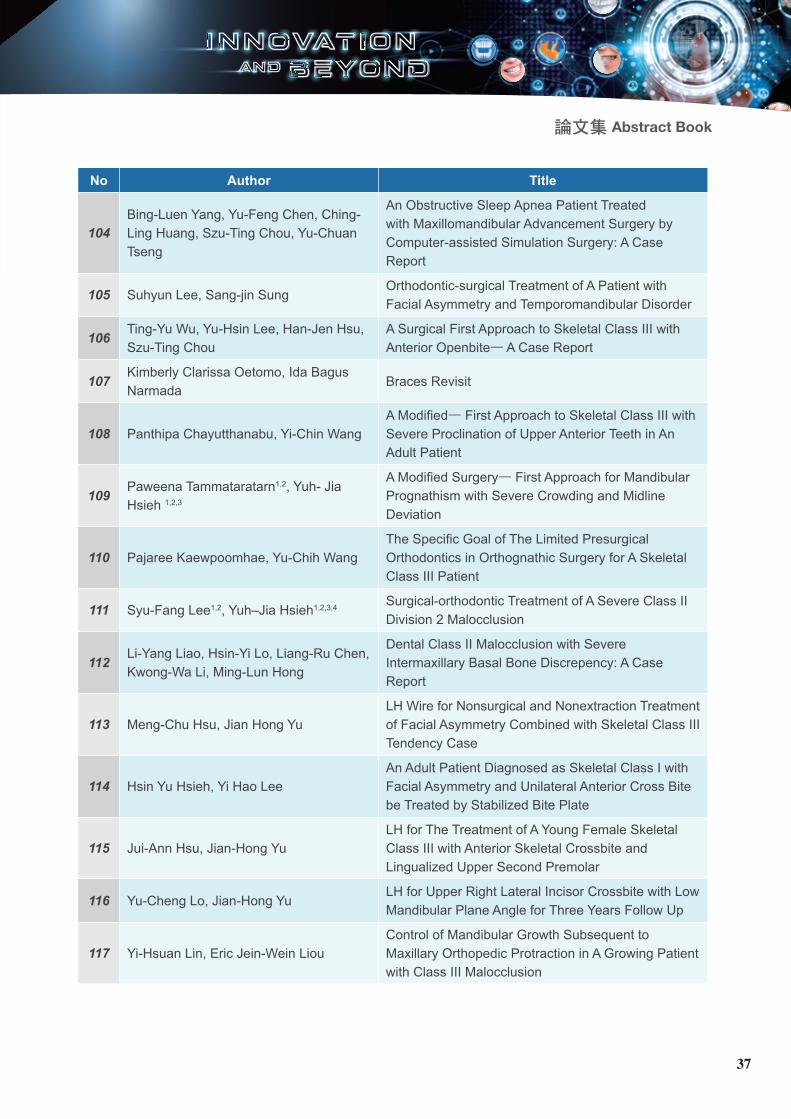
論文集 Abstract Book
No Author Title
104Bing-Luen Yang, Yu-Feng Chen, Ching-Ling Huang, Szu-Ting Chou, Yu-Chuan Tseng
An Obstructive Sleep Apnea Patient Treated with Maxillomandibular Advancement Surgery by Computer-assisted Simulation Surgery: A Case Report
105 Suhyun Lee, Sang-jin SungOrthodontic-surgical Treatment of A Patient with Facial Asymmetry and Temporomandibular Disorder
106Ting-Yu Wu, Yu-Hsin Lee, Han-Jen Hsu, Szu-Ting Chou
A Surgical First Approach to Skeletal Class III with Anterior Openbite— A Case Report
107Kimberly Clarissa Oetomo, Ida Bagus Narmada
Braces Revisit
108 Panthipa Chayutthanabu, Yi-Chin WangA Modified— First Approach to Skeletal Class III with Severe Proclination of Upper Anterior Teeth in An Adult Patient
109Paweena Tammataratarn1,2, Yuh- Jia Hsieh 1,2,3
A Modified Surgery— First Approach for Mandibular Prognathism with Severe Crowding and Midline Deviation
110 Pajaree Kaewpoomhae, Yu-Chih WangThe Specific Goal of The Limited Presurgical Orthodontics in Orthognathic Surgery for A Skeletal Class III Patient
111 Syu-Fang Lee1,2, Yuh–Jia Hsieh1,2,3,4 Surgical-orthodontic Treatment of A Severe Class II Division 2 Malocclusion
112Li-Yang Liao, Hsin-Yi Lo, Liang-Ru Chen, Kwong-Wa Li, Ming-Lun Hong
Dental Class II Malocclusion with Severe Intermaxillary Basal Bone Discrepency: A Case Report
113 Meng-Chu Hsu, Jian Hong YuLH Wire for Nonsurgical and Nonextraction Treatment of Facial Asymmetry Combined with Skeletal Class III Tendency Case
114 Hsin Yu Hsieh, Yi Hao LeeAn Adult Patient Diagnosed as Skeletal Class I with Facial Asymmetry and Unilateral Anterior Cross Bite be Treated by Stabilized Bite Plate
115 Jui-Ann Hsu, Jian-Hong YuLH for The Treatment of A Young Female Skeletal Class III with Anterior Skeletal Crossbite and Lingualized Upper Second Premolar
116 Yu-Cheng Lo, Jian-Hong YuLH for Upper Right Lateral Incisor Crossbite with Low Mandibular Plane Angle for Three Years Follow Up
117 Yi-Hsuan Lin, Eric Jein-Wein LiouControl of Mandibular Growth Subsequent to Maxillary Orthopedic Protraction in A Growing Patient with Class III Malocclusion
37

No. 01
CBCT Evaluation on Condylar Displacement After Orthognathic Surgery: Comparison Between Conventional and Surgery— First Approach
Min-Hee OhKyung-Min LeeJin-Hyoung ChoDepartment of Orthodontics, School of Dentistry, Chonnam National University
ObjectiveThe purpose of this retrospective study was to compare the condylar displacement after asymmetric mandibular setback between the conventional approach and surgery-first approach
Materials and methodsThe subjects consisted of facial a s y m m e t r y w h o u n d e r w e n t mandibular setback surgery and had cone-beam computed tomography (CBCT) taken before and about 1 month after surgery. They were allocated into conventional and surgery-first groups. Position of condyles was measured.
ResultsT h e a m o u n t s o f c o n d y l a r d i s p l a c e m e n t d i d n o t s h o w statistically significant differences
between the conventional and surgery-first groups. Comparing the postoperative condylar position with the preoperative position, the condylar displacement occurred in the poster ior (P < .05) and downward (P < .01) directions in both groups except on the deviated side in the conventional group. The condylar displacement occurred in the posterior (P < .01) direction in the deviated side of conventional group. However, the condylar displacement in the three-directions did not show statistically significant differences between two groups. In the correlation analysis, the condylar displacement in both deviated and contralateral side did not show significant correlation with asymmetric setback in each two groups.
ConclusionIn the surgery-f i rst group, the amount of condylar displacement in asymmetric mandibular setback patients did not show difference from the conventional group.
No. 02
Perception of Trainees Regarding The Need to Incorporate The Topics of Temporomandibular Disorders into The Orthodontic Training— A Preliminary Study
Shiu-Shiung LinDepartment of Orthodontics, Kaohsiung Chang Gung Memorial Hospital and Chang Gung University, College of Medicine
Objectiveto preliminari ly investigate the desire of or thodont ic t ra inees t o h a v e t e a c h i n g c l a s s o f temporomandibu lar d isorders (TMD) in the orthodontic training curriculum.
Materials and MethodsA designed questionnaire was applied for the enrolled orthodontics trainees (n = 14) to answer the relevant questions. A scale (0 to 10) was utilized to reveal trainees’ perception regarding (1) knowledge competence of the relevant TMD topics at their pre-enrolled stage, (2) the desire to have the class of 15 relevant TMD topics, (3) the teaching hours required to be designed for the TMD topics.
Results and Discussion(1) The TMD knowledge taught hours of trainees at pre-enrolled s tage in the program was on average 16.5 ± 12.4 hours (range: 2 to 40 hours). (2) The average l eve l o f compe tence i n TMD knowledge before enrolled in the program was only 25% (2.5/10.0). (3) The three most desired TMD topics were “anatomy, physiology and pathophysiology of the TMJ complex”, “occlusion, condyle position and TMD”, and “bruxism”. The three least desired topics were “neurophysiology of pain”, “Psychosocial issues” and “history and examination procedures”. But
38

論文集 Abstract Book
all 15 topics were in the category of “highly-demanded” (≥ 8.0/10.0). (3) The teaching hour required for all the topics was 41.0 ± 11.5 hours (range: 25 to 72 hours).
ConclusionThe desire of orthodontic trainees to have relevant topics of TMD in the orthodontic curriculum was strong, and the desire scales were in the category of “highly-demanded”.
No. 03
The Study of Perception and Attitude on Malocclusion Among Mongolian People
Gannaran NarangerelHsin-Chung ChengGin-Chia YehOrthodontic Division of Dental department, Taipei Medical University Hospital
ObjectiveT h e a i m o f t h i s s t u d y i s t o investigate the perceptions and attitudes of Mongolian subjects toward their dental appearance and to compare the perception of dental appearance between the investigators and the subjects. Then to determine the factors affecting on patient’s perception and attitude toward various malocclusion.
Material & MethodsThe study was conducted between late June-August in 2019. A total
of 133 participants (53 males and 80 females) aged 18-55years (average= 32±8.99) from General Dentistry department of Mongolian National University of Medical Sc ience ’s Den ta l c l i n i c were selected via random sampling. Participants were questioned to select which photograph of the– “Aes the t ic Component o f the Index of Orthodontic Treatment Need” (IOTN-AC) and “Aesthetic Component of Lateral Occlusion” (AC-LO) is most closely matched their orthodontic aesthetic self-perception of their anterior teeth appearance and lateral occlusion. Subjects also asked to select which of the intraoral lateral photographs of AC-LO is showing the normal occlusion. One-way ANOVA was used to analyze the association between variables.
ResultsCorrelation existed between the researcher’s and participant’s AC scores. (p<0.01) 85.7% of subjects were able to select the normal occlus ion f rom six separate ly lettered (A-F) lateral photographs of 6 malocclusions. As the results of the AC-LO evaluation there was significant statistical difference between evaluated diagnosis and patient’s occlusion perception.
ConclusionAmong Mongolian participant’s eva lua t i on resu l t s , pa t i en t ’s perception of their own occlusion was not identical with researcher’s perception of malocclusion. Patients perceived self- perception was greater than researchers.
No. 04
Mesial Movement of Maxillary First Molar in En Masse Orthodontic Treatment with Extraction of Premolar
Junichiro MoriMori Orthodontic Office
AimI wou ld l i ke to in t roduce the comparison of the amount of mesial movement of maxillary first molar, between 17 cases by the en masse orthodontic treatment, simultaneous distal movement of central, lateral incisor, and cuspid, and 17 cases which were given distal movement of central and lateral incisor after distal movement of cuspid.Materials and Methods: Two groups were compared and examined wi th la tera l roentgenographic cephalograms which were taken before and after their treatments. First group, which is called the enmasse group, is 17 cases with the extraction of maxillary first premolar and mandibular first premolar or second premolar by the enmasse orthodontic treatment. Second one, which is called the control group, is 17 cases with distal movement of central and lateral incisor after distal movement of cuspid.
ResultsThese comparisons and examinations resulted as follows. In all angle and distance measurement items,
39

the statistical significance was not recognized in the changing positions of maxillary first molar.
ConclusionThis fol lows that no difference between the en masse group and the control one is recognized in the mesial movement of maxillary first molars, in the quantity of lingual slant of maxillary central incisors. It is thought that the strong power is necessary because the transseptal fibers between lateral incisor and cuspid are pul led in the distal movement of cuspid and therefore it causes the mesial movement of molars. It is considered that the simultaneous distal movement of central, lateral incisor, and cuspid is more effective with no necessary to pull the transseptal fibers and then with weak power. Therefore it is verified that the en masse orthodontic treatment is useful.
No. 05
Pharyngeal Airway Changes Following Maxillary Expansion or Protraction: A Meta-analysis
Yin-Teng HsiehWei-Cheng LeeDivision of Orthodontics and Dentofacial Orthopedics, Department of Dentistry, Tri-Service General Hospital, Taipei, Taiwan
The aim of this meta-analysis was to investigate the changes in airway dimensions after rapid maxillary
expansion (RME) and facemask (FM) protraction. Using PubMed, Medline, ScienceDirect and Web of Science, only controlled clinical trials, published up to November 2016, with RME and/or FM as keywords that had ≥6 months follow- up period were included in this meta-analysis. The changes in pharyngeal airway dimension in both two-dimensional and three-dimensional images were included in the analysis. Nine studies met the criteria. There are statically significant changes in upper airway and nasal passage airway in the intervention groups as compared to the control groups, assessed in two-dimensional and three-dimensional images. However, in the lower a i rway and the a i rway be low the palatal plane, no statistically significant changes are seen in 2D and 3D images . RME/FM treatments might increase the upper airway space in children and young adolescents. However, more RCTs and long-term cohort studies are needed to further clarify the effects on pharyngeal airway changes.
No. 06
Three-dimensional Evaluation of Dentofacial Transverse Widths of Adults with Various Vertical Facial Patterns
Hosik JangKyung-Ho Kim
Department of Orthodontics, Gangnam Severance Hospital, The Institute of Craniofacial Deformity, College of Dentistry, Yonsei University
ObjectiveTo investigate maxillomandibular t ransverse w id ths and mo la r i n c l i n a t i o n s o f a d u l t s w i t h hypodivergent, normodivergent, and hyperdivergent facial patterns using cone-beam computed tomography.
MethodsWe evaluated Class I subjects (55 men, 66 women) who were divided into hypodivergent (<27), normod ivergen t (28-37) , and hyperdivergent (>38) groups by their mandibular plane angles. Frontal and coronal views of the images were analyzed. Sex differences, vertical facial pattern differences, and related factors were assessed with independent 2-sample t tests, 1-way analysis of variance followed by post hoc Tukey tests , and Pearson correlation analysis.
Results The hypodivergent group had greater maxillary alveolar widths 7 mm apically from the alveolar crest. The intermolar widths and molar inc l inat ions showed no significant differences among the groups. As the mandibular plane angles increased, inter jugular widths, transverse mandibular widths, and buccolingual maxillary alveolar widths at the midroot level decreased, whereas the maxillomandibular width differences and palatal heights increased in both sexes.
40

論文集 Abstract Book
ConclusionsAn increase in the mandibular plane angle is associated with tendencies of narrow mandibular arches, thinner maxillary alveolar bones at the midroot level, and higher palatal arches in both sexes. Intermolar widths and molar inclinations were not significantly affected by vertical facial patterns.
No. 07
Dentofacial Transverse Development in Koreans According to Skeletal Maturation: A Cross-sectional Study
Hosik JangKyung-Ho KimDepartment of Orthodontics, Gangnam Severance Hospital, The Institute of Craniofacial Deformity, College of Dentistry, Yonsei University
ObjectiveTo establish the normative data of dentofacial transverse dimensions according to the skeletal maturation stage in Korean adolescents with good occlusion.
Material and MethodsA total of 577 Korean subjects between ages 7 to 19 years and exhibiting skeletal Class I occlusion were ca tegor ized by ske le ta l maturation index (SMI). Dentofacial t r ansve rse d imens ions we re
assessed using posteroanterior cephalograms.
ResultsDentofacial transverse norms relevant to skeletal maturation stages were established. Males had greater facial, maxillary and mandibular widths compared to females at every SMI stage. The maxillary and mandibular in termolar widths showed the strongest correlation for both sexes
ConclusionDentofacial transverse norms of Korean adolescents were established according to developmental stage. All dentofacial widths were greater in males at growth completion. Maxillary and mandibular intermolar widths were strongly correlated.
No. 08
Periodontal Outcome of Buccally Impacted Maxillary Canines After Orthodontic Traction Following Closed Eruption Technique
Hosik JangKyung-Ho KimDepartment of Orthodontics, Gangnam Severance Hospital, The Institute of Craniofacial Deformity, College of Dentistry, Yonsei University
ObjectiveThe aim of the investigation was to evaluate the periodontal status
of the buccally impacted maxillary canines after orthodontic traction following closed eruption technique and to investigate pretreatment orthodontic variables affecting the periodontal changes.
Methods54 patients with a maxillary canine in a buccally impacted position were chosen as an impaction group and a contralateral canine in normal position served as a control group.
Results Probing depth and bone probing depth increased significantly in the impaction group. The attached gingiva width was signif icantly shorter and the buccal clinical crown length longer in the impaction group. The distance from CEJ to AC was significantly longer and the bone supports shorter in the impaction group.
ConclusionsForced eruption of the maxillary impacted canine after orthodontic traction following closed eruption technique resulted in significant gingival recession on the buccal side and alveolar bone loss on the interproximal sides.
No. 09
The Study of Treatment Modalities in Cases of Congenitally Missing Mandibular Incisors
41

De-Shing ChenHsin-Chung ChengPei-Shein ChenOrthodontic Division of Dental department, Taipei Medical University Hospital
IntroductionFrom the l i te ra ture rev iew o f congenitally missing mandibular inc isors cases, the t reatment modalities are troublesome and complicated. The aim of this study is to analyze the treatment results of the cases with congenitally missing mandibular incisors, which would give orthodontists more hints to deal with such cases.
Material and MethodsThis retrospective cohort study was des igned w i th to ta l 128 cases selected from TMUH. We categorized the initial malocclusion i n t o t h r e e t y p e s : c l a s s I malocclusion, class II malocclusion, and class III malocclusion. We also divide these cases into 4 groups according to the treatment modalities. Group 1: extraction of two upper premolars, group 2: closure of lower congenital missing area, group 3: create space for prosthesis, and group 4: other treatment. We design a modified dental aesthetic index(mDAI) for analyzing the treatment results of all patients and to find the best treatment modalities. We will use the multiple regression analysis to find the affecting factor on treatment modalities of congenitally missing mandibular incisors cases.
ResultsThe treatment results of all these
four groups present similar good outcomes, with mDAI smaller than 36, indicating an acceptable result. Group 1, accounting for 42% total cases, shows good results with average mDAI of 30. Group 2, accounting for 36% total cases, shows good results with average DAI of 32. Group 3, accounting for 22% total cases, shows DAI of 35. We also trace back to the initial malocclusion in order to know which kind of treatment modality is proper for them. The flowchart of the treatment modality would be designed in making treatment plans. The final results will show in the e-poster.
ConclusionThe retrospective study suggests that most of the cases could achieve acceptable results with proper treatment plan. Further prospective study should be done in order to make the flowchart of treatment modality more comprehensive and with stronger evidence.
No. 10
Long-tern Follow-up of Maxilla Advancement with Rigid External Distraction Device(RED)
Wei-Ling GaoChang, Yu-JenOrthodontic department/Kaohsiung Chung Gung Memorial Hospital
ObjectiveIn 1997, Polley and Figueroa first introduced the use of RED to treat
patients with maxillary hypoplasia by using the dental splint fixed. However, the side effects from dental anchorage advice has been widely discussed. Therefore, the aim of this study is to present the long-tern stability result of LeFort I distraction osteogenesis using RED with transcutaneous skeletal anchorage.
Materials and methods7 patients with cleft lips and palate (2 boys, 5 gir ls, age: 11.7±0.5 years) had Le Fort I osteotomy and fixation of the RED device. The distraction protocol was started with 2 turn (1 mm) a day for 4 weeks, and then 2-months consolidation. We used digital software Audex for cephalometr ic analys is to measure the maxillary position after distraction immediately, at 6 months, and 1 year after distraction.
Results and discussionSkeletal changes were record by following the horizontal and vertical positions of 2 points (ANS, and A point) registered on the maxilla. The average records showed: ANS (horizontal: 19.9±6.2 mm, vertical: 2.4±4.0 mm), A point (horizontal: 18.1±7.8 mm, vertical: 3.0±5.6 mm). After 1 year, ANS shows backward 3 mm and downward 0.6 mm, and A point shows backward 0.9 mm and forward 0.8 mm. The relapse rete were 15.2% in ANS and 5.0% in A point in horizontal direction.
ConclusionIn these cases, the distraction from RED devices with skeletal anchorage reveal great amount of maxilla advancement and shows
42

論文集 Abstract Book
less relapse rate af ter 1 year follow up.
No. 11
The Study of Smile Changes in Frontal, Lateral and Oblique View After Orthodontic Treatment
Yu-Ming LinHsin-Chung ChengYi-Guine ChenOrthodontic Division of Dental department, Taipei Medical University Hospital
ObjectiveThe purpose of this study is to compare the smile changes after receiving orthodontic treatment among class I, class II and class III malocclusion, to identify the cephalometric factor affecting smile variables changes after treatment and to develop smile variables for lateral and oblique view photos to help in orthodontic analysis and treatment planning.
Materials and Methods7 8 p e o p l e i n p e r m a n e n t dentition who had been received orthodontic treatment in Taipei medical university hospital were e n r o l l e d . A c c o r d i n g t o t h e i r dental overjet(OJ), these patients were divided into class I (n=34, 0mm<OJ<4mm), class II (n=36, OJ>4mm) and c lass I I I (n=8, OJ<0mm). Complete orthodontic records from pretreatment(T1)
and posttreatment(T2) periods were recorded. The changes in the angular, linear and numerical measurements of the frontal, oblique and lateral smiling photographs after treatment were digitized to compare. Lateral cephalograms were taken with the head in a natural position. Changes in the smiling photos were correlated with the cephalometric parameters in order to observe any potential relationships. Data were calculated with t test, one-way analysis of variance, chi-square test and multiple regression. The significance level was set at p<0.05.
ResultsIn class I malocclusion, a significant difference between T1 and T2 (P<0.05) were found in maxillary incisor angle of the oblique view, and also upper lip to Ear-Eye connecting line angle in lateral smiling photo (P=0.02) and maxillary incisor angle in oblique view(P=0.02). Statistically significant differences were also found in maxillary incisor angle in T1(P=0.04) and T2-T1(P=0.04), nasolabial angle in T1(P=0.01) and T2(P=0.03), upper lip to Ear-Eye connecting line angle in T2-T1(P<0.02), and lateral smile index in T2(P=0.04) between class I and class II malocclusion in lateral smi l ing photo. There were no significant differences in T1, T2, and T2-T1 in oblique smiling photo between c lass I and Class I I malocclusion
ConclusionThe frontal, oblique and lateral smiling measurements showed significant changes fol lowing orthodont ic treatment. Orthodontists should take
oblique and lateral smiling photo as routine data collection, treatment planning and use it for assessment of treatment outcomes.
No. 12
Effects of Masticatory Hypofunction Induced by Botulinum Toxin Type A (BTXA) on Cognitive Function and Hippocampal Neurons in Developing Rats
Fang-Chun WuChi-Yang TsaiOrthodontic Division of Dental department, Taipei Medical University Hospital
ObjectiveMalocclusion is often encountered i n c l i n i ca l , espec ia l l y young individuals. Among older patients, poor mastication due to tooth loss results in functional deficiency, and being suggested as risk factor of Alzheimer. However, little research has been done in develop ing an imals . Th is s tudy a imed to exp lore the e f fec ts o f BTXA-induced masticatory hypofunction on cognitive function of learning and spatial memory as a result of neuropathological change in the hippocampus in developing rats.
Materials and Methods4-week-old male Wistar rats (N
43

= 20) were randomly d iv ided into control and BTXA-injected groups. Total dose of 1U BTXA was prepared for each ra t in experimental group, being injected in bilateral masseter muscle on day 1 . Converse ly, the same amount of sterile, nonpreserved 0.9% normal saline was injected in control group. In the 4th week, the rats were tested in Morr is water maze for five consecutive days. After concluding the maze, rats were sacrificed on day 28, and their brains were collected for evaluation of neuropathological changes in the h ippocampus t h r o u g h N i s s l s t a i n i n g a n d phosphorylated cyclic adenosine 3 ' ,5 ' -monophosphate (cAMP) response element binding protein (CREB) immunohistochemistry.
Results and DiscussionIn Behavior test, the rats from BTXA-injected group required a longer time to escape from maze compared with control. Nissl staining revealed a significant reduction in the neuron density in the BTXA-injected rats. Further, experimental rats exhibited a decreased level of CREB phosphorylation.
ConclusionThe BTXA-injected rats required longer escape latency than did control, signifying the decreased spatial learning ability. The BTXA-in jected rats a lso exhib i ted a reduction in neuron density and phosphorylated CREB, indicating that mastication might influence learning and memory ability during growth period.
No. 13
The Study of Comparison on Smile Esthetic Between Taiwanese and Thais
Parichart PasukdeeHsin-Chung ChengOrthodontic Division of Dental department, Taipei Medical University Hospital
ObjectivesTh is s tudy was to de te rmine whether differences in personal background, demographic such as racial, ethnic, culture, economics and other soc ia l env i ronment affected smile perception of esthetic and smile characteristics among orthodontists, general dentists, orthodontic patients, and laypersons of Taiwanese and Thais.
Materials & MethodsU s i n g a c o m p u t e r - b a s e d q u e s t i o n n a i r e s c o n s i s t e d o f demographics survey and multiple choices of smiling photos. Total of 1,040 Thais and 773 Taiwanese were div ided to dent is t group (orthodontists and general dentists) and general people (orthodontic patients and laypersons) and asked to choose the most favored digitally altered smile of each variable. The frontal smiling photos were altered in various increments of arc ratio, most posterior upper teeth visible, upper teeth exposure, upper dental midline, lower teeth exposure, buccal corridor, maxillary gingival display, and cant. The result of demographics
and smile variables were compared between two countries to analyze the different smile preferences and affecting factors.
ResultsDemographic characteristics of the raters between dentists in Thailand and Taiwan showed statistically significant differences in aspect of gender, age, marital status, and religion. There also showed statistically different preference between dentists in Thailand and Taiwan in aspect of arc rat io, most posterior upper teeth visible, upper teeth exposure, lower teeth exposure, and buccal corridor, while the statistically different were found when compared between Thais and Taiwanese in aspect of arc ratio, most posterior upper teeth visible, upper teeth exposure, upper dental midline, lower teeth exposure, and buccal corridor in frontal smiling view. Overall analysis, dentist group between two countries have more consistent preferences in smile esthetics than general people group. However, it seems that Taiwanese preferred wider smile than Thais. I n a d d i t i o n , s o m e p e r s o n a l demographic data such as age, marital status, religion can affect dentists’ perceptions of esthetic smile, while many demographic factors of general people such as age, marital status, religion, education and income can affect their perceptions of esthetic smile.
ConclusionsMost of the smile variables were signif icantly di fferent between both dentists and general people of Thailand and Taiwan. However,
44

論文集 Abstract Book
compar i ng be tween den t i s t s (orthodontists and general dentists) of two countries has less different preferences of smile esthetics than between general people (orthodontic pat ients and laypersons). The perception of smile esthetics is subjective and can be influenced by personal background especially among general people.
No. 14
Effects of Active Mandibular Exercise After Orthognathic Surgery: A Non-randomized Controlled Trial
Hyo-Jin JangYoun-Kyung ChoiDepartment of Orthodontics, School of Dentistry, Pusan National University, Busan, Republic of Korea
PurposeThis study was to evaluate the effects of active mandibular exercise (AME) in patients with limited mouth opening after maxillomandibular f i x a t i o n ( M M F ) r e l e a s e f o r orthognathic surgery. Methods: The AME was performed in the experimental group for 4 weeks. The exercise AME consisted of maximal mouth opening, lateral excursion and protrusive movement. These movements were repeated ten times a day. After the final exercise of the day, the number of tongue blades used for mouth opening was noted. The effect of AME was evaluated after MMF
release at different time intervals: a) immediately, b) after 1 week, c) after 2 weeks, d) after 4 weeks, and e) after 12 weeks. The exercise was assessed using the following criteria: a) mandibular movements, b) pain scores associated with maximal mouth opening, c) discomfort scores associated with range of movement, and d) daily life activities that involve opening the mouth.
ResultsThe experimental group showed significant improvement regarding the range of mandibular movements (maximal mouth opening (F=23.60, p<.001), lateral excursion to the right side (F=5.25, p=.002), lateral excursion to the left side (F=5.97, p=.001), protrusive movement (F=5.51, p=.001)) , pain score (F=39.59, p<.001), discomfort score (F=9.38, p<.001). Daily life activities that involve opening the mouth were more favorable compared to those in the control group.
ConclusionThe AME in patients after mouth opening limitation is helpful for increasing mandibular movement r a n g e , d e c r e a s i n g p a i n a n d discomfort, and improving day life activities that involve opening the mouth. Therefore, AME is highly recommended as an effect ive intervention.
No. 15
Association of ABO Blood Groups with Craniofacial
Morphology Among Orthodontic Patients of Kathmandu, Nepal
Sanjay Prasad GuptaDepartment of Orthodontics, Tribhuvan University Teaching Hospital, Kathmandu, Nepal
ObjectiveThe objective of this study was to find out the relationship between ABO blood groups and craniofacial morphology among orthodontic patients of Kathmandu district.Materials and Methods In this cross sectional study, a total of 385 participants (age range from 13-45 years) were selected among the orthodontic patients who came for orthodontic treatment in private orthodontic clinics. After obtaining written consent, all the patient’s demographic information were recorded and lateral cephalograms were obtained from the patient’s record. Blood group of al l the participants was recorded.
ResultsThe study found that among the tota l o f 385 par t ic ipants, 162 (42.07%) were male while 223 (57.93%) were female and the mean age was 16.31±4.38 years. Twenty cephalometric parameters depicting craniofacial morphology were digitally analyzed using lateral cephalogram. The prevalence of blood group O patients was highest (32.20%) followed by blood group B (30.64%), blood group A (29.88%) and blood group AB (7.28%). Statistical analysis with
45

one way ANOVA was used for association of numerical data and blood groups that revealed nine out of twenty cephalometric parameters were statistically significant among different blood groups (p<0.05). Tukey post hoc test was done to find out where the significant difference occured among the groups.
DiscussionThe craniofacial morphology and blood groups both are related to genetic components, hence it can be hypothesized that blood groups have an association with craniofacial morphology. Some studies showed the relationship whereas others could not find any relationship that may be due to geographic diversity of the population. The results of this study showed that blood group have associated with some craniofacial parameters.
ConclusionThe evaluation of the relationship b e t w e e n b l o o d g r o u p a n d craniofacial morphology revealed that blood groups have association with some craniofacial parameters. This suggests, there may be some genetic influence of ABO blood group on craniofacial morphology.
KeywordsABO blood groups, Craniofacial m o r p h o l o g y , A s s o c i a t i o n , Orthodontic patients
No. 16
CBCT-generated Cephalogram Evaluation of Short-
term Changes in Condylar Position After Surgery— First Approach
Zheng YuchenChae-Hee ParkKun-Hee LeeMin-Hee OhDepartment of Orthodontics, Tribhuvan University Teaching Hospital, Kathmandu, Nepal
ObjectiveTo i n v e s t i g a t e t h e e f f e c t o f s y m m e t r i c a n d a s y m m e t r i c mandibular setbacks on short-term changes in condylar position after surgery-first approach.Materials and MethodsPatients who underwent mandibular setback surgery using the surgery-first approach were selected and divided into symmetry group and asymmetry group. CBCT images were taken before surgery (T0), within one week after surgery (T1), and seven months after surgery (T2). Condylar posi t ions were measured. Positional changes of mandibular condyles according to time were compared between the two groups and correlation analysis was performed.
ResultsThere were significant condylar displacement over time in both groups. However, these changes showed different patterns. On the lesser setback side, there were significant differences in surgical change (T1-T0) and total change (T2-T0) of condyle angle between the two groups. In the asymmetry
group, there was a greater internal rotation of mandibular condyle. Results of correlation analysis revealed that only the setback difference was associated with changes of condyle angle on the lesser setback side at two time points (T1-T0, T2-T0).
ConclusionIn surgery-first patients, significant condylar displacement occurred immediately after surgery in both g roups fo l l ow ing mand ibu la r s e t b a c k s u r g e r y. C o n d y l a r displacement remained partially even at seven months after surgery. More significant displacement was observed following asymmetric mandibular setback.
No. 17The Result of Facial Soft Tissue Cephalometric Analysis Between The Ages of 6 and 15 Years in Mongolian Children: A Cephalometric Study
1. Erdenebulgan Purevjav, DDS2. Ganjargal Ganburged DDS,
PhDDepartment of Orthodontics, Mongolian National University of Medical Sciences
ObjectivesHarmonious facial aesthetics and optimal functional occlusion have long been recognized as the two
46

論文集 Abstract Book
most important goals of orthodontic treatment. To accomplish some of these goals, a knowledge of the normal craniofacial growth as well as the effects of orthodontic treatment on the soft tissue profile is essential. Facial features have been commonly studied in full- face and profile views. Several methods have been used to evaluate these facial changes including anthropometry, p h o t o g r a m m e t r y, c o m p u t e r imaging, and cephalometry. Some researchers have studied the thickness of the soft tissues to determine the relationship between the hard and soft t issues, and to determine the effect of hard tissues on facial aesthetics. Others have highlighted the necessity for the hard and soft tissues to be evaluated together, and that peri-oral function, facial aesthetics, and stability are important factors in orthodontic treatment.
Materials and MethodsThe present study was conducted on lateral cephalograms of 541 sub jec t s ( 228 ma le and 313 females) having normal occlusion in the age group of 6 to 15 years. All radiographs were digitized on a computer using a cephalometric software program (Winceph 11.0; Rise, Sendai, Japan) Total of 6 angular 38 linear measurements were measured for skeletal hard tissue analysis using 31 landmarks (Figure 1), and 3 reference planes. The landmarks and measurements were taken according to the soft t issue cephalometric analysis, and true vertical line (TVL) was establ ished. Structures to the right of TVL were given a positive
sign and those to the left of TVL we re g i ven a nega t i ve s i gn . The soft t issue cephalometr ic parameters were divided into 5 groups: Dentoskeletal structures (Figure 2), Soft tissue structures (Figure 3), Facial lengths (Figure 4), Projections to TVL (Figure 5), and Harmony values (Figure 6). All statistical analysis was calculated using SPSS version 25.0 statistical analysis software. The result was presented as mean for quantitive variables and compared using the student t-test and Mann-Whitney U test. Statistical significance was determined at a p-value of P<0.05.
ResultsComparison analysis of some measurement results with age groups shows that Mx-occlusal plane angle, Md1 to Md occlusal plane angle, Nasolabial angle has been decreased with age, Mx1, Mx occlusal plane angle, and Overbite was stable with age, whereas the other measurement results show increase in tendency with age groups. No statistically significant gender differences were observed in age groups Distinct ethnic difference was found between Caucasians and Mongolian kids. The facial angle in Mongolian kids was more than north American kids, whereas the Nasolabial angle in Mongolian kids less than North American kids. It shows that facial features in Mongolian kids less pronounced convex i t y f ac ia l p ro f i l e t han Caucasians. The nasal projection feature was observed more in Caucasian kids than Mongolian kids. The other features including upper and lower lip thickness were
thicker in Caucasian kids, whereas the upper and lower lip length feature was shorter in Mongolian kids.
ConclusionThe results of the present study suggested that the differences exist in the facial structures of two ethnic groups (Mongol ian and Caucasian). Therefore, separate norms for distinctive populations are necessary and that one set of norms cannot be applied to patients of different races and ethnic origin. Thus, the results of the present study suggest that racial differences shou ld be cons idered dur ing diagnosis and orthodontic and orthognathic treatment planning.
No. 18
The Study of Factors Associated with Using Frequency and Charge of Temporary Anchorage Devices(TADs) Amongst Taiwanese Orthodontists
Ya-Wen TanHsin-Chung ChengTai-Lian Lu Hau-Tin HuOrthodontic Division of Dental department, Taipei Medical University Hospital
IntroductionTADs use is an invasive anchorage
47

preparat ion in the orthodont ic treatment. The factors affecting practice in the professional field is impor tant fo r both present practitioners and those who will participate afterwards. In this study, the affecting factors on TADs use of the orthodontists in Taiwan were invest igated through the demographic survey.
Materials and MethodsThe survey was conducted in the form of questionnaires with 16 questions, including individual characters (6), clinical working pattern (8), and preferences for TADs use (2). The questionnaires were sent to all the orthodontists attending the Taiwan Association of Orthodontists (TAO) annual meeting in 2018 and the data were analyzed by the statistical analysis system (The significance level was set as p < 0.05).
Results177 questionnaires were returned, giving a response rate of 31.6%, including 46.6% male and 53.4% female responders, and aged from 30 to 70 years old. The frequency of the use of TADs showed significantly associated with the degree of income satisfaction and working time per week. The charge of TADs fee were significantly related to the age distribution, treatment fee and region.
ConclusionThe f requency and charge of TADs use in orthodontic treatment were affected by working patterns and demographic factors. Further study of affecting factors on TADs
use in different views and trend in the future will be needed and concerned.
No. 19
Investigation of Medical Disputes Involving Dentists Performing Orthodontic Treatments in Taiwan
Chih-Ling LinYu-Kuan ChihDepartment of Craniofacial orthodontics, Chang Gung Memorial hospital, Taipei
IntroductionMedical disputes have received increasing at tent ion in recent yea rs . Under the ass i s tance o f the academic and medica l affairs committees of the Taiwan Association of Orthodontists, this study explored medical disputes among orthodontists performing orthodontic treatments in Taiwan in 2016.
MethodsThis study adopted a questionnaire survey, which was divided into three sections, namely the surveyed o r t h o d o n t i s t s ’ d e m o g r a p h i c information, medical services, and subjective views on orthodontic-related medical disputes. The results were compared with those from a study conducted in 2010. Subsequently, topics related to clear aligner therapy and orthognathic
surgery were examined. Results: The results indicated that 32.3% of the respondents had experienced medical disputes, with Class II Division 1 accounting for the highest proportion of malocclusion. Lateral profile–related problems accounted for the highest proportion of clinical problems. Eventually, 6.7% of the disputes involved legal proceedings. Compared to the 2010 results, the proportion of cases involving legal proceedings and costs of monetary compensation increased.
ConclusionThe results of this study signify a changing trend in orthodontic treatments and patient requirements, which can serve as a reference for clinicians who perform orthodontic treatment.
No. 20
Potential Complications Related to Miniscrew-assisted Rapid Palatal Expansion
Huei-Rou TsaiHeng-Ming ChangDepartment of Orthodontics and Craniofacial Developmental Biology, Hiroshima University Graduate School of Biomedical & Health Sciences
ObjectiveMaxillary expansion is necessary w h e n t r e a t i n g p a t i e n t s w i t h transverse maxillary constriction.
48

論文集 Abstract Book
Recent ly, min iscrew-ass is ted rapid palatal expansion (MARPE) appliances were developed with miniscrews incorporated into the expansion screw body. By doing so, the damages to the teeth and periodontium could be l imited. However, some undesirable effects cou ld happen dur ing MARPE procedure. In this study, we aim to record any possible complication related to insertion and expansion o f MARPE app l iance , and to summarize the incidence of each.
Materials and MethodsFourteen adult patients (mean age, 25.1 years) having experienced t h e M A R P E p r o c e d u r e w e r e enrolled. All treatment procedures were completed by one operator. Cone beam computed tomography images were taken before and after insertion of MARPE. Interviews of the four teen pat ients were conducted by one interviewer.Results and DiscussionPotential adverse effects include g ing iva l swe l l ing , so f t t i ssue impingement , ep is tax is wh i le miniscrews insertion, distortion of components, asymmetric expansion, and fa i lure of suture-opening etc. The mean pain score during MARPE is 3.8 (Visual Analogue Scale from 0-10).
ConclusionAlthough some undesirable side effects were reported, MARPE still aids in correction of the maxillary constriction in adults. The mean pain score is mild and the complications a r e r e l a t i v e l y t o l e r a b l e . I n consideration of the risk and benefit of MARPE, we still believe MARPE
is a considerably safe procedure.
No. 21
Treatment of Growing Skeletal Class III Patient with Functional Education Appliance— A Case Report
Shang-Wen ChiuChung-Hsing LiDivision of Orthodontics and Dentofacial Orthopedics, Department of Dentistry, Tri-Service General Hospital, Taipei, Taiwan
The issue that whether functional education appliance is effective to the growing children is always debatable within the orthodontic academies in America or Europe. T h e c o n v e n t i o n a l f u n c t i o n a l education appliances varied in their designs for different malocclusions. Some patients in the mixed dentition with class III malocclusion were suggested re-evaluation or even orthognathic surgery while grow up. Therefore, we wonder that if there is any appliance that may improve the malocclusion in the early age? Recently, some scholars treated the malocclusion with growth problems using natural balance theory that included respiration, swallowing and muscle training and so on.
In this report, A 9-year-old growing girl with class III malocclusion treated with functional education
appl iances and myofunct ional exercises. After following up for 6 months, the anterior cross bite was improved into edge-to-edge b i te. Moreover, the resul ts of cephalometry showed that both maxil la and mandible changed gradually, and the patient’s profiles became more harmonious. In the superimposit ion, the mandible showed clockwise rotation and the maxilla grew downward and forward to achieve better relationship. After 30 months, the occlusion gets better and better. In our presentation, functional education appliance is one of novel treatment options of Class III juvenile patients with maxil la deficiency and vertical growth pattern not only in skeletal growth modification but also in dental alignment. Although the short-term effects are bracing, the further follow-up is still needed.
No. 22
Treating the Mesial Tilting Mandibular Second Molars by Using Two Different Methods— A Case Report
Shang-Wen ChiuGunng-Shing ChenDivision of Orthodontics and Dentofacial Orthopedics, Department of Dentistry, Tri-Service General Hospital, Taipei, Taiwan
“impacted” means that a tooth either has not erupted when expected or a tooth that cannot erupt because
49

it does not have room or may be coming in the wrong direction or position. What causes an impacted tooth may not be known. Genetics can play a role, so if a parent had an impacted tooth, their child may experience the same problem.
T h e i m p a c t e d t o o t h u s u a l l y happened at third molars, maxillary canine and second premolar etc. F ix ing an impacted tooth can range from relatively simple to complicated. It all depends on the extent of the problem. In this report, a growing 12 year-old girl with bilateral mandibular second molars impacted was treated with different orthodontic methods. The mesial tilting second molars were uprighted by using the twisted copper wire and the Ni-Ti wire in the other side. These two local orthodontic methods were both preformed with simple ways, but they still needed to align with the archwire in the finished stage.
Although the tooth buds of the third molars existed on the distal side of the impaceted second molars, they didn’t interfered the uprighting pathways of the tilting teeth in this case. Another issue that we can notice was the second molars had the erupting potential during treatment period, and it might shorten the treatment time. We will keep following this case and treat other space problems in the suited time.
No. 23
Treatment of
Anterior Open Bite by Maxilla Posterior Teeth Intrusion
Ping-Ting LuYi-Jane ChenI-Ling HongDivision of Orthodontics and Dentofacial Orthopedics, Department of Dentistry, National Taiwan University Hospital
ObjectiveTreatment of anterior open bite malocclusion
CaseHerein we will present the non-extraction orthodontic treatment of anterior openbite in an adult case. This patient had TMD history and underwent splint therapy for 8 months. Afterwards, she as referred to have orthodontic treatment for anterior openbite. She presented a Class II malocclusion with 4 mm overjet and 3 mm openbite. Upper incisor shown at rest was inadequate. The cephalometric analysis showed significantly excessive posterior dental height. Therefore, we decided to correct the anterior open bite by maxillary posterior intrusion. Two buccal plate and palatal miniscrews were used as bony anchorage in the upper arch. After treatment, Class I canine and molar relationship w e r e e s t a b l i s h e d . T h e p o s t -treatment cephalometric analysis showed the mand ibu la r p lane angle decreased from 44o to 41.5o, induced a counterclockwise rotation of mandible. The chin projection increased and the profile was nicely improved after treatment. At 1 year
after treatment, minor relapse of the incisor overjet and overbite was noted. The cephalometric analysis showed that upper molar extrusion 1mm, and the upper and lower incisor positions were maintained.
Discussion and SummaryThe long term stability of anterior open bite was always a consideration. Most of relapse was occurred at the first year. So, the design of retention was important. If a proper retention was applied, it would effectively improve the long term stability.
No. 24
Congenitally Missing of Mandibular Incisor: A Case Report
Yan-Cheng LiaoHeng-Ming ChangDepartment of Orthodontics, Chang Bing Show Chwan Memorial Hospital
ObjectiveCongenital missing of mandibular incisor is more frequent in oriental popu la t ion . Th is case repor t describes a female patient with congenitally missing lower right central incisor. Comprehensive orthodontic treatment was done to correct the class II division 1 malocclusion and space closure for the missing tooth.
CaseA 15-year-old female presents class II division 1 malocclusion and congenitally missing of one
50

論文集 Abstract Book
mandibular incisor. Spacing over lower an ter io r reg ion causes esthetic and malocclusion problem. In consideration of facial esthetics, extractions of upper first premolars and lower second premolars, and space closure over the missing tooth was done. Interproximal reduction over the upper anterior teeth was performed to match the ideal Bolton ratio and to establish good functional occlusion.
Discussion and SummaryIn oriental population, some studies demonstrated missing of mandibular incisor was more prevalent than absence of maxillary lateral incisors. The possible treatment options can basically divided into three groups: space closure, space opening and canine/ premolar substitutions. Some factors have to be considered, including occlusion, number of teeth missing, skeletal pattern, molar relationship, Bolton ratio, individual tooth characteristics (size, shpe and shade), bone volume of edentulous ridge, treatment time, cost and patient’s preference. Thorough assessment and mult i - factoral consideration is essential on this type of case.
No. 25
A Three-year Follow up of Distalization and Intrusion with Temporary Anchorage Devices
Wan-Ting LiaoHung-Cheng ChiuChung-Chen YaoNational Taiwan University Hospital
CaseThis is an 18-year-old healthy female patient who had a convex lateral profile and lip incompetence with the complaint of crooked teeth and dental crowding in the anterior region. Tooth 12 was a peg lateral incisor and tooth 23 was congenital missing with tooth 63 retained. She presented skeletal and dental Class II malocclusion with mandibular retrognathism, severe space deficiency on both arches and large overjet . The patient preferred the non-surgical t reatment plan to the surgical treatment. We extracted teeth 12, 63, and used mini-plates and mini-screws for distalization and intrusion to relieve crowding and improve profile. The patient got a satisfactory occlusion and appearance after 2 years of treatment. However, after 3 years of retention, we found overjet increased from 2.5 mm to 4 mm. Relapse resulted from upper molar mesialization and extrusion.
Discussion & SummaryWe p u s h e d t h e e n v e l o p e o f d is ta l i za t ion in lower arch to avoid extraction of lower arch on retrognathic mandible. Dur ing treatment, distal wedge surgery was performed at bilateral retromolar areas. We observed mandible autorotation after upper molars intrusion. Follow-up visit showed mild relapse occurred after 3 years. In summary, temporary anchorage devices enable distalization and
intrusion successful. However, the direction and amount of relapse should be carefully monitored.
No. 26
Management of Severe Maxillary Transverse Deficiency in Child: A Case Report
Le-Ting WangLi-Fan HsuChung-Chen Jane YaoDepartment of Dentistry, National Taiwan University Hospital, Taipei, Taiwan
ObjectiveMaxillary expansion can achieve good dental and skeletal effects in children with narrow maxillary arches. It is a predictable procedure commonly treated for upper crowded dentition and posterior crossbite in children and adolescents.
CaseA 13-year-old boy presented with Class II malocclusion and severe maxillary transverse deficiency. Anterior esthetic was compromised with malformed right central incisor, congenital missing lateral incisors and impacted left central incisors.O r t h o d o n t i c e x p a n s i o n w a s sequent ia l ly performed with a Haas expander for a month and with 1 month retention, and then a second Hyrax appliance for further expans ion. In termolar w id ths expansion of 7mm was achieved . Full mouth fixed appliance was
51

bonded on the 4th month, we extracted bilateral lower left 1st premolars, subst i tut ing lateral incisors with canines in the maxilla. After 26 months of treatment, an acceptable occlusion was achieved. We then arranged composite resin built-up of the upper incisors for the enhancement of esthetic.
Discussion and Summary Transverse malocclusions due to maxillary width deficiency have been un ique ly respons ive to rapid correction in children and adolescents. With proper treatment planning, maxillary expansion can achieve good dental and skeletal effects in children even in severe maxillary transverse deficiency cases.
No. 27
Treatment of Angle Class I Malocclusion with Bilateral Mandibular 2nd Molars Impaction— A Case Report
Yuan-Che Hsu1
Hsin-Chung Cheng1
Hern-Zeu Hsu2
1 Orthodontic Division of Dental department, Taipei Medical University Hospital
2 Hi-level Dental Clinic 麗晶牙醫診所
IntroductionResolution of lower 2nd molar impaction is often challenging and
complex, and often require early diagnosis and treatment. Treatment modal i t ies included upright ing spring, open coiled-spring, Ni-Ti wire and surgical reposition used depending by patient conditions and orthodontist experiences. In this article, the authors present an adult case using open-coil spring to upright the bilateral mandibular 2nd molars mesio-angular impaction, and to treat the distal subgingival deep caries of 1st molar.
DiagnosisSoft tissue: straight facial profile, protrusive lipsSkeletal: Angle Cl.I malocclusionD e n t a l : d e e p b i t e , # 3 3 - # 4 3 crowding, #37 & #47 mesio-angular impaction, #36 & #46 subgingival deep caries, #18 & #28 & #38 & #48 impaction
Treatment overviewi. Uprighting #37 & #47ii. Restored #36 #46 deep cariesiii. Intrusion of supra-erupted #17
#27iv. Achieve normal OB & OJv. Maintained good Class I interdigitationvi. Harmonized facial profile
ConclusionThe use of open-coil spring in bilateral mandibular 2nd molars mesio-angular impaction may be effective in young adults. Early & prudent diagnosis, flexible treatment plans, and regular observation may lead to a better outcome for the patients with 2nd molar impaction.
No. 28
10 Year Follow Up and Treatment for A Skeletal Class III Asymmetric Growing Patient
In-Ru Lin1
Jen-Bang Lo1,2
Tung-Yiu Wang1,2
Jia-Kuang Liu1,2
1 Department of Stomatology, National Cheng Kung University Hospital, Tainan, Taiwan
2 Institute of Oral Medicine, National Cheng Kung University, Tainan, Taiwan
ObjectiveWe observed the mand ibu la r growth in a ske le ta l c lass I I I patient with facial asymmetry for 10 years. Different treatment was shown depending on di fferent age, malocclusion and growth problem. Use of optimal symmetry plane(OSP)method to ident i fy facial asymmetry in final stage of treatment was also presented.
CaseA l i t t le gir l was brought to our h o s p i t a l a t a g e 8 w i t h c h i e f complaints of posterior crossbite and anterior edge to edge occlusion. Cephalometric analysis showed skeletal class III with mandibular prognathism, hyperdivergent facial pattern with lower midline shift only to R’t 1 mm. Her malocclusion was corrected by RPE and 2 by 4 appliance with chin cup used. After phase I treatment, we kept regular follow up for mandibular growth. Occlusal plane canting and lower midline shift to R’t 6.5 mm was noted
52

論文集 Abstract Book
during f/u. Serial cephalometric films showed no further mandibular growth at age 18. Thus, orthodontic treatment combined with 2-jaw orthognathic surgery was planned with 3D navigation system. Facial asymmetry, midline deviation and profile had improved after treatment.
Discussion and summaryIt is challenging for orthodontists to treat skeletal class III patients with overdeveloped mandible and asymmetry due to unpredictable mandibular growth. With CT-based OSP analysis, we can quantify the misalignment between midface and mandible. Also, more precision in planning surgical schemes in correcting facial asymmetry can be achieved with 3D navigation system.
No. 29
Nonsurgical Treatment of Skeletal Class II High Mandibular Plane with TAD
Chin-Chen YangPiin-Ru Jih1
Chen-Jung Chang1,2
Jen-Bang Lo1,2
1 Department of Stomatology, National Cheng Kung University Hospital, Tainan, Taiwan
2 Institute of Oral Medicine, National Cheng Kung University, Tainan, Taiwan
ObjectiveSkeletal class II with high mandibular plane is one of the most difficult
condition in orthodontic treatment. Especially when the patient cared about the profile and did not want to receive surgery. This case presents using TADs for facial profile improvement.
CaseA 30-year-old woman came with the chief complaint of upper lip protrusion. The cephalometr ic a n a l y s i s d e m o n s t r a t e d a skeletal Class II(ANB=7.5°) and hyperdivergent facial pattern (SN-Mp=45°). The dental diagnosis were upper inc isor protrusion and anterior open contact. The soft tissue findings showed acute nasolabial angle, recessive chin, mentalis muscle strain and upper and lower lip relative protrusive to E-line. She did not consider o r thognath ic surgery. So the treatment plan were:(1)Tooth 18 28 38 48 14 24 34 44 extraction(2)Alignment and leveling(3)Maxillary four temporary anchorage devices (TADs) for posterior teeth intrusion and mandibular two TADs for posterior teeth vertical control. Af ter t reatment , fac ia l prof i le improved(upper lip and lower lip retracted 5mm). The posterior molar intruded 2mm, resulting in mandible counterclockwise rotation (Pog forward 2mm).
Discussion and summaryTAD is a powerfu l anchorage controlling the sagittal and vertical tooth position. This case report showed the importance of vertical control in camouflage orthodontic treatment for adult skeletal class II high-angle case. By using TADs we may offer different options
to pat ients to ach ieve prof i le improvement.
No. 30
3D Printing Application in Orthodontic Practice— A Case with Autotransplantation
Tzu-Hsin LeeDepartment of Orthodontics / Changhua Christian Hospital, Taiwan
ObjectiveThe second premolars have the highest incidence of congenital absence, after the third molars. The problem resides not in the prevalence of congenitally missing premolars but in the selection of a treatment plan that will yield the best results over the long term. The common orthodontic treatment choice is space closure or keep for further prothesis evaluation. T h e a l t e r n a t i v e t r e a t m e n t cho i ce i n ex t r ac t i on case i s autotransplantation. An Angle Class I girl with #15 congenital missing was treated with autotransplantation using fixed appliance. The treatment process will be discussed.
CaseThe present study reports a case of a 12 year old female patient who has Angle Class I malocclusion with #15 congenitally missing, #11 impaction with associated crowding of teeth. The t reatment p lans
53

included: extraction of #11, 24, 35 & 45, and autotransplantation of # 35 to #15.
DiscussionThere are typically three treatment options when a permanent tooth is missing: 1. preserve the deciduous tooth; 2. replace the missing tooth and 3. orthodontically close the space. For replacement of the missing tooth, besides prosthesis, t h e o t h e r a p p r o a c h i s t o o t h autotransplantation. The criteria for success depends on : 1. Healthy periodontal ligaments ; 2. Stage of root development; 3. Alveolar bone health; 4. Donor–recipient size match. We use the D2P software to transform CBCT image of #35. Then we printed out the donor tooth (#35) with 3D printer. The perfect donor–recipient size match made the surgical procedure more accurate and faster. The donor tooth stayed vital 3 months after autotransplantation.
ConclusionIt is pertinent for orthodontists to apply 3D printing technique in or thodont ic t reatment w i th autotransplantat ion case. For young pat ients wi th premolar missing, besides space closure, autotransplantation is the treatment o f cho ice to res tore the f ina l occlusion.
No. 31
Invisible Aligners for Molar Extrusion Effect in a Growing
Patient with Deepbite
Jyun-Ying HuangHsin-Lan ShenDepartment of orthodontics, Chang Gung Memorial Hospital, Linkou branch
PurposeTo present the case of a deep-bite growing patient treated with invisible Aligners for molar extrusion effect and to discuss deep-bite treatment effect and indication.
Case reportThis case report describes the treatment of a 15-year-old male patient who want to correct his everted lower l ip with invisible Aligners. He had protrusive lower lip caused by deep-bite and mild anterior teeth crowding. According to the patient's expectations, and considering the patient is still in growing stage, the orthodontic treatment plan relieve crowding by IPR and to improve his deep-bi te by extrusion molars. The total treatment time was 2 years. Following the treatment, a stable result was achieved with ideal, static and functional occlusion.
Discussion and summaryThere are three common ways to correct deepbite. Intrusion front teeth, extrusion posterior teeth and relative intrusion front teeth. This case presents posterior teeth extrusion at the maxilla and also extrusion at the mandible.
No. 32
Skeletal Anchored Expansion in Adult Female with Maxillary Transverse Deficiency: A Case Report with Short-term Effect
Yu-Hsien WuLih-Juh Chou1
Tz-Ya Hung2
Tung-Yu Lee3
Wei-Cheng Lee1
1 Division of Orthodontics, Department of Dentistry, Tri-Service General Hospital, Taipei, Taiwan
2 Tri-service general hospital, Songshan branch
3 Zuoying Branch of Kaohsiung Armed Forces General Hospital
Maxillary transverse deficiency in adult patients can be treated with dental compensation, surgical assisted rapid palatal expansion (SARPE) or orthognathic surgery depending on the nature of the problems and the skeletal maturity of the patients. Recently, maxillary expansion combined with skeletal anchored devices was introduced in various methods in order to expand the scope of the orthodontic treatment. Furthermore, Maxillary skeletal expansion combined with Lefort I one piece could provide more stabil ity compared to the SARPE or Lefort I three piece. However, more long-term cohort studies are needed to further clarify the effects on maxillary changes.A 22-year-old adult female who had
54

論文集 Abstract Book
been diagnosed as Skeletal Class III with mandibular prognathism, facial asymmetry and maxillary transverse deficiency. The objective of this case report was to present the treatment progress of skeletal anchored maxillary expansion.
No. 33
Lingual Orthodontic Treatment in Adult Female with Angle Class I Malocclusion and Severe Crowding by Four Premolars Extraction: A Case Report
Yu-Hsien WuMeng-Chang Wu2
Cheng-Tsung Huang3
Ber-Duen Fang1
Wei-Cheng Lee1
1 Division of Orthodontics and Dentofacial Orthopedics, Department of Dentistry, Tri-Service General Hospital, Taipei, Taiwan
2 Fu-Xin Dental Clinic3 Sunrise Dental Clinics
The objective of this case report was to present the t reatment progress of an adult female who had been diagnosed as skeletal Class I malocclusion and Angle Class I with space deficiency, and treated with extraction of maxillary 1st bicuspid and mandibular 2nd bicuspid using fixed lingual appliance. The chief complain of this patient, a 28-year-
old female, was teeth crowding and protrusive lips. According to a series of clinical examination, including radiographic analysis, intra- and extra-oral photography and profile evaluation, the patient presented with lip incompetence, mentalis strain and mild facial asymmetry. Crowding of upper and lower arches with square arch form and lateral functional shift were presented. To improve l ip incompetence, imbalance lips posture and severe crowding, extraction of four 1st bicuspid was indicated. However, in the panoramic examination, root resorption of mandibular 2nd bicuspids was noted. Hence, the treatment plan was changed to extract maxillary 1st bicuspids and mandibular 2nd bicuspids. This teeth extraction pattern will result in different anchorage requirement in the upper and lower arches. Because of the esthetic demands of patient, fixed lingual orthodontic t reatment was appl ied in th is patient. The progress of treatment will be detailed in the following text.
No. 34
Orthodontic Treatment of Class II Division 1 Malocclusion with Large Overjet in A Non-growing Patient : A Case Report
Chun-Hsiu YangChun-Liang Kuo1,2
1 Department of Orthodontics, Chi Mei Medical Center, Tainan City, Taiwan
2 Center for General Education, Southern Taiwan University of Science and Technology
ObjectiveTo eva luate the management of skeletal Class I I d iv is ion 1 malocclusion in a non-growing patient with extraction of upper fsirst premolars.
CaseA 16-year -o ld female pa t ien t presented for the problem of her protruded upper incisors. Clinical and cephalometric examination revealed skeletal Class II relationship with mandible retrusion, average mandibular plane angle, severe maxillary incisor proclination, convex profile, protruded lips, large overjet and deep bite. Treatment modalities were extraction of bilateral maxillary f i rst premolar to retract upper anterior teeth and the light class II elastics for mesial movement of mandibular teeth. Following treatment, great improvement in patient’s facial profile, bilateral canine class I, normal overjet & overbite were achieved.
Discussion and summaryClass II malocclusions can be t reated by several st rategies, according to the characteristics associated with the problem, such as age, vertical growth pattern, an te ropos te r io r d i sc repancy, U 1 a n d L 1 i n c l i n a t i o n s a n d denta l arch space def ic iency, etc. Methods include functional
55

appliances and fixed appliances associated with Class II elastics. In non-growing patients, methods include orthognathic surgery or selective removal of permanent teeth, wi th subsequent dental camouflage to mask the skeletal discrepancy. Angle's class II division 1 malocclusions exhibit various morphological character ist ics. We s h o u l d t a k e i n t o c a r e f u l c o n s i d e r a t i o n t h e a b o v e f o r satisfactory results.
No. 35
Surgery First Orthodontic Treatment of Mandibular Prognathism with Anterior Crossbite and Unilateral Posterior Lingual Crossbite— A Case Report
Szu Ting Cho1,2
Yi Hsin Chang2
Han-Jen Hsu3
1 Department of Orthodontics, Kaohsiung Medical University Hospital, Kaohsiung, Taiwan
2 Department of Orthodontics, Kaohsiung Medical University Hospital, Kaohsiung, Taiwan
3 Department of Oral and Maxillofacial Surgery, Kaohsiung Medical University Hospital, Kaohsiung, Taiwan
ObjectiveAdvantage of surgery first approach
includes immediate resolut ion of the soft t issue and skeletal imbalance. In cases of mild to moderate crowding and minimal transverse discrepancies, surgery first approach can be applied but clinicians still should pay attention to posterior lingual crossbite that exists immediately after the mandibular setback surgery.
CaseA 23 years-old male patient who complained of protruded mandible. Dental examination showed Class III malocclusion with anterior cross bite and posterior lingual crossbite caused by a block-in upper first molar. Cephalometric analysis showed skeleta l Class I I I jaw relation, mandibular prognathism and hyperdivergent facial pattern. Through “Surgery First” treatment, mandibular setback was achieved by bilateral intraoral vertical ramus osteotomy (IVRO), followed by full-mouth fixed edgewise orthodontic treatment. Selective grinding of surgical wafer splint was done to provide stable occlusal contact during post-surgical orthodontic correction of unilateral dental lingual cross bite. Total treatment time was 1 year and 8 months. Well aligned dentition, solid occlusion and balanced facial profile were achieved.
DiscussionAlthough “Surgery First” approach for severe skeletal Class III provides immediate improvement in facial profile and eliminates the time-consuming pre-surgical orthodontic treatment, it may lead to more challenge and difficulty in post-surgical orthodontic treatment.
No. 36
A Traumatic Case with Third Molar Uprighting and Posterior Teeth Protraction— An Interdisciplinary Case Report
Tsung-Jui HsiehTzu-Ying WuShen-Chieh LinTaipei Veterans General Hospital
IntroductionT r a u m a m a y c a u s e b o n e fracture, crown/root fracture and tooth displacement. Previously traumatized teeth may have the r isk of losing pulp vi tal i ty and replacement resorption. Orthodontist could p lay the ro le of spaces redistr ibution and establishing stabilized occlusion. Radiographic monitoring of traumatized teeth through treatment is mandatory.
CaseWe presented a case of 30 y/o male who had had a bicycle riding accident with facial trauma. Nasoorbitoethmoidal complex fracture and lower anterior alveolar bone fracture were noted. Uncomplicated crown fracture of tooth 21, loss of pulp vitality of tooth 31.32.41, avulsion of tooth 43 and PFM crown loosening during traffic accident of tooth 46 were also recorded. The patient was diagnosed as Class III anterior cross-bite (OJ : -3/-4 mm). Meanwhile, the tooth 46 was unrestorable due to large decay and
56

論文集 Abstract Book
apical lesions. In order to remove poor prognostic teeth, achieve better occlusion and reduce the number of prosthesis, we decided to protract tooth 47 and upright horizontally impacted tooth 48. The treatment plan for the patient was 18.28.38.46 extraction. Tooth 32.33.34 substituted 31.32.33 and tooth 31.41.42 substituted 41.42.43. Q3 screw was planned for distalization. In Q4, we planned to close space by anterior retraction and tooth 47 protraction and 48 uprighting.
Discussion and SummaryAfter 60 months of treatment, the space of extracted tooth 46 and missing tooth 43 were closed by protraction of 47 (7.5 mm) and uprighting and protraction of the horizontally impacted 48. Anterior crossbite was corrected, and the t raumat ized teeth were under good monitoring. The periodontal condition of posterior teeth was also stable after a large amount of mesialization.
No. 37
Clear Anterior Bite Plate for Class II Div. 1 Deep Overbite Growing Patient
Chun-Liang Kuo1,2
1 Department of Orthodontics, Chi Mei Medical Center, Tainan City, Taiwan
2 Center for General Education, Southern Taiwan University of Science and Technology
This case report was a 12-year-old boy with Class II division 1 malocclusion combined with mandibular retrusion associated with proclined upper and lower anterior teeth, deep overbite, and excessive overjet. The treatment object is upper arch retraction and guide mandible forward.Usually, class II malocclusion is one of the most commonly seen problems in daily orthodontic practice. And, the most common characteristic of Class II malocclusion is the mandibular retrusion. Instead of complicated removable functional appliance, we delivered a clear anterior bite plate for bite opening to free the mandibleAdditionally, muscle training, include bite force training and mandible f o rwa rd musc le t r a i n i ng , we requested patient to do it everyday And then, class II elastic for upper an te r io r tee th re t rac t ion and mandible forward was applied.After 27 months of orthodontic treatment, by overall superimpose, the correction of large overjet was achieved by retraction of upper anterior teeth and flaring of lower anterior teeth and mandible growth. The convex profile was improved and became more harmony.
No. 38
Non-extraction Orthodontic Treatment of Class II Division 1 Malocclusion with The Aid of Headgear: A Case Report
Shih-Chieh ChenShih-Chieh Chen2
Yu-Chuan Tseng1, 2
1 School of Dentistry, College of Dental Medicine, Kaohsiung Medical University, Kaohsiung, Taiwan
2 Department of Orthodontics, Kaohsiung Medical University Hospital, Kaohsiung, Taiwan
ObjectiveClinical characteristics of Class II division 1 malocclusion include excessive overjet, extremely labial-everted maxillary incisors, lingual-tipping and usually over-erupted mandibular incisors. In treating non-growing patient with severe Class II division 1 malocclusion, camouflage t reatment wi th extract ion and temporary anchorage device (TADs) is common. However, if patient still have growth potential, headgear may facilitate mandibular growth and decrease the di ff icul ty of orthodontic treatment.
CaseAn 11 year-o ld female whose chief complaint was protrusive upper anterior teeth and upper lips. By clinical examination and cepha lome t r i c ana l ys i s , she was diagnosis as skeletal Class I, hypodivergent facial pattern and Angle’s Class II division 1 malocclusion with 9mm overjet and impinging deep-bite. Our treatment plan was non-extraction orthodontic t rea tmen t by f i xed edgewise appliance with the aid of headgear to retract the protrusive upper anterior teeth.Class I occlusion with acceptable overjet, overbite and well-aligned
57

dentition were achieved. Not only retraction of upper anterior teeth but also growth of mandible makes the profile more harmonious.
DiscussionIn treating growing patients, the treatment plan is usually more conservative. However, if we choose non-extract orthodontic treatment, in order to improve the overjet, faci l i tat ing mandibular growth or distalization will be needed. Headgear is one of the appliance would meet the two condi t ion aforementioned. In this case, we will present the treatment progress, outcome and comparison by extra-oral anchorage to obtain proper treatment result of a Class II division 1 patient.
No. 39
Premolar Substitution in Congenital Missing Canine
Hsin-Chun LeeYu-Ting Wu1,2
Heng-Ming Chang1
Wen-Ken Tai1
Chia-Tze Kao1,2
1 Orthodontic Department, Chung Shan Medical University Hospital
2 School of Dentistry, College of Oral Medicine, Chung Shan Medical University
ObjectiveCongenital missing of maxillary permanent canine is extremely rare.The aim of this case report was to
discuss premolars substitution in congenital missing maxillary canines.[Case] This is a 25y11m female, with retained primary canine. Through x-ray findings, we found the patient had congenital missing of maxillary canines. After diagnosis, the patient had convex profile and Angle’s molar class II malocclusion, the treatment plan was to extract retained pri-mary canines and close the space with premolar substitution. The final treatment result was ac-ceptable with harmonious profile and stable occlusion.
Discussion and SummaryPermanent maxillary canines are known to be one of the most varia-bly positioned teeth with palatal or facial displacement or ectopically eruption from the dental arch. Congenital canine agenesis is a rare condition. Two major treatment approaches for congen-ital missing of canines are space retained for prosthodontic treatment or orthodontic space closing with premolars. The decision of the treatment plan is due to the size, morphology or color of the premolar, the dentition and profile of the patient. In this case, the patient had convex profile and Angle’s molar class II malocclusion, so we suggested primary canine extraction and space clo-sure with premolars. But it is concerned that the clinical crown length of premolar is shorter than canine, substituting maxillary premolar for canine may cause esthetic problems especially for pa-tients with gummy smile. Local gingivectomy or surgical crown lengthening may recommend for these cases to achieve better result esthetically. Furthermore, we
should pay more attention about the type of functional occlusion of the patient and adjusted replaced tooth. In conclusion, how to position the canine to achieve proper esthetic with good functional occlusion is important in pre-molar substitution for canine.
No. 40
Anterior Crossbite Treatment in Early or Late Mixed Dentition Treatment
Chun-Yu ChenChien-Wei Chao1,2
Yun-Ting Huang1,2
Chin-Chen Chou1
Wen-Ken Tai1
Chia-Tze Kao1,2
1 Orthodontic Department, Chung Shan Medical University Hospital
2 School of Dentistry, College of Oral Medicine, Chung Shan Medical University
ObjectiveThis case report is to compare the different type treatments of anterior cross bite in mixed denti-tion.
CaseCase 1 was an 8 year-old boy in ear ly mixed dent i t ion. Data collections found flush terminal plane with class II pattern, the 11, 21, 22 were shown cross bite with spacing of lower anterior teeth, low tongue posture with tongue tie, and orthognathic face. The treatment plan was by myo-functional therapy, using Myobrace (i3) appliance
58

論文集 Abstract Book
combined with tongue tie release surgery. Af-ter this the patient was followed with 2X4 fixed appliance therapy.Case 2 was a 10 year-old boy with meiodens in late mixed dentition. Oral examination showed Angle class I malocclusion, anterior cross bite with 22 blocked in, and skeletal class III tendency. The treatment plan was removing mesiondens, using anterior inclined plane for 1 month and fol-lowed with full mouth fixed brass appliance.
Discussion and SummaryTwo etiologies of anterior cross bite can be the permanent tooth buds are trapped lingually or overretatined primary tooth in mixed dentition. The soft tissue origin anterior crossbite etiology can be caused by patient’s low tongue posture. The dentition can be found with anterior spac-ing. Orthodontic treatment performed in early mixed dentition may be from the following reasons: First, the lingually positioned maxillary incisors limit lateral jaw movements and these teeth will sustain incisal abrasion. Second, the high risk of gingival recession for the incisors.In present cases, we found two different types of anterior cross bite. Case I was low tongue pos-ture problem, treated with myofunctional therapy and 2X4 fixed orthodontic appliance. Case 2 was combined with dental and skeletal problem. He was t rea ted w i th an ter io r inclined plane and full mouth fixed orthodontic appliance. All these two cases, the final treatment outcome showed angle class I molar and normal anterior overbite and overjet.I n c o n c l u s i o n , d i f f e r e n t i a l
malocclusion diagnosis combined with a correct orthodontic appliance c a n h e l p i n t h e o r t h o d o n t i c treatment.
No. 41
Interdisciplinary Treatment in An Elderly Crooked Dentition— Case Report
Hui-I Chen彰化基督教醫院牙科部齒顎矯正科
Most e lder ly pat ien ts ask for orthodontic treatment is due to some problems in prosthodontic reconstruction more than esthetic concern. The case reported a 58-year-old female who was referred by p ros thodont i c depar tment for request of rearrangement of teeth and space for prosthodontic reconstruction Extraorally , the patient had a dolichocephalic appearance with very steep mandibular plan angle and insufficient upper incisal show. Intraoral finding showed :1) taper ,crooked and incoordinate archform . 2) crowding in anterior region of both upper and lower arch . 3) cross-bite of right buccal segment 4) multiple missing posterior teeth and angular tipping of 37. The treatment goal was to align the crooked teeth with rounding and coordinating the archform and minimize excessive space for prosthodontic reconstruction . Finally , she had a pleasing smile with regaining good function and estheics after 3 years treatment .
No.42
Orthodontic Treatment of Bilateral Impacted Mandibular Second Molars with Lingual Holding Arch with Extending Arm
Yi-Jen TsaiYi-Hua Liu1,2
Chung-Li Wang1
Yu-Ling Tsai1,3
Yuan-Yi Wang1
1 Department of Orthodontics, Chi-Mei Medical Center, Tainan City, Taiwan
2 Min-Hwei Junior College of Health Care Management
3 Chi Mei Medical Hospital, Chiali, Tainan City, Taiwan
CaseThis case reported a 16-year old female who presented the straight lateral profile, high mandibular plane angle, bilateral impacted lower second molars and bilateral upper second molars buccal crossbite. The orthodontic treatment was completed using lingual holding arch with extending arm to draw and upright the impacted lower second molars into occlusion, which is a very simple and useful device. The buccal crossbite of upper second molars was also corrected with similar design by transpalatal a rch w i th ex tend ing arm and elastic chain. Treatment outcome showed impacted and crossbite second molars were corrected successfully. In the same time,
59

satisfying occlusion, bilateral canine and molar class I relationship and well periodontal condition were also achieved.
Discussion & SummaryImpacted lower second molar may cause dental caries, root resorption, per icoronal in f lammat ion and periodontal breakdown of the first molar. Early detection and proper treatment is imperative to avoid these risks and to reach best long-term prognosis. In this case, a modification of lingual holding arch with extending arm is described as an easy and efficient method for molar uprighting. By periodic changing the elastic chain, the impacted second molar had been uprighted efficiently and achieved favorable periodontal support and well occlusal function.
No. 43
簡便的輔助矯正器 Bite Turbo,探索它更多的適應症
Tzu-Pin SuHuei-Mei TsaiChia-Yi PanMing-Jeaun SuDr. Su’s Teamwork Orthodontic Center
Objective小咬合平台bite turbo可粘著在前牙舌側(anterior bite turbo),或大臼齒的咬合面(posterior bite turbo),有不影響美觀、不干擾進食及說話及不
需患者合作等優點。只需將咬緊的牙
齒分開出距離,卻可使牙齒移動的阻
礙暫時取消,加速牙齒向前後位移或
向咬合面萌出,治療開始期即有面貌
的改進。將治療困難度大大降低,也
能縮短治療時間,製做簡單易行,一
次完成又不易脫落,與preadjusted bracket system併用。適應症多、失敗率少,堪稱最佳輔助裝置。
咬合干擾(occlusal interference)是妨礙牙齒各自在齒槽骨內自由移動的
最大因素,能夠及早避開是上策。
Bite turbo主要功能在於暫時打開咬合(Temporary bite-raising or bite-opening),不需延遲矯正器粘著,可防止新矯正器脫落,甚至可壓下
已有過度增長的咬頭或整顆牙齒。
Anterior bite turbo可更早讓患者下門牙咬在正常的overjet 和overbite位置,會立即建立上下唇及舌頭的協調
以及下巴的向前,使後牙得以正常
的萌出。牙齒在transverse、vertical 及sagittal三面相可更快而無阻礙的移動,咬合功能如吞嚥、咬肌及舌肌
的正常化也指日可待。
Bite turbo的咬合平台構思來自活動式矯正裝置bite plate,但青出於藍勝於藍,深咬或錯咬病例受益最多。舌
側矯正或數位隱形牙套也有類似bite turbo結構及功能發揮。本報告將介紹anterior bite turbo及posterior bite turbo製作過程及臨床應用,期能分享如何掌握此類矯正裝
置的使用玄機,並針對患者力學需
求,發揮建設性與多樣性效果。
No. 44Orthodontic Treatment of Impacted Maxillary Canine— A Case Report
Wen-Yao YuLin-Chin ChouPao-Chang ChiangSheng-Yang LeeOrthodontic Department of Wan-Fang Medical Center, Taipei, Taiwan
ObjectiveTreatment of impacted canines is a challenge faced by orthodontists. Typical treatment includes extraction or forced erupt ion of canines. However, several complications may arise following surgical exposure and orthodontic forced eruption, and orthodontists need to beware of these situations when making a treatment plan. This case report describes the orthodontic treatment with maxillary canine impactions.
CaseThe case report is about a 15-year-old female patient who came for evaluation of the impacted tooth. Cl in ical examinat ion revealed Angle’s class II molar relationship, unerupted left maxillary canine with 4 mm space between lateral incisor and first premolar, and a gingival bulge over anterior left palate. Radiographic examination indicated skeletal class I relationship, and left maxillary canine was impacted palatally and mesial-angularly. After space gaining, we used open method to erupt impacted canine, and stable occlusion was achieved 24 month later.
Discussion and summary It is important for a tooth to erupt through the attached gingiva, not through mucosa, and this must be considered when exposure of an unerupted canine is planned. Surgical approach will be based on the location of the impacted tooth. There are three different ways of surgical approach, and orthodontists must choose appropriate methods to treat the impaction.
60

論文集 Abstract Book
No. 45
Protraction of Second and Third Molars into Missing First Molar Space with Brake Mechanism: A Case Report
Chung-Li Wang1
Chun-Liang Kuo1,2
Yi-Hua Liu1
1 Department of Orthodontics, Chi-Mei Medical Center, Tainan City, Taiwan
2 Center of General Education, Southern Taiwan University of Science and Technology
IntroductionFirst molar missing is a common c l i n i c a l f i n d i n g i n t h e a d u l t orthodontic patient. When the first molar is lost, adjacent teeth usually tips to first molar space and then the mesial tilting molar can lead to periodontal problem. Treatment of missing first molar could be s e c o n d m o l a r u p r i g h t , t o o t h implant, traditional bridge or molar protraction depend on the patient’s condition. Molar protraction will be a good option with proper bone support and oral hygiene, but closure of first molar space by protracting the second and third molar is time taking and challenging, which easily results in anchorage loss, tipping of the molars and root resorption. Therefore, i f we choose molar protraction to close first molar space, the anchorage control is
more important. Many previous case reports used temporary anchorage devices (TADs) or miniscrews for anchorage; however, our case presented another method, that was using accessory springs as brake mechanism which increase anterior anchorage, resistance to retraction and protracted the posterior teeth into the extraction space.
CaseThis case presents an orthodontic treatment on 46-year-old female patient who had a convex profile w i th four f i rs t molar miss ing. Because the patient was afraid of implant surgery, the treatment plan was using molar protraction to replace tooth implant to close first molar space and rehabilitating functional occlusion.
Discussion and SummarySecond molar is an alternative treatment after cautious selection. This case demonstrates the brake mechanism can be taken into consideration for protracting then mandibular second molars into the first molar space. After 31-month treatment, this patient was satisfied with treatment results.
No. 46
Nonextraction Treatment of A Class III Malocclusion Using Miniscrew-assisted Mandibular Dentition
Distalization— A Case Report
Yu-Hui Huang Chun-Hsiu Yang1 Chun-Liang Kuo1,2 I-Hua Liu1,3
Yu-Ling Tsai14
1 Department of Orthodontics, Chi-Mei Medical
2 Center, Tainan City, TaiwanCenter for General Education, Southern Taiwan University of Science and Technology
3 Min-Hwei Junior College of Health Care Management
4 Chi Mei Medical Hospital, Chiali, Tainan City, Taiwan
ObjectiveTo correct class III malocclusion by using mini-screw for mandibular dentition distalization
CaseThis is a 20-year-old female who has a skeletal class III malocclusion with dental class II I crowding, tooth 12,22 crossbite, and straight profile. The patient refused the orthognathic surgery and wanted to retract her lower dentition. We extracted the four third molars and placed mini-screws in the both side of buccal shelf area to distalize upper and lower dentition. The total treatment duration was 29 months. We achieved Angle Class I canine and molar relationship, while anterior crossbite was corrected by retraction of mandibular teeth, and then obtain a harmonious profile.
Discussion & SummaryIn skeletal class III camouflage t r e a t m e n t , i t i s u s u a l l y t o
61

c o m p e n s a t e t h e s k e l e t a l discrepancy by procl ining and retroc l in ing the maxi l lary and mandibular incisors, respectively. But flaring of the maxillary incisors will compromise the pleasing smile and profile. In our case, by placing the mini-screw in the buccal shelf, it is beneficial to control the labial inclination of the maxillary incisors and distal ize mandibular arch. Buccal shelf mini-screws are placed outside the alveolar process, so extensive lower arch distalization is possible. The thick cort ical plate of high density bone in the buccal shelf area offers very good skeletal anchorage for retracting the mandibular arch.
No. 47
Segmental Maxillomandibular Rotational Advancement to Correct Obstructive Sleep Apnea in A Patient Skeletal Class II Malocclusion— A Case Report
Yea-Ling YangSam Sheng-Pin HsuDepartment of Dentistry, Chang Gung Memorial Hospital, Keelung, Taiwan
The first priority to solve the sleep-disordered breathing could be soft-tissue surgery or non-surgical therapy such as continuous positive
airway pressure (CPAP). However, in patients with incomplete remission or relapse, the sleep quality and day time symptoms may persist. The case repor t p resented a young man with main problems of obstructive sleep apnea (OSA) and Skeletal Class II maxil lary p r o t r u s i o n . W e p r o p o s e d a segmenta l max i l lomandibu lar rotational advancement (SMMRA) with surgery-first procedure at the end of his puberty. The surgical orthodontic treatment achieved good outcome of function and esthetics at the same time. The polysomnogrphic data and airway dimension were dramatically improved without jeopardize the facial profile of the patient.
No. 48
Protraction of Maxillary Single Side Teeth for Substitution of Missing Canine and 1st Molar— A Case Report
Wan-Jung TsaiTzu-Ying WuSzu-Ching LeeMay-Ling LeeOrthodontic section, Department of Stomatology, Veterans General Hospital, Taipei, Taiwan
IntroductionF i rs t mo la rs a re the ea r l i es t permanent teeth that erupt into occlusion and are prone to expose
to caries and periodontal problems. In case wi th ear ly miss ing of maxillary molar and with maxillary sinus pneumatization, space closure could also be a treatment plan.
DiagnosisThis is a 24 years old female case with class III skeletal pattern (ANB=-1.5) due to maxillary retrognathism, anterior cross-bite, retroclined upper incisors with missing teeth (#23, 26, 46). Upper dental midline was shifted to left for 3.5 mm due to early missing tooth 23. Tooth 36 with large apical radiolucency and maxillary sinus pneumatization was noted.
Treatment overviewOur treatment goal was to correct upper dental midline and anterior crossbite. On the other hand, the patient asked to close all of the extraction space and solve the problem of poor prognostic tooth #36. Therefore, we extracted #14 for crowding relief and midline correction. After anterior crossbite and dental midline was corrected, interdental screw were used to protract #24-27 and substitute #23-26. In the lower arch, tooth #36 and #46 extraction space were closed reciprocally. After treatment, the treatment goal was achieved, and the profile was improved.
ConclusionPosterior teeth protraction may be accomplished under proper diagnosis and orthodontic mechanics planning, in this case, the protraction were achieve at quadrant two even with maxillary sinus pneumatization. Besides, the occlusal plane was maintained under these asymmetric
62

論文集 Abstract Book
teeth movement si tuat ion, the detailed mechanics will be illustrated in this case report.
No. 49
Case Report: Angle’s Class I Malocclusion with Second Molar Scissors Bite Treated by Clear Aligner Technique
Ming-Tien ChangMin-Shi TsaiChi-Chun TsaiSheng-Yang LeeOrthodontic Department of Wan-Fang Medical Center, Taipei, Taiwan
ObjectiveClear al igner technique could treated scissors bite without bite plate or bite turbo. The treatment objectives were to resolve the incisors i r regular i t ies and lef t poster ior sc issors b i te , whi le correcting the mandibular midline deviation and obtaining a Class I canine-to-molar relationship with normal overbite and overjet.
CaseA 39 year-old woman with convex facial prof i le, skeletal c lass I I relationship and average mandibular plane angle. Intraoral conditions revealed large overjet and scissors bite on upper left second molar . Study cast analys is showed Angle’s Class II subdivision right malocclusion. De-crowding was planned to be improved by total
distalization. After distalization and midline correction, Angle’s class I molar relationship were achieved, and large overjet was significantly improved. The patient had been treated for 45 months by Invisalign and ideal occlusal function and harmonious facial profi le were achieved.
Discussion and SummaryClear aligner therapy has been widely applied to achieved ideal occlusal funct ion and esthet ic goal. For anchorage condition and individual tooth movement, we can simply use clear aligner to achieve arch expansion and distalization with intermaxllary elastic while correcting scissors bite simultaneously without making any unwanted movement.
No. 50
Orthodontic Treatment in Class II Division 2 Malocclusion Growing Patient Associated with Tooth Impaction and Congenital Missing— A Case Report
Yi-Ting Li1
Yu-Ling Tsai1,2
Chun-Liang Kuo1,3
I-Hua Liu1,4
Chun-Hsiu Yang1
1 Department of Orthodontics, Chi Mei Medical Center, Tainan City, Taiwan
2 Chi Mei Medical Hospital, Chiali,
Tainan City, Taiwan3 Center for General Education,
Southern Taiwan University of Science and Technology
4 Min-Hwei Junior College of Health Care Management
ObjectiveRetroclined maxillary incisors and deep overbite can be frequently observed in Class I I Div is ion 2 malocclusion. Consequently, bite open ing and es t ab l i shmen t o f adequate incl inat ion of incisors with ideal overbite and overjet are important treatment objectives.
CaseThis case was a 12-year-old girl with 100% deep overbite and gummy smile, combined with straight lateral profile. Diagnostic records revealed skeletal Class I and dental Class II malocclusion associated with tooth 75, 85 retained, tooth 35 impaction and congenital missing of tooth 45. Treatment plan included extraction of tooth 14, 24, 35, 75, 85. Overbite and overjet were corrected by compensation curve, Class I and II elastics. Extraction spaces were closed with brake mechanism. Anterior root torquing springs were applicated to correct inclination of anterior teeth in both arch. After 36 months of active treatment, Class I canine and molar relationship were achieved bilaterally with ideal inclination of incisors. Gummy smile was also improved. Moreover, patient was satisfied about the treatment result.
Discussion & SummaryTreatment in Class II Division 2 malocclusion is chal lenging in orthodont ics t reatment due to
63

increased overbite and retroclined maxillary incisors. In our case, congenital missing combined with premolars extraction even raising the difficulties. Nevertheless, we used brake mechanism and anterior root torque auxiliaries effectively and ideal treatment outcomes were accomplished.
No. 51
Case Report: Orthodontic Treatment for Angle’s Class II Malocclusion with A High Mandibular Plane Angle in A Growing Patient
Chung-Ting LiYueh SuYu-Kai LianSheng-Yang LeeOrthodontic Department of Wan-Fang Medical Center, Taipei, Taiwan
ObjectiveC l a s s I I m a l o c c l u s i o n i s a common malocclusion treated by orthodontists. Class II patients usually have retrognathic mandibles and hyperdivergent growth pattern, which make them among the most difficult to treat orthodontically. This report describes how to manage growing class II high-angle patient with headgear and fixed appliances.
CaseAn 11 year-old girl presented a convex profile, Angle Class II div.
1 malocclusion, skeletal class I I re la t ionsh ip w i th mand ib le retrognathism and high mandibular plane angle. Large overjet and deep overb i te fo l lowed by l ip incompetence and mentalis strain. Upper 1st and lower 2nd premolars were extracted for anterior teeth retraction and lip procumbency reduc t i on . A t f i r s t , h i gh -pu l l headgear was used in conjunction with transpalatal arch for anchorage control and vertical control, but later we replaced headgear with 2 TADs over bilateral IZC7 due to patient’s insufficient compliance with headgear. After 37 months, the treatment result showed harmonious profile on patient with an ideal occlusal function.
Discussion and SummaryHigh-pul l headgear can cause restriction of horizontal and vertical max i l l a r y g rowth , as we l l as distalization and intrusion of the maxillary molars, thus allowing mandibular forward rotation, which is beneficial for correction of the high-angle class II malocclusion. Moreover, a favorable amount of growth in mandibular ramus height can greatly facilitate the correction during therapy.
No. 52
Case Report: Angle’s Class I Malocclusion with Bimaxillary Dentoalveolar Protrusion
Yen WuYu-Hsien WuYu-Hsiang LinSheng-Yang LeeOrthodontic Department of Wan-Fang Medical Center, Taipei, Taiwan
ObjectiveBimaxillary dentoalveolar protrusion is a common dentofac ia l t ra i t prevalent in Asian populations. P ro t r us i ve den t i t i on w i t h l i p incompetence and mentalis strain is always the chief complaint of these patients. Retraction and retrocl inat ion of maxi l lary and mandibular incisors after extraction of four first premolars is the typical orthodontic treatment.
CaseA 19 year-old boy with bimaxillary protrusion and hypodivergent facial pattern. Protrusive lips relative to E-line and lip incompetence were noted. Intraoral and panoramic film findings revealed large overjet and existing four third molars. Study cast analysis showed bi lateral Angle’s molar Class I malocclusion. Upper and lower b i la tera l 1st premolars were extracted. After space closure, significant retraction of both lips were achieved, and dentoa lveo lar pro t rus ion was significantly improved. Fixed and removable retainers were given. The patient has been treated for 32 months and ideal occlusal function and harmonious facial profile were achieved.
Discussion and SummaryFixed retainers are indicated for the finished cases that need prolonged retention, maintenance of space closure, and prevention of rotational relapse. In this case, we gave
64

論文集 Abstract Book
patient fixed retainers for prolonged retention due to large amount of retraction.
No. 53
Orthodontic Treatment Combined with Myofunctional Appliance of Class II Division 1 Malocclusion— Case Report
Yin-Ting Liu1
Yu-Chuan Tseng2
1 Jiao Zheng Dental Clinic, Kaohsiung, Taiwan
2 Department of Orthodontics, Kaohsiung Medical University Hospital
ObjectiveThe purpose of this report to show the f ixed edgewise appl iance combined wi th myofunct iona l appliance on the case of Class II division 1 malocclusion with chronic nasal congestion. Nowadays many children suffer from chronic nasal congestion, they will breathe through the mouth, which in turns leads to the development of a low-resting tongue and result in narrow upper arch, high arched palate, associated denta l crowding, re t rognath ic mandible, Class II malocclusion.. Myofunctional appliance can train the lips and tongue, may reduce the treatment time and maintain the treatment outcome.
CaseA 17-year-old female complained of protrusive upper teeth and crooked teeth. She also had the problem of chronic nasal allergies. The diagnosis were distal jaw relation, retrognathic mandible, Class II division 1 malocclusion, narrow upper arch, low tongue position, 14-16/44-46 and 25/35 l ingual crossbite, moderate crowding of upper and lower arch, lower midline deviat ion. The t reatment p lan included extractions of bilateral max i l la ry f i rs t p remolars and mandibular second premolars, and combination of myobrace appoiance and myofuctional training. After about 20 months treatment, we obtained good improvement of profile and occlusion. Class I canine and molar relation were achieved.
Discussion and SummaryThe etiology of this case may be the long term mouth breathing and low tongue position due to chronic nasal congestion. The use of myobrace appliance at night and myofunctional training of tongue and lips my reduce the treatment time and improve the stability after treatment.
No. 54
Orthodontic Camouflage Treatment with Severe Skeletal Class II Discrepancy : 2 Cases Comparison
Yu-Lian LinHsin-Yi LoLiang-Ru ChenKwong-Wa LiMing-Lun HongDivision of Orthodontics, Veterans General Hospital,Taichung
S e v e r e s k e l e t a l C l a s s I I malocclusion is the most challenge therapy that the orthodontist might comfront. The treatment option include orthodontic camouflage treatment and surgical correction of the skeletal discrepancy depends on the complication of the case and the patient’s decision.In th is case presentat ion ,we compared the treatment outcome, using or thodont ic camouf lage m e t h o d , o f t w o s i s t e r s w h o had severe ske le ta l C lass I I discrepancy(ANB =7.5o & 7.0o) with large overjet and hyperdivergent facial pattern(MPA=42o & 45o).Moreover, they were transferal c a s e s . W h e n t h e y c a m e t o o u r h o s p i t a l f o r h e l p , t h e younger s is ter had iat rogenic external root resorption due to previously orthodontic treatment .Rad iograp ica l l y , the la te ra l cephalometric view showed that the root apex against the outer cortical bone and bony penetration was noted .The upper incisor lost torque control(U1-SN=75o) and the facial appearance was quite unpleasant.With the help of mini-screws, we resolved most of the iatrogenic problems : the older sister showed a mandibular counterclockwise rotation(MPA from 42o to 39o) with the aids of both IZC and buccal shelf TADs ,and the younger sister had her upper central incisor back to the
65

alveolar bony housing, root movement 7mm sagittally and the torque of the upper incisor was regained (U1-SN from 75o to 85o) with the help aifs of upper anterior TAD.A l t h o u g h t h e e x t e r n a l r o o t resorption was worsen due to the large distance of root movement, the final treatment outcomes were acceptable of these two cases.
No. 55
Treatment of Labial Inversely Impacted Central Incisor and The Stability Outcome : A Case Report
Yu-Lian LinHsin-Yi LoLiang-Ru ChenKwong-Wa LiMing-Lun HongDivision of Orthodontics, Veterans General Hospital,Taichung
The maxillary incisors have an great influence on facial esthetic. Although the prevalence rate of the labial inversely impacted maxillary central incisor is rarely low (0.02% to 0.06%).Treatment of an impacted maxillary central incisor is challenge to the orthodontist ,because of its unique position within the esthetic zone, which requi r ing carefu l management of the soft t issue issues and the dilaceration root .In this case ,we treated an 8-year-old girl with an labial inversely impacted right central incisor by closed-eruption technique at the
beginning .After the tooth arrived at the muco-gingival junction ,we changed the method into open-eruption technique because it was difficult to rotate the incisor upside down with closed-eruption method. From the posttreatment radiograph examination ,the root dilaceration was noted and the root apex was touchable from the labial side. The pulp vitality need a long-term follow up or root canal treatment and apicoectomy be indicated.After debonding, she was still in her late mixed dentition stage, so we gave her a fixed lingual type retainer. But ,due to the deep bite, the retainer failure after 3 days. And we found a rotational relapse in this case ,which was rarely seen in other study. Therefore, orthodontic retreatment and fixed-type retainer is needed for longterm stability.
No. 56
The Effect of Stage I Treatment with Facemask in Class III Malocclusion : A Case Report
Yun-Ju HuangHsin-Yi LoLiang-Ru ChenKwong-Wa LiMing-Lun HongDivision of Orthodontics, Veteran General Hospital, Taichung
ObjectiveIn stage I treatment, we protract maxilla with facemask. Then, the patient underwent stage II with non-
surgical orthodontic treatment and achieved molar class I relationship, ideal overjet and overbite.
CaseA 10 year-old male presented Skeletal Class III relationship(ANB:-3。), Class III molar relationship and negative overjet. The stage I treatment started at the age of 10. With facemask treatment, the maxilla and upper dentition were protracted. After 2-year treatment, we superimposed the lateral cephalometric film and found 2mm forward displacement of A point, ANB 0。, mandible clockwise rotation and occlusal plane change. The stage II treatment started at the age of 15. By four bi-cuspid extraction and space closure under maximum anchorage, we retracted upper anterior teeth, decreased overjet, relieved crowding and achieved class I molar relationship. Post treatment lateral cephalometric film revealed ANB 2.5。.
Discussion & SummaryThe b i omechan i ca l e f f ec t o f the facemask includes maxil la protraction, mandible clockwise rotation and occlusal plane change. However, the amount of clinical effect is still controversial because growth also influences the skeletal change. Moreover, patient’s cooperation plays an important role.In this case, superimposition of the cephalometric films after 8–year treatment and showed 6mm forward displacement of N point and A point, 7mm downward movement of palatal plane, more vertical than horizontal growth of mandible and clockwise rotation of mandibular plane.
66

論文集 Abstract Book
No. 57
下顎後方單側缺牙引起垂直方向高度減少危機之病例報告
Chih-Hsun HouHuei-Mei TsaiTzu-Wen SuShih-Wei PanDr. Su’s Teamwork Orthodontic Center
發生於臼齒部位的垂直高度喪
失(reduction or loss in vertical dimension),是常見難題與挑戰。後牙區至少有兩種不同異常咬合: 1 . 下顎大臼齒早期喪失、殘根或粘連,引起對咬的上顎大臼齒過度
萌發,但沒有唇側過度傾斜,即
elongation。2. 上顎大臼齒本身有transverse面向的異常,向唇側傾斜角過大且伴隨有下顎大臼齒的舌
側傾倒,容易造成posterior buccal cross bite。處理此狀況的利器是骨釘(TADs, temporary anchorage devices),可用於垂直壓入上顎大臼齒。但在下顎後牙頰側,受限於病人
解剖構造,例如buccal shelf太薄或是附連牙齦量不足,骨釘成功率會
明顯降低。此時的另一項選擇是在
下顎利用活動式矯正裝置—Anterior Modified Hawley Retainer(AMHR),其優點包括:1. 配備有resin bite b l o c k以墊高咬合。2 . 若設計加上extension arm,可搭配 l ingual buttons及elastics施力以改善下顎後牙齒軸。3. 避免牙齒增長,嚴控vertical dimension。4. 力學設計簡單直接。
本篇報告以兩病例報告說明。病例
一、患者為17歲男性,下顎右側第一大臼齒已成殘根,上顎第一及第二
大臼齒過度萌出,右側大臼齒區失去
咬合止點。治療中搭配上顎骨釘達成
上顎右側第一、二大臼齒的垂直壓
入,同時下顎使用AMHR,來改正下顎右側第二大臼齒的角度及位置。另
一病例為19歲女性,亦有posterior buccal cross bite,下顎第二大臼齒有嚴重舌側傾斜,curve of Spee深。同樣在上顎使用骨釘進行大臼
齒壓入,下顎使用AMHR暫時分開咬合,並改善後牙齒軸,亦有助於整平
curve of Spee。兩病例都順利達成下顎大臼齒扶正,重建垂直方向咬合
高度,突破困境,達成治療目標。
No. 58
Orthodontic Camouflage Treatment of Skeletal Class III Malocclusion with Anterior Open-bite— A Case Report
Chao-Yu LuChin-Yun Pan1,2
Ru-Jiun Shiau1,2
1 School of Dentistry, College of Oral Medicine, Kaohsiung Medical University, Kaohsiung, Taiwan
2 Department of Orthodontics, Kaohsiung Medical University Hospital, Kaohsiung, Taiwan
ObjectiveI n t h e f i e l d o f o r t h o d o n t i c s , skeletal Class III malocclusion with anterior open-bite is one of the most notorious dentofacial deformity ever to treated without orthognathic surgery, because it were hard to control occlusal plane by intruding posterior teeth and di ff icul t to maintain post-
treatment stabil i ty. This report presents a case of skeletal Class III with anterior open-bite accepted camouflage orthodontic treatment to achieve normal occlusion and satisfied stability.
CaseA 22-year-old male patient with the chief complaint of multiple interdental spacing and compromised facial profile. Clinical examination revealed Class III molar relationship with anterior open-bite and anterior cross-bite, also, multiple interdental spacing was noted. Cephalometric analysis indicated skeletal Class III jaw relation with orthodivergent facial pattern and facial asymmetry. Orthodontic camouflage treatment is selected after discussion with the patient.
DiscussionIn pat ients wi th mi ld ske le ta l d i s c r e p a n c y , o r t h o d o n t i c camouflage treatment can serve as an alternatives considering the discomforts came after orthognathic surgery. However, several criteria must to evaluate thoroughly before draft ing of treatment plan, the experience of orthodontists and oral surgeons are also important factors.
No. 59
Combined Orthodontic— Orthognathic Surgical Correction of Class III Malocclusion with Anterior Open Bite and Facial
67

Asymmetry: A Case Report
Ying HsuKun-Fong Lee1,2
Yu-Chuan Tseng1,2
1 School of Dentistry, College of Oral Medicine, Kaohsiung Medical University, Kaohsiung, Taiwan
2 Department of Orthodontics, Kaohsiung Medical University Hospital, Kaohsiung, Taiwan
ObjectiveThe purpose of this presentation is a case with anterior open bite and facial asymmetry following orthodontic-orthognathic surgical correction.
CaseA 18-year-old female presented bilateral Class III molar relationship, anterior open bite, and prominent chin. Surgery approaches including mandibular intraoral vertical ramus osteotomy (IVRO) and genioplasty ( G e P ) w e r e s e l e c t e d a f t e r consultation at OMS department. First of all, fixed appliance and o ne TAD was p l aced on l e f t paramedian for pre-operative dental decompensation. Then, patient received orthognathic surgery. After surgery, elastics were used to improve interdigitation. After 2-year-8-month of treatment, stable occlusion and harmonious facial profile were achieved.
Discussion The etiology of anterior open bite include skeletal and dental problems. This case belongs to skeletal problems. The lower jaw grew with downward and forward tendency. Besides, facial asymmetry
and larger proportion of lower face were also noted. Therefore, IVRO and GeP were suggsted. After surgery, right mandible was set back 9 mm at right and 8mm at left side. In addition, mandible rotated counterclockwise, and the anterior open bite was corrected. Finally, patient received post-operative occlusal detail ing, and showed satisfactory treatment result in facial appearance and dental manifestation. However, it is still needed to assess the long-term stability.
No. 60
Surgical— Orthodontic Treatment of Skeletal Class III Malocclusion with An Impacted Maxillary Molar
I-Ting ChiangYi-Horng ChenPo-Chih HsuChien-Cheng ChenYi-Jan Hsia Division of Orthodontics, Department of Dentistry, Taipei Tzu Chi Hospital, Buddhist Tzu Chi Medical Foundation
ObjectiveThis case report describes a patient who received orthodontic treatment, orthognathic surgery, surgical exposure and tract ion of r ight maxillary secondary molar to obtain balanced occlusion and acceptable esthetics.
CaseA 25-year-old male wi th chief c o m p l a i n t o f p o o r c h e w i n g function and long lower jaw visited our hospi ta l for help. Cl in ical examination revealed concave facial profile, Class III canine and molar relationship, crowding in the upper arch, and impaction o f upper r igh t second mo lar. Overjet and overbite were -11 and 1 mm, respectively. The lateral cephalometric analysis showed skeletal Class III relationship and mandibu lar prognath ism. The panoramic radiograph showed impaction of #17,#18,#28,#38,#48. We extracted all the impacted third molars, exposed #17 surgically, and performed pre-surgical alignment. The two- jaw surgery inc luded LeFort I impaction with advance and bilateral sagittal split osteotomies with genioplasty. Upper and lower arches coordinated well soon after surgery. Through the 12-months treatment, we corrected the severe Class III malocclusion and brought #17 into occlusion via traction.
Discussion and SummaryThe prevalence of Class III problem in China and impacted second molars are nearly 2%. Surgical exposure of the impacted second molar seems to be the best choice of treatment, with a success rate of 70%. In this case, we significantly improved the patient’s facial and denta l es thet ics , mast ica tory function, and quality of life.
No. 61
Orthodontic
68

論文集 Abstract Book
Treatment Combined with Orthognathic Surgery for Class III Malocclusion with Missing of Maxillary Four Incisors— A Case Report
Yao ChenWen Hui HsiehDepartment of Orthodontics / Chunghua Christian Hospital
I n t h e C l a s s i c t e x t b o o k , Contemporary Orthodontics, William Proffit states that” Orthognathic surgery is indicated in patients whose orthodontic problems are so severe neither growth modification nor camouflage offers a solution.” As a skeletal discrepancy is so severe that it is not possible to camouflage the jaw discrepancy b y c o m p e n s a t i n g w i t h t o o t h movements alone, or the patient’s fac ia l es the t i cs a re severe ly compromised, orthognathic surgery to reposition the underling basal bone and jaws should be consider s o a s t o a l l o w f o r c o m p l e t e correction of the facial esthetics, skeletal discrepancy and dental malocclusion. However, treating implant cases with Class III skeletal discrepancy is actually more critical in consideration of camouflaging since the implants have to locate uprighted over their respective basal bone, which limits the dental compensation other than nature teeth, and cannot be compromised. This report presents a case of a 20-year-old male with skeletal
Class I I I and high mandibular plane angle. He lost his maxillary four incisors in a car accident and came to ask for reconstruction by implants. According to the patient’s statements, he did not have anterior cross bite or any complaint of occlusion and profi le esthetics before trauma. By evaluating the cephalometric analysis, he was supposed to have acceptable Class III dental compensations there were proclined upper anterior and retrocl ined lower anter ior teeth. Although no profile change after treatment was the patient’s preference, he had no choice but to take orthognathic surgery because the previous anterior proclination was not recommended in implant reconstruction.A f t e r p r e s u r g i c a l d e n t a l decompensation and maxil lary anterior implantation, he got an orthognathic surgery to correct malocc lus ion as wel l as mi ld facial asymmetry.
No. 62
An Adolescent Female Patient with Class III Malocclusion Treated by Lower Incisor Extraction and Lower Canine Substitution
Ting-Wei HsuYi-Hao LeeOrthodontic Department, Kaohsiung Chang Gung Memorial Hospital,
Kaosiung, Taiwan
ObjectiveOrthodontic treatment alone or a combined orthodontic and surgical approach are both al ternat ive treatments for the same Class I I I malocc lus ion. Rather than malocclusion, profile is always the principal concern by the patient. To ach ieve the coord ina t ion , comprehensive evaluation must be done.
CaseA 14-year -o ld female pa t ien t processed Class III malocclusion on mild skeletal Class III base, undergoing falling accident last year. She suffered from traumatic impact over her lower jaw, causing her lower right central incisor root fracture. After one year follow-up of traumatic sequelae, tooth vitality and temporomandibular joint was stable. The perception by her and her mom of the relatively proclined lower incisor and anterior cross bite are then the main concern. This case report presented full mouth fixed appliance, facilitated by extraction of lower right central incisor, to correct her anterior cross bite and maintain the acceptable prof i le . Lower dental midline was set up between lower left central incisor and right lateral incisor. Lower right canine substitution was carried out. After 48 months of total treatment duration, anterior cross bite correction and acceptable buccal interdigitation were achieved.
Discussion and SummaryThe re a re some es tab l i shed cepha lomet r i c ya rds t i cks fo r
69

“surgery or not” decision making to be taken in a more objective. In addition to skeletal and dental aspect, soft tissue landmarks also served as reference points.
No. 63
成人骨性三類異常咬合的掩飾性矯正治療-病例報告 Camouflage Treatment of An Adult Patient with Skeletal Class III Malocclusion: A Case Report
Pei-Chen WuKai-lung Wang秀傳醫療財團法人彰濱秀傳紀念醫院
齒顎矯正科
The case report described the c a m o u f l a g e t r e a t m e n t o f a n adult patient with skeletal Class III,anterior crossbite and dental crowding. A 17-year-old male patient presented with chief complaint of anterior crossbite. The clinical examination showed a profile with midface retrusion, mandibular protrusion,low mandibular plane angle, upper lip retrusion and lower lip protrusion, subdivision Class III molar relationships. The camou f l age o r t hodon t i c treatment was planned along with extraction of bilateral lower first premolars to improve facial esthetics and occ lus ion. The t reatment result showed improvement of facial appearance and occlusion.
The treatment strategies of adult skeletal Class III, considerations in the conception of camouflage orthodont ics and the l imi ts of camouflage were discussed.
No. 64
Orthodontic Treatment for Bilateral Posterior Scissors Bite with Miniscrews— Case Report
Chih Hsiang ChanOrthodontic Department Changhua Christian Hopital
Scissors bite is observed most in the maxillary and mandibular second molars. Some treatment techniques have been p roposed to t rea t scissors bite in the posterior molars: interarch cross-elastic, edgewise bracket appliance, transpalatal arch appliance with intraarch elastic, and lingual arch appliance with intraarch elastic. However, these procedures might generate extrusive forces on the second molars in both jaws and might induce an unfavorable decrease in overbite, clockwise rotat ion of the mandib le, and premature contact at posterior teeth. In addition, treatment results might depend on patient cooperation if interarch elastic is added in treatment.Recently, dental mini-implants, miniplates, and miniscrews have been used as skeletal anchorage. Ske le ta l anchorage p rov ides stationary anchorage for various
tooth movements without the need for active patient compliance and with no undesirable side effects. Titanium miniscrews especially have gradually gained acceptance for stationary anchorage because they provide clinical advantages such as minimal anatomic limitations on placement, lower medical costs, and simpler placement with less invasive surgery. In this case, we demonstrate a simple and fast method that can be used to correct molar scissors bite with the use of miniscrews.
No. 65
Using Anterior Subapical Osteotomy to Correct Skeletal Class III— Case Report
Chin-Yun PanDepartment of Orthodontic , Kaohsiung Medical University Hospital
ObjectiveS k e l e t a l C l a s s I I I p a t i e n t s w i th p ro t ruded mand ib le and compensated lower anterior teeth usually need orthognathic surgery to obtain protrude unlovely profile and amend anterior crossbite. In the report presented two cases accep ted An te r i o r subap i ca l osteotomy combined orthodontic treatment to achieve satisfied profile after surgery and stability of post-treatment.
70

論文集 Abstract Book
CaseA 26 years old female and 25-years o ld male, they are s is ter and brother, had similar skeletal and dental pattern were skeletal Class III relation with normal maxilla and protruded mandi-ble .The dental were super-Class I malocclusion with anterior crossbite, crowding dent i t ion and protruded lower anterior teeth. They both accepted the t rea tmen t o f o r thodon t i c treatment com-bined anterior sub apical osteotomy and obtained well profile and occlusion.
DiscussionCharacters of these cases are normal maxilla with dento-alveolar protruded lower anterior ar-ea and normal mandibular plan angle. After detailed inspection and plan of orthgnathic surgery we can obtain the satisfied profile and well-aligned occlusion using the simple surgical method.
No. 66
Diagnosis of A Class II Deep Bite Malocclusion Case
Shao-Ching SuHuei-Mei TsaiMing-Chu HuangYi-Min LiuDr. Su’s Teamwork Orthodontic Center
ObjectiveClass II mandibular retrognathic case often features protrusive max i l l a ry i nc i so rs w i th over -erupted mandibular incisors and a deep curve of Spee. Although extraction of premolars provide
space for leveling and retraction of incisors, treatment goal should be determined after comprehensive consideration of skeletal, dental and facial elements of each patient.
CaseA 28 year-old female had Class II division 1 malocclusion with deep overbite. Skeletal components of this non-growing patient revealed low mandibular plan angle and a non-extraction treatment plan was chosen. Miniscrews were inserted in maxilla for arch distalization.
SummaryOne of the treatment goals was to maintain her vertical dimension, maxillary skeletal anchorage with limited use of Class II elastics was shown successful ly to achieve the task. Anterior bite-turbo dis-occluded the posterior teeth during tooth movement while miniscrews controlled the vertical position of maxillary molars during distalization. On the mandibular arch wire, bend of the reverse curve of Spee intruded the lower incisors with only slight proclination. Maxillary incisors were retracted into the extraction space without reducing incisal display. Ideal overjet and overbite were obtained without compromising periodontal health and the final soft tissue profile was well balanced.
No. 67
當虛擬遇到實境,檢討Aligners改正深咬及牙齒擁擠的成效
Yu-Ting WangHuei-Mei TsaiYuen-Yung TsangYu-Ling ChengDr. Su’s Teamwork Orthodontic Center
數位隱形牙套(aligners)已經成為矯正治療的另一選擇,雖然仍需
要病人的合作,如配戴橡皮圈及定
期更換等,但在治療期間不致影響
美觀及社交,容易清洗並維持口腔
衛生,以及較為舒適無痛等都是優
點。
不論患者的選擇及喜好如何,成功
的治療結果取決於縝密的病例分
析、診斷及治療計劃,以及能順利
執行的方法。3D數位測量功力及使用材料的不斷更新也不容忽視。如
何有效運用這些資產,使Aligners的適用病例更加寬廣深入,是矯正醫
師的重要課題及學習目標。
本患者為17歲女性,主訴牙齒排列不整。診斷為Angle’s Class II with moderate crowding, large overjet, moderate deep bite, deep curve of Spee and tapered arch form,左上犬齒有腭側阻生牙。因病人在
意美觀、害怕疼痛且必須在兩年半
以內完成治療,而選擇Invisalign。治療時,每7 ~ 10天需更換新的 aligners,每隔6 ~ 8週規律回診。不論患者選擇何種矯正器,正確
的診斷及治療方法設計,仍以
詳細檢查與分析為首要。根據
cephalometry 數值與模型測量,患者之治療計畫可明訂為:1.拔牙選擇,拔除右上第一小臼齒及左上埋
伏犬齒,可改正已偏左的牙齒中心
線及過大的overjet。2.下顎拔除右下第二及左下第一小臼齒,提供打
平過深curve of Spee與去除擁擠之用,使overbite趨於正常。3. 設定interincisal angle 為130度,overjet 與overbite 須達到2mm。4. 適度arch expansion,需將牙齒的牙冠及牙根兩者位置均修正垂直於齒槽
71

骨。以上處置與一般傳統矯正無
異,醫師責無旁貸。
Aligners有真正可以達到客製化的虛擬治療方針ClinCheck,可依照設定的藍圖監控牙齒的移動,並可在電
腦內確認階段性目標是否達成。本
報告將以完成initial leveling 與拔牙空間關閉兩階段,實際臨床的結果
加以分析,以驗證高科技化數位隱
形牙套的治療效率。
No. 68
The Surgical-Orthodontic Treatment of Severe Skeletal Class III with Facial Asymmetry— A Case Report
Wei-Lun ChangFang-Chin ChenTze-Ying WuYu-Ming LiangOrthodontic section, Department of Stomatology, Veterans General Hospital, Taipei, Taiwan
IntroductionThere is higher prevalence of skeletal Class III malocclusion in Asian population, and which is frequently associated with facial asymmetry. In severe mandible prognathism with facial asymmetry cases, the outcomes and long-term stability should be considered when we discuss treatment plan and surgical technique with oral surgeon.
DiagnosisThis is a 18 years old male case with class III skeletal pattern (ANB=-
9°), normal angle, and skeletal menton point was deviated to L’t 7.5mm. Intraorally, negative overjet (-6.5mm) and asymmetric molar relationship (R’t Class III / L’t Class I) were noted.
Treatment overviewO u r t r e a t m e n t g o a l w a s t o correct mandible prognathism and facial asymmetry combined with orthognathic surgery. Upper premolars were ext racted for crowding relieving. After presurgical dental decompensation and arch coordination, the bilateral Intraoral vertical ramus osteotomy (IVRO) was performed. Mandible was setback up to 15mm, however, post-surgery relapse was limited. Total treatment time was 36 months.
ConclusionLong term stability of orthognathic surgery is st i l l chal lenging for orthodontist and oral surgeon. We should consider these potential factors in order to prevent the relapse. In this case, there is great improvement wi th acceptab le stability. The detailed progress, th ree-d imens iona l eva luat ion o f t r e a t m e n t o u t c o m e s a n d consideration of stability will be illustrated in this case report.
No. 69
Surgical Treatment of A Skeletal Class II Malocclusion with Deep Impinging Bite— A Case Report
Yu-Ting ChiouHsin-Fu ChangChung-Chen Jane YaoDivision of Orthodontics and Dentofacial Orthopedics, Department of Dentistry, National Taiwan University Hospital, Taipei, Taiwan
ObjectiveSevere deep impinging bites not only cause esthetics problems, but also jeopardize the periodontal health of maxillary teeth, and might lead to excessive wear of front teeth, or gingival recession. In this report, we present a case of retrognathic mandible with two lower premolars missing and an impinging bite by orthodontic combined orthognathic surgery.
CaseThis is a 21-year-old female patient with mandibular retrognathism, missing 2 lower second premolars, 12mm overjet, and deep impinging bite. Treatment involved extraction of upper first premolars, combined with the use of closed coil springs and class II elastic to retract upper anter ior segment, fol lowed by mandibular advancement wi th BSSO surgery. Leveling of lower curve of Spee was intentionally delayed before surgery and then resolved by premolar extrusion after the surgery. At the end of treatment, ideal overjet, overbite and stable o c c l u s a l r e l a t i o n s h i p s w e r e established. Final results showed balanced and facial esthetics.
Discussion and SummaryThe etiologies of skeletal Class II malocclusion include excessive growth of maxilla, deficient growth
72

論文集 Abstract Book
of mandible, or the combination of both. Severity of the malocclusion, timing of treatment, and patient concerns should all be considered in developing the treatment plan in order to perceive a successful outcome.
No. 70
Nonsurgical Treatment of A Severe Skeletal Class III Malocclusion in An Adolescent Patient with RPE and TAD
I-Hua Liu1 Department of Orthodontics, Chi-
Mei Medical Center, Tainan City, Taiwan
2 Min-Hwei Junior College of Health Care Management
ObjectiveThe camouf lage t reatment o f skeletal Class III malocclusion is completed by dentoalveolar tooth movement even often needs to combine with maxillary expansion.
CaseIn this report, we described the orthodontic camouflage treatment of a 15 y/o male teenager with a severe dental and skeletal Class III malocclusion (ANB -4.5˚), and concave profile with a prognathic mandible and a mild retrusive maxilla. He had severe crowded upper and lower ant. teeth with negat ive OJ 4 mm, dental ML deviation and narrow maxillary arch
with partial post. cross-bite. Because the patient and his parents refused surgery af ter stopping growth and he had only a little remaining growth, the correction of Class III malocclusion, severe crowding and post. crossbite was achieved successfully by the distalization of lower dentition with bilateral buccal shelf TADs, the buccal movement of upper ant. teeth and maxillary expansion with RPE. Finally, the treatment result showed the canine and molar Class I relationships, normal OJ and OB, well functional occlusion and straight lateral profile.
Discussion & SummaryThe mild to moderate severity o f C l a s s I I I m a l o c c l u s i o n i n adult patients without growth or teenager with almost none growth can be camouflaged by dental compensation. With the advent of TAD, it is possible to increase the range of orthodontic tooth movement that is beneficial to increase the possibility to achieve camouflage treatment for severe Class I I I malocclusion without surgery.
No. 71
Treatment Modalities for Uprighting Impacted Permanent Second Molar: A Case Report
Yun-Hsuan ChuangKai-Lung WangHeng-Ming ChangHing-Ho KwokShun-Chu Hsieh
Chang Bing Show Chwan Memorial Hospital
ObjectiveImpaction of permanent second molar is relatively rare, with a reported prevalence ranging from 0%~2.3%. The etiology of impaction is various, involving systemic, local and periodontal factors, as well as developmental disruption of the dental buds. There are several d i fferent t reatment modal i t ies presented in the literature.
CaseA 12-year-old girl was referred to our department due to bilateral impacted mandibular second molar. Cl in ica l assessment revealed skeletal class II pattern with dental molar Class II relationship and a convex profile. Her upper right second premolar was displaced to palatal aspect and her bilateral mandibular second molars were impacted with mesially inclined. Her bilateral maxillary second molars were also unerupted. We decided to extract four second premolars and underwent orthodontic treatment. At the end of treatment, her impacted teeth were uprighted and well-aligned and her facial profile was improved as well.
Discussion and summaryIn this case, we protracted bilateral mandibular first molars by loop mechanism first to create space for uprighting second molars. Once spaces were sufficient, bilateral mandibular second molars were bonded with molar tubes then levelled with NiTi and stainless steel wires. Early detection and adequate treatment plan are important for a successful outcome of impacted
73

permanent second molar therapy.
No. 72
Nonextraction Surgery— First Approach in Correcting Asymmetrical Skeletal Class III Patient with Severe Maxillary Crowding
Chan-Chia Lin1,2
Chad Chan-Chia Lin1,2
Kelvin Wen-Chung Chang2
Jenny Zwei-Chieng Chang2
1 Department of Dentistry, National Taiwan University Hospital Yunlin Branch, Yunlin, Taiwan
2 Division of Orthodontics and Dentofacial Orthopedics, Department of Dentistry, National Taiwan University Hospital, Taipei, Taiwan
ObjectiveSurgery first treatment protocols a c h i e v e i m m e d i a t e f a c i a l improvement fo r o r thogna th ic p a t i e n t s b y e l i m i n a t i n g t h e presurgical orthodontic treatment phase. Because prediction of the desired final occlusion is a difficult task, surgery first candidates have generally been patients with mild facial asymmetry, minimal crowding and compatible dental arches. Due to new methods and technologies such as rigid fixation, skeletal anchorage system (SAS) and computer-aided surgical simulation (CASS). We describe a nonextraction surgery-first
approach in correcting asymmetrical ske le ta l C lass I I I w i th severe maxillary crowding patient.
CaseAn adult male asked for treatment with the chief complaints of facial asymmetry, concave profile, short lower third facial height, skewed f r o n t t e e t h , a n d u n a e s t h e t i c smile. By clinical examination and cephalometric analysis, he was diagnosed as skeletal Class III with maxillary retrognathism, mandibular prognathism, and hypodivergent facial pattern with severe crowded upper denti t ion. We provided a nonextraction surgery-first approach. Bilateral upper and lower TADs were placed for postsurgical distalization of dentition to relief anterior crowding.
Discussion and SummaryT h e s u r g e r y - f i r s t a p p r o a c h has improved rapid ly s ince i ts introduction. The indication for the surgery-first approach has widened with technical advancement such as SAS, CASS and 3D printing cutting guide. However, the limitations and skeletal implications of this approach s h o u l d b e c o n s i d e r e d . Te a m approach between surgeons and orthodontists is a vital component for successful treatment.
No. 73Treatment Consideration in An Adult Female with Bidentoalveolar Protrusion and Limited Alveolar
Boundary— A Case Report
Ling-Chun Wang1
Yi-Hao Lee2
Wei-Yung Hsu3
1 Orthodontic Department, Kaohsiung Chang Gung Memorial Hospital and Chang Gung University, College of Medicine, Koahsiung, Taiwan
2 Orthodontic Department, Kaohsiung Chang Gung Memorial Hospital and Chang Gung University, College of Medicine, Kaohsiung, Taiwan
3 Hsu Wei-Yung Dental Clinic, Kaohsiung, Taiwan
ObjectiveG i n g i v a l r e c e s s i o n a n d alveolar bone loss are common complications in adult patients receiving orthodontic treatment with improper tooth movement, especially in patients with thin biotype of gingiva or narrow bone housing. Therefore, the proper mechan ism des ign and we l l -contro l led tooth movement in these patients are necessary. We present a treatment of an adult patient presenting bidentoalveolar p ro t rus ion w i th th in a l veo la r boundary over upper and lower anterior teeth and demonstrate s ign i f i can t improvement w i th acceptable periodontal status.
CaseA 25-year -o ld female pa t ien t w a s c o n c e r n e d a b o u t h e r protrusive profile. On examination, she p resen ted Ang le C lass I malocclusion on skeletal II base, protruded lips, horizontal open bite
74

論文集 Abstract Book
and generalized gingival recession. Treatment plan was set to be full mouth comprehensive orthodontic treatment with extraction of four premolars, TADs for anchorage enhancement and ASO in maxilla. Sequential movement of lower anter ior teeth was ut i l ized for prevention from more gingival recession. The total treatment l a s t e d f o r 3 y e a r s a n d t h e satisfactory results regarding well-a l igned and proper ly inc l ined dentition as well as good facial profile and periodontal status were achieved.
Discussion and summaryA d u l t p a t i e n t s w h o p r e s e n t dentoalveolar protrusion with thin alveolar boundary and generalized gingival recession often require proper treatment plan and well-designed mechanism to prevent complications of periodontal tissue loss.
No. 74
Periodontal Accelerated Osteogenic Orthodontics for Space Closure in Patients with Missing Lower Molars
Hsin-Lan ShenDepartment of orthodontics, Chang Gung Memorial Hospital, Linkou branch
PurposeTo present the case of periodontal
accelerated osteogenic orthodontics for space closure in patients with missing lower molars and discuss PA O O t r e a t m e n t e f f e c t a n d indication.
Case reportThis case report described the treatment of a 21-year-old female pa t ien t who ca red abou t he r missing molars space. She had #16,36,46 missing and narrow lower edentulous ridge. According to the patient's expectations, she wanted to close the missing tooth space by orthodontic treatment. We cons idered the t reatment plan by extracting the upper left second premolars and PAOO for widening lower ridge for space closure and midline correction. The total treatment time was 2 years. Following the treatment, a stable result was achieved with ideal, static and functional occlusion.
Discussion and summaryPatients with a narrow edentulous ridge are difficult to closure by orthodontic treatment. PAOO can widen ridge width and have regional acce le ra to ry phenomenon to shorten treatment time. This case presented the PAOO and discussed the treatment effect and indication.
No. 75
Early Treatment of Anterior Cross Bite
Tao Wei ChangKai-Sheng LuWen-Ken TaiChih Chen Chou
Chia-Tze Kao1. Orthodontic Department, Chung
Shan Medical University Hospital2. School of Dentistry, College of
Oral Medicine, Chung Shan Medical University
ObjectiveA n t e r i o r c r o s s b i t e c a n b e differential diagnosed as skeletal Class III malocclusion and Pseudo class III malocclusion. The aim of this case report is to describe how to treat a clinical case with Pseudo Class III malocclusion treated by non-extraction fixed appliance. With accurate diagnosis and clinical assessment, the treatment results showed an apparent improvement in both facial harmony and occlusion.
CaseA 8 years-old male patient in mixed dentitions stage was referred for treatment of anterior crossbite. Extra ora l ly, fac ia l evaluat ion revealed a slight retrusion of the upper lip with excessive mandibular anterior displacement. The intraoral examination showed a crossbite of two permanent central incisors and unerupted lateral incisors. Patient also showed a functional shift of occlusion. The cephalometric x ray confirmed with proclined lower incisors and skeletal patterns of normodivergency. The treatment goal is to increase maxilla arch length and reduce the functional shift. Patient was intiated with the installation of a fixed appliance. The total active treatment was about25 months. After completion of the treatment, the anterior cross bite was corrected with harmonious facial appearance.
75

Discussion and SummaryAnterior cross bite without skeletal problem requires early intervention to achieve a normal occlusion. In the case of pseudo-Class III malocclusion, the various treatment can approach, including fixed and expansion appliance, face mask, chin cap and removable appliance. All these appliances can be used in early mixed dentition period. In addition to the growth pattern and treatment decision should consider the number of malpositioned teeth, the degree of overjet and overbite ,and the parents for the treatment. Comparing to removable appliances ,the simple fixed appliance allows three dimensional tooth movement which can correct crossbite , the rotated teeth and diastema with favorable control of angulation and inclination.
No. 76
Successful Treating Cleft-related Maxillary Retrusion with Distraction Osteogenesis Using Rigid External Distraction (RED) Device
Chi-Yu TsaiTe-Ju WuFaye HuangWen-Ching TsaiOrthodontic department, Kaohsiung Chang Gung Memorial Hospital
ObjectiveMaxillary distraction osteogenesis (DO) has been deve loped as an innovative surgical option for correcting cleft-related maxillary retrusion at early stage of life. The maxillary DO procedure involves complete high LeFort I osteotomy fo r c rea t ing mob i le max i l la ry segment and, subsequently, the distraction devices were equipped for progressively achieving great amount of maxillary advancement. The rigid external distraction (RED) system has become a widely used approach for DO, with acceptable skeletal relapse and excel lent esthetic outcome.
CaseA 13-year-old unilateral cleft and palate male patient possessed severe maxillary retrusion, and unaesthetic retrusive midface with -4.0 mm reverse overjet. The patient was treated with LeFort I osteotomy i n c l u d i n g p t e r y g o m a x i l l i a r y dysjunciton and down-fracture procedure. RED system was utilized for maxillary advancement after a 5-day latent period. Distraction was started at a rate of 1 mm per day, and the reverse overjet was over-corrected to 6.0 mm after 25-day distraction period. Facemask was used for the maintenance of DO result. The completion of orthodontic treatment was 26 months after DO, with proper overjet and overbite, and the result was stable after 1 year of retention.
Discussion and SummaryThe anteroposterior of maxil la exhibited minimal relapse of 0.5 mm at point A after 1-year retention.
However, the RED system allowed us to obtain a signif icant total maxillary advancement of 6.0 mm and, consequently, to eliminate the additional orthognathic surgery procedure.
No. 77
Orthodontic Treatment for Congenital Lateral Incisor Missing with Canine Substitution in Adult Patient— A Case Report
Chi-Heng LeeSzu-Ting Chou1,2
Shih-Chieh Chen1
1 Department of Orthodontic, Kaohsiung Medical University Hospital,Kaohsiung, Taiwan
2 School of Dentistry, College of Dental Medicine, Kaohsiung Medical University, Kaohsiung, Taiwan
ObjectiveCongenitally missing teeth is a highly prevalent dental anomaly, the most commonly missing teeth are maxillary lateral incisors and second premolars.The treatment options are available for those patients missing lateral incisors include canine substitution, space closure by orthodontic treatment and a tooth-supported restorations or dental implants. This case report describes a patient with maxillary right lateral incisor missing, and we chose canine substitution to achieve
76

論文集 Abstract Book
optimal aesthetic.
CaseThe case was a 22-year-old female who was Angle’s Class II division1 malocclusion with maxillary right lateral incisor missing. Besides, the crowding anterior teeth over both arches and upper dental midline deviation were also noted. There was a supernumerary tooth under lower right first premolar. Cephalometric analysis showed that skeletal Class II jaw relation due to recessive mandible and slightly hyperdivergent facia l pat tern. Diagnostic records revealed a convex lateral facial profile and lip incompetence. The treatment objectives were to improve the profile, replace maxillary right lateral incisor with canine, obtain bilateral Class I molar relationship, align the crowding teeth, obtain an efficient occlusal function.Left upper first premolar, both lower first premolars and supernumerary tooth were extracted. Infrazygomatic miniscrew were inserted to correct upper midline. Resized , reshaped and occlusal adjustment of upper r ight canine were done during the treatment process. After 26 months of active treatment, overall dentitions were harmonious and occlusion was also stable.
DiscussionA significant number of people in the population are congenitally missing permanent maxillary lateral incisors. Due to the obvious impact that this condition has on both dental and facial esthetics, the demand for orthodontic treatment by these people is high. For these patients,
the treatment optionscan be regain the space for an eventual restoration or closed the space with canine substitution depends on the evaluation of profile, state of occlusion, and the available space.
No.78
A Growing Patient with Class II Division 2 Malocclusion Treated with High-pull Headgear and Fixed Edgewise Appliance
Shih-Hsuan Lin2
Shih-Chieh Chen2
Jeih-Fu Chen2
Yu-Chuan Tseng1,2
1 School of Dentistry, College of Oral Medicine, Kaohsiung Medical University, Kaohsiung, Taiwan
2 Department of Orthodontics, Kaohsiung Medical University Hospital, Kaohsiung, Taiwan
ObjectiveS a g i t t a l d i s c r e p a n c y i s a n important issue in consideration of malocclusion. Class II jaw relation is frequently encountered, though the prevalence is lower in Orientals. Orthopedic effect may be expected by i n te rven t ion o f f unc t iona l appliance during adolescent period. Selection of appliance is also a profound subject in consideration of jaw relation, facial pattern, and bite condition.
CaseThis case is a report of 12-year-old female patient who requested for evaluation of her crowding dentition. Clinical examination revealed Class II molar relationship with anterior c rowd ing and deep overb i te . Radiographic examination indicated skeleta l Class I I jaw relat ion, orthodivergent facial pattern, and retroclined upper incisors.High-pull headgear and full mouth fixed edgewise appliances were selected in our treatment plan. At the beginning, fixed appliances were bonded for initial alignment. Then the patient was required to wear headgear at least 14 hours a day including nighttime sleep. By 22.5 months of treatment, distalization of upper molars and growth of mandible could be observed.
DiscussionIn growing cases of Class I I , res t ra in ing growth o f max i l la and le t t ing mand ib le to grow are expected by extraoral force. Concerning the malocclusion of Class II division 2, the retroclined upper incisors and deep overbite should be corrected to prevent limitation on growth of mandible. This case report will present the mechanism and the pre- and post-treatment changes.
No. 79
Growth Modification for A Preadolescent Patient with Skeletal Class III Malocclusion and
77

Premolar Impaction
Kuan-Yu LinWei-Yung Hsu Chen-Jung ChangJen-Bang LoInstitute of Oral Medicine, National Cheng Kung University, Tainan, TaiwanDepartment of Stomatology, National Cheng Kung University Hospital, Tainan, Taiwan
ObjectiveClass III malocclusion could be a result of mandible prognathism, m a x i l l a r e t r o g n a t h i s m o r comb ina t ion o f bo th . Growth modification with facemask and rapid palatal expansion (RPE) could be a treatment option. This case report presents the treatment of a growing patient with skeletal class III malocclusion combined with premolar impaction.
CaseA 10 -yea r -o l d boy had ch ie f complaints of anterior crossbite and tooth impaction. The cephalometric analysis revealed skeletal Class III, orthodivergent facial pattern with maxilla deficiency. In intraoral examination, tooth 25 impaction was noted. We upright his upper f irst molars with the pendulum app l iance, and the facemask combined wi th RPE is for h is narrow and retrusive maxilla. After treatment, we created space for tooth 25 eruption, and improved the retrusive and constricted maxilla. No CO-CR discrepancy was noted and occlusion is stable. Considering the late growth of mandible, we decided to follow up mandible growth and keep the facemask for retention.
Discussion and SummaryTreatments for preadolescent Class III malocclusion including facemask, functional appliance, and so on. For a growing patient with sagittal and transverse problems, facemask combined with RPE treatment could be an option. Maxillary premolar impaction could be a result of the early loss of primary molar. Methods to upright the molar could be fixed or removable appl iances. The pendulum appliance is a choice for patient with poor compliance.
No. 80
Orthodontic Correction of A Class II Deep Overbite with TADs— A Case ReportYu-Chun Lin1,2
Chiao-Yi Kao2
Yu-Chuan Tseng1,2
1 School of Dentistry, College of Dental Medicine, Kaohsiung Medical University, Kaohsiung, Taiwan
2 Department of Orthodontic, Kaohsiung Medical University Hospital, Kaohsiung, Taiwan
ObjectiveI t is wel l known that Class I I malocc lus ion o f ten combined with excessive overjet and severe deepb i te . Among them, deep overbite correction and maintenance is truly a challenge. This case report represents a successful treatment of lower anterior teeth intrusion using
TADs and segmented wires in case of Class II malocclusion with severe deepbite.
CaseThe case presented is a report of 23-year-old male patient who came to our department withchief complaint of buck teeth and protrusive lips. Clinical examination revealed bilateral Class II molar relation, impinging deepbite, and excessive overjet. In addit ion, deep curve of Spee caused by mandibular incisors over-eruption were also noticed. According to the cephalometrica n a l y s i s , s k e l e t a l C l a s s I I relationship with hyperdivergent facial pattern was diagnosed.After discussing possible treatment plan with patient, he hoped to avoid orthognathic surgery for the reason of too invasive procedure. Both upper first premolars extraction was planned for large overjet correction. For the lower anterior teeth intrusion to correct deepbite, segmented wires and miniscrews were placed between bilateral interradicular area. After the procedure, themandibular incisors had been intruded by 5 mm successfully.After 29 months of t reatment, anterior deepbite and excessive overjet were corrected. Hrmonious facial profile was also achieved.
Discussion & SummaryDeep overbi te is corrected by anterior teeth intrusion or posterior teeth extrusion typically. In this case, posterior teeth extrusion is not appropriate because it will worse the facial esthetics of this patient. By the use of miniscrews and segmented wires, the lower anterior
78

論文集 Abstract Book
teeth were intruded successfully. The stable occlusion provided long term stability after treatment.
No. 81
Orthodontic Treatment of Gummy Smile in A Skeletal Class II Adult PatientJyun-Chen KuoYa-Ying TengDepartment of Craniofacial Orthodontics, Chang Gung Memorial Hospital, Linkou branch, Taiwan
PurposeHarmonious smile is one of the main concerns for patients seeking orthodontic treatment. Patients with excessive gingival display usually feel embarrassed to smile. Treatment of gummy smile should be planned according to the etiology and cost/benefit evaluation. The aim of the presentation is to describe the orthodontic treatment of an adult patient with skeletal Class II and gummy smile.
Case reportA 23-year -o ld female pa t ien t presented with the chief complaint of gummy smile and protrusive teeth. She was diagnosed as skeletal Class II malocclusion and vertical maxillary excess. She had Class II molar relation, deep overbite, large overjet, as well as excessive curve of Spee. It also showed shifted dental midline, severe crowding on the upper arch and mild crowding on the lower arch. According to the
etiology and patient's expectation, camouflage orthodontic treatment was the alternative. Extraction of two upper premolars to relieve crowding and reduce protrusion was provided. In addition, intrusion of upper anterior and posterior teeth was performed by TADs. Through the proper uti l ization of TADs, excessive gingival display was resolved and good incisor torque was established. A pleasing smile, proper overbite and overjet were achieved without evidence of severe root resorption after a three-year treatment.
Discussion and summaryAccurate diagnosis and proper t r e a t m e n t s t r a t e g y a r e t h e prerequisites for solving gummy smile. In selected cases, reduced excessive gingival display can be accomplished by orthodontic treatment without other surgical procedures. The 4-mm intrusion of upper incisors could be expected through utilization of two anterior TADs.
No. 82
A Case of Class I Anterior Crowding with Bilateral Severely Horizontally Impacted Mandibular Second Molars
Ying-Chen ChenYi-Hsuan ChenDepartment of Craniofacial
Orthodontics, Chang Gung Memorial Hospital, Linkou branch, Taiwan
A 16 year-old young male was referred to our clinic due to bilateral deeply horizontal impacted lower second molars and bilateral third molars were mesially impacted on top of the lower second molars. He was also diagnosed as skeletal C l I I re la t i onsh ip and den ta l Angle’s Class I anterior crowding. Treatment was initiated by removal of bi lateral lower third molars and placement of bilateral lower temporary anchorage advice (TADs) over ramus to facilitate uprighting and eruption of the bilateral lower second molars. After six months of treatment, bilateral lower second molars were uprighted and erupted into occlusion, full mouth fixed orthodontic appliances were then bonded. Upper f i rst premolars and lower second premolars were extracted for crowding relief and better profile achievement. Within total 2.5 years of treatment, well-aligned dentition, good occlusion, and harmony lateral facial profile we re ach ieved ; f u r the rmore , bilateral lower second molars had been successfully uprighted in proper position without evidence of alveolar bone problems.
No. 83
Combined Fixed and Functional Appliance Treatment for A Patient with Class II Division 2
79

Malocclusion and Palatally Impacted Canine
Wei-Yu ChenMeng-Yen ChenChin-Shan ChangChen-Jung ChangDepartment of Stomatology , National Cheng Kung University Hospital, Tainan, Taiwan
ObjectiveThe aims of this case report are to present the orthodontic traction of palatally impacted canine and activator treatment of skeletal class II and deep overbite.
CaseA 12Y boy came to our department with chief complaints of unerupted upper right canine, deep bite and retrus ive chin. Cephalometr ic analysis revealed skeletal class II div 2, which was due to mandible retrognathism, and hypodivergent facial pattern. About dental aspect, bilateral class II molar relationship, 1 0 0 % o v e r b i t e , a n d p a l a t a l impaction of upper right canine. The treatment started with upper fixed appliance to create space for palatally impacted canine traction. The angle of SN-UI improved from 83°to 102°and overjet increased from 3 to 5 mm after fixed appliance treatment. He has been wearing activator since 14. With the aid of teeth eruption and favorable condylar growth, the final result was class I canine and molar relationship and stable occlusion.
Discussion and SummaryIn growing skeletal class II patients, we could either stimulate mandibular
growth or restrain maxillary growth. For Class II division 2 patients, upper incisors should be aligned before starting functional appliances so that the mandible can position forward. Also, the palatal impaction seldom erupts without intervention. The key to successful treatment of Class II division II adolescent with palatally impacted canine depends on the definitive diagnosis, proper selection of treatment mechanism, and patient’s compliance.
No. 84
Orthodontic Treatment of Maxillary Impacted Canines in A Skeletal Class III Adolescent
Shih-Jie LiaoYu-Chih WangDepartment of Stomatology , National Cheng Kung University Hospital, Tainan, Taiwan
ObjectivesManagement of deeply impacted maxillary canine is a challenging o r t hodon t i c t ask i n t e rms o f anchorage and biomechanical designs. Meanwhile, the skeletal C l a s s I I I m a l o c c l u s i o n w i t h negative overjet further increases the orthodontic complexity and treatment time. The aim of the presentation is to describe the o r t h o d o n t i c t r e a t m e n t o f a n adolescent female patient with skeletal class I I I and bi lateral maxillary canine impaction.
CaseA 12.5-year-old female adolescent presented with a chief complained of delay tooth eruption, crooked lower anterior teeth and reverse bite. She was diagnosed with skeletal Class III with a hyperdivergent facial pattern and dental Class III malocclusion with 3 mm of overbite and -2 mm of overjet. Bilateral maxillary canines were buccally impacted, with the crown completely over lapping and displacing the roots of lateral incisors. She was in the late mixed dentition stage with the retained primary molars in the maxilla and the mandible. Through the proper utilization of leeway space and orthodontic arch expansion, the arch discrepancy was resolved and proper overjet and overbite were established. After the space was regained for the impacted canines, a 0.017x 0.025-inch stainless wire along with slightly activated Nickel-Titanium open coil springs were placed as the anchorage plan. Upper canines were surgical ly exposed and were guided with the closed force-eruption procedure. A n icke l - t i tan ium tens ion co i l spring was applied between the attachment on the buccal side of canine crown and the heavy main wire. The distally directing coil spring generated a light continuous force to upr ight and proper ly repositioned the impacted canines in the upper arch. The treatment was accomplished in 3 years. Solid Class I occlusion with posit ive overbite and overjet was achieved. The impacted canines were well aligned with good periodontal health and without evidence of severe root resorption
80

論文集 Abstract Book
Discussion and summaryAn accurate identification of the location and removable of possible obstacles are prerequisi tes to the success of force eruption of impacted teeth. A nickel-titanium tension coil spring with the proper b iomechan ica l des ign i n t he closed force-eruption procedure is recommended to successfully guide deeply impacted canine eruption. The management of leeway space could provide additional space for correction of arch discrepancy and anterior cross-bite in the late mixed dentition stage.
No. 85
Orthodontic Treatment of Anterior Open Bite due to Traumatic Intrusion of Incisors—Case Report
Yi-Hsuan ChengWen-Ken Tai1
Kai-Sheng Lu1
Chih-Chen Chou1
Chia-Tze Kao1, 2
1 Orthodontic Department, Chung Shan Medical University Hospital
2 School of Dentistry, College of Oral Medicine, Chung Shan Medical University
ObjectiveIncisor trauma is common in children and may cause complications during the growth. The aim of this case report is to discuss how orthodontic
treatment of traumatic anterior open bite.
CaseThis is a 18 years old female, with anterior open bite. As patient’s descr ibed, she fe l l down and k n o c k e d h e r u p p e r i n c i s o r s when she was 8 years old. Oral exmination, it is found upper central incisor and lateral incisor were intruted with upper right lateral incisor crown fracture. The doctor suggested her upper right lateral incisor needed endodontic treatment and should follow up. Aside from 12 endodontic treatment, no further examination or active treatment was undertaken until she was 18 years old. We found upper right lateral incisor root was resorption, over bite was -3mm, incisor show was 90%, high mandibular plane angle(MP-SN=36°), straight profile and Angle’s molar class I malocclusion. The treatment plan was decided to extract upper right lateral incisor and closed the missing lateral incisor space with canine substitution, correct the anter ior open bi te by intrusion of upper molar by temporary anchorage device.Totally orthodontic treatment took 3 years and 7 months, the final results are acceptable with harmonious profile and stable occlusion.
Discussion and SummaryChi ldren might more prone to t raumat ic in ju r ies to an ter io r teeth, and would cause anterior teeth ankylosis subsequently. In a growing child, ankylosis may cause deleterious effects on occlusal development. It is important to diagnose teeth whether ankylosis
or not. To correct dental anterior open bite, one may choose intrusion of posterior teeth or extrusion of anterior teeth. According to this case, the patient was with increased posterior mandibular height, high mandibu lar p lane angle (MP-SN=36°) and suff icient incisor show(90%) but without ankylosis of incisor, thus we choose the intrusion of upper molar with temporary anchorage device to correst anterior open bite.
No. 86
Non-extraction Treatment Combined with Temporary Anchorage Devices of Class II Division 2 Malocclusion— A Case Report
Yu-Ling Tsai1,2
I-Hua Liu1,3
1 Department of Orthodontics, Chi Mei Medical Center, Tainan City, Taiwan
2 Chi Mei Hospital, Chiali, Tainan City, Taiwan
3 Min-Hwei Junior College of Health Care Management
ObjectiveThe Class II division 2 malocclusion is characterized by retroclined maxillary incisors, a deep overbite, and a Class II molar relationship. Bite opening and establishment of adequate inclination of incisors with ideal overbite and overjet are major
81

treatment objectives.
CaseA 19-year-old female patient was presented with chief complaint of crowding. Diagnostic records r e v e a l e d s k e l e t a l C l a s s I malocclusion with low mandibular p l ane ang le , den ta l C lass I I malocclusion wi th 100% deep overbite. Extraoral examinations revealed straight lateral profile. Treatment plan was non-extraction treatment combined total maxillary arch distalization with temporary anchorage devices. Deep overbite was corrected by a compensatory curved maxillary archwire. Anterior root torquing spring was applicated to correct inclination of maxillary anterior teeth. After 36 months of active treatment, Class I canine and molar relationship were achieved bilaterally and ideal inclination of incisors with proper overbite and overjet were established.
Discussion & SummaryMaxillary molar distalization is the method of choice in the treatment of Class II division 2 malocclusion. In our case, combined temporary anchorage devices as absolute a n c h o r a g e , a c o m p e n s a t o r y curved maxi l lary archwire and an anterior root torque auxiliary, ideal inclinations of incisors were established. Moreover, patient's facial aesthetics were maintained and she was satisfied about the treatment result.
No. 87
Non-extraction
Orthodontic Treatment of A Patient with Cleft of The Soft Palate and Maxillary Hypoplasia with The Use of Face Mask and Hyrax Appliance Protocol
1. Dorota Kustrzycka, MSc2. Marcin Mikulewicz,
MSc, PhD, Professor of Orthodontics
IntroductionSubmucous cleft palate (SMCP) belongs to a group of isolated cleft of the secondary palate which occurs from 1,1 to even 2,5% and is a cleft of the soft tissues. In some of those patients a maxillary hypoplasia can occur due to the lack of potential growth of the maxilla.
Diagnosis11 year old female with cleft of the soft palate and maxillary hypoplasia presented reversed overjet and crowding in upper and lower arch with no space for upper canines. Clinical examination showed a slight concave facial profile, tense lips. Cephalometric analysis showed dolichofacial patient with Class III skeletal pattern. Dental relationship was Angle Class III on both sides and canine class III on right side with cross bite it the front. The panoramic radiograph showed all permanent teeth and teeth buds present.
Treatment OverviewTreatment goals were to protrude
the maxilla and expand the arches. The first phase of the orthodontic treatment was treatment with the use of Hyrax appliance and face mask for 6 months. Straight after a fixed appliance was placed with TPA. During the treatment patient wore class III elastics and IPR was proceeded. As a retention a splint in an upper arch and fixed retainer in the lower arch were placed.
ConclusionIn patients with maxillary hypoplasia it is important to start with the skeletal correction in the proper age and with the good cooperation with the patient results are satisfactory and stable.
No. 88
Correction of Class II Division 2 Malocclusion with Traumatic Deep Bite — Non Extraction Therapy
Intan OktavionaI B. Narmada, drg., Sp.Ort. (K).Department of Orthodontic, Airlangga University, Indonesia
ObjectiveThe treatment were performed to correct the malocclusion without extraction of the tooth using straight wire system f ixed or thodont ic appliance.
CaseA 19 years old female came with a
82

論文集 Abstract Book
chief complaint of protusive upper teeth. Intraoral examination revealed Angle Class I with traumatic deep bite. Patient had palatally inclined maxillary central incisor, labially flared maxillary lateral incisors, exaggerated lower curve of spee, and crowding. Extra oral findings included a convex prof i le with competent l ips. Cephalometric analysis showed skeletal class II with a protusive maxilla with greater vertical growth of the mandible and proclination of upper incisors. Patient was treated with straight wire system f ixed or thodont ic app l iance wi thout ex t rac t ion . During the treatment, patient was instructed to use intermaxillary elastic band class II to correct the relation of canine and molar. After 16 months of treatment, an ideal arch coordination was achieved, the patient feel confident and satisfied with the treatment outcome.
Discussion and SummaryThe treatment of malocclusion class II division 2 with traumatic deep bite without extraction using MBT bracket in adult patient shows good result. The using of reverse curve arch to flare lower jaw assisted with enamel stripping can aid correction of deep over bite. Enamel stripping in the upper jaw and the using of T-loop assisted the reduction of overjet and giving intrusion effect in the upper teeth to correct the deep bite.
No. 89
Orthodontic Treatment Strategies for Patient Receiving Early
Radiotherapy— A Case Report
Jhong-An YeTe-Ju WuOrthodontic Department, Kaohsiung Chang Gung Memorial Hospital
ObjectiveRadiotherapy to treat pediatric o r o f a c i a l c a n c e r m a y a f f e c t the dental or craniofacial bone deve lopment . caus ing denta l agenesis, dental hypoplasia, root blunting, enamel hypoplasia, and obvious facial asymmetry. The orthodontic managements for these pat ients need comprehensive evaluation to make the proper treatment plans. Through this case we would like to demonstrate how we overcome the difficulties to achieve treatment objects in patients receiving early radiotherapy.
Case reportA 20-year-old male came with the concern about facial asymmetry and malocclusion. Our clinical and radiographic examination revealed facial asymmetry with chin deviation to the left side, maxillary hypoplasia, Class III malocclusion with occlusal canting, mult iple carious teeth and root blunting. After careful evaluation, the patient received the orthodontic treatment combined with the orthognathic surgery, and also had free fat graft injection in the left hemiface.
Discussion and SummaryWhen treating patients receiving early radiotherapy, we may face unusual challenges. There are several issues to be considered
i n c l u d i n g c o m p r e h e n s i v e e x a m i n a t i o n a n d s u f f i c i e n t communication.Therefore, we would present the treatment progress and advisable solutions to these challenging issues.
No. 90
A Novel Approach of Orthodontic Treatment for Patients Requiring MRI Examination Annually— A Case Report
Yu-Ying LinTe-Ju WuOrthodontic Department, Kaohsiung Chang Gung Memorial Hospital
ObjectiveM R I s c a n i s a n o n - i n v a s i v e techn ique to c rea te de ta i l ed images of organ or tissue inside the body,which plays a vital role fo r d iagnos is and mon i to r o f disease progression. However ,for patients who need periodical MRI scan, there is the barrier to seek traditional braces treatment due to artifacts from metallic objects. One of solutions demonstrates that debonding of these metal-contained a p p l i a n c e s b u t p o s s e s s i n g drawbacks including waste of time and risk of enamel fracture. This report presents a patient requiring routine MRI scan and undergoing a novel approach for correction of anterior crossbite.
Case report
83

This is a case of 18-year-o ld female concerning about reverse occlusion. With past medical history of pituitary microadenoma , patient received MRI examination annually for periodic follow-up visits. Clinical examination revealed anterior cross bite of #11 , #12 , #22 on skeletal Class III base.After consideration about the inconvenient use of traditional braces , we chose aligner treatment to ensure accuracy of image interpretation under MRI scan. The total treatment procedure took 31 months long. Finally , the reverse occlusion was corrected and restored with solid interdigitation.
Discussion and SummaryFor patients with the need of taking MRI scan annually, there has been challenges to start traditional braces treatment. In our case report , this aligner treatment overcame the problem and optimizing outcomes was achieved.
No. 91
Management of Condylar Fractures and Malocclusion Secondary to Them
Li-Hsin LuZwei-Chieng ChangMing-Hsien LanDivision of Orthodontic and Dentofacial Orthopedic, Department of Dentistry, National Taiwan University Hospital
Objective
Management of condylar fractures h a s s t i l l b e e n c o n t r o v e r s i a l on whether open reduct ion is needed compared to conservative treatment. And no matter surgery is performed or not, complications, such as malocclusion, may occur after treatment. It’s important for orthodontists to understand the different choice about treating condylar fractures, and to know what we should pay attention for during malocclusion correction.
Case This is a 26-year-old male patient who su f f e red f r om a b i cyc le accident. He was diagnosed with subcondylar fracture at left side and intracapsular fracture at right side. Tooth 12 avulsion and teeth 11, 21, 22 intrusive luxation were also noted. After open reduction with intermaxillary fixation, the condition of his temporomandibular joint was stable and the maximum mouth opening was in the normal range. However, malocclusion with anterior open bite was noted. Full mouth orthodontic treatment was performed. Bilateral class I canine, molar relat ionship and normal overjet, overbite was achieved.
Discussion and summaryOpen reduction is recommended for severely displaced or dislocated condyle and deranged occlusion. With the evolution of the use of endoscope, min imal invas ive surgery of th is region can be performed. However, malocclusion may occur due to several reasons such as hea l ing d isorders or unsuccessful surgery. Orthodontic treatment or orthognathic surgery is performed after the joint condition is
stable to achieve ideal occlusion.
No. 92
Facemask for Treating Anterior Crossbite in A Growing Child with Class III Skeletal Pattern
Chia-Chun TuMin-Chih HungChung-Chen Jane YaoDivision of Orthodontics and Dentofacial Orthopedics, Department of Dentistry, National Taiwan University Hospital; Graduate Institute of Clinical Dentistry, School of Dentistry, College of Medicine, National Taiwan University
ObjectiveTo demonstrate correcting anterior crossbite in a growing child using facemask
CaseThis case was a 9 years old female with the protruded chin and the anterior crossbite. Cephalometric film showed the Class III skeletal pattern with high mandibular plane angle, and hand wrist film suggested the patient was in MP3 capping stage. In t raora l ly, the pat ient presented anterior crowding, 6 mm negative overjet, and 6 mm overbite in CR. After maxillary expansion with Schwarz appliance for relieving anterior crowding, facemask was used to protract the maxilla until 3 mm positive overjet was achieved.
84

論文集 Abstract Book
Phase II treatment with extractions prohibited further proclination of upper incisors. Harmonic profile with good occlusion was obtained.
Discussion and summaryTreat ing the Class I I I skeletal pattern orthodontically was always one of the greatest chal lenge in orthodontics. The protruded mandible with the anterior crossbite would hinder the normal growth of the maxilla and further jeopardized the compensation of the maxillary and mandibular dentoalveolar structure. If left untreated, restricted maxil lary growth would lead to possible surgery treatment after growth completion. In this case, by using the traditional facemask with maxillary expander to protract the maxilla, we unlocked her anterior crossbite and harmonize the profile. She was now under regular follow up for further growth monitoring.
No. 93
Correction of Class I Deep Bite Malocclusion with Congenital Agenesis of Bilateral Mandibular Incisors
Kai-Wen YuOrthdontic department/ Mackay Memorial Hospital
Hypodont ia i s the congen i ta l missing of less than six teeth and is a common genetic craniofacial malformation in populations (Endo
et al., 2006a). The most frequent agenesis teeth are mandibular second premolars and maxillary lateral incisors, followed by the mandibular incisors and maxillary premolars. Al though agenesis of either uni lateral or bi lateral mandibular incisors is less common, its clinical manifestations often involves unstable occlusion, severe deep bite, residual overjet due to tooth-size discrepancies between dental arches and midline deviation. Therapeutic treatment opt ions vary wi th number of agenesis (unilateral or bilateral), midline deviation, craniofacial pattern and is further complicated by age. Reported treatment strategies of agenesis of bilateral mandibular incisors involve space regain for prosthetic rehabilitation or maxillary teeth extraction and camouflage treatment of mandibular dentition to achieve balanced occlusion. This case presents a 13 year-old boy with Class I malocclusion moderate crowding, severe deep bite and congenital missing two permanent mandibular incisors. Treatment involves upper single arch extraction for crowding relieve and bilateral mandibular canines camouflage into lateral incisors. Subsequent interproximal stripping and morphology recontouring of mandibular canines were performed to resolve Bolton discrepancies and achieve esthetic function. At the end of the treatment, the patient achieved a harmonious facial profile with balanced occlusion.
No. 94
Orthodontic Tooth Movement Through The Maxillary Sinus in An Adult
Shih-Ying LinLi-Fang HsuDepartment of Orthodontics, Taipei Mackay Memorial Hospital
ObjectiveMoving teeth through the maxillary sinus is considered one of the most chal lenging problems in orthodontics. Herein we report a c a s e w i t h m a x i l l a r y s i n u s pnuemat izat ion f rom previous extractions. And she was treated with bodily movement of teeth through the sinus floor.
CaseThe 38-year-old woman presented with a convex profile, lip protrusion, and gingival display in full smiling. The Class I malocclusion with moderate space deficiency of upper and lower arches (4/ 5.5 mm). The #14 had a large decay and #26 was missing. There was postextraction sinus pneumatization extended near the alveolar process of the #26 edentulous space. Cephalometric analysis showed a skeletal Class II with normodivergent facial pattern (ANB = 6°, MPA = 35°). Normal inclination of maxillary incisors (U1-SN = 108.5° ) , and labial inclination of lower incisors (L1-MP = 105.5°). The t rea tmen t p lan i nc luded extraction of #14, #34, #44 and closed the #26 space to resolve the space problem and lip protrusion, and installation of an upper anterior subapical miniscrew to resolve
85

gummy smile. Lip protrusion was improved and stable occlusion was established after treatment. The total treatment duration was 2 years and 6 months. The panoramic radiograph showed acceptable root parallelism and no apparent root resorption. The cephalometric analysis showed both upper and lower anterior teeth were retracted and intruded. The mandibular plane angle was maintained.
Discussion and SummaryThis case report demonstrates that successful tooth movement through the maxillary sinus can be achieved without noticeable side effects. New bone formation followed tooth movement, and changes in the size and shape of the maxillary sinus were observed.
No. 95
Successful Orthodontic Traction of The Unilateral Impacted Maxillary Canine with Cystic Lesion: A Case Report
Zheng-Lin Chew1
Chun-Hsiu Yang1
Chun-Liang Kuo1,2
Chung-Li Wang1
Jung-Chi Hsu1 1 Department of Orthodontics, Chi
Mei Medical Center, Tainan City, Taiwan
2 Center for General Education, Southern Taiwan University of
Science and Technology
ObjectiveTo treat cyst ic lesion between impacted maxi l lary left canine and lateral incisor and orthodontic traction of impacted canine to its normal position, midline and deep bite correction, achieve canine and molar class I relationship.
CaseA 16-year-old female presented h e r c h i e f c o m p l a i n t w a s a n impacted upper left canine. Clinical examination showed dental Class II division 2 with deep bite and upper midline deviation. Her both lateral incisors were diagnosed with pulp necrosis and apical periodontitis. Periapical radiography showed the impacted canine was buccally posi t ioned wi th mesia l t i l t ing. Radiolucent lesion was significant larger in size and external root resorption of lateral incisor was noted. Treatment plan was root canal treatment of bilateral lateral incisors combined with surgical exposure and orthodontic traction of the impacted canine. In the final results, successful orthodontic traction of the impacted canine with complete healing of cystic lesion, normal overjet, overbite, canine and molar class I relationship were achieved.
Discussion & SummaryI m p a c t e d c a n i n e c a n c a u s e complications such as dentigerous cysts and external resorption of the canine and the adjacent teeth. The aim of treatment for dentigerous cyst is a complete elimination of pathology and maintenance of dentit ion with minimal surgical
intervention. This case concludes tha t e l im ina t ion o f pa tho logy followed by surgical exposure of impacted canine can achieve a favorable result.
No. 96
Protraction of Posterior Teeth Using Temporary Anchorage Devices in Anterior Crossbite with Multiple Spacings— A Case Report
Ti-Feng WuYueh-Tse LeeDepartment of Craniofacial Orthodontics, Chang Gung Memorial Hospital, Linkou, Taiwan
ObjectiveProtraction of posterior teeth to close multiple interdental spaces is challenging. It requires an adequate anchorage and biomechanical design to avoid anterior anchorage loss and tipping of molars. This clinical report describes a case of a patients with multiple interdental spaces t reated by protract ing posterior teeth using temporary anchorage devices (TADs) to close posterior spacings.
CaseA 31-year-old female had a chief complaint of anterior crossbite and multiple spacings. She had skeletal class II I malocclusion with mandibular prognathism and
86

論文集 Abstract Book
functional shift, hypodivergent facial pattern, and a concave facial profile with retrusive lips. Dental problems included anterior teeth l ingual crossbite, maxillary anterior teeth crowding, retroclined mandibular incisors, spacings between #33~47 with #46 missing, and #15 residual root. The treatment plan included non-extract ion, maxi l lary arch development, and interdental space closure using TADs combined with sliding jig/extension hooks for protracting maxillary and mandibular right premolars/molars. The anterior crossbite and the dental sagittal relationship were corrected, the interdental spacings were closed, and proper overbite/overjet and occlusion were obtained. The lip posture and the facial profile were improved.
Discussion and SummaryClosure of mandibular posterior m u l t i p l e s p a c i n g s c a n b e successfu l ly ach ieved by the protraction of posterior teeth using TADs and appropriate mechanics design.
No. 97
Early Interceptive Myofunctional Treatment on Class II, Division 1 Patient
Chun-Te HoHoi-Shing Luk1
Chih-Chen Chou1 Wen-Ken Tai1
Chia-Tze Kao1
1 Orthodontic Department, Chung
Shan Medical University Hospital School of Dentistry
2 College of Oral Medicine, Chung Shan Medical University
IntroductionAbnormal oral function might cause interferences of dentofacial growth in the mixed dentit ion. Various orthodontic appliances have been published for the treatment of this problem. Therefore, the present case report display the overall occlusion changes that occurred during the treatment with a pre-orthodontic trainer The aim of this case report is to comparing the results of these changes by using cephalometric X ray tracing and digital dental model analysis .
Case reportThis is a 9Y1M girl whose chief compl ian t was an te r io r la rge overjet, lip incompetence, mandible retrusion and no without thumb-sucking habit. Treatment plan was two stages orthodontic treatment. The first stage is focused on the myofunction therapy by habit trainer. The patients were instructed to wearing the trainer (Myofunctional R e s e a r c h C o . , Q u e e n s l a n d , Australia) every day for two hour and overnight while she slept. In i t ia l Cephalometr ic analysis showed SNA=77。, SNB=70。, U1-NA=6mm, L1-NB=7mm,U1-SN=106。, IMPA=97。.F ina l cepha logram x ray and m o d e l w e r e t a k e n a f t e r 1 4 months of trainer therapy. Final Cephalometr ic X ray analys is showed SNA=77。 ,SNB=71。 , U1-NA=2.5mm, L1-NB=9mm,U1-S N = 9 5。 , I M PA = 1 0 0。 T h e
patient’s final stone model were scanned by scanner (3Shape TRIOS® Intraoral Scanner) and overlapped by software. The second stage orthodontic treatment of this patient was by the fixed orthodontic appliance. The treatment outcome of this patient showed satisfaction. Superimposed cephalogram found with sagittal growth of the mandible, increased in SNB angle, increased the total facial height, procline of lower incisor and reduced overjet.
ConclusionsThis case report illustrates that pre-orthodontic trainer application might improve dento-alveolar changes that result in a significant reduction of overjet. The early myofunction appliance application might help the patient in the following fixed orthodontic therapy.
No. 98
Replacement of Missing Lower First Molars with Lower Second Molars Assisted with Periodontally Accelerated Osteogenic Orthodontics (PAOO) in A Patient Suffering from Missing Lower First Molars— A Case Report
Chi-Hsin Liu
87

Chung-Hsing LiDivision of Orthodontics and Dentofacial Orthopedics, Department of Dentistry, Tri-Service General Hospital, Taipei, Taiwan
While approaching the patient with missing bilateral lower first molars, we should consider the treatment option whether to close the space or keep the space for prosthesis. If the patient wanted to replace the space with their own natural teeth instead of prosthesis, we should consider the treatment protocol and patient’s condition including resorption ridge and the degree of mesial-tilting adjacent molar. In this case report, we used mini-screw combined with periodontally accelerated osteogenic orthodontics (PAOO) and bone graft to treat pat ient suffer ing from missing bilateral first molars. Orthodontists need to consider the force mechanism: how to design the anchorage and move the teeth with translation motion. Concurrently, take the advantage of the regional acceleratory phenomenon (RAP) after PAOO with bone graft to enhance the rate of tooth movement and ridge augmentation at the same time . In the tentative results, this treatment protocol achieve the goal: move the second molars into the resorption ridge and replace the missing first molars.
No. 99
Anchorage Management in Patients with Facial Asymmetry and
Midline Discrepancy
Chia-Jung ChangYa-Ying TengDepartment of Craniofacial Orthodontics, Linkou Chang Gung Memorial Hospital
ObjectiveTo present an atypical extraction strategy in patient with bimaxillary p r o t r u s i o n a n d a s y m m e t r i c malocclusion
CaseThis case concerned a 21-year-old male whose chief complaint was crooked teeth, lip protrusion and midline discrepancy. An x-ray showed skeletal Class I bimaxillary protrusion and facial asymmetry. Clinical examination showed no functional shift, while the upper dental midline deviated to left side by 2mm and lower deviated to right side by 1mm. There was moderate crowding in both arches, Class II molar relation on the right side and Class I molar relation on the left side. It also showed scissor bite on the upper left second premolar and molar, supraerupted lower left second molar, as well as a subgingivally secondary caries on the upper right second premolar. Extraction of the poor-structured upper right second premolar and three of the f i rst premolars in other quadrants was provided. One miniscrew was placed on the upper right area for anchorage r e i n f o r c e m e n t a n d m i d l i n e correction. The other miniscrew was inserted on the palatal side for scissor bite correction. Despite facial asymmetry and atypical extraction,
reduced dental midline discrepancy, solid occlusion and a satisfactory profile were achieved after a two-year treatment.
Discussion and SummaryWith the aid of miniscrews, a satisfactory treatment outcome can be attained in case of atypical extraction in Class I bimaxillary protrusion and unilateral scissor bite.
No. 100
Extraction of Four First Molars to Treat Anterior Open Bite
Ting-Wei LiuChung-Li WangChun-Liang KuoChun-Hsiu YangChien-Hung Lin1. Department of Orthodontics, Chi-
Mei Medical Center, Tainan City, Taiwan
2. Center for General Education, Southern Taiwan University of Science and Technology
3. Department of Orthodontics, Sin-lâu Hospital, Tainan City, Taiwan
ObjectiveOpen bite malocclusion is one of the highly challenging orthodontic problems. Etiology of open bite may be attr ibuted to genetics, ana tom ic and env i ronmen ta l f a c t o r s . O r t h o d o n t i s t s f a c e several challenges in treating this malocclusion, including whether to recommend extractions or surgery and the maintenance of stability. When the open bite is skeletal, it
88

論文集 Abstract Book
increases the difficulty of correction and maintenance of stability.
CaseThis case presents an orthodontic treatment on 12-year-old female who had a straight profi le with anterior open bite, skeletal and dental class III relationship and questionable prognosis of four first permanent molars. She was treated with extractions of four first permanent molars then pull four second permanent molars mesially to substitute. By four first molars extraction, it is speculated t h a t t h e p o s t e r i o r s e g m e n t s will move mesially, creating the coun te rc lockw ise ro ta t i on o f mandib le , thus improv ing the anterior open bite.
Discussion & SummaryThe extraction of first molars is an option for the treatment with open bite. There were specific dental and facial criteria, including the type of malocclusion, the amount of crowding, patients’ profile, and the factors related open bite. Careful diagnosis and timely intervention with proper treatment plans will improve the treatment outcomes and long-term stability. After 33-month treatment, the patient was satisfied with treatment results of normal overbite, overjet and esthetic profile.
No. 101
Case Report: The Impacted Canine Management
Kai-Hsung ChangChia-Tze KaoWen-Ken TaiChih-Chen ChouHoi-Shing LukChung Shan Medical University Hospital
ObjectivePermanent canine play an important role in the harmonious guidance and functioning occlusion. However, maxillary canine is the second most commonly impacted teeth after the maxillary third molar. The propose of this case is to show how a patient with impacted canines treated with premolar substitution.
CaseThis 20 years old male whose chief complaint was uneruped canine and can`t b i te wel l especial ly an te r i o r t ee th . A f t e r c l i n i ca l examina t ion , pa t ien t showed facial slight asymmetry, straight profile and right impacted maxillary canine. Cephalometric analysis showed SNA=85 ,SNB=79 ,U1-Pp=131.5,IMPA=103,ODI=91,The d iagnos is was Ang le c lass I I malocclusion, with maxillary right impacted canine. Due to high impacted canine ,the treatment plan was to extract impacted canine and closed the space with premolar. The f inal treatment result was acceptable with harmonious profile and occlusion.
DiscussionTo a c h i e v e g o o d f u n c t i o n a l occlusion, it is crucial to control t he ro ta t i on , ex t rac t i on ,and torque of maxillary first premolar. Lateral and protrusive movement
without interferance is an another concerning key factors. By the buccal cusp of premolar, and function as canine guidance, the premolar substitution can also be a satisfactory treatment option.To approach esthet ic resul ts , maxillary first premolar`s gingiva margin should be less than 1-2mm of those of central incisors. In some situations, gingivectomy or flap surgery is indicated. In relation to the shape and size, premolars should be recontoured and reduced to coordinate with the width of upper anterior teeth. Additionally, the long-term consideration of periodontal health should be monitored due to the increasing stress placed on first premolars.
SummaryIn present canine extraction case, the patient treated with canine space closure. Plane the outcome show the premolar substat ion may achieve an acceptable good function and well esthetic results.
No. 102
The Third Molar Replacement the Extracted Second Molar— Case Report
Ying-Tung YehChia-Tze KaoWen-Ken TaiChih-Chen ChouHoi-Shing Luk Chung Shan Medical University Hospital
89

ObjectiveThe second molar alignment in the orthodontic treatment is necessary. Some t ime t he second mo la r may be sacrificed or extracted by the treatment plan. Third molar substituted the second molar might be a good option. The objective of this case is to show how a patient was treated by the third molar replace extracted upper second molar.
CaseThis individual is a 25-years-old male whose chief complaint was upper bi lateral second molars buccally flared out. Via clinical examinat ion, facial symmetry, straight profile and upper second molars buccally tipping without occlusal funct ion were noted. Cephalometric analysis showed SNA=77o, SNB=75 o, U1-PP=122 o, IMPA=72 o, ODI=72. The diagnosis was Angle’s class I malocclusion with bilateral upper second molars buccally tipping. The treatment plan was to extract bilateral upper second molars and closed space with third molars subst i tut ion. The f inal treatment result was acceptable with a harmonious profile and stable occlusion.
DiscussionThe second molar substitut ion may be ind ica ted as fo l lows: e x c e s s i v e c r o w d i n g i n b o t h arches, normal or open growth direction, steep mandibular plane angle with an open bite tendency, the patient has an obtuse nasal-lab ia l ang le . How to ach ieve func t iona l occ lus ion by th i rd molar replaced second molar, it is crucial to control degree of the
rotat ion, extrusion and torque o f max i l l a ry th i rd mo la r. The present case report is to show the biomechanism of third molar protraction.
SummaryThe third molar morphology and size is important in replacing the second molar. How to achieve the normal occlusion should be carefully evaluated.
No. 103
Comprehensive Treatment Approach for Skeletal Class III Malocclusion and Severe Roll Asymmetry with Orthognathic Surgery and CAD/CAM Bone Graft
Tzu-Hsin_WangYu-Fang LiaoDepartment of Craniofacial Orthodontics, Chang Gung Memorial Hospital, Taoyuan, Taiwan
ObjectivesIn this case report, the comprehensive treatment of a patient with skeletal class III malocclusion and severe roll asymmetry is presented.
CaseThis case repor t descr ibes a successful treatment of a 25-year-old female with skeletal c lass I I I malocclusion as wel l as l ip and occlusal cant to left side.
The t rea tmen t p lan i nc luded (1) bonding in both arches, (2) p re fo rming Le For t I 1 -p iece osteotomy and bilateral sagittal sp l i t os teo tomy, gen iop las t y and r ight mandible contouring to co r rec t the d isp laced and asymmetrical sizes of the maxilla and mandible, (3) postsurgical correction of malocclusion, and (4) lef t mandibular graft ing to correct the asymmetrical size of the mandible. The orthognathic surgery was planned with virtual surgical planning. Patient-fitted and customized allogeneic bone block was designed and manufactured based on the patient’s virtual 3D model. Treatment was concluded with detailed orthodontic finishing. Optimum esthetic results were achieved with 2 specialties and the use of state-of-the-art technology.
Discussion and summarySurgical-orthodontic treatment of skeletal class III malocclusion and severe roll asymmetry is always a challenge. Precise diagnosis and proper treatment plan are essential. In this case report, we showed a comprehensive treatment approach to achieve the patient’s esthetic demands with use of orthognathic surgery and CAD/CAM bone graft.
No. 104
An Obstructive Sleep Apnea Patient Treated with Maxillomandibular Advancement Surgery by
90

論文集 Abstract Book
Computer-assisted Simulation Surgery: A Case Report
Bing-Luen Yang1
Yu-Feng Chen2
Ching-Ling Huang1
Szu-Ting Chou1,3
Yu-Chuan Tseng1,3
1 Department of Orthodontics, Kaohsiung Medical University Hospital, Kaohsiung, Taiwan
2 Department of Oral and Maxillofacial Surgery, Kaohsiung Medical University Hospital, Kaohsiung, Taiwan
3 School of Dentistry, College of Dental Medicine, Kaohsiung Medical University, Kaohsiung, Taiwan
Obstructive sleep apnea (OSA) is a worldwide disorder, which is approximately 6% in women aged from 30 to 70 years old. The apnea-hypopnea index (AHI) is the most important criterion to classify OSA patient into different degrees and lead to different treatment protocols. So me re po r t s r e co mme n d e d to process maxi l lomandibular advancement surgery (MMA) in moderate to severe OSA patients could be an effective treatment m e t h o d . M o r e o v e r, w i t h t h e simulation of virtual surgery on the computer, we could achieve our goal precisely.
CaseA 23 year -o ld female pa t ien t h a d r e c e i v e d t o n s i l l e c o t m y, uvulopalatopharyngoplasty (UPPP), nose laser surgery and orthodontic treatment before, but the symptom
was not improved. Her AHI was 73.1 times/hour and the minimum SpO2 was only 41%, classified as a severe OSA case. The patient was skeletal Class II jaw relation w i t h m a n d i b u l a r d e f i c i e n c y, hyperdivergent facia l pat tern, denta l Class I I malocc lus ion, midl ine deviation and occlusal plane canting. We planed to take orthodontic treatment combined with MMA surgery. With the help of surgical virtual simulation on the computer, we could adjust both jaws in all aspects easily with customized surgical splints.
DiscussionThis MMA surgery will counter-clockwisly rotate the occlusal plane for increasing nasopharyngeal space and make yaw and rol l rotation at the same time. With the computer-assisted simulation surgery, the difficulty we faced would be clear at a glance on the computer.
No. 105
Orthodontic-surgical Treatment of A Patient with Facial Asymmetry and Temporomandibular Disorder
Suhyun LeeSang-jin SungAsan Medical Center
IntroductionOr thogna th i c su rge ry (OGS)
i s r equ i red f o r pa t i en t s w i t h severe malocclusion or fac ia l asymmetry. The impact of OGS on temporomandibular disorder ( T M D ) i s s t i l l c o n t r o v e r s i a l . Therefore, orthodontists should carefully evaluate the patient for any symptoms of TMD before starting treatment. In this case report, we present a orthodontic-surgical treatment of a patient with facial asymmetry and TMD.
DiagnosisA 20-year-old female patient was diagnosed as skeletal Class III malocclusion with facial asymmetry, prognathic mandible, and TMD. Cl in ical examinat ions showed concave facial profile, protrusive chin and chin deviation. Also the patient had pain and clicking sound on both TMJ at mouth opening.Treatment Overview: To correct facial asymmetry and prognathic mandible, OGS was considered. Before preoperative orthodontic treatment, TMD was evaluated and arthrocentesis was performed by an oral and maxillofacial surgeon. After the sign and symptoms of TMD were alleviated, orthodontic treatment began and 2- jaw surgery was performed. Improvement of facial asymmetry and functional occlusion was obtained. The patient had no pain or discomfort associated with TMJ. The total treatment period was 3 years 1 month.
ConclusionPre-existing TMD symptoms should be considered prior to treatment p lann ing . Add i t iona l su rg ica l procedures, such as arthrocentesis, could be an option for orthognathic
91

patients with TMD before or during orthodontic-surgical treatment.
No. 106
A Surgical First Approach to Skeletal Class III with Anterior Openbite— A Case Report
Ting-Yu Wu1,2
Yu-Hsin Lee1,2
Han-Jen Hsu3
Szu-Ting Chou1,2
1 School of Dentistry, College of Oral Medicine, Kaohsiung Medical University, Kaohsiung, Taiwan
2 Department of Orthodontics, Kaohsiung Medical University Hospital, Kaohsiung, Taiwan
3 Department of Oral and Maxillofacial Surgery, Kaohsiung Medical University Hospital, Kaohsiung, Taiwan
ObjectiveAnterior open bite is one of the most difficult dentofacial deformities to treat, especially in Class III malocclusion. The correction of skeletal Class III malocclusion with anterior open bite often combines or thodont ic and or thognath ic treatment. This case report describes a surgery-first approach to a patient with Class III, anterior open bite, and bilateral posterior crossbite.
CaseThis 18-year-old male complained a b o u t d i f f i c u l t y i n c h e w i n g . Skeletal Class III malocclusion w i t h h y p e r d i v e r d e n t f a c i a l
pattern was diagnosed. Intraoral examination presented anterior open bite and bilateral posterior crossbite. A surgery-first approach including bilateral intraoral vertical ramus osteotomy ( IVRO) was performed to achieve setback and counterclockwise rotation of the mandible. After surgery, full-mouth fixed edgewise appliance, bone screws, and elastics were used. After 22 months of treatment, acceptable overjet and overbite were achieved; facial profile and occlusion were also improved.
DiscussionSurgical-orthodintic treatment has been widely used in patient having severe posterior or transverse discrepancy. The surgery-f i rst approach has several advantages. In this case, this approach was chosen to correct anterior open bite and reverse overjet to enhance patient’s chewing function and facial appearance soon after surgery. However, the limitations of this approach should be considered. E x p e r i e n c e d s u r g e o n s a n d orthodontists lead to a successful treatment.
No. 107
Braces Revisit
Kimberly Clarissa OetomoIda Bagus NarmadaOrthodontic Department, Dentistry Faculty, Airlangga University, Surabaya, Indonesia
IntroductionA 46-year-old female came to our
clinic with a chief complaint of irregular teeth and severe midline shift. She already had orthodontic treatment when she was 15. Two bicuspids extraction were done on the upper and lower left.
DiagnosisIntraoral examination revealed bilateral Angle Class II molar and canine relationships with midline shift to the left in the upper (3 mm) and lower (4 mm), missing 24 and 34 because of extraction in previous treatment, and severe crowding in the anterior. Extraoral findings included a convex profile. Cephalometric analysis showed retrusive maxilla and mandible with skeletal class II.
Treatment overviewNon-extract ion t reatment and 0.022” s lo t Damon Q bracket system (Ormco) with low torque brackets were bonded on both arches. Interproximal reduction was performed to correct the midline shift and relieve the crowding on both arches. Up and down elastics were prescribed before debonding to detail the occlusion. Fixed retainer for the lower and clear retainer for both arches were planned to retain the correct dentition. After 15 months of active treatment, an ideal arch coordination and midline shift correction were achieved and the patient was satisfied with the treatment outcome.
ConclusionSevere midline shift and crowding co r rec t i on can be done w i th interproximal reduction without any
92

論文集 Abstract Book
extraction. Passive self-ligating brackets were chosen because of its ability to expand the arch without RPE and low friction.
No. 108
A Modified— First Approach to Skeletal Class III with Severe Proclination of Upper Anterior Teeth in An Adult Patient
Panthipa ChayutthanabunYi-Chin WangDepartment of Craniofacial Orthodontics, Chang Gung Memorial hospital, Taipei, Taiwan
ObjectiveThis case repor t descr ibes a modified-surgery first approach to an adult patient who had skeletal Class III malocclusion with facial asymmetry and proclination of upper anterior teeth.
CaseA 27-year-old female complained that her mandible was prognathic, upper teeth were proclined and chin deviation. She had hypodivergent skeletal Class III malocclusion with concave facial profi le and chin deviation to right. Intraoral examination presented anterior lingual cross bite from upper right to upper left canine and posterior lingual cross bite. Upper incisor were severely proclined and there was only mild crowding of upper and
lower teeth. Lower right first molar showed incomplete endodontic treatment with excessive caries and large apical lesion. A pre-surgical orthodontic treatment was performed for five months. Upper right and left first premolar were extracted to correct proclination of upper anterior teeth. Then two-jaw orthognathic surgery, Le Fort I osteotomy with bilateral sagittal split osteotomies were performed. Lower right first molar and all third molar were extracted in the surgery. The surgery was performed to improve facial profile with maxillary advancement and mandibular setback. Pitch and roll rotation of maxillary-mandibular complex were conducted to correct occlusal plane cant and to upright upper anterior teeth. Genioplasty advancement also was performed to improve chin profile. Post-operative orthodontic treatment included closing upper remaining spaces and detailing in occlusion. Kept the space at lower right first molar for future implant. The total treatment duration was one year and six months. After t reatment, the pat ient ’s facia l esthetics, symmetry, occlusion, and chewing funct ion were al l considerably improved.
Discussion and SummaryIn th is case, the pre-surg ica l orthodontic treatment was prepared for only f ive months to correct upper teeth procl ination part ly wi thout complete ly c losure of extraction space before surgery. By do ing so , can be avo ided segmental osteotomy in the maxilla or exaggerated overjet setup for surgery first approach. The patient
showed satisfactory treatment result in facial appearance and dental manifestation in the end. However, long-term follow up is still needed to assess the stability.
No. 109
A Modified Surgery— First Approach for Mandibular Prognathism with Severe Crowding and Midline Deviation
Paweena Tammataratarn1,2 Yuh- Jia Hsieh1,2,3
1 Department of Craniofacial Orthodontics, Chang Gung Memorial hospital, Taoyuan, Taiwan
2 Graduate Institute of Dental and Craniofacial science, Chang Gung University, Taoyuan, Taiwan
3 Craniofacial Research Center, Chang Gung University, Taoyuan, Taiwan
ObjectiveThis case repor t descr ibes a modified surgery first approach to manage an adult patient with skeletal Class III malocclusion, anter ior open bi te and severe crowding of upper anterior teeth.
CaseA 18-year-old female complained about her prominent chin. Extra orally, she had skeletal Class III malocclusion with concave profile, normal mandibular plane angle
93

and mild asymmetry. Intraoral examination presented anterior open bite from canine to canine, bilateral anter ior and poster ior l ingual cross bite, and severe crowding of upper arch, and mild crowding of lower arch. A two-jaw orthognathic surgery was planned with modified surgery-first approach. Five months of limited presurgical orthodontics treatment was performed to relieve denta l c rowd ing , and cor rec t upper dental midline. Le Fort I osteotomy and bilateral sagittal split osteotomies were conducted for maxillary advancement, mandibular setback, and clockwise rotation of maxilla-mandibular complex. Post-operative orthodontic treatment including leveling and alignment, remaining space consolidation, and detailing of the occlusion began immediately after the surgery. The total treatment duration was 1 year and 5 months. After treatment, the patient’s facial esthetics, occlusion, and chewing funct ion were al l considerably improved.
Discussion and SummaryI n th i s case , the p resurg ica l orthodontics is mainly to eliminate anterior occlusal interferences and to align the dental midline with the basal bone. This modified surgery-first approach can simplify pre-surgical occlusion set-up, surgical procedure and postoperative orthodontics. The patient can improve chewing function soon after surgery and post-surgical occlusion detailing is more efficient. Performing l imited presurgical orthodontics is an alternative way of surgery-first approach to improve the efficiency and effectiveness of orthognathic surgery in Class III
malocclusion with severe dental crowding.
No. 110
The Specific Goal of The Limited Presurgical Orthodontics in Orthognathic Surgery for A Skeletal Class III Patient
Pajaree Kaewpoomhae Yu-Chih WangDepartment of Craniofacial Orthodontics, Chang Gung Memorial Hospital, Taipei branch, Taiwan
ObjectivesThe surgery- f i rs t approach in surgical-orthodontic treatment in combination of computer-assisted surgical planning is growing due to the proposed benefits. However, the dental midline deviation to its basal bone might complicate the post-surgical occlusion setup and the surgical planning. The aim of the presentation is to describe the adoption of limited presurgical orthodontic treatment in the surgical-orthodontic treatment for an adult female patient with skeletal class III with the maxillary dental midline deviation.
CaseA 21-year-old female Taiwanese presented with a chief complaint of her prognathic mandible, poor
facial esthetics, and poor occlusion. The clinical examination revealed a skeletal Class III pattern and dental Class III malocclusion. She had 2 mm of overbite and -3 mm of overjet. She had no remarkable facial asymmetry. However, the upper dental midline was 6 mm to the right due to un-eruption of the upper right canine. The impacted canine was removed. The 6-month limited presurgical orthodontics was proceeded to correct her upper dental midline and coincide her upper dental midline to the maxilla. The transient post-surgical occlusion was established in a Class I relation with coincided dental midlines. After the virtual 3D surgical planning, a two-jaw surgery with counterclockwise rotation of the maxilla-mandibular c o m p l e x w a s p e r f o r m e d t o advance the maxilla and set back the mandible. The post-surgical orthodontics resumed immediately to finish and detail the occlusion. The active treatment was finished in 2 years. The facial attractiveness, well-aligned dentition and solid Class I occlusion were achieved.
Discussion and summaryThe computer-assisted surgical planning greatly contributes to clinicians to realize the deformities and the surgical changes in three dimensions. In the transient post-surgical model setup, we should co inc ide the jaw mid l ines by assessing though the 3D virtual composi te model . As the pre-surgical dental midline is off to the facial midline because of the dental malalignment, the limited presurgical orthodontics could be arranged to align the dental midline
94

論文集 Abstract Book
to the relative jaw bone that could simplify the surgical procedures.
No. 111
Surgical-orthodontic Treatment of A Severe Class II Division 2 Malocclusion
Syu-Fang Lee1,2
Yuh–Jia Hsieh1,2,3,4
1 Department of Craniofacial Orthodontics, Chang Gung Memorial
2 Hospital, Taipei, Taiwan Graduate Institute of Dental and Craniofacial Science, Chang Gung University, Taoyuan, Taiwan
3 Department of Craniofacial Orthodontics, Chang Gung Memorial Hospital, Taoyuan, Taiwan
4 Craniofacial Research Center, Chang Gung Memorial Hospital, Taoyuan, Taiwan
ObjectiveThis case report presents the correction of a patient with severe class II division 2 by surgical- orthodontic treatment.
Material and MethodsA 27-year-old male came to clinic with chief complaint of retruded chin. Clinical and radiographic examinations showed an Angle Class II Division 2 malocclusion, a convex profile obtuse nasolabial angle, retrognathic mandible (ANB, 8˚), maxillary dental retrusion, short
lower anterior face, and narrow oro-pharyngeal airway (PASmin, 6 .5mm). Surg ica l -or thodont ic treatment was planned to correct his severe skeletal discrepancy, facial profile and airway function. Presurgical orthodontics involved creating proper upper and lower incisor incl ination and vert ical position and arch compatibility. After 11 months of presurgical treatment, the orthognathic surgery was performed including Le Fort I 1-piece osteotomy, bilateral sagittal split osteotomies and genioplasty. Straight facial prof i le, Class I canine and molar relationship, and improvement of airway dimension were successfully obtained after 20 months of orthodontic treatment.
Discussion and SummaryTreatment of an adult Class II patient requires careful diagnosis and a treatment plan involving esthetic, occlusal, and functional considerations. Different treatment op t i ons depend on the ch ie f complaint, airway, alveolar bone thickness and severity of the skeletal jaw base discrepancy which will be further discussed in the presentation.
No. 112
Dental Class II Malocclusion with Severe Intermaxillary Basal Bone Discrepency: A Case Report
Li-Yang LiaoHsin-Yi Lo
Liang-Ru ChenKwong-Wa LiMing-Lun HongDivision of Orthodontics, Veteran General Hospital, Taichung
ObjectiveBy teeth extract ion and TADs technique, we accomplish class II camouflage treatment with facial profile improved
CaseA 19 year-old female presented Skeleta l C lass I I re la t ionship ( A N B : 1 0 o ) w i t h m a x i l l a prognathism (SNA:94 o ), Class II molar relationship with increased LAFH, anterior large overjet (overjet: 7 mm) and convex facial profile with weak chin. The patient underwent four bi-cuspids extraction and space closure under maximum anchorage. Two TADs were placed at bilateral infrazygomatic crests for anchorage reinforcement and upper posterior teeth intrusion. Two TADs were placed at buccal shelf for vertical control. The other two anterior TADs were placed for root-torque control. After 4-year non-surgical orthodontic treatment, improved facial esthetic and functional results were obtained at the end of the treatment.
Discussion & SummaryThe etiology of skeletal class II malocclusion could be maxillary p r o g n a t h i s m , m a n d i b u l a r retrognagia, or a combination of both. The maxilla protrusion with large intermaxillary A-P discrepancy is the indication of surgical therapy. However, with the development of TADs, Class I I camouf lage treatment can be an alternative
95

while a powerful anchorage system is available. In this case, the patient had a class II malocclusion due to maxillary prognathism. Therefore, after TADs application on bilateral IZCs, we achieved anterior tooth retraction under maximum anchorage while upper subapical screw is contributed for vertical control. Moreover, the IZCs TADs were used to intrude upper molar , while the TADs on bilateral buccal shelf were placed to prevent lower molar from extrusion during space closure. As a result of upper molar intrusion and vertical control of lower molar, the mandible rotated counterclockwise w i th the ANB decreased and increased chin projection.
No. 113
LH Wire for Nonsurgical and Nonextraction Treatment of Facial Asymmetry Combined with Skeletal Class III Tendency Case
Meng-Chu Hsu Jian-Hong YuDepartment of Orthodontics, China Medical University Hospital Medical Center, Taiwan
ObjectiveNonsurgical and nonextraction treatment of facial asymmetry combined with skeletal Class III tendency case with LH wire will be discussed.
CaseA 23-year-old female with a chief complaint of facial asymmetry and anterior crossbite came to our clinic for treatment. Clinical examination found facial asymmetry combined with skeletal Class III tendency. Upper left canine crossbite and crowding of anterior teeth were also noted. We used LH wire not-in-s lot technique to e l iminate occlusal interferences and to correct crossbite. Bite turbo was performed on upper left first and second molar to adjust the mandible position and for lower molar bracketing. Finishing elastics were used for interdigitation. The active treatment was finished after 15 months and the improvement of appearance and dentition alignment were noticeable
Discussion and SummaryIn this case, the propert ies of LH wi re (super-e last ic i ty and shape memory) for alignment of poor positioned teeth, as well as the use of not-in-slot technique and intermaxillary elastics were discussed. We used bite turbo to adjust mandible position as well as align the midline and correct the facial asymmetry appearance. Finally, after 15 months of therapy, a desirable outcome was achieved.
No. 114
An Adult Patient Diagnosed as Skeletal Class I with Facial Asymmetry and Unilateral Anterior Cross
Bite be Treated by Stabilized Bite Plate
Hsin Yu HsiehYi Hao LeeOrthodontic department, Kaoshsiung Chang Gung Memorial Hospital
ObjectiveCross bite are common in patients for seeking orthodontic treatment. When the target region reveals complete cross bite, the orthodontic management is mainly to clear t he occ lusa l i n te r f e rence by occlusal raising. There are many approaches, for instance bite turbo, removable bite plate and occlusal splint, to accomplish the treatment. This report presents a case of skeletal Class I relationship with facial asymmetry and unilateral anterior cross bite, we demonstrate the mechanics to accomplish a successful treatment.
CaseA 30-year-old female came with the concern about her reverse bite of anterior teeth. Clinical examination revealed that anterior cross bite from upper left lateral incisor to first premolar. Meanwhile, lower midline deviate to left and facial asymmetry was found. A f te r d i f fe rent ia l diagnosis, full mouth comprehensive o r t h o d o n t i c t r e a t m e n t w a s per fo rmed and the un i la te ra l anterior cross bite was corrected by cross elastic accompanying with a lower stabilized bite plate wearing. After 33-months, the maladaptive occlusion was restored with solid interdigitation.
96

論文集 Abstract Book
Discussion and SummaryIn this report, we introduce the mechanism for cross bite correction that is efficiently and reduce the unwanted side effect. Base on that, the stabilized bite plate appliance was then used with optimizing outcomes.
No. 115
LH for The Treatment of A Young Female Skeletal Class III with Anterior Skeletal Crossbite and Lingualized Upper Second Premolar
Jui-Ann HsuJian-Hong YuDepartment of Orthodontics / China Medical University and Medical Center, Taiwan
ObjectiveLH for the treatment of a young female low angle Skeletal Class I I I wi th anter ior crossbite and l i n g u v e r s i o n u p p e r s e c o n d premolars will be discussed. [Case] A 13-year-old young female came to our cl inic with a chief complaint of unattractive smile due to anterior skeletal crossbite. After clinical examination she was diagnosed as Skeletal Class III with anterior skeletal crossbite and lingualized #15. The active treatment started with the use of Class III intermaxillary elastics
for bite raising with leveling and alignment of the upper arch. After LH Crossbite Arch in the upper arch was applied, the correction of the skeletal crossbite and the creation of space for anterior dental alignment was accomplished in only four days. Afterwards, distalization of #16 and #17 was done with open coil springs creating space for the inclusion of #15 and bite raising. Mechanism involved the use of open coi l spring, and elastic chain for space closure as well as intermaxillary elastics (IME), aiding in intercuspal interdigitation. A harmonious arch coordination as wel l as a wel l aligned dental arch was achieved.
Discussion and SummaryIn this case, the property of the LH wire (super-elasticity and shape memory) for the correct ion of anterior skeletal crossbite, as well as decision making for the treatment of lingualized premolars and bite raising mechanics will be examined. Finally, after 2 year and 4 months of therapy, a desirable outcome was achieved.
No. 116
LH for Upper Right Lateral Incisor Crossbite with Low Mandibular Plane Angle for Three Years Follow Up
Yu-Cheng LoJian-Hong YUDepartment of Orthodontics, China Medical University and Medical
Center, Taiwan
ObjectiveLH(developed by Tokyo Medical and Dental University) for the treatment o f upper r igh t l a te ra l i nc iso r crossbite with low mandibular plane angle for three years follow up case will be discussed.
Case An adult male (21 years old) came to our clinic with a chief complaint of poor dental alignment. Clinical examination revealed Angle Class III. We used LH to relieve the lateral incisor crossbite over the upper right area and did decrowding. We used differential intermaxillary elastics to correct midline and intercuspal interdigitation. In the end, correction of arch coordination, bite control and stability were successfully achieved and maintained.
Discussion and SummaryIn this case, we corrected poor dental alignment rapidly by using LH and aligner for fine detailing. LH can provide an efficient and easy way to correct such kind of malocclusion. After the treatment, a desirable outcome was achieved and the patient was pleased with the treatment result.
No. 117
Control of Mandibular Growth Subsequent to Maxillary Orthopedic Protraction in A
97

Growing Patient with Class III Malocclusion
Yi-Hsuan LinEric Jein-Wein LiouDepartment of Orthodontics, Chang Gung Memorial Hospital, Taipei branch, Taiwan
ObjectiveMandibular growth affects the treatment results in Class III growing patients. An innovative technique for the control of mandibular growth in a Class III growing patient will be presented.
CaseAn 11-year-old girl with a concave p r o f i l e , a n t e r i o r c r o s s b i t e , inadequate smile arc, maxillary h y p o p l a s i a , & m a n d i b u l a r prognathism wi l l be reported. She was treated with maxillary orthopedic protraction & redirection o f m a n d i b u l a r g r o w t h . T h e maxillary orthopedic protraction was completed in 3 months with a double-hinged expander, Alt-RAMEC, & a pa i r o f in t raora l protraction springs. The mandibular growth was then redirected through bimaxillary total arch extrusion by incremental increase of bite raisers on the upper posterior teeth & bilateral intermaxillary box elastics. The treatment time was 2.5 years, & the results were evaluated by superimposit ions of the CBCT images taken be fo re & a f te r treatment. The maxilla grew 3.1 mm at the frontonasal processes of maxilla, & the mandibular condyles grew 9.2 mm during the treatment.
The total arch extrusion was 4.6-6.4 mm in the maxilla & 3.7-5.2 mm in the mandible, & the mandible was redirected to grow downward 12.0 mm but forward 0.0 mm. The facial profile & smile arc were improved without lip incompetence.
Discussion and SummaryDownward redirection of mandibular growth by bimaxillary total arch extrusion could be an effective t e c h n i q u e f o r t h e c o n t r o l o f mandibular growth direction in growing patients with Class III malocclusion.
98
