snapshot of mycobacterium tuberculosis phylogenetics from
TRANSCRIPT
�����������������
Citation: Mudliar, S.k.R.; Kulsum, U.;
Rufai, S.B.; Umpo, M.; Nyori, M.;
Singh, S. Snapshot of Mycobacterium
tuberculosis Phylogenetics from an
Indian State of Arunachal Pradesh
Bordering China. Genes 2022, 13, 263.
https://doi.org/10.3390/
genes13020263
Academic Editor: Michael McClelland
Received: 16 December 2021
Accepted: 25 January 2022
Published: 29 January 2022
Publisher’s Note: MDPI stays neutral
with regard to jurisdictional claims in
published maps and institutional affil-
iations.
Copyright: © 2022 by the authors.
Licensee MDPI, Basel, Switzerland.
This article is an open access article
distributed under the terms and
conditions of the Creative Commons
Attribution (CC BY) license (https://
creativecommons.org/licenses/by/
4.0/).
genesG C A T
T A C G
G C A T
Article
Snapshot of Mycobacterium tuberculosis Phylogenetics from anIndian State of Arunachal Pradesh Bordering ChinaShiv kumar Rashmi Mudliar 1,†, Umay Kulsum 1,†, Syed Beenish Rufai 2,3 , Mika Umpo 4 , Moi Nyori 5
and Sarman Singh 1,*
1 Department of Microbiology, All India Institute of Medical Sciences, Bhopal 462020, Madhya Pradesh, India;[email protected] (S.k.R.M.); [email protected] (U.K.)
2 Infectious Diseases and Immunity in Global Health Program, Research Institute of the McGill UniversityHealth Center, Montreal, QC H4A 3J1, Canada; [email protected]
3 McGill International TB Center, Montreal, QC H4A 3J1, Canada4 Tomo Riba Institute of Health & Medical Sciences, Naharlagun 791110, Arunachal Pradesh, India;
[email protected] State TB Cell, Naharlagun 791110, Arunachal Pradesh, India; [email protected]* Correspondence: [email protected]; Tel.: +91-9810813435† These authors contributed equally to this work.
Abstract: Uncontrolled transmission of Mycobacterium tuberculosis (M. tuberculosis, MTB) drug resis-tant strains is a challenge to control efforts of the global tuberculosis program. Due to increasingmulti-drug resistant (MDR) cases in Arunachal Pradesh, a northeastern state of India, the trackingand tracing of these resistant MTB strains is crucial for infection control and spread of drug resistance.This study aims to correlate the phenotypic DST, genomic DST (gDST) and phylogenetic analysis ofMDR-MTB strains in the region. Of the total 200 samples 22 (11%) patients suspected of MDR-TBand 160 (80%) previously treated MDR-TB cases, 125 (62.5%) were identified as MTB. MGIT-960 SIREDST detected 71/125 (56.8%) isolates as MDR/RR-MTB of which 22 (30.9%) were detected resistantto second-line drugs. Whole-genome sequencing of 65 isolates and their gDST found Ser315Thrmutation in katG (35/45; 77.8%) and Ser531Leu mutation in rpoB (21/41; 51.2%) associated with drugresistance. SNP barcoding categorized the dataset with Lineage2 (41; 63.1%) being predominant fol-lowed by Lineage3 (10; 15.4%), Lineage1 (8; 12.3%) and Lineage4 (6; 9.2%) respectively. Phylogeneticassignment by cgMLST gave insights of two Beijing sub-lineages viz; 2.2.1 (SNP difference < 19) and2.2.1.2 (SNP difference < 9) associated with recent ongoing transmission in Arunachal Pradesh. Thisstudy provides insights in identifying two virulent Beijing sub-lineages (sub-lineage 2.2.1 and 2.2.1.2)with ongoing transmission of TB drug resistance in Arunachal Pradesh.
Keywords: DST; MGIT 960; WGS; cgMLST; SNP barcoding; phylogeny
1. Introduction
India is leading in the highest rates of tuberculosis (TB) incidence and mortalityglobally, with an estimate of 2.69 million cases [1,2]. Although drug-resistant tuberculosis(DR-TB) is a major public health concern globally, it represents an alarming situation inIndia, with 135,000 MDR-TB cases contributing to 27% of global DR-TB cases [1]. Patientswith DR-TB often require profound changes in their drug regimens, which are invariablylinked to poor treatment adherence and sub-optimal treatment outcomes compared to drug-sensitive TB. Higher drug-resistant TB cases remain a challenge for clinicians and NationalTuberculosis Elimination Programme (NTEP) for accurate and effective TB treatment inIndia [3,4]. In India, the paucity of rapid diagnosis in locations having low resources andhigh endemicity areas where access to health care centers is difficult, remains a majorconstraint in treating DR-TB cases. It is estimated that around 56% of MDR-TB cases inIndia remain undiagnosed [4]. Arunachal Pradesh, one of the states in the northeastern
Genes 2022, 13, 263. https://doi.org/10.3390/genes13020263 https://www.mdpi.com/journal/genes
Genes 2022, 13, 263 2 of 16
region of India bordering China with 80% area covered with forest, mostly with hillyterrains, has awakened consciousness of NTEPs due to the high prevalence of around 78.8%MDR-TB cases [5]. The reason for undiagnosed drug-resistant cases is the location of mostvillages in impoverished forest zones, poor connectivity of roads to health centers leadingto inadequate access to health services.
There is increasing evidence that the inter-strain variation in M. tuberculosis exists dueto variation in gene expression profiles and is biologically significant [6]. Several molecularepidemiological studies have proposed that certain types of M. tuberculosis strains that areprone to drug resistance may have rapid transmission rates and a higher rate of recurrencedue to relapse [7]. Understanding the role of strain variation of M. tuberculosis to clinicalphenotypes requires an approach to categorize M. tuberculosis isolates into groups thatshare most of the genotypic and phenotypic traits. For this, studies based on phylogeneticanalysis, which organize clinical isolates into genetically related groups, are needed. Suchstudies provide an evolutionary framework for investigating polymorphisms and theirpotential biological relevance [8].
Molecular strain typing using the 24-loci MIRU-VNTR along with Spoligotyping iswidely used for characterizing ongoing transmission of M. tuberculosis strains in a particulargeographical location and has been shown to provide crucial information effective for thepublic health interventions [9]. However, these typing methods are reported to missconsiderable amounts of genetic diversity, and where the overall diversity of circulatingclones is limited, these approaches are insufficient to differentiate among strains [10].
With the advancement of Next-generation sequencing, Whole-genome sequencing(WGS) data are appraised for its use in epidemiological studies, strain typing for outbreakinvestigations, and surveillance of infectious disease due to higher sensitivity and rapidturnaround time [11]. Different genotyping methods such as spoligotyping and MIRU-VNTR were previously used for the categorization of lineages and sub-lineages. However,comparative analysis led to the use of single nucleotide polymorphisms (SNPs), whichprovided valuable insights into the epidemiology of circulating strains and were usedas robust genetic signatures for phylogenetic categorization [12–14]. A well-establishedSNP barcode approach is already well known for analyzing 60 loci in M. tuberculosis sensustricto genomes and has efficiently been used for categorization of major Lineages 1–7 andsub-lineages [13]. Moreover, core genome multilocus sequence typing (cgMLST) basedon entire allele change is widely being used with confidence for assessing phylogeneticposition to genomic data sets within a single species The sum of all core genes and theiralleles for a species comprise the species cgMLST schema [14]. Phylogenetic heterogeneity,drug-resistant patterns, and association of lineages with drug resistance are not well knownfrom the Arunachal Pradesh region of India. This study was aimed to utilize approaches ofcgMLST and SNP barcoding to envision circulation of M. tuberculosis sensu stricto lineages.We also aim to perform phenotypic DST and genomic DST (gDST) to see the patterns ofdrug resistance in clinical strains of M. tuberculosis.
This study will help in the identification of hypervirulent strains/clones that are cir-culating among drug-resistant TB cases in Arunachal Pradesh and will provide crucialinformation to NTEP and public health programmers. Fast and accurate tracking of hyper-virulent M. tuberculosis strains is, therefore, essential to keep track of ongoing circulatingclones, which is decisive for infection control and can help in the prediction of potentialfuture outbreaks.
2. Materials and Methods2.1. Study Setting and Sample Collection
A total of 200 sputum samples (1 sample per patient) 22 (11%) patients suspected ofMDR-TB, and 160 (80%) previously treated MDR-TB cases were collected from 6 districtsof Arunachal Pradesh (Papum Pare, East Kameng, Kurung Kumen, Tirap, Lower DibangValley and Kra Daadi) by Department of Microbiology, Tomo Riba Institute of Health andMedical Sciences from September 2019 to September 2021. Samples were transported in
Genes 2022, 13, 263 3 of 16
triple package cold chain within 72 h of collection to TB laboratory, Department of Microbi-ology, AIIMS Bhopal for liquid culture and DST. Informed consent was collected from allstudy participants. Ethical clearance was obtained for carrying out the study at TRIHMSArunachal Pradesh and AIIMS Bhopal under reference number DME (T&R)/IEC/2015/1and IHEC-LOP/2018/EF0104, respectively.
2.2. Decontamination of Samples and Bactec MGIT 960 Culture Inoculation
Sputum samples were processed using NALC-NaOH method [15]. Briefly, a minimum3 mL of sputum sample was mixed with an equal amount of 0.5% NALC-4% NaOH,vortexed and incubated at 37 ◦C for 10 min. Samples were then neutralized and washedwith phosphate buffer (PH 6.8) by centrifuging at 10,000 rpm for 10 min. Pellet wasresuspended in 2 mL of phosphate buffer and mixed well. Smears were prepared forZiehl-Neelsen (ZN) staining.
Five hundred microliter of the decontaminated sample was inoculated in BactecMGIT 960 culture tubes containing 800 µL mixture of oleic acid, albumin, dextrose, andcatalase (OADC) and polymyxin B, amphotericin B, nalidixic acid, trimethoprim, azlocillin(PANTA) supplement as per the manufacturer’s instructions (Becton Dickinson DiagnosticInstrument Systems, Sparks, MD, USA). Leftover decontaminated sample aliquots werestored at −80 ◦C for future use.
2.3. Identification of Cultures Using In-House Multiplex PCR
Bactec MGIT 960 cultures were identified as M. tuberculosis complex using in-housemultiplex PCR, which targets hsp-65 (genus-specific), esat-6 (MTB specific), and internaltranscribed spacer (ITS) MAC region (MAC specific) [16]. Amplified products were resolvedthrough 2% agarose gel in Tris–acetate buffer [16].
Identified cultures of M. tuberculosis complex were subcultured on slants of LowensteinJensen (LJ) medium and incubated at 37 ◦C for 21–28 days. Growth from LJ medium wasused for Bactec MGIT 960 DST and DNA extraction for WGS [17].
2.4. Bactec MGIT 960 SIRE DST
Single colony with the help of sterile inoculating loop from each LJ medium wasinoculated in each MGIT 960 system tube and incubated in Bactec instrument until flaggedpositive. First-line SIRE DST was performed as per the manufacturer’s protocol [18]. DSTwas performed on Day 1 and Day 2 by single dilution (0.5 mL of 1:100 dilution inoculum forgrowth control (GC) and 0.5 mL of inoculums directly in four drug panel tubes) and Day 3to Day 5 (0.5 mL of 1:4 mL dilution inoculums directly for four drug panels and further dilu-tion in 1:100 for GC) from the day of flagged positive of MGIT 960 instrument tube [18,19].M. tuberculosis H37Rv [ATCC (American Type Culture Collection) number 2799] and knownMDR-TB strain were used as quality control. The inoculated MGIT tubes with DST rackswere loaded in the automated Bactec MGIT 960 system and the growth was continuouslymonitored by BD Epi-center.
2.5. Second Line DST Using Bactec MGIT 960
Isolates identified as MDR-TB were tested against second line drugs viz., moxifloxacin(MOX), levofloxacin (LEV), amikacin (AMK), and linezolid (LNZ). All drugs were pur-chased from Sigma-Aldrich Corporation (St. Louis MO, USA) and were chemically in theform of powder. Stock solution of drugs AMK (1 mg/mL), MFX (1 mg/mL), LFX (1 mg/mL)and LNZ (1 mg/mL) were prepared as per the instruction and sterilized through 0.22 µmpore-size Millex-GS filter units (Millipore Bedford, MA, USA). M. tuberculosis H37Rv [ATCC(American Type Culture Collection) number 2799] and known fluoroquinolone (FQ) resis-tant strains were used as quality control strains. Second-line DST was performed as per theprotocol [19–21]. As AST carrier rack for second-line drug (SLDs) panels were not availablecommercially for the MGIT-960 system, it was registered as one of the SIRE (Streptomycin,
Genes 2022, 13, 263 4 of 16
Isoniazid, Rifampicin, Ethambutol) panel in order to obtain a printable report and drugsusceptibility testing results.
2.6. Whole Genome Sequencing
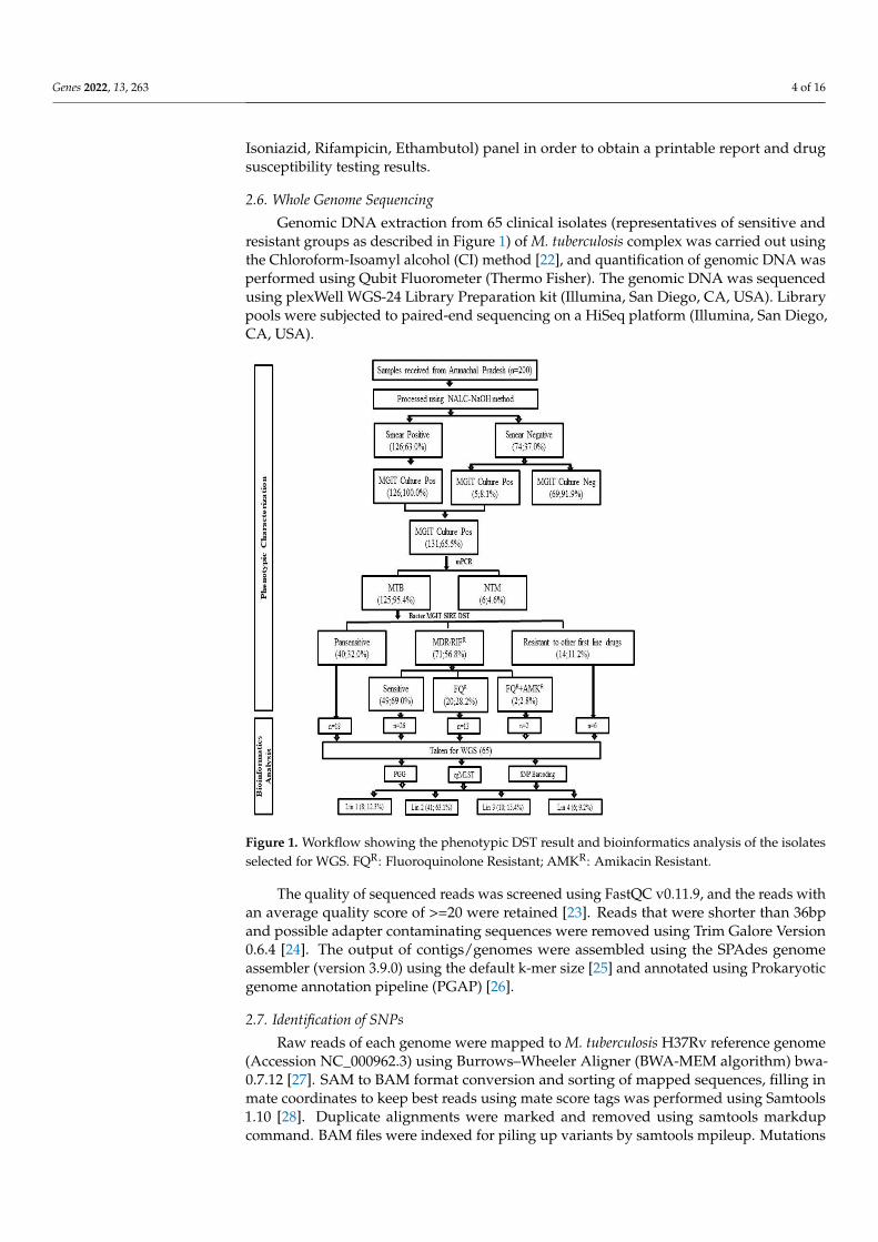
Genomic DNA extraction from 65 clinical isolates (representatives of sensitive andresistant groups as described in Figure 1) of M. tuberculosis complex was carried out usingthe Chloroform-Isoamyl alcohol (CI) method [22], and quantification of genomic DNA wasperformed using Qubit Fluorometer (Thermo Fisher). The genomic DNA was sequencedusing plexWell WGS-24 Library Preparation kit (Illumina, San Diego, CA, USA). Librarypools were subjected to paired-end sequencing on a HiSeq platform (Illumina, San Diego,CA, USA).
Figure 1. Workflow showing the phenotypic DST result and bioinformatics analysis of the isolatesselected for WGS. FQR: Fluoroquinolone Resistant; AMKR: Amikacin Resistant.
The quality of sequenced reads was screened using FastQC v0.11.9, and the reads withan average quality score of >=20 were retained [23]. Reads that were shorter than 36bpand possible adapter contaminating sequences were removed using Trim Galore Version0.6.4 [24]. The output of contigs/genomes were assembled using the SPAdes genomeassembler (version 3.9.0) using the default k-mer size [25] and annotated using Prokaryoticgenome annotation pipeline (PGAP) [26].
2.7. Identification of SNPs
Raw reads of each genome were mapped to M. tuberculosis H37Rv reference genome(Accession NC_000962.3) using Burrows–Wheeler Aligner (BWA-MEM algorithm) bwa-0.7.12 [27]. SAM to BAM format conversion and sorting of mapped sequences, filling inmate coordinates to keep best reads using mate score tags was performed using Samtools1.10 [28]. Duplicate alignments were marked and removed using samtools markdupcommand. BAM files were indexed for piling up variants by samtools mpileup. Mutations
Genes 2022, 13, 263 5 of 16
with read depth above 10 reads were considered true mutations. Annotation and filteringof variants were conducted using SNPEFF 5.0e [29] and SnpSift [30].
2.8. Assignment of Principal Genetic Groups
To assign a principal genetic group (PGG), each sequenced isolate was manuallyscreened for polymorphisms in gyrA codon 95 and KatG codon 463 and were categorizedaccordingly to PGG as 1, 2, or 3, respectively, as explained previously [31].
2.9. Identification of Lineages and Sub-Lineages Using WGS SNP Barcoding
Isolates based on the patterns of SNP at the designated loci were categorized intophylogenetic lineages groups as lineage 1 (Indo-Oceanic), lineage 2 (East Asian), lineage 3(East African Indian), lineage 4 (Euro-American), lineage 5 (West Africa 1), lineage 6 (WestAfrica 2), and lineage 7 (Horn of Africa) and lineage 8 [13]. After splitting WGS isolatesinto lineages, further categorization was conducted on the basis of SNP’s [13]. Constructionof the UPGMA tree was conducted by concatenating SNPs and visualized using iToL V.6software [32].
2.10. Phylogenetic Analysis and Construction of cgMLST
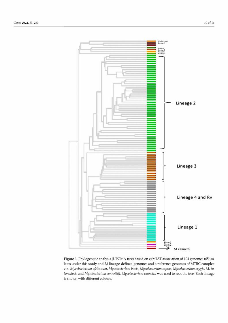
Schema for cgMLST was set up with an efficient Workflow for a Blast Score RatioBased Allele Calling Algorithm (Chew BBACA) [33]. For construction of cgMLST, first awgMLST schema was created using M. tuberculosis H37Rv (accession: NC_000962.3) as atraining file generated by the prodigal algorithm. The wgMLST schema contained 10058loci based on 104 genomes (65 genomes sequenced under this study, 39 complete and draftgenomes downloaded from NCBI). Complete genome sequences of M. tuberculosis complexviz; M. bovis (NC_002945.4), M. orygis (CP063804.1), M. africanum (FR878060.1), M. cannettii(NC_015848.1), M. tuberculosis (NC_000962.3) and M. caprae (NZ_CDHG01000001.1) wereextracted from NCBI. Draft genomes from NCBI database that belong to Lineage 1 (PR-JNA235648, PRJNA223559, PRJNA229273, PRJNA229212, PRJNA229320, PRJNA229266),Lineage 2 (PRJNA219760, PRJNA226779, PRJNA267047, CCDC5180, CCDC5079, PR-JNA229630, NDYV00000000, PRJNA360122), Lineage 3 (PRJNA229310, PRJNA229235,PRJNA229259, NDYU00000000), Lineage 4 (PRJNA229257, PRJNA223558, PRJNA229237,PRJNA228063, PRJNA228052, PRJNA229638, PRJNA218312, PRJNA233359, PRJNA233363),Lineage 5 (PRJNA211660), Lineage 6 (PRJNA211707, PRJNA211702), Lineage 7 (PRJEB8432)and Lineage 8 (PRJNA598991) and lineage B (PRJNA229213), were extracted and used asrepresentative for M. tuberculosis lineage 1-8. These 39 publicly available complete genomeswere used for validation of the cgMLST schema. The resulting loci was then subjectedto AlleleCall, which identified and excluded 104 possible paralogous loci from furtherdownstream analysis using the default BLAST Score Ratio (BSR) threshold of 0.6. Finally,cgMLST was extracted containing a set of 1443 core loci (present in 100% of the isolates).The resulting cgMLST matrix was uploaded in phyloviz 2.0 [34] to generate and visualizeUPGMA Tree. The genome of M. cannettii was used to root the tree.
2.11. Data Analysis
All data obtained from gDST, phenotypic DST, PGG, and categorization of lineage onthe basis of SNP barcoding were maintained on MS Excel 2013 for further analysis.
3. Results3.1. Demographic Details and Characteristics of MDR-TB Patients
Of the total 200 patients included in the study, 91 (45.5%) were males and 109 femaleswith mean age (± standard deviation) of 29.52 ± 13.21 and 27.85 ± 14.17 years, respectively.The majority of cases were adults, 183 (91.5%), and 17 (8.5%) were from the pediatricage group.
Genes 2022, 13, 263 6 of 16
3.2. Bactec MGIT 960 Culture Results and Identification M. tuberculosis Complex Isolates
One hundred and twenty six of 200 (63%) samples were smear-positive, and 74 (37%)smear-negative. Of the total 200 cultures inoculated in Bactec MGIT 960 145 (72.5%)were flagged positive with an average turnaround time (TAT) of 18 days. All flaggedpositive cultures were further confirmed by ZN-stained smear examination, of which131/145 (90.3%) were smear-positive for AFB while 14/145 (9.7%) were contaminated.Of 131 cultures, in-house multiplex PCR identified 6 (4.6%) cultures as Non-tuberculosisMycobacterium and 125 (95.4%) cultures as M. tuberculosis complex (Figure 1).
3.3. Bactec MGIT 960 SIRE DST
Of total 125 cultures, Bactec MGIT 960 SIRE DST detected 66 (52.8%) as MDR-TB(resistant to both RIF and INH), 5 (4.0%) as mono-resistant to RIF, and 14 (11.2%) weredrug-resistant isolates (resistant to any of first-line drug other than MDR/RR). 40 (32.0%)isolates were detected as pan-sensitive (Table 1).
Table 1. Association of first-line drug-resistant patterns with second-line DST on Bactec MGIT-960.
SIRE Drug Susceptibility Pattern by MGIT 960 Second Line Drug Susceptibility Pattern by MGIT 960STR INH RIF EMB n % AMK MFX LFX LNZ n %R R R R 37 29.6% S R R S 13 35.1%
R R R S 2 5.4%S R S S 2 5.4%S S S S 20 54.1%
S R R S 15 12.0% S R R S 2 13.3%S S S S 13 86.7%
S R R R 5 4.0% S R R S 2 40.0%S S S S 3 60.0%
R R R S 9 7.2% S R R S 1 11.1%S S S S 8 88.9%
S S R S 3 2.4% S S S S 3 100.%S R S S 8 6.4% − − − − − −S S R R 1 0.8% S S S S 1 100%R S R S 1 0.8% S S S S 1 100%R R S R 2 1.6% − − − − − −R S S S 1 0.8% − − − − − −S S S R 2 1.6% − − − − − −R R S S 1 0.8% − − − − − −S S S S 40 32.0% − − − − − −STRS = 74STRR = 51
INHS = 48INHR = 77
RIFS = 54RIFR = 71
EMBs = 78EMBR = 47 n = 125
AMKS =69AMKR =2
MFXS = 49MFXR = 22
LFXS =51LFXR = 20 LNZS = 71 n = 71
STRS: Streptomycin susceptible; STRR: Streptomycin resistant; INHS: Isoniazid susceptible; INHR: Isoniazidresistant; RIFS: Rifampicin susceptible; RIFR: Rifampicin resistant; EMBs: Ethambutol susceptible; EMBR:Ethambutol resistant; AMKS: Amikacin susceptible; AMKR: Amikacin resistant; MFXS: Moxifloxacin susceptible;MFXR: Moxifloxacin resistant; LFXS: Levofloxacin susceptible; LFXR: Levofloxacin resistant; LNZS: Linezolidsusceptible.
3.4. Bactec MGIT 960 Second Line DST
Of 71 (56.8%) MDR-TB and RIF mono-resistant isolates subjected to second-line DSTfor drugs AMK (1 mg/mL), LFX(1 mg/mL), LNZ(1 mg/mL), and MOX(1 mg/mL), 49(69.0%) were found to be susceptible, 20 (28.2%) were mono-resistant to FQ, [2(2.8%) wasresistant to only MOX and 18 (25.4%) to (MFX+LFX)]; 2 (2.8%) isolates were found to beresistant to (FQ+AMK) while no isolates were found mono-resistant to AMK or resistant toLNZ. Patterns of first- and second-line drugs are shown in Table 1.
3.5. Mutations in Genes Associated with First- and Second-Line Drugs Using WGS
A total of 65 isolates sequenced were analyzed for mutations conferring drug resistancein genes associated with first-line and second-line drug resistance (Table 2). Genomessequences of each isolate were screened for mutations in genes conferring resistance tofirst-line anti-tuberculosis drugs viz; rpsL, rrs, gidB for STR; KatG, inhA, ahpA, fab, ndhfor INH; rpoA, rpoB, rpoC for RIF; embABC for EMB and pncA for PZA.
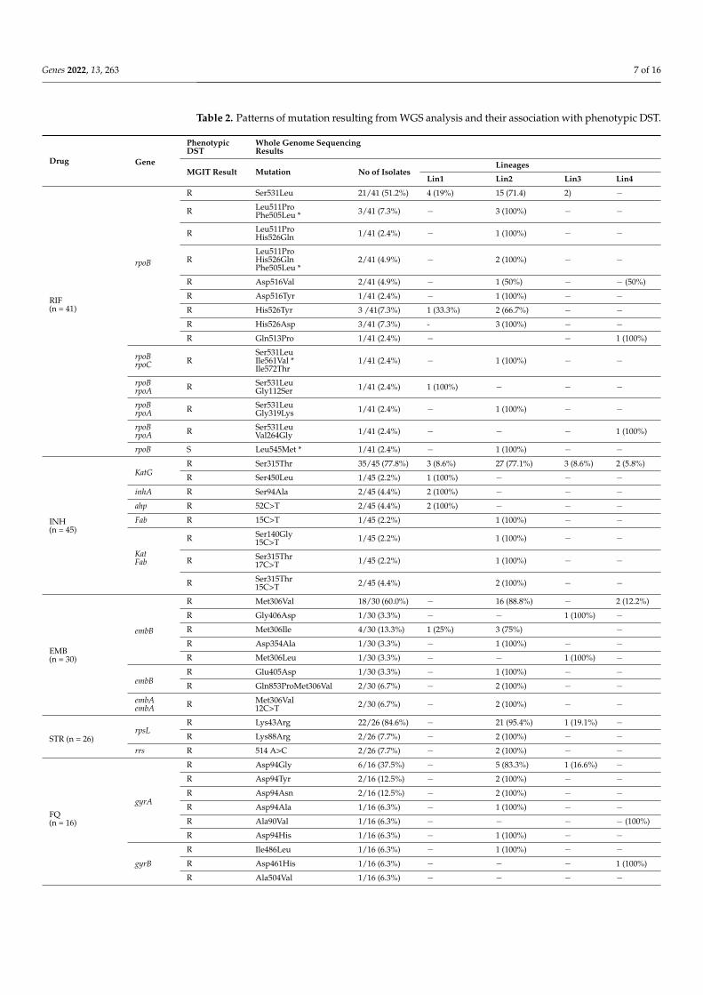
Genes 2022, 13, 263 7 of 16
Table 2. Patterns of mutation resulting from WGS analysis and their association with phenotypic DST.
Drug Gene
PhenotypicDST
Whole Genome SequencingResults
MGIT Result Mutation No of IsolatesLineages
Lin1 Lin2 Lin3 Lin4
RIF(n = 41)
rpoB
R Ser531Leu 21/41 (51.2%) 4 (19%) 15 (71.4) 2) −
R Leu511ProPhe505Leu * 3/41 (7.3%) − 3 (100%) − −
R Leu511ProHis526Gln 1/41 (2.4%) − 1 (100%) − −
RLeu511ProHis526GlnPhe505Leu *
2/41 (4.9%) − 2 (100%) − −
R Asp516Val 2/41 (4.9%) − 1 (50%) − − (50%)
R Asp516Tyr 1/41 (2.4%) − 1 (100%) − −R His526Tyr 3 /41(7.3%) 1 (33.3%) 2 (66.7%) − −R His526Asp 3/41 (7.3%) - 3 (100%) − −R Gln513Pro 1/41 (2.4%) − − 1 (100%)
rpoBrpoC R
Ser531LeuIle561Val *Ile572Thr
1/41 (2.4%) − 1 (100%) − −
rpoBrpoA R Ser531Leu
Gly112Ser 1/41 (2.4%) 1 (100%) − − −
rpoBrpoA R Ser531Leu
Gly319Lys 1/41 (2.4%) − 1 (100%) − −
rpoBrpoA R Ser531Leu
Val264Gly 1/41 (2.4%) − − − 1 (100%)
rpoB S Leu545Met * 1/41 (2.4%) − 1 (100%) − −
INH(n = 45)
KatGR Ser315Thr 35/45 (77.8%) 3 (8.6%) 27 (77.1%) 3 (8.6%) 2 (5.8%)
R Ser450Leu 1/45 (2.2%) 1 (100%) − − −inhA R Ser94Ala 2/45 (4.4%) 2 (100%) − − −ahp R 52C>T 2/45 (4.4%) 2 (100%) − − −Fab R 15C>T 1/45 (2.2%) 1 (100%) − −
KatFab
R Ser140Gly15C>T 1/45 (2.2%) 1 (100%) − −
R Ser315Thr17C>T 1/45 (2.2%) 1 (100%) − −
R Ser315Thr15C>T 2/45 (4.4%) 2 (100%) − −
EMB(n = 30)
embB
R Met306Val 18/30 (60.0%) − 16 (88.8%) − 2 (12.2%)
R Gly406Asp 1/30 (3.3%) − − 1 (100%) −R Met306Ile 4/30 (13.3%) 1 (25%) 3 (75%) −R Asp354Ala 1/30 (3.3%) − 1 (100%) − −R Met306Leu 1/30 (3.3%) − − 1 (100%) −
embBR Glu405Asp 1/30 (3.3%) − 1 (100%) − −R Gln853ProMet306Val 2/30 (6.7%) − 2 (100%) − −
embAembA R Met306Val
12C>T 2/30 (6.7%) − 2 (100%) − −
STR (n = 26)rpsL
R Lys43Arg 22/26 (84.6%) − 21 (95.4%) 1 (19.1%) −R Lys88Arg 2/26 (7.7%) − 2 (100%) − −
rrs R 514 A>C 2/26 (7.7%) − 2 (100%) − −
FQ(n = 16)
gyrA
R Asp94Gly 6/16 (37.5%) − 5 (83.3%) 1 (16.6%) −R Asp94Tyr 2/16 (12.5%) − 2 (100%) − −R Asp94Asn 2/16 (12.5%) − 2 (100%) − −R Asp94Ala 1/16 (6.3%) − 1 (100%) − −R Ala90Val 1/16 (6.3%) − − − − (100%)
R Asp94His 1/16 (6.3%) − 1 (100%) − −
gyrB
R Ile486Leu 1/16 (6.3%) − 1 (100%) − −R Asp461His 1/16 (6.3%) − − − 1 (100%)
R Ala504Val 1/16 (6.3%) − − − −
Genes 2022, 13, 263 8 of 16
Table 2. Cont.
Drug Gene
PhenotypicDST
Whole Genome SequencingResults
MGIT Result Mutation No of IsolatesLineages
Lin1 Lin2 Lin3 Lin4
PZA(n = 10) pncA
R Asp49Ala 5/10 (50.0%) − 5 (100%) − −R Gly108Arg 2/10 (20.0%) − 2 (100%) − −R 11A>G 2/10 (20.0%) − 2 (100%) − −R Asp136Tyr 1/10 (10.0%) 1 (100%) − − −
AMK (n = 2) rrsR 1484 G>T 1/2 (50.0%) − 1 (100%) − −R 1401 A>G 1/2 (50.0%) − 1 (100%) − −
ETH(n = 8)
inha NA Ser94Ala 2/8 (25.0%) 2 (100%) − − −fab NA 15C>T 3/8 (37.5%) − 3 (100%) − −
NA 17G>T 1/8 (12.5%) − − − 1 (100%)
ethA NA 886_886del 2/8(25.0%) − 1 (50%) − 1 (50%)
Cysr (n = 2) alr NA Met343Thr 2/2 (100.0%) − 2 (100%) − −
PAS(n = 2)
thy NA 16C>T 1/2 (50.0%) − 1 (100%) − −folC NA Ile43Thr 1/2 (50.0%) − 1 (100%) − −
STR: Streptomycin; INH: Isoniazid; RIF: Rifampicin; EMB: Ethambutol; PZA: Pyrazinamide; ETH: Ethionamide;FQ: Fluoroquinolone; AMK: Amikacin; PAS: Para-aminosalicylic acid; Cysr.: Cycloserine; S: susceptible; R: resis-tant; * outside Rifampicin Resistance Determining Region (RRDR); n=number of isolates.
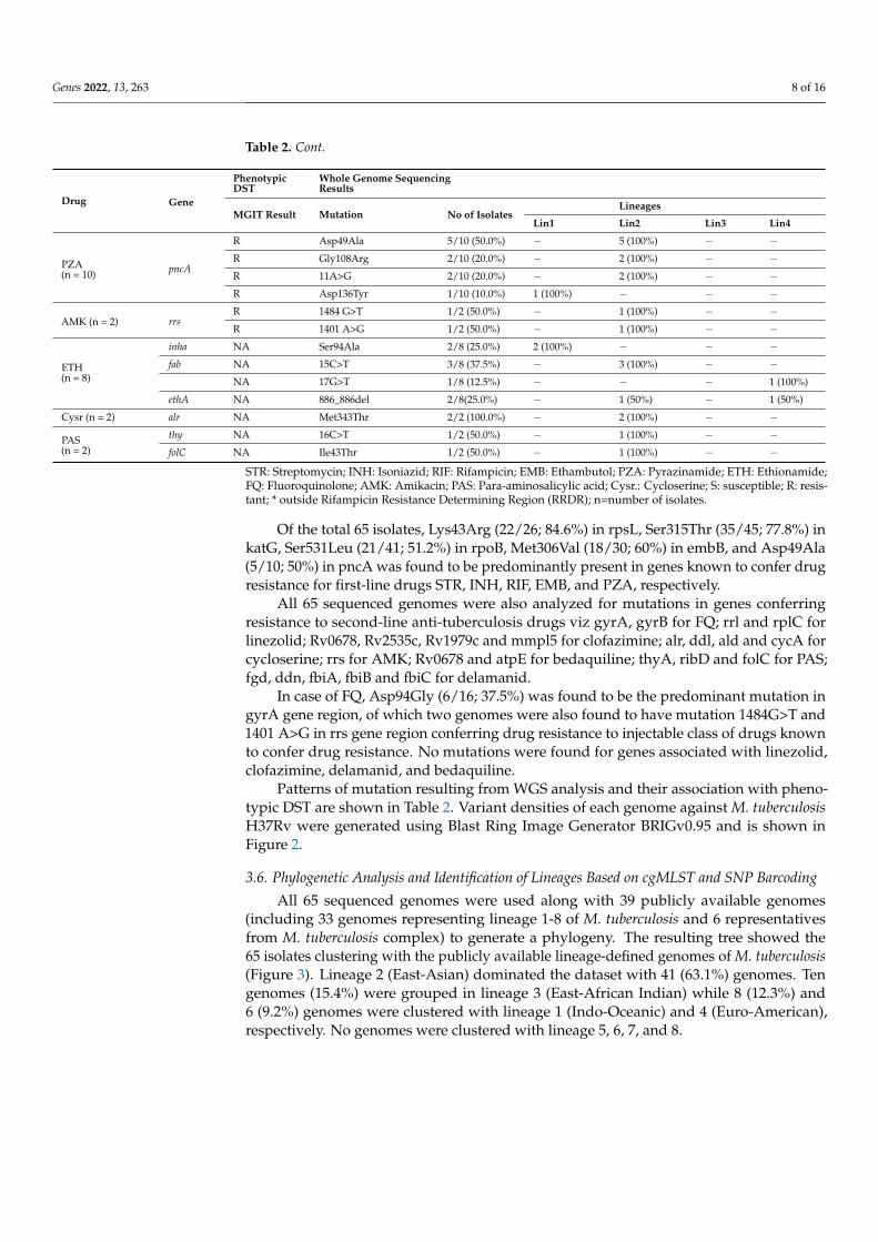
Of the total 65 isolates, Lys43Arg (22/26; 84.6%) in rpsL, Ser315Thr (35/45; 77.8%) inkatG, Ser531Leu (21/41; 51.2%) in rpoB, Met306Val (18/30; 60%) in embB, and Asp49Ala(5/10; 50%) in pncA was found to be predominantly present in genes known to confer drugresistance for first-line drugs STR, INH, RIF, EMB, and PZA, respectively.
All 65 sequenced genomes were also analyzed for mutations in genes conferringresistance to second-line anti-tuberculosis drugs viz gyrA, gyrB for FQ; rrl and rplC forlinezolid; Rv0678, Rv2535c, Rv1979c and mmpl5 for clofazimine; alr, ddl, ald and cycA forcycloserine; rrs for AMK; Rv0678 and atpE for bedaquiline; thyA, ribD and folC for PAS;fgd, ddn, fbiA, fbiB and fbiC for delamanid.
In case of FQ, Asp94Gly (6/16; 37.5%) was found to be the predominant mutation ingyrA gene region, of which two genomes were also found to have mutation 1484G>T and1401 A>G in rrs gene region conferring drug resistance to injectable class of drugs knownto confer drug resistance. No mutations were found for genes associated with linezolid,clofazimine, delamanid, and bedaquiline.
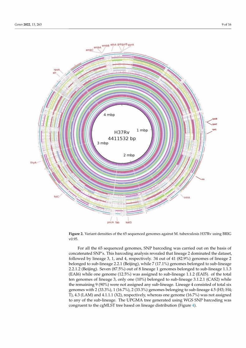
Patterns of mutation resulting from WGS analysis and their association with pheno-typic DST are shown in Table 2. Variant densities of each genome against M. tuberculosisH37Rv were generated using Blast Ring Image Generator BRIGv0.95 and is shown inFigure 2.
3.6. Phylogenetic Analysis and Identification of Lineages Based on cgMLST and SNP Barcoding
All 65 sequenced genomes were used along with 39 publicly available genomes(including 33 genomes representing lineage 1-8 of M. tuberculosis and 6 representativesfrom M. tuberculosis complex) to generate a phylogeny. The resulting tree showed the65 isolates clustering with the publicly available lineage-defined genomes of M. tuberculosis(Figure 3). Lineage 2 (East-Asian) dominated the dataset with 41 (63.1%) genomes. Tengenomes (15.4%) were grouped in lineage 3 (East-African Indian) while 8 (12.3%) and6 (9.2%) genomes were clustered with lineage 1 (Indo-Oceanic) and 4 (Euro-American),respectively. No genomes were clustered with lineage 5, 6, 7, and 8.
Genes 2022, 13, 263 9 of 16
Figure 2. Variant densities of the 65 sequenced genomes against M. tuberculosis H37Rv using BRIGv0.95.
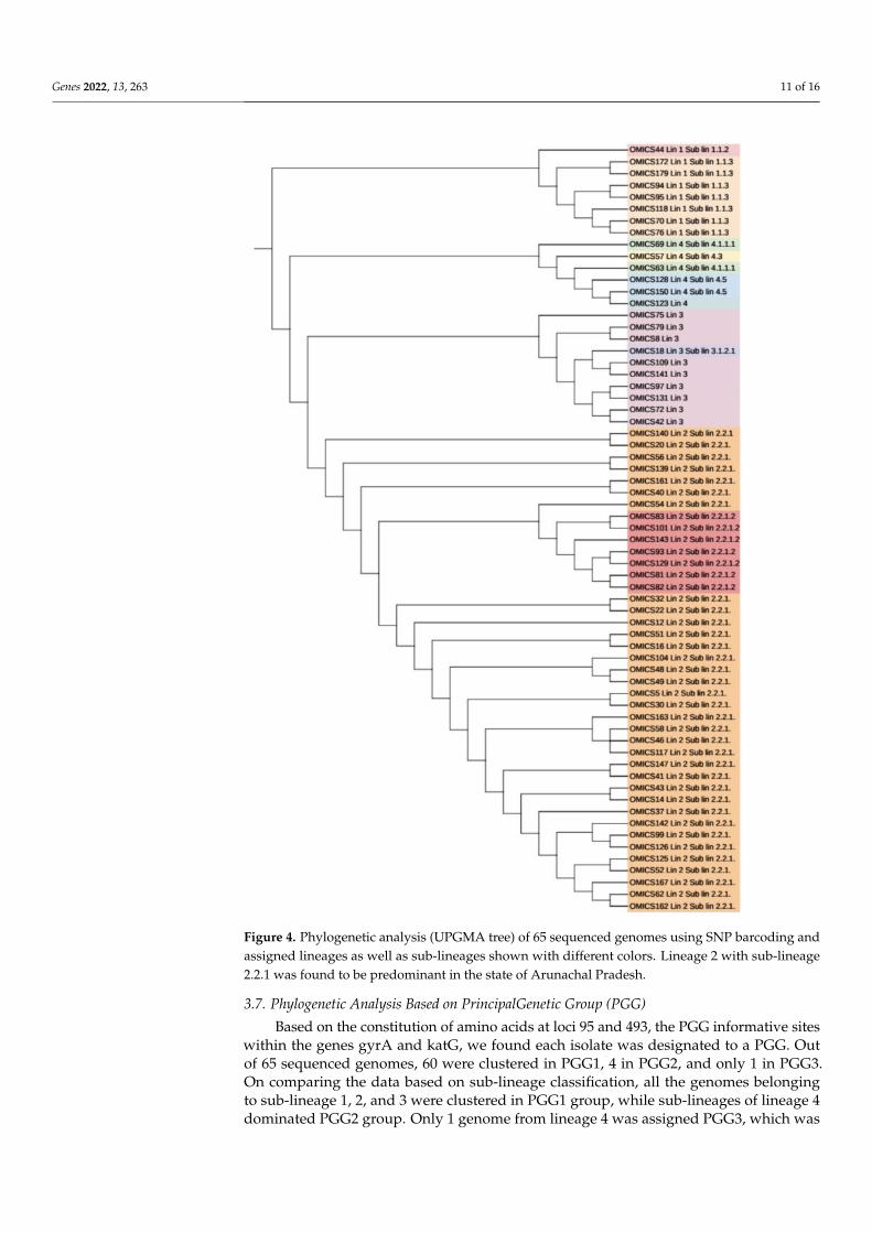
For all the 65 sequenced genomes, SNP barcoding was carried out on the basis ofconcatenated SNP’s. This barcoding analysis revealed that lineage 2 dominated the dataset,followed by lineage 3, 1, and 4, respectively. 34 out of 41 (82.9%) genomes of lineage 2belonged to sub-lineage 2.2.1 (Beijing), while 7 (17.1%) genomes belonged to sub-lineage2.2.1.2 (Beijing). Seven (87.5%) out of 8 lineage 1 genomes belonged to sub-lineage 1.1.3(EAI6) while one genome (12.5%) was assigned to sub-lineage 1.1.2 (EAI5). of the totalten genomes of lineage 3, only one (10%) belonged to sub-lineage 3.1.2.1 (CAS2) whilethe remaining 9 (90%) were not assigned any sub-lineage. Lineage 4 consisted of total sixgenomes with 2 (33.3%), 1 (16.7%), 2 (33.3%) genomes belonging to sub-lineage 4.5 (H3; H4;T), 4.3 (LAM) and 4.1.1.1 (X2), respectively, whereas one genome (16.7%) was not assignedto any of the sub-lineage. The UPGMA tree generated using WGS SNP barcoding wascongruent to the cgMLST tree based on lineage distribution (Figure 4).
Genes 2022, 13, 263 10 of 16
Figure 3. Phylogenetic analysis (UPGMA tree) based on cgMLST association of 104 genomes (65 iso-lates under this study and 33 lineage-defined genomes and 6 reference genomes of MTBC complexviz. Mycobacterium africanum, Mycobacterium bovis, Mycobacterium caprae, Mycobacterium orygis, M. tu-berculosis and Mycobacterium cannettii). Mycobacterium cannettii was used to root the tree. Each lineageis shown with different colours.
Genes 2022, 13, 263 11 of 16
Figure 4. Phylogenetic analysis (UPGMA tree) of 65 sequenced genomes using SNP barcoding andassigned lineages as well as sub-lineages shown with different colors. Lineage 2 with sub-lineage2.2.1 was found to be predominant in the state of Arunachal Pradesh.
3.7. Phylogenetic Analysis Based on PrincipalGenetic Group (PGG)
Based on the constitution of amino acids at loci 95 and 493, the PGG informative siteswithin the genes gyrA and katG, we found each isolate was designated to a PGG. Outof 65 sequenced genomes, 60 were clustered in PGG1, 4 in PGG2, and only 1 in PGG3.On comparing the data based on sub-lineage classification, all the genomes belongingto sub-lineage 1, 2, and 3 were clustered in PGG1 group, while sub-lineages of lineage 4dominated PGG2 group. Only 1 genome from lineage 4 was assigned PGG3, which was
Genes 2022, 13, 263 12 of 16
a pre-XDR isolate. Assignment of PGG and sub-lineage classification for each isolate isshown in Supplementary Table S1.
4. Discussion
With increasing drug-resistant TB cases in India, it becomes pivotal to recognizethe clonal expansion of lineages or clones contributing to drug resistance, specifically ingeographical regions where drug resistance is suspected [35]. Northeastern states of Indiahave higher rates of MDR-TB, around 32.7% of which Arunachal Pradesh region is knownto have 78.8% of TB drug resistance [5,36]. Most of the land in Arunachal Pradesh is underforest area, and villages located in such impoverished forest zones have very inadequate ornon-existent access to health services, including the hampered efforts of the national TBelimination program. Further, proximity to China, difficult terrains, and heavy rainfalls andlandslides impact health services, leading to a high MDR rate. Nonetheless, host geneticsalso might be contributing to high resistance in this area, but that aspect was not studied inthis work, and we are not making any conclusions on the genetic reasons for high MDR.Whole-genome sequencing (WGS) has become a standard for typing of M. tuberculosisisolates and is known to have higher resolution over MIRU-VNTR-based clustering [37]. Inthis study we planned to utilize the approach of WGS and SNP barcoding for typing andclustering of M. tuberculosis lineages circulating in the Arunachal Pradesh region of India.This study will help in defining the transmission of strains associated with drug resistancecirculating in the Arunachal Pradesh region of India.
Our report for MDR-TB from Arunachal Pradesh is 56.8%, slightly lower than pre-viously reported 79.3%. The variation may be due to a lower number of sample sizesin the previous study, results based on DNA-based line probe Assay rather than BactecMGIT 960, which detects viable bacilli and incorrect denominators used while calculatingdrug resistance rates [36]. In our study, 80% of the samples were obtained from previouslytreated TB cases resulting in an increased rate of drug resistance. However, no reports ofFQ resistance were reported from Arunachal Pradesh earlier.
There is mounting evidence that strain diversity plays a role in the transmission ofdisease [38]. In order to spot strain transmission in Arunachal Pradesh, we randomlyselected cultures for WGS from each category of varying data sets of drug-resistant patternsand used gene by gene PGG, SNP barcoding, and core genome MLST (cgMLST) methodto allocate strains in well-defined phylogenetic groupings (Figure 1). All these methodswere used in various phylogenetic studies for standardized phylogenetic assignment ofdiseases and outbreak resolutions [13,39,40]. The PGG results also correlated with resultsof lineage grouping by SNP barcoding as 60 (92.3%) isolates belong to PGG group 1 (KatGLeu463Leu, gyrA Thr95Thr) of which 8 (13.3%) were Lineage 1 (Indo Oceanic), 41 (68.3%)Lineage 2 (East Asian) and 10 (16.7%) Lineage 3 (East-Arican Indian) and 1 (1.7%) Lineage 4(Euro American). PGG group 2 (KatG Leu463Arg, gyrA Thr95Thr) and PGG group 3 (KatGLeu463Arg, gyrA Thr95Ser) included 4 (6.2%) and 1 (1.5%) strain belonging to lineage 4.Categorization of lineage as per the PGG group reported from other studies was consistentwith our finding [31,40] (Supplementary Table S1).
This study provided for the first time a complete picture of TB phylogenetics acrossArunachal Pradesh region based on cgMLST compared to phylogenetics that is based onSNP calling methods. The resulting cgMLST phylogenetic tree contained all referencelineages and four major M. tuberculosis lineages from our dataset and matched with re-sults of SNP-based methods. None of the genomes belong to Lineage 5-8, and all wereM. tuberculosis sensu stricto (Figure 1).
Using SNP barcoding method, we found 41 (63.1%) isolates grouped with lineage2 (East Asian); 10 (15.4%) isolates with lineage 3 (Central Asian), 8 (12.3%) isolates withlineage 1, and 6 (9.2%) isolates to be lineage 4 (Supplementary Table S1). We found Lineage2 (East Asian) as the predominant lineage (63.1%) circulating in Arunachal Pradesh amongsuspected drug-resistant TB cases. To gain further insight, we looked for SNP-based mark-ers to differentiate sub-lineages. Among Lineage 2 only two clones circulating in Arunachal
Genes 2022, 13, 263 13 of 16
Pradesh were found viz: sub-lineage 2.2.1 (82.9%) and 2.2.1.2 (17.1%), respectively. Boththese sub-lineages 2.2.1 and 2.2.1.2 belonged to modern Beijing clade, which was depictedby the presence of SNP markers at mutT2 codon Gly58Ala, ogt Gly12Gly specific to modernBeijing as reported in earlier studies [40,41]. Of a total of 6535 SNP’s, Beijing clone 2.2.1was responsible for >80% of transmission and clusters using thresholds of up to 19 SNPsshowing recent transmission of strains. Another clone of Beijing 2.2.1.2 showed clusterswith SNP differences of 9 SNPs, showing ongoing transmission of the strains specificallyassociated with drug resistance. All these 7 (100.0%) isolates of clone 2.2.1.2 were multidrug-resistant, and 2 (28.6%) were resistant to FQ. Beijing sub-lineages 2.2.1 and 2.2.1.2 wasreported from various parts of the world associated with outbreaks and drug resistancefrom Vietnam and Southern China [42–47]. We also observed Lineage 1 clone 1.1.3 withSNP differences of 101, showing this clone as endemic in Arunachal Pradesh for longer timeperiods. Lineage 1 is known to be associated with activation of long-term latent infectioncompared to that of Lineage 2 (modern Beijing) strains, which are known for more likelyto progress to active disease in various host populations, more virulent, and thus highlytransmissible [48,49]. A total of 6 isolates of lineage 4 were identified and were unclustered.Of Lineage 3, one clone including 9 (90%) isolates was found showing SNP differences of18, also showing ongoing transmission. Out of 9 isolates, 3 (33.3%) were MDR-TB. Oneisolate (10%) of sub-lineage 3.1.2.1 was also found and was MDR-TB as well as resistant toFQ. New clades of lineage 3 were also reported to be circulating in the Assam region ofIndia by Devi et al., which is consistent with our study [50].
The main strength of our study is that it highlights the higher rates of drug resistancein the Indian state of Arunachal Pradesh, which has a common border with China, andprovides insights into the phylogenetic diversity of MDR-TB isolates from this state usingcgMLST. These findings may have important implications in understanding the molecularepidemiology of DR-TB and for its control and prevention, particularly in the state ofArunachal Pradesh. However, our study has some limitations. The main limitation wasthat we could not include a control group from other states/regions of India in orderto compare the strain diversity, specifically the Beijing sub-lineage 2.2.1 and 2.2.1.2, itsassociation with drug resistance.
5. Conclusions
Our findings show dissemination of clusters of Beijing clones associated with drugresistance and regional spread may be emerging and aggressive. Approaches to containBeijing strains (sub-lineage 2.2.1 and 2.2.1.2) may prevent transmission of these strainsacross other parts of India. Clonal expansion of these strains in Arunachal Pradesh in thefuture may lead to an outbreak of Beijing strains and underline the need for surveillancestudies incorporating epidemiological information and a track of ongoing transmission toprevent drug-resistant TB outbreaks. We also found transmission of lineage 3 clade andthe presence of Lineage 1 as endemic in Arunachal Pradesh. These findings may haveimportant implications for control and prevention of TB in the northeastern part of India,Arunachal Pradesh.
Supplementary Materials: The following supporting information can be downloaded at: https://www.mdpi.com/article/10.3390/genes13020263/s1, Table S1.
Author Contributions: S.k.R.M. standardized and performed all experiments and analyzed the data.U.K. performed bioinformatic analysis and contributed to manuscript writing. S.B.R. contributed toreviewing and writing of manuscripts. M.U. and M.N. helped in the collection of TB samples andmanaged the transportation of samples to AIIMS Bhopal. S.S. supervised and coordinated the work,finalized the manuscript, arranged reagents and chemicals. All authors have read and agreed to thepublished version of the manuscript.
Funding: This research and APC was funded by the Department of Biotechnology (DBT), Govern-ment of India grant number MDR-TB/2017/11 provided to Prof. Sarman Singh.
Genes 2022, 13, 263 14 of 16
Institutional Review Board Statement: The study was conducted in accordance with the ethicalclearance committee at TRIHMS Arunachal Pradesh and AIIMS Bhopal under reference numberDME (T&R)/IEC/2015/1 and IHEC-LOP/2018/EF0104, respectively.
Informed Consent Statement: Informed consent was obtained from all subjects involved in the study.
Data Availability Statement: Genomes of sixty-five M. tuberculosis isolates have been deposited inGenBank under BioProject accession no. PRJNA717132. Raw reads of all sixty-five M. tuberculosishave been made available in the Sequence Read Archive (SRA) under study number SRR331414linked with BioProject number PRJNA717132.
Acknowledgments: We wish to thank Payal Soni and Mukesh Patel for their technical help.
Conflicts of Interest: The authors declare no conflict of interest.
References1. World Health Organization. Global Tuberculosis Report 2020; World Health Organization: Geneva, Switzerland, 2020.2. National Tuberculosis Elimination Programme. India TB Report 2020; National Tuberculosis Elimination Programme: New Delhi,
India, 2020.3. Lange, C.; Dheda, K.; Chesov, D.; Mandalakas, A.M.; Udwadia, Z.; Horsburgh, C.R. Management of drug-resistant tuberculosis.
Lancet 2019, 394, 953–966. [CrossRef]4. Husain, A.A.; Kupz, A.; Kashyap, R.S. Controlling the drug-resistant tuberculosis epidemic in India: Challenges and impli-cations.
Epidemiol. Health 2021, 43, e2021022. [CrossRef] [PubMed]5. Singh, S. Early detection of multi-drug resistant tuberculosis in India using GenoType MTBDRplus assay & profile of resistance
mutations in Mycobacterium tuberculosis. Indian J. Med Res. 2014, 140, 477–479.6. Gao, Q.; Kripke, K.E.; Saldanha, A.J.; Yan, W.; Holmes, S.; Small, P.M. Gene expression diversity among Mycobacterium tuber-culosis
clinical isolates. Microbiology 2005, 151, 5–14. [CrossRef]7. Anh, D.D.; Borgdorff, M.W.; Van, L.N.; Lan, N.T.; van Gorkom, T.; Kremer, K.; van Soolingen, D. Mycobacterium tuberculosis Beijing
genotype emerging in Vietnam. Emerg. Infect. Dis. 2000, 6, 302–305. [PubMed]8. Kato-Maeda, M.; Bifani, P.J.; Kreiswirth, B.N.; Small, P.M. The nature and consequence of genetic variability within Mycobacterium
tuberculosis. J. Clin. Invest. 2001, 107, 533–537. [CrossRef] [PubMed]9. Mekonnen, A.; Merker, M.; Collins, J.; Addise, D.; Aseffa, A.; Petros, B.; Ameni, G.; Niemann, S. Molecular epidemiology and
drug resistance patterns of Mycobacterium tuberculosis complex isolates from university students and the local community inEastern Ethiopia. PLoS ONE 2018, 13, e0198054. [CrossRef] [PubMed]
10. Ford, C.; Yusim, K.; Ioerger, T.; Feng, S.; Chase, M.; Greene, M.; Korber, B.; Fortune, S. Mycobacterium tuberculosis—Heterogeneityrevealed through whole genome sequencing. Tuberculosis 2012, 92, 194–201. [CrossRef]
11. Cohen, K.A.; Manson, A.L.; Desjardins, C.A.; Abeel, T.; Earl, A.M. Deciphering drug resistance in Mycobacterium tuberculosis usingwhole-genome sequencing: Progress, promise, and challenges. Genome Med. 2019, 11, 45. [CrossRef]
12. Gagneux, S.; DeRiemer, K.; Van, T.; Kato-Maeda, M.; de Jong, B.; Narayanan, S.; Nicol, M.; Niemann, S.; Kremer, K.; Gutierrez,M.C.; et al. Variable host-pathogen compatibility in Mycobacterium tuberculosis. Proc. Natl. Acad. Sci. USA 2006, 103, 2869–2873.[CrossRef]
13. Kohl, T.A.; Diel, R.; Harmsen, D.; Rothgänger, J.; Walter, K.M.; Merker, M.; Weniger, T.; Niemann, S. Whole-genome-basedMy-cobacterium tuberculosis surveillance: A standardized, portable, and expandable approach. J. Clin. Microbiol. 2014, 5, 2479–2486.[CrossRef]
14. Jones, R.C.; Harris, L.G.; Morgan, S.; Ruddy, M.C.; Perry, M.; Williams, R.; Humphrey, T.; Temple, M.; Davies, A.P. PhylogeneticAnalysis of Mycobacterium tuberculosis Strains in Wales by Use of Core Genome Multilocus Sequence Typing to Analyze Whole-Genome Sequencing Data. J. Clin. Microbiol. 2019, 57, 1–11. [CrossRef] [PubMed]
15. Rufai, S.B.; Singh, A.; Kumar, P.; Singh, J.; Singh, S. Performance of Xpert MTB/RIF Assay in Diagnosis of Pleural Tuberculosis byUse of Pleural Fluid Samples. J. Clin. Microbiol. 2015, 53, 3636–3638. [CrossRef]
16. Gopinath, K.; Singh, S. Multiplex PCR assay for simultaneous detection and differentiation of Mycobacterium tuberculosis,Mycobacterium avium complexes and other Mycobacterial species directly from clinical specimens. J. Appl. Microbiol. 2009, 107,425–435. [CrossRef]
17. Advani, J.; Verma, R.; Chatterjee, O.; Pachouri, P.K.; Upadhyay, P.; Singh, R.; Yadav, J.; Naaz, F.; Ravikumar, R.; Buggi, S.;et al. Whole Genome Sequencing of Mycobacterium tuberculosis Clinical Isolates from India Reveals Genetic Heterogeneity andRegion-Specific Variations That Might Affect Drug Susceptibility. Front. Microbiol. 2019, 10, 309. [CrossRef] [PubMed]
18. Siddiqi, S.; Ahmed, A.; Asif, S.; Behera, D.; Javaid, M.; Jani, J.; Jyoti, A.; Mahatre, R.; Mahto, D.; Richter, E.; et al. Direct drugsusceptibility testing of Mycobacterium tuberculosis for rapid detection of multidrug re-sistance using the Bactec MGIT 960 system:A multicenter study. J. Clin. Microbiol. 2012, 50, 435–440. [CrossRef]
19. Rufai, S.B.; Singh, J.; Kumar, P.; Mathur, P.; Singh, S. Association of gyrA and rrs gene mutations detected by MTBDRsl V1 onMycobacterium tuberculosis strains of diverse genetic background from India. Sci. Rep. 2018, 8, 1–15. [CrossRef] [PubMed]
Genes 2022, 13, 263 15 of 16
20. World Health Organization. Technical Manual for Drug Susceptibility Testing of Medicines Used in the Treatment of Tuberculosis; WorldHealth Organization: Geneva, Switzerland, 2020.
21. Kim, H.; Seo, M.; Kil Park, Y.; Yoo, J.-I.; Lee, Y.S.; Chung, G.T.; Ryoo, S. Evaluation of MGIT 960 System for the Second-Line DrugsSusceptibility Testing of Mycobacterium tuberculosis. Tuberc. Res. Treat. 2013, 2013, 1–6. [CrossRef]
22. Somerville, W.; Thibert, L.; Schwartzman, K.; Behr, M.A. Extraction of Mycobacterium tuberculosis DNA: A Question of Containment.J. Clin. Microbiol. 2005, 43, 2996–2997. [CrossRef] [PubMed]
23. Black, P.A.; de Vos, M.; Louw, G.E.; van der Merwe, R.G.; Dippenaar, A.; Streicher, E.M.; Abdallah, A.M.; Sampson, S.L.; Victor,T.C.; Dolby, T.; et al. Whole genome sequencing reveals genomic heterogeneity and antibiotic purification in Mycobacteriumtuberculosis isolates. BMC Genom. 2013, 16, 857. [CrossRef]
24. Martin, M. Cutadapt removes adapter sequences from high-throughput sequencing reads. EMBnet. J. 2011, 17, 10–12. [CrossRef]25. Bankevich, A.; Nurk, S.; Antipov, D.; Gurevich, A.A.; Dvorkin, M.; Kulikov, A.S.; Lesin, V.M.; Nikolenko, S.I.; Pham, S.; Prjibelski,
A.D.; et al. SPAdes: A New Genome Assembly Algorithm and Its Applications to Single-Cell Sequencing. J. Comput. Biol. 2012,19, 455–477. [CrossRef] [PubMed]
26. Tatusova, T.; DiCuccio, M.; Badretdin, A.; Chetvernin, V.; Nawrocki, E.P.; Zaslavsky, L.; Lomsadze, A.; Pruitt, K.D.; Borodovsky,M.; Ostell, J. NCBI prokaryotic genome annotation pipeline. Nucleic Acids Res. 2016, 44, 6614–6624. [CrossRef] [PubMed]
27. Li, H.; Durbin, R. Fast and accurate short read alignment with Burrows–Wheeler transform. Bioinformatics 2009, 25, 1754–1760.[CrossRef] [PubMed]
28. Li, H.; Handsaker, B.; Wysoker, A.; Fennell, T.; Ruan, J.; Homer, N.; Marth, G.; Abecasis, G.; Durbin, R.; 1000 Genome ProjectData Processing Subgroup. The Sequence Alignment/Map format and SAMtools. Bioinformatics 2009, 25, 2078–2079. [CrossRef][PubMed]
29. Cingolani, P.; Platts, A.; Wang, L.L.; Coon, M.; Nguyen, T.; Wang, L.; Land, S.J.; Lu, X.; Ruden, D.M. A program for annotatingand predicting the effects of single nucleotide polymorphisms, SnpEff: SNPs in the genome of Drosophila melanogaster strainw1118; iso-2; iso-3. Fly 2012, 6, 80–92. [CrossRef]
30. Cingolani, P.; Patel, V.M.; Coon, M.; Nguyen, T.; Land, S.J.; Ruden, D.M.; Lu, X. Using Drosophila melanogaster as a Model forGenotoxic Chemical Mutational Studies with a New Program, SnpSift. Front. Genet. 2012, 3, 35. [CrossRef]
31. Sreevatsan, S.; Pan, X.; Stockbauer, K.E.; Connell, N.D.; Kreiswirth, B.N.; Whittam, T.S.; Musser, J.M. Restricted structural genepolymorphism in the Mycobacterium tuberculosis complex indicates evolutionarily recent global dissemination. Proc. Natl. Acad.Sci. USA 1997, 94, 9869–9874. [CrossRef]
32. Letunic, I.; Bork, P. Interactive Tree of Life (iTOL) v4: Recent updates and new developments. Nucleic Acids Res. 2019, 47,W256–W259. [CrossRef]
33. Silva, M.; Machado, M.P.; Silva, D.N.; Rossi, M.; Moran-Gilad, J.; Santos, S.; Ramirez, M.; Carriço, J.A. chewBBACA: A completesuite for gene-by-gene schema creation and strain identification. Microb. Genom. 2018, 4, e000166. [CrossRef]
34. Francisco, A.P.; Vaz, C.; Monteiro, P.T.; Melo-Cristino, J.; Ramirez, M.; Carriço, J.A. PHYLOViZ: Phylogenetic inference and datavisualization for sequence based typing methods. BMC Bioinform. 2012, 13, 87. [CrossRef]
35. Chatterjee, S.; Poonawala, H.; Jain, Y. Drug-resistant tuberculosis: Is India ready for the challenge? BMJ Glob. Health 2018, 3,e000971. [CrossRef] [PubMed]
36. Singhal, R.; Myneedu, V.P.; Arora, J.; Singh, N.; Sah, G.C.; Sarin, R. Detection of multi-drug resistance & characterization ofmutations in Mycobacterium tuberculosis isolates from North- Eastern States of India using GenoType MTBDRplus assay. Indian J.Med. Res. 2014, 140, 501–506. [PubMed]
37. Roetzer, A.; Diel, R.; Kohl, T.A.; Rückert, C.; Nübel, U.; Blom, J.; Wirth, T.; Jaenicke, S.; Schuback, S.; Rüsch-Gerdes, S.; et al. WholeGenome Sequencing versus Traditional Genotyping for Investigation of a Mycobacterium tuberculosis Outbreak: A LongitudinalMolecular Epidemiological Study. PLoS Med. 2013, 10, e1001387. [CrossRef]
38. Chizimu, J.Y.; Solo, E.S.; Bwalya, P.; Kapalamula, T.F.; Akapelwa, M.L.; Lungu, P.; Shrestha, D.; Fukushima, Y.; Mukonka, V.;Thapa, J.; et al. Genetic Diversity and Transmission of Multidrug-Resistant Mycobacterium tuberculosis strains in Lusaka, Zambia.Int. J. Infect. Dis. 2021, 114, 142–150. [CrossRef] [PubMed]
39. Coll, F.; McNerney, R.; Guerra-Assunção, J.A.; Glynn, J.R.; Perdigão, J.; Viveiros, M.; Portugal, I.; Pain, A.; Martin, N.; Clark, T.G.A robust SNP barcode for typing Mycobacterium tuberculosis complex strains. Nat. Commun. 2014, 5, 4812. [CrossRef]
40. Kohl, T.A.; Harmsen, D.; Rothgänger, J.; Walker, T.; Diel, R.; Niemann, S. Harmonized Genome Wide Typing of Tubercle BacilliUsing a Web-Based Gene-By-Gene Nomenclature System. EBioMedicine 2018, 34, 131–138. [CrossRef]
41. Bergval, I.; Sengstake, S.; Brankova, N.; Levterova, V.; Abadía, E.; Tadumaze, N.; Bablishvili, N.; Akhalaia, M.; Tuin, K.; Schuitema,A.; et al. Combined species identification, genotyping, and drug resistance detection of Mycobacterium tuberculosis cultures byMLPA on a bead-based array. PLoS ONE 2012, 7, e43240. [CrossRef]
42. Nieto Ramirez, L.M.; Ferro, B.E.; Diaz, G.; Anthony, R.M.; de Beer, J.; van Soolingen, D. Genetic profiling of Mycobacteriumtuberculosis revealed “modern” Beijing strains linked to MDR-TB from Southwestern Colombia. PLoS ONE 2020, 15, e0224908.[CrossRef]
43. Rufai, S.B.; Sankar, M.M.; Singh, J.; Singh, S. Predominance of Beijing lineage among pre-extensively drug-resistant and ex-tensively drug-resistant strains of Mycobacterium tuberculosis: A tertiary care center experience. Int. J. Mycobacteriol. 2016, 5 (Suppl1), S197–S198. [CrossRef]
Genes 2022, 13, 263 16 of 16
44. Gupta, A.; Sinha, P.; Nema, V.; Gupta, P.K.; Chakraborty, P.; Kulkarni, S.; Rastogi, N.; Anupurba, S. Detection of Beijing strains ofMDR M. tuberculosis and their association with drug resistance mutations in katG, rpoB, and embB genes. BMC Infect. Dis. 2020,20, 1–7. [CrossRef] [PubMed]
45. Liu, Y.; Jiang, X.; Li, W.; Zhang, X.; Wang, W.; Li, C. The study on the association between Beijing genotype family and drugsusceptibility phenotypes of Mycobacterium tuberculosis in Beijing. Sci. Rep. 2017, 7, 1–7. [CrossRef] [PubMed]
46. San, L.L.; Aye, K.S.; Oo, N.A.T.; Shwe, M.M.; Fukushima, Y.; Gordon, S.; Suzuki, Y.; Nakajima, C. Insight into multidrug-resistantBeijing genotype Mycobacterium tuberculosis isolates in Myanmar. Int. J. Infect. Dis. 2018, 76, 109–119. [CrossRef] [PubMed]
47. Holt, K.E.; McAdam, P.; Thai, P.V.K.; Thuong, N.T.T.; Ha, D.T.M.; Lan, N.N.; Lan, N.H.; Nhu, N.T.Q.; Hai, H.T.; Ha, V.T.N.; et al.Frequent transmission of the Mycobacterium tuberculosis Beijing lineage and positive selection for the EsxW Beijing variant inVietnam. Nat. Genet. 2018, 50, 849–856. [CrossRef] [PubMed]
48. Ajawatanawong, P.; Yanai, H.; Smittipat, N.; Disratthakit, A.; Yamada, N.; Miyahara, R.; Nedsuwan, S.; Imasanguan, W.;Kantipong, P.; Chaiyasirinroje, B.; et al. A novel Ancestral Beijing sublineage of Mycobacterium tuberculosis suggests the transitionsite to Modern Beijing sublineages. Sci. Rep. 2019, 9, 13718. [CrossRef]
49. Hanekom, M.; van Pittius, N.G.; McEvoy, C.; Victor, T.; Van Helden, P.; Warren, R. Mycobacterium tuberculosis Beijing genotype: Atemplate for success. Tuberculosis 2011, 91, 510–523. [CrossRef]
50. Devi, K.R.; Bhutia, R.; Bhowmick, S.; Mukherjee, K.; Mahanta, J.; Narain, K. Genetic Diversity of Mycobacterium tuberculosisIsolates from Assam, India: Dominance of Beijing Family and Discovery of Two New Clades Related to CAS1_Delhi and EAIFamily Based on Spoligotyping and MIRU-VNTR Typing. PLoS ONE 2015, 10, e0145860. [CrossRef]