amphibian albumins as members of the albumin, alpha-fetoprotein, vitamin d-binding protein multigene...
TRANSCRIPT
J Mol Evol (1989) 29:344-354
Journal of Molecular Evolution (~) Spdnger.Verlag New York [nc, 1989
Amphibian Albumins as Members of the Albumin, Alpha-Fetoprotein, Vitamin D-Binding Protein Multigene Family
Denise Nardelli Haefliger, 1 John E. Moskaitis, 2 Daniel R. Schoenberg, 2 and Walter Wahli I
lnstitut de Biologic animale, Universit6 de Lausanne, Batiment de Biologic, CH-1015 Lausanne, Switzerland 2 Department of Pharmacology, Uniformed Services University of the Health Sciences, 4301 Jones Bridge Rd., Bethesda, Maryland 20814-4799, USA
Summary. The Xenopus laevis 68-kd and 74-kd albumin amino acid sequences are examined with respect to their relationship to the other known members of the albumin/c~-fetoprotein/vitamin D-binding protein gene family. Each of the three members of this family presents a unique pattern of conserved regions indicating a differential selec- tive pressure related to specific functional charac- teristics. Furthermore, an evolutionary tree of these genes was deduced from the divergence times cal- culated from direct nucleotide sequence compari- sons of individual gene pairs. These calculations indicate that the vitamin D-binding protein/albu- min separation occurred 560-600 million years (Myr) ago and the albumin/a-fetoprotein divergence 280 Myr ago. This observation leads to the hypoth- esis according to which the albumin/a-fetoprotein gene duplication occurred shortly after the amphib- ian/reptile separation. Consequently, and unlike mammals, amphibians and fishes should lack an ~- fetoprotein in their serum at larval stages, which is consistent with a recent analysis of serum proteins in Xenopus laevis larvae. This hypothesis now will have to be tested further in additional lower ver- tebrates.
Key words: Xenopus laevis -- Gene families -- Evolutionary tree -- Albumin/a-fetoprotein/vita- min D-binding protein -- Percent corrected diver- gence -- Molecular clocks -- Sequence homology
Offprint requests to: W. Wahli
Introduction
Albumin (Alb), a-fetoprotein (AFP), and vitamin D-binding protein (VDBP, group-specific compo- nent) constitute a family of related serum proteins (Innis and Miller 1980; Gorin et al. 1981; Jagod- zinski et al. 1981; Kioussis et al. 1981; Law and Dugaiczyk 1981; Yang et al. 1985; Cooke 1986; Schoentgen et al. 1986). Due to its characteristics, this gene family represents a useful model for elu- cidating the numerous events that are taking place in the evolution o fa multigene family. In the mam- mals analyzed so far, the genes encoding these pro- teins are present as a single copy in the genome, with no evidence for pseudogenes. They are tightly linked on the long arm of the human chromosome 4, within the q 11-22 band (Harper and Dugaiczyk 1983; Ura- no et al. 1984; and Tilghman 1985 for a review); linkage between the Alb and the AFP genes was also observed on the mouse chromosome 5 (Ingrain et al. 1981) and the rat chromosome 14 (Szpirer et al. 1984).
At present, the primary structure of several of these proteins is already known, i.e., for human, bovine, and rat Alb (Brown 1975; Lawn et al. 1981; Sargent et al. 1981; Dugaiczyk et al. 1982); human, rat, and mouse AFP (Gorin et al. 198 I; Jagodzinski et al. 1981; Law and Dugaiczyk 1981; Morinaga et al. 1983); and human and rat VDBP (Yang et al. 1985; Cooke 1986; Schoentgen et al. 1986). Alb and AFP are more similar to each other than either is to VDBP. This suggests that a first duplication of an ancestral gene gave rise to the VDBP gene and
to the Alb /AFP ancestral gene. Dupl icat ion o f the latter led to the separation o f Alb and AFP, which, according to current knowledge, comple ted the for- mat ion o f the gene family. Fur thermore , examina- tion o f the structure o f Alb and AFP as well as the analysis o f the exon sizes o f the corresponding genes revealed that the proteins and the genes can be or- ganized into a thrice repeated pattern, with the three protein domains corresponding exactly to the three genetic domains (Brown 1976; Alexander Eiferman et al. 1981). Thus, the ancestral gene itself is thought to have arisen by the tr ipl ication o f a pr imordia l domain. In addit ion, a model has been proposed that explains the generation o f this entire pr imordia l domain, using only a 27-bp repeat sequence along with various combinat ions o f duplication, unequal crossing-over, and exon fusion or condensat ion (Alexander et al. 1984; Ti lghman 1985; Gibbs and Dugaiczyk 1987). Character izat ion o f the active binding sites o f the three main domains in Alb in- dicates that they are functionally distinct and thus provides a possible explanat ion for the conservat ion of the triplicated pat tern o f the gene. So far, proteins of lower vertebrates have not been compared to other members o f the family. Because the Alb /AFP divergence is thought to have occurred about 300- 500 mill ion years (Myr) ago (reviewed in Ti lghman 1985), it is o f great interest to compare the am- phibian to the mammal i an members o f the family. Indeed, the amphibian/rept i le phylogenetic lines separated about 350 Myr ago (Carroll 1969; Good- man et al. 1975). The Xenopus Alb m R N A s and genes have been cloned, isolated, and partially char- acterized (Schoenberg 1981; Westley et al. 1981; May et al. 1983; Schorpp et al. 1988). In contrast to other species, the gene is present in two copies per genome, one producing a glycosylated form of the protein (Westley and Weber 1982). Both genes have 15 exons like the mammal i an Alb and AFP genes (May et al. 1983). Here, the whole coding Sequence o f both the nonglycosylated 68-kd and the glycosylated 74-kd Xenopus Albs are examined and COmpared to the ma mmal i an members o f the gene family. Furthermore, the evolut ionary history o f this family is investigated.
l~Xperimental Procedures
Nucleotide and Amino Acid Sequences of the 68-kd and 74-kd Xenopus laevis Albs. The nucleotide sequence of the Xenopus laevis 68-kd and 74-kd Alb mRNAs (cDNAs) will be published elsewhere (Moskaitis et al. 1989). The amino acid sequence of the 68-kd and 74-kd Albs analyzed here were derived from the nUcleotide sequence of the cDNAs.
Computer Analysis of the Nucleotide and Amino Acid Se- quences. The analysis of the nucleotide sequences and the align-
345
ment of the pairwise-derived amino acid sequences have been performed with the program ANALYSIS (options 12 and 13), an adaptation for the Norks computer (Rrmy Fritz and Daniel Guinier, LGME, Strasbourg) of the program IDEAS (option Seq A, Seq F, and Seq P; Minoru Kanehisa, National Institutes of Health, Bethesda, Maryland). Seq A is a program that finds the best alignment of two close sequences according to the dynamic programming algorithm applied to a limited region along the main diagonal of the matrix (Needleman and Wunsch 1970; Kanehisa 1982). Seq F and Seq P are Fortran 77 programs to search DNA and protein data bases for good local homologies. First, the program locates separate diagonal positions containing candidates of good matches by hashing techniques (Wilbur and Lipman 1983). Then, the dynamic programming algorithm (Goad and Kanehisa 1982) is performed to find the best local homology along each of these diagonals. In protein comparisons, the mea- sure of similarity is taken from the amino acid mutation data (PAM matrix) among related proteins compiled by Dayhoff et al. (1978).
Results and Discuss ion
The Xenopus laevis Albumins and Their Relationship to Other Members of the Gene Family
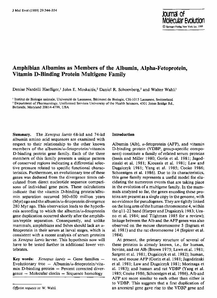
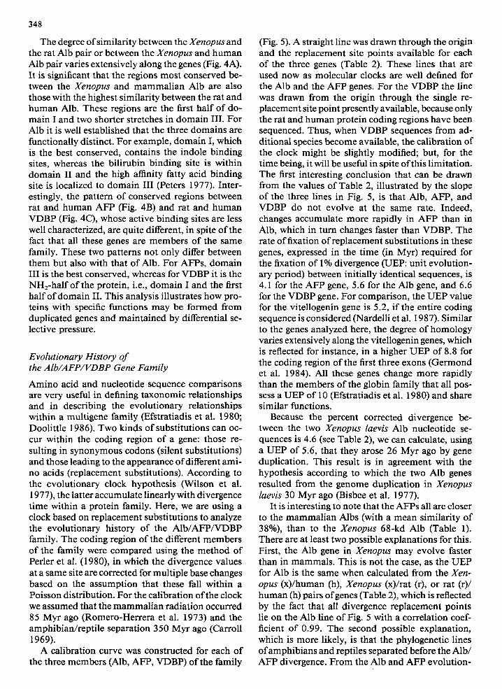
Xenopus Albs (Mr = approx. 70,000) are easily iden- tified in electropherograms o f plasma proteins due to their high mobil i ty and abundance (Bisbee et al. 1977; Wahli et al. 1978; G r a f and Fischberg 1986). In Xenopus laevis, there are two albumins o f 68 kd and 74 kd, respectively, that are encoded by two different genes (Westley and Weber 1982). The pres- ence o f duplicated loci in this species also has been repor ted for other sets o f genes such as the vitello- genin and globin genes (Wahli et al. 1979; Hosbach et al. 1983). These duplications presumably reflect the tetraploidizat ion that occurred in Xenopus laevis about 30-50 Myr ago (Bisbee et al. 1977; K n rch e l et al. 1986). The two closely related m R N A s that code for the 68-kd and 74-kd Alb have been cloned (Schoenberg 1981; Westley et al. 1981) and the se- quences o f their coding region have been de te rmined and compared (Moskaitis et al. 1989). The two Xen- opus laevis Albs proteins share 90% identical amino acids. Examinat ion o f the two amino acid sequences unambiguously reveals that the proteins encoded by the sequenced m R N A s are indeed true Albs for the following reasons. When the amino acid sequence and the disulfide bonding pat tern are displayed ac- cording to the model o f Brown (1976), the spacing o f the cysteine residues in the complete polypept ide chains and the posit ions o f the disulfide bridges cor- respond to that o f Alb ra ther than to that o f A F P as illustrated with the 68-kd Alb sequence in Fig. 1. Indeed the cysteines at posit ions 61, 80, 340, and 383 o f the 68-kd Alb sequence are present in all Albs but are absent in all AFPs. The layout o f Fig. 1 also shows that the 68-kd Xenopus Alb sequence,
346
L
III
gO
30 25
S30
)40
100
210
400
500
430
440
Fig. 1. Amino acid sequence, disulfide bonding pattern, and triple domain structure of the Xenopus 68-kd Alb displayed according to the model of Brown (1976). The amino acids are numbered as in Fig. 3. The disulfide bridges are indicated by shaded boxes.
A X68Alb I 29
X68Alb II 218
X68Alb III 408
a68Alb I I00
X68Alb II 292
• III 479
X68Alb I 162
X68AIb II 349
X68AIb III 550
DHHKtt IADMYN LLTERTFKGLTLAIVSQN LQKCSLEELSKLVNE INDFAKSCTGNDKTPECEKP IGTLFYD
DKQKHFCWIVNNYPERVI KALNLARVSHRYPKPDFKLAHKFTEETTHF IKDCCHGD-MFECMTERLELSEH
AYLKQNCD I LHEHGEY LFENELL I RYTKKMPQVSDETLIGI AHQMAD I GEHCCAVPENQRMPCAEGD LT I h
--KLCADPKVGVNYEWSKECCSKQDPERAQCFRAHRVFEHNPVRPKPEET ....... CALFKEHPDDLLSA
- - -TCQHKDELSTKLE--KCCNLPLLERTYCIVTLENDDVPAELSKPITEFTEDPHVC . . . . . EKYAENKS
IGKMCERQKKTFINNHVAHCCTDSYSGMRSCFTALGPDEDYVPPPVTDDTFHFDDKICTANDKEKQHIKQK
FIHEEARNH---PDLYPPAVLLLTQQYGKLVEHCCEEEDKDKCFAEKMKELMKH 2]2
FLEISPWQSQETPELSEQFLLQSAKEYESLLNKCCFSDNPPECYKDGADRFMNE 402 ** W W * W W** * *
FLVKLIKVS---PKLEKNHIDEWLLEFLKMVQKCCTADEHQPCFDTEKPVLIEH 600
3 4 7
X74Alb
•
X74Alb
X74Alb
X74Alb
X74Alb
X74Alb
a74Alb
X74Alb
I 29 DHHKH IADVYTA LTERTFKGLTLA IVSQNLQKCSLEE LSKLVNE INDFAKSCINDK- TP ECEKPVGTLFFD
II 217 [)gQ}l {FCWILDNFPEgVLNALNIARVSIIRYPgAEFKLAIINFTEEVTHFIKDCCHDD-MFECMTERLEI,TEII
III 4 0 9 AYLKQNCDI LHE HGEYLFENELLI RYTKNMPQVSDETLIGIAH QMAD IGEHCCAVPENQRMPCAEGDLTI L
I 99 --KLCADPAVGVNYEWSKECCAKQDPERAQCFKAIIRDHEHTSIKPEPEET ....... CKLLKEHPDDLLSA
I I 291 - - -TCQHKDELSSKLE--KCCNIPLLERTYCIVTLENDDVPAELSQPITEFTEDPHVC . . . . . EKYAENNE
Ill 480 IGNNCERQKKTF!NNHVAHCCTDSYSGMRSCFTALGPDEDYVPPPVTDDTFHFDDKICTANDKEKQHIKQK
I 161 -FIHEEARN .... HPDLYPPAVLALTKQYHKLAEHCCEEEDKEKCFSEKMKQLMKQ 211 W * * * * W * ** * *
II 348VFLGRYLHAVSRKHQELSEQFLLQSAKEYESLLNKCCKTDNPPECYKDGADRFMNE 403
III 551 -FLVKLIK-VS---PKLEKNHIDECSAEFLKMVQKCCTADEHQPCFDTEKPVLIEH 601
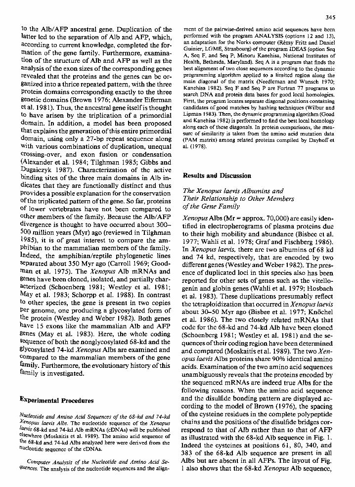
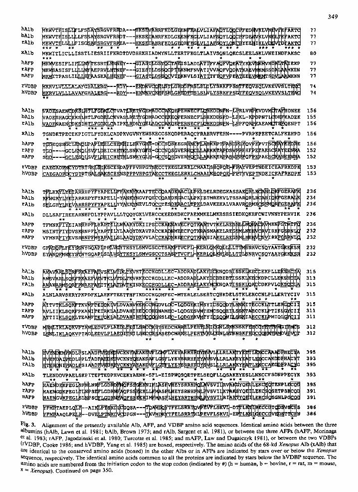
Fig. 2. A l ignmen t o f the a m i n o acid sequences o f the three h o m o l o g o u s d o m a i n s wi th in the 68-kd Alb (A) and the 74-kd Alb (B). The identical a m i n o acids between d o m a i n I and II or d o m a i n II and III are indicated by stars. Those identical between d o m a i n I and III are indicated by stars below d o m a i n 1II. The a m i n o acids are n u m b e r e d as in Fig. 3 (x = Xenopus).
like the one of the other members of the gene family, can be organized in three similar repeat domains. This internal homology is illustrated by the align- ment presented in Fig. 2A. There are 22% identical amino acids between domain I and II and 16% be- tween domain I and III or between domain II and III. In the 74-kd Alb, there are 21% identical amino acids between domain I and II and 16% between domain I and III or between domain I[ and III (Fig. 2E). Based on similar observations also indicating a higher homology between domain I and II com- pared to II and III or I and III in bovine albumin, Brown (1976) concluded that a first duplication of the primordial domain gave rise to domain III and to the ancestral I/II domain. Duplication of the lat- ter resulted in the appearance of domain I and do- main II. Surprisingly, the similarity between the three domains of the mammalian Alb is higher than in Xenopus, with 29% identical amino acids between domain I and II and 20% between domain I and III and domain II and III. Because gene sequences seem to diverge at the same rate in Xenopus and mammals
(as calculated below and Kn6chel et al. 1986), these observations indicate that gene correction events by coincidental evolution (Hood et al. 1975; Lauer et al. 1980) through unequal crossing over events (Smith 1976) might have occurred predominantly after the amphibian/reptile divergence. Indeed, the presence of homologous domains within the gene is favorable ground for such events to occur.
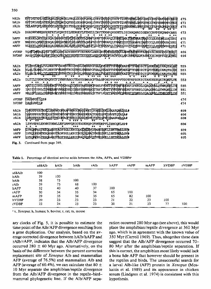
Because the amino acid sequences o f the Xenopus taevis 68-kd and 74-kd Alb are 90% identical, the primary structure of the 68-kd Alb only was com- pared with that of the Alb, AFP, and VDBP from different mammalian species (Figs. 3 and 4 and Ta- ble 1). The 68-kd protein is more similar to mam- malian Albs than to either the AFPs or the VDBPs. There is, on average, 39% amino acid identity with Albs, 30% with AFPs, and 22% with VDBPs. Of the amino acids present in the Xenopus 68-kd Alb, 188 are conserved between this protein and the other Albs and 133 between this protein and the AFPs. One hundred of them are common to all the Albs and AFPs and 31 to the Albs, AFPs, and VDBPs.
348
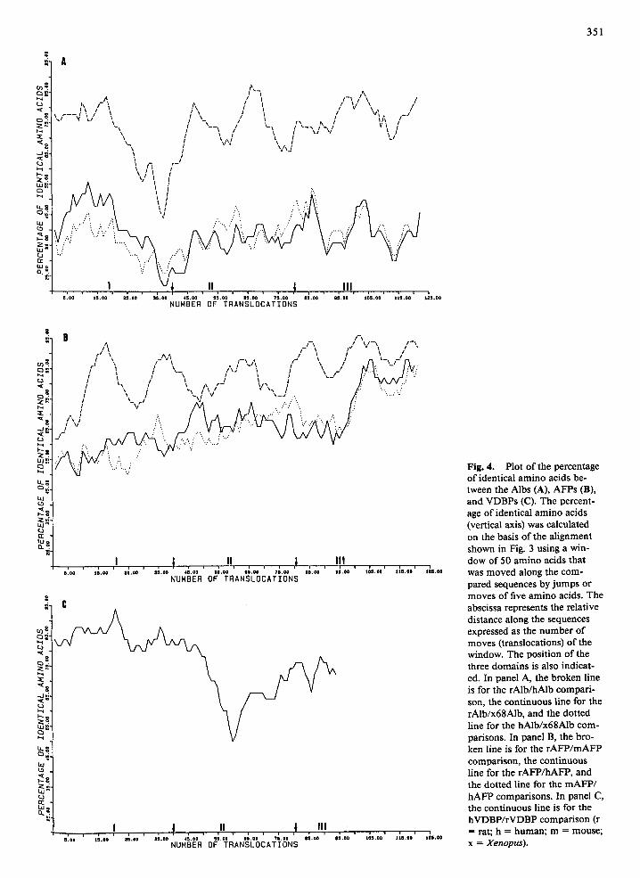
The degree of similarity between the Xenopus and the rat Alb pair or between the Xenopus and human Alb pair varies extensively along the genes (Fig. 4A). It is significant that the regions most conserved be- tween the Xenopus and mammalian Alb are also those with the highest similarity between the rat and human Alb. These regions are the first half of do- main I and two shorter stretches in domain III. For Alb it is well established that the three domains are functionally distinct. For example, domain I, which is the best conserved, contains the indole binding sites, whereas the bilirubin binding site is within domain II and the high affinity fatty acid binding site is localized to domain III (Peters 1977). Inter- estingly, the pattern of conserved regions between rat and human AFP (Fig. 4B) and rat and human VDBP (Fig. 4C), whose active binding sites are less well characterized, are quite different, in spite of the fact that all these genes are members of the same family. These two patterns not only differ between them but also with that of Alb. For AFPs, domain III is the best conserved, whereas for VDBP it is the NH2-half of the protein, i.e., domain I and the first half of domain II. This analysis illustrates how pro- teins with specific functions may be formed from duplicated genes and maintained by differential se- lective pressure.
Evolutionary History of the Alb/AFP/VDBP Gene Family
Amino acid and nucleotide sequence comparisons are very useful in defining taxonomic relationships and in describing the evolutionary relationships within a multigene family (Efstratiadis et al. 1980; Doolittle 1986). Two kinds of substitutions can oc- cur within the coding region of a gene: those re- sulting in synonymous codons (silent substitutions) and those leading to the appearance of different ami- no acids (replacement substitutions). According to the evolutionary clock hypothesis (Wilson et al. 1977), the latter accumulate linearly with divergence time within a protein family. Here, we are using a clock based on replacement substitutions to analyze the evolutionary history of the Alb/AFP/VDBP family. The coding region of the different members of the family were compared using the method of Perler et al. (1980), in which the divergence values at a same site are corrected for multiple base changes based on the assumption that these fall within a Poisson distribution. For the calibration of the clock we assumed that the mammalian radiation occurred 85 Myr ago (Romero-Herrera et al. 1973) and the amphibian/reptile separation 350 Myr ago (Carroll 1969).
A calibration curve was constructed for each of the three members (Alb, AFP, VDBP) of the family
(Fig. 5). A straight line was drawn through the origin and the replacement site points available for each of the three genes (Table 2). These lines that are used now as molecular clocks are well defined for the Alb and the AFP genes. For the VDBP the line was drawn from the origin through the single re- placement site point presently available, because only the rat and human protein coding regions have been sequenced. Thus, when VDBP sequences from ad- ditional species become available, the calibration of the clock might be slightly modified; but, for the time being, it will be useful in spite of this limitation. The first interesting conclusion that can be drawn from the values of Table 2, illustrated by the slope of the three lines in Fig. 5, is that Alb, AFP, and VDBP do not evolve at the same rate. Indeed, changes accumulate more rapidly in AFP than in Alb, which in turn changes faster than VDBP. The rate of fixation of replacement substitutions in these genes, expressed in the time (in Myr) required for the fixation of 1% divergence (UEP: unit evolution- ary period) between initially identical sequences, is 4.1 for the AFP gene, 5.6 for the Alb gene, and 6.6 for the VDBP gene. For comparison, the UEP value for the vitellogenin gene is 5.2, if the entire coding sequence is considered (Nardelli et al. 1987). Similar to the genes analyzed here, the degree of homology varies extensively along the vitellogenin genes, which is reflected for instance, in a higher UEP of 8.8 for the coding region of the first three exons (Germond et al. 1984). All these genes change more rapidly than the members of the globin family that all pos- sess a UEP of 10 (Efstratiadis et al. 1980) and share similar functions.
Because the percent corrected divergence be- tween the two Xenopus laevis Alb nucleotide se- quences is 4.6 (see Table 2), we can calculate, using a UEP of 5.6, that they arose 26 Myr ago by gene duplication. This result is in agreement with the hypothesis according to which the two Alb genes resulted from the genome duplication in Xenopus laevis 30 Myr ago (Bisbee et al. 1977).
It is interesting to note that the AFPs all are closer to the mammalian Albs (with a mean similarity of 38%), than to the Xenopus 68-kd Alb (Table 1). There are at least two possible explanations for this. First, the Alb gene in Xenopus may evolve faster than in mammals. This is not the case, as the UEP for Alb is the same when calculated from the Xen- opus (x)/human (h), Xenopus (x)/rat (r), or rat (r)/ human (h) pairs of genes (Table 2), which is reflected by the fact that all divergence replacement points lie on the Alb line of Fig. 5 with a correlation coef- ficient of 0.99. The second possible explanation, which is more likely, is that the phylogenetic lines of amphibians and reptiles separated before the Alb/ AFP divergence. From the Alb and AFP evolution-
349
hAlb bAlb rAlb
XAlb
hAFp rAFp mAFp
rVDBP hVDBP
MKWITLICLLISSTLIESRIIFKRDTDVDHHKHIADMYNLLTERTFKGLTLAIVSQNLQKCSLEELSKLVNEINDFAKSC 80 * * * * * * * *
KR~LVL~LAVAFGHALERG~--~--~---~-~F~sG~-9--~E~-.~TLS~-L-~YSRK~PSE4TFEQVSQLVKEwSLT~ 74
hAlb bAlb rAlb
XAlb
hAFp rAFP mAFp
rVDBP hVDBP
~AE I~I~'D~~VAT~L]I~ET~r~:M~E~'~-R~]P]I~I-~-~RLVI~i~VD~A~HDNEE 15~
HA~C~IJHTLFGD~LC~VAS~R~T~GbM~DCC~K~Q~ERNECFL~KDD~b-~KL-~PbPNTL~KADEK 155
* * *W* * ** * ** * *** ** * * * *
TGNDKTPECEKPIGTLFYDKLCADPKVGVNYEWSKECCSKQDPERAOCFRAHRVFEHN .... PVRPKPEETCALF~EHPD 156
s ~ _ ~ - - - d G C ~ s v ~ n ~ x C ~ T ~ S S ~ _ L q ~ - ~ _ _ q s ~ s _ ~ w ~ x ~ r ~ J ~ _ m ~ o F ~ _ ~ e ~ _ c ~ m ~ ~s~
153
hAlb bAlb rAlb
XAlb
hAFp rAFp mAFp
rVDBP hVDBP
KK R HpyFyApEL F yr AFTE A %DELRO GKASSA L 236 GK~/I~/~RRH~Y~YAPEL~-~A~(~NGVFQE~C~A~D~(~A~PK~ETMREKVLTSSAP4~~[GERA[~ 234
DLLSAFIHEEARNH•DLYPPAVLLLTQQYGKLVEHCCEEEDKDKCFAEKMKELMKHSHSIEDKQKHFC•IVNNYPERVIK 236
TFMNK~-~IAR~-~-~L~A---~T~'-L~W~-~IIPS~AV~C-FQT---~-~A~TVT~EL---~-~L~KN~R--~F~ 236 MSINT~IYbVSK~NPF~L~L~A~YDK~VPA~CKA~ME~CFQT~R~SMA~ELRE~E~C~RK~G~L~ 232
~Y~N~MW.~-Y~[NYGQAP~sI~S4YTKS~LSMVGS~T~A~TV~FI~-~*ERLQL~/~SL~-L-T~L~NRVC*--~QYAAYG~[KS-S~ 232
hAlb bAlb rAlb
XAlb
hAFp rAFp mAFP
rVDBp hVDBP
V~R~Q~ ~K~%EF~E~T~I~T[~L~I(~/HK~CC HGDL LEe - AD DRA~AKY~B Z B~I S SKI~K~C~(DPC LLE~(S~ ~ 313
ALNLARVSHRYPKPDFKLAHKFTEETTHFIKDCCHGDMFEC-MTERLELSEHTCQHKDELSTKLEKCCNLP LLERTYCIV 315
~I TVT~LS--L-S~ T K V ~ F ~ - ~ O K - ~ ~ V L ~ I~S Y I ~ - ~ D T ~ I T C~6~-I]~T T LE ~ C ' - ~ 315 ~VLI I~LSQ~PKA~I~E~R~A~DVAH~ ~EI~CC~NAME ~ - LQDGE~V~HM~SQ~E I~S~TA~CCKL~ �9 I,~CI ~ 311 ~TT I I ~LSQK~TEA~F~E ~OK~A~DVAH;[ E ~ N S LE~- LQDGE~(V~Y I S ~ I~__~S~I A~CCK L~M I O ~ 311
I KL AQKV p T ~N ~E DV L P L AE ~L~E ~L--~R~6~'-~%[SED6-M~--LPE-~-~I ~'~K~NSKF-~'/(~Y~/E~S ~M~ 312
bAlb P L SV EY H T 393 rAlb SI P G 395
)tAlb TLENDDVPAELSKP I TEFTEDPHVCEKYAENK-SFL-EI SPWQSOETPELSEOFLLQSAKEYESLLNKCCFSDNPPECYK 393
mAFp A~!AE_~G~KP EG L~ I~l~ S (3T LGD I~I~RQ~[.~.~ E~__~I M~M/%._S _~I~H EYS R T H ~I~V SV I I~R I ~ ~SSS GN LP ~ 391
hV~ ~_8~O,.~v_~_~__~v ~rqbg_-~t%~Lq~G~--~-~V~!_m~VLS~mvWLSXV~-~p~Vm__~r~s as~
l?i~. 3. Alignment of the presently available Alb, AFP, and VDBP amino acid sequences. Identical amino acids between the three albumins (hAlb, Lawn et al. 1981; bAlb, Brown 1975; and rAlb, Sargent et al. 1981), or between the three AFPs (hAFP, Morina~a et al. 1983, rAFP, Jagodzinski et al. 1980, Turcotte et al. 1985; and mAFP, Law and Duga~czyk 198 I), or between the two VDBPs (rVDBP, Cooke 1986, and hVDBP, Yang et al. 1985) are boxed, respectively. The amino acids of the 68-kd X~nopu~ A/b (x2db) that are identical to the conserved amino acids (boxed) in the other Albs or in AFPs are indicated by stars over or below the Xenopus sequence, respectively. The identical amino acids common to all the proteins are indicated by stars below the hVDBP sequence. The amino acids are numbered from the initiation codon to the stop codon (indicated by #) (h = human, b = bovine, r = rat, m = mouse, x ~ Xenopu~). Continued on page 350.
350
h/~b b/~b r2U.b
x/~b
r k ~
rVDBP hVDBP
~CDQFEE~-GEMG~QNA~VR~QVSTP TLV~VS~G~RCC~KP~SE~C~ED~ 473
DGADRFMNF2iKERFAYLKQNCDI LHEHGEYI/"ZNELLIRYTKKMP QVSDETLI GIAHQMADI GEHCCAVPENQRMPCAEG 473 * * * * * * * * ** * ** * * ** * * **
K - QLT CCQLS " 475
NLEEELQ S ~ _ _ ~ I ~ QLT ~%~__I/I D ~ S I~S[f CCQLS E~WSC~_~ 471
bAlb ~ L ~ L I ~ K T P V ~ S ~ K C C ~ S L ~ R P C F S A q ~ E T Y V P ~ F p E K I ~ T F F ~ D I C T I ~ D a ~ I ~ Q T ~ 553 rAlb ~LSAI~h~EI~CC~ S~CFSA/2T%~C~~__F}~ D~PD~I KKQTAL~ 555
* * ** * ** * * ** ** ** *** * * *** * , ** *
xAlb DLT I LI GKMCERQKKTF INNHVAHCCTD SYSGMRSCFTALGP DEDYVPPPVTDDTFHFDDK I CTANDKEKQH IKQKFLVK 553 * ** * * * ** ** * * ** * ** * * * * * * ** *
hAFP A~I ~ - - - ~ V ~ V G ~ C ~ ' ~ S S L V ~ ~ K F I FHKDLCQA~QE--~--~ 555 rAFP I ~ ~YF G H ~ V N ~ I N ~ C C ~ S~S~C~TSFL~ET~P~I~FS~DKF I FHKDLCQAQ~%L(~I ~ 551 mAFP ~%D_I~ ~S~_V~_~6~I S }~lC~S_~ _1!S ~3/~I TSF~Ag.~ 551
rVDBP ~ ~ S # 476 hVDBP 474
rAlb 608
xAlb L~ KVSP KLEKNH I DEW7.7 ,~.FLKMVQKCCTADEHQP CFDTEKPVLI EHCQKLHP # 606 * * * * *** ** * **
609 W EQ VF FSGU KCCF K, ISKT 60S
mAFP ~_~__~ I/IEE~VT~%DF S G7,7 RKC~~G~,K LI SKT~ # 605
Fig. 3. Continued from page 349.
Table 1. Percentage o f identical a m i n o acids between the Albs, AFPs , and V D B P s a
x68Alb hAlb bAlb rAlb h A F P rAFP m A F P h V D B P rVDBP
x68Alb 100 hAlb 39 100 bAlb 38 75 100 rAlb 39 73 68 h A F P 32 40 40 rAFP 30 34 35 m A F P 29 35 34 h V D B P 20 23 23 rVDBP 22 24 23
100 37 100 34 65 100 35 65 82 100 23 21 22 23 23 20 21 23
I00 77 100
a x, Xenopus; h, h u m a n ; b, bovine; r, rat; m, m o u s e
ary clocks of Fig. 5, it is possible to estimate the time point of the AIb/AFP divergence resulting from a gene duplication. Our analysis, based on the av- erage corrected divergence between hAlb/hAFP and rAlb/rAFP, indicates that the Alb/AFP divergence occurred 280 ___ 60 Myr ago. Alternatively, on the basis of the difference between the percent corrected replacement site of Xenopus Alb and mammalian AFP (average of 76.5%) and mammalian Alb and AFP (average of 60.4%), we can calculate that 80 _+ 10 Myr separate the amphibian/reptile divergence from the Alb/AFP divergence in the reptile--bird- mammal phylogenetic line. If the Alb/AFP sepa-
ration occurred 280 Myr ago (see above), this would place the amphibian/reptile divergence at 360 Myr ago, which is in agreement with the known value of 350 Myr (Carroll 1969). Thus, altogether these data suggest that the AIb/AFP divergence occurred 70- 80 Myr after the amphibian/reptile separation. If this is correct, the amphibian most likely would lack a bona fide AFP that however should be present in the reptiles and birds. The unsuccessful search for a larval Alb-like (AFP) protein in Xenopus (Mos- kaitis et al. 1989) and its appearance in chicken serum (Lindgren et al. 1974) is consistent with this hypothesis.
g r,. C
m~ ~ _ ~ -
' ~ o "
o
~ ~
,, ,>, !'-', ,,, ,,,"-, , ' , , \ ", / ~ I / \ , I I v,~ ,_, ~ . r - -V ~,,' \ ~ ,, ~, , .'~
",, / '--, / '-, l ' - . , , - v ", / ',., ! ',,," ,, ~ '~, ,, ,',,j
, / I /
V, / I I .; .:: t I :".. : ' ' - :
I ~ li l i l l 6 . 0 0 IS .O0 2a,O0 10 .00 40.0O 1 5 . 0 0 IS,O~ 7 9 , 0 0 90 .00 911.00 105.00 t l l l . O O $20 .00
NUMBER OF TRANSLOCATIONS
1 /'~ / - \ / V - \ / / X ~ r ', .^s, - J " / "-n-,Jn
~'t "' ; " " " / ' ' / " " ~ ~ '-' "
I ~ I I l l I - .:
* / X / "~ I ~v I ' "',._J . .' "'.....,.",- ~- , ,_ ~, ,, . ....,.
/ ~s . ,""." ": ." ^ I :. . : . . : , . " ' v ' " .,. .: :"
,~ ** / '~/ /', ,......... �9 ...., .: ...I~ I : : * : " ,
:, ': '. ,, ::
!~11 ~ L ~ .. . ~ '.. ; /: .... .'
1 , 0 0 1 1 . 0 0 9 0 . 0 0 0 0 . 0 0 4 0 . 0 0 l I . 0 O 9 0 , 0 0 7 9 . 0 0 lI.H I I . 0 O 1 0 1 . 0 0 I l l . C o
NUMBER OF TRANSLOCATIONS
I 1211.00
LU
~~ ,~-
5 . 0 0
I $~.oo I I I
ZO.00 3 5 . 0 0 ,:,~ 5 : . . ' 0:.~176 ~:,oo' NUMBER OF TRANSLOCATIONS
.; # ! . , . ,&.**' d0.**'
g
U
o
~~
t u ~ "
C3 ,~ .
m ".
~:.
351
Fig. 4. Plot of the percentage of identical amino acids be- tween the Albs (A), AFPs (B), and VDBPs (C). The percent- age of identical amino acids (vertical axis) was calculated on the basis of the alignment shown in Fig. 3 using a win- dow of 50 amino acids that was moved along the com- pared sequences by jumps or moves of five amino acids. The abscissa represents the relative distance along the sequences expressed as the number of moves (translocations) of the window. The position of the three domains is also indicat- ed. In panel A, the broken line is for the r A l b / h A l b compari- son, the continuous line for the rAlb/x68Alb, and the dotted line for the hAlb/x68Alb com- parisons. In panel B, the bro- ken line is for the rAFP/mAFP comparison, the continuous line for the rAFP/hAFP, and the dotted line for the mAFP/ hAFP comparisons. In panel C, the continuous line is for the hVDBP/rVDBP comparison (r = rat; h = human; m = mouse; x = X e n o p u s ) .
352
Percent corrected divergence
200
AFP
150 ..... ......" .............
....... " ....... Alb
AFP/VDBF AIb/VDBP ,~176176176
x6g/x74Alb
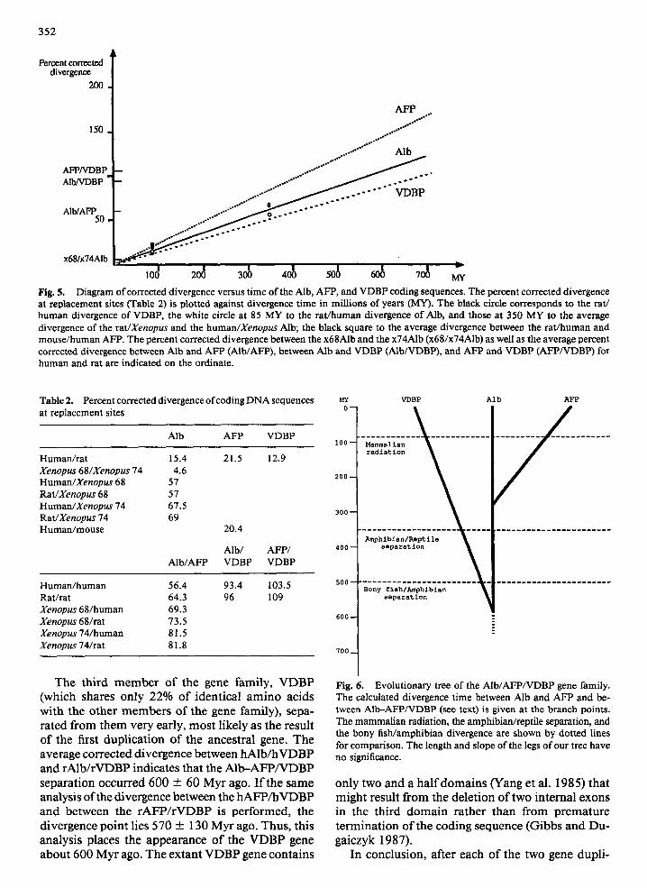
100 20d MY Fig. 5. Diagram of corrected divergence versus time of the Aib, AFP, and VDBP coding sequences. The percent corrected divergence at replacement sites (Table 2) is plotted against divergence time in millions of years (MY). The black circle corresponds to the rat/ human divergence of VDBP, the white circle at 85 MY to the rat/human divergence of Alb, and those at 350 MY to the average divergence of the rat/Xenopus and the human/Xenopus Alb; the black square to the average divergence between the rat/human and mouse/human AFP. The percent corrected divergence between the x68Alb and the x74Alb (x68/x74Alb) as well as the average percent corrected divergence between Alb and AFP (AIb/AFP), between Alb and VDBP (Alb/VDBP), and AFP and VDBP (AFP/VDBP) for human and rat are indicated on the ordinate.
Table 2. Percent corrected divergence of coding DNA sequences at replacement sites
Alb AFP VDBP
Human/rat 15.4 21.5 12.9 Xenopus 68/Xenopus 74 4.6 Human/Xenopus 68 57 RaVXenopus 68 57 Human/Xenopus 74 67.5 Rat/Xenopus 74 69 Human/mouse 20.4
Alb/ AFP/ Alb/AFP VDBP VDBP
Human/human 56.4 Rat/rat 64.3 Xenopus 68/human 69.3 Xenopus 68/rat 73.5 Xenopus 74/human 81.5 Xenopus 74/rat 81.8
93.4 103.5 96 109
MY VDBP Alb AFP
0-
i00 -
200 -
300-
400--
500-- Bony flsh/Amphlbian
separation
600 --
700 _
T h e t h i r d m e m b e r o f t he gene f ami ly , V D B P (which sha res o n l y 22% o f i d e n t i c a l a m i n o a c i d s w i t h t he o t h e r m e m b e r s o f t he gene fami ly) , s epa - r a t e d f r o m t h e m v e r y ear ly , m o s t l ike ly as t he resu l t o f t he first d u p l i c a t i o n o f t h e ances t r a l gene. T h e ave r age c o r r e c t e d d i v e r g e n c e b e t w e e n h A l b / h V D B P a n d r A l b / r V D B P ind ica te s tha t the A I b - A F P / V D B P s e p a r a t i o n o c c u r r e d 600 + 60 M y r ago. I f t he s a m e ana lys i s o f the d ive rgence b e t w e e n the h A F P / h V D B P a n d b e t w e e n the r A F P / r V D B P is p e r f o r m e d , the d i v e r g e n c e p o i n t l ies 570 _ 130 M y r ago. Thus , th is ana lys i s p laces t he a p p e a r a n c e o f the V D B P gene a b o u t 600 M y r ago. T h e e x t a n t V D B P gene c o n t a i n s
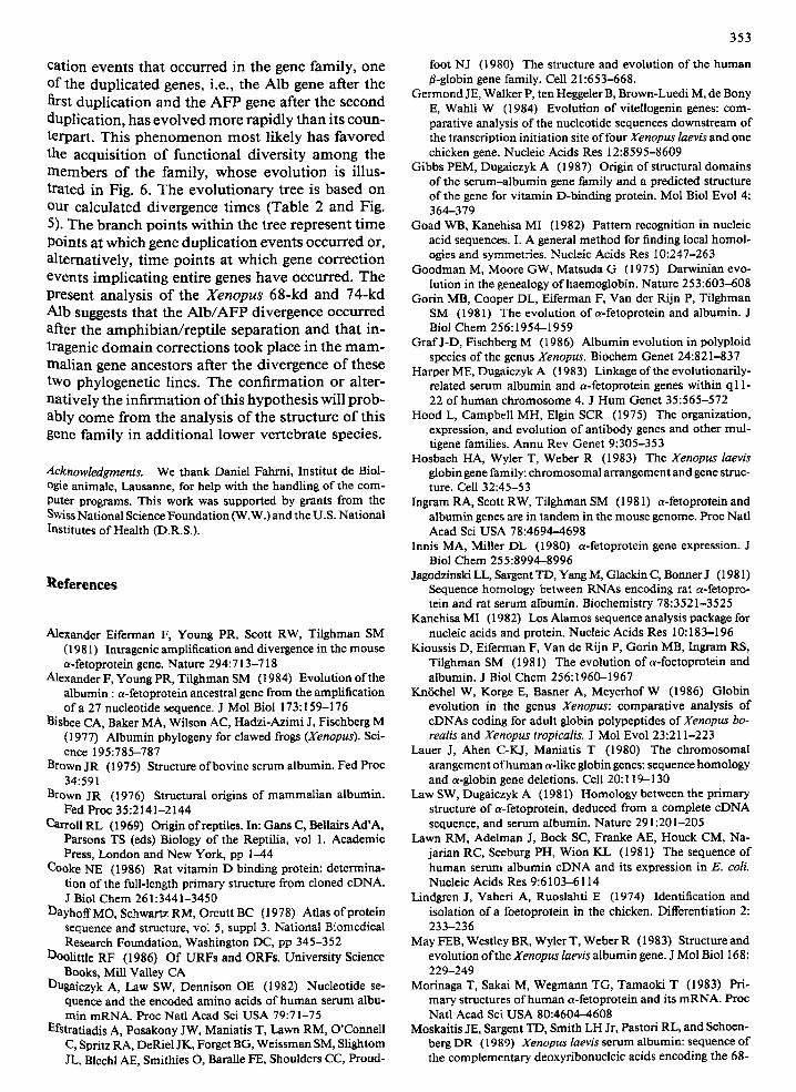
Fig. 6. Evolutionary tree of the AIb/AFP/VDBP gene family. The calculated divergence time between Alb and AFP and be- tween Alb-AFP/VDBP (see text) is given at the branch points. The mammalian radiation, the amphibian/reptile separation, and the bony fish/amphibian divergence are shown by dotted lines for comparison. The length and slope of the legs of our tree have no significance.
o n l y two a n d a h a l f d o m a i n s W a n g et al . 1985) t ha t m i g h t r e su l t f r o m the d e l e t i o n o f two i n t e r n a l e x o n s in the t h i r d d o m a i n r a t h e r t h a n f r o m p r e m a t u r e t e r m i n a t i o n o f the c o d i n g s equence ( G i b b s a n d D u - ga i czyk 1987).
In c o n c l u s i o n , a f te r each o f t he t w o gene d u p l i -
353
ca t ion even t s tha t occur red in the gene fami ly , one of the dup l i ca t ed genes, i.e., the A lb gene af ter the first d u p l i c a t i o n a n d the A F P gene af ter the second dup l i ca t ion , has e v o l v e d m o r e r ap id ly t h a n its c o u n - terpart . Th i s p h e n o m e n o n m o s t l ikely has f avored the acqu i s i t i on o f f u n c t i o n a l d ive r s i ty a m o n g the m e m b e r s o f the fami ly , whose e v o l u t i o n is i l lus- t ra ted in Fig. 6. T h e e v o l u t i o n a r y tree is based o n our ca lcu la ted d ive rgence t imes (Table 2 a n d Fig. 5). T h e b r a n c h p o i n t s w i t h i n the tree r ep resen t t i m e Points a t which gene d u p l i c a t i o n e v e n t s occu r red or, a l t e rna t ive ly , t i m e p o i n t s a t wh ich gene co r r ec t ion even ts i m p l i c a t i n g en t i re genes have occurred. T h e p resen t ana lys i s o f the X e n o p u s 68-kd a n d 74 -kd Alb suggests tha t the A l b / A F P d ive rgence occur red after the a m p h i b i a n / r e p t i l e s epa ra t i on a n d tha t in - t ragenic d o m a i n co r rec t ions took place in the m a m -
m a l i a n gene ances tors after the d ive rgence o f these two phy logene t i c l ines. T h e c o n f i r m a t i o n or al ter- na t ive ly the i n f i r m a t i o n o f this hypo thes i s wil l p r o b - ably c o m e f rom the ana lys i s o f the s t ruc ture o f this gene f ami ly in a d d i t i o n a l lower ve r t eb ra t e species.
Acknowledgments. We thank Daniel Fahrni, Institut de Biol- ogic animale, Lausanne, for help with the handling of the com- Puter programs. This work was supported by grants from the Swiss National Science Foundation (W.W.) and the U.S. National Institutes of Health (D.R.S.).
References
Alexander Eiferman F, Young PR, Scott RW, Tilghman SM (1981) Intragenic amplification and divergence in the mouse a-fetoprotein gene. Nature 294:713-718
Alexander F, YoungPR, TilghmanSM (1984) Evolutionofthe albumin : a-fetoprotein ancestral gene from the amplification of a 27 nucleotide sequence. J Mol Biol 173:159-176
Bisbee CA, Baker MA, Wilson AC, Hadzi-Azimi J, Fischberg M (1977) Albumin phylogeny for clawed frogs (Xenopus). Sci- ence 195:785-787
Brown JR (! 975) Structure of bovine serum albumin. Fed Proc 34:591
Brown JR (1976) Structural origins of mammalian albumin. Fed Proc 35:2141-2144
Carroll RL (1969) Origin of reptiles. In: Gans C, Bellairs Ad'A, Parsons TS (eds) Biology of the Reptilia, vol 1. Academic Press, London and New York, pp 1-44
Cooke NE (1986) Rat vitamin D binding protein: determina- tion of the full-length primary structure from cloned cDNA. J Biol Chem 261:3441-3450
I3ayhoffMO, Schwartz RM, Orcutt BC (1978) Atlas of protein sequence and structure, vol 5, suppl 3. National Biomedical l~eseareh Foundation, Washington DC, pp 345-352
Doolittle RF (1986) Of URFs and ORFs. University Science Books, Mill Valley CA
13ugaiczyk A, Law SW, Dennison OE (1982) Nucleotide se- quence and the encoded amino acids of human serum albu- min mRNA. Proc Natl Acad Sci USA 79:71-75
Efstratiadis A, Posakony JW, Maniatis T, Lawn RM, O'Connell C, Spritz RA, DeRiel JK, Forget BG, Weissman SM, Slightom JL, Blechi AE, Smithies O, Baralle FE, Shoulders CC, Proud-
foot NJ (1980) The structure and evolution of the human 13-globin gene family. Cell 21:653-668.
Germond JE, Walker P, ten Heggeler B, Brown-Luedi M, de Bony E, Wahli W (1984) Evolution of vitellogenin genes: com- parative analysis of the nucleotide sequences downstream of the transcription initiation site of four Xenopus laevis and one chicken gene. Nucleic Acids Res 12:8595-8609
Gibbs PEM, Dugaiczyk A (1987) Origin of structural domains of the serum-albumin gene family and a predicted structure of the gene for vitamin D-binding protein. Mol Biol Evol 4: 364--379
Goad WB, Kanehisa MI (1982) Pattern recognition in nucleic acid sequences. I. A general method for finding local homol- ogies and symmetries. Nucleic Acids Res 10:247-263
Goodman M, Moore GW, Matsuda G (1975) Darwinian evo- lution in the genealogy of haemoglobin. Nature 253:603-608
Gorin MB, Cooper DL, Eiferman F, Van der Rijn P, Tilghman SM (1981) The evolution of c~-fetoprotein and albumin. J Biol Chem 256:1954-1959
GrafJ-D, Fischberg M (1986) Albumin evolution in polyploid species of the genus Xenopus. Biochem Genet 24:821-837
Harper ME, Dugaiczyk A (1983) Linkage of the evolutionarily- related serum albumin and a-fetoprotein genes within ql 1- 22 of human chromosome 4. J Hum Genet 35:565-572
Hood L, Campbell MH, Elgin SCR (1975) The organization, expression, and evolution of antibody genes and other mul- tigene families. Annu Rev Genet 9:305-353
Hosbach HA, Wyler T, Weber R (1983) The Xenopus laevis globin gene family: chromosomal arrangement and gene struc- ture. Cell 32:45-53
Ingram RA, Scott RW, Tilghman SM (1981) a-fetoprotein and albumin genes are in tandem in the mouse genome. Proc Nati Acad Sci USA 78:4694-4698
Innis MA, Miller DL (1980) a-fetoprotein gene expression. J Biol Chem 255:8994-8996
Jagodzinski LL, Sargent TD, Yang M, Glackin C, Bonner J (1981) Sequence homology between RNAs encoding rat a-fetopro- tein and rat serum albumin. Biochemistry 78:3521-3525
KanehisaMI (1982) Los Alamos sequence analysis package for nucleic acids and protein. Nucleic Acids Res 10:183-196
Kioussis D, Eiferman F, Van de Rijn P, Gorin MB, Ingram RS, Tilghman SM (1981) The evolution of a-foetoprotein and albumin. J Biol Chem 256:1960-1967
KnSchel W, Korge E, Basher A, MeyerhofW (1986) Globin evolution in the genus Xenopus: comparative analysis of eDNAs coding for adult globin polypeptides of Xenopus bo- realis and Xenopus tropicalis. J Mol Evol 23:211-223
Lauer J, Ahen C-KJ, Maniatis T (1980) The chromosomal arangement of human a-like globin genes: sequence homology and a-globin gene deletions. Cell 20:119-130
Law SW, Dugaiczyk A (1981) Homology between the primary structure of a-fetoprotein, deduced from a complete cDNA sequence, and serum albumin. Nature 291:201-205
Lawn RM, Adelman J, Book SC, Franke AE, Houck CM, Na- jarian RC, Seeburg PH, Wion K_L (1981) The sequence of human serum albumin cDNA and its expression in E. coll. Nucleic Acids Res 9:6103-6114
Lindgren J, Vaheri A, Ruoslahti E (1974) Identification and isolation of a foetoprotein in the chicken. Differentiation 2: 233-236
May FEB, Westley BR, Wyler T, WeberR (1983) Structure and evolution of the Xenopus laevis albumin gene. J Mol Biol 168: 229-249
Morinaga T, Sakai M, Wegmann TG, Tamaoki T (1983) Pri- mary structures of human a-fetoprotein and its mRNA. Proc Natl Acad Sci USA 80:4604-4608
Moskaitis JE, Sargent TD, Smith LH Jr, Pastori RL, and Schoen- berg DR (1989) Xenopus laevis serum albumin: sequence of the complementary deoxyribonucleic acids encoding the 68-
354
and 74-kilodalton peptides and the regulation of albumin gene expression by thyroid hormone during development. Mol En- docrinol 3:464-473
Nardelli D, van het Ship FD, Gerber-Huber S, Samallo J, Hae- fliger JA, Gruber M, AB G, Wahli W (1987) Comparison of the organization and fine structure of a chicken and a Xen- opus laevis vitellogenin gene. J Biol Chem 262:15377-15385
Needleman SB, Wunsch DD (1970) A general method appli- cable to the search for similarities in the amino acid sequence of two proteins. J Mol Biol 48:443-453
Perler F, Efstratiadis A, Lomedico P, Gilbert W, Kolodner R, Dogson J (1980) The evolution of genes: the chicken pre- proinsulin gene. Cell 20:555-566
Peters T (1977) Serum albumin: recent progress in the under- standing of its structure and biosynthesis. Clin Chem 23:5-12
Romero-Herrera AE, Lehmann H, Joysey KA, Friday AE (1973) Molecular evolution of myoglobin and the fossil record: a phylogenetie synthesis. Nature 246:389-395
Sargent TD, Yang M, Bonnet J (1981) Nucleotide sequence of cloned rat serum albumin messenger RNA. Proc Natl Acad Sci USA 78:243-246
Sehoenberg DR (1981) Albumin is encoded by 2 messenger RNAs in Xenopus laevis. Nucleic Acids Res 9:6669-6688
Schoentgen F, Metz-Boutigue M-H, Joll6s J, Constans J, Joll6s P (1986) Complete amino acid sequence o f h u m a n vitamin D-binding protein (group-specific component): evidence of a three-fold internal homology as in serum albumin and a-fe- toprotein. Biochim Biophys Acta 871:189-198
Schorpp M, D~bbeling U, Wagner U, Ryffel G U (1988) 5'-flanking and 5'-proximal exon regions of the two Xenopus albumin genes. J Mol Biol 199:83-93
Smith GP (1976) Evolution of repeated DNA sequences by unequal crossover. Science 191:528-535
Szpirer J, Levan G, Thorn M, Szpirer C (1984) Gene mapping in the rat by mouse-rat somatic cell hybridization: synteny of the albumin and alpha-fetoprotein. Cytogenet Cell Genet 38:142-149
Tilghman SM (1985) The structure and regulation of the a-fe- toprotein and albumin genes. In: MacLean N (ed) Oxford surveys on eukaryotic genes, vol 3. University Press, Oxford, pp 160-206
Tureotte B, Guertin M, Chevrette M, Belanger L (1985) Rat al-fetoprotein messenger RNA: 5'-end sequence and gluco- corticoid-suppressed liver transcription in an improved nu- clear run-off assay. Nucleic Acids Res 13:2387-2398
Urano Y, Sakai M, Watanabe K, Tamaoki T (1984) Tandem arrangement of the albumin and ~-fetoprotein genes in the human genome. Gene 32:255-26I
Wahli W, Abraham I, Weber R (1978) Retention of the differ- entiated state by larval Xenopus liver cells in primary culture. Wilhelm Roux's Arch Dev Biol 185:235-248
Wahli W, Dawid IB, Wyler T, Jaggi RB, Weber R, Ryffel G U (1979) Vitellogenin in Xenopus laevis is encoded in a small family of genes. Cell 16:535-549
Westley B, Weber R (1982) Divergence of the two albumins of X. laevis: evidence for the glycosylation of the major 74K albumin. Differentiation 22:227-230
Westley B, Wyler T, Ryffel G, Weber R (1981) Xenopus laevis serum albumins are encoded in two closely related genes. Nucleic Acids Res 9:3557-3574
Wilbur WJ, Lipman DJ (1983) Rapid similarity searches of nucleic acid and protein data banks. Proc Natl Acad Sci USA 80:726-730
Wilson AC, Carlson SS, White TJ (1977) Biochemical evolu- tion. Annu Rev Biochem 46:573-639
Yang F, Luna V J, McAnelly RD, Naberhaus KH, Cupplies RL, Bowman BH (1985) Evolutionary and structural relation- ships among the group-specific component, albumin and a-fe- toprotein. Nucleic Acids Res 13:8007-8017
Received September 20, 1988/Revised March 3, 1989