based hiv-1 multigene dna vaccines in murine models - trepo
TRANSCRIPT
MARIA MALM
Assessing the Immunogenicity of GTU-based HIV-1 Multigene DNA Vaccines
in Murine Models
ACADEMIC DISSERTATIONTo be presented, with the permission of
the board of Institute of Biomedical Technology of the University of Tampere,
for public discussion in the Jarmo Visakorpi Auditorium,of the Arvo Building, Lääkärinkatu 1, Tampere,
on June 22nd, 2011, at 12 o’clock.
UNIVERSITY OF TAMPERE

Reviewed byDocent Petteri ArstilaUniversity of HelsinkiFinlandProfessor Kalle SakselaUniversity of HelsinkiFinland
DistributionBookshop TAJUP.O. Box 61733014 University of TampereFinland
Tel. +358 40 190 9800Fax +358 3 3551 7685 [email protected]/tajuhttp://granum.uta.fi
Cover design byMikko Reinikka
Acta Universitatis Tamperensis 1621ISBN 978-951-44-8468-1 (print)ISSN-L 1455-1616ISSN 1455-1616
Acta Electronica Universitatis Tamperensis 1082ISBN 978-951-44-8469-8 (pdf )ISSN 1456-954Xhttp://acta.uta.fi
Tampereen Yliopistopaino Oy – Juvenes PrintTampere 2011
ACADEMIC DISSERTATIONUniversity of Tampere, Institute of Biomedical Technology FinlandSwedish Institute for Infectious Diseases Control and Karolinska InstituteSweden
Supervised byVesna Blazevic, PhDUniversity of TampereFinlandProfessor Kai KrohnUniversity of TampereFinland

To my family,
4
Contents
Abstract ............................................................................................................. 8
Tiivistelmä ........................................................................................................ 9
List of Original Communications .................................................................... 10
Abbreviations .................................................................................................. 11
1. Introduction ................................................................................................ 13
2. Review of the literature .............................................................................. 14
2.1 The human immunodeficiency virus .................................................... 14
2.1.1 From past to the present ............................................................. 14
2.1.2 HIV-1 structure and life cycle .................................................... 15
2.1.3 Genetic variability and global distribution of HIV-1 subtypes..................................................................................... 19
2.1.4 HIV-1 cell tropism ..................................................................... 20
2.1.5 Course of infection .................................................................... 20
2.2 Innate immunity and cytokines ............................................................ 22
2.3 Adaptive immunity .............................................................................. 24 2.3.1 Professional APC – The interphase between innate and
acquired immunity ..................................................................... 25
2.3.2 Activation of humoral immune responses .................................. 26
2.3.3 Activation of cellular immune responses .................................... 26
2.3.3.1 Cytotoxic T cells............................................................ 27
2.3.3.2 Helper T cells ................................................................ 28
2.3.3.3 Memory T cells ............................................................. 29
2.3.4 Lessons learned from HIV-infected individuals and non-human primates ......................................................................... 29
2.3.4.1 The role of cytotoxic T cells in HIV-1 infection ............. 30
2.3.4.2 The role of helper T cells in HIV-1 infection ................. 31
2.3.4.3 The role of antibodies in HIV-1 infection....................... 32
2.4 HIV-1 animal models ........................................................................... 33
2.4.1 Vaccine challenge studies .......................................................... 33
2.4.1.1 Non-human primate models ........................................... 33
2.4.1.2 Murine models ............................................................... 34
2.4.2 Vaccine immunogenicity studies ................................................ 36
5
2.5 Immunization against HIV-1 ............................................................... 36
2.5.1 Virus-based and subunit vaccines .............................................. 37
2.5.2 Viral vector vaccines ................................................................. 38
2.5.3 Genetic plasmid DNA vaccines ................................................. 41
2.5.3.1 Safety of DNA plasmid vaccines ................................... 42
2.5.3.2 Structural features of DNA plasmid vaccine .................. 42
2.5.3.3 HIV-1 antigen selection for vaccine gene insert ............ 43
2.5.4 Induction of immune responses with DNA vaccines .................. 45
2.5.4.1 DNA immunization via variable routes ......................... 45
2.5.4.2 Delivery methods enhancing vaccine immunogenicity ............................................................ 47
2.5.4.3 Use of adjuvants for stronger recruitment of the immune system ............................................................. 48
3. Aims of the study ....................................................................................... 50
4. Materials and Methods ............................................................................... 51
4.1 GTU expression vector (I-IV) ............................................................ 51
4.1.1 HIV-1 multigene plasmid (I-IV) ................................................ 51
4.1.2 HIV-1 multiclade plasmids (II-III) ............................................ 52
4.1.3 Control vectors (I, IV) ............................................................... 52
4.1.4 Plasmid production and analysis (I-IV) ..................................... 52
4.1.5 Expression studies (I, IV) .......................................................... 53
4.2 P815-MultiHIV cells (II) ..................................................................... 54
4.3 HIV-1/MuLV pseudovirus infected cells (III) ...................................... 54
4.4 Animals (I-IV)..................................................................................... 55
4.5 Immunization routes ............................................................................ 55
4.5.1 Gene gun (I-III) ......................................................................... 55
4.5.2 Intradermal and intramuscular injections (I-IV) ......................... 55
4.6 DNA immunogenicity studies.............................................................. 56
4.6.1 GTU -MultiHIV (I) .................................................................. 56
4.6.2 Auxo-GTU -MultiHIVmix (III) .................................................. 56
4.7 Tumor challenge model (II) ................................................................. 57
4.8 HIV-1/MuLV challenge model (III) .................................................... 57
4.9 DC immunization (IV) ........................................................................ 58
4.10 Sample preparations (I-IV) ................................................................. 58
4.11 Immunoassays .................................................................................... 59
4.11.1 Recombinant proteins and peptides (I-IV) ................................. 59
4.11.2 ELISPOT IFN- assay (I-IV) ..................................................... 59
6
4.11.3 Antibody IgG ELISA (I-IV)....................................................... 60
4.11.3.1 MultiHIV-specific antibodies (I-IV) .......................... 60
4.11.3.2 Anti-double strand (ds) DNA antibodies (I) ............... 61
4.11.4 Chromium51 release assay (I, II)................................................. 61
4.12 Statistical analyses .............................................................................. 61
5. Results ....................................................................................................... 62
5.1 High expression of GTU® vector encoded protein was detected in vitro and in vivo............................................................................... 62
5.1.1 Expression by GTU® vector in vitro (I) ...................................... 62
5.1.2 Expression by GTU® vector in vivo (IV) .................................... 62
5.2 GTU® HIV-1 multigene vaccination induces HIV-1 specific cellular and humoral immune responses .............................................. 63
5.2.1 Comparison of GTU -MultiHIV and CMV-MultiHIV immunogenicity (I) .................................................................... 63
5.2.2 GTU -MultiHIV dose escalation immunization (I) .................... 64
5.2.3 Immunization frequency and duration of immune responses (I) .............................................................................. 65
5.2.4 Auxo-GTU -MultiHIVmix induced immune responses (III) ...................................................................................... 67
5.3 MultiHIV immunization protected DBA/2 mice from tumor cell challenge (II) ................................................................................ 69
5.3.1 P815-MultiHIV cells as APC ..................................................... 69
5.3.2 Protection conferred by GTU -MultiHIV .................................. 70
5.3.3 Protection conferred by Auxo-GTU -MultiHIVmix .................... 71
5.4 Protection against HIV-1/MuLV pseudovirus challenge was provided by Auxo-GTU -MultiHIVmix immunization (III) .................. 74
5.5 Immunization with in vivo transfected DC induced HIV-1 specific immune responses (IV) ........................................................... 76
6. Discussion .................................................................................................. 78
6.1 GTU is a potent DNA vaccine vector ................................................. 78
6.2 Evaluation of vaccine immunogenicity................................................. 79 6.2.1 Use of inbred mice for vaccine immunogenicity
evaluation .................................................................................. 79
6.2.2 Effect of the immunization regimen ........................................... 80
6.2.3 Immunogenicity of different HIV-1 antigenic components of MultiHIV ........................................................... 82
6.2.4 HIV-1 multiclade consensus / ancestral vaccine approach .................................................................................... 84
6.2.5 Read-outs for cellular immune responses ................................... 85

7
6.3 Tumor challenge model ....................................................................... 86
6.4 HIV-1/MuLV challenge model ............................................................ 88
6.5 Immunization with in vivo MultiHIV transfected DC........................... 90
6.6 Future perspectives .............................................................................. 91
7. Conclusions ............................................................................................... 93
8. Acknowledgements .................................................................................... 95
9. References ................................................................................................. 97
8
Abstract
HIV-1 vaccine development has proven an extremely challenging task, largely related to the highly variable nature of the virus which generates constantly new variants able to escape the immune system surveillance and lack of correlates of protection. DNA vaccines have the potential to encode multiple viral antigens, thereby eliciting immune responses that could lead to improved containment of the HIV-1 virus in a relatively safe way. Furthermore, the endogenous synthesis of the plasmid encoded antigen mimics the viral replication and enables the antigen presentation in a natural way for immune system cells. The first aim of this study was to evaluate the immunogenicity of the HIV-1 multigene DNA plasmid vaccine, encoding for Rev, Nef, Tat, p17, p24 and selected T cell epitopes of HIV-1 pol and env in mice. GTU vector encoding the multigene is an advanced expression vector resulting in higher expression level and longer maintenance of the plasmid in dividing cells compared to conventional DNA plasmids. In the first part of the work, we demonstrated that GTU -MultiHIV DNA induces cellular and humoral immune responses in mice directed to all components of the HIV-1 multigene. Delivery route and DNA dose used were shown to be major determinants for the efficiency of the immunization. Biolistic gene gun delivery induced strong immune responses with very low DNA doses, whereas intradermal and intramuscular administrations were dependent on high DNA doses. The induced cellular immune responses as measured by IFN- secretion were shown to correlate with cytotoxic T cell activity in vitro and in vivo. To evaluate the protective efficacy of the HIV-1 DNA vaccine induced immune responses, we developed a novel tumor challenge model. We showed that HIV-1 specific cellular immune responses were able to significantly delay the growth of the HIV-1 antigen expressing tumor, thereby demonstrating the cytotoxic activity of the induced T lymphocytes, which is an important characteristic of HIV-1 vaccine. Furthermore, the HIV-1 specific T cells activated by immunization were shown to efficiently clear the HIV-1/MuLV infected cells used for the challenge in another experimental challenge model. Indication of cross-clade protection was demonstrated by evaluating the protection induced by immunization with a multiclade specific plasmid cocktail, containing antigens derived from HIV-1 strains A–C and F-H and subsequently using different HIV-1 subtypes for the challenge. Finally, we briefly addressed the significant role of dendritic cells in eliciting immune responses by GTU -MultiHIV DNA immunization.
9
Tiivistelmä
Intensiivinen HIV-1 tutkimus aloitettiin jo lähes kolmekymmentä vuotta sitten, mutta HIV-1 rokotteen kehittäminen on yhä kaukainen tavoite. Erityisen haastavaksi HIV-1 rokotteen kehittämisen tekee viruksen huomattava muutautumiskyky, minkä tuloksena syntyy jatkuvasti uusia virusmuunnoksia, joita immuunijärjestelmän solut eivät tunnista. DNA rokote voi koodata viruksen useita yksittäisiä eri antigeenejä, joita kohtaan syntynyt elimistön oma immuunivaste voi tällöin tehokkaammin ja suhteellisen turvallisesti rajoittaa virusinfektiota. Lisäksi DNA rokotteiden etuna on solunsisäinen antigeenin tuotanto, joka mahdollistaa antigeenien esittelyn immuunijärjestelmälle virusinfektiota jäljittelevällä, luonnollisella tavalla. Tässä tutkimuksessa arvioitiin HIV-1 plasmidi-DNA rokotteen immunogeenisyyttä hiirimallien avulla. Plasmidi-DNA:n antigeeni koodaa HIV-1 viruksen Rev, Nef, Tat, p17 ja p24 proteiineja, sekä T-solu epitooppeja viruksen env ja pol geenialueilta. GTU on kehittynyt ekspressiovektori, jonka ominaisuudet auttavat ilmentämään antigeeniä tehokkaammin ja pidempikestoisemmin kuin tavallista CMV-pohjaista vektoria käytettäessä, plasmidin pysyessä paremmin jakautuvissa soluissa. Työn ensimmäisessä osatyössä osoitettiin immunisoinnin GTU -MultiHIV plasmidilla aiheuttavan soluvälitteisen ja vasta-ainevälitteisen immuunivasteen jokaista plasmidin koodaamaa antigeenia kohtaan. Käytetyn immunisointireitin ja DNA:n annostuksen osoitettiin vaikuttavan suuresti immunisaation tehokkuuteen. Immunisoinnin biolistisella aseella eli geenipyssyllä osoitettiin aiheuttavan vahvan antigeenispesifisen immuunivasteen jo hyvin pienillä annosmäärillä, kun taas ihonsisäisen tai lihaksensisäisen injektion herättämä immuunivaste oli riippuvainen suuremmasta DNA annoksesta. Soluvälitteistä immuunivastetta arvioitiin lymfosyyttien antigeenispesifisellä IFN- -sytokiinin erityksellä ja sen osoitettiin korreloivan solujen sytotoksisuuden kanssa in vivo ja in vitro. Toisessa osatyössä kehitettiin uusi kasvainmalli HIV-1 DNA rokoteella aiheutetun immuunivasteen suojaavan tehon arviointiin. Immunisoinnin näytettiin oleellisesti hidastavan HIV-1 antigeeniä ilmentävän kasvaimen kehittymistä, mikä osoittaa aktivoitujen T-lymfosyyttien toimivan antigeenispesifisesti sytotoksisina soluina. Tämä on tärkeä ominaisuus rokotteelle, jonka tarkoituksena on herättää immuunipuolustus tuhoamaan HIV-1 infektoituneita soluja. Kolmannen osatyön in vivo hiirimallissa rokotteella aktivoitujen T-solujen osoitettiin tuhoavan HIV-1/MuLV pseudotyyppi-viruksella infektoituja soluja. Immunisointiin käytettiin HIV-1 A-C ja F-H alatyyppi-spesifisiä antigeenejä koodaavaa MultiHIV plasmidiseosta. Viitteitä rokotteen suojatehosta eri HIV-1 kantoja vastaan saatiin käyttämällä solujen pseudovirusinfektioon HIV-1 kantoja, jotka poikkesivat rokotteen antigeenien kannoista. Lopuksi dendriittisolujen oleellista merkitystä immuunivasteen syntymisessä GTU -MultiHIV DNA immunisoinnin jälkeen tutkittiin uutta lähestymistapaa käyttäen.
10
List of Original Communications
This thesis is based on the following publications:
I Blazevic V, Männik A, Malm M, Sikut R, Valtavaara M, Toots U, Ustav M, Krohn K. Induction of human immunodeficiency virus type-1-specific immunity with a novel gene transport unit (GTU)-MultiHIV DNA vaccine. AIDS Research and Human Retroviruses. 2006 Jul;22(7):667-77.
II Malm M, Sikut R, Krohn K, Blazevic V. GTU-MultiHIV DNA vaccine results
in protection in a novel P815 tumor challenge model. Vaccine. 2007 Apr 30;25(17):3293-301.
III Malm M*, Rollman E*, Ustav M, Hinkula J, Krohn K, Wahren B, Blazevic V.
Cross-clade protection induced by human immunodeficiency virus-1 DNA immunogens expressing consensus sequences of multiple genes and epitopes from subtypes A, B, C, and FGH. Viral Immunology. 2005;18(4):678-88. *Equal contribution
IV Malm M, Krohn K, Blazevic V. Immunization with dendritic cells, transfected
in vivo with HIV-1 plasmid DNA, induces HIV-1 specific immune responses. Archives of Virology. 2011 Apr 28. (Epub ahead of print).
The original publications are reproduced in this thesis with the kind permission of the copyright holders.
11
Abbreviations
aa amino acid AAV adeno-associated virus Ab antibody ABOBEC apolipoprotein B mRNA-editing enzyme, catalytic polypeptide-like ADCC antibody-dependent cell-mediated cytotoxicity AIDS acquired immunodeficiency syndrome APC antigen presenting cell BGH bovine growth hormone bp base pair BS binding site CAF CD8 antiviral factor CCL chemokine (C-C motif) ligand CCR C-C chemokine receptor CD cluster of differentiation CM culture media CMV cytomegalovirus CMI cell mediated immunity C-terminal carboxy-terminal CRF circulating recombinant forms CTL cytotoxic T lymphocytes CXCR C-X-C chemokine receptor DC dendritic cells DNA deoxyribonucleic acid ds double-strand ELISA enzyme-linked immunosorbent assay ELISPOT enzyme-linked immunosorbent spot assay Env envelope FACS fluorescence-activated cell sorting FBS fetal bovine serum Gag group-specific antigen GFP green fluorescent protein GM-CSF granulocyte-macrophage colony-stimulating factor gp glycoprotein GST gluthatione S-transferase GTU gene transfer unit HAART highly active antiretroviral treatment HIV human immunodefiency virus HLA human leukocyte antigen HPV human papillomavirus HRP horse radish peroxidase HSV herpes simplex virus HTLV-III human T-cell leukaemia virus type III
12
IFA incomplete Freunds’ adjuvant IFN interferon Ig immunoglobulin IL interleukin LAV lymphadenopathy-associated virus LC Langerhans cells LN lymph node LTNP long-term non-progressor LTR long terminal repeat LV lentivirus mDC myeloid dendritic cell MHC major histocompatibility class MIP macrophage inflammatory protein MuLV murine leukemia virus MVA Modified vaccinia Ankara nAb neutralizing antibody Nef negative regulatory factor NF- B Nuclear factor- B NK natural killer cell NKT natural killer T cell PBS phosphate buffered saline PBMC peripheral blood mononuclear cells pDC plasmocytoid dendritic cell Pol polymerase RANTES regulated on activation, normal T cell expressed and secreted Rev regulator of virion RNA ribonucleic acid RSV Rous sarcoma virus RT reverse transcriptase RV rabies virus SCID severe combined immune deficiency SHIV Simian human immunodeficiency virus SIV simian immunodefiency virus SFC spot forming cell ss single-strand Tat trans-activator of transcription TCM central memory T cell TEM effector memory T cell Th T helper cell TK thymidine kinase TLR toll-like receptor TNF tumor necrosis factor Treg regulatory T cell URF unique recombinant forms Vif virion infectivity factor VLP virus-like particle Vpr viral protein R Vpu viral protein U
13
1. Introduction
A preventive vaccine against human immunodeficiency virus type 1 (HIV-1) has remained elusive goal despite considerable efforts during the thirty years since the discovery of HIV-1 (Barouch 2010). Intensive work carried out on HIV-1 infected individuals, non-human primates with HIV-1 or with related simian immunodeficiency virus (SIV) infection and a wide array of vaccine studies have pointed out the enormous challenge related to overcoming the HIV-1 epidemic. HIV-1 is characterized by several mechanisms enabling efficient viral escape from all immune system components and deterioration of the immune defence of the host. It has an immense capability to constantly vary its most antigenic envelope molecules, thereby leading the immune system onto the wrong track and simultaneously delicately hiding the most essential conserved antigens (Biesinger and Kimata 2008). According to the prevailing understanding, the successful vaccine should be able to activate all arms of the immune system, such as generation of broadly neutralizing antibodies (nAb) and polyfunctional cell-mediated immune responses targeted at a variety of HIV-1 antigens. So far none of the vaccine candidates evaluated in clinical trials have shown true promise of conquering the virus even though they have provided new hope for vaccine development (Barouch and Korber 2010, Haynes et al. 2010). However, even a less pronounced protective effect by immunization would be highly desirable, as the initial immune response, if not protective, could still significantly delay the disease progression and reduce the need for anti-retroviral therapy. Therapeutic vaccines have a similar goal, to awake the HIV-1 infected host’s immune system to fight more efficiently against the viral infection (Hoffmann et al. 2008, Virgin and Walker 2010). At the moment the progression of HIV-1 infection can only be efficiently constrained by antiretroviral therapy, which is still far from the optimal solution while causing severe side effects and, most importantly, being still unavailable to millions of HIV-1 infected people. A variety of highly diverse HIV-1 vaccine approaches have been developed and analyzed in both pre-clinical and clinical settings. Many of the traditional vaccine approaches, such as virus-based vaccines and some subtype vaccines have proven to be too hazardous or too inefficient to be used as HIV-1 vaccine (Berkhout et al. 1999, Connor et al. 1998). However, the rapid development of the molecular biology has significantly increased the ways to construct a new generation of vaccines. DNA vaccines are highly promising vaccine candidates, being safe but still having the potential to target multiple antigens and clades emerging due to fast evolution of HIV-1 viruses. Current efforts related to genetic vaccines are focused largely on increasing their immunogenicity in human beings. However, the missing link in the HIV-1 research causing a major impediment to vaccine development is the lack of correlates of protection. As long as no prominent advance is made in clinical trials, it cannot be known for certain which immunological parameters should be followed when seeking a protective vaccine.
14
2. Review of the literature
2.1 The human immunodeficiency virus
2.1.1 From past to the present
Three decades ago, signs of a new acquired immunodeficiency were already being reported in homosexual communities suffering from Pneumocystis pneumonia, extensive mucosal candidiasis and several viral infections (Gottlieb et al. 1981). The relentless research to curb the HIV-1 epidemic was initiated in 1983, when a T lymphotropic retrovirus was first isolated from a lymphadenopathy patient and the causative virus was first named lymphadenopathy-associated virus (LAV) by French scientists Francoise Barre-Sinoussi and Luc Montagnier (Barre-Sinoussi et al. 1983), the winners of the 2008 Nobel Prize for medicine and physiology. Concurrently, the virus shown to be linked to patients with AIDS was isolated by a group led by Robert Gallo at the National Institutes of Health (NIH) and named human T-cell leukemia virus type III (HTLV-III) (Broder and Gallo 1984) and by another group, denoting the virus as AIDS-associated retrovirus (Levy et al. 1984). This newly identified retrovirus, belonging to the lentivirus genus of the viral family Retroviridae, was named later human immunodeficiency virus (HIV). Lentiviruses (lenti-, Latin for “slow”) are mammalian retroviruses able to replicate in non-dividing cells, characterized with long incubation periods and persistent infection. Members of the primate lentivirus group are HIV-1, HIV-2 and simian immunodeficiency virus (SIV). Current evidence indicates that transmission of HIV-1 crossed the species barrier into humans during the first half of the 20th century (Korber et al. 2000) from chimpanzees (SIVcpz) (Gao et al. 1999). HIV-2 was first recovered from individuals in several countries in West Africa in 1986 and has been traced back to sooty mangabeys (SIVsmm) viral strains (Hahn et al. 2000). HIV-2 is endemic only in certain countries of West Africa, due to its less effective transmission and is characterized by remarkably slower disease progression and lower mortality rates (Gottlieb et al. 2002). Currently there are approximately 35-40 million people infected with HIV-1, and more than 25 million people have died of AIDS. While the rate of new HIV-1 infections in sub-Saharan Africa has slowly declined, it still remains the most heavily HIV-1 affected region. HIV-1 incidence has increased most rapidly in Eastern Europe and Central Asia. Due to its highly variable nature, HIV-1 has proven to be a very challenging virus for the immune system. Efforts to restrain the virus by antiretroviral drugs have so far been inadequate to control the spread of the virus. Additionally, at the end of 2009, of 15 million people in need of antiretroviral treatment in low- and middle-income countries, only ~36% were receiving it
15
(UNAIDS 2010). The development of a safe, effective and economically feasible vaccine against HIV-1 is the priority for controlling the worldwide HIV/AIDS pandemic. Great advances in the understanding of the HIV-1 immunopathogenesis and the immune system itself have brought the goal closer but continuous commitment to basic research, preclinical and clinical studies are crucial.
2.1.2 HIV-1 structure and life cycle
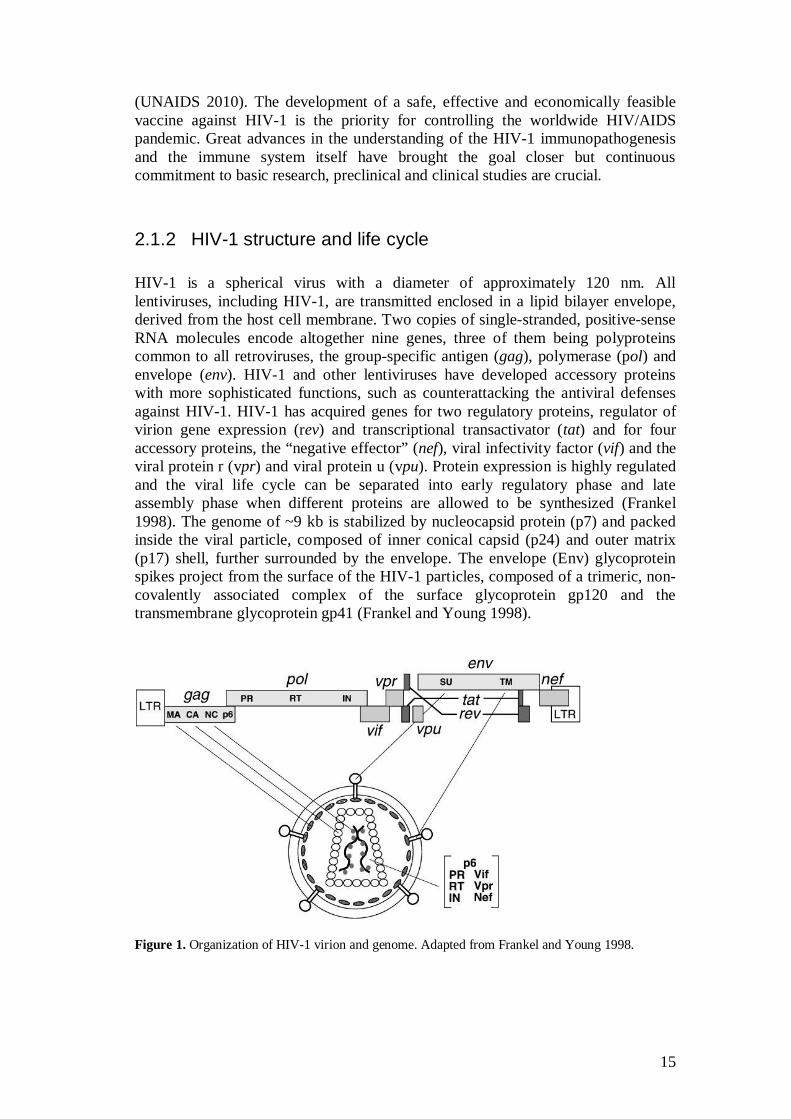
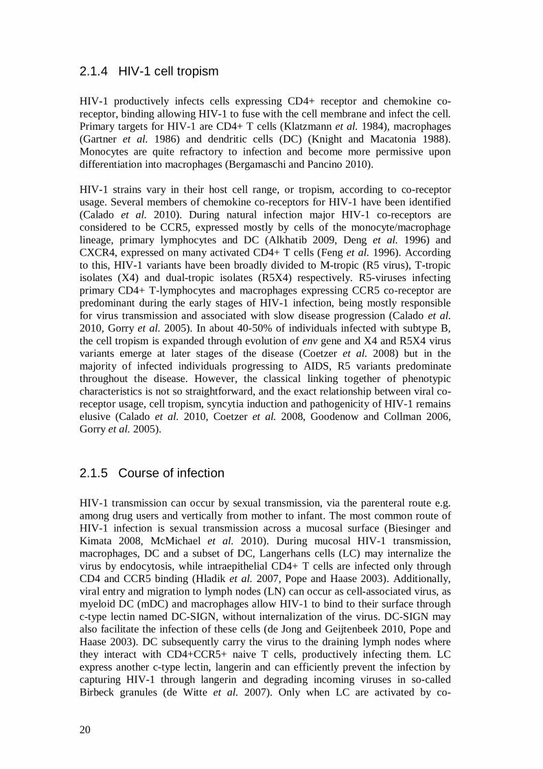
HIV-1 is a spherical virus with a diameter of approximately 120 nm. All lentiviruses, including HIV-1, are transmitted enclosed in a lipid bilayer envelope, derived from the host cell membrane. Two copies of single-stranded, positive-sense RNA molecules encode altogether nine genes, three of them being polyproteins common to all retroviruses, the group-specific antigen (gag), polymerase (pol) and envelope (env). HIV-1 and other lentiviruses have developed accessory proteins with more sophisticated functions, such as counterattacking the antiviral defenses against HIV-1. HIV-1 has acquired genes for two regulatory proteins, regulator of virion gene expression (rev) and transcriptional transactivator (tat) and for four accessory proteins, the “negative effector” (nef), viral infectivity factor (vif) and the viral protein r (vpr) and viral protein u (vpu). Protein expression is highly regulated and the viral life cycle can be separated into early regulatory phase and late assembly phase when different proteins are allowed to be synthesized (Frankel 1998). The genome of ~9 kb is stabilized by nucleocapsid protein (p7) and packed inside the viral particle, composed of inner conical capsid (p24) and outer matrix (p17) shell, further surrounded by the envelope. The envelope (Env) glycoprotein spikes project from the surface of the HIV-1 particles, composed of a trimeric, non-covalently associated complex of the surface glycoprotein gp120 and the transmembrane glycoprotein gp41 (Frankel and Young 1998).
Figure 1. Organization of HIV-1 virion and genome. Adapted from Frankel and Young 1998.
16
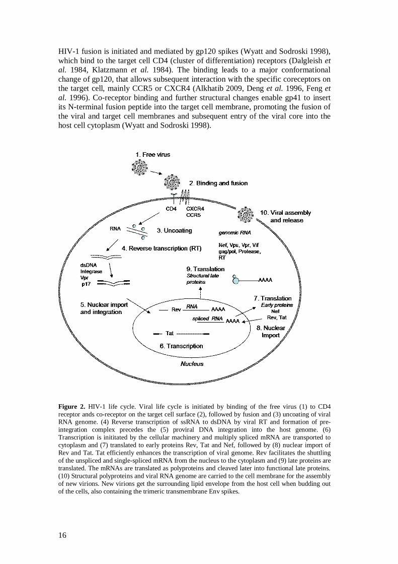
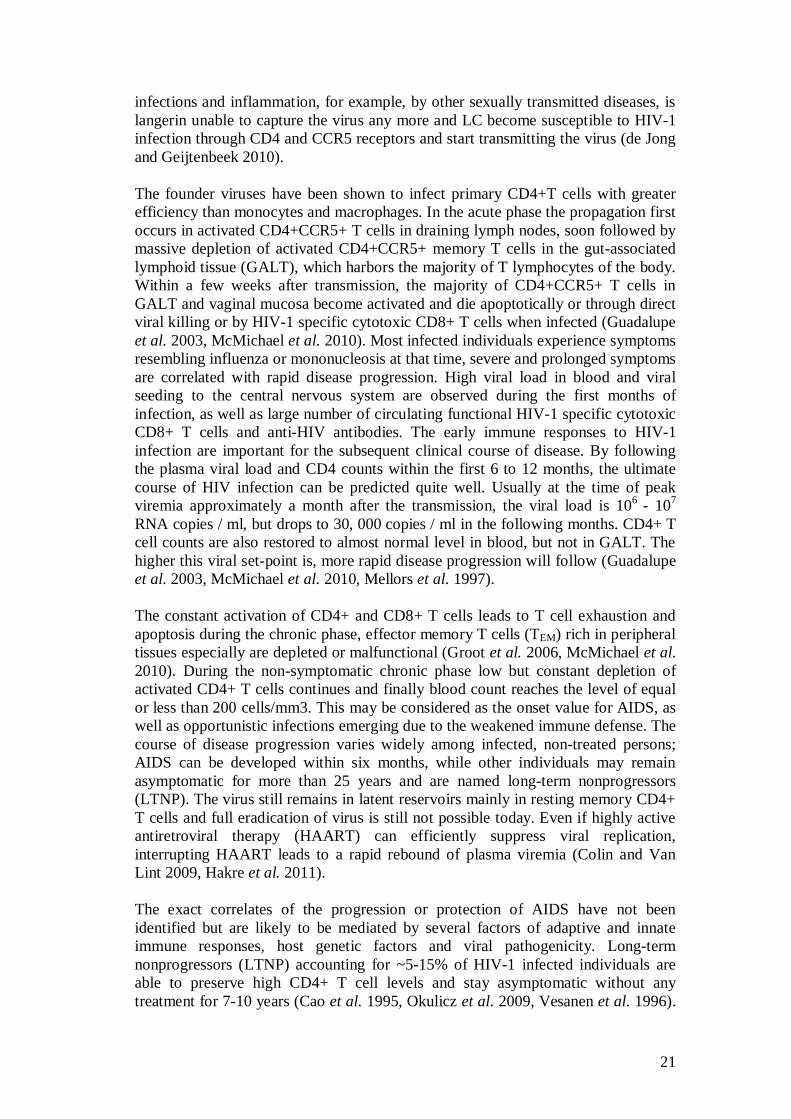
HIV-1 fusion is initiated and mediated by gp120 spikes (Wyatt and Sodroski 1998), which bind to the target cell CD4 (cluster of differentiation) receptors (Dalgleish et al. 1984, Klatzmann et al. 1984). The binding leads to a major conformational change of gp120, that allows subsequent interaction with the specific coreceptors on the target cell, mainly CCR5 or CXCR4 (Alkhatib 2009, Deng et al. 1996, Feng et al. 1996). Co-receptor binding and further structural changes enable gp41 to insert its N-terminal fusion peptide into the target cell membrane, promoting the fusion of the viral and target cell membranes and subsequent entry of the viral core into the host cell cytoplasm (Wyatt and Sodroski 1998).
Figure 2. HIV-1 life cycle. Viral life cycle is initiated by binding of the free virus (1) to CD4 receptor ands co-receptor on the target cell surface (2), followed by fusion and (3) uncoating of viral RNA genome. (4) Reverse transcription of ssRNA to dsDNA by viral RT and formation of pre-integration complex precedes the (5) proviral DNA integration into the host genome. (6) Transcription is inititated by the cellular machinery and multiply spliced mRNA are transported to cytoplasm and (7) translated to early proteins Rev, Tat and Nef, followed by (8) nuclear import of Rev and Tat. Tat efficiently enhances the transcription of viral genome. Rev facilitates the shuttling of the unspliced and single-spliced mRNA from the nucleus to the cytoplasm and (9) late proteins are translated. The mRNAs are translated as polyproteins and cleaved later into functional late proteins. (10) Structural polyproteins and viral RNA genome are carried to the cell membrane for the assembly of new virions. New virions get the surrounding lipid envelope from the host cell when budding out of the cells, also containing the trimeric transmembrane Env spikes.
17
Once in the cell cytosol, the RNA genome is reverse transcribed into double-stranded DNA by HIV-1 reverse transcriptase (RT), enzyme common to all retroviruses. RNAse H domain of RT has a ribonuclease H activity that is used for the degradation of viral RNA and DNA-dependent DNA polymerase activity of RT creates another strand of DNA. The extremely low fidelity of RT lacking the proofreading function leads to error-prone viral DNA synthesis, with error frequency of more than 3 substitutions per 10-5 incorporated nucleotides (Mansky and Temin 1995, Onafuwa-Nuga and Telesnitsky 2009). The extensive genetic heterogeneity of HIV-1 is largely based on the mismatches made during replication. Additionally, action of a host cell cytidine deaminase ABOBEC3G found in some cell types such as macrophages results in very high rate of G-to-A substitutions in retroviral genomes (Zhang et al. 2003). Pre-integration complex containing viral genome, integrase, matrix and Vpr along with host cell co-factors is formed and after being transported to the nucleus the dsDNA integrates into the host cell genome. The integrated HIV DNA persists as a provirus flanked by 5-bp long terminal repeats (LTR) needed both for integration and transcription (Vincent et al. 1990). The 5' LTR functions as a promoter while the 3' LTR site is where viral transcripts become polyadenylated. The integration has been shown to be preferentially targeted at genes activated in cells after infection by HIV-1 (Schroder et al. 2002). Integrated viral DNA may then lie dormant in the latent stage for a long time while the infected cell functions and replicates normally, passing on the HIV-1 genome to its progeny (Ranki et al. 1987). Post-integration latency is established at the beginning of HIV-1 infection, mostly in resting CD4+ memory T cells, when the provirus fails to express its genome. Several factors establishing the latency stage are related to transcription repression, like chromatin environment at the integration site, availability of host transcription factors, transcriptional interference and epigenetic silencing (Colin and Van Lint 2009, Hakre et al. 2011). Alternatively, the virus may activate and start assembling viral particles in the cell, utilizing the cell’s machinery. Transcription activators like cellular NFkB and viral Tat protein trigger the process of transcription to viral mRNA. Tat acts by binding to a stem-loop region (TAR) present near the 5 terminus of retroviral mRNA and stabilizes the elongation process by host RNA-polymerase II. Tat also recruits several cellular proteins for the transcription process. Multiple spliced mRNA is exported to the cytoplasm where early regulatory proteins Rev, Nef and Tat are translated, followed by the importation of Tat and Rev proteins into the nucleus where Tat enhances the transcription (Karn 1999, Romani et al. 2010). When Rev accumulates in the nucleus it binds to the viral Rev response element (RRE) that is present only in unspliced and singly spliced mRNA. Rev permits mRNA entry into the cytoplasm for translation and also enhances the unspliced mRNA encapsidation which directs it to the cell surface where assembly of new virus particles occurs (Brandt et al. 2007). Thus the later synthesis of Gag and Gag-Pol proteins from full length mRNA and Env, Vpu, Vpr and Vif from singly spliced mRNA is enabled by accessory protein Rev (Turner and Summers 1999). The mechanism of Rev dependent nuclear export has been proposed to have evolved to prevent the translation of structural proteins in the early phase of virus replication as their presentation might result in CD8+ T cell recognition (Blissenbach et al. 2010).
18
Single mRNA chain encodes both Gag and Pol proteins. Gag precursor protein (p55) is synthesized first and then subsequent translational frameshift results in translation of Gag-Pol polyprotein (p160). HIV-1 Protease cleaves p55Gag into viral internal proteins matrix p17, capsid p24, nuceocapsid p7 and p6 and small peptides p2 and p1. Polyprotein p160GagPol cleavage products are three HIV-1 enzymes Integrase, RT and Protease (Frankel and Young 1998). The elusive role of Nef in viral replication and pathogenesis is linked to the capability of Nef to downregulate cell surface molecule CD4, which is suggested to facilitate the release of viral progeny by preventing Env-CD4 interactions at the time of viral budding (Kim et al. 1999, Lundquist et al. 2002) and major histocompatibility (MHC) class I expression (Joseph et al. 2005, Piguet et al. 1999, Swann et al. 2001), which impedes the viral antigen presentation to CD8+ T cells. Nef enhances viral replication and infectivity also independently on CD4 downregulation (Saksela et al. 1995). Nef enhances T cell activation and transcription response, thereby increasing the viral production in those cells (Foster and Garcia 2008, Simmons et al. 2001). By inducing FasL expression on infected cells, Nef augments bystander cell death by apoptosis, including CTL. Additionally, Nef protects HIV-1 infected cells from apoptosis (Joseph et al. 2005, Piguet et al. 1999, Swann et al. 2001, Xu et al. 1997). New virions are assembled at the cell plasma membrane. The expressed Env precursor protein gp160 is glycosylated in the endoplasmic reticulum and cleaved in Golgi apparatus by the cellular protease furin. Cleaved protein products gp120 and gp41 are transported to the cell membrane, where they form trimeric structures. Myristoylation of p55Gag and p160GagPol polyprotein directs them to the cell membrane where two unspliced mRNA genomes are bound to Gag and thus packed into the new virion. p55Gag is the driving force in particle formation and release and is sufficient alone to form non-infectious virus-like particles (VLP). Maturation of the virus starts during the budding of the nascent virion from the plasma membrane, when HIV-1 Protease cleaves the Gag and Gag-Pol polyproteins into functional proteins. Gag domain p6 is used to hijack the components of the cellular endosomal sorting machinery, facilitating virus release (Frankel and Young 1998). Mature virion also contains accessory proteins Vif, Vpr and Vpu. Vpr is a part of pre-integration complex imported to nucleus and is able to arrest the cell cycle in the G2 phase and to induce apoptosis of infected cells. Vif and Vpu play important roles in HIV-1 pathogenesis by counteracting cellular antiviral factors (Gramberg et al. 2009). Vif is bound to the mRNA, stabilizing the viral core and inhibits the cellular antiviral protein APOBEC-3G (Zhang et al. 2003). Vpu contributes to the degradation of cell surface CD4 along with Nef and Env and also reduces amount of MCH molecules on the cell surface, promotes the release of the virions and prevents endocytosis of nascent viral particles from the plasma membrane (Gramberg et al. 2009, Piguet et al. 1999). Finally, the mature virus contains the HIV-1 genome, structural proteins and the proteins important for infectivity and for early phase of viral life cycle inside the new host cell.
19
2.1.3 Genetic variability and global distribution of HIV-1 subtypes
The hallmark for RNA viruses and especially for HIV-1 is the broad genetic variability, as every isolate of the virus and almost every virus in one population is different. Rapid turnover of virus replication in vivo and high mutation rate by error-prone RT gives rise to HIV-1 quasispecies, virus particles with modified properties from transmitted founder virus, in a single infected individual (McMichael et al. 2010, Onafuwa-Nuga and Telesnitsky 2009). In addition to point mutations, extensive recombination among quasispecies or among two separate strains in patients with HIV-1 coinfection efficiently generates variation. HIV-1 recombination is a result of RT template switching between two viral RNA during provirus synthesis (Charpentier et al. 2006, Onafuwa-Nuga and Telesnitsky 2009, Temin 1993). The excessive selective pressure exerted both by the host’s immune system and antiretroviral therapy as well as availability of different target cells further drives HIV-1 diversification and evolution. Even if the majority of genome mutations lead to depleted replicative capacity and viability of the virus, they also allow the virus to escape the host defense (Biesinger and Kimata 2008). HIV-1 is subdivided into four divergent phylogenetic subgroups, designated as M (main), O (outlier), N (non-M non-O) (Taylor and Hammer 2008) and a newly identified variant P, being distinct from previously established groups (Plantier et al. 2009). Group O and N viruses are found at low frequencies mainly in Central Africa. HIV-1 group M is the predominant reason for the pandemic, accounting for more than 95% of infections worldwide. Group M consists of nine different genetic subtypes or clades (A, B, C, D, F, G, H, J and K), some of them further divided into sub-subtypes designated with numbers. Advances in the sequencing of HIV have also resulted in the identification of circulating and unique recombinant forms (CRF and URF). HIV-1 genetic forms are distributed over global geographic areas to varying extents. Clade C has become the dominant subtype in countries most heavily infected with HIV-1, like Southern Africa and India and currently accounts for nearly half of global HIV-1 infections. Clade A is predominantly found in Eastern and Central Africa and countries of the former Soviet Union in Eastern Europe. In Western and Central Europe, America and Australia the B clade is prevalent (Buonaguro et al. 2007). The degree of conservation in the different genes varies, pol encoding HIV-1 enzymes is most conserved (>90%), structural gene gag also possessing quite a high degree of conservation (>80%) while nef somewhat less (>70%) (Coplan et al. 2005, Rolland et al. 2007b). The greatest variability occurs in the gene encoding Env, which is the only viral protein presented on the surface of the viral particle and thus most predisposed to selective pressure. Env sequence can differ up to 35% between clades and 20% within a clade (Taylor and Hammer 2008). Conserved sequences are usually related to viral fitness, the sequences that are crucial for viral infectivity and pathogenesis have least variability.
20
2.1.4 HIV-1 cell tropism
HIV-1 productively infects cells expressing CD4+ receptor and chemokine co-receptor, binding allowing HIV-1 to fuse with the cell membrane and infect the cell. Primary targets for HIV-1 are CD4+ T cells (Klatzmann et al. 1984), macrophages (Gartner et al. 1986) and dendritic cells (DC) (Knight and Macatonia 1988). Monocytes are quite refractory to infection and become more permissive upon differentiation into macrophages (Bergamaschi and Pancino 2010). HIV-1 strains vary in their host cell range, or tropism, according to co-receptor usage. Several members of chemokine co-receptors for HIV-1 have been identified (Calado et al. 2010). During natural infection major HIV-1 co-receptors are considered to be CCR5, expressed mostly by cells of the monocyte/macrophage lineage, primary lymphocytes and DC (Alkhatib 2009, Deng et al. 1996) and CXCR4, expressed on many activated CD4+ T cells (Feng et al. 1996). According to this, HIV-1 variants have been broadly divided to M-tropic (R5 virus), T-tropic isolates (X4) and dual-tropic isolates (R5X4) respectively. R5-viruses infecting primary CD4+ T-lymphocytes and macrophages expressing CCR5 co-receptor are predominant during the early stages of HIV-1 infection, being mostly responsible for virus transmission and associated with slow disease progression (Calado et al. 2010, Gorry et al. 2005). In about 40-50% of individuals infected with subtype B, the cell tropism is expanded through evolution of env gene and X4 and R5X4 virus variants emerge at later stages of the disease (Coetzer et al. 2008) but in the majority of infected individuals progressing to AIDS, R5 variants predominate throughout the disease. However, the classical linking together of phenotypic characteristics is not so straightforward, and the exact relationship between viral co-receptor usage, cell tropism, syncytia induction and pathogenicity of HIV-1 remains elusive (Calado et al. 2010, Coetzer et al. 2008, Goodenow and Collman 2006, Gorry et al. 2005).
2.1.5 Course of infection
HIV-1 transmission can occur by sexual transmission, via the parenteral route e.g. among drug users and vertically from mother to infant. The most common route of HIV-1 infection is sexual transmission across a mucosal surface (Biesinger and Kimata 2008, McMichael et al. 2010). During mucosal HIV-1 transmission, macrophages, DC and a subset of DC, Langerhans cells (LC) may internalize the virus by endocytosis, while intraepithelial CD4+ T cells are infected only through CD4 and CCR5 binding (Hladik et al. 2007, Pope and Haase 2003). Additionally, viral entry and migration to lymph nodes (LN) can occur as cell-associated virus, as myeloid DC (mDC) and macrophages allow HIV-1 to bind to their surface through c-type lectin named DC-SIGN, without internalization of the virus. DC-SIGN may also facilitate the infection of these cells (de Jong and Geijtenbeek 2010, Pope and Haase 2003). DC subsequently carry the virus to the draining lymph nodes where they interact with CD4+CCR5+ naive T cells, productively infecting them. LC express another c-type lectin, langerin and can efficiently prevent the infection by capturing HIV-1 through langerin and degrading incoming viruses in so-called Birbeck granules (de Witte et al. 2007). Only when LC are activated by co-
21
infections and inflammation, for example, by other sexually transmitted diseases, is langerin unable to capture the virus any more and LC become susceptible to HIV-1 infection through CD4 and CCR5 receptors and start transmitting the virus (de Jong and Geijtenbeek 2010). The founder viruses have been shown to infect primary CD4+T cells with greater efficiency than monocytes and macrophages. In the acute phase the propagation first occurs in activated CD4+CCR5+ T cells in draining lymph nodes, soon followed by massive depletion of activated CD4+CCR5+ memory T cells in the gut-associated lymphoid tissue (GALT), which harbors the majority of T lymphocytes of the body. Within a few weeks after transmission, the majority of CD4+CCR5+ T cells in GALT and vaginal mucosa become activated and die apoptotically or through direct viral killing or by HIV-1 specific cytotoxic CD8+ T cells when infected (Guadalupe et al. 2003, McMichael et al. 2010). Most infected individuals experience symptoms resembling influenza or mononucleosis at that time, severe and prolonged symptoms are correlated with rapid disease progression. High viral load in blood and viral seeding to the central nervous system are observed during the first months of infection, as well as large number of circulating functional HIV-1 specific cytotoxic CD8+ T cells and anti-HIV antibodies. The early immune responses to HIV-1 infection are important for the subsequent clinical course of disease. By following the plasma viral load and CD4 counts within the first 6 to 12 months, the ultimate course of HIV infection can be predicted quite well. Usually at the time of peak viremia approximately a month after the transmission, the viral load is 106 - 107
RNA copies / ml, but drops to 30, 000 copies / ml in the following months. CD4+ T cell counts are also restored to almost normal level in blood, but not in GALT. The higher this viral set-point is, more rapid disease progression will follow (Guadalupe et al. 2003, McMichael et al. 2010, Mellors et al. 1997). The constant activation of CD4+ and CD8+ T cells leads to T cell exhaustion and apoptosis during the chronic phase, effector memory T cells (TEM) rich in peripheral tissues especially are depleted or malfunctional (Groot et al. 2006, McMichael et al. 2010). During the non-symptomatic chronic phase low but constant depletion of activated CD4+ T cells continues and finally blood count reaches the level of equal or less than 200 cells/mm3. This may be considered as the onset value for AIDS, as well as opportunistic infections emerging due to the weakened immune defense. The course of disease progression varies widely among infected, non-treated persons; AIDS can be developed within six months, while other individuals may remain asymptomatic for more than 25 years and are named long-term nonprogressors (LTNP). The virus still remains in latent reservoirs mainly in resting memory CD4+ T cells and full eradication of virus is still not possible today. Even if highly active antiretroviral therapy (HAART) can efficiently suppress viral replication, interrupting HAART leads to a rapid rebound of plasma viremia (Colin and Van Lint 2009, Hakre et al. 2011). The exact correlates of the progression or protection of AIDS have not been identified but are likely to be mediated by several factors of adaptive and innate immune responses, host genetic factors and viral pathogenicity. Long-term nonprogressors (LTNP) accounting for ~5-15% of HIV-1 infected individuals are able to preserve high CD4+ T cell levels and stay asymptomatic without any treatment for 7-10 years (Cao et al. 1995, Okulicz et al. 2009, Vesanen et al. 1996).
22
Another small subset, < 1% of HIV-1 infected subjects not developing AIDS and additionally displaying more sustained control over viral replication (plasma HIV RNA levels of <50 copies/ml) are denoted elite controllers (Okulicz et al. 2009). These exceptional groups among HIV-1 infected individuals have yielded some insight into the mechanisms delaying the disease. Additionally, the sooty mangabeys that are natural hosts for related SIV infection, do not develop AIDS when infected. One explanation has been suggested to be the absence of chronic activation of the immune system that is detected in non-natural SIV hosts like rhesus macaques, and in human HIV-1 infection where the immunodeficiency is acquired (Mandl et al. 2008). In addition to the massive loss of T lymphocytes by direct viral killing, the driving force for AIDS progression is likely to be the chronic generalized immune activation, associated with elevated turnover rates of T cells and NK cells, polyclonal activation of B cells and decrease of peripheral DC number and reduced capability to regenerate immune cells by the primary lymphoid organs.
2.2 Innate immunity and cytokines
The immune system is composed of two major subdivisions, non-specific innate immunity and specific adaptive immunity. While the mucosal route is the most common for HIV-1 transmission, the mechanisms prevailing there have the greatest impact on protection from HIV-1. The mucosal layer is rich in cells of the immune system and antimicrobial agents produced by these cells. Innate immunity provides immediate first line defense against invading pathogens and is able to respond rapidly. It has a remarkable role in primary HIV-1 response, as adaptive immune responses will be functional only when the HIV-1 infection is already well established. Cells working for innate immune responses during viral infection, such as tissue macrophages, blood monocytes, plasmocytoid DC (pDC), LC, NK and neutrophils can act by directly destroying the viral particles and infected cells or through the secretion of soluble factors like cytokines (Borrow et al. 2010, Chang and Altfeld 2010). These cells use their pattern recognition receptors (PRR), such as toll-like receptors (TLR) for the recognition of structures specific for micro-organisms, called pathogen-associated molecular patterns (PAMPS), which activates cells to produce several cytokines and chemokines. These immunomodulators are the main controllers of the immune system linking the innate and adaptive immunity, turning on and off the inflammatory responses and directing immune cell differentiation, function and migration and having direct anti-viral effects (Alfano et al. 2008, Borrow et al. 2010). Additionally, TLR-induced cytokine production upregulates the expression of multiple TLR receptors and enhances IFN production in a synergistic manner (Makela et al. 2011). In viral infections structures such as double-stranded RNA (dsRNA), single-stranded RNA (ssRNA), unmethylated CpG-oligodeoxynucleotide-containing DNA and certain viral proteins are recognized (Chang and Altfeld 2010, Mandl et al. 2008). Type I IFNs are a superfamily of innate cytokines including 13 IFN- subtypes, IFN- , - , - and - , triggered by virus infections through TLR stimulation. pDC stimulation by CpG DNA through
23
TLR-9 induces expression of several antiviral cytokines, such as IFN- , - , - and IFN- (Coccia et al. 2004). Cytokines are involved in HIV-1 pathogenesis, having an inhibitory effect on infectivity and replication on several stages of HIV-1 life cycle but also by enhancing the infection and leading to exacerbated responses. Their improper regulation during HIV-1 infection is one main effector for HIV-1 pathogenesis (Chang and Altfeld 2010, Mandl et al. 2008) and early cytokine storm in acute HIV-1 has been well documented, associated with enhanced expressions of IFN- , IL-15, IL-6, IL-8, IL-18 and IFN- (Chang and Altfeld 2010, Stacey et al. 2009). A thorough understanding of the roles of each cytokine is also important when utilizing cytokines as immunopotentiators with vaccines or for antiretroviral therapy (Alfano et al. 2008, Borrow et al. 2010, Stacey et al. 2009). One well characterized innate defense mechanism against HIV-1 in humans and SIV infection in non-human primates is the cascade initiated by recognition of the viral HIV-1 ssRNA through TLR7 expressed by pDC, monocytes and B cells (Chang and Altfeld 2010, Mandl et al. 2008). HIV-1 endocytosis enhances pDC viability and maturation through TLR7 and activation leads to the production of several cytokines such as IFN- , TNF- , and proinflammatory chemokines and upregulates some maturation markers (CD83, CCR7) and costimulatory molecules (CD80, CD86) on pDC. HIV-1 activated pDC additionally contribute in mDC maturation, as mDC are not activated directly by HIV-1 (Fonteneau et al. 2004). Greater numbers of pDCs are found in HIV-infected individuals who are healthy, long-term survivors (Soumelis et al. 2001). Mucosal LC are the first DC subset to encounter HIV-1 and similar to pDC, they can detect ssRNA through intracellular TLR7. Additionally, LC work for innate immunity by internalizing and degrading the virus as described earlier (de Witte et al. 2007). NK cells can recognize HIV-1 infected cells that have downregulated MHC molecules and kill them by releasing apoptosis inducing proteins perforin and granzyme (Chang and Altfeld 2010, Pipkin and Lieberman 2007). NK cells also mediate non-cytolytic suppression of viral spread through the secretion of chemokines CCL3, CCL4, and CCL5 (also named MIP-1 and MIP-1 and RANTES) that can function as CCR5 ligands and thereby block the virus from entering target cells. NK cells produce cytokines such as IFN- , TNF and granulocyte/macrophage-stimulating factor (GM-CSF). NK contribution, especially by IFN- production, has been emphasized in studies with seronegative individuals constantly exposed to HIV-1 (Fauci et al. 2005). NK cells are one of the major effector cells for antibody dependent cellular cytotoxicity (ADCC), mechanism based on Fc receptors on NK cells, able to recognize and bind the Fc region of the antibody that is bound to the surface of the infected cells, thus recruiting NK cells. However, the role of ADCC in HIV-1 infection has not been extensively studied. In addition, IgG antibodies can bind proteins of the complement system, thereby leading to engagement of the complement system which plays a critical role in clearing and neutralizing HIV-1 virions. HIV-specific antibodies and complement deposited onto HIV lead to the inactivation of the virus and lysis of infected cells (Mascola and Montefiori 2010, Stoiber et al. 2008). However, the HIV-1 virus, having a broad range of strategies to avoid the host defense, also has a mechanism to avoid complement-mediated lysis and even take advantage of the system.
24
Mammalian cells are protected by the expression of cell membrane complement regulators such as CD55 and CD59 and HIV-1 can obtain these proteins on their surface when budding out of the cell and use them to escape the Ab-dependent, complement-mediated virolysis (Saifuddin et al. 1995). One specific intraepithelial lymphocyte type called T cells bridges the innate and the adaptive immune system. They are mainly found in gut mucosa and thereby play a role in first line defence in HIV-1 infection, secreting chemokines, antiviral factors and killing infected cells through natural killer mechanism. T cells are not dependent on the MHC antigen presentation (Agrati et al. 2011). Additionally, another distict cell type called NKT cells share characteristics both with NK cells and T cell lymphocytes but little is known about their responses in HIV-1 infection (Borrow et al. 2010, Borrow et al. 2010). Innate and adaptive immune responses closely co-operate and the initial innate immune response can determine the quality of the forthcoming adaptive immune response. Cells of innate immunity can capture the foreign material and further present these antigens for lymphocytes in lymphoid organs. The cytokine and chemokine production also controls adaptive immune responses. Vaccines based on attenuated or killed pathogens contain pathogenic molecular patterns recognized by innate immunity and therefore induce strengthened specific immune responses. When applying highly purified vaccine antigens, the lack of these viral components result in weaker immune responses. For that reason the vaccine adjuvants are designed also to activate the innate immunity, further extrapolating the strength of the specific adaptive response (Chang and Altfeld 2010).
2.3 Adaptive immunity
While innate immunity provides rapid but unchanged response to antigen, the adaptive immune response takes days to evolve after the first encounter with the antigen. The highly specific surface receptors initiate tailored responses to the particular pathogen and, most importantly, the adaptive immune system remembers the antigen when repeatedly exposed. The re-encounter of the same antigen triggers significantly faster and more vigorous cellular and humoral immune responses driven by antigen specific memory T and B lymphocytes. The phenomenon of immunological memory is the basis for adaptive immunity, and the key to artificial acquired immunity achieved by vaccination. T lymphocytes maturing in the thymus and B lymphocytes maturing in bone marrow are the cells responsible for cell-mediated and humoral adaptive immunity respectively. They are highly adaptable to recognize wide variety of pathogens by generating a vast number of different antigen receptors. T and B cells can enter the secondary lymphoid organs including the spleen, lymph nodes and mucosal lymphoid tissue as naïve lymphocytes. Lymph nodes, the garrisons of immune cells, are the most important sites for antigen presentation and have a structure highly specific for their function (Banchereau and Steinman 1998).
25
2.3.1 Professional APC – The interphase between innate and acquired immunity
Indispensable to adaptive immunity are the professional antigen presenting cells (APC) such as DC, macrophages and B cells that constantly screen their surroundings for self and non-self antigens. Professional APC can capture and engulf the extracellular molecules from the periphery, degrade them and present antigens on their surface to naive CD4+ and CD8+ T cells, the phenomenon called priming. Professional APC in the lymph nodes and spleen may also acquire antigen from circulating lymph fluid or blood respectively. DC and LC have an enormous capacity to induce strong T cell responses by small numbers of DC and low levels of antigen. Some lines of evidence suggest that macrophages could also be almost as efficient in priming T cell responses (Pozzi et al. 2005). Naive T and B cells can differentiate as effector, helper and regulatory cells and a small number of these antigen specific cells is maintained as memory cells once the pathogen has been eradicated. DC can be resident in tissues or circulate in the blood gathering antigens. In the initial immature state DC express only low levels of MHC class II molecules and costimulatory molecules needed for T cell activation, such as CD80 (B7-1) and CD86 (B7-2), but are instead highly phagocytic and effective in capturing antigens. After encountering the antigen, DC are activated and can migrate to secondary lymphoid organs where they maturate fully if receiveing the costimulation signal through CD40. Maturation is accompanied by increased expression of MHC class I and II molecules and the costimulatory molecules CD80, CD86, CD40 and CD83 and by downregulation of antigen capture molecules such as Langerin and E-cadherin on LC or DC-SIGN on DC. Migration to secondary lymphoid organs maximizes the probability of interaction with T and B cells and is directed by CCR7 chemokine receptor on DC (Banchereau and Steinman 1998, de Jong and Geijtenbeek 2010, Guermonprez et al. 2002). DC can be divided into two subsets according to CD11c expression; myeloid DC (mDC) expressing CD11c+ and plasmocytoid CD11c- DC. CD11c+ DC include LC, dermal and interstitial DC and have additionally been subdivided into myeloid CD11b+CD11c+ DC and lymphoid CD8 +CD11c+ DC with different T cell priming capabilities (Abadie et al. 2009, Donaghy et al. 2001). During HIV-1 infection an inverse correlation has been observed between viral load and CD11c+ DC numbers, CD11c+ loss and shift to Th2 type cytokines has shown to be concurrent (Donaghy et al. 2001). The migration patterns of naïve, effector and memory lymphocytes between lymphoid tissue and non-lymphoid tissue are consequence of differential expression of chemokine and adhesion receptors, such as integrins (Denucci et al. 2009, Muller and Lipp 2003). The precise migration capability of each cell type remains controversial (Cose 2007). Naïve T cell and central memory T cell (TCM) homing receptors are such as L-selectin and CCR7, providing adhesion to lymph nodes, while activated effector T cells and effector memory T cells (TEM) express molecules such as CCR5 and CXCR3, directing cells to peripheral site of infection (Denucci et al. 2009). In general, the primary immune response is triggered during the first few days when DC prime the naïve T cells in the lymph nodes, leading to their clonal expansion. DC migration to the lymphoid tissue after s.c. inoculation has been shown to be more efficient than migration of macrophages. However,
26
when these APC were injected i.v., both cell types were equally presented in the spleen one day later (Pozzi et al. 2005). The number of antigen-carrying DC in draining LN and in mucosal Peyer’s patches has been demonstrated to peak between 4 hours and 2.5 days after skin contact with antigen (Abadie et al. 2009, Belyakov et al. 2004, Garg et al. 2003, Porgador et al. 1998) and shown to persist for approximately two weeks (Akbari et al. 1999, Garg et al. 2003, Pozzi et al. 2005, Tuomela et al. 2005). For CD4+ T cells the clonal expansion has been apparent by day 2 and maximal on days 3 and 4. B cell expansion has been detected 3 days after immunization, reaching maximal level by day 5 and then declining (Garside et al. 1998).
2.3.2 Activation of humoral immune responses
B cells act as professional APC, even if much less efficiently than DC. B cells can engulf and respond directly to some antigens but are partly dependent on antigen presentation by other APC, depending on the nature of the antigen. Antigens are recognized through B cell receptor by naïve B cells as intact antigens, being either soluble or membrane-bound. Activated CD4+ T cells play an important role in promoting B cell activation through CD40L/CD40 interactions although many non-protein antigens, called T cell independent antigens, can stimulate antibody production independently of CD4+ T cells (Batista and Harwood 2009). Once CD4+ T cells are activated by DC priming, they are able to stimulate B cell responses. They express CD40L, downregulate CCR7 expression and upregulate CXCR5 chemokine receptor that directs them to migrate from the T cell zone towards the follicle, augmented by follicular cells that secrete CXCR5 ligand. Conversely, activated B cells downregulate the CXCR5 and start to express CCR7, thus directing them toward the T-B interface and T cell zone. This leads to CD4+ T cell and B cell interaction and B cells can present the MHC class II-associated peptides to CD4+ T cells, thereby recruiting CD4+ T cell help which stimulates B cell proliferation, differentiation and antibody isotype switching (Muller and Lipp 2003, Okada et al. 2002). Activated B cells can differentiate as antibody producing plasma cells or as memory B cells that provide long-lasting protection. The activated B cells require approximately two weeks to accomplish proper antibody response (Batista and Harwood 2009, Tahtinen et al. 2001, Tähtinen 2001).
2.3.3 Activation of cellular immune responses
Cell-mediated immune responses are mediated through cytotoxic CD8+ T lymphocytes (CTL) and CD4+ T helper (Th) lymphocytes. The subclass of Th cells, regulatory T cells (Treg) suppress the immune system and maintain the self-tolerance. Naïve T cell are primed through T cell receptors (TCR) that recognize the peptide in the context of MHC molecules on APC. MHC molecule is additionally bound by another T cell co-receptor, either CD4 or CD8. T cell activation is successful if the second signal is received by a costimulatory molecule on the APC, interacting with the specific ligand on the T cell. APC can use CD80 (B7-1) and CD86 (B7-2) molecules to either activate T cell through CD28 receptor or inhibit T cell responses through CTLA-4 binding (Alegre et al. 2001). The signaling cascade
27
in the T cells is initiated by TCR, associated CD3 molecule and CD4 or CD8 co-receptor. The absence of costimulation leads to anergy or apoptosis of the T cell unable to respond to an antigen. Interactions with professional APC are essential for maintaining the T cell activity, also in the absence of foreign antigen presentation (Hochweller et al. 2010). Once activated, T cells start proliferation and differention. The division of antigen-activated T cells is very rapid and leads to >1000-fold expansion of the responding cells within a few days. At the end of the primary response >90% of the effector cells are destroyed, only few of them surviving as long-lived effector and central memory cells (D'Cruz et al. 2009, Williams and Bevan 2007). Induction of the polyfunctional CD4+ and CD8+ T cell responses is a highly desired goal for immunizations, referring to the ability of T cells to produce high levels of several soluble factors simultaneously, such as cytokines IFN- , IL-2 and TNF- , chemokines like MIP-1 and cytotoxins. Degranulation indicating cytotoxic ability is measured as CD107a surface mobilization (Betts et al. 2006, Duvall et al. 2008).
2.3.3.1 Cytotoxic T cells
The effectors of the cellular immune system are CD8+ T cells that respond to peptides derived from proteins expressed inside the cell that can be derived either from self-proteins or during the viral infection, from viral proteins. These intracellular antigens are presented through MHC class I molecules, expressed not only on professional APC but on every nucleated cell of the body. However, stimulation of naive CD8+ T cells requires a costimulatory signal by professional APC and is thus dependent on these cells. In the case of direct priming, endogenously synthesized antigens are presented on MHC class I molecule (York and Rock 1996), while the cross-priming or cross-presentation refers to the ability of professional APC also to present exogenous antigens through MHC class I to CD8+ T cells (Corr et al. 1996, Corr et al. 1999, Giri et al. 2004, Jung et al. 2002, Norbury and Sigal 2003). DC and macrophages can process the internalized exogenous antigens for MHC class I cross-presentation mainly through phagosome-to-cytosol or vacuolar pathways. A major source of cross-presented antigens are particulates, such as dying cells antigens, while cross-presentation of soluble proteins is less efficient (Rock et al. 2010). For MHC class I direct presentation the antigens are degraded by the cytosolic proteasome and cut into several small peptides which are transported to the endoplasmic reticulum (ER) for binding to MHC I molecules. A single type of MHC I molecule can bind to a large range of different peptides with different affinities depending on the amino acid sequence. Additionally, the peptide binding groove is closed at both ends, limiting the length of the binding peptide. The peptides presented are usually 8 to 10 amino acids in length, the majority being nonamers. Occasionally longer peptides are accommodated but extending out of the groove may decrease the stability of interaction (Blanchard and Shastri 2008). Immunodominance is a central feature of cellular immune responses, reflecting the phenomenon that only a minor fraction of all potential immunogenic peptides derived from complex antigens are actually recognized. Which epitopes will be immunodominant in the individual is dependent on the expressed MHC allomorphs and their capability to bind and present the peptides. The immunogenicity of
28
subdominant determinants may be suppressed by the CD8+ T cells specific for immunodominant determinants (Manuel et al. 2009). A thorough understanding of the immunodominance phenomenon would be imperative for the designing of vaccines that elicit optimal CD8+ T cell responses (Yewdell and Bennink 1999, Yewdell 2006). The human equivalents of MHC are called human leukocyte antigens (HLA), class I encoded by three HLA genes termed HLA-A, -B and –C. HLA-A and HLA-B display greater polymorphic variation and are mainly responsible for presenting antigens. One individual can express up to six different class I molecules if heterozygous (Yewdell 2006, York and Rock 1996). The MHC genomic region in mouse is called H-2. Corresponding to human HLA-A, -B and –C, subtypes in mice are members of the classic MHC class I molecules, named H-2D, H-2K and H-2L. Like their human counterparts, these highly polymorphic genes are expressed widely and play an important role in presentation of non-self antigens to CD8+ CTL. Class I also has three non-classic subclasses that are similar in structure but not in function. Inbred laboratory mice strains are homozygous and have unique MHC haplotype, being H-2d for BALB/c (Ohtsuka et al. 2008, York and Rock 1996). MHC molecules provide a mechanism to monitor the quality of antigens that cells produce. CD8+ T cells are tolerant of healthy self-antigen presenting cells, while recognition of abnormal, such as tumorigenic or infected cells, leads to their elimination by CTL (Rock et al. 2010). Presentation of viral peptides on class I MHC molecules for CTL targets the infected cells for CTL mediated killing, usually by triggering apoptosis (Shastri et al. 2002). Cytotoxic T cells and NK cells of innate immunity employ the same cytotoxic mechanism to initiate target cell apoptosis by releasing the contents of cytotoxic granules, perforin and serine proteases called granzymes into the target cell (Hersperger et al. 2010, Pipkin and Lieberman 2007). CD8+ T cells have additionally a non-cytolytic mechanism mediating strong antiviral immune responses involving several chemokines and secreting soluble CD8 cell antiviral factor (CAF) that suppresses HIV replication (Levy et al. 1996). A broad range of cytokines (IFN- , TNF- , IL-2, GM-CSF, CCL3, CCL4, and CCL5) may be also secreted by activated CD8+ T cells, although IFN- predominates (Giri et al. 2004, Kristensen et al. 2004).
2.3.3.2 Helper T cells
CD4+ T helper cell activation is dependent on antigen presentation by another pathway mediated by MHC class II molecules that are expressed predominantly by professional APC. CD4+ T cells recognize antigens derived from exogenous and endogenous pathogens when antigens are presented by MHC class II molecules on professional APC. Most class II-associated antigens are exogenous, internalized by endocytosis or phagosytosis but also endogenous cytosolic and membrane proteins can enter the class II pathway. Antigens are processed to antigenic peptides, which are further bound to the binding groove of the MHC II molecule in specialized vesicles. MHC class II molecules have an open-ended binding groove allowing the display of considerably larger peptides of 15-20 amino acids (Moss et al. 2007). The MHC-II / peptide complex is then transported to the cell surface where it can
29
present the antigen for CD4+ T cells. Human HLA class II genes are encoded by three loci, HLA-DP, HLA-DQ and HLA-DR. In mouse, I-A and I-E subregions encode several MCH class II proteins (Cosgrove et al. 1992, Matthews et al. 2000). When activated, helper T cells regulate and direct the immune responses by other immune cells by secreting several cytokines such as IFN- , TNF- , IL-2, GM-CSF, CCL3, CCL4, and CCL5 and by expressing co-stimulatory molecules on their surface that can be presented by cell-to-cell interactions. They start to proliferate and secrete IL-2 that acts in autocrine fashion through CD25 receptor that is simultanously upregulated on helper T cells. CD40L is expressed on the surface of activated CD4+ T-helper cells and is involved in their activation and in the development of their effector functions. Depending on cytokine environment they can differentiate into several subtypes, most importantly Th1, Th2 and more recently characterized Th17 and regulatory T cell (Treg), all having distinct patterns of cytokine secretion. Th1 cells direct the immune responses towards cellular immune responses by maximizing CTL and macrophage function through cytokines such as IFN- , TNF- and IL-2. Th2 cells stimulate humoral immune system by secreting cytokines IL-4, IL-5, IL-6, IL-10 and IL-13. Physiologically, Th1 responses are thought to be important for defense against intracellular microbes and Th2 against multicellular parasites (Mosmann and Sad 1996). Th-17 cells regulate tissue inflammatory reactions by secreting proinflammatory cytokines IL-17 and IL-22 that act against various infections, but no IFN- or IL-4. They are also associated with tissue destruction during autoimmune diseases, thereby maintaining the right balance of Th17 cells is crucial (Brenchley et al. 2008, Lin et al. 2010, Park et al. 2005). By contrast, Treg cells function in an immunosuppressive manner by direct cell-to-cell contacts and by secreting inhibitory cytokines IL-10 and TGF- , Treg cells also express FoxP3 transcription factor and disruption of FoxP3 function has been shown to lead to severe immune dysfunction and autoimmune diseases (Sojka et al. 2008). FoxP3+CD25+ Treg cells have been observed to be capable of down-regulating FoxP3 expression, then functioning more as memory and effector cells than as regulatory cells, demonstrating Th cell plasticity (Zhou et al. 2009). Th cells are essential for CD8+ T cell activation and for promoting protective memory cell development and thereby the induction of CD4+ T cell responses is also very important for vaccines.
2.3.3.3 Memory T cells
The generation and retainment of T cell memory during natural infection is not very well known. However, memory T cells are shown to emerge during the covalescence, in few weeks after the clearance of the pathogen (Tuuminen et al. 2007). Memory CD8+ T cells and memory CD4+ T cells can be subdivided into two distinct subsets, central memory cells (TCM), capable of regeneration and long-term maintenance and effector memory cells (TEM) cells, which are more prevalent in peripheral tissues and provide immediate effector functions (Baron et al. 2003). TCM cells express the CCR7 lymph node homing chemokine receptor and efficiently stimulate DC. Upon secondary antigen stimulation TCM can readily differentiate to TEM cells, expressing receptors that direct them to inflamed tissues (Sallusto et al. 1999). CD4+ memory T cells are not so extensively studied than CD8+ memory T cells (MacLeod et al. 2009). For vaccines the capability to elicit long-lasting
30
memory T cells is essential and immunization studies with rotavirus and influenza virus vaccines have yielded mounting evidence that IL-17 and IL-23 producing Th17 memory cells have an important role in long-lasting vaccine-induced memory T cell responses (Lin et al. 2010).
2.3.4 Lessons learned from HIV-infected individuals and non-human primates
Adaptive immunity recognizes an enormous number of antigens but the capability of HIV-1 to escape these responses has still been overwhelming for the immune system. HIV-1 virus defeats the immune system by continuously introducing new variants of the immunogenic surface epitopes and simultaneously hiding the conserved parts from the immune system. Reports of superinfections, i.e. the re-infection of an individual with a second heterologous strain of HIV-1, have demonstrated the major challenge for developing vaccines (Allen and Altfeld 2003). These cases demonstrate the inability of natural immune responses generated during primary infection to protect the individual from cross-clade or even same clade HIV-1 re-infection. However, even if the correlates of protection against HIV-1 remain to be clarified, finding the factors likely to impede the progression of infection are crucial and may finally lead to overcoming the HIV-1 epidemic.
2.3.4.1 The role of cytotoxic T cells in HIV-1 infection
The correlation observed between the HIV-1 specific CTL response and disease progression has indicated the important role of cellular immunity in HIV-1 infection (Hersperger et al. 2011). Primary HIV-1 infection causes strong CD8+ T cell responses that are able to initially reduce the viremia and weaker CD8+ T cell responses have been linked to more severe disease progression (Koup et al. 1994, Musey et al. 1997, Streeck et al. 2009). Similar observations have been made in nonhuman primates in vivo with SIV/SHIV models, demonstrating that the experimental depletion of CD8+ T cells leads to a striking decline in the capability to control the viral replication, even if not completely abolishing it (Amara et al. 2005, Schmitz et al. 2005). The polyfunctionality and proliferative ability of T cells are considered to be critical to the control of HIV-1 replication in vivo. The studies of immune responses of highly exposed seronegative persons, elite controllers and related animal models can provide valuable information. HIV-exposed seronegative subjects have been studied intensively regarding their protective immunity since they were identified in late 80’s. Ranki et al. (Ranki et al. 1989) reported in 1989 on the sex partners of HIV-positive men who remained HIV-1 seronegative and virus-negative but had specific T cell responses to HIV-1 envelope and core proteins, followed by other similar observations of exposed seronegative subjects (Berzofsky et al. 1991, Clerici et al. 1992, Ranki et al. 1997). Improved ability for antigen presentation and enhanced cell-mediated responses have been documented for these subjects. One of the best known examples are sex workers in Nairobi, having broadly cross-reactive cytotoxic CD8+ T cells against a wide range of HIV-1 subtypes in the absence of detectable HIV-1 infection
31
(Rowland-Jones et al. 1998). Significantly enhanced and more rapid perforin expression displayed by CD8+ T cells of elite controllers has been shown to correlate inversely with viral load (Hersperger et al. 2011). There is relationship between the MHC haplotype and its capability to present a specific antigen, also a relevant phenomenon when considering protection against HIV-1. Certain alleles, such as HLA-B*57, B*13, B*15, B*27, B*51, B*5801 and B*81 are found enriched in HIV-1 infected elite controllers indicating their capability to efficiently present protective epitopes (Emu et al. 2008, Frahm et al. 2006, Geldmacher et al. 2007, Honeyborne et al. 2007, Kiepiela et al. 2007), while some other HLA class I alleles are related to poor prognosis. Another genetic characteristic known to be related to natural protection or long-term non-progression is mutation of CCR5 receptor (Liu et al. 1996). All HIV-1 proteins have been shown to be targeted by CD8+ T cells in HIV-1 infected individuals, the breadth and magnitude depending on the stage of the disease (Blazevic et al. 1995). Gag, Pol and Nef have been reported to be the most targeted proteins, while Vpu is considered least immunogenic, although the correlation with viral load is not always obvious (Addo et al. 2003). There are several studies both from clinical and non-clinical settings that clearly demonstrate the substantive role of Gag specific T cell responses in the containment of viremia (Geldmacher et al. 2007, Honeyborne et al. 2007, Kiepiela et al. 2007). In elite controllers the durable control of HIV was shown to be strongly associated with the presence of Gag-specific CD4+IFN- +IL-2+ T cells and to a lesser extent Gag-specific CD8+IFN- +IL-2+ T cells (Emu et al. 2008). In HIV-1 infected non-vaccinated individuals the cellular immune responses were also predominantly focused on HIV-1 Gag protein and less frequently on other proteins such as Nef and Pol (Coplan et al. 2005, Edwards et al. 2002, Frahm et al. 2004). Nef has been reported to indicate substantial cross-reactivity, significantly more than Gag (Coplan et al. 2005). However, Gag specific responses have been associated most often with lower viremia, while responses specific to Env or accessory or regulatory proteins have been associated with higher viremia (Kiepiela et al. 2007, Ngumbela et al. 2008). Studies in rhesus macaques capable of exceptional control of the SIV virus infection showed that previously subdominant CD8+ T cell epitopes and broad Gag-specific CD4+ T cell responses were mediating the control (Friedrich et al. 2007, Martins et al. 2010). The viral containment of SIV has been shown to correlate with broad cellular immune responses against Vif and Gag and high T cell responses to Vpr, Nef, and Pol induced by T cell vaccine administration (Martins et al. 2010).
2.3.4.2 The role of helper T cells in HIV-1 infection
Due to the highly multidimensional role of CD4+ T cell help, the fact that HIV-1 already attacks CD4+ T cells virulently in the early phase of infection most seriously impedes the host’s ability to mount the proper immune response to virus. Additionally, CCR5 expression of effector cells makes them the principal target for destruction during acute HIV/SIV infection. The failure of effective memory cell proliferation has been linked to rapid disease progression (Kaech et al. 2002, Picker et al. 2004). The critical impact of CD4+ T cells on protection has been demonstrated by SIV model (Vaccari et al. 2008) and HIV-1 p24-specific CD4+ Th1 cells combined with gp41-specific IgG2 antibodies have been shown to
32
correlate with long-term non-progression (Martinez et al. 2005). Vaccine induced Gag and Env specific CD4+ responses have been reported to correspond to containment of the viral replication after the SIV challenge (Hel et al. 2002). Despite their acknowledged role in protection, CD4+ T cell responses and related epitopes have been less studied than CD8+ T cell responses. However, similar to CD8+ T cells, the Gag-specific CD4+ T responses targeted at several epitopes have been shown to be associated with viral control in infected subjects (Ramduth et al. 2009). Other HIV-1 antigens have also been shown to be recognized by CD4+ T cells but their correlation with viremia has not been established (Blazevic 1997, Ramduth et al. 2009). HIV-1 specific CD4+CD25+ Th17 cells detected in early HIV-1 infection are likely to increase the proinflammatory cytokine production. IL-17 secreting Th17 cells serve to maintain the integrity of the mucosal barrier and protection against invading pathogens. However, the preferential loss of Th17 cells in gastrointestinal mucosa during SIV and HIV-1 infection has been documented (Brenchley et al. 2008, Favre et al. 2009, Yue et al. 2008) and observed to occur simultaneously with increase in the frequency of Treg cells in SIV (Favre et al. 2009). Treg cell may have a protective role in suppressing generalized T cell activation and negative inflammatory effects in HIV-1 infection but on the other hand, stronger HIV-1 specific T cell responses detected in HIV-1 controllers may be explained by low Treg cell frequencies (Aandahl et al. 2004, Hunt et al. 2011).
2.3.4.3 The role of antibodies in HIV-1 infection
The impact of binding antibodies, neutralizing antibodies (nAb) or antibody-dependent cell-mediated cytotoxicity (ADCC) on HIV-1 infection has remained controversial even if they may show great promise. A recent study on a rhesus macaque model has provided evidence of the importance of mucosal IgA and IgG non-neutralizing antibodies in protection (Bomsel et al. 2011). Additionally, cells expressing Fc receptor, such as monocytes, macrophages and DC can detect cell-free HIV virus bound by antibodies, internalize them by endocytosis and degrade them (Mascola and Montefiori 2010). Binding of Env-specific nAb to free virus can prevent HIV-1 binding to the CD4 receptor, fusion with the cell and thereby block the viral transmission. The potential of nAb to provide sterilizing protection has been demonstrated by passive transfer in chimpanzee challenge studies, where protection against HIV-1 persistent infection seemed to correlate with the presence of anti-V3 domain antibody (Emini et al. 1992). Similar observations have been made in macaques by SHIV challenge (Mascola et al. 1999). However, even if several broadly cross-reactive neutralizing human monoclonal antibodies have been highly effective against HIV-1 infection in vitro, administering them to HIV-1-infected humans has only resulted in modest antiviral effects (Chen and Dimitrov 2009). In natural infection, the development of cross-reactive nAb has been shown to require persistent HIV-1 replication, thus responses are seen only years after infection (Sather et al. 2009). Long-term non-progressors have been associated with slowly developing, low titer broad nAb responses, possibly a phenomenon that could be accelerated by vaccination (Pilgrim et al. 1997). The antibody dependent enhancement, referring to the higher susceptibility to HIV-1 infection by non-neutralizing levels of anti-gp120 antibodies may take place in certain circumstances
33
(Willey et al. 2011) and should also be carefully considered in vaccination induced immune responses (Huisman et al. 2009). The induction of antibodies that could prevent the initial infection or limit early events of viral dissemination could have a significant role in protecting the host but as these antibodies should be targeted at the highly variable surface envelope glycoproteins, it has remained elusive.
2.4 HIV-1 animal models
2.4.1 Vaccine challenge studies
A significant impediment to HIV-1 vaccine research is the fact that HIV-1 is highly specific to humans. Experimental HIV-1 infection of susceptible animals, including chimpanzees and pigtail macaques is generally non-pathogenic and thus does not resemble HIV infection or AIDS in humans (Fultz 1993). The scientific, economic and especially the ethical aspects make chimpanzees unsuitable models for AIDS research. Pigtail macaques, on the other hand, can usually resist the HIV-1 infection but by using a TRIM5 mutated strain and HIV-1 carrying the SIV Vif gene HIV-1 infection has been established. However, infection still resembles only the HIV-1 infection in LTNP group of humans (Hatziioannou et al. 2009).
2.4.1.1 Non-human primate models
Natural SIV hosts sooty mangabey and African green monkey lack the pathogenesis (Pandrea and Apetrei 2010), but instead, the experimental inoculation of SIV in several Asian macaque species, including rhesus, pigtailed and cynomolgus monkeys, causes a spectrum of pathological responses similar to HIV-1 infection in humans (Haigwood 2004). Studies with SIV or genetically engineered chimeric SHIV virus (Hu 2005, Li et al. 1992, Shibata et al. 1991) in rhesus macaque form the major approach for evaluating HIV vaccines and pathogenesis (Girard et al. 2006). The relatively low sequence homology between HIV-1 and SIV (40-50%) (Hirsch et al. 1995) creates one major problem related to SIV challenge models, as the efficacy of HIV-1-based vaccines cannot be directly evaluated in the SIV model. It necessitates the construction of homologous vaccine with SIV specificity for the challenge study, followed by extrapolation of the results to the different vaccine construct intended for use in humans. Chimeric SHIV, in turn, contains the HIV-1 genes, such as tat, rev, vpu and env, thereby allowing direct testing of HIV-1 Env-based vaccines (Hu 2005, Li et al. 1992, Shibata et al. 1991). Interpretation of non-human primate challenge studies is made more complicated by the fact that several strains of these viruses with different virulence and pathogenesis characteristics are used for the challenge and even administered with variable routes and doses. This inevitably has a significant impact on how readily the protection is conferred in each model by immunization. In addition, the most commonly used high titer intravenous
34
challenge does not correspond to the natural route of infection (Benson et al. 1998, Haigwood 2004). In current vaccine research efforts, pre-clinical studies in non-human primates play a crucial role in evaluating the ability of alternative vaccination regimens to induce the effective immune responses and protection. There are several congruent results generated in non-human primates and in human phase I studies, thereby supporting the use of these animal models (Haigwood 2004). However, even if there are dozens of vaccines showing great promise for the prevention or even cure in non-human primates, none of them have really succeeded in human clinical trials. The extrapolation of findings from non-human primates to a natural situation in humans is exacerbated by the incomplete understanding of the immunobiology of non-human primates. In addition, the huge costs, poor availability and lack of standardization related to non-human primate models are the reasons why many experts in the field have concluded that prophylactic vaccines and therapeutic concepts should bypass primate models and instead test the vaccine candidates in other animal models and in clinical phase I or I/II trials (Hu 2005).
2.4.1.2 Murine models
While the HIV-1 cannot infect rodent cells or replicate in them, several humanized mouse models have been developed to complement present models in the study of viral pathogenesis, drug evaluation and immune-based therapies (Berges et al. 2010). Conventional xenotransplant models in mice enable the study of HIV-1 pathogenesis, such as Severe Combines Immunodeficiency (SCID) mice that are unable to produce functional B or T lymphocytes and thus allow engraftment of human tissue. Hu-PBL-SCID mice are transplanted with human peripheral blood leukocytes (Mosier et al. 1988) and SCID-hu mice with human thymus and liver tissue (McCune et al. 1988, Mosier 2000). Trimera mice are lethally irradiated normal BALB/c mice, reconstituted with murine SCID bone marrow and engrafted with human PBMC. HIV-1 infection in Trimera persists 4-6 weeks, accompanied by loss of the human CD4+ T cells, decrease in CD4/CD8 ratio, and increased T cell activation (Ayash-Rashkovsky et al. 2005). Recently improved humanized mouse models have been developed, displaying persistent viremia lasting over one year and continuous decline of CD4+ T cells, namely RAG-hu mice (Berges et al. 2010). Transgenic mice (Browning et al. 1997) and rats (Keppler et al. 2002) expressing the appropriate human receptor complex, hCD4/hCCR5 are candidates as a small animal model for HIV-1 infection. However, only poor replication in CD4+ T cells and low viremia have been reported (Borkow 2005). These models are informative especially for analyzing the viral latency, long-term drug evaluation and immune-based therapies; however, the lack of fully functional immune system and limited infectivity of the HIV-1 virus due to the species barriers impedes the efficient use of these models for vaccine efficacy evaluation. Instead of providing mice with HIV-1 receptors, another approach has been taken by constructing HIV-1 species with receptors for mouse cells. Replacing the coding region of HIV-1 Gp120 with Gp80 of MuLV has resulted in a chimeric virus that is able to infect mice systemically after one inoculation (Potash et al. 2005). This virus has shown the potential for viral replication in lymphocytes and macrophages, induction of antiviral immune responses, neuroinvasiveness and elevated expression of inflammatory and antiviral
35
factors in the brain and has been used for the evaluation of the protective effect of HIV-1 multiclade vaccine VRC 4306 encoding gag, pol and nef (Saini et al. 2007). Various murine challenge models utilizing tumor cells (Fifis et al. 2004, Ni et al. 2004, Thomson et al. 1998) or different viruses (Belyakov et al. 1998, Dittmer et al. 2008, Gupta et al. 2005, Thomson et al. 1998) have been developed to enable rapid evaluation of vaccine immunogenicity and protective efficacy in mice, both in prophylactic (Colmenero et al. 1999) and therapeutic (Ni et al. 2004) settings. In general, immunized mice are challenged by syngeneic tumor cells, such as P815 (Colmenero et al. 1999) or EG7 (Fifis et al. 2004) cells or viruses, such as vaccinia (Belyakov et al. 1998), Friend virus (Dittmer et al. 2008) or murine leukemia virus (MuLV) (Hinkula et al. 2004), expressing the corresponding antigen. Challenges can be introduced via different routes and the vaccine induced protection is measured using variable parameters depending on the model. Considering tumor models, the protective effect can be evaluated by directly measuring tumor size (Fifis et al. 2004, Ni et al. 2004), or weighting them after termination (Thomson 1998). Viral models rely on detecting the virus in terminated mice (Belyakov et al. 1998, Gupta et al. 2005). Tumor challenge models have been widely used for the evaluation of cancer therapies and for vaccine research in general, but not previously for HIV-1 DNA vaccines (Fifis et al. 2004, Ni et al. 2004, Rakhmilevich et al. 1996). On the contrary, viral challenge models are generally adapted for HIV-1 vaccines protection studies. An approach utilizing HIV-1/MuLV pseudoviruses (Andang et al. 1999, Lusso et al. 1990, Spector et al. 1990) for mouse challenge has been developed in order to overcome the cellular tropism of HIV-1 (Hinkula et al. 2004, Spetz et al. 2002) and used for evaluating several HIV-1 vaccine candidates (Bråve et al. 2007, Hinkula et al. 2004, Isaguliants et al. 2000, Ljungberg et al. 2002, Rollman et al. 2007). T-cell line Ampho-CEM-1B, dual infected with MuLV strain with broad host range (A4070) and different HIV-1 strains has been used to prepare HIV-1 virus pseudotyped with an amphotropic MuLV envelope (HIV-1/MuLV) in vitro (Andang et al. 1999, Lusso et al. 1990, Spector et al. 1990). Dual infection of the cell line produces pseudoviruses possessing HIV-1 or MuLV genome surrounded by HIV-1 or MuLV envelope. Pseudovirus with MuLV envelope has been shown to productively infect primary murine splenocytes (Andang et al. 1999, Lusso et al. 1990, Spector et al. 1990) as denoted by continuous release of infectious HIV-1 particles and detection of HIV-1 RNA in vitro. After the challenge of synceneic host by intraperitoneal (i.p.) transplantation of HIV-1/MuLV in vitro infected splenocytes, infectious HIV-1 virus particles can be recoved from the ascites fluid for up to 14 days. However, the cell-free HIV-1/MuLV pseudovirus does dot establish an infection and the viruses produced by inoculated infected cells are unable to reinfect new murine cells in vivo as they do not possess the MuLV envelope anymore (Hinkula et al. 2004). This experimental challenge model has been used to evaluate GTU-based HIV-1 vaccine as described in Paper III.
36
2.4.2 Vaccine immunogenicity studies
Small animal models such as mouse, rat and guinea pig (Nkolola et al. 2010, Shu et al. 2007) have the advantage of short gestation time, well-characterized immune system, susceptibility to transgenic manipulation and wide availability for evaluating HIV-1 vaccine strategies and new drugs. They enable the use of larger group sizes for better statistical significance and a greater number of simultaneous experiments. In laboratory inbred strains the genetic background is simplified, which limits the possible immunological responses and their diversity. On the other hand, the use of inbred animals permits the transfer of immune cells between donor and recipient mice, a widely utilized method in immunological studies (Griffin 2002). The most frequently used small animal models for HIV-1 vaccine immunogenicity studies are inbred mice, namely BALB/c with H-2d haplotype and C57BL/6 possessing H-2b haplotype (Collings et al. 1999, Tahtinen et al. 2001). They are both widely used for immunological studies with well characterized features and immune system, the breeding history starting from the first part of the 20th century. Rats are also used for immunogenicity studies even if less extensively (Moss 2009). Rats are frequently used for pharmacological studies such as for screening the toxicity and safety profiles of the vaccines and drugs (Tuomela et al. 2005) and for studying the molecular mechanisms underlying HIV-related CNS pathology, while having a well-characterized central nervous system (Keppler et al. 2002). The use of outbred animals offers the advantage of generating natural immune responses due to their expression of diverse heterozygous MHC molecules. Primates, ferrets, cats and rabbits are used selectively for immunological studies related to infectious diseases such as HIV-1, influenza and tuberculosis (Griffin 2002). Domestic pigs are outbred animals with similar skin structure and body weight to humans, and have also been used to study GTU -MultiHIV vaccine immunogenicity (Molder et al. 2009). Selecting the most suitable animal model for human vaccine preclinical studies is a challenging task, as no animal model can ever totally mirror the outcome in a natural human host. Additionally, the immune system is very diverse and identifying the counterparts for certain immune cell types is not straightforward as the molecular markers and the cell functions may vary widely between different species.
2.5 Immunization against HIV-1
By active immunization the immune system is deliberately exposed to a foreign antigen through vaccination, thereby stimulating the adaptive immune system to generate immunity against the antigen that enable the recognition and fast response at the time of subsequent infection. An effective vaccine for active immunization holds the best promise to curtail the HIV-1 epidemic. Whereas the active immunization stimulates individuals’ own immune system, in passive immunization the person receives pre-synthesized antibodies, such as gamma globulin, or activated lymphocytes from another immune subject. Passive immunization provides rapid but temporary protection. Passive immunization with broadly nAb has been shown to protect against SHIV-1 challenge in animal models (Mascola et

37
al. 1999) and estimates for protective nAb titers have been gathered in HIV-1 infected individuals (Trkola et al. 2008). HIV-1 vaccines are designed for both prophylactic and therapeutic settings. The ultimate goal of prophylactic immunization is to prevent in advance the infection of healthy individuals. Priming the immune system prepares the specific memory lymphocytes and when the virus is finally encountered, the acquired immunity can be efficiently harnessed to initiate a fast and efficient secondary response. Most viral vaccines can prevent the disease without preventing the initial infection, but in the case of HIV-1 this is not sufficient as the viral infection is established very rapidly once the virus has been transmitted. Consequently, sterilizing immunity would be required for the prevention of initial infection, but so far it remains an elusive goal. However, a prophylactic vaccine able to control the viremia, even if not preventing infection, is more achievable and still a much desired target. Therapeutic vaccines for infected individuals, on the other hand, are designed to limit the deleterious consequences and alleviate the symptoms caused by HIV-1 infection rather than to clear the virus completely. In the case of HIV-1, an effective therapeutic vaccine would be prominent as the current treatment with antiretroviral drugs has severe side effects and is functional only when rigorously followed throughout the lifetime. Also, the emergence of drug resistant genotypes of the virus is a serious and alarming consequence of the treatments. A therapeutic vaccine could be an option for antiretroviral drugs, strengthening the immune defence of the patient and reducing transmission to other individuals. The function of therapeutic vaccination strategy is based on stimulating host immune responses towards protecting epitopes instead of variable non-protecting epitopes, thus limiting HIV-1 replication and sequence variability (Hoffmann et al. 2008). Vaccines against variety of infectious viral diseases are licensed for human use, including live attenuated virus vaccines, inactivated virus vaccines and recombinant subtype vaccines (The National Institute for Health and Welfare (THL) 11/11/09, U.S. Food and Drug Administration (FDA) 10/20/2010). Almost all of them rely on the production of neutralizing antibodies, while succesfull HIV-1 vaccine is likely to be strongly dependent on cellular immune responses as well.
2.5.1 Virus-based and subunit vaccines
Live virus vaccines are usually attenuated viruses lacking virulence due to mutagenesis or producing only asymptomatic infections. Another approach, using closely related but more benign virus to elicit cross-reacting immune responses, was introduced as the first universally applied vaccine in 1796 by Edward Jenner. He used the extracted material of cowpox lesions to generate immunity against another much more pathogenic virus causing the deadly disease of smallpox, and named the procedure vaccination (vacca is a latin word for cow). The advantage of live virus vaccine is the comprehensive induction of all components of the immune system and they have proven to be very successful vaccines. However, the risk of virulence precludes the use of attenuated vaccines in immunocompromised individuals. A major concern in using live attenuated lentivirus vaccines against HIV-1 is the genetic instability of the attenuation during manufacture or as a result of replication
38
in the vaccinated person (Berkhout et al. 1999). Similar concerns are related to replication-deficient inactivated or killed HIV-1 virus vaccines, which might also have severe consequences e.g. in case of incomplete inactivation. Furthermore, they are potent humoral immune response activators but less efficient in inducing T cell responses. Subunit vaccines are recombinant HIV-1 proteins or synthetic peptides presenting an antigen to the immune system without introducing the viral particles. The first one of the two phase III efficacy studies completed so far with HIV-1 vaccine concepts utilized bivalent AIDSVAX (Vaxgen) purified monomeric HIV-1 Env gp120 proteins, aiming to generate virus-specific antibody responses. The vaccine was shown to induce the production of binding antibodies but no broadly reactive nAb and no protective efficacy was conferred by the vaccine (Pitisuttithum et al. 2006). Subunit vaccines aiming at eliciting Env-specific nAb responses have so far failed to exert selection pressure on the infecting HIV-1 strains (Arthur et al. 1989, Connor et al. 1998) and research has focused on generating immunogens more mimicking the native form of the viral trimeric glycoprotein. More recently, a structure based approach to construct vaccine immunogens based on the known neutralizing epitope may lead to more potent nAb inducing vaccine strategies in the future, together with more detailed knowledge of B cell stimulation (Stamatatos et al. 2009). Other challenges for protein based HIV-1 vaccine might be the selection of the correct immunogen and finding an economic production method. Peptide based vaccines are usually formulated with different adjuvants or carriers such as alum (Lambert et al. 2001), incomplete Freunds’ adjuvant (IFA) (Graham et al. 2010) or as lipopeptides (Gahery et al. 2006), resulting in variable degrees of cellular and humoral immune responses. HIV-1 antigens can also be introduced in the form of VLPs. Through VLP formation epitopes can be presented in the same way as native infectious particles. Purified pseudovirions of HIV-1 proteins, such as Gag-Env pseudovirions, are used as a vaccine strategy (Chen et al. 2005).
2.5.2 Viral vector vaccines
Genetic vaccines represent a new generation of vaccines that have become available through the progress of molecular biology. Since the introduction of vaccination, smallpox has been eradicated worldwide and due to the vaccination prevalence has declined significantly for several other infectious diseases, such as polio, rubella, measles, diphtheria, mumps, tetanus and pertussis (Roush et al. 2007). However, some infectious pathogens like HIV-1 and some influenza viruses are extremely advanced in modifying their genetic composition and thus efficiently escape the immune defence elicited by vaccination. Genetic vaccines have several advantages when developing vaccines against these viruses, including the flexible and fast modification of their antigen content, safety and broad immune responses that can be targeted at several viral antigens. Genetic vaccines can be introduced as naked bacterial DNA plasmids or viral recombinant vectors can be used to delivere vaccine antigens to the cytoplasm. Viral vectors have been derived from several RNA and DNA viruses. Both replication competent and replication defective viral vectors have been used, the latter having the genes essential for replication deleted. As the vectors mainly retain the antigenic structural proteins, local and systemic
39
reactions can be elicited by innate immunity, thereby amplifying specific responses. However, immune responses targeted at the viral vector limit the immunization times that certain vector can be administrated. Additionally, the pre-existing vector-specific immunity may reduce the immunogenicity and compromise the safety of the vaccine (Priddy et al. 2008). Many viral vectors for HIV are attenuated members of the poxvirus family, such as vaccinia, canarypox and fowlpox viruses that can also be used in immunocompromised hosts. The most recent, and so far most successful efficacy Phase III study in Thailand (RV 144) (Rerks-Ngarm et al. 2009) evaluated a recombinant canarypox vector expressing subtype B and E HIV-1 Env, Gag and parts of Pol and Nef proteins (ALVAC-HIV), boosted with a subunit vaccine gp120 formulated in alum (AIDSVAX B/E, VaxGen). Subjects were healthy, primarily at heterosexual risk for HIV infection. The results of the first heterologous prime-boost efficacy study showed a modest benefit as a preventive vaccine but the exact mechanism of protection is not yet known (Haynes et al. 2010, Rerks-Ngarm et al. 2009). This vaccination regimen showed 31% effectiveness in protection, setting the baseline for future HIV-1 vaccines evaluated in efficacy studies. Modified vaccinia virus Ankara (MVA) and another attenuated vaccinia strain, Copenhagen derived NYVAC, have been widely used as HIV-1 vaccine vector candidates and evaluated in clinical studies (Pantaleo et al. 2010). MVA has been shown to trigger preferentially CD8+ T cell responses, as NYVAC stimulates more CD4+ T cells (Gomez et al. 2008). A major problem related to attenuated poxviruses is the immunity generated to the antigenically complex vector backbone that restricts the number of doses. In addition, their large-scale production is difficult in primary cell lines and gene inserts are sometimes unstable. Overall the immunogenicity of poxvirus vector based HIV-1 vaccines has been quite variable (Pantaleo et al. 2010). Another widely used viral vaccine application is based on replication-incompetent adenovirus vectors. Despite several related advantages, pre-existing immunity has been recognized as a major challenge, especially in sub-Saharan Africa, where Ad5 seroprevalence is >80% (Barouch and Korber 2010, Barouch 2010). The most obvious indication of that was obtained by the phase IIb clinical trial, known as STEP. The vaccinations with Merck’s Ad5-HIV vaccine, formulated as a trivalent mixture of rAd5 vectors expressing HIV-1 clade B Gag, Pol, and Env antigens, were unexpectedly terminated as primary endpoints were not met and an increase in HIV-1 incidence was observed in vaccinated individuals with baseline Ad5-specific nAbs (Barouch and Korber 2010, Barouch 2010, McElrath et al. 2008). The HVTN 505 trial initiated in October 2009 with Ad5 seronegative subjects evaluated the prime-boost strategy (DNA prime, rAd5 boost Gag/Pol/EnvA/EnvB/EnvC) and additionally, other alternative serotypes as adenovirus vectors are currently being evaluated (Barouch and Korber 2010). In addition to attenuated vectors, non-attenuated adenovirus serotypes 4 and 7 have been developed as HIV-1 candidate vaccines. However, the regulatory issues related to live replicating recombinant vaccine impedes their use even if they are claimed to be both safe and efficient (Peng et al. 2005). Alphavirus vectors, such as Venezuelan equine encephalitis virus (VEE), Sindbis virus and Semliki Forest virus (SFV) vectors, take advantage of the alphavirus replicon. Replicon is a minimal genome with replication origins and packaging
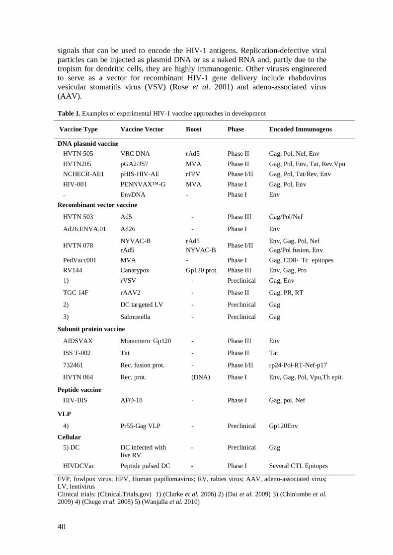
40
signals that can be used to encode the HIV-1 antigens. Replication-defective viral particles can be injected as plasmid DNA or as a naked RNA and, partly due to the tropism for dendritic cells, they are highly immunogenic. Other viruses engineered to serve as a vector for recombinant HIV-1 gene delivery include rhabdovirus vesicular stomatitis virus (VSV) (Rose et al. 2001) and adeno-associated virus (AAV). Table 1. Examples of experimental HIV-1 vaccine approaches in development
FVP, fowlpox virus; HPV, Human papillomavirus; RV, rabies virus; AAV, adeno-associated virus; LV, lentivirus Clinical trials: (Clinical.Trials.gov) 1) (Clarke et al. 2006) 2) (Dai et al. 2009) 3) (Chin'ombe et al. 2009) 4) (Chege et al. 2008) 5) (Wanjalla et al. 2010)
Vaccine Type Vaccine Vector Boost Phase Encoded Immunogens
DNA plasmid vaccine HVTN 505 VRC DNA rAd5 Phase II Gag, Pol, Nef, Env HVTN205 pGA2/JS7 MVA Phase II Gag, Pol, Env, Tat, Rev,Vpu NCHECR-AE1 pHIS-HIV-AE rFPV Phase I/II Gag, Pol, Tat/Rev, Env HIV-001 PENNVAX™-G MVA Phase I Gag, Pol, Env - EnvDNA - Phase I Env
Recombinant vector vaccine
HVTN 503 Ad5 - Phase III Gag/Pol/Nef
Ad26.ENVA.01 Ad26 - Phase I Env
HVTN 078 NYVAC-B rAd5
rAd5 NYVAC-B
Phase I/II Env, Gag, Pol, Nef Gag/Pol fusion, Env
PedVacc001 MVA - Phase I Gag, CD8+ Tc epitopes RV144 Canarypox Gp120 prot. Phase III Env, Gag, Pro 1) rVSV - Preclinical Gag, Env
TGC 14F rAAV2 - Phase II Gag, PR, RT
2) DC targeted LV - Preclinical Gag
3) Salmonella - Preclinical Gag
Subunit protein vaccine
AIDSVAX Monomeric Gp120 - Phase III Env
ISS T-002 Tat - Phase II Tat
732461 Rec. fusion prot. - Phase I/II rp24-Pol-RT-Nef-p17
HVTN 064 Rec. prot. (DNA) Phase I Env, Gag, Pol, Vpu,Th epit.
Peptide vaccine HIV-BIS AFO-18 - Phase I Gag, pol, Nef
VLP
4) Pr55-Gag VLP - Preclinical Gp120Env
Cellular 5) DC DC infected with
live RV - Preclinical Gag
HIVDCVac Peptide pulsed DC - Phase I Several CTL Epitopes
41
2.5.3 Genetic plasmid DNA vaccines
Conventional vaccines based on the whole virus typically induce many immune responses against components of the virus that are irrelevant for protection and subunit vaccines mainly stimulate antibody responses. Furthermore, even if virus-based vectors are widely used and efficient vectors, there are potential risks related to viral gene delivery. These risks can be avoided by using bacterial plasmid-based vectors, also called naked DNA vectors that can accommodate a combination of several antigenic regions from pathogens. The concept has many valuable features, including relative ease and flexibility of construction and manufacturing, the stability of the vaccines and safety as no handling of infectious agents is needed and the expression of DNA vaccines is transient. The local or systemic side-effects are minimal and in the absence of antivector immunity DNA vaccinations can be administrated repeatedly without adverse effects or diminishing the specific responses. Importantly, the production of the antigen by the host cell allows the antigen to be expressed and presented in a way that resembles natural viral infection. This has been shown to lead to balanced CD4+ and CD8+ T cell responses, as well as antibody responses. The biggest challenge related to highly pure plasmid DNA immunization is the immunogenic potency of the naked DNA that has been overall fairly disappointing in humans. However, there are several factors affecting the immunogenicity of DNA vaccines, such as route of administration and dosing regimen, and various approaches can be utilized to improve the efficacy of these vaccines, such as heterologous prime-boost vaccination or co-administration with different immunopotentiators. Traditionally the same vaccines are given multiple times as homologous boosts, but the heterologous prime-boost immunization includes sequential delivery of different types of vaccines (Doria-Rose and Haigwood 2003, Harari et al. 2008, Seaman et al. 2005, Shu et al. 2007). Naked DNA vaccines were introduced more than twenty years ago when mRNA and plasmid DNA encoded genes were shown to be expressed after intramuscular injection (Wolff et al. 1990), an observation followed shortly by several approaches utilizing naked DNA as a vaccine. Tang et al. observed the induction of antibody responses after DNA delivery by gene gun (Tang et al. 1992) and the induction of CTL responses was first reported by Ulmer et al. (Ulmer et al. 1993) in BALB/c mice, demonstrating the protective effect of influenza nucleoprotein specific CTL responses against heterologous influenza virus strain challenge. Since then, DNA vaccines as versatile vehicles against a variety of cancers and pathogens such as malaria parasite, human papilloma virus, dengue viruses, Ebola virus, cytomegalovirus (CMV), seasonal influenza viruses, SARS coronavirus and hepatitis B virus have been generated and evaluated in clinical trials. The almost limitless number of different possible approaches available for DNA vaccine development has generated numerous HIV-1 DNA vaccines and considerable preclinical and clinical experience has been accumulated during the years. However, no DNA vaccines have so far been licensed for human use. Three licensed animal DNA vaccines exist so far, against WNV virus that causes encephalitis in horses, against infectious hematopoietic necrosis virus in salmon and a therapeutic vaccine against skin cancer of dogs (Kutzler and Weiner 2008, Moss 2009).
42
2.5.3.1 Safety of DNA plasmid vaccines
DNA vaccines have a well-tolerated safety profile in both preclinical and clinical trials. The concerns have been related to biodistribution and to the persistence of the genetic material in the body, potential integration into the host DNA especially has raised questions. Integration has been widely studied as reviewed by Schalk et al. (Schalk et al. 2006) showing that the risk of mutation due to plasmid integration is several orders below the spontaneous mutation rate. However, several factors can change the safety profile, such as different delivery methods or co-delivery of adjuvants, and thus the risk cannot be neglected but must be evaluated case by case. Electroporation has been shown to increase the frequency of plasmid DNA association with genomic DNA in some cases (Wang et al. 2004), while no such effect was detected with Biojector immunization, even if both methods have been shown to greatly increase the uptake of the plasmid in the cell (Manam et al. 2000). However, integration is not constantly detected even with electroporation and the use of intradermal electroporation instead of intramuscular electroporation may limit the invasiveness of the method (Bråve et al. 2010). Other concerns such as vertical transmission, induction of autoimmunity (anti-DNA antibody formation), induction of immunological tolerance and toxicity have been studied but no serious disadvantages related to the concept of DNA vaccines have been observed (Schalk et al. 2006). Even the maximum dose evaluated recently in macaques did not induce detectable integration or anti-DNA antibodies (Arrode-Bruses et al. 2010).
2.5.3.2 Structural features of DNA plasmid vaccine
Plasmid DNA vaccines are constructed to enable their efficient production in E. coli cells and to express the encoded antigen efficiently in the target tissue. The bacterial origin of replication and antibiotic resistance gene or some other selection system is incorporated for manufacturing purposes, while in mammalian cells DNA vaccines cannot replicate. Due to regulatory concerns, non-antibiotic selection systems have been developed (Krohn et al. 2005). The expression of the gene encoding the antigen of interest is driven by eukaryotic promoter allowing protein expression in mammalian cells (Garmory et al. 2003). The promoter is selected considering the desired target cell type and expression level, while some promoters operate in a broad range of cells and some of them are more cell or tissue specific. Widely used strong classical promoters are the immediate early promoters/enhancers of human cytomegalovirus (CMV) and Rous sarcoma virus (RSV) promoter, CMV promoter often showing the highest expression. Intron A sequence has been shown to enhance the expression level by the CMV promoter. Downstream inclusion of polyadenylation (polyA) sequence, such as the bovine growth hormone (BGH) polyA sequence or rabbit -globin improves the expression by enhancing nuclear export, translation and mRNA stability (Garmory 2003). In addition to the promoter selected the codon optimization of lentiviral genes increases stability and export of nuclear mRNA and enhances translational efficiency in mammalian cells (Ngumbela et al. 2008). Codon optimization retains the natural amino acid sequence but the codons are synthetized according to human cell preference (Andre et al. 1998, Ramakrishna et al. 2004).
43
2.5.3.3 HIV-1 antigen selection for vaccine gene insert
Several approaches have been assessed over the years in the search for the optimal way to elicit anti-HIV responses by genetic vaccines. As no absolute knowledge of correlates of protection so far exists, the antigen construction is more or less based on assumptions. Several issues need to be considered when evaluating how to construct the HIV-1 vaccine, such as if the vaccine is designed for the induction of nAb or broad cell-mediated responses, or ideally both, and what antigens and epitopes should be included and if it is enough to count on cross-clade reacting immune responses or if the vaccine should cover many clade specificities. The currently prevailing consensus idea is that all arms of immunity should be induced to achieve optimal protective effect for the vaccine, neither cellular nor humoral immune responses are likely to be enough by themselves (Virgin and Walker 2010). Current vaccine approaches are directed at multiple HIV-1 gene products, but enhanced nAb and cell-mediated immune responses with improved breadth are still pursued. nAb in HIV-1 infected individuals have been shown to be directed to different sites of Env-protein including CD4 binding site, glycans on the surface of gp120 and the membrane proximal region of gp41. This indicates that new vaccine antigens can be designed in various ways, using the most recent knowledge of Env and the CD4 binding site structure and of neutralizing epitopes (Mascola and Montefiori 2010, Sather et al. 2009). Several HIV-1 vaccine constructs have been designed and evaluated in clinical studies targeting the Env protein, aiming at the induction of nAb but current HIV-1 vaccines are not able to routinely elicit broadly nAb (Haynes et al. 2010). Considering CD8+ T cells, even if there are several implications of the significant role of Gag-specific CTL responses for protection, it is likely that they will not be enough, but responses to other viral antigens will also be needed. Nef has both highly conserved, as well as highly variable domains and has been shown to contain several highly immunogenic regions and is frequently targeted in early infection. Anti-Nef cellular immune responses elicited after immunization have been observed to induce selective pressure on HIV-1 that was able to limit the variability of the virus in infected individuals (Hoffmann et al. 2008). The degree of conservation can be one argument for antigen selection. However, the most conserved parts may not be very immunogenic, like most conserved gene pol, with comparatively few specific CD8+ T cell responses recognized (Korber et al. 2009). Furthermore, the diversity of HIV-1 subclades must be considered when constructing the antigen (Taylor and Hammer 2008). The vaccine target population may guide the selection of the clade specificity. C-clade should be emphasized in vaccines introduced in South Africa, while the United States and Europe would benefit mostly from B clade specific constructs. The structure of the HIV-1 phylogenetic trees is based on Gag or Env coding sequence similarity and shows the relationship of the M-group subtypes (Korber et al. 2009). Cross-clade protection has been shown to correlate with substantial protein sequence conservation observed for Gag and Nef (Coplan et al. 2005). One option is to select natural isolate resembling closest all the others and assume that sufficient magnitude of cross-clade reactions will be induced. However, many of the CD8+ T cell receptors are known to be sensitive even to single amino acid changes and additionally, there are several
44
indications of HIV-1 variants escaping the T cell responses by single mutation in epitopes (Lee et al. 2004). One way to overcome the wide diversity of HIV-1 is to combine several clades into one vaccine. The approach of using only narrow conserved elements concatenated together has been described by Rolland (Rolland et al. 2007b), the vaccine constituting 45 viral conserved segments across the M group of HIV-1 strains, with a minimum length of eight amino acids. The pressure against conserved regions has been suggested to lead to compromised viability of the virus. The induction of ineffectual and redundant immune responses is avoided by omitting the variable sequences of HIV-1 from the vaccine antigen. However, this approach does not generate any proteins resembling natural HIV-1 gene products. Despite several attempts to take the most conserved parts of the virus and compose an artificial polyepitope vaccine, no strong T cell responses have been generated in clinical phase I studies. When selecting longer conserved regions, the outcome may be improved as more epitopes can be presented, including epitopes in overlapping genomic regions (Barouch and Korber 2010). The “universal” HIV-1 vaccine was pursued by another group by assembling a gene encoding 14 most conserved regions of the HIV-1 proteome, each segment of a chimeric protein being a consensus sequence from the major clades A, B, C and D (Letourneau et al. 2007). Furthermore, 'centralized' consensus and ancestor HIV gene sequences can be generated by phylogenetics-informed algorithm computer programs and designed to minimize the sequence difference between the vaccine and circulating virus isolates. Consensus sequences are designed by selecting the most common amino acid in each position in an alignment while highly variable regions may not be retained. Ancestral sequences are built by means of phylogenetic analysis. However, both consensus and ancestral sequences are artificially derived and their expression, antigenicity and biological activity must be carefully characterized. The proper folding might not be so relevant for eliciting cellular immune responses as it is for humoral antibody response (Ellenberger et al. 2002). Rolland et al. have reconstructed an ancestral HIV sequence utilizing the Center of Tree (COT) approach. COT sequences constituted the HIV-1 Gag, Tat and Nef proteins, which were shown to retain their biological functions and to be immunogenic in mice (Rolland et al. 2007a). So far they have proven immunogenic in mice. Polyvalent vaccines composed of a mixture of ‘mosaic’ immunogens have been suggested to maximize the coverage of potential T cell epitopes of the virus. They are assembled from fragments of natural sequences via a computational optimization, including common and excluding rare epitopes, but are still based on the generation of intact proteins (Fischer et al. 2007). Vaccines are also composed of a cocktail of immunogens derived from different clades (Seaman et al. 2005). One important aspect to consider with multiclade vaccines composed of several related immunogens as a cocktail is the immune interference, such as T cell epitope antagonism, immunodominance and original antigenic sin that has been observed to occur both in vitro and in vivo between closely related peptide sequences in the cocktail (Basu et al. 1998, Larke et al. 2007). Original antigenic sin refers to a phenomenon recognized for both humoral and cellular immunity, where the original exposure to a certain antigen by either infection or immunization induces immunological memory, which by a second encounter of related but not identical antigen results in immune response directed to the original antigen and induces only
45
an impaired immune response against the second antigen. T cell antagonism occurs when an existing memory T cell is functionally inactivated by presentation of a point mutant variant of its original cognate epitope on MHC class I molecule (Basu et al. 1998, Singh et al. 2002). By contrast, there is a line of evidence that the use of vaccines containing many mutant epitopes, even T cell antagonists, can tend to avoid some degree of immune interference generation (Singh et al. 2002). Similarly in HIV-1 infection, naturally arising virus variants may change the CTL epitopes so that they are able to engage the T cell receptor but fail to activate the T cell and further interfere with the recognition of the unmutated epitope (Klenerman et al. 1996).
2.5.4 Induction of immune responses with DNA vaccines
In addition to the selection of antigens and vectors, the choice of delivery route, dose, timing, adjuvants and boosting agents influence the outcome of immunization, changing the magnitude and quality of immunity achieved. DNA molecules are highly susceptible to nuclease-mediated degradation that must be taken into account throughout the whole chain from DNA plasmid vaccine manufacturing process to delivery (Caputo et al. 2003).
2.5.4.1 DNA immunization via variable routes
When DNA is introduced into the body, the environment in which it is administered as well as the delivery technique affect the uptake of the DNA into the cell. Not all aspects of the cellular uptake are well known but both professional APC and non-lymphoid cells such as myocytes and keratinocytes are known to participate and the route of administration affects which cell types are directly transfected. Parenteral DNA injection by needle is most often given intramuscularly (i.m.), intradermally (i.d.) or subcutaneously (s.c.), but intraperitoneal (i.p.) and intravenous (i.v.) routes have also been used. When DNA is injected i.m., antigen is primarily expressed by transfected muscle cells at the site of inoculation and can express the genes and present them through MHC class I molecules (Dupuis et al. 2000, Wolff et al. 1990). Additionally, expressed, secreted proteins may directly stimulate B cells to produce antibodies. However, transfer of antigen from muscle cells to professional APC and their presentation to lymphocytes through MHC class I and II molecules by cross-priming is required for inducing primary T cell responses with optimal efficiency, as the muscle cells lack the costimulatory molecules and MHC class II molecules needed for CD4+ presentation (Agadjanyan et al. 1999, Corr et al. 1996). A similar cross-presentation is induced after i.d. injection (Corr et al. 1999).
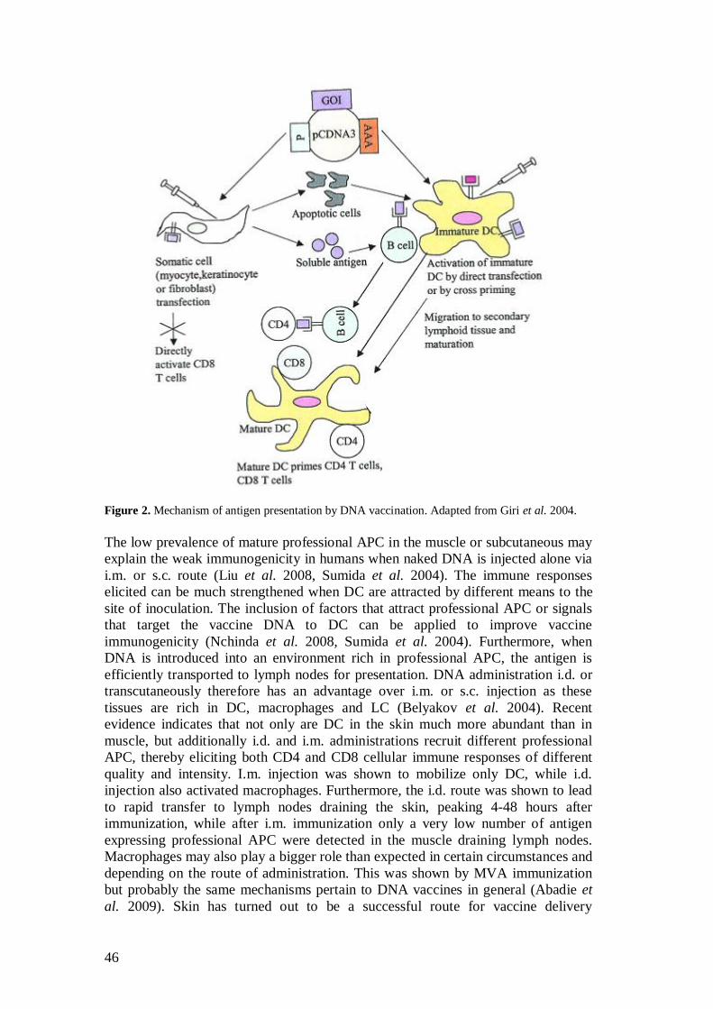
46
Figure 2. Mechanism of antigen presentation by DNA vaccination. Adapted from Giri et al. 2004. The low prevalence of mature professional APC in the muscle or subcutaneous may explain the weak immunogenicity in humans when naked DNA is injected alone via i.m. or s.c. route (Liu et al. 2008, Sumida et al. 2004). The immune responses elicited can be much strengthened when DC are attracted by different means to the site of inoculation. The inclusion of factors that attract professional APC or signals that target the vaccine DNA to DC can be applied to improve vaccine immunogenicity (Nchinda et al. 2008, Sumida et al. 2004). Furthermore, when DNA is introduced into an environment rich in professional APC, the antigen is efficiently transported to lymph nodes for presentation. DNA administration i.d. or transcutaneously therefore has an advantage over i.m. or s.c. injection as these tissues are rich in DC, macrophages and LC (Belyakov et al. 2004). Recent evidence indicates that not only are DC in the skin much more abundant than in muscle, but additionally i.d. and i.m. administrations recruit different professional APC, thereby eliciting both CD4 and CD8 cellular immune responses of different quality and intensity. I.m. injection was shown to mobilize only DC, while i.d. injection also activated macrophages. Furthermore, the i.d. route was shown to lead to rapid transfer to lymph nodes draining the skin, peaking 4-48 hours after immunization, while after i.m. immunization only a very low number of antigen expressing professional APC were detected in the muscle draining lymph nodes. Macrophages may also play a bigger role than expected in certain circumstances and depending on the route of administration. This was shown by MVA immunization but probably the same mechanisms pertain to DNA vaccines in general (Abadie et al. 2009). Skin has turned out to be a successful route for vaccine delivery
47
(Mitragotri 2005). Needle-free jet delivery devices for i.m. or s.c. administration, such as Biojector device may improve the vaccine elicited immune responses and also reduce the risks of acquiring blood borne infections by needle. Jet-injection has been shown to significantly increase plasmid uptake (Bråve et al. 2010). Another needle-free route, mucosal DNA administration via intranasal, intravaginal, oral or anal routes has been assessed with DNA vaccines. Induction of mucosal immune response may be particularly important for protection, while the mucosal surface is the natural route for HIV-1 entry and is the site for large number of immune cells. Preventive vaccination should be able to generate a balanced CD8+ TEM cell response to provide efficient first line immune protection in the mucosal compartment and CD4+ and CD8+ TCM cell responses to prevent viral dissemination. Different prime-boost strategies combined with mucosal delivery as well as the presence of the right costimulatory signals can help the induction of these memory cell types (Ahlers and Belyakov 2010).
2.5.4.2 Delivery methods enhancing vaccine immunogenicity
Relatively weak immune responses in clinical and non-human primate studies with plain plasmid DNA have indicated the need to improve the potency of DNA vaccines. The large size and hydrophilic nature of naked DNA molecules does not allow them to enter cells efficiently. Several methods and different delivery devices have been developed to provide additional costimulation and enhance the cellular DNA uptake and direct transfection frequency and rely on more efficient professional APC recruitment (Al-Dosari and Gao 2009). The first device used for the administration of genetic vaccine was biolistic particle delivery by gene gun, which delivers DNA or RNA coated on gold microparticles into cells with a low pressure helium pulse enabling in vivo transfection (Tang et al. 1992). Administration by gene gun device has been shown to directly transfect the dendritic cells of the skin, although keratinocytes are the main cell type found to be expressing the DNA-encoded proteins (Porgador et al. 1998). Twenty-four hours following gene gun immunization, the total number of CD11c+ DC in the major draining lymph nodes has been shown to increase more than twofold, augmentation rather due to the bombardment than the plasmid DNA itself (Porgador et al. 1998). In addition, gene gun immunization reduces the required DNA dose hundred or even thousands-fold in mice, being the most powerful way to induce cellular and humoral immune responses (Doria-Rose and Haigwood 2003, Pertmer et al. 1995), although electroporation has been found a very effective delivery approach as well (Best et al. 2009). Minute amounts of DNA have also been shown to efficiently induce T and B cell immune responses in humans (Roy et al. 2000). However, biolistic immunization is not a very widely used method in clinical trials despite encouraging results (Al-Dosari and Gao 2009, Mitragotri 2005). Some studies have suggested that the route of administration may have an impact on how immunization will bias the response, namely that i.m. injection would preferably induce Th1 type response while g.g. would elicit more Th2 biased or balanced Th1/Th2 response (Barry and Johnston 1997, Pertmer et al. 1996, Tähtinen 2001). However, this is not frequently observed and is suggested to be more antigen dependent, although it is acknowledged that slight skin irritation in g.g. immunization may create more Th2 responses (Doria-Rose and Haigwood 2003).
48
The potency of plasmid DNA i.m. and i.d. immunization has been significantly improved by in vivo electroporation, which has been shown to decrease DNA demand more than 10-fold in mice. Promisingly, electroporation has been shown to increase the strength, breadth and lead to more rapid onset and long duration of immune responses in non-human primates immunized with HIV-1 specific plasmid vaccines (Luckay et al. 2007, Martinon et al. 2009). Electroporation involves the administration of electrical pulses to muscle or skin tissue following injection, increasing cell permeability, thus enhancing transfection of the cells. In addition, electroporation recruits large cellular infiltrates rich in DC, macrophages and lymphocytes to the site of inoculation. Electroporation has shown similar enhancement of the immune responses than molecular adjuvants, but the effect by electroporation was more durable and, importantly, was shown to generate more effector and central memory CD8+ T cell responses. Interestingly, for viral vectors the advantage is not so obvious, possibly due to the utilization of viral entry pathway into the cells and efficient innate immunity stimulation by the viral constitutes (Liu et al. 2008). Insufficient dose has been proposed to explain the relative poor immunogenicity of DNA vaccines in humans and non-human primates compared to responses detected in mice. This was demonstrated by Arrode-Bruses and colleagues, who used an extremely high single dose for the induction of strong, long-lasting and polyfunctional CD8+ T cell responses in non-human primates. However, as the corresponding dose would not be feasible for human use, the use of electoroporation to achieve the same potency in humans was suggested (Arrode-Bruses et al. 2010).
2.5.4.3 Use of adjuvants for stronger recruitment of the immune system
There is a wide range of molecular adjuvants that DC can recognize by specific receptors and respond to, such as proteins, lipids, nucleic acids, carbohydrates and chemical compounds (Kornbluth and Stone 2006). Molecular adjuvants such as cytokines, chemokines or T cell costimulatory molecules can be co-administered or their coding sequences can be integrated as a part of the vaccine and thus enhance the potency of either Th1 or Th2 type immune responses, as preferred (Calarota et al. 2001, Calarota and Weiner 2004, Sumida et al. 2004). The name adjuvant is derived from the latin word adjuvare meaning helping or aiding. Accordingly, adjuvant co-administration can enhance the immunogenicity of highly purified antigens and reduce the amount of antigen needed to elicit the proper immune responses. Adjuvants may be needed for efficient immunization of special groups like immuno-compromised individuals, newborns and the elderly (Aguilar and Rodriguez 2007). However, the broad range of activities modulated by cytokines carries a risk of also eliciting harmful effects. Stimulation of professional APC through TLR is critical in the initiation of immune response and the adjuvanticity of many TLR ligand molecules, such as heat-shock protein 70 and monophosphoryl lipid A, has been evaluated (Aguilar and Rodriguez 2007). The plasmids may also have co-stimulatory effects themselves, as the vaccine plasmids contain bacterial CpG motifs (Greenland and Letvin 2007, Klinman et al. 1999). The route of administration is critical not only for vaccine efficiency but also has a notable impact on adjuvant function. For example, the alum is generally used only i.m. and not by the more efficient i.d. or s.c. route, due to its local toxicity (Aguilar and
49
Rodriguez 2007). The use of adjuvants can radically change the safety profile of a DNA vaccine, as the local and systemic adverse effects unfortunately usually correlate with the adjuvant potency. To enhance DNA uptake by the cells, vaccines can also be formulated with different chemical carriers, including liposomes, polymers, microparticles and nanoparticles. They may facilitate the permeation of the cell membrane, target the DNA for phagocytosis and protect and stabilize the DNA. The current HIV-1 vaccine research field emphasizes the need of broad and multifunctional immune responses. The candidates showing most promise are vaccines able to elicit highly multifunctional CD8+ T cell responses defined by determinants such as degranulation and expression of IFN- , MIP-1 , TNF- , and IL-2, thus emphasizing more the quality than the quantity of T cell responses as immune correlates (Arrode-Bruses et al. 2010, Betts et al. 2006). The almost limitless possibilities related to DNA vaccines show great promise for future HIV-1 vaccine development, as long as the right combination of different components is developed and found for human use. It is likely that the most efficient vaccine regimen would target broadly both arms of immunity, which could be achieved e.g. by combining different vaccines within heterologous prime - boost vaccination regimens, further increasing the value of DNA vaccines.
50
3. Aims of the study
The aim of the thesis was to assess the immunogenicity of GTU DNA plasmids expressing HIV-1 multigene using different immunization regimens in a mouse model. Eliciting strong CD8+ T cell responses is a requirement for the vaccine to fight against the HIV-1 infection, thus two challenge models relying specifically on the action of cytotoxic lymphocytes were used to evaluate the protective efficacy of HIV-1 immunogens. The hypothesis that immune responses elicited by GTU plasmid vaccine were mediated by DC was briefly investigated. The specific objectives of this study were the following: I To evaluate the immunogenicity of GTU -MultiHIV B-clade/Han-2 and
multiclade (A, B, C, FGH) Auxo-GTU -MultiHIVmix DNA in mice. II To define the optimal immunization regimen for GTU based MultiHIV
vaccine administration using g.g., i.d. and i.m. administration routes. III To assess the protective efficiency of the GTU based MultiHIV
immunizations using two different experimental challenge models in mice. IV To briefly examine the role of DC in the generation of the immune response
by GTU HIV-1 immunogen.
51
4. Materials and Methods
4.1 GTU expression vector (I-IV)
GTU platform was used for constructing HIV-1 multigene plasmids. The GTU expression vector consists of an expression cassette for a gene of a nuclear-anchoring protein E2, a multimerized DNA sequence forming binding sites for E2 and an expression cassette for the DNA sequence of interest. Protein E2 type 1 from bovine papillomavirus (BPV) is driven from the Rous sarcoma virus 5´ LTR (RSV LTR) promoter. Polyadenylation region of the bovine growth hormone is cloned at the end of the E2 transcription cassette from the pHook3 plasmid (Invitrogen). The vector carries ten copies of BPV1 binding sites for E2 (10E2BS). The gene of interest is driven by CMV immediate early promoter. The elements needed for the effective expression of the mRNA include HSV1 TK gene leader sequence, a rabbit -globin gene sequence and an HSV TK gene polyadenylation signal region. A modified form of pMB1 replicon with ColE1 origin of replication was included for propagation in E. coli cells. The patent application for GTU vector was filed on May 5, 2002 (PTC/FI02/00379). Auxo-GTU expression vector was constructed to enable antibiotic-free selection, utilizing L-ribulose-5-phosphate-4-epimerase encoding araD gene as a selection marker when plasmid is produced in a bacterial strain deficient in the araD gene. A patent application for araD based selection system was filed on September 15, 2004 (PTC/FI2004/000540).
4.1.1 HIV-1 multigene plasmid (I-IV)
HIV-1 multiantigen, named MultiHIV (RNTp17/24CTL), is a fusion protein of 120 kDa composed of full-length sequences of Rev, Nef , Tat, p17 and p24 proteins of Gag. C-terminus encodes for a stretch of eleven 17-45 aa long T cell epitope clusters of the reverse transcriptase and envelope sequences. The sequences were derived from Han-2 isolate of the HIV-1 strain. Strong murine H-2d restricted HIV-1 CTL epitope from env (RGPGRAFVTI) was included at C terminus. GTU -MultiHIV encodes for MultiHIV derived from HIV-1 B-clade HAN2 isolate.
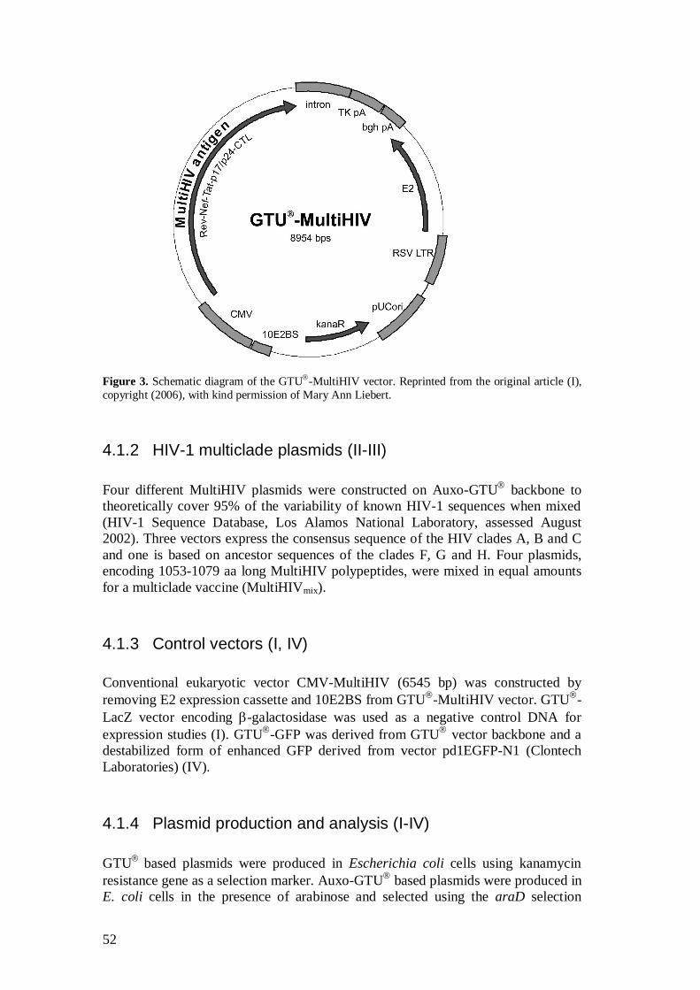
52
Figure 3. Schematic diagram of the GTU -MultiHIV vector. Reprinted from the original article (I), copyright (2006), with kind permission of Mary Ann Liebert.
4.1.2 HIV-1 multiclade plasmids (II-III)
Four different MultiHIV plasmids were constructed on Auxo-GTU backbone to theoretically cover 95% of the variability of known HIV-1 sequences when mixed (HIV-1 Sequence Database, Los Alamos National Laboratory, assessed August 2002). Three vectors express the consensus sequence of the HIV clades A, B and C and one is based on ancestor sequences of the clades F, G and H. Four plasmids, encoding 1053-1079 aa long MultiHIV polypeptides, were mixed in equal amounts for a multiclade vaccine (MultiHIVmix).
4.1.3 Control vectors (I, IV)
Conventional eukaryotic vector CMV-MultiHIV (6545 bp) was constructed by removing E2 expression cassette and 10E2BS from GTU -MultiHIV vector. GTU -LacZ vector encoding -galactosidase was used as a negative control DNA for expression studies (I). GTU -GFP was derived from GTU vector backbone and a destabilized form of enhanced GFP derived from vector pd1EGFP-N1 (Clontech Laboratories) (IV).
4.1.4 Plasmid production and analysis (I-IV)
GTU based plasmids were produced in Escherichia coli cells using kanamycin resistance gene as a selection marker. Auxo-GTU based plasmids were produced in E. coli cells in the presence of arabinose and selected using the araD selection
53
system. Plasmids were purified by Qiagen Endofree Plasmid kits (Hilden Germany) according to the manufacturer’s instructions and dissolved in sterile, endotoxin-free PBS. The quality of the plasmid products used for immunization studies was confirmed by concentration measurement, restriction enzyme analyses, sequencing, plasmid homology and impurity analyses, the latter comprising of E. coli genomic DNA, RNA, endotoxins, kanamycin and bioburden analyses.
4.1.5 Expression studies (I, IV)
The expression properties of GTU -MultiHIV were analyzed by Western Immunolotting after in vitro transfections of the Jurkat, Cos-7 and RD cell lines by electroporation. 0.5 µg or 3 µg of DNA was used for electroporation. Equimolar quantities, 0.27 µg and 2.2 µg, of CMV-MultiHIV control DNA were additionally transfected into RD cell line for comparison. 3 µg of GTU -LacZ was used for transfection of negative control cells. Two and five days posttransfection the cells were lysed with lysis buffer (50 mM Tris–HCl, pH 6.8, 2% SDS, 0.1% bromophenol blue, 100 mM dithiothreitol, and 10% (v/v) glycerol) and samples were run on a 10% sodium dodecyl sulfate–polyacrylamide gel (SDS–PAGE). Proteins were transferred on nitrocellulose membrane and MultiHIV expression was analyzed after membrane blocking by incubation with monoclonal anti-p24 antibody (05-001, FIT Biotech, Tampere, Finland), followed by HRP-conjugated goat antimouse IgG (LabAs, Tartu, Estonia) and visualization using an ECL chemoluminesence detection system (Amersham Pharmacia Biotech, Uppsala, Sweden). GTU -MultiHIV and CMV-MultiHIV transfected RD cells were lysed and the fusion protein expression was quantified by ELISA as described in Chapter 4.11.3, on the rNef coated plate using biotinylated anti-p24 antibody (05-001) and TMB substrate for detection. Subcellular localization of the MultiHIV antigen encoded by GTU -MultiHIV was analyzed in Cos-7 cells, transfected with 2 µg of GTU -MultiHIV DNA, by immunofluorescent staining with anti-Nef (01-001, FIT Biotech) or anti-p24 antibodies and detected by fluorescein isothiocyanate (FITC)-conjugated goat antimouse secondary antibody (LabAs). GFP expression from GTU -GFP (IV) was verified in vitro by fluorescence flow cytometry analysis after transfecting Jurkat cells using Cellfectin® Reagent (Invitrogen). For the in vivo expression study, mice were immunized twice, 24 hours apart, with 250 g of GTU®-GFP plasmid intradermally. On the third day mice were sacrificed and lumbar and sacral LN of the experimental mice and control mice were collected and pooled. As a control, mice immunized with the plasmid lacking GFP were used. Cells were digested from the tissues by incubating with Collagenase D (Sigma). Collected DC were enriched by density gradient centrifugation using Optiprep (Axis-Shield PoC AS, Oslo, Norway) according to manufacturer’s instructions. DC were washed and labeled with R-PE-conjugated anti-mouse CD11c antibody or PE anti-mouse IgG1 as a control (BD Biosciences Pharmingen, San Jose, CA, USA). Non-specific binding to DC CD16 and CD32 was blocked with Mouse Fc BlockTM (BD Biosciences Pharmingen). DC were washed and samples were run with FACSCalibur. CD11c+ cells were gated on FSC/FL2 scatter and
54
analyzed by FlowJo (Tree Star, OR, USA). The percentage of the GFP+ cells in the gated population was determined.
4.2 P815-MultiHIV cells (II)
P815 mastocytoma cell line, possessing H-2d MHC class I molecules was used for challenging DBA/2 mice in the tumor challenge study (II). A plasmid where MultiHIV Han-2 expression was driven by strong hybrid SR -promoter (Takebe et al. 1988) was ligated with another plasmid, carrying an expression cassette for puromycin acetyl transferase and -hCG hormone. Puromycin acetyl transferase was used as a selection marker in the presence of puromycin and -hCG hormone was used monitoring the tumor development as it was shown to correlate positively with the tumor load (Shih et al. 2000). P815 cells were stable transfected by electroporation and one day later puromycin (0.75 µg/ml) was added onto the cells. After one week of cultivation with antibiotic cells were subcloned by limiting dilution method and the subclones were analyzed by western immunoblotting. For positive clones the -hCG hormone secretion was analyzed with Free HCG ELISA kit (DRG Instruments GmbH, Germany) and presentation of MultiHIV was assessed with ELISPOT IFN- assay using the splenocytes of MultiHIV DNA immunized mice. As a control for challenge, similarly analyzed wild type (wt) P815 cells were used. The capability of P815-MultiHIV cell line to present MultiHIV antigen was analyzed in vitro using these cells as APC in ELISPOT IFN- and Cr51 release assay. For ELISPOT, splenocytes of GTU -MultiHIV Han-2 immunized mice were in vitro stimulated with mitomycin C treated P185-MultiHIV and P815-wt cells, followed by IFN- secreting cell enumeration as described in Chapter 4.11.2. Similarly, both P185-MultiHIV and P815-wt cell lines were used as target cells in Cr51 assay (Chapter 4.11.4). Before the challenge, the tumorigenicity of the P185-MultiHIV and P815-wt cells was assessed in vivo in DBA/2 mice. 1×10⁶ tumor cells were inoculated s.c. into the right flank and the tumor growth was followed and tumor diameter measured with an electric calliper every second day. The size (mm2) was calculated as longest diameter × shortest diameter. Mouse welfare was followed and mice were sacrificed when the diameter reached 10 mm.
4.3 HIV-1/MuLV pseudovirus infected cells (III)
HIV-1/MuLV pseudotype viruses were prepared in neomycin-resistant cell line Ampho-CEM-1B as previously described (Spector et al. 1990) (III) at Karolinska Institutet, Stockholm, Sweden. CEM-1B cells were cultured in 10% FCS-RPMI 1640 (Invitrogen, Gibco, Carlsbad, CA) supplemented with antibiotics (Sigma-Aldrich, St. Louis, MO) until infected with the subtype B LAI HIV-1 strain or the
55
primary Kenyan subtype A 9284 HIV-1 isolate. Every 3-4 days culture supernatants were collected, screened for p24 antigen (Vironostika, Bio-Mérieux, Boxtel, France) and stored at -70°C on testing positive for p24. Splenocytes of C57BL/6 were infected as previously described (Andang et al. 1999). In brief, splenocytes were activated with Con A for 24 h, washed and 50×106 cells in one ml were infected with stored pseudoviral supernatant by overnight incubation. One day after infection the amount of p24 protein per 106 splenocytes was determined (Devito et al. 2000).
4.4 Animals (I-IV)
Inbred female mice, ca. 8 weeks old at the beginning of the experiments were used for all studies. All immunizations and challenges were performed under general anesthesia. Animal welfare and health were followed throughout the study and mice were weighed at the beginning and at the end of the studies. BALB/cOlaHsd (H-2d) mice were used for the immunogenicity studies (I, III, IV). The tumor challenge model was made with DBA/2OlaHsd (H-2d) mice, syngeneic for P815 cells, both strains obtained from Harlan (Italy and Netherlands). All procedures were carried out according to the national guidelines and permission of the County Administrative Board of Tampere and of Laboratory Animal Board of Tampere University Medical Faculty, Finland. For HIV-1/MuLV pseudotype virus challenge C57BL/6 (H-2b) strain transgenic for HLA-A201 (Hinkula et al. 2004, Vitiello et al. 1991) was used. Animal care and experimental procedures were approved by the animal research ethical committee of the Karolinska Institutet and The Swedish Institute of Infectious Disease Control, Solna, Sweden.
4.5 Immunization routes
4.5.1 Gene gun (I-III)
For gene gun administration, plasmids dissolved in PBS were coated on gold particles (Bio-Rad, Richmond, CA) according to manufacturer’s instructions. DNA-coated gold particles were administered to shaved abdominal skin by Helios Gene Gun (Bio-Rad) using a pressure of 400 psi and 0.5 mg gold/cartridge (Collings et al. 1999).
4.5.2 Intradermal and intramuscular injections (I-IV)
For i.d. and i.m. injections the plasmids were formulated in sterile, endotoxin-free PBS and given by 0.3 ml insulin needle. I.d. injections were administered just beneath of the skin at the base of the tail, dorsal side of the mouse. I.m. injections were given into the quadriceps femoris of the hind leg. For DC immunization (IV), enriched DC suspended in PBS, were administered i.d. by needle.
56
4.6 DNA immunogenicity studies
4.6.1 GTU -MultiHIV (I)
A comparison of GTU and CMV expression vector was done by immunizing groups of 5 BALB/c mice at days 0, 7 and 21 with three different doses of both MultiHIV expressing plasmids. For GTU -MultiHIV DNA total doses were 24 ng (g.g.), 300 ng (g.g.) and 750 µg (i.d.). Equimolar doses used for CMV-MultiHIV DNA were 17.4 ng (g.g.), 220 ng (g.g.) and 546 µg (i.d.). Mice were sacrificed two weeks after the last immunization. To analyze the dose-dependence of the immune responses, mice were immunized with GTU -MultiHIV (I) by g.g. three times (days 0, 7 and 21) with a total amount of 24 ng, 120 ng, 600 ng, or 3 µg DNA/mouse. Total DNA doses for i.m. and i.d. immunizations were 6, 30, 150, or 750 µg. One third of the dose was given at one immunization time and the mice were terminated on day 31. In another set of experiments, the 0-3 doses (1 µg) of GTU -MultiHIV DNA was administered by g.g. once (day 0), twice (days 0 and 7) or three times (days 0, 7 and 21). Mice were terminated five weeks after the first immunization. Next, duration of the immune responses was followed in 3 × 1 µg immunized groups of mice by terminating groups 2, 6, 10, 14 and 22 weeks after the last immunization on day 21. Boost immunization ten weeks after the third dose was also given in one group. These mice were terminated four weeks later. Similarly, the effect of fourth booster immunization (at wk 13) by i.m. or i.d immunization routes was assessed in mice immunized on days 0, 7 and 21 (wk 0, 1 and 3) with 50 µg/injection of GTU -MultiHIV. CTL responses were analyzed by ELISPOT IFN- four weeks after the boost (wk 17). Each experimental group consisted of 5-10 animals. Negative control mice were immunized with DNA carrier only. Individual spleen and blood samples were collected. Splenocytes were preserved in aliquots in liquid nitrogen and serum samples at -20°C until analyzed.
4.6.2 Auxo-GTU -MultiHIVmix (III)
Auxo-GTU -MultiHIVmix immunogenicity was evaluated first with long-term immunization schedules. Total dose of 8, 40, 200, or 1000 ng DNA was given by g.g. at weeks 0, 4 and 12. Next, the short term immunization with Auxo-GTU -MultiHIVmix at weeks 0, 1 and 3 was used to assess g.g. (3×1 µg), i.m. and i.d. (3×50 µg) administration routes and the persistence of the cellular immune response induced by i.m. and i.d. injections was followed two and fourteen weeks after the last immunization.
57
Finally, mice were immunized 3×40 ng by g.g. with 1) the MultiHIVmix or with 2) the individual constructs alone or with 3) all different combinations of three of the constructs, combined in equal amounts. Negative control mice were immunized i.m. with PBS only. Mice were sacrificed 2 weeks after the third immunization, splenocytes and serum samples were stored until used for ELISPOT IFN- and ELISA analysis respectively.
4.7 Tumor challenge model (II)
Both GTU -MultiHIV and Auxo-GTU -MultiHIVmix were evaluated in a tumor challenge model. First, DBA/2 mice were immunized (n = 9) at weeks 0, 1 and 3 with 1 µg of GTU -MultiHIV DNA by g.g. After two weeks (wk 5) immunized mice were challenged either with 1×10⁶ P815-MultiHIV or 1×10⁶ P815-wt tumor cells. Non-immunized control groups were equally challenged. In the second set of experiments mice were challenged with different cell numbers; 0.2 ×10⁶, 0.4 ×10⁶ and 0.8 ×10⁶ of P815-MultiHIV cells. Immunizations were performed similarly but using Auxo-GTU -MultiHIVmix. The immunized but not challenged control group was terminated at the time of challenge for evaluation of MultiHIV specific cellular and humoral immune responses at the time of challenge by ELISPOT and ELISA. A group of naïve mice was included as a control. Groups of eight mice were divided randomly into two groups terminated at two different time points; 9 and 15 days after the high cell number challenge and 15 and 22 days later for groups challenged with 0.2-0.4 ×10⁶ cells. Individual serum samples were stored at -20°C, splenocytes in liquid nitrogen until used. P815-MultiHIV and P815-wt cells were grown in RPMI 1640 (Sigma–Aldrich, St. Louis, MO) supplemented with 100 U/ml penicillin–streptomycin (Sigma–Aldrich), L-glutamine (BioWhittaker, Verviers, Belgium), 10% FBS (BioWhittaker) at 37 C with 5% CO2. P185 cells used for the challenge in the tumor study (II) were suspended in sterile PBS and injected in 50 µl s.c. in the shaved right flank by 23-gauge needle. Tumor growth was recorded every 2-3 days. For -hCG hormone determination urine samples were collected for a few mice in each group on days 6 and 20. Mice were terminated when the longest diameter of the tumor reached 15 mm, or vertical growth >10 mm, or at the latest on day 26 and samples were collected and stored as previously described until analyzed.
58
4.8 HIV-1/MuLV challenge model (III)
Groups of twelve C57BL/6.A201 mice were immunized with Auxo-GTU -MultiHIVmix at weeks 0, 1 and 3, one group by g.g. (3×1 µg), the other intramuscularly (3×50 µg). The control group was i.m. injected with sterile PBS. Two weeks after the last immunization, at week 5, mice were bled for pre-challenge immunity by PBMC ELISPOT and antibody analysis. PBMC were pooled groupwise for the analysis. Five weeks after the last immunization (at wk 8) mice were challenged by intraperitoneal injection of splenocytes of syngeneic mice, infected in vitro with HIV-1/MuLV pseudovirus (Chapter 4.3.). 1×10⁶ subtype B LAI HIV-1/MuLV infected cells expressing approximately 1 ng p24 protein, or 3×10⁶ subtype A 9284 HIV-1/MuLV infected cells were used for the challenge. Ten days post-challenge, the ascites of each animal were collected and cells were co-cultured with activated human PBMC or Jurkat Tat cells. Every 3 days, 50% of the culture medium was changed and HIV-1 p24 secretion was analyzed (Devito et al. 2000). If the supernatant was positive at more than one time point in at least one of the isolation systems (Jurkat Tat or hPBMC), the animal was regarded as unprotected by immunization. Splenocytes and serum samples were collected.
4.9 DC immunization (IV)
BALB/c mice were immunized twice on consecutive days i.d. with 250 µg of Auxo-GTU®-MultiHIV-B plasmid. On the third day, lumbar and sacral LN and spleens were collected separately. DC were enriched using the gradient as described above (4.1.5.) and further used for immunization. This procedure was done twice with one week interval for two sets of mice, to accomplish DC transfer immunizations at weeks 0 and 1. A group of naïve BALB/c mice were injected twice with 4 × 10⁵ LN DC/mouse and another group of mice twice with 8 × 10⁵ spleen DC/mouse, i.d. in the base of the tail. Tail blood samples were collected at weeks 1, 2 and 3 and mice were terminated three weeks after the second DC transfer (week 4). Splenocytes and serum samples were stored and analyzed later.
4.10 Sample preparations (I-IV)
The spleens were dispersed in a single cell suspension and washed splenocytes were resuspended in RPMI1640 supplemented with 40% FBS + 10% DMSO. Cells were cryopreserved by freezing to -80°C for a few days and then stored in liquid nitrogen (LN). For analysis the splenocytes were thawed and suspended in culture media (CM): RPMI 1640 (BioWhittaker, Verviers, Belgium) supplemented with 10% fetal bovine serum (FBS), 1% penicillin-streptomycin, and 50 µM 2-mercaptoethanol (all
59
from BioWhittaker). For CD8+ T cell depletion magnetic beads (MagCellect, R&D Systems, Minneapolis, MN) were used following the manufacturer’s instructions. The whole blood was collected at the time of termination and serum was obtained by centrifugation at 1300 g 10 min after incubating the blood at RT ~2 h. Serum was collected and stored at -20°C until analyzed.
4.11 Immunoassays
4.11.1 Recombinant proteins and peptides (I-IV)
Glutathione S-transferase (GST)-tagged HIV-1 (Han-2) Nef and GST proteins were affinity purified from lysates from E. coli BL21 (DE3) cells which were transformed with a pGex-Nef-GST or pGex-GST plasmids using Pharmacia (Piscataway, NJ) reagents. Coding sequences of HIV-1 Rev, Tat, p17/24, CTL and E2 were cloned into pET24d vector and purified from E. coli lysate using Ni-NTA Agarose (Qiagen). H-2d-restricted HIV-1 gp120 (Env) 10-mer peptide (aa 311–320; RGPGRAFVTI) (Takahashi 1988) and 9-mer HIV-1 Gag peptide (aa 65–73; AMQMLKETI) (Qiu 1999) were purchased from Sigma-Genosys (Cambridge, UK) as 95% pure. 15-mer peptide pools of Rev (29 peptides), Nef (51 peptides), Tat (25 peptides), Gag (91 peptides) and CTL (67 peptides) were purchased from Sigma-Genosys and were
85% pure. The peptides in the pools were 15 aa in length, overlapping by 11 aa, covering the MultiHIV B consensus sequence. For C57BL/6.A201 mice (III) EnvB.gp120 15-mer peptide pool overlapping by 8 amino acids (ThermoHybaid, Ulm, Germany) and GagB.p24 peptide pool containing 15-mer peptides overlapping by 10 (ThermoHybaid, Ulm, Germany) were used. 20-mer herpes simplex virus (HSV) peptide (RRHTQKAPKRIRLPHIREAD) was used as a negative control. Peptide pools were used at a final concentration of 2.5 µg/ml of each peptide whereas single peptide was used at 1 µg/ml.
4.11.2 ELISPOT IFN- assay (I-IV)
CTL responses of BALB/c and DBA/2 mice were measured by quantification of IFN- producing cells using mouse ELISpot IFN- assay kit (R&D Systems, MN, USA) on liquid nitrogen stored frozen cells (I-IV). Polyvinylidene difluoride-backed 96-well plates were pre-coated with IFN- specific monoclonal antibody. 105 cells per well were plated and restimulated for 24 h at 37°C and 5% CO2. Cells were incubated with culture media (CM), T lymphocyte mitogen concanavalin A (Con A, 5µg/ml, Pharmacia) or with HIV-1 specific peptides and peptide pools. Single short peptides Env and Gag were used at a final concentration of 1 µg/ml and
60
five peptide pools specific for Rev, Nef, Tat, Gag and CTL were used at a final concentration of 2 µg/ml of each peptide (Chapter 4.11.1). P815-MultiHIV and P815-wt cells were used similarly for stimulation in tumor challenge model (II). Secreted IFN- was detected by incubating with biotinylated antibody, following streptavidin-alkaline phosphatase conjugate and finally the BCIP/NBT substrate incubation. Spot forming cells (SFC) were counted using an automated plate reader (ImmunoSpot™ analyzer, CTL Europe, Germany). The results were expressed as mean SFC/106 splenocytes of duplicate wells and were considered positive if both replica wells were 50 SFC/106
above the control (unstimulated cells) and twice above the control. All CM values were <30 SFC/106 cells, Con A values typically between 2500-8000 SFC/106 cells. C57BL/6.A201 mice PBMC and splenocytes were also analyzed using IFN- ELISpot kit of Mabtech AB (Stockholm, Sweden), with the similar protocol. Cells were stimulated with EnvB.gp120 pool, GagB.p24 pool and HSV peptide ThermoHybaid) in addition to MultiHIV specific CTL pool. Peptide pools were used at a final concentration of 2.5 µg/ml of each peptide whereas single peptide was used at 1 µg/ml. Spots were counted by an AID ELISPOT reader (Autoimmune Diagnostika GmbH, Germany).
4.11.3 Antibody IgG ELISA (I-IV)
4.11.3.1 MultiHIV-specific antibodies (I-IV)
HIV-1 Rev, Nef, Tat, p17/24, CTL, and E2-specific antibodies were assayed in 1:100 or 1:400 PBS-BSA diluted sera by ELISA. HIV-1 Rev-, Nef-, Tat-, p17/24-, CTL-, and BPV-1 E2-specific monoclonal antibodies (FIT Biotech Oy) were used as positive controls. The serum samples and controls were plated as duplicates to protein-coated, blocked 96-well microplates (Nunc MaxiSorp) and incubated either at RT for 2 h or at 4°C overnight. After extensive washes the plates were incubated at RT for 2 h with 1:500 diluted peroxidase-conjugated antimouse Ig (P0447, DAKO, Glostrup, Denmark) or IgG (P0161, DAKO). After washing the substrate was added; either the 2,2’-azino-bis(3-ethylbenzthiazoline-6-sulfonic acid) (ABTS) in phosphate-citrate buffer or tetramethylbenzidine (TMB peroxidase EIA Substrate Kit, BioRad, München, Germany) was used for detection of bound antibodies. The absorbance of the ABTS substrate was measured at 405 nm. Reaction with TMB was stopped with 0.2 M sulphuric acid and optical absorbance read at 450 nm. The photometric analysis was performed by ELISA plate reader (Labsystems, Helsinki, Finland). Mean blank optical density (OD) value was subtracted from sample OD values. An OD above the mean of naïve sera plus 3 standard deviations was regarded as positive (Cut-off value = mean OD of the negative control group × 3 SD of negative control group).
61
4.11.3.2 Anti-double strand (ds) DNA antibodies (I)
Anti-dsDNA antibody levels were assayed by ELISA using phage dsDNA antigen, bounded on poly-L-lysine coated plates (I). Sera of immunized mice, positive control mice (MRL/MpJ-Faslpr, a generous gift from Dr. Gene Shearer, NIH Bethesda, MD, USA), and negative control mice (DNA carrier immunized mice) were diluted 1:10, 1:50, 1:250 and 1:1250 and antibodies were assayed as described above, using the ABTS susbstrate.
4.11.4 Chromium51 release assay (I, II)
Thawed splenocytes of g.g. 3 × 1 µg GTU -MultiHIV immunized mice were used to generate HIV-1 Env-specific effector cells by stimulating cells in vitro with the gp120 10-mer peptide (2 µg/ml) and recombinant IL-2 (25 U/ml, Boehringer Mannheim, Indianapolis, IN, USA) for 5 days, to be used in Cr51 release assay (Townsend et al. 2006). As target cells, peptide pulsed (1 µg/ml; 2 h at 37°C), MHC I restricted syngeneic P815 cells (Tobery and Siliciano 1997) were used, while control target cells were P815 cells incubated without the peptide (I). For evaluating stable transfected p815-MultiHIV APC function, mitomycin treated P815-MultiHIV or P815-wt (control) were used as target cells (II). The target cells were prepared by incubating for 1.5 - 2.5 h at 37°C in medium containing 150 µCi Na2 51CrO4 (New England Nuclear, Boston, MA). After washing target cells were mixed with the effector cells at ratios (E:T) of 50:1, 25:1, 12.5:1. Assays were done in quadruplicate, 5000 targets/well. Supernatants were collected after 5 h incubation and radioactivity measured by a -counter (Wallac Oy, Turku, Finland). Nonspecific lysis of the control targets was subtracted from the specific lysis and results were expressed as mean percent lysis of quadruplicate determinations. Results were calculated by the following formula: (experimental release - spontaneous media release) / (maximum detergent release - spontaneous media release) × 100. Experimental release represents the mean count per minute released by target cells in the presence of effector cells. Maximum release represents the radioactivity released after lysis of target cells with 5% Triton X-100. Spontaneous release represents the radioactivity present in medium derived from target cells alone.
4.12 Statistical analyses
Differences in mean values were tested by paired and unpaired two-tailed Student’s t-test (two-tailed for equal variance assumed); p values 0.05 were considered significant. The software GraphPad Prism software was used for analyses of HIV-1/MuLV challenge protection (III). The log rank survival analysis was used for comparison of time to infection between the challenged mouse groups both in the overall test and in the post hoc pairwise comparisons between the naïve group and the various vaccinated groups.
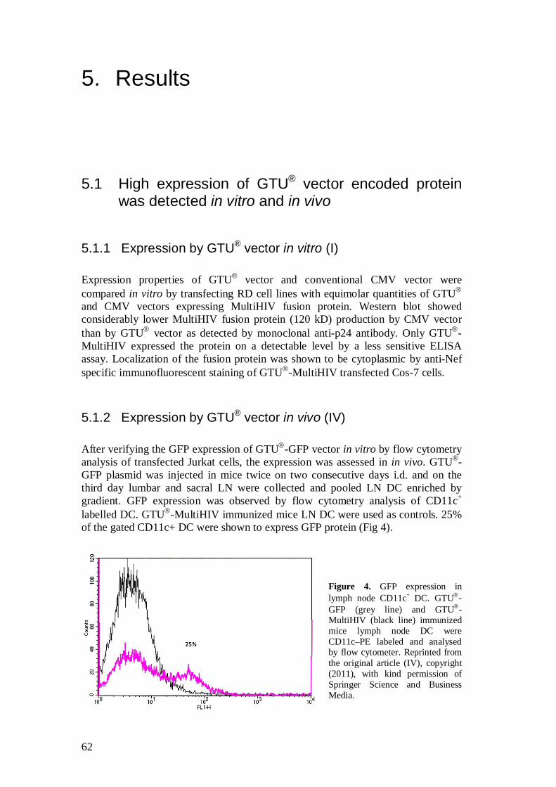
62
5. Results
5.1 High expression of GTU® vector encoded protein was detected in vitro and in vivo
5.1.1 Expression by GTU® vector in vitro (I)
Expression properties of GTU vector and conventional CMV vector were compared in vitro by transfecting RD cell lines with equimolar quantities of GTU and CMV vectors expressing MultiHIV fusion protein. Western blot showed considerably lower MultiHIV fusion protein (120 kD) production by CMV vector than by GTU vector as detected by monoclonal anti-p24 antibody. Only GTU -MultiHIV expressed the protein on a detectable level by a less sensitive ELISA assay. Localization of the fusion protein was shown to be cytoplasmic by anti-Nef specific immunofluorescent staining of GTU -MultiHIV transfected Cos-7 cells.
5.1.2 Expression by GTU® vector in vivo (IV)
After verifying the GFP expression of GTU -GFP vector in vitro by flow cytometry analysis of transfected Jurkat cells, the expression was assessed in in vivo. GTU®-GFP plasmid was injected in mice twice on two consecutive days i.d. and on the third day lumbar and sacral LN were collected and pooled LN DC enriched by gradient. GFP expression was observed by flow cytometry analysis of CD11c+ labelled DC. GTU -MultiHIV immunized mice LN DC were used as controls. 25% of the gated CD11c+ DC were shown to express GFP protein (Fig 4).
Figure 4. GFP expression in lymph node CD11c+ DC. GTU -GFP (grey line) and GTU -MultiHIV (black line) immunized mice lymph node DC were CD11c–PE labeled and analysed by flow cytometer. Reprinted from the original article (IV), copyright (2011), with kind permission of Springer Science and Business Media.
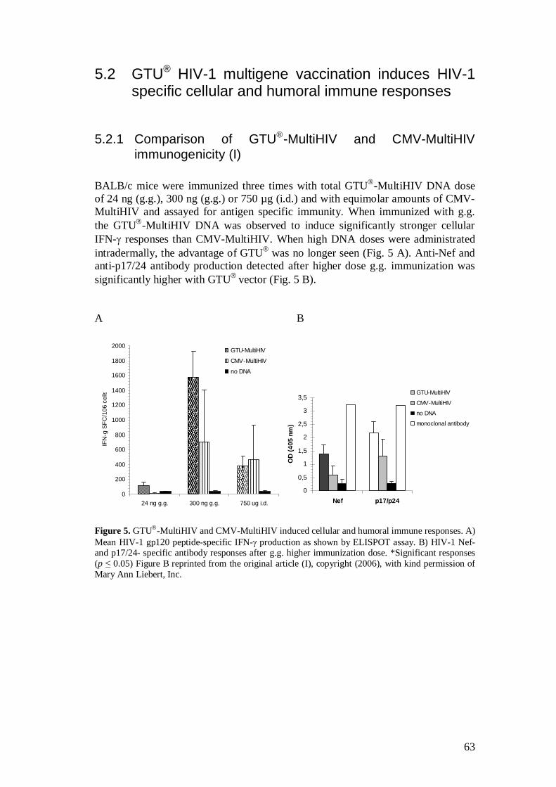
63
5.2 GTU® HIV-1 multigene vaccination induces HIV-1 specific cellular and humoral immune responses
5.2.1 Comparison of GTU -MultiHIV and CMV-MultiHIV immunogenicity (I)
BALB/c mice were immunized three times with total GTU -MultiHIV DNA dose of 24 ng (g.g.), 300 ng (g.g.) or 750 µg (i.d.) and with equimolar amounts of CMV-MultiHIV and assayed for antigen specific immunity. When immunized with g.g. the GTU -MultiHIV DNA was observed to induce significantly stronger cellular IFN- responses than CMV-MultiHIV. When high DNA doses were administrated intradermally, the advantage of GTU was no longer seen (Fig. 5 A). Anti-Nef and anti-p17/24 antibody production detected after higher dose g.g. immunization was significantly higher with GTU vector (Fig. 5 B). A B
Figure 5. GTU -MultiHIV and CMV-MultiHIV induced cellular and humoral immune responses. A) Mean HIV-1 gp120 peptide-specific IFN- production as shown by ELISPOT assay. B) HIV-1 Nef- and p17/24- specific antibody responses after g.g. higher immunization dose. *Significant responses (p 0.05) Figure B reprinted from the original article (I), copyright (2006), with kind permission of Mary Ann Liebert, Inc.
0
0,5
1
1,5
2
2,5
3
3,5
Nef p17/p24
OD
(405
nm
)
GTU-MultiHIV
CMV-MultiHIV
no DNA
monoclonal antibody
0
200
400
600
800
1000
1200
1400
1600
1800
2000
24 ng g.g. 300 ng g.g. 750 ug i.d.
IFN
-g S
FC/1
06 c
ells
GTU-MultiHIV
CMV-MultiHIV
no DNA
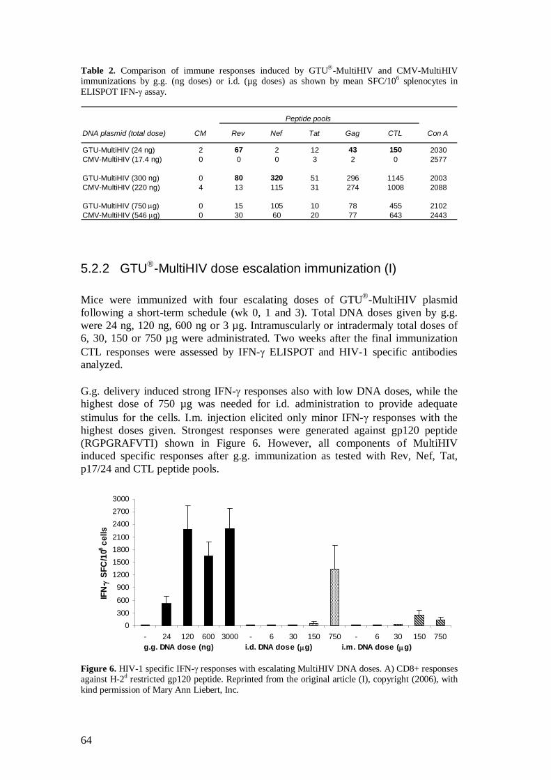
64
Table 2. Comparison of immune responses induced by GTU -MultiHIV and CMV-MultiHIV immunizations by g.g. (ng doses) or i.d. (µg doses) as shown by mean SFC/106 splenocytes in ELISPOT IFN- assay.
Peptide pools
DNA plasmid (total dose) CM Rev Nef Tat Gag CTL Con A
GTU-MultiHIV (24 ng) 2 67 2 12 43 150 2030CMV-MultiHIV (17.4 ng) 0 0 0 3 2 0 2577
GTU-MultiHIV (300 ng) 0 80 320 51 296 1145 2003CMV-MultiHIV (220 ng) 4 13 115 31 274 1008 2088
GTU-MultiHIV (750 g) 0 15 105 10 78 455 2102CMV-MultiHIV (546 g) 0 30 60 20 77 643 2443
5.2.2 GTU -MultiHIV dose escalation immunization (I)
Mice were immunized with four escalating doses of GTU -MultiHIV plasmid following a short-term schedule (wk 0, 1 and 3). Total DNA doses given by g.g. were 24 ng, 120 ng, 600 ng or 3 µg. Intramuscularly or intradermaly total doses of 6, 30, 150 or 750 µg were administrated. Two weeks after the final immunization CTL responses were assessed by IFN- ELISPOT and HIV-1 specific antibodies analyzed. G.g. delivery induced strong IFN- responses also with low DNA doses, while the highest dose of 750 µg was needed for i.d. administration to provide adequate stimulus for the cells. I.m. injection elicited only minor IFN- responses with the highest doses given. Strongest responses were generated against gp120 peptide (RGPGRAFVTI) shown in Figure 6. However, all components of MultiHIV induced specific responses after g.g. immunization as tested with Rev, Nef, Tat, p17/24 and CTL peptide pools. Figure 6. HIV-1 specific IFN- responses with escalating MultiHIV DNA doses. A) CD8+ responses against H-2d restricted gp120 peptide. Reprinted from the original article (I), copyright (2006), with kind permission of Mary Ann Liebert, Inc.
0300600
9001200150018002100
240027003000
- 24 120 600 3000 - 6 30 150 750 - 6 30 150 750 g.g. DNA dose (ng) i.d. DNA dose ( g) i.m. DNA dose ( g)
IFN
-SF
C/1
06 cel
ls
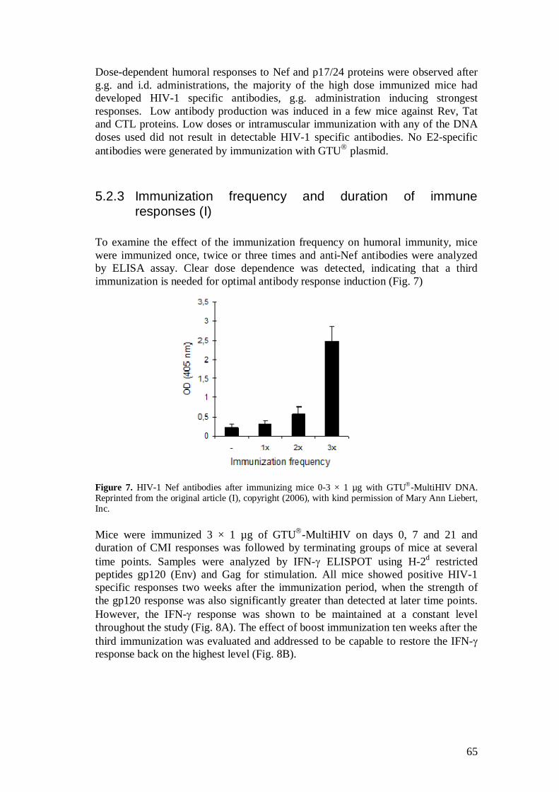
65
Dose-dependent humoral responses to Nef and p17/24 proteins were observed after g.g. and i.d. administrations, the majority of the high dose immunized mice had developed HIV-1 specific antibodies, g.g. administration inducing strongest responses. Low antibody production was induced in a few mice against Rev, Tat and CTL proteins. Low doses or intramuscular immunization with any of the DNA doses used did not result in detectable HIV-1 specific antibodies. No E2-specific antibodies were generated by immunization with GTU plasmid.
5.2.3 Immunization frequency and duration of immune responses (I)
To examine the effect of the immunization frequency on humoral immunity, mice were immunized once, twice or three times and anti-Nef antibodies were analyzed by ELISA assay. Clear dose dependence was detected, indicating that a third immunization is needed for optimal antibody response induction (Fig. 7)
Figure 7. HIV-1 Nef antibodies after immunizing mice 0-3 × 1 µg with GTU -MultiHIV DNA. Reprinted from the original article (I), copyright (2006), with kind permission of Mary Ann Liebert, Inc. Mice were immunized 3 × 1 µg of GTU -MultiHIV on days 0, 7 and 21 and duration of CMI responses was followed by terminating groups of mice at several time points. Samples were analyzed by IFN- ELISPOT using H-2d restricted peptides gp120 (Env) and Gag for stimulation. All mice showed positive HIV-1 specific responses two weeks after the immunization period, when the strength of the gp120 response was also significantly greater than detected at later time points. However, the IFN- response was shown to be maintained at a constant level throughout the study (Fig. 8A). The effect of boost immunization ten weeks after the third immunization was evaluated and addressed to be capable to restore the IFN- response back on the highest level (Fig. 8B).
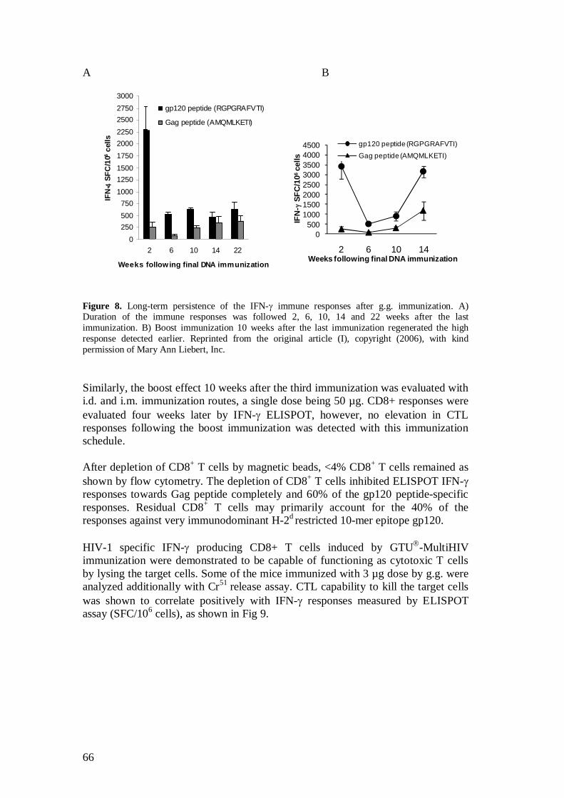
66
A B
0250500750
100012501500175020002250250027503000
2 6 10 14 22
Weeks following final DNA immunization
IFN
- S
FC/1
06 cel
lsgp120 peptide (RGPGRAFVTI)
Gag peptide (AMQMLKETI)
Figure 8. Long-term persistence of the IFN- immune responses after g.g. immunization. A) Duration of the immune responses was followed 2, 6, 10, 14 and 22 weeks after the last immunization. B) Boost immunization 10 weeks after the last immunization regenerated the high response detected earlier. Reprinted from the original article (I), copyright (2006), with kind permission of Mary Ann Liebert, Inc. Similarly, the boost effect 10 weeks after the third immunization was evaluated with i.d. and i.m. immunization routes, a single dose being 50 µg. CD8+ responses were evaluated four weeks later by IFN- ELISPOT, however, no elevation in CTL responses following the boost immunization was detected with this immunization schedule. After depletion of CD8+ T cells by magnetic beads, <4% CD8+ T cells remained as shown by flow cytometry. The depletion of CD8+ T cells inhibited ELISPOT IFN- responses towards Gag peptide completely and 60% of the gp120 peptide-specific responses. Residual CD8+ T cells may primarily account for the 40% of the responses against very immunodominant H-2d restricted 10-mer epitope gp120. HIV-1 specific IFN- producing CD8+ T cells induced by GTU -MultiHIV immunization were demonstrated to be capable of functioning as cytotoxic T cells by lysing the target cells. Some of the mice immunized with 3 µg dose by g.g. were analyzed additionally with Cr51 release assay. CTL capability to kill the target cells was shown to correlate positively with IFN- responses measured by ELISPOT assay (SFC/106 cells), as shown in Fig 9.
0500
10001500200025003000350040004500
2 6 10 14
IFN
-SF
C/1
06 c
ells
Weeks following final DNA immunization
gp120 peptide (RGPGRAFVTI)Gag peptide (AMQMLKETI)
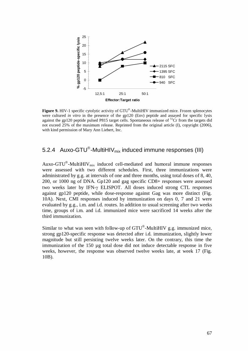
67
-5
0
5
10
15
20
25
12,5:1 25:1 50:1
Effector:Target ratio
% g
p120
pep
tide-
spec
ific
lysi
s
2115 SFC1395 SFC810 SFC940 SFC
Figure 9. HIV-1 specific cytolytic activity of GTU -MultiHIV immunized mice. Frozen splenocytes were cultured in vitro in the presence of the gp120 (Env) peptide and assayed for specific lysis against the gp120 peptide pulsed P815 target cells. Spontaneous release of 51Cr from the targets did not exceed 25% of the maximum release. Reprinted from the original article (I), copyright (2006), with kind permission of Mary Ann Liebert, Inc.
5.2.4 Auxo-GTU -MultiHIVmix induced immune responses (III)
Auxo-GTU -MultiHIVmix induced cell-mediated and humoral immune responses were assessed with two different schedules. First, three immunizations were administrated by g.g. at intervals of one and three months, using total doses of 8, 40, 200, or 1000 ng of DNA. Gp120 and gag specific CD8+ responses were assessed two weeks later by IFN- ELISPOT. All doses induced strong CTL responses against gp120 peptide, while dose-response against Gag was more distinct (Fig. 10A). Next, CMI responses induced by immunization on days 0, 7 and 21 were evaluated by g.g., i.m. and i.d. routes. In addition to usual screening after two weeks time, groups of i.m. and i.d. immunized mice were sacrificed 14 weeks after the third immunization. Similar to what was seen with follow-up of GTU -MultiHIV g.g. immunized mice, strong gp120-specific response was detected after i.d. immunization, slightly lower magnitude but still persisting twelve weeks later. On the contrary, this time the immunization of the 150 µg total dose did not induce detectable response in five weeks, however, the response was observed twelve weeks late, at week 17 (Fig. 10B).
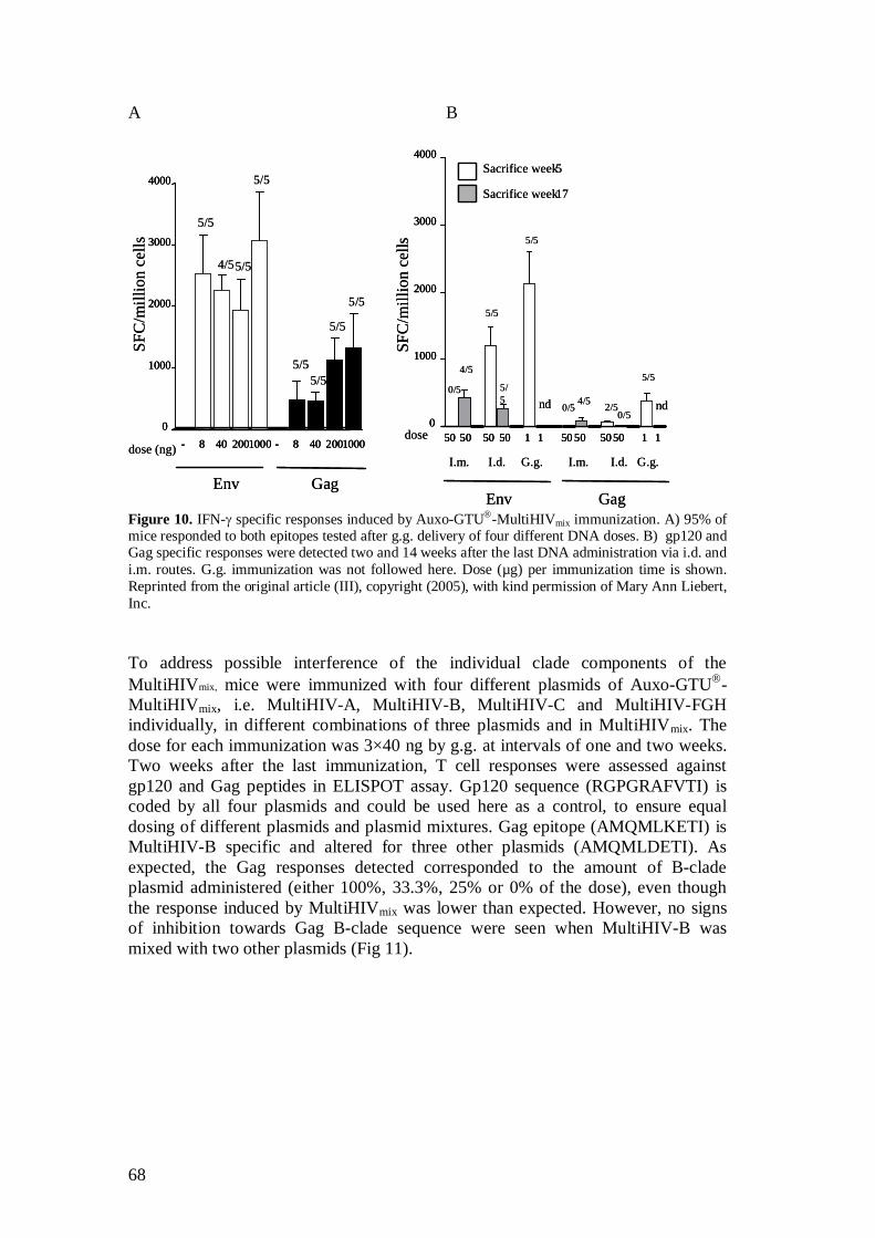
68
A B
Env Gag
SFC
/mill
ion
cells
5/5
5/5
5/5
4/5
5/55/5
5/5
5/5
dose (ng) - 8 40 2001000 - 8 40 20010000
1000
2000
3000
4000
50 50 50 50 1 1 50 50 5050 1 10
1000
2000
3000
4000
Env Gag
dose
nd nd
I.m. I.d. G.g. I.m. I.d. G.g.
0/5
4/5
5/5
5/5
5/5
4/50/5 2/5
0/5
5/5
Sacrifice week5
Sacrifice week17
SFC
/mill
ion
cells
Env Gag
SFC
/mill
ion
cells
5/5
5/5
5/5
4/5
5/55/5
5/5
5/5
dose (ng) - 8 40 2001000 - 8 40 20010000
1000
2000
3000
4000
- 8 40 2001000 - 8 40 20010000
1000
2000
3000
4000
50 50 50 50 1 1 50 50 5050 1 10
1000
2000
3000
4000
50 50 50 50 1 1 50 50 5050 1 10
1000
2000
3000
4000
Env Gag
dose
nd nd
I.m. I.d. G.g. I.m. I.d. G.g.
0/5
4/5
5/5
5/5
5/5
4/50/5 2/5
0/5
5/5
Sacrifice week5
Sacrifice week17
SFC
/mill
ion
cells
Figure 10. IFN- specific responses induced by Auxo-GTU -MultiHIVmix immunization. A) 95% of mice responded to both epitopes tested after g.g. delivery of four different DNA doses. B) gp120 and Gag specific responses were detected two and 14 weeks after the last DNA administration via i.d. and i.m. routes. G.g. immunization was not followed here. Dose (µg) per immunization time is shown. Reprinted from the original article (III), copyright (2005), with kind permission of Mary Ann Liebert, Inc. To address possible interference of the individual clade components of the MultiHIVmix, mice were immunized with four different plasmids of Auxo-GTU -MultiHIVmix, i.e. MultiHIV-A, MultiHIV-B, MultiHIV-C and MultiHIV-FGH individually, in different combinations of three plasmids and in MultiHIVmix. The dose for each immunization was 3×40 ng by g.g. at intervals of one and two weeks. Two weeks after the last immunization, T cell responses were assessed against gp120 and Gag peptides in ELISPOT assay. Gp120 sequence (RGPGRAFVTI) is coded by all four plasmids and could be used here as a control, to ensure equal dosing of different plasmids and plasmid mixtures. Gag epitope (AMQMLKETI) is MultiHIV-B specific and altered for three other plasmids (AMQMLDETI). As expected, the Gag responses detected corresponded to the amount of B-clade plasmid administered (either 100%, 33.3%, 25% or 0% of the dose), even though the response induced by MultiHIVmix was lower than expected. However, no signs of inhibition towards Gag B-clade sequence were seen when MultiHIV-B was mixed with two other plasmids (Fig 11).
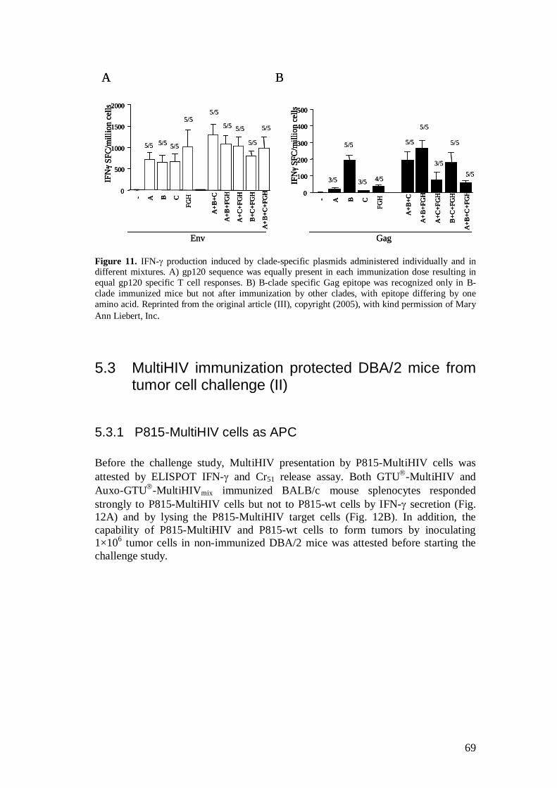
69
- A B C
FGH
A+B
+C
A+B
+FG
H
A+C
+FG
H
B+C
+FG
H
A+B
+C+F
GH0
100
200
300
400
500
IFN
-SFC
/mill
ion
cells
Env Gag
A B
5/5 5/5 5/5
5/55/5
5/5 5/5
5/5
5/5
3/5
5/5
3/5 4/5
5/5
5/5
5/5
5/53/5
- A B C
FGH
A+B
+C
A+B
+FG
H
A+C
+FG
H
B+C
+FG
H
A+B
+C+F
GH0
500
1000
1500
2000IF
N-
SFC
/mill
ion
cells
- A B C
FGH
A+B
+C
A+B
+FG
H
A+C
+FG
H
B+C
+FG
H
A+B
+C+F
GH0
100
200
300
400
500
IFN
-SFC
/mill
ion
cells
- A B C
FGH
A+B
+C
A+B
+FG
H
A+C
+FG
H
B+C
+FG
H
A+B
+C+F
GH0
100
200
300
400
500
IFN
-SFC
/mill
ion
cells
Env Gag
A B
5/5 5/5 5/5
5/55/5
5/5 5/5
5/5
5/5
3/5
5/5
3/5 4/5
5/5
5/5
5/5
5/53/5
- A B C
FGH
A+B
+C
A+B
+FG
H
A+C
+FG
H
B+C
+FG
H
A+B
+C+F
GH0
500
1000
1500
2000IF
N-
SFC
/mill
ion
cells
- A B C
FGH
A+B
+C
A+B
+FG
H
A+C
+FG
H
B+C
+FG
H
A+B
+C+F
GH0
500
1000
1500
2000IF
N-
SFC
/mill
ion
cells
Figure 11. IFN- production induced by clade-specific plasmids administered individually and in different mixtures. A) gp120 sequence was equally present in each immunization dose resulting in equal gp120 specific T cell responses. B) B-clade specific Gag epitope was recognized only in B-clade immunized mice but not after immunization by other clades, with epitope differing by one amino acid. Reprinted from the original article (III), copyright (2005), with kind permission of Mary Ann Liebert, Inc.
5.3 MultiHIV immunization protected DBA/2 mice from tumor cell challenge (II)
5.3.1 P815-MultiHIV cells as APC
Before the challenge study, MultiHIV presentation by P815-MultiHIV cells was attested by ELISPOT IFN- and Cr51 release assay. Both GTU -MultiHIV and Auxo-GTU -MultiHIVmix immunized BALB/c mouse splenocytes responded strongly to P815-MultiHIV cells but not to P815-wt cells by IFN- secretion (Fig. 12A) and by lysing the P815-MultiHIV target cells (Fig. 12B). In addition, the capability of P815-MultiHIV and P815-wt cells to form tumors by inoculating 1×10⁶ tumor cells in non-immunized DBA/2 mice was attested before starting the challenge study.
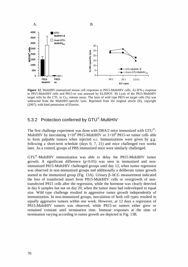
70
A. B.
0
500
1000
1500
2000
2500
3000
3500
4000
P815-MultiHIV
P815-wt
Auxo-GTU-MultiHIVmix
GTU-MultiHIV-B
SFC
/10
6ce
lls
0
500
1000
1500
2000
2500
3000
3500
4000
P815-MultiHIV
P815-wt
Auxo-GTU-MultiHIVmix
GTU-MultiHIV-B
SFC
/10
6ce
lls
Figure 12. MultiHIV-immunized mouse cell responses to P815-MultiHIV cells. A) IFN- response to P815-MultiHIV cells and P815-wt was assessed by ELISPOT. B) Lysis of the P815-MultiHIV target cells by the CTL in Cr51 release assay. The lysis of wild type P815-wt target cells (%) was subtracted from the MultiHIV-specific lysis. Reprinted from the original article (II), copyright (2007), with kind permission of Elsevier.
5.3.2 Protection conferred by GTU -MultiHIV
The first challenge experiment was done with DBA/2 mice immunized with GTU -MultiHIV by inoculating 1×10⁶
P815-MultiHIV or 1×10⁶ P815-wt tumor cells able
to form palpable tumors when injected s.c. Immunizations were given by g.g. following a short-term schedule (days 0, 7, 21) and mice challenged two weeks later. As a control, groups of PBS immunized mice were similarly challenged. GTU -MultiHIV immunization was able to delay the P815-MultiHIV tumor growth. A significant difference (p<0.05) was seen in immunized and non-immunized P815-MultiHIV challenged groups until day 12, when tumor regression was observed in non-immunized groups and additionally a deliberate tumor growth started in the immunized group (Fig. 13A). Urinary –hCG measurement indicated the loss of transfected insert from P815-MultiHIV cells or overgrowth of non-transfected P815 cells after the regression, while the hormone was clearly detected in day 6 samples but not on day 20, when the tumor mass had redeveloped to equal size. Wild type challenge resulted in aggressive tumor growth independently of immunization. In non-immunized groups, inoculation of both cell types resulted in equally aggressive tumors within one week. However, at 12 days a regression of P815-MultiHIV tumors was observed, while P815-wt tumors either grew or remained constant until termination time. Immune responses at the time of termination varying according to tumor growth are depicted in Fig. 13B.
0
5
10
15
20
25
30
GTU-MultiHIV-BAuxo-GTU-MultiHIVmix
50:1 25:1 12,5:1
E:T ratio
% H
IV-s
peci
fic ly
sis
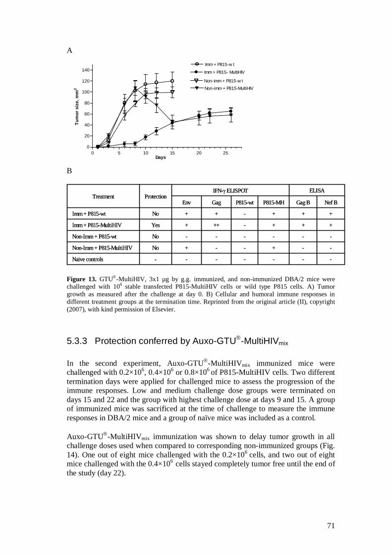
71
A
0 5 10 15 20 250
20
40
60
80
100
120
140Imm + P815-w t
Imm + P815- MultiHIV
Non-imm + P815-w tNon-imm + P815-MultiHIV
Days
Tum
or s
ize,
mm
2
B
-------Naive controls
--+--+NoNon-Imm + P815-MultiHIV
------NoNon-Imm + P815-wt
+++-+++YesImm+ P815-MultiHIV
+++-++NoImm+ P815-wt
Nef BGag BP815-MHP815-wtGag Env
ELISAIFN- ELISPOTProtectionTreatment
-------Naive controls
--+--+NoNon-Imm + P815-MultiHIV
------NoNon-Imm + P815-wt
+++-+++YesImm+ P815-MultiHIV
+++-++NoImm+ P815-wt
Nef BGag BP815-MHP815-wtGag Env
ELISAIFN- ELISPOTProtectionTreatment
Figure 13. GTU -MultiHIV, 3x1 µg by g.g. immunized, and non-immunized DBA/2 mice were challenged with 106 stable transfected P815-MultiHIV cells or wild type P815 cells. A) Tumor growth as measured after the challenge at day 0. B) Cellular and humoral immune responses in different treatment groups at the termination time. Reprinted from the original article (II), copyright (2007), with kind permission of Elsevier.
5.3.3 Protection conferred by Auxo-GTU -MultiHIVmix
In the second experiment, Auxo-GTU -MultiHIVmix immunized mice were challenged with 0.2×10⁶, 0.4×10⁶ or 0.8×10⁶
of P815-MultiHIV cells. Two different termination days were applied for challenged mice to assess the progression of the immune responses. Low and medium challenge dose groups were terminated on days 15 and 22 and the group with highest challenge dose at days 9 and 15. A group of immunized mice was sacrificed at the time of challenge to measure the immune responses in DBA/2 mice and a group of naïve mice was included as a control. Auxo-GTU -MultiHIVmix immunization was shown to delay tumor growth in all challenge doses used when compared to corresponding non-immunized groups (Fig. 14). One out of eight mice challenged with the 0.2×10⁶
cells, and two out of eight mice challenged with the 0.4×10⁶
cells stayed completely tumor free until the end of the study (day 22).
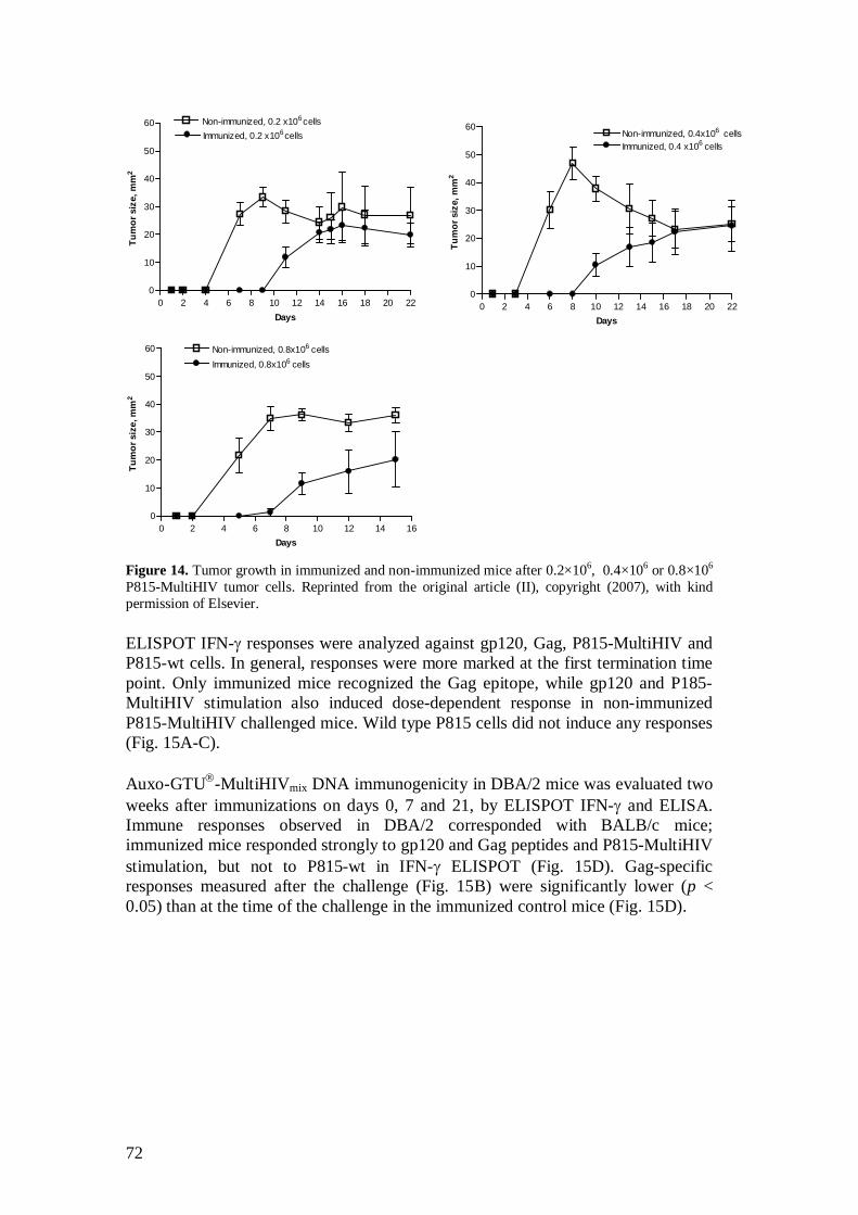
72
0 2 4 6 8 10 12 14 16 18 20 220
10
20
30
40
50
60 Non-immunized, 0.2 x10⁶cellsImmunized, 0.2 x10⁶cells
Days
Tum
or s
ize,
mm
2
0 2 4 6 8 10 12 14 16 18 20 220
10
20
30
40
50
60Non-immunized, 0.4x10⁶ cellsImmunized, 0.4 x10⁶ cells
Days
Tum
or s
ize,
mm
2
0 2 4 6 8 10 12 14 160
10
20
30
40
50
60 Non-immunized, 0.8x10⁶ cellsImmunized, 0.8x10⁶ cells
Days
Tum
or s
ize,
mm
2
0 2 4 6 8 10 12 14 16 18 20 220
10
20
30
40
50
60 Non-immunized, 0.2 x10⁶cellsImmunized, 0.2 x10⁶cells
Days
Tum
or s
ize,
mm
2
0 2 4 6 8 10 12 14 16 18 20 220
10
20
30
40
50
60Non-immunized, 0.4x10⁶ cellsImmunized, 0.4 x10⁶ cells
Days
Tum
or s
ize,
mm
2
0 2 4 6 8 10 12 14 16 18 20 220
10
20
30
40
50
60 Non-immunized, 0.2 x10⁶cellsImmunized, 0.2 x10⁶cells
Days
Tum
or s
ize,
mm
2
0 2 4 6 8 10 12 14 16 18 20 220
10
20
30
40
50
60Non-immunized, 0.4x10⁶ cellsImmunized, 0.4 x10⁶ cells
Days
Tum
or s
ize,
mm
2
0 2 4 6 8 10 12 14 160
10
20
30
40
50
60 Non-immunized, 0.8x10⁶ cellsImmunized, 0.8x10⁶ cells
Days
Tum
or s
ize,
mm
2
Figure 14. Tumor growth in immunized and non-immunized mice after 0.2×10⁶, 0.4×10⁶ or 0.8×10⁶
P815-MultiHIV tumor cells. Reprinted from the original article (II), copyright (2007), with kind permission of Elsevier. ELISPOT IFN- responses were analyzed against gp120, Gag, P815-MultiHIV and P815-wt cells. In general, responses were more marked at the first termination time point. Only immunized mice recognized the Gag epitope, while gp120 and P185-MultiHIV stimulation also induced dose-dependent response in non-immunized P815-MultiHIV challenged mice. Wild type P815 cells did not induce any responses (Fig. 15A-C). Auxo-GTU -MultiHIVmix DNA immunogenicity in DBA/2 mice was evaluated two weeks after immunizations on days 0, 7 and 21, by ELISPOT IFN- and ELISA. Immune responses observed in DBA/2 corresponded with BALB/c mice; immunized mice responded strongly to gp120 and Gag peptides and P815-MultiHIV stimulation, but not to P815-wt in IFN- ELISPOT (Fig. 15D). Gag-specific responses measured after the challenge (Fig. 15B) were significantly lower (p < 0.05) than at the time of the challenge in the immunized control mice (Fig. 15D).
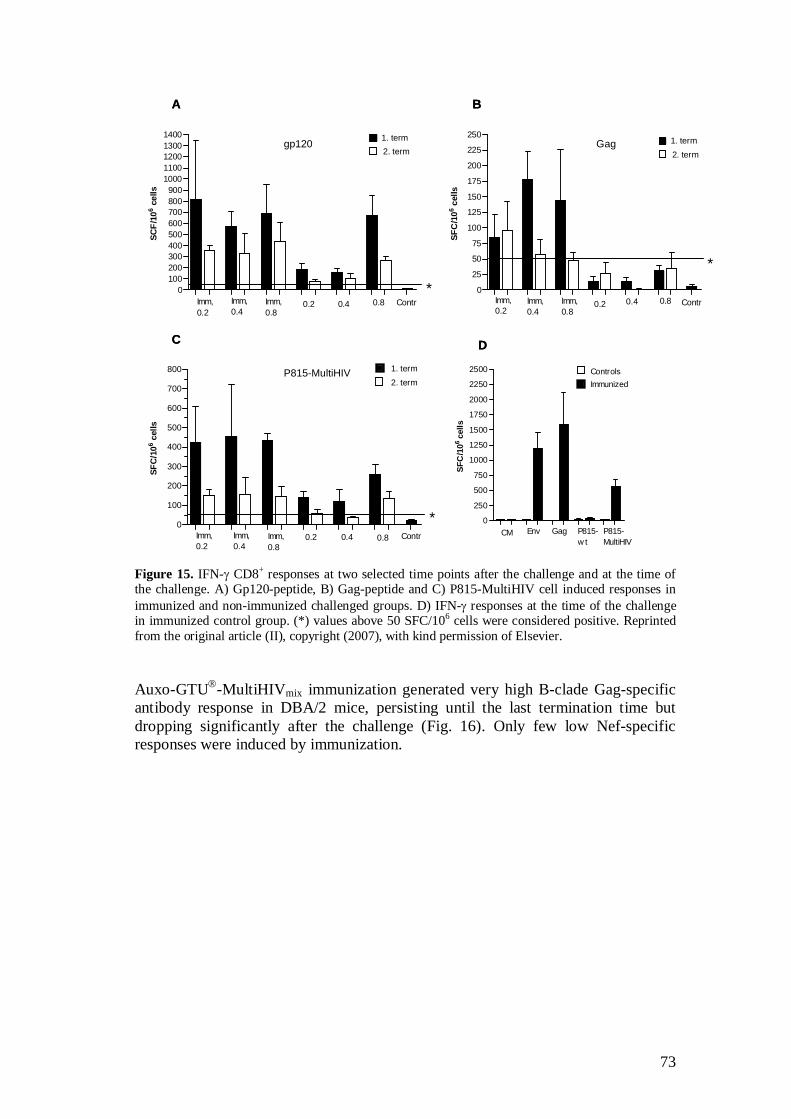
73
0100200300400500600700800900
10001100120013001400
Imm,0.2
0.8 Contr0.2Imm,0.8
Imm,0.4
0.4
2. term1. term
SCF/
106 c
ells
* 0
25
50
75
100
125
150
175
200
225
2501. term2. term
Imm,0.2
0.8 Contr0.2Imm,0.8
Imm,0.4
0.4
SFC
/106 c
ells
*
0
100
200
300
400
500
600
700
800
Imm,0.2
Imm,0.4
Imm,0.8
0.2 0.4 0.8 Contr
*
2. term1. term
SFC
/106 c
ells
0
250
500
750
1000
1250
1500
1750
2000
2250
2500
CM Env Gag P815-w t
P815-MultiHIV
ImmunizedControls
SFC
/106
cells
A B
C D
gp120 Gag
P815-MultiHIV
0100200300400500600700800900
10001100120013001400
Imm,0.2
0.8 Contr0.2Imm,0.8
Imm,0.4
0.4
2. term1. term
SCF/
106 c
ells
*0100200300400500600700800900
10001100120013001400
Imm,0.2
0.8 Contr0.2Imm,0.8
Imm,0.4
0.4
2. term1. term
SCF/
106 c
ells
* 0
25
50
75
100
125
150
175
200
225
2501. term2. term
Imm,0.2
0.8 Contr0.2Imm,0.8
Imm,0.4
0.4
SFC
/106 c
ells
*
0
100
200
300
400
500
600
700
800
Imm,0.2
Imm,0.4
Imm,0.8
0.2 0.4 0.8 Contr
*
2. term1. term
SFC
/106 c
ells
0
250
500
750
1000
1250
1500
1750
2000
2250
2500
CM Env Gag P815-w t
P815-MultiHIV
ImmunizedControls
SFC
/106
cells
A B
C D
gp120 Gag
P815-MultiHIV
Figure 15. IFN- CD8+ responses at two selected time points after the challenge and at the time of the challenge. A) Gp120-peptide, B) Gag-peptide and C) P815-MultiHIV cell induced responses in immunized and non-immunized challenged groups. D) IFN- responses at the time of the challenge in immunized control group. (*) values above 50 SFC/106 cells were considered positive. Reprinted from the original article (II), copyright (2007), with kind permission of Elsevier. Auxo-GTU -MultiHIVmix immunization generated very high B-clade Gag-specific antibody response in DBA/2 mice, persisting until the last termination time but dropping significantly after the challenge (Fig. 16). Only few low Nef-specific responses were induced by immunization.
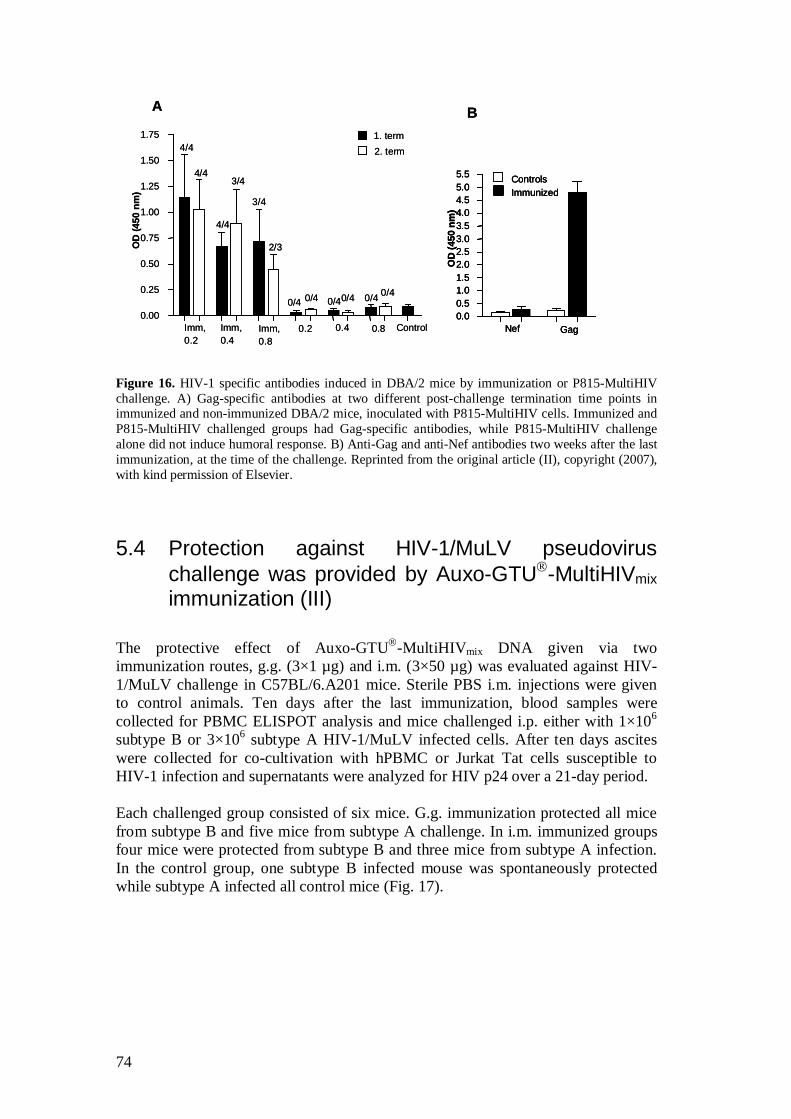
74
0.00.51.01.52.02.53.03.54.04.55.05.5
Nef Gag
ImmunizedControls
OD
(450
nm
)
0.00
0.25
0.50
0.75
1.00
1.25
1.50
1.75 1. term2. term4/4
4/4
ControlImm,0.2
Imm,0.4
Imm,0.8
0.2 0.4 0.8
4/4
3/4
0/4 0/4
3/4
2/3
0/4 0/4 0/40/4
OD
(450
nm
)
A B
0.00.51.01.52.02.53.03.54.04.55.05.5
Nef Gag
ImmunizedControls
OD
(450
nm
)
0.00.51.01.52.02.53.03.54.04.55.05.5
Nef Gag
ImmunizedControls
OD
(450
nm
)
0.00
0.25
0.50
0.75
1.00
1.25
1.50
1.75 1. term2. term4/4
4/4
ControlImm,0.2
Imm,0.4
Imm,0.8
0.2 0.4 0.8
4/4
3/4
0/4 0/4
3/4
2/3
0/4 0/4 0/40/4
OD
(450
nm
)
A B
Figure 16. HIV-1 specific antibodies induced in DBA/2 mice by immunization or P815-MultiHIV challenge. A) Gag-specific antibodies at two different post-challenge termination time points in immunized and non-immunized DBA/2 mice, inoculated with P815-MultiHIV cells. Immunized and P815-MultiHIV challenged groups had Gag-specific antibodies, while P815-MultiHIV challenge alone did not induce humoral response. B) Anti-Gag and anti-Nef antibodies two weeks after the last immunization, at the time of the challenge. Reprinted from the original article (II), copyright (2007), with kind permission of Elsevier.
5.4 Protection against HIV-1/MuLV pseudovirus challenge was provided by Auxo-GTU -MultiHIVmix immunization (III)
The protective effect of Auxo-GTU -MultiHIVmix DNA given via two immunization routes, g.g. (3×1 µg) and i.m. (3×50 µg) was evaluated against HIV-1/MuLV challenge in C57BL/6.A201 mice. Sterile PBS i.m. injections were given to control animals. Ten days after the last immunization, blood samples were collected for PBMC ELISPOT analysis and mice challenged i.p. either with 1×10⁶ subtype B or 3×10⁶ subtype A HIV-1/MuLV infected cells. After ten days ascites were collected for co-cultivation with hPBMC or Jurkat Tat cells susceptible to HIV-1 infection and supernatants were analyzed for HIV p24 over a 21-day period. Each challenged group consisted of six mice. G.g. immunization protected all mice from subtype B and five mice from subtype A challenge. In i.m. immunized groups four mice were protected from subtype B and three mice from subtype A infection. In the control group, one subtype B infected mouse was spontaneously protected while subtype A infected all control mice (Fig. 17).
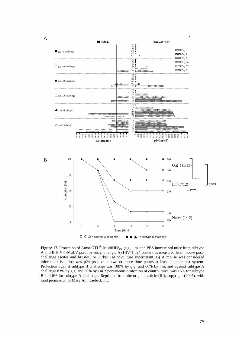
75
= subtype A challenge = subtype B challenge
6/6
5/6
4/6
3/6
1/6
0/6
G.g (11/12)
I.m (7/12)
Naive (1/12)0
25
50
75
100
3 6 9 14 17 21
Time (days)
Prot
ectio
n (%
)
A
B
.hPBMC
-850
0
-800
0
-750
0-7
000
-650
0
-600
0-5
500
-500
0-4
500
-400
0
-350
0
-300
0
-250
0
-200
0-1
500
-100
0
-500 0
p24 (ng/ml)
Jurkat Tat
0
500
1000
1500
2000
2500
3000
3500
4000
4500
5000
5500
6000
6500
7000
7500
8000
8500
C1C5C4C3C2C6
C7C11C10C12
C8C9
B6B2B1B5B4B3
B7B12B11B10
B9B8
A5A3A4A6A2A1
A12A11A10
A9A8A7
Day 3
Day 6
Day 9Day 14
Day 17
Day 21
p24(ng/ml)
g.g /B challenge
g.g. /A challenge
i.m. /B challenge
i.m. /A challenge
- /B challenge
- /A challenge
*
*
p=ns
p<0.05
p=ns
Figure 17. Protection of Auxo-GTU -MultiHIVmix g.g., i.m. and PBS immunized mice from subtype A and B HIV-1/MuLV pseudovirus challenge. A) HIV-1 p24 content as measured from mouse post-challenge ascites and hPBMC or Jurkat Tat co-culture supernatant. B) A mouse was considered infected if isolation was p24 positive in two or more time points at least in other test system. Protection against subtype B challenge was 100% by g.g. and 66% by i.m. and against subtype A challenge 83% by g.g. and 50% by i.m. Spontaneous protection of control mice was 16% for subtype B and 0% for subtype A challenge. Reprinted from the original article (III), copyright (2005), with kind permission of Mary Ann Liebert, Inc.
76
Cellular immune responses were measured by ELISPOT against EnvB.gp120 pool, CTL pool (corresponding to the subtype B MultiHIV CTL) and GagB.p24 pool, atigens matching for C57BL/6.A201 mice, having different genetics than BALB/c. IFN- responses were measured from mPBMC pre-challenge samples and post-challenge splenocytes. In general, g.g. immunization resulted in more frequent and stronger cell mediated immune responses. However, post-challenge responses were quite similar in both g.g. and i.m. immunized groups. The most striking observation was the switching from pre-challenge gag-specificity to post-challenge env-specificity. Before the challenge groupwise pooled mPBMCs of g.g. immunized mice responded to Gag-specific stimulation strongly (max. 231 SFC/106 cells for the group to be challenged with clade B virus / 583 SFC/106 cells for the group to be challenged with clade A virus), while EnvB.gp120 stimulation induced only weak responses (max. 20 SFC/106 cells B clade challenged group / 27 SFC/106 cells A clade challenge group). Only weak EnvB.gp120 response (30 SFC/106 cells) was detected after i.m. immunization. The post-challenge ELISPOT analysis of splenocytes showed that after the challenge, Gag-specific responses had declined in the g.g. immunized groups, being 13-30 SFC/106 cells (2 responders in both groups). A similar weak response to Gag was also detected this time in few mice of i.m. immunized groups. On the contrary, Env stimulation elicited better responses post-challenge both in g.g. and i.m. immunized groups. One mouse in both g.g. immunized groups responded to EnvB.gp120 (50 / 100 SFC/106 cells). Four responders in B subtype challenged group (25-70 SFC/106 cells) and five responders in A subtype challenged group (25-165 SFC/106 cells) were detected after CTL pool stimulation. In i.m. immunized groups EnvB.gp120 peptides were recognized in three mice (13-103) after subtype B challenge and in one mouse (73 SFC/106 cells) after subtype A challenge. Responses to CTL pool were not analyzed for i.m. immunized groups. No specific responses were detected in control mice. Antibodies against recombinant Gag A-, B-, C-, and FGH -clade proteins were detected at the time of the challenge in g.g. immunized mice. Challenge with HIV-1/MuLV infected cells raised the anti-Gag antibodies in all treated groups, also in the control group. Only low antibody levels were detected overall.
5.5 Immunization with in vivo transfected DC induced HIV-1 specific immune responses (IV)
T cells of BALB/c mice were primed by immunization on days 1 and 2 with Auxo-GTU®-MultiHIV-B. On the third day draining LN and spleens were collected and dendritic cells enriched by gradient. DC were used further for immunizing naïve groups of mice, one group with LN DC and the other with spleen DC. DC immunization was given with freshly isolated, in vivo transfected DC, twice at one week intervals. Blood samples were collected weekly for antibody measurement.
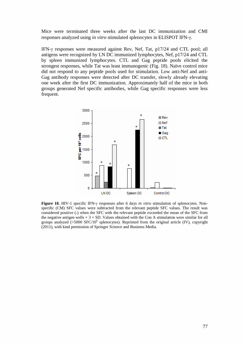
77
Mice were terminated three weeks after the last DC immunization and CMI responses analyzed using in vitro stimulated splenocytes in ELISPOT IFN- . IFN- responses were measured against Rev, Nef, Tat, p17/24 and CTL pool; all antigens were recognized by LN DC immunized lymphocytes, Nef, p17/24 and CTL by spleen immunized lymphocytes. CTL and Gag peptide pools elicited the strongest responses, while Tat was least immunogenic (Fig. 18). Naïve control mice did not respond to any peptide pools used for stimulation. Low anti-Nef and anti-Gag antibody responses were detected after DC transfer, slowly already elevating one week after the first DC immunization. Approximately half of the mice in both groups generated Nef specific antibodies, while Gag specific responses were less frequent.
Figure 18. HIV-1 specific IFN- responses after 6 days in vitro stimulation of splenocytes. Non-specific (CM) SFC values were subtracted from the relevant peptide SFC values. The result was considered positive (*) when the SFC with the relevant peptide exceeded the mean of the SFC from the negative antigen wells + 3 × SD. Values obtained with the Con A stimulation were similar for all groups analyzed (>5000 SFC/106 splenocytes). Reprinted from the original article (IV), copyright (2011), with kind permission of Springer Science and Business Media.

78
6. Discussion
6.1 GTU is a potent DNA vaccine vector
With the two DNA vectors used, MultiHIV antigen is expressed as a fusion protein with a molecular mass of approximately 120 kDa in the cytoplasm of transfected cells. Significantly higher MultiHIV protein expression level was observed when expressed from GTU vector, compared to expression from regular eukaryotic CMV vector. This observation is in agreement with the results obtained in other studies with GTU -GFP, addressing the improved maintenance of the vector in dividing cells, as well as enhanced transcription (Krohn et al. 2005, Martinon et al. 2009). The advantage of GTU vector comes from the expressed BPV-1 E2 protein (Abroi et al. 2004, Silla et al. 2010, Ustav et al. 1993) which attaches to multiple oligomerized binding sites of the vector, thereby acting as a transcription activator for the gene of interest. Secondly, E2 has a natural chromatin binding function accomplishing the GTU vector segregation function leading the plasmid to the progeny of cells during mitosis. This is a valuable aspect while the major impediment with DNA vaccine development is related to the relatively low and transient immune responses generated in humans (Estcourt et al. 2004), most probably largely due to too low dose of the vector in the expressing cells and to the rapid loss of non-replicating vector during cell division (Arrode-Bruses et al. 2010). The expression level of the MultiHIV gene by the GTU vector enables more efficient immune responses with smaller DNA doses. This was demonstrated when the HIV-1 specific immune responses were compared after GTU or CMV based MultiHIV immunization. Low doses (24 ng and 300 ng of GTU and equimolar amount of CMV) administered showed the significant superiority of GTU vector, while with high dose immunization (750 µg of GTU and equimolar amount of CMV) the advantage was no longer observed, possibly due to saturation. Similarly, the less immunogenic antigens such as Rev were more efficiently recognized after GTU immunization. There are concerns related to anti-vector immune responses with the use of naked DNA vaccines, even if not so striking as in case of viral genetic vaccines (Schalk et al. 2006). However, we detected no antibodies to dsDNA in the sera of GTU immunized mice, neither have others reported anti-dsDNA antibodies after repeated DNA immunizations (Giri et al. 2004, Parker et al. 2001, Schalk et al. 2006). Previously GTU -MultiHIV general safety, biodistribution, persistence and tolerability have been studied in rats and no concerns were observed (Tuomela et al. 2005). GTU based HIV-1 vaccines have been found safe and well-tolerated in several clinical trials conducted both on healthy volunteers and HIV-1 infected subjects (Krohn et al. 2005, Vardas and Stanescu,I, Valtavaara,M, Kuntonen,T, Gray,C, Leionen,M, Ustav,M, Reijonen, K 2010).
79
6.2 Evaluation of vaccine immunogenicity
The strength of DNA vaccines is to direct the expressed immunogen for MHC class I and II presentation, making them especially suitable inducers of cellular immune responses. GTU - MultiHIV is designed to elicit cellular immune responses to HIV-1 regulatory proteins Rev, Nef and Tat expressed at an early phase of the viral life cycle, (Blazevic 1997, Ranki et al. 1994) as well as to highly immunogenic structural proteins p17 and p24. Furthermore, the 17-45 amino acid long stretches containing T cell epitope clusters included can elicit RT and Env specific CD8+ and CD4+ T cell responses. The essential role in viral infectivity and expression early in the viral life cycle makes the regulatory proteins Rev, Nef and Tat attractive antigens to be included in vaccine (Blazevic 1997). The immunological pressure towards these antigens leading to detrimental changes in their function could effectively reduce the acute phase viral burden. Nef has been reported to be the most targeted HIV-1 antigen in early stage of HIV-1 infection (Masemola et al. 2004) even if these immune responses have not been shown to correlate with viral load. The role of cellular immune responses against Gag, also expressed by the MultiHIV sequence, has an indisputable effect on HIV-1 viral load. Rev and Tat are not so frequently recognized by the immune systems of HIV-1 infected individuals, likely due to their nuclear location and low expression level. But the importance of responses generated to these antigens cannot be ignored until the correlates of protection related to vaccination are better known. It may be possible that even if there is no evidence of the efficacy of non–Gag-specific CD8+ T-cell responses in HIV infected individuals, such responses may mediate control of viremia if vaccinated HIV-negative individual subsequently becomes HIV infected. Several groups are also working with DNA vaccine constructs encoding full-length Env proteins capable of inducing nAb (Estcourt et al. 2004, Haynes et al. 2010). However, as MultiHIV does not encode the full envelope sequence and hence not capable of inducing nAb, the humoral immune responses to encoded proteins were of secondary interest.
6.2.1 Use of inbred mice for vaccine immunogenicity evaluation
In the experimental work we used murine models for evaluating the immunogenicity and protective efficacy of GTU based MultiHIV DNA plasmids. The mouse strain used for immunogenicity studies is BALB/c of H-2d haplotype, the same haplotype as the DBA/2 mice used for the tumor challenge study. Using the inbred mouse for immunogenicity studies has the advantage of more reproducible results as the variation due to the MHC class diversity in outbred animals and humans is bypassed. The advantage of inbred laboratory mice is obvious when preliminary studies are performed. When evaluating the immunogenicity of different vaccine constructs, variable routes or time schedules, the less variation the test system itself generates, the easier it is to detect the differences related to the different vaccine constructs. Also, good knowledge of the immunodominant epitopes in these animals facilitates the characterization of immune responses and permits the inclusion of the epitopes for screening purposes. This approach has been utilized when constructing the MultiHIV sequence, carrying H-2d-restricted highly immunodominant HIV-1
80
epitopes gp120 (aa 311–320; RGPGRAFVTI) (Takahashi et al. 1988) and Gag peptide (aa 65–73; AMQMLKETI) (Qiu et al. 1999). Their presence in the vaccine constructs facilitates the screening of MultiHIV expression, presentation and immunogenicity in mice. However, the immunodominance of these sequences in H-2d mice may reduce the immune responses targeted at other less immunogenic epitopes, thereby possibly hampering the induction of broader responses in mice. Yet it must be conceded that the homozygous H-2 haplotype of inbred mice creates restrictions not corresponding to the natural human host (Griffin 2002). Although there are limitations and differences in the ability of H-2 and HLA molecules to present variable peptides, several identical CD4+ and CD8+ T cell epitopes are still recognized both in mice and humans, permitting the evaluation of vaccine immunogenicity in mice prior to moving to non-human primate or clinical studies (Berzofsky et al. 1991, Hosmalin et al. 1990). The inbred mouse model is the first level model when a vaccine construct is evaluated and the real breadth of the immune responses in humans can be evaluated only in clinical trials. Although the mouse system utilized may not be a perfect model for less predictable human antiviral responses, it obviously provides useful knowledge that can subsequently be used as an experimental hypothesis setup.
6.2.2 Effect of the immunization regimen
The immunization regimen, including the route, dose and frequency of administrations has a crucial role in how efficiently the immune response is generated. The routes used for evaluating the GTU -MultiHIV vaccine in BALB/c were g.g., i.m. and i.d. Each route was assessed with four escalating doses and GTU -MultiHIV immunogenicity was evaluated by measuring the MultiHIV-specific IFN- production of immunized mice splenocytes using ELISPOT assay. Three immunizations were given at intervals of one and two weeks and mice were terminated 10-14 days after the last immunization to allow both cellular and humoral immune responses enough time to develop following the boost. Consistent with previously published studies demonstrating the superiority of gene gun immunization over needle injections (Barry and Johnston 1997, Cohen et al. 1998, Pertmer et al. 1995, Trimble et al. 2003), the g.g. administration resulted in extremely high gp120 epitope specific responses in the order of magnitude of >2000 SFC / million splenocytes. G.g. immunization is known to already induce strong cell activation with minute DNA doses administered (Pertmer et al. 1995), and using the highly immunodominant gp120 10-mer for screening, dose dependency after GTU -MultiHIV immunization was observed only by the lowest dose (24 ng), while all other doses (120 ng, 600 ng and 3000 ng) induced equally strong “saturated” responses. A similar study performed with multiclade vaccine by the same route and doses resulted in equally high gp120–specific responses with all doses. However, the gag-specific dose dependency was clear, indicating that the smallest doses by g.g. are probably not effective in inducing cellular immune responses to less immundominant epitopes of MultiHIV vaccine. Earlier research has demonstrated that i.d. and i.m. routes are efficient inducers only with high antigen doses, likely due to the inefficient uptake of the plasmid (Barry and Johnston 1997, Caputo et al. 2003). Here, the highest total dose of 750 µg was
81
able to efficiently induce IFN- response via i.d. route, while high dose i.m. injection elicited only moderate cellular immune responses. I.m. needle injection has been found by others, too, to be an inefficient delivery route for naked DNA vaccination (Hinkula et al. 1997) and was unable to elicit detectable humoral immune responses against the MultiHIV proteins. Induction of humoral immune responses requires more DNA than cellular immune responses (Barry and Johnston 1997, Tähtinen 2001) and consistently, antibodies were induced only after high dose g.g. and i.d. administrations. A more feasible approach for the induction of antibodies might be administration of heterologous boost with the viral vector (Bråve et al. 2007). Using a g.g. immunization, we demonstrated the need for at least one boost to elicit humoral responses, while the third boost is needed to elicit high level of antibodies, which is in agreement with previously published results (Hinkula et al. 1997). The duration of induced immune responses is a critical parameter for vaccine efficacy, as vaccination is supposed to create immunological memory cells able to confer protection for subsequent encounters with related pathogen later in life. We evaluated the kinetics and duration of the cellular immune responses in g.g. immunized mice and demonstrated the persistent cellular response up to 22 weeks following the last immunization, being highest two weeks after the third immunization and then staying at constant lower level throughout the study. The fourth boost immunization ten weeks after the third immunization was able to restore the strong IFN- response, demonstrating the efficient priming of the immune system and capability of homologous boost to induce high responses. Additionally, the HIV-1/MuLV challenge was given 5 weeks after the third g.g. or i.m. immunization and addressed the emergence of highly effective, protective CD8+ T cells. The recovery of the cellular immune response even one year after immunization of BALB/c has been demonstrated by others (Bråve et al. 2007). In addition they showed that DNA immunization resulted in the generation of CD8+ memory T cells, while MVA immunization elicited CD4+ memory T cells. It was also demonstrated that the DNA primed cellular immune response can be biased to CD4+ or CD8+ T cells according to the boosting agent selected. This further adds to the value of DNA vaccines as stand-alone vaccines or as efficient priming agents for heterologous immunization. We also determined whether the immune responses induced via i.m. or i.d. injections could be similarly enhanced by a fourth boost immunization, with the single dose of 50 µg. Even if the fourth boost injection resulted in the restoration of earlier immune responses, thereby resembling the results by g.g. immunization, only fairly low responses were induced. An interesting observation was made when mice immunized with MultiHIV multiclade vaccine via i.m. and i.d. route were followed 2 weeks and 17 weeks after the third immunization. In the i.m. immunized groups gp120-specific IFN- responses were detected only at a later time point, suggesting that the kinetics of the immune responses after i.m. route are different and the response develops only later. In light of recent evidence this may be expected to happen as a result of differences between the intrinsic structure of skin and muscle tissues and their ability to be infiltrated by innate immune cells, especially by DC and macrophages (Abadie et al. 2009). The plasmid is readily carried for presenting DC by i.d. injection, while i.m. immunization mainly transfects muscle cells capable of expressing the antigen but the lack of resident professional APC in muscle is
82
likely to delay and decrease the efficiency of immune response induction. On the contrary, a biodistribution study demonstrated the longer persistence of the GTU plasmid in the skin than in muscle, but assessed only the existence of the plasmid in tissue, not the kinetics of antigen presentation (Tuomela et al. 2005). These observations indicate that a different delivery route might preferably not be compared head-to-head but the tissue structure related variables should be considered as they might affect the kinetics and quality of the immune responses induced. However, similar to what is generally acknowledged, and based on the results generated here, it can be concluded that plasmid DNA delivery through i.m. injection is not optimal but needs to be augmented with more invasive delivery devices, boosting agents or immunomodulators to induce proper responses (Sumida et al. 2004). In line with earlier reports, the success of plain DNA vaccination is dependent on a high dose of DNA and many booster immunizations (~100 µg in mice, up to 2-4 mg in non-human primates and humans) (Caputo et al. 2003). In addition, given that mice are known to generate elevated immune responses by DNA immunization than larger animals or humans, the injection of pure naked DNA alone is probably not enough to induce proper responses in humans. More invasive delivery methods such as electroporation or gene gun delivery and other means to enhance the immune system responses are needed.
6.2.3 Immunogenicity of different HIV-1 antigenic components of MultiHIV
There are several indications of the protective role of Gag specific cellular immune responses (Geldmacher et al. 2007, Honeyborne et al. 2007, Kiepiela et al. 2007) but it is not known how big role immune responses to this or other antigens will finally play in vaccine induced HIV-1 viral suppression. Thus the goal of many HIV-1 vaccine approaches is currently to induce broad responses to several HIV-1 antigens. Concerns may be raised that competition between CTL responses directed against different HIV-1 antigens would result in diminished responses, but on the contrary, a recent study of HIV-1 infected individuals showed that in hosts responding to a broad range of epitopes, the average epitope-specific response is rather larger than smaller (Fryer et al. 2009). All MultiHIV components including Rev, Nef, Tat, p17/24 (Gag) and CTL epitopes were appropriately presented and recognized by mice splenocytes, as assayed with five MultiHIV peptide pools or single peptides. IFN- responses of variable magnitude were detected, and as expected, the strongest immune responses were generated towards H-2d restricted epitopes gp120 and Gag. IFN- responses to Rev, Nef and Tat were analyzed using respective 15-mer peptide pools that were all recognized, despite their non-optimal length for CD8+ T cell presentation (as discussed later in this chapter) (Betts et al. 2001, Tarosso et al. 2010). Both Rev and Nef specific responses were shown to have significant benefit of GTU expression vector compared to CMV vector. Tat must be considered as a fairly poor immunogen, as only low positive responses to Tat were elicited. This concurs with what others have reported, independently of Tat being administered alone (Tahtinen et al. 2001) or as a part of multiantigen DNA vaccine. Vaccine evaluated by Burgers et al, encoding gag, RT, tat, nef and env genes, induced similarly strong gag-specific and weak tat-specific responses
83
(Burgers et al. 2009). Studies in HIV-1 infected individuals have also shown a similar distribution of CD8+ T cell responses towards different HIV-1 proteins, Tat being one of the least targeted proteins while Nef was more immunogenic than Rev and Tat (Masemola et al. 2004). However, in macaques immunized intradermally with MultiHIV plasmid, T cell responses induced were quite equally distributed against Gag, Tat and Nef (Martinon et al. 2009), demonstrating the immunogenic potential of all MultiHIV antigens. As mentioned, the immunodominance of gp120 and gag epitopes in BALB/c may reduce the immune responses to other epitopes, further restricted by the homozygous haplotype of inbred mice. As a consequence, it may be questionable how well inbred H-2d mice are suited to their purpose when evaluating the breadth of immune responses (Griffin 2002). However, it is of importance to show that each antigenic part of the MultiHIV is properly expressed and efficiently processed for presentation in vivo. Similarly, all MultiHIV components have been found immunogenic in clinical trials, when the GTU -MultiHIV vaccine has been administered via i.d. or i.m. delivery, even though the level of responses has been low especially in healthy volunteers (Krohn et al. 2005). The immunogenicity of MultiHIV vaccine has also recently been evaluated in non-human primates (Martinon et al. 2009) and outbred pigs (Molder et al. 2009), which were shown to recognize several immunogenic epitopes. Even if the response in BALB/c mice is strongly biased to two H-2d restricted epitopes that elicit considerably higher IFN- responses in ELISPOT than achieved with any other model (pigs, macaques, human), still relatively many of the epitopes recognized by pigs have also been shown to be immunogenic in BALB/c mice, when they were evaluated with 15-mer overlapping MultiHIV specific peptide pools in matrix (non published data). Similarly, the Gag immunogenicity is frequently seen in all animals used for evaluation as well as in humans. Antibodies were predominantly targeted at Nef and p17/24 proteins, emerging after g.g. and i.d. immunization with high dose. Low level of antibodies against other MultiHIV antigens Rev, Tat and CTL were detected only occasionally. CTL protein is entirely artificial and only included for the induction of cellular responses. The absence of Rev and Tat specific antibodies has been reported also previously (Caputo et al. 2003, Tahtinen et al. 2001), when the Nef, Rev and Tat were introduced individually in mice. Only Nef was found to be highly immunogenic, while Rev induced proliferation and cytotoxic responses but no antibodies, and all Tat-specific immune responses were almost absent (Tahtinen et al. 2001). Additionally, the expression of the MultiHIV sequence as a fusion protein is not likely to support the efficient presentation of all MultiHIV components to B cells, which are dependent on the intact antigen on their activation. As a methodological aspect, the general use of overlapping 15-mer peptides for assaying the cytokine production has been criticized for underestimating the specific CD8+ T cell responses (Betts et al. 2001, Tarosso et al. 2010). Betts et al. reported nearly equal responses to 15-mer and 20-mer overlapping pools by CD4+ T cells, while CD8+ T cells recognized more frequently 15-mer pools and furthermore, when compared to optimized 8- to 11-mers, they observed clear preference for shorter peptides. Despite the fact that overlapping 15-mers can detect both CD4- and CD8- mediated T cells, MHC class I restricted minimal short peptides are expected to elicit only CD8+ T cell response. Similarly Tarosso et al. reported similarly on higher cellular responses detected with short optimal peptides (Tarosso et al. 2010).
84
This is likely to be related to peptide overhang length and the location of the immunogenic epitope within the longer peptide sequence. The critical impact of the optimal epitope presentation has also been demonstrated by our group when 9-mer gag epitope (AMQMLKETI) was compared to the corresponding most immunogenic 15-mer (HQAAMQMLKETINEE) in the overlapping Gag pool. The IFN- production, mainly produced by CD8+ T cells, was considerably higher when stimulated with 9-mer than with 15-mer, whereas CD4+ T cell related IL-2 production was induced on higher level with the longer peptide (unpublished data). Thus the use of optimal length of peptides for analysis may improve the readouts and whenever feasible, the careful optimization of peptides used should be done.
6.2.4 HIV-1 multiclade consensus / ancestral vaccine approach
The subtype specific MultiHIV sequences were constructed by consensus (A, B, C) or ancestral (FGH) lineages as described in more detail by Krohn et al. (Krohn et al. 2005), to represent wide variation within the clades. The Auxo-GTU-MultiHIV B consensus plasmid has been observed to induce responses in mice comparable to GTU -MultiHIV B Han-2 plasmid. We aimed to detect the effect of mixing four plasmids together and compared these to the responses induced by single clade immunizations. To demonstrate the equal success of immunizations with all four individual constructs, we assayed IFN- responses directed to gp120 epitope (RGPGRAFVTI) that is found identically in all four constructs. However, the strict epitope sequence specificity related to T cell activation through MHC presentation (Lee et al. 2004) was elucidated by one amino acid difference found between the B-clade Gag epitope (AMQMLKETI) and other three constructs A, C and FGH (AMQMLDETI). The immunization with the B-clade specific plasmid was shown to virtually solely contribute to the immune responses to the Gag peptide in the cocktail, while CD8+ T cell activated by immunization with other constructs could not efficiently recognize the sequence differing by one amino acid. This demonstrates that even minimal change in the epitope made the sequence suboptimal for H-2d mice recognition. Although cross-reactivity of murine CD8+ T cells does not directly translate to humans, the possible immune interference between the four plasmids in a cocktail was also briefly investigated by comparing the IFN- responses induced by immunization with different mixtures of three plasmids and the cocktail of four plasmids (MultiHIVmix). Mixing the plasmids was not observed to result in competition or enhancement, but the gag-specific response was directly dependent on the amount of the identical construct in the cocktail. It would be of value to have several immunogenic clade specific epitopes analyzed for a broader picture of the phenomenon, but the use of inbred mice may be the optimal model for this. By contrats, when MultiHIV vaccine (g.g.) was co-administrated with gp160Env encoding plasmid (Biojector ) in BALB/c, CD8+ T cell immune responses were observed to be slightly compromised (Brave et al. 2009). This might have been avoided by using some other combination of immunization routes, utilizing different draining lymph nodes. Other research groups working with multiclade plasmid cocktail vaccines have ended up with contradictory results when studying the immune interference. Kong et al. (Kong et al. 2003) evaluated the immune
85
interference in complex multiantigen (Env and Gag-Pol-Nef) and multiclade (A/B/C) plasmid mixture and despite mixing various components the combining of individual genes with each other was shown not to cause interference. Furthermore, the simultaneous administration of mutant epitopes has been suggested to even avoid the induction of immune interference (Singh et al. 2002). However, others have ended up with severe immune interference. Larke et al. compared the single A, B and C clade vaccines alone or in dual-clade formulation in BALB/c. The encoded immunogens were Gag, RT and Nef and five H-2d epitopes, two of which were identical in all clades used. As results of mixing the plasmids, they reported immunodominance, “original antigenic sin” and T cell antagonism. They also point out that using peptide pools for analysis would mask the effect of immune interference. The administration of individual clade vaccines to anatomically separated sites was shown to overcome the antagonism (Larke et al. 2007). With multiclade constructs there undoubtedly is a need for careful assessment of the immune interference and analysis should be designed individually for each vaccine modality and based on thorough selection of appropriate peptide epitopes to be used for screening. The induction of immune interference is related to several variables such as immunization sites, routes of administration and essentially on antigen design, thus in light of the current knowledge of this phenomenon, the occurrence of immune interferance cannot be extrapolated from results with other vaccine regimens. Finally, it can be concluded that the extreme complexity of the immune responses combined with the versatile HIV-1 virus and huge variability that can be included in vaccine constructs sets the requirements for immune response analysis extremely high. Complex studies are demanded to carefully estimate the outcome of administration of such complex vaccines as many of the current HIV-1 vaccine candidates are.
6.2.5 Read-outs for cellular immune responses
There is compelling evidence that CD8+ T cells play a crucial role in the control of HIV-1 infection and broad activation of this cell type is also one major goal of GTU -MultiHIV immunization. So far, the functional characterization of activated CD8+ T cells has mainly relied on the measurement of IFN- , which is the predominating cytokine secreted by activated CTL, the activator for immune system cells and furthermore significantly and inversely has correlated with viral load in several studies (Betts et al. 2001, Masemola et al. 2004, McMichael 1998, McMichael et al. 2010). Additionally, the role of granzyme B and perforin in CTL activity is acknowledged (Kuerten et al. 2008). Despite the unquestionable requirement for cytokine measurement as a tool for evaluating the cellular immune responses, it would be of importance to supplement them with assays measuring the direct antiviral function. Additionally, it has been reported that CD8+ T cells showing high antigen specific IFN- activation in ELISPOT have not been able to suppress the viral replication (Addo et al. 2002, Addo et al. 2003, Chung et al. 2007, Martins et al. 2010). However, cellular immune responses are versatile and currently the idea prevails that broad and polyfunctional CD8+ as well as CD4+ T cell responses are a requirement for the efficient containment of the HIV-1 virus. Hence the use of more sensitive assays to carefully measure all characteristics of cellular immunity, such as proliferative capacity, cytotoxicity and cytokine
86
expression profile would be needed (Arrode-Bruses et al. 2010, Betts et al. 2006). While lacking clearly defined correlates of protection, it cannot be known for certain which characteristics are the most valuable to be monitored and until they are clarified, comprehensive immunomonitoring is the only way to properly define the immunogenicity of vaccine candidates. The immunogenicity of a certain antigen is always limited by the technology available for detecting the responses, but fortunately immunological techniques have developed enormously. For the work presented in this thesis, the main readout used for measuring the cellular immune responses was IFN- secretion by splenocytes, analyzed at single cell level by ELISPOT assay. The IFN- production was demonstrated to be produced by CD8+ T cells, by assessing CD8+ T cell depleted samples in ELISPOT assay. The use of short 9-mer and 10-mer peptides and restricts the presentation to CD8+ T cells. We elucidated the correlation between IFN- production and cytotoxic activity by utilizing the cytotoxic T lymphocyte assay to demonstrate that the CD8+ T cells with higher IFN- secretion were more efficient to lyse the peptide-pulsed P815 target cells. Recently, we have frequently also assessed the IL-2 as a cytokine marker for CD4+ T cells by ELISPOT in immunological studies and correlation between IL-2 and IFN- production has been observed. However, it would be valuable to include a broader range of assays, such as multiparametric flow cytometric analysis, to have more detailed information on the expression profile of the activated cells, and the cell types mounting the immune responses and immunological memory.
6.3 Tumor challenge model
There is a need for small animal models measuring vaccine protective efficacy in vivo. Vaccine immunogenicity is constantly evaluated in mice but measuring cellular immune responses and their cytotoxic capability by in vitro assays does not fully represent the function of cytotoxic cells in their natural surroundings with all constituents of the immune system. To meet this need, we developed a tumor challenge model where MultiHIV immunized DBA/2 H-2d mice were challenged with syngeneic P815 tumor cells stable transfected with MultiHIV B-clade Han-2 isolate antigen. The P815-MultiHIV cells were evaluated for their MultiHIV presentation in vitro by using them as target cells for immunized BALB/c effector cells and additionally, their tumorigenicity in DBA/2 mice was assessed in pilot study. Mastocytoma P815 cell line is a well-characterized and therefore an affordable model to also adapt for HIV-1 vaccine protection studies. Wild type P815 express high level of MHC class I molecules, explaining their high capacity to work as APC in in vivo assays, but not MHC class II molecules, thereby stimulating only CD8+ T cells. Wild type P815 cells are known to possess a low degree of immunogenicity, leading to anti-tumor immunity when inoculated several times in syngeneic mice (Chen et al. 1994) and even if partial immune rejection of the tumor by the host is generally observed, it very rarely results in the complete elimination of the tumor cells. Additionally, the generation of protective CTL responses has been shown to be largely dependent on the mode of P815 tumor injection, with 600 P815 cells by
87
i.p. injection already leading consistently to tumor growth (Uyttenhowe 1983), whereas injecting s.c. 106 cells in the footpad leads to progressive tumors in only 30-40% of cases (Brichard et al. 1995). P815 cells injected s.c. in the flank have been reported to lead to progressive tumor growth in 85% of the mice (Colmenero et al. 1999) being thus a more suitable route for a challenge model. In the first experimental setup evaluating the GTU -MultiHIV B-clade g.g. immunization, the vaccine antigen sequence was identical with that encoded by transfected P815 cells. A protective effect was clearly observable by delayed and low P815-MultiHIV tumor growth in immunized mice when compared to aggressive tumor growth in non-immunized mice or P815 wild type challenged mice. However, the P815-MultiHIV induced tumors in non-immunized mice showed regression on day 12, while tumor mass temporarily diminished radically. This has been suggested to be consequence of antigenic MultiHIV sequence that is presented by P815 cells, thereby leading to CTL activation during the first week and subsequent killing of P815-MultiHIV cells. Additionally, the slow growth of P815-MultiHIV tumors observed in immunized mice after the first week is likely to be another consequence of the immunological pressure by CTL, constraining the tumor cells to lose the transfected antigen. This hypothesis was further strengthened by beta-hCG measurement, which also indicated the loss of insert from the cells forming the tumors after regression. The same phenomenon by CTL pressure has been shown also to take place in i.p. injected wild-type P815-cells, losing some of their antigenic determinants. Additionally in our model, a small number of non-transfected cells may be already present in the challenge cell suspension, initiating the slow tumor growth. The slow P815-MultiHIV tumor escape in immunized mice and the regression in non-immunized mice occur consistently with the time needed for effective cellular immune responses to emerge. Immunity is likely to be induced by cross-priming as P815 cells lacking costimulatory (B7) molecules cannot prime naïve T cells. Additionally, anti-tumor immune responses have been demonstrated to be mostly dependent on CD8+ T cell, being effective without the help of CD4+ T cells. Similar kinetics for the development of anti-tumor immunity has also been reported by others utilizing immunogenic tumor cells (Chen et al. 1994, La Motte et al. 1999). Furthermore, the analysis of cellular and humoral immune responses at the time of termination revealed MultiHIV specific cellular immune responses not only in immunized mice, but also in non-immunized mice, where P815-MultiHIV was inoculated, demonstrating the induction of MultiHIV specific immunity by transfected tumor cells. In the second set of experiments we wanted to evaluate protection by Auxo-GTU -MultiHIVmix immunization, thereby assessing the cross-clade protection by utilizing consensus/ancestral multiclade vaccine and Han-2 subtype specific challenge. We demonstrated that cellular immune responses induced by MultiHIV vaccine in DBA/2 by the time of the challenge were directed largely to Gag epitope and less extensively to gp120, showing a difference to BALB/c mice invariably reacting most strongly to gp120 epitope (Env). Similarly, very high Gag-specific humoral response was observed. Three escalating P815-MultiHIV doses were used for the challenge and similarly to an earlier study, a clear delay by MultiHIV immunization was observed in each group when compared to the respective non-immunized control groups and tumor regression in non-immunized mice emerged by day 12. However, the analysis of post-challenge immune responses indicated that P815-
88
MultiHIV cells present the gp120 epitope most efficiently, as high dose challenge in non-immunized mice induced high gp120-specific cellular response but no Gag-specific responses. Moreover, strong Gag-specific responses elicited by immunization were depleted more strikingly after the challenge, while gp120-specific responses were maintained longer. As a conclusion, both tumor challenge studies successfully attested the protective efficacy of MultiHIV specific CD8+ T cells induced by immunization with a minor dose of GTU -MultiHIV vaccines administered by gene. Importantly we demonstrated the correlation between strong specific IFN- responses and cytotoxic in vivo activity of these cells. The cross-clade protection evaluated by considerable sequence diversity between immunizing antigen and the challenging antigen showed some promise for vaccine development against highly variable HIV-1, although this challenge model is evidently highly simplified to answer that question in its real depth. The advantage of our tumor model is its feasibility; there is no need to work with infectious material and monitoring the tumor growth is effortless. Importantly, the evaluation of protection by following subcutaneous tumor growth enables follow-up of the protection, while several other challenge models require termination of mice to measure the protection, thereby giving the answer only for that particular day (Gupta et al. 2005, Thomson et al. 1998, Trimble et al. 2003). The utilization of beta-hCG hormone adds more value for the model, strengthening the tumor size measurements, although it may be valuable to include a control cell line of P815 cells stable transfected with irrelevant plasmid encoding beta-hCG, to exclude any effect by inclusion of the coding gene.
6.4 HIV-1/MuLV challenge model
Protection conferred by i.m. or g.g. Auxo-GTU -MultiHIVmix vaccine immunization was assessed by pseudotype HIV-1/murine leukemia virus (HIV/MuLV) challenge model. As the model requires a syngeneic mouse strain for HIV-1/MuLV pseudovirus to be used, C57Bl/6.A201 mice were used in the study. After g.g. immunization cellular immune responses were directed to Gag and with less extent to Env, while i.m. immunization elicited only low Env response as observed by mPBMC ELISPOT IFN- pre-challenge samples. Consistently, protection was shown to be conferred more efficiently by g.g. immunization than by i.m. injection. Two HIV-1 strains were used for the challenge, both deviating from the sequences of MultiHIVmix vaccine sequences, thereby indicating the cross-protective efficacy of the vaccine. G.g. immunization was able to protect 100% of B-clade challenged mice and 83% of A-clade challenged mice, while the corresponding values for i.m. immunized mice were 66% and 50% respectively. Antibody responses before the challenge were detected only to Gag after g.g. immunization. However, based on the previous data they are not presumed to be critical for mediating protection towards the pseudovirus challenge that is primarily based on cytotoxic effect by CD8+ T cells (Hinkula et al. 2004, Rollman et al. 2007).
89
IFN- responses after the challenge revealed that cellular immune responses had been shifted to Env, while Gag responses had radically decreased in g.g. immunized group. Also, i.m. immunized mice showed higher Env-specific response at the time of termination (week 9) than four weeks earlier before the challenge, which might reflect the longer time needed for immune response to develop after i.m. immunization (Abadie et al. 2009). On the other hand, inoculation of HIV-1/MuLV infected splenocytes i.p. has been shown to efficiently induce cellular immune responses to p24Gag, gp160Env, Nef, Tat and Rev (Hinkula et al. 2004), thereby the immune responses after the challenge are boosted by the pseudovirus antigens which may partly explain the difference in immune responses detected. An interesting observation is the fact that a similar change from pre-challenge Gag response to post-challenge Env response was detected both in the tumor model and here. Whether it is only related to more efficient Env presentation by challenging cells or related to other mechanism cannot be known. However, the initial Gag specific cellular immune responses detected before the challenge in g.g. immunized mice can be speculated to be responsible for the clearance of the HIV-1/MuLV particles and their high activation may explain their further depletion observed later at termination. The important role of Gag-specific cellular immune responses contributing to the protection in this model has also been emphasized by others, while no correlation with other vaccine constituents, Env or Tat was detected (Bråve et al. 2007). However, the mechanism of protection in this model is not very well known and some contradictory results have been seen when the correlation between immune responses and protection has been evaluated (Rollman et al. 2007). No absolute answer was provided either in our challenge study, but it strongly suggests that high cytotoxic cell activation elicited by g.g. GTU -MultiHIVmix immunization was the main provider of the protection. DNA plasmid immunization has been demonstrated to be crucial for protection in this model, while protein was shown to serve efficiently as boosting agent, but not as stand-alone vaccine, which elicits mainly high antibody responses (Bråve et al. 2007). Methodologically, the HIV-1/MuLV pseudovirus challenge model possesses the clear advantage of utilizing the total HIV-1 genome in the challenged host. This model thus resembles more natural HIV-1 infection, whereas several other models, including our tumor challenge model include shorter parts of HIV-1 in the challenging agent, usually closely or totally identical with the vaccine antigen. However, HIV-1/MuLV infected splenocytes used for the challenge produce viruses lacking the MuLV envelope and thereby infectivity in mice, this model is still rather distinct from the actual HIV-1 infection model. The drawback is related to biosafety, while this model involves the handling of the live HIV-1 virus and the work must therefore be performed following high-level safety regulations (Spector et al. 1990). Additionally, the short-term infectivity of inoculated HIV-1/MuLV infected cells (~10 days) (Hinkula et al. 2004) in mice restricts the possibility to follow-up the development of protection which is likely not to be identical in each animal. The weakness of both the tumor model and HIV-1/MuLV is related to the challenging route that might not be considered relevant, compared to HIV-1 which is transmitted usually through mucosal surfaces. Some other challenge models, like recombinant vaccinia challenge can be given through mucosal surfaces (Belyakov et al. 1998, Gupta et al. 2005). However, as the correlates of protection in HIV-1 are not known, eliciting systemic immune responses by vaccination can be considered equally important.
90
Furthermore, from the results obtained it can be concluded that Auxo-GTU -MultiHIVmix vaccine was shown to be able to elicit highly cross-protective cellular immune responses when administrated via efficient route, as g.g. has been shown to be. Weaker CD8+ T cell responses elicited by i.m. immunization are not enough to confer protection, indicating the important role of these cells and the necessity to use either stronger vaccine delivery methods or other immunopotentiators. Mixing of several MultiHIV plasmids with variable clade-specificity (A, B, C, FGH) (Krohn et al. 2005) as a cocktail immunogen was not observed to result in immune interference in this model.
6.5 Immunization with in vivo MultiHIV transfected DC
With this brief study, we wanted to demonstrate the high potential of GTU -MultiHIV DNA for eliciting immune responses by activating CD11c+ DC, the main players in inducing immune responses and additionally, we wanted to provide a new approach for studying the DC function in immunization. The fact that DC play a critical role in CD8+ T cell priming following DNA vaccination is generally acknowledged (Akbari et al. 1999, Corr et al. 1996, Corr et al. 1999) but here we provided a novel way to study the mechanism related to immune response generation. DC cellular immunization is a widely applied method but these studies are done constantly utilizing in vitro antigen loaded DC and thereby are not comparable to the present study, even though providing useful information on the related mechanisms. The research of DC is highly relevant; recent studies have indicated the existence of highly heterogenous subtypes of DC with different locations and with variable efficiency to work for generation of immune response, however, constantly emphasizing their crucial role in eliciting immune responses. Their participation in antigen presentation has been demonstrated to involve much higher numbers of DC than earlier assumed and the efficiency of DC recruitment has been shown to be essentially related to the mode of vaccine delivery. The exact role in T cell priming of two CD11c+ subtypes, CD11b+CD11c+ myeloid DC and CD8 +CD11c+ lymphoid DC is still very controversial and often they are treated as one CD11c+ population. We first demonstrated the expression of GTU encoded antigen in abdominal draining lymph nodes one day after the two i.d. injections on sequential days, showing that 25% of myeloid or lymphoid CD11c+ DC were expressing GFP . Previously, Garg et al. utilizing Cre/loxP recombination strategy (Sauer 1998) reported that 60 hours after g.g. immunization, 12% of the purified lymph node DC were antigen-bearing CD11c+ DC, a higher number than reported earlier, but still less than what we observed (Garg et al. 2003). However, the recent study demonstrated the peak of antigen expressing DC and macrophages (CD11c-) in lymph nodes already four hours after i.d. injection followed by a rapid decline, being less than one tenth at 24 hours or 48 hours after injection (Abadie et al. 2009), indicating that an even higher percentage of expressing cells could have been detected at an earlier time point in our study, and partly explaining the lower number in Garg et al. (Garg et al. 2003) We continued with GTU -MultiHIV
91
immunization, using the same immunization regimen and this time enriched DC both from draining lymph nodes and spleen and used these in vivo transfected DC to immunize naïve mice. Because of the minimal size of lymph nodes we were able to use 0.4×10⁶ lymph node cells per one i.d. injection and correspondingly, we used a twice larger quantity of spleen DC. This was consistent with the fact that i.d. injection is known to direct the professional APC mostly to draining lymph nodes and less efficiently to spleen (Pozzi et al. 2005). Accordingly, quite similar immune responses were detected in both lymph node DC immunized and spleen DC immunized groups, irrespective of the difference in cell numbers used. Lymph node DC induced somewhat broader cellular immune responses as IFN- responses towards Rev, Nef, p17/24Gag and CTL 15-mer peptide pools were detected, while spleen DC immunized mice developed slightly higher Rev, p17/24Gag and CTL specific immune responses. No positive responses to Tat were detected. Overall the immune responses observed after DC immunization were complementary to the responses generated by DNA MultiHIV immunization, demonstrating that the efficient presentation of most MultiHIV antigens is mediated by enriched in vivo transfected DC. This model has much potential to be developed further for studying mechanisms related to immunization and cell types involved in induction of immune responses, on a level of basic research that forms the basis for the whole field of immunology and vaccinology. More valuable control for GTU expression in lymph nodes would be the use of GTU -MultiHIV vaccine instead of GTU -GFP, although the result is presumed to be the same. The DC isolation from the lymph nodes and spleen before transfer immunization could be improved by use of FACS sorting or magnetic bead separation and the purity and the cell types involved could be verified by flow cytometric assays using additional specific markers for different skin DC subtypes. The impact of using apoptotic DC for transfer immunization could further clarify the role of donor versus recipient mouse DC in inducing immunity and the role of cross-presentation, even though previous studies have argued that the viability of DC is crucial for triggering T cell responses (Kleindienst and Brocker 2003). Furthermore, the use of in vitro MultiHIV pulsed DC as a control immunization could yield a more precise estimation of the magnitude and efficiency of in vivo transfected DC assessed here.
6.6 Future perspectives
Here, the immunogenicity and protective efficacy of GTU based MultiHIV DNA plasmids were demonstrated in mice. The final goal, however, is to develop a vaccine that could be used as a therapeutic agent in HIV-1 infected individuals alone or in addition to highly active antiviral treatment. The mouse model is a valuable test model for the first line screening of the immunogenicity of the vaccine constructs. Even if immune responses generated in mice predict neither the quantity nor quality of vaccine immunogenicity in humans, they still serve as a fairly reliable predictor of immunogenicity overall and are to be recommended for use as a test model before moving to the larger outbred animals and further to clinical trials. As an example, Gag specific immune responses elicited in mice by the GTU -
92
MultiHIV immunization have subsequently also been shown to be induced in pigs (Molder et al. 2009), in non-human primates (Martinon et al. 2009) and in humans (Vardas and Stanescu,I, Valtavaara,M, Kuntonen,T, Gray,C, Leionen,M, Ustav,M, Reijonen, K 2010), demonstrating that immunogenicity of Gag antigen is not species restricted. In light of the results obtained here, GTU vector encoding MultiHIV antigen can be seen as a potent HIV-1 DNA plasmid vaccine candidate. Several different vaccines including various DNA plasmid constructs are being evaluated in clinical trials and more data is being gathered on their effects and modes of action in humans. It is not currently exactly known which mechanisms are induced when HIV-1 infected individuals are immunized with HIV-1 antigens, but the goal is to induce and maintain strong cellular and humoral virus-specific immune responses, probably against antigens with higher protective capability. Targeting the immune responses in infected individuals against several HIV-1 antigens can be considered a way to counteract the variability of the virus. MultiHIV DNA plasmid vaccine is able to induce strong cellular immune responses against several HIV-1 antigens. Whether these responses will be effective in humans can be ascertained only in clinical trials. According to current knowledge, the HIV-1 vaccine should target all parts of the immune system and GTU -MultiHIV DNA plasmid vaccines can be seen as promising agents at least for priming the immune responses, while co-administration with another vaccine inducing neutralizing antibodies may be needed to cover the humoral part efficiently.
93
7. Conclusions
The aim of this thesis was to assess the immunogenicity of GTU -MultiHIV-B DNA and multiclade Auxo-GTU -MultiHIVmix DNA and to evaluate the different modes of DNA delivery, including g.g. administration, i.m. and i.d. injection in mice. The protective efficacy of these immune responses was moreover analyzed by two experimental murine challenge models. We also provided an insight into the mechanism responsible for eliciting immune response by GTU -MultiHIV immunization, emphasizing the DC role. The results generated led us to the following conclusions:
- All antigens expressed by MultiHIV antigen are immunogenic in mice, denoting their efficient expression and presentation to the immune system. In mouse, two H-2d restricted immunodominant epitopes specific for Gp120 and Gag induce the greatest responses, but additionally Rev and Nef have been shown to be highly immunogenic, while only low and frequent responses were induced against Tat. GTU vector backbone adds to the value of the MultiHIV vaccine approach by significantly improving the expression level and the broadness and magnitude of cellular and humoral immune responses (I).
- The route of administration is one of the main determinants for the success
of GTU -MultiHIV immunization. Gene gun delivery enables the use of a minute amount of DNA for inducing robust cellular and humoral immune responses. Intradermal administration can be an efficient inducer of T cells and antibody producing cells if a sufficiently high DNA dose is given. Intramuscular injection by naked DNA alone elicits slowly developing, low cellular immune responses and almost negligible antibody responses. Whereas g.g. may not be available for clinical use, other approaches to improve the potency of GTU -MultiHIV DNA vaccine, such as modulation of DNA delivery are recommendable as high doses are neither profitable nor feasible (I, III).
- Both immunization schedules applied were equally effective in inducing
cellular immune responses in mice, short term immunizations at one and two-week intervals (I, II, III) and longer schedule immunizations at one and two-month intervals (III). Induced CD8+ T cells mounted strong HIV-1 specific IFN- responses that were shown to correlate with efficient cytolytic activity. The third boost immunization was shown to elevate the humoral responses significantly, while two administrations were the minimum. The cellular immune response was shown to be highest two weeks after the last immunization, declining rapidly during the following month. The boost immunization at ten weeks following the last immunization by g.g. was
94
demonstrated to restore the high response, indicating the efficient priming of the T lymphocytes (I).
- GTU -MultiHIV i.d. immunization was able to confer protection by
delaying the tumor challenge by P815 cells stable transfected with MultiHIV antigen. Auxo-GTU -MultiHIVmix in turn, was evaluated both in tumor model and in HIV-1/MuLV model, inducing cross-reactive, protective immune responses. Corresponding to the immunogenicity studies, g.g. was shown to confer almost total protection in the HIV-1/MuLV challenge model while i.m. induced immune responses cleared the virus from approximately half of the animals. The tumor model developed by us was proven to be a safe and feasible small animal model for HIV-1 vaccine efficacy studies. The multiclade MultiHIV vaccine was found to be a more efficient inducer of MultiHIV immune responses than single clade vaccine, showing no signs of immune interference. The cross-protective potential attested was promising, even though more detailed studies would be needed to collect more substantial data on the multiclade approach (II, III).
- Immunization by i.d. delivery with GTU -MultiHIV can efficiently transfect
the DC of the skin in vivo, enabling them to act as professional antigen presenting cells. The good potential of antigen presentation by the MultiHIV transfected DC, enriched from lymph nodes and spleen was demonstrated by their ability to induce MultiHIV specific immune responses in naïve recipient mice.

95
8. Acknowledgements
I wish to express my sincere gratitude to all those who have encouraged and supported me in my work on this thesis. My deepest gratitude I owe to my excellent supervisors Dr. Vesna Blazevic and Professor Kai Krohn for the time I have been privileged to work with you, I admire your expertise. I am so grateful for your never-ending enthusiasm on our projects and your constant support and encouragement and for so much valuable advice you have given me during all these years. I want to thank Kai for giving me this opportunity to work in his group and for the company established after his long pioneering career in HIV-1 research. I warmly thank Vesna for being such an inspiring supervisor and a friend for all the years filled with serious work and cheerful moments! The reviewers, Professor Kalle Saksela and Docent Petteri Arstila are sincerely thanked for their valuable comments and constructive criticism on the thesis, despite the tight schedule. I would like to acknowledge Professor Timo Vesikari for his professional advice and support in this project. I would like to thank all the co-authors of the original publications for good collaboration, colleagues in FIT Biotech Oy, Tartu, and the group of Professor Britta Wahren in Karolinska Institutet. I am grateful to Virginia Mattila, M.A, for her excellent language revision. This work was carried out at FIT Biotech Oy, Tampere. I had the good fortune to have the greatest group with delightful and professional people surrounding me; I especially want to thank Marja Pirinen and Tuula Myllymäki for their great work and support of this research during all these years. Satu Ruisniemi, Marianne Karlsberg, Kirsi Nurminen, Sini Kartaslammi, Aulikki Lehmus, Minna Valtavaara, Vladimir Ovod and Marja Tähtinen, I have enjoyed our time together at S&T! I also want to thank “eläintallin tytöt”, for taking good care of our little subjects and our pleasant co-operation. I owe my thanks for Ioana Stanescu for support in carrying out this work. I want to thank my past and present colleagues in FIT, especially for offering all those fruitful discussions and laughs at the coffee table and also in our free time. My beloved friends and colleagues, Mari Sevéus and Piritta Maunu, I extend to you special thanks for sharing the ups and downs at work and indeed in life in general! The support of my dear friends has been invaluable to me, I want to thank you for all refreshing moments and adventures we have experienced together, your frienship has kept me sane and happy through the previous hectic years. We have shared happiness and been there for each other for difficulties, I hope our friendship will continue strong.

96
I want to express my gratitude towards my whole family including the parents and the grandparents. I am most grateful to my parents Birgitta and Pekka, who have always without hesitation offered to help with absolutely anything and everything. Your love, care and trust have carried me this far. I owe thanks to my parents-in-law, Paula and Eero, for your friendship, support and time given to our family. I also want to thank my dear, “retired” dog Elmeri for cheering me up and taking me out, whatever the weather. Finally and most importantly, I want to thank the source of love and happiness in my life, my husband Tero and our precious daughters Minea and Juulia. You mean everything to me! Tero, your patience and support has made this possible, thank you for your invaluable love and friendship. Minea and Juulia, I could not have even imagined how perfect life can be before we had you!
97
9. References
Aandahl EM, Michaelsson J, Moretto WJ, Hecht FM and Nixon DF (2004): Human CD4+ CD25+ regulatory T cells control T-cell responses to human immunodeficiency virus and cytomegalovirus antigens. J Virol 78: 2454-2459.
Abadie V, Bonduelle O, Duffy D, Parizot C, Verrier B and Combadiere B (2009): Original encounter with antigen determines antigen-presenting cell imprinting of the quality of the immune response in mice. PLoS One 4: e8159.
Abroi A, Ilves I, Kivi S and Ustav M (2004): Analysis of chromatin attachment and partitioning functions of bovine papillomavirus type 1 E2 protein. J Virol 78: 2100-2113.
Addo MM, Yu XG, Rathod A, Cohen D, Eldridge RL, Strick D, Johnston MN, Corcoran C, Wurcel AG, Fitzpatrick CA, Feeney ME, Rodriguez WR, Basgoz N, Draenert R, Stone DR, Brander C, Goulder PJ, Rosenberg ES, Altfeld M and Walker BD (2003): Comprehensive epitope analysis of human immunodeficiency virus type 1 (HIV-1)-specific T-cell responses directed against the entire expressed HIV-1 genome demonstrate broadly directed responses, but no correlation to viral load. J Virol 77: 2081-2092.
Addo MM, Yu XG, Rosenberg ES, Walker BD and Altfeld M (2002): Cytotoxic T-lymphocyte (CTL) responses directed against regulatory and accessory proteins in HIV-1 infection. DNA Cell Biol 21: 671-678.
Agadjanyan MG, Kim JJ, Trivedi N, Wilson DM, Monzavi-Karbassi B, Morrison LD, Nottingham LK, Dentchev T, Tsai A, Dang K, Chalian AA, Maldonado MA, Williams WV and Weiner DB (1999): CD86 (B7-2) can function to drive MHC-restricted antigen-specific CTL responses in vivo. J Immunol 162: 3417-3427.
Agrati C, D'Offizi G, Gougeon ML, Malkovsky M, Sacchi A, Casetti R, Bordoni V, Cimini E and Martini F (2011): Innate Gamma/Delta T-Cells during HIV Infection: Terra Relatively Incognita in Novel Vaccination Strategies? AIDS Rev 13: 3-12.
Aguilar JC and Rodriguez EG (2007): Vaccine adjuvants revisited. Vaccine 25: 3752-3762. Ahlers JD and Belyakov IM (2010): Memories that last forever: strategies for optimizing vaccine T-
cell memory. Blood 115: 1678-1689. Akbari O, Panjwani N, Garcia S, Tascon R, Lowrie D and Stockinger B (1999): DNA vaccination:
transfection and activation of dendritic cells as key events for immunity. J Exp Med 189: 169-178.
Al-Dosari MS and Gao X (2009): Nonviral gene delivery: principle, limitations, and recent progress. AAPS J 11: 671-681.
Alegre ML, Frauwirth KA and Thompson CB (2001): T-cell regulation by CD28 and CTLA-4. Nat Rev Immunol 1: 220-228.
Alfano M, Crotti A, Vicenzi E and Poli G (2008): New players in cytokine control of HIV infection. Curr HIV/AIDS Rep 5: 27-32.
Alkhatib G (2009): The biology of CCR5 and CXCR4. Curr Opin HIV AIDS 4: 96-103. Allen TM and Altfeld M (2003): HIV-1 superinfection. J Allergy Clin Immunol 112: 829-35; quiz
836. Amara RR, Ibegbu C, Villinger F, Montefiori DC, Sharma S, Nigam P, Xu Y, McClure HM and
Robinson HL (2005): Studies using a viral challenge and CD8 T cell depletions on the roles of cellular and humoral immunity in the control of an SHIV-89.6P challenge in DNA/MVA-vaccinated macaques. Virology 343: 246-255.
Andang M, Hinkula J, Hotchkiss G, Larsson S, Britton S, Wong-Staal F, Wahren B and Ahrlund-Richter L (1999): Dose-response resistance to HIV-1/MuLV pseudotype virus ex vivo in a hairpin ribozyme transgenic mouse model. Proc Natl Acad Sci U S A 96: 12749-12753.
98
Andre S, Seed B, Eberle J, Schraut W, Bultmann A and Haas J (1998): Increased immune response elicited by DNA vaccination with a synthetic gp120 sequence with optimized codon usage. J Virol 72: 1497-1503.
Arrode-Bruses G, Sheffer D, Hegde R, Dhillon S, Liu Z, Villinger F, Narayan O and Chebloune Y (2010): Characterization of T-cell responses in macaques immunized with a single dose of HIV DNA vaccine. J Virol 84: 1243-1253.
Arthur LO, Bess JW,Jr, Waters DJ, Pyle SW, Kelliher JC, Nara PL, Krohn K, Robey WG, Langlois AJ and Gallo RC (1989): Challenge of chimpanzees (Pan troglodytes) immunized with human immunodeficiency virus envelope glycoprotein gp120. J Virol 63: 5046-5053.
Ayash-Rashkovsky M, Bentwich Z, Arditti F, Friedman S, Reisner Y and Borkow G (2005): A novel small animal model for HIV-1 infection. FASEB J 19: 1149-1151.
Banchereau J and Steinman RM (1998): Dendritic cells and the control of immunity. Nature 392: 245-252.
Baron V, Bouneaud C, Cumano A, Lim A, Arstila TP, Kourilsky P, Ferradini L and Pannetier C (2003): The repertoires of circulating human CD8(+) central and effector memory T cell subsets are largely distinct. Immunity 18: 193-204.
Barouch DH (2010): Novel adenovirus vector-based vaccines for HIV-1. Curr Opin HIV AIDS 5: 386-390.
Barouch DH and Korber B (2010): HIV-1 vaccine development after STEP. Annu Rev Med 61: 153-167.
Barre-Sinoussi F, Chermann JC, Rey F, Nugeyre MT, Chamaret S, Gruest J, Dauguet C, Axler-Blin C, Vezinet-Brun F, Rouzioux C, Rozenbaum W and Montagnier L (1983): Isolation of a T-lymphotropic retrovirus from a patient at risk for acquired immune deficiency syndrome (AIDS). Science 220: 868-871.
Barry MA and Johnston SA (1997): Biological features of genetic immunization. Vaccine 15: 788-791.
Basu D, Williams CB and Allen PM (1998): In vivo antagonism of a T cell response by an endogenously expressed ligand. Proc Natl Acad Sci U S A 95: 14332-14336.
Batista FD and Harwood NE (2009): The who, how and where of antigen presentation to B cells. Nat Rev Immunol 9: 15-27.
Belyakov IM, Derby MA, Ahlers JD, Kelsall BL, Earl P, Moss B, Strober W and Berzofsky JA (1998): Mucosal immunization with HIV-1 peptide vaccine induces mucosal and systemic cytotoxic T lymphocytes and protective immunity in mice against intrarectal recombinant HIV-vaccinia challenge. Proc Natl Acad Sci U S A 95: 1709-1714.
Belyakov IM, Hammond SA, Ahlers JD, Glenn GM and Berzofsky JA (2004): Transcutaneous immunization induces mucosal CTLs and protective immunity by migration of primed skin dendritic cells. J Clin Invest 113: 998-1007.
Benson J, Chougnet C, Robert-Guroff M, Montefiori D, Markham P, Shearer G, Gallo RC, Cranage M, Paoletti E, Limbach K, Venzon D, Tartaglia J and Franchini G (1998): Recombinant vaccine-induced protection against the highly pathogenic simian immunodeficiency virus SIV(mac251): dependence on route of challenge exposure. J Virol 72: 4170-4182.
Bergamaschi A and Pancino G (2010): Host hindrance to HIV-1 replication in monocytes and macrophages. Retrovirology 7: 31.
Berges BK, Akkina SR, Remling L and Akkina R (2010): Humanized Rag2(-/-)gammac(-/-) (RAG-hu) mice can sustain long-term chronic HIV-1 infection lasting more than a year. Virology 397: 100-103.
Berkhout B, Verhoef K, van Wamel JL and Back NK (1999): Genetic instability of live, attenuated human immunodeficiency virus type 1 vaccine strains. J Virol 73: 1138-1145.
Berzofsky JA, Pendleton CD, Clerici M, Ahlers J, Lucey DR, Putney SD and Shearer GM (1991): Construction of peptides encompassing multideterminant clusters of human immunodeficiency virus envelope to induce in vitro T cell responses in mice and humans of multiple MHC types. J Clin Invest 88: 876-884.
Best SR, Peng S, Juang CM, Hung CF, Hannaman D, Saunders JR, Wu TC and Pai SI (2009): Administration of HPV DNA vaccine via electroporation elicits the strongest CD8+ T cell immune responses compared to intramuscular injection and intradermal gene gun delivery. Vaccine 27: 5450-5459.
99
Betts MR, Ambrozak DR, Douek DC, Bonhoeffer S, Brenchley JM, Casazza JP, Koup RA and Picker LJ (2001): Analysis of total human immunodeficiency virus (HIV)-specific CD4(+) and CD8(+) T-cell responses: relationship to viral load in untreated HIV infection. J Virol 75: 11983-11991.
Betts MR, Nason MC, West SM, De Rosa SC, Migueles SA, Abraham J, Lederman MM, Benito JM, Goepfert PA, Connors M, Roederer M and Koup RA (2006): HIV nonprogressors preferentially maintain highly functional HIV-specific CD8+ T cells. Blood 107: 4781-4789.
Biesinger T and Kimata JT (2008): HIV-1 Transmission, Replication Fitness and Disease Progression. Virology (Auckl) 2008: 49-63.
Blanchard N and Shastri N (2008): Coping with loss of perfection in the MHC class I peptide repertoire. Curr Opin Immunol 20: 82-88.
Blazevic V. (1997) CD4+ and CD8+ T Cell Responses in Human Immunodeficiency Virus Type 1 (HIV-1)-Infected Individuals. Dissertation thesis. University of Tampere, Institute of Medical Technology, Tampere, Finland.
Blazevic V, Ranki A and Krohn KJ (1995): Helper and cytotoxic T cell responses of HIV type 1-infected individuals to synthetic peptides of HIV type 1 Rev. AIDS Res Hum Retroviruses 11: 1335-1342.
Blissenbach M, Grewe B, Hoffmann B, Brandt S and Uberla K (2010): Nuclear RNA export and packaging functions of HIV-1 Rev revisited. J Virol 84: 6598-6604.
Bomsel M, Tudor D, Drillet AS, Alfsen A, Ganor Y, Roger MG, Mouz N, Amacker M, Chalifour A, Diomede L, Devillier G, Cong Z, Wei Q, Gao H, Qin C, Yang GB, Zurbriggen R, Lopalco L and Fleury S (2011): Immunization with HIV-1 gp41 subunit virosomes induces mucosal antibodies protecting nonhuman primates against vaginal SHIV challenges. Immunity 34: 269-280.
Borkow G (2005): Mouse models for HIV-1 infection. IUBMB Life 57: 819-823. Borrow P, Shattock RJ, Vyakarnam A and EUROPRISE Working Group (2010): Innate immunity
against HIV: a priority target for HIV prevention research. Retrovirology 7: 84. Brandt S, Blissenbach M, Grewe B, Konietzny R, Grunwald T and Uberla K (2007): Rev proteins of
human and simian immunodeficiency virus enhance RNA encapsidation. PLoS Pathog 3: e54. Brenchley JM, Paiardini M, Knox KS, Asher AI, Cervasi B, Asher TE, Scheinberg P, Price DA,
Hage CA, Kholi LM, Khoruts A, Frank I, Else J, Schacker T, Silvestri G and Douek DC (2008): Differential Th17 CD4 T-cell depletion in pathogenic and nonpathogenic lentiviral infections. Blood 112: 2826-2835.
Brichard VG, Warnier G, Van Pel A, Morlighem G, Lucas S and Boon T (1995): Individual differences in the orientation of the cytolytic T cell response against mouse tumor P815. Eur J Immunol 25: 664-671.
Broder S and Gallo RC (1984): A pathogenic retrovirus (HTLV-III) linked to AIDS. N Engl J Med 311: 1292-1297.
Browning J, Horner JW, Pettoello-Mantovani M, Raker C, Yurasov S, DePinho RA and Goldstein H (1997): Mice transgenic for human CD4 and CCR5 are susceptible to HIV infection. Proc Natl Acad Sci U S A 94: 14637-14641.
Buonaguro L, Tornesello ML and Buonaguro FM (2007): Human immunodeficiency virus type 1 subtype distribution in the worldwide epidemic: pathogenetic and therapeutic implications. J Virol 81: 10209-10219.
Burgers WA, Chege GK, Muller TL, van Harmelen JH, Khoury G, Shephard EG, Gray CM, Williamson C and Williamson AL (2009): Broad, high-magnitude and multifunctional CD4+ and CD8+ T-cell responses elicited by a DNA and modified vaccinia Ankara vaccine containing human immunodeficiency virus type 1 subtype C genes in baboons. J Gen Virol 90: 468-480.
Bråve A, Gudmundsdotter L, Gasteiger G, Hallermalm K, Kastenmuller W, Rollman E, Boberg A, Engstrom G, Reiland S, Cosma A, Drexler I, Hinkula J, Wahren B and Erfle V (2007): Immunization of mice with the nef gene from Human Immunodeficiency Virus type 1: study of immunological memory and long-term toxicology. Infect Agent Cancer 2: 14.
Bråve A, Gudmundsdotter L, Sandstrom E, Haller BK, Hallengard D, Maltais AK, King AD, Stout RR, Blomberg P, Hoglund U, Hejdeman B, Biberfeld G and Wahren B (2010): Biodistribution, persistence and lack of integration of a multigene HIV vaccine delivered by needle-free intradermal injection and electroporation. Vaccine 28: 8203-8209.
100
Bråve A, Hallengard D, Malm M, Blazevic V, Rollman E, Stanescu I and Krohn K (2009): Combining DNA technologies and different modes of immunization for induction of humoral and cellular anti-HIV-1 immune responses. Vaccine 27: 184-186.
Bråve A, Hinkula J, Cafaro A, Eriksson LE, Srivastava IK, Magnani M, Ensoli B, Barnett SW, Wahren B and Rollman E (2007): Candidate HIV-1 gp140DeltaV2, Gag and Tat vaccines protect against experimental HIV-1/MuLV challenge. Vaccine 25: 6882-6890.
Calado M, Matoso P, Santos-Costa Q, Espirito-Santo M, Machado J, Rosado L, Antunes F, Mansinho K, Lopes MM, Maltez F, Santos-Ferreira MO and Azevedo-Pereira JM (2010): Coreceptor usage by HIV-1 and HIV-2 primary isolates: the relevance of CCR8 chemokine receptor as an alternative coreceptor. Virology 408: 174-182.
Calarota SA, Kjerrstrom A, Islam KB and Wahren B (2001): Gene combination raises broad human immunodeficiency virus-specific cytotoxicity. Hum Gene Ther 12: 1623-1637.
Calarota SA and Weiner DB (2004): Enhancement of human immunodeficiency virus type 1-DNA vaccine potency through incorporation of T-helper 1 molecular adjuvants. Immunol Rev 199: 84-99.
Cao Y, Qin L, Zhang L, Safrit J and Ho DD (1995): Virologic and immunologic characterization of long-term survivors of human immunodeficiency virus type 1 infection. N Engl J Med 332: 201-208.
Caputo A, Gavioli R, Altavilla G, Brocca-Cofano E, Boarini C, Betti M, Castaldello A, Lorenzini F, Micheletti F, Cafaro A, Sparnacci K, Laus M, Tondelli L and Ensoli B (2003): Immunization with low doses of HIV-1 tat DNA delivered by novel cationic block copolymers induces CTL responses against Tat. Vaccine 21: 1103-1111.
Chang JJ and Altfeld M (2010): Innate immune activation in primary HIV-1 infection. J Infect Dis 202 Suppl 2: S297-301.
Charpentier C, Nora T, Tenaillon O, Clavel F and Hance AJ (2006): Extensive recombination among human immunodeficiency virus type 1 quasispecies makes an important contribution to viral diversity in individual patients. J Virol 80: 2472-2482.
Chege GK, Shephard EG, Meyers A, van Harmelen J, Williamson C, Lynch A, Gray CM, Rybicki EP and Williamson AL (2008): HIV-1 subtype C Pr55gag virus-like particle vaccine efficiently boosts baboons primed with a matched DNA vaccine. J Gen Virol 89: 2214-2227.
Chen L, McGowan P, Ashe S, Johnston J, Li Y, Hellstrom I and Hellstrom KE (1994): Tumor immunogenicity determines the effect of B7 costimulation on T cell-mediated tumor immunity. J Exp Med 179: 523-532.
Chen W and Dimitrov DS (2009): Human monoclonal antibodies and engineered antibody domains as HIV-1 entry inhibitors. Curr Opin HIV AIDS 4: 112-117.
Chen X, Rock MT, Hammonds J, Tartaglia J, Shintani A, Currier J, Slike B, Crowe JE,Jr, Marovich M and Spearman P (2005): Pseudovirion particle production by live poxvirus human immunodeficiency virus vaccine vector enhances humoral and cellular immune responses. J Virol 79: 5537-5547.
Chin'ombe N, Bourn WR, Williamson AL and Shephard EG (2009): Oral vaccination with a recombinant Salmonella vaccine vector provokes systemic HIV-1 subtype C Gag-specific CD4+ Th1 and Th2 cell immune responses in mice. Virol J 6: 87.
Chung C, Lee W, Loffredo JT, Burwitz B, Friedrich TC, Giraldo Vela JP, Napoe G, Rakasz EG, Wilson NA, Allison DB and Watkins DI (2007): Not all cytokine-producing CD8+ T cells suppress simian immunodeficiency virus replication. J Virol 81: 1517-1523.
Clarke DK, Cooper D, Egan MA, Hendry RM, Parks CL and Udem SA (2006): Recombinant vesicular stomatitis virus as an HIV-1 vaccine vector. Springer Semin Immunopathol 28: 239-253.
Clerici M, Giorgi JV, Chou CC, Gudeman VK, Zack JA, Gupta P, Ho HN, Nishanian PG, Berzofsky JA and Shearer GM (1992): Cell-mediated immune response to human immunodeficiency virus (HIV) type 1 in seronegative homosexual men with recent sexual exposure to HIV-1. J Infect Dis 165: 1012-1019.
Clinical.Trials.gov http://clinicaltrials.gov/ct2/home. Coccia EM, Severa M, Giacomini E, Monneron D, Remoli ME, Julkunen I, Cella M, Lande R and
Uze G (2004): Viral infection and Toll-like receptor agonists induce a differential expression of type I and lambda interferons in human plasmacytoid and monocyte-derived dendritic cells. Eur J Immunol 34: 796-805.
101
Coetzer M, Nedellec R, Salkowitz J, McLaughlin S, Liu Y, Heath L, Mullins JI and Mosier DE (2008): Evolution of CCR5 use before and during coreceptor switching. J Virol 82: 11758-11766.
Cohen AD, Boyer JD and Weiner DB (1998): Modulating the immune response to genetic immunization. FASEB J 12: 1611-1626.
Colin L and Van Lint C (2009): Molecular control of HIV-1 postintegration latency: implications for the development of new therapeutic strategies. Retrovirology 6: 111.
Collings A, Pitkanen J, Strengell M, Tahtinen M, Lagerstedt A, Hakkarainen K, Ovod V, Sutter G, Ustav M, Ustav E, Mannik A, Ranki A, Peterson P and Krohn K (1999): Humoral and cellular immune responses to HIV-1 nef in mice DNA-immunised with non-replicating or self-replicating expression vectors. Vaccine 18: 460-467.
Colmenero P, Liljeström P and Jondal M (1999): Induction of P815 tumor immunity by recombinant Semliki Forest virus expressing the P1A gene. Gene Ther 6: 1728.
Connor RI, Korber BT, Graham BS, Hahn BH, Ho DD, Walker BD, Neumann AU, Vermund SH, Mestecky J, Jackson S, Fenamore E, Cao Y, Gao F, Kalams S, Kunstman KJ, McDonald D, McWilliams N, Trkola A, Moore JP and Wolinsky SM (1998): Immunological and virological analyses of persons infected by human immunodeficiency virus type 1 while participating in trials of recombinant gp120 subunit vaccines. J Virol 72: 1552-1576.
Coplan PM, Gupta SB, Dubey SA, Pitisuttithum P, Nikas A, Mbewe B, Vardas E, Schechter M, Kallas EG, Freed DC, Fu TM, Mast CT, Puthavathana P, Kublin J, Brown Collins K, Chisi J, Pendame R, Thaler SJ, Gray G, Mcintyre J, Straus WL, Condra JH, Mehrotra DV, Guess HA, Emini EA and Shiver JW (2005): Cross-reactivity of anti-HIV-1 T cell immune responses among the major HIV-1 clades in HIV-1-positive individuals from 4 continents. J Infect Dis 191: 1427-1434.
Corr M, Lee DJ, Carson DA and Tighe H (1996): Gene vaccination with naked plasmid DNA: mechanism of CTL priming. J Exp Med 184: 1555-1560.
Corr M, von Damm A, Lee DJ and Tighe H (1999): In vivo priming by DNA injection occurs predominantly by antigen transfer. J Immunol 163: 4721-4727.
Cose S (2007): T-cell migration: a naive paradigm? Immunology 120: 1-7. Cosgrove D, Bodmer H, Bogue M, Benoist C and Mathis D (1992): Evaluation of the functional
equivalence of major histocompatibility complex class II A and E complexes. J Exp Med 176: 629-634.
Dai B, Yang L, Yang H, Hu B, Baltimore D and Wang P (2009): HIV-1 Gag-specific immunity induced by a lentivector-based vaccine directed to dendritic cells. Proc Natl Acad Sci U S A 106: 20382-20387.
Dalgleish AG, Beverley PC, Clapham PR, Crawford DH, Greaves MF and Weiss RA (1984): The CD4 (T4) antigen is an essential component of the receptor for the AIDS retrovirus. Nature 312: 763-767.
D'Cruz LM, Rubinstein MP and Goldrath AW (2009): Surviving the crash: transitioning from effector to memory CD8+ T cell. Semin Immunol 21: 92-98.
de Jong MA and Geijtenbeek TB (2010): Langerhans cells in innate defense against pathogens. Trends Immunol 31: 452-459.
de Witte L, Nabatov A, Pion M, Fluitsma D, de Jong MA, de Gruijl T, Piguet V, van Kooyk Y and Geijtenbeek TB (2007): Langerin is a natural barrier to HIV-1 transmission by Langerhans cells. Nat Med 13: 367-371.
Deng H, Liu R, Ellmeier W, Choe S, Unutmaz D, Burkhart M, Di Marzio P, Marmon S, Sutton RE, Hill CM, Davis CB, Peiper SC, Schall TJ, Littman DR and Landau NR (1996): Identification of a major co-receptor for primary isolates of HIV-1. Nature 381: 661-666.
Denucci CC, Mitchell JS and Shimizu Y (2009): Integrin function in T-cell homing to lymphoid and nonlymphoid sites: getting there and staying there. Crit Rev Immunol 29: 87-109.
Devito C, Levi M, Broliden K and Hinkula J (2000): Mapping of B-cell epitopes in rabbits immunised with various gag antigens for the production of HIV-1 gag capture ELISA reagents. J Immunol Methods 238: 69-80.
Dittmer U, Werner T and Kraft AR (2008): Co-immunization of mice with a retroviral DNA vaccine and GITRL-encoding plasmid augments vaccine-induced protection against retrovirus infection. Viral Immunol 21: 459-467.
102
Donaghy H, Pozniak A, Gazzard B, Qazi N, Gilmour J, Gotch F and Patterson S (2001): Loss of blood CD11c(+) myeloid and CD11c(-) plasmacytoid dendritic cells in patients with HIV-1 infection correlates with HIV-1 RNA virus load. Blood 98: 2574-2576.
Doria-Rose NA and Haigwood NL (2003): DNA vaccine strategies: candidates for immune modulation and immunization regimens. Methods 31: 207-216.
Dupuis M, Denis-Mize K, Woo C, Goldbeck C, Selby MJ, Chen M, Otten GR, Ulmer JB, Donnelly JJ, Ott G and McDonald DM (2000): Distribution of DNA vaccines determines their immunogenicity after intramuscular injection in mice. J Immunol 165: 2850-2858.
Duvall MG, Precopio ML, Ambrozak DA, Jaye A, McMichael AJ, Whittle HC, Roederer M, Rowland-Jones SL and Koup RA (2008): Polyfunctional T cell responses are a hallmark of HIV-2 infection. Eur J Immunol 38: 350-363.
Edwards BH, Bansal A, Sabbaj S, Bakari J, Mulligan MJ and Goepfert PA (2002): Magnitude of functional CD8+ T-cell responses to the gag protein of human immunodeficiency virus type 1 correlates inversely with viral load in plasma. J Virol 76: 2298-2305.
Ellenberger DL, Li B, Lupo LD, Owen SM, Nkengasong J, Kadio-Morokro MS, Smith J, Robinson H, Ackers M, Greenberg A, Folks T and Butera S (2002): Generation of a consensus sequence from prevalent and incident HIV-1 infections in West Africa to guide AIDS vaccine development. Virology 302: 155-163.
Emini EA, Schleif WA, Nunberg JH, Conley AJ, Eda Y, Tokiyoshi S, Putney SD, Matsushita S, Cobb KE and Jett CM (1992): Prevention of HIV-1 infection in chimpanzees by gp120 V3 domain-specific monoclonal antibody. Nature 355: 728-730.
Emu B, Sinclair E, Hatano H, Ferre A, Shacklett B, Martin JN, McCune JM and Deeks SG (2008): HLA class I-restricted T-cell responses may contribute to the control of human immunodeficiency virus infection, but such responses are not always necessary for long-term virus control. J Virol 82: 5398-5407.
Estcourt MJ, McMichael AJ and Hanke T (2004): DNA vaccines against human immunodeficiency virus type 1. Immunol Rev 199: 144-155.
Fauci AS, Mavilio D and Kottilil S (2005): NK cells in HIV infection: paradigm for protection or targets for ambush. Nat Rev Immunol 5: 835-843.
Favre D, Lederer S, Kanwar B, Ma ZM, Proll S, Kasakow Z, Mold J, Swainson L, Barbour JD, Baskin CR, Palermo R, Pandrea I, Miller CJ, Katze MG and McCune JM (2009): Critical loss of the balance between Th17 and T regulatory cell populations in pathogenic SIV infection. PLoS Pathog 5: e1000295.
Feng Y, Broder CC, Kennedy PE and Berger EA (1996): HIV-1 entry cofactor: functional cDNA cloning of a seven-transmembrane, G protein-coupled receptor. Science 272: 872-877.
Fifis T, Gamvrellis A, Crimeen-Irwin B, Pietersz GA, Li J, Mottram PL, McKenzie IF and Plebanski M (2004): Size-dependent immunogenicity: therapeutic and protective properties of nano-vaccines against tumors. J Immunol 173: 3148-3154.
Fischer W, Perkins S, Theiler J, Bhattacharya T, Yusim K, Funkhouser R, Kuiken C, Haynes B, Letvin NL, Walker BD, Hahn BH and Korber BT (2007): Polyvalent vaccines for optimal coverage of potential T-cell epitopes in global HIV-1 variants. Nat Med 13: 100-106.
Fonteneau JF, Larsson M, Beignon AS, McKenna K, Dasilva I, Amara A, Liu YJ, Lifson JD, Littman DR and Bhardwaj N (2004): Human immunodeficiency virus type 1 activates plasmacytoid dendritic cells and concomitantly induces the bystander maturation of myeloid dendritic cells. J Virol 78: 5223-5232.
Foster JL and Garcia JV (2008): HIV-1 Nef: at the crossroads. Retrovirology 5: 84. Frahm N, Kiepiela P, Adams S, Linde CH, Hewitt HS, Sango K, Feeney ME, Addo MM, Lichterfeld
M, Lahaie MP, Pae E, Wurcel AG, Roach T, St John MA, Altfeld M, Marincola FM, Moore C, Mallal S, Carrington M, Heckerman D, Allen TM, Mullins JI, Korber BT, Goulder PJ, Walker BD and Brander C (2006): Control of human immunodeficiency virus replication by cytotoxic T lymphocytes targeting subdominant epitopes. Nat Immunol 7: 173-178.
Frahm N, Korber BT, Adams CM, Szinger JJ, Draenert R, Addo MM, Feeney ME, Yusim K, Sango K, Brown NV, SenGupta D, Piechocka-Trocha A, Simonis T, Marincola FM, Wurcel AG, Stone DR, Russell CJ, Adolf P, Cohen D, Roach T, StJohn A, Khatri A, Davis K, Mullins J, Goulder PJ, Walker BD and Brander C (2004): Consistent cytotoxic-T-lymphocyte targeting of immunodominant regions in human immunodeficiency virus across multiple ethnicities. J Virol 78: 2187-2200.
103
Frankel AD and Young JA (1998): HIV-1: fifteen proteins and an RNA. Annu Rev Biochem 67: 1-25.
Friedrich TC, Valentine LE, Yant LJ, Rakasz EG, Piaskowski SM, Furlott JR, Weisgrau KL, Burwitz B, May GE, Leon EJ, Soma T, Napoe G, Capuano SV,3rd, Wilson NA and Watkins DI (2007): Subdominant CD8+ T-cell responses are involved in durable control of AIDS virus replication. J Virol 81: 3465-3476.
Fryer HR, Scherer A, Oxenius A, Phillips R and McLean AR (2009): No evidence for competition between cytotoxic T-lymphocyte responses in HIV-1 infection. Proc Biol Sci 276: 4389-4397.
Fultz PN (1993): Nonhuman primate models for AIDS. Clin Infect Dis 17 Suppl 1: S230-5. Gahery H, Daniel N, Charmeteau B, Ourth L, Jackson A, Andrieu M, Choppin J, Salmon D, Pialoux
G and Guillet JG (2006): New CD4+ and CD8+ T cell responses induced in chronically HIV type-1-infected patients after immunizations with an HIV type 1 lipopeptide vaccine. AIDS Res Hum Retroviruses 22: 684-694.
Gao F, Bailes E, Robertson DL, Chen Y, Rodenburg CM, Michael SF, Cummins LB, Arthur LO, Peeters M, Shaw GM, Sharp PM and Hahn BH (1999): Origin of HIV-1 in the chimpanzee Pan troglodytes troglodytes. Nature 397: 436-441.
Garg S, Oran A, Wajchman J, Sasaki S, Maris CH, Kapp JA and Jacob J (2003): Genetic tagging shows increased frequency and longevity of antigen-presenting, skin-derived dendritic cells in vivo. Nat Immunol 4: 907-912.
Garmory HS, Brown KA and Titball RW (2003): DNA vaccines: improving expression of antigens. Genet Vaccines Ther 1: 2.
Garside P, Ingulli E, Merica RR, Johnson JG, Noelle RJ and Jenkins MK (1998): Visualization of specific B and T lymphocyte interactions in the lymph node. Science 281: 96-99.
Gartner S, Markovits P, Markovitz DM, Kaplan MH, Gallo RC and Popovic M (1986): The role of mononuclear phagocytes in HTLV-III/LAV infection. Science 233: 215-219.
Geldmacher C, Currier JR, Herrmann E, Haule A, Kuta E, McCutchan F, Njovu L, Geis S, Hoffmann O, Maboko L, Williamson C, Birx D, Meyerhans A, Cox J and Hoelscher M (2007): CD8 T-cell recognition of multiple epitopes within specific Gag regions is associated with maintenance of a low steady-state viremia in human immunodeficiency virus type 1-seropositive patients. J Virol 81: 2440-2448.
Girard MP, Osmanov SK and Kieny MP (2006): A review of vaccine research and development: the human immunodeficiency virus (HIV). Vaccine 24: 4062-4081.
Giri M, Ugen KE and Weiner DB (2004): DNA vaccines against human immunodeficiency virus type 1 in the past decade. Clin Microbiol Rev 17: 370-389.
Gomez CE, Najera JL, Krupa M and Esteban M (2008): The poxvirus vectors MVA and NYVAC as gene delivery systems for vaccination against infectious diseases and cancer. Curr Gene Ther 8: 97-120.
Goodenow MM and Collman RG (2006): HIV-1 coreceptor preference is distinct from target cell tropism: a dual-parameter nomenclature to define viral phenotypes. J Leukoc Biol 80: 965-972.
Gorry PR, Churchill M, Crowe SM, Cunningham AL and Gabuzda D (2005): Pathogenesis of macrophage tropic HIV-1. Curr HIV Res 3: 53-60.
Gottlieb GS, Sow PS, Hawes SE, Ndoye I, Redman M, Coll-Seck AM, Faye-Niang MA, Diop A, Kuypers JM, Critchlow CW, Respess R, Mullins JI and Kiviat NB (2002): Equal plasma viral loads predict a similar rate of CD4+ T cell decline in human immunodeficiency virus (HIV) type 1- and HIV-2-infected individuals from Senegal, West Africa. J Infect Dis 185: 905-914.
Gottlieb MS, Schroff R, Schanker HM, Weisman JD, Fan PT, Wolf RA and Saxon A (1981): Pneumocystis carinii pneumonia and mucosal candidiasis in previously healthy homosexual men: evidence of a new acquired cellular immunodeficiency. N Engl J Med 305: 1425-1431.
Graham BS, McElrath MJ, Keefer MC, Rybczyk K, Berger D, Weinhold KJ, Ottinger J, Ferarri G, Montefiori DC, Stablein D, Smith C, Ginsberg R, Eldridge J, Duerr A, Fast P, Haynes BF and AIDS Vaccine Evaluation Group (2010): Immunization with cocktail of HIV-derived peptides in montanide ISA-51 is immunogenic, but causes sterile abscesses and unacceptable reactogenicity. PLoS One 5: e11995.
Gramberg T, Sunseri N and Landau NR (2009): Accessories to the crime: recent advances in HIV accessory protein biology. Curr HIV/AIDS Rep 6: 36-42.
Greenland JR and Letvin NL (2007): Chemical adjuvants for plasmid DNA vaccines. Vaccine 25: 3731-3741.
104
Griffin JF (2002): A strategic approach to vaccine development: animal models, monitoring vaccine efficacy, formulation and delivery. Adv Drug Deliv Rev 54: 851-861.
Groot F, van Capel TM, Schuitemaker J, Berkhout B and de Jong EC (2006): Differential susceptibility of naive, central memory and effector memory T cells to dendritic cell-mediated HIV-1 transmission. Retrovirology 3: 52.
Guadalupe M, Reay E, Sankaran S, Prindiville T, Flamm J, McNeil A and Dandekar S (2003): Severe CD4+ T-cell depletion in gut lymphoid tissue during primary human immunodeficiency virus type 1 infection and substantial delay in restoration following highly active antiretroviral therapy. J Virol 77: 11708-11717.
Guermonprez P, Valladeau J, Zitvogel L, Thery C and Amigorena S (2002): Antigen presentation and T cell stimulation by dendritic cells. Annu Rev Immunol 20: 621-667.
Gupta S, Janani R, Bin Q, Luciw P, Greer C, Perri S, Legg H, Donnelly J, Barnett S, O'Hagan D, Polo JM and Vajdy M (2005): Characterization of human immunodeficiency virus Gag-specific gamma interferon-expressing cells following protective mucosal immunization with alphavirus replicon particles. J Virol 79: 7135-7145.
Hahn BH, Shaw GM, De Cock KM and Sharp PM (2000): AIDS as a zoonosis: scientific and public health implications. Science 287: 607-614.
Haigwood NL (2004): Predictive value of primate models for AIDS. AIDS Rev 6: 187-198. Hakre S, Chavez L, Shirakawa K and Verdin E (2011): Epigenetic regulation of HIV latency. Curr
Opin HIV AIDS 6: 19-24. Harari A, Bart PA, Stohr W, Tapia G, Garcia M, Medjitna-Rais E, Burnet S, Cellerai C, Erlwein O,
Barber T, Moog C, Liljestrom P, Wagner R, Wolf H, Kraehenbuhl JP, Esteban M, Heeney J, Frachette MJ, Tartaglia J, McCormack S, Babiker A, Weber J and Pantaleo G (2008): An HIV-1 clade C DNA prime, NYVAC boost vaccine regimen induces reliable, polyfunctional, and long-lasting T cell responses. J Exp Med 205: 63-77.
Hatziioannou T, Ambrose Z, Chung NP, Piatak M,Jr, Yuan F, Trubey CM, Coalter V, Kiser R, Schneider D, Smedley J, Pung R, Gathuka M, Estes JD, Veazey RS, KewalRamani VN, Lifson JD and Bieniasz PD (2009): A macaque model of HIV-1 infection. Proc Natl Acad Sci U S A 106: 4425-4429.
Haynes BF, Liao HX and Tomaras GD (2010): Is developing an HIV-1 vaccine possible? Curr Opin HIV AIDS 5: 362-367.
Hel Z, Nacsa J, Tryniszewska E, Tsai WP, Parks RW, Montefiori DC, Felber BK, Tartaglia J, Pavlakis GN and Franchini G (2002): Containment of simian immunodeficiency virus infection in vaccinated macaques: correlation with the magnitude of virus-specific pre- and postchallenge CD4+ and CD8+ T cell responses. J Immunol 169: 4778-4787.
Hersperger AR, Migueles SA, Betts MR and Connors M (2011): Qualitative features of the HIV-specific CD8+ T-cell response associated with immunologic control. Curr Opin HIV AIDS .
Hersperger AR, Pereyra F, Nason M, Demers K, Sheth P, Shin LY, Kovacs CM, Rodriguez B, Sieg SF, Teixeira-Johnson L, Gudonis D, Goepfert PA, Lederman MM, Frank I, Makedonas G, Kaul R, Walker BD and Betts MR (2010): Perforin expression directly ex vivo by HIV-specific CD8 T-cells is a correlate of HIV elite control. PLoS Pathog 6: e1000917.
Hinkula J, Rollman E, Lundholm P, Benthin R, Okuda K and Wahren B (2004): Genetic immunization with multiple HIV-1 genes provides protection against HIV-1/MuLV pseudovirus challenge in vivo. Cells Tissues Organs 177: 169-184.
Hinkula J, Svanholm C, Schwartz S, Lundholm P, Brytting M, Engstrom G, Benthin R, Glaser H, Sutter G, Kohleisen B, Erfle V, Okuda K, Wigzell H and Wahren B (1997): Recognition of prominent viral epitopes induced by immunization with human immunodeficiency virus type 1 regulatory genes. J Virol 71: 5528-5539.
Hirsch VM, Dapolito G, Goeken R and Campbell BJ (1995): Phylogeny and natural history of the primate lentiviruses, SIV and HIV. Curr Opin Genet Dev 5: 798-806.
Hladik F, Sakchalathorn P, Ballweber L, Lentz G, Fialkow M, Eschenbach D and McElrath MJ (2007): Initial events in establishing vaginal entry and infection by human immunodeficiency virus type-1. Immunity 26: 257-270.
Hochweller K, Wabnitz GH, Samstag Y, Suffner J, Hammerling GJ and Garbi N (2010): Dendritic cells control T cell tonic signaling required for responsiveness to foreign antigen. Proc Natl Acad Sci U S A 107: 5931-5936.
105
Hoffmann D, Seebach J, Cosma A, Goebel FD, Strimmer K, Schatzl HM and Erfle V (2008): Therapeutic vaccination reduces HIV sequence variability. FASEB J 22: 437-444.
Honeyborne I, Prendergast A, Pereyra F, Leslie A, Crawford H, Payne R, Reddy S, Bishop K, Moodley E, Nair K, van der Stok M, McCarthy N, Rousseau CM, Addo M, Mullins JI, Brander C, Kiepiela P, Walker BD and Goulder PJ (2007): Control of human immunodeficiency virus type 1 is associated with HLA-B*13 and targeting of multiple gag-specific CD8+ T-cell epitopes. J Virol 81: 3667-3672.
Hosmalin A, Clerici M, Houghten R, Pendleton CD, Flexner C, Lucey DR, Moss B, Germain RN, Shearer GM and Berzofsky JA (1990): An epitope in human immunodeficiency virus 1 reverse transcriptase recognized by both mouse and human cytotoxic T lymphocytes. Proc Natl Acad Sci U S A 87: 2344-2348.
Hu SL (2005): Non-human primate models for AIDS vaccine research. Curr Drug Targets Infect Disord 5: 193-201.
Huisman W, Martina BE, Rimmelzwaan GF, Gruters RA and Osterhaus AD (2009): Vaccine-induced enhancement of viral infections. Vaccine 27: 505-512.
Hunt PW, Landay AL, Sinclair E, Martinson JA, Hatano H, Emu B, Norris PJ, Busch MP, Martin JN, Brooks C, McCune JM and Deeks SG (2011): A low T regulatory cell response may contribute to both viral control and generalized immune activation in HIV controllers. PLoS One 6: e15924.
Isaguliants MG, Petrakova NN, Zuber B, Pokrovskaya K, Gizatullin R, Kostyuk DA, Kjerrstrom A, Winberg G, Kochetkov SN, Hinkula J and Wahren B (2000): DNA-encoding enzymatically active HIV-1 reverse transcriptase, but not the inactive mutant, confers resistance to experimental HIV-1 challenge. Intervirology 43: 288-293.
Joseph AM, Kumar M and Mitra D (2005): Nef: "necessary and enforcing factor" in HIV infection. Curr HIV Res 3: 87-94.
Jung S, Unutmaz D, Wong P, Sano G, De los Santos K, Sparwasser T, Wu S, Vuthoori S, Ko K, Zavala F, Pamer EG, Littman DR and Lang RA (2002): In vivo depletion of CD11c+ dendritic cells abrogates priming of CD8+ T cells by exogenous cell-associated antigens. Immunity 17: 211-220.
Kaech SM, Wherry EJ and Ahmed R (2002): Effector and memory T-cell differentiation: implications for vaccine development. Nat Rev Immunol 2: 251-262.
Karn J (1999): Tackling Tat. J Mol Biol 293: 235-254. Keppler OT, Welte FJ, Ngo TA, Chin PS, Patton KS, Tsou CL, Abbey NW, Sharkey ME, Grant RM,
You Y, Scarborough JD, Ellmeier W, Littman DR, Stevenson M, Charo IF, Herndier BG, Speck RF and Goldsmith MA (2002): Progress toward a human CD4/CCR5 transgenic rat model for de novo infection by human immunodeficiency virus type 1. J Exp Med 195: 719-736.
Kiepiela P, Ngumbela K, Thobakgale C, Ramduth D, Honeyborne I, Moodley E, Reddy S, de Pierres C, Mncube Z, Mkhwanazi N, Bishop K, van der Stok M, Nair K, Khan N, Crawford H, Payne R, Leslie A, Prado J, Prendergast A, Frater J, McCarthy N, Brander C, Learn GH, Nickle D, Rousseau C, Coovadia H, Mullins JI, Heckerman D, Walker BD and Goulder P (2007): CD8+ T-cell responses to different HIV proteins have discordant associations with viral load. Nat Med 13: 46-53.
Kim YH, Chang SH, Kwon JH and Rhee SS (1999): HIV-1 Nef plays an essential role in two independent processes in CD4 down-regulation: dissociation of the CD4-p56(lck) complex and targeting of CD4 to lysosomes. Virology 257: 208-219.
Klatzmann D, Champagne E, Chamaret S, Gruest J, Guetard D, Hercend T, Gluckman JC and Montagnier L (1984): T-lymphocyte T4 molecule behaves as the receptor for human retrovirus LAV. Nature 312: 767-768.
Kleindienst P and Brocker T (2003): Endogenous dendritic cells are required for amplification of T cell responses induced by dendritic cell vaccines in vivo. J Immunol 170: 2817-2823.
Klenerman P, Phillips R and McMichael A (1996): Cytotoxic T-cell antagonism in HIV-1 Seminars in Virology 7: 31-39.
Klinman DM, Barnhart KM and Conover J (1999): CpG motifs as immune adjuvants. Vaccine 17: 19-25.
Knight SC and Macatonia SE (1988): Dendritic cells and viruses. Immunol Lett 19: 177-181.
106
Kong WP, Huang Y, Yang ZY, Chakrabarti BK, Moodie Z and Nabel GJ (2003): Immunogenicity of multiple gene and clade human immunodeficiency virus type 1 DNA vaccines. J Virol 77: 12764-12772.
Korber B, Muldoon M, Theiler J, Gao F, Gupta R, Lapedes A, Hahn BH, Wolinsky S and Bhattacharya T (2000): Timing the ancestor of the HIV-1 pandemic strains. Science 288: 1789-1796.
Korber BT, Letvin NL and Haynes BF (2009): T-cell vaccine strategies for human immunodeficiency virus, the virus with a thousand faces. J Virol 83: 8300-8314.
Kornbluth RS and Stone GW (2006): Immunostimulatory combinations: designing the next generation of vaccine adjuvants. J Leukoc Biol 80: 1084-1102.
Koup RA, Safrit JT, Cao Y, Andrews CA, McLeod G, Borkowsky W, Farthing C and Ho DD (1994): Temporal association of cellular immune responses with the initial control of viremia in primary human immunodeficiency virus type 1 syndrome. J Virol 68: 4650-4655.
Kristensen NN, Madsen AN, Thomsen AR and Christensen JP (2004): Cytokine production by virus-specific CD8(+) T cells varies with activation state and localization, but not with TCR avidity. J Gen Virol 85: 1703-1712.
Krohn K, Stanescu I, Blazevic V, Vesikari T, Ranki A and Ustav M (2005): A DNA HIV-1 vaccine based on a fusion gene expressing non-structural and structural genes of consensus sequence of the A-C subtypes and the ancestor sequence of the F-H subtypes. Preclinical and clinical studies. Microbes Infect 7: 1405-1413.
Kuerten S, Nowacki TM, Kleen TO, Asaad RJ, Lehmann PV and Tary-Lehmann M (2008): Dissociated production of perforin, granzyme B, and IFN-gamma by HIV-specific CD8(+) cells in HIV infection. AIDS Res Hum Retroviruses 24: 62-71.
Kutzler MA and Weiner DB (2008): DNA vaccines: ready for prime time? Nat Rev Genet 9: 776-788.
La Motte RN, Sharpe AH, Bluestone JA and Mokyr MB (1999): Host B7-1 and B7-2 costimulatory molecules contribute to the eradication of B7-1-transfected P815 tumor cells via a CD8+ T cell-dependent mechanism. J Immunol 162: 4817-4823.
Lambert JS, Keefer M, Mulligan MJ, Schwartz D, Mestecky J, Weinhold K, Smith C, Hsieh R, Moldoveanu Z, Fast P, Forrest B and Koff W (2001): A Phase I safety and immunogenicity trial of UBI microparticulate monovalent HIV-1 MN oral peptide immunogen with parenteral boost in HIV-1 seronegative human subjects. Vaccine 19: 3033-3042.
Larke N, Im EJ, Wagner R, Williamson C, Williamson AL, McMichael AJ and Hanke T (2007): Combined single-clade candidate HIV-1 vaccines induce T cell responses limited by multiple forms of in vivo immune interference. Eur J Immunol 37: 566-577.
Lee JK, Stewart-Jones G, Dong T, Harlos K, Di Gleria K, Dorrell L, Douek DC, van der Merwe PA, Jones EY and McMichael AJ (2004): T cell cross-reactivity and conformational changes during TCR engagement. J Exp Med 200: 1455-1466.
Letourneau S, Im EJ, Mashishi T, Brereton C, Bridgeman A, Yang H, Dorrell L, Dong T, Korber B, McMichael AJ and Hanke T (2007): Design and pre-clinical evaluation of a universal HIV-1 vaccine. PLoS One 2: e984.
Levy JA, Hoffman AD, Kramer SM, Landis JA, Shimabukuro JM and Oshiro LS (1984): Isolation of lymphocytopathic retroviruses from San Francisco patients with AIDS. Science 225: 840-842.
Levy JA, Mackewicz CE and Barker E (1996): Controlling HIV pathogenesis: the role of the noncytotoxic anti-HIV response of CD8+ T cells. Immunol Today 17: 217-224.
Li J, Lord CI, Haseltine W, Letvin NL and Sodroski J (1992): Infection of cynomolgus monkeys with a chimeric HIV-1/SIVmac virus that expresses the HIV-1 envelope glycoproteins. J Acquir Immune Defic Syndr 5: 639-646.
Lin Y, Slight SR and Khader SA (2010): Th17 cytokines and vaccine-induced immunity. Semin Immunopathol 32: 79-90.
Liu J, Kjeken R, Mathiesen I and Barouch DH (2008): Recruitment of antigen-presenting cells to the site of inoculation and augmentation of human immunodeficiency virus type 1 DNA vaccine immunogenicity by in vivo electroporation. J Virol 82: 5643-5649.
Liu R, Paxton WA, Choe S, Ceradini D, Martin SR, Horuk R, MacDonald ME, Stuhlmann H, Koup RA and Landau NR (1996): Homozygous defect in HIV-1 coreceptor accounts for resistance of some multiply-exposed individuals to HIV-1 infection. Cell 86: 367-377.
107
Ljungberg K, Rollman E, Eriksson L, Hinkula J and Wahren B (2002): Enhanced immune responses after DNA vaccination with combined envelope genes from different HIV-1 subtypes. Virology 302: 44-57.
Luckay A, Sidhu MK, Kjeken R, Megati S, Chong SY, Roopchand V, Garcia-Hand D, Abdullah R, Braun R, Montefiori DC, Rosati M, Felber BK, Pavlakis GN, Mathiesen I, Israel ZR, Eldridge JH and Egan MA (2007): Effect of plasmid DNA vaccine design and in vivo electroporation on the resulting vaccine-specific immune responses in rhesus macaques. J Virol 81: 5257-5269.
Lundquist CA, Tobiume M, Zhou J, Unutmaz D and Aiken C (2002): Nef-mediated downregulation of CD4 enhances human immunodeficiency virus type 1 replication in primary T lymphocytes. J Virol 76: 4625-4633.
Lusso P, di Marzo Veronese F, Ensoli B, Franchini G, Jemma C, DeRocco SE, Kalyanaraman VS and Gallo RC (1990): Expanded HIV-1 cellular tropism by phenotypic mixing with murine endogenous retroviruses. Science 247: 848-852.
MacLeod MK, Clambey ET, Kappler JW and Marrack P (2009): CD4 memory T cells: what are they and what can they do? Semin Immunol 21: 53-61.
Makela SM, Osterlund P and Julkunen I (2011): TLR ligands induce synergistic interferon-beta and interferon-lambda1 gene expression in human monocyte-derived dendritic cells. Mol Immunol 48: 505-515.
Manam S, Ledwith BJ, Barnum AB, Troilo PJ, Pauley CJ, Harper LB, Griffiths TG,2nd, Niu Z, Denisova L, Follmer TT, Pacchione SJ, Wang Z, Beare CM, Bagdon WJ and Nichols WW (2000): Plasmid DNA vaccines: tissue distribution and effects of DNA sequence, adjuvants and delivery method on integration into host DNA. Intervirology 43: 273-281.
Mandl JN, Barry AP, Vanderford TH, Kozyr N, Chavan R, Klucking S, Barrat FJ, Coffman RL, Staprans SI and Feinberg MB (2008): Divergent TLR7 and TLR9 signaling and type I interferon production distinguish pathogenic and nonpathogenic AIDS virus infections. Nat Med 14: 1077-1087.
Mansky LM and Temin HM (1995): Lower in vivo mutation rate of human immunodeficiency virus type 1 than that predicted from the fidelity of purified reverse transcriptase. J Virol 69: 5087-5094.
Manuel ER, Yeh WW, Seaman MS, Furr K, Lifton MA, Hulot SL, Autissier P and Letvin NL (2009): Dominant CD8+ T-lymphocyte responses suppress expansion of vaccine-elicited subdominant T lymphocytes in rhesus monkeys challenged with pathogenic simian-human immunodeficiency virus. J Virol 83: 10028-10035.
Martinez V, Costagliola D, Bonduelle O, N'go N, Schnuriger A, Theodorou I, Clauvel JP, Sicard D, Agut H, Debre P, Rouzioux C, Autran B and Asymptomatiques a Long Terme Study Group (2005): Combination of HIV-1-specific CD4 Th1 cell responses and IgG2 antibodies is the best predictor for persistence of long-term nonprogression. J Infect Dis 191: 2053-2063.
Martinon F, Kaldma K, Sikut R, Culina S, Romain G, Tuomela M, Adojaan M, Mannik A, Toots U, Kivisild T, Morin J, Brochard P, Delache B, Tripiciano A, Ensoli F, Stanescu I, Le Grand R and Ustav M (2009): Persistent immune responses induced by a human immunodeficiency virus DNA vaccine delivered in association with electroporation in the skin of nonhuman primates. Hum Gene Ther 20: 1291-1307.
Martins MA, Wilson NA, Reed JS, Ahn CD, Klimentidis YC, Allison DB and Watkins DI (2010): T-cell correlates of vaccine efficacy after a heterologous simian immunodeficiency virus challenge. J Virol 84: 4352-4365.
Mascola JR, Lewis MG, Stiegler G, Harris D, VanCott TC, Hayes D, Louder MK, Brown CR, Sapan CV, Frankel SS, Lu Y, Robb ML, Katinger H and Birx DL (1999): Protection of Macaques against pathogenic simian/human immunodeficiency virus 89.6PD by passive transfer of neutralizing antibodies. J Virol 73: 4009-4018.
Mascola JR and Montefiori DC (2010): The role of antibodies in HIV vaccines. Annu Rev Immunol 28: 413-444.
Masemola A, Mashishi T, Khoury G, Mohube P, Mokgotho P, Vardas E, Colvin M, Zijenah L, Katzenstein D, Musonda R, Allen S, Kumwenda N, Taha T, Gray G, McIntyre J, Karim SA, Sheppard HW, Gray CM and HIVNET 028 Study Team (2004): Hierarchical targeting of subtype C human immunodeficiency virus type 1 proteins by CD8+ T cells: correlation with viral load. J Virol 78: 3233-3243.
Matthews VB, Christiansen FT and Price P (2000): Lymphocytes from H2 mice produce lower levels of several cytokines than congenic H2 or H2 mice. Immunol Cell Biol 78: 247-253.
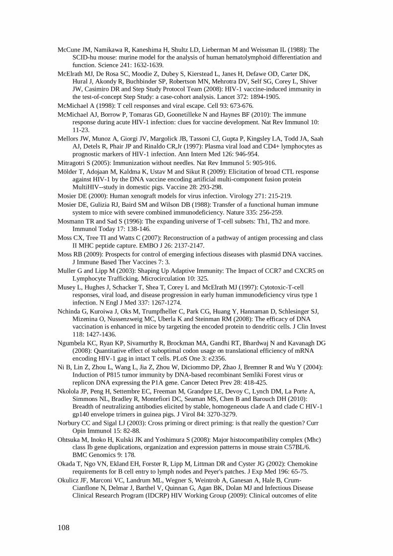
108
McCune JM, Namikawa R, Kaneshima H, Shultz LD, Lieberman M and Weissman IL (1988): The SCID-hu mouse: murine model for the analysis of human hematolymphoid differentiation and function. Science 241: 1632-1639.
McElrath MJ, De Rosa SC, Moodie Z, Dubey S, Kierstead L, Janes H, Defawe OD, Carter DK, Hural J, Akondy R, Buchbinder SP, Robertson MN, Mehrotra DV, Self SG, Corey L, Shiver JW, Casimiro DR and Step Study Protocol Team (2008): HIV-1 vaccine-induced immunity in the test-of-concept Step Study: a case-cohort analysis. Lancet 372: 1894-1905.
McMichael A (1998): T cell responses and viral escape. Cell 93: 673-676. McMichael AJ, Borrow P, Tomaras GD, Goonetilleke N and Haynes BF (2010): The immune
response during acute HIV-1 infection: clues for vaccine development. Nat Rev Immunol 10: 11-23.
Mellors JW, Munoz A, Giorgi JV, Margolick JB, Tassoni CJ, Gupta P, Kingsley LA, Todd JA, Saah AJ, Detels R, Phair JP and Rinaldo CR,Jr (1997): Plasma viral load and CD4+ lymphocytes as prognostic markers of HIV-1 infection. Ann Intern Med 126: 946-954.
Mitragotri S (2005): Immunization without needles. Nat Rev Immunol 5: 905-916. Mölder T, Adojaan M, Kaldma K, Ustav M and Sikut R (2009): Elicitation of broad CTL response
against HIV-1 by the DNA vaccine encoding artificial multi-component fusion protein MultiHIV--study in domestic pigs. Vaccine 28: 293-298.
Mosier DE (2000): Human xenograft models for virus infection. Virology 271: 215-219. Mosier DE, Gulizia RJ, Baird SM and Wilson DB (1988): Transfer of a functional human immune
system to mice with severe combined immunodeficiency. Nature 335: 256-259. Mosmann TR and Sad S (1996): The expanding universe of T-cell subsets: Th1, Th2 and more.
Immunol Today 17: 138-146. Moss CX, Tree TI and Watts C (2007): Reconstruction of a pathway of antigen processing and class
II MHC peptide capture. EMBO J 26: 2137-2147. Moss RB (2009): Prospects for control of emerging infectious diseases with plasmid DNA vaccines.
J Immune Based Ther Vaccines 7: 3. Muller G and Lipp M (2003): Shaping Up Adaptive Immunity: The Impact of CCR7 and CXCR5 on
Lymphocyte Trafficking. Microcirculation 10: 325. Musey L, Hughes J, Schacker T, Shea T, Corey L and McElrath MJ (1997): Cytotoxic-T-cell
responses, viral load, and disease progression in early human immunodeficiency virus type 1 infection. N Engl J Med 337: 1267-1274.
Nchinda G, Kuroiwa J, Oks M, Trumpfheller C, Park CG, Huang Y, Hannaman D, Schlesinger SJ, Mizenina O, Nussenzweig MC, Uberla K and Steinman RM (2008): The efficacy of DNA vaccination is enhanced in mice by targeting the encoded protein to dendritic cells. J Clin Invest 118: 1427-1436.
Ngumbela KC, Ryan KP, Sivamurthy R, Brockman MA, Gandhi RT, Bhardwaj N and Kavanagh DG (2008): Quantitative effect of suboptimal codon usage on translational efficiency of mRNA encoding HIV-1 gag in intact T cells. PLoS One 3: e2356.
Ni B, Lin Z, Zhou L, Wang L, Jia Z, Zhou W, Diciommo DP, Zhao J, Bremner R and Wu Y (2004): Induction of P815 tumor immunity by DNA-based recombinant Semliki Forest virus or replicon DNA expressing the P1A gene. Cancer Detect Prev 28: 418-425.
Nkolola JP, Peng H, Settembre EC, Freeman M, Grandpre LE, Devoy C, Lynch DM, La Porte A, Simmons NL, Bradley R, Montefiori DC, Seaman MS, Chen B and Barouch DH (2010): Breadth of neutralizing antibodies elicited by stable, homogeneous clade A and clade C HIV-1 gp140 envelope trimers in guinea pigs. J Virol 84: 3270-3279.
Norbury CC and Sigal LJ (2003): Cross priming or direct priming: is that really the question? Curr Opin Immunol 15: 82-88.
Ohtsuka M, Inoko H, Kulski JK and Yoshimura S (2008): Major histocompatibility complex (Mhc) class Ib gene duplications, organization and expression patterns in mouse strain C57BL/6. BMC Genomics 9: 178.
Okada T, Ngo VN, Ekland EH, Forster R, Lipp M, Littman DR and Cyster JG (2002): Chemokine requirements for B cell entry to lymph nodes and Peyer's patches. J Exp Med 196: 65-75.
Okulicz JF, Marconi VC, Landrum ML, Wegner S, Weintrob A, Ganesan A, Hale B, Crum-Cianflone N, Delmar J, Barthel V, Quinnan G, Agan BK, Dolan MJ and Infectious Disease Clinical Research Program (IDCRP) HIV Working Group (2009): Clinical outcomes of elite
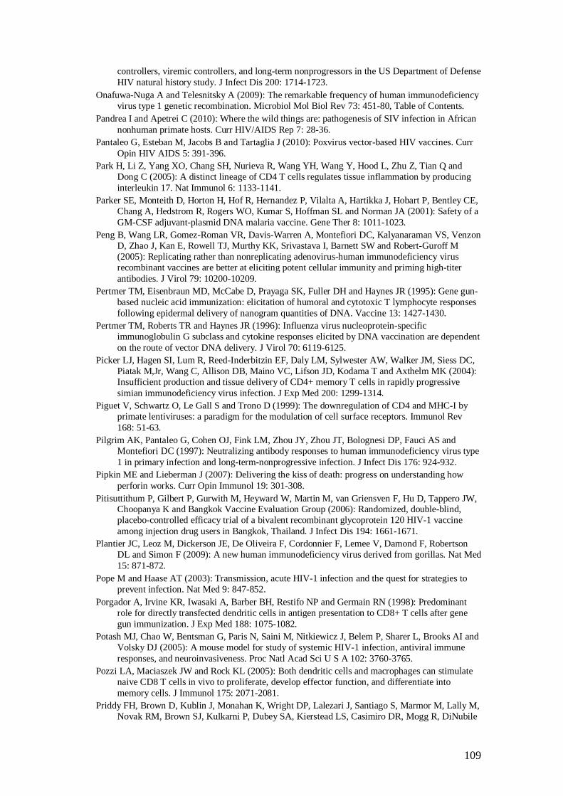
109
controllers, viremic controllers, and long-term nonprogressors in the US Department of Defense HIV natural history study. J Infect Dis 200: 1714-1723.
Onafuwa-Nuga A and Telesnitsky A (2009): The remarkable frequency of human immunodeficiency virus type 1 genetic recombination. Microbiol Mol Biol Rev 73: 451-80, Table of Contents.
Pandrea I and Apetrei C (2010): Where the wild things are: pathogenesis of SIV infection in African nonhuman primate hosts. Curr HIV/AIDS Rep 7: 28-36.
Pantaleo G, Esteban M, Jacobs B and Tartaglia J (2010): Poxvirus vector-based HIV vaccines. Curr Opin HIV AIDS 5: 391-396.
Park H, Li Z, Yang XO, Chang SH, Nurieva R, Wang YH, Wang Y, Hood L, Zhu Z, Tian Q and Dong C (2005): A distinct lineage of CD4 T cells regulates tissue inflammation by producing interleukin 17. Nat Immunol 6: 1133-1141.
Parker SE, Monteith D, Horton H, Hof R, Hernandez P, Vilalta A, Hartikka J, Hobart P, Bentley CE, Chang A, Hedstrom R, Rogers WO, Kumar S, Hoffman SL and Norman JA (2001): Safety of a GM-CSF adjuvant-plasmid DNA malaria vaccine. Gene Ther 8: 1011-1023.
Peng B, Wang LR, Gomez-Roman VR, Davis-Warren A, Montefiori DC, Kalyanaraman VS, Venzon D, Zhao J, Kan E, Rowell TJ, Murthy KK, Srivastava I, Barnett SW and Robert-Guroff M (2005): Replicating rather than nonreplicating adenovirus-human immunodeficiency virus recombinant vaccines are better at eliciting potent cellular immunity and priming high-titer antibodies. J Virol 79: 10200-10209.
Pertmer TM, Eisenbraun MD, McCabe D, Prayaga SK, Fuller DH and Haynes JR (1995): Gene gun-based nucleic acid immunization: elicitation of humoral and cytotoxic T lymphocyte responses following epidermal delivery of nanogram quantities of DNA. Vaccine 13: 1427-1430.
Pertmer TM, Roberts TR and Haynes JR (1996): Influenza virus nucleoprotein-specific immunoglobulin G subclass and cytokine responses elicited by DNA vaccination are dependent on the route of vector DNA delivery. J Virol 70: 6119-6125.
Picker LJ, Hagen SI, Lum R, Reed-Inderbitzin EF, Daly LM, Sylwester AW, Walker JM, Siess DC, Piatak M,Jr, Wang C, Allison DB, Maino VC, Lifson JD, Kodama T and Axthelm MK (2004): Insufficient production and tissue delivery of CD4+ memory T cells in rapidly progressive simian immunodeficiency virus infection. J Exp Med 200: 1299-1314.
Piguet V, Schwartz O, Le Gall S and Trono D (1999): The downregulation of CD4 and MHC-I by primate lentiviruses: a paradigm for the modulation of cell surface receptors. Immunol Rev 168: 51-63.
Pilgrim AK, Pantaleo G, Cohen OJ, Fink LM, Zhou JY, Zhou JT, Bolognesi DP, Fauci AS and Montefiori DC (1997): Neutralizing antibody responses to human immunodeficiency virus type 1 in primary infection and long-term-nonprogressive infection. J Infect Dis 176: 924-932.
Pipkin ME and Lieberman J (2007): Delivering the kiss of death: progress on understanding how perforin works. Curr Opin Immunol 19: 301-308.
Pitisuttithum P, Gilbert P, Gurwith M, Heyward W, Martin M, van Griensven F, Hu D, Tappero JW, Choopanya K and Bangkok Vaccine Evaluation Group (2006): Randomized, double-blind, placebo-controlled efficacy trial of a bivalent recombinant glycoprotein 120 HIV-1 vaccine among injection drug users in Bangkok, Thailand. J Infect Dis 194: 1661-1671.
Plantier JC, Leoz M, Dickerson JE, De Oliveira F, Cordonnier F, Lemee V, Damond F, Robertson DL and Simon F (2009): A new human immunodeficiency virus derived from gorillas. Nat Med 15: 871-872.
Pope M and Haase AT (2003): Transmission, acute HIV-1 infection and the quest for strategies to prevent infection. Nat Med 9: 847-852.
Porgador A, Irvine KR, Iwasaki A, Barber BH, Restifo NP and Germain RN (1998): Predominant role for directly transfected dendritic cells in antigen presentation to CD8+ T cells after gene gun immunization. J Exp Med 188: 1075-1082.
Potash MJ, Chao W, Bentsman G, Paris N, Saini M, Nitkiewicz J, Belem P, Sharer L, Brooks AI and Volsky DJ (2005): A mouse model for study of systemic HIV-1 infection, antiviral immune responses, and neuroinvasiveness. Proc Natl Acad Sci U S A 102: 3760-3765.
Pozzi LA, Maciaszek JW and Rock KL (2005): Both dendritic cells and macrophages can stimulate naive CD8 T cells in vivo to proliferate, develop effector function, and differentiate into memory cells. J Immunol 175: 2071-2081.
Priddy FH, Brown D, Kublin J, Monahan K, Wright DP, Lalezari J, Santiago S, Marmor M, Lally M, Novak RM, Brown SJ, Kulkarni P, Dubey SA, Kierstead LS, Casimiro DR, Mogg R, DiNubile
110
MJ, Shiver JW, Leavitt RY, Robertson MN, Mehrotra DV, Quirk E and Merck V520-016 Study Group (2008): Safety and immunogenicity of a replication-incompetent adenovirus type 5 HIV-1 clade B gag/pol/nef vaccine in healthy adults. Clin Infect Dis 46: 1769-1781.
Qiu JT, Song R, Dettenhofer M, Tian C, August T, Felber BK, Pavlakis GN and Yu XF (1999): Evaluation of novel human immunodeficiency virus type 1 Gag DNA vaccines for protein expression in mammalian cells and induction of immune responses. J Virol 73: 9145-9152.
Rakhmilevich AL, Turner J, Ford MJ, McCabe D, Sun WH, Sondel PM, Grota K and Yang NS (1996): Gene gun-mediated skin transfection with interleukin 12 gene results in regression of established primary and metastatic murine tumors. Proc Natl Acad Sci U S A 93: 6291-6296.
Ramakrishna L, Anand KK, Mohankumar KM and Ranga U (2004): Codon optimization of the tat antigen of human immunodeficiency virus type 1 generates strong immune responses in mice following genetic immunization. J Virol 78: 9174-9189.
Ramduth D, Day CL, Thobakgale CF, Mkhwanazi NP, de Pierres C, Reddy S, van der Stok M, Mncube Z, Nair K, Moodley ES, Kaufmann DE, Streeck H, Coovadia HM, Kiepiela P, Goulder PJ and Walker BD (2009): Immunodominant HIV-1 Cd4+ T cell epitopes in chronic untreated clade C HIV-1 infection. PLoS One 4: e5013.
Ranki A, Lagerstedt A, Ovod V, Aavik E and Krohn KJ (1994): Expression kinetics and subcellular localization of HIV-1 regulatory proteins Nef, Tat and Rev in acutely and chronically infected lymphoid cell lines. Arch Virol 139: 365-378.
Ranki A, Mattinen S, Yarchoan R, Broder S, Ghrayeb J, Lahdevirta J and Krohn K (1989): T-cell response towards HIV in infected individuals with and without zidovudine therapy, and in HIV-exposed sexual partners. AIDS 3: 63-69.
Ranki A, Suni J, Blazevic V, Holmstrom P, Mattinen S, Krohn K and Valle SL (1997): T-cell recognition of HIV antigens in HIV-seroreverted persons. AIDS 11: 132-133.
Ranki A, Valle SL, Krohn M, Antonen J, Allain JP, Leuther M, Franchini G and Krohn K (1987): Long latency precedes overt seroconversion in sexually transmitted human-immunodeficiency-virus infection. Lancet 2: 589-593.
Rerks-Ngarm S, Pitisuttithum P, Nitayaphan S, Kaewkungwal J, Chiu J, Paris R, Premsri N, Namwat C, de Souza M, Adams E, Benenson M, Gurunathan S, Tartaglia J, McNeil JG, Francis DP, Stablein D, Birx DL, Chunsuttiwat S, Khamboonruang C, Thongcharoen P, Robb ML, Michael NL, Kunasol P, Kim JH and MOPH-TAVEG Investigators (2009): Vaccination with ALVAC and AIDSVAX to prevent HIV-1 infection in Thailand. N Engl J Med 361: 2209-2220.
Rock KL, Farfan-Arribas DJ and Shen L (2010): Proteases in MHC class I presentation and cross-presentation. J Immunol 184: 9-15.
Rolland M, Jensen MA, Nickle DC, Yan J, Learn GH, Heath L, Weiner D and Mullins JI (2007a): Reconstruction and function of ancestral center-of-tree human immunodeficiency virus type 1 proteins. J Virol 81: 8507-8514.
Rolland M, Nickle DC and Mullins JI (2007b): HIV-1 group M conserved elements vaccine. PLoS Pathog 3: e157.
Rollman E, Mathy N, Brave A, Boberg A, Kjerrstrom A, van Wely C, Engstrom G, Johansson S, Aperia K, Eriksson LE, Benthin R, Ertl P, Heeney J, Hinkula J, Voss G and Wahren B (2007): Evaluation of immunogenicity and efficacy of combined DNA and adjuvanted protein vaccination in a human immunodeficiency virus type 1/murine leukemia virus pseudotype challenge model. Vaccine 25: 2145-2154.
Romani B, Engelbrecht S and Glashoff RH (2010): Functions of Tat: the versatile protein of human immunodeficiency virus type 1. J Gen Virol 91: 1-12.
Rose NF, Marx PA, Luckay A, Nixon DF, Moretto WJ, Donahoe SM, Montefiori D, Roberts A, Buonocore L and Rose JK (2001): An effective AIDS vaccine based on live attenuated vesicular stomatitis virus recombinants. Cell 106: 539-549.
Roush SW, Murphy TV and Vaccine-Preventable Disease Table Working Group (2007): Historical comparisons of morbidity and mortality for vaccine-preventable diseases in the United States. JAMA 298: 2155-2163.
Rowland-Jones SL, Dong T, Fowke KR, Kimani J, Krausa P, Newell H, Blanchard T, Ariyoshi K, Oyugi J, Ngugi E, Bwayo J, MacDonald KS, McMichael AJ and Plummer FA (1998): Cytotoxic T cell responses to multiple conserved HIV epitopes in HIV-resistant prostitutes in Nairobi. J Clin Invest 102: 1758-1765.
111
Roy MJ, Wu MS, Barr LJ, Fuller JT, Tussey LG, Speller S, Culp J, Burkholder JK, Swain WF, Dixon RM, Widera G, Vessey R, King A, Ogg G, Gallimore A, Haynes JR and Heydenburg Fuller D (2000): Induction of antigen-specific CD8+ T cells, T helper cells, and protective levels of antibody in humans by particle-mediated administration of a hepatitis B virus DNA vaccine. Vaccine 19: 764-778.
Saifuddin M, Parker CJ, Peeples ME, Gorny MK, Zolla-Pazner S, Ghassemi M, Rooney IA, Atkinson JP and Spear GT (1995): Role of virion-associated glycosylphosphatidylinositol-linked proteins CD55 and CD59 in complement resistance of cell line-derived and primary isolates of HIV-1. J Exp Med 182: 501-509.
Saini M, Hadas E, Volsky DJ and Potash MJ (2007): Vaccine-induced protection from infection of mice by chimeric human immunodeficiency virus type 1, EcoHIV/NL4-3. Vaccine 25: 8660-8663.
Saksela K, Cheng G and Baltimore D (1995): Proline-rich (PxxP) motifs in HIV-1 Nef bind to SH3 domains of a subset of Src kinases and are required for the enhanced growth of Nef+ viruses but not for down-regulation of CD4. EMBO J 14: 484-491.
Sallusto F, Lenig D, Forster R, Lipp M and Lanzavecchia A (1999): Two subsets of memory T lymphocytes with distinct homing potentials and effector functions. Nature 401: 708-712.
Sather DN, Armann J, Ching LK, Mavrantoni A, Sellhorn G, Caldwell Z, Yu X, Wood B, Self S, Kalams S and Stamatatos L (2009): Factors associated with the development of cross-reactive neutralizing antibodies during human immunodeficiency virus type 1 infection. J Virol 83: 757-769.
Sauer B (1998): Inducible gene targeting in mice using the Cre/lox system. Methods 14: 381-392. Schalk JA, Mooi FR, Berbers GA, van Aerts LA, Ovelgonne H and Kimman TG (2006): Preclinical
and clinical safety studies on DNA vaccines. Hum Vaccin 2: 45-53. Schmitz JE, Johnson RP, McClure HM, Manson KH, Wyand MS, Kuroda MJ, Lifton MA,
Khunkhun RS, McEvers KJ, Gillis J, Piatak M, Lifson JD, Grosschupff G, Racz P, Tenner-Racz K, Rieber EP, Kuus-Reichel K, Gelman RS, Letvin NL, Montefiori DC, Ruprecht RM, Desrosiers RC and Reimann KA (2005): Effect of CD8+ lymphocyte depletion on virus containment after simian immunodeficiency virus SIVmac251 challenge of live attenuated SIVmac239delta3-vaccinated rhesus macaques. J Virol 79: 8131-8141.
Schroder AR, Shinn P, Chen H, Berry C, Ecker JR and Bushman F (2002): HIV-1 integration in the human genome favors active genes and local hotspots. Cell 110: 521-529.
Seaman MS, Xu L, Beaudry K, Martin KL, Beddall MH, Miura A, Sambor A, Chakrabarti BK, Huang Y, Bailer R, Koup RA, Mascola JR, Nabel GJ and Letvin NL (2005): Multiclade human immunodeficiency virus type 1 envelope immunogens elicit broad cellular and humoral immunity in rhesus monkeys. J Virol 79: 2956-2963.
Shastri N, Schwab S and Serwold T (2002): Producing nature's gene-chips: the generation of peptides for display by MHC class I molecules. Annu Rev Immunol 20: 463-493.
Shibata R, Kawamura M, Sakai H, Hayami M, Ishimoto A and Adachi A (1991): Generation of a chimeric human and simian immunodeficiency virus infectious to monkey peripheral blood mononuclear cells. J Virol 65: 3514-3520.
Shih IM, Torrance C, Sokoll LJ, Chan DW, Kinzler KW and Vogelstein B (2000): Assessing tumors in living animals through measurement of urinary beta-human chorionic gonadotropin. Nat Med 6: 711-714.
Shu Y, Winfrey S, Yang ZY, Xu L, Rao SS, Srivastava I, Barnett SW, Nabel GJ and Mascola JR (2007): Efficient protein boosting after plasmid DNA or recombinant adenovirus immunization with HIV-1 vaccine constructs. Vaccine 25: 1398-1408.
Silla T, Mannik A and Ustav M (2010): Effective formation of the segregation-competent complex determines successful partitioning of the bovine papillomavirus genome during cell division. J Virol 84: 11175-11188.
Simmons A, Aluvihare V and McMichael A (2001): Nef triggers a transcriptional program in T cells imitating single-signal T cell activation and inducing HIV virulence mediators. Immunity 14: 763-777.
Singh RA, Rodgers JR and Barry MA (2002): The role of T cell antagonism and original antigenic sin in genetic immunization. J Immunol 169: 6779-6786.
Sojka DK, Huang YH and Fowell DJ (2008): Mechanisms of regulatory T-cell suppression - a diverse arsenal for a moving target. Immunology 124: 13-22.
112
Soumelis V, Scott I, Gheyas F, Bouhour D, Cozon G, Cotte L, Huang L, Levy JA and Liu YJ (2001): Depletion of circulating natural type 1 interferon-producing cells in HIV-infected AIDS patients. Blood 98: 906-912.
Spector DH, Wade E, Wright DA, Koval V, Clark C, Jaquish D and Spector SA (1990): Human immunodeficiency virus pseudotypes with expanded cellular and species tropism. J Virol 64: 2298-2308.
Spetz AL, Sorensen AS, Walther-Jallow L, Wahren B, Andersson J, Holmgren L and Hinkula J (2002): Induction of HIV-1-specific immunity after vaccination with apoptotic HIV-1/murine leukemia virus-infected cells. J Immunol 169: 5771-5779.
Stacey AR, Norris PJ, Qin L, Haygreen EA, Taylor E, Heitman J, Lebedeva M, DeCamp A, Li D, Grove D, Self SG and Borrow P (2009): Induction of a striking systemic cytokine cascade prior to peak viremia in acute human immunodeficiency virus type 1 infection, in contrast to more modest and delayed responses in acute hepatitis B and C virus infections. J Virol 83: 3719-3733.
Stamatatos L, Morris L, Burton DR and Mascola JR (2009): Neutralizing antibodies generated during natural HIV-1 infection: good news for an HIV-1 vaccine? Nat Med 15: 866-870.
Stoiber H, Soederholm A, Wilflingseder D, Gusenbauer S, Hildgartner A and Dierich MP (2008): Complement and antibodies: a dangerous liaison in HIV infection? Vaccine 26 Suppl 8: I79-85.
Streeck H, Jolin JS, Qi Y, Yassine-Diab B, Johnson RC, Kwon DS, Addo MM, Brumme C, Routy JP, Little S, Jessen HK, Kelleher AD, Hecht FM, Sekaly RP, Rosenberg ES, Walker BD, Carrington M and Altfeld M (2009): Human immunodeficiency virus type 1-specific CD8+ T-cell responses during primary infection are major determinants of the viral set point and loss of CD4+ T cells. J Virol 83: 7641-7648.
Sumida SM, McKay PF, Truitt DM, Kishko MG, Arthur JC, Seaman MS, Jackson SS, Gorgone DA, Lifton MA, Letvin NL and Barouch DH (2004): Recruitment and expansion of dendritic cells in vivo potentiate the immunogenicity of plasmid DNA vaccines. J Clin Invest 114: 1334-1342.
Swann SA, Williams M, Story CM, Bobbitt KR, Fleis R and Collins KL (2001): HIV-1 Nef blocks transport of MHC class I molecules to the cell surface via a PI 3-kinase-dependent pathway. Virology 282: 267-277.
Takahashi H, Cohen J, Hosmalin A, Cease KB, Houghten R, Cornette JL, DeLisi C, Moss B, Germain RN and Berzofsky JA (1988): An immunodominant epitope of the human immunodeficiency virus envelope glycoprotein gp160 recognized by class I major histocompatibility complex molecule-restricted murine cytotoxic T lymphocytes. Proc Natl Acad Sci U S A 85: 3105-3109.
Takebe Y, Seiki M, Fujisawa J, Hoy P, Yokota K, Arai K, Yoshida M and Arai N (1988): SR alpha promoter: an efficient and versatile mammalian cDNA expression system composed of the simian virus 40 early promoter and the R-U5 segment of human T-cell leukemia virus type 1 long terminal repeat. Mol Cell Biol 8: 466-472.
Tang DC, DeVit M and Johnston SA (1992): Genetic immunization is a simple method for eliciting an immune response. Nature 356: 152-154.
Tarosso LF, Sauer MM, Sanabani S, Giret MT, Tomiyama HI, Sidney J, Piaskowski SM, Diaz RS, Sabino EC, Sette A, Kalil-Filho J, Watkins DI and Kallas EG (2010): Unexpected diversity of cellular immune responses against Nef and Vif in HIV-1-infected patients who spontaneously control viral replication. PLoS One 5: e11436.
Taylor BS and Hammer SM (2008): The challenge of HIV-1 subtype diversity. N Engl J Med 359: 1965-1966.
Temin HM (1993): Retrovirus variation and reverse transcription: abnormal strand transfers result in retrovirus genetic variation. Proc Natl Acad Sci U S A 90: 6900-6903.
The National Institute for Health and Welfare (THL) (11/11/09): Taulukko 1. Eri rokotetyypit. Thomson SA, Sherritt MA, Medveczky J, Elliott SL, Moss DJ, Fernando GJ, Brown LE and Suhrbier
A (1998): Delivery of multiple CD8 cytotoxic T cell epitopes by DNA vaccination. J Immunol 160: 1717-1723.
Tobery TW and Siliciano RF (1997): Targeting of HIV-1 antigens for rapid intracellular degradation enhances cytotoxic T lymphocyte (CTL) recognition and the induction of de novo CTL responses in vivo after immunization. J Exp Med 185: 909-920.
113
Townsend AR, Rothbard J, Gotch FM, Bahadur G, Wraith D and McMichael AJ (2006): The epitopes of influenza nucleoprotein recognized by cytotoxic T lymphocytes can be defined with short synthetic peptides. 1986. J Immunol 176: 5141-5150.
Trimble C, Lin CT, Hung CF, Pai S, Juang J, He L, Gillison M, Pardoll D, Wu L and Wu TC (2003): Comparison of the CD8+ T cell responses and antitumor effects generated by DNA vaccine administered through gene gun, biojector, and syringe. Vaccine 21: 4036-4042.
Trkola A, Kuster H, Rusert P, von Wyl V, Leemann C, Weber R, Stiegler G, Katinger H, Joos B and Gunthard HF (2008): In vivo efficacy of human immunodeficiency virus neutralizing antibodies: estimates for protective titers. J Virol 82: 1591-1599.
Tuomela M, Malm M, Wallen M, Stanescu I, Krohn K and Peterson P (2005): Biodistribution and general safety of a naked DNA plasmid, GTU-MultiHIV, in a rat, using a quantitative PCR method. Vaccine 23: 890-896.
Turner BG and Summers MF (1999): Structural biology of HIV. J Mol Biol 285: 1-32. Tuuminen T, Kekalainen E, Makela S, Ala-Houhala I, Ennis FA, Hedman K, Mustonen J, Vaheri A
and Arstila TP (2007): Human CD8+ T cell memory generation in Puumala hantavirus infection occurs after the acute phase and is associated with boosting of EBV-specific CD8+ memory T cells. J Immunol 179: 1988-1995.
Tähtinen M. (2001) Characterisation of Humoral Immune Response to HIV- 1 Non-Structural Proteins Nef, Rev and Tat in HIV-1 Infected Individuals and in Immunised Animals. University of Tampere, Institute of Medical Technology, Tampere, Finland.
Tähtinen M, Strengell M, Collings A, Pitkanen J, Kjerrstrom A, Hakkarainen K, Peterson P, Kohleisen B, Wahren B, Ranki A, Ustav M and Krohn K (2001): DNA vaccination in mice using HIV-1 nef, rev and tat genes in self-replicating pBN-vector. Vaccine 19: 2039-2047.
U.S. Food and Drug Administration (FDA) (10/20/2010): Vaccines Licensed for Immunization and Distribution in the US with Supporting Documents (05/02/2011).
Ulmer JB, Donnelly JJ, Parker SE, Rhodes GH, Felgner PL, Dwarki VJ, Gromkowski SH, Deck RR, DeWitt CM and Friedman A (1993): Heterologous protection against influenza by injection of DNA encoding a viral protein. Science 259: 1745-1749.
UNAIDS (2010): Global report: UNAIDS report on the global AIDS epidemic 2010. Ustav E, Ustav M, Szymanski P and Stenlund A (1993): The bovine papillomavirus origin of
replication requires a binding site for the E2 transcriptional activator. Proc Natl Acad Sci U S A 90: 898-902.
Vaccari M, Mattapallil J, Song K, Tsai WP, Hryniewicz A, Venzon D, Zanetti M, Reimann KA, Roederer M and Franchini G (2008): Reduced protection from simian immunodeficiency virus SIVmac251 infection afforded by memory CD8+ T cells induced by vaccination during CD4+ T-cell deficiency. J Virol 82: 9629-9638.
Vardas E, Stanescu I, Valtavaara M, Kuntonen T, Gray C, Leionen M, Ustav M and Reijonen K. (2010): Indicators of therapeutic vaccine effect using GTU-MultiHIV B clade DNA in treatment-naïve subtype C HIV-1 infected subjects. XVIII International AIDS Conference. Poster presentation. AIDS 2010, Vienna, Austria: International AIDS Society.
Vesanen M, Stevens CE, Taylor PE, Rubinstein P and Saksela K (1996): Stability in controlling viral replication identifies long-term nonprogressors as a distinct subgroup among human immunodeficiency virus type 1-infected persons. J Virol 70: 9035-9040.
Vincent KA, York-Higgins D, Quiroga M and Brown PO (1990): Host sequences flanking the HIV provirus. Nucleic Acids Res 18: 6045-6047.
Virgin HW and Walker BD (2010): Immunology and the elusive AIDS vaccine. Nature 464: 224-231.
Vitiello A, Marchesini D, Furze J, Sherman LA and Chesnut RW (1991): Analysis of the HLA-restricted influenza-specific cytotoxic T lymphocyte response in transgenic mice carrying a chimeric human-mouse class I major histocompatibility complex. J Exp Med 173: 1007-1015.
Wang Z, Troilo PJ, Wang X, Griffiths TG, Pacchione SJ, Barnum AB, Harper LB, Pauley CJ, Niu Z, Denisova L, Follmer TT, Rizzuto G, Ciliberto G, Fattori E, Monica NL, Manam S and Ledwith BJ (2004): Detection of integration of plasmid DNA into host genomic DNA following intramuscular injection and electroporation. Gene Ther 11: 711-721.
Wanjalla CN, Faul EJ, Gomme EA and Schnell MJ (2010): Dendritic cells infected by recombinant rabies virus vaccine vector expressing HIV-1 Gag are immunogenic even in the presence of vector-specific immunity. Vaccine 29: 130-140.
114
Willey S, Aasa-Chapman MM, O'Farrell S, Pellegrino P, Williams I, Weiss RA and Neil SJ (2011): Extensive complement-dependent enhancement of HIV-1 by autologous non-neutralising antibodies at early stages of infection. Retrovirology 8: 16.
Williams MA and Bevan MJ (2007): Effector and memory CTL differentiation. Annu Rev Immunol 25: 171-192.
Wolff JA, Malone RW, Williams P, Chong W, Acsadi G, Jani A and Felgner PL (1990): Direct gene transfer into mouse muscle in vivo. Science 247: 1465-1468.
Wyatt R and Sodroski J (1998): The HIV-1 envelope glycoproteins: fusogens, antigens, and immunogens. Science 280: 1884-1888.
Xu XN, Screaton GR, Gotch FM, Dong T, Tan R, Almond N, Walker B, Stebbings R, Kent K, Nagata S, Stott JE and McMichael AJ (1997): Evasion of cytotoxic T lymphocyte (CTL) responses by nef-dependent induction of Fas ligand (CD95L) expression on simian immunodeficiency virus-infected cells. J Exp Med 186: 7-16.
Yewdell JW (2006): Confronting complexity: real-world immunodominance in antiviral CD8+ T cell responses. Immunity 25: 533-543.
Yewdell JW and Bennink JR (1999): Immunodominance in major histocompatibility complex class I-restricted T lymphocyte responses. Annu Rev Immunol 17: 51-88.
York IA and Rock KL (1996): Antigen processing and presentation by the class I major histocompatibility complex. Annu Rev Immunol 14: 369-396.
Yue FY, Merchant A, Kovacs CM, Loutfy M, Persad D and Ostrowski MA (2008): Virus-specific interleukin-17-producing CD4+ T cells are detectable in early human immunodeficiency virus type 1 infection. J Virol 82: 6767-6771.
Zhang H, Yang B, Pomerantz RJ, Zhang C, Arunachalam SC and Gao L (2003): The cytidine deaminase CEM15 induces hypermutation in newly synthesized HIV-1 DNA. Nature 424: 94-98.
Zhou X, Bailey-Bucktrout S, Jeker LT and Bluestone JA (2009): Plasticity of CD4(+) FoxP3(+) T cells. Curr Opin Immunol 21: 281-285.
AIDS RESEARCH AND HUMAN RETROVIRUSESVolume 22, Number 7, 2006, pp. 667–677© Mary Ann Liebert, Inc.
Induction of Human Immunodeficiency Virus Type-1-Specific Immunity with a Novel Gene
Transport Unit (GTU)-MultiHIV DNA Vaccine
VESNA BLAZEVIC,1 ANDRES MÄNNIK,2 MARIA MALM,1 REIN SIKUT,2 MINNA VALTAVAARA,1
URVE TOOTS,2 MART USTAV,2 and KAI KROHN1
ABSTRACT
A multiHIV fusion gene expressing an antigenic fusion protein composed of regulatory HIV-1 proteins Rev,Nef, and Tat, as well as Gag p17/p24 and a stretch of 11 cytotoxic T lymphocyte (CTL) epitope clusters fromPol and Env, was cloned into a novel DNA vector named the Gene Transport Unit (GTU). A mouse H-2d-re-stricted HIV-1 gp120 epitope (RGPGRAFVTI) was cloned into the fusion gene as well. In addition to the HIV-1 genes the GTU codes for a nuclear anchoring protein (bovine papilloma virus E2), ensuring the long main-tenance of the vector and a high expression level of the selected immunogens. BALB/c mice were immunizedwith the GTU-MultiHIV DNA construct by different routes and regimens of immunization to assess the im-munogenicity of the DNA vaccine in vivo. Mice developed strong CD8� CTL responses to HIV-1 Env and Gagmeasured by an ELISPOT-IFN-� assay and chromium release assay. In addition, T cell responses to regula-tory proteins Rev, Nef, and Tat were induced. Antibody responses were detected to each of the HIV antigensencoded by the DNA construct. Minimal doses of the GTU-MultiHIV DNA delivered by gene gun were po-tent in inducing significant HIV-specific CTL responses. The equivalent doses of the conventional plasmid ex-pressing MultiHIV DNA delivered by gene gun failed to do so. An ideal DNA vaccine should yield high ex-pression of the viral antigens for a prolonged period of time, and expression of the multiple viral antigens isprobably required for the induction of a broad and protective immune response. The GTU-MultiHIV DNAvaccine described is a good vaccine candidate that meets the above criteria.
667
INTRODUCTION
DESPITE THE EFFORTS, to date there is neither a curative anti-viral drug1,2 nor a safe and effective vaccine against HIV-
1 infection. The correlates of immune protection in HIV infec-tion remain to be elucidated, however, the ability to inducestrong HIV-1-specific CD8� T cell responses is considered tobe the main feature of an effective HIV vaccine.3 It has beendemonstrated that cytotoxic T lymphocyte (CTL) responses inlong-term nonprogressors correlate with reduced viral loads.4,5
In addition, CTL responses in HIV-exposed uninfected indi-viduals suggest immunological control of limited infection.6 In-deed, in vivo studies show that CD8� CTL are crucial for con-trolling SIV/HIV replication and delaying disease progression.7
There are indications that a CTL response directed against HIV
regulatory proteins could prevent the release of infectious viri-ons by destroying infected cells and clearing the infection at anearly stage.8–10
The high degree of vaccine efficacy induced by live attenu-ated vaccines11 indicated that multiple viral proteins were re-quired as antigens to stimulate a protective immune response.However, the use of live attenuated retroviral vaccine has notconsidered for human use due to safety concerns. DNA vaccinemimics the live vaccine but without the pathogenic potential ofthe virus. Furthermore, DNA vaccination may provide a nat-ural mode of antigen presentation, especially to the cellular im-mune system, as it allows in vivo production and processing ofthe vaccine-encoded gene products within the cell. DNA vac-cination has been shown to induce both cellular and humoralimmune responses in mice, nonhuman primates, and hu-
1FIT Biotech Oyj Plc., Tampere, Finland.2FIT Biotech Oyj Plc., Tartu, Estonia.
6293_09_p667-677 6/23/06 10:02 AM Page 667
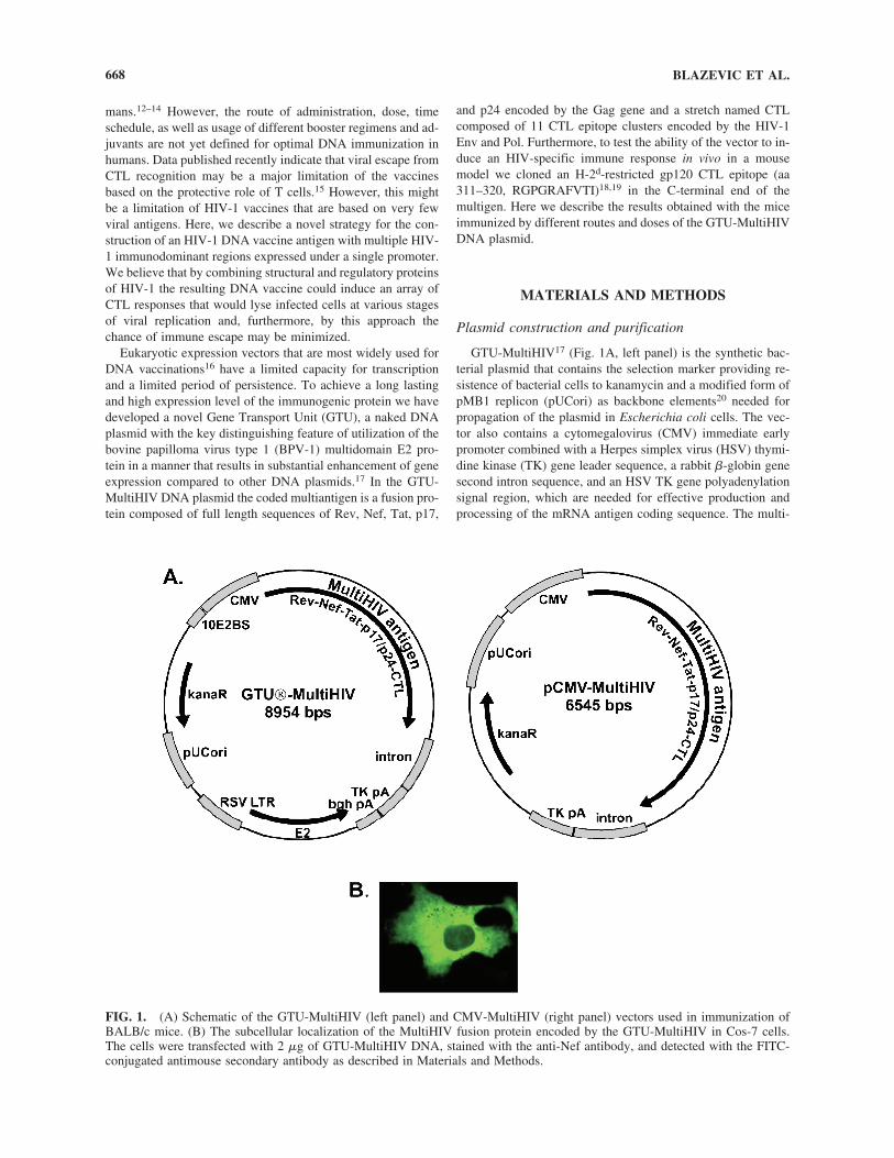
mans.12–14 However, the route of administration, dose, timeschedule, as well as usage of different booster regimens and ad-juvants are not yet defined for optimal DNA immunization inhumans. Data published recently indicate that viral escape fromCTL recognition may be a major limitation of the vaccinesbased on the protective role of T cells.15 However, this mightbe a limitation of HIV-1 vaccines that are based on very fewviral antigens. Here, we describe a novel strategy for the con-struction of an HIV-1 DNA vaccine antigen with multiple HIV-1 immunodominant regions expressed under a single promoter.We believe that by combining structural and regulatory proteinsof HIV-1 the resulting DNA vaccine could induce an array ofCTL responses that would lyse infected cells at various stagesof viral replication and, furthermore, by this approach thechance of immune escape may be minimized.
Eukaryotic expression vectors that are most widely used forDNA vaccinations16 have a limited capacity for transcriptionand a limited period of persistence. To achieve a long lastingand high expression level of the immunogenic protein we havedeveloped a novel Gene Transport Unit (GTU), a naked DNAplasmid with the key distinguishing feature of utilization of thebovine papilloma virus type 1 (BPV-1) multidomain E2 pro-tein in a manner that results in substantial enhancement of geneexpression compared to other DNA plasmids.17 In the GTU-MultiHIV DNA plasmid the coded multiantigen is a fusion pro-tein composed of full length sequences of Rev, Nef, Tat, p17,
and p24 encoded by the Gag gene and a stretch named CTLcomposed of 11 CTL epitope clusters encoded by the HIV-1Env and Pol. Furthermore, to test the ability of the vector to in-duce an HIV-specific immune response in vivo in a mousemodel we cloned an H-2d-restricted gp120 CTL epitope (aa311–320, RGPGRAFVTI)18,19 in the C-terminal end of themultigen. Here we describe the results obtained with the miceimmunized by different routes and doses of the GTU-MultiHIVDNA plasmid.
MATERIALS AND METHODS
Plasmid construction and purification
GTU-MultiHIV17 (Fig. 1A, left panel) is the synthetic bac-terial plasmid that contains the selection marker providing re-sistence of bacterial cells to kanamycin and a modified form ofpMB1 replicon (pUCori) as backbone elements20 needed forpropagation of the plasmid in Escherichia coli cells. The vec-tor also contains a cytomegalovirus (CMV) immediate earlypromoter combined with a Herpes simplex virus (HSV) thymi-dine kinase (TK) gene leader sequence, a rabbit �-globin genesecond intron sequence, and an HSV TK gene polyadenylationsignal region, which are needed for effective production andprocessing of the mRNA antigen coding sequence. The multi-
BLAZEVIC ET AL.668
FIG. 1. (A) Schematic of the GTU-MultiHIV (left panel) and CMV-MultiHIV (right panel) vectors used in immunization ofBALB/c mice. (B) The subcellular localization of the MultiHIV fusion protein encoded by the GTU-MultiHIV in Cos-7 cells.The cells were transfected with 2 �g of GTU-MultiHIV DNA, stained with the anti-Nef antibody, and detected with the FITC-conjugated antimouse secondary antibody as described in Materials and Methods.
6293_09_p667-677 6/23/06 10:02 AM Page 668
antigen coding sequence was derived from the HAN2 isolate ofHIV-1 and was inserted downstream of the CMV promoter. Itis composed of fused coding sequences of Rev, Nef, and Tat(RNT), p17, p24, and 11 17–45 aa long T cell epitopes (humanleukocyte antigen, HLA restricted) rich clusters from Pol andEnv polypeptides (CTL).21 The gene segments encoding p17,p24, and CTL are synthetic gene codons optimized for expres-sion in human cells. The fusion of regulatory proteins in the order of Rev–Nef–Tat, followed by the p17/24 sequence, inactivated the ability of the Tat protein to activate the LTR-dependent expression of the reporter gene (data not shown). Po-sitioning of the Nef coding sequence in the middle of the anti-gen eliminated the ability to localize the Nef protein throughthe myristylated N-terminus to the inner surface of the plasmamembrane. An H-2d-restricted HIV-1 gp120 epitope19 codingsequence was cloned into the 3� end of the MultiHIV antigento test the potency of the GTU-MultiHIV in inducing a CD8�
CTL response in a mouse model.In addition to the elements described above, the GTU-Mul-
tiHIV vector also contains 10 clustered high-affinity E2 bind-ing sites from the BPV-1 genome (10E2BS) positioned just up-stream of the CMV promoter. The vector also carries anexpression cassette for the BPV-1 E2 protein.22 The E2 ex-pression is driven from the Rous sarcoma virus 5� LTR (RSVLTR). The bovine growth hormone polyadenylation region (bghpA) was cloned at the end of the E2 transcription cassette fromthe pHook3 plasmid (Invitrogen).
A conventional eukaryotic vector16 named CMV-MultiHIV(Fig. 1A, right panel) was generated by removing the E2 ex-pression cartridge and its binding sites from the GTU vector(Fig. 1A, left panel) but retaining identical elements controllingthe expression of the HIV antigens. GTU-MultiHIV and CMV-MultiHIV were produced in E. coli, and purified by QiagenEndofree Giga columns (Hilden, Germany) according to themanufacturer’s instructions.
In vitro expression studies
Expression properties of the MultiHIV fusion protein wereanalyzed in Jurkat, Cos-7, and RD cell lines. The cells weretransfected with 0.5 �g or 3 �g of the DNA or with controlDNA GTU-LacZ by electroporation as described earlier.22 Twoand five days posttransfection the cells were lysed by treatingwith a sample buffer: 50 mM Tris–HCl, pH 6.8, 2% SDS, 0.1%bromophenol blue, 100 mM dithiothreitol, and 10% (v/v) glyc-erol. The lysates were run on a 10% sodium dodecyl sul-fate–polyacrylamide gel (SDS–PAGE) and subsequently trans-ferred onto a 0.45-�m nitrocellulose membrane. After overnightblocking, the membrane was incubated with monoclonal anti-p24 antibody 05-001 (FIT Biotech, Tampere, Finland) andthereafter with HRP-conjugated goat antimouse IgG (LabAs,Tartu, Estonia) followed by visualization using an ECL chemo-luminesence detection system (Amersham Pharmacia Biotech,Uppsala, Sweden). To analyze subcellular localization of theMultiHIV antigen the transfected cells were fixed in cold methanol and stained with anti-Nef (01-001, FIT Biotech) oranti-p24 antibodies and detected by fluorescein isothio-cyanate (FITC)-conjugated goat antimouse secondary antibody(LabAs).
For quantitative analysis of fusion protein expression the
transfected RD cells were lysed in a Tris–HCl buffer (pH 8)containing 1% Triton X-100 and an EDTA free protease in-hibitor cocktail (Roche, Penzberg, Germany). The cell lysateswere analyzed by the capture enzyme-linked immunosorbentassay (ELISA) method based on an anti-Nef antibody immobi-lized onto Nunc Maxi Sorp plates, and biotinylated anti-p24 an-tibody 05-001 as detection antibody. The bound anti-p24 anti-body was detected by streptavidin-horseradish peroxidase(HRP) conjugate and color developed by TMB substrate(LabAs).
DNA Immunizations
Six- to nine-week-old female BALB/c (H-2d) mice were im-munized by DNA administered on shaved abdominal skin us-ing plasmid DNA-coated gold particles and a Helios Gene Gun(Bio-Rad, Richmond, CA) using a pressure of 400 psi and a0.5-mg gold/cartridge. Mice were immunized under generalanesthesia three times (at day 0, day 7, and day 21) with a to-tal amount of 24 ng, 120 ng, 600 ng, or 3 �g DNA/mouse. One-third of the total DNA dose is given at each immunization. Inanother set of experiments mice were immunized three timesintramuscularly (im) in the quadriceps femoris of the hind legor intradermaly (id) on the base of the tale with the followingtotal DNA doses: 6, 30, 150, or 750 �g. Mice were sacrificedand serum and spleens collected 2 weeks after the last immu-nization. In order to monitor duration of the immune responsegroups of mice were sacrificed at weeks 6, 10, 14, and 22 inaddition. Each experimental group consisted of 5-10 animals.Negative control mice were immunized with DNA carrier only.
Recombinant proteins and peptides
Glutathione S-transferase (GST)-tagged HIV-1 Nef and GSTproteins were affinity purified from lysates from E. coli BL21(DE3) cells which were transformed with a pGex-Nef-GST orpGex-GST plasmids using Pharmacia (Piscataway, NJ)reagents. Coding sequences of Rev, Tat, p17/24, CTL and E2were cloned into pET24d vector and purified from E. coli lysateusing Ni-NTA Agarose (Qiagen). H-2d-restricted HIV-1 gp120(Env) 10-mer peptide (aa 311–320; RGPGRAFVTI)19 and 9-mer HIV-1 Gag peptide (aa 65–73; AMQMLKETI)23 were pur-chased from Sigma-Genosys (Cambridge, UK) as �95% pure.Overlapping HIV-1 Rev, Nef, Tat, Gag, and CTL peptides werepurchased from Sigma-Genosys and were at least 85% pure.The peptides were 15 aa in length, which overlapped by 11 aa.
Anti-double strand (ds) DNA antibodies
dsDNA antibodies were assayed in the serum of immunizedmice, positive control mice (MRL/MpJ-Faslpr, a generous giftfrom Dr. Gene Shearer, NIH), and negative control mice (DNAcarrier immunized mice). Each of the sera was diluted 1:10,1:50, 1:250, and 1:1250 and antibodies were assayed by ELISAon poly-L-lysine bounded � phage dsDNA as previously de-scribed.24
HIV-1- and E2-specific antibodies
HIV-1 Rev, Nef, Tat, p17/24, CTL, and E2-specific anti-bodies were assayed in 1:100 diluted sera by ELISA as previ-ously described.25 Briefly, the sera were added to protein-coated
IMMUNOGENICITY OF A MULTIHIV DNA VACCINE 669
6293_09_p667-677 6/23/06 10:02 AM Page 669
(100 ng/well) 96-well plates (Nunc Maxi Sorp). After extensivewashes the peroxidase-conjugated antimouse IgG was added(1:500 dilution), plates were washed, and the substrate (2,2�-azino-bis(3-ethylbenzthiazoline-6-sulfonic acid), ABTS, in phos-phate-citrate buffer] was added for 30 min incubation. The pho-tometric analysis was carried out using an ELISA plate reader(Labsystems, Helsinki, Finland). HIV-1 Rev-, Nef-, Tat-, Gag-,CTL-, and BPV-1 E2-specific monoclonal antibodies (FITBiotech) were used as positive controls. Samples were consid-ered positive when the optical density (OD; at 405 nm wave-length) value was higher than the cutoff value (mean OD of thenegative control group � 3 SD).
Cell preparations
The spleens were dispersed to a single cell suspension andthe splenocytes frozen to liquid nitrogen (LN). For use in theassays LN frozen cells were washed and suspended in culturemedia (CM): RPMI 1640 (BioWhittaker, Verviers, Belgium)supplemented with 10% fetal bovine serum (FBS), 1% peni-cillin-streptomycin, and 50 �M 2-mercaptoethanol (all fromBioWhittaker). In some experiments, splenocytes were depletedof CD8� T cells by using magnetic beads (MagCellect, R&DSystems, Minneapolis, MN) as recommended by the manufac-turer. The remaining cells contained �4% CD8� T cells, de-termined by flow cytometric analysis.
ELISPOT-IFN-� assay
HIV-1-specific CTL responses were measured by quantifi-cation of the interferon-� (IFN-�) production at the single celllevel in an ELISPOT-IFN-� assay kit (R&D Systems). In brief,a monoclonal antibody specific for IFN-� was precoated ontoa polyvinylidene difluoride-backed 96-well plate. Liquid nitro-gen frozen splenocytes unstimulated (CM only), or stimulatedwith concanavalin A (con A, 5 �g/ml, Pharmacia), 104 of syn-genic P815 cells26 pulsed with the gp120 or Gag peptides for2 h (10 �g/ml), and mitomycin C (Sigma) treated, or peptidesalone (1 �g/ml) were plated at 0.1 � 106/well and incubatedfor 24 h at 37°C and 5% CO2. To assay for each MultiHIV anti-gen-specific IFN-� production 15-mer peptide pools of Rev (29peptides), Nef (51 peptides), Tat (25 peptides), Gag (91 pep-tides), and CTL (67 peptides) were used at a final concentra-tion of 2 �g/ml of each peptide. IFN-� was detected with a biotinylated antibody followed by incubation with strepta-vidin-alkaline phosphatase conjugate. The reaction was devel-oped using the BCIP/NBT substrate. Spot-forming cells (SFC)were counted with an automated ImmunoSpot Series II ana-lyzer (CTL Europe, Leinfelden-Echterdingen, Germany). Theresults are expressed as mean SFC/106 splenocytes of duplicatewells. The coefficient of variation was �25% for every dupli-cate. The results are considered positive if �50 SFC/106 sple-nocytes are above the control (unstimulated cells; CM only) andtwice above the control. The control SFC were �30 SFC/106
cells.
Chromium51 release assay
HIV-1 Env-specific effector cells were generated by in vitrostimulating liquid nitrogen frozen splenocytes with the H-2d-restricted gp120 peptide (2 �g/ml) and recombinant interleukin
(IL)-2 (25 U/ml, Boehringer Mannheim, Indianapolis, IN) for5 days. Peptide pulsed (1 �g/ml; 2 h at 37°C), MHC class I-matched, MHC class II-negative P815 syngeneic cells26 wereused as targets. The targets were incubated for 2.5 h at 37°C inmedium containing 150 �Ci Na2
51CrO4 (New England Nuclear,Boston, MA) and after washing, target cells were mixed withthe effector cells at different ratios in a 5 h chromium releaseassay. Control target cells were P815 cells incubated withoutthe peptide. Results are expressed as mean percent lysis of qua-druplicate determinations and are calculated by the followingformula: (experimental release � spontaneous media re-lease)/(maximum detergent release � spontaneous media re-lease) � 100. Spontaneous release of 51Cr from the targets didnot exceed 25% of the maximum release.
Statistics
Differences in mean values were tested by paired and un-paired two-tailed Student’s t-test (two-tailed for equal varianceassumed); p values � 0.05 were considered significant.
RESULTS
In vitro MultiHIV protein expression
In order to study the expression of the MultiHIV fusion pro-tein encoded by the GTU-MultiHIV vector, the plasmid wastransfected into Cos-7, RD, and Jurkat cell lines. Immunofluo-rescent staining of the transfected Cos-7 cells by anti-Nef an-tibody revealed cytoplasmic localization of the fusion antigen(Fig. 1B). In order to compare the expression capacities of theGTU- and CMV-MultiHIV vectors, both vectors were trans-fected in equimolar quantities into the RD cell line. Two andfive days posttransfection the cells were lysed and the expressedMultiHIV protein was analyzed by Western blot (Fig. 2A) andcapture ELISA (Fig. 2B) as described in Materials and Meth-ods. The MultiHIV antigen expressed in these cells has a mo-lecular weight around 120 kDa. As seen in Fig. 2B, only theGTU-MultiHIV vector produced the fusion protein in anamount that was detectable by the capture ELISA. The CMV-MultiHIV vector produced the corresponding protein in con-siderably lower amounts, which was possible to detect only withthe Western blot developed by the ECL substrate (Fig. 2A).
Anti-HIV and E2 antibodies
HIV-1- and E2-specific antibodies in GTU-MultiHIV im-munized mice were analyzed by ELISA as described in Mate-rials and Methods. Figure 3 shows the antibody results of themice immunized by gene gun (Fig. 3A) and id (Fig. 3B) withthe escalating doses of the GTU-MultiHIV DNA plasmid. Inthese mice a dose-dependent response was seen against Nef andp17/p24 proteins. The high doses of the DNA induced Nef-spe-cific responses in 90% of the mice and p17/p24-specific re-sponses in 70% of the mice. Rev, Tat, and CTL proteins in-duced antibody responses infrequently and at low magnitude(data not shown). None of the immunized mice had E2-specificantibodies (Fig. 3A and 3B). In mice immunized im no HIV-specific antibodies were detected regardless of the DNA doseadministered.
BLAZEVIC ET AL.670
6293_09_p667-677 6/23/06 10:02 AM Page 670
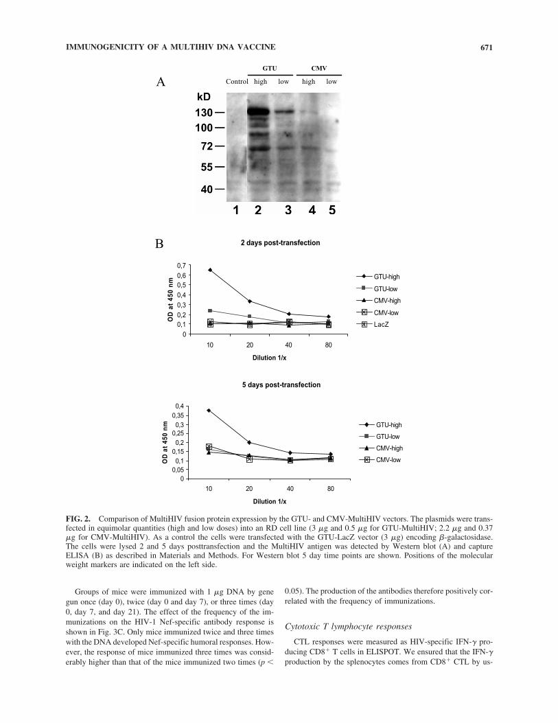
Groups of mice were immunized with 1 �g DNA by genegun once (day 0), twice (day 0 and day 7), or three times (day0, day 7, and day 21). The effect of the frequency of the im-munizations on the HIV-1 Nef-specific antibody response isshown in Fig. 3C. Only mice immunized twice and three timeswith the DNA developed Nef-specific humoral responses. How-ever, the response of mice immunized three times was consid-erably higher than that of the mice immunized two times (p �
0.05). The production of the antibodies therefore positively cor-related with the frequency of immunizations.
Cytotoxic T lymphocyte responses
CTL responses were measured as HIV-specific IFN-� pro-ducing CD8� T cells in ELISPOT. We ensured that the IFN-�production by the splenocytes comes from CD8� CTL by us-
IMMUNOGENICITY OF A MULTIHIV DNA VACCINE 671
FIG. 2. Comparison of MultiHIV fusion protein expression by the GTU- and CMV-MultiHIV vectors. The plasmids were trans-fected in equimolar quantities (high and low doses) into an RD cell line (3 �g and 0.5 �g for GTU-MultiHIV; 2.2 �g and 0.37�g for CMV-MultiHIV). As a control the cells were transfected with the GTU-LacZ vector (3 �g) encoding �-galactosidase.The cells were lysed 2 and 5 days posttransfection and the MultiHIV antigen was detected by Western blot (A) and captureELISA (B) as described in Materials and Methods. For Western blot 5 day time points are shown. Positions of the molecularweight markers are indicated on the left side.
6293_09_p667-677 6/23/06 10:02 AM Page 671
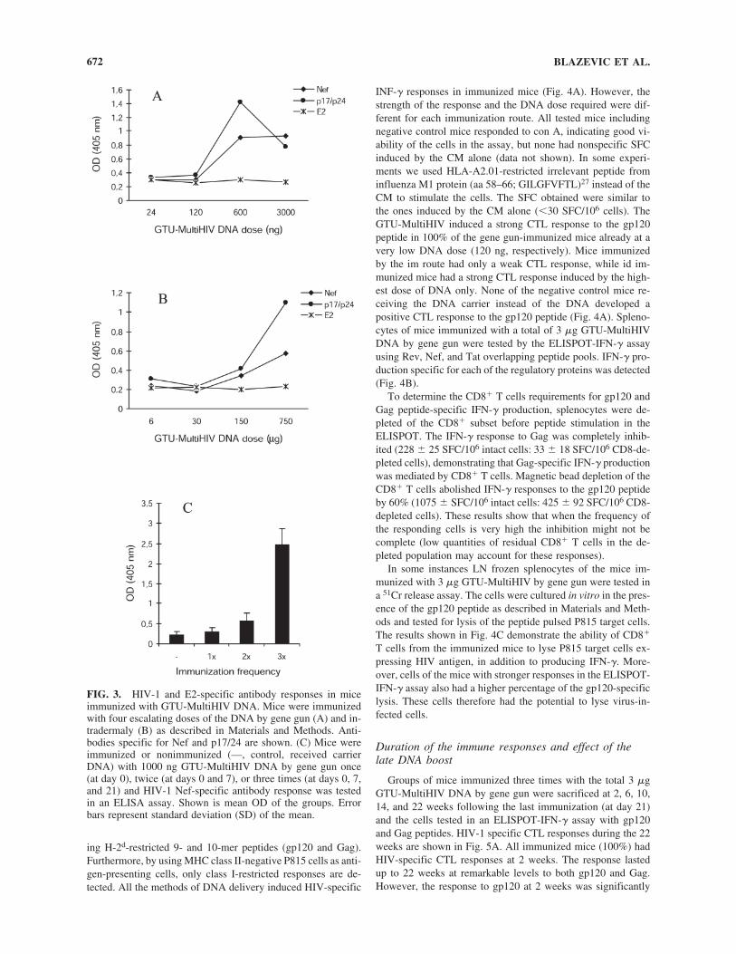
ing H-2d-restricted 9- and 10-mer peptides (gp120 and Gag).Furthermore, by using MHC class II-negative P815 cells as anti-gen-presenting cells, only class I-restricted responses are de-tected. All the methods of DNA delivery induced HIV-specific
INF-� responses in immunized mice (Fig. 4A). However, thestrength of the response and the DNA dose required were dif-ferent for each immunization route. All tested mice includingnegative control mice responded to con A, indicating good vi-ability of the cells in the assay, but none had nonspecific SFCinduced by the CM alone (data not shown). In some experi-ments we used HLA-A2.01-restricted irrelevant peptide frominfluenza M1 protein (aa 58–66; GILGFVFTL)27 instead of theCM to stimulate the cells. The SFC obtained were similar tothe ones induced by the CM alone (�30 SFC/106 cells). TheGTU-MultiHIV induced a strong CTL response to the gp120peptide in 100% of the gene gun-immunized mice already at avery low DNA dose (120 ng, respectively). Mice immunizedby the im route had only a weak CTL response, while id im-munized mice had a strong CTL response induced by the high-est dose of DNA only. None of the negative control mice re-ceiving the DNA carrier instead of the DNA developed apositive CTL response to the gp120 peptide (Fig. 4A). Spleno-cytes of mice immunized with a total of 3 �g GTU-MultiHIVDNA by gene gun were tested by the ELISPOT-IFN-� assayusing Rev, Nef, and Tat overlapping peptide pools. IFN-� pro-duction specific for each of the regulatory proteins was detected(Fig. 4B).
To determine the CD8� T cells requirements for gp120 andGag peptide-specific IFN-� production, splenocytes were de-pleted of the CD8� subset before peptide stimulation in theELISPOT. The IFN-� response to Gag was completely inhib-ited (228 25 SFC/106 intact cells: 33 18 SFC/106 CD8-de-pleted cells), demonstrating that Gag-specific IFN-� productionwas mediated by CD8� T cells. Magnetic bead depletion of theCD8� T cells abolished IFN-� responses to the gp120 peptideby 60% (1075 SFC/106 intact cells: 425 92 SFC/106 CD8-depleted cells). These results show that when the frequency ofthe responding cells is very high the inhibition might not becomplete (low quantities of residual CD8� T cells in the de-pleted population may account for these responses).
In some instances LN frozen splenocytes of the mice im-munized with 3 �g GTU-MultiHIV by gene gun were tested ina 51Cr release assay. The cells were cultured in vitro in the pres-ence of the gp120 peptide as described in Materials and Meth-ods and tested for lysis of the peptide pulsed P815 target cells.The results shown in Fig. 4C demonstrate the ability of CD8�
T cells from the immunized mice to lyse P815 target cells ex-pressing HIV antigen, in addition to producing IFN-�. More-over, cells of the mice with stronger responses in the ELISPOT-IFN-� assay also had a higher percentage of the gp120-specificlysis. These cells therefore had the potential to lyse virus-in-fected cells.
Duration of the immune responses and effect of thelate DNA boost
Groups of mice immunized three times with the total 3 �gGTU-MultiHIV DNA by gene gun were sacrificed at 2, 6, 10,14, and 22 weeks following the last immunization (at day 21)and the cells tested in an ELISPOT-IFN-� assay with gp120and Gag peptides. HIV-1 specific CTL responses during the 22weeks are shown in Fig. 5A. All immunized mice (100%) hadHIV-specific CTL responses at 2 weeks. The response lastedup to 22 weeks at remarkable levels to both gp120 and Gag.However, the response to gp120 at 2 weeks was significantly
BLAZEVIC ET AL.672
FIG. 3. HIV-1 and E2-specific antibody responses in miceimmunized with GTU-MultiHIV DNA. Mice were immunizedwith four escalating doses of the DNA by gene gun (A) and in-tradermaly (B) as described in Materials and Methods. Anti-bodies specific for Nef and p17/24 are shown. (C) Mice wereimmunized or nonimmunized (—, control, received carrierDNA) with 1000 ng GTU-MultiHIV DNA by gene gun once(at day 0), twice (at days 0 and 7), or three times (at days 0, 7,and 21) and HIV-1 Nef-specific antibody response was testedin an ELISA assay. Shown is mean OD of the groups. Errorbars represent standard deviation (SD) of the mean.
6293_09_p667-677 6/23/06 10:02 AM Page 672

higher (p � 0.005) than at later time points. In order to raisethe faded CTL response depicted in Fig. 5A, a single boosterimmunization with 1 �g GTU-MultiHIV DNA by gene gun wasdelivered at week 10. Boosting with the 1 �g DNA increased
the gp120- and Gag-specific CTL responses assayed at 14weeks (Fig. 5B).
The effect of DNA boosting as described above on the CD8�
CTL responses induced by im and id immunizations was also
IMMUNOGENICITY OF A MULTIHIV DNA VACCINE 673
FIG. 4. (A) gp120 peptide (RGPGRAFVTI)-specific CD8� CTL responses were tested in an ELISPOT-IFN-� assay. Micewere immunized or nonimmunized (—, control) with GTU-MultiHIV DNA by different routes (g.g., gene gun; i.d., intrader-mally; i.m., intramuscularly) and with four escalating DNA doses as described in detail in Materials and Methods. Shown aremean IFN-� spot-forming cells (SFC)/106 cells of 5–10 mice/DNA dose. (B) IFN-� production induced by Rev, Nef, and Tatpeptide pools and media control (CM) of GTU-MultiHIV immunized (n 10) and control mice (n 5). Overlapping 15-merpeptides were pooled and used to stimulate the cells in the ELISPOT-IFN-� assay as described in Materials and Methods. (C)Lysis of target cells pulsed with the gp120 peptide by the CTL. Mice were immunized with 3 �g GTU-MultiHIV by g.g. Sple-nocytes were stimulated in vitro for 5 days in IL-2-containing media in the presence of the H-2d-restricted gp120 peptide. Thecells were then assayed for Env-specific lysis against P815 target cells pulsed with the gp120 peptide as described in Materialsand Methods. Results are expressed as mean percent lysis of quadruplicate determinations. Nonspecific lysis of the control tar-gets was subtracted from the specific lysis. Each symbol represents an individual. The gp120 peptide-specific IFN-� SFC/106
cells of each mouse are noted in the legend.
6293_09_p667-677 6/23/06 10:02 AM Page 673
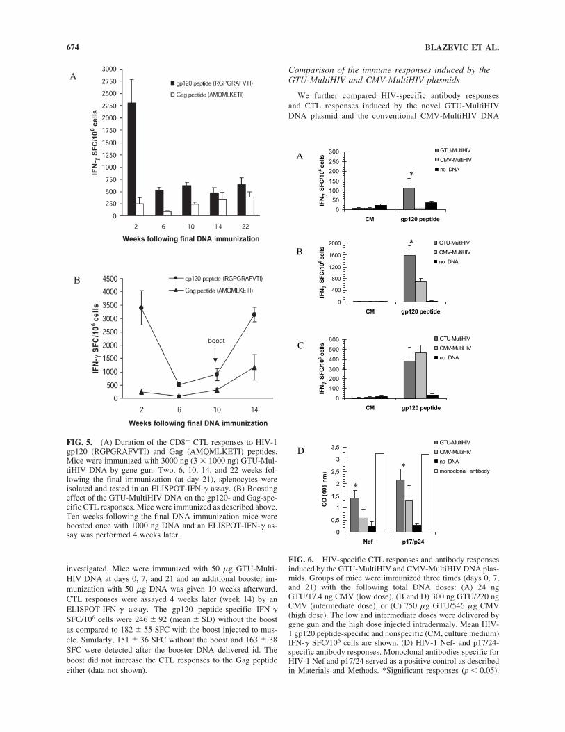
investigated. Mice were immunized with 50 �g GTU-Multi-HIV DNA at days 0, 7, and 21 and an additional booster im-munization with 50 �g DNA was given 10 weeks afterward.CTL responses were assayed 4 weeks later (week 14) by anELISPOT-IFN-� assay. The gp120 peptide-specific IFN-�SFC/106 cells were 246 92 (mean SD) without the boostas compared to 182 55 SFC with the boost injected to mus-cle. Similarly, 151 36 SFC without the boost and 163 38SFC were detected after the booster DNA delivered id. Theboost did not increase the CTL responses to the Gag peptideeither (data not shown).
Comparison of the immune responses induced by theGTU-MultiHIV and CMV-MultiHIV plasmids
We further compared HIV-specific antibody responses and CTL responses induced by the novel GTU-MultiHIVDNA plasmid and the conventional CMV-MultiHIV DNA
BLAZEVIC ET AL.674
FIG. 5. (A) Duration of the CD8� CTL responses to HIV-1gp120 (RGPGRAFVTI) and Gag (AMQMLKETI) peptides.Mice were immunized with 3000 ng (3 � 1000 ng) GTU-Mul-tiHIV DNA by gene gun. Two, 6, 10, 14, and 22 weeks fol-lowing the final immunization (at day 21), splenocytes wereisolated and tested in an ELISPOT-IFN-� assay. (B) Boostingeffect of the GTU-MultiHIV DNA on the gp120- and Gag-spe-cific CTL responses. Mice were immunized as described above.Ten weeks following the final DNA immunization mice wereboosted once with 1000 ng DNA and an ELISPOT-IFN-� as-say was performed 4 weeks later.
FIG. 6. HIV-specific CTL responses and antibody responsesinduced by the GTU-MultiHIV and CMV-MultiHIV DNA plas-mids. Groups of mice were immunized three times (days 0, 7,and 21) with the following total DNA doses: (A) 24 ngGTU/17.4 ng CMV (low dose), (B and D) 300 ng GTU/220 ngCMV (intermediate dose), or (C) 750 �g GTU/546 �g CMV(high dose). The low and intermediate doses were delivered bygene gun and the high dose injected intradermaly. Mean HIV-1 gp120 peptide-specific and nonspecific (CM, culture medium)IFN-� SFC/106 cells are shown. (D) HIV-1 Nef- and p17/24-specific antibody responses. Monoclonal antibodies specific forHIV-1 Nef and p17/24 served as a positive control as describedin Materials and Methods. *Significant responses (p � 0.05).
6293_09_p667-677 6/23/06 10:02 AM Page 674
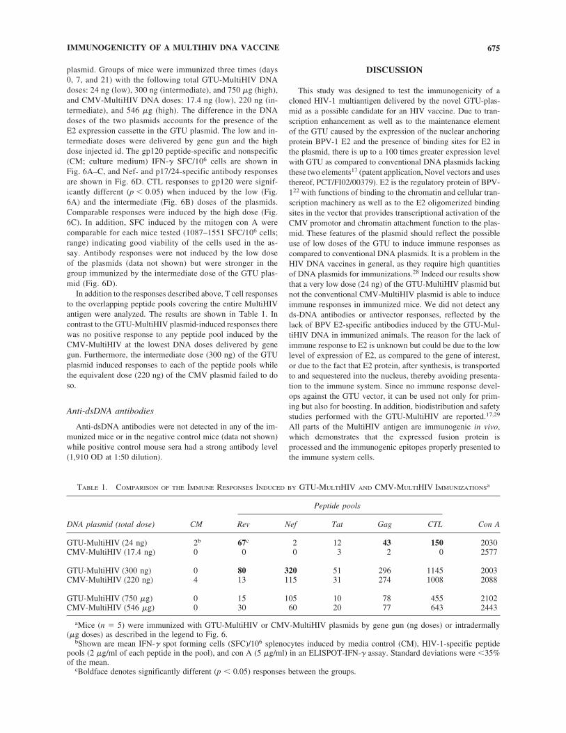
plasmid. Groups of mice were immunized three times (days0, 7, and 21) with the following total GTU-MultiHIV DNAdoses: 24 ng (low), 300 ng (intermediate), and 750 �g (high),and CMV-MultiHIV DNA doses: 17.4 ng (low), 220 ng (in-termediate), and 546 �g (high). The difference in the DNAdoses of the two plasmids accounts for the presence of theE2 expression cassette in the GTU plasmid. The low and in-termediate doses were delivered by gene gun and the highdose injected id. The gp120 peptide-specific and nonspecific(CM; culture medium) IFN-� SFC/106 cells are shown in Fig. 6A–C, and Nef- and p17/24-specific antibody responsesare shown in Fig. 6D. CTL responses to gp120 were signif-icantly different (p � 0.05) when induced by the low (Fig.6A) and the intermediate (Fig. 6B) doses of the plasmids.Comparable responses were induced by the high dose (Fig.6C). In addition, SFC induced by the mitogen con A werecomparable for each mice tested (1087–1551 SFC/106 cells;range) indicating good viability of the cells used in the as-say. Antibody responses were not induced by the low doseof the plasmids (data not shown) but were stronger in thegroup immunized by the intermediate dose of the GTU plas-mid (Fig. 6D).
In addition to the responses described above, T cell responsesto the overlapping peptide pools covering the entire MultiHIVantigen were analyzed. The results are shown in Table 1. Incontrast to the GTU-MultiHIV plasmid-induced responses therewas no positive response to any peptide pool induced by theCMV-MultiHIV at the lowest DNA doses delivered by genegun. Furthermore, the intermediate dose (300 ng) of the GTUplasmid induced responses to each of the peptide pools whilethe equivalent dose (220 ng) of the CMV plasmid failed to doso.
Anti-dsDNA antibodies
Anti-dsDNA antibodies were not detected in any of the im-munized mice or in the negative control mice (data not shown)while positive control mouse sera had a strong antibody level(1,910 OD at 1:50 dilution).
DISCUSSION
This study was designed to test the immunogenicity of acloned HIV-1 multiantigen delivered by the novel GTU-plas-mid as a possible candidate for an HIV vaccine. Due to tran-scription enhancement as well as to the maintenance elementof the GTU caused by the expression of the nuclear anchoringprotein BPV-1 E2 and the presence of binding sites for E2 inthe plasmid, there is up to a 100 times greater expression levelwith GTU as compared to conventional DNA plasmids lackingthese two elements17 (patent application, Novel vectors and usesthereof, PCT/FI02/00379). E2 is the regulatory protein of BPV-122 with functions of binding to the chromatin and cellular tran-scription machinery as well as to the E2 oligomerized bindingsites in the vector that provides transcriptional activation of theCMV promotor and chromatin attachment function to the plas-mid. These features of the plasmid should reflect the possibleuse of low doses of the GTU to induce immune responses ascompared to conventional DNA plasmids. It is a problem in theHIV DNA vaccines in general, as they require high quantitiesof DNA plasmids for immunizations.28 Indeed our results showthat a very low dose (24 ng) of the GTU-MultiHIV plasmid butnot the conventional CMV-MultiHIV plasmid is able to induceimmune responses in immunized mice. We did not detect anyds-DNA antibodies or antivector responses, reflected by thelack of BPV E2-specific antibodies induced by the GTU-Mul-tiHIV DNA in immunized animals. The reason for the lack ofimmune response to E2 is unknown but could be due to the lowlevel of expression of E2, as compared to the gene of interest,or due to the fact that E2 protein, after synthesis, is transportedto and sequestered into the nucleus, thereby avoiding presenta-tion to the immune system. Since no immune response devel-ops against the GTU vector, it can be used not only for prim-ing but also for boosting. In addition, biodistribution and safetystudies performed with the GTU-MultiHIV are reported.17,29
All parts of the MultiHIV antigen are immunogenic in vivo,which demonstrates that the expressed fusion protein isprocessed and the immunogenic epitopes properly presented tothe immune system cells.
IMMUNOGENICITY OF A MULTIHIV DNA VACCINE 675
TABLE 1. COMPARISON OF THE IMMUNE RESPONSES INDUCED BY GTU-MULTIHIV AND CMV-MULTIHIV IMMUNIZATIONSa
Peptide pools
DNA plasmid (total dose) CM Rev Nef Tat Gag CTL Con A
GTU-MultiHIV (24 ng) 2b 67c 2 12 43 150 2030CMV-MultiHIV (17.4 ng) 0 0 0 3 2 0 2577
GTU-MultiHIV (300 ng) 0 80 320 51 296 1145 2003CMV-MultiHIV (220 ng) 4 13 115 31 274 1008 2088
GTU-MultiHIV (750 �g) 0 15 105 10 78 455 2102CMV-MultiHIV (546 �g) 0 30 60 20 77 643 2443
aMice (n 5) were immunized with GTU-MultiHIV or CMV-MultiHIV plasmids by gene gun (ng doses) or intradermally(�g doses) as described in the legend to Fig. 6.
bShown are mean IFN-� spot forming cells (SFC)/106 splenocytes induced by media control (CM), HIV-1-specific peptidepools (2 �g/ml of each peptide in the pool), and con A (5 �g/ml) in an ELISPOT-IFN-� assay. Standard deviations were �35%of the mean.
cBoldface denotes significantly different (p � 0.05) responses between the groups.
6293_09_p667-677 6/23/06 10:02 AM Page 675
It is important to determine for each new vaccine the opti-mal immunization regime in terms of vaccine doses, frequencyof immunizations, and delivery routes. Our results show thatgene gun immunization is superb in inducing B cell and CTLresponses in immunized mice compared to im and id immu-nizations. Extremely low doses of the GTU-MultiHIV plasmidDNA delivered by gene gun induce HIV-specific CTL re-sponses. Intramuscular immunization induces only a low CTLresponse without antibody production, which is in agreementwith previously published results.30 Furthermore, the CTL re-sponse induced by im and id immunizations requires high doses(micrograms) of the plasmid DNA. Earlier studies have also re-ported that gene gun immunization is more efficient than nee-dle injection in eliciting cellular and humoral responses.31,32
The strong and broad immune response induced by gold parti-cles delivered DNA compared to injected DNA could be dueto the fact that the response is initiated by Langerhans cells inthe skin, which are directly transfected or take up the secretedantigen from the immunization site (cross-priming), migrate tolocal lymph nodes, and stimulate T cells.33 In order to improvethe HIV-specific immune responses induced by needle immu-nization, which is the widely used route of DNA vaccinationfor human subjects, more studies using different molecular ad-juvants34 and/or different boosting regimens should be per-formed. Our results show no effect of the homologous DNAboost on both im and id delivery methods in contrast to genegun DNA delivery. It is likely that different heterologous boost-ing strategies might increase the HIV-specific immune re-sponses induced by the needle-injected DNA. We are currentlyevaluating these strategies. Overall, these results show that genegun immunization is more potent in inducing various types ofimmune response and requires 1000-fold less DNA to be ad-ministered than im or id immunizations. Gene gun and deliv-ery methods based on similar technology should therefore beconsidered as alternative methods for delivering DNA vaccinesto human subjects in clinical trials.
The results described here also show that the HIV-specificimmune response induced by DNA vaccination is not only routeand dose dependent, but also depends on the frequency of theimmunizations. HIV-specific antibody response induction re-quired at least two DNA immunizations while the third immu-nization induced an optimal response. Our findings are in agree-ment with the previously published observation that at least twoimmunizations are required to obtain detectable humoral im-mune responses to HIV-1 proteins.16 We have found that al-most 6 months following DNA delivery HIV-specific CTL re-sponses could still be detected. It was earlier reported16 that 3months after the initial DNA delivery there was low IL-2 pro-duction detected in the stimulated cell culture supernatants. Theweak residual IL-2 production as described above could pro-vide help for the CTL response35 we detected at 22 weeks post-immunization. However, one booster immunization with theGTU-MultiHIV DNA at 10 weeks restored strong HIV-specificCTL responses.
All components of the MultiHIV antigen are immunogenicin vivo in a mouse model. Mice have developed considerableRev-, Nef-, Tat-, Gag-, and CTL-specific T cell responses aswell as Nef- and Gag-specific antibody responses. The CTLpart of the fusion protein is very immunogenic for T cells asseen by the strong CD8� CTL responses to H-2d-restricted
gp120 peptide but not for B cells. This is understandable, asCTL contains 11 clusters of the T cell epitope-rich regions ofHIV-1 Pol and Env. As we could detect T cell responses to eachof the HIV-1 regulatory proteins coded by the GTU-MultiHIV,our concern that the strong immunodominant epitope, as is theH-2d-restricted gp120 described here, which induces a high fre-quency of the responding cells, would preclude responsesagainst subdominant epitopes is minimized. The reason whyTat and Rev are poor B cell immunogens in vivo is not known.However, other investigators have reported low immunogenic-ity of these proteins in their reports.16,36
As we could induce an immune response to each of the HIV-1 proteins cloned into the GTU-MultiHIV under the control ofa single promotor, we conclude that there is no interference orinhibition of the specific responses, which might be the casewhen combinations of different plasmids expressing a singlegene are used for immunizations.37 A single DNA vaccine con-struct as described here, which contains multiple HIV-1 genes,is easier to administer and more cost effective than developingand administering a cocktail of single gene DNA vectors. Theresults described here support the hypothesis that DNA con-taining several HIV-1 genes is a potential vaccine candidateagainst HIV infection when an immune response to the multi-ple HIV proteins is desirable in order to minimize virus escapefrom the immune system.38
ACKNOWLEDGMENTS
We thank Ms. Marja Pirinen, Ms. Tuula Myllymäki, Ms.Marja Tahtinen, Ms. Aulikki Lehmus, and Mrs. Helle Remmefor great technical assistance and important contributions.
REFERENCES
1. Brenner B, Wainberg MA, Salomon H, et al.: Resistance to anti-retroviral drugs in patients with primary HIV-1 infection. Int J An-timicrob Agents 2000;16:429–434.
2. Martinez-Picado J, DePasquale MP, Kartsonis N, et al.: Antiretro-viral resistance during successful therapy of HIV type 1 infection.Proc Natl Acad Sci USA 2000;97:10948–10953.
3. Letvin N: Strategies for an HIV vaccine. J Clin Invest 2002;109:15–20.
4. Greenough TC, Brettler DB, Somasundaran M, Panicali DL, andSullivan JL: Human immunodeficiency virus type 1-specific cyto-toxic T lymphocytes (CTL), virus load, and CD4 T cell loss: Evi-dence supporting a protective role for CTL in vivo. J Infect Dis1997;176:118–125.
5. Betts MR, Krowka JF, Kepler TB, et al.: Human immunodeficiencyvirus type 1-specific cytotoxic T lymphocyte activity is inverselycorrelated with HIV type 1 viral load in HIV type 1-infected long-term survivors. AIDS Res Hum Retroviruses 1999;15:1219–1228.
6. Rowland-Jones S, Sutton J, Ariyoshi K, et al.: HIV-specific cyto-toxic T-cells in HIV-exposed but uninfected Gambian women. NatMed 1995;1:59–64.
7. Matano T, Shibata R, Siemon C, Connors M, Lane HC, and Mar-tin MA: Administration of an anti-CD8 monoclonal antibody in-terferes with the clearance of chimeric simian/human immunode-ficiency virus during primary infections of rhesus macaques. J Virol1998;72:164–169.
8. Ensoli B and Cafaro A: Novel strategies toward the development
BLAZEVIC ET AL.676
6293_09_p667-677 6/23/06 10:02 AM Page 676
of an effective vaccine to prevent human immunodeficiency virusinfection or acquired immunodeficiency virus. AIDS Clin Rev2000;1:23–61.
9. Addo MM, Altfeld M, Rosenberg ES, et al.: The HIV-1 regulatoryproteins Tat and Rev are frequently targeted by cytotoxic T lym-phocytes derived from HIV-1-infected individuals. Proc Natl AcadSci USA 2001;98:1781–1786.
10. Gotch F: Cross-clade T cell recognition of HIV-1. Curr Opin Im-munol 1998;10:388–392.
11. Wyand MS, Manson KH, Garcia-Moll M, Montefiori D, andDesrosiers RC: Vaccine protection by a triple deletion mutant ofsimian immunodeficiency virus. J Virol 1996;70:3724–3733.
12. Barouch DH, Santra S, Schmitz JE, et al.: Control of viremia andprevention of clinical AIDS in rhesus monkeys by cytokine-aug-mented DNA vaccination. Science 2000;290:486–492.
13. Ayyavoo V, Kudchodkar S, Ramanathan MP, et al.: Immuno-genicity of a novel DNA vaccine cassette expressing multiple hu-man immunodeficiency virus (HIV-1) accessory genes. AIDS2000;14:1–9.
14. Calarota S, Bratt G, Nordlund S, et al.: Cellular cytotoxic responseinduced by DNA vaccination in HIV-1-infected patients. Lancet1998;351:1320–1325.
15. Barouch DH, Kunstman J, Kuroda MJ, et al.: Eventual AIDS vac-cine failure in a rhesus monkey by viral escape from cytotoxic Tlymphocytes. Nature 2002;415:335–339.
16. Hinkula J, Svanholm C, Schwartz S, et al.: Recognition of promi-nent viral epitopes induced by immunization with human immu-nodeficiency virus type 1 regulatory genes. J Virol. 1997;71:5528–5539.
17. Krohn K, Stanescu I, Blazevic V, Vesikari T, Ranki A, and UstavM: A DNA HIV-1 vaccine based on a fusion gene expressing non-structural and structural genes of consensus sequence of the A, Band C subtypes and the ancestor sequence of the F, G and H sub-types. Preclinical and clinical studies. Microbes Infect 2005;7:1405–1413.
18. Hanke T and McMichael A: Pre-clinical development of a multi-CTL epitope-based DNA prime MVA boost vaccine for AIDS. Im-munol Lett 1999;66:177–181.
19. Takahashi H, Cohen J, Hosmalin A, et al.: An immunodominantepitope of the human immunodeficiency virus envelope glycopro-tein gp160 recognized by class I major histocompatibility complexmolecule-restricted murine cytotoxic T lymphocytes. Proc NatlAcad Sci USA 1988;85:3105–3109.
20. Yanisch-Perron C, Vieira J, and Messing J: Improved M13 phagecloning vectors and host strains: Nucleotide sequences of theM13mp18 and pUC19 vectors. Gene 1985;33:103–119.
21. Korber B, Brander C, Haynes B, Koup R, Kuiken C, Moore JP,Walker BD, and Watkins DI (eds.): HIV Molecular Immunology2000. Theoretical Biology and Biophysics, Los Alamos NationalLaboratory, Los Alamos, NM.
22. Ustav M and Stenlund A: Transient replication of BPV-1 requirestwo viral polypeptides encoded by the E1 and E2 open readingframes. EMBO J 1991;10:449–457.
23. Qiu JT, Song R, Dettenhofer M, et al.: Evaluation of novel humanimmunodeficiency virus type 1 Gag DNA vaccines for protein ex-pression in mammalian cells and induction of immune responses.J Virol 1999;73:9145–9152.
24. Singer DS, Kohn LD, Zinger H, and Mozes E: Methimazole pre-vents induction of experimental systemic lupus erythematosus inmice. J Immunol 1994;153:873–880.
25. Collings A, Pitkänen J, Strengell M, et al.: Humoral and cellularimmune responses to HIV-1 nef in mice DNA-immunised withnon-replicating or self-replicating expression vectors. Vaccine1999;18:460–467.
26. Tobery TW and Siliciano RF: Targeting of HIV-1 antigens for rapidintracellular degradation enhances cytotoxic T lymphocyte (CTL)recognition and the induction of de novo CTL responses in vivoafter immunization. J Exp Med 1997;185:909–920.
27. Lavlani A, Brookes R, Hambleton S, Britton WJ, Hill AVS, andMcMichael AJ: Rapid effector function in CD8� memory T cells.J Exp Med 1997;186:859–865.
28. Caputo A, Gavioli R, Altavilla G, et al.: Immunization with lowdoses of HIV-1 tat DNA delivered by novel cationic block copoly-mers induces CTL responses against Tat. Vaccine 2003;21:1103–1111.
29. Tuomela M, Malm M, Wallen M, Stanescu I, Krohn K, and Pe-terson P: Biodistribution and general safety of a naked DNA plas-mid, GTU-MultiHIV, in a rat, using a quantitative PCR method.Vaccine 2005;23:890–896.
30. Cohen AD, Boyer JD, and Weiner DB: Modulating the immuneresponse to genetic immunization. FASEB J 1998;12:1611–1626.
31. Wolff JA: Naked DNA transport and expression in mammaliancells. Neuromuscul Disord 1997;7:314–318.
32. Pertmer TM, Eisenbraun MD, McCabe D, Prayaga SK, Fuller DH,and Haynes JR: Gene gun-based nucleic acid immunization: Elic-itation of humoral and cytotoxic T lymphocyte responses follow-ing epidermal delivery of nanogram quantities of DNA. Vaccine1995;13:1427–1430.
33. Creusot RJ, Thomsen LL, van Wely CA, Topley P, Tite JP, andChain BM: Early commitment of adoptively transferred CD4� Tcells following particle-mediated DNA vaccination: implicationsfor the study of immunomodulation. Vaccine 2001;19:1678–1687.
34. McKay PF, Barouch DH, Santra S, et al.: Recruitment of differentsubsets of antigen-presenting cells selectively modulates DNA vac-cine-elicited CD4� and CD8� T lymphocyte responses. Eur J Im-munol 2004;34:1011–1020.
35. Blazevic V, Pinto LA, Trubey CM, and Shearer GM: Alloantigenicstimulation bypasses CD28-B7 costimulatory blockade by an interleukin-2-dependent mechanism. J Leukoc Biol 2000;67:817–824.
36. Tähtinen M, Strengell M, Collings A, et al.: DNA vaccination inmice using HIV-1 nef, rev and tat genes in self-replicating pBN-vector. Vaccine 2001;19:2039–2047.
37. Kjerrström A, Hinkula J, Engstrom G, et al.: Interactions of singleand combined human immunodeficiency virus type 1 (HIV-1)DNA vaccines. Virology 2001;284:46–61.
38. Kong WP, Huang Y, Yang ZY, Chakrabarti BK, Moodie Z, andNabel G: Immunogenicity of multiple gene and clade human im-munodeficiency virus type 1 DNA vaccines. J Virol 2003;77:12764–12772.
Address reprint requests to:Vesna Blazevic
FIT Biotech Oyj Plc.Biokatu 8
33520 Tampere, Finland
E-mail: [email protected]
IMMUNOGENICITY OF A MULTIHIV DNA VACCINE 677
6293_09_p667-677 6/23/06 10:02 AM Page 677
A
DAsefwd©
K
1
dEcmpmciPmcSb
0d
Vaccine 25 (2007) 3293–3301
GTU®-MultiHIV DNA vaccine results in protectionin a novel P815 tumor challenge model
Maria Malm a,∗, Rein Sikut b, Kai Krohn c, Vesna Blazevic a
a FIT Biotech Oyj Plc., Biokatu 8, 33520 Tampere, Finlandb FIT Biotech Plc., Tartu, Estonia
c Institute of Medical Technology, Tampere, Finland
Received 2 August 2006; received in revised form 18 December 2006; accepted 4 January 2007Available online 12 January 2007
bstract
A novel animal model for testing the immunogenicity and protective immune response induced by HIV-1 DNA vaccines was developed.BA/2 mice were immunized with GTU®-MultiHIV DNA encoding multigene for Rev, Nef, Tat, optp17/24 and a stretch of Pol/Env epitopes.single GTU®-MultiHIV B-clade specific plasmid or Auxo-GTU®-MultiHIVmix (mixture of four plasmids with A, B, C and FGH clade
pecific MultiHIV antigens) were administered via gene gun and cell-mediated and humoral immune responses were analysed. The protectivefficacy of the immune response was evaluated by challenging the mice with syngeneic tumor cells (P815) stably transfected with the MultiHIV
®
usion gene. Our results show that the strong MultiHIV-specific immune response generated by the GTU -MultiHIV vaccines in DBA/2 miceas able to delay the tumor growth substantially, indicating that the CTL response detected in vitro confers protection in vivo. The modelescribed here is a safe and feasible in vivo assay for assessment of the vaccine potency to induce protective cell-mediated immune responses.2007 Elsevier Ltd. All rights reserved.
tii[cmvAbwmiv
eywords: HIV-1 DNA vaccine; P815 tumor model; Cytotoxic T cells
. Introduction
The lack of an effective vaccine against HIV is partlyue to the fact that the correlates of protection are unknown.valuation of a vaccine candidate in clinical settings is timeonsuming and costly process, therefore, different animalodels are used for the evaluation of the immunity and
rotection induced by the vaccine candidates [1]. An ani-al model closely resembling HIV-1 infection in humans
an be attained in nonhuman primates infected with simianmmunodeficiency virus (SIV) or HIV/SIV chimeric viruses.rimate challenge studies have ethical constraints and aajor defect as a model for HIV-1 infection, due to the
onsiderable differences between HIV-1, SIV and chimericHIV infection [2–4]. Several small animal models haveeen developed to demonstrate the capability of a vaccine
∗ Corresponding author. Tel.: +358 3 3138 7054; fax: +358 3 3138 7050.E-mail address: [email protected] (M. Malm).
gGi[
m
264-410X/$ – see front matter © 2007 Elsevier Ltd. All rights reserved.oi:10.1016/j.vaccine.2007.01.003
o induce protective immunity. Infection resembling HIV-1nfection in humans has been established in severe combinedmmunodeficiency mice [5] and recently in Trimera mice6]. However, the usability of these mouse models for vac-ine studies is limited due to the low availability of theseice, the safety reasons related to work with the infectious
irus and the limited duration of the HIV-1 infection in mice.dditional mouse challenge studies utilize different recom-inant live viruses, including vaccinia virus [7,8]. Recently,e have tested [9] a DNA vaccine candidate in a protectionodel where immunized mice were challenged with the cells
nfected with a pseudotype virus carrying murine leukaemiairus (MuLV) envelope and the clade A and B specific HIVenome [10]. The gene gun immunization with the Auxo-TU®-MultiHIVmix DNA induced a cross-clade protection
n 83–100% of the pseudotype HIV-1/MuLV challenged mice9].
Numerous tumor challenge studies are carried out in aouse model to evaluate the efficacy of the vaccines [11,12]
3294 M. Malm et al. / Vaccine 25 (2007) 3293–3301
Table 1Immunizations and challenge experiments in DBA/2 mice
Group Immunizations weeks-5, -4, and -2 Challenge at day 0 Termination day
Study 1I GTU®-MultiHIV-B 1 × 10⁶ P815-MultiHIV <26a
II – 1 × 10⁶ P815-MultiHIV <26a
III GTU®-MultiHIV-B 1 × 10⁶ P815-wt <26a
IV – 1 × 10⁶ P815-wt <26a
Study 2I Auxo-GTU®-MultiHIVmix 0.2 × 10⁶ P815-MultiHIV 15 and 22II – 0.2 × 10⁶ P815-MultiHIV 15 and 22
III Auxo-GTU®-MultiHIVmix 0.4 × 10⁶ P815-MultiHIV 15 and 22IV – 0.4 × 10⁶ P815-MultiHIV 15 and 22
V Auxo-GTU®-MultiHIVmix 0.8 × 10⁶ P815-MultiHIV 9 and 15VI – 0.8 × 10⁶ P815-MultiHIV 9 and 15
5 mm (
aoccCi
ttvsBDDset
2
2
t(ltuma
2
1t
Mtmf1tam
pApTsc1hrrH
2
HlgbPwAw
VII Auxo-GTU®-MultiHIVmix
a Mice were terminated when the longest diameter of the tumor reached 1
nd cancer immunotherapies [13–15]. After the inductionf immunological memory by vaccination, tumor-specificytotoxic T cells (CTL) play a pivotal role in the tumorlearance and protection [16,17]. In concordance, a strongTL response is thought to be needed for combating HIV-1
nfection in humans [18–20].Here we describe a novel mouse model developed for
esting the capability of an HIV DNA vaccine to induce pro-ection against the tumor challenge. The GTU®-MultiHIVaccine described here has been previously shown to inducetrong HIV-specific cell-mediated immune responses inALB/c [21] and C57Bl/6 mice [9]. In the present study,BA/2 mice were immunized with the GTU®-MultiHIVNA and subsequently challenged with P815 tumor cells
table transfected with the MultiHIV antigen. A protectiveffect of the vaccination was evaluated by comparing theumor growth in immunized and naıve mice.
. Materials and methods
.1. Animals
Young female DBA/2OlaHsd mice, 6 weeks old at theime of the first immunization, were obtained from HarlanItaly and Netherlands). All procedures were carried out inive animals according to the guidelines and permission ofhe County administrative board. Treatments were performednder ketamine (Ketalar®, Pfizer AB, Espoo, Finland) andedetomidine (Domitor®, Orion Pharma, Espoo, Finland)
nesthesia.
.2. DNA plasmids
DNA vaccine constructs used here are well described HIV-DNA vaccines based on the Gene Transfer Unit (GTU®)
echnology developed by FIT Biotech Plc. [9,21–23].
1Sf
– 0
or vertical growth >10 mm) or at day 26.
In the first set of experiments (Study 1, Table 1) the GTU®-ultiHIV B clade (Han-2) plasmid was used for immunizing
he mice prior to the challenge. The GTU®-MultiHIV plas-id vector encodes for the HIV-1 multigene, containing fused
ull-length sequences of rev, nef, tat, gag (p17 and p24) and1 T cell epitope rich clusters from pol and env polypep-ides. The above DNA plasmid has been shown to inducestrong and broad HIV-specific CTL responses in BALB/cice [21].The second set of experiments (Study 2, Table 1) were
erformed with more advanced DNA vaccine candidate,uxo-GTU®-MultiHIVmix plasmid, where four differentlasmids (A, B, C and FGH) are combined as a mixture.hese plasmids contain the MultiHIV consensus sequencepecific for clade A, B, C or the ancestral sequence forlades F, G and H, to meet the great variability of the HIV-subtypes/isolates. The Auxo-GTU®-MultiHIVmix vaccineas been shown to induce a strong HIV-specific immuneesponse and in addition it is capable to generate cross-eaction and cross-protection against different clades ofIV-1 [9].
.3. Tumor cell lines and transfections
A mastocytoma cell line P815 of DBA/2 origin carrying-2d class I MHC molecules, was used for constructing cell
ines for the tumor challenge model. These cells are tumori-eneic in DBA/2 mice, forming well-defined palpable tumorselow the skin when injected subcutaneously (sc) [24]. The815 cell line used for the challenge was stable transfectedith a p2SR�-MultiHIV-Han-2 plasmid (P815-MultiHIV).s a challenge control the wild type P815 cell line (P815-wt)as used.
The plasmid p2SR�-MultiHIV-Han-2 expresses the HIV-Han-2 isolate specific MultiHIV antigen, using a strongR�-hybrid promoter comprised of SV40 early promoterused to HTLV U5 region [25]. In order to be able to select
cine 25
tretoimmgteriPao9isblPcQda
RtlbC
2P
pE(coguca
marfctMpt
AaFmrEriort
2c
tl(Dic(ot
2
wncwtpsTwtPt1wtaghbd>su
M. Malm et al. / Vac
ransfected cells we have constructed the vector that car-ies the cassette for expressing puromycin acetyl transferase,nabling selection of transfected cells in the presence ofhe puromycin. In addition, the vector encodes a �-subunitf human chorionic gonadotropin hormone (�-hCG), whichs secreted into urine of the tumor bearing animals. The
easurement of urinary �-hCG is generally used in tumorouse models for augmenting the monitoring of the tumor
rowth, as the amount of the hormone in urine is showno positively correlate with tumor load [26]. As the vectorncoding the MultiHIV antigen does not encode antibiotic-esistance genes, the above two vectors were ligated togethernto con-catemers. Con-catemers were electroporated into815 cells and 1 day post-transfection the puromycin wasdded to the cells (0.75 �g/ml). One week later the poolf cells was sub-cloned by limiting dilution method onto6-well plates, containing 0.5 �g/ml puromycin. The result-ng clones were cultivated further on 24-well plates and aample of cells from each clone was taken and analysedy Western blot method (WB). For WB analysis the cellsysate was separated on 10% SDS-PAGE, transferred ontoVDF-membrane and probed with the cocktail of mono-lonal antibodies against Nef and p24 (01-001 and 05-001,uattromed Ltd., Tartu, Estonia). Bound antibodies wereetected by HRP-conjugated goat anti-mouse IgG secondaryntibody and ECL-substrate.
Both, P815-wt and P815-MultiHIV cells were grown inPMI 1640 (Sigma–Aldrich, St. Louis, MO) supplemen-
ed with 100 U/ml penicillin–streptomycin (Sigma–Aldrich),-glutamine (BioWhittaker, Verviers, Belgium), 10% fetalovine serum (FBS, BioWhittaker) at 37 ◦C with 5%O2.
.4. In vitro antigen presenting function of the815-MultiHIV cell line
In order to test that P815-MultiHIV cells express andresent MultiHIV antigen, they were used in an IFN-�LISPOT assay and Cr51 assay as antigen presenting cells
APC). The P815 cells are routinely used as APC to MHClass I-restricted CD8+ T cells [27]. In brief, splenocytesf BALB/c mice immunized with the GTU-MultiHIV-B byene gun as described earlier [9,21] were in vitro stim-lated for 24 h with mitomycin C treated P815-MultiHIVells and P815-wt cells as a control in the ELISPOT IFN-�ssay.
The lysis of the P815-MultiHIV cells by CD8+ CTL ofice immunized by gene gun with the GTU®-MultiHIV
nd Auxo-GTU®-MultiHIV plasmids was tested in a Cr51
elease assay as described previously [21], using stable trans-ected P815-MultiHIV and P815-wt as target cells. The targetells were prepared by incubating the cells in medium con-
aining 150 �Ci Na251CrO4 (New England Nuclear, Boston,A, USA) 1.5 h at 37 ◦C with 5% CO2. The assays were
erformed in quadruplicates with 5000 targets/well at effec-or to target cell ratios (E:T) of 50:1, 25:1, and 12.5:1.
−
uo
(2007) 3293–3301 3295
fter 5 h incubation 100 �l of supernatant was collectednd radioactivity counted in a �-counter (Wallac Oy, Turku,inland). Results were expressed according to the for-ula: % specific lysis = (experimental release − spontaneous
elease)/(maximum release − spontaneous release) × 100.xperimental release represents the mean count per-minute
eleased by target cells in the presence of effector cells. Max-mum release represents the radioactivity released after lysisf target cells with 5% Triton X-100. Spontaneous releaseepresents the radioactivity present in medium derived fromarget cells alone.
.5. Tumorigenicity of the P815-MultiHIV and P815-wtell lines
The capability of the P815-MultiHIV and P815-wt cellso induce tumors in DBA/2 mice was tested. We inocu-ated 1 × 10⁶ cells subcutaneously (sc) in a 50 �l sterile PBSBioWhittaker) by 23-gauge needle into the right flank of theBA/2 mice and the tumor growth was followed by measur-
ng the diameter of the tumor every second day with electronicalliper. The size (mm2) was calculated as longest diametermm) × shortest diameter (mm). The welfare and behaviourf the mice were also observed. Mice were sacrificed whenhe diameter reached 10 mm.
.6. Protection model in DBA/2 mice
In the Study 1 (Table 1) the P815-MultiHIV and P815-t tumor development was followed in immunized andon-immunized DBA/2 mice after inoculating 1 × 10⁶ P815ells. Mice were immunized three times with 1 and 2eeks intervals (week-5, week-4, week-2) with 1 �g of
he GTU®-MultiHIV-B by Helios gene gun as describedreviously [9,21]. This immunization schedule was earlierhown to induce a strong CTL response in immunized mice.wo weeks after the last immunization (week 0) the miceere challenged by sc injection of 1 × 10⁶ P815-MultiHIV
umor cells on the shaved right flank in a 50 �l sterileBS by 23-gauge needle. In addition, the following con-
rol groups were used: immunized mice challenged with× 106 P815-wt cells and non-immunized mice challengedith 1 × 10⁶ P815-MultiHIV or 1 × 10⁶ P815-wt cells. The
umor growth was monitored every 2 or 3 days as describedbove. Urine samples were collected for few mice in eachroup at days 6 and 20 for measurement of secreted �-hCGormone by using Free beta-hCG ELISA assay (IBL, Ham-urg, Germany). Mice were terminated when the longestiameter of the tumor reached 15 mm (or vertical growth10 mm) or at day 26. The spleens were collected andplenocytes in a single cell suspension were frozen to liq-id nitrogen. Serum samples were collected and stored at
20 ◦C.In the Study 2 (Table 1), different cell numbers weresed for the challenge: 0.2 × 10⁶, 0.4 × 10⁶ and 0.8 × 10⁶
f P815-MultiHIV cells. The immunization regimen was
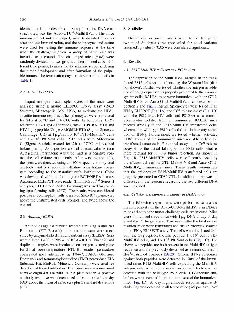
3 cine 25
isiawwirftbT
2
aSsfrHCaCbAttagwAaipac
2
BswdfcDSdaa((
3
ta
4
4
fntsMSIwSrwtCtamFtMtpdv
4
imw7niwMasHan
296 M. Malm et al. / Vac
dentical to the one described in Study 1, but the DNA con-truct used was the Auxo-GTU®-MultiHIVmix. The micemmunized but not challenged, were terminated 2 weeksfter the last immunization, and the splenocytes and serumere used for testing the immune response at the timehen the challenge is given. A group of naıve mice was
ncluded as a control. The challenged mice (n = 8) wereandomly divided into two groups and terminated at two dif-erent time points, to assay for the immune response duringhe tumor development and after formation of the palpa-le tumors. The termination days are described in details inable 1.
.7. IFN-γ ELISPOT
Liquid nitrogen frozen splenocytes of the mice werenalysed using a mouse ELISPOT IFN-� assay (R&Dystems, Minneapolis, MN, USA) to evaluate the HIV-1pecific immune response. The splenocytes were stimulatedor 24 h at 37 ◦C and 5% CO2 with the following: H-2d-estricted HIV-1 gp120 peptide (Env = RGPGRAFVTI) andIV-1 gag peptide (Gag = AMQMLKETI) (Sigma-Genosys,ambridge, UK) at 1 �g/ml, 1 × 10⁴ P815-MultiHIV cellsnd 1 × 10⁴ P815-wt cells. P815 cells were Mitomycin
(Sigma–Aldrich) treated for 2 h at 37 ◦C and washedefore plating. As a positive control concanavalin A (con, 5 �g/ml, Pharmacia) was used, and as a negative con-
rol the cell culture media only. After washing the cells,he spots were detected using an IFN-�-specific biotinylatedntibody, and a streptavidin–alkaline phosphatase conju-ate according to the manufacturer’s instructions. Coloras developed with the chromogenic BCIP/NBT substrate.utomated ELISPOT plate reader (ImmunoSpotTM Series II
nalyzer, CTL Europe, Aalen, Germany) was used for count-ng spot forming cells (SFC). The results were consideredositive if both replica wells were >50 SFC/106 splenocytesbove the unstimulated cells (control) and twice above theontrol.
.8. Antibody ELISA
Antibodies against purified recombinant Gag B and Nefproteins (FIT Biotech) in termination sera were mea-
ured by enzyme-linked immunosorbent assay (ELISA). Seraere diluted 1:400 in PBS + 1% BSA + 0.01% Tween20 anduplicate samples were incubated on antigen coated plateor 2 h at room temperature (RT). Horseradish peroxidaseonjugated goat anti-mouse Ig (P0447, DAKO, Glostrup,enmark) and tetramethylbenzidine (TMB peroxidase EIAubstrate Kit, BioRad, Munchen, Germany) were used for
etection of bound antibodies. The absorbance was measuredt wavelength 450 nm with ELISA plate reader. A positiventibody response was determined to be an optical densityOD) above the mean of naıve sera plus 3 standard deviationsS.D.).adbmc
(2007) 3293–3301
. Statistics
Differences in mean values were tested by pairedwo-tailed Student’s t-test (two-tailed for equal variancessumed); p values ≤0.05 were considered significant.
. Results
.1. P815-MultiHIV cells act as APC in vitro
The expression of the MultiHIV-B antigen in the trans-ected P815 cells was confirmed by the Western blot (dataot shown). Further we tested whether the antigen in addi-ion of being expressed, is properly presented to the immuneystem cells. BALB/c mice were immunized with the GTU-
ultiHIV-B or Auxo-GTU-MultiHIVmix as described inection 2 and Fig. 1 legend. Splenocytes were tested in anFN-� ELISPOT (Fig. 1A) and Cr51 release assay (Fig. 1B)ith the P815-MultiHIV cells and P815-wt as a control.plenocytes isolated from all immunized BALB/c miceeacted strongly to the P815-MultiHIV transfected cells,hereas the wild type P815 cells did not induce any secre-
ion of IFN-�. Furthermore, we tested whether activatedD8+ T cells of the immunized mice are able to lyse the
ransfected tumor cells. Functional assays, like Cr51 releasessay show the actual killing of the P815 cells what isore relevant for in vivo tumor rejection. As shown inig. 1B, P815-MultiHIV cells were efficiently lysed by
he effector cells of the GTU-MultiHIV-B and Auxo-GTU-ultiHIVmix immunized mice. These results demonstrate
hat the epitopes on P815-MultiHIV transfected cells areroperly presented to CD8+ CTL. In addition, there was noifference in the response regarding the two different DNAaccines used.
.2. Cellular and humoral immunity in DBA/2 mice
The following experiments were performed to test themmunogenicity of the Auxo-GTU-MultiHIVmix in DBA/2
ice at the time the tumor challenge cells are injected. Miceere immunized three times with 1 �g DNA at day 0, dayand day 21 by gene gun. Two weeks after the final immu-
ization mice were terminated and the splenocytes assayedn an IFN-� ELISPOT assay. The cells were incubated 24 hith the Gag peptide, the Env peptide, 1 × 10⁴ cells P815-ultiHIV cells, and 1 × 10⁴ P815-wt cells (Fig. 1C). The
bove two peptides are both present in the MultiHIV antigenequence and are previously described as immunodominant-2d-restricted epitopes [28,29]. Strong IFN-� responses
gainst both peptides were detected in 100% of the immu-ized mice. P815-MultiHIV cells expressing the MultiHIVntigen induced a high specific response, which was not
etected with the wild type P815 cells. HIV-specific anti-odies were measured in termination sera of the immunizedice (Fig. 1D). A very high antibody response against B-lade Gag was detected in all tested mice (5/5 positive). Nef
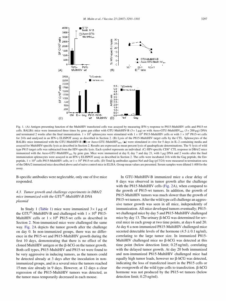
M. Malm et al. / Vaccine 25 (2007) 3293–3301 3297
Fig. 1. (A) Antigen presenting function of the MultiHIV transfected cells was assayed by measuring IFN-� response to P815-MultiHIV cells and P815-wtcells. BALB/c mice were immunized three times by gene gun either with GTU-MultiHIV-B (3× 1 �g) or with Auxo-GTU-MultiHIVmix (3× 200 �g) DNAand terminated 2 weeks after the final immunization. 1 × 10⁵ splenocytes were stimulated with 1 × 10⁴ P815-MultiHIV cells or with 1 × 10⁴ P815-wt cellsfor 24 h and analysed in an IFN-� ELISPOT assay as described in Section 2. (B) Lysis of the P815-MultiHIV target cells by the CTL. Splenocytes of theBALB/c mice immunized with the GTU-MultiHIV-B (�) or Auxo-GTU-MultiHIVmix (�) were stimulated in vitro for 5 days in IL-2 containing media andassayed for MultiHIV-specific lysis as described in Section 2. Results are expressed as mean percent lysis of quadruplicate determinations. The % lysis of wildtype P815 target cells was subtracted from the HIV-specific lysis. Each symbol represents an individual. (C) HIV-specific CD8+ CTL response in DBA/2 miceimmunized with the Auxo-GTU-MultiHIVmix by gene gun. Mice were immunized at day 0, day 7 and day 21, with 1 �g DNA and 2 weeks after the finalimmunization splenocytes were assayed in an IFN-� ELISPOT assay as described in Section 2. The cells were incubated 24 h with the Gag peptide, the Envp otal Ig ao LISA.a
Br
4mp
tMSwoeficBbbi1rt
8wtPPsiwmeAscMtwae
eptide, 1 × 10⁴ cells P815-MultiHIV cells, or 1 × 10⁴ P815-wt cells. (D) Tf the DBA/2 immunized mice described above and of naıve control mice in Essay.
-specific antibodies were neglectable, only one of five miceesponded.
.3. Tumor growth and challenge experiments in DBA/2ice immunized with the GTU®-MultiHIV-B DNAlasmid
In Study 1 (Table 1) mice were immunized 3× 1 �g ofhe GTU®-MultiHIV-B and challenged with 1 × 10⁶ P815-
ultiHIV cells or 1 × 10⁶ P815-wt cells as described inection 2. Non-immunized mice were challenged the sameay. Fig. 2A depicts the tumor growth after the challengen day 0. In non-immunized groups, there was no differ-nce in the P815-wt and P815-MultiHIV growth during therst 10 days, demonstrating that there is no effect of theloned MultiHIV antigen or the �-hCG on the tumor growth.oth cell types, P815-MultiHIV and P815-wt were found toe very aggressive in inducing tumors, as the tumors coulde detected already at 3 days after the inoculation in non-
mmunized groups, and in a several mice the tumors reached5 mm size already in 9 days. However, at 12 days a clearegression of the P815-MultiHIV tumors was detected, ashe tumor mass temporarily decreased in each mouse.ithd
ntibodies against Nef and Gag (p17/24) were measured in termination seraGroup mean values are presented. Serum samples were diluted 1:400 for the
In GTU-MultiHIV-B immunized mice a clear delay ofdays was observed in tumor growth after the challengeith the P815-MultiHIV cells (Fig. 2A), when compared to
he growth of P815-wt tumors. In addition, the growth of815-MultiHIV tumors was much slower than the growth of815-wt tumors. After the wild type cell challenge an aggres-ive tumor growth was seen in all mice, independently ofmmunization. All mice developed tumors eventually; P815-t challenged mice by day 5 and P815-MultiHIV challengedice by day 13. The urinary �-hCG was determined for sev-
ral mice in each group at two time points, at days 6 and 20.t day 6 a non-immunized P815-MultiHIV challenged mice
ecreted detectable levels of the hormone (4.5 ± 0.1 ng/ml),orrelating to the large tumor size. In immunized P815-ultiHIV challenged mice no �-hCG was detected at this
ime point (below detection limit; 0.25 ng/ml), correlatingith the delayed tumor growth. At day 20 both immunized
nd non-immunized P815-MultiHIV challenged mice hadqually high tumor loads, however no �-hCG was detected,
ndicating the loss of transfected insert in the P815 cells orhe overgrowth of the wild type cells to transfection. �-hCGormone was not produced by the P815-wt tumors (belowetection limit; 0.25 ng/ml).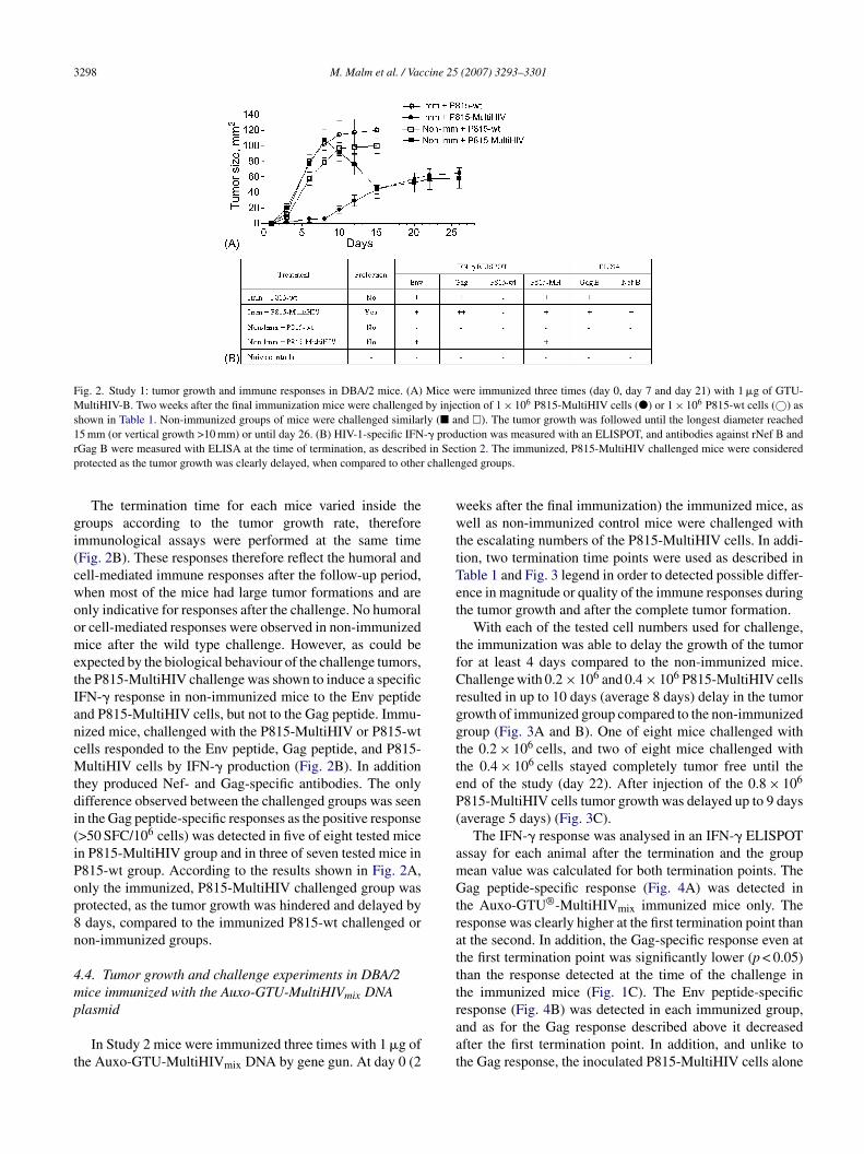
3298 M. Malm et al. / Vaccine 25 (2007) 3293–3301
Fig. 2. Study 1: tumor growth and immune responses in DBA/2 mice. (A) Mice were immunized three times (day 0, day 7 and day 21) with 1 �g of GTU-MultiHIV-B. Two weeks after the final immunization mice were challenged by injection of 1 × 10⁶ P815-MultiHIV cells (�) or 1 × 10⁶ P815-wt cells (©) asshown in Table 1. Non-immunized groups of mice were challenged similarly (� and �). The tumor growth was followed until the longest diameter reached1 -� prodr in Secp challen
gi(cwoometIancMtdi(iPop8n
4mp
t
wwttTet
tfCrggtteP(
amGtrattt
5 mm (or vertical growth >10 mm) or until day 26. (B) HIV-1-specific IFNGag B were measured with ELISA at the time of termination, as describedrotected as the tumor growth was clearly delayed, when compared to other
The termination time for each mice varied inside theroups according to the tumor growth rate, thereforemmunological assays were performed at the same timeFig. 2B). These responses therefore reflect the humoral andell-mediated immune responses after the follow-up period,hen most of the mice had large tumor formations and arenly indicative for responses after the challenge. No humoralr cell-mediated responses were observed in non-immunizedice after the wild type challenge. However, as could be
xpected by the biological behaviour of the challenge tumors,he P815-MultiHIV challenge was shown to induce a specificFN-� response in non-immunized mice to the Env peptidend P815-MultiHIV cells, but not to the Gag peptide. Immu-ized mice, challenged with the P815-MultiHIV or P815-wtells responded to the Env peptide, Gag peptide, and P815-ultiHIV cells by IFN-� production (Fig. 2B). In addition
hey produced Nef- and Gag-specific antibodies. The onlyifference observed between the challenged groups was seenn the Gag peptide-specific responses as the positive response>50 SFC/106 cells) was detected in five of eight tested micen P815-MultiHIV group and in three of seven tested mice in815-wt group. According to the results shown in Fig. 2A,nly the immunized, P815-MultiHIV challenged group wasrotected, as the tumor growth was hindered and delayed bydays, compared to the immunized P815-wt challenged or
on-immunized groups.
.4. Tumor growth and challenge experiments in DBA/2ice immunized with the Auxo-GTU-MultiHIVmix DNA
lasmidIn Study 2 mice were immunized three times with 1 �g ofhe Auxo-GTU-MultiHIVmix DNA by gene gun. At day 0 (2
raat
uction was measured with an ELISPOT, and antibodies against rNef B andtion 2. The immunized, P815-MultiHIV challenged mice were consideredged groups.
eeks after the final immunization) the immunized mice, asell as non-immunized control mice were challenged with
he escalating numbers of the P815-MultiHIV cells. In addi-ion, two termination time points were used as described inable 1 and Fig. 3 legend in order to detected possible differ-nce in magnitude or quality of the immune responses duringhe tumor growth and after the complete tumor formation.
With each of the tested cell numbers used for challenge,he immunization was able to delay the growth of the tumoror at least 4 days compared to the non-immunized mice.hallenge with 0.2 × 10⁶ and 0.4 × 10⁶ P815-MultiHIV cells
esulted in up to 10 days (average 8 days) delay in the tumorrowth of immunized group compared to the non-immunizedroup (Fig. 3A and B). One of eight mice challenged withhe 0.2 × 10⁶ cells, and two of eight mice challenged withhe 0.4 × 10⁶ cells stayed completely tumor free until thend of the study (day 22). After injection of the 0.8 × 10⁶
815-MultiHIV cells tumor growth was delayed up to 9 daysaverage 5 days) (Fig. 3C).
The IFN-� response was analysed in an IFN-� ELISPOTssay for each animal after the termination and the groupean value was calculated for both termination points. Theag peptide-specific response (Fig. 4A) was detected in
he Auxo-GTU®-MultiHIVmix immunized mice only. Theesponse was clearly higher at the first termination point thant the second. In addition, the Gag-specific response even athe first termination point was significantly lower (p < 0.05)han the response detected at the time of the challenge inhe immunized mice (Fig. 1C). The Env peptide-specific
esponse (Fig. 4B) was detected in each immunized group,nd as for the Gag response described above it decreasedfter the first termination point. In addition, and unlike tohe Gag response, the inoculated P815-MultiHIV cells alone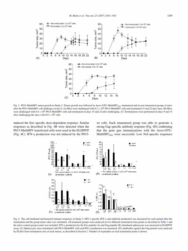
M. Malm et al. / Vaccine 25 (2007) 3293–3301 3299
Fig. 3. P815-MultiHIV tumor growth in Study 2. Tumor growth was followed in Auxo-GTU-MultiHIVmix immunized and in non-immunized groups of micea with 0.2w s 15 ana
irP(
Fttab
fter the P815-MultiHIV cell challenge on day 0. (A) Mice were challengedere challenged with 0.4 × 10⁶ P815-MultiHIV cells and terminated on day
fter challenging the mice with 0.8 × 10⁶ cells.
nduced the Env-specific dose-dependent response. Similaresponses as described in Fig. 4B were detected when the815-MultiHIV transfected cells were used in the ELISPOTFig. 4C). IFN-� production was not induced by the P815-
wstM
ig. 4. The cell-mediated and humoral immune responses in Study 2. HIV-1-speciermination and the group mean value was calculated. All treatment groups were anhe naıve control group (contr) was included. IFN-� production by the Env peptide (ssay. (C) Splenocytes were stimulated with P815-MultiHIV cells and IFN-� produy ELISA from termination sera of each mouse, as described in Section 2. Number
× 106 P815-MultiHIV cells and terminated 15 and 22 days later. (B) Miced 22 after challenging. (C) Terminations were performed on days 9 and 15
t cells. Each immunized group was able to generate atrong Gag-specific antibody response (Fig. 4D) confirminghat the gene gun immunizations with the Auxo-GTU-
ultiHIVmix were successful. Low Nef-specific responses
fic IFN-� and antibody production was measured for each animal after thealysed on two different termination time points as described in Table 1 andA) and Gag peptide (B) stimulated splenocytes was measured in ELISPOTction was measured. (D) Antibodies against the Gag protein were analysedof responders at each termination point is shown.
3 cine 25
ws
5
itHHGpsa
iDtwtictv(iiaigIEdwlaIccotitdwaocDb
ptvt
hetw
ktmcPTsalpattartsttb
pgGmkaccpcttG
A
ba
R
300 M. Malm et al. / Vac
ere detected after the immunization as well (data nothown).
. Discussion
In the present study a tumor challenge model traditionallymplemented in the cancer research [15,30], is for the firstime used to test protective immune responses induced by anIV DNA plasmid vaccine. Due to the global spread of theIV infection a vaccine against HIV is urgently needed [31].rowing evidence suggests that immune responses needed torotect against the HIV cover all main arms of the immuneystem, especially the cytotoxic T lymphocytes [17,27,32,33]nd neutralizing antibodies [33,34].
A way of testing the protective role of a vaccine inducedmmune response is to use P815 tumor challenge model inBA/2 mice [24]. Tumor cell rejection in this model is known
o be the result of action of high avidity CD8+ CTL [17,35]hile an antibody response is not considered important for the
umor cell clearance. In the present study, DBA/2 mice weremmunized with GTU®-MultiHIV DNA and subsequentlyhallenged with P815 tumor cells stable transfected withhe MultiHIV antigen. The GTU®-MultiHIV based DNAaccines, both B-clade (HAN2) specific and the multicladeA + B + C + FGH) specific vaccine constructs were able tonduce strong cell-mediated and humoral immune responsesn DBA/2 mice. The immune response induced with a minutemount of DNA (3 �g) was shown to have cytotoxic effectn vivo, as it was able to delay the P815-MultiHIV tumorrowth and to induce a complete protection in some animals.n the protected animals specific CTL responses against thenv peptide, the Gag peptide and P815-MultiHIV cells wereetected in vitro. The first challenge study was performedith the identical MultiHIV sequence (B-clade, HAN2 iso-
ate) present in both, the GTU®-MultiHIV plasmid vaccinend the P815-MultiHIV transfected cells used for challenge.n the second challenge study, the vaccine construct washanged to more advanced MultiHIV construct, consisting ofonsensus sequences for A, B, C, and the ancestral sequencef FGH clades. Importantly, the delay of the P815-MultiHIVumor growth in immunized mice was observed in both stud-es as compared to the growth in non-immunized mice oro the wild type P815 tumor growth. The above observationemonstrates the efficacy of the DNA plasmid immunizationith considerable sequence diversity between immunizing
ntigen (B consensus) and challenging antigen (HAN2). Tour understanding this is an important observation whenonsidering antigenic sequence diversity of the HIV-specificNA vaccines and multiple viral isolates infecting humaneings world-wide.
P815-MultiHIV cells were shown to function as antigen
resenting cells in vitro (Fig. 1A and B). In consequence,hese cells are able to present the MultiHIV antigen also inivo in challenged mice, resulting to the immunization ofhe non-vaccinated animals. In addition, the CTL response(2007) 3293–3301
as been shown to take place approximately 10 days afterncountering the tumor antigen [35], which is in concordanceo the tumor regression observed 10 days after the challengeith P815-MultiHIV cells of naıve mice.In HIV infection the immune escape of the virus is well
nown [36,37]. A similar mechanism is likely to act duringhe P815-MultiHIV challenge in immunized mice. In this
odel, the induction of effector and/or memory cells in vac-inated mice was able to inhibit an aggressive growth of the815-MultiHIV cells, leading to delay of the tumor growth.he drawback of the present model is in the growth and divi-ion of the transfected P815 cells in immunized mice. Wessume that the eventual loss of the transfected cells in chal-enged mice may be a consequence of the immunologicalressure that contrives cells to loose the transfected insertnd to reverse to wild type cell phenotype. In addition, if theransfection efficiency of 100% is not achieved, a few wildype P815 cells will be carried along with the transfected cellst the time of the challenge. However, because of the aboveeasons there will be slow but almost definite tumor forma-ion, which makes this model unsuitable for targeting at theterilizing immunity induction by vaccination. We believehat this model is however suitable to demonstrate the induc-ion and effector function of the HIV-specific CTL generatedy the DNA vaccines.
The above discussed results, together with previouslyublished studies [9,21], demonstrate comparable immuno-enicity of the single-clade and the multi-clade cocktailTU®-MultiHIV vaccines in several mouse strains. Further-ore, CTL induced by the DNA vaccination were shown to
ill HIV-1 antigen presenting cells both in vivo and in vitrond to protect the immunized animals from the P815 tumorhallenge. The safe and novel animal model described hereould be applicable for the rapid assessment of the in vivorotective immune responses induced by the different vac-ine candidates. Taken together, we show here an attractiveool for the evaluation of vaccine immunogenicity and pro-ection and provide further evidence of the potency of theTU®-MultiHIV vaccines.
cknowledgements
We thank Marja Pirinen, Satu Ruisniemi, Marianne Karls-erg and Tuula Myllymaki for excellent technical assistancend Andres Mannik and Urve Toots for their efforts.
eferences
[1] Giri M, Ugen KE, Weiner DB. DNA vaccines against human immun-odeficiency virus type 1 in the past decade. Clin Microbiol Rev
2004;17(April (2)):370–89.[2] Uberla K. Efficacy of AIDS vaccine strategies in nonhuman primates.Med Microbiol Immunol (Berl) 2005;194(4):201–6.
[3] Joag SV. Primate models of AIDS. Microbes Infect 2000;2(February(2)):223–9.
cine 25
[
[
[
[
[
[
[
[
[
[
[
[
[
[
[
[
[
[
[
[
[
[
[
[
[
[
[
M. Malm et al. / Vac
[4] Reimann KA, Li JT, Veazey R, Halloran M, Park IW, Karlsson GB,et al. A chimeric simian/human immunodeficiency virus expressinga primary patient human immunodeficiency virus type 1 isolate envcauses an AIDS-like disease after in vivo passage in rhesus monkeys.J Virol 1996;70(October (10)):6922–8.
[5] Bonyhadi ML, Kaneshima H. The SCID-hu mouse: An in vivomodel for HIV-1 infection in humans. Mol Med Today 1997;3(June(6)):246–53.
[6] Ayash-Rashkovsky M, Bentwich Z, Arditti F, Friedman S, Reisner Y,Borkow G. A novel small animal model for HIV-1 infection. FASEB J2005;19(July (9)):1149–51.
[7] Belyakov IM, Derby MA, Ahlers JD, Kelsall BL, Earl P, Moss B, etal. Mucosal immunization with HIV-1 peptide vaccine induces mucosaland systemic cytotoxic T lymphocytes and protective immunity in miceagainst intrarectal recombinant HIV-vaccinia challenge. Proc Natl AcadSci USA 1998;95(February (4)):1709–14.
[8] Gupta S, Janani R, Bin Q, Luciw P, Greer C, Perri S, et al. Char-acterization of human immunodeficiency virus gag-specific gammainterferon-expressing cells following protective mucosal immunizationwith alphavirus replicon particles. J Virol 2005;79(June (11)):7135–45.
[9] Malm M, Rollman E, Ustav M, Hinkula J, Krohn K, Wahren B,et al. Cross-clade protection induced by human immunodeficiencyvirus-1 DNA immunogens expressing consensus sequences of multiplegenes and epitopes from subtypes A, B, C, and FGH. Viral Immunol2005;18(4):678–88.
10] Hinkula J, Rollman E, Lundholm P, Benthin R, Okuda K, WahrenB. Genetic immunization with multiple HIV-1 genes provides protec-tion against HIV-1/MuLV pseudovirus challenge in vivo. Cells TissuesOrgans 2004;177(3):169–84.
11] Fifis T, Gamvrellis A, Crimeen-Irwin B, Pietersz GA, Li J, MottramPL, et al. Size-dependent immunogenicity: Therapeutic and protec-tive properties of nano-vaccines against tumors. J Immunol 2004;173(September (5)):3148–54.
12] Ni B, Lin Z, Zhou L, Wang L, Jia Z, Zhou W, et al. Induction ofP815 tumor immunity by DNA-based recombinant semliki forest virusor replicon DNA expressing the P1A gene. Cancer Detect Prev 2004;28(6):418–25.
13] Trimble C, Lin CT, Hung CF, Pai S, Juang J, He L, et al. Comparisonof the CD8+ T cell responses and antitumor effects generated by DNAvaccine administered through gene gun, biojector, and syringe. Vaccine2003;21(September (25–26)):4036–42.
14] Rakhmilevich AL, Turner J, Ford MJ, McCabe D, Sun WH, SondelPM, et al. Gene gun-mediated skin transfection with interleukin 12gene results in regression of established primary and metastatic murinetumors. Proc Natl Acad Sci USA 1996;93(June (13)):6291–6.
15] Wu Y, Wan Y, Bian J, Zhao J, Jia Z, Zhou L, et al. Phage display particlesexpressing tumor-specific antigens induce preventive and therapeuticanti-tumor immunity in murine p815 model. Int J Cancer 2002;98(April(5)):748–53.
16] Soo Hoo W, Lundeen KA, Kohrumel JR, Pham NL, Brostoff SW,Bartholomew RM, et al. Tumor cell surface expression of granulocyte-macrophage colony-stimulating factor elicits antitumor immunity andprotects from tumor challenge in the P815 mouse mastocytoma tumormodel. J Immunol 1999;162(June (12)):7343–9.
17] Snyder JT, Alexander-Miller MA, Berzofskyl JA, Belyakov IM. Molec-ular mechanisms and biological significance of CTL avidity. Curr HIVRes 2003;1(July (3)):287–94.
18] Rowland-Jones S, Sutton J, Ariyoshi K, Dong T, Gotch F, McAdamS, et al. HIV-specific cytotoxic T-cells in HIV-exposed but uninfectedgambian women. Nat Med 1995;1(January (1)):59–64.
19] Borrow P, Lewicki H, Hahn BH, Shaw GM, Oldstone MB. Virus-
specific CD8+ cytotoxic T-lymphocyte activity associated with controlof viremia in primary human immunodeficiency virus type 1 infection.J Virol 1994;68(September (9)):6103–10.20] Musey L, Hughes J, Schacker T, Shea T, Corey L, McElrath MJ.Cytotoxic-T-cell responses, viral load, and disease progression in
[
(2007) 3293–3301 3301
early human immunodeficiency virus type 1 infection. N Engl J Med1997;337(October (18)):1267–74.
21] Blazevic V, Mannik A, Malm M, Sikut R, Valtavaara M, Toots U, etal. Induction of human immunodeficiency virus (HIV type-1)-specificimmunity with a novel gene transport unit (GTU)-MultiHIV DNAvaccine. AIDS Res Hum Retroviruses 2006;22(7):667–77.
22] Tuomela M, Malm M, Wallen M, Stanescu I, Krohn K, Peter-son P. Biodistribution and general safety of a naked DNA plasmid,GTU-MultiHIV, in a rat, using a quantitative PCR method. Vaccine2005;23(January (7)):890–6.
23] Krohn K, Stanescu I, Blazevic V, Vesikari T, Ranki A, Ustav M. ADNA HIV-1 vaccine based on a fusion gene expressing non-structuraland structural genes of consensus sequence of the A–C subtypes and theancestor sequence of the F–H subtypes. Preclinical and clinical studies.Microbes Infect 2005;7(November (14)):1405–13.
24] Kamate C, Baloul S, Grootenboer S, Pessis E, Chevrot A, Tulliez M, etal. Inflammation and cancer, the mastocytoma P815 tumor model revis-ited: triggering of macrophage activation in vivo with pro-tumorigenicconsequences. Int J Cancer 2002;100(August (5)):571–9.
25] Takebe Y, Seiki M, Fujisawa J, Hoy P, Yokota K, Arai K, et al. SRalpha promoter: an efficient and versatile mammalian cDNA expressionsystem composed of the simian virus 40 early promoter and the R-U5segment of human T-cell leukemia virus type 1 long terminal repeat.Mol Cell Biol 1988;8(January (1)):466–72.
26] Shih IM, Torrance C, Sokoll LJ, Chan DW, Kinzler KW, VogelsteinB. Assessing tumors in living animals through measurement of uri-nary beta-human chorionic gonadotropin. Nat Med 2000;6(June (6)):711–4.
27] Tobery TW, Siliciano RF. Targeting of HIV-1 antigens for rapid intracel-lular degradation enhances cytotoxic T lymphocyte (CTL) recognitionand the induction of de novo CTL responses in vivo after immunization.J Exp Med 1997;185(March (5)):909–20.
28] Takahashi H, Cohen J, Hosmalin A, Cease KB, Houghten R, CornetteJL, et al. An immunodominant epitope of the human immunodefi-ciency virus envelope glycoprotein gp160 recognized by class I majorhistocompatibility complex molecule-restricted murine cytotoxic Tlymphocytes. Proc Natl Acad Sci USA 1988;85(May (9)):3105–9.
29] Qiu JT, Song R, Dettenhofer M, Tian C, August T, Felber BK, et al.Evaluation of novel human immunodeficiency virus type 1 gag DNAvaccines for protein expression in mammalian cells and induction ofimmune responses. J Virol 1999;73(November (11)):9145–52.
30] Liu HL, Wu YZ, Zhao JP, Ni B, Jia ZC, Zhou W, et al. Effec-tive elicitation of anti-tumor immunity by collocation of antigen withencoding gene in the same vaccine. Immunol Lett 2003;89(October(2–3)):167–73.
31] Spearman P. Current progress in the development of HIV vaccines. CurrPharm Des 2006;12(9):1147–67.
32] Schmitz JE, Kuroda MJ, Santra S, Sasseville VG, Simon MA, LiftonMA, et al. Control of viremia in simian immunodeficiency virusinfection by CD8+ lymphocytes. Science 1999;283(February (5403)):857–60.
33] Brown SA, Hurwitz JL, Zhan X, Doherty PC, Slobod KS. CD8+ T-cells:are they sufficient to prevent, contain or eradicate HIV-1 infection? CurrDrug Targets Infect Disord 2005;5(June (2)):113–9.
34] Haigwood NL, Stamatatos L. Role of neutralizing antibodies in HIVinfection. AIDS 2003;17(Suppl. 4):S67–71.
35] Chen L, McGowan P, Ashe S, Johnston J, Li Y, Hellstrom I, et al. Tumorimmunogenicity determines the effect of B7 costimulation on T cell-mediated tumor immunity. J Exp Med 1994;179(February (2)):523–32.
36] Barouch DH, Kunstman J, Kuroda MJ, Schmitz JE, Santra S, Pey-
erl FW, et al. Eventual AIDS vaccine failure in a rhesus monkey byviral escape from cytotoxic T lymphocytes. Nature 2002;415(January(6869)):335–9.37] Goulder PJ, Watkins DI. HIV and SIV CTL escape: Implications forvaccine design. Nat Rev Immunol 2004;4(August (8)):630–40.
VIRAL IMMUNOLOGYVolume 18, Number 4, 2005© Mary Ann Liebert, Inc.Pp. 678–688
Cross-Clade Protection Induced by Human ImmunodeficiencyVirus-1 DNA Immunogens Expressing Consensus Sequences of Multiple Genes and Epitopes From
Subtypes A, B, C, and FGH
MARIA MALM,1,* ERIK ROLLMAN,2,* MART USTAV,3 JORMA HINKULA,2 KAI KROHN,1BRITTA WAHREN,2 and VESNA BLAZEVIC1
ABSTRACT
The correlate of protection in human immunodeficiency virus (HIV) infection is not known, but pre-clinical and clinical studies support the involvement of both antibodies and cellular immunity. Inaddition, the existence of multiple HIV clades makes HIV vaccine design especially challenging. Wehave constructed a vaccine platform with an HIV-1 subtype B DNA immunogen expressing fulllength consensus sequences from HIV-1 rev, nef, tat, and gag with additional cellular epitope clus-ters from the env and pol regions. Furthermore, this platform has been extended to three additionalplasmids expressing the same immunogens but originating from subtypes A or C consensus or FGHancestral sequences. Immunogenicity in BALB/c mice, by gene gun or intramuscular delivery, re-vealed strong IFN-� production in response to in vitro re-stimulation with a H-2d restricted gag pep-tide (AMQMLKETI) or even stronger toward an env epitope (RGPGRAFVTI). Weak humoral im-munity was detected. Gene gun immunization with a cocktail of all four plasmids inducedpre-challenge cellular immunity in C57Bl6/A2.01 mice and subsequently a robust frequency of pro-tection (11/12 animals) after experimental challenge with subtype A or B HIV-1/Murine LeukemiaVirus (HIV-1/MuLV). The cross-clade protection observed in this challenge experiment demon-strates that these multigene/multiepitope HIV DNA immunogens are likely to be potent immuno-gens also against the HIV-infection of human beings.
1FIT Biotech Plc, Tampere, Finland.2Swedish Institute for Infectious Diseases Control and Karolinska Institutet, Stockholm, Sweden.3FIT Biotech Plc, Tartu, Estonia.*Maria Malm and Erik Rollman contributed equally to the study.
678
INTRODUCTION
SINCE THE human immunodeficiency virus–1 (HIV-1)was recognized to cause acquired immunodeficiency
syndrome (AIDS) more than 20 years ago, the HIV-1 epi-demic has expanded throughout all continents. Presently,the most disastrous spread continues to take place in South-
east Asia, sub-Saharan Africa, and especially SouthernAfrica, as well as in Latin America (UNAIDS/WHO. AIDSepidemic update: December 2004. [online] http://www.un-aids.org/wad2004/EPI_1204_pdf_en/EpiUpdate04_en.pdf). Extensive efforts have been made in the last two decadesin trying to develop an effective vaccine against HIV/AIDS(19).
5919_12_p678-688 12/9/05 1:42 PM Page 678
Even though more knowledge is constantly gathered re-garding the mechanism underlying the generation of theHIV-specific immune response, it is still not clear what theexact correlates of protection in HIV-1 infection are. Cur-rent opinion emphasizes the implication of the collectiveeffect of CD4� and CD8� T cells (20), broadly neutral-izing antibodies (17), and innate immunity. Especially thesignificance of cytotoxic T-lymphocytes (CTL) in protec-tion is now widely recognized (8,21,25,26). Indications ofthe essential role of CD8� T lymphocytes have beengained by studying individuals who remain apparently un-infected even after repeated exposure to the virus (12) andin experimental animal challenge studies (1).
The high viral mutation rate arising from the infidelityof the reverse transcriptase results in enormous sequenceheterogeneity of HIV-1. This increases the challenges forvaccine design, as illustrated by the inability to maintainprotection in primate challenge models, with viral muta-tions leading to CTL escape (2). A strategy to potentiallyovercome the development of such mutants that wouldlead to immune escape is to generate immunity against awide range of HIV-1 subtypes and toward several viralantigens. DNA immunization can, if delivered efficiently,induce strong CTL responses, and is shown to be apromising approach within HIV-1 vaccine development,used either alone or combined with the different heterol-ogous prime-boost regimens (22).
Nonhuman primates such as macaques are powerfulmodels for simian/simian–human immunodeficiencyvirus (SIV/SHIV) infection. However, there are severaldistinct immune characteristics in the primate models thatdiffer from HIV-1 infection in humans (22, 32). A sig-nificant limitation for HIV vaccine development is thatthere are no small-animal in which actual productiveHIV-1 infection can be established (3).
In this study we show that DNA immunization withcandidate vaccines comprising multiple genes of cladesA, B, C, F, G, and H create strong cellular responsesin BALB/c mice, especially after gene gun immuniza-tion. Second, we show the powerful protective effectof our DNA immunogens upon experimental viral chal-lenge based on pseudotyped HIV-1/murine leukemiavirus (HIV-1/MuLV)–infected cells (10,16,29). Thismodel allows challenging the mice with the completeHIV-1 genome, and immune response to the majorityof the HIV-proteins has been shown (10). The corre-lates of protection are still under investigation, but al-ready published data strongly suggest that at least oneof the components necessary is effective T-cell immu-nity and that antibodies are less needed to mediateclearance (10).The genetic immunogens studied canprotect against both HIV-1 subtype A and subtype Bexperimental challenge inoculates, illustrating cross-clade protection.
PROTECTION BY HIV-1 MULTIGENE VACCINATION IN MICE
MATERIALS AND METHODS
The Auxo-GTU®-MultiHIV DNA vaccine vectors.Four different MultiHIV DNA immunogens have beendesigned according to the consensus sequences of A, B,C, F, G, and H subtypes of HIV-1 and cloned into theAuxo-GTU vector system (patent applications,PCT/FI02/00379 filed 03.05.2002 and PCT/FI2004/000540 filed 15.09.2004), developed by FIT Biotech(Fig. 1). The antigens are named MultiHIV-A (based onthe subtype A consensus sequence), MultiHIV-B (sub-type B consensus sequence), MultiHIV-C (subtype Cconsensus sequence), and MultiHIV-FGH (based on an-cestral sequence for subtypes F, G, and H). Multigene se-quences of the four plasmids were selected to cover�95% of the known HIV-1 isolates published by year2002 (9,14, HIV Sequence Database. Los Alamos Na-tional Laboratory. Assessed August 2002. [online]http://hiv-web.lanl.gov/content/hiv-db/CONSENSUS/M_GROUP/Consensus.html). The MultiHIV DNAs en-code 1053-1079 aminoacid (aa) long polypeptides con-sisting of a fusion of the full-length regulatory proteinsRev, Nef and Tat as well structural proteins p17 and p24.In addition, the antigens contain 17–45 aa long regions
679
MultiHIV - ARev Nef Tat p17/p24
* **Env/Pol (CTL)
BRev Nef Tat p17/p24 Env/Pol (CTL)
CRev Nef Tat p17/p24 Env/Pol (CTL)
FGHRev Nef Tat p17/p24 Env/Pol (CTL)
FIG. 1. Genetic immunogens. The Auxo-GTU MultiHIV(MultiHIV) platform consists of a plamid encoding the codonhumanized rev, nef, and tat genes followed by the gag(p17/p24) codon humanized gene fragment. The 3� end of thevector is designed to encode 11 clusters from the env and polregions that are known to be rich in cytotoxic T lymphocyte(CTL) epitopes. (A) The Auxo-GTU MultiHIV subtype A vec-tor where all gene inserts originate from the consensus sub-type A sequence. (B, C) Corresponding Auxo-GTU MultiHIVsubtype B and C vectors that encode the consensus subtypeB and C gene fragments. (FGH) The Auxo-GTU MultiHIVsubtype FGH vector that is a mosaic vector encoding subtypeF, G, and H fragments. *Symbolizes the position of murinegag epitope AMQMLKETI. **Indicates the location of themurine env epitope RGPGRAFVTI located in the very end ofeach construct.
5919_12_p678-688 12/9/05 1:42 PM Page 679
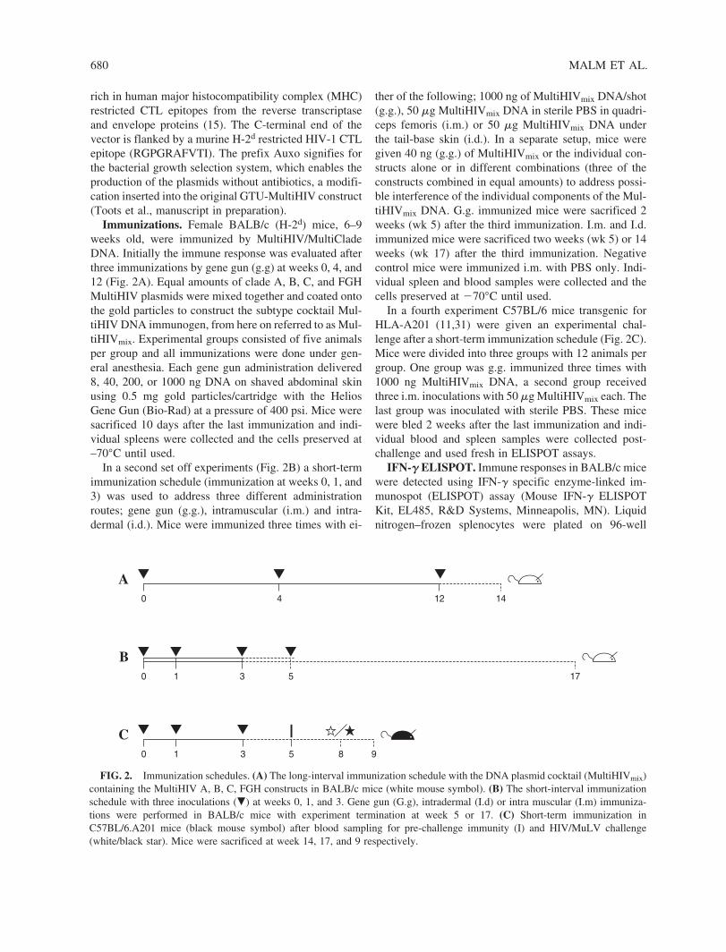
rich in human major histocompatibility complex (MHC)restricted CTL epitopes from the reverse transcriptaseand envelope proteins (15). The C-terminal end of thevector is flanked by a murine H-2d restricted HIV-1 CTLepitope (RGPGRAFVTI). The prefix Auxo signifies forthe bacterial growth selection system, which enables theproduction of the plasmids without antibiotics, a modifi-cation inserted into the original GTU-MultiHIV construct(Toots et al., manuscript in preparation).
Immunizations. Female BALB/c (H-2d) mice, 6–9weeks old, were immunized by MultiHIV/MultiCladeDNA. Initially the immune response was evaluated afterthree immunizations by gene gun (g.g) at weeks 0, 4, and12 (Fig. 2A). Equal amounts of clade A, B, C, and FGHMultiHIV plasmids were mixed together and coated ontothe gold particles to construct the subtype cocktail Mul-tiHIV DNA immunogen, from here on referred to as Mul-tiHIVmix. Experimental groups consisted of five animalsper group and all immunizations were done under gen-eral anesthesia. Each gene gun administration delivered8, 40, 200, or 1000 ng DNA on shaved abdominal skinusing 0.5 mg gold particles/cartridge with the HeliosGene Gun (Bio-Rad) at a pressure of 400 psi. Mice weresacrificed 10 days after the last immunization and indi-vidual spleens were collected and the cells preserved at–70°C until used.
In a second set off experiments (Fig. 2B) a short-termimmunization schedule (immunization at weeks 0, 1, and3) was used to address three different administrationroutes; gene gun (g.g.), intramuscular (i.m.) and intra-dermal (i.d.). Mice were immunized three times with ei-
MALM ET AL.
ther of the following; 1000 ng of MultiHIVmix DNA/shot(g.g.), 50 �g MultiHIVmix DNA in sterile PBS in quadri-ceps femoris (i.m.) or 50 �g MultiHIVmix DNA underthe tail-base skin (i.d.). In a separate setup, mice weregiven 40 ng (g.g.) of MultiHIVmix or the individual con-structs alone or in different combinations (three of theconstructs combined in equal amounts) to address possi-ble interference of the individual components of the Mul-tiHIVmix DNA. G.g. immunized mice were sacrificed 2weeks (wk 5) after the third immunization. I.m. and I.d.immunized mice were sacrificed two weeks (wk 5) or 14weeks (wk 17) after the third immunization. Negativecontrol mice were immunized i.m. with PBS only. Indi-vidual spleen and blood samples were collected and thecells preserved at �70°C until used.
In a fourth experiment C57BL/6 mice transgenic forHLA-A201 (11,31) were given an experimental chal-lenge after a short-term immunization schedule (Fig. 2C).Mice were divided into three groups with 12 animals pergroup. One group was g.g. immunized three times with1000 ng MultiHIVmix DNA, a second group receivedthree i.m. inoculations with 50 �g MultiHIVmix each. Thelast group was inoculated with sterile PBS. These micewere bled 2 weeks after the last immunization and indi-vidual blood and spleen samples were collected post-challenge and used fresh in ELISPOT assays.
IFN-� ELISPOT. Immune responses in BALB/c micewere detected using IFN-� specific enzyme-linked im-munospot (ELISPOT) assay (Mouse IFN-� ELISPOTKit, EL485, R&D Systems, Minneapolis, MN). Liquidnitrogen–frozen splenocytes were plated on 96-well
680
A0 4 12 14
B0 1 3 175
C0 5 83 91
FIG. 2. Immunization schedules. (A) The long-interval immunization schedule with the DNA plasmid cocktail (MultiHIVmix)containing the MultiHIV A, B, C, FGH constructs in BALB/c mice (white mouse symbol). (B) The short-interval immunizationschedule with three inoculations (�) at weeks 0, 1, and 3. Gene gun (G.g), intradermal (I.d) or intra muscular (I.m) immuniza-tions were performed in BALB/c mice with experiment termination at week 5 or 17. (C) Short-term immunization inC57BL/6.A201 mice (black mouse symbol) after blood sampling for pre-challenge immunity (I) and HIV/MuLV challenge(white/black star). Mice were sacrificed at week 14, 17, and 9 respectively.
5919_12_p678-688 12/9/05 1:42 PM Page 680
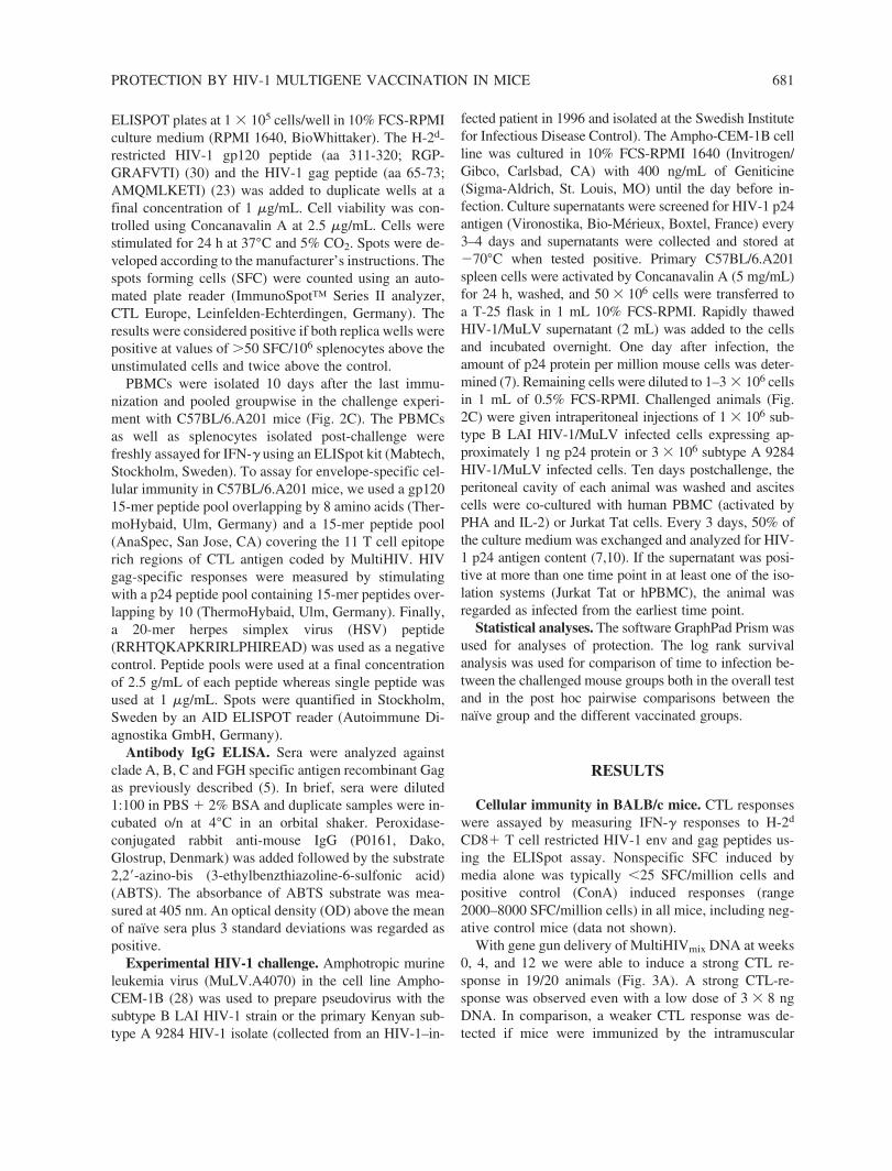
ELISPOT plates at 1 � 105 cells/well in 10% FCS-RPMIculture medium (RPMI 1640, BioWhittaker). The H-2d-restricted HIV-1 gp120 peptide (aa 311-320; RGP-GRAFVTI) (30) and the HIV-1 gag peptide (aa 65-73;AMQMLKETI) (23) was added to duplicate wells at afinal concentration of 1 �g/mL. Cell viability was con-trolled using Concanavalin A at 2.5 �g/mL. Cells werestimulated for 24 h at 37°C and 5% CO2. Spots were de-veloped according to the manufacturer’s instructions. Thespots forming cells (SFC) were counted using an auto-mated plate reader (ImmunoSpot™ Series II analyzer,CTL Europe, Leinfelden-Echterdingen, Germany). Theresults were considered positive if both replica wells werepositive at values of �50 SFC/106 splenocytes above theunstimulated cells and twice above the control.
PBMCs were isolated 10 days after the last immu-nization and pooled groupwise in the challenge experi-ment with C57BL/6.A201 mice (Fig. 2C). The PBMCsas well as splenocytes isolated post-challenge werefreshly assayed for IFN-� using an ELISpot kit (Mabtech,Stockholm, Sweden). To assay for envelope-specific cel-lular immunity in C57BL/6.A201 mice, we used a gp12015-mer peptide pool overlapping by 8 amino acids (Ther-moHybaid, Ulm, Germany) and a 15-mer peptide pool(AnaSpec, San Jose, CA) covering the 11 T cell epitoperich regions of CTL antigen coded by MultiHIV. HIVgag-specific responses were measured by stimulatingwith a p24 peptide pool containing 15-mer peptides over-lapping by 10 (ThermoHybaid, Ulm, Germany). Finally,a 20-mer herpes simplex virus (HSV) peptide(RRHTQKAPKRIRLPHIREAD) was used as a negativecontrol. Peptide pools were used at a final concentrationof 2.5 g/mL of each peptide whereas single peptide wasused at 1 �g/mL. Spots were quantified in Stockholm,Sweden by an AID ELISPOT reader (Autoimmune Di-agnostika GmbH, Germany).
Antibody IgG ELISA. Sera were analyzed againstclade A, B, C and FGH specific antigen recombinant Gagas previously described (5). In brief, sera were diluted1:100 in PBS � 2% BSA and duplicate samples were in-cubated o/n at 4°C in an orbital shaker. Peroxidase-conjugated rabbit anti-mouse IgG (P0161, Dako,Glostrup, Denmark) was added followed by the substrate 2,2�-azino-bis (3-ethylbenzthiazoline-6-sulfonic acid)(ABTS). The absorbance of ABTS substrate was mea-sured at 405 nm. An optical density (OD) above the meanof naïve sera plus 3 standard deviations was regarded aspositive.
Experimental HIV-1 challenge. Amphotropic murineleukemia virus (MuLV.A4070) in the cell line Ampho-CEM-1B (28) was used to prepare pseudovirus with thesubtype B LAI HIV-1 strain or the primary Kenyan sub-type A 9284 HIV-1 isolate (collected from an HIV-1–in-
PROTECTION BY HIV-1 MULTIGENE VACCINATION IN MICE
fected patient in 1996 and isolated at the Swedish Institutefor Infectious Disease Control). The Ampho-CEM-1B cellline was cultured in 10% FCS-RPMI 1640 (Invitrogen/Gibco, Carlsbad, CA) with 400 ng/mL of Geniticine(Sigma-Aldrich, St. Louis, MO) until the day before in-fection. Culture supernatants were screened for HIV-1 p24antigen (Vironostika, Bio-Mérieux, Boxtel, France) every3–4 days and supernatants were collected and stored at�70°C when tested positive. Primary C57BL/6.A201spleen cells were activated by Concanavalin A (5 mg/mL)for 24 h, washed, and 50 � 106 cells were transferred toa T-25 flask in 1 mL 10% FCS-RPMI. Rapidly thawedHIV-1/MuLV supernatant (2 mL) was added to the cellsand incubated overnight. One day after infection, theamount of p24 protein per million mouse cells was deter-mined (7). Remaining cells were diluted to 1–3 � 106 cellsin 1 mL of 0.5% FCS-RPMI. Challenged animals (Fig.2C) were given intraperitoneal injections of 1 � 106 sub-type B LAI HIV-1/MuLV infected cells expressing ap-proximately 1 ng p24 protein or 3 � 106 subtype A 9284HIV-1/MuLV infected cells. Ten days postchallenge, theperitoneal cavity of each animal was washed and ascitescells were co-cultured with human PBMC (activated byPHA and IL-2) or Jurkat Tat cells. Every 3 days, 50% ofthe culture medium was exchanged and analyzed for HIV-1 p24 antigen content (7,10). If the supernatant was posi-tive at more than one time point in at least one of the iso-lation systems (Jurkat Tat or hPBMC), the animal wasregarded as infected from the earliest time point.
Statistical analyses. The software GraphPad Prism wasused for analyses of protection. The log rank survivalanalysis was used for comparison of time to infection be-tween the challenged mouse groups both in the overall testand in the post hoc pairwise comparisons between thenaïve group and the different vaccinated groups.
RESULTS
Cellular immunity in BALB/c mice. CTL responseswere assayed by measuring IFN-� responses to H-2d
CD8� T cell restricted HIV-1 env and gag peptides us-ing the ELISpot assay. Nonspecific SFC induced by media alone was typically �25 SFC/million cells andpositive control (ConA) induced responses (range2000–8000 SFC/million cells) in all mice, including neg-ative control mice (data not shown).
With gene gun delivery of MultiHIVmix DNA at weeks0, 4, and 12 we were able to induce a strong CTL re-sponse in 19/20 animals (Fig. 3A). A strong CTL-re-sponse was observed even with a low dose of 3 � 8 ngDNA. In comparison, a weaker CTL response was de-tected if mice were immunized by the intramuscular
681
5919_12_p678-688 12/9/05 1:42 PM Page 681
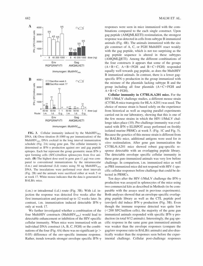
(i.m.) or intradermal (i.d.) route (Fig. 3B). With i.d. in-jection the response was detected five weeks after thefirst immunization and persisted up to 12 weeks later. Incontrast, i.m. immunization induced detectable IFN-�only at week 17.
We further investigated whether a combination of thefour MultiHIV constructs (MultiHIVmix) would lead todetectable enhancement or inhibition of the HIV-specificcellular immunity. When mice were immunized with anindividual DNA construct (A, B, C, FGH) or the combi-nations of the four (Fig. 4A) there was no significant (p �0.05) difference of the env-specific immune response.Rather, trends towards stronger envelope specific IFN-�
MALM ET AL.
responses were seen in mice immunized with the com-binations compared to the each single construct. Upongag peptide (AMQMLKETI) restimulation, the strongestresponse was detected in cells from subtype B immunizedanimals (Fig. 4B). The animals immunized with the sin-gle construct of A, C, or FGH MultiHIV react weaklywith the gag peptide, which is not too surprising as thegag peptide sequence is altered in these subtypes(AMQMLDETI). Among the different combinations ofthe four constructs it appears that some of the groups(A�B�C, A�B�FGH and B�C�FGH) respondedequally well towards gag peptide, as does the MultiHIVB immunized animals. In contrast, there is a lower gag-specific IFN-� production in the group immunized withthe mixture of the plasmids lacking subtype B and thegroup including all four plasmids (A�C�FGH andA�B�C�FGH).
Cellular immunity in C57BL/6.A201 mice. For theHIV-1/MuLV challenge studies, a different mouse strain(C57BL/6 mice transgenic for HLA-A201) was used. Thechoice of mouse strain is based solely on the experiencefrom historical as well as ongoing parallel experimentscarried out in our laboratory, showing that this is one ofthe few mouse strains in which the HIV-1/MuLV chal-lenge takes place (10). Pre-challenge immunity was eval-uated with IFN-� ELISPOT assay performed on freshlyisolated murine PBMCs at week 5. (Fig. 1C and Fig. 5).Because the genetics of this mouse strain is different fromthe BALB/c mice, additional antigens were used for invitro restimulation. After gene gun immunization theC57BL/6.A201 mice showed robust gag-specific re-sponse detectable with an overlapping peptide library.The detectable envelope specific cellular immunity inthese gene gun–immunized animals was very low beforechallenge. In comparison, i.m. immunized mice as wellas PBS immunized mice did not respond with HIV-1 spe-cific cellular responses before challenge that could be de-tected in PBMCs.
Ten days after the HIV-1/MuLV challenge the IFN-�production was assayed in splenocytes of the mice usingtwo commercial kits as described in Methods (to be com-parable with the assays used in previous experiments).Both analyses showed that an envelope-specific overlap-ping peptide library as well as the CTL peptide pool(env/pol) did induce IFN-� production (Fig. 5B). Eventhough the immune response detected was quite low(�200 SFC/million cells), the majority of the gene gunimmunized animals responded with specific IFN-� pro-duction (in total 9/12 animals). Interestingly, the gag spe-cific response in the same gene gun immunized animalswas weaker than the envelope responses (compare thegag/env response ratio in BALB/c animals) and also dras-tically weaker than the responses seen before the exper-imental challenge. Cellular post-challenge responses
682
A
08
5/5IF
N-γ
SF
C/m
illio
n ce
lls
1000
2000
3000
4000
40
EnvRGPGRAFVTI
Immunogen: MultiHIVmix
GagAMQMLKETI
4/5
200
5/5
1000
5/5
8--
5/5
40
5/5
200
5/5
1000 dose (ng)
5/5
B
050
I.m.Route:50
4/50/5IF
N-γ
SF
C/m
illio
n ce
lls
1000
2000
3000
4000
50
Sacrifice week 5
I.d.50
5/5
5/5
1G.g.1
nd
5/5
50I.m.
50
4/50/5
50I.d.
500/5
2/5
1G.g.1
nd
5/5
EnvRGPGRAFVTI
Immunogen: MultiHIVmix
GagAMQMLKETI
dose (µg)
Sacrifice week 17
FIG. 3. Cellular immunity induced by the MultiHIVMix
DNA. (A) Dose titration (8-1000 ng per immunization) of theMultiHIVmix DNA cocktail in the long interval immunizationschedule (Fig. 2A) using gene gun. The cellular immunity isdetermined as IFN-� production against env and gag peptideepitopes. Each bar represent the group geometric mean IFN-�spot forming cells (SFCs)/million splenocytes from five ani-mals. (B) The highest dose used in gene gun (1 �g) was com-pared to conventional immunizations by the intramuscular(I.m.) and intradermal (I.d) routes using 50 ug MultiHIVmix
DNA. The inoculations were performed over short intervals(Fig. 2B) and the animals were sacrificed either at week 5 orat week 17. White mouse indicates that the data is generated inBALB/c mice.
5919_12_p678-688 12/9/05 1:42 PM Page 682
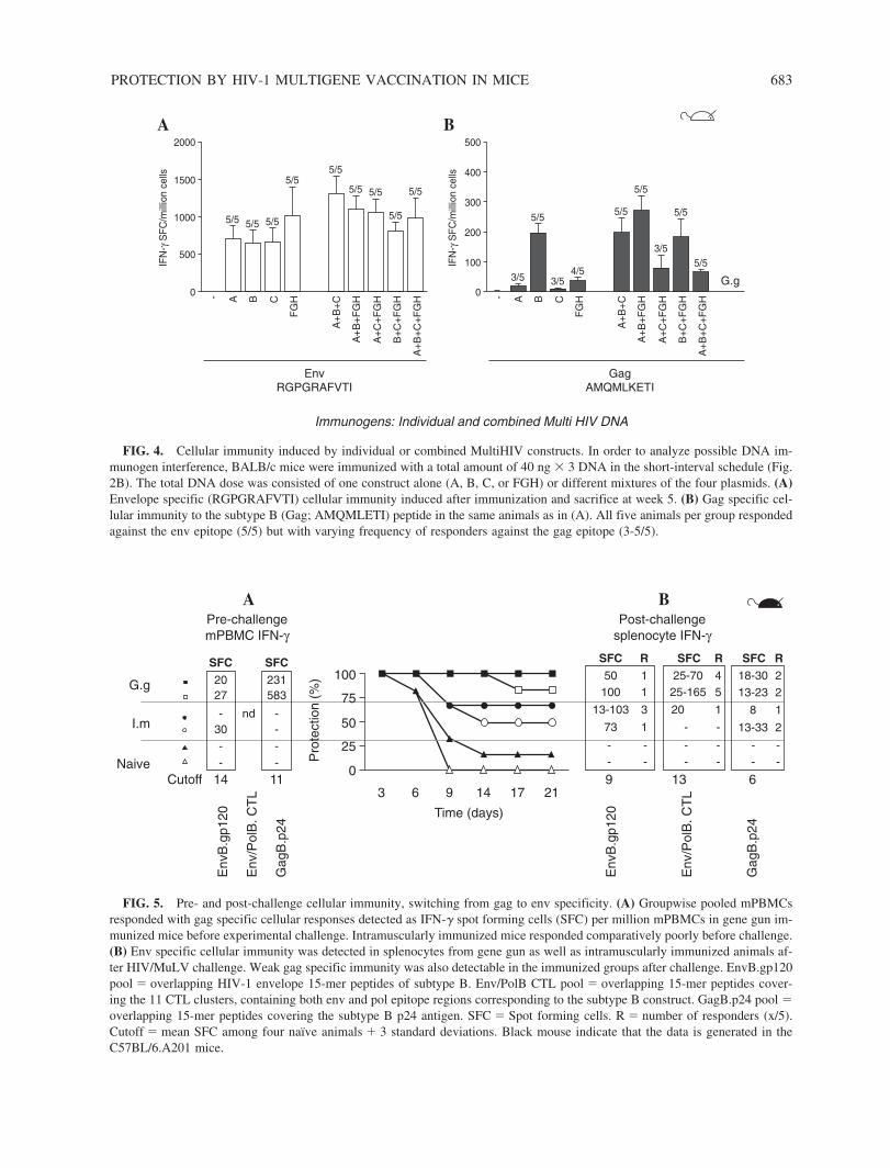
PROTECTION BY HIV-1 MULTIGENE VACCINATION IN MICE 683
A
0
5/5
IFN
-γ S
FC
/mill
ion
cells
500
1000
1500
2000
Immunogens: Individual and combined Multi HIV DNA
GagAMQMLKETI
G.g
EnvRGPGRAFVTI
A- B C
FG
H
5/5 5/5
5/55/5
A+
B+
C
A+
B+
FG
H
A+
C+
FG
H
B+
C+
FG
H
5/5 5/5
5/5
A+
B+
C+
FG
H
5/5
B
0
3/5
IFN
-γ S
FC
/mill
ion
cells
100
200
300
400
500
A- B C
FG
H
5/5
3/54/5
5/5
A+
B+
C
A+
B+
FG
H
A+
C+
FG
H
B+
C+
FG
H
5/5
3/5
5/5
A+
B+
C+
FG
H
5/5
A
14Cutoff
Pro
tect
ion
(%)
Naive
Env
B.g
p120
Env
/Pol
B. C
TL
Gag
B.p
24
Env
B.g
p120
Env
/Pol
B. C
TL
Gag
B.p
24
I.m
G.g100
75
25
50
0
3
Time (days)
11
SFC SFC20 23127 583
30 -- -
- -- -
nd
Pre-challengemPBMC IFN-γ
B
9 13 6
SFC SFC50
R R1 25-70 4
SFC R18-30 213-23 2
13-33 2
8 1
100 1 25-165 5
13-103 3
73 1
20 1
- -
- --
--
---
- -- -
Post-challengesplenocyte IFN-γ
6 9 14 17 21
FIG. 4. Cellular immunity induced by individual or combined MultiHIV constructs. In order to analyze possible DNA im-munogen interference, BALB/c mice were immunized with a total amount of 40 ng � 3 DNA in the short-interval schedule (Fig.2B). The total DNA dose was consisted of one construct alone (A, B, C, or FGH) or different mixtures of the four plasmids. (A)Envelope specific (RGPGRAFVTI) cellular immunity induced after immunization and sacrifice at week 5. (B) Gag specific cel-lular immunity to the subtype B (Gag; AMQMLETI) peptide in the same animals as in (A). All five animals per group respondedagainst the env epitope (5/5) but with varying frequency of responders against the gag epitope (3-5/5).
FIG. 5. Pre- and post-challenge cellular immunity, switching from gag to env specificity. (A) Groupwise pooled mPBMCsresponded with gag specific cellular responses detected as IFN-� spot forming cells (SFC) per million mPBMCs in gene gun im-munized mice before experimental challenge. Intramuscularly immunized mice responded comparatively poorly before challenge.(B) Env specific cellular immunity was detected in splenocytes from gene gun as well as intramuscularly immunized animals af-ter HIV/MuLV challenge. Weak gag specific immunity was also detectable in the immunized groups after challenge. EnvB.gp120pool � overlapping HIV-1 envelope 15-mer peptides of subtype B. Env/PolB CTL pool � overlapping 15-mer peptides cover-ing the 11 CTL clusters, containing both env and pol epitope regions corresponding to the subtype B construct. GagB.p24 pool �overlapping 15-mer peptides covering the subtype B p24 antigen. SFC � Spot forming cells. R � number of responders (x/5).Cutoff � mean SFC among four naïve animals � 3 standard deviations. Black mouse indicate that the data is generated in theC57BL/6.A201 mice.
5919_12_p678-688 12/9/05 1:42 PM Page 683
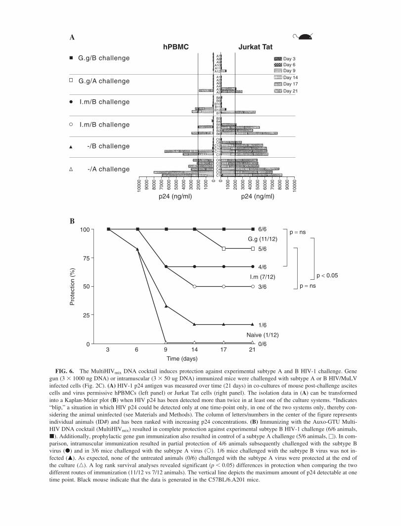
A
-/A challenge
p24 (ng/ml) p24 (ng/ml)
1000
0
9000
8000
9000
1000
0
8000
7000
7000
6000
6000
5000
5000
4000
4000
3000
3000
2000
2000
1000
1000
Day 3
0 0
-/B challenge
I.m/B challenge
I.m/B challenge
G.g/A challenge
G.g/B challenge
hPBMC Jurkat Tat
Day 6Day 9
Day 14Day 17
Day 21
C1C5C4C3C2C6
C7C11C10C12C8C9
B6B2B1B5B4B3
B7B12B11B10B9B8
A5A3A4A6A2A1
A12A11A10A9A8A7
B
Pro
tect
ion
(%)
6/6
5/6
p = ns
p = ns
p < 0.054/6
3/6
1/6
G.g (11/12)
I.m (7/12)
Naive (1/12)
0/6
100
75
25
50
03
Time (days)
6 9 14 17 21
FIG. 6. The MultiHIVmix DNA cocktail induces protection against experimental subtype A and B HIV-1 challenge. Genegun (3 � 1000 ng DNA) or intramuscular (3 � 50 ug DNA) immunized mice were challenged with subtype A or B HIV/MuLVinfected cells (Fig. 2C). (A) HIV-1 p24 antigen was measured over time (21 days) in co-cultures of mouse post-challenge ascitescells and virus permissive hPBMCs (left panel) or Jurkat Tat cells (right panel). The isolation data in (A) can be transformedinto a Kaplan-Meier plot (B) when HIV p24 has been detected more than twice in at least one of the culture systems. *Indicates“blip,” a situation in which HIV p24 could be detected only at one time-point only, in one of the two systems only, thereby con-sidering the animal uninfected (see Materials and Methods). The column of letters/numbers in the center of the figure representsindividual animals (ID#) and has been ranked with increasing p24 concentrations. (B) Immunizing with the Auxo-GTU Multi-HIV DNA cocktail (MultiHIVmix) resulted in complete protection against experimental subtype B HIV-1 challenge (6/6 animals,�). Additionally, prophylactic gene gun immunization also resulted in control of a subtype A challenge (5/6 animals, �). In com-parison, intramuscular immunization resulted in partial protection of 4/6 animals subsequently challenged with the subtype Bvirus (�) and in 3/6 mice challenged with the subtype A virus (�). 1/6 mice challenged with the subtype B virus was not in-fected (�). As expected, none of the untreated animals (0/6) challenged with the subtype A virus were protected at the end ofthe culture (�). A log rank survival analyses revealed significant (p � 0.05) differences in protection when comparing the twodifferent routes of immunization (11/12 vs 7/12 animals). The vertical line depicts the maximum amount of p24 detectable at onetime point. Black mouse indicate that the data is generated in the C57BL/6.A201 mice.
5919_12_p678-688 12/9/05 1:42 PM Page 684
where also detected in the intramuscular immunized an-imals but were absent in the 12 naïve animals.
Antibody responses. Antibodies against recombinantGag A-, B-, C-, and FGH -clade proteins were detectedin a few BALB/c mice after immunizing with the Mul-tiHIVmix. In C57BL/6 mice antibody production was analyzed at week 5 after the immunization and afterHIV/MuLV challenge. Before the challenge antibodieswere induced against all four rGag antigens only in a genegun–immunized group. After challenging antibodieswere raised in all treated groups, including the nonim-munized group. Overall the level of antibodies detectedwas low (data not shown).
Protection from HIV-1/MuLV challenge. The pro-tective effect of the vaccination was tested by inoculationof HIV/MuLV infected syngenic cells into the peritoneumof the vaccinated mice, followed by HIV isolation on as-cites fluid withdrawn from the animals after 10 days. Theascites cells were recovered by co-cultivation with hPBMCor the Jurkat Tat cell line in vitro and culture supernatantsamples were collected over a 21-day period (Fig. 6A).The Kaplan-Meier curves in Figure 6B are based on virusisolation on both hPBMC and Jurkat Tat cells. If HIV p24could be detected more than once in at least one of the iso-lation systems during this culture period, then the animalwas regarded as infected. In the group that was immunizedby gene gun administration, all mice (6/6) were protectedfrom the subtype B challenge. When mice were challengedwith subtype A pseudovirus, only one out of six mice wasshown to be infected on day 17, showing 83% protection.With intramuscular immunization the protection rates ofmice after subtype B or subtype A challenge were 66%and 50%, respectively. In the nonimmunized challengecontrol group, none of the mice were protected from theinfection when subtype A challenge was introduced, how-ever one of six mice was spontaneously protected fromsubtype B challenge (16% protected).
A log rank survival analyses revealed that there weredifferences between the six groups of animals. Post hocpairwise comparisons however, showed no significantdifferences when comparing gene gun to intramuscularimmunization of groups that later were challenged withthe same virus. The most likely explanation being thatthe number of animals per group was too small (n � 6).When the groups that received the same vaccine treat-ment were analyzed as one group, regardless of subtypechallenge (g.g. 11/12 vs i.m. 7/12 protected) there was asignificant difference in vaccine route (p � 0.05).
DISCUSSION
There are two major aspects to consider when evalu-ating the potency of an HIV-1 vaccine. First of all, thevaccine should be capable to induce a strong immune re-
PROTECTION BY HIV-1 MULTIGENE VACCINATION IN MICE
sponse, especially CD8� T lymphocyte mediated reac-tions. Second, it is preferable to demonstrate that the vac-cine-induced immunity has in vivo efficacy, already atthe level of preclinical studies. Because it is not possibleto use natural HIV-1 challenge in experimental animals,substitution methods must be used to evaluate protection.In this study we have used a mouse model to show thatthe HIV-1 GTU-MultiHIVmix vaccine meets both crite-ria; that of inducing virus-specific CD8� T cellular im-munity as well as protection against experimental HIV-1challenge.
In the first part of the study we immunized BALB/cmice with the MultiHIVmix DNA cocktail using gene gunas the way of delivery, since this method has earlier beenthe most effective when single clade (B and C) Multi-HIV constructs were used (Blazevic et al., submitted forpublication) and also shown by other research groups(4,33). Even though low DNA doses were used (8–1000ng/immunization), all doses resulted in high HIV enve-lope specific ELISPOT reactivity with 1000-4000 IFN-� SFC/million cells. There was no clear dose responseobserved when the highly immunogenic envelope pep-tide (RGPGRAFVTI) was used in the IFN-� ELISPOTassay. However, gag-specific immunity, assayed againstthe AMQMLKETI epitope was weaker than the enveloperesponse and dose-dependent. The highest gag-specificcellular response was observed in the group of animalsthat received the 3 � 1000 nanogram dose, a dose thatwas therefore used for gene gun immunizations in thesubsequent challenge experiment.
In the next study we used a shorter immunizationschedule (0, 1, and 3 weeks) to find out if even this sched-ule, more suitable for a challenge experiment, would givecomparable immune responses. Three different immu-nization routes (gene gun, i.d., and i.m. deliveries wereevaluated in parallel. The selection of doses for the i.m.and i.d. immunizations was based on our earlier studieswith the single clade MultiHIV construct. The shorten-ing of the immunization schedule did not affect the re-sponses induced by gene gun immunization, which wassuperior compared to the other two routes tested. Inter-estingly the response to both the env and the gag peptideincreased at 17 weeks as compared to the response at 5weeks, when immunization was by the i.m. This resultmay reflect the fact that in the muscle, the plasmid ismost likely taken up by nondividing and stable musclecells, whereas by the intradermal route, the plasmid willbe taken up by professional APCs such as dendritic cells(4,6) that are able to process and present the epitopesfaster and more efficiently to T cells.
The rationale in our vaccine development has been touse multiclade constructs, to create a broad immune re-sponse, targeted against multiple epitopes in the differ-ent HIV-1 subtypes (9,14). Theoretically, however, animmunodominant epitope in one clade could repress the
685
5919_12_p678-688 12/9/05 1:42 PM Page 685
response against recessive epitopes in other clades(27,13). To address this question, we next compared theimmune response obtained when mice were immunizedwith the single clade MultiHIV construct or with mix-tures of three or four plasmids. When the env peptideRGPGRAFVTI was used as a target antigen, MultiHIVmixtures gave somewhat higher responses than immu-nization with each single plasmid, although this differ-ence was not statistically different. Because the envelopeepitope used for restimulation is equally present in allconstructs and the total amount of DNA delivered to eachanimal was identical, it can be speculated that there is atrend towards envelope specific immune enhancement bythe mixture of four constructs. For the gag peptide, thesequence in the B clade vaccine (AMQMLKETI) differswith one amino acid from the other three vaccines(AMQMLDETI). Therefore, the animals immunized withthe non-B MultiHIV plasmids responded to the gag pep-tide but to a less degree than the B-clade immunized an-imals. Furthermore, in the animals immunized with thecocktail of all four plasmids (MultiHIVmix), the gag-spe-cific response was weaker. This finding is not surprisingas the MultiHIVmix contains only one fourth of the B-clade plasmid of the total DNA amount, which might beless than required to induce the stronger gag-specific re-sponse seen in other groups. In fact, we are currently clar-ifying the effects of mixing the four plasmids together,by comparing the gag-specific immune responses in miceimmunized with the single B plasmid and the Multi-HIVmix by using identical amount of plasmid B in bothcases.
The weak antibody responses observed were antici-pated, because DNA plasmid immunization usually re-quires high doses of DNA for the induction of antibod-ies. In addition, use of different adjuvants is generallyrequired (5,24). Our previous data suggest that protectionfrom experimental HIV-1/MuLV challenge is primarilybased on the action of T cells, recognizing and killing theengrafted HIV-1 infected cells (10). In addition, we haveshown that in this challenge model the protection is notmediated by anti-MuLV immunity (29). As gene gun de-livery of the MultiHIVmix induced strong CD8� T cellrestricted IFN-� production in BALB/c mice, as well asin PBMCs from C57BL/6.A201 mice, we hypothesizethat this vaccine would protect against a subsequent ex-perimental HIV-1 challenge.
Indeed, we were able to show that 83–100% of themice were protected after gene gun immunization and50–66% when the intramuscular route was used. This dif-ference in protection proves to be significantly differentusing survival analyses (p � 0.05). Looking at the im-mune responses in the immunized C57BL/6.A201 miceassayed before challenge, the ELISPOT reactivity sug-gests that animals immunized with gene gun have a high
MALM ET AL.
level of gag specific T cells whereas intramuscularly im-munized animals do not. We speculate that these gag-specific CD8� T cells are responsible for the clearanceof the incoming challenge graft. However, the fact thati.m immunized mice can mount partial protection evenwith low pre-challenge HIV-specific T cells reactivity issomewhat puzzling. The explanation could be in the lim-ited sensitivity of the mPBMC ELISpot assay for inter-feron, with another possible reason being that kinetics ofimmune response was different. Hence, we have showndelayed cellular immunogenicity (higher at week 17 thanat week 5 post–initial immunization) in BALB/c mice af-ter i.m. administration. This was supported by the factthat there is detectable post-challenge cellular immunityamong the i.m. immunized animals but not among thenaïve controls, demonstrating that vaccine priming hadoccurred.
In conclusion, we have demonstrated that a multi-epi-tope DNA immunogen (GTU-MultiHIV), if delivered bygene gun immunization, can induce a strong cellular im-munity in mice, even when only a few nanograms ofDNA was used. Furthermore, a cocktail of four differentMultiHIV plasmids expressing sequences from subtypesA, B, C, and FGH can be used to induce similar cellularimmunity against one of the epitopes (env) but with a re-duced response against a second epitope (gag), if com-pared to immunization with each single DNA. These dataillustrate the complexity of intra- and inter-plasmid im-munodominance, a problematic issue for many DNA vac-cines (27,13,34).
The cocktail of MultiHIV protected 19/24 animalsagainst an experimental HIV-1 challenge. This experi-ment also demonstrates cross-clade protection, as the sub-types A and B viruses used for challenge were derivedfrom different subtypes than the sequences found in theclade A or B specific MultiHIV constructs. Furthermore,it demonstrates that consensus approach used in Multi-HIVmix vaccine is functional and causes protectionagainst naturally occurring isolates. Gene gun immu-nization was superior to intramuscular immunization interms of both the T cell immunity induced, as well ashigher frequency of protection. Data generated in thiswork support the hypothesis that DNA representing sev-eral HIV-1 subtypes and several genes are immunogenicand protective.
ACKNOWLEDGMENTS
The authors thank Margareta Hagelin and ReinholdBenthin for excellent animal handling. This study wassupported by the Karolinska Institutet, the Swedish Re-search Council and a multicenter grant from the EU Com-mission (AVIP grant LSHP-CT-2004-503487). Also ac-
686
5919_12_p678-688 12/9/05 1:42 PM Page 686
knowledged is the excellent technical assistance of MarjaPirinen, Satu Ruisniemi, and Tuula Myllymäki. The au-thors also thank Minna Valtavaara, Rein Sikut, UrveToots, Andres Männik, Toomas Kivisild, and Marja Ado-jaan for their important contributions.
REFERENCES
1. Barouch, D.H., S. Santra, J. E. Schmitz, M. J. Kuroda, T.M. Fu, W. Wagner, M. Bilska, A. Craiu, X.X. Zheng, G.R.Krivulka, K. Beaudry, M.A. Lifton, C.E. Nickerson, W.L.Trigona, K. Punt, D.C. Freed, L. Guan, S. Dubey, D.Casimiro, A. Simon, M. E. Davies, M. Chastain, T.B.Strom, R.S. Gelman, D.C. Montefiori, M.G. Lewis, E.A.Emini, J.W. Shiver, and N.L. Letvin. 2000. Control ofviremia and prevention of clinical AIDS in rhesus monkeysby cytokine-augmented DNA vaccination. Science290:486–492.
2. Barouch, D.H., J. Kunstman, J. Glowczwskie, K.J. Kunst-man, M.A. Egan, F.W. Peyerl, S. Santra, M.J. Kuroda, J.E.Schmitz, K. Beaudry, G.R. Krivulka, M.A. Lifton, D.A.Gorgone, S.M. Wolinsky, and N.L. Letvin. 2003. Viral es-cape from dominant simian immunodeficiency virus epi-tope-specific cytotoxic T lymphocytes in DNA-vaccinatedrhesus monkeys. J. Virol. 77:7367–7375.
3. Browning, J., J.W. Horner, M. Pettoello-Mantovani, C.Raker, S. Yurasov, R.A. DePinho, and H. Goldstein. 1997.Mice transgenic for human CD4 and CCR5 are suscepti-ble to HIV infection. Proc. Natl. Acad. Sci. U.S.A. 94:14637–14641.
4. Cohen, A.D., J.D. Boyer, and D.B. Weiner. 1998. Modu-lating the immune response to genetic immunization.FASEB J. 12:1611–1626.
5. Collings, A., J. Pitkanen, M. Strengell, M. Tahtinen, A.Lagerstedt, K. Hakkarainen, V. Ovod, G. Sutter, M. Ustav,E. Ustav, A. Mannik, A. Ranki, P. Peterson, and K. Krohn.1999. Humoral and cellular immune responses to HIV-1nef in mice DNA-immunised with non-replicating or self-replicating expression vectors. Vaccine 18:460–467.
6. Creusot, R.J., L.L. Thomsen, C.A. van Wely, P. Topley,J.P. Tite, and B.M. Chain. 2001. Early commitment ofadoptively transferred CD4� T cells following particle-mediated DNA vaccination: implications for the study ofimmunomodulation. Vaccine 19:1678–1687.
7. Devito, C., M. Levi, K. Broliden, and J. Hinkula. 2000.Mapping of B-cell epitopes in rabbits immunised with var-ious gag antigens for the production of HIV-1 gag captureELISA reagents. J. Immunol. Methods 238:69–80
8. Esser, M.T., R.D. Marchese, L.S. Kierstead, L.G. Tussey,F. Wang, N. Chirmule, and M. W. Washabaugh. 2003.Memory T cells and vaccines. Vaccine 21:419–430.
9. Gaschen, B., J. Taylor, K. Yusim, B. Foley, F. Gao, D.Lang, V. Novitsky, B. Haynes, B. H. Hahn, T. Bhat-
PROTECTION BY HIV-1 MULTIGENE VACCINATION IN MICE
tacharya, and B. Korber. 2002. Diversity considerations inHIV-1 vaccine selection. Science 296:2354–2360.
10. Hinkula, J., E. Rollman, P. Lundholm, R. Benthin, K.Okuda, and B. Wahren. 2004. Genetic immunization withmultiple HIV-1 genes provides protection against HIV-1/MuLV pseudovirus challenge in vivo. Cells Tissues Or-gans 177:169–184.
11. Irwin, M.J., W.R. Heath, and L.A. Sherman. 1989. Species-restricted interactions between CD8 and the alpha 3 do-main of class I influence the magnitude of the xenogeneicresponse. J. Exp. Med. 170:1091–1101.
12. Kaul, R., F.A. Plummer, J. Kimani, T. Dong, P. Kiama, T.Rostron, E. Njagi, K.S. MacDonald, J.J. Bwayo, A.J.McMichael, and S.L. Rowland-Jones. 2000. HIV-1-spe-cific mucosal CD8� lymphocyte responses in the cervixof HIV-1-resistant prostitutes in Nairobi. J. Immunol.164:1602–1611.
13. Kjerrstrom, A., J. Hinkula, G. Engstrom, V. Ovod, K.Krohn, R. Benthin, and B. Wahren. 2001. Interactions ofsingle and combined human immunodeficiency virus type1 (HIV-1) DNA vaccines. Virology 284:46–61.
14. Korber, B., M. Muldoon, J. Theiler, F. Gao, R. Gupta, A.Lapedes, B.H. Hahn, S. Wolinsky, and T. Bhattacharya.2000. Timing the ancestor of the HIV-1 pandemic strains.Science 288:1789–1796.
15. Korber, B.T.M., C. Brander, B.F. Haynes, R. Koup, C.Kuiken, J.P. Moore, B.D. Walker, and D.I. Watkins. 2001.HIV Molecular Immunology 2001, Publisher: Los AlamosNational Laboratory, Theoretical Biology and Biophysics,Los Alamos, New Mexico. LA-UR 02-4663. [online]http://www.hiv.lanl.gov/content/immunology/con-tents2001.html
16. Ljungberg, K., E. Rollman, L. Eriksson, J. Hinkula, and B.Wahren. 2002. Enhanced immune responses after DNAvaccination with combined envelope genes from differentHIV-1 subtypes. Virology 302:44–57.
17. Mascola, J.R., G. Stiegler, T.C. VanCott, H. Katinger, C.B.Carpenter, C.E. Hanson, H. Beary, D. Hayes, S.S. Frankel,D.L. Birx, and M.G. Lewis. 2000. Protection of macaquesagainst vaginal transmission of a pathogenic HIV-1/SIVchimeric virus by passive infusion of neutralizing antibod-ies. Nat. Med. 6:207–210.
18. Nkolola, J.P., E.G. Wee, E.J. Im, C.P. Jewell, N. Chen,X.N. Xu, A.J. McMichael, and T. Hanke. 2004. Engineer-ing RENTA, a DNA prime-MVA boost HIV vaccine tailored for Eastern and Central Africa. Gene Ther. 11:1068–1080.
19. Letvin, N.L. 2002. Strategies for an HIV vaccine. Clin. In-vest, 110:15–27
20. Norris, P.J., H.F. Moffett, O.O. Yang, D.E. Kaufmann, M.J.Clark, M.M. Addo, and E.S. Rosenberg. 2004. Beyondhelp: direct effector functions of human immunodeficiencyvirus type 1-specific CD4(�) T cells. J. Virol. 78:8844–8851.
687
5919_12_p678-688 12/9/05 1:42 PM Page 687
21. Petrovas, C., Y.M. Mueller, and P.D. Katsikis. 2004. HIV-specific CD8� T cells: serial killers condemned to die?Curr. HIV Res. 2:153–162.
22. Puaux, A.L., and M.L. Michel. 2003. New gene-based ap-proaches for an AIDS vaccine. Comp. Immunol. Micro-biol. Infect. Dis. 26:357–372.
23. Qiu, J.T., R. Song, M. Dettenhofer, C. Tian, T. August,B.K. Felber, G.N. Pavlakis, and X.F. Yu. 1999. Evaluationof novel human immunodeficiency virus type 1 Gag DNAvaccines for protein expression in mammalian cells and in-duction of immune responses. J. Virol. 73:9145–9152.
24. Rollman, E., J. Hinkula, J. Arteaga, B. Zuber, A. Kjerrstrom,M. Liu, B. Wahren, and K. Ljungberg. 2004. Multi-subtypegp160 DNA immunization induces broadly neutralizing anti-HIV antibodies. Gene Ther. 11:1146–1154.
25. Sallusto, F., D. Lenig, R. Forster, M. Lipp, and A. Lanza-vecchia. 1999. Two subsets of memory T lymphocytes withdistinct homing potentials and effector functions. Nature401:708–712.
26. Seaman, M.S., F.W. Peyerl, S.S. Jackson, M. A. Lifton,D.A. Gorgone, J.E. Schmitz, and N.L. Letvin. 2004. Sub-sets of memory cytotoxic T lymphocytes elicited by vac-cination influence the efficiency of secondary expansion invivo. J. Virol. 78:206–215.
27. Sedegah, M., Y. Charoenvit, L. Minh, M. Belmonte, V.F.Majam, S. Abot, H. Ganeshan, S. Kumar, D.J. Bacon, A.Stowers, D.L. Narum, D.J. Carucci, and W.O. Rogers.2004. Reduced immunogenicity of DNA vaccine plasmidsin mixtures. Gene Ther. 11:448–456.
28. Spector, D.H., E. Wade, D.A. Wright, V. Koval, C. Clark,D. Jaquish, and S.A. Spector. 1990. Human immunodefi-ciency virus pseudotypes with expanded cellular andspecies tropism. J. Virol. 64:2298–2308.
29. Spetz, A.L., A.S. Sorensen, L. Walther-Jallow, B. Wahren,J. Andersson, L. Holmgren, and J. Hinkula. 2002. Induc-
MALM ET AL.
tion of HIV-1-specific immunity after vaccination withapoptotic HIV-1/murine leukemia virus-infected cells. J.Immunol. 169:5771–5779.
30. Takahashi, H., J. Cohen, A. Hosmalin, K.B. Cease, R.Houghten, J.L. Cornette, C. DeLisi, B. Moss, R.N. Ger-main, and J.A. Berzofsky. 1988. An immunodominant epitope of the human immunodeficiency virus envelopeglycoprotein gp160 recognized by class I major histocom-patibility complex molecule-restricted murine cytotoxic Tlymphocytes. Proc. Natl. Acad. Sci. U.S.A. 85:3105–3109.
31. Vitiello, A., D. Marchesini, J. Furze, L.A. Sherman, and R.W.Chesnut. 1991. Analysis of the HLA-restricted influenza-spe-cific cytotoxic T lymphocyte response in transgenic mice car-rying a chimeric human-mouse class I major histocompati-bility complex. J. Exp. Med. 173:1007–1015.
32. Vodros, D., and E.M. Fenyo. 2004. Primate models for hu-man immunodeficiency virus infection. Evolution of re-ceptor use during pathogenesis. Acta Microbiol. Immunol.Hung. 51:1–29.
33. Wolff, J.A. 1997. Naked DNA transport and expression inmammalian cells. Neuromusc. Disord. 7:314–318.
34. Yewdell, J.W., and M. Del Val. 2004. Immunodominancein TCD8� responses to viruses: cell biology, cellular im-munology, and mathematical models. Immunity 21:149–153.
Address reprint requests to:Dr. Vesna Blazevic
FIT Biotech Oyj Plc.Biokatu 8
33520 Tampere, Finland
E-mail: [email protected]
Received March 30, 2005; accepted August 9, 2005.
688
5919_12_p678-688 12/9/05 1:42 PM Page 688
BRIEF REPORT
Immunization with dendritic cells transfected in vivo with HIV-1plasmid DNA induces HIV-1-specific immune responses
Maria Malm • Kai Krohn • Vesna Blazevic
Received: 5 January 2011 / Accepted: 12 April 2011
� Springer-Verlag 2011
Abstract We evaluated the importance of dendritic cells
(DCs) in the induction of the immune response after
immunization of mice with DNA plasmid Auxo-GTU�-
MultiHIV. First, GTU�-encoded protein was shown to be
expressed by DCs of the draining lymph nodes (LNs) fol-
lowing intradermal (i.d.) immunization. Next, donor mice
were immunized with the MultiHIV DNA plasmid, and
DCs were enriched and further used to immunize naıve
recipient mice. For the first time, the results show that i.d.
immunization with Auxo-GTU�-MultiHIV transfects DCs
in vivo, enabling them to present antigens and induce HIV-
specific immune responses in recipient mice.
Dendritic cells (DCs), the most important antigen-pre-
senting cells (APCs) in the body, have been shown to be
the only cell type that can efficiently present both endog-
enous and exogenous antigens (Ag) through major histo-
compatibility complex (MHC) class I and II molecules and
activate naıve CD8? T lymphocytes [2, 19, 21]. Following
dermal DNA delivery, DCs are either transfected directly
or acquire and present Ag from other transfected cells, a
phenomenon known as cross-presentation [21]. After
encountering an immunogen in vivo, DCs mature and
migrate from the periphery to the lymphoid organs, i.e., the
lymph node (LN) and spleen, the primary sites of Ag
presentation [2]. Nevertheless, the role and significance of
skin DCs in naked DNA plasmid immunization remain
poorly evaluated and understood.
GTU�-MultiHIV DNA plasmid has been shown to
induce strong HIV-1-specific cellular and humoral immune
responses in several preclinical studies in mice [3, 13, 15],
pigs [18] and macaques [17]. Furthermore, GTU�-based
HIV-1 vaccines have been evaluated in several phase I/II
clinical trials [13]. The results of the present study show
the direct involvement of DCs as APCs in DNA
immunization.
Auxo-GTU�-MultiHIV-B DNA encodes the B-clade
consensus HIV-1 multigene, containing fused full-length
sequences of rev, nef, tat, gag (p17 and p24) and 11 T-cell-
epitope-rich clusters from pol and env polypeptides [3, 15].
The GTU� vector carrying the destabilized form of
enhanced green fluorescent protein (eGFP) derived from
vector pd1EGFP-N1 (7) instead of the MultiHIV antigen
was constructed. The expression and intensity of the GFP
was verified using a FACSCalibur flow cytometer (Becton-
Dickinson, NJ, USA) after in vitro transfection (Cellfectin�
Reagent, Invitrogen) of Jurkat cells with GTU�-GFP
plasmid.
For the GFP in vivo expression study, mice were
immunized twice, 24 hours apart with 50 ll of GTU�-GFP
plasmid (*250 lg/delivery) i.d. in the base of the tail
(Table 1). On the third day, mice were sacrificed, and
lumbar and sacral LNs of the experimental mice and con-
trol mice were collected and pooled. Mice immunized with
the plasmid lacking GFP were used as controls. Cells were
digested from the tissues by incubating twice with 1 mg/ml
collagenase D (Sigma) at 37�C, 5% CO2 for 30 minutes,
collected, and mixed with Optiprep (Axis-Shield PoC AS,
M. Malm (&)
FIT Biotech Oy, Biokatu 8, 33520 Tampere, Finland
e-mail: [email protected]
K. Krohn
Department of Pathology, Tampere University Hospital
and CliniXion Oy, Tampere, Finland
V. Blazevic
Vaccine Research Center, Virology, University of Tampere
Medical School, Tampere, Finland
123
Arch Virol
DOI 10.1007/s00705-011-1003-2
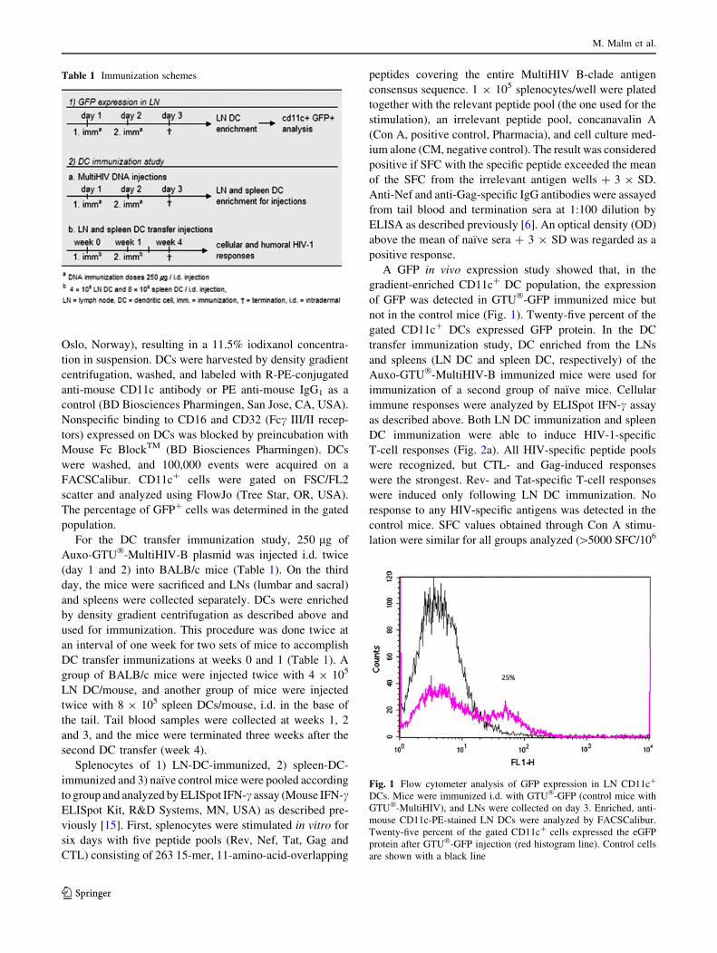
Oslo, Norway), resulting in a 11.5% iodixanol concentra-
tion in suspension. DCs were harvested by density gradient
centrifugation, washed, and labeled with R-PE-conjugated
anti-mouse CD11c antibody or PE anti-mouse IgG1 as a
control (BD Biosciences Pharmingen, San Jose, CA, USA).
Nonspecific binding to CD16 and CD32 (Fcc III/II recep-
tors) expressed on DCs was blocked by preincubation with
Mouse Fc BlockTM (BD Biosciences Pharmingen). DCs
were washed, and 100,000 events were acquired on a
FACSCalibur. CD11c? cells were gated on FSC/FL2
scatter and analyzed using FlowJo (Tree Star, OR, USA).
The percentage of GFP? cells was determined in the gated
population.
For the DC transfer immunization study, 250 lg of
Auxo-GTU�-MultiHIV-B plasmid was injected i.d. twice
(day 1 and 2) into BALB/c mice (Table 1). On the third
day, the mice were sacrificed and LNs (lumbar and sacral)
and spleens were collected separately. DCs were enriched
by density gradient centrifugation as described above and
used for immunization. This procedure was done twice at
an interval of one week for two sets of mice to accomplish
DC transfer immunizations at weeks 0 and 1 (Table 1). A
group of BALB/c mice were injected twice with 4 9 105
LN DC/mouse, and another group of mice were injected
twice with 8 9 105 spleen DCs/mouse, i.d. in the base of
the tail. Tail blood samples were collected at weeks 1, 2
and 3, and the mice were terminated three weeks after the
second DC transfer (week 4).
Splenocytes of 1) LN-DC-immunized, 2) spleen-DC-
immunized and 3) naıve control mice were pooled according
to group and analyzed by ELISpot IFN-c assay (Mouse IFN-cELISpot Kit, R&D Systems, MN, USA) as described pre-
viously [15]. First, splenocytes were stimulated in vitro for
six days with five peptide pools (Rev, Nef, Tat, Gag and
CTL) consisting of 263 15-mer, 11-amino-acid-overlapping
peptides covering the entire MultiHIV B-clade antigen
consensus sequence. 1 9 105 splenocytes/well were plated
together with the relevant peptide pool (the one used for the
stimulation), an irrelevant peptide pool, concanavalin A
(Con A, positive control, Pharmacia), and cell culture med-
ium alone (CM, negative control). The result was considered
positive if SFC with the specific peptide exceeded the mean
of the SFC from the irrelevant antigen wells ? 3 9 SD.
Anti-Nef and anti-Gag-specific IgG antibodies were assayed
from tail blood and termination sera at 1:100 dilution by
ELISA as described previously [6]. An optical density (OD)
above the mean of naıve sera ? 3 9 SD was regarded as a
positive response.
A GFP in vivo expression study showed that, in the
gradient-enriched CD11c? DC population, the expression
of GFP was detected in GTU�-GFP immunized mice but
not in the control mice (Fig. 1). Twenty-five percent of the
gated CD11c? DCs expressed GFP protein. In the DC
transfer immunization study, DC enriched from the LNs
and spleens (LN DC and spleen DC, respectively) of the
Auxo-GTU�-MultiHIV-B immunized mice were used for
immunization of a second group of naıve mice. Cellular
immune responses were analyzed by ELISpot IFN-c assay
as described above. Both LN DC immunization and spleen
DC immunization were able to induce HIV-1-specific
T-cell responses (Fig. 2a). All HIV-specific peptide pools
were recognized, but CTL- and Gag-induced responses
were the strongest. Rev- and Tat-specific T-cell responses
were induced only following LN DC immunization. No
response to any HIV-specific antigens was detected in the
control mice. SFC values obtained through Con A stimu-
lation were similar for all groups analyzed ([5000 SFC/106
Table 1 Immunization schemes
Fig. 1 Flow cytometer analysis of GFP expression in LN CD11c?
DCs. Mice were immunized i.d. with GTU�-GFP (control mice with
GTU�-MultiHIV), and LNs were collected on day 3. Enriched, anti-
mouse CD11c-PE-stained LN DCs were analyzed by FACSCalibur.
Twenty-five percent of the gated CD11c? cells expressed the eGFP
protein after GTU�-GFP injection (red histogram line). Control cells
are shown with a black line
M. Malm et al.
123
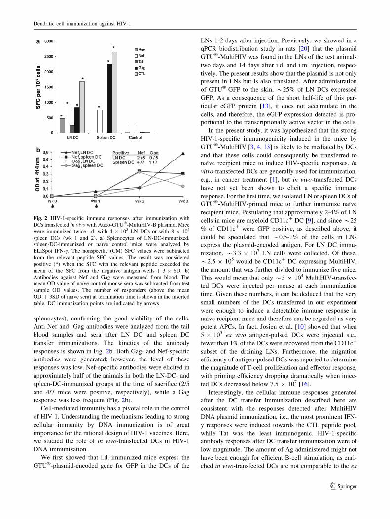
splenocytes), confirming the good viability of the cells.
Anti-Nef and -Gag antibodies were analyzed from the tail
blood samples and sera after LN DC and spleen DC
transfer immunizations. The kinetics of the antibody
responses is shown in Fig. 2b. Both Gag- and Nef-specific
antibodies were generated; however, the level of these
responses was low. Nef-specific antibodies were elicited in
approximately half of the animals in both the LN-DC- and
spleen-DC-immunized groups at the time of sacrifice (2/5
and 4/7 mice were positive, respectively), while a Gag
response was less frequent (Fig. 2b).
Cell-mediated immunity has a pivotal role in the control
of HIV-1. Understanding the mechanisms leading to strong
cellular immunity by DNA immunization is of great
importance for the rational design of HIV-1 vaccines. Here,
we studied the role of in vivo-transfected DCs in HIV-1
DNA immunization.
We first showed that i.d.-immunized mice express the
GTU�-plasmid-encoded gene for GFP in the DCs of the
LNs 1-2 days after injection. Previously, we showed in a
qPCR biodistribution study in rats [20] that the plasmid
GTU�-MultiHIV was found in the LNs of the test animals
two days and 14 days after i.d. and i.m. injection, respec-
tively. The present results show that the plasmid is not only
present in LNs but is also translated. After administration
of GTU�-GFP to the skin, *25% of LN DCs expressed
GFP. As a consequence of the short half-life of this par-
ticular eGFP protein [13], it does not accumulate in the
cells, and therefore, the eGFP expression detected is pro-
portional to the transcriptionally active vector in the cells.
In the present study, it was hypothesized that the strong
HIV-1-specific immunogenicity induced in the mice by
GTU�-MultiHIV [3, 4, 13] is likely to be mediated by DCs
and that these cells could consequently be transferred to
naıve recipient mice to induce HIV-specific responses. In
vitro-transfected DCs are generally used for immunization,
e.g., in cancer treatment [1], but in vivo-transfected DCs
have not yet been shown to elicit a specific immune
response. For the first time, we isolated LN or spleen DCs of
GTU�-MultiHIV-primed mice to further immunize naıve
recipient mice. Postulating that approximately 2-4% of LN
cells in mice are myeloid CD11c? DC [9], and since *25
% of CD11c? were GFP positive, as described above, it
could be speculated that *0.5-1% of the cells in LNs
express the plasmid-encoded antigen. For LN DC immu-
nization, *3.3 9 107 LN cells were collected. Of these,
*2.5 9 105 would be CD11c? DC-expressing MultiHIV,
the amount that was further divided to immunize five mice.
This would mean that only *5 9 104 MultiHIV-transfec-
ted DCs were injected per mouse at each immunization
time. Given these numbers, it can be deduced that the very
small numbers of the DCs transferred in our experiment
were enough to induce a detectable immune response in
naıve recipient mice and therefore can be regarded as very
potent APCs. In fact, Josien et al. [10] showed that when
5 9 105 ex vivo antigen-pulsed DCs were injected s.c.,
fewer than 1% of the DCs were recovered from the CD11c?
subset of the draining LNs. Furthermore, the migration
efficiency of antigen-pulsed DCs was reported to determine
the magnitude of T-cell proliferation and effector response,
with priming efficiency dropping dramatically when injec-
ted DCs decreased below 7.5 9 107 [16].
Interestingly, the cellular immune responses generated
after the DC transfer immunization described here are
consistent with the responses detected after MultiHIV
DNA plasmid immunization, i.e., the most prominent IFN-
y responses were induced towards the CTL peptide pool,
while Tat was the least immunogenic. HIV-1-specific
antibody responses after DC transfer immunization were of
low magnitude. The amount of Ag administered might not
have been enough for efficient B-cell stimulation, as enri-
ched in vivo-transfected DCs are not comparable to the ex
0
500
1000
1500
2000
2500
3000
LN DC Spleen DC Control
SF
C p
er 1
06
cells
Rev
Nef
Tat
Gag
CTL
*
*
*
* *
*
*
0
500
1000
1500
2000
2500
3000
LN DC Spleen DC Control
SF
C p
er 1
06
cells
Rev
Nef
Tat
Gag
CTL
*
*
*
* *
*
*a
b
Fig. 2 HIV-1-specific immune responses after immunization with
DCs transfected in vivo with Auxo-GTU�-MultiHIV-B plasmid. Mice
were immunized twice i.d. with 4 9 105 LN DCs or with 8 9 105
spleen DCs (wk 1 and 2). a) Splenocytes of LN-DC-immunized,
spleen-DC-immunized or naıve control mice were analyzed by
ELISpot IFN-c. The nonspecific (CM) SFC values were subtracted
from the relevant peptide SFC values. The result was considered
positive (*) when the SFC with the relevant peptide exceeded the
mean of the SFC from the negative antigen wells ? 3 9 SD. b)
Antibodies against Nef and Gag were measured from blood. The
mean OD value of naıve control mouse sera was subtracted from test
sample OD values. The number of responders (above the mean
OD ? 3SD of naıve sera) at termination time is shown in the inserted
table. DC immunization points are indicated by arrows
Dendritic cell immunization against HIV-1
123
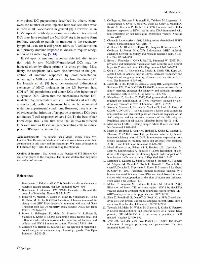
vivo-pulsed DC preparations described by others. More-
over, the number of cells injected here was less than what
is used in DC vaccination in general [8]. However, as an
HIV-1-specific antibody response was induced, transferred
DCs must have retained the MultiHIV Ag in its native form
for long enough to permit its transport to the secondary
lymphoid tissue for B-cell presentation, as B-cell activation
in a primary immune response is known to require recog-
nition of an intact Ag [5, 14].
HIV-1-specific immune responses detected after injec-
tion with in vivo MultiHIV-transfected DCs may be
induced either by direct priming or cross-priming. Most
likely, the recipients DCs were also involved in the gen-
eration of immune responses by cross-presentation,
obtaining the MHC-peptide molecules from the donor DC.
De Heusch et al. [8] have demonstrated bidirectional
exchange of MHC molecules in the LN between host
CD11c? DC populations and donor DCs after injection of
allogeneic DCs. Given that the mechanisms behind DC-
mediated Ag presentation are still undefined and not fully
characterized, both mechanisms have to be recognized
under our experimental conditions. However, there is a line
of evidence that injection of apoptotic or necrotic DCs does
not induce T-cell responses in vivo [12]. To the best of our
knowledge, this is the first time that in vivo-transfected
DCs were used as HIV-1 antigen-presenting cells to induce
potent HIV-specific immunity.
Acknowledgments The authors thank Marja Pirinen, Tuula My-
llymaki, Satu Ruisniemi, Vladimir Ovod and Ioana Stanescu for their
contribution to this study and the manuscript. We thank colleagues at
FIT Biotech Oy, Tartu, for constructing the plasmids.
Conflict of interest Kai Krohn is the founder of FIT Biotech Oy
and owns shares of the company. The authors declare that they have
no conflict of interest.
References
1. Banchereau J, Palucka AK (2005) Dendritic cells as therapeutic
vaccines against cancer. Nat Rev Immunol 5:296–306
2. Banchereau J, Steinman RM (1998) Dendritic cells and the
control of immunity. Nature 392:245–252
3. Blazevic V, Mannik A, Malm M, Sikut R, Valtavaara M, Toots
U, Ustav M, Krohn K (2006) Induction of human immunodefi-
ciency virus (HIV Type-1)-specific immunity with a novel Gene
Transport Unit (GTU)-MultiHIV DNA vaccine. AIDS Res Hum
Retrovir 22:667–677
4. Brave A, Hallengard D, Malm M, Blazevic V, Rollman E,
Stanescu I, Krohn K (2009) Combining DNA technologies and
different modes of immunization for induction of humoral and
cellular anti-HIV-1 immune responses. Vaccine 27:184–186
5. Carrasco YR, Batista FD (2006) B cell recognition of membrane-
bound antigen: an exquisite way of sensing ligands. Curr Opin
Immunol 18:286–291
6. Collings A, Pitkanen J, Strengell M, Tahtinen M, Lagerstedt A,
Hakkarainen K, Ovod V, Sutter G, Ustav M, Ustav E, Mannik A,
Ranki A, Peterson P, Krohn K (1999) Humoral and cellular
immune responses to HIV-1 nef in mice DNA-immunised with
non-replicating or self-replicating expression vectors. Vaccine
18:460–467
7. Clontech Laboratories (1998) Living colors destabilized EGFP
vectors. Clontechniques XIII 2:16–17
8. de Heusch M, Blocklet D, Egrise D, Hauquier B, Vermeersch M,
Goldman S, Moser M (2007) Bidirectional MHC molecule
exchange between migratory and resident dendritic cells. J Leu-
koc Biol 82:861–868
9. Encke J, Findeklee J, Geib J, Pfaff E, Stremmel W (2005) Pro-
phylactic and therapeutic vaccination with dendritic cells against
hepatitis C virus infection. Clin Exp Immunol 142:362–369
10. Garg S, Oran A, Wajchman J, Sasaki S, Maris CH, Kapp JA,
Jacob J (2003) Genetic tagging shows increased frequency and
longevity of antigen-presenting, skin-derived dendritic cells in
vivo. Nat Immunol 4:907–912
11. Josien R, Li HL, Ingulli E, Sarma S, Wong BR, Vologodskaia M,
Steinman RM, Choi Y (2000) TRANCE, a tumor necrosis factor
family member, enhances the longevity and adjuvant properties
of dendritic cells in vivo. J Exp Med 191:495–502
12. Kleindienst P, Brocker T (2003) Endogenous dendritic cells are
required for amplification of T cell responses induced by den-
dritic cell vaccines in vivo. J Immunol 170:2817–2823
13. Krohn K, Stanescu I, Blazevic V, Vesikari T, Ranki A, Ustav M
(2005) A DNA HIV-1 vaccine based on a fusion gene expressing
non-structural and structural genes of consensus sequence of the
A-C subtypes and the ancestor sequence of the F-H subtypes.
Preclinical and clinical studies. Microbes Infect 7:1405–1413
14. MacLennan I (2007) Holding antigen where B cells can find it.
Nat Immunol 8:909–910
15. Malm M, Rollman E, Ustav M, Hinkula J, Krohn K, Wahren B,
Blazevic V (2005) Cross-clade protection induced by human
immunodeficiency virus-1 DNA immunogens expressing con-
sensus sequences of multiple genes and epitopes from subtypes
A, B, C, and FGH. Viral Immunol 18:678–688
16. MartIn-Fontecha A, Sebastiani S, Hopken UE, Uguccioni M,
Lipp M, Lanzavecchia A, Sallusto F (2003) Regulation of den-
dritic cell migration to the draining lymph node: impact on T
lymphocyte traffic and priming. J Exp Med 198:615–621
17. Martinon F, Kaldma K, Sikut R, Culina S, Romain G, Tuomela
M, Adojaan M, Mannik A, Toots U, Kivisild T, Morin J, Bro-
chard P, Delache B, Tripiciano A, Ensoli F, Stanescu I, Le Grand
R, Ustav M (2009) Persistent immune responses induced by a
human immunodeficiency virus DNA vaccine delivered in asso-
ciation with electroporation in the skin of nonhuman primates.
Hum Gene Ther 20:1291–1307
18. Molder T, Adojaan M, Kaldma K, Ustav M, Sikut R (2009)
Elicitation of broad CTL response against HIV-1 by the DNA
vaccine encoding artificial multi-component fusion protein Mul-
tiHIV—study in domestic pigs. Vaccine 28:293–298
19. Shen Z, Reznikoff G, Dranoff G, Rock KL (1997) Cloned den-
dritic cells can present exogenous antigens on both MHC class I
and class II molecules. J Immunol 158:2723–2730
20. Tuomela M, Malm M, Wallen M, Stanescu I, Krohn K, Peterson
P (2005) Biodistribution and general safety of a naked DNA
plasmid, GTU-MultiHIV, in a rat, using a quantitative PCR
method. Vaccine 23:890–896
21. Vyas JM, Van der Veen AG, Ploegh HL (2008) The known
unknowns of antigen processing and presentation. Nat Rev
Immunol 8:607–618
M. Malm et al.
123