multigene phylogeny, beauvericin production and ... - mdpi
TRANSCRIPT

Citation: Rana, S.; Singh, S.K.;
Dufossé, L. Multigene Phylogeny,
Beauvericin Production and Bioactive
Potential of Fusarium Strains Isolated
in India. J. Fungi 2022, 8, 662.
https://doi.org/10.3390/jof8070662
Academic Editor: Ivan M. Dubovskiy
Received: 17 May 2022
Accepted: 21 June 2022
Published: 24 June 2022
Publisher’s Note: MDPI stays neutral
with regard to jurisdictional claims in
published maps and institutional affil-
iations.
Copyright: © 2022 by the authors.
Licensee MDPI, Basel, Switzerland.
This article is an open access article
distributed under the terms and
conditions of the Creative Commons
Attribution (CC BY) license (https://
creativecommons.org/licenses/by/
4.0/).
FungiJournal of
Article
Multigene Phylogeny, Beauvericin Production and BioactivePotential of Fusarium Strains Isolated in IndiaShiwali Rana 1,2 , Sanjay Kumar Singh 1,2,* and Laurent Dufossé 3,*
1 National Fungal Culture Collection of India, Biodiversity and Palaeobiology Group, MACS’ AgharkarResearch Institute, G.G. Agarkar Road, Pune 411004, India; [email protected]
2 Faculty of Science, Savitribai Phule Pune University, Ganeshkhind Road, Ganeshkhind, Pune 411007, India3 Chembiopro Chimie et Biotechnologie des Produits Naturels, ESIROI Département Agroalimentaire,
Université de la Réunion, F-97490 Sainte-Clotilde, Ile de La Réunion, France* Correspondence: [email protected] or [email protected] (S.K.S.);
[email protected] (L.D.); Tel.: +91-20-2532-5103 (S.K.S.); +33-66-873-1906 (L.D.)
Abstract: The taxonomy of the genus Fusarium has been in a flux because of ambiguous circum-scription of species-level identification based on morphotaxonomic criteria. In this study, multigenephylogeny was conducted to resolve the evolutionary relationships of 88 Indian Fusarium isolatesbased on the internal transcribed spacer region, 28S large subunit, translation elongation factor1-alpha, RNA polymerase second largest subunit, beta-tubulin and calmodulin gene regions. Fusar-ium species are well known to produce metabolites such as beauvericin (BEA) and enniatins. Theseidentified isolates were subjected to fermentation in Fusarium-defined media for BEA production andtested using TLC, HPLC and HRMS. Among 88 isolates studied, 50 were capable of producing BEA,which varied from 0.01 to 15.82 mg/g of biomass. Fusarium tardicrescens NFCCI 5201 showed maxi-mum BEA production (15.82 mg/g of biomass). The extract of F. tardicrescens NFCCI 5201 showedpromising antibacterial activity against Staphylococcus aureus MLS16 MTCC 2940 and Micrococcusluteus MTCC 2470 with MIC of 62.5 and 15.63 µg/mL, respectively. Similarly, the F. tardicrescensNFCCI 5201 extract in potato dextrose agar (40 µg/mL) exhibited antifungal activity in the foodpoison technique against plant pathogenic and other fungi, Rhizoctonia solani NFCCI 4327, Sclerotiumrolfsii NFCCI 4263, Geotrichum candidum NFCCI 3744 and Pythium sp. NFCCI 3482, showing %inhibition of 84.31, 49.76, 38.22 and 35.13, respectively. The antibiotic effect was found to synergizewhen Fusarium extract and amphotericin B (20 µg/mL each in potato dextrose agar) were used incombination against Rhizopus sp. NFCCI 2108, Sclerotium rolfsii NFCCI 4263, Bipolaris sorokinianaNFCCI 4690 and Absidia sp. NFCCI 2716, showing % inhibition of 50.35, 79.37, 48.07 and 76.72,respectively. The extract also showed satisfactory dose-dependent DPPH radical scavenging activitywith an IC50 value of 0.675 mg/mL. This study reveals the correct identity of the Indian Fusariumisolates based on multigene phylogeny and also throws light on BEA production potential, suggestingtheir possible applicability in the medicine, agriculture and industry.
Keywords: antimicrobial; fermentation; Fusarium; multilocus DNA sequencing; synergistic effect
1. Introduction
Members of the genus Fusarium are ubiquitous and found in soil, air and on plants [1].Fusarium is an important group of plant-pathogenic fungi which affect a vast diversity ofcrops in all climatic zones across the globe [2]. Being a genus of great concern, accurateand correct identification of its species becomes a necessity. However, its taxonomy hasbeen in flux due to ambiguous circumscription based on morphotaxonomic criteria. Overthe past two decades, many phylogenetic studies have established that morphologicalspecies recognition frequently fails to distinguish many Fusarium species [3]. Phylogeneticstudies have revealed that nuclear ribosomal DNA (nrDNA) is nearly useless for species-level recognition in the case of Fusarium and related genera; these markers are useful
J. Fungi 2022, 8, 662. https://doi.org/10.3390/jof8070662 https://www.mdpi.com/journal/jof

J. Fungi 2022, 8, 662 2 of 30
only in the discrimination of Fusarium species complexes [4]. Discordance on using thenuclear rDNA internal transcribed spacer 2 (ITS2) gene region is because of the highlydivergent ITS2 rDNA xenologs or paralogs discovered in many species’ complexes [5,6].Many protein-coding genes have been explored for identification and taxonomic purposesin Fusarium and related genera. The two primary genes commonly used nowadays foridentification that offer high discriminatory power and are well represented in publicdatabases are translation elongation factor 1-alpha (tef-1α) and RNA polymerase secondlargest subunit (rpb2). Tef1-α is the most common marker, which has very good resolutionpower for most species [7], and Rpb2 provides enhanced discrimination between closelyrelated species [8–10]; however, PCR amplification and sequencing success rate is betterfor Tef1-α than for Rpb2. In multigene phylogenetic analyses, additional genetic markersare often used with the previously mentioned genes, including β-tub, CaM and Rpb1; thesemarkers vary in resolution depending on species complexes [11].
Fusarium spp. are known to produce several secondary metabolites including beau-vericin (BEA) and enniatins. BEA is a cyclic hexadepsipeptide that consists of alternatingD-2-hydroxyisovaleric acid and N-methylphenylalanine [12]. BEA is a potent bioactivecompound that exhibits very effective anticancer, cytotoxic, antiplatelet aggregation, an-timicrobial, leishmanicidal and insecticidal activities [13]. These activities are primarily dueto its ionophoric properties that generally disrupt the normal physiological concentrationof cations across the plasma membrane [14–16]. It releases Ca2+ from internal Ca2+ storesof the endoplasmic reticulum [17].
Studies prove that BEA can exhibit promising antibacterial activity against gram-positive or gram-negative bacteria, for example, Agrobacterium tumefaciens, Bacillus cereus,B. pumilus, B. sphaericus, B. subtilis, B. mycoides, Bifidobacterium adolescentis, Clostridiumperfringens, Escherichia coli, Eubacterium biforme, Mycobacterium tuberculosis, Paenibacillusazotofixans, P. alvei, P. pulvifaciens, P. validus P. macquariensis, Peptostreptococcus productus,P. anaerobius, Pseudomonas lachrymans, Salmonella typhimurium, Staphylococcus haemolyticus,Vibrio fisheri and Xanthomonas vesicatoria are inhibited by BEA [18–23].
There has been public concern about certain opportunistic fungi that can cause in-fections; particularly, cases of Candida albicans are greater than before. Patients withcompromised immune systems, such as those receiving organ transplants and cancerchemotherapy or those infected by human immunodeficiency virus (HIV), are more proneto such opportunistic infections [24]. BEA alone does not exhibit any antifungal activity;however, it has been found to enhance miconazole activity against C. albicans (wild andfluconazole-resistant) [25]. The activity of azole antifungals becomes enhanced againstazole-resistant Candida isolates by BEA via inhibition of multidrug efflux [26]. The BEAwith ketoconazole is known to synergize the antifungal effect against Candida albicans and C.tropicalis, signifying BEA’s possible use as a co-drug for antifungal infections [23,27,28]. Apromising strategy using BEA, which involves simultaneous targeting drug resistance andmorphogenesis, can boost antifungal efficacy against human fungal pathogens to combatlife-threatening fungal infections.
In this study, 88 isolates of Fusarium and related genera from various substrates wereidentified by multigene phylogeny based on the internal transcribed spacer region, transla-tion elongation factor 1-alpha, 28S large subunit, RNA polymerase second largest subunit,beta-tubulin and calmodulin gene regions. Further, these isolates were screened for beau-vericin production. The isolate which showed maximum BEA production was subjectedto large-scale fermentation, and its extract was used to study antibacterial, antifungal andantioxidant potential.
2. Materials and Methods2.1. Isolates and Fungarium Specimens
First, 64 fungal strains identified as Fusarium based on morphology were procuredfrom the National Fungal Culture Collection of India (NFCCI-WDCM 932) Table 1. Thebacterial isolates used in this study, Escherichia coli MTCC 739, Bacillus subtilis MTCC 121,

J. Fungi 2022, 8, 662 3 of 30
Micrococcus luteus MTCC 2470, Raoultella planticola MTCC 530, Pseudomonas aeruginosaMTCC 2453, Staphylococcus aureus MLS16 MTCC 2940 and Staphylococcus aureus MTCC96, were procured from Microbial Type Culture Collection (MTCC). Additionally, fungalpathogens Pythium sp. NFCCI 3482, Geotrichum candidum NFCCI 3744, Rhizoctonia solaniNFCCI 4327, Rhizopus sp. NFCCI 2108, Sclerotium rolfsii NFCCI 4263, Bipolaris sorokinianaNFCCI 4690 and Absidia sp. NFCCI 2716 were also procured from the National FungalCulture Collection of India (NFCCI) to study the antifungal activity.
Table 1. Sixty-four Fusarium isolates procured from the National Fungal Culture Collection of India(NFCCI), along with other details such as host, place of collection and date of isolation, are listed.
NFCCI No. Host Place of Collection Date ofIsolation
NFCCI 680 Azadirachta indica (Endophyte) Maharashtra 25 September 2006NFCCI 681 Azadirachta indica (Endophyte) Maharashtra 20 December 2006NFCCI 1127 Jatropha curcus (Phyloplane) Durg, Chhattisgarh 23 May 2007NFCCI 1788 Soil Imphal, Manipur 23 June 2009NFCCI 1895 Soil Mahabaleshwar, Maharashtra 25 August 2009NFCCI 2053 Wilted Guava plant (Root) Rajasthan 9 February 2010NFCCI 2150 Soil Arctic Tundra 25 June 2010NFCCI 2315 Potato Goa 15 June 2009NFCCI 2460 Zanthooxylum armatum Udhampur, Jammu and Kashmir 13 June 2011NFCCI 2467 Bottle Gourd Dapoli, Maharashtra 16 June 2011
NFCCI 2470 Coconut leaves Vellayani, Thiruvananthapuram,Kerala 22 June 2011
NFCCI 2491 Soil Bangalore, Karnataka 8 July 2012NFCCI 2696 Coleus sp. Puthenthope, Kerala 19 March 2012NFCCI 2871 Wilted tomato plant (Root) Manipur 25 September 2012NFCCI 2872 Wilted tomato plant (Root) Manipur 25 September 2012NFCCI 2885 Azadirachta indica (Endophyte) Chandravati, Rajasthan 9 April 2012NFCCI 2886 Azadirachta indica (Endophyte) Chikkamagaluru, Karnataka 15 July 2012NFCCI 2904 Aegle marmelos Karnataka 28 November 2009NFCCI 2945 Soil Tiruchirappalli, Tamil Nadu 7 January 2012NFCCI 2946 Aegle marmelos Karnataka 25 November 2010NFCCI 2949 Cotton field Shirpur, Maharashtra 9 October 2012NFCCI 2953 Cotton field Shirpur, Maharashtra 6 August 2015NFCCI 2956 Cotton field Shirpur, Maharashtra 19 October 2012NFCCI 2959 Pigeon pea (Root) Amravati, Maharashtra 30 October 2012NFCCI 2960 Pigeon pea (Root) Amravati, Maharashtra 30 October 2012NFCCI 2961 Pigeon pea (Root) Beed, Maharashtra 26 October 2012NFCCI 2962 Pigeon pea (Root) Kalamb, Osmanabad, Maharashtra 26 October 2011NFCCI 2964 Pigeon pea (Root) Latur, Maharashtra 30 August 2012NFCCI 2972 Pigeon pea (Root) Ahmednagar, Maharashtra 30 October 2012NFCCI 3020 Soil Karnataka 24 December 2010NFCCI 3031 Piper betle (Leaf-endophyte) Tamil Nadu 26 December 2011NFCCI 3038 Wilted cumin plant Gujarat 24 February 2013NFCCI 3044 Wilted cumin plant Gujarat 24 February 2013NFCCI 3048 Wilted cumin plant Gujarat 24 February 2013NFCCI 3049 Wilted cumin plant Gujarat 24 February 2013NFCCI 3051 Wilted cumin plant Gujarat 24 February 2013NFCCI 3065 Ocimum sanctum Maharashtra 20 August 2012NFCCI 3072 Barleria prionitis (Stem) Maharashtra 24 September 2012NFCCI 3074 Barleria prionitis Maharashtra 20 September 2012

J. Fungi 2022, 8, 662 4 of 30
Table 1. Cont.
NFCCI No. Host Place of Collection Date ofIsolation
NFCCI 3091 Textile sludge Tamil Nadu 14 September 2012NFCCI 3092 Textile sludge Tamil Nadu 8 September 2012NFCCI 3093 Textile sludge Tamil Nadu 15 September 2012NFCCI 3147 Rotten sugarcane Dapoli, Maharashtra 5 January 2013NFCCI 3239 Marigold (Seeds) Mumbai, Maharashtra 8 February 2013NFCCI 3243 Cow pea Thiruvananthapuram, Kerala 20 June 2012NFCCI 3264 Cow pea Thiruvananthapuram, Kerala 12 September 2012NFCCI 3270 Dead wood Mumbai, Maharashtra 29 March 2013NFCCI 3282 Soil Amarkantak, Madhya Pradesh 26 September 2012NFCCI 3300 Cathranthus roseus Raipur, Chhattisgarh 12 August 2012NFCCI 3475 Ocimum sanctum (Endophyte) Amaravati, Maharashtra 21 July 2013NFCCI 3703 Onion Nanded, Maharashtra 14 September 2014NFCCI 3706 Castor sp. (Root) Aanand, Gujarat 20 December 2014NFCCI 4095 Soil Pune, Maharashtra 2 April 2014NFCCI 4180 Soil Chandigarh 19 June 2018NFCCI 4759 Anaphalis contorta (Root-endophyte) Imphal, Manipur 18 February 2019NFCCI 4792 Chickpea (Root) Pratapgarh, Uttar Pradesh 21 January 2018NFCCI 4830 Cotton (Root) Madurai, Tamil Nadu 5 September 2018NFCCI 4859 Dillenia indica Arunachal Pradesh 15 October 2016NFCCI 4885 Cucumber (Twig and leaf) Jobner, Jaipur, Rajasthan 3 May 2020NFCCI 4888 Cucumber (Twig) Jobner, Jaipur, Rajasthan 2 June 2020NFCCI 4889 Unknown insect Vishakhapatnum Andhra Pradesh 30 October 2020NFCCI 4919 Soil Pathanamthitta, Kerala 15 September 2020
NFCCI 4963 Plant litter Raiganj, Uttar dinajpur,West Bengal 25 March 2019
NFCCI 4972 Sugarcane bagasse Dharur, Beed, Maharashtra 22 December 2020
2.2. Collection, Isolation and Morphological Identification
During the course of this study, Fusarium and other related fungi were isolated fromvarious substrates collected from different regions of India. Table 2 enlists the 24 isolateswhich were isolated afresh in this study along with details of their host, place of collection,date of collection and date of isolation. Pure cultures of all 24 isolates were raised using asingle spore isolation technique.
Colony characteristics of all Fusarium isolates were studied on multiple media, such aspotato dextrose agar (PDA), malt extract agar (MEA), potato carrot agar (PCA), oatmeal agar(OMA), cornmeal agar (CMA) and synthetic nutrient agar (SNA). Microscopic structuresof these isolates were observed using lactophenol-cotton blue under a Carl Zeiss ImageAnalyzer 2 (Germany) microscope. Measurements and photomicrographs of the fungalstructures were recorded using Axiovision Rel 4.8 software and Digi-Cam with an attachedCarl Zeiss Image Analyzer 2 microscope (Figures 1 and 2). The pure cultures are depositedand accessioned in the National Fungal Culture Collection of India (NFCCI 5189-5212)(Table 2).

J. Fungi 2022, 8, 662 5 of 30
Table 2. Twenty-four isolates isolated in this study along with their accession nos., host, place of collection, date of collection and date of isolation details.
NFCCI No. Host Place ofCollection
Date ofCollection
Date ofIsolation
NFCCI 5189 Soil Haridwar, Uttarakhand 02 July 2018 12 July 2018NFCCI 5190 Ginger Pune, Maharashtra 27 August 2017 8 September 2017NFCCI 5191 Pomegranate Pune, Maharashtra 07 September 2019 15 September 2019NFCCI 5192 Zingiber officinale Aurangabad, Maharashtra 9 March 2017 10 March 2017NFCCI 5193 Soil Jalgaon, Maharashtra 10 October 2017 14 October 2017NFCCI 5194 Forest litter Mahabaleshwar, Maharashtra 12 October 2017 14 October 2017NFCCI 5195 Soil Chandigarh 1 April 2019 19 April 2019NFCCI 5196 Wilted sugarcane plant Gorakhpur, Uttar Pradesh 2 July 2021 17 July 2021NFCCI 5197 Chrysanthemum roseum Mahabaleshwar, Maharashtra 25 September 2018 27 September 2018NFCCI 5198 Soil Pune, Maharashtra 5 March 2020 16 March 2020NFCCI 5199 Soil Jalgaon, Maharashtra 8 October 2017 14 October 2017NFCCI 5200 Pea Pune, Maharashtra 15 April 2019 21 April 2019NFCCI 5201 Dead bark Simbal, Himachal Pradesh 14 December 2017 25 December 2017NFCCI 5202 Wilted Cashew plant Pilcode, Kerala 6 August 2019 20 August 2019NFCCI 5203 Rotten mushroom Simbal, Himachal Pradesh 19 May 2019 25 May 2019NFCCI 5204 Potato Pune, Maharashtra 28 December 2018 12 January 2019NFCCI 5205 Aloe vera (Leaf) Pune, Maharashtra 5 October 2019 16 October 2019NFCCI 5206 Soil Vadodara, Gujarat 3 October 2019 15 October 2019NFCCI 5207 Coconut Goa 15 October 2018 29 October 2018NFCCI 5208 Zingiber officinale Aurangabad, Maharashtra 5 October 2018 12 March 2018NFCCI 5209 Carrot rhizosphere Pithoragarh, Uttarakhand 3 October 2019 7 October 2019NFCCI 5210 Chilli (Fruit) Simbal, Himachal Pradesh 5 February 2019 12 February 2019NFCCI 5211 Damping-off Onion Udaipur, Rajasthan 19 September 2019 27 September 2019NFCCI 5212 Grape (Leaf) Nashik, Maharashtra 16 March 2020 17 April 2020
J. Fungi 2022, 8, 662 6 of 30
J. Fungi 2022, 8, x. https://doi.org/10.3390/xxxxx www.mdpi.com/journal/jof
Colony characteristics of all Fusarium isolates were studied on multiple media, such as potato dextrose agar (PDA), malt extract agar (MEA), potato carrot agar (PCA), oatmeal agar (OMA), cornmeal agar (CMA) and synthetic nutrient agar (SNA). Microscopic struc-tures of these isolates were observed using lactophenol-cotton blue under a Carl Zeiss Image Analyzer 2 (Germany) microscope. Measurements and photomicrographs of the fungal structures were recorded using Axiovision Rel 4.8 software and Digi-Cam with an attached Carl Zeiss Image Analyzer 2 microscope (Figures 1 and 2). The pure cultures are deposited and accessioned in the National Fungal Culture Collection of India (NFCCI 5189-5212) (Table 2).
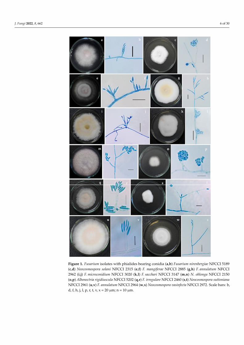
Figure 1. Fusarium isolates with phialides bearing conidia (a,b) Fusarium nirenbergiae NFCCI 5189 (c,d) Neocosmospora solani NFCCI 2315 (e,f) F. mangiferae NFCCI 2885 (g,h) F. annulatum NFCCI 2962 (i,j) F. microconidium NFCCI 3020 (k,l) F. sacchari NFCCI 3147 (m,n) N. oblonga NFCCI 2150 (o,p)
Figure 1. Fusarium isolates with phialides bearing conidia (a,b) Fusarium nirenbergiae NFCCI 5189(c,d) Neocosmospora solani NFCCI 2315 (e,f) F. mangiferae NFCCI 2885 (g,h) F. annulatum NFCCI2962 (i,j) F. microconidium NFCCI 3020 (k,l) F. sacchari NFCCI 3147 (m,n) N. oblonga NFCCI 2150(o,p) Albonectria rigidiuscula NFCCI 5202 (q,r) F. irregulare NFCCI 2460 (s,t) Neocosmospora suttonianaNFCCI 2961 (u,v) F. annulatum NFCCI 2964 (w,x) Neocosmospora vasinfecta NFCCI 2972. Scale bars: b,d, f, h, j, l, p, r, t, v, x = 20 µm; n = 10 µm.

J. Fungi 2022, 8, 662 7 of 30
J. Fungi 2022, 8, x FOR PEER REVIEW 2 of 32
Albonectria rigidiuscula NFCCI 5202 (q,r) F. irregulare NFCCI 2460 (s,t) Neocosmospora suttoniana NFCCI 2961 (u,v) F. annulatum NFCCI 2964 (w,x) Neocosmospora vasinfecta NFCCI 2972. Scale bars: b, d, f, h, j, l, p, r, t, v, x = 20 µm; n = 10 µm.
Figure 2. Fusarium tardicrescens (NFCCI 5201) culture grown on (a) Potato dextrose agar (b) Malt extract agar (c) Cornmeal agar (d–j) Phialides arising from mycelial hyphae producing micro and macroconidia. (k) Micro and macroconidia. Scale bars: d, f, j = 20 µm; e, g, h, i, k = 10 µm.
2.3. DNA Extraction, PCR Amplification and DNA Sequencing Genomic DNA was isolated from pure colonies of all 88 isolates growing on Potato
Dextrose Agar (PDA) petri-plates, incubated for a week’s time using a simple and rapid DNA extraction protocol using FastPrep®24 tissue homogenizer (MP Biomedicals GmbH, Eschwege, Germany) [29]. Partial gene sequences of isolates were determined for six gene markers, i.e., ITS, tef1-α, LSU, rpb2, β-tub and CaM. The primer sets used to amplify par-ticular gene region are summarized in Table 3.
Figure 2. Fusarium tardicrescens (NFCCI 5201) culture grown on (a) Potato dextrose agar (b) Maltextract agar (c) Cornmeal agar (d–j) Phialides arising from mycelial hyphae producing micro andmacroconidia (k) Micro and macroconidia. Scale bars: d, f, j = 20 µm; e, g, h, i, k = 10 µm.
2.3. DNA Extraction, PCR Amplification and DNA Sequencing
Genomic DNA was isolated from pure colonies of all 88 isolates growing on Potato Dex-trose Agar (PDA) petri-plates, incubated for a week’s time using a simple and rapid DNA ex-traction protocol using FastPrep®24 tissue homogenizer (MP Biomedicals GmbH, Eschwege,Germany) [29]. Partial gene sequences of isolates were determined for six gene markers,i.e., ITS, tef1-α, LSU, rpb2, β-tub and CaM. The primer sets used to amplify particular generegion are summarized in Table 3.

J. Fungi 2022, 8, 662 8 of 30
Table 3. PCR primer sets used in this study for amplification as well as sequencing.
Name of the Gene Region Primer Direction Sequence (5′-3′) Reference
Internal transcribed spacerregion of the nrDNA (ITS)
ITS-5 Forward GGAAGTAAAAGTCGTAACAAGG [30]ITS-4 Reverse TCCTCCGCTTATTGATATGC [30]
Translation elongation factor1-alpha (tef1-α)
EF-1 Forward ATGGGTAAGGARGACAAGAC [31]EF-2 Reverse GGARGTACCAGTSATCATG [31]
28S large subunit of thenrDNA (LSU)
LR-OR Forward ACCCGCTGAACTTAAGC [32]LR-7 Reverse TACTACCACCAAGATCT [33]
RNA polymerase second largestsubunit (rpb2)
fRPB2-5f Forward GAYGAYMGWGATCAYTTYGG [34]fRPB2-7cR Reverse CCCATRGCTTGYTTRCCCAT [34]
Beta-tubulin (β-tub) βtuFFo1 Forward CAGACCGGTCAGTGCGTAA [35]βtuFRo1 Reverse TTGGGGTCGAACATCTGCT [35]
Calmodulin (CaM)CF-1 Forward GCCGACTCTTTGACYGARGAR [36]CF-4 Reverse TTTYTGCATCATRAGYTGGAC [36]
PCR was carried out in a 25 µL reaction using 12.5 µL 2X Invitrogen Platinum SuperFiPCR Mastermix, 2 µL template DNA (10–20 ng), 1.5 µL 10 pmol primer, 5 µL 5X GCenhancer and H2O (Sterile Ultra-Pure Water, Sigma, St. Louis, MO, USA), with the volumemade to 25 µL. The conditions of the thermo-cycling involved:
An initial denaturation at 94 ◦C for 5 min, 35 cycles of 1 min at 94 ◦C, 30 s at 52 ◦C,1 min at 72 ◦C and a final extension at 72 ◦C for 8 min for ITS gene region;
An initial denaturation of 5 min at 94 ◦C, 30 cycles of 45 s at 94 ◦C, 30 s at 57 ◦C and1 min at 72 ◦C followed by a final 7 min extension at 72 ◦C for Tef1-α;
5 min denaturation at 94 ◦C, 35 cycles of 1 min at 94 ◦C, 50 s at 52 ◦C and 1.2 min at72 ◦C with a final 8 min extension at 72 ◦C for LSU;
5 min denaturation at 95 ◦C, 35 cycles of 45 s at 95 ◦C, 1 min at 52 ◦C and 1.5 min at72 ◦C with a final 10 min extension at 72 ◦C for Rpb2;
2 min denaturation at 94 ◦C, 40 cycles of 35 s at 94 ◦C, 55 s at 52 ◦C and 2 min at 72 ◦Cwith a final 10 min extension at 72 ◦C for β-tub;
An initial denaturation of 5 min at 94 ◦C, 30 cycles of 1 min at 94 ◦C, 30 s at 55 ◦C and2 min at 72 ◦C followed by a final 7 min extension at 72 ◦C for CaM.
The PCR amplicons were purified with a FavorPrep™ PCR purification kit as perthe manufacturer’s instructions. Purified PCR products of all marker genes were checkedon 1.2% agarose electrophoresis gels stained with ethidium bromide and were furthersubjected to a sequencing PCR reaction using a BigDye®Terminator v3.1 Cycle SequencingKit, as per manufacturer’s instructions.
The sequencing PCR reaction of 20 µL included 4 µL of 5× sequencing buffer, 2 µL ofBigDye™ Terminator premix, 4 µL of primer (5 pmol), 4 µL of the purified amplicon andH2O (Sterile Ultra-Pure Water, Sigma), with the volume made to 20 µL. Thermal cyclingconditions consisted of an initial denaturing at 96 ◦C for 3 min, followed by 30 cycles of94 ◦C for 10 s, 50 ◦C for 40 s and 60 ◦C for 4 min. The BigDye® terminators and salts wereremoved from using The BigDye Xterminator® Purification Kit (Thermo Fisher Scientific,Waltham, MA, USA) as per the manufacturer’s instructions. The purified sequencingproducts were transferred into a 96-well microplate. The sequence was elucidated onApplied Biosystems SeqStudio Genetic Analyzer (Applied Biosystems, Foster City, CA,USA). Sequences obtained were submitted in the NCBI GenBank (accession numbersin Table S1).
2.4. Phylogenetic Analysis
To determine the phylogenetic status of all 88 isolates, the Internal transcribed spacerregion of the nrDNA (ITS), translation elongation factor 1-alpha (tef1-α), 28S large subunit ofthe nrDNA (LSU), RNA polymerase second largest subunit (rpb2), beta-tubulin (β-tub) andcalmodulin (CaM) loci were used to compare the isolates used in this study with alreadyknown authentic strains in the genus Fusarium. The sequences of the type/reference strains

J. Fungi 2022, 8, 662 9 of 30
were retrieved from NCBI. A total of 186 isolates of Fusarium and related genera wereused in the phylogenetic analysis (Table S1). Atractium stilbaster CBS 410.67 was selected asthe outgroup taxon. The chosen strains used in the construction of the phylogenetic tree,along with their accession numbers and other related details, are listed in Table S1. Eachgene region was individually aligned using MAFFT v. 6.864b [37]. The alignments werechecked and adjusted manually using Aliview [38]. Further, alignments were concatenatedand subjected to phylogenetic analyses. The best substitution model out of 286 DNAmodels was figured using ModelFinder [39]. Additionally, windows version IQ-tree toolv.1.6.11 [40] was used to construct the phylogenetic tree. The reliability of the tree brancheswas assessed and tested based on 1000 ultrafast bootstrap (UFBoot) support replicatesand a SH-like approximate likelihood ratio test (SH-like aLRT) with 1000 replicates. Theconstructed phylogenetic tree was visualized in FigTree v.1.4.4 (http://tree.bio.ed.ac.uk/software/figtree/, accessed on 20 February 2022).
2.5. Fermentation for Beauvericin Production
Fusarium isolates were subcultured onto PDA and incubated at 25 ◦C for a week.Once the cultures were mature enough, spore suspensions were prepared separately forall isolates. Then, 10 µL of Tween 20 was added to 10 mL of normal saline (0.89%) andmixed adequately, saline solution was added to the culture and the spores were harvestedcarefully with the help of a sterile cotton swab and vortexed. The spore suspension’s opticaldensity (OD) was adjusted between 0.08 and 0.1 at 530 nm [41]. Next, 1 mL of this sporesuspension was added to 100 mL of Fusarium defined media (FDM) (Sucrose 25 g, Sodiumnitrate (NaNO3) 4.25 g, Sodium chloride (NaCl) 5 g, Magnesium sulfate heptahydrate(MgSO4·7H2O) 2.5 g, Potassium dihydrogen phosphate (KH2PO4) 1.36 g, Ferrous sulphateheptahydrate (FeSO4·7H2O) 0.01 g, Zinc sulfate heptahydrate (ZnSO4·7H2O) 0.0029 g/L;pH 5.5) [42] in a 250 mL Erlenmeyer flask and was incubated in a shaking incubator at25 ◦C at 150 rpm for 7 days (Figure S1).
2.6. Extraction of Beauvericin
Beauvericin was extracted from the fermented broth as per the protocol describedearlier, with slight modification [43]. The fermented broth was filtered using Whatman filterpaper no. 4. Biomass was collected, washed twice with distilled water and was allowed todry at 50 ◦C. Dried biomass was extracted overnight in 25 mL of acetonitrile:water 90:10(v/v). Further, the mixture was sonicated twice for 15 min. The mixture was filtered; filtratewas extracted with 25 mL of heptane and separated using a separating funnel. The bottomlayer was evaporated to dryness in a rotary evaporator (Heidolph, Schwabach, Germany).The dry residue was dissolved in 25 mL of methanol:water, 60:40 (v/v) and extracted twicewith 25 mL of dichloromethane. The dichloromethane phase containing beauvericin (BEA)was collected and evaporated to dryness in a rotary evaporator. The evaporated extractcontaining beauvericin was dissolved in 1 mL of Acetonitrile.
2.7. Detection of Beauvericin
The presence of beauvericin was initially qualitatively analyzed by thin-layer chro-matography (TLC) and quantitatively by high-performance liquid chromatography (HPLC).In addition, high-resolution mass spectrometry (HRMS) was carried out for a few samplesfor further confirmation.
2.7.1. Thin-Layer Chromatography (TLC)
About 5 to 10 µL of the extracts were spotted onto TLC Silica gel plates (TLC Silicagel 60 F254; Merck life science Pvt. Ltd., Bengaluru, Karnataka, India) along with standardBeauvericin (BEA) (500 mg/mL) (Sigma) with the help of glass capillary tube. TLC plateswere developed in petroleum ether:ethyl acetate (1:1) as a solvent system. The plates wereair-dried, and consequently, the spots formed on the TLC plates were detected using iodinevapours. Retention factor (Rf) values for the standard and extracts were measured as

J. Fungi 2022, 8, 662 10 of 30
described earlier [44,45]. The extracts were further subjected to high-performance liquidchromatography (HPLC).
2.7.2. High-Performance Liquid Chromatography (HPLC)
The amount of BEA in the extracts was determined by HPLC (Waters Corporation, Mil-ford, MA, USA) using a C18 column and acetonitrile: H2O (85:15 v/v) as the mobile phaseat a flow rate of 1 mL/min under isocratic conditions and U.V. detection at 210 nm [46]. Astock solution of standard beauvericin with a concentration of 1 mg/mL was prepared inacetonitrile and was further diluted to 15.6 µg/mL, 31.25 µg/mL, 62.5 µg/mL, 125 µg/mL,250 µg/mL and 500 µg/mL. The retention time for BEA was found to be 9.1 min. Theinjection volume was 20 µL. The peak area was calibrated to the amount of BEA standard(Sigma). The amount of BEA produced was calculated with respect to the area underthe peak.
2.7.3. High-Resolution Mass Spectrometry (HRMS)
The mass of beauvericin was determined in selected extracts; high-resolution production spectra were acquired using ESI-UHR-Q-TOF (Bruker Impact II Ultra-High Resolution-TOF) to confirm the presence of Beauvericin. The system ionization source type was ESIwith positive ion polarity. The instrument was run in the full-scan mode (m/z 150–1200).Nitrogen was used for drying (200 ◦C; 7 L/min), and the nebulizer gas pressure was1.7 Bar. Other typical operating parameters were set as capillary voltage—4500 V, end plateoffset—500 V, and charging voltage—2000 V.
2.8. Large Scale Fermentation and Extraction of Beauvericin from Fusarium tardicrescensNFCCI 5201
Among the 88 tested isolates, the highest beauvericin producer, F. tardicrescens NFCCI5201, was subjected to flask scale fermentation in a total of 3.5 L of Fusarium definedmedium (FDM). Each flask was inoculated with 1% spore suspension of O.D. 0.08–0.1,as described earlier in Section 2.5, and incubated at 25 ◦C with 150 rpm for one week.Extraction of beauvericin was carried out as described in Section 2.6. The final dry residuewas dissolved in 36 mL of acetonitrile. This extract was used for further study.
2.9. Determination of the Antibacterial Activity
The Fusarium tardicrescens NFCCI 5201 extract containing BEA dissolved in acetonitrile(500 µg/mL) was used to check antimicrobial activity. The antibacterial activity was testedagainst a panel of test organisms, including Escherichia coli MTCC 739, Bacillus subtilisMTCC 121, Micrococcus luteus MTCC 2470, Raoultella planticola MTCC 530, Pseudomonasaeruginosa MTCC 2453, Staphylococcus aureus MLS16 MTCC 2940 and Staphylococcus aureusMTCC 96. Cultures were grown on nutrient agar plates and incubated at 37 ◦C for 48 h.After incubation, one to two pure colonies of each culture were transferred to 10 mL Mullerhinton broth (MHB) and incubated for 4–5 h at 37 ◦C. Further, optical density was adjustedto 1 using broth. The cultures were diluted to a cell density of 1 × 10⁶ cells/mL. The testwas carried out using a resazurin-based turbidometric assay in a 96-well microtiter plate.All wells of rows (A–G) were filled with 100 µL Muller hinton broth. The first well of eachrow was filled with 100 µL of Fusarium tardicrescens NFCCI 5201 extract containing BEA(500 µg/mL), which was mixed well, and then 100 µL of the mixture from the first wellwas transferred to the second well of the vertical row. This serial dilution was continueduntil the tenth well; lastly, 100 µL of the mixture was discarded from the tenth well. Thefinal concentration of the extract was half of the original concentration in each well. Ten µLof each bacterial suspension was added in vertical rows individually, except for the twelfthwell (sterility control). One hundred µL of antibiotic ampicillin (10 µg/50 µL) was used,except Pseudomonas aeruginosa MTCC 2453, in which neomycin (30 µg/50 µL) was addedin the eleventh well of each row (positive control). After 48 hrs of incubation at 37 ◦C,25 µL of resazurin (0.01%) was added to all wells and incubated at 37 ◦C for 75 min in the

J. Fungi 2022, 8, 662 11 of 30
dark. Color changes were observed and recorded. The lowest concentration before thecolor change was considered as the minimum inhibitory concentration for that particulartest organism [47,48].
2.10. Determination of the Individual and Combined Effect of Fusarium tardicrescens NFCCI 5201Extract Containing Beauvericin and Amphotericin B on Pathogenic Fungi ofAgricultural Importance
The antifungal potential of the individual and combined effect of F. tardicrescensNFCCI 5201 extract containing beauvericin and amphotericin B on agriculturally importantpathogenic fungi was carried out using the poisoned food technique as described earlier [49].Fungal pathogens Pythium sp. NFCCI 3482, Geotrichum candidum NFCCI 3744, Rhizoctoniasolani NFCCI 4327, Rhizopus sp. NFCCI 2108, Sclerotium rolfsii NFCCI 4263, Bipolarissorokiniana NFCCI 4690 and Absidia sp. NFCCI 2716 were selected for the antifungal study.These pathogenic fungi were grown on PDA and incubated at 25 ± 2 ◦C for 5–7 days.
Agar discs with mycelia (5 mm in diameter) were cut out from the actively growingregions of the 5–7 days old pure cultures using a sterile cork borer and aseptically inoculatedat the center of four different Petri plates, as follows:
1. Control plate containing PDA;2. PDA supplemented with amphotericin B (40 µg/mL);3. PDA supplemented with F. tardicrescens NFCCI 5201 extract containing beauvericin
(40 µg/mL);4. PDA supplemented with amphotericin B (20 µg/mL) and F. tardicrescens NFCCI 5201
extract containing beauvericin (20 µg/mL).
The growth of fungal colonies on different media was measured between 3 and7 days depending upon the growth of fungi. Each treatment was repeated thrice, andthe plates were incubated at 25 ◦C. The diameter of the fungal colony was measuredafter 3 and 7 days of incubation. The percentage inhibition of the mycelial growth of thetest fungi was calculated using the formula described earlier [50]. Inhibition of mycelialgrowth (%) = (dc−dt)/dc × 100, where dc is the mean diameter (in mm) of the colony inthe control sample and dt is the diameter (in mm) of the colony in the treatment.
2.11. Antioxidant Activity of Extract of Fusarium tardicrescens NFCCI 5201Containing Beauvericin
The extract was tested for in vitro antioxidant activity using the 2,2-diphenyl-1-picrylhydrazyl (DPPH) radical scavenging method.
The concentration of the extract as well as standard solutions (control) glucose (Hime-dia) (negative) and ascorbic acid (Sigma) (positive) used were 1, 0.9, 0.8, 0.7, 0.6, 0.5, 0.4,0.3, 0.2 and 0.1 mg/mL in acetonitrile. The extract or standard solution (10 µL) was addedto DPPH in acetonitrile solution (200 µL, 100 µM) in a 96-well microtiter plate (TarsonsProducts (P) Ltd., Kolkata, India). After incubation at 37 ◦C for 30 min, the absorbance ofeach solution was determined at 517 nm using a Synergy HT Multi-detection microplatereader (BioTek, Winooski, VT, USA).
The radical scavenging activity was calculated by the following formula:Radical scavenging activity (%) = (OD control−OD sample)/OD control× 100 [51,52].
Three replicates were maintained for each treatment.
3. Results3.1. Phylogenetic Analysis
The nrDNA (ITS), translation elongation factor 1-alpha (Tef1-α), 28S large subunit ofthe nrDNA (LSU), RNA polymerase second largest subunit (Rpb2), beta-tubulin (β-tub) andcalmodulin (CaM) loci sequence alignments were together used to confirm the resolutionof the isolates used in this study. The concatenated file contained sequence data of 186 taxa.Alignment contained 6944 columns, 3815 distinct patterns, 2049 parsimony-informative,950 singleton sites and 3944 constant sites. TIM2e + R4 was found to be the best-fit model

J. Fungi 2022, 8, 662 12 of 30
of 286 models tested and was chosen based on the Bayesian Information Criterion (BIC).The phylogeny was inferred using the Maximum Likelihood Method based on the modelmentioned above. The log-likelihood of the consensus tree was –59368.419. Rate parameters:A-C: 1.27988, A-G: 2.69656, A-T: 1.27988, C-G: 1.00000, C-T: 5.18110, G-T: 1.00000; Basefrequencies: A: 0.250, C: 0.250, G: 0.250, T: 0.250; Site proportion and rates: (0.587, 0.115)(0.171, 1.101) (0.200, 2.369) (0.042, 6.470) (Figure 3).J. Fungi 2022, 8, x FOR PEER REVIEW 11 of 32
Figure 3. Cont.

J. Fungi 2022, 8, 662 13 of 30J. Fungi 2022, 8, x FOR PEER REVIEW 12 of 32
Figure 3. Maximum-likelihood (IQ-TREE-ML) consensus tree inferred from the combined ITS, LSU,Rpb2, β-tub, CaM and Tef-1α multiple sequence alignment of genus Fusarium and related genera.Numbers at the branches indicate statistical support values (UFBS and SH-aLRT). The scale barindicates expected changes per site. The tree is rooted to Atractium stilbaster (CBS 410.67). Isolatesused in this study are shown in blue bold.
Of the isolates, 88 were found to belong to Fusarium and related genera, i.e., Albonectriaand Neocosmospora. Strains used in this study were found to represent 35 species, includingAlbonectria rigidiuscula, Fusarium acutatum, F. annulatum, F. brachygibbosum, F. caatingaense,

J. Fungi 2022, 8, 662 14 of 30
F. carminascens, F. commune, F. compactum, F. cugenangense, F. duoseptatum, F. fabacearum,F. glycines, F. gossypinum, F. grosmichelii, F. irregulare, F. lacertarum, F. lumajangense, F. mangiferae,F. microconidium, F. nanum, F. nirenbergiae, F. oxysporum, F. pernambucanum, F. proliferatum,F. sacchari, F. spinosum, F. sulawesiense, F. tardichlamydosporum, F. tardicrescens, F. verticillioides,Neocosmospora metavorans, N. oblonga, N. solani, N. suttoniana and N. vasinfecta.
Fusarium isolates were found to represent six species complexes. The species fallinginto a particular species complex are included in brackets, viz. Fusarium sambucinum speciescomplex [F. brachygibbosum], Fusarium chlamydosporum species complex [F. microconidium,F. spinosum], Fusarium incarnatum-equiseti species complex [F. caatingaense, F. compactum,F. irregulare, F. lacertarum, F. nanum, F. pernambucanum, F. sulawesiense], Fusarium oxysporumspecies complex [F. carminascens, F. cugenangense, F. duoseptatum, F. fabacearum, F. glycines,F. gossypinum, F. grosmichelii, F. nirenbergiae, F. oxysporum, F. tardichlamydosporum, F. tardi-crescens], Fusarium nisikadoi species complex [F. commune] and Fusarium fujikuroi speciescomplex [F. acutatum, F. annulatum, F. lumajangense, F. mangiferae, F. proliferatum, F. sacchari,F. verticillioides].
Interestingly, out of 35 species reported in this study, 17 species, Fusarium caatin-gaense, F. carminascens, F. compactum, F. cugenangense, F. duoseptatum, F. fabacearum, F. glycines,F. gossypinum, F. grosmichelii, F. lumajangense, F. microconidium, F. nanum, F. nirenbergiae,F. sulawesiense, F. tardichlamydosporum, F. tardicrescens, Neocosmospora oblonga and N. suttoni-ana, were found to be new records from India [53,54], most of them being reported for thefirst time from a new host in this study (Table 4).
Table 4. New records of Fusarium from India along with their host detail.
IdentityThis Study Earlier Reports
NFCCI No. Host Host Reference
Fusarium caatingaense NFCCI 5191 Pomegranate Dactylopius opuntiae [55]
Fusarium carminascens NFCCI 5204 Potato Zea mays [56]
Fusarium compactum
NFCCI 2946Aegle marmelos
Poa annuaSoilSpinachWild rocketCultivated rocketLettuceQuercus suberWheatApple fruit cultivar Idared and Pink ladyBanana corm and root rotSafflower (Carthamus tinctorius L.)Wheat soilGrasses
[https://www.ncbi.nlm.nih.gov/nuccoreaccessed on 29 April
2022, [57–62]]
NFCCI 2904
NFCCI 5208 Zingiber officinale
Fusarium cugenangense NFCCI 2872 Wilted tomato plant (Root)
Crocus sp.Gossypium barbadenseHuman toe nailVicia fabaMusa sp. var. Pisang KepokGossypium sp.
[56,63]
Fusarium duoseptatum NFCCI 681 Azadirachta indica(Endophyte)
Musa sapientum cv. Pisang AmbonMusa sp. var. Pisang RastaliM. acuminata var. Dwarf CavendishM. acuminata var. Pisang AmbonMusa sp. var. Pisang RajaMusa sp. var. Pisang HawaMusa sp. var. Pisang AwakMusa sp. var. Pisang SusuMusa sp. var. Pisang Keling
[56,63]
Fusarium fabacearum
NFCCI 3239 Marigold seedsZea maysGlycine max [56]NFCCI 3706 Castor root
NFCCI 5200 Pea

J. Fungi 2022, 8, 662 15 of 30
Table 4. Cont.
IdentityThis Study Earlier Reports
NFCCI No. Host Host Reference
Fusarium glycinesNFCCI 3048 Wilted cumin plant Linum usitatissium
Ocimum basilicumGlycine max
[56]NFCCI 1788 Soil
Fusarium gossypinum NFCCI 2467 Bottle gourd Gossypium hirsutum [56]
Fusarium grosmichelii NFCCI 3243 Cow pea
Banana cormM. acuminata var. Pisang AmbonMusa sp. var. Pisang AwakM. acuminata var. Pisang Ambon LumutM. acuminata var. CavendishMusa sp. var. Pisang Siem JumboM. acuminata var. Pisang Ambon KuningMusa sp. var. Pisang Kepok
[63,64]
Fusarium lumajangense NFCCI 4180 Soil Musa sp. var. Pisang Raja NangkaMusa acuminata var. Pisang Mas Kirana [65]
Fusariummicroconidium NFCCI 3020 Soil Unknown [11]
Fusarium nanum NFCCI 5192 Zingiber officinale
Musa nana (Leaves)Solanum lycopersicumMusa acuminataOatSoilTriticum sp.Sorghum sp.
[[66],https://www.ncbi.
nlm.nih.gov/nuccoreaccessed on
29 April 2022]
Fusarium nirenbergiae
NFCCI 5189 Soil Secale cerealeMusa sp.S. tuberosumSolanum lycopersicumPassiflora edulisChrysanthemum sp.Bouvardia longifloraDianthus caryophyllusAgathosma betulinaTulip rootsAmputated human toeHuman leg ulcer
[56]NFCCI 4859 Dillenia indica
Fusarium sulawesiense
NFCCI 2956 Cotton field Musa acuminata var. Pisang Cere (AAA)Oryza sp.Smilax corbulariaAcalypha insulanaAlocasia odoraIpomoea batatasMusa nanaMusa paradisiacalPlum leafTriticum aestivumSoilOryza sativaAleurocanthus woglumiPhaseolus lunatusEucalyptusCarica papayaProsopis sp.Cucumis meloGalia melonBixa orellanaGossypium hirsutumSorghum vulgareMusa sampientum var. RobustaMango leafRhizosphere soil (Bromus tectorum)Soybean
[[11,67–70],https://www.ncbi.
nlm.nih.gov/nuccoreaccessed on 29 April
2022]
NFCCI 2886 Azadirachta indica(Endophyte)
NFCCI 3031 Piberbettle leaf (endophyte)
NFCCI 4919 Soil

J. Fungi 2022, 8, 662 16 of 30
Table 4. Cont.
IdentityThis Study Earlier Reports
NFCCI No. Host Host Reference
Fusariumtardichlamydosporum
NFCCI 3051 Wilted cumin plant M. sapientum cv. Pisang Awak LegorMusa sp. var. MonthanM. acuminata var. Pisang BaranganMusa sp. var. BluggoeM. acuminata var. Lady fingerMusa sp. var. Ney PoovanMusa sp. var. Pisang Awak Legor
[56,63]NFCCI 1895
SoilNFCCI 2491
Fusarium tardicrescensNFCCI 680 Azadirachta indica
(Endophyte)Musa sp. var. HarareCicer sp.Raphanus sp.
[63]NFCCI 5201 Dead bark
Neocosmospora oblonga NFCCI 2150 Soil Human eyeCarbonatite
[[11],https://www.ncbi.
nlm.nih.gov/nuccoreaccessed on 29 April
2022]
Neocosmosporasuttoniana
NFCCI 5190 Ginger Human woundEquine eyeSoilHomo sapiensGossypium hirsutumPodocnemis unifilisHuman corneaHuman skin leukemicHuman blood leukemiaHuman blood
[[11],https://www.ncbi.
nlm.nih.gov/nuccoreaccessed on 29 April
2022]
NFCCI 2961 Pigeon pea root (Wilted)
NFCCI 5210 Chilli fruit
NFCCI 4830 Cotton rot
NFCCI 5211Onion plants
(Damping-off)
3.2. Detection of Beauvericin Produced3.2.1. Thin Layer Chromatography
Thin layer chromatography analysis of fungal extracts along with the standard beau-vericin showed spots at the same retention factor as the standard beauvericin, depictingthe possible presence of beauvericin in the fungal extracts (Figure 4).
J. Fungi 2022, 8, x FOR PEER REVIEW 13 of 32
Figure 3. Maximum-likelihood (IQ-TREE-ML) consensus tree inferred from the combined ITS, LSU, Rpb2, β-tub, CaM and Tef1 multiple sequence alignment of genus Fusarium and releted genera. Num-bers at the branches indicate statistical support values (UFBS and SH-aLRT). The scale bar indicates expected changes per site. The tree is rooted to Atractium stilbaster (CBS 410.67). Isolates used in this study are shown in blue bold.
3.2. Detection of Beauvericin Produced 3.2.1. Thin Layer Chromatography
Thin layer chromatography analysis of fungal extracts along with the standard beau-vericin showed spots at the same retention factor as the standard beauvericin, depicting the possible presence of beauvericin in the fungal extracts (Figure 4).
Figure 4. Thin layer chromatography developed in petroleum ether: Ethyl acetate (1:1) showing spots of beauvericin detected using iodine vapours. Black arrow shows spots of beauvericin (1) Standard beauvericin (500 µg/mL) (2) Fusarium tardicrescens NFCCI 5201 (3) F. cugenangense NFCCI 2872 (4) Neocosmospora vasinfecta NFCCI 2960 (5) Fusarium annulatum NFCCI 2962.
3.2.2. High-Performance Liquid Chromatography There was a linear correlation between the concentration of the standard beauvericin
and the areas of the peak in HPLC chromatogram. The retention time of standard beau-vericin was found to be 9.1 min (Figure 5). Biomass produced by various isolates varied from 4.41 to 14.17 g/L of FDM. Among 88, 50 isolates (56%) were found to be capable of beauvericin production which varied from 0.01 to 15.82 mg/g of biomass. Table 5 shows the mycelial biomass, BEA content of mycelial biomass and final medium pH after a week’s fermentation of different Fusarium isolates in Fusarium defined medium. F. tardi-crescens NFCCI 5201 showed maximum beauvericin production of 15.82 mg/g of biomass. F. carminascens NFCCI 5204 and F. fabacearum NFCCI 5200 also produced a significant amount of beauvericin: 13.54 and 14.25 mg/g of biomass, respectively (Figure 6). The final pH of the fermented broth varied from 6.38 to 8.96 for different isolates. The correlation coefficient between biomass produced and the beauvericin produced was 0.2692; that be-tween the pH of the fermented broth and the beauvericin produced was 0.16, indicating very weak and/or no association in both cases.
Figure 4. Thin layer chromatography developed in petroleum ether: Ethyl acetate (1:1) showing spotsof beauvericin detected using iodine vapours. Black arrow shows spots of beauvericin (1) Standardbeauvericin (500 µg/mL) (2) Fusarium tardicrescens NFCCI 5201 (3) F. cugenangense NFCCI 2872(4) Neocosmospora vasinfecta NFCCI 2960 (5) Fusarium annulatum NFCCI 2962.

J. Fungi 2022, 8, 662 17 of 30
3.2.2. High-Performance Liquid Chromatography
There was a linear correlation between the concentration of the standard beauvericinand the areas of the peak in HPLC chromatogram. The retention time of standard beau-vericin was found to be 9.1 min (Figure 5). Biomass produced by various isolates variedfrom 4.41 to 14.17 g/L of FDM. Among 88, 50 isolates (56%) were found to be capable ofbeauvericin production which varied from 0.01 to 15.82 mg/g of biomass. Table 5 showsthe mycelial biomass, BEA content of mycelial biomass and final medium pH after a week’sfermentation of different Fusarium isolates in Fusarium defined medium. F. tardicrescensNFCCI 5201 showed maximum beauvericin production of 15.82 mg/g of biomass. F. carmi-nascens NFCCI 5204 and F. fabacearum NFCCI 5200 also produced a significant amount ofbeauvericin: 13.54 and 14.25 mg/g of biomass, respectively (Figure 6). The final pH of thefermented broth varied from 6.38 to 8.96 for different isolates. The correlation coefficientbetween biomass produced and the beauvericin produced was 0.2692; that between thepH of the fermented broth and the beauvericin produced was 0.16, indicating very weakand/or no association in both cases.
1
Figure 5. HPLC chromatograms of (a) standard beauvericin (500 µg/mL) (b) Fusarium fabacearumNFCCI 5200 extract (c) F. sacchari NFCCI 4889 extract (d) F. carminascens NFCCI 5204 extract(e) F. tardicrescens NFCCI 5201 extract (f) F. cugenangense NFCCI 2872 extract.

J. Fungi 2022, 8, 662 18 of 30
Table 5. Mycelial biomass and BEA content of Fusarium isolates and their pH in the Fusarium definedmedium (on day 7).
Identity NFCCI No. Biomass(g/L)
BEA Content(mg/g)
Final pH ofMedium
Fusarium nirenbergiae NFCCI 5189 6.61 ± 0.26 2.93 ± 0.12 7.73 ± 0.32Fusarium annulatum NFCCI 3264 6.72 ± 0.37 0.97 ± 0.14 7.48 ± 0.18Fusarium lacertarum NFCCI 3044 8.07 ± 0.52 0.66 ± 0.14 7.65 ± 0.24Neocosmospora suttoniana NFCCI 5190 7.14 ± 0.08 - 7.63 ± 0.22Fusarium fabacearum NFCCI 3239 6.22 ± 0.31 - 7.77 ± 0.4Fusarium sacchari NFCCI 3147 6.34 ± 0.10 - 7.6 ± 0.06Fusarium caatingaense NFCCI 5191 9.26 ± 0.26 0.33 ± 0.03 7.63 ± 0.03Neocosmospora solani NFCCI 2315 8.06 ± 0.65 0.34 ± 0.07 7.61 ± 0.12Fusarium annulatum NFCCI 3072 6.83 ± 0.09 - 7.75 ± 0.15Fusarium glycines NFCCI 3048 5.64 ± 0.12 2.00 ± 0.44 7.39 ± 0.24Fusarium annulatum NFCCI 3300 7.10 ± 0.34 0.66 ± 0.05 7.62 ± 0.54Fusarium annulatum NFCCI 2964 7.07 ± 0.19 1.11 ± 0.18 7.5 ± 0.13Fusarium mangiferae NFCCI 2885 7.97 ± 0.28 - 7.8 ± 0.15Fusarium grosmichelii NFCCI 3243 6.37 ± 0.59 1.44 ± 0.083 7.57 ± 0.21Fusarium annulatum NFCCI 2962 7.42 ± 0.06 3.26 ± 0.23 7.49 ± 0.17Fusarium nanum NFCCI 5192 6.40 ± 0.18 - 7 ± 0.14Fusarium sacchari NFCCI 3093 8.61 ± 0.83 2.31 ± 0.58 7.32 ± 0.22Fusarium sulawesiense NFCCI 2956 6.44 ± 0.37 - 7.44 ± 0.29Neocosmospora metavorans NFCCI 5193 6.25 ± 0.46 - 7.34 ± 0.17Fusarium sulawesiense NFCCI 2886 5.89 ± 0.27 - 7.34 ± 0.12Fusarium annulatum NFCCI 2959 6.13 ± 0.49 - 7.29 ±0.18Fusarium sulawesiense NFCCI 3031 8.99 ± 0.67 - 7.31 ± 0.48Fusarium compactum NFCCI 2946 8.89 ± 0.11 0.08 ± 0.04 7.44 ± 0.42Neocosmospora vasinfecta NFCCI 2960 6.64 ± 0.41 4.11 ± 0.17 7.44 ± 0.13Fusarium brachygibbosum NFCCI 3703 5.74 ± 0.32 0.15 ± 0.06 8.79 ± 0.04Fusarium irregulare NFCCI 5194 6.58 ± 0.48 1.88 ± 0.06 8.5 ± 0.20Neocosmospora oblonga NFCCI 2150 5.06 ± 0.19 - 8.67 ± 0.37Neocosmospora metavorans NFCCI 3475 6.51 ± 0.22 - 8.64 ± 0.06Fusarium commune NFCCI 2871 8.15 ± 0.18 - 8.67 ± 0.18Neocosmospora metavorans NFCCI 4095 6.71 ± 0.37 - 8.96 ± 0.28Fusarium spinosum NFCCI 5195 9.15 ± 0.64 - 8.9 ± 0.07Fusarium microconidium NFCCI 3020 8.52 ± 0.13 - 8.82 ± 0.08Fusarium cugenangense NFCCI 2872 7.01 ± 0.07 4.47 ± 0.51 8.69 ± 0.15Fusarium irregulare NFCCI 2460 9.04 ± 0.13 - 8.84 ± 0.03Fusarium annulatum NFCCI 3065 6.22 ± 0.42 0.20 ± 0.06 8.93 ± 0.17Fusarium tardicrescens NFCCI 680 7.12 ± 0.34 0.47 ± 0.07 8.87 ± 0.05Fusarium sacchari NFCCI 5196 9.18 ± 0.18 0.60 ± 0.06 7.87 ± 0.05Fusarium lacertarum NFCCI 5197 7.79 ± 2.1 0.49 ± 0.17 8.03 ± 0.02Fusarium irregulare NFCCI 5198 11.30 ± 4.8 - 8.36 ± 0.15Fusarium annulatum NFCCI 2470 10.71 ± 0.29 - 7.87 ± 0.13Fusarium sacchari NFCCI 3091 5.68 ± 0.17 - 7.28 ± 0.05Neocosmospora metavorans NFCCI 5199 5.35 ± 0.57 - 7.42 ± 0.17Fusarium brachygibbosum NFCCI 3074 7.80 ± 0.69 0.01 ± 0.05 7.72 ± 0.21Neocosmospora suttoniana NFCCI 2961 10.24 ± 0.7 0.67 ± 0.07 8.17 ± 0.45Fusarium lacertarum NFCCI 3049 7.22 ± 0.51 2.39 ± 0.39 7.19 ± 0.16Fusarium sacchari NFCCI 3092 5.79 ± 0.14 - 7.26 ± 0.15Fusarium annulatum NFCCI 1127 5.56 ± 0.61 - 6.92 ± 0.09Fusarium fabacearum NFCCI 3706 7.30 ± 0.47 1.00 ± 0.20 8.6 ± 0.05Fusarium compactum NFCCI 2904 6.45 ± 0.18 3.33 ± 0.51 7.02 ± 0.04Fusarium lacertarum NFCCI 3038 7.59 ± 0.09 0.16 ± 0.06 8.07 ± 0.15Fusarium annulatum NFCCI 2953 7.38 ± 0.15 2.21 ± 0.12 7.85 ± 0.17

J. Fungi 2022, 8, 662 19 of 30
Table 5. Cont.
Identity NFCCI No. Biomass(g/L)
BEA Content(mg/g)
Final pH ofMedium
Fusariumtardichlamydosporum NFCCI 3051 7.99 ± 0.12 2.19 ± 0.18 7.92 ± 0.13
Fusarium duoseptatum NFCCI 681 6.37 ± 0.28 - 6.86 ± 0.12Fusarium annulatum NFCCI 3270 5.14 ± 0.19 4.84 ± 0.36 7.24 ± 0.17Fusarium verticillioides NFCCI 2945 5.43 ± 1.2 - 7.18 ±0.3Neocosmospora vasinfecta NFCCI 2972 7.61 ± 2.1 - 7.32 ± 0.02Fusarium annulatum NFCCI 2053 4.64 ± 0.09 3.78 ± 0.55 7.73 ± 0.5Fusarium proliferatum NFCCI 3282 6.51 ± 3.2 0.16 ± 0.19 7.55 ± 0.12Fusarium fabacearum NFCCI 5200 10.07 ± 0.17 14.25 ± 0.28 8.11 ± 0.16Fusarium tardicrescens NFCCI 5201 11.23 ± 0.38 15.82 ± 0.54 8.2 ± 0.11Fusarium sacchari NFCCI 4889 14.17 ± 2.7 5.95 ± 0.61 8.23 ± 0.04Albonectria rigidiuscula NFCCI 5202 14.03 ± 0.07 0.16 ± 0.04 7.39 ± 0.13Fusariumtardichlamydosporum NFCCI 1895 7.80 ± 1.8 0.12 ± 0.05 7.27 ± 0.24
Fusarium pernambucanum NFCCI 5203 9.90 ± 0.15 1.04 ± 0.11 8.09 ± 0.15Fusarium carminascens NFCCI 5204 11.15 ± 2.4 13.54 ± 0.62 8.44 ± 0.19Fusarium nirenbergiae NFCCI 4859 6.52 ± 0.08 - 7.5 ± 0.32Fusarium irregulare NFCCI 5205 9.32 ± 1.6 - 7.59 ± 0.27Fusarium brachygibbosum NFCCI 4972 11.03 ± 0.15 - 7.66 ± 0.21Fusarium brachygibbosum NFCCI 5206 8.52 ± 2.9 - 7.66 ± 0.51Fusarium pernambucanum NFCCI 5207 9.80 ± 2.6 1.43 ± 0.16 7.22 ± 0.18Fusarium oxysporum NFCCI 4759 7.69 ± 0.14 2.22 ± 0.43 7.12 ± 0.16Fusarium verticillioides NFCCI 4963 10.55 ± 3.7 - 7.55 ± 0.14Fusarium verticillioides NFCCI 2696 11.05 ± 2.7 - 7.85 ± 0.05Fusarium compactum NFCCI 5208 4.41 ± 0.13 1.146 ± 0.13 6.67 ± 0.07Fusarium gossypinum NFCCI 2467 4.59 ± 0.17 0.27 ± 0.08 8.29 ± 1.5Fusarium acutatum NFCCI 5209 4.69 ± 0.15 0.29 ± 0.06 6.38 ± 0.7Neocosmospora suttoniana NFCCI 5210 7.50 ± 3.7 0.90 ± 0.08 7.86 ± 0.36Neocosmospora metavorans NFCCI 4885 7.12 ± 2.5 0.86 ± 0.07 7.94 ± 0.59Fusarium glycines NFCCI 1788 11.20 ± 0.13 1.05 ± 0.10 8.04 ± 0.16Fusariumtardichlamydosporum NFCCI 2491 13.59 ± 0.19 1.38 ± 0.11 8 ± 0.08
Albonectria rigidiuscula NFCCI 4888 13.77 ± 0.27 1.09 ± 0.08 8.13 ± 0.8Neocosmospora suttoniana NFCCI 4830 4.67 ± 2.6 1.01 ± 0.05 7.65 ±0.02Neocosmospora suttoniana NFCCI 5211 8.56 ± 3.1 - 8 ± 0.07Fusarium annulatum NFCCI 5212 10.94 ± 1.2 - 8.14 ± 0.4Fusarium lumajangense NFCCI 4180 8.26 ± 2.7 1.07 ± 0.39 8.18 ±0.05Fusarium lacertarum NFCCI 4792 9.66 ± 1.8 - 8.23 ± 0.1Fusarium annulatum NFCCI 2949 6.70 ± 0.27 - 8.16 ± 0.02Fusarium sulawesiense NFCCI 4919 8.72 ± 0.44 - 8.25 ± 0.14
- Not Detected; All values represent means ± S.E.M.
3.2.3. High-Resolution Mass Spectrometry (HRMS)
The HRMS results showed molecular ion peaks at 806.3999 (m/z) [M + Na]+,indicatingthe presence of beauvericin (C45H57N3O9) in the fungal extracts (Figure 7). Similar (m/z)[M + Na]+ 806.3956 have been reported for beauvericin (C45H57N3O9) [71]. It was observedthat the fungal extract contains, majorly, beauvericin, as maximum ion intensity wasobserved with m/z 806.3999.

J. Fungi 2022, 8, 662 20 of 30
J. Fungi 2022, 8, x FOR PEER REVIEW 16 of 32
Fusarium brachygibbosum NFCCI 5206 8.52 ± 2.9 - 7.66 ± 0.51 Fusarium pernambucanum NFCCI 5207 9.80 ± 2.6 1.43 ± 0.16 7.22 ± 0.18 Fusarium oxysporum NFCCI 4759 7.69 ± 0.14 2.22 ± 0.43 7.12 ± 0.16 Fusarium verticillioides NFCCI 4963 10.55 ± 3.7 - 7.55 ± 0.14 Fusarium verticillioides NFCCI 2696 11.05 ± 2.7 - 7.85 ± 0.05 Fusarium compactum NFCCI 5208 4.41 ± 0.13 1.146 ± 0.13 6.67 ± 0.07 Fusarium gossypinum NFCCI 2467 4.59 ± 0.17 0.27 ± 0.08 8.29 ± 1.5 Fusarium acutatum NFCCI 5209 4.69 ± 0.15 0.29 ± 0.06 6.38 ± 0.7 Neocosmospora suttoniana NFCCI 5210 7.50 ± 3.7 0.90 ± 0.08 7.86 ± 0.36 Neocosmospora metavorans NFCCI 4885 7.12 ± 2.5 0.86 ± 0.07 7.94 ± 0.59 Fusarium glycines NFCCI 1788 11.20 ± 0.13 1.05 ± 0.10 8.04 ± 0.16 Fusarium tardichlamydosporum NFCCI 2491 13.59 ± 0.19 1.38 ± 0.11 8 ± 0.08 Albonectria rigidiuscula NFCCI 4888 13.77 ± 0.27 1.09 ± 0.08 8.13 ± 0.8 Neocosmospora suttoniana NFCCI 4830 4.67 ± 2.6 1.01 ± 0.05 7.65 ±0.02 Neocosmospora suttoniana NFCCI 5211 8.56 ± 3.1 - 8 ± 0.07 Fusarium annulatum NFCCI 5212 10.94 ± 1.2 - 8.14 ± 0.4 Fusarium lumajangense NFCCI 4180 8.26 ± 2.7 1.07 ± 0.39 8.18 ±0.05 Fusarium lacertarum NFCCI 4792 9.66 ± 1.8 - 8.23 ± 0.1 Fusarium annulatum NFCCI 2949 6.70 ± 0.27 - 8.16 ± 0.02 Fusarium sulawesiense NFCCI 4919 8.72 ± 0.44 - 8.25 ± 0.14 - Not Detected; All values represent means ± S.E.M.
Figure 6. Mycelial biomass and BEA content produced by different Fusarium isolates in the Fusarium defined medium (on day 7).
3.2.3. High-Resolution Mass Spectrometry (HRMS) The HRMS results showed molecular ion peaks at 806.3999 (m/z) [M + Na]+,indicating
the presence of beauvericin (C45H57N3O9) in the fungal extracts (Figure 7). Similar (m/z) [M + Na]+ 806.3956 have been reported for beauvericin (C45H57N3O9) [71]. It was observed
Figure 6. Mycelial biomass and BEA content produced by different Fusarium isolates in the Fusariumdefined medium (on day 7).
J. Fungi 2022, 8, x FOR PEER REVIEW 17 of 32
that the fungal extract contains, majorly, beauvericin, as maximum ion intensity was ob-served with m/z 806.3999.
Figure 7. HRMS spectra from higher-collision dissociation of the [M + Na]+ ions of fungal extract, (a) Fusarium sacchari NFCCI 4889 (b) F. tardicrescens NFCCI 5201.
Figure 7. Cont.

J. Fungi 2022, 8, 662 21 of 30
J. Fungi 2022, 8, x FOR PEER REVIEW 17 of 32
that the fungal extract contains, majorly, beauvericin, as maximum ion intensity was ob-served with m/z 806.3999.
Figure 7. HRMS spectra from higher-collision dissociation of the [M + Na]+ ions of fungal extract, (a) Fusarium sacchari NFCCI 4889 (b) F. tardicrescens NFCCI 5201.
Figure 7. HRMS spectra from higher-collision dissociation of the [M + Na]+ ions of fungal extract,(a) Fusarium sacchari NFCCI 4889 (b) F. tardicrescens NFCCI 5201.
3.3. Large Scale Fermentation
Fermentation of 3.5 liters of Fusarium defined medium resulted in 36.27 gms of biomasswhich, on extraction, resulted in 144 mg of crude which was dissolved in 36 mL of acetoni-trile, making the final concentration 4 mg/mL. This extract was subjected to HPLC; it wasfound that beauvericin is the major compound in the extract, as the area covered by it wasfound to be nearly 64–68%. This stock was diluted as per requirement for further studies.
3.4. Antimicrobial and MIC of Crude Extract of Fusarium tardicrescens NFCCI 5201
The extract showed promising results against Staphylococcus aureus MLS16 MTCC2940 and Micrococcus luteus MTCC 2470. The MIC of the crude extract of F. tardicrescensNFCCI 5201 was found to be 15.63 µg/mL against Micrococcus luteus MTCC 2470 and62.5 µg/mL against Staphylococcus aureus MLS16 MTCC 2940 (Figure 8). The extract did notshow any antimicrobial activity against Escherichia coli MTCC 739, Bacillus subtilis MTCC121, Raoultella planticola MTCC 530, Pseudomonas aeruginosa MTCC 2453 and Staphylococcusaureus MTCC 96.

J. Fungi 2022, 8, 662 22 of 30
J. Fungi 2022, 8, x FOR PEER REVIEW 18 of 32
3.3. Large Scale Fermentation Fermentation of 3.5 liters of Fusarium defined medium resulted in 36.27 gms of bio-
mass which, on extraction, resulted in 144 mg of crude which was dissolved in 36 mL of acetonitrile, making the final concentration 4 mg/mL. This extract was subjected to HPLC; it was found that beauvericin is the major compound in the extract, as the area covered by it was found to be nearly 64–68%. This stock was diluted as per requirement for further studies.
3.4. Antimicrobial and MIC of Crude Extract of Fusarium tardicrescens NFCCI 5201 The extract showed promising results against Staphylococcus aureusMLS16 MTCC
2940 and Micrococcus luteus MTCC 2470. The MIC of the crude extract of F. tardicrescens NFCCI 5201 was found to be 15.63 µg/mL against Micrococcus luteus MTCC 2470 and 62.5 µg/mL against Staphylococcus aureus MLS16 MTCC 2940 (Figure 8). The extract did not show any antimicrobial activity against Escherichia coli MTCC 739, Bacillus subtilis MTCC 121, Raoultella planticola MTCC 530, Pseudomonas aeruginosa MTCC 2453 and Staphylococcus aureus MTCC 96.
Figure 8. Microtiter plate showing MIC of crude extract of F. tardicrescens NFCCI 5201 against Staph-ylococcus aureus MLS16 MTCC 2940 (Rows a–c) and Micrococcus luteus MTCC 2470 (Rows d–f).
3.5. Individual and Combined Effect of Extract of Fusarium tardicrescens NFCCI 5201 Contain-ing Beauvericin and Amphotericin B on Pathogenic Fungi
The antifungal activity was observed as a reduction in the mycelial growth of patho-genic fungi in poisoned plates when compared to the control plates (Figure 9). The extract of F. tardicrescens NFCCI 5201 containing beauvericin (40µg/mL) showed good antifungal activity against plant pathogenic fungi, Rhizoctonia solani NFCCI 4327, Sclerotium rolfsii NFCCI 4263, Geotrichum candidum NFCCI 3744 and Pythium sp. NFCCI 3482 showed a % inhibition of 84.31, 49.76, 38.22 and 35.13. Amphotericin B (40µg/mL) inhibited Geotrichum candidum NFCCI 3744, Rhizoctonia solani NFCCI 4327, Sclerotium rolfsii NFCCI 4263 and Absidia sp. NFCCI 2716, showing % inhibition of 71.76, 55.72, 56.82 and 48.68. Interest-ingly, the results were remarkable when the extracts of F. tardicrescens NFCCI 5201
Figure 8. Microtiter plate showing MIC of crude extract of F. tardicrescens NFCCI 5201 againstStaphylococcus aureus MLS16 MTCC 2940 (Rows a–c) and Micrococcus luteus MTCC 2470 (Rows d–f).
3.5. Individual and Combined Effect of Extract of Fusarium tardicrescens NFCCI 5201 ContainingBeauvericin and Amphotericin B on Pathogenic Fungi
The antifungal activity was observed as a reduction in the mycelial growth ofpathogenic fungi in poisoned plates when compared to the control plates (Figure 9).The extract of F. tardicrescens NFCCI 5201 containing beauvericin (40 µg/mL) showedgood antifungal activity against plant pathogenic fungi, Rhizoctonia solani NFCCI 4327,Sclerotium rolfsii NFCCI 4263, Geotrichum candidum NFCCI 3744 and Pythium sp. NFCCI3482 showed a % inhibition of 84.31, 49.76, 38.22 and 35.13. Amphotericin B (40 µg/mL)inhibited Geotrichum candidum NFCCI 3744, Rhizoctonia solani NFCCI 4327, Sclerotiumrolfsii NFCCI 4263 and Absidia sp. NFCCI 2716, showing % inhibition of 71.76, 55.72, 56.82and 48.68. Interestingly, the results were remarkable when the extracts of F. tardicrescensNFCCI 5201 containing beauvericin (20 µg/mL) and amphotericin B (20 µg/mL) wereused in combination against Rhizopus sp. NFCCI 2108, Sclerotium rolfsii NFCCI 4263,Bipolaris sorokiniana NFCCI 4690 and Absidia sp. NFCCI 2716, showing % inhibition of50.35, 79.37, 48.07 and 76.72%, respectively. Individually, extract of F. tardicrescens NFCCI5201 containing beauvericin (40 µg/mL) showed % inhibition of 1.68, 49.76, 2.56 and7.9 against Rhizopus sp. NFCCI 2108, Sclerotium rolfsii NFCCI 4263, Bipolaris sorokinianaNFCCI 4690 and Absidia sp. NFCCI 2716. Individually, amphotericin B (40 µg/mL)showed % inhibition of 11.51, 56.82, 1.28 and 48.67 against Rhizopus sp. NFCCI 2108,Sclerotium rolfsii NFCCI 4263, Bipolaris sorokiniana NFCCI 4690 and Absidia sp. NFCCI2716. The extract of F. tardicrescens NFCCI 5201 containing beauvericin (40 µg/mL)showed better results than amphotericin B in the case of Pythium sp. NFCCI 3482 andRhizoctonia solani NFCCI 4327 (Figure 10).

J. Fungi 2022, 8, 662 23 of 30J. Fungi 2022, 8, x FOR PEER REVIEW 20 of 32
Figure 9. Antifungal activity of individual and combined effect of extract of F. tardicrescens NFCCI 5201 containing beauvericin and amphotericin B on various pathogenic fungi (a) Pythium sp. NFCCI 3482 (b) Rhizopus sp. NFCCI 2108 (c) Rhizoctonia solani NFCCI 4327 (d) Sclerotium rolfsii NFCCI 4263 (e) Geotrichum candidum NFCCI 3744 (f) Bipolaris sorokiniana NFCCI 4690. PDA: Potato dextrose agar (Control); Ab: Potato dextrose agar containing amphotericin B (40 µg/mL); BEA: Potato dextrose agar having extract of F. tardicrescens NFCCI 5201 containing beauvericin (40 µg/mL); Ab + BEA: Potato dextrose agar having amphotericin B (20 µg/mL) and extract of F. tardicrescens NFCCI 5201 containing beauvericin (20 µg/mL).
Figure 9. Antifungal activity of individual and combined effect of extract of F. tardicrescens NFCCI5201 containing beauvericin and amphotericin B on various pathogenic fungi (a) Pythium sp. NFCCI3482 (b) Rhizopus sp. NFCCI 2108 (c) Rhizoctonia solani NFCCI 4327 (d) Sclerotium rolfsii NFCCI 4263(e) Geotrichum candidum NFCCI 3744 (f) Bipolaris sorokiniana NFCCI 4690. PDA: Potato dextrose agar(Control); Ab: Potato dextrose agar containing amphotericin B (40 µg/mL); BEA: Potato dextroseagar having extract of F. tardicrescens NFCCI 5201 containing beauvericin (40 µg/mL); Ab + BEA:Potato dextrose agar having amphotericin B (20 µg/mL) and extract of F. tardicrescens NFCCI 5201containing beauvericin (20 µg/mL).

J. Fungi 2022, 8, 662 24 of 30J. Fungi 2022, 8, x FOR PEER REVIEW 21 of 32
Figure 10. Inhibition of mycelial growth (%) of individual and combined effect of extract of F. tardi-crescens NFCCI 5201 containing beauvericin and amphotericin B on various pathogenic fungi using food poison technique.
3.6. Antioxidant Effect of Extract of Fusarium tardicrescens NFCCI 5201 Containing Beauver-icin
Figure 11 shows the free radical scavenging activity of the extract of F. tardicrescens NFCCI 5201 containing beauvericin and the standard ascorbic acid when tested for in vitro antioxidant activity. The extract of F. tardicrescens NFCCI 5201 containing beauver-icin showed good satisfactory dose-dependent DPPH radical scavenging activity with an IC50 value of 0.675 mg/mL when tested with standard ascorbic acid, which showed IC50 value of 0.146 mg/mL.
Figure 11. The free radical scavenging activity of the extract of F. tardicrescens NFCCI 5201 contain-ing beauvericin (mg/mL) and the standard ascorbic acid (mg/mL) when tested for in vitro antioxi-dant activity.
Figure 10. Inhibition of mycelial growth (%) of individual and combined effect of extract ofF. tardicrescens NFCCI 5201 containing beauvericin and amphotericin B on various pathogenic fungiusing food poison technique.
3.6. Antioxidant Effect of Extract of Fusarium tardicrescens NFCCI 5201 Containing Beauvericin
Figure 11 shows the free radical scavenging activity of the extract of F. tardicrescensNFCCI 5201 containing beauvericin and the standard ascorbic acid when tested for in vitroantioxidant activity. The extract of F. tardicrescens NFCCI 5201 containing beauvericinshowed good satisfactory dose-dependent DPPH radical scavenging activity with an IC50value of 0.675 mg/mL when tested with standard ascorbic acid, which showed IC50 valueof 0.146 mg/mL.
J. Fungi 2022, 8, x FOR PEER REVIEW 21 of 32
Figure 10. Inhibition of mycelial growth (%) of individual and combined effect of extract of F. tardi-crescens NFCCI 5201 containing beauvericin and amphotericin B on various pathogenic fungi using food poison technique.
3.6. Antioxidant Effect of Extract of Fusarium tardicrescens NFCCI 5201 Containing Beauver-icin
Figure 11 shows the free radical scavenging activity of the extract of F. tardicrescens NFCCI 5201 containing beauvericin and the standard ascorbic acid when tested for in vitro antioxidant activity. The extract of F. tardicrescens NFCCI 5201 containing beauver-icin showed good satisfactory dose-dependent DPPH radical scavenging activity with an IC50 value of 0.675 mg/mL when tested with standard ascorbic acid, which showed IC50 value of 0.146 mg/mL.
Figure 11. The free radical scavenging activity of the extract of F. tardicrescens NFCCI 5201 contain-ing beauvericin (mg/mL) and the standard ascorbic acid (mg/mL) when tested for in vitro antioxi-dant activity.
Figure 11. The free radical scavenging activity of the extract of F. tardicrescens NFCCI 5201 con-taining beauvericin (mg/mL) and the standard ascorbic acid (mg/mL) when tested for in vitroantioxidant activity.

J. Fungi 2022, 8, 662 25 of 30
4. Discussion
One of the greatest hindrances in studying Fusarium has been the ambiguous nomen-clature or incorrect species names of the isolates because of the limitations in recognition ofthe species based on morphology [72]. Being the world’s most important pathogens, itsprophylaxis and management needs correct and rapid identification [73]. As mentionedearlier, ITS and LSU frequently fail to distinguish at the species level and, preferably, Tef1-αand Rpb2 can be used to distinguish isolates at the species level. Literature reveals thatIndian Fusarium isolates have been mostly identified on the basis of morphology. Veryfew studies were found where the Fusarium isolates had been identified using molecularstudies. Wherever molecular studies were performed, they were based on the ITS generegion; very limited studies used either Tef1-α or Rpb2 or both of these gene regions [74].To our knowledge, this is the first study from India where Fusarium isolates have beenidentified on the basis of six gene regions, nrDNA (ITS), translation elongation factor 1-alpha(Tef1-α), 28S large subunit of the nrDNA (LSU), RNA polymerase second largest subunit (Rpb2),beta-tubulin (β-tub) and calmodulin (CaM). Interestingly, in this study, 17 species, Fusariumcaatingaense, F. carminascens, F. compactum, F. cugenangense, F. duoseptatum, F. fabacearum,F. glycines, F. gossypinum, F. grosmichelii, F. lumajangense, F. microconidium, F. nanum, F. niren-bergiae, F. sulawesiense, F. tardichlamydosporum, F. tardicrescens, Neocosmospora oblonga andN. suttoniana were found to be new records for India, most of them being reported fromnew hosts [53,54].
BEA is a potent bioactive compound possessing antimicrobial, anti-insecticidal, an-titumor and antiplatelet activities at extremely low concentration and with unique un-characterized active mechanisms. A review of the literature indicates that no study wasundertaken in India on BEA production from Indian species of Fusarium, except [43] whohad reported BEA production from two isolates of Fusarium viz., F. anthophilum (Host:Sugarcane; 1300 µg/g) and F. nygamai (Host: Cajanus indicus; 3 µg/g) [43]. This workwas undertaken in Bari, Italy. This suggests that Indian Fusarium isolates have remainedunexplored for their capability of BEA production and its applicability.
Albonectria rigidiuscula, F. acutatum, F. annulatum, F. brachygibbosum, F. caatingaense, F. carmi-nascens, F. compactum, F. cugenangense, F. fabacearum, F. glycines, F. gossypinum, F. grosmichelii,F. irregulare, F. lacertarum, F. lumajangense, F. nirenbergiae, F. oxysporum, F. pernambucanum,F. proliferatum, F. sacchari, F. tardichlamydosporum, F. tardicrescens, N. metavorans, N. solani,N. suttoniana and N. vasinfecta were found to be the positive producers of BEA in this study,many of them being reported as BEA producers for the first time in this study. Fusariumtardicrescens NFCCI 5201 showed maximum beauvericin production of 15.82 mg/g of biomass.Fusarium carminascens NFCCI 5204 and F. fabacearum NFCCI 5200 also produced a significantlyample amount of beauvericin, 13.54 and 14.25 mg/g of biomass, respectively, all three of thembeing reported for the first time as significant BEA producers in this study.
In earlier reports, BEA was detected in cultures of Fusarium moniliforme, F. semitec-tum [75], F. subglutinans, F. thapsinum [76], F. sambucinum, F. acuminatum, F. equiseti, F. longipes,F. anthophilum, F. oxysporum, F. poae, F. avenaceum, F. beomiforme, F. dlamini, F. bulbicola,F. nygamai [43], F. chlamydosporum, F. solani, F. proliferatum and F. sacchari [77].
The crude extract of F. tardicrescens NFCCI 5201 showed promising results againstStaphylococcus aureus MLS16 MTCC 2940 and Micrococcus luteus MTCC 2470, with MICvalues of 15.63 µg/mL against Micrococcus luteus MTCC 2470 and 62.5 µg/mL againstStaphylococcus aureus MLS16 MTCC 2940. BEA from F. oxysporum had a potent inhibitoryeffect on the growth of pathogenic Staphylococcus aureus with a MIC of 3.91 µM [23].Earlier reports reveal that inhibitory concentration (IC50) values of BEA against Bacillussubtilis, Staphylococcus haemolyticus, Pseudomonas lachrymans, Agrobacterium tumefaciens,Escherichia coli and Xanthomo vesicatoria by a 96-well microplate broth dilution–MTT assayranged between 18.45 and 70.41 µg/mL [78]. BEA has been known to inhibit bacteria,including Bacillus pumilus (0.1 µg of BEA per disk), several other species of Bacillus andPaenibacillus (1 µg of BEA per disk), P. validus, Bifidobacterium adolescentis, Clostridiumperfringens, Eubacterium biforme, Peptostreptococcus anaerobius and P. productus (25 µg of

J. Fungi 2022, 8, 662 26 of 30
BEA per disk) [18]. Strong antimicrobial activity has been reported against Staphylococcusaureus, Salmonella typhimurium and Bacillus cereus with MIC values of 3.91 µM, 6.25 and3.12 µg/mL, respectively [19,23].
With BEA being a high score ABC inhibitor, it has been reported to show strong synergywith some azole compounds (miconazole, ketoconazole) against Candida albicans andCandida parapsilosis (in vitro as well as in vivo). It has been reported that BEA individuallyfails to show antifungal activity, but it was found to synergize the effect of ketoconazole [25].
In this study, the antifungal activity was observed as a reduction in the mycelialgrowth of pathogenic fungi of agricultural importance in poisoned plates when comparedto the control plates. The extract of F. tardicrescens NFCCI 5201 containing beauvericin(40 µg/mL) showed good antifungal activity against pathogenic fungi Rhizoctonia solaniNFCCI 4327, Sclerotium rolfsii NFCCI 4263, Geotrichum candidum NFCCI 3744 and Pythium sp.NFCCI 3482 showing % inhibition of 84.31, 49.76, 38.22 and 35.13. Interestingly, when theextract of F. tardicrescens NFCCI 5201 containing beauvericin (20 µg/mL) and amphotericinB (20 µg/mL) were used in combination, they produced a synergized antifungal effectagainst Rhizopus sp. NFCCI 2108, Sclerotium rolfsii NFCCI 4263, Bipolaris sorokiniana NFCCI4690 and Absidia sp. NFCCI 2716, showing % inhibition of 50.35, 79.37, 48.07 and 76.72%,respectively. Individually, the extract of F. tardicrescens NFCCI 5201 containing beauvericin(40 µg/mL) showed a % inhibition of 1.68, 49.76, 2.56 and 7.9 against Rhizopus sp. NFCCI2108, Sclerotium rolfsii NFCCI 4263, Bipolaris sorokiniana NFCCI 4690 and Absidia sp. NFCCI2716, and amphotericin B (40 µg/mL) showed a % inhibition of 11.51, 56.82, 1.28 and 48.67against Rhizopus sp. NFCCI 2108, Sclerotium rolfsii NFCCI 4263, Bipolaris sorokiniana NFCCI4690 and Absidia sp. NFCCI 2716. The extract of F. tardicrescens NFCCI 5201 containingbeauvericin (40 µg/mL) showed better results than amphotericin B in the case of Pythiumsp. NFCCI 3482, Rhizoctonia solani NFCCI 4327. This is the first report on the testing of BEAagainst filamentous, agriculturally important fungal pathogens. The synergistic effect ofBEA along with amphotericin B against filamentous agriculturally important pathogens hasalso been studied for the first time. Earlier studies focused on the synergistic effect of BEAand azoles (miconazole, ketoconazole) on yeast (Candida albicans and Candida parapsilosis).This study focuses on the synergistic effect of fungal extracts and polyene (amphotericinB) on agriculturally important filamentous fungal pathogens. Moreover, the extract ofF. tardicrescens NFCCI 5201 containing beauvericin showed good dose-dependent DPPHradical scavenging activity with an IC50 value of 0.675 mg/mL. Antioxidant activity hasbeen reported from metabolites produced by Fusarium oxysporum [79]. The DPPH-radicals’scavenging activity has been reported from F. solani with IC50 value of 24 µg/mL [80].
5. Conclusions
Fusarium spp. have been known globally for centuries for their pathogenic behavior;however, they can prove to be a treasure trove because of their capability to produce otherinteresting metabolites of great applicability in various sectors such as food, agriculture andmedicine. As shown in this study, the antifungal activity became synergized when a halfdosage of the fungal extract and amphotericin B was used. Therefore, this study indicatesthat concerted efforts are required, in which high-throughput screening needs to be carriedout with dosage of the BEA and the other antifungal compound, which can possibly beused at a large scale for calculating the optimum dosage required. Similar studies can becarried out in the field of medicine where the dosage is optimized and the delivery of thedrug combinations is also targeted.
Supplementary Materials: The following supporting information can be downloaded at https://www.mdpi.com/article/10.3390/jof8070662/s1, Figure S1: Fermentation of different isolates in Fusariumdefined media after a week’s fermentation (a) Control (Uninnoculated FDM medium) (b) Fusar-ium fabacearum NFCCI 5200 (c) F. annulatum NFCCI 3300 (d) F. sulawesiense NFCCI 4919 (e) F. sac-chari NFCCI 4889 (f) F. annulatum NFCCI 5212 (g) F. nirenbergiae NFCCI 5189 (h) F. tardicrescensNFCCI 5201 (i) F. lacertarum NFCCI 4792 (j) F. lumajangense NFCCI 4180 (k) F. pernambucanum NFCCI5203 (l) F. duoseptatum NFCCI 681 (m) F. tardichlamydosporum NFCCI 1895 (n) Neocosmospora solani

J. Fungi 2022, 8, 662 27 of 30
NFCCI 2315 (o) F. mangiferae NFCCI 2885 (p) F. annulatum NFCCI 2964 (q) F. sacchari NFCCI 3147(r) F. grosmichelii NFCCI 3243; Table S1: GenBank Accession No. and other details of the isolates usedin the phylogenetic study.
Author Contributions: Conceptualization, S.R. and S.K.S.; methodology, S.R. and S.K.S.; formalanalysis, S.R. and S.K.S.; investigation, S.R.; resources, S.K.S.; writing—original draft preparation,S.R.; writing—review and editing, S.R., S.K.S. and L.D.; supervision, S.K.S. All authors have read andagreed to the published version of the manuscript.
Funding: This research received no external funding.
Institutional Review Board Statement: Not applicable.
Informed Consent Statement: Not applicable.
Data Availability Statement: Not applicable.
Acknowledgments: We thank Prashant Dhakephalkar, Director MACS-Agharkar Research Institute,Pune for providing the necessary facilities and encouragement to carry out the research work. ShiwaliRana acknowledges the University Grants Commission (U.G.C.), New Delhi for granting a SeniorResearch Fellowship (SRF), and S.P. Pune University, Pune for registering for the Ph.D. degree.We acknowledge technical support and help by Deepak Kumar Maurya and Subhash Gaikwadof Agharkar Research Institute, Pune. Laurent Dufossé deeply acknowledges the strong financialsupport from Conseil Régional de Bretagne and Conseil Régional de La Réunion for research activitiesdedicated to microbial biotechnology.
Conflicts of Interest: The authors declare no conflict of interest.
References1. Gupta, A.K.; Baran, R.; Summerbell, R.C. Fusarium infections of the skin. Curr. Opin. Infect. Dis. 2000, 13, 121–128. [CrossRef]2. Summerell, B.A. Resolving Fusarium: Current status of the genus. Annu. Rev. Phytopathol. 2019, 57, 323–339. [CrossRef]3. Taylor, J.W.; Jacobson, D.J.; Kroken, S.; Kasuga, T.; Geiser, D.M.; Hibbett, D.S.; Fisher, M.C. Phylogenetic species recognition and
species concepts in fungi. Fungal Genet. Biol. 2000, 31, 21–32. [CrossRef]4. Schoch, C.L.; Seifert, K.A.; Huhndorf, S.; Robert, V.; Spouge, J.L.; Levesque, C.A.; Chen, W.; Bolchacova, E.; Voigt, K.;
Crous, P.W.; et al. Nuclear ribosomal internal transcribed spacer (ITS) region as a universal DNA barcode marker for Fungi. Proc.Natl. Acad. Sci. USA 2012, 109, 6241–6246. [CrossRef]
5. Odonnell, K.; Cigelnik, E. Two divergent intragenomic rDNA ITS2 types within a monophyletic lineage of the fungus Fusariumare nonorthologous. Mol. Biol. Evol. 1997, 7, 103–116. [CrossRef]
6. O’Donnell, K.; Cigelnik, E.; Nirenberg, H.I. Molecular systematics and phylogeography of the Gibberella fujikuroi species complex.Mycologia 1998, 90, 465–493. [CrossRef]
7. O’Donnell, K.; Sutton, D.A.; Rinaldi, M.G.; Gueidan, C.; Crous, P.W.; Geiser, D.M. Novel multilocus sequence typing schemereveals high genetic diversity of human pathogenic members of the Fusarium incarnatum-F. equiseti and F. chlamydosporum speciescomplexes within the United States. J. Clin. Microbiol. 2009, 47, 3851–3861. [CrossRef]
8. O’Donnell, K. Molecular phylogeny of the Nectria haematococca-Fusarium solani species complex. Mycologia 2000, 92, 919–938. [CrossRef]9. Nalim, F.A.; Samuels, G.J.; Wijesundera, R.L.; Geiser, D.M. New species from the Fusarium solani species complex derived from
perithecia and soil in the Old-World tropics. Mycologia 2011, 103, 1302–1330. [CrossRef]10. Herron, D.A.; Wingfield, M.J.; Wingfield, B.D.; Rodas, C.A.; Marincowitz, S.; Steenkamp, E.T. Novel taxa in the Fusarium fujikuroi
species complex from Pinus spp. Stud. Mycol. 2015, 80, 131–150. [CrossRef]11. Crous, P.W.; Lombard, L.; Sandoval-Denis, M.; Seifert, K.A.; Schroers, H.J.; Chaverri, P.; Gene, J.; Guarro, J.; Hirooka, Y.;
Bensch, K.; et al. Fusarium: More than a node or a foot-shaped basal cell. Stud. Mycol. 2021, 98, 100116. [CrossRef]12. Zhang, T.; Jia, X.P.; Zhuo, Y.; Liu, M.; Gao, H.; Liu, J.T.; Zhang, L.X. Cloning and characterization of a novel 2-ketoisovalerate
reductase from the beauvericin producer Fusarium proliferatum LF061. BMC Biotechnol. 2012, 12, 55. [CrossRef]13. Sood, S.; Sandhu, S.S.; Mukherjee, T.K. Pharmacological and therapeutic potential of beauvericin: A Short Review. J. Proteom.
Bioinform. 2017, 10, 18–23. [CrossRef]14. Hilgenfeld, R.; Saenger, W. Structural chemistry of natural and synthetic ionophores and their complexes with cations. Top. Curr.
Chem. 1982, 101, 1–82.15. Ovchinnikov, Y.A.; Ivanov, V.T.; Evstratov, A.E.; Mikhaleva, I.I.; Bystrov, V.F.; Portnova, S.L.; Balashova, T.A.; Meshcheryakova, E.N.;
Tulchinsky, V.M. The enniatins ionophores, conformation and ion binding properties. Int. J. Pept. Protein Res. 1974, 6, 465–498. [CrossRef]16. Steinrauf, L.K. Beauvericin and the other enniatins. Met. Ions Biol. Syst. 1985, 19, 139–171.17. Lin, H.I.; Lee, Y.J.; Chen, B.F.; Tsai, M.C.; Lu, J.L.; Chou, C.J.; Jow, G.M. Involvement of Bcl-2 family, cytochrome c and caspase 3 in
induction of apoptosis by beauvericin in human non-small cell lung cancer cells. Cancer Lett. 2005, 230, 248–259. [CrossRef]

J. Fungi 2022, 8, 662 28 of 30
18. Castlebury, L.A.; Sutherland, J.B.; Tanner, L.A.; Henderson, A.L.; Cerniglia, C.E. Short Communication: Use of a bioassay toevaluate the toxicity of beauvericin to bacteria. World J. Microbiol. Biotechnol. 1999, 15, 131–133. [CrossRef]
19. Dzoyem, J.P.; Melong, R.; Tsamo, A.T.; Maffo, T.; Kapche, D.; Ngadjui, B.T.; McGaw, L.J.; Eloff, J.N. Cytotoxicity, antioxidant andantibacterial activity of four compounds produced by an endophytic fungus Epicoccum nigrum associated with Entada abyssinica.Rev. Bras. Farmacogn. 2017, 27, 251–253. [CrossRef]
20. Fotso, J.; Smith, J.S. Evaluation of beauvericin toxicity with the bacterial bioluminescence assay and the Ames mutagenicitybioassay. J. Food Sci. 2003, 68, 1938–1941. [CrossRef]
21. Meca, G.; Sospedra, I.; Soriano, J.M.; Ritieni, A.; Moretti, A.; Manes, J. Antibacterial effect of the bioactive compound beauvericinproduced by Fusarium proliferatum on solid medium of wheat. Toxicon 2010, 56, 349–354. [CrossRef]
22. Nilanonta, C.; Isaka, M.; Kittakoop, P.; Trakulnaleamsai, S.; Tanticharoen, M.; Thebtaranonth, Y. Precursor-directed biosynthesis ofbeauvericin analogs by the insect pathogenic fungus Paecilomyces tenuipes BCC 1614. Tetrahedron 2002, 58, 3355–3360. [CrossRef]
23. Zhang, H.W.; Ruan, C.F.; Bai, X.L.; Zhang, M.; Zhu, S.S.; Jiang, Y.Y. Isolation and identification of the antimicrobial agent beauvericinfrom the endophytic Fusarium oxysporum 5-19 with NMR and ESI-MS/MS. Biomed Res. Int. 2016, 2016, 1084670. [CrossRef]
24. Wu, X.F.; Xu, R.; Ouyang, Z.J.; Qian, C.; Shen, Y.; Wu, X.D.; Gu, Y.H.; Xu, Q.; Sun, Y. Beauvericin ameliorates experimental colitisby inhibiting activated T Cells via downregulation of the PI3K/Akt signaling pathway. PLoS ONE 2013, 8, e83013. [CrossRef]
25. Fukuda, T.; Arai, M.; Tomoda, H.; Omura, S. New beauvericins, potentiators of antifungal miconazole activity, produced byBeauveria sp FKI-1366-II. Structure elucidation. J. Antibiot. 2004, 57, 117–124. [CrossRef]
26. Shekhar-Guturja, T.; Tebung, W.A.; Mount, H.; Liu, N.N.; Kohler, J.R.; Whiteway, M.; Cowen, L.E. Beauvericin potentiates azoleactivity via inhibition of multidrug efflux, blocks Candida albicans morphogenesis, and is effluxed via Yor1 and circuitry controlledby Zcf29. Antimicrob. Agents Chemother. 2016, 60, 7468–7480. [CrossRef]
27. Zhang, L.X.; Yan, K.Z.; Zhang, Y.; Huang, R.; Bian, J.; Zheng, C.S.; Sun, H.X.; Chen, Z.H.; Sun, N.; An, R.; et al. High-throughputsynergy screening identifies microbial metabolites as combination agents for the treatment of fungal infections. Proc. Natl. Acad.Sci. USA 2007, 104, 4606–4611. [CrossRef]
28. Campos, F.F.; Sales, P.A.; Romanha, A.J.; Araujo, M.S.S.; Siqueira, E.P.; Resende, J.M.; Alves, T.M.A.; Martins, O.A.; dos Santos,V.L.; Rosa, C.A.; et al. Bioactive endophytic fungi isolated from Caesalpinia echinata Lam. (Brazilwood) and identification ofbeauvericin as a trypanocidal metabolite from Fusarium sp. Mem. Inst. Oswaldo Cruz 2015, 110, 65–74. [CrossRef]
29. Aamir, S.; Sutar, S.; Singh, S.K.; Baghela, A. A rapid and efficient method of fungal genomic DNA extraction, suitable for PCRbased molecular methods. Plant Pathol. Quar. 2015, 5, 74–81. [CrossRef]
30. White, T.J.; Bruns, T.; Lee, S.; Taylor, J. Amplification and direct sequencing of fungal ribosomal RNA genes for phylogenetics. InPCR Protocols: A Guide to Methods and Applications; Innes, M.A., Gelfand, D.H., Sninsky, J.J., White, T.J., Eds.; Academic Press:Cambridge, MA, USA, 1990; pp. 315–322.
31. O’Donnell, K.; Kistler, H.C.; Cigelnik, E.; Ploetz, R.C. Multiple evolutionary origins of the fungus causing Panama diseaseof banana: Concordant evidence from nuclear and mitochondrial gene genealogies. Proc. Natl. Acad. Sci. USA 1998, 95,2044–2049. [CrossRef]
32. Vilgalys, R.; Sun, B.L. Ancient and recent patterns of geographic speciation in the oyster mushroom Pleurotus revealed byphylogenetic analysis of ribosomal DNA-sequences. Proc. Natl. Acad. Sci. USA 1994, 91, 7832. [CrossRef] [PubMed]
33. Vilgalys, R.; Hester, M. Rapid genetic identification and mapping of enzymatically amplified ribosomal DNA from severalCryptococcus species. J. Bacteriol. 1990, 172, 4238–4246. [CrossRef] [PubMed]
34. Liu, Y.J.J.; Whelen, S.; Benjamin, D.H. Phylogenetic relationships among ascomycetes: Evidence from an RNA polymerase IIsubunit. Mol. Biol. Evol. 1999, 16, 1799–1808. [CrossRef] [PubMed]
35. Watanabe, M.; Yonezawa, T.; Lee, K.; Kumagai, S.; Sugita-Konishi, Y.; Goto, K.; Hara-Kudo, Y. Molecular phylogeny of the higherand lower taxonomy of the Fusarium genus and differences in the evolutionary histories of multiple genes. BMC Evol. Biol. 2011,11, 322. [CrossRef] [PubMed]
36. Peterson, S.W. Multilocus DNA sequence analysis shows that Penicillium biourgeianum is a distinct species closely related to P.brevicompactum and P. olsonii. Mycol. Res. 2004, 108, 434–440. [CrossRef]
37. Katoh, K.; Standley, D.M. MAFFT Multiple sequence alignment software version 7: Improvements in performance and usability.Mol. Biol. Evol. 2013, 30, 772–780. [CrossRef]
38. Larsson, A. AliView: A fast and lightweight alignment viewer and editor for large data sets. Bioinformatics 2014, 30,3276–3278. [CrossRef]
39. Kalyaanamoorthy, S.; Minh, B.Q.; Wong, T.K.F.; von Haeseler, A.; Jermiin, L.S. ModelFinder: Fast model selection for accuratephylogenetic estimates. Nat. Methods 2017, 14, 587. [CrossRef]
40. Nguyen, L.T.; Schmidt, H.A.; von Haeseler, A.; Minh, B.Q. IQ-TREE: A fast and effective stochastic algorithm for estimatingmaximum-likelihood phylogenies. Mol. Biol. Evol. 2015, 32, 268–274. [CrossRef]
41. Aberkane, A.; Cuenca-Estrella, M.; Gomez-Lopez, A.; Petrikkou, E.; Mellado, E.; Monzón, A.; Rodriguez-Tudela, J.L. Compar-ative evaluation of two different methods of inoculum preparation for antifungal susceptibility testing of filamentous fungi.J. Antimicrob. Chemother. 2002, 50, 719–722. [CrossRef]
42. Lee, H.; Kang, J.; Kim, B.; Park, S.; Lee, C. Statistical optimization of culture conditions for the production of enniatins H, I, andMK1688 by Fusarium oxysporum KFCC 11363P. J. Biosci. Bioeng. 2011, 111, 279–285. [CrossRef] [PubMed]

J. Fungi 2022, 8, 662 29 of 30
43. Logrieco, A.; Moretti, A.; Castella, G.; Kostecki, M.; Golinski, P.; Ritieni, A.; Chelkowski, J. Beauvericin production by Fusariumspecies. Appl. Environ. Microbiol. 1998, 64, 3084–3088. [CrossRef] [PubMed]
44. Touchstone, J.C. Practice of Thin Layer Chromatography, 3rd ed.; John Wiley and Sons, Inc.: Hoboken, NJ, USA, 1992; pp. 1–14.45. Fessenden, R.J.; Fessenden, J.S.; Feist, P. Organic Laboratory Techniques, 3rd ed.; Cengage Learning: Boston, MA, USA; Brooks/Cole
Thomson Leaning, Inc.: Belmont, CA, USA, 2001; pp. 133–138.46. Xu, L.J.; Liu, Y.S.; Zhou, L.G.; Wu, J.Y. Optimization of a liquid medium for beauvericin production in Fusarium redolens Dzf2
mycelial culture. Biotechnol. Bioprocess Eng. 2010, 15, 460–466. [CrossRef]47. Teh, C.H.; Nazni, W.A.; Nurulhusna, A.; Norazah, A.; Lee, H.L. Determination of antibacterial activity and minimum inhibitory
concentration of larval extract of fly via resazurin-based turbidometric assay. BMC Microbiol. 2017, 17, 36. [CrossRef]48. Foerster, S.; Desilvestro, V.; Hathaway, L.J.; Althaus, C.L.; Unemo, M. A new rapid resazurin-based microdilution assay for
antimicrobial susceptibility testing of Neisseria gonorrhoeae. J. Antimicrob. Chemother. 2017, 72, 1961–1968. [CrossRef]49. Adjou, E.S.; Kouton, S.; Dahouenon-Ahoussi, E.; Sohounhloue, C.K.; Soumanou, M.M. Antifungal activity of Ocimum canum
essential oil against toxinogenic fungi isolated from peanut seeds in post-harvest in Benin. Int. Res. J. Biol. 2012, 1, 20–26.50. Philippe, S.; Soua¨ıbou, F.; Guy, A.; Sébastien, D.T.; Boniface, Y.; Paulin, A.; Issaka, Y.; Dominique, S. Chemical composition and
antifungal activity of essential oil of fresh leaves of Ocimum gratissimum from Benin against six mycotoxigenic fungi isolated fromtraditional cheese wagashi. Res. J. Biol. Sci. 2012, 1, 22–27.
51. Jinesh, V.K.; Jaishree, V.; Badami, S.; Shyam, W. Comparative evaluation of antioxidant properties of edible and non-edible leavesof Anethum graveolens Linn. Indian J. Nat. Prod. Reso. 2010, 1, 168–173.
52. Elizabeth, K.; Rao, M.W.A. Oxygen radical scavenging activity of Curcumin. Int. J. Pharmaceu. 1990, 58, 237–240.53. Jamaluddin; Goswami, M.S.; Ojha, B.M. Fungi of India 1989–2001; Scientific Publishers: Jodhpur, India, 2004; pp. 1–326.54. Manoharachary, C.; Atri, N.S.; Devi, T.P.; Kamil, D.; Singh, S.K.; Singh, A.P. Bilgrami’s Fungi of India List and References (1988–2020);
Today & Tomorrow’s Printers and Publishers: New Delhi, India, 2022; pp. 1–475.55. Santos, A.C.D.; Trindade, J.V.C.; Lima, C.S.; Barbosa, R.D.; da Costa, A.F.; Tiago, P.V.; Oliveira, N. Morphology, phylogeny,
and sexual stage of Fusarium caatingaense and Fusarium pernambucanum, new species of the Fusarium incarnatum-equiseti speciescomplex associated with insects in Brazil. Mycologia 2019, 111, 244–259. [CrossRef]
56. Lombard, L.; Sandoval-Denis, M.; Lamprecht, S.C.; Crous, P.W. Epitypification of Fusarium oxysporum—Clearing the taxonomicchaos. Persoonia 2019, 43, 1–47. [CrossRef]
57. Vanwyk, P.S.; LOS, O.; Palterl, G.D.C.; Marasas, W.F.O. Geographic distribution and pathogenicity of Fusarium species associatedwith crown rot of wheat in the orange free state, South Africa. Phytophylactica 1987, 19, 271–274.
58. Matic, S.; Tabone, G.; Gullino, M.L.; Garibaldi, A.; Guarnaccia, V. Emerging leafy vegetable crop diseases caused by the Fusariumincarnatum-equiseti species complex. Phytopathol. Mediterr. 2020, 59, 303–317. [CrossRef]
59. Sever, Z.; Ivic, D.; Kos, T.; Milicevic, T. Fusarium rot in stored apples in Croatia Arh. Hig. Rada. Toksikol. 2012, 63, 463–470. [CrossRef]60. Frisullo, S.; Logrieco, A.; Moretti, A.; Grammatikaki, G.; Bottalico, A. Banana Corm and Root Rot by Fusarium compactum, in Crete.
Phytopathol. Mediterr. 1994, 33, 78–82.61. Mazhar, M.A.; Jahangir, A.Z.; Banihashemi, Z.; Izadpanah, K. First report of isolation and pathogenicity of Fusarium compactum
on safflower. Iran. J. Plant Pathol. 2013, 49, 277.62. Brizuela, A.M.; De la Lastra, E.; Marin-Guirao, J.I.; Galvez, L.; de Cara-Garcia, M.; Capote, N.; Palmero, D. Fusarium consortium
populations associated with Asparagus crop in Spain and their role on field decline syndrome. J. Fungi 2020, 6, 336. [CrossRef]63. Maryani, N.; Lombard, L.; Poerba, Y.S.; Subandiyah, S.; Crous, P.W.; Kema, G.H.J. Phylogeny and genetic diversity of the
banana Fusarium wilt pathogen Fusarium oxysporum f. sp. cubense in the Indonesian centre of origin. Stud. Mycol. 2019, 92,155–194. [CrossRef]
64. Ujat, A.; Vadamalai, G.; Hattori, Y.; Nakashima, C.; Wong, C.; Zulperi, D. Current classification and diversity of Fusarium speciescomplex, the causal pathogen of Fusarium wilt disease of banana in Malaysia. Agronomy 2021, 11, 1955. [CrossRef]
65. Maryani, N.; Sandoval-Denis, M.; Lombard, L.; Crous, P.W.; Kema, G.H.J. New endemic Fusarium species hitch-hiking with pathogenicFusarium strains causing Panama disease in small-holder banana plots in Indonesia. Persoonia 2019, 43, 48–69. [CrossRef]
66. Wang, M.M.; Chen, Q.; Diao, Y.Z.; Duan, W.J.; Cai, L. Fusarium incarnatum-equiseti complex from China. Persoonia 2019, 43,70–89. [CrossRef]
67. Lu, Y.A.; Qiu, J.B.; Wang, S.F.; Xu, J.H.; Ma, G.Z.; Shi, J.R.; Bao, Z.H. Species diversity and toxigenic potential of Fusariumincarnatum-equiseti species complex isolates from rice and soybean in China. Plant Dis. 2021, 105, 2628–2636. [CrossRef]
68. Yi, R.H.; Lian, T.; Su, J.J.; Chen, J. First report of internal black rot on Carica papaya fruit caused by Fusarium sulawesiense in China.Plant Dis. 2022, 106, 319. [CrossRef]
69. Lu, M.; Zhang, Y.; Li, Q.; Huang, S.; Tang, L.; Chen, X.; Guo, T.; Mo, J.; Ma, L. First report of leaf blight caused by Fusariumpernambucanum and Fusarium sulawesiense on Plum in Sichuan, China. Plant Dis. 2022. [CrossRef]
70. Araújo, M.B.M.; Moreira, G.M.; Nascimento, L.V.; Nogueira, G.D.; Nascimento, S.R.D.; Pfenning, L.H.; Ambrosio, M.M.D.Fusarium rot of melon is caused by several Fusarium species. Plant Pathol. 2021, 70, 712–721. [CrossRef]
71. Urbaniak, M.; Stepien, L.; Uhlig, S. Evidence for naturally produced beauvericins containing N-Methyl-Tyrosine in Hypocrealesfungi. Toxins 2019, 11, 182. [CrossRef]

J. Fungi 2022, 8, 662 30 of 30
72. Geiser, D.M.; Jimenez-Gasco, M.D.; Kang, S.C.; Makalowska, I.; Veeraraghavan, N.; Ward, T.J.; Zhang, N.; Kuldau, G.A.;O’Donnell, K. FUSARIUM-ID v. 1.0: A DNA sequence database for identifying Fusarium. Eur. J. Plant Pathol. 2004, 110,473–479. [CrossRef]
73. Wingfield, M.J.; De Beer, Z.W.; Slippers, B.; Wingfield, B.D.; Groenewald, J.Z.; Lombard, L.; Crous, P.W. One fungus, one namepromotes progressive plant pathology. Mol. Plant Pathol. 2012, 13, 604–613. [CrossRef]
74. Aiyaz, M.; Thimmappa Divakara, S.; Mudili, V.; George Moore, G.; Kumar Gupta, V.; i Yli-Mattila, T.; Chandra Nayaka, S.;Ramachandrappa Niranjana, S. Molecular diversity of seed-borne Fusarium species associated with maize in India. Curr. Genom.2016, 17, 132–144. [CrossRef]
75. Gupta, S.; Krasnoff, S.B.; Underwood, N.L.; Renwick, J.A.A.; Roberts, D.W. Isolation of beauvericin as an insect toxin fromFusarium semitectum and Fusarium moniliforme var. subglutinans. Mycopathologia 1991, 155, 185–189. [CrossRef]
76. Moretti, A.; Logrieco, A.; Bottalico, A.; Ritieni, A.; Fogliano, V.; Randazzo, G. Diversity in beauvericin and fusaproliferinproduction by different populations of Gibberella fujikuroi (Fusarium section Liseola). Sydowia 1997, 48, 44–56.
77. Azliza, I.N.; Hafizi, R.; Nurhazrati, M.; Salleh, B. Production of major mycotoxins by Fusarium species isolated from wild grassesin peninsular Malaysia. Sains Malays. 2014, 43, 89–94.
78. Xu, L.; Wang, J.; Zhao, J.; Li, P.; Shan, T.; Wang, J.; Li, X.; Zhou, L. Beauvericin from the endophytic fungus, Fusarium redolens,isolated from Dioscorea zingiberensis and its antibacterial activity. Nat. Prod. Commun. 2010, 5, 811–814. [CrossRef]
79. Caicedo, N.H.; Davalos, A.F.; Puente, P.A.; Rodriguez, A.Y.; Caicedo, P.A. Antioxidant activity of exo-metabolites produced byFusarium oxysporum: An endophytic fungus isolated from leaves of Otoba gracilipes. Open Microbiol. J. 2019, 8, e903. [CrossRef]
80. Menezes, B.S.; Solidade, L.S.; Conceição, A.A.; Santos Junior, M.N.; Leal, P.L.; de Brito, E.S.; Canuto, K.M.; Mendonça, S.;de Siqueira, F.G.; Marques, L.M. Pigment production by Fusarium solani BRM054066 and determination of antioxidant andanti-inflammatory properties. AMB Express 2020, 10, 117. [CrossRef]