comparative genomics and evolution of the alpha-defensin multigene family in primates
TRANSCRIPT
and Evolution. All rights reserved. For permissions, please e-mail: [email protected] The Author 2010. Published by Oxford University Press on behalf of the Society for Molecular Biology 1
Comparative genomics and evolution of the alpha-defensin multigene family in primates
Research Article
Sabyasachi Das1*, Nikolas Nikolaidis2, Hiroki Goto3, Chelsea McCallister2, Jianxu Li1,
Masayuki Hirano1, Max D Cooper1*
1Department of Pathology and Laboratory Medicine, Emory Vaccine Center, School of Medicine, Emory University, Atlanta, GA 30322, USA
2 Department of Biological Science, California State University Fullerton,
Fullerton, CA 92834, USA
3 Department of Biology and Center for Comparative Genomics and Bioinformatics, Pennsylvania State University, University Park, PA 16802, USA
*To whom correspondence should be addressed: [email protected] (M.D. Cooper), [email protected] (S. Das).
Key words: alpha-defensin gene, primate evolution, innate immunity, comparative genomics,
positive selection, birth-and-death evolution.
Running head: Evolution of α-defensin genes in primates
MBE Advance Access published May 9, 2010

2
Abstract
Defensin genes encode small cationic antimicrobial peptides that form an important part of the
innate immune system. They are divided into three families, alpha (α), beta (β), and theta (θ),
according to arrangement of the disulfide bonding pattern between cysteine residues.
Considering the functional importance of defensins, investigators have studied the evolution and
the genomic organization of defensin genes. However, these studies have been restricted mainly
to β-defensins. To understand the evolutionary dynamics of α-defensin genes among primates,
we identified the α-defensin repertoires in human, chimpanzee, orangutan, macaque, and
marmoset. The α-defensin genes in primates can be classified into three phylogenetic classes
(class I, II, and III). The presence of all three classes in the marmoset indicates that their
divergence occurred before the separation of New World and Old World monkeys. Comparative
analysis of the α-defensin genomic clusters suggests that the makeup of the α-defensin gene
repertoires between primates is quite different, as their genes have undergone dramatic birth-and-
death evolution. Analysis of the encoded peptides of the α-defensin genes indicates that despite
the overall high level of sequence divergence, certain amino acid residues or motifs are
conserved within and between the three phylogenetic classes. The evolution of α-defensins in
primates, therefore, appears to be governed by two opposing evolutionary forces. One force
stabilizes specific amino acid residues and motifs to preserve the functional and structural
integrity of the molecules and the other diversifies the sequences generating molecules with a
wide range of activities against a large number of pathogens.

3
Introduction
Innate immunity is an evolutionarily ancient defense strategy used by multiple species to
defend against pathogens. A large number of antimicrobial peptides are known to be involved in
the innate immunity. Among them defensins constitute one of the most important families of
antimicrobial peptides because of their capacity to enhance phagocytosis and the production of
proinflammatory cytokines, promote neutrophil recruitment, suppress anti-inflammatory
mediators, and regulate complement activation (Yang et al. 2002; Zhang et al. 2002; Ganz 2003;
Kim et al. 2005; Wehkamp et al. 2005; Presicce et al. 2009). The defensins are engaged in host
defenses against a broad spectrum of pathogens (i.e. bacteria, fungi, and viruses) by either
interacting directly with the pathogens, or acting in concert with other components of the
immune system (Ganz 2003; Klotman and Chang 2006; Menendez and Brett Finlay 2007). In
mammals and especially in primates, in addition to their natural antimicrobial activity, defensins
participate in other physiological processes such as sperm protection, immune recognition, and
cell-signaling (Yang et al. 2002; Yudin et al. 2005). It has also been proposed that defensins may
function as a link between innate and adaptive immune responses (Yang et al. 1999).
Mammalian defensins are small cationic peptides containing three pairs of intramolecular
disulfide bonds mediated by six conserved cysteines (Ganz and Lehrer 1994; Ganz 2003). On the
basis of the disulfide bonding pattern and the position of the cysteines, defensins are divided into
alpha (α), beta (β) and theta (θ) families (Ganz 2003), although functional θ-defensin genes are
found only in the rhesus macaque (Macaca mulatta) and olive baboon (Papio anubis) (Garcia et
al. 2008; Tran et al. 2008). The α- and β-defensins form a triple-stranded β-sheet structure
stabilized by disulfide bonds, whereas θ-defensins are structurally unrelated to the α and β
families (Tang et al. 1999; Selsted 2004). All functional α-defensin genes are expressed as

4
prepropeptides (Valore and Ganz 1992). The mature peptide results from sequential removal of
the signal peptide and prosegment giving rise to a mature, tridisulfide-containing peptide
(Michaelson et al. 1992).
Recent studies have shown that many physiological and morphological characters are
generally controlled by genes belonging to multigene families (i.e., immunoglobulin, T-cell
receptor, major histocompatibility complex, histone, ubiquitin, and olfactory receptor genes).
Therefore, detailed investigation of the evolution of multigene families is an important step
towards understanding the evolution of phenotypic characters. It has been shown that several
multigene families are subject to birth-and-death evolution and that the rates of gene gain and
gene loss vary considerably between families (Ota and Nei 1994; Nei, Gu, and Sitnikova 1997;
Su and Nei 2001; Niimura and Nei 2005; Das et al. 2008a). Due to rapid birth-and-death
evolution the number of functional genes may be quite different between closely related species
or even between individuals of the same species (Nei 2007; Nozawa, Kawahara, and Nei 2007;
Das et al. 2008b; Das 2009). Considering the functional importance of defensins, investigators
have studied the genomic organization and the evolution of defensin genes in several vertebrate
species. However, these studies are largely restricted to β-defensins (Boniotto et al. 2003;
Morrison et al. 2003; Semple, Rolfe, and Dorin 2003; Xiao et al. 2004; Semple et al. 2005;
Hollox and Armour 2008) with the exception of only a few studies on the α-defensin family
(Patil, Hughes, and Zhang 2004; Lynn and Bradley 2007). Fortunately, the draft genome
sequences of several primate species with greater than 5X coverage are available. These
sequences allow us to carry out genome-wide comparisons of the α-defensin clusters in primates.
Here, we present the complete repertoire of α-defensin genes in human, chimpanzee, orangutan,
macaque, and marmoset. This analysis aims to provide a better understanding into the general

5
pattern of the evolutionary processes that have shaped the differences in α-defensin repertoire
between primate species and new insights into the evolutionary changes of the functional
activities of α-defensin genes.
Materials and Methods
Identification of alpha defensin genes
The procedure of the retrieval of functional and nonfunctional α-defensin genes is drawn
as a flow chart in supplementary figure S1 (Supplementary Material online). To identify all the
α-defensin genes we performed a two-round tBLASTn search against the draft genome
sequences of human (Homo sapiens; assembly: GRCh37, Feb 2009), chimpanzee (Pan
troglodytes, assembly: CHIMP 2.1, Mar 2006), orangutan (Pongo pygmaeus, assembly: PPYG2,
Sep 2007), macaque (Macaca mulatta, assembly: MMUL 1.0, Feb 2006), and marmoset
(Callithrix jacchus, assembly: Callithrix_jacchus-3.2, Feb 2009) from Ensembl Genome
Browser. In the first round, the amino acid sequences of five functional α-defensin genes from
human available at the RefSeq protein database (accession numbers: NP_001916, NP_001917,
NP_004075, NP_005208, and NP_066290) were used as queries. These five α-defensin
sequences in the query data set align to the same genomic regions because they are similar to one
another. For this reason, we retrieved only non-overlapping genomic sequences that produced
alignments with the lowest E-values. Taking into account the alignment with the query α-
defensin genes, we categorized the retrieved sequences into potential functional genes if they
contained a start codon, aligned with query sequence without any frame shifts or premature stop
codons, and encoded for six conserved cysteine residues in the mature peptide region. Other

6
sequences were regarded as α-defensin pseudogenes. In the second round, the procedure was
repeated by using the α-defensin genes identified in the first round to identify additional α-
defensin genes. Intron–exon boundaries were identified with reference to the open reading frame.
For each species after collecting all non-overlapping sequences, we annotated the entire α-
defensin cluster according to their genomic positions.
Phylogenetic analysis
The translated amino acid sequences from identified α-defensin genes were aligned using
CLUSTALW program. To ensure codon to codon alignment the nucleotide alignments were
retrieved from the amino acid ones. The evolutionary distances were computed using the
Maximum Composite Likelihood method (Tamura, Nei, and Kumar 2004) after elimination of
nucleotide sequence alignment gaps only in pairwise sequence comparisons (Pairwise deletion
option). The phylogenetic trees were constructed by (i) neighbor-joining (NJ) (Saitou and Nei
1987) and (ii) maximum parsimony (MP) (Eck and Dayhoff 1966) methods using the MEGA4.0
program (Tamura et al. 2007). The reliability of the trees was assessed by bootstrap resampling
of 1000 replications. One mouse α-defensin sequence (accession no. NM_010031) was used as
outgroup.
Determination of orthologous sequences
Because α-defensin coding sequences are short and evolve rapidly (Patil, Hughes, and
Zhang 2004), to determine true orthologous relationships we have used three different methods:
(i) phylogenetic analysis with the combination of any two species under study (i.e. human-
chimpanzee, human-orangutan, chimpanzee-macaque, orangutan-macaque, macaque-marmoset
etc.); (ii) reciprocal Blast best hits and (iii) comparison of the repetitive elements flanking the 5′

7
and 3′ sides of α-defensin genes. The repetitive elements of the entire α-defensin cluster were
identified using the CENSOR software tool (Kohany et al. 2006).
Tests for positive selection
To detect positive selection, the CODEML program as implemented in PAML was used
to calculate the codon-substitution models for heterogeneous selection pressure at amino acid
positions (Yang 2007; Yang et al. 2000; Yang et al 2005). The models used in this study were
M0, M1a, M2a, M7, M8 and M8a. M1a (nearly neutral), M7 (beta), and M8 (beta and ω = 1)
were null models that did not support ω > 1, while the alternative models M2a (positive
selection) and M8a (beta and ω) have an additional class that allowed ω > 1. Using these null and
alternative models, three Likelihood ratio tests (LRT) were carried out: (1) M1a versus M2a, (2)
M7 versus M8, and (3) M8a versus M8. For LRTs, twice the log likelihood difference, 2Δl= 2(l1
– l0), was compared with a χ2 distribution to test whether the null model could be rejected, where
l1 and l0 were the log likelihood for the alternative model and the null model, respectively.
Moreover, naïve empirical Bayes analysis was employed to calculate the posterior probability
that each site belonged to a particular site class. Sites with high posterior probability of
belonging to the site class of ω > 1 were inferred to be under positive selection.
Structural analysis
To understand the structural characteristics of α-defensin peptides, we predicted the
folding pattern of several primate sequences using the SWISS-MODEL
(http://swissmodel.expasy.org) (Arnold et al. 2006) and PHYRE
(http://www.sbg.bio.ic.ac.uk/~phyre) (Bennett-Lovsey et al. 2008) web servers. Pairwise
structural alignments and structural superimposition were performed using the DaliLite

8
(http://www.ebi.ac.uk/Tools/dalilite) (Holm and Park 2000) web server. Electrostatic potential
was calculated using the PBEQ-Solver (Jo et al. 2008) web server. All figures were generated
with PyMol (DeLano Scientific; http://pymol.org). The hydrophobicity score was calculated
using the Kyte-Doolittle scale (Kyte and Doolittle 1982).
Results
Number of α-defensin genes in five primate species
Our homology searches (see Materials and Methods) detected 41 functional and 30
nonfunctional α-defensin genes in the genomes of the five primate species under study (table 1).
The α-defensin genes in human, chimpanzee, orangutan, and macaque are located in the
subtelomeric region of chromosome 8 of the respective species (supplementary fig. S2,
Supplementary Material online). The chromosomal location for marmoset α-defensin genes is
not available due to incompleteness of the genome assembly. However, most (12 out of 14) of
the α-defensin genes in marmoset are located in a single genomic region (contig 333). All
identified α-defensin genes in five primate species are listed in supplementary table S1
(Supplementary Material online). In all species, with the exception of human, the numbers of
functional genes given in table 1 are the minimum estimates because some genomic regions of
the α-defensin cluster are incomplete in the draft genome sequences. It is possible that the partial
genes can be annotated as functional genes when more complete versions of the genome
sequences will be available. In this study we have found five partial genes (two from
chimpanzee, one from orangutan, and two from macaque). These genes contain either the entire
first exon or the entire second exon and some portion of the intron. We used these partial
9
sequences as queries in similarity searches against the nucleotide database of NCBI to find out
whether these partial α-defensin sequences are functional or nonfunctional genes. We found that
two out of five identified partial α-defensin genes, chimpanzee Ptr 1 and macaque Mmu 18 (see
supplementary table S1, Supplementary Material online) exhibit 100% sequence identity with
two nucleotide sequences. The sequence exhibiting 100% identity with Ptr 1 is a full length
mRNA sequence (accession number AY746440), whereas for Mmu 18 the 100% identical
sequence is part of a BAC clone (accession number AC202726). The retrieved α-defensin gene
(100% identical to the nucleotide sequence of Mmu 18) from this BAC clone is a full length
gene. Therefore, we decided to regard Ptr1 and Mmu18 sequences as functional genes in this
study. As shown in table 1, the proportions of total numbers of functional and nonfunctional α-
defensin genes are nearly the same for hominids (human, chimpanzee, and orangutan) and old
world monkeys (macaque). By contrast, the total number of functional α-defensin genes in
marmoset (member of New World monkeys) is considerably higher than that of catarrhine
primate species (hominids and Old World monkeys). It is also noticeable that marmoset showed
the lowest number of pseudogenes from all the species studied.
Phylogenetic relationships of α-defensin genes
To examine the evolutionary relationships between the α-defensin genes we constructed
phylogenetic trees for the dataset of 41 functional and 22 nonfunctional α-defensin genes using
the neighbor-joining (NJ) and maximum parsimony (MP) methods. We excluded three partial
genes and eight pseudogenes because they were truncated and they had highly diverged
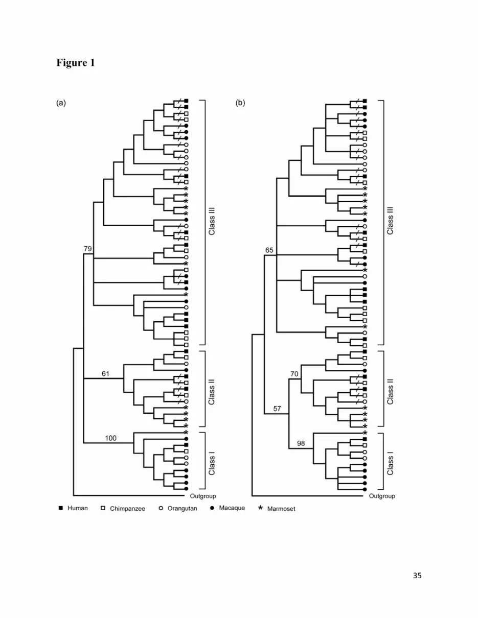
sequences, respectively. In fig. 1, we present condensed phylogenetic trees at the 50% bootstrap
consensus value level. The tree topologies produced by both methods (fig. 1a and 1b) are nearly
the same and classify the α-defensin genes in primates into three major phylogenetic classes

10
(class I, II, and III). In both phylogenetic trees the branch leading to class I is supported by >90%
bootstrap values. For classes II and III although the bootstrap support is relatively low (>60%
and <80%), both classes are reproduced by NJ and MP trees. The presence of α-defensin genes
from all five primate species in all three phylogenetic classes suggests that the separation
between the class I, II, and III genes occurred before the divergence between New world and Old
World monkeys.
The number of α-defensin genes varies considerably among the three classes (table 2). In
all species, the largest numbers of genes belong to class III, with the exception of macaque. In
the latter species the highest number of genes is found in class I. In hominids, class II contains
one functional gene and two pseudogenes, indicating that no significant gain or loss occurred in
class II genes after divergence of old world monkeys and hominids from their last common
ancestor.
Identification of Phylogenetic class-specific amino acid residues in α-defensins
To identify whether there are specific amino acid residues or motifs that distinguish the
three phylogenetic classes, we analyzed the amino acid sequences of α-defensins. The class-
specific consensus sequences were generated by identifying the most commonly used amino acid
residues or motifs in each class of α-defensin (fig. 2). From the alignment of the encoded
proteins of all functional α-defensin genes we identified five molecular markers, which can
distinguish the α-defensin sequences belonging to class I, II, and III. Interestingly, all five
molecular markers are located in the prosegment regions of α-defensins. Although the identified
molecular markers are mostly class-specific, in certain cases the same amino acid residue(s) in a
particular position are shared by two phylogenetic classes. For example, class I α-defensin
11
sequences possess Ser residues at position 22, whereas at the same position both class II and III
consensus sequences contain Pro residues. At positions 39-40, 49-50, 57-60, and 70-72 the class
I sequences have TQ, DL(X), NGLS, and QAR motifs, respectively (see fig. 2). Here, “X”
represents any amino acid which appeared due to the substitution of consensus residue at
particular position. In contrast, the motifs present at the same positions in class II α-defensin
sequences are AQ, DF, DASS, and T(R)R(T), whereas relatively less conserved motifs are found
at the same positions of class III sequences.
Structural characteristics of primate α-defensins
We analyzed the amino acid sequences of signal peptide, propeptide, and mature peptide
regions to understand the structural features of primate α-defensin peptides. In all primate
sequences studied the signal peptide is the most conserved segment, followed by the prosegment
and mature peptide regions (supplementary fig. S3, Supplementary Material online). It has been
shown that the prosegment region of defensins contains several amino acids, which are involved
in folding and functional inhibition of the mature peptides (Zou et al. 2008; Figueredo et al.
2009). Most of the negatively charged residues are conserved in the prosegment of primate
defensins (E20, D27, E28, E34, D39, and E42) (numbering is according to sequence Hsa 6 in fig.
2 excluding gaps). In addition to the negatively charged amino acids, three regions that are
predominantly occupied by hydrophobic residues (positions 29-33, 35-38, and 43-49) are also
reasonably well conserved. These observations suggest that although the prosegment sequence is
not highly conserved, specific structural characteristics are preserved most probably due to
functional constraints.

12
The most well studied region of α-defensins is the mature peptide. Defensin structures
have been solved using both x-ray crystallography and NMR. To date, structures for several α-
defensins have been reported from human and other mammals (McManus et al. 2000; Szyk et al.
2006). The overall fold of the α-defensin monomer is composed by three β-strands arranged into
an antiparallel β-sheet. These architectural elements are restrained in their relative orientations
by three disulfide bridges, C66–C94, C68–C83, C73–C93, and one salt bridge formed by the side
chains of R69 and E77 (numbering is according to sequence Hsa 6 in fig. 2 excluding gaps).
These residues are well conserved in all primate defensins (fig. 2). These observations indicate
that despite the high level of divergence at their primary amino acid sequences, primate α-
defensins fold very similar to each other. This notion is also supported by theoretical models
built by homology (fig. 3). These models suggest that primate defensins display the canonical α-
defensin disulfide arrangement and a similar fold, but differ markedly in surface charge
distribution and loop sizes/orientations (fig. 3). The electrostatic surface analysis also supports
the above notion and shows the amphipathicity of these proteins (supplementary fig. S4,
Supplementary Material online). All primate α-defensin structures contain several Arg residues
distributed fairly evenly in the primary sequence; however, when the protein is folded, most of
these basic residues are located on one face. As has been observed for other primate defensins
(Vasudevan et al. 2008), the positively charged surface is distinct and separate from the
hydrophobic region. The positively charged surface distinguishes both paralogous and
orthologous defensin proteins (supplementary fig. S4, Supplementary Material online).
Furthermore, the different classes can be differentiated by the differential presence of one or two
small negatively charged regions (supplementary fig. S4, Supplementary Material online). These
variations in α-defensin electrostatic surface distributions imply that these proteins have distinct

13
mechanisms for their mode of antimicrobial action. In addition to the charge distributions, it has
been hypothesized that the large hydrophobic surfaces of defensins may play a role in
hydrophobic interactions with the membrane hydrocarbons of the target cell (Vasudevan et al.
2008). Thus, the differences in hydrophobicity (supplementary fig. S5, Supplementary Material
online) and the distribution of these surfaces, together with the presence/absence of exposed
aromatic amino acids (figs. 2-3; supplementary fig. S4, Supplementary Material online) may be
additional determinants of functional specificity and differentiation between the different primate
α-defensins.
Genomic organization of α-defensin cluster
For relatively short and highly evolving sequences the establishment of true orthologous
relationships is often difficult using conventional phylogenetic tree building methods (Das et al.
2008a; Das et al. 2008b). In the case of α-defensin sequences, which are short (≤ 100 aa) and
evolve rapidly (Lynn et al. 2004; Patil, Hughes, and Zhang 2004), we used three different
methods to identify orthologous relationships: (i) phylogenetic analysis, (ii) reciprocal BLAST
best hits, and (iii) comparison of the flanking repetitive elements of α-defensin genes (see
Materials and Methods). For the third method, we have identified the repetitive elements in the
entire α-defensin cluster and compared the repetitive elements flanking the 5′ and 3′ sides of each
α-defensin gene. One example of the comparison of flanking repetitive elements of α-defensin
gene is shown diagrammatically in figure 4. In our study we regarded as orthologs, genes for
which orthology was supported by at least two methods. These analyses allowed us to establish
orthologous relationships between α-defensin genes in all primates, with the exception of some
marmoset sequences. The estimated divergence time between New World and Old World
Monkeys is ~ 43 MYR (Million year) (Steiper and Young 2006). It is possible that due to their

14
relatively long divergence time, establishing true orthologous relationships between α-defensin
genes of marmoset and Old World Monkeys is difficult. Hence, the orthology between α-
defensin genes is established for four primate species (human, chimpanzee, orangutan, and
macaque) as they are separated from each other by a relatively short divergence time.
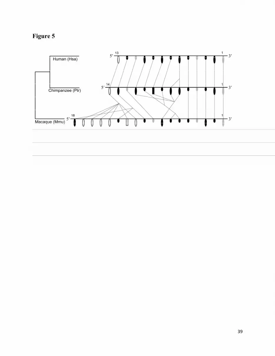
Using the orthologous sequences we have carried out a comparative genomic analysis of
α-defensin clusters of human, chimpanzee, and macaque. Due to the incompleteness of the
genome assembly (see supplementary Table S1, Supplementary Material online), the complete
genomic organization of the orangutan α-defensin cluster could not be established. Therefore, we
excluded orangutan from the comparative genomics’ analysis of the α-defensin locus. Because of
this limitation, certain conclusions can be drawn only tentatively. The physical maps of α-
defensin genes in human, chimpanzee, and macaque are shown in figure 5. The results of the
analysis reveal conservation in the synteny of five genes (i.e. Hsa1, Hsa2, Hsa3, Hsa4, and Hsa5
and their orthologous sequences), located at the 3′ regions of α-defensin clusters in the human,
chimpanzee, and macaque (fig. 5). Although no duplication or deletion of genes is observed in
this region, one α-defensin gene (Mmu2) became nonfunctional in the macaque lineage.
Similarly, Hsa3 and Ptr3 became nonfunctional in the human-chimpanzee lineage, whereas the
ortholog in macaque (Mmu3) is functional. In contrast to the conservation observed in the 3′
region, in the 5′ regions of α-defensin clusters we identified a few events of species-specific gene
duplication or deletion.
To gain a better understanding of the evolution of the α-defensin cluster we analyzed the
distribution of α-defensin functional genes and pseudogenes in the three phylogenetic classes.
This analysis showed that the genes in all three phylogenetic classes are intermingled in the
genome (fig. 5). The genomic distribution of class I and III α-defensin genes in macaque is quite
15
different from that of hominids (human and chimpanzee). In contrast, it is noticeable that the
functional and pseudogenes belonging to class II are conserved (i.e. no duplication or deletion)
from macaque to human.
Examination of positive selection
Intrigued by the high divergence among α-defensin genes, we investigated whether this
observation could be explained by positive selection. To examine if positive selection acts on
orthologous gene groups, we carried out three Likelihood ratio tests (LRT) using 6 codon-
substitution models for heterogeneous selection pressure at amino acid positions (Yang 2007;
Yang et al. 2000; Yang et al 2005) : (1) M1a versus M2a, (2) M7 versus M8, and (3) M8a versus
M8 (table 3 and table 4). For this analysis, we used all the potentially functional genes from five
primate species and compared both orthologous and paralogous defensin genes. This analysis
suggests the action of positive selection on some specific sites of α-defensins (table 3, table 4,
and fig. 2). Except for one site at the boundary of prosegments and mature peptides, all of the
other sites that could be positively selected were detected in the mature peptides. Our results are
similar with previous observations by Patil et al. (2004) and Lynn et al. (2004). These results
indicate that the presence of multiple potential positively selected sites in the mature peptide
might be functionally important for the broad range of antimicrobial activities of α-defensin.
Discussion
16
In this study, we identified the α-defensin gene repertoires in human, chimpanzee,
orangutan, macaque, and marmoset based on currently available genome sequences and analyzed
the genomic organization using comparative genomic and evolutionary approaches. We found
that the ratio between functional and nonfunctional α-defensin genes is nearly identical for
hominids (human, chimpanzee, and orangutan) and old world monkeys (macaque), whereas, that
of marmoset (New World monkey) is quite different. The latter species also showed the lowest
fraction of pseudogenes compared to all species studied (table 1). In several multigene families, a
considerable number of pseudogenes has been described (Das et al. 2010; Piontkivska and Nei
2003; Nei 2007). These genes have accumulated non-sense mutations, frameshift deletions and
insertions, or single nucleotide substitutions within functionally important sites, which disrupt
the expression of functional proteins (Kawasaki et al. 1997; Das et al. 2008a). It is, therefore,
possible that comparatively higher accumulation of mutations in the α-defensin locus of
catarrhine primates (human, chimpanzee, orangutan, and macaque) may have led to the higher
number of pseudogenes compared to that of Platyrrhine (or New World monkey) primate species
(marmoset).
Our analysis suggests that the α-defensin genes of primates fall into three major
phylogenetic classes (class I, II, and III). The presence of all three classes in the marmoset
indicates that their divergence occurred before the separation of New World and Old World
monkeys and these classes have persisted for ~ 43 Myr in the primate genomes. The comparative
analysis of the α-defensin genomic clusters suggests that several genomic rearrangements
occurred in these genomic regions. In all classes, with the exception of class II genes, the
differences in the number of class I and class III genes between the hominids and macaque
lineages have been generated mainly by repeated tandem gene duplication within each genomic

17
cluster (fig. 5). In the phylogenetic trees all three classes contain α-defensin genes from five
primate species and the phylogenetically distantly related genes are more or less intermingled in
the genomic cluster. Hence, considering the phylogeny and the genomic organization of α-
defensin genes, we can infer that the α-defensin multigene family is mainly subject to the birth-
and-death model of evolution rather than to concerted evolution. In the birth-and-death model,
new genes are originated by repeated gene duplication, and by accumulation of mutation some of
them may acquire a new function and remain in the genome for a long time, while others become
pseudogenes or are deleted from the genome (Nei, Rogozin, and Piontkivska 2000; Su and Nei
2001; Rooney 2004; Das et al. 2008b). In contrast, the concerted evolution model proposes that
the genes in a multigene family of a species are homogenized over some period of time by gene
conversion or unequal crossingover, causing higher sequence similarity of genes within species
than between species (Liao 1999; Nikolaidis and Nei 2004).
In catarrhine primates the α-defensin cluster is located in the subtelomeric region of the
chromosome. Due to incompleteness of the genome assembly the chromosomal location for
marmoset α-defensin cluster is not available. The subtelomeres harbor several multigene
families, including the olfactory receptor and immunoglobulin heavy chain variable region genes
which exhibit a fairly high level of sequence divergence as well as multiple duplication and
deletion events (Linardopoulou et al. 2001; Das et al. 2008b). Recent studies suggest that the
subtelomeric and the pericentromeric regions might be less constrained than other genomic
regions for recombination, duplication, gene conversion, point mutation, and translocation
(Linardopoulou et al. 2005; Webber and Ponting 2005). Thus, these regions may facilitate the
birth and death of their harboring genes. The multiple duplications and the acquisition of several
point mutations observed in the α-defensin locus are consistent with this notion. Due to point

18
mutations α-defensin genes have been diversified and some of them became pseudogenes in
certain lineages. In fact, we found that less than 50% of the α-defensin genes are functional
genes in human, chimpanzee, orangutan, and macaque. Moreover, our analyses, together with
previous studies (Lynn et al. 2004; Patil, Hughes, and Zhang 2004), imply that the functional α-
defensin genes may have evolved under positive selection. In our analysis we have detected
potential positively selected sites in the α-defensin matured region, most of which are consistent
with the sites predicted to be under positive selection by Lynn et al. (2004). The experimental
evidence available for primate’s α-defensins show that the variation in the mature peptides leads
to the diversity in their potency against different pathogens. For example, although the primary
amino acid sequences of Hsa6 (HNP1), Hsa8 (HNP2), and Hsa10 (HNP3) are identical, except
for a single amino acid alteration at the N-terminal end of the mature peptide (Ala to Asp), the
Hsa6 (HNP1) and Hsa8 (HNP2) are different from Hsa10 (HNP3) in the activities to kill
Candida albicans (Lehrer et al. 1988). The selective replacements of Arg residues in Hsa13
(HD5) at positions 81 (numbering is according to the alignment of α-defensins presented in fig.
2) significantly decrease the antimicrobial activity, whereas a replacement of Arg to His at
position 85 as found in a patient suffering from Crohn’s disease (a chronic inflammatory disease)
severely reduce the bactericidal activity of Hsa13 (HD5) (de Leeuw et al. 2009). In macaque, the
matured regions of functional class I α-defensins are diverged from each other except for
canonical conservation of six Cys residues and one Arg residue (see fig. 2). Tanabe et al. (2004)
showed that these α-defensins are expressed in the intestine of macaque and that their matured
regions are highly variable in bactericidal activities. It is, therefore, possible that positive
selection in the specific regions of α-defensin may facilitate diverse profiles of antimicrobial
activity and maximize host ability to defend against infections.

19
The α-defensin gene classes are defined by phylogenetic relationships only in this study
and therefore the classification may not be related to gene function or expression pattern.
However, considering the 100% sequence similarity with the deposited human α-defensin
sequences in NCBI we found that human class I and class II genes are primarily expressed in the
paneth cells of intestine, whereas class III genes are expressed mainly in neutrophils (see
supplementary table S2, Supplementary Material online). To test whether the phylogenetic
classification reflects any structural and functional clustering, we undertook a systematic analysis
of the translation products of all functional α-defensin genes. Our analysis reveals that certain
peptide residues differentiate the α-defensin sequences belonging to class I, II, and III (see fig.
2). Hence, corresponding conserved nucleotide sequences define phylogenetic class-specific
amino acid residues or motifs whose structures have been maintained across species and
evolutionary barriers. Interestingly, all of the class-specific signatures are located in the
prosegments and not in the matured peptides. It has been reported that the acidic prosegment
may be important for neutralization, processing, and/or folding of the cationic C-terminal
matured peptide (Valore and Ganz 1992; Liu and Ganz 1995). The arrangement of negatively
charged residues (Glu, Asp) in the prosegment is functionally important for the interaction of the
prosegment and the mature peptides (Liu and Ganz 1995; Zou et al. 2008). It is possible that the
conservation in phylogenetic class-specific amino acid residues or motifs in the propeptides
might have some functional importance. Moreover, our results suggest that despite the high level
of divergence at the primary amino acid sequences of the mature peptide, primate α-defensins
fold in a very similar fashion to each other (fig. 3). The differences observed in the surface
charge distribution and the loop sizes/orientations (fig. 3; supplementary fig. S4, Supplementary
Material online) together with the divergent hydrophobicity patterns and the presence or absence
20
of exposed aromatic amino acids (figs. 2-3; supplementary fig. S5, Supplementary Material
online) suggests functional differentiation between the different α-defensin classes. The latter
supposition is in accordance with the birth-and-death model of evolution, which postulates that
new genes may acquire a new function. Therefore, it seems that the evolution of the α-defensins
in primates is driven by two opposing forces, one diversifying and the other stabilizing the
specific amino acid residues or motifs due to functional constraints.
Supplementary Material
Supplementary tables S1-S2 and figures S1-S5 are available at Molecular Biology and Evolution
online (http://www.mbe.oxfordjournals.org/).
Acknowledgements
We thank Jan Klein, Masatoshi Nei, and Masafumi Nozawa for valuable comments and
suggestions. This work was supported by the National Institutes of Health [grant AI72435 to M.
D. C.], Georgia Research Alliance and the California State University, Fullerton (CSUF) [start-
up money to N. N.]. C.M. was supported by the Faculty Development Center Grant from CSUF.

21
References
Arnold K, Bordoli L, Kopp J, Schwede T. 2006. The SWISS-MODEL workspace: a web-based
environment for protein structure homology modelling. Bioinformatics. 22:195–201.
Bennett-Lovsey RM, Herbert AD, Sternberg MJ, Kelley LA. 2008. Exploring the extremes of
sequence/structure space with ensemble fold recognition in the program Phyre. Proteins.
70:611–625.
Boniotto M, Tossi A, DelPero M, Sgubin S, Antcheva N, Santon D, Masters J, Crovella S. 2003.
Evolution of the beta defensin 2 gene in primates. Genes Immun. 4:251-257.
Das S. 2009. Evolutionary Origin and Genomic Organization of Micro-RNA Genes in
Immunoglobulin Lambda Variable Region Gene Family. Mol Biol Evol. 26:1179-1189.
Das S, Mohamedy U, Hirano M, Nei M, Nikolaidis N. 2010. Analysis of the immunoglobulin
light chain genes in zebra finch: evolutionary implications. Mol Biol Evol. 27:113-120.
Das S, Nikolaidis N, Klein J, Nei M. 2008a. Evolutionary redefinition of immunoglobulin light
chain isotypes in tetrapods using molecular markers. Proc Natl Acad Sci USA.
105:16647–16652.
Das S, Nozawa M, Klein J, Nei M. 2008b. Evolutionary dynamics of the immunoglobulin heavy
chain variable region genes in vertebrates. Immunogenetics. 60:47-55.
de Leeuw E, Rajabi M, Zou G, Pazgier M, Lu W. 2009. Selective arginines are important for the
antibacterial activity and host cell interaction of human alpha-defensin 5. FEBS Lett. 583:
2507-2512.
Eck RV, Dayhoff MO. 1966. Atlas of Protein Sequence and Structure. National Biomedical
Research Foundation, Silver Springs, Maryland.

22
Figueredo SM, Weeks CS, Young SK, Ouellette AJ. 2009. Anionic amino acids near the pro-
alpha-defensin N terminus mediate inhibition of bactericidal activity in mouse pro-
cryptdin-4. J Biol Chem. 284:6826-6831.
Ganz T. 2003. Defensins: antimicrobial peptides of innate immunity. Nat Rev Immunol. 3:710-
720.
Ganz T, Lehrer RI. 1994. Defensins. Curr Opin Immunol. 6:584-589.
Garcia AE, Osapay G, Tran PA, Yuan J, Selsted ME. 2008. Isolation, synthesis, and
antimicrobial activities of naturally occurring theta-defensin isoforms from baboon
leukocytes. Infect Immun. 76:5883-5891.
Hollox EJ, Armour JA. 2008. Directional and balancing selection in human beta-defensins. BMC
Evol Biol. 8:113.
Holm L, Park J. 2000. DaliLite workbench for protein structure comparison. Bioinformatics.
16:566-567.
Jo S, Vargyas M, Vasko-Szedlar J, Roux B, Im W. 2008. PBEQ-Solver for online visualization
of electrostatic potential of biomolecules. Nucleic Acids Res. 36:W270-275.
Kawasaki K, Minoshima S, Nakato E, Shibuya K, Shintani A, Schmeits JL, Wang J, Shimizu N.
1997. One-megabase sequence analysis of the human immunoglobulin lambda gene
locus. Genome Res. 7:250–261.
Kim C, Gajendran N, Mittrucker HW, Weiwad M, Song YH, Hurwitz R, Wilmanns M, Fischer
G, Kaufmann SH. 2005. Human alpha-defensins neutralize anthrax lethal toxin and
protect against its fatal consequences. Proc Natl Acad Sci USA. 102:4830-4835.
Klotman ME, Chang TL. 2006. Defensins in innate antiviral immunity. Nat Rev Immunol.
6:447-456.
23
Kohany O, Gentles AJ, Hankus L, Jurka J. 2006. Annotation, submission and screening of
repetitive elements in Repbase: RepbaseSubmitter and Censor. BMC Bioinformatics.
7:474.
Kyte J, Doolittle RF. 1982. A simple method for displaying the hydropathic character of a
protein. J Mol Biol. 157:105-132.
Lehrer RI, Ganz T, Szklarek D, Selsted ME. 1988. Modulation of the in vitro candidacidal
activity of human neutrophil defensins by target cell metabolism and divalent cations. J
Clin Invest. 81:1829-1835.
Liao, D. 1999. Concerted evolution: molecular mechanism and biological implications. Am J
Hum Genet. 64:24-30.
Linardopoulou E, Mefford HC, Nguyen O, Friedman C, van den Engh G, Farwell DG, Coltrera
M, Trask BJ. 2001. Transcriptional activity of multiple copies of a subtelomerically
located olfactory receptor gene that is polymorphic in number and location. Hum Mol
Genet. 10:2373-2383.
Linardopoulou EV, Williams EM, Fan Y, Friedman C, Young JM, Trask BJ. 2005. Human
subtelomeres are hot spots of interchromosomal recombination and segmental
duplication. Nature. 437:94-100.
Liu L, Ganz T. 1995. The pro region of human neutrophil defensin contains a motif that is
essential for normal subcellular sorting. Blood. 85:1095-1103.
Lynn DJ, Bradley DG. 2007. Discovery of alpha-defensins in basal mammals. Dev Comp
Immunol. 31:963-967.
Lynn DJ, Lloyd AT, Fares MA, O'Farrelly C. 2004. Evidence of positively selected sites in
mammalian alpha-defensins. Mol Biol Evol. 21:819-827.

24
McManus AM, Dawson NF, Wade JD, Carrington LE, Winzor DJ, Craik DJ. 2000. Three-
dimensional structure of RK-1: a novel alpha-defensin peptide. Biochemistry. 39:15757-
15764.
Menendez A, Brett Finlay B. 2007. Defensins in the immunology of bacterial infections. Curr
Opin Immunol. 19:385-391.
Michaelson D, Rayner J, Couto M, Ganz T. 1992. Cationic defensins arise from charge-
neutralized propeptides: a mechanism for avoiding leukocyte autocytotoxicity? J Leukoc
Biol. 51:634-639.
Morrison GM, Semple CA, Kilanowski FM, Hill RE, Dorin J. R. 2003. Signal sequence
conservation and mature peptide divergence within subgroups of the murine beta-
defensin gene family. Mol Biol Evol. 20:460-470.
Nei M. 2007. The new mutation theory of phenotypic evolution. Proc Natl Acad Sci USA.
104:12235-12242.
Nei M, Gu X, Sitnikova T. 1997. Evolution by the birth-and-death process in multigene families
of the vertebrate immune system. Proc Natl Acad Sci USA. 94:7799-7806.
Nei M, Rogozin IB, Piontkivska H. 2000. Purifying selection and birth-and-death evolution in
the ubiquitin gene family. Proc Natl Acad Sci USA. 97:10866-10871.
Niimura Y, Nei M. 2005. Evolutionary dynamics of olfactory receptor genes in fishes and
tetrapods. Proc Natl Acad Sci USA. 102:6039-6044.
Nikolaidis N, Nei M. 2004. Concerted and nonconcerted evolution of the Hsp70 gene
superfamily in two sibling species of nematodes. Mol Biol Evol. 21:498-505.
Nozawa M, Kawahara Y, Nei M. 2007. Genomic drift and copy number variation of sensory
receptor genes in humans. Proc Natl Acad Sci USA. 104:20421-20426.
25
Ota T, Nei M. 1994. Divergent evolution and evolution by the birth-and-death process in the
immunoglobulin VH gene family. Mol Biol Evol. 11:469-482.
Patil A, Hughes AL, Zhang G. 2004. Rapid evolution and diversification of mammalian alpha-
defensins as revealed by comparative analysis of rodent and primate genes. Physiol
Genomics. 20:1-11.
Piontkivska H, Nei M. 2003. Birth-and-death evolution in primate MHC class I genes:
divergence time estimates. Mol Biol Evol. 20:601-609.
Presicce P, Giannelli S, Taddeo A, Villa ML, Della Bella S. 2009. Human defensins activate
monocyte-derived dendritic cells, promote the production of proinflammatory cytokines,
and up-regulate the surface expression of CD91. J Leukoc Biol. 86:941-948.
Rooney AP. 2004. Mechanisms underlying the evolution and maintenance of functionally
heterogeneous 18S rRNA genes in Apicomplexans. Mol Biol Evol 21:1704-1711.
Saitou N, Nei M. 1987. The neighbor-joining method: a new method for reconstructing
phylogenetic trees. Mol Biol Evol. 4:406-425.
Selsted ME. 2004. Theta-defensins: cyclic antimicrobial peptides produced by binary ligation of
truncated alpha-defensins. Curr Protein Pept Sci. 5:365-371.
Semple CA, Maxwell A, Gautier P, Kilanowski FM, Eastwood H, Barran PE, Dorin JR. 2005.
The complexity of selection at the major primate beta-defensin locus. BMC Evol Biol.
5:32.
Semple CA, Rolfe M, Dorin JR. 2003. Duplication and selection in the evolution of primate
beta-defensin genes. Genome Biol. 4:R31.
Steiper ME, Young NM. 2006. Primate molecular divergence dates. Mol Phylogenet Evol.
41:384-394.

26
Su C, Nei M. 2001. Evolutionary dynamics of the T-cell receptor VB gene family as inferred
from the human and mouse genomic sequences. Mol Biol Evol. 18:503-513.
Szyk A, Wu Z, Tucker K, Yang D, Lu W, Lubkowski J. 2006. Crystal structures of human alpha-
defensins HNP4, HD5, and HD6. Protein Sci. 15:2749-2760.
Tamura K, Nei M, Kumar S. 2004. Prospects for inferring very large phylogenies by using the
neighbor-joining method. Proc Natl Acad Sci USA. 101:11030-11035.
Tamura K, Dudley J, Nei M, Kumar S. 2007. MEGA4: Molecular Evolutionary Genetics
Analysis (MEGA) software version 4.0. Mol Biol Evol. 24:1596-1599.
Tanabe H, Yuan J, Zaragoza MM, Dandekar S, Henschen-Edman A, Selsted ME, Ouellette AJ.
2004. Paneth cell alpha-defensins from rhesus macaque small intestine. Infect Immun.
72: 1470-1478.
Tang YQ, Yuan J, Osapay G, Osapay K, Tran D, Miller CJ, Ouellette AJ, Selsted ME. 1999. A
cyclic antimicrobial peptide produced in primate leukocytes by the ligation of two
truncated alpha-defensins. Science. 286:498-502.
Tran D, Tran P, Roberts K, Osapay G, Schaal J, Ouellette A, Selsted ME. 2008. Microbicidal
properties and cytocidal selectivity of rhesus macaque theta defensins. Antimicrob
Agents Chemother. 52:944-953.
Valore EV, Ganz T. 1992. Posttranslational processing of defensins in immature human myeloid
cells. Blood. 79:1538-1544.
Vasudevan S, Yuan J, Osapay G, Tran P, Tai K, Liang W, Kumar V, Selsted ME, Cocco MJ.
2008. Synthesis, structure, and activities of an oral mucosal alpha-defensin from rhesus
macaque. J Biol Chem. 283:35869-35877.
27
Webber C, Ponting CP. 2005. Hotspots of mutation and breakage in dog and human
chromosomes. Genome Res. 15:1787-1797.
Wehkamp J, Salzman NH, Porter E, Nuding S, Weichenthal M, Petras RE, Shen B, Schaeffeler
E, Schwab M, Linzmeier R, Feathers RW, Chu H, Lima H Jr, Fellermann K, Ganz T,
Stange EF, Bevins CL. 2005. Reduced Paneth cell alpha-defensins in ileal Crohn's
disease. Proc Natl Acad Sci USA. 102:18129-18134.
Xiao Y, Hughes AL, Ando J, Matsuda Y, Cheng JF, Skinner-Noble D, Zhang G. 2004. A
genome-wide screen identifies a single beta-defensin gene cluster in the chicken:
implications for the origin and evolution of mammalian defensins. BMC Genomics. 5:56.
Yang D, Biragyn A, Kwak LW, Oppenheim JJ. 2002. Mammalian defensins in immunity: more
than just microbicidal. Trends Immunol. 23:291-296.
Yang D, Chertov O, Bykovskaia SN, Chen Q, Buffo MJ, Shogan J, Anderson M, Schroder JM,
Wang JM, Howard OM, Oppenheim JJ. 1999. Beta-defensins: linking innate and
adaptive immunity through dendritic and T cell CCR6. Science. 286:525-528.
Yang Z. 2007. PAML 4: Phylogenetic analysis by maximum likelihood. Mol Biol Evol.
24:1586–1591.
Yang Z, Nielsen R, Goldman N, Pedersen AM. 2000. Codon-substitution models for
heterogeneous selection pressure at amino acid sites. Genetics. 155:431–449.
Yang Z, Wong WS, Nielsen R. 2005. Bayes empirical bayes inference of amino acid sites under
positive selection. Mol Biol Evol. 22:1107–1118.
Yudin AI, Generao SE, Tollner TL, Treece CA, Overstreet JW, Cherr GN. 2005. Beta-defensin
126 on the cell surface protects sperm from immunorecognition and binding of anti-
sperm antibodies. Biol Reprod. 73:1243-1252.

28
Zhang L, Yu W, He T, Yu J, Caffrey RE, Dalmasso EA, Fu S, Pham T, Mei J, Ho JJ, Zhang W,
Lopez P, Ho DD. 2002. Contribution of human alpha-defensin 1, 2, and 3 to the anti-
HIV-1 activity of CD8 antiviral factor. Science. 298:995-1000.
Zou G, de Leeuw E, Lubkowski J, Lu W. 2008. Molecular determinants for the interaction of
human neutrophil alpha defensin 1 with its propeptide. J Mol Biol. 381:1281-1291.
Figure legends
Fig. 1. Phylogenetic tree condensed at the 50% bootstrap value level for α-defensin genes of five
primate species. The phylogenetic trees are calculated by the (a) Neighbor joining (NJ) and (b)
Maximum parsimony (MP) methods, respectively. The pseudogenes are shown by “/” symbol.
In both trees, a mouse α-defensin sequence is used as an outgroup.
Fig. 2. Multiple sequence alignment of the functional α-defensin amino acid sequences. The
conserved Cys residues are highlighted in grey and the amino acid residues or motifs that
distinguish the three phylogenetic classes (class I, II, and III) are marked with boxes. The
probable consensus sequences (shown below the boxes) are the most commonly used amino acid
residues (≥ 50%) or motifs in each α-defensin class. If only one specific residue appeared due to
a substitution of the consensus residue at a particular position, that residue is shown in
parentheses after the consensus sequence. If multiple residues are found in a particular position,
an “X” has been used, either within parentheses after the consensus sequence or without
parentheses (where no consensus found), which represents any amino acid. The potential
positively selected sites are indicated by asterisks below the alignment. These sites are detected
29
by both M2 and M8 models. (see text for description). * and ** indicate significance at 95% and
99% levels, respectively.
Fig. 3. Primate α-defensins fold similarly but have different composition of charged and surface
amino acids. The theoretical 3D models for the marmoset proteins were constructed using as
templates the experimentally resolved structures of human α-defensins, which are shown on the
left panels for comparison. For convenience, the cartoon representations of the molecules are
also shown inside the semitransparent surfaces. Three sets of amino acids are shown with
different colors (blue, positive; red, negative; yellow, aromatic).The PDB code of the templates
used are 1ZMP for class I, 1ZMQ for class II, 2PM4 for class III (HNP1), and 1ZMM for class
III (HNP4).
Fig. 4. A representative example of flanking repetitive elements that were used to determine the
orthologous relationships between α-defensin genes. Ten flanking repetitive elements from the 5′
and 3′ ends of Hsa1, Ptr1, Ppy1, and Mmu1 genes are shown. The α-defensin gene is shown as
circles and the repetitive elements as rods. Rods above and below the lines indicate that the
repetitive elements are located on different strands. The lines connecting the rods show
orthologous and paralogous relationships of repetitive elements. The figure is not drawn in scale.
Fig. 5. Chromosomal localization of α-defensin genes and their orthologous and paralogous
relationships in human (Hsa), chimpanzee (Ptr), and macaque (Mmu). The large oval and the
small oval shapes represent functional genes and pseudogenes, respectively. The rectangular
shapes indicate partial genes. The open, gray, and black shapes represent class I, class II, and
class III genes (as defined by the phylogenetic analysis), respectively. The lines connecting the

30
shapes show orthologous and paralogous relationships of α-defensin genes. The figure is not
drawn in scale.
Supplementary fig. S1. Flowchart for identification of α-defensin genes in five primate genomes.
Supplementary fig. S2. Chromosomal location of the α-defensin cluster in primates. The specific
position of the α-defensin cluster is denoted by a gray band.
Supplementary fig. S3. The signal peptide region is the most well conserved segment of α-
defensins. The percentage of amino acid identity between primate α-defensin proteins is shown
in a sliding window format. The window and step sizes used were 15 and 3, respectively. The
number of amino acid identities was calculated using SWAAP v1.0.3
(http://asiago.stanford.edu/SWAAP/SwaapPage.htm). All positions containing alignment gaps
and missing data were eliminated only in pairwise sequence comparisons.
Supplementary fig. S4. Distribution of the electrostatic potential on solvent-accessible surfaces
of human (two left panels) and marmoset (right two panels) α-defensins showing the
amphipathicity of these proteins. The regions colored in blue are associated with positively-
charged groups, red with negative charges, and white-gray areas are electrically neutral.
Monomers of all defensins are shown in equivalent orientations and are seen from two opposite
views differing by 180o. For convenience, the cartoon representations of the molecules are also
shown inside the semitransparent surfaces.
Supplementary fig. S5. The hydrophobicity profiles of α-defensin proteins are different between
human and marmoset sequences. The hydrophobicity score was calculated using the protscale
program in the Expasy web server (http://www.expasy.ch/cgi-bin/protscale.pl) with the Kyte-
Doolittle scale (Kyte and Doolittle 1982) and a window size of 9.
31
Table 1. Number of α-defensin genes in the five primate species
Species Func Pseu Part Total
Human 6 7 0 13
Chimpanzee 6(1) 6 1 14 Hominidae
Orangutan 7 7 1 15
Old World Monkey Macaque 8(1) 8 1 18
New World Monkey Marmoset 12 2 0 14
Func, functional genes; Pseu, pseudogenes; Part, partial genes.
Due to incompleteness of few genomic regions, the genes located
on these regions are represented as partial.
The number in the parenthesis is the number of partial genes, which
are designated as functional on the basis of 100% unique identities
with the deposited nucleotide sequences in NCBI (see text).

32
Table 2. Number of α-defensin genes for three phylogenetic classes
in each primate species
Class I Class II Class III
Species Func Pseu Func Pseu Func Pseu
Human 1 0 1 2 4 5
Chimpanzee 1 0 1 2 5 4(1)
Orangutan 2 0 1 2 4 5(1)
Macaque 5 0(1) 1 2 3 6
Marmoset 1 0 4 2 7 0
The phylogenetic classes in this table are defined in fig. 1.
Func, functional genes; Pseu, pseudogenes.
The number in the parenthesis is the number of partial gene.
33
Table 3. Parameter estimates and log-likelihood values under models of variable ω ratios among sites
Model p Parameter InL dN/dS Positively selected sites M0: one ratio 1 ω = 1.141 -3854.68 = ω M1a: neutral 1 p0 = 0.465, ω0 = 0.173 -3755.88 0.6158 p1 = 0.535, ω1 = 1.000 M2a: selection 3 p0 = 0.324, ω0 = 0.170 -3672.53 1.3913 73, 74, 76, 79, 81, 83, 84, 85, 88, 89, p1 = 0.462, ω1 = 1.000 91, 93, 94, 95 p2 = 0.214, ω2 = 4.078 M7: beta 2 p = 0.500, q = 0.315 -3761.11 0.6136 M8: beta and ω 4 p0 = 0.777, p = 0.497, q = 0.320 -3673.45 1.3358 73, 74, 76, 79, 81, 83, 84, 85, 87, 88, p1 = 0.223, ω = 3.874 89, 91, 93, 94, 95 M8a:beta and ω = 1 4 p0 = 0.528, p = 1.154, q = 4.021 -3752.41 0.5856 p1 = 0.472, ω = 1.000
Bold number indicates significance at the 99 % level and the other indicates significance at the 95 % level. The dS and dN stand for the numbers of synonymous and non- synonymous substitutions per site, respectively.

34
Table 4. Likelihood ratio statistics (2ΔInL)
Models 2ΔInL p value M1a vs M2a 166.709442 < 10-10 M7 vs M8 175.301622 < 10-10 M8a vs M8 157.9137 < 10-10
35
Figure 1
36
Figure 2

37
Figure 3

38
Figure 4
39
Figure 5