3737.full.pdf - the journal of immunology
TRANSCRIPT
of January 9, 2022.This information is current as
Actions on Cellular Metabolic ActivitiesPromotes Lymphocyte Survival through Its
γPeroxisome Proliferator-Activated Receptor
Mariusz A. Wasik, Pin Lu and Y. Lynn WangSeung-Hee Jo, Chunyan Yang, Qi Miao, Michal Marzec,
http://www.jimmunol.org/content/177/6/3737doi: 10.4049/jimmunol.177.6.3737
2006; 177:3737-3745; ;J Immunol
Referenceshttp://www.jimmunol.org/content/177/6/3737.full#ref-list-1
, 27 of which you can access for free at: cites 59 articlesThis article
average*
4 weeks from acceptance to publicationFast Publication! •
Every submission reviewed by practicing scientistsNo Triage! •
from submission to initial decisionRapid Reviews! 30 days* •
Submit online. ?The JIWhy
Subscriptionhttp://jimmunol.org/subscription
is online at: The Journal of ImmunologyInformation about subscribing to
Permissionshttp://www.aai.org/About/Publications/JI/copyright.htmlSubmit copyright permission requests at:
Email Alertshttp://jimmunol.org/alertsReceive free email-alerts when new articles cite this article. Sign up at:
Print ISSN: 0022-1767 Online ISSN: 1550-6606. Immunologists All rights reserved.Copyright © 2006 by The American Association of1451 Rockville Pike, Suite 650, Rockville, MD 20852The American Association of Immunologists, Inc.,
is published twice each month byThe Journal of Immunology
by guest on January 9, 2022http://w
ww
.jimm
unol.org/D
ownloaded from
by guest on January 9, 2022
http://ww
w.jim
munol.org/
Dow
nloaded from
Peroxisome Proliferator-Activated Receptor � PromotesLymphocyte Survival through Its Actions on CellularMetabolic Activities1
Seung-Hee Jo,* Chunyan Yang,* Qi Miao,* Michal Marzec,† Mariusz A. Wasik,† Pin Lu,‡ andY. Lynn Wang2*
Peroxisome proliferator-activated receptor � (PPAR�) is a metabolic regulator that plays an important role in sensitizing tissuesto the action of insulin and in normalizing serum glucose and free fatty acids in type 2 diabetic patients. The receptor has also beenimplicated in the modulation of inflammatory responses, and ligands of PPAR� have been found to induce apoptosis in lympho-cytes. However, apoptosis induction may not depend on the receptor, because high doses of PPAR� agonists are required for thisprocess. Using cells containing or lacking PPAR�, we reported previously that PPAR� attenuates apoptosis induced by cytokinewithdrawal in a murine lymphocytic cell line via a receptor-dependent mechanism. PPAR� exerts this effect by enhancing theability of cells to maintain their mitochondrial membrane potential during cytokine deprivation. In this report, we demonstratethat activation of PPAR� also protects cells from serum starvation-induced apoptosis in human T lymphoma cell lines. Further-more, we show that the survival effect of PPAR� is mediated through its actions on cellular metabolic activities. In cytokine-deprived cells, PPAR� attenuates the decline in ATP level and suppresses accumulation of reactive oxygen species (ROS). More-over, PPAR� regulates ROS through its coordinated transcriptional control of proteins and enzymes involved in ROS scavenging,including uncoupling protein 2, catalase, and copper zinc superoxide dismutase. Our studies identify cell survival promotion asa novel activity of PPAR� and suggest that PPAR� may modulate cytokine withdrawal-induced activated T cell death. TheJournal of Immunology, 2006, 177: 3737–3745.
P eroxisome proliferator-activated receptors (PPARs)3 aremembers of the nuclear hormone receptor superfamily.They are involved in diverse biological functions and
pathological conditions. PPARs consist of three subtypes, �, �(�),and �, which differ in tissue distribution and ligand requirement.Like other members of the nuclear hormone receptors, PPARsserve as transcription factors and require ligands for activation.Among the subtypes, PPAR� is of particular interest because itplays a role in a variety of human diseases, including diabetes,atherosclerosis, inflammation, and cancer (1, 2). Ligands ofPPAR� include derivatives of fatty acid molecules such as pros-taglandins. 15-Deoxy-�12,14-PGJ2 (15d-PGJ2), a derivative ofPGD2, is in particular thought to be one of the physiological li-
gands of PPAR�. Thiazolidinediones (TZDs; glitazone drugs),pharmacological agents for the treatment of type 2 diabetes, arealso potent agonists for PPAR�.
Human PPAR� was first cloned from the bone marrow (3), andthe receptor is expressed in monocytes/macrophages as well as inB and T lymphocytes and bone marrow precursors (4). Recentstudies have demonstrated that activation of PPAR� suppressesinflammatory reactions in macrophages and monocytes. PPAR�
ligand treatment leads to down-regulation of inflammatory genes,including inducible NO synthase and gelatinase B in activatedmacrophages (5), and inhibition of the production of inflammatorycytokines, including TNF-�, IL-6, and IL-1� in monocytes (6).Moreover, ligands of PPAR�, including 15d-PGJ2 and troglita-zone, are also effective against inflammatory diseases in animalmodels, including rheumatoid arthritis, inflammatory bowel dis-ease, and allergic encephalomyelitis (7–9).
PPAR� also seems to play an anti-inflammatory role in lym-phocytes. We and others (10–12) have shown that PPAR� is up-regulated during the activation of mouse and human T cells. Inmouse T cells, this up-regulation requires the presence of IL-4(10). Several studies have shown that treatment of T cells withPPAR� ligands leads to inhibition of T cell proliferation and adecrease in the production of IL-2 and IFN-� (13–15). A couple ofgroups have found that PPAR� ligands induce apoptosis in T, B,and lymphoma cell lines and suggest that the induction of lym-phocyte death by PPAR� may contribute to its anti-inflammatoryeffects (12, 16, 17). It is, however, unclear whether the proapo-ptotic effects of PPAR� agonists are mediated through PPAR�,because high concentrations of the drugs are required to producethis effect in cell cultures. Accumulating evidence indicates thatPPAR� agonists, such as 15d-PGJ2, ciglitazone and troglitazone,modulate various cellular activities not involving PPAR� (18–23).
*Department of Pathology and Laboratory Medicine, Weill Medical College of Cor-nell University, New York, NY 10021; †Department of Pathology and LaboratoryMedicine, University of Pennsylvania School of Medicine, Philadelphia, PA 19104;and ‡Immunoregulation Laboratory, Hospital of Special Surgery, New York, NY10021
Received for publication February 1, 2006. Accepted for publication June 23, 2006.
The costs of publication of this article were defrayed in part by the payment of pagecharges. This article must therefore be hereby marked advertisement in accordancewith 18 U.S.C. Section 1734 solely to indicate this fact.1 This work was supported by a National Heart, Lung, and Blood Institute CareerAward K08-HL068850 (to Y.L.W.).2 Address correspondence and reprint requests to Dr. Y. Lynn Wang, Department ofPathology and Laboratory Medicine, Weill Medical College of Cornell University,525 East 68th Street, Box 69, New York, NY 10021. E-mail address:[email protected] Abbreviations used in this paper: PPAR, peroxisome proliferator-activated receptor;Ct, threshold cycle; 2DOG, 2-deoxyglucose; 15d-PGJ2, 15-deoxy-�12,14-PGJ2;CuZnSOD, copper-zinc-dependent superoxide dismutase; siRNA, small interferingRNA; DCF, 5-(and 6-)carboxy-2�,7�-dichlorodihydrofluorescein diacetate; FCCP,carbonyl cyanide p-trifluoromethoxyphenylhydrazone; IAA, iodoacetic acid; PPRE,PPAR response element; ROS, reactive oxygen species; TZD, thiazolidinedione;UCP, uncoupling protein.
The Journal of Immunology
Copyright © 2006 by The American Association of Immunologists, Inc. 0022-1767/06/$02.00
by guest on January 9, 2022http://w
ww
.jimm
unol.org/D
ownloaded from
Although high concentrations of 15d-PGJ2 and ciglitazone in-deed induce T cell death, we have observed no effects of thesedrugs on cell survival at the low concentrations that are compara-ble to their KD for PPAR� (11). Moreover, we have found that lowdoses of PPAR� ligands promote survival in an IL-3-dependentmouse lymphocytic cell line in the absence of the cytokine/growthfactor. Using stable cell lines expressing or lacking PPAR�, wehave shown that the survival-enhancing effects depend on both thepresence and activation of PPAR�. Furthermore, PPAR� promotescell survival by enhancing the ability of cells to maintain theirmitochondrial membrane potential.
Lymphocytes depend on cytokines for survival as well as forproliferation. Cytokine withdrawal occurs in vivo following thepeak of immune responses and causes apoptotic death in activatedT lymphocytes (24). Removal of T cells at the end of the immuneresponses is thought to be important for the prevention of autoim-munity and the maintenance of T cell homeostasis. At the cellularlevel, cytokine withdrawal induces a series of metabolic eventsthat precede cell death, including reduced glucose uptake, de-creased glycolytic activity, depolarization of mitochondrial mem-brane, loss of mitochondrial integrity, and release of cytochrome c;these events ultimately lead to activation of caspases and cell death(25). Well-known anti-apoptotic factors such as Akt and Bcl-xL
promote lymphocyte survival through their regulation of cellularmetabolism. Akt, activated by the CD28 costimulatory signal, pro-motes glucose uptake and glycolysis that provide fuel to maintainmitochondrial function (26). Bcl-xL, a member of BCL2 family,prevents cell death by promoting efficient mitochondrial ATP/ADPexchange (27). Because PPAR� is a metabolic regulator, we in-vestigated whether its prosurvival effects are linked to its regula-tion of metabolism in the current studies. We found that PPAR�activation leads to higher ATP levels in cytokine-withdrawn cells.Moreover, ATP production by mitochondria is required forPPAR� to exert its prosurvival effect. We also demonstrated thatactivation of PPAR� results in reduced reactive oxygen species(ROS) accumulation in growth factor-deprived cells. Furthermore,PPAR� regulates the ROS levels through transcriptional regulationof several proteins and enzymes that control cellular ROS. Takentogether, we describe cell survival promotion as a novel activity ofPPAR� that is sustained by a metabolic profile that favors cellsurvival. Our data suggest that PPAR� may play a role in modu-lating cytokine withdrawal-induced T cell death.
Materials and MethodsCell lines and culture
FL5.12 cell lines transfected with pcDNA3.1-hPPAR�1 were describedpreviously (11). Cells were cultured at 37°C in RPMI 1640 supplementedwith 10% FBS (Mediatech), 0.3 pg/ml rIL-3 (BD Biosciences), 10 mMHEPES buffer, 1 mg/ml geneticin (Calbiochem), penicillin/streptomy-cin, and 50 �M 2-ME (Sigma-Aldrich). The lymphoma cell lines Kar-pas 299 and SUP-M2 have been described previously (28 –30) and weremaintained at 37°C in RPMI 1640 supplemented with 10% FBS andpenicillin/streptomycin.
IL-3 withdrawal, serum starvation, and cell viabilitydetermination
To perform IL-3 withdrawal, FL5.12 cells were washed three times inRPMI 1640, resuspended, and cultured in otherwise complete mediumlacking rIL-3. To perform serum withdrawal, cells were washed three timeswith RPMI 1640, resuspended, and cultured in serum-free medium. Cellviability was determined by cellular exclusion of 2 �g/ml propidium iodidefollowed by flow cytometric analysis of 10,000 events as described previ-ously (11).
RNA preparation, reverse transcription, and real-time PCR
Total RNA was isolated from cells using an RNeasy kit (Qiagen) accordingto the manufacturer’s instructions. The amounts of total RNA were quan-
tified using spectrophotometric measurements. RNA was reverse tran-scribed into cDNA using a Promega reverse transcription system accordingto the manufacturer’s protocol. Real-time PCRs were conducted in an ABIPRISM 7000 sequence detection system (Applied Biosystems). cDNAmade from 100 ng of total RNA was added to a 20 �l of 1� TaqManuniversal master mix (Applied Biosystems). PCR was conducted at 50°Cfor 2 min and 95°C for 10 min followed by 50 cycles at 95°C for 15 s and60°C for 60 s. Primers and probe were purchased from Applied Biosystemsfor detection of PPAR� (Homo sapiens 00234592_m1), catalase (Mus mus-culus 00437992_m1), copper-zinc-dependent superoxide dismutase(CuZnSOD) (Mus musculus 01344231-g1), p40phox (Mus musculus00476300_m1), and p67phox (Mus musculus 00726636_s1); sequenceswere not provided by the manufacturer. Uncoupling protein (UCP) 2 wasdetected using SYBR Green method. Primer sequences were 5�-GAC CTCCCT TGC CAC TTC AC-3� (forward) and 5�-GCA TGG AGC GGC TCAGAA AG-3� (reverse). Real-time PCR results were analyzed with ABIPRISM 7000 SDS software. Auto thresholds and auto baselines determinedby the software were applied to generate values of corresponding thresholdcycles (Ct). Ct values of mouse genes were normalized to the Ct of mouse�-actin that was purchased from ABI (Mus musculus 00607939_s1). Ctvalues of human PPAR� were normalized to human �-actin that was pur-chased from Applied Biosystems (Homo sapiens 99999903_m1).
Western blot analyses
The analyses were conducted as described previously (11). Abs againstPPAR�, UCP2, and CuZnSOD were purchased from Santa Cruz Biotech-nology, Alpha Diagnostic International, and Nventa Biopharmaceuticals,respectively.
Measurements of cellular ATP levels
Intracellular ATP levels were determined using the ATP bioluminescenceassay kit HS II (Roche) following the manufacturer’s instruction. Briefly,25 �l of lysis reagent was added to 25 �l cells (1 � 105 cells/ml) andincubated for 5 min at room temperature, and 50 �l of luciferase reagentwas then added. Luminescence was quantified using an MLX microtiterplate luminometer (Dynex Technologies).
Measurement of intracellular ROS
Intracellular ROS were detected with 5-(and 6-)carboxy-2�, 7�-dichlorodi-hydrofluorescein diacetate (DCF; Molecular Probes). Cells (3 � 105) wereresuspended in 500 �l of RPMI 1640 containing 5% FBS and loaded with10 �M DCF for 30 min at 37°C. DCF fluorescence of 10,000 events wasmeasured by flow cytometry.
RNA interference and nucleofection
small interfering RNA (siRNA) against UCP2 and scrambled dsRNA con-trols were purchased from Dharmacon in SMARTpool form. The deliveryof nucleic acids into cells was performed using nucleofection technologywith the Amaxa Biosystems nucleofector instrument. For transfection intoPPAR�-expressing FL5.12 cells, a total of 3 �g of the siRNA pools wasdelivered into 2 � 106 cells suspended in 100 �l of solution V using theG16 nucleofection program (Amaxa Biosystems).
Assay for catalase activity
The activity of the enzyme was assayed in whole cell extracts by the ad-dition of H2O2 for cell extraction and by monitoring the decrease of H2O2
absorbance at 240 nm (31).
Statistical analysis
A Student t test was used to perform the statistical analyses. The p valuesare indicated in each figure.
ResultsLow concentrations of several PPAR� ligands promote survivalin cytokine-deprived mouse pro-B cells in a receptor-dependentfashion
FL5.12 cells, an IL-3-dependent mouse lymphocytic cell line, un-dergo apoptosis in 24–48 h following the cytokine withdrawal.Previously, we showed that activation of PPAR� with low doses ofrosiglitazone, a highly specific agonist of the TZD class, attenuatesapoptosis in IL-3-deprived FL5.12 cells (11). To ensure that theprosurvival effect we observed depends on PPAR� instead of a
3738 PPAR� PROMOTES LYMPHOCYTE SURVIVAL
by guest on January 9, 2022http://w
ww
.jimm
unol.org/D
ownloaded from
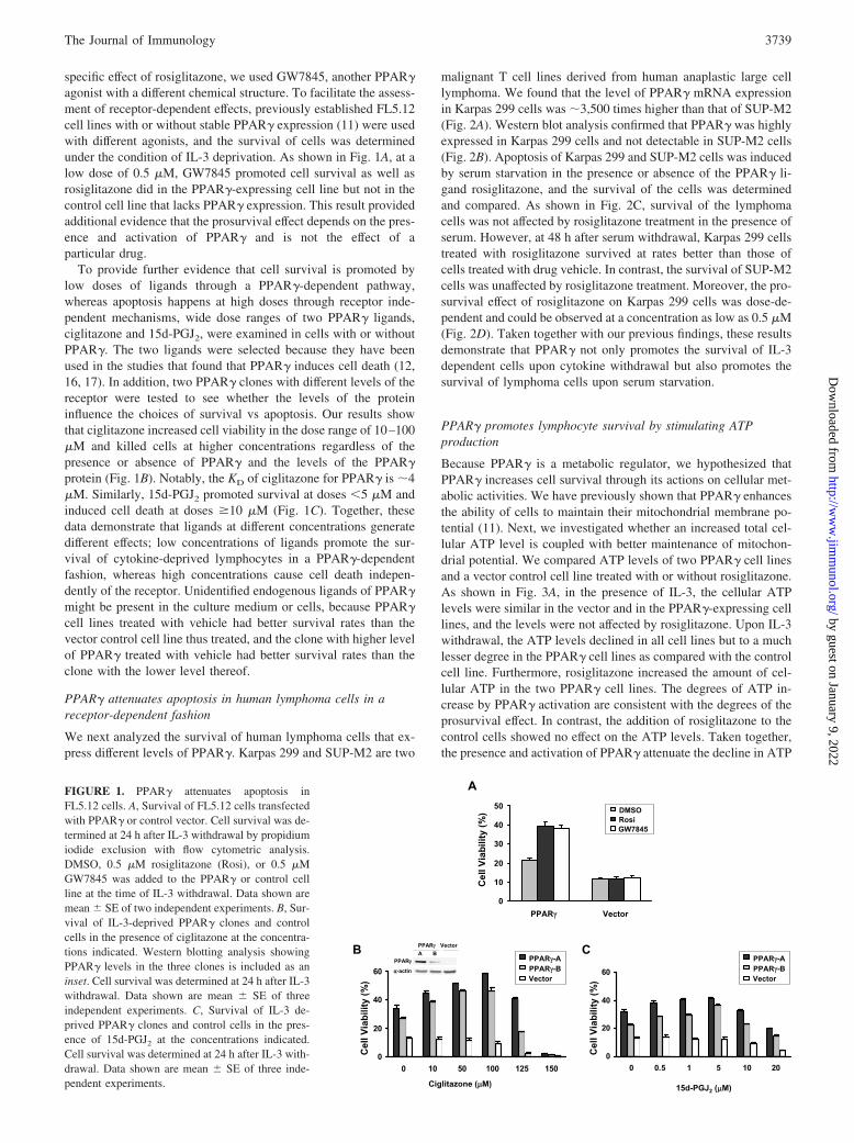
specific effect of rosiglitazone, we used GW7845, another PPAR�agonist with a different chemical structure. To facilitate the assess-ment of receptor-dependent effects, previously established FL5.12cell lines with or without stable PPAR� expression (11) were usedwith different agonists, and the survival of cells was determinedunder the condition of IL-3 deprivation. As shown in Fig. 1A, at alow dose of 0.5 �M, GW7845 promoted cell survival as well asrosiglitazone did in the PPAR�-expressing cell line but not in thecontrol cell line that lacks PPAR� expression. This result providedadditional evidence that the prosurvival effect depends on the pres-ence and activation of PPAR� and is not the effect of aparticular drug.
To provide further evidence that cell survival is promoted bylow doses of ligands through a PPAR�-dependent pathway,whereas apoptosis happens at high doses through receptor inde-pendent mechanisms, wide dose ranges of two PPAR� ligands,ciglitazone and 15d-PGJ2, were examined in cells with or withoutPPAR�. The two ligands were selected because they have beenused in the studies that found that PPAR� induces cell death (12,16, 17). In addition, two PPAR� clones with different levels of thereceptor were tested to see whether the levels of the proteininfluence the choices of survival vs apoptosis. Our results showthat ciglitazone increased cell viability in the dose range of 10–100�M and killed cells at higher concentrations regardless of thepresence or absence of PPAR� and the levels of the PPAR�protein (Fig. 1B). Notably, the KD of ciglitazone for PPAR� is �4�M. Similarly, 15d-PGJ2 promoted survival at doses �5 �M andinduced cell death at doses �10 �M (Fig. 1C). Together, thesedata demonstrate that ligands at different concentrations generatedifferent effects; low concentrations of ligands promote the sur-vival of cytokine-deprived lymphocytes in a PPAR�-dependentfashion, whereas high concentrations cause cell death indepen-dently of the receptor. Unidentified endogenous ligands of PPAR�might be present in the culture medium or cells, because PPAR�cell lines treated with vehicle had better survival rates than thevector control cell line thus treated, and the clone with higher levelof PPAR� treated with vehicle had better survival rates than theclone with the lower level thereof.
PPAR� attenuates apoptosis in human lymphoma cells in areceptor-dependent fashion
We next analyzed the survival of human lymphoma cells that ex-press different levels of PPAR�. Karpas 299 and SUP-M2 are two
malignant T cell lines derived from human anaplastic large celllymphoma. We found that the level of PPAR� mRNA expressionin Karpas 299 cells was �3,500 times higher than that of SUP-M2(Fig. 2A). Western blot analysis confirmed that PPAR� was highlyexpressed in Karpas 299 cells and not detectable in SUP-M2 cells(Fig. 2B). Apoptosis of Karpas 299 and SUP-M2 cells was inducedby serum starvation in the presence or absence of the PPAR� li-gand rosiglitazone, and the survival of the cells was determinedand compared. As shown in Fig. 2C, survival of the lymphomacells was not affected by rosiglitazone treatment in the presence ofserum. However, at 48 h after serum withdrawal, Karpas 299 cellstreated with rosiglitazone survived at rates better than those ofcells treated with drug vehicle. In contrast, the survival of SUP-M2cells was unaffected by rosiglitazone treatment. Moreover, the pro-survival effect of rosiglitazone on Karpas 299 cells was dose-de-pendent and could be observed at a concentration as low as 0.5 �M(Fig. 2D). Taken together with our previous findings, these resultsdemonstrate that PPAR� not only promotes the survival of IL-3dependent cells upon cytokine withdrawal but also promotes thesurvival of lymphoma cells upon serum starvation.
PPAR� promotes lymphocyte survival by stimulating ATPproduction
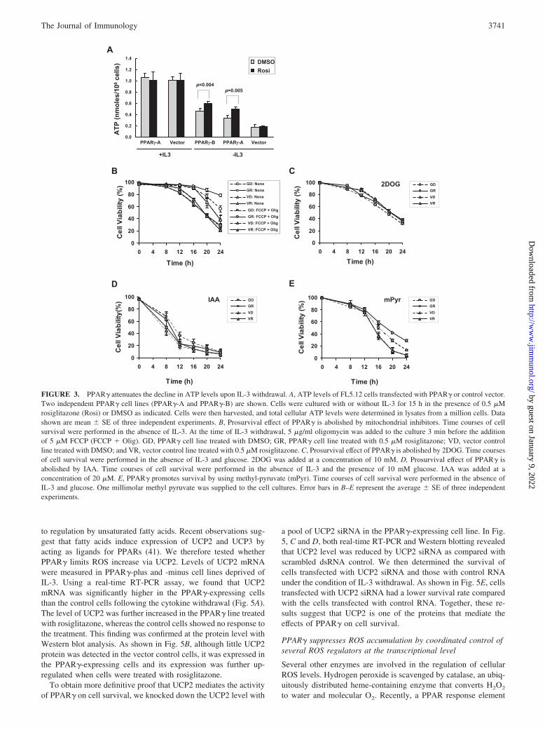
Because PPAR� is a metabolic regulator, we hypothesized thatPPAR� increases cell survival through its actions on cellular met-abolic activities. We have previously shown that PPAR� enhancesthe ability of cells to maintain their mitochondrial membrane po-tential (11). Next, we investigated whether an increased total cel-lular ATP level is coupled with better maintenance of mitochon-drial potential. We compared ATP levels of two PPAR� cell linesand a vector control cell line treated with or without rosiglitazone.As shown in Fig. 3A, in the presence of IL-3, the cellular ATPlevels were similar in the vector and in the PPAR�-expressing celllines, and the levels were not affected by rosiglitazone. Upon IL-3withdrawal, the ATP levels declined in all cell lines but to a muchlesser degree in the PPAR� cell lines as compared with the controlcell line. Furthermore, rosiglitazone increased the amount of cel-lular ATP in the two PPAR� cell lines. The degrees of ATP in-crease by PPAR� activation are consistent with the degrees of theprosurvival effect. In contrast, the addition of rosiglitazone to thecontrol cells showed no effect on the ATP levels. Taken together,the presence and activation of PPAR� attenuate the decline in ATP
FIGURE 1. PPAR� attenuates apoptosis inFL5.12 cells. A, Survival of FL5.12 cells transfectedwith PPAR� or control vector. Cell survival was de-termined at 24 h after IL-3 withdrawal by propidiumiodide exclusion with flow cytometric analysis.DMSO, 0.5 �M rosiglitazone (Rosi), or 0.5 �MGW7845 was added to the PPAR� or control cellline at the time of IL-3 withdrawal. Data shown aremean � SE of two independent experiments. B, Sur-vival of IL-3-deprived PPAR� clones and controlcells in the presence of ciglitazone at the concentra-tions indicated. Western blotting analysis showingPPAR� levels in the three clones is included as aninset. Cell survival was determined at 24 h after IL-3withdrawal. Data shown are mean � SE of threeindependent experiments. C, Survival of IL-3 de-prived PPAR� clones and control cells in the pres-ence of 15d-PGJ2 at the concentrations indicated.Cell survival was determined at 24 h after IL-3 with-drawal. Data shown are mean � SE of three inde-pendent experiments.
3739The Journal of Immunology
by guest on January 9, 2022http://w
ww
.jimm
unol.org/D
ownloaded from

upon IL-3 withdrawal that correlates well with better-maintainedmitochondrial membrane potential and improved cell survival.
Higher cellular ATP in the PPAR� cells relative to the controlcells might result from either increased ATP production or de-creased ATP consumption. To distinguish between these two pos-sibilities, we analyzed survival of the PPAR� and vector controlcells in the presence of two drugs that inhibit mitochondrial ATPproduction. Carbonyl cyanide p-trifluoromethoxyphenylhydrazone(FCCP) is a derivative of cyanide; it is a proton ionophore that actsto collapse the mitochondrial membrane potential required forATP synthesis. Oligomycin is an inhibitor of the F1-F0-ATPase,the complex V of the mitochondrial respiratory chain. Acting to-gether, the two drugs prevent ATP production from the mitochon-drial source (32, 33). As shown in Fig. 3B, the survival benefit ofthe PPAR� line was lost in the presence of FCCP and oligomycinbecause the cells treated with or without rosiglitazone showed thesame survival rate as the vector control cells. These data demon-strate that ATP production is required for PPAR� to exert its pro-survival effect.
To further test this notion, cell survival was determined in thepresence of glycolytic inhibitors to prevent the fueling of glyco-lytic substrates to mitochondria for ATP production. Under theroutine culturing condition (see Material and Methods), FL5.12cells rely on glucose in the medium as the main energy source forATP production to sustain their survival, growth, and proliferation.Inhibition of glycolysis impairs cellular ATP synthesis that mayaffect the prosurvival effect by PPAR�. To inhibit glycolysis, wemade use of 2-deoxyglucose (2DOG), a nonmetabolizable analogof glucose, and iodoacetic acid (IAA), an inhibitor of GAPDH anda key enzyme in the glycolytic pathway. When cells were culturedin IL-3-free medium containing 2DOG in place of glucose, nosurvival advantage was observed in the PPAR� cells treated withor without rosiglitazone as compared with the control (Fig. 3C).Similarly, the addition of IAA in the presence of glucose abolishedthe survival advantage of the PPAR� cells (Fig. 3D). When cellswere supplied with methyl pyruvate, a cell-permeable form ofpyruvate as an alternative energy source replacing glucose, ros-iglitazone was able to promote cell survival in a PPAR�-dependentfashion (Fig. 3E). Taken together, PPAR� relies on a source ofenergy to promote survival. Combined with our previous data,these results suggest that PPAR� promotes survival by maintain-
ing mitochondrial integrity through an energy source that func-tionally leads to increased ATP production.
PPAR� suppresses accumulation of ROS in growth factor-deprived cells
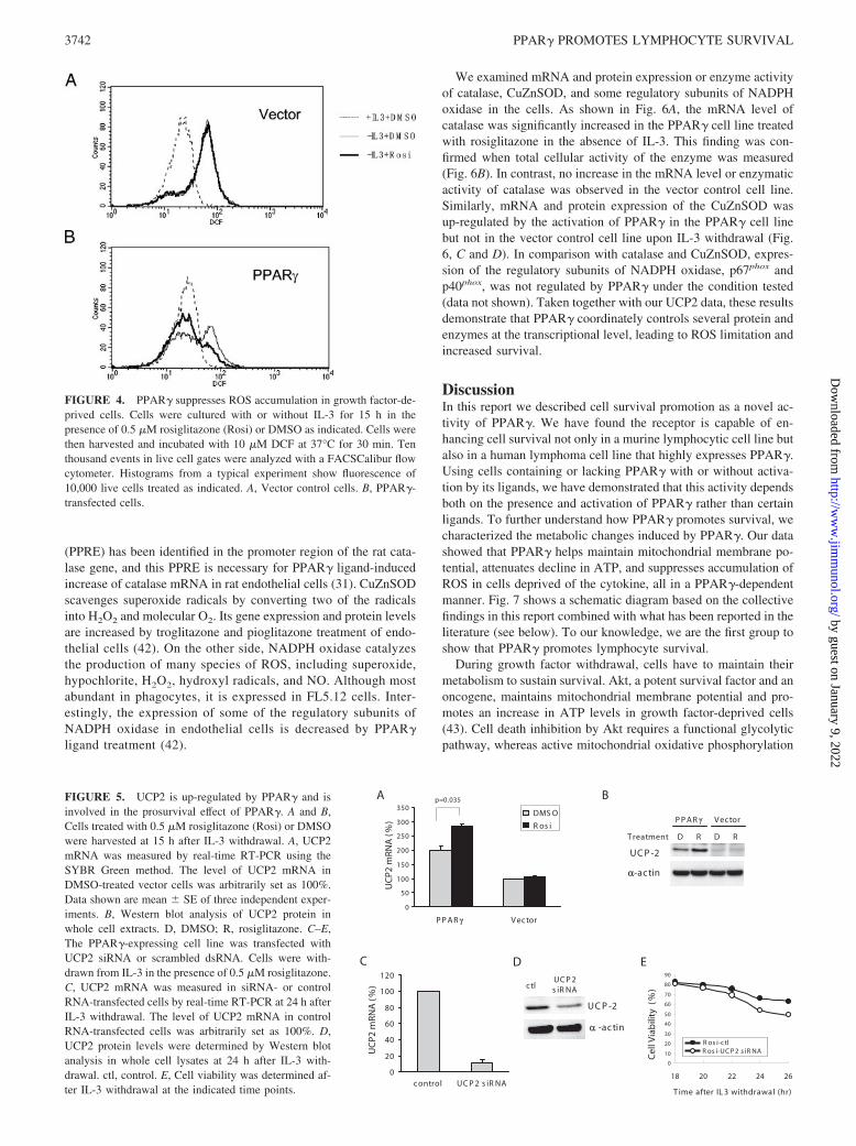
ROS are generated following application of many apoptotic stim-uli (34), and mitochondria are the major sites of ROS productionin most apoptotic systems (35, 36). It has been shown that anincrease in ROS precedes mitochondrial depolarization duringTNF-�-induced apoptosis and that ROS scavenger treatment de-lays apoptosis (34). Based on our finding that PPAR� attenuatescell death by maintaining mitochondrial homeostasis, we reasonedthat PPAR� might reduce the amounts of ROS that are harmful tothe mitochondria during IL-3 withdrawal. We measured the intra-cellular oxidants in the PPAR� and the vector control cell lines inthe presence or absence of rosiglitazone. As shown in Fig. 4, in thepresence of IL-3, the ROS levels in the two cell lines were low andcomparable to each other (dashed traces). In IL-3-depleted cellsROS was elevated in the vector control cells, and rosiglitazoneaddition had no appreciable effect on the ROS level (Fig. 4A). Incomparison, fewer PPAR�-expressing cells had elevated ROS(Fig. 4B, thin solid trace). Moreover, even fewer PPAR� trans-fected cells had increased ROS when treated with rosiglitazone(Fig. 4B, thick solid trace). These results demonstrate that bettersurvival of the PPAR� cell line in the absence of IL-3 is accom-panied by suppressed ROS increase in the cells.
UCP2 is up-regulated by PPAR� and involved in the cellsurvival-promoting effect of PPAR�
Cycloheximide and actinomycin D blocked the ability of PPAR�to promote survival in IL-3-deprived cells, suggesting that thisfunction of PPAR� requires new mRNA and protein synthesis(data not shown). Having established that the limiting of ROSincrease is one of the mechanisms underlying the prosurvival effectof PPAR�, we pursued potential transcriptional targets of the re-ceptor that may mediate its regulation of ROS. UCP2, unlikeUCP1 and UCP3, is mainly involved in the limitation of free rad-ical levels in cells rather than in physiological uncoupling andthermogenesis (37, 38). Recently, it has been shown that UCP2protects neurons and cardiomyocytes from death by reducing ROSproduction in mitochondria (39, 40). Interestingly, UCP2 is subject
0
02
04
06
08
001
PPA
Rγ
mR
NA
(fol
d)
sapraK 2M-PUS 92 9
Cel
l Via
bilit
y (%
)US -P 2M Karp sa 992
%01 SBF 0% SBF
C OSMDisoR
US -P 2M raK p sa 992
p=0 000. 4
0
1
01
001
0001
00001A B
USP-M
2rp
as2
aK
99
RAPP γcificepsnoN
RAPP γcificepsnoN
RAPP γcificepsnoN
D
0
01
02
03
04
05
06
543210
Cel
l Via
bilit
y (%
)
enozatilgisoR (µ )M
FIGURE 2. PPAR� attenuates apoptosis in Karpas299 lymphoma cells. A, PPAR� mRNA expression inlymphoma cell lines. Levels of PPAR� mRNA weremeasured by real-time RT-PCR assay. Expression levelswere calculated relative to the level of PPAR� inSUP-M2 cells that is arbitrarily defined as 1. B, Westernblot analysis of PPAR� in lymphoma cell lines. PPAR�in whole cell extracts was analyzed. D, Rosiglitazonepromotes survival in serum-deprived Karpas 299 but notin SUP-M2 cells. Cells were cultured with or without10% serum for 48 h in the presence of 2 �M rosiglita-zone or DMSO as indicated. Data shown are mean � SEof three independent experiments. D, Prosurvival isdose-dependent on the concentration of rosiglitazone.Serum-deprived Karpas 299 cells were cultured in thepresence of rosiglitazone at the concentrations indi-cated, and survival was determined at 48 h after serumwithdrawal.
3740 PPAR� PROMOTES LYMPHOCYTE SURVIVAL
by guest on January 9, 2022http://w
ww
.jimm
unol.org/D
ownloaded from
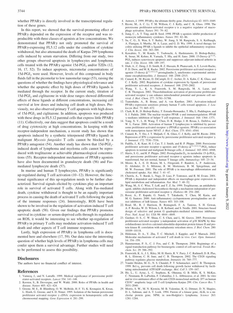
to regulation by unsaturated fatty acids. Recent observations sug-gest that fatty acids induce expression of UCP2 and UCP3 byacting as ligands for PPARs (41). We therefore tested whetherPPAR� limits ROS increase via UCP2. Levels of UCP2 mRNAwere measured in PPAR�-plus and -minus cell lines deprived ofIL-3. Using a real-time RT-PCR assay, we found that UCP2mRNA was significantly higher in the PPAR�-expressing cellsthan the control cells following the cytokine withdrawal (Fig. 5A).The level of UCP2 was further increased in the PPAR� line treatedwith rosiglitazone, whereas the control cells showed no response tothe treatment. This finding was confirmed at the protein level withWestern blot analysis. As shown in Fig. 5B, although little UCP2protein was detected in the vector control cells, it was expressed inthe PPAR�-expressing cells and its expression was further up-regulated when cells were treated with rosiglitazone.
To obtain more definitive proof that UCP2 mediates the activityof PPAR� on cell survival, we knocked down the UCP2 level with
a pool of UCP2 siRNA in the PPAR�-expressing cell line. In Fig.5, C and D, both real-time RT-PCR and Western blotting revealedthat UCP2 level was reduced by UCP2 siRNA as compared withscrambled dsRNA control. We then determined the survival ofcells transfected with UCP2 siRNA and those with control RNAunder the condition of IL-3 withdrawal. As shown in Fig. 5E, cellstransfected with UCP2 siRNA had a lower survival rate comparedwith the cells transfected with control RNA. Together, these re-sults suggest that UCP2 is one of the proteins that mediate theeffects of PPAR� on cell survival.
PPAR� suppresses ROS accumulation by coordinated control ofseveral ROS regulators at the transcriptional level
Several other enzymes are involved in the regulation of cellularROS levels. Hydrogen peroxide is scavenged by catalase, an ubiq-uitously distributed heme-containing enzyme that converts H2O2
to water and molecular O2. Recently, a PPAR response element
FIGURE 3. PPAR� attenuates the decline in ATP levels upon IL-3 withdrawal. A, ATP levels of FL5.12 cells transfected with PPAR� or control vector.Two independent PPAR� cell lines (PPAR�-A and PPAR�-B) are shown. Cells were cultured with or without IL-3 for 15 h in the presence of 0.5 �Mrosiglitazone (Rosi) or DMSO as indicated. Cells were then harvested, and total cellular ATP levels were determined in lysates from a million cells. Datashown are mean � SE of three independent experiments. B, Prosurvival effect of PPAR� is abolished by mitochondrial inhibitors. Time courses of cellsurvival were performed in the absence of IL-3. At the time of IL-3 withdrawal, 5 �g/ml oligomycin was added to the culture 3 min before the additionof 5 �M FCCP (FCCP � Olig). GD, PPAR� cell line treated with DMSO; GR, PPAR� cell line treated with 0.5 �M rosiglitazone; VD, vector controlline treated with DMSO; and VR, vector control line treated with 0.5 �M rosiglitazone. C, Prosurvival effect of PPAR� is abolished by 2DOG. Time coursesof cell survival were performed in the absence of IL-3 and glucose. 2DOG was added at a concentration of 10 mM. D, Prosurvival effect of PPAR� isabolished by IAA. Time courses of cell survival were performed in the absence of IL-3 and the presence of 10 mM glucose. IAA was added at aconcentration of 20 �M. E, PPAR� promotes survival by using methyl-pyruvate (mPyr). Time courses of cell survival were performed in the absence ofIL-3 and glucose. One millimolar methyl pyruvate was supplied to the cell cultures. Error bars in B–E represent the average � SE of three independentexperiments.
3741The Journal of Immunology
by guest on January 9, 2022http://w
ww
.jimm
unol.org/D
ownloaded from
(PPRE) has been identified in the promoter region of the rat cata-lase gene, and this PPRE is necessary for PPAR� ligand-inducedincrease of catalase mRNA in rat endothelial cells (31). CuZnSODscavenges superoxide radicals by converting two of the radicalsinto H2O2 and molecular O2. Its gene expression and protein levelsare increased by troglitazone and pioglitazone treatment of endo-thelial cells (42). On the other side, NADPH oxidase catalyzesthe production of many species of ROS, including superoxide,hypochlorite, H2O2, hydroxyl radicals, and NO. Although mostabundant in phagocytes, it is expressed in FL5.12 cells. Inter-estingly, the expression of some of the regulatory subunits ofNADPH oxidase in endothelial cells is decreased by PPAR�ligand treatment (42).
We examined mRNA and protein expression or enzyme activityof catalase, CuZnSOD, and some regulatory subunits of NADPHoxidase in the cells. As shown in Fig. 6A, the mRNA level ofcatalase was significantly increased in the PPAR� cell line treatedwith rosiglitazone in the absence of IL-3. This finding was con-firmed when total cellular activity of the enzyme was measured(Fig. 6B). In contrast, no increase in the mRNA level or enzymaticactivity of catalase was observed in the vector control cell line.Similarly, mRNA and protein expression of the CuZnSOD wasup-regulated by the activation of PPAR� in the PPAR� cell linebut not in the vector control cell line upon IL-3 withdrawal (Fig.6, C and D). In comparison with catalase and CuZnSOD, expres-sion of the regulatory subunits of NADPH oxidase, p67phox andp40phox, was not regulated by PPAR� under the condition tested(data not shown). Taken together with our UCP2 data, these resultsdemonstrate that PPAR� coordinately controls several protein andenzymes at the transcriptional level, leading to ROS limitation andincreased survival.
DiscussionIn this report we described cell survival promotion as a novel ac-tivity of PPAR�. We have found the receptor is capable of en-hancing cell survival not only in a murine lymphocytic cell line butalso in a human lymphoma cell line that highly expresses PPAR�.Using cells containing or lacking PPAR� with or without activa-tion by its ligands, we have demonstrated that this activity dependsboth on the presence and activation of PPAR� rather than certainligands. To further understand how PPAR� promotes survival, wecharacterized the metabolic changes induced by PPAR�. Our datashowed that PPAR� helps maintain mitochondrial membrane po-tential, attenuates decline in ATP, and suppresses accumulation ofROS in cells deprived of the cytokine, all in a PPAR�-dependentmanner. Fig. 7 shows a schematic diagram based on the collectivefindings in this report combined with what has been reported in theliterature (see below). To our knowledge, we are the first group toshow that PPAR� promotes lymphocyte survival.
During growth factor withdrawal, cells have to maintain theirmetabolism to sustain survival. Akt, a potent survival factor and anoncogene, maintains mitochondrial membrane potential and pro-motes an increase in ATP levels in growth factor-deprived cells(43). Cell death inhibition by Akt requires a functional glycolyticpathway, whereas active mitochondrial oxidative phosphorylation
FIGURE 5. UCP2 is up-regulated by PPAR� and isinvolved in the prosurvival effect of PPAR�. A and B,Cells treated with 0.5 �M rosiglitazone (Rosi) or DMSOwere harvested at 15 h after IL-3 withdrawal. A, UCP2mRNA was measured by real-time RT-PCR using theSYBR Green method. The level of UCP2 mRNA inDMSO-treated vector cells was arbitrarily set as 100%.Data shown are mean � SE of three independent exper-iments. B, Western blot analysis of UCP2 protein inwhole cell extracts. D, DMSO; R, rosiglitazone. C–E,The PPAR�-expressing cell line was transfected withUCP2 siRNA or scrambled dsRNA. Cells were with-drawn from IL-3 in the presence of 0.5 �M rosiglitazone.C, UCP2 mRNA was measured in siRNA- or controlRNA-transfected cells by real-time RT-PCR at 24 h afterIL-3 withdrawal. The level of UCP2 mRNA in controlRNA-transfected cells was arbitrarily set as 100%. D,UCP2 protein levels were determined by Western blotanalysis in whole cell lysates at 24 h after IL-3 with-drawal. ctl, control. E, Cell viability was determined af-ter IL-3 withdrawal at the indicated time points.
FIGURE 4. PPAR� suppresses ROS accumulation in growth factor-de-prived cells. Cells were cultured with or without IL-3 for 15 h in thepresence of 0.5 �M rosiglitazone (Rosi) or DMSO as indicated. Cells werethen harvested and incubated with 10 �M DCF at 37°C for 30 min. Tenthousand events in live cell gates were analyzed with a FACSCalibur flowcytometer. Histograms from a typical experiment show fluorescence of10,000 live cells treated as indicated. A, Vector control cells. B, PPAR�-transfected cells.
3742 PPAR� PROMOTES LYMPHOCYTE SURVIVAL
by guest on January 9, 2022http://w
ww
.jimm
unol.org/D
ownloaded from

is dispensable (44). Unlike Akt, oxidative phosphorylation is re-quired for PPAR� to inhibit cell death. In the presence of FCCPand oligomycin, PPAR� loses its ability to increase cell survival,although ATP derived from glycolysis is still available to the cells(Fig. 3B). In the mean time, glycolysis is not needed for thePPAR� survival-promoting effect. Although glycolytic inhibitorsabolished survival-promoting effects by PPAR�, methyl pyruvaterestored cellular response to rosiglitazone (Fig. 3E). These datasuggest that glycolysis per se is not required. Instead, the feedingof pyruvate, the end product of glycolysis, to the tricarboxylic acidcycle and the subsequent mitochondrial oxidative phosphorylationare the processes that are needed for PPAR� to exert its survivaleffect.
ROS are generated in apoptotic processes induced by variousstimuli, including TNF-� (45), Fas (46), glucocorticoid hormones(47), radiation (48), and chemotherapeutic drugs (49), and mito-chondria are major sites of ROS production during apoptosis (35,
36). It has been shown that an increase in ROS inhibits mitochon-drial electron transport and leads to mitochondrial depolarizationand caspase activation (34, 50). In line with these findings, weshowed that cellular ROS increased following IL-3 withdrawal.The increase in ROS was suppressed by the presence and activa-tion of PPAR� (Fig. 4), which correlates well with the receptor’sability to maintain mitochondrial membrane potential (11) and in-crease ATP production (Fig. 3A). Further investigation revealedthat a set of proteins and enzymes regulating ROS were coordi-nately controlled by PPAR�. PPAR� increased UCP2, catalase,and CuZnSOD at mRNA and protein levels, leading to reducedROS accumulation, limited mitochondrial damage, and, eventu-ally, enhanced cell survival. At this point, it is unclear whetherthese genes are direct targets of PPAR� transcriptional activity.Functional PPRE have been identified in the promoters of the ratcatalase gene and CuZnSOD (31, 51), but are not present in thepromoter of UCP2 (41). Further studies are needed to determine
FIGURE 6. PPAR� coordinately controls severalROS-regulating enzymes. Cells treated with 0.5 �Mrosiglitazone (Rosi) or DMSO were harvested at15 h after IL-3 withdrawal. A, Catalase mRNA wasmeasured by real-time RT-PCR using TaqMan tech-nology. The amount of catalase mRNA in DMSO-treated vector cells was arbitrarily set as 100%. B,Catalase activity in whole cell extracts was mea-sured at 15 h after IL-3 withdrawal. C, CuZnSODmRNA was measured by real-time RT-PCR usingTaqMan technology. The amount of CuZnSODmRNA in DMSO-treated vector cells was arbitrarilyset as 100%. Data shown in A–C are mean � SE ofthree independent experiments. D, CuZnSOD pro-tein levels were determined by Western blot analysisin whole cell lysates at 15 h after IL-3 withdrawal.D, DMSO; R, rosiglitazone.
FIGURE 7. Schematic diagram shows how PPAR�might increase cell survival in cytokine-deprived cells.The scheme was made based on findings in the currentreport and the literature (see Discussion). Dashed arrowindicates the effects may or may not be direct.
3743The Journal of Immunology
by guest on January 9, 2022http://w
ww
.jimm
unol.org/D
ownloaded from
whether PPAR� is directly involved in the transcriptional regula-tion of these genes.
In this report, we showed that the survival-promoting effect ofPPAR� depended on the expression of the receptor and was re-producible with three classes of agonists at low concentrations. Wedemonstrated that PPAR� not only promoted the survival ofPPAR�-expressing FL5.12 cells under the condition of cytokinewithdrawal, but also attenuated the death of Karpas 299 lymphomacells induced by serum starvation. Differing from our study, twoother groups observed apoptosis in lymphocytes and lymphomacells treated with the PPAR� agonist 15d-PGJ2 and/or TZDs (12,16, 17, 52). To induce apoptosis, micromolar concentrations of15d-PGJ2 were used. However, levels of this compound in bodyfluids fall in the picomolar to low nanomolar range (53), raising thequestions of whether the findings have physiological relevance andwhether the apoptotic effect by high doses of PPAR� ligands ismediated through the receptor. In the current study, titration of15d-PGJ2 and ciglitazone in wide dose ranges showed differentialeffects of these ligands at different concentrations, increasing cellsurvival at low doses and inducing cell death at high doses. Pre-viously, we also observed primary T cell apoptosis with high dosesof 15d-PGJ2 and ciglitazone. In addition, apoptosis was producedwith these drugs in FL5.12 parental cells that express little PPAR�(11). Collectively, our data suggest that apoptosis could be a resultof drug cytotoxicity at high concentrations. In keeping with thereceptor-independent mechanism, a recent study has shown thatapoptosis induced by a synthetic triterpenoid (PPAR� ligand) inmalignant Mycosis fungoides T cells cannot be blocked by aPPAR� antagonist (54). Another study has shown that 15d-PGJ2-induced death of lymphoma and myeloma cells cannot be repro-duced with troglitazone at achievable pharmacological concentra-tions (55). Receptor-independent mechanisms of PPAR� agonistshave also been documented in granulocyte death (56) and Fas-mediated lymphocyte death (57).
In murine and human T lymphocytes, PPAR� is significantlyup-regulated during T cell activation (10–12). However, the func-tional significance of this up-regulation needs to be further char-acterized. Survival signals elicited by cytokines play an importantrole in survival of activated T cells. Along with Fas-mediateddeath, cytokine withdrawal is thought to be an equally importantprocess in causing the death of activated T cells following the peakof the immune responses (24). Interestingly, ROS have beenshown to be involved in the regulation of activation-induced T cellapoptotic death (58). Given our findings that PPAR� promotessurvival in cytokine- or serum-deprived cells through its regulationon ROS, it would be interesting to see whether up-regulation ofPPAR� in primary T cells may modulate activation-induced T celldeath and other aspects of T cell immune responses.
Lastly, high expression of PPAR� in lymphoma cell is docu-mented here and elsewhere (17, 59). Our data raise the interestingquestion of whether high levels of PPAR� in lymphoma cells mayconfer upon them a survival advantage. Further studies will needto be performed to assess this possibility.
DisclosuresThe authors have no financial conflict of interest.
References1. Vamecq, J., and N. Latruffe. 1999. Medical significance of peroxisome prolif-
erator-activated receptors. Lancet 354: 141–148.2. Kersten, S., B. Desvergne, and W. Wahli. 2000. Roles of PPARs in health and
disease. Nature 405: 421–424.3. Greene, M. E., B. Blumberg, O. W. McBride, H. F. Yi, K. Kronquist, K. Kwan,
L. Hsieh, G. Greene, and S. D. Nimer. 1995. Isolation of the human peroxisomeproliferator activated receptor � cDNA: expression in hematopoietic cells andchromosomal mapping. Gene Expression 4: 281–299.
4. Auwerx, J. 1999. PPAR�, the ultimate thrifty gene. Diabetologia 42: 1033–1049.5. Ricote, M., A. C. Li, T. M. Willson, C. J. Kelly, and C. K. Glass. 1998. The
peroxisome proliferator-activated receptor-� is a negative regulator of macro-phage activation. Nature 391: 79–82.
6. Jiang, C., A. T. Ting, and B. Seed. 1998. PPAR-� agonists inhibit production ofmonocyte inflammatory cytokines. Nature 391: 82–86.
7. Su, C. G., X. Wen, S. T. Bailey, W. Jiang, S. M. Rangwala, S. A. Keilbaugh,A. Flanigan, S. Murthy, M. A. Lazar, and G. D. Wu. 1999. A novel therapy forcolitis utilizing PPAR-� ligands to inhibit the epithelial inflammatory response.J. Clin. Invest. 104: 383–389.
8. Kawahito, Y., M. Kondo, Y. Tsubouchi, A. Hashiramoto, D. Bishop-Bailey,K. Inoue, M. Kohno, R. Yamada, T. Hla, and H. Sano. 2000. 15-Deoxy-�12,14-PGJ2 induces synoviocyte apoptosis and suppresses adjuvant-induced arthritis inrats. J. Clin. Invest. 106: 189–197.
9. Diab, A., C. Deng, J. D. Smith, R. Z. Hussain, B. Phanavanh, A. E. Lovett-Racke,P. D. Drew, and M. K. Racke. 2002. Peroxisome proliferator-activated receptor-�agonist 15-deoxy-�12,1412,14-prostaglandin J2 ameliorates experimental autoim-mune encephalomyelitis. J. Immunol. 168: 2508–2515.
10. Cunard, R., M. Ricote, D. DiCampli, D. C. Archer, D. A. Kahn, C. K. Glass, andC. J. Kelly. 2002. Regulation of cytokine expression by ligands of peroxisomeproliferator activated receptors. J. Immunol. 168: 2795–2802.
11. Wang, Y. L., K. A. Frauwirth, S. M. Rangwala, M. A. Lazar, andC. B. Thompson. 2002. Thiazolidinedione activation of peroxisome proliferator-activated receptor � can enhance mitochondrial potential and promote cell sur-vival. J. Biol. Chem. 277: 31781–31788.
12. Tautenhahn, A., B. Brune, and A. von Knethen. 2003. Activation-inducedPPAR� expression sensitizes primary human T cells toward apoptosis. J. Leu-kocyte Biol. 73: 665–672.
13. Clark, R. B., D. Bishop-Bailey, T. Estrada-Hernandez, T. Hla, L. Puddington, andS. J. Padula. 2000. The nuclear receptor PPAR � and immunoregulation: PPAR� mediates inhibition of helper T cell responses. J. Immunol. 164: 1364–1371.
14. Yang, X. Y., L. H. Wang, T. Chen, D. R. Hodge, J. H. Resau, L. DaSilva, andW. L. Farrar. 2000. Activation of human T lymphocytes is inhibited by peroxi-some proliferator-activated receptor � (PPAR�) agonists. PPAR� co-associationwith transcription factor NFAT. J. Biol. Chem. 275: 4541–4544.
15. Cunard, R., Y. Eto, J. T. Muljadi, C. K. Glass, C. J. Kelly, and M. Ricote. 2004.Repression of IFN-� expression by peroxisome proliferator-activated receptor �.J. Immunol. 172: 7530–7536.
16. Padilla, J., K. Kaur, H. J. Cao, T. J. Smith, and R. P. Phipps. 2000. Peroxisomeproliferator activator receptor-� agonists and 15-deoxy-�12,1412,14-PGJ2 induceapoptosis in normal and malignant B-lineage cells. J. Immunol. 165: 6941–6948.
17. Harris, S. G., and R. P. Phipps. 2002. Prostaglandin D2, its metabolite 15-�-PGJ2,and peroxisome proliferator activated receptor-� agonists induce apoptosis intransformed, but not normal, human T lineage cells. Immunology 105: 23–34.
18. Moore, K. J., E. D. Rosen, M. L. Fitzgerald, F. Randow, L. P. Andersson,D. Altshuler, D. S. Milstone, R. M. Mortensen, B. M. Spiegelman, andM. W. Freeman. 2001. The role of PPAR-� in macrophage differentiation andcholesterol uptake. Nat. Med. 7: 41–47.
19. Chawla, A., Y. Barak, L. Nagy, D. Liao, P. Tontonoz, and R. M. Evans. 2001.PPAR-� dependent and independent effects on macrophage-gene expression inlipid metabolism and inflammation. Nat. Med. 7: 48–52.
20. Wang, M., S. C. Wise, T. Leff, and T. Z. Su. 1999. Troglitazone, an antidiabeticagent, inhibits cholesterol biosynthesis through a mechanism independent of per-oxisome proliferator-activated receptor-�. Diabetes 48: 254–260.
21. Rossi, A., P. Kapahi, G. Natoli, T. Takahashi, Y. Chen, M. Karin, andM. G. Santoro. 2000. Anti-inflammatory cyclopentenone prostaglandins are di-rect inhibitors of I�B kinase. Nature 403: 103–108.
22. Read, M., R. J. Harrison, B. Romagnoli, F. A. Tanious, S. H. Gowan,A. P. Reszka, W. D. Wilson, L. R. Kelland, and S. Neidle. 2001. Structure-baseddesign of selective and potent G quadruplex-mediated telomerase inhibitors.Proc. Natl. Acad. Sci. USA 98: 4844–4849.
23. Gardner, O. S., C. W. Shiau, C. S. Chen, and L. M. Graves. 2005. Peroxisomeproliferator-activated receptor �-independent activation of p38 MAPK by thia-zolidinediones involves calcium/calmodulin-dependent protein kinase II and pro-tein kinase R: correlation with endoplasmic reticulum stress. J. Biol. Chem. 280:10109–10118.
24. Hildeman, D. A., Y. Zhu, T. C. Mitchell, J. Kappler, and P. Marrack. 2002.Molecular mechanisms of activated T cell death in vivo. Curr. Opin. Immunol.14: 354–359.
25. Hammerman, P. S., C. J. Fox, and C. B. Thompson. 2004. Beginnings of asignal-transduction pathway for bioenergetic control of cell survival. Trends Bio-chem. Sci. 29: 586–592.
26. Frauwirth, K. A., J. L. Riley, M. H. Harris, R. V. Parry, J. C. Rathmell, D. R. Plas,R. L. Elstrom, C. H. June, and C. B. Thompson. 2002. The CD28 signalingpathway regulates glucose metabolism. Immunity 16: 769–777.
27. Vander Heiden, M. G., N. S. Chandel, P. T. Schumacker, and C. B. Thompson.1999. Bcl-xL prevents cell death following growth factor withdrawal by facili-tating mitochondrial ATP/ADP exchange. Mol. Cell 3: 159–167.
28. Ho, L., U. Aytac, L. C. Stephens, K. Ohnuma, G. B. Mills, K. S. McKee,C. Neumann, R. LaPushin, F. Cabanillas, J. L. Abbruzzese, et al. 2001. In vitroand in vivo antitumor effect of the anti-CD26 monoclonal antibody 1F7 on humanCD30� anaplastic large cell T-cell lymphoma Karpas 299. Clin. Cancer Res. 7:2031–2040.
29. Morris, S. W., M. N. Kirstein, M. B. Valentine, K. G. Dittmer, D. N. Shapiro,D. L. Saltman, and A. T. Look. 1994. Fusion of a kinase gene, ALK, to a nu-cleolar protein gene, NPM, in non-Hodgkin’s lymphoma. Science 263:1281–1284.
3744 PPAR� PROMOTES LYMPHOCYTE SURVIVAL
by guest on January 9, 2022http://w
ww
.jimm
unol.org/D
ownloaded from
30. Popovic, M., P. S. Sarin, M. Robert-Gurroff, V. S. Kalyanaraman, D. Mann,J. Minowada, and R. C. Gallo. 1983. Isolation and transmission of human retro-virus (human t-cell leukemia virus). Science 219: 856–859.
31. Girnun, G. D., F. E. Domann, S. A. Moore, and M. E. Robbins. 2002. Identifi-cation of a functional peroxisome proliferator-activated receptor response ele-ment in the rat catalase promoter. Mol. Endocrinol. 16: 2793–2801.
32. Smaili, S. S., Y. T. Hsu, K. M. Sanders, J. T. Russell, and R. J. Youle. 2001. Baxtranslocation to mitochondria subsequent to a rapid loss of mitochondrial mem-brane potential. Cell Death Differ. 8: 909–920.
33. Nilsen, J., and R. Diaz Brinton. 2003. Mechanism of estrogen-mediated neuro-protection: regulation of mitochondrial calcium and Bcl-2 expression. Proc. Natl.Acad. Sci. USA 100: 2842–2847.
34. Gottlieb, E., M. G. Vander Heiden, and C. B. Thompson. 2000. Bcl-xL preventsthe initial decrease in mitochondrial membrane potential and subsequent reactiveoxygen species production during tumor necrosis factor �-induced apoptosis.Mol. Cell. Biol. 20: 5680–5689.
35. Kroemer, G., B. Dallaporta, and M. Resche-Rigon. 1998. The mitochondrialdeath/life regulator in apoptosis and necrosis. Annu. Rev. Physiol. 60: 619–4278.
36. Mignotte, B., and J. L. Vayssiere. 1998. Mitochondria and apoptosis. Eur. J. Bio-chem. 252: 1–15.
37. Arsenijevic, D., H. Onuma, C. Pecqueur, S. Raimbault, B. S. Manning,B. Miroux, E. Couplan, M. C. Alves-Guerra, M. Goubern, R. Surwit, et al. 2000.Disruption of the uncoupling protein-2 gene in mice reveals a role in immunityand reactive oxygen species production. Nat. Genet. 26: 435–439.
38. Kizaki, T., K. Suzuki, Y. Hitomi, N. Taniguchi, D. Saitoh, K. Watanabe,K. Onoe, N. K. Day, R. A. Good, and H. Ohno. 2002. Uncoupling protein 2 playsan important role in nitric oxide production of lipopolysaccharide-stimulatedmacrophages. Proc. Natl. Acad. Sci. USA 99: 9392–9397.
39. Paradis, E., S. Clavel, F. Bouillaud, D. Ricquier, and D. Richard. 2003. Uncou-pling protein 2: a novel player in neuroprotection. Trends Mol. Med. 9: 522–525.
40. Teshima, Y., M. Akao, S. P. Jones, and E. Marban. 2003. Uncoupling protein-2overexpression inhibits mitochondrial death pathway in cardiomyocytes. Circ.Res. 93: 192–200.
41. Thompson, M. P., and D. Kim. 2004. Links between fatty acids and expressionof UCP2 and UCP3 mRNAs. FEBS Lett. 568: 4–9.
42. Inoue, I., S. Goto, T. Matsunaga, T. Nakajima, T. Awata, S. Hokari, T. Komoda,and S. Katayama. 2001. The ligands/activators for peroxisome proliferator-acti-vated receptor � (PPAR�) and PPAR� increase Cu2�,Zn2�-superoxide dis-mutase and decrease p22phox message expressions in primary endothelial cells.Metabolism 50: 3–11.
43. Plas, D. R., S. Talapatra, A. L. Edinger, J. C. Rathmell, and C. B. Thompson.2001. Akt and Bcl-xL promote growth factor-independent survival through dis-tinct effects on mitochondrial physiology. J. Biol. Chem. 276: 12041–12048.
44. Elstrom, R. L., D. E. Bauer, M. Buzzai, R. Karnauskas, M. H. Harris, D. R. Plas,H. Zhuang, R. M. Cinalli, A. Alavi, C. M. Rudin, and C. B. Thompson. 2004. Aktstimulates aerobic glycolysis in cancer cells. Cancer Res. 64: 3892–3899.
45. Sakon, S., X. Xue, M. Takekawa, T. Sasazuki, T. Okazaki, Y. Kojima, J. H. Piao,H. Yagita, K. Okumura, T. Doi, and H. Nakano. 2003. NF-�B inhibits TNF-induced accumulation of ROS that mediate prolonged MAPK activation and ne-crotic cell death. EMBO J. 22: 3898–3909.
46. Sato, T., T. Machida, S. Takahashi, S. Iyama, Y. Sato, K. Kuribayashi,K. Takada, T. Oku, Y. Kawano, T. Okamoto, et al. 2004. Fas-mediated apopto-
some formation is dependent on reactive oxygen species derived from mitochon-drial permeability transition in Jurkat cells. J. Immunol. 173: 285–296.
47. Zamzami, N., P. Marchetti, M. Castedo, D. Decaudin, A. Macho, T. Hirsch,S. A. Susin, P. X. Petit, B. Mignotte, and G. Kroemer. 1995. Sequential reductionof mitochondrial transmembrane potential and generation of reactive oxygen spe-cies in early programmed cell death. J. Exp. Med. 182: 367–377.
48. Mirkovic, N., D. W. Voehringer, M. D. Story, D. J. McConkey, T. J. McDonnell,and R. E. Meyn. 1997. Resistance to radiation-induced apoptosis in Bcl-2-ex-pressing cells is reversed by depleting cellular thiols. Oncogene 15: 1461–1470.
49. Friesen, C., S. Fulda, and K. M. Debatin. 1999. Induction of CD95 ligand andapoptosis by doxorubicin is modulated by the redox state in chemosensitive- anddrug-resistant tumor cells. Cell Death Differ. 6: 471–480.
50. Recchioni, R., F. Marcheselli, F. Moroni, and C. Pieri. 2002. Apoptosis in humanaortic endothelial cells induced by hyperglycemic condition involves mitochon-drial depolarization and is prevented by N-acetyl-L-cysteine. Metabolism 51:1384–1388.
51. Yoo, H. Y., M. S. Chang, and H. M. Rho. 1999. Induction of the rat Cu/Znsuperoxide dismutase gene through the peroxisome proliferator-responsive ele-ment by arachidonic acid. Gene 234: 87–91.
52. Soller, M., A. Tautenhahn, B. Brune, K. Zacharowski, S. John, H. Link, andA. von Knethen. 2006. Peroxisome proliferator-activated receptor � contributesto T lymphocyte apoptosis during sepsis. J. Leukocyte Biol. 79: 235–243.
53. Kobayashi, Y., S. Ueki, G. Mahemuti, T. Chiba, H. Oyamada, N. Saito,A. Kanda, H. Kayaba, and J. Chihara. 2005. Physiological levels of 15-deoxy-�12,14-prostaglandin J2 prime eotaxin-induced chemotaxis on human eosinophilsthrough peroxisome proliferator-activated receptor-� ligation. J. Immunol. 175:5744–5750.
54. Zhang, C., X. Ni, M. Konopleva, M. Andreeff, and M. Duvic. 2004. The novelsynthetic oleanane triterpenoid CDDO (2-cyano-3,12-dioxoolean-1,9-dien-28-oicacid) induces apoptosis in Mycosis fungoides/Sezary syndrome cells. J. Invest.Dermatol. 123: 380–387.
55. Piva, R., P. Gianferretti, A. Ciucci, R. Taulli, G. Belardo, and M. G. Santoro.2005. 15-Deoxy-�12,14-prostaglandin J2 induces apoptosis in human malignant Bcells: an effect associated with inhibition of NF-�B activity and down-regulationof antiapoptotic proteins. Blood 105: 1750–1758.
56. Ward, C., I. Dransfield, J. Murray, S. N. Farrow, C. Haslett, and A. G. Rossi.2002. Prostaglandin D2 and its metabolites induce caspase-dependent granulocyteapoptosis that is mediated via inhibition of I�B � degradation using a peroxisomeproliferator-activated receptor-�-independent mechanism. J. Immunol. 168:6232–6243.
57. Cippitelli, M., C. Fionda, D. Di Bona, A. Lupo, M. Piccoli, L. Frati, andA. Santoni. 2003. The cyclopentenone-type prostaglandin 15-deoxy-�12,14-pros-taglandin J2 inhibits CD95 ligand gene expression in T lymphocytes: interferencewith promoter activation via peroxisome proliferator-activated receptor-�-inde-pendent mechanisms. J. Immunol. 170: 4578–4592.
58. Hildeman, D. A., T. Mitchell, T. K. Teague, P. Henson, B. J. Day, J. Kappler, andP. C. Marrack. 1999. Reactive oxygen species regulate activation-induced T cellapoptosis. Immunity 10: 735–744.
59. Ray, D. M., F. Akbiyik, S. H. Bernstein, and R. P. Phipps. 2005. CD40 engage-ment prevents peroxisome proliferator-activated receptor � agonist-induced ap-optosis of B lymphocytes and B lymphoma cells by an NF-�B-dependent mech-anism. J. Immunol. 174: 4060–4069.
3745The Journal of Immunology
by guest on January 9, 2022http://w
ww
.jimm
unol.org/D
ownloaded from