immunology - nursing 411
TRANSCRIPT

U.S. ARMY MEDICAL DEPARTMENT CENTER AND SCHOOL FORT SAM HOUSTON, TEXAS 78234-6100
IMMUNOLOGY
SUBCOURSE MD0838 EDITION 100

DEVELOPMENT This subcourse is approved for resident and correspondence course instruction. It reflects the current thought of the Academy of Health Sciences and conforms to printed Department of the Army doctrine as closely as currently possible. Development and progress render such doctrine continuously subject to change.
ADMINISTRATION Students who desire credit hours for this correspondence subcourse must enroll in the subcourse. Application for enrollment should be made at the Internet website: http://www.atrrs.army.mil. You can access the course catalog in the upper right corner. Enter School Code 555 for medical correspondence courses. Copy down the course number and title. To apply for enrollment, return to the main ATRRS screen and scroll down the right side for ATRRS Channels. Click on SELF DEVELOPMENT to open the application; then follow the on-screen instructions. For comments or questions regarding enrollment, student records, or examination shipments, contact the Nonresident Instruction Section at DSN 471-5877, commercial (210) 221-5877, toll-free 1-800-344-2380; fax: 210-221-4012 or DSN 471-4012, e-mail [email protected], or write to: NONRESIDENT INSTRUCTION BRANCH AMEDDC&S ATTN: MCCS-HSN 2105 11TH STREET SUITE 4191 FORT SAM HOUSTON TX 78234-5064
Be sure your social security number is on all correspondence sent to the Academy of Health Sciences.
CLARIFICATION OF TERMINOLOGY When used in this publication, words such as "he," "him," "his," and "men" 'are intended to include both the masculine and feminine genders, unless specifically stated otherwise or when obvious in context.
USE OF PROPRIETARY NAMES The initial letters of the names of some products may be capitalized in this subcourse. Such names are proprietary names, that is, brand names or trademarks. Proprietary names have been used in this subcourse only to make it a more effective learning aid. The use of any name, proprietary or otherwise, should not be interpreted as endorsement, deprecation, or criticism of a product; nor should such use be considered to interpret the validity of proprietary rights in a name, whether it is registered or not.

MD0838 i
TABLE OF CONTENTS Lesson Paragraphs INTRODUCTION 1 IMMUNITY Section I. Introduction to Immunity............................. 1-1--1-2 Section II. Immune Response..................................... 1-3--1-8 Exercises 2 OVERVIEW OF THE IMMUNE SYSTEM Section I. Antigens ................................................... 2-1--2-2 Section II. Antibodies .................................................. 2-3--2-5 Section III. Complement............................................... 2-6--2-12 Exercises 3 CELLS OF THE IMMUNE SYSTEM Section I. Introduction ................................................ 3-1--3-4 Section II. B Cells and Humoral Immunity................... 3-5--3-8 Section III. T Cells and Cellular Immunity .................... 3-9--3-10 Exercises 4 HLA COMPLEX Section I. Immunogenetics......................................... 4-1--4-2 Section II. HLA Complex............................................. 4-3--4-4 Exercises 5 IMMUNOLOGICAL TECHNIQUES Section I. Introduction ................................................ 5-1--5-2 Section II. Precipitation Reactions .............................. 5-3--5-4 Section III. Serum Protein Electrophoresis (SPE) ........ 5-5--5-6 Section IV. Immunoelectrophoresis (IEP)..................... 5-7--5-8 Section V. Enzyme Immunoassay Techniques ........... 5-9--5-13 Exercises 6 ANTINUCLEAR ANTIBODIES AND TESTING Section I. Immunofluorescent Microscopy ................. 6-1--6-3 Section II. Antinuclear Antibodies ............................... 6-4--6-6 Section III. Fluorescent Antinuclear Antibody Test....... 6-7--6-10 Exercises

MD0838 ii
Lesson Paragraphs 7 VIRAL IMMUNITY Section I. Virology ...................................................... 7-1--7-2 Section II. Human Immunodeficiency Virus ................ 7─3──7─9 Section III. Viral Hepatitis............................................. 7-10--7-13 Section IV. Hepatitis A Virus (HAV).............................. 7-14--7-17 Section V. Hepatitis B Virus (HBV).............................. 7-18--7-22 Section VI. Hepatitis D Virus (HDV, Delta) ................... 7-23--7-27 Section VII. Hepatitis Non-A, Non-B (NANB) ................ 7-28--7-30 Exercises APPENDIX

MD0838 iii
CORRESPONDENCE COURSE OF UNITED STATES ARMY MEDICAL DEPARTMENT CENTER AND SCHOOL
SUBCOURSE MD0838
IMMUNOLOGY
INTRODUCTION
This subcourse was developed to prepare you for your future course work at the Academy of Health Sciences. Study it carefully so that you will be ready to achieve the specific instructional objectives of your future course. You've heard that an ounce of prevention is worth a pound of cure. Well, immunity is one way the body has of giving itself both prevention and sometimes a cure. The body develops an immune response to prevent the growth of foreign invaders that might cause disease. Incidentally, there are also times when the immune response becomes a problem in itself, as with allergies, and the rejection of transplanted organs. Subcourse Components: This subcourse consists of 7 lessons and an appendix: Lesson 1, Immunity. Lesson 2, Overview of the Immune System. Lesson 3, Cells of the Immune System. Lesson 4, HLA Complex. Lesson 5, Immunological Techniques. Lesson 6, Antinuclear Antibodies and Testing. Lesson 7, Viral Immunity. Appendix. References. Here are some suggestions that may be helpful to you in completing this subcourse: --Read and study each lesson carefully. --Complete the subcourse lesson by lesson. After completing each lesson, work the exercises at the end of the lesson, marking your answers in this booklet. --After completing each set of lesson exercises, compare your answers with those on the solution sheet that follows the exercises. If you have answered an exercise incorrectly, check the reference cited after the answer on the solution sheet to determine why your response was not the correct one.

MD0838 iv
Credit Awarded: Upon successful completion of the examination for this subcourse, you will be awarded 10 credit hours. To receive credit hours, you must be officially enrolled and complete an examination furnished by the Nonresident Instruction Section at Fort Sam Houston, Texas. You can enroll by going to the web site http://atrrs.army.mil and enrolling under "Self Development" (School Code 555). A listing of correspondence courses and subcourses available through the Nonresident Instruction Section is found in Chapter 4 of DA Pamphlet 350-59, Army Correspondence Course Program Catalog. The DA PAM is available at the following website: http://www.usapa.army.mil/pdffiles/p350-59.pdf.

MD0838 1-1
LESSON ASSIGNMENT LESSON 1 Immunity TEXT ASSIGNMENT Paragraphs 1-1 through 1-8. LESSON OBJECTIVES After completing this lesson, you should be able to: 1-1. Identify the types of immunity. ` 1-2. Explain concepts related to the immune response, including specificity, heterogeneity, memory, and three phases of antigen removal. 1-3. Describe induction of the immune response, including the primary and secondary immune response. 1-4. Identify beliefs associated with the clonal selection theory and the template or instructive theory. 1-5. Identify two nonspecific immune responses. SUGGESTION After completing the assignment, complete the exercises at the end of this lesson. These exercises will help you to achieve the lesson objectives.

MD0838 1-2
LESSON 1
IMMUNITY
Section I. INTRODUCTION TO IMMUNITY 1-1. INTRODUCTION The concepts of immunology are ancient and pragmatic and are derived primarily from the study of resistance to infection. Immunity is defined as the physical, chemical, and cellular defense against antigens and the specific activities of certain body cells and/or chemical constituents of body fluids that aid in its defense. 1-2. TYPES OF IMMUNITY Resistance or immunity to disease can be classified as innate immunity (also known as natural, native, or inherited) and acquired immunity. If the disease-resisting ability is a result of one's genetic makeup, and not acquired by exposure to infectious agents, it is said to be innate immunity. In contrast, acquired immunity develops after birth and as a result of contact with a foreign substance. a. Innate Immunity. Innate immunity, known also as natural immunity or inherited immunity, refers to that type of resistance which each individual has by virtue of being the individual he or she is in terms of species, race, sex, or other factors associated with genetically controlled resistance. Natural immunity is commonly thought of as a nonspecific barrier that is effective against many different kinds of infectious agents. b. Acquired Immunity. Acquired immunity is the type of immunity which an individual develops during a lifetime. It is antigen specific and may be based on antibodies or may be cell-mediated in origin and more closely associated with the activities of macrophages and T lymphocytes. This form of immunity is subdivided into that which is actively acquired and that which is passively acquired. In active immunity, the individual synthesizes his or her own antibodies; in passive immunity, the individual receives antibodies from some other individual. Both active and passive immunity are subdivided into two categories, depending on whether the immunity is acquired by natural or artificial means. (1) Active immunity. Active immunity refers to that type of immunity in which production of significant amounts of antibody occurs about 7 to 14 days (or longer) after initial exposure to antigen. Upon subsequent exposure, antibody is detectable in 1 to 3 days.

MD0838 1-3
(a) Naturally acquired active immunity. Active immunity may be naturally acquired either by having a subclinical infection with a particular microorganism or by actually having the disease. During the illness, the individual receives an antigenic stimulus which initiates antibody production against a specific pathogen. On a subsequent exposure to the same or antigenically related pathogen, these antibodies will assist in the body's defenses. (b) Artificially acquired active immunity. A major role of immunologists interested in preventing infectious diseases has been the development of vaccines or toxoids that are used in immunization. The immunity resulting from the injection of these immunogens is said to be of the artificially acquired type, since this is a man-made procedure. Killed and attenuated strains of bacteria and viruses now are widely used forms of immunization against many diseases, including tuberculosis, mumps, poliomyelitis, yellow fever, and measles. Toxoids, which are detoxified but still antigenically active poisons excreted by certain bacteria, are excellent antigens. Antibodies against toxoids are fully reactive with the native toxin and provide excellent immunity against diseases caused by toxigenic bacteria such as tetanus and diphtheria. (2) Passive immunity. Passive immunity is another type of acquired immunity because antibodies are involved. It differs from active immunity by the fact that the antibodies are produced in another individual or animal and injected into the patient, thus providing immediate protection. Although protection is provided upon completion of the injection, the duration of passive immunization is relatively short, a few days to several weeks, compared to years for active immunity. This is due to the natural degradation of injected antibody from the circulation without internal replacement. Passive immunity also may be acquired by natural means or by artificial means. (a) Naturally acquired passive immunity. This type of immunity is significant mainly in the survival of the newborn infant. The infant passively acquires antibodies from its mother. The antibodies may pass from the immune mother to the fetus across the placental barrier. In addition, the infant may acquire these antibodies through breast feeding. The mother's milk is rich in antibodies for a short time. Of course, immunity is transferred only for the diseases to which the mother is immune. Passive immunity is especially important to the newborn; newborns are incapable of producing antibodies on their own for a few months after birth. The antibodies received via natural transfer from the mother are relatively short-lived with protection seldom exceeding 6 months. Fortunately, by this time the infant's immunologic system is functional. (b) Artificially acquired passive immunity. Antibodies that have been produced in another individual or animal and then administered to the patient provide this type of immunity. This method has been used extensively in the past in the treatment of diphtheria and tetanus through the injection of antibodies produced in horses. Before the advent of antibiotics, passively administered antibodies were used as the treatment for pneumococcal pneumonia. Currently, passive immunization is mainly used for prophylaxis following exposure to such diseases as rubella and infectious hepatitis. This is usually accomplished by injecting the patient with gamma globulin which has been extracted from the blood of immune persons. These antibodies provide protection for a relatively short time. Other examples of this type of immunization are injections of hyperimmune serum and antiserum.

MD0838 1-4
Section II. IMMUNE RESPONSE 1-3. DEFINITION Immune response is defined as a reaction due to an antigenic stimulus characterized by the formation of humoral antibodies or the development of cellular immunity or both. 1-4. SPECIFIC IMMUNE RESPONSE Specific immune responses are concerned with the recognition and ultimate disposal of foreign substances. The responses are made up of a series of cellular interactions, including the elaboration of specific cell products. Three general characteristics that distinguish specific immune responses are: a. Specificity. This is the property of the specific immune response that distinguishes one antigen from another. The products of the immune response will react solely with the antigenic configuration identical or very similar to that which initiated the response. b. Heterogeneity. This is characterized by the induction and interaction of a variety of new cell types specific for the inducing antigen. Heterogeneity contributes a fine degree of homeostatic control with which the host can respond in a highly variable and specific manner to foreign structures. In other words, heterogeneity is the body's way of proving "There's more than one way to skin a cat." c. Memory. This is the property that results in proliferation and differentiation of sensitized cells upon subsequent exposure to an immunogen. 1-5. ANTIGEN ELIMINATION Three phases of antigen removal take place following exposure. a. The first phase takes only 10 to 20 minutes and represents the time required for equilibration of the antigen with the tissues and fluids. Because of extensive phagocytosis in the liver, lungs, and spleen, nearly 90of the antigen is removed from the circulation in its first passage through these organs. b. The second phase of antigen elimination is a phase of gradual catabolic degradation and removal. This phase lasts for 4 to 7 days and represents the gradual enzymatic hydrolysis and digestion of the antigen. Consequently, the limits of this period are regulated by the enzymatic capability of the host for the particular type of substrate making up the antigen. c. During the third phase, there is accelerated removal of antigen, as a result of the combination of newly formed antibody molecules with the antigen, enhancing the phagocytic process.

MD0838 1-5
1-6. INDUCTION OF THE IMMUNE RESPONSE a. Primary Immune Response. A primary antibody response occurs when an individual first encounters a foreign antigen. After antigen exposure there is a lag of several hours to several days before antibody is detected. This latent period depends upon the kind and amount of antigen given, the route of administration, and other host-dependent factors. Antibody appears between the fifth and tenth day. (1) The latent period in antibody production is due to two factors. First, since only few cells are producing antibody at this time, it may take several days before antibody is measurable in the blood. Second, the first antibody molecules to appear in the blood may find residual antigen still in circulation. When these antigen and antibody molecules combine, they will be excreted, and the antibody will not be detected. (2) After the latent period ends, the primary antibody response becomes detectable. The titer of antibody gradually increases over a period of a few days to a few weeks, plateaus, and then begins to drop. The initial antibody formed in the primary response is IgM. During the first and second week, IgM production declines. Although the mechanism is not well understood, evidence strongly suggests that a single precursor B cell can give rise to a clone, which can switch from IgM to IgG production. This phenomenon is referred to as the IgM-IgG shift. IgG production declines after a few weeks. b. Secondary Immune Response. With subsequent exposure to antigen, the antibody response differs dramatically from the primary antibody response. This secondary response is characterized by a sharp drop in circulating antibody because it complexes with the newly injected antigen. Immediately thereafter, a marked increase in antibody levels becomes evident; the antibody is primarily IgG. This increase continues for several days, and ultimately the titer far surpasses that of the primary response. The titer also has a longer duration when compared to the primary response. The secondary response is often called the memory, anamnestic, or booster response (Figure 1-1).

MD0838 1-6
COMPARISON OF PRIMARY AND SECONDARY RESPONSES
Primary Response
Secondary Response
Memory Cells Absent Present Time for lg Production Lengthy Short Major lg Produced Lg M Lg G PC indicates plasma cell, Ab is antibody, and Ag is antigen.
Figure 1-1. Comparison of primary and secondary responses.

MD0838 1-7
1-7. THEORIES OF IMMUNOGLOBULIN FORMATION The current theories of antibody formation are modifications of selective type theories or of the template theory. Each of these theories has evidence to support it, but the clonal selection theory seems to be the most widely accepted. a. Clonal Selection Theory. This theory is based upon the idea that each individual has a population of committed lymphocytes. Their surfaces contain receptors determining which antigens they are capable of recognizing. When unstimulated by antigen, only small amounts of surface IgM and IgD immunoglobulin are found. When these cells come into contact with that particular antigenic determinant, the cells multiply and differentiate into a clone of immunoglobulin-producing plasma cells. b. Template or Instructive Theory. The basic tenet of the template theory is that antigen penetrates an antibody-forming cell and serves as a template for antibody synthesis. The result is that globulin is configured complementary to the antigen. 1-8. NONSPECIFIC RESPONSES REPRESENT THE BODY'S INITIAL ENCOUNTER WITH A FOREIGN AGENT Two nonspecific immune responses worthy of mention are: a. Inflammation. The inflammatory process is characterized by an increase in the number of leukocytes at the site of injury, the formation of fibrin in limiting the spread of bacteria from the invasion site, and increased blood and lymph flow, which dilutes and flushes away toxic substances. b. Phagocytosis. Both neutrophils and macrophages are capable of phagocytosis. Neutrophils are concerned primarily with destruction of extracellular pathogens while macrophages are involved with the control of microorganisms that are able to survive intracellular residence and against which neutrophils are ineffective.
Continue with Exercises

MD0838 1-8
EXERCISES, LESSON 1 INSTRUCTIONS: Answer the following items by completing the statement or by writing the answer in the space provided. After you have completed all of these items, turn to "Solutions to Exercises" at the end of the lesson and check your answers with the solutions. 1. Immunity is defined as the p_____l, c_____l, and c_____r defense against a____s. Immunity also refers to specific activities of certain body cells and/or chemical constituents of body fluids that aid in the body's d_____e. 2. There are two types of immunity: i____e and a____d. In innate immunity, the disease-resisting ability is a result of one's g____c makeup, and not acquired by exposure to infectious agents. In contrast, acquired immunity develops (before)(after) birth and as a result of contact with a f_____ substance. 3. Innate immunity is also known as n_____l immunity or ____ted immunity. Innate immunity refers to that type of resistance which each individual has by virtue of being the individual he or she is in g_____ terms, such as species and other factors. Natural immunity is commonly thought of as a non_____c barrier that is effective against many different kinds of infectious agents. 4. Acquired immunity is the type of immunity which an individual develops during a _____time. It is a_____n sp_____c and may be based on a_____ies or may be c_____l-mediated in origin and more closely associated with the activities of macro____s and T l____tes. This form of immunity is subdivided into that which is a____ly acquired and that which is p____ly acquired. In active immunity, the individual s_____sizes his or her own antibodies. In passive immunity, the individual receives antibodies from some other i_____al. Both active and passive immunity are subdivided into two categories, depending on whether the immunity is acquired by n_____l or art___l means. 5. Active immunity refers to that type of immunity in which production of significant amounts of antibody occurs about 7 to 14 _____s after initial exposure to antigen. 6. ____ve immunity may be n____ly acquired either by having a subclinical infection with a particular microorganism or by actually having the disease. 7. The immunity resulting from the injection of a vaccine or toxoid is said to be a___ly a____d a____e i____y. Killed and attenuated strains of bacteria and viruses now are widely used forms of immunization against many diseases; for example, t____sis, m___s, p___myelitis, y___w fever, and me___s. Artificially acquired active immunity against tetanus and diphtheria is created by the injection of t___s, which are de__ed but still an____ly active poisons excreted by bacteria.

MD0838 1-9
8. Passive immunity is a type of acquired immunity because ____ies are involved. It differs from active immunity by the fact that the antibodies are produced in another in___l or an____l and in___d into the patient to provide immediate protection. The duration of passive immunization is relatively short, a few d___s to several w___s compared to ___s for active immunity. There is no internal re____nt of antibodies. 9. Naturally acquired passive immunity is significant mainly in the survival of the n_____n. The fetus or infant p_____ly acquires _____es from its mother through the pl____l b____r and later through br____ f____g. 10. Antibodies that have been produced in another individual or animal and then administered to the patient provide a____ly a____d p____ immunity. Currently, passive immunization is mainly accomplished by injecting the patient with g____a g____n which has been extracted from the blood of i____e p____s. These antibodies provide protection for a relatively (short) (long) time. 11. Immune response is defined as a re____n due to an an____c stimulus characterized by the formation of humoral a____s or the development of c____r i_____y or both. 12. Specific immune responses are concerned with the recognition and ultimate disposal of f____n s____s and encompass a series of c____r interactions expressed by the elaboration of specific ____l products. Three general characteristics that distinguish specific immune responses are sp____ty, h____ty, and m____y. 13. The property of the specific immune response that distinguishes one antigen from another is called _____ty. The products of the immune response will react solely with the antigenic configuration id_____l or very s_____r to that which initiated the response. 14. Heterogeneity is characterized by the induction and interaction of a v____y of new _____l types specific for the inducing a____. It contributes a fine degree of homeostatic control with which the host can respond in a highly v_____ and ____c manner to foreign structures. 15. Memory is the property that results in proliferation and differentiation of sensitized cells upon _____nt ex_____e to an immunogen. 16. Following exposure, antigen removal takes place in how many phases? __.

MD0838 1-10
17. The first phase of antigen removal takes only 10 to 20 m____s and represents time required for e____bration of the antigen with the tissues and fluids. Nearly 90% of the antigen is removed from the circulation in its first passage through the___r, ___s, and s___n by extensive p_____osis. 18. The second phase of antigen elimination is a phase of gradual ____bolic degradation and r____al. This phase lasts for 4 to 7 ____s and represents the gradual enzymatic h___sis and d_____n of the antigen. 19. During the third phase, removal of antigen is a_____ated by the combination of newly formed ____y molecules with the _____en, enhancing _____is. 20. A primary immune response occurs when an individual first en_____rs a foreign antigen. After antigen exposure, there is a lag of several hours to several days before antibody is detected. This latent period depends upon the kind and _____nt of antigen given, the ___te of administration, and other h__t-dependent factors. Antibody appears between the fifth and tenth ____y. 21. After the first exposure to an antigen, the lag before response is called the l___nt period. This lag is due to two factors. First, it may take several days before enough antibody is produced for it to be m____ble. Second, the first antibody molecules may be excreted in combination with r___dual antigen and thus not detected. 22. After the latent period ends, the primary antibody response becomes d____e. The titer of antibody gradually in____s over a period of a few days to a few weeks, plateaus, and then b__s to d__p. The initial antibody formed in the primary response is ___. During the first and second week, IgM production ____s. Evidence suggests that a single precursor B cell can give rise to a cl___e, which can switch from IgM to ___ production. This phenomenon is referred to as the Ig_-Ig_ shift. IgG production declines after a few ____s. 23. With subsequent exposure to antigen, the antibody response differs dramatically from the primary antibody response. This secondary response is characterized by a sharp ___p in circulating antibody because it c____xes with the newly injected a____n. Immediately thereafter, a marked (decrease) (increase) in antibody levels becomes evident; the antibody is primarily Ig_. Ultimately, the titer far surpasses that of the primary response and has a more extended d____n. 24. Current theories of immunoglobulin formation include c___l s___tion theory and temp___ or ____tive theory.

MD0838 1-11
25. Clonal selection theory is based upon the idea that each individual has a population of c_____tted l____cytes. Their surfaces contain r____rs determining which antigens they are capable of r_____zing. When unstimulated by antigen, only small amounts of surface Ig_ and Ig_ immunoglobulin are found. When these cells come into contact with that particular antigenic determinant, the cells multiply and differentiate into a cl___ of immuno_____-producing plasma cells. 26. The basic tenet of the template theory is that antigen p_________ates an antibody-forming cell and serves as a t____ate or pattern for antibody synthesis. The result is that globulin is configured to be com_____ary to the antigen. 27. Nonspecific responses represent the body's initial encounter with a f____n agent. Two nonspecific immune responses are in_____tion and ph___s.
Check Your Answers on Next Page

MD0838 1-12
SOLUTIONS TO EXERCISES, LESSON 1 1. Immunity is defined as the physical, chemical, and cellular defense against antigens. Immunity also refers to specific activities of certain body cells and/or chemical constituents of body fluids that aid in the body's defense. (para 1-1) 2. There are two types of immunity: innate and acquired. In innate immunity, the disease-resisting ability is a result of one's genetic makeup, and not acquired by exposure to infectious agents. In contrast, acquired immunity develops after birth and as a result of contact with a foreign substance. (para 1-2) 3. Innate immunity is also known as natural immunity or inherited immunity. Innate immunity refers to that type of resistance which each individual has by virtue of being the individual he or she is in genetic terms, such as species and other factors. Natural immunity is commonly thought of as a nonspecific barrier that is effective against many different kinds of infectious agents. (para 1-2a) 4. Acquired immunity is the type of immunity which an individual develops during a lifetime. It is antigen specific and may be based on antibodies or may be cell- mediated in origin and more closely associated with the activities of macrophages and T lymphocytes. This form of immunity is subdivided into that which is actively acquired and that which is passively acquired. In active immunity, the individual synthesizes his or her own antibodies. In passive immunity, the individual receives antibodies from some other individual. Both active and passive immunity are subdivided into two categories, depending on whether the immunity is acquired by natural or artificial means. (para 1-2b) 5. Active immunity refers to that type of immunity in which production of significant amounts of antibody occurs about 7 to 14 days after initial exposure to antigen. (para 1-2b(1)) 6. Active immunity may be naturally acquired either by having a subclinical infection with a particular microorganism or by actually having the disease.(para 1-2b(1)(a)) 7. The immunity resulting from the injection of a vaccine or toxoid is said to be artificially acquired active immunity. Killed and attenuated strains of bacteria and viruses now are widely used forms of immunization against many diseases; for example, tuberculosis, mumps, poliomyelitis, yellow fever, and measles. Artificially acquired active immunity against tetanus and diphtheria is created by the injection of toxoids, which are detoxified but still antigenically active poisons excreted by bacteria. (para 1-2b(1)(b))

MD0838 1-13
8. Passive immunity is a type of acquired immunity because antibodies are involved. It differs from active immunity by the fact that the antibodies are produced in another individual or animal and injected into the patient to provide immediate protection. The duration of passive immunization is relatively short, a few days to several weeks compared to years for active immunity. There is no internal replacement of antibodies. (para 1-2b(2)) 9. Naturally acquired passive immunity is significant mainly in the survival of the newborn. The fetus or infant passively acquires antibodies from its mother through the placental barrier and later through breast feeding. (para 1-2b(2)(a)) 10. Antibodies that have been produced in another individual or animal and then administered to the patient provide artificially acquired passive immunity. Currently, passive immunization is mainly accomplished by injecting the patient with gamma globulin which has been extracted from the blood of immune persons. These antibodies provide protection for a relatively (short) time. (para 1-2b(2)(b)) 11. Immune response is defined as a reaction due to an antigenic stimulus characterized by the formation of humoral antibodies or the development of cellular immunity or both. (para 1-3) 12. Specific immune responses are concerned with the recognition and ultimate disposal of foreign substances and encompass a series of cellular interactions expressed by the elaboration of specific cell products. Three general characteristics that distinguish specific immune responses are specificity, heterogeneity, and memory. (para 1-4) 13. The property of the specific immune response that distinguishes one antigen from another is called specificity. The products of the immune response will react solely with the antigenic configuration identical or very similar to that which initiated the response. (para 1-4a) 14. Heterogeneity is characterized by the induction and interaction of a variety of new cell types specific for the inducing antigen. It contributes a fine degree of homeostatic control with which the host can respond in a highly variable and specific manner to foreign structures. (para 1-4b) 15. Memory is the property that results in proliferation and differentiation of sensitized cells upon subsequent exposure to an immunogen. (para 1-4c) 16. 3 (para 1-5)

MD0838 1-14
17. The first phase of antigen removal takes only 10 to 20 minutes and represents time required for equilibration of the antigen with the tissues and fluids. Nearly 90% of the antigen is removed from the circulation in its first passage through the liver, lungs, and spleen by extensive phagocytosis. (para 1-5) 18. The second phase of antigen elimination is a phase of gradual catabolic degradation and removal. This phase lasts for 4 to 7 days and represents the gradual enzymatic hydrolysis and digestion of the antigen. (para 1-5) 19. During the third phase, removal of antigen is accelerated by the combination of newly formed antibody molecules with the antigen, enhancing phagocytosis. (para 1-5) 20. A primary immune response occurs when an individual first encounters a foreign antigen. After antigen exposure, there is a lag of several hours to several days before antibody is detected. This latent period depends upon the kind and amount of antigen given, the route of administration, and other host-dependent factors. Antibody appears between the fifth and tenth day. (para 1-6a) 21. After the first exposure to an antigen, the lag before response is called the latent period. This lag is due to two factors. First, it may take several days before enough antibody is produced for it to be measurable. Second, the first antibody molecules may be excreted in combination with residual antigen and thus not detected. (para 1-6a(1)) 22. After the latent period ends, the primary antibody response becomes detectable. The titer of antibody gradually increases over a period of a few days to a few weeks, plateaus, and then begins to drop. The initial antibody formed in the primary response is IgM. During the first and second week, IgM production declines. Evidence suggests that a single precursor B cell can give rise to a clone, which can switch from IgM to IgG production. This phenomenon is referred to as the IgM-IgG shift. IgG production declines after a few weeks. (para 1-6a(2)) 23. With subsequent exposure to antigen, the antibody response differs dramatically from the primary antibody response. This secondary response is characterized by a sharp drop in circulating antibody because it complexes with the newly injected antigen. Immediately thereafter, a marked increase in antibody levels becomes evident; the antibody is primarily IgG. Ultimately, the titer far surpasses that of the primary response and has a more extended duration. (para 1-6b) 24. Current theories of immunoglobulin formation include clonal selection theory and template or instructive theory. (para 1-7)

MD0838 1-15
25. Clonal selection theory is based upon the idea that each individual has a population of committed lymphocytes. Their surfaces contain receptors determining which antigens they are capable of recognizing. When unstimulated by antigen, only small amounts of surface IgM and IgD immunoglobulin are found. When these cells come into contact with that particular antigenic determinant, the cells multiply and differentiate into a clone of immunoglobulin-producing plasma cells. (para 1-7a) 26. The basic tenet of the template theory is that antigen penetrates an antibody- forming cell and serves as a template or pattern for antibody synthesis. The result is that globulin is configured to be complementary to the antigen. (para 1-7b) 27. Nonspecific responses represent the body's initial encounter with a foreign agent. Two nonspecific immune responses are inflammation and phagocytosis. (para 1-8)
End of Lesson 1

MD0838 2-1
LESSON ASSIGNMENT LESSON 2 Overview of the Immune System TEXT ASSIGNMENT Paragraphs 2-1 through 2-12. LESSON OBJECTIVES After completing this lesson, you should be able to: 2-1. Define terms related to the immune system, including antigen, immunogen, hapten, carrier, immunogenicity, antigenicity, antibodies, and immunoglobulins. 2-2. Name and explain antigen factors and host-related factors in immunogenicity. 2-3. Name and describe the five major immunoglobulin classes. 2-4. Explain the complement system, including the roles of complement, the classical pathway, the alternative pathway, the membrane attack complex (MAC), control mechanisms for the complement system, and biological effects of complement activation. SUGGESTION After completing the assignment, complete the exercises at the end of this lesson. These exercises will help you to achieve the lesson objectives.

MD0838 2-2
LESSON 2
OVERVIEW OF THE IMMUNE SYSTEM
Section I. ANTIGENS 2-1. DEFINITIONS a. An antigen is traditionally defined as any substance that will cause production of antibodies and which reacts specifically with those antibodies. This term, however, is incomplete because it emphasizes the production of immunoglobulins. Therefore, the term immunogen was introduced to include biological processes involving proliferation of lymphocytes and synthesis of specific substances or recognition molecules which can specifically combine with the inducing antigen. In physiochemical terms, antigens are macromolecules that possess a high degree of internal chemical complexity. They are soluble or easily solubilized by phagocytic cells of the animal and are foreign to the animal. b. A hapten is defined as a small molecule which by itself cannot stimulate antibody synthesis but will combine with the antibody once formed. When the hapten is conjugated to a protein molecule called a carrier, it can elicit an immune response. 2-2. IMMUNOGENICITY Immunogenicity may be defined as that property of a substance (immunogen) that endows it with the capacity to provoke a specific immune response. Antigenicity, on the other hand, is the property of a substance (antigen) that allows it to react with the products of the specific immune response. Substances that are immunogenic are always antigenic, but antigens are not necessarily immunogenic. a. Antigen Factors. (1) Molecular weight. As a general rule, for a molecule to be immunogenic it should have a molecular weight of 10,000 or more. The greater the molecular weight of a substance, the more likely it is to function as an antigen. (2) Molecular complexity. Large molecular size alone is not enough to confer antigenicity on a substance. A molecule must possess a certain degree of chemical complexity; generally, immunogenicity increases with structural complexity. (3) Solubility. Molecules that are insoluble in body fluids and cannot be converted to a soluble form by tissue enzymes are poor antigens. (4) Foreignness. A substance that the body does not recognize as belonging to or being a part of itself.

MD0838 2-3
b. Host-Related Factors. (1) Nonspecific factors. The response to a given immunogen is not only a function of the physiochemical properties of the substance, but also is influenced by several host-related factors, including genetic makeup, age, and host environmental and nutritional status. Existing disease in the host may alter immune response capability. (2) Antigen dose and administration route. As a rule, low antigen doses induce the formation of small amounts of antibody with high affinity and specificity. Low doses injected frequently over long periods of time will induce greater response than large doses over a short period of time. The route of antigen administration greatly affects the nature of the immune response.
Section II. ANTIBODIES 2-3. DEFINITIONS Just as antigens are defined in terms of their reactivity with antibodies, all antibodies are intimately associated with their antigens. Antibodies belong to a group of proteins called globulins. More specifically, since they are active in immunity, they are frequently called immunoglobulins. They are a collection of protein molecules capable of specifically combining with the antigen that caused their formation. 2-4. STRUCTURE a. Each immunoglobulin is composed of at least one basic unit or monomer, consisting of four polypeptide chains (Figure 2-1). This basic four-chain subunit consists of two identical heavy chains (H) and two identical light chains (L). The hinge region is found at the central junction of Figure 2-1. The chains above the hinge region yield Fab (antigen-binding) fragments. The chains below the hinge region yield Fc (crystallizable) fragments. b. Based on structural differences in the constant regions, there are five classes of heavy chains. The different forms of heavy chains are designated gamma, alpha, mu, delta, and epsilon. The type of heavy chain determines the class of the immunoglobulin. There are five classes of immunoglobulins, designated as IgG, IgM, IgA, IgD, and IgE. Light chains can be classified as kappa and lambda on the basis of multiple structural differences in the constant region.

MD0838 2-4
Figure 2-1. Basic immunoglobulin structure (V indicates variable region; C indicates constant region).
2-5. CHARACTERISTICS OF IMMUNOGLOBULIN CLASSES a. IgG. In normal human adults, IgG constitutes approximately 75% of total serum immunoglobulins. It is the best known and most fully studied of the immunoglobulins. The molecule is made up of one basic structural unit known as a monomer, consisting of two heavy chains and two light chains (Figure 2-1). IgG is the only class of immunoglobulin that can cross the placenta, and it is responsible for the protection of the newborn during the first few months of life. b. IgM. This high-molecular-weight macromolecule constitutes approximately 10% of the normal serum immunoglobulins. It exists as a pentamer consisting of ten heavy chains and ten light chains joined together by a J chain (Figure 2-2). c. IgA. IgA represents approximately 15% of the serum immunoglobulins. It normally exists in serum both in monomeric and polymeric forms. The IgA dimer consists of two monomeric units (Figure 2-3). It is the predominant immunoglobulin class found in body secretions. d. IgD. The IgD molecule is a monomer and is normally present in serum in trace amounts. Its main function has not been determined. e. IgE. IgE comprises only 0.004% of the total serum immunoglobulins. It normally exists in monomeric form.

MD0838 2-5
Figure 2-2. Pentameric polypeptide chain structure of human IgM.
Figure 2-3. Structure of human secretory IgA1 (sIgA1).

MD0838 2-6
Section III. COMPLEMENT 2-6. INTRODUCTION Complement is a system of serum proteins that is the primary dissolved mediator of antigen-antibody reactions. The complement system plays an integral role in the basic defense mechanisms of the body. It is a complex series of enzymatic proteins occurring in normal serum which interact to enhance the immune response. a. In normal individuals, the complement system enables the body to respond in several ways to infections: production of anaphylatoxins which contract smooth muscle and increase vascular permeability and cause edema, production of chemotactic agents which cause an influx of leukocytes, and facilitation of phagocytosis by which infective agents are consumed and destroyed. Opsonization is the term for rendering bacteria and other cells susceptible to phagocytosis. b. The system involves the sequential activation and interaction of at least 14 serum proteins and may occur via the classical pathway or the alternative pathway. These two pathways are parallel but independent. However, the two pathways do become identical at the point of the membrane attack complex. Natural inhibitors and instability of the complexes act to balance the system so that uncontrolled activation does not occur. Thus, total consumption of the components does not occur every time there is activation. c. The key step in these pathways is the cleavage of C3. C3a promotes inflammatory changes useful for fighting infection. C3b promotes adherence of phagocytes and continuation of the alternative pathway. The alternative pathway is notable because it can be activated without an antibody reaction. d. One way to appreciate the importance of the complement system is to observe what happens in those rare individuals with hereditary deficiencies of the classical pathway. These individuals are predominantly ill with a variety of diseases and repeated infections, including Neisseria organisms. 2-7. ROLES OF COMPLEMENT a. Activated complement components affect the inflammatory and immune response in the following manner: (1) Increased vascular permeability. (2) Smooth muscle contraction. (3) Mast cell and basophil degranulation with the subsequent release of histamines.

MD0838 2-7
(4) Neutrophil activation and chemotaxis. (5) Enhanced opsonization and phagocytosis. (6) Lysis of target cells, bacteria, and viruses. b. Many of these effects are due to complement cleavage products known as anaphylatoxins. You are probably already familiar with the related term anaphylaxis, which is used to refer to an exaggerated allergic reaction. 2-8. THE CLASSICAL PATHWAY a. Activators of the classical pathway are primarily antigen-antibody complexes or aggregated immunoglobulins. Human immunoglobulins belonging to IgG1, IgG2, and IgG3 subclasses and IgM are capable of initiating the classical pathway. The most effective activator, however, is the large pentamer IgM. Nonimmunologic activators of this pathway include DNA, C-reactive protein, certain cellular membranes, and trypsin-like enzymes. b. Activation of the classical pathway begins with the interaction of C1 with an antigen-antibody complex. The C1 component is comprised of three distinct protein molecules: C1q, C1r, and C1s. The binding of the C1q component to the Fc portion of the IgG or IgM molecule initiates the pathway (Figure 2-4). Changes in C1q causes C1r to enzymatically activate C1s.
Figure 2-4. C1 molecule.

MD0838 2-8
c. Activated C1s cleaves C4 into two fragments: C4a which is released into the fluid phase as an anaphylatoxin and C4b which may bind directly to the activating surface (Figure 2-5). C4b may also be released into the fluid phase as an opsonin. Activated C1s is also capable of cleaving and activating C2, generating C2a and C2b. A site on the C2a fragment allows it to bind to the surface-bound C4b to form the complex C4b2a. d. The C4b2a complex is known as C3 convertase and is capable of cleaving and activating C3. C3a is the smaller of the two fragments produced and is released into the fluid phase as an anaphylatoxin. The larger C3b fragment may either be released into the fluid phase as an opsonin or attach to the activating surface at a site distinct from the C4b2a and antibody. Only a small portion of the total number of C3b molecules bind to the activating surface and interact with C4b2a. The resulting C4b2a3b complex is known as C5 convertase and is capable of cleaving and activating C5. This is the first step to the formation of the membrane attack complex.
Figure 2-5. Classical pathway.

MD0838 2-9
2-9. THE ALTERNATIVE PATHWAY a. The primary activators of the alternative pathway are usually nonimmunological in nature. They include bacterial lipopolysaccharides, erythrocytes of certain species, viruses, fungi, and parasites. Immunological activators of this pathway are aggregated IgE, IgA, and IgG subclass 4. b. An initial requirement for activation of the alternative pathway is the presence of C3b, which is continuously generated in small amounts by natural hydrolysis of C3. Continuation of the alternative pathway occurs only if an activating surface is present to provide a binding site for the C3b and protect it from control protein activity (Figure 2-6). c. In the presence of C3b, factor B is cleaved by factor D into two fragments, Bb and Ba. The Bb fragment forms a complex with C3b and the resultant C3b, Bb complex is known as C3 convertase. This complex has enzymatic properties and is capable of cleaving and activating more C3. Properdin (P) acts as a stabilizer for the C3b, Bb complex by protecting it from decay and control mechanisms. Large amounts of C3b are generated and resupply the reaction cycle. The C3b may release to the fluid phase as an opsonin, bind directly to the activating surface, or attach to C3 convertase forming the complex, C3b, Bb, 3b. This complex is known as C5 convertase and is capable of cleaving and activating C5, the first component of the membrane attack complex.
Figure 2-6. Alternative pathway.

MD0838 2-10
2-10. THE MEMBRANE ATTACK COMPLEX (MAC) The membrane attack complex (MAC), beginning with the cleavage and activation of C5, is common to both complement pathways. The activation of C5 by either C4b2a3b or C3b, Bb, 3b results in two fragments. The smaller C5a fragment is released into the fluid phase as an anaphylatoxin or chemotactic factor. The larger C5b fragment binds directly to the activating surface, followed by the binding of C6 and C7 (Figure 2-7). The C5b67 complex provides a binding site for C8 which initiates some membrane damage. However, the subsequent binding of multiple molecules of C9 is required for efficient cell lysis.
Figure 2-7. Membrane attack complex.

MD0838 2-11
2-11. CONTROL MECHANISMS Several control mechanisms are involved in the complement system which preclude uncontrolled activation and consumption of its protein components. a. Labile binding sites on the activated proteins decay rapidly causing dissociation of complexes. This decay results in failure to achieve membrane or surface attachment of the complex, which restricts the complement activation to a local area. b. C1 Inactivator binds to activated C1s and inhibits further activation of C4 and C2 in the classical pathway. c. Factor I prevents further activation of C3b by cleaving the molecule into inactive fragments, C3c and C3d. Factor I and C4 binding protein displace C4b from the C4b2a complex and cleaves C4b into the inactive fragments, C4c and C4d. d. Factor H accelerates the action of factor I on C3b and also displaces the Bb from the C3b, Bb complex of the alternative pathway, rendering it inactive. 2-12. BIOLOGICAL EFFECTS OF COMPLEMENT ACTIVATION Several substances are released to produce the biological effects of complement activation. a. Anaphylatoxins (C3a, C4a, C5a). Anaphylatoxins cause the release of histamine from mast cells and basophils. Histamine in turn enhances vascular permeability and causes smooth muscle contractions, resulting in edema and inflammation. b. Opsonins (C3b, C4b). Many cells, including polymorphonuclear cells, B lymphocytes, and macrophages, have receptor sites for C3b and C4b. Also many of the nonimmunological activators have receptors for C3b and C4b. These fragments bind to their receptors and act to facilitate adherence and phagocytosis of the target cell. c. Chemotactic Factor (C5a). The function of the chemotactic factor is to induce and direct the migration and accumulation of phagocytic cells at the site of the immune reaction.
Continue with Exercises

MD0838 2-12
EXERCISES, LESSON 2 INSTRUCTIONS: Answer the following items by completing the statement or by writing the answer in the space provided. After you have completed all of these items, turn to "Solutions to Exercises" at the end of the lesson and check your answers with the solutions. 1. An antigen is traditionally defined as any substance that will cause production of a________s and which reacts specifically with those a________s. This term, however, is i________e because it emphasizes the production of immunoglobulins. Therefore, the term i_________n was introduced to include other biological processes, such as proliferation of l_________s and synthesis of r_____tion m_______les which can specifically combine with the inducing a________n. Antigens are macro_____s that possess a high degree of internal chemical c_____ty. They are soluble or easily s____zed by p_____c cells of the animal and are foreign to the animal. 2. A hapten is defined as a small m_____e which by itself cannot stimulate a_____y s_____s but will combine with the a_____y once formed. When the hapten is conjugated to a protein molecule called a c_____r, it can elicit an immune response. 3. The capacity to provoke a specific immune response is called i_____city. The capacity to react with the products of the specific immune response is called a______y. Substances that are immunogenic are always a____c, but antigens are not necessarily i_____c. 4. The greater the m_____r w_____t of a substance, the more likely it is to function as an antigen. Immunogenicity increases with str_____l c_____y. Poor antigens often consist of molecules that are i_____ble in body fluids and which cannot be converted to a s_____e form by tissue e_____s. 5. A substance not recognized as belonging to or being a part of the body is said to be a f_____n substance. This characteristic is f_____ness. 6. Some host-related factors in immunogenicity are called no_____c f____s. These host-related factors include g_____c m_____p, a_____e, and host e_____l and n_____l status. Existing disease in the host may alter the capability for i_____e r_____e.

MD0838 2-13
7. Other host-related factors are the a_____n dose and a_____n r_____e. A greater immune response can be expected with low doses injected f_____ly over (long) (short) periods of time. 8. You've already learned that antigens are defined in terms of their reactivity with a_____s. Likewise, all antibodies are intimately associated with their a_____s. Antibodies belong to a group of proteins called g______s. More specifically, since they are active in immunity, these proteins are frequently called i_____s. They are a collection of p_____n molecules capable of specifically combining with the a_____n that caused their formation. 9. Each immunoglobulin is composed of at least one basic unit or m_____r comprised of four p_____e chains. This basic four-chain subunit consists of two identical h_____y chains (H) and two identical l______t chains (L). 10. Based on structural differences in the constant regions, there are five classes of h_____y chains. The different forms of heavy chains are designated g____a, a_____a, mu, d_____a, and e_____n. The type of heavy chain determines the class of the immunoglobulin. There are five classes of immunoglobulins, designated as Ig_, Ig_, Ig_, Ig_, and Ig_. Light chains can be classified as k_____a and l_____a on the basis of multiple structural differences in the c_____t region. 11. The immunoglobulins are divided into how many classes? __. The most common of these classes is Ig_. In normal human adults, IgG constitutes approximately (25%) (50%) (75%) total serum immunoglobulins. The molecule is made up of one basic structural unit known as a m_____r, consisting of (1) (2) (3) (4) heavy chains and (1) (2) (3) (4) light chains. IgG is the only class of immunoglobulin that can cross the p_____a, and it is responsible for the protection of the n_____n during the first few m_____s of life. 12. The third most common form of immunoglobulin in serum is Ig_. It has a (high)(low) molecular weight. The macromolecule makes up about (10%) (20%) (30%) of the normal serum immmunoglobulins. It exists as a p_____r consisting of (2) (4) (6) (8) (10) heavy chains and (2) (4) (6) (8) (10) light chains joined together by a J chain.

MD0838 2-14
13. The second most common form of immunoglobulin in serum is Ig_. It represents approximately (5%) (10%) (15%) total serum immunoglobulins. It normally exists in serum both in m___c and p_____c forms. The IgA dimer consists of two m________ic units. It is the predominant immunoglobulin class found in body s__tions. 14. The IgD molecule is a m_____r and is normally present in serum in t___e amounts. Its main function has not been determined. IgE comprises only 0.004% of the total serum i_____s. IgE normally exists in m_____ic form. 15. Complement is a system of ___m p___s that is the primary dissolved m___tor of antigen-antibody reactions. The complement system is one of the basic d____e mechanisms of the body. It is a complex series of e___tic proteins occurring in normal serum which interact to enhance the i___e r___e. The system involves the sequential a___tion and interaction of at least (4) (14) (24) serum proteins and may occur via the c____l pathway or the a____e pathway. These two pathways are p____l but i_____t. The two pathways do become identical at the point of the m___e a___k c___x. Natural inhibitors and instability of the complexes act to balance the system so that u___d activation does not occur. Thus, total c____tion of the components does not occur every time there is activation. 16. Activated complement components affect the i___tory and immune response in the following manner: a. Increased vascular p_____y. b. Smooth muscle c_____tion. c. Mast cell and basophil de___tion with the subsequent release of h_____s. d. N____l activation and c___xis. e. Enhanced o____tion and p_____s. f. L___s of target cells, b____a, and v_____s. Many of these effects are due to complement cleavage products known as a____ns. A related term is a____s, which is used to refer to an exaggerated allergic reaction.

MD0838 2-15
17. Activators of the classical pathway are primarily a____n-a____y complexes or aggregated i_____s. Activators of the classical pathway include the IgG subclasses IgG_, IgG_, and IgG_, but the most effective activator is the large pentamer Ig_. Nonimmunologic activators of this pathway include DNA, C- reactive p___n, certain c____r membranes, and t____n-like enzymes. 18. Activation of the classical pathway begins with the interaction of C_ with an a____n-a____y complex. The C1 component is comprised of three distinct protein molecules: C1_, C1_, and C1. The binding of the C1q component to the Fc portion of the IgG or IgM molecule initiates the p____y. Changes in C1q causes C1_ to enzymatically activate C1_. 19. In the classical pathway, activated C1s cleaves C4 into two fragments: C4a which is released into the fl___d phase as an a____n and C4b which may bind directly to the a____ing surface. C4b may also be released into the fluid phase as an o____n. Activated C1 also is capable of cl___ing and a___ing C2 generating C2_ and C2_. A site on the C2a fragment allows it to bind to the surface-bound C4b to form the complex C4b__. 20. In the classical pathway, the C4b2a complex is known as C3 c____e and is capable of cleaving and activating C_. C3a is the smaller of the two fragments produced and is released into the fluid phase as an a_____n. A (small) (large) portion of the total number of C3b molecules bind to the activating surface and interact with C4b2a. The resulting C4b2a3b complex is known as C5 c_____e and is capable of cleaving and activating C5. This is the first step to the formation of the m___e a___k c_____x. 21. The primary activators of the alternative pathway are usually non-i_____l in nature. They include bacterial l_____s, erythrocytes of certain species, v___s, f_____i, and p____s. Aggregated IgE, IgA, and IgG subclass 4 are i_____l activators of this pathway. 22. An initial requirement for activation of the alternative pathway is the presence of C3_, which is c____ly generated in (small) (large) amounts by natural hydrolysis of C3. Continuation of the alternative pathway occurs only if an activating s____e is present to provide a b____g site for the C3b and p____t it from control protein activity.

MD0838 2-16
23. In the alternative pathway, in the presence of C3b, factor B is cleaved by factor __ into two f____nts, Bb and Ba. The Bb fragment forms a complex with C3_ and the resultant C3b, Bb complex is known as C3 c_____e. This complex has enzymatic properties and is capable of cleaving and activating more C_. Properdin (P) acts as a s___r for the C3b, Bb complex by p___g it from decay and control mechanisms. Large amounts of C3b are generated and r____y the reaction cycle. The C3b may release to the fluid phase as an o____n, bind directly to the activating s____e, or attach to C3 c____e forming the complex, C3b, Bb, 3b. This complex is known as C5 convertase and is capable of c____g and a___g C5, the first component of the membrane a____k complex. 24. The membrane attack complex (MAC), beginning with the cleavage and activation of C5, is common to both complement p____s. The activation of C5 results in two f____s. The smaller C5a fragment is released into the fluid phase as an a____xin or c____ctic factor. The larger C5b fragment binds directly to the a___g s____e, followed by the binding of C6 and C7 (Figure 2-7). The C5b67 complex provides a b___ing site for C8 which initiates some m____e d___e. However, the subsequent binding of multiple molecules of C9 is required for efficient cell l___s. 25. There are control mechanisms in the complement system that preclude uncontrolled a____n and c____n of its protein components. For example, binding sites on activated proteins d___y rapidly, causing dissociation of c____s. This restricts the complement activation to a l____l a___a. 26. Several substances are released to produce the biological effects of c____t activation. For example, anaphylatoxins cause the release of h___e from mast cells and basophils. Histamine in turn enhances vascular p___y and causes smooth muscle c_____s, resulting in edema and inflammation. Many cells, including polymorphonuclear cells, B lymphocytes, and macrophages, have receptor sites for o____s (C3b and C4b). Also many of the nonimmunological activators have receptors for these o____s. Opsonin fragments bind to their receptors and act to facilitate adherence and p____s of the target cell. The function of the chemotactic factor (C5a) is to induce and direct the migration and a____tion of phagocytic cells at the site of the immune reaction.
Check Your Answers on Next Page

MD0838 2-17
SOLUTIONS TO EXERCISES, LESSON 2 1. An antigen is traditionally defined as any substance that will cause production of antibodies and which reacts specifically with those antigens. This term, however, is incomplete because it emphasizes the production of immunoglobulins. Therefore, the term immunogen was introduced to include other biological processes, such as proliferation of lymphocytes and synthesis of recognition molecules which can specifically combine with the inducing antigen. Antigens are macromolecules that possess a high degree of internal chemical complexity. They are soluble or easily solubilized by phagocytic cells of the animal and are foreign to the animal. (para 2-1a) 2. A hapten is defined as a small molecule which by itself cannot stimulate antibody synthesis but will combine with the antibody once formed. When the hapten is conjugated to a protein molecule called a carrier, it can elicit an immune response. (para 2-1b) 3. The capacity to provoke a specific immune response is called immunogenicity. The capacity to react with the products of the specific immune response is called antigenicity. Substances that are immunogenic are always antigenic, but antigens are not necessarily immunogenic. (para 2-2) 4. The greater the molecular weight of a substance, the more likely it is to function as an antigen. Immunogenicity increases with structural complexity. Poor antigens often consist of molecules that are insoluble in body fluids and which cannot be converted to a soluble form by tissue enzymes. (para 2-2a) 5. A substance not recognized as belonging to or being a part of the body is said to be a foreign substance. This characteristic is foreignness. (para 2-2a(4)) 6. Some host-related factors in immunogenicity are called nonspecific factors. These host-related factors include genetic makeup, age, and host environmental and nutritional status. Existing disease in the host may alter the capability for immune response. (para 2-2b(1)) 7. Other host-related factors are the antigen dose and administration route. A greater immune response can be expected with low doses injected frequently over short periods of time. (para 2-2b(2)) 8. You've already learned that antigens are defined in terms of their reactivity with antibodies. Likewise, all antibodies are intimately associated with their antigens. Antibodies belong to a group of proteins called globulins. More specifically, since they are active in immunity, these proteins are frequently called immunoglobulins. They are a collection of protein molecules capable of specifically combining with the antigen that caused their formation. (para 2-3)

MD0838 2-18
9. Each immunoglobulin is composed of at least one basic unit or monomer comprised of four polypeptide chains. This basic four-chain subunit consists of two identical heavy chains (H) and two identical light chains (L). (para 2-4a) 10. Based on structural differences in the constant regions, there are five classes of heavy chains. The different forms of heavy chains are designated gamma, alpha, mu, delta, and epsilon. The type of heavy chain determines the class of the immunoglobulin. There are five classes of immunoglobulins, designated as IgG, IgM, IgA, IgD, and IgE. Light chains can be classified as kappa and lambda on the basis of multiple structural differences in the constant region. (para 2-4b) 11. The immunoglobulins are divided into how many classes? 5 The most common of these classes is IgG. In normal human adults, IgG constitutes approximately 75% of total serum immunoglobulins. The molecule is made up of one basic structural unit known as a monomer, consisting of 2 heavy chains and 2 light chains. IgG is the only class of immunoglobulin that can cross the placenta, and it is responsible for the protection of the newborn during the first few months of life. (para 2-5a) 12. The third most common form of immunoglobulin in serum is IgM. It has a high molecular weight. The macromolecule makes up about 10% of the normal serum immmunoglobulins. It exists as a pentamer consisting of 10 heavy chains and 10 light chains joined together by a J chain. (para 2-5b) 13. The second most common form of immunoglobulin in serum is IgA. It represents approximately 15% of the total serum immunoglobulins. It normally exists in serum both in monomeric and polymeric forms. The IgA dimer consists of two monomeric units. It is the predominant immunoglobulin class found in body secretions. (para 2-5c) 14. The IgD molecule is a monomer and is normally present in serum in trace amounts. Its main function has not been determined. IgE comprises only 0.004% of the total serum immunoglobulins. IgE normally exists in monomeric form. (para 2-5d, e) 15. Complement is a system of serum proteins that is the primary dissolved mediator of antigen-antibody reactions. The complement system is one of the basic defense mechanisms of the body. It is a complex series of enzymatic proteins occurring in normal serum which interact to enhance the immune response. The system involves the sequential activation and interaction of at least 14 serum proteins and may occur via the classical pathway or the alternative pathway. These two pathways are parallel but independent. The two pathways do become identical at the point of the membrane attack complex. Natural inhibitors and instability of the complexes act to balance the system so that uncontrolled activation does not occur. Thus, total consumption of the components does not occur every time there is activation. (para 2-6)

MD0838 2-19
16. Activated complement components affect the inflammatory and immune response in the following manner: a. Increased vascular permeability. b. Smooth muscle contraction. c. Mast cell and basophil degranulation with the subsequent release of histamines. d. Neutrophil activation and chemotaxis. e. Enhanced opsonization and phagocytosis. f. Lysis of target cells, bacteria, and viruses. Many of these effects are due to complement cleavage products known as anaphylatoxins. A related term is anaphylaxis, which is used to refer to an exaggerated allergic reaction. (para 2-7) 17. Activators of the classical pathway are primarily antigen-antibody complexes or aggregated immunoglobulins. Activators of the classical pathway include the IgG subclasses IgG1, IgG2, and IgG3, but the most effective activator is the large pentamer IgM. Nonimmunologic activators of this pathway include DNA, C- reactive protein, certain cellular membranes, and trypsin-like enzymes.(para 2-8a) 18. Activation of the classical pathway begins with the interaction of C1 with an antigen-antibody complex. The C1 component is comprised of three distinct protein molecules: C1q, C1r, and C1s. The binding of the C1q component to the Fc portion of the IgG or IgM molecule initiates the pathway. Changes in C1q causes C1r to enzymatically activate C1s. (para 2-8b) 19. In the classical pathway, activated C1s cleave C4 into two fragments: C4a which is released into the fluid phase as an anaphylatoxin and C4b which may bind directly to the activating surface. C4b may also be released into the fluid phase as an opsonin. Activated C1 also is capable of cleaving and activating C2 generating C2a and C2b. A site on the C2a fragment allows it to bind to the surface-bound C4b to form the complex C4b2a. (para 2-8c) 20. In the classical pathway, the C4b2a complex is known as C3 convertase and is capable of cleaving and activating C3. C3a is the smaller of the two fragments produced and is released into the fluid phase as an anaphylatoxin. A small portion of the total number of C3b molecules bind to the activating surface and interact with C4b2a. The resulting C4b2a3b complex is known as C5 convertase and is capable of cleaving and activating C5. This is the first step to the formation

MD0838 2-20
of the membrane attack complex. (para 2-8d) 21. The primary activators of the alternative pathway are usually non-immunological in nature. They include bacterial lipopolysaccharides, erythrocytes of certain species, viruses, fungi, and parasites. Aggregated IgE, IgA, and IgG subclass 4 are immunological activators of this pathway. (para 2-9a) 22. An initial requirement for activation of the alternative pathway is the presence of C3b, which is continuously generated in small amounts by natural hydrolysis of C3. Continuation of the alternative pathway occurs only if an activating surface is present to provide a binding site for the C3b and protect it from control protein activity. (para 2-9b) 23. In the alternative pathway, in the presence of C3b, factor B is cleaved by factor D into two fragments, Bb and Ba. The Bb fragment forms a complex with C3b and the resultant C3b, Bb complex is known as C3 convertase. This complex has enzymatic properties and is capable of cleaving and activating more C3. Properdin (P) acts as a stabilizer for the C3b, Bb complex by protecting it from decay and control mechanisms. Large amounts of C3b are generated and resupply the reaction cycle. The C3b may release to the fluid phase as an opsonin, bind directly to the activating surface, or attach to C3 convertase forming the complex, C3b, Bb, 3b. This complex is known as C5 convertase and is capable of cleaving and activating C5, the first component of the membrane attack complex. (para 2-9c) 24. The membrane attack complex (MAC), beginning with the cleavage and activation of C5, is common to both complement pathways. The activation of C5 results in two fragments. The smaller C5a fragment is released into the fluid phase as an anaphylatoxin or chemotactic factor. The larger C5b fragment binds directly to the activating surface, followed by the binding of C6 and C7 (Figure 2-7). The C5b67 complex provides a binding site for C8 which initiates some membrane damage. However, the subsequent binding of multiple molecules of C9 is required for efficient cell lysis. (para 2-10) 25. There are control mechanisms in the complement system that preclude uncontrolled activation and consumption of its protein components. For example, binding sites on activated proteins decay rapidly, causing dissociation of complexes. This restricts the complement activation to a local area. (para 2-11) 26. Several substances are released to produce the biological effects of complement activation. For example, anaphylatoxins cause the release of histamine from mast cells and basophils. Histamine in turn enhances vascular permeability and causes smooth
muscle contractions, resulting in edema and inflammation. Many cells, including polymorphonuclear cells, B lymphocytes, and macrophages, have receptor sites

MD0838 2-21
for opsonins (C3b and C4b). Also many of the nonimmunological activators have receptors for these opsonins. Opsonin fragments bind to their receptors and act to facilitate adherence and phagocytosis of the target cell.
The function of the chemotactic factor (C5a) is to induce and direct the migration and accumulation of phagocytic cells at the site of the immune reaction. (para 2-12)
End of Lesson 2

MD0838 3-1
LESSON ASSIGNMENT LESSON 3 Cells of the Immune System TEXT ASSIGNMENT Paragraphs 3-1 through 3-10. LESSON OBJECTIVES After completing this lesson, you should be able to: 3-1. Name and describe the functions of major types of cells involved in immunity: T cells, B cells, and macrophages. 3-2. Describe a simplified model of humoral immunity, describe B cell proliferation to form antibodies, describe a simplified model for cell-mediated immunity, and describe T cell proliferation to produce T cell subsets. 3-3. Distinguish the functions of helper T cells, suppressor T cells, and other special types of T cells. SUGGESTION After completing the assignment, complete the exercises at the end of this lesson. These exercises will help you to achieve the lesson objectives.

MD0838 3-2
LESSON 3
CELLS OF THE IMMUNE SYSTEM
Section I. INTRODUCTION 3-1. LYMPHOID TISSUES Acquired immunity is the work of the body's lymphoid tissues. Lymphoid tissue can be divided into two major groups. The central lymphoid organs consist of the bone marrow and thymus (and the fetal liver). In these areas, stem cells give rise to proliferating and differentiating lymphocytes through processes completely independent of antigen stimulation. The peripheral lymphatic tissue includes lymph nodes, spleen, and gut-associated lymphoid tissue. Lymphoid development is antigen-dependent in these areas. 3-2. LYMPHOCYTES When studied with a microscope, most of the lymphocytes found in the lymphoid tissue look pretty much alike. However, there are two distinct types when studied further. One group is responsible for cellular immunity; they are called T cells because they must be preprocessed in the thymus gland. The other group, whose purpose is to form antibodies, is called B cells, because they were first discovered in birds (in which B cells are preprocessed in a structure unique to birds, the bursa of Fabricius). 3-3. DEVELOPMENT OF B CELLS AND T CELLS a. Two types of lymphocytes are produced, and the bone marrow (and fetal liver) is the site of origin. These lymphocytes are called T lymphocytes and B lymphocytes. While the major role of the bone marrow in adults is to replenish blood cells, it also serves as a protected environment in which T and B lymphocytes undergo antigen-independent proliferation. b. Precursor T cells move through the bloodstream and pass through the walls of blood vessels to the thymus. They rapidly proliferate within the gland and acquire new surface markers. T cells pass from the thymus to the blood and seed peripheral lymphoid tissue, where they begin to function as immunocompetent T cells. c. Changes in T cell specific surface markers occur at various stages of T cell development. The end stage of differentiation results in two distinct T cell subsets: (1) those which express T4 (helper/inducer T cells) and (2) those which express T8 (suppressor/cytotoxic T cells). Helper versus suppressor T cells will be discussed in more detail later in this lesson. Monoclonal antibodies have been produced to several T cell specific antigens.

MD0838 3-3
d. Maturation of B cells in humans takes place first in the fetal liver and later in the bone marrow of the adult. e. At various stages of maturation, a B cell expresses unique markers on its surface that are characteristic of a particular developmental stage. Examples include IgM and IgD, and Fc markers. Monoclonal antibodies can be used to detect B-specific markers. 3-4. MACROPHAGES a. The macrophage is a relatively large, phagocytic cell that belongs to a family of cells that are collectively referred to as "mononuclear phagocytes." They play an essential role in many different types of immune and inflammatory reactions. Macrophages are unique in that unlike most cell types, they have multiple diverse functions. They are important effector cells in killing intracellular parasites and tumor cells; they act as scavengers for foreign material and extracellular debris; and they act as regulators of immune responsiveness. b. The major functional roles of macrophages in the immunological process are antigen processing and antigen presentation. c. An additional function attributed to macrophages is the production of factors that influence the activity of lymphocytes. Macrophages secrete over 50 products, many related to immunity. These include enzymes, plasma proteins (including coagulation proteins and complement components), lipids, and factors regulating cellular functions. One of the factors regulating cellular functions is interleukin-1, which has a number of important effects. For example, interleukin-1, also called lymphocyte-activating factor (LAF), induces lymphocytes to produce interleukin-2, which in turn encourages short-term proliferation of lymphocytes.
Section II. B CELLS AND HUMORAL IMMUNITY 3-5. ANTIGEN PROCESSING AND PRESENTATION a. Antigen bound to macrophage surfaces or internalized by macrophages is more immunogenic than antigen that has not been "processed" by macrophages. Macrophages function in processing the antigen and subsequently presenting it to lymphocytes. It is thought that processing may expose determinants otherwise not available or change pre-existing determinants into a recognizable form. b. Once the antigen is processed by the macrophage, it is presented to lymphocytes. There is evidence that small amounts of antigen bound to the macrophage surface are important in the induction phase of the immune response. Evidence also suggests that macrophage processing is not essential for all antigens. The size of the antigen may determine whether macrophage processing is necessary.

MD0838 3-4
c. The mechanism of macrophage interaction with B and T cells is not completely understood. A common opinion is that macrophages digest complex antigens to make them "palatable" for B cells. Another concept is that macrophages ingest antigens and then manufacture some informational type of ribonucleic acid (RNA) which is transferred to B lymphocytes and triggers antibody production. 3-6. ANTIGEN TRIGGERING OF SPECIFIC T CELLS a. The triggering of specific T cells occurs with most, but not all, antigens. Those antigens that are called T-dependent antigens cannot trigger B cells to synthesize antibodies in the absence of T cells. T-independent antigens, on the other hand, can stimulate B cells without the aid of T cells. b. Most antigens are T-dependent. They include microorganisms, proteins, and haptens on various carriers. T-dependent antigens react either directly with a T cell, or with a macrophage which processes the information and transfers it to a T cell. T cells that function in this mechanism are designated as helper T cells. These antigens may induce IgG, IgE, IgA, or IgM responses and produce immunological memory. c. T-independent antigens are generally large polymers with many repeating units. It appears that each T-independent antigen carries a specific antigenic signal and a nonspecific signal which acts as a mitogen. (A mitogen is a substance that induces mitosis or cell transformation, particularly transformation of lymphocytes.) This mitogenic signal is directly capable of activating B cells irrespective of their antigen reactivity. Although T-independent antigens can initiate antibody production in the absence of T cells, substantial production of antibody does not occur. The antibody produced is largely IgM and little or no immunological memory is produced (Figure 3-1).
Figure 3-1. Simplified model for humoral immunity.

MD0838 3-5
3-7. B CELL ACTIVATION a. Early evidence about hapten-carrier systems suggests that T cells recognize the carrier while B cells recognize the hapten. As discussed previously, haptens alone are not able to induce immune responses. If the hapten is coupled to a carrier, antibody that reacts with the hapten will be produced. The B cell which binds the hapten will make antihapten antibody. b. It was recognized that helper T cells were actively involved in helping rather than passively focusing antigen. Another mechanism proposed that helper T cells exert their effect through the release of diffusible factors called lymphokines which act on local B cells. These factors may be immunologically specific or nonspecific. 3-8. ANTIBODY AND MEMORY B CELL PRODUCTION Once B cells are stimulated, they become metabolically active and undergo morphological changes (Figure 3-2). This process is called blast transformation. B lymphocytes are small oval cells, but after transformation they become enlarged. B cells then go through several cell divisions called clonal expansion in order to increase the number of activated cells. They then differentiate into plasma cells and memory B cells. Plasma cells secrete antibodies. They are end cells and survive only about two weeks. Memory cells have the same appearance as small lymphocytes. Exactly how they arise is not known. Memory cells are responsible for the anamnestic response, the rapid production of antibody on re-exposure to antigen.
Figure 3-2. B cell proliferation to form antibodies.

MD0838 3-6
Section III. T CELLS AND CELLULAR IMMUNITY 3-9. ANTIGEN STIMULATED T CELLS The cell-mediated reaction is initiated by the binding of antigen with an antigen receptor on the surface of a sensitized T lymphocyte. In recent years, one of the most vigorous controversies in cellular immunology concerns the nature of receptors used by T cells to recognize antigen. Although B cells use surface immunoglobulin for this purpose, studies with antisera against immunoglobulins have failed to identify such immunoglobulins on the T cell surface. Binding of the T cell receptor may occur directly or may be mediated by macrophage-bound antigen (Figure 3-3).
Figure 3-3. Simplified model for cell- mediated immunity.

MD0838 3-7
3-10. BIOLOGIC EXPRESSION See figure 3-4.
Figure 3-4. T cell proliferation to produce T cell subsets.
a. Helper cells (Th), also called T4 cells, proliferate in the cell-mediated immune response. These Th cells function to present antigen to antibody-forming B cells in such a way to facilitate the interaction between antigen and the B cell. The cells also aid in cell-mediated immune responses. b. Suppressor cells (Ts), also called T8 cells, are defined as cells capable of aborting an otherwise anticipated immune response and of terminating an ongoing immune response. If no brake is applied to the immune response, the ultimate response may be a malignant process. The suppressor cells probably evolved to terminate the immunologically induced proliferation of antibody-forming cells at a point beyond which further proliferation is unnecessary to provide the appropriate level of immunity. It has been shown that there exists Ts cells which are specific for the cell-mediated immune response and other Ts cells specific for humoral immune responses. c. Cytotoxic T cells (Tc) kill antigen-modified self-cells or allogeneic cells (cells which are of genetically dissimilar members of the same species) after direct contact. The mechanism by which Tc cells kill target cells is not well understood. Direct contact between killer and target cell membranes, via undefined receptors, apparently leads to membrane changes that cause lysis. These Tc cells are important in transplantation and tumor immunity.

MD0838 3-8
d. Another mechanism by which T cells bring about cell-mediated immune reactions is through proliferation of delayed hypersensitivity T cells (Tdh). These cells are involved in delayed hypersensitivity reactions such as contact sensitivity. They promote inflammation by the release of lymphokines which in turn activate nonlymphoid cells to enhance destruction of the antigen. Lymphokines generally act on non-immunocompetent cell populations, such as macrophages and neutrophils, inciting them to increased levels of normal activity. e. The final event is the generation of memory T cells which function in the anamnestic response upon subsequent encounter with antigen.
Continue with Exercises

MD0838 3-9
EXERCISES, LESSON 3 INSTRUCTIONS: Answer the following items by completing the statement or by writing the answer in the space provided at the end of the item. After you have completed all of these items, turn to "Solutions to Exercises" at the end of the lesson and check your answers with the solutions. 1. Acquired immunity is the work of the body's l____d tissues. Lymphoid tissue can be divided into two major groups: the c___l lymphoid organs and the p___l lymphatic tissue. The central lymphoid organs consist of the b__e m___w and t____s (and the fetal l___r). In these areas, s___m cells give rise to proliferating and d______g l_______s through processes completely independent of antigen stimulation. The peripheral lymphatic tissue includes lymph n____s, s_____n, and g____t-associated lymphoid tissue. 2. One group of lymphocytes called (T) (B) cells are responsible for cellular i____y. They are called this because they must be preprocessed in the t____s gland. The other group, whose purpose is to form a_____s, is called (T) (B) cells. 3. While the major role of the bone marrow in adults is to replenish blood cells, it also serves as a p______d environment in which T and B lymphocytes undergo a____n-independent proliferation. Precursor T cells then move through the bloodstream and pass through the walls of blood vessels to the t___s. They rapidly proliferate within the gland and acquire new surface m____s. T cells pass from the thymus to the blood and seed peripheral l___d tissue, where they begin to function as i______tent T cells. At the end stage of T cell differentiation, there are two distinct subsets of T cells: (1) h____r (inducer) T cells, which express T_ and (2) s____sor (cytotoxic) T cells, which express T_. Maturation of B cells in humans takes place first in the f____l l____r and later in the b___e m____w of the adult. M___nal a____s can be used to detect B-specific markers. At various stages of m____n, a B cell expresses unique m____s on its surface that are characteristic of a particular d______tal stage.

MD0838 3-10
4. The macrophage is a relatively large, p____c cell. They play an essential role in many different types of i____e and i_____y reactions. Macrophages have m____ple functions. They are important in killing intracellular p____s and t__or cells. They act as s____rs for f___n material and ext____r debris. They also act as regulators of immune r____ness. The major functional roles of macrophages in the immunological process are antigen pr____ing and antigen pr____tion. An additional function attributed to macrophages is the production of factors that influence the activity of l____cytes. Macrophages secrete over 50 products, many related to i____y. These include enzymes, plasma proteins (including c____tion proteins and c____ment components), lipids, and factors regulating c____r functions. One of the factors regulating cellular functions is i_____n-1, which has a number of important effects. For example, interleukin-1, also called lymphocyte- a______ing factor (LAF), induces lymphocytes to produce i_______n-2, which in turn encourages short-term proliferation of l_____cytes. 5. Antigen bound to macrophage s_____s or i______d by macrophages is more i mmunogenic than antigen that has not been "processed" by macrophages. Macrophages function in processing the a____n and subsequently pr_____ing it to lymphocytes. It is thought that processing may ex___e determinants otherwise not available or change pre-existing d____nants into a r____able form. Once the antigen is processed by the macrophage, it is p____ted to lymphocytes. There is evidence that small amounts of antigen bound to the macrophage s____e are important in the i____ction phase of the immune response. Evidence also suggests that macro___e processing is not essential for all antigens. The size of the antigen may determine whether macrophage processing is n____y. A common opinion about the interaction of macrophages with B and T cells is that macrophages d___t complex antigens to make them "palatable" for _ cells. Another concept is that macrophages ingest a___s and then manufacture some informational type of r____c a__d which is transferred to B l____s and triggers a___y production.

MD0838 3-11
6. Antigens which trigger specific T cells are called T-d__t antigens. In the absence of T cells, these antigens cannot t___r B cells to synthesize antibodies. T- independent antigens, on the other hand, can stimulate _ cells without the aid of _ cells. Most antigens are _-dependent. They include m____sms, proteins, and h___ns on various carriers. T-dependent antigens react either directly with a T cell, or with a macro___e which processes the information and transfers it to a T cell. T cells that function in this mechanism are called h___r T cells. These antigens may induce IgG, IgE, IgA, or IgM responses and produce immunological m____y. T-independent antigens are generally large polymers with many r__g units. It appears that each T-independent antigen carries a specific a___c signal and a nonspecific s__nal which acts as a mitogen. (A mitogen is a substance that induces m__s or cell tr____tion, particularly transformation of lymphocytes.) This mitogenic signal is directly capable of a____ting B cells irrespective of their antigen reactivity. Although T-independent antigens can initiate antibody production in the absence of T cells, substantial production of antibody (does) ( does not) occur. The antibody produced is largely IgM and little or no immunological m_____y is produced. 7. Early evidence about hapten-carrier systems suggests that T cells recognize the c___r while B cells recognize the h___n. As discussed previously, h__s alone are not able to induce immune responses. If the hapten is coupled to a c____r, antibody that reacts with the h___n will be produced. The B cell which binds the hapten will make antihapten a_____y. 8. Once B cells are stimulated, they become metabolically active and undergo m_____cal changes. This process is called blast ___ion. B lymphocytes are small oval cells, but after transformation they become en____d. B cells then go through several cell divisions called c__nal expansion in order to increase the number of activated cells. They then differentiate into p__a cells and m__y B cells. Plasma cells secrete a____s. They are e___d cells and survive only about two w___s. Memory cells have the same appearance as small l___cytes. Memory cells are responsible for the a___tic response, the rapid production of antibody on re-e___re to antigen.

MD0838 3-12
9. When T cells are stimulated by antigens, the cell-mediated reaction is initiated by the b______ing of antigen with an antigen r______r on the s_______e of a sensitized T lymphocyte. Binding of the T cell receptor may occur d____ly or may be mediated by m____ge-bound antigen. 10. Helper cells (Th) proliferate and function to pr___t antigen to a____y-forming B cells in such a way to facilitate the interaction between antigen and the B cell. The cells also aid in c____l-mediated immune responses. Suppressor cells (Ts) are defined as cells capable of a___ing an otherwise anticipated immune response and of t____ting an ongoing immune response. It has been shown that there exists Ts cells which are specific for the c____l- mediated immune response and other Ts cells specific for h____l immune responses. The mechanism by which Tc cells kill target cells is not well understood. Direct contact between killer and target cell m_____nes, via undefined receptors, apparently leads to m____e changes that cause l____is. These Tc cells are important in t_____tion and t____or immunity. Another mechanism by which T cells bring about cell-mediated immune reactions is through proliferation of delayed h_____ty T cells (Tdh). The final event is the generation of m____y T cells which function in the anamnestic response upon subsequent encounter with a_____n.
Check Your Answers on Next Page

MD0838 3-13
SOLUTIONS TO EXERCISES, LESSON 3 1. Acquired immunity is the work of the body's lymphoid tissues. Lymphoid tissue can be divided into two major groups: the central lymphoid organs and the peripheral lymphatic tissue. The central lymphoid organs consist of the bone marrow and thymus (and the fetal liver). In these areas, stem cells give rise to proliferating and differentiating lymphocytes through processes completely independent of antigen stimulation. The peripheral lymphatic tissue includes lymph nodes, spleen, and gut-associated lymphoid tissue. (para 3-1) 2. One group of lymphocytes called T cells are responsible for cellular immunity. They are called this because they must be preprocessed in the thymus gland. The other group, whose purpose is to form antibodies, is called B cells. (para 3-2) 3. While the major role of the bone marrow in adults is to replenish blood cells, it also serves as a protected environment in which T and B lymphocytes undergo antigen-independent proliferation. Precursor T cells then move through the bloodstream and pass through the walls of blood vessels to the thymus. They rapidly proliferate within the gland and acquire new surface markers. T cells pass from the thymus to the blood and seed peripheral lymphoid tissue, where they begin to function as immunocompetent T cells. At the end stage of T cell differentiation, there are two distinct subsets of T cells: (1) helper (inducer) T cells, which express T4 and (2) suppressor (cytotoxic) T cells, which express T8. Maturation of B cells in humans takes place first in the fetal liver and later in the bone marrow of the adult. Monoclonal antibodies can be used to detect B-specific markers. At various stages of maturation, a B cell expresses unique markers on its surface that are characteristic of a particular developmental stage. (para 3-3)

MD0838 3-14
4. The macrophage is a relatively large, phagocytic cell. They play an essential role in many different types of immune and inflammatory reactions. Macrophages have multiple functions. They are important in killing intracellular parasites and tumor cells. They act as scavengers for foreign material and extracellular debris. They also act as regulators of immune responsiveness. The major functional roles of macrophages in the immunological process are antigen processing and antigen presentation. An additional function attributed to macrophages is the production of factors that influence the activity of lymphocytes. Macrophages secrete over 50 products, many related to immunity. These include enzymes, plasma proteins (including coagulation proteins and complement components), lipids, and factors regulating cellular functions. One of the factors regulating cellular functions is interleukin-1, which has a number of important effects. For example, interleukin-1, also called lymphocyte-activating factor (LAF), induces lymphocytes to produce interleukin-2, which in turn encourages short-term proliferation of lymphocytes. (para 3-4) 5. Antigen bound to macrophage surfaces or internalized by macrophages is more immunogenic than antigen that has not been "processed" by macrophages. Macrophages function in processing the antigen and subsequently presenting it to lymphocytes. It is thought that processing may expose determinants otherwise not available or change pre-existing determinants into a recognizable form. Once the antigen is processed by the macrophage, it is presented to lymphocytes. There is evidence that small amounts of antigen bound to the macrophage surface are important in the induction phase of the immune response. Evidence also suggests that macrophage processing is not essential for all antigens. The size of the antigen may determine whether macrophage processing is necessary. A common opinion about the interaction of macrophages with B and T cells is that macrophages digest complex antigens to make them "palatable" for B cells. Another concept is that macrophages ingest antigens and then manufacture some informational type of ribonucleic acid which is transferred to B lymphocytes and triggers antibody production. (para 3-5)

MD0838 3-15
6. Antigens which trigger specific T cells are called T-dependent antigens. In the absence of T cells, these antigens cannot trigger B cells to synthesize antibodies. T-independent antigens, on the other hand, can stimulate B cells without the aid of T cells. Most antigens are T-dependent. They include microorganisms, proteins, and haptens on various carriers. T-dependent antigens react either directly with a T cell, or with a macrophage which processes the information and transfers it to a T cell. T cells that function in this mechanism are called helper T cells. These antigens may induce IgG, IgE, IgA, or IgM responses and produce immunological memory. T-independent antigens are generally large polymers with many repeating units. It appears that each T-independent antigen carries a specific antigenic signal and a nonspecific signal which acts as a mitogen. (A mitogen is a substance that induces mitosis or cell transformation, particularly transformation of lymphocytes.) This mitogenic signal is directly capable of activating B cells irrespective of their antigen reactivity. Although T-independent antigens can initiate antibody production in the absence of T cells, substantial production of antibody does not `occur. The antibody produced is largely IgM and little or no immunological memory is produced. (para 3-6) 7. Early evidence about hapten-carrier systems suggests that T cells recognize the carrier while B cells recognize the hapten. As discussed previously, haptens alone are not able to induce immune responses. If the hapten is coupled to a carrier, antibody that reacts with the hapten will be produced. The B cell which binds the hapten will make antihapten antibody. (para 3-7) 8. Once B cells are stimulated, they become metabolically active and undergo morphological changes. This process is called blast transformation. B lymphocytes are small oval cells, but after transformation they become enlarged. B cells then go through several cell divisions called clonal expansion in order to increase the number of activated cells. They then differentiate into plasma cells and memory B cells. Plasma cells secrete antibodies. They are end cells and survive only about two weeks. Memory cells have the same appearance as small lymphocytes. Memory cells are responsible for the anamnestic response, the rapid production of antibody on re-exposure to antigen. (para 3-8)

MD0838 3-16
9. When T cells are stimulated by antigens, the cell-mediated reaction is initiated by the binding of antigen with an antigen receptor on the surface of a sensitized T lymphocyte. Binding of the T cell receptor may occur directly or may be mediated by macrophage-bound antigen. (para 3-9) 10. Helper cells (Th) proliferate and function to present antigen to antibody-forming B cells in such a way to facilitate the interaction between antigen and the B cell. The cells also aid in cell-mediated immune responses. Suppressor cells (Ts) are defined as cells capable of aborting an otherwise anticipated immune response and of terminating an ongoing immune response. It has been shown that there exists Ts cells which are specific for the cell-mediated immune response and other Ts cells specific for humoral immune responses. The mechanism by which Tc cells kill target cells is not well understood. Direct contact between killer and target cell membranes, via undefined receptors, apparently leads to membrane changes that cause lysis. These Tc cells are important in transplantation and tumor immunity. Another mechanism by which T cells bring about cell-mediated immune reactions is through proliferation of delayed hypersensitivity T cells (Tdh). The final event is the generation of memory T cells which function in the anamnestic response upon subsequent encounter with antigen. (para 3-10)
End of Lesson 3

MD0838 4-1
LESSON ASSIGNMENT LESSON 4 HLA Complex TEXT ASSIGNMENT Paragraphs 4-1 through 4-4. LESSON OBJECTIVES After completing this lesson, you should be able to: 4-1. Define immunogenetics, the major histocompatibility complex (MHC), the HLA complex, and histocompatibility. 4-2. Describe the two broad areas of immunogenetics, the role of the HLA complex in immunity, the nomenclature for the HLA complex, and the classification of antigens. SUGGESTION After completing the assignment, complete the exercises at the end of this lesson. These exercises will help you to achieve the lesson objectives.

MD0838 4-2
LESSON 4
HCLA COMPLEX
Section I. IMMUNOGENETICS 4-1. INTRODUCTION a. Immunogenetics is the study of processes involved in the immune response that may have a genetic basis. These processes include all the factors that control the immune response of the host, as well as the transmission of antigenic specificities from generation to generation. b. The field of immunogenetics can be divided into two broad areas of study. (1) The first major area concerns the genetic regulation and control of the immune system itself. The maturation of the immune cells in a given individual is an example of genetic control of cellular proliferation and differentiation. Interruption or alterations of these developmental sequences lead to immunodeficiency disorders, autoimmune disorders, and perhaps malignancies, as the cells escape the influence of their normal control mechanisms. (2) The second area with the broadest application is the use of antibodies and sensitized immune cells, the products of the immune system, as probes to detect and characterize various antigens that may show genetic variation. c. The major histocompatibility complex (MHC) is the region of a specific chromosome that controls histocompatibility. This is related in concept to concerns that donated tissues or organs not be rejected by their host, that is, be compatible. The term histocompatibility refers to the presence of certain antigens which mean that the host of an organ or tissue graft will not reject the graft. In humans, the MHC is called the HLA complex, which refers to "human leukocyte antigens." Histocompatibility is a relationship of donor and host based upon the presence of compatible HLA antigens. 4-2. GENETICS OF IMMUNE REGULATION Several early observations had suggested a genetic basis for the immune response. Studies on human immune response (Ir) genes had been stimulated by the recognition of the overall homology of the HLA complex with major histocompatibility complexes of animals. A large number of genes exist that code for the regulatory components of the complex network of the immune system. It was originally thought that the genes controlling the immune response were located within the genetic segment coding for histocompatibility antigens. Although the histocompatibility-linked Ir

MD0838 4-3
genes play an important role, they compose only a part of the immune system. Certain Ir genes may code for cellular receptors for antigen and others may control mediator secretion. Thus, the system controls not only have the ability to respond to antigens but also to control the level and duration of the response.
Section II. HLA COMPLEX 4-3. NOMENCLATURE a. The HLA complex is a region on chromosome 6 that codes for three functionally different proteins: those that regulate the immune response, those that determine the acceptance or rejection of transplanted tissues between individuals within that species, and those that are a part of the complement system (Figure 4-1). The two MHC systems that have been most extensively characterized are the H-2 system in the mouse and the HLA (human leukocyte antigen) system in man. The MHC of mice has provided information for the understanding of the human MHC. b. In 1975, the World Health Organization established a nomenclature committee for the human MHC system. HLA designates the region on chromosome 6 that carries the MHC gene segment. A letter after the HLA, such as HLA-D, refers to a gene locus. International exchanges of typing reagents are organized every two to four years by International Histocompatibility Workshops for HLA Genetics that engaged in studying the genetics and serologic behavior of these proteins. These scientists frequently reassign the position of a certain antigen (specificity) that in turn alters the number of antigens (specificities) within the A, B, or other locus.
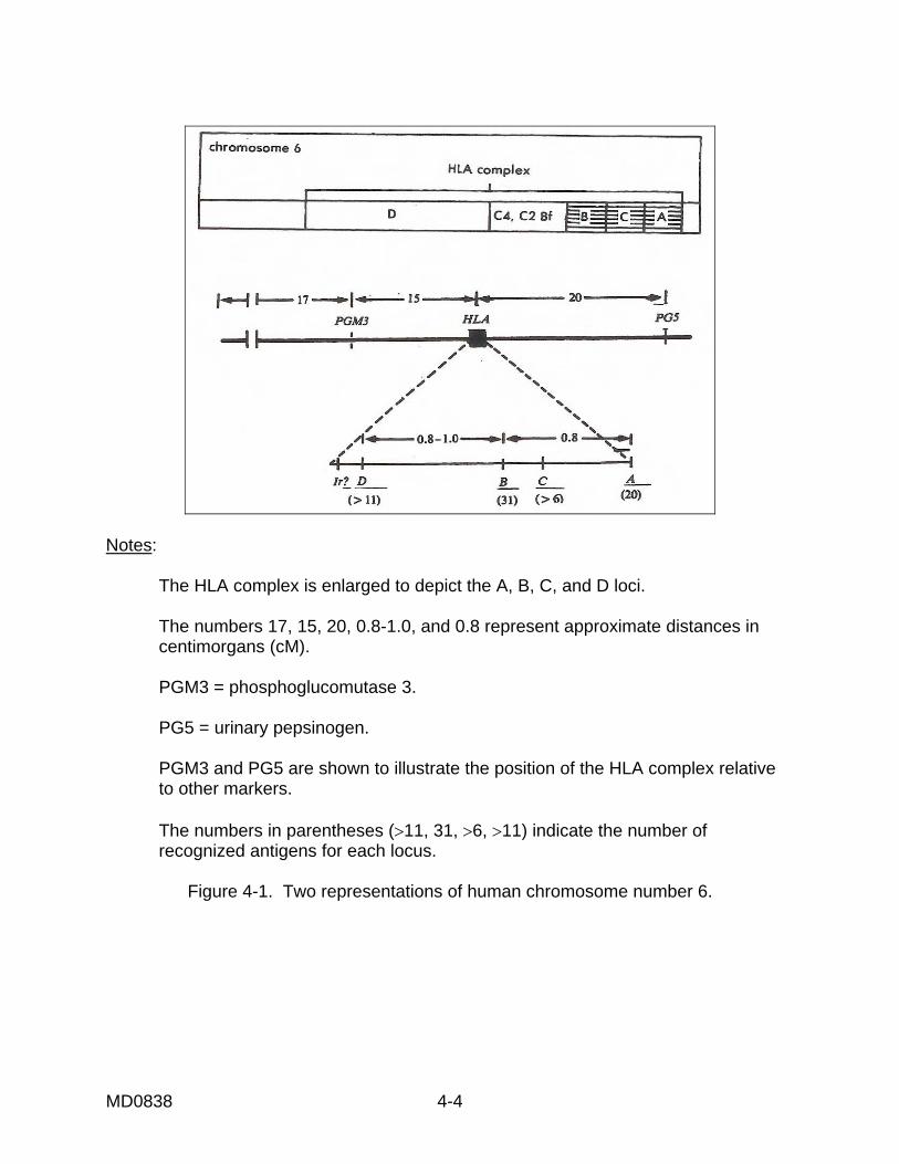
MD0838 4-4
Notes: The HLA complex is enlarged to depict the A, B, C, and D loci. The numbers 17, 15, 20, 0.8-1.0, and 0.8 represent approximate distances in centimorgans (cM). PGM3 = phosphoglucomutase 3. PG5 = urinary pepsinogen. PGM3 and PG5 are shown to illustrate the position of the HLA complex relative to other markers. The numbers in parentheses (>11, 31, >6, >11) indicate the number of recognized antigens for each locus.
Figure 4-1. Two representations of human chromosome number 6.

MD0838 4-5
4-4. CLASSIFICATIONS a. Class I Antigens. Class I molecules are coded for by the HLA-A, B, and C genes. Each gene is highly pleomorphic, that is, occurring in various distinct forms. The A gene is currently recognized to have at least 23 specificities, the B gene 50 specificities, and the C gene 8 specificities. The Class I antigen consists of two polypeptides: one MHC-encoded peptide and the other non-MHC encoded peptide, beta microglobulin, which is a small globular peptide. HLA-A, B, and C antigens are found on virtually every human cell. b. Class II Antigens. They are different structurally and functionally from Class I. The exact number of genes in the D region is uncertain but three are widely accepted. These are DR, DP, and DQ. DP and DQ replace the earlier designation SB and DC, respectively. The number of alleles for each gene is not known. At least six different specificities for each of the DP and DQ genes are known. Each Class II protein consists of two peptides, an alpha chain and a beta chain that is slightly smaller. HLA-DR, DP, and DQ are found chiefly on the surface of immunocompetent cells, including macrophage/monocytes, resting T lymphocytes, activated T lymphocytes, and particularly B lymphocytes. c. Class III Antigens. Four gene loci, located between the HLA-B and the HLA-D loci on chromosome 6, are associated with the complement system. These Class III genes are the structural genes for proteins C2, C4A, C4B, and factor B of the complement system.
Continue with Exercises

MD0838 4-6
EXERCISES, LESSON 4 INSTRUCTIONS: Answer the following items by completing the statement or by writing the answer in the space provided. After you have completed all of these items, turn to "Solutions to Exercises" at the end of the lesson and check your answers with the solutions. 1. Immunogenetics is the study of processes involved in the i___e response that may have a g___c basis. These processes include the factors that control the i__e r____e of the host and the transmission of antigenic s____ties from generation to generation. 2. The first major area of immunogenetics is concerned with the genetic re__ation and c___l of the immune system itself. Interruption or alterations of developmental sequences may lead to i_____cy disorders, auto____e disorders, and perhaps m_cies. The second major area of immunogenetics, the one with the broadest application, is the use of a___dies and s____d immune cells, products of the i___e system, as probes to detect and characterize various a___ns that may show genetic variation. 3. The major histocompatibility complex (MHC) is the region of a specific c___me that controls hi_____y. The term histocompatibility refers to the presence of certain a____s which mean that the host of an organ or tissue graft will not reject the graft. In humans, the MHC is called the HLA complex, which refers to "h__n l___e a____s." Histocompatibility is a relationship of d___r and host based upon the presence of compatible HLA a____s. 4. The histocompatibility-linked Ir genes play an important role but compose only a p___t of the immune system. Certain Ir genes may code for c___r receptors for antigen and others may control mediator s____n. The system controls not only have the ability to r___d to antigens but also to control the l___l and d____n of the response. 5. The HLA complex is a region on chromosome _ that codes for three functionally different proteins: those that r___late the immune response, those that determine the a____ce or r___tion of transplanted tissues between individuals within that species, and those that are a part of the c____ment system (Figure 4-1). The MHC of m___e has provided information for the understanding of the human MHC.

MD0838 4-7
6. A letter after HLA, such as HLA-D, refers to a gene l____s. 7. Class I antigens are coded for by the HLA-_, _, and _ genes. Each gene is highly pleomorphic, which means it occurs in various distinct f___s. The A gene is currently recognized to have at least 23 s____ties, the B gene 50 s_________s, and the C gene 8 s_____s. HLA-A, B, and C antigens are found on virtually e____y human cell. 8. Class II antigens are different s_____ly and f_____ly from Class I. The exact number of g__s in the D region is uncertain but three are widely accepted. These are DR, DP, and DQ. HLA-DR, DP, and DQ are found chiefly on the s____e of immunocompetent cells, including m____ge/monocytes, r___ing T lymphocytes, a____ted T lymphocytes, and particularly B l_____s. 9. Class III genes are the structural genes for p___ns C2, C4A, C4B, and factor B of the c____t system.
Check Your Answers on Next Page

MD0838 4-8
SOLUTIONS TO EXERCISES, LESSON 4 1. Immunogenetics is the study of processes involved in the immune response that may have a genetic basis. These processes include the factors that control the immune response of the host and the transmission of antigenic specificities from generation to generation. (para 4-1a) 2. The first major area of immunogenetics is concerned with the genetic regulation and control of the immune system itself. Interruption or alterations of developmental sequences may lead to immunodeficiency disorders, autoimmune disorders, and perhaps malignancies. The second major area of immunogenetics, the one with the broadest application, is the use of antibodies and sensitized immune cells, products of the immune system, as probes to detect and characterize various antigens that may show genetic variation. (para 4-1b) 3. The major histocompatibility complex (MHC) is the region of a specific chromosome that controls histocompatibility. The term histocompatibility refers to the presence of certain antigens which mean that the host of an organ or tissue graft will not reject the graft. In humans, the MHC is called the HLA complex, which refers to "human leukocyte antigens." Histocompatibility is a relationship of donor and host based upon the presence of compatible HLA antigens. (para 4-1c) 4. The histocompatibility-linked Ir genes play an important role but compose only a part of the immune system. Certain Ir genes may code for cellular receptors for antigen and others may control mediator secretion. The system controls not only have the ability to respond to antigens but also to control the level and duration of the response. (para 4-2) 5. The HLA complex is a region on chromosome 6 that codes for three functionally different proteins: those that regulate the immune response, those that determine the acceptance or rejection of transplanted tissues between individuals within that species, and those that are a part of the complement system (Figure 4-1). The MHC of mice has provided information for the understanding of the human MHC. (para 4-3a) 6. A letter after HLA, such as HLA-D, refers to a gene locus. (para 4-3b)

MD0838 4-9
7. Class I antigens are coded for by the HLA-A, B, and C genes. Each gene is highly pleomorphic, which means it occurs in various distinct forms. The A gene is currently recognized to have at least 23 specificities, the B gene 50 specificities, and the C gene 8 specificities. HLA-A, B, and C antigens are found on virtually every human cell. (para 4-4a) 8. Class II antigens are different structurally and functionally from Class I. The exact number of genes in the D region is uncertain but three are widely accepted. These are DR, DP, and DQ. HLA-DR, DP, and DQ are found chiefly on the surface of immunocompetent cells, including macrophage/monocytes, resting T lymphocytes, activated T lymphocytes, and particularly B lymphocytes. (para 4-4b) 9. Class III genes are the structural genes for proteins C2, C4A, C4B, and factor B of the complement system. (para 4-4c)
End of Lesson 4

MD0838 5-1
LESSON ASSIGNMENT LESSON 5 Immunological Techniques TEXT ASSIGNMENT Paragraphs 5-1 through 5-13. LESSON OBJECTIVES After completing this lesson, you should be able to identify principles and procedures for the following types of immunological techniques: precipitation reactions (including radial immunodiffusion and double diffusion), serum protein electrophoresis, immunoelectrophoresis, and enzyme immunoassay techniques. SUGGESTION After completing the assignment, complete the exercises at the end of this lesson. These exercises will help you to achieve the lesson objectives.

MD0838 5-2
LESSON 5
IMMUNOLOGICAL TECHNIQUES
Section I. INTRODUCTION 5-1. CONTEXT Since 1970, immunological laboratory methods have gradually become increasingly more refined and simplified. Because of their improved specificity and sensitivity, these methods have now achieved a major role in modern clinical laboratory science. As many new laboratory tests employing immunologic principles are developed, these methods of laboratory diagnosis have often been applied to clinical situations. 5-2. APPLICATION You will be better able to interpret and apply the new knowledge about immunology as you develop an understanding of the methods now in use. In this lesson, the techniques and applications of the various immunological tests for the detection of antigens and antibodies are discussed.
Section II. PRECIPITATION REACTIONS
5-3. PRECIPITATION REACTIONS a. In 1934 Marrack, a prominent Englishman, proposed a new model for antigen-antibody reactions. His hypothesis states that under appropriate experimental conditions, antigen-antibody complexes precipitate. Antibody molecules are bivalent, that is, they contain two antigen-binding sites. For this reason, when antibodies are complexed with antigen, they can form a cross-linked mass. At a proper antigen and antibody concentration, this cross-linked mass enlarges and precipitates. b. At the onset of the antigen-antibody reaction, an invisible formation of antigen and antibody complexes occurs. A lattice of soluble complexes slowly develops and gradually expands into a visible precipitate (lattice formation) as the antibody and antigen reach a zone of equivalent concentration. If there is an excess of either antigen (postzone) or antibody (prozone) present, the proper lattice formation needed for precipitate formation cannot occur. The formation of the antigen-antibody complex is reversible and may dissolve if more antigen or antibody is added.

MD0838 5-3
5-4. IMMUNODIFFUSION Immunodiffusion techniques detect antigen-antibody precipitation reactions in a semisolid medium. The formation of antigen and antibody complexes can be influenced by a number of factors: relative concentration of antigens and antibodies, ionic strength of the buffer, pH, and temperature. Two techniques most often used in a clinical laboratory are single and double diffusion. a. Radial Immunodiffusion (RID). In this method, a known concentration of antibody is incorporated into an agarose medium. The reactant (antigen) is applied to a well cut in the agarose and radially diffuses from the site of application (Figure 5-1). At the point of equivalence, the antigen and antibody react to form a visible precipitin ring. The size of the precipitin ring is proportional to the concentration of the antigen. In the clinical laboratory, radial immunodiffusion is primarily used to quantitate serum immunoglobulins and complement components such as C3 or C4. b. Double Diffusion (DD). DD is based on the principle that antigen and antibody diffuse through a semisolid medium and form a precipitin line. In Ouchterlony's method, a layer of agar gel is deposited in a petri dish and circular wells are punched out near one another in the gel. Antibody is then added to one well while antigen is added to the other. These materials are allowed to diffuse radially from their respective wells. As the perimeter of the diffusing substance increases, the concentration of that substance within the perimeter continually decreases. When the optimal concentration of antigen and antibody is reached, a line of precipitation is formed in the gel. The precipitin line is relatively straight and is perpendicular to the axis line between the two wells. The immunologic reactions in double gel diffusion are of three types (Figure 5-2):
Figure 5-1. Single immunodiffusion.

MD0838 5-4
Figure 5-2. Double gel diffusion (1) Reaction of identity. A solution of the same antigen is placed in two wells and its corresponding antibody is placed in the center well. Two precipitin bands form, joining at their contiguous ends and fusing. (2) Reaction of partial identity. If the antigen in one well and the antibody in the central well are homologous and if the antigen in the other well is a cross-reacting antigen, the precipitin bands fuse and in addition form a spike that extends toward the cross-reacting antigen. (3) Reaction of nonidentity. When two unrelated antigens are placed in adjacent wells and diffuse toward a central well that contains antibodies for each, two precipitin bands form independently of each other and cross. Currently, double diffusion is utilized in the clinical laboratory for immune complex studies and comparing antigens or antibodies for the presence of identical or cross-reacting components.
Section III. SERUM PROTEIN ELECTROPHORESIS (SPE) 5-5. PRINCIPLE Serum protein electrophoresis is a screening procedure utilized to detect abnormalities of the various protein fractions. The patient's serum is applied to a support medium (agarose gel) along with a normal control. The agarose gel with the serum applied is placed in a barbital buffer (pH 8.6) and then subjected to an electrical charge. Due to the pH of the barbital buffer, the serum proteins will assume a net negative charge. The serum protein components migrate to one of the five possible characteristic zones (albumin, alpha-1, alpha-2, beta, and gamma) (Figure 5-3) based upon their net negative electrical charge, size, and molecular weight. Following a staining procedure using a protein stain, the serum protein components are compared to those of the normal control. Changes or abnormalities may be identified by observing the electrophoretic pattern or a densitometer tracing of the pattern (electrophoretogram) for increases, decreases, or complete absence of normally occurring components.

MD0838 5-5
Figure 5-3. Electrophoretic zones. 5-6. INTERPRETATION a. Normal. Patients with normal serum protein fractions are identified when their serum protein electrophoretic patterns are similar to that of the normal control with no observable increase, decrease, or absence of any particular zone (Figure 5-4A).
Figure 5-4. Interpretation of serum protein electrophoresis.

MD0838 5-6
b. Hypogammaglobulinemia (agammaglobulinemia). This is an immunological deficiency state characterized by an abnormally low level of generally all classes of gamma globulin. It is identified by an absent or decreased zone. In this situation, the patient is not producing immunoglobulins in sufficient quantities to maintain a normal immune state (Figure 5-4B). c. Polyclonal Hypergammaglobulinemia. A broad increase in gammaglobulins due to numerous clones of plasma cells producing a heterogeneous group of immunoglobulins. It is identified by a diffuse increase in the gamma zone (Figure 5-4C). This condition is exhibited in a variety of clinical disorders and therefore is not indicative of any single disease state. d. Monoclonal Hypergammaglobulinemia. An excess of homogeneous immunoglobulin molecules of a single specificity following the unexplained proliferation of a single clone of immunoglobulin-producing cells. It appears as a narrow, tall spike in the gamma zone (Figure 5-4D). The class of immunoglobulin and increased or detectable amounts of free light chains in the serum or urine are diagnostic as to the clinical condition of the patient.
Section IV. IMMUNOELECTROPHORESIS (IEP) 5-7. PRINCIPLE Immunoelectrophoresis is a qualitative method that combines electrophoresis and immunoprecipitation. Both identification and approximate quantitation may be accomplished for individual proteins present in serum, urine, or other biological fluids. This technique is especially useful in the identification and diagnosis of the monoclonal gammopathies. It is a two stage procedure with the first step involving the electrophoretic separation of the patient's serum specimen and normal control. Following electrophoresis, specific antisera (anti-human IgG, IgM, IgA, kappa, and lambda) are placed in troughs parallel to the line of the fractionated proteins. The proteins and antisera diffuse in all directions with immunoprecipitin arcs forming where specific antisera and corresponding protein antigen meet. Following a staining procedure using a protein stain, increases or decreases of the individual's immunoglobulins may be observed by comparing the patient's immunoprecipitin arcs to those of the normal control.

MD0838 5-7
5-8. MONOCLONAL GAMMOPATHIES a. Multiple Myeloma. It is the most common of the monoclonal gammopathies and is characterized by neoplastic proliferation of plasma cells or morphologically abnormal plasma cells (myeloma cells), primarily occurring in the bone marrow. Bone pain is the most common symptom, with the presence of bone lesions and frequent bone fractures. SPE shows the presence of a monoclonal hypergammaglobulinemia while the IEP demonstrates an increase in one of the immunoglobulins, excluding an increase of IgM. The secretion of Bence Jones protein (light chains, either kappa or lambda) in the urine is common and diagnostic. b. Waldenstrom's Macroglobulinemia. This disorder is characterized by an increase in the immunoglobulin IgM and one of the light chains. The associated symptoms are due to an increase in serum viscosity. Hyperviscosity and sludging of blood may lead to visual disturbances, neurological symptoms, impaired kidney function, and congestive heart failure. Bone pain and lesions are rare. c. Heavy Chain Disease. A heterogeneous group of paraprotein disorders characterized by the presence of monoclonal but incomplete heavy chains without light chains in serum or urine. The heavy chain involved may be gamma, alpha, or mu with alpha being the most common. The key to diagnosis is the demonstration of the presence of the heavy chain without any discernable light chain.
Section V. ENZYME IMMUNOASSAY TECHNIQUES 5-9. INTRODUCTION Enzyme immunoassays have emerged as quantitative techniques for detection of extremely small quantities of antigens, haptens, and antibodies. They all employ various enzymes linked to either an antigen or antibody to form an enzyme-labeled tag which can easily be detected by measurement of the enzyme activity. 5-10. TYPES OF ENZYME IMMUNOASSAYS a. The most widely used assays are enzyme-linked immunoabsorbent assay (ELISA) and the enzyme immunoassay (EIA). The principles of ELISA and EIA tests are similar to those of radioimmunoassay (RIA) technique except enzyme activity is measured instead of radioactivity. See the following paragraphs for the meaning of substrate and conjugate. b. To measure antibody, antigen is fixed to a solid phase, incubated with test serum (which contains the antibody to be detected), and then incubated with anti-human globulin tagged with an enzyme (conjugate). The tagged antihuman globulin reacts with the antibody being detected. Substrate is then added and the enzyme activity adherent to the solid phase is then related to the amount of antibody bound (Figure 5-5).

MD0838 5-8
Figure 5-5. Sandwich enzyme immunoassay for antibody detection. c. To measure antigen, antibody is bound to the solid phase, a test solution containing the antigen is added, and then a second enzyme-labeled antibody (conjugate) with specificity for the antigen being assayed is added and allowed to incubate. Substrate is then added and measurement of enzyme activity is related to the antigen concentration (Figure 5-6).
Figure 5-6. Sandwich enzyme immunoassay for antibody detection.

MD0838 5-9
5-11. CONJUGATE ENZYMES Conjugates are enzyme-labeled antigen or antibody that retains activity capable of converting the substrate into a product that can be easily detected. The enzymes most frequently used for conjugates include horseradish peroxidase (HRP) and alkaline phosphatase (AP). a. The most commonly used conjugate enzyme in the EIA test procedures is horseradish peroxidase which has the capability of reacting with a wider array of highly chromogenic substances and is produced from a vegetable source. b. The second most commonly used conjugate enzyme is alkaline phosphatase which has a greater resistance to environmental toxins and is derived from the mucosa of the calf intestines. 5-12. SUBSTRATES A substrate is a substance upon which an enzyme acts. Substrates are chromogenic substances which are initially colorless but yield a colored product after enzyme degradation. The substrates most frequently used in the EIA procedures are ortho-phenylenediamine (OPD) and para-nitrophenyl phosphate (p-NPP). a. Ortho-phenylenediamine, the most satisfactory and commonly used peroxidase substrate, yields a highly chromogenic orange product after enzyme degradation has occurred. b. Para-nitrophenyl phosphate is the substrate used for the enzyme alkaline phosphatase and yields a highly chromogenic yellow-colored product after enzyme degradation. Both of the above mentioned substrates are highly soluble and extremely light sensitive. 5-13. CLINICAL APPLICATIONS Clinical application of EIA procedures include but are not limited to quantitation and/or qualitation of serum components and various hormones. They also serve as clinical tools in the diagnosis, evaluation, and treatment of the many autoimmune and viral diseases.
Continue with Exercises

MD0838 5-10
EXERCISES, LESSON 5 INSTRUCTIONS: Answer the following items by completing the statement or by writing the answer in the space provided. After you have completed all of these items, turn to "Solutions to Exercises" at the end of the lesson and check your answers with the solutions. 1. You will be better able to interpret and apply the new knowledge about immunology as you develop an understanding of the m__s now in use. In this lesson, the techniques and applications of the various i______cal tests for the detection of a____s and a____s are discussed. 2. In Marrack's model, under appropriate experimental conditions, antigen-antibody complexes p___tate. Antibody molecules are bivalent, that is, they contain two a____n-binding sites. At a proper antigen and antibody concentration, a cross- linked m___s enlarges and p_____s. At the onset of the antigen-antibody reaction, an i_____ble formation of antigen and antibody complexes occurs. This is followed by an expanding visible precipitate, called a l____ce f____tion, as the antigen and antibody reach a zone of equivalent concentration. If there is an excess of either antigen or antibody present, the proper l___ce formation needed for precipitate formation cannot occur. The formation of the antigen-antibody complex is r____ble and may d___ve if more antigen or antibody is added. 3. Immunodiffusion techniques detect antigen-antibody precipitation reactions in a semis____d medium. The formation of antigen and antibody complexes can be influenced by a number of factors: relative c____tion of antigens and antibodies, i__c s____th of the buffer, pH, and t_____e. Two techniques most often used in a clinical laboratory are s____le and d___le diffusion. 4. In the r___l immunodiffusion (RID) method, a known concentration of antibody is incorporated into an agarose m__m. The reactant (antigen) is applied to a well cut in the a___e and radially diffuses from the site of application. At the point of e______ce, the antigen and antibody react to form a visible p___tin ring. The size of the precipitin ring is proportional to the c_____tion of the antigen. In the clinical laboratory, radial immunodiffusion is primarily used to quantitate serum i____lins and c______t components such as C3 or C4.

MD0838 5-11
5. Double diffusion (DD) is based on the principle that antigen and antibody diffuse through a semisolid medium and form a precipitin l___e. In Ouchterlony's method, a layer of agar gel is deposited in a petri dish and circular w___s are punched out near one another in the gel. Antibody is then added to one well while a___n is added to the other. These materials are allowed to d___e radially from their respective wells. As the perimeter of the diffusing substance increases, the concentration of that substance within the perimeter continually (increases) (decreases). When the optimal concentration of a___n and a____y is reached, a line of p_____n is formed in the gel. The precipitin line is relatively s____t and is p_____r to the axis line between the two wells. The immunologic reactions in double gel diffusion are of (two) (three) types. Consider this: A solution of the same antigen is placed in two wells and its corresponding antibody is placed in the center well. Two precipitin bands form, joining at their contiguous ends and fusing. This is a reaction of i____ty. To produce a reaction or partial identity, the antigen in one well and the antibody in the central well are h____s, and the antigen in the other well is a cross-reacting antigen. The precipitin bands fuse, but in addition form a s_____e that extends toward the cross-reacting antigen. This is a reaction of p___l i____y. When two unrelated antigens are placed in adjacent wells and diffuse toward a central well that contains antibodies for each, two precipitin bands form i_____ly of each other and c____s. This is a reaction of non-i____y. Currently, double diffusion is utilized in the clinical laboratory for immune complex studies and comparing antigens or antibodies for the presence of i__________l or c____s-reacting components. 6. A screening procedure utilized to detect abnormalities of the various protein fractions is serum protein e______sis. The patient's serum is applied to a support medium along with a normal c___l. The agarose gel with the serum applied is placed in a b____l buffer (pH 8.6) and then subjected to an e____l charge. Due to the pH of the barbital buffer, the serum proteins will assume a net n_____e charge. The serum protein components migrate to one of the five possible characteristic zones (a____n, alpha-1, alpha-2, b__a, and g___a) based upon their net negative electrical charge, size, and molecular weight. Following a s_____g procedure using a protein stain, the serum p____n components are compared to those of the normal control.

MD0838 5-12
7. Patients with normal serum protein fractions are identified when their serum protein electrophoretic patterns are similar to that of the normal control with no observable i_____e, d_____e, or a_____e of any particular zone. Hypogammaglobulinemia (agammaglobulinemia) is an immunological deficiency state characterized by an abnormally (low) (high) level of generally all classes of gamma globulin. It is identified by an a___t or d___d zone. In this situation, the patient is not producing immunoglobulins in sufficient quantities to maintain a normal i____e state. Polyclonal hypergammaglobulinemia is a broad (decrease) (increase) in gamma globulins due to numerous c___s of plasma cells producing a heterogeneous group of i_________s. It is identified by a diffuse increase in the g____a zone. This condition (is) (is not) indicative of a specific disease state. Monoclonal hypergammaglobulinemia follows the unexplained proliferation of a single c____e of immunoglobulin-producing cells. It appears as a narrow, tall spike in the g____a zone. 8. Immuno__________ is a qualitative method that combines e_________s and i____________n. This technique is especially useful in the identification and diagnosis of the m________l gammopathies. It is a two-stage procedure with the first step involving the electrophoretic s_______n of the patient's serum specimen and normal control. Following electrophoresis, specific a______a are placed in troughs parallel to the line of the fractionated proteins. The proteins and antisera diffuse in all directions with immunoprecipitin a___ forming where specific a_____ and corresponding protein antigen meet. Following a staining procedure using a protein stain, increases or decreases of the individual's immunoglobulins may be observed by comparing the patient's i________________n arcs to those of the normal c_____.

MD0838 5-13
9. The most common of the monoclonal gammopathies is m____le my___a, which is characterized by neo____c prol___tion of plasma cells or abnormal plasma cells (m___a cells), primarily occurring in the bone m___w. Serum protein electrophoresis (SPE) shows the presence of a m_________al hypergammaglobulinemia while the immunoelectrophoresis (IEP) demonstrates an increase in one of the i_________s. Waldenstrom's macrog_______mia is characterized by an increase in the immunoglobulin Ig_ and one of the l___t chains. The associated symptoms are due to an increase in serum vi____ity. Hyperviscosity and sludging of blood may lead to v____l disturbances, n_______cal symptoms, impaired k__y function, and congestive h___t failure. Heavy chain disease is characterized by the presence of monoclonal but incomplete h____y chains without l____t chains in serum or urine. The heavy chain involved may be gamma, alpha, or mu with a___a being the most common. The key to diagnosis is the demonstration of the presence of the heavy chain without any discernable l____t chain. 10. Enzyme immunoassays have emerged as qu_____ive techniques for detection of extremely s____l quantities of antigens, haptens, and a_____s. They all employ various e___s linked to either an antigen or antibody to form an e____e-labeled tag which can easily be detected by measurement of the e___e activity. 11. The most widely used immunoassays are enzyme-linked i_________ent assay (ELISA) and the enzyme i______ay (EIA). The principles of ELISA and EIA tests are similar to those of r_______say (RIA) technique except e_____e activity is measured instead of r____oactivity. To measure antibody, antigen is fixed to a s___d phase, incubated with test serum, and then incubated with anti-h____n globulin tagged with an enzyme (conjugate). Substrate is then added and the enzyme activity adherent to the solid phase is then related to the amount of a____y bound. To measure antigen, a___y is bound to the solid phase, a test solution containing the a____n is added, and then a second e_____e-labeled antibody (conjugate) with specificity for the antigen being assayed is added and allowed to in____e. Substrate is then added and measurement of e____e activity is related to the antigen concentration.

MD0838 5-14
12. Conjugates are e___e-labeled a___n or a____y which retain activity capable of converting the substrate into a product that can be easily detected. The conjugates (enzymes) most frequently used include h____sh peroxidase (HRP) and alkaline p_____ase (AP). The most commonly used conjugate in the EIA test procedures is horseradish p_____ase. The second most commonly used conjugate is a____ine p____ase, which has a greater resistance to environmental toxins and is derived from the mucosa of the c__f i______s. 13. Substrates are c_____nic substances which are initially colorless but yield a colored product after e____e degradation. The substrates most frequently used in the EIA procedures are: Ortho-phenylenediamine (OPD), the most s_____ory and commonly used peroxidase substrate, which yields a highly c_______ic orange product after enzyme degradation has occurred. Para-nitrophenyl phosphate (p-NPP), the substrate used for the enzyme alkaline phosphatase and which yields a highly chromogenic y____w-colored product after enzyme degradation. Both of the above mentioned substrates are highly s___le and extremely l___t sensitive.
Check Your Answers on Next Page

MD0838 5-15
SOLUTIONS TO EXERCISES, LESSON 5 1. You will be better able to interpret and apply the new knowledge about immunology as you develop an understanding of the methods now in use. In this lesson, the techniques and applications of the various immunological tests for the detection of antigens and antibodies are discussed. (paras 5-1 thru 5-2) 2. In Marrack's model, under appropriate experimental conditions, antigen-antibody complexes precipitate. Antibody molecules are bivalent, that is, they contain two antigen-binding sites. At a proper antigen and antibody concentration, a cross- linked mass enlarges and precipitates. At the onset of the antigen-antibody reaction, an invisible formation of antigen and antibody complexes occurs. This is followed by an expanding visible precipitate, called a lattice formation, as the antigen and antibody reach a zone of equivalent concentration. If there is an excess of either antigen or antibody present, the proper lattice formation needed for precipitate formation cannot occur. The formation of the antigen-antibody complex is reversible and may dissolve if more antigen or antibody is added. (para 5-3) 3. Immunodiffusion techniques detect antigen-antibody precipitation reactions in a semisolid medium. The formation of antigen and antibody complexes can be influenced by a number of factors: relative concentration of antigens and antibodies, ionic strength of the buffer, pH, and temperature. Two techniques most often used in a clinical laboratory are single and double diffusion. (para 5-4) 4. In the r_____l immunodiffusion (RID) method, a known concentration of antibody is incorporated into an agarose medium. The reactant (antigen) is applied to a well cut in the agarose and radially diffuses from the site of application. At the point of equivalence, the antigen and antibody react to form a visible precipitin ring. The size of the precipitin ring is proportional to the concentration of the antigen. In the clinical laboratory, radial immunodiffusion is primarily used to quantitate serum immunoglobulins and complement components such as C3 or C4. (para 5-4a)

MD0838 5-16
5. Double diffusion (DD) is based on the principle that antigen and antibody diffuse through a semisolid medium and form a precipitin line. In Ouchterlony's method, a layer of agar gel is deposited in a petri dish and circular wells are punched out near one another in the gel. Antibody is then added to one well while antigen is added to the other. These materials are allowed to diffuse radially from their respective wells. As the perimeter of the diffusing substance increases, the concentration of that substance within the perimeter continually decreases. When the optimal concentration of antigen and antibody is reached, a line of precipitation is formed in the gel. The precipitin line is relatively straight and is perpendicular to the axis line between the two wells. The immunologic reactions in double gel diffusion are of three types. Consider this: A solution of the same antigen is placed in two wells and its corresponding antibody is placed in the center well. Two precipitin bands form, joining at their contiguous ends and fusing. This is a reaction of identity. To produce a reaction or partial identity, the antigen in one well and the antibody in the central well are homologous, and the antigen in the other well is a cross- reacting antigen. The precipitin bands fuse, but in addition form a spike that extends toward the cross-reacting antigen. This is a reaction of partial identity. When two unrelated antigens are placed in adjacent wells and diffuse toward a central well that contains antibodies for each, two precipitin bands form independently of each other and cross. This is a reaction of non-identity. Currently, double diffusion is utilized in the clinical laboratory for immune complex studies and comparing antigens or antibodies for the presence of identical or cross-reacting components. (para 5-4b) 6. A screening procedure utilized to detect abnormalities of the various protein fractions is serum protein electrophoresis. The patient's serum is applied to a support medium along with a normal control. The agarose gel with the serum applied is placed in a barbital buffer (pH 8.6) and then subjected to an electrical charge. Due to the pH of the barbital buffer, the serum proteins will assume a net negative charge. The serum protein components migrate to one of the five possible characteristic zones (albumin, alpha-1, alpha-2, beta, and gamma) based upon their net negative electrical charge, size, and molecular weight. Following a staining procedure using a protein stain, the serum protein components are compared to those of the normal control. (para 5-5)

MD0838 5-17
7. Patients with normal serum protein fractions are identified when their serum protein electrophoretic patterns are similar to that of the normal control with no observable increase, decrease, or absence of any particular zone. Hypogammaglobulinemia (agammaglobulinemia) is an immunological deficiency state characterized by an abnormally low level of generally all classes of gamma globulin. It is identified by an absent or decreased zone. In this situation, the patient is not producing immunoglobulins in sufficient quantities to maintain a normal immune state. Polyclonal hypergammaglobulinemia is a broad increase in gamma globulins due to numerous clones of plasma cells producing a heterogeneous group of immunoglobulins. It is identified by a diffuse increase in the gamma zone. This condition is not indicative of a specific disease state. Monoclonal hypergammaglobulinemia follows the unexplained proliferation of a single clone of immunoglobulin-producing cells. It appears as a narrow, tall spike in the gamma zone. (para 5-6) 8. Immunoelectrophoresis is a qualitative method that combines electrophoresis and immunoprecipitation. This technique is especially useful in the identification and diagnosis of the monoclonal gammopathies. It is a two-stage procedure with the first step involving the electrophoretic separation of the patient's serum specimen and normal control. Following electrophoresis, specific antisera are placed in troughs parallel to the line of the fractionated proteins. The proteins and antisera diffuse in all directions with immunoprecipitin arcs forming where specific antisera and corresponding protein antigen meet. Following a staining procedure using a protein stain, increases or decreases of the individual's immunoglobulins may be observed by comparing the patient's immunoprecipitin arcs to those of the normal control. (para 5-7)

MD0838 5-18
9. The most common of the monoclonal gammopathies is multiple myeloma, which is characterized by neoplastic proliferation of plasma cells or abnormal plasma cells (myeloma cells), primarily occurring in the bone marrow. Serum protein electrophoresis (SPE) shows the presence of a monoclonal hypergammaglobulinemia while the immunoelectrophoresis (IEP) demonstrates an increase in one of the immunoglobulins. Waldenstrom's macroglobulinemia is characterized by an increase in the immunoglobulin IgM and one of the light chains. The associated symptoms are due to an increase in serum viscosity. Hyperviscosity and sludging of blood may lead to visual disturbances, neurological symptoms, impaired kidney function, and congestive heart failure. Heavy chain disease is characterized by the presence of monoclonal but incomplete heavy chains without light chains in serum or urine. The heavy chain involved may be gamma, alpha, or mu with alpha being the most common. The key to diagnosis is the demonstration of the presence of the heavy chain without any discernable light chain. (para 5-8) 10. Enzyme immunoassays have emerged as quantitative techniques for detection of extremely small quantities of antigens, haptens, and antibodies. They all employ various enzymes linked to either an antigen or antibody to form an enzyme-labeled tag which can easily be detected by measurement of the enzyme activity. (para 5-9) 11. The most widely used immunoassays are enzyme-linked immunoabsorbent assay (ELISA) and the enzyme immunoassay (EIA). The principles of ELISA and EIA tests are similar to those of radioimmunoassay (RIA) technique except enzyme activity is measured instead of radioactivity. To measure antibody, antigen is fixed to a solid phase, incubated with test serum, and then incubated with anti-human globulin tagged with an enzyme (conjugate). Substrate is then added and the enzyme activity adherent to the solid phase is then related to the amount of antibody bound. To measure antigen, antibody is bound to the solid phase, a test solution containing the antigen is added, and then a second enzyme-labeled antibody (conjugate) with specificity for the antigen being assayed is added and allowed to incubate. Substrate is then added and measurement of enzyme activity is related to the antigen concentration. (para 5-10)

MD0838 5-19
12. Conjugates are enzyme-labeled antigen or antibody which retain activity capable of converting the substrate into a product that can be easily detected. The conjugates (enzymes) most frequently used include horseradish peroxidase (HRP) and alkaline phosphatase (AP). The most commonly used conjugate in the EIA test procedures is horseradish peroxidase. The second most commonly used conjugate is alkaline phosphatase, which has a greater resistance to environmental toxins and is derived from the mucosa of the calf intestines. (para 5-11) 13. Substrates are chromogenic substances which are initially colorless but yield a colored product after enzyme degradation. The substrates most frequently used in the EIA procedures are: Ortho-phenylenediamine (OPD), the most satisfactory and commonly used peroxidase substrate, which yields a highly chromogenic orange product after enzyme degradation has occurred. Para-nitrophenyl phosphate (p-NPP), the substrate used for the enzyme alkaline phosphatase and which yields a highly chromogenic yellow-colored product after enzyme degradation. Both of the above mentioned substrates are highly soluble and extremely light sensitive. (para 5-12)
End of Lesson 5

MD0838 6-1
LESSON ASSIGNMENT LESSON 6 Antinuclear Antibodies and Testing TEXT ASSIGNMENT Paragraphs 6-1 through 6-10. LESSON OBJECTIVES After completing this lesson, you should be able to describe the principles and techniques of immunofluorescence, including the following: the direct method, the indirect method, the transmitted light microscope, the incident light microscope, antinuclear antibodies and related diseases, and the fluorescent antinuclear antibody test. SUGGESTION After completing the assignment, complete the exercises at the end of this lesson. These exercises will help you to achieve the lesson objectives.

MD0838 6-2
LESSON 6
ANTINUCLEAR ANTIBODIES AND TESTING
Section I. IMMUNOFLUORESCENT MICROSCOPY 6-1. INTRODUCTION Immunofluorescence is a method of detecting an antigen or antibody in tissue by the pattern of fluorescence resulting when the tissue is exposed to the specific antibody or antigen labeled with a fluorochrome such as fluorescein. The technique of immunofluorescence was introduced by Coons and his associates in 1941. Immunofluorescence, the use of fluorochrome-labeled antibodies for the detection of antigens, can be qualitative or quantitative. In a qualitative procedure, a fluorescent microscope is necessary to visualize the presence of a labeled antigen or antibody in the specimen. Fluorescence is the emission of light of one color or wavelength, while a substance is irradiated with light of a different color or wavelength. The emitted wavelength is at a lower energy level than the incident or absorbed light. Fluorescein isothiocyanate is one of the most common fluorochromes with an absorption maximum of 490-495 nm and a yellow-green emission maximum of 517 nm. 6-2. IMMUNOFLUORESCENT METHODS The two most common methods used in the performance of immunofluorescent microscopy are the direct and indirect techniques. a. Direct Method. In the direct method, the antibody is labeled with a fluorescent compound and is used to detect the presence of antigen in tissue fixed to a slide. The direct technique utilizes biopsy material obtained from a patient. The fluorescent-labeled antibodies are added to the antigen in an optimal dilution and allowed to react. The preparation is washed to remove any unreacted labeled antibodies. The tissue sections are blotted and the preparation mounted with buffered glycerol for examination with the fluorescent microscope (Figure 6-1a). b. Indirect Method. The indirect method is used for the detection of serum antibodies utilizing an antigen-containing substrate and a fluorescein-labeled antibody specific for human immunoglobulins. The specific antigen-antibody (unlabeled) reaction may be visualized by the addition of labeled antihuman globulin directed against the antibody in the primary reaction. The antigen substrate plus patient's serum antibody plus labeled antihuman immunoglobulin complex results in fluorescence and detection of the specific patient's antibody in question (Figure 6-1b).

MD0838 6-3
Figure 6-1. Immunofluorescent methods.
6-3. THE FLUORESCENT MICROSCOPE The transmitted light microscope (Figure 6-2) and the incident light or epiillumination microscope (Figure 6-3) are the two types of microscopes used in immunofluorescent techniques. The two systems share some common component parts, but there are some structural and operational differences. One major difference is the direction from which the exciter light or energy strikes the specimen. The transmitted microscope light strikes the specimen from below through a condenser, while the incident microscope light strikes the specimen from above passing through the objective. This eliminates the need for a condenser and also eliminates the problems of centering a condenser. The incident microscope has more brightness, clearer images, and greater fluorescence since illumination and observation of the specimen are made from the same direction. With incident light, exciting and emitted fluorescence radiation are well separated since the exciting light passes through the specimen downwards and is lost and does not interfere with the fluorescence image. The component parts of the two types of microscopes are as follows (Figure 6-4):

MD0838 6-4
Figure 6-2. Transmitted light
Figure 6-3 Incident light microscope.

MD0838 6-5
Figure 6-4. Components of a fluorescent microscope.
a. Light System. (1) Light reflector. Concave mirror located behind the light source which redirects lost energy (light) back into system. (2) Light source. Mercury vapor lamp. (3) Collecting lenses. Concentrates light from light source into a single beam. b. Filter System. (1) Heat-absorbing filter. Removes excess heat from exciting light that may damage the system. (2) Exciter filter (primary filter). Transmits only the effective or exciter light and suppresses all other energy from light source emission which are not required for specimen fluorescence. (3) Dichroic mirror. Part of incident microscope only. Allows passage of light of selected wavelengths in one direction through the mirror but not in the opposite direction (Figure 6-5). (4) Barrier filter (secondary filter). Transmits only the emitted fluorescent light from the specimen and suppresses all other energy.

MD0838 6-6
Figure 6-5. Dichroic mirror of incident light microscope.
Section II. ANTINUCLEAR ANTIBODIES 6-4. INTRODUCTION Antinuclear antibodies are a collection of autoantibodies that are directed against nuclear constituents, usually in nucleoprotein, and that are present in various autoimmune diseases. The detection of one or more of these antinuclear antibodies is pathologically significant. The antinuclear antibody-related diseases belong to the systemic autoimmune disease classification and their etiology remains unknown. 6-5. RELATED DISEASES a. Rheumatoid Arthritis (RA). Rheumatoid arthritis is a chronic systemic disease characterized by inflammatory changes in joints and related structures that result in crippling deformities.
MD0838 6-7
b. Systemic Lupus Erythematosus (SLE). SLE is a chronic inflammatory disease of connective tissue that affects the skin, joints, kidneys, nervous system, and mucous membranes. A characteristic light-induced butterfly rash or erythema may be present across the nose. The disease may begin acutely with fever, joint pain, and malaise or smolder over a period of years with intermittent fever and malaise. Symptoms from any organ system may be present. The disease occurs most often in women. c. Sjogren's Syndrome (SS). This is a benign chronic disease characterized by a lack of tears and dryness of the eyes and mouth with little or no saliva. It often occurs secondary to RA or one of the other connective tissue disorders. The disease occurs most often in women. d. Progressive Systemic Sclerosis (PSS). PSS is a chronic illness characterized by a fibrous thickening of the skin (scleroderma) and several internal organs (gastrointestinal tract, heart, kidney, and lungs). Two-thirds of the patients are female. e. Mixed Connective Tissue Disease (MCTD). This is a relatively newly defined syndrome whose designation is reserved for patients with combined clinical features of RA, SLE, and PSS. 6-6. ANTINUCLEAR ANTIBODIES a. Anti-Native (Double-Stranded) DNA. High levels of this antibody are associated with SLE and a positive correlation exists with associated nephritis. b. Anti-Deoxyribonucleoprotein (DNP). A significant number of SLE patients demonstrate high levels of anti-DNP. Persons with other connective tissue diseases may display low levels of this antibody. c. Anti-Sm (Smith). 30% of all patients with SLE are positive, but this is highly specific for SLE. Greater than 75% of patients with clinically active SLE are anti-Sm positive. It is absent in other autoimmune conditions. d. Anti-Ribonucleoprotein (RNP). High titer found in all patients with MCTD. Low titers may be seen in SLE. e. Antinucleolar. High titers are highly indicative of PSS.

MD0838 6-8
Section III. FLUORESCENT ANTINUCLEAR ANTIBODY TEST (FANA) 6-7. PRINCIPLE The FANA utilizes the indirect fluorescent antibody technique. Antinuclear antibodies in a patient's serum will bind with nuclear antigens of a tissue cell culture substrate affixed to a slide. Fluorescein-conjugated antihuman globulin interacts with nuclear antibodies attached to the cell nuclei in a positive assay which is indicated by an apple-green fluorescence. 6-8. REPORTING RESULTS a. Negative. Fluorescent intensity of the cells' nuclei approximates that of the negative control, and there is no discernible pattern in the nucleus. b. Positive. Fluorescent intensity of the cells' nuclei is greater than the negative control, and there is a clearly discernible pattern in the nucleus. Report positive controls and patients by the specific fluorescent pattern observed. 6-9. NUCLEAR PATTERNS a. Homogeneous. (1) Smooth, even staining of the nucleus. (2) Antibodies indicated, anti-native DNA, anti-DNP. (3) Condition indicated-SLE (high titer), RA (low titer). b. Peripheral. (1) Staining of the nuclear membrane only. (2) Antibodies indicated, anti-native DNA. (3) Condition indicated, SLE. c. Speckled. (1) Grainy staining throughout the nucleus usually not affecting the nucleoli. (2) Antibodies indicated, anti-Sm, anti-RNP. (3) Conditions indicated, SLE, PSS, SS, MCTD.

MD0838 6-9
d. Nucleolar. (1) Solid staining of the nucleoli. (2) Antibodies indicated, antinucleolar. (3) Condition indicated, PSS. 6-10. LIMITATIONS a. The FANA is a laboratory diagnostic aid and by itself is not diagnostic. Positive results require further testing for specific antinuclear antibody identification and quantitation. b. SLE patients undergoing steroid therapy may have negative test results. c. Many commonly prescribed drugs may induce ANA positive results.
Continue with Exercises

MD0838 6-10
EXERCISES, LESSON 6 INSTRUCTIONS: Answer the following items by completing the statement or by writing the answer in the space provided. After you have completed all of these items, turn to "Solutions to Exercises" at the end of the lesson and check your answers with the solutions. 1. Immunofluorescence is a method of detecting an a___n or a___y in tissue by the pattern of f_______e resulting when the tissue is exposed to the specific antibody or antigen-labeled with a f____me such as fluorescein. Immunofluorescence can be q____tive or q___tive. In a qualitative procedure, a f____t microscope is necessary to visualize the presence of a labeled antigen or antibody in the specimen. Fluorescence is the e____n of light of one color from a substance being exposed to l____t of a different color or wavelength. One of the most common fluorochromes is f_______n isothiocyanate. 2. The two most common methods used in the performance of immunofluorescent microscopy are the d___t and i_____t techniques. In the direct method, the antibody is labeled with a fluorescent compound and is used to detect the presence of a___n in tissue fixed to a slide. The direct technique utilizes b___sy material obtained from a patient. The fluorescent- labeled a___s are added to the antigen in an optimal dilution and allowed to react. The preparation is washed to remove any unr____d labeled antibodies. The tissue sections are blotted and the preparation mounted with buffered g____l for examination with the fluorescent microscope. 3. The indirect method is used for the detection of serum a_____s utilizing an a_____n-containing substrate and a f_____n-labeled antibody specific for human imm_____s. The specific antigen-antibody (unlabeled) reaction may be visualized by the addition of labeled a_____n globulin directed against the antibody in the primary reaction. The antigen substrate plus patient's serum antibody plus labeled anti____n i_____globulin complex results in f_____e and detection of the specific patient's a____y in question.

MD0838 6-11
4. Two types of microscopes used in immunofluorescent techniques are the t_______d l___t microscope and the i____t light or e__-i_______n microscope. One major difference is the d____n from which the light or energy strikes the specimen. In the transmitted light microscope, light strikes the specimen from (above) (below) through a c______r. In the incident light microscope, light strikes the specimen from (above) (below), passing through the o___ve; this eliminates the need for a c___r and also eliminates the problems of c____g a condenser. The incident microscope has more b____ness, clearer i___ges, and greater f____e since illumination and observation of the specimen are made from the same d_____n. 5. The component parts of the two types of microscopes include the l___t system and the f___r system. The light system includes the light r_____r, the light s____e, and the collecting l__ses. The filter system includes the h__t-absorbing filter, the e___r filter, the dic___c mirror, and the b___r filter. The light reflector is a concave m____r located behind the l___t s___e which redirects l__t energy (light) back into system. The light source is a mercury v___r lamp. The collecting lenses c______rate light from the light source into a single b___m. The heat-absorbing filter removes excess h__t from the exciting light that may damage the system. The exciter filter (primary filter) transmits only the e____ve light and supp_____s all other energy from light source emission which are not required for s____n fluorescence. The dichroic mirror, which is part of the i____t microscope only, allows passage of light of selected wave l____s in one direction through the mirror but not in the o____e direction. The barrier filter transmits only the e___ted fluorescent light from the specimen and suppresses all other e___y. 6. Antinuclear antibodies are a collection of a___antibodies which are directed against n____r constituents, usually in nucleoprotein, and which are present in various auto_____e diseases. The detection of one or more of these antinuclear antibodies is pathologically s_____t.

MD0838 6-12
7. Related diseases include rheumatoid arth____s, systemic l__s erythematosus, Sjogren's syn__e, progressive systemic scl____s, and mixed c___ve tissue disease. Rheumatoid arthritis (RA) is a chronic systemic disease characterized by inflammatory changes in j___s and related structures that result in crippling d_____ies. Systemic lupus erythematosus (SLE) is a chronic i___y disease of connective tissue that affects the s__n, j__s, k__s, n__s system, and m_s membranes. A characteristic light-induced b__fly rash or erythema may be present across the n_e. The disease may be a_e or chronic. Symptoms from any o_n system may be present. The disease occurs most often in w__n. Sjogren's syndrome (SS) is a benign c_c disease characterized by a lack of t__s and d_ness of the eyes and mouth with little or no s_a. It often occurs secondary to RA or one of the other c__e tissue disorders. The disease occurs most often in w__n. Progressive systemic sclerosis (PSS) is a (acute) (chronic) illness characterized by a fibrous t_ing of the skin and several internal o__s. Two-thirds of the patients are (male) (female). Mixed connective tissue disease (MCTD) is a recently defined syndrome whose designation is reserved for patients with c___d clinical features of RA, SLE, and PSS. 8. Of the antinuclear antibodies discussed in this lesson, the ones strongly associated with SLE are anti-n___e-DNA, anti-D_, and anti-S_. High titers of anti-R__ are found in all patients with MCTD. Low titers may be seen in SLE. High titers of antinucleolar antibodies are highly indicative of P__. 9. The fluorescent antin___r antibody test (FANA) utilizes the indirect f__t antibody technique. Antinuclear a__s in a patient's serum will bind with nuc__r antigens of a tissue c__l c__e substrate affixed to a slide. Fluorescein-conj___d antihuman globulin interacts with n___r antibodies attached to the cell nuclei in a positive assay which is indicated by an apple-green f______ce.

MD0838 6-13
10. If the results for the FANA are negative, the fluorescent intensity of the cells' nuclei is about the same as that of the (negative) (positive) control, and there is no discernible p____n in the nucleus. If the results are positive, the fluorescent intensity of the cells' nuclei is (less)(greater) than the negative control, and there is a clearly dis___ble pattern in the nucleus. Report positive controls and patients by the specific fl____t pattern observed. 11. For a homogeneous pattern, there is sm__h, e__n staining of the nucleus. The antibodies indicated are anti-native-D_ and anti-D_. The condition indicated by a (high) (low) titer is SLE. The condition indicated by a (high) (low) titer is RA. For a peripheral pattern, there is staining of the n___r m____e only. The antibody indicated is anti-native-D_. The condition indicated is S_. For a speckled pattern, there is g__y staining throughout the nucleus usually not affecting the n___i. The antibodies indicated are anti-S_ and anti-R_. Four possible conditions indicated are (RA) (SLE) (PSS) (SS) (MCTD). For a nucleolar pattern, there is solid staining of the n____i. The antibodies indicated are anti____r. The condition indicated is P_. 12. The FANA is a laboratory diagnostic a__d and by itself is not d_____c. Positive results require f____r testing for specific antinuclear antibody i_____cation and quantitation. Another limitation of the FANA is that SLE patients undergoing steroid therapy may have (negative) (positive) test results. Also, many commonly prescribed drugs may induce (negative) (positive) results.
Check Your Answers on Next Page

MD0838 6-14
SOLUTIONS TO EXERCISES, LESSON 6 1. Immunofluorescence is a method of detecting an antigen or antibody in tissue by the pattern of fluorescence resulting when the tissue is exposed to the specific antibody or antigen-labeled with a fluorochrome such as fluorescein. Immunofluorescence can be qualitative or quantitative. In a qualitative procedure, a fluorescent microscope is necessary to visualize the presence of a labeled antigen or antibody in the specimen. Fluorescence is the emission of light of one color from a substance being exposed to light of a different color or wavelength. One of the most common fluorochromes is fluorescein isothiocyanate. (para 6-1) 2. The two most common methods used in the performance of immunofluorescent microscopy are the direct and indirect techniques. In the direct method, the antibody is labeled with a fluorescent compound and is used to detect the presence of antigen in tissue fixed to a slide. The direct technique utilizes biopsy material obtained from a patient. The fluorescent-labeled antibodies are added to the antigen in an optimal dilution and allowed to react. The preparation is washed to remove any unreacted labeled antibodies. The tissue sections are blotted and the preparation mounted with buffered glycerol for examination with the fluorescent microscope. (para 6-2a) 3. The indirect method is used for the detection of serum antibodies utilizing an antigen-containing substrate and a fluorescein-labeled antibody specific for human immunoglobulins. The specific antigen-antibody (unlabeled) reaction may be visualized by the addition of labeled antihuman globulin directed against the antibody in the primary reaction. The antigen substrate plus patient's serum antibody plus labeled antihuman immunoglobulin complex results in fluorescence and detection of the specific patient's antibody in question. (para 6-2b) 4. Two types of microscopes used in immunofluorescent techniques are the transmitted light microscope and the incident light or epi-illumination microscope. One major difference is the direction from which the light or energy strikes the specimen. In the transmitted light microscope, light strikes the specimen from below through a condenser. In the incident light microscope, light strikes the specimen from above, passing through the objective; this eliminates the need for a condenser and also eliminates the problems of centering a condenser. The incident microscope has more brightness, clearer images, and greater fluorescence since illumination and observation of the specimen are made from the same direction. (para 6-3)

MD0838 6-15
5. The component parts of the two types of microscopes include the light system and the filter system. The light system includes the light reflector, the light source, and the collecting lenses. The filter system includes the heat-absorbing filter, the exciter filter, the dichroic mirror, and the barrier filter. The light reflector is a concave mirror located behind the light source which redirects lost energy (light) back into system. The light source is a mercury vapor lamp. The collecting lenses concentrate light from the light source into a single beam. The heat-absorbing filter removes excess heat from the exciting light that may damage the system. The exciter filter (primary filter) transmits only the effective light and suppresses all other energy from light source emission which are not required for specimen fluorescence. The dichroic mirror, which is part of the incident microscope only, allows passage of light of selected wave lengths in one direction through the mirror but not in the opposite direction. The barrier filter transmits only the emitted fluorescent light from the specimen and suppresses all other energy. (para 6-3) 6. Antinuclear antibodies are a collection of autoantibodies which are directed against nuclear constituents, usually in nucleoprotein, and which are present in various autoimmune diseases. The detection of one or more of these antinuclear antibodies is pathologically significant. (para 6-4)

MD0838 6-16
7. Related diseases include rheumatoid arthritis, systemic lupus erythematosus, Sjogren's syndrome, progressive systemic sclerosis, and mixed connective tissue disease. Rheumatoid arthritis (RA) is a chronic systemic disease characterized by inflammatory changes in joints and related structures that result in crippling deformities. Systemic lupus erythematosus (SLE) is a chronic inflammatory disease of connective tissue that affects the skin, joints, kidneys, nervous system, and mucous membranes. A characteristic light-induced butterfly rash or erythema may be present across the nose. The disease may be acute or chronic. Symptoms from any organ system may be present. The disease occurs most often in women. Sjogren's syndrome (SS) is a benign chronic disease characterized by a lack of tears and dryness of the eyes and mouth with little or no saliva. It often occurs secondary to RA or one of the other connective tissue disorders. The disease occurs most often in women. Progressive systemic sclerosis (PSS) is a chronic illness characterized by a fibrous thickening of the skin and several internal organs. Two-thirds of the patients are female. Mixed connective tissue disease (MCTD) is a recently defined syndrome whose designation is reserved for patients with combined clinical features of RA, SLE, and PSS. (para 6-5) 8. Of the antinuclear antibodies discussed in this lesson, the ones strongly associated with SLE are anti-native-DNA, anti-DNP, and anti-Sm. High titers of anti-RNP are found in all patients with MCTD. Low titers may be seen in SLE. High titers of antinucleolar antibodies are highly indicative of PSS. (para 6-6)

MD0838 6-17
9. The fluorescent antinuclear antibody test (FANA) utilizes the indirect fluorescent antibody technique. Antinuclear antibodies in a patient's serum will bind with nuclear antigens of a tissue cell culture substrate affixed to a slide. Fluorescein- conjugated antihuman globulin interacts with nuclear antibodies attached to the cell nuclei in a positive assay which is indicated by an apple-green fluorescence. (para 6-7) 10. If the results for the FANA are negative, the fluorescent intensity of the cells' nuclei is about the same as that of the negative control, and there is no discernible pattern in the nucleus. If the results are positive, the fluorescent intensity of the cells' nuclei is greater than the negative control, and there is a clearly discernible pattern in the nucleus. Report positive controls and patients by the specific fluorescent pattern observed. (para 6-8) 11. For a homogeneous pattern, there is smooth, even staining of the nucleus. The antibodies indicated are anti-native-DNA and anti-DNP. The condition indicated by a high titer is SLE. The condition indicated by a low titer is RA. For a peripheral pattern, there is staining of the nuclear membrane only. The antibody indicated is anti-native-DNA. The condition indicated is SLE. For a speckled pattern, there is grainy staining throughout the nucleus usually not affecting the nucleoli. The antibodies indicated are anti-Sm and anti-RNP. Four possible conditions indicated are SLE, PSS, SS, and MCTD. For a nucleolar pattern, there is solid staining of the nucleoli. The antibodies indicated are antinucleolar. The condition indicated is PSS. (para 6-9) 12. The FANA is a laboratory diagnostic aid and by itself is not diagnostic. Positive results require further testing for specific antinuclear antibody identification and quantitation. Another limitation of the FANA is that SLE patients undergoing steroid therapy may have negative test results. Also, many commonly prescribed drugs may induce positive results. (para 6-10)
End of Lesson 6

MD0838 7-1
LESSON ASSIGNMENT LESSON 7 Viral Immunity. LESSON ASSIGNMENT Paragraphs 7-1 through 7-30. LESSON OBJECTIVES After completing this lesson, you should be able to: 7-1. Describe the structure and classification of viruses. 7-2. Describe the human immunodeficiency virus (HIV), its effects upon the immune system, and laboratory procedures for HIV antibodies and antigens. 7-3. Describe viral hepatitis, its symptoms, and causative agents, and immunological markers for hepatitis. Describe the virus, mode of transmission, incubation period and infectivity, and immunological assays for the hepatitis A virus (HAV), the hepatitis B virus (HBV), the hepatitis D virus (HDV), and hepatitis non-A, non-B (NANB). SUGGESTION After completing the assignment, complete the exercises at the end of this lesson. These exercises will help you to achieve the lesson objectives.

MD0838 7-2
LESSON 7
VIRAL IMMUNITY
Section I. VIROLOGY 7-1. STRUCTURE OF A VIRUS A virus is an intracellular parasite that will develop both acute and chronic infections that may or may not lead to host cell transformation and malignancy. The structure of a virus consists of a core, a capsid, and an envelope. The core consists mainly of a nucleic acid, either DNA or RNA, never both together. The capsid is a protective protein coat around the core. It is constructed of individual subunits termed capsomers. The envelope is a lipid outer covering; it may or may not be present depending on the virus. 7-2. CLASSIFICATION OF VIRUSES Viruses are classified by the following properties: morphology, structure, and cytopathic effects in cell cultures.
Section II. HUMAN IMMUNODEFICIENCY VIRUS 7-3. HISTORY OF HUMAN IMMUNODEFICIENCY VIRUS Acquired Human Immunodeficiency Virus (HIV) [immunodeficiency syndrome (AIDS)] is a new disease entity that was first recognized in the late 1970's. The origin of the virus is unclear. Epidemiological data and serological data suggest that the viral infection began in Central Africa. 7-4. CLASSIFICATION The virus that causes AIDS belongs to a unique class of viruses distinguished by the presence of an enzyme that catalyzes the formation of DNA from RNA. The catalyzing enzyme is a reverse transcriptase; viruses that contain it are called retroviruses. Human Immunodeficiency Virus (HIV) is a cytopathic retrovirus that appears to be genetically related to some of the other cytopathic retroviruses, such as visna virus, which causes a dementing disease in sheep.

MD0838 7-3
7-5. STRUCTURE The virus is approximately 110-140 nanometers in size and has an outer envelope surrounding the core. The core contains RNA (the genetic information), reverse transcriptase, and a protein identified as p24. The p24 protein is antigenic, and antibodies against it are detected in the ELISA test and Western Blot. The envelope contains two important glycoproteins. Glycoprotein 41 (gp 41) spans the membrane and is also antigenic. The other protein, gp 120, is the major outer membrane glycoprotein of HIV (figure 7-1). The envelope gene that codes for a portion of the virus's outer membrane varies considerably from isolate to isolate. An effective vaccine for AIDS would therefore need to protect against many different strains of the virus. Human Immunodeficiency Virus contains a gene, called the TAT (trans-activator) gene, whose product acts as a powerful promotor of viral DNA replication. This promotion of viral replication at the expense of cellular replication may be an important mechanism in virally induced cell death.
Figure 7-1. Human retroviruses.

MD0838 7-4
7-6. HUMAN IMMUNODEFICIENCY VIRUS REPLICATION AND TRANSMISSION The HIV contains a unique enzyme that enables the viruses to make DNA copies of its RNA. This enzyme, commonly referred to as reverse transcriptase, is an RNA-dependent DNA polymerase that catalyzes the reverse flow of genetic information from RNA to DNA. Once the DNA is made, these viruses use other enzymes to insert or integrate the DNA copies of their genes into the chromosomal DNA of the host cell. Insertion having been accomplished, the proviral DNA genes become a part of the genes of the host cell. Once present in the cellular DNA, retrovirus "proviral" genes may be: (1) nonfunctional or silent, (2) partially transcribed with subsequent expression of individual viral proteins within the cell or on its surface, or (3) fully transcribed to produce new viral RNA genes that are encapsulated in viral structural proteins to form new virus particles, which subsequently "bud" from the surface of the host cell. These new virons may then infect any cell they contact (figure 7-2).
Notes: 1. Binding of viruse to target cell. 2. Uncoating of virus and transcription of viral RNA to DNa by reverse transcriptase. 3. Viral circularization an integration into host genome. 4. Transcription and protein synthesis. 5. Assembly of viral proteins and RNA at cell surface. 6. Budding of mature viral particle from host.
Figure 7-2. Life cycle of retroviruses.

MD0838 7-5
7-7. HUMAN IMMUNODEFICIENCY VIRUS-CELL INTERACTIONS The HIV is cytopathic for many of the lymphocytes in which the virus actively replicates. The primary targets for HIV are the T4 helper (also called Th) lymphocytes. However, other cells, including some of the monocyte/macrophage lineage, B lymphocytes, dendritic reticular cells, and some glial and endothelial cells in the brain may become infected. 7-8. DAMAGE TO THE IMMUNE SYSTEM The HIV virus destroys many of the body's T4 helper cells. The infected T4 cells, that are not destroyed, are functionally defective. The infected monocytes appear to be nonfunctional. The virus appears to stimulate all B cells into producing antibodies, thereby, leaving no unstimulated B cells available to respond to new infections. 7-9. LABORATORY DETECTION The most widely used screening tests for HIV are ELISA or EIA procedures. These procedures test for antibodies formed against various proteins or glycoproteins from HIV. There are new procedures available now to test directly for HIV antigens. This allows for earlier and more definitive detection of the disease. The procedure, which is presently used for confirmation of positive screening tests (ELISA, EIA), is the Western blot.
Section III. VIRAL HEPATITIS 7-10. INTRODUCTION Viral hepatitis has been recognized by the medical community for almost two centuries and remains a disease of major significance in the world today. It is an infection which results in inflammation of the liver caused by one of at least four distinct viral agents. The diagnosis of acute viral hepatitissometimes can be made on the basis of clinical features and history that suggest a particular causative agent. In most cases and because of recent immunological advances, specific tests exist which accurately detect, diagnose, and monitor the progression of hepatitis. 7-11. SYMPTOMS OF HEPATITIS Early symptoms of hepatitis are similar to the common flu with accompanying fatigue, joint and muscle pain, and loss of appetite. Low-grade fever, nausea, vomiting, and diarrhea or constipation may occur. As the disease progresses, the liver may enlarge and become tender. Jaundice (the yellowing of the skin and eyes) appears as bilirubin accumulates in the blood. The severity of the symptoms varies from patient to patient, and symptoms are not specific for the causative agent.

MD0838 7-6
7-12. CAUSATIVE AGENTS Of the several viruses capable of producing hepatitis, four are considered to be the most predominant causative agents of the disease today. These agents are: a. Hepatitis A virus (HAV). b. Hepatitis B virus (HBV). c. Hepatitis D virus (HDV, "Delta Hepatitis"). d. Hepatitis Non-A, Non-B (NANB). 7-13. HEPATITIS IMMUNOLOGICAL MARKERS Testing for the specific type of viral hepatitis involved in the disease process is based on detecting certain possible viral antigens present in the patient's serum as well as the detection of specific antibodies produced in the immune response to the viral agent. Antibodies associated with viral hepatitis are either IgM or IgG immunoglobulins. IgM immunoglobulins are involved in the primary immune response and serve as good immunological test markers of recent or acute infection since they appear at the onset of the infection and are fairly short lived. IgG immunoglobulins usually appear at about the same time as IgM but result in a longer sustained response and generally serve as good immunological markers of past exposure and possible immunity. The most common methodology utilized to detect the serological markers of hepatitis is the enzyme immunoassay.
Section IV. HEPATITIS A VIRUS 7-14. VIRUS STRUCTURE The hepatitis A virus (HAV) is a small nonenveloped, single-stranded RNA virusthat belongs to the picornavirus class of viruses. It consists of an outer capsid containing the hepatitis A antigen. The capsid surrounds the single strand of RNA and the viral protein genome (VPG). The VPG aids the virus in attaching to the host cytoplasmic ribosomes for self-replication (figure 7-3).

MD0838 7-7
Figure 7-3. Structure of HAV. 7-15. MODE OF TRANSMISSION The hepatitis A virus is transmitted via the oral-fecal (enteric) route. The HAV may be found in feces and may be transmitted to others through poor personal hygiene, through the sharing of eating utensils, through oral-anal sexual contact, or through the sharing of common items which are contaminated with infectious fecal material such as children's toys. Contaminated food and water are frequent modes of transmission. 7-16. INCUBATION PERIOD AND INFECTIVITY The appearance of symptoms and infectivity ranges from 15 to 45 days following the first exposure to the hepatitis A virus. The virus is rapidly cleared from the body at the onset of symptoms. The highest period of infectivity occurs during the late incubation period. The patient is considered potentially infectious for up to 2 weeks after the onset of symptoms. There are no reported cases of a chronic carrier (infection longer than 6 months) HAV state.

MD0838 7-8
7-17. HUMAN IMMUNODEFICIENCY VIRUS IMMUNOLOGICAL ASSAYS See figure 7-4. a. Anti-Human Immunodeficiency Virus IgM. Detects IgM antibody produced in the initial response to hepatitis A antigen. It is specific for diagnosing or confirming an acute HAV infection. b. Anti-Human Immunodeficiency Virus. Measures total antibody (IgM and IgG) to hepatitis A antigen. It is an indicator of recent infection as well as past infection. It is primarily useful in confirming previous exposure and immunity to hepatitis A.
Figure 7-4. Antibody response in Hepatitis A.

MD0838 7-9
Section V. HEPATITIS B VIRUS 7-18. VIRUS STRUCTURE The hepatitis B virus (HBV) consists of a central core containing the core antigen (HBcAg) and a surrounding envelope containing the surface antigen (HBsAg). (figure 7-5). DNA, hepatitis Be antigen (HBeAg), and an enzyme (DNA polymerase) required to help the DNA reproduce are also located in the central core. The intact virus particle containing these components is referred to as the "Dane particle" and is considered the infectious virus. Hepatitis B belongs to the hepadna class of viruses.
Figure 7-5. Structure of HBV. 7-19. MODE OF TRANSMISSION Hepatitis B virus is predominantly transmitted via the parenteral route. Parenteral transmission occurs mainly through exposure to contaminated blood or blood products (blood transfusions, dialysis patients, hemophiliacs, infected needles, and so forth). Though transmission appears to be primarily parenteral, HBV can also be transmitted through a number of nonparenteral routes such as close intimate contact. 7-20. INCUBATION PERIOD AND INFECTIVITY The length of the incubation period for HBV correlates inversely with the amount of virus to which the individual is exposed. (Large dose of HBV = short incubation time.) The average incubation period for hepatitis B is about 45 days with a typical range from 30 to 120 days. Hepatitis B is potentially highly infectious. As long as HBsAg is detectable, the individual should be considered infectious. A chronic carrier state does exist for hepatitis B infections.

MD0838 7-10
7-21. HEPATITIS B VIRUS IMMUNOLOGICAL ASSAYS See figure 7-6.
Figure 7-6. Hepatitis B core window identification. a. HBsAg. The first marker to appear during an acute HBV infection is hepatitis B surface antigen (HBsAg). It can be detected in the serum of infected patients during the incubation period. The presence of HBsAg in serum indicates the possible presence of HBV and the potential infectious state of that person. The presence of HBsAg beyond six months and the failure of seroconversion to anti-HBs implies progression to the chronic carrier state. b. HBeAg. This marker appears shortly after HBsAg and acts as an early indicator of acute infection. It indicates that the virus is actively replicating and that the individual is highly infectious. Its presence is usually short lived (3-6 weeks) and its persistence beyond six months without seroconversion to anti-HBe is indicative of progression to chronic carrier state. c. Anti-HBe. Seroconversion from HBeAg to anti-HBe should occur during the acute phase and is prognostic for resolution of the infection. The presence of anti-HBe is an indicator of the patient's reduced infectious state. The failure to seroconvert to anti-HBe and the continued presence of HBeAg beyond six months implies progression to chronic carrier state. d. Anti-HBc IgM. IgM antibody produced in the initial response to hepatitis B core antigen. It is an early marker of acute infection and is used to distinguish an acute HBV infection from a chronic carrier state. Anti-HBc IgM is also used to distinguish hepatitis B from non-A, non-B hepatitis.

MD0838 7-11
e. Anti-HBc. Measures total antibody (IgM and IgG) to hepatitis B core antigen. It is a lifelong marker and acts as an indicator of current or previous hepatitis B infection. It does not appear to be associated with recovery from or immunity to hepatitis B. f. Anti-HBs. This immunological marker does not appear during the acute phase of the disease, but rather during convalescence. Anti-HBs is not detectable in the serum until HBsAg has disappeared and therefore acts as an indicator of recovery and immunity. The "window period" is the gap of time between the disappearance of the surface antigen and the appearance of anti-HBs. This antibody is the major protective antibody against the virus and results from either past exposure to the virus or as the result of successful vaccination. 7-22. TYPES OF HEPATITIS B VIRUS INFECTIONS Hepatitis B infections may present themselves in a number of ways. The severity and course of the infection depends upon several factors to include initial dose of virus, immunocompetency of the host, and overall health of the individual prior to infection. a. Asymptomatic Infection. The asymptomatic infection is the most frequent response. In this case, the patient's symptoms are so mild that the patient is unaware of the infection or considers the illness as minor. Anti-HBs and anti-HBc levels are detectable and lasting immunity results. b. Acute Infection. The period of acute infection usually lasts one to six months. The symptoms may be mild to quite severe. The infection is considered resolved with the production of anti-HBs. c. Chronic Carrier Infection. The classic definition of chronic hepatitis is a person who exhibits HBsAg positivity and/or elevated liver enzymes for more than six months. It can last as little as one year or as long as several decades. Chronic HBV can be mild or quite severe. Two classifications of chronic hepatitis B exist: chronic persistent hepatitis (CPH) and chronic active hepatitis (CAH). Chronic persistent hepatitis is a nonprogressive benign condition that is characterized by fluctuating low enzyme levels, mild liver changes, and occasional mild symptoms and physical findings. Chronic active hepatitis usually results in liver degeneration that can progress to cirrhosis and possible liver cancer. d. Fulminant Infection. One to three percent of acute HBV infections progress to this stage. The diagnosis of fulminant hepatitis is reserved for patients with signs and symptoms of liver failure during the course of acute hepatitis. Serum bilirubin and liver enzymes are extremely high and immunological markers are similar to those in typical acute hepatitis. Death usually results from the destruction of at least 90 percent of the body's liver.

MD0838 7-12
Section VI. HEPATITIS D VIRUS (HEPATITIS B VIRUS, HEPATITIS D VIRUS ) 7-23. VIRUS STRUCTURE Little is known as to the true structure and classification of the hepatitis D virus (delta). It is thought to be a single-stranded RNA virus and is considered to be a defective agent since it requires the presence of HBsAg in order to replicate. 7-24. MODE OF TRANSMISSION The HDV is transmissible via the parenteral route. Persons with frequent and repeated blood exposures (drug addicts, hemophiliacs, multiple blood transfusions, and so forth) appear to be at higher risk for contacting this virus. Since HDV requires the presence of HBV, contact with body fluids contaminated with HBV may also result in the transmission of HDV. A delta infection should be suspected in patients with fulminant hepatitis and in chronic HBV carriers who have a sudden deterioration in clinical course. 7-25. INCUBATION PERIOD AND INFECTIVITY Since the hepatitis D virus is strictly dependent upon HBV, its incubation period is considered the same as HBV (45 days, range 30-120 days). The patient with a delta infection is considered infectious as long as the HBsAg is detectable. 7-26. HEPATITIS D VIRUS IMMUNOLOGICAL ASSAYS a. Anti-HDV IgM indicates acute infection. b. Anti-HDV indicates recent HDV infection or progression to chronic infection. 7-27. TYPES OF HEPATITIS D VIRUS INFECTIONS The clinical course of HDV infection depends upon the type of infection: coinfection with HBV or superinfection in a patient who is already infected with HBV. a. Coinfection. This type of infection is a simultaneous acute HDV infection in conjunction with an acute HBV infection. Since the majority of the cases of acute HBV are short lived, the severity of the HDV infection is limited. However, the liver damage can become extensive and, when combined with the damage caused by the hepatitis B virus, can induce a more severe case of hepatitis. b. Superinfection. A superinfection of HDV in a hepatitis B chronic carrier produces the most severe damage. It may produce new hepatitis symptoms or may aggravate existing conditions. This type of infection is often severe and may cause fulminant hepatitis. Many superinfected carriers go on to develop chronic delta infections.

MD0838 7-13
Section VII. HEPATITIS NON-A, NON-B 7-28. VIRUS STRUCTURE The exact structure and classification of the virus attributable for hepatitis non-B (NANB) is unknown at this time. It is believed there are two or more viruses involved due to recurrent episodes of hepatitis which have not been related to the other types of hepatitis (HAV, HBV, or delta). The identification and classification of this infectious agent is the focus of intense research. 7-29. MODE OF TRANSMISSION a. Hepatitis non-A, non-B is believed to be capable of both parenteral and enteric transmission. However, there is strong evidence that the parenteral route is predominant since hepatitis NANB accounts for 80-90 percent of all post-transfusion hepatitis cases in the United States (US) and is a major source of hepatitis among dialysis patients. Proposed nomenclature for blood-borne hepatitis NANB is hepatitis C (HCV). b. Enteric (epidemic) NANB has been responsible for several large, well-documented waterborne outbreaks of hepatitis. Most cases have been reported in developing countries or among travelers who have recently returned from areas in which the disease is known to be endemic. Proposed nomenclature for enteric hepatitis NANB is hepatitis E (HEV). 7-30. NON-B IMMUNOLOGICAL ASSAYS a. No hepatitis non-A, non-B immunological markers have been positively identified at this time. However, promising research has provided for a possible immunological assay in the near future. b. Presently a diagnosis of NANB is made through clinical symptoms, patient history, elevated liver enzymes, and the exclusion of the other types of hepatitis through immunological assays.
Continue with Exercises

MD0838 7-14
EXERCISES, LESSON 7 INSTRUCTIONS: Answer the following items by completing the statement or by writing the answer in the space provided. After you have completed all of these items, turn to "Solutions to Exercises" at the end of the lesson and check your answers with the solutions. 1. The structure of a virus consists of a c__e, a c___d, and an e____e. The core consists mainly of a n___c acid, either DNA or RNA, never both together. The capsid is a protective protein coat around the c___e. It is constructed of individual subunits termed c_____s. The envelope is a l__d outer covering; it may or may not be pr___t depending on the virus. 2. Viruses are classified by the following properties: m_______gy, structure, and cytopathic effects in cell c____es. 3. Acquired immunodeficiency syndrome (AIDS) is caused by the human i________y virus (HIV). Epidemiological data and serological data suggest that the viral infection began in Central A____a. 4. Human Immunodeficiency Virus belongs to a unique class of viruses, distinguished by the presence of an e___e that catalyzes the formation of ____ from RNA. The catalyzing enzyme is a reverse t_____e, and viruses that contain it are called r_____s. The HIV is a cytopathic retrovirus that appears to be genetically related to some of the other c____c r_____s. 5. The virus is approximately 110-140 n_meters in size and has an outer envelope surrounding the core. The core contains __ (the genetic information), reverse t____e, and p24, which is a p___n. The p24 protein is antigenic, and a____s against it are detected in the ELISA test and Western blot. The envelope contains two important glycop____s. Glycoprotein 41 (gp 41) spans the m___e and is also antigenic. The other protein, gp 120, is the major o___r membrane glycoprotein of HIV. The envelope gene that codes for a portion of the virus's outer membrane v__________s considerably from isolate to isolate. An effective vaccine for AIDS would therefore need to protect against many different s______s of the virus. The HIV contains a gene, called the TAT (trans-a____or) gene, whose product acts as a powerful promotor of viral DNA r____tion. This promotion of viral replication at the expense of c____r replication may be an important mechanism in virally induced cell death.

MD0838 7-15
6. The HIV contains a unique enzyme that enables the viruses to make ____ copies of its ___. This enzyme, commonly referred to as r__e t___ase, is an RNA-dependent DNA polymerase that catalyzes the reverse flow of g____c information from RNA to DNA. Once the DNA is made, these viruses use other e___s to insert, or integrate, the DNA copies of their genes into the ch______l DNA of the host cell. Insertion having been accomplished, the proviral DNA genes become a part of the genes of the h__t cell. Once present in the c_________ar DNA, retrovirus "proviral" genes may be: (1) nonfunctional or s___t, (2) p_____ly transcribed with subsequent expression of individual viral p___s within the cell or on its surface, or (3) fully tr___d to produce new viral RNA genes that are encapsulated in viral structural pr____s to form new virus particles. These new virons may then i____t any cell they contact. 7. The HIV is cytopathic for many of the l____ytes in which the virus actively replicates. The primary targets for HIV are the T4 h___r lymphocytes. However, other cells may become infected; these other cells include the m___cytes, m___ges, _ lymphocytes, dendritic reticular cells, and some glial and endo___-l cells in the brain may become infected. 8. The HIV virus destroys many of the body's T4 h__r cells. The infected T4 cells that are not destroyed are functionally de__ve. The infected monocytes appear to be nonf______l. The virus appears to stimulate all B cells into producing a______s, thereby, leaving no unstimulated B cells available to respond to new i______s. 9. ELISA or EIA procedures are the most widely used s___ing tests for HIV. These procedures test for a____s formed against various proteins or glycoproteins from HIV. There are new procedures available now to test directly for HIV a____ns. This allows for e__r and more d___ve detection of the disease. The procedure, which is presently used for confirmation of positive screening tests (ELISA, EIA), is the Western b_t. 10. Viral hepatitis has been recognized by the medical community for almost two decades) (centuries) and remains a disease of major significance in the world today. It is an infection which results in inflammation of the liver caused by one of at least (2) (3) (4) distinct viral agents. The diagnosis of acute viral hepatitis sometimes can be made on the basis of c___l features and history that suggest a particular c_____ve agent. In most cases and due to recent immunological advances, specific tests exist which accurately d___t, d____e, and m___r the progression of hepatitis.

MD0838 7-16
11. Early symptoms of hepatitis are similar to the common flu with accompanying f___e, joint and muscle p__n, and loss of a___te. Low-grade fever, n__a, v___g, and d___a or constipation may occur. As the disease progresses, the liver may en___e and become tender. Jaundice or the yellowing of the skin and eyes appears as b____n, accumulates in the blood. The severity of the symptoms varies from p____t to p___t, and symptoms are not specific for the c____ve agent. 12. The predominant causative agents of hepatitis are: hepatitis _ virus (HAV), hepatitis _ virus (HBV), hepatitis _ virus (HDV), and hepatitis non-_, non-_ (NANB). 13. Testing for the specific type of viral hepatitis involved in the disease process is based on detecting viral a___s present in the patient's serum as well as the detection of specific a___ies produced in the immune response to the viral agent. Antibodies associated with viral hepatitis are either IgM or IgG im_____ns. IgM immunoglobulins are involved in the primary immune response and serve as good im__cal test markers of recent or acute infection since they appear at the o__t of the infection and are short l__d. IgG immunoglobulins usually serve as good immunological markers of p__t exposure and possible i___y. The most common methodology utilized to detect the serological markers of hepatitis is the e___e immunoassay. 14. Does the hepatitis A virus have an envelope? __. It is a single-stranded RNA virus which belongs to the p___virus class of viruses. It consists of an outer c____d containing the hepatitis A antigen. The capsid surrounds the single s___d of RNA and the viral protein g____e (VPG). The VPG aids the virus in attaching to the host cytoplasmic ri_____somes for self-replication. 15. The hepatitis A virus is transmitted via the o__l-f__l (enteric) route. HAV may be found in f__s and may be transmitted to others through poor personal h__e, through the sharing of e__g utensils, through oral-anal s__l contact, or through the sharing of common items which are contaminated with infectious f__l material such as children's toys. Contaminated f____d and w____r are frequent modes of transmission.

MD0838 7-17
16. The appearance of symptoms and infectivity ranges from 15 to 45 (hours) (days )(weeks) following the first exposure to the hepatitis A virus. The virus is rapidly cleared from the body at the onset of s____s. The highest period of infectivity occurs during the (early) (late) incubation period. The patient is considered potentially infectious for up to 2 (days) (weeks) (months) after the onset of symptoms. There are no reported cases of a c____c carrier (infection longer than 6 months) HAV state. 17. Considering HAV immunological assays, the anti-HAV IgM detects ___ antibody produced in the i___l r____se to hepatitis A antigen. It is specific for diagnosing or confirming an a____e HAV infection. The anti-HAV measures total a_y (IgM and I gG) to hepatitis A antigen. It is an indicator of r___t infection as well as p__t infection. It is primarily useful in confirming p___s exposure and immunity to hepatitis A. 18. The hepatitis B virus consists of a central core containing the core a__n (HBcAg) and a surrounding e___e containing the surface antigen (HBsAg). DNA, hepatitis Be antigen, and an DNA polymerase are located in the central c_____e. The intact virus particle containing these components is referred to as the "D______e particle." 19. Hepatitis B virus is predominantly transmitted via the p____l route. Parenteral transmission occurs mainly through exposure to contaminated b__d or b__d products (blood transfusions, d__is patients, h____cs, infected n___les, and so forth). Though transmission appears to be primarily parenteral, HBV can also be transmitted through a number of nonparenteral routes such as close intimate contact. 20. The length of the incubation period for HBV correlates inversely with the a___t of virus to which the individual is exposed. (Large dose of HBV = (long) (short) incubation time.) The average incubation period for hepatitis B is about 45 (hours) (days) with a typical range from 30 to 120. Hepatitis B is potentially highly i______s. As long as HBsAg is d__ble, the individual should be considered infectious. A chronic carrier state (does) (does not) exist for hepatitis B infections.

MD0838 7-18
21. Considering HBV immunological assays, the first marker to appear during an acute HBV infection is hepatitis B s___e antigen (HBsAg). It can be detected in the s___m of infected patients during the incubation period. The presence of HBsAg in serum indicates the possible presence of ___ and the potential i_____s state of that person. The presence of HBsAg beyond six months and the failure of seroconversion to anti-HBs implies progression to the chronic c___r s___e. The HBeAg marker appears shortly after HB__ and acts as an early indicator of (acute) (chronic) infection. It indicates that the virus is actively r_____g and that the individual is highly i_____s. Seroconversion from HBeAg to anti-HBe should occur during the acute phase and is prognostic for r_____n of the infection. The presence of anti-HBe is an indicator of the patient's (increased) (reduced) infectious state. The failure to seroconvert to anti-HBe and the continued presence of HBeAg beyond six months implies progression to c____c c____r state. Anti-HBc IgM is an IgM antibody produced in the i____l response to hepatitis B core antigen. It is an early m____r of acute infection and is used to distinguish an a_e HBV infection from a chronic c_r state. Anti-HBc IgM is also used to distinguish hepatitis B from non-_, non-_ hepatitis. Anti-HBc measures total antibody (IgM and IgG) to hepatitis B c__e antigen. It is a l___g marker and acts as an indicator of current or p__s hepatitis B infection. It does not appear to be associated with r___ry from or i____y to hepatitis B. The immunological marker anti-HBs does not appear during the acute phase of the disease but rather during c___ce. Ant-HBs is not detectable in the serum until HBsAg has disappeared and therefore acts as an indicator of r__y and i___y. The "window period" is the gap of time between the disappearance of the s____e antigen and the appearance of anti-HBs. This antibody is the major pr___ve antibody against the virus and results from either past exposure to the virus or as the result of successful va_____n.

MD0838 7-19
22. The severity and course of hepatitis B infections depends upon several factors, including the initial d__e of virus, i___cy of the host, and overall h__h of the individual prior to infection. The most frequent response is an a____-_ic infection. In this case, the patient's symptoms are so m__d that the patient is un___e of the infection or considers the illness as minor. Anti-HBs and anti-HBc levels are detectable and lasting immunity results. The period of acute infection usually lasts one to six (weeks) (months). The symptoms may be mild to quite severe. The infection is considered resolved with the production of anti-HB_. The classic definition of chronic hepatitis is a person who exhibits HBsAg p__ty and/or elevated liver e_s for more than six months. It can last as little as one (month) (year) or as long as several d___s. Chronic HBV can be mild or quite s___e. Two classifications of chronic hepatitis B exist: chronic p____t hepatitis (CPH) and chronic a___e hepatitis (CAH). One to three percent of acute HBV infections progress to the stage f____t infection. The diagnosis of fulminant hepatitis is reserved for patients with signs and symptoms of l___r failure during the course of acute hepatitis. Serum bili___n and liver e_____s are extremely high and immunological markers are similar to those in typical acute hepatitis. Death usually results from the destruction of at least (50, 70, and 90 percent of the body's liver. 23. Little is known as to the true structure and classification of the hepatitis _ virus. It is thought to be a s___e-stranded RNA virus and is considered to be a d____ve agent since it requires the presence of HBsAg in order to r____e. 24. The HDV is transmissible via the p______l route. Persons with frequent and repeated blood exposures (drug a____s, hemophiliacs, multiple blood t_____ns, and so forth) appear to be at higher risk for contacting this virus. Since HDV requires the presence of H__, contact with body fluids contaminated with HBV may also result in the transmission of H__. Delta infection should be suspected in patients with fulminant hepatitis and in c___c HBV carriers who have a sudden deterioration in clinical course. 25. Considering HDV immunological assays, anti-HDV IgM indicates (acute) (chronic) infection. Anti-HDV indicates r____t HDV infection or progression to c_____c infection.

MD0838 7-20
26. The clinical course of HDV infection depends upon the type of infection: c____infection with HBV or s__infection in a patient who is already infected with HBV. In coinfection, there is a simultaneous acute H__ infection in conjunction with an acute H__ infection. A superinfection of HDV in a hepatitis B chronic carrier produces (little) (severe) damage. It may cause f____t hepatitis. Many superinfected carriers go on to develop (acute) (chronic) delta infections. 27. The identification and classification of the infectious agent for hepatitis NANB is the focus of intense r____h. 28. Concerning mode of transmission, hepatitis non-A, non-B is believed to be capable of both p____l and e___c transmission. However, there is strong evidence that the (parenteral) (enteric) route is predominant since hepatitis NANB accounts for 80-90% of all post-t___n hepatitis cases in the U.S. and is a major source of hepatitis among d___s patients. Proposed nomenclature for blood- borne hepatitis NANB is hepatitis _ (HCV). Enteric (epidemic) NANB has been responsible for several large, well-documented w___borne outbreaks of hepatitis. Most cases have been reported in d__ing countries or among travelers who have recently returned from areas in which the disease is known to be e___c. Proposed nomenclature for enteric hepatitis NANB is hepatitis _ (HEV). 29. Regarding NANB immunological assays, promising r___h has provided for a possible immunological assay in the near future. Presently a diagnosis of NANB is made through c___l symptoms, patient h___y, elevated liver e___s, and the exclusion of the other types of hepatitis through i_____l assays.
Check Your Answers on Next Page

MD0838 7-21
SOLUTIONS TO EXERCISES, LESSON 7 1. The structure of a virus consists of a core, a capsid, and an envelope. The core consists mainly of a nucleic acid, either DNA or RNA, never both together. The capsid is a protective protein coat around the core. It is constructed of individual subunits termed capsomers. The envelope is a lipid outer covering; it may or may not be present depending on the virus. (para 7-1) 2. Viruses are classified by the following properties: morphology, structure, and cytopathic effects in cell cultures. (para 7-2) 3. Acquired immunodeficiency syndrome (AIDS) is caused by the human immunodeficiency virus (HIV). Epidemiological data and serological data suggest that the viral infection began in Central Africa. (para 7-3) 4. The HIV belongs to a unique class of viruses, distinguished by the presence of an enzyme that catalyzes the formation of DNA from RNA. The catalyzing enzyme is a reverse transcriptase, and viruses that contain it are called retroviruses. HIV is a cytopathic retrovirus that appears to be genetically related to some of the other cytopathic retroviruses. (para 7-4) 5. The virus is approximately 110-140 nanometers in size and has an outer envelope surrounding the core. The core contains RNA (the genetic information), reverse transcriptase, and p24, which is a protein. The p24 protein is antigenic, and antibodies against it are detected in the ELISA test and Western blot. The envelope contains two important glycoproteins. Glycoprotein 41 (gp 41) spans the membrane and is also antigenic. The other protein , gp 120, is the major outer membrane glycoprotein of HIV. The envelope gene that codes for a portion of the virus's outer membrane varies considerably from isolate to isolate. An effective vaccine for AIDS would therefore need to protect against many different strains of the virus. HIV contains a gene, called the TAT (trans-activator) gene, whose product acts as a powerful promotor of viral DNA replication. This promotion of viral replication at the expense of cellular replication may be an important mechanism in virally induced cell death. (para 7-5)

MD0838 7-22
6. The HIV contains a unique enzyme that enables the viruses to make DNA copies of its RNA. This enzyme, commonly referred to as reverse transcriptase, is an RNA-dependent DNA polymerase that catalyzes the reverse flow of genetic information from RNA to DNA. Once the DNA is made, these viruses use other enzymes to insert, or integrate, the DNA copies of their genes into the chromosomal DNA of the host cell. Insertion having been accomplished, the proviral DNA genes become a part of the genes of the host cell. Once present in the cellular DNA, retrovirus "proviral" genes may be: (1) nonfunctional or silent, (2) partially transcribed with subsequent expression of individual viral proteins within the cell or on its surface, or (3) fully transcribed to produce new viral RNA genes that are encapsulated in viral structural proteins to form new virus particles. These new virons may then infect any cell they contact. (para 7-6) 7. Human Immunodeficiency Virus is cytopathic for many of the lymphocytes in which the virus actively replicates. The primary targets for HIV are the T4 helper lymphocytes. However, other cells may become infected; these other cells include the monocytes, macrophages, B lymphocytes, dendritic reticular cells, and some glial and endothelial cells in the brain may become infected. (para 7-7) 8. The HIV virus destroys many of the body's T4 helper cells. The infected T4 cells that are not destroyed are functionally defective. The infected monocytes appear to be nonfunctional. The virus appears to stimulate all B cells into producing antibodies, thereby, leaving no unstimulated B cells available to respond to new infections. (para 7-8) 9. ELISA or EIA procedures are the most widely used screening tests for HIV. These procedures test for antibodies formed against various proteins or glycoproteins from HIV. There are new procedures available now to test directly for HIV antigens. This allows for earlier and more definitive detection of the disease. The procedure, which is presently used for confirmation of positive screening tests (ELISA, EIA), is the Western blot. (para 7-9) 10. Viral hepatitis has been recognized by the medical community for almost two centuries and remains a disease of major significance in the world today. It is an infection which results in inflammation of the liver caused by one of at least 4 distinct viral agents. The diagnosis of acute viral hepatitis sometimes can be made on the basis of clinical features and history that suggest a particular causative agent. In most cases and due to recent immunological advances, specific tests exist which accurately detect, diagnose, and monitor the progression of hepatitis. (para 7-10)

MD0838 7-23
11. Early symptoms of hepatitis are similar to the common flu with accompanying fatigue, joint and muscle pain, and loss of appetite. Low-grade fever, nausea, vomiting, and diarrhea or constipation may occur. As the disease progresses, the liver may enlarge and become tender. Jaundice or the yellowing of the skin and eyes appears as bilirubin accumulates in the blood. The severity of the symptoms varies from patient to patient, and symptoms are not specific for the causative agent. (para 7-11) 12. The predominant causative agents of hepatitis are: hepatitis A virus (HAV), hepatitis B virus (HBV), hepatitis D virus (HDV), and hepatitis non-A, non-B (NANB). (para 7-12) 13. Testing for the specific type of viral hepatitis involved in the disease process is based on detecting viral antigens present in the patient's serum as well as the detection of specific antibodies produced in the immune response to the viral agent. Antibodies associated with viral hepatitis are either IgM or IgG immunoglobulins. IgM immunoglobulins are involved in the primary immune response and serve as good immunological test markers of recent or acute infection since they appear at the onset of the infection and are short lived. IgG immunoglobulins usually serve as good immunological markers of past exposure and possible immunity. The most common methodology utilized to detect the serological markers of hepatitis is the enzyme immunoassay. (para 7-13) 14. Does the hepatitis A virus have an envelope? No. It is a single-stranded RNA virus which belongs to the picornavirus class of viruses. It consists of an outer capsid containing the hepatitis A antigen. The capsid surrounds the single strand of RNA and the viral protein genome (VPG). The VPG aids the virus in attaching to the host cytoplasmic ribosomes for self-replication. (para 7-14) 15. The hepatitis A virus is transmitted via the oral-fecal (enteric) route. HAV may be found in feces and may be transmitted to others through poor personal hygiene, through the sharing of eating utensils, through oral-anal sexual contact, or through the sharing of common items which are contaminated with infectious fecal material such as children's toys. Contaminated food and water are frequent modes of transmission. (para 7-15)

MD0838 7-24
16. The appearance of symptoms and infectivity ranges from 15 to 45 days following the first exposure to the hepatitis A virus. The virus is rapidly cleared from the body at the onset of symptoms. The highest period of infectivity occurs during the late incubation period. The patient is considered potentially infectious for up to 2 weeks after the onset of symptoms. There are no reported cases of a chronic carrier (infection longer than 6 months) HAV state. (para 7-16) 17. Considering HAV immunological assays, the anti-HAV IgM detects IgM antibody produced in the initial response to hepatitis A antigen. It is specific for diagnosing or confirming an acute HAV infection. The anti-HAV measures total antibody (IgM and IgG) to hepatitis A antigen. It is an indicator of recent infection as well as past infection. It is primarily useful in confirming previous exposure and immunity to hepatitis A. (para 7-17) 18. The hepatitis B virus consists of a central core containing the core antigen (HBcAg) and a surrounding envelope containing the surface antigen (HBsAg). DNA, hepatitis Be antigen, and an DNA polymerase are located in the central core. The intact virus particle containing these components is referred to as the "Dane particle." (para 7-18) 19. Hepatitis B virus is predominantly transmitted via the parenteral route. Parenteral transmission occurs mainly through exposure to contaminated blood or blood products (blood transfusions, dialysis patients, hemophiliacs, infected needles, etc.). Though transmission appears to be primarily parenteral, HBV can also be transmitted through a number of nonparenteral routes such as close intimate contact. (para 7-19) 20. The length of the incubation period for HBV correlates inversely with the amount of virus to which the individual is exposed. (Large dose of HBV = short incubation time.) The average incubation period for hepatitis B is about 45 days with a typical range from 30 to 120. Hepatitis B is potentially highly infectious. As long as HBsAg is detectable, the individual should be considered infectious. A chronic carrier state does exist for hepatitis B infections. (para 7-20)

MD0838 7-25
21. Considering HBV immunological assays, the first marker to appear during an acute HBV infection is hepatitis B surface antigen (HBsAg). It can be detected in the serum of infected patients during the incubation period. The presence of HBsAg in serum indicates the possible presence of HBV and the potential infectious state of that person. The presence of HBsAg beyond six months and the failure of seroconversion to anti-HBs implies progression to the chronic carrier state. The HBeAg marker appears shortly after HBsAg and acts as an early indicator of acute infection. It indicates that the virus is actively replicating and that the individual is highly infectious. Seroconversion from HBeAg to anti-HBe should occur during the acute phase and is prognostic for resolution of the infection. The presence of anti-HBe is an indicator of the patient's reduced infectious state. The failure to seroconvert to anti-HBe and the continued presence of HBeAg beyond six months implies progression to chronic carrier state. Anti-HBc IgM is an IgM antibody produced in the initial response to hepatitis B core antigen. It is an early marker of acute infection and is used to distinguish an acute HBV infection from a chronic carrier state. Anti-HBc IgM is also used to distinguish hepatitis B from non-A, non-B hepatitis. Anti-HBc measures total antibody (IgM and IgG) to hepatitis B core antigen. It is a lifelong marker and acts as an indicator of current or previous hepatitis B infection. It does not appear to be associated with recovery from or immunity to hepatitis B. The immunological marker anti-HBs does not appear during the acute phase of the disease but rather during convalescence. Ant-HBs is not detectable in the serum until HBsAg has disappeared and therefore acts as an indicator of recovery and immunity. The "window period" is the gap of time between the disappearance of the surface antigen and the appearance of anti-HBs. This antibody is the major protective antibody against the virus and results from either past exposure to the virus or as the result of successful vaccination. (para 7-21)

MD0838 7-26
22. The severity and course of hepatitis B infections depends upon several factors, including the initial dose of virus, immunocompetency of the host, and overall health of the individual prior to infection. The most frequent response is an asymptomatic infection. In this case, the patient's symptoms are so mild that the patient is unaware of the infection or considers the illness as minor. Anti-HBs and anti-HBc levels are detectable and lasting immunity results. The period of acute infection usually lasts one to six months. The symptoms may be mild to quite severe. The infection is considered resolved with the production of anti-HBs. The classic definition of chronic hepatitis is a person who exhibits HBsAg positivity and/or elevated liver enzymes for more than six months. It can last as little as one year or as long as several decades. Chronic HBV can be mild or quite severe. Two classifications of chronic hepatitis B exist: chronic persistent hepatitis (CPH) and chronic active hepatitis (CAH). One to three percent of acute HBV infections progress to the stage of fulminant infection. The diagnosis of fulminant hepatitis is reserved for patients with signs and symptoms of liver failure during the course of acute hepatitis. Serum bilirubin and liver enzymes are extremely high and immunological markers are similar to those in typical acute hepatitis. Death usually results from the destruction of at least 90% of the body's liver. (para 7-22) 23. Little is known as to the true structure and classification of the hepatitis D virus. It is thought to be a single-stranded RNA virus and is considered to be a defective agent since it requires the presence of HBsAg in order to replicate. (para 7-23) 24. HDV is transmissible via the parenteral route. Persons with frequent and repeated blood exposures (drug addicts, hemophiliacs, multiple blood transfusions,and so forth) appear to be at higher risk for contacting this virus. Since HDV requires the presence of HBV, contact with body fluids contaminated with HBV may also result in the transmission of HDV. Delta infection should be suspected in patients with fulminant hepatitis and in chronic HBV carriers who have a sudden deterioration in clinical course. (para 7-24) 25. Considering HDV immunological assays, anti-HDV IgM indicates acute infection. Anti-HDV indicates recent HDV infection or progression to chronic infection. (para 7-26)

MD0838 7-27
26. The clinical course of HDV infection depends upon the type of infection: coinfection with HBV or superinfection in a patient who is already infected with HBV. In coinfection, there is a simultaneous acute HDV infection in conjunction with an acute HBV infection. A superinfection of HDV in a hepatitis B chronic carrier produces severe damage. It may cause fulminant hepatitis. Many superinfected carriers go on to develop chronic delta infections. (para 7-27) 27. The identification and classification of the infectious agent for hepatitis NANB is the focus of intense research. (para 7-28) 28. Concerning mode of transmission, hepatitis non-A, non-B is believed to be capable of both parenteral and enteric transmission. However, there is strong evidence that the parenteral route is predominant since hepatitis NANB accounts for 80-90 percent of all post-transfusion hepatitis cases in the US and is a major source of hepatitis among dialysis patients. Proposed nomenclature for blood-borne hepatitis NANB is hepatitis C. (HCV). Enteric (epidemic) NANB has been responsible for several large, well-documented waterborne outbreaks of hepatitis. Most cases have been reported in developing countries or among travelers who have recently returned from areas in which the disease is known to be endemic. Proposed nomenclature for enteric hepatitis NANB is hepatitis E (HEV). (para 7-29) 29. Regarding NANB immunological assays, promising research has provided for a possible immunological assay in the near future. Presently a diagnosis of NANB is made through clinical symptoms, patient history, elevated liver enzymes, and the exclusion of the other types of hepatitis through immunological assays. (para 7-30)
End of Lesson 7

APPENDIX
REFERENCES Barrett, James T. Textbook of Immunology, 5th ed. The C.V. Mosby Co., St. Louis, 1988. Bellanti, Joseph A. Immunology III, 3rd ed. W.B. Saunders, Philadephia, 1985. Bryant, Neville. Laboratory Immunology & Serology, 2nd ed. W.B. Saunders, Philadelphia, 1986. Coslett, G.D. Hepatitis Learning Guide. Abbott Diagnostics, 1985. Henry, John B., et al. Clinical Diagnosis and Management by Laboratory Methods, 7th ed. W.B. Saunders, Philadelphia, 1984 Immunoglobulin Abnormality Detection. Millipore Corporation, 1974 Peter, G. Report of the Committee on Infectious Diseases, 20th ed. American Academy of Pediatrics, Elk Grove Village, 1986. Reese, R.E. A Practical Approach to Infectious Diseases, 2nd ed. Little, Brown and Co., Boston, 1986. Roitt, Ivan, et al. Immunology. The C.V. Mosby Co., St Louis, 1985 Sonnenwith, J., et al. Gradwohl's Clinical Laboratory Methods and Diagnosis, 8th ed. The C.V. Mosby Co., St Louis, 1980. Stites, D.P., et al. Basic and Clinical Immunology, 6th ed. Lange Medical Publications, Los Altos, 1982. Tietz, Norbert W. Textbook of Clinical Chemistry. W.B. Saunders., Philadelphia, 1986.
End of Appendix
Appendix-1