2043.full.pdf - the journal of immunology
TRANSCRIPT
of July 23, 2022.This information is current as
MyocarditisPathogenic Responses in AutoimmuneAdministration Inhibits Inflammatory and Suppressor of Cytokine Signaling 1 DNA
YasutomiShimojo, Satoshi Sakai, Kazutaka Aonuma and YasuhiroYusuke Tsujimura, Michiaki Hiroe, Tetsuji Naka, Nobutake Kazuko Tajiri, Kyoko Imanaka-Yoshida, Akihiro Matsubara,
http://www.jimmunol.org/content/189/4/2043doi: 10.4049/jimmunol.1103610July 2012;
2012; 189:2043-2053; Prepublished online 13J Immunol
MaterialSupplementary
0.DC1http://www.jimmunol.org/content/suppl/2012/07/13/jimmunol.110361
Referenceshttp://www.jimmunol.org/content/189/4/2043.full#ref-list-1
, 22 of which you can access for free at: cites 56 articlesThis article
average*
4 weeks from acceptance to publicationFast Publication! •
Every submission reviewed by practicing scientistsNo Triage! •
from submission to initial decisionRapid Reviews! 30 days* •
Submit online. ?The JIWhy
Subscriptionhttp://jimmunol.org/subscription
is online at: The Journal of ImmunologyInformation about subscribing to
Permissionshttp://www.aai.org/About/Publications/JI/copyright.htmlSubmit copyright permission requests at:
Email Alertshttp://jimmunol.org/alertsReceive free email-alerts when new articles cite this article. Sign up at:
Print ISSN: 0022-1767 Online ISSN: 1550-6606. Immunologists, Inc. All rights reserved.Copyright © 2012 by The American Association of1451 Rockville Pike, Suite 650, Rockville, MD 20852The American Association of Immunologists, Inc.,
is published twice each month byThe Journal of Immunology
by guest on July 23, 2022http://w
ww
.jimm
unol.org/D
ownloaded from
by guest on July 23, 2022
http://ww
w.jim
munol.org/
Dow
nloaded from
The Journal of Immunology
Suppressor of Cytokine Signaling 1 DNA AdministrationInhibits Inflammatory and Pathogenic Responses inAutoimmune Myocarditis
Kazuko Tajiri,*,† Kyoko Imanaka-Yoshida,‡,x Akihiro Matsubara,*,{ Yusuke Tsujimura,*
Michiaki Hiroe,‖ Tetsuji Naka,# Nobutake Shimojo,† Satoshi Sakai,† Kazutaka Aonuma,† and
Yasuhiro Yasutomi*,{
Myocarditis and subsequent dilated cardiomyopathy are major causes of heart failure in young adults. Myocarditis in humans is
highly heterogeneous in etiology. Recent studies have indicated that a subgroup of myocarditis patients may benefit from immune-
targeted therapies, because autoimmunity plays an important role in myocarditis as well as contributing to the progression to
cardiomyopathy and heart failure. Suppressor of cytokine signaling (SOCS) 1 plays a key role in the negative regulation of both
TLR- and cytokine receptor-mediated signaling, which is involved in innate immunity and subsequent adaptive immunity. In this
study, we investigated the therapeutic effect of SOCS1 DNA administration on experimental autoimmune myocarditis (EAM) in
mice. EAM was induced by s.c. immunization with cardiac-specific peptides derived from a myosin H chain in BALB/c mice. In
contrast to control myocarditis mice, SOCS1 DNA-injected mice were protected from development of EAM and heart failure.
SOCS1 DNA administration was effective for reducing the activation of autoreactive CD4+ T cells by inhibition of the function of
Ag-presenting dendritic cells. Our findings suggest that SOCS1 DNA administration has considerable therapeutic potential in
individuals with autoimmune myocarditis and dilated cardiomyopathy. The Journal of Immunology, 2012, 189: 2043–2053.
Dilated cardiomyopathy (DCM) is a potentially lethaldisorder of various etiologies for which no treatment iscurrently satisfactory (1); it often results from enteroviral
myocarditis (2, 3). Many patients show heart-specific autoanti-bodies (3, 4), and immunosuppressive therapy can improve car-diac function in DCM patients who show no evidence of viral orbacterial genomes in heart biopsy samples (5). These observationssuggest that autoimmunity plays an important role in myocarditis
as well as contributing to the progression to cardiomyopathy andheart failure (6).Experimental autoimmune myocarditis (EAM) is a model of
postinfectious myocarditis and cardiomyopathy (7). A number ofproinflammatory cytokines, including IL-1b, IL-6, IL-12, TNF-a,and GM-CSF, have been shown to contribute to the developmentof autoimmune myocarditis in animal models and human cases(8–13). EAM is a CD4+ T cell-mediated disease (7, 14), and ac-tivation of self-Ag–loaded dendritic cells (DCs) is critical forexpansion of autoreactive CD4+ T cells. Activation of TLRs andIL-1 type 1 receptor and their common downstream signalingadaptor molecule, MyD88, in self-Ag–presenting DCs is alsocritical for the development of EAM (11, 15, 16). Compared withinhibition of a single cytokine, a more effective treatment mightbe inhibition of various signaling pathways to induce productionof cytokines through both innate and adaptive immunity. Onestrategy that could accomplish this would be to target shared cy-tokine and TLR signal transduction pathways using suppressor ofcytokine signaling (SOCS) molecules.Recent lines of evidence indicate that SOCS proteins, originally
identified as negative-feedback regulators in cytokine signaling,are involved in the regulation of TLR-mediated immune responses(17, 18). The SOCS family is composed of eight members: cyto-kine-inducible Src homology 2 domain-containing protein andSOCS1 to SOCS7 (19, 20). SOCS1 plays a key role in the negativeregulation of both TLR-mediated signaling and cytokine receptor-mediated signaling, which are involved in innate immunity andsubsequent adaptive immunity (21). The expression of SOCS1is induced by various cytokines, including IFN-g, IL-4, and IL-6,and also by TLR ligands, such as LPS and CpG-DNA (22).Several studies have demonstrated that SOCS1 is a negative reg-ulator of LPS-induced macrophage activation and plays an es-sential role in suppression of systemic autoimmunity mediated byDCs (23–25). Thus, SOCS1 regulates not only adaptive immunity
*Laboratory of Immunoregulation and Vaccine Research, Tsukuba Primate ResearchCenter, National Institute of Biomedical Innovation, Tsukuba, Ibaraki 305-0843,Japan; †Department of Cardiovascular Medicine, Majors of Medical Sciences, Grad-uate School of Comprehensive Human Sciences, University of Tsukuba, Tsukuba,Ibaraki 305-8575, Japan; ‡Department of Pathology and Matrix Biology, Mie Uni-versity Graduate School of Medicine, Tsu, Mie 514-8507, Japan; xMie UniversityMatrix Biology Research Center, Mie University Graduate School of Medicine, Tsu,Mie 514-8507, Japan; {Division of Immunoregulation, Department of Molecular andExperimental Medicine, Mie University Graduate School of Medicine, Tsu, Mie 514-8507, Japan; ‖Department of Cardiology, National Center for Global Health andMedicine, Shinjuku, Tokyo 162-8655, Japan; and #Laboratory of Immune Signal,National Institute of Biomedical Innovation, Ibaragi, Osaka 565-0871, Japan
Received for publication December 13, 2011. Accepted for publication June 5, 2012.
This work was supported by Health Science Research grants from the Ministry ofHealth, Labor and Welfare of Japan and the Ministry of Education, Culture, Sports,Science and Technology of Japan.
Address correspondence and reprint requests to Dr. Yasuhiro Yasutomi, Laboratoryof Immunoregulation and Vaccine Research, Tsukuba Primate Research Center, Na-tional Institute of Biomedical Innovation, 1-1 Hachimandai, Tsukuba, Ibaraki 305-0843, Japan. E-mail address: [email protected]
The online version of this article contains supplemental material.
Abbreviations used in this article: BMDC, bone marrow-derived dendritic cell; DC,dendritic cell; dnSOCS1, dominant-negative suppressor of cytokine signaling 1;EAM, experimental autoimmune myocarditis; FS, fractional shortening; KO, knock-out; LV, left ventricular; LVEDd, left ventricular end-diastolic dimension; LVESd,left ventricular end-systolic dimension; MyHC-a, cardiac myosin-specific peptide;pdnSOCS1, plasmid vector encoding dominant-negative suppressor of cytokine sig-naling 1; pSOCS1, plasmid vector encoding suppressor of cytokine signaling 1; QRT-PCR, quantitative real-time RT-PCR; SOCS, suppressor of cytokine signaling.
Copyright� 2012 by The American Association of Immunologists, Inc. 0022-1767/12/$16.00
www.jimmunol.org/cgi/doi/10.4049/jimmunol.1103610
by guest on July 23, 2022http://w
ww
.jimm
unol.org/D
ownloaded from
but also innate immunity by suppressing hyperactivation ofmacrophages and DCs.In this study, we describe the therapeutic effect of SOCS1 DNA
administration using plasmid DNA encoding SOCS1 for EAM.SOCS1 DNA therapy reduces myocarditis by regulating DCpopulations during EAM.
Materials and MethodsAnimals
BALB/c mice and CB17.SCID mice were purchased from CLEA Japan. Weused 5–7-wk-old male mice. All animals were cared for according toethical guidelines approved by the Institutional Animal Care and UseCommittee of the National Institute of Biomedical Innovation.
Immunization protocols
Mice were immunized with 100 mg cardiac myosin-specific peptide(MyHC-a614–629) Ac-RSLKLMATLFSTYASADR-OH (Toray ResearchCenter) emulsified 1:1 in PBS/CFA (1 mg/ml; H37Ra; Sigma-Aldrich) ondays 0 and 7 as described previously (12). For DC immunization, bonemarrow-derived DCs (BMDCs) were generated as described (26). BMDCswere pulsed overnight with 10 mg/ml MyHC-a peptide and stimulated foranother 4 h with 0.1 mg/ml LPS (Sigma-Aldrich) and 5 mg/ml anti-CD40(BD Pharmingen) (15). Recipient mice received 2.5 3 105 pulsed andactivated BMDCs i.p. on days 0, 2, and 4 and were killed 10 d after the firstinjection.
Plasmid construction and DNA transfection
Mouse SOCS1 cDNA and dominant-negative SOCS1 (dnSOCS1) cDNAwere subcloned into the mammalian vector pcDNA3.1-myc/His(-) usingoligonucleotide primers containing restriction sites for XhoI and EchoRIat the 59 and 39 ends, respectively. MyHC-a/CFA-immunized mice wereinjected i.p. with 100 mg of plasmid DNA in 200 ml PBS on days 0, 5, and10. BMDC-transferred mice and CD4+ T cell adoptive-transferred SCIDmice were treated with plasmid DNA on days 0 and 5.
Histopathologic examination
Myocarditis severity was scored on H&E-stained sections using gradesfrom 0–4: 0, no inflammation; 1, ,25% of the heart section involved; 2,25–50%; 3, 50–75%; and 4, .75%. To quantify the fibrotic area, ven-tricular sections were stained with Sirius Red. The fibrotic area was cal-culated as the sum of all areas stained positive for Sirius Red divided bythe sum of all myocardial areas in each mouse. Two independent re-searchers scored the slides separately in a blinded manner.
Flow cytometry
Heart inflammatory cells were isolated and processed as described (15, 27).Cells were stained using fluorochrome-conjugated mouse-specific Absagainst CD45, CD4, CD3e, CD44, CD62L, and CD40L (BD Biosciences).Samples were analyzed on an FACSCalibur cell sorter (BD Biosciences).
Measurements of cytokines and chemokines
Hearts were homogenized in media containing 2.5% FBS. Supernatantswere collected after centrifugation and stored at 280˚C. For in vitrostimulation assay of primary CD4+ T cells, naive CD4+CD62L+ T cellswere isolated from the spleens by MACS (CD4+CD62L+ T Cell IsolationKit II; Miltenyi Biotec). A total of 1.53 107 CD4+CD62L+ cells were thenstimulated with recombinant mouse IL-2 (R&D Systems) or recombinantmouse IL-12 (R&D Systems). Concentrations of cytokines and chemo-kines in the heart homogenates or culture supernatants were measured withQuantikine ELISA kits (R&D Systems).
Proliferative responses of T cells
Proliferation of T cells was assessed as previously described (28). Briefly,mice were immunized as described above, and the spleens collected on day14. Cells were cultured with 5 mg/ml MyHC-a for 72 h and pulsed with0.5 mCi [3H]thymidine 8 h before being measured with a b counter. Forin vitro stimulation assay of primary CD4+ T cells, naive CD4+CD62L+
T cells were isolated from the spleens by MACS (CD4+CD62L+ T CellIsolation Kit II; Miltenyi Biotec). A total of 105 CD4+CD62L+ cells werethen stimulated with 5 mg/ml anti-CD3ε, 5 mg/ml anti-CD3ε, 1 mg/ml anti-CD28, 50 ng/ml PMA, and 500 ng/ml ionomycin or with 1 mg/ml Con Atogether with 0.25 3 105 DCs. Proliferative responses were assessed after
48 h in 2.5% RPMI 1640 medium by measurement of the [3H]thymidineincorporation.
Western blot analysis
Total lysates from CD4+ T cells or DCs were immunoblotted and probedwith Abs directed against STAT1 (Santa Cruz Biotechnology) and p-STAT1 protein (Cell Signaling Technology). HRP-conjugated goat anti-rabbit IgG (Bio-Rad) was used to identify the binding sites of the primaryAb.
Adoptive transfer of T cells
Splenocytes were collected from diseased mice and cultured with 5 mg/mlMyHC-a for 48 h. A total of 5 3 106 CD4+ T cells were purified by usinganti-CD4 magnetic beads (Miltenyi Biotec) and injected i.p. into the SCIDmice. The mice were killed 10 d after the injection.
Quantitative real-time RT-PCR
Total RNAwas prepared using TRIzol reagent (Invitrogen) according to themanufacturer’s instructions. cDNA was synthesized from 1 mg total RNAby reverse transcriptase (Takara). Quantitative real-time RT-PCR (QRT-PCR) analysis was performed with LightCycler (Roche Diagnostics).Primers for mouse Socs1 were 59-GTGGTTGTGGAGGGTGAGAT-39(sense) and 59-CCTGAGAGGTGGGATGAGG-39 (antisense). Primers formouse Hprt were 59-TCCTCCTCAGACCGCTTTT-39 (sense) and 59-CC-TGGTTCATCATCGCTAATC-39 (antisense). Data were normalized by thelevel of Hprt expression in each sample.
Echocardiography
Transthoracic echocardiography was performed on animals on day 35 byusing a Prosound a6 with a 10-MHz transducer (Aloka). The left ven-tricular (LV) chamber dimensions were measured from the M-mode. Twoindependent investigators who conducted the echocardiography were un-aware of the treatment status.
Statistical analysis
All data were expressed as means 6 SEM. Statistical analyses were per-formed using the two-tailed t test or Mann–Whitney U test for experimentscomparing two groups. The p values ,0.05 were considered statisticallysignificant.
ResultsSOCS1 DNA administration inhibits the development of EAM
To examine the effect of in vivo gene delivery of Socs1 on thepathogenesis of EAM, BALB/c mice were injected with a mam-malian expression plasmid vector encoding SOCS1 (pSOCS1)during the course of EAM induction (Fig. 1A). QRT-PCR analysisrevealed elevated expression of Socs1 in the control EAM heart(Fig. 1B). Importantly, in the SOCS1 DNA-administered mice,Socs1 was strongly expressed in the heart. By day 28, Socs1 geneexpression was significantly elevated in the pSOCS1-treated heartas compared with the controls (Supplemental Fig. 1). Gross car-diac enlargement and edema were reduced in mice with EAM thatreceived pSOCS1 as compared with those in control empty plas-mid DNA-administered EAM mice (Fig. 1C). The heart-to-bodyweight ratio in the pSOCS1-injected mice was significantly de-creased as compared with that in the control plasmid-administeredmice (Fig. 1D). The pSOCS1-injected EAM mice had a signifi-cantly lower myocarditis severity score and fewer infiltrating in-flammatory cells than did the control plasmid-injected mice (Fig.1E–G). The empty vector [pcDNA3.1-myc/His(-)] was used as thecontrol and did not have any effects on EAM in our experiments(data not shown).Recently, Hanada et al. (29) demonstrated that dnSOCS1, which
has a point mutation (F59D) in a functionally critical kinase in-hibitory region of SOCS1, strongly augmented cytokine-depen-dent JAK-STAT activation both in vivo and in vitro as an antag-onist of SOCS1. We examined the effect of dnSOCS1 on theclinical course of EAM. Mice administered a plasmid vector
2044 Socs1 GENE DELIVERY FOR AUTOIMMUNE MYOCARDITIS
by guest on July 23, 2022http://w
ww
.jimm
unol.org/D
ownloaded from
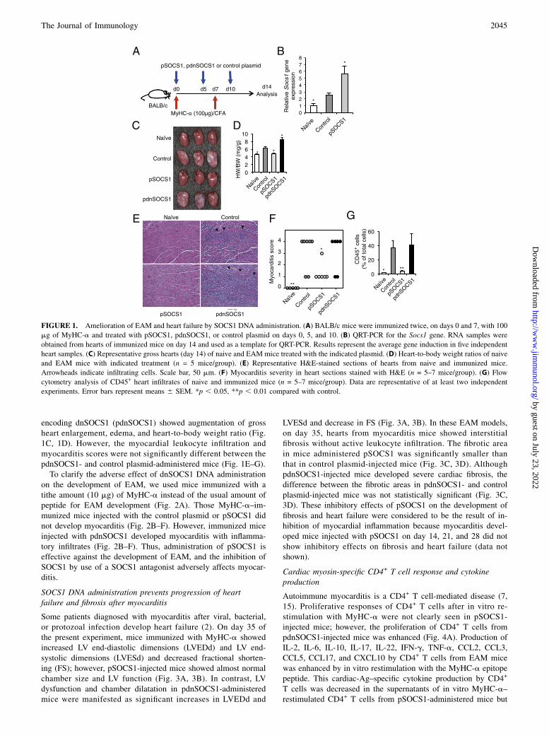
encoding dnSOCS1 (pdnSOCS1) showed augmentation of grossheart enlargement, edema, and heart-to-body weight ratio (Fig.1C, 1D). However, the myocardial leukocyte infiltration andmyocarditis scores were not significantly different between thepdnSOCS1- and control plasmid-administered mice (Fig. 1E–G).To clarify the adverse effect of dnSOCS1 DNA administration
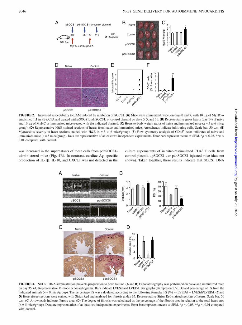
on the development of EAM, we used mice immunized with atithe amount (10 mg) of MyHC-a instead of the usual amount ofpeptide for EAM development (Fig. 2A). Those MyHC-a–im-munized mice injected with the control plasmid or pSOCS1 didnot develop myocarditis (Fig. 2B–F). However, immunized miceinjected with pdnSOCS1 developed myocarditis with inflamma-tory infiltrates (Fig. 2B–F). Thus, administration of pSOCS1 iseffective against the development of EAM, and the inhibition ofSOCS1 by use of a SOCS1 antagonist adversely affects myocar-ditis.
SOCS1 DNA administration prevents progression of heartfailure and fibrosis after myocarditis
Some patients diagnosed with myocarditis after viral, bacterial,or protozoal infection develop heart failure (2). On day 35 ofthe present experiment, mice immunized with MyHC-a showedincreased LV end-diastolic dimensions (LVEDd) and LV end-systolic dimensions (LVESd) and decreased fractional shorten-ing (FS); however, pSOCS1-injected mice showed almost normalchamber size and LV function (Fig. 3A, 3B). In contrast, LVdysfunction and chamber dilatation in pdnSOCS1-administeredmice were manifested as significant increases in LVEDd and
LVESd and decrease in FS (Fig. 3A, 3B). In these EAM models,on day 35, hearts from myocarditis mice showed interstitialfibrosis without active leukocyte infiltration. The fibrotic areain mice administered pSOCS1 was significantly smaller thanthat in control plasmid-injected mice (Fig. 3C, 3D). AlthoughpdnSOCS1-injected mice developed severe cardiac fibrosis, thedifference between the fibrotic areas in pdnSOCS1- and controlplasmid-injected mice was not statistically significant (Fig. 3C,3D). These inhibitory effects of pSOCS1 on the development offibrosis and heart failure were considered to be the result of in-hibition of myocardial inflammation because myocarditis devel-oped mice injected with pSOCS1 on day 14, 21, and 28 did notshow inhibitory effects on fibrosis and heart failure (data notshown).
Cardiac myosin-specific CD4+ T cell response and cytokineproduction
Autoimmune myocarditis is a CD4+ T cell-mediated disease (7,15). Proliferative responses of CD4+ T cells after in vitro re-stimulation with MyHC-a were not clearly seen in pSOCS1-injected mice; however, the proliferation of CD4+ T cells frompdnSOCS1-injected mice was enhanced (Fig. 4A). Production ofIL-2, IL-6, IL-10, IL-17, IL-22, IFN-g, TNF-a, CCL2, CCL3,CCL5, CCL17, and CXCL10 by CD4+ T cells from EAM micewas enhanced by in vitro restimulation with the MyHC-a epitopepeptide. This cardiac-Ag–specific cytokine production by CD4+
T cells was decreased in the supernatants of in vitro MyHC-a–restimulated CD4+ T cells from pSOCS1-administered mice but
FIGURE 1. Amelioration of EAM and heart failure by SOCS1 DNA administration. (A) BALB/c mice were immunized twice, on days 0 and 7, with 100
mg of MyHC-a and treated with pSOCS1, pdnSOCS1, or control plasmid on days 0, 5, and 10. (B) QRT-PCR for the Socs1 gene. RNA samples were
obtained from hearts of immunized mice on day 14 and used as a template for QRT-PCR. Results represent the average gene induction in five independent
heart samples. (C) Representative gross hearts (day 14) of naive and EAM mice treated with the indicated plasmid. (D) Heart-to-body weight ratios of naive
and EAM mice with indicated treatment (n = 5 mice/group). (E) Representative H&E-stained sections of hearts from naive and immunized mice.
Arrowheads indicate infiltrating cells. Scale bar, 50 mm. (F) Myocarditis severity in heart sections stained with H&E (n = 5–7 mice/group). (G) Flow
cytometry analysis of CD45+ heart infiltrates of naive and immunized mice (n = 5–7 mice/group). Data are representative of at least two independent
experiments. Error bars represent means 6 SEM. *p , 0.05, **p , 0.01 compared with control.
The Journal of Immunology 2045
by guest on July 23, 2022http://w
ww
.jimm
unol.org/D
ownloaded from
was increased in the supernatants of these cells from pdnSOCS1-administered mice (Fig. 4B). In contrast, cardiac-Ag–specificproduction of IL-1b, IL-10, and CXCL1 was not detected in the
culture supernatants of in vitro-restimulated CD4+ T cells fromcontrol plasmid-, pSOCS1-, or pdnSOCS1-injected mice (data notshown). Taken together, these results indicate that SOCS1 DNA
FIGURE 2. Increased susceptibility to EAM induced by inhibition of SOCS1. (A) Mice were immunized twice, on days 0 and 7, with 10 mg of MyHC-a
emulsified 1:1 in PBS/CFA and treated with pSOCS1, pdnSOCS1, or control plasmid on days 0, 5, and 10. (B) Representative gross hearts (day 14) of naive
and 10 mg of MyHC-a–immunized mice treated with the indicated plasmid. (C) Heart-to-body weight ratios of naive and immunized mice (n = 5 to 6 mice/
group). (D) Representative H&E-stained sections of hearts from naive and immunized mice. Arrowheads indicate infiltrating cells. Scale bar, 50 mm. (E)
Myocarditis severity in heart sections stained with H&E (n = 5 to 6 mice/group). (F) Flow cytometry analysis of CD45+ heart infiltrates of naive and
immunized mice (n = 5 mice/group). Data are representative of at least two independent experiments. Error bars represent means6 SEM. *p, 0.05, **p,0.01 compared with control.
FIGURE 3. SOCS1 DNA administration prevents progression to heart failure. (A and B) Echocardiography was performed on naive and immunized mice
on day 35. (A) Representative M-mode echocardiograms. Bars indicate LVESd and LVEDd. Bar graphs (B) represent LVEDd and percentage of FS from the
indicated animals (n = 9 mice/group). The percentage FS was calculated according to the following formula: FS (%) = (LVEDd 2 LVESd)/LVEDd. (C and
D) Heart tissue sections were stained with Sirius Red and analyzed for fibrosis at day 35. Representative Sirius Red-stained sections of hearts. Scale bar, 50
mm. (C) Arrowheads indicate fibrotic area. (D) The degree of fibrosis was calculated as the percentage of the fibrotic area in relation to the total heart area
(n = 5 mice/group). Data are representative of at least two independent experiments. Error bars represent means 6 SEM. *p , 0.05, **p , 0.01 compared
with control.
2046 Socs1 GENE DELIVERY FOR AUTOIMMUNE MYOCARDITIS
by guest on July 23, 2022http://w
ww
.jimm
unol.org/D
ownloaded from

delivery inhibits the activation of myosin-specific CD4+ T cellsand strongly suggest that impaired CD4+ Th cell function preventsEAM development in pSOCS1-injected mice after immunizationwith cardiac self-Ag.To evaluate whether pSOCS1 administration affects Ag-specific
CD4+ T cell function in other models, we injected plasmid DNAinto an autoimmune gastritis model and an OVA-immunizedmodel. In the autoimmune gastritis model, gastric-Ag–specificproduction of IL-2, IL-6, IL-13, IL-17, IL-22, IFN-g, TNF-a,CCL2, CCL5, CCL17, and CXCL10 by CD4+ T cells was de-creased in pSOCS1-administered mice but increased in pdnSOCS1-administered mice (Supplemental Fig. 2). Lower amounts ofcytokines (including IL-2, IL-6, IL-13, IFN-g, TNF-a, CCL2,CCL3, CCL5, CCL17, and CXCL10) were also produced in CD4+
T cells from pSOCS1-injected OVA-immunized mice (Supple-mental Fig. 3). These results suggest that pSOCS1 administrationmay suppress Ag-specific CD4+ T cell activation in various au-toimmune diseases and foreign body infections.
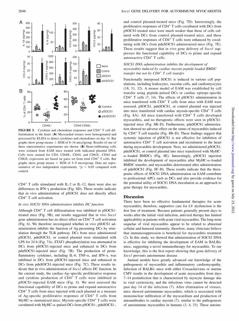
SOCS1 DNA administration inhibits the production ofproinflammatory cytokines and CD4+ T cell differentiation inthe heart
We also examined whether SOCS1 DNA administration has aneffect on cytokine and chemokine milieu in the heart. On day 14after MyHC-a immunization, heart homogenates from pSOCS1-injected mice had significantly decreased amounts of proin-flammatory cytokines, including IL-1b and IL-6, and of myelo-tropic chemokines, including CCL5, CXCL1, and CXCL10 (Fig.5A). In contrast, hearts from mice injected with pdnSOCS1
showed greatly increased amounts of proinflammatory cytokinesand chemokines (Fig. 5A). SOCS1 protein has been shown toregulate T cell differentiation (17, 18). To determine the differ-entiation of CD4+ T cells during EAM, we examined the heart-infiltrating CD4+ T cell populations by FACS analysis. ActivatedCD4+ T cells (CD4+CD40L+) and effector memory CD4+ T cells(CD44+CD62L2) were reduced in the pSOCS1-injected mice(Fig. 5B). Thus, protection from EAM in pSOCS1-administeredmice is associated with abrogation of proinflammatory cytokines,chemokines, and CD4+ T cell differentiation in the heart.
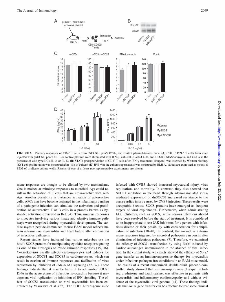
SOCS1 DNA injection does not have a direct suppressive effecton CD4+ T cell activation
To gain new insights into the mechanism of protection frommyocarditis, we investigated whether pSOCS1 therapy directlyaffects CD4+ T cell activation. Naive T cells (CD4+CD62L+ cells)were isolated from non-EAM mice injected with pSOCS1,pdnSOCS1, or control plasmid, and their primary responses tovarious stimuli were compared (Fig. 6A). As shown in Fig. 6B,there were no differences in IFN-g–induced STAT1 activationamong these CD4+ T cells. There were also no differences inprimary responses to stimulation with anti-CD3ε, anti-CD3ε/anti-CD28, PMA/ionomycin, or Con A presented by mitomycin C-treated wild-type DCs among pSOCS1-, pdnSOCS1-, and con-trol plasmid-treated CD4+ T cells (Fig. 6C). Chong et al. (30)demonstrated that SOCS1-deficient T cells produced substantiallygreater levels of IFN-g in response to IL-2 or IL-12. From thesefindings, we assessed the production of IFN-g from CD4+ T cellsby using the same experiments. In the culture supernatants of
FIGURE 4. Impaired expansion of heart-specific CD4+ T cells in pSOCS1-treated mice. (A) Splenocytes were isolated from naive and EAM mice treated
with pSOCS1, pdnSOCS1, or control plasmid on day 14 and restimulated in vitro with MyHC-a or OVA peptide for 72 h. Proliferation was assessed by
measurement of [3H]thymidine incorporation. Data represent means 6 SEM of triplicates from one of three independent experiments. (B) Cytokines and
chemokines in the culture supernatants of splenocytes were measured by ELISA after 48 h of restimulation with MyHC-a or OVA peptide. Data are
expressed as mean 6 SEM from triplicate culture wells. Results of one of two representative experiments are shown. *p , 0.05 compared with MyHC-a–
stimulated control, #p , 0.05 compared with OVA-stimulated control.
The Journal of Immunology 2047
by guest on July 23, 2022http://w
ww
.jimm
unol.org/D
ownloaded from
CD4+ T cells stimulated with IL-2 or IL-12, there were also nodifferences in IFN-g production (Fig. 6D). These results indicatethat in vivo administration of pSOCS1 does not directly affectCD4+ T cell activation.
In vivo SOCS1 DNA administration inhibits DC function
Although CD4+ T cell differentiation was inhibited in pSOCS1-treated mice (Fig. 5B), our results suggested that in vivo Socs1gene administration has no direct effect on CD4+ T cell activation(Fig. 6). We therefore investigated whether in vivo pSOCS1 ad-ministration inhibits the function of Ag-presenting DCs by stim-ulation through the TLR pathway. DCs from mice administeredpSOCS1, pdnSOCS1, or control plasmid were stimulated withLPS for 24 h (Fig. 7A). STAT1 phosphorylation was attenuated inDCs from pSOCS1-injected mice and enhanced in DCs frompdnSOCS1-injected mice (Fig. 7B). The production of proin-flammatory cytokines, including IL-6, TNF-a, and IFN-g, wasinhibited in DCs from pSOCS1-injected mice and enhanced inDCs from pdnSOCS1-injected mice (Fig. 7C). These results in-dicate that in vivo administration of Socs1 affects DC function. Inthe current study, the cardiac-Ag–specific proliferative responseand cytokine production of CD4+ T cells were inhibited inpSOCS1-injected EAM mice (Fig. 4). We next assessed thefunctional capability of DCs to prime and expand autoreactiveCD4+ T cells from mice injected with each plasmid as a measureof Ag-specific proliferative responses of CD4+ T cells fromMyHC-a–immunized mice. Myosin-specific CD4+ T cells werecocultured with MyHC-a–pulsed DCs from pSOCS1-, pdnSOCS1-,
and control plasmid-treated mice (Fig. 7D). Interestingly, theproliferative responses of CD4+ T cells cocultured with DCs frompSOCS1-treated mice were much weaker than those of cells cul-tured with DCs from control plasmid-treated mice, and theseproliferative responses of CD4+ T cells were enhanced by cocul-turing with DCs from pdnSOCS1-administered mice (Fig. 7E).These results suggest that in vivo gene delivery of Socs1 sup-presses the functional capability of DCs to prime and expandautoreactive CD4+ T cells.
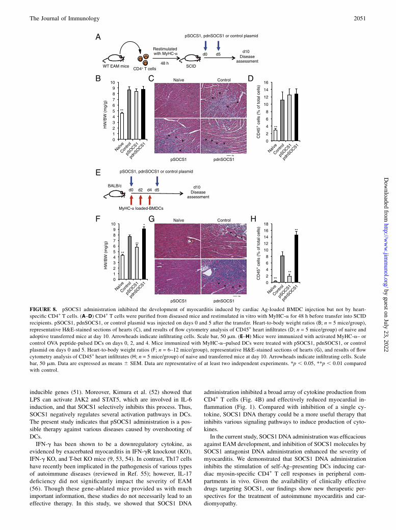
SOCS1 DNA administration inhibits the development ofmyocarditis induced by cardiac myosin peptide-loaded BMDCtransfer but not by CD4+ T cell transfer
Functionally interposed SOCS1 is induced in various cell pop-ulations, including leukocytes, vascular cells, and cardiomyocytes(18, 31, 32). A mouse model of EAM was established by celltransfer using peptide-pulsed DCs or cardiac epitope-specificCD4+ T cells (7, 14). The effects of pSOCS1 administration inmice transferred with CD4+ T cells from mice with EAM wereassessed. pSOCS1, pdnSOCS1, or control plasmid was injectedinto mice transferred with cardiac myosin-specific CD4+ T cells(Fig. 8A). All mice transferred with CD4+ T cells developedmyocarditis, and no therapeutic effects were seen in pSOCS1-injected mice (Fig. 8B–D). Furthermore, pdnSOCS1 administra-tion showed no adverse effect on the status of myocarditis inducedby CD4+ T cell transfer (Fig. 8B–D). These findings suggest thatsystemic injection of pSOCS1 is not effective for inhibition ofautoreactive CD4+ T cell activation and recruitment to the heartduring myocarditis development. Next, we administered pSOCS1,pdnSOCS1, or control plasmid into mice transferred with MyHC-a–loaded BMDCs (Fig. 8E). Interestingly, pSOCS1 injectioninhibited the development of myocarditis after MyHC-a–loadedBMDC transfer, and myocarditis deteriorated after administrationof pdnSOCS1 (Fig. 8F–H). These results indicate that the thera-peutic effects of SOCS1 DNA administration on EAM contributeto professional APCs such as DCs and also provide evidence forthe potential utility of SOCS1 DNA inoculation as an approach togene therapy for myocarditis.
DiscussionThere have been no effective fundamental therapies for acutemyocarditis; therefore, supportive care for LV dysfunction is thefirst line of treatment. Because patients generally present days toweeks after the initial viral infection, antiviral therapy has limitedapplicability in patients with acute viral myocarditis. The long-termsequelae of viral myocarditis appear to be related to abnormalcellular and humoral immunity; therefore, many clinicians believethat immunosuppression is beneficial for myocarditis treatment(2). In this study, we showed that administration of SOCS1 DNAis effective for inhibiting the development of EAM in BALB/cmice, suggesting a novel immunotherapy for myocarditis. To ourknowledge, this is the first report showing that gene delivery ofSocs1 prevents autoimmune disease.Animal models have greatly advanced our knowledge of the
pathogenesis of myocarditis and inflammatory cardiomyopathy.Infection of BALB/c mice with either Coxsackievirus or murineCMV results in the development of acute myocarditis from days7–14 postinfection that is characterized by myocyte damage dueto viral cytotoxicity, and the infectious virus cannot be detectedpast day 14 of the infection (7). After elimination of viruses,mice showed autoimmune myocarditis, which is associated withmononuclear infiltration of the myocardium and production ofautoantibodies to cardiac myosin (7), similar to the pathogenesisof autoimmune myocarditis in humans (3, 4, 33). These autoim-
FIGURE 5. Cytokine and chemokine responses and CD4+ T cell dif-
ferentiation in the heart. (A) Myocardial tissues were homogenated and
processed by ELISA to detect cytokines and chemokines on day 14. Bar
graphs show group means 6 SEM of 8–16 mice/group. Results of one of
three representative experiments are shown. (B) Heart-infiltrating cells
were isolated from EAM mice treated with indicated plasmid DNA.
Cells were stained for CD4, CD40L, CD44, and CD62L. CD44 and
CD62L expression are based on gates set from total CD4+ T cells. Bar
graphs show group means 6 SEM of 5–9 mice/group. Data are repre-
sentative of two independent experiments. *p , 0.05 compared with
control.
2048 Socs1 GENE DELIVERY FOR AUTOIMMUNE MYOCARDITIS
by guest on July 23, 2022http://w
ww
.jimm
unol.org/D
ownloaded from
mune responses are thought to be elicited by two mechanisms.One is molecular mimicry: responses to microbial Ags could re-sult in the activation of T cells that are cross-reactive with self-Ags. Another possibility is bystander activation of autoreactivecells. APCs that have become activated in the inflammatory milieuof a pathogenic infection can stimulate the activation and prolif-eration of autoreactive T or B cells in a process known as by-stander activation (reviewed in Ref. 34). Thus, immune responsesto myocytes involving various innate and adaptive immune path-ways were recognized during myocarditis development. The car-diac myosin peptide-immunized mouse EAM model reflects hu-man autoimmune myocarditis and heart failure after eliminationof infectious pathogens.Recent studies have indicated that various microbes use the
host’s SOCS proteins for manipulating cytokine receptor signalingas one of the strategies to evade immune responses (35, 36).Coxsackievirus usually infects cardiomyocytes and induces theexpression of SOCS1 and SOCS3 in cardiomyocytes, which canresult in evasion of immune responses and facilitation of virusreplication by inhibition of JAK–STAT signaling (32, 37). Thesefindings indicate that it may be harmful to administer SOCS1DNA in the acute phase of infectious myocarditis because it mayaugment viral replication by inhibition of IFN signaling. The ef-fect of SOCS1 transduction on viral myocarditis has been ex-amined by Yasukawa et al. (32). The SOCS1-transgenic mice
infected with CVB3 showed increased myocardial injury, virusreplication, and mortality. In contrast, they also showed thatSOCS1 inhibition in the heart through adeno-associated virus-mediated expression of dnSOCS1 increased resistance to theacute cardiac injury caused by CVB3 infection. These results wereacceptable because SOCS proteins have emerged as frequenttargets of viral exploitation. Furthermore, when administratingJAK inhibitors, such as SOCS, active serious infections shouldhave been resolved before the start of treatment. It is consideredto be inappropriate to use JAK inhibitors for a person with infec-tious disease or their possibility with consideration for compli-cation of infection (38–40). In contrast, the overactive autoim-mune responses triggered by microbial pathogens can persist afterelimination of infectious pathogens (7). Therefore, we examinedthe efficacy of SOCS1 transfection by using EAM induced bycardiac autoantigen immunization in the absence of viral infec-tion. In the current study, we clearly showed the efficacy of Socs1gene transfer as an immunosuppressive therapy for myocarditisunder infectious pathogen-free conditions in an EAM mice model.The results of a recent randomized, double-blind, placebo-con-trolled study showed that immunosuppressive therapy, includ-ing prednisone and azathioprine, was effective in patients withmyocarditis and inflammatory cardiomyopathy and without evi-dence of the myocardial viral genome (41). These findings indi-cate that Socs1 gene transfer can be effective to treat some clinical
FIGURE 6. Primary responses of CD4+ T cells from pSOCS1-, pdnSOCS1-, and control plasmid-treated mice. (A) CD4+CD62L+ T cells from mice
injected with pSOCS1, pdnSOCS1, or control plasmid were stimulated with IFN-g, anti-CD3ε, anti-CD3ε, anti-CD28, PMA/ionomycin, and Con A in the
presence of wild-type DCs, IL-2, or IL-12. (B) STAT1 phosphorylation of CD4+ T cells after IFN-g treatment (10 ng/ml) was assessed by Western blotting.
(C) T cell proliferation was measured after 48 h of culture. (D) IFN-g in the culture supernatants was measured by ELISA. Values are expressed as means6SEM of triplicate culture wells. Results of one of at least two representative experiments are shown.
The Journal of Immunology 2049
by guest on July 23, 2022http://w
ww
.jimm
unol.org/D
ownloaded from

cases of myocarditis and inflammatory cardiomyopathy associatedwith autoimmunity and without the virus genome in the myocar-
dium, as well as EAM in mice.In the current study, we demonstrated that the administration
of plasmid DNA encoding SOCS1 did not affect autoreactive
CD4+ T cell function (Fig. 6) and adoptive transfer of autoreactive
CD4+ T cells was able to induce myocarditis in SOCS1 DNA-
administered SCID mice (Fig. 8A–D), suggesting that SOCS1
DNA does not suppress either CD4+ T cell recruitment or accu-
mulation of other inflammatory cells in the heart. In contrast, the
introduced SOCS1 DNA inhibited the activation of DCs producing
proinflammatory cytokines (Fig. 7C). In fact, inhibition of the
phosphorylation of STAT1 molecules was observed in DCs from
mice injected with SOCS1 DNA (Fig. 7B). In addition, the pro-
liferative responses of CD4+ T cells cocultured with DCs from
pSOCS1-treated mice were much weaker than those of cells cul-
tured with DCs from control plasmid-injected mice (Fig. 7E).
These results suggest that the inoculated SOCS1 DNA may have
been transfected into DCs and impaired DC function in vivo.
Contrary to expectations, we could not find evidence of direct
transfection of inoculated DNA into DCs in the heart, spleen,
peritoneal cavity, or lymph nodes. Although the introduced DNA
is expressed predominantly by somatic cells (e.g., cardiomyo-
cytes, keratinocytes, and fibroblasts), it is known that relatively
small but biologically significant numbers of DCs are transfected
with the inoculated DNA (42–44). Based on this fact, the inocu-
lated SOCS1 DNA may have inhibited DC activation through the
direct transfection into DCs; however, our data do not exclude thepossibility of another indirect mechanisms.In the EAM model, activation of TLRs on self-Ag–presenting
DCs is essential for the expansion of autoreactive CD4+ T cells toinduce myocarditis and heart failure (15). We previously reportedthat Tlr4 mutant C3H/HeJ mice are resistant to development ofEAM (45). Furthermore, IL-1 type 1 receptor signaling on DCs iscritical for autoimmune myocarditis development (11). MyD88 isa crucial common adaptor molecule that mediates both TLRs andIL-1 type 1 receptor activation (46, 47), and MyD88 signaling inDCs is critical for the induction of EAM (16). SOCS1 negativelyregulates the MyD88-dependent pathway by interacting with bothIL-1R–associated kinase and NF-kB (17), which results in a de-crease in the induction of inflammatory cytokines such as TNF-aand IL-6. In fact, production of these inflammatory cytokines wasinhibited by the administration of SOCS1 DNA in the currentstudy (Fig. 7C). Although nearly all TLRs recruit MyD88, otherspecific adaptor proteins function downstream of particular TLRs.One such adaptor molecule is Toll/IL-1R domain-containing adap-tor protein/Mal. SOCS1 also binds to tyrosine-phosphorylatedMal through its interaction with Bruton’s tyrosine kinase, leadingto the suppression of Mal-dependent p65 phosphorylation andtransactivation of NF-kB (48). Another important mechanism ofthe suppression of APC activation by SOCS1 is inhibition of thesecondary activated JAK–STAT pathway (49, 50). The Toll/IL-1Rdomain-containing adaptor protein-inducing IFN-b–IFN-regula-tory factor 3 pathway rapidly induces IFN-b, which in turn acti-vates JAK–STAT1 and contributes to the expression of IFN-
FIGURE 7. Functional capacities of DCs from pSOCS1-, pdnSOCS1-, and control plasmid-treated mice. (A) DCs from mice treated with pSOCS1,
pdnSOCS1, or control plasmid were stimulated with LPS for 24 h. (B) STAT1 phosphorylation of DCs was assessed by Western blotting. Densitometry
ratios of pSTAT1/STAT1 are shown as fold induction, the ratio for DCs from control plasmid-injected mice being set at 1. Results are means of five
independent experiments 6 SEM. Blots are representative of experiments performed a minimum of three times. (C) IL-6, TNF-a, and IFN-g in the culture
supernatants were measured by ELISA. Values indicate means 6 SEM of triplicate culture wells from one of three independent experiments. (D and E)
Heart-specific CD4+ T cells from EAM mice were restimulated with MyHC-a or OVA peptide on DCs from mice treated with control plasmid, pSOCS1, or
pdnSOCS1 for 72 h before measurement of [3H]thymidine incorporation. Each value represents mean 6 SEM cpm values of six different culture wells.
Results of one of three representative experiments are shown. *p , 0.05, **p , 0.01 compared with control.
2050 Socs1 GENE DELIVERY FOR AUTOIMMUNE MYOCARDITIS
by guest on July 23, 2022http://w
ww
.jimm
unol.org/D
ownloaded from
inducible genes (51). Moreover, Kimura et al. (52) showed thatLPS can activate JAK2 and STAT5, which are involved in IL-6induction, and that SOCS1 selectively inhibits this process. Thus,SOCS1 negatively regulates several activation pathways in DCs.The present study indicates that pSOCS1 administration is a pos-sible therapy against various diseases caused by overshooting ofDCs.IFN-g has been shown to be a downregulatory cytokine, as
evidenced by exacerbated myocarditis in IFN-gR knockout (KO),IFN-g KO, and T-bet KO mice (9, 53, 54). In contrast, Th17 cellshave recently been implicated in the pathogenesis of various typesof autoimmune diseases (reviewed in Ref. 55); however, IL-17deficiency did not significantly impact the severity of EAM(56). Though these gene-ablated mice provided us with muchimportant information, these studies do not necessarily lead to aneffective therapy. In this study, we showed that SOCS1 DNA
administration inhibited a broad array of cytokine production fromCD4+ T cells (Fig. 4B) and effectively reduced myocardial in-flammation (Fig. 1). Compared with inhibition of a single cy-tokine, SOCS1 DNA therapy could be a more useful therapy thatinhibits various signaling pathways to induce production of cyto-kines.In the current study, SOCS1 DNA administration was efficacious
against EAM development, and inhibition of SOCS1 molecules bySOCS1 antagonist DNA administration enhanced the severity ofmyocarditis. We demonstrated that SOCS1 DNA administrationinhibits the stimulation of self-Ag–presenting DCs inducing car-diac myosin-specific CD4+ T cell responses in peripheral com-partments in vivo. Given the availability of clinically effectivedrugs targeting SOCS1, our findings show new therapeutic per-spectives for the treatment of autoimmune myocarditis and car-diomyopathy.
FIGURE 8. pSOCS1 administration inhibited the development of myocarditis induced by cardiac Ag-loaded BMDC injection but not by heart-
specific CD4+ T cells. (A–D) CD4+ T cells were purified from diseased mice and restimulated in vitro with MyHC-a for 48 h before transfer into SCID
recipients. pSOCS1, pdnSOCS1, or control plasmid was injected on days 0 and 5 after the transfer. Heart-to-body weight ratios (B; n = 5 mice/group),
representative H&E-stained sections of hearts (C), and results of flow cytometry analysis of CD45+ heart infiltrates (D; n = 5 mice/group) of naive and
adoptive transferred mice at day 10. Arrowheads indicate infiltrating cells. Scale bar, 50 mm. (E–H) Mice were immunized with activated MyHC-a– or
control OVA peptide-pulsed DCs on days 0, 2, and 4. Mice immunized with MyHC-a–pulsed DCs were treated with pSOCS1, pdnSOCS1, or control
plasmid on days 0 and 5. Heart-to-body weight ratios (F; n = 6–12 mice/group), representative H&E-stained sections of hearts (G), and results of flow
cytometry analysis of CD45+ heart infiltrates (H; n = 5 mice/group) of naive and transferred mice at day 10. Arrowheads indicate infiltrating cells. Scale
bar, 50 mm. Data are expressed as means 6 SEM. Data are representative of at least two independent experiments. *p , 0.05, **p , 0.01 compared
with control.
The Journal of Immunology 2051
by guest on July 23, 2022http://w
ww
.jimm
unol.org/D
ownloaded from
AcknowledgmentsWe thank T. Okamura, Y. Shiogama, T. Wada, K. Watanabe, H. Shibata, and
M. Namikata for technical support and valuable discussion and F. Miya-
masu of the Medical English Communications Center, University of Tsu-
kuba, for grammatical revision of this manuscript.
DisclosuresThe authors have no financial conflicts of interest.
References1. Brown, C. A., and J. B. O’Connell. 1995. Myocarditis and idiopathic dilated
cardiomyopathy. Am. J. Med. 99: 309–314.2. Feldman, A. M., and D. McNamara. 2000. Myocarditis. N. Engl. J. Med. 343:
1388–1398.3. Caforio, A. L., N. J. Mahon, F. Tona, and W. J. McKenna. 2002. Circulating
cardiac autoantibodies in dilated cardiomyopathy and myocarditis: pathogeneticand clinical significance. Eur. J. Heart Fail. 4: 411–417.
4. Lauer, B., M. Schannwell, U. Kuhl, B. E. Strauer, and H. P. Schultheiss. 2000.Antimyosin autoantibodies are associated with deterioration of systolic and di-astolic left ventricular function in patients with chronic myocarditis. J. Am. Coll.Cardiol. 35: 11–18.
5. Frustaci, A., C. Chimenti, F. Calabrese, M. Pieroni, G. Thiene, and A. Maseri.2003. Immunosuppressive therapy for active lymphocytic myocarditis: virolog-ical and immunologic profile of responders versus nonresponders. Circulation107: 857–863.
6. Caforio, A. L., J. H. Goldman, A. J. Haven, K. M. Baig, L. D. Libera, andW. J. McKenna; The Myocarditis Treatment Trial Investigators. 1997. Circu-lating cardiac-specific autoantibodies as markers of autoimmunity in clinical andbiopsy-proven myocarditis. Eur. Heart J. 18: 270–275.
7. Fairweather, D., Z. Kaya, G. R. Shellam, C. M. Lawson, and N. R. Rose. 2001.From infection to autoimmunity. J. Autoimmun. 16: 175–186.
8. Eriksson, U., M. O. Kurrer, W. Sebald, F. Brombacher, and M. Kopf. 2001. Dualrole of the IL-12/IFN-gamma axis in the development of autoimmune myocar-ditis: induction by IL-12 and protection by IFN-gamma. J. Immunol. 167: 5464–5469.
9. Afanasyeva, M., Y. Wang, Z. Kaya, E. A. Stafford, K. M. Dohmen, A. A. SadighiAkha, and N. R. Rose. 2001. Interleukin-12 receptor/STAT4 signaling is requiredfor the development of autoimmune myocarditis in mice by an interferon-gamma-independent pathway. Circulation 104: 3145–3151.
10. Eriksson, U., M. O. Kurrer, N. Schmitz, S. C. Marsch, A. Fontana, H. P. Eugster,and M. Kopf. 2003. Interleukin-6-deficient mice resist development of autoim-mune myocarditis associated with impaired upregulation of complement C3.Circulation 107: 320–325.
11. Eriksson, U., M. O. Kurrer, I. Sonderegger, G. Iezzi, A. Tafuri, L. Hunziker,S. Suzuki, K. Bachmaier, R. M. Bingisser, J. M. Penninger, and M. Kopf. 2003.Activation of dendritic cells through the interleukin 1 receptor 1 is critical for theinduction of autoimmune myocarditis. J. Exp. Med. 197: 323–331.
12. Sonderegger, I., G. Iezzi, R. Maier, N. Schmitz, M. Kurrer, and M. Kopf. 2008.GM-CSF mediates autoimmunity by enhancing IL-6-dependent Th17 cell de-velopment and survival. J. Exp. Med. 205: 2281–2294.
13. Satoh, M., G. Tamura, I. Segawa, A. Tashiro, K. Hiramori, and R. Satodate.1996. Expression of cytokine genes and presence of enteroviral genomic RNA inendomyocardial biopsy tissues of myocarditis and dilated cardiomyopathy.Virchows Arch. 427: 503–509.
14. Eriksson, U., and J. M. Penninger. 2005. Autoimmune heart failure: newunderstandings of pathogenesis. Int. J. Biochem. Cell Biol. 37: 27–32.
15. Eriksson, U., R. Ricci, L. Hunziker, M. O. Kurrer, G. Y. Oudit, T. H. Watts,I. Sonderegger, K. Bachmaier, M. Kopf, and J. M. Penninger. 2003. Dendriticcell-induced autoimmune heart failure requires cooperation between adaptiveand innate immunity. Nat. Med. 9: 1484–1490.
16. Marty, R. R., S. Dirnhofer, N. Mauermann, S. Schweikert, S. Akira, L. Hunziker,J. M. Penninger, and U. Eriksson. 2006. MyD88 signaling controls autoimmunemyocarditis induction. Circulation 113: 258–265.
17. Dimitriou, I. D., L. Clemenza, A. J. Scotter, G. Chen, F. M. Guerra, andR. Rottapel. 2008. Putting out the fire: coordinated suppression of the innate andadaptive immune systems by SOCS1 and SOCS3 proteins. Immunol. Rev. 224:265–283.
18. Yoshimura, A., T. Naka, and M. Kubo. 2007. SOCS proteins, cytokine signallingand immune regulation. Nat. Rev. Immunol. 7: 454–465.
19. Shuai, K., and B. Liu. 2003. Regulation of JAK-STAT signalling in the immunesystem. Nat. Rev. Immunol. 3: 900–911.
20. Croker, B. A., H. Kiu, and S. E. Nicholson. 2008. SOCS regulation of the JAK/STAT signalling pathway. Semin. Cell Dev. Biol. 19: 414–422.
21. Fujimoto, M., and T. Naka. 2010. SOCS1, a Negative Regulator of CytokineSignals and TLR Responses, in Human Liver Diseases. Gastroenterol. Res.Pract. 2010: 2010.
22. Naka, T., M. Fujimoto, H. Tsutsui, and A. Yoshimura. 2005. Negative regulationof cytokine and TLR signalings by SOCS and others. Adv. Immunol. 87: 61–122.
23. Hanada, T., H. Yoshida, S. Kato, K. Tanaka, K. Masutani, J. Tsukada,Y. Nomura, H. Mimata, M. Kubo, and A. Yoshimura. 2003. Suppressor of cy-tokine signaling-1 is essential for suppressing dendritic cell activation and sys-temic autoimmunity. Immunity 19: 437–450.
24. Kinjyo, I., T. Hanada, K. Inagaki-Ohara, H. Mori, D. Aki, M. Ohishi, H. Yoshida,M. Kubo, and A. Yoshimura. 2002. SOCS1/JAB is a negative regulator of LPS-induced macrophage activation. Immunity 17: 583–591.
25. Nakagawa, R., T. Naka, H. Tsutsui, M. Fujimoto, A. Kimura, T. Abe, E. Seki,S. Sato, O. Takeuchi, K. Takeda, et al. 2002. SOCS-1 participates in negativeregulation of LPS responses. Immunity 17: 677–687.
26. Lutz, M. B., N. Kukutsch, A. L. Ogilvie, S. Rossner, F. Koch, N. Romani, andG. Schuler. 1999. An advanced culture method for generating large quantities ofhighly pure dendritic cells from mouse bone marrow. J. Immunol. Methods 223:77–92.
27. Valaperti, A., R. R. Marty, G. Kania, D. Germano, N. Mauermann, S. Dirnhofer,B. Leimenstoll, P. Blyszczuk, C. Dong, C. Mueller, et al. 2008. CD11b+monocytes abrogate Th17 CD4+ T cell-mediated experimental autoimmunemyocarditis. J. Immunol. 180: 2686–2695.
28. Cihakova, D., J. G. Barin, M. Afanasyeva, M. Kimura, D. Fairweather, M. Berg,M. V. Talor, G. C. Baldeviano, S. Frisancho, K. Gabrielson, et al. 2008.Interleukin-13 protects against experimental autoimmune myocarditis by regu-lating macrophage differentiation. Am. J. Pathol. 172: 1195–1208.
29. Hanada, T., T. Yoshida, I. Kinjyo, S. Minoguchi, H. Yasukawa, S. Kato,H. Mimata, Y. Nomura, Y. Seki, M. Kubo, and A. Yoshimura. 2001. A mutantform of JAB/SOCS1 augments the cytokine-induced JAK/STAT pathway byaccelerating degradation of wild-type JAB/CIS family proteins through theSOCS-box. J. Biol. Chem. 276: 40746–40754.
30. Chong, M. M., D. Metcalf, E. Jamieson, W. S. Alexander, and T. W. Kay. 2005.Suppressor of cytokine signaling-1 in T cells and macrophages is critical forpreventing lethal inflammation. Blood 106: 1668–1675.
31. Ortiz-Munoz, G., J. L. Martin-Ventura, P. Hernandez-Vargas, B. Mallavia,V. Lopez-Parra, O. Lopez-Franco, B. Munoz-Garcia, P. Fernandez-Vizarra,L. Ortega, J. Egido, and C. Gomez-Guerrero. 2009. Suppressors of cytokinesignaling modulate JAK/STAT-mediated cell responses during atherosclerosis.Arterioscler. Thromb. Vasc. Biol. 29: 525–531.
32. Yasukawa, H., T. Yajima, H. Duplain, M. Iwatate, M. Kido, M. Hoshijima,M. D. Weitzman, T. Nakamura, S. Woodard, D. Xiong, et al. 2003. The sup-pressor of cytokine signaling-1 (SOCS1) is a novel therapeutic target forenterovirus-induced cardiac injury. J. Clin. Invest. 111: 469–478.
33. Kanzaki, Y., F. Terasaki, M. Okabe, T. Hayashi, H. Toko, H. Shimomura,S. Fujioka, Y. Kitaura, K. Kawamura, Y. Horii, et al. 2001. Myocardial in-flammatory cell infiltrates in cases of dilated cardiomyopathy as a determinant ofoutcome following partial left ventriculectomy. Jpn. Circ. J. 65: 797–802.
34. Munz, C., J. D. Lunemann, M. T. Getts, and S. D. Miller. 2009. Antiviral immuneresponses: triggers of or triggered by autoimmunity? Nat. Rev. Immunol. 9: 246–258.
35. Baetz, A., S. Zimmermann, and A. H. Dalpke. 2007. Microbial immune evasionemploying suppressor of cytokine signaling (SOCS) proteins. Inflamm. AllergyDrug Targets 6: 160–167.
36. Akhtar, L. N., and E. N. Benveniste. 2011. Viral exploitation of host SOCSprotein functions. J. Virol. 85: 1912–1921.
37. Yajima, T., H. Yasukawa, E. S. Jeon, D. Xiong, A. Dorner, M. Iwatate, M. Nara,H. Zhou, D. Summers-Torres, M. Hoshijima, et al. 2006. Innate defensemechanism against virus infection within the cardiac myocyte requiring gp130-STAT3 signaling. Circulation 114: 2364–2373.
38. Pesu, M., A. Laurence, N. Kishore, S. H. Zwillich, G. Chan, and J. J. O’Shea.2008. Therapeutic targeting of Janus kinases. Immunol. Rev. 223: 132–142.
39. Yamaoka, K., B. Min, Y. J. Zhou, W. E. Paul, and J. J. O’shea. 2005. Jak3 negativelyregulates dendritic-cell cytokine production and survival. Blood 106: 3227–3233.
40. Changelian, P. S., D. Moshinsky, C. F. Kuhn, M. E. Flanagan, M. J. Munchhof,T. M. Harris, D. A. Whipple, J. L. Doty, J. Sun, C. R. Kent, et al. 2008. Thespecificity of JAK3 kinase inhibitors. Blood 111: 2155–2157.
41. Frustaci, A., M. A. Russo, and C. Chimenti. 2009. Randomized study on theefficacy of immunosuppressive therapy in patients with virus-negative inflam-matory cardiomyopathy: the TIMIC study. Eur. Heart J. 30: 1995–2002.
42. Tuting, T., W. J. Storkus, and L. D. Falo, Jr. 1998. DNA immunization targeting theskin: molecular control of adaptive immunity. J. Invest. Dermatol. 111: 183–188.
43. Condon, C., S. C. Watkins, C. M. Celluzzi, K. Thompson, and L. D. Falo, Jr.1996. DNA-based immunization by in vivo transfection of dendritic cells. Nat.Med. 2: 1122–1128.
44. Porgador, A., K. R. Irvine, A. Iwasaki, B. H. Barber, N. P. Restifo, andR. N. Germain. 1998. Predominant role for directly transfected dendritic cells inantigen presentation to CD8+ T cells after gene gun immunization. J. Exp. Med.188: 1075–1082.
45. Nishikubo, K., K. Imanaka-Yoshida, S. Tamaki, M. Hiroe, T. Yoshida, Y. Adachi,and Y. Yasutomi. 2007. Th1-type immune responses by Toll-like receptor 4signaling are required for the development of myocarditis in mice with BCG-induced myocarditis. J. Autoimmun. 29: 146–153.
46. Akira, S., and H. Hemmi. 2003. Recognition of pathogen-associated molecularpatterns by TLR family. Immunol. Lett. 85: 85–95.
47. Li, X., and J. Qin. 2005. Modulation of Toll-interleukin 1 receptor mediatedsignaling. J. Mol. Med. 83: 258–266.
48. Mansell, A., R. Smith, S. L. Doyle, P. Gray, J. E. Fenner, P. J. Crack,S. E. Nicholson, D. J. Hilton, L. A. O’Neill, and P. J. Hertzog. 2006. Suppressorof cytokine signaling 1 negatively regulates Toll-like receptor signaling bymediating Mal degradation. Nat. Immunol. 7: 148–155.
49. Gingras, S., E. Parganas, A. de Pauw, J. N. Ihle, and P. J. Murray. 2004. Re-examination of the role of suppressor of cytokine signaling 1 (SOCS1) in theregulation of toll-like receptor signaling. J. Biol. Chem. 279: 54702–54707.
50. Baetz, A., M. Frey, K. Heeg, and A. H. Dalpke. 2004. Suppressor of cytokinesignaling (SOCS) proteins indirectly regulate toll-like receptor signaling in in-nate immune cells. J. Biol. Chem. 279: 54708–54715.
2052 Socs1 GENE DELIVERY FOR AUTOIMMUNE MYOCARDITIS
by guest on July 23, 2022http://w
ww
.jimm
unol.org/D
ownloaded from
51. Qin, H., C. A. Wilson, S. J. Lee, and E. N. Benveniste. 2006. IFN-beta-inducedSOCS-1 negatively regulates CD40 gene expression in macrophages andmicroglia. FASEB J. 20: 985–987.
52. Kimura, A., T. Naka, T. Muta, O. Takeuchi, S. Akira, I. Kawase, andT. Kishimoto. 2005. Suppressor of cytokine signaling-1 selectively inhibits LPS-induced IL-6 production by regulating JAK-STAT. Proc. Natl. Acad. Sci. USA102: 17089–17094.
53. Eriksson, U., M. O. Kurrer, R. Bingisser, H. P. Eugster, P. Saremaslani,F. Follath, S. Marsch, and U. Widmer. 2001. Lethal autoimmune myocarditis ininterferon-gamma receptor-deficient mice: enhanced disease severity by im-paired inducible nitric oxide synthase induction. Circulation 103: 18–21.
54. Rangachari, M., N. Mauermann, R. R. Marty, S. Dirnhofer, M. O. Kurrer,V. Komnenovic, J. M. Penninger, and U. Eriksson. 2006. T-bet negatively reg-ulates autoimmune myocarditis by suppressing local production of interleukin17. J. Exp. Med. 203: 2009–2019.
55. Ghoreschi, K., A. Laurence, X. P. Yang, K. Hirahara, and J. J. O’Shea. 2011. Thelper 17 cell heterogeneity and pathogenicity in autoimmune disease. TrendsImmunol. 32: 395–401.
56. Baldeviano, G. C., J. G. Barin, M. V. Talor, S. Srinivasan, D. Bedja, D. Zheng,K. Gabrielson, Y. Iwakura, N. R. Rose, and D. Cihakova. 2010. Interleukin-17Ais dispensable for myocarditis but essential for the progression to dilated car-diomyopathy. Circ. Res. 106: 1646–1655.
The Journal of Immunology 2053
by guest on July 23, 2022http://w
ww
.jimm
unol.org/D
ownloaded from