e1907653118.full.pdf - pnas
TRANSCRIPT
CD47 blockade reduces the pathologic features ofexperimental cerebral malaria and promotes survivalof hosts with Plasmodium infectionLaughing Bear Torrez Dulgeroffa,1, Miranda S. Oakleyb, Michal C. Tala, Ying Ying Yiua
, Joy Q. Hea, Maia Shohama,
Victoria Majamb, Winter A. Okothb, Pallavi Mallab, Sanjai Kumarb, and Irving L. Weissmana,1
aInstitute for Stem Cell Biology and Regenerative Medicine, Stanford University School of Medicine, Stanford, CA 94305; and bLaboratory of EmergingPathogens, Division of Emerging and Transfusion Transmitted Diseases, Office of Blood Research and Review, Center for Biologics Evaluation and Research,Food and Drug Administration, Silver Spring, MD 20993
Contributed by Irving L. Weissman, January 28, 2021 (sent for review June 24, 2019; reviewed by Jay A. Berzofsky and Christian Engwerda)
CD47 is an antiphagocytic “don’t eat me” signal that inhibits pro-grammed cell removal of self. As red blood cells (RBCs) age theylose CD47 expression and become susceptible to programmed cellremoval by macrophages. CD47−/− mice infected with Plasmodiumyoelii, which exhibits an age-based preference for young RBCs,were previously demonstrated to be highly resistant to malariainfection. Our study sought to test the therapeutic benefit ofCD47 blockade on ameliorating the clinical syndromes of experi-mental cerebral malaria (ECM), using the Plasmodium bergheiANKA (Pb-A) murine model. In vitro we tested the effect of anti-CD47 mAb on Plasmodium-infected RBC phagocytosis and foundthat anti-CD47 treatment significantly increased clearance ofPlasmodium-infected RBCs. Infection of C57BL/6 mice with Pb-Ais lethal and mice succumb to the clinical syndromes of CMbetween days 6 and 10 postinfection. Strikingly, treatment withanti-CD47 resulted in increased survival during the cerebral phaseof Pb-A infection. Anti-CD47–treated mice had increased lympho-cyte counts in the peripheral blood and increased circulating levelsof IFN-γ, TNF-α, and IL-22. Despite increased circulating levels ofinflammatory cytokines, anti-CD47–treated mice had reducedpathological features in the brain. Survival of ECM in anti-CD47–treated mice was correlated with reduced cellular accumu-lation in the cerebral vasculature, improved blood–brain barrierintegrity, and reduced cytotoxic activity of infiltrating CD8+
T cells. These results demonstrate the therapeutic benefit ofanti-CD47 to reduce morbidity in a lethal model of ECM, whichmay have implications for preventing mortality in young Africanchildren who are the highest casualties of CM.
CD47 | Plasmodium berghei ANKA | cerebral malaria
Malaria remains a major public health challenge in devel-oping countries. According to the World Health Organi-
zation (WHO), in 2018, there were an estimated 218 millioncases and 405,000 malaria-related deaths. About 93% of thesedeaths were reported from the WHO African regions alone andwere mostly caused by the most virulent form of human malaria,Plasmodium falciparum infections. Cerebral malaria (CM) is amajor pathogenic consequence of P. falciparum infection and aleading cause of malaria deaths in African children under 5 y ofage. Currently, there is no treatment available to suppressthe clinical syndromes of CM and patient symptoms are man-aged by administration of antimalaria drugs to control para-sitemia and supported by adjunct therapies including exchangetransfusion (1).CD47 is a 50-kDa transmembrane surface protein with an
extracellular IgV domain that is ubiquitously expressed at vary-ing levels across all cell types (2, 3). CD47 is a potent “don’t eatme” signal regulating programed cell removal (PrCR) (4, 5).When CD47 binds to inhibitory receptor signal-regulatoryprotein-α (SIRPα), on the surface of phagocytic cells, theimmunoreceptor tyrosine-based inhibitory motif within the
cytoplasmic domain of SIRPα of phosphorylates tyrosine phos-phatases SHP1/2, which inhibit the myosin assembly necessaryfor phagocytosis (6–8). CD47 is the predominant surface markerof red blood cell (RBC) turnover; newly produced RBCs havehigh surface expression of CD47, which degrades as a function ofRBC age. The importance of CD47 expression on RBCs wasdemonstrated when labeled CD47 knockout cells were rapidlycleared after transplantation into wild-type mice. Furthermore,these studies used SIRPα antibodies to block phagocytosisin vitro and demonstrated that CD47, the ligand for the inhibi-tory receptor SIRPα, is critical to preventing PrCR of healthyRBCs (9, 10).While blocking the interaction of CD47 is important for pro-
moting phagocytosis, CD47 blockade alone is not sufficient andrequires concurrent expression of a prophagocytic or “eat me”signal on the surface of the target cell (11). Subsequent studiesidentified cell surface expression of calreticulin (CRT) as thepredominant prophagocytic signal that preferentially promotesphagocytosis of tumor cells while sparing healthy cells uponCD47 blockade (12). The role of CRT as a prophagocytic signal
Significance
Novel therapies are urgently needed that can ameliorate theclinical syndromes of cerebral malaria, the most severe conse-quences of Plasmodium infection, and thereby reduce malariafatality. Monoclonal antibodies that target CD47, a “don’t eatme” signal, have been demonstrated to enhance cellularclearance of cancer cells by promoting macrophage phagocy-tosis. We sought to adopt this therapeutic strategy to amelio-rate the clinical syndromes associated with cerebral malariawith the goals of reducing disease-associated morbidity andmortality. We demonstrate that CD47 blockade by anti-CD47injection leads to survival from cerebral malaria in mice.
Author contributions: L.B.T.D., S.K., and I.L.W. designed research; L.B.T.D., M.S.O., M.C.T.,Y.Y.Y., J.Q.H., M.S., V.M., W.A.O., and P.M. performed research; L.B.T.D., M.S.O., andM.C.T. analyzed data; and L.B.T.D., S.K., and I.L.W. wrote the paper.
Reviewers: J.A.B., National Cancer Institute, NIH; and C.E., Queensland Institute ofMedical Research.
Competing interest statement: I.L.W. is on patent applications from Stanford on the useof CD47 blocking agents in infectious disease. He was also cofounder, director, stock-holder, and consultant in the company Forty Seven, Inc., which was purchased by Gilead,Inc., a company in which he has no consulting or financial role or agreements. As co-founder, I.L.W. has been asked to explain topics pursued by Forty Seven in the transition,but without financial compensation from or other agreements with Gilead currently.
This open access article is distributed under Creative Commons Attribution-NonCommercial-NoDerivatives License 4.0 (CC BY-NC-ND).1To whom correspondence may be addressed. Email: [email protected] [email protected].
This article contains supporting information online at https://www.pnas.org/lookup/suppl/doi:10.1073/pnas.1907653118/-/DCSupplemental.
Published March 8, 2021.
PNAS 2021 Vol. 118 No. 11 e1907653118 https://doi.org/10.1073/pnas.1907653118 | 1 of 8
IMMUNOLO
GYAND
INFLAMMATION
Dow
nloa
ded
by g
uest
on
Feb
ruar
y 2,
202
2
is intriguing in the context of transcriptionally silent enucleatedRBCs, as activated macrophages have been demonstrated tosecrete CRT that binds to asialoglycans on the surface of targetcells, leading to macrophage CD91-mediated PrCR, and canthus act as a cell extrinsic prophagocytic signal (13, 14).Previous reports have demonstrated that CD47 plays an im-
portant role in Plasmodium infection. During late-stage malariainfection (15 d postinfection) CD47 has been demonstrated tocounterbalance high levels of phosphatidyl serine on both un-infected and infected RBCs the dynamics of which play an im-portant role in malarial anemia (15). Experiments by Banerjeeet al. (16) were the first to demonstrate that Plasmodium yoelii,which preferentially infects young RBCs that possess high ex-pression of CD47, are protected from PrCR; this suggested thatblockade of the CD47 “don’t eat me” signal can provide thera-peutic benefit in Plasmodium infection (16). Furthermore, dis-ruption of SIRPα has been demonstrated to increasephagocytosis of P. falciparum-infected RBCs (17). In this study,we have investigated the therapeutic benefit of CD47 blockadeon parasite burden and survival from the clinical syndromes ofexperimental CM (ECM) in mice caused by infection withPlasmodium berghei ANKA parasites. P. berghei ANKA (Pb-A) isa lethal model of severe malaria where infected mice succumb toECM (C57BL/6 mice) or severe anemia (BALB/c mice). Ourresults show that CD47 blockade by injection of anti-CD47 mAbprotected 80% of C57BL/6 mice against ECM and death.
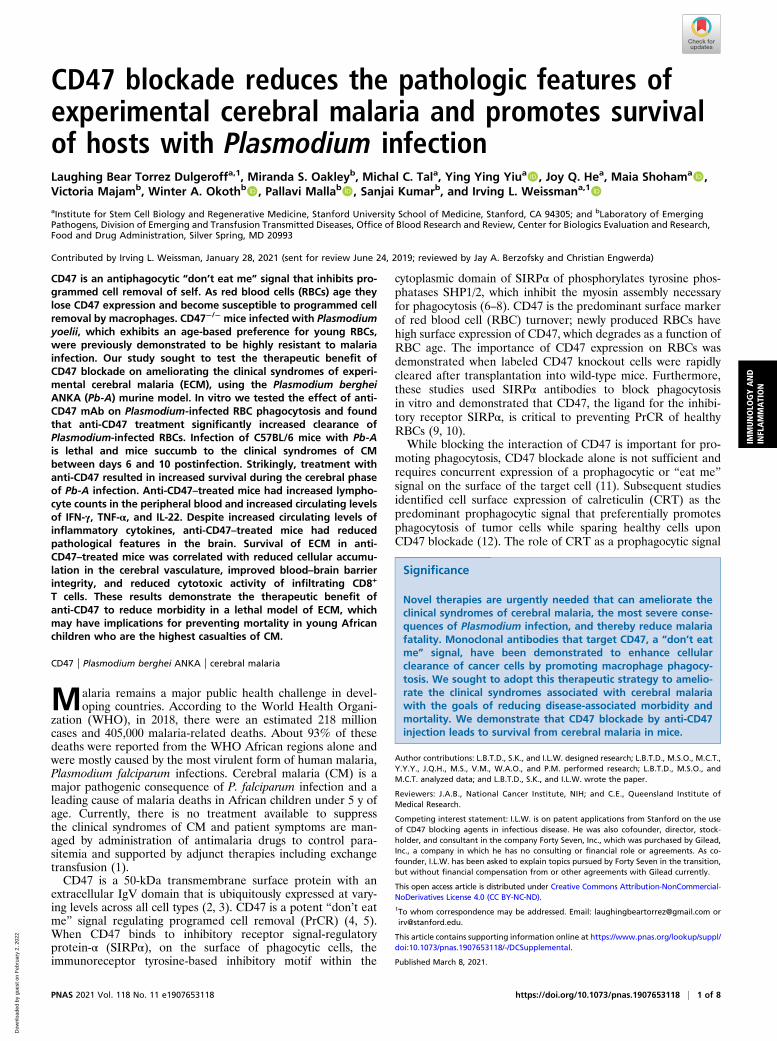
ResultsTreatment with Anti-CD47 Enhances Macrophage-Mediated PrCR ofPlasmodium-Infected RBCs In Vitro. CD47 blockade alone is notsufficient to promote PrCR; there must be a prophagocytic signalon the surface of the target cell. We initiated our studies byexamining the expression of prophagocytic signal CRT on thesurface of RBCs from mice over the course of Pb-A infection inBALB/c mice. In this murine model of severe malaria, 100% ofPb-A–infected BALB/c mice develop lethal severe anemia andhyperparasitemia. Blood was collected from BALBc mice in-fected with 106 parasites of a transgenic strain of Pb-A, whichconstitutively expresses GFP, on days 0, 5, 10, 15, and 20 post-infection. Flow cytometry analysis was used to determine thepercentage of CRT+ parasitized RBCs (pRBCs) over the courseof infection. A gradual but significant increase in the percentageof CRT+ pRBCs was observed over the course of infection(Fig. 1A). Additionally, pRBCs from infected mice had signifi-cantly higher levels of CRT on their cell surface compared toRBCs from uninfected mice (Fig. 1B). Given the inherent tran-scriptional silence of mature RBCs, it is likely that prophagocyticCRT on the surface of RBCs over the course of malaria infectionis achieved in a cell-extrinsic manner, likely by macrophage se-cretion as has been demonstrated previously (13).We then determined whether CD47 expression on RBCs
during Plasmodium infection prevents efficient cellular clearanceby performing in vitro phagocytosis assays. We tested the effectof mouse anti-CD47 mAb, MIAP410, compared to IgG1 isotype,and PBS controls when RBCs from a Pb-A–infected mouse werecoincubated with syngeneic mouse macrophages. Anti-CD47treatment resulted in a 4.7-fold increase in pRBC phagocytosis,supporting the hypothesis that CD47 blockade in vivo may re-duce parasite load and potentially reduce morbidity and mor-tality (Fig. 1C). In addition to mouse phagocytosis assays, we alsotested the efficacy of human anti-CD47 mAb, Hu5F9G4, againstP. falciparum–infected human RBCs. Similar to our observationsin the mouse model, we found that CD47 blockade withHu5F9G4 lead to a 2.3-fold increase in phagocytosis comparedto the IgG4 isotype control (Fig. 1D). These results confirmedthat CD47 blockade promotes PrCR of pRBCs in both murineand human in vitro models.
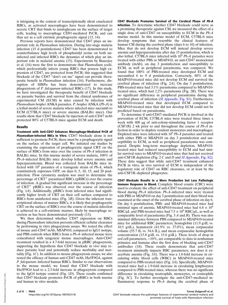
CD47 Blockade Promotes Survival of the Cerebral Phase of Pb-AInfection. To determine whether CD47 blockade could serve asa promising therapeutic against CM, we measured the effect of asingle dose of anti-CD47 on susceptibility to ECM in the Pb-Amurine model. In this murine model of ECM, C57BL/6 micedevelop symptoms that resemble the clinical features ofhuman CM during the cerebral phase (days 6 to 10) of infection.Mice that do not develop ECM will instead develop severeanemia and hyperparasitemia after day 15 postinfection, which isalso lethal. C57BL/6 mice infected with 106 Pb-A parasites weretreated with either PBS or MIAP410, an anti-CD47 monoclonalantibody (mAb), on day 3 postinfection and susceptibility toECM, as well as peripheral parasitemia, was compared. Wefound that 100% of PBS-treated mice developed ECM andsuccumbed 6 to 9 d postinfection. Conversely, 80% of theMAIP410-treated mice did not develop ECM and survived thecerebral phase of infection (Fig. 2A). On day 5 postinfection,PBS-treated mice had 3.1% parasitemia compared to MIAP410-treated mice, which had 2.2% parasitemia (Fig. 2B). There wasno significant difference in peripheral parasitemia during thecerebral phase of infection (SI Appendix, Fig. S1). Furthermore,MIAP410-treated mice that developed ECM compared toMIAP410-treated mice that did not develop ECM could not bepredicted based on parasitemia.To determine if anti-CD47–mediated PrCR is involved in the
prevention of ECM, C57BL/6 mice were treated three times aweek with 400 μg of anti-colony-stimulating factor 1 receptor(CSF1R) 2 wk prior to and throughout the course of Pb-A in-fection in order to deplete resident monocytes and macrophages.Depleted mice were infected with 106 Pb-A parasites and treatedwith either PBS or MIAP410 on day 3 postinfection and sus-ceptibility to ECM, as well as peripheral parasitemia was com-pared. Despite long-term macrophage depletion, MIAP410-treated mice had reduced susceptibility to ECM and had simi-lar survival rates to MIAP410-treated mice that had not receivedanti-CSF1R depletion (Fig. 2 C and D and SI Appendix, Fig. S2).These data suggest that while anti-CD47 treatment enhancedPrCR in vitro, in vivo survival of ECM is independent of theregulatory role of CD47 on RBC clearance, or at least by theanti-CSF1R–depleted phagocytes.
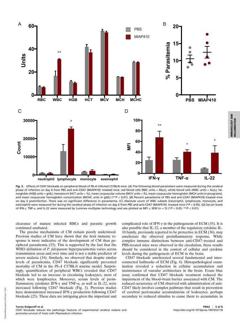
CD47 Blockade Results in a More Productive but Less PathologicImmune Response in Blood. Complete blood counts (CBC) wereused to evaluate the effect of anti-CD47 treatment on peripheralblood during Pb-A infection. Pb-A–infected mice were treatedwith PBS or MIAP410 on day 3 postinfection and their blood wasexamined at the onset of the cerebral phase of infection on day 6.On day 6 postinfection, PBS- and MIAP410-treated mice hadminimal signs of anemia. MIAP410-treated mice had 6.8 × 10⁶
RBC/μL and PBS-treated mice had 7.5 × 10⁶ RBC/μL, as well ascomparable level of parasitemia (Fig. 3 A and B). There was alsominimal difference between PBS compared to MIAP410-treatedmice for additional RBC parameters; hemoglobin (11.9 g/dL vs.10.5 g/dL), hematocrit (41.9% vs. 37.0%), mean corpuscularvolume (55.7 fL vs. 54.6 fL), and mean corpuscular hemoglobinconcentration (15.8 g/dL vs. 15.6 g/dL). These levels of drop inred cell parameters, ∼10%, are comparable to data in nonhumanprimates and humans after the first dose of blocking anti-CD47antibodies (18). These results demonstrate that anti-CD47treatment minimally impacts RBC parameters, nor does it ex-acerbate anemia (Fig. 3A). There was a 1.8-fold increase in cir-culating white blood cells (WBC) in MIAP410-treated micecompared to PBS-treated mice (Fig. 3A). Specifically, MIAP410-treated mice had a 1.9-fold increase in circulating lymphocytescompared to PBS-treated mice, whereas there was no significantdifference in circulating neutrophils, monocytes, or eosinophils(Fig. 3C). To further investigate the anti-CD47–mediated in-flammatory response to Pb-A during the cerebral phase of
2 of 8 | PNAS Torrez Dulgeroff et al.https://doi.org/10.1073/pnas.1907653118 CD47 blockade reduces the pathologic features of experimental cerebral malaria and
promotes survival of hosts with Plasmodium infection
Dow
nloa
ded
by g
uest
on
Feb
ruar
y 2,
202
2
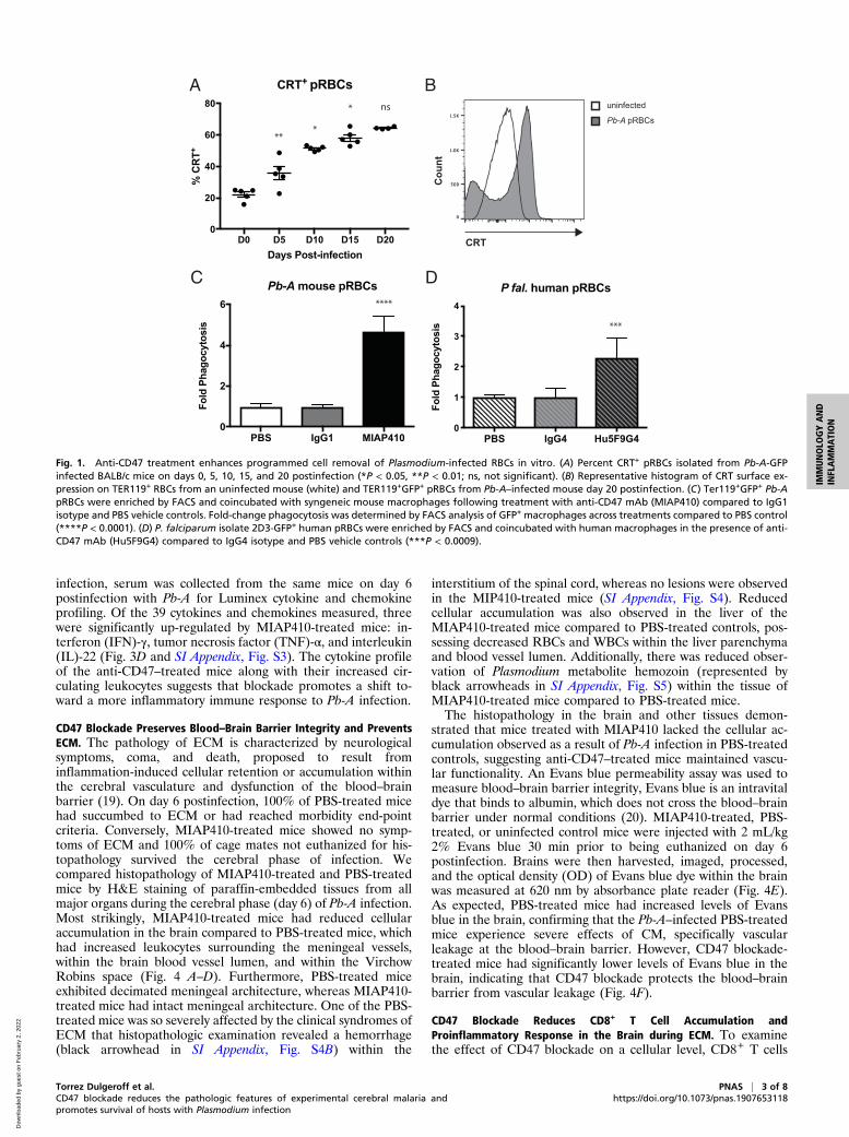
infection, serum was collected from the same mice on day 6postinfection with Pb-A for Luminex cytokine and chemokineprofiling. Of the 39 cytokines and chemokines measured, threewere significantly up-regulated by MIAP410-treated mice: in-terferon (IFN)-γ, tumor necrosis factor (TNF)-α, and interleukin(IL)-22 (Fig. 3D and SI Appendix, Fig. S3). The cytokine profileof the anti-CD47–treated mice along with their increased cir-culating leukocytes suggests that blockade promotes a shift to-ward a more inflammatory immune response to Pb-A infection.
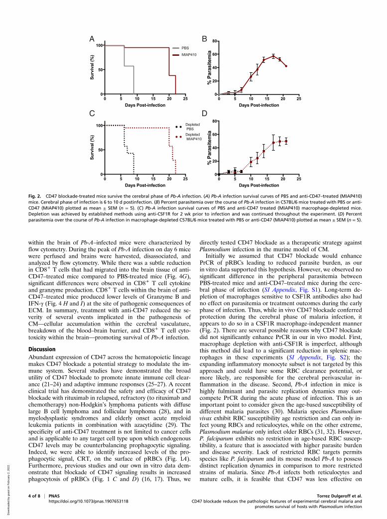
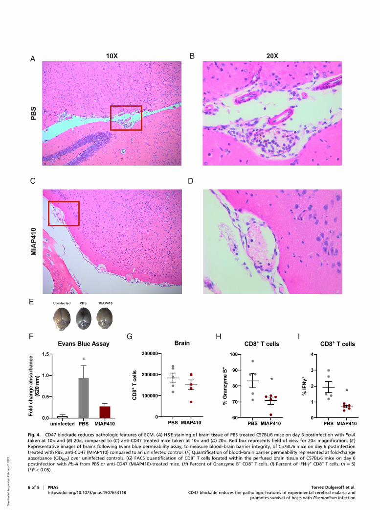
CD47 Blockade Preserves Blood–Brain Barrier Integrity and PreventsECM. The pathology of ECM is characterized by neurologicalsymptoms, coma, and death, proposed to result frominflammation-induced cellular retention or accumulation withinthe cerebral vasculature and dysfunction of the blood–brainbarrier (19). On day 6 postinfection, 100% of PBS-treated micehad succumbed to ECM or had reached morbidity end-pointcriteria. Conversely, MIAP410-treated mice showed no symp-toms of ECM and 100% of cage mates not euthanized for his-topathology survived the cerebral phase of infection. Wecompared histopathology of MIAP410-treated and PBS-treatedmice by H&E staining of paraffin-embedded tissues from allmajor organs during the cerebral phase (day 6) of Pb-A infection.Most strikingly, MIAP410-treated mice had reduced cellularaccumulation in the brain compared to PBS-treated mice, whichhad increased leukocytes surrounding the meningeal vessels,within the brain blood vessel lumen, and within the VirchowRobins space (Fig. 4 A–D). Furthermore, PBS-treated miceexhibited decimated meningeal architecture, whereas MIAP410-treated mice had intact meningeal architecture. One of the PBS-treated mice was so severely affected by the clinical syndromes ofECM that histopathologic examination revealed a hemorrhage(black arrowhead in SI Appendix, Fig. S4B) within the
interstitium of the spinal cord, whereas no lesions were observedin the MIP410-treated mice (SI Appendix, Fig. S4). Reducedcellular accumulation was also observed in the liver of theMIAP410-treated mice compared to PBS-treated controls, pos-sessing decreased RBCs and WBCs within the liver parenchymaand blood vessel lumen. Additionally, there was reduced obser-vation of Plasmodium metabolite hemozoin (represented byblack arrowheads in SI Appendix, Fig. S5) within the tissue ofMIAP410-treated mice compared to PBS-treated mice.The histopathology in the brain and other tissues demon-
strated that mice treated with MIAP410 lacked the cellular ac-cumulation observed as a result of Pb-A infection in PBS-treatedcontrols, suggesting anti-CD47–treated mice maintained vascu-lar functionality. An Evans blue permeability assay was used tomeasure blood–brain barrier integrity, Evans blue is an intravitaldye that binds to albumin, which does not cross the blood–brainbarrier under normal conditions (20). MIAP410-treated, PBS-treated, or uninfected control mice were injected with 2 mL/kg2% Evans blue 30 min prior to being euthanized on day 6postinfection. Brains were then harvested, imaged, processed,and the optical density (OD) of Evans blue dye within the brainwas measured at 620 nm by absorbance plate reader (Fig. 4E).As expected, PBS-treated mice had increased levels of Evansblue in the brain, confirming that the Pb-A–infected PBS-treatedmice experience severe effects of CM, specifically vascularleakage at the blood–brain barrier. However, CD47 blockade-treated mice had significantly lower levels of Evans blue in thebrain, indicating that CD47 blockade protects the blood–brainbarrier from vascular leakage (Fig. 4F).
CD47 Blockade Reduces CD8+ T Cell Accumulation andProinflammatory Response in the Brain during ECM. To examinethe effect of CD47 blockade on a cellular level, CD8+ T cells
D0 D5 D10 D15 D200
20
40
60
80
Days Post-infection
% C
RT+
CRT+ pRBCs
PBS IgG1 MIAP4100
2
4
6
Fold
Pha
gocy
tosi
s
Pb-A mouse pRBCs
PBS IgG4 Hu5F9G40
1
2
3
4
Fold
Pha
gocy
tosi
s
P fal. human pRBCs
Cou
nt
CRT
A B
C D
uninfected
Pb-A pRBCs
***
* ns
****
***
Fig. 1. Anti-CD47 treatment enhances programmed cell removal of Plasmodium-infected RBCs in vitro. (A) Percent CRT+ pRBCs isolated from Pb-A-GFPinfected BALB/c mice on days 0, 5, 10, 15, and 20 postinfection (*P < 0.05, **P < 0.01; ns, not significant). (B) Representative histogram of CRT surface ex-pression on TER119+ RBCs from an uninfected mouse (white) and TER119+GFP+ pRBCs from Pb-A–infected mouse day 20 postinfection. (C) Ter119+GFP+ Pb-ApRBCs were enriched by FACS and coincubated with syngeneic mouse macrophages following treatment with anti-CD47 mAb (MIAP410) compared to IgG1isotype and PBS vehicle controls. Fold-change phagocytosis was determined by FACS analysis of GFP+ macrophages across treatments compared to PBS control(****P < 0.0001). (D) P. falciparum isolate 2D3-GFP+ human pRBCs were enriched by FACS and coincubated with human macrophages in the presence of anti-CD47 mAb (Hu5F9G4) compared to IgG4 isotype and PBS vehicle controls (***P < 0.0009).
Torrez Dulgeroff et al. PNAS | 3 of 8CD47 blockade reduces the pathologic features of experimental cerebral malaria andpromotes survival of hosts with Plasmodium infection
https://doi.org/10.1073/pnas.1907653118
IMMUNOLO
GYAND
INFLAMMATION
Dow
nloa
ded
by g
uest
on
Feb
ruar
y 2,
202
2
within the brain of Pb-A–infected mice were characterized byflow cytometry. During the peak of Pb-A infection on day 6 micewere perfused and brains were harvested, disassociated, andanalyzed by flow cytometry. While there was a subtle reductionin CD8+ T cells that had migrated into the brain tissue of anti-CD47–treated mice compared to PBS-treated mice (Fig. 4G),significant differences were observed in CD8+ T cell cytokineand granzyme production. CD8+ T cells within the brain of anti-CD47–treated mice produced lower levels of Granzyme B andIFN-γ (Fig. 4 H and I) at the site of pathogenic consequences ofECM. In summary, treatment with anti-CD47 reduced the se-verity of several events implicated in the pathogenesis ofCM—cellular accumulation within the cerebral vasculature,breakdown of the blood–brain barrier, and CD8+ T cell cyto-toxicity within the brain—promoting survival of Pb-A infection.
DiscussionAbundant expression of CD47 across the hematopoietic lineagemakes CD47 blockade a potential strategy to modulate the im-mune system. Several studies have demonstrated the broadutility of CD47 blockade to promote innate immune cell clear-ance (21–24) and adaptive immune responses (25–27). A recentclinical trial has demonstrated the safety and efficacy of CD47blockade with rituximab in relapsed, refractory (to rituximab andchemotherapy) non-Hodgkin’s lymphoma patients with diffuselarge B cell lymphoma and follicular lymphoma (28), and inmyelodysplastic syndromes and elderly onset acute myeloidleukemia patients in combination with azacytidine (29). Thespecificity of anti-CD47 treatment is not limited to cancer cellsand is applicable to any target cell type upon which endogenousCD47 levels may be counterbalancing prophagocytic signaling.Indeed, we were able to identify increased levels of the pro-phagocytic signal, CRT, on the surface of pRBCs (Fig. 1A).Furthermore, previous studies and our own in vitro data dem-onstrate that blockade of CD47 signaling results in increasedphagocytosis of pRBCs (Fig. 1 C and D) (16, 17). Thus, we
directly tested CD47 blockade as a therapeutic strategy againstPlasmodium infection in the murine model of CM.Initially we assumed that CD47 blockade would enhance
PrCR of pRBCs leading to reduced parasite burden, as ourin vitro data supported this hypothesis. However, we observed nosignificant difference in the peripheral parasitemia betweenPBS-treated mice and anti-CD47–treated mice during the cere-bral phase of infection (SI Appendix, Fig. S1). Long-term de-pletion of macrophages sensitive to CSF1R antibodies also hadno effect on parasitemia or treatment outcomes during the earlyphase of infection. Thus, while in vivo CD47 blockade conferredprotection during the cerebral phase of malaria infection, itappears to do so in a CSF1R macrophage-independent manner(Fig. 2). There are several possible reasons why CD47 blockadedid not significantly enhance PrCR in our in vivo model. First,macrophage depletion with anti-CSF1R is imperfect, althoughthis method did lead to a significant reduction in splenic mac-rophages in these experiments (SI Appendix, Fig. S2); theexpanding inflammatory monocyte subset is not targeted by thisapproach and could have some RBC clearance potential, ormore likely, are responsible for the cerebral perivascular in-flammation in the disease. Second, Pb-A infection in mice ishighly fulminant and parasite replication dynamics may out-compete PrCR during the acute phase of infection. This is animportant point to consider given the age-based susceptibility ofdifferent malaria parasites (30). Malaria species Plasmodiumvivax exhibit RBC susceptibility age restriction and can only in-fect young RBCs and reticulocytes, while on the other extreme,Plasmodium malariae only infect older RBCs (31, 32). However,P. falciparum exhibits no restriction in age-based RBC suscep-tibility, a feature that is associated with higher parasite burdenand disease severity. Lack of restricted RBC targets permitsspecies like P. falciparum and its mouse model Pb-A to possessdistinct replication dynamics in comparison to more restrictedstrains of malaria. Since Pb-A infects both reticulocytes andmature cells, it is feasible that CD47 was less effective on
0 5 10 15 20 250
50
100
Days Post-infection
Surv
ival
(%)
A
0 5 10 15 20 250
50
100
Days Post-infection
Surv
ival
(%)
0 5 10 15 20 250
20
40
60
80
Days Post-infection
% P
aras
item
ia
PBS
MIAP410
Depleted PBSDepleted MIAP410
B
C D
0 5 10 15 20 250
20
40
60
80
Days Post-infection
% P
aras
item
ia
Fig. 2. CD47 blockade-treated mice survive the cerebral phase of Pb-A infection. (A) Pb-A infection survival curves of PBS and anti-CD47–treated (MIAP410)mice. Cerebral phase of infection is 6 to 10 d postinfection. (B) Percent parasitemia over the course of Pb-A infection in C57BL/6 mice treated with PBS or anti-CD47 (MIAP410) plotted as mean ± SEM (n = 5). (C) Pb-A infection survival curves of PBS and anti-CD47 treated (MIAP410) macrophage-depleted mice.Depletion was achieved by established methods using anti-CSF1R for 2 wk prior to infection and was continued throughout the experiment. (D) Percentparasitemia over the course of Pb-A infection in macrophage-depleted C57BL/6 mice treated with PBS or anti-CD47 (MIAP410) plotted as mean ± SEM (n = 5).
4 of 8 | PNAS Torrez Dulgeroff et al.https://doi.org/10.1073/pnas.1907653118 CD47 blockade reduces the pathologic features of experimental cerebral malaria and
promotes survival of hosts with Plasmodium infection
Dow
nloa
ded
by g
uest
on
Feb
ruar
y 2,
202
2
clearance of mature infected RBCs and parasite growthcontinued unabated.The precise mechanisms of CM remain poorly understood.
Previous studies of CM have shown that the host immune re-sponse is more indicative of the development of CM than pe-ripheral parasitemia (33). This is supported by the fact that theWHO definition of P. falciparum hyperparasitemia varies acrosstransmission areas and over time and is not a stable predictor ofsevere malaria (34). Similarly, we observed that despite similarlevels of parasitemia, CD47 blockade significantly preventedmortality of CM in the Pb-A C57BL/6 murine model. Surpris-ingly, quantification of peripheral WBCs revealed that CD47blockade led to an increase in circulating leukocytes, most ofwhich were lymphocytes. Moreover, serum levels of proin-flammatory cytokines IFN-γ and TNF-α, as well as IL-22, wereincreased following CD47 blockade (Fig. 3). Previous studieshave demonstrated increased IFN-γ production following CD47blockade (25). These data are intriguing given the important and
complicated role of IFN-γ in the pathogenesis of ECM (35). It isalso possible that IL-22, a member of the regulatory cytokine IL-10 family, previously reported to be protective in ECM (36), mayameliorate the observed proinflammatory response. Whilecomplex immune distinctions between anti-CD47–treated andPBS-treated mice were observed in the circulation, these resultsshould be considered in the context of cellular and cytokinelevels during the pathogenesis of ECM in the brain.CD47 blockade ameliorated several fundamental and inter-
connected hallmarks of ECM (Fig. 4). Histopathological exam-ination revealed a reduction in cellular accumulation andmaintenance of vascular architecture in the brain. Evans blueassay confirmed that CD47 blockade treatment reduced theimpairment of the blood–brain barrier associated with CM. Thereduced occurrence of CM observed with administration of anti-CD47 likely involves complex pathways that result in preventionof cerebral perivascular accumulations of leukocytes, perhapssecondary to reduced stimulus to cause them to accumulate in
A
C
neutrophil lymphocyte monocyte eosinophil0
10000
20000
30000
Cou
nt
D
IL-220
20
40
60
80
100
MFI
RBC WBC HGB HCT MCV MCH MCHC0
20
40
60U
nits
PBS
MIAP410
**
*
*
**
*
PBS MIAP4100
5
10
15
20
% P
aras
item
ia
B
Fig. 3. Effects of CD47 blockade on peripheral blood of Pb-A infected C57BL/6 mice. (A) The following blood parameters were measured during the cerebralphase of infection on day 6 from PBS and anti-CD47 (MIAP410) -treated mice: red blood cells (RBC units = M/μL), white blood cells (WBC units = K/μL), he-moglobin (HGB units = g/dL), hematocrit (HCT units =%), mean corpuscular volume (MCV units = fL), mean corpuscular hemoglobin (MCH units in picograms),and mean corpuscular hemoglobin concentration (MCHC units in g/dL) (**P < 0.01). (B) Percent parasitemia of PBS and anti-CD47 (MIAP410) treated miceon day 6 postinfection. There was no significant difference in parasitemia. (C) Absolute count of WBC subsets (neutrophil, lymphocyte, monocyte, andeosinophil) were measured for during the cerebral phase of infection on day 6 from PBS and anti-CD47 (MIAP410) -treated mice (*P < 0.05). (D) Serum levelsof IFN-γ, TNF-α, and IL-22 were measured by Luminex multiplex technology and are plotted as MFI ± SEM (n = 5) (*P < 0.05, **P < 0.01).
Torrez Dulgeroff et al. PNAS | 5 of 8CD47 blockade reduces the pathologic features of experimental cerebral malaria andpromotes survival of hosts with Plasmodium infection
https://doi.org/10.1073/pnas.1907653118
IMMUNOLO
GYAND
INFLAMMATION
Dow
nloa
ded
by g
uest
on
Feb
ruar
y 2,
202
2
PBS MIAP4100
100000
200000
300000
Brain
CD
8+ T c
ells
PBS MIAP41060
70
80
90
100
CD8+ T cells
% G
ranz
yme
B+
PBS MIAP4100
1
2
3
4
CD8+ T cells
**
A
C D
B
PBS
MIA
P410
10X 20X
E
G
uninfected PBS MIAP4100.0
0.5
1.0
1.5
Fold
cha
nge
abso
rban
ce
(620
nm
)
Evans Blue Assay
*
H IF
Uninfected PBS MIAP410
Fig. 4. CD47 blockade reduces pathologic features of ECM. (A) H&E staining of brain tissue of PBS treated C57BL/6 mice on day 6 postinfection with Pb-Ataken at 10× and (B) 20×, compared to (C) anti-CD47 treated mice taken at 10× and (D) 20×. Red box represents field of view for 20× magnification. (E)Representative images of brains following Evans blue permeability assay, to measure blood–brain barrier integrity, of C57BL/6 mice on day 6 postinfectiontreated with PBS, anti-CD47 (MIAP410) compared to an uninfected control. (F) Quantification of blood–brain barrier permeability represented as fold-changeabsorbance (OD620) over uninfected controls. (G) FACS quantification of CD8+ T cells located within the perfused brain tissue of C57BL/6 mice on day 6postinfection with Pb-A from PBS or anti-CD47 (MIAP410)-treated mice. (H) Percent of Granzyme B+ CD8+ T cells. (I) Percent of IFN-γ+ CD8+ T cells. (n = 5)(*P < 0.05).
6 of 8 | PNAS Torrez Dulgeroff et al.https://doi.org/10.1073/pnas.1907653118 CD47 blockade reduces the pathologic features of experimental cerebral malaria and
promotes survival of hosts with Plasmodium infection
Dow
nloa
ded
by g
uest
on
Feb
ruar
y 2,
202
2
these vessels. These distinctions are also potentially integral tothe activation of CD8+ T cells, which play a central role in thepathogenesis of CM (37). Pb-A infection is associated with in-creased cellular accumulation and cross-presentation to CD8+
T cells, leading to migration into the perivascular spaces withinthe brain microvasculature. Perivascular APCs, presenting par-asite antigen as a result of reduced blood–brain barrier integrity,prime CD8+ T cells, which upon activation release cytokines andcytotoxic granules. CD47 blockade was demonstrated to reducecellular accumulation in the brain and improve the integrity ofthe blood–brain barrier during Pb-A infection, which were cor-related to a reduction in CD8+ T cell cytotoxicity.Although we initially hypothesized that CD47 blockade would
enhance PrCR and attenuate disease burden by reducing para-sitemia, we found that CD47 blockade reduced morbidity ofECM through reducing the cytotoxicity of leukocyte infiltrationwithin the brain that is directly associated with disease patho-genesis. While it is difficult to determine the precise order ofevents that prevent ECM, given the ubiquity of CD47 expressionand complexity of ECM pathology, the effects of CD47 on CD8+
T cell cytotoxicity warrant further investigation. Our study rep-resents a foundation for preclinical and clinical studies to in-vestigate the prospect of anti-CD47 mAb administration toreduce morbidity and mortality in CM patients, particularly inyoung African children.
Materials and MethodsMice, Parasites, and Infection. Age-matched female C57BL/6 or BALB/c micewere obtained from The Jackson Laboratory. Mice from the I.L.W. laboratoryat Stanford University were maintained in the Stem Cell Institute and ex-periments were carried out in accordance with guidelines set by the Stan-ford University Administrative Panel on Laboratory Animal Care (AnimalStudy Protocol 30109). Mice from the S.K. laboratory at the Food and DrugAdministration (FDA) were maintained at the facilities at the Center forBiologics Evaluation and Research and experiments were carried out in ac-cordance with the guidelines set forth for the care and use of laboratoryanimals by the FDA (Animal Study Protocol 2002-21). Pb-A, Pb-A GFPCON259cl2 obtained through BEI Resources, was used as the source of parasiteinfection in both in vitro and in vivo experiments. Mice were infected byintraperitoneal injection of 106 pRBCs from a donor mouse. Water for donormice was treated with 0.07 mg/mL pyrimethamine to select for GFP+
parasites.
CRT Expression. To determine the expression of CRT, mice were bled andwhole blood was stained with anti-CRT mAb (FMC 75) conjugated to phy-coerythrin (PE) and Ter119 (Ly-76) conjugated to Alexa Fluor 700. pRBCswere gated as Ter119+GFP+ and the percent CRT+ pRBCs and median fluo-rescent intensity (MFI) were measured on an LSR Fortessa.
Phagocytosis Assays of RBCs by Mouse Bone Marrow-Derived Macrophages.Macrophage derivation. Bone marrow-derived mouse macrophages weregenerated by performing a bone marrow flush of the tibia and femur ofBALB/c mice that were 6 to 12 wk old. Bone marrow cells were mechanicallydissociated and treated with ACK lysis buffer for 5 min. Cells were thendifferentiated in Iscove modified Dulbecco media + 10% FBS + 10 ng/mLmouse macrophage colony stimulating factor for 6 to 8 d. Peripheral blood-derived human macrophages were generated by Ficoll enrichment of whiteblood cells from peripheral blood (Leukocyte Reduction System Chambers ofdonor blood were purchased from the Stanford Blood Center). Cells weretreated with ACK lysis buffer for 5 min and then adherence selection for30 min and cultured in RPMI + 10% human serum for 6 to 9 d.
Target cell isolation. Mouse pRBCs were purified from BLABc mouse pe-ripheral blood infected with Pb-A GFPCON 259cl2 by FACS. Human pRBCswere purified from type O donor blood infected in vitro with P. falciparum3D7HT-GFP (courtesy of the Yeh laboratory at Stanford University, Cal-ifornia, strain from BEI resources) using magnetic isolation with MACS col-umns (Miltenyi Biotech).
Phagocytosis Assays of RBCs by Human Monocyte-Derived Macrophages. De-rived macrophages were harvested and counted by hemocytometer. Oncepurified, target cells were stained with carboxyfluorescein diacetate succi-nimidyl ester (CFSE) to enhance target cell signal strength and then counted.
Target cells were cocultured with macrophages at a ratio of 2:1 for 1 h in theindicated treatment condition. CD47 blockade in vitro was achieved usinganti-CD47 mAb, for mice MIAP410 (Bio X Cell) was used and for humanHu5F9G4 (Lonza) was used. Isotype control treatment for mouse was IgG1isotype control (Bio X Cell) and for human was Ultra-LEAF purified IgG4isotype control (BioLegend). Mouse macrophages were labeled with anti-mouse F4/80 (BioLegend), human macrophages were labeled with anti-human Macrophage Marker (eBioscience). Percent phagocytosis (CFSE+
Macrophages) was quantified by FACS and normalized to PBS control tocalculate fold-change in phagocytosis across treatment conditions.
In Vivo Mouse Treatments.CD47 blockade. C57BL/6 mice were injected intraperitoneally with 100 μg ofanti-CD47 MIAP410 (Bio X Cell) in 0.1 mL of phosphate buffered saline so-lution. Mice were treated with a single dose of anti-CD47 on day 3postinfection unless otherwise noted.Long-term macrophage depletion. Mice were treated injected intraperitoneallywith 400 μg of anti-CSF1R (Bio X Cell) in 0.1 mL of phosphate buffered salinesolution. Depletion began 2 wk prior to infection with Pb-A, mice weretreated three times a week, and continued to receive treatment three timesa week until experimental endpoints were reached.
Parasitemia and CBC. The peripheral parasitemia of experimental mice C57BL/6 mice was determined by FACS analysis. For FACS, 1 μL of blood was stainedTer119 (Ly-76) conjugated to Alexa Fluor 700 for 30 min and then diluted in500 μL of FACS buffer (phosphate buffered saline solution + 2% FBS) foranalysis. Peripheral parasitemia was measured as the percent of Ter119+
RBCs that were Pb-A–GFP+, parasitemia was also confirmed by microscopyon Giemsa-stained thin blood films. CBC were performed on the Sysmex XT-2000iV analyzer system and confirmed by blood smears analysis preparedwith Wright–Giemsa stain.
Luminex. This assay was performed in the Human ImmuneMonitoring Centerat Stanford University. Mouse 38 plex kits were purchased from eBiosciences/Affymetrix and used according to the manufacturer’s recommendations,with modifications as described below. Briefly, beads were added to a96-well plate and washed in a Biotek ELx405 washer. Samples were added tothe plate containing the mixed antibody-linked beads and incubated atroom temperature for 1 h followed by overnight incubation at 4 °C withshaking. Cold and room temperature incubation steps were performed onan orbital shaker at 500 to 600 rpm. Following the overnight incubation,plates were washed in a Biotek ELx405 washer and then biotinylated de-tection antibody added for 75 min at room temperature with shaking. Theplate was washed as above, and streptavidin-PE was added. After incubationfor 30 min at room temperature, wash was performed as above and readingbuffer was added to the wells. Each sample was measured in duplicate.Plates were read using a Luminex 200 instrument with a lower bound of 50beads per sample per cytokine. Custom assay Control beads by Radix Bio-solutions was added to all wells.
Immunohistochemistry. During the cerebral phase of infection, on day 6postinfection, portions of all major organs were harvested from mice fromeach treatment group. Harvested tissues were fixed in 10% buffered neutralformalin, routinely processed, and embedded in paraffin for sectioning andstaining. H&E stainingwas performed by the Animal Diagnostic Laboratory inthe Department of Comparative Medicine at Stanford University. Parafin-embedded sections were shipped to Histowiz for immunohistochemistry,CD11b (ab133357, clone EPR1344).
Evans Blue Permeability Assay. On day 6 postinfection with Pb-A, C57/BL6mice treated with PBS or anti-CD47, as described above, were injected retro-orbitally with 2 mL/kg 2% Evans blue and euthanized 30 min postinjection ofthe dye. Brains were collected, rinsed with PBS, and imaged. Brains werethen mechanically dissociated and incubated in formaldehyde for 20 h at55 °C, Evans blue-infused supernatant was transferred to 96-well plate andOD620 was measured by absorbance plate reader.
Flow Cytometry Characterization of CD8+ T Cells. On day 6 postinfection withPb-A, C57/BL6 mice, treated with PBS or anti-CD47 as described above, wereperfused and euthanized. Brains were harvested, weighted, and mechan-ically dissociated in MACS buffer (PBS, 2% FBS, 2 mM EDTA). Dissociated cellswere counted and stained with the following antibodies: anti-CD3, anti-CD8,anti–IFN-γ, and anti-Granzyme B (BioLegend). Spleens were harvested,weighted, and mechanically dissociated in MACS buffer. Dissociated cells
Torrez Dulgeroff et al. PNAS | 7 of 8CD47 blockade reduces the pathologic features of experimental cerebral malaria andpromotes survival of hosts with Plasmodium infection
https://doi.org/10.1073/pnas.1907653118
IMMUNOLO
GYAND
INFLAMMATION
Dow
nloa
ded
by g
uest
on
Feb
ruar
y 2,
202
2
were counted and stained for anti-CD3, anti-CD8, anti–IFN-γ, and anti–TNF-α(BioLegend).
Data Availability. All study data are included in the article and SI Appendix.
ACKNOWLEDGMENTS. We thank all members of the I.L.W. and S.K. labo-ratories for support and helpful discussions; Katie Amberg-Johnson and theYeh laboratory at Stanford for providing Plasmodium falciparum 3D7 usedin phagocytosis assays; Yael Rosenberg-Hasson and the staff of the StanfordHuman Immune Monitoring Center for their technical expertise and for run-ning the mouse Luminex assays; José G. Vilches-Moure for consulting onmouse pathology; and the Animal Diagnostic Laboratory in the Departmentof Comparative Medicine at Stanford University. The following reagentswere obtained through BEI Resources, National Institute of Allergy and
Infectious Diseases, NIH: Plasmodium berghei, strain (ANKA) GFPCON259cl2, MRA 865, contributed by Chris J. Janse and Andrew P. Waters, andPlasmodium falciparum, strain 3D7HT-GFP, MRA-1029, contributed by A. Tal-man and R. Sinden. Research reported in this publication was supported bythe Virginia and D. K. Ludwig Fund for Cancer Research, Robert J. Kleberg Jr.and Helen C. Kleberg Foundation, and Food and Drug Administration intra-mural research funding. L.B.T.D. was supported by the Stanford DiversifyingAcademia, Recruiting Excellence fellowship; M.C.T. and Y.Y.Y. were sup-ported by the Stanford Immunology Training Grant 5T32AI007290; andM.C.T. was also supported by the NIH National Research Service Award 1F32 AI124558-01. J.Q.H was supported by the National Cancer Institute Na-tional Research Service Award F30CA228215 and Stanford Medical ScientistTraining Grant T32GM007365.
1. WHO, World Malaria Report (World Health Organization, 2018).2. F. P. Lindberg, H. D. Gresham, E. Schwarz, E. J. Brown, Molecular cloning of integrin-
associated protein: An immunoglobulin family member with multiple membrane-spanning domains implicated in alpha v beta 3-dependent ligand binding. J. CellBiol. 123, 485–496 (1993).
3. M. I. Reinhold et al., In vivo expression of alternatively spliced forms of integrin-associated protein (CD47). J. Cell Sci. 108, 3419–3425 (1995).
4. S. Jaiswal et al., CD47 is upregulated on circulating hematopoietic stem cells andleukemia cells to avoid phagocytosis. Cell 138, 271–285 (2009).
5. S. Jaiswal, M. P. Chao, R. Majeti, I. L. Weissman, Macrophages as mediators of tumorimmunosurveillance. Trends Immunol. 31, 212–219 (2010).
6. A. Veillette, E. Thibaudeau, S. Latour, High expression of inhibitory receptor SHPS-1and its association with protein-tyrosine phosphatase SHP-1 in macrophages. J. Biol.Chem. 273, 22719–22728 (1998).
7. Y. Fujioka et al., A novel membrane glycoprotein, SHPS-1, that binds the SH2-do-main-containing protein tyrosine phosphatase SHP-2 in response to mitogens andcell adhesion. Mol. Cell. Biol. 16, 6887–6899 (1996).
8. R. K. Tsai, D. E. Discher, Inhibition of “self” engulfment through deactivation ofmyosin-II at the phagocytic synapse between human cells. J. Cell Biol. 180, 989–1003(2008).
9. P.-A. Oldenborg et al., Role of CD47 as a marker of self on red blood cells. Science 288,2051–2054 (2000).
10. P. A. Oldenborg, H. D. Gresham, F. P. Lindberg, CD47-signal regulatory protein alpha(SIRPalpha) regulates Fcgamma and complement receptor-mediated phagocytosis.J. Exp. Med. 193, 855–862 (2001).
11. R. Majeti et al., CD47 is an adverse prognostic factor and therapeutic antibody targeton human acute myeloid leukemia stem cells. Cell 138, 286–299 (2009).
12. M. P. Chao et al., Calreticulin is the dominant pro-phagocytic signal on multiple hu-man cancers and is counterbalanced by CD47. Sci. Transl. Med. 2, 63ra94 (2010).
13. M. Feng et al., Macrophages eat cancer cells using their own calreticulin as a guide:Roles of TLR and Btk. Proc. Natl. Acad. Sci. U.S.A. 112, 2145–2150 (2015).
14. M. Feng et al., Programmed cell removal by calreticulin in tissue homeostasis andcancer. Nat. Commun. 9, 3194 (2018).
15. C. Fernandez-Arias et al., Anti-self phosphatidylserine antibodies recognize unin-fected erythrocytes promoting malarial anemia. Cell Host Microbe 19, 194–203 (2016).
16. R. Banerjee, S. Khandelwal, Y. Kozakai, B. Sahu, S. Kumar, CD47 regulates thephagocytic clearance and replication of the Plasmodium yoelii malaria parasite. Proc.Natl. Acad. Sci. U.S.A. 112, 3062–3067 (2015).
17. K. Ayi et al., CD47-SIRPα interactions regulate macrophage uptake of Plasmodiumfalciparum-infected erythrocytes and clearance of malaria in vivo. Infect. Immun.,IAI.01426-15 (2016).
18. J. Liu et al., Pre-clinical development of a humanized anti-CD47 antibody with anti-cancer therapeutic potential. PLoS One 10, e0137345 (2015).
19. I. M. Medana, G. D. H. Turner, Human cerebral malaria and the blood-brain barrier.Int. J. Parasitol. 36, 555–568 (2006).
20. O. Uyama et al., Quantitative evaluation of vascular permeability in the gerbil brainafter transient ischemia using Evans blue fluorescence. J. Cereb. Blood Flow Metab. 8,282–284 (1988).
21. M. P. Chao et al., Anti-CD47 antibody synergizes with rituximab to promote phago-cytosis and eradicate non-Hodgkin lymphoma. Cell 142, 699–713 (2010).
22. S. B. Willingham et al., The CD47-signal regulatory protein alpha (SIRPa) interaction isa therapeutic target for human solid tumors. Proc. Natl. Acad. Sci. U.S.A. 109,6662–6667 (2012).
23. K. Weiskopf et al., CD47-blocking immunotherapies stimulate macrophage-mediateddestruction of small-cell lung cancer. J. Clin. Invest. 126, 2610–2620 (2016).
24. S. Gholamin et al., Disrupting the CD47-SIRPα anti-phagocytic axis by a humanizedanti-CD47 antibody is an efficacious treatment for malignant pediatric brain tumors.Sci. Transl. Med. 9, eaaf2968 (2017).
25. X. Liu et al., CD47 blockade triggers T cell-mediated destruction of immunogenictumors. Nat. Med. 21, 1209–1215 (2015).
26. D. Tseng et al., Anti-CD47 antibody-mediated phagocytosis of cancer by macrophagesprimes an effective antitumor T-cell response. Proc. Natl. Acad. Sci. U.S.A. 110,11103–11108 (2013).
27. M. N. McCracken, A. C. Cha, I. L. Weissman, Molecular pathways: Activating T cellsafter cancer cell phagocytosis from blockade of CD47 “don’t eat me” signals. Clin.Cancer Res. 21, 3597–3601 (2015).
28. R. Advani et al., CD47 blockade by Hu5F9-G4 and rituximab in non-Hodgkin’s lym-phoma. N. Engl. J. Med. 379, 1711–1721 (2018).
29. M. P. Chao et al., Therapeutic targeting of the macrophage immune checkpoint CD47in myeloid Malignancies. Front. Oncol. 9, 1380 (2020).
30. P. G. McQueen, F. E. McKenzie, Age-structured red blood cell susceptibility and thedynamics of malaria infections. Proc. Natl. Acad. Sci. U.S.A. 101, 9161–9166 (2004).
31. F. E. McKenzie, G. M. Jeffery, W. E. Collins, Plasmodium malariae blood-stage dy-namics. J. Parasitol. 87, 626–637 (2001).
32. F. E. McKenzie, G. M. Jeffery, W. E. Collins, Plasmodium vivax blood-stage dynamics.J. Parasitol. 88, 521–535 (2002).
33. D. S. Hansen, Inflammatory responses associated with the induction of cerebral ma-laria: Lessons from experimental murine models. PLoS Pathog. 8, e1003045 (2012).
34. P. Wilairatana, N. Tangpukdee, S. Krudsood, Definition of hyperparasitemia in severefalciparum malaria should be updated. Asian Pac. J. Trop. Biomed. 3, 586 (2013).
35. T. King, T. Lamb, Interferon-γ: The Jekyll and Hyde of malaria. PLoS Pathog. 11,e1005118 (2015).
36. J. Sellau et al., IL-22 dampens the T cell response in experimental malaria. Sci. Rep. 6,28058 (2016).
37. T. N. Shaw et al., Perivascular arrest of CD8+ T cells is a signature of experimentalcerebral malaria. PLoS Pathog. 11, e1005210 (2015).
8 of 8 | PNAS Torrez Dulgeroff et al.https://doi.org/10.1073/pnas.1907653118 CD47 blockade reduces the pathologic features of experimental cerebral malaria and
promotes survival of hosts with Plasmodium infection
Dow
nloa
ded
by g
uest
on
Feb
ruar
y 2,
202
2